Herpetological Review Herpetological Review - Doczine
Herpetological Review Herpetological Review - Doczine
Herpetological Review Herpetological Review - Doczine

You also want an ePaper? Increase the reach of your titles
YUMPU automatically turns print PDFs into web optimized ePapers that Google loves.
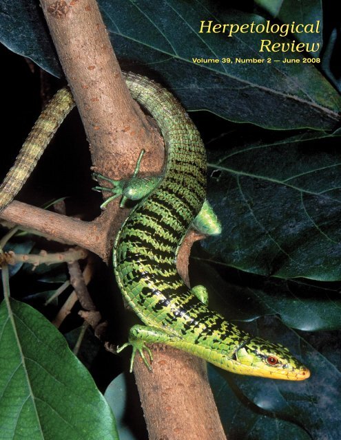
<strong>Herpetological</strong><strong>Review</strong>Volume 39, Number 2 — June 2008
SSAR Officers (2008)PresidentROY MCDIARMIDUSGS Patuxent Wildlife Research CenterNational Museum of Natural HistoryWashington, DC 20560, USAPresident-electBRIAN CROTHERDepartment of Biological SciencesSoutheastern Louisiana UniversityHammond, Louisiana 70402, USASecretaryMARION R. PREESTJoint Science DepartmentThe Claremont CollegesClaremont, California 91711, USATreasurerKIRSTEN E. NICHOLSONDepartment of Biology, Brooks 217Central Michigan UniversityMt. Pleasant, Michigan 48859, USAe-mail: kirsten.nicholson@cmich.eduPublications SecretaryBRECK BARTHOLOMEWP.O. Box 58517Salt Lake City, Utah 84158, USAe-mail: ssar@herplit.comImmediate Past PresidentROBIN M. ANDREWSDepartment of BiologyVirginia Polytechnic Institute& State UniversityBlacksburg, Virginia 24061-0406, USADirectorsRAFE BROWN (2008)MEREDITH MAHONEY (2008)JIM MCGUIRE (2008)RICHARD SHINE (2008)PAUL CHIPPINDALE (2010)TIFFANY DOAN (2010)TRAVIS LADUC (2010)STEPHEN RICHTER (2010)SSAR EditorsJournal of HerpetologyGEOFFREY R. SMITH, EditorDepartment of BiologyDenison UniversityGranville, Ohio 43023, USAContributions to HerpetologyKRAIG ADLER, EditorDepartment of Neurobiology & BehaviorCornell UniversityIthaca, New York 14853, USAFacsimile Reprints in HerpetologyAARON M. BAUER, EditorDepartment of BiologyVillanova UniversityVillanova, Pennsylvania 19085, USA<strong>Herpetological</strong> CircularsJOHN J. MORIARTY, Editor3261 Victoria StreetShoreview, Minnesota 55126, USACatalogue of American Amphibiansand ReptilesANDREW H. PRICE, EditorTexas Parks and Wildlife DepartmentAustin, Texas 78744, USA<strong>Herpetological</strong> ConservationROBIN E. JUNG, Co-EditorUSGS Patuxent Wildlife Research CenterLaurel, Maryland 20708-4039, USAJOSEPH C. MITCHELL, Co-EditorDepartment of BiologyUniversity of RichmondRichmond, Virginia 23173, USAHERPETOLOGICAL REVIEWThe Quarterly News-Journal of the Society for the Study of Amphibians and ReptilesEditorROBERT W. HANSEN16333 Deer Path LaneClovis, California 93619-9735, USArwh13@csufresno.eduAssociate EditorsROBERT E. ESPINOZA CHRISTOPHER A. PHILLIPS DEANNA H. OLSONCalifornia State University, Northridge Illinois Natural History Survey USDA Forestry Science LabROBERT N. REED MICHAEL S. GRACE R. BRENT THOMASUSGS Fort Collins Science Center Florida Institute of Technology Emporia State UniversityEMILY N. TAYLOR GUNTHER KÖHLER MEREDITH J. MAHONEYCalifornia Polytechnic State University Forschungsinstitut und Illinois State MuseumNaturmuseum SenckenbergSection EditorsBook <strong>Review</strong>s Current Research Current ResearchAARON M. BAUER JOSHUA M. HALE BEN LOWEDepartment of Biology Department of Sciences Department of BiologyVillanova University MuseumVictoria, GPO Box 666 San Diego State UniversityVillanova, Pennsylvania 19085, USA Melbourne, Victoria 3001, Australia San Diego, California 92182, USAaaron.bauer@villanova.edu jhale@museum.vic.gov.au systematist@gmail.comGeographic Distribution Geographic Distribution Geographic DistributionALAN M. RICHMOND INDRANEIL DAS JERRY D. JOHNSONBiology Department, Morrill IV South Institute of Biodiversity & Department of Biological SciencesUniversity of Massachusetts Environmental Conservation The University of Texas at El Paso611 North Pleasant Street Universiti Malaysia Sarawak El Paso, Texas 79968, USAAmherst, Massachusetts 01003-9297, USA 94300, Kota Samarahan, Sarawak, Malaysia jjohnson@utep.edualanr@bio.umass.eduidas@ibec.unimas.myGeographic Distribution Zoo View <strong>Herpetological</strong> HusbandryGUSTAVO J. SCROCCHI JAMES B. MURPHY BRAD LOCKInstituto de Herpetología Department of Herpetology Department of HerpetologyFundación Miguel Lillo, Miguel Lillo 251 National Zoological Park Zoo Atlanta4000 Tucumán, Argentina 3001 Connecticut Ave., NW 800 Cherokee Ave., S.E.soniak@unt.edu.ar Washington, D.C. 20008, USA Atlanta, Georgia 30315, USAjbmurphy2@juno.comblock@zooatlanta.orgNatural History Notes Natural History Notes Natural History NotesCHARLES W. PAINTER JAMES H. HARDING ANDREW T. HOLYCROSSNew Mexico Dept. of Game & Fish MSU Museum School of Life SciencesP.O. Box 25112 Michigan State University Arizona State UniversitySanta Fe, New Mexico 87504, USA East Lansing, Michigan 48824, USA Tempe, Arizona 85287-4701, USAcharles.painter@state.nm.us hardingj@msu.edu holycross@asu.eduCopy EditorsBARBARA BANBURYRAUL DIAZMICHAEL JORGENSENKYLE HESEDNatural History NotesMARC P. HAYES2636 59th Avenue NWOlympia, Washington 98502-3449, USAranahayes@msn.comSOCIETY FOR THE STUDY OF AMPHIBIANS AND REPTILESwww.ssarherps.orgThe Society for the Study of Amphibians and Reptiles, the largest international herpetological society, isa not-for-profit organization established to advance research, conservation, and education concerningamphibians and reptiles. Founded in 1958, SSAR is widely recognized today as having the most diversesociety-sponsored program of services and publications for herpetologists. Membership is open to anyonewith an interest in herpetology—professionals and serious amateurs alike—who wish to join with usto advance the goals of the Society.All members of the SSAR are entitled to vote by mail ballot for Society officers, which allows overseasmembers to participate in determining the Society's activities; also, many international members attendthe annual meetings and serve on editorial boards and committees.ANNUAL DUES AND SUBSCRIPTIONS: Annual membership dues for the year 2008 in the Society for the Study of Amphibians andReptiles are as follows: REGULAR membership US$60 (Student $30)—includes Journal of Herpetology and <strong>Herpetological</strong><strong>Review</strong>; PLENARY membership US$80 (Student $45)—includes JH, HR, and annual subscription to the Catalogue ofAmerican Amphibians and Reptiles; INSTITUTIONAL SUBSCRIPTION $115—includes JH and HR. Additional fee forairmail postage outside USA $35 for one year. Additional membership categories available on the SSAR webpage: http://www.ssarherps.org/pages/membership.html.All members and institutions receive the Society’s primary technical publication, the Journal of Herpetology, and its newsjournal,<strong>Herpetological</strong> <strong>Review</strong>; both are published four times per year. Members also receive pre-publication discounts onother Society publications, which are advertised in <strong>Herpetological</strong> <strong>Review</strong>.To join SSAR or to renew your membership, please visit the secure online Allen Press website:http://timssnet.allenpress.com/ECOMSSAR/timssnet/common/tnt_frontpage.cfmFuture Annual Meetings2008 — Montreal, Canada, 23–28 July (with ASIH, HL)2009 — Portland, Oregon, 22–27 July (with ASIH, HL)2010 — Providence, Rhode Island, 7–12 July (with ASIH, HL)2011 — Minneapolis, Minnesota, 6–11 July (with ASIH, HL)
About Our Cover: Zonosaurus maramaintsoThe remarkableherpetofauna of Madagascarremains woefullyunderexplored. Manyspecies have long beenknown from single typespecimens collectedover 100 years ago.However, a resurgenceof exploratory interestover the last two decadeshas yielded new specimensof very poorlyknown species as well asnumerous animals previouslyundescribed.Among the former isZonosaurus boettgeri, described by Steindachner in 1891 from asingle specimen obtained on the island of Nosy Be near the northwesterncoast of Madagascar. Field work by Malagasy and Americanherpetologists beginning in 1993 yielded additional specimensof Z. boettgeri, along with new information about its diet and arborealhabits (Raselimanana, Nussbaum, and Raxworthy 2006.Occasional Papers of the Museum of Zoology, University of Michigan,No. 739, 16 pp.). Exploration of the Antsalova region in westernMadagascar in late 1996 yielded a single specimen of a newspecies—Z. maramaintso, which seems to be closely related to Z.boettgeri. Both species are strongly arboreal canopy specialists,restricted to low elevation primary forests. Likely predators includeSerpent Eagles, nocturnal lemurs, and arboreal snakes. Apparentlyrare—or at least rarely observed—Z. maramaintso isknown only from an imprecise type locality and warrants conservationattention.The cover image of Z. maramaintso was obtained by Bill Loveat Olaf Pronk’s export compound in Antananarivo. A collectorhad found the 46 cm long lizard in the Plateau de Bemaraha southwestof the capital, an isolated region of mixed forest and karstlimestone (“tsingy”) thatwas virtually unknownherpetologically at thetime. Love recorded thisimage using a NikonF90X camera with aNikkor 55mm macrolens, Nikon SB29 ringflash unit, andFujichrome RVP slidefilm. Bill is a photographer,writer, lecturer,and ecotour leaderthrough his company Blue Chameleon Ventures(www.BlueChameleon.org). He is perhaps best known for hismonthly column in REPTILES magazine. Bill resides in ruralLee County, Florida on wild acreage with wife Kathy and theircaptive colony of corn snakes and other herps.NEWSNOTESW. Frank Blair Eminent Naturalist AwardThe W. Frank Blair Eminent Naturalist Award recognizes excellencein a lifetime of commitment to outstanding study or conservationof the flora or fauna of the southwestern United States,Mexico, and Central America. For 2007, this award, which is sponsoredby the Southwestern Association of Naturalists (SWAN),was given to two herpetologists well known to SSAR members:PHOTO BY CARL J. FRANKLIN.Jonathan A. Campbell (University of Texas at Arlington; aboveleft) and Ernest A. Liner (Houma, Louisiana; above right). Theawards were presented at the SWAN annual meeting in Memphis,Tennessee in April 2008. SSAR congratulates Jon and Ernie forthis well-deserved recognition.USGS National Amphibian AtlasThe USGS Patuxent Wildlife Research Center has launched anew website, the National Amphibian Atlas (http://www.pwrc.usgs.gov/naa). This website replaces the formerwebsite, ARMI National Atlas for Amphibian Distributions (http://www.pwrc.usgs.gov/armiatlas). The National Amphibian Atlasdisplays amphibian distribution maps that are a compilation ofcurrent and historic records of amphibian occurrences. These mapsare based on the original dataset assembled as background for thebook edited by Dr. Michael Lannoo, Amphibian Declines: TheConservation Status of United States Species. The dataset has beenrevised to include new information, such as from recent editionsof <strong>Herpetological</strong> <strong>Review</strong> and other sources.New Features• Users can select species by common or scientific name• Maps allow users to zoom in• Maps display data quality supporting the species occurrence,using 3 color codes to represent museum records, publishedrecords, or presumed presence. See website for more information.• Maps are updated from the former version, which had last beenupdated in 2004. See Version Information in the Informationsection for more details.PHOTO COURTESY OF KRAIG ADLER<strong>Herpetological</strong> <strong>Review</strong> 39(2), 2008 129
• Maps will be periodically updated based on data fromHepetological <strong>Review</strong>, Herp Atlases, and other sources.• Map images can be downloaded for PowerPoint presentationsor other uses.Coming Soon• Download GIS layers• View or Download data source information• Print friendly version of mapsPlease help to make better mapsMaps will be periodically updated based on museum and publisheddata, including <strong>Herpetological</strong> <strong>Review</strong>, Herp Atlas Projects,and other sources. If you have data to contribute, please contactme. All data contributors are credited in the Acknowledgmentssection on the website.National Amphibian Atlas website address is: http://www.pwrc.usgs.gov/naaContact information:Linda Weir, USGS Patuxent Wildlife Research Center, 12100 BeechForest Road, Laurel, Maryland 20708-4038, USA; e-mail:lweir@usgs.gov.MEETINGS2008 Gopher Tortoise Council MeetingAnnouncement and Call for PapersPlease join us for the Annual Meeting of the Gopher Tortoise Councilat beautiful Jekyll Island, Georgia, 3–4 October 2008. Themeeting will feature a special session on Friday of presentationson Wildlife and Ecosystem Health, with confirmed presentaitonsby Elliot Jacobson, Sonya Hernandez Divers, Charles Innis, StevenH. Divers, Terry Norton, John Maerz, Scott Connelly, NancyStedman, Lori Wendland, Matt Aresco, Kimberely Andrews, andGreg Lewbart. Saturday the scientific program continues withcontributed presentations and posters on any topic relating to theGopher Tortoise and the Longleaf Pine ecosystem. There will beplenty of time for relaxing and socializing, and enjoying good foodand drink at a Low Country Boil Friday night and a BarbecueSaturday night. Also, a tour of the Georgia Sea Turtle Center willbe offered Friday evening. For more information and registrationinformation, please visit the Gopher Tortoise Council’s website:http://www.gophertortoisecouncil.org/events.php.Meetings CalendarMeeting announcement information should be sent directly to the Editor(rwh13@csufresno.edu) well in advance of the event.23–28 July 2008—51 st Annual Meeting, Society for the Study ofAmphibians and Reptiles; 88 th Annual Meeting, American Society ofIchthyologists and Herpetologists; 66 th Annual Meeting, The Herpetologists’League. Montreal, Quebec, Canada. Information: http://www.dce.ksu.edu/jointmeeting/17–20 August 2008—6 th World Congress of Herpetology, Manaus,Brazil (meeting jointly with SSAR). Information: http://www.worldcongressofherpetology.org/index.php?section=513–4 October 2008—Annual Meeting of the Gopher Tortoise Council,Jekyll Island, Georgia, USA. Refer to meeting announcementabove.24–29 November 2008—VIII Latin-American Congress of Herpetology(VIII Congreso Latinoamericano de Herpetologia), Topesde Collantes, Sancti Spiritus, Cuba. Information: Roberto AlonsoBosch (e-mail: 8voclah@fbio.uh.cu or ralonso@ecologia.cu).CURRENT RESEARCHThe purpose of Current Research is to present brief summaries andcitations for selected papers from journals other than those published bythe American Society of Ichthyologists and Herpetologists, The Herpetologists’League, and the Society for the Study of Amphibians and Reptiles.Limited space prohibits comprehensive coverage of the literature,but an effort will be made to cover a variety of taxa and topics. To ensurethat the coverage is as broad and current as possible, authors are invitedto send reprints to the Current Research section editors, Joshua Hale orBen Lowe; postal and e-mail addresses may be found on the inside frontcover.The current contents of various herpetological journals and other publicationscan be found at: http://www.herplit.com/contents.Assessment of Two Antivenoms for Coral SnakesThere are three species of coral snakes within the United Statesand all are considered extremely lethal. However, as one of them,Micruroides euryxanthus, is elusive, only two species, Micrurustener tener and Micrurus fulvius fulvius, are considered medicallyrelevant. Medical intervention involves treatment with antivenom,and while no deaths have been reported since antivenom becameavailable, previously 10% of cases proved fatal. The North AmericanCoral Snake Anitvenom (NACSA), produced by the pharmaceuticalcompany Wyeth, was discontinued in 2006, necessitatingdevelopment of an alternative antivenom. In this study, the authorscompared the NACSA with Carolmyn, an antivenom producedby Mexican company Bioclon. The results of a number oftrials using laboratory mice demonstrated that M. f. fulvius venomwas 3.4 times more toxic than M. t. tener venom, consistent withpast research. Importantly, results indicated that Carolmyn is moreeffective than NACSA at neutralizing venom from both clinicallyimportant coral snake species, with Carolmyn therefore representinga viable replacement for NASCA.SÁNCHEZ, E. E., J. C. LOPEZ-JOHNSTON, A. RODRIGUEZ-ACOSTA, AND J. C.PÉREZ. 2008. Neutralization of two North American coral snake venomswith United States and Mexican antivenoms. Toxicon 51:297–303.Correspondence to: Elda E. Sánchez, Natural Toxins Research Center,975 W. Avenue B, MSC 158, Texas A & M University-Kingsville,Kingsville, Texas 78363, USA; e-mail: elda.sanchez@tamuk.edu.130 <strong>Herpetological</strong> <strong>Review</strong> 39(2), 2008
Prey-specific Predatory Behavior in a SnakeIn predator-prey relationships, selection is generally much strongeron the prey, which may lose its life, than on the predator, whichis only risking a meal. However, this situation is different whenthe prey is toxic. The Floodplain Death Adder, Acanthophispraelongus, from Northern Australia, feeds primarily on frogs. Ofthese frogs, some are non-toxic, like Litoria nasuta, others secretesticky mucous, like Limnodynastes convexiusculus, and one species,Litoria dahlia, is highly toxic. Observation trials revealedthat adders consume these prey items in different and specific ways.While non-toxic prey were consumed immediately, other taxa wereenvenomated, released, and consumed at a later time. As the toxinsand glue-like mucous degrade within about 20 minutes, adderscircumvent prey defenses by delaying consumption. The authorssuggest that this highly specific predatory behavior is a consequenceof the selective asymmetry operating on the predator andprey.PHILLIPS, B., AND R. SHINE. 2007. When dinner is dangerous: toxic frogselicit species-specific responses from a generalist predator. The AmericanNaturalist 170:936–942.Correspondence to: Ben Phillips, School of Biological Science A08, Universityof Sydney, Sydney, NSW 2006, Australia; mail:bphi4487@mail.usyd.edu.au.Traffic Noise Masks Frog CallsAs reproduction in anurans is highly dependent on auditory communication,they are particularly vulnerable to anthropogenic noisewhich may interfere with calling behavior. Road traffic is a commonsource of anthropogenic noise. In this study, the authors testedthe impact of traffic noise on acoustic signaling in the GreyTreefrog, Hyla chrysoscelis, in Minnesota. Females in amplexuswere collected in the field and transported to the laboratory wherethey underwent a number of phonotaxis trials. Females were presentedwith recorded calls at one of nine signal levels (37–85 dBat 6 dB increments), without background noise, with a simulatedchorus, or with recorded traffic noise. In trials where the signalwas masked by traffic noise or the simulated chorus, females tooklonger to respond (by moving toward the signal source), and onlyresponded to relatively loud signals. Although anthropogenic noisemay significantly interfere with acoustic signaling in anurans, theauthors suggest that more study is required to understand howplastic behavioral or physiological responses may potentially overcomethis interference.BEE, M. A., AND E. M. SWANSON. 2007. Auditory masking of anuran advertisementcalls by road traffic noise. Animal Behaviour 74:1765–1776.Correspondence to: Mark A. Bee, Department of Ecology, Evolution, andBehavior, University of Minnesota, 100 Ecology, 1987 Upper BufordCircle, St. Paul, Minnestoa 55108, USA; e-mail: mbee@unm.edu.Survey of Chytrid Fungus in Hong KongChytridiomycosis has been implicated in the decline and extinctionof a number of amphibian species globally.Batrachochytrium dendrobatidis, the pathogen that causesChytridiomycosis, is present in wild populations on every continentexcept Asia. However, little research has been completed inthis region. For this study, the authors conducted a large scale surveyfor B. dendrobatidis in Hong Kong, the first systematic surveyof this type undertaken in Asia. Four species of native amphibians,considered at high risk of infection, were examined, withnone of the 274 individuals testing positive to B. dendrobatidisinfection. A large number of amphibians are imported into HongKong each year as part of the pet and food trade representing apossible means of pathogen transmission. Despite this, the authorsdid not detect B. dendrobatidis on any of the 137 imported amphibianssampled. The authors concluded that, until it is confirmedthat B. dendrobatidis is present in Hong Kong, management effortshould be targeted at preventing it from entering the country andspreading into wild populations.ROWLEY, J. J. L., S. K. F. CHAN, W. S. TANG, R. SPEARE, L. F. SKERRATT, R.A. ALFORD, K. S. CHEUNG, C. Y. HO, AND R. CAMPBELL. 2007. Survey forthe amphibian chytrid Batrachochytrium dendrobatidis in Hong Kongin native amphibians and the international amphibian trade. Diseasesof Aquatic Organisms 78:87–95.Correspondence to: Jodi Rowley, School of Marine and Tropical Biologyand Amphibian Disease Ecology Group, James Cook UniversityTownsville, Queensland, Australia 4811; e-mail: jodi.rowley@gmail.com.Convergence on Ultrasonic Communication inSoutheast Asian FrogsOdorrana tormota (previously Amolops tormotus), is the firstnon-mammalian vertebrate demonstrated to communicate with ultrasound.It is also one of two anuran species that possess tympanicmembranes embedded in the skull, similar to mammals. LikeO. tormota, the other anuran with sunken tympana, Huiacavitympanum, is both a southeast Asian member of the familyRanidae, and calls near rushing streams. However, they are notrelated at the generic level, and their distributions do not overlap.In this study, the authors investigated calls of H. cavitympanum,to determine if this species also communicates using ultrasound.Analysis of recordings of spontaneous male calls indicated thatthis species produces a number of high frequency calls, some ofwhich are entirely ultrasonic. Along with the Blue-throated Hummingbird,H. cavitympanum is the only other non-mammalianvertebrate to produce purely ultrasonic vocalizations. The authorssuggest that the convergence of call characteristics between O.tormota and H. cavitympanum may be a response to their callingenvironment, which is dominated by low frequency ambient streamnoise. The authors also suggest that ultrasonic vocalization mayconfer an energetic advantage.ARCH, V. S., T. U. GRAFE, AND P. M. NARINS. 2008. Ultrasonic signaling bya Bornean frog. Biology Letters 4:19–22.<strong>Herpetological</strong> <strong>Review</strong> 39(2), 2008 131
Correspondence to: Peter M. Narins, Department of Ecology and EvolutionaryBiology and Department of Physiological Science, University ofCalifornia, Los Angeles, 621 Charles E. Young Drive S., Los Angeles,California 90095, USA; e-mail: pnarins@ucla.edu.Species, Rather Than Body Size, DeterminesSocial Dominance in LizardsLarge body size often confers a significant advantage in bothintra- and interspecific resource competition, generally indicatingsuperior fighting ability or strength. This has proven problematicto confirm experimentally, because if the dominant taxon consistssolely of individuals larger than the subordinate taxon, then separatingthe influence of species from the influence of size becomesdifficult. In this study, the authors separated the influence of speciesidentity and body size in interspecific interactions by conductinglaboratory shelter-choice trials using five sympatric montaneskink species from southeastern Australia: Egerniacunninghami, Egernia saxatilis, Egernia whitii, Eulamprusheatwolei, and Eulamprus tympanum. Combinations of juvenilesand adults from a number of the species were forced to competefor a desirable resource (in this case a ‘hot’ shelter maintained at36.5°C, in contrast to a ‘cold’ shelter at 21°C). Interestingly, juvenilesof larger species were as successful as conspecific adults atdeterring adults of smaller species, even when much smaller thanthe adults they displaced. Analysis of bite force confirmed thatjuveniles posed limited threat to large heterospecifics. The authorsconclude that in this system, species identity is more importantthan body size in determining interspecific dominance.LANGKILDE, T., AND R. SHINE. 2007. Interspecific conflict in lizards: socialdominance depends upon an individual’s species not its body size. AustralEcology 32:869–877.Correspondence to: Tracy Langkilde, Department of Biology, 208 MuellerLaboratory, The Pennsylvania State University, University Park, Pennsylvania16802, USA; e-mail: t1130@psu.edu.Identifying Divergent mtDNA Lineages in aLizardMolecular research on hybrid zones has primarily focused onmtDNA, which displays substantial variation both between andwithin species. However, large scale sequencing is both costly andlabor-intensive. In this study, the authors developed a quick, costeffective polymerase chain reaction (PCR)-based method to identifydivergent lineages within a contact zone in a North Americanlizard, eliminating the need to sequence large numbers of individuals.Two highly divergent clades of the Side-blotched Lizard,Uta stansburiana, form a contact zone on the peninsula of BajaCalifornia in northwestern Mexico. The authors used lineage-selectiveprimers generated from sequence data from 15 individualsto amplify a PCR product diagnostic of each of the two mitochondriallineages. This assay was then applied to an additional 132specimens from a transect spanning the contact zone to identifymitochondrial lineages. The authors suggest that this cost effectiveand reliable technique could be used in other species wherediagnostic lineage variation occurs.LINDELL, J., AND R. W. MURPHY. 2008. Simple identification of mitochondriallineages in contact zones based on lineage-selective primers. MolecularEcology Resources 8:66–73.Correspondence to: Johan Lindell, Department of Ecology and EvolutionaryBiology, University of Toronto, 25 Willcocks Street, Toronto,Ontario, Canada M5S 3B2; e-mail: johan.lindell@utoronto.ca.Cost of Phenotypic Plasticity in the Wood FrogPhenotypic plasticity can allow an organism to respond to temporalchanges in its environment; however, plastic responses inone trait can have negative fitness consequences for another. Inthis study, the authors examined the impact of a plastic trait expressedat the larval stage on post-metamorphic fitness in the WoodFrog, Rana sylvatica. This species breeds in temporary ponds, andcan accelerate larval development to avoid desiccation, but thishas potential impacts on postmetamorphic immune functioning.To examine this, tadpoles housed in the laboratory were exposedto one of four desiccation treatments. Subsequently, individual immunefunction was assessed by administering a single phytohaemagglutinin(PHA) injection, which causes inflammationaround the injection point, with greater inflammation representinga stronger immune response. Leucocyte counts were also conductedto assess immune functioning. Tadpoles exposed to desiccationdeveloped faster than those from control conditions, buthad reduced postmetamorphic immune functioning, as determinedby both the PHA injection and leucocyte counts. The authors suggestthat this reduction in immune functioning may result from atrade-off between rapid development of traits essential for terrestriallife and traits that may not be immediately important. Whilethe duration of immune depression is currently unknown, the authorssuggest that even a temporary period may be highly costlyto individuals following metamorphosis.GERVASI, S. G., AND J. FOUFOPOULOS. 2008. Costs of plasticity: responsesto desiccation decrease post-metamorphic immune function in a pondbreedingamphibian. Functional Ecology 22:100–108.Correspondence to: Stephanie Gervasi, Department of Zoology, OregonState University, 3029 Cordley Hall, Corvallis, Oregon 97331, USA; e-mail: gervasis@science.oregonstate.edu.Maternal Care in the Dwarf NewtMost amphibian species do not demonstrate parental care, andthere is an extremely high mortality at aquatic larval stages. However,females of the Dwarf Newt, Triturus pygmaeus, from theIberian Peninsula, may indirectly affect embryonic survival bywrapping their eggs in leaves from aquatic plants. In this study,the authors investigated whether wrapping protects the eggs fromcontamination by ammonium nitrate, a compound commonly foundin fertilizer, and water acidification. First, females were collectedin the field (N = 54) and exposed in the laboratory to one of threetreatments; water containing ammonium nitrate, acid water or acontrol treatment. Results indicated that low pH altered ovipositionbehavior, with the percentage of wrapped eggs lower in the132 <strong>Herpetological</strong> <strong>Review</strong> 39(2), 2008
acid water treatment than in the controls. Second, to investigatethe impact of egg wrapping on embryonic survival, pre-wrappedeggs were either unwrapped or left wrapped and then exposed tothe three water treatments. In the ammonium nitrate treatment,unwrapped eggs suffered higher mortality than wrapped eggs, butthere was no difference in the other treatments. The authors suggestedthat more research is needed to understand the complexinterrelatedness between water pollution and egg wrapping behaviorin this species.ORTIZ-SANTALIESTRA, M. E., A. MARCO, M. J. FERNÁNDEZ-BENÉITEZ, ANDM. LIZANA. 2007. Effects of ammonium nitrate exposure and wateracidification on the dwarf newt: the protective effect of ovipositionbehavior on embryonic survival. Aquatic Toxicology 85:251–257.Correspondence to: Manuel Ortiz-Santaliestra, Department of AnimalBiology, University of Salamanca, Campus Miguel de Unamuno,Salamanca 37007, Spain; e-mail: meortiz@usal.es.Costs of Tail Autotomy in the Cape Dwarf GeckoThe survival benefits of tail autotomy to avoid predation arewell established; however, the loss of other tail functions may becostly. In this study, the authors compared the locomotor performanceof autotomized and intact Cape Dwarf Geckos,Lygodactylus capensis, from Pretoria, South Africa. Intact geckoswere tested for escape speed and distance, across both horizontaland vertical surfaces. Geckos were subsequently autotomized andretested. Results of repeated measures ANOVA demonstrated thatautotomized geckos were slower than intact geckos on the verticalsurface, but that there was no difference in performance on thehorizontal surface. The authors propose that the observed differencesin performance reflect the tail’s importance in supportingthe body against the vertical surface. The authors also suggestedthat the tail may not be of great use in horizontal movement, althoughmore research is required. Finally, the authors discuss theimpact of autotomization on behavior, suggesting that autotomizedgeckos may select denser, more horizontal habitats, to maximizeescape speed and avoid predation.MEDGER, K., L. VERBURGT, AND P. W. BATEMAN. 2008. The influence of tailautotomy on the escape response of the Cape Dwarf Gecko,Lygodactylus capensis. Ethology 114:42–52.Correspondence to: Phillip Bateman, Department of Zoology and Entomology,University of Pretoria, Pretoria 0002, South Africa; e-mail:pwbateman@zoology.up.ac.za.ZOO VIEW“CROCODILIANS MAY PERHAPS LIVE TO A GREAT AGE: PROBABLY LONGER IN THESHELTERED CONDITIONS OF CAPTIVITY THAN WHEN EXPOSED TO THE ACTIVE, COM-BATANT, COMPETITIVE CAREER THAT IS THEIRS IN NATURE.”—MAJOR STANLEY SMYTH FLOWER (1925)Crocodiles may have been the first zoo animals and they remainmysterious, frightening, yet popular with visitors, due to their largesize, predatory habits, and occasional attacks upon humans. In myopinion, the most significant zoo program with crocodilians hasbeen the one at the Bronx Zoo/Wildlife Conservation Society. Therehave been a number of crocodilian papers on a variety of topics,mostly by F. Wayne King, Herndon Dowling, John Behler, PeterBrazaitis, George Amato, and John Thorbjarnarson. One exampleis the publication by Dowling and Brazaitis (1966), who recordedsize and growth of American and Chinese alligators, and BlackCaiman, with extensive data on the Nile Crocodile. They provideda table of weight-length measurements for 14 species. In additionto the titles listed in the paper here by Peter Brazaitis and JoeAbene, staff members at the Zoo have published the followingstudies, focusing in large part on the protection and conservationof these endangered animals.BEHLER, J. 1978. Feasibility of the establishment of a captive-breedingpopulation of the American crocodile. National Park Service ReportT-509, 94 pp.––––––, AND D. A. BEHLER. 1998. Alligators & Crocodiles. Voyageur Press,Inc., Stillwater, Minnesota.––––––, AND F. W. KING. 1979. The Audubon Society Field Guide to NorthAmerican Reptiles and Amphibians. Alfred A. Knopf, Inc., New York.BRAZAITIS, P. 1969. Determination of sex in living crocodilians. Brit. J.Herpetol. 4:54–58.––––––. 1969. Occurrence and ingestion of gastrolith in two crocodilians.Herpetologica 25:63–64.––––––. 1981. Maxillary regeneration in a marsh crocodile, Crocodyluspalustris. J. Herpetol. 15:360–362.––––––. 1982. International Union for Conservation of Nature and NaturalResources. Red Data Book, Brian Groombridge (ed.). Species accountsof eight species of endangered South American crocodilians.––––––. 1983. Crocodiles as a Resource for the Tropics. National Academyof Sciences, Washington, D.C. Revision and update: 3 May 1996.––––––. 1984. The U.S. trade in crocodilian hides and products, a currentperspective, p. 103–107. Proc. 6th Working Meet., IUCN/SSC CrocodileSpecialist Group, Victoria Falls, Zimbabwe and St. Lucia Estuary,Repub. South Africa, Sept. 19–30, 1982.––––––. 1984. Problems in the identification of commercial crocodilianhides and products, and the effect upon law enforcement, p. 110–116.Proc. 6th Working Meet., IUCN/SSC Crocodile Specialist Group,Victoria Falls, Zimbabwe and St. Lucia Estuary, Repub. South Africa,Sept. 19–30, 1982.––––––. 1986. An assessment of the current crocodilian hide and productmarket in the United States, p. 370–383. Proc. 7th Working Meet., IUCN/SSC Crocodile Specialist Group, Caracas, Venezuela Oct. 21–28, 1984.––––––. 1986. Reptile leather trade: the forensic science examiner’s rolein litigation and wildlife law enforcement. J. Forensic Sci. 31:621–629.––––––. 1986. Biochemical techniques: new tools for the forensic identificationof crocodilian hides and products, p. 384–388. Proc. 7th WorkingMeet., IUCN/SSC Crocodile Specialist Group, Caracas, VenezuelaOct. 21–28, 1984.<strong>Herpetological</strong> <strong>Review</strong> 39(2), 2008 133
Imagining that larger crocodilians can attack, kill and eat them with gusto, these impressive reptiles have always fascinated zoo visitors. FromHistoire naturelle de Lacépède, comprenant les cétacés, les quadrupèdes ovipares, les serpents et les poissons by Bernard Germain Etienne de La Villesur Illon La Cépède (Count de Lacepède) in 1860. Imprint: Paris, Furne, 1860.––––––. 1987. Identification of crocodilian skins and products. In G. J.W. Webb, S. C. Manolis, and P. J. Whitehead (eds.), Wildlife Management.Crocodiles and Alligators, pp. 373–386. Surrey, Beatty & Sons,Pty Ltd., Chipping North, NSW, Australia.––––––. 1989. The caiman of the Patanal, past, present and future. Crocodiles:Proc. 8th Working Group Meeting of the IUCN/SSC CrocodileSpecialist Group, Quito, Ecuador, pp. 119–124. IUCN Publ. N.S., Gland,Switzerland.––––––. 1989. The trade in crocodilians. Crocodiles and Alligators, pp.196–201. Golden Press Pty Ltd., Silverwater, NSW, Australia.––––––. 1990. Trade in crocodilian hides and products in the USA. TrafficUSA 10(2):4–5.––––––. 1998. The status of Caiman crocodilus crocodilus and Melanosuchusniger populations in the Amazonian regions of Brazil.Amphibia-Reptilia 17:377–385.––––––. 2003. You Belong in a Zoo. Tales from a Lifetime Spent withCobras, Crocs, and Other Creatures. Villard Books, New York.––––––, G. AMATO, G. H. RÊBELO, C. YAMASHITA, AND J. GATESY. 1993.Report to CITES on the biochemical systematics study of Yacare caiman,Caiman yacare of central South America, Office of CITES Secretariat,Lausanne, Switzerland.––––––, AND T. JOANEN. 1984. Report on the status of the captive breedingprogram for the Chinese alligator Alligator sinensis in the United States.Proc. 6th Working Meet., IUCN/SSC Crocodile Specialist Group,Victoria Falls, Zimbabwe and St. Lucia Estuary, Repub. South Africa,Sept. 19–30, 1982, pp. 117–121.––––––, G. H. RÊBELO, AND C. YAMASHITA. 1992. Report of the WWF/TRAFFIC USA survey of Brazilian Amazonian crocodilians surveyperiod July 1988–January 1992, pp. 207–220. WWF/TRAFFIC USA,Washington, DC.––––––, ––––––, ––––––, E. A. ODEIRA, AND M. E. WATANABE. 1996.Threats to Brazilian crocodilian populations. Oryx 30:275–284.––––––, AND M. E. WATANABE. 1982. The Doppler, a new tool for reptileand amphibian hematological studies. J. Herpetol. 16:1–6.––––––, AND ––––––. 1990. Los crocodylia en Venezuela—un recursonatural renovable no aprovechado. Natura 88:34–39.––––––, ––––––, AND G. AMATO. 1998. The caiman trade. Sci. Amer.278(3):53–58.––––––, AND M. WISE. 1994. Conservation of commercially importantreptiles today: an analysis based on crocodilians. In J. B. Murphy, K.Adler, and J. T. Collins (eds.), Captive Management and Conservationof Amphibians and Reptiles, pp. 209–221. Society for the Study ofAmphibians and Reptiles. Contributions to Herpetology, volume 11,Ithaca, New York.––––––, C. YAMASHITA, AND G. REBÊLO. 1990. A summary report of theCITES central South American caiman study: Phase I: Brazil. Crocodiles:Proc. 9th Working Group Meeting of the Crocodile SpecialistGroup, Lae, Papua-New Guinea, pp. 100–115. The World ConservationUnion Publ. N.S., Gland, Switzerland.CAMPBELL, H. W. 1973. Observations on the acoustic behavior of crocodilians.Zoologica (New York) 58(1):1–11. [acoustic behavior in theAmerican alligator and crocodile, and black and spectacled caimans.].DOWLING, H. G. 1968. The karyotype of the Chinese alligator (Alligatorsinensis). Mamm. Chromosomes Newsl. 9:81–82.HORNADAY, W. T. 1993. The Experiences of a Hunter and Naturalist in theMalay Peninsula and Borneo. Oxford Univ. Press, Kuala Lumpur, Malaysia.[originally published in 1883].KING, F. W. 1973. Summary of the status of crocodilian species in SouthAmerica undertaken by Professor F. Medem. Crocodiles: Proc. 2ndWorking Meeting of the IUCN/SSC Crocodile Specialist Group, NewYork, pp. 33–36. IUCN Publ. N.S., Morges, Switzerland.––––––. 1974. Trade in live crocodilians. Inter. Zoo Yearb. 14:52–56.134 <strong>Herpetological</strong> <strong>Review</strong> 39(2), 2008
TABLE 1. Time-line history of crocodilian science at the Bronx Zoo.YearEvent1898 Raymond L. Ditmars, Curator (1898–1942)1898 Bronx Zoo reptile house opens with American Alligators1933 Hatched American Alligator eggs taken from the wild1933 Mixed crocodilian collection exhibited (first time)1944 John Tee-Van, interim Curator (1943–1945)1945 Brayton Eddy, Curator (1945–1950)1951 Dr. James A. Oliver, Curator (1951–1958)1953 Reptile house renovated for the first time1954 Peter Brazaitis, tenure as Keeper/Superintendent (1954–1988)1954 Reptile house reopens with adult American Alligators as central exhibit1957 Mixed collection emerges1958 Herndon G. Dowling, Curator (1958–1967)1960 Four large American Alligators leave reptile house1960 Smooth-fronted Caiman, Paleosuchus trigonatus, unanticipated breeding1962 Reptile house renovated second time1963 Crocodilian sexing technique developed (Brazaitis 1966)1964 First museum quality record keeping system (Dowling and Gilboa 1968)1964 First attempt to breed Chinese Alligators, Alligator sinensis1965 West African Dwarf Crocodile, Osteolaemus tetraspis. unanticipated breeding1966 Endangered Species Act, amended 1969, 1981, 19881966 Crocodilian size and growth documented (Dowling and Brazaitis 1966)1967 Dr. F. Wayne King, Curator (1967–1973)1970 First crocodilian genetics study (Cohen and Gans 1970)1971 Species identification of crocodilian hides and products (King and Brazaitis 1971)1971 IUCN Crocodile Specialist Group founded1973 Convention on International Trade in Endangered Species of Wild Fauna and Flora (CITES)1973 Comprehensive identification account of living crocodilians (Brazaitis 1973a)1973 Identity of Crocodylus siamensis confirmed (Brazaitis 1973b)1973 John L. Behler, Curator (1973–2006)1974 Second floor renovated to include crocodilian rearing facilities1977 First successful breeding of Chinese Alligator, Alligator sinensis (Behler and Brazaitis 1982)1977 Les Garrick, Crocodilian behavior studies (Garrick and Lang 1977)1979 Crocodilian diets modified; improved 19811979 First Mugger Crocodile, Crocodylus palustris reproduction1980 Yacare Caiman, Caiman yacare, bred (Brazaitis 1986)1980 Captive breeding protocols implemented/breeding calendar1980 Chinese Alligator SSP/studbook initiated to coordinate breeding efforts1980 Siamese Crocodiles, Crocodylus siamensis, bred (Brazaitis and Watanabe 1983)1981 West crocodilian pools retrofitted for multi-species breeding1981 Black light/Vita light protocols initiated (Townsend and Cole 1985)1983 Ultrasound scanning of C. siamensis eggs (Brazaitis and Watanabe 1983)1983 Cuban Crocodile, Crocodylus rhombifer, bred1985 Malayan False Gharial, Tomistoma schlegelii, bred (Brazaitis 1999)1986 AZA Crocodilian Advisory Group founded1988 William Holmstrom, Collection Manager (1988–present)1988 Dwarf Caiman, Paleosuchus palpebrosus, bred1989 Broad-snouted Caiman, Caiman latirostris, bred2006 Dr. Jennifer Pramuk, Curator (2006–present)136 <strong>Herpetological</strong> <strong>Review</strong> 39(2), 2008
1977; Werner 1933). Medem and Marx (1955) provided one ofthe first keys to the living New World species of crocodilians.Comprehensive papers on the species identification of living crocodilians,directed toward live animals and visible physical morphology,appeared in the 1970s (Brazaitis 1971, 1973a, 1973b).What appeared to be lacking were behavioral, natural history,and reproductive data. One of the earliest comprehensive reportson observations and natural history of a crocodilian was made byE. A. McIlhenny of Louisiana in his classic work, The Alligator’sLife History, first published in 1935, and reprinted in 1987.McIlhenny reported shooting and killing one of the largest wildalligators ever recorded, 584 cm in length, on January 2, 1890.Probably the most knowledgeable group of crocodilian expertsof the 1800s and early 1900s was composed of the hide huntersand crocodilian skin dealers who derived their livelihoods andprofits from knowing where and when to find crocodiles. Theyhad to know basic crocodilian behavior and habits if they were toavoid being killed or maimed during the course of their dangerousnightly work of hunting crocodiles. Some crocodile entrepreneursput their knowledge to use to short-circuit the problem of havingto spend numerous uncomfortable nights in mosquito-infestedswamps to capture only a handful of crocodiles, or none, by startingcrocodile farms in the hope of breeding the animals and increasingmanyfold the number of skins they could have availableto sell. Rather than sharing their knowledge, much of what theyknew about crocodilian reproduction and behavior was kept secret,lest a competitor out-produce them.Collections of living crocodilians were generally confined toconsiderably less than natural circumstances of captivity in privateroadside attractions, circuses, and zoos, and they were managedby entertainment entrepreneurs. The Saint Augustine AlligatorFarm, St. Augustine, Florida, opened to the public in May,1893, is the oldest major exhibitor of crocodilians in the UnitedStates. The name is a misnomer as it was never a “farm” for breedingand producing alligators. The Alligator Farm, as it is still locallyreferred to, is now a registered national historical site andexhibits all of the 23 generally accepted species of crocodiliansunder natural conditions. The Alligator Farm is considered a worldcenter for crocodilian study, reproductive biology, and conservation,and serves as an important repository and bank for crocodiliansthat are potentially part of captive endangered species breedingprograms.Zoos probably contributed least to the then-known lexicon ofcrocodilian knowledge. Space was limited and better devoted tolarge mammals and colorful birds that were more in the public orzoo director’s interest. In the 1800s, exotic birds already enjoyeda great scientific following, augmented by a global cadre of seriouscollectors and breeders of live birds, who produced a wealthof scientific and popular writings. We knew a lot about birds andmammals, including that crocodiles were prone to eat some of ourfavorite species. Reptiles, including crocodilians, were usuallyreviled by the average zoo visitor, who wished only to see themFIG. 1. Original floor plan of the reptile house. The alligator pools are major exhibits at the left, at the west end of the building. They are referred toas “The West Pools.”<strong>Herpetological</strong> <strong>Review</strong> 39(2), 2008 137
The Reptile House at the Bronx Zoo was one of the first buildingsto be constructed in the newly chartered New York ZoologicalPark. Opened to the public in 1898, the Reptile House wasimmediately of immense popularity with the general public. CuratorRaymond L. Ditmars (1899–1942) wrote in the zoologicalsociety’s fifth annual report for 1900: “The Reptile House is permanentlyfixed in the minds of visitors as a center of attraction,”and “All things considered, the alligator pool is perhaps the satisfactorysingle feature in the Reptile House” (anon., 1898, 1900).Fig. 1. shows the original floorplan of the Reptile House as it wasconstructed in 1898, in a spacious, state-of-the-art, modern buildingof the times (Fig. 2). To this day, the exterior of the reptilehouse remains much as it was originally constructed, a sturdy structureof steel and dense, fire-kilned brick. Its roofline and cornicesare festooned with the sculptured cement heads of reptiles andamphibians to mark the presence of its scaly inhabitants. Thesewere especially created by the well known animal sculptor of thetime, Mr. A. P. Proctor. The alligator pools at the west end of thebuilding were designed to be main attractions and meant to houseonly American Alligators (Fig. 3).Ditmars was well aware that a constant warm environment wasessential to the health of crocodilians. He had insisted that heatingpipes carrying warm water, immersed along the perimeter of thealligator pools to maintain pool water temperatures in the range of27–30°C, be included in the construction of the 1898 building.The Most Beautiful Reptile House in the WorldFIG. 2. The Reptile House at the Bronx Zoo as it appeared in 1898.West pools conservatory is to the left.out of curiosity as ferocious potential man-eaters. It was generallyheld, even by the curatorial staff, that as crocodilians inhabitedthe warm tropical and sub-tropical wetlands of the world, theycould be exhibited only at considerable cost in space and utilitiesif they were to be kept alive at all, especially in northern climates.Certainly, they would not reproduce.A House for Reptiles in New York CityThe Reptile House remained largely unaltered until 1954, when,under the curatorship of Dr. James A. Oliver (1951–1958), it underwentits first renovation and modernization. Oliver’s article inAnimal Kingdom was aptly titled, “The most beautiful reptile housein the world” (Oliver 1954). The curved glass-walled conservatorycan still be seen, designed to allow the overhead sunlight tobrighten the alligator pools and their luxuriant plantings at thewest end of the Reptile House (Fig. 4). Oliver’s designs for therenovated reptile house advanced the heated pool concept and includeda state-of-the art heating system for all of the reptile exhibits,with heated concrete slabs for basking crocodilians. An opennursery with an unobstructed view of juvenile crocodilians wasadded to the major exhibits, to exhibit the many public donationsof alligators. Visitors were treated to a frenzy of crocodilian activityas the keeper staff provided regular feeding demonstrationsseveral times a week (Fig. 5). True to the original concept, therenovated Reptile House exhibited only adult American Alligators,Alligator mississippiensis, in the center main pool. However,the east end of the Reptile House now included a spacious crocodileexhibit, patterned after an Egyptian tomb, and a doorwaypainted with pictograms taken from the Book of the Dead, attributedto Sobk, the Egyptian crocodile god, son of Neith (Faulkner1985). The exhibit housed a single 3.7 m long Nile Crocodile,Crocodylus niloticus, named “Joe.” The public would be greetedby a bevy of large alligators in a tropical setting as they enteredthe reptile house and leave with the image of a fearsome maneatingcrocodile. While the 1954 Reptile House included a nursery forrearing baby crocodilians, a main pool designed to exhibit a fewspectacular animals, and two smaller flanking pools for exhibitingspecial species of interest, there was still no provision for breedingcrocodilians, incubating a potentially large number of eggs, orhousing a multi-species collection of crocodilians of various sizesand life stages.The Early Bronx Zoo CollectionFIG. 3. Alligator pools in the conservatory at the west end of the reptilehouse in 1900.The 1900 annual report lists two species of crocodilians in thereptile collection. In September, 1899, Ditmars specifically calledattention to the rapid growth of a 395 cm long alligator (Ditmars1900) (Fig. 6). However, it is unclear how extensive a plan therewas for increasing the diversity of crocodilian species in the BronxZoo collections between 1898 and the first major renovation in1954. In the original zoo plan, there was some limited space for138 <strong>Herpetological</strong> <strong>Review</strong> 39(2), 2008
small crocodilians but only two primary exhibit pools and no holdingenclosures for larger animals. The exhibition of American Alligatorsin the main pool was an absolute given, while the acquisitionof crocodilians of other species was most often one of chancerather than of design. Crocodilians, like all reptiles, were freelyavailable in the exotic pet trade to anyone who might wish such apet. Inevitably, if the pet survived poor care, it might be broughtto the Reptile House as a donation. Such donations were commonplace,were routinely accepted, and were added to the collectionwithout medical quarantine or concern for possible infectiousdiseases.Ditmars proudly wrote in 1913 that the crocodile nursery displayeda mound of baby American Alligators, brought to the zooby tourists who had vacationed in Florida, and a few crocodiles.By then, he reported that the collection included a number of rarespecies that included the Indian Gharial, Gavialis gangeticus;Senegal Crocodile (West African Slender-snouted Crocodile),Crocodylus cataphractus; Salt Marsh Crocodile (Saltwater Crocodile),Crocodylus porosus; Orinoco Crocodile, Crocodylus intermedius;American Crocodile, Crocodylus acutus; West AfricanBroad-nosed Crocodile, Osteolaemus tetraspis; and Rough-eyedCaiman (spectacled caiman), Caiman crocodilus, as well as AmericanAlligators (Ditmars 1913). Ditmars eloquently wrote of hisvisit to a ship, “a big freighter from the east,” in New York harbor,whose holds were crammed with cages and boxes of large cats,hoofed stock, giant snakes, and crocodiles in long boxes, of whichhe purchased three. Ditmars did not have easy access to the scholarlyliterature, and had to depend on the dealers’ anecdotal informationfor origin and species when he purchased animals for thezoo collections. We know that C. Ralph DeSola (1933) publisheda comprehensive article on crocodilians in the Zoological SocietyBulletin, with a foreword by Ditmars, that suggested that a numberof crocodilian species were on hand, and that the general philosophyamong zoological institutions of the period was to outcompeteeach other by virtue of the number and rarity of speciesthey exhibited. An outdoor pool located immediately to the east ofthe Reptile House, useable for crocodilians only during warm summermonths, is shown to contain a number of basking animals,including several alligators and a crocodile (Fig. 7).FIG. 4. The west alligator pools as they appeared in 1954, after the firstreptile house renovation since 1898. The modernized configuration andsupporting columns remain true to the original 1898 floor plan. The centerpool houses four large alligators. Two smaller pools, designed to holdsmaller crocodilians, lie just out of sight to the left and right. The largealligators were replaced with a multi-species group in the early 1960s.No good record-keeping system for the reptile collection existeduntil 1964. Records included a small box of index cards onthe head keeper’s desk, with a card for each animal marked #1, 2,3, etc. within each species. Should #2 die, the next animal of thatspecies to arrive would assume the position of #2. Thus, it is retroactivelyimpossible to track longevity of any individual animalwithin the collection other than, perhaps, an animal that mighthave some special notoriety attached to it. Herndon G. Dowlingassumed the department curatorship in 1958 (1958–1966) and sooninitiated a museum-based system of records keeping that includedthe permanent marking of individual animals for identification anda system of cataloging the collections (Dowling and Gilboa 1968).One of us (Brazaitis) had been unofficially recording crocodiliansize data for many years, and the publication of these data gavethe crocodilian collection new value, as a wide range of captivecrocodilian growth data would be critical for planning future programs(Dowling and Brazaitis 1966). Dowling brought with hima new direction that positioned the collection and staff for a leapforward in science and the new era of conservation that was athand.A number of basic technological issues needed to be resolvedbefore crocodilian collections could gain the scientific importancethat avian collections had achieved after many bird species hadbeen decimated by the milliners demand for fashionable feathersand commercial hunters had obliterated passenger pigeon populations.Globally, crocodilians had suffered a similar fate. The wildpopulations for most species were abusively over-utilized for theskin and pet trades, species were disappearing from many wildplaces, and even populations of common species were plummeting.The state of the art at the Bronx Zoo mirrored the state of theart throughout the zoological community: collection managementand conservation were not yet in sight.The sex of animals in the crocodilian collection generally re-Lack of TechnologyFIG. 5. A crocodilian nursery and public feeding enhanced the visitorexperience in the modernized reptile house in 1954.<strong>Herpetological</strong> <strong>Review</strong> 39(2), 2008 139
FIG. 6. A 3.9 m (12' 11 1 / 2") American Alligator was a center of attractionin the 1898 reptile house.mained unknown until they grew to adult sizes and displayed whatmight be construed as male behavior. Mature males might be identified,but the identification of females often remained dubious.Animals that might have been maintained for years in the hope offuture reproduction might ultimately be found to be of the samesex. When Brazaitis arrived as a keeper in the reptile house (1954),the main west pools housed four large alligators, acquired in thehope that they constituted two breeding pairs. As they grew insize, their incessant combats led to their medication with hormonesin an effort to reduce their belligerence. The animals were eventuallydisposed of to an animal dealer and the main pools were thendedicated to holding the array of crocodilian species that wereaccumulating.Fig. 8 shows the northeast pool and two Indian Gharial, Gavialisgangeticus. The animal in the foreground lacks a lower jaw andwas the oldest crocodilian in the collection, having been purchasedfor $100 from an animal dealer in 1946. Its importance as a rarespecies was immediately recognized. Measuring 116 cm on arrival,the animal was presumed to be a female. In 1954, at about178 cm in length, the animal damaged its lower jaw in a gate accidentand the lower jaw was amputated immediately anterior to themandibular symphysis, at about the 20 th mandibular tooth. Thereafter,all feeding had to be accomplished by a keeper using a longforceps, holding food in the animal’s mouth until it could be swallowed.The animal thoroughly learned the technique and trainedmany new keepers, including Brazaitis, in its use. It died of unknowncauses in 1974 at a length of 295 cm, having been in thecollection for about 28 years. The second animal in Fig. 8 is oneof four animals acquired around 1952, for the future opening ofthe newly renovated Reptile House. The sex of these animals wasunknown, and all eventually perished from causes of undeterminedetiology. Not until 1985, with the opening of Jungle World in theWild Asia exhibit at the Bronx Zoo was the species exhibited again.five sub-adult gharial from Orissa, India, were added to the collection,to become the nucleus of a future captive breeding programfor this critically endangered species.Still, by 1954, there was no plan for breeding crocodilians. Itwas not until 1963 that a reliable method of sexing crocodiliansFIG. 7. Outdoor summer pool for crocodilians with a crocodile at thefar right. About 1930. From the Bulletin of the Society.was discovered at the Reptile House (Brazaitis 1969), when fiveAmerican Alligators were placed on their backs and compared forany sexual dimorphism. The management of crocodilian collections,planned reproduction, and the interpretation of behaviorswas now possible.However, breeding potential was still haphazard. Most crocodiliansdied well before maturity due to dietary deficiencies orconflicts with larger animals as they approached adulthood. Rapidlygrowing juvenile crocodilians often suffered developmentalanomalies. For food, zoos generally provided only those speciesof fish that were commercially available in human food markets,and it was yet unknown that certain fatty saltwater species, particularlyafter being frozen and thawed, were detrimental to crocodilians,prohibiting the absorption of critically needed vitaminsand minerals, and adversely affecting fertility. Frozen saltwaterfish, horsemeat, liver and heart meats, as well as all vitamin supplements,were removed from all crocodilian diets beginning in September1979 and replaced with fresh-killed whole rodents, poultry,and live freshwater fish. In addition, color-corrected and ultravioletlighting regimens, developed by Townsend and Cole(1985) at the American Museum of Natural History for enhancingreproduction of parthenogenic lizards, were applied to hatchlingand rapidly growing crocodilians and proved equally successful.The management changes precipitated an unparalleled era of reproductivesuccess.Golden Age of Discovery in Crocodilian ScienceOur ignorance of crocodilian behavior and reproductive biologywas quickly being dissipated by a cadre of new scientists,inspired by awareness of the plight of threatened and criticallyendangered species. We had been working in the “dark ages” bythe “seat of our pants,” and there seemed to be no time left asspecies populations were designated by the international communityas either threatened or endangered.Ted Joanen, of the Louisiana Department of Fur, Fish and Game,was perhaps one of the most forward and practical thinkers of thetimes in conservation biology. Joanen understood the need forendangered species to have value if they were to be preserved forfuture generations, and he also set about developing management140 <strong>Herpetological</strong> <strong>Review</strong> 39(2), 2008
FIG. 8. Indian Gharial, about 1957. The animal on the left has lowerjaw amputated as a result of an earlier injury. It survived for more than 26years by hand feeding. Photo by Peter Brazaitis.protocols for captive breeding and husbandry that still stand as amodel. Tracts of various types of alligator habitats were identifiedin Louisiana, along with their endemic alligator populations andecology, to monitor the effects of human and natural predation,weather, and environmental changes on population dynamics, nesting,and reproductive success. Most important, a population ofcaptive alligators was established under intensive control and studyto compare the success of captive management techniques withthe reproductive success of wild populations. His was the onlystudy of its kind for any species anywhere in the world, where aknown population of individually identified crocodilians was continuouslymonitored and documented through successive generations.The facilities of the Rockefeller Refuge in Grand Chenier, Louisiana,which Joanen headed, and its invaluable wild and domesticalligator populations provided unparalleled opportunities that wereutilized by scientists and students for many years. Data providedcomparative standards for the management of wild populationsand for developing the husbandry for any species of crocodiliansin captivity. Much of what we know of crocodilian reproductivebiology and behavior was generated from Joanen, his staff, andtheir work at Rockefeller Refuge (Joanen 1969; Joanen andMcNease 1971, 1975, 1980). Much of this knowledge allowedthe State of Louisiana to determine its alligator population andways to manage it through controlled harvesting. Alligator populationsand their habitats became a renewable, desirable, and highlyprofitable natural resource by allowing for sale of harvested animalhides and meat.Unfortunately, upon his retirement around 2003, Joanen’s domesticcaptive alligator research population was destroyed for lackof support funding and interest (Joanen, pers. comm.).While Joanen provided ongoing data on reproduction and captivepopulation management, a new field of animal behavioremerged. Myrna Watanabe, a graduate student from New YorkUniversity, noted that little was known of maternal behavior inalligators or other crocodilians, and set about to observe alligatorsattending their nests and throughout the parenting process. In 1976,Watanabe was the first to record the litany of vocalizations andbehaviors between mother alligators and their hatching young. Herreports of nest excavation by female alligators to liberate hatchingyoung, of carrying hatchlings to the water in her jaws, and of providingongoing protection and maternal care documented that trueto their evolutionary ancestry, alligators continue to practice behaviorsgenerally attributed to birds (Watanabe 1977, 1979, 1980,1981, 1982a, 1982b, 1986a). Such observations were transmittedto Bronx zoo staff on a daily basis, as Bronx zoo crocodiliansnested and produced offspring.After first completing American Alligator studies at RockefellerWildlife Refuge, Watanabe, fluent in Chinese, continued her researchin China, joining with Chinese scientists (Watanabe 1983,1986a,b) to document the secretive behavior and biology of theChinese Alligator, Alligator sinensis, the only relative of the AmericanAlligator. They provided field data that promoted an understandingof the breeding behaviors of Chinese Alligators in captivity.By then, she estimated that the species had been reduced inthe wild to fewer than 500 individuals, and it was designated themost endangered species of crocodilian. Her early reports indicatedthat nearly all wild individuals and their habitats were closeto extinction, and the remaining wild population had been relegatedto tree farms and cultivated areas, where they continued to be decimated.The first government-sponsored Chinese Alligator farmwas established in 1981. Her collaborations with the Wildlife ConservationSociety and National Geographic contributed greatly tothe success of the Bronx Zoo’s Chinese Alligator propagation program.In St. Lucia, Natal, South Africa, Anthony (Tony) Pooley wasalso documenting Nile Crocodile nesting and maternal care behaviorsand demonstrated that female crocodiles were so in-tuneto the maternal care of their young that he recorded a 4-m-longfemale Nile Crocodile gently take her hatchling directly fromTony’s hand (Pooley 1982; pers. comm.). Other researchers wereworking to understand crocodilian behaviors as well. Leslie D.Garrick, a crocodilian research intern at the Bronx Zoo, and JeffreyLang, then of the University of Minnesota, collaborated atthe Zoo to document the social signals of crocodilians (Garrick1974, 1975; Garrick and Lang 1977); and Kent Vliet, Universityof Florida, documented alligator social behavior (Vliet 2001) byobserving their activities from the alligator’s perspective: in thewater at alligator eye level. His work continues today to enhancecrocodilian reproduction programs at the St. Augustine AlligatorFarm and provide guidance for crocodilian captive managementprograms throughout North America.Mark W. J. Ferguson (1981), then a professor of anatomy atThe Queen’s University of Belfast, documented the embryonicdevelopment, egg degradation, and embryology of American Alligators,and astonished scientists by reporting that the sex of crocodilianswas not determined by chromosomes, but by the temperaturethat the crocodilian’s eggs were subject to during incubation(Ferguson and Joanen 1982). Also, Ferguson used the palatal developmentof American Alligator embryos to better understandthe problem of cleft palate in humans (Ferguson et al. 1983). Fromthese studies, we now know why crocodilian embryos perished soeasily during some stages of development and not during others.Rotating crocodilian eggs during critical developmental periodsduring incubation may cause the embryo to break loose from itsegg membranes and die. The incubating egg must also enjoy afine balance of moisture and gas exchange if the eggshell is todegrade sufficiently during incubation to allow the fully developedembryo to break out of its shell and hatch.<strong>Herpetological</strong> <strong>Review</strong> 39(2), 2008 141
One of the earliest studies of crocodilian genetics was conductedat the Bronx Zoo by human geneticist Maimon Cohen, director ofthe Cytogenetics Laboratory of the Greater Baltimore MedicalCenter, and herpetologist Carl Gans (Cohen and Gans 1970). Withthe advent of molecular genetics technology, a new generation ofscientists emerged to give us a fresh look at the relationships amongcrocodilian species: George Amato, first at the Zoo and now at theAmerican Museum of Natural History (Amato 1991, 1994; Amatoet al. 1998); paleontologist Chris Brochu of the University of Iowa(Brochu 2001; Brochu and Densmore 2001), and Lew Densmoreof Texas Tech University (Densmore 1983; Densmore andDessauer 1984; Densmore and Owen 1989; Densmore and White1991).Roland A. Coulson and Thomas Hernandez (Coulson andHernandez 1964, 1983), of Louisiana State University, pioneeredwork on crocodilian metabolism working with Ted Joanen and hiscolleagues at Rockefeller Refuge.The Right Time and the Right Place for CrocodiliansThe United Nations Convention on International Trade in EndangeredSpecies of Wild Fauna Flora (CITES) came into beingin 1973 and called attention to the alarming depletion of crocodiliansworldwide. The Lacey Act of 1900 was broadened and theBlack Bass Act of 1926 and the Endangered Species Act (ESA)(1966) were combined and amended, which gave sweeping protectionsto crocodilian species throughout the world. The exoticreptile leather trade was itself facing extinction as wild populationsplummeted and anticipated profits evaporated, because thedemands on the resource outstripped the legal and illegal supplyof raw skins. In the United States, the American Alligator, the symbolof America’s southland, was endangered. The Wildlife ConservationSociety (then the New York Zoological Society) and thereptile department were at the heart of the crocodilian conservationmovement. F. Wayne King (1967–1973) had succeededDowling as Curator of Reptiles and he soon developed a consuminginterest in crocodilian conservation, expanding on the crocodilianwork begun by Dowling and Brazaitis (1966). King organizeda meeting in 1971 between law enforcement agencies, Louisianaand Florida wildlife authorities concerned with the plight ofAmerican alligators, and representatives of the exotic leather trade.The first comprehensive work on species identification of crocodilianhides and products was introduced to facilitate wildlife lawenforcement (King and Brazaitis 1971) followed by the speciesidentification of living crocodilians (Brazaitis 1973a). These factorsprovided a huge incentive to develop captive breeding programs.In this new era at the Zoo, we still had no accommodations forbreeding crocodilians, no conviction that crocodilians could besuccessfully bred in artificial pools, and no official plan to initiatea crocodilian breeding program at the Bronx Zoo. Then, one dayin March 1964, a maintenance truck parked behind the ReptileHouse as the workers enjoyed a coffee break. In the back of thetruck was a discarded wooden tub that had served for many yearsas a temporary pool for a Pigmy Hippopotamus in the ElephantHouse. It measured about 2.5 m in length, 2 m in width, and 70 cmdeep at its deepest end. The reptile house keepers commandeeredthe half-rotted tub, installed it in the conservatory behind the westpools, and immediately populated it with a trio of Chinese Alligators(Figs. 9, 10). The first Bronx Zoo endangered crocodilian speciesbreeding initiative was born (Brazaitis 1968). Something wasmissing, however, and no breeding occurred.The west crocodilian pools and the conservatory areas (Fig. 11)were eventually retrofitted in 1981 to include two off-exhibit breedingpools, five nesting areas, and three main exhibit breeding pools.Conspicuous is the exceptionally small size of the complex andits nesting areas, and, in particular, the shallow nature of the pools.The main exhibit breeding pools (1, 2, 3) are less than 65 cm attheir deepest point (Table 2).A number of endangered species in the collection had nowreached sexual maturity, and with improved diets and health, beganto display reproductive behavior. Because more than one species,following their own intrinsic breeding cycles, would be breedingwithin a relatively short time period, it was necessary to establisha “breeding calendar,” to anticipate having the appropriateaccommodations ready at the right time for the right animals. Thecalendar indicated what species was anticipated to be breedingduring what period, and when the females were scheduled to layeggs. “Musical crocodiles” became the theme, as males and femalesof one species after another were scheduled in rotation, firstto pair and breed in the main or rear breeding pools. Then, femaleswere isolated in secluded rear pools during gestation andlater given access to a nesting enclosure to deposit their eggs. Finally,the female crocodile was rotated out to make room for thenext gravid female. Eggs would be removed immediately afterlaying for artificial incubation in the reptile nursery. Hatchlingsand growing young, up to a meter in length, were then reared ingalvanized cattle troughs on the second floor of the Reptile House,at about 30–35°C, under black light and color-corrected daylightfluorescent lights. In 1983, eggs of three species of crocodilianswere under incubation simultaneously.To date (2008), 10 species of crocodilians have been successfullybred at the Bronx Zoo. Prior to 1980, unanticipated and unrecordedreproduction had taken place with the hatching of singleeggs of West African Dwarf Crocodile, Osteolaemus tetraspis, anda Smooth-fronted Caiman, Paleosuchus trigonatus, found in thepool water. Behler et al. (1987) provided an overview of crocodilianreproduction at the Bronx Zoo.A Chronology of Crocodilian ReproductionInitially, the species we bred were the species that were alreadyon hand. As husbandry techniques were refined and new data onthe status of wild populations emerged, captive breeding programsfocused and gave priority to the most critically endangered species.Programs evolved to include collaborative efforts amongconsortiums of interested private individuals, zoological institutions,and governments; the Crocodilian Advisory Group of theAmerican Zoo and Aquarium Association, and the IUCN CrocodileSpecialist Group. Dedicated space and resources are limited,and priorities changed as some species and wild populations recovered.Yacare Caiman, Caiman yacareThe Yacare Caiman of the grasslands of central South Americadeserved special interest. Decimated by excessive hide hunting, it142 <strong>Herpetological</strong> <strong>Review</strong> 39(2), 2008
FIG. 9. Chinese Alligator enclosure with old hippo pool, reptile houseconservatory, 1964. Illustration by Lloyd Sandford.was the first crocodilian to be listed as an endangered species underthe US ESA on June 2, 1970. Ten hatchling animals, rangingin length from 50–97 cm, were acquired from the U.S. Fish andWildlife Service in September 1971. An 11-year-old yacare measuring145 cm in length commenced to lay infertile eggs in 1977and 1979. Diets prior to 1980 had yet to be improved and consistedlargely of frozen saltwater fish and horse and beef meatsand Vitamin E supplements. However, with the diet change to freshkilledsmall mammals and birds in September 1979, the effectwas dramatic. In April 1980, a 137-cm-long female laid four fertileeggs which were successfully incubated and hatched. This wasthe first of a planned and documented, successful, crocodilian reproductionat the Bronx Zoo (Brazaitis 1986).Chinese Alligators, Alligator sinensisThe odyssey of the Chinese Alligator, a burrowing dwarf speciesfound only in possibly three provinces in southern China (Chen1990; Huang 1981), beginning with the Bronx Zoo collection, is aglobal story spanning decades. In 1963, the collection includedthree Chinese Alligators, which newly developed sexing techniquesFIG. 10. The original pair of Chinese Alligators in the first establishedbreeding pool in 1964. The animal at the rear is the original male estimatedto be more than 40 years old at the time. Photo by Peter Brazaitis.FIG. 11. The west crocodilian pools and the conservatory areas as theywere retrofitted to provide a multi-species crocodilian reproduction complex.The complex includes: three primary exhibit and breeding pools (1,2, 3), exhibit basking areas are bare cement and have imbedded heat coilsto enhance basking; two off-exhibit breeding pools (4, 5). See Table 4 fordimensions for breeding pools and nesting areas. Connecting doorwaysand accesses allow for any crocodilian in any pool to be moved withoutcapture to any pool or nesting area throughout the complex. Keeper staffmay view all areas from the elevated walkway around the perimeter ofthe complex or from the public space. Soils nest areas are filled with asoil/sand/mulch substrate ca. 40–60 cm deep. All nest areas have buriedtemperature-controlled electric heated pads as attractive nest sites (grids).Nest Box 2 contains less than 30 cm deep substrate and measured ca. 80× 100 cm in size. Illustration by Peter Brazaitis.told us were comprised of an adult male and female and a secondyounger female. It now seemed logical to attempt to breed thissmall rare species, although the male had been acquired as an adultfrom Poland in 1956 and was presumed to be at least 40 years old.The trio was established in one of the smaller west pools with asmall box of substrate to permit nesting. The older female immediatelytook over the nest box (Brazaitis 1968). No breeding occurred,although the animals engaged in regular courtship. A secondattempt under slightly improved conditions in 1964 also metwith failure. The Reptile House effort was abandoned.In 1975, under the leadership of the late John Behler, who hadassumed the curatorship of reptiles in 1973, the program was renewedand expanded to include two animals from the NationalZoological Park, in collaboration with the Rockefeller WildlifeRefuge. Two pairs of Chinese Alligators were moved to RockefellerRefuge in 1976, where a half acre of wetlands and ponds wereincluded in each pair’s enclosures. In 1977 the first breeding occurred,resulting in three offspring. The forty-year-old-plus maleshad sired their first offspring. As in American Alligators, a coldperiod of hibernation appeared to play a critical role in reproduction.Subsequent breeding occurred in 1978, 1979, and 1980(Behler and Brazaitis 1982; Brazaitis and Joanen 1984). The programhas since produced numerous offspring (Fig. 12), includingin a number of satellite zoos, and has evolved into one of the mostsuccessful endangered species programs under the Association ofZoos and Aquariums (AZA).Three Chinese Alligators were sent to China in 2003 to augmentthe national program to preserve the species. Twelve animalsin total, including six males from the Bronx Zoo program<strong>Herpetological</strong> <strong>Review</strong> 39(2), 2008 143
TABLE 2. Dimensions and water depths of the crocodilian breeding complex.Breeding areas and pools West pool SW pool NW pool Rear RearArea 1 Area 2 Area 3 Area 4 Area 5Pool depth (cm) 49 63.5 43.2 63.5 71.1Total area (approx m 2 ) 45 11.8 14.6 8.9 4.9Pool (approx sq m 2 ) 18.2 5.7 7.5 5.1 4.9Basking area (approx sq m 2 ) 30 5.6 4 na naSoil nesting areas (approx. sq m 2 ) shared shared 2.67, 4.9 2.67,(2, 4, 5) (4) shared (1, 5) shared (3)and three females each from St. Augustine and Disney AnimalKingdom, were sent to China in May 2006. The Chinese Alligatorbreeding centers in China are well funded and emphasize theirroles as both tourist attractions and to ensure the survival of thespecies, at least in captivity (Thorbjarnarson, pers. comm.). Captiveindividuals now number in the thousands. Field surveys byWatanabe (1982) found, and Thorbjarnarson et al. (2002) confirmed,that the wild population of Chinese Alligators continuesto face the prospect of imminent extinction.Behler officially became the AZA Chinese Alligator studbookkeeper in May 1982; this was the first studbook for a reptile breedingprogram. He was succeeded by Joe Abene, reptile keeper atthe Bronx Zoo. The AZA Crocodilian Advisory Group was establishedin 1986, and the Chinese Alligator program, formally createdby Behler in 1980, continues to stand as a model for AZAreptile and amphibian management programs.Siamese Crocodiles, Crocodylus siamensisConsidered virtually extinct in the wild, this species had notbeen seen in the wild for decades, although older captive animalswere the bases of extensive captive breeding for hides and meat inThailand. The first crocodilians to be seized in 1971 under thenew protections (listed as endangered under CITES, 1975; ESA,1976) were a group of hatchling Siamese Crocodiles without documentation,hidden in an air cargo box at John F. Kennedy InternationalAirport in New York. U.S. Fish and Wildlife Service SpecialAgents Warren Diffendal and Ed Baker were checking importcargo when they casually tapped on a box labeled machinery andthe box began to croak. These 14 youngsters became the first groupcolony of managed endangered species at the Bronx Zoo, producingoffspring almost annually from 1983, at age 12, (Brazaitis andWatanabe 1983) until 1987. They would eventually populate zoosthroughout the United States.Cuban Crocodiles, Crocodylus rhombifer“Fidel” and “Maria” were perhaps two of the most famous crocodiliansin US zoos. Fidel (Fig. 16) came to the Bronx Zoo in 1958from the Tarpon Springs Zoo, Florida, as a juvenile and quicklymade his mark by attempting to consume the hand of Brazaitisduring a public feeding. The two crocodiles were tightly bondedand remained intolerant of all other crocodiles throughout theirlives. Both animals were of immense genetic important to the AZACuban Crocodile endangered species propagation program in thatthey represented pure Cuban Crocodiles from times prior to thecommercial hybridization of Cuban and American crocodiles inCuban crocodile farms. “Fidel” and “Maria” produced numerousoffspring: one in 1983, six in 1984, and 21 in 1985.Indian Mugger Crocodile, Crocodylus palustrisCrocodylus palustris was well represented in the collection between1969 and 1994, with the arrival of a male and a female in1969. An additional male and two females were acquired from theU.S. Fish and Wildlife Service in 1971. All of the first eggs producedby these animals were infertile and laid in the water, probablyas a result of poor diets of saltwater fish during their prereproductiveages, and not having available a nest site with a temperatureelevated above the pool water temperature. Typically, poolwater temperatures averaged 29°C, while ambient air temperaturesmight vary from 21 to 26°C during the months from Februaryto April. Prior to the introduction of a heated nesting site inbreeding enclosures in 1981, it was not unusual for animals toselect for the warmer water as a “nest site” in which to lay theireggs. The pattern of egg production suggests that the reproductivecycle of these animals may well be genetically programmed. InIndia, C. palustris typically lays eggs from February to April inthe wild, and averages 28 (10–48 range) eggs per clutch (Lang1986; Whitaker and Whitaker 1977). Despite having been rearedin captivity and subjected to an alien annual photoperiod, lack ofnatural sunlight, and varying temperatures from the first yearthroughout maturity, this group of C. palustris exhibited a repro-Fig. 12. Hatching Chinese Alligator, Bronx Zoo, 1984. Photo by PeterBrazaitis.144 <strong>Herpetological</strong> <strong>Review</strong> 39(2), 2008
TABLE 3. Reproductive history of Crocodylus palustris at the Bronx Zoo.C. palustris 69086 No. 71098 No. 71099 No. 71100 69503female eggs female eggs female eggs male maleSize on arrival cm 41.5 35.2 33.5 34.6 39.6Year first breeding 1979 1986 1985 1982 NoneFirst breeding size 219.0 218.44 221.0 193 naEst. age first breeding 11 yrs 15 yrs 15 yrs 12yrs naEggs laidMonth1979 March–May 271980 April 3119811982 mounting1983 March 27 mounting*1984 March 271985 March 28 22 March1986 March 281987 March 28 28 March* Successful breeding with Crocodylus rhombiferductive cycle that remained typical for the species in its nativehomeland (Table 3).Indian Mugger Crocodile, Crocodylus palustris × Cuban CrocodileCrocodylus rhombifer hybrid.In 1983, a male Crocodylus palustris successfully bred with anapproximately 1.5 m long female Crocodylus rhombifer. A numberof eggs were laid and artificially incubated on 30 April 1983.One egg successfully hatched on 5 July 1983, producing a healthyhybrid crocodile 249 mm in length and weighing 57.2 g. On 13August 1984, it was transferred to Florida holding facilities. Theanimal measured 2.5 m in length and weighed 70 kg (Table 3).Malayan False Gharial, Tomistoma schlegelliPerhaps one of the most exciting breeding successes took placein July, 1985 (Brazaitis 1999) with the hatching of seven animals.The female, 245 cm long at the time of breeding, had been acquiredfrom the San Jose Zoo, California, in 1974 with an injuredupper jaw, which was splinted and wired to stabilize the jaw onarrival. The male, acquired in 1974 from an animal dealer, measured131 cm on arrival, and was ca. 3.6 m long at the time ofbreeding in 1985. The female built a nest of hay, bark, and sand, inwhich she buried and guarded 26 eggs. Breeding took place in apool shared with a third and smaller animal in breeding complex#4 (Fig. 14), in a pool only slightly longer than the larger male’slength and half his width, with a water depth of less than 76 cm.However, breeding was never repeated by these young adult animalsfor unknown reasons. Their offspring have not, as yet, bred.Tomistoma in the care of Bruce Shwedick (pers. comm.), in Florida,where his animals are housed under more natural conditions, alsohave bred only one time to date.Broad-snouted Caiman, Caiman latirostrisAlthough a lone male was acquired from the U.S. Fish and WildlifeService in 1971 as an undocumented importation, it was notuntil October, 1985, that a group of five male and five female,four-year-old animals, ranging in size from 96 to 185 cm, wereobtained from the Atagawa Crocodile Farm in Japan. The animalswere captive bred and were to become the nucleus of a US captivebreeding program. However, the animals appeared somewhat debilitatedand undernourished on arrival, and it was not until April1987 that the first ones began producing fertile eggs. Eighteeneggs hatched in July of 1987; eight in July, 1988; six in July 1990,seven in August 1994, and one each in July,1995 and August 1996.By that time, commercial farming operations were well underwayin Argentina. The species was no longer considered endangeredand it was downgraded to lower risk by the International Unionfor the Conservation of Nature and Natural Resources (IUCN).Dwarf Caiman, Paleosuchus palpebrosusCrocodilian reproduction continues today with the successfulbreeding of this prolific, secretive, but common northern and centralSouth American species (Medem 1958). A male was collectedin Surinam in 1977 and has since bred with two females acquiredfrom the Paramaribo Zoo in Surinam in 1985 (103 cm and 89 cm,respectively). Since then, these animals have produced numerouseggs and hatchlings (Table 4).Future HistoryThe history of crocodilian breeding and reproduction has gonefull circle over the past 107 years, since Ditmars first put a 12 footlong alligator on exhibit in the new Reptile House at the BronxZoo, and ecstatically announced the successful hatching of anAmerican Alligator egg the zoo had acquired from a donor. We nolonger put animals together whose species identity and sex areuncertain. Molecular science now allows us to selectively pair thoseindividuals that best typify the species’ genetic profile and preservegenetic diversity. Our basic concept of what is a species,and which “species” is more closely related to which and what do<strong>Herpetological</strong> <strong>Review</strong> 39(2), 2008 145
we call it, is in flux.Forty years ago, we learned that, allgood intentions aside, the successfulbreeding of a single female could providea stock of more animals than all of thefacilities provided by most of the UnitedStates’ zoos combined could maintain.Yet, other than the most critically endangeredspecies, returning some of thoseanimals to the wild to repopulate decimatedpopulations is often neither possiblenor practical. Original habitats, becauseof recent development and human encroachments,may no longer exist in acondition where they can still supportcrocodilian populations. In some cases, thecountries involved may have implemented national breeding andreintroduction programs. The captive propagation of crocodilianshas evolved from asking the question, “Can we breed any speciesat all?” to “What species do we really want to breed, and howoften?” In fact, we have opted to not breed most of the 23-oddspecies of crocodilians because we have come to realize that, collectively,zoos do not have sufficient accommodations or resourcesto manage long-term captive breeding programs for any but themost critically endangered species, such as the Chinese Alligatorand the Philippine Crocodile, Crocodylus mindorensis.However, the same crocodilian science and technology that allowedthe Bronx Zoo to lead the way in captive crocodilian propagationalso provided a more lucrative way to save endangered speciesthrough commercial propagation. We may yet lose species,not to over-utilization or lack of technology, but to the loss ofspecies identity through the careless breeding of captive animals,disregard for the preservation of the species’ genetic integrity, orworse, the large-scale deliberate hybridization of species to producemore prolific crocodilians that are faster growing and have amore valuable skin. The accidental hybridization of C. palustriswith C. rhombifer at the Bronx Zoo in 1983 demonstrates the easewith which crocodilians may interbreed under captive conditions.C. rhombifer and American Crocodiles, C. acutus, were commerciallyhybridized for the commercial skin trade in Cuba, and mayhave compromised the genetic integrity of the Cuban Crocodile.The saltwater Crocodile, C. porosus, and the Siamese Crocodile,C. siamensis, continue to be hybridized in great numbers for thecommercial skin trade in Thailand. In Colombia, commercialcaiman skin and meat farms produce millions of skins per year asa product of indiscriminate captive breeding of closely related butdistinct Caiman species with little regard for population integrity.Non-native crocodilian species continue to be introduced into nonendemiccountries to commercially capitalize on a perhaps highervalue of skins than those of native species. An example is the commercialintroduction of Nile Crocodiles into Brazil for commercialfarming. Future zoological historians may yet accuse, and simultaneouslycongratulate, our generation for developing the skillsto produce thousands of individuals of any given species at will incaptivity, yet failing to preserve the habitats and wild populationsthat are our natural wildlife heritage. There may well be tens ofthousands of Chinese Alligators in captivity, while the species mayyet become extinct in the wild.TABLE 4. Reproductive success of Paleosuchus palpebrosus at the Bronx Zoo.Female F Unknown Hatched F850075 Hatched F850076 Hatched1988 15 eggs 51990 15 eggs 15 13 eggs 121991 14 eggs 111992 14 eggs 0 10 eggs 11998 21999 72001 32004* 15* Breeding curtailed and egg hatching interrupted. Resumed 2007.Acknowledgments.—This paper is dedicated to the late John Behler,curator of reptiles, The Wildlife Conservation Society. It is not possibleto adequately acknowledge the many other scientists and staff whose researchmade the Bronx Zoo crocodilian programs possible. However, thekeeper staff of the reptile department, who daily risked life and limb,without hesitation to capture, move, and work with dangerous crocodilians,simply in the interest of science, deserve special thanks and acknowledgment:Bruce Foster, Kathy Gerety, Joel Dobbin, Itzchak Gilboa, JuanSoto, Bob Brandner, Bill Holmstrom, and the current Reptile House staff.Curators Herndon G. Dowling and F. Wayne King were instrumental inproviding a framework for crocodilian science to develop at the BronxZoo. Bill McMahan, Louisville Zoological Garden, Louisville, KY providedimportant species breeding data. We thank Dr. Kent Vliet and R.Andrew Odum, AZA Crocodilian Advisory Group and the staff of the St.Augustine Alligator Farm, and the biologists and scientists who freelyand openly shared the fruits of their often hard-gained research and knowledge,in particular, Drs. Myrna E. Watanabe, Carl Gans, Maimon M.Cohen, Jeffrey Lang, Mark Ferguson, and Leslie Garrick; Prof. HuangChu-Chien; Ted Joanen; and Tony Pooley. We thank Dr. Jennifer Pramuk.,Wildlife Conservation Society, for her support. Thanks also go to the newgeneration of molecular scientists who may yet decide what is a species:Drs. George Amato, Chris Brochu, Lew Densmore. Lastly, and most importantly,we thank the late Dr. Raymond L. Ditmars, who began it all.Literature CitedAMATO, G. D. 1991. Species hybridization and protection of endangeredspecies. Science 253 (5017):250.––––––. 1994. A systematic approach for identifying evolutionary significantunits for conservation: the dilemma of subspecies. PhD thesis.Yale University, New Haven Conn.––––––, J. GATESY AND P. BRAZAITIS. 1998. PCR assays of variable nucleotidesites for identification of conservation units: an example fromCaiman. In R. DeSalle and B. Schierwater (eds.), Molecular Approachesto Ecology and Evolution. pp.177–190. Birkhauser Verlag, Basel, Switzerland.ANON. 1898. The reptile house. News Bulletin of the Zoological Society.New York Zoological Society. December, 1898 (3):8ANON. 1900. In the reptile house. News Bulletin of the Zoological Society.New York Zoological Society. May 1900 (4):1BEHLER, J. L., AND P. BRAZAITIS. 1982. The Chinese alligator (Alligatorsinensis) its status and propagation in captivity. Zoological Garten N.F.Jena 52 (2):73–77.––––––, ––––––, GERETY, K., AND B. FOSTER. 1987. Propagation of crocodiliansat the Bronx Zoo. Proc. 11th International <strong>Herpetological</strong> Symposiumon Captive Propagation and Husbandry, Chicago, Illinois. June146 <strong>Herpetological</strong> <strong>Review</strong> 39(2), 2008
17–20. pp. 63–73.BOULENGER, G. A. 1889 Catalogue of the chelonians, rhynchocephaliansand crocodiles in the British Museum. Taylor and Francis, London.n.e. pp.273 –298.BRAZAITIS, P. 1968. T’o. Anim. Kingd. 71(5):2427.–––––– 1969. Determination of sex in living crocodilians. Brit. J. Herpetol.4:54–58.––––––. 1971. Notes on Crocodylus intermedius Graves, a review of therecent literature. Zoologica 56 (2):1–75.––––––. 1973a. The identification of living crocodilians. Zoologica 58(3–4):59–101.––––––. 1973b. The identification of Crocodylus siamensis. ZoologicaAugust 29 (1):43–45.––––––. 1986. Management, reproduction, and growth of Caimancrocodilus yacare at the New York Zoological Park. Proc. 7th WorkingMeeting of the Species Survival Commission of IUCN, Crocodile SpecialistsGroup, Caracas, Venezuela, Oct. 21–28, 1984. pp. 389–397.––––––. 1999. Report on 1985 nesting of Tomistoma schlegelii at BronxZoo. Meeting of the AZA Crocodilian Advisory Group. Discovery Island,Lake Buena Vista, Florida.––––––, AND T. JOANEN. 1984. Report on the status of the captive breedingprogram for the Chinese alligator, Alligator sinensis, in the UnitedStates. Proc. 6 th Working Meeting of the Species Survival Commissionof IUCN. Victoria Falls, Zimbabwe and St. Lucia Estuary, South Africa.19–20 Sept. 1982. ISBN 2-88032-905-1. pp. 117–121.––––––, AND M. WATANABE. 1983. Ultrasound scanning of Siamese crocodileeggs: Hello, are you in there? J. Herpetol. 17:286–287.BRISBIN, I. L. 1986. A bibliography of the American alligator. A publicationof the Savannah River Ecology Laboratory. National EnvironmentalResearch Park Program, United States Department of Energy. 310 pp.BROCHU, C.A. 2001. Congruence between physiology, phylogenetics, andthe fossil record on crocodylian historical biogeography. In G. Grigg,F. Seebacher, and C.E. Franklin (eds.), Crocodilian Biology and Evolution,pp. 9–28. Surrey Beatty and Sons, Sydney.––––––, AND L. D. DENSMORE. 2001. Crocodile phylogenetics: A reviewof current progress. In G. Grigg, F. Seebacher, and C. E. Franklin (eds.),Crocodilian Biology and Evolution, pp. 3–8. Surrey Beatty and Sons,Sydney.CHEN, B. C. 1990. The past and present situation of the Chinese alligator.Asiatic Herpetol. Res. 3:129–136.COHEN, M. M., AND C. GANS. 1970. The chromosomes of the OrderCrocodilia. Cytogenetics 9:81–105COULSON, R. A., AND T. HERNANDEZ. 1964. Biochemistry of the Alligator.Louisiana State University, Baton Rouge, Louisiana. 138 pp.––––––, AND ––––––. 1983. Alligator Metabolism. Pergamon Press, NewYork. 82 pp.DENSMORE, L. D. 1983. Biochemical and immunological systematics ofthe Order Crocodylia. Evol. Biol. 15:397–465.––––––, AND H. C. DESSAUER. 1984. Low levels of protein divergencedetected between Gavialis and Tomistoma: Evidence for crocodilianmonophyly. Comp. Biochem. Physiol. 77B(4):715–720.––––––, AND R. D. OWEN.1989. Molecular systematics of the OrderCrocodilia. Am. Zool. 29:831–841.––––––, AND P. S. WHITE. 1991. The systematics and evolution of theCrocodilia as suggested by restriction endonuclease analysis of mitochondrialand nuclear ribosomal DNA. Copeia 1991(3):602–615.DESOLA, R. C. 1933. The crocodilians of the world. Zool. Soc. Bull. NewYork 26(1):1–24DITMARS, R.L. 1900. Department of Reptiles. Fifth Annual Report of theNew York Zoological Society. pp. 51–54.––––––. 1913. Some rare crocodilians. Zool. Soc. Bull., New York.16(58):1001–1005.DOWLING, H. G., AND P. BRAZAITIS. 1966. Size and growth in captive crocodilians.Inter. Zoo Yearb. 6:265–270.––––––, AND I. GILBOA. 1968. A zoo record system: The method used inthe Department of Herpetology at New York Zoo. Inter. Zoo Yearb.8:405–408FAULKNER, R. O. (trans.), C. ANDREWS (ed.). 1985. The Ancient EgyptianBook of the Dead (rev. ed.). University of Texas Press, Austin, Texas.192 pp.FERGUSON, M. W. J. 1981. Extrinsic microbial degradation of the alligatoreggshell. Science 214:1135–1137.––––––, L. S. HONIG, AND H. C. SLAVKIN. 1983. Differentiation of culturedpalatal shelves from alligator, chick, and mouse embryos. Anat. Rec.209(2):231–249.––––––, AND T. JOANEN. 1982. Temperature of egg incubation determinessex. Nature 296:850–853.GARRICK, L.D. 1974. Vocalizations of adult American alligators. Amer.Zool. 14:1278.––––––. 1975. Structure and pattern of the roars of Chinese alligators(Alligator sinensis Fauvel). Herpetologica 31:26–31.––––––, AND J. LANG. 1977. Social signals and behaviors of adult alligatorsand crocodiles. Amer. Zool. 17:225–239.HUANG, Z. 1981. The Chinese alligator. Oryx 16:139–140.JOANEN, T. 1969. Nesting ecology of alligators in Louisiana. Proc. of the23rd. Annual Conference of the SE Association of the Game and FishCommission, 1969. pp.141–151.––––––, AND L. MCNEASE. 1971. Propagation of the American alligator incaptivity. Proc. of the 25 th . Annual Conference of the SE Associationof the Game and Fish Commission. 25:106–116.––––––, AND ––––––. 1975. Notes on the reproductive biology and captivepropagation of the American alligator. Proc. of the 29 th . AnnualConference of the SE Association of the Game and Fish Commission.pp. 407–415––––––, AND ––––––. 1980. Reproductive biology of the American alligatorin southwest Louisiana. In J. B. Murphy and J. T. Collins (eds.).SSAR Contributions to Herpetology 1:153–159.KING, F. W., AND P. BRAZAITIS. 1971. Species identification of commercialcrocodilian skins. Zoologica 56(2):15–72.LANG, J., WHITAKER, R., AND H. ANDREWS. 1986. Male parental care inmugger crocodiles. Nat. Geog. Res. 2(4):519–525.MCILHENNY, E. A. 1935. The Alligator’s Life History. Christopher PublishingHouse, Boston, Massachusetts. 117 pp. Reprinted 1987 by TenSpeed Press, Berkeley, California.MEDEM, F. 1958. The crocodilian genus Paleosuchus. Field. Zool.39(21):227–247.––––––, AND H. MARX. 1955. An artificial key to the New World speciesof crocodilians. Copeia 1955(1):1–2.MERTENS, R. 1943. Die rezenten Krokodile des Natur-MuseumsSenckenberg. Sonderabdruck aus Senkenbergiana. 26(4):252–312.MOOK, C. 1921. Skull characters of Recent Crocodilia, with notes on theaffinities of the recent genera. Bull. Amer. Mus. Nat. Hist. 44(13):123–268.OLIVER, J. A. 1954. The most beautiful reptile house in the world. Anim.King. July–August, LVII(4):98–109.POOLEY, T. 1982. Discoveries of a Crocodile Man. William Collins Sons& Co. Ltd., London. 213 pp.THORBJARNARSON, J., X. WANG, S. MING, L. HE, Y. DING, Y. WU, AND S. T.MCMURRY. 2002. Wild populations of the Chinese alligator approachextinction. Biol. Cons. 103:93–102.TOWNSEND, C. R., AND C. J. COLE. 1985. Additional notes on requirementsof captive whiptails (Cnemidophorus), with emphasis on ultravioletradiation. Zoo Biol. 4:49–55.VLIET, K. A. 2001. Courtship behaviour of American alligators Alligatormississippiensis. In G. C. Grigg, F. Seebacher, and C. E. Franklin (eds.),Crocodilian Biology and Evolution, pp. 383–408. Surrey Beatty & Sons,Chipping Norton, NSW, Australia.WATANABE, M. E. 1977. First photographed nest opening by an Americanalligator. Ann. Mtg. Amer. Assoc. Advance. Sci., Denver, Colorado,<strong>Herpetological</strong> <strong>Review</strong> 39(2), 2008 147
Feb. 20–25. abstract.––––––. 1979. Maternal behavior of the American alligator in a naturalhabitat (abst). Ann. Mtg. HL-SSAR, Knoxville, Tennessee, Aug. 12–16.––––––. 1980. An ethological study of the American alligator, Alligatormississippiensis (Daudin), with emphasis on vocalizations and responseto vocalizations. Doctoral diss., New York University, New York. 169pp.––––––. 1981. Observational notes on an American alligator family.Huanqiu (Globe) 10:16–17 (in Chinese).––––––. 1982a. Behavioral observations in the field: results in spite ofthe odds. Presented at Ann. Conv. New York State Psychol. Assoc.,Inc., New York, New York. 23–25 April.––––––. 1982b. Suggestions for standardization of data collection techniquesand terminology used in studying crocodilian nesting. Proc. 5thWorking Meeting, IUCN Crocodile Specialist Group, Gainesville,Florida. August, 1980. pp. 404–409.––––––. 1982. The Chinese alligator. Is farming the last hope? Oryx17:176–181.––––––. 1986a. Saving the world’s crocodiles. Nature Study 39:5–11.––––––. 1986b. A change of fortune for the Chinese alligator. Anim. Kingdom89:34–39.WERMUTH, H., AND R. MERTENS. 1961. Schildkröten, Krokodile,Brückenechsen. Veb Gustav Fischer Verlag, Jena. xxvi + 422 pp.––––––. AND ––––––. 1977. Testudines, Crocodylia, Rhyncocephalia. DasTierreich 100:1–174.WERNER, F. 1933. Eine Zusammenstellung und ung der rezenten men.Das Tierreich. Loricata, Lief. 62, s. 29.WHITAKER, R., AND Z. WHITAKER. 1977. Notes on captive breeding in mugger(Crocodylus palustris). J. Bombay Nat. Hist. Soc. 75:228–231.Hyla versicolor (Gray Treefrog). USA: Virginia: Greene Co. Illustrationby Will Brown (http://www.blueridgebiological.com/).POINTS OF VIEW<strong>Herpetological</strong> <strong>Review</strong>, 2008, 39(2), 148–149.© 2008 by Society for the Study of Amphibians and ReptilesToe Clipping of Anurans for Mark-RecaptureStudies: Acceptable if JustifiedKIRSTEN M. PARRIS*andMICHAEL A. MCCARTHYSchool of Botany, University of Melbourne VIC 3010, Australia* Corresponding author: e-mail: k.parris@unimelb.edu.auIndividual marking of animals so that they are uniquely identifiableupon recapture is a common practice in ecological field studies(Nietfeld et al. 1994). The return rate observed during a markrecapturestudy is a product of the probability of survival (survivalrate) and the probability of recapturing an animal that is alive(recapture rate). An ideal marking method will not harm the studyorganism or change its behavior, and therefore will not affect survivalor recapture rates. Toe clipping is a common method of markinganurans, but its use has become controversial (Funk et al. 2005;May 2004) following our study identifying a consistent, negativeeffect of toe clipping on the return rate of anurans in the wild(McCarthy and Parris 2004).Phillott et al. (2007) argue that toe clipping is an acceptablemethod of marking anurans for mark-recapture studies, for threeprincipal reasons. First, because it is unclear whether the observedreduction in the return rate of anurans with increasing numbers oftoes clipped is due to changes in survival, changes in recapturerate, or both. Second, because the impacts of alternative markingmethods have not been properly assessed; and third, because individualmarking of anurans is essential for the conservation managementof the group. We would like to respond to some of theirpoints here.As we state in our earlier papers (McCarthy and Parris 2004;Parris and McCarthy 2001), the negative effect of toe clipping onanuran return rates could be due to an increase in mortality, changesin the behavior of animals leading to a lower recapture rate, or acombination of the two. Phillott et al. (2007), like May (2004) andFunk et al. (2005), focus on the ethical problems associated withreduced survival of toe-clipped animals, implying that behavioralchanges following marking are less important. While this distinctionis understandable, any impact of toe clipping on anuran behavioralso has ethical and scientific consequences. Changes inbehavior following toe clipping such as a reduction in calling orforaging activity, or migration from the study area, could haveconsequences for the fitness of the study animals and the persistenceof populations. Furthermore, whether the reduction in returnrates is due to the death of the study animals or changes intheir behavior, the data arising from a toe-clipping study will bebiased. Interestingly, the scientific validity of studies using biaseddata from toe clipping has received little attention. Some researchershave taken issue with our papers because of subsequent restrictionsthat could be placed on their use of toe clipping. We aresurprised by such reactions, because we had expected that usersof a biased research method would want to know the size of the148 <strong>Herpetological</strong> <strong>Review</strong> 39(2), 2008
ias so it could be corrected. Researchers using toe clipping in thefuture need to ensure that the inherent bias is 1) minimized bycareful survey design, and 2) accounted for during data analysis(Parris and McCarthy 2001).Phillott et al. (2007) propose a number of strategies for minimizingthe negative impacts of toe clipping, including removingas few toes as possible and using aseptic handling techniques. Wemake these same recommendations (Parris and McCarthy 2001),although we propose that care be taken to remove the minimumnumber of toes from all anurans, not just species that are “heavilyreliant on toes”. Phillott et al. (2007) suggest that removal of thetoe pad only (“toe tipping” rather than toe clipping) may reducethe effects of marking on anurans. While this may be the case,there are no supporting data – in fact, the effect of toe tipping onthe return rate of Hyla labialis (Lüddecke and Amézquita 1999) iscomparable to the effect of toe clipping on the return rate of Bufowoodhousei and Crinia signifera (Clarke 1972; Lemckert 1996;Williamson and Bull 1996; see McCarthy and Parris (2004), Figs1 and 2).It is true that the effects of alternative methods for markinganurans have not been properly assessed in the wild (althoughDavis and Ovaska (2001) found a 33% lower return rate of salamanderswith three toes clipped compared to those implanted withfluorescent elastomer tags). It is likely that other marking methodswill affect the recapture and/or survival rates of anurans tosome degree, so they, too, must be used with caution. However,the prospect that other methods are just as bad, or worse, than toeclipping does not necessarily make it acceptable. The possiblebenefits of conducting a mark-recapture study using toe clippingmust be weighed objectively against the likely impacts on the studyanimals, to determine whether toe clipping is justified in a particularcircumstance. Phillott et al. (2007) argue that data gainedfrom the individual marking of anurans is essential for their conservationmanagement. For this to be the case, the data must beessential for identifying practical management actions that willimprove the viability of a species, and must be of sufficient qualityand quantity to achieve this objective. To our knowledge, fewmark-recapture studies of anurans meet these criteria, especiallystudies of threatened species that have small populations. The burdenof proof that the value of their research outweighs the harm itmay cause lies with researchers who propose a mark-recapturestudy; the burden of oversight lies with ethics committees.LITERATURE CITEDCLARKE, R. D. 1972. The effect of toe clipping on survival in Fowler’stoad (Bufo woodhousei fowleri). Copeia 1972:182–185.DAVIS, T. M., AND K. OVASKA. 2001. Individual recognition of amphibians:Effects of toe clipping and fluorescent tagging on the salamanderPlethodon vehiculum. J. Herpetol. 35:217–225.FUNK, W. C., M. A. DONNELLY, AND K. R. LIPS. 2005. Alternative views onamphibian toe clipping. Nature 433:193.LEMCKERT, F. L. 1996. Effects of toe-clipping on the survival and behaviourof the Australian frog Crinia signifera. Amphibia-Reptilia 17:287–290.LÜDDECKE, H., AND A. AMÉZQUITA. 1999. Assessment of disc clipping onthe survival and behaviour of the Andean frog Hyla labialis. Copeia1999:824–830.MAY, R. M. 2004. Ethics and amphibians. Nature 431:403.MCCARTHY, M. A., AND K. M. PARRIS. 2004. Clarifying the effect of toeclippingon frogs with Bayesian statistics. J. Appl. Ecol. 41:780–786.NIETFELD, M. T., M. W. BARRETT, AND N. SILVY. 1994. Wildlife markingtechniques. In T. A. Bookhout (ed.), Research and Management Techniquesfor Wildlife and Habitats, pp. 140–168. Wildlife Society,Bethesda, Maryland.PARRIS, K. M., AND M. A. MCCARTHY. 2001. Identifying effects of toeclippingon anuran return rates: the importance of statistical power.Amphibia-Reptilia 22:275–289.PHILLOTT, A. D., L. F. SKERRATT, K. R. MCDONALD, F. L. LEMCKERT, H. B.HINES, J. M. CLARKE, R. A. ALFORD, AND R. SPEARE. 2007. Toe-clippingas an acceptable method of identifying individual anurans in mark recapturestudies. Herpetol. Rev. 38:305–308.WILLIAMSON, I., AND C. M. BULL. 1996. Population ecology of the Australianfrog Crinia signifera: Adults and juveniles. Wildl. Res. 23:249–266.<strong>Herpetological</strong> <strong>Review</strong>, 2008, 39(2), 149–150.© 2008 by Society for the Study of Amphibians and ReptilesToe Clipping of Anurans for Mark-RecaptureStudies: Acceptable if Justified. That’s What WeSaid!ANDREA D. PHILLOTT*andLEE F. SKERRATTSchool of Veterinary and Biomedical SciencesAmphibian Disease Ecology Group, James Cook UniversityTownsville Qld 4811, AustraliaKEITH R. MCDONALDQueensland Parks and Wildlife Service, PO Box 975Atherton Qld 4883, AustraliaFRANK L. LEMCKERTSchool of Environmental and Life Sciences, The University of NewcastleCallaghan NSW 2308, AustraliaandForest Science Centre, Department of Primary IndustriesPO Box 100, Beecroft NSW 2119, AustraliaHARRY B. HINESQueensland Parks and Wildlife Service, PO Box 64Bellbowrie Qld 4070, AustraliaJOHN M. CLARKEQueensland Parks and Wildlife Service, PO Box 3130Rockhampton Shopping Fair, Rockhampton Qld 4701, AustraliaROSS A. ALFORDSchool of Marine and Tropical Biology, Amphibian Disease Ecology GroupJames Cook University, Townsville Qld 4811, AustraliaRICK SPEARESchool of Public Health, Tropical Medicine and Rehabilitation SciencesAmphibian Disease Ecology Group, James Cook UniversityTownsville Qld 4811, Australia*Corresponding author: andrea.phillott@jcu.edu.auParris and McCarthy (2008) have over-simplified the argumentsof Phillott et al. (2007) that toe-clipping is an acceptable methodof marking anurans. We discussed six points in defending toeclippingas a marking method:1. The absence of unequivocal data to quantify the effect oftoe-clipping on return rates. Parris and McCarthy (2001)and McCarthy and Parris (2004) used statistical projections,based on five studies with limited details of search effort toevaluate the likelihood of encountering a marked frog andhygiene procedures that may have influenced survival be-<strong>Herpetological</strong> <strong>Review</strong> 39(2), 2008 149
yond current practices (Clarke 1972, Humphries 1979,Lemckert 1996, Williamson and Bull 1996, Lüddecke andAmézquita 1999).2. The reasons for reduced return rates after toe-clipping.Mortality after an invasive marking procedure is a real andrecognized concern. However, failure to encounter an individualafter marking can also be due to behavioral changesthat may or may not affect the fitness of the study animals.3. Current hygiene practices minimise the risk of exposure topathogens. These are less likely to have been or were notconsidered in the studies which Parris and McCarthy (2001)and McCarthy and Parris (2004) used as data sources.4. Ethical concerns raised by May (2004) are unsubstantiated,yet have strongly influenced ethics committees, with somerecommending the use of anesthetics that are at times inappropriateor have unknown dosage requirements that arelikely to pose a greater threat to the well-being of the animalsthan toe-clipping.5. Ethics committees have also favoured the use of other markingtechniques, such as pit-tagging, over toe-clipping. Themajority of these are still invasive, pit-tagging arguably morethan toe-clipping, and their effects are at best no furtherunderstood than those of toe-clipping. They have certainlynot been shown to pose less of a physiological or physicalrisk to animals.6. Some field studies require the recognition of individuals,which for many species requires marking techniques suchas toe-clipping. We believe such focused studies with measurableoutcomes contributing to understanding and managementof a species have value that outweighs the potentialimpacts on the species.Parris and McCarthy (2008) responded to points 2, 3, 5, and 6.They are largely in agreement with these points although they donot distinguish between the importance of the effects of toe-clippingon mortality versus behavior. They argue that both are equallyimportant because of the potential effect of changes in behavioron population fitness and study bias. Unfortunately there are nodata to test whose opinion is correct. Parris and McCarthy (2008)point out that a minimum number of toes should be removed fromall anurans; in general we agree but we stand by the intent of ourcomment that the function and importance of toes should equallybe taken into account when toe-clipping. Parris and McCarthy(2008) also suggest that there is evidence that the effect of toeclippingis no different from that of toe-tipping although the studythey cite is confounded by species. In addition, they argue thattoe-clipping may be unacceptable even if it is no worse than otherinvasive methods of marking animals. The point of our commentwas that it is always best to use the most practical, least harmfulmethod of marking, and that toe-clipping should be evaluated onthat basis, along with other marking methods.We believe that mark-recapture studies can contribute to thedevelopment of conservation management plans for many anuransin a variety of ways. McCarthy and Parris (2008) argue that therisk of toe-clipping is justifiable only if one is answering questionsof direct relevance to management applications. Unfortunately,too little is known about many amphibian populations toknow in advance what these questions might be. Correctly conductedpopulation studies can be the only means of identifyingrisk factors. Potential increases in mortality or emigration ratesare generally quite small, but can be accounted for in a study andthe need for information must be balanced against those risks.Parris and McCarthy (2008) are surprised that the scientific validityof potentially biased data from studies using toe-clippinghas received little attention. We acknowledge that any field researchinvolving the capture and marking of animals may potentiallyaffect return rates through altered survival and/or behavior,so all techniques violate assumptions related to population estimationmodels, and bias needs to be considered (Phillott et al.2007). However, in the absence of evidence-based results that provea lesser effect on return rates of alternative marking techniques,field researchers will continue with toe-clipping as it is known tohave small effects that have been quantified for some species. Wehope this discussion has shown that toe-clipping and toe-tippingare acceptable techniques if carried out appropriately, that theiruse needs to be justified, that their effects on a study need to consideredwhen analysing results, and that they will remain in useuntil alternative techniques are shown to be superior. We reiteratethat controlled studies to evaluate the physical, physiological andbehavioural effects of invasive marking techniques on a range offrog species are urgently needed.Phillott et al. (2007) did not primarily aim to address Parris andMcCarthy (2001) and McCarthy and Parris (2004) as we believethe weakness of their arguments was adequately discussed in Funket al. (2003). Our concern is that animal ethics committees andgovernment agencies have banned the use of toe-clipping as a resultof these papers, but have done so without evidence that theprocedure has a greater effect than the alternative marking methods.Our paper specifically demonstrated the problems with dismissingtoe-clipping in favour of other, less understood invasivemarking techniques.LITERATURE CITEDCLARKE, R. D. 1972. The effect of toe clipping on survival of Fowler'stoad (Bufo woodhousei fowleri). Copeia 1972:182–185.FUNK, W. C., M. A. DONNELLY, AND K. R. LIPS. 2005. Alternative views ofamphibian toe-clipping. Nature 433:193.HUMPHRIES, R. B. 1979. Dynamics of a breeding frog community. Ph.D.thesis, Australian National University, Canberra.LEMCKERT, F. L. 1996. Effects of toe-clipping on the survival and behaviourof the Australian frog Crinia signifera. Amphibia-Reptilia 17:287–290.LÜDDECKE, H., AND A. AMÉZUITA. 1999. Assessment of disc clipping on thesurvival and behaviour of the Andean frog Hyla labialis. Copeia1999:824–830.MAY, R. M. 2004. Ethics and amphibians. Nature 431:403.MCCARTHY, M. A., AND K. M. PARRIS. 2004. Clarifying the effect of toeclipping on frogs with Bayesian statistics. J. Appl. Ecol. 41:780–786.PARRIS, K. M., AND M. A. MCCARTHY. 2001. Identifying effects of toeclipping on anuran return rates: the importance of statistical power.Amphibia-Reptilia 22:275–289.PHILLOTT, A. D., L. F. SKERRATT, K. R. MCDONALD, F. L. LEMCKERT, H. B.HINES, J. M. CLARKE, R. A. ALFORD, AND R. SPEARE. 2007. Toe-clippingas an acceptable method of identifying individual anurans in mark recapturestudies. Herpetol. Rev. 38: 305–308.WILLIAMSON, I., AND C. M. BULL. 1996. Population ecology of the Australianfrog Crinia signifera: adults and juveniles. Wildl. Res. 23:249–266.150 <strong>Herpetological</strong> <strong>Review</strong> 39(2), 2008
ARTICLES<strong>Herpetological</strong> <strong>Review</strong>, 2008, 39(2), 151–154.© 2008 by Society for the Study of Amphibians and ReptilesOophagy and Larval Cannibalism withoutPolyphenism in Tadpoles of the Great BasinSpadefoot (Spea intermontana)SUE FOXP.O. Box 68, Cedarville, California 96104, USAe-mail: suefox@hughes.netPolyphenism associated with cannibalism has been reliably documentedin two species of North American spadefoot toads (familyPelobatidae: Spea bombifrons and S. multiplicata) (Bragg 1956,1964; Bragg and Bragg 1959; Pfennig 1989; Pomeroy 1981). Spealarvae occur as two morphologically distinct phenotypes: 1) carnivoresare cannibalistic, have beak-shaped, keratinized mouthparts,and hypertrophied jaw musculature; and 2) omnivores haveflat, keratinized mouthparts, and feed primarily on detritus (Bragg1965; Pfennig 1992; Pomeroy 1981). Pomeroy (1981) noted polymorphismin pools containing S. multiplicata 2–4 days after feedingcommenced. In S. multiplicata, consumption of anostracanshrimps or other tadpoles induces the carnivore morphology andmorph determination is reversible based on diet (Pfennig 1990;Pomeroy 1981).Reports of polyphenism in Spea intermontana have been limitedto mouthpart characteristics (Acker and Larsen 1979; Black1973; Orton 1954; Turner 1952). The description provided by Tanner(1939) of the mouthparts of S. intermontana collected in Utahhas been interpreted as indicative of a carnivore morph (Hall et al.2002; Pfennig 1992). Subsequent workers have assumed that larvaeof S. intermontana are potentially morphologically variable(Hall 1993; Hall et al. 1997; Hall et al. 2002). Hall (1998) includestwo photographs of the carnivorous and herbivorous morphologicaltypes. However, the two types have not been describedin detail and there are no reports whether the carnivorous morphis cannibalistic. I report field observations of cannibalism andoophagy by larvae of S. intermontana and the lack of polyphenismassociated with cannibalism.Methods.—I conducted this study at permanent, semi-permanentand temporary ponds in Mono County, California, USA(118.965°N, 38.086°W), during the breeding seasons April throughJune of 1984–1989. Permanent and semi-permanent pools are createdby artesian wells. Semi-permanent pools are present yearround,except in drought years. Temporary pools formed fromground water and surface run-off, and were present only duringwet years. Study pools varied in size from less than one metersquare to several hectares. Additional observations and collectionswere made from three small pools formed from springs near thesouth shore of Mono Lake, Mono County, California (119.053°W,37.940°N).Tadpoles were observed in the field for 1380 h on 260 daysbetween April 1984 and May 1989. The duration of observationsranged from 3–7 h per day. I identified a tadpole as a cannibal if itwas observed eating all or part of a conspecific, while a non-cannibaldid not eat a conspecific. For cannibalistic encounters forwhich an entire sequence was observed I recorded: the length oftime it took a cannibal to consume its prey; the number of cannibalsfeeding on a single tadpole; and the snout–vent length (SVL;tip of the snout to the junction of the posterior body wall and cloaca)and developmental stage (DS, Gosner 1960) of the cannibal(s)and prey.Tadpoles were collected for morphometric analyses mid-Aprilto mid-June in 1984 through 1987 and during May in 1988 and1989. Tadpoles were randomly collected with a dip net once aweek from the main study pool and less frequently from otherpools that contained fewer tadpoles. While phenotypic differencesbetween morphs of S. bombifrons and S. multiplicata are readilydetermined by visual inspection (Bragg 1965; Pfennig 1990;Pomeroy 1981; Storz 2004), there were no obvious morphologicaldifferences among tadpoles of S. intermontana in my studypools. For more detailed morphometric comparisons, cannibalsand noncannibals were identified by offering field-collected tadpoles(stages 25–36) at least one pre-feeding stage conspecific for24 h. Tadpoles were housed individually in 2.4-liter round plasticcontainers (16.8 cm x 12.5 cm) filled with approximately 1680 mlwater to a depth of 8.75 cm. Cannibalism was inferred if a tadpolewas missing or its partially consumed remains were present. Tadpolesthat did not eat conspecifics were labeled non-cannibals.Cannibals (N = 34) and non-cannibals (N = 36) were preservedfor morphometric analysis.All tadpoles were cold-killed and preserved in 10% formalin. Iexamined the external morphology of 1089 tadpoles of differentsizes and developmental stages. Using dial calipers and a dissectingmicroscope, three characteristics were measured: SVL, totallength (TL; tip of the snout to the tip of the tail), DS, and numberof posterior and anterior labial teeth rows (PLT and ALT) usingAltig and McDiarmid’s (1999) terminology. The criterion for labialtooth row presence was at least three teeth on a tooth ridge. Aqualitative description of the keratinized jaw sheaths also was recorded(e.g., serrations on jaw sheaths, thick, thin; see Altig andMcDiarmid 1999).Gut length (GL) and musculus orbitohyoideus length (OH) weremeasured for the 70 experimental tadpoles identified as cannibalsand non-cannibals and for 157 tadpoles collected from a singlepopulation over the course of their development. These two traitsare diagnostic of the carnivore morphotype for S. multiplicata(Pfennig 1989; Pomeroy 1981). A dissecting microscope with ocularmicrometer was used to measure OH to the nearest 0.1 mm.The relationships of GL and OH to body size were analyzed usinganalysis of covariance.I tested whether metamorphs were cannibalized by conspecificlarvae by placing metamorphs in the water of ponds 1–2 m fromshore. This forced the individual to swim to shore above feedingaggregations of tadpoles. I conducted 35 trials with individualmetamorphs in 1984 and 45 metamorphs in 1986.Results.—Cannibalism was observed on 8 of 260 days of fieldobservations: 6 days in 1984 (April 15–18, 21, 22) and 2 days in1987 (May 8, 15). A total of 41 occurrences of cannibalism wereobserved in the 8 days over a period of 27.5 h, in three differentponds. Based on the number of egg clutches recorded in the threeponds and the average number of eggs per clutch (mean = 812,SD = 297), the number of tadpoles present on each day the cannibalismwas recorded ranged from 4872 to more than 10,000 tad-<strong>Herpetological</strong> <strong>Review</strong> 39(2), 2008 151
poles (Pond 1 > 10,000, Pond 2 4,872, and Pond 3 5,684).All cannibalistic encounters occurred in feeding aggregationsin the vicinity of a clutch of conspecific eggs that had hatchedwithin the previous two days or were in the process of hatching.Cannibals were in early feeding stages, 25–27, and preyed on tadpolesin stages 20–25. Prey tadpoles with yolk sacs did not strugglewhile older tadpoles in stages 24–25 lashed their tails, but did notescape. Cannibals seized conspecifics on all body sites, includingthe head, back, abdomen, and tail. Cannibalistic encounters wereinitiated by a tadpole butting into another individual with its snout.Butting was a characteristic behavior engaged in by feeding tadpoles.Cannibalistic tadpoles did not pursue their prey if it movedaway when butted, nor did they remain in the vicinity of newlyhatched tadpoles to selectively hunt and prey on them. Most tadpolesthat butted into newly hatched conspecifics did not seizethem. These non-cannibals were frequently observed resting alongsideor on top of newly hatched tadpoles.Other tadpoles butted the feeding cannibal and some individualsseized part of the prey. Some individual tadpoles fed on theprey for a few minutes before leaving and being replaced by anothertadpole. In 32 instances where the entire cannibalistic encounterfrom initiation to complete consumption was observed,from 1–7 tadpoles consumed part of a single newly hatched tadpole(mean = 2, SD = 1.5).Cannibals were behaviorally and morphologically indistinguishablefrom other tadpoles in the feeding aggregations. However,their cannibalistic feeding behavior was conspicuous because theyjerked sharply from side to side as they engulfed their prey. Groupsof cannibals feeding on one tadpole tugged in opposing directionsand tumbled about in the water, often upside down. In three cases,one individual pulled the prey away from the other tadpoles andrapidly swam away. The other tadpoles pursued a few millimetersbefore stopping to feed elsewhere.In 19 timed observations, a cannibal consumed a tadpole in lessthan 5–2400 sec (mean = 866 sec, SD = 697.7). Large cannibalsconsumed small prey faster than cannibals that consumed preyclose to their size. Some cannibals were the same size (SVL) astheir prey, while others were up to 50% larger. The length of timefor ten groups of two or more cannibals to completely consumethe same prey ranged from 79–1800 sec (mean = 610.6, SD =554.4).Cannibalism was not observed experimentally or incidentallyon transforming tadpoles, but was recorded on dead and injuredtadpoles. In 1985, cattle fed and watered directly in the main studypool. The cows killed some tadpoles and mortally wounded otherswhen they waded in the pond. Dead and injured tadpoles burstopen so that their intestines were exposed. Unaffected tadpolesate the intestines of both dead and live tadpoles as well as otherinternal organs. The cannibals did not eat the remainder of thetadpoles’ bodies (e.g., head, back, tail) until several days later whenthe carcasses were affected by fungus and algae. The cattle’s hoovesalso created water-filled depressions along the pond’s margin. Fluctuationsin water levels caused some of the depressions to becomeseparated from the main body of water, trapping tadpoles that diedwhen the water evaporated. When the water level rose again, tadpolesformed feeding aggregations on the dead tadpoles.Toads appeared to employ a flexible breeding strategy in responseto annual hydrologic conditions that appeared to be relatedto the incidence of oophagy. During dry years (1985, 1986), theywere explosive breeders and females deposited their eggs in a periodof a few days. In wet years (1984, 1987), they were prolongedbreeders and females deposited their eggs over a period of twomonths. During prolonged breeding seasons, oophagy was observedin all study pools, except in temporary pools in which onlyone clutch was deposited. In explosive breeding years, few to nofeeding stage tadpoles were present that could eat conspecific eggsas there were only a few days of overlap in the time of occurrenceof eggs and larvae.Thirty-eight of 118 egg masses (32%) were completely eatenby conspecifics. Nine of 118 egg masses were destroyed by desiccation(7.6%). Dense aggregations of feeding tadpoles formed onboth viable and nonviable clutches of eggs. Nonviable eggs wereaffected by fungus, and the outer jelly became coated with greenalgae. Females typically deposited multiple, discrete clumps ofeggs. Asynchronous hatching of eggs was recorded in 75.6% ofegg clutches (N = 91; range = 1–4 days). Asynchronous developmentof embryos within clutches provided opportunities for siblingcannibalism as some tadpoles hatched up to four days beforetheir siblings. Sibling cannibalism was observed only once whentwo Stage 25 tadpoles ate a Stage 20 tadpole. All three tadpoleswere from the same discrete clump of eggs.Female toads appeared to select egg deposition sites away fromegg clutches and aggregations of tadpoles (unpubl. data). Due topatterns of egg deposition and oophagy, few situations existed inthe field where tadpoles greater than Stage 27 could prey on newlyhatched tadpoles. The females’ egg laying was not always effectiveat preventing complete depredation. In 1984, ten clutches ofeggs were entirely consumed in less than five days.Analysis of covariance, with SVL as a covariate, revealed nosignificant differences in GL or OH length between cannibals (N= 34) and noncannibals (N = 36) for these variables. The use ofdevelopmental stage as a covariate in addition to SVL also showedno significant difference between cannibals and noncannibals foreither OH (F = 0.32; df = 1, 70; p > 0.05) or GL (F = 0.79; df = 1,70; p > 0.05). The standardized residuals of OH (mean = 2.68, SD= 8) and GL (mean = 144.3, SD = 113.5) regressed against SVL(mean = 13.8, SD = 8.9) were used to check for bimodality for asubset of tadpoles randomly collected from a single pond (N =157). The results showed normal curves, which suggest that onlyone morphotype was present (see Pfennig 1990).There was a difference in the number of posterior labial teethrows between cannibals and noncannibals (χ 2 = 13.2, df = 3, p 0.05). Cannibalshad more rows of posterior teeth (mean = 1.85, SD = 1,range = 0–3) than non-cannibals (mean = 0.9, SD = 1.2, range =0–3).The number of labial teeth rows varied among tadpoles sampledfrom the same pool from 1984–1987. Analysis of variance forhomogeneity among samples of tadpoles from four different yearsrevealed that the differences in mean number of posterior and anteriorlabial teeth rows among the years were significant (posterior:F = 18.4; df = 3, 199; p < 0.001; anterior: F = 7.8; df = 3, 199;p < 0.001). There was also a difference in the mean number ofposterior labial teeth rows among samples of tadpoles collectedfrom three different pools (F = 6.5; df = 2, 77; p < 0.01), but notfor mean number of anterior labial teeth rows (F = 1.2; df = 2, 77;152 <strong>Herpetological</strong> <strong>Review</strong> 39(2), 2008
p > 0.05).The tadpoles’ lower jaw sheaths formed a shallow to steep V-shape, while their upper jaw sheaths were medially rounded. Notadpole (N = 1089) had a jaw sheath with the incised morphologyindicative of the carnivore morphs of other Spea. The jaw sheathsof tadpoles collected from small pools with little organic matterwere characterized by reduced pigmentation that was brown incolor rather than the typical black pigment. Sharply serrated jawswere present in 93 of 1089 (8.5%) tadpoles examined, but werenot present in any of the known cannibals. The serration mightwear away with use of the jaws because it was not observed in anytadpoles greater than Stage 30.Discussion.—Tadpoles exhibited both opportunistic cannibalismin which the prey does not attempt to evade the predator (e.g.,oophagy), and cannibalism, in which conspecifics were attacked,killed, and eaten (Crump 1986). Groups of tadpoles feeding on asingle prey did not always involve predaceous cannibalism, becausein many cases, the prey was already dead from the initialcannibal’s feeding activity.Cannibalism in this population of S. intermontana was not associatedwith poylphenism. Cannibalistic tadpoles did not appearto require specialized oral morphology because the age classes oftheir prey were vulnerable and easily eaten. The only morphologicaltrait found to differ between cannibals and non-cannibals wasthe number of rows of posterior labial teeth, which were greater incannibals. This trait cannot be considered diagnostic of cannibalismor a history of cannibalism because the number of tooth rowsalso varied by: pool site; year for the same pool; and developmentalstage of the tadpole. Reduced number of labial tooth rows, particularlythe anterior rows in S. multiplicata, is a characteristictrait of S. hammondii, S. bombifrons, and S. multiplicata carnivoremorphs (Bragg and Bragg 1959; Bragg 1965; Pomeroy 1981).However, this is only one trait among a suite of morphologicaland behavioral characters indicative of polymorphism. Moreover,numerous investigators have reported microgeographic variabilityin labial tooth rows for other species of Spea (Bragg and Hayes1964; Bragg et al. 1964; Hampton and Volpe 1963; Potthoff andLynch 1986) as well as for S. intermontana (Brown 1989; Hall1993).Polyphenism in S. mulitplicata and S. bombifrons appears tooccur because these species inhabit variable environments wherediscrete trophic morphs may provide a selective advantage. Thecarnivore develops faster than the omnivore and survives better inrapidly evaporating ephemeral pools, while the omnivore survivesbetter in long-lasting pools (Pomeroy 1981; Pfennig 1990). Theheterogeneous environmental conditions that make polymorphismevolutionarily advantageous for other Spea species may be missingin this population of S. intermontana. Tadpoles in my studyarea developed in permanent and long-lasting temporary pools anddid not appear to experience any selection pressure to escape evaporation.Use of permanent bodies of water, including human-constructedimpoundments, has been noted in other populations of S.intermontana (Blair 1956; Brown 1989; Morey and Reznik 2004).Desiccation before metamorphosis was not a source of mortalityin this study population (unpubl. data). Morey and Reznik (2004)also reported no risk of evaporation for their study population ofS. intermontana.Differences in developmental markers between S. intermontanaand other Spea suggest that S. intermontana might have evolvedin long-lived pools. The eggs of S. intermontana took an averageof 6.5 days to hatch, with a range of 4–9 days. The eggs of otherSpea species hatch within an average of 48 h (Black 1973; Bragg1965; Mayhew 1965; Pomeroy 1981). The minimum time to metamorphosisfor S. intermontana tadpoles in my study area rangedfrom 36 days in 1985 to 75 days in 1984. Brown (1989) reported36 days for S. intermontana tadpoles to complete development,Nussbaum et al. (1983) reported that S. intermontana larvae metamorphoseafter one or two months of larval development, andMorey and Reznick (2004) reported a range of 36–79 days. Otherspecies of Spea have a minimum time to metamorphosis of 13days (Black 1973; Pomeroy 1981).It is likely that under natural field conditions, as well as in thelaboratory experiment, cannibalism in this population was too limitedin scope to produce any effect on morphology. Fairy shrimp(Brachinecta mackkini) were not sympatric with tadpoles of S.intermontana at my study sites; the fairy shrimp occupied salinewater and could not survive in the fresh water inhabited by thetadpoles. Thus, a dietary mechanism that causes differential morphdevelopment in S. mulitplicata larvae appears to be lacking in thispopulation of S. intermontana. The only other report of cannibalismin larvae of S. intermontana did not note any morphologicaldifferences among the larvae (Durham 1956).The lack of evidence for a carnivore morph in S. intermontanafrom my study in Mono County, California, compared to specimensfrom Washington State and Idaho, might not be unexpectedfor a species that is widely distributed. It is possible that S.intermontana has a latent potential for polyphenism that is morereadily expressed in other geographic locations and under differentconditions. The mechanism by which this occurs warrants furtherinvestigation.Acknowledgments.—This research was funded in part by the TheodoreRoosevelt Memorial Fund of the American Museum of Natural Historyand a Grant-in-Aid of Research from Sigma Xi.LITERATURE CITEDACKER, R. L. AND J. H. LARSEN. 1979. A functional analysis of morphologicalvariation in larval Scaphiopus intermontanus. Amer. Soc. Zool.19:1012.ALTIG, R., AND R.W. MCDIARMID. 1999. Body plan development and morphology.In R.W. McDiarmid and R. Altig (eds.), Tadpoles, The Biologyof Anuran Larvae, pp. 24–51. University of Chicago Press, Chicago.BLACK, J. H. 1973. Ethoecology of Scaphiopus (Pelobatidae) larvae intemporary pools in central and southwestern Oklahoma. Ph.D. thesis,Univ. Oklahoma, Tulsa.BLAIR, W. F. 1956. Mating call and possible stage of speciation of theGreat Basin spadefoot toad. Texas J. Sci. 8:236–238.BRAGG, A. N. 1956. Dimorphism and cannibalism in tadpoles ofScaphiopus bombifrons (Amphibia, Salientia). Southwest. Nat. 1:105–108.––––––.1964. Further study of predation and cannibalism in spadefoottadpoles. Herpetologica 20:17-24.––––––, AND W. N. BRAGG. 1959. Variations in the mouth parts in tadpolesof Scaphipus (Spea) bombifrons Cope (Amphibia: Salientia).Southwest. Nat. 3:55–69.––––––, AND S. HAYES. 1964. A study of labial teeth rows in tadpoles ofCouch’s spadefoot. Wasmann J. Biol. 21:149–154.<strong>Herpetological</strong> <strong>Review</strong> 39(2), 2008 153
––––––, R. MATHEWS, AND R. KINGSINGER, JR. 1964. The mouth parts oftadpoles of Hurter’s spadefoot. Herpetologica 19:284–285.BROWN, H. A. 1989. Tadpole development and growth of the Great Basinspadefoot toad, Scaphiopus intermontanus, from central Washington.Can. Field Nat. 103:531–534.CRUMP, M. L. 1986. Cannibalism by younger tadpoles: another hazard ofmetamorphosis. Copeia 1986:1007–1009.DURHAM, F. E. 1956. Amphibians and reptiles of the North Rim, GrandCanyon, Arizona. Herpetologica 12:220–224.GOSNER, K. L. 1960. A simplified table for staging anuran embryos andlarvae with notes on identification. Herpetologica 16:183–190.HALL, J. A. 1993. Post-embryonic Ontogeny and Larval Beahvior of theSpadefoot toad, Scaphiopus intermontanus (Anura: Pelobatidae). Ph.D.thesis, Washington State Univ., Pullman.––––––. 1998. Scaphiopus intermontanus Cope Great Basin spadefoot.Cat. Amer. Amphib. Rept. 650.1–17.––––––, J.H. LARSEN JR, AND R.E. FRITZNER. 1997. Postembryonic ontogenyof the spadefoot toad, Scaphiopus intermontanus (Anura:Pelobatidae): external morphology. Herpetol. Monogr. 11:124–178.––––––, ––––––, AND ––––––. 2002. Morphology of the prometamorphiclarva of the spadefoot toad, Scaphiopus intermontanus (Anura:Pelobatidae), with an emphasis on the lateral line system and mouthparts.J. Morphol. 252:114–130.HAMPTON, S. H., AND E. P. VOLPE. 1963. Development and interpopulationvariability of the mouthparts of Scaphiopus holbrooki. Amer. Midl.Nat. 70:319–328.MAYHEW, W. W. 1965. Adaptations of the amphibian, Scaphiopus couchi,to desert conditions. Amer. Midl. Nat. 74:95–109.MOREY, S., AND D. REZNICK. 2004. The relationship between habitat permanenceand larval development in California spadefoot toads: fieldand laboratory comparisons of developmental plasticity. Oikos104:1736–1749.NUSSBAUM, R. A., E. D. BRODIE, JR., AND R. M. STORM. 1983. Amphibiansand Reptiles of the Pacific Northwest. University Press of Idaho, Moscow,Idaho.ORTON, G. L. 1954. Dimorphism in larval mouthparts in spadefoot toadsof the Scaphiopus hammondii group. Copeia 1954:97–100.PFENNIG, D. W. 1990. The adaptive significance of an environmentallycueddevelopmental switch in an anuran tadpole. Oecologia 85:101–107.––––––. 1992. Proximate and functional causes of polyphenism in ananuran tadpole. Functional Ecology 6:167–174.POMEROY, L. V. 1981. Developmental polymorphism in the tadpoles ofthe spadefoot toad Scaphiopus multiplicata. Ph.D. thesis, Univ. of California,Riverside.POTTHOFF, T. L., AND J. D. LYNCH. 1986. Interpopulation variability inmouthparts of Scaphiopus bombifrons in Nebraska (Amphibia:Pelobatidae). Prairie Nat. 18:15.STORZ, B. L. 2004. Reassessment of the environmental mechanisms controllingdevelopmental polyphenism in spadefoot toad tadpoles.Oecologia 141:402–410.TANNER, V. M. 1939. A study of the genus Scaphiopus: the spadefoot toads.Great Basin Nat. 1:3–19.TURNER, F. B. 1952. The mouth parts of tadpoles of the spadefoot toad,Scaphiopus hammondii. Copeia 1952:172–175.<strong>Herpetological</strong> <strong>Review</strong>, 2008, 39(2), 154–155© 2008 by Society for the Study of Amphibians and ReptilesSpring Peepers and Pitcher Plants: A Case ofCommensalism?RONALD W. RUSSELLDepartment of Biology, Saint Mary’s University923 Robie Street, Halifax, NS, Canada, B3H 3C3e-mail: ron.russell@smu.caSarracenia purpurea (Northern Pitcher Plant) is a carnivorousplant found throughout northeastern North America (Schnell 2002).This plant is frequently encountered in nutrient-poor bogs, oftenassociated with Sphagnum spp. The water-filled, pitcher-shapedleaves of S. purpurea serve as a trap for small invertebrates attractedby extra-floral nectaries near the entrance to the pitcher.Once entry to the pitcher is accomplished, escape is difficult dueto downward oriented hairs on the inner surface of the leaf, andcaptured organisms drown in accumulated rainwater. Nutrientsfrom decomposing invertebrates are absorbed by the plant (Ellisonand Gotelli 2001). Capture efficiency of insect prey in NorthernPitcher Plants is low (0.83–0.93%) (Newell and Nastase 1998). Amutualistic relationship is hypothesized to exist between pitcherplants and the inquiline community contained within the pitchers(Bradshaw and Creelman 1984; Ellison and Gotelli 2001). Tinyvertebrates are also known to become entrapped in Sarraceniapitchers (Schnell 2002).There are a number of anecdotal references to amphibian consumptionby Sarracenia pitchers as well as pitcher use by amphibiansin the popular press (The Sentinel 2007). One of the earliestreferences to Sarracenia describes pitchers as insect refugiafrom amphibian predation (Catesby 1743). Amphibians are knownto become entrapped and digested in pitchers (Butler et al. 2006;Schnell 2002), forage for insect prey on pitchers (Jones 1935),and inhabit pitchers (Lim and Ng 1991). In this study, I quantifypitcher use by Pseudacris crucifer (Northern Spring Peeper) andelucidate the nature of the frog-pitcher plant interaction.Twelve adult Northern Spring Peepers (8 females, 4 males) werecollected from the field in early May 2004 and placed in a 90-literglass terrarium extensively planted with Sphagnum sp. and fourNorthern Pitcher Plants with 5–11 pitchers per plant. Pitcher plantdensity in the laboratory was similar to plant densities observed inthe field and peepers had access to non-dessicating roosting siteswithin the Sphagnum mat. All spring peepers were reproductiveand ranged in SVL from 18–26 mm. Spring peeper density in thelaboratory was much greater than observed in the field. Amphibianswere fed wingless fruit flies and juvenile crickets. The artificialhabitat was observed at least 3 days per week from May–September for 15 minutes per day. Fruit flies were attracted toextra-floral nectaries on pitchers and spring peepers were frequentlyobserved (at least once per observation period) climbing pitchersto consume these insects. Peepers were routinely observed insideS. purpurea pitchers during the day, but were never observed feedingwhile inside pitchers. Suitably sized pitchers of all plants wereoccupied and no territorial behavior was observed. Occupancy rateswere typically less than 5% (0, 1, or 2 peepers observed in pitchers).Only pitchers large enough to admit peepers were used to154 <strong>Herpetological</strong> <strong>Review</strong> 39(2), 2008
estimate occupancy. The entrance to the smallest pitchers was toosmall to admit adult frogs, but could potentially accommodate recentlymetamorphosed spring peepers. This microcosm was maintainedfor six months, with no amphibian losses resulting fromentrapment in pitchers.Sarracenia purpurea plants located on a coastal barren nearHalifax, Nova Scotia, Canada (44°33.246'°N, 63°31.396'W) weresurveyed for the presence of spring peepers. In 2004, 163 pitcherswere observed and 306 in 2005. Density of Northern Pitcher Plantsbased on ten random 1 m quadrats was 8.5 ± 4.4 plants/m 2 . Quadratswere selected by overlaying a sketch of the study area usingapproximate distances with a grid, randomly numbering the squaresof this grid, then selecting ten squares by a random number generatorfor density estimates. Spring peeper surveys were conductedover three consecutive days during late May in both years in alarge bog dominated by Sphagnum sp., Eriophorum sp., and Sarraceniapurpurea where plants grew on a nearly continuous matof Sphagnum. Observations were limited to a relatively short periodat the peak of the spring peeper breeding season since theseamphibians disperse back into the forest following reproduction.Additionally, in late May 2005, 50 pitchers were randomly collectedfrom this site, dissected by longitudinal incision with a scalpelin the laboratory, and examined for the presence of amphibianremains. Five pitchers of sufficient size were collected from tenquadrats, randomly selected as described above.There were no peepers observed in Sarracenia pitchers in 2004,however in 2005, four spring peepers were observed in pitchersduring daylight. This corresponds to an incidence of 1.3% of pitchersoccupied. When a pitcher was determined to be occupied by aspring peeper, it was marked with a ring of jute twine. Markedpitchers were inspected the following day and it was observedthat none was occupied on consecutive days, indicating that amphibianshad moved prior to the second survey. Frogs were nottrapped within the previously occupied pitchers. Night surveys of12 wetlands on this same coastal barrens from 2002 to 2006 (atleast 8 surveys per year) revealed a large and active spring peeperpopulation based on call surveys. Peepers were occasionally observedcalling from pitchers at night and none of the 50 dissectedpitchers contained identifiable amphibian remains. Complete digestionfor trapped amphibians in S. purpurea pitchers requires atleast 10 days (Butler et al. 2006). The sample of dissected pitcherswas probably too small to detect amphibian remains with 1% orless.The combination of laboratory behavior and field observationsindicate that spring peepers occasionally use Sarracenia pitchers.Peepers forage for small insects attracted by extra-floral nectarieson the pitcher, intercepting invertebrates that might otherwise becomeplant food. There is no obvious advantage to the plant fromthis interaction, thus no mutualistic association, as observed withthe pitcher-inquiline community, which assists in decompositionand release of nutrients to the plant. By intercepting nutrition thatwould normally be routed to the plant and inquiline community,spring peepers may function as parasites; however, only a smallfraction of insects attracted to the extra-floral nectaries becomeentrapped in the pitcher (Newell and Nastase 1998). Parasitism isa minor interaction because of the low incidence of peepers inpitchers. Peepers may function as commensals by harvesting insectsattracted to the pitcher.Spring peepers forage for insect prey mostly during the day(Oplinger 1967), thus exposing these amphibians to potentiallydesiccating conditions. Peepers avoid desiccation during dry conditionsby moving under debris (Wright and Wright 1949). Themoist environment of the Sarracenia pitcher provides an ideal refugefrom desiccation for amphibians. While refuge in the pitchermay appear to be neutral to the plant, the amphibian partially occludesthe entrance to the digestion chamber by taking residencein the pitcher, which may affect pitcher trapping ability. Additionally,the frog is in an ideal position to consume trapped insects.This interaction clearly does not benefit the plant. Use of pitchersas a refuge was a rare event (1.3%) at the study site therefore thiswas not an important intertaxa interaction at this particular location.The importance of this interaction may increase where pitcherplants are less abundant. While use of Northern Pitcher Plants byspring peepers is not beneficial to the plant, it is a rare event. Consumptionof amphibians by pitcher plants is an equally rare event(Butler et al. 2006) which could compensate plants for nutritionusurped by spring peepers.Acknowledgments.—Funding was provided by a Discovery Grant fromthe Natural Sciences and Engineering Research Council of Canada. Permitswere obtained from the Nova Scotia Department of Natural Resourcesand the local Animal Care Committee. I thank A. M. Ellison and an anonymousreviewer for constructive comments on this manuscript.LITERATURE CITEDBRADSHAW, W. E., AND R. A. CREELMAN. 1984. Mutualism between thecarnivorous purple pitcher plant and its inhabitants. Am. Midl. Nat.112:294–304.BUTLER, J. L., D. Z. ATWATER, AND A. M. ELLISON. 2006. Red-spotted newts:an unusual nutrient source for northern pitcher plants. Northeast. Nat.12:1–10.CATESBY, M. 1743. The Natural History of Carolina, Florida, and theBahama Islands, Vol. II. Benjamin White, London, England.ELLISON, A. M., AND N. J. GOTELLI. 2001. Evolutionary ecology of carnivorousplants. Trends Ecol. Evol. 16:623–629.JONES, F. N. 1935. Pitcher plants and their insect associates. In M. V.Walcott (ed.), Illustrations of North American Pitcher Plants, pp 25–34. Smithsonian Institution Press, Washington, DC.LIM, K. K. P., AND P. K. L. NG, 1991. Nepenthiphilous larvae and breedinghabits of the sticky frog, Kalophrynus pleurostigma Tschudi (Amphibia:Microhylidae). Raffles Bull. Zool. 39:209–214.NEWELL, S. J., AND A. J. NASTASE. 1998. Efficiency of insect capture bySarracenia purpurea (Sarraceniaceae), the northern pitcher plant. Am.J. Bot. 85:88–91.OPLINGER, C. S. 1967. Food habits and feeding activity of recently transformedand adult Hyla crucifer crucifer Wied. Herpetologica 23:209–217.SCHNELL, D. E. 2002. Carnivorous Plants of the United States and Canada.Timber Press, Portland, Oregon.THE SENTINEL. 2007. Plants Hungry for Meat, May 01. Danville, California.WRIGHT, A. H., AND A. A. WRIGHT. 1949. Handbook of Frogs and Toads ofthe United States and Canada, 3 rd ed. Comstock Publishing Associates,Ithaca, New York. 640 pp.<strong>Herpetological</strong> <strong>Review</strong> 39(2), 2008 155
<strong>Herpetological</strong> <strong>Review</strong>, 2008, 39(2), 156–162.© 2008 by Society for the Study of Amphibians and ReptilesBody-flip and Immobility Behavior in RegalHorned Lizards: A Gape-limiting DefenseSelectively Displayed Toward One of Two SnakePredatorsWADE C. SHERBROOKESouthwestern Research Station, American Museum of Natural HistoryPortal, Arizona 85632, USAe-mail: wcs@amnh.organdCLAYTON J. MAY5814 South Hopdown Lane, Tucson, Arizona 85746, USAIn 1937 Howard K. Gloyd led a herpetological expedition tosouthern Arizona (Gloyd 1937). Film records show a body-flippingreaction, termed “plays dead,” of a Regal Horned Lizard(Phrynosoma solare) to prodding with a thin stick and subsequenthuman handling. An adult lizard repeatedly flipped itself onto itsback, nine times within 25 s (immediately righted by Gloyd’s handafter each flip). We found only one subsequent mention of thebehavior in P. solare, without functional explanation (Parker 1971).Death feigning, letisimulation, or tonic immobility, has beenconsidered an antipredator response across invertebrate and vertebratetaxa without clear identification of its adaptive significance(Carpenter and Ferguson 1977; Greene 1994; Honma et al. 2006;Ruxton 2006; Ruxton et al. 2004), although in some predatoryfish the use of death feigning appears to be clearly adaptive duringaggressive mimicry (Tobler 2005). Many hypotheses offeredto explain immobility responses of prey tacitly assume that preymanipulate predators by sending false information that they aredead and that this information interrupts prey-subjugation behaviors,thus providing opportunities for prey escape (Honma et al.2006; Ruxton 2006). Honma et al. (2006) proposed that an inducibledeath-feigning response of a pygmy grasshopper (Criotettixjaponicus) is a specific antipredator response against a gape-limitedanuran predator to avoid being swallowed. The grasshopper’scharacteristic rigid posture, with body parts physically extended,interferes with prey manipulation and does not mimic death, butdirectly enhances prey survival.In the case of P. solare, it is difficult to identify an evolutionarilyadaptive advantage to body-flipping and re-flipping, to upside-down,by an immobile, death-feigning, animal. This leavesthe behavior lacking a clear biological explanation. Our study attemptsto place body-flipping and immobility behavior by P. solarein the context of adaptive antipredator behaviors that are effectiveresistance against specific predators that rely on jaw capture ofprey which they ingest whole, such as a non-venomous snake.We report several additional encounters (rare) of body-flippingbehavior in P. solare in response to human handling. We then describefield trials aimed at elicitation of the body-flipping andimmobility response or alternative responses (such as runningflight) in specific predator-context encounters involving twosnakes, one non-venomous (Masticophis flagellum) and one venomous(Crotalus atrox). The two snakes present the lizards withtwo different threats based on their prey-subjugation strategies(Endler 1991; Sherbrooke 2008), 1) M. flagellum: search/wait,identify, rapidly pursue, physically jaw-capture, subjugate, andingest, and 2) C. atrox: wait, identify, envenomate (strike), track,and ingest carcass. We use our observations to propose that thelizards distinguish between two categories of predator threat, thetwo snakes, and respond to each with distinctive antipredator behaviors(flipping or running) that appear appropriate for selectivelyenhancing survival in response to each predator’s subjugationskills. We also use the differences in responses of P. solare tothe two snakes to propose a hypothesis for the previously unexplainedbody-flipping and immobility behavior, noting the predatorcontexts in which it is employed and not employed, and wediscuss aspects of body-flipping and immobility that may functionas antipredation defenses with M. flagellum.METHODSCarpenter and Ferguson (1977) reviewed literature reports andnumerically catalogued lizard behaviors (termed “act systems”)involving body inversion (act system #26, turn over) andletisimulation (act system #150) in various lineages oflepidosaurians. Similarly, Greene (1994) enumerated several categoriesof antipredator behaviors (#3, catalepsy, letisimulation,death feigning, tonic immobility; #22, invert body). It is difficultto unequivocally assign our observations to a particular categorydue to the paucity of examples, diversity of descriptions, and frequentlack of meaningful context for the reported behaviors. Wesimply use descriptive terms, body-flip and immobility behavior.In a body-flip followed by immobility a lizard rapidly raises oneside (by extending its legs on that side) to effect a role over alongits nose-to-vent axis, landing upside down where it remains motionless(see Figs. 1 and 2).Following an observation of repeated body-flipping and immobilityof a captive P. solare in response to human handling (Fig. 1,A–C; 30 June 2006), we reviewed our field notes and summarizedadditional records of this behavior.We then studied the behavioral responses of adult P. solare duringfield trials utilizing a known ophidian predator of P. solare,the Coachwhip (M. flagellum) (Kauffeld 1957), that also preys onother similarly-armored horned lizards (Sherbrooke 1981). Theindividual M. flagellum (SVL 128 cm, tail length [TL] 47 cm;mass 787 g) utilized had previously been observed to capture andeat a P. solare (SVL 88 mm, TL 48 mm; mass 36.4 g) on the studyarea (May, unpubl. data). Our trials involved four P. solare fittedwith radio-transmitters (Holohil PD-2; approximately 3 g), whichwere relocated in the field, and six lizards encountered in situ whiletraversing the study area. Fourteen trials, involving 52 encounters(presentations), occurred between 26 August and 2 September 2006(Table 1): 0930–1200 h MST (12), and 1730–1900 h MST (2).The study area is immediately adjacent, on the west and northwestsides, to a small volcanic hill in the Altar Valley, Pima Co.,Arizona (32°02'11.5"N, 111°23'46.6"W, datum WGS 384).During trial encounters, the M. flagellum was restrained in glovedhands at mid-body, allowing the anterior third or more to movefreely as it was held to the ground approximately 1 m from thelizards. It was then allowed/encouraged to approach and contacteach lizard (Fig. 2, A). During each trial, an attempt was made toexpose the lizard four times to the snake. Contact by the snake156 <strong>Herpetological</strong> <strong>Review</strong> 39(2), 2008
TABLE 1. Summary of behaviors exhibited by ten Regal Horned Lizards (Phrynosoma solare) in response to approach of a Coachwhip (Masticophisflagellum) in 14 trials involving 52 staged encounters (usually 4/trial). For runs, specific encounter numbers (1–4) are identified. Encounter data aresummarized, including number of tilts/trial and the distance (estimated ranges in dm; distance data not complete for lizards #3 26 August, and # 7) ofthe snake when initiated, occurrence of horn raising by the lizard, and number of encounters in which the snake effected tactile contact, or not, with thelizard. In addition, for encounters of each trial, the mean time and range of the lizard remaining in a flipped position for encounters of each trial arepresented.Lizard # Sex SVL Lizard Lizard Tilt #/s Horns Snake FlipTrial — Date (mm) body-flip run distance raised contact mean/rangeencounters encounters (range, (+ / –) (+ / –) (s)/trial /trial, (#) dm)1— 26 Aug F 96 0 1 (1 st ) 0/— 0/1 0/11— 2 Sept 4 0 4/1–2 0/4 3/1 52/11–1062— 26 Aug F 95 4 0 4/3–4 4/0 4/0 33/23–422— 2 Sept 4 0 4/0.5–2 0/4 3/1 92/43–1233— 26 Aug F 100 4 0 4/3 2/2 4/0 21/15–393— 2 Sept 4 0 4/0.5–1 1/3 3/1 36/19–494— 26 Aug M 88 4 0 4/3–4 4/0 3/1 48/35–714— 2 Sept 2 1 (3 rd ) 3/3–4 0/3 2/1 6.5/5–85— 28 Aug F 91 4 0 4/2–5 3/1 4/0 17/6–286— 27 Aug F 109 3 1 (2 nd ) 4/2–5 3/1 3/1 28/6–517— 26 Aug M 89 4 0 4/0.5–7 1/3 3/1 14/3–198— 26 Aug M 82 4 0 4/2–3 4/0 4/0 21/3–419— 31 Aug M 93 4 0 4/1–5 2/2 4/0 32/12–6010— 1 Sept F 97 3 1 (4 th ) 4/1–3 0/4 3/1 4/2–7Totals 14 48 4 51/0.5–7 24/28 43/9 n = 46(present +, or absent –) prior to a lizard response (body-flip orrun) was noted. Following a lizard response, the snake was withdrawnand hidden from view behind the experimenter. A subsequentencounter was initiated within about 1 min of the lizard’srighting itself. Reactions of the lizards were noted: distance atwhich body tilting (dorso-ventral flattening of the abdomen whileraising one side and lowering the opposite side, as in “dorsalshield;” Sherbrooke 2008) toward the snake occurred, body-flipping,time lizards spent resting on dorsum following flipping beforeself-righting, eyelids (opened or closed), eyelid bulging(present or absent), horns raised (executed +, or not – ; Sherbrooke1987), color change (effected or not, and resulting color), runningescape (if employed, distance). For each trial, a range of the fourencounters is given for the distance at which the lizard began exhibitingtilt behavior (except in trials with fewer encounters, Table1).On 14 September 2006 we studied, in a similar fashion (Table2) and at the same field site noted above, the behavioral responsesof adult P. solare to a known venomous ophidian predator, theWestern Diamond-backed Rattlesnake (C. atrox) (Vorhies 1948),which also preys on other horned lizards (Sherbrooke 2003, unpubl.data). The snake was collected west of the Tucson Mountains, AvraValley, Pima Co. Tucson, Arizona (32°11'09.5"N, 111°05'58.9"W).The snake (SVL 82 cm, TL 8 cm; mass 385 g) was placed in a 46cm long clear plastic tube of 3.5 cm diameter. The snake’s headand fore-body extended 15 cm from one end of the tube (Fig. 2C),and the tail extended from the opposite end. The apparatus, withsnake, was hand held at the tube base where the posterior extendingportion of the snake was duct-taped to the tube rim to ensure agrip of adherence to the snake’s body scales without undue constriction,thus preventing forward movement in the tube. The tubedsnake, safely and not aggressively restricted in its movements,was held with its head extended during encounters.Trial encounters were conducted between 0900–1100 h MST,with five lizards (including the four radiotagged lizards) havinghad previous encounters with M. flagellum. The other three lizards(#s 1, 2, 7) had no previous experimental contact with snakes.The radiotagged lizards were located and tested where found inthe field. The other four lizards had previously been captured andbriefly maintained (3–8 days) in outdoor enclosures (fed and watered)before release back at their field capture sites, where theywere subjected to our trial encounters.The eight lizards were exposed to C. atrox in a total of 24 encounters(presentations), which varied between two and six dependingon the outcome of encounters. Similar to the Masticophisencounters, the rattlesnake’s fore-body was placed on the groundnear the lizard and moved toward the lizard until it was withinattack distance. The anterior portion of the snake extending fromthe tube moved freely as the snake explored its surroundings. Lizardsthat ran were followed and the snake was again presented tothe lizard, usually within a minute of it having stopped.RESULTSBody-flip responses to human stimuli.—Incidental to other studiesthat involved capture of hundreds of P. solare over the years<strong>Herpetological</strong> <strong>Review</strong> 39(2), 2008 157
TABLE 2. Summary of running (including distances run in m) and body-flipping behaviors exhibited by eight Regal Horned Lizards (Phrynosomasolare) in response to 24 encounters with a Western Diamond-backed Rattlesnake (Crotalus atrox). The number of encounters of a trial in which bodytilting, horn raising, and snake contact with the lizard occurred are noted.Lizard # Sex SVL # encounters Lizard run Lizard Tilt Horns Snake(mm) encounters body-flip # raised contact#, distance (m) encounters # (+) (+)1 M 89 6 4 (1–2) 0 0 0 22 M 93 3 0 0 3 3 33 F 96 2 2 (0.5) 0 0 0 04 F 100 2 2 (1–2) 0 0 0 05 F 91 2 2 (1–4) 0 2 0 06 F 95 2 2 (1–4) 0 0 0 07 F 92 3 3 (1–2) 0 0 0 08 M 88 4 0 4 3 0 2totals 24 15 4 8 3 71976–2006 in Pima Co., Arizona, we observed and recorded eightinstances of body flipping and immobility (5 females, 3 males;SVLs 29, 48, 62, 69, 91 mm, and 61, 91, 96 mm, respectively) inresponse to human prodding or handling during the months ofApril (1), June (4), July (2), and September (1); we estimate thiswas < 1% of captures. All eight lizards repeated flipping of thebody in response to being righted (6, 9, 11, and 10–15 times; andfour lizards flipped numerous uncounted times), thus repeatedlyexposing the lizard’s solid-white or white with small black/graydotsventral surface, with all legs extended to the sides, tail downon substratum, and the lizard lying on its back (Fig. 1, A–C). Eachbody flip was accomplished quickly enough so that it was difficultfor the human eye/brain to follow the motion (although capturedon film, 1/250 s exposure). Lizards remained on their backsfor several seconds to many minutes before righting themselvesbetween flips. Four of these lizards had been previously or weresubsequently captured without exhibiting body-flipping behavior.Predator-prey trial encounters with Phrynosoma solare andMasticophis flagellum.—Of 52 total encounters with M. flagellum,48 (92%) resulted in the lizard doing a body-flip (Fig. 2, A,B) and four (8%) resulted in a run, without a body-flip. All lizardsthat exhibited a body-flip initially showed some form of body tiltingtoward the approaching snake, as did three of the four lizardsthat ran. The snake’s distance from the lizard at which this reactionby the lizard was initiated was usually 20–50 cm (Table 1).The raising of horns and lowering of rostral nose/chin areas wasseen in approximately half of the encounters (24 of 52; Table 1),but not in the lizards that ran. In the raising of horns response,individual lizards showed variation within a trial from encounterto encounter (Table 1), and individual lizards changed the predominanceof their reactions (+ or –) from one trial to another(lizard #s 2–4; Table 1). The snake made contact with the lizard in43 of 52 encounters; of the nine encounters in which contact bythe snake was absent, four resulted in lizard runs and five in bodyflips.Most lizard body-flips (90%) were associated with a tactilestimulus by the snake. No runs were initiated by contact with thesnake. Of 46 body-flips for which the duration of time spent on itsback before the lizard righted itself was recorded, the mean timespent inverted was 34 s, range 2–123 s; encounter data were onlyrecorded twice for lizard # 4 (2 September) and lizard # 6, andwere only recorded three times for lizards # 7 and # 10. All bodyinversions (lateral rollovers) to return to standing posture wereexecuted in a slower and more deliberate fashion than the originalinstantaneous body-flip, but still quickly. These were accomplishedin the visual absence of the snake, in the presence of the experimenter,and with the lizard’s eyelids open. In four cases of bodyflippingbehavior, the time the lizard remained inverted during aflip was not recorded (Table 1). Four encounters in two trials wereomitted due to running escapes (lizard # 1 ran down a rodent holeon 26 August, and lizard # 4 ran 41 m, terminating the encountertrials, on 2 September); this reduced total encounters from 56 to52 (Table 1). With the four radiotagged lizards (#s 1–4), trials offour encounters each were repeated twice, a week apart; all arereported together (Table 1).During the trial encounters, no lizard exhibited eyelid swelling,engorging the circumorbital sinuses with blood (associated withcanid defense; Middendorf and Sherbrooke 1992). In addition, lizardswere noted to have their eyelids closed during body-flippingand initial resting upside down, but then to open their eyelids priorto rolling back onto their ventral side. And, frequently, but notalways, lizards appeared to exhibit a change in color, usually becominglighter and more yellow during the predator encounters.No tongue extrusions, that might have enhanced taste or vomeronasalexploration, were seen before or after body-flipping. Thesnake did not bite any lizard during the trials.Predator-prey trial encounters with Phrynosoma solare and Crotalusatrox.—In responses during the 24 encounters with C. atrox,the eight lizards ran 15 times (62%), body-flipped four times (17%),and failed to respond five times (21%). Five lizards (#s 3–7; 63%)executed runs in all of their encounters (11), and one lizard (12.5%)ran in four of six encounters. Two lizards (25%) failed to run inany encounter; one was previously exposed to a snake (# 8) andone was not (# 2), and one was radiotagged and one was not. Onlyone lizard (12.5%) executed body-flips in all four of its encounters.In contrast to other lizards, this radiotagged lizard was foundin heavy grass cover under a shrub. Time spent inverted was recordedfor two encounters (103 s, 71 s). No changes in lizard colorwere noted.158 <strong>Herpetological</strong> <strong>Review</strong> 39(2), 2008
Of the three lizards that executed body tilting, one ran, and oneraised horns. Body contact by the snake was made with three lizardsin non-running encounters. One lizard (#2) was struck andfatally envenomated by the rattlesnake during its third encounter(Sherbrooke and May 2008).Comparison of Phrynosoma solare responses to two snake predators.—Someof the responses of P. solare to M. flagellum and to C.atrox appear to be distinct, in spite of the fact that slightly differentdesigns of the two snake trials do not always provide directlycomparative sets of data (Tables 1 and 2; see Discussion). Thesedifferences in responses of lizards are seen in the predominanceof body-flipping/immobility (48 of 52; 92%) over running (4 of52; 8%) responses to M. flagellum encounters and in the predominanceof running (15 of 24; 62%) over body-flipping/immobility(4 of 24; 17%) responses to C. atrox encounters (no response, 5 of24; 21%). Considering only the first encounter response of eachlizard (omitting non-responses to C. atrox) to the two predatorthreats (to M. flagellum 9 body-flips, 1 run; to C. atrox 1 bodyflip, 6 runs), the lizards’ reactions again suggest that M. flagellumis more likely to elicit body-flipping than C. atrox, which is morelikely to elicit running that M. flagellum. Also, there appear to bedifferences in other responses of P. solare during each encounterwith M. flagellum and C. atrox (Tables 1 and 2).DISCUSSIONFIG. 1. Adult female (SVL 69 mm) Regal Horned Lizard (Phrynosomasolare) exhibiting body-flipping behaviors on 30 June 2006 in responseto human handling and righting of body orientation. A) initial upwardthrustof one side in body-flipping. B) mid-flip position (immediatelyfollowing tactile stimulation by a human finger, left); note closed eyelids.C) inverted posture held following flip, illustrating wide and white ventralsurface (not inflated), spread legs, and tail depressed toward substratum.Eyelids were closed.The differences in defensive body-flipping and running escapereactions exhibited by P. solare to M. flagellum and C. atrox appearto be clear, with some possible errors in animal judgment(potentially influenced in one case with C. atrox by obscuringvegetation); P. solare tends to body-flip to M. flagellum and runfrom C. atrox. The other defense responses (Tables 1 and 2; tiltand horns raised) also suggest a greater tendency to run from C.atrox, and stand in place to M. flagellum. These data suggest thatP. solare are able to differentiate between these two predatorysnakes, as are P. cornutum (Sherbrooke 2008). Similarly, femaleskinks Mabuya longicaudata at their nests have been shown torespond with flight from a lizard-eating snake Elaphe carinata,and with nest defense against an intruding snake that is an eggpredator,Oligodon formosanus (Huang 2006).Statistical tests were not run on comparisons of the body-flipversus run responses of P. solare to M. flagellum and C. atrox(first responses 9:1 and 1:6, respectively) due to a number of issues,including low number of experimental animals, involvementof some lizards in more than one encounter or trial, differences inpretrial treatment of lizards (radiotagged or not, field encounteredor not), differences in methods of restraining snakes, and diversityof conditions at individual field sites. Nevertheless, we feelthat the distinctly different responses of the lizards to the two formsof predatory snakes represent real differences in categorization ofpredators by the lizards during the execution of antipredator tacticsexhibited in body-flipping and immobility, and in runningflight. Supporting this view, we note that similar and statisticallysupported response differences were seen under controlled trialsin non-field conditions with the same two predatory snakes and P.cornutum (Sherbrooke 2008).The ten rare instances of body-flipping and immobility behaviorin response to human handling (< 1%; versus 92 % to M. flagellum)that we recorded may be cases of prey error in categorizationof predator threat. These responses to humans might be similarto blood squirting at humans in P. solare, which occurs in only4.6% of encounters (Parker 1971), whereas with dogs it occurs in60% of encounters (Sherbrooke and Middendorf 2001); in P.cornutum it occurs in 5.9% of human encounters, 70–100% of<strong>Herpetological</strong> <strong>Review</strong> 39(2), 2008 159
FIG. 2. Responses of adult Regal Horned Lizards (Phrynosoma solare)to presentation of encounters with ophidian predators. A) body-flip responseto an approaching Coachwhip (Masticophis flagellum). B) continuedholding of a body-flip response to a M. flagellum following retractionand hiding of the snake. C) running response (lower right) to a tubedWestern Diamond-backed Rattlesnake (Crotalus atrox) (upper left/center).dog encounters, and 75% of encounters with a native canid(Sherbrooke and Middendorf 2001, 2004). Assuming humans werenot a selective force in the evolution of blood squirting (nor in theevolution of body-flipping/immobility behavior in P. solare), thesesignificant differences in frequency of horned lizards employingantipredator defenses at humans versus at predators suggest thathumans present confounding stimuli that may sometimes erroneouslyelicit defensive behaviors evolved as defenses against othercategories of organisms.Ingestion of horned lizards by gape-limited predators such assnakes involves significant risk of death to the predator (Holteand Houck 2000; Sherbrooke 1981, 2003; Vorhies 1948). Becauseof this, features of the P. solare body-flip and immobility displaymay inhibit the likelihood of further attack, depending on the relativesize of the snake and the lizard, the experience of the snake,and the snake’s level of hunger (Aubret et al. 2007). And, if asnake initiates an attack, the flipped and immobile lizard is in aposition for maintaining a stance that maximizes the effectivenessof its physical defenses against such a predator. Rattlesnakes possesssimilar size-related ingestion limitations as do whipsnakes,but envenomation may reduce some features of resistance to captureand ingestion, such as leg rigidity, but not others, such ashorn erection (Sherbrooke and May 2008). A body-flip and immobileresponse offers little defense against envenomation by arattlesnake, but a running response distances the lizard from a strikeby this predator that does not rapidly pursue fleeing prey.Phrynosoma solare responses to M. flagellum were initiated ata distance of 5–70 cm, suggesting a visual identification of thepredator as a precursor to subsequent reactions. Tilting was themost common response (98%). This was followed by body-flipping(92%) and horn raising (54%). We suggest that followingvisual identification of predator type (as non-venomous rather thanvenomous; breadth and characteristics of these predator categorieshave not yet been determined), the lizard adjusts its body defensivelywith a tilting of its dorsal surface toward the predator,with a raising of the horns in many cases, and then, or even beforethese reactions, it executes a body-flip.Once body-flipping behavior has instantaneously inverted a P.solare, still located at the site of its encounter with a M. flagellum,its appearance has been visually altered. This may startle (Edmund1974; Ruxton et al. 2004) a non-venomous snake enough to preventan immediate jaw-grasping attack. During body-flipping thelizard’s cryptically-colored and disruptively-patterned dorsal surface(visually fragmented) is replaced by a nearly pure white (sometimeswith small gray spots; Fig. 2, B) ventral surface. This surfaceis broadly oval with a row of lateral fringe scales (jagged inappearance) along each side of the abdomen, four laterally-splayedlimbs, and an extended tail. This suddenly appearing new visionmay advertise to the snake that its potential prey possesses a broaddimension and extended sharp structures, which present potentialdifficulties for ingestion (Inbar and Lev-Yadun 2005; Speed andRuxton 2005). The wide-taxonomic occurrence of uniformly-whiteventral surfaces in iguanid lizards suggests that this character isplesiomorphic in the clade. Therefore the uniform-white color ofventral surfaces exposed during body-flip/immobility displays ofP. solare may have evolved as an exaptation.If body-flip and immobility displays do not successfully thwartsubjugation and consumption by a relatively large M. flagellum, itmay be unlikely that an attempted running escape would enhancesurvival. Limb length is short and sprint speed is low inPhrynosoma (Bonine and Garland 1999; Pianka and Parker 1975),virtually assuring capture by a pursuing M. flagellum. Fleeing preyoften elicit chase and capture responses by predators (Cyr 1972),and running horned lizards 1) may not easily visually monitor themovements of a pursuing snake, 2) may provide a horizontallyflattenedtarget for the vertically-grasping jaws of whipsnakes(Sherbrooke 2008), and 3) may be unable to display their morphologicalfeatures that are threats to whole-prey ingestion.The best defense of a P. solare against a M. flagellum appears tobe remaining stationary (flipped and immobile), thus visually in-160 <strong>Herpetological</strong> <strong>Review</strong> 39(2), 2008
timidating its adversary with potentially life-threatening defenses(horns), broad-body/appendage circumference and pseudo-defenses(pointed lateral-fringe scales). Unlike the “dorsal shield”response of P. cornutum to Masticophis spp. (Sherbrooke 2008),the body-flipping response of P. solare, once assumed, does notallow continuous adjustments of defensive positioning by the invertedlizard. Nevertheless, we noted increased rigidity of limbsand further raising of horns, possible antipredator adjustments,during our simulations of snake biting by pinching (dorso-ventrally)the edge of inverted lizards’ bodies (unpubl. data, Sherbrookeand May). As a defense against ingestion by gape-limited snakes,body-flipping and immobility in P. solare resembles defenses insome anguid and cordylid lizards. When threatened by colubridsnakes, they effect body conformation changes by grasping theirtail in their jaws to create a broad circular body form that is difficultor impossible for snakes to ingest (Arnold 1993; Fitch 1935;Mouton et al. 1999).The apparent visual monitoring for the continued presence ofM. flagellum by the lizards (by opening their eyelids), and theshort duration of the time lizards spent resting in an inverted positiononce the snake was removed (usually < 1 min) suggests thatthe lizards, once flipped, remained open to subsequent runningescapes from the snake encounter site. The duration of immobilitymay be influenced by access to escape routes and absence of predatorthreat (Burghardt and Greene 1988; Hennig et al 1976; O’Brienand Dunlap 1975). Repeated body-flips by P. solare during multiple-interruptedencounters of the trials (Table 1) illustrate thesignificance of the presence or absence of the predator threat tothe lizard assuming the flipped and immobile posture.In contrast to M. flagellum, C. atrox do not rapidly pursue prey,but strike nearby prey with a venomous injection from their fangs,from which a horned lizard has no chance of survival (Sherbrooke2008; Sherbrooke and May 2008). Prevention of envenomationmay be best accomplished by avoidance, which P. solare accomplishesby running to quickly remove itself from the vicinity of C.atrox. Crotalus atrox is unlikely to pursue an unenvenomated lizard.In contrast to this appropriate escape behavior, assumption ofa body-flip and immobility stance by P. solare to a C. atrox threatwould only facilitate prey capture and subjugation (envenomation).Thus, in response to two predator threats, P. solare appearsto identify the category of snake predator involved and respondsto each with a distinct defensive behavior that may enhance itspotential for survival in each of two distinctly-different predationscenarios, non-venomous and venomous snakes.We consider body-flipping behavior by P. solare to be an adaptivesurvival response involving honest presentation and amplification(Taylor et al. 2000) of prey resistance abilities to subjugationand consumption by a gape-limited predator. This is in contrastto the death-feigning hypothesis (untested) that prey deathfeigning(tonic immobility, etc.) might have intrinsic survival value,without a clear explanation of how it functions in enhancing preysurvival (Honma et al. 2006; Ruxton 2006; Ruxton et al. 2004).Although we see no support from our study for the death-feigninghypothesis, we note that the two hypotheses are not mutually exclusive:both display of features of morphological resistance andimmobility per se may contribute to prey survival.Acknowledgments.—A VHS copy of a Chicago Academy of Sciencefilm of Gloyd’s Arizona expedition was supplied by R. Vasile. Access toKing Anvil Ranch, Altar Valley, Pima Co., Arizona, was provided by J.and P. King. Comments on the manuscript by G. D. Ruxton helped us toclarify and extend issues considered in the discussion. Arizona Game andFish Department provided scientific collecting Permit A01124495; AmericanMuseum of Natural History IACUC protocol was followed.LITERATURE CITEDAUBERT, F., X. BONNET, AND D. BRADSHAW. 2007. Food versus risk: foragingdecision in young tiger snakes, Notechis scutatus. Amphib.-Rept.28:304–308.ARNOLD, S. J. 1993. Foraging theory and prey-size — predator-size relationshipsin snakes. In R. A. Seigel and J. T. Collins (eds.), Snakes:Ecology and Behavior, pp. 87–115. McGraw-Hill, Inc., New York.BONINE, K.E., AND T. GARLAND, JR. 1999. Sprint performance ofphrynosomatid lizards, measured on a high-speed treadmill, correlateswith hindlimb length. J. Zool. (London) 248:255–265.BURGHARDT, G. M., AND H. W. GREENE. 1988. Predator simulation andduration of death feigning in neonate hognose snakes. Anim. Behav.36:1842–1844.CARPENTER, C. C., AND G. W. FERGUSON. 1977. Variation and evolution instereotyped behavior in reptiles. In C. Gans and D.W. Tinkle (eds.),Biology of the Reptilia, Vol. 7, pp. 335–554. Academic Press, NewYork.CYR, M. A. 1972. Predatory Behavior of the Grasshopper Mouse,Onychomys. Ph.D. dissertation, University of California, Los Angeles.EDMUND, M. 1974. Defense in Animals: A Survey of Anti-predator Defenses.Longman, Essex, United Kingdom.ENDLER, J. A. 1991. Interaction between predators and prey. In J. R. Krebsand N. B. Davies (eds.), Behavioural Ecology, pp. 169–196. BlackwellScientific Publications, Oxford, United Kingdom.FITCH, H. S. 1935. Natural history of the alligator lizard. Trans. St. LouisAcad. Sci. 29:1–38.GLOYD, H. K. 1937. The Chicago Academy of Sciences Arizona ExpeditionApril–June 1937. Progr. Activ. Chicago Acad. Sci. 8(1 & 2):1–26.GREENE, H. W. 1994. Antipredator mechanisms in reptiles. In C. Gansand R. B. Huey (eds.), Biology of the Reptilia, Vol. 16, pp. 1–152.Branta Books, Ann Arbor, Michigan.HENNIG, C. W., W. P. DUNLAP, AND G. G. GALLUP, JR. 1976. Effect of distancebetween predator and prey and the opportunity to escape on tonicimmobility in Anolis carolinensis. Psychol. Rec. 26:313–320.HOLTE, A. E., AND M. A. HOUCK. 2000. Juvenile greater roadrunner(Cuculidae) killed by choking on a Texas horned lizard(Phrynosomatidae). Southwest. Nat. 45:74–76.HONMA, A., S. OKU, AND T. NISHIDA. 2006. Adaptive significance of deathfeigning posture as a specialized inducible defense against gape-limitedpredators. Proc. Royal Soc. London B 273:1631–1636.HUANG, W. 2006. Parental care in the long-tailed skink, Mabuyalongicaudata, on a tropical Asian island. Anim. Behav. 72:791–795.INBAR, M., AND S. LEV-YADUN. 2005. Conspicuous and aposematic spinesin the animal kingdom. Naturwissenschaften 92:170–172.KAUFFELD, C. 1957. Snakes and Snake Hunting. Hanover House, GardenCity, New York.MIDDENDORF, G. A., III, AND W. C. SHERBROOKE. 1992. Canid elicitation ofblood-squirting in a horned lizard (Phrynosoma cornutum). Copeia1992:519–527.MOUTON, P. LE F. N., A. F. FLEMMING, AND E. M. KANGA. 1999. Groupingbehaviour, tail-biting behaviour and sexual dimorphism in the armadillolizard (Cordylus cataphractus) from South Africa. J. Zool. (London)249:1–10.O’BRIEN, T. J., AND W. P. DUNLAP. 1975. Tonic immobility in the blue crab(Callinectes sapidus, Rathbun): its relation to threat of predation. J.Comp. Physiol. Psychol. 89:86–94.PARKER, W. S. 1971. Ecological observations on the regal horned lizard(Phrynosoma solare) in Arizona. Herpetologica 27:333–338.<strong>Herpetological</strong> <strong>Review</strong> 39(2), 2008 161
PIANKA, E. R., AND W. S. PARKER. 1975. Ecology of horned lizards: a reviewwith special reference to Phrynosoma platyrhinos. Copeia1975:141–162.RUXTON, G. 2006. Grasshoppers don’t play possum. Nature 440:880.––––––, T. N. SHERRATT, AND M. P. SPEED. 2004. Avoiding Attack: TheEvolutionary Ecology of Crypsis, Warning Signals and Mimicry. OxfordUniversity Press, Oxford, United Kingdom.SHERBROOKE, W. C. 1981. Horned Lizards: Unique Reptiles of WesternNorth America. Southwest Parks and Monuments Association, Globe,Arizona.––––––. 1987. Defensive head posture in horned lizards (Phrynosoma:Sauria: Iguanidae). Southwest. Nat. 32:512–515.––––––. 2003. Introduction to Horned Lizards of North America. Universityof California Press, Berkeley.––––––. 2008. Antipredator responses by Texas horned lizards to twosnake taxa with different foraging and subjugation strategies. J.Herpetol. 42:145–152.––––––, AND C. J. MAY. 2008. Phrynosoma solare (Regal Horned Lizard).Crotalus envenomation. Herpetol. Rev. 39:90–91.––––––, AND G. A. MIDDENDORF III. 2001. Blood-squirting variability inhorned lizards (Phrynsoma). Copeia 2001:1114–1122.––––––, AND ––––––. 2004. Responses of kit foxes (Vulpes macrotis) toantipredator blood-squirting and blood of Texas horned lizards(Phrynosoma cornutum). Copeia 2004:652–658.SPEED, M. P., AND G. D. RUXTON. 2005. Warning displays in spiny animals:one (more) evolutionary route to aposematism. Evolution59:2499–2508.TAYLOR, P. W., O. HASSON, AND D. L. CLARK. 2000. Body posture andpatterns as amplifiers of physical condition. Proc. Royal Soc. LondonB 267:917–922.TOBLER, M. 2005. Feigning death in the Central American cichlidParachromis friedrichsthalii. J. Fish Biol. 66:877–881.VORHIES, C. T. 1948. Food items of rattlesnakes. Copeia 1948:302–303.<strong>Herpetological</strong> <strong>Review</strong>, 2008, 39(2), 162–164.© 2008 by Society for the Study of Amphibians and ReptilesPredation on Caecilians (Caecilia orientalis) byBarred Hawks (Leucopternis princeps) Dependson RainfallHAROLD F. GREENEYandRUDY A. GELISYanayacu Biological Station, Cosanga, Napo, EcuadorandW. CHRIS FUNKDepartment of Biology, Colorado State UniversityFort Collin, Colorado 80523-1878, USAe-mail: Chris.Funk@colostate.eduCaecilians are limbless, subterranean or aquatic amphibiansfound throughout much of the tropics (Duellman and Trueb 1994;Himstedt 1996). Although amphibians are declining dramatically(Stuart et al. 2004), the conservation status of caecilians is largelyunknown due to lack of information on their ecology and naturalhistory (Gower and Wilkinson 2005). A handful of studies havedocumented caecilian life histories (e.g., Funk et al. 2004; Gans1961; Kupfer et al. 2005; Malonza and Measey 2005; Parker 1936,1958; Sanderson 1937; Sarasin and Sarasin 1887–1890; Taylor1968; Wake 1980). Nevertheless, 114 out of 172 species (66%) ofcaecilians remain too poorly known for an accurate status assessment,and thus are listed as “Data Deficient” by the IUCN (2006).Perhaps because of their elusive nature, there is an increasing interestin the biology of caecilians (Kupfer et al. 2006; Measey andHerrel 2006).Predator-prey interactions are widely recognized to have importanteffects on population dynamics (e.g., Krebs et al. 1995;Lotka 1925; Volterra 1926), but in the case of caecilians, little iseven known about which taxa act as predators. Snakes are consideredthe main predators of caecilians (Duellman and Trueb 1994;Kupfer et al. 2003), although some other predators such as turtles(Zamprogno and Zamprogno 1998), spiders (Boistel and Pauwels2002), and ants (Measey 2004) have been documented preying oncaecilians. Identifying predators of amphibians is important in thecontext of amphibian declines, because predation may tip alreadydeclining populations over the edge toward extinction (Corn 1993;Parker et al. 2000). Here we show that a tropical hawk acts as animportant predator of the caecilian Caecilia orientalis and thatthis unexpected ecological interaction depends strongly on weather.Methods.—We filmed a Barred Hawk (Leucopternis princeps)nest using a hidden camera from 15 February–8 May 2004 and 7–28 January 2005 for a total of 599 h in the private reserve ofCabañas San Isidro, next to Yanayacu Biological Station (00°35'S,77°53'W; 1950 m elev.). During most days of filming, the nestwas filmed continuously during daylight hours (from morning toevening) when the hawks were active. The Barred Hawk is a rare,large hawk (total length = 52–61 cm) found from northern Peru toCosta Rica (700–2200 m; Ridgely and Greenfield 2001). The 1700ha reserve comprises a mosaic of primary and secondary growthin humid, montane, evergreen forest about 3 km W of the town ofCosanga in the Napo Province of northeastern Ecuador (for a morecomplete site description, see Greeney et al. 2006).Each year, the same Barred Hawk pair raised a single chick inthe same nest. The nest was located on a rocky ledge 5 m from arushing waterfall. The blind was installed 3.5 m above and 10 mfrom the nest, on the opposite side of a stream. All videos weretranscribed at a later date. In addition to recording prey brought tothe nest, we recorded whether it rained during each hour-long timeinterval. Since video quality was excellent, most taxa were clearlyidentifiable, but seven unknown taxa were excluded from the analysis.Because caecilians surface primarily during heavy rains andsnakes are active at Yanayacu when it is clear and sunny, we hypothesizedthat Barred Hawks would bring more caecilians to thenest, but fewer snakes, when it was raining. We tested this in 2004using a Fisher’s exact test. This was the second Barred Hawk nestever documented (Muela and Valdez 2003) and the first closelymonitored to document feeding behavior. Details of the breedingecology of these Barred Hawks are being prepared separately foran ornithological journal (R. A. Gelis and H. F. Greeney, unpubl.ms.).Results.—To our surprise, a caecilian species (Caeciliaorientalis) was the main prey item brought to the nest by two BarredHawk parents to feed a single chick brooded each year (Fig. 1;videos available upon request). Caecilia orientalis is a large caecilian(total length = 31–62.5 cm) found in the Andes of Ecuadorand Colombia and is the only caecilian known from this site (Funket al. 2004; IUCN 2006). Prey items delivered to nestlings included50 caecilians (48.1% of diet), 36 snakes (34.6%; Atractusoccipitoalbus and two unidentified colubrid species), five giant162 <strong>Herpetological</strong> <strong>Review</strong> 39(2), 2008
FIG. 1. (A) Barred Hawk (Leucopternis princeps) with caecilian(Caecilia orientalis) in talons (left). The white chick is seen in background(upper right). A color version of this photo is available upon request. (B)Proportion of Barred Hawk diet in 2004 and 2005 composed of differenttaxa. (C) Barred Hawks bring significantly more caecilians than snakesto the nest when it is raining (P < 0.00001, N = 58).earthworms (4.8%), three young birds (2.9%), three small mammals(2.9%), and seven unknown animals (6.7%) (Fig. 1B).Caecilia orientalis vouchers from this site are available in theMuseo de Zoología at the Pontificia Universidad Católica del Ecuador(QCAZ 21417–21419).As predicted, we found that Barred Hawks brought significantlymore caecilians than snakes to the nest during hour-long time intervalswhen it was raining (Fisher’s exact test, P < 0.00001, N =58; Fig. 1C).Discussion.—It is surprising that an aerial predator, the BarredHawk, was able to find subterranean prey such as caecilians. Aprevious report states that birds may occasionally prey on caecilians(Wake 1983), but this report did not provide specific birdspecies names or details of this predator-prey interaction. Duringseveral years of research at Yanayacu, caecilians were rarely encounteredon the surface in the day even when it was raining (Funket al. 2004). Thus it is unknown how Barred Hawks are able toconsistently find these elusive amphibians. These results suggestthat Caecilia orientalis may actually be fairly common, as hasbeen found for some other caecilian species (Measey 2004), yetC. orientalis is reported as “uncommon in Ecuador” by the GlobalAmphibian Assessment (IUCN 2006). Because of the rarity andhuge ranges of Barred Hawks, we were only able to find and intensivelymonitor one pair over two years. It will likely take severalyears of intensive searching to find additional Barred Hawknests. Nonetheless, the dominance of caecilians in this pair’s dietand consistent use of these amphibians over two years suggeststhat caecilian predation by Barred Hawks will likely be widespreadat sites with abundant caecilian populations.Rainfall has increased at some sites in Ecuador over the last 40years (Haylock et al. 2006), although rainfall trends at Yanayacuare unknown. If predation on caecilians is typical for Barred Hawks,changes in rainfall could alter this predator-prey interaction andpotentially impact populations of both species. Climate changehas already been implicated in amphibian declines in Ecuador andelsewhere in the Neotropics (Blaustein and Dobson 2006; Poundset al. 2006). Predicting the ecological impacts of climate change,however, will require a better understanding of trophic interactionsand the influence of weather on these interactions as documentedhere.Determining the effect of predators on caecilian populations willalso require a much better understanding of caecilian populationdynamics. Studying the population ecology of these fossorial amphibianshas proven difficult in the past due to low detectability, apaucity of methods for individually marking caecilians, and therareness of some caecilians species. However, at Yanayacu BiologicalStation and some other sites (Bustamante 2005; Measey2004; Péfaur et al. 1987), caecilians can be abundant and thuspotentially amenable to study. New methods have also recentlybeen developed for marking caecilians for capture-recapture estimationof vital rates and demographic parameters (Gower et al.2006; Measey et al. 2001, 2003). Use of these methods in combinationwith population modeling (Biek et al. 2002) and moleculargenetic markers (Beebee 2005; Funk et al. 2005) should help illuminatethe demography, ecology, and conservation status of thesefascinating animals.Acknowledgments.—We thank Carmen Bustamante for permission toconduct research on the San Isidro Reserve. We also thank J. Beatty, L.Coloma, D. Gower, J. Matthews, J. Measey, and A. Sheldon for commentson the manuscript. Funding was provided by Matt Kaplan, John V.and Ruth Ann Moore, a Pamela and Alexander F. Skutch Award, and aDeclining Amphibian Populations Task Force Seed Grant. This study wasconducted in compliance with Ecuadorian laws. This is publication number59 of the Yanayacu Natural History Research Group.LITERATURE CITEDBEEBEE, T. J. C. 2005. Conservation genetics of amphibians. Heredity95:423–427.BIEK, R., W. C. FUNK, B. A. MAXELL, AND L. S. MILLS. 2002. What ismissing in amphibian decline research: Insights from ecological sensitivityanalysis. Conserv. Biol. 16:728–734.BLAUSTEIN, A. R., AND A. DOBSON. 2006. A message from the frogs. Nature439:143–144.BOISTEL, R., AND O. S. G. PAUWELS. 2002. Oscaecilia zweifeli (Zweifel’scaecilian). Predation. Herpetol. Rev. 33:120–121.<strong>Herpetological</strong> <strong>Review</strong> 39(2), 2008 163
BUSTAMANTE, M. R. 2005. La cecilia más grande del mundo. EcuadorTerra Incognita 33:34–36.CORN, P. S. 1993. Bufo boreas (boreal toad). Predation. Herpetol. Rev.24:57.DUELLMAN, W. E., AND L. TRUEB. 1994. Biology of Amphibians. JohnsHopkins University Press, Baltimore, Maryland.FUNK, W. C., M. S. BLOUIN, P. S. CORN, B. A. MAXELL, D. S. PILLIOD, S.AMISH, AND F. W. ALLENDORF. 2005. Population structure of Columbiaspotted frogs (Rana luteiventris) is strongly affected by the landscape.Mol. Ecol. 14:483–496.––––––, G. FLETCHER-LAZO, F. NOGALES-SORNOSA, AND D. ALMEIDA-REINOSO. 2004. First description of a clutch and nest site for the genusCaecilia (Gymnophiona: Caeciliidae). Herpetol. Rev. 35:128–130.GANS, C. 1961. The first record of egg laying in the caecilian Siphonopspaulensis Boettger. Copeia 1961:490–491.GOWER, D. J., AND M. WILKINSON. 2005. Conservation biology of caecilianamphibians. Conserv. Biol. 19:45–55.––––––, O. V. OOMMEN, AND M. WILKINSON. 2006. Marking amphibianswith alpha numeric fluorescent tags: caecilians lead the way. Herpetol.Rev. 37:302.GREENEY, H. F., R. C. DOBBS, G. I. C. DIAZ, S. KERR, AND J. G. HAYHURST.2006. Breeding biology of the green-fronted lancebill (Doryferaludovicae) in eastern Ecuador. Ornitol. Neotrop. 17:321–331.HAYLOCK, M. R., T. C. PETERSON, L. M. ALVES, T. AMBRIZZI, Y. M. T.ANUNCIACAO, J. BAEZ, V. R. BARROS, M. A. BERLATO, M. BIDEGAIN, G.CORONEL, V. CORRADI, V. J. GARCIA, A. M. GRIMM, D. KAROLY, J. A.MARENGO, M. B. MARINO, D. F. MONCUNILL, D. NECHET, J. QUINTANA, E.REBELLO, M. RUSTICUCCI, J. L. SANTOS, I. TREBEJO, AND L. A. VINCENT.2006. Trends in total and extreme South American rainfall in 1960–2000 and links with sea surface temperature. J. Clim. 19:1490–1512.HIMSTEDT, W. 1996. Die Blindwühlen. Westarp-Wissenschaften,Magdeburg.IUCN, CONSERVATION INTERNATIONAL, AND NATURESERVE. 2006. GlobalAmphibian Assessment (http://www.globalamphibians.org).KREBS, C. J., S. BOUTIN, R. BOONSTRA, A. R. E. SINCLAIR, J. N. M. SMITH,M. R. T. DALE, K. MARTIN, AND R. TURKINGTON. 1995. Impact of foodand predation on the snowshoe hare cycle. Science 269:1112–1115.KUPFER, A., D. J. GOWER, AND W. HIMSTEDT. 2003. Field observations onthe predation of the caecilian amphibian, genus Ichthyophis (Fitzinger,1826), by the red-tailed pipe snake Cylindrophis ruffus (Laurenti, 1768).Amphibia-Reptilia 24:212–215.––––––, H. MÜLLER, M. M. ANTONIAZZI, C. JARED, H. GREVEN, R. A.NUSSBAUM, AND M. WILKINSON. 2006. Parental investment by skin feedingin a caecilian amphibian. Nature 440:926–929.––––––, J. NABHITABHATA, AND W. HIMSTEDT. 2005. Life history of amphibiansin the seasonal tropics: habitat, community and populationecology of a caecilian (genus Ichthyophis). J. Zool. (Lond.) 266:237–247.LOTKA, A. J. 1925. Elements of Physical Biology. Williams and WilkinsCo., Baltimore, Maryland.MALONZA, P. K., AND G. J. MEASEY. 2005. Life history of an African caecilian:Boulengerula taitanus Loveridge 1935 (Caeciilidae AmphibiaGymnophiona). Trop. Zool. 18:49–66.MEASEY, G. J. 2004. Are caecilians rare? An East African perspective. J.East Afr. Nat. Hist. 93:1–21.––––––, D. J. GOWER, O.V. OOMMEN, AND M. WILKINSON. 2001. Permanentmarking of a fossorial caecilian, Gegeneophis ramaswamii (Amphibia:Gymnophiona: Caeciliidae). J. South Asian Nat. Hist. 5:109–115.––––––, ––––––, ––––––, AND ––––––. 2003. A mark-recapture study ofthe caecilian amphibian Gegeneophis ramaswamii (Amphibia:Gymnophiona: Caeciliidae) in southern India. J. Zool. (Lond.) 261:129–133.––––––, AND A. HERREL. 2006. Rotational feeding in caecilians: putting aspin on the evolution of cranial design. Biol. Lett. 2:485–487.MUELA, A., AND U. VALDEZ. 2003. First report of the nest of the BarredHawk (Leucopternis princeps) in Panama. Ornitol. Neotrop. 14:267–268.PARKER, H. W. 1936. The caecilians of the Mamfe Division, Cameroons.Proc. Zool. Soc. Lond. 1936:135–163.––––––. 1958. Caecilians of Seychelles Islands with description of a newsubspecies. Copeia 1958:71–76.PARKER, J., S. H. ANDERSON, AND F. J. LINDZEY. 2000. Bufo baxteri (Wyomingtoad). Predation. Herpetol. Rev. 31:167–168.PÉFAUR, J. E., R. PÉREZ, N. SIERRA, AND F. GODOY. 1987. Density reappraisalof caeciliids in the Andes of Venezuela. J. Herpetol. 21:335–337.POUNDS, J. A., M. R. BUSTAMANTE, L. A. COLOMA, J. A. CONSUEGRA, M. P.L. FOGDEN, P. N. FOSTER, E. LA MARCA, K. L. MASTERS, A. MERINO-VITERI, R. PUSCHENDORF, S. R. RON, G. A. SANCHEZ-AZOFEIFA, C. J. STILL,AND B. E. YOUNG. 2006. Widespread amphibian extinctions from epidemicdisease driven by global warming. Nature 439:161–167.RIDGELY, R. S., AND P. J. GREENFIELD. 2001. Birds of Ecuador. CornellUniversity Press, Ithaca, New York.SANDERSON, I. T. 1937. Animal Treasure. Viking Press, New York.SARASIN, P., AND F. SARASIN. 1887–90. Ergebnisse Naturwissenschaftlichen.Forschungen auf Ceylon in den Jahren 1884–1886. ZurEntwicklungsgeschichte u. Anat. der Ceylonische BlindwuhleIchthyophis glutinosus. C. W. Kreidel’s Verlag, Wiesbaden.STUART, S. N., J. S. CHANSON, N. A. COX, B. E. YOUNG, A. S. L. RODRIGUES,D. L. FISCHMAN, AND R. W. WALLER. 2004. Status and trends of amphibiandeclines and extinctions worldwide. Science 306:1783–1786.TAYLOR, E. H. 1968. The Caecilians of the World. University of KansasPress, Lawrence, Kansas.VOLTERRA, V. 1926. Variazioni e fluttuazioni del numero d’individui inspecie animali conviventi. Memorie della Reale Accademia dei Lincei2:31–113.WAKE, M. H. 1980. Reproduction, growth, and population structure ofthe Central American caecilian Dermophis mexicanus. Herpetologica36:244–256.––––––. 1983. Gymnopis multiplicata, Dermophis mexicanus, andDermophis parviceps (soldas, suelda con suelda, dos cabezas, caecilians).In D. Janzen (ed.), Costa Rican Natural History, pp. 400–401.University of Chicago Press, Chicago, Illinois.ZAMPROGNO, C., AND G. F. ZAMPROGNO. 1998. Siphonops hardyi (Hardy’scaecilian). Predation. Herpetol. Rev. 29:166.Xantusia sierrae (Sierra Night Lizard): USA: California: Kern Co.,foothills of Greenhorn Mountains. Illustration by Jackson Shedd.164 <strong>Herpetological</strong> <strong>Review</strong> 39(2), 2008
<strong>Herpetological</strong> <strong>Review</strong>, 2008, 39(2), 165–169.© 2008 by Society for the Study of Amphibians and ReptilesEcology and Behavior of Polypedates leucomystax(Anura: Rhacophoridae) in Northeast ThailandJENNIFER A. SHERIDANSection of Ecology, Behavior, and EvolutionDivision of Biological Sciences,University of California, San DiegoLa Jolla, California 92093-0116, USAe-mail: jsherida@biomail.ucsd.eduPolypedates leucomystax (Rhacophoridae) is among the mostcommon of the nearly 800 species of anurans in southeast Asia(Iskandar and Colijn 2000), and among the most widespread, rangingfrom southern China south to Indonesia, and from India to thePhilippines. Compared to most anurans of tropical southeast Asia,there are several reports on its habitat (Garcia-Rutledge and Narins2001; Inger and Steubing 1997; Malkmus et al. 2002; Narins et al.1998), calls (e.g., Christensen-Dalsgaard et al. 2002; Sheridan2008) and reproductive habits (Feng and Narins 1991; Malkmuset al. 2002; Yorke 1983). Despite this, relatively little is known ofits population size, sex ratio, and variation in reproduction. In thispaper, I present a detailed study of reproduction (clutch size andvariation therein), time to metamorphosis, adult body size, andmale site fidelity at a seasonal site (Sakaerat, Thailand) to allowfor an evaluation of variation in these traits across the broad rangeof P. leucomystax. My observations permit the exploration of theadaptations seen in other tropical anurans that range from aseasonalto seasonal environments.Polypedates leucomystax is a medium-sized tree frog (male SVL37–64 mm, female SVL 57–89 mm) common in disturbed areas.It breeds in standing water such as natural ponds, cattle tanks,cisterns, and flower pots. Males form calling groups around standingwater and females create foam nests above water in emergentvegetation or other suitable substrate (e.g., cistern wall). In somecases, multiple males will clasp a single female during a givenbreeding event (Feng and Narins 1991; pers. obs.). In Singapore,eggs have been found in January, February, April, August, andSeptember, and females had enlarged oviducts in all months inwhich they were captured (all months except May, July, and November,Berry 1964). Zeller (1960) reported that they can breedthroughout the year but are inhibited by dry conditions in westernJava. Yorke (1983) noted that near Kuala Lumpur, Malaysia, thefemales deposit 100–400 eggs in foam masses measuring about10 cm in length on vegetation above ephemeral ponds. In Sabah,Malaysia, Malkmus et al. (2002) found larger clutch sizes, 150–900 eggs. Time to metamorphosis was reported as 4 weeks atSakaerat, northeast Thailand (Heyer 1973), and 7 weeks in KualaLumpur (Yorke 1983).Polypedates leucomystax is not a well-defined species and isprobably a complex of cryptic sympatric and allopatric taxa. Narinset al. (1998) reported significant differences in calling habits oftwo genetically distinct sympatric morphotypes of P. leucomystaxnear Kuala Lumpur, but did not propose new nomenclature. Withinthe Sakaerat, Thailand, population, Sheridan (2008) found no significantdifferences in calls, and analysis of 500 bp of the 16Sgene indicated variation of less than 1%. Thus, all individuals encounteredat Sakaerat are likely conspecific, but further studies ofFig. 1. Schematic diagram of study areas of Polypedates leucomystaxbreeding at Sakaerat, Thailand. Image not to scale. Tai Yee Pun is 7 kmsouth of Sakaerat headquarters.call, genetic, and morphological variation are necessary to determinehow this population of P. leucomystax is related to others.Methods.—I conducted this study from April to September 2005(except for call recordings as noted below) at Sakaerat EnvironmentalResearch Station (14.5°N, 101.92°E), Thailand. This forestedregion is 60 km S of Nakhon Ratchasima and 250 km NE ofBangkok on the northeastern slope of the central highlands at theedge of the Korat Plateau. I monitored five areas during the rainyseason between 25 April and 4 September 2005. Annual meanrainfall is 1240 mm and there is a marked dry season from Novemberthrough March. The 78 km 2 area is 70% dry dipterocarpand dry evergreen forest, with the remaining area comprised ofgrasslands, bamboo, and plantation forests (Heyer 1973; Lynamet al. 2006). Elevation ranges between 280 and 762 m above sealevel at the site, but all study areas were below 600 m. I selectedareas in different habitat types, including dry dipterocarp forest,dry evergreen forest, two types of pond systems, and a clearedarea (Fig. 1). For logisitical reasons, two areas (Tam Jong An andTai Yee Pun) were added to the study at 3 and 5 weeks, respectively.For a full description of each study area, see Appendix 1.Each area was surveyed 1–3 times per week between 1900 hand 000 h. Frogs were detected by eye-shine and vocalizations.All adult P. leucomystax encountered were measured for snoutventlength (SVL) using a ruler, individually marked according toHero (1989), and released at point of capture. Sex was determinedby size, calling behavior, or presence of male nuptial pads. In thisspecies, females are larger than males, so any individual between35 and 60 mm, heard calling, or with nuptial pads was assumed tobe a male. All individuals above 70 mm were assumed to be female(no individuals measuring 65–70 mm SVL were found).Voucher specimens were deposited in the Natural History Museumof Chulalongkorn University, Bangkok.Areas were searched in the morning at least every other day forfoam-covered egg masses. Within 36 hours of discovery, eggs werestaged (Gosner 1960) and counted. If eggs were at or below stage13, diameter of ten eggs was measured under a dissecting scope to<strong>Herpetological</strong> <strong>Review</strong> 39(2), 2008 165
Fig. 3. Mean clutch size (number of eggs, solid line and squares) andmean clutch volume (mm 3 , solid line and triangles) of Polypedatesleucomystax, and rainfall (mm, dashed line and diamonds) at SakaeratEnvironmental Research Station, Thailand.the nearest 0.01 mm with digital calipers, averaged to give a meanegg size per clutch, and used to calculate clutch volume ((2/3*3.14*(radius of egg) 3 )*clutch size). Eggs of this species lackjelly capsules, making it easy to obtain ovum size.Time to metamorphosis for P. leucomystax was determined in 7L round plastic basins, 35 cm in diameter. A single clutch wasdivided among eleven basins with thirteen tadpoles each whentadpoles were at developmental stage 25 (Gosner 1960). Tadpoleswere given algae water once a week and fed commercial aquariumfish food (flakes and pellets). Basins were kept in a shade housewith plastic roofing to prevent exposure to rainfall and direct sunlight.Nighttime water temperatures in cement basins in the forestsof Sakaerat were 26.0 ± 0.2°C during the study period andthat of the experimental basins was assumed to be similar. Meandaytime air temperature fell from 35.4°C in April to 30.0°C inSeptember.Calls were recorded between 5 July and 30 September 2006using a Sony WM D6C Professional Walkman Cassette Recorderand an Audio-technica condenser. Calls were digitized using WindowsSound Recorder at 44.1 kHz. Audiospectrograms and oscillogramswere produced and quantified by Raven Software 1.2.Results.—I marked 225 P. leucomystax (174 males, 31 females,and 20 juveniles) over 150 search nights. Males were commonlyFig. 2. Number of individual Polypedates leucomystax observed persearch night (solid line and squares) and number of new individuals persearch night (solid line and triangles) at Sakaerat, Thailand. Rainfall (mm,dashed line and diamonds) is also shown.Fig. 4. Spectrogram of calls made by Polypedates leucomystax atSakaerat, Thailand. Call elements are from a single individual but did notoccur consecutively; call elements were cut and pasted from a 5 minuterecording.encountered and were heard calling even when no females wereobserved. Across weeks, the mean ± SE number of observationsper night was 9.35 ± 0.87 (range = 3.5–17, N = 19). Mean ± SEmale SVL was 55.05 ± 0.24 mm (range = 43–65 mm) and that forfemales was 79.0 ± 0.69 mm (range = 71–89 mm). Overall recapturerate during the study period was 75% for males and 29% forfemales (Table 1). Most of the locations where males were encounteredwere spaced such that determining site fidelity to anaccuracy of 20 m was possible for 98 of the 130 recaptured males.Of these 98, 72 (73 %) were only ever encountered within 20 m oftheir original capture point, and 26 (27%) were encountered atleast one time more than 20 m from their original capture point.Rainfall did not influence total observations or new individualsper search night (regression R 2 < 0.01 for both; see Fig. 2). Afterthe first week of surveys, the number of new individuals per searchnight each week was 1.94 ± 0.28 (N = 19) which represented 23.3± 3.7% of the total observations per search night each week.I counted eggs of 76 P. leucomystax clutches. Number of clutchesfound per week ranged between 1–15 (mean ± SE = 3.07 ± 0.76)and differed between weeks (χ 2 = 52.9, P < 0.01). However, thisvariation between weeks is due to a single week in which 15clutches were found, and was the week in which an additionalarea (Tai Yee Pun) was added to my searches. Number of clutchesfound in a given week did not change predictably over the studyperiod and was not dependent on rainfall (regression R 2 = 0.01).An additional 10 clutches were not included in the analyses becauseova could not be counted accurately due to their late stageof development or the presence of insect larvae which appeared tohave eaten a large number of eggs. Mean ± SE clutch size for theentire study period was 454.45 ± 12.41 (range = 230–804; N =76), mean ± SE egg diameter was 1.81 ± 0.02 mm (N = 37), andmean ± SE clutch volume was 1494.79 ± 75.11 mm 3 (N = 37). Notall clutches were used to calculate clutch volume because someclutches were found after eggs had passed developmental stage13 (Gosner 1960). Dissections of preserved females and from femalescollected immediately after oviposition indicated that femalescontain eggs at different stages of development at any giventime, but that all eggs of a given size class were oviposited at once(Sheridan 2008).There was no difference in clutch size (ANOVA F-value = 0.76,P > 0.70) or clutch volume (ANOVA F-value = 0.60, P > 0.85)between weeks (Fig. 3). Clutch size and clutch volume were unrelatedto rainfall in a given week (regression R 2 = 0.01 and 0.02,166 <strong>Herpetological</strong> <strong>Review</strong> 39(2), 2008
TABLE 1. Overall recapture rate for adult Polypedates leucomystax atSakaerat, Thailand, between 25 April and 4 September 2005.Number of recaptures0 1–2 3–5 6–10 11+Males 44 52 39 27 12Females 22 8 0 1 0respectively). Time to metamorphosis for tadpoles in basins withconstant water levels was 41.66 ± 0.35 days and size (SVL) atmetamorphosis was 19.40 ± 0.16 mm (mean ± SE).I recorded several different call types. The most common was asingle note (“normal”) followed by 0–3 lower notes. Males alsoproduced a “wreh-eh-eh” akin to a drawn out croak (“staccato”), acackling sound (“cackle”), and a chuckle or laughing sound(“bark”; Fig. 4). These corresponded to call elements recorded inwestern Thailand (Christensen-Dalsgaard et al. 2002). All call elementswere heard to be combined in various ways to create acomplex call repertoire (Fig. 4). I analyzed call parameters of onlythe most common type of call, as this is believed to be the matingcall (Christensen-Dalsgaard et al. 2002). Dominant frequency was1197.1 ± 183.5 Hz, call duration was 58.5 ± 5.4 ms, pulse number(pulses/call) was 4.2 ± 0.3, and pulse rate (pulses/second) was72.8 ± 4.2.Discussion.—I report several new findings regarding the reproductivedemography and behavior of P. leucomystax from Sakaerat,Thailand. First, both the mean and the maximum SVL of P.leucomystax at Sakaerat were larger than the maximum SVL of P.leucomystax in Borneo (Inger and Stuebing 1997: male maximumSVL, 50 mm; female maximum SVL, 75mm). The larger bodysize at higher latitudes is not surprising since many amphibianshave been shown to follow Bergmann’s Rule (Ashton 2002). Second,recapture rate for males was high, as was site fidelity (percentageof males only encountered within 20 m of their originalcapture point). This could indicate that males of this populationare territorial. Christensen-Dalsgaard et al. (2002) frequently observedvocal interactions between males, in one case leading towrestling between males. Although I never observed such interactionsin the Sakaerat population, such interactions may also beindicative of territoriality. Although the number of new individualsfound per search night was relatively low, it represented nearlya quarter of the total observations per search night. One possibilityis that these individuals were present but not found during previoussearches, but it is also possible that new individuals wereconstantly entering the local population.Geographic variation in clutch size across the range of this speciesis uncertain. Although most reports on clutch size of P.leucomystax give only ranges and not mean values, Berry (1964)reported a mean clutch size of 315 (range 270–373) in Singapore,which is smaller than that found at Sakaerat (454). However, therange of clutch sizes at Sakaerat (230–804) overlapped with clutchsize ranges from Borneo (150–900, Malkmus et al. 2002) and thePhilippines (150–900, Alcala 1962; Taylor 1921; Villadolid anddel Rosario 1930), indicating that within-site variation may swampvariation between sites.Lack of variation in clutch size over time is surprising, as otherfrogs in seasonal tropical environments have shown decreasingclutch sizes over the course of the rainy season (Lampert andLinsenmair 2002; Lips 2001; Spieler and Linsenmair 1997;Williamson and Bull 1995). This could be due to the differentvariabilities, durations, or severities of the rainy seasons of eachstudy location. For studies showing a decrease in clutch size asthe rainy season progresses, rainfall generally decreases over thecourse of the season. At Sakaerat, the rainy season typically hasfive months of consistent rainfall (about 100 mm/month April–August) and then one to two months of extremely heavy rain (400mm/month in September–October). Given the relatively short timeto metamorphosis (about 42 d in basins kept in a shade house atambient temperature), this 5–6 month rainy season might allowtadpoles to reach metamorphosis before larval habitat dries, evenif eggs are laid in September or October. Temporal variation inbreeding times of P. leucomystax at this site may reduce competitionfor resources among tadpoles and increase survivorship tometamorphosis.Time to metamorphosis (42 d) was one and a half times longerthan the 28 d previously recorded at Sakaerat (Heyer 1973) butnearly the same as the 49 d in Kuala Lumpur, Malaysia (Yorke1983). It is important to note that Heyer’s (1973) values are fromnon-experimental settings, and differences may be due to watertemperature, food availability, and food type. Time to metamorphosisin my study was faster than the 70 to 119+ days reportedfor Philippine populations (Alcala and Brown 1956), and size atmetamorphosis was slightly larger than the 14–17.5 mm reportedfor Philippine frogs (Alcala and Brown 1956). As with body sizeand reproductive measures, these differences in time to metamorphosisand size at metamorphosis could be due to temperature andrainfall differences between study sites, or could reflect the unrecognizedtaxonomic differences within this species.Call types and diversity are similar to those reported from westernThailand (Christensen-Dalsgaard et al. 2002). Multiple calltypes also have been reported from northern Thailand (Garcia-Rutledge and Narins 2001) and Vietnam (Trepanier et al. 1999)but calls of P. leucomystax in Peninsular Malaysia, Borneo, andBali appear to be less diverse (Marquez and Eekhout 2006; Matsuiet al. 1986; Sanchez-Herriaz et al. 1995). A detailed summary ofknown call parameters from across the range of this species isgiven by Sheridan (2008).Differences in life history variables between central Thailandand other populations of P. leucomystax are not surprising. Severalstudies on temperate amphibians show variation in clutch size,egg size, time to metamorphosis, and size at metamorphosis acrossa species’ range (Berven 1982; Bury and Adams 1999; Kaplan1980; Meeks and Nagel 1973; Riha and Berven 1991). However,no consistent trends of increases or decreases in these traits acrosslatitude emerge from published data on temperate species and ingeneral, we are still unable to predict variation in reproductionacross latitudes for tropical species. Detailed studies on the reproductiveecology and behavior of tropical species such as P.leucomystax illustrate variation in reproduction across the rangeof a tropical species, and provide a baseline against which futurechanges can be measured.Acknowledgments.—Taksin Atchakorn, Tanya Chan-Ard, WichaseKhonsue, Jarujin Nabithabata, Kumthorn Thirakupt, and David Woodruff<strong>Herpetological</strong> <strong>Review</strong> 39(2), 2008 167
facilitated work in Thailand. I thank J. Avina, P. Bowles, R. Businello, K.Hesed, D. McLeod, J. Ocock, J. Rice, P. Valcarcel, and the staff at SERSfor their assistance in the field. This research was carried out under NationalResearch Council of Thailand Permit number 0004.3/0191 to J. A.Sheridan. R. F. Inger, Dede Olson, H. K. Voris, D. S. Woodruff, and ananonymous reviewer provided helpful comments on the manuscript. K.Ronnenberg assisted in producing the figures. This work was supportedby a graduate fellowship from the Biological Sciences program at UCSDand a Gaige Award from ASIH.LITERATURE CITEDALCALA, A. C. 1962 Breeding behavior and early development of frogs ofNegros, Philippine Islands. Copeia 1962:679–726.––––––, AND W. C. BROWN. 1956. Early life history of two Philippinefrogs with notes on deposition of eggs. Herpetologica 12:241–246.ASHTON, K. G. 2002. Do amphibians follow Bergmann’s rule? Can. J.Zool. 80:708–716.BERRY, P. Y. 1964. The breeding patterns of seven species of SingaporeAnura. J. Anim. Ecol. 33:227–243.BERVEN, K. A. 1982. The genetic basis of altitudinal variation in the woodfrog Rana sylvatica I. An experimental analysis of life history traits.Evolution 36:962–983.BURY, R. B., AND M. J. ADAMS. 1999. Variation in age at metamorphosisacross a latitudinal gradient for the tailed frog, Ascaphus truei.Herpetologica 55:283–291.CHRISTENSEN-DALSGAARD, J., T. A. LUDWIG, AND P. M. NARINS. 2002. Calldiversity in an Old World treefrog: Level dependence and latency ofacoustic responses. Bioacoustics 13:21–35.FENG, A. S., AND P. M. NARINS. 1991. Unusual mating behavior of Malaysianyreefrogs Polypedates leucomystax. Naturwissenschaften 78:362–365.GARCIA-RUTLEDGE, E. J., AND P. M. NARINS. 2001. Shared acoustic resourcesin an Old World frog community. Herpetologica 57:104–116.GOSNER, K. L. 1960. A simplified table for staging anuran embryos andlarvae with notes on identification. Herpetologica 16:183–190.HEYER, W. R. 1973. Ecological interactions of frog larvae at a seasonaltropical location in Thailand. J. Herpetol. 7:337–361.INGER, R. F., AND R. B. STEUBING. 1997. A Field Guide to the Frogs ofBorneo, pp. 173–174. Natural History Publications, Kota Kinabalu.ISKANDAR, D. T., AND E. COLIJN. 2000. Preliminary checklist of SoutheastAsian and New Guinean herpetofauna: I. Amphibians. Treubia 34:1–134.KAPLAN, R. H. 1980. The implicaitons of ovum size variability for offspringfitness and clutch size within several populations of salamanders(Ambystoma). Evolution 34:51–64.LAMPERT, K. P., AND K. E. LINSENMAIR. 2002. Alternative life cycle strategiesin the West African reed frog Hyperolius nitidulus: The answer toan unpredictable environment? Oecologia 130:364–372.LIPS, K. R. 2001. Reproductive trade-offs and bet-hedging in Hyla calypsa,a Neotropical treefrog. Oecologia 128:509–518.LYNAM, A. R., P. D. ROUND, AND W. Y. BROCKLEMAN. 2006. Status of birdsand large mammals in Thailand’s Dong Phayayen - Khao Yai forestcomplex. BRT program and Wildlife Conservation Society, Bangkok.MALKMUS, R., U. MANTHEY, G. VOGEL, P. HOFFMAN, AND J. KOSUCH. 2002.Amphibians and Reptiles of Mount Kinabalu (North Borneo), pp. 194–195. Verlag, Munich.MARQUEZ, R., AND X. R. EEKHOUT. 2006. Advertisement calls of six speciesof anurans from Bali, Republic of Indonesia. J. Nat. Hist. 40:571–588.MATSUI, M., T. SETO, AND T. UTSUNOMIYA. 1986. Acoustic and karyotypicevidence for specific separation of Polypedates megacephalus fromPolypedates leucomystax. J. Herpetol. 20:483–489.MEEKS, D. E., AND J. W. NAGEL. 1973. Reproduction and development ofthe wood frog Rana sylvatica in eastern Tennessee. Herpetologica29:188–191.NARINS, P. M., A. S. FENG, H. S. YONG, AND J. CHRISTENSEN-DALSGAARD.1998. Morphological, behavioral, and genetic divergence of sympatricmorphotypes of the treefrog Polypedates leucomystax in peninsularMalaysia. Herpetologica 54:129–142.RIHA, V. F., AND K. A. BERVEN. 1991. An analysis of latitudinal variationin the larval development of the wood frog Rana sylvatica. Copeia1991:209–221.SANCHEZ-HERRIAZ, M. J., R. MARQUEZ, L. J. BARBADILLO, AND J. BOSCH.1995. Mating calls of 3 species of anurans from Borneo. Herpetol. J.5:293–297.SHERIDAN, J. A. 2008. Variation in Southeast Asian Anurans. Ph.D. dissertation.University of California, San Diego, La Jolla.SPIELER, M., AND K. E. LINSENMAIR. 1997. Choice of optimal ovipositionsites by Hoplobatrachus occipitalis (Anura: Ranidae) in an unpredictableand patchy environment. Oecologia (Berlin) 109:184–199.TAYLOR, E. H. 1921. Amphibians and Turtles of the Philippine Islands.Philippine Bureau of Science, publication no. 15, Manila.TREPANIER, T. L., A. LATHROP, AND R. W. MURPHY. 1999. Rhacophorusleucomystax in Vietnam with acoustic analyses of courtship and territorialcalls. Asiatic Herpetol. Res. 8:102–106.VILLADOLID, D. V., AND N. DEL ROSARIO. 1930. Studies on the developmentand feeding habits of Polypedates leucomystax (Gravenhorst), with aconsideration of the ecology of the more common frogs of Los Banosand vicinity. Philippine Agriculturist 18:475–503.WILLIAMSON, I., AND C. M. BULL. 1995. Life-history variation in a populationof the Australian frog Ranidella signifera: Seasonal changes inclutch parameters. Copeia 1995:105–113.YORKE, C. D. 1983. Survival of embryos and larvae of the frog Polypedatesleucomystax in Malaysia. J. Herpetol. 17:235–241.ZELLER, C. 1960. Das periodische Eierlegen das KletterfroschesRhacophorus leucomystax (Kuhl). Revue Suisse Zool. 67:303–308.APPENDIX IDetailed Description of Study AreasDry dipterocarp forest.—This area consisted of 20 cement cisterns located1–5 m from the main road in the deciduous dipterocarp forest atSakaerat. These cisterns are round, 0.75 m in diameter, 0.32 m deep, andheld water at depths of 0.1–0.3 m during the study period. This area occursbetween km 1 and km 2.7 of the station road, with km 0 located atthe junction of Highway 304 and the station road (Fig. 1).Dry evergreen forest.—This area consisted of 34 cement cisterns, 0.75m in diameter, in the evergreen forest at Sakaerat. Water depths were notless than 0.25 m. Cisterns were 1–5 m from the main road, except for twocisterns located 10 and 20 m from the road. The evergreen forest extendswest from km 3 along the station road (Fig. 1).Tam Jong An (Cobra Cave Pond) .—This area comprised 70 m of anephemeral stream that runs parallel to the main road through Sakaerat,about 700 m to the north of the main road in the evergreen forest. Thewestern end of the area was a semi-permanent pool of water at the base ofa 3 m waterfall. As the two years prior to the study year were drier thannormal, rainfall was quickly absorbed by the ground, the stream was notflowing during the study period, and the pool shrank from 4 x 20 m, to3.5 x 12 m. Water in the remaining 65 m of stream bed was restricted tosmall ephemeral pools in rock crevices. The stream was bounded on thenorth and south by steep banks about 6 m apart. Note that the first datethis area was sampled was 9 May 2005.Dam Pond.—This area was an ephemeral pond covering approximately75 m 2 created by a 5 m dam located approximately 100 m north of themain road near the km 5 marker (distance measured from Highway 304along the main road through Sakaerat) in evergreen forest. The bottom ofthe pond was covered with herbaceous vegetation during this study periodand contained standing water on only 2 survey nights. No eggs were168 <strong>Herpetological</strong> <strong>Review</strong> 39(2), 2008
ever found on this transect, but amplectant pairs were found twice. Thisis not the same area as the “dam stream pond” referred to in Heyer (1973).Tai Yee Pun (Thai Japanese ReAfforestation Project) .—This area wasseparated from those detailed above by ca. 7 km, and was a cleared areaused as a plant nursery, ca. 120 m on a side. There were 20 cement cisterns0.8 m in diameter along the road, and four 15 x 25 m nurseriescovered by shade cloth. Two nurseries contained 6 rectangular 0.8 x 2 mcisterns 1m deep, with variable volumes of water. Depth of water in thesecisterns varied between 0.1–1 m. A third nursery had four of these cisternsand the fourth nursery had four standard 0.75 m round cisterns.Note that the first day this area was sampled was 25 May 2005.<strong>Herpetological</strong> <strong>Review</strong>, 2008, 39(2), 169–170.© 2008 by Society for the Study of Amphibians and ReptilesHigh Densities of a “Rare” SkinkHAROLD HEATWOLEDepartment of Zoology, North Carolina State UniversityRaleigh, North Carolina 27695-7617, USAe-mail: harold_heatwole@ncsu.eduandBRYAN L. STUARTThe Field Museum, Department of Zoology1400 S. Lake Shore Drive, Chicago, Illinois 60605-2496, USAandMuseum of Vertebrate Zoology, 3101 Valley Life Sciences Bldg.University of California, Berkeley, California 94720-3160, USA:“Rarity” and “commonness” is more of a perception than a realityfor some species. If one were to conduct a visual sampling ofa cryptically colored species and compare it with that of a moreconspicuous, non-camouflaged species of the same general size,the results might be very different, even if both species had thesame absolute population density. In the same way, quiescent sitand-waitpredators may be more easily overlooked than similarlycolored species of the same size that actively forage and attractattention because of their movement. Thisproperty of human visual perception canlead to erroneous assessments of densities,assemblage structure, and community dynamicsby underestimating the relativeimportance of cryptic or secretive componentsof the fauna. General hand-collectingis especially prone to this source oferror. Some other methods, such as pitfalltraps that discount human visual acuity anddiscrimination, are more accurate by notbeing influenced by visual properties, butare biased in favor of selectively capturingthe more active species and underestimatingsecretive ones. Even plot samplingcan be highly inaccurate and numbers underestimatedif the plots are small or unfencedand unless special measures aretaken to prevent escapes or immigration(Heatwole 2008). Heatwole and Sexton(1966) devised a method of fenced plots,subsequently refined by Rodda et al.(2001a) and Heatwole (2008), that improves on other plot methodsby completely censusing animals of all ages, including eggs,i.e., all individuals are found.The present paper reports on a census of a small, Southeast Asian,forest-floor skink, Sphenomorphus tridigitus (Bourret 1939), usingthis method of fenced plots. This “rare” species was previouslyknown from only four specimens. It was originally describedon the basis of a single specimen in a poor state of preservationfound dead on a road at Bach Ma, Thua Thien-Hue Province, Vietnam(Bourret 1939). Greer et al. (2006) redescribed the speciesfrom a second specimen found “at day, hidden inside a log lyingon grass near a small creek in an open forest” at 1200–1250 melevation on the Bolaven Plateau (=“Boloven Highlands”) inChampasak Province, Laos. Bain et al. (2007) reported on twoadditional specimens that were collected in pitfall traps, one at940 m elevation and one at 1470 m elevation, on Mt. Ngoc Linh,Tra Don Commune, Tra My District, Quang Nam Province, Vietnam.The present study took place at three sites (15°02'48"N106°10'45"E, 400 m elev.; 15°04'37"N 106°08'15"E, 1000 m elev.;15°03'55"N 106°13'03"E, 1200 m elev.) on the Bolaven Plateauin the Dong Hua Sao National Protected Area (formerly NationalBiodiversity Conservation Area), Pakxong District, ChampasakProvince, Laos during 10–25 September 1999. Eight plots, each10 m x 10 m, were fenced by mosquito netting 1 m high with thebottom edge buried in a trench, and then the low vegetation, litterand wood removed down to mineral soil by Heatwole’s (2008)method. Specimens of S. tridigtus collected in the study were depositedat The Field Museum (FMNH 258772–98, 258824–40,258843–63, 258914–18, 258929–38). These specimens fully agreewith the detailed redescription of the species by Greer et al. (2006),including discrepancies from the type. Like the specimens of Greeret al. (2006) and Bain et al. (2007), ours have a frontonasal scalewith two separated prefrontals and the nasal and first supralabialare fused (erroneously called the “first infralabial” by Greer et al.[2006] and repeated by Bain et al. [2007]), and the loreal and theTABLE 1. Abundance of Sphenomorphus tridigitus in the forest floor of Wet Evergreen Forest,Bolaven Plateau, Laos, September 1999.Elevation/ Number of Number of Density (no./m 2 ) of Density (no./m 2 )Plot No. individuals eggs individuals of eggs400 mPlot 3 0 0 0 0Plot 4 0 0 0 01000 mPlot 1 19 6 0.19 0.06Plot 2 19 5 0.19 0.05Mean: 1000 m 19 5.5 0.19 0.0551200 mPlot 5 18 0 0.18 0Plot 6 14 2 0.14 0.02Plot 7 2 2 0.02 0.02Plot 8 1 0 0.01 0Mean: 1200 m 8.8 1 0.09 0.01<strong>Herpetological</strong> <strong>Review</strong> 39(2), 2008 169
preocular are not fused. The specimens in our series have snoutventlength from 16.5 mm (hatchling) to 43.6 mm (largest adult).The eggs, collected from under leaf litter in the plots (see Table1), are creamy-white to yellow, oval, and leathery and average 8.2mm long by 4.8 mm wide (after preservation in 10% formalin).Most have a small, purplish-black spot visible through the eggshell.A fully developed skink is visible through the eggshell oftwo eggs (FMNH 258918). A baby skink hatched from one egg(FMNH 258917) immediately upon immersion of the egg in formalinin the field, making identification unambiguous. Animalswith regenerating tails accounted 5.5% of the specimens captured.The plot results for S. tridigitus are summarized in Table 1. Allindividuals and eggs from the census plots were found under leaflitter. Six of the eight plots (75%) contained this species and itwas clear that abundance was related to elevation. Intensive searchingover 16 days (10–25 September 1999) at these sites by theauthors and two camp assistants (and others sporadically) in theconventional way yielded only two individuals of this species, oneunder leaf litter when clearing a campsite and the other inside arotten log. Thus, by the standard methods of expeditionary fieldsurveys, this species would have been considered rare at the studysite. In fact, it was the most abundant reptile in the area. Withoutthe plot method the small numbers of individuals otherwise obtainedwould not have revealed this. No animals were found atlow elevations but they were relatively abundant at 1000 m. Populationdensity was lower again at 1200 m. Hence, this species ismost abundant at mid-elevations.At 1000 m on average there was one egg for every 3.5 adults,whereas at 1200 this value dropped to one egg for every 8.75 adultssuggesting either that reproductive rate was much lower at thehigher elevation, or that the reproductive season differed betweenthe two sites.This species, rather than being an insignificant rarity, is abundantat higher elevations on the Bolaven Plateau, where it accountedfor 86% of the total individuals of the forest-floor lizards and frogs(6 species; snakes inadequately sampled) at 1000 m and 57% ofthe individuals of forest-floor lizards and frogs (8 species) at 1200m. At a lower elevation on the plateau where S. tridigitatus wasnot present, it was replaced by a similarly small skink in theScincella reevesi complex (mean density: 0.05/m 2 ; 64% of totalindividuals of the forest-floor frogs and lizards; four species). Thesetwo skinks probably play an important role in the dynamics of theforest floor community as significant predators upon small invertebratesand as food for various snakes.Rodda et al. (2001a,b), using a censusing technique similar tothe present one, also found unexpectedly high densities of somesmall reptiles and it is likely that many small forest-floor lizardsare far more abundant than they appear to be. Estimates of densityare used in studies of population biology and structure of assemblagesand often play an important role in decisions about conservation.Much of the previous literature, even that based on fencedplots, probably contains serious underestimates and needs to bereassessed by research using more refined, fenced-plot techniques.Acknowledgments.—Fieldwork was conducted under the auspices ofthe Wildlife Conservation Society/Division of Forest Resource ConservationCooperative Program. Specimens were exported to the Field Museumof Natural History under permits issued by the Ministry of Agricultureand Forestry, Vientiane, Laos. This research was funded by the NationalGeographic Society, the Wildlife Conservation Society, the JohnD. and Catherine T. MacArthur Foundation and the North Carolina AgriculturalResearch Service.LITERATURE CITEDBAIN, R. H., T. Q. NGUYEN, AND K. V. DOAN. 2007. New herpetofaunalrecords from Vietnam. Herpetol. Rev. 38:107–117.BOURRET, R. 1939. Notes herpétologiques sur l’Indochine française. XVIII.Reptiles et batraciens reçus au Laboratoire des Sciences Naturelles del’Université au cours de l’année 1939. Descriptions de quatre espèceset d’une variété nouvelles. Annexe au Bulletin Général de l’InstructionPublique 4:5–39.GREER, A. E., P. DAVID, AND A. TEYNIÉ. 2006. The Southeast Asian scincidlizard Siaphos tridigitus Bourret, 1939 (Reptilia, Scincidae): a secondspecimen. Zoosystema 28:1–6.HEATWOLE, H. 2008. Quadrat sampling. In M. S. Foster (ed.), Methods forMeasuring and Monitoring Reptile Biodiversity. SmithsonianInstitution, Washington, DC. In press.––––––, AND O. J. SEXTON. 1966. Herpetofaunal comparisons betweentwo climatic zones in Panama. Amer. Midl. Nat. 75:45–60.RODDA, G. H., E. W. CAMPBELL III, AND T. H. FRITTS. 2001a. A high validitycensus technique for herpetofaunal assemblages. Herpetol. Rev.32:24–30.––––––, G. PERRY, R. J. RONDEAU, AND J. LAZELL. 2001b. The densest terrestrialvertebrate. J. Trop. Ecol. 17:331–338.TECHNIQUES<strong>Herpetological</strong> <strong>Review</strong>, 2008, 39(2), 170–174.© 2008 by Society for the Study of Amphibians and ReptilesBromeliad Patch Sampling Technique for CanopyHerpetofauna in Neotropical ForestsSHAWN F. MCCRACKEN*Department of Biology, Texas State University601 University Drive, San Marcos, Texas 78666, USATADPOLE Organization2214 South First, Austin, Texas 78704, USAandMICHAEL R. J. FORSTNERDepartment of Biology, Texas State University601 University Drive, San Marcos, Texas 78666, USA*Corresponding author; e-mail: smccracken@txstate.eduThe canopy strata of tropical forests are one of the remainingunexplored biotic frontiers. Canopy research is a relatively newdiscipline facilitated by recent methodological advances in canopyaccess techniques (Basset et al. 2003b). Forest canopies are amongthe most species-rich terrestrial habitats on earth, supporting approximately40% of known extant species and estimated to holdup to 50% of the earth’s biodiversity (Basset et al. 2003b; Mitchellet al. 2002). The ecological role of amphibians and reptiles in forestcanopies is mostly unknown. Thus far the research focus hasbeen on arthropods, birds, mammals, plants and ecological processes;investigations of canopy herpetofauna have only recentlybeen documented (De Vries et al. 1997; Guayasamin et al. 2006;170 <strong>Herpetological</strong> <strong>Review</strong> 39(2), 2008
FIG. 1. Schematic of tree with bromeliads illustrating distribution strategyfor sampling units (bromeliads), numbers denote bromeliads sampled.Schiesari et al. 2003). Kays and Allison (2001) reviewed publishedecology and study methods for arboreal tropical forest vertebratesand found amphibians and reptiles to be grossly understudied comparedto mammals, primarily due to their cryptic habits and samplingdifficulties. Of 752 articles on tropical forest arboreal vertebratespublished between 1988 and 1998 only 4% focused on reptilesand amphibians, with the majority of those covering reptiles(Kays and Allison 2001). While many studies report arboreal occupancyby an extensive number of amphibian species, few havedocumented ecological characteristics besides presence/absencedata based on calling males and new species descriptions(Duellman and Trueb 1986; Guayasamin et al. 2006; Schiesari etal. 2003). Most data for arboreal amphibians were obtained throughcollection and observation during reproduction of those speciesthat descend from the canopy to breed in water bodies at the forestfloor level (Duellman 1978; Duellman 2005; Ron and Pramuk1999). Standard survey techniques for amphibians, such as thoseat breeding sites, only encompass a small stratum (~2 m verticalheight) of forest diversity (McCracken et al. 2007). Amphibiansthat specialize within the upper canopy remain mostly unaccountedfor as a result of this limited vertical sampling bias (Guayasaminet al. 2006). More practical methods for studying canopy amphibiansand reptiles is a high priority to facilitate the need for moresurvey and natural history work (Kays and Allison 2001).A component of neotropical rainforest canopies that provide richfauna microhabitats are the phytotelmata, defined as plants or partsof plants which hold rainwater (e.g. bromeliads, fruits, inflorescences,palm fronds and tree holes). In some tropical locations theavailability of this habitat for aquatic organisms is up to 50,000liters per hectare, literally a “wetland in the sky” (Kitching 2000;McCracken and Forstner 2006). In particular, epiphytic tank bromeliadsare capable of holding relatively large amounts of waterand play a principal role as a “keystone resource” and microhabitatfor invertebrates, vertebrates and other plants (Nadkarni 1994).Canopy bromeliad arthropod surveys have reported them as reservoirsof incredibly high biodiversity (Basset et al. 2003a; Kitching2000). Typically, tank bromeliads occur in the upper canopy andoverstory trees of lowland rainforest at vertical heights between5–45 m. Bromeliads normally range in number of individuals from~5 to >150 on a single tree. Herein, we describe a technique forcanopy bromeliad patch sampling of herpetofauna in lowlandneotropical forests which is similar to those used in other canopyresearch disciplines but has not been documented for herpetofaunalinvestigations.Methods.—Bromeliad patch sampling was conducted during2004 and 2006 at the Tiputini Biodiversity Station (TBS)–Universidad San Franciso de Quito (USFQ), Orellana Province,Ecuador (00.63847°S, 076.14908°W, 217 m elev.). The vegetationtype of the site has been defined as Amazonian EvergreenLowland Forest (Palacios et al. 1999). Sampling units consistedof five bromeliads from each of 16 trees for a total of 80 bromeliadssampled. A tree was not sampled if less than 15 bromeliads ofany species to be sampled were present to ensure continued persistenceof the bromeliad community. Host trees were measuredfor diameter at 1.5 m above ground, height using a clinometer,and canopy cover using hemispherical photography with the GapLight Analyzer (GLA) software. A leader line was positioned inthe tree using a large slingshot (Sherrill Big Shot) which enablessetting lines at 30+ m. The canopy was accessed using singleropetechnique, which should only be performed by trained andexperienced individuals (Fig. 2d). The lowest and highest elevationbromeliads were sampled with the remaining three sampledat estimated even intervals in between (Fig. 1). Before removal ofeach bromeliad a wide-angle photograph was taken and the followingvariables collected: elevation, ambient air temperature,relative humidity, barometric pressure, water temperature and pHare measured inside one of the outer leaf bracts, and a 50 ml watersample is collected by siphon. Ambient air temperature, relativehumidity and barometric pressure were also collected at 1.5 melevation. The bromeliad was removed by holding several leavesat the tips in one hand and cutting its base support stem with apruning saw. The response of most animals is to retreat into thebromeliad bracts and therefore alleviates loss of specimens due toescape. The bromeliad was placed in a 55 gal. plastic bag withminimal disturbance, sealed, and placed in a tarp connected to arope that is threaded through a carabiner on the climbers harnessand the other end held by a ground support person. It was thengently lowered to the forest floor by the ground support person.Another photograph was taken of the site where the bromeliadwas removed. After removal of the five bromeliads, a herbariumsample was collected from the tree to confirm identification anddeposit in a herbarium. Bromeliads were processed at camp in ascreen tent to prevent escape of animals (Fig. 2c). Bromeliad waterwas strained through a 1 mm mesh screen to separate arthropods,leaf litter, and detritus. Water volume was measured with a graduatedcylinder. Bromeliads were measured, number of leavescounted, and photographed including a meter stick for scale reference(Fig. 2a, b). Individual leaves were removed to facilitate collectionof herpetofauna, which were temporarily stored in bags<strong>Herpetological</strong> <strong>Review</strong> 39(2), 2008 171
FIG. 2A) Side view photo of Aechmea zebrina bromeliad with meter stick in background. Bar = 20 cm. 2B) Top view of A. zebrina bromeliad withmeter stick below. Bar = 20 cm. 2C) Senior author in screened tent with sampled bromeliad to prevent escape of herpetofauna. 2D) Senior authorascending into canopy to access bromeliads for sampling using single-rope technique (SRT) to climb.for further processing. Herpetofauna species were photographed,measured and weighed. Blood or tissue samples were collectedand stored in blood storage buffer or 95% ethanol, respectively.Animals were euthanized in 10% ethyl alcohol or by ventral applicationof 20% benzocaine (Orajel®) and preserved using 10%formalin before being transferred to 70% ethyl alcohol for storage.Results.—In 2004, eight trees were surveyed for a total of 40bromeliads sampled. Three species of bromeliads were sampled:20 individuals of Aechmea zebrina, 17 of Aechmea sp., and threeof an unidentified tankless bromeliad. In 2006, eight trees weresurveyed for a total of 40 A. zebrina bromeliads sampled as partof a current study. Bromeliads were collected at elevations of 5.7–38.0 m (mean 27.0 ± 6.2 m) above ground. Aechmea zebrina bromeliadswere 58.5–125.0 cm (mean 79.9 ± 13.9 cm, N = 40) talland 54.0–147.5 cm (mean 89.5 ± 22.2 cm, N = 40) in diameter, Asp. bromeliads were 32.0–58.0 cm (mean 47.2 ± 9.7 cm, N = 17)and 54.0–94.0 cm (mean 66.8 ± 12.6 cm, N = 17) in diameter, andthe unknown tankless bromeliads were 41.0–47.0 cm (mean 43.8± 3.1 cm, N = 3) and 33.0–43.0 cm (mean 37.3 ± 5.1 cm, N = 3) indiameter.Thirty-four adults, 10 juveniles, 15 tadpoles, and 17 eggs ofanurans representing at least four species were collected duringthe two survey periods. The identified adult and juvenile speciesincluded Dendrobates (Ranitomeya) ventrimaculatus,Eleutherodactylus (Pristimantis) aureolineatus, Eleutherodactylus(Pristimantis) waoranii, and Osteocephalus taurinus. Eight of thetadpole specimens were easily identified as D. ventrimaculatusdue to their advanced stages of development. The remaining tadpolespecimens are to be identified using morphological and/ormolecular techniques. One gecko, Thecadactylus rapicauda, wascollected in an A. zebrina in 2006. Only one anuran was observedjumping from a bromeliad during removal and was visually identifiedwhen it landed on a nearby bromeliad before retreating intothe leaf bracts.Of the three bromeliad species, no anurans were found in thethree tankless bromeliads, nine tadpoles of D. ventrimaculatus andthree adult E. waoranii in five Aechmea sp., and the remainder in26 A. zebrina (65% of A. zebrina sampled had anurans). All anuranswere collected in bromeliads between 20.0–36.0 m (mean 28.3 ±5.3 m) above ground.Discussion.—Visual encounter surveys, focal point observations,and inspection of individual bromeliads along a 100 m-long canopywalkway and two ~40 m high observation towers built aroundemergent trees at TBS–USFQ revealed 13 species of anurans; thesesurveys were conducted 3–4 times a year from 1998 to 2001 dur-172 <strong>Herpetological</strong> <strong>Review</strong> 39(2), 2008
ing the morning, afternoon, and night for 4–5 days duration(Cisneros-Heredia 2003; D. F. Cisneros-Heredia, pers. comm.).During one week in May 2002 canopy searches targeted at callinganurans were conducted using tree-climbing spurs at the YasuniScientific Research Station–Universidad Católica del Ecuador andresulted in the discovery of six anuran species occupying canopyhabitat (S. Ron, pers. comm.). Canopy bromeliad patch samplingrevealed a minimum of four species and the additional species E.waoranii (McCracken et al. 2007). Results from our surveys contributedsignificantly to the new species description for E.aureolineatus and a manuscript on the reproductive ecology andbehavior; they are wholly responsible for the new species descriptionof E. waoranii (Guayasamin et al. 2006; McCracken andForstner 2006; McCracken et al. 2007). Three other species foundduring these canopy surveys are newly described since 1999, demonstratingthe value of such research techniques (Guayasamin etal. 2006). The potential for the discovery of additional new speciesand collection of detailed ecological data at other sites is evidentin the fact that our surveys and the previous canopy surveysrepresent a limited sampling effort at two sites within close geographicproximity (~28 km) and similar habitat structure.The technique provides a labor intensive, but successful, methodfor surveying the otherwise inaccessible microhabitats of the upperforest canopy strata herpetofauna. While our bromeliad patchsampling technique recovered less than a third of the number ofspecies collected during the canopy walkway/tower surveys it representsa much less intensive sampling effort. Our sampling focusedon the specific microhabitat provided by bromeliads, ofwhich we only investigated three species. Our results indicate thatthe largest tank bromeliad in our surveys, A. zebrina, had the greatestoccurrence rate, with 65% of those sampled having anuranspresent. The use of canopy bromeliad patch sampling is also supportedby the limited availability of canopy walkways and towersfor research in Amazonia, and the financially prohibitive constructioncosts of such infrastructure for most research projects. Canopybromeliad patch sampling can be employed anywhere the forest isaccessible and facilitates the collection of independent replicatesampling units with associated biotic and abiotic factors for theanalysis of ecological correlates of species diversity and abundancein a robust sampling design. Our current study targets aspecies specific (A. zebrina) tank bromeliad microhabitat, but thetechnique may be applied to other species and microhabitats (e.g.tree holes/cavities) within the canopy. The technique may also beused to survey other forest canopies and their specific microhabitats.Fauna of forest canopy habitats are at risk due to high rates ofdeforestation and habitat fragmentation, which are primary reasonsfor the rapid decline in amphibian populations worldwidewith nearly one-third of all amphibians being threatened and atleast 43% declining in population size (IUCN et al. 2006). Therapid exploitation of natural resources is having a profound effecton the rainforests and its inhabitants of the Ecuadorian Amazon.Yet, little is known about the effects of canopy biota loss. Epiphytesare considered hypersensitive to climatic conditions, requiringthe very conditions they promote for existence (Benzing1998, 2000; Hietz 1998). This hypersensitivity makes them particularlysusceptible to forest microclimate changes as a result ofanthropogenic disturbance, making epiphytes suitable as abioindicator of diversity and forest ecosystem functions (Benzing1998; Brighigna et al. 2002; Hietz 1998). Loss of epiphyte diversitywill degrade all biodiversity within inclusive ecosystems bycausing shifts in faunal resource availability, nutrient budgets andcycling, system energetics, and hydrology (Benzing 1998). Amphibiansmay be considered a vertebrate counterpart to epiphytesas bioindicator species and their utilization of epiphytic tank bromeliadhabitat provides the researcher with a unique system formonitoring anthropogenic disturbance in forest canopies. Bromeliadpatch sampling surveys are essential to documentation of thefaunal diversity in neotropical forest canopies and promoting theconservation of these important “wetlands in the sky” (McCrackenand Forstner 2006).Acknowledgments.—This research was funded by a National ScienceFoundation Graduate Research Fellowship to S. F. McCracken, the TAD-POLE Organization, Austin, Texas in conjunction with private donors,and Texas State University–Department of Biology. We thank the following:staff at the Tiputini Biodiversity Station–Universidad San Franciscode Quito, especially Jaime Guerra, David Romo, Kelly Swing, andConsuelo de Romo for coordinating logistical support; David Romo forhelp obtaining research, collection, and export permits; Bejat McCrackenfor photography, field work assistance, and unwavering support; PaulHerbertson, Robert Winters, and Tana Ryan for field work assistance;James R. Dixon of Texas A&M University and Mark Mulligan of King’sCollege London for continued support; anonymous reviewers and theeditors for their review, comments, and suggestions used in the manuscript.Specimens were collected under permit numbers 006-IC-FA-PNY-RSO and 012-IC-FA-PNY-RSO, and exported under permit numbers 001-EXP-IC-FA-RSO-MA and 005-EXP-IC-FA-RSO-MA issued by theMinisterio del Ambiente, Ecuador. Research conducted in accordance withTexas State University Institutional Animal Care and Use Committee protocol#06-01C694AF.LITERATURE CITEDBASSET, Y., V. NOVOTNY, S. E. MILLER, AND R. L. KITCHING. 2003a. Conclusion:arthropods, canopies and interpretable patterns. In Y. Basset, V.Novotny, S. E. Miller, and R. L. Kitching (eds.), Arthropods of TropicalForests: Spatio-Temporal Dynamics and Resource Use in theCanopy, pp. 394–406. Cambridge University Press, Cambridge, UnitedKingdom.––––––, ––––––, ––––––, AND ––––––. 2003b. Methodological advancesand limitations in canopy entomology. In Y. Basset, V. Novotny, S. E.Miller, and R. L. Kitching (eds.), Arthropods of Tropical Forests: Spatio-Temporal Dynamics and Resource Use in the Canopy, pp. 7–16. CambridgeUniversity Press, Cambridge, United Kingdom.BENZING, D. H. 1998. Vulnerabilities of tropical forests to climate change:the significance of resident epiphytes. Clim. Change. 39:519–540.––––––. 2000. Bromeliaceae: Profile of an Adaptive Radiation. CambridgeUniversity Press, Cambridge, U.K.BRIGHIGNA, L., A. PAPINI, S. MOSTI, A. CORNIA, P. BOCCHINI, AND G. GALLETTI.2002. The use of tropical bromeliads (Tillandsia spp.) for monitoringatmospheric pollution in the town of Florence, Italy. Rev. Biol. Trop.50:577–584.CISNEROS-HEREDIA, D.F. 2003. Herpetofauna de la Estación deBiodiversidad Tiputini, Amazonía Ecuatoriana. In S. De la Torre andG. Reck (eds.), Ecología y Ambiente en el Ecuador: Memorias del ICongreso de Ecología y Ambiente. CD. Universidad San Francisco deQuito. Quito, Ecuador.DE VRIES, P. J., D. MURRAY, AND R. LANDE. 1997. Species diversity invertical, horizontal, and temporal dimensions of a fruit-feeding butterflycommunity in an Ecuadorian rainforest. Biol. J. Linn. Soc. 62:343–<strong>Herpetological</strong> <strong>Review</strong> 39(2), 2008 173
364.DUELLMAN, W. E. 1978. The biology of an equatorial herpetofauna inAmazonian Ecuador. Misc. Publ. Mus. Nat. Hist. Univ. Kansas. 65:1–352.––––––. 2005. Cusco Amazónico: The Lives of Amphibians and Reptilesin an Amazonian Rainforest. Cornell University Press, Ithaca, NewYork.DUELLMAN, W. E., AND L. TRUEB. 1986. Biology of Amphibians. McGrawHill, New York.GUAYASAMIN, J. M., S. RON, D. F. CISNEROS-HEREDIA, W. LAMAR, AND S. F.MCCRACKEN. 2006. A new species of frog of the Eleutherodactyluslacrimosus assemblage (Leptodactylidae) from the western AmazonBasin, with comments on the utility of canopy surveys in lowlandrainforest. Herpetologica 62:191–202.HIETZ, P. 1998. Diversity and conservation of epiphytes in a changingenvironment, p. 2114. In International Conference on Biodiversity andBioresources: Conservation and Utilization. Vol. 70. Pure and AppliedChemistry, Phuket, Thailand.IUCN, C. INTERNATIONAL, AND NATURESERVE. 2006. Global AmphibianAssessment.KAYS, R., AND A. ALLISON. 2001. Arboreal tropical forest vertebrates: currentknowledge and research trends. Plant Ecol. 153:109–120.KITCHING, R. L. 2000. Food Webs and Container Habitats: the NaturalHistory and Ecology of Phytotelmata. Cambridge University Press,Cambridge, UK.MCCRACKEN, S. F., AND M. R. J. FORSTNER. 2006. Reproductive ecologyand behavior of Eleutherodactylus aureolineatus (Anura,Brachycephalidae) in the canopy of the Upper Amazon Basin, Ecuador.Phyllomedusa 5:135–143.––––––, ––––––, AND J. R. DIXON. 2007. A new species of theEleutherodactylus lacrimosus assemblage (Anura, Brachycephalidae)from the lowland rainforest canopy of Yasuni National Park, AmazonianEcuador. Phyllomedusa 6:23–35.MITCHELL, A. W., K. SECOY, AND T. JACKSON. 2002. Global Canopy Handbook:Techniques of Access and Study in the Forest Roof. GlobalCanopy Programme, Oxford, UK.NADKARNI, N. M. 1994. Diversity of species and interactions in the uppertree canopy of forest ecosystems. Am. Zool. 34:70–78.PALACIOS, W., C. CERÓN, R. VALENCIA, AND R. SIERRA. 1999. Lasformaciones naturales de la Amazonía del Ecuador. En: PropuestaPreliminar de un Sistema de Clasificación de Vegetación Para el EcuadorContinental. Proyecto INEFAN/GEF-BIRF y Ecociencia, Quito,Ecuador.RON, S., AND J. B. PRAMUK. 1999. A new species of Osteocephalus (Anura:Hylidae) from Amazonian Ecuador and Peru. Herpetologica 55:433–446.SCHIESARI, L., M. GORDO, AND W. HÖDL. 2003. Treeholes as calling, breeding,and developmental sites for the Amazonian canopy frog,Phrynohyas resinifictrix (Hylidae). Copeia 2003:263–272.<strong>Herpetological</strong> <strong>Review</strong>, 2008, 39(2), 174–179.© 2008 by Society for the Study of Amphibians and ReptilesEfficacy of PIT Tags for Tracking the TerrestrialAnurans Rana pipiens and Rana sylvaticaSEAN M. BLOMQUISTDepartment of Wildlife Ecology, 5755 Nutting HallUniversity of Maine, Orono, Maine 04469-5755, USAe-mail: sean.blomquist@umit.maine.eduJOSEPH D. ZYDLEWSKIUS Geological Survey, Maine Cooperative Fish and Wildlife Research Unit5755 Nutting Hall, University of Maine, Orono, Maine 04469-5755, USAandMALCOLM L. HUNTER, JR.Department of Wildlife Ecology, 5755 Nutting HallUniversity of Maine, Orono, Maine 04469-5755, USAThe terrestrial ecology of many amphibians is poorly knowncompared with the aquatic stages (e.g., Regosin et al. 2003). Althoughadvances have employed radiotelemetry on terrestrial adults(e.g., Hodgkison and Hero 2001; Watson et al. 2003), the size andbattery life of transmitters are limitations on the use of radiotelemetryfor smaller amphibian species and life stages. Other approachesfor following small amphibians have included powdertracking, radioactive tags, and harmonic radar diodes, but each ofthese techniques has significant limitations (Heyer et al. 1994;Langkilde and Alford 2002).Passive integrated transponders (PIT tags) overcome many limitationsof these other techniques. PIT tags are small, glass-encasedelectromagnetic coils with a microchip containing a 10-spaceunique alphanumeric code that is emitted at a radio frequency (typically134.2 kHz) when the coil is activated. PIT tags are easilyapplied and relatively benign to the tagged animal, provide a uniqueand essentially permanent mark, and can be cost-effective (Arntzenet al. 2004; Gibbons and Andrews 2004; Ott and Scott 1999). As aresult, PIT tags have been increasingly used for marking fish,amphibians, reptiles, and other animals for demographic and behavioralstudies (e.g., Camper and Dixon 1988; Kurth et al. 2007;Reaser 2000; Rowe and Kelly 2005; Sinsch 1992). Usually, PITtag detection relies on the physical recapture of the tagged organismbecause the tag needs to be within range (usually ~ 0.3 m) ofan antenna to transmit the alphanumeric identification code to thetransceiver (see review by Gibbons and Andrews 2004). Portableantenna and transceiver systems (PIT-packs) are a new approachto locating and identifying a tagged organism without physicalrecapture, thereby minimizing associated disturbances (Hill et al.2006; Kurth et al. 2007; Zydlewski et al. 2001).We evaluated a PIT-pack as a tool to locate and identify confinedindividuals of two pond-breeding amphibian species, recentlymetamorphosed Rana pipiens (Northern Leopard Frogs) and adultR. sylvatica (Wood Frogs). We evaluated the detection range ofthe PIT-pack using PIT tags alone and the detection probability offrogs implanted with PIT tags and held in terrestrial enclosures.We used the PIT-pack to identify breeding pairs in a small vernalpool and collect information on the breeding ecology of R.sylvatica. In addition, we evaluated three surgical implant locationsand PIT-tag retention in recently metamorphosed R. pipiens.174 <strong>Herpetological</strong> <strong>Review</strong> 39(2), 2008
Methods.—The PIT-pack consisted of a battery-poweredDestron-Fearing transceiver (Model FS 2001A-ISO; Digital AngelCo., St. Paul, Minnesota, USA) and custom-built antenna. Theantenna head was constructed in an airtight oval (0.20 × 0.25 m)with 1.27-cm schedule 40 PVC. The antenna consisted of 20-gaugemulti-strand wire wrapped 26 times through the PVC frame untilan inductance of approximately 425 µH was reached. Capacitorswere attached to the antenna lead cable and enclosed in the PVC,fixing the capacitance at ~ 3300 pF. Fine-scale tuning was achievedwith a 400–1600 pF variable capacitor. The head of the antennawas mounted on an adjustable 1.5-m long handle at an angle of ~120° (Fig. 1). The instrument was tuned in water or air immediatelyprior to use at each site to maximize current at 3.0 to 3.3Amps. In theory, changes in soil or water density and chemistrycan affect the electromagmetic field generated by the antenna, andconsequently it is necessary to tune the antenna prior to use in theFIG. 1. Using a PIT-pack to search for PIT-tagged, recently metamorphosedRana pipiens in a terrestrial enclosure in a three-year old clearcutin Maine, USA. We held the transceiver in a shoulder bag, and constructedthe antenna using a modified forearm crutch for ergonomics. We variedthe angle of the antenna to increase the detection probability as we searchedfor concealed frogs, and an audible beep from the transceiver alerted usto detection of a tag. Photograph by Valerie Moreau.medium (i.e., air or water) in which it will be used to achieve themaximum detection range. The PIT-pack is light (3.1 kg) and portablein the terrestrial environment (Fig. 1), but the transceiver issmall and low-powered. Heavier equipment with a larger antennahead size (e.g., 0.55 × 0.40 m and 19.3 kg in Hill et al. 2006)would probably have greater detection ranges but would sacrificethe convenience of the smaller unit (Kurth et al. 2007; Zydlewskiet al. 2001). We used 12-mm PIT tags (134.2 kHz ISO tag; ModelTX1411SST, Digital Angel Co., St. Paul, Minnesota, USA) in allexperiments because the small size of our study frogs. Tag sizemay contribute to performance, and larger tags may increase thedetection range for other applications (Hill et al. 2006; Roussel etal. 2000).Prior research with larger 23-mm tags and more powerful readersreported detection ranges of 30–38 cm in air and 60–91 cm inwater (Cucherousset et al. 2005; Hill et al. 2006). With a blindobserver, we evaluated the PIT-pack detection range for 30 PITtags in 30 mL polyethylene vials in each of two soil types commonlyfound in Maine, USA, forests (N = 60 total tags). We visuallyevaluated each area and assessed one to be predominantlyglaciomarine hydric soils found in wetlands and the second to bepredominantly well-drained till soils found in uplands (NaturalResources Conservation Service, 1963). One observer dispersedPIT tags in a 16 m 2 area (4 × 4 m) at depths ranging from the soilsurface to 76 cm by driving a measured metal rod to the desireddepths in the soil. A second observer, naive to the location andnumber of tags, searched the area with the PIT-pack by walking ina systematic zig-zag pattern through the area and making threepasses through the area to find the tags. The first observer, whoplaced the tags, recorded the number and identity of the tags foundon each pass. The first, informed observer then made one passthough the area and attempted to detect tags that were missed usingthe PIT-pack.We collected recently metamorphosed R. pipiens and adult R.sylvatica from the University of Maine’s Dwight B. Demeritt andPenobscot Experimental Forests (Penobscot County, Maine, USA,44°50'N, 68°35'W) with hand capture and pitfall traps in August2006. We housed all frogs in 125 L plastic tanks or 38 L glassaquaria in small groups ( 20 metamorphs and 5 adults) for 1–16 days prior to experiments (described below). Each containerhad leaf litter for cover, holes in the top, and a wet paper towel onthe bottom to maintain moisture. We fed captive frogs crickets adlibitum. We measured (snout–vent length [SVL], mass) and markedeach animal individually with a PIT tag.We surgically implanted PIT tags sub-dermally as recommendedfor small amphibians (Ott and Scott 1999). We anesthetized allfrogs using 0.5 g/L MS-222 (tricaine methanesulfonate; SigmaAldrich, St. Louis, Missouri, USA) in well water prior to surgery.We lightly anesthetized the frogs to minimize mortality associatedwith small frogs (e.g., Cecala et al. 2007), and held frogs inanesthesia only until they lost their righting response but remainedresponsive to touch (< 15 min in most cases). We made a 2-mmlong incision with a sterile, single-use blood lancet (Propper Mfg.Co., Long Island City, New York, USA). To cut only the skin, weplaced the blood lancet at an acute angle to the body of the frogand lightly pressed it into the skin until the skin began to foldupwards. We continued to apply pressure until we pierced the skin.After making the incision, we slipped a sterile PIT tag through the<strong>Herpetological</strong> <strong>Review</strong> 39(2), 2008 175
incision, and placed one drop of Bactine (Bayer Co., Pittsburgh,Pennsylvania, USA) on the wound to sterilize the incision andpromote healing. Frogs recovered from surgery for 6 hours beforerelease, and we assessed tag retention and the condition ofthe wound after the frog recovered.We conducted a two-week laboratory trial to determine the bestposition for PIT tag placement in small ranids. Three positions(scapula insertion, pubis insertion, ilium insertion) were tested inrecently metamorphosed R. pipiens (N = 20 for each position).For scapula insertion, a longitudinal incision on the dorsum wasmade above the scapula ~ 3 mm posterior to the eye and ~ 2 mmmedial to the tympanum. For pubis insertion, a lateral incisionwas made ~ 2 mm anterior to the posterior end of the urostyle. Forilium insertion, a longitudinal incision was made ~ 1 mm anteriorto the anterior end of the ilium and centered on the dorsum. Thefrogs used in the experiment were 34 ± 1 mm (mean ± SE; range31–38) SVL and weighed 4.0 ± 0.3 g (range 2.8–6.1). Frogs werechecked twice daily for tag retention and healing of the surgicalwound.Based on the results of the retention study, we PIT tagged(scapula insertion) 50 adult R. sylvatica (26 males, 24 females; 46± 1 mm SVL, range 41–55 mm; 14.5 ± 0.5 g, range 10.1–24.9 g)and 52 recently metamorphosed R. pipiens (37 ± 1 mm SVL, range31–48; 4.5 ± 0.2 g, range 1.0–8.7 g) in August 2006. Tagged frogswere placed into uninhabited 3.8 × 3.8 m (14.4 m 2 ) terrestrial enclosuresconstructed 15 months prior to data collection in an unharvestedforest (unharvested), a forest partially harvested to 50%crown closure (partial), and a 3-year old clearcut with coarse woodydebris removed (removed) on the Dwight B. Demeritt andPenobscot Experimental Forests (see Patrick et al. 2006 for a descriptionof the sites). Enclosure walls were 1.2 m tall galvanizedsteel hardware cloth (3.2 mm square mesh; TWP Inc., Berkeley,California, USA) supported with wooden garden stakes. Enclosurewalls were buried 20–30 cm in the ground and bent 10 cm atthe top toward the inside of the pen to prevent escape of animals.We stocked terrestrial enclosures with R. pipiens metamorphsand R. sylvatica adults. Rana pipiens metamorphs were stocked tothree enclosures: one enclosure in the removed treatment at a densityof 12 per enclosure (0.83 m -2 ), one in the removed treatmentat a density of 20 per enclosure (1.39 m -2 ), and one in the unharvestedtreatment at a density of 20 per enclosure (1.39 m -2 ). Wewere unable to capture enough R. pipiens metamorphs at our studysites to replicate each density and treatment combination. For R.sylvatica adults, we stocked each of 10 enclosures at a density offive per enclosure (0.35 m -2 ): five enclosures in the partial treatmentand five in the unharvested treatment. We located R. pipiensmetamorphs every three days during 23 August – 7 September2006 and once weekly thereafter through 11 October (the end ofthe growing season in central Maine). We located R. sylvatica adultsonce weekly from 26 August to 27 September 2006. We removeddead frogs and did not include them in subsequent detection probabilitycalculations.Lastly, we captured (drift fences and by hand) 139 adult R.sylvatica (61 females, 78 males) returning to breed at a single, ~80-m 2 vernal pool on the University of Maine’s Dwight B. DemerittExperimental Forest in April 2007. Each frog was PIT tagged(scapula insertion), and held in captivity for < 9 h prior to releaseat ~ 1 h before sunset. Nightly during 22 April – 2 May we locatedpairs in amplexus with a spotlight and by scanning the surface ofthe water with the PIT-pack. We attempted to identify both membersof each located pair with the PIT-pack without disturbing thefrogs. We relocated the pair visually and with the PIT-pack untilthe female oviposited. Each morning we counted the number offresh egg masses in the pond. We conducted all statistical analysesin SAS (SAS Institute, Cary, North Carolina, USA) with α =0.05.Results and Discussion.—Our mean detection probability was0.65 ± 0.14 (± 95% confidence interval), and we detected 100 ±0% of the tags at 13 cm and 33 ± 7% of PIT tags at 43 cm in thesoil (Fig. 2). The informed observer (i.e., who knew the locationof the tags) detected a higher proportion of tags in a single pass(0.76) than the blind observer (0.61 ± 0.03; range 0.57–0.67) didin three passes. This higher success in detecting tags is probablydue to increased effort in an area known to have a tag versus thesystematic pattern employed by the blind observer. Subtle changesin antenna orientation associated with concentrated effort in onearea can change detection success without a change in detectionrange. The antenna is most effective at detecting a tag if the tag isperpendicular to the face of the antennae (Cucherousset et al. 2005).No frogs died during the two-week tag retention experiment.Tag retention after two weeks was highest with the scapula insertiontechnique; all R. pipiens retained their tags. Retention alsowas high with ilium insertion (90%), but retention with pubis insertionwas poor (55%). All tag loss occurred before the incisionhealed, generally in < 6 days during these laboratory trials. Thescapula and ilium insertion techniques will probably result in hightag retention rates in other similar sized frogs, although retentionrates are important to quantify for any field study.The proportion of recently metamorphosed R. pipiens detectedwith a PIT-pack was not affected by harvesting treatment or density,and the proportion detected in the three terrestrial enclosuresremained at 1.00 throughout the study (Fig. 3). The proportion ofadult R. sylvatica detected remained high (> 0.90) until the firsttime the minimum daily temperature (MDT) was < 0°C, but declinedover subsequent surveys. Because the proportion detectedFIG. 2. Mean (± 95% confidence interval) proportion of PIT tags detectedper depth in the soil using a PIT-pack in two 16 m 2 areas. Eachdepth had six tags available for detection and means were calculated fromall four passes with the PIT-pack. All depths 50 cm were lumped.176 <strong>Herpetological</strong> <strong>Review</strong> 39(2), 2008
FIG. 3. Mean (± SE) proportion of PIT-tagged frogs detected with aPIT-pack in 14.4-m 2 terrestrial enclosures in unharvested forest (recentlymetamorphosed Rana pipiens: one enclosure with 20 frogs; adult R.sylvatica: five enclosures with five frogs each), a forest partially harvestedto 50% crown closure (adult R. sylvatica: five enclosures with fivefrogs each), and a 3-year old clearcut (recently metamorphosed Ranapipiens: one enclosure with 20 frogs and one enclosure with 12 frogs) inMaine, USA, in 2006. Data for the three enclosures containing R. pipiensare presented together because the proportion of frogs detected was always100%. Proportion of frogs detected dropped for R. sylvatica afterthe minimum daily temperature fell below 0°C for the first time.remained high until 11 October 2006 for the aquatic hibernator R.pipiens (Rorabaugh 2005), we speculate that adult R. sylvaticabegan to enter subterranean hibernacula (Redmer and Trauth 2005)in refugia below our detection range with the PIT-pack, and therebyreduced the proportion of frogs detected. We are confident thatthe enclosures were escape proof (only one of 1600 R. sylvaticaand Ambystoma maculatum stocked into 64 enclosures in 2005escaped; SMB and MLH, unpubl. data). Although the steel wallsreduce detection range when the antenna is nearby, we detectedadult R. sylvatica within ~ 5 cm of the fences at depths of 16 cmdeep on 27 September.A PIT-pack is a non-invasive method for locating tagged individuals,and this technique can make multiple recapture studies inconfined areas more feasible. Most studies using terrestrial enclosuresuse destructive sampling (e.g., Rodda et al. 2001) or pitfalltrapping to sample or census animals in enclosures (e.g., Bailey etal. 2004). With a PIT-pack, a user can repeatedly search an enclosurewith minimal disturbance. Advantages of this technique forsampling enclosures are that it is relatively noninvasive, the usercan search until all animals are detected, and detection probabilityshould remain at 1.00 unless the study animal is likely to movebelow a depth of 13 cm (detection range of the PIT-pack; Fig. 2).The effectiveness of the PIT-pack would be limited for speciesthat burrow deeper than 13 cm. For example, Ambystomamaculatum burrows up to 1.3 m in winter (Semlitsch 1983). Inaddition, some species may not be detected during some seasons.For example, Spea multiplicata burrows 1.3–10 cm deep in summerand up to 90 cm in winter (Rubial et al. 1969).Two potential future applications for this technology are trackingin subterranean environments and tracking juvenile anurans.Anurans, especially bufonids (e.g., Eggert 2002), are known touse the subterranean environment as a refuge from thermal extremesand to conserve water (Duellman and Trueb 1986). Ranidfrogs can dig their own burrows (Parris 1998), and many speciesuse burrows excavated by other animals (e.g., Blomquist and Tull2002; Lips 1991). However, the duration of time spent in the subterraneanenvironment is not well studied, and PIT-tag telemetrycould be used to non-invasively monitor amphibians in shallowsubterranean habitats (see the study design of Quintella et al. 2005for a possible method). A PIT-pack would be an effective techniquefor tracking burrowing species that use shallow burrows
13 cm deep (e.g., Spea hammondii; Morey 2005).Juvenile survival and movement can be important factors inpopulation persistence (e.g., Red-legged Frogs in Biek et al. 2002;Conroy and Brook 2003). For example, dispersal in most amphibianspecies probably occurs as juveniles (e.g., Berven and Grudzen1990; Dole 1971). Survival and movement probably are quite differentin many anuran species, and PIT-based telemetry could beused to improve knowledge about the ecology of juvenile and smalladult amphibians. However, the applicability of PIT-tag telemetryto free-ranging individuals could be limited. The technique willprobably work best with animals that have small home range sizesand are not likely to use the subterranean habitat deeper than 13cm during the period of study. Searching the terrestrial habitat formoving individuals (e.g., dispersing juveniles) could be labor-intensiveand thus costly and only generate low recaptures of markedanimals (see Arntzen et al. 2004 for a detailed analysis of the useof PIT tags and associated costs of a capture-mark-recapture studies).For example, searching the 14.4-m 2 enclosures took 6 ± 4 (±SD) minutes with the 0.20 × 0.25 m head antenna across all foresttypes. In addition, dispersing or migrating animals can move relativelylong distances in a short period when environmental conditionsare conducive to movement (e.g., a warm, rainy night forpond-breeding amphibians in Maine, USA), which would necessitatemore frequent relocation in these conditions.We used a PIT-pack to non-invasively identify 40 pairs of PITtaggedR. sylvatica in amplexus (Table 1), and relocate and monitor25 of these pairs until the female oviposited. The number ofpairs we identified and monitored until the female oviposited eachnight was highly correlated with the number of new egg masses inthe pond the following morning (Pearson’s correlation r = 0.983,p < 0.0001). This result indicates that we identified most of thefrogs that successfully bred in the pond and the other 59 frogs wecaptured entering the pond did not successfully breed. In mostinstances where both male and female were identified, we wereable to position the antenna underwater below the pair to read thefemale’s tag. In seven instances, we were able to identify only themale because his PIT tag interfered with detection of the female’stag (Table 1, additional pairs observed column). We lost track ofone pair prior to observing oviposition. The male stopped amplexusby releasing the female (Table 1, pairs disturbed column)when we placed the antennae near seven pairs. This disturbancetypically occurred after we identified the male and moved the waterand vegetation while moving toward the pair with the antenna toidentify the female. We speculate that using a PIT-pack to identifybreeding pairs of R. sylvatica was much less invasive than wouldbe required using other techniques. Identifying animals markedwith visual implant elastomer or toe clipping usually requires handling,and externally attached radio-transmitters can interfere withswimming and amplexus in some frogs (e.g., Muths 2003).In summary, we successfully used PIT-tag telemetry to trackrecently metamorphosed and adult ranids in the terrestrial andaquatic environments, and this technique has potential for manymore applications in anurans and other small animals, such asmonitoring of animals in the shallow subterranean environment.Limitations for PIT tag and PIT-pack use are tag size and limiteddetection range. We successfully implanted 12-mm tags into ranids> 30 mm SVL. Currently available, 8-mm tags should be suitablefor frogs > ~ 20 mm SVL and ~ 0.7 g, but use with smaller animalsis not possible due to tag size. Also, additional work is neededto assess the long-term affects of tagging on animals of this size. APIT-pack can detect 100% of tags in the terrestrial environment toa depth of 13 cm and > 90% of tags to a depth of 20 cm.Acknowledgments.—We thank Dawn Bavaro, Rebecca Dionne, AliciaMiller, Valerie Moreau, and Brian Shaw for their help in the field and lab.Frogs were collected under Maine Department of Inland Fisheries andWildlife Permits 05–281 and 06–377, and experiments were conductedunder University of Maine IACUC permit A2006–03–03. This researchwas supported by the LEAP project (Land-use Effects on AmphibianPopulations, National Science Foundation Grant No. 0239915) and theUSGS Maine Cooperative Fish and Wildlife Research Unit. Mention oftrade names does not imply endorsement by the U.S. government. Wethank Aram Calhoun, Daniel Harrison, Cynthia Loftin, and Alan Whitefor improving the manuscript. This is Maine Agricultural and Forest ExperimentStation Paper 2989.LITERATURE CITEDARNTZEN J. W., I. B. J. GOUDIE, J. HALLEY, AND R. JEHLE. 2004. Costcomparison of marking techniques in long-term population studies: PITtagsversus pattern maps. Amphibia-Reptilia 25:305–315.BAILEY, L. L., T. R. SIMONS, AND K. H. POLLOCK. 2004. Spatial and temporalvariation in detection probability of plethodon salamanders usingthe robust capture-recapture design. J. Wildl. Manage. 68:14–24.BERVEN, K. A., AND T. A. GRUDZIEN. 1990. Dispersal in the wood frog(Rana sylvatica): implications for genetic population structure. Evolution44:2047–2056.BIEK, R., W. C. FUNK , B. A. MAXELL, AND L. S. MILLS. 2002. What ismissing in amphibian decline research: Insights from ecological sensitivityanalysis. Cons. Biol. 16:728–734.BLOMQUIST, S. M., AND J. C. TULL. 2002. Rana luteiventris (Columbiaspotted frog). Burrow use. Herpetol. Rev. 33:131.CAMPER, J. D., AND J. R. DIXON. 1988. Evaluation of a microchip markingsystem for amphibians and reptiles. Texas Parks and Wildlife DepartmentRes. Publ. 7100-159:1–22.CECALA, K. K., S. J. PRICE, AND M. E. DORCAS. 2007. A comparison of theeffectiveness of recommended doses of MS-222 (tricainemethanesulfonate) and Orajel® (benzocaine) for amphibian anesthesia.Herpetol. Rev. 38:63–66.CONROY, S. D. S., AND B. W. BROOK. 2003. Demographic sensitivity andpersistence of the threatened white- and orange-bellied frogs of WesternAustralia. Popul. Ecol. 45:105–114.CUCHEROUSSET, J., J. M. ROUSSEL, R. KEELER, R. A. CUNJAK, AND R. STUMP.2005. The use of two new portable 12 mm PIT tag detectors to tracksmall fish in shallow streams. N. Am. J. Fish. Manage. 25:270–274.DOLE, J. W. 1971. Dispersal of recently metamorphosed leopard frogs,Rana pipiens. Copeia 1971:221–228.DUELLMAN, W. E., AND L. TRUEB. 1986. Biology of Amphibians. McGraw-Hill Book Co., New York, New York. 670 pp.EGGERT, C. 2002. Use of fluorescent pigments and implantable transmittersto track a fossorial toad (Pelobates fuscus). Herpetol. J. 12:69–74.GIBBONS, J. W., AND K. M. ANDREWS. 2004. PIT tagging: simple technologyat its best. Bioscience 54:447–454.HEYER, W. R., M. A. DONNELLY, R. W. MCDIARMID, L. C. HAYEK, AND M. S.FOSTER (eds). 1994. Measuring and Monitoring Biological Diversity:Standard Methods for Amphibians. Smithsonian Institution Press,Washington, D.C. 364 pp.HILL, M. S., G. B. ZYDLEWSKI, J. D. ZYDLEWSKI, AND J. M. GASVODA. 2006.Development and evaluation of portable PIT tag detection units: PITpacks.Fish. Res. 77:102–109.HODGKISON, S., AND J. M. HERO. 2001. Daily behavior and microhabitatuse of the waterfall frog, Litoria nannotis in Tully Gorge, eastern Aus-178 <strong>Herpetological</strong> <strong>Review</strong> 39(2), 2008
tralia. J. Herpetol. 35:116–120.KURTH, J., C. LOFTIN, J. ZYDLEWSKI, AND J. RHYMER. 2007. PIT tags increaseeffectiveness of freshwater mussel recaptures. J. N. Am. Benthol.Soc. 26:253–260.LANGKILDE, T., AND R. A. ALFORD. 2002. The tail wags the frog: harmonicradar transponders affect movement behavior in Litoria lesueuri. J.Herpetol. 36:711–715.LIPS, K. R. 1991. Vertebrates associated with tortoise (Gopheruspolyphemus) burrows in four habitats in south-central Florida. J.Herpetol. 25:477–481.MOREY, S. R. 2005. Spea hammondii, western spadefoot. In M. J. Lannoo(ed.), Amphibian Declines: The Conservation Status of United StatesSpecies, pp. 514–517. University of California Press, Berkeley.MUTHS, E. 2003. A radio transmitter belt for small ranid frogs. Herpetol.Rev. 34:345–348.NATURAL RESOURCES CONSERVATION SERVICE. 1963. Penobscot County SoilSurvey. Natural Resources Conservation Service, Bangor, Maine.OTT, J. A., AND D. E. SCOTT. 1999. Effects of toe-clipping and PIT-taggingon growth and survival in metamorphic Ambystoma opacum. J.Herpetol. 33:344–348.PARRIS, M. J. 1998. Terrestrial burrowing ecology of newly metamorphosedfrogs (Rana pipiens complex) Can. J. Zool. 76:2124–2129.PATRICK, D. A., M. L. HUNTER JR., AND A. J. K. CALHOUN. 2006. Effects ofexperimental forestry treatments on a Maine amphibian community.Forest Ecol. Manag. 234:323–332.QUINTELLA, B. R., N. O. ANDRADE, R. ESPANHOL, AND P. R. ALMEIDA. 2005.The use of PIT telemetry to study movements of ammocoetes and metamorphosingsea lampreys in river beds. J. Fish Biol. 66:97–106.REASER, J. K. 2000. A demographic analysis of Columbia spotted frog(Rana luteiventris) populations: case study in spatiotemporal variation.Can. J. Zool. 78:1158–1167.REDMER, M., AND S. E. TRAUTH. 2005. Rana sylvatica, wood frog. In M. J.Lannoo (ed.), Amphibian Declines: The Conservation Status of UnitedStates Species, pp. 590–593. University of California Press, Berkeley.REGOSIN, J. V., B. S. WINDMILLER, AND J. M. REED. 2003. Terrestrial habitatuse and winter densities of the wood frog (Rana sylvatica). J. Herpetol.37:390–394.RODDA, G. H., E. W. CAMPBELL, III, AND T. H. FRITTS. 2001. A high validitycensus technique for herpetofaunal assemblages. Herpetol. Rev. 32:24–30.RORABAUGH, J. C. 2005. Rana pipiens, northern leopard frog. In M. J.Lannoo (ed.), Amphibian Declines: The Conservation Status of UnitedStates Species, pp. 570–577. University of California Press, Berkeley.ROUSSEL, J.-M., A. HARO, AND R.A. CUNJAK. 2000. Field test of a newmethod for tracking small fishes in shallow rivers using passive integratedtransponder (PIT) technology. Can. J. Fish. Aquat. Sci. 57:1326–1329.ROWE, C. L., AND S. M. KELLY. 2005. Marking hatchling turtles via intraperitonealplacement of PIT tags: implications for long-term studies.Herpetol. Rev. 36:408–411.RUBIAL, R., L. TEVIS JR., AND V. ROIG. 1969. Terrestrial ecology of thespadefoot toad Scaphiopus hammondii. Copeia 1969:571–584.SEMLITSCH, R. D. 1983. Burrowing ability and behavior of salamanders ofthe genus Ambystoma. Can. J. Zool. 61:616–620.SINSCH, U. 1992. Structure and dynamics of a natterjack toad (Bufocalamita) metapopulation. Oecologia 90:489–499.WATSON, J. W., K. R. MCALLISTER, AND D. J. PIERCE. 2003. Home ranges,movements, and habitat selection of Oregon spotted frogs (Ranapretiosa). J. Herpetol. 37:292–300.ZYDLEWSKI, G. B., A. HARO, K. G. WHALEN, AND S. D. MCCORMICK. 2001.Performance of stationary and portable passive transponder detectionsystems for monitoring of fish movements. J. Fish Biol. 58:1471–1475.<strong>Herpetological</strong> <strong>Review</strong>, 2008, 39(2), 179–181.© 2008 by Society for the Study of Amphibians and ReptilesA Minimally Invasive Method for ObtainingVenom from Helodermatid LizardsHANG FAI KWOK*Univesity of Ulster, School of Biomedical SciencesColeraine, Northern Ireland, U.K. BT52 1SAandCRAIG IVANYIArizona-Sonora Desert Museum, 2021 North Kinney RoadTucson, Arizona 85743, USA*Current address: Fusion Antibodies Ltd., Belfast, Northern Ireland, UKe-mail: h.f.kwok@qub.ac.ukBiomedical researchers are examining the venoms of severalreptiles in their search for bioactive peptides that may be beneficialto human medicine (Chen et al. 2002; Raufman 1996). Acquiringvenom for research can be problematic. Some venoms arecommercially available from biochemical supply companies, whileothers, such as Beaded Lizard (Heloderma horridum) and GilaMonster (H. suspectum) venoms, may only be periodically available(in our experience), may be of suspect origin, and are expensive.Therefore, researchers may have to collect venom from liveor recently killed animals they have direct access to. In the past,proteomic and genomic research on helodermatid venom toxinsoften resulted in animal sacrifice or surgical removal of the lizard’svenom glands (Chen and Drucker 1997; Pohl and Wank 1998).Sacrificing animals for such research may be deemed objectionableon moral and ethical grounds or present conservation concerns.Thus, researchers may need to collect venom from live lizards,but this, too, can be problematic.Helodermatid lizards are protected, either federally (in Mexico)or by state governments (in the U.S.), and all helodermatids arelisted under CITES (Levell 1997). Thus, it can be difficult to acquirethese animals. Venom collection from helodermatids mayprove difficult due to the nature of the helodermatid venom apparatus(Strimple et al. 1997). Additionally, work with live venomouslizards presents safety risks, both to the researchers and thelizards, and necessitates special training and equipment to manipulatethem safely (Poulin and Ivanyi 2003). Surprisingly,helodermatid bites are not uncommon and, at a minimum, thesebites can be extremely painful. The only fatalities reported fromGila Monster bites are suspect (Beck 2005; Brown and Carmony1999), but its venom and that of H. horridum can have systemiceffects that can be life-threatening to humans, including a rapiddrop in blood pressure (which can result in hypotensive shock)(Burnett et al. 1985; Preston 1989), severe angioedema (Piacentineet al. 1986), coagulopathy and renal failure (Preston 1989), acutemyocardial infarction (Bou-Abboud and Kardassakis 1988), andanaphylaxis (Cantrell 2003).We explored several published methods of helodermatid venomcollection wherein the investigators forced the lizard to bite theedge of a saucer, which it was inclined to do (and, once the animalhad seized the saucer, it was hard to remove); or offering a spongematerial for the lizard to bite, with the venom collected from thesponge after the animal released it (Arrington 1930; Mitchell and<strong>Herpetological</strong> <strong>Review</strong> 39(2), 2008 179
FIG. 1. Medicine dropper (60 ml) with rubber bulb, over plastic tube.Note: that only the squeeze bulb with plastic tube is used with this technique.Reichert 1883; Russell and Bogert 1981). In addition, Stuart et al.(1998) demonstrated the toxic effects that anesthesia and parasympatheticstimulation can have on Heloderma while collectingvenom. These techniques were found to be impractical, or injuriousto the lizards, and/or ineffective. However, modifying the techniqueof offering a rubber-covered object to bite (Russell andBogert 1981) worked extremely well.Materials and Methods.—After the lizard was safely restrained(Poulin and Ivanyi 2003), had opened its mouth and the mouthcavity had been rinsed with water or saline, we presented it with aplastic-reinforced rubber squeeze bulb (14.5 mm diameter) froma Nalgene® 60 ml medicine dropper (Fig. 1). As soon as the bulbcontacted the lizard’s mouth, the lizard would voluntarily bite downon the bulb. Care was taken to ensure that the posterior teeth (towardthe angle of the jaw) engaged the bulb. The lizard was thenheld with its head at a 30–45° angle (below horizontal) and itsmouth was suspended above a glass or plastic vessel (Fig. 2).Results and Discussion.—After a lizard bites the squeeze bulb,released and compressed its jaws several times, drops of venomwould be produced and the venom would drop into the vessel fromalong the gums of the lower jaw (Fig. 2). Initially, 40 drops (~ 2ml) of venom were obtained from a several adult specimens. Subsequenttrials suggest that this volume of venom can frequently beobtained using this technique but the quantity of venom varieswith animal size and vigor (range of 1–4 ml), and it is important tonote that the venom may be mixed with saliva and blood. Thoughthe amount of venom per bite diminished after several jaw compressions,generally enough venom was collected in two minutes,allowing us to stop the trial. After each trial, the animal was placedback in its enclosure. In every case, as soon as the lizard was released,it would release the medicine dropper, making it easy toretrieve.This method was used on six each of H. horridum and H.suspectum during late morning to early afternoon hours in spring,summer, and autumn. The lizards ranged from subadults (2–3 yrs.old) to large adults (up to 21+ yrs. old), and in both species, malesand females were used for venom collection. All of the animalshad been in captivity for a minimum of 2 years and a maximum of21 years. Each lizard would bite and release (but not let go of) thebulb in a rhythmic pattern (i.e., bite and hold down for 4 seconds,bite and hold for 6 seconds, etc.). Though the defensive attitude ofanimals varied, only the oldest animal (an adult male Gila Monster),displayed less vigor in biting frequency (quantity of bitecompressions).Collected venoms (pooled by species/collection event) werestored at 4°C and immediately transported to a facility for lyophilizationand then kept at -80°C. After CITES permits were obtained,lyophilized venoms were shipped to the research laboratoryfor genomic and proteomic analysis. The lyophilized venomsfrom H. suspectum and H. horridum were separately analyzed bygel permeation chromatography, reverse-phase HPLC, and massspectrometry for the isolation and structural characterization ofbioactive peptides. The typical and novel helodermatid venom peptides,exendins and helokinestatin respectively, were both isolatedand characterized from the lyophilized venoms collected by thismethod (Chen et al. 2006; Kwok et al. 2008). The precursor cDNAsof exendin-4 and exendin-3 were also cloned where the lyophilizedvenoms used as the material for reverse transcriptase PCR (Chenet al. 2006).Data published by Chen et al. (2006) and Kwok et al. (2008)showed that this technique for helodermatid venom collectionworked extremely well. The advantages of this method are that 1)generally it is non-injurious to the lizard; 2) reduces animal stressand the amount of blood from breaking teeth that might be inadvertentlycollected using other methods (Mitchell and Reichert1883); and 3) maximizes the quantity of venom that can be safelycollected.FIG. 2. Restrained Gila monster biting down on rubber bulb. The animal’shead is held at a 30-45° angle (below horizontal). Note drop of venomhanging from lower jaw of lizard.180 <strong>Herpetological</strong> <strong>Review</strong> 39(2), 2008
Acknowledgments.—We thank the Herpetology staff of the Arizona-Sonora Desert Museum for assistance with venom acquisition. We thankthe University of Arizona for venom lyophilization, the University of Ulsterfor venom analysis, and Phil Alegranti for assistance with acquiring CITESimport and export permits. We also thank Peter Siminski and Rick Bruscafor review of this manuscript. HFK thanks the University of Ulster for itssupport of his research under the Vice Chancellor’s Research Studentship.<strong>Herpetological</strong> <strong>Review</strong>, 2008, 39(2), 181–186.© 2008 by Society for the Study of Amphibians and ReptilesAnalysis and Comparison of Three CaptureMethods for the Eastern Hellbender(Cryptobranchus alleganiensis alleganiensis)LITERATURE CITEDARRINGTON, O. N. 1930. Notes on the two poisonous lizards with specialreference to Heloderma suspectum. Bull. Antiven. Inst. Am. 4:29.BECK, D. D. 2005. Biology of Gila Monsters and Beaded Lizards. Universityof California Press, California.BOU-ABBOUD C. F., AND D. G. KARDASSAKIS. 1988. Acute myocardial infarctionfollowing a Gila monster [Heloderma suspectum cinctum] bite.West. J. Med. 148(5):577–579.BROWN, D. E., AND N. B. CARMONY. 1999. Gila Monster: Facts and Folkloreabout America’s Aztec Lizard. University of Utah Press.BURNETT, J. W., G. J. CALTON, AND R. J. MORGAN. 1985. Gila monster bites.Cutis 35(4):323.CANTRELL, F. L. 2003. Envenomation by the Mexican beaded lizard: acase report. J. Toxicol.-Clin. Toxicol. 41(3):241–244.CHEN, T., A. J. BJOURSON, D. F. ORR, H. F. KWOK, P. RAO, C. IVANYI, AND C.SHAW. 2002. Unmasking venom gland transcriptomes in reptile venoms.Analyt. Biochem. 311:152–156.––––––, H. F. KWOK, C. IVANYI, AND C. SHAW. 2006. Isolation and cloningof exendin precursor cDNAs from single samples of venom from theMexican beaded lizard (Heloderma horridum) and the Gila monster(Heloderma suspectum). Toxicon 47(3):288–295.CHEN, Y. E., AND D. J. DRUCKER. 1997. Tissue-specific expression of uniquemRNAs that encode proglucagon-derived peptides or exendin-4 in thelizard. J. Biol. Chem. 272(7):4108–4115.LEVELL, J. 1997. A Field Guide to Reptiles and the Law. Sang Froid Press,Inc., Canada.KWOK, H. F., T. CHEN, M. O’ROURKE, C. IVANYI, D. HIRST, AND C. SHAW.2008. Helokinestatin: a novel bradykinin B2 receptor antagonistdecapeptide from lizard venom. Peptides 29(1):65–72.MITCHELL, M. K., AND E. I. REICHERT. 1883. A partial study of the poisonof Heloderma suspectum (Cope). Med. News 42:209.PIACENTINE, J., S. C. CURRY, AND P. J. RYAN. 1986. Life-threatening anaphylaxisfollowing Gila monster bite. Ann. Emerg. Med. 15(8):959–961PRESTON, C. A. 1989. Hypotension, myocardial infarction, andcoagulopathy following Gila monster bite. J. Emerg. Med. 7(1):37–40.POHL, M., AND S. A. WANK. 1998. Molecular cloning of the heloderminand exendin-4 cDNAs in the lizard. Relationship to vasoactive intestinalpolypeptide/pituitary adenylate cyclase activating polypeptide andglucagons-like peptide 1 and evidence against the existence of mammalianhomologues. J. Biol. Chem. 273(16):9778–9784.POULIN, S., AND C. S. IVANYI. 2003. A technique for manual restraint ofhelodermatid lizards. Herpetol. Rev. 34:43.RAUFMAN, J. 1996. Bioactive peptides from lizard venoms. RegulatoryPeptides 61(1):1–18.RUSSELL, F. E., AND C. M. BOGERT. 1981. Gila monster: its biology, venomand bite—a review. Toxicon 19(3):341–359.STRIMPLE, P. D., A. J. TOMASSONI, E. J. OTTEN, AND D. BAHNER. 1997. Reporton envenomation by a Gila monster (Heloderma suspectum) witha discussion of venom apparatus, clinical findings, and treatment. WildernessEnviron. Med. 8(2):111–116.STUART, B., J. CROOM, JR., AND H. HEATWOLE. 1998. Hypersensitivity ofsome lizards to pilocarpine. Herpetol. Rev. 29(4):223–224.ROBIN L. FOSTER 1AMY M. MCMILLAN 1*ALVIN R. BREISCH 2KENNETH J. ROBLEE 3andDAWN SCHRANZ 11Department of Biology, Buffalo State CollegeBuffalo, New York, 14222, USA2Endangered Species UnitNew York State Department of Environmental ConservationAlbany, New York 12233, USA3Region 9, New York State Department of Environmental ConservationBuffalo, New York 14203, USA*Corresponding author; e-mail: mcmillam@buffalostate.eduThe Hellbender (Cryptobranchus alleganiensis Daudin) is NorthAmerica’s only member of the Cryptobranchidae, and one of theworld’s largest salamanders. Hellbenders are elusive animals; theyare nocturnal, cryptically-colored, and spend most of their timebeneath large rocks on the bottoms of swift-flowing streams. Thesecharacteristics make them difficult to locate and capture. A varietyof capture methods have been tested and evaluated, but eventhe most widely accepted of these are still questionable in termsof their impact on breeding habitat and reproductive behavior. Inaddition, no effective technique has been reported to consistentlylocate and capture larvae or juveniles.A common method of searching for Hellbenders involves liftingthe upstream ends of rocks greater than 30 cm diameter, andcapturing any Hellbender below it by hand or net with or withoutthe aid of a mask and snorkel. Whereas this method is inexpensiveand relatively quick (Nickerson and Krysko 2003), turning rocksduring the breeding season may disrupt nest sites and result inmortality of eggs or larvae (Williams et al. 1981). Although appropriatefor locating large adults, it may be ineffective for locatingsmaller size classes, especially larvae and juveniles less than20 cm total length (Peterson et al. 1983). Nickerson and Krysko(2003) speculated that turning small rocks and other objects inshallow water might yield more larval Hellbenders. Additionaldisadvantages to rock turning include injury to the researcher dueto heavy lifting, difficulty seeing Hellbenders because of streamsurface glare, possibility of Hellbenders escaping unnoticed byresearchers, inability to locate Hellbenders in deep water, and timerequired for silt to clear after a rock is lifted (Nickerson and Krysko2003; Pauley et al. 2003).Electroshocking has been used extensively with high capturesuccess reported (Williams et al. 1981). Bothner and Gottlieb(1991) reported that Hellbenders were completely unaffected bythe electrode unless directly touched with it, and even then appearedonly mildly disturbed. Regardless of capture success,electroshocking equipment is heavy and expensive, and risk to<strong>Herpetological</strong> <strong>Review</strong> 39(2), 2008 181
esearchers and Hellbenders, especially eggs and larvae, is potentiallysignificant (Nickerson et al. 2002; Nickerson and Krysko2003).Searching the stream bottom at night using spotlights is anothertechnique that has been used to locate Hellbenders (Humphriesand Pauley 2000). Hellbenders observed in the open are capturedby hand or net, and rocks are lifted when Hellbender heads areobserved protruding out from underneath. Nighttime searches maybe useful for determining the presence/absence of Hellbendersduring periods of peak nocturnal activity (Humphries and Pauley2000). Humphries (2007) reported on an apparently unique populationin North Carolina that seasonally exhibits a high degree ofdiurnal activity which made daytime visual searches very productive.Several attempts have been made to capture Hellbenders usingbaited traps. Hellbenders are believed to forage at least partiallyby chemoreception, and have been documented responding to deadbait from a considerable distance (Nickerson and Mays 1973). Wiremesh traps baited with chicken liver proved unsuccessful (Souleand Lindberg 1994), but hoop traps baited with sucker fish didsuccessfully capture Hellbenders (Kern 1984). Despite the mixedsuccess with which traps have been used, they allow researchersto investigate deeper areas, and are not affected by turbidity.Nickerson et al. (2003) effectively used snorkeling and SCUBAto capture Hellbenders, including larvae, in deep water areas. Intheir study more than 20 Hellbenders under 20 cm total lengthwere located and 16 of these were gilled larvae. Most were locatedunder small rocks, or in the interstices of small accumulationsof gravel or gravel mixed with twigs near the stream banks,but some were located under large rocks in deeper water, or indeep gravel beds. Petokas (pers. comm.) has captured Hellbendersin water as deep as 6 m in the Susquehanna River in Pennsylvaniausing SCUBA techniques.The objective of this study was to examine three methods ofsearching for Hellbenders in terms of efficiency and effectiveness:turning rocks, trapping, and searching along stream banks. Advantages,disadvantages, and limitations also were assessed in orderto recommend a capture protocol for Hellbender populationstudies that will minimize disturbance and increase the likelihoodof locating individuals of a variety of size classes.searches were conducted between late August and October of 2004and 2005, and generally involved two to four active searchers.Hellbenders were located by slowly lifting the upstream ends ofsuitable rocks in each study site. A peavey or cant hook was usedto provide leverage when needed. Suitable rocks were defined asthose measuring at least 30 cm in diameter that did not require theuse of multiple leverage devices for lifting. Before lifting a rock, anet was placed against the downstream edge to catch Hellbendersescaping with the silt plume. Hellbenders remaining in place ormoving upstream were captured by grasping them behind the headand maneuvering them into a trout net. Rocks deemed likely to benest sites were not turned in 2005. This is because during the 2004survey, several nests were discovered and later found to be destroyed,possibly as a result of being disturbed.Bank Searches.—Bank searches were conducted at all sites duringthe summer of 2005, between late May and late August, in aneffort to locate smaller size classes. This technique was performedby two searchers. Habitable stretches of bank area, defined as havingsubstrate larger than 7 cm in diameter, within the study sitewere divided into sections 1 m wide and extending 4 m into thestream. Five percent of these sections were randomly selected forsearch in each site (see Foster 2006 for site descriptions). Searchinginvolved turning or agitating all substrate particles in the section.Aquarium nets with flat bottoms were held downstream tocapture any juvenile hellbenders that were observed. Hellbenderswere located by feel and captured by hand when visibility waspoor.Trapping.—The traps were a rectangular box design made of1.3 cm plastic-coated hardware cloth. The traps measured 61 × 46× 23 cm with a funnel on one end 7.5 cm high and 10 cm wide. Ahinged door on the end opposite the funnel, held closed with abungee cord, could be opened to add bait or remove any capturedanimals (Fig. 1). During the summer of 2004 we conducted a preliminarytrapping test at Site No. 5 to aid in protocol development.We informally tested two baits: previously frozen venison(Odocoileus virginianus) and White Sucker (Catostomuscommersonii). Both baits were selected for their availability, andMATERIALS and METHODSStudy SitesThis study was conducted at eight sites in three streams of theAllegheny River drainage in Cattaraugus County, New York, USA.Extant populations of Hellbenders were documented in these sitesby a previous study (Bothner and Gottleib 1991). Substrate compositionand embeddedness were visually estimated along transectsat each site. Percent composition was visually estimated acrossthe entire transect. Embeddedness was estimated at each bank,and at 1 / 4 , 1 / 2 , and 3 / 4 of the stream width. Three independentestimations were averaged for each transect.Capture MethodsRock Turning.—As part of a mark-recapture study, rock turningFIG. 1. Trap used to capture Hellbenders in Allegheny River drainageduring the summers of 2004 and 2005. Bait (White Sucker) was attachedto the inside of the hinged door in a wire mesh cage (later the cage wasremoved and replaced with plastic zip ties, see text).182 <strong>Herpetological</strong> <strong>Review</strong> 39(2), 2008
sucker fish has been used successfully to capture Hellbenders inhoop traps (Kern 1984). Based on the preliminary data generated,we selected White Sucker as the bait for our future trapping efforts.Trapping was conducted in all sites during the summer of 2005,between late May and late July. Trapping was performed at onesite in May, two sites in June, and five sites in July. We performedfour consecutive nights of trapping in each site. The number oftraps used varied with site size. Traps were set approximately 20m apart in potentially habitable areas with sufficient water depthto cover the entire trap (0.25 m minimum). These areas includedsections of stream bed covered with large rocks, wood, or decayingvegetation, and areas with rock ledges at the banks. Traps alsowere set in areas that lacked large cover rocks, but were adjacentto habitable areas. In areas that contained habitat not accessibleby turning rocks, such as rock ledges or large, unliftable rocks,and in areas too deep to be searched by hand, traps were set moredensely (up to every 5 m). Traps were baited with pieces of WhiteSucker placed in a wire mesh cage on the trap door (Fig. 1) and setwith the entrance facing downstream. In shallow water, traps wereset flat against the stream bottom. In deep water, this often wasnot possible to ensure. Traps were tied to sturdy vegetation andweighted down with rocks. They were checked and bait waschanged daily, except for site No. 8, at which bait was only changedevery other day. Baiting and setting traps was completed by 1500h each day, and traps were checked the following morning.Evaluation of Capture MethodsEfficiency.—Capture efficiency was calculated for each methodas the number of Hellbender captures per unit of effort. For thetwo manual search methods, effort was measured in person hours.Person hours included all time spent actively searching for andprocessing Hellbenders. Hellbenders were processed as they werefound and processing time averaged 8 min/Hellbender. For thetrapping method, effort was measured in trap nights. Trap nightswere calculated by multiplying the number of traps set by the numberof nights deployed. One trap night required 0.5 person hoursFIG. 2. Relative success of three capture methods in locating varioussize classes of Hellbenders (Rock Turning, N = 123; Bank Searches, N =14; Trapping, N = 22; recaptures are not included in these numbers).(it took two people approximately 15 min to bait, set, and check atrap). All Hellbender captures, including recaptures, were includedin this analysis.Effectiveness.—Each of the three methods was evaluated in termsof its effectiveness at locating Hellbenders of different sizes. Hellbenderswere grouped into seven size classes, and the percentageof Hellbenders in each size class was determined for each method.Each animal was only counted once for this analysis, regardlessof how many times it was captured. Each technique also was assessedin terms of its ability to capture Hellbenders at differentwater depths.RESULTSStudy Sites.—Site areas ranged from 2355 to 15,741 m 2 . Substratein all sites included rocks > 30 cm diameter covering 4–8%of the stream bed, and fine particles (sand and silt) and gravelwere prevalent. Site No. 8 also contained large areas of exposedTABLE 1. Capture efficiency, measured as catch per unit effort, for three methods used to locate Hellbenders in the Allegheny River drainage in NewYork State. For rock turning and bank searches, effort was measured in person hours. For the trapping method, effort was measured in trap nights. Onetrap night is roughly equivalent to 0.5 person hours since it takes two people approximately 15 minutes to set a trap. Site No. 3 is excluded because noHellbenders were observed.Site Rock Turning Rock Turning Bank Searches Bank Searches Trapping Trapping(No. of captures) 1 (Captures / person hour) (No. of captures) (Captures / person hour) (No. of captures) (Captures /trap night)1 5 0.21 2 0.50 1 0.032 19 0.35 7 0.47 2 0.034 33 0.49 0 0.00 4 0.055 32 0.55 0 0.00 4 0.036 12 0.32 0 0.00 2 0.027 33 0.83 5 1.00 1 0.018 23 0.64 0 0.00 8 0.10total 157 14 221These numbers include recaptures.<strong>Herpetological</strong> <strong>Review</strong> 39(2), 2008 183
FIG. 3. Relative success of traps placed at different depth ranges. N =number of traps placed at each depth.bedrock. Substrate embeddedness was greater than 50% at all sites.The study streams were all relatively shallow (< 1 m deep in mostplaces) and very clear during the study period.Efficiency.—Capture efficiency varied by site for both manualsearch methods (Table 1). A total of 317 person hours were spenton rock turning and 55 person hours were spent on bank searching.A total of 157 captures (including recaptures) resulted fromrock turning searches. Rock turning yielded 0.2 to 0.8 captures/person hour. Bank searches resulted in a total of 14 captures. Infour of the seven sites, no Hellbenders were captured using thebank searching method. In the remaining three sites, capture efficiencywas higher for bank searching than for rock turning, rangingfrom 0.47 to 1.0 captures/person hour. Traps were set for atotal of 627 trap nights among all seven sites, resulting in 22 captures.Capture efficiency was highly variable between sites, rangingfrom 0.01 to 0.10 captures/trap night (Table 1). Only rock turningresulted in recaptures.Effectiveness.—Each of the three methods was successful at locatingboth adult and juvenile Hellbenders, but only the two manualsearch methods resulted in the capture of gilled larvae. The smallestanimal, less than 11 cm total length, was captured using therock turning method. Captures of very large Hellbenders, greaterthan 60 cm total length, resulted only from the use of traps. Nomethod was capable of locating all size classes equally (Fig. 2).Rock turning searches were biased toward middle-sized adults,between 41 and 50 cm total length. Of 123 individual Hellbenderscaptured using this method, 48% were in this size class. Banksearches were biased toward immature animals. Of the 14 Hellbenderscaptured in bank searches, 53.8% were between 11 and20 cm total length. An additional 30.8% were between 21 and 30cm total length. Trapping was most successful in capturing largeadults. More than 80% of the 22 Hellbenders captured in trapswere greater than 40 cm total length.Each method was capable of locating Hellbenders at a range ofdepths, although manual search methods were limited to water 1 m deep)exceeding maximum depth of - Requires large supply of bait - Minimum of 100 trap nights / siteother methods - Risk of injury to Hellbender recommended- Useful for areas with unliftable - Incidental catch may result inrocks or ledgesmortality of turtles- Captures largest size class - Cannot use during breeding- Only method that detected seasonHellbenders at all sites- Did not detect larvae184 <strong>Herpetological</strong> <strong>Review</strong> 39(2), 2008
Trapping appeared to be most successful in deep water, between0.76 and 1.0 m (Fig. 3). Of 27 traps set at this depth range, 18.5%resulted in captures. Less than 15% of traps set in shallower areasresulted in captures, and fewer than 4% of traps set in water >1meter were successful in capturing Hellbenders.DISCUSSIONOf the three methods examined, rock turning was the most efficientwhen viewed in terms of overall catch per unit effort. However,in some streams, bank searches were also highly efficient.Capture efficiency for trapping was lower than for rock turning inall sites, and was lowest in terms of overall catch per unit effort.Bank searches were notable in the “all or nothing” type of capturesuccess seen between sites. At all three sites where the methodwas successful, it exceeded the capture efficiency of rock turning.In the remaining four sites, it yielded no captures. This disparity ismost likely due to differences in habitat between the sites. Banksearches were successful where stream margins included deepcobble piles interspersed with larger rocks. These areas presumablyprovide refuge from predation and an abundant food supply.Sites at which bank searches were unsuccessful fell into twocategories: those at which bank habitat was poor, and those at whichbank habitat was exceptionally good. Poor bank habitat was characterizedby silt and sparse rock cover. Exceptionally good bankhabitat was characterized by dense piles of various-sized cobbleand boulder along the stream edges. These areas were difficult tosearch thoroughly. It is possible that employing seines, buried intothe substrate on either end of the search area, might increase thesuccess of this method in areas with good habitat.The capture rate for rock turning can be impacted by water andweather conditions. Hellbenders are difficult to see when the surfaceof the water is choppy. When turbidity is high, Hellbendersare often lost in the silt plume that is generated by lifting the rock.Under certain circumstances, a mask and snorkel might help alleviatethese problems (Nickerson and Krysko 2003). Early in ourstudy we tried using a mask and snorkel for both rock turning andbank searches, but because our streams were extremely shallowand clear this method did not prove useful. Even in our streams,SCUBA may have been useful for turning rocks in deep pools.However, we did not attempt this method and thus could not manuallysearch for Hellbenders in areas deeper than ca. 1 m.The usefulness of trapping may be limited by the nature of Hellbendersas predators. Hellbenders often lie in wait for prey, withonly their noses protruding from rocks (Humphries and Pauley2000), utilizing a powerful type of suction feeding enabled byunique jaw asymmetries and hyoid movements (Lorenz Elwoodand Cundall 1994). This may limit their need to move about insearch for food (Nickerson and Krysko 2003), reducing the likelihoodof their capture using traps. On the other hand, Humphriesand Pauley (2000) suggest that during times of high metabolicdemand Hellbenders may forage more actively. This may increasetrapping success at some times of the year, especially prior to thebreeding season.Prey availability may influence Hellbender foraging. Large numbersof crayfish were observed in our sites throughout the studyperiod, which may have minimized trapping success. Bait choicealso may affect trapping success. Although White Sucker was successfulin capturing Hellbenders in our study, other baits may provemore enticing. For example, Hellbenders may be attracted to baitwith fresh blood (Bishop 1941).Traps appeared to be most successful when set at depths between0.75 and 1.0 m. Few Hellbenders were trapped at depths >1m, possibly due to difficulty in setting traps flat against the streambottom at these depths. This problem could be corrected by divingto the bottom using a mask and snorkel or SCUBA equipment andproperly setting the trap. Diving to set deep-water traps may beuseful if rock ledges or unliftable rocks are present.Each of these methods has associated advantages and disadvantagesthat affect their usefulness in various situations (Table 2).Rock turning may be the most efficient method for capturing Hellbenders,but may have serious repercussions during the breedingseason. Hellbenders tend to select nest rocks that are mostly embeddedin smaller substrate and have only a single opening, whichthe male defends (Bishop 1941). Once the nest is disturbed, severalopenings may exist, exposing the eggs or larvae to a varietyof predators. In addition, overturning potential nest rocks may renderthem unsuitable as nest sites because they will no longer besealed by small particles.Bank searches are extremely useful for locating juvenile Hellbenders,but may be highly disturbing to the habitat. Many organismsmay be affected, including crayfish, small fish, mudpuppies,tadpoles, and macroinvertebrates. While not impacting Hellbenderreproduction, bank searches during the summer may affect thereproduction of some other organisms.Trapping was the only method that did not cause substantialdisturbance to the stream habitat. It also worked in situations whereother methods failed, such as in habitat areas with very large rocksor rock ledges. Of the 22 Hellbenders trapped in 2005, 16 werenot located using any other method. Of these, one was trappednear a rock ledge, seven were trapped near unliftable rocks, andfive were trapped in deep water.Trapping also has some disadvantages. Traps are heavy, bulky,and take a considerable amount of time to set. There is risk ofinjury to Hellbenders. Several Hellbenders sustained minor injurieson the original wire bait holder. As a result we removed theholders and used plastic zip ties to hold bait for the remainder ofthe study. There is also a risk to other animals that may becomecaught in the trap, particularly turtles. Trapping should not be conductedduring the breeding season, since females captured in trapsovernight could become stressed and drop their eggs, and capturedmales would be prevented from returning to their nests rocks,possibly exposing eggs to predation.When determining which capture method to use for studying aparticular group of Hellbenders, it is important to consider theattributes of the site and the advantages versus the disadvantagesof each method. Our results suggest that no single method forHellbender capture is capable of providing access to all portionsof the population. Based on its high catch efficiency and ability tolocate some juveniles, rock turning is most likely the best methodfor studies aimed at determining population size. The inclusion ofrocks smaller than 30 cm diameter in bank areas may increase theability of this method to provide information on age structure. However,the main advantage of rock turning in the breeding season,gathering sex ratio data, is outweighed by the potential negativeimpacts to reproductive success. To provide the most complete<strong>Herpetological</strong> <strong>Review</strong> 39(2), 2008 185
data on Hellbender population structure, including age structureand habitat usage, we recommend a combined approach usingextensive summer rock turning, bank searches focused on appropriatecobble piles adjacent to large rock areas, and limited trappingin areas of deeper water, or where unliftable substrate rendersother search methods impossible.Acknowledgments.—We thank Richard Bothner for advice and helpingto find the original sites. We are greatly indebted to the followingpeople for help in the field: Joshua Daigler, Dan Kinsey, Gary Klock,Brian Landahl, Holly Morehouse, Noelle Rayman, Heather Warner, andnumerous others we may have unintentionally missed. Thanks to ChrisPennuto for field advice and manuscript review. Thanks also to ChrisPhillips and two anonymous reviewers. This project was supported inpart by State Wildlife Grant T-2-1 from the U.S. Fish and Wildlife Serviceto the NYS Department of Environmental Conservation and administeredby Buffalo State College. Field work was conducted under a protocolapproved August 2, 2004 by Buffalo State College’s InstitutionalAnimal Care and Use Committee.LITERATURE CITEDBISHOP, S. C. 1941. The Salamanders of New York. New York State MuseumBulletin No. 324. The University of the State of New York. Albany,New York.BOTHNER, R. C., AND J. A. GOTTLIEB. 1991. A study of the New York Statepopulations of the hellbender, Cryptobranchus alleganiensisalleganiensis. Proc. Rochester Acad. Sci. 17:41–54.FOSTER, R. L. 2006. A Study of the Hellbender Salamander(Cryptobranchus alleganiensis) in the Allegheny River Drainage ofNew York State: Examination of Population Trends, Assessment ofCapture Methods, and Development of Genetic Techniques. Unpublishedthesis. Buffalo State College. Buffalo, New York. 117 pp.HUMPHRIES, W. J. 2007. Diurnal seasonal activity of Cryptobranchusalleganiensis (Hellbender) in North Carolina. Southeast. Nat. 6:135–140.––––––, AND T. K. PAULEY. 2000. Seasonal changes in nocturnal activityof the hellbender, Cryptobranchus alleganiensis, in West Virginia. J.Herpetol. 34:604–607.KERN, W. H. 1984. The hellbender, C. alleganiensis, in Indiana. Unpublishedthesis. Indiana State University. Terre Haute , Indiana. 48 pp.LORENZ ELWOOD, J. R., AND D. CUNDALL. 1994. Morphology and behaviorof the feeding apparatus in Cryptobranchus alleganiesis (Amphibia:Caudata). J. Morphol. 220:47–70.NICKERSON, M. A., AND K. L. KRYSKO. 2003. Surveying for hellbendersalamanders, Cryptobranchus alleganiensis (Daudin): A review andcritique. Appl. Herpetol. 1:37–44.––––––, ––––––, AND R. D. OWEN. 2002. Ecological status of the hellbender(Cryptobranchus alleganiensis) and the mudpuppy (Necturusmaculosus) salamanders in the Great Smoky Mountains National Park.J. North Carolina Acad. Sci. 118:27–34.––––––, ––––––, AND ––––––. 2003. Habitat differences affecting age classdistributions of the hellbender salamander, Cryptobranchusalleganiensis. Southeast. Nat. 2:619–629.––––––, AND C. E. MAYS. 1973. The Hellbenders: North American “GiantSalamanders.” Milwaukee Public Museum, Milwaukee, Wisconsin. 106pp.PAULEY, T. W., W. J. HUMPHRIES, AND M. B. WATSON. 2003. USGS manager’smonitoring manual: various techniques for hellbenders.www.pwrc.usgs.gov/monmanual/techniques/hellbendersvarious.htm[Accessed April 5, 2006]PETERSON, C. L., R. F. WILKINSON, JR., M. S. TOPPING, AND D. E. METTER.1983. Age and growth of the Ozark hellbender (Cryptobranchusalleganiensis bishopi). Copeia 1983:225–231.SOULE, N., AND A. J. LINDBERG. 1994. The use of leverage to facilitate thesearch for the hellbender. Herpetol. Rev. 25:16.WILLIAMS, R. D., J. E. GATES, AND C. H. HOCUTT. 1981. An evaluation ofknown and potential sampling techniques for hellbender,Cryptobranchus alleganiensis. J. Herpetol. 15:23–27.<strong>Herpetological</strong> <strong>Review</strong>, 2008, 39(2), 186–188.© 2008 by Society for the Study of Amphibians and ReptilesRelative Efficacy of Three Different Baits forTrapping Pond-dwelling Turtles in East-centralKansasR. BRENT THOMAS*IAN M. NALLandWILLIAM J. HOUSEDepartment of Biological Sciences, Emporia State UniversityEmporia, Kansas 66801-5087, USA*Corresponding author; e-mail: rthomas2@emporia.eduThe diverse array of collection methods used to sample freshwaterturtles (e.g., Gibbons 1990; Glorioso and Niemiller 2006;Plummer 1979; Vogt 1980) do not necessarily provide equivocalresults in ecological studies of freshwater turtles. Using differentmethods may significantly influence estimates of abundance, sexratio, and population/community structure (Frazer et al. 1990;Gamble 2006; Ream and Ream 1966; Thomas et al. 1999). Frazeret al. (1990) argued that studies designed to compare differentmethods of capturing turtles were needed to ensure that the resultsof ecological studies accurately reflect reality.Baited funnel traps of various designs are commonly used tosample freshwater turtle populations/communities, using a varietyof baits (see Gibbons 1990; Kennett 1992; Plummer 1979).We are aware of three published studies that attempt to quantitativelycompare the effectiveness of different baits on the capturerate of freshwater turtles (Ernst 1965; Jensen 1998; Voorhees etal. 1991). We experimentally examined the effectiveness of threetypes of bait in funnel traps to capture Trachemys scripta elegansand Chrysemys picta bellii. The baits selected were canned creamedcorn (Zea mays), canned Jack Mackerel (Trachurus symmetricus),and frozen fish (Pomoxis annularis or Lepomis cyanellus); thesebaits were selected because they were commonly used in previousstudies.Methods.—Rectangular frame nets (65 × 90 cm frame coveredwith 3.8 cm treated nylon mesh; Nichols Net & Twine Inc.) wereused to capture Red-eared Sliders (T. s. elegans) and WesternPainted Turtles (C. p. bellii) in a complex of eight manmade ponds(pond sizes ranged from 0.2–9.6 ha) located on or within 2.5 kmof Emporia State University’s Ross Natural History Reservation(RNHR; Spencer 1988) near Americus, Kansas, USA (38.49491°Ν,96.33540°W; NADS 1983). Three frame nets were set in each ofthe eight ponds (total = 24 frame nets). Baits were placed in perforatedPVC tubes so that turtles could detect but not consume baits.The three frame nets within each of the eight study ponds werebaited either with canned creamed corn, canned jack mackerel, orfrozen fish (i.e., each of the three frame nets within a single pondwere baited with a different bait). The initial assignment of bait186 <strong>Herpetological</strong> <strong>Review</strong> 39(2), 2008
FIG. 1. Mean (+ SE) number of turtles captured/pond during 13 days oftrapping (A = Trachemys scripta elegans; N = 7 ponds; B = Chrysemyspicta bellii; N = 8 ponds) with canned creamed corn, canned mackerel,and frozen fish. Means sharing the same lower case letter were not significantlydifferent (Fisher’s Protected LSD; α = 0.05).type to the three frame nets within a pond was random. Thereafter,frame nets were checked daily and the position of the PVCtubes containing the different baits systematically rotated dailyamong the three frame nets. The purpose of systematically rotatingthe baits among the three traps within each pond was to equallydistribute the possible influence of differences in capture ratesbetween specific trap locations within a given pond. Baits wereremoved from the perforated PVC tubes and replenished with freshbait every other day.Frame nets were set in 1–3 ponds/day over the 5 d period thatbegan 14 May 2007. We reversed this staggered schedule for removalof frame nets at the end of the 13 d study period to maintainan approximately equal number of trap hours in all ponds. Thus,all ponds were sampled continuously for a total of 13 d with substantialtemporal overlap of the trapping schedules (i.e., all trappingconducted from mid- to late May). There were 3 traps/pondfor 13 d in eight ponds (overall, 7488 trap hours summed acrossall three baits, 2496 trap hours/ bait, 936 trap hours/pond, and 312trap hours/pond/bait).All C. p. bellii and T. s. elegans captured were uniquely markedusing the system described by Cagle (1939). We considered allcaptures (original captures and recaptures) as independent in statisticalanalyses.Trap success may have been influenced by inherent site-specificvariation among the eight ponds and/or the temporal variationthat may have resulted from slight differences in the precisetrapping schedules among sites. Because of possible site-specificvariation, we used Analysis of Variance (ANOVA) within a randomizedblock design to compare the mean number of turtles (ofeach species) captured with each of the baits during the 13 d studyperiod. Blocking designs are desirable in such situations becausethey increase the precision of the model by removing one sourceof known (or suspected) variation (e.g., inherent site-specific variationin capture rates among ponds) from experimental error(Peterson 1985; Sokal and Rohlf 1995). The eight study pondswere considered as blocks, the different baits were the treatments,and mean number of turtles (i.e., either C. p. bellii or T. s. elegans)captured/pond with each of the three baits served as the responsevariable. Separation of means was accomplished using Fisher’sProtected Least Significant Difference (LSD; Peterson 1985) andalpha was set at 0.05 in all statistical tests.Results.—We captured 93 (69 original captures + 24 recaptures)C. p. bellii and 81 (50 original captures + 31 recaptures) T. s. elegansduring the 13 d study period. We captured C. p. bellii in all pondsand caught T. s. elegans with one or more of the baits in all but onepond. Therefore, we excluded this pond from the analyses for T. s.elegans. We observed a significant bait effect for both species (T.s. elegans: F = 5.25; d.f. = 2, 12; P = 0.023; R 2 = 0.69; C. p. bellii:F = 4.73; d.f. = 2, 14; P = 0.027; R 2 = 0.75). We captured significantlymore T. s. elegans in frame nets baited with frozen fish thanwith creamed corn, but observed no significant difference betweenfrozen fish and canned mackerel or between canned mackerel andcreamed corn (Fig. 1A). Mean separation procedures for C. p. belliirevealed that both canned mackerel and frozen fish were significantlymore effective than creamed corn but canned mackerel andfrozen fish were not significantly different from each other (Fig.1B).Discussion.—We concluded that both canned mackerel and frozenfish were significantly “better” than creamed corn for attractingthese two species to frame nets. First, we discuss several potentiallyconfounding factors that are known to influence capturerates. For example, captured females may serve as an additionalenticement for males to enter nets (Frazer et al. 1990; Jensen 1998;Thomas et al. 1999). However, we did not observe unusually largenumbers of males within traps containing females (see Jensen1998), and we see no reason to expect that the particular food baitwithin a trap would alter the attractiveness of a female within thattrap. Therefore, the influence of this factor should have been equalacross all three baits. Likewise, individual turtles sometimes exhibitso-called “trap-happy” or “trap-shy” behaviors (Deforce etal. 2004; Koper and Brooks 1998). But, we do not think that theobserved differences were an artifact of such behaviors. First, becauseof the short duration of our study period many individualswere never recaptured (i.e., captured only once) and most of thosethat were recaptured were recaptured only once. Second, turtleswere not rewarded for entering a trap (i.e., not allowed to consumethe baits) and the bait tubes were systematically rotated everyday among the three traps within each pond. Therefore, wehave no reason to expect the propensity for “trap-happiness” or“trap-shyness” to have differed among the three baits. Therefore,the influence of such behaviors (if any) should have been equalacross all three baits.Consistent with previous studies, we found that using different<strong>Herpetological</strong> <strong>Review</strong> 39(2), 2008 187
aits sometimes resulted in differences in capture rates. But variousfactors limit our ability to directly compare our results withthose of previous studies. For example, the three published studies(Ernst 1965; Jensen 1998; Voorhees et al. 1991) were conductedin different geographic locations and/or habitats, sometimesused different baits, and/or involved different species. Our studywas conducted in manmade ponds located within a small portionof east-central Kansas and we cannot necessarily assume that ourresults are applicable across the relatively large geographic distributionsof these species. We examined three commonly used baitsbut there are a large number of baits that have been used as bait infunnel traps. Currently, nothing is known with respect to the relativeeffectiveness of most of these baits. Likewise, the potentialfor seasonal, sexual, and ontogenetic variation in the effectivenessof particular baits deserves further consideration.Acknowledgments.—We thank landowners P. F. DeBauge, P. Matile,R. Moore, and A. S. Webb for generously granting permission to trap intheir ponds. We acknowledge the support of the Emporia State UniversityDepartment of Biological Sciences and thank the department for allowingaccess to ponds located on the RNHR. We thank T. Gamble forreviewing a previous version of the manuscript. This study was partiallyfunded by an Emporia State University (ESU) Faculty Research and CreativityGrant to R. B. Thomas and an ESU Graduate Student ResearchGrant to I. M. Nall. All applicable Emporia State University animal careand use policies were followed during the study. Turtles were collectedunder permit number SC-127-2006 issued to R. B. Thomas by the KansasDepartment of Wildlife and Parks.LITERATURE CITEDCAGLE, F. R. 1939. A system for marking turtles for future identification.Copeia 1939:170–173.DEFORCE, E. A., C. D. DEFORCE, AND P. V. LINDEMAN. 2004. Phrynops gibbus(Gibba Turtle). Trap-happy behavior. Herpetol. Rev. 35:55–56.ERNST, C. H. 1965. Bait preferences of some freshwater turtles. J. OhioHerpetol. Soc. 5:53.FRAZER, N. B., J. W. GIBBONS, AND T. J. OWENS. 1990. Turtle trapping:tests of conventional wisdom. Copeia 1990:1150–1152.GAMBLE, T. 2006. The relative efficiency of basking and hoop traps forpainted turtles (Chrysemys picta). Herpetol. Rev. 37:308–312.GIBBONS, J. W. 1990. Turtle studies at SREL: a research perspective. In J.W. Gibbons (ed.), Life History and Ecology of the Slider Turtle, pp.19–44. Smithsonian Institution Press, Washington, D.C.GLORIOSO, B. M., AND M. L. NIEMILLER. 2006. Using deep-water crawfishnets to capture aquatic turtles. Herpetol. Rev. 37:185–187.JENSEN, J. B. 1998. Bait preferences of southeastern United States coastalplain riverine turtles: fish or fowl? Chelon. Cons. Biol. 3:109–111.KENNETT, R. 1992. A new trap design for catching freshwater turtles. Wildl.Res. 19:443–445.KOPER, N., AND R. J. BROOKS. 1998. Population-size estimators and unequalcatchabilty in painted turtles. Can. J. Zool. 76:458–465.PETERSON, R. G. 1985. Design and Analysis of Experiments. MarcelDekker, Inc., New York, New York. 429 pp.PLUMMER, M. V. 1979. Collecting and marking. In M. Harless and H.Morlock (eds.), Turtles: Perspectives and Research, pp. 45–60. JohnWiley and Sons, New York, New York.REAM, C., AND R. REAM. 1966. The influence of sampling methods on theestimation of population structure in painted turtles. Am. Midl. Nat.75:325–338.SOKAL, R. R., AND F. J. ROHLF. 1995. Biometry, 3 rd edition. W. H. Freemanand Co., New York, New York. 887 pp.SPENCER, D. 1988. Emporia State University natural areas. Trans. KansasAcad. Sci. 91:37–40.THOMAS, R. B., N. VOGRIN, AND R. ALTIG. 1999. Sexual and seasonal differencesin behavior of Trachemys scripta (Testudines: Emydidae). J.Herpetol. 33:511–515.VOGT, R. C. 1980. New methods for trapping aquatic turtles. Copeia1980:368–371.VOORHEES, W., J. SCHNELL, AND D. EDDS. 1991. Bait preferences of semiaquaticturtles in southeast Kansas. Kansas Herpetol. Soc. Newsl. 85:13–15.<strong>Herpetological</strong> <strong>Review</strong>, 2008, 39(2), 188–190.© 2008 by Society for the Study of Amphibians and ReptilesA Simple Pitfall Trap for Sampling NestingDiamondback TerrapinsJOEL A. BORDEN*Department of Biology, University of South AlabamaMobile, Alabama 36688, USAandGABRIEL J. LANGFORDSchool of Biological Sciences, University of Nebraska at LincolnLincoln, Nebraska 68588, USA*Corresponding author; e-mail: jab315@jaguar1.usouthal.eduThe Diamondback Terrapin (Malaclemys terrapin) is an estuarineturtle inhabiting coastal salt marshes from Massachusetts toTexas (Ernst et. al. 1994). Previous data on nesting terrapins haveprimarily been collected via visual searches during peak nestingactivity (Feinberg and Burke 2003; Roosenberg 1996; Roosenbergand Dunham 1997). Although effective, this method is time consuming,requires a reasonably large population, and sometimesrequires numerous volunteers. Many of these studies occurredalong the Atlantic Coast where nesting beaches are located on themainland. This has allowed researches to easily access nestingbeaches to conduct visual searches for terrapins. Nesting beachesin the Northern Gulf of Mexico, however, are located almost exclusivelyon islands (D. H. Nelson, pers. comm.), making nestingbeaches accessible only by boat. Boat travel results in increasedtravel time and thus decreased searching time for nesting terrapins.To complicate matters, nesting beaches in the northern GulfCoast are usually small and widely distributed, making typicalnesting surveys almost impossible to conduct (Nelson et. al. 2005).Unlike Atlantic coast nesting beaches, beaches in Alabama arelargely composed of oyster shells, which prevents locating turtlesand their nests via female crawls (a method used in Feinberg andBurke 2003). Given these atypical nesting conditions, we decidedthat a passive trapping method would be more appropriate for capturingterrapins on local nesting beaches.Initial efforts with pitfall traps constructed from Christiansenand Vandewalle (2000) fell short of our expectations. These trapsdid not hold up to the demands of the estuarine environment (e.g.,saltwater, winds). Specifically, the wooden lid and metal rod degradedquickly, rendering the trap non-functional. Furthermore,5-gallon buckets did not seem to provide suitable space for trappedterrapins to maneuver. We used the Christiansen and Vandewalle(2000) design as a starting point and began experimenting withvarious modifications of their design. Herein, we describe the188 <strong>Herpetological</strong> <strong>Review</strong> 39(2), 2008
modified design that best addressed the specific problems describedabove.We erected four, 90-m long drift fences on shell middens (composedof oyster shell) at Barton Island (30°23'N, 88°22'W), MonLuis Island (30°20'N,88°11'W), and Cedar Point (30°19'N,88°08'W) in southern Mobile County, Alabama, USA. Pre-stakedconstruction silt fencing, 90 cm in height, was used for severalreasons including, low cost, ease of handling, and durability. Fenceswere buried to a depth of 30 cm leaving 60 cm above ground. Twopitfall traps were installed along each fence 30 m from the endsand 30 m apart. Pitfalls had a self-righting lid that was placed overthe pitfall (Fig. 1). This type of lid was chosen for two reasons: 1)to increase the numbers of turtles that were captured, since turtlesseemed wary of uncovered pitfalls (Christiansen and Vandewalle,2000), and 2) to provide shelter from thermal stress. Temperaturewas monitored in the month of June (tropical storms preventedsampling temperature in July) using HBE International Inc. Minimum-Maximumthermometers. One thermometer was secured withcable-ties midway inside a pitfall trap, while another thermometerwas staked on exposed shell adjacent (outside) to the same pitfalltrap. Thermometers were reset everyday to acquire daily temperatureextremes. Minimum temperature inside the pitfall ranged from18°C to 23°C and a maximum temperature ranged from 25°C to29°C. Temperatures outside the pitfall had a minimum range of24°C to 30°C and a maximum range from 40°C to 47°C (N = 20days).Each trap consisted of a single 68.1 L (19 gal) plastic storagecontainer. The rigid construction of the Sterilite® brand workedbetter than other available styles, as shifting sand and shells ofnesting beaches tended to warp and distort other plastic containers.To prevent water accumulation from rain and/or over washcaused by storm events, holes were drilled through the bottom ofthe containers. The lid was designed to rotate when a large or heavyanimal walked across one side or the other. The lid returned to itsoriginal, horizontal position after a turtle fell into the trap via aFIG. 1. Diagram for 68.1 L (19 gal) pitfall trap with self-righting lid,shown attached to a drift fence. (Illustration provided by B. Gill)pendulum. Construction of the rotating lid assembly began by drillingholes in the handles where 15.2 cm (6 inch) steel I-bolts weremounted on each handle with 3/8 inch washers and nuts to createa mount for the rotating lid. Next, the outer edges of the lids werecut to fit inside the container, which allowed the lids to rotate freelyinside the pitfall trap. The lid was very flimsy, so 1/2 inch PVCtubing was used as a framework for the rotating lid and attachedto the lid with 20.4 cm (8 inch) cable ties. The PVC was cut to fitthrough the I-bolts and capped to prevent sliding of the lid assembly.A hole was cut at the center of the lid for a pendulum to passthrough into the pitfall. The pendulum consisted of a 15.2 cm (6inch) section of 1/2 inch PVC, filled with lead fishing weights,attached to the central rib at the T-joint. The pendulum allowedthe rotating lid to remain level even during high winds and rightitself after a turtle was captured.The labor and construction cost involved in erecting the fencesand burying the pitfalls was minimal compared to hourly monitoringof nesting beaches. Total cost of one 90 m fence with twopitfall assemblies was US $90, and each array required 2 personhoursfor construction and instillation. No part of the traps neededto be replaced throughout the nesting season, which included twotropical storm events.From 13 May to 19 August 2005, we successfully captured 14gravid female terrapins 16 times in 310 trap days (one trap day =one pitfall open for one night) for a catch per unit effort of 0.05terrapins per trap day. Capture rates were greater than those ofmodified crab traps (similar traps used by Wood 1997) samplednear the nesting beaches, 21 captures over 2048 trap days (0.01;Borden, unpubl. data). Although catch per unit effort was low,terrapin populations in Alabama appear to be uncommon to rareand highly isolated (Nelson and Marion 2004). During the nestingseason, the plastic pitfalls did not degrade. However, it was necessaryfrom time to time to adjust the fit of the lid with a utility knifeto ensure unobstructed rotation. We opened traps on Mondays andclosed them on Fridays. Traps were checked daily. When not inuse, we covered traps with a 60 × 80 cm piece of rubber-coatedchicken wire and staked the wire at each corner, to prevent theinadvertent capture of terrapins. We observed no apparent predationon terrapins within the traps, although Raccoons (Procyonlotor) and River Otters (Lontra canadensis) were observed on thenesting beaches. Although nesting beaches are used by humans,there was no evidence of trap disturbance during this study.We found this technique to work well for terrapins inhabitingestuaries with minimal nesting habitat. However, it may be lesseffective than visual searches in habitats with expansive or readilyaccessible nesting beaches. Longer drift fences with more pitfallswill need to be tested to determine their effectiveness at large nestingareas. Possible disadvantages of this technique are that nestingfemales may be forced to alter nesting behavior when theyencounter the fence. In our experience, however, turtles nestedalong and even beneath drift fences with no adverse impacts.In conclusion, although pitfall traps have been used to captureturtles for many years (Congdon et. al. 1987; Gibbons et. al. 1983;Tucker 2000), our trap is the first to incorporate a pendulum andthe first designed for harsh, estuarine environments. This trappingsystem will be appreciated by terrapin researchers along the northernGulf Coast where typical capture methods (e.g., visual transects,locating signs of nesting females) are not effective. Overall, this<strong>Herpetological</strong> <strong>Review</strong> 39(2), 2008 189
trapping system seems to be a relatively inexpensive and time savingtechnique for sampling terrapins in an undersampled portionof their range.Acknowledgments.—We thank D. Nelson for sharing his knowledgeof Diamondback Terrapin nesting habitat in Alabama. We also thank theUniversity of South Alabama Biology Department for logistical support.We thank B. Jones, B. Gill, and A. Coleman for their assistance withconstruction of the drift fence and pitfalls. We are grateful to the GrandBay National Estuarine Research Reserve staff and Grand Bay US Fishand Wildlife Refuge Manager P. Dixon who provided detailed site descriptions,maps, and aerial photography. Funding for this project wasprovided by the Alabama Center for Estuarine Studies through a grant toD. Nelson. The University of South Alabama Institutional Animal Careand Use Committee approved animal care guidelines. Terrapins were capturedwith permission of the Alabama Department of Conservation andNatural Resources.LITERATURE CITEDCHRISTIANSEN, J. L., AND T. VANDEWALLE. 2000. Effectiveness of three traptypes in drift fence surveys. Herpetol. Rev. 31:158–160.CONGDON, J. D., G. L. BREITENBACH, R. C. VAN LOBEN SELS, AND D. W.Tinkle. 1987. Reproduction and nesting ecology of snapping turtles(Chelydra serpentina) in southeastern Michigan. Herpetologica 43:39–54.ERNST, C. H., J. E. LOVICH, AND R. W. BARBOUR. 1994. Turtles of the UnitedStates and Canada. Smithsonian Institution Press, Washington andLondon. 578 pp.FEINBERG, J. A., AND R. L. BURKE. 2003. Nesting ecology and predation ofdiamondback terrapins, Malaclemys terrapin, at Gateway NationalRecreation Area, New York. J. Herpetol. 37:517–526.GIBBONS, J. W., J. L. GREENE, AND J. D. CONGDON. 1983. Drought-relatedresponses of aquatic turtle populations. J. Herpetol. 17:242–246.NELSON, D. H., AND K. R. MARION. 2004. Mississippi diamondback terrapinMalaclemys terrapin pileata. In R. E. Mirarchi, M. A. Bailey, T. M.Haggerty, and T. L. Best (eds.), Alabama Wildlife. Volume 3. ImperiledAmphibians, Reptiles, Bird, and Mammals. The University of AlabamaPress, Tuscaloosa, Alabama.––––––, T. WIBBELS, K. R. MARION, AND J. DINDO. 2005. Annual report:survey of diamondback terrapin populations in Alabama estuaries.Unpubl. report, Alabama Center for Estuarine Studies.ROOSENBERG, W. M. 1996. Maternal condition and nest site choice: analternative for the maintenance of environmental sex determination?Amer. Zool. 36:157–168.––––––, AND A. E. DUNHAM. 1997. Allocation of reproductive output: eggandclutch-size variation in the diamondback terrapin. Copeia 1997:290–297.TUCKER, J. K. 2000. Body size and migration of hatchling turtles: interandintraspecific comparisons. J. Herpetol. 34:541–546.WOOD, R. C. 1997. The impact of commercial crab traps on northern diamondbackterrapins, Malaclemys terrapin terrapin. Proceedings: Conservation,Restoration, and Management of Tortoises and Turtles—AnInternational Conference, pp. 21–27.<strong>Herpetological</strong> <strong>Review</strong>, 2008, 39(2), 190–191.© 2008 by Society for the Study of Amphibians and ReptilesUse of Traditional Turtle Marking to Obtain DNAfor Population StudiesPATRICK J. DAWESCOLLEEN S. SINCLAIR*andRICHARD A. SEIGELDepartment of Biological Sciences, Towson University8000 York Road, Towson, Maryland 21252, USA*Corresponding author; e-mail: csinclair@towson.eduGenetic analysis has been applied extensively to studies of wildlifepopulations to examine population diversity, gene flow, inbreedingdepression, source-sink dynamics, and extinctionrecolonizationfrequencies (Hedrick and Kalinowski 2000; Jehleand Arntzen 2002). Blood or tissue samples and buccal swabs arethe common source of DNA for most genetic studies. In chelonians,blood samples are acquired by drawing blood from the tail,leg, or neck (Avery and Vitt 1984; Jacobson et al. 1992; Rosskopf1982). Although effective, obtaining blood samples is invasive,sometimes difficult to accomplish, and possibly stressful to theturtle. However, alternative methods have also had serious drawbacks,including a protocol using shell samples that required suchlarge amounts of bone that either a deceased animal was neededor the animal had to be sacrificed (Hsieh et al. 2006).In this paper, we describe a method of obtaining tissue for geneticstudies that makes use of the traditional marking techniquesfor turtles, i.e., drilling or notching the marginal scutes (Cagle 1939;Ernst et al. 1974; Mockford et al. 1999). The drill shavings producedduring marking are used as the source of DNA for geneticanalysis instead of being thrown away thereby eliminating the invasiveand stressful procedure of blood extraction and increasingthe speed with which the samples can be taken.Materials and Methods.—All samples collected came from gophertortoises at the Kennedy Space Center (Brevard and VolusiaCounties, Florida, USA) where mark-recapture studies have beenconducted for over 30 years (Pike et al. 2005). Tortoises collectedwere examined for previous marks and mass and length measurementswere recorded (Pike et al. 2005). While wearing sterilegloves, 100% ethanol was used to swab the scute area to be drilledin order minimize contamination of the sample. A 1/8 th inch (3.17mm) drill bit was used to drill holes in scutes of unmarked tortoises,while a larger bit was used to drill holes in scutes of previouslymarked individuals. Filter paper was placed under the scutearea where the hole was drilled to catch the drill shavings duringthe marking process. Drill shavings from one or two holes wereenough to facilitate genomic DNA extraction. After drilling, theshavings were placed in a sterile 15 ml polypropylene tube, andstored at ambient temperature. After a tortoise was marked, thedrill bit was cleaned by brushing with a firm toothbrush dipped in100% ethanol. The drill bit was then dipped in 100% ethanol andflamed to sterilize. (Note: Isopropyl alcohol could be substitutedfor ethanol.)The extraction process used two 5/8 th inch (15.88 mm) hex bolts190 <strong>Herpetological</strong> <strong>Review</strong> 39(2), 2008
and a matching nut. Prior to use, the threaded end of the boltswere ground flat to provide an even grinding surface to maximizethe pulverization of the drill shavings (Thomas and Moore 1997).The nuts and bolts were then wrapped in aluminum foil and sterilizedin an autoclave. The nut was partially threaded on to one boltcreating a small cup that was then filled with approximately 0.1 gof drill shavings. The second bolt was added to make a sealedchamber encapsulating the drill shavings (Thomas and Moore1997). The bolt assembly was placed in liquid nitrogen for 30seconds to one minute (Thomas and Moore 1997). Upon removalfrom the liquid nitrogen, the sample was pulverized by simultaneouslytwisting both bolts together. The bolt assembly was tightenedand loosened several times and tapped on the hard surface toassure the sample was uniformly pulverized (Thomas and Moore1997). The bolt assembly was then carefully dismantled over apiece of filter paper to collect the powder and facilitate transfer toa 1.5 ml microcentrifuge tube.The powdered drill shavings were decalcified in 500 µl of 0.5M pH 8.0 EDTA at 37ºC while shaking at 225 rpm until the pelletshad broken down, (approximately three to five days) following amodified protocol for the Qiagen DNeasy Blood and Tissue kit(Qiagen, Valencia, California) (Qiagen 2006). The decalcificationstep is critical since the cells containing DNA must be freed fromthe calcified matrix in order for the extraction to be successful.The samples were centrifuged at 13,000 rpm to pellet and washedthree times with sterile deionized water to remove ions that hadaccumulated during the decalcification process. Decalcifiedsamples were then processed using a Qiagen DNeasy Blood andTissue kit following the modified protocol above.Results.—A total of 0.1 g of drill shavings yielded enough DNAfor multiple genetic analyses. Total DNA yields ranged from 110to 4650 ng. The high molecular weight DNA produced with thismethod has been verified by ultraviolet spectrophotometry andPCR. Ratios of A260/A280 measurements ranged from 1.6 to 2.0.PCR amplifications with seven species-specific microsatelliteprimers produced strong, clear bands in >91% of the samples tested.Discussion.—Our results show that DNA can be obtained froma standard method of marking turtles. By refining and combiningexisting techniques, we have developed a protocol that requireslesser amounts of shell material and minimizes the invasive procedureswhen compared to currently published techniques. Thedrilling or filing of the marginal scutes has been observed to showno signs of physical pain in the animals (Gibbons 1968). Pike etal. (2005) also noted that handling and drilling stress wore offquickly enough to have no discernable effect on recapture rates,demonstrating that drill marking has little detrimental effect ontortoises. Mockford et al. (1999) described a similar protocol forDNA extraction from freshwater turtle hatchlings; however it isimportant to note that the shell material was not ossified at thetime of removal making the DNA extraction process less problematic.Our technique provides the basis to easily gather and processgenetic material from ossified shell to examine chelonian geneticdiversity and long-term viability. A better understanding of thepopulation structure and effective populations of these long-livedand slow reproducing animals is necessary to properly formulatemanagement strategies for these animals (Gibbons et al. 2000; Scottand Seigel 1992). Our protocol uses a waste product of a commonmarking technique to obtain high-quality DNA allowing for theaddition of genetic analysis to ongoing mark-recapture studies.Our technique enables non-invasive sampling of a population toget DNA samples of comparable quality to those obtained withmore invasive methods.Acknowledgments.—Special thanks go to R. Bolt for her constant andgracious help with this project. Much of what we have done could nothave been accomplished without her assistance. Thanks also go to S. Weissand P. Cain for their help in locating tortoises for this study. We wouldalso like to thank T. Crabill for her advice on bucket trap placement and J.Sinclair for his valuable comments on this manuscript. We are also gratefulto R. Hinkle of Dynamac and J. Stiner of the National Park Service forfinancial support. We thank personnel from the Merritt Island NationalWildlife Refuge (especially M. Epstein and M. Legare) for providingpermits to conduct this study. We also thank the Canaveral National Seashore(J. Stiner) for logistical support and special use permits. All procedureswere approved by the NASA Animal Care and Use Permit #GRD-06-042 with R. Bolt as the point of contact.LITERATURE CITEDAVERY, H. R., AND L. J. VITT. 1984. How to get blood from a turtle. Copeia1984:209–210.CAGLE, F. R. 1939. A system of marking turtles for future identification.Copeia 1939:170–173.ERNST, C. H., R. W. BARBOUR, AND M. F. HERSHEY. 1974. A new codingsystem for hard shelled turtles. Trans. Kentucky Acad. Sci. 35:27–28.GIBBONS, J. W. 1968. Population structure and survivorship in the paintedturtle, Chrysemys picta. Copeia 1968:360–368.––––––, D. E. SCOTT, T. J. RYAN, K. A. BUHLMANN, T. D. TUBERVILLE, B. S.METTS, J. L. GREEN, T. MILLS, Y. LEIDEN, S. POPPY, AND C. T. WINNE.2000. The global decline of reptiles, dèjá vu amphibians. Bioscience50:653–666.HEDRICK, P. W., AND S. T. KALINOWSKI. 2000. Inbreeding depression inconservation biology. Annu. Rev. Ecol. Syst. 31:139–162.HSIEH, H.-M., L.-H. HUANG, L.-C. TSAI, C.-L. LIU, Y.-C. KUO, C.-T. HSIAO,A. LINACRE, AND J. C.-I. LEE. 2006. Species identification of Kachugatecta using the cytochrome b gene. J. Forensic Sci. 51:52–56.JACOBSON, E. R., J. SCHUMACHER, AND M. GREEN. 1992. Field and clinicaltechniques for sampling and handling blood for hematologic and selectedbiochemical determinations in the desert tortoise Xerobatesagassizii. Copeia 1992:237–241.JEHLE, R., AND J. W. ARNTZEN. 2002. Microsatellite markers in amphibianconservation genetics. Herpetol. J. 12:1–9.MOCKFORD, S. W., J. M. WRIGHT, M. SNYDER, AND T. B. HERMAN. 1999. Anon-destructive source of DNA from hatchling freshwater turtles foruse in PCR base assays. Herpetol. Rev. 30:148–149.PIKE, D. A., A. DINSMORE, T. CRABILL, R. B. SMITH, AND R. A. SEIGEL.2005. Short-term effects of handling and permanently marking gophertortoises (Gopherus polyphemus) on recapture rates and behavior.Appl. Herpetol. 2:139–147.QIAGEN. 2006. Purification of total DNA from compact animal bone usingDNeasy blood & tissue kit. http://www1.qiagen.com/literature/protocols/pdf/DY01.pdf.ROSSKOPF, W. J. 1982. Normal hemogram and blood chemistry values forCalifornia desert tortoises. Vet. Med./Small Anim. Clin. 77:85–87.SCOTT JR., N. J., AND R. A. SEIGEL. 1992. The management of amphibianand reptile populations: species priorities and methodological and theoreticalconstraints. In D. R. McCullough, and R. H. Barrett (eds.), Wildlife2001: Populations, pp. 343–368. Elsevier Applied Science, London.THOMAS, M. G., AND L. J. MOORE. 1997. Preparation of bone samples forDNA extraction: a nuts and bolts approach. BioTechniques 22:402.<strong>Herpetological</strong> <strong>Review</strong> 39(2), 2008 191
AMPHIBIAN CHYTRIDIOMYCOSISGEOGRAPHIC DISTRIBUTION<strong>Herpetological</strong> <strong>Review</strong>, 2008, 39(2), 192–193.© 2008 by Society for the Study of Amphibians and ReptilesAmphibian Chytridiomycosis in CaptiveAcris crepitans blanchardi (Blanchard’s CricketFrog) Collected from Ohio, Missouri, andMichigan, USAKEVIN C. ZIPPELDetroit Zoo, Royal Oak, Michigan 48068, USACurrent address: Amphibian Ark, 12101 Johnny Cake Ridge RoadApple Valley, Minnesota 55124, USAe-mail: KevinZ@AmphibianArk.organdCHRIS TABAKADetroit Zoo, Royal Oak, Michigan 48068, USACurrent address: Binder Park Zoo, 7400 Division DriveBattle Creek, Michigan 49014, USAe-mail: tortvet@gmail.comAmphibian chytridiomycosis, a disease caused by the fungusBatrachochytrium dendrobatidis (Bd), has been documented innumerous wild populations in North America (Ouellet et al. 2005),including an Illinois population of Blanchard’s Cricket Frog (Acriscrepitans blanchardi) (Pessier et al. 1999). Herein we documentthe possible occurrence of Bd in this species in Ohio in 1999 andthe likely occurrence of Bd in populations from Missouri in 2001and Michigan in 2004.On 16 September 1999, 10 Blanchard’s Cricket Frogs were collectedat St. Mary’s Fish Hatchery in Auglaize County, Ohio, andtransferred to the Toledo Zoo where they spent 10 days in isolationbefore being sent to the Detroit Zoo, Michigan. By 20 September2000, eight frogs had been moved from their quarantineenclosure to a native-Michigan, mixed-species amphibian exhibit.The other two frogs were missing from their quarantine enclosureand presumed dead; it is not unusual for small amphibians to decomposecompletely before their death has been detected, particularlyin naturalistic enclosures. The first two confirmed deathswere over a year after their arrival in Detroit and within months oftheir addition to the mixed exhibit (10 December 2000, 7 February2001); these two frogs tested positive histologically for Bd. Asthe Bd treatment protocol had only recently been published(Nichols and Lamirande 2000) and was not yet known by zoostaff, nothing was done for the animals remaining in the exhibit.Three more frogs died between 27 February 2001 and 10 July2003: histopathology results were inconclusive for two, and Bdwas not detected histologically in the third. Two additional frogswere missing and presumed dead at that time also. On 30 March2003, an American Toad (Anaxyrus americanus) in that exhibitdied; this animal tested positive histologically for Bd. Medicationwith the established protocol (Nichols and Lamirande 2000) wasbegun for the other amphibians in that enclosure in late July 2003.The last cricket frog died on 22 July 2003 while under treatmentfor Bd; there was no histologic evidence of active Bd infection,but there was mild multifocal epidermal hyperplasia likely fromprevious Bd infection. There are two scenarios that could explainthe presence of Bd in this group of cricket frogs from Ohio. Thewild population could be infected and these frogs could have enteredthe collection asymptomatically carrying Bd and graduallysuccumbed over nearly four years. Although some species susceptibleto Bd tend to die within a few weeks of infection, otherscan carry light infections of the disease with no clinical symptomsand only succumb under duress. In this case, the last cricket frogsurvived over two and half years after the first Bd-positive deathin the enclosure. The other possibility is that, since the two positivecases were not detected until after the group was moved intothe mixed-species exhibit, the cricket frogs could have been infectedby the other amphibians in that exhibit, which could havebeen asymptomatic. Two of the three other taxa (Anaxyrusamericanus, Notophthalmus viridescens, but not Lithobatespipiens) eventually tested positive.On 22 October 2001, 10 Blanchard’s Cricket Frogs were collectedfrom Franklin County, Missouri, and transferred to the DetroitZoo. All were dead within four months. Four frogs died inquarantine throughout December 2001; histopathology results wereinconclusive. The remaining six frogs were moved on 2 January2002 into a general holding room with other species but weremaintained in isolation in their own enclosure. Two more frogswere dead by 10 January 2002; histopathology results on thesefrogs were also inconclusive. The seventh frog died on 29 January2002; this frog tested positive histologically for Bd. The remainingthree frogs were dead by 18 February 2002 before we learnedthe results from the seventh frog and had a chance to medicatethem using the established protocol; histopathology results wereinconclusive. The Bd in these animals could have come from poorhusbandry (transmission from other isolated asymptomatic animals),but because these frogs were in an isolated enclosure withdedicated tools during their entire time in captivity, more likelythey came infected from the wild.From 4 August through 7 October 2004, 835 Blanchard’s CricketFrog adults and 176 tadpoles were collected from a wetland inYpsilanti, Washtenaw County, Michigan, where their native habitatwas slated for development. These frogs were held temporarilyin isolated quarantine at the Detroit Zoo, then released into threenewly constructed local wetland sites from 24 August to 7 October2004 (Rickard et al. 2004). Histopathology results from threefrogs and four tadpoles collected and sacrificed a month prior totranslocation revealed no Bd or other diseases. While they were incaptivity, 181 frogs died. Of those, eight were submitted for histopathologyand the last two tested positive for Bd. Because theseanimals were kept in isolated quarantine in a new building withno other amphibians, it is likely that they came infected from thewild.In all three cases, there is a possibility that the Bd in the captivecricket frogs originated from other animals in the captive collection.However, we feel that this is rather unlikely, at least in thetwo cases where the animals were kept isolated from others in thecollection. In the future, we recommend testing for Bd in all amphibiansarriving into captive collections. Not only will this benefitthe health of the captive collection, it can also provide valuabledata on the distribution of Bd in the wild. Bd is not somethingmost zoos test for in quarantine, although a simple PCR test isnow available (Annis et al. 2004) and might someday be afford-192 <strong>Herpetological</strong> <strong>Review</strong> 39(2), 2008
able for such routine testing. Sampling a subset of the animals ischeaper, but as in the case of the cricket frogs from Michigan, itcan lead to oversight.LITERATURE CITEDANNIS, S. L., F. DASTOOR, H. ZIEL, P. DASZAK, AND J. E. LONGCORE. 2004. ADNA-based assay identifies Batrachochytrium dendrobatidis in amphibians.J. Wildl. Dis. 40:420–428.NICHOLS, D., AND E. W. LAMIRANDE. 2000. Treatment of cutaneouschytridiomycosis in blue-and-yellow poison dart frogs (Dendrobatestinctorius). In More and Speare [eds.], Proceedings: Getting the Jumpon Amphibian Disease, p. 51. Rainforest CRC, Cairns, Australia.OUELLET, M., I. MIKAELIAN, B. D. PAULI, J. RODRIGUE, AND D. M. GREEN.2005. Historical evidence of widespread chytrid infection in NorthAmerican amphibian populations. Conserv. Biol.19:1431–1440.PESSIER, A. P., D. K. NICHOLS, J. E. LONGCORE, AND M. S. FULLER. 1999.Cutaneous chytridiomycosis in poison dart frogs (Dendrobates spp.)and White’s tree frogs (Litoria caerulea). J. Vet. Diag. Invest. 11:194–199.RICKARD, A., E. SONNTAG, AND K. ZIPPEL. 2004. Amphibian conservationstrategies: Translocating an entire population of Blanchard’s cricketfrog (Acris crepitans blanchardi) in southeast Michigan. The EndangeredSpecies UPDATE 21:128–131.<strong>Herpetological</strong> <strong>Review</strong>, 2008, 39(2), 193–196© 2008 by Society for the Study of Amphibians and ReptilesOccurrence of the Amphibian PathogenBatrachochytrium dendrobatidis in Blanchard’sCricket Frog (Acris crepitans blanchardi) in theU.S. Midwestand Skinner 2006; Mierzwa 1998; Mossman et al. 1998). A numberof hypotheses have been proposed to account for the asymmetricdecline of Blanchard’s Cricket Frog including: habitat lossand fragmentation, drought and climate change, contaminants,competition and/or predation by fish or other amphibians, andchanges in local and regional successional patterns (Beasley et al.2005; Gray and Brown 2005; Hammerson and Livo 1999; Hay1998; Irwin 2005; Jung 1993; Lannoo 1998; Lehtinen 2002;Lehtinen and Skinner 2006; Reeder et al. 2005; Russell et al. 2002).The potential involvement of B. dendrobatidis in the decline ofthis species is clearly another hypothesis in need of investigation.Here we report on tests for the presence of B. dendrobatidis inBlanchard’s Cricket Frog.Methods.—Skin swabs or tissue samples from 205 Blanchard’sCricket Frogs were collected from 21 haphazardly chosen pondsin six states in the midwestern United States (Fig. 1). Most samples(N = 197) were collected between June and October of 2006 fromlive frogs in the field. A small number of samples (N = 8) camefrom venter skin sections of alcohol-preserved museum specimenscollected in April 2002 and June 2003. Most samples were fromjuveniles or adults but a small number of larvae were also sampled.Skin swabs were obtained by running a sterile cotton swab alongthe skin of the captured frog for approximately 30 seconds, focusingon the hands, feet and pelvic region. For larvae, swabbing wasconcentrated around the oral apparatus. Tissue samples were eithertoe clips or skin sections. Toe clips were removed from livefrogs using sharp, sterilized scissors. Both skin swabs and tissuesamples were preserved in 70% ethanol in 2.0 ml screw-cappedmicrocentrifuge tubes. In a few cases, voucher specimens wereretained for reference purposes (deposited at the Illinois NaturalHistory Survey and the James Ford Bell Museum of Natural History),otherwise frogs were released at the site of capture. AllSHELDON L. STEINERandRICHARD M. LEHTINEN*The College of Wooster, Department of Biology931 College Mall, Wooster, Ohio, 44691 USAcorresponding author e-mail: rlehtinen@wooster.eduBatrachochytrium dendrobatidis is a chytrid fungal pathogenof amphibians that has been implicated in a number of amphibiandeclines (Berger et al. 1998; Lips et al. 2006). However, despitethe importance of B. dendrobatidis as a potential causative agentof population declines and biodiversity loss in amphibians, manyquestions remain regarding this pathogen and its impact. Mostimportantly, we still have relatively few data on which species areinfected by B. dendrobatidis, if infection is commonly associatedwith declines, and the geographic scope of its occurrence. Even inNorth America, where the amphibian fauna is relatively wellknown,only a small number of studies have examined infectionand distribution patterns of B. dendrobatidis.Blanchard’s Cricket Frog (Acris crepitans blanchardi) is a smallNorth American hylid that was formerly one of the most commonfrogs in North America (Gray et al. 2005). Recently, serious declineshave been reported in Blanchard’s Cricket Frog populationsthroughout much of the midwestern United States, particularlythe northern and western parts of its range (Brodman and Kilmurry1998; Hay 1998; Lannoo et al. 1994; Lehtinen 2002; LehtinenFIG. 1. Geographic distribution of sites where Blanchard’s Cricket Frogswere sampled in the U.S. Midwest. Filled dots indicate sites where B.dendrobatidis was detected with the PCR assay. U.S. States: IL = Illiniois;IN = Indiana; IA = Iowa; KS = Kansas; MI = Michigan; MO = Missouri;OH = Ohio; OK = Oklahoma.<strong>Herpetological</strong> <strong>Review</strong> 39(2), 2008 193
TABLE 1. Prevalence of B. dendrobatidis infection of Blanchard’s Cricket Frog (Acris crepitans blanchardi) samples from the midwestern U.S., asdetermined by standard PCR assay.No. infected / PrevalenceState County Locality Latitude Longitude total sampled of infectionOhio Auglaize St. Marys Fish Hatchery 40.526°N 84.418°W 6 / 42 0.14Ohio Preble Woodland Trails W.A. 39.750°N 84.633°W 3 / 19 0.16Ohio Greene Caesar Creek St. Park 39.748°N 83.816°W 0 / 1 0.00Ohio Greene Fish & Game Club 39.856°N 83.943°W 2 / 9 0.11Ohio Clinton Unspecified pond 39.667°N 83.964°W 3 / 10 0.30Michigan Kalamazoo Harrison Lake 42.149°N 85.680°W 0 / 1 0.00Michigan Barry Lux Arbor Reserve 42.613°N 85.228°W 2 / 8 0.25Michigan Lenawee Ives Road gravel pit 42.180°N 84.156°W 2 / 10 0.20Michigan Washtenaw Ypsilanti 42.207°N 83.578°W 1 / 10 0.10Michigan St. Clair Port Huron 42.970°N 82.425°W 1 / 10 0.10Michigan Ottawa Grand Rapids gravel pit 42.941°N 85.820°W 1 / 9 0.11Michigan Kent Unspecified pond 42.989°N 85.509°W 2 / 6 0.33Illinois Jackson Unspecified pond 37.727°N 89.209°W 0 / 9 0.00Illinois Effingham Unspecified pond 38.995°N 88.621°W 5 / 16 0.31Illinois Will Unspecified pond 41.570°N 88.072°W 1 / 14 0.07Iowa Madison Unspecified pond 41.300°N 93.744°W 1 / 3 0.33Iowa Guthrie Unspecified pond 41.686°N 94.359°W 0 / 1 0.00Iowa Lucas Unspecified pond 41.016°N 93.115°W 0 / 8 0.00Iowa Ringgold Unspecified pond 40.741°N 94.241°W 0 / 5 0.00Kansas Jefferson Unspecified pond 39.082°N 95.546°W 0 / 5 0.00Oklahoma Ellis Packsaddle Wildlife Area 35.846°N 99.616°W 1 / 9 0.11samples were transported to the College of Wooster (Wooster,Ohio) for processing with a PCR-based assay.All DNA from tissue samples were extracted according to theanimal tissues protocol provided by the manufacturer (Qiagen,Valencia, CA; DNeasy Blood and Tissue Handbook, 07/2006).Skin swab samples were agitated for thirty seconds in a vortexand the swab was removed and squeezed along the side of thetube to remove the maximum amount of solution. An aliquot of300 µl was removed from the tube and centrifuged in a 2.5 mltube for five minutes. The supernatant was subsequently removedand the remaining pellet was used for the remainder of the extractionprocedure. Following the extraction procedure, all sampleswere concentrated to approximately 20 µl by centrifugal evaporation.Amplification of the extracted samples was completed with standardpolymerase chain reactions (PCR) using the primers reportedin Annis et al. (2004). Amplification reactions consisted of 13 µlof deionized water, 5.0 µl of template (containing approximately10–30 µg of DNA), 2.5 µl 10x PCR buf fer (Qiagen, Valencia,CA), 2.5 µl dNTPs (10 uM of each), 1.0 mM MgCl 2, 0.5 µl ofeach primer at a concentration of 50 pmol/µl, and 0.25 µl Taqpolymerase (5 units/mL) in a volume of 24 µl. The amplificationof the mixture took place according to the following steps: an initialdenaturation at 94°C for 10 minutes, followed by 30 cycles of45 seconds at 93°C and 45 seconds at 60°C, and then a final extensionat 72°C for 10 minutes to complete the amplification. Productswere then viewed on ethidium bromide stained 1.0% agarosegel dissolved in TAE (40 mM Tris [pH 8.0], 20 mM acetic acid, 1mM EDTA) alongside a 1 kb ladder. A band approximately 300base-pairs in length indicated the presence of B. dendrobatidisinfection.A positive control of B. dendrobatidis broth culture andzoospores was used to optimize and determine the sensitivity ofthe PCR assay. A dilution series of the positive control was runand successful amplification took place in 1/5, 1/10, 1/50, 1/100,and 1/500 dilutions. All PCRs were run twice for each sample.Amplification in both replicates was considered a true positivesignal of infection, while no amplification in both replicates wasconsidered a true negative signal. Templates that amplified oncewere subject to a third replicate, in which a successful amplificationwas taken as indicating infection. To avoid false-positive andfalse-negative results, negative and positive controls were usedwith all samples analyzed.Available information suggests that declines in Blanchard’scricket frogs may be moving from north to south, and inward fromboth the western and eastern range boundaries (Lannoo andGrundel 2004; Lehtinen and Skinner 2006). Based on where declineshave been reported, we used chi-squared analyses to testfor differences in infection prevalence north and south of threelatitudes (38°N, 40°N, and 42°N) and east and west of two longitudes(88°W and 90°W). Also, the difference in prevalence of infectionbetween skin swab samples and tissue samples was testedusing a chi-square test. All statistical analyses were performed usingSPSS (version 13.0, SPSS Inc., Chicago).194 <strong>Herpetological</strong> <strong>Review</strong> 39(2), 2008
Results.—Batrachochytrium dendrobatidis was detected in15.1% (31 of 205) of samples from Blanchard’s Cricket Frogs(Table 1). Within infected sites, prevalence of infection was generallylow (18.8 ± 9.8%; Table 1). We did not detect B.dendrobatidis at seven sites but all of these localities had relativelysmall sample sizes. In all sites where more than ten frogswere sampled, B. dendrobatidis was detected (Table 1). No dead,dying or obviously diseased frogs were found at any of the investigatedsites.Infected frogs were found in Ohio, Michigan, Iowa, Illinois,and Oklahoma (but not in any of the five samples from Kansas;Table 1). There was no obvious geographic pattern in infection.Infection rates did not differ significantly at longitudes greaterand less than 90°W or 88°W (χ 2 = 1.281, df = 1, P = 0.258; χ 2 =1.130, df = 1, P = 0.288, respectively). Prevalence of infectionalso did not differ significantly at latitudes greater and less than38° N (χ 2 = 1.407, df = 1, P = 0.236), 40°N (χ 2 = 0.784, df = 1, P =0.376), or 42°N (χ 2 = 0.136, df = 1, P = 0.712). The prevalence ofinfection did not differ significantly between samples obtained byskin swabbing and those from tissue samples (χ 2 = .273, df = 1, P= .0602).Discussion.—We document the widespread presence of B.dendrobatidis in populations of Blanchard’s Cricket Frogs in themidwestern United States. Although B. dendrobatidis is nowknown from captive situations (Zippel and Tabaka 2008), to ourknowledge, this is the first documented case of B. dendrobatidisinfection in cricket frogs from wild populations. However, no mortalitywas found or reported in these infected, yet seemingly healthycricket frogs. Additionally, we have mark-recapture data from oneof these populations since 2004 (St. Marys Fish Hatchery, Ohio;R. Lehtinen, unpubl. data). This population has maintained a largeand relatively stable population size over the last four years (2004–2007), despite B. dendrobatidis infection. In this respect, our resultsparallel Ouellet et al. (2005) who found B. dendrobatidis tobe enzootic in Quebec with no symptoms associated with infection.It is possible that Blanchard’s Cricket Frogs utilize antimicrobialskin peptides (Woodhams et al. 2006) or behavioral thermoregulation(Woodhams et al. 2003) as defenses against B.dendrobatidis, but these possibilities have yet to be investigated.While preliminary, our data suggest that Blanchard’s Cricket Frogpopulations are able to persist in the face of ongoing B.dendrobatidis infection (e.g., Daszak et al. 2005; Retallick et al.2004).We found B. dendrobatidis to have no obvious geographic patternof occurrence. In fact, B. dendrobatidis was present at everysite where ten or more samples were available. These data reinforcethe observations of others that B. dendrobatidis appears tobe widespread in North America (Longcore et al. 2007; Ouellet etal. 2005; Pearl et al. 2007). Importantly, we detected B.dendrobatidis both in areas where Blanchard’s Cricket Frog declineshave occurred (Ohio, Michigan, northern Illinois) as wellas where declines have not been reported (Oklahoma, southernIllinois). The apparent absence of declines in more southerly partsof the range suggests that B. dendrobatidis may not be solely responsiblefor recent population declines in Blanchard’s CricketFrogs further north. However, more northerly populations couldbe more vulnerable to infection since B. dendrobatidis grows bestin cooler temperatures (Longcore et al. 1999) and the amphibianimmune system functions most effectively at higher temperatures(Maniero and Carey 1997). The declines in the northern portionsof the range could also have resulted from earlier declines causedby B. dendrobatidis that have now stabilized. Similar patterns ofpersistence with B. dendrobatidis after initial declines are known(Retallick et al. 2004; Woodhams and Alford 2005). More work isclearly needed to assess what threat (if any) B. dendrobatidis infectionposes to Blanchard’s Cricket Frogs.Many records of B. dendrobatidis infection in North Americananurans are from ranid and bufonid frogs (e.g., Ouellet et al. 2005;Pearl et al. 2007). Fewer hylid species have been reported to beinfected. Using histological techniques, Ouellet et al. (2005) reportedB. dendrobatidis infection in Pseudacris triseriata (54 outof 143 individuals) and Hyla versicolor (1 out of 16). However,Longcore et al. (2007) found no infection in Hyla versicolor (0out of 50), or Pseudacris crucifer (0 out of 21). Pearl et al. (2007)also found no infections in Pseudacris regilla (0 out of 28) in thePacific Northwest. Our results with Acris crepitans (31 out of 205)suggest that B. dendrobatidis may be more widely distributed inNorth American hylids than previously suspected.Acknowledgments.—We thank Dean Fraga for help with laboratoryprocedures and Nate Busman for help in the field. Janalee Caldwell, JeffDavis, Tony Gamble, Karen Kinkead, Chris Phillips, and Edi Sonntagcollected and sent us cricket frog skin swabs or tissue samples from theirrespective study areas and they have our sincere thanks for making theseavailable. We thank Corinne Richards for supplying the zoospores for thepositive control and the protocols for the skin swab DNA extraction procedure.We thank the Henry J. Copeland Fund and the Biology Departmentat the College of Wooster for funding this project which was a seniorIndependent Study project by the lead author. The Institutional AnimalCare and Use Committee at the College of Wooster approved themethods used in this project. Cricket frogs were captured in Ohio underpermit 06-266 from the Ohio Division of Wildlife and in Michigan undera scientific collector’s permit from the Michigan Department of NaturalResources.LITERATURE CITEDANNIS, S. L., F. P. DASTOOR, H. ZIEL, P. DASZAK, AND J. E. LONGCORE. 2004. ADNA-based assay identifies Batrachochytrium dendrobatidis in amphibians.J. Wildl. Diseases 40:420–428.BEASLEY, V. R., S. A. FAEH, B. WIKOFF, C. STAEHLE, J. EISOLD, D. NICHOLS,R. COLE, A. M. SCHOTTHOEFER, M. GREENWELL, AND L. E. BROWN. 2005.Risk factors and declines in northern cricket frogs (Acris crepitans). InM. J. Lannoo (ed.), Amphibian Declines: The Conservation Status ofUnited States Species, pp. 75–86. University of California Press, Berkeley.BERGER, L., SPEARE, R., DASZAK, P., GREEN, D. E., CUNNINGHAM, A. A.,GOGGIN, C. L., SLOCOMBE, R., RAGAN, M. A., HYATT, A. D., MCDONALD,K. R., HINES, H. B., LIPS, K. R., MARANTELLI, G., AND H. PARKES. 1998.Chytridiomycosis causes amphibian mortality associated with populationdeclines in the rain forests of Australia and Central America. PNAS95:9031–9036.BRODMAN, R., AND M. KILMURRY. 1998. Status of amphibians in northwesternIndiana. In M. J. Lannoo (ed.), Status and Conservation ofMidwestern Amphibians, pp. 125–136. University of Iowa Press, IowaCity.DASZAK, P., D. E. SCOTT, A. M. KILPATRICK, C. FAGGIONI, J. W. GIBBONS,AND D. PORTER. 2005. Amphibian population declines at Savannah Riversite are linked to climate, not chytridiomycosis. Ecology 86:3232–3237.GRAY, R. H., AND L. E. BROWN. 2005. Decline of northern cricket frogs(Acris crepitans). In M. J. Lannoo (ed.), Amphibian Declines: The<strong>Herpetological</strong> <strong>Review</strong> 39(2), 2008 195
Conservation Status of United States Species, pp. 47–54. University ofCalifornia Press, Berkeley.––––––, ––––––, AND L. BLACKBURN. 2005. Acris crepitans. In M. J. Lannoo(ed.), Amphibian Declines: The Conservation Status of United StatesSpecies, pp. 441–443. University of California Press, Berkeley.HAMMERSON, G. A., AND L. J. LIVO. 1999. Conservation status of the northerncricket frog (Acris crepitans) in Colorado and adjacent areas at thenorthwestern extent of the range. Herpetol. Rev. 30:78–80.HAY, R. 1998. Blanchard’s cricket frogs in Wisconsin: a status report. InM. J. Lannoo (ed.), Status and Conservation of Midwestern Amphibians,pp. 79–82. University of Iowa Press, Iowa City.IRWIN, J. T. 2005. Overwintering in northern cricket frogs (Acris crepitans).In M. J. Lannoo (ed.), Amphibian Declines: The Conservation Statusof United States Species, pp. 55–58. University of California Press,Berkeley.JUNG, R. E. 1993. Blanchard’s cricket frogs (Acris crepitans blanchardi)in southwest Wisconsin. Trans. Wisconsin Acad. Sci., Arts Lett. 81:79–87.LANNOO, M. J. 1998. Amphibian conservation and wetland managementin the upper Midwest: A catch-22 for the cricket frog? In M. J. Lannoo(ed.), Status and Conservation of Midwestern Amphibians, pp. 330–339. University of Iowa Press, Iowa City.––––––, AND R. G. GRUNDEL. 2004. United States Northern Cricket FrogSymposium. Froglog 66:1.––––––, K. LANG, T. WALTZ, AND G. S. PHILLIPS. 1994. An altered amphibianassemblage: Dickinson County, Iowa, 70 years after FrankBlanchard’s survey. Am. Midl. Nat. 131:311–319.LEHTINEN, R. M. 2002. A historical study of the distribution of Blanchard’scricket frog in southeastern Michigan. Herpetol. Rev. 33:194–197.––––––, AND A. A. SKINNER. 2006. The enigmatic decline of Blanchard’scricket frog (Acris crepitans blanchardi): A test of the habitat acidificationhypothesis. Copeia 2006:159–167.LIPS, K. R., F. BREM, R. BRENES, J. D. REEVE, R. A. ALFORD, J. VOYLES, C.CAREY, L. LIVO, A. P. PESSIER, AND J. P. COLLINS. 2006. Emerging infectiousdisease and the loss of biodiversity in a Neotropical amphibiancommunity. PNAS 103:3165–3170.LONGCORE, J. R., J. E. LONGCORE, A. P. PESSIER, AND W. A. HALTEMAN.2007. Chytridiomycosis widespread in anurans of northeastern UnitedStates. J. Wildl. Manage. 71:435–444.LONGCORE, J. E., A. P. PESSIER, AND D. K. NICHOLS. 1999. Batrachochytriumdendrobatidis gen. et sp. nov., a chytrid pathogenic to amphibians.Mycologia 91:219–227.MANIERO, G. D., AND C. CAREY. 1997. Changes in selected aspects of immunefunction in the leopard frog, Rana pipiens, associated with exposureto cold. J. Comp. Phys. B. 167:256–263.MIERZWA, K. S. 1998. Status of northeastern Illinois amphibians. In M. J.Lannoo (ed.), Status and Conservation of Midwestern Amphibians, pp.115–124. University of Iowa Press, Iowa City.MOSSMAN, M. J., L. M. HARTMAN, R. HAY, J. R. SAUER, AND B. J. BHUEY.1998. Monitoring long-term trends in Wisconsin frog and toad populations.In M. J. Lannoo (ed.), Status and Conservation of MidwesternAmphibians, pp. 169–198. University of Iowa Press, Iowa City.OUELLET, M., I. MIKAELIAN, B. D. PAULI, J. RODRIGUE, AND D. M. GREEN.2005. Historical evidence of widespread chytrid infection in NorthAmerican amphibian populations. Conserv. Biol. 19:1431–1440.PEARL, C. A., E. L. BULL, D. E. GREEN, J. BOWERMAN, M. J. ADAMS, A.HYATT, AND W. H. WENTE. 2007. Occurrence of the amphibian pathogenBatrachochytrium dendrobatidis in the Pacific Northwest. J. Herpetol.41:145–149.REEDER, A. L., M. O. RUIZ, A. PESSIER, L. E. BROWN, J. M. LEVENGOOD, C.A. PHILLIPS, M. B. WHEELER, R. E. WARNER, AND V. R. BEASLEY. 2005.Intersexuality and the cricket frog decline: Historic and geographictrends. Environ. Health Perspect. 113:261–265.RETALLICK, R. W. R., H. MCCALLUM, AND R. SPEARE. 2004. Endemic infectionof the amphibian chytrid fungus in a frog community post-decline.PLoS Biol. 2:e351.RUSSELL, R. W., G. J. LIPPS, S. J. HECNAR, AND G. D. HAFFNER. 2002. Persistentorganic pollutants in Blanchard’s cricket frogs from Ohio. OhioJ. Sci. 102:119–122.WOODHAMS, D., R. A. ALFORD, AND G. MARANTELLI. 2003. Emerging diseaseof amphibians cured by elevated body temperature. Diseases Aquat.Org. 55:65–67.––––––, AND ––––––. 2005. Ecology of chytridiomycosis in rainforeststream frog assemblages of tropical Queensland. Conserv. Biol.19:1449–1459.––––––, L. ROLLINS-SMITH, C. CAREY, L. REINART, M. TYLER, AND R. ALFORD.2006. Population trends associated with skin peptide defenses againstchytridiomycosis in Australian frogs. Oecologia 146:531–540.ZIPPEL, K. C., AND C. TABAKA. 2008. Amphibian chytridiomycosis in captiveAcris crepitans blanchardi (Blanchard's cricket frog) collected fromOhio, Missouri, and Michigan, USA. Herpetol. Rev. 39:192–193.<strong>Herpetological</strong> <strong>Review</strong>, 2008, 39(2), 196–199.© 2008 by Society for the Study of Amphibians and ReptilesLow Prevalence of Batrachochytrium dendrobatidisAcross Rana sylvatica Populations in SoutheasternMichigan, USAAMANDA J. ZELLMER *CORINNE L. RICHARDSandLISA M. MARTENSUniversity of Michigan, Department of Ecology and Evolutionary Biologyand Museum of Zoology, 1109 Geddes AvenueAnn Arbor, Michigan 48109-1079, USA*Corresponding author; e-mail: azellmer@umich.eduThe emerging infectious disease chytridiomycosis has beenimplicated in the decline and extinction of numerous amphibianspecies worldwide (Berger et al. 1998; Lips et al. 2006; Skerratt etal. 2007). The fungus causing this disease, Batrachochytriumdendrobatidis (Bd), has been present in North American amphibianpopulations since at least the 1960s (Ouellet et al. 2005); however,in many areas of North America, there is little evidence ofnegative effects of the disease on amphibian population persistence.Understanding how environmental factors affect infectionprevalence is thus important for determining under what conditionschytridiomycosis is likely to have a devastating impact onpopulations.We conducted a preliminary assessment of the role of seasonand habitat quality on chytridiomycosis infection prevalence inpopulations of the Wood Frog, Rana sylvatica, in southeasternMichigan, USA. In laboratory studies, Bd appears to be limited bytemperatures outside the range of 4–25°C (Piotrowski et al. 2004).Several studies also have noted that the prevalence and severity ofinfections in wild populations tend to vary seasonally (Berger etal. 2004; Kriger and Hero 2006, 2007; Retallick et al. 2004;Woodhams and Alford 2005). Given this, we predicted that levelsof infection would be higher in the spring as opposed to the summerbecause the warmer temperatures experienced during the summermonths in southeastern Michigan should limit Bd infectionrates (Berger et al. 2004; Kriger and Hero 2006, 2007; Ouellet etal. 2005; Retallick et al. 2004; Woodhams and Alford 2005;Woodhams et al. 2003). Additionally, habitat quality may affect196 <strong>Herpetological</strong> <strong>Review</strong> 39(2), 2008
infection rates because the higher stress levels associated with lowquality habitats may make individuals more susceptible to infection(Carey and Bryant 1995). Ponds that are exposed to high levelsof agricultural and urban runoff may be particularly stressfulfor amphibians. For example, both pesticides (Relyea 2005) androad de-icing salt (Sanzo and Hecnar 2006) affect larval WoodFrog survivorship. Thus, we predicted that Wood Frog (Ranasylvatica) populations in ponds surrounded by agricultural or urbanareas would show higher levels of infection than populationssurrounded by intact, forested habitat.Methods.—To assess whether season affects infection prevalence,breeding adults and metamorphs were tested for the presenceof Bd DNA, since breeding adults often experience coldertemperatures than metamorphs. Adults were sampled from eightpopulations in March 2007 and metamorphs were sampled fromfive populations in June 2007. All adults and metamorphs weresampled from populations on the University of Michigan’s EdwinS. George Reserve (Fig. 1). Temperatures during the 30 days priorto sampling ranged from -16–23°C (mean temperature = 2°C) forthe adults and 5–33°C (mean temperature = 20°C) for themetamorphs. The dorsum, venter, and feet of adults andmetamorphs were swabbed with a sterile cotton swab. Swabs werestored in 95% ethanol until extraction.In addition, to assess the relationship between habitat qualityand Bd distribution, we collected larvae of R. sylvatica from 16populations across southeastern Michigan (Fig. 1) during June 2005and 2006. Aerial images (Michigan Department of Natural Resources1998) were used to select ponds with varying degrees ofsurrounding forest and wetland fragmentation. Larvae were storedin 95% ethanol until extraction. The oral discs of six individualsfrom each population were excised in the lab using sterilized razorblades and forceps.Extraction of Bd DNA was completed following the methodologyof Hyatt et al. (2007). DNA from larval samples was extractedfrom the oral discs, whereas DNA from the metamorphs and adultswas extracted from the swabs. DNA extracted from larvae waspooled in groups of three for each population. The pooled-larvalsamples and both the adult and metamorph samples were thendiluted 1:10 with double deionized water. Taqman diagnostic quantitativePCR (Boyle et al. 2004) was used to detect the presence ofBd DNA. Quantitative Taqman PCR assays were performed intriplicate using an Applied Biosystems Prism 7700 Sequence DetectionSystem following the protocol of Boyle et al. (2004). VIC TMExogenous Internal Positive Control reagents were used for thedetection of PCR inhibitors (Applied Biosystems following Hyattet al. 2007). Inhibitors did not appear to be present in any of thesamples. A sample was only considered positive for Bd if all threereplicates indicated the presence of the fungus. Samples testingpositive in one or two replicates were re-assayed once. If the secondassay produced a consistent negative or positive result for allthree replicates the sample was considered negative or positive,respectively. Samples testing positive in one or two replicates ofthe second assay were considered “suspicious.” Prevalence rateswere calculated by dividing the number of infected individuals bythe total number of sampled individuals, and 95% confidence intervalswere calculated based on a binomial distribution (StataIntercooled v. 10.0).The percentage of combined agricultural and urban land coverFIG. 1. Wood Frog (Rana sylvatica) sampling locations in southeasternMichigan, USA, showing areas sampled for Batrachochytriumdendrobatidis in adults (open circles), metamorphs (triangles), and larvae(squares). Agricultural and urban areas are white. Forests, wetlands,rivers, and lakes are gray.within 1 km (estimated genetic neighborhood size of R. sylvatica:Berven and Grudzien 1990) of each of the 16 ponds sampled forlarvae was calculated in ArcGIS v. 9.2 using the 2001 NationalLand-Cover Database (Homer et al. 2004). This percentage rangedfrom 6.18 to 78.55 (Table 1).Results.—Two of 239 (prevalence = 0.83%; 95% confidenceinterval = 0.1–3.0%) samples tested positive for the presence ofBd. One of 70 (1.4%) adults, zero of 73 metamorphs, and 1–3 of96 (1.0–3.1%) larvae tested positive in each of three replicates.The range surrounding the number of infected larvae arises frompooling the larvae into groups of three for the analyses. As a result,a positive sample indicates that at least one of the three individualswas positive for Bd. In addition, one of 70 (1.4%) adultstested positive in two out of three replicates and thus was classifiedas suspicious.Discussion.—We found a very low level of Bd infection in populationsof R. sylvatica in southeastern Michigan (0.83%). Otherstudies of North American R. sylvatica populations have foundmuch higher rates of infection (15.5%, Longcore et al. 2007; 6.6%,Ouellet et al. 2005). We calculated 95% confidence intervals foreach of these studies to assess the extent to which our results differedfrom these previous studies and found that our confidenceintervals did not overlap (Longcore et al. 2007: 6.4–29.4%; Ouelletet al. 2005: 3.4–11.1%). Our results are consistent with the ideathat the quality of the habitat and the season may be importantpredictors of infection rates. For the effects of season, we foundone adult that tested positive for Bd, while no metamorphs testedpositive. Temperatures during the adult breeding period remainedat or below the optimal temperature range for Bd, whereas duringthe metamorph sampling period, temperatures exceeded the maximumtemperature at which Bd can survive in the laboratory(Piotrowski et al. 2004). Similarly, we detected Bd in larvae in apond exposed to one of the largest areas of anthropogenic distur-<strong>Herpetological</strong> <strong>Review</strong> 39(2), 2008 197
TABLE 1. Wood Frog (Rana sylvatica) larval infection rates in relationto amount of agricultural and urban habitat surrounding ponds in southeasternMichigan, USA.Percent Agricultural/Urban Land CoverNumber Infected/Sample Size6.18 0/68.73 0/614.61 0/615.35 0/617.06 0/617.09 0/618.09 0/625.42 0/635.28 0/654.07 0/655.49 0/657.57 0/659.24 0/662.82 0/665.39 (1–3)/678.55 0/6bance in this study (Table 1). However, the low level of infectionwe found prevents us from testing our hypotheses statistically.Future research on the effects of habitat quality and seasonality onBd infection prevalence remains a priority.Two hypotheses may explain the low infection levels detectedin this study, given the high prevalence of Bd both worldwide andin other areas of the Wood Frog’s range. First, the more terrestriallife-history of R. sylvatica may help prevent infection in this species(Longcore et al. 2007). Rana sylvatica is an explosive breederthat breeds in early spring. Larvae develop and metamorphose inapproximately 6 weeks, and juveniles then move into the terrestrialhabitat for foraging (Regosin et al. 2003). In comparison toother co-occurring species, wood frogs are in the ponds for a shorteramount of time. These results are consistent with Lips et al. (2003)hypothesis that the probability of decline as a result of Bd infectionis positively related to the amount of time the species spendsin aquatic habitats. However, while rates of infection in R. sylvaticaare typically lower than in other co-occurring species (Longcoreet al. 2007; Ouellet et al. 2005), the levels of infection in R. sylvaticaseen in this study are much lower than in other studies, suggestingthat other factors may have contributed to the low prevalence ofBd across southeastern Michigan.Second, it is possible that habitat differences between southeasternMichigan and other parts of R. sylvatica’s range could accountfor the low infection prevalence seen in our study, as comparedwith other studies. Differences in climate, for instance, intemperature or the amount of rainfall, are associated with differencesin infection rates (Kriger and Hero 2007), and thus maydictate the range over which Bd is viable. However, this seemsunlikely, because projections from an ecological niche model (Ron2005) suggest that the habitat of southeastern Michigan is moreclimatically suitable for Bd than other areas where Bd prevalencein wood frogs has been found to be higher (Longcore et al. 2007).Similarly, differences in the structure of the landscape separatingpopulations may contribute to the variation in infection prevalenceacross R. sylvatica’s range. A fragmented landscape, resulting inreduced connectivity among amphibian populations, may hinderthe spread of Bd and thus keep regional infection rates low. Furtherresearch at a broader geographic scale will be necessary forevaluating whether such habitat differences contribute to the observedpatterns of infection in R. sylvatica.Acknowledgments.—We thank Jess Middlemis Maher, Jeff Yeo, andWendy Luo for their assistance in collecting the data, as well as DeannaOlson and an anonymous reviewer for providing useful comments thathelped improve the manuscript. In conducting this research, we have compliedwith all applicable institutional Animal Care guidelines and obtainedall required permits. This research was funded by the University ofMichigan’s Helen Olsen Brower Scholarship to AJZ.LITERATURE CITEDBERGER, L., R. SPEARE, P. DASZAK, D. E. GREEN, A. A. CUNNINGHAM, C. L.GOGGIN, R. SLOCOMBE, M. A. RAGAN, A. D. HYATT, K. R. MCDONALD,H. B. HINES, K. R. LIPS, G. MARANTELLI, AND H. PARKES. 1998.Chytridiomycosis causes amphibian mortality associated with populationdeclines in the rain forests of Australia and Central America.Proc. Natl. Acad. Sci. 95:9031–9036.––––––, ––––––, H. B. HINES, G. MARANTELLI, A. D. HYATT, K. R.MCDONALD, L. F. SKERRATT, V. OLSEN, J. M. CLARKE, G. GILLESPIE, M.MAHONY, N. SHEPPARD, C. WILLIAMS, AND M. J. TYLER. 2004. Effect ofseason and temperature on mortality in amphibians due tochytridiomycosis. Aust. Vet. J. 82:434–439.BERVEN, K. A., AND T. A. GRUDZIEN. 1990. Dispersal in the wood frog(Rana sylvatica)—implications for genetic population structure. Evolution44:2047–2056.BOYLE, D. G., D. B. BOYLE, V. OLSEN, J. A. T. MORGAN, AND A. D. HYATT.2004. Rapid quantitative detection of chytridiomycosis(Batrachochytrium dendrobatidis) in amphibian samples using realtimeTaqman PCR assay. Dis. Aquat. Org. 60:141–148.CAREY, C., AND C. J. BRYANT. 1995. Possible interrelations among environmentaltoxicants, amphibian development, and decline of amphibianpopulations. Environ. Health Persp. 103:13–17.HOMER, C., C. Q. HUANG, L. M. YANG, B. WYLIE, AND M. COAN. 2004.Development of a 2001 National Land-Cover Database for the UnitedStates. Photogramm. Eng. Rem. Sens. 70:829–840.HYATT, A. D., D. G. BOYLE, V. OLSEN, D. B. BOYLE, L. BERGER, D. OBENDORF,A. DALTON, K. KRIGER, M. HERO, H. HINES, R. PHILLOTT, R. CAMPBELL,G. MARANTELLI, F. GLEASON, AND A. COLLING. 2007. Diagnostic assaysand sampling protocols for the detection of Bd. Dis. Aquat. Org.73:175–192.KRIGER, K. M., AND J. M. HERO. 2006. Survivorship in wild frogs infectedwith chytridiomycosis. EcoHealth 3:171–177.––––––, AND ––––––. 2007. Large-scale seasonal variation in the prevalenceand severity of chytridiomycosis. J. Zool. 271:352–359.LIPS, K. R., F. BREM, R. BRENES, J. D. REEVE, R. A. ALFORD, J. VOYLES, C.CAREY, L. LIVO, A. P. PESSIER, AND J. P. COLLINS. 2006. Emerging infectiousdisease and the loss of biodiversity in a Neotropical amphibiancommunity. Proc. Natl. Acad. Sci. 103:3165–3170.––––––, J. D. REEVE, AND L. R. WITTERS. 2003. Ecological traits predictingamphibian population declines in Central America. Conserv. Biol.17:1078–1088.LONGCORE, J. R., J. E. LONGCORE, A. P. PESSIER, AND W. A. HALTEMAN.2007. Chytridiomycosis widespread in anurans of northeastern UnitedStates. J. Wildlife Manage. 71:435–444.198 <strong>Herpetological</strong> <strong>Review</strong> 39(2), 2008
OUELLET, M., I. MIKAELIAN, B. D. PAULI, J. RODRIGUE, AND D. M. GREEN.2005. Historical evidence of widespread chytrid infection in NorthAmerican amphibian populations. Conserv. Biol. 19:1431–1440.PIOTROWSKI, J. S., S. L. ANNIS, AND J. E. LONGCORE. 2004. Physiology ofBd, a chytrid pathogen of amphibians. Mycologia 96:9–15.REGOSIN, J. V., B. S. WINDMILLER, AND J. M. REED. 2003. Terrestrial habitatuse and winter densities of the wood frog (Rana sylvatica). J. Herpetol.37:390–394.RELYEA, R. A. 2005. The lethal impact of roundup on aquatic and terrestrialamphibians. Ecol. Appl. 15:1118–1124.RETALLICK, R. W. R., H. MCCALLUM, AND R. SPEARE. 2004. Endemic infectionof the amphibian chytrid fungus in a frog community post-decline.Plos Biol. 2:1965–1971.RON, S. R. 2005. Predicting the distribution of the amphibian pathogenBatrachochytrium dendrobatidis in the New World. Biotropica 37:209–221.SANZO, D., AND S. J. HECNAR. 2006. Effects of road de-icing salt (NaCl) onlarval wood frogs (Rana sylvatica). Environ. Pollut. 140:247–256.SKERRATT, L. F., L. BERGER, R. SPEARE, S. CASHINS, K. R. MCDONALD, A.D. PHILLOTT, H. B. HINES, AND N. KENYON. 2007. Spread ofchytridiomycosis has caused the rapid global decline and extinction offrogs. EcoHealth 4:125–134.WOODHAMS, D. C., AND R. A. ALFORD. 2005. Ecology of chytridiomycosisin rainforest stream frog assemblages of tropical Queensland. Conserv.Biol. 19:1449–1459.––––––, ––––––, AND G. MARANTELLI. 2003. Emerging disease of amphibianscured by elevated body temperature. Dis. Aquat. Org. 55:65–67.<strong>Herpetological</strong> <strong>Review</strong>, 2008, 39(2), 199–200.© 2008 by Society for the Study of Amphibians and ReptilesOccurrence of Batrachochytrium dendrobatidis inAmphibian Populations in Denmark(pers. comm. to R. Scalera, 2007). Here, we report the results ofsurveys carried out at four sites in Denmark (Fig. 1) on two nativeamphibians: Rana temporaria and Rana kl. esculenta.In summer 2007, we hand captured individual amphibians andsampled them for B. dendrobatidis by rubbing a cotton-tipped swabover the body of each individual. Frogs were held separately priorto swabbing and technicians wore a new pair of gloves for eachindividual handled. The sampling is harmless and was carried outin-situ so as to release the sampled animals within just a few minutesat the location where they were captured. As the frog wasrestrained, the swab was firmly rubbed back and forth 25–30 times,targeting the drink patch, the mouth, and the webbing betweeneach toe. The swab was immediately inserted, cotton side down,into a 2 ml screw-cap tube containing 1 ml of 70% ethanol andstored upright. Vials were shipped to the laboratory for analysis,and each swab was analyzed individually for the presence of B.dendrobatidis. Swabs were qualitatively analyzed using a PCRassay (45 amplification cycles). Presence of B. dendrobatidis wasdetermined by presence of PCR product visualized on agarose gels(30–90 minute electrophoresis) containing positive controls. Fragmentswere sized using a molecular weight marker (Pisces MolecularLLC, Boulder, Colorado, USA (Annis et al. 2004; J. Wood,pers. comm.). All field gear was cleaned with a brush and waterand then sterilized using a dilute bleach solution between eachsampling location.Two of the 13 amphibians we swabbed were positive for B.dendrobatidis (Table 1). We found B. dendrobatidis on individualsfrom both species and at 2 of the 4 study areas we examined.One of the positive results was for an adult of Rana kl. esculentacaptured in Vestamager. The other positive result was for a juvenileof Rana temporaria captured in Egense. We did not find anyfrogs that were dead or that appeared to be sick.RICCARDO SCALERA*Via Torcegno 49 V1 A2, Rome 00124, ItalyMICHAEL J. ADAMSandSTEPHANIE K. GALVANU. S. Geological Survey, Forest and Rangeland Ecosystem Science Center3200 SW Jefferson Way, Corvallis, Oregon 97331, USA*Corresponding author: Riccardo.Scalera@alice.itAmphibian decline is a global phenomenon with multiple causes(Stuart et al. 2004). Some declines have been attributed to thedisease chytridiomycosis that affects the skin of amphibians(Skerratt et al. 2007). The agent responsible for chytridiomycosisis the fungus Batrachochytrium dendrobatidis (Berger et al. 1998).There is evidence that the spread of B. dendrobatidis around theworld occurred in the last half century (Ouellet et al. 2005), andthere is a need for detailed information on its current spatial extent.In Europe, B. dendrobatidis has been reported in several amphibianspecies in multiple countries, such as Spain, Portugal, Italy,Switzerland, France, Germany and the UK (Cunningham et al.2005; Garner et al. 2005, 2006; Mutschmann et al. 2000; Simoncelliet al. 2005; Stagni et al. 2004). No comprehensive surveys haveoccurred in Denmark but a single record of B. dendrobatidis forRana kl. esculenta on the island of Bornholm is reported(www.spatialepidemiology.net) and confirmed by Trent GarnerFIG. 1. Locations of study areas in Denmark where amphibians weresampled for the presence of Batrachochytrium dendrobatidis in 2007.Circles are filled at locations where we found B. dendrobatidis. The squaresymbol indicates the location of the positive record reported by TrentGarner (see text). Vestamager is located on the island of Zealand, close toCopenhagen, Egense is on Fyn Island, and both Amtoft and Klosterhedenare on the Jutland Peninsula.<strong>Herpetological</strong> <strong>Review</strong> 39(2), 2008 199
TABLE 1. Anurans that tested positive or negative for the presence of Batrachochytrium dendrobatidis in Denmark in 2007. See Fig. 1 for thelocations of the study areas referenced in the table. The species examined were Rana kl. esculenta (RANESC), and Rana temporaria (RANTEM).StudyArea Latitude Longitude Species Stage Sex No. Positive No. NegativeEgense 55.044167 10.519444 RANTEM adult unknown 1 3Amtoft 57.007500 8.939722 RANTEM adult male 0 1Klosterheden 56.485278 8.362500 RANTEM juvenile unknown 0 1Vestamager 55.614722 12.577222 RANESC adult male 0 3Vestamager 55.614722 12.577222 RANESC adult female 0 1Vestamager 55.614722 12.577222 RANESC juvenile unknown 1 2Further surveys should be undertaken to determine the extent ofthe pathogen in Denmark. In the meantime, proper sanitizing ofequipment would be prudent for anyone entering amphibian habitats.Particular care should be used around sensitive species suchas Bombina bombina, which is actively managed in Denmark (Pihlet al. 2001), however the threat posed by B. dendrobatidis to thisand other Danish species is currently unknown.Acknowledgments.—These surveys were authorized by The Danish Forestand Nature Agency of the Ministry of the Environment with letter of15 January 2008 (SNS-441-00088). No chemicals or other substanceswere used on the body of the amphibians and all criteria for the humancare of captured animals were followed. Use of trade names does notconstitute endorsement.LITERATURE CITEDANNIS, S.L., F.P. DASTOOR, H. ZIEL, P. DASZAK, AND J.E. LONGCORE. 2004.A DNA-based assay identifies Batrachochytrium dendrobatidis inamphibians. J. Wild. Dis. 40:420–428.BERGER, L., R. SPEARE, P. DASZAK, D. E. GREEN, A. A. CUNNINGHAM, C. L.GOGGIN, R. SLOCOMBE, M. A. RAGAN, A. D. HYATT, K. R. MCDONALD, H.B. HINES, K. R. LIPS, G. MARANTELLI, AND H. PARKES. 1998.Chytridiomycosis causes amphibian mortality associated with populationdecline in the rain forests of Australia and Central America. Proc.Natl. Acad. Sci. USA 95:9031–9036.CUNNINGHAM, A.A., T.W.J. GARNER, V. ANGUILAR-SANCHEZ, B. BANKS, J.FOSTER, A.W. SAINSBURY, M. PERKINS, S.F. WALKER, A.D. HYATT, ANDM.C. FISHER. 2005. The emergence of amphibian chytridiomycosis inBritain. Vet. Rec. 157: 386–387.GARNER, T.W.J., M. PERKINS, P. GOVINDARAJULU, D. SEGLIE, S.J. WALKER,A.A. CUNNINGHAM, AND M.C. FISHER. 2006. The emerging amphibianpathogen Batrachochytrium dendrobatidis globally infects introducedpopulations of the North American bullfrog, Rana catesbeiana. Biol.Lett. 2:455–459.––––––, S. WALKER, J. BOSCH, A. D. HYATT, A. A. CUNNINGHAM, AND M. C.FISHER. 2005. Chytrid fungus in Europe. Emerg. Infect. Dis. 11:1639–1641.MUTSCHMANN, F., L. BERGER, P. ZWART, AND C. GAEDICKE. 2000.Chytridiomycosis in amphibians—first report in Europe. Berl. Munch.Tierarztl. Wochenschr. 113:380–383.OUELLET, M., I. MIKAELIAN, B. D. PAULI, J. RODRIGUE, AND D. M. GREEN.2005. Historical evidence of widespread chytrid infection in NorthAmerican amphibian populations. Conserv. Biol. 19:1431–1440.PIHL, S., R., EJRNÆS, B. SØGAARD, E. AUDE, K.E. NIELSEN, K. DAHL. ANDJ.S. LAURSEN. 2001. Habitats and species covered by the EEC HabitatsDirective: a preliminary assessment of distribution and conservationstatus in Denmark. NERI Technical Report No. 365. NationalEnvironmental Research Institute, Denmark. 121 pp.SIMONCELLI, F., A. FAGOTTI, R. DALL’OLIO, D. VAGNETTI, R. PASCOLINI, ANDI. DI ROSA. 2005. Evidence of Batrachochytrium dendrobatidis infectionin water frogs of the Rana esculenta complex in central Italy. EcoHealth2:307–312.SKERRATT, L. F., L. BERGER, R. SPEARE, S. CASHINS, K. R. MCDONALD, A.D. PHILLOTT, H. B. HINES, AND N. KENYON. 2007. Spread ofchytridiomycosis has caused the rapid global decline and extinction offrogs. EcoHealth 4:125–134.STAGNI G., R. DALL’OLIO, U. FUSINI, S. MAZZOTTI, C. SCOCCIANTI, AND A.SERRA. 2004. Declining populations of Apennine yellow-bellied toadBombina pachypus in northern Apennines, Italy: is Batrachochytriumdendrobatidis the main cause? Ital. J. Zool. Suppl. 2:151–154.STUART, S.N., J.S. CHANSON, N.A. COX, B.E. YOUNG, A.S.L. RODRIGUES,D.L. FISCHMAN, AND R.W. WALTER. 2004. Status and trends of amphibiandeclines and extinctions worldwide. Science 306:1783–1786.<strong>Herpetological</strong> <strong>Review</strong>, 2008, 39(2), 200–202.© 2008 by Society for the Study of Amphibians and ReptilesBatrachochytrium dendrobatidis Not Detected inOophaga pumilio on Bastimentos Island, PanamaCORINNE L. RICHARDS *AMANDA J. ZELLMERandLISA M. MARTENSDepartment of Ecology and Evolutionary BiologyandMuseum of Zoology, University of Michigan, 1109 Geddes AvenueAnn Arbor, Michigan 48109-1079, USA*Corresponding author; e-mail: clrichar@umich.eduAmphibian chytridiomycosis, caused by the chytrid fungusBatrachochytrium dendrobatidis (Bd), has been implicated in thedecline and extinction of many populations worldwide (Berger etal. 1998; Lips et al. 2006; Skerratt et al. 2007), and has led tomassive die-offs in Latin America over the past few decades (Lipset al. 2006). However, not all populations that carry Bd have experiencedsuch declines (e.g., American Bullfrog: Garner et al.2006; African Clawed Frog: Weldon et al. 2004). Some specieshave been shown to have physiological (Woodhams et al. 2007a),behavioral (C.L. Richards, pers. comm.), or bacterial (Woodhamset al. 2007b) defenses that may allow them to cope with Bd. Formany species that have not experienced declines, it is unknown200 <strong>Herpetological</strong> <strong>Review</strong> 39(2), 2008
whether they have lower susceptibility,due to such defenses, or if they have notbeen exposed to the disease. Evaluationof these alternatives is necessary for distinguishingbetween species that may beat risk of population declines and thosethat may be Bd carriers.Despite the fact that an epidemic waveof Bd has apparently swept through CostaRica and western Panama since the late1980s (Lips et al. 2006), populations ofthe Strawberry Dart Frog, Oophagapumilio, remain relatively stable.Oophaga pumilio is a dendrobatid frogfound in rainforests of the Caribbeancoast from Nicaragua to Panama. Theyare found across a range of elevations,from sea level to about 1000 m (Walls1994). Populations of O. pumilio in theBocas del Toro archipelago in Panamaremain abundant and apparently healthy,and we are unaware of any chytrid-relateddie-offs in other areas of their range orany studies that have assessed Bd prevalencein this species. The stability of theBocas del Toro populations suggests thateither O. pumilio is able to physiologicallyor behaviorally cope with the disease or else Bd is not presentin this region. To distinguish between these alternative hypotheses,we tested for the presence of Bd in O. pumilio acrossBastimentos Island in the Bocas del Toro archipelago, Panama.Methods.—We captured adults from the leaf-litter at 17 transectpoints across Bastimentos Island (Fig. 1) during July 2007. Samplinglocations were chosen to ensure all areas of the island (approximatelyevery 2 km) were assessed for the presence of Bd.The dorsum, venter, and feet of five adults from each locationwere swabbed with a sterile cotton swab for a total of 85 individuals.Swabs were stored in dry microcentrifuge tubes and upon returningto the lab were refrigerated at 4°C until extraction. Individualswere released at the site of capture.Extraction of Bd DNA was accomplished using the methodologyof Hyatt et al. (2007). Taqman diagnostic quantitative PCR(Boyle et al. 2004) was used to detect presence of Bd DNA. QuantitativeTaqman PCR assays were performed in triplicate using anApplied Biosystems Prism 7700 Sequence Detection System followingthe protocol of Boyle et al. (2004). VIC TMExogenous InternalPositive Control reagents were used for the detection ofPCR inhibitors (Applied Biosystems following Hyatt et al. 2007).A sample was only considered positive for Bd if all three replicatesindicated a presence of the fungus. Prevalence rates werecalculated by dividing the number of infected individuals by thetotal number of sampled individuals, and 95% confidence intervalswere calculated based on a binomial distribution (StataIntercooled v. 10.0).Results and Discussion.—None of the 85 individuals sampledtested positive for the presence of Bd in any of the three replicates(95% confidence interval = 0–4.2%). Inhibitors did not appear tobe present in any of the samples. Our results suggest that there isFig. 1. Sampling locations for Batrochochytrium dendrobatidis in Oophaga pumilio frogs acrossBastimentos Island, Panama.either a very low level of Bd prevalence or that Bd is absent fromthe Island of Bastimentos. We have two hypotheses as to why Bdwas not detected in our study. First, Bd may not yet have reachedBastimentos Island. The geographic isolation of Bastimentos O.pumilio populations from mainland populations may have impededthe spread of the disease. The island is, however, heavily traveledby tourists, implying that populations on these islands may be moreconnected to mainland populations than expected by geographyalone.Alternatively, the apparent absence of Bd on Bastimentos couldbe due to unsuitable environmental conditions. Bd is limited bytemperatures outside the range of 4–25°C under laboratory conditions(Piotrowski et al. 2004), and the distribution and severity ofinfections appear to be correlated with rainfall and temperaturepatterns in wild populations (Kriger et al. 2007). However, Bd hasbeen detected in at least one population of Panamanian frogs thatTABLE 1. Climate data for Bastimentos Island, Panama (Hijmans et al.2005).Climate VariableAnnual Mean TemperatureMaximum Temperature of the Warmest Month (April)Minimum Temperature of the Coldest Month (February)Annual Temperature RangeAnnual PrecipitationPrecipitation of the Wettest Month (July)Precipitation of the Driest Month (March)Value25.8°C30.0°C21.0°C9.0°C3109 mm398 mm146 mm<strong>Herpetological</strong> <strong>Review</strong> 39(2), 2008 201
is, on average, exposed to slightly hotter conditions than are O.pumilio populations on Bastimentos Island (C. L. Richards, pers.comm.). In addition, the average temperature and precipitation onBastimentos Island (Hijmans et al. 2005; Table 1) is within therange of values for a number of infected sites in Central America(Ron 2005). It is therefore predicted that if Bd was introduced tothese populations that it would be able to survive.Identification of areas where Bd is absent is crucial for determiningthe physiological limits of the pathogen and for establishingareas of priority for conservation. Further research on the distributionof Bd will provide necessary information for predictingthe patterns of spread, potentially assisting managers in preventingoutbreaks of the disease.Acknowledgments.—We thank the staff at the Smithsonian TropicalResearch Institute (STRI) for their help with logistics and obtaining permitsas well as Deanna Olson and Eli Greenbaum for providing usefulcomments that helped improve the manuscript. In conducting this research,we have complied with all applicable institutional Animal Care guidelinesand obtained all required permits. This research was funded in partby a STRI Short Term Fellowship to CLR.LITERATURE CITEDBERGER, L., R. SPEARE, P. DASZAK, D. E. GREEN, A. A. CUNNINGHAM, C. L.GOGGIN, R. SLOCOMBE, M. A. RAGAN, A. D. HYATT, K. R. MCDONALD, H.B. HINES, K. R. LIPS, G. MARANTELLI, AND H. PARKES. 1998.Chytridiomycosis causes amphibian mortality associated with populationdeclines in the rain forests of Australia and Central America. Proc.Natl. Acad. Sci. 95:9031–9036.BOYLE, D. G., D. B. BOYLE, V. OLSEN, J. A. T. MORGAN, AND A. D. HYATT.2004. Rapid quantitative detection of chytridiomycosis(Batrachochytrium dendrobatidis) in amphibian samples using realtimeTaqman PCR assay. Dis. Aquat. Org. 60:141–148.GARNER, T. W. J., M. W. PERKINS, P. GOVINDARAJULU, D. SEGLIE, S. WALKER,A. A. CUNNINGHAM, AND M. C. FISHER. 2006. The emerging amphibianpathogen Batrachochytrium dendrobatidis globally infects introducedpopulations of the North American bullfrog, Rana catesbeiana. Biol.Lett. 2:455–459.HIJMANS, R. J., S. E. CAMERON, J. L. PARRA, P. G. JONES, AND A. JARVIS.2005 Very high resolution interpolated climate surfaces for global landareas. Int. J. Climatol. 25:1965–1978.HYATT, A. D., D. G. BOYLE, V. OLSEN, D. B. BOYLE, L. BERGER, D. OBENDORF,A. DALTON, K. KRIGER, M. HERO, H. HINES, R. PHILLOTT, R. CAMPBELL,G. MARANTELLI, F. GLEASON, AND A. COLLING. 2007. Diagnostic assaysand sampling protocols for the detection of Batrachochytriumdendrobatidis. Dis. Aquat. Org. 73:175–192.KRIGER, K. M., F. PEREOGLOU, AND J. M. HERO. 2007. Latitudinal variationin the prevalence and intensity of chytrid (Batrachochytriumdendrobatidis) infection in eastern Australia. Conserv. Biol. 21:1280–1290.LIPS, K. R., F. BREM, R. BRENES, J. D. REEVE, R. A. ALFORD, J. VOYLES, C.CAREY, L. LIVO, A. P. PESSIER, AND J. P. COLLINS. 2006. Emerging infectiousdisease and the loss of biodiversity in a Neotropical amphibiancommunity. Proc. Natl. Acad. Sci. 103:3165–3170.PIOTROWSKI, J. S., S. L. ANNIS, AND J. E. LONGCORE. 2004. Physiology ofBatrachochytrium dendrobatidis, a chytrid pathogen of amphibians.Mycologia 96:9–15.RON, S. R. 2005. Predicting the distribution of the amphibian pathogenBatrachochytrium dendrobatidis in the New World. Biotropica 37:209–221.SKERRATT, L. F., L. BERGER, R. SPEARE, S. CASHINS, K. R. MCDONALD, A.D. PHILLOTT, H. B. HINES, AND N. KENYON. 2007. Spread ofchytridiomycosis has caused the rapid global decline and extinction offrogs. EcoHealth 4:125–134.WALLS, J. G. 1994. Jewels of the Rainforest – Poison Frogs of the FamilyDendrobatidae. J.F.H. Publications, Neptune City, New Jersey.WELDON, C., L. H. DU PREEZ, A. D. HYATT, R. MULLER, AND R. SPEARE.2004. Origin of the amphibian chytrid fungus. Emerg. Infect. Dis.10:2100–2105.WOODHAMS, D. C., L. A. ROLLINS-SMITH, R. A. ALFORD, M. A. SIMON, ANDR. N. HARRIS. 2007a. Innate immune defenses of amphibian skin: antimicrobialpeptides and more. Anim. Conserv. 10:425–428.––––––, V. T. VREDENBURG, M. A. SIMON, D. BILLHEIMER, B. SHAKHTOUR,Y. SHYR, C. J. BRIGGS, L. A. ROLLINS-SMITH, AND R. N. HARRIS. 2007b.Symbiotic bacteria contribute to innate immune defenses of the threatenedmountain yellow-legged frog, Rana muscosa. Biol. Conserv.138:390–398.<strong>Herpetological</strong> <strong>Review</strong>, 2008, 39(2), 202–204.© 2008 by Society for the Study of Amphibians and ReptilesResults of Amphibian Chytrid (Batrachochytriumdendrobatidis) Sampling in Denali National Park,Alaska, USATARA CHESTNUT*Washington State Department of Transportation, Olympic RegionEnvironmental and Hydraulic Services, Tumwater, Washington 98504, USAJAMES E. JOHNSONandR. STEVEN WAGNERDepartment of Biological Sciences, Central Washington UniversityEllensburg, Washington 98925 USA*Corresponding author: tarachestnut@gmail.comThe amphibian chytrid fungus Batrachochytrium dendrobatidis(Bd) has been detected in amphibian populations along the northwestcoast of North America from Vancouver Island, British Columbia(Adams et al. 2008) north to the Kenai Peninsula (Reevesand Green 2006). However, Bd has not been detected in interiorAlaska in the Innoko or Tetlin National Wildlife Refuges (Reeves2008). The Wood Frog (Rana sylvatica) is the only amphibianspecies that occurs in interior Alaska (Wright and Wright 1995)and is susceptible to Bd infection (Reeves and Green 2006; Ouelletet al. 2005). In 2006, we sought to determine if Bd occurred inWood Frogs in Denali National Park.Methods.—Denali National Park (DNP) is located in centralAlaska, approximately 183 km S of Fairbanks and 317 km N ofAnchorage in Denali Borough (63.97°N, 149.13°W), and covers2.4 million ha (Fig. 1). Three areas were surveyed for Wood Frogswithin the park boundary: Wonder Lake vicinity roadside andbackcountry, Teklanika River vicinity roadside, and along the roadsidebetween the park entrance and the Savage River.Known Wood Frog pond sites were surveyed in the Wonder Lakearea (Hokit and Brown 2006), and seven additional ponds with noWood Frog survey history were selected along the roadside in theTeklanika River vicinity and between the park entrance and theSavage River for their high traffic location. Surveys took placeduring a two-week period in August 2006 using standard techniques(Olson et al. 1997). Non-invasive techniques were used to202 <strong>Herpetological</strong> <strong>Review</strong> 39(2), 2008
ecover skin cells (Boyle et al. 2004;Retallick et al. 2006); each frog was stroked20 to 30 times on the abdomen, pelvic patch,inner thighs and in between toes or, in thecase of larvae, the oral disk was rubbed 5times in a circular motion with a sterile cottonswab. The swab was then placed individuallyin a 1.5 ml microfuge tube containinga DNA extraction buffer (Zolan andPukkila 1986) and stored at ambient temperature.All samples were processed twiceby JEJ using polymerase chain reaction(PCR) assay to detect Bd (Boyle et al. 2004;Annis et al. 2004). Individual animals werehandled with clean latex gloves or a plasticbag inverted over the observers hand. Equipmentwas sprayed with a concentrated sodiumhypochlorite solution between eachsite. Malformed metamorphs were collected,preserved in a 95% ethanol solutionat ambient temperature and x-rayed by a veterinarianto determine if malformationswere due to injury or amputation (SummitIndustries, Innovet Classic, ModelLX125V).Results.—We found Wood Frogs at 20 of26 known sites near Wonder Lake (Fig. 1).Frogs were not found at additional sites searched between the Parkentrance and the Savage River or in the Teklanika vicinity, althoughthey were not previously known to be present and may notoccur at these sites. Thirty frogs were swabbed, 12 at roadsideFIG. 1. Denali National Park (DNP), Alaska, USA, and Wood Frog (Rana sylvatica) Bd samplinglocations in the Wonder Lake vicinity.sites and 18 at backcountry sites up to 4.5 km from the road (Table1). No Bd was detected. Nearly 87% of the animals sampled werenewly metamorphosed. At one backcountry site, five (16%)metamorphs were malformed and had missing limbs, and threeTABLE 1. Bd was not detected in 30 wood frogs (Rana sylvatica) at Denali National Park, Alaska, USA, in 2006.Date Latitude Longitude Number Life Stage Condition Bd Detected?Observedof Animal(s)8 Aug 06 N63 26.960 W 150 52.169 1 Metamorph Normal No8 Aug 06 N63 26.248 W 150 53.979 7 Metamorph 4 Missing Limb, 3 Dead No9 Aug 06 N63 25.826 W 150 56.670 1 Metamorph Normal No10 Aug 06 N63 26.196 W 150 53.856 2 Adult Normal No10 Aug 06 N63 26.197 W 150 53.857 1 Metamorph Normal No11 Aug 06 N63 27.212 W 150 51.760 1 Metamorph Normal No12 Aug 06 N63 28.041 W 150 50.481 2 Metamorph Normal No14 Aug 06 N63 28.103 W 150 50.551 1 Metamorph Normal No14 Aug 06 N63 28.694 W 150 51.241 3 2 Metamorph, 1 Larvae Normal No14 Aug 06 N63 28.834 W 150 51.621 1 Metamorph Normal No14 Aug 06 N63 28.824 W 150 51.612 2 Metamorph Normal No14 Aug 06 N63 29.151 W 150 51.828 1 Metamorph Normal No14 Aug 06 N63 27.280 W 150 53.086 1 Adult Missing Limb No15 Aug 06 N63 27.279 W 150 51.806 1 Metamorph Normal No15 Aug 06 N63 27.371 W 150 53.327 1 Metamorph Normal No15 Aug 06 N63 27.485 W 150 53.706 1 Metamorph Normal No15 Aug 06 N63 27.652 W 150 53.528 1 Metamorph Normal No15 Aug 06 N63 27.646 W 150 53.402 2 Metamorph Normal No<strong>Herpetological</strong> <strong>Review</strong> 39(2), 2008 203
(10%) were dead but not obviously malformed. The mechanismof malformation or death is unknown and could not be determinedfrom the x-rays. In addition, one adult frog recovered from a sitemore than 4 km NNW of the metamorph malformation site wasmissing a hind limb.Discussion.—The lack of Bd detection does not demonstrateabsence from DNP. The low sample size at each site, between 1and 7 animals, greatly reduces the probability and confidence levelof Bd detection at each site. Bd prevalence varies with a numberof abiotic (Berger et al. 2004; Drew et al. 2006) and biotic (Careyet al. 2003) factors. The effect of season (Berger et al. 2004), altitude(Young et al. 2001; Collins et al. 2003), rainfall (Collins et al.2003), and temperature (Collins et al. 2003) are implicated in Bdoutbreaks. Amphibians also exhibit differential sensitivity to Bdinfection depending on life stage (Blaustein et al. 2005; Garcia etal. 2006). Bd may be less detectable in newly metamorphosed frogs(J. E. Johnson, unpublished), and may not be reliably detected insome species until two to three weeks after metamorphosis (C.Carey, pers. comm.). Pearl et al. (2007) reported a comparablesample size of juvenile frogs (N = 29) and detected Bd in 34.5%of their samples. However, they report fewer Bd detections in thesummer months (6.15%) compared to the winter months (38.6%)which suggests sampling during the breeding season may improvethe likelihood of Bd detection. An assay of a greater sample sizeof Wood Frogs across all life stages in DNP will better assess Bdprevalence.Acknowledgments.—Funding was provided by the Denali Foundationand Denali Education Center. National Park Service staff at Denali NationalPark provided support including L.Tyrell, T. Meier, B. Burnell, B.Napier, C. Lane, M. Nickley, J. Caufield, H.and P. Hassinger, D. Tomeo,Cotton, and Steve. We thank J. and S. Hamm, L. and L. Cole, W. and J.Cole of Camp Denali and North Face Lodge for logistical support andallowing surveys of their property, H. Anderson, M. Reeves, M. Purdue,D. Metcalf, A. Ambros, A. Schwaub, D. Saffir, Z. Huff, and P. Hassingerfor field assistance, C. Crisafulli, G. Hokit, D. Olson, H. Shuford, C.Shuford, C. Apodaca, J. O’Donnell, and E. Lund for logistical supportand equipment, and S. Kim Martin, DVM, for x-rays. The manuscriptwas improved by comments from M. Reeves, D. Olson, H. Purdom, V.Vredenburg, and one anonymous reviewer. K. Christiansen assisted withFig. 1. All animals in this study were treated in compliance with institutionalanimal care guidelines. We obtained appropriate research permitsto do this work.LITERATURE CITEDADAMS, M. J., S. GALVAN, D. REINITZ, R. A. COLE, AND S. PYARE. 2008.Incidence of the fungus Batrachochytrium dendrobatidis, in amphibianpopulations along the Northwest Coast of North America. Herpetol.Rev. 39:430–431.ANNIS, S. L., F. DASTOOR, H. ZIEL, P. DASZAK, AND J. E. LONGCORE. 2004. ADNA-based assay identifies Batrachochytrium dendrobatidis in amphibians.J. Wildl. Dis. 40:420–428.BERGER, L., R. SPEARE, H. HINES, G. MARANTELLI, A. D. HYATT, K. R.MCDONALD, L. F. SKERRATT, V. OLSEN, J. M. CLARKE, G. GILLESPIE, M.MAHONEY, N. SHEPPARD, C. WILLIAMS, AND M. TYLER. 2004. Effect ofseason and temperature on mortality in amphibians due tochytridiomycosis. Aust. Vet. J. 82:31–36.BLAUSTEIN, A. R., J. M. ROMANSIC, E. A. SCHEESSELE, B. A. HAN, A. P.PESSIER, AND J. E. LONGCORE. 2005. Interspecific variation in susceptibilityof frog tadpoles to the pathogenic fungus Batrachochytriumdendrobatidis. Conserv. Biol. 19:1460–1468.BOYLE, D. G., D. B. BOYLE, V. OLSEN, J. A. T. MORGAN, AND A. D. HYATT.2004. Rapid quantitative detection of chytridiomycosis(Batrachochytrium dendrobatidis) in amphibian samples using realtimeTaqman PCR assay. Dis. Aquat. Org. 60:141–148.CAREY, C., D. F. BRADFORD, J. L. BRUNNER, J. P. COLLINS, E. W. DAVIDSON,J. E. LONGCORE, M. OUELLET, A. P. PESSIER, AND D. M. SCHROCK. 2003.Biotic factors in amphibian population declines. In G. Linder, S. K.Krest, and D. W. Sparling (eds.), Amphibian decline: an integratedanalysis of multiple stressor events. Soc. Environ. Toxicol. Chem2003:153–208.COLLINS, J. P., J. L. BRUNNER, V. MIERA, M. J. PARRIS, D. M. SCHOCK, ANDA. STORFER. 2003. Ecology and evolution of infectious disease. In R.D. Semlitsch (ed.), Amphibian Conservation, pp 137–151. SmithsonianInstitution, Washington, D.C.DREW, A., E. J. ALLEN, AND L. J. ALLEN. 2006. Analysis of climatic andgeographic factors affecting the presence of chytridiomycosis in Australia.Dis. Aq. Org. 68:245–250.GARCIA, T. S., J. M. ROMANSIC, AND A. R. BLAUSTEIN. 2006. Survival ofthree species of anuran metamorphs exposed to UV-B radiation andthe pathogenic fungus Batrachochytrium dendrobatidis. Dis. Aq. Org.72:163–169.HOKIT, D. G., AND A. BROWN. 2006. Distribution patterns of wood frogs(Rana sylvatica) in Denali National Park. Northwest. Nat. 87:128–137.OLSON, D. H., W. P. LEONARD, AND R. B. BURY (EDS.). 1997. SamplingAmphibians in Lentic Habitats: Methods and Approaches for the PacificNorthwest. Society for Northwestern Vertebrate Biology, NorthwestFauna Series Number 4:1–134.OUELLET, M., I. MIKAELIAN, B. D. PAULI, J. RODRIGUE, AND D. M. GREEN.2005. Historical evidence of widespread chytrid infection in NorthAmerican amphibian populations. Conserv. Biol. 19:1431–1440.PEARL, C. A., E. L. BULL, D. E. GREEN, J. BOWERMAN, M. J. ADAMS, A.HYATT, AND W. H. WENTE. 2007. Occurrence of the amphibian pathogenBatrachochytrium dendrobatidis in the Pacific Northwest. J. Herpetol.41:145–149.REEVES, M. K. 2008. Batrachochytrium dendrobatidis in wood frogs (Ranasylvatica) from three national wildlife refuges in Alaska, USA. Herpetol.Rev. 39:68–70.––––––, AND D. E. GREEN. 2006. Rana sylvatica (wood frog). Chytridiomycosis.Herpetol. Rev. 37:450.RETALLICK, R. W. R., V. MIERA, K. L. RICHARDS, K. J. FIELD, AND J. P.COLLINS. 2006. A non-lethal technique for detecting the chytrid fungusBatrachochytrium dendrobatidis on tadpoles. Dis. Aq. Org. 72:77–85.WRIGHT, A. H. AND A. A. WRIGHT. 1995. Handbook of Frogs and Toads ofthe United States and Canada. Cornell University Press, Ithaca, NewYork.YOUNG, B. E., K. R. LIPS, J. K. REASER, R. IBANEZ, A. W. SALAS, J. R.CEDENO, L. A. COLOMA, S. R. RON, E. L. MARCA, J. L. MEYER, A. MUNOZ,F. BOLANOS, G. CHAVES, AND D. ROMO. 2001. Population declines andpriorities for amphibian conservation in Latin America. Conserv. Biol.15:1213–1223.ZOLAN, M. E. AND P. J. PUKKILA. 1986. Inheritance of DNA methylation inCoprinus cinereus. Mol. Cell. Biol. 6:195–200.204 <strong>Herpetological</strong> <strong>Review</strong> 39(2), 2008
NATURAL HISTORY NOTESInstructions for contributors to Natural History Notes appear in Volume39, Number 1 (March 2008).CAUDATA — SALAMANDERSLISSOTRITON VULGARIS (Smooth Newt). PREY. Lissotritonvulgaris is a widespread species in Europe and western Asia.Because of their nectonic lifestyle (Dolmen 1983. J. Herpetol.17:23–31), adult L. vulgaris feed primarily on planktonic Crustacea(Dolmen and Koksvik 1983. Amph.-Rept. 6:133–136). Thefollowing observations were made on 5 May 2007 between 1500and 1545 h at an artificial pond in northern Hesse, Germany(51.2069444°N, 9.0722222°E; elev. 330 m). An adult female L.vulgaris (ca. 95 mm TL) was observed to capture a large dragonflynymph (total length about 50 mm, family Aeshnidae). The nymph,which was sitting on a floating leaf, had recently moulted and thechitin armor appeared to be soft. The newt approached the leaffrom beneath and captured the insect with a quick bite into itsanterior portion. Another female L. vulgaris appeared and snappedseveral times at the legs of the nymph. The intruder took the preyfrom the other female and disappeared into deeper water, holdingthe nymph between its jaws. Whether the newt succeeded inswallowing its prey remains unknown. Because of their size andusually protective exoskeleton such large dragonfly nymphs maynot form a regular part of the diet of L. vulgaris (Avery 1986.Oikos 19:408–412).Submitted by ANDREAS HERTZ, ForschungsinstitutSenckenberg, Senckenberganlage 25, 60325 Frankfurt am Main,Germany; e-mail: ahertz@senckenberg.de.NECTURUS MACULOSUS (Red River Mudpuppy). HOST.Several species of leeches have been known to prey upon amphibianspecies (Briggler et al. 2001. J. Freshwater Ecol. 16:105–111;Moser et al. 2005. J. North Carolina Acad. Sci. 121:36–40; Sawyer1972. Illinois Biol. Monogr. 46:1–46). The leech, Placobdellacryptobranchii (Ozark Hellbender Leech) was described in 1977(Johnson and Klemm 1977. Trans. Amer. Micros. Soc. 96:327–331). To date, the only known host for P. cryptobranchii is theOzark Hellbender, Cryptobranchus alleganiensis bishopi (Moseret al. 2006. J. Arkansas Acad. Sci. 60:84–95). Herein, we provideinformation on the first report of P. cryptobranchii on Necturusmaculosus.On 1 Sept 2005, four juvenile P. cryptobranchii were found attachedto a N. maculosus captured on the Eleven Point River, OregonCounty, Missouri, USA. The N. maculosus appeared healthy(mass 105 g; SVL 16.5 cm; TL 25.5 cm). While attached to the N.maculosus, all four leeches exhibited a reddish colored crop areathat indicated blood feeding upon the host. Two leeches were collectedand deposited at the National Museum of Natural History,Smithsonian Institution, Washington DC (USNM 1100749) whereblood-feeding was further confirmed by full crops in each leech(WEM, pers. obs.). This account is the first report of P.cryptobranchii feeding on N. maculosus, and the second knownhost for P. cryptobranchii. More information should be collectedon P. cryptobranchii to determine if its presence on N. maculosusis a common occurrence or an isolated event. Since 2005, surveyorshave captured 6 N. maculosus from the Eleven Point River inMissouri and this is the only instance when P. cryptobranchii wasobserved. With the continued decline of the main host, C. a. bishopi,this leech may need to rely increasingly upon N. maculosus.We extend our thanks to M. Allen, G. Cravens, G. Smith, R.Rimer, and P. Veatch for assistance in the field.Submitted by JEFFREY T. BRIGGLER, Missouri Departmentof Conservation, 2901 W. Truman Blvd, Jefferson City, Missouri65109, USA (e-mail: jeff.briggler@mdc.mo.gov); and WILLIAME. MOSER, Department Invertebrate Zoology, National Museumof Natural History, Smithsonian Institution, Washington, DC20013-7012, USA (e-mail: moserw@si.edu).NOTOPHTHALMUS VIRIDESCENS LOUISIANENSIS (CentralNewt). LEECH INFESTATION. Glossiphoniid leeches(Placobdella picta) have previously been reported to infest RedspottedNewts, Notophthalmus v. viridescens in Maryland (Mock1987. J. Parasitol. 73:730–737), New York (Barrow 1953. Trans.Amer. Microsc. Soc. 72:197–216; Pough 1971. Science 174:1144–1146), Pennsylvania (Raffel et al. 2006. J. Parasitol. 92:1256–1264), and Virginia (Gill 1978. Ecol. Monogr. 48:145–166). Toour knowledge, P. picta has not been reported from N. v.louisianensis.On 22 March 2007, D.J. visited a fishless pond with rootedaquatic vegetation situated in an oak-hickory forest of mountainousterrain, 1 km W St. Hwy. 23, Carroll County, Arkansas. SixtythreeN. v. louisianensis were collected and examined for leeches;18 (29%) had P. picta firmly attached to their integument, themajority under their lip, while others had leeches attached to thetail and lower abdomen. On several occasions, newts were observedtrying to physically remove leeches by biting at their tailsand shaking their heads vigorously, unusual behavior previouslyreported in N. v. viridescens by Gill (op. cit.). The same site wasrevisited about 2 months later on 19 May 2007 and 49 N. v.louisianensis were examined; only three (6%) possessed leeches.This observation further supports the understanding that P. pictais a temporary ectoparasite on amphibians and may be an importantregulator of certain populations (Brockleman 1969. Ecology50:632–644; Berven and Boltz 2001. Copeia 2001:907–915).We document herein the first report of P. picta infesting N. v.louisianensis. Interestingly, this leech has previously been reportedon other amphibians in northern Arkansas (McAllister et al. 1995.J. Helminthol. Soc. Washington 62:143–149; Briggler et al. 2001.J. Freshwater Ecol. 16:105–111; Turbeville and Briggler. 2003. J.Freshwater Ecol. 18:155–159; Moser et al. 2006. J. Arkansas Acad.Sci. 60:84–95).Voucher specimens of P. picta are deposited in the AmericanMuseum of Natural History (AMNH 5427); a voucher of N. v.louisianensis is deposited in the Arkansas State University Museumof Zoology, <strong>Herpetological</strong> Collection (ASUMZ 30705).We thank the Arkansas Game and Fish Commission for ScientificCollecting Permits 032920062 and 042320071 issued to C.T.McAllister. We also thank S.E. Trauth (ASUMZ) and S.C. Watson(AMNH) for curatorial assistance.<strong>Herpetological</strong> <strong>Review</strong> 39(2), 2008 205
Submitted by CHRIS T. McALLISTER, Dept. Physical andLife Sciences, Chadron State College, Chadron, Nebraska 69337,USA (e-mail: cmcallister@csc.edu); DAVID JAMIESON, BiologicalSciences, Northwest Arkansas Community College, OneCollege Drive, Bentonville, Arkansas 72712, USA (e-mail:djamieson@nwacc.edu); and MARK E. SIDDALL, Division ofInvertebrate Zoology, American Museum of Natural History, CentralPark West at 79 th Street, New York, New York 10024, USA (email:siddall@amnh.org).PSEUDOEURYCEA LEPROSA (Leprous False Brook Salamander).REPRODUCTION. There are ca. 26 species in the genusPseudoeurycea that occur in Mexico, but data on reproductionexist only for eight of those species. Pseudoeurycea leprosais one of the most abundant and broadly distributed species inMexico, inhabiting the mountains of the central part and east ofEje Neovolcánico, from Pico de Orizaba to Serranía del Ajusco(Vega and Alvarez 1992. Act. Zool. Mex. 15:1–25). The habitat ofP. leprosa is mainly pine forest, under decomposing logs, piles ofdead vegetation, or under rocks. Reproduction in this species ismostly unknown. On 14 March 2005, at Parque NacionalZoquiapan y Anexas, where the vegetation is dominated Pinushartwegii, ALV observed a mature female P. leprosa, (19°27'4"N,98°41'5"W; 3433 m elev.) under a log. The female (78 mm TL)was wrapped around a ball-shaped clutch of 10 eggs. The eggswere slightly oval, with a maximum diameter of 5 mm and theminimum of 4.9 mm. Nine eggs were attacked by fungus and onlyone egg hatched a month later (15 April), the hatchling measured25 mm TL.We thank the authorities of the Parque Nacional Zoquiapan whoallowed us access to their facilities. Xóchitl Aguilar-Miguel verifiedidentification of the salamander.Submitted by ANGELICA LIZARRAGA-VALENCIA,Laboratorio de Herpetología, Instituto de Biología, UniversidadNacional Autónoma de México, A.P. 70-153, México D.F. 04510(e-mail: alizarraga@ibiologia.unam.mx); GUSTAVO CASAS-ANDREU, Laboratorio de Herpetología, Instituto de Biología,Universidad Nacional Autónoma de México, A.P. 70-153, MéxicoD. F. 04510; and XOCHITL AGUILAR-MIGUEL, Centro deInvestigación en Recursos Bióticos, Facultad de Ciencias,Universidad Autónoma del Estado de México, Instituto Literario#100, 50000, Toluca, Estado de México, México.ANURA — FROGSCRAUGASTOR BERKENBUSCHII (Berkenbusch’s RobberFrog). MICROHABITAT USE. Craugastor berkenbuschii hasbeen reported as a common nocturnal species in the Los Tuxtlasrainforest (Vogt et al. 1997. In Gonzáles-Soriano et al. [eds.],Historia Natural de Los Tuxtlas, pp. 507–522. UNAM México).Some changes have recently been reported in Los Tuxtlas’ concerningthe herpetofaunal species abundance (Urbina-Cardona etal. 2005. In Halffter et al. [eds.], Sobre Diversidad Biológica: Elsignificado de las Diversidades Alfa, Beta y Gamma, pp. 191–207. Vol. 4. Editorial Monografías Tercer Milenio, Zaragoza,España). To date there is little natural history information for C.berkenbuschii.During six sampling seasons, between June 2003 and April 2005,I surveyed 108, 50 m permanent transects across the tropicalrainforest at Los Tuxtlas, Veracruz, Mexico (18°32'N, 95°6'W).During 1007 person-hours of effort I recorded only a single adultC. berkenbuschii (80 mm SLV) in the tropical rainforest interior(100 m from the forest edge) on 31 July 2003 at 2350 h. Thisindividual was located in the biggest forest fragment (472 ha), at200 m elevation. Microhabitat characteristics were: in a hole inthe soil 38 cm deep, 25°C soil temperature, 88% relative humidity,62% leaf litter cover, 7 cm leaf litter depth, 40% herbaceouscover, 6.2% understory density, and 44.7% canopy cover. On 23April 2005 at 2030 h in the second biggest forest fragment in theregion (177 ha), and during the last survey night of the last fieldseason I found a small congregation of C. berkenbuschii with 3adults (72 mm average SVL) and 10 juveniles (22 mm averageSVL). These individuals were in a 2 m 2 area surrounding an almostdried stream in the forest interior (200 m from the forestedge). Microhabitat characteristics at this site were: 25°C air temperature,83% relative humidity, 90.5% leaf litter cover, 8.25 cmleaf litter depth, 30% herbaceous cover, 4.4% understory density,and 46.9% canopy cover.This is the first report on habitat use by this species, which hasbeen listed as Near Threatened (Santos-Barrera and Flores-Villela2004. In 2006 IUCN Red List of Threatened Species.. 10 August 2007). Craugastorberkenbuschii should be placed on the “Los Tuxtlas forest interiorspecies list” reported by Urbina-Cardona et al. (2006. Biol. Cons.132:61–75). This population has been identified as fragile and mayexperience regional extinction. Thus, the extinction risk rankingof this species should be reconsidered because of its endemism,small range, and natural rarity. It is of special concern in the LosTuxtlas region because of the increase in anthropogenic activitiesand habitat loss that continuously threaten the persistence of amphibiansin the forest.I thank Adriana González-Hernández for the species identificationand help with my dissertation fieldwork. I thank Victor HugoReynoso for advice and support during the project, and GeorginaSantos-Barrera for review of this note.Submitted by J. NICOLÁS URBINA-CARDONA,Departamento de Biología Evolutiva, Facultad de Ciencias –Universidad Nacional Autónoma de México, A.P. 70-399, C.P.04510, México, D.F.; e-mail: nurbina@yahoo.com.HYPSIBOAS FABER (Smith Frog). DIET. Hypsiboas faber is alarge hylid frog allocated to the H. faber group (Faivovich et al.2005. Bull. Amer. Mus. Nat. Hist. 294:1–240) that occurs fromnorthern Argentina to eastern Brazil, in permanent ponds in theAtlantic Forest Domain (Ab’Saber 1977. Geomorfologica 52:1–21; Martins 1993. Herpetol. J. 3:31–34). Published studies regardingthe diet of H. faber are restricted to two papers that reportedthe major prey items are opilionids and arboreal hylid frogs (Scinaxgranulatus in Solé et al. 2005. Stud. Neotrop. Fauna Environ.40[1]:23–28 and S. granulatus and Aplastodiscus perviridis in Soléet al. 2004. Herpetol. Rev. 35:159).On 24 Jan 2007 at ca. 2100 h a gravid female H. faber (SVL 85mm) was found preying on a juvenile Eleutherodactylus binotatus(SVL 29 mm) in a forest fragment at the municipality of Mariana,206 <strong>Herpetological</strong> <strong>Review</strong> 39(2), 2008
state of Minas Gerais, Brazil (UTM:23K 0656671/7760247; ca.1190 m elev.). The individual was encountered 2 m above groundand away from any pond or stream with the E. binotatus in itsmouth. Both specimens were collected and the stomach contentsof the female H. faber were examined and contained only plantmaterial, likely ingested incidentally. Voucher specimens of theH. faber (LZV 855) and E. binotatus (LZV 856) were deposited inLaboratório de Zoologia dos Vertebrados of Universidade Federalde Ouro Preto (LZV/UFOP).Eleutherodactylus binotatus is a medium-sized frog of the familyBrachycephalidae occurring in Atlantic forests from Bahia tosoutheastern Brazil (Ribeiro et al. 2005. Biota Neotrop. 5[2]:1–15) and is found usually on the leaf litter and in low vegetation(Heyer 1990. Arq. Zool. 31[4]:237–410). This is the first recordof a brachycephalid and of a terrestrial anuran being predated byH. faber.Submitted by FELIPE SÁ FORTES LEITE, ClasseConstruções e Meio Ambiente, Rua Almirante Tamandaré, 438,CEP 30430-120, Belo Horizonte, MG, Brazil; TIAGO LEITEPEZZUTI and LEANDRO DE OLIVEIRA DRUMMOND,Laboratório de Zoologia dos Vertebrados, Universidade Federalde Ouro Preto, CEP 35400-000, Ouro Preto, MG, Brazil (e-mail:pjandaia@yahoo.com.br).HYPSIBOAS FABER (Smith Frog). PREDATION. Predation isa major cause of mortality among tadpoles (Calef 1973. Ecology54:741–758), and aquatic invertebrates often are cited as tadpolepredators (Eterovick and Sazima 2000. Amphibia-Reptilia 21:439–461). Herein, we report predation by the belostomatid Lethocerusmelloleitaoi on a Hypsiboas faber tadpole.On 18 April 2006 at 2100 h (air temp. 12° C, water temp. 20°C)we found a H. faber tadpole (Gosner Stage 25; 34 mm TL) (Gosner1960. Herpetologica 16:183–190) on the edge of permanent pondin Botucatu, State of São Paulo, Brazil (22°50'S, 48°25'W), beingpreyed upon by the water bug, L. melloleitaoi (68.1 mm bodylength; 25 mm width). The water bug was holding the tadpole onthe anterodorsal region of the body (Fig. 1). The animals wereFIG. 1. Hypsiboas faber tadpole predated by Lethocerus melloleitaoi.captured and preserved. The tadpole had a cut on the side of itsspiracle, caused by the belostomatid proboscis. The water bug isdeposited in the entomological collection of the Museu de Zoologiaof the Universidade de São Paulo, Brazil, and the tadpole is depositedat the Jorge Jim Collection, Departamento de Zoologia,Universidade Estadual Paulista, Botucatu, São Paulo State, Brazil(without numbers).We thank C. Campaner for identification of the belostomatidand I. Martins for suggestions on the text. Sacae Watanabe permittedour fieldwork at the Recanto Ecologico, and S. Watanabeand CNPq provided financial support (S.C. Almeida, proc. 141733/2006-3).Submitted by SILVIO CÉSAR DE ALMEIDA (e-mail:scesar@ibb.unesp.br), DANIEL CONTIERI ROLIM,DOMINGOS GERALDO SCARPELLINI JÚNIOR, andJORGE JIM, Departamento de Zoologia, Instituto de Biociências,Universidade Estadual Paulista, 18618-000, Botucatu, SP, Brazil.ISCHNOCNEMA HOEHNEI (Hoehnei’s Robber Frog). AD-VERTISEMENT CALL. Ischnocnema hoehnei was describedfrom Paranapiacaba, in the coastal Atlantic Forest of the state ofSão Paulo, southeastern Brazil (Lutz 1958. Mem. Oswaldo Cruz,Rio de Janeiro, 56:378). It has also been observed at Boracéia,(Heyer et al. 1990. Arq. Zool., São Paulo, 31:231–410) andTeresópolis, ca. 400 km NE of the type locality in the state of Riode Janeiro (http://www.globalamphibians.org/servlet/GAA).On 29 Nov 1997 AAG observed the species at its type locality.Two males were heard calling, although only the call of a singleindividual was recorded. This call was recorded with a UHER4200 (19 cm/s) tape recorder and a UHER M518A Microphone.The call was digitized and audio spectrogram prepared with theSpectrogram software (Horne 1994. Spectrogram.www.visualizationsoftware.com/gram.html). Sample rate was setat 22050 Hz, with 16-bit resolution. The call was analyzed andoscillogram and spectrogram derived with the SoundRuler software(Griddi-Papp 2007. Sound Ruler. V0.9.6.0. http://soundruler.sourceforge.net), using a Fast Fourier Transformationat 1024 data points, frequency resolution at 21.5 Hz, and low andhigh band limit at 50 and 7500 Hz. The individual recorded wascalling on the ground in an open area, among tuffs of grass-likeplants (40 cm tall) about 8 m from the forest border. The othercalling individual was about 10 m away in similar habitat. Callswere released sporadically. The recorded call (Fig. 1) presented50 pulses and lasted 1.3 s; pulses lasted 0.025–0.035 s; pulse ratewas 2123/s. Call frequency and intensity modulated, beginninglow and quiet, rising in frequency and intensity to about mid-call,then maintaining relatively constant frequency and intensity to theend; 4 th to 13 th note frequency ranging from 1591 to 1980 Hz;second half of call with frequencies between 720 to 2900 Hz; sidebands evident. Ischnocnema hoehnei has traditionally been allocatedto the E. binotatus species group (Lynch and Duellman 1997.Nat. Hist. Mus., Univ. Kansas Spec. Publ. 23:1–236), althoughHeinicke et al. (2007. PNAS. 104[24]:10092–10097) reported E.binotatus only distantly related to the other eleutherodactylines ofsoutheastern Brazil. Acoustic data, such as presented here, mighthelp resolve the species group relationships among theeleutherodactyline frogs of southeastern Brazil.<strong>Herpetological</strong> <strong>Review</strong> 39(2), 2008 207
FIG. 1. Oscillogram, spectrogram, and power spectrum of the call ofIschnocnema hoehnei, Paranapiacaba, São Paulo, Brazil, air 16.5°C.Voucher: AmphibiaWeb photo (CalPhotos ID: 0000 0000 0504 0973);call also available in the AmphibiaWeb (unnumbered MP3 sound file).Submitted by LUCIANO ELIAS OLIVEIRA (e-mail:luc.deoliveira@gmail.com), RENATA MIGLIORINICARDOSO OLIVEIRA (e-mail: remigliorini@hotmail.com),and ARIOVALDO ANTONIO GIARETTA (e-mail:thoropa@inbio.ufu.br), Laboratório de Taxonomia,Comportamento e Sistemática de Anuros Neotropicais,Universidade Federal de Uberlândia, Uberlândia, Minas Gerais,Brasil.KALOULA PULCHRA (Painted Burrowing Frog).ANTIPREDATOR BEHAVIOR. Kaloula pulchra is known fromnorthern India (Meghalaya) east into Vietnam (IUCN, ConservationInternational, and NatureServe 2006. Global Amphibian Assessment.. Accessed 15 June 2006).Kaloula pulchra occurs throughout Thailand (Nabhitabhata et al.“2000” 2004. Checklist of Amphibians and Reptiles in Thailand.Office of Environmental Policy and Planning. 152 pp.) and is oftenused for human consumption. To our knowledge, antipredatorbehavior in K. pulchra is poorly known and no antipredator behaviorhas been described from Thailand. On 29 March 2003 duringour visit to Sam Pran Protected Unit, Khao Ang Rui Ni WildlifeSanctuary, Tha Takhieb District, Chachoengsao Province(southeastern Thailand), we had an opportunity to photograph frogsthat had been collected for food. While being handled during thedaytime, an individual frog displayed an antipredator behaviorwhen touched. The frog inflated its lungs and outstretched thelimbs. It presented a large color pattern image on its dorsum. Theinflation of the lungs did not lift the body. When we overturnedthe frog the lungs remained inflated and the individual remainedrigid and immobile for several seconds (Fig. 1). Anurans frequentlyemploy posture as a defensive mechanism, and in species ofScaphiopus, Limnodynastes, Leptodactylus, and Bufo, inflation ofthe lungs is usually accompanied by elevation of the body fromthe substrate (Duellman and Trueb 1986. Biology of Amphibians.McGraw-Hill Book Company, New York. 670 pp.).FIG. 1. Top: Dorsolateral view of Kaloula pulchra, showing antipredatorbehavior. Lower: Ventral view.We thank Anton Russell and Sutee Duangjai for their editorialassistance.Submitted by YODCHAIY CHUAYNKERN, Muséum nationald’histoire naturelle, Laboratoire des reptiles et amphibiens,25 rue Cuvier, F-75005 Paris, France; Thailand Natural HistoryMuseum, Technopolis, Khlong 5, Khlong Luang, Pathum Thani,12120 Thailand (e-mail: ychuaynkern@yahoo.com); CHANTIPINTHARA, Muséum national d’Histoire naturelle, Laboratoiredes reptiles et amphibiens, 25 rue Cuvier, F-75005 Paris, France;Khon Kaen University, Department of Biology, Faculty of Science,Muang, Khon Kaen, 40002 Thailand (e-mail:ichant@kku.ac.th); and PONNARIN KUMTONG, Phu Si ThanWildlife Sanctuary, P.O. Box 2, Huay Mueng, Kuchinarai, Kalasin,Thailand.NECTOPHRYNE BATESII (Bates’ Tree Toad). JUVENILECOLORATION. Marked differences in the coloration and patternbetween juvenile (i.e., recently metamorphosed) and adultanurans are seldom noted in the literature. In most cases, this isbecause there are few remarkable differences between differentage classes. Nectophryne comprises two species found in CentralAfrican forests extending from the coast of Cameroon, Equatorial208 <strong>Herpetological</strong> <strong>Review</strong> 39(2), 2008
FIG. 1. Juvenile Nectophryne batesii (MCZ A-138144), photographedin life, exhibit coloration and pattern that differ remarkably from adults.Guinea, and Gabon across to northeastern Democratic Republicof Congo (IUCN et al. 2006. ). Recentlymetamorphosed juveniles of N. afra are velvet black withthin bluish white lines that cover much of the dorsal surface andform loops or even rings (Scheel 1970. Rev. Zool. Bot. Afr. 81:225–236). In contrast, adult N. afra are black or brown with dorsolateralbands that extend posteriorly from the eyes to the inguinalregion and are lighter shades of brown or yellow. There are noreports of similar ontogenetic changes in color or pattern in N.batesii or the closely related Cameroonian toad genusWolterstorffina.In June 2006, juvenile Nectophryne specimens, MCZ A-138204(SUL 5.6 mm) and 138144 (SUL 6.9 mm), were collected by VDfrom leaves and branches surrounding orchids in moist, tropical,submontane forest near Bidjouka (3.1430556°N, 10.4775°E) andAkom 2 (2.7444444°N, 10.5305556°E), in Sud Province, Republicof Cameroon. These specimens exhibit a color pattern similarto each other but different from N. afra juveniles. The specimensare identifiable as Nectophryne because both exhibit lamellae onthe hands and feet, which are a unique, derived characteristic ofthis genus. To determine the species identity of these specimens, agenomic region consisting of 2365 base pairs of the mitochondrial12S and 16S ribosomal RNA, and intervening Valine tRNA,was amplified from MCZ A-138144 (Genbank [GB] No.EU394537) and compared to sequence data from the same genomicregion of adults of both N. afra (MVZ 234685, GBEU394535; MVZ 234686, GB EU394533; CAS 207832, GBEU394534; GB DQ283360) and N. batesii (MVZ 234687, GBEU394536; GB DQ283169). Sequences were aligned in ClustalX v.1.83.1 using default parameters and uncorrected pairwise sequencedivergences calculated using PAUP v.4.0b10. The meanpairwise divergence found within N. afra is 0.83% (range: 0.17–1.59%; N = 6 pairwise comparisons) and the pairwise divergencebetween the two N. batesii specimens is 6.67%. The mean divergencebetween N. afra and N. batesii is 10.80% (range: 10.08–11.23%; N = 8 pairwise comparisons). The mean pairwise divergencebetween the juvenile specimen (MCZ A-138144) and N.afra is 10.25% (range: 10.05–10.81%), whereas it is only 4.32%and 4.67% from the two N. batesii specimens. Because the latterare less than the divergence between the two N. batesii adults andfall within the range of intraspecific divergence in 16S rRNA documentedin other anurans (i.e., Vences et al. 2005. Front. Zool. 2:1–12), it is reasonable to assign these juvenile specimens to N. batesii.Similar results were obtained by local BLAST searches in BioEditv.7.0.5.The juvenile specimens were compared to adults of both N. afra(MCZ A-2607, A-101156–59; MVZ 234685–86) and N. batesii(MCZ A-46621, A-101155; MVZ 234687). In dorsal view, juvenileN. batesii are black with four prominent and solid transversestripes that are distributed at roughly equal intervals across therostrocaudal axis. In life, these stripes are pale light green andchange to either gray or white in preservative. In addition to thedorsal stripes, there is a white stripe extending proximodistally onthe posterodorsal surface of the femur, a small transverse stripe atboth the proximal and distal ends of the tibiofibula, and a spot atthe most proximal part of the tarsus. The throat is somewhat darkenedbut the belly exhibits little, if any, pigmentation. Only one ofthe adult N. batesii examined (MCZ A-101155) exhibits any markingsthat can be interpreted as similar to the juveniles. However,these are only apparent as very poorly defined lighter regions onthe dorsal surface in the approximate position of the four transversestripes. Relatively little is known of the natural history ofNectophryne (Scheel 1970, op. cit.). The function, if any, of thestrikingly different coloration and pattern of juveniles and adultsremains enigmatic. Future study should focus on whether this distinctivejuvenile coloration plays a role in crypsis, mimicry, orpossibly aposematism.Submitted by DAVID C. BLACKBURN, Department of Organismicand Evolutionary Biology, Harvard University, 26 OxfordStreet, Museum of Comparative Zoology, Cambridge, Massachusetts02138, USA (e-mail: dblackb@fas.harvard.edu); andVINCENT DROISSART, Laboratoire de Botanique systématiqueet de Phytosociologie, Université Libre de Bruxelles, CP 169 AvF. Roosevelt 50 B – 1050, Brussels, Belgium (e-mail:vincent.droissart@ulb.ac.be).PHYSALAEMUS CUVIERI (Barker Frog). PREDATION. Althoughanuran amphibians are often preyed upon by invertebrates,including spiders of the family Ctenidae (Toledo 2005. Herpetol.Rev. 36:395–400; Menin et al. 2005. Phyllomedusa 4:39–47), thetaxonomic extent and size aspects of that relationship remain poorlydocumented. We observed a relatively small ctenid spider preyingon a Physalaemus cuvieri at 2152 h, 1 Jan 2007, on the propertyof Escola Evangelica Buriti (5.4066667°S; 55.8030556°W), ca. 7km W of Chapada dos Guimarães on Hwy MT251, Mato GrossoState, Brazil. The female spider (0.8 g) was obliquely head-up ona lichen- and moss-covered sapling (dbh 3 cm), ca. 1.2 m abovethe ground near a small stream, with the male P. cuvieri (SVL 29mm; mass 1.8 g; CFBH 14277, Coleção de Anuros, UNESP Rio<strong>Herpetological</strong> <strong>Review</strong> 39(2), 2008 209
FIG. 1. Adult Physalaemus cuvieri restrained in the chelicerae of anunknown species of ctenid spider. The P. cuvieri was more than two timesthe weight of the spider.Claro, São Paulo, Brazil) restrained in its chelicerae. The frog wasimmobile but not limp, bleeding slightly from its wounds, andresponded to human handling with faint leg movements. The impressivelyhigh prey/predator mass ratio (2.25) was likely facilitatedby venom injection, a specialized predation tactic (Toledo etal. 2007. J. Zool. 271:170–177). Physalaemus cuvieri is typicallyterrestrial so perhaps the spider captured the frog on the groundand then carried it up the tree, after immobilizing it with venom.The ctenid is likely an undescribed species in an as yet undeterminedgenus of Cteninae (Daniele Polotow, pers comm.), and isdeposited in the arachnology collection of Insituto Butantan, SãoPaulo, Brazil (IBSP 85314).Submitted by MARY C. DURYEA (e-mail:mcd33@cornell.edu), KELLY R. ZAMUDIO, HARRY W.GREENE, Department of Ecology and Evolutionary Biology,Cornell University, Corson Hall, Ithaca, New York 14853-2701,USA ; and FERNANDO J. ZARA, UNESP - Campus do LitoralPaulista, Unidade de São Vicente, Praça Infante Don Henrique, s/n, CEP 11330-205, Parque Bitaru, São Vicente, São Paulo, Brazil.PHYSALAEMUS CUVIERI (NCN). PREDATION. On 26 Dec2006 at 2145 h in Eldorado City (26°24'32"S, 54°34'56"W),Misiones Province, Argentina, we found a dead adult malePhysalaemus cuvieri (26.7 mm TL) caught by a giant water bugLethocerus annulipes (Belostomatidae: Lethocerinae) (67.0 mmTL) (Fig. 1) in a shallow temporary pond surrounded by tall grasses.There were several calling males on the edge of the pond, and afoam nest was observed, indicating reproductive activity of P.cuvieri. Predation of adult anurans by Belostomatinae giant waterbugs was previously reported by Oda (2006. Herpetol. Rev. 37:4)and Toledo (Phyllomedusa 2:105–108). To our knowledge, this isthe first report of a Lethocerinae giant water bug predating an adultP. cuvieri.Fig. 1. Physalaemus cuvieri predated by Lethocerus annulipes inEldorado city, Misiones, Argentina.We thank G. C. Küppers for her comments on the manuscriptand for assistance in the field, and to J. Muzón for help identifyingthe giant water bug.Submitted by SANTIAGO J. NENDA, División Herpetología,Museo Argentino de Ciencias Naturales “Bernardino Rivadavia,”Ángel Gallardo 470, (CP 1405) Ciudad Autónoma de Buenos Aires,Argentin (e-mail: santiagojnenda@yahoo.com.ar); DIEGO A.BARRASSO, Centro de Investigaciones del Medio Ambiente,Departamento de Química, Facultad de Ciencias Exactas, UNLP,47 y 115 (CP 1900), La Plata, Bs. As., Argentina; and RODRIGOCAJADE, Centro de Ecología Aplicada del Litoral (CECOAL-CONICET), Ruta 5 Km 2,5 (CP 3400), Corrientes, Argentina.PROCERATOPHRYS SP. (NCN). PREDATION. Anundescribed Cerrado species of the genus Proceratophrys belongsto the cristiceps group, and is found in open grasslands and ingallery forests (R. Brandão, pers. comm.). On 12 March 2002, atFazenda Água Limpa, Distrito Federal, Brazil four adult WhiteearedPuffbirds (Nystalus chacuru) were captured on mist nets ina “campo cerrado” area. One of them was holding a Proceratophryssp. in its beak (Fig. 1). This puffbird is known to dig in ground orbanks to construct a gallery as an entrance for the nest chamber,and could have captured the frog in its diurnal retreat. The WhiteearedPuffbird is a common Cerrado species that is consideredmostly insectivorous, although other food items, including lizardsand vegetable matter, have been reported (Del Hoyo et al. 2002.Handbook of the Birds of the World. Cotingas to Pipits and Wagtails.Lynx Edicions, Barcelona). This is the first record ofProceratophrys being preyed upon by a White-eared Puffbird.Submitted by MIEKO F. KANEGAE, Laboratory of Ornithology,Department of Ecology, São Paulo University, São Paulo,Brazil (e-mail: miekok@terra.com.br); and THAÍS M.AGUILAR, Biology Course, Instituto de Educação Superior de210 <strong>Herpetological</strong> <strong>Review</strong> 39(2), 2008
(4.25°N, 75.32°W), we observed three Belostoma sp. graspingfrogs in the inguinal region to kill them (Fig. 1). This observationof high predation in a short time coincides with the beginning ofthe rainy season and the explosive reproductive behavior of S.ruber.MHB thanks COLCIENCIAS and Universidad del Tolima forfinancial support.Submitted by EVER EDREY HERNÁNDEZ-CUADRADO(e-mail: ecuadra@ut.edu.co), Laboratorio de Herpetología andEco-Fisiología, Universidad del Tolima; and MANUELHERNANDO BERNAL (e-mail: mhbernal@ut.edu.co), Grupode Investigación en Zoología, Laboratorio de Herpetología andEco-Fisiología, Universidad del Tolima, Ibagué, Colombia, andUniversidad Nacional de Colombia, Bogotá, Colombia.FIG. 1. White-eared Puffbird captured in a mist net while carrying anindividual Proceratophrys sp.Manhuaçu, Minas Gerais, Brazil (e-mail:thais_maya@yahoo.com.br.SCINAX RUBER (Red-Snouted Treefrog). PREDATION. It haslong been known that invertebrates are predators of adult frogs(Duellman and Trueb 1986. Biology of Amphibians. McGraw-Hill, New York. 670 pp.; Hayes 1983. Biotropica 15:74–76).Recently, Brasileiro and Oyamaguchi (2006. Herpetol. Rev.37:451) reported a male Scinax alcatraz being preyed upon by animmature Wandering Spider, Oligoctenus medius. Here, we reportthe predation of adult Scinax ruber by a water bug, Belostoma sp.(Hemiptera: Belostomidae).On 30 March 2007, between 1900 and 2200 h, in a smalltemporal pond close to the Coello River in Tolima, ColombiaSPHAENORHYNCHUS DORISAE (Spotted Hatchet-facedTreefrog). OCULAR ANOMALY. There are no reports in theliterature regarding ocular anomaly in Sphaenorhynchus dorisae.On 18 May 2006 at 1050 h we observed an adult female S. dorisae(INPA-H 17704; SVL 28.3 mm, mass 2.8 g), without the visualorgans, in the Reserva Extrativista do Baixo Juruá (03.60194°S,066.06711°W), Amazonas, Brazil. We found the specimen in themargin of flooded forest near a branch 1.5 m above ground, atIgarapé Central. The ocular cavities were covered only with a finemembrane (Fig. 1).We are grateful to IBAMA for financial, logistic support andresearch permits; we thank the inhabitants of the ReservaExtrativista do Baixo Juruá for fieldwork assistance; FAPEAMfor scholarships to V. T. Carvalho; CAPES and IIEB (BECA Program,Gordon and Betty Moore Foundation) for scholarships toR. Arruda.Submitted by VINICIUS T. CARVALHO (e-mail:viniciustc@ig.com.br), SOLEDAD M. H. NOVELLE, LUIZAP. C. LOPES, RICHARD C. VOGT, Coleção de Anfíbios eFIG. 1. Predation of Scinax ruber by Belostoma sp.FIG. 1. Adult female Sphaenorhynchus dorisae with ocular anomalyfound at Reserva Extrativista do Baixo Jurua, Brazil. Photograph byVinicius T. de Carvalho.<strong>Herpetological</strong> <strong>Review</strong> 39(2), 2008 211
Répteis, Instituto Nacional de Pesquisas da Amazônia - INPA, CP428, CEP 69.083-000 Manaus, Amazonas, Brazil; RAFAELARRUDA, Coordenação de Pesquisas em Ecologia, INPA, CP478, CEP 69.011-970, Manaus, Amazonas, Brazil; and MARIAERMELINDA E. S. OLIVEIRA, Departamento de Parasitologia,Universidade Federal do Amazonas - UFAM, CEP 69.070-000,Manaus, Amazonas, Brazil.TESTUDINES — TURTLESAPALONE SPINIFERA (Spiny Softshell Turtle). DIET. Apalonespinifera is a generalist carnivore known to consume a variety ofinvertebrates and vertebrates; vegetation is also occasionally eaten,but nuts (= a single seeded fruit with a woody pericarp, partiallyor wholly encased in a husk) have only rarely been reported in thediet (Ernst et al. 1994. Turtles of the United States and Canada.Smithsonian Institution Press, Washington, D.C. 578 pp.). On 22July 2007 we captured two large (carapace length = 380 and 405mm) female A. spinifera in a baited (canned sardines) hoopnet setin Bayou Duplantier, ca. 1 km downstream from the LouisianaState University Campus in Baton Rouge, East Baton Rouge Parish,Louisiana, USA. Bayou Duplantier is a turbid, low gradientstream draining an extensive urban watershed. We returned theseturtles to the lab and each was placed in a water-filled plastic tub(capacity ca. 75 L) for 48 h, and then permanently marked andreleased at the capture site. Both turtles defecated within 24 h ofcapture and we passed the contents of each tub over a sieve (2-mm mesh) to recover food items. Numerous acorn (Quercus spp.)fragments were found in the feces of both turtles, and pieces of atleast one pecan (Carya illinoensis) were recovered from the smallerfemale; other food items included cicadas (Cicadidae), crawfish(Procambarus sp.), grasshoppers (Caelifera), and an unidentifiedfish. The number of acorn fragments indicated that each turtle hadconsumed 5–6 of these fruits. We were unable to identify the acornsto species owing to the small size of the fragments; however, Water(Quercus nigra) and Live (Q. virginiana) Oaks are commonalong the bayou and produce small acorns that could be readilyswallowed by turtles. Viable nuts rapidly sink (Schopmeyer 1974.Seeds of Woody Plants in the United States. USDA Forest Service,Agriculture Handbook No. 450, Washington, D.C. 883 pp.)and the fragments we recovered were darkly stained suggestingthe nuts had been buried in sediments since the previous autumnand probably consumed as turtles foraged among the benthos. Ourobservation compliments an earlier report from Iowa (Williamsand Christiansen 1981. J. Herpetol. 15:303–308) where 61% of A.spinifera stomachs contained plant material, including “acorns,leaves, and vegetable matter” (specific breakdown not provided).To our knowledge, these are the only two reports of A. spiniferaconsuming acorns. We are unaware of any previous report documentingpecan consumption by A. spinifera. Apalone spiniferaare known to forage extensively among benthic debris (op. cit.)and consumption of nuts is therefore not unexpected. Acorns andpecans represent a concentrated source of carbohydrates and fatand also contain relatively high levels of protein, calcium, andphosphorus (Goodrum et al. 1971. J. Wildl. Manage. 35:520–532).Sloan et al. (1996. Chelon. Conserv. Biol. 2:96–99) found intactacorns and pecans in the stomachs of Macrochelys temminckii andraise the possibility that these turtles function as seed dispersalagents in riparian ecosystems. However, our observations, albeitlimited, indicate that A. spinifera are seed predators rather thanseed dispersers because nuts are digested instead of being passedintact through the digestive tract.We thank Mike Leggio, Charles Hardin, and Paul Spence forfield assistance during the summer 2007. Facilities for processingturtles were kindly provided by George S. Platt.Submitted by STEVEN G. PLATT, Department of Biology,P.O. Box C-64, Sul Ross State University, Alpine, Texas, 79832,USA (e-mail: splatt@sulross.edu); JOHN D. MCVAY, Museumof Natural Science, 119 Foster Hall, Louisiana State University,Baton Rouge, Louisiana, 70803, USA (e-mail: jmcvay@lsu.edu);and THOMAS R. RAINWATER, The Institute of Environmentaland Human Health, Department of Environmental Toxicology,P.O. Box 764, Jefferson, Texas, 75657, USA (e-mail:trrainwater@gmail.com).APALONE SPINIFERA ASPERA (Gulf Coast Spiny Softshell).PREDATION. The Flathead Catfish, Pylodictis olivaris, is an obligateomnivore; adults are primarily piscivorous and young consumeinvertebrate prey. Native to the Rio Grande, Mississippi,and Mobile River drainages, P. olivaris has been introduced toother drainages beyond their native ranges in North America.Where introduced, they are considered an invasive species due totheir heavy predation on native fish species and associated populationdeclines (Pine 2005. Trans. Am. Fish. Soc.134: 901–909).During the summer of 2007, P.olivaris were captured onIchawaynochaway Creek at the J.W. Jones Ecological ResearchCenter (31.367°N, 84.800°W, Newton, Georgia) for a study examiningthe impacts of this species on native aquatic fauna. Stomachcontents were examined and in one male P. olivaris (1016 mmTL; 13.3 kg) a partially digested A. spinifera was found. Much ofthe turtle was intact, enabling identification and measurements(carapace length 130 mm; width 128 mm; plastron length 109 mm;width 123 mm). Flathead Catfish at this size predominately feedon live fishes such as Centrarchidae (sunfish) and Percidae (perchesand darters) in addition to decapods (crayfish). Known predatorsof A. spinifera include fishes, other turtles, snakes, alligators, wadingbirds, and small mammals (Webb 1962. Univ. Kansas Publ.Mus. Nat. Hist. 13:429–611). To our knowledge, this is the firstrecord of turtles in the diet of P. olivaris in the southeastern U.S.Submitted by TARA K. MUENZ (e-mail:tmuenz@jonesctr.org), DAVID A. STEEN, BOBBY E. BASS,J.W.Jones Ecological Research Center, Newton, Georgia 39870,USA; and ADAM KAESER, Department of Natural Resources,Region 5 Fisheries Management, Albany, Georgia 31701, USA.CHELODINA LONGICOLLIS (Eastern Long-necked Turtle).DRINKING BEHAVIOR. Most species of freshwater turtles useterrestrial habitats at several points in their annual and life cycles,including nesting, movements between water bodies, and, in somespecies, for over-wintering and aestivation. Some of these behaviorsrequire an extended time out of water (i.e., weeks, months, oreven years) and thus present a challenge for the maintenance ofwater balance. During a radio-telemetric study of C. longicollis in212 <strong>Herpetological</strong> <strong>Review</strong> 39(2), 2008
Booderee National Park, Jervis Bay Territory, Australia, I observedone obvious and two apparent instances of terrestrial drinking behaviorduring a rainfall event (44.2 mm) on 20 February 2005 inthe austral summer. All three turtles (2 male, 1 female; carapacelengths 170.0–215.5) had been inactive and completely buriedunder leaf litter in the forest near a wetland that had been dry for93 days, but between 1649–1655 h during heavy rainfall at 20°C,all three were observed on the surface within one meter of theirrefuge sites. Two turtles were in a sprawled posture with legs andneck fully extended, while the third was actively drinking waterthat had pooled in a shallow natural depression in the litter. Theturtles were buried in their previous refuge sites the following day.During drought, terrestrial tortoises are well known for their abilityto drink pooled water during rainstorms (Medica et al. 1980.Herpetologica 36:301–304), but this is apparently the first reportfor drinking in a freshwater turtle while in natural terrestrial aestivation.Such behavior may in part replace respiratory and evaporativewater losses incurred throughout aestivation and allow formore extended periods of survival out of water.Submitted by JOHN H. ROE, Institute for Applied Ecology,University of Canberra, Bruce, Australian Capitol Territory 2601,Austrália; e-mail: roe@aerg.canberra.edu.au.CHELONIA MYDAS (Green Sea Turtle). HABITAT AND OC-CURRENCE. Of the seven recognized sea turtle species, five areknown to visit the coastline of Brazil to feed and nest (Marcovaldi1999. Marine Turtles of Brazil: The History and Structure of ProjetoTAMAR-IBAMA. Biological Conservation. 35 pp.). The regionof Cananéia, at the southernmost part of the state of São Paulo,southeastern Brazil, is a complex of conservational units visitedby marine turtles year round, particularly the Chelonia mydas,which uses this region to feed and grow (Bondioli et al. 2005. In IIJornada de Conservação de Pesquisa de Tartarugas Marinhas doAtlântico Sul Ocidental, Praia do Cassino, Rio Grande do Sul.Livro de Resumos. 53 pp.). In this area, there are artisanal fishingtraps called “cerco-fixo” or permanent fencing. These traps incidentlycapture the turtles, maintaining them alive. Since 2003,monitoring of the region’s beaches, as well as the “cerco-fixo”distributed along the entire estuary, has allowed us to register theoccurrence of 222 marine turtles. Of these, 216 were Green SeaTurtles (1% adults, with a curvilinear length of the top shell > 70cm, and 99% juveniles), one subadult Leatherback Turtle(Dermochelys coriacea), two subadult Hawksbills (Eretmochelysimbricata), five Loggerhead Sea Turtles (Caretta caretta, two subadultsand three adults), and the carcass of an adult Olive RidleySea Turtle (Lepidochelys olivacea). Of the 222 turtles, 177 (alljuvenile Green Turtles) were collected in the “cerco-vivo,” therefore,alive. Remaining turtles were found dead along the beachesin the region. Aside from these data, informal reports as well asshells displayed in homes, museums, and local restaurants (in approximatelythe same proportions registered in this study) confirmthe presence of these species in this region. Based on thesedata, we can conclude that, besides being characterized as a feedingarea for Green Sea Turtles, the Estuarine Complex Lagoon ofCananéia is visited by all the species of marine turtles which visitthe Brazilian coastline. Because this region is characterized bydifferent conservation units, its preservation also helps assure thepreservation of the marine turtles that spend at least one phase oftheir lives in the region.Submitted by ANA CRISTINA VIGLIAR BONDIOLI (email:anabondioli@yahoo.com.br), SHANY MAYUMINAGAOKA (e-mail: shanynagaoka@yahoo.com.br), andEMYGDIO LEITE DE ARAÚJO MONTEIRO-FILHO (email:kamonteiro@uol.com.br), Instituto de Pesquisas Cananéia-IPeC, CEP11990-000, Cananéia, São Paulo, Brazil.DERMOCHELYS CORIACEA (Atlantic Leatherback Sea Turtle).REPRODUCTION. Dermochelys coriacea is reported to nestalong the entire eastern coast of Florida, with highest concentrationsin southern Florida (Meylan et al. 1995. Sea Turtle NestingActivity in the State of Florida. Florida Marine Research, Pub.No. 52). Herein, we provide support for these findings along witha first county record voucher. At ca. 1645 h on 26 June 2007, afemale (ca. 340 kg) D. coriacea (UF photographic voucher 151367)came ashore on Vilano Beach ca. 4 km N of the St AugustineInlet, St. Johns County, Florida (29.950575°N, 81.3034694°W, datumWGS84, elev.
discovered 145 live hatchlings and 36 eggs that were undevelopedor in different stages of decay. Whether the lizards werewaiting for the hatchlings to emerge or digging them from thesand is unknown.To our knowledge, this is the first reported observation of aneotropical lizard preying on hatchling sea turtles. Species ofAmeiva are among the largest terrestrial carnivorous lizards in theLesser Antilles and A. fuscata can reach mean densities of 379individuals/ha on Dominica (Bullock and Evans 1990. J. Zool.Lond. 222:421–443). The consequences of high population densitiesand active foraging behavior of A. fuscata may have significanteffects on hatchling sea turtle survival on Dominica and warrantsfurther study wherever coastal populations of Ameiva aresympatric with nesting sea turtles.We thank Alan Bolten and Karen Bjorndal for confirming theidentification of the sea turtle hatchings and for providing commentson this note. This observation was made while conductingIguana delicatissima research on Dominica, funded through thecenter for Conservation and Research for Endangered Species(CRES) at the Zoological Society of San Diego.Submitted by CHARLES R. KNAPP, Conservation and Researchfor Endangered Species, Zoological Society of San Diego,15600 San Pasqual Valley Road, Escondido, California 92027,USA (e-mail: cknapp@ufl.edu); and LYNDON PRINCE,Salisbury Village, Commonwealth of Dominica, West Indies.GOPHERUS AGASSIZII (Desert Tortoise). PREDATION. Predationevents on Gopherus agassizii are rarely observed and documented.Thus, most predators of the Desert Tortoise have beeninferred by the presence of tortoise parts in scats, pellets, and/orcarcasses deposited at nests or denning sites. Boarman (2002. InBoarman and Beaman [eds.], The Sensitive Plant and Animal Speciesof the Western Mojave Desert. U.S. Geological Survey, WesternEcological Research Center, Sacramento, California) recordsseveral native predators that are known to prey on Desert Tortoiseeggs, hatchlings, juveniles, and adults, including Coyotes (Canislatrans), Kit Foxes (Vulpes macrotis), Badgers (Taxidea taxus),Skunks (Spilogale putorius), Common Ravens (Corvus corax),Golden Eagles (Aquila chrysaetos), and Gila Monsters (Helodermasuspectum).During a four year study (2003–2006) of Desert Tortoises at asite ca. 40 km NE of Barstow, California, in San Bernardino Co.(Walde et al. 2007. Southwest. Nat. 52:147–149; Walde et al. 2007.West. N. Am. Nat. 67:147–149), we observed many BurrowingOwls (Athene cunicularia). Examination of owl pellets that areejected close to burrows and perches revealed that they often containinsect remains (Lepidoptera and Coleoptera), and less frequentlyremains of small rodents. On 10 May 2006, a BurrowingOwl pellet was found close to a perch that had several parts of abeetle, Cerenopus concolor (Coleoptera: Tenebrionidae) in it, aspecies which frequently comprised 100% of pellets. This particularpellet, however, also contained vertebral and marginal scutematerial and bones of the Desert Tortoise. One vertebral scute wasentirely intact and had growth annuli suggesting that the DesertTortoise was at least one year old. The disarticulated pellet wasdeposited in the Natural History Museum of Los Angeles County,Los Angeles, California (LACM 168081). To our knowledge, thisis the first documentation of predation by A. cunicularia on theDesert Tortoise.We thank Rolf Aalbu for identification of the Tenebrionidae inthe owl pellet and Rick Feeney of the Natural History Museum ofLos Angeles County for his assistance with the specimen.Submitted by ANDREW D. WALDE, ITS Corporation, 7686SVL Box, Victorville, California 92395, USA (e-mail:awalde@hotmail.com); ANGELA M. WALDE, Walde Research& Environmental Consulting, 12127 Mall Blvd., Suite A156,Victorville, California 92392, USA; and DAVID K. DELANEY,USACERL, P.O. Box 9005, Champaign, Illinois 61826, USA.GOPHERUS POLYPHEMUS (Gopher Tortoise). RECORDSIZE. To our knowledge, the largest Gopherus polyphemus reportedto date had a straight-line carapace length of 38.7 cm(Timmerman and Roberts 1994. Herpetol. Rev. 25:64). Here wereport a specimen that exceeds this size. In March 2007, one of us(AE) received for rehabilitation a large, injured Gopher Tortoise.The cause of injury was unknown, but its wounds, which provedfatal, were consistent with damage from a backhoe shovel. Thetortoise originated from Lee County, Florida, west of InterstateHighway 75, within the city limits of Fort Myers. The exact pointof collection was withheld, due to the potentially illegal actionwhich led to the discovery and death of this tortoise. Ultrasoundevaluation (by O. Diaz, DVM, of Orlando, Florida) revealed testes,showing the tortoise to be male. However, the posterior plastronhas an unpronounced indentation, and the anal scute is single,flat, extends toward the tail, and is not divided or curved as isnormal in male G. polyphemus. Post mortem weight was 12.2 kg.The straight-line carapace length was 41.6 cm, and the plastronlength was 40.6 cm. The specimen is preserved in the ChelonianResearch Institute collection (PCHP 12633).Submitted by RAY E. ASHTON, JR., Ashton BiodiversityResearch and Preservation Institute, Inc., 14260-331 W. NewberryRd., Newberry, Florida 32669, USA (e-mail: Tortfarm2@aol.com);and AMANDA EBENHACK, 2005 NW 392 nd St., Okeechobee,Florida 34972, USA.GRAPTEMYS FLAVIMACULATA (Yellow-blotched MapTurtle). INTERSPECIFIC BASKING SITE COMPETITION.Graptemys flavimaculata is a highly aquatic, riverine turtle endemicto the Pascagoula River and its tributaries of southern Mississippi,USA (Ernst et al. 1994. Turtles of the United States andCanada. Smithsonian Institute Press, Washington, D.C. 578 pp.).It is common to see multiple turtles of different species occupyingthe same snag within the Pascagoula River system, but there havebeen no reports concerning interspecific competition among turtlesfor basking locations in this area. Here we report observations ofinterspecific aggression and competitor avoidance behavior by G.flavimaculata when trying to secure a desired basking location.On 17 April 2007 (1410 h), on the Leaf River (Forrest County,Mississippi), WS observed from a distance of 30 m a small G.flavimaculata female basking partly submerged on a low-angledtree crown snag, while a slightly smaller Apalone mutica was baskingdirectly above her. The female G. flavimaculata extended her214 <strong>Herpetological</strong> <strong>Review</strong> 39(2), 2008
forelimb and pushed the A. mutica off of the basking snag, thenclimbed to the location that the A. mutica had vacated. Shortlythereafter (ca. 30–45 sec), presumably the same A. mutica reemergedon the same snag below the G. flavimaculata. Soon afterthe A. mutica emerged, a second small G. flavimaculata female,similar in size to the first, emerged from the bank side of the snag.While she was climbing onto the snag, she placed her right forelimbon the carapace of the A. mutica, apparently prompting it tomove to another emergence point on the same snag (ca. 0.5 maway). After several minutes, the second G. flavimaculata vacatedthe log and then quickly reemerged and oriented itself behind thefirst G. flavimaculata.Also on 17 April 2007 (1530 h), an alternative strategy, avoidanceof a much larger interspecific, was observed by several G.flavimaculata. Upon approaching a 1.5 m long horizontal branchsizedsnag, several emydid turtles were observed by WS vacatingtheir basking locations before they could be identified. The snagwas watched (via spotting scope) from a distance to see if theturtles would reemerge. Within minutes, a large female G.flavimaculata (>15 cm CL) emerged from the lowest angle of thesnag/water interface and climbed approximately 15 cm up the snag.A large Pseudemys concinna (>20 cm CL) emerged behind the G.flavimaculata female and occupied the lowest emerged portion ofthe snag. After the emergence and ‘roadblock’ of the snag by theP. concinna, several more G. flavimaculata were observed swimmingaround the snag (heads emerged from the water). A secondlarge G. flavimaculata female climbed vertically up the channelside of the snag to a basking location about 0.75 m away from thefirst G. flavimaculata female. A third G. flavimaculata femaleexhibited the same vertical climbing behavior, except approachingfrom the bank side of the snag and choosing a position betweenthe first and second G. flavimaculata. It appeared that thesecond and third G. flavimaculata females used this technique,climbing a steeper, vertical angle, to get to a desired basking locationwhile avoiding encounters with the previous two occupantsof the snag.In the first observation, aggression appeared to be advantageousfor the larger G. flavimaculata females in order to obtain a favorablebasking location. However, in the second observation, thetwo female G. flavimaculata ‘climbers’ were smaller than the P.concinna, and therefore, may not have had the option of usingaggression to advance themselves to a favorable basking locality.These observations are supported by previous research that examinedaggressive interactions among four emydids: Trachemysscripta, Pseudemys concinna, Graptemys pseudogeographica, andGraptemys oauchitensis (Lindeman 1999. J. Herpetol. 33:214–219). Lindeman noted that aggressive interactions were “won”70% of the time by larger turtles, which is consistent with ourobservations and interpretations of G. flavimaculata behavior.Submitted by WILL SELMAN and CARL QUALLS, Departmentof Biological Sciences, Box 5018, University of SouthernMississippi, Hattiesburg, Mississippi 39401, USA (e-mail:will.selman@usm.edu).GRAPTEMYS FLAVIMACULATA (Yellow-blotched MapTurtle). FORAGING BEHAVIOR. The Graptemys flavimaculatais a freshwater aquatic turtle that is endemic to the PascagoulaRiver system of southern Mississippi, USA (Ernst et al. 1994.Turtles of the United States and Canada. Smithsonian InstitutePress, Washington, D.C. 578 pp.). R. J. Brauman and R. A. Seigel(unpubl. report) suggest that the primary food items of G.flavimaculata are insects, sponges, mollusks, and algae. They concludedthat the presence of algae was due to secondary ingestion,rather than being a primary food item. However, very little is knownabout the foraging behavior of this species. Here we report twoseparate observations of female G. flavimaculata foraging on algae-coveredsubmerged logs.On 2 June 2006 (1650 h), a female G. flavimaculata was observed(by WS) ca. 0.3 m deep in a swift-flowing riffle section ofthe Leaf River (Forrest County, Mississippi) foraging on an algaecoveredlog. The female was grasping the downstream side of thelog with her forelimbs as she appeared to “graze” on the periphyton.After brief observation, the female was captured with a dipnet for marking and measurement (16.7 cm straight-line carapacelength, 740 g); she had some filamentous algae in her mouth at thetime of capture.The second observation (also by WS) occurred on 30 August2006 in the Lower Pascagoula River (Jackson County, Mississippi)at 1340 h. An adult female G. flavimaculata was observed from2–2.5 m away (the presence of the boat did not appear to affecther behavior) “grazing” on the periphyton of a submerged log, inthe same manner as noted before on the Leaf River. Her forelimbswere gripping the log as she foraged on the bank side. While feeding,she would quickly protrude her head, bite, and pull with herjaws, sometimes doing a “pushup” motion with her forelimbs toassist in tearing the algae off the submerged log. Feeding appearedat random without a side-to-side or a top-to-bottom order. Thisforaging behavior occurred in water ca. 20–80 cm deep and continuedas the turtle moved ca. 1.5 m along the log, sometimesholding onto the log with all four legs. After continuously feedingfor ca. 15 minutes, the turtle surfaced (apparently for air), noticedits observer and quickly swam away. Inspection of the log showedlittle evidence of aquatic insects, but it was covered by a verythick layer of short growth filamentous algae. Also, while watchingthe adult female, a juvenile female G. flavimaculata was alsoseen foraging in a similar manner for the first 3–5 minutes of theobservation.During both of these observations it could not be ascertained ifthe turtles were feeding directly on algae or on macroinvertebrateswithin the algae. Lahanas (1982. Unpubl. M.Sc. thesis, AuburnUniversity) found that the diet of a closely related species, G.nigrinoda, had 28% and 41% average volume plant material forfemales and males, respectively. Similar behavior hás been notedin G. oculifera, another closely related species (R. L. Jones, pers.comm.). Thus, based on the above observations, it is plausiblethat G. flavimaculata is omnivorous and supplements its diet withplant material. More study is needed to determine if this speciesconsumes algae as a primary component of the diet, or if algae issecondarily ingested during foraging for macroinvertebrates.Submitted by WILL SELMAN and CARL QUALLS, Departmentof Biological Sciences, Box 5018, University of SouthernMississippi, Hattiesburg, Mississippi 39401, USA (e-mail:will.selman@usm.edu).<strong>Herpetological</strong> <strong>Review</strong> 39(2), 2008 215
GRAPTEMYS GIBBONSI (Pascagoula Map Turtle). BASKINGAND PARASITE REMOVAL. The primary physiological roleof basking by turtles is presumed to be for thermoregulation (Boyer1965. Ecology 46:99–118), to increase metabolism and digestionrates (Moll and Legler 1971. Bull. Los Angeles Co. Mus. Nat.Hist. Sci. 11:1–102). However, additional basking hypotheses andsecondary roles of basking have been proposed including to aid invitamin D synthesis (Pritchard and Greenhood 1968. Int. TurtleTortoise Soc. J. 2:20–25, 34) and to rid turtles of ectoparasites(Cagle 1950. Ecol. Monogr. 20:31–54; Neill and Allen 1954. Ecology35:581–584; Vogt 1979. Auk 96:608–609). Here we provideevidence to support the hypothesis that basking aids turtles in parasiteremoval as a secondary role and (to our knowledge) the firstdocumented observation of basking-induced release of a parasitefrom a turtle while basking.On 17 May 2007 (1125 h), WS and DS observed a large femaleGraptemys gibbonsi (>20 cm, ca. 1500 g) emerge on a large logsizedsnag to bask (Leaf River, Forrest Co., Mississippi). The femaleemerged facing the observers and a large leech (Placobdellasp.) was noted on the anterior left pleural scute of the carapace. Atthis time, the leech was in an elongated position. After 10 minutes(1135 h), the leech was observed shortening into a ball-shapedposture; it is presumed that this posture was used by the leech forwater conservation. After 35 minutes of basking (1200 h), the turtlebegan to exhibit ‘gaping’ behavior while basking, evidently reachinga high internal body temperature. Soon after at 1212 h (47minutes after turtle emergence), the leech terminated the ball-shapeand moved slowly toward the left margin of the turtle near thebridge of the shell. At 1214 h (after 49 minutes), the leech removedone end of its body from the margin of the turtle and beganto ‘search’ below toward the log, while the other end was stillattached at the margin of the carapace. The leech attached the freeend of its body to the log and the attached end on the carapacefollowed, thus removing itself from the turtle. At 1215 h (after 50minutes), the leech reentered the water at the edge of the baskinglog.During this time, a G. flavimaculata female was basking on thesame log as the female G. gibbonsi. The surface temperature ofthe G. flavimaculata female was being monitored via a carapacemounted temperature sensitive transmitter (AVM Instruments). Thecarapace temperature of the G. flavimaculata female during thisobservation of the G. gibbonsi female was 40–42°C. Therefore,the carapace temperature of the G. gibbonsi female was probablycomparable to the carapace temperature of the G. flavimaculata,and thus, likely hot enough to induce the leech to voluntarily releaseitself from its host organism. To our knowledge, this is thefirst documented observation of voluntary parasite release duringbasking, and supports the secondary hypothesis of basking as ameans for parasite removal.Submitted by WILL SELMAN, DUSTIN STRONG, andCARL QUALLS, Department of Biological Sciences, Box 5018,University of Southern Mississippi, Hattiesburg, Mississippi39401, USA (e-mail: will.selman@usm.edu).GRAPTEMYS GIBBONSI (Pascagoula Map Turtle). INTER-SPECIFIC COMPETITION FOR BASKING SITES.Graptemys gibbonsi is a highly aquatic freshwater turtle that inhabitsthe Pearl and Pascagoula rivers and tributaries of Mississippiand Louisiana, USA (Ernst et al. 1994. Turtles of the UnitedStates and Canada. Smithsonian Institute Press, Washington, D.C.578 pp.). Very little is known concerning the life history and ecologyof G. gibbonsi, and previous research within the Pearl andPascagoula Rivers has focused predominantly on the two federallythreatened species (G. flavimaculata within the PascagoulaRiver and G. oculifera within the Pearl River) that occur sympatricallywith G. gibbonsi. These observations of G. gibbonsi baskingbehavior were made within the Pascagoula River system.On 14 June 2006 (1200 h), on the Chickasawhay River (GreeneCounty, Mississippi), WS observed a male G. gibbonsi emergefrom the water and position himself above the water’s surface ona low-angled, branch-sized snag. After several minutes, a slightlylarger Apalone sp. approached and climbed onto the submergedportion of the same snag. The Apalone remained partially abovethe surface of the water, but was apparently not in an optimal baskinglocation; it then extended its head forward and proceeded tonudge or bite (unknown due to the observation distance) the posteriorof the male G. gibbonsi. The G. gibbonsi reacted by climbingup the snag. This sequence occurred again until the G. gibbonsimoved far enough up the snag to allow the Apalone to emergefully from the water, presumably achieving a more optimal baskingsite.On 11 April 2007 (1250 h), WS observed a male G. gibbonsi,ca. 9 cm in carapace length, climb vertically 19.5 cm to get to adesired basking location on a low-angled snag (Leaf River, ForrestCounty, Mississippi). No other turtles occupied the log at this time.This chosen basking location was 1.5–1.8 m from the lowest angleat the air/water interface where most turtles would emerge ontothe snag. It could have chosen this “easier” location to emerge andonly climb a 10–15° angle rather than the chosen vertical climb. Itis unclear why the G. gibbonsi male chose this more difficult routeto get to a desired basking location. However, it is possible that hetook this route to avoid larger turtles that may emerge to baskfrom the lower angle at the air/water interface.Submitted by WILL SELMAN and CARL QUALLS, Departmentof Biological Sciences, Box 5018, University of SouthernMississippi, Hattiesburg, Mississippi 39401, USA (e-mail:will.selman@usm.edu).GRAPTEMYS GIBBONSI (Pascagoula Map Turtle). INTERAC-TIONS WITH DUCKS. Graptemys gibbonsi is an aquatic turtlethat inhabits the Pearl and Pascagoula rivers and tributaries ofMississippi and Louisiana, USA (Ernst et al. 1994. Turtles of theUnited States and Canada. Smithsonian Institute Press, Washington,D.C. 578 pp.). Very little is known about the life history of G.gibbonsi following its description as a species in 1992 (Lovichand McCoy 1992. Ann. Carnegie Mus. 61:293–315). Observationswere made on two occasions of interactions between adult femaleG. gibbonsi and Wood Ducks (Aix sponsa).On 11 May 2006 (1500 h), on Oakohay Creek (CovingtonCounty, Mississippi), WS observed a basking adult female G.gibbonsi on a horizontal log-sized snag, along with a pair of WoodDucks. The two Wood Ducks were startled by the approachingboat, but the female G. gibbonsi was not startled off the log whenthey flew away. However, the turtle vacated the log ca. 30 seconds216 <strong>Herpetological</strong> <strong>Review</strong> 39(2), 2008
after the ducks flew away, apparently in response to the boat’sapproach. On 16 April 2007 (1450 h), on the Leaf River (ForrestCounty, Mississippi) WS observed from shore via spotting scopea female G. gibbonsi basking on a large log-sized snag with threePseudemys concinna. A female Wood Duck was also perched abovethe four turtles on the same log. Upon noticing their observer, thefemale Wood Duck, and a male Wood Duck (that was previouslyunnoticed nearby on the water), flew away but none of the turtleswere startled by this event. The turtles remained on the log andwere only startled once they were approached by boat ca. 5 minuteslater. From these observations, it appears that Wood Ducksare not viewed as a potential predator by these turtles and further,do not serve in any ‘sentinel’ capacity to alert turtles of dangerwhile these turtle species are basking.Submitted by WILL SELMAN and CARL QUALLS, Departmentof Biological Sciences, Box 5018, University of SouthernMississippi, Hattiesburg, Mississippi 39401, USA (e-mail:will.selman@usm.edu).KINOSTERNON SCORPIOIDES ABAXILLARE (CentralChiapas Mud Turtle). SIZE, GROWTH, AND REPRODUC-TION. Since its description (Baur, in Stejneger 1925. J. WashingtonAcad. Sci. 15[20]:462–463) the only published growth or reproductivedata for Kinosternon scorpioides abaxillare were thoseof Alvarez del Toro (1983. Los Reptiles de Chiapas. TerceraEdición, Corregida, y Aumentada. Instituto Historia Natural, LosTuxtlas, Chiapas. 248 pp.), who reported that 6–12 eggs are laidin March or April, and they hatch after three months. The presentreport is based on 23 specimens trapped between 1700 h 30 Apriland 0800 h 1 May 1981 (15 in the first two hours) near the RioCintalapa bridge on Hwy 190 in Chiapas, Mexico, as well as 52examined museum specimens of this subspecies (see acknowledgments).Males did not differ significantly from females in carapace length(CL: males, 122.4 ± 15.7 mm, 88–149, N = 21 versus females,118.6 ± 14.6 mm, 84–153, N = 36; t = 0.93, P = 0.35) or plastronlength (PL: males, 116.1 ± 15.6 mm, 83–143, N = 20 versus females,115.8± 14.8 mm, 81–149, N = 36; t = 0.8, P = 0.94), butdiffered significantly for the ratio PL/CL (males, 0.95 ± 0.02, 0.91–0.99, versus females, 0.98 ± 0.02, 0.93–1.01; t = 3.6, P = 0.0007).Females outnumbered males in the field collection (12:7) and themuseum collections (24:14). Body mass (in grams) for the fieldcollectedanimals was related to CL (in mm) by the equation BM= 0.000506CL 2.727 (r = 0.994; P < 0.0001; N = 23), and the equationsfor males and females were nearly identical. A single neonate(CU 48847) was available, collected 25 June 1971, and measured29.6 mm CL and 24.1 mm PL.Although K. s. abaxillare was originally diagnosed as lackingan axillary scute, of 66 specimens examined for the trait, 8 hadpartial medial axillary seams, and 6 had complete axillary seams.In addition, I have observed the absence of axillary scutes in occasionalspecimens of K. s. cruentatum from Veracruz (BCB-SM7852) and Cozumel (UF 24135, 24138).The largest immature (no eggs, corpora lutea, or ovarian follicles> 3 mm), field-collected females were 99, 102, 110, 110,121, and 126 mm CL. The smallest mature females were 124 mmCL (two 9 mm follicles), and 130 mm (one 8 mm follicle). Othermature females were 130 mm CL (follicles, 15, 15, 10, 10 mm),130 mm CL (9, 9, 8, 8, 8, 8, 8 mm), 131 mm CL (9, 9, 8, 8 mm),and 133 mm CL (8, 8, 8, 7). These data suggest sexual maturity at120–130 mm CL. Another 132 mm CL female (UIMNH 39373;27 December 1955) bore 5 partially shelled oviducal eggs (3crushed, the others 32.3 × 16.5 mm, and 28.75 × 17.5 mm), andfollicles of 14, 13, 13, 9, and 8 mm, but counts of corpora luteawere not possible because of preservative effects. These preliminarydissection data suggest that clutch sizes might range fromone to five eggs, and that the clutch sizes reported by Alvarez delToro (op. cit.) may be exaggerated. The presence of preovulatoryand enlarged follicles in females at the beginning of May suggestthat the production of clutches might be possible in May and June,in addition to March and April as suggested by Alvarez del Toro(op. cit.). Furthermore, the ovaries of the UIMNH female suggestthat eggs could also be laid in January. Whether K. s. abaxillareexhibits as long a reproductive season as captive K. scorpioidesfrom Honduras (Goode 1994. In Murphy and Adler [eds.], CaptiveManagement and Conservation of Amphibians and Reptiles,pp. 275–295. Soc. Study Amphib. Rept., Lawrence, Kansas) or K.s. cruentatum on the Yucatan Peninsula (Iverson, unpubl.) remainsto be determined. In addition, whether K. s. abaxillare also exhibitsdelayed embryonic development, with hatching synchronizedto the onset of the summer rainy season, like other populations ofMexican and Central American K. scorpioides (Ewert 1991. InDeeming and Ferguson [eds.], Egg Incubation: Its Effect on EmbryonicDevelopment in Birds and Reptiles, pp. 173–191. CambridgeUniv. Press, Cambridge, UK), also deserves attention.Measurements of right abdominal scute annuli were used to estimateprevious plastral lengths following the method of Ernst etal. (1973. Herpetologica 29:247–250). Three estimates of PL forage one year were 34.2, 40.9, and 39.3 mm; four estimates for twoyears were 45.2, 52.1, 62.0, and 66.4 mm; one for three years was64.2 mm; and one for four years was 70.7 mm. A crude extrapolationof those data suggests that 8–10 years would be required toapproach maturity at 120 mm PL (ca. 122 mm CL).I thank T. Leithauser, R. Magill, P. A. Meylan, P. Moler, and C.R. Smith for field assistance, and the American Museum of NaturalHistory (AMNH; D. Frost, C. W. Myers, R. G. Zweifel), theUniversity of Colorado Museum (CU; T. P. Maslin, H. M. Smith),the University of Kansas Museum of Natural History (KU; J. T.Collins, W. E. Duellman), the Museum of Comparative Zoologyat Harvard University (MCZ; J. Rosado, the late E. E. Williams),the Strecker Museum (BCB-SM; B. Brown), the Texas CooperativeWildlife Collection (TCWC; J. R. Dixon), the University ofArizona collection (UAZ; the late C. W. Lowe), the University ofFlorida-Florida State Museum (UF; the late W. Auffenberg, P. A.Meylan), the University of Illinois Museum of Natural History(UIMNH; D. Hoffmeister, D. Smith), the University of MichiganMuseum of Zoology (UMMZ; A. G. Kluge, R. Nussbaum, thelate D. W. Tinkle), the United States Natural History Museum(USNM; R. Crombie, R. W. McDiarmid, R. Reynolds, G. R. Zug),and the University of Utah (UU; J. F. Berry, J. M. Legler) for theloan of specimens. La Dirección General de la Fauna Silvestre deMéxico provided permits for the field work. Treatment of all animalswas in accordance with the ethical principles outlined in theSSAR Guidelines for Use of Live Amphibians and Reptiles inField Research. Support for the project was provided by Earlham<strong>Herpetological</strong> <strong>Review</strong> 39(2), 2008 217
College, and the Joseph Moore Museum of Natural History.Submitted by JOHN B. IVERSON, Department of Biology,Earlham College, Richmond, Indiana 47374, USA; e-mail:johni@earlham.edu.KINOSTERNON SUBRUBRUM (Eastern Mud Turtle). DIET.Kinosternon subrubrum is an ubiquitous turtle found in wetlandsthroughout the eastern U.S. Food items taken by this turtle werelisted in Ernst et al. (1994. Turtles of the United States and Canada.Smithsonian Inst. Press, Washington, D.C.), and include invertebrates,vertebrates, and plant material. On 27 July 2007, we discoveredthe carcass of an adult K. subrubrum on the substrate of alarge drying beaver pond in Tuskegee National Forest, MaconCounty, Alabama (UTM 16S 0627179N 3588804E). The turtlemeasured 97 mm in carapace length and 82 mm in plastron length,and is believed to be a male due to its deep posterior plastral notch(AUMO 37608). Upon dissection, the specimen was found tocontain 190 seeds of the aquatic plant Nuphar luteum (spatterdockor yellow pond-lily) and one chelicera of a crayfish(Cambarus sp.). This represents the first record of K. subrubrumfeeding on the seeds of N. luteum. The seeds appeared to be ripeand at a later stage of development than seeds found on the N.luteum at the time of collection, suggesting that the turtle mayhave been foraging on the seeds in the mud on the bottom of thepond. Finally, it is possible that this species may play a role in theseed dispersal of this plant, although seed viability experimentswould need to be performed to confirm this.We thank C. Guyer for comments on this note. This observationwas made while conducting research funded by NIH grant R01-A149724 under ADCNR Permit 4268.Submitted by SEAN P. GRAHAM (e-mail:grahasp@auburn.edu) and GEOFFREY G. SORRELL, AuburnUniversity Department of Biological Sciences, 331 Funchess Hall,Auburn, Alabama 36830, USA.TERRAPENE CAROLINA TRIUNGUIS (Three-toed BoxTurtle). CARRION FEEDING. Terrapene carolina triunguis iscommon throughout Arkansas and is reported to have a broadbasedomnivorous diet (Trauth et al. 2004. The Amphibians andReptiles of Arkansas, Univ. Arkansas Press, Fayetteville, 421 pp.).Carrion is commonly reported in the diet of box turtles (Dodd2001. North American Box Turtles: A Natural History, Univ. ofOklahoma Press, Norman, 231 pp.). Dead birds, including ducks(Anas spp.) and Green Herons (Butorides striatus) have been recordedin the diet of T. carolina (Ernst et al. 1994. Turtles of theUnited States and Canada, Smithsonian Institution Press, Washington,578 pp.). The literature appears to lack mention of smallerbirds, such as songbirds (passerines) being consumed as carrion.Here we report the consumption of a Brown-headed Cowbird(Molothrus ater) as carrion by a T. c. triunguis.As part of a study estimating scavenging rates of avian carcasses,we distributed eight female Brown-headed Cowbird carcassesthroughout an open field in Greene Co., Arkansas on 10 September2007. On 13 September 2007 at 0735 h, one of us (IG) observeda male T. c. triunguis (113.6 mm CL, 87.5 mm CW, 308 g)feeding on one of the cowbird carcasses (UTM 15N 0701128,3975567). The turtle was measured, sexed, and then released.Because small birds inevitably share habitat with box turtles, theirconsumption as carrion by the turtles is not unexpected and mayoccur fairly frequently.Submitted by IDUN GUENTHER, MATTHEW B.CONNIOR, and ERIN MACCHIA, Department of BiologicalSciences, Arkansas State University, State University, Arkansas,72467 USA (e-mail: erin.macchia@smail.astate.edu).TRACHEMYS GAIGEAE GAIGEAE (Big Bend Slider). KY-PHOSIS. The term kyphosis has been used, often incorrectly, todescribe a variety of spinal deformities seen in turtles, rangingfrom true kyphosis to lordosis (Rhodin et al. 1984. British J.Herpetol. 6:369–373). During studies of Trachemys g. gaigeae inthe Rio Grande Valley of Socorro and Sierra counties, New Mexicoin 1994–1998, we captured a small adult male that was distinctlykyphotic or possibly kyphoscoliotic (sensu Rhodin et al., op. cit.).The turtle (Fig. 1) was captured in May 1998 in Elephant ButteReservoir near Nogal Canyon, Sierra Co. and had the followingmeasurements: straight-line carapace length at midline (CL) = 135mm; straight-line plastron length at midline (PL) = 123.5 mm;maximal shell width = 111 mm; maximal shell height (at hump) =61.5 mm; mass = 346 g. The hump was centered slightly to theright of the carapace midline at the second vertebral scute, suggestingkyphoscoliosis. All five vertebral scutes (V1–V5) wereirregular in shape and asymmetrical. No melanistic disruption ofthe shell and skin color pattern, a common feature of older andlarger male T. g. gaigeae, was evident. The slider appeared healthywhen captured, but later died in captivity.Four other specimens (all adults that were marked and released)were examined from our study area that exhibited varying degreesof kyphosis or kyphoscoliosis, although none as pronounced asthe illustrated example. These included: 1) a female (258.5 mmCL; 238 mm PL) with a slight, localized hump to the right of themidline of V2 and V3; 2) a female (245 mm CL; 233 mm PL) witha slight, localized hump to the left of the midline of V2 and V3; 3)a female (268 mm CL, 239 mm PL) with a highly domed carapaceFIG. 1. Kyphotic male Trachemys g. gaigeae from Sierra Co., NewMexico.218 <strong>Herpetological</strong> <strong>Review</strong> 39(2), 2008
centered to the left of the midline; and 4) a male (114 mm CL; 105mm PL) with a hump between V1 and V2; spine laterally curvedat hump; scutes V1–V5 were deformed.Only 5 of the 235 (2.1%) adult and subadult specimens examinedin our study exhibited noticeable kyphosis or kyphoscoliosis,and only one (0.4%) was markedly abnormal. We observed nospinal deformities in any hatchlings (N = 123) obtained from gravidfemales captured in the study area (Stuart and Painter 2006.Herpetol. Rev. 37:79).Within the genus, kyphosis or related spinal deformities havebeen reported in T. scripta scripta (Carr 1952. Handbook of Turtles:The Turtles of the United States, Canada, and Baja California.Cornell Univ. Press, Ithaca, New York), T. s. elegans (Tucker etal. 2007. Herpetol. Rev. 38:337–338), T. s. troostii (Cagle 1950.Ecol. Monog. 20:31–54), and T. yaquia (Plymale et al. 1978. Southwest.Nat. 23:457–462). Tucker et al. (op. cit.) found kyphosis inonly 0.06% of all T. s. elegans (N = 21,786) they captured in Illinoisbut noted that higher rates of occurrence (< 2.6%) have beenreported in other turtle species based on much smaller sample sizes.Our report is the first for spinal deformities in T. gaigeae and suggeststhat its occurrence is also uncommon in this species.We thank C. Travis Darwin for help in the field.Submitted by JAMES N. STUART (e-mail:James.Stuart@state.nm.us) and CHARLES W. PAINTER (email:Charles.Painter@state.nm.us), New Mexico Department ofGame and Fish, Conservation Services Division, P.O. Box 25112,Santa Fe, New Mexico 87504, USA.SQUAMATA — LIZARDSABRONIA TAENIATA (Bromeliad Arboreal Alligator Lizard).MICROHABITAT. Lizards of the genus Abronia (Anguidae) displayboth morphology and behavior specialized for arboreality(Campbell and Frost 1993. Bull. Am. Mus. Nat. Hist. 216:1–121).The genus also appears to be among the most endangered of Neotropicalsquamate lineages, and several species are known fromonly one or a few specimens (Campbell and Frost, op. cit.). Inaddition, species of Abronia are secretive (Formanowicz et al. 1990.Biotropica 22:391–396), and probably exhibit naturally low densities,though detailed information on population size is difficultto obtain in their arboreal habitats. Consequently, observations ofAbronia are rare, and few reports describe activity in terrestrialenvironments (Martin 1955. Copeia 1955:173–180; Campbell andFrost, op. cit.). Abronia taeniata is a relatively widespread Mexicanspecies that occurs in the pine-oak forests of the Sierra MadreOriental between 1000–3000 m (Martin 1958. Misc. Publ. Mus.Zool. Univ. Michigan 101:1–102). Terrestrial habitat use in thistaxon has been infrequently reported, with little information availableon associated behavior (Martin 1955, op. cit.). Here, we providetwo additional observations of terrestrial activity in this speciesfrom the state of Hidalgo.At 1545 h on 1 July 2006, BPS, ELMV, and NI found an adultfemale A. taeniata (83.9 mm SVL, 116.5 mm tail, 10.7 g) in thecrevice of a large limestone boulder (20.8778°N, 99.2299°W, datum:WGS84; elev. 2464 m) under pine (P. greggii/P. patula)-oak(Q. crassipes) forest canopy near the community of La Manzana,in Parque Nacional Los Mármoles (PNLM). The observation wasmade following strong morning rains. On our approach, the A.taeniata retreated into the boulder, but we captured it at 1645 hafter it reappeared at the edge of the same crevice. The nearesttree (a mature pine, probably P. greggii) was ca. 1.5 m away.Elsewhere in the range of A. taeniata, the species has been collectedin trees (Martin 1958, op. cit.). Other species of Abroniahave been collected on tree trunks, and in epiphytic bromeliadsand mosses (Campbell and Frost, op. cit.). In the Los Mármolesregion, however, mature trees support few large epiphytes (BPS,pers. observ.), so A. taeniata may use terrestrial refugia in thisarea with greater frequency, at least during inclement weather. Thespecimen (BPS-CIB 24) was deposited in the vertebrate collectionsof the Centro de Investigaciones Biológicas (CIB) at theUniversidad Autónoma del Estado de Hidalgo.At 1030 h on 29 March 2007, IECS also found an adult femaleA. taeniata (106.8 mm SVL, 143.0 mm tail, 19.0 g) on leaf litterin a pine (P. rudis)-oak (Q. rugosa) forest at Campamento Conejoin Parque Nacional El Chico (20.1877°N, 98.7097°W, datum:WGS84; elev. 2915 m). The lizard was found in a patch of forestfloor illuminated by morning sunlight; the nearest trees were 2–3m away. IECS observed it for ca. 15 min, then captured it for depositioninto the CIB vertebrate collection (specimen: ARP-00109).We thank A. Leyte Manrique for logistical assistance. JonathanA. Campbell (University of Texas, Arlington) confirmed the PNLMrecord as A. taeniata. We thank SEMARNAT and SRE (Governmentof Mexico) for providing collecting permits, and the Municipalityof Zimapán and the Bienes Comunales of La Encarnaciónfor providing additional logistic support. Grants from SEP-PROMEP-1103.5/03/1130, Projects PIFI-PROMEP 3.3. 2007,CONACYT-S 52552-Q, and FOMIX-CONACYT-43761 fundedthis study.Submitted by BARRY P. STEPHENSON, Department of Biology,University of Miami, Coral Gables, Florida 33124, USA(e-mail: barry@bio.miami.edu); URIEL HERNÁNDEZ SALI-NAS, Centro de Investigaciones Biológicas (CIB), UniversidadAutónoma del Estado de Hidalgo, A.P. 1-69 Plaza Juárez, C.P.42001, Pachuca, Hidalgo, México (e-mail:hu128613@uaeh.reduaeh.mx); IGNACIO E. CASTELLANOSSTUREMARK (e-mail: ignacioe@uaeh.edu.mx), ERIKA L.MENDOZA VARELA (e-mail: litzahaya@gmail.com),NIKOLETT IHÁSZ (e-mail: ihaszniki@yahoo.com), andAURELIO RAMÍREZ BAUTISTA (e-mail:aurelior@uaeh.edu.mx).AMEIVA EXSUL (Puerto Rican Ground Lizard). DIET. Ameivaexul has a diverse diet that includes invertebrates (earthworms,snails, insects, crabs), vertebrates (frogs, lizards), vegetable matter(banana, apple, cactus fruits), dog food, and even certain typesof garbage (Joglar [ed.] 2005. Biodiversidad de Puerto Rico–Vertebrados Terrestres y Ecosistemas. Editorial del Instituto deCultura Puertorriqueña, San Juan. 563 pp.; Lewis 1989. J. Herpetol.23:164–170; Rivero. 1998. Los Anfibios y Reptiles de Puerto Rico.Editorial de la Universidad de Puerto Rico, San Juan. 510 pp.).Here, we add observations of several unreported food items to itsalready broad diet.At 1247 h on 3 November 2005, we observed an adult A. exsul(ca. 13 cm) capture and eat an adult male of the Puerto Rico groundtarantula spider (Cyrthopholis portoricae; ca. 30 mm cephalotho-<strong>Herpetological</strong> <strong>Review</strong> 39(2), 2008 219
FIG. 1. Common Ground Lizard (Ameiva exsul) ingesting a Puerto RicoGround Tarantula (Cyrthopholis portoricae).rax–abdominal length)) at Gurabo Abajo, Juncos, Puerto Rico(18.2519°N, 65.8964°W; datum: WGS84; elev. 171 m). The lizard,which located the male tarantula from a distance of ca. 3 m,rushed the spider, grabbed it (Fig. 1), and broke it into pieces bystriking it on the substrate before eating it. Additional photos ofthe episode were deposited in the collection at the University ofPuerto Rico-Humacao (UPR-H -Ameiva 1,2,3 – 2007).During February 2007 (dry season), RAPR twice observed afemale A. exsul (ca. 15 cm) eating insect larvae on drying dogfeces at Bairoa Park, Caguas (18.2594°N, 66.0439°W; elev. 82m). This lizard also ingested 3–4 mm fragments of fecal materialthat broke off of the fecal bolus.On 15 September 2007, also in Caguas (18.2353°N, 66.0278°W;elev. 79 m), RAPR observed a Greater Antillean Grackle (Quiscalusniger) eating the crust and the insides of a piece of a recently discardedcheese pie. This attracted an adult (ca. 13 cm) A. exsul thatcame to investigate, which induced the grackle to leave. The lizardbegan eating the coagulated melted cheese almost immediately.A little later, a larger (ca. 15 cm) A. exsul supplanted thefirst, and after examining the remains, also consumed cheese.Additionally, AMO has often observed A. exsul in Juncos consumediscarded human food as cooked beans, boiled rice, andpieces of boiled squash.Our observations represent the first report of predation on tarantulaspiders and the ingestion of cheese and fecal material byA. exsul. Tarantulas such as Cyrthopholis may be atypical prey fordiurnal lizards. Nevertheless, during the breeding season the malesemerge from their holes and start wander in search of femaleseven during light day hours (Torres and Pérez–Rivera 1976. Science-Ciencia3/4:104–108). Ingestion of feces may also be atypical.Drought reducing the typical prey base for Ameiva may explainthis rare behavior. Our observations may support the notionthat A. exsul is and opportunistic generalist, but the possibilityalso remains that great variation in food selection by individualsin this lizard may exist.Submitted by RAUL A. PEREZ–RIVERA and ALBERTOMOLINA-OPIO, Department of Biology, University of PuertoRico-Humacao, CUH Station, Humacao, Puerto Rico 00791 (email[RAPR]: raperezrivera@yahoo.com).ANOLIS UNIFORMIS (Lesser Scaly Anole). DIET. Anolisuniformis is a small lizard widely distributed in wet tropical rainforests from southern Tamaulipas, México through northern Guatemalaand Belize to northern Honduras. It occurs in forest understoryfrom near sea level to about 900 m (Campbell 1998. Amphibiansand Reptiles of Nothern Guatemala, the Yucatán, andBelize. University of Oklahoma Press, Norman. 380 pp.; Campbellet al. 1989. Biotropica 21:237–243; Lee 1996. The Amphibiansand Reptiles of the Yucatan Peninsula. Cornell University Press.Ithaca, New York. 500 pp.). The most observable lizard at LosTuxtlas tropical rain forest, Veracruz, México, A. uniformis isthought to be a diurnal insectivore across its geographic range(Duellman 1963. Univ. Kansas Publ. Mus. Nat. Hist. 15:205–249;Villarreal 1997. In González et al. [eds.], Historia Natural de losTuxtlas. Universidad Nacional Autónoma de México. 647 pp.;Villarreal and Heras 1997. In González et al., op. cit.). Here wereport predation by A. uniformis on an anuran.At 2315 h on 3 September 2003 during a nocturnal herpetofaunalsurvey, we observed a young A. uniformis male (29 mm SVL)swallowing a very small (ca. 9 mm SVL) leaf litter frog(Craugastor sp.). The event occurred on a leaf of a medium-sizedplant in a small bamboo and tropical rain forest remnant in theLos Tuxtlas region, Veracruz, México (18.6072°N, 95.1437°W,datum: WGS84; elev. 650 m). The anole was collected and depositedin Colección Nacional de Anfibios y Reptiles, Instituto deBiología, Universidad Nacional Autónoma de México (CNAR IBH21138).From September 2006 to July 2007, we also collected and dissected30 A. uniformis adults at the Laguna Escondida rainforestremnant at Los Tuxtlas region (18.5909°N, 95.0883°W; elev. 150m) as a part of a parasitological study. Examination of stomachand intestinal contents revealed only arthropod remains, mostlyterrestrial and flying insects (flying Diptera, Hymenoptera, Hemiptera;terrestrial Orthoptera) and a few spiders. We found no amphibianremains in this sample.Previous diet records for Anolis uniformis (Stuart 1948. Misc.Publ. Mus. Zool. Univ. Michigan 69:1–109; Villarreal, op. cit.;Villarreal and Heras, op. cit.) indicate that it preys on insects andlitter-dwelling invertebrates, especially soft-bodied arthropods. Our2003 observation indicates that A. uniformis can prey on smallamphibians and that it may sometimes feed at nightWe thank F. Bertoni, M. Márquez, R. Paredes, H. Reyes, andM. Sánchez for assistance in the field.Submitted by ELISA CABRERA GUZMÁN and VÍCTORHUGO REYNOSO, Colección Nacional de Anfibios y Reptiles.Instituto de Biología, Departamento de Zoología, UniversidadNacional Autónoma de México, Circuito exterior, CiudadUniversitaria, México D.F. C.P. 04510; e-mail:anfisbenido@yahoo.com.ASPIDOSCELIS VELOX (Plateau Striped Whiptail). PREDA-TOR EVASION. Successful attempts form the basis of most predationreports in the herpetological literature. In contrast, failedattempts are rarely reported even though they are essential to understandingof behavior, natural history, and selection pressuresof predators and their prey. Here I report an observation ofAspidoscelis velox successfully avoiding a predation attempt by a220 <strong>Herpetological</strong> <strong>Review</strong> 39(2), 2008
Long-tailed Weasel (Mustela frenata) in southeastern Utah.At 1051 h on 19 August 2007, in the valley of Indian Creek, SanJuan Co., Utah, USA (38.0523°N, 109.5587°W, datum: WGS84;elev. 1697 m), I observed a M. frenata moving from an open areaof compacted sand to the cover of a Greasewood (Sarcobatusvermiculatus) shrub, ca. 40 m W of the intermittent stream channelof Indian Creek. Air temperature was ca. 27°C and cloud coverwas 60%. Vegetation was dominated by Big Sagebrush (Artemisiatridentata), Greasewood, and Four–wing Saltbush (Atriplexcanescens). I observed the weasel through 10× binoculars from adistance of ca. 3 m. The weasel seemed aware of my presence asits gaze was fixed in my direction. After ca. 1 min of observation,the weasel darted from the shrub in pursuit of an adult(ca. 7 cm SVL) A. velox that I had not previously noticed. Thelizard maneuvered in a series of rapid zigzag movements up–slopeand away from where the weasel had appeared, making at leastfour abrupt (ca. 90°) turns over a distance of ca. 4 m. The weaselseemed to follow closely, tracing each abrupt turn of the lizard,but the speed of the pursuit made it impossible to ascertain frommy position whether the weasel was gaining on the lizard or thelizard was gaining distance from the weasel. The weasel gave upchase after ca. 4 m and returned rapidly to the shrub from which ithad emerged, where it apparently entered a burrow and disappearedfrom sight. As the weasel gave up the chase, the lizard crested asmall rise in the slope, leaving my field of view. Based on therecording times of photographs taken during the chase, the chaselasted ca. 26 sec.Because the weasel was aware of my presence prior to chasingthe lizard it may have been motivated to terminate the chase earlierthan it would have otherwise. However, Long-tailed Weaselshave been reported to continue apparently normal foraging behavioreven in front of large groups of people (e.g., Hamilton 1933.Am. Midl. Nat. 14:289–344). Long-tailed Weasels are regardedas generalist predators even though they eat primarily small rodents,and only rarely take lizards (Sheffield and Thomas 1997.Mamm. Species 570:1–9). Predation attempts, successful or otherwise,by M. frenata on A. velox have not been previously reported.Whiptail lizards are known for their speed and evasiveabilities, and being notoriously difficult for humans to capture isthe origin for the species name “velox” (Springer 1928. Copeia169:100–104; Stuart 1998. Cat. Am. Amphib. Rept. 656:1–6). Thisobservation suggests that the rapid zigzag escape strategy of A.velox is effective in avoiding capture by other mammalian predatorsas well.Submitted by RYAN P. O’DONNELL, Department of Biologyand the Ecology Center, 5305 Old Main Hill, Utah State University,Logan, Utah 84322–5305, USA; e-mail:Ryan@biology.usu.edu.COLEODACTYLUS NATALENSIS (NCN). CLUTCH SIZE;HATCHLING SIZE. Coleodactylus natalensis is a small lizardendemic to the Atlantic Forest of Rio Grande do Norte, Brazil(Freire 1999. Bol. Mus. Nac. 399:1–14). Clutch size is not known,but its geographically proximate congener, C. meridionalis, has aone-egg clutch (Vanzolini et al. 1980. Répteis das Caatingas. Acad.Bras. de Ciênc. Rio de Janeiro, Brazil. 161 pp.). Here, we providean observation of clutch size and hatchling size in C. natalensis.At 1630 on 24 January 2006, PAGS collected two eggs of C.natalensis ca. 1 m apart among leaf litter in a 30-cm deep cavityin a large rock (ca. 1 m 2 ) at the Estação Experimental RommelMesquita de Faria (Mata do Jiquí; 5.9305°S, 35.1814°W; datum:WGS84; elev. 40 m), an Atlantic Forest fragment on an EMPARN(Empresa de Pesquisas Agropecuárias do Rio Grande do Norte)farm of 79 ha in the of municipality Parnamirim. These data andfield observations of females carrying one egg (CMCAL, pers.obs.) indicates that this species likely has a fixed clutch size of asingle egg.The eggs were placed in a terrarium (20 cm × 12 cm × 20 cm)with a substrate of sand and leaf-litter, and maintained atLaboratório de Herpetologia, in the Departamento de Botânica,Ecologia e Zoologia in Universidade Federal do Rio Grande doNorte) at an ambient temperature averaging about 25°C, but whichvaried between 24°C and 32°C over the incubation period. On 6March 2006 (41 days after collection), one juvenile emerged.Measurements were SVL: 11 mm; tail length: 0.8 mm; foreleglength: 3.1 mm; fourth finger: 0.4 mm; hindleg length: 3.6 mm;fourth toe: 0.7 mm; head length: 2.9 mm; head width: 2.0 mm;jaw length: 1.6 mm; head height: 1.1 mm; body width: 2.2 mm;pelvis width; 1.3 mm; axilla–groin length: 4.4 mm; and mass: 0.024g. This is the first record of hatchling size in C. natalensis.The specimen (CHBEZ 1504) was deposited in the <strong>Herpetological</strong>Collection of Universidade Federal do Rio Grande do Norte(CHBEZ), municipality of Natal. We thank two anonymous reviewersfor suggestions on the manuscript. Conselho Nacional deDesenvolvimento Científico e Tecnológico (CNPq) provided researchgrants to LBR (process 141993/2006-5) and to PAGS (process107762/2006-4).Submitted by CAROLINA M. C. A. LISBOA 1, 2 , PABLOAUGUSTO GURGEL DE SOUSA 2 , LEONARDO B.RIBEIRO 3 , and ELIZA M. X. FREIRE 2 ; 1 Programa de Pós–Graduação em Ciências Biológicas, Centro de Biociências,Universidade Federal do Rio Grande do Norte, 59072–970, Natal,RN, Brazil; 2 Departamento de Botânica, Ecologia e Zoologia,Centro de Biociências, Universidade Federal do Rio Grande doNorte, 59072–970, Natal, RN, Brazil; 3 Programa de Pós–Graduação em Psicobiologia, Universidade Federal do Rio Grandedo Norte, Centro de Biociências, Departamento de Fisiologia,Caixa Postal 1511, 59078–970, Natal, RN, Brazil; e-mail(CMCAL): carolisboabio@yahoo.com.br; (PAGS):pabloguitar2@hotmail.com; (LBR): ribeiro.lb@gmail.com;(EMXF): elizajuju@ufrnet.br.CYCLURA CYCHLURA CYCHLURA (Andros Iguana).ATTEMPED PREDATION. Shifts in prey size may reflect severalprocesses including limitations on gape (Shine and Sun 2003.Funct. Ecol. 17:340–348). Alternatively, rather than the ability tophysically ingest prey, limitations may reflect a predator’s abilityto capture, kill, or digest prey of different sizes. Few field accountsexist demonstrating a snake’s ability to dispatch but not ingestprey (but see Sabo and Ku 2004. Herpetol. Rev. 35:396–397). Thefew reports of failed predation attempts may reflect a combinationof the inability to record them without direct observation andbias against reporting unsuccessful predation events even thoughsuch events can inform aspects of species-specific predation be-<strong>Herpetological</strong> <strong>Review</strong> 39(2), 2008 221
havior unavailable elsewhere. Here we report three failed predationattempts by snakes on hatchling Cyclura cychlura cychlurafrom two island localities over an eight-day period in September2003.In coordination with a hatchling dispersal study of the AndrosIguana, we affixed 2.7 g radio-transmitters (model PD-2, HolohilSystems, Ltd., Ontario, Canada) to 41 hatchling iguanas (Knappand Owens 2005. Herpetol. Rev. 36:264–266) on Sandy and MangroveCays of Andros Island, Bahamas (see Knapp and Owens2004. Caribb. J. Sci 40:265–270 for site descriptions). We attemptedto locate telemetered hatchlings daily after release at theirrespective nests. On 3 September 2003 (20 days after release), wefound a dead hatchling C. c. cychlura (102 mm SVL, 45 g) on thelimestone substrate of Mangrove Cay. The head of this hatchlingwas severely compressed with blood seeping from the mouth andtympanum, while the head and thorax were coated with shiny clearand brown residue. The residue extended down half the body andstopped ca. 10 mm anterior to the transmitter, which was attachedon the dorsal side of the pelvic girdle. On 4 and 11 September2003 (3 days after release for each hatchling), we found deadhatchling iguanas (98 mm SVL, 43 g; 93 mm SVL, 38 g) on thelimestone substrate of Sandy Cay. The skull of each hatchling wassimilarly compressed with blood seeping from the mouth and tympanum.These hatchlings differed from the first observation in thatonly the head was coated with shiny clear and brown residue andstopped at the pectoral girdle. These observations are similar to afailed predation attempt on Sceloporus occidentalis (Sabo and Ku,op. cit.) and led us to conclude that the hatchlings had been captured,partially swallowed but regurgitated by a predator. Onlytwo snakes (Alsophis vudii and Epicrates striatus) occurring atour study sites have been confirmed to be capable of ingestinghatchling C. c. cychlura iguanas. Indeed, we recorded 18 A. vudiiindividuals consuming 19 C. c. cychlura hatchlings and six E.striatus individuals consuming nine hatchlings. We infer that thefailed predation attempts were attributable to A. vudii based onthe fresh, wet residue on the carcasses, which were discoveredduring the day. Additionally, based on observations of predatorpreyinteractions for both species of snakes using radio telemetry,the open locations of the carcass discoveries are indicative of typicalkill sites for the diurnal Alsophis rather than the nocturnal Epicrates.Snakes are known to feed selectively, and hence need to discriminatebetween objects that are or are not acceptable as food (Shineand Sun 2003, op. cit.). Our observations are significant becausethey indicate that potentially gape–limited snakes may be successfulin capturing and subjugating prey but can fail in their ingestionattempts. Sabo and Ku (op. cit.) concluded that a failed predationattempt of a gravid S. occidentalis was the direct result of the girthof the body cavity caused by the internal egg mass. In our case,the first failed predation event where the clear and brown residueextended to within 10 mm of the pelvic transmitter could be attributableto the increased girth caused by the transmitter. However,it appeared as if the ingestion process was aborted anteriorto the pectoral girdle in the latter two observations indicating thatthe snakes were able to dispatch but not ingest their prey items.We suspect that failed predation events may be more commonthan expected and that these events can provide interesting hypothesesfor testing evolutionary predator-prey relationships.We thank the Bahamas Department of Agriculture for permissionto conduct our research and permits. The John G. SheddAquarium and a grant from the Association of Zoos and Aquariums(AZA) Conservation Endowment Fund supported our research.Submitted by CHARLES R. KNAPP, Conservation and Researchfor Endangered Species, Zoological Society of San Diego,15600 San Pasqual Valley Road, Escondido, California 92027USA, and Conservation Department, John G. Shedd Aquarium,Chicago, Illinois 60605, USA (e-mail: cknapp@ufl.edu); andAUDREY K. OWENS, Arizona Game and Fish Department, 2221W. Greenway Rd., Phoenix, Arizona 85023, USA (e-mail:audreykowens@yahoo.com).ECPLEOPUS GAUDICHAUDII (NCN). REPRODUCTION.Ecpleopus gaudichaudii, a poorly known gymnophtalmid lizard,is thought to be endemic to the Atlantic Rainforest of southeasternBrazil (Peters et al. 1986. U.S. Nat. Mus. Bull. 297:1–293). Dataon clutch size and reproductive behavior is limited to one observationof two individuals (Uzzell 1969. Postilla 135:1–23). Herein,we report data on seven gravid females collected in Minas Gerais,southeastern Brazil.All specimens are deposited in the herpetological collection ofthe Universidade Federal de Minas Gerais (UFMG), BeloHorizonte, Brazil. All seven females, collected in arthropod and/or herpetofauna pitfall traps, contained one developed egg averaging7.0 ± 0.3 mm SD (range: 7.5–6.7 mm). UFMG 987 (36.6mm SVL) was collected in an area of secondary forest of a smallurban park, the Estação Ecológica da UFMG (19.92°S, 43.93°W;elev. 850 m) in the interval 24–30 October 2000. UFMG 1659,1663, 1661, and 1660 (respectively 34.9, 34.7, 35.2, and 35.3 mmSVL) were collected in primary forest of a large Atlantic Rainforestreserve, the Parque Estadual do Rio Doce (19.80°S, 42.63°W; elev.230–515 m) in the interval 1–10 September 2001. UFMG 1094(38.4 mm SVL) was collected in primary forest of another AtlanticRainforest reserve, the Reserva do Patrimônio Natural FelicianoMiguel Abdala (19.83°S, 41.83°W; elev. 340–680 m) in the interval22 December 2000–16 January 2001, whereas UFMG 1095(37.1 mm SVL) was collected in the same locality, but lacks acollection date. These observations agree with the previous observation(Uzzell 1969, op. cit.) that E. gaudichaudii clutch size istypically one egg.Submitted by FERNANDO A. PERINI, Laboratório deBiodiversidade Molecular, Departamento de Genética, Institutode Biologia, Universidade Federal do Rio de Janeiro, CP 68011,Rio de Janeiro, RJ, CEP 21941–590, Brazil (e-mail:faperini@yahoo.com.br); and MARIELLA BUTTI, Instituto deCiências Biológicas, Universidade Federal de Minas Gerais, CP486, Belo Horizonte, MG, CEP 31270–901, Brazil (e-mail:maributti@uol.com.br).ELGARIA COERULEA (Northern Alligator Lizard). JUVENILEGROWTH. Rutherford (2004. Can. J. Zool. 82:817-822) providedthe only juvenile growth data for Elgaria coerulea, but those datawere based on individuals from the Creston Valley, British Columbia,located toward the northern end of the species range222 <strong>Herpetological</strong> <strong>Review</strong> 39(2), 2008
(Stebbins 2003. A Field Guide to Western Reptiles and Amphibians,3rd ed. Houghton Mifflin Co., Boston, Massachusetts. 560pp.). Hence, we provide an observation of juvenile growth in E.coerulea from west-central Washington State.We recorded these observations along the south edge of a second-growthDouglas-fir (Pseudotsuga menziesii) stand in a ruralneighborhood < 0.15 km from Puget Sound near Olympia, Washington(47°06'59"N, 122°56'08"W, WGS 84; elev. 37 m). All measurementswere made with a 15-cm ruler to the nearest 0.5 mm;and masses were obtained with a top-loading Ohaus 1320 fieldscale with 0.01 g accuracy.While cleaning yard debris at 2010 h on 29 May 2007, CBHcaptured a juvenile female E. coerulea (49.0 mm SVL, 65.0 mmtail [unbroken], 1.70 g) beneath a discarded painting tarp exposeddaily to midday and afternoon sun. Based on having recaptured E.coerulea under similar objects, we opted not to remove the tarp,marked the animal with a single toe clip, and released it at thecapture point. MPH subsequently recaptured this animal at thesame location three times over the next 65 days. On 15 June, itwas 52.5 mm SVL (75 mm tail, 2.26 g); on 2 July, it measured 55mm SVL (82.5 mm tail, 2.50 g); and on 8 August, it was 60 mm(92 mm tail [still unbroken], 3.55 g). These data reveal mean growthrates of 0.21 mm/dy and 0.59 mm/day for the body and tail in the1st interval; 0.15 mm/dy and 0.44 mm/day in the 2nd interval; and0.16 mm/dy and 0.24 mm/dy in the 3rd interval. Mass increased0.04 g/dy in the 1st interval, declined to 0.01 g/dy in the 2nd interval,but increased again to 0.03 g/dy in the 3rd interval.Based on the data of Rutherford (op. cit.) and the fact that thesmallest E. coerulea observed locally (28–32 mm SVL) have alwaysbeen found in later summer and fall (MPH, unpubl. data),the animal we captured belongs to the 2006 cohort. Based on thegrowth curve of Rutherford (op. cit.), the growth of this juvenileseems slightly faster than that of juveniles from the CanadianOkanogan; no animal that Rutherford captured had attained 60mm by their second winter (see her Figure 2), and after our lastrecapture, we expect to have at least 40 days of higher-growthratetemperature conditions in our summer season. One furtherpoint merits comment. Growth rate of the tail declining nearlythree-fold relative to the roughly constant body growth rate asmass increased indicates that substantial allocation to tail growthoccurs early, asymmetry that deserves exploration.Work was done under a Washington Department of Fish andWildlife programmatic handling permit under which MPH was apermitee. This is contribution No. 16 of the Forests and Fish Sectionof the Washington Department of Fish and Wildlife HabitatProgram Science Division.Submitted by CHARLEEN BRIGETTE HAYES, 2636 59thAvenue NW, Olympia, Washington 98502 (e-mail:charleenhayes@msn.com); and MARC PHILIP HAYES, WashingtonDept. Fish and Wildlife, Habitat Program, 600 Capitol WayNorth, Olympia, Washington 98501, USA (e-mail:hayesmph@dfw.wa.gov).EUMECES ELEGANS (Elegant Skink). PREDATION. Eumeceselegans occurs in eastern China, Taiwan, and the Diaoyutai(=Senkaku) Archipelago (Hikida 1993. Japan. J. Herpetol. 15:1–21). In Taiwan, it inhabits primarily open mountainous areas andFIG. 1. The Amphiesma stolatum in the process of regurgitating theEumeces elegans (top), and the prey item after it has been regurgitated(bottom). Note the partly digested head of the prey in the bottom image.areas disturbed by human activities below 2500 m (Lue et al. 2002.The Transition World—Guidebook of Amphibians and Reptilesof Taiwan. SWAN, Taipei. 350 pp. [in Chinese]; Pope 1929. Bull.Amer. Mus. Nat. Hist. 58:335–487; Shang and Lin. 2001. NaturalPortraits of Lizards of Taiwan. Big Trees Publishers, Taipei. 174pp. [in Chinese]).On 14 September 2007, a juvenile male Striped Keelback(Amphiesma stolatum) (293 mm SVL, 100 mm tail, 9.3 g post–regurgitation mass) was collected from a drift fence funnel trapset in a Betelnut Palm (Areca catechu) plantation in Santzepu,Sheishan District, Chiayi County (23.4267°N, 120.4856°E; datum:WGS84; elev. 85 m). Habitat consisted of A. catechu, Alocasiaodora, Bidens pilosa var. radiata, Ipomoea cairica, Mikaniamicrantha, and Panicum maximum; canopy cover, created by thecrowns of A. catechu, was 25%. The A. stolatum had an enlargedmid-body, and after gentle palpation, the snake regurgitated a juvenileE. elegans (Fig. 1) with a partly digested head (45 mm SVL,59 mm tail, 1.4 g). After being scale-clipped for future identification,the snake was released in the area where it was collected.Little is known about the feeding habits of A. stolatum, but thefollowing prey types have been recorded: insects (Acrididae), tadpoles,toads, frogs, fish (Lee and Lue 1996. Biol. Bull. Nat. TaiwanNormal Univ. 31:119–121 [in Chinese]), earthworms, geckoes,lizards, and scorpions (Das 2002. A Photographic Guide toSnakes and Other Reptiles of India. New Holland Publishers [UK]Ltd., London. 144 pp.). Prey size of the skink reported here, at<strong>Herpetological</strong> <strong>Review</strong> 39(2), 2008 223
15% of predator mass, is typical for a colubrid (Greene 1997.Snakes: The Evolution of Mystery in Nature. University of CaliforniaPress, Berkeley. 351 pp.). This is the first description of E.elegans as prey of A. stolatum.Submitted by GERRUT NORVAL, Applied Behavioural Ecology& Ecosystem Research Unit, Department of Nature Conservation,UNISA, Private Bag X6, Florida, 1710, Republic of SouthAfrica (e-mail: gerrutnorval507@yahoo.com); JEAN-JAY MAO,Department of Natural Resources, National Ilan University. No.1, Sec. 1, Shen-Lung Rd., Ilan, Taiwan 260, R.O.C.; and HSIEN-PIN CHU, Taitung Animal Propagation Station. No. 30, Community27, Binlang Village, Beinan, Taitung County 954, Taiwan,R.O.C.FURCIFER LABORDI (Labord’s Chameleon). REPRODUC-TION. Though nesting behavior in Chamaeleo chamaeleon is relativelywell-studied in Spain (Blázquez et al. 2000. Herpetol. J.10:91–94), nest excavation behavior in chameleons of Madagascarremains undescribed. Additionally, such behavior in nature maydiffer from that described for captives or under semi-natural environments(e.g., Bourgat 1968. Bull. Soc. Zool. France 93:355–356.; Ferguson et al. 2004. The Panther Chameleon: Color Variation,Natural History, Conservation, and Captive Management.Krieger Publishing Co., Malabar, Florida. 118 pp.). Here, we describeclutch size and nest excavation behavior in a chameleonfrom Madagascar, Furcifer labordi. To our knowledge, no otherpublished field observations of nesting in this species exist.On 03 February 2004, we observed nest excavation and eggdeposition by an adult female F. labordi (77.2 mm SVL) at Ranobeforest (23.0250°S, 43.6100°E, datum: WGS84; elev. 17 m), ca. 30km N of the provincial capital of Toliara (Tuléar), southwesternMadagascar. We located this female ca. 2 h before dusk at 1654 h,after she had already excavated a burrow deep enough to havesubmerged ca. 10 cm of her total body length below the substrate.The female dug the entrance burrow in the sand substrate at aroughly 45° angle. For the next 4 h and 18 min, she remainedunderground. In Chamaeleo chamaeleon, and in captive chameleonsfrom Madagascar, females use only one burrow during nestexcavation (Blázquez et al., op. cit.; KBK, pers. obs.). However,the female of this species deposited eggs without exiting the sameburrow, and instead, excavated upwards at an angle almost perpendicularto the entry burrow. She emerged from the oppositeend at 2112 h, filling in the burrow as she exited. By 2137 h, shehad completely exited and remained motionless for the next 18min. By 2155 h, she began to crawl toward the burrow entrance,and began covering the partially collapsed entrance. She completedfilling the entrance by 2230 h, after which she climbed nearbyvegetation to roost. We estimate that nesting behavior in this individuallasted at least 6 h given that she had begun excavation beforewe arrived, much shorter than that described in C. chamaeleon(Blázquez et al., op. cit.).The next day, we recorded egg and nest dimensions. The entranceburrow measured 175 mm in length and the exit angle wasacute, resulting in the exit burrow being only about 150 mm. Nestdepth, measured from the substrate to the top of the egg mass, was138 mm. On 30 January 2004, the female weighed 12 g, but only6.4 g after egg deposition on 04 February. The 11-egg clutch had atotal mass of 4.4 g. Egg length averaged 11.7 ± 0.37 mm SD (N =11). After data collection, we replaced all eggs to their originalorientation and re-covered the nest. We recorded nest temperatureat the same level as egg depth over the next several days between0800–2200 h. Mean nest temperature was 27.2 ± 0.92°C (N = 9)during this early period of incubation. We were unable to recordnest temperatures beyond 11 February. Hatching occurs in earlyNovember in this species (KBK, unpubl. data).Submitted by KRISTOPHER B. KARSTEN, Department ofZoology, Oklahoma State University, Stillwater, Oklahoma 74078,USA (e-mail: kris.karsten@okstate.edu); and LAZA N.ANDRIAMANDIMBIARISOA, Département du BiologieAnimale, Université d’Antananarivo, BP 906, Antananarivo 101,Madagascar.HELODERMA SUSPECTUM (Gila Monster). PREY. Gila Monstersare specialized nest predators; their diet includes eggs ofground-nesting birds and reptiles, and juvenile mammals (e.g.,Ammospermophilus leucurus, Neotoma albigula, and Sylvilagusaudubonii; Beck 2005. Biology of Gila Monsters and Beaded Lizards.University of California Press, Berkeley. 247 pp.). Here, wereport prey not previously known for H. suspectum, Desert KangarooRats (Dipodomys deserti). We describe predation episodeson juvenile kangaroo rats in the field, and we document adult kangaroorat rescue of nestlings from H. suspectum predation.We radio-tracked H. suspectum from March 2000 to August 2004at a Mojave Desert site near Lake Mead, Nevada (36.5°N, 114.5°W;elev. 600 m; Gienger 2003. Natural History of the Gila Monster inNevada. Unpubl. MSc Thesis. Univ. of Nevada, Reno. 55 pp.).We located each lizard 2–4 times per day during the active season(March–October). When we found H. suspectum surface active,we followed each lizard from a distance of 5–10 m to record successfulforaging bouts and specific prey.At 0710 h on 30 May 2003, we observed an adult female H.suspectum excavating an entrance to a rodent burrow complex atthe base of a sandy mound (Fig. 1a). After 2 min of excavation,the H. suspectum disappeared into the burrow and an adult kangaroorat ran out of a hole on the other side of the sand mound.Immediately, two altricial (eyes still closed) kangaroo rat pupswere observed trying to crawl out of the burrow. The H. suspectumthen emerged from the burrow behind the pups (Fig. 1b) and seizedone pup by the mid-body. After consuming the first pup, alongwith considerable sand, the Gila Monster then exited the burrow,seized the second pup by the head (Fig. 1c), and consumed it aswell.At 0758 h on 19 June 2003, we observed the same female H.suspectum excavating a rodent burrow. After digging for ca. 1 min,the burrow collapsed on itself and the lizard disappeared inside.An adult Desert Kangaroo Rat then sprinted out of a second burrowentrance 70 cm from the collapsed entrance. Three juvenileDesert Kangaroo Rats (pre-weening age; eyes still closed) becamevisible at the second entrance of the collapsed burrow, with onepup attempting to crawl out of the burrow. The adult Desert KangarooRat (presumably the mother) returned to the opening of thesecond burrow (Fig. 2a), grabbed the pup that was outside of theburrow (Fig. 2b) and carried it to a third burrow opening located3.5 m away from the second (Fig. 2c). The mother then stood out-224 <strong>Herpetological</strong> <strong>Review</strong> 39(2), 2008
FIG. 1. Excavation and consumption of a nest of kangaroo rats(Dipodomys sp.) by a foraging Gila Monster.FIG. 2 (opposite). Sequence showing adult female Desert KangarooRat (Dipodomys deserti) attempting to rescue her pups from predation bya Gila Monster. The Gila Monster is inside the burrow and the motherremoves one of the pups before it can be eaten.<strong>Herpetological</strong> <strong>Review</strong> 39(2), 2008 225
side the third burrow and began a foot-drumming display (Kenagy1976. J. Mammal. 57:781–785; Randall and Matocq 1997. Behav.Ecol. 8:404–413.) in which she rapidly and repeatedly beat herfeet against the sand.The remaining pups inside the second burrow were squeaking,and the mother returned and moved quickly into the burrow withboth the remaining two visible pups and the H. suspectum. At 0807h, the mother emerged from the second burrow (without any pups)and moved over to the third burrow where she had left the rescuedpup. At 0820 h, the mother left the third burrow without her pupand moved out of sight. For 20 min, the H. suspectum remainedinside the second burrow. At 0840 h, squeaking noises emanatedfrom the third burrow where the Desert Kangaroo Rat mother hadplaced the rescued pup. The Gila Monster remained undergroundin the second burrow until 0923 h, and then emerged from theburrow (Fig. 2d) and walked to, and into, the third burrow, whereit presumably consumed the rescued pup. The H. suspectum remainedinside the third burrow for ca. 30 min and then emergedabove ground. After walking a few steps, the H. suspectum lickedits face and arched its back with snout pointed upward, a postureGila Monsters often assume after eating large meals (pers. obs.;Beck 2005, op. cit.). This arching posture may help force fooditems down into the stomach, especially when the stomach is alreadyfull.We were also able to verify nest predation by three other GilaMonsters on kangaroo rats at this Nevada site on four additionaloccasions. Each time, we were able to verify the genus of the prey(Dipodomys) by observing adults leaving the nest or pups attemptingto crawl out of the burrow. Most of the time we were not ableto identify Dipodomys to species, as the adults fled the nest once itwas discovered, and Gila Monsters immediately consumed thepups. The possible kangaroo rat species in this area include D.deserti and D. merriami (Hall 1946. Mammals of Nevada. Universityof California Press, Berkeley. 710 pp.), but because theseobservations were made as part of a larger natural history study ofNevada H. suspectum, we did not interfere with lizard foragingactivities to determine which of the two Dipodomys species wereinvolved. However, the observation of 19 June 2003 is undoubtedlya nest of D. deserti, as the kangaroo rat we observed waslarge and had white hairs on the terminal end of its tail (D. merriamiare smaller and have black terminal tail hairs).These six observations suggest that kangaroo rats can constitutean important part of the diet for certain populations of GilaMonsters. Our study site had a considerable amount of sand dunesand Creosote Bush (Larrea tridentata), both of which are appropriatehabitat elements for Dipodomys (Longland and Price 1991.Ecology 72:2261–2273; Schroder 1987. Ecology 68:1071–1083).Most previous ecological studies of Gila Monsters have been conductedat sites lacking sand dunes (Beck 2005, op. cit.), henceDipodomys might not be available prey to those populations. Additionally,our Nevada study site lacks conspicuous populationsof Desert Cottontail Rabbits (S. audubonii), which constitute themost common food item at other study populations of Gila Monsters(populations in Arizona and Utah; Beck 2005, op. cit.).We thank Ned Dochtermann and Kellie Kuhn for reading earlydrafts of the manuscript. The Clark County (Nevada) Multi-speciesHabitat Conservation Program and the Biological ResourcesResearch Center at UNR provided funding.Submitted by C. M. GIENGER and C. RICHARD TRACY,Program in Ecology, Evolution, and Conservation Biology, Departmentof Biology, University of Nevada, Reno, Reno, Nevada89557, USA (e-mail [CMG]: gienger@biodiversity.unr.edu).HEMIDACTYLUS MABOUIA (Tropical House Gecko). HU-MAN-INDUCED INTRODUCTION. Hemidactylus mabouiaoccurs in urban and other anthropogenic environments as well asvaried less disturbed habitats such as tropical rainforest, sand dunesand rock outcrops (Teixeira 2001. Atlântica 23:77–84). A smalllizard native to sub-Saharan Africa, it was accidentally introducedto and has colonized most of the South and Central America andFlorida (Butterfield et al. 1993. Herpetol. Rev. 24:111–112), Caribbeanislands (Townsend and Krysko 2003. Florida Scient.66:204–208), and Atlantic islands such as Cape Verde (Jesus et al.2001. J. Herpetol. 35:672–675), Madeira (Jesus et al. 2002.Herpetozoa 15:179–180), São Thomé and Príncipe (Jesus et al.2005. Mol. Phylogenet. Evol. 34:480–485), and the Abrolhos Archipelago,70 km off the Brazilian coast (Rocha et al. 2002. Braz.J. Biol. 62:285–291). Populations in the Gulf of Guinea andMacronesian islands are genetically homogenous likely as a resultof recent introductions (Jesus et al. 2005, op. cit.). Introductionto the Brazilian mainland likely occurred through slave shipsfrom Africa (Vanzolini 1968. Arq. Zool. São Paulo 17:1–84). Here,we describe the recent colonization of the oceanic Trindade Islandin the mid-Atlantic Ocean and discuss the likely introduction event.Trindade Island, located 1140 km off the Brazilian coast, is a smallvolcanic island (5 km × 2.5 km) with an area of 13.5 km 2 andelevations over 600 m (Almeida et al. 2001. In Schobbenhaus etal. [eds.], Sítios Geológicos e Paleontológicos do Brasil, pp. 369–377. DNPM, Brasília, Brazil). Originally covered by forests ofColubrina glandulosa, fire and domestic grazing (sheep and goats)drove this tree to extinction (Alves 1998. Ilha da Trindade &Arquipélago de Martin Vaz, um Ensaio Geobotânico. Serviço deDocumentação da Marinha, Rio de Janeiro, Brazil). Reforestationwas begun in 1994, and eradication of goats occurred over theinterval 1999–2005 (Alves 2006. In Alves and Castro [eds.], IlhasOceânicas Brasileiras, da Pesquisa ao Manejo, pp. 83–104. MMA,Brasília, Brazil). Both these domestic grazers are now eradicated,but despite its success, reforestation has ceased. Apart from humans,the only terrestrial vertebrate on Trindade Island is the exoticHouse Mouse, Mus musculus, which is now abundant throughoutthe island. Since 1957, a small number of Navy personnel (currently~25 people) who maintain a weather station live on TrindadeIsland. A boat transports food, equipment, and personnel to theisland every two months.We first observed H. mabouia at dusk on 31 December 2006 ona plateau above Príncipe Beach (20.5165°S, 29.3096°W; datum:Córrego Alegre; elev. 140 m) in rock outcrops bordered by thedense, tall (ca. 50 cm high) sedge, Cyperus atlanticus. The nextday, we captured one H. mabouia and found one semi-buried clutchof two eggs and a second clutch of three eggs, both under rocks.Because this species typically deposits two eggs, this may havebeen a communal nest (Rocha et al., op. cit.). After this initialdiscovery, we made several subsequent observations of adults andjuveniles. From January to April 2007, we found up to 10 individualsduring search sessions lasting about 1 h, with higher num-226 <strong>Herpetological</strong> <strong>Review</strong> 39(2), 2008
ers recorded near the reforestation area where we first recordedthe species.H. mabouia or their eggs were probably introduced with saplingsduring the reforestation interval in the late 1990s or early2000s (Alves 2006, op. cit.). As no harbor exists on Trindade andbecause of its steep topography, saplings were usually transportedfrom boats to the drop areas by helicopters, which would explainthe absence of H. mabouia near human settlement. Despite theabundance of the terrestrial crab, Gecarcinus lagostoma, on theisland that could prey on this lizard, a well-established populationnow exists that is apparently spreading to other areas. We alsofound 3 H. mabouia at sea level on Andradas Beach on differentoccasions at dusk and at night, roughly 400 m SE from the placewhere it was presumably first introduced. The nocturnal and crepuscularhabit of H. mabouia is well known (Rocha et al. 2002, op.cit.).Four specimens were collected and deposited in Setor deHerpetologia, Museu Nacional do Rio de Janeiro (MNRJ 17117,one juvenile collected 1 January 2007; MNRJ 17118 one adultcollected 28 February 2007), and in the Museu de Biologia Prof.Mello Leitão, Santa Teresa, Espírito Santo (MBML 2107 and 2108,one juvenile recently hatched and one adult, both collected 19 January2007). We also found exoskeleton fragments of insects andspiders underneath rocks inhabited by H. mabouia. Arthropodsare the main prey of H. mabouia in Brazil in both urban (Bonfiglioet al. 2006. Biociências, Porto Alegre 14:107–111) and less disturbedcoastal environment (Teixeira 2001, op. cit.). To our knowledge,no detailed study of the arthropods on Trindade has beenundertaken, but the island is home to varied arthropod species includingdragonflies, beetles, spiders, flies, moths, grasshoppers,and exotic cockroaches, and may include several introduced speciesas on Gough Island in the South Atlantic (Jones et al. 2003.Biol. Cons. 113:75–87). H. mabouia is expected to expand its distributionthroughout the island and arrive at the human settlementsoon. As it is the only terrestrial reptile on the island, no conservationconcern for related species as a function of H. mabouia presencecurrently exists, as has occurred on other islands (Arnold2000. Bonn. Zool. Monogr. 46:309–323), but it may represent athreat to endemic invertebrates.We thank P. Passos (MNRJ) and J. E. Simon (MBML) who assistedus with the identification on H. mabouia, and E. G. Hancockand M. P. Hayes for editorial suggestions.Submitted by LEANDRO BUGONI, University of Glasgow,Department of Environmental and Evolutionary Biology, G812QQ, Glasgow, United Kingdom (e-mail:L.bugoni.1@research.gla.ac.uk); PEDRO WELFF-NETO, RuaDiógenes Nascimento das Neves, 165/ 704, CEP 29057–670,Vitória, Espírito Santo, Brazil.IGUANA DELICATISSIMA (Lesser Antillean Iguana). MOR-TALITY. Major natural disturbances such as hurricanes have beenreported responsible for the extirpation of island biotas (Schoeneret al. 2004. Proc. Natl. Acad. Sci. USA 101:177–181) and as amechanism for overwater dispersal (Censky et al. 1998. Nature395:556). Indeed, these catastrophic disturbances can have profoundeffects on insular ecosystems and influence current biogeographicalpatterns (Spiller et al. 1998. Science 281:695–697).Though extirpations and population reductions have been inferredto be caused by catastrophic hurricanes, to our knowledge, directevidence of hurricane-induced mortality on larger squamates islacking. Here, we report direct evidence of mortality on Iguanadelicatissima caused by Hurricane Dean. The eye of HurricaneDean passed south of Dominica between St. Lucia and Martiniqueduring the morning of 17 August 2007. Hurricane Dean was aCategory 2 storm with sustained winds of ca. 160 kph.On 17 August 2007, hours after Hurricane Dean struck the Commonwealthof Dominica, SV was inspecting damage along themain western coastal road east of Layou Village. Approximately100 m N of the Layou Quarry is a 10-m wide ravine (15.3989°N,61.4236°W, datum: WGS84; elev. 20 m), which runs down theslope of the western coastal ridge to the main coastal road. Theravine is typically dry but a 1-m diameter culvert under the roadaccommodates runoff. On this day, the culvert was blocked withdebris, causing runoff to overflow the main coastal road as it madeits way toward the sea. Rapidly moving water, ca. 35 cm deep,was choked with debris including branches, leaves, and stones.Within this debris-choked flow, SV observed the head of an adultiguana (ca. 28 cm SVL) on the surface of the road being batteredby the onrushing water. The force of the water appeared to havewedged the iguana between rocks. As the iguana lacked signs ofrigor mortis or decomposition, we presumed it had recently died.However, whether the iguana was dead prior to becoming wedgedin the rocks or it drowned as a result of being trapped in the rushingwater was unclear.On 19 August 2007, CRK was inspecting hurricane damage ona communal iguana nesting area (ca. 72 m 2 ) located on the coastalslope between the main western coastal road and Batali Beach(15.4497°N, 61.4478°W; elev. 16 m). The storm caused the upperportion of the ridge to fail, resulting in a landslide of large boulders(up to 2 m long; 1 m high) and trees. The heavy rains alsocaused severe erosional rutting up to 1.5 m deep. Twenty-five tornand crushed iguana eggs with near-term neonates were countedscattered about the lower and middle portions of the slope. Onedead hatchling iguana was discovered partially unearthed whiletrapped in a 2-cm wide collapsed exit burrow located toward thelower portion of the slope. A group of dead hatchlings was found5 m above the first hatchling. Three iguanas in the group werevisible, protruding partially from the surface of the slope while 14others were entombed just under the surface. All hatchlings werefacing the same outward direction and appeared to be in the processof exiting the same ca. 10 cm wide collapsed tunnel of compressedsoil. Three hatchlings had crushed skulls. These animalsare presumed to have died in their exit burrow as a result of thelandslide.Hurricane season in the Caribbean is typically from 1 June to30 November, which encompasses the incubation and emergentperiod for I. delicatissima hatchlings, and therefore can impactnot only existing iguana populations but also the annual recruitmentof hatchlings into these populations. In fact, hurricanes havebeen implicated in population declines of I. delicatissima on thePetite Terre Islands in the French West Indies (Lorvelec et al. 2004.Rev. Ecol. Terre Vie 59:331–344). By reporting mortality in multiplelife stages (egg, hatchling, adult), our observations provideevidence for the ecological mechanisms of such population declinescaused by catastrophic disturbance.<strong>Herpetological</strong> <strong>Review</strong> 39(2), 2008 227
We thank Arlington James and the staff at the Division of Forestry,Wildlife, and Parks for their support and permission to conductthis study. Glenn Gerber provided valuable comments on theoriginal draft of this note. This observation was made while conductingI. delicatissima research on Dominica funded through thecenter for Conservation and Research for Endangered Species(CRES) at the Zoological Society of San Diego.Submitted by CHARLES R. KNAPP, Conservation and Researchfor Endangered Species, Zoological Society of San Diego,15600 San Pasqual Valley Road, Escondido, California 92027,USA (e-mail: cknapp@ufl.edu); and SHERMAIN VALERI,Layou Village, Commonwealth of Dominica, West Indies.LEIOCEPHALUS CARINATUS (Northern Curly-tail Lizard).SAP FEEDING. Despite their widespread distribution and locallyhigh densities throughout the Caribbean, the natural history of lizardsin the genus Leiocephalus is relatively poorly known. Thefew studies of Leiocephalus diet suggest that these lizards areomnivorous, eating vegetation, insects, and vertebrates, includinglizards (Fong G. and Del Castillo 2002. Herpetol. Rev. 33:205–206; Jenssen et al. 1989. Anim. Behav. 38:1054–1061; Micco etal. 1997. Herpetol. Nat. Hist. 5:147–156; Schoener et al. 1982.Oecologia 53:160–169; Schwartz and Henderson 1991. Amphibiansand Reptiles of the West Indies: Descriptions, Distributions,and Natural History. University of Florida Press, Gainesville. 720pp.). They can also be scavengers (Iverson and Smith 2006.Herpetol. Rev. 37:345–346). Here, we describe a previously unreportedpotential food source for Leiocephalus carinatus.On 25 June 2007, we observed an adult L. carinatus (sex unknown)feeding on the sap of a Bay Cedar (Suriana maritima) onAlligator Cay, Exumas, Bahamas (24.3931°N, 76.6428°W, datum:WGS84; elev. 2 m; see Knapp 2001. J. Herpetol. 35:239–248 fordescription of the island). The sap originated from a crack in abranch of the Bay Cedar. The lizard was observed to lick the surfaceof the Bay Cedar branch repeatedly at the location of sap“bubbles,” which disappeared when the lizard licked them, suggestingthat the tongue was used to consume the sap, rather thanbeing used in exploratory tongue-flicking behaviors (digital videoavailable from authors). To date, sap feeding has been documentedin only one other lizard, the gecko Gehyra australis (Letnic andMadden 1997. West. Austr. Nat. 21:207–208; see review by Cooperand Vitt 2002. J. Zool. 257:487–517). Combined with the previousobservations on the diets of Leiocephalus, our results suggestthat these lizards are more broadly omnivorous than previouslyrecognized.Submitted by GEOFFREY R. SMITH, Department of Biology,Denison University, Granville, Ohio 43023, USA (e-mail:smithg@denison.edu); LYNNE PIEPER, College of Education,University of Illinois at Chicago, Chicago, Illinois 60607, USA(e-mail: lypieper@juno.com); and JOHN B. IVERSON, Departmentof Biology, Earlham College, Richmond, Indiana 47374, USA(e-mail: johni@earlham.edu).PLESTIODON REYNOLDSI (Florida Sand Skink). ALBINISM.Plestiodon reynoldsi, a federally threatened species, is restrictedFIG. 1. Albinistic and typical juvenile of Plestiodon reynoldsi.to upland scrub and sandhill habitats in central Florida (McCoy etal. 1999. Conserv. Biol. 13:190–194). On 1 September 2007, wecollected an albino juvenile (30.5 mm SVL, 27.0 mm tail; 0.18 g)in an Inopina Oak (Quercus inopina) scrubby flatwood at ArchboldBiological Station, Lake Placid, Highlands Co., Florida, USA(27.1346°N, 81.3597°W, datum: WGS84; elev. 40 m). This individualwas recaptured at the same location on 16 and 19 September2007. Albinism has never before been reported in P. reynoldsi.The background color was pink with a paler dorsal area and littlecontrast between dorsal and either the lateral or ventral coloration(Fig. 1). Juveniles are typically gray-brown with a distinctly darkbrown lateral band from the snout to the tip of the tail on each sideof the body. The heart, major circulatory system vessels, and partsof the digestive system were visible through its ventral side. Nonalbinojuveniles have a pattern of small, dark brown spots on agray-brown light background on top of the head, whereas this albinisticjuvenile had but one slightly darker spot on one of theparietal scales against a pale pink bakground. The irises of thealbino juvenile were red (black in typical juveniles). We depositeddigital color images of the specimen in the Calphotos database(http://calphotos.berkeley.edu/).Submitted by ALESSANDRO CATENAZZI, HENRY R.MUSHINSKY, and EARL D. MCCOY, Division of IntegrativeBiology, University of South Florida, Tampa, Florida 33142, USA(e-mail: acatenazzi@gmail.com).SCELOPORUS POINSETTII (Crevice Spiny Lizard). DIET.Lizards of the genus Sceloporus are mainly insectivorous (e.g.,Ballinger 1978. Southwest. Nat. 23:641–649; Goldberg and Bursey1990. J. Herpetol. 24:446–448; Pough 1973. Ecology 54:837–844),with notable herbivorous exceptions (Sceloporus torquatustorquatus; Búrquez et al. 1986. J. Herpetol. 20:262–264; S.poinsettii; Ballinger et al. 1977. Amer. Midl. Nat. 97:482–484).Ballinger et al. (op. cit.) described a shift in diet preference frominsects to plants during ontogeny in S. poinsettii. We examinedthe diets of 21 S. poinsettii from northwestern Mexico, including11 juveniles (44–93 mm SVL), 5 young adults (83–93 mm SVL),and 5 older adults (93–100.2 mm SVL), allowing examination ofany potential ontogenetic dietary shift. As part of a taxonomic and228 <strong>Herpetological</strong> <strong>Review</strong> 39(2), 2008
TABLE 1. Prey taken from 21 Sceloporus poinsettii stomachs from northwesternChihuahua, México.Prey Taxon Individuals Items VolumeN % N % cm 3 %InvertebratesAnnelida 1 4.8 1 0.3 0.20 1.1InsectaColeopteraAdults 12 57.1 37 12.4 6.96 37.8Larvae 1 4.8 1 0.3 0.02 0.1Hemiptera 1 4.8 1 0.3 0.32 1.7Homoptera 2 9.5 4 1.3 0.41 2.2HymenopteraAnts 4 19.1 156 52.2 5.06 27.5Other 1 4.8 1 0.3 0.03 0.2Isoptera 4 19.1 83 27.8 0.16 0.9Orthoptera 4 19.1 83 27.8 0.16 0.9Grasshoppers 1 4.8 1 0.3 0.09 0.5Other 9 42.9 13 4.4 4.77 25.9Unknown 4 19.1 83 27.8 0.16 0.9distributional survey of the Chihuahuan Desert and surroundingareas (Lemos-Espinal et al. 2004. Introducción a los Anfibios yReptiles del Estado de Chihuahua. UNAM/CONABIO, Ciudadde México. 128 pp.), specimens were captured in spring and summer2001 and 2002 from various localities in northwestern Chihuahua,México. We removed their stomachs by dissection fordietary analysis. SVL, head-length (HL), and head-width (HW)were measured with digital calipers to the nearest 0.1 mm. Weidentified prey items to the lowest possible taxonomic category,usually order. We measured prey length and width with digitalcalipers to the nearest 0.1 mm and calculated prey volumes usingthe formula for a prolate spheroid (Vitt et al. 2005. Herpetol.Monogr. 19:137–152).The S. poinsettii that we examined consumed nearly 100% insectmaterial, with ants being most important numerically (156)and beetles being the most important volumetrically (37.83%) (seeTable 1). We found no significant relationships among SVL, HL,or HW; and prey length, width, or volume (all P > 0.30).Our findings are contrary to those of Ballinger (op. cit.) andBallinger et al. (op. cit.) who report ontological diet shifts frominsects to plants. Our samples showed no such shift. Thus, ontogeneticdietary shifts in S. poinsettii seem to vary among populations.Submitted by CHRISTOPHER J. DIBBLE, GEOFFREY R.SMITH, Department of Biology, Denison University, Granville,Ohio 43023, USA (e–mail: smithg@denison.edu); and JULIO A.LEMOS–ESPINAL, Laboratorio de Ecología, Tecnología yPrototipos, Facultad de Estudios Superiores Iztacala, UNAM,Apartado Postal 314, Avenida de Los Barrios No. 1, Los ReyesIztacala, Tlalnepantla, Estado de México, 54090 México (e-mail:lemos@servidor.unam.mx).TRACHYDOSAURUS RUGOSUS ASPER (Shingle Back, Boggi,or Pine-cone Lizard) PREDATION. Trachydosaurus rugosus, includingfour subspecies, is a lizard widespread across much of thesouthern half of continental Australia and selected western offshoreislands (Shea 1992. Unpubl. Ph.D. thesis, University ofSydney; Shea 2000. In Hauschild et al. [eds.], Blauzungenskinke.Beitrage zu Tiliqua und Cyclodomorphus, pp. 108–112. Natur undTier Verlag, Munster), but few records of its predators exist. Thisnote documents an observation of predation on T. r. asper byWedge-tailed Eagles, Aquila audax.At ~0900 h on 8 December 2007, MD observed 3 A. audax ca.40 km N of Conargo, New South Wales, on Conargo-CarrathoolRoad ca. 400 m S of its intersection with Steam Plains Road(35.0708°S, 145.3779°E, datum: WGS84; elev. 110 m). Two weresitting on a stock watering-trough drinking, and the third was circlingin the air ~100 m distant from the other two. The latter wasobserved to swoop to the ground, where it stood erect with itshead above the grass; when approached by the observer to within25 m, it flew into the air ~4 m above the ground with an adult T. r.asper (ca. 25 cm SVL) in its talons, and briefly hovered for 2–3seconds until the observer veered off. The A. audax then settledback on the ground, where it was joined by one of the other twoeagles; both then commenced feeding on the lizard. Habitat wasvery open Acacia pendula woodland with sparse graminoid herbaceouslayer; air temperature was ~22°C, with no cloud coverand no wind. A. audax is a well-known scavenger of road-killsand farm animal mortalities, and as a predator of small wallabiesand introduced rabbits, however predation on reptiles is somewhatunusual. Interestingly, an A. audax was observed to seize alive adult Eastern Brown Snake, Pseudonaja textilis, in March2007, ca. 16 km S of Riverina Highway on Aratula Road (KatrinaMolesworth, pers. comm.), which is ca. 50 km SW of the abovelocality. There has been an extended drought in the region overthe previous four years, which might partly explain the willingnessof A. audax to take reptilian prey in this region, as rabbits androad-kills are currently few.We thank M. Hayes for editorial suggestions.Submitted by DEAN C. METCALFE, PO Box 4056,Werrington, New South Wales, Australia 2747 (e-mail:dean_metcalfe@yahoo.com.au); and MARTIN DRIVER, MurrayCatchment Management Authority, PO Box 835, Deniliquin, NewSouth Wales, Australia 2710 (e-mail:Martin.Driver@cma.nsw.gov.au).TROPIDURUS OREADICUS (Neotropical Ground Lizard).DIET. Tropidurus oreadicus occurs in savanna-like habitats. InBelém, it is usually seen on the trunks of isolated trees, and insome places, on walls and fences; in Amazonia, the species is alsofrequently terrestrial (Ávila-Pires 1995. Lizards of Brazilian Amazonian[Reptilia:Squamata]. Zool. Verhandelinger 299:1–706).Food consists of diverse insect arthropods, but can sometimes includecentipedes, millipedes, and plant matter (Ávila-Pires, op.cit.). However, arthropod prey are typically not identified to species.Here I describe an observation of an adult T. oreadicus preyingon an adult centipede, Scolopendra viridicornis.At 1120 h on 13 July 2006, I found an adult male T. oreadicus(ca. 110 mm SVL) running in the leaf litter with an adult (ca. 85<strong>Herpetological</strong> <strong>Review</strong> 39(2), 2008 229
mm) S. viridicornis in its mouth in a urban park of Museu ParaenseEmílio Goledi (MPEG) (1.4523°S, 48.4762°W, datum: WGS84,elev. 25 m), where the trees are closely spaced but significant openareas still exist. The T. oreadicus exhibited some difficulty ingestingthe centipede because its caudal appendices and the last twopair of legs were protruding from the lizard’s mouth for 9 minbefore the lizard was able to completely swallow it.My observation reveals that medium-sized (> 90 mm) centipedescan be vulnerable to medium-sized lizards, like Tropidurus species.In 71 T. oreadicus stomachs, centipedes were present in six(Ávila-Pires, op. cit.). Scolopendromorph centipedes can delivera painful bite that has potential medical implications for humansand have been reported to sometimes prey upon small lizards(Lewis 1981. The Biology of Centipedes. Cambridge UniversityPress, Cambridge, United Kingdom. 476 pp.), so they may presentsome risk that is in part related to size. Scolopendra viridicornis isa common large or medium-sized species in Brazil that can alsobe found in Guyana, Venezuela, Bolivia, Paraguay, and Argentina(Adis 2002. In Adis [org.], Amazonian Arachnida and Myriapoda—Identification Keys to All Classes, Orders, Families, Some Genera,and Lists of Known Terrestrial Species. Pensoft Publishers,Sofia, Bulgaria. 590 pp.).Submitted by FERNANDO DA SILVA CARVALHO-FILHO,Departamento de Biologia, Universidade Federal do Pará, RuaAugusto Corrêa, 01, CEP 66040-170, Guamá, Belém-PA, Brazil;e-mail: fernanbio@yahoo.com.br.TROPIDURUS OREADICUS (Neotropical Ground Lizard).CNEMIDOPHORUS LEMNISCATUS (Rainbow Whiptail).PREDATION. Tropidurus oreadicus and Cnemidophoruslemniscatus are common lizards in open and sunny areas in easternAmazonia (Ávila-Pires 1995. Lizards of Brazilian Amazonian(Reptilia: Squamata). Zool. Verhandelinger 299:1–706). Hawksof the genera Leucopternis and Gampsonyx and the Common Egret,Egretta alba, are documented avian predators of C. lemniscatus(Hoogmoed 1973. Biogeographica 4:1–419; Ávila-Pires, op. cit.),but few avian predators of T. oreadicus have been reported (Ávila-Pires, op. cit.). Here, I describe an observation of Guira Cuckoo(Guira guira) predation on T. oreadicus and C. lemniscatus fromnorthern Brazil.At 1520 h on May 2006, a clear sunny day, I observed a flock of11 adult G. guira foraging on mowed grass on the campus ofUniversidade Federal do Pará, Belém, Pará State, Brazil (1.47°S,48.45°E). During my observation, I noted a disturbance in birdgroup as one member of flock arose with a dead juvenile of T.oreadicus (ca. 5 cm SVL) in its bill. The G. guira began to run toavoid another member of its flock that tried to steal the lizard.When the G. guira with the lizard had distanced itself from itscongener, it swallowed the lizard by head first. The entire predationepisode took 9 min.Forty minutes later, I noted another G. guira with a C.lemniscatus (ca. 10 cm SVL) in its beak. The lizard, held sidewaysby its neck, was entirely limp (including tail and limbs) andseemed dead. The lizard remained limp during the entire 2 minobservation period, after which the G. guira flew out of view withits prey to a nearby tree, because three members of its flock simultaneouslytried to steal the lizard.Our observations suggest that G. guira may be important lizardpredators in open habitats. The Guira Cuckoo, one of the bestknownabundant birds in eastern Brazil, is common in parks, cities,pastures, and plantations, but is absent from most of Amazoniabecause it avoids continuous forests; it is also frequent in grasslandsalong the Amazonian estuary (Sigrist 2006. Aves do Brasil,uma Visão Artística. Brazil, São Paulo. 672 pp.). This bird is anactive forager, preying on insects and small vertebrates such asfrogs, mice, and small birds (Martins and Donatelli 2001. Ararajuba9:89–94; Sigrist 2006, op. cit.), but few records exist of its preyingon lizards.Submitted by FERNANDO DA SILVA CARVALHO-FILHO,Departamento de Biologia, Universidade Federal do Pará, RuaAugusto Corrêa, 01, CEP 66040-170, Guamá, Belém-PA, Brazil;e–mail: fernanbio@yahoo.com.br.SQUAMATA — SNAKESBOTHROPS ASPER (Terciopelo). PREDATION. Few predatorsof neotropical lanceheaded pitvipers (Bothrops) have beenidentified (Campbell and Lamar 2004. The Venomous Reptiles ofthe Western Hemisphere. Cornell University Press, Ithaca, NewYork. xviii+870+[28] pp.). I herein report predation on B. asperby a land crab (Gecarcinus quadratus, Gecarcinidae). On 26 August2003 at 2000 h I found a juvenile B. asper (SVL ca. 60 cm) coiledup next to a trail in the coastal rain forest near La Leona station,Corcovado National Park, Peninsular de Osa, Costa Rica. When Ipassed the spot again at 2230 h, the snake was dead, with theanterior half of its body laying inside a hole in the ground. Thehole turned out to be the burrow of a large land crab, which wasfound at the end of the burrow. It had already eaten the snake’shead and anterior parts of the body. The short period between thetwo observations makes it unlikely that the snake was killedotherwise and only subsequently eaten by the crab. Gecarcinusquadratus, which is considered a seed and seedling predator, isabundant in the coastal forest of the Corcovado National Park,with up to six crabs / m 2 (Sherman 2002. J. Trop. Ecol. 18:67–89),and can be found several hundred meters from the coast inside theforest (pers. obs.). It is therefore possible that this crab is a regular,though opportunistic, predator of B. asper and other snakes in thisarea.Submitted by JONAS MAXIMILIAN DEHLING, Departmentof Animal Ecology and Tropical Biology, Biozentrum, Universityof Würzburg, Am Hubland, D-97074 Würzburg, Germany; e-mail:Jonas.M.Dehling@stud-mail.uni-wuerzburg.de.230 <strong>Herpetological</strong> <strong>Review</strong> 39(2), 2008
GEOGRAPHIC DISTRIBUTIONInstructions for contributors to Geographic Distribution appear inVolume 39, Number 1 (March 2008, p. 102). Please note that the responsibilityfor checking literature for previously documented range extensionslies with authors. Do not submit range extension reports unless athorough literature review has been completed.CAUDATA – SALAMANDERSAMBYSTOMA JEFFERSONIANUM (Jefferson Salamander).USA: OHIO: SHELBY CO.: Washington Township: Lockington DamRoad. 1.5 km NW of Lockington. (40.21633°N, 84.25759°W). 13April 2007. Jeffrey G. Davis. Verified by John W. Ferner. Voucherspecimens deposited at Cincinnati Museum Center, Frederick andAmye Geier Research and Collections Center. (CMC 10672 and10673). New county record (Pfingsten and Matson. 2003. OhioSalamander Atlas. Ohio Biol. Surv. Misc. Contr. No. 9).Submitted by JEFFREY G. DAVIS, Cincinnati Museum Center– Fredrick and Amye Geier Research and Collections Center,1301 Western Avenue, Cincinnati, Ohio 45203-1130, USA; e-mail:anura@fuse.net.AMBYSTOMA OPACUM (Marbled Salamander). USA: ILLI-NOIS: HAMILTON CO.: Hamilton County State Fish and WildlifeArea, vernal pond ca. 1.2 km SE of the Dolan Lake spillway parkinglot (38.0508333°N, 88.3913889°W). 20 Jan 2008. Cy L. Mott.Verified by Ronald A. Brandon. SIUC H-8684. Larval specimens.New county record (Phillips et al. 1999. Field Guide to Amphibiansand Reptiles of Illinois. Illinois Nat. Hist. Surv. Manual 8,Champaign, Illinois. xii + 282 pp.).Submitted by CY L. MOTT, Cooperative Wildlife ResearchLaboratory, Department of Zoology, Southern Illinois University,Carbondale, Illinois 62901, USA; e-mail: cm8755@siu.edu.AMBYSTOMA TIGRINUM (Tiger Salamander). USA: ILLI-NOIS: CLARK CO.: Sparkling Waters Tree Farm; 500 m W of CooperChapel Road on Fishback Road (39.3795°N, 87.5898°W;NAD83). 20 March 2008. Andrew R. Kuhns and John A. Crawford.Verified by Christopher A. Phillips. INHS 20813. One of five malescaptured in minnow traps from the westernmost pond on the property.County record (Phillips et al. 1999. Field Guide to Amphibiansand Reptiles of Illinois. Illinois Nat. Hist. Surv. Manual 8,Champaign, Illinois xii + 282 pp.), further verified by checkingrecent issues of <strong>Herpetological</strong> <strong>Review</strong> and databases located atthe Illinois Natural History Survey containing records of amphibiansand reptiles from Illinois in 30 museum and private collectionsand unvouchered records from herpetologists and other statebiologists. We thank the Pickering Family for granting us accessto their ponds, and the Illinois Wildlife Preservation Fund for financialsupport.Submitted by ANDREW R. KUHNS, Illinois Natural HistorySurvey, Division of Biodiversity and Ecological Entomology, Sectionof Biotic Surveys and Monitoring, 1816 South Oak Street,Champaign, Illinois, USA (e-mail: arkuhns@uiuc.edu); and JOHNA. CRAWFORD, Indiana University, School of Medicine – THand Lakehead University, Faculty of Forestry and Forest Environment,135 Holmstedt Hall, Terre Haute, Indiana 47809, USA (email:jcrawford10@isugw.indstate.edu).AMPHIUMA PHOLETER (One-toed Amphiuma). USA: ALA-BAMA: COVINGTON CO.: Found under log in saturated muck ofseepage in Conecuh National Forest, 50 m SW of Covington CR24 bridge over Pond Creek (31.1015°N, 86.5390556°W). 28 March2007. S. Graham. Verified by C. Guyer. AU 37412. New countyrecord. This is only the third time this species has been reported inAlabama, and this specimen is the first voucher for this speciescollected in Alabama since 1985 (Carey 1985. Herpetol. Rev.16:31). This record extends this species’ range ca. 50 km N fromthe closest populations to the south (Eglin AFB). Amphiumapholeter exists syntopically at this locality with Pseudotriton ruber,Desmognathus cf. conanti, Eurycea cirrigera, and Amphiumameans.Submitted by SEAN GRAHAM , Auburn University Departmentof Biological Sciences, 331 Funchess Hall, Auburn University,Auburn, Alabama 36849, USA.BOLITOGLOSSA PLATYDACTYLA (Broad-footed MushroomtonguedSalamander). MÉXICO: HIDALGO: Municipality ofHuejutla de Reyes (21.04543°N, 98.24259°W; WGS849), 148 melev. 31 March 2006. A. Ramírez-Bautista and U. Hernández-Salinas. Verified by G. Parra. Herpetology collection, LaboratorioEcología de Poblaciones, Centro de Investigaciones Biológicas,Universidad Autónoma del Estado de Hidalgo, México (UHS-UAEH-00057). First record for Municipality and second for state,and a range extension of 20 km W from the closest record atChapulhuacán, Hidalgo (Smith and Taylor 1966. Herpetology ofMexico. Annotated Checklist and Keys to the Amphibians andReptiles. A reprint of Bulletins 187, 194, and 199 of the USNM.Eric Lundeberg, Ashton, Maryland. 610 pp.). The salamander wasfound under a log in rainforest. Field work was funded by SEP-PROMEP-1103.5/03/1130, Projects PIFI-PROMEP 3.3. 2007,CONACYT-S 52552-Q, and CONACYT-43761.Submitted by URIEL HERNÁNDEZ SALINAS (e-mail:hu128613@uaeh.reduaeh.mx), AURELIO RAMÍREZBAUTISTA (e-mail: aurelior@edu.uaeh.mx), and ADRIANLEYTE-MANRIQUE, Centro de Investigaciones Biológicas(CIB), Universidad Autónoma del Estado de Hidalgo, A. P. 1-69Plaza Juárez, C.P. 42001, Pachuca, Hidalgo, México (e-mail:leytebi2@yahoo.com.mx).NECTURUS MACULOSUS (Mudpuppy) CANADA:MANITOBA: Berens River. Single larva collected while electrofishingover shallow bedrock outcrop along the south shore of theBerens River (ca. 52.3291417°N, 96.918713°W). 15 August 1991.University of Manitoba, Department of Zoology (MZH17; fieldsample KWS 91-22). Verified by K.W. Stewart, University ofManitoba. Previously known only to about 51°N around southernend of the Narrows of Lake Winnipeg, Manitoba (Preston 1982.The Amphibians and Reptiles of Manitoba. Manitoba Museum ofMan and Nature, Winnipeg, Manitoba. 128 pp.); new record is ca.110 km N of previously known range.Submitted by GAVIN F. HANKE, Royal British Columbia Museum,675 Belleville Street, Victoria, British Columbia, V8W 9W2Canada; e-mail: ghanke@royalbcmuseum.bc.ca.<strong>Herpetological</strong> <strong>Review</strong> 39(2), 2008 231
NECTURUS MACULOSUS (Common Mudpuppy). USA:NORTH CAROLINA: ALLEGHANY CO.: Adult caught in minnowtrap in New River at New River State Park, 18.8 km airline NWSparta. 09 March 2007. Lori Williams, Ed Corey, and New RiverState Park staff, Jeff Matheson, and Paul Bailey. First documentedrecord in the New River for Alleghany County (Williams 2007.NC NHP Special Animal Survey Form). Specimen verified byJeffrey C. Beane. North Carolina State Museum of Natural Sciencesphoto voucher (accession number 12194). Closest previoushistorical record is one occurrence from the New River at NewRiver State Park in Ashe Co., North Carolina, 16 km airline SSW(Williams and Corey. 2007. Herpetol. Rev. 38:472.)Submitted by LORI A. WILLIAMS, North Carolina WildlifeResources Commission, 177 Mountain Laurel Lane, Fletcher,North Carolina 28732, USA; and J. EDWARD COREY III, NorthCarolina Division of Parks and Recreation, 12700 Bay Leaf ChurchRoad, Raleigh, North Carolina, 27614-9633, USA.NOTOPHTHALMUS VIRIDESCENS (Red-spotted Newt).CANADA: ONTARIO: Lake of the Woods. Single eft found underbarrel when outhouse was renovated 02 September 1991,(49.4352111°N, 94.0198333°W). University of Manitoba, Departmentof Zoology (MZH27). Verified by K. W. Stewart, Universityof Manitoba. Collected by W. G. Franzin (Department of Fisheriesand Oceans, Winnipeg) along with a Plethodon cinereus. Thisspecimen is from the western-most limit of the species’ range,with the nearest record in the Ontario Ministry of Natural Resourcesdatabase at 49.4236111°N, 94.0877778°W (Oldham and Weller2000. Ontario Herpetofaunal Atlas. Natural Heritage InformationCentre, Ontario Ministry of Natural Resources. http://www.mnr.gov.on.ca/MNR/nhic/herps/ohs.html (updated 15-01-2001). http://nhic.mnr.gov.on.ca/herps/salamanders.html http://nhic.mnr.gov.on.ca/herps/Northern_Ont/Salamanders/cene.jpg).See also the generalized range map by MacCulloch (2002. Amphibiansand Reptiles of Ontario. Royal Ontario Museum, Toronto,Ontario. 168 pp.).Submitted by GAVIN F. HANKE, Royal British Columbia Museum,675 Belleville Street, Victoria, British Columbia, V8W 9W2Canada; e-mail: ghanke@royalbcmuseum.bc.ca.NOTOPHTHALMUS VIRIDESCENS LOUISIANENSIS (CentralNewt). USA: ARKANSAS: DALLAS CO.: 5.6 km S Sparkmanoff St. Hwy 7 at Brushy Creek (Sec. 15, T10S, R17W). 07 May1986. Henry W. Robison. Verified by S. E. Trauth. Arkansas StateUniversity <strong>Herpetological</strong> Museum (ASUMZ 30829). New countyrecord filling a distributional gap between Clark and Clevelandcounties (Trauth et al. 2004. Amphibians and Reptiles of Arkansas.Univ. Arkansas Press, Fayetteville. 421 pp.), and leaving onlyLafayette County without documentation of N. v. louisianensisfor all of south Arkansas.Submitted by HENRY W. ROBISON, Department of Biology,Southern Arkansas University, Magnolia, Arkansas 71754, USA(e-mail: hwrobison@saumag.edu); and CHRIS T.MCALLISTER, Department of Physical and Life Sciences,Chadron State College, Chadron, Nebraska 69337, USA (e-mail:drctmcallister@aol.com).NOTOPHTHALMUS VIRIDESCENS LOUISIANENSIS(Central Newt). USA: MISSOURI: PERRY CO.: larva found in farmpond near PCR 606, Biehle (37.644444°N, 89.8575°W; WGS84).02 September 2006. Collected by Richard L. Essner, Jr., Paul E.Brunkow, Roma Patel, Daniel L. Huff, and James H. Robins.Verified by Ralph W. Axtell, Southern Illinois UniversityEdwardsville (SIUE 2949). New county record. This is the firstreport from Perry County (Daniel and Edmond 2008. Atlas ofMissouri Reptiles and Amphibians for 2007).Submitted by RICHARD L. ESSNER, JR. (e-mail:ressner@siue.edu), PAUL E. BRUNKOW (e-mail:pbrunko@siue.edu), ROMA PATEL, DANIEL L. HUFF, Departmentof Biological Sciences, Southern Illinois UniversityEdwardsville, Illinois, 62026, USA; and JAMES H. ROBINSDepartment of Biology, Southeast Missouri State University, CapeGirardeau, Missouri, 63701, USA (e-mail: jhrobins@semo.edu).PLETHODON GLUTINOSUS (Slimy Salamander). USA: IL-LINOIS: HAMILTON CO.: Hamilton County State Fish and WildlifeArea, forested hillside ca. 10 m S of Kiwanis Point Picnic Area(38.060277°N, 88.4°W). 06 Oct 2007. Cy L. Mott. Verified byRonald A. Brandon. SIUC H-8683. Adult specimen. New countyrecord (Phillips et al. 1999. Field Guide to Amphibians and Reptilesof Illinois. Illinois Nat. Hist. Surv. Manual 8, Champaign,Illinois. xii + 282 pp.).Submitted by CY L. MOTT, Cooperative Wildlife ResearchLaboratory, Department of Zoology, Southern Illinois University,Carbondale, Illinois 62901, USA; e-mail: cm8755@siu.edu.PLETHODON MISSISSIPPI (Mississippi Slimy Salamander).USA: TENNESSEE: GIBSON CO.: Bradford (36.03358°N,88.47916°W; datum WGS 84). 04 February 2008. Joshua M. Hall.Verified by A. Floyd Scott. Austin Peay State University’s DavidH. Snyder Museum of Zoology (APSU 18883 [color photo]). Twoadult specimens found underneath damp log in heavily woodedarea; one photographed and released. New county record(Redmond and Scott 1996. Atlas of Amphibians in Tennessee.Austin Peay State Univ. Misc. Publ. 12:1–94; Redmond and Scott1996. Atlas of Amphibians in Tennessee. The Center for FieldBiology, Austin Peay State University, Clarksville, Tennessee. http//www.apsu.edu/amatlas/).Submitted by JOSHUA M. HALL, 4105 Caldwell Drive, Milan,Tennessee 38358, USA.ANURA – FROGSATELOGNATHUS JEINIMENENSIS. ARGENTINA: SANTACRUZ: DEPARTAMENTO LAGO BUENOS AIRES: 71 km by road (50 kmair line) S of Los Antiguos (46.977444°S, 71.828222°W; datum:WGS84; 1256 m elev.). 23 March 2007. A. Scolaro. <strong>Herpetological</strong>collection, Museo de La Plata, Buenos Aires (MLP A 4994–4999, three adults and three juveniles, collected at a small streamin the arid Patagonian steppe). Verified by J. D. Williams. Thespecies was only known from the type locality, a small pond in theReserva Nacional Lago Jeinimeni, southern Chile, 46.8332222°S,71.99925°W (Meriggio et al. 2004, Bol. Mus. Nac. Hist. Nat. Chile53:99–123), and is listed as Near Threatened by the IUCN, ConservationInternational, and NatureServe (2006, Global Amphib-232 <strong>Herpetological</strong> <strong>Review</strong> 39(2), 2008
ian Assessment. ). The habitat differsfrom the type locality in lacking a Nothofagus pumilio forest.First country record, extends the known range of the species to theE of the Andes, 20.7 km air line SE from the type locality.Submitted by ALEJANDRO SCOLARO, LIZA B.MARTINAZZO, and NESTOR G. BASSO, Centro NacionalPatagónico, Blvd. Brown 2825, 9120 Puerto Madryn, Chubut,Argentina.BATRACHYLA ANTARTANDICA (Marbled Wood Frog).CHILE: DE LOS RIOS REGION (XIV): Valdivia Province, SanPablo de Tregua Farm (39.6127778°S, 72.0911111°W) 30 km byroad from Panguipulli City. 09 December 2007. C. Cuevas and Y.Ugarte. Colección de Anfibios del Instituto de Zoología,Universidad Austral de Chile, Valdivia (IZUA 0001, female). Thelocality is in Pre-Andean Mountain (692 m elev.) with native forestdominated by Nothofagus dombeyi, Laurelia philippiana, andSaxogotea conspicua.Verified by R. Formas. Batrachylaantartandica has a wide distribution along the Andes(54.9333333°S to 40.5833333°S), and Coastal Range(42.6333333°S to 39.3833333°S) (Atalah and Sielfeld 1976. Analesdel Instituto de la Patagonia [Chile] 7:169–170; Díaz and Ortiz2003. Revista Chilena de Historia Natural 76:509–525). Northernmostrecord for the species along the Andes Range, extendsdistribution 102 km N from Puyehue (40.7497222°S,72.2008333°W) the closest record previously known (Formas1979. In W. E. Duellman [ed.], The South American Herpetofauna,pp. 341–379. Monograph 7, Museum of Natural History, Universityof Kansas).Submitted by CÉSAR C. CUEVAS, Instituto de Zoología,Universidad Austral de Chile, Casilla 567, Valdivia-Chile (e-mail:ccuevas@uach.cl); and YURI E. UGARTE, Instituto de Botánica,Universidad Austral de Chile, Casilla 567, Valdivia-Chile (e-mail:yuriugarte@uach.cl).BUFO OLIVACEOUS (Olivaceous Toad): INDIA: HARYANA:GURGAON DISTRICT: Sultanpur Bird Sanctuary (28.6166667°N,77.0666667°E, 230 m elev.). 16 August 2006. Sukumar Ray andSukesh Ray. ZSIC A 10667. Verified by Saibal Sengupta and comparedwith syntypes, ZSIC 3523–3525 from Balochistan, Pakistan.Adult male, SVL 54.6 mm. Found on moist ground, ca. 20 mfrom wetland. Previously known from Baluchistan Province, Pakistan(Blanford 1874. Ann & Mag. nat. Hist. Ser 414:35). Eastwardrange extension by ca. 750 km and a new record for India.We are grateful to the Director, ZSI, for support.Submitted by SUKUMAR RAY and KAUSHIK DEUTI, ZoologicalSurvey of India, 27 Jawaharlal Nehru Road, Kolkata 700016, West Bengal, India; e-mail: kaushikdeuti@rediffmail.com.CACOSTERNUM PLIMPTONI (Plimpton’s Dainty Frog).ETHIOPIA: OROMIA REGION: Awash River, 40 km SW ofAddis Ababa (08.84635°N, 38.4570333°E), 2048 m elev. 24 July2006. B. M. Zimkus, R. Kerney, and D. Pawlos. AAU A2008–324–346 (23 specimens). Muti Deyo, 53 km S of Addis Ababa(08.6536333°N, 38.58155°E), 2108 m elev. 24 July 2006. B. M.Zimkus, R. Kerney, and D. Pawlos. AAU A2008-356–360 (5 specimens).Verified by D. C. Blackburn. Originally documented fromnorthern Tanzania and highlands of Kenya (Channing et al. 2005.African J. Herpetol. 54[2]:139–148). New country records forEthiopia, substantially extending range ca. 1300 km NNE fromtype locality at Musabi Plain in Serengeti National Park, Tanzania.Original species description postulated that populations isolatedin Ethiopia as C. boettgeri, were in fact, C. plimptoni; additionalstudy required to confirm this.Submitted by BREDA M. ZIMKUS, Department of Organismicand Evolutionary Biology, Harvard University, 26 OxfordStreet, Museum of Comparative Zoology, Cambridge, Massachusetts02138, USA; e-mail: bzimkus@oeb.harvard.edu.HEMIPHRACTUS SCUTATUS (Spix’s Horned Treefrog).BOLIVIA: PANDO: Nueva Esperanza, Province Federico Román(10.0635278°S, 65.4026111°W; 114 m elev.). 02 November 2006.A. Muñoz. Museo de Historia Natural Alcide d’OrbignyCochabamba, Bolivia (MHNC AF 1227, Digital photo, an adultspecimen found dead in forest litter). Verified by I. De la Riva.Previously known from Brazil (Porto Walter and Juruá); Colombia;Ecuador (Parque Nacional Yasuní), and Perú, (Ucayali, Iquitosand Ampiyacu), (Lehr 2001. Herpetol. Rev. 32:130–132;InfoNatura: Birds, mammals, and amphibians of Latin America.2004: NatureServe. Available: http://www.natureserve.org/infonatura; Vitt and Caldwell 1996. Inventário e Ecologia daHerpetofauna da Amazônia: Rio Juruá, Porto Walter, Acre, Brazil.Report Proj. NSF Project DEB-9505518, unpublished report). Firstcountry record extends known distribution 164 km E from rangecited by Coloma et al. 2004. (IUCN Red List of Threatened Species.).Submited by ARTURO MUÑOZ-SARAVIA, Museo deHistoria Natural Alcide d’Orbigny, Casilla Postal 843,Cochabamba, Bolivia; e-mail: hyla_art@yahoo.com.HYLA CINEREA (Green Treefrog). USA: ARKANSAS: LINCOLNCO.: 1.6 km S Glendale off St. Hwy 54 at Sanders Creek (Sec. 8,T19S, R8W). 14 June 2003. Henry W. Robison. Verified by S. E.Trauth. Arkansas State University <strong>Herpetological</strong> Museum(ASUMZ 30830). New county record filling a distributional gapbetween Cleveland (Robison and McAllister 2007. Herpetol. Rev.38:245–246) and Desha counties (Trauth et al. 2004. Amphibiansand Reptiles of Arkansas. Univ. Arkansas Press, Fayetteville. 421pp.).Submitted by HENRY W. ROBISON, Department of Biology,Southern Arkansas University, Magnolia, Arkansas 71754, USA(e-mail: hwrobison@saumag.edu); and CHRIS T.MCALLISTER, Department of Physical and Life Sciences,Chadron State College, Chadron, Nebraska 69337, USA (e-mail:drctmcallister@aol.com).HYLA VERSICOLOR (Gray Treefrog). USA: ARKANSAS:SEARCY CO.: Off AR 14, ca. 2 km down Ramblewood Trail byprivate residence. 12 June 2007. J. S. Hicks, M. B. Connior. Verifiedby S. E. Trauth. Arkansas State University Museum of ZoologyHerpetology Collection (ASUMZ 30743). First county record(Trauth et al. 2004. The Amphibians and Reptiles of Arkansas.Univeristy of Arkansas Press, Fayetteville. 421 pp.).Submitted by MATTHEW B. CONNIOR and IDUNGUENTHER, Department of Biological Sciences, Arkansas StateUniversity, P.O. Box 599, State University, Arkansas 72467, USA;e-mail: matthew.connior@smail.astate.edu.<strong>Herpetological</strong> <strong>Review</strong> 39(2), 2008 233
LITHOBATES CATESBEIANUS (American Bullfrog). USA:ARKANSAS: SEARCY CO.: Off AR 14, ca. 2.2 km downRamblewood Trail in small drainage creek (36.0566°N,92.6002°W; NAD 83). 07 October 2007. M. B. Connior. Verifiedby S. E. Trauth. Arkansas State University Museum of ZoologyHerpetology Collection (ASUMZ 30805). First county record(Trauth et al. 2004. The Amphibians and Reptiles of Arkansas.University of Arkansas Press, Fayetteville. 421 pp.).Submitted by MATTHEW B. CONNIOR, Department of BiologicalSciences, Arkansas State University, P.O. Box 599, StateUniversity, Arkansas 72467, USA; e-mail:matthew.connior@smail.astate.edu.LITHOBATES CLAMITANS (Green Frog). USA: ARKANSAS:SEARCY CO.: Off AR 14, ca. 2.2 km down Ramblewood Trail insmall drainage creek (36.0566°N, 92.6002°W; NAD 83). 06 October2007. M. B. Connior. Verified by S. E. Trauth. ArkansasState University Museum of Zoology Herpetology Collection(ASUMZ 30806). First county record (Trauth et al. 2004. TheAmphibians and Reptiles of Arkansas. Univeristy of ArkansasPress, Fayetteville. 421 pp.).Submitted by MATTHEW B. CONNIOR, Department of BiologicalSciences, Arkansas State University, P.O. Box 599, StateUniversity, Arkansas 72467, USA; e-mail:matthew.connior@smail.astate.edu.LITHOBATES CLAMITANS (Green Frog). USA: TENNESSEE:GIBSON CO.: Bradford (36.03175°N, 88.47956°W: datum WGS 84).08 February 2008. Joshua M. Hall. Verified by A. Floyd Scott.Austin Peay State University’s David H. Snyder Museum of Zoology(APSU 18882 [color photo]). One adult specimen foundunderneath damp log in drainage ditch. New county record(Redmond and Scott 1996. Atlas of Amphibians in Tennessee.Austin Peay State Univ. Misc. Publ. 12:1–94; Redmond and Scott1996. Atlas of Amphibians in Tennessee. The Center for Field Biology,Austin Peay State University, Clarksville, Tennessee. http://www.apsu.edu/amatlas/).Submitted by JOSHUA M. HALL, 4105 Caldwell Drive, Milan,Tennessee 38358, USA.LITHOBATES PALUSTRIS (Pickerel Frog). USA: ARKANSAS:VAN BUREN CO.: Choctaw at Choctaw Creek off St. Hwy 65 (Sec.12, T10N, R14W). 10 July 1990. M. Evans. Verified by S. E. Trauth.Arkansas State University <strong>Herpetological</strong> Museum (ASUMZ30828). New county record partially filling a distributional hiatusin north-central Arkansas among Cleburne and Pope counties(Trauth et al. 2004. Amphibians and Reptiles of Arkansas. Univ.Arkansas Press, Fayetteville. 421 pp.).Submitted by HENRY W. ROBISON, Department of Biology,Southern Arkansas University, Magnolia, Arkansas 71754, USA(e-mail: hwrobison@saumag.edu); and CHRIS T.MCALLISTER, Department of Physical and Life Sciences,Chadron State College, Chadron, Nebraska 69337, USA (e-mail:drctmcallister@aol.com).LITHOBATES SYLVATICUS (Wood Frog). USA: ILLINOIS:GALLATIN CO.: Shawnee National Forest, vernal pond ca. 1 kmfrom Pounds Hollow Rd. on York Lane Rd. (37.6038889°N,88.2627778°W). 26 March 2007. Cy L. Mott. Verified by RonaldA. Brandon. SIUC H-8685. New county record (Phillips et al. 1999.Field Guide to Amphibians and Reptiles of Illinois. Illinois Nat.Hist. Surv. Manual 8, Champaign, Illinois. xii + 282 pp.).Submitted by CY L. MOTT, Cooperative Wildlife ResearchLaboratory, Department of Zoology, Southern Illinois University,Carbondale, Illinois 62901, USA; e-mail: cm8755@siu.edu.LITHOBATES SYLVATICUS (Wood Frog). USA: OHIO: GREENECO.: Spring Valley Township: Caesar Creek Wildlife Area. 3.5 kmSE of Roxanna from floodplain of Caesar Creek. (39.57876°N,83.93340°W). 03 June 2007. Jeffrey G. Davis. Verified by JohnW. Ferner. Cincinnati Museum Center, Frederick and Amye GeierResearch and Collections Center (CMC 10799). New county record(Davis and Menze 2000. Ohio Frog and Toad Atlas. Ohio Biol.Surv. Misc. Contr. No. 6).Submitted by JEFFREY G. DAVIS, Cincinnati Museum Center– Fredrick and Amye Geier Research and Collections Center,1301 Western Avenue, Cincinnati, Ohio 45203-1130, USA; e-mail:anura@fuse.net.NANORANA CHAYUENSIS (Chayun Bull Frog). INDIA: WESTBENGAL: DARJEELING DISTRICT: Neora Valley National Park: hillstream, 3 km from Kolakham village (27.1138889°N,88.8905556°E, 1860 m elev.). 07 August 2007. A. K. Ayyaswamyand K. Deuti. ZSIC A10683. Verified by A. Ohler. Adult female(SVL 76.12 mm) on boulders under dense bushes, ca. 2 m fromhill stream. Previously known from holotype (CIB 7319524), collectedfrom Chayu (28°25'N, 97°06'E, 1540 m elev.), XizangZizhiqu Dixing, China (Ye 1977. Acta Zool. Sinica 23:58, 62).Westward range extension by ca. 920 km (map distance), and newrecord for India. We thank the Director, Zoological Survey of India,for support, West Bengal Forest Department for permission,and Raj Bose, Help Tourism, for logistic facilities.Submitted by KAUSHIK DEUTI and ANAND KUMARAYYASWAMY, Zoological Survey of India, Nizam Palace Office,234/4 A. J. C. Bose Road, Kolkata 700 020, India; e-mail:kaushikdeuti@rediffmail.com.NASIRANA ALTICOLA (Annandale’s Frog). BANGLADESH:RANGAMATI DISTRICT: Kaptai National Park (22.5°N, 92.2°E; 201–210 m elev). 13 July 2007. A. H. M. Ali Reza. Wildlife Laboratory,Department of Zoology, Jahangirnagar University, Savar, Dhaka(JU 0082). Photograph deposited at USDZ, Raffles Museum ofBiodiversity Research, National University of Singapore (ZRC[IMG] 1.24). Verified by Guin Wogan. First verified locality forRangamati District. Reported from Dhaka (ca. 200 km NE) withno voucher specimens or photograph (Dutta 1997. Amphibians ofIndia and Sri Lanka. Odyssey Publishing House, Bhubaneswar,India. xiii + 342 + xxii pp.). Mentioned in herpetofaunal list ofBangladesh, without voucher specimens, photographs, or localityinformation (Khan 2004. Cobra 57:1–31). Fieldwork supportedby Cleveland Metroparks Zoo and Rufford Small GrantsFoundation and conducted with permission of Bangladesh ForestDepartment (CCF [Wildlife]/2M–47/2006). I thank Md. KamalHossain from JU for assistance in the field.Submitted by A. H. M. ALI REZA, Department of Natural ResourcesManagement, Texas Tech University, Lubbock, Texas234 <strong>Herpetological</strong> <strong>Review</strong> 39(2), 2008
79409, USA, and Department of Zoology, Jahangirnagar University,Dhaka 1342, Bangladesh; e-mail: wild_reza@yahoo.com.OCCIDOZYGA BOREALIS (Northern Trickle Frog).BANGLADESH: BANDARBAN DISTRICT: Bandarban Hill District(22.1666667°N, 92.2166667°E; 195–210 m elev.). 16 July 2007.A. H. M. Ali Reza. Wildlife Laboratory, Department of Zoology,Jahangirnagar University, Savar, Dhaka (JU 0088). Photographdeposited at USDZ, Raffles Museum of Biodiversity Research,National University of Singapore (ZRC [IMG] 1.23). Verified byGuin Wogan. First country record for species. Nearest populationreported from Nameri National Park, Assam, > 500 km N (Pawarand Birand 2001. A Survey of Amphibians, Reptiles, and Birds inNortheast India. CERC Tech. Rep. No. 6, Centre for EcologicalResearch and Conservation, Mysore, India. 118 pp.). Fieldworkfinanced by Cleveland Metroparks Zoo and Rufford Small GrantsFoundation, and was conducted with permission of BangladeshForest Department (CCF [Wildlife]/2M–47/2006). Thanks are dueto Md. Kamal Hossain for assistance in the field.Submitted by A. H. M. ALI REZA, Department of Natural ResourcesManagement, Texas Tech University, Lubbock, Texas79409, USA, and Department of Zoology, Jahangirnagar University,Dhaka 1342, Bangladesh; e-mail: wild_reza@yahoo.com.PHRYNOBATRACHUS BULLANS (Bubbling Puddle Frog).ETHIOPIA: SOUTHERN NATIONS, NATIONALITIES, ANDPEOPLE’S REGION: East shore of Lake Awassa (07.04845°N,38.4609833°E), 1692 m elev. 25 June 2006. B. M. Zimkus, R.Kerney, and D. Pawlos. AAU A2008–032. KENYA: NYANZAPROVINCE: Homa Bay (0.5166667°N, 34.45°E). 14 May 2003.D. R. Buchholz, T. B. Hayes, A. Espira, K. M. Haston, and M.Kahindi. MVZ 238716. TANZANIA: MWANZA REGION:Lamadi, near Lake Victoria (02.2434167°N, 33.8522167°E), 1145m elev. 23 May 2000. D. R. Buchholz, T. B. Hayes, A. Vonk, E.Marquez, and A. Espira. MVZ 234151. All specimens verified byD. C. Blackburn. Originally documented from central Tanzania inthe Arusha, Tabora, and Singida regions (Crutsinger et al. 2004.Afr. Zool. 39[1]:19–23). These specimens represent new countryrecords for Ethiopia and Kenya and a new province record forTanzania, and substantially extend range by over 1600 km N fromholotype locality along Great Ruaha River, Tanzania, and suggestsoccurrence in additional areas in Kenya and Ethiopia. Presencein northwest Tanzania, as well as southwestern Kenya, suggestsits distribution may be continuous along the shores of LakeVictoria.Submitted by BREDA M. ZIMKUS, Department of Organismicand Evolutionary Biology, Harvard University, 26 OxfordStreet, Museum of Comparative Zoology, Cambridge, Massachusetts02138, USA; e-mail: bzimkus@oeb.harvard.edu.PSEUDACRIS TRISERIATA (Upland Chorus Frog). USA: TEN-NESSEE: MADISON CO.: Field adjacent to a creek and woodlandarea along Old Pinson Road (35.513291°N, 88.760154°W;WGS84). 23 February 2006. Mandy Messer. Verified by A. Floyd Scott.Austin Peay State University Museum of Zoology (APSU 18281,audio recording). New county record (Redmond and Scott 1996.Atlas of Amphibians in Tennessee. Misc. Publ. No. 12. The Centerfor Field Biology, Austin Peay State University, Clarksville,Tennessee. 25 pp.). GIBSON CO.: Woodland area adjacent to RutherfordFork of the Obion River along Cades-Atwood Road (35.978074°N, 88.772482°W; WGS 84). 08 March 2006. MandyMesser. (APSU 18257, audio recording). New county record(Redmond and Scott 1996, op. cit.). CARROLL CO.: Grassland areawith temporary pools along Cutlip Road (35.931891°N,92.146490°W; WGS 84). 08 March 2006. Mandy Messer (APSU18248, audio recording). New county record (Redmond and Scott1996, op. cit.).Submitted by MANDY MESSER, LAURIE BENNIE, andBRIAN P. BUTTERFIELD, Department of Biology, Freed-Hardeman University, Henderson, Tennessee 38340, USA (e-mail:bbutterfield@fhu.edu).TESTUDINES – TURTLESCHELYDRA S. SERPENTINA (Eastern Snapping Turtle). USA:OHIO: CHAMPAIGN CO.: Urbana Township: Gravel quarry pondsin the Mad River Valley 1.5 km SW of Urbana (40.09651°N,83.78784°W). 01 June 2007. Jeffrey G. Davis. Cincinnati MuseumCenter, Frederick and Amye Geier Research and CollectionsCenter (CMC HP 469–471, photo vouchers). CLARK CO.:Moorefield Township, Prairie Road Fen (39.9971833°N,83.7092667°W). 11 April 2006. Jeffrey G. Davis. (CMC HP 252,photo voucher). Both specimens verified by John W. Ferner. Newcounty records (Wynn and Moody 2006. Ohio Turtle, Lizard, andSnake Atlas. Ohio Biol. Surv. Misc. Contr. No. 10).Submitted by JEFFREY G. DAVIS, Cincinnati Museum Center– Fredrick and Amye Geier Research and Collections Center,1301 Western Avenue, Cincinnati, Ohio 45203-1130, USA; e-mail:anura@fuse.net.CLEMMYS GUTTATA (Spotted Turtle). USA: GEORGIA: JEFFDAVIS CO.: Found crossing Hwy. 107 between Snipesville andCoffee County line (31.76214°N, 82.82097°W). 26 February 2008.John B. Jensen and Dirk J. Stevenson. Verified by ElizabethMcGhee. Georgia Museum of Natural History (GMNH 50086,photographic voucher). First record for county (Jensen et al. [eds.]2008. Amphibians and Reptiles of Georgia, Univ. Georgia Press,575 pp.).Submitted by JOHN B. JENSEN, Georgia Department of NaturalResources, Nongame Conservation Section, 116 Rum CreekDrive, Forsyth, Georgia 31029, USA (e-mail:john_jensen@dnr.state.ga.us); and DIRK J. STEVENSON, 414Club Drive, Hinesville, Georgia 31313, USA.GRAPTEMYS GEOGRAPHICA (Northern Map Turtle). USA:OHIO: CLARK CO.: Springfield Township. Buck Creek below theCJ Brown Reservoir spillway (39.9502817°N, 83.7515883°W).30 May 2006. Brian Menker. Verified by John W. Ferner. CincinnatiMuseum Center (CMC Herp Photodocumentation CollectionHP 254). New county record (Wynn and Moody 2006. Ohio Turtle,Lizard, and Snake Atlas. Ohio Biol. Surv. Misc. Contr. No. 10,Columbus).Submitted by BRIAN T. MENKER, C.J. Brown Dam and Reservoir,2630 Croft Road, Springfield, Ohio 45503, USA (e-mail:Brian.T.Menker@lrl02.usace.army.mil); and JEFFREY G.DAVIS, Cincinnati Museum Center – Fredrick and Amye Geier<strong>Herpetological</strong> <strong>Review</strong> 39(2), 2008 235
Research and Collections Center, 1301 Western Avenue, Cincinnati,Ohio 45203-1130, USA (e-mail: anura@fuse.net).GRAPTEMYS GEOGRAPHICA (Northern Map Turtle). USA:ILLINOIS: HANCOCK CO.: Turtle was captured in a MississippiRiver side channel between Eagle Island and the Illinois bank.Aproximate Mississippi River Mile 362 (40.37840°N,091.39471°W). 03 June 2007. James T. Lamer, Sean E. Jenkins,Brian P. Jenkins, and Samuel W. Jenkins. INHS 20749. Verifiedby Chris Phillips. Specimen is a first county record and located atthe Illinois Natural History Survey in Champaign/Urbana, Illinois(Phillips et al. 1999. Field Guide to Amphibians and Reptiles ofIllinois. Illinois Nat. Hist. Surv. Manual 8, Champaign, Illinois.xii + 282 pp.). Male turtle was captured in a Legler-style hoop netbaited with Common Carp (Cyprinus carpio) carcasses. Turtledrowned during 24 h set. The following measurements were recorded:carapace length = 131 mm, carapace width = 95 mm, carapaceheight = 44 mm, plastron length = 107 mm, and mass = 236g.Submitted by JAMES T. LAMER, SEAN E. JENKINS,BRIAN P. JENKINS, SAMUEL W. JENKINS, Western IllinoisUniversity, 1 University Circle, Macomb, Illinois 61455, USA;CHAD R. DOLAN and JOHN K. TUCKER, Illinois NaturalHistory Survey, 8450 Montclair Ave, Brighton, Illinois 62012,USA.GRAPTEMYS PSEUDOGEOGRAPHICA (False Map Turtle).USA: ILLINOIS: PIKE CO.: Turtle was dip-netted during a 300 mstretch of shoreline electro-fishing off of Denmark Island in theMississippi River at River Mile 292 (39.54534°N, 91.13331457°W.25 September 2006. Eric Ratcliff, Eric J. Gittinger, and AdamCarey. INHS 20222. Verified by Chris Phillips. New county record(Phillips et al. 1999. Field Guide to Amphibians and Reptiles ofIllinois. Illinois Nat. Hist. Surv. Manual 8, Champaign, Illinois.xii + 282 pp.). Male turtle was not immobilized during electrofishing,but was disoriented to allow capture.Submitted by JAMES T. LAMER, Western Illinois University,1 University Circle, Macomb, Illinois 61455, USA; JOHNK. TUCKER, CHAD R. DOLAN, ERIC RATCLIFF, ERIC J.GITTINGER, and ADAM CAREY, Illinois Natural History Survey,8450 Montclair Ave, Brighton, Illinois 62012, USA.GRAPTEMYS PSEUDOGEOGRAPHICA (False Map Turtle).USA: ILLINOIS: TAZEWELL CO.: Turtle was captured in a fyke netat Lower Powerton on Illinois River; mile 151 (40.55012°N,89.68115°W). 03 October 2006. Kevin Irons, Melissa Smith, andNerissa Michaels. INHS 20221. Verified by Chris Phillips, IllinoisNatural History Survey. New county record (Phillips et al.1999. Field Guide to Amphibians and Reptiles of Illinois. IllinoisNat. Hist. Surv. Manual 8, Champaign, Illinois. xii + 282 pp.).Male turtle was captured in a standard fyke net set perpendicularto the shoreline, set for a duration of 24 h. This individual had akohni morph head pattern. Carapace length = 90 mm.Submitted by JAMES T. LAMER, Western Illinois University,1 University Circle, Macomb, Illinois 61455, USA; JOHNK. TUCKER, CHAD R. DOLAN, Illinois Natural History Survey,8450 Montclair Ave, Brighton, Illinois 62012, USA; KEVINIRONS, MELISSA SMITH, and NERISSA MICHAELS, IllinoisNatural History Survey, 704 North Shrader, Havana, Illinois62644, USA.GRAPTEMYS PSEUDOGEOGRAPHICA PSEUDO-GEOGRAPHICA (False Map Turtle). USA: FLORIDA: COLUM-BIA Co.: O’Leno State Park, Santa Fe River, 1.2 km upstream fromRiver Sink (29.917°N, 82.574875°W; datum WGS84). 1 October2007. Anthony Lau and Gerald R. Johnston. UF 150678. Verifiedby Kenneth L. Krysko. New county record. Adult male (carapacelength 131 mm, plastron length 122 mm, mass 320 g) captured inturtle hoop trap baited with canned sardines and frozen fish chum.This is the only Graptemys that we observed in over 400 trap nightsconducted during freshwater turtle surveys in O’Leno State Parkfrom May 2006 to November 2007. This non-native species iscommonly sold in the pet trade, and given its role as an omnivorein the Mississippi River drainage (Ernst et al. 1994. Turtles of theUS and Canada. Smithsonian Inst. Press, Washington. 578 pp.),the potential exists for establishment if additional releases of unwantedpets occur. This is the second record of a G. p.pseudogeographica in Florida (K. Krysko, pers. comm, UF121459, Miami-Dade Co.).Submitted by ANTHONY LAU, Department of Wildlife Ecologyand Conservation, University of Florida, Gainesville, Florida32611, USA (e-mail: alau0924@ufl.edu); and GERALD R.JOHNSTON, Department of Natural Sciences, Santa Fe CommunityCollege, Gainesville, Florida 32606, USA (e-mail:jerry.johnston@sfcc.edu).MESOCLEMMYS PERPLEXA. BRAZIL: CEARÁ: Viçosa doCeará (03.3655278°S; 41.1555833°W; 707 m elev.). 29 May 2007.D. Loebmann. Verified by M. Trefaut Rodrigues. Coleção dereferência do Instituto Butantan, São Paulo, Brazil (CRIB 289).Previously reported only from type locality, Serra das ConfusõesNational Park, Piauí state (09°16'S, 43°51'W) in Bour and Zaher(2005. Papeis Avulsos Zool. 45[24]:295–311). First state recordextends the species distribution nearly 780 km N from the typelocality.Submitted by DANIEL LOEBMANN, Departamento deZoologia, Instituto de Biociências, Universidade Estadual Paulista,Caixa Postal 199, CEP 13506-970, Rio Claro, São Paulo, Brazil;e-mail: contato@danielloebmann.com.TERRAPENE C. CAROLINA (Eastern Box Turtle). USA: OHIO:CLARK CO.: Moorefield Township. Crabill Homestead(39.9602612°N, 83.7338066°W). 21 May 2006. Brian Menker.Verified by John W. Ferner. Cincinnati Museum Center (CMC HerpPhotodocumentation Collection HP 253). New county record(Wynn and Moody 2006. Ohio Turtle, Lizard, and Snake Atlas.Ohio Biol. Surv. Misc. Contr. No. 10, Columbus).Submitted by BRIAN T. MENKER, C. J. Brown Dam and Reservoir,2630 Croft Road, Springfield, Ohio 45503, USA (e-mail:Brian.T.Menker@lrl02.usace.army.mil); and JEFFREY G.DAVIS, Cincinnati Museum Center – Fredrick and Amye GeierResearch and Collections Center, 1301 Western Avenue, Cincinnati,Ohio 45203-1130, USA (e-mail: anura@fuse.net).TRACHEMYS SCRIPTA ELEGANS (Red-eared Slider). USA:FLORIDA: COLUMBIA Co.: River Rise Preserve State Park, Santa236 <strong>Herpetological</strong> <strong>Review</strong> 39(2), 2008
Fe River, 0.3 km downstream from River Rise (29.8716389°N,82.5897917°W; datum WGS84). 13 June 2007. Gerald R. Johnston.UF 152605. Verified by Kenneth L. Krysko. New county record.Adult male (carapace length 160 mm, plastron length 148 mm,mass 680 g) captured by hand while snorkeling. A second specimen,UF 152604, was collected in Santa Fe River on 12 October2007. This non-native turtle was previously observed in the SantaFe River in 1987 (K. Enge, pers. comm.), 1991 (R. Ashton, pers.comm.), and 2006 (GRJ), but no voucher specimen was collected.Alachua County is the southernmost distribution where T. s. scriptanaturally occurs (Thomas 2006. In P. A. Meylan [ed.]. Biologyand Conservation of Florida Turtles. Chelonian Research Monog.3:296–312). Morphological intermediates between native T. s.scripta and non-native T. s. elegans have been recorded from theFlorida panhandle in Leon County (Aresco and Jackson 2006.Herpetol. Rev. 37:239–240), as well as the Santa Fe River (AL,YVK, and GRJ, pers. obs.), suggesting that T. s. elegans might benegatively affecting the existence of the native population.Submitted by ANTHONY LAU (e-mail: alau0924@ufl.edu) andYURII V. KORNILEV (e-mail: yukornilev@gmail.com), Departmentof Wildlife Ecology and Conservation, University of Florida,Gainesville, Florida 32611, USA; and GERALD R. JOHNSTON,Department of Natural Sciences, Santa Fe Community College,Gainesville, Florida 32606, USA (e-mail:jerry.johnston@sfcc.edu).TRACHEMYS SCRIPTA ELEGANS (Red-eared Slider). USA:ILLINOIS: HANCOCK CO.: 600 North County Rd. (40.17833°N,091.46452°W) 14 May 2007. Specimen collected by James T.Lamer. INHS 20750. Verified by Chris Phillips, Illinois NaturalHistory Survey. Specimen is a first county record and located atthe Illinois Natural History Survey in Champaign/Urbana, Illinois(Phillips et al. 1999. Field Guide to Amphibians and Reptiles ofIllinois. Illinois Nat. Hist. Surv. Manual 8, Champaign, Illinois.xii + 282 pp.). Turtle was found dead on road between two smalldrainage ditches.Submitted by JAMES T. LAMER, Western Illinois University,1 University Circle, Macomb, Illinois 61455, USA; CHADR. DOLAN, and JOHN K. TUCKER, Illinois Natural HistorySurvey, 8450 Montclair Ave, Brighton, Illinois 62012, USA.TRACHEMYS SCRIPTA ELEGANS (Red-eared Slider). USA:OHIO: CLARK CO.: Springfield Township. Pond at Old Reid Park(39.949005°N, 83.7551133°W). 30 May 2006. Brian Menker.Verified by John W. Ferner. Cincinnati Museum Center (CMC HerpPhotodocumentation Collection HP 256). New county record(Wynn and Moody 2006. Ohio Turtle, Lizard, and Snake Atlas.Ohio Biol. Surv. Misc. Contr. No. 10, Columbus).Submitted by BRIAN T. MENKER, C. J. Brown Dam and Reservoir,2630 Croft Road, Springfield, Ohio 45503, USA (e-mail:Brian.T.Menker@lrl02.usace.army.mil); and JEFFREY G.DAVIS, Cincinnati Museum Center – Fredrick and Amye GeierResearch and Collections Center, 1301 Western Avenue, Cincinnati,Ohio 45203-1130, USA (e-mail: anura@fuse.net).TRACHEMYS SCRIPTA SCRIPTA (Yellow-bellied Slider).CANADA: BRITISH COLUMBIA: VANCOUVER ISLAND. Victoria,Beacon Hill Park, Fountain Pond (48.4130556°N, 23.3655556°W),(RBCM Herpetology 1955). 02 July 2005. Verified by C. Copley,Royal BC Museum. A second female with dark facial markingsidentified in Beacon Hill Park, Goodacre Lake (48.415°N,123.3641667°W), 13August 2005. A smaller third specimen withbright facial markings found in Goodacre Lake (48.415°N,123.3641667°W), 22 April 2006. This is a first record of T. scriptascripta in British Columbia (Matsuda et al. 2006. Amphibians andReptiles of British Columbia. Royal BC Museum Handbook.Victoria, British Columbia. 266 pp.). Range extension of over 3245km northwest of Alabama (western-most state with native T. s.scripta), and 2220 km northwest of the western-most native rangeof T. s. elegans (range extension estimated Ernst 1990. In Gibbons[ed.], Life History and Ecology of the Slider Turtle, pp. 57–67. Smithsonian Institution Press, Washington, DC).Submitted by GAVIN F. HANKE, Royal British Columbia Museum,675 Belleville Street, Victoria, British Columbia, V8W 9W2Canada; e-mail: ghanke@royalbcmuseum.bc.ca.SQUAMATA – LIZARDSAMPHISBAENA MENSAE (Cobra-de-duas-cabeças; Worm Lizard).BRAZIL: MATO GROSSO: Municipality of Rondonópolis(16.4713056°S, 54.6371111°W, elev. ca. 304 m). 02 July 2007. E.Silva de Brito and R. A. Kawashita-Ribeiro. Verified by M. A. deCarvalho. Coleção Zoológica de Vertebrados da UniversidadeFederal do Mato Grosso, Cuiabá, Brazil (UFMT 6303–6309). Previouslyknown from Brazil in Goiás State, municipality of Minaçú,Serra da Mesa, 14.0333333°S, 48.3166667°W (Castro-Mello 2000.Pap. Avul. Zool. 41[16]:243–246, type locality) and Distrito Federal,Brasilia (16.0°S, 47.9333333°W), nearly 250 km S of typelocality (Campos-Nogueira 2001. Herpetol. Rev. 32:285–287).First state record, extends the known distribution about 730 km Wfrom the type locality.Submitted by TAMI MOTT, Departamento de Zoologia,Instituto de Biociências, Universidade Federal de Mato Grosso,Av. Fernando Corrêa da Costa, s/n, Bairro Coxipó, 78060-900,Cuiabá, MT, Brazil (e-mail: tamimott@yahoo.com);ELIZANGELA SILVA DE BRITO, Programa de Pós-Graduaçãoem Ecologia e Conservação da Biodiversidade, Universidade Federalde Mato Grosso, Av. Fernando Corrêa da Costa, s/n, BairroCoxipó, 78060-900, Cuiabá, MT, Brazil (e-mail:elizlinz@hotmail.com); and RICARDO ALEXANDREKAWASHITA-RIBEIRO, Coleção Zoológica de Vertebrados,Universidade Federal de Mato Grosso, Av. Fernando Corrêa daCosta, s/n, Bairro Coxipó, 78060-900, Cuiabá, MT, Brazil (e-mail:serpentesbr@gmail.com).ANOLIS ( = NOROPS) SAGREI (Brown Anole). USA: GEOR-GIA: CHARLTON CO.: St Mary’s River boat ramp along GeorgiaHighway 94. 09 March 2008. Giff Beaton. Georgia Museum ofNatural History (GMNH 50087). Verified by John B. Jensen. Firstcounty record for this exotic anole (Jensen et al. 2008. Amphibiansand Reptiles of Georgia. University of Georgia Press. 575pp.). The individual was observed and photographed as it foragedalong a rocky rip-rap.Submitted by GIFF BEATON, 320 Willow Glen Drive,Marietta, Georgia 30068, USA; e-mail:giffbeaton@mindspring.com.<strong>Herpetological</strong> <strong>Review</strong> 39(2), 2008 237
BRONCHOCELA VIETNAMENSIS (Vietnam Long-tailedAgama). VIETNAM: DONG NAI: Cat Tien (11.35°–11.8°N,107.1666667°–107.5666667°E). 19 May 2001. Paul Moler. IEBR657. Verified by Jakob Hallermann. Adult male, SVL 86.0 mm,TL 316 mm, Previously known from Gia Lai and Phu Yen provinces(Hallermann 2005. Russian J. Herpetol. 12[3]:176; Ananjevaet al. 2007. Mitt. Mus. Naturk. Berl., Zool. Reihe 83, Suppl.:17).Third and most southward record from Vietnam , and ca. 250 kmfrom type locality in Gia Lai Province .Submitted by NGUYEN QUANG TRUONG, Institute of Ecologyand Biological Resources, 18 Hoang Quoc Viet, Hanoi, Vietnam;current address: Zoologisches Forschungsmuseum AlexanderKoenig, Adenauerallee 160, D-53113 Bonn, Germany (e-mail:nqt2@yahoo.com); and WOLFGANG BÖHME, ZoologischesForschungsmuseum Alexander Koenig, Adenauerallee 160, D-53113 Bonn, Germany (e-mail: w.boehme.zfmk@uni-bonn.de).GEHYRA LACERATA (Kanchanaburi Four-clawed Gecko).THAILAND: KHON KHAEN PROVINCE: Ban Fang District,Hin Chang See. June 2004. K. Kunya. Institut Royal des SciencesNaturelles de Belgique, Brussels (IRSNB 17032). Verified by G.L. Lenglet (IRSNB). This adult male specimen (58.6 mm SVL,single series of 17 precloacal pores) and others found active at ca.2200 h on rocks and on ground on limestone hill. Locally abundantand was found in syntopy with Gehyra mutilata (IRSNB17031). First provincial record, and known from Chonburi,Kanchanaburi, Khon Kaen, Nakhon Ratchasima, Phetchaburi, andSakaeo provinces, Thailand (Brown 1999. Copeia 1999[4]:990–1001; Chuaynkern 2004. Advanced Thailand Geographic 9[3]:42[in Thai]; Nabhitabhata and Chan-ard 2005. Thailand Red Data:Mammals, Reptiles and Amphibians. Office of Natural Resourcesand Environmental Policy and Planning, Bangkok. 234 pp.;Nabhitabhata et al. “2000” 2004. Checklist of Amphibians andReptiles in Thailand. Office of Environmental Policy and Planning,Bangkok. 152 pp.). Gehyra lacerata has previously beenrecorded from Thành Phô Hô Chí Minh, Vietnam (Szczerbak andNekrasova 1994. Vestn. Zool. 1994:48–52; Bobrov 1995.Smithson. Herpetol. Inform. Serv. 105:1–28), but has not beenincluded in the most recent checklist (Nguyen et al. 2005. Danhluc êch nhái và bò sát Viêt Nam. A Checklist of Amphibians andReptiles of Vietnam. Nhà Xuât ban Nông Nghiêp, Hà Nôi. 180pp. [in Vietnamese]) and its occurrence outside Thailand needsconfirmation. We thank Yodchaiy Chuaynkern (National ScienceMuseum, Pathumthani) for providing literature.Submitted by OLIVIER S. G. PAUWELS, Département desVertébrés Récents, Institut Royal des Sciences naturelles deBelgique, Rue Vautier 29, 1000 Brussels, Belgium (e-mail:osgpauwels@yahoo.fr); KIRATI KUNYA, Korat Zoo, MuangDistrict, Nakhon Ratchasima, Thailand (e-mail:kkunya2006@yahoo.com); and AARON M. BAUER, Departmentof Biology, Villanova University, 800 Lancaster Avenue, Villanova,Pennsylvania 19085, USA (e-mail: aaron.bauer@villanova.edu).HEMIDACTYLUS TURCICUS (Mediterranean House Gecko).USA: SOUTH DAKOTA: FALL RIVER CO.: Turkey Track Rd., 4.8km W Hot Springs (43.426°N; 103.5370333°W). 01 September2005. Steven G. Platt. Verified by Lars Bryan Williams. CampbellMuseum, Clemson University (CUSC 2326). Found among debrisin heated outbuilding. First record for South Dakota (Ballingeret al. 2000. Trans. Nebraska Acad. Sci. 26:29–46). Nearest documentedpopulations in Utah and Nevada (Reed et al. 2006.Herpetol. Rev. 37:106).Submitted by STEVEN G. PLATT, Department of Biology,P.O. Box C-64, Sul Ross State University, Alpine, Texas 79832,USA; THOMAS R. RAINWATER, The Institute of Environmentaland Human Health, Department of Environmental Toxicology,P.O. Box 764, Jefferson, Texas 75657, USA; and STANLEEMILLER, Department of Biological Sciences, 132 Long Hall,Clemson University, Clemson, South Carolina, 29634-1903, USA(e-mail: amblyommavt@gmail.com).KENTROPYX CALCARATA. BRAZIL, RIO GRANDE DONORTE, Municipality of Parnamirim, Mata do Jiqui(05.9166667°S, 35.1833333°W). 03 February 2006. Pablo A. G.Sousa. Verified by Miguel T. U. Rodrigues. Coleção Herpetológicado Departamento de Botânica, Ecologia e Zoologia, UniversidadeFederal do Rio Grande do Norte, Natal. Rio Grande do Norte(CHBEZ 1316, 1487, 1488, 1566). The species was known fromVenezuela to Maranhão State in Brazil. In the coastal areas of Brazil,it occurs from 19°S in Espírito Santo State to the AmbientalPreservation Area of Mamanguape in the Paraíba State and theSerra de Baturité, an isolated forested mountain range in the semiaridCaatingas in Ceará State. The Rio Grande do Norte State constitutesa gap between those two localities (Ávila-Pires 1995. Zool.Verh. Leid. 299:1–706; Gallagher and Dixon 1992. Boll. Mus. reg.Sci. nat. Hist. 10[1]:125–171; Vanzolini 1988. Proc. Work. NeotropicalDistribution Patterns, pp. 317–342; Borges-Nojosa andCaramaschi 2003. Ecol. Cons. Caat. v. 01, pp. 489–540). Firststate record, extends the range 450 km E and 225 km N from theareas of Ceará and Paraíba states, respectively, and fills the gap inthe distribution of the species.Submitted by PABLO A. G. SOUSA and ELIZA M. X.FREIRE, Laboratório de Herpetologia. Departamento deBotânica, Ecologia e Zoologia, Centro de Biociências,Universidade Federal do Rio grande do Norte, CampusUniversitário, Lagoa Nova, CEP 59072-970, Natal, Rio Grandedo Norte, Brazil.LEIOLEPIS TRIPLOIDA (Malaysian Butterfly Lizard). MALAY-SIA: KEDAH: Kampung Wang Perah (6.3645333°N,100.46005°E). 26 October 2008. M. S. Shahrul Anuar. La SierraUniversity <strong>Herpetological</strong> Collection (LSUHC 8734). KualaNerang (18.9 km SW of Kampung Wang Perah). 10 March 1930and 15 March 1930. G. Hope Sworder. Raffles Museum ofBiodiversity Research, National University of Singapore, ZoologicalReference Collection (ZRC 2.961 and ZRC 2.962–63, respectively).Pokok Sena (22.2 km S of Kampung Wang Perah). 16February 1930. G. Hope Sworder. ZRC 2.964. PENANG:Mengkuang Dam (5.3897833°N, 100.5025833°E). 25 October2008. M. A. Muin. LSUHC 8715. All specimens verified by J. L.Grismer. Type locality given as “Malayisch-thailändischesGrenzgebiet auf der Malayischen Halbinsel” (= Malaysia-Thailandborder of the Malay Peninsula; Peters 1970. Zool. Jb. Syst.Bd. 98:11–130), which could conceivably mean any place alongthe ca. 400 km border. A locality was subsequently illustrated ondistribution maps (Darevsky and Kupriyanova 1993. Herpetozoa238 <strong>Herpetological</strong> <strong>Review</strong> 39(2), 2008
6:3–20; Aranyavalai et al. 2004. Nat. Hist. J. Chulalongkorn Univ.4:15–19), showing it to be near the Thai border in northern Kedah,Malaysia at the town of Baling (5.6752333°N, 100.9170167°N).We visited Baling on 26 October 2008 but residents indicated thisspecies was not present and directed us to Kampung Bandar(5.7500333°N, 100.8808333°E), 9.2 km to the NW, where wefound five specimens (LSUHC 8710–14). In reference to KampungBandar, the presence of this species at Kuala Nerang, 68 km to theNNW; at Kampung Wang Perah, 84 km to the NW; and at PokokSena, 68 km to the WNW, extends the distribution of this speciesalong the Malaysia-Thailand border to the northwest. Its presenceat the Mengkuak Dam in Seberang Perai, 57.8 km to the SW extendsits distribution towards the west coast. These widely distributedlocalities suggest L. triploida ranges throughout most ofKedah, rather than being restricted to the border of Malaysia andThailand.Submitted by L. LEE GRISMER, Department of Biology, LaSierra University, 4700 Riverwalk Parkway, Riverside, California92515, USA (e-mail: lgrismer@lasierra.edu); M. S. SHAHRULANUAR, School of Biological Sciences, Universiti Sains Malaysia,11800 Penang, Malaysia; PERRY L. WOOD, JR., Departmentof Biology, Villanova University, 800 Lancaster Ave,Villanova, Pennsylvania 19085, USA; M. A. MUIN, School ofBiological Sciences, Universiti Sains Malaysia, 11800 Penang,Malaysia; and N. NUROLHUDA, World Wildlife Fund for NatureMalaysia, No. 49, Jalan SS23/15, Taman SEA, 47301, PetalingJaya, Selangor, Malaysia.LIOLAEMUS JOSEI (NCN). ARGENTINA: LA PAMPA PROV-INCE: CHICAL CO DEPARTAMENT: Provincial road 14, 54 km W junctionNacional Road 151 (36.708833°S, 67.950111°W; datum:WGS84, elev. 802 m). 30 November 2001. N. Frutos, C. H. F.Perezand L. J. Avila (LJAMM 4229). Verified by N. Basso. Previouslyknown from Agua del Toro, Salinillas, Puente El Zampal and 15km N Matancilla; all localities from Malargüe departament,Mendoza province (Abdala 2005. Cuad. Herpetol. 19[1]:3–33).First province record and easternmost distributional record for thespecies extending 70 km E from the nearest vouchered locality ineastern Mendoza Province.Submitted by NICOLAS FRUTOS, CENPAT-CONICET, BoulevardAlmirante Brown 2825, U9120ACV, Puerto Madryn,Chubut, Argentina (e-mail: frutos@cenpat.edu.ar); CRISTIANHERNÁN FULVIO PEREZ (e-mail: liolaemu@criba.edu.ar);and LUCIANO JAVIER AVILA, CENPAT-CONICET, BoulevardAlmirante Brown 2825, U9120ACV, Puerto Madryn, Chubut,Argentina (e-mail: avila@cenpat.edu.ar).LIPINIA VITTIGERA (Striped Tree Skink). CAMBODIA: SIEMREAP PROVINCE: Preah Khan Temple (13.4619444°N,103.8722222°E), Angkor. 30 August 2004. S. Mahony. Activelyforaging when found at 1300 h, ca. 1.5 m up on tree trunk withina moderately disturbed forest patch at rear of temple. Specimennot collected; digital voucher deposited at USDZ, Raffles Museumof Biodiversity Research, National University of Singapore(ZRC [IMG].2.71). Verified by Bryan L. Stuart. First record fornorthwestern Cambodia. Nearest recorded locality in Cambodiais >100 km S, from Phnom Aural (12°01'N, 104°08'E), PhnomAural Wildlife Sanctuary in the eastern Cardamom Mountains(Grismer et al. 2007. Hamadryad 31:216–241).Submitted by STEPHEN MAHONY, Madras Crocodile BankTrust, Post Bag 4, Mamallapuram, Tamil Nadu 603 104, India; e-mail: stephenmahony2@gmail.com.PLESTIODON FASCIATUS (Common Five-lined Skink). USA:ILLINOIS: WAYNE CO.: Fairfield Reservoir (38.3738889°N,8.2369444°W). 31 July 2007. Michael A. Steffen. Verified byRonald A. Brandon. Color photo voucher SIUC R-03449. Newcounty record (Phillips et al. 1999. Field Guide to Amphibiansand Reptiles of Illinois. Illinois Nat. Hist. Surv. Manual 8,Champaign, Illinois. xii + 282 pp.).Submitted by MICHAEL A. STEFFEN, Department of Zoology,Southern Illinois University, Carbondale, Illinois 62901, USA;e-mail: bass2187@siu.edu.PLESTIODON LATICEPS (Broad-headed Skink). USA: OHIO:ROSS CO.: Scioto Township. Earl G. Barnhart Nature Preserve(39.34805°N, 83.0571667°W). 15 Sept. 2007. Greg Gentry andWilliam J. Letsche. Verified by Jeffrey G. Davis. Cincinnati MuseumCenter (CMC 11,000). New county record (Wynn and Moody2006. Ohio Turtle, Lizard and Snake Atlas. Ohio Biol. Surv. Misc.Contr. No. 10, Columbus).Submitted by WILLIAM J. LETSCHE, 168 Crouse-ChapelRd., Chillicothe, Ohio 45601, USA; e-mail:salamanderhunter71@yahoo.com.PTYCTOLAEMUS GULARIS (Green Fan-throated Lizard).BANGLADESH: MOULVIBAZAR DISTRICT: Lawachara NationalPark (24.3166667°N, 91.7833333°E; 144–150 m elev.). 27 June2007. A. H. M. Ali Reza. Wildlife Laboratory, Department of Zoology,Jahangirnagar University, Savar, Dhaka (JU 0056). Photographdeposited at USDZ, Raffles Museum of Biodiversity Research,National University of Singapore (ZRC [IMG].2.69).Verifed by Aaron M. Bauer. First country record for genus andspecies. Nearest population reported from Barail Reserved Forestof Assam, northeast India, > 100 km E (Pawar and Birand 2001. ASurvey of Amphibians, Reptiles, and Birds in Northeast India.CERC Tech. Rep. No. 6, Centre for Ecological Research and Conservation,Mysore, India. 118 pp.). Fieldwork financed by ClevelandMetroparks Zoo and Rufford Small Grants Foundation, withpermission from Bangladesh Forest Department (CCF [Wildlife]/2M–47/2006). Thanks are due to Md. Kamal Hossain and DMKamruzzaman for assistance in the field.Submitted by A. H. M. ALI REZA, Department of Natural ResourcesManagement, Texas Tech University, Lubbock, Texas79409, USA, and Department of Zoology, Jahangirnagar University,Dhaka 1342, Bangladesh; e-mail: wild_reza@yahoo.com.SAUROMALUS ATER (Common Chuckwalla). MÉXICO:SONORA: ISLA PÁJAROS (27.88798°N, 110.84722°W; NAD 27),10 m elev. 10 July 2007. J. Ventura-Trejo. Verified by J. AngelSoto-Centeno. SDNHM-HerpPC 5204. First record for Isla Pájaros,which lies 6 km SW of Guaymas, Sonora, where the southernmostmainland populations of the species occur (Hollingsworth1998. Herpetol. Monog. 12:38–191).Submitted by JESUS VENTURA-TREJO, Protección de Floray Fauna de las Islas el Golfo de California SEMARNAT-CONANP<strong>Herpetological</strong> <strong>Review</strong> 39(2), 2008 239
Oficina Regional Guaymas, Guaymas, Sonora, México (e-mail:jventurat@yahoo.com.mx); JORGE H. VALDEZ-VILLAVICENCIO, Grupo de Ecología y Conservación de Islas,A.C. Ave. López Mateos 1590-3 Fracc. Playa Ensenada, Ensenada,Baja California, México, C.P. 22880 (e-mail:jorge.valdez@conservaciondeislas.org); and BRADFORD D.HOLLINGSWORTH, Department of Herpetology, San DiegoNatural History Museum, P.O. Box 121390, San Diego, California92112-1390, USA (e-mail: bhollingsworth@sdnhm.org).STENOCERCUS ROSEIVENTRIS (NCN). BRAZIL:RONDONIA: Municipality of Vilhena (12.7°S, 60.25°W). 24 October2007. A. Pansonato and E. Silva de Brito. Verified by C.Strüssmann. Coleção Zoológica de Vertebrados of the UniversidadeFederal de Mato Grosso, Mato Grosso, Brazil (UFMT 6269). Thespecies was known from western Amazon basin and eastern slopesof the central and southern Andes, in Peru, Brazil, Bolivia, andArgentina (Torres-Carvajal 2007. Herpetol. Monog. 21:76–178).Reported from Brazil from the state of Acre, by Etheridge (1970.In Peters and Orejas Miranda [eds.], Catalogue of the NeotropicalSquamata. Bull. U.S. Nat. Mus. 297:254–258) (unvouchered), andTorres-Carvajal (2005. Phyllomedusa 4[2]:123–132). First staterecord and easternmost locality for the species, extends knowndistribution 1027 km southeast of Purus River (Torres-Carvajal,op. cit.).Submitted by ANDRÉ PANSONATO (e-mail: andrepan@hotmail.com),ELIZÂNGELA SILVA BRITO (e-mail:esbbr@yahoo.com.br), and DRÁUSIO HONÓRIO MORAIS,Instituto de Biociências, Universidade Federal de Mato Grosso,Av. Fernando Corrêa da Costa, 780760-900 Cuiabá, Mato Grosso,Brazil (e-mail: amblyommavt@gmail.com).SQUAMATA – SNAKESCOLUBER (=MASTICOPHIS) TAENIATUS (StripedWhipsnake). MÉXICO: AGUASCALIENTES: Municipality ofTepezalá: Western side of Cerro Altamira, 2 km airline E from thetown Tepezalá (22.234032°N, 102.139619°W; NAD 27), 2299 melev. 31 July 2004. Jorge Ivan Sigala-Rodríguez. Museo deZoología, Departamento de Biología, Universidad Autónoma deAguascalientes (UAA-VR-00317). Municipality of El Llano: 5km airline E from El Llano (21.92441667°N, 101.93025°W; NAD27), 2120 m. elev. 22 September 2005. Rarámuri Reyes Ardit.UAA-VR-00318. Municipality of Asientos: 2 km N vicinity ofLas Adjuntas, Asientos (22.055990°N, 101.922762°W; NAD 27),2044 m elev. 06 June 2006. Gustavo E. Quintero-Díaz and JoelVázquez-Díaz. UAA-VR-00319. All verified by Jeffrey D. Camper.First records for Aguascalientes, and fills the approximate 300 kmdistributional gap between the closest previous records at 13.1 kmESE of Tepetatillo, Jalisco (CAS 165260) and two specimens(FMNH 106181; CAS 165223) from north of Ciudad Zacatecas,Zacatecas (Camper and Dixon 1994. Ann. Carnegie Mus. 63:1–48). All snakes were found on rocky hillsides covered with lowermontane dry forest vegetation, as classified by Campbell (1999.In W. E. Duellman [ed.], Patterns of Distribution of Amphibians:A Global Perspective, pp. 111–210. John Hopkins Univ. Press,Baltimore, Maryland).Submitted by J. JESÚS SIGALA-RODRÍGUEZ, Departmentof Ecology and Evolutionary Biology, Corson Hall, Cornell University,Ithaca, New York 14853-2701, USA (e-mail:js324@cornell.edu); JOEL VÁZQUEZ-DÍAZ, Departamento deInvestigación y Desarrollo Tecnológico, Internacional de Relojes,Arte y Diseño S. A. de C. V., Ave. Aguascalientes Sur 203, Fracc.Prados del Sur, Aguascalientes 20280, México; and GUSTAVOE. QUINTERO-DÍAZ, JORGE IVAN SIGALA-RODRÍGUEZ, and RARAMURI REYES ARDIT, UniversidadAutónoma de Aguascalientes, Centro de Ciencias Básicas,Departamento de Biología, Ave. Universidad 940, Aguascalientes,20100, México.CROTALUS ATROX (Western Diamondback Rattlesnake). USA:TEXAS: CALDWELL CO.: Plumb Creek Farm off Farm Road 672, 3miles NE of Lockhart (29.8983833°N, 97.6494667°W). 01 February2006. Verified by J. R. Dixon, Texas A&M University, TexasCooperative Wildlife Collection. TCWC 90669. New county record(Dixon 2000. Amphibians and Reptiles of Texas. Texas A&MUniversity Press, College Station, Texas. 148 pp.). A total of 3adult females, 1 adult male, and 1 juvenile female Crotalus atroxwere collected from an old farm shed under a wood pile. Theseare the first reported specimens from within Caldwell County. Eachindividual was measured, weighed, and had a blood sample (MF-19885,19914–19917) and digital photo taken. The specimensranged in size from SVL length of 38–96 cm and tail length of 22–71 mm. One individual was retained as a specimen voucher andpreserved and accessioned into the Texas Cooperative WildlifeCollection (TCWC 90669).Submitted by MELISSA JONES, JEFF TROY, and M.R.J.FORSTNER, Department of Biology, Texas State University atSan Marcos, San Marcos, Texas 78666, USA; e-mail:mj46953@txstate.edu.CROTALUS PUSILLUS (Tancitaran Dusky Rattlesnake).MÉXICO: JALISCO: Municipality Quitupan: 24.5 km (by road)S of Valle de Juarez (19.42630°N, 102.57527°W, WGS 84), 2288m elev. 06 August 2007. Chris I. Grünwald and Jason M. Jones.Verified by Robert W. Bryson, Jr. UTA Digital Collection 1087.Fills a distributional gap of ca. 160 km between Nevado de Colima(14.4 km W of Atenquique, Jalisco) and Carapan, Michoacán(Campbell and Lamar 2004. The Venomous Reptiles of the WesternHemisphere, Vol. II. Comstock Pub. Assoc., Ithaca, New York,xiv + 477–870 pp.). The snake was found AOR at night in an areaof humid pine-oak forest.Submitted by JACOBO REYES-VELASCO, CentroUniversitario de Ciencias Biológicas y Agropecuarias, Carreteraa Nogales Km. 15.5. Las Agujas, Nextipac, Zapopan, Jalisco,México (e-mail: jackobz@gmail.com); CHRISTOPH I.GRÜNWALD, 450 Jolina Way, Encinitas, California 92024, USA(e-mail: cgruenwald@switaki.com); and JASON M. JONES,16310 Avenida Florencia, Poway, California 92064, USA (e-mail:jjones@switaki.com).DIADOPHIS PUNCTATUS (Ring-necked Snake). USA: MIS-SOURI: PERRY CO.: limestone bluffs along Apple Creek near PCR614, Biehle (37.631861°N, 89.866167°W; WGS84). 01 October2006. Richard L. Essner, Jr. Verified by Paul E. Brunkow, SouthernIllinois University Edwardsville (SIUE 2948). New county240 <strong>Herpetological</strong> <strong>Review</strong> 39(2), 2008
ecord. First report from Perry County (Daniel and Edmond 2008.Atlas of Missouri Reptiles and Amphibians for 2007).Submitted by RICHARD L. ESSNER, JR. (e-mail:ressner@siue.edu) and RALPH W. AXTELL, Department of BiologicalSciences, Southern Illinois University Edwardsville, Illinois,62026, USA (e-mail: raxtell@siue.edu).DIPSAS GAIGEAE (Gaige’s Thirst Snake). MÉXICO:MICHOACÁN: Municipality of Aquila: 2.5 km N Maruata, onroad to Pomaro (18.174679°N, 103.204734°W; WGS 84), 32 melev. UTA Digital Collection 1084. 0.8 km E Maruata, on Hwy200 (18.162361°N, 103.202725°W; WGS 84), 17 m. elev. UTADigital Collection 1083. 15 August 2005. Daniel Grubb and JasonM. Jones. Verified by Jonathan A. Campbell. First records for thestate and a range extension of ca. 60 km S from previously knownrecords in southern Colima (Kofron 1982. J. Herpetol. 16:270–286). The snakes were AOR at night in tropical deciduous forest.Submitted by JACOBO REYES-VELASCO, CentroUniversitario de Ciencias Biológicas y Agropecuarias, Carreteraa Nogales Km. 15.5. Las Agujas, Nextipac, Zapopan, Jalisco,México (e-mail: jackobz@gmail.com); CHRISTOPH I.GRÜNWALD, 450 Jolina Way, Encinitas, California 92024, USA(e-mail: cgruenwald@switaki.com); and JASON M. JONES,16310 Avenida Florencia, Poway, California 92064, USA (e-mail:jjones@switaki.com).ECHINANTHERA AFFINIS (Günther’s Forest Snake). BRA-ZIL: CEARÁ: Ubajara (03.8404722°S, 40.9076389°W; 896 melev.). 02 Jul 2007. D. Loebmann. Ubajara (03.8475278°S,40.8890833°W; 884 m elev.). 05 Sep 2007. H. Klein. ColeçãoInstituto Butantan, São Paulo, Brazil (IBSP 76363–76364). Verifiedby M. Trefaut Rodrigues. The species was known from thestates of Rio Grande do Sul, Santa Catarina, Paraná, São Paulo,Rio de Janeiro, Minas Gerais, Espírito Santo, and Bahia (Di-Bernardo and De Lema 1988. Acta Biol. Leopol. 10[2]:223–252;Argôlo 1998. Herpetol. Rev. 29:176). These new records representan isolated population in the rain forests of Ibiapaba’s plateauand also are the first records for Ceará state. Extends distributionca. 1230 km N from Vitória da Conquista, Bahia state, Brazil(Argôlo, op. cit.).Submitted by DANIEL LOEBMANN, Departamento deZoologia, Instituto de Biociências, Universidade Estadual Paulista,Caixa Postal 199, CEP 13506-970, Rio Claro, São Paulo, Brazil;e-mail: contato@danielloebmann.com.LAMPROPELTIS CALLIGASTER OCCIPITOLINEATA(South Florida Mole Kingsnake). USA: FLORIDA: CHARLOTTECO.: 4.4 km NE of intersection of Graham Road and County Road74 (26.9711389°N, 81.6781667°W). 07 March 2008. Robert A.O’Horo. Adult male (825 mm SVL) collected alive on an unpavedroad at 1215 h in an area of dry prairie habitat surrounded by agriculturalfields. Verified by Kenneth L. Krysko. Florida Museumof Natural History photo voucher (UF 152523). New county record.Extends range ca. 22 km SE of the nearest record from DeSotoCo. (UF 152370). Vouchers of this subspecies also exist fromBrevard and Okeechobee counties (Layne et al. 1986. Florida Sci.49:171–175), and we have reports unsupported by vouchers fromnearby Glades, Indian River, and Osceola counties. A purportedspecimen (UMMZ 77481) collected from Leesburg, Lake Co.(Layne et al., op. cit.), was a misidentified Pantherophis guttatus(G. Schneider, pers. comm.).Submitted by ROBERT A. O’HORO, Florida Fish and WildlifeConservation Commission, 2423 Edwards Drive, Ft. Myers,Florida 33901, USA (e-mail: robert.ohoro@myfwc.com); andKEVIN M. ENGE, Florida Fish and Wildlife Conservation Commission,1105 S.W. Williston Road, Gainesville, Florida 32601,USA (e-mail: kevin.enge@myfwc.com).LAMPROPELTIS TRIANGULUM ARCIFERA (JaliscoMilksnake). MÉXICO: MÉXICO: Municipality of Tejupilco,(18.51314°N, 100.25347°W; NAD27 México), 1800 m elev. 14April 2001. Octavio Vilchis. Verified by Óscar Sánchez. IBH15741. First record for Tejupilco and a 49.5 km range extensionW from the closest known locality at Sultepequito, México (Casasand Aguilar 1998. Biol. Soc. Herpetol. Mex. 8:22–24). The recordalso fills the distributional gap on the southcentral portion of theMexican Plateau that was depicted by Williams (1988. Systematicsand Natural History of the American Milk Snake, Lampropeltistriangulum. 2 nd revised ed. Milwaukee Pub. Mus., Milwaukee, 176pp.). The snake was found in pine-oak forest (Quercus ellipticaand Pinus oocarpa).Submitted by FELIPE RODRÍGUEZ-ROMERO, OCTAVIOMONROY-VILCHIS, and OSWALDO HERNÁNDEZ-GALLEGOS, Facultad de Ciencias, Centro de Investigación enRecursos Bióticos–CIRB, Universidad Autónoma del Estado deMéxico, Instituto Literario # 100, 50000, Toluca, Estado de México,México (e-mail: fjrr@uaemex.mx).LAMPROPELTIS TRIANGULUM SYSPILA (Red Milksnake).USA: ARKANSAS: PULASKI CO.: 34.791535°N, 92.487877°W;WGS84, 195 m elev. 29 March 2007. UALR HPC 0001.34.475527°N, 92.342141°W, WGS84, 165 m elev. 03 April 2007.UALR HPC 0002. Verified by M. V. Plummer. New county records(Trauth et al. 2004. Amphibians and Reptiles of Arkansas. Universityof Arkansas Press, Fayetteville, Arkansas. 421 pp.).Allocation to L. t. syspila based upon features of distinctive colorationand markings (Conant and Collins 1998. A Field Guide toReptiles and Amphibians of Eastern and Central North America.3rd ed. Houghton Mifflin Co., Boston, Massachusetts. 616 pp.).UALR HPC 0001 was captured under a piece of discarded carpetwithin a utility right-of-way adjacent to a mixed hardwood woodlot.It is currently housed as a live specimen within the University ofArkansas at Little Rock Biology Department and will be preservedas an alcohol specimen upon its death. The adjacent area fromwhich UALR HPC 0001 was captured is currently undergoingdevelopment as a gated housing community. Urban sprawl in LittleRock might pose a threat to the habitat for L.t. syspila at this particularlocality.UALR HPC 0002 was captured at a private residence within amixed hardwood forest and was photographed and released at thecapture site. An additional individual was sighted 15 August 2007at the same location as UALR HPC 0002.Submitted by DARRELL R. HEATH, DAVID W. CLARK,and KRYSTIAN A. SAMEK, Department of Biology, Universityof Arkansas at Little Rock, Little Rock, Arkansas 72204, USA(e-mail: drheath@ualr.edu).<strong>Herpetological</strong> <strong>Review</strong> 39(2), 2008 241
MICRURUS TENER (Texas Coralsnake). MÉXICO: HIDALGO:Municipality of Metztitlán, San Pablo Tetlapayac, Reserva de laBiosfera Barranca de Metztitlán (20.38311°N, 98.55400°W;WGS849), 993 m elev. 23 March 2007. V. D. Vite-Silva and U.Hernández-Salina. Verified by Adrian Leyte-Manrique. Herpetologycollection, Laboratorio Ecología de Poblaciones, Centro deInvestigaciones Biológicas, Universidad Autónoma del Estado deHidalgo (VDVS-UAEH-0042). First record for the municipalityand a range extension of 51 km S of record in the Municipality ofTlanchinol (Mendoza-Quijano et al. 2006. Publicación Especialde la Sociedad Herpetológica Mexicana [3]:99–109). The snakewas found in tropical dry forest. Fieldwork was funded by SEP-PROMEP-1103.5/03/1130, Projects PIFI-PROMEP 3.3. 2007,CONACYT-S 52552-Q, and CONACYT-43761.Submitted by URIEL HERNÁNDEZ SALINAS (e-mail:hu128613@uaeh.reduaeh.mx), VICTOR D. VITE-SILVA (email:dark_land121@hotmail.com), and AURELIO RAMÍREZBAUTISTA, Centro de Investigaciones Biológicas (CIB),Universidad Autónoma del Estado de Hidalgo, A.P. 1-69 PlazaJuárez, C.P. 42001, Pachuca, Hidalgo, México (e-mail:aurelior@edu.uaeh.mx).NERODIA ERYTHROGASTER (Plain-bellied Watersnake).USA: ILLINOIS: HENDERSON CO.: Snake was captured as it baskedon riprap stone embankment alongside lake at Crystal Lake GunClub, Gulfport, Illinois (40.83540°N, 091.06121°W). 08 June2007. James T. Lamer and Sean E. Jenkins. Illinois Natural HistorySurvey in Champaign/Urbana, Illinois (INHS 20745). Verifiedby Chris Phillips. First county record (Phillips et al. 1999.Field Guide to Amphibians and Reptiles of Illinois. Illinois Nat.Hist. Surv. Manual 8, Champaign, Illinois. xii + 282 pp.)Submitted by JAMES T. LAMER, SEAN E. JENKINS, WesternIllinois University, 1 University Circle, Macomb, Illinois61455, USA; JOHN K. TUCKER and CHAD R. DOLAN, IllinoisNatural History Survey, 8450 Montclair Ave, Brighton, Illinois62012, USA.NERODIA ERYTHROGASTER ERYTHROGASTER (Red-belliedWatersnake). USA: VIRGINIA: HENRICO CO.: 11.4 km ESESandston, Whiteoak Swamp Creek (37.4683333°N,77.2086111°W). 10 August 2004. H. Lacy. Verified by WilliamM. Palmer. North Carolina State Museum of Natural Sciences(NCSM 73872). New county record. Extends range ca. 59 km NNEand ca. 68 km WNW of nearest records in Sussex and York counties,respectively (Mitchell 1994. The Reptiles of Virginia.Smithsonian Institution Press, Washington, DC. xv + 352 pp.).Submitted by JEFFREY C. BEANE, North Carolina State Museumof Natural Sciences, Research Laboratory, 4301 Reedy CreekRoad, Raleigh, North Carolina 27607, USA (e-mail:jeff.beane@ncmail.net); and THOMAS J. THORP, Three LakesNature Center and Aquarium, 400 Sausiluta Drive, Richmond,Virginia 23227, USA (e-mail: tho56@co.henrico.va.us).OPHEODRYS VERNALIS (Smooth Greensnake). USA: ILLI-NOIS: DEKALB CO.: Kishwaukee River State Fish and WildlifeArea in Kirkland (42.09431°N, 88.86872°W). A live female individual(SVL 217 mm) was captured under cover boards on 20April 2007. The individual was photographed and released as partof an ongoing herpetological survey within the park (verified andvouchered via photograph by Chris Phillips at the Illinois NaturalHistory Survey, voucher number: INHS 2008-3). This is the secondverified sighting of this species within this county, the firstsighting was published as a county record (Walley 1977. Herpetol.Rev. 8:125). Walley’s sighting occurred in the southern portion ofthe county, and this latest record documents that this species persistsin the far northern portion of the county and potentially countywide.This capture extends the known range of this species 53.7km NW, and is 123 km NW from the only viable population ofthis species in northern Illinois known to us.Submitted by JACE W. ROBINSON and JESSE W. RAY, Departmentof Biological Sciences, Northern Illinois University,DeKalb, Illinois 60115, USA; e-mail: z052078@wpo.cso.niu.edu.OXYRHOPUS GUIBEI. BRAZIL: RIO DE JANEIRO: Municipalityof Resende: Serrinha do Alambari (22.3833333°S,44.5333333°W, ca. 700 m elev.). 02 September 2005. U.Caramaschi and H. Niemeyer. Museu Nacional, Rio de Janeiro,RJ, Brazil (MNRJ 13737, 13738). Resende: District of Viscondede Mauá (22.3333333°S, 44.5333333°W, ca. 1000 m elev.). 02January 2006. M. A. S. Alves. Museu Nacional (MNRJ 14627).Municipality of Barra Mansa (22.55°S, 44.1666667°W, ca. 450elev.). 14 March 2006. A. Chiessi. Museu Nacional (MNRJ 15603).All localities in the Atlantic Forest biome. Verified by R. Fernandes.Species was previously known from Brazilian states of MinasGerais, São Paulo, Goiás, Mato Grosso, Mato Grosso do Sul,Paraná, Bahia, and Alagoas, and the Distrito Federal; Bolivia, Paraguay,and northeastern Argentina (Argôlo 2004. As Serpentes dosCacauais do Sudeste da Bahia. Editora da UESC, Ilhéus, 259 pp.;França and Araújo 2006. S. Amer. J. Herpetol. 1[1]:25–36; Freire1999. Herpetol. Rev. 30:55; Zaher and Caramaschi 1992. Bull.Mus. Natl. Hist. Nat. 14[3–4]:805–827). First state records, therecord from Barra Mansa extends known distribution ca. 230 kmNE from the closest previous record (Municpality of Guarulhos,São Paulo; Zaher and Caramaschi, op. cit.) and ca. 320 km S fromthe closest record in Minas Gerais (Municipality of Lagoa Santa;Zaher and Caramaschi, op. cit.).Submitted by ADRIANO LIMA SILVEIRA, Setor deHerpetologia, Departamento de Vertebrados, Museu Nacional /Universidade Federal do Rio de Janeiro, Quinta da Boa Vista, SãoCristóvão, CEP 20940-040, Rio de Janeiro, RJ, Brazil; e-mail:biosilveira@yahoo.com.br.OXYRHOPUS RHOMBIFER BACHMANNI (FalseCoralsnake). ARGENTINA: CHUBUT: TELSEN DEPARTMENT:Estancia Maria de las Nieves, 15–20 km NW Sierra Chata(42.53124°S, 65.6369°W; WGS84) by Ruta Provincial 4. September2007. V. Marquez and H. Vallejos. Verified by C. H. F. Perez.Museo Argentino de Ciencias Naturales Bernardino Rivadavia,Buenos Aires, Argentina (MACN 39042). First province record,southermost record for the species in Argentina, extends the knowndistribution 600 km (airline) S from previous and southernmostvouchered citation (Avila and Morando 1999. Herpetol. Rev.30:114). Previous records in Argentina are for Catamarca, Córdoba,La Pampa, La Rioja, Mendoza, Río Negro, San Juan, San Luis,Santiago del Estero, and Tucumán (Giraudo and Scrocchi 2002.Smithson. Herpetol. Infor. Serv. 132:1–53).242 <strong>Herpetological</strong> <strong>Review</strong> 39(2), 2008
Submitted by MARCELO CARRERA and LUCIANOJAVIER AVILA, CENPAT-CONICET, Boulevard AlmiranteBrown 2825, U9120ACF, Puerto Madryn (Chubut) Argentina; e-mail: avila@cenpat.edu.ar.PHILODRYAS PATAGONIENSIS. BRAZIL: RIO GRANDE DONORTE: Municipality of Parnamirim, Mata do Jiqui(05.9166667°S, 35.1833333°W). 31 January 2007. P. A. G. Sousa.Verified by M. T. U. Rodrigues. Coleção Herpetológica doDepartamento de Botânica, Ecologia e Zoologia, UniversidadeFederal do Rio Grande do Norte, Natal. Rio Grande do Norte(CHBEZ 1609); Municipality of Natal, unknown collector (05.8°S,35.15°W; CHBEZ 1400). This species presents a wide distributionfrom southern Argentina through Uruguay, Paraguay, andBolivia to central Brazil in Brasília, Amazonian savannahs in thePará State and all coastal regions of the state of Bahia (Laurent1973. Acta Zool. Lilloana 26[20]:291–298; Thomas 1976. Ph.D.Thesis. Texas A&M University; Di-Bernardo et al. 2007. InNascimento and Oliveira [organizers], Herpetologia no Brasil II,pp. 222–263. Sociedade Brasileira de Herpetologia; Marques andSazima 2004. In Marques and Duleba [organizers], EstaçãoEcológica Juréia-Itatins: Ambiente Físico, Flora e Fauna, pp. 257–277. Ribeirão Preto: Holos Editora-FAPESP; Nogueira 2001.Herpetol. Rev. 32:285–287; Giraudo and Scrocchi 2002. Smithson.Herpetol. Infor. Serv. 132, 53 pp.; França et al. 2006. Occas. Pap.Oklahoma Mus. Nat. Hist.17:1–13; Freitas 1999. Serpentes da Bahiae do Brasil – Suas Características e Hábitos. Ed. Dall. 77 pp.).First state record extends range ca. 700 km N from the limit ofcoastal region of Bahia.Submitted by PABLO A. G. SOUSA and ELIZA M. X.FREIRE, Laboratório de Herpetologia, Departamento deBotânica, Ecologia e Zoologia, Centro de Biociências,Universidade Federal do Rio Grande do Norte, CampusUniversitário, Lagoa Nova, CEP 59072-970, Natal, Rio Grandedo Norte, Brazil.PHYLLORHYNCHUS BROWNI (Saddled Leaf-nosed Snake).USA: ARIZONA: PINAL CO.: East side of Goldmine Mountains(33.191596°N, 111.616985°W) 461 m elev. 17 May 2004. JustinP. Pullins and Tanzy D. Pullins. University of Arizona Museum(photo voucher; UAZ 56666-PSV). Verified by George Bradley.This record fills a gap in the known distribution of this species.Specimen found ca. 28 km NW of a series of specimens collectednear Florence, Arizona and ca. 90 km ENE of the nearest specimen(ASU 33136) to the west (A. T. Holycross, pers. comm;Brennan and Holycross 2006. A Field Guide to Amphibians andReptiles in Arizona. Arizona Game and Fish Department, Phoenix,Arizona. 150 pp.).Submitted by JUSTIN P. PULLINS, Arizona State UniversityPolytechnic Campus, 7001 E. Williams Field Road, Mesa, Arizona85212, USA.SISTRURUS MILIARIUS STRECKERI (Western Pigmy Rattlesnake).USA: ARKANSAS: SEARCY CO.: Off AR 14, ca. 2 kmdown Ramblewood Trail by private residence. 21 June 2007. J. S.Hicks, M. B. Connior. Verified by S. E. Trauth. Arkansas StateUniversity Museum of Zoology Herpetology Collection (ASUMZ30742). First county record (Trauth et al. 2004. The Amphibiansand Reptiles of Arkansas. University of Arkansas Press,Fayetteville. 421 pp.).Submitted by MATTHEW B. CONNIOR and IDUNGUENTHER, Department of Biological Sciences, Arkansas StateUniversity, P.O. Box 599, State University, Arkansas 72467, USA(e-mail: matthew.connior@smail.astate.edu).STORERIA DEKAYI (DeKay’s Brown Snake). USA: OHIO:BROWN CO.: Pleasant Township: White Oak Creek at Old StateRoute 125 bridge, 1.0 km W of Georgetown (38.86546°N,83.86546°W). 14 June 2007. Jeffrey G. Davis, John W. Ferner,and Paul J. Krusling. Verified by Jason Folt. Voucher specimendeposited at Cincinnati Museum Center, Ferderick and Amye GeierResearch and Collections Center (CMC 10919). New county record(Wynn and Moody 2006. Ohio Turtle, Lizard, and Snake Atlas.Ohio Biol. Surv. Misc. Contr. No. 10).Submitted by JEFFREY G. DAVIS (e-mail: anura@fuse.net),and PAUL J. KRUSLING (e-mail: pkrusling@fuse.net), CincinnatiMuseum Center – Fredrick and Amye Geier Research andCollections Center, 1301 Western Avenue, Cincinnati, Ohio 45203-1130, USA; and JOHN W. FERNER, Department of Biology,Thomas More College, Crestview Hills, Kentucky 41017, USA(e-mail: JohnFerner@Thomasmore.edu).THAMNOPHIS EQUES (Mexican Gartersnake). USA: ARI-ZONA: GILA CO.: Tonto Creek, between Gisela and Punkin Center.On 23 August 1995 at 2200 h, we found a Thamnophis eques(UAZ 50327) dead on State Route 188, ca. 3 km N of PunkinCenter at 720 m elev. The specimen measured 310 mm SVL and91 mm tail. This specimen is the first record of T. eques from theTonto Creek watershed, and fills a substantial gap in the distributionof the species in the Gila River watershed between voucherspecimens from Maricopa and Yavapai counties ca. 80 airline kmto the west and records from the Black River and its tributaries ca.110 airline km to the east.On 14 July 2004, 18–19 August 2004, and 20–24 June 2005 wespent 165 person-hours searching along the banks of Tonto Creekbetween Gisela and “The Box” (ca. 3 km of stream at 870 m elev.);a location approximately 26 river km upstream of the collectionlocality of UAZ 50327 (Holycross et al. 2006. Surveys forThamnophis eques and Thamnophis rufipunctatus in the GilaWatershed of Arizona and New Mexico. Report to Arizona Gameand Fish Department. 105 pp.). We also deployed 32 Gee’s “minnowtraps” along the banks on 18–19 August 2004 (704 trap-hours)and 210 traps from 20–24 June 2005 (19,740 trap-hours). Oneadult female T. eques (ASU 34844) was trapped. From 20–23 June2005 we captured 14 neonates (163–216 mm SVL, mean = 184;2.5–6.0 g, mean = 3.8) and one adult female (820 mm SVL, 170g) by hand. A second adult female (650 mm SVL, 135 g) wastrapped twice.Non-native predators (Bullfrogs, Lithobates catesbeianus; crayfish,Orconectes virilis; catfish, Ameirus spp.; bass, Micropterusspp.) were abundant, whereas a native prey species (Lowland LeopardFrog, Lithobates yavapaiensis) was not found. Only relativelylarge adult females and neonates were captured in 2004 and 2005.Low catch per unit effort (adults), absence of intermediate ageclasses, and presence of non-native predators suggest a low densitypopulation and raise the possibility that recruitment may be<strong>Herpetological</strong> <strong>Review</strong> 39(2), 2008 243
declining in this recently discovered population.Submitted by J. ERIC WALLACE, School of Natural Resources,University of Arizona, Tucson, Arizona 85721, USA (email:batrachia@yahoo.com); ROBERT J. BRAUMAN, NewYork City Department of Environmental Protection, 182 JolineAvenue, Staten Island, New York 10307, USA; JOHN WINDES,1128 West Emerine Drive, Tucson, Arizona 85704, USA; WILL-IAM P. BURGER, Arizona Game and Fish Department, 7200East University, Mesa, Arizona 85207, USA; ERNEST J. NIGRO,THOMAS C. BRENNAN, and ANDREW T. HOLYCROSS,School of Life Sciences, Arizona State University, Tempe, Arizona85287-4501, USA.TROPIDODIPSAS REPLETA (Black Snail-eating Snake).MEXICO: SONORA: MUNICIPIO DE YECORA: West slope of theSierra Madre Occidental, west of Yecora Junction on Mex Hwy16 (28.22336°N, 109.03293°W; WGS 84),1581 m elev. 17 August2007. Young Cage and Kenneth Sharrocks. Verified by JimRorabaugh. LACM PC 1446. Second record for Sonora (Smith etal. 2005. Bull. Maryland Herpetol. Soc. 41:39–41), and only thethird documented specimen of T. repleta (Lemos Espinal and Smith2007. Amphibians and Reptiles of the State of Chihuahua México,Universidad Nacional Autónoma de México and CONABIO). Thesnake was found DOR in pine-oak forest.Submitted by ERIC A. DUGAN, Department of Earth and BiologicalSciences, Loma Linda University, Loma Linda, California92350, USA (e-mail: edugan04g@llu.edu); YOUNG CAGE, 5839West Sonoran Links Lane, Marana, Arizona 85653, USA (e-mail:ydcage@aol.com); and KENNETH SHARROCKS, 20437 North17 th Way, Phoenix, Arizona 85024, USA (e-mail:freeformdesigns@cox.net)TYPHLOPS BRONGERSMIANUS (Brongersma’s WormSnake). BRAZIL: CEARÁ: Ubajara (03.8619444°S;40.9172222°W; 834 m elev.). 06 April 2007. D. Loebmann.Coleção Instituto Butantan, São Paulo, Brazil (IBSP 76365). Verifiedby M. T. Rodrigues. Species widely distributed with recognizedrecords from Trinidad, Peru, Ecuador, Colombia, Venezuela,Guiana, French Guiana, Suriname, Brazil, Bolivia, Paraguay,and Argentina (Dixon and Hendricks 1979. Zool. Verh. Leiden.173:1–39; McDiarmid et al. 1999. Snake Species of the World: ATaxonomic and Geographic Reference, Vol. 1. Herpetologists’League, Washington, DC, xii + 511 pp.). First state record, extendsthe distribution previously known as follows: ca. 760 kmNW from the João Pessoa city, Paraíba state, Brazil (Santana et al.2008. Biotemas. 21[1]:75–84); ca. 700 km N from the ecologicalstation of Uruçui-Una, Piauí state, Brazil and ca. 700 km NE fromthe Balsas city, Maranhão state, Brazil (Barreto 2007. CerradoNorte do Brasil = North Cerrado of Brazil. União Sul Americanade Estudos da Biodiversidade, Pelotas, Brazil, 378 pp.); also ca.620 km E from the Junco do Maranhão city, state of Maranhão,Brazil (Cunha and Nascimento 1993. Bol. Mus. Para. EmílioGoeldi, sér. Zool. 9[1]:1–191).Submitted by DANIEL LOEBMANN, Departamento deZoologia, Instituto de Biociências, Universidade Estadual Paulista,Caixa Postal 199, CEP 13506-970, Rio Claro, São Paulo, Brazil;e-mail: contato@danielloebmann.com.<strong>Herpetological</strong> <strong>Review</strong>, 2008, 39(2), 244–245.© 2008 by Society for the Study of Amphibians and ReptilesNoteworthy Geographic Distribution Records forColubrid Snakes from the Arkansas ValleyEcoregion of Westcentral Arkansas, USAHENRY W. ROBISONDepartment of Biology, Southern Arkansas UniversityMagnolia, Arkansas 71754, USAe-mail: hwrobison@saumag.eduandCHRIS T. McALLISTERDepartment of Physical and Life Sciences, Chadron State CollegeChadron, Nebraska 69337, USAe-mail: drctmcallister@aol.comThirty-eight species and subspecies of colubrid snakes occur inArkansas (Trauth et al. 2004). Since the compilation of Trauth etal. (op. cit.), numerous investigators have recently reported newcounty records for colubrids in various parts of the state (Engelbertand Patrick 2007; Engelbert et al. 2007; Howey and Dinkelacker2007; Plummer and McKenzie 2007; Robison and McAllister2007). Between December 2005 and September 2006, additionalgeographic (new county) records for eight species of colubridswere collected from Johnson, Pope, and Yell counties of the ArkansasValley of west-central Arkansas. Township, section, andrange are provided for each locality. Specimens were verified byS. E. Trauth and vouchers are deposited in the Arkansas StateUniversity <strong>Herpetological</strong> Museum (ASUMZ), State University,Arkansas. Current common and scientific names follow Crotheret al. (2000) except where noted.ColubrinaeLampropeltis calligaster calligaster (Prairie Kingsnake). JOHNSONCO.: Clarksville, Clark Road (Sec. 1, T9N, R23W). 05 July 2005.Joe Kremers. ASUMZ 30796. New county record that partiallyfills a distributional gap among Franklin and Newton counties andnear a previous record from Madison County (Roberts et al. 2005).Juvenile specimen.Lampropeltis getula holbrooki (Speckled Kingsnake). JOHNSON CO.:Clarksville, Clark Road (Sec. 1, T9N, R23W). 05 July 2005. JoeKremers. ASUMZ 30794. New county record that fills a hiatus inthe northwestern Arkansas River Valley among Franklin, Logan,and Pope counties. Juvenile Specimen. This snake is one of themost common colubrids of the state, now being reported from 73of 75 (97%) counties.Opheodrys aestivus (Rough Greensnake). YELL CO.: Mt. George(Sec. 4, T5N, R21W). 08 June 2006. Joe Kremers. ASUMZ 30800.New county record partially filling a hiatus in the southern ArkansasRiver Valley among Perry and Scott counties.NatricinaeNerodia rhombifer rhombifer (Northern Diamond-backedWatersnake). JOHNSON CO.: Clarksville, Clark Road (Sec. 1, T9N,R23W). 27 December 2005. Joe Kremers. ASUMZ 30793. Newcounty record and juvenile specimen.244 <strong>Herpetological</strong> <strong>Review</strong> 39(2), 2008
Storeria dekayi wrightorum (Midland Brownsnake). JOHNSON CO.:Clarksville, Clark Road (Sec. 1, T9N, R23W). 11 September 2006.Joe Kremers. ASUMZ 30797. New county record.Storeria occipitomaculata occipitomacula (Northern Red-belliedSnake). POPE CO.: 1.6 km N London (Sec. 17, T8N, R21W). 15September 2006. Joe Kremers. ASUMZ 30799. New county recordamong Johnson, Logan, and Pope counties. This snake is uncommonin the state although it is widely distributed among allecoregions (Trauth et al., op. cit.).Thamnophis proximus proximus (Orange-striped Ribbonsnake).JOHNSON CO.: Clarksville, Clark Road (Sec. 1, T9N, R23W). 27December 2005. Joe Kremers. ASUMZ 30795. New county record.Thamnophis sirtalis sirtalis (Eastern Gartersnake). POPE CO.: 3.2km S Dover (Sec. 3, T8N, R20W). 15 September 2006. JoeKremers. ASUMZ 30798. New county record partially filling ahiatus among Conway and Johnson counties.Acknowledgments.—We especially want to thank Joe Kremers(Clarksville, Arkansas) for providing these specimens. We also thank S.E. Trauth (ASUMZ) for curatorial assistance and specimen verification.Specimens were collected under the authority of Arkansas Scientific CollectingPermits issued to HWR by the Arkansas Game and Fish Commission.LITERATURE CITEDCROTHER, B. I. (ed.). 2000. Scientific and standard English names of amphibiansand reptiles of North America north of Mexico, with commentsregarding confidence in our understanding. SSAR Herpetol. Circ.29:1–82.ENGELBERT, J., AND M. PATRICK. 2007. Geographic distribution: Thamnophisproximus proximus. Herpetol. Rev. 38:106.––––––, ––––––, AND M. SOLIS. 2007. Geographic distribution: Opheodrysaestivus. Herpetol. Rev. 38:105.HOWEY, C. A., AND S. A. DINKELACKER. 2007. New distributional recordsfor reptiles in central Arkansas. Herpetol. Rev. 38:237–238.ROBERTS, K., C. E. MONTGOMERY, AND S. J. BEAUPRE. 2005. Geographicdistribution: Lampropeltis calligaster calligaster. Herpetol. Rev. 36:82.ROBISON, H. W., AND C. T. MCALLISTER. 2007. New geographic distributionrecords of amphibians and reptiles in south Arkansas. Herpetol.Rev. 38:245–246.PLUMMER, M. V., AND D. F. MCKENZIE. 2007. Geographic distribution:Storeria dekayi. Herpetol. Rev. 38:106.TRAUTH, S. E., H. W. ROBISON, AND M. V. PLUMMER. 2004. Amphibiansand Reptiles of Arkansas. Univ. Arkansas Press, Fayetteville. 421 pp.<strong>Herpetological</strong> <strong>Review</strong>, 2008, 39(2), 245–246.© 2008 by Society for the Study of Amphibians and ReptilesNew Distribution Records for Reptiles andAmphibians from the Charlotte-MetropolitanArea of the Western Piedmont of North CarolinaEVAN A. ESKEWDepartment of Biology, Davidson CollegeDavidson, North Carolina 28035-7118, USAe-mail: eveskew@davidson.eduSTEVEN J. PRICEDepartment of Biology, Wake Forest UniversityWinston-Salem, North Carolina 27109, USAandDepartment of Biology, Davidson CollegeDavidson, North Carolina 28035-7118, USAe-mail: sjprice@davidson.eduMICHAEL E. DORCASDepartment of Biology, Davidson CollegeDavidson, North Carolina 28035-7118, USAe-mail: midorcas@davidson.eduKnowledge of reptile and amphibian distribution patterns is essentialfor the development of effective conservation strategies(Tuberville et al. 2005), especially in regions that are becomingincreasingly urbanized. Here we report new county records of reptilesand amphibians from the rapidly growing Charlotte-metropolitanarea in the western Piedmont of North Carolina. The followingrecords were collected from 2003 to 2007 by members ofthe Davidson College Herpetology Laboratory. All coordinateslisted use NAD83/WGS84 datum and were recorded using aGarmin® hand-held geographic positioning system or online mappingsoftware (www.carolinaherpatlas.org/utmfinder/). New distributionrecords were verified by Jeffrey C. Beane and/or AlvinL. Braswell at the North Carolina State Museum of Natural Sciences(NCSM) and were based on Palmer and Braswell (1995) orBraswell (1996). All specimens or photo vouchers are housed atNCSM.Caudata– SalamandersEurycea guttolineata (Three-lined Salamander). LINCOLN CO.: 0.8km ENE of intersection of Woodcock trail and Killian Farm Rd(35.4143°N, 80.9676°W). 03 May 2007. Steven J. Price and KristenK. Cecala. NCSM 73513. New county record.Gyrinophilus porphyriticus (Spring Salamander). MECKLENBURGCO.: Stephen’s Road Nature Preserve, ca. 1.5 km SW of intersectionof Stephen’s Rd and Beaties Ford Rd (35.4013°N, 80.9448°W).18 April 2007. Steven J. Price. NCSM 73512. New county record.Pseudotriton ruber (Red Salamander). IREDELL CO.: DavidsonCollege Ecological Preserve, ca. 1.0 km E of intersection ofDunmurry Rd and State Hwy 115 (35.5104°N, 80.8278°W). 1 May2004. Yurii V. Kornilev and William J. Johnson. NCSM photo DC-308. New county record.Anura – FrogsHyla cinerea (Green Treefrog). CABARRUS CO.: 0.9 km NW of intersectionof Cox Mill Rd and Christenbury Rd (35.3853°N,<strong>Herpetological</strong> <strong>Review</strong> 39(2), 2008 245
80.7372°W). 02 November 2005. Steven J. Price and Yurii V.Kornilev. NCSM 72672. New county record.Testudines – TurtlesChelydra serpentina (Snapping Turtle). IREDELL CO.: Five MileBranch Stream Restoration Site, 0.3 km SSE of intersection ofRiver Hill Rd and Swann Rd (35.4143°N, 80.9676°W). 23 February2007. Leigh Anne Harden and Wesley M. Anderson. NCSMphoto DC-305. Observation reported by Palmer and Braswell(1995). First photo voucher from county.Chrysemys picta (Painted Turtle). IREDELL CO.: Five Mile BranchStream Restoration Site, ca. 1.0 km ESE of intersection of RiverHill Rd and Swann Rd (35.8462°N, 80.7680°W). 2 July 2007.Leigh Anne Harden and Steven J. Price. NCSM photo DC-306.New county record.Kinosternon subrubrum (Eastern Mud Turtle). IREDELL CO.: ca.0.7 km NE of intersection of Midway Lake Rd and Beracah Place(35.5294°N, 80.8210°W). 24 April 2006. Shannon E. Pittman.NCSM 72703. Observation reported by Palmer and Braswell(1995). First specimen from county.Sternotherus odoratus (Eastern Musk Turtle). CABARRUS CO.: 0.3km NE of intersection of Blackwelder Rd and Roberta Rd(35.3506°N, 80.6324°W). 8 July 2005. Elisabeth L. Failey. NCSM72705. New county record.Squamata – SnakesLampropeltis getula (Common Kingsnake). CABARRUS CO.:Morrison Rd, 0.3 km E of intersection with Pioneer Mill Rd(35.2612°N, 80.5870°W). 24 April 2006. Steven J. Price andKristen K. Cecala. NCSM 72677. Observation reported by Palmerand Braswell (1995). First specimen from county.Storeria dekayi (Dekay’s Brownsnake). IREDELL CO.: Five MileBranch Stream Restoration Site, ca. 2.0 km ESE of intersection ofRiver Hill Rd and Swann Rd (35.8510°N, 80.7535°W). 29 October2006. Leigh Anne Harden and Wesley M. Anderson. NCSMphoto DC-307. Observation reported by Palmer and Braswell(1995). First photo voucher from county.Storeria occipitomaculata (Red-bellied Snake). IREDELL CO.:Davidson College Ecological Preserve, ca. 1.0 km ENE of intersectionof Dunmurry Rd and State Hwy 115 (35.5097°N,80.8305°W). 30 April 2003. Kristine L. Grayson. NCSM 73525.Observation reported by Palmer and Braswell (1995). First specimenfrom county.LITERATURE CITEDBRASWELL, A. L. 1996. Distribution of Amphibians in North Carolina.North Carolina State Museum of Natural Sciences. 14 pp.PALMER, W. M., AND A. L. BRASWELL. 1995. Reptiles of North Carolina.University of North Carolina Press, Chapel Hill, North Carolina. xiii +412 pp.TUBERVILLE, T. D., J. D. WILLSON, M. E. DORCAS, AND J. W. GIBBONS. 2005.Herpetofaunal species richness of southeastern national parks. Southeast.Nat. 4:537–569.<strong>Herpetological</strong> <strong>Review</strong>, 2008, 39(2), 246–247.© 2008 by Society for the Study of Amphibians and ReptilesNew County Records of Reptiles and Amphibiansfrom East TexasJESSICA L. COLEMANandJAKE A. PRUETTDepartment of Biology, University of Texas at TylerTyler, Texas 75799, USAe-mail (JLC): jessicoleman@gmail.comHere we report several new reptile and amphibian county recordsfor the state of Texas. Despite the number of researchers workingthroughout the state, many common species have not been documented(Dixon 2000). During the summer and fall of 2007, wesurveyed various east Texas counties to document unrecorded reptileand amphibian species. The nomenclature used is in accordancewith Crother et al. (2000) and identifications were verifiedby C. J. Franklin. Voucher specimens are deposited at the Amphibianand Reptile Diversity Research Center (UTA), Universityof Texas at Arlington, Arlington, Texas and the University of Texasat Tyler, Tyler, Texas. All specimens were collected under the authorizationof the Texas Parks and Wildlife Department (permitno. SPF-0806-713).Anura – FrogsAnaxyrus americanus charlesmithi (=Bufo americanuscharlesmithi) (Dwarf American Toad). CAMP CO.: Approximately0.48 km N from the intersection of County Rd 2319 and Farm Rd1520 (33.06639°N, 095.02459°W; NAD27), 105 m elev. 14 September2007. Jake A. Pruett and Jessica L. Coleman. UTA A-58017.Found in an oak-hickory woodland near Bob Sandlin Lake with atleast 3 other A. a. charlesmithi, as well as several Acris crepitansand Rana sphenocephala.Testudines – TurtlesChelydra serpentina (Common Snapping Turtle). LAMAR CO.:Camp Maxey, Texas Army National Guard training site. Approximately16.1 km N of Paris, Texas on Hwy 271 (33.79811°N,095.567703°W; NAD27), 167 m elev. 24 July 2007. Jessica L.Coleman. UTA R-55473. Shell was found on shore of a small pondamong sedges and grass species. Holes were present in the carapaceresembling punctures of small mammal incisors; however,predation could not be determined.Graptemys pseudogeographica kohnii (Mississippi Map Turtle).WOOD CO.: Texas Parks and Wildlife Department Old Sabine BottomWildlife Management Area. Approximately 12.8 km N ofLindale, Texas on County Rd. 4106 (32.60190°N, 095.32918°W;NAD27), 85 m elev. 15 May 2007. Jessica L. Coleman. UTADC1094–95. Adult male photographed basking (1125 h) in a 1.5 kmsection of the Sabine River with approximately nine G. p. kohniiand five other species of turtles (Emydidae). In this section ofriver a total of 116 individual turtles of various species were observedbasking that day.246 <strong>Herpetological</strong> <strong>Review</strong> 39(2), 2008
Sternotherus carinatus (Razorback Musk Turtle). TITUS CO.: BobSandlin State Park. Approximately 19.3 km SW of Mt. Pleasant,Texas (33.06188°N, 095.06506°W; NAD27), 127 m elev. 14 September2007. Jake A. Pruett and Jessica L. Coleman. UTA R-55474.Shell was found within 2 m of the north shoreline of Bob SandlinLake in oak woodland habitat.Trachemys scripta elegans (Red-eared Slider). UPSHUR CO.: Approximately0.2 km W from the intersection of West Lake Driveand Woodbine Drive (32.54222°N, 94.94694°W; NAD27). 15 September2005. Jessica L. Coleman. University of Texas at Arlington(UTADC 1099). A female was found crossing West Lake Drive,ca. 50 m S of Gladewater Lake in a residential area.Squamata – SnakesRegina grahamii (Grahams Crayfish Snake). SMITH CO.: TexasParks and Wildlife Department Old Sabine Bottom Wildlife ManagementArea. Approximately 12.8 km N of Lindale, Texas onCounty Rd. 4106 (32.58589°N, 095.35719°W; NAD27), 129 melev. 20 May 2007. Jake A. Pruett. University of Texas at Tyler(UT Tyler 516). Snake captured in minnow trap placed at the land/water interface of an oxbow lake. An unidentified species of crayfish(determined by palpation) had been consumed by the snake,but it could not be determined if predation occurred after enteringthe trap.Acknowledgments.—We thank R. C. Jadin, C. J. Franklin, and J. A.Campbell for depositing specimens and images at the Amphibian andReptile Diversity Research Center. We additionally thank R. C. Jadin andJ. Placyk for their constructive criticism.LITERATURE CITEDCROTHER, B. I. (ed.) 2000. Scientific and Standard English Names ofAmphibians and Reptiles of North America North of Mexico, with CommentsRegarding Confidence in Our Understanding. SSAR Herpetol.Circ. 29:1–82.DIXON, J. R. 2000. Amphibians and Reptiles of Texas, 2 nd ed. Texas A &M University Press, College Station, Texas.Pristimantis palmeri (Brachycephalidae) eating a small cricket. Illustrationby Fernando Vargas-Salinas based on a photograph taken in westernAndes, Department of Valle del Cauca, Colombia.BOOK REVIEWS<strong>Herpetological</strong> <strong>Review</strong>, 2008, 39(2), 247–248.© 2008 by Society for the Study of Amphibians and ReptilesAtlante degli Anfibi e dei Rettili d’Italia/Atlas of Italian Amphibiansand Reptiles, edited by Roberto Sindaco, Giuliano Doria,Edoardo Razzetti and Franco Bernini. 2006. Edizioni Polistampa,Firenze, Italy (www.polistampa.com). 789 pp. Hardcover. € 70.00(approx. $US 109.00). ISBN 88-8304-941-1.AARON M. BAUERDepartment of Biology, Villanova University800 Lancaster Avenue, Villanova, Pennsylvania 19085, USAe-mail: aaron.bauer@villanova.eduMost Western and CentralEuropean countries have recentlycompiled national atlasesof their herpetofaunas. The Italianeffort to map the nationalherpetofauna has been noteworthyfor its generation of manyregional and provincial atlases,as well as a preliminary atlas(Societas Herpetologica Italica,1996), prior to the publicationof the definitive work. The resultis a spectacular, highlyreadable and highly useful volume.The book provides a dualItalian/English text, making itaccessible to a broad range ofreaders.The book begins with an engaging history of Italian herpetologyby Violani and Barbagli. This well illustrated chapter, withportraits of Italian herpetologists and illustrations and title pagesfrom their works covers the gamut from the earliest forays intoherpetology to the great collectors of the 19 th and 20 th centuries.Among the more illustrious names included are Aldrovandi, Redi,Malpighi, Vallisneri, Spallanzani, Rafinesque, Rusconi, Panizza,Gené, Peracca, Camerano, Lessona, Bonaparte, Jan, Scortecci, andDoria.Chapter two, by Massimo Delphino, summarizes the fossil recordof the living species. The Miocene to Holocene record is surprisinglyrepresentative of the living diversity, with 38 taxa representedplus another 11 genera that are extant, but no longer occurin Italy (e.g., Tomistoma, Agama, Varanus, Eryx). This chapter isillustrated by drawings of selected fossils and small photographsof the corresponding living taxa.Chapter three overviews the genesis of the atlas project and relevantdata gathering. The project was begun in 1994 and resulted,only two years later, in the publication of the provisional atlas(Societas Herpetologica Italica, 1996). Over the intervening yearsmany regional and provincial atlases were published. In all over70,000 data points were plotted on 3382 10 × 10 km UTM coordinates.These were gathered by more than 900 collaborators. Althoughmost of Italy has been at least moderately well covered by<strong>Herpetological</strong> <strong>Review</strong> 39(2), 2008 247
this effort, 7% of the 10 × 10 km units (chiefly in Basilicata andSardegna [Sardinia]) have no records for any species.A short chapter on altitudinal distribution presents data in theform of bar graphs by province and shows the results of a simplecluster analysis. Figure 4.1 (p. 142) would have been easier to usehad the areas mentioned been clearly indicated on the map.The final prelude to the accounts themselves is a checklist withtaxonomic remarks. Ninety-one species (40 amphibians and 51reptiles) inhabit Italian territory. The list is accompanied by extensiveendnotes that provide useful information on dates of publicationof names, ICZN rulings, recent generic allocations, andinstances of taxonomic confusion, conflict, or uncertainty in theItalian herpetofauna.The species accounts themselves begin with a convenient “howto read distributional maps” section. Each account except two ofthe sea turtles, Chelonia mydas and Dermochelys coriacea, andthe naturalized Red-Eared slider, Trachymys scripta, is accompaniedby full page, color, topographic map, marked with a half degreegrid. Plotted points are divided into 1984 and earlier records,1985 and later records from the SHI database, other records withoutspecific locality (some provinces provided only imprecise localitiesto protect the animals), and doubtful records. Account sectionsare: taxonomy (including phylogenetics), general distribution,comments on the distribution map (often very detailed), habitat,altitudinal distribution, annual activity cycle, reproduction, andstatus of populations in Italy. All accounts are illustrated by multiplecolor photos. For some species these include eggs, larvae orjuveniles, and views of courtship. In general the quality of thephotos is good—only a photo of the venter of Bombina pachypus(p. 274) appears out of focus. The provenance of most photos isnoted and the majority of photos actually depict Italian specimens.Chapter 7 by Corti, Lo Cascio, and Razzetti is an overview ofthe Italian island herpetofaunas. The island groups include the relativelyextensive island groups surrounding Sardinia and Sicily andoff the coast of Tuscany, as well as the smaller archipelagoes andislands of Liguria, Latium (the Pontine Islands), Venetia, and southernItaly (Campania, Basilicata, Calabria, and Apulia). An extensivetable provides species lists by island and a short text sectionoutlines relevant literature and taxonomic issues. The islands havea disproportionately high number of outstanding taxonomic problemsand conservation issues as many subspecies and even specieshave been described from them.Razzetti and Sindaco briefly discuss 15 additional unconfirmedtaxa that may or may not occur in Italy. Some are introductions(e.g., Xenopus laevis and Agama agama), whereas others approachthe borders of Italy and might eventually be added to the fauna(e.g., Rana arvalis and Pseudopus apodus). Most interesting isthe bizarre case of Rana osca, which the authors conclude is asynonym of R. italica—it was described multiple times, once inan unlocatable reference of uncertain date, and its type materialappears to have disappeared within a decade of its description.A biogeographic chapter by Bologna and Mazzotti reviews thegeological history of Italy from the mid-Tertiary onwards andemphasizes the diversity of spatial patterns reflected by theherpetofauna and the resulting biogeographic mosaic. Among thefaunal components recognized are widespread Euro-Asiatic elements,Western and Eastern Mediterranean forms, North Africanspecies, and peninsular, Sardinian, and Sicilian endemics. Of particularnote are taxa with affinities outside of Europe, e.g.,Speleomantes and Proteus, each with North American sister taxa.A variety of historical factors, including the Messinian salinitycrisis and glacial advance and retreat, are proposed to explain someof the observed affinities of the fauna. Italian endemism at thespecies level is especially high for amphibians (50%), whereasreptile endemism is lower (17%) and largely restricted to Sardiniaand Sicily, which are the most distinctive herpetofaunal regions ofthe country based on cluster analysis.The last two chapters deal with herpetofaunal conservation andlegislation. In Chapter 10, Roberto Sindaco evaluates the status ofthe Italian herpetofauna. Like most amphibians and reptiles worldwide,a lack of data regarding threats and population sizes necessitatescategorization based chiefly on areas of occupancy andhabitat fragmentation. All taxa are assigned to categories basedon this information as well as the proportion of the global range inItaly and, for endemics, the specific nature of their distribution(e.g., insular or mainland). In the following chapter Vincenzo Ferrioutlines the complex set of regulations than govern the protectionof the herpetofauna. This includes European, Italian, and provincialstatutes, some of which protect certain species and others ofwhich focus on habitat protection. Tables summarize the categorizationof each species under the Bern Convention, the EuropeanUnion Habitats Directive, and CITES. All species are covered underthe Bern Convention, and most by the Habitats Directive, but onlysea turtles, tortoises, and Vipera ursinii are CITES listed. Twentoneregional and provincial laws affecting amphibians and reptilesare also summarized.As is fitting for such a comprehensive volume, the bibliographyincludes more than 1500 references, a resource in itself, as notedby Benedetto Lanza in his preface to the book. In a welcome changefrom many recent books, a good deal of care seems to have beenpaid to bibliographic accuracy. Finally the book concludes withshort biographical sketches of the 54 authors who contributed tothe volume, photo credits, and a taxonomic index listing entriesby genus, species, and both English and Italian common name.Unfortunately, the references only relate to the species accountsproper; mentions in other chapters are not cited.Aside from the inadequacy of the index, I find little to criticizein this aesthetically attractive volume. Although my knowledge ofItalian is rudimentary, it appears that the English translation isfaithful. The information provided is up-to-date and I especiallyappreciate the fact that potentially confusing taxonomic and distributionalissues are explained and that the reader is lead to theoriginal literature throughout the book. This volume is a testamentto the vibrant herpetological community in Italy today. Inaddition to producing this atlas, the highly active SocietasHerpetologica Italica has also recently launched the predominantlyEnglish language journal, Acta Herpetologica. I highly recommendthe Atlas of Italian Amphibians and Reptiles to anyone withan interest in any aspect of the European herpetofauna.LITERATURE CITEDSOCIETAS HERPETOLOGICA ITALICA. 1996. Atlante provisorio degli Anfibi edei Rettili italiani. Ann. Mus. Civ. Stor. Nat. G. Doria 91:95–178.248 <strong>Herpetological</strong> <strong>Review</strong> 39(2), 2008
<strong>Herpetological</strong> <strong>Review</strong>, 2008, 39(2), 249–250.© 2008 by Society for the Study of Amphibians and ReptilesHomalopsid Snakes. Evolution in the Mud, by John C. Murphy.2007. Krieger Publishing Company, Malabar, Florida(www.krieger-publishing.com). viii + 249 pp. Hardcover. US$68.50. ISBN 1-57524-259-1.OLIVIER S. G. PAUWELSDépartement des Vertébrés RécentsInstitut Royal des Sciences Naturelles de BelgiqueRue Vautier 29, 1000 Brussels, Belgiume-mail: osgpauwels@yahoo.frNo synthetic work onHomalopsidae has been publishedsince the group’s revisionby the late Myanmar herpetologistKo Ko Gyi, which datesback to 1970. Much research hasbeen done since then, includingmany ecological and taxonomicstudies by Daryl Karns, John C.Murphy, Harold K. Voris andtheir collaborators. Several specieshave been described or revalidated,others synonymized,and after nearly four decades thetime is right for a new majorwork on these snakes. Taxonomically,the main recent contributorto the knowledge of this group is John C. Murphy, who(co-)described Enhydris chanardi, E. gyii, and E. vorisi. He is,thus, the best placed herpetologist to provide us with an overviewof the group, and his new opus is a very welcome contribution.The book includes two main parts, i.e., the introduction and keys(pp. 1–48) preceded by the preface and acknowledgments, andthe generic and specific accounts (pp. 49–212) followed by theliterature cited, appendices and the index to scientific names. Theauthor recognizes 10 genera and 37 species in the Homalopsidae,and adds a section on three homalopsid-like incertae sedis snakes(Anoplohydrus aemulans, Brachyorrhos albus, and B. jobiensis),which might eventually turn out to be homalopsids once detailedtaxonomic studies have been conducted.The introduction provides a well-written presentation of thegroup, its general ecology and classification. Figure 1 shows asnake phylogeny to help understand the homalopsids’ positionamong other snakes; its caption mentions that groups containingat least one aquatic representative are marked with an “A.” However,no A was associated to Grayia ornata, a strictly aquatic snake,nor to the Boidae, which however contain freshwater snakes suchas Eunectes murinus (see Pauwels et al., 2008 for a review of freshwatersnake diversity). The introduction also includes a chapterco-authored by Brooks et al. on the water snake harvest at TonléSap Lake, giving really impressive figures on the homalopsid meatand skin business in Cambodia.The identification keys include all homalopsid snakes, but unfortunatelynot the three incertae sedis ones. These keys are notfully reliable, since many ranges of characters provided are in contradictionwith those given in the species accounts. As an example,couplet 9a mentions that female Enhydris jagorii have more than50 subcaudals, while the species account (p. 134) says they have48–54 subcaudals, and that males have about 68, while the speciesaccount gives a variation of 53–68. Another example is thatcouplet 2a, “Nasal scales in contact” leads a.o. to Myronrichardsonii (couplet 4c), which actually has separated nasal scales,as rightly mentioned in its species account. I noted in total 34 suchdiscrepancies between the keys and the main text, with more-orlesssignificant consequences on species identification. It is alsoto be noted that there is no entry to couplet 9 of the key, excludingidentification of snake specimens identifiable as Enhydris jagoriiand E. longicauda. Consequently, an identification using these keysmust be carefully double-checked with a comparison of the specimento the presumably associated species account.The generic and species accounts are well constructed, with clearsections on etymology, species content, distribution, and diagnosis,and a partial chreso-synonymy. Each species account includesa partial chreso-synonymy and sections on etymology, commonnames, distribution, diagnosis (except Enhydris punctata), size,external morphology, habitat, diet and feeding behavior, reproduction,relationships, and on the museum material examined bythe author. In cases in which certain aspects of natural history areparticularly well known, additonal sections have been added(predators, etc.). The chreso-synonymies are most often incomplete;their literature references are mentioned using the authors,dates, and abbreviated titles. Since there is a literature section atthe end of the book, citing the authors and dates only in the chresosynonymywould have been sufficient and would have saved a lotof space. The external morphology section follows the same organizationfor all species and this is helpful for interspecific comparisons.A point locality map is provided for each species. Unfortunately,although there was an effort to track literature referenceseven in local journals, as stressed by Luiselli (2008) in his reviewof this book, many such references were not listed by the authorand numerous localities are thus missing from the maps of manyspecies, sometimes giving a misleading impression of rarity ordisparate populations.Most species are illustrated in life and in color—the book includes76 color photos. One species only, Brachyorrhos jobiensis,is not illustrated at all. There are also 38 black-and-white plates,each composed of six pictures, showing details of head or body.Additional illustrations, mainly drawings, are provided in 47 figuresthroughout the book, and there are often several drawings perfigure. The book is thus lavishly illustrated, most illustrations beingof very good quality. A number of specimen photographspresent important information, such as the only known picture ofa live Enhydris dussumieri, or a very unusually patternedHomalopsis buccata from Songkhla Lake, southern Thailand.Many photographs are accompanied by precise locality data, whichincreases their informational value. The natural history ofHomalopsidae is extremely interesting, and is well detailed foreach species: specialized diets and habitats, hunting strategies, etc.Typical biotope photographs are provided for a number of species.The main text often refers to the work of Gyi (1970), re-evaluatingthe accuracy of his observations and updating the data anddiagnostic characters for each species, indicating real progress in<strong>Herpetological</strong> <strong>Review</strong> 39(2), 2008 249
the knowledge of the group. The author moreover stresses a numberof gaps in the current knowledge of homalopsids and highlightsinteresting variation among populations (see for example p.75 for Cerberus rynchops), and thus provides useful directionsfor future research. The inclusion of species accounts for theincertae sedis species is also an excellent feature and underscoresthe need of additional taxonomic studies on these taxa.The literature cited section (pp. 213–229) is not exhaustive, butprovides all of the most important references. The most recentreference dates from 2007 (only one for that year). A number ofreferences cited in the main text are not in the literature section,some perhaps due to a lapsus calami with respect to publicationdate. Among those referred to in the text and which were certainlyomitted from the literature section (since the authors do not evenappear in the literature section) are the following: Biswas andAcharyo (1977) (p. 121), Duvernoy (1832) (p. 24), Frith andBoswell (1978) (p. 63), Hundley (1964) (p. 95), Iskandar and Nio(1996) (p. 116, etc.), Kaup (1858) (p. 56), Mattison (1995) (p. 63),Mocquard (1907) (p. 139, 166, etc.), Obst (1977) (p. 155), Phisalix(1922) (p. 139), Reitinger (1978) (p. 148, etc.), Seba (1735, etc.)(p. 233), Shaw (1802) (p. 72, etc.), Sing et al. (1970) (p. 76, etc.)and Thu (2001) (p. 94, etc.). I will not list here the presumed associatedreferences, since this would be too speculative given thepossibility of erroneous dates.Appendix 1 gives a list of species names and their current status,information on the type material and type locality. Appendix2 is a summary of species distribution by country. This latter informationmust be used with caution, since I detected not less than19 discrepancies between this table and the maps and/or text providedin the species accounts. Appendix 3 gives the maximal knownsizes for each species. One regrets that the errata on p. 244 couldnot have been included within the main text.The most disturbing weakness of the book is the huge numberof misspellings. Indeed, I counted more than 450 misspelled words,and this figure is certainly not exhaustive. Most such mistakes arefound in the scientific names, authors’ names, and in the Frenchand English citations (chreso-synonymy and literature cited). Asan example, the binomen Homalopsis buccata was spelled fivedifferent ways. So many easy-to-detect mistakes and the existenceof an errata section seems to indicate that the book was publishedin a hurry. It would have greatly benefitted from a carefulreading, particularly by French- and German-speaking herpetologists,since so many important literature references were writtenin these languages.Discrepancies between character variations are not limited tothe above mentioned contradictions between the keys and the speciesaccounts. These discrepancies are also found within the textand between the text and tables. A striking example is found in theEnhydris jagorii species account (p. 133), where the type specimenis described twice, once in the left column, once in the rightone. For the same specimen and on the same page, two differenttotal lengths are given (463 vs. 471 mm), as well as two dorsalscale row numbers before vent (21 vs. 20) and two numbers ofsubcaudal scales (86 vs. 68). Another example is found in theEnhydris punctata species account, where one reads “Thesubcaudal scales are divided and number 27–46 (32–44 in females,46–48 in males).” The diagnosis for Myron richardsonii (p. 205)mentions that the species has a white belly, while a picture on thesame page shows a yellowish-pinkish belly with transverse lineson each ventral and a black mid-line; and so on. Often these discrepancieshave an influence on the diagnosis and species identification.For instance, on p. 168 Enhydris subtaeniata is comparedwith E. enhydris. Their respective ventral scale numbers aregiven as 136–153 vs. 153–174, thus with nearly no overlap. However,on p. 170, the minimum ventral scale number for E.subtaeniata is given as 134, and on p. 118 (Table 9) the minimumnumber for E. enhydris is given as 148; their ventral numbers arethus to be corrected to 134–153 and 148–174, respectively, thistime with a wide overlap. In addition to the discrepancies in morphologicalvariation between the keys and the main text, I noted106 problems within and between the main text and the tables, orsometimes between the text and the figures; this number does notinclude the discrepancies between the main text and Appendix 2.Tables 5, 6, and 11 exhibit an especially large number of discrepancieswith the associated species accounts and information availableon figures.The preface explains that the main goal of the book is threefold:to “provide a means of identification for the species of homalopsidsnakes, clear up some taxonomic confusion, and provide the readerwith a summary of what is known about their natural history.”With the caveat indicated above, i.e., always carefully compare akey-based identification with the associated species account, thebook indeed does provide a means for identification. The secondand third goals are achieved more successfully, and this makes ofthe present book an important reference to have not only for allherpetologists, but also for readers interested in general naturalhistory and Southeast Asia. The price indicated on KriegerPublishing’s website for the book is US $68.50. Given the verygood binding and glossy paper quality, the well-illustrated hardcover, the high number of color pictures and the important contentof the book, this is a very reasonable price. I thank Patrick David(Muséum National d’Histoire Naturelle, Paris) for useful commentson this review.LITERATURE CITEDGYI, K. K. 1970. A revision of colubrid snakes of the subfamilyHomalopsinae. Univ. Kansas Publ. Mus. Nat. Hist. 20:47–223.LUISELLI, L. 2008. Murphy, J.C. (2007): Homalopsid Snakes: Evolutionin the Mud [Book review]. Amphibia-Reptilia 29:142.PAUWELS, O.S.G., V. WALLACH, AND P. DAVID. 2008. Global diversity ofsnakes (Serpentes; Reptilia) in freshwater. Hydrobiologia 595:599–605.250 <strong>Herpetological</strong> <strong>Review</strong> 39(2), 2008
<strong>Herpetological</strong> <strong>Review</strong>, 2008, 39(2), 251–252.© 2008 by Society for the Study of Amphibians and ReptilesTurtles of the World, by Franck Bonin, Bernard Devaux, andAlain Dupré (translated by Peter C.H. Pritchard). 2006. JohnHopkins University Press, Baltimore Maryland(www.press.jhu.edu). 416 pp. Hardcover. US $50.00. ISBN 0-8018-8496-9.WILLIAM H. ESPENSHADE IIIDepartment of Biology, Villanova UniversityPO Box 26018, Philadelphia, Pennsylvania 19128, USAe-mail: william.espenshadeiii@gmail.comThis book is the English translationof the third edition of theoriginal French language work.It is difficult to know what thethree authors set out to accomplishwith this book, as there isneither a foreword nor a preface.Peter Pritchard, however, in his“translator’s notes,” suggeststhat their intent is to distinguishthe book from other works witha chelonian world view by beingtaxonomically up-to-dateand by including discussions ofsurvival status.This book is divided into twomajor sections, General Biology(9 pp.), and Identification (401 pp.), followed by a short list ofreferences and index of scientific names. The volume is illustratedby 403 photos, two diagrams, and numerous maps. The bindingand paper are of high quality, and there are handy colored pagemargins organized by family or section description throughout.General Biology begins with a description of the placement ofchelonians among living things. Proganochelys, considered by theauthors the ancestor [sic] of all turtles, is the basis for further taxonomicdiscussion. The unique chelonian skeleton and other organsystems are treated in about one page, as are senses, sexual dimorphism,metabolism, ethology, and threats and protection. Caveatsabout taxonomy being a dynamic science always find their wayinto turtle books; there is a single highlighted paragraph at the endof the General Biology section which states that the nomenclaturalconcepts in the book incorporate many recent changes byRoger Bour, and that only minor disagreements are to be expectedfrom turtle systematists around the world.The Identification section is organized alphabetically by family,genus and species, first within Pleurodira, then withinCryptodira. There are 311 species accounts, 280 of which have atleast one accompanying photo (of those lacking photos, over halfhave been described since 1950). Each species account begins withthe scientific name, author, and year of description. Common namesare also provided, but appear in the page margin adjacent to thereference maps (see below). Most accounts are divided into thesubsections Distribution, Description, Natural History, and Protection,each of which may be followed by just one sentence orseveral detailed paragraphs for the chelonian of interest. A fewaccounts also include extra sections, such as ‘Ethnozoology’ forCarettochelys insculpta and Centrochelys (Geochelone) sulcata.Some short sections of text were inadvertently left untranslatedand appear in the original French, as in the introduction to thePlatysternidae (p. 116).All accounts have a shaded range map indicating the approximatedistribution of each species. In the page margin of each accountis a continental scale reference map with a square box indicatingthe location of the more detailed distribution map featuredin the main part of the account. Several of the range descriptionsfrom the text are not in agreement with the distribution maps. Associatedspecimen photos are placed at various places within theirrespective accounts, but most figure legends do not mention thespecies name. As a consequence, the identity of photos immediatelyfollowing the text for one species and preceding that for thenext may be unclear to readers. Most of the photos are acceptable,but several have severe shadows, show animals posed in people’shands, or have poor depth-of-field. Interestingly, there is a shortbiographical appreciation of John Cann on page 30, lauding hisskills as a photographer. Indeed, Cann’s photos are as near perfectas they can be—no shadows, excellent focus, and alert subjectslooking at the camera, always in a natural setting and pose. It isunfortunate that only six of his photos were included in the book!The Distribution subsection uses both geographic features (riversor mountains) and political boundaries for a general idea ofthe range. While usually straight forward, there are some confusingmix ups in this section, e.g., in the range description forManouria impressa, it seems there was an intended comparisonwith M. emys, but the text reads as if to compare M. impressa withitself. ‘Description’ includes a general color and size scheme forthe species as well as any other noteworthy shell, skin, or colorationfeature. The Natural History subsection lists habitat type withinthe occupied range; some diet and reproductive information maybe included too. The Protection text includes an incomplete listingof national and international laws or treaties that offer somelevel of legal protection. Appendix III listings under CITES arenot included, and some species covered under Appendices I or IIare not listed as such. Status, as listed by the IUCN and/ot theTurtle Conservation Fund, is sporadically noted and localizedthreats or conservation practices known to the authors are alsomentioned. Protective status is always in flux, but the simple consistentreference to CITES, IUCN, and Turtle Conservation Fundstatus for each species would have increased the utility of the book.Most references included in species accounts cannot be foundin the book’s reference section. For example, the account for thePantanal swamp turtle, Acanthochelys macrocephala, cites“Buskirk (1988)” for documentation of Paraguay within the rangeof the species. Neither this paper, published in <strong>Herpetological</strong><strong>Review</strong>, or any other senior authored works by Buskirk appear inthe reference list. This systematic oversight severely hampers theuse of the book by anyone wishing to obtain further information.Additionally there are errors for the works that are listed. Turtleand Tortoise Newsletter (TTN) is listed under “Kalb, J.H. 1992.”Heather Kalb was one of two editors for the premier issue of thisnewsletter that was first published in January 2000, with a publicationcity and state of Lunenburg, Massachusetts, not Evansville,Indiana as the citation reads. The parent publication for TTN, isthe peer reviewed journal Chelonian Conservation and Biology<strong>Herpetological</strong> <strong>Review</strong> 39(2), 2008 251
which is listed under “Rhodin, A.,” who is a founding editor, andsometime author in this journal, which was first published in November1993, not 1997 as the citation reads.The index of scientific names only lists taxa by genus, then species,following the authors’ taxonomic interpretation. This doesnot provide user-friendly access, particularly in light of the taxonomicand nomenclatural changes that have occurred in recentyears. For instance, the Western Pond turtle of North America cannotbe found under the still widely used name Clemmys marmorata,but only under Actinemys marmorata. At a minimum, cross-listingby specific epithet followed by genus would have greatly facilitatedreader use of the index.After only two years, the English edition of Turtles of the Worldis already in its second printing. The online library catalogueWorldCat (www.worldcat.org) shows that at least 448 institutionallibraries worldwide have a copy. It would be prudent for the thriftyturtle book enthusiast to seek one of these library holdings to inspecta copy before making a purchase.LITERATURE CITEDBUSKIRK, J. R. 1988. New locality records in Argentina and Paraguay forchelid turtles Platemys pallidectoris (Freiberg) and Platemysmacrocephala (Rhodin et al.). Herpetol. Rev. 19:74–75.<strong>Herpetological</strong> <strong>Review</strong>, 2008, 39(2), 252–253.© 2008 by Society for the Study of Amphibians and ReptilesLes Urodèles du Monde, by Jean Raffaëlli. 2007. Penclen Édition,Plumelec, France (contact: jean.raffaelli@laposte.net). 377 pp.Softcover. ISBN 978-2-9528246-0-6. € 65.00 (approx. US$97.00).AARON M. BAUERDepartment of Biology, Villanova University800 Lancaster Avenue, Villanova, Pennsylvania 19085, USAe-mail: aaron.bauer@villanova.eduJean Raffaëlli has produced astrange, but useful and inspiringbook on salamanders. LesUrodèles du Monde can best beunderstood in light of author’sown background, interests, andmotivations, which are summarizedin the 26 pages of“L’aventure personnelle autourdes salamanders.” Raffaëlli beganhis personal odyssey with salamandersin his native France,studying them both in the field andin the terrarium. His love of allthings urodelan led him to expand,not only his captive studies, buthis field work to encompass muchof the world, but particularly the Americas, where salamander diversityis greatest. Raffaëlli’s narrative incorporates comments onand by a diversity of salamander experts from both Europe (e.g.,Marc Alcher, Robert Thorn) and America (e.g., David Wake, JamesHanken, Richard Highton, C. Kenneth Dodd, Jr.), all of whomhave influenced his passion for salamanders or contributed to hisown knowledge of and appreciation for these amphibians. Thispersonal background, which also touches on chytrid fungus, habitatdestruction in Mexico, and the secrets of maintaining salamandersin captivity, is illustrated by small photos of the people,places and salamanders that have made the deepest impressionson him.The main body of the book consists of concise species accountsof all salamander taxa recognized as of June 2006. These havedrawn heavily from the Global Amphibian Assessment (GAA;www.globalamphibians.org), using GAA threat categories, maps,and estimates of extent of occurrence. For each taxon (subspeciesthrough all higher order ranks within Amphibia) the author anddate of description are provided and for all groupings less inclusivethan Urodela there is accompanying text. For supraspecificcategories, diagnostic features, fossils, phylogeny and taxonomyare discussed. The currency of the taxonomy is impressive. I suspectthat many of the genera, subgenera, and species detailed willbe unfamiliar, even to most amphibian specialists, unless their ownknowledge of the literature is both global and comprehensive.Each species/subspecies account (all recognized forms, as wellas several undescribed forms of Chiropterotriton and Pachytriton,are illustrated and discussed) occupies between a quarter of a halfpagecolumn and a full page. Although not divided into discretesubsections, each account provides information about taxonomy(particularly recent changes), characteristic features, color (includingontogenetic and sexual variation), habitat, reproduction (numberof eggs, phenology), diet, threats, distribution (including extentof occurrence), and GAA threat/status category. For someaccounts a separate “note d’élevage,” in bold, provides husbandryobservations. More extensive husbandry information is providedin boxed features scattered throughout the text and tailored forgroups with similar housing and rearing requirements (e.g.,ambystomatids, Triturus and related genera plus Cynops,Plethodon, small-bodied bolitoglossines).The main complaint that I had and, I suspect, most readers willhave is with the maps and illustrations. Each species account has asmall (and I mean small!, mostly ~35 × 33 mm) map associatedwith it (subspecies are typically shown together on a single map,which for Salamandra salamandra, with 15 subspecies, is a problem).These are sometimes sufficient to allow the reader to gethis/her bearings, but in other cases all but the most geographicallysavvy will be lost. In many instances the range markings on themaps are so small and inconspicuous that only careful inspectionreveals them (a surprisingly large number of species have areas ofoccurrence of
graphs (although there are none for any of the five species of thesubgenus Oaxakia within Bolitoglossa and few of Oedipina), andin some instances larvae and/or eggs are also depicted. For someChinese taxa colored drawings are provided instead and in somecases, chiefly within the Ambystoma tigrinum group, museumspecimens have been photographed. The photos come from manysources and, as such, are highly variable in quality. Some are adequateto illustrate diagnostic features, but many, especially giventheir small size, leave much to be desired. In one case the samephoto, showing Ambystoma macrodactylum eggs, has been usedtwice (pp. 76 and 97). Some photos are on neutral backgrounds,others are clearly taken in aquaria or terraria, and still others onnaturalistic backgrounds. On page 113 a photo of Ommatotritonvittatus cilicensis floating in a water column has an out of focusstreet scene behind it!The text concludes with a table comparing the suprageneric taxonomyof Dubois (2005) with that of Frost et al. (2006) and alarge table summarizing the familial and less inclusive taxonomyemployed in the book, with allocation to subgenera, species groupsand complexes as well as genera noted. There is a glossary of 58terms and a bibliography of >450 references. The index entriesare by genus only, except (rather confusingly) for species groupnames, such as “dunni” and “elongatus,” which appear withoutreference to the genera to which they belong. At least one taxon,Pseudoeurycea cephalica rubrimembris, is not indexed, despitehaving a full text account.What makes this book so unique is the juxtaposition of informationof various types. On the one hand, the book should appealto herpetoculturalists who specialize in urodeles. In comparisonto other groups of amphibians and reptiles, the literature on salamanderhusbandry is limited, and Rafaëlli has provided concise,taxon-specific information about the care and breeding of mostgroups. On the other hand, the book’s species accounts providethe only complete summarization of all living salamanders usingcurrent taxonomy in book form, and should be attractive to systematistsand conservationists. Although much of the informationis available from the Global Amphibian Assessment, Raffaëlli hasadded material and, of course, made everything available in bookform. These two seemingly disparate elements of the book, however,make perfect sense in light of the third component — theauthor’s autobiographical introduction. I especially enjoyed readingthis “personal adventure” and suspect that it might inspire somereaders with rather focused interests to expand their horizons andappreciate the “holistic salamander” as it is clear the author does.Jean Raffaëlli has written a book not for herpetoculturalists or forsystematists, but for those, like himself, with an abiding fondnessfor urodeles in all contexts. On a more practical note, despite thetoo small images, Les Urodèles du Monde has become my “go to”source for basic information about salamanders on a global scale.For anyone with a serious interest in salamanders, amateur or professional,this book will serve as a global urodelan who’s who.LITERATURE CITEDDUBOIS, A. 2005. Amphibia Mundi 1.1. An ergotaxonomy of recent amphibians.Alytes. 23:1–24.FROST, D.R., T. GRANT, J. FAVOVICH, R. H. BAIN, A. HAAS, C. F. HADDAD,R. O. DE SA, A. CHANNING, M. WILKINSON, S. C. DONNELLAN, C. J.RAXWORTHY, J. A. CAMPBELL, B. L. BLOTTO, P. MOLER, R. C. DREWES, R.A. NUSSBAUM, J. D. LYNCH, D. M. GREEN, AND W. C. WHEELER. 2006.The amphibian tree of life. Bull. Amer. Mus. Nat. His. 297:1–370 + 1folding cladogram.<strong>Herpetological</strong> <strong>Review</strong>, 2008, 39(2), 253–254.© 2008 by Society for the Study of Amphibians and ReptilesReptiles of the Solomon Islands, by Michael McCoy. 2006.Pensoft Publishers, Sofia, Bulgaria (www.pensoft.net). 147 pp.Hardcover. ISBN 978-954-642-275-0. € 45.00 (approx. US$67.00).AARON M. BAUERDepartment of Biology, Villanova University800 Lancaster Avenue, Villanova, Pennsylvania 19085, USAe-mail: aaron.bauer@villanova.eduThis book is a new edition of awork initially published in 1980and subsequently revised and releasedas a CD-ROM (McCoy2000). The geographic scope ofthe volume includes the entiretyof the nation of Solomon Islands,including the Santa Cruz Groupto the southeast of the mainSolomon chain, and Bougainvilleand nearby islands, which constitutea part of the North Solomonsand are politically part of PapuaNew Guinea. The reptile fauna ofthe Solomons is both species-rich(86 species), and phylogeneticallydiverse. Lying close to New Guinea, these islands supportreptiles, such as crocodiles, varanids, agamids, acrochordids, andterrestrial elapids, that are lacking from most of the more distantisland groups of Oceania. Among the more widely-distributedscincid lizards, the Solomons boasts 11 genera, including the monotypicendemics Corucia zebrata, which was CITES listed in2002, and Geomyersia glabra, and 16 species of Emoia. The needfor a new edition of this work is clear given both the taxonomicadvances and environmental losses (particularly the degradationof lowland forests and impact of the pet trade) of the last quartercentury.The introduction includes a brief overview of the topography,geological history, and climate of the region, and an explanationof the species accounts, each of which includes information onEnglish and Latin names, author and date of description, distribution(extralimital and within the Solomons) and type locality, description(based on specimens and published data), color and pattern(based chiefly on observations of live specimens), and naturalhistory. In general, the most recent taxonomic revisions havebeen followed, for example Zug (2004) for Carlia, and Mantheyand Denzer (2006) for Hypsilurus. McCoy also provides insight(usually based on consultation with experts on particular groups)into remaining taxonomic problems, indicating that Lepidodactylusguppyi, Emoia pseudocyanura, and Sphenomorphuis solomonisare probably composite, signaling the presence of an undescribedspecies of Sphenomorphus (listed as S. undulatus by McCoy 1980),<strong>Herpetological</strong> <strong>Review</strong> 39(2), 2008 253
noting that Sphenomorphus bignelli is currently incorrectly assignedto genus, and rejecting the validity of Varanus juxtindicus(Böhme et al. 2002) from Rennell Island. His observation thatSolomons populations of Cyrtodactylus louisiadensis are probablynot conspecific with those from the Louisiade Archipelagohas subsequently been confirmed and C. solomonensis has beenerected to accommodate these geckos (Rösler et al. 2007).Keys are provided for marine turtles and for families and generaof lizards and snakes. Keys to species are also presented for allgenera with two or more species. One significant problem is thatthe keys for geckos, as well as the corresponding text in speciesaccounts, confuse digit I and digit V (e.g., on pp. 28 and 36Hemiphyllodactylus is said to have a reduced and clawless digit V,whereas these features characterize digit I only). This error hassome potential ramifications; most importantly, are the reportedlamellar counts for digit IV (a standard character reported for geckoswith scansors) actually referring to digit II because of the incorrectpolarity? This aside, I noted only two substantive errors inthe text: 1) Sprackland is credited as author of Varanus spinulosus,but the taxon was actually described by Mertens (1941) and subsequentlyelevated to full species by Sprackland (1994); and 2)Ramphotyphlops angusticeps is listed as occurring in NewCaledonia as well as the Solomons; the New Caledonian type locality,and sole record, is considered incorrect (Bauer and Sadlier2000).Some of the most useful and interesting aspects of the text relateto the natural history notes, many of which are drawn fromMcCoy’s own observations. While a few accounts, most notablythat of Corucia zebrata, cite other sources, the majority of naturalhistory data are unreferenced. At least for serious users of the book,it would have been beneficial to have citations in the relevant primaryliterature (e.g., pseudocopulation in Lepidodactylus lugubris,p. 41).This new edition is far better illustrated than McCoy’s first edition.Most taxa are illustrated by color photos (exceptions includeCaretta caretta, Dermochelys coriacea, Hypsilurus longii,Lepidodactylus mutahi, L. shebae, Acutotyphlops kunuaensis, A.solomonis, Ramphotyphlops mansuetus, and the three regionalHydrophis) that appear two per page—large enough to be useful.Not only are the photos generally sharp and illustrative of importantfeatures, they are also accompanied by localities, with all butfour species represented by animals from the Solomons. A fewrare species are represented by photos of preserved specimens(Sphenomorphus fragosus, S. tanneri, S. taylori, S. transversus,Parapistocalamus hedigeri), but these are also of high quality. Incomparison with the first edition (McCoy 1980), which had 20plates (8, with 64 separate photos, in color), the current versionhas 128 color photos. The current volume is also nearly twice thelength of the older book and offers a new set of checklists givingthe occurrence of reptiles for each of 17 islands or island groupswithin the Solomons. The 82-entry glossary is essentially unchangedfrom the 1980 edition and the literature cited includes 71references, only 15 more than the older book, but with many olderentries replaced by more recent ones. A few seemingly relevantcitations are missing but given that the corresponding text is notintended to be a technical monographic this is understandable. Onlythe index is a disappointment by comparison with the earlier book;the 1980 index conveniently included index entries by specificepithet as well as genus, whereas the 2006 index lists entries onlyby genus.Although not every herpetologist needs a guide to the reptilesof the Solomon Islands, those who do will find this book to beconcise, authoritative, and well illustrated. Although Euro pricesmake the book relatively expensive by American standards, it isthe only up-to-date source for the region and should certainly finda place on the shelves of anyone working on insular herpetofaunasor interested in Pacific biodiversity.LITERATURE CITEDBAUER, A. M., AND R. A. SADLIER. 2000. The Herpetofauna of NewCaledonia. Society for the Study of Amphibians, Ithaca, New York.310 pp., 24 pls.BÖHME, W., P. KAI, AND T. ZIEGLER. 2002. Another new member of theVaranus (Euprepiosaurus) indicus group (Sauria, Varanidae): anundescribed species from Rennell Island, Solomon Islands. Salamandra38:16–26.MANTHEY, U., AND W. DENZER. 2006. A revision of the Melanesian-Australianangle-headed lizards of the genus Hypsilurus (Sauria: Agamidae:Amphibolurinae), with description of four new species and one newsubspecies. Hamadryad 30:1–40.MCCOY, M. 1980. Reptiles of the Solomon Islands. Wau Ecology Institute,Wau, Papua New Guinea. vi + 80 pp., 20 pls.––––––. 2000. Reptiles of the Solomon Islands [CD-ROM]. Zoographics,Kuranda, Australia.MERTENS, R. 1941. Zwei neue Warane des australischen Fauenengebietes.Senckenbergiana 23:266–272.RÖSLER, H., S.J. RICHARDS, AND R. GÜNTHER. 2007. Remarks on morphologyand taxonomy of geckos of the genus Cyrtodactylus Gray, 1827,occurring east of Wallacea, with descriptions of two new species (Reptilia:Sauria: Gekkonidae). Salamandra 43:193–230.SPRACKLAND, R.G. 1994. Rediscovery and taxonomic review of Varanusindicus spinulosus Mertens, 1941. Herpetofauna 24(2):33–39.ZUG, G.R. 2004. Systematics of the Carlia “fusca” lizards (Squamata:Scincidae) of New Guinea and nearby islands. Bishop. Mus. Bull. Zool.(5):i–viii, 1–84.ErratumIn the article “Detection of Crotamine and Crotoxin Gene Sequencesin Genomic DNA from Formaldehyde-fixed Rattlesnakes”by Corrêa et al., published in Volume 38, Number 2 (2007) of<strong>Herpetological</strong> <strong>Review</strong>, PCR conditions indicated on p. 159 shouldhave read: 0.2 mM of dNTP, 2 mM of MgCl2 and 0.6 U of Taqpolymerase.254 <strong>Herpetological</strong> <strong>Review</strong> 39(2), 2008
A New Batrachological ReprintJean-Louis AMIETPublications sur les Batraciens du CamerounFrom 1970 to 2007, Jean-Louis AMIET published 53 papers dealing with the amphibians of Cameroun.These papers covered many fields of the study of these animals: systematics (description of 42 newtaxa), ethology, ecology, faunistics. Most of these papers were published in periodicals which no longerexist and may be difficult to find, especially for young biologists who do not have direct access to majorlibraries. The author decided to prepare a complete facsimile reprint of these works, including in manycases post-scriptum notes providing additional unpublished information on the topic of the paper.The whole reprint covers 1587 pages, published by ISSCA as two separate volumes that will constitutethe first two issues of Monalytes, a new series of batrachological monographs issued by ISSCA. Bothvolumes will be published in 2008. Volume 1 (748 pages) covers the period 1970–1980.PRICESINDIVIDUALS.—Monalytes (single volume 1 or 2), 40 € or 60 $ (within Europe); 60 € or 97.50 $ (outsideEurope). Monalytes (both volumes 1 and 2), 70 € or 105 $ (within Europe); 110 € or 180 $ (outside Europe).INSTITUTIONS.—Monalytes (single volume 1 or 2), 80 € or 120 $ (within Europe);100 € or 157.50 $ (outsideEurope). Monalytes (both volumes 1 and 2), 140 € or 210 $ (within Europe); 180 € or 285 $ (outside Europe).Prices shown include surface mailing, non-registered. For other conditions of mailing, contact our secretariat.MODES OF PAYMENT_In Euros, by cheque drawn on a French bank payable to “ISSCA,” sent to: ISSCA, Reptiles & Amphibiens,Departement de Systematique & Evolution, Museum National d’Histoire Naturelle, CP 30, 25 rue Cuvier,75005 Paris, France._In Euros, by direct postal transfer to our postal account: “ISSCA,” Nr. 1-398-91 L, Paris. If you use thismode of payment, add 2.50 € to your payment for postal charges at our end._In US Dollars, by credit card or by cheque sent to: Bibliomania!, P.O. Box 58355, Salt Lake City, UT 84158,USA; phone/fax: +1-801-562-2660; e-mail: Breck@Herplit.com.<strong>Herpetological</strong> <strong>Review</strong> 39(2), 2008 255
256 <strong>Herpetological</strong> <strong>Review</strong> 39(2), 2008
SSAR COMMITTEE CHAIRSAND COORDINATORSCHAIRPERSONSStandard English and Scientific NamesBRIAN I. CROTHERDepartment of Biological SciencesSoutheastern Louisiana UniversityHammond, Louisiana 70402, USAConservationSTEPHEN C. RICHTERDepartment of Biological SciencesEastern Kentucky UniversityRichmond, Kentucky 40475, USAGrants-In-HerpetologyERIK R. WILDDepartment of BiologyUniversity of Wisconsin-Stevens PointStevens Point, Wisconsin 54481-3897, USAJOSHUA M. KAPFERNatural Resources Consulting, Inc.119 South Main Street, PO Box 128Cottage Gove, Wisconsin 53527, USAKennedy Student AwardLYNETTE SIEVERTDepartment of Biological SciencesEmporia State UniversityEmporia, Kansas 66801, USAMetter Memorial AwardJOSEPH J. BEATTYDepartment of ZoologyOregon State UniversityCorvallis, Oregon 97331-2914, USAMeetingsHENRY R. MUSHINSKYDepartment of BiologyUniversity of South FloridaTampa, Florida 33620-5150, USANominatingKIRSTEN E. NICHOLSONDepartment of Biology, Brooks 217Central Michigan UniversityMt. Pleasant, Michigan 48859, USAResolutionsRICHARD WASSERSUGAnatomy DepartmentDalhousie UniversityHalifax, NS B3H 4H7 CanadaSeibert AwardsPATRICK OWENDepartment of EEO BiologyThe Ohio State University at LimaLima, Ohio 45804, USAStudent Travel AwardsDAWN S. WILSONSouthwestern Research StationAmerican Museum of Natural HistoryPortal, Arizona 85632, USARelations with Herpetologists at Zoological ParksCLAY GARRETTFt. Worth Zoo1989 Colonial ParkwayFt. Worth, Texas 76110, USAWebmasterRAUL E. DIAZUniversity of Kansas Medical CenterLawrence, Kansas 66160, USAe-mail: lissamphibia@gmail.comCOORDINATORSElectorROBIN M. ANDREWSDepartment of BiologyVirginia Polytechnic Institute& State UniversityBlacksburg, Virginia 24061-0406, USASymposium CoordinatorRICHARD D. DURTSCHEDepartment of Biological SciencesNorthern Kentucky UniversityHighland Heights, Kentucky 41099, USAINFORMATION FOR CONTRIBUTORS<strong>Herpetological</strong> <strong>Review</strong> is a peer-reviewed quarterly that publishes, in English, articlesand notes of a semi-technical or non-technical nature, as well as book reviews,institutional features, commentaries, regional and international herpetological societynews, and letters from readers directed to the field of herpetology. Articles reporting theresults of experimental research, descriptions of new taxa, or taxonomic revisions arenot published in HR, but should be submitted to the Journal of Herpetology (see insidefront cover for Editor’s address). To submit a manuscript to HR, please consult theSSAR webpage at:ReprintsReprints of notes or articles published in HR should be ordered using the forms providedto authors as the issue goes to press. Proofs are not sent to authors of GeographicDistribution or Natural History notes; these authors should indicate (at time of submission)whether reprints are desired and if so, the appropriate order form will be sent priorto publication. These are also available upon request from the Editor.Advertising<strong>Herpetological</strong> <strong>Review</strong> accepts commercial advertising. Rates and copy informationare available from the SSAR web page (http://www.ssarherps.org/pdf/Adrates.pdf).<strong>Herpetological</strong> <strong>Review</strong> (ISSN: 0018-084X) is published quarterly (March, June, September,and December) by the Society for the Study of Amphibians and Reptiles atCentral Michigan University, Department of Biology, 217 Brooks Hall, Mt. Pleasant,MI 48859, USA. Periodicals postage paid at Mt. Pleasant, MI 48859 and at additionalmailing offices. POSTMASTER: Send address changes to <strong>Herpetological</strong> <strong>Review</strong>, AllenPress, Inc., P.O. Box 1897, Lawrence, KS 66044, USA. All rights reserved. No part ofthis periodical may be reproduced without written permission of the Editor. Regardingmissing or damaged issues, please notify <strong>Herpetological</strong> <strong>Review</strong>, Allen Press, Inc., P.O.Box 1897, Lawrence, KS 66044, USA (tel. 785/843-1234 or e-mail:herps@allenpress.com).SSAR PublicationsSSAR publishes seven different series: Journal of Herpetology, <strong>Herpetological</strong> <strong>Review</strong>,Facsimile Reprints in Herpetology, Contributions to Herpetology, <strong>Herpetological</strong>Circulars, <strong>Herpetological</strong> Conservation, and the Catalogue of American Amphibiansand Reptiles (see below for CAAR details). SSAR members receive pre-publicationdiscounts on all Facsimiles and Circulars and on volumes in the Contributions and<strong>Herpetological</strong> Conservation series. A complete pricelist of Society publications is availableat: Catalogue of American Amphibians and ReptilesThe Catalogue consists of loose-leaf accounts of taxa prepared by specialists, includingsynonymy, definition, description, distribution map, and comprehensive list of literaturefor each taxon. Covers amphibians and reptiles of the entire Western Hemisphere. Availableby subscription only. See inside front cover to start annual subscription. Use the pricesbelow to order back issues.COMPLETE SET: NUMBERS 1 – 840 US $460INDEX TO ACCOUNTS 1 – 400: Cross-referenced, 64 pages $6INDEX TO ACCOUNTS 401 – 600: Cross-referenced, 32 pages $6SYSTEMATIC TABS (Ten tabs to fit binder: “Class Amphibia,” “Order Caudata,” etc.) $6IMPRINTED POST BINDER (Note: one binder holds about 200 accounts) $35INCOMPLETE SET: NUMBERS 1 – 190 $75191 – 410 $85411 – 840 $320To order: make checks payable to “SSAR” and mail to Breck Bartholomew, SSAR PublicationsSecretary, P.O. Box 58517, Salt Lake City, Utah 84158, USA (fax 801/453-0489).e-mail: ssar@herplit.com.
ISSN 0018-084XThe Official News-Journalof theSociety for the Study ofAmphibians and Reptiles<strong>Herpetological</strong><strong>Review</strong>Volume 39, Number 2June 2008POINTS OF VIEWToe Clipping of Anurans for Mark-Recapture Studies: Acceptable if Justified ...................................by K. M. PARRIS AND M. A. MCCARTHY 148Toe Clipping of Anurans for Mark-Recapture Studies: Acceptable if Justified. That’s What We Said! ... by A. D. PHILLOTT AND COLLEAGUES 149ARTICLESOophagy and Larval Cannibalism without Polyphenism in Tadpoles of the Great Basin Spadefoot (Spea intermontana) .............. by S. FOX 151Spring Peepers and Pitcher Plants: A Case of Commensalism? ............................................................................................ by R. W. RUSSELL 154Body-flip and Immobility Behavior in Regal Horned Lizards: A Gape-limiting Defense Selectively Displayed Toward One of Two SnakePredators .................................................................................................................................................. by W. C. SHERBROOKE AND C. J. MAY 156Predation on Caecilians (Caecilia orientalis) by Barred Hawks (Leucopternis princeps) Depends on Rainfall................................................................................................................................................ by H. F. GREENEY, R. A. GELIS, AND W. C. FUNK 162Ecology and Behavior of Polypedates leucomystax (Anura: Rhacophoridae) in Northeast Thailand .................................. by J. A. SHERIDAN 165High Densities of a “Rare” Skink ................................................................................................................ by H. HEATWOLE AND B. L. STUART 169TECHNIQUESBromeliad Patch Sampling Technique for Canopy Herpetofauna in Neotropical Forests ........... by S. F. MCCRACKEN AND M. R. J. FORSTNER 170Efficacy of PIT Tags for Tracking the Terrestrial Anurans Rana pipiens and Rana sylvatica.......................................................................................................................... by S. M. BLOMQUIST, J. D. ZYDLEWSKI, AND M. L. HUNTER, JR. 174A Minimally Invasive Method for Obtaining Venom from Helodermatid Lizards ............................................. by H. F. KWOK AND C. IVANYI 179Analysis and Comparison of Three Capture Methods for the Eastern Hellbender (Cryptobranchus alleganiensis alleganiensis).............................................................................................. by R. L. FOSTER, A. M. MCMILLAN, A. R. BREISCH, K. ROBLEE, AND D. SCHRANZ 181Relative Efficacy of Three Different Baits for Trapping Pond-dwelling Turtles in East-central Kansas................................................................................................................................................. by R. B. THOMAS, I. M. HALL, AND W. J. HOUSE 186A Simple Pitfall Trap for Sampling Nesting Diamondback Terrapins .................................................... by J. A. BORDEN AND G. J. LANGFORD 188Use of Traditional Turtle Marking to Obtain DNA for Population Studies .......................... by P. J. DAWES, C. S. SINCLAIR, AND R. A. SEIGEL 190AMPHIBIAN CHYTRIDIOMYCOSIS GEOGRAPHIC DISTRIBUTIONAmphibian Chytridiomycosis in Captive Acris crepitans blanchardi (Blanchard’s Cricket Frog) Collected from Ohio, Missouri, andMichigan, USA ................................................................................................................................................. by K. C. ZIPPEL AND C. TABAKA 192Occurrence of the Amphibian Pathogen Batrachochytrium dendrobatidis in Blanchard’s Cricket Frog (Acris crepitans blanchardi) in theU.S. Midwest ...........................................................................................................................................by S. L. STEINER AND R. M. LEHTINEN 193Low Prevalence of Batrachochytrium dendrobatidis Across Rana sylvatica Populations in Southeastern Michigan, USA..................................................................................................................................... by A. J. ZELLMER, C. L. RICHARDS, AND L. M. MARTENS 196Occurrence of Batrachochytrium dendrobatidis in Amphibian Populations in Denmark ..... by R. SCALERA, M. J. ADAMS, AND S. K. GALVAN 199Batrachochytrium dendrobatidis Not Detected in Oophaga pumilio on Bastimentos Island, Panama..................................................................................................................................... by C. L. RICHARDS, A. J. ZELLMER, AND L. M. MARTENS 200Results of Amphibian Chytrid (Batrachochytrium dendrobatidis) Sampling in Denali National Park, Alaska, USA............................................................................................................................................ by T. CHESTNUT, J. E. JOHNSON, AND R. S. WAGNER 202NEWSNOTES ................................................ 129 MEETINGS ................................................. 130CURRENT RESEARCH ........................... 130 ZOO VIEW .................................................. 133NATURAL HISTORY NOTES ................... 205 GEOGRAPHIC DISTRIBUTION ................. 231BOOK REVIEWS ...................................... 247