Article

Anti-Shigellosis Activity and Mechanisms of Action of Extracts from Diospyros gilletii Stem Bark

by
Audrey Carrel Nguelo Talla
1,
Eugénie Aimée Madiesse Kemgne
1,
Vincent Ngouana
2,
Bijou-Lafortune Noumboue Kouamou
1,
Listone Monelle Nzeye Ngameni
1,
Brice Rostan Pinlap
1,
Yanick Kevin Dongmo Melogmo
1,
Branly-Natalien Nguena-Dongue
1,
Boniface Pone Kamdem
1,3,*,
Paul Keilah Lunga
1,* and
Fabrice Fekam Boyom
1
1
Antimicrobial and Biocontrol Agents Unit (AmBcAU), Laboratory for Phytobiochemistry and Medicinal Plants Studies, Department of Biochemistry, Faculty of Science, University of Yaounde I, Yaounde P.O. Box 812, Cameroon
2
Department of Pharmacy, Faculty of Medicine and Pharmaceutical Sciences, University of Dschang, Dschang P.O. Box 96, Cameroon
3
Higher Institute of Agriculture, Forestry, Water and Environment (HIAFWE), University of Ebolowa, Ebolowa P.O. Box 755, Cameroon
*
Authors to whom correspondence should be addressed.
Drugs Drug Candidates 2024, 3(1), 256-274; https://doi.org/10.3390/ddc3010015
Submission received: 2 January 2024 / Revised: 19 February 2024 / Accepted: 28 February 2024 / Published: 1 March 2024
(This article belongs to the Section Drug Candidates from Natural Sources)

Abstract

:
Shigellosis is a pathological condition that affects the digestive system and possibly causes diarrhoea. Shigella species, which are responsible for this disease, are highly contagious and spread through contaminated food and water. The increasing development of resistance by Shigella species necessitates the urgent need to search for new therapies against diarrhoea-causing shigellosis. The scientific validation of medicinal plants, such as Diospyros gilletii, which is used for the traditional treatment of diarrhoeal conditions is worthwhile. The present study aims to investigate the antibacterial activity of extracts from D. gilletii against selected Shigella species. Extracts from D. gilletii stem bark were prepared by maceration using various solvents. The antibacterial activity of D. gilletii extracts was evaluated in Shigella dysenteriae, S. flexneri, S. boydii, and S. sonnei using a microdilution method, whereas a cytotoxicity test was performed on Vero and Raw cells using resazurin-based colorimetric assays. Bacterial membrane-permeability studies were evaluated using propidium iodide (PI)- and 1-N-phenyl-naphthylamine (NPN)-uptake assays, whereas inhibition and eradication tests on bacterial biofilms were carried out by spectrophotometry. As a result, methanol, ethanol and hydroethanol (water: ethanol; 30:70, v/v) extracts of D. gilletii inhibited the growth of S. boydii, S. flexneri and S. sonnei, with minimum inhibitory concentration (MIC) values ranging from 125 to 500 µg/mL, without toxicity to Vero and Raw cells. Time-kill kinetics revealed bactericidal orientation at 2 MIC and 4 MIC and a bacteriostatic outcome at 1/2 MIC. The mechanistic basis of antibacterial action revealed that D. gilletii extracts inhibited and eradicated Shigella biofilms and promoted the accumulation of NPN and PI within the inner and outer membranes of bacteria to increase membrane permeability, thereby causing membrane damage. This novel contribution toward the antibacterial mechanisms of action of D. gilletii extracts against Shigella species substantiates the use of this plant in the traditional treatment of infectious diarrhoea.

1. Introduction

Shigella spp. are Gram-negative and facultative anaerobic bacteria that can easily spread from one person to another and cause an enteric infection called shigellosis or bacillary dysentery [1]. Species of Shigella that are responsible for pathogenic diarrhoea include Shigella dysenteriae, Shigella flexneri, Shigella boydii and Shigella sonnei [2]. Infectious diarrhoea has been the cause of high morbidity and mortality in developing countries, especially in sub-Saharan Africa and South Asia [2,3]. Recent estimates pointed out about 188 million cases of Shigella infection per year with 1 million deaths. In fact, there are nearly 1.7 billion cases of childhood diarrhoeal disease every year, with approximately 525,000 deaths of those under five [4]. Even children who survive diarrhoeal diseases face the likelihood of repeated infections and subsequent health issues over the rest of their lives. The 2015 estimates noted 30,000,000 severe cases of diarrhoea in Africa with 330,000 deaths [5]. In a study conducted by Njunda et al. [6], 4.5% of the cases of Shigella infection were recorded among children under 15 years old after stool examination in the Buea Health District, Buea, and the Southwest Region of Cameroon. It has also been reported that infantile diarrhoea is among the main causes of death in children under 5 years old in Cameroon, together with malaria, measles, and respiratory tract disease [7]. Humans are the main reservoir of Shigella bacteria since there is well-established human-to-human transmission by a fecal–oral route or the ingestion of contaminated food and/or water [8]. When Shigella species enter the digestive system, they are able to resist the acidity of the stomach to reach the small intestine and multiply. Next, Shigella migrates to the small intestine and produces enterotoxins and serotype toxin 1, which are responsible for watery and bloody diarrhoea. It takes only 12 h to 3 days to observe the first clinical symptoms following contact with Shigella species [8]. Common manifestations of Shigella infection include vomiting, fever, tenesmus, headache, abdominal pain, and bloody mucoid diarrhoea [9,10]. Current treatments for infectious diarrhoea include the use of antibiotics, such as ciprofloxacin, ceftriaxone, and azithromycin [11]. However, the development of resistance by Shigella species toward these antibiotics has resulted in their inefficiency [12]. In addition to drug resistance, the problem of toxicity has rendered these drugs useless. Thus, there is an urgent need to search for safe and effective anti-shigellosis drugs. Research on the use of plants to treat infectious diseases has significantly contributed to the discovery of a number of anti-infective agents, including colchicine, emetine, quinine, berberine, and so on. These essential medicines, which are powered by plants, have prompted many scientists to work on plants with the intention of developing safe therapies. For example, oregano oil encompasses the two most powerful antibacterial and antifungal compounds, namely carvacrol and thymol [13]. Numerous active antimicrobial principles (reserpine, opium, menthol, and allicin), which were isolated, respectively, from plants, such as Vinca minor, Papaver somniferum, Mentha piperita, and Allium cepa, were found to inhibit the growth of a number of bacterial and fungal strains.
Based on these considerations, the search for safe antimicrobial agents might be inspired by active secondary metabolites from plants. One such plant is Diospyros gilletii, which contains several compounds that were found to inhibit the growth of Gram-positive (Bacillus subtilis DSMZ 704, Micrococcus luteus DSMZ 1605, Staphylococcus warneri DSMZ 20036) and Gram-negative (Escherichia coli DSMZ 1058 and Pseudomonas agarici DSMZ 11810) bacteria [14]. As a matter of fact, the chemistry of Diospyros gilletii and other Diospyros species has been studied and reported by several authors [14,15,16,17,18]. The research by Tameye [17], which detailed the extraction and isolation of pure compounds in various parts (leaves, stem bark, and twigs) of Diospyros gilletii, led to the identification of seven coumarins: 11-O-(E)-cinnamoylnorbergenin (1) (leaves), 4-O-p-hydroxybenzoylnorbergenin (2), 4-O-galloylnorbergenin (3), norbergenin (4) (leaves, stem bark, and twigs), a per-acetylated derivative of 4-O-p-hydroxybenzoylnorbergenin (5), a per-allylated derivative of 4-O-p-hydroxybenzoylnorbergenin (6) (stem bark), and 11-O-p-hydroxybenzoylnorbergenin (7) (twigs) (Figure S1, Supplementary Materials); three pentacyclic triterpene acids: ursolic acid (8), ursolic acid 28-allyle (9) (leaves), and corosolic acid (10) (leaves and twigs); and five pentacyclic triterpenoids: lupeol (11) (leaves, stem bark, and twigs), betulin (12) (leaves and twigs), betulinic acid (13) (leaves and stem bark), oleanolic acid (14) (leaves), and rotundic acid (15) (twigs) (Figure S2, Supplementary Materials). Compounds 8, 10, 11, 13, and 14 were also identified in Diospyros fragrans by the same research group [18]. Furthermore, previous studies by Mallavadhani et al. [19] have shown that Diospyros plants afford an incredible amount of naphthoquinones, which can be used as a baseline for taxonomic studies of this plant. As summarized and discussed in Maridass’s research paper [20], the chemistry of Diospyros plants has led to the identification of numerous classes of compounds, such as flavonoids, terpenoids, fatty acids, coumarins, and naphthoquinones, among others. More recently, Ribeiro et al. [21] documented the existence of triterpenoids, naphthoquinone, fatty acids, flavonoids, phenols, monoterpenoids, diterpenoids, and others in seventeen Diospyros species [15,16,21]. Therefore, there is no denying that Diospyros gilletii contains a myriad of secondary metabolites that have been identified and reported thus far. Among these compounds, 4-O-galloylnorbergenin (2), norbergenin (4), ursolic acid (8), corosolic acid (10), lupeol (11), betulin (12), and betulinic acid (13) were reported to exhibit antibacterial activity against bacteria other than Shigella species. Indeed, a few authors revealed that extracts and compounds from D. gilletti possess antioxidant and antibacterial activities, without cytotoxicity on KB-3-1 (human epithelial carcinoma cells) and HT-29 (human colorectal adenocarcinoma cells with epithelial morphology) cell lines [14,17]. Although little is documented on the ethnomedicinal uses of D. gilletti, various organs (leaves, fruits, stem, and bark) of other Diospyros species were recorded as being used to treat malaria, headache, dysentery, fever, and diarrhoea [22,23]. The decoction of D. mafiensis and D. rotundifolia roots and D. ferrea fruits are used for the traditional treatment of diarrhoea in Mozambique [24], South Africa [25], and India [26], respectively. The leaf and bark decoction of D. mespiliformis is used in the Central African Region to treat malaria, diarrhoea, and dysentery, whereas the roots are used as chewing sticks for oral hygiene [27,28,29]. In Cameroon, Diospyros gilletti is used by the Pygmées Baka people to treat respiratory tract diseases [30].
Meanwhile, a detailed evaluation of the antibacterial activity of Diospyros gilletii stem bark against Shigella species—the pathogens responsible for infectious diarrhoea—is yet to be unveiled. Thus, the present study aims to investigate the antibacterial activity of Diospyros gilletii extracts against a number of Shigella species, including Shigella flexneri, Shigella boydii, Shigella dysenteriae, and Shigella sonnei. The mechanistic basis of the antibacterial efficacy of D. gilletii extracts is also evaluated through biofilm inhibition and eradication and membrane-permeabilization assays.

2. Results

2.1. Yield of Extraction

The yields of extraction were found to be 5.6%, 7.61% and 8.27% for ethanol, methanol and hydroethanol (water/ethanol; 30:70, v/v) extracts, respectively.

2.2. Antibacterial Activity

2.2.1. Minimum Inhibitory Concentrations (MICs)

Table 1 shows the MIC values corresponding to the lowest concentrations of the extracts for which there was no observed growth of Shigella spp. The in vitro incubation of Shigella species with different plant extracts led to a decrease in the bacterial load as the minimum inhibitory concentrations varied from 125 to 1000 μg/mL. Methanol and hydroethanol extracts were found to be the most active extracts, yielding MIC values of 125 and 250 μg/mL in Shigella boydii and Shigella sonnei, respectively. Shigella boydii (MIC < 250 μg/mL) was found to be the most susceptible strain, whereas Shigella dysenteriae (MIC: 1000 μg/mL) was the most resistant strain. Ciprofloxacin, the standard antibiotic compound, presented MIC values varying from 0.010 to 0.039 μg/mL.

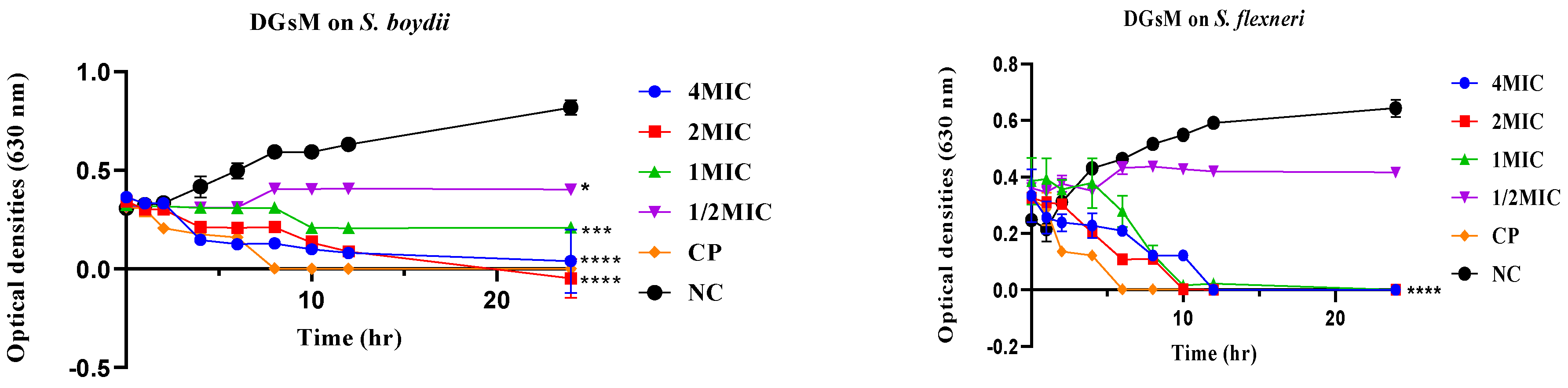
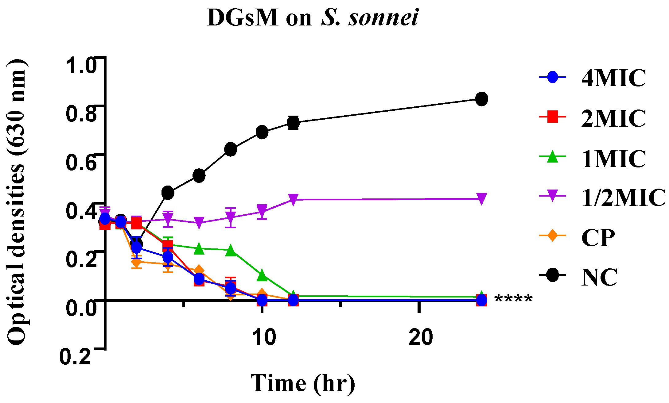
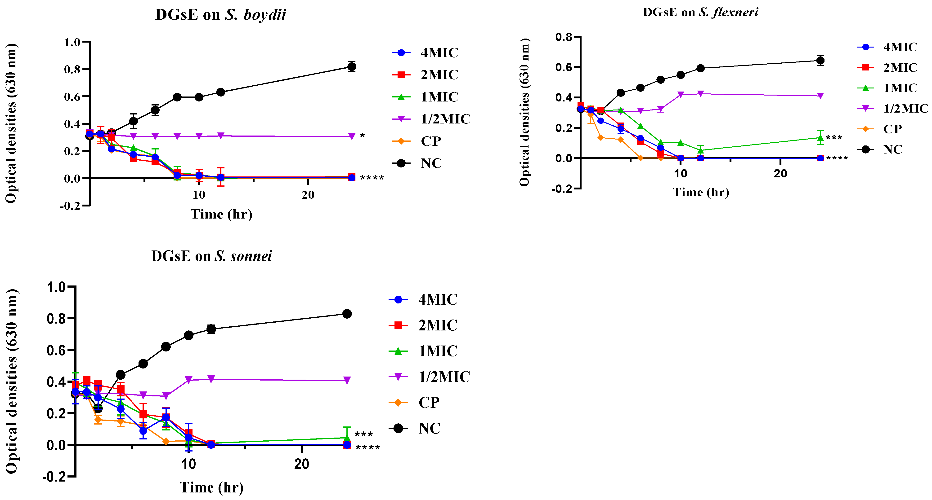
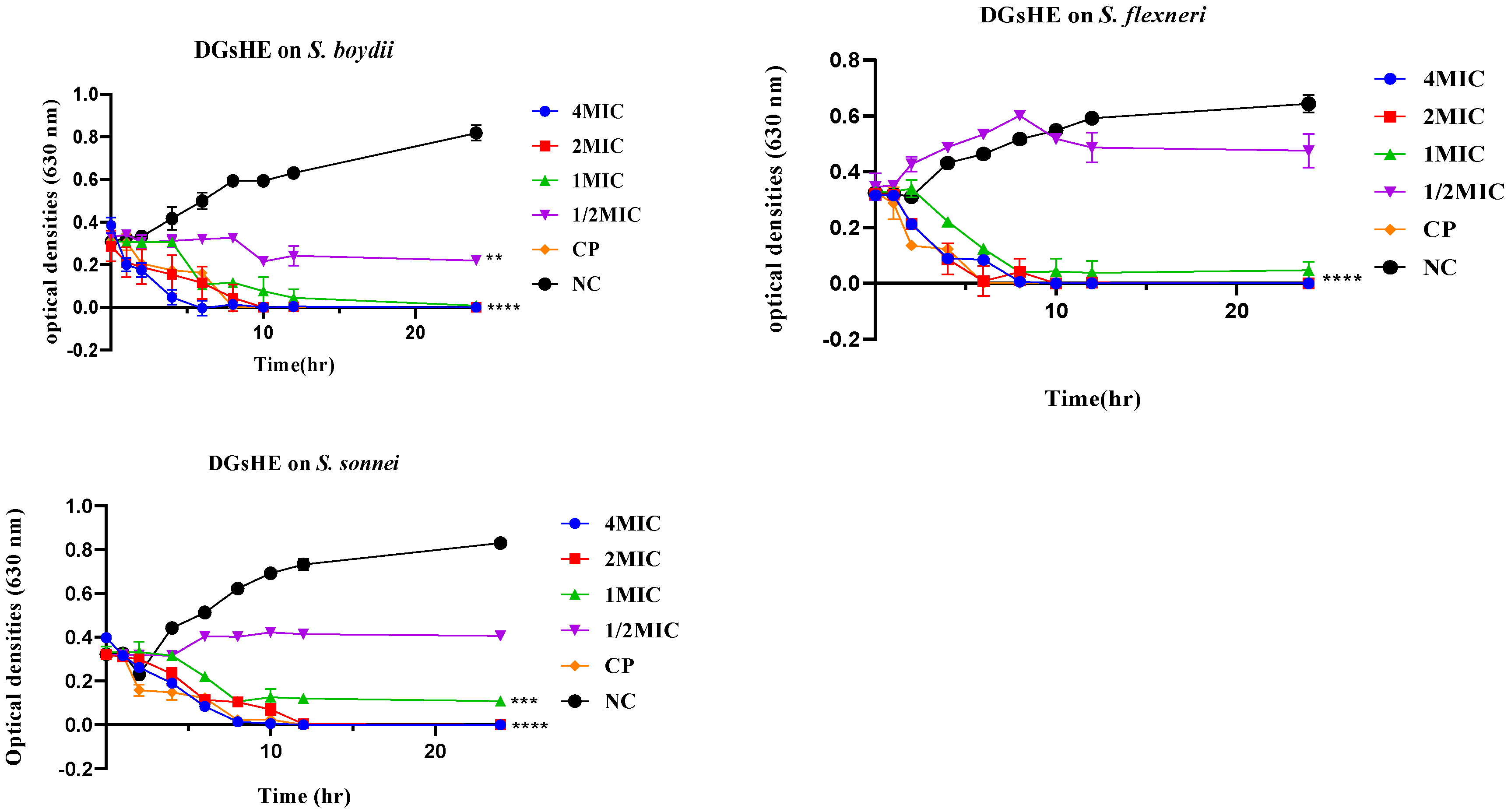
2.2.2. Time-Kill Kinetics

The time-kill kinetics of methanol, ethanol and hydroethanol extracts were evaluated on the most susceptible strains, viz. S. flexneri NR 518, S. boydii NR 521, and S. sonnei NR 519. Figure 1, Figure 2 and Figure 3 show the curves obtained by plotting optical densities (values that are proportional to the bacterial population) versus the time of incubation of bacteria with methanol, ethanol, and hydroethanol extracts, respectively. After two hours of bacterial incubation with the methanol extract, there was a decreasing trend in the curves, especially at MIC, 2 MIC, and 4 MIC, irrespective of the Shigella strains considered (Figure 1). After 10 h of incubation, at MIC, 2 MIC, and 4 MIC, almost all the curves overlapped with the X axis (except for S. boydii), suggesting that the bacterial population was cleared, irrespective of the strain considered. This bacterial clearance was later confirmed, as beyond 10 h of incubation (10–24 h), there was no resurgence of the curves, suggesting the bactericidal orientation of the methanol extract at MIC, 2 MIC, and 4 MIC. Treatment with ciprofloxacin showed the same trend, confirming the bactericidal trend of this drug vis-à-vis Shigella species. At a concentration less than the MIC (1/2 MIC), there was an increased inclination of the curve from 8 h of incubation time onwards, suggesting a bacteriostatic orientation. By contrast, the curve obtained from the untreated group of bacteria showed an increased trend from time 0 to 24 h, suggesting no inhibition and the exponential growth of bacteria.
Figure 2 shows the growth kinetics of Shigella species after incubation with the ethanol extract of Diospyros gilletii. Irrespective of the concentration of extract considered (MIC, 2 MIC and 4 MIC), there was a decreased trend in the curves, suggesting the inhibition of the bacterial population. After 10 h of incubation at MIC, 2 MIC, and 4 MIC, all the curves overlapped with the X axis, suggesting the clearance of the bacterial population, irrespective of the strain considered. This bacterial clearance was later confirmed, since beyond 10 h of incubation (10–24 h), the bacteria did not resume growth, suggesting the bactericidal orientation of the ethanol extract at MIC, 2 MIC, and 4 MIC.
Figure 3 reveals the growth kinetics of Shigella species after incubation with the hydroethanol extract of Diospyros gilletii. At MIC, 2 MIC, and 4 MIC, there was a decreased trend of the curves after 2 h of incubation time, suggesting the inhibition of the bacterial population. After 8 h of incubation, all the curves overlapped with the X axis, suggesting the clearance of the bacterial population. This bacterial clearance was later confirmed, since beyond 8 h of incubation (8–24 h), Shigella did not resume growth, suggesting the bactericidal inclination of the hydroethanol extract at MIC, 2 MIC, and 4 MIC.

2.2.3. Plausible Antibacterial Mechanisms of Action

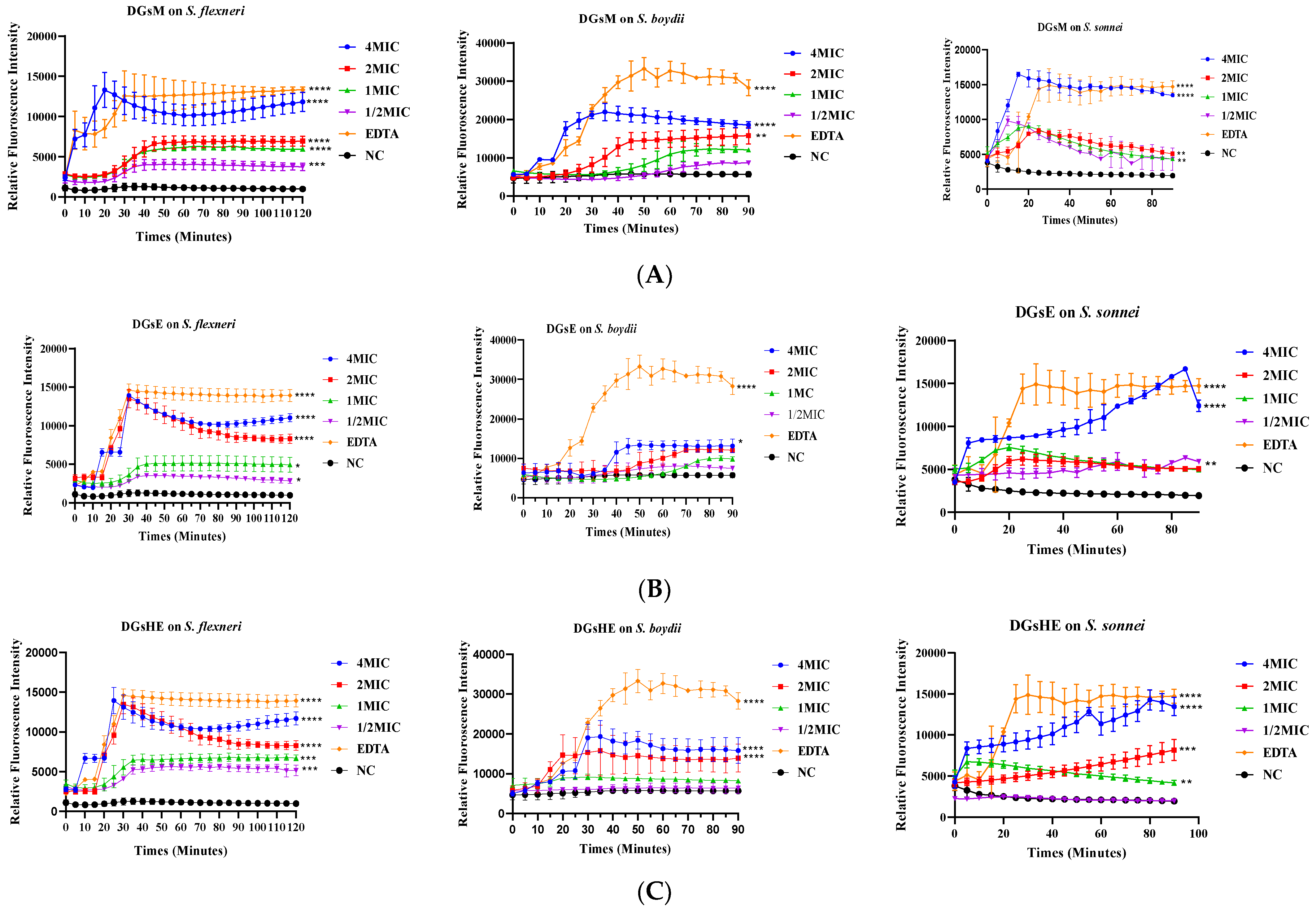
Effect of Extracts on N-phenyl-naphthylamine Uptake by Bacteria

The outer-membrane permeability of the susceptible bacterial species (S. boydii, S. flexneri, and S. sonnei) was evaluated by measuring their 1-N-phenyl-naphthylamine’s uptake upon treatment with different extracts of Diospyros gilletii stem bark. Figure 4A–C reveals the effect of various concentrations of extracts (1/2 MIC, MIC, 2 MIC, and 4 MIC) on the uptake of 1-N-phenyl-naphthylamine in S. boydii, S. flexneri, and S. sonnei. At 2 MIC and 4 MIC, there was a significant increase in the relative fluorescence intensity from t0 to t90 min, suggesting a higher intake of 1-N-phenyl-naphthylamine by bacteria at these concentrations. A similar trend was observed when the bacterial strains were treated with ethylene diamine tetraacetic acid (EDTA), a compound that was used as a positive control. By contrast, the curves, which were obtained from the group of bacteria that did not receive any treatment with extracts, remained unchanged throughout the experiment (from t0 to t90 min), inferring that the untreated bacteria did not uptake 1-N-phenyl-naphthylamine.

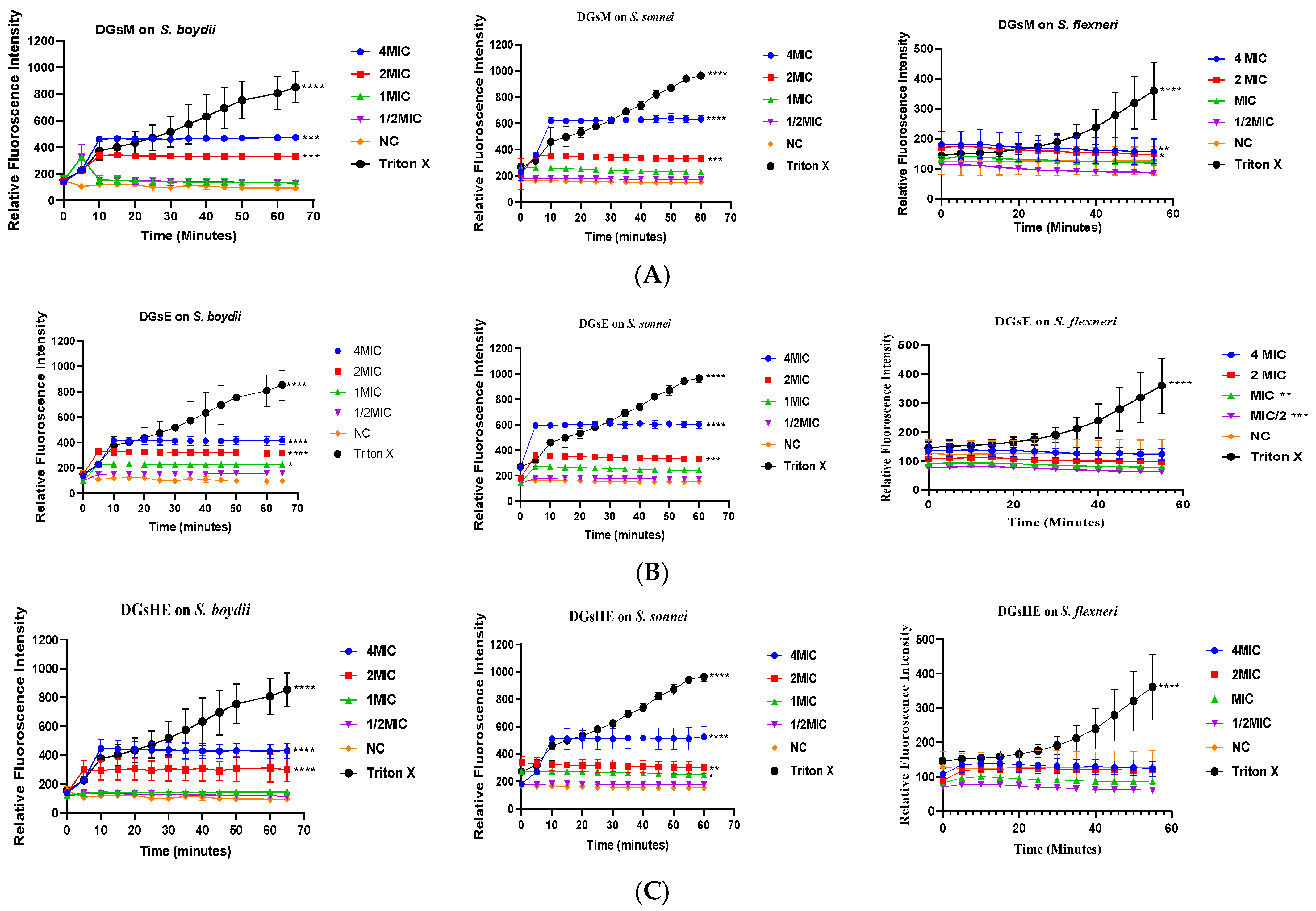
Propidium Iodide Uptake Assay

Following the graphs displayed in Figure 5A–C, it can be seen that the fluorescence intensity, which was measured as a function of time, increased continuously for all treatments with extracts at 2 MIC and 4 MIC, vis-à-vis S. sonnei and S. boydii, suggesting propidium iodide (PI) uptake by these bacteria. Moreover, the curves obtained from treatments against S. flexneri remained unchanged throughout the experiment (0–70 min). Overall, methanol, ethanol, and hydroethanol extracts triggered the rapid permeability of the bacterial membrane in S. sonnei and S. boydii, and propidium iodide fluorescence started increasing from 10 min at 4 MIC and 2 MIC; thereafter, it became constant, while at 1 MIC and ½ MIC, there was no increase in fluorescence in S. boydii and S. sonnei (Figure 5A–C). Normally, it is known that for extracts that work by propidium iodide uptake, the fluorescence intensity increases continuously. This shows that the antibacterial mode of action of the extracts is not achieved in S. flexneri through propidium iodide uptake. Nonetheless, this mode of action was verified in S. sonnei and S. boydii.

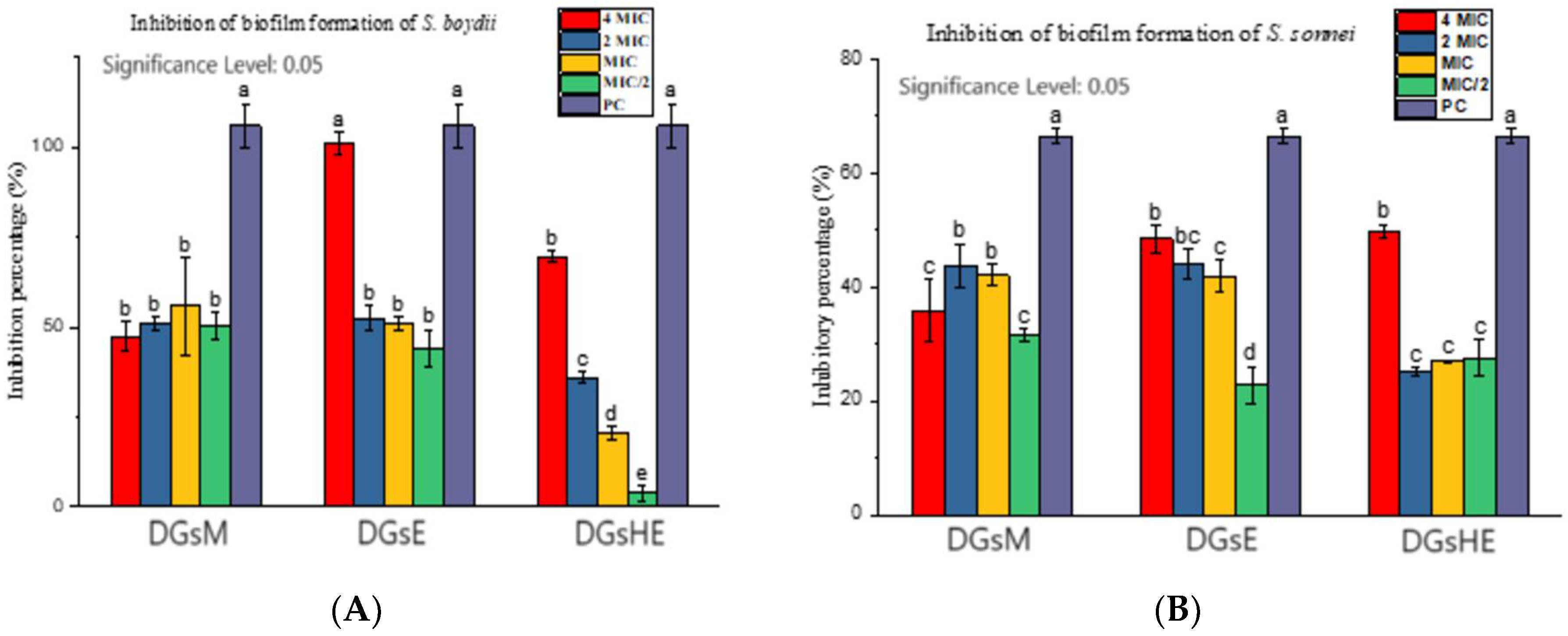
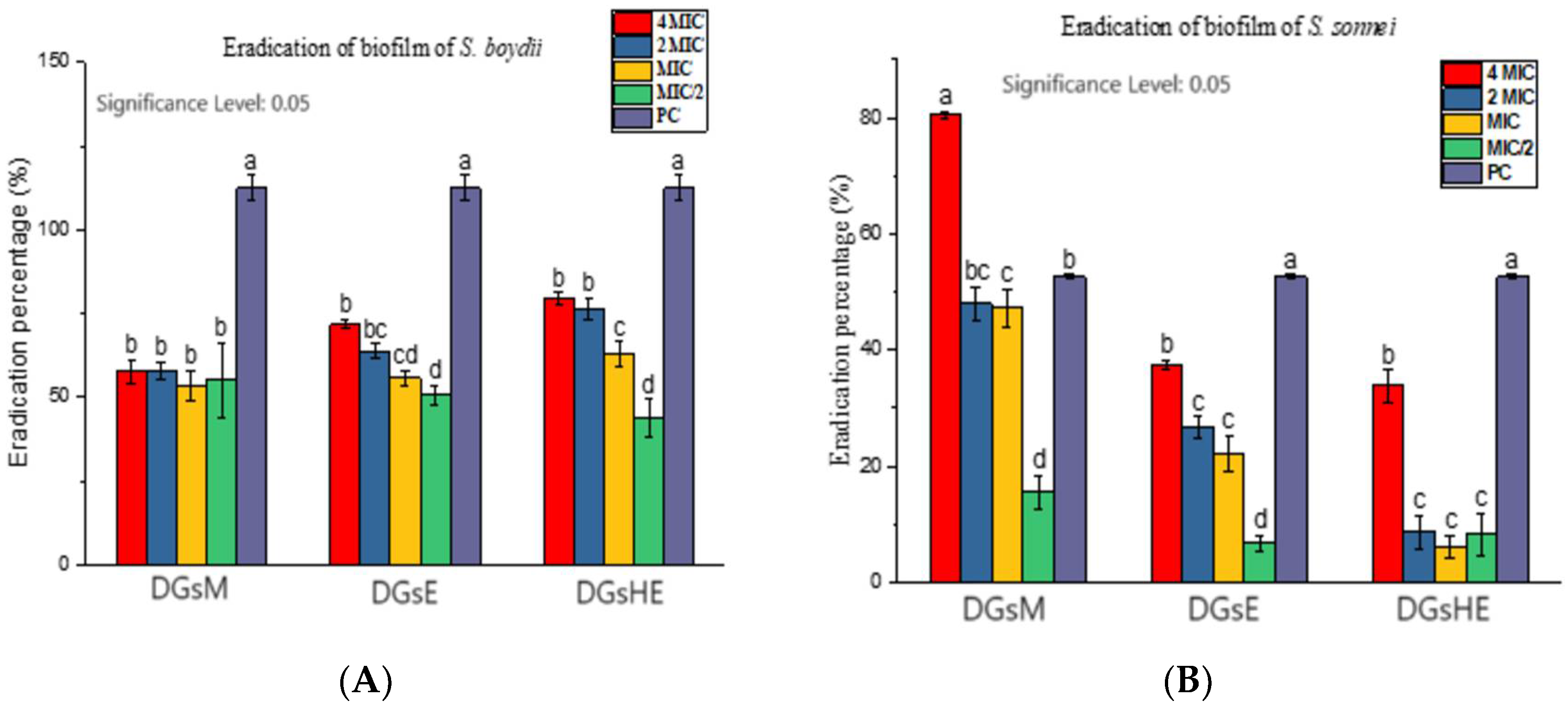
Inhibition and Eradication of Bacterial Biofilms

a.
Inhibition of biofilm formation
According to Figure 6, the biofilm formation [of S. boydii (A) and S. sonnei (B)] was significantly inhibited (p ≤ 0.05) by methanol, ethanol, and hydroethanol extracts of D. gilletii stem bark (at MIC, 2 MIC and 4 MIC) after 24 h of incubation. The highest percentages of the inhibition of biofilm formation were observed with 4 MIC. Moreover, ciprofloxacin, the positive control used, showed more than a 100% inhibition of biofilms.
b.
Eradication of biofilms
According to Figure 7, more than 50% of biofilms formed by S. boydii (within 24 h of incubation) (Figure 7A) were eradicated by methanol, ethanol, and hydroethanol extracts at MIC, 2 MIC, and 4 MIC after 24 h of incubation. Moreover, the biofilms formed by S. sonnei (Figure 7B) were also eradicated (<50%) by all the extracts (except for the methanol extract, which showed 80% inhibition at 4 MIC) at 4 MIC, 2 MIC, and MIC.

2.2.4. Cytotoxicity Studies

Table 2 summarizes the 50% cytotoxic concentration of methanol (45.24 and 52.00 µg/mL), ethanol (54.52 and 65.30 µg/mL), and hydroethanol (39.94 and 47.15 µg/mL) extracts against Vero and Raw cells, respectively, vs. dimethylsulfoxide (DMSO, a reference cytotoxic agent) (1.40 and 0.95 µg/mL). According to the National Cancer Institute classification, compounds with CC50 > 20 μg/mL are poorly cytotoxic or non-cytotoxic [31]. Compared to DMSO, which showed CC50 values less than 2 µg/mL, methanol, ethanol, and hydroethanol extracts can be considered non-cytotoxic extracts.

3. Discussion

In the present study, extracts from the stem bark of Diospyros gilletii were evaluated for antibacterial activity against four species of Shigella, viz. S. boydii, S. sonnei, S. flexneri and S. dysentariae. Moreover, the antibacterial mechanism of action of the most active extracts was investigated. Solvent extraction of the stem bark of Diospyros gilletii yielded methanol, ethanol, and hydroethanol extracts with yields of extraction of 5.6%, 7.61%, and 8.27%, respectively. The hydroethanol, composed of two solvents, i.e., ethanol and water, might have induced a leakage of more secondary metabolites from the plant, thereby increasing the yield of extraction (8.27%) [32]. The antibacterial test of the extracts revealed MIC values ranging from 125 to 1000 µg/mL, with the most active extracts being the methanol and hydroethanol with MICs of 125 µg/mL when tested in S. boydii. According to previous reports [33,34,35], the degree of antibacterial activity can be classified as very active (<100 µg/mL), active (100 < MIC < 512 µg/mL), moderately active (512 < MIC < 2048 µg/mL), and poorly active (MIC > 2048 µg/mL). Thus, the methanol extract was significantly active in S. flexneri (100 < MIC < 512 µg/mL), whereas ethanol and hydroethanol extracts were active (100 < MIC < 512 µg/mL) in all the bacterial species. All the extracts showed poor activity against S. dysenteriae. The observed antibacterial activity might be attributed to the presence of a number of secondary metabolites [pentacyclic tritepenes (corosolic acid and ursolic acid) and terpenoids (betulinic acid and lupeol), coumarins (norbergenin, 4-O-galloylnorbergenin), and the cholesterol derivative β-sitosterol] in Diospyros gilletii stem bark [14]. As already discussed, Diospyros gilletti contains bergenin and its derivatives, which are well known to possess antibacterial activity [14,17,36]. In a recently published review article by Salimo et al. [37], the antibacterial activity of bergenins was reported in 15 in vitro studies and 4 in vivo experiments [37]. Noteworthy, bergenin and its congeners participate in several mechanisms, such as lipid-peroxidation inhibitory activity and free-radical-scavenging activity [38], and initiate apoptosis and cell-cycle arrest in the G0/G1 phase [39]. Moreover, these compounds are able to easily cross the complex and multilayer lipopolysaccharide of bacteria cell walls, thereby causing lysis and cell death [40]. Thus, it is not unreasonable to speculate that these compounds might have contributed to the anti-shigellosis activity. The differences in the antibacterial action might be due to the diversity of the biochemical constituents and the genetic identity of bacteria, which potentially influenced the resistance in the tested bacteria [41]. Time-kill kinetic studies revealed that the bacterial inhibition was proportional to the concentration of the extracts. In fact, the extracts were found to be bactericidal at 2 MIC and 4 MIC when tested against S. boydii, S. sonnei, and S. flexneri. To exert their activity within the cell, almost all antibiotics have to cross the outer membrane of bacteria either by increasing membrane permeability or disruption. The measurement of the membrane permeability and disruption in bacteria might help in better understanding the mechanistic basis of the action [12]. The incubation of S. boydii, S. sonnei, and S. flexneri with various concentrations of extracts (4 MIC, 2 MIC, and MIC) promoted the uptake of N-phenyl-naphthylamine and propidium iodide by these bacteria, leading to an increase in the fluorescence [42]. The fluorescence of the NPN probe increases when this compound is incorporated into the hydrophobic core of the bacterial cell membrane, as compared with the fluorescence of a non-permeating bacteria cell. The incubation of bacteria with D. gilletti extracts induced a significant accumulation of NPN in the bacterial membrane, which was further inhibited by free divalent cations, such as Mg2+ and Ca2+. These cations, which normally bind to negatively charged phosphate groups between lipopolysaccharide (LPS) molecules via ionic bridges, are crucial for the outer-membrane integrity of Gram-negative bacteria. Consequently, the chelation of these divalent ions by NPN would definitely disrupt the outer membrane’s integrity and permeabilize the bacteria [43]. Propidium iodide, a dye that labels bacteria with a compromised inner membrane, binds to nucleic acids, increases its fluorescent signal, and constitutes a strong indicator of inner-membrane permeabilization. Methanol, ethanol, and hydroethanol extracts promoted the PI and NPN uptake in S. boydii, and S. sonnei, inferring that the test extracts permeabilized the inner and outer membranes of these bacteria [44,45]. Consequently, these extracts might have exerted antibacterial activity through bacterial membrane permeabilization. Extracts from Diospyros gilletii were further evaluated for antibiofilm formation using S. boydii, S. sonnei, and S. flexneri. Biofilm formation by bacteria is intricately involved in the mechanism of bacterial resistance as the biofilm matrix can slow antibiotic penetration through the membrane. To avoid the development of microbial biofilms, the inhibition of bacterial adhesion, the reduction of the initial bacterial growth, and the eradication of formed biofilms are noteworthy. Methanol, ethanol and hydroethanol extracts from Diospyros gilletii inhibited the formation of biofilm, with the percentage of inhibition ranging from 25% to 55% in S. sonnei and from 13% to 90% in S. boydii. The observed antibiofilm activity of the extracts might be attributed to the presence of a variety of secondary metabolites present in the plant [46]. Moreover, the extracts eradicated the biofilm formed by bacteria with percentages of inhibition varying from 8% to 80% in S. sonnei and from 48% to 78% in S. boydii. Antibiofilm activity has been attributed to the potential of active compounds to interfere with biofilm development, destabilize the biofilm, promote detachment, and sensitize biofilm cells [47]. In S. boydii, ethanol and hydroethanol extracts inhibited and eradicated the biofilms. All the extracts eradicated biofilms formed by S. sonnei. For ethanol and hydroethanol extracts, the MIC50 was found to be 4 MIC, whereas the MEC50 could not be observed in any of the extracts. To evaluate the cytotoxicity profile of the active extracts, their median cytotoxic concentrations (CC50) were determined vis-à-vis human mammalian cells, such as Vero and Raw cells. The CC50 values ranged from 39.94 ± 1.73 µg/mL to 54.525 ± 2.73 µg/mL on Vero cells and from 47.15 ± 10.38 µg/mL to 65.3 ± 5.52 µg/mL on Raw cells. As the CC50 values of all the tested extracts were found to be more than 30 µg/mL, it can be speculated that none of the extracts were toxic to the normal cells, according to the criteria of cytotoxicity defined by the American Nation Cancer Institute [48,49].
To the best of our knowledge, no report has demonstrated the antibacterial activity and mechanisms of action of methanol, ethanol, and hydroethanol extracts from D. gilletii stem bark. The anti-Shigella activity of D. gilletii extracts, which is demonstrated in this study, substantiates the ethnopharmacological use of D. gilletti in the treatment of infectious diseases, such as infectious diarrhoea. However, antibacterial-guided fractionation of D. gilletti extracts should be performed to identify the active anti-Shigella principles of this plant. In addition, in vivo anti-Shigella and pharmacokinetic studies, as well as knowledge of detailed mechanisms of action, are required for the successful utilization of D. gilletti in the treatment of infectious diarrhoea.

4. Materials and Methods

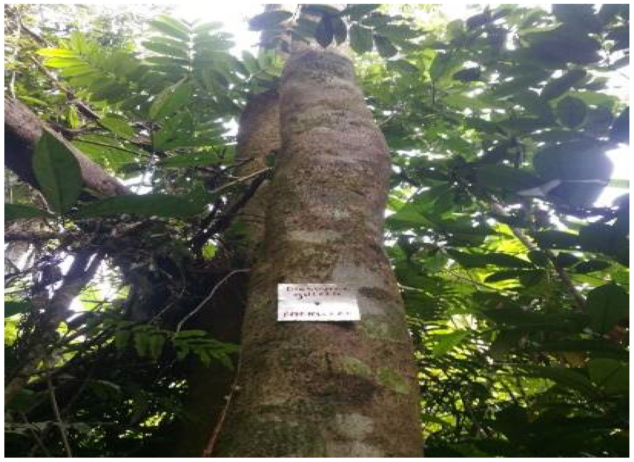
4.1. Plant Collection and Identification

The stem bark of Diospyros gilletii De Wild (Figure 8) was collected in Badounga in the West Region of Cameroon in May 2022 and identified at the National Herbarium of Cameroon, where a voucher specimen of the species was deposited under number No 15418 HNC.

4.2. Plant Extraction

The stem bark was then cut into small pieces, air-dried for two weeks, and ground to obtain a powder. The crude extracts were prepared by the maceration of plant powder in three solvents, including ethanol, hydroethanol (water/ethanol; 30/70, v/v), and methanol. In brief, 10 g of dried powder was macerated in pure ethanol, methanol, and hydroethanol (30/70; v/v) (100 mL) for 72 h at room temperature with daily shaking. The resulting solution was then filtered using hydrophilic cotton, and the filtrate was further concentrated using a Buchi rotary evaporator at 55 °C to yield the crude extracts. The extraction yield was calculated using the following formula:
E x t r a c t i o n   y i e l d   ( % ) = M a s s   o f   t h e   c r u d e   e x t r a c t   ( g ) M a s s   o f   t h e   p l a n t   m a t e r i a l   ( g ) × 100
The crude extracts were kept in a refrigerator until further use.

4.3. Preparation of Stock Solutions

The stock solutions of the crude extracts were prepared at 100 mg/mL by dissolving 100 mg of each crude extract in 1 mL of dimethlysulfoxide 100% in 1.5 mL Eppendorf tubes and storing at 4 °C. Ciprofloxacin, which was used as the reference antibiotic, was prepared by dissolving 1 mg of the drug in 1 mL of 0.5 N HCL to yield a concentration of 1 mg/mL.

4.4. Antibacterial Activity

a.
Bacterial strains
The in vitro antibacterial activity of extracts from Diospyros gilletii stem bark was evaluated in three reference bacterial strains (Shigella flexneri NR 518, Shigella boydii NR 521 and Shigella sonnei NR 519), which were obtained from the Biodefense and Emerging Infections Research Resources Repository (BEI Resources, Rockville, MD, USA, 20,852), and one clinical isolate, Shigella dysenteriae, which was a kind gift from the Centre Pasteur of Cameroon. These bacterial strains were maintained in continuous culture in tubes on the Mueller Hinton Agar slope (4 °C) at the Laboratory for Phytobiochemistry and Medicinal Plant Studies of the Faculty of Science of the University of Yaounde 1, Cameroon.
b.
Preparation of bacterial inoculum
The different bacterial inocula were prepared based on the 0.5 McFarland standard. Briefly, a colony from a 24 h culture on Müller Hinton agar (MHA) was removed using a platinum loop and introduced into a test tube containing 10 mL sterile physiological water (NaCl 0.9%); then, it was calibrated to a 0.5 McFarland standard using turbidity comparison to obtain 1.5 × 10⁸ UFC/mL.
c.
Determination of minimum inhibitory concentrations
The determination of the MIC was performed using the microdilution method in a liquid medium, as described by the Clinical Laboratory Standard Institute (CLSI), M07-A9 protocol, with slight modifications [50]. Briefly, 196 μL of Muller Hinton broth (MHB) was introduced into the first twelve wells of the microplate (CoStar, Washington, DC, USA), and 100 μL was placed in the rest of the wells of the microplate. Next, 4 μL of sterile solution containing different extracts concentrated at 100 mg/mL was introduced into the first twelve wells of column A, followed by five subsequent two-fold serial dilutions from line A to line F (concentration range: 250 μg/mL to 3.90625 μg/mL). Then, 100 μL of a bacterial suspension at 1 × 10⁶ CFU/mL was introduced into all the wells, except for that of the sterility control. The negative control was composed of the culture medium and inoculum only, while the positive control (concentration range: 2.5–0.15625 μg/mL) was made up of the medium, inoculum, and ciprofloxacin. Noteworthily, the final concentration of the inoculum in the wells was 5 × 10⁵ CFU/mL. The microplates were subsequently incubated at 37 °C for 24 h. At the end of the incubation period, 20 μL of a freshly prepared resazurin solution (0.15 mg/mL) was added to all the wells and further incubated for 30 min. The lowest concentration at which no color change from blue (resazurin) to pink (resorufin) was observed was considered as the minimum inhibitory concentration (MIC), corresponding to an absence of visible bacteria growth, and was expressed in μg/mL. The test was performed in triplicate and repeated twice. Subsequently, a time-kill kinetic assay was used to follow up the activity of plant extracts on bacterial growth (bactericidal or bacteriostatic activity) over time.

Time-Kill Kinetics

Time-kill kinetics were studied using a modified protocol described by [51]: notably, the use of opacimetry based on the turbidity of the cell suspensions as a function of the load rather than colonies counting on agar. In short, the assay was performed in sterile 96-well microplates at sub-inhibitory, inhibitory, and supra-inhibitory extract concentrations. Briefly, the extracts were diluted in MHB medium as previously described, in order to obtain ½ MIC, MIC, 2 MIC, and 4 MIC. Ciprofloxacin (2.5 µg/mL) was used as a positive control. Then, 100 μL inoculum at 1 × 10⁶ CFU/mL was introduced into each well to obtain a solution of 5 × 10⁵ CFU/mL, except for the wells used for sterility control. The plates were then covered and incubated at 37 °C for 24 h, during which the evolution of the bacterial load was followed by reading the optical densities at 630 nm of the preparations at different time intervals (0, 1, 2, 4, 6, 8, 10, 12, and 24 h) using a microplate reader (TECAN-Infinite M200, Tecan Austria GmbH, Salzburg, Austria). The assay was performed in triplicate and repeated twice. The bacterial growth curves were plotted using the optical densities against the incubation times. From these curves, we determined the minimum time at which the inhibitory effects started, the bactericidal and bacteriostatic effects of the extracts, and the re-emergence time of the bacterial strains.
Next, plausible mechanisms of the antibacterial action of the most active extracts were investigated using a membrane-permeabilization test, and we determined the inhibitory/eradicative effect on bacterial biofilm formation.

4.5. Evaluation of Possible Mechanisms of Action

4.5.1. Membrane Permeabilization

The membrane permeabilization of the most promising extracts was evaluated using NPN (1-N- phenyl-naphthylamine) and propidium iodide (PI) uptake assays.
a.
1-N-phenyl-naphthylamine uptake assay
The outer membrane of Gram-negative bacteria provides a formidable barrier (an asymmetric bilayer that contains mainly lipopolysaccharide (LPS) in the outer leaflet and phospholipids in the inner leaflet) that should be crossed by antimicrobial agents to exert their action. NPN (1-N-phenyl-naphthylamine) is a neutral, hydrophobic fluorescent probe that cannot permeate the intact outer membrane of the bacteria. When the integrity of the bacteria outer membrane is destroyed, it will diffuse into the phospholipid bilayer and exhibit low-fluorescence quantum yield in an aqueous environment, and it becomes strongly fluorescent in a glycerophospholipid or hydrophobic environment [44]. Briefly, a 1-N-phenyl-naphthylamine (NPN) assay was performed to compare the effects of the plant extract on the permeabilization of the outer membranes using the modified protocol of [43,44]. Briefly, in sterile, 96-well, black flat-bottomed microplates, bacterial cells were grown overnight, washed with normal saline (0.9% NaCl), and centrifuged at 10,000 tr for 10 min in Eppendorfs tubes. The pellet was resuspended in normal saline and then adjusted to a 0.5 McFarland standard in tubes containing glucose with phosphate-buffered saline (50 mg of PBS in 50 mL of glucose). Next, 160 μL of cells was added to 20 μL NPN and 20 μL of extracts at various concentrations (4 MIC, 2 MIC, MIC, and ½ MIC). The positive control consisted of ethylene diamine tetraacetate (EDTA) and NPN with bacteria suspension, whereas NPN was used as a negative control. Sterility control was composed of NPN with normal saline. The microplate was kept for 1 h 30 min in the dark, and then NPN fluorescence was read at 5 min intervals using a TECAN infinite M200 microplate reader at 350 nm and 420 nm excitation and emission wavelengths, respectively. The assay was performed in triplicate and repeated twice.
The percentage of NPN uptake was calculated using the following equation:
%   N P N   u p t a k e = ( F o b s F 0 ) ( F 100 F 0 ) × 100
where
Fobs = observed fluorescence at a given concentration of plant extract.
F0 = initial fluorescence of NPN with bacterial cells in the absence of plant extract
F100 = fluorescence of NPN with bacteria cells upon addition of 2 μL of EDTA.
b.
Propidium iodide uptake assay
The effect of antibacterial crude extracts on the bacterial membrane integrity was assayed using a propidium iodide test. Propidium iodide (PI) is membrane-impermeable for intact cells and can only enter cells when the membrane permeability is augmented. Once in the cells, this fluorescent intercalating agent binds to nucleic acids (DNA, RNA) of necrotic cells, thereby discriminating between apoptotic and necrotic cells [45]. In this study, the PI uptake test was carried out according to a previously described protocol by Kwon et al. [44]. Briefly, bacterial cells were grown overnight in sterile, 96-well, black flat-bottomed microplates, washed with normal saline (0.9% NaCl), and then centrifuged at 10,000 tr/10 min in 1.5 mL Eppendorf tubes. The pellet was resuspended in normal saline and then adjusted to 106 cells/mL from a 0.5 McFarland standard, for a final volume of 64,000 μL in tubes containing 50 mg of PBS dissolved in 50 mL of glucose. Next, 20 μL of PI solution (1.3 mg of PI dissolved in 20 mL of PBS) was added to 160 μL of the bacterial suspension in a sterile, 96-well, black flat-bottomed microplate and incubated in the dark for 30 min. Afterward, 20 μL of various concentrations of plant extracts (4 MIC, 2 MIC, MIC, and ½ MIC) were added, while triton X was used as the positive control. The negative control was composed of propidium iodide and the bacterial suspension, while propidium iodide plus PBS was used as a sterility control. Then, the microplate was incubated for 1 h without light exposure, and the optical densities were read using a TECAN infinite M200 microplate reader at 544 nm and 620 nm as excitation and emission wavelengths, respectively. The assay was performed in triplicate and repeated twice.

4.5.2. Antibiofilm Activity

Biofilms are structured microbial cells that occur as suspended aggregates attached to an inert or living surface to cause chronic infections [52]. Bacterial biofilms allow bacteria to resist and survive environmental pressure and enhance their resistance to antimicrobial agents [52].
a.
Quantification of biofilm
Biofilm biomass measurement was performed by crystal violet staining according to a previously described protocol [53]. In brief, a single colony, which was isolated from an overnight bacteria culture on MHA, was suspended in normal saline and adjusted to 1 × 10⁶ CFU/mL in MHB supplemented with 2% glucose and 0.4% bovine bile. Next, 200 μL aliquot of the bacterial suspension was dispensed into a 96-well, U-shaped sterile microplate, whereas the negative-control wells were filled with 200 μL MHB. The plates were then incubated for 24 h at 37 °C. At the end of the incubation period, the microplate was gently emptied, and 200 μL of pure ethanol was added to each well and incubated for 15 min in order to fix the cells in the wells. Next, the wells were emptied and washed twice with sterile distilled water, followed by the addition of 200 μL crystal violet 0.1% to stain the biofilm formed. The plates were further incubated at 37 °C for 30 min, the remaining (residual) crystal violet was removed, and the wells were washed three times using sterile distilled water. Finally, 200 μL of a 33% acetic acid solution was introduced into the wells, and the absorbance was read at 590 nm using a TECAN (infinite M200) (Austria GmbH, Salzburg, Austria) plate reader.
b.
Determination of the minimum inhibitory concentration (MIC50) and minimum eradicative concentration (MEC50) of biofilms
MIC50 and MEC50 were determined according to the microdilution method in a liquid medium. This assay was performed in triplicate in sterile, 96-well microplates at sub-inhibitory, inhibitory, and supra-inhibitory extract concentrations. Briefly, the extracts were diluted in MHB medium supplemented with 2% glucose and 0.4% bovine bile salt, as previously described, to obtain 4 MIC, 2 MIC, MIC, and ½ MIC. Next, a two-fold serial dilution of order 2 was carried out. Afterward, 100 μL of inoculum was added to all the wells, which were then incubated for 24 h at 37 °C. The final bacterial load in each well was 1 × 10⁶ CFU/mL. Ciprofloxacin (50 µg/mL) was used as a positive control. After incubation, non-adherent or planktonic cells were discarded, and 200 μL of ethanol was added to each well in order to fix the cells in the wells, and the microplates were incubated for an additional 15 min. Then, the plates were gently washed (two times) with distilled water and dried at room temperature. Next, each well was stained by adding 200 μL of 0.1% crystal violet and further incubated for 30 min, after which the stain was discarded and washed with distilled water so as to remove the excess stain. Finally, 200 μL of a 33% glacial acetic acid was introduced into the different wells, and the absorbance was read at 590 nm using a TECAN (infinite M200) plate reader. The experiment was performed in triplicate and repeated thrice. The MEC50 was determined under the same conditions, with the only difference being that the biofilms were first formed in the culture medium (MHB medium supplemented with 2% glucose and 0.4% bovine bile salt) for 24 h at 25 °C, based on the strain. Next, the test samples were prepared as described above and introduced into the corresponding wells of the microplates, already containing the formed biofilms. The microplates were then re-incubated under the same conditions and treated as elucidated above. Finally, 200 μL of a 33% glacial acetic acid was introduced into the different wells, and the absorbance was read at 590 nm using a TECAN (infinite M200) plate reader. The experiment was performed in triplicate and repeated thrice. The optical densities, which were obtained from the spectrophotometer, were used to calculate the percentages of inhibition/eradication of biofilms using the following formula:
%   i n h i b i t i o n / e r a d i c a t i o n = O D   n e g a t i v e   c o n t r o l O D   t e s t O D   n e g a t i v e   c o n t r o l × 100
where OD is the optical density.
MIC50 = minimal concentration that inhibited 50% biofilm formation, MEC50 = minimal concentration that eradicated 50% of the biofilms formed.
Cytotoxicity studies of the most active extracts were carried out on Vero and Raw cells to evaluate their toxicity and selectivity.

4.6. Cytotoxicity Test

The extracts that inhibited the growth of the bacterial strains were evaluated against two selected human mammalian cells using a colorimetric method based on the reduction of resazurin to resorufin [54].

4.6.1. The Human Mammalian Cells

The in vitro cytotoxicity of the active extracts was evaluated on the mammalian African Green monkey Vero ATCC CRL 1586 and murine macrophage Raw cells 264.7 cell lines, provided by the Centre Pasteur of Cameroon and the Noguchi Memorial Institute for Medical Research, University of Ghana, respectively. The cells were maintained in a continuous culture in the laboratory and later used for the cytotoxicity test.

4.6.2. In Vitro Culture of the Vero and Raw Cell Lines

The Vero and Raw cells were maintained in complete DMEM (Dulbecco’s Modified Eagle medium) (1% antibiotic and 10% FBS) and stored in a 25 cm3 cultured flask (T-Flask) (CosStar, Washington, DC, USA) under standard conditions of 5% CO2 at 37 °C. The culture medium was renewed every 72 h. Once cell confluence (70–90%) was reached, the cells were detached by treatment with 1 mL of trypsin-EDTA 0.05% for 5 min. After trypsin inactivation by adding 9 mL of complete DMEM, the detached cells were centrifuged at 1800 rpm for 5 min. The obtained pellet was then suspended in 1 mL of the medium, and the cell viability was evaluated using Trypan blue to calibrate the cell load on the Neubauer hemocytometer.

4.6.3. Determination of Median Cytotoxic Concentrations (CC50)

As already discussed, the cytotoxicity of the most promising extracts was assessed according to the method described by Bowling et al. [54]. The assay was performed in duplicate in sterile 96-well microplates (CoStar, Washington, DC, USA). Briefly, 100 μL of cells’ suspension containing 1 × 10⁴ cells/well was introduced into all the wells of the plate and incubated for cell adhesion at 37 °C, 5% CO2 for 18 h. Next, the cell culture medium was replaced with 90 μL of fresh medium, followed by the addition of 10 μL of test samples at different concentrations (final concentrations of the extracts: 2000, 1000, 500, 250, 125, and 62.5 μg/mL), in all wells except for those that served as the positive (DMSO, 20 μM) and negative (culture medium without test sample) controls. The plates were then incubated at 37 °C, 5% CO2 for 48 h, after which 10 μL of a freshly prepared resazurin solution (0.15 mg/mL in PBS) was introduced into each well and incubated for 4 h at 37 °C under 5% CO2. Next, the fluorescence of the obtained preparation was measured using a microtiter plate reader (TECAN-Infinite M200, Tecan Austria GmbH, Salzburg, Austria) at excitation and emission wavelengths of 530 and 590 nm, respectively. From the resulting values of optical densities, the percentage of cell viability was calculated with Microsoft Excel software (version 2013, Washington, DC, USA) using the following equation:
% C e l l   i n h i b i t i o n = ( A c A t ) ( A c ) × 100
where At = Absorbance of test samples, Ac = Absorbance of negative control (cells without test samples).
A dose–response curve (cell viability versus concentration of the samples) was plotted using GraphPad Prism software to determine the median cytotoxic concentration (CC50).

4.7. Statistical Analysis

The values were represented as the mean ± standard deviation. Statistical analysis was performed using one-way analysis of variance (ANOVA) using GraphPad Prism 8.0.1 software. The histograms were obtained using OriginPro Software (8.5.1). The percentages were calculated using Excel 2013 software. The differences between the means were compared using Dunnett’s test with a 95% confidence level (p ≤ 0.05).

5. Conclusions and Perspectives

The present study aimed to evaluate the antibacterial activity and mechanisms of action of extracts from D. gilletti. Upon in vitro antibacterial testing, extracts from D. gilletti afforded MIC values ranging from 125 to 1000 µg/mL, with the methanol extract being the most active as it had the lowest MIC values (125 µg/mL). Mechanistic studies of potent antibacterial extracts from D. gilletti revealed bactericidal orientation at 4 MIC and bacteriostatic tendency at 2 MIC. Furthermore, the bioactive extracts contributed to the destabilization of the inner and outer membranes of the Shigella species through 1-N-phenyl-naphthylamine and propidium iodide uptake by these membranes. In addition, D. gilletti extracts inhibited and eradicated biofilms formed by the shigellosis-causing microbes. D. gilletti extracts were also found to be non-cytotoxic on Vero and Raw cells. These results validate the ethnomedicinal uses of D. gilletti in the traditional treatment of infectious diseases such as diarrhoea. However, antibacterial-guided fractionation of D. gilletti extracts should be performed to identify the active principles of this plant. Moreover, in vivo anti-shigellosis activity, pharmacokinetics, and in-depth mechanistic studies should be investigated for the successful utilization of D. gilletti in the treatment of diarrhoea caused by Shigella species.

Supplementary Materials

The following supporting information can be downloaded at: https://www.mdpi.com/article/10.3390/ddc3010015/s1, Figure S1: Coumarins isolated thus far from Diospyros gilletii, and Figure S2: Pentacyclic triterpenes (810) and triterpenoids (1115) isolated thus far from Diospyros gilletii.

Author Contributions

Conceptualization, P.K.L., B.P.K. and F.F.B.; methodology, A.C.N.T., E.A.M.K. and B.-L.N.K.; software, E.A.M.K., B.-N.N.-D. and V.N.; validation, P.K.L., F.F.B. and B.P.K.; formal analysis, B.-L.N.K., L.M.N.N., Y.K.D.M., B.-N.N.-D. and B.R.P.; investigation, A.C.N.T., B.-L.N.K., E.A.M.K., B.-N.N.-D. and V.N.; resources, P.K.L., F.F.B. and B.P.K.; data curation, A.C.N.T., B.-L.N.K., E.A.M.K., B.-N.N.-D., V.N., L.M.N.N., Y.K.D.M. and B.R.P.; writing—original draft preparation, A.C.N.T., E.A.M.K., B.-L.N.K., L.M.N.N., Y.K.D.M. and B.R.P.; writing—review and editing, P.K.L., B.P.K. and V.N.; visualization, P.K.L., B.P.K., V.N. and F.F.B.; supervision, P.K.L. and F.F.B.; project administration, P.K.L. and F.F.B.; funding acquisition, P.K.L., B.P.K. and F.F.B. All authors have read and agreed to the published version of the manuscript.

Funding

This research received external funding from the Yaounde–Bielefeld Bilateral Graduate School for Natural Products with Anti-parasite and Antibacterial Activity (YaBiNaPA) (grant number 57316173) and Seeding Labs’ Instrumental Access.

Institutional Review Board Statement

Not applicable.

Informed Consent Statement

Not applicable.

Data Availability Statement

Data are available from the corresponding author(s) upon reasonable request. The data are not publicly available due to their sensitive nature.

Acknowledgments

Authors are thankful to the Cameroon National Herbarium (Yaounde, Cameroon) for their assistance with plant identification. The authors also acknowledge the “Centre Hospitalier Universitaire” of Yaounde–Cameroon and “Centre Pasteur” of Cameroon and for providing the bacterial strains species and the mammalian African Green monkey Vero ATCC CRL 1586. Thanks to the Noguchi Memorial Institute for Medical Research, University of Ghana, for providing the murine macrophage Raw cells 264.7 cell lines. This work was supported by and also received material and equipment support from the Yaoundé–Bielefeld Bilateral Graduate School for Natural Products with Anti-parasite and Antibacterial Activity (YaBiNaPA) and Seeding Labs’ Instrumental Access.

Conflicts of Interest

The authors declare no conflicts of interest.

References

  1. Al-Dahmoshi, H.; Al-Khafaji, N.; Al-Allak, M.; Salman, W.; Alabbasi, A. A review on shigellosis: Pathogenesis and antibiotic resistance. Drug Invent. Today 2020, 15, 793–798. [Google Scholar]
  2. Puzari, M.; Sharma, M.; Chetia, P. Emergence of antibiotic resistant Shigella species: A matter of concern. J. Infect. Public Health 2018, 11, 451–454. [Google Scholar] [CrossRef] [PubMed]
  3. Merzon, E.; Gutbir, Y.; Vinker, S.; Golan Cohen, A.; Horwitz, D.; Ashkenazi, S.; Sadaka, Y. Early childhood shigellosis and attention deficit hyperactivity disorder: A population-based cohort study with a prolonged follow-up. J. Atten. Disord. 2021, 25, 1791–1800. [Google Scholar] [CrossRef] [PubMed]
  4. The World Health Organization (WHO). Diarrhoeal Disease. The Key Facts. 2023. Available online: https://www.who.int/news-room/fact-sheets/detail/diarrhoeal-disease (accessed on 23 December 2023).
  5. Reiner, R.C., Jr.; Graetz, N.; Casey, D.C.; Troeger, C.; Garcia, G.M.; Mosser, J.F.; Deshpande, A.; Swartz, S.J.; Ray, S.E.; Blacker, B.F.; et al. Variation in childhood diarrheal morbidity and mortality in Africa, 2000–2015. N. Engl. J. Med. 2018, 379, 1128–1138. [Google Scholar] [CrossRef] [PubMed]
  6. Njunda, A.L.; Assob, J.C.; Nsagha, D.S.; Kamga, H.L.; Awafong, M.P.; Weledji, E.P. Epidemiological, clinical features and susceptibility pattern of shigellosis in the buea health district, Cameroon. BMC Res. Notes 2012, 5, 54. [Google Scholar] [CrossRef] [PubMed]
  7. Ateudjieu, J.; Bita’a, L.; Guenou, E.; Chebe, A.; Chukuwchindun, B.; Goura, A.; Bisseck, A. Profile and antibiotic susceptibility pattern of bacterial pathogens associated with diarrheas in patients presenting at the Kousseri Regional Hospital Anne, Far North, Cameroon. Pan Afr. Med. J. 2018, 29, 170. [Google Scholar] [PubMed]
  8. Aslam, A.; Okafor, C.N. Shigella. In StatPearls; StatPearls Publishing: Treasure Island, FL, USA, 2022. [Google Scholar]
  9. Sheikh, A.F.; Moosavian, M.; Abdi, M.; Heidary, M.; Shahi, F.; Jomehzadeh, N.; Seyed-Mohammadi, S.; Saki, M.; Khoshnood, S. Prevalence and antimicrobial resistance of Shigella species isolated from diarrheal patients in Ahvaz, southwest Iran. Infect. Drug Resist. 2019, 12, 249–253. [Google Scholar] [CrossRef] [PubMed]
  10. Libby, T.E.; Delawalla, M.L.M.; Al-Shimari, F.; MacLennan, C.A.; Vannice, K.S.; Pavlinac, P.B. Consequences of Shigella infection in young children: A systematic review. Int. J. Infect. Dis. 2023, 129, 78–95. [Google Scholar] [CrossRef] [PubMed]
  11. The World Health Organization (WHO), 2021: Global Priority Pathogens. Google Scholar [WWW Document]. Available online: https://scholar.google.com/scholar_lookup?author=World%20Health%20Organization&publication_year=2017&journal=WHO+global+priority+pathogens+list+of+antibiotic-resistant+bacteria (accessed on 22 May 2023).
  12. Ranjbar, R.; Farahani, A. Shigella: Antibiotic-resistance mechanisms and new horizons for treatment. Infect. Drug Resist. 2019, 12, 3137–3167. [Google Scholar] [CrossRef]
  13. Cowan, M.M. Plant products as antimicrobial agents. Clin. Microbiol. Rev. 1999, 12, 564–582. [Google Scholar] [CrossRef]
  14. Tameye, N.S.J.; Akak, C.M.; Happi, G.M.; Frese, M.; Stammler, H.-G.; Neumann, B.; Lenta, B.N.; Sewald, N.; Nkengfack, A.E. Antioxidant norbergenin derivatives from the leaves of Diospyros gilletii De Wild (Ebenaceae). Phytochem. Lett. 2020, 36, 63–67. [Google Scholar] [CrossRef]
  15. Akak, C.M.; Djama, C.M.; Nkengfack, A.E.; Tu, P.-F.; Lei, L.-D. New coumarin glycosides from the leaves of Diospyros crassiflora (Hiern). Fitoterapia 2010, 81, 873–877. [Google Scholar] [CrossRef]
  16. Akak, C.M.; Nkengfack, A.E.; Tu, P.F. Norbergenin derivatives from Diospyros crassiflora (Ebenaceae). Nat. Prod. Commun. 2013, 8, 1575–1578. [Google Scholar] [CrossRef]
  17. Tameye, J.N.S. Phytochemical Studies of Diospyros gilletii De Wild and Diospyros fragrans Gürke (Ebenaceae), Chemical Transformations and Antibacterial, Antioxidant and Cytotoxic Activities of Extracts and Isolated Compounds. Ph.D. Thesis, Department of Chemistry, Faculty of Science, University of Yaounde 1, Yaoundé, Cameroon, 2022; 289p. Available online: https://savoirs.cames.online/jspui/handle/20.500.12177/11202 (accessed on 14 February 2024).
  18. Tameye, N.S.J.; Akak, C.M.; Tabekoueng, G.B.; Mkounga, P.; Bitchagno, G.T.M.; Lenta, B.N.; Sewald, N.; Nkengfack, A.E. Chemical constituents from Diospyros fragrans Gürke (Ebenaceae). Biochem. Syst. Ecol. 2022, 100, 104373. [Google Scholar] [CrossRef]
  19. Mallavadhani, U.V.; Panda, A.K.; Rao, Y.R. Pharmacology and chemotaxonomy of Diospyros. Phytochemistry 1998, 49, 901–951. [Google Scholar] [CrossRef]
  20. Maridass, M. Phytochemicals from genus Diospyros (L.) and their biological activities. Ethnobot. Leafl. 2008, 12, 231–244. [Google Scholar]
  21. Ribeiro, A.; Serrano, R.; da Silva, I.B.M.; Gomes, E.T.; Pinto, J.F.; Silva, O. The genus Diospyros: A review of novel insights into the biological activity and species of Mozambican flora. Plants 2023, 12, 2833. [Google Scholar] [CrossRef]
  22. Ragasa, C.; Puno, M.; Sengson, J.; Shen, C.; Rideout, J.; Raga, D. Bioactive triterpenes from Diospyros blancoi. Nat. Prod. Res. 2009, 23, 1252–1258. [Google Scholar] [CrossRef]
  23. Rauf, A.; Uddin, G.; Patel, S.; Khan, A.; Halim, S.; Bawazeer, S.; Ahmad, K.; Muhammad, N.; Mubarak, M. Diospyros, an under-utilized, multi-purpose plant genus: A review. Biomed. Pharmacother. 2017, 91, 714–730. [Google Scholar] [CrossRef]
  24. Mmongoyo, J.A.; Nair, M.G.; Linz, J.E.; Wu, F.; Mugula, J.K.; Dissanayake, A.A.; Zhang, C.; Day, D.M.; Wee, J.M.; Strasburg, G.M. Bioactive compounds in Diospyros mafiensis roots inhibit growth, sporulation and aflatoxin production by Aspergillus flavus and Aspergillus parasiticus. World Mycotoxin. J. 2017, 10, 237–248. [Google Scholar] [CrossRef]
  25. Kaikabo, A.A.; Suleiman, M.M.; Samuel, B.B.; Eloff, J.N. Antibacterial activity of eleven South African plants use in treatment of diarrhoea in folkloric medicine. Afr. J. Tradit. Complement. Altern. Med. 2009, 6, 315–316. [Google Scholar]
  26. Kadavul, K.; Dixit, A.K. Ethnomedicinal studies of the woody species of Kalrayan & Shervarayan hills, Eastern Chats, Tamil Nadu. Indian J. Tradit. Knowl. 2009, 8, 592–597. [Google Scholar]
  27. Adzu, B.; Amos, S.; Muazzam, I.; Inyang, U.S.; Gamaniel, K.S. Neuropharmacological screening of Diospyros mespiliformis in mice. J. Ethnopharmacol. 2002, 83, 139–143. [Google Scholar] [CrossRef]
  28. Adzu, B.; Amos, S.; Dzarma, S.; Muazzam, I.; Gamaniel, K.S. Pharmacological evidence favouring the folkloric use of Diospyros mespiliformis Hochst in the relief of pain and fever. J. Ethnopharmacol. 2002, 82, 191–195. [Google Scholar] [CrossRef]
  29. Belemtougri, R.G.; Constantin, B.; Cognard, C.; Raymond, G.; Sawadogo, L. Effects of two medicinal plants Psidium guajava L. (Myrtaceae) and Diospyros mespiliformis L. (Ebenaceae) leaf extracts on rat skeletal muscle cells in primary culture. J. Zhejiang Univ. Sci. B 2006, 7, 56–63. [Google Scholar] [CrossRef]
  30. Brisson, R. Mythologie des Pygmées Baka (Sud-Cameroun): Mythologie et Contes; Peeters Publishers: Leuven, Belgium, 1999; 416p. [Google Scholar]
  31. Anywar, G.U.; Kakudidi, E.; Oryem-Origa, H.; Schubert, A.; Jassoy, C. Cytotoxicity of medicinal plant species used by traditional healers in treating people suffering from HIV/AIDS in Uganda. Front. Toxicol. 2022, 4, 832780. [Google Scholar] [CrossRef]
  32. Nur Syukriah, A.R.; Liza, M.S.; Harisun, Y.; Fadzillah, A.A.M. Effect of solvent extraction on antioxidant and antibacterial activities from Quercus infectoria (Manjakani). Int. Food Res. J. 2014, 21, 1067–1073. [Google Scholar]
  33. Tamokou, J.D.D.; Mbaveng, A.; Kuete, V. Antimicrobial activities of African medicinal spices and vegetables. In Medicinal Spices and Vegetables from Africa: Therapeutic Potential Against Metabolic, Inflammatory, Infectious and Systemic Diseases; Academic Press: Cambridge, MA, USA, 2017; pp. 207–237. [Google Scholar] [CrossRef]
  34. Franklin Loic, T.T.; Boniface, P.K.; Vincent, N.; Zuriatou, Y.T.; Victorine Lorette, Y.; Julius, N.N.; Paul, K.L.; Fabrice, F.B. Biological synthesis and characterization of silver-doped nanocomposites: Antibacterial and mechanistic studies. Drugs Drug Candidates 2024, 3, 13–32. [Google Scholar]
  35. Brice Rostan, P.; Boniface, P.K.; Eutrophe Le Doux, K.; Vincent, N.; Yanick Kevin, M.D.; Paul, K.L.; Fabrice, F.B. Extracts from Cardiospermum grandiflorum and Blighia welwitschii (Sapindaceae) reveal antibacterial activity against Shigella species. S. Afr. J. Bot. 2024, 164, 419–428. [Google Scholar]
  36. Liu, B.; Wang, M.; Wang, X. Phytochemical analysis and antibacterial activity of methanolic extract of Bergenia purpurascens against common respiratory infection causing bacterial species in vitro and in neonatal rats. Microb. Pathog. 2018, 117, 315–319. [Google Scholar] [CrossRef]
  37. Salimo, Z.M.; Yakubu, M.N.; da Silva, E.L.; de Almeida, A.C.G.; Chaves, Y.O.; Costa, E.V.; da Silva, F.M.A.; Tavares, J.F.; Monteiro, W.M.; de Melo, G.C.; et al. Chemistry and pharmacology of bergenin or its derivatives: A promising molecule. Biomolecules 2023, 13, 403. [Google Scholar] [CrossRef]
  38. Kour, H.; Raina, R.; Verma, P.K.; Pankaj, N.K.; Singh, S.P. Phytochemical ingredients and pharmacological properties of Bergenia ciliata. J. Vet. Pharmacol. Toxicol. 2019, 18, 1–10. [Google Scholar]
  39. Mehta, S.; Kadian, V.; Dala, S.; Dalal, P.; Kumar, S.; Garg, M.; Rao, R. A fresh look on bergenin: Vision of its novel drug delivery systems and pharmacological activities. Future Pharmacol. 2022, 2, 64–91. [Google Scholar] [CrossRef]
  40. Nyemb, J.N.; Djankou, M.T.; Talla, E.; Tchinda, A.T.; Ngoudjou, D.T.; Iqbai, J.; Mbafor, J.T. Antimicrobial, α-glucosidase and alkaline phosphatase inhibitory activities of bergenin, the major constituent of Cissus populnea roots. Med. Chem. 2018, 8, 426–430. [Google Scholar] [CrossRef]
  41. Van Bambeke, F.; Mingeot-Leclercq, M.-P.; Struelens, M.J.; Tulkens, P.M. The bacterial envelope as a target for novel anti-MRSA antibiotics. Trends Pharmacol. Sci. 2008, 29, 124–134. [Google Scholar] [CrossRef]
  42. Wang, Q.; Kim, H.; Halvorsen, T.M.; Buie, C.R. Leveraging microfluidic dielectrophoresis to distinguish compositional variations of lipopolysaccharide in E. coli (preprint). Front. Bioeng. Biotechnol. 2023, 11, 991784. [Google Scholar] [CrossRef]
  43. Loh, B.; Grant, C.; Hancock, R.E. Use of the fluorescent probe 1-N-phenylnaphthylamine to study the interactions of aminoglycoside antibiotics with the outer membrane of Pseudomonas aeruginosa. Antimicrob. Agents Chemother. 1984, 26, 546–551. [Google Scholar] [CrossRef]
  44. Kwon, J.Y.; Kim, M.K.; Mereuta, L.; Seo, C.H.; Luchian, T.; Park, Y. Mechanism of action of antimicrobial peptide P5 truncations against Pseudomonas aeruginosa and Staphylococcus aureus. AMB Express 2019, 9, 122. [Google Scholar] [CrossRef]
  45. Van Moll, L.; De Smet, J.; Paas, A.; Tegtmeier, D.; Vilcinskas, A.; Cos, P.; Van Campenhout, L. In vitro evaluation of antimicrobial peptides from the black soldier fly (Hermetia Illucens) against a selection of human pathogens. Microbiol. Spectr. 2022, 10, e01664-21. [Google Scholar] [CrossRef]
  46. Qu, Y.; Locock, K.; Verma-Gaur, J.; Hay, I.D.; Meagher, L.; Traven, A. Searching for new strategies against polymicrobial biofilm infections: Guanylated polymethacrylates kill mixed fungal/bacterial biofilms. J. Antimicrob. Chemother. 2016, 71, 413–421. [Google Scholar] [CrossRef]
  47. Wolfmeier, H.; Pletzer, D.; Mansour, S.C.; Hancock, R.E.W. New perspectives in biofilm eradication. ACS Infect. Dis. 2018, 4, 93–106. [Google Scholar] [CrossRef] [PubMed]
  48. Ogbole, O.O.; Segun, P.A.; Adeniji, A.J. In vitro cytotoxic activity of medicinal plants from Nigeria ethnomedicine on Rhabdomyosarcoma cancer cell line and HPLC analysis of active extracts. BMC Complement. Altern. Med. 2017, 17, 494. [Google Scholar] [CrossRef] [PubMed]
  49. Serrano-Vega, R.; Pérez-Gutiérrez, S.; Alarcón-Aguilar, F.; Almanza-Pérez, J.; Pérez-González, C.; González-Chávez, M.M. Phytochemical composition, anti-inflammatory and cytotoxic activities of chloroform extract of Senna crotalarioides Kunth. Am. J. Plant Sci. 2021, 12, 887–900. [Google Scholar] [CrossRef]
  50. CLSI. CLSI Publishes 2012 Antimicrobial Susceptibility Testing Standards—Medical Design and Outsourcing. 2012. Available online: https://www.medicaldesignandoutsourcing.com/clsi-publishes-2012-antimicrobial-susceptibility-testing-standards/ (accessed on 7 June 2023).
  51. Klepser, M.E.; Ernst, E.J.; Lewis, R.E.; Ernst, M.E.; Pfaller, M.A. Influence of test conditions on antifungal time-kill curve results: Proposal for standardized methods. Antimicrob. Agents Chemother. 1998, 42, 1207–1212. [Google Scholar] [CrossRef] [PubMed]
  52. Liu, X.; Yao, H.; Zhao, X.; Ge, C. Biofilm formation and control of foodborne pathogenic bacteria. Molecules 2023, 28, 2432. [Google Scholar] [CrossRef]
  53. Kalia, M.; Singh, D.; Sharma, D.; Narvi, S.; Agarwal, V. Senna alexandriana mill as a potential inhibitor for quorum sensing-controlled virulence factors and biofilm formation in Pseudomonas aeruginosa PAO1. Phcog. Mag. 2020, 16, 802. [Google Scholar] [CrossRef]
  54. Bowling, T.; Mercer, L.; Don, R.; Jacobs, R.; Nare, B. Application of a resazurin-based high-throughput screening assay for the identification and progression of new treatments for human African trypanosomiasis. Int. J. Parasitol. Drugs Drug Resist. 2012, 2, 262–270. [Google Scholar] [CrossRef]
Figure 1. Effects of DGs Diospyros gilletii methanolic extracts on the growth kinetics of S. boydii, S. flexneri, and S. sonnei. DGsM: Methanol extract of Diospyros gilletii; MIC: Minimum inhibitory concentration; CP: Ciprofloxacin; NC: Negative control; S. boydii: Shigella boydii; S. flexneri: Shigella flexneri; S. sonnei: Shigella sonnei; Data are represented as mean ± standard deviation; p ˂ 0.05 (*), p ≤ 0.001 (***), p ≤ 0.0001 (****); Significant difference compared with negative control (Dunnett’s test).
Ddc 03 00015 g001aDdc 03 00015 g001b
Figure 2. Effects of Diospyros gilletii ethanolic extracts on the growth kinetics of S. boydii, S. flexneri, and S. sonnei. DGsE: Ethanol extract of Diospyros gilletii; MIC: Minimum inhibitory concentration; CP: Ciprofloxacin; NC: Negative control; S. boydii: Shigella boydii; S. flexneri: Shigella flexneri; S. sonnei: Shigella sonnei. Data are represented as mean ± standard deviation; p ˂ 0.05 (*), p ≤ 0.001 (***), p ≤ 0.0001 (****); Significant difference compared with negative control (Dunnett’s test).
Ddc 03 00015 g002
Figure 3. Effects of the hydroethanol extract of Diospyros gilletii on the growth kinetics of S. boydii, S. flexneri and S. sonnei. DGsHE: Hydroethanol extract of Diospyros gilletii; MIC: Minimum inhibitory concentration; CP: Ciprofloxacin; NC: Negative control; S. boydii: Shigella boydii; S. flexneri: Shigella flexneri; S. sonnei: Shigella sonnei. Data are represented as mean ± standard deviation; p ≤ 0.01 (**), p ≤ 0.001 (***), p ≤ 0.0001 (****); Significant difference compared with negative control (Dunnett’s test).
Ddc 03 00015 g003
Figure 4. Effect of various concentrations of methanol (A), ethanol (B) and hydroethanol (C), and extracts on the uptake of N-phenyl-naphthylamine by S. boydii, S. flexneri, and S. sonnei. DGsE: Ethanol extract of Diospyros gilletii; DGsHE: Hydroethanol extract of Diospyros gilletii; DGsM: Methanol extract of Diospyros gilletii; EDTA: Ethylene diamine tetraacetic acid; MIC: Minimum inhibitory concentration; NC: Negative control; CP: Ciprofloxacin; S. boydii: Shigella boydii; S. flexneri: Shigella flexneri; S. sonnei: Shigella sonnei. Data are represented as mean ± standard deviation; p ˂ 0.05 (*), p ≤ 0.01 (**), p ≤ 0.001 (***), p ≤ 0.0001 (****); Significant difference compared with negative control (Dunnett’s test).
Ddc 03 00015 g004
Figure 5. Effect of various concentrations of methanol (A), ethanol (B) and hydroethanol (C), and extracts on the uptake of propidium iodide by S. boydii, S. flexneri, and S. sonnei. DGsE: Ethanol extract of Diospyros gilletii; DGsHE: Hydroethanol extract of Diospyros gilletii; DGsM: Methanol extract of Diospyros gilletii; MIC: Minimum inhibitory concentration; NC: Negative control; CP: Ciprofloxacin; S. boydii: Shigella boydii; S. flexneri: Shigella flexneri; S. sonnei: Shigella sonnei. Data are represented as mean ± standard deviation; p ˂ 0.05 (*), p ≤ 0.01 (**), p ≤ 0.001 (***), p ≤ 0.0001 (****); Significant difference compared with negative control (Dunnett’s test).
Ddc 03 00015 g005
Figure 6. Inhibition of biofilms from S. boydii (A) and S. sonnei (B) by methanol, ethanol and hydroethanol extracts. DGsM: Methanol extract of Diospyros gilletii; DGsE: Ethanol extract of Diospyros gilletii; DGsHE: Hydroethanol extract of Diospyros gilletii; MIC: Minimum inhibitory concentration. Data are represented as mean ± standard deviation. a p ≤ 0.05, b p ≤ 0.01, c p ≤ 0.001, d and e: The values are not significantly different (p > 0.05) (Dunnett’s test); Values are significantly different compared to the value of negative control (no biofilm inhibition).
Ddc 03 00015 g006
Figure 7. Eradication of biofilms formed by S. boydii (A) and S. sonnei (B) using methanol, ethanol and hydroethanol extracts. DGsM: Methanol extract of Diospyros gilletii; DGsE: Ethanol extract of Diospyros gilletii; DGsHE: Hydroethanol extract of Diospyros gilletii; MIC: Minimum inhibitory concentration. Data are represented as mean ± standard deviation. a p ≤ 0.05, b p ≤ 0.01, c p ≤ 0.001, d: The values are not significantly different (p > 0.05) (Dunnett’s test); Values are significantly different compared to the value of negative control (no biofilm eradication).
Ddc 03 00015 g007
Figure 8. Photograph of Diospyros gilletii growing in Badounga village, Tonga sub-division, West Region, Cameroon (picture by B.-L.M.K.).
Ddc 03 00015 g008
Table 1. Minimum inhibitory concentration of extracts in Shigella spp.
SampleMinimum Inhibitory Concentrations (μg/mL) a
SF NR 518 SB NR 521SD CPCSO NR 519
DGsM2501251000250
DGsE2502501000250
DGsHE5001251000250
Ciprofloxacin (PC)0.0100.0190.0100.039
a: Values are means of triplicate determination (n = 3); DGsE: Ethanol extract of Diospyros gilletii; DGsHE: Hydroethanol extract of Diospyros gilletii; DGsM: Methanol extract of Diospyros gilletii; PC: Positive control; SF: Shigella flexneri; SB: Shigella boydii; SD: Shigella dysenteriae; CPC: Centre Pasteur of Cameroon; SO: Shigella sonnei.
Table 2. Median cytotoxic concentrations of the extracts.
SampleMedian Cytotoxic Concentrations (µg/mL) a
Vero ATCC CRL 1586Raw 264.7
DGsM45.245 ± 4.5152 ± 0.0565
DGsE54.525 ± 2.7365.3 ± 5.529
DGsHE39.94 ± 1.7347.15 ± 10.38
DMSO1.40 ± 0.210.95 ± 0.022
a: Values are means of triplicate determination (n = 3); DGsM: Methanol extract of Diospyros gilletii, DGsE: Ethanol extract of Diospyros gilletii; DGsHE: Hydroethanol extract of Diospyros gilletii; DMSO: Dimethylsulfoxide. Data are represented as mean ± standard deviation.
Disclaimer/Publisher’s Note: The statements, opinions and data contained in all publications are solely those of the individual author(s) and contributor(s) and not of MDPI and/or the editor(s). MDPI and/or the editor(s) disclaim responsibility for any injury to people or property resulting from any ideas, methods, instructions or products referred to in the content.

