Review

Sweet Basil (Ocimum basilicum L.)―A Review of Its Botany, Phytochemistry, Pharmacological Activities, and Biotechnological Development

by
Nabilah Sekar Azizah
1,
Budi Irawan
1,
Joko Kusmoro
1,
Wahyu Safriansyah
2,
Kindi Farabi
2,
Dina Oktavia
3,
Febri Doni
1 and
Mia Miranti
1,*
1
Department of Biology, Faculty of Mathematics and Natural Sciences, Universitas Padjadjaran, Jatinangor 45363, Indonesia
2
Department of Chemistry, Faculty of Mathematics and Natural Sciences, Universitas Padjadjaran, Jatinangor 45363, Indonesia
3
Department of Transdisciplinary, Graduate School, Universitas Padjadjaran, Bandung 40132, Indonesia
*
Author to whom correspondence should be addressed.
Plants 2023, 12(24), 4148; https://doi.org/10.3390/plants12244148
Submission received: 3 November 2023 / Revised: 9 December 2023 / Accepted: 11 December 2023 / Published: 13 December 2023

Abstract

:
An urgent demand for natural compound alternatives to conventional medications has arisen due to global health challenges, such as drug resistance and the adverse effects associated with synthetic drugs. Plant extracts are considered an alternative due to their favorable safety profiles and potential for reducing side effects. Sweet basil (Ocimum basilicum L.) is a valuable plant resource and a potential candidate for the development of pharmaceutical medications. A single pure compound or a combination of compounds exhibits exceptional medicinal properties, including antiviral activity against both DNA and RNA viruses, antibacterial effects against both Gram-positive and Gram-negative bacteria, antifungal properties, antioxidant activity, antidiabetic potential, neuroprotective qualities, and anticancer properties. The plant contains various phytochemical constituents, which mostly consist of linalool, eucalyptol, estragole, and eugenol. For centuries, community and traditional healers across the globe have employed O. basilicum L. to treat a wide range of ailments, including flu, fever, colds, as well as issues pertaining to digestion, reproduction, and respiration. In addition, the current research presented underscores the significant potential of O. basilicum-related nanotechnology applications in addressing diverse challenges and advancing numerous fields. This promising avenue of exploration holds great potential for future scientific and technological advancements, promising improved utilization of medicinal products derived from O. basilicum L.

1. Introduction

In recent years, natural plant-based products have emerged as a valuable global resource for the development and innovation of novel drugs [1,2]. Hence, exploring bioactive compounds from various sources, including plants, might be an excellent method for discovering new potential drugs [3]. This is because the current availability of raw materials for drug discovery and development, pharmacophores, and a framework for effective medications for a wide range of clinical indications is notably limited [4]. Hence, ethnopharmacological studies are of great significance, as they harness traditional knowledge to effectively screen and improve the chances of discovering novel drugs [5].
Basil (Ocimum basilicum L.) is one of the species in the Lamiaceae family, which is well known for having a wide variety of medicinal properties [6]. The plant is traditionally recognized for its utilization for both culinary and perfumery purposes [7]. For example, in the province of East Nusa Tenggara, Indonesia, the Tetun people frequently consume fresh, raw O. basilicum L. leaves in order to treat malaria [8]. In addition, it is also used for treating rheumatism, high cholesterol, hypertension, headaches, and stroke in the Indonesian province of North Sumatra by the Batak Karo people [9]. O. basilicum L. leaves also find application as an anti-helminthic remedy among the Muna Tribe in the province of Southeast Sulawesi, Indonesia [10].
Some of the uses stated are associated with the main constituents found in O. basilicum L. plant parts, which include linalool, eugenol, geranial, methyl eugenol, 1,8-cineole, and other compounds [11]. These compounds were found to play important roles as antimicrobials, antioxidants, anticancer agents, and antidiabetics [12]. Certain chemical compounds, specifically linalool and eugenol, are in great demand at present. This urgency arises from the resistance exhibited by Staphylococcus aureus, which is known for its ability to create biofilms [13].
This article aims to present a comprehensive overview of the current and ongoing progress in the use of O. basilicum L. for medical purposes in human and animal healthcare, with the aim of serving as a guide, which traces the historical uses of O. basilicum L. from ethnopharmacology to biotechnological development. Additionally, this article aims to promote further clinical research efforts and the development of pharmaceutical formulations using O. basilicum L. as a valuable resource.
The investigation commences by examining the morphological and chemical compositions of O. basilicum L. Subsequently, we proceed to gather empirical evidence derived from ethnomedicinal data originating from diverse regions and continents around the world. Moreover, there are substantial data supporting the therapeutic benefits of this plant species from the perspectives of microbiology and biomedicine. Finally, we will explore the future prospects of nanotechnology in this field and investigate the strategies to enhance its metabolite production.

2. Methodology

A literature survey using various keywords, such as “Ocimum basilicum L.”, “antiviral”, “phytochemical constituents”, “ethnomedicinal use”, was conducted in scientific databases, including Scopus, ResearchGate, and Google Scholar. Out of all the collected publications, 156 underwent thorough evaluation and included research articles, review articles, and book chapters. From this array of scientific resources, our focus was directed toward the antimicrobial activity, phytochemical constituents, and biotechnological advancements of O. basilicum L.

3. Ocimum basilicum L. Ecology and Morphology

Basil is renowned for its ability to thrive in diverse temperature ranges and geographical regions, making it a globally cultivated herb [14]. The genus Ocimum, which belongs to the Lamiaceae (Labiatae) family, has distribution throughout tropical and subtropical America, Africa, and Asia continents [15]. Ocimum has over 150 species and is extensively cultivated in countries such as Indonesia, India, Morocco, France, Hungary, Greece, and Egypt [16]. Although it is grown as a common garden herb, basil is most likely native to Asia and Africa. It is believed that Alexander the Great (356–323 BCE) brought it from India to ancient Greece, to England in the middle of the 1500s, and to the United States in the early 1600s. Many countries, including Egypt, India, Indonesia, Mexico, and the United States, produce this plant commercially for the market [17].
Sweet basil is an annual herb with dense foliage and a variety of aromatic components [18]. This plant thrives in an agroclimatic environment, with temperatures ranging from 7 to 27 °C, annual precipitation from 0.6 to 4.3 m, and soil pH from 4.3 to 8.2. This plant requires low maintenance, and it is easy to grow in indoor and outdoor settings [17]. Although it can be damaged by frost and temperatures below freezing, this species flourishes under conditions of long daylight with full sun and well-drained soil [19].
The plant can grow up to 0.6 m in height, with lateral branches creating an angle of more than 30° with the main branch. The stem is round–quadrangular, glabrous (smooth, hairless), or puberulent (fine short hairs), concentrated on the two opposing faces of the stem (Figure 1A). Inflorescence is dense (Figure 1B), arranged around a point on an axis up to 12 mm apart; the axis is pubescent and with a total of six flowers surrounding the apex (Figure 1C). The leaves are green, the apex mostly acute or acuminate; the shape is ovate or elliptic ovate; the size is about 15–50 × 5–25 mm; the leaf margin is entirely or sparsely serrate and with a glandular–punctate shape. The petiole is about 20 mm long and pubescent (covered with soft short hair) (Figure 1D). The corolla with a white or pinkish color tube—about 7–8 mm long—is funnel-shaped (Figure 1E). The calyx pilose (covered with soft long hair or pubescent) has a dense ring of hairs at the throat, and a fruiting calyx is about 6 mm long. The stamen has tufted hairs near the base. The nutlets are dark brown in color, with an elliptic shape, and they produce mucilage upon interaction with water [20,21].

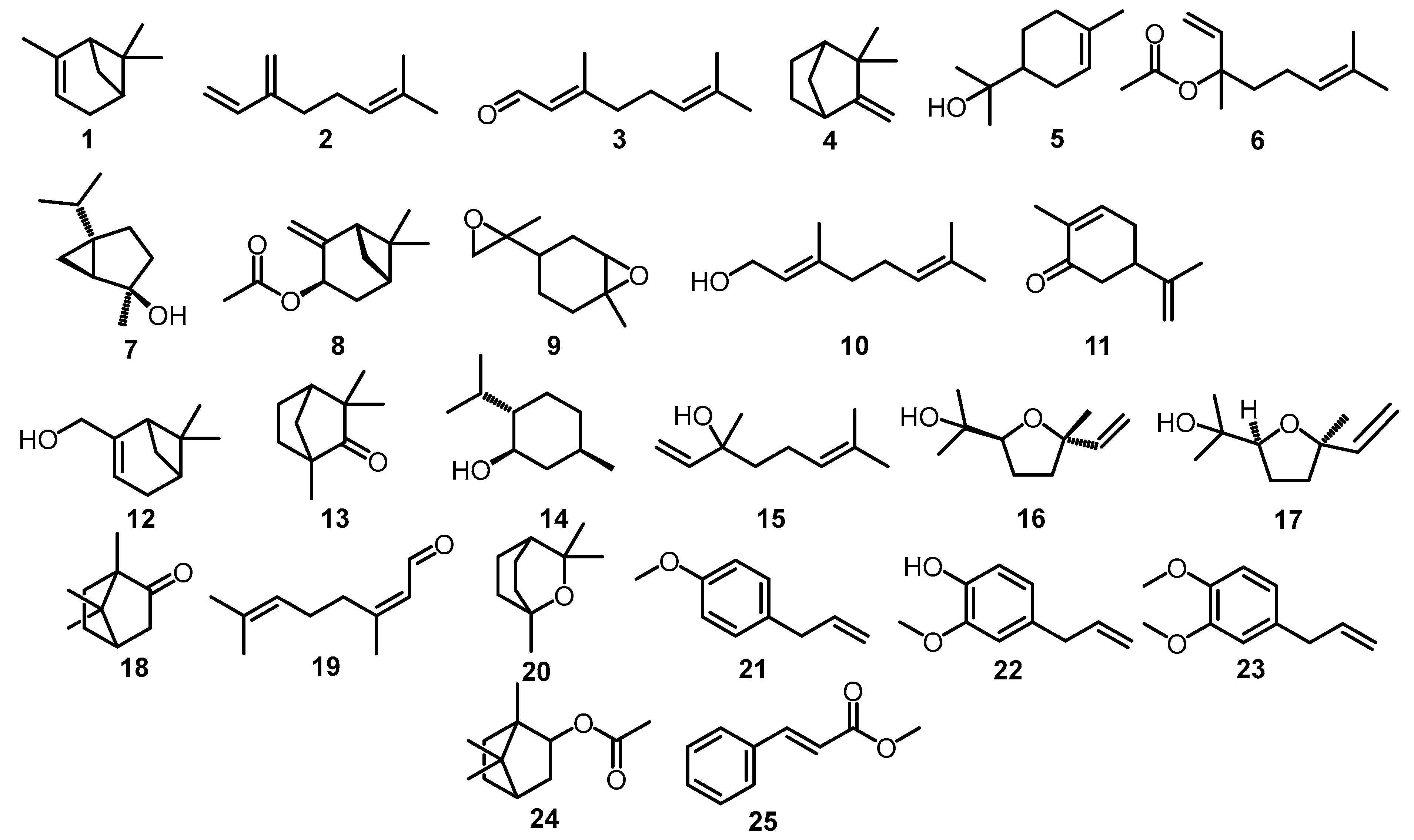
4. Phytochemical Constituents

The type of chemotype can affect the main chemical constituent of O. basilicum L. [22]. Varga et al. discovered five chemotypes, among which (A) linalool (15); (B) linalool (15)/trans-α-bergamotene (29); (C) linalool (15)/methyl chavicol (21); (D) linalool (15)/trans-methyl cinnamate (25); and (E) methyl chavicol (21). Based on the distribution in the regions, chemotypes A and C are European chemotypes; D is a tropical chemotype; and E is a Reunion chemotype [22]. The chemical constituents of O. basilicum L. were dominated by compounds from the phenylpropanoid and monoterpenoid classes [23].
One of the most crucial aspects determining the quality of essential oil depends on the method by which it was adopted. In some studies, conventional extraction methods, such as Soxhlet, hydrodistillation, steam distillation, solvent extraction, and a combination of steam and solvent extraction, are still being used [24]. However, there are promising green extraction methods, including microwave-assisted extraction (MAE), ultrasound-assisted extraction (UAE), high-pressure-assisted extraction (HAE), supercritical and subcritical fluid extraction, electrically assisted extractions, and enzyme-assisted extraction [25]. Green extraction techniques address the challenges by offering several advantages, including cutting down on the use of organic solvents, ease of use due to the simplicity, time efficiency, and cost effectiveness in the extraction process [26].
The O. basilicum L. extract and essential oil contain classes of chemical compounds, mainly terpenoids, such as oxygenated sesquiterpenes, oxygenated monoterpenes, sesquiterpene hydrocarbons, monoterpene hydrocarbons, and non-terpene derivatives. Furthermore, this plant contains phenylpropanoid compounds, including eugenol, methyl eugenol, chavicol, estragole, and methyl cinnamate [27,28]. Monoterpene, geraniol, myrtenol, pinene, camphor, and borneol hold potential for medical applications [29].
The analysis of O. basilicum L. methanolic leaves’ extract, obtained by sonication, showed the presence of various polyphenol compounds, such as caffeic acid, caftaric acid, 3,4-dihydroxyphenylacetic acid, ferulic acid, rosmarinic acid, and rutoside (rutin) [30]. Phenolic compounds have been widely recognized for their beneficial properties and applications in the medical field [31]. Furthermore, O. basilicum L. extract was also found to contain phytosterols, such as β-sitosterol, stigmasterol [32], and campesterol [33].
Studies on the chemical constituents of O. basilicum L. are shown in Table 1, representing its essential oil and extract with various extraction and identification methods. Meanwhile, Figure 2 and Figure 3 show the chemical structures of the chemical compounds presented in Table 1.

5. Ethnomedicinal Evidence for O. basilicum L.

More than 10% of plant species and over 50,000 species have been utilized for the development of medications and healthcare products [2]. O. basilicum L., also known as the king of herbs in the Greek word, has been recognized since ancient times for its therapeutic properties and was used in the Unani and Ayurvedic medical systems [41]. The proper way to use medicinal plants is typically passed down from one generation to the next and often pertains to traditional remedies for age-old ailments [42]. These traditional beliefs about medicinal plants will be enhanced by the integration of technology for the production of sustainable pharmaceuticals.
Ethnobotany is a subdiscipline of ethnobiology, which studies the traditional botanical knowledge in different cultures, the techniques for utilizing plants, the management of plant resources, and the role, which plants play in ritual, cultural, or religious beliefs [43]. As a result, it serves as a foundation for selecting plants, which can be developed for medicinal purposes [44]. Ethnobotany contributes to exploration of the ways to fill the gap between scientific research and cultural or indigenous understanding [45]. The majority of ethnobotanical and ethnopharmacological studies have been conducted to acquire knowledge about the use of medicinal plants to treat various illnesses [46].
India is known as a country for the knowledge and utilization of herbal medicine [47]. Indigenous and local people in the Bageshwar District of Uttarakhand, India, use O. basilicum L. leaf and seed for treating fever, cough, and cold [48]. The community around Lawachara National Park in Bangladesh utilizes O. basilicum L. leaf as a treatment for reducing high blood pressure, fever, and cough [49]. Another area in Lalmohan, Bhola District, Bangladesh, utilized the whole plant of O. basilicum L. to treat fever and as a carminative [50].
Remote areas in India, such as the Uttara Kannada District, used herbal treatments with O. basilicum L. to treat reproductive diseases, such as dysmenorrhea, by crushing the bark in milk and drinking it once a day for seven days [51]. Additionally, local healers from the Khatling Valley and Pauri District in Uttarakhand, India, drink the decoction of leaves and seeds of O. basilicum L. for fever, cough, cold, and urinary problems [52,53]. Traditional healers in the North West Ganjam District, Odisha, India, employ powder and decoction from the leaves of O. basilicum L. for treating dysuria, cough, and cold [54]. Meanwhile, the traditional healers from the Rabha Tribe in the Kamrup District, India, use the leaves and inflorescence as a remedy for cough and chronic fungal infections [55].
Pakyoung in East Sikkim, India, reported that the leaves and seeds of O. basilicum L. were utilized for colds, coughs, fevers, and constipation [56]. Based on an interview with 33 traditional healers from Nelliyampathy, Kerala, India, the leaves and seeds of O. basilicum L. were made into paste, inhalation, juice, and infusion, which could treat tumors, headaches, insomnia, heart trembling, coughs, chest pain, dysentery, diarrhea, and gonorrhea [57].
In Dharan, Nepal, O. basilicum L. leaf juices are used for digestive disorders, such as diarrhea, dysentery, constipation, gastritis, and vomiting [58]. In the Sulaymaniyah province in Iraq, it had been reported that the local community applied O. basilicum L. as an ethnobotanical treatment for headaches, colds, and halitosis (bad breath) [59]. Meanwhile, in the Peshawar Valley, Pakistan, O. basilicum L. leaves were made into an extract and given orally to help improve digestion and for other purposes, such as ornamental decoration [60].
O. basilicum L. has a history of being used as a medical treatment in some southeast Asian nations for a variety of diseases. Traditional healers from Phatthalung, southern Thailand, use O. basilicum L. for treating flatulence and peptic ulcers [61]. Local tribes from Tina and Libas Gua Village, Mindanao, Philippines, utilized the leaves by rubbing them around the affected area of the body to treat cold sores [62]. Furthermore, in Indonesia, this plant is widely used in the big islands, such as Sulawesi, Sumatra, and Kalimantan. The tribes in Kolaka and East Kolaka, Southeast Sulawesi, make use of the leaves of O. basilicum L. for treating tuberculosis [63]. Meanwhile, the traditional healers in many areas of Sumatra utilized the seeds of O. basilicum L. as a therapy for treating back pain sickness [64]. The sub-ethnic Dayak tribe—like the Dayak Linoh tribe, who live in the Sintang District, West Kalimantan—utilized the leaves, flowers, and fruit to reduce body odor and fever [65]. Another sub-ethnic group, Dayak Tamambaloh, in the Kapuas Hulu District, West Kalimantan, used the leaves to treat ringworm, blisters, and reduce body odor [66].
In addition, in the South American continent, e.g., in Maragogipe, State of Bahia, Brazil, the leaves were made into tea for the treatment of delayed menstruation, fever, flu in children, indigestion, and nasal congestion [67]. In an ethnomedicine inventory record in the African continent, e.g., the Abia State in Nigeria, the seeds of O. basilicum L. were used for treating diarrhea [68]. In Western Oromia State, Ethiopia, this species is used to cure allergic reactions by crushing it and mixing it with food [69]. In central Kenya, the plant is used for curing common cold [70]. In the Rainforest Research Station, Ondo State, Nigeria, the whole plant of O. basilicum L. is used for treating inflammation [71].
The utilization of O. basilicum L. as a herbal remedy has been observed on a global scale and spread across regions such as Asia, Africa, and South America. Traditional healers from various regions used the whole plant, as well as some parts of the plant, such as leaves, seeds, inflorescences, fruits, and stems. The plants were made into infusion, inhalation, paste formulations, powder, tea, and they were incorporated into food. According to the beliefs of traditional healers, O. basilicum L. possesses therapeutic properties in the management of typical illnesses affecting the digestive, respiratory, urinary, and reproductive systems. Additionally, certain individuals within the society employed this particular plant for the purpose of enhancing decorative esthetics.

6. Antimicrobial Activities and Biomedical Uses

Medicinal plants contain rich, yet underexploited, bioactive compounds, with a limited amount of their potential qualities having been thoroughly examined [72]. The exploration of a medicinal plant with high chemical constituents holds promise in the pursuit of developing therapeutic treatments in the future [73]. Each part of the plant offers potentially valuable biomedical knowledge, which remains uncovered [74]. As one of the medicinal plants, O. basilicum L. possesses considerable undiscovered potential in the field of antimicrobial and biomedical research. The goal of biomedical therapy through the utilization of medicinal plants requires continuous exploration and development.

6.1. Antiviral Activity

Based on the ethnomedicinal records and data, traditional healers all over the world consider O. basilicum L. to be an important herb. A number of studies have shown that O. basilicum L. has the potential for antiviral activity. For instance, a recent study of O. basilicum L. against SARS-CoV-2 with an in silico assay showed that polyphenol constituents apigenin-7-glucuronide and dihydrokaempferol-3-glucoside have binding affinity for −8.77 Kcal/mol and −8.96 Kcal/mol, respectively, which possess great potential for antiviral activity. These compounds have binding affinity with the main protease (Mpro) enzymes on SARS-CoV-2 [75]. The Mpro enzyme, typically referred to as 3-chymotrypsin-like protease (3CLpro), serves a pivotal role in viral replication and is being targeted as a way of preventing COVID-19 infection [76].
Aside from the polyphenol compound, for the first time, monoterpenes showed the potential for antiviral activity toward SARS-CoV-2. An in vitro study demonstrated that five different monoterpene compounds, such as carvone, carvacrol, menthofuran, 1,8 cineole, and pulegone, potentially inhibited SARS-CoV-2 in infected Vero 76 cells. Among these five compounds, carvacrol and carvone showed significant antiviral activity with half-maximal inhibitory concentration (IC50) < 100 µM. In addition, an essential oil, which contained the highest carvone concentration (>200 mg/mL), had the greatest antiviral activity with IC50 127 ± 4.63 ppm. The antiviral properties of monoterpene compounds had been observed to bind and interrupt the important viral proteins, among them, the main protease and spike protein [77]. On top of its potent antiviral effects, carvacrol had a favorable safety profile, suggesting its potential as a viable candidate for the development of preventive therapies [78].
Another recent study tested the lipophilic fraction of the stem of this plant in vitro and in silico against dengue virus (DENV). This in vitro study revealed that the fraction significantly reduced DENV titer in pre-treatment and post-treatment conditions at a concentration of 3.125 µg/mL. Meanwhile, an in silico study of the lipophilic fraction showed that the two compounds had great binding affinity. Stigmasterol had a binding affinity for −8.3 Kcal/mol with NS1 protein, and campesterol exhibited the biggest binding affinity for −8.2 Kcal/mol with E glycoprotein [33]. A computational study demonstrated that this plant has the potential for antiviral drug development through its mechanism of inhibiting the active site of human immunodeficiency virus (HIV) gp120 and gp41. α-guaiene is the compound with the highest negative value for binding affinity, which was −9.62 Kcal/mol at the active site gp120, and sitosterol displayed a binding affinity for −10.99 Kcal/mol at the active site gp41 [79]. Furthermore, an in vitro study of the crude ethanolic extract of O. basilicum L. leaves demonstrated antiviral activity against Zika virus (ZIKV) with 97% virus infectivity at the highest concentration (1:16 dilution) [80].
There are a few studies, which show that O. basilicum L. can treat some viruses infecting livestock, such as cattle and poultry. An in vitro study of alcoholic extract from O. basilicum L. leaves showed potential in managing the Newcastle disease virus (NDV), which infects poultry, with a reduction titer up to 10−7 at the concentration of 500 µg/mL [81]. Kubiça et al. reported an in vitro study of 1,8-cineole and camphor, which both reduced the plaque in bovine viral diarrhea virus (BVDV) by approximately 75% and 84%, respectively, at the maximum non-toxic dose [82]. Another ethanolic extract of O. basilicum L. was made into an ointment for daily application in bovine cutaneous papillomatosis, which is caused by bovine papillomavirus (BV). The ointment was made into a 2% formulation with 20 mg/g weight per weight (w/w) of ethanolic extract and white petroleum jelly for the base. Clinically, the papilloma began to regress and eventually disappeared between days 7 and 21, and the skin texture progressively returned to normal. The antiviral activity of this topical formulation may be due to the phenolic, flavonoid, tannin, and alkaloid compounds found in it [83].
Another crude aqueous and ethanolic extracts of the whole plant of O. basilicum L., as well as selected purified constituents, exhibited antiviral activity against DNA viruses, such as the herpes virus (HSV), adenoviruses (ADV), hepatitis-B virus (HBV), and RNA viruses, such as coxsackievirus B1 (CVB1) and enterovirus 71 (EV71) [84]. This in vitro research found that purified constituents, such as apigenin, ursolic acid, and linalool, had antiviral activity against HSV-1 similar to that of acyclovir and also against HBV and enterovirus. The strongest purified constituents were ursolic acid against HSV-1 (EC50 6.6 mg/L), ADV-8 (EC50 4.2 mg/L), CVB1 (EC50 0.4 mg/L), and EV71 (EC50 0.5 mg/L). Furthermore, apigenin possessed the highest antiviral activity against HSV-2 (EC50 9.7 mg/L), ADV-3 (EC50 11.1 mg/L), and hepatitis-B surface antigen (EC50 7.1 mg/L). Meanwhile, linalool has recorded moderate anti-adenoviral activity against ADV-3, ADV-8, and it has exhibited the strongest effects against ADV-11 (EC50 16.9 mg/L) [84]. In addition, methanolic extracts of O. basilicum L. significantly inhibited herpes simplex virus 1 strain F (HSV-1F) after viral adsorption [85]. Another purified compound, such as eugenol, from the methanolic extract of O. basilicum L. showed inhibition of pre-HIV-1 infection in the host cell at the effective concentration of 350 µg/mL [86].
The evidence presented above indicates that the phytochemical constituents of O. basilicum L. hold the potential to be developed into new drugs, offering an opportunity to address issues such as drug resistance and side effects [87]. Understanding the phytochemical constituent mechanisms of action in pharmacology is crucial before developing drugs from O. basilicum L. [88]. The antiviral properties of phytochemicals have been subject of substantial research in recent years, even after the world experienced a worldwide pandemic [89]. Therefore, it is crucial to maintain and expand this research into their antiviral properties in light of ongoing global health challenges.

6.2. Antibacterial Activity

The increasing incidence of antibiotic resistance in recent years has prompted an immediate demand for new strategies and innovative antibiotic formulations [90]. The aforementioned issue arises from the irresponsible use of antibiotics in the context of human healthcare and the practice of animal husbandry [91]. O. basilicum L. is one of many medicinal plants, which have demonstrated potential as antibacterial agents. Essential oils, methanolic extracts, and fractions from the plant have been explored over these past few years for their antibacterial properties against Gram-positive and Gram-negative bacteria. The evidence of antibacterial activity against Gram-positive and Gram-negative bacteria is shown at the minimum inhibitory concentration (MIC) in Table 2, and the diameter of the inhibitory zone is shown in Table 3.
An in vitro study of the antibacterial characteristics of O. basilicum L. essential oil demonstrated inhibition and eradication activities against Vibrio strains’ mature biofilm. A concentration of 50 mg/mL of O. basilicum L. essential oil was shown to greatly inhibit the biofilm, with a percentage of 55% for V. parahaemolyticus and up to 87.45% for both V. vulnificus and V. cholerae. The bactericidal effects of this essential oil may have a correlation with the high amount of linalool found in its composition [99]. The potential antibacterial activity of monoterpene compounds involves the degradation of cell walls and cell membranes, as well as the interruption of membrane protein and ion transport processes [100].
Another study demonstrated that methanolic leaf extracts of O. basilicum L. were shown to have a great potential in antibacterial activity against B. cereus, P. aeruginosa, L. monocytogenes, E. coli, M. flavus, and S. aureus, with MIC < 0.5 mg/mL and MBC < 0.9 mg/mL. This methanolic extract contains some polyphenol compounds, such as 3,4-dihydroxyphenylacetic acid and rutoside (rutin), which mainly contribute to the antibacterial effects [30]. The antibacterial effects of polyphenol compounds arise from their ability to interact with bacterial cell walls and membranes, disrupt protein regulation, inhibit microbial enzymes, and exhibit iron-chelating capabilities [101].
The essential oil of O. basilicum L. combined with the antibiotic imipenem had a synergistic interaction, resulting in antibacterial activity against S. aureus and P. aeruginosa. Meanwhile, the essential oil combined with the antibiotic ciprofloxacin had an antagonistic and indifferent interaction [95]. This means that O. basilicum L. phytochemical-derived substances have the potential to enhance the existing antibiotics. Anwar et al. demonstrated that O. basilicum L. in multiple regions of Saudi Arabia exhibited variations in antibacterial activity because of the different chemical compositions, which are influenced by the diverse agro-climatic regions [102].
In addition to diverse regions, various factors influence antimicrobial activity, such as the chemical compounds, bacterial strain, temperature, and bacterial cell number [103]. The evidence of antibacterial properties indicates that O. basilicum L. has a broad spectrum of antibacterial activity against both Gram-positive and Gram-negative bacteria.

6.3. Antifungal Activity

In the intensive care unit (ICU), invasive fungal infections are commonly identified in patients with a weakened immune system [104]. Fungal infections are a major cause of infectious-disease-related mortality around the world [105]. Additionally, because fungi are eucaryote organisms, they only have a few molecular targets, which can be exploited by medications to activate their effects [106]. Recently, phytochemical constituents have received a lot of interest in the research and development of antifungal medications.
The essential oil of O. basilicum L. exhibits antifungal activity against pathogenic fungal Aspergillus flavus at a concentration of 1000 ppm, which could suppress the fungal growth and aflatoxin B1 biosynthesis. The chemical compounds, which possess good antifungal properties, are linalool and 1,8-cineol from monoterpene and eugenol from polyphenol [40]. The commercial basil extracts in Slovenia were tested against several Fusarium species for antifungal properties. The extracts were found to inhibit colony growth in F. proliferatum at concentrations of, respectively, 0.35% and 0.70% by up to 33.37% and 44.30%, and they inhibited colony growth in F. subglutinans by up to 24.74% and 29.27%. The commercial extracts are known to contain estragole (86.72%), trans-α-bergamotene (2.91%), and eucalyptol or 1,8-cineole (2.67%) [107].
The essential oil of O. basilicum L. has antifungal properties against Candida albicans, with an inhibition zone of 27 mm. These activities are supported by linalool, methyl chavicol, β-elemene, and α-bulnesene [108]. In Serbia, twelve cultivars of O. basilicum L. were tested against seven species of fungi, including A. ochraceus, A. versicolor, A. fumigatus, A. niger, Penicillium funiculosum, P. ochrochloron, and Trichoderma viridae. The twelve cultivars exhibited great antifungal properties in inhibiting fungal growth, with the minimum inhibition concentration being 10–100-fold higher than the commercial antifungal drug ketoconazole and minimum fungicidal concentrations ranging from 0.14 µg/mL to 27.00 µg/mL. These antifungal properties of the twelve cultivars of O. basilicum L. may be due to high linalool composition in the essential oils [109].
In addition to essential oil, the methanolic fraction from the aerial parts of O. basilicum L. showed antifungal activity. The fraction was tested against eight species of fungi, including A. flavus, A. niger, Penicillium, Rhizopus solani, Alterneria alternata, Candida albicans, Curvilaria lunata, and A. fumigates. These methanolic fractions exhibited strong inhibition of fungal growth, from the lowest at 10% up to 100%, at concentrations of 1 mg/mL, 3 mg/mL, and 6 mg/mL. C. albicans was one of the resistant species; at 3 mg/mL, it only had 17% inhibition compared to other species, which had 43–100% inhibition [110]. In contrast, the ethanolic extracts from the aerial parts of O. basilicum L. demonstrated antifungal activities against C. albicans. The extracts showed the presence of an inhibitory zone measuring 18 mm for the fungi [111].
A study showed that high concentrations of terpene compounds, such as citral, eugenol, nerolidol, and α-pinene, demonstrated an antifungal mechanism by breaking down the cell membrane [112]. Meanwhile, an in silico study of polyphenols from the plant, including rutin, kaempferol, and quercetin, demonstrated an underlying antifungal mechanism. The polyphenol compound was found to have the ability to inhibit fungal enzymes. Rutin—also called rutoside—had the greatest antifungal activity through binding with 14-alpha demethylase (CYP51) and nucleoside diphosphokinase (NDK), with binding affinity for −9.4 and −8.9, respectively [113].
The synergistic action of various constituents comprising the essential oil suppresses the chance of resistance. This is attributed to the difficulty the pathogens face in adapting resistance characteristics against multiple compounds present in essential oils [114]. This indicates that O. basilicum L. has potential for development into a new class of natural antifungal drugs.

6.4. Biomedical Activity

Numerous records and findings have demonstrated the advantages of O. basilicum L. plant in the medical field, such as its antioxidant, anticancer, analgesic, antidiabetic, anti-inflammatory, and antidepressant effects [12]. The O. basilicum L. essential oil from the seeds had good antioxidant activity when using the DPPH assay compared to the positive control, which was the Trolox compound. Great antioxidant activity was shown with an inhibition concentration (IC50) of 23.44 ± 0.9 µg/mL [115]. Antioxidant activity eventually resulted from the synergy of each compound and had correlation with the total phenolic compound, which composed the essential oil [116]. The phenolic compound has the ability to donate a hydrogen atom to the free radicals, the ability to chelate metal cations, and the ability to scavenge free radicals [39,117]. Stanojevic et al. also reported that with the DPPH assay, basil essential oil had good antioxidant properties with effective concentration (EC50) at 2.38 ± 0.10 mg/mL, and it could be used as an alternative to synthetic antioxidants with a higher safety profile [108]. In addition to essential oil, the O. basilicum L. hexane extracts also showed possible antioxidant activity in a concentration- and dose-dependent manner [118].
The O. basilicum L. in Jordan was tested against three cancer cell lines for anticancer activity, such as the triple-negative breast cancer cell line (MDA-MB-231) with IC50 432.3 ± 32.2 µg/mL, the ER+ breast cancer cell line (MCF7) with IC50 320.4 ± 23.2 µg/mL, and the glioblastoma cancer cell line (U-87 MG) with IC50 431.2 ± 15.3 µg/mL. It turned out that the essential oils containing major components of linalool, eugenol, and eucalyptol exhibited potential anticancer activity [119]. The essential oil was also tested on cancer cell lines from liver cancer (Hep 3B) and breast cancer (MCF-7), which resulted in good cytotoxic effect on both cell lines [115]. In addition to the essential oils, methanolic extracts from the aerial parts of the plant also exhibited promising anticancer activity against MCF-7 and MDA-MB-231. The anticancer properties are shown by the expression level of apoptosis-related genes, which decreases the bcl-2 gene’s ability to act as an inhibitor protein for programmed cell death and allows the cell to undergo apoptosis. In this study, the anticancer activity of O. basilicum L. came from the active compound eugenol [120].
The combination of Morus nigra and O. basilicum L. extracts was tested in various cancer cell lines and normal human cells for anticancer activity. It was discovered that the chloroform extracts (MO2C) possessed the highest anticancer activity. The reason for this is that MO2C was cytotoxic against all tested cell lines at the lowest concentration—particularly the breast cancer cell line—and had selective cytotoxicity toward the normal cell line. In addition, this extract contains α-trans-bergamotene, germacrene D, selin-4,7(11)-diene, 2-decel-1-ol, and 2-tridecen-1-ol, which play anticancer roles. The anticancer capacities observed in cell morphology include shrinkage, loss of cellular integrity, cell detachment, and contraction of the cytoplasm [121].
Another in vitro study demonstrated that the methanolic, hexane, and dichloromethane extracts showed a potential antidiabetic property. These three extracts were subjected to a cytotoxic assay, which is considered safe for methanolic extracts up to 0.25 mg/mL and for hexane and dichloromethane extracts up to 0.5 mg/mL. The hexane extract demonstrated an “insulin-like” effect in the absence of insulin due to translocation of the glucose transporter (GLUT4) to the plasma membrane. This study stated that there were 17 newly identified compounds, which possibly played antidiabetic roles in the extracts. Some of these compounds contained glycerol, cyanuric acid, talose, oleamide, inositol, hydroquinone-beta-d-glucopyranoside, pentane-1,2,5-triol, and glucopyranose. Inositol was first found in the O. basilicum L. methanolic extract in this study [122].
Meanwhile, an in vivo study performed in male albino mice showed that O. basilicum L. leaf extract administered orally had the potential to improve neuromuscular coordination, active behavior, the ability to recognize objects, and short-term memory. The optimum daily supplementation dose was found to be 100 mg/mL solvent/kg body weight and was considered suitable for oral administration without any safety concerns [123]. Hydroethanolic extract, ethyl acetate, and n-hexane fractions had anticonvulsant and neuroprotective characteristics, which prevented oxidative damage to the brain tissue, with optimum dose at 200 mg/kg [124]. For the first time, new compounds called 5,7-dihydroxy-3′,4′,5′-trimethoxyflavone and 3-hydroxy-3′,4′,5′-trimethoxyflavone have been found in O. basilicum L. leaf extracts and fractions. An in silico study showed that both compounds had binding interaction energy for −9.93309 and −15.9683, respectively, with Caspase-3 target protein. Both of these compounds helped improve long-term memory by reducing Caspase-3 concentration and suggesting the role of anti-apoptotic cells against neuron cells. This neuroprotective ability is due to the combination of anticholinergic, antioxidant, anti-inflammatory, and anti-apoptotic effects of the compound [125]. The inhalation of essential oil derived from O. basilicum L. has been shown to possess neuroprotective properties and exhibit depressive effects in mice. The essential oil demonstrated antidepressant properties in mice subjected to chronic unpredictable mild stress [126].
Moreover, O. basilicum L. has been identified as an effective agent in exerting anti-inflammatory effects. An in vivo study performed in mice showed that the essential oil of O. basilicum L. with the estragole (methyl chavicol) chemotype in doses of 100 mg/kg and 50 mg/kg greatly reduced paw edema induced by carrageenan by 74% and 44%, respectively, between the first and fifth hour of evaluation. Furthermore, these doses of essential oil are deemed safe for oral administration [127]. An in vitro study showed that O. basilicum L. treated with chemical elicitors, such as arachidonic acid, jasmonic acid, and β-aminobutyric acid, enhanced the flavonoid and phenolic content, which possess anti-inflammatory properties. This finding showed that a plant with arachidonic acid elicitor had the greatest inhibitory effect against lipoxygenase (LOX) (EC50 = 1.67 mg FW mL−1) and cyclooxygenase (COX) (EC50 = 0.31 mg FW mL−1). The inhibitory efficacy exhibited positive correlation with the increased content of rosmarinic, benzoic, and o-coumaric acids [128]. Moreover, an in vitro study demonstrated that ethanolic leaf and leaf callus extracts significantly reduced nitric oxide as pro-inflammatory mediators with concentrations of 0.01–1 mg/mL on RAW 264.7 macrophage cells. This anti-inflammatory activity may be a result of the major compounds found in the extracts, which are 2,3-dihydroxy-3,5-dihydroxy-6-methyl-4H-pyran-4-one and 2-methoxy-4-vinylphenol [129].
Another in vivo study demonstrated that O. basilicum L. methanolic extract emulgel formulation represented a potential alternative for second-degree-burn wound-induced rabbits. Formulations with 5% extract, polymer, and other excipients were compatible and had a good safety profile for topical emulgel. This extract formulation showed a healing capacity on the 16th day, with 98.78% wound contraction, which was insignificantly different to the healing capacity of commercial healing products [130].
In addition, a novel hydrogel formulation based on O. basilicum L. and Trifolium pratense extract combination had a great wound healing ability. In vitro tests demonstrated that the combined extract with concentration of 50 µg/mL had the greatest healing efficacy in terms of complete healing time and fibroblast density. An in vivo study also showed that the combined extract healed the wound 100% on the 13th day—better than the control group—which means that it exhibited a remarkable wound healing capability. Moreover, the hydrogel formulation was tested in a clinical case of a patient with Psoriasis vulgaris twice a day. The formulation was shown to reduce erythema symptoms within one week of treatment. The tremendous wound healing capability could be explained by the synergistic effect of the extract’s phytochemical mixture. The O. basilicum L. extract was rich in phenolic and flavonoid contents, especially ferulic acid; meanwhile, the T. pratense was rich in chlorogenic acid [131]. Ferulic acid is known for its ability to enhance wound healing through the promotion of angiogenesis, reduction in oxidative stress, and inhibition of bacterial growth [132].
Moreover, there are three novel compounds found in O. basilicum L., such as inositol, 5,7-dihydroxy-3′,4′,5′-trimethoxyflavone, and 3-hydroxy-3′,4′,5′-trimethoxyflavone. The studies above indicated that O. basilicum L. holds significant potential for development and formulation into a natural pharmaceutical alternative. Thus, additional investigation of the drug delivery system and clinical trials are necessary in order to create natural accessible medication.

7. Biotechnological Development in O. basilicum L. Research

The field of biotechnology involves the utilization of scientific methodologies to alter and enhance the characteristics of plants, animals, and micro-organisms in order to increase their overall value [133]. The demand for herbal medicine on a global scale is substantial and exhibits a consistent growth rate. Various technologies have been implemented to facilitate the promotion of bioactive compounds in medicinal plants [134]. Secondary metabolites, which are considered vital constituents of the plants, hold significant economic value due to their applications as pharmaceutical products, perfumes, pigments, and food additive products [135].

7.1. Green Nanotechnology Production in O. basilicum L. for Medical Application

Previously, we discussed the antibacterial and antifungal properties of O. basilicum L., which were extensively explored from 2010 to 2018, revealing its potential for combating various bacterial and fungal infections. Over the last five years, numerous studies on nanotechnology have demonstrated the ways of enhancing the antimicrobial properties of this plant. One notable advantage of using plant extracts for synthesizing nanoparticles is their ability to generate a larger zone of inhibition compared to chemical synthesis methods [136].
The essential oil of O. basilicum L. had moderate antibacterial activity against Gram-negative bacteria. However, combining and formulating the essential oil into chitosan nanocarriers with nanoencapsulation technology exhibited strong antibacterial and antibiofilm properties against E. coli and S. aureus, resulting in inhibitory zones measuring 15.3 mm and 21 mm, respectively. This combination damages the cell membrane, and therefore, it causes the leakage of biological macromolecules [137]. Therefore, the combination has good potential for overcoming Gram-negative resistance against antibiotics. ZnO NP is one of the nanoparticles, which showed great antibacterial activity against Pseudomonas aeruginosa, with 20 mm inhibitory zone diameter [138].
ZnO NP synthesized with the O. basilicum L. extract was tested against other bacteria species and exhibited a great inhibitory zone diameter for S. aureus (19.3 mm), E. coli (13.2 mm), S. typhimurium (8.2 mm), L. monocytogenes (11.4 mm), B. subtilis (9.3 mm), and P. aeruginosa (12.4 mm). It also showed great MIC for antibacterial activity, ranging from 0.78 µg/mL, 1.56 µg/mL, 3.12 µg/mL to 6.25 µg/mL [136]. Along with ZnO NP, copper oxide nanoparticles (CuO NPs) enhance the antibacterial activity against S. aureus and E. coli more than the extract itself [139]. In addition to the monometallic synthesized nanoparticle, there are bimetallic synthesized nanoparticles. This is a combination of two different types of metallic nanoparticles in one particle, which work synergistically [140]. In this study, a combination of silver and platinum nanoparticles (AgPt NP) exhibited a significant inhibitory effect on S. aureus, E. faecalis, E. coli, and K. pneumoniae rather than the monometallic nanoparticle. The bimetallic particle showed an inhibitory diameter of 9–25 mm, whereas the monometallic particle of each nanoparticle only showed an inhibitory diameter under 10 mm [141].
Another study demonstrated the green synthesis of reduced graphene oxide (RGO)-zinc oxide (ZnO) nanocomposite, or RGO-ZnO NCs. It was shown that at a concentration of 30 µg/mL, an inhibition zone was observed for the Cocci strain and E. coli at 20 mm and 10 mm, respectively. RGO-ZnO NCs had antibacterial activity at a small concentration, whereas the essential oil or extract of O. basilicum L. itself needed higher concentrations to achieve the same results. This study will also become the basis for the next development and investigation of RGO-ZnO NCs as potential antioxidant candidates and diabetes treatments [142]. Another potential diabetic therapy based on a synthesized silver nanoparticle was found in O. basilicum L. leaf extract. The result demonstrated inhibitory activity against α-amylase—which was higher than antidiabetic medicine acarbose—and high inhibitory activity against α-glucosidase, higher than acarbose and crude extract [143]. This finding suggests the need for alternative therapies for diabetic treatment.
A recent study demonstrated that O. basilicum L. chemical constituents were responsible for the green biosynthesis of ZnO NPs. In combination with bacterial phages, ZnO NPs demonstrated antibacterial activity against Salmonella enterica and deformation on biofilm, which were caused by Staphylococcus sciuri [144]. Another study demonstrated the green synthesis of silver nanoparticles (Ag NPs) in combination with phage ZCSE6 for antibacterial activity against Salmonella enterica. The O. basilicum L. extract works as a bio-reducing agent in order to create Ag NPs effectively. It was shown that the Ag NPs exhibited antibacterial activity; the minimum concentration to inhibit growth was 6.25 µg/mL, and the minimum bactericidal concentration was 12.5 µg/mL. Surprisingly, the Ag NPs in combination with phage ZCSE6 had great bactericidal activity, with a lower concentration than the MIC, which suppressed the growth of S. enterica 24 h after treatment [145].
In addition to utilization of the O. basilicum L. extract for synthesizing nanoparticles, the mucilage from the seed in combination with nanoparticles can create a novel natural wound dressing. Basil seed mucilage (BSM) was dried and then combined with ZnO NP to create a hydrogel sponge. As the weight percent (wt%) of ZnO NP increased, the antibacterial activity of the BSM hydrogel sponge was enhanced. It exhibited great antibacterial activity at 50 wt% ZnO NP against E. coli and S. aureus, with an inhibitory zone at 15.9 mm and 16.7 mm, respectively. The increasing ZnO NP wt% content on the hydrogel sponge also resulted in a slight decrease in thickness, porosity, degree of swelling, and a slight increase in the water holding capacity. The BSM with ZnO NP is considered non-toxic to human keratinocyte (HaCat) cells [146]. This hydrogel sponge could have the potential to be commercialized as a natural healthcare product.
In addition to antibacterial functions against human pathogens, the synthesis of silver nanoparticles (Ag NPs) can also work as a control agent for the management of plant viral infections. This study tested Ag NPs against cucumber mosaic virus (CMV), which infects squash. Spraying the foliar containing Ag NPs at a concentration of 100 µg/mL resulted in enhanced growth, delayed indication of disease symptoms, and a significant reduction of up to 92% in CMV accumulation levels as compared to the non-treated plants. It also increased the soluble carbohydrate, free radical scavenging activity, antioxidant enzymes, and total phenolic and flavonoid contents. This finding could be an alternative for treating plant viral disease instead of using chemical biocides [147]. There is a substantial opportunity in developing a green synthesis of nanoparticles within the O. basilicum L. extracts, which could be a potential therapy and alternative treatment in many cases of human diseases.

7.2. Biotechnological Techniques for Improving the Metabolite Production of O. basilicum L.

It is known that the medicinal plant O. basilicum L. is a rich source of valuable phytoconstituents [148]. The diversity of chemical compounds in O. basilicum L., alone or in synergy, exhibits some medicinal properties [149]. The current production of horticulture crops is centered on improving the quality, quantity, and safety of products, as well as yield, in order to meet the demands of the food and health industries, which have a strong reliance on chemical compounds [150]. The advancement of O. basilicum L. production is influenced by various aspects, including environmental parameters (light, soil nutrients, temperature, water, CO2 levels), cultivars, and cultivation methods [151]. Several studies have demonstrated various experiments on how to improve the chemical compounds derived from the plant O. basilicum L.
One study showed that narrow-bandwidth light treatments of basil seeds were observed to have relative effects on volatile oils. Light conditions may increase the value and quality of this herb, which is appreciated for human wellness. Light treatments could induce the three main compounds in O. basilicum L., which are eugenol, linalool, and 1,8-cineol (eucalyptol). Eugenol and linalool are induced by blue-red-green (BRG) light, and 1,8-cineole is induced by BRG, blue-red-yellow (BRY), and blue-red-far-red (BRFr) light [152]. These compounds mainly play a role as antimicrobial and antioxidant agents [153]. The blue and red LED treatments can potentially improve O. basilicum L. growth and increase the phenolic content of the plants; thus, the different cultivars can also have a different result. The green cultivar in this study was mostly stimulated by the red light, and the red cultivar was stimulated by the blue light [154].
In addition to the light treatments, the abiotic (CdCl2 and AgNO3) and biotic (YE) yeast extract elicitors were found to increase the total amount of phenolic and flavonoid contents. Chicoric and rosmarinic acid increased with the treatment of CdCl2 and AgNO3 at 5 µM. Rutin and isoquercetin also increased with the YE treatment, up to 1.6 times and 1.9 times. Meanwhile, the highest amounts of linalool and estragole were observed in the treatment with AgNO3, up to 2.8 times and 0.5 times [155]. Arbuscular mycorrhizal fungi (AMF), which are another type of biotic elicitors, showed a promising capacity in increasing the production of essential oil, with eugenol and γ-cadinene being the compounds with the highest ratios, which composed the essential oil [156].
Various methods of enhancing the chemical compounds of O. basilicum L. were discussed above to emphasize the importance of naturally synthesized compounds. One such approach involves improving the growth factors through light treatments and optimizing the formulation of biotic or abiotic elicitors.

8. Conclusions

O. basilicum L. is a plant species, which exhibits wide distribution throughout several regions of the world. Over time, there has been a significant evolution in the understanding and application of this plant in the context of healthcare. The plant is regarded as a highly valuable source due to its distinctive chemical composition, which provides a diverse array of antimicrobial and other medicinal attributes, including anticancer, antioxidant, antidiabetic, and neuroprotective functions. This plant could change the way in which drugs are produced, either by isolating pure phytochemical compounds or by combining several compounds. This could revolutionize the pharmaceutical industry by providing a natural substitute for synthetic drugs.
The utilization of O. basilicum L. as a medicinal plant has developed over years, starting from community beliefs. The beliefs held by communities were recorded, and many studies were conducted to prove the efficacy of this plant. Several studies have highlighted the potential of the aerial parts—particularly the leaves—of this plant for the development of novel medicines. The phytochemical classes predominantly associated with antimicrobial and biomedical activities are polyphenols, terpenes, and phytosterols. Noteworthy compounds with promising potential for antiviral drug development include carvacrol, α-guaiene, ursolic acid, apigenin, stigmasterol, and campesterol. Additionally, compounds such as linalool, rutin, eugenol, estragole, citral, α-pinene, nerolidol, kaempferol, and trans-α-bergamotene could be utilized in the creation of medicines targeting bacteria and fungi. Moreover, the exploration of new neuroprotective medicines may be facilitated by novel compounds, such as 5,7-dihydroxy-3′,4′,5′-trimethoxyflavone and 3-hydroxy-3′,4′,5′-trimethoxyflavone. Furthermore, topical formulation for wound healing has been demonstrated to be a promising alternative treatment.
Furthermore, evidence is becoming the key point to be subsequently developed and formulated into a novel drug. In terms of the plant’s usefulness in the healthcare field, its distinctive chemical compound, and its higher safety profile, future research should focus more on formulating this plant into a natural choice in addition to chemical drugs. As another alternative, this plant could be combined with chemical drugs to create new efficacy and new mechanisms, which are destined for later commercialization.
Future innovations could come from researching the ways to patent the extraction methods, formulating a standardized approach to boost the chemical compound contents, conducting more clinical research in a mechanistic and molecular way, and advancing it up to the industrial stage. This will allow not only gaining a deeper understanding of the mechanistic action, but it will also lead the path to developing more effective, safer drugs and reduce undesirable side effects.

Author Contributions

Conceptualization and writing—original draft preparation, N.S.A.; writing—review and editing, and supervision, B.I., M.M., J.K., D.O. and F.D.; writing—review and editing, and drawing chemical constituents, W.S. and K.F. All authors have read and agreed to the published version of the manuscript.

Funding

This work was funded by Universitas Padjadjaran through the Hibah Riset Percepatan Lektor Kepala awarded to Mia Miranti under grant number 159/UN6.3.1/PT.00/2023.

Data Availability Statement

This study did not report any data.

Acknowledgments

The authors would like to thank Karina Kalasuba and Sulistya Ika Akbari for their assistance during the trip.

Conflicts of Interest

The authors declare no conflict of interest.

References

  1. Ali, S.I.; Sheikh, W.M.; Rather, M.A.; Venkatesalu, V.; Muzamil Bashir, S.; Nabi, S.U. Medicinal plants: Treasure for antiviral drug discovery. Phytother. Res. 2021, 35, 3447–3483. [Google Scholar] [CrossRef] [PubMed]
  2. Chen, S.L.; Yu, H.; Luo, H.M.; Wu, Q.; Li, C.F.; Steinmetz, A. Conservation and sustainable use of medicinal plants: Problems, progress, and prospects. Chin. Med. 2016, 11, 37. [Google Scholar] [CrossRef] [PubMed]
  3. Thomas, E.; Stewart, L.E.; Darley, B.A.; Pham, A.M.; Esteban, I.; Panda, S.S. Plant-based natural products and extracts: Potential source to develop new antiviral drug candidates. Molecules 2021, 26, 6197. [Google Scholar] [CrossRef] [PubMed]
  4. Veeresham, C. Natural products derived from plants as a source of drugs. J. Adv. Pharm. Technol. Res. 2012, 3, 200–201. [Google Scholar] [CrossRef] [PubMed]
  5. Süntar, I. Importance of ethnopharmacological studies in drug discovery: Role of medicinal plants. Phytochem. Rev. 2020, 19, 1199–1209. [Google Scholar] [CrossRef]
  6. Karpiński, T.M. Essential oils of lamiaceae family plants as antifungals. Biomolecules 2020, 10, 103. [Google Scholar] [CrossRef] [PubMed]
  7. Al Jaouni, S.; Saleh, A.M.; Wadaan, M.A.M.; Hozzein, W.N.; Selim, S.; AbdElgawad, H. Elevated CO2 induces a global metabolic change in basil (Ocimum basilicum L.) and peppermint (Mentha piperita L.) and improves their biological activity. J. Plant. Physiol. 2018, 224–225, 121–131. [Google Scholar] [CrossRef]
  8. Taek, M.M.; Bambang, P.E.W.; Agil, M. Plants used in traditional medicine for treatment of malaria by Tetun Ethnic people in West Timor Indonesia. Asian Pac. J. Trop. Med. 2018, 11, 630–637. [Google Scholar] [CrossRef]
  9. Silalahi, M.; Nisyawati; Walujo, E.B.; Supriatna, J.; Mangunwardoyo, W. The local knowledge of medicinal plants trader and diversity of medicinal plants in the Kabanjahe Traditional Market, North Sumatra, Indonesia. J. Ethnopharmacol. 2015, 175, 432–443. [Google Scholar] [CrossRef]
  10. Kasmawati, H.; Ihsan, S.; Munasari, D.; Ode Elafita, W. Ethnomedicine studies of traditional medicinal plants of the Muna Tribe in the Village of Bungi Southeast Sulawesi Province of Indonesia. Int. J. Sci. Res. 2019, 8, 1882–1887. [Google Scholar] [CrossRef]
  11. Aminian, A.R.; Mohebbati, R.; Boskabady, M.H. The effect of Ocimum basilicum L. and its main ingredients on respiratory disorders: An experimental, preclinical, and clinical review. Front. Pharmacol. 2022, 12, 805391. [Google Scholar] [CrossRef] [PubMed]
  12. Dhama, K.; Sharun, K.; Gugjoo, M.B.; Tiwari, R.; Alagawany, M.; Iqbal Yatoo, M.; Thakur, P.; Iqbal, H.M.N.; Chaicumpa, W.; Michalak, I.; et al. A comprehensive review on chemical profile and pharmacological activities of Ocimum basilicum. Food Rev. Int. 2023, 39, 119–147. [Google Scholar] [CrossRef]
  13. Abd El-Hamid, M.I.; El-Tarabili, R.M.; Bahnass, M.M.; Alshahrani, M.A.; Saif, A.; Alwutayd, K.M.; Safhi, F.A.; Mansour, A.T.; Alblwi, N.A.N.; Ghoneim, M.M.; et al. Partnering essential oils with antibiotics: Proven therapies against bovine Staphylococcus aureus mastitis. Front. Cell. Infect. Microbiol. 2023, 13, 1265027. [Google Scholar] [CrossRef] [PubMed]
  14. Barickman, T.C.; Olorunwa, O.J.; Sehgal, A.; Walne, C.H.; Reddy, K.R.; Gao, W. Interactive impacts of temperature and elevated CO2 on basil (Ocimum basilicum L.) root and shoot morphology and growth. Horticulturae 2021, 7, 112. [Google Scholar] [CrossRef]
  15. O’Leary, N. Taxonomic revision of Ocimum (lamiaceae) in Argentina. J. Torrey Bot. Soc. 2017, 144, 74–87. [Google Scholar] [CrossRef]
  16. Akbari, G.A.; Soltani, E.; Binesh, S.; Amini, F. Cold tolerance, productivity and phytochemical diversity in sweet basil (Ocimum basilicum L.) Accessions. Ind. Crops. Prod. 2018, 124, 677–684. [Google Scholar] [CrossRef]
  17. Li, Q.X.; Chang, C.L. Basil (Ocimum basilicum L.) oils. In Essential Oils in Food Preservation, Flavor and Safety; Preedy, V.R., Ed.; Elsevier: Amsterdam, The Netherlands, 2015; pp. 231–238. ISBN 9780124166417. [Google Scholar]
  18. Nazarian, H.; Amouzgar, D.; Sedghianzadeh, H. Effects of different concentrations of cadmium on growth and morphological changes in basil (Ocimum basilicum L.). Pak. J. Bot. 2016, 48, 945–952. [Google Scholar]
  19. Lal, R.K.; Gupta, P.; Chanotiya, C.S.; Sarkar, S. Traditional plant breeding in Ocimum. In The Ocimum Genome; Shasany, A.K., Kole, C., Eds.; Springer: Cham, Switzerland, 2018; pp. 89–98. [Google Scholar]
  20. Paton, A.; Harley, M.R.; Harley, M.M. Ocimum: An overview of classification and relationships. In Basil: The Genus Ocimum; Hiltunen, R., Holm, Y., Eds.; CRC Press: London, UK, 1999; pp. 1–38. ISBN 9781135296124. [Google Scholar]
  21. Kumar, A.; Shukla, A.K.; Shasany, A.K.; Sundaresan, V. Systematic position, phylogeny, and taxonomic revision of Indian Ocimum. In The Ocimum Genome; Shasany, A.K., Kole, C., Eds.; Springer: Cham, Switzerland, 2018; pp. 61–72. [Google Scholar]
  22. Varga, F.; Carović-Stanko, K.; Ristić, M.; Grdiša, M.; Liber, Z.; Šatović, Z. Morphological and biochemical intraspecific characterization of Ocimum basilicum L. Ind. Crops. Prod. 2017, 109, 611–618. [Google Scholar] [CrossRef]
  23. Padalia, R.C.; Verma, R.S.; Chauhan, A. Analyses of organ specific variations in essential oils of four Ocimum species. J. Essent. Oil Res. 2014, 26, 409–419. [Google Scholar] [CrossRef]
  24. Tongnuanchan, P.; Benjakul, S. Essential oils: Extraction, bioactivities, and their uses for food preservation. J. Food Sci. 2014, 79, R1231–R1249. [Google Scholar] [CrossRef]
  25. Majid, I.; Khan, S.; Aladel, A.; Dar, A.H.; Adnan, M.; Khan, M.I.; Mahgoub Awadelkareem, A.; Ashraf, S.A. Recent insights into green extraction techniques as efficient methods for the extraction of bioactive components and essential oils from foods. CYTA—J. Food 2023, 21, 101–114. [Google Scholar] [CrossRef]
  26. Ameer, K.; Shahbaz, H.M.; Kwon, J.H. Green extraction methods for polyphenols from plant matrices and their byproducts: A review. Compr. Rev. Food Sci. Food Saf. 2017, 16, 295–315. [Google Scholar] [CrossRef] [PubMed]
  27. Rezzoug, M.; Bakchiche, B.; Gherib, A.; Roberta, A.; Guido, F.; Kilinçarslan, Ö.; Mammadov, R.; Bardaweel, S.K. Chemical composition and bioactivity of essential oils and ethanolic extracts of Ocimum basilicum L. and Thymus algeriensis Boiss. & Reut. from the Algerian Saharan Atlas. BMC Complement. Altern. Med. 2019, 19, 146. [Google Scholar] [CrossRef]
  28. Hussain, A.I.; Anwar, F.; Hussain Sherazi, S.T.; Przybylski, R. Chemical composition, antioxidant and antimicrobial activities of basil (Ocimum basilicum) essential oils depends on seasonal variations. Food Chem. 2008, 108, 986–995. [Google Scholar] [CrossRef] [PubMed]
  29. Zielińska-Blajet, M.; Feder-Kubis, J. Monoterpenes and their derivatives—Recent development in biological and medical applications. Int. J. Mol. Sci. 2020, 21, 7078. [Google Scholar] [CrossRef] [PubMed]
  30. Elansary, H.O.; Szopa, A.; Kubica, P.; Ekiert, H.; El-Ansary, D.O.; Al-Mana, F.A.; Mahmoud, E.A. Saudi Rosmarinus officinalis and Ocimum basilicum L. polyphenols and biological activities. Processes 2020, 8, 446. [Google Scholar] [CrossRef]
  31. Ge, L.; Li, S.P.; Lisak, G. Advanced sensing technologies of phenolic compounds for pharmaceutical and biomedical analysis. J. Pharm. Biomed. Anal. 2020, 179, 112913. [Google Scholar] [CrossRef]
  32. Qamar, F.; Sana, A.; Naveed, S.; Faizi, S. Phytochemical characterization, antioxidant activity and antihypertensive evaluation of Ocimum basilicum L. in L-NAME induced hypertensive rats and its correlation analysis. Heliyon 2023, 9, e14644. [Google Scholar] [CrossRef]
  33. Joshi, R.K.; Agarwal, S.; Patil, P.; Alagarasu, K.; Panda, K.; Cherian, S.; Parashar, D.; Roy, S. Anti-dengue activity of lipophilic fraction of Ocimum basilicum L. Stem. Molecules 2023, 28, 1446. [Google Scholar] [CrossRef]
  34. Falowo, A.B.; Mukumbo, F.E.; Idamokoro, E.M.; Afolayan, A.J.; Muchenje, V. Phytochemical constituents and antioxidant activity of sweet basil (Ocimum basilicum L.) essential oil on ground beef from Boran and Nguni cattle. Int. J. Food. Sci. 2019, 2019, 2628747. [Google Scholar] [CrossRef]
  35. Rabbani, M.; Rabbani, M.; Ebrahim Sajjadi, S.; Vaezi, A. Evaluation of anxiolytic and sedative effect of essential oil and hydroalcoholic extract of Ocimum basilicum L. and chemical composition of its essential oil. Res. Pharm. Sci. 2015, 10, 535–543. [Google Scholar] [PubMed]
  36. Saaban, K.F.; Ang, C.H.; Khor, S.M.; Chuah, C.H. Chemical constituents and antioxidant capacity of Ocimum basilicum and Ocimum sanctum. Iran J. Chem. Chem. Eng. 2019, 38, 139–152. [Google Scholar] [CrossRef]
  37. Chenni, M.; Abed, D.E.; Rakotomanomana, N.; Fernandez, X.; Chemat, F. Comparative study of essential oils extracted from Egyptian basil leaves (Ocimum basilicum L.) using hydro-distillation and solvent-free microwave extraction. Molecules 2016, 21, 113. [Google Scholar] [CrossRef] [PubMed]
  38. Hapsari, I.P.; Feroniasanti, Y.M.L. Phytochemical screening and in vitro antibacterial activity of sweet basil leaves (Ocimum basilicum L.) essential oil against Cutibacterium acnes ATCC 11827. In AIP Conference Proceedings; AIP Publishing: New York, NY, USA, 2019; Volume 2099. [Google Scholar]
  39. Sharafati-Chaleshtori, R.; Rokni, N.; Rafieian-Kopaei, M.; Drees, F.; Salehi, E. Antioxidant and antibacterial activity of basil (Ocimum basilicum L.) Essential Oil in Beef Burger. J. Agric. Sci. Technol. 2015, 17, 817–826. [Google Scholar]
  40. El-Soud, N.H.A.; Deabes, M.; El-Kassem, L.A.; Khalil, M. Chemical composition and antifungal activity of Ocimum basilicum L. essential oil. Maced. J. Med. Sci. 2015, 3, 374–379. [Google Scholar] [CrossRef] [PubMed]
  41. Nadeem, H.R.; Akhtar, S.; Ismail, T.; Qamar, M.; Sestili, P.; Saeed, W.; Azeem, M.; Esatbeyoglu, T. Antioxidant effect of Ocimum basilicum essential oil and its effect on cooking qualities of supplemented chicken nuggets. Antioxidants 2022, 11, 1882. [Google Scholar] [CrossRef] [PubMed]
  42. Khadka, D.; Dhamala, M.K.; Li, F.; Aryal, P.C.; Magar, P.R.; Bhatta, S.; Thakur, M.S.; Basnet, A.; Cui, D.; Shi, S. The use of medicinal plants to prevent COVID-19 in Nepal. J. Ethnobiol. Ethnomed. 2021, 17, 26. [Google Scholar] [CrossRef] [PubMed]
  43. Camou-Guerrero, A.; Casas, A.; Moreno-Calles, A.I.; Aguilera-Lara, J.; Garrido-Rojas, D.; Rangel-Landa, S.; Torres, I.; Pérez-Negrón, E.; Solís, L.; Blancas, J.; et al. Ethnobotany in Mexico: History, development, and perspectives. In Ethnobotany of Mexico; Lira, R., Casas, A., Blancas, J., Eds.; Springer: New York, NY, USA, 2016; pp. 21–39. [Google Scholar]
  44. Abbasi, A.M.; Shah, M.H.; Khan, M.A. Ethnobotany and ethnomedicine. In Wild Edible Vegetables of Lesser Himalayas; Springer International Publishing: Berlin/Heidelberg, Germany, 2015; pp. 19–29. [Google Scholar]
  45. Rodríguez-Calderón, Á.; Muñoz, J.A.; Moreno, D.; Celis, M. Describing and diff using the ethnobotanical knowledge of Bogotá D.C. (Colombia) through an online tool focused on common names of plants. Acta Bot. Brasilica 2019, 33, 303–314. [Google Scholar] [CrossRef]
  46. Mechaala, S.; Bouatrous, Y.; Adouane, S. Traditional knowledge and diversity of wild medicinal plants in El Kantara’s Area (Algerian Sahara Gate): An ethnobotany survey. Acta Ecol. Sin. 2022, 42, 33–45. [Google Scholar] [CrossRef]
  47. Anand, U.; Tudu, C.K.; Nandy, S.; Sunita, K.; Tripathi, V.; Loake, G.J.; Dey, A.; Proćków, J. Ethnodermatological use of medicinal plants in India: From Ayurvedic formulations to clinical perspectives—A review. J. Ethnopharmacol. 2022, 284, 114744. [Google Scholar] [CrossRef]
  48. Ojha, S.N.; Tiwari, D.; Anand, A.; Sundriyal, R.C. Ethnomedicinal knowledge of a Marginal Hill community of Central Himalaya: Diversity, usage pattern, and conservation concerns. J. Ethnobiol. Ethnomed. 2020, 16, 29. [Google Scholar] [CrossRef] [PubMed]
  49. Uddin, M.Z.; Arefin, M.K.; Alam, M.F.; Kibria, M.G.; Podder, S.L.; Hassan, M.A. Knowledge of ethnomedical plants and informant consensus in and around Lawachara National Park. J. Asiat. Soc. Bangladesh Sci. 2017, 43, 101–123. [Google Scholar] [CrossRef]
  50. Sohel, M.D.D.; Kawsar, M.D.H.; Sumon, M.D.H.U.; Sultana, T. Ethnomedicinal studies of Lalmohan Thana in Bhola District, Bangladesh. Altern. Integr. Med. 2016, 5, 317–327. [Google Scholar] [CrossRef]
  51. Hegde, H.V.; Hegde, G.R.; Kholkute, S.D. Herbal care for reproductive health: Ethno medicobotany from Uttara Kannada District in Karnataka, India. Complement. Ther. Clin. Pract. 2007, 13, 38–45. [Google Scholar] [CrossRef]
  52. Hussain, J.; Mehta, J.P.; Singh, A.; Bagri, A.S.; Singh, H.; Nautiyal, M.C.; Bussmann, R.W. Ethnomedicinal plants used in Khatling Valley of Western Himalaya, India. Ethnobot. Res. Appl. 2023, 25, 1–19. [Google Scholar] [CrossRef]
  53. Khajuria, A.K.; Manhas, R.K.; Kumar, H.; Bisht, N.S. Ethnobotanical study of traditionally used medicinal plants of Pauri District of Uttarakhand, India. J. Ethnopharmacol. 2021, 276, 114204. [Google Scholar] [CrossRef] [PubMed]
  54. Leelaveni, A.; Dash, S.; Behera, S.K.; Das, S.; Mandal, A.K. Ethno medicinal study in North West Ganjam, Odisha. Int. J. Innov. Sci. Res. 2018, 7, 1167–1174. [Google Scholar]
  55. Bora Ajit Kr Das, R. An inventory of ethnomedicinal plants among the Rabha Tribe residing nearby Chandubi Beel of Kamrup District (Assam). Int. J. Innov. Res. Sci. Technol. 2015, 1, 126–129. [Google Scholar]
  56. Tamang, S.; Singh, A.; Bussmann, R.W.; Shukla, V.; Nautiyal, M.C. Ethno-medicinal plants of tribal people: A case study in Pakyong subdivision of East Sikkim, India. Acta Ecol. Sin. 2023, 43, 34–46. [Google Scholar] [CrossRef]
  57. Vijayakumar, S.; Morvin Yabesh, J.E.; Prabhu, S.; Manikandan, R.; Muralidharan, B. Quantitative ethnomedicinal study of plants used in the Nelliyampathy Hills of Kerala, India. J. Ethnopharmacol. 2015, 161, 238–254. [Google Scholar] [CrossRef]
  58. Magar, R.A.; Mallik, A.R.; Chaudhary, S.; Parajuli, S. Ethno-medicinal plants used by the people of Dharan, Eastern Nepal. Indian J. Tradit. Knowl. 2022, 21, 72–80. [Google Scholar]
  59. Ahmed, H.M. Ethnopharmacobotanical study on the medicinal plants used by herbalists in Sulaymaniyah Province, Kurdistan, Iraq. J. Ethnobiol. Ethnomed. 2016, 12, 8. [Google Scholar] [CrossRef] [PubMed]
  60. Bahadur, S.; Khan, M.S.; Shah, M.; Shuaib, M.; Ahmad, M.; Zafar, M.; Begum, N.; Gul, S.; Ashfaq, S.; Mujahid, I.; et al. Traditional usage of medicinal plants among the local communities of Peshawar Valley, Pakistan. Acta Ecol. Sin. 2020, 40, 1–29. [Google Scholar] [CrossRef]
  61. Maneenoon, K.; Khuniad, C.; Teanuan, Y.; Saedan, N.; Prom-in, S.; Rukleng, N.; Kongpool, W.; Pinsook, P.; Wongwiwat, W. Ethnomedicinal plants used by traditional healers in Phatthalung Province, Peninsular Thailand. J. Ethnobiol. Ethnomed. 2015, 11, 43. [Google Scholar] [CrossRef] [PubMed]
  62. Montero, J.C.; Geducos, D.T. Ethnomedicinal plants used by the local folks in two selected villages of San Miguel, Surigao Del Sur, Mindanao, Philippines. Int. J. Agric. Technol. 2021, 17, 193–212. [Google Scholar]
  63. Hadanu, R.; Saparuddin, S.M.; Wahyuningrum, R.; Sartika, G.P. Ethnopharmacological survey of medicinal herbs in Tolaki-Mekongga Tribe of Kolaka Regency and East Kolaka Regency, Southeast Sulawesi, Indonesia. J. Med. Plants Stud. 2022, 10, 20–29. [Google Scholar] [CrossRef]
  64. Nisa, U.; Triyono, A.; Ardiyanto, D.; Novianto, F.; Fitriani, U.; Jannah, W.D.M.; Astana, P.R.W.; Zulkarnain, Z. Ethnopharmacological study of medicinal plants indigenous knowledge about low back pain therapy in Sumatra, Indonesia. J. Appl. Pharm. Sci. 2022, 12, 178–188. [Google Scholar] [CrossRef]
  65. Julung, H.; Supiandi, M.I.; Ege, B.; Zubaidah, S.; Mahanal, S. Ethnobotany of medicinal plants in the Dayak Linoh Tribe in Sintang District, Indonesia. Biodiversitas 2023, 24, 767–775. [Google Scholar] [CrossRef]
  66. Supiandi, M.I.; Julung, H.; Susanti, Y.; Zubaidah, S.; Mahanal, S. Potential of traditional medicinal plants in the Dayak Tamambaloh Tribe, West Kalimantan, Indonesia. Biodiversitas 2023, 24, 3384–3393. [Google Scholar] [CrossRef]
  67. De Santana, B.F.; Voeks, R.A.; Funch, L.S. Quilombola ethnomedicine: The role of age, gender, and culture change. Acta Bot. Bras. 2022, 36, e2020abb0500. [Google Scholar] [CrossRef]
  68. Nduche, M.U.; Omosun, G. The use of medicinal plants in the treatment of diarrhoea in Nigeria: Ethnomedical inventory of Abia State. Sch. J. Agric. Vet. Sci. 2016, 3, 270–274. [Google Scholar]
  69. Dubale, S.; Abdissa, N.; Kebebe, D.; Debella, A.; Zeynudin, A.; Suleman, S. Ethnomedicinal plants and associated indigenous knowledge for the treatment of different infectious diseases in Ethiopia. J. Herb. Med. 2023, 40, 100669. [Google Scholar] [CrossRef]
  70. Njoroge, G.N.; Bussmann, R.W. Traditional management of ear, nose and throat (ENT) diseases in Central Kenya. J. Ethnobiol. Ethnomed. 2006, 2, 54. [Google Scholar] [CrossRef] [PubMed]
  71. Abiodun, A.; Tunji, B. Medicinal weed diversity and ethno-medicinal weeds in Odigbo local government area, Ondo State, Nigeria. J. Med. Plants Stud. 2019, 7, 81–85. [Google Scholar]
  72. Vaou, N.; Stavropoulou, E.; Voidarou, C.; Tsigalou, C.; Bezirtzoglou, E. Towards advances in medicinal plant antimicrobial activity: A review study on challenges and future perspectives. Microorganisms 2021, 9, 2041. [Google Scholar] [CrossRef]
  73. Mnif, S.; Aifa, S. Cumin (Cuminum cyminum L.) from traditional uses to potential biomedical applications. Chem. Biodivers. 2015, 12, 733–742. [Google Scholar] [CrossRef] [PubMed]
  74. Li, F.S.; Weng, J.K. Demystifying traditional herbal medicine with modern approaches. Nat. Plants 2017, 3, 17109. [Google Scholar] [CrossRef]
  75. Kurnia, D.; Putri, S.A.; Tumilaar, S.G.; Zainuddin, A.; Dharsono, H.D.A.; Amin, M.F. In silico study of antiviral activity of polyphenol compounds from Ocimum basilicum by molecular docking, ADMET, and drug-likeness analysis. Adv. Appl. Bioinf. Chem. 2023, 16, 37–47. [Google Scholar] [CrossRef]
  76. Hu, Q.; Xiong, Y.; Zhu, G.H.; Zhang, Y.N.; Zhang, Y.W.; Huang, P.; Ge, G.B. The SARS-CoV-2 main protease (Mpro): Structure, function, and emerging therapies for COVID-19. MedComm 2022, 3, e151. [Google Scholar] [CrossRef]
  77. Ćavar Zeljković, S.; Schadich, E.; Džubák, P.; Hajdúch, M.; Tarkowski, P. Antiviral activity of selected Lamiaceae essential oils and their monoterpenes against SARS-CoV-2. Front. Pharmacol. 2022, 13, 893634. [Google Scholar] [CrossRef]
  78. Wang, L.; Wang, D.; Wu, X.; Xu, R.; Li, Y. Antiviral mechanism of carvacrol on HSV-2 infectivity through inhibition of RIP3-mediated programmed cell necrosis pathway and ubiquitin-proteasome system in BSC-1 cells. BMC Infect. Dis. 2020, 20, 283. [Google Scholar] [CrossRef] [PubMed]
  79. Arenas Buan, I.J.; Muncal Mendoza, D.V. Computational study of bioactive components of sweet basil (Ocimum basilicum Linn.), Luyang Dilaw (Curcuma longa Linn.) and Lagundi (Vitex negundo) as inhibitor against human immunodeficiency virus (HIV-1). Orient. J. Chem. 2020, 36, 767–772. [Google Scholar] [CrossRef]
  80. Singh, P.; Chakraborty, P.; He, D.H.; Mergia, A. Extract prepared from the leaves of Ocimum basilicum inhibits the entry of Zika Virus. Acta Virol. 2019, 63, 316–321. [Google Scholar] [CrossRef] [PubMed]
  81. Al-Amri, S.A.; Odisho, S.M.; Ibrahem, O.M. In vitro antiviral potential of Ocimum basilicum and Olea europaea leaves extract against Newcastle Disease Virus of poultry. Iraqi J. Vet. Med. 2015, 39, 94–99. [Google Scholar] [CrossRef]
  82. Kubiça, T.F.; Alves, S.H.; Weiblen, R.; Lovato, L.T. In vitro inhibition of the Bovine Viral Diarrhoea Virus by the essential oil of Ocimum basilicum (Basil) and Monoterpenes. Braz. J. Microbiol. 2014, 45, 209–214. [Google Scholar] [CrossRef] [PubMed]
  83. Saied, A.; El-Ghoneimy, A.; Seddek, A.; Abdel-Ghafar, S.; Morad, S. Therapeutic effectiveness of Ocimum basilicum extract on bovine cutaneous papillomatosis. SVU-Int. J. Vet. Sci. 2020, 3, 60–77. [Google Scholar] [CrossRef]
  84. Chiang, L.-C.; Ng, L.-T.; Cheng, P.-W.; Chiang, W.; Lin, C.-C. Antiviral activities of extracts and selected pure constituents of Ocimum basilicum. Clin. Exp. Pharmacol. Physiol. 2005, 32, 811–816. [Google Scholar] [CrossRef]
  85. Yucharoen, R.; Anuchapreeda, S.; Tragoolpua, Y. Anti-herpes simplex virus activity of extracts from the culinary herbs Ocimum sanctum L., Ocimum basilicum L. and Ocimum americanum L. Afr. J. Biotechnol. 2011, 10, 860–866. [Google Scholar]
  86. Behbahani, M.; Mohabatkar, H.; Soltani, M. Anti-HIV-1 activities of aerial parts of Ocimum basilicum and its parasite Cuscuta campestris. J. Antivir. Antiretrovir. 2013, 5, 57–61. [Google Scholar] [CrossRef]
  87. Ben-Shabat, S.; Yarmolinsky, L.; Porat, D.; Dahan, A. Antiviral effect of phytochemicals from medicinal plants: Applications and drug delivery strategies. Drug. Deliv. Transl. Res. 2020, 10, 354–367. [Google Scholar] [CrossRef]
  88. Chandra, N.; Padiadpu, J. Network approaches to drug discovery. Expert Opin. Drug. Discov. 2013, 8, 7–20. [Google Scholar] [CrossRef] [PubMed]
  89. Chen, Z.; Ye, S. Research progress on antiviral constituents in traditional Chinese medicines and their mechanisms of action. Pharm. Biol. 2022, 60, 1063–1076. [Google Scholar] [CrossRef] [PubMed]
  90. Khare, T.; Anand, U.; Dey, A.; Assaraf, Y.G.; Chen, Z.S.; Liu, Z.; Kumar, V. Exploring phytochemicals for combating antibiotic resistance in microbial pathogens. Front. Pharmacol. 2021, 12, 720726. [Google Scholar] [CrossRef] [PubMed]
  91. Yu, Z.; Tang, J.; Khare, T.; Kumar, V. The alarming antimicrobial resistance in eskapee pathogens: Can essential oils come to the rescue? Fitoterapia 2020, 140, 104433. [Google Scholar] [CrossRef] [PubMed]
  92. Semeniuc, C.A.; Pop, C.R.; Rotar, A.M. Antibacterial activity and interactions of plant essential oil combinations against Gram-positive and Gram-negative bacteria. J. Food Drug Anal. 2017, 25, 403–408. [Google Scholar] [CrossRef] [PubMed]
  93. Hossain, M.A.; Kabir, M.J.; Salehuddin, S.M.; Rahman, S.M.M.; Das, A.K.; Singha, S.K.; Alam, M.K.; Rahman, A. Antibacterial properties of essential oils and methanol extracts of sweet basil Ocimum basilicum occurring in Bangladesh. Pharm. Biol. 2010, 48, 504–511. [Google Scholar] [CrossRef] [PubMed]
  94. Gaio, I.; Saggiorato, A.G.; Treichel, H.; Cichoski, A.J.; Astolfi, V.; Cardoso, R.I.; Toniazzo, G.; Valduga, E.; Paroul, N.; Cansian, R.L. Antibacterial activity of basil essential oil (Ocimum basilicum L.) in Italian-Type sausage. J. Consum. Prot. Food Saf. 2015, 10, 323–329. [Google Scholar] [CrossRef]
  95. Silva, V.A.; Da Sousa, J.P.; De Luna Freire Pessôa, H.; De Freitas, A.F.R.; Coutinho, H.D.M.; Alves, L.B.N.; Lima, E.O. Ocimum basilicum: Antibacterial activity and association study with antibiotics against bacteria of clinical importance. Pharm. Biol. 2016, 54, 863–867. [Google Scholar] [CrossRef]
  96. Al Abbasy, D.W.; Pathare, N.; Al-Sabahi, J.N.; Khan, S.A. Chemical composition and antibacterial activity of essential oil isolated from omani basil (Ocimum basilicum Linn.). Asian. Pac. J. Trop. Dis. 2015, 5, 645–649. [Google Scholar] [CrossRef]
  97. Khan, I.; Ahmad, K.; Khalil, A.T.; Khan, J.; Khan, Y.A.; Saqib, M.S.; Umar, M.N.; Ahmad, H. Evaluation of antileishmanial, antibacterial and brine shrimp cyto-toxic potential of crude methanolic extract of herb Ocimum basilicum (Lamiacea). J. Tradit. Chin. Med. 2015, 35, 316–322. [Google Scholar] [CrossRef]
  98. Ouibrahim, A.; Tlili-Ait-kaki, Y.; Bennadja, S.; Amrouni, S.; Djahoudi, A.G.; Djebar, M.R. Evaluation of antibacterial activity of Laurus nobilis L., Rosmarinus officinalis L. and Ocimum basilicum L. from Northeast of Algeria. Afr. J. Microbiol. Res. 2013, 7, 4968–4973. [Google Scholar] [CrossRef]
  99. Snoussi, M.; Dehmani, A.; Noumi, E.; Flamini, G.; Papetti, A. Chemical composition and antibiofilm activity of Petroselinum crispum and Ocimum basilicum essential oils against Vibrio ssp. strains. Microb. Pathog. 2016, 90, 13–21. [Google Scholar] [CrossRef] [PubMed]
  100. Badawy, M.E.I.; Marei, G.I.; Rabea, E.I.; Taktak, N.E.M. Antimicrobial and antioxidant activities of hydrocarbon and oxygen-ated monoterpenes against some foodborne pathogens through in vitro and in silico studies. Pestic. Biochem. Physiol. 2019, 158, 185–200. [Google Scholar] [CrossRef] [PubMed]
  101. Papuc, C.; Goran, G.V.; Predescu, C.N.; Nicorescu, V.; Stefan, G. Plant polyphenols as antioxidant and antibacterial agents for shelf-life extension of meat and meat products: Classification, structures, sources, and action mechanisms. Compr. Rev. Food Sci. Food Saf. 2017, 16, 1243–1268. [Google Scholar] [CrossRef] [PubMed]
  102. Anwar, F.; Alkharfy, K.M.; Mehmood, T.; Bakht, M.A.; Najeeb-ur-Rehman. Variation in chemical composition and effective antibacterial potential of Ocimum basilicum L. essential oil harvested from different regions of Saudi Arabia. Pharm. Chem. J. 2021, 55, 187–193. [Google Scholar] [CrossRef]
  103. Rattanachaikunsopon, P.; Phumkhachorn, P. Assessment of factors influencing antimicrobial activity of carvacrol and cymene against Vibrio cholerae in Food. J. Biosci. Bioeng. 2010, 110, 614–619. [Google Scholar] [CrossRef] [PubMed]
  104. De Pascale, G.; Tumbarello, M. Fungal infections in the ICU: Advances in treatment and diagnosis. Curr. Opin. Crit. Care 2015, 21, 421–429. [Google Scholar] [CrossRef]
  105. Lee, Y.; Puumala, E.; Robbins, N.; Cowen, L.E. Antifungal drug resistance: Molecular mechanisms in Candida albicans and beyond. Chem. Rev. 2021, 121, 3390–3411. [Google Scholar] [CrossRef]
  106. Robbins, N.; Wright, G.D.; Cowen, L.E. Antifungal Drugs: The current armamentarium and development of new agents. Microbiol. Spectr. 2016, 4, FUNK-0002. [Google Scholar] [CrossRef]
  107. Kocić-Tanackov, S.; Dimić, G.; Lević, J.; Tanackov, I.; Tuco, D. Antifungal activities of basil (Ocimum basilicum L.) extract on Fusarium species. Afr. J. Biotechnol. 2011, 10, 10188–10195. [Google Scholar] [CrossRef]
  108. Stanojevic, L.P.; Marjanovic-Balaban, Z.R.; Kalaba, V.D.; Stanojevic, J.S.; Cvetkovic, D.J.; Cakic, M.D. Chemical composition, antioxidant and antimicrobial activity of basil (Ocimum basilicum L.) essential oil. J. Essent. Oil Bear. Plants 2017, 20, 1557–1569. [Google Scholar] [CrossRef]
  109. Beatović, D.; Krstić-Milošević, D.; Trifunović, S.; Šiljegović, J.; Glamočlija, J.; Ristić, M.; Jelačić, S. Chemical composition, antioxidant and antimicrobial activities of the essential oils of twelve Ocimum basilicum L. cultivars grown in Serbia. Nat. Prod. 2015, 9, 62–75. [Google Scholar]
  110. Ahmad, K.; talha Khalil, A.; Somayya aa Kafeel Ahmad, R.; Somayya, R. Antifungal, phytotoxic and hemagglutination activity of methanolic extracts of Ocimum basilicum. J. Tradit. Chin. Med. 2016, 36, 794–798. [Google Scholar] [CrossRef]
  111. Vlase, L.; Benedec, D.; Hanganu, D.; Damian, G.; Csillag, I.; Sevastre, B.; Mot, A.C.; Silaghi-Dumitrescu, R.; Tilea, I. Evaluation of antioxidant and antimicrobial activities and phenolic profile for Hyssopus officinalis, Ocimum basilicum and Teucrium chamaedrys. Molecules 2014, 19, 5490–5507. [Google Scholar] [CrossRef]
  112. Miron, D.; Battisti, F.; Silva, F.K.; Lana, A.D.; Pippi, B.; Casanova, B.; Gnoatto, S.; Fuentefria, A.; Mayorga, P. Antifungal activity and mechanism of action of monoterpenes against dermatophytes and yeasts. Rev. Bras. Farmacogn. 2014, 24, 660–667. [Google Scholar] [CrossRef]
  113. Khanzada, B.; Akhtar, N.; Okla, M.K.; Alamri, S.A.; Al-Hashimi, A.; Baig, M.W.; Rubnawaz, S.; AbdElgawad, H.; Hirad, A.H.; Haq, I.-U.; et al. Profiling of antifungal activities and in silico studies of natural polyphenols from some plants. Molecules 2021, 26, 7164. [Google Scholar] [CrossRef]
  114. Nazzaro, F.; Fratianni, F.; Coppola, R.; De Feo, V. Essential oils and antifungal activity. Pharmaceuticals 2017, 10, 86. [Google Scholar] [CrossRef]
  115. Eid, A.M.; Jaradat, N.; Shraim, N.; Hawash, M.; Issa, L.; Shakhsher, M.; Nawahda, N.; Hanbali, A.; Barahmeh, N.; Taha, B.; et al. Assessment of anticancer, antimicrobial, antidiabetic, anti-obesity and antioxidant activity of Ocimum basilicum seeds essential oil from Palestine. BMC Complement. Med. Ther. 2023, 23, 221. [Google Scholar] [CrossRef]
  116. Ahmed, A.F.; Attia, F.A.K.; Liu, Z.; Li, C.; Wei, J.; Kang, W. Antioxidant activity and total phenolic content of essential oils and extracts of sweet basil (Ocimum basilicum L.) plants. Food Sci. Hum. Wellness 2019, 8, 299–305. [Google Scholar] [CrossRef]
  117. Do, T.H.; Truong, H.B.; Nguyen, H.C. Optimization of extraction of phenolic compounds from Ocimum basilicum leaves and evaluation of their antioxidant activity. Pharm. Chem. J. 2020, 54, 162–169. [Google Scholar] [CrossRef]
  118. Osei Akoto, C.; Acheampong, A.; Boakye, Y.D.; Naazo, A.A.; Adomah, D.H. Anti-inflammatory, antioxidant, and anthelmintic activities of Ocimum basilicum (sweet basil) fruits. J. Chem. 2020, 2020, 2153534. [Google Scholar] [CrossRef]
  119. Aburjai, T.A.; Mansi, K.; Azzam, H.; Alqudah, D.A.; Alshaer, W.; Abuirjei, M. Chemical compositions and anticancer potential of essential oil from greenhouse-cultivated Ocimum basilicum leaves. Indian J. Pharm. Sci. 2020, 82, 179–184. [Google Scholar] [CrossRef]
  120. Behbahani, M. Evaluation of in vitro anticancer activity of Ocimum basilicum, Alhagi maurorum, Calendula officinalis and their parasite Cuscuta campestris. PLoS ONE 2014, 9, e116049. [Google Scholar] [CrossRef] [PubMed]
  121. Almutairi, B.O.; Alsayadi, A.I.; Abutaha, N.; Al-Mekhlafi, F.A.; Wadaan, M.A. Evaluation of the anticancer potential of Morus nigra and Ocimum basilicum mixture against different cancer cell lines: An in vitro evaluation. Biomed. Res. Int. 2023, 2023, 9337763. [Google Scholar] [CrossRef] [PubMed]
  122. Kadan, S.; Saad, B.; Sasson, Y.; Zaid, H. In vitro evaluation of anti-diabetic activity and cytotoxicity of chemically analysed Ocimum basilicum extracts. Food Chem. 2016, 196, 1066–1074. [Google Scholar] [CrossRef]
  123. Zahra, K.; Khan, M.A.; Iqbal, F. Oral supplementation of Ocimum basilicum has the potential to improves the locomotory, exploratory, anxiolytic behavior and learning in adult male albino mice. Neurol. Sci. 2015, 36, 73–78. [Google Scholar] [CrossRef] [PubMed]
  124. Mansouri, S.; Hosseini, M.; Alipour, F.; Beheshti, F.; Rakhshandeh, H.; Mohammadipour, A.; Anaeigoudari, A.; Jalili-Nik, M.; Khazdair, M.R.; Jahani, A. Neuroprotective effects of the fractions of Ocimum basilicum in seizures induced by pentylenetetrazole in mice. Avicenna J. Phytomed. 2022, 12, 614–626. [Google Scholar] [CrossRef]
  125. Singh, V.; Kaur, K.; Kaur, S.; Shri, R.; Singh, T.G.; Singh, M. Trimethoxyflavones from Ocimum basilicum L. leaves improve long term memory in mice by modulating multiple pathways. J. Ethnopharmacol. 2022, 295, 115438. [Google Scholar] [CrossRef] [PubMed]
  126. Ayuob, N.N.; Balgoon, M.J.; Ali, S.; Alnoury, I.S.; ALmohaimeed, H.M.; AbdElfattah, A.A. Ocimum basilicum (basil) modulates apoptosis and neurogenesis in olfactory pulp of mice exposed to chronic unpredictable mild stress. Front. Psychiatry 2020, 11, 569711. [Google Scholar] [CrossRef]
  127. Rodrigues, L.B.; Oliveira Brito Pereira Bezerra Martins, A.; Cesário, F.R.; Ferreira, E.C.F.; de Albuquerque, T.R.; Martins Fernandes, M.N.; Fernandes da Silva, B.A.; Quintans Junior, L.J.; da Costa, J.G.; Melo Coutinho, H.D.; et al. Anti-inflammatory and antiedematogenic activity of the Ocimum basilicum essential oil and its main compound estragole: In vivo mouse models. Chem. Biol. Interact. 2016, 257, 14–25. [Google Scholar] [CrossRef]
  128. Złotek, U.; Szymanowska, U.; Karaś, M.; Świeca, M. Antioxidative and anti-inflammatory potential of phenolics from purple basil (Ocimum basilicum L.) leaves induced by jasmonic, arachidonic, and β-aminobutyric acid elicitation. Int. J. Food Sci. Technol. 2016, 51, 163–170. [Google Scholar] [CrossRef]
  129. Aye, A.; Jeon, Y.-D.; Lee, J.-H.; Bang, K.-S.; Jin, J.-S. Anti-inflammatory activity of ethanol extract of leaf and leaf callus of basil (Ocimum basilicum L.) on RAW 264.7 macrophage cells. Orient. Pharm. Exp. Med. 2019, 19, 217–226. [Google Scholar] [CrossRef]
  130. Ali Khan, B.; Ullah, S.; Khan, M.K.; Alshahrani, S.M.; Braga, V.A. Formulation and evaluation of Ocimum basilicum-based emulgel for wound healing using animal model. Saudi Pharm. J. 2020, 28, 1842–1850. [Google Scholar] [CrossRef]
  131. Antonescu (Mintas), I.A.; Antonescu, A.; Miere (Groza), F.; Fritea, L.; Teusdea, A.C.; Vicas, L.; Vicas, S.I.; Brihan, I.; Domuta, M.; Zdrinca, M.; et al. Evaluation of wound healing potential of novel hydrogel based on Ocimum basilicum and Trifolium pratense extracts. Processes 2021, 9, 2096. [Google Scholar] [CrossRef]
  132. Li, X.; Wu, J.; Xu, F.; Chu, C.; Li, X.; Shi, X.; Zheng, W.; Wang, Z.; Jia, Y.; Xiao, W. Use of ferulic acid in the management of diabetes mellitus and its complications. Molecules 2022, 27, 6010. [Google Scholar] [CrossRef] [PubMed]
  133. Wieczorek, A. Use of Biotechnology in Agriculture—Benefits and Risks; University of Hawaii: Honolulu, HI, USA, 2003. [Google Scholar]
  134. El Sheikha, A.F. Medicinal plants: Ethno-uses to biotechnology era. In Biotechnology and Production of Anti-Cancer Compounds; Springer International Publishing: Cham, Switzerland, 2017; pp. 1–38. ISBN 9783319538808. [Google Scholar]
  135. Siahsar, B.; Rahimi, M.; Tavassoli, A.; Raissi, A. Application of biotechnology in production of medicinal plants. Am.-Euras. J. Agric. Environ. Sci. 2011, 11, 439–444. [Google Scholar]
  136. Stan, M.; Popa, A.; Toloman, D.; Silipas, T.D.; Vodnar, D.C. Antibacterial and antioxidant activities of ZnO nanoparticles synthesized using extracts of Allium sativum, Rosmarinus officinalis and Ocimum basilicum. Acta Metall. Sin. Engl. Lett. 2016, 29, 228–236. [Google Scholar] [CrossRef]
  137. Cai, M.; Wang, Y.; Wang, R.; Li, M.; Zhang, W.; Yu, J.; Hua, R. Antibacterial and antibiofilm activities of chitosan nanoparticles loaded with Ocimum basilicum L. essential oil. Int. J. Biol. Macromol. 2022, 202, 122–129. [Google Scholar] [CrossRef]
  138. Rahman, T.U.; Anwar, M.R.; Zeb, M.A.; Liaqat, W. Green synthesis, characterization, antibacterial activity of metal nanoparticles and composite oxides using leaves extract of Ocimum basilicum L. Microsc. Res. Tech. 2022, 85, 2857–2865. [Google Scholar] [CrossRef]
  139. Altikatoglu, M.; Attar, A.; Erci, F.; Cristache, C.M.; Isildak, I. Green synthesis of copper oxide nanoparticles using Ocimum basilicum extract and their antibacterial activity. Fresenius Environ. Bull. 2017, 26, 7832. [Google Scholar]
  140. Loza, K.; Heggen, M.; Epple, M. Synthesis, structure, properties, and applications of bimetallic nanoparticles of noble metals. Adv. Funct. Mater. 2020, 30, de1909260. [Google Scholar] [CrossRef]
  141. More, P.R.; Zannella, C.; Folliero, V.; Foglia, F.; Troisi, R.; Vergara, A.; Franci, G.; De Filippis, A.; Galdiero, M. Antimicrobial applications of green synthesized bimetallic nanoparticles from Ocimum basilicum. Pharmaceutics 2022, 14, 2457. [Google Scholar] [CrossRef] [PubMed]
  142. Malik, A.R.; Sharif, S.; Shaheen, F.; Khalid, M.; Iqbal, Y.; Faisal, A.; Aziz, M.H.; Atif, M.; Ahmad, S.; Fakhar-e-Alam, M.; et al. Green synthesis of RGO-ZnO mediated Ocimum basilicum leaves extract nanocomposite for antioxidant, antibacterial, antidiabetic and photocatalytic activity. J. Saudi Chem. Soc. 2022, 26, 101438. [Google Scholar] [CrossRef]
  143. Malapermal, V.; Botha, I.; Krishna, S.B.N.; Mbatha, J.N. Enhancing antidiabetic and antimicrobial performance of Ocimum basilicum, and Ocimum sanctum (L.) using silver nanoparticles. Saudi J. Biol. Sci. 2017, 24, 1294–1305. [Google Scholar] [CrossRef] [PubMed]
  144. Abdelsattar, A.S.; Farouk, W.M.; Mohamed Gouda, S.; Safwat, A.; Hakim, T.A.; El-Shibiny, A. Utilization of Ocimum basilicum extracts for zinc oxide nanoparticles synthesis and their antibacterial activity after a novel combination with phage. Mater. Lett. 2022, 309, 131344. [Google Scholar] [CrossRef]
  145. Abdelsattar, A.S.; Hakim, T.A.; Rezk, N.; Farouk, W.M.; Hassan, Y.Y.; Gouda, S.M.; El-Shibiny, A. Green synthesis of silver nanoparticles using Ocimum basilicum L. and Hibiscus sabdariffa L. extracts and their antibacterial activity in combination with phage zcse6 and sensing properties. J. Inorg. Organomet. Polym. Mater. 2022, 32, 1951–1965. [Google Scholar] [CrossRef]
  146. Tantiwatcharothai, S.; Prachayawarakorn, J. Characterization of an antibacterial wound dressing from basil seed (Ocimum basilicum L.) mucilage-zno nanocomposite. Int. J. Biol. Macromol. 2019, 135, 133–140. [Google Scholar] [CrossRef]
  147. Abdelkhalek, A.; El-Gendi, H.; Alotibi, F.O.; Al-Askar, A.A.; Elbeaino, T.; Behiry, S.I.; Abd-Elsalam, K.A.; Moawad, H. Ocimum basilicum-mediated synthesis of silver nanoparticles induces innate immune responses against Cucumber Mosaic Virus in squash. Plants 2022, 11, 2707. [Google Scholar] [CrossRef]
  148. Filip, S. Basil (Ocimum basilicum L.) a source of valuable phytonutrients. Int. J. Clin. Nutr. Diet. 2017, 3, 118. [Google Scholar] [CrossRef]
  149. Mith, H.; Yayi-Ladékan, E.; Sika Kpoviessi, S.D.; Yaou Bokossa, I.; Moudachirou, M.; Daube, G.; Clinquart, A. Chemical composition and antimicrobial activity of essential oils of Ocimum basilicum, Ocimum canum and Ocimum gratissimum in function of harvesting time. J. Essent. Oil Bear. Plants 2016, 19, 1413–1425. [Google Scholar] [CrossRef]
  150. Solouki, A.; Zare Mehrjerdi, M.; Azimi, R.; Aliniaeifard, S. Improving basil (Ocimum basilicum L.) essential oil yield following down-regulation of photosynthetic functionality by short-term application of abiotic elicitors. Biocatal. Agric. Biotechnol. 2023, 50, 102675. [Google Scholar] [CrossRef]
  151. Sipos, L.; Balázs, L.; Székely, G.; Jung, A.; Sárosi, S.; Radácsi, P.; Csambalik, L. Optimization of basil (Ocimum basilicum L.) production in led light environments—A review. Sci. Hortic. 2021, 289, 110486. [Google Scholar] [CrossRef]
  152. Carvalho, S.D.; Schwieterman, M.L.; Abrahan, C.E.; Colquhoun, T.A.; Folta, K.M. Light quality dependent changes in morphology, antioxidant capacity, and volatile production in sweet basil (Ocimum basilicum). Front. Plant Sci. 2016, 7, 1328. [Google Scholar] [CrossRef] [PubMed]
  153. Mandal, D.; Sarkar, T.; Chakraborty, R. Critical review on nutritional, bioactive, and medicinal potential of spices and herbs and their application in food fortification and nanotechnology. Appl. Biochem. Biotechnol. 2023, 195, 1319–1513. [Google Scholar] [CrossRef] [PubMed]
  154. Lobiuc, A.; Vasilache, V.; Pintilie, O.; Stoleru, T.; Burducea, M.; Oroian, M.; Zamfirache, M.M. Blue and red LED illumination improves growth and bioactive compounds contents in acyanic and cyanic Ocimum basilicum L. microgreens. Molecules 2017, 22, 2111. [Google Scholar] [CrossRef] [PubMed]
  155. Açıkgöz, M.A. Establishment of cell suspension cultures of Ocimum basilicum L. and enhanced production of pharmaceutical active ingredients. Ind. Crops. Prod. 2020, 148, 112278. [Google Scholar] [CrossRef]
  156. Yilmaz, A.; Karik, Ü. AMF and PGPR enhance yield and secondary metabolite profile of basil (Ocimum basilicum L.). Ind. Crops. Prod. 2022, 176, 114327. [Google Scholar] [CrossRef]
Figure 1. (A) O. basilicum L. plant habit in a plantation area of Parongpong subdistrict, Bandung, Indonesia. (B) Full O. basilicum L. inflorescence. (C) Separate photograph detailed section of inflorescence. (D) Leaf. (E) Flower (photographs courtesy of Nabilah Sekar Azizah).
Plants 12 04148 g001
Figure 2. Structure of monoterpenoids.
Plants 12 04148 g002
Figure 3. Structure of sesquiterpenoids.
Plants 12 04148 g003
Table 1. Chemical constituents from extracts and essential oil of O. basilicum L.
No.Chemical CompoundMolecular WeightSourceExtraction and Identification MethodReference
Monoterpene Hydrocarbon
1.α-pinene136.23 g/molLeafHydrodistillation, GC-MS
Maceration 24 h, GC-MS
Hydrodistillation, solvent extraction, GC-MS
[34]
[35]
[36]
2.β-Myrcene136.23 g/molLeafHydrodistillation, GC-MS[34]
3.Citral153.23 g/molLeafHydrodistillation, GC-MS[34]
4.Camphene136.23 g/molLeafMaceration, GC-MS
SFME, hydrodistillation 1 h, GC-MS
[32]
[37]
5.Terpineol154.25 g/molLeafMaceration, GC-MS[32]
6.Linalyl acetate196.29 g/molLeafMaceration, GC-MS[32]
7.cis-Sabinene hydrate154.25 g/molLeafMaceration, GC-MS[32]
8.(−)-trans-Pinocarvyl acetate194.27 g/molLeafMaceration, GC-MS[32]
9.Limonene dioxide168.23 g/molLeafMaceration, GC-MS
Hydrodistillation, GC-MS
[32]
[38]
10.Geraniol154.25 g/molLeafHydrodistillation, GC-MS[38]
11.Carvone150.22 g/molLeafHydrodistillation 3 h, GC-MS[39]
12.Myrtenol152.23 g/molLeafHydrodistillation, GC-MS[40]
13.Fenchone152.23 g/molAerial partsMaceration 24 h, GC-MS
Hydrodistillation 4 h, GC-MS
[35]
[28]
Oxygenated Monoterpene
14.l-Menthol156.26 g/molLeafHydrodistillation, GC-MS[34]
15.Linalool154.25 g/molLeafMaceration, GC-MS
Hydrodistillation, GC-MS
[32]
[38]
16.trans-linalool oxide170.25 g/molLeafMaceration, GC-MS[32]
17.cis-Linalool-oxide213.27 g/molLeafHydrodistillation, GC-MS
Maceration, GC-MS
[34]
[32]
18.Camphor152.23 g/molLeafMaceration, GC-MS
Maceration 24 h, GC-MS
SFME, hydrodistillation 1 h, GC-MS
[32]
[35]
[37]
19.Neral152.23 g/molAerial partsMaceration 24 h, GC-MS
SFME, hydrodistillation 1 h, GC-MS
[35]
[37]
20.1,8-cineole (Eucalyptol)154.25 g/molLeafHydrodistillation, GC-MS
Maceration, GC-MS
Hydrodistillation, solvent extraction, GC-MS
[34]
[32]
[36]
21.Estragole (Methyl chavicol)148.20 g/molLeaf, flower, inflorescenceHydrodistillation, GC-MS
Hydrodistillation 3 h, GC-MS
Maceration 24 h, GC-MS
Hydrodistillation, solvent extraction, GC-MS
[34]
[39]
[35]
[36]
22.Eugenol164.20 g/molLeaf, aerial partsHydrodistillation, GC-MS
Maceration 24 h, GC-MS
SFME, hydrodistillation 1 h, GC-MS
[34]
[35]
[37]
23.Methyl eugenol173.23 g/molLeafHydrodistillation 3 h, GC-MS
Maceration 24 h, GC-MS
[39]
[35]
24.Bornyl acetate196.29 g/molAerial partsHydrodistillation 4 h, GC-MS[28]
25.Methyl cinnamate162.18 g/molLeafHydrodistillation, GC-MS[40]
Sesquiterpene Hydrocarbon
26.Copaene204.35 g/molLeafHydrodistillation, GC-MS[34]
27.Neoisolongifolene202.33 g/molLeafHydrodistillation, GC-MS[34]
28.α-Bergamotene204.35 g/molLeafSFME, hydrodistillation 1 h, GC-MS[37]
29.trans-.alpha.-Bergamotene204.35 g/molLeafHydrodistillation, GC-MS
Hydrodistillation, solvent extraction, GC-MS
Hydrodistillation, GC-MS
[34]
[36]
[38]
30.β-farnesene204.35 g/molLeafHydrodistillation, GC-MS[38]
31.Alloaromadendrene204.35 g/molLeafHydrodistillation, GC-MS[34]
32.γ-Cadinene204.35 g/molLeafSFME, hydrodistillation 1 h, GC-MS[37]
33.Humulene204.35 g/molLeafHydrodistillation, GC-MS[34]
34.α-Humulene204.35 g/molLeafSFME, hydrodistillation 1 h, GC-MS[37]
35.α-Copaene204.35 g/molLeafSFME, hydrodistillation 1 h, GC-MS[37]
36.β-Copaene204.35 g/molLeafHydrodistillation, GC-MS[34]
37.β-Bisabolene204.35 g/molLeafHydrodistillation, GC-MS[34]
38.cis-muurola-3,5-diene204.35 g/molLeafHydrodistillation, GC-MS[34]
39.cis-.alpha.-Bisabolene204.35 g/molLeafHydrodistillation, GC-MS[34]
40.α-Cubebene204.35 g/molLeaf, aerial partsHydrodistillation, GC-MS
Maceration 24 h, GC-MS
Hydrodistillation, solvent extraction, GC-MS
[40]
[35]
[36]
41.Germacrene B204.35 g/molLeafHydrodistillation, GC-MS[40]
42.Germacrene D204.35 g/molLeafHydrodistillation, GC-MS
Hydrodistillation 4 h, GC-MS
Maceration 24 h, GC-MS
Hydrodistillation, GC-MS
[40]
[28]
[35]
[38]
43.β-Elemene204.35 g/molLeafSFME, hydrodistillation 1 h, GC-MS[37]
44.β-Cubebene204.35 g/molAerial partsMaceration 24 h, GC-MS
Hydrodistillation, solvent extraction, GC-MS
[35]
[36]
45.β-Caryophyllene204.35 g/molAerial partsMaceration 24 h, GC-MS
Hydrodistillation, GC-MS
SFME, hydrodistillation 1 h, GC-MS
[35]
[38]
[37]
Oxygenated Sesquiterpene
46.α-Bisabolol222.37 g/molAerial partsHydrodistillation 4 h, GC-MS[28]
47.α-Cadinol222.37 g/molAerial partsHydrodistillation 4 h, GC-MS[28]
48.Nerolidol222.37 g/molLeafSFME, hydrodistillation 1 h, GC-MS[37]
49.Caryophyllene oxide220.35 g/molAerial partsMaceration 24 h, GC-MS[35]
Other Compounds
50.trans-4-Methoxycinnamaldehyde162.18 g/molLeafHydrodistillation, GC-MS[34]
51.Mandelic Acid (Benzeneacetic acid, alpha.-hydroxy)152.15 g/molLeafHydrodistillation, GC-MS[34]
52.Phenylethanolamine137.18 g/molLeafHydrodistillation, GC-MS[34]
53.N-Benzyl-N-ethyl-p-isopropylbenzamide281.4 g/molLeafHydrodistillation, GC-MS[34]
54.cis-2-(2-pentenyl) furan136.19 g/molLeafMaceration, GC-MS[32]
SFME: Solvent-free microwave extraction.
Table 2. Minimum inhibitory concentration (MIC) value of antibacterial activity of O. basilicum L.
Bacterial SpeciesEssential Oil/ExtractMIC ValueReference
Gram Positive
Bacillus cereus (ATCC 11778)Essential oil
Essential oil and methanolic extract
10.80 µL/mL
62.5 µg/mL
[92]
[93]
Bacillus subtilisEssential oil and methanolic extract125 µg/mL[93]
Bacillus megateriumMethanolic extract62.5 µg/mL[93]
Enterococcus faecalis (ATCC 19433)Essential oil0.75 mg/mL[94]
Listeria monocytogenesEssential oil and methanolic extract125 µg/mL[93]
Micrococcus luteus (ATCC 10240)Essential oil0.50 mg/mL[94]
Sarcina sp.Essential oil0.75 mg/mL[94]
Staphylococcus aureus (ATCC 6538P)Essential oil
Essential oil
Essential oil
Essential oil and methanolic extract
2.45 µL/mL
32 µg/mL
1 mg/mL
62.5 µg/mL
[92]
[95]
[94]
[93]
Staphylococcus epidermidisEssential oil0.75 mg/mL[94]
Streptococcus mutansEssential oil0.75 mg/mL[94]
Gram Negative
Acinetobacter sp.Essential oil0.75 mg/mL[94]
Aeromonas sp.Essential oil1 mg/mL[94]
Citrobacter freundii (ATCC 8090)Essential oil1 mg/mL[94]
Escherichia coli (ATCC 25922)Essential oil
Methanolic extract
10.80 µL/mL
125 µg/mL
[92]
[93]
Klebsiella pneumoniae (ATCC 13833)Essential oil0.75 mg/mL[94]
Proteus mirabilis (ATCC 25933)Essential oil1 mg/mL[94]
P. vulgaris (ATCC 13315)Essential oil0.75 mg/mL[94]
Pseudomonas aeruginosa (ATCC 27853)
P. aeruginosa (ATCC 25853)
P. aeruginosa (1662339)
Essential oil
Essential oil
Essential oil
22.68 µL/mL
256 µg/mL
32 µg/mL
[92]
[95]
[95]
Salmonella choleraesuis (ATCC 10708)Essential oil0.5 mg/mL[94]
Salmonella typhimurium (ATCC 14028)Essential oil22.68 µL/mL[92]
Serratia marcescens (ATCC 13880)Essential oil0.25 mg/mL[94]
Shigella boydiiEssential oil250 µg/mL[93]
Shigella dysenteriaeEssential oil and methanolic extract250 µg/mL[93]
Shigella flexneri (ATCC 12022)Essential oil0.75 mg/mL[94]
Vibrio parahaemolyticusEssential oil250 µg/mL[93]
Vibrio mimicusEssential oil250 µg/mL[93]
Yersinia enterocolitica (ATCC 10460)Essential oil0.25 mg/mL[94]
Table 3. Diameter of zone inhibition of antibacterial activity of O. basilicum L.
Bacterial SpeciesEssential Oil/ExtractDiameter of Zone InhibitionReference
Gram Positive
Bacillus cereusEssential oil
Ethyl acetate fraction
25 mm
21.1 mm
[96]
[93]
Bacillus subtilisEthyl acetate fraction
Methanolic extract
19.3 mm
31.86 mm
[93]
[97]
Bacillus megateriumEthyl acetate fraction18.2 mm[93]
Clostridium perfringens type CMethanolic extract31.13 mm[97]
Cutibacterium acnes (ATCC 11827)Essential oil18.13 mm[38]
Enterococcus sp.Methanolic extract30.73 mm[97]
Enterococcus faecalis (ATCC 19433)Essential oil
Essential oil
10.3 mm
11.2 mm
[94]
[98]
Listeria monocytogenesEssential oil17.1 mm[93]
Micrococcus luteus (ATCC 10240)Essential oil13.5 mm[94]
Sarcina sp.Essential oil14.6 mm[94]
Staphylococcus aureus (ATCC 6538)
S. aureus (ATCC 6538)
S. aureus (ATCC 25923)
S. aureus
Essential oil
Ethyl acetate fraction
Essential oil
Methanolic extract
9 mm
17.1 mm
9.7 mm
30.66 mm
[96]
[93]
[98]
[97]
Staphylococcus epidermidis (ATCC 12228)Essential oil13.3 mm[98]
Staphylococcus mutans (ATCC 25175)Essential oil11 mm[94]
Gram Negative
Acinetobacter sp.Essential oil15 mm[94]
Aeromonas sp.Essential oil10.6 mm[94]
Citrobacter freundii (ATCC 8090)Essential oil11.6 mm[94]
Escherichia coli
E. coli
E. coli
E. coli
E. coli (ATCC 25922)
Essential oil
Essential oil
Ethyl acetate fraction
Methanolic extract
Essential oil
11 mm
10.3 mm
14.2 mm
28.30 mm
13.5 mm
[96]
[94]
[93]
[97]
[98]
Klebsiella pneumoniaeEssential oil
Essential oil
Methanolic extract
12.2 mm
17.2 mm
26.66 mm
[94]
[98]
[97]
Proteus mirabilis (ATCC 25933)Essential oil
Essential oil
11.3 mm
13.1 mm
[94]
[98]
Proteus vulgaris (ATCC 13315)Essential oil18 mm[94]
Pseudomonas aeruginosaMethanolic extract28.83 mm[97]
Salmonella choleraesuis (ATCC 10708)Essential oil10 mm[94]
Salmonella typhymuriumEssential oil
Methanolic extract
10 mm
15.30 mm
[96]
[97]
Serratia marcescens (ATCC 13880)Essential oil
Essential oil
16.6 mm
10.4 mm
[94]
[98]
Shigella boydiiEssential oil13.3 mm[93]
Shigella dysenteriaeEthyl acetate fraction15.2 mm[93]
Shigella flexneri (ATCC 12022)Essential oil17.1 mm[94]
Vibrio parahaemolyticusEthyl acetate fraction16.2 mm[93]
Vibrio mimicusMethanolic extract51.2 mm[93]
Xanthomonas sp.Methanolic extract14.36 mm[97]
Yersinia enterocolitica (ATCC 10460)Essential oil12.6 mm[94]
Disclaimer/Publisher’s Note: The statements, opinions and data contained in all publications are solely those of the individual author(s) and contributor(s) and not of MDPI and/or the editor(s). MDPI and/or the editor(s) disclaim responsibility for any injury to people or property resulting from any ideas, methods, instructions or products referred to in the content.

