Article

Salvia rosmarinus Spenn. (Lamiaceae) Hydroalcoholic Extract: Phytochemical Analysis, Antioxidant Activity and In Vitro Evaluation of Fatty Acid Accumulation

1
Laboratory of Pharmaceutical Biology, Department of Health Sciences, Institute of Research for Food Safety and Health IRC-FSH, University “Magna Græcia” of Catanzaro, 88100 Catanzaro, Italy
2
Department of Health Sciences, Institute of Research for Food Safety and Health IRC-FSH, University “Magna Græcia” of Catanzaro, 88100 Catanzaro, Italy
3
H&AD Srl, 89032 Bianco, Italy
4
Department of Clinical and Experimental Medicine, University “Magna Græcia” of Catanzaro, 88100 Catanzaro, Italy
5
Department of Pharmacy, Health and Nutritional Sciences, University of Calabria, 87036 Rende, Italy
6
Veterinary Pharmacology Laboratory, Department of Health Sciences, Institute of Research for Food Safety and Health IRC-FSH, University “Magna Græcia” of Catanzaro, 88100 Catanzaro, Italy
*
Authors to whom correspondence should be addressed.
These authors contributed equally to this work.
Plants 2023, 12(18), 3306; https://doi.org/10.3390/plants12183306
Submission received: 8 September 2023 / Revised: 15 September 2023 / Accepted: 17 September 2023 / Published: 18 September 2023

Abstract

:
Salvia rosmarinus Spenn. is a native Mediterranean shrub belonging to the Lamiaceae family and is well-known as a flavoring and spicing agent. In addition to its classical use, it has drawn attention because its biological activities, due particularly to the presence of polyphenols, including carnosic acid and rosmarinic acid, and phenolic diterpenes as carnosol. In this study, the aerial part of rosemary was extracted with a hydroalcoholic solution through maceration, followed by ultrasound sonication, to obtain a terpenoids-rich Salvia rosmarinus extract (TRSrE) and a polyphenols-rich Salvia rosmarinus extract (PRSrE). After phytochemical characterization, both extracts were investigated for their antioxidant activity through a classical assay and with electron paramagnetic resonance (EPR) for their DPPH and hydroxyl radicals scavenging. Finally, their potential beneficial effects to reduce lipid accumulation in an in vitro model of NAFLD were evaluated.

1. Introduction

Salvia rosmarinus Spenn., belonging to the Lamiaceae family, is one of the oldest native Mediterranean shrubs. It has a powerful aroma, dark green elongated leaves and whitish, bluish- or bluish-purple flowers [1,2,3]. The name generally used, Rosmarinus officinalis L., is a synonym of the actual name, Salvia rosmarinus Spenn., because recent evidence has shown that Rosmarinus L. are nested in Salvia L. [4].
S. rosmarinus is a well-known aromatic and ornamental plant, and the oil or crude extract from its aerial parts has been used traditionally for several purposes [2,5]. Due to its protective effect against oxidative decay and reduction in lipid oxidation, S. rosmarinus was widely employed as a spice in cooking, and as a natural preservative in the food industry [6,7]. Traditionally, in the flagellation rituals of a small village situated in Calabria (Nocera Terinese, southern Italy), the flagellants called “vattienti”, after striking up their legs, used to wash the wounds with a rosemary and vinegar decoction, thanks to its soothing and disinfectant properties [8].
In addition to its use as a flavoring and spicing agent or as ingredients in cosmetics, perfumes, and lotions [9], S. rosmarinus has been traditionally used for medicinal purposes. Its carminative action, particularly in dyspepsia with improvements in hepatic and biliary function, has been described [10]. Its digestive, diuretic, balsamic [11] as well as rubefacient [12] properties have been extensively exploited in traditional herbal medicine [13]. Moreover, in folk medicine, women usually employed S. rosmarinus for menstrual complaints [14].
Most of the effects of S. rosmarinus are related to its phytochemical composition, which consists of various families of bioactive compounds such as polyphenols and phenolic terpenoids, besides its rich amounts of essential oil [2,15]. Its polyphenols include rosmarinic acid, while its phenolic terpenoids include carnosic acid, carnosol, ursolic acid as well as caffeic acid. These compounds lend the well-known beneficial effects to S. rosmarinus extract [16,17].
S. rosmarinus is broadly recognized as one of the species with the highest antioxidant activity [13]. Although this powerful antioxidant activity is due to the synergistic actions of several metabolites present in the plant, these has been attributed to its major polyphenol, rosmarinic acid, and to the two mains phenolic diterpenes, carnosol and carnosic acid [18,19]. Likewise, the presence of these three compounds is principally responsible for the anti-inflammatory property of rosemary belonging to its ursolic, micromeric and oleanolic acids [1,16].
The beneficial effects of S. rosmarinus in the prevention and treatment of skeletal muscle atrophy have been also recognized. Indeed, a recent study highlighted that carnosol was implicated in the decrease in skeletal muscle loss by reducing the ubiquitin–proteasome system-dependent protein degradation pathway, which inhibits the E3 ubiquitin ligase muscle ring finger protein-1 [20].
Furthermore, S. rosmarinus ameliorates the lipids profile; indeed, Soliman showed that S. rosmarinus improves lipid metabolism in a streptozotocin-induced diabetic model [21], while Wang et al. showed that an ethanol extract of S. rosmarinus significantly reduced the amounts of triglycerides, free fatty acids, and total cholesterol in the liver in an animal model of non-alcoholic fatty liver disease (NAFLD) [22]. Therefore, these findings suggest that the antihyperlipidemic effects of S. rosmarinus could have potential benefits in the prevention of NAFLD, improving hepatic function and avoiding the spread of complications related to this disease [23,24].
The phytochemical composition of the S. rosmarinus extracts is affected by the area of collection, environmental factors, part of the plant used and the harvest time [25]. Indeed, the phenolic diterpenes and rosmarinic acid contents as well as the flavones vary according to different geographical regions of growth [26]. It has been reported that the levels of these compounds are higher in the warm months [27]. Moreover, the chosen method of extraction and the parameters used affect the phytochemical composition and beneficial properties of the extract. Generally, extracts containing non-volatile compounds were obtained using conventional maceration or Soxhlet extraction [28]. Maceration is an extraction method based on solid–liquid separation, with the liquid phase consisting of an organic solvent, water or a mixture of organic solvent and water in which the solid phase is immersed [29]. However, maceration as well as Soxhlet extraction are affected by some disadvantages, including long extraction times, high solvent consumption and/or a degradation of thermolabile compounds [30]. Several studies reported using different extraction techniques that improve the efficiency of extraction from S. rosmarinus, minimizing the amounts of solvents and preventing the decomposition of natural bioactive compounds in the extracts [31]. The most investigated methods for obtaining rosemary extracts are ultrasound-assisted extraction (UAE), microwave-assisted extraction (MAE), pressurized liquid extraction (PLE), and supercritical fluid extraction (SFE) [32].
Among these methods, ultrasound sonication has been extensively exploited to improve the bioactive compounds’ extraction yield, since it is recognized as a faster and more efficient technique. Ultrasound sonication generally requires twenty minutes compared to the at least 12 h required for the maceration method [33]. Thus, in a shorter time, sound waves are used to enhance the recovery of polyphenolic compounds from the samples mixed with solvents in the flask through cell wall rupture [34]. Based on this evidence and to enhance the efficiency of extraction, a 3-h maceration protocol using ethanol/water followed by sonication were applied to obtain a higher total phenolic content [34,35]. Beyond the extraction method, the temperature and the type of the solvent play a key role in the recovery of the bioactive compounds of S. rosmarinus. It has been demonstrated that high processing temperatures could lead to the degradation of rosmarinic acid, carnosic acid and carnosol, since they are thermolabile compounds, which reduces the bioactivity of the obtained extracts [36]. Moreover, the use of ethanol as a solvent increases the rosmarinic acid content obtained, and ethanol coupled with sonication increases the total phenolic contents by more than three times [32]. Another evidence highlighted that the extraction yield of rosmarinic acid and terpenoids was higher when ultrasound was coupled with 70% or 90% ethanol [37].
The aim of our research was to compare the antioxidant and antihyperlipidemic effects of a terpenoids-rich Salvia rosmarinus extract (TRSrE) and a polyphenols-rich Salvia rosmarinus extract (PRSrE) obtained through a unique maceration process followed by ultrasound sonication extraction. After the phytochemical characterization of the two extracts was carried out with HPLC, the antioxidant power was evaluated using electron paramagnetic resonance (EPR), which is the only method that allows us to identify and directly quantify free radical species [38]. Particularly, the scavenging activity of a S. rosmarinus extracts against hydroxyl radicals was evaluated for the first time using this spectroscopy method. Then, the capability to reduce lipid accumulation was evaluated in McA-RH7777 cells exposed to oleic acid (OA) as an in vitro model of NAFLD.

2. Materials and Methods

2.1. Plant Material

The aerial parts of S. rosmarinus Spenn. (Figure 1) were harvested in June, in the geographical area of Trebisacce (39°51′24.6″ N 16°30′02.3″ E, Cosenza, Calabria). The taxonomic identification was confirmed by Dr. C. Lupia, Department of Health Sciences, University “Magna Graecia” of Catanzaro, and a specimen was preserved in the Mediterranean Ethnobotanical Conservatory (Sersale, Catanzaro, Italy) with the following accession number for S. rosmarinus Spenn.: Lamiaceae section, 73.

2.2. Extraction Procedure

The aerial parts of S. rosmarinus Spenn. were dried at 40 °C for 72 h in the dark, and then minced in liquid nitrogen with a mortar and pestle. The plant material was extracted with a mixture of ethanol/water (EtOH/w, 75/25, % v/v), through maceration for 3 h at room temperature (RT) in the dark, followed by sonication for 20 min. The aerial parts/solvent ratio was 1 g/20 mL. The sonication was performed with a frequency of 20 kHz, using an ultrasonic homogenizer (Sonopuls model HD 2070, Bandelin, Berlin, Germany) equipped with a titanium alloy flat-tip probe (13 mm diameter; VS 70 T, Bandelin, Berlin, Germany), in a cooling bath, to avoid the degradation of phenolic compounds due to the high temperatures. The sonication was employed at a controlled amplitude with an output power of 70%, and in a continuous mode of operation (10 cycles). Then, the solution was centrifuged at 4 °C and 1036 rcf for 5 min and filtered through filter paper. From the supernatant obtained, the solvent was removed via evaporation at 40 °C, and then the residue was completely dried to achieve a terpenoids-rich Salvia rosmarinus extract (TRSrE). The aqueous phase was passed through a polystyrene-absorbing resin column (Mitsubishi Chemical Group, Tokyo, Japan) to concentrate the polyphenolic compounds [39] and to eliminate sugar impurities. The entrapped polyphenols were recovered with ethanol; then, following the evaporation of the solvent, a polyphenols-rich Salvia rosmarinus extract (PRSrE) was obtained.

2.3. HPLC Analysis

The phytochemical characterization and quantification of the two S. rosmarinus dry crude extracts were carried out with HPLC through a PerkinElmer Flexar Module equipped with a series 200 autosampler, a series 200 Peltier LC column oven, a series 200 LC pump and an Agilent 4μ C18 100A (250 × 4.6 mm) column. The HPLC system was coupled to a photodiode array (PDA) detector, and HPLC analysis was performed with Chromera software (version 3.4.0.5712).
Totals of 61.7 mg of the dry TRSrE and 20 mg of the dry PRSrE were dissolved in 10.0 mL of EtOH (100%) and 10.0 mL of EtOH/w (50/50; % v/v), respectively, then vortexed until dissolution was complete; then, the samples were filtered with a 0.2 μm PTFE filter. Finally, 10 μL of each sample was injected into the HPLC system. A two-solvent gradient (0.88% trifluoroacetic acid/acetonitrile) was used for the elution with a flow of 0.7 mL/minute. The detection wavelength was set at 285 nm, while the column temperature was set at 30 °C.

2.4. DPPH Assay: Radical Scavenging Activity

The free radical scavenging potential was evaluated via a modified 1,1-diphenyl-2-picrylhydrazyl (DPPH) assay; a traditional method is generally performed to assess the scavenging activity of an extract.
In a methanolic solution of DPPH (40 mg/mL), 10 μL of six different concentrations of the S. rosmarinus Spenn. extracts were added (0, 0.25, 0.5, 1.0, 2.0 and 5.0 mg/mL). After 30 min at 25 °C in the dark, the absorbances of the resulting solutions were measured through a UV–Vis spectrophotometer (Multiskan GO, Thermo Scientific, Denver, CO, USA) at 517 nm. The results of the DPPH assay are expressed in terms of inhibition % and IC50 value (the concentration required to scavenge 50% of the DPPH radical). Ascorbic acid was used as a positive control, and all tests were carried out in triplicate.

2.5. Evaluation of Radicals Scavenging via Electron Paramagnetic Spectroscopy (EPR)

The scavenging activity of the S. rosmarinus Spenn. extracts against DPPH and hydroxyl (OH) radicals was assessed using electron paramagnetic spectroscopy (EPR), as previously described [34].
The DPPH radical scavenging capacity of the S. rosmarinus extracts was evaluated, adding 50 μL of the tested extract (5 mg/mL concentrations of each) to 200 μL of methanolic DPPH solution (0.1 mM), mixing, and then acquiring the EPR spectra after 1 min of reaction. Ascorbic acid was used as a positive control.
Moreover, the capability of the S. rosmarinus Spenn. extracts to reduce hydroxyl radicals (OH) was determined. Hydroxyl radicals (10−9 s half-life) were generated through a non-catalytic Fenton reaction, and BMPO (5-tert-butoxycarbonyl-5-methyl-1-pyrroline-N-oxide, B568-10, Dojindo EU GmbG, Munich, Germany) was used as a spin trap.
The EPR acquisitions were performed 1 min after mixing 15 μL of the BMPO solution (1.5 mg were dissolved in 5 mL of ddH2O), 75 μL of 1 mM H2O2, 75 μL of 100 μM iron (II) sulfate heptahydrate (FeSO4 • 7H2O, 7782-63-0, Merck KgaA, Darmstadt, Germany) and 50 μL of ddH2O. A volume of 75 μL of the corresponding S. rosmarinus Spenn. extract (5 mg/mL ethanolic solution) was added to the reaction mixture after the production of the hydroxyl radical. Ascorbic acid (5 mg/mL) was used as a positive control. The solutions of the single components were prepared the same day of the analysis.
All of the EPR spectra were acquired in the X band (9.43 GHz) using a Bruker Magnettech ESR5000 (Bruker Biospin MRI GmbH, Ettlingen, Germany) with the following experimental parameters: 0.05 mT modulation amplitude, 336.64 mT central field, 12.00 mT sweep, 30 s sweep time, a modulation frequency of 100 Khz, 8 accumulations, and 20 mW (for DPPH radical) or 6 mW microware power (for BMPO-OH spin adduct).
To assess the total amount of free radicals in each acquisition and evaluate the radical scavenging activity of the S. rosmarinus Spenn extract, the spectral areas were integrated and calculated (OriginPro 2018). Finally, the scavenger percentage was quantified using the following formula, as described by Lamponi et al. (2021) [40]:
scavenger % = (A0 − Aextract/A0) × 100

2.6. Total Phenolic Content (TPC) Determination

The total phenolic content (TPC) of the S. rosmarinus extracts was determined according to the spectrophotometric Folin–Ciocalteau modified method [41]. A 100 μL aliquot of stock extract solution was mixed with 500 μL of 2 N Folin–Ciocalteu’s phenol reagent (5 min of incubation) and 400 μL of 10.75% w/v anhydrous sodium carbonate (w/v) (incubation for 25 min). The absorbance was measured at 760 nm in a UV–Vis spectrophotometer (Multiskan GO, Thermo Scientific, Denver, CO, USA). The calibration curve was carried out using gallic acid as the standard solution (y = 0.0033x; R2 = 0.9966), and the results of the TPC are expressed as mg of gallic acid equivalent per gram of dry weight (mg GAE/g dw).

2.7. Total Flavonoid Content (TFC) Determination

The total flavonoid content (TFC) of the S. rosmarinus extracts was quantitated according to the aluminum chloride method, using rutin as the standard. Following 5 min of incubation at RT of 100 μL of the stock extract solution (200 μg/mL) with 30 μL of NaNO2 5%, the solution was mixed with 30 μL of 10% AlCl3 (incubated for 5 min at RT). Afterward, 200 μL of NaOH (1M) was added to the solution that was incubated for 5 min at RT. The mixture was vortexed, then incubated for 10 min at RT. Finally, the absorbance was measured at 513 nm in a UV–Vis spectrophotometer (Multiskan GO, Thermo Scientific, Denver, CO, USA). The calibration curve was calculated using rutin as the standard solution (y = 0.0008x; R2 = 0.9973), and the results of the TFC are expressed as mg of rutin equivalent per gram of dry weight (mg RE/g dw).

2.8. Cell Culture

McA-RH7777 is a rat Morris hepatoma-derived cell line obtained from the American Type Culture Collection (ATCC), Rockville, MD ~CRL 1601. The cells were cultured in Dulbecco’s modified Eagle’s high glucose medium (DMEM W/Glutamax-I, Pyr,4.5 g Glu-31966047-Gibco, Waltham, MA, USA) at 37 °C under 5% CO2 in a humidified 95% atmosphere. The DMEM was supplemented with 10% v/v fetal bovine serum (FBS, Qualified, Hi,10500064-Gibco, Waltham, MA, USA), 10,000 U/mL penicillin, and 10 mg/mL streptomycin (Penicillin Streptomycin Sol, 15140122-Gibco, Waltham, MA, USA).

2.9. Free Fatty Acid (FFA) Exposure and S. rosmarinus Spenn. Treatment

The cells were cultured in Dulbecco’s modified Eagle’s high glucose medium (DMEM) at 37 °C under 5% CO2 in a humidified 95% atmosphere. The DMEM was supplemented with 1% FBS, 10,000 U/mL penicillin and 10 mg/mL streptomycin (Penicillin Streptomycin Sol, 15140122-Gibco).
To reproduce an in vitro model of NAFLD, oleic acid (OA) was chosen. Free fatty acid (FFA) was dissolved in 100% ethanol to prepare the stock solution of OA 240 mM (O1008-Sigma-Aldrich). The cells were exposed to exogenous FFA (100 μM) for 24 h. Moreover, the FFA was complexed with bovine serum albumin (BSA) 33.3 μM at a 3:1 molar ratio. The two S. rosmarinus Spenn. extracts were dissolved in DMSO to reach a stock concentration of 80 mg/mL. Increasing concentrations of S. rosmarinus extracts (25 μg/mL, 50 μg/mL, 100 μg/mL and 150 μg/mL) were used to treat the cells exposed to OA.

2.10. MTT Assay

An MTT colorimetric assay was performed to evaluate cell viability. This colorimetric assay is based on the reduction of a yellow tetrazolium salt to a water insoluble blue formazan crystal by dehydrogenases of metabolically active cells [42]. Using 1 × 10⁴ cells/well plated in 96 well dish, the exposure was for 24 h to OA 100 μM and the two extracts of S. rosmarinus at increasing concentrations (25 μg/mL, 50 μg/mL, 100 μg/mL and 200 μg/mL). Subsequently, 0.5 mg/mL of the MTT was added to the medium. Then, after 4 h of incubation at 37 °C, the generated blue formazan crystals were dissolved in DMSO. Finally, the resulting-colored solution was quantified by measuring the absorbance at 570 nm and 690 nm (Blank) through a microplate reader (Multiskan GO, Thermo Scientific, Denver, CO, USA). The absorbance of the untreated controls was taken as 100% survival.

2.11. Determination of Intracellular Total Fatty Acid Content

The total intracellular fatty acid accumulation was determined using Oil Red O and Nile red cell stainings [43].
In regard to the Oil Red O cell staining, 5 × 10⁴ cells/well were treated with FFA and the two S. rosmarinus extracts at concentrations of 25 μg/mL and 50 μg/mL for 24 h. Afterward, the cells were washed twice using PBS and fixed with 3% paraformaldehyde for 15 min. Finally, Oil Red O staining at 3.3 μg/mL for 8 min (ORO 1.02419-Sigma-Aldrich) was used to stain the intracellular lipids, whereas DAPI (D8417, Sigma Aldrich, Milan, Italy) was used to stain the cell nuclei.
For Nile red staining, 1 × 10⁴ cells/well plated in a 96-well dish were exposed for 24 h to OA 100 μM and increasing concentrations of the two S. rosmarinus extracts (25 μg/mL, 50 μg/mL and 100 μg/mL). Afterward, following the medium’s removal, the cells were washed with PBS, then incubated with 0.75 μg/mL AdipoRedTM Reagent (PT-7009, Lonza, Basel, Switzerland) dye for 15 min at RT. The Nile red fluorescence was determined using a Fluoroskan Ascent Microplate Fluorometer (Thermo FisherScientific, Waltham, MA, USA) with 485 nm excitation and 535 nm emission.

2.12. Fluorescence Image Acquisition

The acquisition of ORO-DAPI stained fluorescence images was performed using a confocal microscope TCS SP5 (Leica Microsystems, Wetzlar, Germany) with a 63X objective. The lipid droplets analysis was carried out using ImageJ Fiji (version 2.3.0/1.53f), and the positive pixels percentages were used as measures of the total fatty acid accumulation. At least six images for each group were acquired, and then processed using a minimum threshold value of 90–255, which was selected and kept constant during all of the analyses.

2.13. Statistical Analysis

All of the statistical analyses were performed using GraphPad PRISM 9.3.1 (GraphPad Software, Inc., La Jolla, CA, USA). All the results are shown as mean ± S.E.M. The normality was tested using the Shapiro–Wilk normality test. The data were analyzed via one-way ANOVA followed by Tukey’s test (for normally distributed data), or by a Kruskal–Wallis analysis of variance followed by Dunn’s tests (data without normal distribution). Comparisons of the data derived from the two groups were performed with the unpaired two-tailed Student’s t test or the Mann-Whitney test. Values with p < 0.05 were considered statistically significant. The DPPH assay data were fitted using nonlinear regression to compute the IC50 values. A correlation analysis between the TPC/TFC and antioxidant activity (DPPH assay, DPPH-EPR, BMPO-OH-EPR) was carried out using Pearson’s correlation coefficient.

3. Results

3.1. Phytochemical Characterization and Quantification via HPLC

The phytochemical characterization of the two S. rosmarinus dry extracts performed through HPLC showed the presence of rosmarinic acid among the polyphenols, and the presence of carnosic acid and carnosol among the terpenoids.
As shown by the chromatograms, PRSrE was abundant in rosmarinic acid (19%) and had a lower concentration in terpenoids (0.6% carnosic acid and 0.3% carnosol, Figure 2A), while the major component in TRSrE was carnosol (7.2%) followed by rosmarinic acid (2%) and carnosic acid (1.9%, Figure 2B).

3.2. In Vitro Antioxidant Activity of S. rosmarinus Spenn. Extracts

First, the antioxidant capacities of the PRSrE and TRSrE were assessed using the classical DPPH assay. The strong relation between concentration and percentage of inhibition was explained with a nonlinear regression. As shown in Figure 3, both extracts showed a concentration-dependent radical scavenging activity. The PRSrE exerted a higher free radical (DPPH) scavenging activity (IC50: 2.57 ± 0.22 mg/mL) compared to the TRSrE (IC50: 3.27 ± 0.14 mg/mL). As a reference, the IC50 value of ascorbic acid was 1.85 ± 0.22 mg/mL.

3.3. DPPH and Hydroxyl Radicals Scavenging Activity of S. rosmarinus Spenn. Extracts through EPR Spectroscopy

The scavenging activities of the S. rosmarinus Spenn. extracts against DPPH and hydroxyl (OH) radicals were also evaluated. The decrease in the EPR signal intensity was related to the radical scavenging activity of the extracts.
As shown in Figure 4, the DPPH-EPR spectrum highlighted a six-line pattern with an integrated spectral area value of 614.52 a.u. Although both S. rosmarinus Spenn. extracts showed DPPH radical scavenging capacity, the PRSrE showed a higher antioxidant capacity with a scavenger percentage equal to 93.11% (∫ = 42.35 a.u., Figure 4) compared to the TRSrE (∫ = 53.05 a.u., scavenger percentage 91.37%, Figure 4). As the positive control, the integrated spectral area value and the scavenger percentage of ascorbic acid were 46.25 a.u. and 92.47%, respectively (Figure 4).
In regard to the evaluation of the scavenging activity against hydroxyl radical (OH), the EPR showed the typical 4-line spectrum of a BMPO-OH adduct with an integrated spectral area value of 43.05 a.u.
A higher scavenging activity against hydroxyl radicals with a scavenger percentage of 65.23% was exerted by the PRSrE (∫ = 14.97 a.u., Figure 5) compared to the TRSrE (∫ = 22.06 a.u., scavenger percentage 48.76%, Figure 5). Ascorbic acid was used as the positive control (∫ = 21.94 a.u., 49.04%, Figure 5).

3.4. Characterization of Total Phenolic and Flavonoid Contents of S. rosmarinus Extracts

The phytochemical compounds analysis showed that the total phenolic content was significantly higher in the PRSrE (255.6 ± 10.98 mg GAE/g dw) than in the TRSrE (42.80 ± 4.42 mg GAE/g dw, p < 0.001, Figure 6). Likewise, the total flavonoid content of the PRSrE was significantly higher (1065 ± 18.83 mg RE/g dw) compared to the TRSrE (208.4 ± 16.33 mg RE/g dw, p < 0.001, Figure 6).

3.5. Antioxidant Activity Is Significantly Correlated with Total Phenolic and Flavonoid Contents

The Pearson’s correlation analyses showed that both the TPC and TFC were related with antioxidant activity (DPPH assay, DPPH-EPR, BMPO-OH-EPR). Indeed, as shown in Table 1, a strong correlation was found between the antioxidant activity and TPC (r: −0.975, p < 0.001) and TFC as well (r: −0.993, p < 0.001). Therefore, the higher antioxidant activity exerted by the PRSrE compared to the TRSrE was due to the higher TPC and TFC in the aqueous extract (Figure 7).

3.6. Treatment with Terpenoids-Rich or Polyphenols-Rich Salvia rosmarinus Extracts Does Not Affect Cell Viability

The screening of the viability of the McA-RH7777 in response to its exposure to OA 100 μM, vehicle (DMSO) or treatment with the S. rosmarinus extracts was performed through an MTT assay [44].
The viability of the hepatoma cells was not reduced following exposure to OA 100 μM as well as after the treatment with increasing concentrations of PRSrE or TRSrE (Figure 8).
The exposure to DMSO led to a significant, dose-dependent decrease in the viability of the McA-RH7777 cells, with a maximum decrease at a % of vehicle equivalent to a concentration of 200 μg/mL (Figure 8).

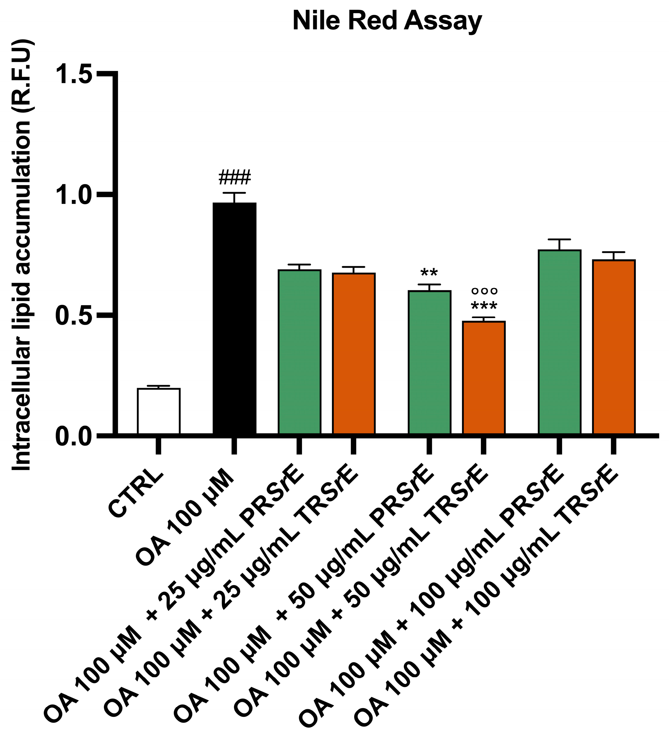
3.7. Salvia rosmarinus Spenn. Extracts Reduce Intracellular Lipid Accumulation

The Oil Red O (ORO) and Nile Red stainings showed that the 24h exposure to OA 100 μM led to a significant accumulation of cytoplasmic lipid droplets in the McA-RH7777 (p < 0.001, Figure 9 and Figure 10). The treatment with PRSrE or TRSrE significantly reduced the intracellular lipid accumulation at a concentration of 50 μg/mL (Figure 9 and Figure 10). The ORO staining highlighted a reduction in intracellular lipid accumulation at a concentration of 25 μg/mL as well, with both extract treatments (p < 0.001, Figure 9). Furthermore, the TRSrE exerted a better anti-lipidemic activity at 50 μg/mL compared to the PRSrE (Figure 9 and Figure 10).

4. Discussion

The bioactivity of plant extracts depends on their phytochemical profiles, which may be influenced by several factors, such as the harvesting period, geographical origin, chemical properties of the compounds, the extraction, and the purification methods used as well as the chosen solvent. The polarity of the latter covers an important role in the selective extraction of natural antioxidants [45].
The extraction of carnosic acid, carnosol and rosmarinic acids, which are known as the main antioxidants in S. rosmarinus, is mainly carried out in acetone, pure ethanol or hydroalcoholic mixtures [46]. Noteworthily, a mixture ranging from 50% to 80% ethanol results in the highest extraction yields and total bioactive compounds recoveries in comparison to lower EtOH or pure solvents levels [47].
Furthermore, ultrasound is a useful technology, as it does not require complex instruments and is relatively low-cost, ensuring a higher extractive value compared to traditional extraction methods such as Soxhlet, which may have some drawbacks such as their high temperature and long processing times that could affect thermolabile and unstable compounds, as well as their low selectivity and elimination of solvent residues that are often prohibited by international food and cosmetic regulations [48]. Moreover, the use of ultrasound along with ethanol improves the extraction performance of antioxidant compounds compared with other solvents (i.e., methanol) when dried plant material is used [49]. In this study, a 3-h maceration with 75% EtOH followed by sonication of dried leaves of S. rosmarinus was performed. To purify the hydroalcoholic extract and separate molecules as a function of their physicochemical properties, we first removed EtOH via a rotary evaporator [28]. The decrease in EtOH led more lipophilic molecules to precipitate in residual water, which contained hydrophilic compounds, such as polyphenols, flavonoids and short-chain organic acids [50]. The presence of physically different components of the mixture allowed us to separate the solid and liquid phases obtained from the extract via a mechanical filtration procedure. Therefore, a precipitate mainly containing terpenoids and aromatics polycyclic, named TRSrE, was mechanically separated from the hydrophilic phase via filtration through a cellulose filter. The aqueous phase was finally purified using a chromatographic column selective for anions, and an extract concentrate in phenolic compounds [51], named PRSrE, was obtained. This strategy allowed us to obtain 96 mg of TRSrE and 35 mg of PRSrE of dry extracts. Each gram of rosemary underwent extraction, corresponding to a total percentage yield of 13.1%. This percentage was in line with data reported by Lamponi et al., from another Italian rosemary collected in Tuscany [40], but our yield was different from the data published by Jacotet-Navarro et al. or by Nguyen-Kim et al., which reported extraction yields of around 25% and 33%, respectively, with 75% EtOH for S. rosmarinus collected in Morocco [47] and in Vietnam, respectively [52]. Since these authors used a conventional method of extraction, this difference may be ascribed to the environmental conditions, as the chemical composition of rosemary can be affected by soil properties [53], fertilizers [54], saltiness [55], humidity and temperature [56]. Furthermore, Balouiri et al., using a 22-h two-step maceration with hexane and methanol, were able to achieve an extraction yield of 15.8%, in line with our data, whereas, using an ultrasonic extraction, a yield of 8.7% was obtained [52].
The antioxidant power of a certain extract is generally due to its content in polyphenol and flavonoid compounds [51]. In our study, the TPC for the PRSrE was 255.6 ± 10.98 mg GAE/g dw, whereas in the TRSrE the TPC was 42.80 ± 4.42 mg GAE/g dw. Pontillo et al., using an enzyme-assisted extraction as a pre-treatment for maceration, achieved a maximum TPC of 15.2± 0.3 mg GAE/g, whereas a microwave-assisted extraction with various ratios of hydroalcoholic solution obtained a TPC in a range between 3 ± 0.3 and 8.9 ± 06 mg GAE/g [57]. Furthermore, in a recent study, it was shown that using a Soxhlet extraction, a TPC range between 15 ± 1.93 mg GAE/g and 34.72 ± 1.65 mg GAE/g, depending on the solvent used, could be obtained in a wild rosemary from northern Morocco [58].
Regarding the flavonoid content, the PRSrE showed a TFC of 1065 ± 18.83 mg RE/g dw, and the TRSrE showed a TFC of 208.4 ± 16.33 RE/g dw. Butu et al. achieved TFC values of 337.97 ± 0.50 mg RE/g and 243.63 ± 0.17 mg RE/g via Soxhlet and percolation methods, respectively [59].
Therefore, our TPC and TFC results are coherent with the values reported in the literature in regard to the TRSrE, whereas the higher TPC and TFC values showed by the PRSrE were due to the adopted strategy to concentrate polyphenolic compounds through the polystyrene absorbing resin column.
Overall, the optimization of our protocol of ultrasound-assisted maceration also offered a valid tool to obtain a more powerful extraction selectivity compared to a simple maceration technique. Indeed, the phytochemical analysis of the PRSrE showed a higher concentration of rosmarinic acid (19%) versus carnosic acid (0.6%) and carnosol (0.3%), whereas the TRSrE exhibited an enrichment in carnosol (7.2%) and carnosic acid (1.9%) followed by rosmarinic acid (2%).
Based on these results and on the biological properties referred to S. rosmarinus [60], we tested the effects of PRSrE and TRSrE on the hepatoma McA-RH7777 cell line and, specifically, their antioxidant and antilipidemic activities.
To verify the antioxidant properties, we first performed the classical DPPH assay [61] that highlighted the concentration-dependent radical scavenging activity of both studied extracts, although the PRSrE exerted a higher free radical scavenging power than the TRSrE. A further characterization, carried out using EPR, demonstrated that this difference can be attributed to the diversified scavenging activity against hydroxyl radical (OH) detected through the typical 4-line spectrum of the BMPO-OH adduct. The analysis demonstrated that the PRSrE displayed a hydroxyl radical scavenging percentage of 65.23%, compared to the 48.76% of the TRSrE. On the other hand, the TRSrE exerted a better anti-lipidemic activity compared to the PRSrE at 50 μg/mL.
This latter result, highlighting the different activities of the two extracts, may underline the added value of TRSrE in counteracting the development of metabolic diseases such as NAFLD/MAFLD [62,63]. The first step in NAFLD is represented by lipid accumulation in hepatocytes, triggering free radical overproduction that further amplifies cell damage [23]. Evidence exists that the biological activity of carnosic acid can be due to its anti-obesity properties. Indeed, it can regulate fatty acid metabolism and can inversely control the expression of hepatic lipogenesis-related genes (L-FABP, SCD1 and FAS) and of a lipolysis-related gene (CPT1) [64]. In addition, carnosol and carnosic acid revealed hypoglycemic and hypolipidemic effects by the activation of signaling pathways, including AMP-activated protein kinase (AMPK) and PPAR-γ, and by the up-regulation of the low-density lipoprotein cholesterol (LDL-C) receptor and PGC1α [65,66]. Thus, despite the lowest scavenging activity of TRSrE against OH, this action, combined with the modulation of lipid metabolism and the inhibition of lipid accumulation, represents an added value. Indeed, it suggests a potential use of our extract in the prevention of hepatic steatosis, but also of heart dysfunction [67,68,69,70], confirming the cardioprotective properties of polyphenols and terpenoids [71,72].
Overall, these results demonstrated that the optimization of selective extraction methods of bioactive compounds from plants constitutes a fundamental approach to studying their biological activity that is aimed at supporting their use in several research fields.

Author Contributions

Conceptualization, V.M. (Vincenzo Musolino) and V.M. (Vincenzo Mollace); formal analysis, V.M. (Vincenzo Musolino) and A.C.; funding acquisition, V.M. (Vincenzo Musolino), M.G. and V.M. (Vincenzo Mollace); investigation, V.M. (Vincenzo Musolino), R.M. (Roberta Macrì), A.C., L.T., M.S., C.L., S.M., R.M. (Rosario Mare), S.N., L.G., M.M., A.R.C., C.C. and M.G.; project administration, V.M. (Vincenzo Musolino), M.G. and V.M. (Vincenzo Mollace); supervision, V.M. (Vincenzo Mollace); writing—original draft, V.M. (Vincenzo Musolino), A.C., A.R.C. and M.G. All authors have read and agreed to the published version of the manuscript.

Funding

This research was funded by PON-MIUR 03PE000_78_1 and PONMIUR 03PE000_78_2/Ministry of Education, Universities and Research. PRIR Calabria Asse 1/Azione 1.5.1/FESR (Progetto AgrInfra Calabria)/PRIR Calabria.

Data Availability Statement

The data presented in this study are available in the article. The original files are available on request from the corresponding author.

Conflicts of Interest

The authors declare no conflict of interest.

References

  1. de Macedo, L.M.; dos Santos, É.M.; Militão, L.; Tundisi, L.L.; Ataide, J.A.; Souto, E.B.; Mazzola, P.G. Rosemary (Rosmarinus officinalis L.; syn Salvia rosmarinus Spenn.) and Its Topical Applications: A Review. Plants 2020, 9, 651. [Google Scholar] [CrossRef]
  2. Leporini, M.; Bonesi, M.; Loizzo, M.R.; Passalacqua, N.G.; Tundis, R. The Essential Oil of Salvia rosmarinus Spenn. from Italy as a Source of Health-Promoting Compounds: Chemical Profile and Antioxidant and Cholinesterase Inhibitory Activity. Plants 2020, 9, 798. [Google Scholar] [CrossRef]
  3. de Oliveira, J.R.; Camargo, S.E.A.; de Oliveira, L.D. Rosmarinus officinalis L. (rosemary) as therapeutic and prophylactic agent. J. Biomed. Sci. 2019, 26, 5. [Google Scholar] [CrossRef] [PubMed]
  4. Drew, B.T.; González-Gallegos, J.G.; Xiang, C.; Kriebel, R.; Drummond, C.P.; Walked, J.B.; Sytsma, K.J. Salvia united: The greatest good for the greatest number. Taxon 2017, 66, 133–145. [Google Scholar] [CrossRef]
  5. Ribeiro-Santos, R.; Carvalho-Costa, D.; Cavaleiro, C.; Costa, H.S.; Albuquerque, T.G.; Castilho, M.D.; Ramos, F.; Melo, N.R.; Sanches-Silva, A. A novel insight on an ancient aromatic plant: The rosemary (Rosmarinus officinalis L.). Trends Food Sci. Technol. 2015, 45, 355–368. [Google Scholar] [CrossRef]
  6. Lorenzo, J.M.; Munekata, P.E.S.; Pateiro, M.; Domínguez, R.; Alaghbari, M.A.; Tomasevic, I. Preservation of meat products with natural antioxidants from rosemary. IOP Conf. Ser. Earth Environ. Sci. 2021, 854, 012053. [Google Scholar] [CrossRef]
  7. Josipović, R.; Knežević, Z.M.; Frece, J.; Markov, K.; Kazazić, S.; Mrvčić, J. Improved Properties and Microbiological Safety of Novel Cottage Cheese Containing Spices. Food Technol. Biotechnol. 2015, 53, 454–462. [Google Scholar] [CrossRef] [PubMed]
  8. Loi, S. De-orientalising Ritual Blood Calabria’s Vattienti, a Case Study. Ann. Ca’ Foscari Ser. Orient. 2018, 54, 461–490. [Google Scholar] [CrossRef]
  9. Sarkic, A.; Stappen, I. Essential Oils and Their Single Compounds in Cosmetics—A Critical Review. Cosmetics 2018, 5, 11. [Google Scholar] [CrossRef]
  10. Puerto, J. La Fuerza de Fierabrás. Medicina, Ciencia y Terapéutica en Tiempos del Quijote; Editorial Just in Time SL: Madrid, Spain, 2005. [Google Scholar]
  11. Colica, C.; Di Renzo, L.; Aiello, V.; De Lorenzo, A.; Abenavoli, L. Rosmarinic Acid as Potential Anti-Inflammatory Agent. Rev. Recent Clin. Trials 2018, 13, 240–242. [Google Scholar] [CrossRef]
  12. Ferreira, H.P. Assessment Report on Rosmarinus officinalis L., Aetheroleum and Rosmarinus officinalis L., Folium; Committee on Herbal Medicinal Products (HMPC): London, UK, 2010. [Google Scholar]
  13. Apostolides, N.A.; El Beyrouthy, M.; Dhifi, W.; Najm, S.; Cazier, F.; Najem, W.; Labaki, M.; AbouKaïs, A. Chemical Composition of Aerial Parts of Rosmarinus officinalis L. Essential Oil Growing Wild in Lebanon. J. Essent. Oil Bear. Plants 2013, 16, 274–282. [Google Scholar] [CrossRef]
  14. Saeidi, S.; Forgani, F.; Javadian, F.; Javadian, E. Effects of Rosmarinus officinalis Plant Extract on Trichomonas vaginalis Parasites and Candida albicans under Laboratory Conditions: An Experimental Study. Gene Cell Tissue 2019, 6, e92867. [Google Scholar] [CrossRef]
  15. Loizzo, M.R.; Bonesi, M.; Passalacqua, N.G.; Saab, A.; Menichini, F.; Tundis, R. Antiproliferative activities on renal, prostate and melanoma cancer cell lines of Sarcopoterium spinosum aerial parts and its major constituent tormentic acid. Anti-Cancer Agents Med. Chem. 2013, 13, 768–776. [Google Scholar] [CrossRef] [PubMed]
  16. Bai, N.; He, K.; Roller, M.; Lai, C.S.; Shao, X.; Pan, M.H.; Ho, C.T. Flavonoids and phenolic compounds from Rosmarinus officinalis. J. Agric. Food Chem. 2010, 58, 5363–5367. [Google Scholar] [CrossRef] [PubMed]
  17. Borrás-Linares, I.; Stojanović, Z.; Quirantes-Piné, R.; Arráez-Román, D.; Švarc-Gajić, J.; Fernández-Gutiérrez, A.; Segura-Carretero, A. Rosmarinus officinalis Leaves as a Natural Source of Bioactive Compounds. Int. J. Mol. Sci. 2014, 15, 20585–20606. [Google Scholar] [CrossRef]
  18. Rašković, A.; Milanović, I.; Pavlović, N.; Ćebović, T.; Vukmirović, S.; Mikov, M. Antioxidant activity of rosemary (Rosmarinus officinalis L.) essential oil and its hepatoprotective potential. BMC Complement. Altern. Med. 2014, 14, 225. [Google Scholar] [CrossRef] [PubMed]
  19. Ngo, S.N.T.; Williams, D.B.; Head, R.J. Rosemary and Cancer Prevention: Preclinical Perspectives. Crit. Rev. Food Sci. Nutr. 2011, 51, 946–954. [Google Scholar] [CrossRef]
  20. Morel, S.; Hugon, G.; Vitou, M.; Védère, M.; Fons, F.; Rapior, S.; Saint, N.; Carnac, G. A Bioassay-Guided Fractionation of Rosemary Leaf Extract Identifies Carnosol as a Major Hypertrophy Inducer in Human Skeletal Muscle Cells. Nutrients 2021, 13, 4190. [Google Scholar] [CrossRef]
  21. Soliman, G.A. Effect of Rosmarinus officinalis on Lipid Profile of Streptozotocin-induced Diabetic Rats. Egypt. J. Hosp. Med. 2013, 53, 809–815. [Google Scholar] [CrossRef]
  22. Wang, S.J.; Chen, Q.; Liu, M.Y.; Yu, H.Y.; Xu, J.Q.; Wu, J.Q.; Zhang, Y.; Wang, T. Regulation effects of rosemary (Rosmarinus officinalis Linn.) on hepatic lipid metabolism in OA induced NAFLD rats. Food Funct. 2019, 10, 7356–7365. [Google Scholar] [CrossRef]
  23. Musolino, V.; Gliozzi, M.; Scarano, F.; Bosco, F.; Scicchitano, M.; Nucera, S.; Carresi, C.; Ruga, S.; Zito, M.C.; Maiuolo, J.; et al. Bergamot Polyphenols Improve Dyslipidemia and Pathophysiological Features in a Mouse Model of Non-Alcoholic Fatty Liver Disease. Sci. Rep. 2020, 10, 2565. [Google Scholar] [CrossRef] [PubMed]
  24. Nucera, S.; Ruga, S.; Cardamone, A.; Coppoletta, A.R.; Guarnieri, L.; Zito, M.C.; Bosco, F.; Macrì, R.; Scarano, F.; Scicchitano, M.; et al. MAFLD progression contributes to altered thalamus metabolism and brain structure. Sci. Rep. 2022, 12, 1207. [Google Scholar] [CrossRef] [PubMed]
  25. Del Baño, M.J.; Castillo, J.; Benavente-García, O.; Lorente, J.; Martín-Gil, R.; Acevedo, C.; Alcaraz, M. Radioprotective-antimutagenic effects of rosemary phenolics against chromosomal damage induced in human lymphocytes by gamma-rays. J. Agric. Food Chem. 2006, 54, 2064–2068. [Google Scholar] [CrossRef]
  26. del Baño, M.J.; Lorente, J.; Castillo, J.; Benavente-García, O.; del Río, J.A.; Ortuño, A.; Quirin, K.W.; Gerard, D. Phenolic diterpenes, flavones, and rosmarinic acid distribution during the development of leaves, flowers, stems, and roots of Rosmarinus officinalis. Antioxidant activity. J. Agric. Food Chem. 2003, 51, 4247–4253. [Google Scholar] [CrossRef]
  27. Yesil-Celiktas, O.; Nartop, P.; Gurel, A.; Bedir, E.; Vardar-Sukan, F. Determination of phenolic content and antioxidant activity of extracts obtained from Rosmarinus officinalis’ calli. J. Plant Physiol. 2007, 164, 1536–1542. [Google Scholar] [CrossRef] [PubMed]
  28. Zhang, Q.W.; Lin, L.G.; Ye, W.C. Techniques for extraction and isolation of natural products: A comprehensive review. Chin. Med. 2018, 13, 20. [Google Scholar] [CrossRef] [PubMed]
  29. Azmir, J.; Zaidul, I.S.M.; Rahman, M.M.; Sharif, K.M.; Mohamed, A.; Sahena, F.; Jahurul, M.H.A.; Ghafoor, K.; Norulaini, N.A.N.; Omar, A.K.M. Techniques for extraction of bioactive compounds from plant materials: A review. J. Food Eng. 2013, 117, 426–436. [Google Scholar] [CrossRef]
  30. Nn, A. A Review on the Extraction Methods Use in Medicinal Plants, Principle, Strength and Limitation. Med. Aromat. Plants 2015, 4, 1–6. [Google Scholar]
  31. Ali, A.; Chua, B.L.; Chow, Y.H. An insight into the extraction and fractionation technologies of the essential oils and bioactive compounds in Rosmarinus officinalis L.: Past, present and future. TrAC Trends Anal. Chem. 2019, 118, 338–351. [Google Scholar] [CrossRef]
  32. Lešnik, S.; Furlan, V.; Bren, U. Rosemary (Rosmarinus officinalis L.): Extraction techniques, analytical methods and health-promoting biological effects. Phytochem. Rev. 2021, 20, 1273–1328. [Google Scholar] [CrossRef]
  33. Chemat, F.; Rombaut, N.; Sicaire, A.G.; Meullemiestre, A.; Fabiano-Tixier, A.S.; Abert-Vian, M. Ultrasound assisted extraction of food and natural products. Mechanisms, techniques, combinations, protocols and applications. A review. Ultrason. Sonochem. 2017, 34, 540–560. [Google Scholar] [CrossRef] [PubMed]
  34. Musolino, V.; Macrì, R.; Cardamone, A.; Serra, M.; Coppoletta, A.R.; Tucci, L.; Maiuolo, J.; Lupia, C.; Scarano, F.; Carresi, C.; et al. Nocellara Del Belice (Olea europaea L. Cultivar): Leaf Extract Concentrated in Phenolic Compounds and Its Anti-Inflammatory and Radical Scavenging Activity. Plants 2022, 12, 27. [Google Scholar] [CrossRef] [PubMed]
  35. Irakli, M.; Chatzopoulou, P.; Ekateriniadou, L. Optimization of ultrasound-assisted extraction of phenolic compounds: Oleuropein, phenolic acids, phenolic alcohols and flavonoids from olive leaves and evaluation of its antioxidant activities. Ind. Crops Prod. 2018, 124, 382–388. [Google Scholar] [CrossRef]
  36. Erkan, N.; Ayranci, G.; Ayranci, E. Antioxidant activities of rosemary (Rosmarinus officinalis L.) extract, blackseed (Nigella sativa L.) essential oil, carnosic acid, rosmarinic acid and sesamol. Food Chem. 2008, 110, 76–82. [Google Scholar] [CrossRef] [PubMed]
  37. Bernatoniene, J.; Cizauskaite, U.; Ivanauskas, L.; Jakstas, V.; Kalveniene, Z.; Kopustinskiene, D.M. Novel approaches to optimize extraction processes of ursolic, oleanolic and rosmarinic acids from Rosmarinus officinalis leaves. Ind. Crops Prod. 2016, 84, 72–79. [Google Scholar] [CrossRef]
  38. Giordano, A.; Morales-Tapia, P.; Moncada-Basualto, M.; Pozo-Martínez, J.; Olea-Azar, C.; Nesic, A.; Cabrera-Barjas, G. Polyphenolic Composition and Antioxidant Activity (ORAC, EPR and Cellular) of Different Extracts of Argylia radiata Vitroplants and Natural Roots. Molecules 2022, 27, 610. [Google Scholar] [CrossRef]
  39. Mollace, R.; Macrì, R.; Tavernese, A.; Gliozzi, M.; Musolino, V.; Carresi, C.; Maiuolo, J.; Fini, M.; Volterrani, M.; Mollace, V. Comparative Effect of Bergamot Polyphenolic Fraction and Red Yeast Rice Extract in Rats Fed a Hyperlipidemic Diet: Role of Antioxidant Properties and PCSK9 Expression. Nutrients 2022, 14, 477. [Google Scholar] [CrossRef]
  40. Lamponi, S.; Baratto, M.C.; Miraldi, E.; Baini, G.; Biagi, M. Chemical Profile, Antioxidant, Anti-Proliferative, Anticoagulant and Mutagenic Effects of a Hydroalcoholic Extract of Tuscan Rosmarinus officinalis. Plants 2021, 10, 97. [Google Scholar] [CrossRef]
  41. Araniti, F.; Marrelli, M.; Lupini, A.; Mercati, F.; Statti, G.A.; Abenavoli, M.R. Phytotoxic activity of Cachrys pungens Jan, a mediterranean species: Separation, identification and quantification of potential allelochemicals. Acta Physiol. Plant. 2014, 36, 1071–1083. [Google Scholar] [CrossRef]
  42. Kumar, P.; Nagarajan, A.; Uchil, P.D. Analysis of Cell Viability by the MTT Assay. Cold Spring Harb. Protoc. 2018, 6. [Google Scholar] [CrossRef]
  43. De Chiara, F.; Ferret-Miñana, A.; Fernández-Costa, J.M.; Senni, A.; Jalan, R.; Ramón-Azcón, J. Fatty Hepatocytes Induce Skeletal Muscle Atrophy In Vitro: A New 3D Platform to Study the Protective Effect of Albumin in Non-Alcoholic Fatty Liver. Biomedicines 2022, 10, 958. [Google Scholar] [CrossRef] [PubMed]
  44. Ghasemi, M.; Turnbull, T.; Sebastian, S.; Kempson, I. The MTT Assay: Utility, Limitations, Pitfalls, and Interpretation in Bulk and Single-Cell Analysis. Int. J. Mol. Sci. 2021, 22, 12827. [Google Scholar] [CrossRef] [PubMed]
  45. Kaczorová, D.; Karalija, E.; Dahija, S.; Bešta-Gajević, R.; Parić, A.; Ćavar Zeljković, S. Influence of Extraction Solvent on the Phenolic Profile and Bioactivity of Two Achillea Species. Molecules 2021, 26, 1601. [Google Scholar] [CrossRef] [PubMed]
  46. de AR Oliveira, G.; de Oliveira, A.E.; da Conceição, E.C.; Leles, M.I. Multiresponse optimization of an extraction procedure of carnosol and rosmarinic and carnosic acids from rosemary. Food Chem. 2016, 211, 465–473. [Google Scholar] [CrossRef] [PubMed]
  47. Jacotet-Navarro, M.; Laguerre, M.; Fabiano-Tixier, A.S.; Tenon, M.; Feuillère, N.; Bily, A.; Chemat, F. What is the best ethanol-water ratio for the extraction of antioxidants from rosemary? Impact of the solvent on yield, composition, and activity of the extracts. Electrophoresis 2018, 39, 1946–1956. [Google Scholar] [CrossRef] [PubMed]
  48. Sridhar, A.; Ponnuchamy, M.; Kumar, P.S.; Kapoor, A.; Vo, D.N.; Prabhakar, S. Techniques and modeling of polyphenol extraction from food: A review. Environ. Chem. Lett. 2021, 19, 3409–3443. [Google Scholar] [CrossRef] [PubMed]
  49. Paniwnyk, L.; Cai, H.; Albu, S.; Mason, T.J.; Cole, R. The enhancement and scale up of the extraction of anti-oxidants from Rosmarinus officinalis using ultrasound. Ultrason. Sonochem. 2009, 16, 287–292. [Google Scholar] [CrossRef] [PubMed]
  50. Plaskova, A.; Mlcek, J. New insights of the application of water or ethanol-water plant extract rich in active compounds in food. Front. Nutr. 2023, 10, 1118761. [Google Scholar] [CrossRef]
  51. Arceusz, A.; Wesolowski, M.; Konieczynski, P. Methods for extraction and determination of phenolic acids in medicinal plants: A review. Nat. Prod. Commun. 2013, 8, 1821–1829. [Google Scholar] [CrossRef]
  52. Nguyen-Kim, M.; Truong, Q.; Nguyen, M.; Cao-Thi, B.; Tong, T.; Dao, T.; Tran, T.; Van Tan, L.; Le, X. Optimized extraction of polyphenols from leaves of Rosemary (Rosmarinus officinalis L.) grown in Lam Dong province, Vietnam, and evaluation of their antioxidant capacity. Open Chem. 2021, 19, 1043–1051. [Google Scholar] [CrossRef]
  53. Mwithiga, G.; Maina, S.; Gitari, J.; Muturi, P. Rosemary (Rosmarinus officinalis L.) growth rate, oil yield and oil quality under differing soil amendments. Heliyon 2022, 8, e09277. [Google Scholar] [CrossRef] [PubMed]
  54. Al-Fraihat, A.H.; Al-Dalain, S.Y.; Zatimeh, A.A.; Haddad, M.A. Enhancing Rosemary (Rosmarinus officinalis, L.) Growth and Volatile Oil Constituents Grown under Soil Salinity Stress by Some Amino Acids. Horticulturae 2023, 9, 252. [Google Scholar] [CrossRef]
  55. Rathore, S.; Mukhia, S.; Kapoor, S.; Bhatt, V.; Kumar, R.; Kumar, R. Seasonal variability in essential oil composition and biological activity of Rosmarinus officinalis L. accessions in the western Himalaya. Sci. Rep. 2022, 12, 3305. [Google Scholar] [CrossRef] [PubMed]
  56. Oualdi, I.; Brahmi, F.; Mokhtari, O.; Abdellaoui, S.; Tahani, A.; Oussaid, A. Rosmarinus officinalis from Morocco, Italy and France: Insight into chemical compositions and biological properties. Mater. Today Proc. 2021, 45, 7706–7710. [Google Scholar] [CrossRef]
  57. Pontillo, A.R.N.; Papakosta-Tsigkri, L.; Lymperopoulou, T.; Mamma, D.; Kekos, D.; Detsi, A. Conventional and Enzyme-Assisted Extraction of Rosemary Leaves (Rosmarinus officinalis L.): Toward a Greener Approach to High Added-Value Extracts. Appl. Sci. 2021, 11, 3724. [Google Scholar] [CrossRef]
  58. Zeroual, A.; Sakar, E.H.; Mahjoubi, F.; Chaouch, M.; Chaqroune, A.; Taleb, M. Effects of extraction technique and solvent on phytochemicals, antioxidant, and antimicrobial activities of cultivated and wild rosemary (Rosmarinus officinalis L.) from Taounate region. Biointerface Res. Appl. Chem. 2020, 12, 8441–8452. [Google Scholar]
  59. Butu, M.; Rodino, S.; Golea, D.A.; Butu, A. Screening of the antioxidant capacity and total phenolic content of three native plants. Dig. J. Nanomater. Biostruct. (DJNB) 2014, 9, 337–345. [Google Scholar]
  60. Mena, P.; Cirlini, M.; Tassotti, M.; Herrlinger, K.A.; Dall’Asta, C.; Del Rio, D. Phytochemical Profiling of Flavonoids, Phenolic Acids, Terpenoids, and Volatile Fraction of a Rosemary (Rosmarinus officinalis L.) Extract. Molecules 2016, 21, 1576. [Google Scholar] [CrossRef]
  61. Marrelli, M.; Amodeo, V.; Puntillo, D.; Statti, G.; Conforti, F. In vitro antioxidant and anti-denaturation effects of Buglossoides purpurocaerulea (L.) I. M. Johnst. fruit extract. Nat. Prod. Res. 2023, 37, 1012–1015. [Google Scholar] [CrossRef]
  62. Song, H.M.; Li, X.; Liu, Y.Y.; Lu, W.P.; Cui, Z.H.; Zhou, L.; Yao, D.; Zhang, H.M. Carnosic acid protects mice from high-fat diet-induced NAFLD by regulating MARCKS. Int. J. Mol. Med. 2018, 42, 193–207. [Google Scholar] [CrossRef] [PubMed]
  63. Habtemariam, S. Anti-Inflammatory Therapeutic Mechanisms of Natural Products: Insight from Rosemary Diterpenes, Carnosic Acid and Carnosol. Biomedicines 2023, 11, 545. [Google Scholar] [CrossRef]
  64. Nassir, F. NAFLD: Mechanisms, Treatments, and Biomarkers. Biomolecules 2022, 12, 824. [Google Scholar] [CrossRef] [PubMed]
  65. Yao, P.; Liu, Y. Terpenoids: Natural Compounds for Non-Alcoholic Fatty Liver Disease (NAFLD) Therapy. Molecules 2022, 28, 272. [Google Scholar] [CrossRef] [PubMed]
  66. Rau, O.; Wurglics, M.; Dingermann, T.; Abdel-Tawab, M.; Schubert-Zsilavecz, M. Screening of herbal extracts for activation of the human peroxisome proliferator-activated receptor. Die Pharm. 2006, 61, 952–956. [Google Scholar]
  67. Tu, Z.; Moss-Pierce, T.; Ford, P.; Jiang, T.A. Rosemary (Rosmarinus officinalis L.) extract regulates glucose and lipid metabolism by activating AMPK and PPAR pathways in HepG2 cells. J. Agric. Food Chem. 2013, 61, 2803–2810. [Google Scholar] [CrossRef]
  68. Timm, K.N.; Tyler, D.J. The Role of AMPK Activation for Cardioprotection in Doxorubicin-Induced Cardiotoxicity. Cardiovasc. Drugs Ther. 2020, 34, 255–269. [Google Scholar] [CrossRef]
  69. Finck, B.N.; Kelly, D.P. Peroxisome proliferator-activated receptor gamma coactivator-1 (PGC-1) regulatory cascade in cardiac physiology and disease. Circulation 2007, 115, 2540–2548. [Google Scholar] [CrossRef]
  70. Cordero-Herrera, I.; Kozyra, M.; Zhuge, Z.; McCann Haworth, S.; Moretti, C.; Peleli, M.; Caldeira-Dias, M.; Jahandideh, A.; Huirong, H.; Cruz, J.d.C.; et al. AMP-activated protein kinase activation and NADPH oxidase inhibition by inorganic nitrate and nitrite prevent liver steatosis. Proc. Natl. Acad. Sci. USA 2019, 116, 217–226. [Google Scholar] [CrossRef]
  71. Scicchitano, M.; Carresi, C.; Nucera, S.; Ruga, S.; Maiuolo, J.; Macrì, R.; Scarano, F.; Bosco, F.; Mollace, R.; Cardamone, A.; et al. Icariin Protects H9c2 Rat Cardiomyoblasts from Doxorubicin-Induced Cardiotoxicity: Role of Caveolin-1 Upregulation and Enhanced Autophagic Response. Nutrients 2021, 13, 4070. [Google Scholar] [CrossRef]
  72. Carresi, C.; Musolino, V.; Gliozzi, M.; Maiuolo, J.; Mollace, R.; Nucera, S.; Maretta, A.; Sergi, D.; Muscoli, S.; Gratteri, S.; et al. Anti-oxidant effect of bergamot polyphenolic fraction counteracts doxorubicin-induced cardiomyopathy: Role of autophagy and c-kitposCD45negCD31negcardiac stem cell activation. J. Mol. Cell. Cardiol. 2018, 119, 10–18. [Google Scholar] [CrossRef] [PubMed]
Figure 1. S. rosmarinus Spenn.
Plants 12 03306 g001
Figure 2. Phytochemical characterization and quantification of S. rosmarinus extracts. HPLC chromatograms of (A) PRSrE and (B) TRSrE.
Plants 12 03306 g002
Figure 3. DPPH Assay. Antiradical scavenging activity expressed as % inhibition and IC50 values of PRSrE and TRSrE. Ascorbic Acid was used as positive control. Data were fitted with nonlinear regression to quantify the IC50 values. The results are expressed as mean ± SEM (n = 3).
Plants 12 03306 g003
Figure 4. EPR spectroscopy for DPPH radical scavenging activity. EPR spectra and respective integrated spectral areas (∫) of DPPH in the absence (blue) and presence of PRSrE (green) or TRSrE (orange). Ascorbic acid was used as positive control (black).
Plants 12 03306 g004
Figure 5. EPR spectroscopy for hydroxyl radical scavenging activity. EPR spectra and respective integrated spectral areas (∫) of BMPO-OH adduct in the absence (blue) and presence of PRSrE (green) or TRSrE (orange). Ascorbic acid was used as positive control (black).
Plants 12 03306 g005
Figure 6. Phytochemical compounds analysis. (A) Total phenolic content (TPC) and (B) total flavonoid content (TFC) of PRSrE and TRSrE. The results are expressed as mean ± SEM. ***: p < 0.001.
Plants 12 03306 g006
Figure 7. Correlation analysis.
Plants 12 03306 g007
Figure 8. MTT assay for cell viability. Cell viability in response to exposure to OA 100 μM, vehicle (DMSO) or treatment with increasing concentrations of (A) PRSrE or (B) TRSrE. The results are expressed as mean ± SEM; *: p < 0.05, **: p < 0.01, ***: p < 0.001 vs. CTRl; #: p < 0.05, ###: p < 0.001 vs. OA 100 μM; ^: p < 0.05 vs. OA 100 + DMSO (50 μg/mL); °°°: p < 0.001 vs. OA 100 + DMSO (100 μg/mL); §§§: p < 0.001 vs. OA 100 + DMSO (200 μg/mL).
Plants 12 03306 g008
Figure 9. Total fatty acid accumulation assessed using Oil Red O (ORO) staining. (A) Representative confocal images of OA-induced lipid accumulation in McA-RH7777. (B) The results are expressed as mean ± S.E.M.; ###: p < 0.001 vs. CTRL; ***: p < 0.001 vs. OA 100 µM; °: p < 0.05 vs. OA 100 µM + PRSrE 50 µg/mL.
Plants 12 03306 g009
Figure 10. Total fatty acid accumulation assessed via Nile Red assay. Intracellular lipid accumulation in McA-RH7777. The results are expressed as mean ± S.E.M. ###: p < 0.001 vs. CTRL; **: p < 0.01; ***: p < 0.001 vs. OA 100 µM; °°°: p < 0.01 vs. OA 100 µM + PRSrE µg/mL.
Plants 12 03306 g010
Table 1. Correlation analysis.
Antioxidant ActivityPearson’s rp
TPCIC50, DPPH-EPR, BMPO-OH−0.975<0.001
TFCIC50, DPPH-EPR, BMPO-OH−0.993<0.001
Disclaimer/Publisher’s Note: The statements, opinions and data contained in all publications are solely those of the individual author(s) and contributor(s) and not of MDPI and/or the editor(s). MDPI and/or the editor(s) disclaim responsibility for any injury to people or property resulting from any ideas, methods, instructions or products referred to in the content.

