Article

Fusarium foetens AQF6 Isolated from Amentotaxus ynnanensis H.L.Li as a Prolific Source of Antioxidant Compounds

1
Institute of Biotechnology, Vietnam Academy of Science and Technology, Hanoi 100000, Vietnam
2
Graduate University of Science and Technology, Vietnam Academy of Science and Technology, Hanoi 100000, Vietnam
3
Vietnam–Russia Tropical Centre, Hanoi 100000, Vietnam
4
Institute of Marine Biochemistry, Vietnam Academy of Science and Technology, Hanoi 100000, Vietnam
*
Authors to whom correspondence should be addressed.
These authors contributed equally to this work.
Appl. Sci. 2024, 14(5), 2048; https://doi.org/10.3390/app14052048
Submission received: 21 January 2024 / Revised: 20 February 2024 / Accepted: 27 February 2024 / Published: 29 February 2024
(This article belongs to the Section Applied Microbiology)

Abstract

:
Amentotaxus yunnanensis H.L.Li is a threatened conifer distributed only in China, Laos, and Vietnam, whose fungal endophytes have not been exploited yet. The aims of this study were to screen and characterize antioxidant metabolites from endophytic fungi obtained from A. yunnanensis. Using the surface sterilization method, sixteen endophytic fungi were isolated from A. yunnanensis and classified into seven genera including Fusarium, Penicillium, Aspergillus, Diaporthe, Neopestalotiopsis, Purpureocillium, and Simplicillium through the analysis of Internal Transcribed Spacer (ITS) gene sequences. Among the ethyl acetate crude extracts, Fusarium foetens AQF6 contained the highest amount of polyphenol (117.76 ± 0.94 mg gallic acid equivalent/g) and flavonoid (169.01 ± 2.09 mg quercetin equivalent/g). Importantly, 400 µg/mL AQF6 extract exhibited 95.75 ± 1.1% and 85.66 ± 1.91% of 1,1-diphenyl-2-picrylhydrazyl (DPPH) and hydroxyl radical-scavenging activities, respectively. Chemical investigation led to the isolation and identification of a new bisabolane-type sesquiterpene fusafoetriol (FUS) and 14 known compounds, among which six pure compounds showed free-radical scavenging activity against DPPH and hydroxyl radicals. Under H2O2 stress, the yeast model Saccharomyces cerevisiae treated with 4 mM FUS showed 43.31% increase in viability. FUS has the potential to be applied in nutraceutical, pharmaceutical and food technological applications. Thus, this is the first report demonstrating the presence of endophytic fungi inhabiting A. yunnanensis and their potential as a prolific source of antioxidant metabolites.

1. Introduction

Fungal endophytes are known to reside within host plants without causing apparent disease symptoms, and display mutualistic interaction. In addition, endophytic fungi have adapted to their respective host plant through the production of metabolites with diverse bioactivities such as anticancer, antimicrobial, and antioxidant activities [1]. Of note, fungal endophytes from medicinal plants even mimic the bioactive metabolites similar to their host plant, in which anticancer compounds such as paclitaxel, camptothecin, and vinblastine are outstanding examples [1,2]. Fungal metabolites extracted from endophytic fungi such as huperzine and resveratrol are reported to be natural and less toxic [3,4], leading to increasing interest in deciphering their chemical structures using mass spectrometry-based tools. Therefore, the prospection of endophytic colonizing in rare medicinal plants is essential to find new medically important compounds.
Reactive oxygen species (ROS) such as superoxide, hydrogen peroxide, and hydroxyl radicals are known as either useful by-products or the harmful burden of cellular metabolism [5]. Excessive accumulation of ROS is extremely toxic to cellular macromolecules such as lipids, proteins, carbohydrates and DNA, causing several diseases including cancer, Alzheimer, diabetes, and aging over the human life cycle [6]. Rare medicinal plants such as Tsuga chinensis and Oroxylum indicum were previously reported as prolific sources of endophytic fungi with antioxidant activity [7,8]. Endophytic fungus Fusarium solani CARE-1 was found to produce bostrycoidin, anhydrofusarubin, 4-hydroxybenzaldehyde, and fusarubin, which were highly active against DPPH free radicals [4]. Due to the side effects of synthetic antioxidants used previously, endophytic fungi may serve as an epitome of natural products in preventing ROS-related diseases.
A. yunnanensis is a threatened conifer listed in the International Union for Conservation of Nature (IUCN) Red List of Threatened Species, distributed only in Vietnam, China and Laos [9]. By chemical analysis, the plant was reported to contain sequoiaflavone, sotetsuflavone, 7,7″-dimethoxyamentoflavone, lutein, beta-sitosterol, sequoyitol, and new amentoflavone biflavonoid, 2,3-dihydro-7,7″-dimethoxyamentoflavone in the plant extracts [10]. However, endophytic fungi associated with A. yunnanensis have not been explored yet. Hence, the objective of the present study was to screen for an endophytic fungus from A. yunnanensis collected in Vietnam, which demonstrates substantial antioxidant activity. In addition, the study aimed to identify fungal metabolites, and evaluate their antioxidant potential.

2. Materials and Methods

2.1. Sample Collection and Endophytic Fungi Isolation

Fresh and healthy samples of the rare conifer A. yunnanensis were collected from Quan Ba (23°8′16″ N, 105°0′44″ E), Ha Giang province, northern Vietnam in March 2020, where there was no specific permission required. All plant samples divided into 3 parts including roots, stems, and leaves were then promptly delivered to the laboratory of the Institute of Biotechnology, Vietnam Academy of Science and Technology for fungal isolation. The collected plants were authenticated by Dr. Do Van Hai from the Institute of Ecology and Biological Resources, Vietnam Academy of Science and Technology. The voucher specimen (DL-141146) of the plant was preserved and deposited at the National Institute of Medicinal Materials, Vietnam.
The isolation of endophytic fungi was carried out according to the previous procedure [8]. Briefly, surface sterilization was performed with 70% (v/v) ethanol for 30 s, 3.5% (v/v) sodium hypochlorite for 2 min, 70% (v/v) ethanol for 2–5 s, and sterile water. The surface-disinfected samples were chopped and evenly placed onto Potato Dextrose Agar (PDA) plates supplemented with 100 mg/L streptomycin. Finally, these plates were incubated at 28 °C for 7–9 d and were observed daily. The hyphae of fungi that appeared from samples were carefully sub-cultured onto fresh PDA plates to gain pure isolates.

2.2. Identification of Endophytic Fungi

Hyphae and conidia were observed by light microscope (Olympus, Tokyo, Japan). For molecular identification, genomic DNA of fungi extracted by the microwave method was used for PCR reaction to amplify the ITS gene sequence using primer pair ITS1F (5′-CTTGGTCATTTAGAGGAAGTAA-3′) and ITS4 (5′-TCCTCCGCTTATTGATAT GC-3′) [11]. The PCR product was sequenced by the First BASE Laboratories Sdn. Bhd. (Malaysia), and was subsequently compared to the existing reference ITS sequences available onto GenBank (NCBI) using BLASTn. A phylogenetic tree was reconstructed using ClustalX v.1.81 and the Kimura two-parameter distance calculation by the Molecular Evolutionary Genetics Analysis v.7.0 (MEGA7).

2.3. Evaluation of Total Polyphenol and Flavonoid Contents Present in the Fungal Extracts

Fungal isolates were grown in 200 mL of fresh reformative medium (RM) containing 200 g/L potato infusion, 40 g/L glucose, 0.5 g/L peptone, 0.8 g/L yeast extract, and 3.0 g/L (NH4)2SO4, 2.0 g/L KH2PO4, 0.5 g/L MgSO4.7H2O adjusted to pH 5.0 at 28 °C for 10 d. After fermentation, the liquid was filtered in vacuum and extracted with ethyl acetate (EtOAc) (1:3, v/v) in a separatory funnel. The extracted liquid was evaporated using a vacuum rotary evaporator at 45 °C until the product became dry. The crude extract was weighed then dissolved in 70% (v/v) ethanol for further studies.
The Folin–Ciocalteu method with a slight modification was used to determine total phenolic content [12]. The absorbance against an ethanol blank was recorded at 765 nm using a microplate reader and the results was expressed as gallic acid equivalents (GAEs) in mg/g of dried extract (mg GAE/g). Total flavonoid content was determined following the NaNO2-Al(NO)3 colorimetric method developed previously [13]. The absorbance was measured at 510 nm and total flavonoid content was expressed in mg of quercetin equivalents (QE) per gram of dried extract (mg QE/g).

2.4. Antioxidant Activity

The ability of aqueous fungal extract or pure compound to scavenge 1,1-diphenyl-2-picrylhydrazyl (DPPH) was assessed with minor changes [8]. Briefly, the reaction contained 100 µL of 0.1 mM DPPH solution in ethanol and 100 µL of each fungal extract (400 µg/mL) or pure compound (40 µg/mL) dissolved in 70% (v/v) ethanol followed by incubation at room temperature for 30 min in dark conditions. After that, the plate was measured at 517 nm with a microplate reader and 10 µg/mL ascorbic acid was tested as a positive control.
Regarding hydroxyl radical scavenging capacity, the reaction was started by adding 1.0 mL of crude extract (400 µg/mL) or pure compound (40 µg/mL) dissolved in 70% (v/v) ethanol to a test tube containing 0.5 mL of 0.435 mM brilliant green, 1.0 mL of 0.5 mM FeSO4, and 0.75 mL of 3% (v/v) H2O2 [8]. The reaction was kept in the dark for 30 min at 37 °C, and was subsequently measured at 536 nm. About 10 µg/mL ascorbic acid was used as the standard and the experiments were performed in triplicate for each fungal extract or pure compound.

2.5. Large-Scale Fermentation, Extraction, and Identification of Pure Compounds

Large-scale fermentation was performed in a 100-L fermentor containing 70 L of RM in the following conditions: temperature 28 °C, aeration rate 0.5 vvm, agitation speed 200 rpm, 10% (v/v) seed culture, and working volume 70-L. After 10 d, the cell-free supernatant and fungal mycelia were extracted with EtOAc and methanol (MeOH) at room temperature to give EtOAc and MeOH extracts, respectively. Both extracts were combined and concentrated under reduced pressure to give a residue (25 g). The residue was suspended in water and successively partitioned with n-hexane, CH2Cl2, and EtOAc to give the corresponding extracts. The CH2Cl2 and EtOAc extracts were combined, concentrated, subjected to reversed-phase (RP) C18 column chromatography (CC) and eluted with gradient 30–100% MeOH to provide nine fractions, F1–F9. F1 was purified by RP C18 prep. HPLC using 23% acetonitrile in H2O as eluent to release 15 (2 mg), and the two subfractions F1.1 and F1.2. F1.1 was separated by RP C18 prep. HPLC, with elution of 43% MeOH to give 5 (1.7 mg). Compound 4 (5 mg) was isolated from F1.2 by RP C18 prep. HPLC using 43% MeOH as the mobile phase. F2 was subjected to purification using RP C18 prep. HPLC, with elution of 37% acetonitrile to afford 7 (9 mg) and 3 (5 mg). F3 was separated using RP C18 prep. HPLC, with a mobile phase of 43% acetonitrile to yield 6 (12 mg) and two subfractions F3.1 and F3.2. F3.1 was purified by RP C18 prep. HPLC, using 52% MeOH as the mobile phase to provide 8 (3 mg). Compound 2 (5 mg) was isolated from F1.2 by RP C18 prep. HPLC, using 43% MeOH as the mobile phase. F4 was separated using RP C18 prep. HPLC, with elution of 43% acetonitrile to provide two subfractions F4.1 and F4.2. F4.1 was purified by RP C18 prep. HPLC, using 26% acetonitrile as eluent to obtain 1 (2 mg). F4.2 was separated by RP C18 prep. HPLC, using 74% MeOH as eluent to give 11 (2.1 mg). Similarly, 9 (1.7 mg) was purified from F5 using RP C18 prep. HPLC (with elution of 43% acetonitrile and 60% MeOH). F7 was separated using RP C18 prep. HPLC, with elution of 52% acetonitrile to give two subfractions, F7.1 and F7.2. F7.1 was further purified using 70% MeOH as a mobile phase to provide 10 (2.7 mg). Compound 12 (1.9 mg) was isolated from F7.2 by RP C18 prep. HPLC using 78% MeOH as the mobile phase. The water extract was chromatographed over Diaion HP-20, using a gradient elution of MeOH in water to yield two fractions, W1 and W2. W1 was introduced to RP C18 CC, eluting with gradient 20–100% MeOH to give three subfractions (W1.1–W1.3). W1.1 was further purified by silica gel CC, using CH2Cl2-MeOH (8:1, v/v) as eluent to give 14 (1.7 mg) and 13 (3.1 mg).

2.6. Evaluation of Protective Effects of FUS on Yeast Cells during Oxidative Stress Provoked by H2O2

The S. cerevisiae BY4741 wild type purchased from Thermo Fisher Scientific, Waltham, MA, USA were cultivated overnight in the Yeast Extract Peptone Dextrose (YPD) medium at 30 °C and at 160 rpm. The overnight culture was transferred to the new YPD medium and adjusted to an optical density at an optical density at 500 nm (OD500) of 0.1. S. cerevisiae BY4741 was cultivated until an OD500 of 0.5 and treated with different FUS concentrations (2–4 mM) in a microwell plate. For the survival assay, the culture was incubated for 10 h at 30 °C and 6 μL of serial dilution was spotted onto YPD agar plates for 48 h.
For the protective experiment, the exponential culture of S. cerevisiae BY4741 was treated with 2 and 4 mM FUS for 1 h and shaken at 160 rpm and 30 °C, followed by exposure to 2 mM H2O2 for 1 h. Serially diluted cells were spread on YPD agar plates to count colony-forming units (CFUs) and cell viability [14]. The sample without treatment with H2O2 was considered as 100% viability.

2.7. Statistical Analysis

All experiments were conducted in triplicate and the data were expressed as means ± standard deviation (SD) of three replicates. The Fisher’s least significant difference (LSD) test was utilized to compare antioxidant activities of fungal extracts and pure compounds, and differences were considered statistically significant when p < 0.05.

3. Results

3.1. Isolation and Identification of Endophytic Fungi

In total, sixteen fungal endophytes were successfully recovered from the root, stem, and leaf parts of A. yunnanensis based on distinct morphological characteristics. Most of the endophytic fungi were isolated from roots (43.75%, seven isolates), followed by stems (37.50%, six isolates) and leaves (18.75%, three isolates). Based on morphological and microscopic characteristics, they belonged to different seven genera: Fusarium, Penicillium, Aspergillus, Diaporthe, Neopestalotiopsis, Purpureocillium, and Simplicillium. In support of these results, ITS sequence analysis revealed that there were 14 fungal isolates that reached over 99% in similarity to the closest match, except for two strains, AQF5 (95.33%) and AQF23 (98.83%) (Table S1). The phylogenetic tree further confirmed that fungal isolates were identified as the following: Purpureocillium lilacinum AQF1, Penicillium citrinum AQF19, Penicillium crustosum (AQF8, AQF13, AQF21), Aspergillus assiutensis AQF2, Aspergillus austwickii AQF7, Diaporthe hongkongensis AQF3, Fusarium macrosporum AQF18, Fusarium foetens (AQF4, AQF6), Fusarium perseae AQF9, Neopestalotiopsis formicarum AQF15, Simplicillium obclavatum AQF16, Fusarium sp. AQF23, and Diaporthe sp. AQF5 (Figure 1).

3.2. Preliminary Phytochemical Screening of Fungal Crude Extracts

All EtOAc extracts of obtained fungi were screened for the presence of polyphenols and flavonoids. The quantitative assays revealed that 9 out of 16 extracts contained polyphenols ranging from 57.68 ± 1.16 to 117.76 ± 0.94 mg GAE/g (Table 1). The highest total polyphenol contents were determined in the root-derived AQF6 (117.76 ± 0.94 mg GAE/g), followed by stem-derived AQF13 (106.67 ± 1.28 mg GAE/g), and root-derived AQF8 (101.84 ± 5.51 mg GAE/g). Moreover, the flavonoid content ranged from 47.19 ± 0.36 to 169.01 ± 2.09 mg QE/g, in which flavonoids were observed in the AQF6 extract mounting to 169.01 ± 2.09 mg QE/g.

3.3. Evaluation of Antioxidant Activity

The DPPH and hydroxyl assays revealed that 9 out of 16 fungal crude extracts were good scavengers of free radicals (Table 1). Among them, the AQF6, AQF13, and AQF23 extracts strongly reduced DPPH radicals at 400 µg/mL, with the highest percentage of 95.75 ± 1.06%, 93.51 ± 1.31%, and 93.72 ± 3.12%, respectively. Meanwhile, these extracts also showed moderate hydroxyl radical-scavenging activity, among which the AQF6 extract showed the highest percentage of inhibition, at 85.66 ± 1.91% (Table 1). Therefore, the fungal strain F. foetens AQF6 was chosen for further chemical investigations.

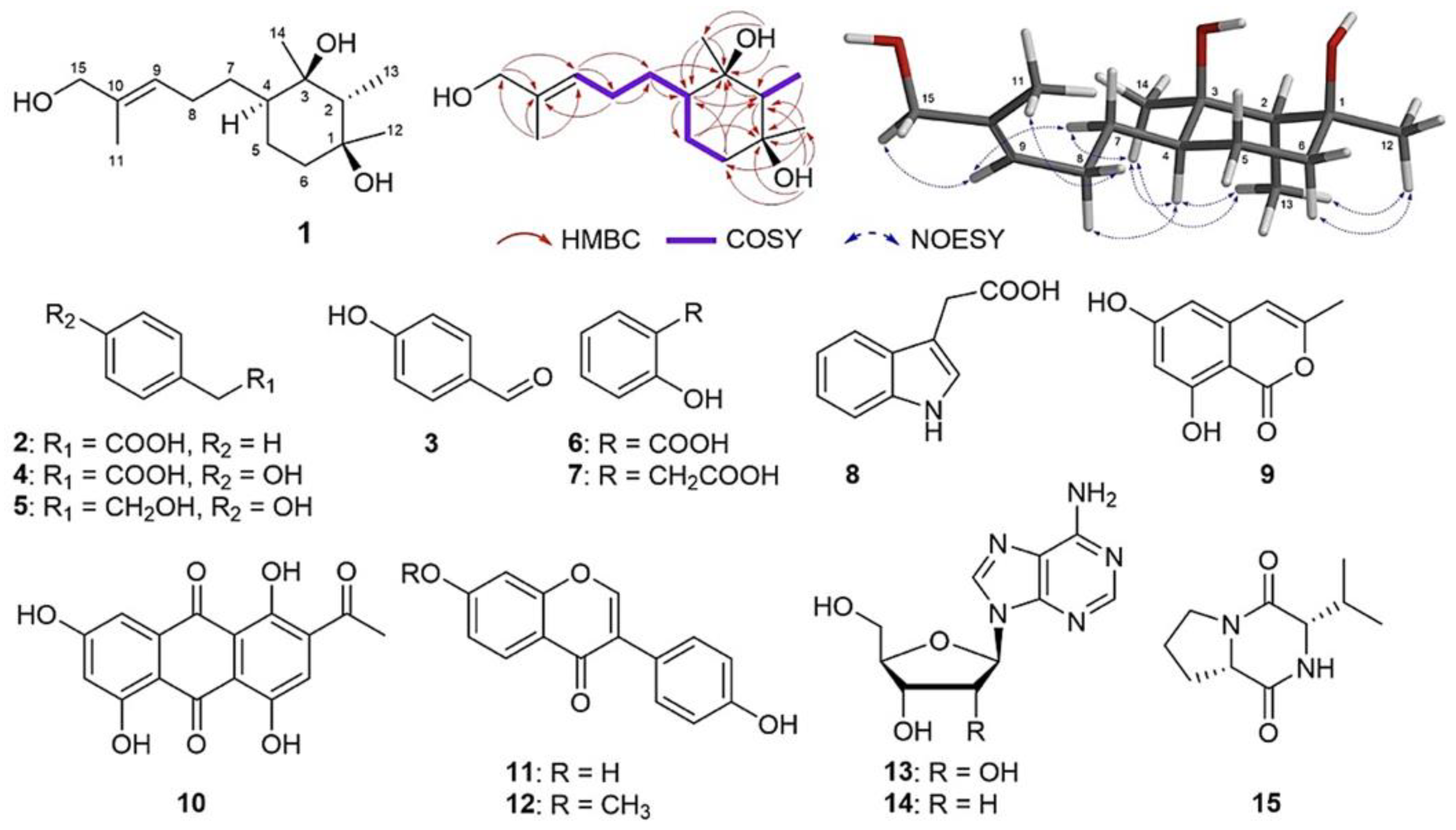
3.4. Structural Elucidation of Major Compounds

Using various chromatographic methods, 15 compounds were isolated from the fermentation culture of F. foetens AQF6. By applying spectroscopic analyses in comparison with the literature data, a new compound, FUS (1), and 14 known compounds, phenylacetic acid (2) [15], p-hydroxybenzaldehyde (3) [16], p-hydroxybenzeneacetic acid (4) [17], 2-(4-hydroxyphenyl)ethanol (5) [18], salicylic acid (6) [19], 2-hydroxybenzeneacetic acid (7) [20], indole-3-acetic acid (8) [21], 6,8-dihydroxy-3-methylisocoumarin (9) [22], 2-acetyl-1,4,5,7-tetrahydroxy-9,10-anthraquinone (10) [23], daidzein (11) [24], isoformononetin (12) [25], β-adesonine (13) [26], β-2′-deoxyadenosine (14) [26], and cyclo-Pro-Val (15) [27] were identified (Figure 2).
Compound 1 was identified as a white and amorphous powder. Its molecular formula C15H28O3 was deduced by a sodium adduct ion at m/z 279.1933 (calcd. for C15H28O3Na+, 279.1931) in high-resolution electrospray ionization mass spectrometry (HRESIMS), corresponding to two degrees of unsaturation. Three methyl singlets at δH 1.69 (H-11), 1.25 (H-12), and 1.17 (H-14), one methyl doublet at δH 1.05 (J = 7.2 Hz, H-13), and one oxymethylene broad singlet at δH 3.94 (H-15) were also observed in the 1H NMR spectrum (Figures S1 and S2). Analysis of the 13C NMR and HSQC spectra indicated 15 carbon signals attributable to one trisubstituted double bond [δC 127.0 (C-9) and 135.8 (C-10)], four methyls, five methylenes [including one oxymethylene at δC 69.0 (C-15)], two methines, and two oxygen-bearing nonprotonated carbons at δC 82.0 (C-1) and 75.5 (C-3) (Figures S3 and S4). This, in combination with HMBC correlations from H-11 to C-9, C-10, and C-15 and from H-15 to C-9 and C-10, suggested the presence of a 2-methylpent-2-en-1-ol substituent (Figure 2). Additionally, observations of COSY correlations suggested the existence of a 1,2,3,4-tetrasubstituted cyclohexane ring (Figures S5–S7). These suppositions were supported by comparison with the NMR data of 1 with those of the previously reported bisabolane-type sesquiterpene, citrantifidiol, showing a good agreement, except for a methyl group in citrantifidiol, which was replaced by an oxymethylene group at C-10 in 1H 3.94 (br s)/ δC 69.0 (C-15)] [28].
The planar structure of 1 was further confirmed by analyzing its 1D and 2D NMR spectra acquired in DMSO-d6 (Figures S8 and S9), which showed HMBC correlations from δH 3.76 (br s, 1-OH) to C-1, C-2, C-6, and C-13 and from δH 3.82 (br s, 3-OH) to C-3, C-4, and C-14 (Figures S10 and S11). The relative configuration of 1 was determined by detailed analysis of the NOESY spectrum (Figure 2). H-9 was shown to have NOE correlations with H-7 and H-15, while H-11 correlating with H-8 indicated E geometry for the 9,10-double bond. Furthermore, NOE correlations observed between H-4/H-13, H-4/H-14, and H-13/H-12 suggested that axial protons H-4 and H-13 and equatorial H-12 and H-14 are co-facial (Figure 2). On the basis of spectroscopic evidence, the structure of 1 was identified and named FUS.

3.5. Antioxidant Potential of Pure Compounds

Out of 15 pure compounds, only 6 showed antioxidant activity. Indeed, four pure compounds, such as 2-(4-hydroxyphenyl)ethanol, isoformononetin, salicylic acid, and p-hydroxybenzaldehyde, exhibited the highest antioxidant activity against DPPH and hydroxyl radicals with IC50 values ranging from 0.0041–1.92 mM (Table 2). At the lower level, the IC50 values of FUS were found to be 1.76 ± 0.32 mM and 0.95 ± 0.37 mM for DPPH and hydroxyl radicals, respectively. Despite the lower IC50 value, FUS as new compound was selected for the evaluation of a protective experiment in yeast.

3.6. Protective Effect of FUS on Yeast Cells during Oxidative Stress

Given that S. cerevisiae was utilized as an excellent model organism to evaluate the potent antioxidant activity of bioactive compounds [6], yeast cells were firstly treated with 2 mM and 4 mM FUS. It turned out that FUS did not affect the growth or induce the cytotoxicity of yeast cells (Figure 3A). Even the growth of S. cerevisiae was slightly promoted by FUS. Lately, S. cerevisiae was treated with different concentrations of FUS and exposed to an oxidant H2O2. In terms of control, the exposure to 2 mM H2O2 strongly reduced the survival of yeast cells to 22.39 ± 2.30%. CFU counts indicated an increase in the survival of stressed yeast cells about 28.49% when being treated with 2 mM FUS compared to the control (Figure 3B). An increase in FUS concentration to 4 mM led to the highest rescue of survival, which was not comparable to ascorbic acid.

4. Discussion

In the present study, 16 endophytic fungi assigned to 7 fungal genera were successfully isolated from A. yunnanensis, among which 43.75% of the fungal endophytes originated from roots, 37.50% from stems, and 18.75% from leaves. In contrast, the distribution of fungi was found to be highest in leaves of Paeonia lactiflora Pallas and Ephedra pachyclada Boiss [29]. Among seven fungal genera identified, Fusarium, Penicillium, Aspergillus, and Neopestalotiopsis were the most commonly isolated genera colonizing into medicinal plants [1,8,30], while Purpureocillium and Simplicillium served as a potent biocontrol agent against plant diseases on crops [31,32]. This result supported an assumption that distribution and the culturable fungi community are greatly affected by isolation methods and the environmental and host-mediated factors in play. To the best of our knowledge, this is the first study on the isolation and identification of endophytic fungi from A. yunnanensis.
Using chemical analysis, F. foetens AQF6 isolated from roots was found to be a prolific source of plant-derived compounds. F. foetens is a phytopathogenic fungus that causes damping-off of rooibos and the destructive vascular wilt disease in Begonia plants [33]. In addition, F. foetens associated with maize produced mycotoxins including beauvericin and four fusaric acid analogs [34]. In our study, the crude extract of F. foetens AQF6 was rich in phytochemical compounds including polyphenols and flavonoids whose antioxidant potential was about 2-fold higher than Colletotrichum gloeosporioides OI-L6 from Oroxylum indicum (L.) Kurz [7]. Chemical investigation of the AQF6 extract revealed the presence of 15 plant-derived compounds divided into five groups: sesquiterpenoid (FUS), alkaloids (indole-3-acetic acid, β-2′-deoxyadenosine, β-adenosine, and cyclo-Pro-Val), simple phenolics (2-hydroxybenzeneacetic acid, p-hydroxybenzaldehyde, 2-(4-hydroxyphenyl)ethanol, p-hydroxybenzeneacetic acid, phenylacetic acid, salicylic acid), flavonoids (6,8-dihydroxy-3-methylisocoumarin, isoformononetin, daidzein), and anthraquinone (2-acetyl-1,4,5,7-tetrahydroxy-9,10-anthraquinone). Of note, the chemical profile of AQF6 was completely different from previously published F. foetens strains. A literature survey revealed that 8 out of 15 pure compounds were previously reported in the Fusarium genera, including p-hydroxybenzaldehyde, 2-(4-hydroxyphenyl)ethanol, phenylacetic acid, daidzein, β-2′-deoxyadenosine, β-adenosine, indole-3-acetic acid, and salicylic acid [1,34,35]. Based on these findings, it seems that the metabolic profile of AQF6 may be unique due to endophytic lifestyle and horizontal gene transfer during co-evolution. Since only one genome sequence of F. foetens NRRL 38302 is available on the Genbank database to date, genomic structure, fungus–plant interactions, and the biosynthetic gene clusters of AQF6 through whole-genome sequencing and genome mining will be interesting subjects for future studies.
In free-radical scavenging assays, five known compounds showed potent in vitro antioxidant activities, including 2-(4-hydroxyphenyl)ethanol (tyrosol), salicylic acid, isoformononetin, p-hydroxybenzaldehyde, and phenylacetic acid. Isoformononetin is known as a methoxydaidzein produced only by actinomycetes such as Nocardia sp. NRRL 5646 and Amycolatopsis sp. YIM 130642 [36]. Despite the lack of in vitro antioxidant-activity evidence, isoformononetin treatment led to significant decrease in ROS production through inhibition of the NLRP3/ASC/IL-1 axis, which prevented streptozotocin-induced neuroinflammation in the rat model [37]. Salicylic acid functions as a phenolic phytohormone, contributing to protection against oxidative stress through the activation of the antioxidant system of plants [38]. In contrast, p-hydroxybenzaldehyde has in vitro antioxidant activity against ABTS•+(0.96 µM Trolox equivalents/g) and peroxyl radicals (27.58 mM Trolox equivalents/g) [39]. The DPPH and hydroxyl radical-scavenging activities of 2-(4-hydroxyphenyl)ethanol obtained from our study was comparable to that of Fusarium solani PQF9 [2]. Of note, antioxidant activity of phenylacetic acid has not been reported yet, to date. Taken together, these results further confirmed that the endophytic fungus F. foetens AQF6 is a producer of antioxidant metabolites.
An important finding of the present study was the identification of FUS. Endophytic Fusarium spp. have been demonstrated to produce a number of sesquiterpenes including fusanoid, fusartricin, and agripilol which exhibit antimicrobial and anticancer activities [35]. Using the S. cerevisiae cell model, FUS was proved to be safe, and alleviated H2O2-induced oxidative stress. Similar to this study, plant-derived compounds such as quercetin, steppogenin, and morin improved the tolerance of S. cerevisiae BY4741 to oxidative stress provoked by acetic acid [6]. It is possible that the protective effect of FUS on yeast cells resulted from the direct neutralization of H2O2 or the expression of enzymatic and non-enzymatic scavengers. Thus, the detection of ROS, enzymatic activities, expression levels of antioxidant markers such as catalase, glutaredoxin, and superoxide dismutase, and the survival of antioxidant-deficient mutants in response to oxidative stress under the protection of FUS are required to demonstrate the mode of action of FUS in future studies.

5. Conclusions

The present study communicates for the first time endophytic fungi from the native conifer A. yunnanensis listed on the IUCN Red List as a promising source of antioxidants. Among 16 fungal endophytes, F. foetens AQF6 was found to produce 14 known compounds and FUS, among which 6 compounds showed moderate in vitro antioxidant activity. Current study demonstrates that AQF6 is a prospective source of plant-derived compounds, which can be utilized in the area of pharmacology. However, whole-genome sequencing to decipher fungus–plant interactions and biosynthetic gene clusters encoding for the bioactive compounds will give additional novel findings. Importantly, FUS was safe and protected S. cerevisiae from H2O2-induced oxidative damage. Also, further in vivo studies should be carried out to investigate the antioxidant mechanisms of FUS. Thus, the present study provides more encouraging information, not only to reinforce the potential of endophytic fungi from an underexplored niche, but also to expand the search for new antioxidant metabolites.

Supplementary Materials

The following supporting information can be downloaded at: https://www.mdpi.com/article/10.3390/app14052048/s1, Figure S1: HRESITOF mass spectrum of compound 1, Figure S2: 1H NMR spectrum (CD3OD, 600 MHz) of compound 1, Figure S3: 13C NMR spectrum (CD3OD, 150 MHz) of compound 1, Figure S4: HSQC spectrum (CD3OD, 600 MHz) of compound 1, Figure S5: HMBC spectrum (CD3OD, 600 MHz) of compound 1, Figure S6: COSY spectrum (CD3OD, 600 MHz) of compound 1, Figure S7: NOESY spectrum (CD3OD, 600 MHz) of compound 1, Figure S8: 1H NMR spectrum (DMSO-d6, 600 MHz) of compound 1, Figure S9: 13C NMR spectrum (DMSO-d6, 150 MHz) of compound 1, Figure S10: HSQC spectrum (DMSO-d6, 600 MHz) of compound 1, Figure S11: HMBC spectrum (DMSO-d6, 600 MHz) of compound 1; Table S1. Distribution and molecular identification of 16 endophytic strains isolated from Amentotaxus yunnanensis.

Author Contributions

Conceptualization, T.H.N.V. and N.S.P.; methodology, N.T.Q., P.C.L., Q.A.P. and C.C.N.; investigation, T.H.N.V., V.T.N., D.H.A., T.H.Q. and N.S.P.; writing—original draft preparation, T.H.N.V. and N.S.P.; writing—review and editing, H.H.C. and Q.-T.P.; T.H.N.V. and N.S.P. contributed equally to this work. All authors have read and agreed to the published version of the manuscript.

Funding

This study was financially supported by the Vietnam Academy of Science and Technology under Grant number TĐCNSH.05/20-22.

Institutional Review Board Statement

Not applicable.

Informed Consent Statement

Not applicable.

Data Availability Statement

The data presented in this study are available in this paper.

Acknowledgments

The authors would like to acknowledge the support of the VAST-Culture Collection of Microorganisms, Institute of Biotechnology, Vietnam Academy of Science and Technology.

Conflicts of Interest

The authors declare no conflicts of interest.

References

  1. Chandra, S. Endophytic fungi: Novel sources of anticancer lead molecules. Appl. Microbiol. Biotechnol. 2012, 95, 47–59. [Google Scholar] [CrossRef]
  2. Vu, T.H.N.; Quach, N.T.; Pham, Q.A.; Le, P.C.; Nguyen, V.T.; Le, T.T.X.; Do, T.T.; Anh, D.H.; Quang, T.H.; Chu, H.H.; et al. Fusarium solani PQF9 isolated from Podocarpus pilgeri growing in Vietnam as a new producer of paclitaxel. Indian J. Microbiol. 2023, 63, 596–603. [Google Scholar] [CrossRef] [PubMed]
  3. Gupta, A.; Meshram, V.; Gupta, M.; Goyal, S.; Qureshi, K.A.; Jaremko, M.; Shukla, K.K. Fungal endophytes: Microfactories of novel bioactive compounds with therapeutic interventions; A comprehensive review on the biotechnological developments in the field of fungal endophytic biology over the last decade. Biomolecules 2023, 13, 1038. [Google Scholar] [CrossRef]
  4. Khan, N.; Afroz, F.; Begum, M.N.; Roy Rony, S.; Sharmin, S.; Moni, F.; Mahmood Hasan, C.; Shaha, K.; Sohrab, M.H. Endophytic Fusarium solani: A rich source of cytotoxic and antimicrobial napthaquinone and aza-anthraquinone derivatives. Toxicol. Rep. 2018, 5, 970–976. [Google Scholar] [CrossRef]
  5. Tung, Q.N.; Loi, V.V.; Busche, T.; Nerlich, A.; Mieth, M.; Milse, J.; Kalinowski, J.; Hocke, A.C.; Antelmann, H. Stable integration of the Mrx1-roGFP2 biosensor to monitor dynamic changes of the mycothiol redox potential in Corynebacterium glutamicum. Redox Biol. 2019, 20, 514–525. [Google Scholar] [CrossRef] [PubMed]
  6. Zahoor, H.; Watchaputi, K.; Hata, J.; Pabuprapap, W.; Suksamrarn, A.; Chua, L.S.; Soontorngun, N. Model yeast as a versatile tool to examine the antioxidant and anti-ageing potential of flavonoids, extracted from medicinal plants. Front. Pharmacol. 2022, 13, 980066. [Google Scholar] [CrossRef] [PubMed]
  7. Rai, N.; Keshri, P.K.; Gupta, P.; Verma, A.; Kamble, S.C.; Singh, S.K.; Gautam, V. Bioprospecting of fungal endophytes from Oroxylum indicum (L.) Kurz with antioxidant and cytotoxic activity. PLoS ONE 2022, 17, e0264673. [Google Scholar] [CrossRef] [PubMed]
  8. Vu, T.H.N.; Pham, N.S.; Le, P.C.; Pham, Q.A.; Quach, N.T.; Nguyen, V.T.; Do, T.T.; Chu, H.H.; Phi, Q.T. Distribution, cytotoxicity, and antioxidant activity of fungal endophytes isolated from Tsuga chinensis (Franch.) Pritz. in Ha Giang province, Vietnam. Ann. Microbiol. 2022, 72, 36. [Google Scholar] [CrossRef]
  9. Zhang, J.; Cao, Z.P.; Li, S.; Liu, M.M.; Huo, X.W. The complete chloroplast genome sequence of Amentotaxus yunnanensis (Taxaceae). Mitochondrial DNA B Resour. 2019, 4, 2477–2478. [Google Scholar] [CrossRef] [PubMed]
  10. Li, S.H.; Zhang, H.J.; Niu, X.M.; Yao, P.; Sun, H.D.; Fong, H.H. Chemical constituents from Amentotaxus yunnanensis and Torreya yunnanensis. J. Nat. Prod. 2003, 66, 1002–1005. [Google Scholar] [CrossRef]
  11. Quach, N.T.; Ngo, C.C.; Nguyen, T.H.; Nguyen, P.L.; Vu, T.H.N.; Phan, T.H.T.; Nguyen, Q.H.; Le, T.T.M.; Chu, H.H.; Phi, Q.T. Genome-wide comparison deciphers lifestyle adaptation and glass biodeterioration property of Curvularia eragrostidis C52. Sci. Rep. 2022, 12, 11411. [Google Scholar] [CrossRef]
  12. da Silva, M.H.R.; Cueva-Yesquén, L.G.; Júnior, S.B.; Garcia, V.L.; Sartoratto, A.; de Angelis, D.d.F.; de Angelis, D.A. Endophytic fungi from Passiflora incarnata: An antioxidant compound source. Arch. Microbiol. 2020, 202, 2779–2789. [Google Scholar] [CrossRef] [PubMed]
  13. Tang, Z.; Qin, Y.; Chen, W.; Zhao, Z.; Lin, W.; Xiao, Y.; Chen, H.; Liu, Y.; Chen, H.; Bu, T.; et al. Diversity, chemical constituents, and biological activities of endophytic fungi isolated from Ligusticum chuanxiong Hort. Front. Microbiol. 2021, 12, 771000. [Google Scholar] [CrossRef]
  14. Oliveira, D.; Dias, M.I.; Barros, L.; Custódio, L.; Oliveira, R. Antigenotoxic properties of the halophyte Polygonum maritimum L. highlight its potential to mitigate oxidative stress-related damage. Sci. Rep. 2023, 13, 3727. [Google Scholar] [CrossRef] [PubMed]
  15. Evidente, A.; Andolfi, A.; Maddau, L.; Franceschini, A.; Marras, F. Biscopyran, a phytotoxic hexasubstituted pyranopyran produced by Biscogniauxia mediterranea, a fungus pathogen of Cork Oak. J. Nat. Prod. 2005, 68, 568–571. [Google Scholar] [CrossRef]
  16. Kwon, Y.S.; Kim, C.M. Antioxidant constituents from the stem of Sorghum bicolor. Arch. Pharm. Res. 2003, 26, 535. [Google Scholar] [CrossRef]
  17. Agerbirk, N.; Warwick, S.I.; Hansen, P.R.; Olsen, C.E. Sinapis phylogeny and evolution of glucosinolates and specific nitrile degrading enzymes. Phytochemistry 2008, 69, 2937–2949. [Google Scholar] [CrossRef] [PubMed]
  18. Kwak, J.H.; Kang, M.W.; Roh, J.H.; Choi, S.U.; Zee, O.P. Cytotoxic phenolic compounds from Chionanthus retusus. Arch. Pharm. Res. 2009, 32, 1681–1687. [Google Scholar] [CrossRef]
  19. Carvalho, M.J.; Carvalho, L.M.; Ferreira, A.M.; Silva, A.M.S. A new xanthone from Hedychium gardnerianum. Nat. Prod. Res. 2003, 17, 445–449. [Google Scholar] [CrossRef]
  20. Gutierrez-Lugo, M.-T.; Woldemichael, G.M.; Singh, M.P.; Suarez, P.A.; Maiese, W.M.; Montenegro, G.; Timmermann, B.N. Isolation of three new naturally occurring compounds from the culture of Micromonospora sp. P1068. Nat. Prod. Res. 2005, 19, 645–652. [Google Scholar] [CrossRef]
  21. Singh, S.P.; Parmar, S.S.; Stenberg, V.I.; Farnum, S.A. Carbon-13 nuclear magnetic resonance spectra of anti-inflammatory drugs: Phenylbutazone, oxyphenbutazone and indomethacin. J. Heterocycl. Chem. 1978, 15, 13–16. [Google Scholar] [CrossRef]
  22. Kendall, J.K.; Fisher, T.H.; Schultz, H.P.; Schultz, T.P. An improved synthesis of 6,8-dimethoxy-3-methylisocoumarin, a fungal metabolite precursor. J. Org. Chem. 1989, 54, 4218–4220. [Google Scholar] [CrossRef]
  23. Malak, L.G.; Bishay, D.W.; Abdel-Baky, A.M.; Moharram, A.M.; Cutler, S.J.; Ross, S.A. New anthraquinone derivatives from Geosmithia lavendula. Nat. Prod. Commun. 2013, 8, 1934578X1300800215. [Google Scholar] [CrossRef]
  24. Venkateswarlu, S.; Panchagnula, G.K.; Guraiah, M.B.; Subbaraju, G.V. Isoaurostatin: Total synthesis and structural revision. Tetrahedron 2005, 61, 3013–3017. [Google Scholar] [CrossRef]
  25. Wenkert, E.; Gottlieb, H.E. Carbon-13 nuclear magnetic resonance spectroscopy of flavonoid and isoflavonoid compounds. Phytochemistry 1977, 16, 1811–1816. [Google Scholar] [CrossRef]
  26. Ciuffreda, P.; Casati, S.; Manzocchi, A. Spectral assignments and reference data complete 1H and 13C NMR spectral assignment of α- and β-adenosine, 2’-deoxyadenosine and their acetate derivatives. Magn. Reson. Chem. 2007, 45, 781–784. [Google Scholar] [CrossRef] [PubMed]
  27. Mehnaz, S.; Saleem, R.S.Z.; Yameen, B.; Pianet, I.; Schnakenburg, G.; Pietraszkiewicz, H.; Valeriote, F.; Josten, M.; Sahl, H.-G.; Franzblau, S.G.; et al. Lahorenoic Acids A–C, ortho-dialkyl-substituted aromatic acids from the biocontrol strain Pseudomonas aurantiaca PB-St2. J. Nat. Prod. 2013, 76, 135–141. [Google Scholar] [CrossRef]
  28. Evidente, A.; Ricciardiello, G.; Andolfi, A.; Sabatini, M.A.; Ganassi, S.; Altomare, C.; Favilla, M.; Melck, D. Citrantifidiene and citrantifidiol: Bioactive metabolites produced by Trichoderma citrinoviride with potential antifeedant activity toward Aphids. J. Agric. Food. Chem. 2008, 56, 3569–3573. [Google Scholar] [CrossRef] [PubMed]
  29. Cheng, X.; Wei, Z.; Pu, S.; Xiang, M.; Yan, A.; Zhang, Y.; Wang, X. Diversity of endophytic fungi of Paeonia lactiflora Pallas and screening for fungal paeoniflorin producers. FEMS Microbiol. Lett. 2018, 365, fny263. [Google Scholar] [CrossRef]
  30. Santos, T.F.B.; Dos Santos Carvalho, C.; de Almeida, M.A.; Delforno, T.P.; Duarte, I.C.S. Endophytic fungi isolated from Brazilian medicinal plants as potential producers of antioxidants and their relations with anti-inflammatory activity. 3 Biotech 2020, 10, 223. [Google Scholar] [CrossRef] [PubMed]
  31. Khan, M.; Tanaka, K. Purpureocillium lilacinum for plant growth promotion and biocontrol against root-knot nematodes infecting eggplant. PLoS ONE 2023, 18, e0283550. [Google Scholar] [CrossRef] [PubMed]
  32. Li, W.; Luo, T.; Li, J.; Zhang, J.; Wu, M.; Yang, L.; Li, G. Antifungal activity and biocontrol potential of Simplicillium lamellicola JC-1 against multiple fungal pathogens of oilseed rape. J. Fungi 2022, 9, 57. [Google Scholar] [CrossRef] [PubMed]
  33. Lamprecht, S.C.; Tewoldemedhin, Y.T. Fusarium species associated with damping-off of rooibos seedlings and the potential of compost as soil amendment for disease suppression. S. Afr. J. Bot. 2017, 110, 110–117. [Google Scholar] [CrossRef]
  34. González-Jartín, J.M.; Alfonso, A.; Sainz, M.J.; Vieytes, M.R.; Aguín, O.; Ferreiroa, V.; Botana, L.M. First report of Fusarium foetens as a mycotoxin producer. Mycotoxin Res. 2019, 35, 177–186. [Google Scholar] [CrossRef] [PubMed]
  35. Xu, M.; Huang, Z.; Zhu, W.; Liu, Y.; Bai, X.; Zhang, H. Fusarium-derived secondary metabolites with antimicrobial effects. Molecules 2023, 28, 3424. [Google Scholar] [CrossRef]
  36. Zheng, K.-X.; Jiang, Y.; Jiang, J.-X.; Huang, R.; He, J.; Wu, S.-H. A new phthalazinone derivative and a new isoflavonoid glycoside from lichen-associated Amycolatopsis sp. Fitoterapia 2019, 135, 85–89. [Google Scholar] [CrossRef]
  37. Chaturvedi, S.; Tiwari, V.; Gangadhar, N.M.; Rashid, M.; Sultana, N.; Singh, S.K.; Shukla, S.; Wahajuddin, M. Isoformononetin, a dietary isoflavone protects against streptozotocin induced rat model of neuroinflammation through inhibition of NLRP3/ASC/IL-1 axis activation. Life Sci. 2021, 286, 119989. [Google Scholar] [CrossRef]
  38. Singh, S. Salicylic acid elicitation improves antioxidant activity of spinach leaves by increasing phenolic content and enzyme levels. Food Chem. Ad. 2023, 2, 100156. [Google Scholar] [CrossRef]
  39. Kinjo, Y.; Wada, K.; Oe, M.; Hou, D.-X.; Takahashi, M. Effects of p-hydroxybenzaldehyde and p-hydroxyacetophenone from non-centrifuged Cane Sugar, Kokuto, on serum corticosterone, and liver conditions in chronically stressed mice fed with a high-fat diet. Food Sci. Technol. Res. 2020, 26, 501–507. [Google Scholar] [CrossRef]
Figure 1. A phylogenetic tree based on the ITS gene sequences showing the relationship between 16 endophytic fungi isolated from A. yunnanensis and other related species. The numbers at the nodes show bootstrap values higher than 40% and the bar scale indicates 5% divergence. Cunninghamella elegans CBS 160.28T was used as an outgroup.
Applsci 14 02048 g001
Figure 2. Chemical structures of compounds 115 and key COSY, HMBC, and NOESY correlations of fusafoetriol (1).
Applsci 14 02048 g002
Figure 3. Oxidative stress−alleviating potential of FUS. (A) Effect of FUS on the survival of S. cerevisiae BY4741. (B) Survival of S. cerevisiae pretreated with 2 mM and 4 mM FUS under H2O2 stress. Ascorbic acid (AA) was used for comparison.
Applsci 14 02048 g003
Table 1. Determination of polyphenols, flavonoids, and antioxidant activity of 16 endophytic fungi isolated from Amentotaxus yunnanensis.
Endophytic FungiPlant OrganPolyphenols
(mg QAE/g)
Flavonoids
(mg QE/g)
The Free-Radical Scavenging Activity (%)
DPPHHydroxyl
AQF1Stem71.36 ± 0.55 c47.92 ± 0.9 a45.43 ± 2.88 a62.45 ± 2.20 c
AQF2Leaf84.27 ± 0.34 d65.21 b ± 1.67 b61.43 ± 2.68 c37.18 ± 2.22 a
AQF3Root----
AQF4Root87.53 ± 0.23 d81.25 ± 1.88 d58.55 ± 1.93 c43.59 ± 2.67 b
AQF5Root----
AQF6Root117.76 ± 0.94 h169.01 ± 2.09 f95.75 ± 1.06 f85.66 ± 1.91 e
AQF7Root----
AQF8Root101.84 ± 5.5 f101.44 ± 2.70 e84.85 ± 2.2 e72.41 ± 1.43 d
AQF9Root----
AQF13Stem106.67 ± 1.28 g73.46 ± 1.89 c93.51 ± 1.31 f45.18 ± 1.52 b
AQF15Stem----
AQF16Stem----
AQF18Stem57.68 ± 1.16 a47.19 ± 0.36 a76.66 ± 1.33 d63.07 c ± 1.60 c
AQF19Stem67.06 ± 0.54 b70.63 ± 1.56 c54.08 ± 1.50 b84.86 ± 2.15 e
AQF21Leaf----
AQF23Leaf94.82 ± 0.78 e64.84 ± 1.46 b93.72 ± 3.12 f74.09 ± 2.22 d
Ascorbic acid 96.32 ± 3.97 f99.19 ± 0.23 f
Note: (-), no activity/not determined. The final concentration of each fungal extract was applied at 400 µg/mL and 10 µg/mL ascorbic acid was used as a positive control. Values with different superscripts are significantly different according to LSD test (p < 0.05).
Table 2. Antioxidant activity of 15 compounds extracted from the AQF6 extract.
CompoundsThe Free-Radical Scavenging Activity IC50 (mM)
DPPHHydroxyl
FUS (1)1.76 ± 0.32 c0.95 ± 0.37 a,b
Phenylacetic acid (2)3.73 ± 0.39 d4.46 ± 0.86 d
p-hydroxybenzaldehyde (3)1.65 ± 0.27 c0.36 ± 0.09 a
p-hydroxybenzeneacetic acid (4)ndnd
2-(4-hydroxyphenyl)ethanol (5)5.33 e ± 0.063.97 d ± 0.11
Salicylic acid (6)0.95 ± 0.08 b1.92 ± 0.61 c
2-hydroxybenzeneacetic acid (7)ndnd
Indole-3-acetic acid (8)ndnd
6,8-dihydroxy-3-methylisocoumarin (9)ndnd
2-acetyl-1,4,5,7-tetrahydroxy-9,10-anthraquinone (10)ndnd
Daidzein (11)ndnd
Isoformononetin (12)1.02 ± 0.09 c1.14 ± 0.25 b
β-adesonine (13)ndnd
β-2′-deoxyadenosine (14)ndnd
Cyclo-Pro-Val (15)ndnd
Ascorbic acid0.42 ± 0.18 a0.82 ± 0.17 a,b
Note: (nd). not determined. Values with different letters are significantly different according to the LSD test (p < 0.05).
Disclaimer/Publisher’s Note: The statements, opinions and data contained in all publications are solely those of the individual author(s) and contributor(s) and not of MDPI and/or the editor(s). MDPI and/or the editor(s) disclaim responsibility for any injury to people or property resulting from any ideas, methods, instructions or products referred to in the content.

