
Insect Antifeedant Benzofurans from Pericallis Species
by
 , ,
, ,
Carmen E. Díaz
1,* ,
,
Braulio M. Fraga
1,
Adriana G. Portero
1,
Iván Brito
2,
Carmen López-Balboa
3,
Liliana Ruiz-Vásquez
4 and
Azucena González-Coloma
3,* 1
Instituto de Productos Naturales y Agrobiología, Consejo Superior de Investigaciones Científicas (CSIC), Avda. Astrofísico F. Sánchez 3, 38206 La Laguna, Tenerife, Spain
2
Departamento de Química, Universidad de Antofagasta, Antofagasta 124000, Chile
3
Instituto de Ciencias Agrarias, Consejo Superior de Investigaciones Científicas (CSIC), Serrano 115 dpdo., 28006 Madrid, Spain
4
Laboratorio de Productos Naturales Antiparasitarios de la Amazonia, Centro de Investigación de Recursos Naturales, Universidad Nacional de la Amazonia Peruana (UNAP), 16001 Iquitos, Peru
*
Authors to whom correspondence should be addressed.
Molecules 2023, 28(3), 975; https://doi.org/10.3390/molecules28030975
Submission received: 23 November 2022
/
Revised: 4 January 2023
/
Accepted: 11 January 2023
/
Published: 18 January 2023
(This article belongs to the Special Issue Natural Product-Inspired Molecules: From Weed to Remedy)
Abstract
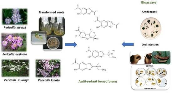
:In this work, we have studied the benzofurans of Pericallis echinata (aerial parts and transformed roots), P. steetzii (aerial parts and transformed roots), P. lanata (aerial parts), and P. murrayi (aerial parts and roots). This work has permitted the isolation of the new benzofurans 10-ethoxy-11-hydroxy-10,11-dihydroeuparin (10), (-)-eupachinin A ethyl ether (12), 11,15-didehydro-eupachinin A (13), 10,12-dihydroxy-11-angelyloxy-10,11-dihydroeuparin (14), 2,4-dihydroxy-5-formyl-acetophenone (15) isolated for the first time as a natural product, 11-angelyloxy-10,11-dihydroeuparin (16), and 12-angelyloxyeuparone (17), along with several known ones (1–9, 11). In addition, the incubation of the abundant component, 6-hydroxytremetone (1), with the fungus Mucor plumbeus has been studied. Benzofurans in the tremetone series (1, 1a, 2–5, 18, 18a), the euparin series (6, 7, 7a, 8–10, 14, 16), and the eupachinin-type (11, 12) were tested for antifeedant effects against the insect Spodoptera littoralis. The antifeedant compounds (1, 4, 6, 11, 12) were further tested for postingestive effects on S. littoralis larvae. The most antifeedant compounds were among the tremetone series, with 3-ethoxy-hydroxy-tremetone (4) being the strongest antifeedant. Glucosylation of 1 by its biotransformation with Mucor plumbeus gave inactive products. Among the euparin series, the dihydroxyangelate 14 was the most active, followed by euparin (6). The eupachinin-type compounds (11, 12) were both antifeedants. Compounds 4, 11, and 12 showed antifeedant effects without postingestive toxicity to orally dosed S. littoralis larvae. Euparin (6) had postingestive toxicity that was enhanced by the synergist piperonyl butoxide.
1. Introduction
Pericallis, a genus of the tribe Senecioneae (subtribe Senecioninae) in the Asteraceae family, comprises seventeen taxa of woody and herbaceous species which are endemic to Canary Islands, Madeira, and Azores [1]. The name Pericallis was introduced by Don in 1834 for P. tussilaginis [2] but was not used until 134 years later when Pericallis was considered close to the African Cineraria and several Cineraria and Senecio species were included in it [3]. However, a study on the molecular phylogeny of Pericallis found it closer to the American Packera genus [4]. Later, the monophyly of Pericallis was strongly supported and, again, a close relation with the African Cineraria genus was suggested considering morphologic and molecular ITS data [5]. More recently, the influence of geographical isolation, multiple habitat shifts, and hybridization in the evolution of this genus has been studied, using more taxon’s sampling and studying both nuclear and chloroplast genomes’ data [1,6].
Senecio and Pericallis species produce pyrrolizidine alkaloids (PAs) [7]. PAs are considered feeding deterrents for insect herbivores. A number of insect species from different taxa have evolved adaptations to sequester, store, and utilize plant PAs against their predators and parasitoids [8,9].
Ethanolic extracts of some PA-producing plants from the Canary Islands, including Pericallis (P. appendiculata, P. echinata, P. hansenii, P. multiflora, P. steetzii), were overall more active insect antifeedants than the alkaloidal ones. Considering that the alkaloidal fraction of these ethanolic extracts accounted for a maximum of 2%, the authors concluded that the chemistry of the non-alkaloidal fraction could explain most of their antifeedant effects [7]. Non-alkaloidal fractions of Senecio (and former Senecio such as the Canarian endemism Bethencourtia) species contain insect antifeedant sesquiterpenoids with different skeletons [10,11,12,13,14]. However, little is known on the non-alkaloidal defensive chemistry of Pericallis spp.
In this work, we have studied the benzofuran-based defensive chemistry of Pericallis echinata (L. fil.) B. Nord. (aerial parts and transformed roots), P. steetzii (Bolle) B. Nord. (aerial parts and transformed roots), P. lanata (L’Hér.) B. Nord. (aerial parts), and P. murrayi (Bornm.) B. Nord. (aerial parts and roots). Our study led to the isolation of the known benzofurans (1–9, 11), the new ones 10-ethoxy-11-hydroxy-10,11-dihydroeuparin (10), (-)-eupachinin A ethyl ether (12), 11,15-didehydro-eupachinin A (13), 10,12-dihydroxy-11-angelyloxy-10,11-dihydroeuparin (14), 11-angelyloxy-10,11-dihydroeuparin (16), 12-angelyloxyeuparone (17), and 2,4-dihydroxy-5-formyl-acetophenone (15) isolated for the first time as a natural product. In addition, the incubation of the abundant component, 6-hydroxytremetone (1), with the fungus Mucor plumbeus has been studied. Moreover, an improved identification of euparone and tremetone derivatives has been carried out using NMR data, allowing the correction of the 13C data of some of these compounds. Benzofurans in the tremetone series (1, 1a, 2–5, 18, 18a), the euparin series (6, 7, 7a, 8–10, 14, 16), and the eupachinin-type (11, 12) were tested for antifeedant effects against the insect Spodoptera littoralis. The antifeedant compounds (1, 4, 6, 11, 12) were further tested for postingestive effects on S. littoralis larvae.
2. Results and Discussion
2.1. Components of Pericallis
All the species studied contained benzofurans (Figure 1) as the major components, along with triterpenoids and sterols. Most benzofuran-type sesquiterpenes have been isolated from Asteraceae species [15], with a few examples from Senecio [11], but none have been reported from Pericallis.
The benzofurans 6-hydroxytremetone (1) [16,17], 3β,6-dihydroxytremetone (2) [18,19], 3β-methoxy-6-hydroxytremetone (3) [20,21,22], 3β-ethoxy-6-hydroxytremetone (4) [23], 3α,6-dihydroxytremetone (5) [24], euparin (6) [22,25,26], euparone (7) [22,27], 10,11-dihydroxy-10,11-dihydroeuparin (8) [28], 10-methoxy-11-hydroxy-10,11-dihydroeuparin (9) [29], 10-ethoxy-11-hydroxy-10,11-dihydroeuparin (10), eupachinin A (11) [30], eupachinin A ethyl ether (12), 11,15-didehydro eupachinin A (13), 10,12-dihydroxy-11-angelyloxy-10,11-dihydroeuparin (14), 2,4-dihydroxy-5-formyl-acetophenone (15) [31], 11-angelyloxy-10,11-dihydroeuparin (16), and 12-angelyloxyeuparone (17) were isolated from the different Pericallis species and plant parts. The pentacyclic triterpenes 3β,16β-dihydroxy-lup-20(29)-ene [32], 11-oxo-α-amirin [33], 11-oxo-β-amirin [34], 3α,16β-dihydroxy-urs-12-ene [35], 3β,16β-dihydroxy-urs-12-ene [35], and 3β,16β-dihydroxy-olean-12-ene [36], and the sterols campesterol, stigmasterol, and β-sitosterol, were also identified in these species.
We describe now for the first time the 13C NMR (Table 1) spectra of 3β,6-dihydroxytremetone (2) [18,19], 3β-ethoxy-6-hydroxytremetone (4) [23], 3α,6-dihydroxytremetone (5) [24], 10,11-dihydroxy-10,11-dihydroeuparin (8) [28], and 10-methoxy-11-hydroxy-10,11-dihydroeuparin (9) [29]. In addition, we have corrected some assignments in the carbon resonances of 6-hydroxytremetone (1) [17] and euparin (6) [26]. The 13C NMR spectrum of euparone (7), reported by Hussein (1992) [28], does not match with our data and those described by Habtemariam (2001) [22], which has also now been reassigned.
To complete these studies, derivatives of 6-hydroxytremetone (1) and euparone (7) were prepared. Compounds 1 and 7 were acetylated to give 6-AcO derivatives 1a and 7a, respectively, and their NMR and MS spectroscopic data confirmed the presence of the AcO group at C-6.
The high-resolution mass spectrum (HREIMS) of compound 11 showed the molecular ion at m/z 274.0834 (C15H14O5). In its 1H NMR spectrum were observed signals corresponding to the ring A of a 6-hydroxy-5-acetyl benzofuran grouping. Other resonances present in it were a methyl geminal to an oxygenated function at δ 1.78 and two methylene groups located in contiguous positions. The hydrogens of one of them resonate at δ 2.63 (1H, ddd, J = 17.0, 6.5 and 5.0 Hz, H-15β) and 2.92 (1H, ddd, J = 17.0, 9.0 and 5.0 Hz, H-15α), while that those of the second methylene, coupled with the previous one, appear centred at δ 2.36 (m, 2H-11). In its 13C NMR spectrum were detected signals of two methyls, two methylenes, two aromatic protons, and two carbonyl groups, one the acetyl group on C-5 to δ 194.1 and another of an α,β-unsaturated carbonyl group at δ 204.6 (C-16). Moreover, a quaternary carbon bearing an oxygen atom at δ 67.4 (C-10) and other six unsaturated quaternary carbons also appear in this spectrum.
The location of the different functional groups was established based on the connectivity observed in the HSQC and HMBC spectra. The correlations in the HMBC experiment of the aromatic protons and the phenolic proton, with their respective geminal and neighbourhood carbons, confirmed that the ring A of the molecule was similar to that of other benzofurans of euparin type. This spectrum also indicated that the two methylene groups were located between the carbonyl group and the quaternary carbon bearing an oxygen atom. Thus, connectivities of H-11 with C-2, C-10, and C-15, of the two H-15 with C-10, C-11, and C-16, and of methyl group (H-12) with C-2, C-10, and C-11 were observed. These spectroscopic data allowed us to assign to our product the structure 11 [37]. To confirm this hypothesis, we submit it to an X-ray diffraction analysis (Figure 2).
The asymmetric unit of the title cocrystal, 3C15H14O5—C15H12O5 contains four structural units. The A-C molecules with the same structure, C15H14O5, and the D-molecule with an additional 11,15-double bond, C15H12O5. Intramolecular O—H···O interactions occur in all four independent molecules with graph-set motif S(6). In the crystal, only B molecule is linked to C and D molecules by O—H···O intermolecular hydrogen bonds and π-π stacking interactions between the benzene rings A and C molecules (centroid–centroid distances ranging from 3.696 (3) to 3.721 (4) Å). The conformation of the molecules A and B are similar showing an envelope conformation for the methyl-cyclohexanone ring. The C molecule has a screw-boat conformation for the methyl-cyclohexanone ring, and the methyl group is in an axial position. The molecule D has a planar conformation (mean torsion angle 5.106 (12)° due to the presence of the 11,15-double bond.
Once the relative structure of compound 11 and its 11,15-didehydro derivative 13 had been established by X-ray analysis, a pair of enantiomers (-) and (+)-eupachinin A were isolated from Eupatorium chinense with [α]D -5.1 and +4.8, respectively [30]. Spectroscopic data were identical to those of our compound of optical rotation −8.33, which was consequently identified as (-)-eupachinin A (11). The presence of the undescribed 11,15-didehydro derivative 13 in the crystal fraction of the X-ray analysis was unexpected, because NMR signals of the 11,15-double bond were not observed in the 1H and 13C spectra of the fractions studied.
(-)-10-Ethoxy-eupachinin A (12), the corresponding ethyl derivative of 11, was also isolated from P. echinata. With the molecular formula C17H18O5, its 1H and 13C NMR spectra were very similar to that of compound 11, except that now the signals corresponding to a 10-ethoxy group also appear. This compound probably is an artefact formed in the extraction of the plant with ethanol. The same applies to the undescribed benzofuran 10-ethoxy-11-hydroxy-euparin (10), also obtained from aerial part of P. echinata together with its known analogues 8 and 9.
The new benzofuran 14 was isolated from the transformed roots of P. echinata and P.steetzzi. Its high-resolution mass spectrum showed the molecular ion at m/z 348.1211 (C18H20O7). The 1H NMR spectrum displayed the signals of the aromatic protons H-3, H-4, and H-7 at δ 6.75, 6.99, and 7.95, respectively, the phenolic proton associated with the carbonyl group at δ 12.44 and the acetyl group at δ 2.69 (H-14), which are similar to those observed in other euparin derivatives. The main differences with euparin (6) were the disappearance of the methylene double bond and the presence in 14 of singlets corresponding to two hydroxyls and also the characteristic signals of an angelyloxy group. The oxygenated methylenes, H-11 and H-12, resonate as pairs of doublets at δ 4.55 and 4.61 (J = 11.6 Hz) and δ 3.90 and 3.98 (J = 12.7 Hz), respectively, while the signals of the angelate group appear at δ 1.83 (t, H-5′), 1.91 (dd, H-4′), and 6.12 (ddd, H-3′). In its 13C NMR spectrum (Table 2), eighteen signals were observed, three methyls, two methylenes, four methines, and nine quaternary carbons. The HMBC experiment showed correlations in the angelyloxy group of the H-4′ and H-5′ methyls with C-1′/C-2′/C-3′, and the location of this ester at C-11, with crosspeaks of H-11 with C-2/C-10/C-12/C-1′, H-12 with C-2 and OH-12 with C-2/C-10/C-12. Thus, the structure 10, 12-dihydroxy-11-angelyloxy-10,11-dihydroeuparin (14) was assigned to this new product.
Compound 15 (benzofuran precursor), also isolated from the aerial part of P. echinata, was determined as 2,4-dihydroxy-5-formyl-acetophenone (5-acetyl-2,4-dihydroxy-benzaldehyde) (15). The HRMS showed the molecular ion at m/z 180.0428 in accordance with the formula C9H8O4. In its 1H NMR spectrum only singlets were observed, the methyl ketone at δ 2.64 and two aromatic protons at δ 6.46 and 7.99 due to H-6 and H-3, respectively, while the aldehyde hydrogen resonated at δ 9.77. Two phenolic protons were displaced at low-field, δ 11.62 and 13.08, due to hydrogen bonds with carbonyl groups, which were assigned to HO-4 and HO-2, respectively, considering the connectivities observed in the HMBC experiment. The 13C NMR spectrum, described in the experiment, displayed the corresponding signals of carbons bearing hydrogens and those of four substituted aromatic carbons. The position of the substituents was established based on the correlations observed in the HMBC spectrum: H-3 with C-1/C-4; H-6 with C-2/C-4/C-7/C-9; H-8/with C-1/C-7; and H-9 with C-4/C-5. This compound 15 had been obtained as an intermediate in the synthesis of neobavachalcone [31] and now has been isolated for the first time as a natural product.
The new benzofurans 11-angelyloxy-10,11-dihydroeuparin (16) and 12-angelyloxyeuparone (17) were isolated from the transformed roots of P. steetzii. Compound 16 was isolated as an oil. Its HRMS was in accordance with the formula C18H20O5. The 1H NMR spectrum showed the characteristic signals of a benzofuran ring, which were similar to those observed in euparin (6), but now the isopropylene group of this molecule has been substituted in 16 by a -CH(CH3)-CH2OAng group. Thus, the two H-11 protons of the oxymethylene group resonates as a pair of double doublets at δ 4.33 and 4.40, with 12.0 and 7.0 Hz coupling, respectively, while the H-12 methyl and the H-10 appears at δ 1.41 (d, J = 7.0 Hz) and 3.32 (m). In the 13C NMR spectrum the corresponding carbons were detected at δ 66.2 (C-11), 15.5 (C-12) and 33.3 (C-10). The typical signals of the angelate group in both spectra were similar to those described for 14. Therefore, the structure of this compound was determined as 11-angelyloxy-10,11-dihydroeuparin (16). This compound has not been previously described in the chemical literature.
The HRMS of 12-Angelyloxyeuparone (17) was in accordance with the structural formula C17H16O6. The 1H NMR spectrum showed singlet resonances of H-3, H-4, and H-7 at 7.57, 8.20, and 7.10, which were similar to those observed for euparone (7) at δ 7.50, 8.23, and 7.10, respectively. The same occurs with the methyl group at δ 2.74 (H-14) and the associated proton of the hydroxyl group at δ 12.57, in comparison with 2.70 and 12.63 described for 7 [27]. Moreover, in 17 the oxymethylene group appears as a singlet at δ 5.34, and the signals of the angelate ester were similar to those observed in compounds 14 and 16. The presence of this angelate ester was confirmed in the EIMS with fragments at m/z 217 and 203, which were originated from the molecular ion by cleavage of the C-10, C-11 bond and loss of the angelyloxy group, respectively.
2.2. Biotransformation of 6-Hydroxytremetone (1)
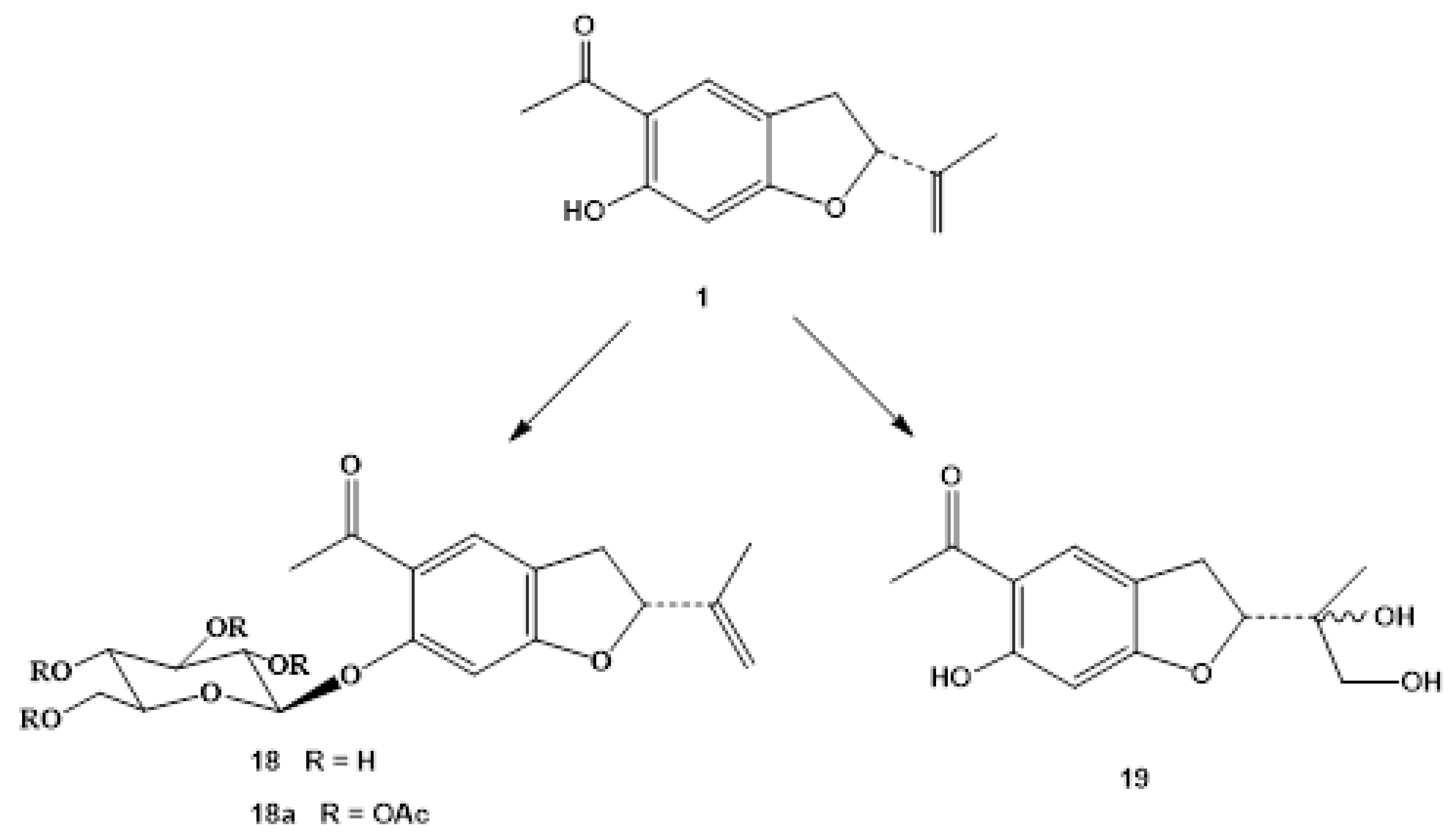
Two benzofuran derivatives, 6-hydroxytremetone β-D-glucoside (18) and 6,10,11-trihydroxytremetone (19), have been obtained from the biotransformation of 6-hydroxytremetone (1) with Mucor plumbeus (Figure 3). This fungus has a broad specificity of substrate and has been used in the biotransformation of sesquiterpenes and diterpenes [38,39,40].
The major biotransformation compound was 6-hydroxytremetone β-D-glucoside (18). Its high-resolution mass spectrum showed the molecular ion at m/z 380, which corresponds to the molecular formula C19H24O8. The 1H NMR spectrum was similar to that of the substrate, but with additional signals of a glucose moiety, with proton signal between δ 3.44 and 5.05. In the HMBC spectrum, a correlation between the anomeric proton H-1`and C-6 was observed. The coupling constant of H-1′ (J = 7.3 Hz) indicated an axial β-configuration for this substituent, which was confirmed by the resonance of the anomeric carbon at δ 103.4 [41]. Acetylation of this compound led to the acetyl derivative 18a, which showed the molecular ion at 548.1874 m/z, corresponding to the molecular formula C27H32O12. Their NMR spectra confirmed this structure.
The second product, structure 19, was obtained in very low yield. Its mass spectrum showed the molecular ion at m/z: 252.1008 (C13H16O5), which indicates the introduction of two new oxygen atoms into the molecule with respect to the substrate 1. In addition to the signals observed in the starting benzofuran, an AB system appears, at δ 3.55 and 3.77 due to the two methylene protons (H-11) and the signal of a methyl at δ 1.12, which was assigned as a substituent on C-2, based on the correlations observed in the HMBC experiment. In the 13C NMR spectrum (Table 3), the disappearance of the signals of the vinyl carbons of the substrate and the presence of two new resonances at δ 67.0 and 73.5 ppm, were typical of C-10 and C-11 respectively, which are linked to oxygens. Previously, this product had been isolated from Helianthopsis stuebelii [42]. It could be originated by opening of the corresponding epoxide during the isolation procedure, as also occurred in the formation of 10,11-dihydroxyeuparin (8).
2.3. Antifeedant and Postingestive Effects
Table 4 shows the antifeedant and postingestive effects of the tested compounds. The most antifeedant compounds were among the tremetone series (1, 1a, 2–5, 18, 18a), with 3-ethoxy-hydroxy-tremetone (4) being the strongest antifeedant (1st in EC50 ranking), followed by 6-hydroxy-tremetone (1) (2nd in EC50 ranking) and 1a (6th in EC50 ranking). Glucosylation of 1 by its biotransformation with Mucor plumbeus gave inactive products (18 and 18a). Among the euparin series (6, 7, 7a, 8–10, 14, 16), the dihydroxyangelate 14 (third in the EC50 ranking) was the most active, followed by euparin (6) and euparin angelate (16) (sixth and seventh in the EC50 ranking). The eupachinin-type compounds (11, 12) were both antifeedants (fourth and fifth in the EC50 ranking) and this is the first report on the antifeedant effects of this type of benzofurans.
The antifeedant compounds (1, 4, 6, 11, 12) were further tested for postingestive effects on S. littoralis larvae (Table 4). At a dose of 40 µg/larvae, 4, 11, and 12 showed antifeedant effects without postingestive toxicity (pANCOVA2 > 0.05). Compound 6 (euparin) had postingestive toxicity at 40 µg/larvae (pANCOVA2 = 0.035) that was lost at 20 µg/larvae, except when the larvae were pre-treated with PBO (piperonyl butoxide, a synergist), resulting in a significant antifeedant postingestive effect and indicating the involvement of a PSMO (polysubstrate monoxygenases) detoxification mechanism for 6 in S. littoralis.
Insect antifeedant effects of benzofurans have been previously described. Natural dihydrobenzofurans such as remirol and aurones were antifeedants to Spodoptera litura [43,44,45]. Euparin (6) was antifeedant to S. litura, but methyleuparin showed stronger effects [43]. Inclusion of an acetyl or methoxy on the aromatic group of benzofurans gave effective antifeedant activity, while hydroxylation or methylation decreased this effect [43]. In this work, ethoxylation of the C3 hydroxy group in the furan ring gave the strongest antifeedant effect (4), while acetylation of the aromatic hydroxyl group reduced or eliminated the antifeedant activity in the tremetone series (1 vs. 1a). Modifications of the side chain also affected the activity. For example, the oxidation of euparin 6 in C10 eliminated the activity (in 7 and 7a), and the presence of a C11 angelate substituent in the side chain (16, 14) increased the activity (8).
Previously, methyleuparin showed a moderate growth inhibition effect on Peridroma saucia [45], and 6-hydroxy tremetone derivatives and euparin affected the ratio of Tenebrio molitor larval pupation [46]. In this work, we have shown growth inhibitory effects on S. littoralis larvae for the tremetone derivative 4 and eupachinin compounds 11 and 12, while euparin 6, with an unsaturated benzofuran ring, was the most toxic compound. The oxidation of the unsaturated furan ring turns benzofurans in alkylating agents, becoming cytotoxic and mutagenic [47]. This will explain the larval postingestive toxicity of euparin (6), with an unsaturated furan ring. The larval toxicity of 6 was enhanced by the application of the synergist PBO. PBO is a specific inhibitor of microsomal oxidases and resistance-associated esterases [48] and has shown insecticidal synergistic effects with synthetic unsaturated benzofurans [49], indicating that insects can detoxify these compounds as shown here.
3. Materials and Methods
3.1. General Procedure
Melting points were determined with a Reichert Thermovar apparatus and are uncorrected (Reichert Technologies, Buffalo, NY, USA). Optical rotations were measured at room temperature on a Perkin Elmer 343 polarimeter (Perkin Elmer, Waltham, MA, USA). IR spectra were recorded with a Perkin-Elmer 1600 FT spectrometer (Perkin Elmer, Waltham, MA, USA). 1H and 13C NMR spectra were run in CDCl3 solution at 500.1 and 125.8 MHz, respectively, in a Bruker AMX-500 spectrometer (Bruker Corporation, Billerica, MA, USA) with pulsed field gradient, using this solvent signal (CDCl3, at δH7.26 and δC77.0) as internal standard. Chemical shifts are given in ppm (δ). EI and HREIMS were taken in a Micromass Autospect instrument (Manchester, UK) at 70 eV (probe). HRESIMS data were recorded on a Waters Micromass LCT Premier XE (Manchester, UK). Column chromatographies were made on silica gel (40∓63 μm, Merck, Darmstadt, Germany), and Sephadex LH-20 (Amersham Pharmacia Biotech AB, Uppsala, Sweden). Preparative and semipreparative HPLC was performed on a Beckman System Gold 125P (Beckman Coulter Life Sciences, Brea, CA, USA) equipped with a diode-array detector Beckman Coulter 168 and preparative Interstil Prep-sil (Gasukuro Kogio) (20 mm × 250 mm, 10 µm) and semipreparative Beckman Ultrasphere silica (10 mm × 250 mm, 5 µm) columns.
3.2. Plant Material
All Pericallis species were collected at their flowering stage (aerial parts) in the Canary Islands (Spain) and identified by Dr. Arnoldo Santos. All voucher specimens (ORT) have been deposited at the Herbarium del Jardín de Aclimatación de La Orotava, Tenerife, Spain.
Pericallis echinata (L. fil.) B. Nord. from Tenerife Island (Los Realejos, 28°20′59.99″ N 16°35′59.99″ W) was collected in March (aerial parts) and May (seeds) (ORT 32003). P. steetzii (Bolle) B. Nord from Gomera Island (Garajonay National Park, 28°07′34″ N 17°14′14″ W) was collected in May (aerial parts) and June (seeds) 1991 (ORT 33451). P. murrayi (Bornm.) B. Nord. from El Hierro Island (28°16′07″ N 16°36′20″ O) was collected in April 2009 (aerial parts and roots) (ORT 27594). P. lanata DC. from Tenerife Island (Barranco del Río, 28°34′10″ N 16°18′48″ O) was collected in May 1994 (aerial parts) (ORT 32001).
3.3. Plant Material
Agrobacterium rhizogenes ATCC-15834 was inoculated with a needle to the stem of aseptic plantlets germinated from seeds and cultured on agar medium containing 30 g/mL sucrose and half-strength MS medium [50]. The induced hairy roots were excised and cultured on hormone-free half-Gamborg B5 solid medium [51] supplemented with 30 g/L sucrose and 0.5 mg/mL ampicillin to eliminate bacteria. The axenic hairy roots obtained were subcultured every 25–30 days in the dark at 25 °C on the same solid medium without antibiotics. Then, they were cultivated in the dark at 25°C in 250 mL Erlenmeyer flasks containing 100 mL of half-GB liquid medium supplemented with 30 g/L sucrose and shaken on a rotary shaker at 90 rpm. After five weeks, the hairy roots were harvested and separated from the culture medium by filtration through filter paper under vacuum.
3.4. DNA Extraction and Analysis
Total genomic DNA was extracted from transformed root tissue and from the untransformed root plants by using a “GenEluteTM Plant Genomic DNA Miniprep Kit” (Sigma-Aldrich, St. Louis, MO, USA). Plasmid DNA from A. rhizogenes strain ATCC-15834 was used as a positive control. Polymerase chain reaction was performed using REDExtract-N-Amp Plant PCR Kit (Sigma) to detect the insertion of TL-DNA of A. rhizogenes ATCC-15834 in the transformed roots. The oligonucleotide primers for TL-DNA were 5′-ATGGATCCCAAATTGCTATTCCTTCCA-3′ and 5′- TTAGGCTTCTTTCTTCAGG TTTA-3′ which amplify a segment complementary to the 5′ coding sequence of rol B to the 3′ coding sequence of rol C on the TL-DNA region. PCR amplification was performed in a DNA thermal cycler (Applied Biosystems 2700, Foster City, CA) under the following conditions: initial denaturation at 94 °C for 2 min, followed by 30 cycles of 90 °C for 30 s, annealing at 55 °C for 1 min, with a final extension at 72 °C for 5 min. The PCR reaction mixture was electrophoresed on a 1.2% agarose gel using tris-acetate-EDTA buffer and visualized by ethidium bromide staining under ultraviolet light at 260 nm.
3.5. Extraction and Isolation of Compounds from Pericallis
Pericallis plant parts (aerial and roots) were dried in open air in the shade and grinded to give dry plant materials (P. echinata, Pe, 4.700 g; P. lanata, Pl, 980 g; P. steetzii, Ps, 1800 g; P. murrayi aerial, Pma, 1251.0 g; P. murrayi roots, Pmr, 687.5 g). Hairy roots were freeze-dried and powdered to give transformed roots dried materials (P. echinata, PeTR, 112 g and P. steetzii, PsTR, 49.7 g). The dry materials were exhaustively extracted with EtOH in a Soxhlet. The solvent was evaporated at reduced pressure to give crude extracts (Pe,120 g, PeTR 41 g, Pl 175 g, Ps 180 g, PsTR 11.4 g Pma 137 g, Pmr 90.2 g).
The ethanolic extracts were chromatographed with silica gel vacuum liquid chromatography columns (VLC column, 10 × 25 cm) eluted with a hexane-EtOAc-MeOH gradients. Further, the main fractions were chromatographed on Si gel columns, Sephadex LH-20 and/or preparative normal phase HPLC eluted with different solvents and proportions of hexane-EtOAc and/or CH2Cl2/MeOH.
The following compounds were isolated: 6-hydroxytremetone (1) (Pe 0.49%, Pma 0.09% of the extract), 3β,6-dihydroxytremetone (2) (Pe 0.003%, PeTR 0.0025% of the extract), 3β-methoxy-6-hydroxytremetone (3) (Pe 0.001%), 3β-ethoxy-6-hydroxytremetone (4) (Pe 0.59%, Pl 0.019%, Ps 0.002%, Pma 0.01%, Pmr 0.001%, PeTR 0.005%, PsTR 0.02% of the extract), 3α,6-dihydroxytremetone (5) (Pe 0.001% of the extract), euparin (6) (Pe 0.135%, Pl 0.22%, Ps 0.04%, Pma 0.08%, Pmr 0.35%, PeTR 0.17%, PsTR 0.2% of the extract), euparone (7) (Pe 0.08%, Pma 0.02%, Pmr 0.03%, PeTR 0.02%, PsTR 0.045% of the extract), 10,11-dihydroxy-10,11-dihydroeuparin (8) (Pe 0.003% of the extract), 10-methoxy-11-hydroxy-10,11-dihydroeuparin (9) (Pe 0.001% of the extract), 10-ethoxy-11-hydroxy-10,11-dihydroeuparin (10) (Pe 0.01% of the extract), (-)-eupachinin A (11) (Pe 0.003% of the extract), (-)-eupachinin A ethyl ether (12) (Pe 0.004% of the extract), 10,12-dihydroxy-11-angelyloxy-10,11-dihydroeuparin (14) (PeTR 0.00025%, PsTR 0.0043%), 2,4-dihydroxy-5-formyl-acetophenone (15) (Pe 0.005% of the extract), 11-angelyloxy-10,11-dihydroeuparin (16) (PsTR 0.07% of the extract), 12-angelyloxyeuparone (17) (PsTR 0.02% of the extract).
3.6. Benzofurans
3.6.1. 6-Hydroxytremetone (1)
White crystals, m.p. 48–50 °C; [α]D20 -42.2 (c 0.62, EtOH); IR (KBr) λmax: 2540, 1640, 1485, 1255, 1135, 1040 cm−1;1H NMR (500 MHz): δ 1.75 (3H, t, J = 1.4 Hz, H-12), 2.52 (3H, s, H-14), 2.96 (1H, dd, J =15.3, 7.5, 1.6 Hz, H-3α), 3.29 (1H, dd, J = 15.3, 9.5, 1.6 Hz, H-3β), 4.93 (1H, t, J = 1.4 Hz, H-11), 5.07 (1H, s ancho, H-11), 5.25 (1H, dt, J = 7.5, 9.5 Hz, H-2β), 6.35 (1H, s, H-7), 7.48 (1H, t, J = 1.6 Hz, H-4), 12.96 (1H, s, OH).; 13C NMR (125 MHz): See Table 1; EIMS m/z (rel. int.): 218 [M]+ (100), 203 (98), 175 (61), 161 (9), 160 (26), 157 (7), 132 (3), 115 (5); HREIMS m/z: 218.0942 [M]+. Calculated for C13H14O3, 218.0943.
3.6.2. 3β,6-. Dihydroxytremetone (2)
Colourless oil; [α]D20 -13.6 (c 0.17, CHCl3); 1H NMR (500 MHz): δ 1.74 (3H, br s, H-12), 2.58 (3H, s, H-14), 4.95 (1H, t, J = 1.7 Hz, H-11), 4.97 (1H, d, J = 3.3 Hz, H-2β), 5.06 (1H, br s, H-11), 5.08 (1H, d, J = 3.3 Hz, H-3α), 6.44 (1H, s, H-7), 7.77 (1H, s, H-4), 12.99 (1H, s, H0-6); 13C NMR (125 MHz): See Table 1; EIMS m/z (rel. int.): 234 [M]+ (25), 216 (90), 201 (100), 191 (6), 178 (35), 149 (19), 115 (12); HREIMS m/z: 234.0891 [M]+. Calculated for C13H14O4, 234.0892.
3.6.3. 3β-. Methoxy-6-Hydroxytremetone (3)
Colourless oil; 1H NMR (500 MHz): δ 1.72 (3H, s, H-12), 2.58 (3H, s, H-14), 3.41 (3H, s, -OMe), 4.71 (1H, d, J = 2.5 Hz, H-3), 4.94 and 5.06 (each 1H, br s, H-11), 5.09 (1H, d, J = 2.5 Hz, H-2), 6.45 (1H, s, H-7), 7.75 (1H, s, H-4); 13C NMR (125 MHz): See Table 1; EIMS m/z (rel. int.): 248 [M]+ (100), 232 (16), 217 (57), 203 (20), 201 (16), 157 (15), 129 (9). HREIMS m/z: 248.1043 [M]+. Calculated for C14H16O3, 248.1049.
3.6.4. 3β-. Ethoxy-6-Hydroxytremetone (4)
White crystals, m.p. 71–73 °C; [α]D20–13.6 (c 0.17, CHCl3); 1H NMR (500 MHz): δ 1.25 (3H, t, J = 7.0 Hz, H-2′), 1.70 (3H, s, H-12), 2.56 (3H, s, H-14), 3.62 (2H, c, J = 7.0 Hz, H-1′), 4.77 (1H, d, J = 2.5 Hz, H-3), 4.93 and 5.06 (each 1H, s, H-11), 5.07 (1H, d, J = 2.5 Hz, H-2), 6.42 (1H, s, H-7), 7.72 (1H, s, H-4), 12.99 (1H, s, OH); 13C NMR (125 MHz): See Table 1; EIMS m/z (rel. int.): 262 [M]+ (100), 245 (8), 233 (12), 219 (3), 217 (49), 203 (16); HREIMS m/z: 262.1208 [M]+. Calculated for C15H18O4, 262.1205.
3.6.5. 3α,6-. Dihydroxytremetona (5)
Colourless oil; [α]D20–6.9 (c 0.13, CHCl3); 1H NMR (500 MHz): δ 1.91 (3H, br s, H-12), 2.59 (3H, s, H-14), 5.05 (1H, d, J = 6.1 Hz, H-2), 5.17 (1H, br s, H-3), 5.21 and 5.26 (each 1H, s, H-11), 6.45 (1H, s, H-7), 7.82 (1H, s, H-4), 12.99 (1H, s, HO-6); 13C NMR (125 MHz): See Table 1; HRESIMS (negative-ion mode) m/z: 233.0810 [M-H]+. Calculated for C13H13O4, 233.0814.
3.6.6. Euparin (6)
Yellow crystals, m.p. 132–134 °C; 1H NMR (500 MHz): δ 2.10 (3H, s, H-12), 2.67 (3H, s, H-14), 5.18 (1H, d, J =1.3 Hz, H-11), 5.75 (1H, s, H-11), 6.54 (1H, s, H-3), 6.97 (1H, s, H-7), 7.89 (1H, s, H-4), 12.49 (1H, s, OH); 13C NMR (125 MHz): See Table 1; EIMS m/z (rel. int.): 262 [M]+ (100), 245 (8), 233 (12), 219 (3), 217 (49), 203 (16); HREIMS m/z: 262.1208 [M]+. Calculated for C15H18O4, 262.1208.
3.6.7. Euparone (7)
Amorphous solid; 1H NMR (500 MHz): δ 2.59 (3H, s, H-11), 2.73 (3H, s, H-13), 7.09 (1H, s, H-7), 7.46 (1H, br s, H-3), 8.17 (1H, s, H-4), 12.54 (1H, s, OH); 13C NMR (125 MHz): See Table 1; EIMS m/z (rel. int.): 218 [M]+ (59), 203 (100), 204 (9), 165 (11), 137 (7), 107 (15); HREIMS m/z: 218.0579 [M]+. Calculated for C12H10O4, 218.0579.
3.6.8. 10,11-. Dihydroxy-10,11-Dihydroeuparin (8)
Colourless oil; 1H NMR (500 MHz): δ 1.59 (3H, s, H-12), 2.68 (3H, s, H-14), 3.69, 4.00 (1H each signal, d, J = 11.0 Hz, H-11), 6.66 (1H, s, H-3), 6.98 (1H, s, H-7), 7.92 (1H, s, H-4), 12.43 (1H, s, -OH); 13C NMR (125 MHz): See Table 1; EIMS m/z (rel. int.): 250 [M]+ (10), 219 (100), 203 (10), 176 (3); HREIMS m/z: 250.0841 [M]+. Calculated for C13H14O5, 250.0841.
3.6.9. 10-Methoxy-11-Hydroxy-10,11-Dihydroeuparin (9)
Colourless oil; 1H NMR (500 MHz): δ 1.63 (3H, s, H-12), 2.69 (3H, s, H-14), 3.23 (3H, s, -OMe), 3.73 and 3.92 (each 1H, d, J = 11.0 Hz, H-11), 6.67 (1H, s, H-3), 7.02 (1H, s, H-7), 7.95 (1H, s, H-4), 12.44 (1H, s, -OH); EIMS m/z (rel. int.): 264 [M]+ (5), 234 (14), 233 (100), 218 (9), 203 (22); HREIMS m/z: 264.0999 [M]+. Calculated for C14H16O5, 264.0998.
3.6.10. 10-Ethoxy-11-Hydroxy-10,11-Dihydroeuparin (10)
Colourless oil; [α]D =–3 (c, 0.13, CHCl3); 1H NMR (500 MHz): δ 1.18 (3H, t, J = 7.0 Hz, -OCH2CH3), 1.63 (3H, s, H-12), 2.68 (3H, s, H-14), 3.31 and 3.49 (each 1H, dd, J = 8.8, 7.0 Hz, -OCH2CH3), 3.71 and 3.89 (each 1H, d, J = 11.0 Hz, H-11), 6.64 (1H, br. s, H-3), 7.00 (1H, s, H-7), 7.93 (1H, s, H-4), 12.42 (1H, s, -OH); EIMS m/z (rel. int.): 278 [M]+ (11), 248 (14), 247 (86), 232 (19), 219 (100), 203 (49); HREIMS m/z: 278.1153 [M]+. Calculated for C15H18O5, 180.0423.
3.6.11. (-)-. Eupachinin A (11)
White crystal (hexane/EtOAc); [α]D = -8.33 (c, 0.36, CHCl3); IR (KBr) λmax: 2540, 1640, 1483, 1257, 1134, 1039 cm−1; 1H NMR (500 MHz) δ 1.78 (3H, s, H-14), 2.36 (2H, m, H-12), 2.63 (1H, ddd, J = 17.0, 6.6, 5.0Hz, H-11β), 2.74 (3H, s, H-16), 2.92 (1H, ddd, J = 17.0, 9.0, 5.0 Hz, H-11α), 7.05 (1H, s, H-7), 8.43 (1H, s, H-4), 12.61 (1H, s, OH); EIMS m/z (rel. int.): 274 [M]+ (56), 259 (100), 257 (13), 241 (4), 231 (12), 203 (11); HREIMS m/z: 274.0834 [M]+. Calculated for C15H14O5, 274.0841.
3.6.12. X-ray Crystallography Data of (-)-Eupachinin A (11) and (-)-Didehydroeupachinin A (13)
Colourless crystals, C60H54O20, Mr = 1095.03, triclinic space group P1, a = 7.4090 (5) Å, b = 12.9440(7) Å, c = 13.8110 (7) Å, α = 97.232(4)°, β= 102.730(5)°, γ = 94.243(5)°, V = 1274.57 (13) Å3, Z = 1, µ (Cu Kα) = 0.90 cm−1. Intensity data were collected at 293 °K on a Enraf-Nonius Kappa CCD diffractometer using Cu Kα radiation and a graphite monochromator. No. of measured, independent, and observed [I > 2σ(I)] reflections 19038, 9410, and 5198, respectively (Rint = 0.042). Refinement: R[F2 > 2σ(F2)] = 0.077, wR(F2) = 0.265, S = 1.03. No. of reflections = 9410, no. of parameters = 738, no. of restraints = 4. H-atom parameters constrained, (Δ/σ)max = 0.780, Δρmax, = 0.36 e Å−3 and Δρmin =−0.24 e Å−3. Computer programs: SHELXL2013 (Sheldrick, 2013). Supplementary Data have been deposited with the Cambridge Crystallographic Data Centre, no. 2031183. Copies of available material can be obtained, free of charge, via http://www.ccdc.cam.ac.uk/conts/retrieving.html, accessed on 30 April 2021) or from the CCDC, 12 Union Road, Cambridge CB2 1EZ, UK (Fax: +44-1223-336033; e-mail.: [email protected]).
3.6.13. (-)-. Eupachinin A Ethyl Ether (12)
Amorphous solid; [α]D = −3.70 (c, 0.27, CHCl3); 1H NMR (500 MHz): δ 1.15 (3H, t, J = 7.0 Hz, -OCH2CH3), 1.71 (3H, s, H-14), 2.18 (1H, ddd, J = 13.7, 11.0, 4.5 Hz, H-12β), 2.45 (1H, dt, J = 13.7, 4.5 Hz, H-12α), 2.57 (1H, dt, J = 17.1, 4.8 Hz, H-11β), 2.74 (3H, s, H-16), 3.00 (1H, ddd, J = 17.1, 11.0, 4.5 Hz, H-11α), 3.37 and 3.64 (each 1H, dd, J = 8.7, 7.0 Hz, -OCH2CH3), 7.08 (1H, s, H-7), 8.48 (1H, s, H-4), 12.59 (1H, s, -OH); EIMS m/z (rel. int.): 302 [M]+ (52), 287 (25), 259 (52), 257 (100), 241 (16), 229 (8), 214 (8), 115 (8); HREIMS m/z: 302.1152 [M]+. Calculated for C17H18O5, 302.1154.
3.6.14. 10,12-. Dihydroxy-11-Angelyloxy-10,11-Dihydroeuparin (14)
1H NMR (500 MHz): δ (3H, t, J = 1.4 Hz, H-4′), 1.91 (3H, dd, J = 7.4, 1.6 Hz, H-5′), 2.33 (1H, br. s, HO-10), 2.69 (3H, s, H-14), 3.52 (1H, s, HO-12), 3.90 and 3.98 (each 1H, br. d, J = 12.6 Hz, H-12), 4.55 and 4.61 (1H each, d, J = 11.6 Hz, H-11), 6.12 (1H, qd, J = 7.3, 1.4 Hz, H-3′), 6.75 (1H, d, J = 0.9 Hz, H-3), 6.99 (1H, br. s, H-7), 7.95 (1H, s, H-4), 12.44 (1H, s, HO-6); EIMS m/z (rel. int.) 348 [M]+ (6), 235 (22), 230 (24), 217 (11), 215 (20), 203 (15), 189 (17), 83 (100); HREIMS m/z: 348.1211 [M]+. Calculated for C18H20O7, 348.1209).
3.6.15. 2,4-. Dihydroxy-5-Formyl-Acetophenone (15)
Amorphous solid; 1H NMR (500 MHz): δ 2.64 (3H, s, H-8), 6.46 (1H, s, H-6), 7.99 (1H, s, H-3), 9.77 (1H, s, -CHO), 11.62 (1H, s, HO-4), 13.08 (1H, s, HO-2); 13C NMR (125 MHz): δ 114.8 (C-1), 169.7 (C-2), 104.9 (C-3), 167.8 (C-4), 114.5 (C-5), 139.6 (C-6), 202.4 (C-7), 26.2 (C-8), 194.0 (C-9); EIMS m/z (rel. int.): 180 [M]+ (60), 165 (100), 123 (6), 109 (4); HREIMS m/z: 180.0428 [M]+. Calculated for C9H8O4, 180.0423.
3.6.16. 11-Angelyloxy-10,11-Dihydroeuparin (16)
1H NMR (CDCl3, 500 MHz): δ 1.41 (3H, d, J = 7.0 Hz, H-12), 1.85 (3H, t, J = 1.5 Hz, H-5′), 1.92 (3H, dd, J = 7.2, 1.5 Hz, H-4′), 2.68 (3H, s, H-14), 3.32 (1H, m, H-10), 4.33 and 4.40 (each 1H, dd, J = 12.0, 7.0 Hz, H-11), 6.06 (1H, qd, J = 7.3, 1.3 Hz, H-3′), 6.40 (1H, s, H-3), 6.96 (1H, s, H-7), 7.88 (1H, s, H-4), 12.43 (1H, s, -OH); EIMS m/z, (rel. int.): 316 [M]+ (4), 217 (40), 216 (100), 201 (53), 198 (5), 185 (7), 173 (6), 83 (51); HREIMS m/z: 316.1304 [M]+. Calculated for C18H20O5, 316.1310.
3.6.17. 12-Angelyloxyeuparone (17)
1H NMR (CDCl3, 200 MHz): δ 2.01 (3H, br s, H-5′), 2.05 (3H, d, J = 7.2 Hz, H-4′), 2.74 (3H, s, H-13), 5.34 (2H, s, H-12), 6.20 (1H, m, H-3′), 7.10 (1H, s, H-7), 7.57 (1H, s, H-3), 8.20 (1H, s, H-4), 12.57 (1H, s, -OH); EIMS m/z (rel. int.): 316 [M]+ (46), 234 (9), 217 (4), 203 (73), 189 (3), 83 (100), 55 (60); HREIMS m/z: 316.0958 [M]+. Calculated for C17H16O6, 316.0946.
3.7. Acetylation of 6-Hydroxytremetone (1)
Compound 1 (10 mg) in pyridine (0.5 mL) was treated with acetic anhydride (0.5 mL) and stirred for 24 h at room temperature. Extraction with EtOAc in the usual way and column chromatography afforded 6-acetoxy-tremetone (1a) (12.3 mg):
Amorphous solid; 1H NMR (500 MHz): δ 1.77 (3H, br s, H-12), 2.34 (3H, s, -OAc), 2.49 (3H, s, H-14), 3.05 (1H, dd, J= 15.7, 9.0 Hz, H-3α), 3.36 (1H, dd, J= 15.8, 9.0 Hz, H-3β), 4.95 and 5.09 (1H each, s, H-11), 5.29 (1H, t, J = 9.0, Hz, H-2β), 6.52 (1H, s, H-7), 7.68 (1H, s, H-4); 13C NMR (125 MHz): See Table 1; EIMS m/z (rel. int.): 260 [M]+ (17), 218 (78), 203 (100), 175 (57), 160 (11), 129 (7); HREIMS m/z: 260.1053 [M]+. Calculated for C15H16O4, 260.1049.
3.8. Acetylation of Euparone (7)
Compound 7 (8 mg) in pyridine (0.3 mL) at room temperature was treated with Ac2O (0.3 mL) and stirred for 24 h. Usual work-up and column chromatography yielded 6-acetoxy-euparone (7a) (9.3 mg):
Colourless oil; 1H NMR (500 MHz): δ 2.39 (3H, s, -OAc), 2.62 (6H, s, H-11 and H-13), 7.34 (1H, s, H-7), 7.54 (1H, br s, H-3), 8.20 (1H, s, H-4; 13C NMR (125 MHz): See Table 1; EIMS m/z (rel. int.): 260 [M]+ (17), 218 (100), 203 (90), 175 (57), 160 (11), 129 (7); HREIMS m/z: 260.0689 [M]+. Calculated for C14H12O5, 260.0685.
3.9. Biotransformation of 6-Hydroxytremetone (1)
3.9.1. Microorganism
The fungal strain Mucor plumbeus (CECT 20422) used in this work was obtained from Colección Española de Cultivos Tipo (CECT), Valencia, Spain.
3.9.2. Incubation of 1
Two millilitres of a conidial suspension in water containing approximately 9 × 10⁶ conidia/mL of the fungus M. plumbeus was added to each 52 conical flasks (250 mL), containing sterile medium (50 mL) [40]. The cultures were grown at 25 °C for 36 h on an orbital shaker. The substrate 1 (265 mg) in EtOH (5.3 mL) was distributed equally between the flasks, and the incubation was allowed to continue for a further 6 days. The mycelium was separated by vacuum filtration and the broth extracted three times with EtOAc. The solvent was evaporated to give a residue (1.30 g), which was chromatographed on a silica gel column using hexane-EtOAc and EtOAc-MeOH gradients. Final purification of fractions of interest were carried out by semipreparative HPLC using n-hexane-EtOAc gradient as the mobile phase and flow rate 3 mL/min. Biotransformation of 1 gave in polarity order, starting material (1) (25.5 mg, 1.96% of extract), 6-hydroxytremetone β-D-glucoside (18) (145.0 mg, 11,5% of extract), and 6,10,11-trihydroxytremetone (19) (2.0 mg, 0.15% of extract).
Compound 18 (15 mg) in pyridine (0.6 mL) at room temperature was treated with Ac2O (0.6 mL) and stirred for 24 h. Extraction with EtOAc in the usual way and column chromatography afforded 6-hydroxytremetone β-D-tetraacetylglucoside (18a) (17.3 mg).
3.9.3. 6-Hydroxytremetone β-D-Glucoside (18)
M.p.: 85.0–90.1 °C (EtOAc-MeOH); 1H NMR (500 MHz, CDCl3): δ 1.72 (3H, s, H-12), 2.46 (3H, s, H-14), 2.94 and 3.22 (each 1H, dd, J =15.8, 8.0 Hz, H-3), 3.47 (1H, d, J =10.0 Hz, H-5′), 3.68 (3H, br s, H-2′, H-3′ y H-4′), 3.88 (2H, br s, H-6′), 4.85 (1H, d, J =6.6 Hz, H-1′), 4.90 and 5.04 (each 1H, s, H-11), 5.20 (1H, t, J =8.0 Hz, H-2), 6.60 (1H, s, H-7), 7.47 (1H, s, H-4); 1H NMR (500 MHz, CD3OD): δ 1.76 (3H, s, H-12), 2.65 (3H, s, H-14), 3.00 and 3.37 (each 1H, dd, J =15.6, 8.0 Hz, H-3), 3.44 (2H, m, H-3′, H-4′), 3.50 (1H, dd, J =5.4, 2.2 Hz, H-5′), 3.55 (1H, m, H-2′), 3.75 (1H, dd, J =12.1, 5.4 Hz, H-6′), 3.93 (1H, dd, J =12.1, 2.2 Hz, H-6′), 4.93 and 5.09 (each 1H, s, H-11), 5.05 (1H, d, J =7.3 Hz, H-1′), 5.30 (1H, t, J =8.0 Hz, H-2), 6.75 (1H, s, H-7), 7.63 (1H, s, H-4); 13C NMR (125 MHz): See Table 3; EIMS m/z (rel. int.): 380 [M]+ (5), 218 (100), 203 (93), 185 (13), 175 (49), 160 (21), 91 (10); HR-EISI-MS m/z: 380.1471 [M]+. Calculated for C19H24O8, 380.1471. 6-Hydroxytremetone β-D-tetraacetylglucoside (18a): 1H NMR (500 MHz): δ 1.73 (3H, s, H-12), 2.02, 2.03, 2.05 and 2.09 (each 3H, s, -OAc), 2.48 (3H, s, H-14), 2.98 and 3.29 (each 1H, ddd, J =15.4, 8.0, 1.3 Hz, H-3), 3.88 (1H, ddd, J =10.0, 6.3, 2.5 Hz, H-5′), 4.17 (1H, dd, J =12.3, 2.5 Hz, H-6′), 4.26 (1H, dd, J =12.3, 6.3 Hz, H-6′), 4.92 and 5.05 (each 1H, br s, H-11), 5.16 (1H, t, J =11.0 Hz, H-4′), 5.18 (1H, d, J =9.5 Hz, H-1′), 5.24 (1H, br t, J =8.0 Hz, H-2), 5.30 (1H, t, J =9.0 Hz, H-3′), 5.34 (1H, m, H-2′), 6.51 (1H, s, H-7), 7.60 (1H, t, J =1.3 Hz, H-4); 13C NMR (125 MHz): See Table 3; EIMS m/z (rel. int.): 548 [M]+ (1), 331 (25), 218 (10), 203 (13), 169 (100), 145 (8), 127 (23), 109 (65), 91 (6); HR-EISI-MS m/z: 548.1874 [M]+. Calculated for C27H32O12, 548.1894.
3.9.4. 6,10,11-. Trihydroxytremetone (19)
Colourless oil; [α]D = +8 (c, 0.13, CHCl3); 1H NMR (500 MHz): δ 1.19 (3H, s, H-12), 2.54 (3H, s, H-14), 3.19 (2H, dd, J =12.1, 7.6 Hz, H-3), 3.55 and 3.77 (each 1H, dd, J =10.6, 5.0 Hz, H-11), 4.90 (1H, dd, J =9.5, 7.6 Hz, H-2), 6.33 (1H, s, H-7), 7.51 (1H, s, H-4), 12.96 (1H, s, OH); 13C RMN (125 MHz): See Table 3; MS m/z (rel. int.): 252 [M]+ (71), 221 (12), 203 (56), 178 (100), 165 (21), 163 (48), 160 (24), 134 (68), 107 (19), 75 (35), 57 (24); HR-EISI-MS m/z: 252.1008 [M]+. Calculated for C13H16O5, 252.0998.
3.10. Insect Bioassays
3.10.1. Choice Feeding Assays (<6 h)
Spodoptera littoralis colonies are maintained at ICA-CSIC, reared on artificial diet, and kept at 22 ± 1 °C and >70% RH, with a photoperiod of 16:8 h (L:D) in a growth chamber. The bioassays were conducted as described [52]. The upper surface of Capsicum anuum (1.0 cm2) were treated with 10 μL of the test substance. The products were tested at an initial dose of 5 µg/µL (50 µg/cm2). Five Petri dishes with two sixth-instar S. littoralis larvae (>24 h after moulting) were allowed to feed in a growth chamber (until 75% larval consumption of control disks or tretament disks, environmental conditions as above). Each experiment was repeated 2 times. Feeding inhibition was calculated by measuring the disk surface consumption (digitalized with https://imagej.nih.gov/ij/, accessed on 23 March 2020) as % FI = [1 − (T/C) × 100], where T and C represent feeding on treated and control leaf disks, respectively. The antifeedant effects (% FI) were analyzed for significance by the nonparametric Wilcoxon signed-rank test. Extracts and compounds with an FI > 70% were further tested in a dose-response experiment (4–5 serial dilutions) to calculate their relative potency (EC50, the effective dose to give a 50% settling reduction) from linear regression analysis (% FI on Log-dose).
3.10.2. Cannulation
This experiment was performed with preweighed newly moulted S. littoralis L6 larvae in the absence or presence of piperonyl butoxide (PBO), under the same environmental conditions as above. Each experiment consisted of 20 larvae orally dosed with 40 and 20 µg of the test compound in 4 µL of DMSO (treatment) or solvent alone (control) using a Rheodyne Hamilton syringe (50 μL) attached to a Hamilton microdispenser. The syringe tip was inserted into the mouth of the larvae (maximum of 5 mm), and then larvae were forced fed until no regurgitation was observed. PBO-pretreated larvae were orally injected with 10 µg of PBO in 2 µL of DMSO, 1 h prior to the oral applicaton of the test [53]. At the end of the experiments (72 h), larval consumption and growth were calculated on a dry weight basis. The possible effect of variations in initial larval weight was analyzed by an analysis of covariance (ANCOVA) performed on biomass gains with initial biomass as covariate. The covariate effect was not significant (p > 0.05), showing that changes in insect biomass were similar among all treatments. An additional ANOVA was performed with ΔI as covariate for the extracts that signifcantly reduced ΔB, (ANCOVA2) to understand their post-ingestive mode of action (antifeedant and/or toxic) [54,55].
4. Conclusions
In this work, we have studied the benzofurans of Pericallis echinata (aerial parts and transformed roots), P. steetzii (aerial parts and transformed roots), P. lanata (aerial parts), and P. murrayi (aerial parts and roots). This work has permitted the isolation of the new compounds 10-ethoxy-11-hydroxy-10,11-dihydroeuparin (10), (-)-eupachinin A ethyl ether (12), 11,15-didehydro-eupachinin A (13), 10,12-dihydroxy-11-angelyloxy-10,11-dihydroeuparin (14), 11-angelyloxy-10,11-dihydroeuparin (16), 12-angelyloxyeuparone (17) and 2,4-dihydroxy-5-formyl-acetophenone (15) isolated for the first time as a natural product along with several known ones (1–9, 11). The incubation of the abundant component, 6-hydroxytremetone (1), with the fungus Mucor plumbeus afforded two benzofuran derivatives, the new compound 6-hydroxytremetone β-D-glucoside (18), and 6,10,11-trihydroxytremetone (19).
Benzofurans in the tremetone series (1, 1a, 2–5, 18, 18a), the euparin series (6, 7, 7a, 8–10, 14, 16), and the eupachinin-type (11, 12) were tested for antifeedant effects against the insect Spodoptera littoralis. In this work, ethoxylation of the C3 hydroxy group in the furan ring gave the strongest antifeedant effect (4), while acetylation of the aromatic hydroxyl group reduced or eliminated the antifeedant activity in the tremetone series (1 vs. 1a). Modifications of the side chain also affected the activity. For example, the oxydation of euparin 6 in C10 eliminated the activity (in 7 and 7a) and the presence of a C11 angelate substituent in the side chain (16, 14) increased the activity (8).
Compounds 4, 11, and 12 showed antifeedant effects without postingestive toxicity to orally dosed S. littoralis larvae. In this work, we have shown growth inhibitory effects on S. littoralis larvae for the tremetone derivative 4 and eupachinin compounds 11 and 12, while euparin 6, with an unsaturated benzofuran ring, was the most toxic compound. The larval toxicity of 6 was enhanced by the application of the synergist PBO, indicating that insects can detoxify these compounds.
Supplementary Materials
The following are available online at https://www.mdpi.com/article/10.3390/molecules28030975/s1, Figures S1–S2: 1H-NMR and 13C-NMR of compound 1a; Figure S3: 13C-NMR of compound 2; Figures S4–S5: 13C-NMR of compounds 4–5; Figures S6–S7: 1H-NMR an 13C-NMR of compound 7a; Figures S8–S10: 13C-NMR of compounds 8–10; Figures S11–S20: 1H-NMR and 13C-NMR of compounds 11–14 and 15–16; Figure S21: 1H-NMR of compound 17; Figures S22–S27: 1H-NMR and 13C-NMR of compounds 18,18a and 19.
Author Contributions
Conceptualization, A.G.-C. and C.E.D.; methodology, A.G.P., C.L.-B., L.R.-V., C.E.D. and A.G.-C.; formal analysis, A.G.P., C.L.-B., L.R.-V., I.B., B.M.F., C.E.D. and A.G.-C.; investigation, A.G.P., C.L.-B., L.R.-V., I.B., C.E.D. and A.G.-C.; data curation, B.M.F., C.E.D., A.G.-C. and I.B.; writing—original draft preparation, B.M.F., C.E.D. and A.G.-C.; writing—review and editing, C.E.D. and A.G.-C.; supervision, B.M.F., C.E.D. and A.G.-C.; funding acquisition, A.G.-C. and C.E.D. All authors have read and agreed to the published version of the manuscript.
Funding
This work has been supported by grant PID2019-106222RB-C31, MCI, Spain.
Institutional Review Board Statement
Not applicable.
Informed Consent Statement
Not applicable.
Data Availability Statement
The data presented in this study are available in the Research Group databases, within the article and its Supplementary Material.
Acknowledgments
We acknowledge the deceased Matías López, (Universidad de La Laguna) and Matías Reina (IPNA-CSIC) for their partial contribution to X-ray analysis and compound isolation.
Conflicts of Interest
The authors declare no conflict of interest.
Sample Availability
Samples of the compounds are not available from the authors.
References
- Jones, K.E.; Pérez-Espona, S.; Reyes-Betancort, J.A.; Pattinson, D.; Caujapé-Castells, J.; Hiscock, S.J.; Carine, M.A. Why do different oceanic archipelagos harbour contrasting levels of species diversity? The macaronesian endemic genus Pericallis (Asteraceae) provides insight into explaining the ‘Azores diversity Enigma’. BMC Evol. Biol. 2016, 16, 202. [Google Scholar] [CrossRef] [PubMed] [Green Version]
- Don, D. Pericallis tussilaginis. In The British Flower Garden; Ser. 2, plate 228; Sweet, R., Ed.; James Ridgway, Piccadilly: London, UK, 1834; Volume III. [Google Scholar]
- Nordenstam, B. Taxonomic studies in the tribe Senecioneae (Compositae). Opera Bot. 1978, 44, 1–84. [Google Scholar] [CrossRef]
- Panero, J.L.; Francisco-Ortega, J.; Jansen, R.K.; Santos-Guerra, A. Molecular evidence for multiple origins of woodiness and a New World biogeographic connection of the Macaronesian Island endemic Pericallis (Asteraceae: Senecioneae). Proc. Natl. Acad. Sci. USA 1999, 96, 13886–13891. [Google Scholar] [CrossRef] [PubMed] [Green Version]
- Swenson, U.; Manns, U. Phylogeny of Pericalis (Asteraceae): A total evidence approach reappraising the double origin of woodiness. Taxon 2003, 52, 533–546. [Google Scholar] [CrossRef]
- Jones, K.E.; Reyes-Bethancort, A.; Hiscock, S.J.; Carine, M.A. Allopatric diversification, multiple habitat shifts and hybridization in the evolution of Pericallis (Asteraceae), a Macaranosian endemic genus. Am. J. Botany 2014, 101, 637–641. [Google Scholar] [CrossRef]
- Domínguez, D.M.; Reina, M.; Santos-Guerra, A.; Santana, O.; Agulló, T.; López-Balboa, C.; González-Coloma, A. Pyrrolizidine alkaloids from Canarian endemic plants and their biological effects. Biochem. Syst. Ecol. 2008, 36, 153–156. [Google Scholar] [CrossRef]
- Hartmann, T. Chemical ecology of pyrrolizidine alkaloids. Planta 1999, 207, 483–495. [Google Scholar] [CrossRef]
- Hartmann, T.; Ober, D. Biosynthesis and metabolism of pyrrolizidine alkaloids in plants and specialized insect herbivores; Biosynthesis. In Topics in Current Chemistry; Leeper, F.J., Vederas, J.C., Eds.; Springer: Berlin/Heidelberg, Germany, 2000; Volume 209. [Google Scholar] [CrossRef]
- Portero, A.G.; González-Coloma, A.; Reina, M.; Díaz, C.E. Plant-defensive sesquiterpenoids from Senecio species with biopesticide potential. Phytochem. Rev. 2012, 11, 391–403. [Google Scholar] [CrossRef]
- Ruiz-Vásquez, L.; Ruiz Mesia, L.; Reina-Artiles, M.; López-Rodríguez, M.; González-Platas, J.; Giménez, C.; Cabrera, R.; González-Coloma, A. Benzofurans, benzoic acid derivatives, diterpenes and pyrrolizidine alkaloids from Peruvian Senecio. Phytochem. Lett. 2018, 28, 47–54. [Google Scholar] [CrossRef]
- Ruiz-Vásquez, L.; Olmeda, A.S.; Zuñiga, G.; Villarroel, L.; Echeverri, L.F.; González-Coloma, A.; Reina, M. Insect antifeedant and ixodicidal compounds from Senecio adenotrichius. Chem. Biodivers. 2017, 14, e1600155. [Google Scholar] [CrossRef]
- Ruiz-Vásquez, L.; Reina, M.; López-Rodríguez, M.; González-Coloma, A.; Giménez, C.; Cabrera, R.; Cuadra, P.; Fajardo, V. Sesquiterpenes, flavonoids, shikimic acid derivatives and pyrrolizidine alkaloids from Senecio kingii. Phytochemistry 2015, 117, 245–253. [Google Scholar] [CrossRef]
- Fraga, B.M.; Díaz, C.E.; Amador, L.J.; Reina, M.; Santana, O.; González-Coloma, A. Bioactive compounds from transformed root cultures and aerial parts of Bethencourtia hermosae. Phytochemistry 2014, 108, 220–228. [Google Scholar] [CrossRef]
- Proksch, P.; Rodriguez, E. Chromenes and benzofurans of the Asteraceae, their chemistry and biological significance. Phytochemistry 1983, 22, 2335–2348. [Google Scholar] [CrossRef]
- Bonner, W.A.; De Graw, J.I.; Bowen, D.M.; Shah, V.R. Toxic constituents of “white snakeroot”. Tetrahedron Lett. 1961, 12, 417–420. [Google Scholar] [CrossRef]
- Liu, Z.L.; Tian, X. The components of Cacalia tangutica. Bull. Korean Chem. Soc. 2004, 25, 1078–1080. [Google Scholar] [CrossRef]
- Bohlmann, F.; Knoll, K.H.; Zdero, C.; Mahanta, P.K.; Grenz, M.; Suwita, A.; Ehlers, D.; Le Van, N.; Abraham, W.R.; Natu, A.A. Terpen-derivate aus Senecio-arten. Phytochemistry 1977, 16, 965–985. [Google Scholar] [CrossRef]
- Bohlmann, F.; Jakupovic, J.; Lonitz, M. Natürlich vorkommende Terpen-Derivate, Über inhaltsstoffe der Eupatorium-gruppe. Chem. Ber. 1977, 110, 301–314. [Google Scholar] [CrossRef]
- Zdero, C.; Bohlmann, F.; King, R.M. Diterpenes and norditerpenes from the Aristeguetia group. Phytochemistry 1991, 30, 2991–3000. [Google Scholar] [CrossRef]
- Castañeda, P.; Gómez, L.; Mata, R.; Lotina Hennsen Anaya, A.L.; Bye, R. Phytogrowth-inhibitory and antifungal constituents of Helianthella quinquenervis. J. Nat. Prod. 1996, 59, 323–326. [Google Scholar] [CrossRef]
- Habtemariam, S. Antiinflammatory activity of the antirheumatic herbal drug, gravel root (Eupatorium purpureum): Further biological activities and constituents. Phytother. Res. 2001, 15, 687–690. [Google Scholar] [CrossRef]
- Kuroda, C.; Kiuchi, K.; Torihata, A.; Takeshita, K.; Gong, X.; Shen, Y.; Hirota, H.; Onuki, H.; Hanai, R. Chemical and genetic diversity of Ligularia latihastata and Ligularia villosa in Yunnan province of China. Chem. Biodivers. 2007, 4, 2210–2216. [Google Scholar] [CrossRef] [PubMed]
- Bohlmann, F.; Zdero, C.; King, R.M.; Robinson, H. New sesquiterpene lactones and other constituents from Fitchia speciosa. Phytochemistry 1980, 19, 1141–1143. [Google Scholar] [CrossRef]
- Kamthong, B.; Robertson, A. Furano-compounds. Part III. Euparin. J. Chem. Soc. 1939, 925–930. [Google Scholar] [CrossRef]
- Zhao, Y.; Jia, Z.; Yang, L. Novel sesquiterpenes from Ligularia veitchiana. Planta Med. 1994, 60, 91–92. [Google Scholar] [CrossRef] [PubMed]
- Elsohly, M.A.; Doorenbos, N.J.; Quimby, M.W.; Knapp, J.E.; Slatkin, D.J.; Schiff Jr, P.L. Euparone, a new benzofuran from Ruscus aculeatus L. J. Pharm. Sci. 1974, 63, 1623–1624. [Google Scholar] [CrossRef]
- Hussein, N.S. Benzofurans from Senecio desfontainei. Pharmazie 1992, 47, 468–469. [Google Scholar]
- Saito, Y.; Takiguchi, K.; Gong, X.; Kuroda, C.; Tori, M. Thiophene, furans, and related aromatic compounds from Eupatorium heterophyllum. Nat. Prod. Commun. 2011, 6, 361–366. [Google Scholar] [CrossRef] [Green Version]
- Wang, W.J.; Wang, L.; Huang, X.J.; Jiang, R.W.; Yang, X.L.; Zhang, D.M.; Chen, W.M.; Tang, B.Q.; Wang, Y.; Zhang, X.Q.; et al. Two pair of new benzofuran enantiomers with unusual skeletons from Eupatorium chinense. Tetrahedron Lett. 2013, 54, 3321–3324. [Google Scholar] [CrossRef]
- Gupta, S.R.; Seshadri, T.R.; Sood, G.R. The structure and synthesis of neobavachalcone, a new component of Psoralea corylifolia. Phytochemistry 1977, 16, 1995–1997. [Google Scholar] [CrossRef]
- Baddeley, G.V.; Bealing, A.J.; Jefferies, P.R.; Retallack, R.W. The chemistry of the Euphorbiaceae. VI. A triterpene from Beyeria leschenaultii. Austral. J. Chem. 1964, 17, 908–914. [Google Scholar] [CrossRef]
- Yashigita, N. The chemical structure of neoilexonol. Part II. Oxidation and reduction of neoilexonol and its derivatives. Agric. Biol. Chem. 1961, 25, 844–851. [Google Scholar] [CrossRef]
- Hui, W.H.; Li, M.M.; Luk, K. Triterpenoids and steroids from Rhodomyrtus tomentosa. Phytochemistry 1975, 14, 833–834. [Google Scholar] [CrossRef]
- Tamai, M.; Watanabe, N.; Someya, M.; Kondoh, H.; Omura, S.; Ling, Z.P.; Chang, R.; Ming, C.W. New hepatoprotective triterpenes form Canarium album. Planta Med. 1989, 55, 44–47. [Google Scholar] [CrossRef]
- Quijano, L.; Rios, T.; Fronczek, F.R.; Fisher, N.H. The molecular structure of maniladiol from Baccharis salicina. Phytochemistry 1998, 49, 2065–2068. [Google Scholar] [CrossRef]
- Portero, A.G.; Díaz, C.E.; Reina, M.; González-Coloma, A.; Fraga, B.M. Minor terpenes from Pericallis echinata. In Proceedings of the VI Mediterranean Organic Chemistry Meeting (REQOMED), Granada, Spain, 19–21 June 2013. [Google Scholar]
- Fraga, B.M.; de Alfonso, I.; González-Vallejo, V.; Guillermo, R. Microbial transformation of two 15α-hydroxy-ent-kaur-16-ene diterpenes by Mucor plumbeus. Tetrahedron 2010, 66, 227–234. [Google Scholar] [CrossRef]
- Fraga, B.M.; Díaz, C.E.; Amador, L.J.; Reina, M.; López-Rodriguez, M.; González-Coloma, A. Biotransformation of an africanane sesquiterpene by the fungus Mucor plumbeus. Phytochemistry 2017, 135, 73–79. [Google Scholar] [CrossRef]
- Fraga, B.M.; Díaz, C.E.; Bailén, M.; González-Coloma, A. Sesquiterpene lactones from Artemisia absinthium. Biotransformation and rearrangement of the insect antifeedant 3α-hydroxypelenolide. Plants 2021, 10, 891. [Google Scholar] [CrossRef]
- Duddeck, H.; Dietrich, W.; Tóth, G. Structure elucidation by NMR. In A Workbook, 3rd ed.; Springer: Berlin/Heidelberg, Germany, 1998; p. 66. [Google Scholar]
- Spring, O.; Vargas, D.; Fischer, N.H. Sesquiterpene lactones and benzofurans in glandular trichomes of three Pappobolus species. Phytochemistry 1991, 30, 1861–1867. [Google Scholar] [CrossRef]
- Morimoto, M.; Urakawa, M.; Fujitaka, T.; Komai, K. Structure-activity relationship for the insect antifeedant activity of benzofuran derivatives. Biosci. Biotechnol. Biochem. 1999, 63, 840–846. [Google Scholar] [CrossRef]
- Morimoto, M.; Fukumoto, H.; Nozoe, T.; Hagiwara, A.; Komai, K. Synthesis and insect antifeedant activity of aurones against Spodoptera litura larvae. J. Agric. Food Chem. 2007, 55, 700–705. [Google Scholar] [CrossRef]
- Isman, M.B.; Proksch, P. Deterrent and insecticidal chromenes and benzofurans from Encelia (asteraceae). Phytochemistry 1985, 24, 1949–1951. [Google Scholar] [CrossRef]
- Carrizo, F.R.; Sosa, M.E.; Favier, L.S.; Penna, F.; Guerreiro, E.; Giordano, O.S.; Tonn, C.E. Growth-inhibitory activities of benzofuran and chromene derivatives toward Tenebrio molitor. J. Nat. Prod. 1998, 61, 1209–1211. [Google Scholar] [CrossRef] [PubMed]
- Morimoto, M.; Urakawa, M.; Komai, K. Electrochemical synthesis of dihydrobenzofurans and evaluation of their insect antifeedant activities. J. Oleo Sci. 2017, 66, 857–862. [Google Scholar] [CrossRef] [PubMed] [Green Version]
- Young, S.J.; Gunning, R.; Moores, G. Effect of pre-treatment with Piperonyl butoxide on pyrethroid efficacy against insecticide resistant Helicoverpa armigera (Hubner) (Lepidoptera: Noctuidae), and Bemisia tabaci (Sternorhyncha: Aleyrodidae). Pest Manag. Sci. 2006, 61, 397–401. [Google Scholar] [CrossRef] [PubMed]
- Williams, L.A.D.; Anderson, M.J.; Jackson, Y.A. Insecticidal activity of synthetic 2-carboxylbenzofurans and their coumarin precursors. Pestic. Sci. 1994, 42, 167–171. [Google Scholar] [CrossRef]
- Murashigue, T.; Skoog, F. A revised medium for rapid growth and bioassays with tobacco tissue cultures. Physiol. Plant. 1962, 15, 473–479. [Google Scholar] [CrossRef]
- Gamborg, O.L.; Miller, R.A.; Ojima, K. Nutrient requirements of suspension cultures of soybean root cells. Exp. Cell Res. 1968, 50, 151–158. [Google Scholar] [CrossRef]
- Navarro-Rocha, J.; Barrero, A.F.; Burillo, J.; Olmeda, A.S.; González-Coloma, A. Valorization of essential oils from two populations (wild and commercial) of Geranium macrorrhizum L. Ind. Crops Prod. 2018, 116, 41–45. [Google Scholar] [CrossRef]
- González-Coloma, A.; Gutiérrez, C.; Hübner, H.; Achenbach, H.; Terrero, D.; Fraga, B.M. Selective insect antifeedant and toxic action of ryanoid diterpenes. J. Agric. Food Chem. 1999, 47, 4419–4424. [Google Scholar] [CrossRef]
- Horton, R.D.; Redak, R.A. Further comments on analysis of covariance in insect dietary studies. Entomol. Exp. Appl. 1993, 69, 263–275. [Google Scholar] [CrossRef]
- Raubenheimer, D.; Simpson, S.J. Analysis of covariance: An alternative to nutritional indices. Entomol. Exp. Appl. 1992, 62, 221–231. [Google Scholar] [CrossRef]
Figure 1.
Chemical structures of compounds 1–17.

Figure 2.
View of the eupachinin (B) and didehydro-eupachinin (D) molecules. Molecules A and C were omitted for sake.
Figure 2.
View of the eupachinin (B) and didehydro-eupachinin (D) molecules. Molecules A and C were omitted for sake.

Figure 3.
Biotransformation compounds of 1 by Mucor plumbeus.


Table 1.
13C NMR data of compounds 1–7, 1a, and 7a.
| Carbon | 1 | 1a | 2 | 3 | 4 | 5 | 6 | 7 | 7a |
|---|---|---|---|---|---|---|---|---|---|
| 2 | 87.6 | 87.8 | 95.8 | 92.1 | 92.8 | 90.6 | 157.9 | 153.7 | 154.6 |
| 3 | 33.1 | 33.7 | 75.5 | 83.1 | 81.9 | 71.0 | 102.4 | 113.1 | 112.6 |
| 4 | 126.6 | 127.0 | 128.7 | 129.1 | 129.3 | 129.1 | 123.5 | 126.9 | 127.6 |
| 5 | 113.8 | 123.0 | 114.6 | 114.4 | 114.4 | 114.8 | 116.9 | 118.4 | 128.3 |
| 6 | 165.8 | 164.0 | 167.1 | 167.0 | 167.1 | 166.9 | 161.6 | 163.4 | 149.6 |
| 7 | 98.1 | 105.1 | 98.7 | 98.5 | 98.5 | 99.0 | 99.2 | 100.4 | 108.1 |
| 8 | 166.6 | 151.3 | 166.6 | 167.0 | 167.0 | 166.1 | 159.7 | 160.0 | 157.0 |
| 9 | 118.6 | 124.8 | 120.3 | 117.1 | 118.4 | 120.9 | 121.9 | 119.7 | 124.7 |
| 10 | 143.2 | 142.9 | 140.8 | 141.1 | 141.3 | 138.7 | 132.1 | 188.0 | 188.1 |
| 11 | 112.7 | 112.9 | 113.2 | 113.1 | 113.2 | 113.7 | 113.7 | 26.4 | 26.5 |
| 12 | 17.0 | 17.1 | 17.4 | 17.4 | 17.5 | 20.0 | 19.2 | 204.0 | 196.7 |
| 13 | 201.9 | 195.6 | 202.3 | 202.2 | 202.3 | 202.5 | 203.9 | 26.9 | 26.5 |
| 14 | 26.2 | 29.2 | 26.3 | 26.1 | 26.2 | 26.3 | 26.8 | ||
| 1′ | - | - | 55.3 | 63.6 | - | - | - | ||
| 2′ | - | - | - | 15.4 | - | - | - | ||
| AcO-C(6) | 169.5 21.2 | 169.4 21.1 |
Table 2.
13C NMR data of compounds 8–12, 14, and 16.
| Carbon | 8 | 9 | 10 | 11 | 12 | 14 | 16 |
|---|---|---|---|---|---|---|---|
| 2 | 159.5 | 159.6 | 159.1 | 171.0 | 170.8 | 157.6 | 160.8 |
| 3 | 102.8 | 105.3 | 104.7 | 115.8 | 116.1 | 104.6 | 101.8 |
| 4 | 123.6. | 123.7 | 123.6 | 125.2 | 125.3 | 123.8 | 122.9 |
| 5 | 117.0. | 117.1 | 117.0 | 118.2 | 118.2 | 117.2 | 116.6 |
| 6 | 161.1 | 165.0 | 161.2 | 162.0 | 162.0 | 161.2 | 160.8 |
| 7 | 99.8 | 99.9 | 99.8 | 100.4 | 100.4 | 99.9 | 99.5 |
| 8 | 161.1 | 161.3 | 159.5 | 159.0 | 159.0 | 159.5 | 159.4 |
| 9 | 121.0 | 120.7 | 120.8 | 115.1 | 115.9 | 120.6 | 121.3 |
| 10 | 72.2 | 73.0 | 76.2 | 194.1 | 194.8 | 74.1 | 33.3 |
| 11 | 68.8 | 68.2 | 68.5 | 35.7 | 35.5 | 66.2 | 66.2 |
| 12 | 23.4 | 18.0 | 18.7 | 38.2 | 36.8 | 65.3 | 15.5 |
| 13 | 204.0 | 204.0 | 203.9 | 67.4 | 71.3 | 204.0 | 203.9 |
| 14 | 26.8 | 26.8 | 26.8 | 25.8 | 21.8 | 26.8 | 26.8 |
| 15 | - | - | - | 204.6 | 204.6 | - | - |
| 16 | - | - | - | 27.2 | 27.2 | - | - |
| 1′ | - | 51.1 | 58.9 | - | 59.9 | 168.3 | 167.7 |
| 2′ | - | - | 15.7 | - | 15.8 | 126.9 | 127.5 |
| 3′ | - | - | - | - | - | 140.1 | 138.4 |
| 4′ | - | - | - | - | - | 20.4 | 20.5 |
| 5′ | - | - | - | - | - | 15.8 | 15.7 |
Table 3.
13C NMR data of compounds 18, 18a and 19.
| Carbon | 18 b | 18 c | 18a d | 19 |
|---|---|---|---|---|
| 2 | 87.7 | 88.9 | 87.8 | 86.7 |
| 3 | 33.4 | 34.3 | 33.4 | 28.7 |
| 4 | 126.9 | 127.6 | 126.8 | 126.7 |
| 5 | 121.3 | 122.6 | 122.4 | 114.0 |
| 6 | 158.9 | 160.8 | 157.5 | 165.2 |
| 7 | 98.7 | 98.3 | 96.6 | 98.2 |
| 8 | 164.6 | 166.5 | 164.5 | 166.2 |
| 9 | 121.5 | 122.6 | 121.8 | 118.7 |
| 10 | 143.1 | 145.2 | 143.1 | 73.5 |
| 11 | 112.8 | 112.6 | 112.6 | 67.0 |
| 12 | 17.1 | 17.2 | 17.0 | 19.0 |
| 13 | 199.0 | 200.7 | 197.8 | 202.1 |
| 14 | 30.6 | 32.1 | 31.6 | 26.2 |
| 1′ | 102.5 | 102.6 | 98.6 | |
| 2′ | 73.3 | 74.8 | 71.0 | |
| 3′ | 76.2 | 78.3 | 72.9 | |
| 4′ | 69.5 | 71.2 | 68.2 | |
| 5′ | 76.0 | 78.2 | 72.3 | |
| 6′ | 61.7 | 62.4 | 61.9 |
b CDCl3, c CD3OD. d Acetates (18a): δ 169.2, 169.3, 170.1, 170.5 (CO); 20.5 (Me).
Table 4.
Insect antifeedant and postingestive effects of compounds 1–8, 10–12, 14, 16, 18, and 18a.
| Compound | Spodoptera littoralis L6 Larvae | |||||
|---|---|---|---|---|---|---|
| Antifeedant | Postingestive | |||||
| %FI a | EC50 (µg/μL) b | µg/larvae | ΔB c | ΔI d | pANCOVA2 | |
| 1 | 83.8 ± 3.5 e | 0.50 (0.24–1.03) | 91.41 ± 1.17 | 88.39 ± 1.16 | ||
| 1a | 72.5 ± 9.16 e | nc | ||||
| 2 | 37.55 ± 3.11 | |||||
| 3 | 50.3 ± 7.5 | |||||
| 4 | 90.4 ± 6.2 e | 0.13 (0.05–0.34) | 40 | 39.95 ± 15.03 g | 55.21 ± 10.94 g | 0.051 |
| 20 | 106.39 ± 0.77 | 92.63 ± 0.76 | ||||
| 6 | 74.2 ± 7.8 e | 19.7 (11.5–34.0) | 40 | 66.16 ± 1.11 g | 96.12 ± 1.56 | 0.035 |
| 20 | 80.85 ± 0.87 g | 102.38 ± 0.94 | 0.465 | |||
| 20 + 10 PBO h | 59.29 ± 1.17 g | 83.76 ± 1.51 g | 0.298 | |||
| 7 | 5.37 ± 4.47 | |||||
| 7a | 1.00 ± 1.00 | |||||
| 8 | 8.15± 8.15 | |||||
| 10 | 29.50 ± 7.70 | |||||
| 11 | 98.5 ± 0.9 e | 1.04 (0.49–2.20) | 40 f | 51.56 ± 16.76 g | 68.63 ± 12.99 g | 0.082 |
| 12 | 89.8± 6.5 e | 2.94 (2.21–3.91) | 40 f | 43.00 ± 18.78 g | 69.47 ± 13.15 g | 0.135 |
| 14 | 83.6 ± 6.6 e | 0.84 (0.33–0.47) | ||||
| 16 | 64.13 ± 8.29 | |||||
| 18 | 42.7 ± 13.2 | |||||
| 18a | 8.06 ± 3.50 | |||||
a Percent feeding inhibition (%FI, 5 µg/L); b EC50, efficient dose to give 50% activity (µg/L) and 95% confidence limits (lower, upper); c ΔB, change in larval body mass expressed as percent of the control; d ΔI; change in larval ingestion expressed as percent of the control; e p < 0.05, Wilcoxon Signed Rank Test; f N = 10 larvae; g pANCOVA1 < 0.05; h PBO (piperonyl butoxide).
Disclaimer/Publisher’s Note: The statements, opinions and data contained in all publications are solely those of the individual author(s) and contributor(s) and not of MDPI and/or the editor(s). MDPI and/or the editor(s) disclaim responsibility for any injury to people or property resulting from any ideas, methods, instructions or products referred to in the content. |
© 2023 by the authors. Licensee MDPI, Basel, Switzerland. This article is an open access article distributed under the terms and conditions of the Creative Commons Attribution (CC BY) license (https://creativecommons.org/licenses/by/4.0/).
Share and Cite
MDPI and ACS Style
Díaz, C.E.; Fraga, B.M.; G. Portero, A.; Brito, I.; López-Balboa, C.; Ruiz-Vásquez, L.; González-Coloma, A. Insect Antifeedant Benzofurans from Pericallis Species. Molecules 2023, 28, 975. https://doi.org/10.3390/molecules28030975
AMA Style
Díaz CE, Fraga BM, G. Portero A, Brito I, López-Balboa C, Ruiz-Vásquez L, González-Coloma A. Insect Antifeedant Benzofurans from Pericallis Species. Molecules. 2023; 28(3):975. https://doi.org/10.3390/molecules28030975
Chicago/Turabian StyleDíaz, Carmen E., Braulio M. Fraga, Adriana G. Portero, Iván Brito, Carmen López-Balboa, Liliana Ruiz-Vásquez, and Azucena González-Coloma. 2023. "Insect Antifeedant Benzofurans from Pericallis Species" Molecules 28, no. 3: 975. https://doi.org/10.3390/molecules28030975