WO2010125566A2 - Markers for cancer detection - Google Patents
Markers for cancer detection Download PDFInfo
- Publication number
- WO2010125566A2 WO2010125566A2 PCT/IL2010/000342 IL2010000342W WO2010125566A2 WO 2010125566 A2 WO2010125566 A2 WO 2010125566A2 IL 2010000342 W IL2010000342 W IL 2010000342W WO 2010125566 A2 WO2010125566 A2 WO 2010125566A2
- Authority
- WO
- WIPO (PCT)
- Prior art keywords
- seq
- protein
- cancer
- level
- detecting
- Prior art date
Links
- 206010028980 Neoplasm Diseases 0.000 title claims abstract description 174
- 201000011510 cancer Diseases 0.000 title claims abstract description 110
- 238000001514 detection method Methods 0.000 title claims abstract description 73
- 108090000623 proteins and genes Proteins 0.000 claims abstract description 257
- 102000004169 proteins and genes Human genes 0.000 claims abstract description 237
- 238000000034 method Methods 0.000 claims abstract description 148
- 210000001519 tissue Anatomy 0.000 claims description 118
- 239000000523 sample Substances 0.000 claims description 108
- 239000003153 chemical reaction reagent Substances 0.000 claims description 105
- 150000007523 nucleic acids Chemical class 0.000 claims description 77
- 102000039446 nucleic acids Human genes 0.000 claims description 75
- 108020004707 nucleic acids Proteins 0.000 claims description 75
- 206010009944 Colon cancer Diseases 0.000 claims description 67
- 208000001333 Colorectal Neoplasms Diseases 0.000 claims description 56
- 108091007433 antigens Proteins 0.000 claims description 38
- 239000000427 antigen Substances 0.000 claims description 34
- 102000036639 antigens Human genes 0.000 claims description 34
- 108010064862 Nicotinamide phosphoribosyltransferase Proteins 0.000 claims description 32
- 102000015532 Nicotinamide phosphoribosyltransferase Human genes 0.000 claims description 32
- 239000000203 mixture Substances 0.000 claims description 31
- 239000008194 pharmaceutical composition Substances 0.000 claims description 31
- 238000012360 testing method Methods 0.000 claims description 28
- 239000012472 biological sample Substances 0.000 claims description 25
- 238000001839 endoscopy Methods 0.000 claims description 24
- 238000003556 assay Methods 0.000 claims description 22
- 102100041029 60S ribosomal protein L9 Human genes 0.000 claims description 21
- 102100023818 ADP-ribosylation factor 3 Human genes 0.000 claims description 21
- 102100023826 ADP-ribosylation factor 4 Human genes 0.000 claims description 21
- 102100023833 ADP-ribosylation factor 5 Human genes 0.000 claims description 21
- 102100032921 ATP-dependent 6-phosphofructokinase, liver type Human genes 0.000 claims description 21
- 102100024085 Alpha-aminoadipic semialdehyde dehydrogenase Human genes 0.000 claims description 21
- 102100034193 Aspartate aminotransferase, mitochondrial Human genes 0.000 claims description 21
- 102100021645 Complex I assembly factor ACAD9, mitochondrial Human genes 0.000 claims description 21
- 102100028005 Cytochrome b-c1 complex subunit 9 Human genes 0.000 claims description 21
- 102100038418 Cytoplasmic FMR1-interacting protein 2 Human genes 0.000 claims description 21
- 102100022204 DNA-dependent protein kinase catalytic subunit Human genes 0.000 claims description 21
- 102100029790 Defensin-6 Human genes 0.000 claims description 21
- 102100022354 FAS-associated factor 2 Human genes 0.000 claims description 21
- 108010062427 GDP-mannose 4,6-dehydratase Proteins 0.000 claims description 21
- 102000002312 GDPmannose 4,6-dehydratase Human genes 0.000 claims description 21
- 102100033044 Glutathione peroxidase 2 Human genes 0.000 claims description 21
- 101000672886 Homo sapiens 60S ribosomal protein L9 Proteins 0.000 claims description 21
- 101000684275 Homo sapiens ADP-ribosylation factor 3 Proteins 0.000 claims description 21
- 101000684189 Homo sapiens ADP-ribosylation factor 4 Proteins 0.000 claims description 21
- 101000684206 Homo sapiens ADP-ribosylation factor 5 Proteins 0.000 claims description 21
- 101000730830 Homo sapiens ATP-dependent 6-phosphofructokinase, liver type Proteins 0.000 claims description 21
- 101000690235 Homo sapiens Alpha-aminoadipic semialdehyde dehydrogenase Proteins 0.000 claims description 21
- 101000799549 Homo sapiens Aspartate aminotransferase, mitochondrial Proteins 0.000 claims description 21
- 101000677550 Homo sapiens Complex I assembly factor ACAD9, mitochondrial Proteins 0.000 claims description 21
- 101001079630 Homo sapiens Cytochrome b-c1 complex subunit 9 Proteins 0.000 claims description 21
- 101000956870 Homo sapiens Cytoplasmic FMR1-interacting protein 2 Proteins 0.000 claims description 21
- 101000619536 Homo sapiens DNA-dependent protein kinase catalytic subunit Proteins 0.000 claims description 21
- 101000865479 Homo sapiens Defensin-6 Proteins 0.000 claims description 21
- 101000824586 Homo sapiens FAS-associated factor 2 Proteins 0.000 claims description 21
- 101000871129 Homo sapiens Glutathione peroxidase 2 Proteins 0.000 claims description 21
- 101000839020 Homo sapiens Hydroxymethylglutaryl-CoA synthase, mitochondrial Proteins 0.000 claims description 21
- 101000966742 Homo sapiens Leucine-rich PPR motif-containing protein, mitochondrial Proteins 0.000 claims description 21
- 101001023513 Homo sapiens NADH dehydrogenase [ubiquinone] 1 alpha subcomplex subunit 9, mitochondrial Proteins 0.000 claims description 21
- 101000619805 Homo sapiens Peroxiredoxin-5, mitochondrial Proteins 0.000 claims description 21
- 101000734646 Homo sapiens Programmed cell death protein 6 Proteins 0.000 claims description 21
- 101001098982 Homo sapiens Propionyl-CoA carboxylase beta chain, mitochondrial Proteins 0.000 claims description 21
- 101000891848 Homo sapiens Protein FAM3D Proteins 0.000 claims description 21
- 101000642671 Homo sapiens Spermatid perinuclear RNA-binding protein Proteins 0.000 claims description 21
- 101000653567 Homo sapiens T-complex protein 1 subunit delta Proteins 0.000 claims description 21
- 102100028889 Hydroxymethylglutaryl-CoA synthase, mitochondrial Human genes 0.000 claims description 21
- 102100040589 Leucine-rich PPR motif-containing protein, mitochondrial Human genes 0.000 claims description 21
- 102100021769 Mitochondrial 2-oxoglutarate/malate carrier protein Human genes 0.000 claims description 21
- 102100035383 NADH dehydrogenase [ubiquinone] 1 alpha subcomplex subunit 9, mitochondrial Human genes 0.000 claims description 21
- 101150089545 PYCR3 gene Proteins 0.000 claims description 21
- 102100022078 Peroxiredoxin-5, mitochondrial Human genes 0.000 claims description 21
- 102100037170 Phosphate carrier protein, mitochondrial Human genes 0.000 claims description 21
- 102100034785 Programmed cell death protein 6 Human genes 0.000 claims description 21
- 102100039025 Propionyl-CoA carboxylase beta chain, mitochondrial Human genes 0.000 claims description 21
- 102100040821 Protein FAM3D Human genes 0.000 claims description 21
- 108091006417 SLC25A11 Proteins 0.000 claims description 21
- 108091006710 SLC25A3 Proteins 0.000 claims description 21
- 102100035935 Spermatid perinuclear RNA-binding protein Human genes 0.000 claims description 21
- 102100029958 T-complex protein 1 subunit delta Human genes 0.000 claims description 21
- 239000012634 fragment Substances 0.000 claims description 20
- 101001121958 Homo sapiens OCIA domain-containing protein 2 Proteins 0.000 claims description 19
- 101000860835 Homo sapiens Ubiquinone biosynthesis protein COQ9, mitochondrial Proteins 0.000 claims description 19
- 102100027182 OCIA domain-containing protein 2 Human genes 0.000 claims description 19
- 102100028230 Ubiquinone biosynthesis protein COQ9, mitochondrial Human genes 0.000 claims description 19
- 238000001727 in vivo Methods 0.000 claims description 19
- 230000012010 growth Effects 0.000 claims description 16
- 101000950648 Homo sapiens Malectin Proteins 0.000 claims description 14
- 102100037750 Malectin Human genes 0.000 claims description 14
- 230000002496 gastric effect Effects 0.000 claims description 13
- 238000003018 immunoassay Methods 0.000 claims description 13
- 239000000463 material Substances 0.000 claims description 12
- 239000012530 fluid Substances 0.000 claims description 11
- 102100033400 4F2 cell-surface antigen heavy chain Human genes 0.000 claims description 9
- 102100034013 Gamma-glutamyl phosphate reductase Human genes 0.000 claims description 9
- 101001133924 Homo sapiens Gamma-glutamyl phosphate reductase Proteins 0.000 claims description 9
- 238000001574 biopsy Methods 0.000 claims description 9
- 102100031930 Anterior gradient protein 3 Human genes 0.000 claims description 8
- 101000775037 Homo sapiens Anterior gradient protein 3 Proteins 0.000 claims description 8
- 101000798727 Homo sapiens Transmembrane 9 superfamily member 2 Proteins 0.000 claims description 8
- 101000805481 Homo sapiens Vigilin Proteins 0.000 claims description 8
- 108091006313 SLC3A2 Proteins 0.000 claims description 8
- 102100032503 Transmembrane 9 superfamily member 2 Human genes 0.000 claims description 8
- 102100037814 Vigilin Human genes 0.000 claims description 8
- 238000012544 monitoring process Methods 0.000 claims description 8
- 230000002163 immunogen Effects 0.000 claims description 7
- 210000001165 lymph node Anatomy 0.000 claims description 7
- 238000002823 phage display Methods 0.000 claims description 6
- 238000000159 protein binding assay Methods 0.000 claims description 6
- 210000004369 blood Anatomy 0.000 claims description 4
- 239000008280 blood Substances 0.000 claims description 4
- 238000007899 nucleic acid hybridization Methods 0.000 claims description 4
- 208000002151 Pleural effusion Diseases 0.000 claims description 3
- 206010036790 Productive cough Diseases 0.000 claims description 3
- 238000007918 intramuscular administration Methods 0.000 claims description 3
- 210000000019 nipple aspirate fluid Anatomy 0.000 claims description 3
- 238000011002 quantification Methods 0.000 claims description 3
- 210000003296 saliva Anatomy 0.000 claims description 3
- 210000003802 sputum Anatomy 0.000 claims description 3
- 208000024794 sputum Diseases 0.000 claims description 3
- 238000007920 subcutaneous administration Methods 0.000 claims description 3
- 210000002700 urine Anatomy 0.000 claims description 3
- 210000002966 serum Anatomy 0.000 claims description 2
- 238000004393 prognosis Methods 0.000 abstract description 20
- 210000001035 gastrointestinal tract Anatomy 0.000 abstract description 9
- -1 c-erbB2 Proteins 0.000 description 57
- 210000004027 cell Anatomy 0.000 description 45
- 230000014509 gene expression Effects 0.000 description 45
- 108090000765 processed proteins & peptides Proteins 0.000 description 38
- 102000004196 processed proteins & peptides Human genes 0.000 description 36
- 208000037062 Polyps Diseases 0.000 description 30
- 230000027455 binding Effects 0.000 description 30
- 208000037265 diseases, disorders, signs and symptoms Diseases 0.000 description 25
- 238000009396 hybridization Methods 0.000 description 21
- 125000003729 nucleotide group Chemical group 0.000 description 19
- 102000040430 polynucleotide Human genes 0.000 description 19
- 108091033319 polynucleotide Proteins 0.000 description 19
- 239000002157 polynucleotide Substances 0.000 description 19
- 208000029742 colonic neoplasm Diseases 0.000 description 18
- 201000010099 disease Diseases 0.000 description 17
- 239000000758 substrate Substances 0.000 description 17
- 102100027308 Apoptosis regulator BAX Human genes 0.000 description 16
- 108050006685 Apoptosis regulator BAX Proteins 0.000 description 16
- 101000603161 Homo sapiens NAD(P) transhydrogenase, mitochondrial Proteins 0.000 description 16
- 102100038943 NAD(P) transhydrogenase, mitochondrial Human genes 0.000 description 16
- WEVYAHXRMPXWCK-UHFFFAOYSA-N Acetonitrile Chemical compound CC#N WEVYAHXRMPXWCK-UHFFFAOYSA-N 0.000 description 15
- 239000002773 nucleotide Substances 0.000 description 15
- 239000002775 capsule Substances 0.000 description 14
- 229920001184 polypeptide Polymers 0.000 description 14
- 101001018100 Homo sapiens Lysozyme C Proteins 0.000 description 13
- 102100033468 Lysozyme C Human genes 0.000 description 13
- 238000003491 array Methods 0.000 description 13
- 230000000295 complement effect Effects 0.000 description 13
- 230000000875 corresponding effect Effects 0.000 description 13
- 108020004414 DNA Proteins 0.000 description 12
- 239000007787 solid Substances 0.000 description 12
- 108060003951 Immunoglobulin Proteins 0.000 description 10
- 238000004458 analytical method Methods 0.000 description 10
- 102000018358 immunoglobulin Human genes 0.000 description 10
- 239000008177 pharmaceutical agent Substances 0.000 description 10
- 102100036762 Extended synaptotagmin-2 Human genes 0.000 description 9
- 101000851521 Homo sapiens Extended synaptotagmin-2 Proteins 0.000 description 9
- 210000001072 colon Anatomy 0.000 description 9
- 238000002372 labelling Methods 0.000 description 9
- YBJHBAHKTGYVGT-ZKWXMUAHSA-N (+)-Biotin Chemical compound N1C(=O)N[C@@H]2[C@H](CCCCC(=O)O)SC[C@@H]21 YBJHBAHKTGYVGT-ZKWXMUAHSA-N 0.000 description 8
- 239000011324 bead Substances 0.000 description 8
- 238000009472 formulation Methods 0.000 description 8
- 239000000499 gel Substances 0.000 description 8
- 230000003993 interaction Effects 0.000 description 8
- 239000000243 solution Substances 0.000 description 8
- 241000894007 species Species 0.000 description 8
- 239000000126 substance Substances 0.000 description 8
- 102000008394 Immunoglobulin Fragments Human genes 0.000 description 7
- 108010021625 Immunoglobulin Fragments Proteins 0.000 description 7
- 239000002299 complementary DNA Substances 0.000 description 7
- 238000011161 development Methods 0.000 description 7
- 210000004408 hybridoma Anatomy 0.000 description 7
- 108020004999 messenger RNA Proteins 0.000 description 7
- 238000000148 multi-dimensional chromatography Methods 0.000 description 7
- 239000004033 plastic Substances 0.000 description 7
- 229920003023 plastic Polymers 0.000 description 7
- WSFSSNUMVMOOMR-UHFFFAOYSA-N Formaldehyde Chemical compound O=C WSFSSNUMVMOOMR-UHFFFAOYSA-N 0.000 description 6
- 101001075374 Homo sapiens Gamma-glutamyl hydrolase Proteins 0.000 description 6
- 101000604583 Homo sapiens Tyrosine-protein kinase SYK Proteins 0.000 description 6
- XSQUKJJJFZCRTK-UHFFFAOYSA-N Urea Chemical compound NC(N)=O XSQUKJJJFZCRTK-UHFFFAOYSA-N 0.000 description 6
- 239000004480 active ingredient Substances 0.000 description 6
- 231100000433 cytotoxic Toxicity 0.000 description 6
- 230000001472 cytotoxic effect Effects 0.000 description 6
- 230000002255 enzymatic effect Effects 0.000 description 6
- 108020001507 fusion proteins Proteins 0.000 description 6
- 102000037865 fusion proteins Human genes 0.000 description 6
- 238000011534 incubation Methods 0.000 description 6
- 230000003902 lesion Effects 0.000 description 6
- BDAGIHXWWSANSR-UHFFFAOYSA-N methanoic acid Natural products OC=O BDAGIHXWWSANSR-UHFFFAOYSA-N 0.000 description 6
- 150000003839 salts Chemical class 0.000 description 6
- 108091032973 (ribonucleotides)n+m Proteins 0.000 description 5
- 101001076715 Homo sapiens RNA-binding protein 39 Proteins 0.000 description 5
- 102100025858 RNA-binding protein 39 Human genes 0.000 description 5
- 238000003745 diagnosis Methods 0.000 description 5
- 230000000694 effects Effects 0.000 description 5
- 238000005516 engineering process Methods 0.000 description 5
- 239000011521 glass Substances 0.000 description 5
- 238000011065 in-situ storage Methods 0.000 description 5
- 239000004615 ingredient Substances 0.000 description 5
- 238000004519 manufacturing process Methods 0.000 description 5
- 238000004949 mass spectrometry Methods 0.000 description 5
- 239000012528 membrane Substances 0.000 description 5
- 230000002285 radioactive effect Effects 0.000 description 5
- 238000012216 screening Methods 0.000 description 5
- 238000002560 therapeutic procedure Methods 0.000 description 5
- 238000011282 treatment Methods 0.000 description 5
- 102100024853 Carnitine O-palmitoyltransferase 2, mitochondrial Human genes 0.000 description 4
- 101000909313 Homo sapiens Carnitine O-palmitoyltransferase 2, mitochondrial Proteins 0.000 description 4
- 101001042781 Homo sapiens Hydroxysteroid dehydrogenase-like protein 2 Proteins 0.000 description 4
- 101000836075 Homo sapiens Serpin B9 Proteins 0.000 description 4
- 108010001336 Horseradish Peroxidase Proteins 0.000 description 4
- 102100021656 Hydroxysteroid dehydrogenase-like protein 2 Human genes 0.000 description 4
- 108020004711 Nucleic Acid Probes Proteins 0.000 description 4
- 102000003992 Peroxidases Human genes 0.000 description 4
- 102000012987 SLC1A5 Human genes 0.000 description 4
- 108060002241 SLC1A5 Proteins 0.000 description 4
- 102100025517 Serpin B9 Human genes 0.000 description 4
- 239000002671 adjuvant Substances 0.000 description 4
- 229960002685 biotin Drugs 0.000 description 4
- 235000020958 biotin Nutrition 0.000 description 4
- 239000011616 biotin Substances 0.000 description 4
- 238000006243 chemical reaction Methods 0.000 description 4
- 150000001875 compounds Chemical class 0.000 description 4
- 230000001419 dependent effect Effects 0.000 description 4
- 239000003814 drug Substances 0.000 description 4
- 239000000835 fiber Substances 0.000 description 4
- 230000004927 fusion Effects 0.000 description 4
- 239000007788 liquid Substances 0.000 description 4
- 230000003211 malignant effect Effects 0.000 description 4
- 239000002853 nucleic acid probe Substances 0.000 description 4
- 108040007629 peroxidase activity proteins Proteins 0.000 description 4
- 210000000813 small intestine Anatomy 0.000 description 4
- 238000001356 surgical procedure Methods 0.000 description 4
- 230000001225 therapeutic effect Effects 0.000 description 4
- 238000005406 washing Methods 0.000 description 4
- XLYOFNOQVPJJNP-UHFFFAOYSA-N water Substances O XLYOFNOQVPJJNP-UHFFFAOYSA-N 0.000 description 4
- 102100027328 2-hydroxyacyl-CoA lyase 2 Human genes 0.000 description 3
- OSWFIVFLDKOXQC-UHFFFAOYSA-N 4-(3-methoxyphenyl)aniline Chemical compound COC1=CC=CC(C=2C=CC(N)=CC=2)=C1 OSWFIVFLDKOXQC-UHFFFAOYSA-N 0.000 description 3
- QTBSBXVTEAMEQO-UHFFFAOYSA-N Acetic acid Chemical compound CC(O)=O QTBSBXVTEAMEQO-UHFFFAOYSA-N 0.000 description 3
- 108020004774 Alkaline Phosphatase Proteins 0.000 description 3
- 102000002260 Alkaline Phosphatase Human genes 0.000 description 3
- ATRRKUHOCOJYRX-UHFFFAOYSA-N Ammonium bicarbonate Chemical compound [NH4+].OC([O-])=O ATRRKUHOCOJYRX-UHFFFAOYSA-N 0.000 description 3
- 229910000013 Ammonium bicarbonate Inorganic materials 0.000 description 3
- 231100000699 Bacterial toxin Toxicity 0.000 description 3
- 102100031457 Collagen alpha-1(V) chain Human genes 0.000 description 3
- 102100025890 Complement C1q tumor necrosis factor-related protein 3 Human genes 0.000 description 3
- 102100031780 Endonuclease Human genes 0.000 description 3
- LFQSCWFLJHTTHZ-UHFFFAOYSA-N Ethanol Chemical compound CCO LFQSCWFLJHTTHZ-UHFFFAOYSA-N 0.000 description 3
- 102100021023 Gamma-glutamyl hydrolase Human genes 0.000 description 3
- PEDCQBHIVMGVHV-UHFFFAOYSA-N Glycerine Chemical compound OCC(O)CO PEDCQBHIVMGVHV-UHFFFAOYSA-N 0.000 description 3
- 101001009238 Homo sapiens 2-hydroxyacyl-CoA lyase 2 Proteins 0.000 description 3
- 101000941708 Homo sapiens Collagen alpha-1(V) chain Proteins 0.000 description 3
- 101000933673 Homo sapiens Complement C1q tumor necrosis factor-related protein 3 Proteins 0.000 description 3
- 101001060736 Homo sapiens Peptidyl-prolyl cis-trans isomerase FKBP1B Proteins 0.000 description 3
- 101001031398 Homo sapiens Peptidyl-prolyl cis-trans isomerase FKBP9 Proteins 0.000 description 3
- 101001100101 Homo sapiens Retinoic acid-induced protein 3 Proteins 0.000 description 3
- 101000772914 Homo sapiens Ubiquitin-associated protein 2 Proteins 0.000 description 3
- 102000006496 Immunoglobulin Heavy Chains Human genes 0.000 description 3
- 108010019476 Immunoglobulin Heavy Chains Proteins 0.000 description 3
- 241000124008 Mammalia Species 0.000 description 3
- 102100038809 Peptidyl-prolyl cis-trans isomerase FKBP9 Human genes 0.000 description 3
- 231100000742 Plant toxin Toxicity 0.000 description 3
- 102100038453 Retinoic acid-induced protein 3 Human genes 0.000 description 3
- 102000004142 Trypsin Human genes 0.000 description 3
- 108090000631 Trypsin Proteins 0.000 description 3
- 102100038183 Tyrosine-protein kinase SYK Human genes 0.000 description 3
- 102100030424 Ubiquitin-associated protein 2 Human genes 0.000 description 3
- 150000001413 amino acids Chemical class 0.000 description 3
- 235000012538 ammonium bicarbonate Nutrition 0.000 description 3
- 239000001099 ammonium carbonate Substances 0.000 description 3
- 239000002246 antineoplastic agent Substances 0.000 description 3
- 210000003719 b-lymphocyte Anatomy 0.000 description 3
- 239000000688 bacterial toxin Substances 0.000 description 3
- 239000004202 carbamide Substances 0.000 description 3
- 239000003795 chemical substances by application Substances 0.000 description 3
- 239000013068 control sample Substances 0.000 description 3
- 229940127089 cytotoxic agent Drugs 0.000 description 3
- 230000003247 decreasing effect Effects 0.000 description 3
- 208000035475 disorder Diseases 0.000 description 3
- 239000002270 dispersing agent Substances 0.000 description 3
- 229940079593 drug Drugs 0.000 description 3
- 239000003937 drug carrier Substances 0.000 description 3
- 239000007850 fluorescent dye Substances 0.000 description 3
- 235000019253 formic acid Nutrition 0.000 description 3
- 230000002068 genetic effect Effects 0.000 description 3
- 230000000155 isotopic effect Effects 0.000 description 3
- 238000005259 measurement Methods 0.000 description 3
- 238000012986 modification Methods 0.000 description 3
- 230000004048 modification Effects 0.000 description 3
- 230000003287 optical effect Effects 0.000 description 3
- 210000000056 organ Anatomy 0.000 description 3
- 239000002245 particle Substances 0.000 description 3
- 239000003123 plant toxin Substances 0.000 description 3
- 239000002243 precursor Substances 0.000 description 3
- 238000002360 preparation method Methods 0.000 description 3
- 239000000047 product Substances 0.000 description 3
- 230000002829 reductive effect Effects 0.000 description 3
- 230000001105 regulatory effect Effects 0.000 description 3
- 239000002904 solvent Substances 0.000 description 3
- 238000012799 strong cation exchange Methods 0.000 description 3
- 235000000346 sugar Nutrition 0.000 description 3
- 239000000725 suspension Substances 0.000 description 3
- 230000008685 targeting Effects 0.000 description 3
- 239000012588 trypsin Substances 0.000 description 3
- 238000012800 visualization Methods 0.000 description 3
- PUPZLCDOIYMWBV-UHFFFAOYSA-N (+/-)-1,3-Butanediol Chemical compound CC(O)CCO PUPZLCDOIYMWBV-UHFFFAOYSA-N 0.000 description 2
- GEYOCULIXLDCMW-UHFFFAOYSA-N 1,2-phenylenediamine Chemical compound NC1=CC=CC=C1N GEYOCULIXLDCMW-UHFFFAOYSA-N 0.000 description 2
- KDCGOANMDULRCW-UHFFFAOYSA-N 7H-purine Chemical compound N1=CNC2=NC=NC2=C1 KDCGOANMDULRCW-UHFFFAOYSA-N 0.000 description 2
- 102100021569 Apoptosis regulator Bcl-2 Human genes 0.000 description 2
- 108090001008 Avidin Proteins 0.000 description 2
- 102100037676 CCAAT/enhancer-binding protein zeta Human genes 0.000 description 2
- 101100314454 Caenorhabditis elegans tra-1 gene Proteins 0.000 description 2
- 102100025475 Carcinoembryonic antigen-related cell adhesion molecule 5 Human genes 0.000 description 2
- 102100030960 DNA replication licensing factor MCM2 Human genes 0.000 description 2
- 102100039606 DNA replication licensing factor MCM3 Human genes 0.000 description 2
- AOJJSUZBOXZQNB-TZSSRYMLSA-N Doxorubicin Chemical compound O([C@H]1C[C@@](O)(CC=2C(O)=C3C(=O)C=4C=CC=C(C=4C(=O)C3=C(O)C=21)OC)C(=O)CO)[C@H]1C[C@H](N)[C@H](O)[C@H](C)O1 AOJJSUZBOXZQNB-TZSSRYMLSA-N 0.000 description 2
- 102100028067 EGF-containing fibulin-like extracellular matrix protein 2 Human genes 0.000 description 2
- 108010042407 Endonucleases Proteins 0.000 description 2
- 102000004190 Enzymes Human genes 0.000 description 2
- 108090000790 Enzymes Proteins 0.000 description 2
- 101001052004 Escherichia phage T5 L-shaped tail fiber protein pb1 Proteins 0.000 description 2
- 201000006107 Familial adenomatous polyposis Diseases 0.000 description 2
- ZRALSGWEFCBTJO-UHFFFAOYSA-N Guanidine Chemical compound NC(N)=N ZRALSGWEFCBTJO-UHFFFAOYSA-N 0.000 description 2
- 208000017095 Hereditary nonpolyposis colon cancer Diseases 0.000 description 2
- 241000282412 Homo Species 0.000 description 2
- 101000971171 Homo sapiens Apoptosis regulator Bcl-2 Proteins 0.000 description 2
- 101000880588 Homo sapiens CCAAT/enhancer-binding protein zeta Proteins 0.000 description 2
- 101000914324 Homo sapiens Carcinoembryonic antigen-related cell adhesion molecule 5 Proteins 0.000 description 2
- 101000583807 Homo sapiens DNA replication licensing factor MCM2 Proteins 0.000 description 2
- 101000963174 Homo sapiens DNA replication licensing factor MCM3 Proteins 0.000 description 2
- 101001018431 Homo sapiens DNA replication licensing factor MCM7 Proteins 0.000 description 2
- 101001060248 Homo sapiens EGF-containing fibulin-like extracellular matrix protein 2 Proteins 0.000 description 2
- 101001135088 Homo sapiens LIM domain only protein 7 Proteins 0.000 description 2
- 101000798114 Homo sapiens Lactotransferrin Proteins 0.000 description 2
- 101001120760 Homo sapiens Olfactomedin-4 Proteins 0.000 description 2
- 101001123683 Homo sapiens Paraneoplastic antigen-like protein 5 Proteins 0.000 description 2
- 101001017783 Homo sapiens Protein LRATD2 Proteins 0.000 description 2
- 101000841505 Homo sapiens Uridine-cytidine kinase 2 Proteins 0.000 description 2
- 101000666063 Homo sapiens WD repeat-containing protein 74 Proteins 0.000 description 2
- 102000001706 Immunoglobulin Fab Fragments Human genes 0.000 description 2
- 108010054477 Immunoglobulin Fab Fragments Proteins 0.000 description 2
- 102000013463 Immunoglobulin Light Chains Human genes 0.000 description 2
- 108010065825 Immunoglobulin Light Chains Proteins 0.000 description 2
- 102000017727 Immunoglobulin Variable Region Human genes 0.000 description 2
- 108010067060 Immunoglobulin Variable Region Proteins 0.000 description 2
- 102100035692 Importin subunit alpha-1 Human genes 0.000 description 2
- 102100033515 LIM domain only protein 7 Human genes 0.000 description 2
- 102100032241 Lactotransferrin Human genes 0.000 description 2
- 206010027476 Metastases Diseases 0.000 description 2
- 108091007491 NSP3 Papain-like protease domains Proteins 0.000 description 2
- 108091028043 Nucleic acid sequence Proteins 0.000 description 2
- 102100026071 Olfactomedin-4 Human genes 0.000 description 2
- 241000283973 Oryctolagus cuniculus Species 0.000 description 2
- 102000004316 Oxidoreductases Human genes 0.000 description 2
- 108090000854 Oxidoreductases Proteins 0.000 description 2
- 102100028912 Paraneoplastic antigen-like protein 5 Human genes 0.000 description 2
- 108010004729 Phycoerythrin Proteins 0.000 description 2
- 239000004793 Polystyrene Substances 0.000 description 2
- 102100033355 Protein LRATD2 Human genes 0.000 description 2
- 102100032442 Protein S100-A8 Human genes 0.000 description 2
- 102100032420 Protein S100-A9 Human genes 0.000 description 2
- 241000700159 Rattus Species 0.000 description 2
- 108020004511 Recombinant DNA Proteins 0.000 description 2
- FAPWRFPIFSIZLT-UHFFFAOYSA-M Sodium chloride Chemical compound [Na+].[Cl-] FAPWRFPIFSIZLT-UHFFFAOYSA-M 0.000 description 2
- 102100029150 Uridine-cytidine kinase 2 Human genes 0.000 description 2
- 102100038091 WD repeat-containing protein 74 Human genes 0.000 description 2
- 230000002159 abnormal effect Effects 0.000 description 2
- 239000002253 acid Substances 0.000 description 2
- 150000007513 acids Chemical class 0.000 description 2
- 125000003275 alpha amino acid group Chemical group 0.000 description 2
- 230000003321 amplification Effects 0.000 description 2
- 125000004429 atom Chemical group 0.000 description 2
- 235000013405 beer Nutrition 0.000 description 2
- 108010005774 beta-Galactosidase Proteins 0.000 description 2
- 230000004071 biological effect Effects 0.000 description 2
- 239000012620 biological material Substances 0.000 description 2
- 230000000903 blocking effect Effects 0.000 description 2
- 210000001124 body fluid Anatomy 0.000 description 2
- 231100000504 carcinogenesis Toxicity 0.000 description 2
- 230000001413 cellular effect Effects 0.000 description 2
- 238000012512 characterization method Methods 0.000 description 2
- 239000003593 chromogenic compound Substances 0.000 description 2
- 208000029664 classic familial adenomatous polyposis Diseases 0.000 description 2
- 239000003086 colorant Substances 0.000 description 2
- 230000021615 conjugation Effects 0.000 description 2
- 230000002596 correlated effect Effects 0.000 description 2
- 230000008021 deposition Effects 0.000 description 2
- 230000029087 digestion Effects 0.000 description 2
- 239000003085 diluting agent Substances 0.000 description 2
- 150000002019 disulfides Chemical class 0.000 description 2
- VYFYYTLLBUKUHU-UHFFFAOYSA-N dopamine Chemical compound NCCC1=CC=C(O)C(O)=C1 VYFYYTLLBUKUHU-UHFFFAOYSA-N 0.000 description 2
- 239000003995 emulsifying agent Substances 0.000 description 2
- 239000000839 emulsion Substances 0.000 description 2
- 229940088598 enzyme Drugs 0.000 description 2
- 150000002148 esters Chemical class 0.000 description 2
- VJJPUSNTGOMMGY-MRVIYFEKSA-N etoposide Chemical compound COC1=C(O)C(OC)=CC([C@@H]2C3=CC=4OCOC=4C=C3[C@@H](O[C@H]3[C@@H]([C@@H](O)[C@@H]4O[C@H](C)OC[C@H]4O3)O)[C@@H]3[C@@H]2C(OC3)=O)=C1 VJJPUSNTGOMMGY-MRVIYFEKSA-N 0.000 description 2
- 229960005420 etoposide Drugs 0.000 description 2
- 208000020603 familial colorectal cancer Diseases 0.000 description 2
- MHMNJMPURVTYEJ-UHFFFAOYSA-N fluorescein-5-isothiocyanate Chemical compound O1C(=O)C2=CC(N=C=S)=CC=C2C21C1=CC=C(O)C=C1OC1=CC(O)=CC=C21 MHMNJMPURVTYEJ-UHFFFAOYSA-N 0.000 description 2
- 238000006062 fragmentation reaction Methods 0.000 description 2
- 230000006870 function Effects 0.000 description 2
- JYGXADMDTFJGBT-VWUMJDOOSA-N hydrocortisone Chemical compound O=C1CC[C@]2(C)[C@H]3[C@@H](O)C[C@](C)([C@@](CC4)(O)C(=O)CO)[C@@H]4[C@@H]3CCC2=C1 JYGXADMDTFJGBT-VWUMJDOOSA-N 0.000 description 2
- 239000001257 hydrogen Substances 0.000 description 2
- 229910052739 hydrogen Inorganic materials 0.000 description 2
- 125000004435 hydrogen atom Chemical class [H]* 0.000 description 2
- 238000003384 imaging method Methods 0.000 description 2
- 230000001900 immune effect Effects 0.000 description 2
- 230000028993 immune response Effects 0.000 description 2
- 230000016784 immunoglobulin production Effects 0.000 description 2
- 229940072221 immunoglobulins Drugs 0.000 description 2
- 238000003364 immunohistochemistry Methods 0.000 description 2
- 238000007901 in situ hybridization Methods 0.000 description 2
- 238000000338 in vitro Methods 0.000 description 2
- 238000011835 investigation Methods 0.000 description 2
- PGLTVOMIXTUURA-UHFFFAOYSA-N iodoacetamide Chemical compound NC(=O)CI PGLTVOMIXTUURA-UHFFFAOYSA-N 0.000 description 2
- 238000001948 isotopic labelling Methods 0.000 description 2
- 108010011989 karyopherin alpha 2 Proteins 0.000 description 2
- 210000002429 large intestine Anatomy 0.000 description 2
- 238000007726 management method Methods 0.000 description 2
- 239000003550 marker Substances 0.000 description 2
- 201000001441 melanoma Diseases 0.000 description 2
- GLVAUDGFNGKCSF-UHFFFAOYSA-N mercaptopurine Chemical compound S=C1NC=NC2=C1NC=N2 GLVAUDGFNGKCSF-UHFFFAOYSA-N 0.000 description 2
- 230000035772 mutation Effects 0.000 description 2
- 230000001613 neoplastic effect Effects 0.000 description 2
- 238000003199 nucleic acid amplification method Methods 0.000 description 2
- 238000012856 packing Methods 0.000 description 2
- 125000002467 phosphate group Chemical group [H]OP(=O)(O[H])O[*] 0.000 description 2
- PTMHPRAIXMAOOB-UHFFFAOYSA-L phosphoramidate Chemical compound NP([O-])([O-])=O PTMHPRAIXMAOOB-UHFFFAOYSA-L 0.000 description 2
- BASFCYQUMIYNBI-UHFFFAOYSA-N platinum Chemical compound [Pt] BASFCYQUMIYNBI-UHFFFAOYSA-N 0.000 description 2
- 229920000642 polymer Polymers 0.000 description 2
- 229920002223 polystyrene Polymers 0.000 description 2
- 230000002265 prevention Effects 0.000 description 2
- 230000008569 process Effects 0.000 description 2
- 230000017854 proteolysis Effects 0.000 description 2
- 238000004445 quantitative analysis Methods 0.000 description 2
- 238000003127 radioimmunoassay Methods 0.000 description 2
- 210000000664 rectum Anatomy 0.000 description 2
- 238000002271 resection Methods 0.000 description 2
- 230000004044 response Effects 0.000 description 2
- 230000009870 specific binding Effects 0.000 description 2
- 238000001228 spectrum Methods 0.000 description 2
- 150000008163 sugars Chemical class 0.000 description 2
- 230000004083 survival effect Effects 0.000 description 2
- 239000000375 suspending agent Substances 0.000 description 2
- 208000024891 symptom Diseases 0.000 description 2
- 238000004885 tandem mass spectrometry Methods 0.000 description 2
- 238000002626 targeted therapy Methods 0.000 description 2
- 210000004881 tumor cell Anatomy 0.000 description 2
- 239000000439 tumor marker Substances 0.000 description 2
- 241001515965 unidentified phage Species 0.000 description 2
- 230000003827 upregulation Effects 0.000 description 2
- 239000000080 wetting agent Substances 0.000 description 2
- ASWBNKHCZGQVJV-UHFFFAOYSA-N (3-hexadecanoyloxy-2-hydroxypropyl) 2-(trimethylazaniumyl)ethyl phosphate Chemical compound CCCCCCCCCCCCCCCC(=O)OCC(O)COP([O-])(=O)OCC[N+](C)(C)C ASWBNKHCZGQVJV-UHFFFAOYSA-N 0.000 description 1
- KGLPWQKSKUVKMJ-UHFFFAOYSA-N 2,3-dihydrophthalazine-1,4-dione Chemical class C1=CC=C2C(=O)NNC(=O)C2=C1 KGLPWQKSKUVKMJ-UHFFFAOYSA-N 0.000 description 1
- UFBJCMHMOXMLKC-UHFFFAOYSA-N 2,4-dinitrophenol Chemical compound OC1=CC=C([N+]([O-])=O)C=C1[N+]([O-])=O UFBJCMHMOXMLKC-UHFFFAOYSA-N 0.000 description 1
- UEJJHQNACJXSKW-UHFFFAOYSA-N 2-(2,6-dioxopiperidin-3-yl)-1H-isoindole-1,3(2H)-dione Chemical compound O=C1C2=CC=CC=C2C(=O)N1C1CCC(=O)NC1=O UEJJHQNACJXSKW-UHFFFAOYSA-N 0.000 description 1
- JKMHFZQWWAIEOD-UHFFFAOYSA-N 2-[4-(2-hydroxyethyl)piperazin-1-yl]ethanesulfonic acid Chemical compound OCC[NH+]1CCN(CCS([O-])(=O)=O)CC1 JKMHFZQWWAIEOD-UHFFFAOYSA-N 0.000 description 1
- OBWSOTREAMFOCQ-UHFFFAOYSA-N 4-(4-amino-3,5-dimethylphenyl)-2,6-dimethylaniline;hydrochloride Chemical compound Cl.CC1=C(N)C(C)=CC(C=2C=C(C)C(N)=C(C)C=2)=C1 OBWSOTREAMFOCQ-UHFFFAOYSA-N 0.000 description 1
- XZKIHKMTEMTJQX-UHFFFAOYSA-N 4-Nitrophenyl Phosphate Chemical compound OP(O)(=O)OC1=CC=C([N+]([O-])=O)C=C1 XZKIHKMTEMTJQX-UHFFFAOYSA-N 0.000 description 1
- QFVHZQCOUORWEI-UHFFFAOYSA-N 4-[(4-anilino-5-sulfonaphthalen-1-yl)diazenyl]-5-hydroxynaphthalene-2,7-disulfonic acid Chemical compound C=12C(O)=CC(S(O)(=O)=O)=CC2=CC(S(O)(=O)=O)=CC=1N=NC(C1=CC=CC(=C11)S(O)(=O)=O)=CC=C1NC1=CC=CC=C1 QFVHZQCOUORWEI-UHFFFAOYSA-N 0.000 description 1
- 102100031126 6-phosphogluconolactonase Human genes 0.000 description 1
- 108010029731 6-phosphogluconolactonase Proteins 0.000 description 1
- STQGQHZAVUOBTE-UHFFFAOYSA-N 7-Cyan-hept-2t-en-4,6-diinsaeure Natural products C1=2C(O)=C3C(=O)C=4C(OC)=CC=CC=4C(=O)C3=C(O)C=2CC(O)(C(C)=O)CC1OC1CC(N)C(O)C(C)O1 STQGQHZAVUOBTE-UHFFFAOYSA-N 0.000 description 1
- 108010066676 Abrin Proteins 0.000 description 1
- USFZMSVCRYTOJT-UHFFFAOYSA-N Ammonium acetate Chemical compound N.CC(O)=O USFZMSVCRYTOJT-UHFFFAOYSA-N 0.000 description 1
- 239000005695 Ammonium acetate Substances 0.000 description 1
- 206010002091 Anaesthesia Diseases 0.000 description 1
- 101000798762 Anguilla anguilla Troponin C, skeletal muscle Proteins 0.000 description 1
- 102000052609 BRCA2 Human genes 0.000 description 1
- 108700020462 BRCA2 Proteins 0.000 description 1
- 108090000363 Bacterial Luciferases Proteins 0.000 description 1
- 102100026189 Beta-galactosidase Human genes 0.000 description 1
- 206010005003 Bladder cancer Diseases 0.000 description 1
- 206010005949 Bone cancer Diseases 0.000 description 1
- 208000018084 Bone neoplasm Diseases 0.000 description 1
- 241000283690 Bos taurus Species 0.000 description 1
- 108091003079 Bovine Serum Albumin Proteins 0.000 description 1
- 208000003174 Brain Neoplasms Diseases 0.000 description 1
- 101150008921 Brca2 gene Proteins 0.000 description 1
- 206010006187 Breast cancer Diseases 0.000 description 1
- 208000026310 Breast neoplasm Diseases 0.000 description 1
- 101150038645 CLN3 gene Proteins 0.000 description 1
- 241000282465 Canis Species 0.000 description 1
- GAGWJHPBXLXJQN-UORFTKCHSA-N Capecitabine Chemical compound C1=C(F)C(NC(=O)OCCCCC)=NC(=O)N1[C@H]1[C@H](O)[C@H](O)[C@@H](C)O1 GAGWJHPBXLXJQN-UORFTKCHSA-N 0.000 description 1
- GAGWJHPBXLXJQN-UHFFFAOYSA-N Capecitabine Natural products C1=C(F)C(NC(=O)OCCCCC)=NC(=O)N1C1C(O)C(O)C(C)O1 GAGWJHPBXLXJQN-UHFFFAOYSA-N 0.000 description 1
- 241000283707 Capra Species 0.000 description 1
- OKTJSMMVPCPJKN-UHFFFAOYSA-N Carbon Chemical compound [C] OKTJSMMVPCPJKN-UHFFFAOYSA-N 0.000 description 1
- 208000005623 Carcinogenesis Diseases 0.000 description 1
- 201000009030 Carcinoma Diseases 0.000 description 1
- DLGOEMSEDOSKAD-UHFFFAOYSA-N Carmustine Chemical compound ClCCNC(=O)N(N=O)CCCl DLGOEMSEDOSKAD-UHFFFAOYSA-N 0.000 description 1
- 206010008342 Cervix carcinoma Diseases 0.000 description 1
- 108091026890 Coding region Proteins 0.000 description 1
- 241000557626 Corvus corax Species 0.000 description 1
- 241000186216 Corynebacterium Species 0.000 description 1
- CMSMOCZEIVJLDB-UHFFFAOYSA-N Cyclophosphamide Chemical compound ClCCN(CCCl)P1(=O)NCCCO1 CMSMOCZEIVJLDB-UHFFFAOYSA-N 0.000 description 1
- UHDGCWIWMRVCDJ-CCXZUQQUSA-N Cytarabine Chemical compound O=C1N=C(N)C=CN1[C@H]1[C@@H](O)[C@H](O)[C@@H](CO)O1 UHDGCWIWMRVCDJ-CCXZUQQUSA-N 0.000 description 1
- 102100037579 D-3-phosphoglycerate dehydrogenase Human genes 0.000 description 1
- IGXWBGJHJZYPQS-SSDOTTSWSA-N D-Luciferin Chemical compound OC(=O)[C@H]1CSC(C=2SC3=CC=C(O)C=C3N=2)=N1 IGXWBGJHJZYPQS-SSDOTTSWSA-N 0.000 description 1
- HMFHBZSHGGEWLO-SOOFDHNKSA-N D-ribofuranose Chemical compound OC[C@H]1OC(O)[C@H](O)[C@@H]1O HMFHBZSHGGEWLO-SOOFDHNKSA-N 0.000 description 1
- 102000053602 DNA Human genes 0.000 description 1
- 238000000018 DNA microarray Methods 0.000 description 1
- CYCGRDQQIOGCKX-UHFFFAOYSA-N Dehydro-luciferin Natural products OC(=O)C1=CSC(C=2SC3=CC(O)=CC=C3N=2)=N1 CYCGRDQQIOGCKX-UHFFFAOYSA-N 0.000 description 1
- 102000016607 Diphtheria Toxin Human genes 0.000 description 1
- 108010053187 Diphtheria Toxin Proteins 0.000 description 1
- 206010061818 Disease progression Diseases 0.000 description 1
- 101100321440 Drosophila melanogaster zfh1 gene Proteins 0.000 description 1
- 108700014236 Drosophila zfh1 Proteins 0.000 description 1
- 108050002772 E3 ubiquitin-protein ligase Mdm2 Proteins 0.000 description 1
- 102000012199 E3 ubiquitin-protein ligase Mdm2 Human genes 0.000 description 1
- 102000001301 EGF receptor Human genes 0.000 description 1
- 108060006698 EGF receptor Proteins 0.000 description 1
- 108700024394 Exon Proteins 0.000 description 1
- 201000001342 Fallopian tube cancer Diseases 0.000 description 1
- 208000013452 Fallopian tube neoplasm Diseases 0.000 description 1
- 241000282324 Felis Species 0.000 description 1
- 108090000331 Firefly luciferases Proteins 0.000 description 1
- BJGNCJDXODQBOB-UHFFFAOYSA-N Fivefly Luciferin Natural products OC(=O)C1CSC(C=2SC3=CC(O)=CC=C3N=2)=N1 BJGNCJDXODQBOB-UHFFFAOYSA-N 0.000 description 1
- GHASVSINZRGABV-UHFFFAOYSA-N Fluorouracil Chemical compound FC1=CNC(=O)NC1=O GHASVSINZRGABV-UHFFFAOYSA-N 0.000 description 1
- 102000003688 G-Protein-Coupled Receptors Human genes 0.000 description 1
- 108090000045 G-Protein-Coupled Receptors Proteins 0.000 description 1
- 108010015133 Galactose oxidase Proteins 0.000 description 1
- 208000000321 Gardner Syndrome Diseases 0.000 description 1
- 201000003741 Gastrointestinal carcinoma Diseases 0.000 description 1
- 108010010803 Gelatin Proteins 0.000 description 1
- 108700004714 Gelonium multiflorum GEL Proteins 0.000 description 1
- 108700039691 Genetic Promoter Regions Proteins 0.000 description 1
- 108010073178 Glucan 1,4-alpha-Glucosidase Proteins 0.000 description 1
- 102100022624 Glucoamylase Human genes 0.000 description 1
- 108010015776 Glucose oxidase Proteins 0.000 description 1
- 239000004366 Glucose oxidase Substances 0.000 description 1
- 108010018962 Glucosephosphate Dehydrogenase Proteins 0.000 description 1
- 239000007995 HEPES buffer Substances 0.000 description 1
- 208000002250 Hematologic Neoplasms Diseases 0.000 description 1
- 206010073069 Hepatic cancer Diseases 0.000 description 1
- 208000008051 Hereditary Nonpolyposis Colorectal Neoplasms Diseases 0.000 description 1
- 101000666340 Homo sapiens Tenascin Proteins 0.000 description 1
- XDXDZDZNSLXDNA-TZNDIEGXSA-N Idarubicin Chemical compound C1[C@H](N)[C@H](O)[C@H](C)O[C@H]1O[C@@H]1C2=C(O)C(C(=O)C3=CC=CC=C3C3=O)=C3C(O)=C2C[C@@](O)(C(C)=O)C1 XDXDZDZNSLXDNA-TZNDIEGXSA-N 0.000 description 1
- XDXDZDZNSLXDNA-UHFFFAOYSA-N Idarubicin Natural products C1C(N)C(O)C(C)OC1OC1C2=C(O)C(C(=O)C3=CC=CC=C3C3=O)=C3C(O)=C2CC(O)(C(C)=O)C1 XDXDZDZNSLXDNA-UHFFFAOYSA-N 0.000 description 1
- 102000009786 Immunoglobulin Constant Regions Human genes 0.000 description 1
- 108010009817 Immunoglobulin Constant Regions Proteins 0.000 description 1
- 108700005091 Immunoglobulin Genes Proteins 0.000 description 1
- 108091092195 Intron Proteins 0.000 description 1
- 208000008839 Kidney Neoplasms Diseases 0.000 description 1
- FBOZXECLQNJBKD-ZDUSSCGKSA-N L-methotrexate Chemical compound C=1N=C2N=C(N)N=C(N)C2=NC=1CN(C)C1=CC=C(C(=O)N[C@@H](CCC(O)=O)C(O)=O)C=C1 FBOZXECLQNJBKD-ZDUSSCGKSA-N 0.000 description 1
- OUYCCCASQSFEME-QMMMGPOBSA-N L-tyrosine Chemical compound OC(=O)[C@@H](N)CC1=CC=C(O)C=C1 OUYCCCASQSFEME-QMMMGPOBSA-N 0.000 description 1
- 102100038609 Lactoperoxidase Human genes 0.000 description 1
- 108010023244 Lactoperoxidase Proteins 0.000 description 1
- GQYIWUVLTXOXAJ-UHFFFAOYSA-N Lomustine Chemical compound ClCCN(N=O)C(=O)NC1CCCCC1 GQYIWUVLTXOXAJ-UHFFFAOYSA-N 0.000 description 1
- 108060001084 Luciferase Proteins 0.000 description 1
- 239000005089 Luciferase Substances 0.000 description 1
- DDWFXDSYGUXRAY-UHFFFAOYSA-N Luciferin Natural products CCc1c(C)c(CC2NC(=O)C(=C2C=C)C)[nH]c1Cc3[nH]c4C(=C5/NC(CC(=O)O)C(C)C5CC(=O)O)CC(=O)c4c3C DDWFXDSYGUXRAY-UHFFFAOYSA-N 0.000 description 1
- 206010058467 Lung neoplasm malignant Diseases 0.000 description 1
- 108010026217 Malate Dehydrogenase Proteins 0.000 description 1
- 241001465754 Metazoa Species 0.000 description 1
- 101000829705 Methanopyrus kandleri (strain AV19 / DSM 6324 / JCM 9639 / NBRC 100938) Thermosome subunit Proteins 0.000 description 1
- FQISKWAFAHGMGT-SGJOWKDISA-M Methylprednisolone sodium succinate Chemical compound [Na+].C([C@@]12C)=CC(=O)C=C1[C@@H](C)C[C@@H]1[C@@H]2[C@@H](O)C[C@]2(C)[C@@](O)(C(=O)COC(=O)CCC([O-])=O)CC[C@H]21 FQISKWAFAHGMGT-SGJOWKDISA-M 0.000 description 1
- 208000003445 Mouth Neoplasms Diseases 0.000 description 1
- 108010086093 Mung Bean Nuclease Proteins 0.000 description 1
- 108010014251 Muramidase Proteins 0.000 description 1
- 102000016943 Muramidase Human genes 0.000 description 1
- 241001529936 Murinae Species 0.000 description 1
- 241000699666 Mus <mouse, genus> Species 0.000 description 1
- 241000699670 Mus sp. Species 0.000 description 1
- 101710135898 Myc proto-oncogene protein Proteins 0.000 description 1
- 102100038895 Myc proto-oncogene protein Human genes 0.000 description 1
- 108010062010 N-Acetylmuramoyl-L-alanine Amidase Proteins 0.000 description 1
- ZDZOTLJHXYCWBA-VCVYQWHSSA-N N-debenzoyl-N-(tert-butoxycarbonyl)-10-deacetyltaxol Chemical compound O([C@H]1[C@H]2[C@@](C([C@H](O)C3=C(C)[C@@H](OC(=O)[C@H](O)[C@@H](NC(=O)OC(C)(C)C)C=4C=CC=CC=4)C[C@]1(O)C3(C)C)=O)(C)[C@@H](O)C[C@H]1OC[C@]12OC(=O)C)C(=O)C1=CC=CC=C1 ZDZOTLJHXYCWBA-VCVYQWHSSA-N 0.000 description 1
- CHJJGSNFBQVOTG-UHFFFAOYSA-N N-methyl-guanidine Natural products CNC(N)=N CHJJGSNFBQVOTG-UHFFFAOYSA-N 0.000 description 1
- 206010061309 Neoplasm progression Diseases 0.000 description 1
- 208000034176 Neoplasms, Germ Cell and Embryonal Diseases 0.000 description 1
- 206010029260 Neuroblastoma Diseases 0.000 description 1
- 239000000020 Nitrocellulose Substances 0.000 description 1
- 238000000636 Northern blotting Methods 0.000 description 1
- 101710163270 Nuclease Proteins 0.000 description 1
- 239000004677 Nylon Substances 0.000 description 1
- 108091034117 Oligonucleotide Proteins 0.000 description 1
- 108700026244 Open Reading Frames Proteins 0.000 description 1
- 206010033128 Ovarian cancer Diseases 0.000 description 1
- 206010061535 Ovarian neoplasm Diseases 0.000 description 1
- 229910019142 PO4 Inorganic materials 0.000 description 1
- 229930012538 Paclitaxel Natural products 0.000 description 1
- 206010061902 Pancreatic neoplasm Diseases 0.000 description 1
- 108090000526 Papain Proteins 0.000 description 1
- 208000000821 Parathyroid Neoplasms Diseases 0.000 description 1
- 241000237988 Patellidae Species 0.000 description 1
- 241001494479 Pecora Species 0.000 description 1
- 102000057297 Pepsin A Human genes 0.000 description 1
- 108090000284 Pepsin A Proteins 0.000 description 1
- 108091093037 Peptide nucleic acid Proteins 0.000 description 1
- 102000045595 Phosphoprotein Phosphatases Human genes 0.000 description 1
- 108700019535 Phosphoprotein Phosphatases Proteins 0.000 description 1
- 108010053210 Phycocyanin Proteins 0.000 description 1
- 241000276498 Pollachius virens Species 0.000 description 1
- 239000004952 Polyamide Substances 0.000 description 1
- 241000288906 Primates Species 0.000 description 1
- 206010060862 Prostate cancer Diseases 0.000 description 1
- 208000000236 Prostatic Neoplasms Diseases 0.000 description 1
- 239000004365 Protease Substances 0.000 description 1
- 108010009341 Protein Serine-Threonine Kinases Proteins 0.000 description 1
- 102000009516 Protein Serine-Threonine Kinases Human genes 0.000 description 1
- 101000762949 Pseudomonas aeruginosa (strain ATCC 15692 / DSM 22644 / CIP 104116 / JCM 14847 / LMG 12228 / 1C / PRS 101 / PAO1) Exotoxin A Proteins 0.000 description 1
- CZPWVGJYEJSRLH-UHFFFAOYSA-N Pyrimidine Chemical compound C1=CN=CN=C1 CZPWVGJYEJSRLH-UHFFFAOYSA-N 0.000 description 1
- 108010092799 RNA-directed DNA polymerase Proteins 0.000 description 1
- 206010038389 Renal cancer Diseases 0.000 description 1
- 108091028664 Ribonucleotide Proteins 0.000 description 1
- PYMYPHUHKUWMLA-LMVFSUKVSA-N Ribose Natural products OC[C@@H](O)[C@@H](O)[C@@H](O)C=O PYMYPHUHKUWMLA-LMVFSUKVSA-N 0.000 description 1
- 108010039491 Ricin Proteins 0.000 description 1
- 241000283984 Rodentia Species 0.000 description 1
- 240000004808 Saccharomyces cerevisiae Species 0.000 description 1
- 235000014680 Saccharomyces cerevisiae Nutrition 0.000 description 1
- 108010084592 Saporins Proteins 0.000 description 1
- 206010039491 Sarcoma Diseases 0.000 description 1
- 238000012300 Sequence Analysis Methods 0.000 description 1
- VYPSYNLAJGMNEJ-UHFFFAOYSA-N Silicium dioxide Chemical compound O=[Si]=O VYPSYNLAJGMNEJ-UHFFFAOYSA-N 0.000 description 1
- 206010041067 Small cell lung cancer Diseases 0.000 description 1
- 208000005718 Stomach Neoplasms Diseases 0.000 description 1
- 102100038126 Tenascin Human genes 0.000 description 1
- 208000024770 Thyroid neoplasm Diseases 0.000 description 1
- 101710150448 Transcriptional regulator Myc Proteins 0.000 description 1
- ISAKRJDGNUQOIC-UHFFFAOYSA-N Uracil Chemical group O=C1C=CNC(=O)N1 ISAKRJDGNUQOIC-UHFFFAOYSA-N 0.000 description 1
- 108010046334 Urease Proteins 0.000 description 1
- 208000007097 Urinary Bladder Neoplasms Diseases 0.000 description 1
- 208000006105 Uterine Cervical Neoplasms Diseases 0.000 description 1
- JXLYSJRDGCGARV-WWYNWVTFSA-N Vinblastine Natural products O=C(O[C@H]1[C@](O)(C(=O)OC)[C@@H]2N(C)c3c(cc(c(OC)c3)[C@]3(C(=O)OC)c4[nH]c5c(c4CCN4C[C@](O)(CC)C[C@H](C3)C4)cccc5)[C@@]32[C@H]2[C@@]1(CC)C=CCN2CC3)C JXLYSJRDGCGARV-WWYNWVTFSA-N 0.000 description 1
- 241000700605 Viruses Species 0.000 description 1
- 102100033220 Xanthine oxidase Human genes 0.000 description 1
- 108010093894 Xanthine oxidase Proteins 0.000 description 1
- 230000001594 aberrant effect Effects 0.000 description 1
- 230000005856 abnormality Effects 0.000 description 1
- 239000013543 active substance Substances 0.000 description 1
- 230000006978 adaptation Effects 0.000 description 1
- 201000005188 adrenal gland cancer Diseases 0.000 description 1
- 208000024447 adrenal gland neoplasm Diseases 0.000 description 1
- 208000037842 advanced-stage tumor Diseases 0.000 description 1
- 238000001042 affinity chromatography Methods 0.000 description 1
- 238000011366 aggressive therapy Methods 0.000 description 1
- 229940100198 alkylating agent Drugs 0.000 description 1
- 239000002168 alkylating agent Substances 0.000 description 1
- 108010004469 allophycocyanin Proteins 0.000 description 1
- HMFHBZSHGGEWLO-UHFFFAOYSA-N alpha-D-Furanose-Ribose Natural products OCC1OC(O)C(O)C1O HMFHBZSHGGEWLO-UHFFFAOYSA-N 0.000 description 1
- 230000004075 alteration Effects 0.000 description 1
- WNROFYMDJYEPJX-UHFFFAOYSA-K aluminium hydroxide Chemical compound [OH-].[OH-].[OH-].[Al+3] WNROFYMDJYEPJX-UHFFFAOYSA-K 0.000 description 1
- LXQXZNRPTYVCNG-YPZZEJLDSA-N americium-241 Chemical compound [241Am] LXQXZNRPTYVCNG-YPZZEJLDSA-N 0.000 description 1
- 125000000539 amino acid group Chemical group 0.000 description 1
- 229940043376 ammonium acetate Drugs 0.000 description 1
- 235000019257 ammonium acetate Nutrition 0.000 description 1
- 238000001949 anaesthesia Methods 0.000 description 1
- 230000037005 anaesthesia Effects 0.000 description 1
- 239000004037 angiogenesis inhibitor Substances 0.000 description 1
- 238000000137 annealing Methods 0.000 description 1
- 239000005557 antagonist Substances 0.000 description 1
- 229940045799 anthracyclines and related substance Drugs 0.000 description 1
- 239000003242 anti bacterial agent Substances 0.000 description 1
- 230000000340 anti-metabolite Effects 0.000 description 1
- 229940088710 antibiotic agent Drugs 0.000 description 1
- 230000009833 antibody interaction Effects 0.000 description 1
- 238000011319 anticancer therapy Methods 0.000 description 1
- 229940121375 antifungal agent Drugs 0.000 description 1
- 239000003429 antifungal agent Substances 0.000 description 1
- 230000000890 antigenic effect Effects 0.000 description 1
- 229940100197 antimetabolite Drugs 0.000 description 1
- 239000002256 antimetabolite Substances 0.000 description 1
- 229940045719 antineoplastic alkylating agent nitrosoureas Drugs 0.000 description 1
- 239000003963 antioxidant agent Substances 0.000 description 1
- 238000013459 approach Methods 0.000 description 1
- 239000003125 aqueous solvent Substances 0.000 description 1
- 239000008135 aqueous vehicle Substances 0.000 description 1
- 238000000376 autoradiography Methods 0.000 description 1
- VSRXQHXAPYXROS-UHFFFAOYSA-N azanide;cyclobutane-1,1-dicarboxylic acid;platinum(2+) Chemical compound [NH2-].[NH2-].[Pt+2].OC(=O)C1(C(O)=O)CCC1 VSRXQHXAPYXROS-UHFFFAOYSA-N 0.000 description 1
- 239000013602 bacteriophage vector Substances 0.000 description 1
- 230000008901 benefit Effects 0.000 description 1
- WQZGKKKJIJFFOK-FPRJBGLDSA-N beta-D-galactose Chemical compound OC[C@H]1O[C@@H](O)[C@H](O)[C@@H](O)[C@H]1O WQZGKKKJIJFFOK-FPRJBGLDSA-N 0.000 description 1
- 102000005936 beta-Galactosidase Human genes 0.000 description 1
- 229960000397 bevacizumab Drugs 0.000 description 1
- 239000011230 binding agent Substances 0.000 description 1
- 230000005540 biological transmission Effects 0.000 description 1
- 239000002981 blocking agent Substances 0.000 description 1
- 239000010839 body fluid Substances 0.000 description 1
- 210000000988 bone and bone Anatomy 0.000 description 1
- 229940098773 bovine serum albumin Drugs 0.000 description 1
- 210000004556 brain Anatomy 0.000 description 1
- 239000000872 buffer Substances 0.000 description 1
- 239000007975 buffered saline Substances 0.000 description 1
- 235000019437 butane-1,3-diol Nutrition 0.000 description 1
- 230000036952 cancer formation Effects 0.000 description 1
- 208000035269 cancer or benign tumor Diseases 0.000 description 1
- 230000005773 cancer-related death Effects 0.000 description 1
- 229960004117 capecitabine Drugs 0.000 description 1
- 238000004850 capillary HPLC Methods 0.000 description 1
- 229910052799 carbon Inorganic materials 0.000 description 1
- 229960004562 carboplatin Drugs 0.000 description 1
- 229960005243 carmustine Drugs 0.000 description 1
- 238000004113 cell culture Methods 0.000 description 1
- 239000013592 cell lysate Substances 0.000 description 1
- 210000000170 cell membrane Anatomy 0.000 description 1
- 230000004663 cell proliferation Effects 0.000 description 1
- 239000000919 ceramic Substances 0.000 description 1
- 201000010881 cervical cancer Diseases 0.000 description 1
- TVFDJXOCXUVLDH-RNFDNDRNSA-N cesium-137 Chemical compound [137Cs] TVFDJXOCXUVLDH-RNFDNDRNSA-N 0.000 description 1
- 230000008859 change Effects 0.000 description 1
- 239000013043 chemical agent Substances 0.000 description 1
- 239000003638 chemical reducing agent Substances 0.000 description 1
- 238000002512 chemotherapy Methods 0.000 description 1
- 108091006116 chimeric peptides Proteins 0.000 description 1
- 238000004587 chromatography analysis Methods 0.000 description 1
- 229960004316 cisplatin Drugs 0.000 description 1
- DQLATGHUWYMOKM-UHFFFAOYSA-L cisplatin Chemical compound N[Pt](N)(Cl)Cl DQLATGHUWYMOKM-UHFFFAOYSA-L 0.000 description 1
- 239000013625 clathrin-independent carrier Substances 0.000 description 1
- 230000004186 co-expression Effects 0.000 description 1
- 230000001427 coherent effect Effects 0.000 description 1
- 238000013270 controlled release Methods 0.000 description 1
- 230000001276 controlling effect Effects 0.000 description 1
- 238000005859 coupling reaction Methods 0.000 description 1
- 229960004397 cyclophosphamide Drugs 0.000 description 1
- 229960000684 cytarabine Drugs 0.000 description 1
- 230000009089 cytolysis Effects 0.000 description 1
- 239000003145 cytotoxic factor Substances 0.000 description 1
- 238000007405 data analysis Methods 0.000 description 1
- STQGQHZAVUOBTE-VGBVRHCVSA-N daunorubicin Chemical compound O([C@H]1C[C@@](O)(CC=2C(O)=C3C(=O)C=4C=CC=C(C=4C(=O)C3=C(O)C=21)OC)C(C)=O)[C@H]1C[C@H](N)[C@H](O)[C@H](C)O1 STQGQHZAVUOBTE-VGBVRHCVSA-N 0.000 description 1
- 229960000975 daunorubicin Drugs 0.000 description 1
- 230000034994 death Effects 0.000 description 1
- 231100000517 death Toxicity 0.000 description 1
- 230000002939 deleterious effect Effects 0.000 description 1
- 238000013461 design Methods 0.000 description 1
- 229960003957 dexamethasone Drugs 0.000 description 1
- UREBDLICKHMUKA-CXSFZGCWSA-N dexamethasone Chemical compound C1CC2=CC(=O)C=C[C@]2(C)[C@]2(F)[C@@H]1[C@@H]1C[C@@H](C)[C@@](C(=O)CO)(O)[C@@]1(C)C[C@@H]2O UREBDLICKHMUKA-CXSFZGCWSA-N 0.000 description 1
- 238000002405 diagnostic procedure Methods 0.000 description 1
- 238000010586 diagram Methods 0.000 description 1
- 229910003460 diamond Inorganic materials 0.000 description 1
- 239000010432 diamond Substances 0.000 description 1
- 230000009274 differential gene expression Effects 0.000 description 1
- 230000004069 differentiation Effects 0.000 description 1
- SWSQBOPZIKWTGO-UHFFFAOYSA-N dimethylaminoamidine Natural products CN(C)C(N)=N SWSQBOPZIKWTGO-UHFFFAOYSA-N 0.000 description 1
- 230000003292 diminished effect Effects 0.000 description 1
- 230000005750 disease progression Effects 0.000 description 1
- 238000009826 distribution Methods 0.000 description 1
- 239000003534 dna topoisomerase inhibitor Substances 0.000 description 1
- 229960003668 docetaxel Drugs 0.000 description 1
- 229960003638 dopamine Drugs 0.000 description 1
- 229960004679 doxorubicin Drugs 0.000 description 1
- 239000000975 dye Substances 0.000 description 1
- 238000013399 early diagnosis Methods 0.000 description 1
- 230000005684 electric field Effects 0.000 description 1
- 230000008030 elimination Effects 0.000 description 1
- 238000003379 elimination reaction Methods 0.000 description 1
- 238000010828 elution Methods 0.000 description 1
- 239000003974 emollient agent Substances 0.000 description 1
- 238000011846 endoscopic investigation Methods 0.000 description 1
- 210000000981 epithelium Anatomy 0.000 description 1
- 210000003743 erythrocyte Anatomy 0.000 description 1
- 210000003238 esophagus Anatomy 0.000 description 1
- 210000003527 eukaryotic cell Anatomy 0.000 description 1
- 239000013604 expression vector Substances 0.000 description 1
- 239000000284 extract Substances 0.000 description 1
- 230000002349 favourable effect Effects 0.000 description 1
- 239000000945 filler Substances 0.000 description 1
- 239000000796 flavoring agent Substances 0.000 description 1
- 229960000390 fludarabine Drugs 0.000 description 1
- GIUYCYHIANZCFB-FJFJXFQQSA-N fludarabine phosphate Chemical compound C1=NC=2C(N)=NC(F)=NC=2N1[C@@H]1O[C@H](COP(O)(O)=O)[C@@H](O)[C@@H]1O GIUYCYHIANZCFB-FJFJXFQQSA-N 0.000 description 1
- ZFKJVJIDPQDDFY-UHFFFAOYSA-N fluorescamine Chemical compound C12=CC=CC=C2C(=O)OC1(C1=O)OC=C1C1=CC=CC=C1 ZFKJVJIDPQDDFY-UHFFFAOYSA-N 0.000 description 1
- 229960002949 fluorouracil Drugs 0.000 description 1
- 235000013355 food flavoring agent Nutrition 0.000 description 1
- 235000003599 food sweetener Nutrition 0.000 description 1
- 238000013467 fragmentation Methods 0.000 description 1
- 206010017758 gastric cancer Diseases 0.000 description 1
- 239000008273 gelatin Substances 0.000 description 1
- 229920000159 gelatin Polymers 0.000 description 1
- 235000019322 gelatine Nutrition 0.000 description 1
- 235000011852 gelatine desserts Nutrition 0.000 description 1
- SDUQYLNIPVEERB-QPPQHZFASA-N gemcitabine Chemical compound O=C1N=C(N)C=CN1[C@H]1C(F)(F)[C@H](O)[C@@H](CO)O1 SDUQYLNIPVEERB-QPPQHZFASA-N 0.000 description 1
- 229960005277 gemcitabine Drugs 0.000 description 1
- 238000001415 gene therapy Methods 0.000 description 1
- 239000011491 glass wool Substances 0.000 description 1
- 229940116332 glucose oxidase Drugs 0.000 description 1
- 235000019420 glucose oxidase Nutrition 0.000 description 1
- 150000004676 glycans Chemical class 0.000 description 1
- QYNAQIXCSIMIOO-UHFFFAOYSA-N gnf-pf-4168 Chemical compound O1C(C)C(O)C(O)(C)C(OC)C1OC1C2=C(O)C(C(=O)C3=C4C(C5(C)OC(C(C(C5OC5OC(C)C(OC6C(C(C)(O)C(O)C(C)O6)OC)C(C)(C5)[N+]([O-])=O)N(C)C)O)O4)=CC=C3C3=O)=C3C=C2CC(C)(O)C1OC QYNAQIXCSIMIOO-UHFFFAOYSA-N 0.000 description 1
- PCHJSUWPFVWCPO-UHFFFAOYSA-N gold Chemical compound [Au] PCHJSUWPFVWCPO-UHFFFAOYSA-N 0.000 description 1
- PCHJSUWPFVWCPO-OUBTZVSYSA-N gold-198 Chemical compound [198Au] PCHJSUWPFVWCPO-OUBTZVSYSA-N 0.000 description 1
- 239000007902 hard capsule Substances 0.000 description 1
- 201000010536 head and neck cancer Diseases 0.000 description 1
- 208000014829 head and neck neoplasm Diseases 0.000 description 1
- 108060003552 hemocyanin Proteins 0.000 description 1
- 125000000623 heterocyclic group Chemical group 0.000 description 1
- 238000004128 high performance liquid chromatography Methods 0.000 description 1
- 230000036732 histological change Effects 0.000 description 1
- 229960000890 hydrocortisone Drugs 0.000 description 1
- 230000002209 hydrophobic effect Effects 0.000 description 1
- 229960000908 idarubicin Drugs 0.000 description 1
- 238000010166 immunofluorescence Methods 0.000 description 1
- 230000002055 immunohistochemical effect Effects 0.000 description 1
- 230000001939 inductive effect Effects 0.000 description 1
- 239000003701 inert diluent Substances 0.000 description 1
- 239000003112 inhibitor Substances 0.000 description 1
- 230000000977 initiatory effect Effects 0.000 description 1
- 239000007972 injectable composition Substances 0.000 description 1
- 238000002347 injection Methods 0.000 description 1
- 239000007924 injection Substances 0.000 description 1
- 238000007641 inkjet printing Methods 0.000 description 1
- 229910052500 inorganic mineral Inorganic materials 0.000 description 1
- 238000007689 inspection Methods 0.000 description 1
- 230000002452 interceptive effect Effects 0.000 description 1
- 201000002313 intestinal cancer Diseases 0.000 description 1
- 230000000968 intestinal effect Effects 0.000 description 1
- 230000009545 invasion Effects 0.000 description 1
- XMBWDFGMSWQBCA-YPZZEJLDSA-N iodane Chemical compound [125IH] XMBWDFGMSWQBCA-YPZZEJLDSA-N 0.000 description 1
- 229940044173 iodine-125 Drugs 0.000 description 1
- 238000005040 ion trap Methods 0.000 description 1
- GKOZUEZYRPOHIO-IGMARMGPSA-N iridium-192 Chemical compound [192Ir] GKOZUEZYRPOHIO-IGMARMGPSA-N 0.000 description 1
- 229960004768 irinotecan Drugs 0.000 description 1
- UWKQSNNFCGGAFS-XIFFEERXSA-N irinotecan Chemical compound C1=C2C(CC)=C3CN(C(C4=C([C@@](C(=O)OC4)(O)CC)C=4)=O)C=4C3=NC2=CC=C1OC(=O)N(CC1)CCC1N1CCCCC1 UWKQSNNFCGGAFS-XIFFEERXSA-N 0.000 description 1
- 201000010982 kidney cancer Diseases 0.000 description 1
- 230000002147 killing effect Effects 0.000 description 1
- 229940057428 lactoperoxidase Drugs 0.000 description 1
- 229910052747 lanthanoid Inorganic materials 0.000 description 1
- 150000002602 lanthanoids Chemical class 0.000 description 1
- 230000000670 limiting effect Effects 0.000 description 1
- 125000005647 linker group Chemical group 0.000 description 1
- 208000012987 lip and oral cavity carcinoma Diseases 0.000 description 1
- 210000004185 liver Anatomy 0.000 description 1
- 201000007270 liver cancer Diseases 0.000 description 1
- 208000014018 liver neoplasm Diseases 0.000 description 1
- 230000004807 localization Effects 0.000 description 1
- 229960002247 lomustine Drugs 0.000 description 1
- 239000007937 lozenge Substances 0.000 description 1
- 239000000314 lubricant Substances 0.000 description 1
- 201000005202 lung cancer Diseases 0.000 description 1
- 208000020816 lung neoplasm Diseases 0.000 description 1
- 229960000274 lysozyme Drugs 0.000 description 1
- 239000004325 lysozyme Substances 0.000 description 1
- 235000010335 lysozyme Nutrition 0.000 description 1
- 208000015486 malignant pancreatic neoplasm Diseases 0.000 description 1
- 208000026045 malignant tumor of parathyroid gland Diseases 0.000 description 1
- 230000010534 mechanism of action Effects 0.000 description 1
- 208000001118 melena Diseases 0.000 description 1
- 229960001428 mercaptopurine Drugs 0.000 description 1
- 229910052751 metal Inorganic materials 0.000 description 1
- 239000002184 metal Substances 0.000 description 1
- 229910021645 metal ion Inorganic materials 0.000 description 1
- 150000002739 metals Chemical class 0.000 description 1
- 230000009401 metastasis Effects 0.000 description 1
- 230000001394 metastastic effect Effects 0.000 description 1
- 208000037819 metastatic cancer Diseases 0.000 description 1
- 208000011575 metastatic malignant neoplasm Diseases 0.000 description 1
- 206010061289 metastatic neoplasm Diseases 0.000 description 1
- 125000001360 methionine group Chemical group N[C@@H](CCSC)C(=O)* 0.000 description 1
- 229960000485 methotrexate Drugs 0.000 description 1
- 125000002496 methyl group Chemical group [H]C([H])([H])* 0.000 description 1
- CKXZSZGQUGQFOR-UHFFFAOYSA-N methyliminomethylphosphonic acid Chemical compound CN=CP(O)(O)=O CKXZSZGQUGQFOR-UHFFFAOYSA-N 0.000 description 1
- 229960004584 methylprednisolone Drugs 0.000 description 1
- 238000002493 microarray Methods 0.000 description 1
- 108010029942 microperoxidase Proteins 0.000 description 1
- 238000000386 microscopy Methods 0.000 description 1
- 230000003278 mimic effect Effects 0.000 description 1
- 239000011707 mineral Substances 0.000 description 1
- 230000000394 mitotic effect Effects 0.000 description 1
- 238000002156 mixing Methods 0.000 description 1
- 108091005601 modified peptides Proteins 0.000 description 1
- 239000003607 modifier Substances 0.000 description 1
- 238000010369 molecular cloning Methods 0.000 description 1
- 239000003147 molecular marker Substances 0.000 description 1
- UPSFMJHZUCSEHU-JYGUBCOQSA-N n-[(2s,3r,4r,5s,6r)-2-[(2r,3s,4r,5r,6s)-5-acetamido-4-hydroxy-2-(hydroxymethyl)-6-(4-methyl-2-oxochromen-7-yl)oxyoxan-3-yl]oxy-4,5-dihydroxy-6-(hydroxymethyl)oxan-3-yl]acetamide Chemical compound CC(=O)N[C@@H]1[C@@H](O)[C@H](O)[C@@H](CO)O[C@H]1O[C@H]1[C@H](O)[C@@H](NC(C)=O)[C@H](OC=2C=C3OC(=O)C=C(C)C3=CC=2)O[C@@H]1CO UPSFMJHZUCSEHU-JYGUBCOQSA-N 0.000 description 1
- 239000002105 nanoparticle Substances 0.000 description 1
- 230000009826 neoplastic cell growth Effects 0.000 description 1
- 229920001220 nitrocellulos Polymers 0.000 description 1
- 208000002154 non-small cell lung carcinoma Diseases 0.000 description 1
- 230000000683 nonmetastatic effect Effects 0.000 description 1
- 230000009871 nonspecific binding Effects 0.000 description 1
- 231100000252 nontoxic Toxicity 0.000 description 1
- 230000003000 nontoxic effect Effects 0.000 description 1
- 239000000346 nonvolatile oil Substances 0.000 description 1
- 238000003499 nucleic acid array Methods 0.000 description 1
- 238000001821 nucleic acid purification Methods 0.000 description 1
- 229920001778 nylon Polymers 0.000 description 1
- 239000003921 oil Substances 0.000 description 1
- 238000011275 oncology therapy Methods 0.000 description 1
- 238000011369 optimal treatment Methods 0.000 description 1
- 150000007524 organic acids Chemical class 0.000 description 1
- 235000005985 organic acids Nutrition 0.000 description 1
- 230000008520 organization Effects 0.000 description 1
- 229960001756 oxaliplatin Drugs 0.000 description 1
- DWAFYCQODLXJNR-BNTLRKBRSA-L oxaliplatin Chemical compound O1C(=O)C(=O)O[Pt]11N[C@@H]2CCCC[C@H]2N1 DWAFYCQODLXJNR-BNTLRKBRSA-L 0.000 description 1
- 239000005022 packaging material Substances 0.000 description 1
- 238000004806 packaging method and process Methods 0.000 description 1
- 229960001592 paclitaxel Drugs 0.000 description 1
- 230000036407 pain Effects 0.000 description 1
- 201000002528 pancreatic cancer Diseases 0.000 description 1
- 208000008443 pancreatic carcinoma Diseases 0.000 description 1
- 229940055729 papain Drugs 0.000 description 1
- 235000019834 papain Nutrition 0.000 description 1
- 231100000915 pathological change Toxicity 0.000 description 1
- 230000036285 pathological change Effects 0.000 description 1
- 102000042635 pellino family Human genes 0.000 description 1
- 108091053402 pellino family Proteins 0.000 description 1
- 229960005079 pemetrexed Drugs 0.000 description 1
- WBXPDJSOTKVWSJ-ZDUSSCGKSA-L pemetrexed(2-) Chemical compound C=1NC=2NC(N)=NC(=O)C=2C=1CCC1=CC=C(C(=O)N[C@@H](CCC([O-])=O)C([O-])=O)C=C1 WBXPDJSOTKVWSJ-ZDUSSCGKSA-L 0.000 description 1
- 229940111202 pepsin Drugs 0.000 description 1
- 230000008855 peristalsis Effects 0.000 description 1
- 239000000546 pharmaceutical excipient Substances 0.000 description 1
- 235000021317 phosphate Nutrition 0.000 description 1
- 150000004713 phosphodiesters Chemical class 0.000 description 1
- 150000008300 phosphoramidites Chemical group 0.000 description 1
- 150000003013 phosphoric acid derivatives Chemical class 0.000 description 1
- 229910052697 platinum Inorganic materials 0.000 description 1
- 108700028325 pokeweed antiviral Proteins 0.000 description 1
- 229920001983 poloxamer Polymers 0.000 description 1
- 229920002647 polyamide Polymers 0.000 description 1
- 229920000447 polyanionic polymer Polymers 0.000 description 1
- 238000003752 polymerase chain reaction Methods 0.000 description 1
- 238000006116 polymerization reaction Methods 0.000 description 1
- 229920005862 polyol Polymers 0.000 description 1
- 150000003077 polyols Chemical class 0.000 description 1
- 208000014081 polyp of colon Diseases 0.000 description 1
- 208000022131 polyp of large intestine Diseases 0.000 description 1
- 239000005017 polysaccharide Substances 0.000 description 1
- 229920001282 polysaccharide Polymers 0.000 description 1
- 229960005205 prednisolone Drugs 0.000 description 1
- OIGNJSKKLXVSLS-VWUMJDOOSA-N prednisolone Chemical compound O=C1C=C[C@]2(C)[C@H]3[C@@H](O)C[C@](C)([C@@](CC4)(O)C(=O)CO)[C@@H]4[C@@H]3CCC2=C1 OIGNJSKKLXVSLS-VWUMJDOOSA-N 0.000 description 1
- 239000003755 preservative agent Substances 0.000 description 1
- 238000003498 protein array Methods 0.000 description 1
- 238000000751 protein extraction Methods 0.000 description 1
- 230000002797 proteolythic effect Effects 0.000 description 1
- 230000002685 pulmonary effect Effects 0.000 description 1
- 238000000746 purification Methods 0.000 description 1
- 239000000700 radioactive tracer Substances 0.000 description 1
- 229910052705 radium Inorganic materials 0.000 description 1
- HCWPIIXVSYCSAN-UHFFFAOYSA-N radium atom Chemical compound [Ra] HCWPIIXVSYCSAN-UHFFFAOYSA-N 0.000 description 1
- 229910052704 radon Inorganic materials 0.000 description 1
- SYUHGPGVQRZVTB-UHFFFAOYSA-N radon atom Chemical compound [Rn] SYUHGPGVQRZVTB-UHFFFAOYSA-N 0.000 description 1
- 238000003753 real-time PCR Methods 0.000 description 1
- 238000011160 research Methods 0.000 description 1
- 230000004043 responsiveness Effects 0.000 description 1
- 230000000717 retained effect Effects 0.000 description 1
- 238000003757 reverse transcription PCR Methods 0.000 description 1
- 230000002441 reversible effect Effects 0.000 description 1
- PYWVYCXTNDRMGF-UHFFFAOYSA-N rhodamine B Chemical compound [Cl-].C=12C=CC(=[N+](CC)CC)C=C2OC2=CC(N(CC)CC)=CC=C2C=1C1=CC=CC=C1C(O)=O PYWVYCXTNDRMGF-UHFFFAOYSA-N 0.000 description 1
- 239000002336 ribonucleotide Substances 0.000 description 1
- 125000002652 ribonucleotide group Chemical group 0.000 description 1
- 239000012266 salt solution Substances 0.000 description 1
- 238000003345 scintillation counting Methods 0.000 description 1
- 238000010845 search algorithm Methods 0.000 description 1
- 230000035945 sensitivity Effects 0.000 description 1
- 238000000926 separation method Methods 0.000 description 1
- 238000007493 shaping process Methods 0.000 description 1
- 239000000741 silica gel Substances 0.000 description 1
- 229910002027 silica gel Inorganic materials 0.000 description 1
- 239000010703 silicon Substances 0.000 description 1
- 229910052710 silicon Inorganic materials 0.000 description 1
- 208000000587 small cell lung carcinoma Diseases 0.000 description 1
- 150000003384 small molecules Chemical class 0.000 description 1
- 239000011780 sodium chloride Substances 0.000 description 1
- 238000002415 sodium dodecyl sulfate polyacrylamide gel electrophoresis Methods 0.000 description 1
- 239000007901 soft capsule Substances 0.000 description 1
- 238000005063 solubilization Methods 0.000 description 1
- 230000007928 solubilization Effects 0.000 description 1
- 125000006850 spacer group Chemical group 0.000 description 1
- 230000003595 spectral effect Effects 0.000 description 1
- 239000003381 stabilizer Substances 0.000 description 1
- 238000010186 staining Methods 0.000 description 1
- 150000003431 steroids Chemical class 0.000 description 1
- 210000002784 stomach Anatomy 0.000 description 1
- 201000011549 stomach cancer Diseases 0.000 description 1
- 238000012437 strong cation exchange chromatography Methods 0.000 description 1
- 238000006467 substitution reaction Methods 0.000 description 1
- 239000006228 supernatant Substances 0.000 description 1
- 239000004094 surface-active agent Substances 0.000 description 1
- 238000013268 sustained release Methods 0.000 description 1
- 239000012730 sustained-release form Substances 0.000 description 1
- 239000003765 sweetening agent Substances 0.000 description 1
- 239000003826 tablet Substances 0.000 description 1
- RCINICONZNJXQF-MZXODVADSA-N taxol Chemical compound O([C@@H]1[C@@]2(C[C@@H](C(C)=C(C2(C)C)[C@H](C([C@]2(C)[C@@H](O)C[C@H]3OC[C@]3([C@H]21)OC(C)=O)=O)OC(=O)C)OC(=O)[C@H](O)[C@@H](NC(=O)C=1C=CC=CC=1)C=1C=CC=CC=1)O)C(=O)C1=CC=CC=C1 RCINICONZNJXQF-MZXODVADSA-N 0.000 description 1
- 230000002123 temporal effect Effects 0.000 description 1
- NRUKOCRGYNPUPR-QBPJDGROSA-N teniposide Chemical compound COC1=C(O)C(OC)=CC([C@@H]2C3=CC=4OCOC=4C=C3[C@@H](O[C@H]3[C@@H]([C@@H](O)[C@@H]4O[C@@H](OC[C@H]4O3)C=3SC=CC=3)O)[C@@H]3[C@@H]2C(OC3)=O)=C1 NRUKOCRGYNPUPR-QBPJDGROSA-N 0.000 description 1
- 229960001278 teniposide Drugs 0.000 description 1
- CXVCSRUYMINUSF-UHFFFAOYSA-N tetrathiomolybdate(2-) Chemical compound [S-][Mo]([S-])(=S)=S CXVCSRUYMINUSF-UHFFFAOYSA-N 0.000 description 1
- 229960003433 thalidomide Drugs 0.000 description 1
- 229940124597 therapeutic agent Drugs 0.000 description 1
- 239000002562 thickening agent Substances 0.000 description 1
- 125000003396 thiol group Chemical group [H]S* 0.000 description 1
- 208000008732 thymoma Diseases 0.000 description 1
- 201000002510 thyroid cancer Diseases 0.000 description 1
- 210000001685 thyroid gland Anatomy 0.000 description 1
- 230000000699 topical effect Effects 0.000 description 1
- 229940044693 topoisomerase inhibitor Drugs 0.000 description 1
- 229960000303 topotecan Drugs 0.000 description 1
- UCFGDBYHRUNTLO-QHCPKHFHSA-N topotecan Chemical compound C1=C(O)C(CN(C)C)=C2C=C(CN3C4=CC5=C(C3=O)COC(=O)[C@]5(O)CC)C4=NC2=C1 UCFGDBYHRUNTLO-QHCPKHFHSA-N 0.000 description 1
- 231100000331 toxic Toxicity 0.000 description 1
- 230000002588 toxic effect Effects 0.000 description 1
- 238000012546 transfer Methods 0.000 description 1
- 230000001131 transforming effect Effects 0.000 description 1
- 230000007704 transition Effects 0.000 description 1
- 238000013519 translation Methods 0.000 description 1
- 230000004614 tumor growth Effects 0.000 description 1
- 230000005751 tumor progression Effects 0.000 description 1
- 208000029729 tumor suppressor gene on chromosome 11 Diseases 0.000 description 1
- 230000001173 tumoral effect Effects 0.000 description 1
- OUYCCCASQSFEME-UHFFFAOYSA-N tyrosine Natural products OC(=O)C(N)CC1=CC=C(O)C=C1 OUYCCCASQSFEME-UHFFFAOYSA-N 0.000 description 1
- 201000005112 urinary bladder cancer Diseases 0.000 description 1
- 239000003981 vehicle Substances 0.000 description 1
- 108700026220 vif Genes Proteins 0.000 description 1
- 229960003048 vinblastine Drugs 0.000 description 1
- JXLYSJRDGCGARV-XQKSVPLYSA-N vincaleukoblastine Chemical compound C([C@@H](C[C@]1(C(=O)OC)C=2C(=CC3=C([C@]45[C@H]([C@@]([C@H](OC(C)=O)[C@]6(CC)C=CCN([C@H]56)CC4)(O)C(=O)OC)N3C)C=2)OC)C[C@@](C2)(O)CC)N2CCC2=C1NC1=CC=CC=C21 JXLYSJRDGCGARV-XQKSVPLYSA-N 0.000 description 1
- 229960004528 vincristine Drugs 0.000 description 1
- OGWKCGZFUXNPDA-XQKSVPLYSA-N vincristine Chemical compound C([N@]1C[C@@H](C[C@]2(C(=O)OC)C=3C(=CC4=C([C@]56[C@H]([C@@]([C@H](OC(C)=O)[C@]7(CC)C=CCN([C@H]67)CC5)(O)C(=O)OC)N4C=O)C=3)OC)C[C@@](C1)(O)CC)CC1=C2NC2=CC=CC=C12 OGWKCGZFUXNPDA-XQKSVPLYSA-N 0.000 description 1
- OGWKCGZFUXNPDA-UHFFFAOYSA-N vincristine Natural products C1C(CC)(O)CC(CC2(C(=O)OC)C=3C(=CC4=C(C56C(C(C(OC(C)=O)C7(CC)C=CCN(C67)CC5)(O)C(=O)OC)N4C=O)C=3)OC)CN1CCC1=C2NC2=CC=CC=C12 OGWKCGZFUXNPDA-UHFFFAOYSA-N 0.000 description 1
- 238000011179 visual inspection Methods 0.000 description 1
Classifications
-
- G—PHYSICS
- G01—MEASURING; TESTING
- G01N—INVESTIGATING OR ANALYSING MATERIALS BY DETERMINING THEIR CHEMICAL OR PHYSICAL PROPERTIES
- G01N33/00—Investigating or analysing materials by specific methods not covered by groups G01N1/00 - G01N31/00
- G01N33/48—Biological material, e.g. blood, urine; Haemocytometers
- G01N33/50—Chemical analysis of biological material, e.g. blood, urine; Testing involving biospecific ligand binding methods; Immunological testing
- G01N33/53—Immunoassay; Biospecific binding assay; Materials therefor
- G01N33/574—Immunoassay; Biospecific binding assay; Materials therefor for cancer
- G01N33/57407—Specifically defined cancers
- G01N33/57419—Specifically defined cancers of colon
-
- A—HUMAN NECESSITIES
- A61—MEDICAL OR VETERINARY SCIENCE; HYGIENE
- A61K—PREPARATIONS FOR MEDICAL, DENTAL OR TOILETRY PURPOSES
- A61K39/00—Medicinal preparations containing antigens or antibodies
- A61K39/0005—Vertebrate antigens
- A61K39/0011—Cancer antigens
-
- G—PHYSICS
- G01—MEASURING; TESTING
- G01N—INVESTIGATING OR ANALYSING MATERIALS BY DETERMINING THEIR CHEMICAL OR PHYSICAL PROPERTIES
- G01N2800/00—Detection or diagnosis of diseases
- G01N2800/56—Staging of a disease; Further complications associated with the disease
Definitions
- the present invention relates to methods for detecting, prognosing and staging cancers, in particular cancers of the gastrointestinal tract.
- the methods of the invention comprise detecting specific protein markers in a tissue of interest, wherein the detected levels thereof may be indicative of pre-cancerous or cancerous tissue, or the stage or prognosis of a cancer. Further provided are methods of using the markers for treating cancer, and cancer detection kits.
- Colorectal cancer also referred to as colon cancer or large bowel cancer, is a malignant neoplastic disease associated with tumors in the colon, rectum and appendix. With 655,000 deaths worldwide per year, it is the third most common form of cancer and the second leading cause of cancer-related death in the Western world.
- Colorectal cancers originate in the colorectal epithelium and typically are not extensively vascularized (and therefore not invasive) during the early stages of development. The transition to a highly vascularized, invasive and ultimately metastatic cancer, which spreads throughout the body, commonly takes ten years or longer. If the cancer is detected prior to invasion, surgical removal of the cancerous tissue is an effective cure. However, colorectal cancer is often detected only upon manifestation of clinical symptoms, such as pain and black tarry stool. Generally, such symptoms are present only when the disease is well established, often after metastasis has occurred, and the prognosis for the patient is poor, even after surgical resection of the cancerous tissue. For example, patients diagnosed with early colon cancer generally have a much greater five-year survival rate as compared to the survival rate for patients diagnosed with distant metastasized colon cancer. Accordingly, early detection of colorectal cancer is of critical importance for reducing its morbidity.
- GI gastrointestinal
- Endoscopy involves inspection with a miniaturized light source at a probe end of a coherent bundle fiber optic cable. Reflected light beam images are returned through the fiber optic cable for detection by an external digital camera and display on an external monitor or for recording on an external video recorder or both. While this technique allows for identification, removal, and biopsy of potentially cancerous growths such as polyps, its use is associated with certain disadvantages, such as being expensive, uncomfortable, inherently risky due to its invasive nature, and the inability to access some portions of the large intestine and most of the small intestine.
- Swallowable endoscopy capsules containing miniaturized optical, digital camera and radio transmission systems have been subsequently developed along with complementary external monitoring systems for inspecting the GI tract.
- the capsule marketed under the trade name PillCam ® SB was initially approved by the U.S. Food and Drug Administration in 2001 for detection and diagnosis of disorders of the small intestine.
- U.S. Patent No. 5,604,531 discloses an in vivo video camera system comprising a swallowable capsule. The transit of endoscopy capsules through the small intestine is dependent on peristalsis, meaning that some areas with lesions may be missed if the capsule is not retained in that area for a sufficient amount of time. Further, the endoscopy capsules in current use are not capable of identifying molecular markers, which may be early indicators of colorectal cancer, even prior to the development of pre-cancerous polyps.
- U.S. Patent No. 7,468,044 discloses a system and method for in vivo and in situ detection of body lumen conditions, such as in the GI tract.
- the system comprises an interaction chamber comprising an indicator; a light source for illuminating the interaction chamber; and an optical detector for detecting in vivo optical changes occurring in the interaction chamber upon reaction of the indicator with an endo-luminal sample.
- U.S. Patent No. 7,515,953 discloses a method for detecting fluorescence emitted by cells in a wall of a body lumen, such as an intestinal wall, the method comprising use of a swallowable capsule, the capsule comprising a light source and a fluorescent-labeled probe, which is released from a reservoir in the capsule.
- an electric field generated from an electrode in the capsule enhances uptake of the probe, and a detector in the capsule detects the fluorescent signal emitted.
- a drug for killing abnormal cells is released from a second reservoir in the capsule.
- the abnormal cells may be cancer cells, colon polyps or precancerous cells.
- Patent Application Publication No. 2008/0146896 discloses a device, such as an autonomous capsule, for in vivo analysis which includes a reaction chamber to store a detecting reagent able to react with a sample collected in vivo; and optionally a labeled- substance chamber to store a labeled substance able to bind to a compound resulting from a reaction of the detecting reagent and the sample.
- the detecting reagent may be an antibody.
- U.S. Patent Application Publication No. 2009/0216082 discloses a device system for in vivo detection of target molecules in an endo-luminal sample, and a method for in vivo magnetic immunoassay, which may be used for the detection of cancer in the gastrointestinal tract.
- U.S. Patent No. 7,507,541 discloses a method of detecting the presence of inter alia colon cancer that is based on determining the level of 36P6D5 protein expressed by cells in a test tissue sample from an individual, and comparing the level to that expressed in a corresponding normal tissue sample.
- U.S. Patent No. 7,501,242 discloses a method of detecting colon cancer that is based on detecting levels of expression of tyrosine threonine kinase (TTK) in a test sample, such as a colon sample, that are increased by at least two fold relative to the level of expression in a normal non-cancer sample of the same type.
- TTK tyrosine threonine kinase
- U.S. Patent No. 7,452,727 discloses a automatable method for identifying cancer cells and their precursor cells that is based on detecting at least two molecular markers, wherein the detection of each marker alone is not a reliable indicator of the presence of cancer cells and their precursor cells.
- the molecular markers may be selected from her2/neu, Ki67, p53, her2/neu, bcl-2, MN, mdm-2, EGF receptor, bcl-2 and pl6.
- U.S. Patent No.7,402,403 discloses a method for the detection of cancer or early neoplastic change that is based on detecting autoantibodies directed to tumor marker antigens in a sample of bodily fluids, wherein the tumor marker antigens are selected from MUCl, p53, c-erbB2, Ras, c-myc, BRCAl, BRCA2, PSA, APC and CAl 25.
- U.S. Patent No. 7,129,043 discloses a method of identifying a human subject having an increased risk of developing colon cancer that is based on detecting upregulation of the CLN3 gene.
- U.S. Patent No. 7,115,368 relates to a method of detecting epithelial cancer cells inter alia colon cancer that is based on detection of expression in a biological sample of pellino proteins.
- U.S. Patent No. 7,098,008 relates to a method for detection of cancer inter alia colon cancer that is based on detecting expression of melanoma antigen gene (MAGE).
- MAGE melanoma antigen gene
- U.S. Patent No. 7,078,180 relates to a method of diagnosing a cancer inter alia colon cancer that is based on detection of a ZEB (zfh-1 /delta EFl) polypeptide.
- U.S. Patent No. 6,949,339 relates to methods for detecting, diagnosing, monitoring, staging, and prognosticating colon cancers, based on detection of Colon Specific Genes.
- U.S. Patent No. 6,919,176 discloses a method of detecting cancer inter alia colon cancer that is based on detection of expression of specific G-protein coupled receptors.
- the present invention provides methods of detecting cancer that are based on the qualitative or quantitative identification of particular proteins, also referred to herein as molecular markers. Further provided are methods of cancer prevention, prognosis and treatment.
- Disclosed herein for the first time is a specific group of protein markers which are indicative of both pre-cancerous and cancerous lesions. Further disclosed herein for the first time is an additional specific group of protein markers which are primarily indicative of pre- cancerous lesions.
- the invention is based in part, on the unexpected discovery that the level of expression within the gastrointestinal tract of certain proteins is significantly increased in both precancerous and cancerous tissue relative to the level of expression of the same proteins in healthy tissue of the same type Surprisingly, the expression of these markers is elevated, even compared to healthy tissue bordering tumor growth. It has also been surprisingly found that the expression of yet other markers is significantly increased in early stage cancer, and significantly decreased in later stages of cancer
- the invention enables identification of individuals at risk of developing cancer, in particular colorectal cancer, even prior to observable histological changes in affected tissue. Since the methods of the invention are based on changes in protein expression patterns in cells, rather than later occurring pathological changes in tissue, a level of sensitivity is obtained that is greater by many orders of magnitude than current techniques of cancer detection. Thus, in the case of colorectal cancer, the invention provides a means of predicting the disease well in advance of the possibility of detecting potentially cancerous polyps by conventional endoscopic examination, the latter being the current yet inadequate standard for early detection.
- the principles of the current invention are exemplified herein by quantitative mass spectroscopy analysis of healthy, pre-cancerous and cancerous tissue obtained from the gastrointestinal tract of patients during surgical excision of early stage (e.g. polyps) or more advanced stage tumors, which has resulted in the identification of a specific group of proteins, the expression of each of which is significantly increased in diseased colon tissue, as compared to healthy colon tissue.
- This group of proteins which includes KIAAO 152 (SEQ ID NO:1), NAMPT (SEQ ID NO:2), PYCRl (SEQ ID NO:3), GPX2 (SEQ ID NO:4), PRKDC (SEQ ID NO:5), ALDHl 8Al (SEQ ID NO:6), OCIAD2 (SEQ ID NO:7), GCSl (SEQ ID NO:8), GMDS (SEQ ID NO:9), ARF4 (SEQ ID NO: 10), ARF5 (SEQ ID NO:11), LRPPRC (SEQ ID NO: 12), CTNNBl (SEQ ID NO: 13), ARF3 (SEQ ID NO: 14), GCNlLl (SEQ ID NO:15), BDHl (SEQ ID NO:16), RPL9 (SEQ ID NO: 17), UGCGLl (SEQ ID NO: 18), FAM3D (SEQ ID NO: 19) and CCT4 (SEQ ID NO:20), has not been previously disclosed, either in part or as
- the current method of the invention need not be limited to examination of colon tissue obtained by surgical means, nor should it be limited to detection and quantification of the subject molecular markers using mass spectrometry techniques. Rather, the invention may be advantageously practiced using for example immunological techniques and reagents for detection of the subject molecular markers, either in vivo or ex vivo.
- immunological techniques and reagents for detection of the subject molecular markers, either in vivo or ex vivo.
- labeled monoclonal antibodies can be used for in vivo detection and quantitation of such proteins in different tissue compartments and regions.
- the detection may be accomplished for example, using pharmaceutical compositions or endoscopy probes which incorporate specifically designed chemical, immunological or nucleic acid reagents.
- labeled reagents such as antibodies, which specifically interact with the subject molecular markers may be prepared as injectable or ingestible pharmaceutical compositions and following administration the interaction with their molecular targets may be externally monitored.
- the invention may be practiced by analysis of biological samples obtained from a subject, such as biopsy tissue.
- the invention provides a method of detecting cancer in a subject, the method comprising: (i) detecting in a biological sample from the subject at least one protein selected from the group consisting of KIAAOl 52 (SEQ ID NO:1), NAMPT (SEQ ID NO:2), PYCRl (SEQ ID NO:3), GPX2 (SEQ ID NO:4), PRKDC (SEQ ID NO:5), ALDHl 8Al (SEQ ID NO:6), OCIAD2 (SEQ ID NO:7), GCSl (SEQ ID NO:8), GMDS (SEQ ID NO:9), ARF4 (SEQ ID NO:10), ARF5 (SEQ ID NO:11), LRPPRC (SEQ ID NO:12), CTNNBl (SEQ ID NO:13), ARF3 (SEQ ID NO:14), GCNlLl (SEQ ID NO:15), BDHl (SEQ ID NO:16), RPL9 (SEQ ID NO: 17), UGCGLl (SEQ ID NO:
- the invention provides a method for determining the stage of a cancerous or pre-cancerous growth in a subject, the method comprising: (i) detecting in a test sample from the subject at least one protein selected from the group consisting of KIAAO 152 (SEQ ID NO:1), NAMPT (SEQ ID NO:2), PYCRl (SEQ ID NO:3), GPX2 (SEQ ID NO:4), PRKDC (SEQ ID NO:5), ALDHl 8Al (SEQ ID NO:6), OCIAD2 (SEQ ID NO:7), GCSl (SEQ ID NO:8), GMDS (SEQ ID NO:9), ARF4 (SEQ ID NO:10), ARF5 (SEQ ID NO:11), LRPPRC (SEQ ID NO:12), CTNNBl (SEQ ID NO: 13), ARF3 (SEQ ID NO: 14), GCNlLl (SEQ ID NO:15), BDHl (SEQ ID NO:16), RPL9 (SEQ ID NO
- the invention provides a method for determining the prognosis of a cancerous disease in a subject, the method comprising: (i) detecting in a test sample from the subject at least one protein selected from the group consisting of KIAA0152 (SEQ ID NO:1), NAMPT (SEQ ID NO:2), PYCRl (SEQ ID NO:3), GPX2 (SEQ ID NO:4), PRKDC (SEQ ID NO:5), ALDH18A1 (SEQ ID NO:6), OCIAD2 (SEQ ID NO:7), GCSl (SEQ ID NO:8), GMDS (SEQ ID NO:9), ARF4 (SEQ ID NO: 10), ARF5 (SEQ ID NO:11), LRPPRC (SEQ ID NO:12), CTNNBl (SEQ ID NO:13), ARF3 (SEQ ID NO:14), GCNlL
- the biological sample is selected from the group consisting of blood, serum, nipple aspirate fluid, lymph node aspirate, a biopsy sample, a tumor sample, a tissue sample, mucosal fluid, cervical wash, lacrimal duct fluid, urine, saliva, pleural effusion and sputum.
- blood serum, nipple aspirate fluid, lymph node aspirate, a biopsy sample, a tumor sample, a tissue sample, mucosal fluid, cervical wash, lacrimal duct fluid, urine, saliva, pleural effusion and sputum.
- the biological sample comprises a tissue sample.
- the tissue sample comprises gastrointestinal tissue, particularly colorectal tissue or pre-cancerous tissue such as polyps.
- the biological sample is selected from a tumor within the gastrointestinal tract, particularly a colorectal tumor.
- the gastrointestinal tissue is from an area or organ selected from the group consisting of the esophagus, the stomach, the small intestine, the large intestine (colon), the rectum, the appendix and a combination thereof.
- the cancer being detected is selected from the group consisting of adrenal cancer, bladder cancer, bone cancer, brain cancer, breast cancer, cervical cancer, colorectal cancer, fallopian tube cancer, gastric cancer, head and neck cancer, hepatic cancer, lung cancer including small cell lung cancer and non-small cell lung cancer, melanoma, neuroblastoma, oral cancer, ovarian cancer, pancreatic cancer, prostate cancer, thyroid and parathyroid cancer, renal cancer, sarcoma, thymoma, hematological malignancies and germ cell tumors.
- the cancer is colorectal cancer.
- Each of the aforementioned cancers represents a separate embodiment of the invention and may be used independently from any of the others.
- the at least one protein detected is indicative of a disorder selected from the group consisting of pre-cancerous polyps, early stage colorectal cancer and advanced stage colorectal cancer.
- a method of detecting precancerous polyps or early stage colorectal cancer comprises detecting at least one protein selected from the group consisting of KIAAOl 52 (SEQ ID NO: 1), NAMPT (SEQ ID NO:2), PYCRl (SEQ ID NO:3), GPX2 (SEQ ID NO:4), PRKDC (SEQ ID NO:5), ALDHl 8Al (SEQ ID NO:6), OCIAD2 (SEQ ID NO:7), GCSl (SEQ ID NO:8), GMDS (SEQ ID NO:9), ARF4 (SEQ ID NO:10), ARF5 (SEQ ID NO.l l), LRPPRC (SEQ ID NO:12), CTNNBl (SEQ ID NO:13), ARF3 (SEQ ID NO:
- a method of detecting pre-cancerous polyps or early stage colorectal cancer comprises detecting at least one of KIAAO 152 (SEQ ID NO:1) and NAMPT (SEQ ID NO:2); and further comprises detecting at least one protein selected from the group consisting of CPT2 (SEQ ID NO:21), ARLl (SEQ ID NO:22), PFKL (SEQ ID NO:23), GOT2 (SEQ ID NO:24), APlGl (SEQ ID NO:25), STRBP (SEQ ID NO:26), CLCAl (SEQ ID NO:27), CYFIPl (SEQ ID NO:28), COQ9 (SEQ ID NO:29), NDUFA9 (SEQ ID NO:30), ALDH7A1 (SEQ ID NO:31), HMGCSl (SEQ ID NO:32), NNT (SEQ ID NO:33), PRDX5 (SEQ ID NO:34), PCCB (SEQ ID NO:35), COPZl (SEQ ID NO:
- HKl SEQ ID NO:47
- CYFIP2 SEQ ID NO:48
- DCI SEQ ID NO:49
- CISDl SEQ ID NO:50
- a method of the invention comprises use of at least one reagent suitable for detecting the level of at least one protein selected from the group consisting of KIAAO 152 (SEQ ID NO:1), NAMPT (SEQ ID NO:2), PYCRl (SEQ ID NO:3),
- GPX2 (SEQ ID NO:4), PRKDC (SEQ ID NO:5), ALDHl 8Al (SEQ ID NO:6), OCIAD2
- ARF5 (SEQ ID NO:11), LRPPRC (SEQ ID NO:12), CTNNBl (SEQ ID NO:13), ARF3
- SEQ ID NO:14 GCNlLl (SEQ ID NO:15), BDHl (SEQ ID NO:16), RPL9 (SEQ ID NO: 17), UGCGLl (SEQ ID NO: 18), FAM3D (SEQ ID NO: 19) and CCT4 (SEQ ID NO:20).
- said reagent is suitable for detecting the level of at least one of KIAA0152 (SEQ ID NO.l) and NAMPT (SEQ ID NO:2).
- SEQ ID NO.l KIAA0152
- NAMPT SEQ ID NO:2
- a method of the invention further comprises use of at least one reagent suitable for detecting the level of at least one protein selected from the group consisting of CPT2 (SEQ ID NO:21), ARLl (SEQ ID NO:22), PFKL (SEQ ID NO:23), GOT2 (SEQ ID NO:24), APlGl (SEQ ID NO:25), STRBP (SEQ ID NO:26), CLCAl (SEQ ID NO:27), CYFIPl (SEQ ID NO:28), COQ9 (SEQ ID NO:29), NDUFA9 (SEQ ID NO:30), ALDH7A1 (SEQ ID NO:31), HMGCSl (SEQ ID NO:32), NNT (SEQ ID NO:33), PRDX5 (SEQ ID NO:34), PCCB (SEQ ID NO:35), COPZl (SEQ ID NO:36), BAX (SEQ ID NO:37), ACAD9 (SEQ ID NO:38), UBXD8 (SEQ ID NO:21),
- the biological sample or test sample is obtained from the subject by a procedure selected from the group consisting of biopsy, flexible endoscopy, double balloon endoscopy and surgical colorectal re-sectioning.
- the biological sample or test sample is assessed in vivo in the subject.
- the method comprises contacting a body tissue, cavity or fluid with at least one of a pharmaceutical composition and an endoscopy apparatus.
- the method comprises contacting a body tissue, cavity or fluid with at least one reagent suitable for detecting the level of at least one protein selected from the group consisting of KIAA0152 (SEQ ID NO:1), NAMPT (SEQ ID NO:2), PYCRl (SEQ ID NO:3), GPX2 (SEQ ID NO:4), PRKDC (SEQ ID NO:5), ALDH18A1 (SEQ ID NO:6), OCIAD2 (SEQ ID NO:7), GCSl (SEQ ID NO:8), GMDS (SEQ ID NO:9), ARF4 (SEQ ID NO: 10), ARF5 (SEQ ID NO: 11), LRPPRC (SEQ ID NO: 12), CTNNBl (SEQ ID NO:13), ARF3 (SEQ ID NO:14), GCNlLl (SEQ ID NO:15), BDHl (SEQ ID NO:16), RPL9 (SEQ ID NO: 17), UGCGLl (SEQ ID NO:18), FAM
- the body tissue, cavity or fluid is contacted with a reagent is suitable for detecting the level of at least one of KIAAO 152 (SEQ ID NO:1) and NAMPT (SEQ ID NO:2).
- a reagent is suitable for detecting the level of at least one of KIAAO 152 (SEQ ID NO:1) and NAMPT (SEQ ID NO:2).
- the method further comprises contacting a body tissue, cavity or fluid with at least one reagent suitable for detecting the level of at least one protein selected from the group consisting of CPT2 (SEQ ID NO:21), ARLl (SEQ ID NO:22), PFKL (SEQ ID NO:23), GOT2 (SEQ ID NO:24), APlGl (SEQ ID NO:25), STRBP (SEQ ID NO:26), CLCAl (SEQ ID NO:27), CYFIPl (SEQ ID NO:28), COQ9 (SEQ ID NO:29), NDUFA9 (SEQ ID NO:30), ALDH7A1 (SEQ ID NO:31), HMGCSl (SEQ ID NO:32), NNT (SEQ ID NO:33), PRDX5 (SEQ ID NO:34), PCCB (SEQ ID NO:35), COPZl (SEQ ID NO:36), BAX (SEQ ID NO:37), ACAD9 (SEQ ID NO:38), UBXD
- the method comprises administering a diagnostic pharmaceutical composition.
- the administering of the pharmaceutical composition is by a route selected from the group consisting of oral, parenteral, subcutaneous, intramuscular, intrathoracic and intraarticular.
- the pharmaceutical composition or the endoscopy apparatus comprise at least one reagent suitable for detecting the level of at least one of the aforementioned proteins.
- the pharmaceutical composition or the endoscopy apparatus comprise at least one reagent suitable for detecting the level of at least one of KIAA0152 (SEQ ID NO:1) and NAMPT (SEQ ID NO:2).
- the reagent specifically interacts with at least one of the aforementioned proteins or with nucleic acid encoding at least one of the aforementioned proteins, or a fragment of said protein.
- the pharmaceutical composition or the endoscopy apparatus comprises a multiplicity of reagents, wherein each reagent of the multiplicity specifically interacts with one distinct protein or with nucleic acid encoding one distinct protein or a fragment thereof.
- the reagent is selected from an antibody, an antibody mimetic and a nucleic acid.
- the antibody is selected from a monoclonal antibody, a humanized antibody, a single chain antibody, an antibody fragment and combinations thereof.
- the pharmaceutical composition or the endoscopy apparatus comprise a multiplicity of antibody mimetics, wherein each antibody mimetic of the multiplicity specifically interacts with one of the aforementioned proteins.
- the reagent comprises a detectable label, such as a fluorescent label, a radiolabel or an enzymatic label.
- the pharmaceutical composition or the endoscopy apparatus comprise at least one least one nucleic acid, wherein the at least one nucleic acid is complementary to a nucleic acid encoding at least one of the aforementioned proteins, or a fragment of said protein.
- the at least one nucleic acid comprises a multiplicity of nucleic acids, wherein each nucleic acid of the multiplicity is complementary to a nucleic acid encoding one distinct protein of the aforementioned proteins, or a fragment of said protein.
- the nucleic acid comprises a detectable label, such as a fluorescent label, a radiolabel or an enzymatic label.
- the detecting in step (i) comprises use of an assay system.
- the assay system comprises an immunoassay, a nucleic acid hybridization assay, a binding assay, an array, a phage display library or combinations thereof.
- the array is a protein array, or a phage display library array.
- the assay system comprises at least one reagent suitable for detecting the level of at least one of the aforementioned proteins, as described herein.
- the assay system comprises a multiplicity of such reagents, for example antibodies, wherein each reagent in the multiplicity specifically interacts with one of the aforementioned proteins. ⁇ .
- the detecting comprises use of an external monitoring system.
- the external monitoring system is configured to display the level of the at least one protein detected.
- a method of the invention further comprises detecting in the sample at least one protein selected from the group consisting of FAM62B (SEQ ID NO:51), SLCl A5 (SEQ ID NO:52), RSLlDl (SEQ ID NO:53), LYZ (SEQ ID NO:54), THBSl (SEQ ID NO:55), LMO7 (SEQ ID NO:56), TNC (SEQ ID NO:57), RBM39 (SEQ ID NO:58), ILVBL (SEQ ID NO:59), EROlL (SEQ ID NO:60), LOC442497 (SEQ ID NO:61), TCOFl (SEQ ID NO:62), SERPINB9 (SEQ ID NO:63), HSDL2 (SEQ ID NO:64), ADAMDECl (SEQ ID NO:65), AMACR (SEQ ID NO:66), AMACR;C1QTNF3 (SEQ ID NO:67), ARIDlA (SEQ ID NO:68), CEBPZ (SEQ ID NO:51
- a method of the invention further comprises use of at least one reagent suitable for detecting the level of at least one protein selected from the group consisting of FAM62B (SEQ ID NO:51), SLC1A5 (SEQ ID NO:52), RSLlDl (SEQ ID NO:53), LYZ (SEQ ID NO:54), THBSl (SEQ ID NO:55), LM07 (SEQ ID NO:56), TNC (SEQ ID NO:57), RBM39 (SEQ ID NO:58), ILVBL (SEQ ID NO:59), EROlL (SEQ ID NO:60), LOC442497 (SEQ ID NO:61), TCOFl (SEQ ID NO:62), SERPINB9 (SEQ ID NO:63), HSDL2 (SEQ ID NO:64), ADAMDECl (SEQ ID NO:65), AMACR (SEQ ID NO:51), SLC1A5 (SEQ ID NO:52), RSLlDl (SEQ ID NO:53), LYZ (
- the cancer is a cancer other than colorectal cancer and the at least one protein includes at least one of FAM62B (SEQ ID NO:51), SLC1A5 (SEQ ID NO:51), SLC1A5 (SEQ ID NO:52), SEQ ID NO:53, SLC1A5 (SEQ ID NO:56), SEQ ID NO:58, SEQ ID NO:59, SEQ ID NO:58, SLC1A5 (SEQ ID NO:51), SLC1A5 (SEQ ID NO:51), SLC1A5 (SEQ ID NO:51), SLC1A5 (SEQ ID
- NAMPT SEQ ID NO:2
- SERPINB9 SEQ ID NO:63
- HSDL2 SEQ ID NO:64
- the level of the at least one protein in the biological or test sample is increased by at least 2-fold, or at least 3 -fold, or at least 5-fold, or at least 10-fold, or at least 20-fold, or at least 50-fold, relative to the reference level.
- the reference level is representative of the level of the same protein in non- diseased tissue.
- the reference level is representative of the level of the same protein in a particular stage or form of cancer.
- the reference level is representative of the level of the same protein in a cancer having a known prognosis.
- the invention provides a method of treating cancer, the method comprising administering to a subject in need thereof at least one pharmaceutical agent, wherein the pharmaceutical agent specifically interacts with at least one protein selected from the group consisting of KIAA0152 (SEQ ID NO:1), NAMPT (SEQ ID NO:2), PYCRl (SEQ ID NO:3), GPX2 (SEQ ID NO:4), PRKDC (SEQ ID NO:5), ALDHl 8Al (SEQ ID NO:6), 0CIAD2 (SEQ ID NO:7), GCSl (SEQ ID NO:8), GMDS (SEQ ID NO:9), ARF4 (SEQ ID NO:10), ARF5 (SEQ ID NO:11), LRPPRC (SEQ ID NO:12), CTNNBl (SEQ ID NO:13), ARF3 (SEQ ID NO:14), GCNlLl (SEQ ID NO:15), BDHl (SEQ ID NO:16), RPL9 (SEQ ID NO:17), UGC
- the at least one protein is selected from the group consisting of KIAA0152 (SEQ ID NO:1), LYZ (SEQ ID NO:54), LOC442497;SLC3A2 (SEQ ID NO:61), DMBTl (SEQ ID NO:84), NUCBl (SEQ ID NO:.85), GGH (SEQ ID NO:86), AGR3 (SEQ ID NO:87), TM9SF2 (SEQ ID NO:88), SYK (SEQ ID NO:89), GCA (SEQ ID NO:90), HDLBP (SEQ ID NO:91), ClQBP (SEQ ID NO:92) and CLICl (SEQ ID NO:93).
- the pharmaceutical agent specifically interacts with KIAAOl 52 (SEQ ID NO:1).
- KIAAOl 52 SEQ ID NO:1
- the pharmaceutical agent is selected from an antibody and an antibody mimetic.
- the pharmaceutical agent further comprises a cytotoxic moiety, such as a plant toxin, a bacterial toxin, a radioactive moiety or a chemotherapeutic agent.
- the pharmaceutical agent is selected from a chemical conjugate and fusion protein.
- the invention provides an antigen composition
- at least one protein selected from the group consisting of KIAAO 152 (SEQ ID NO:1), NAMPT (SEQ ID NO:2), PYCRl (SEQ ID NO:3), GPX2 (SEQ ID NO:4), PRKDC (SEQ ID NO:5), ALDHl 8Al (SEQ ID NO:6), OCIAD2 (SEQ ID NO:7), GCSl (SEQ ID NO:8), GMDS (SEQ ID NO:9), ARF4 (SEQ ID NO: 10), ARF5 (SEQ ID NO:11), LRPPRC (SEQ ID NO:12), CTNNBl (SEQ ID NO: 13), ARF3 (SEQ ID NO:14), GCNlLl (SEQ ID NO: 15), BDHl (SEQ ID NO:16), RPL9 (SEQ ID NO: 17), UGCGLl (SEQ ID NO:18), FAM3D (SEQ ID NO: 19), CCT4 (SEQ ID NO:1)
- the composition comprises at least one of CPT2 (SEQ ID NO:61), DMBTl (SEQ ID NO:84), NUCBl (SEQ ID NO:.85), GGH (SEQ ID NO:86), AGR3 (SEQ ID NO:87), TM9SF2 (SEQ ID NO:88), SYK (SEQ ID NO:89), GCA (SEQ ID NO:90), HDLBP (SEQ ID NO:91), ClQBP (SEQ ID NO:92) and CLICl (SEQ ID NO:93), or an immunogenic fragment of said at least one protein.
- Each of the aforementioned proteins represents a separate embodiment of the invention and may be used independently from or in combination with any of the others.
- the composition comprises at least one of CPT2 (SEQ ID NO:61), DMBTl (SEQ ID NO:84), NUCBl (SEQ ID NO:.85), GGH (SEQ ID NO:86), AGR3 (SEQ ID NO:87), TM9SF2 (SEQ ID NO:88
- the composition comprises KIAA0152 (SEQ ID NO:1) or an immunogenic fragment thereof.
- a method of preventing or treating cancer in a subject in need thereof comprises administering to the subject an effective amount of said antigen composition.
- an antigen composition as previously specified; for use in preventing or treating cancer.
- the invention provides a kit for detecting, staging or prognosing cancer, the kit comprising at least one reagent suitable for detecting the level of at least one protein selected from the group consisting of KIAAO 152 (SEQ ID NO:1), NAMPT (SEQ ID NO:2), PYCRl (SEQ ID NO:3), GPX2 (SEQ ID NO:4), PRKDC (SEQ ID NO:5), ALDHl 8Al (SEQ ID NO:6), OCIAD2 (SEQ ID NO:7), GCSl (SEQ ID NO:8), GMDS (SEQ ID NO:9), ARF4 (SEQ ID NO: 10), ARF5 (SEQ ID NO: 11), LRPPRC (SEQ ID NO: 12), CTNNBl (SEQ ID NO:13), ARF3 (SEQ ID NO:14), GCNlLl (SEQ ID NO: 15), BDHl (SEQ ID NO: 16), RPL9 (SEQ ID NO: 17), UGCGLl (SEQ ID
- the kit is for detecting pre-cancerous polyps or early stage colorectal cancer and comprises at least one reagent suitable for detecting the level of at least one protein selected from the group consisting of CPT2 (SEQ ID NO:21), ARLl (SEQ ID NO:21), ARLl (SEQ ID NO:21), ARLl (SEQ ID NO:21), ARLl (SEQ ID NO:21), ARLl (SEQ ID NO:21), ARLl
- NDUFA9 SEQ ID NO:30
- ALDH7A1 SEQ ID NO:31
- HMGCSl SEQ ID NO:32
- NNT SEQ ID NO:33
- PRDX5 SEQ ID NO:34
- PCCB PCCB
- HMGCS2 SEQ ID NO:40
- SLC25A3 SEQ ID NO:41
- SLC25A11 SEQ ID NO.39
- the kit comprises a reagent suitable for detecting the level of KIAAO 152 (SEQ ID NO:1).
- the present invention excludes proteins known to be associated with colorectal cancers. According to other embodiments the methods of the present invention exclude proteins that were known to be associated with other types of cancers.
- Figure 1 illustrates methods used for the identification of the protein diagnostic markers of cancer according to the invention.
- Figure 2 illustrates quantitative data analysis of protein markers identified in diseased and non-diseased tissues from human subjects with colorectal cancer, including patients with polyps.
- Sample numbers indicated along the top of the figure correspond to different patients, and in some cases different samples from a single patient. Proteins are indicated along the left side.
- the height of each rectangle corresponds to the relative signal intensity, and the shading corresponds to the quantitative ratio (protein in diseased tissue :protein in non-diseased tissue) from the same patient. Black shaded rectangles, ratio >10; white rectangles, ratio in the range
- the present invention provides methods of identification of cancerous cells by detection of levels of particular proteins, also referred to herein as "cancer-associated proteins or "molecular markers", which have been found to be differentially expressed in cancer tissue, in particular, colorectal cancer tissue. Details of the subject cancer-associated proteins disclosed herein are provided in Tables 1 and 3-7. Some of the identified proteins for which significantly elevated levels were detected in colorectal cancer tissue as compared to normal colorectal tissue have not been previously disclosed to be associated with cancerous disease. Examples of such proteins are indicated in Table 1 as denoted by the symbol "+” in the column labeled "Newly identified as cancer-associated” and include the proteins FAM62B, SLC 1A5.
- Such proteins are indicated in Table 1 as denoted by the symbol “+” in the column labeled "Detected in cancerous but not healthy” and include the proteins AMACR, AMACR;C1QTNF3, COL5A1, FKBPlO, FKBP9, GPRC5A, POLRlC, SPARC and UBAP2.
- Table 3 list proteins which are highly expressed in colorectal cancer, both in pre- cancerousi polyps and in more advanced stages of the disease. Accordingly, any of the proteins listed in Table 3 represent candidates for use as diagnostic markers of colorectal cancer, which can be indicative of any of pre-cancerous lesions, early stage or late stage forms of the disease. Two currently preferred protein markers are KIAAOl 52 and NAMPT.
- FIG. 2 A quantitative analysis of some of the proteins listed in Table 3 is shown in Figure 2. The analysis shows that in a large number of the patients examined, the quantitative ratio of protein in diseased tissue (i.e. polyps and/or cancer): protein in non-diseased tissue from the same patient, is 3 or greater.
- Table 4 lists proteins which appear to be highly expressed in pre-cancerous polyps, yet their expression tends to be diminished in more advanced stages of the disease. Accordingly, proteins listed in Table 4 represent promising candidates for use as very early diagnostic markers to identify individuals at risk of developing colorectal cancer.
- Identification and quantification of a panel of tumor-associated proteins such as those listed in Tables 1 and 3-6, provides a specific means of detecting colorectal cancer in a subject, as well as means of prognosing and staging previously diagnosed colorectal cancers.
- Such methods can conveniently be carried out using detectably-labeled reagents which specifically bind the selected target proteins.
- reagents comprise monoclonal antibodies or small chemical mimetics thereof.
- nucleic acids complementary to mRNA species encoding the target proteins so as to quantitate expression activity which may be correlated with the corresponding protein levels.
- therapeutic pharmaceutical agents directed against tumor-associated proteins for example, those disclosed in Table 7 herein, may be prepared, for example monoclonal antibodies conjugated to cytotoxic moieties, for use in targeted therapy of colorectal cancer.
- subject and patient refer to any single subject for which cancer detection, prognosis, staging or therapy is desired, including humans and non-human mammals, such as primate, bovine, ovine, canine, feline and rodent mammals. Also included are subjects involved in clinical research trials not showing any clinical sign of disease, or subjects involved in epidemiological studies, or subjects used as controls.
- cancer detection and “cancer diagnosis” and related grammatical terms, such as “detecting cancer” and “diagnosing cancer”, respectively, are used herein interchangeably to refer to any of: determination of a subject's susceptibility to a malignant cancer disease; determination as to whether a subject is presently affected by a malignant cancer disease; determination of a subject's stage of cancer, determination of and monitoring the effect on the cancer in response to anti-cancer therapy.
- characteristics of a cancerous or pre-cancerous growth refers to one or more molecular, physiological, histological, clinical or other properties which may be used to define the nature and behavior of the growth.
- cancer neoplasm
- tumor neoplasm
- tumor neoplasm
- tumor neoplasm
- growth neoplasm
- pre-malignant e.g., benign hyperplasic
- malignant e.g., metastatic
- non- metastatic cells e.g., benign hyperplasic
- prognosis refers to the expected or predicted outcome of a disease, such as a cancer, in a patient following diagnosis.
- a prognosis may predict the relative chance of disease progression, arrest or cure.
- a prognosis may be established on the basis of prognostic indicators specific for a particular disease.
- Prognostic indicators in cancer may include for example, the grade and stage of cancer at initial diagnosis, the genetic makeup of the patient, the presence and level of cancer-associated antigens in the tumor, and patient responsiveness to a particular therapy.
- biological sample” and test sample as used herein encompass a variety of types of biological materials that can be used in the methods of the invention.
- the sample may be procured from the body of an individual or investigated without removal from the body of an individual.
- the term encompasses solid tissue samples, such as from biopsy specimens, tumors or tumor metastases, or tissue cultures or cells derived there from and the progeny thereof.
- the terms also encompass blood and other liquid samples of biological origin, such as nipple aspirate fluid, lymph node aspirate, mucosal fluid, cervical wash, lacrimal duct fluid, urine, saliva, pleural effusion and sputum.
- the terms encompass samples that have been manipulated in any way after their procurement, such as by lysis, treatment with reagents, solubilization, or enrichment for certain components. Also included are clinical samples, cells in cell culture, cell supernatants and cell lysates. It is to be explicitly understood that in accordance with the invention, a biological or test sample may be obtained i.e. removed, from the body of a subject, or accessed in vivo, for example by contacting with a specific rea
- a biological or test sample that is "obtained from a subject” means that the sample is removed from the body of the subject, and any subsequent analysis thereof may be performed outside the body for example under in vitro or ex vivo conditions.
- a biological or test sample is "assessed in vivo" or “accessed in vivo” it means that the sample is maintained within the body of the subject and direct or indirect contact is established using any suitable means, such as via a reagent, composition, device or apparatus. Subsequent analysis of the accessed sample is performed under in vivo conditions, which can include conditions of local or general anaesthesia.
- Means of accessing and assessing a sample in vivo include for example, contacting the tissue or organ or region thereof of interest with a pharmaceutical composition, a swallowable endoscopy capsule or an endoscopy probe.
- a normal biological sample of the same type refers to a non- diseased sample consisting of the same biological material or type of tissue and/or cells e.g. blood, colorectal tissue, as that of the test sample.
- the normal biological sample may be that from a single individual, including the subject in which cancer detection, prognosing or characterizing is performed, or from a group of individuals of known healthy status.
- the level of a protein in a normal biological sample may be obtained from a single determination or may advantageously represent a statistical average of multiple determinations, such as from a group of healthy individuals or from multiple healthy tissue sites in a single individual.
- a control sample refers to the standard provided by either a normal i.e. non-diseased sample or group of samples, or a sample or group of samples corresponding to an established form, type, stage or grade of a disease, in particular a cancer disease.
- the level of a protein in a control sample may be obtained from a single determination or may advantageously represent a statistical average of multiple determinations, for example from a group of healthy individuals, or from a group of diseased individuals established to have the same form, type, stage or grade of a cancer disease.
- non-diseased tissue refers to tissue which is determined to be free of a cancer disease, on the basis of any technique known in the art, for example, histological and immuno-histochemical investigation. It is to be understood that diseased and non-diseased tissue of the same tissue type may be present in close proximity in a individual patient.
- immunological fragment refers to a portion of the protein which is capable of inducing an immune response, such as antibody production, following administration to an individual.
- a protein associated with cancer refers to a protein that is present at relatively higher or lower levels in a cancer cell relative to a normal cell of the same type (e.g., as in protein associated with colon cancer).
- nucleic acid encoding a protein encompass polynucleotides and polypeptides respectively having sequence similarity or sequence identity to the respective gene and gene products having the accession numbers of the particular protein referred to, of at least about 65%, preferably at least about 80%, more preferably at least about 85%, and can be about 90%, 91%, 92%, 93%, 94%, 95%, 96%, 97%, 98%, 99% or more.
- Sequence similarity and sequence identity are calculated based on a reference sequence, which may be a subset of a larger sequence, such as a conserved motif, coding region, flanking region, etc.
- a reference sequence will usually be at least about 18 nt long, more usually at least about 30 nt long, and may extend to the complete sequence that is being compared.
- percent sequence identity is calculated by counting the number of residue matches (e.g., nucleotide residue or amino acid residue) between the query and test sequence and dividing total number of matches by the number of residues of the individual sequences found in the region of strongest alignment, as is known in the art.
- Algorithms for computer-based sequence analysis are known in the art, such as BLAST (see, e.g., Altschul et al., J. MoI. Biol., 215:403-10 (1990)), particularly the Smith- Waterman homology search algorithm as implemented in MPSRCH program (Oxford Molecular).
- protein and “polypeptide” are used interchangeably herein to refer to polymeric forms of amino acids of any length, which can include coded and non-coded amino acids, chemically or biochemically modified or derivatized amino acids, and polypeptides having modified peptide backbones.
- the term includes fusion proteins, including, but not limited to, fusion proteins with a heterologous amino acid sequence, fusions with heterologous and homologous leader sequences, with or without N-terminal methionine residues; immunologically tagged proteins; and the like.
- fusion protein or “chimeric peptide” refers to a protein or polypeptide which comprises at least a portion of a first naturally occurring protein or polypeptide fused to least a portion of a second protein or polypeptide.
- a fusion protein for targeting FAM62B may include a portion or all of an anti-FAM62B antibody fused with another peptide or polypeptide such as a protein label moiety or a cytotoxic protein.
- antibody as used herein is used in the broadest sense and specifically encompasses monoclonal antibodies, humanized antibodies, polyclonal antibodies, multispecific antibodies (e.g., bispecif ⁇ c antibodies), single chain antibodies and antibody fragments (e.g., F(ab') 2 , Fab', Fab, Fv) so long as they bind specifically to a target antigen or epitope of interest.
- the term "monoclonal antibody” as used herein refers to an antibody obtained from a population of substantially homogeneous antibodies, i.e., the individual antibodies comprising the population are identical except for possible naturally occurring mutations that may be present in minor amounts. Monoclonal antibodies are highly specific, being directed against a single antigenic site. Furthermore, in contrast to conventional (polyclonal) antibody preparations that typically include different antibodies directed against different determinants (epitopes), each monoclonal antibody is directed against a single determinant on the antigen.
- the modifier "monoclonal” indicates the character of the antibody as being obtained from a substantially homogeneous population of antibodies, and is not to be construed as requiring production of the antibody by any particular method.
- the monoclonal antibodies to be used in accordance with the present invention may be made by the hybridoma method first described by Kohler et al., Nature 256:495 (1975), or may be made by recombinant DNA methods (see, e.g., U.S. Pat. No.4,816,567).
- the "monoclonal antibodies” may also be isolated from phage antibody libraries, as is known in the art, for example using techniques such as those described in Clackson et al., Nature 352:624-628 (1991) and Marks et al., J. MoI. Biol. 222:581-597 (1991).
- monoclonal antibodies specifically include "chimeric" antibodies (immunoglobulins) in which a portion of the heavy and/or light chain is identical with or homologous to corresponding sequences in antibodies derived from a particular species or belonging to a particular antibody class or subclass, while the remainder of the chain(s) is identical with or homologous to corresponding sequences in antibodies derived from another species or belonging to another antibody class or subclass, as well as fragments of such antibodies, so long as they exhibit the desired biological activity (see for example U.S. Pat.
- Humanized forms of non-human (e.g., murine) antibodies are chimeric antibodies that contain minimal sequence derived from non-human immunoglobulin.
- humanized antibodies are human immunoglobulins (recipient antibody) in which residues from a hypervariable region of the recipient are replaced by residues from a hypervariable region of a non-human species (donor antibody) such as mouse, rat, rabbit or non-human primate having the desired specificity, affinity, and capacity.
- donor antibody such as mouse, rat, rabbit or non-human primate having the desired specificity, affinity, and capacity.
- Fv framework region (FR) residues of the human immunoglobulin are replaced by corresponding non-human residues.
- humanized antibodies may comprise residues that are not found in the recipient antibody or in the donor antibody. These modifications are made to further refine antibody performance.
- the humanized antibody will comprise substantially all of at least one, and typically two, variable domains, in which all or substantially all of the hypervariable loops correspond to those of a non- human immunoglobulin and all or substantially all of the FRs are those of a human immunoglobulin sequence.
- the humanized antibody optionally also will comprise at least a portion of an immunoglobulin constant region (Fc), typically that of a human immunoglobulin.
- Fc immunoglobulin constant region
- reagent suitable for detecting the level of a protein refers to a reagent which either specifically binds the protein itself or is complementary to a nucleic acid molecule that is involved in expression of the protein.
- Particularly suitable examples of reagents which specifically bind to proteins are antibodies and chemical mimetics thereof having similar level of binding affinity and/or avidity.
- Nucleic acid reagents which are complementary to mRNA encoding a protein of interest are suitable for determining the level of the protein, typically by correlating the amount of mRNA detected with the corresponding level of protein product produced in a suitable translation system.
- a reagent such as an antibody to a specific polypeptide or epitope thereof, such as for example, an epitope of any of the proteins FAM62B, SLCl A5, RSLlDl, LYZ, RBM39 and TCOFl.
- Antibody binding to its epitope is stronger than binding of the same antibody to any other epitope, particularly those which may be present in molecules in association with, or in the same sample, as the specific polypeptide of interest.
- Antibodies which bind specifically to a polypeptide of interest may be capable of binding other polypeptides at a weak, yet detectable, level (e.g., 10% or less of the binding shown to the polypeptide of interest). Such weak binding, or background binding, is readily discernible from the specific antibody binding to the compound or polypeptide of interest, e.g., by use of appropriate controls.
- the term "primary antibody” as used herein refers to an antibody which binds specifically to the target protein antigen in a biological or test sample.
- a primary antibody is generally the first antibody used in an immunoassay procedure. In some embodiments, the primary antibody is the only antibody used in an immunoassay procedure.
- secondary antibody refers to an antibody which binds specifically to a primary antibody, thereby forming a bridge between the primary antibody and a subsequent reagent, if any.
- the secondary antibody is typically directed against the Fc portion of the immunoglobulin type of the primary antibody (e.g., anti-mouse Fc antibody).
- polynucleotide and “nucleic acid” are used interchangeably herein to refer to polymeric forms of nucleotides of any length, either ribonucleotides or deoxynucleotides, including but are not limited to, single-, double-, or multi-stranded DNA or RNA, genomic DNA, cDNA, DNA-RNA hybrids, or a polymer comprising purine and pyrimidine bases or other natural, chemically or biochemically modified, non-natural, or derivatized nucleotide bases. Further includee are mRNA or cDNA that comprise intronic sequences (see, e.g., Niwa et al. (1999) Cell 99(7):691-702).
- the backbone of the polynucleotide can comprise sugars and phosphate groups (as typically be found in RNA or DNA), or modified or substituted sugar or phosphate groups.
- the backbone of the polynucleotide can comprise a polymer of synthetic subunits such as phosphoramidites and thus can be an oligodeoxynucleoside phosphoramidate or a mixed phosphoramidate-phosphodiester oligomer. Peyrottes et al. (1996) Nucl. Acids Res. 24:1841-1848; Chaturvedi et al. (1996) Nucl. Acids Res. 24:2318-2323.
- a polynucleotide may comprise modified nucleotides, such as methylated nucleotides and nucleotide analogs, uracyl, other sugars, and linking groups such as fluororibose and thioate, and nucleotide branches.
- the sequence of nucleotides may be interrupted by non-nucleotide components.
- a polynucleotide may be further modified after polymerization, such as by conjugation with a labeling component, capping, substitution of one or more of naturally occurring nucleotides with an analog, and introduction of means for attaching the polynucleotide to proteins, metal ions, labeling components, other polynucleotides, or a solid support.
- nucleic acids refers to the broad concept of subunit sequence complementarity between two nucleic acids, e.g., two DNA molecules or a DNA/RNA hybrid.
- nucleotide position in both of the molecules is occupied by nucleotides normally capable of base pairing with each other, then the nucleic acids are considered to be complementary to each other at this position.
- two nucleic acids are complementary to each other when a substantial number (at least 50%) of corresponding positions in each of the molecules are occupied by nucleotides which normally base pair with each other (e.g., A:T and G:C nucleotide pairs).
- hybridization encompasses any process by which a strand of nucleic acid joins with a complementary strand through base pairing (see for example,
- a hybridization assay is a quantitative means of determining the extent of hybridization between a nucleic acid in a test sample, such as a tissue sample, and a nucleic acid probe corresponding to at least a fragment of a gene (DNA or RNA) encoding a protein of interest, such as a cancer associated protein.
- “Stringency” typically occurs in a range from about Tm -5° C (5° C below the Tm of the nucleic acid probe) to about 20° C to 25° C below Tm.
- a stringency hybridization can be used to identify or detect identical polynucleotide sequences or to identify or detect similar or related polynucleotide sequences.
- Amplification as carried out in the polymerase chain reaction technologies is described in Dieffenbach et al., PCR Primer, a Laboratory Manual, Cold Spring Harbor Press, Plainview N. Y. (1995), and may be performed prior to or as part of a hybridization assay.
- the term “differentially expressed” generally refers to a polynucleotide and/or the corresponding protein that is expressed at levels in a test cell that differ significantly from levels in a reference or control cell, e.g., a cancer-associated protein as disclosed herein is found at levels at least about 50% to about 100% increased, generally at least about 1.5-fold, at least about 2-fold, at least about 3-fold, at least about 5-fold, at least about 10-fold, at least about 20-fold, or at least about 30-fold or more increased in a cancerous cell when compared with a cell of the same type that is not cancerous.
- the comparison can be made between two tissues, for example, if one is using in situ hybridization or another assay method that allows some degree of discrimination among cell types in the tissue.
- the comparison may also be made between cells removed from their tissue source, or cells maintained in their native state in vivo.
- “Differential expression” refers to both quantitative, as well as qualitative, differences in the genes' temporal and/or cellular expression patterns among, for example, normal and neoplastic tumor cells, and/or among tumor cells which have undergone different tumor progression events.
- polynucleotide corresponds to a differentially expressed gene are used to refer to the relationship between a given polynucleotide and the gene from which the polynucleotide sequence is derived (e.g., a polynucleotide that is derived from a coding region of the gene, a splice variant of the gene, an exon, and the like) or to which the polynucleotide hybridizes to under stringent conditions.
- label refers to a compound or composition which is conjugated or fused directly or indirectly to a reagent such as an antibody, a nucleic acid probe or a chemical agent and facilitates detection of the reagent to which it is conjugated or fused.
- the label may itself be detectable (e.g., radioisotope labels or fluorescent labels) or, in the case of an enzymatic label, may catalyze chemical alteration of a substrate compound or composition which is detectable.
- mimetic refers to any entity, including natural and synthesized inorganic or organic molecules, including recombinant molecules, that mimic the properties of the molecule of which it is a mimetic. Accordingly, a mimetic of a particular antibody has the same, similar or enhanced epitope binding properties of that antibody.
- gene refers to any nucleic acid sequence or portion thereof with a functional role in encoding or transcribing a protein or regulating other gene expression.
- the gene may consist of all the nucleic acids responsible for encoding a functional protein or only a portion of the nucleic acids responsible for encoding or expressing a protein.
- the nucleic acid sequence may contain a genetic abnormality within exons, introns, initiation or termination regions, promoter sequences, other regulatory sequences or unique adjacent regions to the gene.
- the present invention is based on the discovery that specific proteins, including for example, KIAA0152, NAMPT, PYCRl, GPX2, PRKDC, ALDH18A1, OCIAD2, GCSl,
- GMDS GMDS, ARF4, ARF5, LRPPRC, CTNNBl, ARF3, GCNlLl, BDHl, RPL9, UGCGLl,
- This discovery serves as the basis for identification of cancerous tissue, as well as for staging and prognosing tumors, and development of therapeutic agents which target the particular cancer-associated markers. Detection of the markers disclosed herein and/or the genes encoding them, enables early diagnosis based on molecular changes leading to carcinogenesis and/or decision making in disease management. For example, a relatively increased level of a particular protein, such as FAM62B, compared to normal cells or tissues of the same type can be indicative of a poorer prognosis, and therefore warrant more aggressive therapy (e.g., chemotherapy following surgery).
- the correlation of tumor specific markers with response to treatment and outcome in patients can define prognostic indicators that allow the design of tailored therapy based on the molecular profile of the tumor.
- These therapies include antibody targeting, antagonists (e.g., small molecules), and gene therapy. Determining colon cancer-specific protein levels and comparison of a patient's profile with known levels in normal tissue and variants of the disease allows a determination of optimal treatment strategies.
- the marker expression pattern can also be used to better classify (i.e. stage and grade), and thus diagnose and treat different forms of cancer.
- a protein identified as being differentially or specifically expressed in, one type of cancer may also have implications for development or risk of development of other types of cancer.
- Colorectal cancer is one of the most common neoplasms in humans and perhaps the most frequent form of hereditary neoplasia. Prevention and early detection are key factors in controlling and curing colorectal cancer. Colorectal cancer begins as polyps, which are small, benign growths of cells that form on the inner lining of the colon. Over a period of several years, some of these polyps accumulate additional mutations and become cancerous. Multiple familial colorectal cancer disorders have been identified, as follows: 1) Familial adenomatous polyposis (FAP); 2) Gardner's syndrome; 3) Hereditary nonpolyposis colon cancer
- FAP Familial adenomatous polyposis
- Gardner's syndrome Hereditary nonpolyposis colon cancer
- Staging is a process used in the medical arts to describe how advanced the cancerous state is in a patient. While staging systems vary with the types of cancer, they generally involve the "TNM” system: “T” indicates the type of tumor, “N” indicates whether the cancer has metastasized to nearby lymph nodes; and “M” indicates whether the cancer has metastasized to other parts of the body. Generally, if a cancer is only detectable in the area of the primary lesion without having spread to any lymph nodes it is called Stage I. If it has spread only to the closest lymph nodes, it is called Stage II. In Stage III, the cancer has generally spread to the lymph nodes in near proximity to the site of the primary lesion. Cancers that have spread to a distant part of the body, such as the liver, bone, brain or other site, are Stage IV, the most advanced stage.
- the grade of a cancer describes how closely a tumor resembles normal tissue of its same type.
- the microscopic appearance of a tumor is used to identify tumor grade based on parameters such as cell morphology, cellular organization, and other markers of differentiation.
- the grade of a tumor corresponds to its rate of growth or aggressiveness, with undifferentiated or high-grade tumors generally being more aggressive than well differentiated or low-grade tumors.
- the following guidelines are generally used for grading tumors: GX, Grade cannot be assessed; Gl, Well differentiated; G2, Moderately well differentiated; G3, Poorly differentiated; G4, Undifferentiated.
- Methods of detection, prognosis, and characterization as disclosed herein are based on levels of at least one, and preferably a panel or multiplicity of cancer-associated proteins, in comparison to levels of the same protein(s) in suitable non-cancerous or cancerous control samples.
- a detection of cancer may be enabled by the detection of a level of one or more proteins of interest in a sample, such as FAM62b, that is increased at least by 50% or 100% or greater or, alternatively by 2-fold, 3 -fold 5 -fold, 10-fold, 30-fold, or greater, relative to a normal non-cancerous sample of the same tissue type.
- the normal non-cancerous sample may be from the same individual as the test sample or from one or more different individuals.
- the level in the normal non-cancerous sample is a statistical average of multiple determinations, such as from a group of healthy individuals or from multiple healthy tissue sites in a single individual.
- prognosis and characterization of the cancer may be established on the levels of proteins of interest relative to reference standards established for particular grades, stages and forms of cancer. For example, detection of a level of a particular protein which is 1.5- fold compared to that of the control may be taken to indicate a relatively positive prognosis and/or relatively non-aggressive type of cancer, whereas detection of a level of the same protein of 10-fold or more compared to that of the control may be taken to indicate a poorer prognosis and/or a substantially aggressive type of cancer.
- the methods of the invention may be carried out using various assay systems and methods for detection of the level of the protein of interest in a test biological sample.
- Suitable systems include those employing an immunoassay, a nucleic acid hybridization assay, a binding assay, an array, a phage display library, or a combination thereof.
- Immunoassays for detecting levels of specific binding between an antibody and its target antigen are known in the art and include for example, radioimmunoassay, (RIA), fluorescent immunoassay, (FIA) enzyme-linked immunosorbant assay (ELISA), immunohistochemistry (IHC) and fluorescent activated cell sorting (FACS) (see, e.g., Harlow and Lane, Using Antibodies, A Laboratory Manual, Cold Spring Harbor Laboratory, New York (1999)).
- an immunoassay may be either direct or indirect.
- a direct assay the binding of antibody to the target antigen is determined directly using a labeled reagent, such as a fluorescent labeled or an enzyme-labeled primary antibody, which can be detected without further antibody interaction.
- unconjugated primary antibody binds to the antigen and then a labeled secondary antibody binds to the primary antibody.
- Signal amplification occurs because several secondary antibodies may react with different epitopes on the primary antibody.
- both the primary and secondary antibodies may be unlabeled and labeled tertiary antibody is employed.
- a chromagenic or fluorogenic substrate is added to provide detection of the antigen.
- the primary antibody used for detection of the protein(s) of interest is stably associated with e.g., directly or indirectly bound to, the surface of a solid support, e.g. column, microtiter plate, beads, membrane, typically made of glass, plastic, polysaccharides, nylon or nitrocellulose.
- a multiplicity of antibody specificities for detecting a panel of tumor- associated proteins may be simultaneously bound to the same support, such as in an array.
- test sample is allowed to contact the support during a period of incubation, generally following blocking of non-specific binding sites with non-interfering proteins such as bovine serum albumin.
- reagent e.g. blocking agent, primary antibody, secondary antibody
- the support is washed to remove non-bound components.
- Immunoassays can also be employed histologically, as in immunofluorescence or immunoelectron microscopy, for in situ detection of tumor associated antigen.
- In situ detection can be accomplished by removing a histological sample from a subject, and contacting the sample with a labeled antibody. The antibody is typically contacted with the sample by overlaying the labeled antibody onto the sample. Through the use of such a procedure, the presence of the tumor associated antigen can be determined and/or the distribution of the antigen in the histological sample can be examined.
- histological methods such as staining procedures
- Detectable labels suitable for conjugation to antibodies and other binding reagents include radioisotopes, fluorescent labels, enzyme-substrate labels, chromogenic labels, chemiluminescent labels and colloidal gold particles.
- Radioisotopes include for example, 35 S, 14 C, 125 I 5 3 H, 32 P and 131 I.
- Fluorescent labels include for example, fluorescent molecules fluorescein isothiocyanate (FITC), rhodamine, phycoerythrin (PE), phycocyanin, allophycocyanin, ortho-phthaldehyde, fluorescamine, peridinin-chlorophyll a (PerCP), Cy3 (indocarbocyanine), Cy5 (indodicarbocyanine), lanthanide phosphors, and the like.
- FITC fluorescein isothiocyanate
- PE phycoerythrin
- PerCP peridinin-chlorophyll a
- Cy3 indocarbocyanine
- Cy5 indodicarbocyanine
- lanthanide phosphors and the like.
- Enzymatic labels include luciferases (e.g. firefly luciferase and bacterial luciferase), luciferin, 2,3-dihydrophthalazinediones, malate dehydrogenase, urease, peroxidase such as horseradish peroxidase (HRPO), alkaline phosphatase, ⁇ -galactosidase, glucoamylase, lysozyme, saccharide oxidases (e.g., glucose oxidase, galactose oxidase, and glucose-6- phosphate dehydrogenase), heterocyclic oxidases (such as unease and xanthine oxidase), lactoperoxidase, microperoxidase, and the like.
- luciferases e.g. firefly luciferase and bacterial luciferase
- luciferin 2,3-dihydrophthala
- enzyme-substrate combinations include, for example: horseradish peroxidase (HRPO) with hydrogen peroxidase as a substrate, wherein the hydrogen peroxidase oxidizes a dye precursor (e.g., orthophenylene diamine (OPD) or 3,3',5,5'-tetramethyl benzidine hydrochloride (TMB)); alkaline phosphatase (AP) with para-nitrophenyl phosphate as chromogenic substrate; and ⁇ -D- galactosidase ⁇ -D-Gal) with a chromogenic substrate (e.g., p-nitrophenyl- ⁇ -D-galactosidase) or fiuorogenic substrate (e.g., 4-methylumbelliferyl- ⁇ -D-galactosidase).
- HRPO horseradish peroxidase
- OPD orthophenylene diamine
- TMB 3,3',5,5'-tetramethyl benzidine hydrochloride
- a label may be indirectly conjugated with an antibody or other reagent, as is known in the art.
- an antibody can be conjugated with biotin and any of the types of labels mentioned above can be conjugated with avidin, or vice versa.
- Biotin binds selectively to avidin and thus, the label can be conjugated with the antibody in an indirect manner.
- detectable labels are attached by spacer arms of various lengths to reduce potential steric hindrance.
- Detection of bound, labeled antibody can be carried out by standard colorimetric, radioactive, photometric and/or fluorescent detection means.
- signal can be detected by, for example, a scanning confocal microscope in photon counting mode. Appropriate scanning devices are described by, for example, U.S. Pat.
- the reaction can be treated with the appropriate streptavidin-conjugate (e.g., streptavidin-horseradish peroxidase, streptavidin-alkaline phosphatase, streptavidin-luciferase, and the like) and then treated with the appropriate reagents for calorimetric or photometric detection.
- streptavidin-conjugate e.g., streptavidin-horseradish peroxidase, streptavidin-alkaline phosphatase, streptavidin-luciferase, and the like
- signal can be detected using a scintillation counter, phosphoimager or similar device.
- Arrays Cancer associated proteins in a sample may be detected using an array-based binding assay system.
- an array-based system may incorporate an immunoassay as described above, or incorporate small molecule chemical entities which specifically interact with particular cancer associated proteins.
- the solid substrate used for the array comprises a plurality of binding reagents attached to the substrate, wherein each binding reagent has specificity for a different cancer associated protein.
- the protein panel or "set" for which the array is predetermined to specifically bind and detect is characteristic of a particular disease, or stage of form of a disease.
- the binding reagents are immobilized onto the substrate surface, preferably in a spatially addressable manner.
- the binding reagents may be antibodies, antibody fragments or small molecule chemical entities.
- the nature and geometry of the solid substrate will depend upon a variety of factors, including, among others, the type of array (e.g., one-dimensional, two-dimensional or three- dimensional).
- the surface can be composed of any material which will permit immobilization of the binding reagents and which will not substantially degrade under the conditions used in the applications of the array.
- the solid substrate used for the array may be in the form of beads, particles or sheets, and may be permeable or impermeable, depending on the type of array, wherein the surface is coated with a suitable material enabling binding of the binding reagents at high affinity.
- the surface may be in the form of beads or particles, fibers (such as glass wool or other glass or plastic fibers) or glass or plastic capillary tubes.
- the solid surface may be in the form of plastic, micromachined chips, membranes, slides, plates or sheets in which at least one surface is substantially flat, wherein these surfaces may comprise glass, plastic, silicon, low cross- linked and high cross-linked polystyrene, silica gel, polyamide, and the like.
- Fluorescence tagged beads are also an addressable (liquid) array in which each bead is tagged with a different set of fluorescent colors and bound with an antibody; specific to an array is detected with devices such as fluorescence scanners for arrays or FACS for beads.
- the arrays used for the present invention may be of any desired size. The upper and lower limits on the size of the array are determined solely by the practical considerations of resolution, size of molecules expressed at each address and the like.
- Either a population of discrete proteins is employed to form the array, such that each address presents a different molecule, or a single or a few addresses are employed with a similar protein. In many applications, redundancies in the spots are desirable for the purposes of acting as internal controls.
- test sample is allowed to contact the array comprising a coated surface containing the anchored binding reagents. Following contact, the array is optionally washed, typically under conditions such that any complexes formed will remain immobilized on the solid surface and unbound material will be removed.
- the detection of complexes anchored on the solid surface can be accomplished in a number of ways.
- the non-immobilized sample is pre-labeled, and the detection is directed to label immobilized on the surface indicating that complexes were formed.
- an indirect label can be used to detect complexes anchored on the surface; e.g., using a labeled antibody specific for the previously non-immobilized sample
- the immobilized molecules of the microarray are labeled, the array can be scanned or otherwise analyzed for detectable assay signal, and the signal from each labeled spot, or alternatively from all spots, quantified.
- An important consideration is the presence of an amount of a label at each position within the array that is proportional to the amount of molecule immobilized at that particular spot. Thus, it is important that the efficiencies of the coupling reactions which are used to immobilize the labeled molecules are substantially similar.
- any label that produces a detectable, quantifiable signal and that is capable of being attached to an immobilized binding reagent on a substrate can be used in conjunction with the array of the invention.
- Suitable labels include: radioisotopes, fluorophores, chromophores, chemiluminescent moieties, as described above.
- the position of the label will not interfere with interaction between a desired sample and the immobilized molecules and with the detection in case of an interaction between the desired sample and an immobilized molecule of the array.
- Suitable methods of making labeled molecules are well known in the art.
- each spot in the array contains an amount of a label or "tracer" proportional to the amount of molecules immobilized at the particular spot
- the signals obtained from the arrays of the invention can be normalized.
- signal intensities from spots within a single array, or across multiple arrays can be directly compared.
- a normalized signal of a particular spot may be defined by (I t -I 0 )Zl 0 , where It is the intensity of the signal of the spot after contacting with a sample of interest and I 0 is the intensity of the background signal of the spot before contacting with a sample of interest.
- I 0 is the intensity of the background signal of the spot before contacting with a sample of interest.
- Imaging apparatus may be selected, for example, from ScanArray 4000 (General Scanning), Biochip Imager (Hewlett Packard), GMS 418 Array Scanner (Genetic Microsystems), GeneTAC 1000 (Genomic Solutions), Chip Reader (Virtek).
- Phosphorimager systems are available for detecting radiolabels, e.g. Cyclone (Packard Instrument Co.) and BAS-5000 (Fujifilm).
- Hybridization assays generally comprise contacting a sample containing nucleic acids
- target nucleic acids with a nucleic acid probe capable of hybridizing to tumor associated antigen nucleic acids, under conditions such that hybridization can occur, and detecting or measuring any resulting hybridization.
- Suitable hybridization assays include, for example, Northern blots, dot blots, RT-PCR, and quantitative PCR. Such procedures can be performed in situ directly for example, in tissue sections (e.g., fixed and/or frozen) of subject tissue obtained from biopsies or resections, such that no nucleic acid purification is necessary. Tumor associated antigen nucleic acids can be used as probes and/or primers for such procedures (see e.g., Nuovo, PCR In Situ Hybridization: Protocols and Applications, Raven Press, NY (1992)).
- Detection of tumor associated antigen nucleic acids typically involves contacting and incubating nucleic acids from a test sample with one or more labeled nucleic acids, (i.e. "probes") under conditions favorable for the specific annealing of the nucleic acids to their complementary sequences.
- the lengths of the nucleic acid reagents are at least 15 to 30 nucleotides. After incubation, all non-annealed nucleic acids are removed. The presence of bound i.e. hybridized, nucleic acids from the sample, if any such molecules exist, is then detected.
- the nucleic acid from the tissue or cell type of interest can be immobilized, to a solid support such as a membrane, or a plastic surface such as that on a microliter plate or polystyrene beads.
- Nucleic acid arrays can be used to monitor the expression of tumor associated genes, such as, for example, those corresponding to KIAAO 152, NAMPT, PYCRl, GPX2, PRKDC, ALDH18A1, 0CIAD2, GCSl, GMDS, ARF4, ARF5, LRPPRC, CTNNBl, ARF3, GCNlLl, BDHl, RPL9, UGCGLl, FAM3D and CCT4, CPT2, ARLl, PFKL, GOT2, APlGl, STRBP, CLCAl, CYFIPl, COQ9, NDUFA9, ALDH7A1, HMGCSl, NNT, PRDX5, PCCB, COPZl, BAX, ACAD9, UBXD8, HMGCS2, SLC25A3, SLC25A11, PDCD6, UCRC, DEFA6, DYNClHl, HKl, CYFIP2, DCI, CISDl, FAM62B, S
- an array of polynucleotide probes may be contacted with a sample of target nucleic acids to produce a hybridization pattern. The binding of the target nucleic acids to one or more probes of the array is then detected to obtain a qualitative and/or quantitative profile of expression of the tumor associated antigen gene.
- a variety of different arrays can be used and are known in the art.
- the polymeric or probe molecules of the arrays can be polynucleotides or hybridizing derivatives or analogs thereof, including: nucleic acids in which the phosphodiester linkage has been replaced with a substitute linkage, such as phophorothioate, methylimino, methyl-phosphonate, phosphoramidate, guanidine, and the like; nucleic acids in which the ribose subunit has been substituted, for example, hexose phosphodiester; peptide nucleic acids; and the like.
- the length of the probes will generally range from about 10 to about 1000 nucleotides, typically, from about 15 to about 150 nucleotides in length, but also possibly from about 150 to about 1000 nucleotides in length.
- the probes can be single or double stranded, usually single stranded, and can be PCR fragments amplified from cDNA.
- the probe molecules on the surface of the substrates will typically correspond to at least one of the tumor associated antigen genes and be positioned on the array at known locations so that positive hybridization events can be correlated to expression of a particular gene in the physiological source from' which the target nucleic acid sample is derived. Because of the manner in which the target nucleic acid sample is generated, the arrays of probes will generally have sequences that are complementary to the non-template strands of the gene to which they correspond.
- the substrate for the array can be fabricated from a variety of materials, including plastics, ceramics, metals, gels, membranes, glasses, and the like.
- the arrays can be produced according to any convenient methodology, such as pre-forming the probes and then stably associating them with the surface of the support or growing the probes directly on the support.
- a number of different array configurations and methods for their production are known to those of skill in the art and disclosed in, for example, U.S. Pat. Nos.
- the target nucleic acid is typically contacted with the array under conditions sufficient for hybridization of target nucleic acid to probe to occur. Suitable hybridization conditions are well known to those of skill in the art (see, e.g., Sambrook et al., Molecular Cloning, A Laboratory Manual, 3rd ed., Cold Spring Harbor, Cold Spring Harbor, N. Y. (2001)).
- the amount of tumor associated antigen nucleic acids in the sample can be quantitated (see, e.g., U.S. Pat. No. 6,004,755).
- the target nucleic acids in the sample can be end-labeled in a manner such that each of the target nucleic acids in the sample produces a signal of the same specific activity.
- generating the same specific activity is meant that each individual target polynucleotide in the sample being assayed is labeled in a manner such that the molecule is capable of providing the same signal (e.g., the same intensity of signal) as every other labeled target in the sample.
- Each of the target nucleic acids generates a signal of the same specific activity because the number of labeled nucleotide bases in each of the target molecules is either identical or substantially the same.
- the label is capable of providing a detectable signal, either directly or through interaction with one or more additional members of a signal producing system.
- Suitable detectable labels include radioactive, fluorescent and enzymatic labels as described above.
- target nucleic acids from different samples are separately contacted to identical probe arrays under conditions of hybridization, typically stringent hybridization conditions, such that labeled nucleic acids hybridize to their complementary probes on the substrate surface, and the target nucleic acids bound to the array separately detected.
- a set of standard nucleic acid molecules can optionally be used.
- the standard nucleic acids can be provided by reverse transcribing standard RNA.
- a washing step is usually employed to remove non- specifically bound nucleic acid from the support surface, generating a pattern of hybridized nucleic acid on the substrate surface.
- Various wash solutions and protocols for their use are known to those of skill in the art.
- the array can be contacted with the other member(s) of the signal producing system that is being employed.
- the array can be contacted with streptavidin-fluorophore conjugate under conditions sufficient for binding between the specific binding member pairs to occur. Following contact, any unbound members of the signal producing system will then be removed (e.g., by washing).
- the resultant hybridization pattern(s) of target nucleic acids bound to the array can be visualized or detected in a variety of ways, with the particular manner of detection being chosen based on the particular label of the nucleic acid.
- detection means can include scintillation counting, autoradiography, fluorescence measurement, colorimetric measurement, light emission measurement, and the like.
- the array of hybridized target/probe complexes can be optionally treated with an endonuclease, for example, mung bean nuclease, Sl nuclease, and the like.
- an endonuclease for example, mung bean nuclease, Sl nuclease, and the like.
- the endonuclease degrades single stranded, but not double stranded DNA.
- the hybridization pattern can be used to determine qualitative and/or quantitative information about the expression of tumor associated antigen genes.
- the hybridization patterns of different samples can be compared with each other, or with a control sample, to identify differences between the patterns.
- the hybridization arrays can also be used to identify differential gene expression, in the analysis of diseased and normal tissue. Antibody production
- Antibodies directed against cancer associated proteins include polyclonal antibodies, monoclonal antibodies, chimeric antibodies, single chain antibodies, antibody fragments (e.g., Fab, Fab', F(ab') 2 , Fv, or hypervariable regions), bi-specific antibodies and humanized antibodies, methods of production of which are known in the art.
- a host animal e.g., rabbits, mice, rats, sheep, goats, and the like
- Various adjuvants can be used to increase the immunological response, depending on the host species.
- Such adjuvants include, for example, Freund's adjuvant (complete and incomplete), mineral gels such as aluminum hydroxide, surface active substances such as lysolecithin, pluronic polyols, polyanions, peptides, oil emulsions, keyhole limpet hemocyanins, dinitrophenol, and other adjuvants, such as BCG (bacille Calmette-Guerin) and Corynebacterium parvum.
- BCG Bacille Calmette-Guerin
- Corynebacterium parvum include, for example, Freund's adjuvant (complete and incomplete), mineral gels such as aluminum hydroxide, surface active substances such as lysolecithin, pluronic polyols, polyanions, peptides, oil emulsions, keyhole limpet hemocyanins, dinitrophenol, and other adjuvants, such as BCG (bacille Calmette-Guerin) and Corynebacterium parvum.
- Techniques for preparation of monoclonal antibodies include the original Kohler and Milstein hybridoma technique (see, e.g., Nature 256:495 97 (1975)), the trioma technique (see, e.g., Hagiwara and Yuasa, Hum. Antibodies Hybridomas 4:15 19 (1993); Hering et al., Biomed. Biochim. Acta 47:211 16 (1988)), the human B-cell hybridoma technique (see, e.g., Kozbor et al., Immunology Today 4:72 (1983)), and the EBV-hybridoma technique (see, e.g., Cole et al., In: Monoclonal Antibodies and Cancer Therapy, Alan R.
- Human antibodies can be obtained using human hybridomas (see, e.g., Cote et al., Proc. Natl. Acad. Sci. USA 80:2026 30 (1983)) or by transforming human B cells with EBV virus in vitro (see, e.g., Cole et al., supra).
- Chimeric antibodies are typically prepared by splicing the genes (of one species) for an antibody molecule specific for tumor associated antigen together with genes from another species of antibody molecule of appropriate biological activity. It can be desirable to transfer the antigen binding regions (e.g., Fab', F(ab') 2 , Fab, Fv, or hypervariable regions) of antibodies from one species into the framework of an antibody from another species by recombinant DNA techniques to produce a chimeric molecule. Methods for producing such molecules are described in, for example, U.S. Pat. Nos. 4,816,567; 4,816,397; 5,693,762, and 5,712,120.
- a human monoclonal antibody or portion(s) thereof can be identified by screening a human B-cell cDNA library for nucleic acid molecules that encode antibodies that specifically bind to a tumor associated antigen according to the method generally set forth by Huse et al. (Science 246:1275 81 (1989)). The nucleic acid molecule can then be cloned and amplified to obtain sequences that encode the antibody (or antigen-binding domain) of the desired specificity. Phage display technology offers another technique for selecting antibodies that bind to tumor associated antigens, fragments, derivatives or analogs thereof (see, e.g., International Patent Publications WO 91/17271 and WO 92/01047; Huse et al., supra.)
- a Fab expression library (see, e.g., Huse et al., supra) allows rapid and easy identification of monoclonal Fab fragments with the desired specificity for tumor associated antigens, fragments, derivatives, or analogs thereof.
- F(ab') 2 antibody fragments can be produced by pepsin digestion of an antibody molecule.
- Fab' fragments can be generated by reducing the disulfide bridges of a F(ab') 2 fragment, Fab and Fv fragments can be generated by treating an antibody molecule with papain and a reducing agent.
- Recombinant Fv fragments can also be produced in eukaryotic cells using, for example, as described in U.S. Pat. No. 5,965,405.
- Bi-specific antibodies can be monoclonal antibodies that have binding specificities for at least two different antigens.
- one of the binding specificities can be for a tumor associated antigen and the other one is for any other antigen.
- one specificity is for a first tumor associated antigen, while the other specificity is for a second, different tumor associated antigen.
- bi-specific antibodies may be based on the co-expression of two immunoglobulin heavy-chain/light-chain pairs, where the two heavy chains have different specificities (see, e.g., Milstein and Cuello, Nature 305:537 39 (1983)). Because of the random assortment of immunoglobulin heavy and light chains, these hybridomas produce a potential mixture of different antibody molecules, some of which have the desired bi-specific structure. The purification of the correct molecule is usually accomplished by affinity chromatography steps.
- Antibody variable domains with the desired binding specificities can be fused to immunoglobulin constant domain sequences.
- the fusion typically is with an immunoglobulin heavy-chain constant domain, comprising at least part of the hinge, CH2, and CH3 regions.
- the first heavy-chain constant region (CHl) containing the site necessary for light-chain binding is usually present in at least one of the fusions.
- DNAs encoding the immunoglobulin heavy-chain fusions and, if desired, the immunoglobulin light chain are inserted into separate expression vectors, and are co-transfected into a suitable host organism.
- a cDNA library is first obtained from mRNA which is isolated from cells, e.g., the hybridoma, which expresses the desired protein to be expressed on the phage surface, e.g., the desired antibody. cDNA copies of the mRNA are produced using reverse transcriptase. cDNA which specifies immunoglobulin fragments are obtained by PCR and the resulting DNA is cloned into a suitable bacteriophage vector to generate a bacteriophage DNA library comprising DNA specifying immunoglobulin genes.
- the procedures for making a bacteriophage library comprising heterologous DNA are well known in the art and are described, for example, in Sambrook et al., supra. Pharmaceutical compositions and agents
- a pharmaceutical or antigen composition according to the invention comprises at least one protein selected from the group consisting of KIAAOl 52, NAMPT, PYCRl, GPX2, PRKDC, ALDH18A1, OCIAD2, GCSl, GMDS, ARF4, ARF5, LRPPRC, CTNNBl, ARF3, GCNlLl, BDHl, RPL9, UGCGLl, FAM3D, CCT4, CPT2, ARLl, PFKL, GOT2, APlGl, STRBP, CLCAl, CYFIPl, COQ9, NDUFA9, ALDH7A1, HMGCSl, NNT, PRDX5, PCCB, COPZl, BAX, ACAD9, UBXD8, HMGCS2, SLC25A3, SLC25A11, PDCD6, UCRC, DEFA6, DYNClHl, HKl, CYFIP2, DCI, CISDl, LYZ, LOC442497;SLC3
- the composition may be used in a therapeutic method for preventing or treating cancer, wherein an effective amount of the composition is administered to a subject in need thereof.
- a subject in need thereof may be for example, an individual in which colorectal polyps have been identified, and/or has been determined to have other risk factors for development of colorectal cancer.
- a method of preventing colorectal cancer may comprise administering an effective amount of a pharmaceutical composition comprising at least one protein highly expressed in polyps, for example any of CPT2, ARLl, PFKL, GOT2, APlGl, STRBP, CLCAl, CYFIPl, COQ9, NDUFA9, ALDH7A1, HMGCSl, NNT, PRDX5, PCCB, COPZl, BAX, ACAD9, UBXD8, HMGCS2, SLC25A3, SLC25A11, PDCD6, UCRC, DEFA6, DYNClHl, HKl, CYFIP2, DCI and CISDl.
- a pharmaceutical composition comprising at least one protein highly expressed in polyps, for example any of CPT2, ARLl, PFKL, GOT2, APlGl, STRBP, CLCAl, CYFIPl, COQ9, NDUFA9, ALDH7A1, HMGCSl, NNT, PRD
- compositions comprising one or more reagents suitable for detecting the level of at least one protein selected from the group consisting of
- CCT4 CPT2, ARLl, PFKL, GOT2, APlGl, STRBP, CLCAl, CYFIPl, COQ9, NDUFA9,
- such reagents may comprise labeled antibodies, antibody mimetics or nucleic acids as the active ingredients.
- Advantageously such reagents are provided as a "cocktail" tailored to detect in vivo a particular panel of cancer associated proteins, or genes expressing same, Following administration, the localization of such reagents to specific body and organ regions may be detected by means of an external monitoring apparatus or system appropriate for detection and quantitation of the specific label incorporated into the reagents.
- pharmaceutical agents in particular antibodies and an antibody mimetics, are provided, which may be used for targeted therapy of cancers expressing particular cancer associated proteins.
- Such agents may advantageously incorporate a cytotoxic moiety, such as a plant toxin, a bacterial toxin, a radioactive atom or a chemotherapeutic agent.
- the pharmaceutical agent may be in the form of a chemical conjugate or a fusion protein and provided in a suitably formulated pharmaceutical composition.
- Plant toxins include for example, ricin, abrin, pokeweed antiviral protein, saporin, gelonin, and derivatives thereof.
- Bacterial toxins include for example, Pseudomonas exotoxin, diphtheria toxin and derivatives thereof, iodine- 125 and radon.
- Radioactive atoms include for example, radium, cesium-137, iridium-192, americium-241 and gold-198.
- Chemotherapeutic agents include, without limitation, alkylating agents, for example cyclophosphamide; nitrosoureas, for example carmustine and lomustine; antimetabolites, for example 5-fluorouracil, capecitabine, 6-mercaptopurine, methotrexate, gemcitabine, cytarabine, fludarabine and pemetrexed; anthracyclines, for example daunorubicin, doxorubicin respinomycin D and idarubicin; topoisomerase inhibitors, for example topotecan, irinotecan, etoposide and teniposide; mitotic inhibitors, for example paclitaxel, docetaxel, etoposide, vinblastine and vincristine; platinum based drugs, for example cisplatin, carboplatin and oxaliplatin; steroids for example hydrocortisone, dexamethasone, methylprednisolone and predn
- the pharmaceutical composition can be used to administer diagnostic reagents designed to detect levels of proteins disclosed herein, in particular KIAAO 152 (SEQ ID NO:1), NAMPT (SEQ ID NO:2), PYCRl (SEQ ID NO:3), GPX2 (SEQ ID NO:4), PRKDC (SEQ ID NO:5), ALDH18A1 (SEQ ID NO:6), OCIAD2 (SEQ ID NO:7), GCSl (SEQ ID NO:8), GMDS (SEQ ID NO:9), ARF4 (SEQ ID NO:10), ARF5 (SEQ ID NO:11), LRPPRC (SEQ ID NO:12), CTNNBl (SEQ ID NO:13), ARF3 (SEQ ID NO: 14), GCNlLl (SEQ ID NO: 15), BDHl (SEQ ID NO: 16), RPL9 (SEQ ID NO: 17), UGCGLl (SEQ ID NO: 18), FAM3D (SEQ ID NO: 19) and CCT4 (S
- compositions for targeting tumors may include a reagent which specifically interacts with a protein selected from NAMPT, PYCRl, GPX2, PRKDC, ALDH18A1, OCIAD2, GCSl, GMDS, ARF4, ARF5, LRPPRC, CTNNBl, ARF3, GCNlLl, BDHl, RPL9, KIAA0152, UGCGLl, FAM3D, CCT4, LYZ, LOC442497;SLC3A2, DMBTl, NUCBl, GGH, AGR3, TM9SF2, SYK, GCA, HDLBP, ClQBP, KIAA0152 and CLICl.
- a protein selected from NAMPT, PYCRl, GPX2, PRKDC, ALDH18A1, OCIAD2, GCSl, GMDS, ARF4, ARF5, LRPPRC, CTNNBl, ARF3, GCNlLl, BDHl, RPL9, KIAA0152, UGCGLl
- the pharmaceutical composition may be prepared by suspending the desired reagent in any pharmaceutically acceptable carrier, for example, HEPES buffered saline at a pH of about 7.8.
- pharmaceutically acceptable carriers which are useful include, but are not limited to, glycerol, water, saline, ethanol and other pharmaceutically acceptable salt solutions such as phosphates and salts of organic acids. Examples of these and other pharmaceutically acceptable carriers are described in Remington's Pharmaceutical Sciences (1991, Mack Publication Co., New Jersey).
- compositions may be prepared in the form of a sterile injectable aqueous or oily suspension or solution.
- This suspension or solution may be formulated according to the known art, and may comprise, in addition to the active ingredient, additional ingredients such as the dispersing agents, wetting agents, or suspending agents.
- Sterile injectable formulations may be prepared using a non-toxic parenterally-acceptable diluent or solvent, such as water or 1,3-butane diol, for example.
- Other acceptable diluents and solvents include, but are not limited to, Ringer's solution, isotonic sodium chloride solution, and fixed oils such as synthetic mono- or di-glycerides.
- compositions that are useful in the methods of the invention may be administered, prepared, packaged, and/or sold in formulations suitable for oral, rectal, vaginal, parenteral, topical, pulmonary, intranasal, buccal, ophthalmic, or another route of administration.
- Other contemplated formulations include projected nanoparticles, liposomal preparations, resealed erythrocytes containing the active ingredient, and immunologically- based formulations.
- physiologically acceptable ester or salt means an ester or salt form of the active ingredient which is compatible with any other ingredients of the pharmaceutical composition, which is not deleterious to the subject to which the composition is to be administered.
- compositions described herein may be prepared by any method known or hereafter developed in the art of pharmacology.
- preparatory methods include the step of bringing the active ingredient into association with a carrier or one or more other accessory ingredients, and then, if necessary or desirable, shaping or packaging the product into a desired single- or multi-dose unit.
- Controlled- or sustained-release formulations of a pharmaceutical composition of the invention may be made using conventional technology.
- a formulation of a pharmaceutical composition of the invention suitable for oral administration may be prepared in the form of a discrete solid dose unit including, but not limited to, a tablet, a hard or soft capsule, a cachet, a troche, or a lozenge, each containing a predetermined amount of the active ingredient.
- Other formulations suitable for oral administration include, but are not limited to, a powdered or granular formulation, an aqueous or oily suspension, an aqueous or oily solution, or an emulsion.
- an "oily" liquid is one which comprises a carbon-containing liquid molecule and which exhibits a less polar character than water.
- additional ingredients include, but are not limited to, one or more of the following: excipients; surface active agents; dispersing agents; inert diluents; granulating and disintegrating agents; binding agents; lubricating agents; sweetening agents; flavoring agents; coloring agents; preservatives; physiologically degradable compositions such as gelatin; aqueous vehicles and solvents; oily vehicles and solvents; suspending agents; dispersing or wetting agents; emulsifying agents, demulcents; buffers; salts; thickening agents; fillers; emulsifying agents; antioxidants; antibiotics; antifungal agents; stabilizing agents; and pharmaceutically acceptable polymeric or hydrophobic materials.
- excipients surface active agents
- dispersing agents inert diluents
- granulating and disintegrating agents binding agents
- lubricating agents such as gelatin; aqueous vehicles and solvents; oily vehicles and solvents; suspending agents; dispersing or wetting agents; e
- the invention also includes kits for detecting, diagnosing, prognosing or staging a cancer or a tumor in a mammal.
- the cancer or tumor can be of any of the types described herein, and is preferably colorectal cancer.
- the kit comprises a container or a sample tube, or the like, for storing a sample of a cell, a population of cells, a tissue or a body fluid obtained from the mammal.
- the kit also comprises one or more detection reagents selected from: an antibody or antibody mimetic which specifically bind with a cancer associated protein disclosed herein; a nucleic acid such as an oligonucleotide which specifically binds a nucleic acid (such as mRNA) encoding said cancer associated protein, and a PCR primer pair specific for a nucleic acid encoding said cancer associated protein.
- detection reagents are as described herein.
- the kit comprises the one or more detection reagents in an amount effective to permit detection of the protein(s) of interest or a corresponding nucleic acid in the sample. Detection of the proteins or the nucleic acids is accomplished using any of the methods described herein or known to a skilled artisan for detecting a specific protein or specific nucleic acid molecule within a biological sample.
- Reagents in the kit may be directed to a protein selected from KIAAO 152, NAMPT, PYCRl, GPX2, PRKDC, ALDHl 8Al, OCIAD2, GCSl, GMDS, ARF4, ARF5, LRPPRC, CTNNBl, ARF3, GCNlLl, BDHl, RPL9, UGCGLl, FAM3D, CCT4, CPT2, ARLl, PFKL, GOT2, APlGl, STRBP, CLCAl, CYFIPl, COQ9, NDUFA9, ALDH7A1, HMGCSl, NNT, PRDX5, PCCB, COPZl, BAX, ACAD9, UBXD8, HMGCS2, SLC25A3, SLC25A11, PDCD6, UCRC, DEFA6, DYNClHl, HKl, CYFIP2, DCI and CISDl.
- a protein selected from KIAAO 152, NAMPT
- the kit may be intended for detecting pre-cancerous or early stage colorectal cancer and include reagents detecting any protein selected from the group consisting of CPT2, ARLl, PFKL, GOT2, APlGl, STRBP, CLCAl, CYFIPl, COQ9, NDUFA9, ALDH7A1, HMGCSl, NNT, PRDX5, PCCB, COPZl, BAX, ACAD9, UBXD8, HMGCS2, SLC25A3, SLC25A11,
- the kit comprises a reagent suitable for detecting KIAAOl 52.
- the kit also comprises at least one control biological sample, such as from non- diseased tissue, for comparison to a biological sample obtained from a subject under investigation.
- control biological samples which correspond to samples from a cancer of a known stage or having a known prognosis, may also be included.
- the kit also comprises an instructional material which directs the use of the reagents and the samples for the determining the amount and the location of the proteins or the nucleic acids in one or more cells of the sample.
- the instructional material also directs the correlation of the amount and the location of the protein or the nucleic acid in the cells of the sample with the diagnosis, prognosis and/or stage of a cancer or a tumor in the mammal.
- an "instructional material” includes a publication, a recording, a diagram, or any other medium of expression which directs or dictates the use of the components of a kit for performing the function of a method of the invention described herein.
- the instructional material of the kit of the invention may, for example, be affixed to a container which contains the composition or be shipped together with a container which contains the composition.
- the proteins in each gel slice were proteolyzed inside the stained gel: stained slices were reduced with 10 mM DTT, incubated at 60 0 C for 30 min, alkylated with 10 mM iodoacetamide, at room temperature for 30 min and digested with trypsin overnight at 37 0 C, using modified trypsin (Promega) at a 1 :100 enzyme-to-substrate ratio.
- the tryptic peptides were analyzed by ⁇ LC-MS/MS using the OrbitrapXL mass spectrometer (Thermo-Fisher) fitted with a capillary HPLC (Eksigent).
- the peptides were resolved on homemade capillary columns (75 micron ID) packed with reversed phase 3.5 micron beads Reprosil C 18- Aqua, using a method described by (Ishihama, Rappsilber et al. 2002).
- the HPLC separations of the peptides were at flow rates of about 250 nanoliters per minute during 2 hrs and with 7-40% gradients of acetonitrile in the presence of 0.1% formic acid.
- the capillary columns were connected on line to the Orbitrap mass spectrometer through an electrospray interface.
- the mass spectrometer was operated in a data-dependent mode where the masses of the eluting peptides were measured at high accuracy in the Orbitrap part of the machine and the seven most intense masses, detected at each full MS spectra whose charge states were determined to be double and triple, were selected for fragmentation by CID in the linear trap at the subsequent seven CID fragmentations.
- the denatured and carboxymethylated protein mixtures were diluted three-fold with water to reduce urea concentration to about 2M followed by digestion in solution, overnight at 37 0 C using modified trypsin (Promega) at a 1:100 enzyme-to-substrate ratio.
- modified trypsin Promega
- the resulting peptides from the trypsinized proteins were desalted with reversed-phase with a Cl 8 tip disposable micro-columns (Harvard), eluted with 90% acetonitrile, dried and dissolved in 0.1% formic acid.
- the resulting peptides were resolved by multi-dimensional chromatography with on-line first dimension of strong cation exchange (SCX) chromatography 0.3x5 mm columns (LC Packings) using ten salt steps of 20, 40, 60, 80, 100, 120, 160, 200, 300 and 500 mM ammonium acetate in 5% acetonitrile with 0.1% acetic acid.
- SCX strong cation exchange
- the peptides from each increased salt elution from the SCX columns were transferred on-line to a Cl 8 trap column (0.3x5 mm, LC-Packings), which was connected on-line to a Reprosil Cl 8 homemade capillary columns (75 micron ID), resolved by 7-40% acetonitrile gradients, during 2 hrs, in the presence of 0.1% formic acid as described before for the gel-slicing method.
- Isotope labeling peptides to enable quantitative analysis. Labeling tryptic peptides with light or heavy stable isotope reagents may rely on commercial reagents, reviewed in (Ong and Mann 2005; Regnier and Julka 2006). To facilitate the accurate comparison of the relative amounts of each of the proteins in the different samples, the mixture of peptides produced by the trypsinization of the entire in-solution protein proteolysis were covalently modified with light and heavy stable-isotope reagents. Reductive dimethylation labeling was done with heavy and light formaldehyde as described by (Hsu, Huang et al. 2003).
- proteolytic peptides 50ug were covalently modified with stable-isotope labeled (heavy and light formaldehyde) reagents (reductive dimethylation).
- the labeled peptides were resolved by multi-dimensional chromatography with on-line SCX column as described above.
- Stable isotope labeling was also performed by iTRAQ (Ross, Huang et al. 2004) using a labeling kit that was purchased from Applied Biosystems and labeling was done according to the manufacturer's protocol.
- the labeled peptides were resolved by multidimensional chromatography with on-line SCX column as described above.
- the mass spectrometry data of both the tryptic peptides obtained from proteolysis in the gel slices mentioned above and the tryptic peptides resolved by multidimensional chromatography were clustered and analyzed using the Pep-Miner software tool (Beer, Barnea et al. 2004).
- the search against the human part of the IPI database was done by using multiple search engines: Pep-Miner (Beer, Barnea et al. 2004), Mascot (Perkins, Pappin et al. 1999) and Sequest (Eng, McCormack et al. 1994). Both Mascot and Sequest were run together using the Protein Discoverer software tool (Thermo-Fisher).
- a peptide was used for the analysis if it was identified with the above criteria at least once for Sequest and once for Mascot, although not necessarily both in the same scan or run or patient.
- Example 1 Clinical characteristics of patients undergoing surgical gastrointestinal re- sectioning.
- the entire protein repertoires of healthy and diseased gastrointestinal tissues were analyzed from samples obtained from greater than 50 patients undergoing surgery for colorectal cancer.
- Example 2 Identification of a group of protein markers that are found in polyps and in advanced stages of colorectal cancer.
- Table 3 lists proteins that were observed to be highly expressed in polyps, as well as in early and advanced stages of various colorectal cancers. As shown in Table 3 and in Figure 2, in a large number of patients, the expression level of these proteins in polyps or cancerous tissue was at least 3 times greater than the expression level in healthy tissue from the same patient. Similarly, few if any, of the patients exhibited decreased expression levels of the same proteins in polyps or cancerous tissue as compared to the corresponding level in healthy tissue from the same patient. That is, a ratio of protein expression (polyps/tumor vs healthy tissue from the same patient), that was less than 1:3, was rarely observed. Accordingly, a diagnostic array of reagents directed to detection of at least some of the proteins in this group may be used as a general screening test for colorectal cancers.
- nPeptides number of peptides that were identified in all the samples analyzed; nBigT, number of patients in which the ratio (expression level in tumor tissue/ expression level in healthy tissue) was >3; nSmallT, number of patients in which the ratio (expression level in tumor tissue/ expression level in healthy tissue) was ⁇ l/3; nBigP, number of patients in which the ratio (expression level in polyp tissue/ expression level in healthy tissue) was >3; nSmallP, number of patients in which the ratio (expression level in polyp tissue/ expression level in healthy tissue) was ⁇ l/3.
- Example 3 Identification of a group of protein markers that are more highly expressed in polyps than in advanced stages of colorectal cancer.
- Table 4 lists proteins that were observed to be highly expressed in polyps, whereas in more advanced stages of colorectal cancer these proteins tended to have decreased levels of expression. Accordingly, a diagnostic array of reagents directed to detection of at least some of the proteins in this group may be used as a screening test for very early detection of colorectal cancer. Such a screening test could identify susceptible at-risk individuals, even prior to the stage at which polyp visualization is possible by endoscopic techniques. Table 4.
- nBigTumor number of patients in which the ratio (expression level in tumor tissue/ expression level in healthy tissue) was >3; nSmallTumor, number of patients in which the ratio (expression level in tumor tissue/ expression level in healthy tissue) was ⁇ l/3; nBigPolyp, number of patients in which the ratio (expression level in polyp tissue/ expression level in healthy tissue) was >3; nSmallPolyp, number of patients in which the ratio (expression level in polyp tissue/ expression level in healthy tissue) was ⁇ l/3.
- the proteins identified by isotopic labeling included those listed in Table 5. All of the listed proteins were found to be present in colon cancer tissue at levels that were at least 2.5- fold greater than the level of the same protein in healthy colon tissue from the same subject, as indicated by the median ratios of the protein levels (i.e. cancerous tissue:healthy tissue) listed in the column denoted "MED".
- IPI Ace No, accession number in IPI database; nPEP, number of identified peptides in all the samples, MED, median of the ratio tumor/healthy from all the patients; #>2.5, number of patients in which tumor/healthy >2.5.
- Example 5 Cancer associated proteins identified by multidimensional chromatography.
- the proteins identified on the basis of the peptides detected by the multidimensional chromatography technique include those listed in Table 6, and reflects the abundance of these proteins in cancerous tissues. None of the proteins listed in Table 6 were identified by the isotopic labeling technique.
- the proteins listed in Tables 3 and 6 appear to be specifically expressed in colorectal cancer tissue, as they were substantially undetectable in all healthy colorectal tissues obtained from the colorectal cancer patients.
- Example 6 Cancer associated proteins as potential therapeutic targets.
- Table 7 lists proteins that are considered potential targets for development of cytotoxic reagents specifically directed to these proteins, for example, specific antibody or antibody fragments conjugated to toxic moieties for targeted elimination of cancer cells. As well as being highly expressed in early stage polyps and in tumors, these proteins are generally exposed, and considered vulnerable to attack by targeted cytotoxic reagents.
Abstract
The present invention relates to methods for detecting, prognosing and staging cancers, in particular cancers of the gastrointestinal tract. The methods of the invention comprise detecting specific protein markers in a tissue of interest, wherein the detected levels thereof may be indicative of pre-cancerous or cancerous tissue, or the stage or prognosis of a cancer. Further provided are methods of treating cancer, and cancer detection kits.
Description
MARKERS FOR CANCER DETECTION
FIELD OF THE INVENTION The present invention relates to methods for detecting, prognosing and staging cancers, in particular cancers of the gastrointestinal tract. The methods of the invention comprise detecting specific protein markers in a tissue of interest, wherein the detected levels thereof may be indicative of pre-cancerous or cancerous tissue, or the stage or prognosis of a cancer. Further provided are methods of using the markers for treating cancer, and cancer detection kits.
BACKGROUND OF THE INVENTION
Colorectal cancer, also referred to as colon cancer or large bowel cancer, is a malignant neoplastic disease associated with tumors in the colon, rectum and appendix. With 655,000 deaths worldwide per year, it is the third most common form of cancer and the second leading cause of cancer-related death in the Western world.
Colorectal cancers originate in the colorectal epithelium and typically are not extensively vascularized (and therefore not invasive) during the early stages of development. The transition to a highly vascularized, invasive and ultimately metastatic cancer, which spreads throughout the body, commonly takes ten years or longer. If the cancer is detected prior to invasion, surgical removal of the cancerous tissue is an effective cure. However, colorectal cancer is often detected only upon manifestation of clinical symptoms, such as pain and black tarry stool. Generally, such symptoms are present only when the disease is well established, often after metastasis has occurred, and the prognosis for the patient is poor, even after surgical resection of the cancerous tissue. For example, patients diagnosed with early colon cancer generally have a much greater five-year survival rate as compared to the survival rate for patients diagnosed with distant metastasized colon cancer. Accordingly, early detection of colorectal cancer is of critical importance for reducing its morbidity.
Diagnostic methods for colon cancer most frequently depend on direct visual inspection of the gastrointestinal (GI) tract. Endoscopy involves inspection with a miniaturized light source at a probe end of a coherent bundle fiber optic cable. Reflected light beam images are returned through the fiber optic cable for detection by an external digital camera and display on an external monitor or for recording on an external video recorder or both. While this
technique allows for identification, removal, and biopsy of potentially cancerous growths such as polyps, its use is associated with certain disadvantages, such as being expensive, uncomfortable, inherently risky due to its invasive nature, and the inability to access some portions of the large intestine and most of the small intestine. Swallowable endoscopy capsules containing miniaturized optical, digital camera and radio transmission systems have been subsequently developed along with complementary external monitoring systems for inspecting the GI tract. For example, the capsule marketed under the trade name PillCam® SB was initially approved by the U.S. Food and Drug Administration in 2001 for detection and diagnosis of disorders of the small intestine. U.S. Patent No. 5,604,531 discloses an in vivo video camera system comprising a swallowable capsule. The transit of endoscopy capsules through the small intestine is dependent on peristalsis, meaning that some areas with lesions may be missed if the capsule is not retained in that area for a sufficient amount of time. Further, the endoscopy capsules in current use are not capable of identifying molecular markers, which may be early indicators of colorectal cancer, even prior to the development of pre-cancerous polyps.
U.S. Patent No. 7,468,044 discloses a system and method for in vivo and in situ detection of body lumen conditions, such as in the GI tract. The system comprises an interaction chamber comprising an indicator; a light source for illuminating the interaction chamber; and an optical detector for detecting in vivo optical changes occurring in the interaction chamber upon reaction of the indicator with an endo-luminal sample.
U.S. Patent No. 7,515,953 discloses a method for detecting fluorescence emitted by cells in a wall of a body lumen, such as an intestinal wall, the method comprising use of a swallowable capsule, the capsule comprising a light source and a fluorescent-labeled probe, which is released from a reservoir in the capsule. According to the disclosure, an electric field generated from an electrode in the capsule enhances uptake of the probe, and a detector in the capsule detects the fluorescent signal emitted. By determining the intensity and/or position in the lumen wall of the fluorescent signal, a drug for killing abnormal cells is released from a second reservoir in the capsule. According to the disclosure, the abnormal cells may be cancer cells, colon polyps or precancerous cells. U.S. Patent Application Publication No. 2008/0146896 discloses a device, such as an autonomous capsule, for in vivo analysis which includes a reaction chamber to store a detecting reagent able to react with a sample collected in vivo; and optionally a labeled- substance chamber to store a labeled substance able to bind to a compound resulting from a
reaction of the detecting reagent and the sample. According to the disclosure, the detecting reagent may be an antibody.
U.S. Patent Application Publication No. 2009/0216082 discloses a device system for in vivo detection of target molecules in an endo-luminal sample, and a method for in vivo magnetic immunoassay, which may be used for the detection of cancer in the gastrointestinal tract.
Yet other methods of colon cancer detection are based on detection of particular proteins or genes which are considered to be specifically or differentially expressed in colon cancer. U.S. Patent No. 7,507,541 discloses a method of detecting the presence of inter alia colon cancer that is based on determining the level of 36P6D5 protein expressed by cells in a test tissue sample from an individual, and comparing the level to that expressed in a corresponding normal tissue sample.
U.S. Patent No. 7,501,242 discloses a method of detecting colon cancer that is based on detecting levels of expression of tyrosine threonine kinase (TTK) in a test sample, such as a colon sample, that are increased by at least two fold relative to the level of expression in a normal non-cancer sample of the same type.
U.S. Patent No. 7,452,727 discloses a automatable method for identifying cancer cells and their precursor cells that is based on detecting at least two molecular markers, wherein the detection of each marker alone is not a reliable indicator of the presence of cancer cells and their precursor cells. According to the disclosure, the molecular markers may be selected from her2/neu, Ki67, p53, her2/neu, bcl-2, MN, mdm-2, EGF receptor, bcl-2 and pl6.
U.S. Patent No.7,402,403 discloses a method for the detection of cancer or early neoplastic change that is based on detecting autoantibodies directed to tumor marker antigens in a sample of bodily fluids, wherein the tumor marker antigens are selected from MUCl, p53, c-erbB2, Ras, c-myc, BRCAl, BRCA2, PSA, APC and CAl 25.
U.S. Patent No. 7,129,043 discloses a method of identifying a human subject having an increased risk of developing colon cancer that is based on detecting upregulation of the CLN3 gene. U.S. Patent No. 7,115,368 relates to a method of detecting epithelial cancer cells inter alia colon cancer that is based on detection of expression in a biological sample of pellino proteins.
U.S. Patent No. 7,098,008 relates to a method for detection of cancer inter alia colon cancer that is based on detecting expression of melanoma antigen gene (MAGE).
U.S. Patent No. 7,078,180 relates to a method of diagnosing a cancer inter alia colon cancer that is based on detection of a ZEB (zfh-1 /delta EFl) polypeptide. U.S. Patent No. 6,949,339 relates to methods for detecting, diagnosing, monitoring, staging, and prognosticating colon cancers, based on detection of Colon Specific Genes.
U.S. Patent No. 6,919,176 discloses a method of detecting cancer inter alia colon cancer that is based on detection of expression of specific G-protein coupled receptors.
There remains an unmet need for methods of early detection, prognosis and treatment of colon cancer.
SUMMARY OF THE INVENTION
The present invention provides methods of detecting cancer that are based on the qualitative or quantitative identification of particular proteins, also referred to herein as molecular markers. Further provided are methods of cancer prevention, prognosis and treatment.
Disclosed herein for the first time is a specific group of protein markers which are indicative of both pre-cancerous and cancerous lesions. Further disclosed herein for the first time is an additional specific group of protein markers which are primarily indicative of pre- cancerous lesions.
The invention is based in part, on the unexpected discovery that the level of expression within the gastrointestinal tract of certain proteins is significantly increased in both precancerous and cancerous tissue relative to the level of expression of the same proteins in healthy tissue of the same type Surprisingly, the expression of these markers is elevated, even compared to healthy tissue bordering tumor growth. It has also been surprisingly found that the expression of yet other markers is significantly increased in early stage cancer, and significantly decreased in later stages of cancer
Without wishing to be bound by any particular theory or mechanism of action, the invention enables identification of individuals at risk of developing cancer, in particular colorectal cancer, even prior to observable histological changes in affected tissue. Since the methods of the invention are based on changes in protein expression patterns in cells, rather than later occurring pathological changes in tissue, a level of sensitivity is obtained that is
greater by many orders of magnitude than current techniques of cancer detection. Thus, in the case of colorectal cancer, the invention provides a means of predicting the disease well in advance of the possibility of detecting potentially cancerous polyps by conventional endoscopic examination, the latter being the current yet inadequate standard for early detection.
The principles of the current invention are exemplified herein by quantitative mass spectroscopy analysis of healthy, pre-cancerous and cancerous tissue obtained from the gastrointestinal tract of patients during surgical excision of early stage (e.g. polyps) or more advanced stage tumors, which has resulted in the identification of a specific group of proteins, the expression of each of which is significantly increased in diseased colon tissue, as compared to healthy colon tissue. This group of proteins, which includes KIAAO 152 (SEQ ID NO:1), NAMPT (SEQ ID NO:2), PYCRl (SEQ ID NO:3), GPX2 (SEQ ID NO:4), PRKDC (SEQ ID NO:5), ALDHl 8Al (SEQ ID NO:6), OCIAD2 (SEQ ID NO:7), GCSl (SEQ ID NO:8), GMDS (SEQ ID NO:9), ARF4 (SEQ ID NO: 10), ARF5 (SEQ ID NO:11), LRPPRC (SEQ ID NO: 12), CTNNBl (SEQ ID NO: 13), ARF3 (SEQ ID NO: 14), GCNlLl (SEQ ID NO:15), BDHl (SEQ ID NO:16), RPL9 (SEQ ID NO: 17), UGCGLl (SEQ ID NO: 18), FAM3D (SEQ ID NO: 19) and CCT4 (SEQ ID NO:20), has not been previously disclosed, either in part or as a whole, to be useful for detection of colorectal cancer at any stage of the disease. It is to be specifically understood however, that the current method of the invention need not be limited to examination of colon tissue obtained by surgical means, nor should it be limited to detection and quantification of the subject molecular markers using mass spectrometry techniques. Rather, the invention may be advantageously practiced using for example immunological techniques and reagents for detection of the subject molecular markers, either in vivo or ex vivo. For example, labeled monoclonal antibodies can be used for in vivo detection and quantitation of such proteins in different tissue compartments and regions. The detection may be accomplished for example, using pharmaceutical compositions or endoscopy probes which incorporate specifically designed chemical, immunological or nucleic acid reagents. Advantageously, labeled reagents such as antibodies, which specifically interact with the subject molecular markers may be prepared as injectable or ingestible pharmaceutical compositions and following administration the interaction with their molecular targets may be externally monitored. Alternately or in addition, the invention may be practiced by analysis of biological samples obtained from a subject, such as biopsy
tissue.
In a first aspect, the invention provides a method of detecting cancer in a subject, the method comprising: (i) detecting in a biological sample from the subject at least one protein selected from the group consisting of KIAAOl 52 (SEQ ID NO:1), NAMPT (SEQ ID NO:2), PYCRl (SEQ ID NO:3), GPX2 (SEQ ID NO:4), PRKDC (SEQ ID NO:5), ALDHl 8Al (SEQ ID NO:6), OCIAD2 (SEQ ID NO:7), GCSl (SEQ ID NO:8), GMDS (SEQ ID NO:9), ARF4 (SEQ ID NO:10), ARF5 (SEQ ID NO:11), LRPPRC (SEQ ID NO:12), CTNNBl (SEQ ID NO:13), ARF3 (SEQ ID NO:14), GCNlLl (SEQ ID NO:15), BDHl (SEQ ID NO:16), RPL9 (SEQ ID NO: 17), UGCGLl (SEQ ID NO:18), FAM3D (SEQ ID NO:19) and CCT4 (SEQ ID NO:20), so as to determine the level of the at least one protein; and (ii) comparing the level determined in (i) to a reference level of the same at least one protein, wherein detection of a level of said at least one protein in the biological sample which is significantly different from the reference level, is indicative of cancer in the subject.
Each of the aforementioned proteins represents a separate embodiment of the invention and may be used independently from or in combination with any of the others.
In another aspect, the invention provides a method for determining the stage of a cancerous or pre-cancerous growth in a subject, the method comprising: (i) detecting in a test sample from the subject at least one protein selected from the group consisting of KIAAO 152 (SEQ ID NO:1), NAMPT (SEQ ID NO:2), PYCRl (SEQ ID NO:3), GPX2 (SEQ ID NO:4), PRKDC (SEQ ID NO:5), ALDHl 8Al (SEQ ID NO:6), OCIAD2 (SEQ ID NO:7), GCSl (SEQ ID NO:8), GMDS (SEQ ID NO:9), ARF4 (SEQ ID NO:10), ARF5 (SEQ ID NO:11), LRPPRC (SEQ ID NO:12), CTNNBl (SEQ ID NO: 13), ARF3 (SEQ ID NO: 14), GCNlLl (SEQ ID NO:15), BDHl (SEQ ID NO:16), RPL9 (SEQ ID NO: 17), UGCGLl (SEQ ID NO: 18), FAM3D (SEQ ID NO: 19) and CCT4 (SEQ ID NO:20), so as to determine the level of the at least one protein; and (ii) comparing the level determined in (i) to a reference level of the same at least one protein; wherein the level detected in the test sample is indicative of the stage of the growth.
Each of the aforementioned proteins represents a separate embodiment of the invention and may be used independently from or in combination with any of the others. In another aspect, the invention provides a method for determining the prognosis of a cancerous disease in a subject, the method comprising: (i) detecting in a test sample from the subject at least one protein selected from the group consisting of KIAA0152 (SEQ ID NO:1),
NAMPT (SEQ ID NO:2), PYCRl (SEQ ID NO:3), GPX2 (SEQ ID NO:4), PRKDC (SEQ ID NO:5), ALDH18A1 (SEQ ID NO:6), OCIAD2 (SEQ ID NO:7), GCSl (SEQ ID NO:8), GMDS (SEQ ID NO:9), ARF4 (SEQ ID NO: 10), ARF5 (SEQ ID NO:11), LRPPRC (SEQ ID NO:12), CTNNBl (SEQ ID NO:13), ARF3 (SEQ ID NO:14), GCNlLl (SEQ ID NO:15), BDHl (SEQ ID NO: 16), RPL9 (SEQ ID NO: 17), UGCGLl (SEQ ID NO: 18), FAM3D (SEQ ID NO: 19) and CCT4 (SEQ ID NO:20), so as to determine the level of the at least one protein; and (ii) comparing the level determined in (i) to a reference level of the same at least one protein; wherein the level detected in the test sample is indicative of the prognosis of the cancerous disease. Each of the aforementioned proteins represents a separate embodiment of the invention and may be used independently from or in combination with any of the others.
In particular embodiments, the biological sample is selected from the group consisting of blood, serum, nipple aspirate fluid, lymph node aspirate, a biopsy sample, a tumor sample, a tissue sample, mucosal fluid, cervical wash, lacrimal duct fluid, urine, saliva, pleural effusion and sputum. Each of the aforementioned materials represents a separate embodiment of the invention and may be used independently from or in combination with any of the others.
In currently preferred embodiments, the biological sample comprises a tissue sample. In currently preferred embodiments, the tissue sample comprises gastrointestinal tissue, particularly colorectal tissue or pre-cancerous tissue such as polyps. In currently preferred embodiments, the biological sample is selected from a tumor within the gastrointestinal tract, particularly a colorectal tumor. In particular embodiments, the gastrointestinal tissue is from an area or organ selected from the group consisting of the esophagus, the stomach, the small intestine, the large intestine (colon), the rectum, the appendix and a combination thereof. In particular embodiments, the cancer being detected is selected from the group consisting of adrenal cancer, bladder cancer, bone cancer, brain cancer, breast cancer, cervical cancer, colorectal cancer, fallopian tube cancer, gastric cancer, head and neck cancer, hepatic cancer, lung cancer including small cell lung cancer and non-small cell lung cancer, melanoma, neuroblastoma, oral cancer, ovarian cancer, pancreatic cancer, prostate cancer, thyroid and parathyroid cancer, renal cancer, sarcoma, thymoma, hematological malignancies and germ cell tumors. In a currently preferred embodiment, the cancer is colorectal cancer. Each of the aforementioned cancers represents a separate embodiment of the invention and may be used independently from any of the others.
In a particular embodiment, the at least one protein detected is indicative of a disorder selected from the group consisting of pre-cancerous polyps, early stage colorectal cancer and advanced stage colorectal cancer. In particular embodiments, a method of detecting precancerous polyps or early stage colorectal cancer comprises detecting at least one protein selected from the group consisting of KIAAOl 52 (SEQ ID NO: 1), NAMPT (SEQ ID NO:2), PYCRl (SEQ ID NO:3), GPX2 (SEQ ID NO:4), PRKDC (SEQ ID NO:5), ALDHl 8Al (SEQ ID NO:6), OCIAD2 (SEQ ID NO:7), GCSl (SEQ ID NO:8), GMDS (SEQ ID NO:9), ARF4 (SEQ ID NO:10), ARF5 (SEQ ID NO.l l), LRPPRC (SEQ ID NO:12), CTNNBl (SEQ ID NO:13), ARF3 (SEQ ID NO:14), GCNlLl (SEQ ID NO:15), BDHl (SEQ ID NO: 16), RPL9 (SEQ ID NO: 17), UGCGLl (SEQ ID NO: 18), FAM3D (SEQ ID NO: 19) and CCT4 (SEQ ID NO:20); and further comprises detecting at least one protein selected from the group consisting of CPT2 (SEQ ID NO:21), ARLl (SEQ ID NO:22), PFKL (SEQ ID NO:23), GOT2 (SEQ ID NO:24), APlGl (SEQ ID NO:25), STRBP (SEQ ID NO:26), CLCAl (SEQ ID NO:27), CYFIPl (SEQ ID NO:28), COQ9 (SEQ ID NO:29), NDUFA9 (SEQ ID NO:30), ALDH7A1 (SEQ ID NO:31), HMGCSl (SEQ ID NO:32), NNT (SEQ ID NO:33), PRDX5 (SEQ ID NO:34), PCCB (SEQ ID NO:35), COPZl (SEQ ID NO:36), BAX (SEQ ID NO:37), ACAD9 (SEQ ID NO:38), UBXD8 (SEQ ID NO:39), HMGCS2 (SEQ ID NO:40), SLC25A3 (SEQ ID NO:41), SLC25A11 (SEQ ID NO:42), PDCD6 (SEQ ID NO:43), UCRC (SEQ ID NO:44), DEFA6 (SEQ ID NO:45), DYNClHl (SEQ ID NO:46), HKl (SEQ ID NO:47), CYFIP2 (SEQ ID NO:48), DCI (SEQ ID NO:49) and CISDl (SEQ ID NO:50). Each of the aforementioned proteins represents a separate embodiment of the invention and may be used independently from or in combination with any of the others.
In particular embodiments, a method of detecting pre-cancerous polyps or early stage colorectal cancer comprises detecting at least one of KIAAO 152 (SEQ ID NO:1) and NAMPT (SEQ ID NO:2); and further comprises detecting at least one protein selected from the group consisting of CPT2 (SEQ ID NO:21), ARLl (SEQ ID NO:22), PFKL (SEQ ID NO:23), GOT2 (SEQ ID NO:24), APlGl (SEQ ID NO:25), STRBP (SEQ ID NO:26), CLCAl (SEQ ID NO:27), CYFIPl (SEQ ID NO:28), COQ9 (SEQ ID NO:29), NDUFA9 (SEQ ID NO:30), ALDH7A1 (SEQ ID NO:31), HMGCSl (SEQ ID NO:32), NNT (SEQ ID NO:33), PRDX5 (SEQ ID NO:34), PCCB (SEQ ID NO:35), COPZl (SEQ ID NO:36), BAX (SEQ ID NO:37), ACAD9 (SEQ ID NO:38), UBXD8 (SEQ ID NO:39), HMGCS2 (SEQ ID NO:40), SLC25A3 (SEQ ID NO:41), SLC25A11 (SEQ ID NO:42), PDCD6 (SEQ ID NO:43), UCRC (SEQ ID NO:44), DEFA6 (SEQ ID NO:45), DYNClHl (SEQ ID NO:46),
HKl (SEQ ID NO:47), CYFIP2 (SEQ ID NO:48), DCI (SEQ ID NO:49) and CISDl (SEQ
ID NO:50). Each of the aforementioned proteins represents a separate embodiment of the invention and may be used independently from or in combination with any of the others.
In particular embodiments, a method of the invention comprises use of at least one reagent suitable for detecting the level of at least one protein selected from the group consisting of KIAAO 152 (SEQ ID NO:1), NAMPT (SEQ ID NO:2), PYCRl (SEQ ID NO:3),
GPX2 (SEQ ID NO:4), PRKDC (SEQ ID NO:5), ALDHl 8Al (SEQ ID NO:6), OCIAD2
(SEQ ID NO:7), GCSl (SEQ ID NO:8), GMDS (SEQ ID NO:9), ARF4 (SEQ ID NO: 10),
ARF5 (SEQ ID NO:11), LRPPRC (SEQ ID NO:12), CTNNBl (SEQ ID NO:13), ARF3
(SEQ ID NO:14), GCNlLl (SEQ ID NO:15), BDHl (SEQ ID NO:16), RPL9 (SEQ ID NO: 17), UGCGLl (SEQ ID NO: 18), FAM3D (SEQ ID NO: 19) and CCT4 (SEQ ID NO:20).
In particular embodiments, said reagent is suitable for detecting the level of at least one of KIAA0152 (SEQ ID NO.l) and NAMPT (SEQ ID NO:2). Each of the aforementioned reagents represents a separate embodiment of the invention and may be used independently from or in combination with any of the others. In particular embodiments, a method of the invention further comprises use of at least one reagent suitable for detecting the level of at least one protein selected from the group consisting of CPT2 (SEQ ID NO:21), ARLl (SEQ ID NO:22), PFKL (SEQ ID NO:23), GOT2 (SEQ ID NO:24), APlGl (SEQ ID NO:25), STRBP (SEQ ID NO:26), CLCAl (SEQ ID NO:27), CYFIPl (SEQ ID NO:28), COQ9 (SEQ ID NO:29), NDUFA9 (SEQ ID NO:30), ALDH7A1 (SEQ ID NO:31), HMGCSl (SEQ ID NO:32), NNT (SEQ ID NO:33), PRDX5 (SEQ ID NO:34), PCCB (SEQ ID NO:35), COPZl (SEQ ID NO:36), BAX (SEQ ID NO:37), ACAD9 (SEQ ID NO:38), UBXD8 (SEQ ID NO:39), HMGCS2 (SEQ ID NO:40), SLC25A3 (SEQ ID NO:41), SLC25A11 (SEQ ID NO:42), PDCD6 (SEQ ID NO:43), UCRC (SEQ ID NO:44), DEFA6 (SEQ ID NO:45), DYNClHl (SEQ ID NO:46), HKl (SEQ ID NO:47), CYFIP2 (SEQ ID NO:48), DCI (SEQ ID NO:49) and CISDl (SEQ ID NO:50). Each of the aforementioned reagents represents a separate embodiment of the invention and may be used independently from or in combination with any of the others.
In particular embodiments, the biological sample or test sample is obtained from the subject by a procedure selected from the group consisting of biopsy, flexible endoscopy, double balloon endoscopy and surgical colorectal re-sectioning.
In particular embodiments, the biological sample or test sample is assessed in vivo in the subject. In particular embodiments, the method comprises contacting a body tissue, cavity
or fluid with at least one of a pharmaceutical composition and an endoscopy apparatus.
In particular embodiments, the method comprises contacting a body tissue, cavity or fluid with at least one reagent suitable for detecting the level of at least one protein selected from the group consisting of KIAA0152 (SEQ ID NO:1), NAMPT (SEQ ID NO:2), PYCRl (SEQ ID NO:3), GPX2 (SEQ ID NO:4), PRKDC (SEQ ID NO:5), ALDH18A1 (SEQ ID NO:6), OCIAD2 (SEQ ID NO:7), GCSl (SEQ ID NO:8), GMDS (SEQ ID NO:9), ARF4 (SEQ ID NO: 10), ARF5 (SEQ ID NO: 11), LRPPRC (SEQ ID NO: 12), CTNNBl (SEQ ID NO:13), ARF3 (SEQ ID NO:14), GCNlLl (SEQ ID NO:15), BDHl (SEQ ID NO:16), RPL9 (SEQ ID NO: 17), UGCGLl (SEQ ID NO:18), FAM3D (SEQ ID NO:19) and CCT4 (SEQ ID NO:20)4. In particular embodiments, the body tissue, cavity or fluid is contacted with a reagent is suitable for detecting the level of at least one of KIAAO 152 (SEQ ID NO:1) and NAMPT (SEQ ID NO:2). Each of the aforementioned reagents represents a separate embodiment of the invention and may be used independently from or in combination with any of the others. In particular embodiments, the method further comprises contacting a body tissue, cavity or fluid with at least one reagent suitable for detecting the level of at least one protein selected from the group consisting of CPT2 (SEQ ID NO:21), ARLl (SEQ ID NO:22), PFKL (SEQ ID NO:23), GOT2 (SEQ ID NO:24), APlGl (SEQ ID NO:25), STRBP (SEQ ID NO:26), CLCAl (SEQ ID NO:27), CYFIPl (SEQ ID NO:28), COQ9 (SEQ ID NO:29), NDUFA9 (SEQ ID NO:30), ALDH7A1 (SEQ ID NO:31), HMGCSl (SEQ ID NO:32), NNT (SEQ ID NO:33), PRDX5 (SEQ ID NO:34), PCCB (SEQ ID NO:35), COPZl (SEQ ID NO:36), BAX (SEQ ID NO:37), ACAD9 (SEQ ID NO:38), UBXD8 (SEQ ID NO:39), HMGCS2 (SEQ ID NO:40), SLC25A3 (SEQ ID NO:41), SLC25A11 (SEQ ID NO:42), PDCD6 (SEQ ID NO:43), UCRC (SEQ ID NO:44), DEFA6 (SEQ ID NO:45), DYNClHl (SEQ ID NO:46), HKl (SEQ ID NO:47), CYFIP2 (SEQ ID NO:48), DCI (SEQ ID NO:49) and CISDl (SEQ ID NO:50). Each of the aforementioned reagents represents a separate embodiment of the invention and may be used independently from or in combination with any of the others.
In particular embodiments, the method comprises administering a diagnostic pharmaceutical composition. In particular embodiments, the administering of the pharmaceutical composition is by a route selected from the group consisting of oral, parenteral, subcutaneous, intramuscular, intrathoracic and intraarticular.
In particular embodiments, the pharmaceutical composition or the endoscopy apparatus
comprise at least one reagent suitable for detecting the level of at least one of the aforementioned proteins. In particular embodiments, the pharmaceutical composition or the endoscopy apparatus comprise at least one reagent suitable for detecting the level of at least one of KIAA0152 (SEQ ID NO:1) and NAMPT (SEQ ID NO:2). In particular embodiments, the reagent specifically interacts with at least one of the aforementioned proteins or with nucleic acid encoding at least one of the aforementioned proteins, or a fragment of said protein. In particular embodiments, the pharmaceutical composition or the endoscopy apparatus comprises a multiplicity of reagents, wherein each reagent of the multiplicity specifically interacts with one distinct protein or with nucleic acid encoding one distinct protein or a fragment thereof. In particular embodiments, the reagent is selected from an antibody, an antibody mimetic and a nucleic acid. In particular embodiments, the antibody is selected from a monoclonal antibody, a humanized antibody, a single chain antibody, an antibody fragment and combinations thereof. In particular embodiments, the pharmaceutical composition or the endoscopy apparatus comprise a multiplicity of antibody mimetics, wherein each antibody mimetic of the multiplicity specifically interacts with one of the aforementioned proteins.
In particular embodiments, the reagent comprises a detectable label, such as a fluorescent label, a radiolabel or an enzymatic label.
In particular embodiments, the pharmaceutical composition or the endoscopy apparatus comprise at least one least one nucleic acid, wherein the at least one nucleic acid is complementary to a nucleic acid encoding at least one of the aforementioned proteins, or a fragment of said protein. In particular embodiments, the at least one nucleic acid comprises a multiplicity of nucleic acids, wherein each nucleic acid of the multiplicity is complementary to a nucleic acid encoding one distinct protein of the aforementioned proteins, or a fragment of said protein. In particular embodiments, the nucleic acid comprises a detectable label, such as a fluorescent label, a radiolabel or an enzymatic label.
In particular embodiments, the detecting in step (i) comprises use of an assay system. In particular embodiments, the assay system comprises an immunoassay, a nucleic acid hybridization assay, a binding assay, an array, a phage display library or combinations thereof. In particular embodiments, the array is a protein array, or a phage display library array. In particular embodiments, the assay system comprises at least one reagent suitable for detecting the level of at least one of the aforementioned proteins, as described herein. In particular embodiments, the assay system comprises a multiplicity of such reagents, for
example antibodies, wherein each reagent in the multiplicity specifically interacts with one of the aforementioned proteins. ■.
In particular embodiments, the detecting comprises use of an external monitoring system. In particular embodiments, the external monitoring system is configured to display the level of the at least one protein detected.
In particular embodiments, a method of the invention further comprises detecting in the sample at least one protein selected from the group consisting of FAM62B (SEQ ID NO:51), SLCl A5 (SEQ ID NO:52), RSLlDl (SEQ ID NO:53), LYZ (SEQ ID NO:54), THBSl (SEQ ID NO:55), LMO7 (SEQ ID NO:56), TNC (SEQ ID NO:57), RBM39 (SEQ ID NO:58), ILVBL (SEQ ID NO:59), EROlL (SEQ ID NO:60), LOC442497 (SEQ ID NO:61), TCOFl (SEQ ID NO:62), SERPINB9 (SEQ ID NO:63), HSDL2 (SEQ ID NO:64), ADAMDECl (SEQ ID NO:65), AMACR (SEQ ID NO:66), AMACR;C1QTNF3 (SEQ ID NO:67), ARIDlA (SEQ ID NO:68), CEBPZ (SEQ ID NO:69), COL5A1 (SEQ ID NO:70), EFEMP2 (SEQ ID NO:71), FAM84B (SEQ ID NO:72), FKBPlO (SEQ ID NO:73), FKBP9 (SEQ ID NO:74), GPRC5A (SEQ ID NO:75), KPNA2 (SEQ ID NO:76), MMPl (SEQ ID NO:77), PNMA5 (SEQ ID NO:78), POLRlC (SEQ ID NO:79), SPARC (SEQ ID NO:80), UBAP2 (SEQ ID NO:81), UCK2 (SEQ ID NO:82) and WDR74 (SEQ ID NO:83). Each of the aforementioned proteins represents a separate embodiment of the invention and may be used independently from or in combination with any of the others. In particular embodiments, a method of the invention further comprises use of at least one reagent suitable for detecting the level of at least one protein selected from the group consisting of FAM62B (SEQ ID NO:51), SLC1A5 (SEQ ID NO:52), RSLlDl (SEQ ID NO:53), LYZ (SEQ ID NO:54), THBSl (SEQ ID NO:55), LM07 (SEQ ID NO:56), TNC (SEQ ID NO:57), RBM39 (SEQ ID NO:58), ILVBL (SEQ ID NO:59), EROlL (SEQ ID NO:60), LOC442497 (SEQ ID NO:61), TCOFl (SEQ ID NO:62), SERPINB9 (SEQ ID NO:63), HSDL2 (SEQ ID NO:64), ADAMDECl (SEQ ID NO:65), AMACR (SEQ ID NO:66), AMACR;C1QTNF3 (SEQ ID NO:67), ARIDlA (SEQ ID NO:68), CEBPZ (SEQ ID NO:69), COL5A1 (SEQ ID NO:70), EFEMP2 (SEQ ID N0:71), FAM84B (SEQ ID NO:72), FKBPlO (SEQ ID NO:73), FKBP9 (SEQ ID NO:74), GPRC5A (SEQ ID NO:75), KPNA2 (SEQ ID NO:76), MMPl (SEQ ID NO:77), PNMA5 (SEQ ID NO:78), POLRlC (SEQ ID NO:79), SPARC (SEQ ID NO:80), UBAP2 (SEQ ID NO:81), UCK2 (SEQ ID NO:82) and WDR74 (SEQ ID NO:83). Each of the aforementioned reagents represents a separate embodiment of the invention and may be used independently from or in combination with
any of the others.
In particular embodiments, the cancer is a cancer other than colorectal cancer and the at least one protein includes at least one of FAM62B (SEQ ID NO:51), SLC1A5 (SEQ ID
NO:52), RSLlDl (SEQ ID NO:53), LYZ (SEQ ID NO:54), TNC(SEQ ID NO:57), RBM39 (SEQ ID NO:58), EROlL (SEQ ID NO:60), LOC442497 (SEQ ID NO:61), TCOFl (SEQ ID
NO:62), NAMPT (SEQ ID NO:2), SERPINB9 (SEQ ID NO:63), HSDL2 (SEQ ID NO:64).
In particular embodiments, the level of the at least one protein in the biological or test sample is increased by at least 2-fold, or at least 3 -fold, or at least 5-fold, or at least 10-fold, or at least 20-fold, or at least 50-fold, relative to the reference level. In particular embodiments, the reference level is representative of the level of the same protein in non- diseased tissue. In particular embodiments, the reference level is representative of the level of the same protein in a particular stage or form of cancer. In particular embodiments, the reference level is representative of the level of the same protein in a cancer having a known prognosis. In another aspect, the invention provides a method of treating cancer, the method comprising administering to a subject in need thereof at least one pharmaceutical agent, wherein the pharmaceutical agent specifically interacts with at least one protein selected from the group consisting of KIAA0152 (SEQ ID NO:1), NAMPT (SEQ ID NO:2), PYCRl (SEQ ID NO:3), GPX2 (SEQ ID NO:4), PRKDC (SEQ ID NO:5), ALDHl 8Al (SEQ ID NO:6), 0CIAD2 (SEQ ID NO:7), GCSl (SEQ ID NO:8), GMDS (SEQ ID NO:9), ARF4 (SEQ ID NO:10), ARF5 (SEQ ID NO:11), LRPPRC (SEQ ID NO:12), CTNNBl (SEQ ID NO:13), ARF3 (SEQ ID NO:14), GCNlLl (SEQ ID NO:15), BDHl (SEQ ID NO:16), RPL9 (SEQ ID NO:17), UGCGLl (SEQ ID NO:18), FAM3D (SEQ ID NO:19), CCT4 (SEQ ID NO:20), CPT2 (SEQ ID NO:21), ARLl (SEQ ID NO:22), PFKL (SEQ ID NO:23), GOT2 (SEQ ID NO:24), APlGl (SEQ ID NO:25), STRBP (SEQ ID NO:26), CLCAl (SEQ ID NO:27), CYFIPl (SEQ ID NO:28), COQ9 (SEQ ID NO:29), NDUFA9 (SEQ ID NO:30), ALDH7A1 (SEQ ID NO:31), HMGCSl (SEQ ID NO:32), NNT (SEQ ID NO:33), PRDX5 (SEQ ID NO:34), PCCB (SEQ ID NO:35), COPZl (SEQ ID NO:36), BAX (SEQ ID NO:37), ACAD9 (SEQ ID NO:38), UBXD8 (SEQ ID NO:39), HMGCS2 (SEQ ID NO:40), SLC25A3 (SEQ ID NO:41), SLC25A11 (SEQ ID NO:42), PDCD6 (SEQ ID NO:43), UCRC (SEQ ID NO:44), DEFA6 (SEQ ID NO:45), DYNClHl (SEQ ID NO:46), HKl (SEQ ID NO:47), CYFIP2 (SEQ ID NO:48), DCI (SEQ ID NO:49), CISDl (SEQ ID NO:50), LYZ (SEQ ID NO:54), LOC442497;SLC3A2 (SEQ ID NO:61), DMBTl (SEQ ID NO:84), NUCBl (SEQ
ID NO:.85), GGH (SEQ ID NO:86), AGR3 (SEQ ID NO:87), TM9SF2 (SEQ ID NO:88), SYK (SEQ ID NO:89), GCA (SEQ ID NO:90), HDLBP (SEQ ID NO:91), ClQBP (SEQ ID NO:92) and CLICl (SEQ ID NO:93). Each of the aforementioned agents represents a separate embodiment of the invention and may be used independently from or in combination with any of the others.
In a particular embodiment, the at least one protein is selected from the group consisting of KIAA0152 (SEQ ID NO:1), LYZ (SEQ ID NO:54), LOC442497;SLC3A2 (SEQ ID NO:61), DMBTl (SEQ ID NO:84), NUCBl (SEQ ID NO:.85), GGH (SEQ ID NO:86), AGR3 (SEQ ID NO:87), TM9SF2 (SEQ ID NO:88), SYK (SEQ ID NO:89), GCA (SEQ ID NO:90), HDLBP (SEQ ID NO:91), ClQBP (SEQ ID NO:92) and CLICl (SEQ ID NO:93). In particular embodiments, the pharmaceutical agent specifically interacts with KIAAOl 52 (SEQ ID NO:1). Each of the aforementioned reagents represents a separate embodiment of the invention and may be used independently from or in combination with any of the others. In particular embodiments, the pharmaceutical agent is selected from an antibody and an antibody mimetic. In particular embodiments, the pharmaceutical agent further comprises a cytotoxic moiety, such as a plant toxin, a bacterial toxin, a radioactive moiety or a chemotherapeutic agent. In particular embodiments, the pharmaceutical agent is selected from a chemical conjugate and fusion protein. In another aspect, the invention provides an antigen composition comprising at least one protein selected from the group consisting of KIAAO 152 (SEQ ID NO:1), NAMPT (SEQ ID NO:2), PYCRl (SEQ ID NO:3), GPX2 (SEQ ID NO:4), PRKDC (SEQ ID NO:5), ALDHl 8Al (SEQ ID NO:6), OCIAD2 (SEQ ID NO:7), GCSl (SEQ ID NO:8), GMDS (SEQ ID NO:9), ARF4 (SEQ ID NO: 10), ARF5 (SEQ ID NO:11), LRPPRC (SEQ ID NO:12), CTNNBl (SEQ ID NO: 13), ARF3 (SEQ ID NO:14), GCNlLl (SEQ ID NO: 15), BDHl (SEQ ID NO:16), RPL9 (SEQ ID NO: 17), UGCGLl (SEQ ID NO:18), FAM3D (SEQ ID NO: 19), CCT4 (SEQ ID NO:20), CPT2 (SEQ ID NO:21), ARLl (SEQ ID NO:22), PFKL (SEQ ID NO:23), GOT2 (SEQ ID NO:24), APlGl (SEQ ID NO:25), STRBP (SEQ ID NO:26), CLCAl (SEQ ID NO:27), CYFIPl (SEQ ID NO:28), COQ9 (SEQ ID NO:29), NDUFA9 (SEQ ID NO:30), ALDH7A1 (SEQ ID NO:31), HMGCSl (SEQ ID NO:32), NNT (SEQ ID NO:33), PRDX5 (SEQ ID NO:34), PCCB (SEQ ID NO:35), COPZl (SEQ ID NO:36), BAX (SEQ ID NO:37), ACAD9 (SEQ ID NO:38), UBXD8 (SEQ ID NO:39), HMGCS2 (SEQ ID NO:40), SLC25A3 (SEQ ID NO:41), SLC25A11 (SEQ ID NO:42),
PDCD6 (SEQ ID NO:43), UCRC (SEQ ID NO:44), DEFA6 (SEQ ID NO:45), DYNClHl (SEQ ID NO:46), HKl (SEQ ID NO:47), CYFIP2 (SEQ ID NO:48), DCI (SEQ ID NO:49), CISDl (SEQ ID NO:50), LYZ (SEQ ID NO:54), LOC442497;SLC3A2. (SEQ ID NO:61), DMBTl (SEQ ID NO:84), NUCBl (SEQ ID NO:.85), GGH (SEQ ID NO:86), AGR3 (SEQ ID NO:87), TM9SF2 (SEQ ID NO:88), SYK (SEQ ID NO:89), GCA (SEQ ID NO:90), HDLBP (SEQ ID NO:91), ClQBP (SEQ ID NO:92) and CLICl (SEQ ID NO:93), or an immunogenic fragment of said at least one protein. Each of the aforementioned proteins represents a separate embodiment of the invention and may be used independently from or in combination with any of the others. In a particular embodiment, the composition comprises at least one of CPT2 (SEQ ID
NO:21), ARLl (SEQ ID NO:22), PFKL (SEQ ID NO:23), GOT2 (SEQ ID NO:24), APlGl (SEQ ID NO:25), STRBP (SEQ ID NO:26), CLCAl (SEQ ID NO:27), CYFIPl (SEQ ID NO:28), COQ9 (SEQ ID NO:29), NDUFA9 (SEQ ID NO:30), ALDH7A1 (SEQ ID NO:31), HMGCSl (SEQ ID NO:32), NNT (SEQ ID NO:33), PRDX5 (SEQ ID NO:34), PCCB (SEQ ID NO:35), COPZl (SEQ ID NO:36), BAX (SEQ ID NO:37), ACAD9 (SEQ ID NO:38), UBXD8 (SEQ ID NO:39), HMGCS2 (SEQ ID NO:40), SLC25A3 (SEQ ID NO:41), SLC25A11 (SEQ ID NO:42), PDCD6 (SEQ ID NO:43), UCRC (SEQ ID NO:44), DEFA6 (SEQ ID NO:45), DYNClHl (SEQ ID NO:46), HKl (SEQ ID NO:47), CYFIP2 (SEQ ID NO:48), DCI (SEQ ID NO:49) and CISDl (SEQ ID NO:50), or an immunogenic fragment thereof. Each of the aforementioned proteins represents a separate embodiment of the invention and may be used independently from or in combination with any of the others.
In a particular embodiment, the composition comprises KIAA0152 (SEQ ID NO:1) or an immunogenic fragment thereof. In a particular embodiment, a method of preventing or treating cancer in a subject in need thereof comprises administering to the subject an effective amount of said antigen composition. Further provided is an antigen composition as previously specified; for use in preventing or treating cancer.
In another aspect, the invention provides a kit for detecting, staging or prognosing cancer, the kit comprising at least one reagent suitable for detecting the level of at least one protein selected from the group consisting of KIAAO 152 (SEQ ID NO:1), NAMPT (SEQ ID NO:2), PYCRl (SEQ ID NO:3), GPX2 (SEQ ID NO:4), PRKDC (SEQ ID NO:5), ALDHl 8Al (SEQ ID NO:6), OCIAD2 (SEQ ID NO:7), GCSl (SEQ ID NO:8), GMDS (SEQ ID NO:9), ARF4 (SEQ ID NO: 10), ARF5 (SEQ ID NO: 11), LRPPRC (SEQ ID NO: 12), CTNNBl (SEQ ID NO:13), ARF3 (SEQ ID NO:14), GCNlLl (SEQ ID NO: 15),
BDHl (SEQ ID NO: 16), RPL9 (SEQ ID NO: 17), UGCGLl (SEQ ID NO:18), FAM3D (SEQ ID NO: 19), CCT4 (SEQ ID NO:20); CPT2 (SEQ ID NO:21), ARLl (SEQ ID NO:22), PFKL (SEQ ID NO:23), GOT2 (SEQ ID NO:24), APlGl (SEQ ID NO:25), STRBP (SEQ ID NO:26), CLCAl (SEQ ID NO:27), CYFIPl (SEQ ID NO:28), C0Q9 (SEQ ID NO:29), NDUFA9 (SEQ ID NO:30), ALDH7A1 (SEQ ID NO:31), HMGCSl (SEQ ID NO:32), NNT (SEQ ID NO:33), PRDX5 (SEQ ID NO:34), PCCB (SEQ ID NO:35), COPZl (SEQ ID NO:36), BAX (SEQ ID NO:37), ACAD9 (SEQ ID NO:38), UBXD8 (SEQ ID NO:39), HMGCS2 (SEQ ID NO:40), SLC25A3 (SEQ ID NO:41), SLC25A11 (SEQ ID NO:42), PDCD6 (SEQ ID NO:43), UCRC (SEQ ID NO:44), DEFA6 (SEQ ID NO:45), DYNClHl (SEQ ID NO:46), HKl (SEQ ID NO:47), CYFIP2 (SEQ ID NO:48), DCI (SEQ ID NO:49) and CISDl (SEQ ID NO:50). Each of the aforementioned proteins represents a separate embodiment of the invention and may be used independently from or in combination with any of the others.
In particular embodiments, the kit is for detecting pre-cancerous polyps or early stage colorectal cancer and comprises at least one reagent suitable for detecting the level of at least one protein selected from the group consisting of CPT2 (SEQ ID NO:21), ARLl (SEQ ID
NO:22), PFKL (SEQ ID NO:23), GOT2 (SEQ ID NO:24), APlGl (SEQ ID NO:25), STRBP
(SEQ ID NO:26), CLCAl (SEQ ID NO:27), CYFIPl (SEQ ID NO:28), COQ9 (SEQ ID
NO:29), NDUFA9 (SEQ ID NO:30), ALDH7A1 (SEQ ID NO:31), HMGCSl (SEQ ID NO:32), NNT (SEQ ID NO:33), PRDX5 (SEQ ID NO:34), PCCB (SEQ ID NO:35), COPZl
(SEQ ID NO:36), BAX (SEQ ID NO:37), ACAD9 (SEQ ID NO:38), UBXD8 (SEQ ID
NO.39), HMGCS2 (SEQ ID NO:40), SLC25A3 (SEQ ID NO:41), SLC25A11 (SEQ ID
NO:42), PDCD6 (SEQ ID NO:43), UCRC (SEQ ID NO:44), DEFA6 (SEQ ID NO:45),
DYNClHl (SEQ ID NO:46), HKl (SEQ ID NO:47), CYFIP2 (SEQ ID NO:48), DCI (SEQ ID NO:49) and CISDl (SEQ ID NO:50). In particular embodiments, the kit comprises a reagent suitable for detecting the level of KIAAO 152 (SEQ ID NO:1).
According to some embodiments the present invention excludes proteins known to be associated with colorectal cancers. According to other embodiments the methods of the present invention exclude proteins that were known to be associated with other types of cancers.
Other objects, features and advantages of the present invention will become clear from the following description and examples.
BRIEF DESCRIPTION OF THE DRAWINGS
Figure 1 illustrates methods used for the identification of the protein diagnostic markers of cancer according to the invention.
Figure 2 illustrates quantitative data analysis of protein markers identified in diseased and non-diseased tissues from human subjects with colorectal cancer, including patients with polyps. Sample numbers indicated along the top of the figure correspond to different patients, and in some cases different samples from a single patient. Proteins are indicated along the left side. The height of each rectangle corresponds to the relative signal intensity, and the shading corresponds to the quantitative ratio (protein in diseased tissue :protein in non-diseased tissue) from the same patient. Black shaded rectangles, ratio >10; white rectangles, ratio in the range
3-10; gray shaded rectangles, ratio in the range 0.3-3; diamond filled rectangles, ratio <0.3.
DETAILED DESCRIPTION OF THE INVENTION
The present invention provides methods of identification of cancerous cells by detection of levels of particular proteins, also referred to herein as "cancer-associated proteins or "molecular markers", which have been found to be differentially expressed in cancer tissue, in particular, colorectal cancer tissue. Details of the subject cancer-associated proteins disclosed herein are provided in Tables 1 and 3-7. Some of the identified proteins for which significantly elevated levels were detected in colorectal cancer tissue as compared to normal colorectal tissue have not been previously disclosed to be associated with cancerous disease. Examples of such proteins are indicated in Table 1 as denoted by the symbol "+" in the column labeled "Newly identified as cancer-associated" and include the proteins FAM62B, SLC 1A5. RSLlDl, LYZ, TNC, RBM39, ILVBL, EROlL, LOC442497, TCOFl, NAMPT, SERPINB9 and HSDL2. Yet other proteins, in particular THBSl and LM07, have been disclosed in the prior art to be down-regulated in cancerous conditions, whereas the inventors of the present invention now disclose significantly increased levels of these proteins in cancer tissue, indicative of up-regulation. These proteins are indicated in Table 1 as denoted by the symbol "UR" in the column labeled "Newly identified as cancer- associated". Yet other proteins were detected at significant levels in colorectal cancer tissue but were completely absent from healthy colorectal tissue in the same patients. Such proteins are indicated in Table 1 as denoted by the symbol "+" in the column labeled "Detected in cancerous but not healthy" and include the proteins AMACR, AMACR;C1QTNF3, COL5A1, FKBPlO, FKBP9, GPRC5A, POLRlC, SPARC and UBAP2.
Table 3 list proteins which are highly expressed in colorectal cancer, both in pre- cancerousi polyps and in more advanced stages of the disease. Accordingly, any of the proteins listed in Table 3 represent candidates for use as diagnostic markers of colorectal cancer, which can be indicative of any of pre-cancerous lesions, early stage or late stage forms of the disease. Two currently preferred protein markers are KIAAOl 52 and NAMPT.
A quantitative analysis of some of the proteins listed in Table 3 is shown in Figure 2. The analysis shows that in a large number of the patients examined, the quantitative ratio of protein in diseased tissue (i.e. polyps and/or cancer): protein in non-diseased tissue from the same patient, is 3 or greater. Table 4 lists proteins which appear to be highly expressed in pre-cancerous polyps, yet their expression tends to be diminished in more advanced stages of the disease. Accordingly, proteins listed in Table 4 represent promising candidates for use as very early diagnostic markers to identify individuals at risk of developing colorectal cancer.
Identification and quantification of a panel of tumor-associated proteins such as those listed in Tables 1 and 3-6, provides a specific means of detecting colorectal cancer in a subject, as well as means of prognosing and staging previously diagnosed colorectal cancers.
Such methods can conveniently be carried out using detectably-labeled reagents which specifically bind the selected target proteins. Typically such reagents comprise monoclonal antibodies or small chemical mimetics thereof. In some cases however, it may be advantageous to employ nucleic acids complementary to mRNA species encoding the target proteins so as to quantitate expression activity which may be correlated with the corresponding protein levels. Furthermore, therapeutic pharmaceutical agents directed against tumor-associated proteins, for example, those disclosed in Table 7 herein, may be prepared, for example monoclonal antibodies conjugated to cytotoxic moieties, for use in targeted therapy of colorectal cancer.
Table 1.


Definitions
The terms "subject" and "patient" as used herein refer to any single subject for which cancer detection, prognosis, staging or therapy is desired, including humans and non-human mammals, such as primate, bovine, ovine, canine, feline and rodent mammals. Also included are subjects involved in clinical research trials not showing any clinical sign of disease, or subjects involved in epidemiological studies, or subjects used as controls.
The terms "cancer detection" and "cancer diagnosis" and related grammatical terms, such as "detecting cancer" and "diagnosing cancer", respectively, are used herein interchangeably to refer to any of: determination of a subject's susceptibility to a malignant cancer disease; determination as to whether a subject is presently affected by a malignant cancer disease; determination of a subject's stage of cancer, determination of and monitoring the effect on the cancer in response to anti-cancer therapy.
The term "characteristics of a cancerous or pre-cancerous growth" as used herein refers to one or more molecular, physiological, histological, clinical or other properties which may be used to define the nature and behavior of the growth.
The terms "cancer", "neoplasm", "tumor", "growth" and the like are used interchangeably herein to refer to cells which exhibit relatively autonomous growth, so that they exhibit an aberrant growth phenotype characterized by a significant loss of control of cell proliferation. In general, cells of interest for detection or treatment in the present
application include pre-malignant (e.g., benign hyperplasic), malignant, metastatic, and non- metastatic cells.
The term "prognosis" as used herein refers to the expected or predicted outcome of a disease, such as a cancer, in a patient following diagnosis. A prognosis may predict the relative chance of disease progression, arrest or cure. A prognosis may be established on the basis of prognostic indicators specific for a particular disease. Prognostic indicators in cancer may include for example, the grade and stage of cancer at initial diagnosis, the genetic makeup of the patient, the presence and level of cancer-associated antigens in the tumor, and patient responsiveness to a particular therapy. The terms "biological sample" and "test sample" as used herein encompass a variety of types of biological materials that can be used in the methods of the invention. The sample may be procured from the body of an individual or investigated without removal from the body of an individual. The term encompasses solid tissue samples, such as from biopsy specimens, tumors or tumor metastases, or tissue cultures or cells derived there from and the progeny thereof. The terms also encompass blood and other liquid samples of biological origin, such as nipple aspirate fluid, lymph node aspirate, mucosal fluid, cervical wash, lacrimal duct fluid, urine, saliva, pleural effusion and sputum. The terms encompass samples that have been manipulated in any way after their procurement, such as by lysis, treatment with reagents, solubilization, or enrichment for certain components. Also included are clinical samples, cells in cell culture, cell supernatants and cell lysates. It is to be explicitly understood that in accordance with the invention, a biological or test sample may be obtained i.e. removed, from the body of a subject, or accessed in vivo, for example by contacting with a specific reagent or apparatus.
As used herein, a biological or test sample that is "obtained from a subject", means that the sample is removed from the body of the subject, and any subsequent analysis thereof may be performed outside the body for example under in vitro or ex vivo conditions. When however, a biological or test sample is "assessed in vivo" or "accessed in vivo" it means that the sample is maintained within the body of the subject and direct or indirect contact is established using any suitable means, such as via a reagent, composition, device or apparatus. Subsequent analysis of the accessed sample is performed under in vivo conditions, which can include conditions of local or general anaesthesia. Means of accessing and assessing a sample in vivo include for example, contacting the tissue or organ or region thereof of interest with a pharmaceutical composition, a swallowable endoscopy capsule or an endoscopy probe.
The term "a normal biological sample of the same type" as used herein refers to a non- diseased sample consisting of the same biological material or type of tissue and/or cells e.g. blood, colorectal tissue, as that of the test sample. The normal biological sample may be that from a single individual, including the subject in which cancer detection, prognosing or characterizing is performed, or from a group of individuals of known healthy status. Accordingly, the level of a protein in a normal biological sample may be obtained from a single determination or may advantageously represent a statistical average of multiple determinations, such as from a group of healthy individuals or from multiple healthy tissue sites in a single individual. The term "a control sample" as used herein refers to the standard provided by either a normal i.e. non-diseased sample or group of samples, or a sample or group of samples corresponding to an established form, type, stage or grade of a disease, in particular a cancer disease. Accordingly, the level of a protein in a control sample may be obtained from a single determination or may advantageously represent a statistical average of multiple determinations, for example from a group of healthy individuals, or from a group of diseased individuals established to have the same form, type, stage or grade of a cancer disease.
The term "non-diseased tissue" as used herein refers to tissue which is determined to be free of a cancer disease, on the basis of any technique known in the art, for example, histological and immuno-histochemical investigation. It is to be understood that diseased and non-diseased tissue of the same tissue type may be present in close proximity in a individual patient.
The term "immunogenic fragment" as used herein in reference to a protein refers to a portion of the protein which is capable of inducing an immune response, such as antibody production, following administration to an individual. As used herein, the terms "a protein associated with cancer", "tumor associated protein", "molecular marker" and the like, interchangeably refer to a protein that is present at relatively higher or lower levels in a cancer cell relative to a normal cell of the same type (e.g., as in protein associated with colon cancer).
As used herein the terms "nucleic acid encoding a protein," encompass polynucleotides and polypeptides respectively having sequence similarity or sequence identity to the respective gene and gene products having the accession numbers of the particular protein referred to, of at least about 65%, preferably at least about 80%, more preferably at least
about 85%, and can be about 90%, 91%, 92%, 93%, 94%, 95%, 96%, 97%, 98%, 99% or more. Sequence similarity and sequence identity are calculated based on a reference sequence, which may be a subset of a larger sequence, such as a conserved motif, coding region, flanking region, etc. A reference sequence will usually be at least about 18 nt long, more usually at least about 30 nt long, and may extend to the complete sequence that is being compared. In general, percent sequence identity is calculated by counting the number of residue matches (e.g., nucleotide residue or amino acid residue) between the query and test sequence and dividing total number of matches by the number of residues of the individual sequences found in the region of strongest alignment, as is known in the art. Algorithms for computer-based sequence analysis are known in the art, such as BLAST (see, e.g., Altschul et al., J. MoI. Biol., 215:403-10 (1990)), particularly the Smith- Waterman homology search algorithm as implemented in MPSRCH program (Oxford Molecular).
The terms "protein" and "polypeptide" are used interchangeably herein to refer to polymeric forms of amino acids of any length, which can include coded and non-coded amino acids, chemically or biochemically modified or derivatized amino acids, and polypeptides having modified peptide backbones. The term includes fusion proteins, including, but not limited to, fusion proteins with a heterologous amino acid sequence, fusions with heterologous and homologous leader sequences, with or without N-terminal methionine residues; immunologically tagged proteins; and the like. As used herein, a "fusion protein" or "chimeric peptide" refers to a protein or polypeptide which comprises at least a portion of a first naturally occurring protein or polypeptide fused to least a portion of a second protein or polypeptide. For example, a fusion protein for targeting FAM62B may include a portion or all of an anti-FAM62B antibody fused with another peptide or polypeptide such as a protein label moiety or a cytotoxic protein.
The term "antibody" as used herein is used in the broadest sense and specifically encompasses monoclonal antibodies, humanized antibodies, polyclonal antibodies, multispecific antibodies (e.g., bispecifϊc antibodies), single chain antibodies and antibody fragments (e.g., F(ab')2, Fab', Fab, Fv) so long as they bind specifically to a target antigen or epitope of interest.
The term "monoclonal antibody" as used herein refers to an antibody obtained from a population of substantially homogeneous antibodies, i.e., the individual antibodies comprising the population are identical except for possible naturally occurring mutations that
may be present in minor amounts. Monoclonal antibodies are highly specific, being directed against a single antigenic site. Furthermore, in contrast to conventional (polyclonal) antibody preparations that typically include different antibodies directed against different determinants (epitopes), each monoclonal antibody is directed against a single determinant on the antigen. The modifier "monoclonal" indicates the character of the antibody as being obtained from a substantially homogeneous population of antibodies, and is not to be construed as requiring production of the antibody by any particular method. For example, the monoclonal antibodies to be used in accordance with the present invention may be made by the hybridoma method first described by Kohler et al., Nature 256:495 (1975), or may be made by recombinant DNA methods (see, e.g., U.S. Pat. No.4,816,567). The "monoclonal antibodies" may also be isolated from phage antibody libraries, as is known in the art, for example using techniques such as those described in Clackson et al., Nature 352:624-628 (1991) and Marks et al., J. MoI. Biol. 222:581-597 (1991).
Furthermore, monoclonal antibodies specifically include "chimeric" antibodies (immunoglobulins) in which a portion of the heavy and/or light chain is identical with or homologous to corresponding sequences in antibodies derived from a particular species or belonging to a particular antibody class or subclass, while the remainder of the chain(s) is identical with or homologous to corresponding sequences in antibodies derived from another species or belonging to another antibody class or subclass, as well as fragments of such antibodies, so long as they exhibit the desired biological activity (see for example U.S. Pat.
No. 4,816,567; and Morrison et al., Proc. Natl. Acad. Sci. USA 81:6851-6855 (1984)).
"Humanized" forms of non-human (e.g., murine) antibodies are chimeric antibodies that contain minimal sequence derived from non-human immunoglobulin. For the most part, humanized antibodies are human immunoglobulins (recipient antibody) in which residues from a hypervariable region of the recipient are replaced by residues from a hypervariable region of a non-human species (donor antibody) such as mouse, rat, rabbit or non-human primate having the desired specificity, affinity, and capacity. In some instances, Fv framework region (FR) residues of the human immunoglobulin are replaced by corresponding non-human residues. Furthermore, humanized antibodies may comprise residues that are not found in the recipient antibody or in the donor antibody. These modifications are made to further refine antibody performance. In general, the humanized antibody will comprise substantially all of at least one, and typically two, variable domains, in which all or substantially all of the hypervariable loops correspond to those of a non-
human immunoglobulin and all or substantially all of the FRs are those of a human immunoglobulin sequence. The humanized antibody optionally also will comprise at least a portion of an immunoglobulin constant region (Fc), typically that of a human immunoglobulin. For further details, see Jones et al., Nature 321:522-525 (1986); Riechmann et al., Nature 332:323-329 (1988); and Presta, Curr. Op. Struct. Biol. 2:593-596 (1992).
The term "reagent suitable for detecting the level of a protein" as used herein refers to a reagent which either specifically binds the protein itself or is complementary to a nucleic acid molecule that is involved in expression of the protein. Particularly suitable examples of reagents which specifically bind to proteins are antibodies and chemical mimetics thereof having similar level of binding affinity and/or avidity. Nucleic acid reagents which are complementary to mRNA encoding a protein of interest are suitable for determining the level of the protein, typically by correlating the amount of mRNA detected with the corresponding level of protein product produced in a suitable translation system.
The terms "specifically interacts" and "specifically binds" are used herein interchangeably to refer to high avidity and/or high affinity binding of a reagent, such as an antibody to a specific polypeptide or epitope thereof, such as for example, an epitope of any of the proteins FAM62B, SLCl A5, RSLlDl, LYZ, RBM39 and TCOFl. Antibody binding to its epitope is stronger than binding of the same antibody to any other epitope, particularly those which may be present in molecules in association with, or in the same sample, as the specific polypeptide of interest. Antibodies which bind specifically to a polypeptide of interest may be capable of binding other polypeptides at a weak, yet detectable, level (e.g., 10% or less of the binding shown to the polypeptide of interest). Such weak binding, or background binding, is readily discernible from the specific antibody binding to the compound or polypeptide of interest, e.g., by use of appropriate controls. The term "primary antibody" as used herein refers to an antibody which binds specifically to the target protein antigen in a biological or test sample. A primary antibody is generally the first antibody used in an immunoassay procedure. In some embodiments, the primary antibody is the only antibody used in an immunoassay procedure.
The term "secondary antibody" as used herein refers to an antibody which binds specifically to a primary antibody, thereby forming a bridge between the primary antibody and a subsequent reagent, if any. The secondary antibody is typically directed against the Fc portion of the immunoglobulin type of the primary antibody (e.g., anti-mouse Fc antibody).
The terms "polynucleotide" and "nucleic acid" are used interchangeably herein to refer to polymeric forms of nucleotides of any length, either ribonucleotides or deoxynucleotides, including but are not limited to, single-, double-, or multi-stranded DNA or RNA, genomic DNA, cDNA, DNA-RNA hybrids, or a polymer comprising purine and pyrimidine bases or other natural, chemically or biochemically modified, non-natural, or derivatized nucleotide bases. Further includee are mRNA or cDNA that comprise intronic sequences (see, e.g., Niwa et al. (1999) Cell 99(7):691-702). The backbone of the polynucleotide can comprise sugars and phosphate groups (as typically be found in RNA or DNA), or modified or substituted sugar or phosphate groups. Alternatively, the backbone of the polynucleotide can comprise a polymer of synthetic subunits such as phosphoramidites and thus can be an oligodeoxynucleoside phosphoramidate or a mixed phosphoramidate-phosphodiester oligomer. Peyrottes et al. (1996) Nucl. Acids Res. 24:1841-1848; Chaturvedi et al. (1996) Nucl. Acids Res. 24:2318-2323. A polynucleotide may comprise modified nucleotides, such as methylated nucleotides and nucleotide analogs, uracyl, other sugars, and linking groups such as fluororibose and thioate, and nucleotide branches. The sequence of nucleotides may be interrupted by non-nucleotide components. A polynucleotide may be further modified after polymerization, such as by conjugation with a labeling component, capping, substitution of one or more of naturally occurring nucleotides with an analog, and introduction of means for attaching the polynucleotide to proteins, metal ions, labeling components, other polynucleotides, or a solid support.
The term "complementary" as used herein refers to the broad concept of subunit sequence complementarity between two nucleic acids, e.g., two DNA molecules or a DNA/RNA hybrid. When a nucleotide position in both of the molecules is occupied by nucleotides normally capable of base pairing with each other, then the nucleic acids are considered to be complementary to each other at this position. Thus, two nucleic acids are complementary to each other when a substantial number (at least 50%) of corresponding positions in each of the molecules are occupied by nucleotides which normally base pair with each other (e.g., A:T and G:C nucleotide pairs).
The term "hybridization" as used herein encompasses any process by which a strand of nucleic acid joins with a complementary strand through base pairing (see for example,
Coombs, Dictionary of Biotechnology, Stockton Press, New York N. Y. (1994)).
Accordingly, a hybridization assay is a quantitative means of determining the extent of hybridization between a nucleic acid in a test sample, such as a tissue sample, and a nucleic
acid probe corresponding to at least a fragment of a gene (DNA or RNA) encoding a protein of interest, such as a cancer associated protein. "Stringency" typically occurs in a range from about Tm -5° C (5° C below the Tm of the nucleic acid probe) to about 20° C to 25° C below Tm. As will be understood by those of skill in the art, a stringency hybridization can be used to identify or detect identical polynucleotide sequences or to identify or detect similar or related polynucleotide sequences.
Amplification as carried out in the polymerase chain reaction technologies is described in Dieffenbach et al., PCR Primer, a Laboratory Manual, Cold Spring Harbor Press, Plainview N. Y. (1995), and may be performed prior to or as part of a hybridization assay. As used herein, the term "differentially expressed" generally refers to a polynucleotide and/or the corresponding protein that is expressed at levels in a test cell that differ significantly from levels in a reference or control cell, e.g., a cancer-associated protein as disclosed herein is found at levels at least about 50% to about 100% increased, generally at least about 1.5-fold, at least about 2-fold, at least about 3-fold, at least about 5-fold, at least about 10-fold, at least about 20-fold, or at least about 30-fold or more increased in a cancerous cell when compared with a cell of the same type that is not cancerous. The comparison can be made between two tissues, for example, if one is using in situ hybridization or another assay method that allows some degree of discrimination among cell types in the tissue. The comparison may also be made between cells removed from their tissue source, or cells maintained in their native state in vivo. "Differential expression" refers to both quantitative, as well as qualitative, differences in the genes' temporal and/or cellular expression patterns among, for example, normal and neoplastic tumor cells, and/or among tumor cells which have undergone different tumor progression events.
The terms "correspond to" or "represents" as used in, for example, the phrase "polynucleotide corresponds to a differentially expressed gene" are used to refer to the relationship between a given polynucleotide and the gene from which the polynucleotide sequence is derived (e.g., a polynucleotide that is derived from a coding region of the gene, a splice variant of the gene, an exon, and the like) or to which the polynucleotide hybridizes to under stringent conditions. The term "label" as used herein refers to a compound or composition which is conjugated or fused directly or indirectly to a reagent such as an antibody, a nucleic acid probe or a chemical agent and facilitates detection of the reagent to which it is conjugated or fused. The label may itself be detectable (e.g., radioisotope labels or fluorescent labels) or, in
the case of an enzymatic label, may catalyze chemical alteration of a substrate compound or composition which is detectable.
The term "mimetic" as used herein refers to any entity, including natural and synthesized inorganic or organic molecules, including recombinant molecules, that mimic the properties of the molecule of which it is a mimetic. Accordingly, a mimetic of a particular antibody has the same, similar or enhanced epitope binding properties of that antibody.
The term "gene" as used herein refers to any nucleic acid sequence or portion thereof with a functional role in encoding or transcribing a protein or regulating other gene expression. The gene may consist of all the nucleic acids responsible for encoding a functional protein or only a portion of the nucleic acids responsible for encoding or expressing a protein. The nucleic acid sequence may contain a genetic abnormality within exons, introns, initiation or termination regions, promoter sequences, other regulatory sequences or unique adjacent regions to the gene.
The singular forms "a", "and", and "the" include plural referents unless the context clearly dictates otherwise. Thus, for example, reference to "a cell" includes a plurality of such cells and reference to "the reagent" includes reference to one or more reagents and equivalents thereof known to those skilled in the art, and so forth.
Where a range of values is provided, it is understood that each intervening value, to the tenth of the unit of the lower limit unless the context clearly dictates otherwise, between the upper and lower limits of that range is also specifically disclosed. Each smaller range between any stated value or intervening value in a stated range and any other stated or intervening value in that stated range is encompassed within the invention. The upper and lower limits of these smaller ranges may independently be included or excluded in the range, and each range where either, neither or both limits are included in the smaller ranges is also encompassed within the invention, subject to any specifically excluded limit in the stated range. Where the stated range includes one or both of the limits, ranges excluding either or both of those included limits are also included in the invention.
Detection, prognosis and management of colorectal cancer
The present invention is based on the discovery that specific proteins, including for example, KIAA0152, NAMPT, PYCRl, GPX2, PRKDC, ALDH18A1, OCIAD2, GCSl,
GMDS, ARF4, ARF5, LRPPRC, CTNNBl, ARF3, GCNlLl, BDHl, RPL9, UGCGLl,
FAM3D and CCT4, CPT2, ARLl, PFKL, GOT2, APlGl, STRBP, CLCAl, CYFIPl, COQ9,
NDUFA9, ALDH7A1, HMGCSl, NNT, PRDX5, PCCB, COPZl, BAX, ACAD9, UBXD8, HMGCS2, SLC25A3, SLC25A11, PDCD6, UCRC, DEFA6, DYNClHl, HKl, CYFIP2, DCI, CISDl, FAM62B, S100A8, S100A9, MPO, MCM2, LTF, OLFM4, FERMTl, CEACAM5, SLC1A5, THBSl, NATlO, RSLlD, LMO7, LYZ and MCM3 are present at significantly higher levels in cancerous or pre-cancerous colorectal tissue as compared to normal tissue of the same cell type, even in the same individual..
This discovery serves as the basis for identification of cancerous tissue, as well as for staging and prognosing tumors, and development of therapeutic agents which target the particular cancer-associated markers. Detection of the markers disclosed herein and/or the genes encoding them, enables early diagnosis based on molecular changes leading to carcinogenesis and/or decision making in disease management. For example, a relatively increased level of a particular protein, such as FAM62B, compared to normal cells or tissues of the same type can be indicative of a poorer prognosis, and therefore warrant more aggressive therapy (e.g., chemotherapy following surgery). The correlation of tumor specific markers with response to treatment and outcome in patients can define prognostic indicators that allow the design of tailored therapy based on the molecular profile of the tumor. These therapies include antibody targeting, antagonists (e.g., small molecules), and gene therapy. Determining colon cancer-specific protein levels and comparison of a patient's profile with known levels in normal tissue and variants of the disease allows a determination of optimal treatment strategies. The marker expression pattern can also be used to better classify (i.e. stage and grade), and thus diagnose and treat different forms of cancer. Furthermore, a protein identified as being differentially or specifically expressed in, one type of cancer may also have implications for development or risk of development of other types of cancer.
Colorectal cancer is one of the most common neoplasms in humans and perhaps the most frequent form of hereditary neoplasia. Prevention and early detection are key factors in controlling and curing colorectal cancer. Colorectal cancer begins as polyps, which are small, benign growths of cells that form on the inner lining of the colon. Over a period of several years, some of these polyps accumulate additional mutations and become cancerous. Multiple familial colorectal cancer disorders have been identified, as follows: 1) Familial adenomatous polyposis (FAP); 2) Gardner's syndrome; 3) Hereditary nonpolyposis colon cancer
(HNPCC); and 4) Familial colorectal cancer in Ashkenazi Jews.
Staging is a process used in the medical arts to describe how advanced the cancerous state is in a patient. While staging systems vary with the types of cancer, they generally
involve the "TNM" system: "T" indicates the type of tumor, "N" indicates whether the cancer has metastasized to nearby lymph nodes; and "M" indicates whether the cancer has metastasized to other parts of the body. Generally, if a cancer is only detectable in the area of the primary lesion without having spread to any lymph nodes it is called Stage I. If it has spread only to the closest lymph nodes, it is called Stage II. In Stage III, the cancer has generally spread to the lymph nodes in near proximity to the site of the primary lesion. Cancers that have spread to a distant part of the body, such as the liver, bone, brain or other site, are Stage IV, the most advanced stage.
The grade of a cancer describes how closely a tumor resembles normal tissue of its same type. The microscopic appearance of a tumor is used to identify tumor grade based on parameters such as cell morphology, cellular organization, and other markers of differentiation. As a general rule, the grade of a tumor corresponds to its rate of growth or aggressiveness, with undifferentiated or high-grade tumors generally being more aggressive than well differentiated or low-grade tumors. The following guidelines are generally used for grading tumors: GX, Grade cannot be assessed; Gl, Well differentiated; G2, Moderately well differentiated; G3, Poorly differentiated; G4, Undifferentiated.
Methods of detection, prognosis, and characterization as disclosed herein are based on levels of at least one, and preferably a panel or multiplicity of cancer-associated proteins, in comparison to levels of the same protein(s) in suitable non-cancerous or cancerous control samples. For example, a detection of cancer may be enabled by the detection of a level of one or more proteins of interest in a sample, such as FAM62b, that is increased at least by 50% or 100% or greater or, alternatively by 2-fold, 3 -fold 5 -fold, 10-fold, 30-fold, or greater, relative to a normal non-cancerous sample of the same tissue type. The normal non-cancerous sample may be from the same individual as the test sample or from one or more different individuals. Preferably, the level in the normal non-cancerous sample is a statistical average of multiple determinations, such as from a group of healthy individuals or from multiple healthy tissue sites in a single individual.
Similarly, prognosis and characterization of the cancer may be established on the levels of proteins of interest relative to reference standards established for particular grades, stages and forms of cancer. For example, detection of a level of a particular protein which is 1.5- fold compared to that of the control may be taken to indicate a relatively positive prognosis and/or relatively non-aggressive type of cancer, whereas detection of a level of the same protein of 10-fold or more compared to that of the control may be taken to indicate a poorer
prognosis and/or a substantially aggressive type of cancer.
Assay systems and methods
The methods of the invention may be carried out using various assay systems and methods for detection of the level of the protein of interest in a test biological sample. Suitable systems include those employing an immunoassay, a nucleic acid hybridization assay, a binding assay, an array, a phage display library, or a combination thereof.
Immunoassays
Immunoassays for detecting levels of specific binding between an antibody and its target antigen are known in the art and include for example, radioimmunoassay, (RIA), fluorescent immunoassay, (FIA) enzyme-linked immunosorbant assay (ELISA), immunohistochemistry (IHC) and fluorescent activated cell sorting (FACS) (see, e.g., Harlow and Lane, Using Antibodies, A Laboratory Manual, Cold Spring Harbor Laboratory, New York (1999)).. In general, an immunoassay may be either direct or indirect. In a direct assay, the binding of antibody to the target antigen is determined directly using a labeled reagent, such as a fluorescent labeled or an enzyme-labeled primary antibody, which can be detected without further antibody interaction. In a typical indirect assay, unconjugated primary antibody binds to the antigen and then a labeled secondary antibody binds to the primary antibody. Signal amplification occurs because several secondary antibodies may react with different epitopes on the primary antibody. Alternately, both the primary and secondary antibodies may be unlabeled and labeled tertiary antibody is employed. Where the antibody (primary, secondary or tertiary) is conjugated to an enzymatic label, a chromagenic or fluorogenic substrate is added to provide detection of the antigen.
The primary antibody used for detection of the protein(s) of interest is stably associated with e.g., directly or indirectly bound to, the surface of a solid support, e.g. column, microtiter plate, beads, membrane, typically made of glass, plastic, polysaccharides, nylon or nitrocellulose. A multiplicity of antibody specificities for detecting a panel of tumor- associated proteins may be simultaneously bound to the same support, such as in an array.
The test sample is allowed to contact the support during a period of incubation, generally following blocking of non-specific binding sites with non-interfering proteins such as bovine serum albumin. After incubation with each reagent e.g. blocking agent, primary antibody, secondary antibody, the support is washed to remove non-bound components.
Determination of suitable reagents, conditions for washing, incubation etc. is within the
ability of one of average skill in the art.
Immunoassays can also be employed histologically, as in immunofluorescence or immunoelectron microscopy, for in situ detection of tumor associated antigen. In situ detection can be accomplished by removing a histological sample from a subject, and contacting the sample with a labeled antibody. The antibody is typically contacted with the sample by overlaying the labeled antibody onto the sample. Through the use of such a procedure, the presence of the tumor associated antigen can be determined and/or the distribution of the antigen in the histological sample can be examined. Those of ordinary skill in the art will readily appreciate that any of a wide variety of histological methods (such as staining procedures) can be modified in order to achieve such in situ detection.
Detectable labels suitable for conjugation to antibodies and other binding reagents include radioisotopes, fluorescent labels, enzyme-substrate labels, chromogenic labels, chemiluminescent labels and colloidal gold particles.
Radioisotopes include for example, 35S, 14C, 125I5 3H, 32P and 131I. Fluorescent labels include for example, fluorescent molecules fluorescein isothiocyanate (FITC), rhodamine, phycoerythrin (PE), phycocyanin, allophycocyanin, ortho-phthaldehyde, fluorescamine, peridinin-chlorophyll a (PerCP), Cy3 (indocarbocyanine), Cy5 (indodicarbocyanine), lanthanide phosphors, and the like.
Enzymatic labels include luciferases (e.g. firefly luciferase and bacterial luciferase), luciferin, 2,3-dihydrophthalazinediones, malate dehydrogenase, urease, peroxidase such as horseradish peroxidase (HRPO), alkaline phosphatase, β-galactosidase, glucoamylase, lysozyme, saccharide oxidases (e.g., glucose oxidase, galactose oxidase, and glucose-6- phosphate dehydrogenase), heterocyclic oxidases (such as unease and xanthine oxidase), lactoperoxidase, microperoxidase, and the like. Examples of enzyme-substrate combinations include, for example: horseradish peroxidase (HRPO) with hydrogen peroxidase as a substrate, wherein the hydrogen peroxidase oxidizes a dye precursor (e.g., orthophenylene diamine (OPD) or 3,3',5,5'-tetramethyl benzidine hydrochloride (TMB)); alkaline phosphatase (AP) with para-nitrophenyl phosphate as chromogenic substrate; and β-D- galactosidase β-D-Gal) with a chromogenic substrate (e.g., p-nitrophenyl-β-D-galactosidase) or fiuorogenic substrate (e.g., 4-methylumbelliferyl-β-D-galactosidase).
A label may be indirectly conjugated with an antibody or other reagent, as is known in the art. For example, an antibody can be conjugated with biotin and any of the types of labels
mentioned above can be conjugated with avidin, or vice versa. Biotin binds selectively to avidin and thus, the label can be conjugated with the antibody in an indirect manner. In some cases, detectable labels are attached by spacer arms of various lengths to reduce potential steric hindrance. Detection of bound, labeled antibody can be carried out by standard colorimetric, radioactive, photometric and/or fluorescent detection means. For fluorescent labels, signal can be detected by, for example, a scanning confocal microscope in photon counting mode. Appropriate scanning devices are described by, for example, U.S. Pat. Nos. 5,578,832 and 5,631,734. For antibodies labeled with biotin, the reaction can be treated with the appropriate streptavidin-conjugate (e.g., streptavidin-horseradish peroxidase, streptavidin-alkaline phosphatase, streptavidin-luciferase, and the like) and then treated with the appropriate reagents for calorimetric or photometric detection. For radiolabeled antibody, signal can be detected using a scintillation counter, phosphoimager or similar device.
Arrays Cancer associated proteins in a sample may be detected using an array-based binding assay system. Such an array-based system may incorporate an immunoassay as described above, or incorporate small molecule chemical entities which specifically interact with particular cancer associated proteins. In either case, the solid substrate used for the array comprises a plurality of binding reagents attached to the substrate, wherein each binding reagent has specificity for a different cancer associated protein. The protein panel or "set" for which the array is predetermined to specifically bind and detect is characteristic of a particular disease, or stage of form of a disease. The binding reagents, are immobilized onto the substrate surface, preferably in a spatially addressable manner. The binding reagents may be antibodies, antibody fragments or small molecule chemical entities. The nature and geometry of the solid substrate will depend upon a variety of factors, including, among others, the type of array (e.g., one-dimensional, two-dimensional or three- dimensional). Generally, the surface can be composed of any material which will permit immobilization of the binding reagents and which will not substantially degrade under the conditions used in the applications of the array. The solid substrate used for the array may be in the form of beads, particles or sheets, and may be permeable or impermeable, depending on the type of array, wherein the surface is coated with a suitable material enabling binding of the binding reagents at high affinity.
For example, for linear or three-dimensional arrays the surface may be in the form of beads or particles, fibers (such as glass wool or other glass or plastic fibers) or glass or plastic capillary tubes. For two-dimensional arrays, the solid surface may be in the form of plastic, micromachined chips, membranes, slides, plates or sheets in which at least one surface is substantially flat, wherein these surfaces may comprise glass, plastic, silicon, low cross- linked and high cross-linked polystyrene, silica gel, polyamide, and the like.
Fluorescence tagged beads are also an addressable (liquid) array in which each bead is tagged with a different set of fluorescent colors and bound with an antibody; specific to an array is detected with devices such as fluorescence scanners for arrays or FACS for beads. The arrays used for the present invention may be of any desired size. The upper and lower limits on the size of the array are determined solely by the practical considerations of resolution, size of molecules expressed at each address and the like.
Either a population of discrete proteins is employed to form the array, such that each address presents a different molecule, or a single or a few addresses are employed with a similar protein. In many applications, redundancies in the spots are desirable for the purposes of acting as internal controls.
Technologies for the deposition of droplets containing protein binding reagents onto a suitable solid surface are known in the art. An ink-jet printing technology for deposition of small droplets while avoiding overlap or splatter is disclosed in U.S. Patent No. 5,449,754. In order to conduct array-based binding assays, the test sample is allowed to contact the array comprising a coated surface containing the anchored binding reagents. Following contact, the array is optionally washed, typically under conditions such that any complexes formed will remain immobilized on the solid surface and unbound material will be removed.
The detection of complexes anchored on the solid surface can be accomplished in a number of ways. In some embodiments, the non-immobilized sample is pre-labeled, and the detection is directed to label immobilized on the surface indicating that complexes were formed. In other embodiments, an indirect label can be used to detect complexes anchored on the surface; e.g., using a labeled antibody specific for the previously non-immobilized sample
(the antibody, in turn, may be directly labeled or indirectly labeled with a labeled anti-Ig antibody). In another embodiment, the immobilized molecules of the microarray are labeled, the array can be scanned or otherwise analyzed for detectable assay signal, and the signal from each labeled spot, or alternatively from all spots, quantified.
An important consideration is the presence of an amount of a label at each position within the array that is proportional to the amount of molecule immobilized at that particular spot. Thus, it is important that the efficiencies of the coupling reactions which are used to immobilize the labeled molecules are substantially similar. Virtually any label that produces a detectable, quantifiable signal and that is capable of being attached to an immobilized binding reagent on a substrate can be used in conjunction with the array of the invention. Suitable labels include: radioisotopes, fluorophores, chromophores, chemiluminescent moieties, as described above.
Preferably, the position of the label will not interfere with interaction between a desired sample and the immobilized molecules and with the detection in case of an interaction between the desired sample and an immobilized molecule of the array. Suitable methods of making labeled molecules are well known in the art.
In the case where each spot in the array contains an amount of a label or "tracer" proportional to the amount of molecules immobilized at the particular spot, the signals obtained from the arrays of the invention can be normalized. As a consequence, signal intensities from spots within a single array, or across multiple arrays, can be directly compared. A normalized signal of a particular spot may be defined by (It -I0)Zl0, where It is the intensity of the signal of the spot after contacting with a sample of interest and I0 is the intensity of the background signal of the spot before contacting with a sample of interest. Various methods and devices for detection and analysis of the array are known in the art. Practically, any imaging system that is capable of detecting with a resolution appropriate to the size of the array features can be utilized. For example, a method for screening an array of proteins for interactions with a fluid sample is disclosed in U.S. Patent No. 6,475,809. Imaging apparatus may be selected, for example, from ScanArray 4000 (General Scanning), Biochip Imager (Hewlett Packard), GMS 418 Array Scanner (Genetic Microsystems), GeneTAC 1000 (Genomic Solutions), Chip Reader (Virtek). Phosphorimager systems are available for detecting radiolabels, e.g. Cyclone (Packard Instrument Co.) and BAS-5000 (Fujifilm).
Hybridization assays Hybridization assays generally comprise contacting a sample containing nucleic acids
(target nucleic acids) with a nucleic acid probe capable of hybridizing to tumor associated antigen nucleic acids, under conditions such that hybridization can occur, and detecting or
measuring any resulting hybridization.
Suitable hybridization assays include, for example, Northern blots, dot blots, RT-PCR, and quantitative PCR. Such procedures can be performed in situ directly for example, in tissue sections (e.g., fixed and/or frozen) of subject tissue obtained from biopsies or resections, such that no nucleic acid purification is necessary. Tumor associated antigen nucleic acids can be used as probes and/or primers for such procedures (see e.g., Nuovo, PCR In Situ Hybridization: Protocols and Applications, Raven Press, NY (1992)).
Detection of tumor associated antigen nucleic acids typically involves contacting and incubating nucleic acids from a test sample with one or more labeled nucleic acids, (i.e. "probes") under conditions favorable for the specific annealing of the nucleic acids to their complementary sequences. Typically, the lengths of the nucleic acid reagents are at least 15 to 30 nucleotides. After incubation, all non-annealed nucleic acids are removed. The presence of bound i.e. hybridized, nucleic acids from the sample, if any such molecules exist, is then detected. Using such a detection scheme, the nucleic acid from the tissue or cell type of interest can be immobilized, to a solid support such as a membrane, or a plastic surface such as that on a microliter plate or polystyrene beads.
Nucleic acid arrays can be used to monitor the expression of tumor associated genes, such as, for example, those corresponding to KIAAO 152, NAMPT, PYCRl, GPX2, PRKDC, ALDH18A1, 0CIAD2, GCSl, GMDS, ARF4, ARF5, LRPPRC, CTNNBl, ARF3, GCNlLl, BDHl, RPL9, UGCGLl, FAM3D and CCT4, CPT2, ARLl, PFKL, GOT2, APlGl, STRBP, CLCAl, CYFIPl, COQ9, NDUFA9, ALDH7A1, HMGCSl, NNT, PRDX5, PCCB, COPZl, BAX, ACAD9, UBXD8, HMGCS2, SLC25A3, SLC25A11, PDCD6, UCRC, DEFA6, DYNClHl, HKl, CYFIP2, DCI, CISDl, FAM62B, S100A8, S100A9, MPO, MCM2, LTF, OLFM4, FERMTl, CEACAM5, SLC1A5, THBSl, NATlO, RSLlD, LM07, LYZ and/or MCM3. For detection of a multiplicity of genes encoding distinct cancer associated proteins, an array of polynucleotide probes may be contacted with a sample of target nucleic acids to produce a hybridization pattern. The binding of the target nucleic acids to one or more probes of the array is then detected to obtain a qualitative and/or quantitative profile of expression of the tumor associated antigen gene. A variety of different arrays can be used and are known in the art. The polymeric or probe molecules of the arrays can be polynucleotides or hybridizing derivatives or analogs thereof, including: nucleic acids in which the phosphodiester linkage has been replaced with a substitute linkage, such as phophorothioate, methylimino, methyl-phosphonate,
phosphoramidate, guanidine, and the like; nucleic acids in which the ribose subunit has been substituted, for example, hexose phosphodiester; peptide nucleic acids; and the like. The length of the probes will generally range from about 10 to about 1000 nucleotides, typically, from about 15 to about 150 nucleotides in length, but also possibly from about 150 to about 1000 nucleotides in length. The probes can be single or double stranded, usually single stranded, and can be PCR fragments amplified from cDNA. The probe molecules on the surface of the substrates will typically correspond to at least one of the tumor associated antigen genes and be positioned on the array at known locations so that positive hybridization events can be correlated to expression of a particular gene in the physiological source from' which the target nucleic acid sample is derived. Because of the manner in which the target nucleic acid sample is generated, the arrays of probes will generally have sequences that are complementary to the non-template strands of the gene to which they correspond.
The substrate for the array can be fabricated from a variety of materials, including plastics, ceramics, metals, gels, membranes, glasses, and the like. The arrays can be produced according to any convenient methodology, such as pre-forming the probes and then stably associating them with the surface of the support or growing the probes directly on the support. A number of different array configurations and methods for their production are known to those of skill in the art and disclosed in, for example, U.S. Pat. Nos. 5,445,934; 5,532,128; 5,556,752; 5,242,974; 5,384,261; 5,405,783; 5,412,087; 5,424,186; 5,429,807; 5,436,327; 5,472,672; 5,527,681; 5,529,756; 5,545,531; 5,554,501; 5,561,071; 5,571,639; 5,593,839; 5,599,695; 5,624,711; 5,658,734; and 5,700,637.
The target nucleic acid is typically contacted with the array under conditions sufficient for hybridization of target nucleic acid to probe to occur. Suitable hybridization conditions are well known to those of skill in the art (see, e.g., Sambrook et al., Molecular Cloning, A Laboratory Manual, 3rd ed., Cold Spring Harbor, Cold Spring Harbor, N. Y. (2001)).
The amount of tumor associated antigen nucleic acids in the sample can be quantitated (see, e.g., U.S. Pat. No. 6,004,755). For example, the target nucleic acids in the sample can be end-labeled in a manner such that each of the target nucleic acids in the sample produces a signal of the same specific activity. By generating the same specific activity is meant that each individual target polynucleotide in the sample being assayed is labeled in a manner such that the molecule is capable of providing the same signal (e.g., the same intensity of signal) as every other labeled target in the sample. Each of the target nucleic acids generates a signal of the same specific activity because the number of labeled nucleotide bases in each of the
target molecules is either identical or substantially the same.
The label is capable of providing a detectable signal, either directly or through interaction with one or more additional members of a signal producing system. Suitable detectable labels include radioactive, fluorescent and enzymatic labels as described above. In some applications, it is desired to analyze populations of target nucleic acids from two or more samples. Such samples can be differentially labeled. Alternatively, target nucleic acids from different samples are separately contacted to identical probe arrays under conditions of hybridization, typically stringent hybridization conditions, such that labeled nucleic acids hybridize to their complementary probes on the substrate surface, and the target nucleic acids bound to the array separately detected. A set of standard nucleic acid molecules can optionally be used. For example, the standard nucleic acids can be provided by reverse transcribing standard RNA.
Following hybridization, a washing step is usually employed to remove non- specifically bound nucleic acid from the support surface, generating a pattern of hybridized nucleic acid on the substrate surface. Various wash solutions and protocols for their use are known to those of skill in the art.
Where the label on the target nucleic acid is not directly detectable, the array can be contacted with the other member(s) of the signal producing system that is being employed. For example, where the label on the target is biotin, the array can be contacted with streptavidin-fluorophore conjugate under conditions sufficient for binding between the specific binding member pairs to occur. Following contact, any unbound members of the signal producing system will then be removed (e.g., by washing).
The resultant hybridization pattern(s) of target nucleic acids bound to the array can be visualized or detected in a variety of ways, with the particular manner of detection being chosen based on the particular label of the nucleic acid. For example, detection means can include scintillation counting, autoradiography, fluorescence measurement, colorimetric measurement, light emission measurement, and the like.
Prior to detection or visualization, the array of hybridized target/probe complexes can be optionally treated with an endonuclease, for example, mung bean nuclease, Sl nuclease, and the like. The endonuclease degrades single stranded, but not double stranded DNA.
Following detection or visualization, the hybridization pattern can be used to determine qualitative and/or quantitative information about the expression of tumor associated antigen
genes. The hybridization patterns of different samples can be compared with each other, or with a control sample, to identify differences between the patterns. The hybridization arrays can also be used to identify differential gene expression, in the analysis of diseased and normal tissue. Antibody production
Antibodies directed against cancer associated proteins include polyclonal antibodies, monoclonal antibodies, chimeric antibodies, single chain antibodies, antibody fragments (e.g., Fab, Fab', F(ab')2, Fv, or hypervariable regions), bi-specific antibodies and humanized antibodies, methods of production of which are known in the art. For the production of polyclonal antibodies, a host animal (e.g., rabbits, mice, rats, sheep, goats, and the like) can be immunized by injection with a tumor associated antigen, fragment, derivative or analog. Various adjuvants can be used to increase the immunological response, depending on the host species. Such adjuvants include, for example, Freund's adjuvant (complete and incomplete), mineral gels such as aluminum hydroxide, surface active substances such as lysolecithin, pluronic polyols, polyanions, peptides, oil emulsions, keyhole limpet hemocyanins, dinitrophenol, and other adjuvants, such as BCG (bacille Calmette-Guerin) and Corynebacterium parvum.
Techniques for preparation of monoclonal antibodies include the original Kohler and Milstein hybridoma technique (see, e.g., Nature 256:495 97 (1975)), the trioma technique (see, e.g., Hagiwara and Yuasa, Hum. Antibodies Hybridomas 4:15 19 (1993); Hering et al., Biomed. Biochim. Acta 47:211 16 (1988)), the human B-cell hybridoma technique (see, e.g., Kozbor et al., Immunology Today 4:72 (1983)), and the EBV-hybridoma technique (see, e.g., Cole et al., In: Monoclonal Antibodies and Cancer Therapy, Alan R. Liss, Inc., pp. 77 96 (1985)). Human antibodies can be obtained using human hybridomas (see, e.g., Cote et al., Proc. Natl. Acad. Sci. USA 80:2026 30 (1983)) or by transforming human B cells with EBV virus in vitro (see, e.g., Cole et al., supra).
Chimeric antibodies are typically prepared by splicing the genes (of one species) for an antibody molecule specific for tumor associated antigen together with genes from another species of antibody molecule of appropriate biological activity. It can be desirable to transfer the antigen binding regions (e.g., Fab', F(ab')2, Fab, Fv, or hypervariable regions) of antibodies from one species into the framework of an antibody from another species by recombinant DNA techniques to produce a chimeric molecule. Methods for producing such
molecules are described in, for example, U.S. Pat. Nos. 4,816,567; 4,816,397; 5,693,762, and 5,712,120. A human monoclonal antibody or portion(s) thereof can be identified by screening a human B-cell cDNA library for nucleic acid molecules that encode antibodies that specifically bind to a tumor associated antigen according to the method generally set forth by Huse et al. (Science 246:1275 81 (1989)). The nucleic acid molecule can then be cloned and amplified to obtain sequences that encode the antibody (or antigen-binding domain) of the desired specificity. Phage display technology offers another technique for selecting antibodies that bind to tumor associated antigens, fragments, derivatives or analogs thereof (see, e.g., International Patent Publications WO 91/17271 and WO 92/01047; Huse et al., supra.)
Techniques for the production of single chain antibodies are described for example in U.S. Pat. Nos. 4,946,778 and 5,969,108. A Fab expression library (see, e.g., Huse et al., supra) allows rapid and easy identification of monoclonal Fab fragments with the desired specificity for tumor associated antigens, fragments, derivatives, or analogs thereof. F(ab')2 antibody fragments can be produced by pepsin digestion of an antibody molecule. Fab' fragments can be generated by reducing the disulfide bridges of a F(ab')2 fragment, Fab and Fv fragments can be generated by treating an antibody molecule with papain and a reducing agent. Recombinant Fv fragments can also be produced in eukaryotic cells using, for example, as described in U.S. Pat. No. 5,965,405. Bi-specific antibodies can be monoclonal antibodies that have binding specificities for at least two different antigens. For example, one of the binding specificities can be for a tumor associated antigen and the other one is for any other antigen. Alternatively, one specificity is for a first tumor associated antigen, while the other specificity is for a second, different tumor associated antigen. Methods for making bi-specific antibodies may be based on the co-expression of two immunoglobulin heavy-chain/light-chain pairs, where the two heavy chains have different specificities (see, e.g., Milstein and Cuello, Nature 305:537 39 (1983)). Because of the random assortment of immunoglobulin heavy and light chains, these hybridomas produce a potential mixture of different antibody molecules, some of which have the desired bi-specific structure. The purification of the correct molecule is usually accomplished by affinity chromatography steps.
Antibody variable domains with the desired binding specificities (antibody-antigen combining sites) can be fused to immunoglobulin constant domain sequences. The fusion typically is with an immunoglobulin heavy-chain constant domain, comprising at least part of
the hinge, CH2, and CH3 regions. The first heavy-chain constant region (CHl) containing the site necessary for light-chain binding is usually present in at least one of the fusions. DNAs encoding the immunoglobulin heavy-chain fusions and, if desired, the immunoglobulin light chain, are inserted into separate expression vectors, and are co-transfected into a suitable host organism. For further details of generating bi-specific antibodies see, for example, Suresh et al (Methods in Enzymology 121 :210 (1986)).
To generate a phage antibody library, a cDNA library is first obtained from mRNA which is isolated from cells, e.g., the hybridoma, which expresses the desired protein to be expressed on the phage surface, e.g., the desired antibody. cDNA copies of the mRNA are produced using reverse transcriptase. cDNA which specifies immunoglobulin fragments are obtained by PCR and the resulting DNA is cloned into a suitable bacteriophage vector to generate a bacteriophage DNA library comprising DNA specifying immunoglobulin genes. The procedures for making a bacteriophage library comprising heterologous DNA are well known in the art and are described, for example, in Sambrook et al., supra. Pharmaceutical compositions and agents
A pharmaceutical or antigen composition according to the invention comprises at least one protein selected from the group consisting of KIAAOl 52, NAMPT, PYCRl, GPX2, PRKDC, ALDH18A1, OCIAD2, GCSl, GMDS, ARF4, ARF5, LRPPRC, CTNNBl, ARF3, GCNlLl, BDHl, RPL9, UGCGLl, FAM3D, CCT4, CPT2, ARLl, PFKL, GOT2, APlGl, STRBP, CLCAl, CYFIPl, COQ9, NDUFA9, ALDH7A1, HMGCSl, NNT, PRDX5, PCCB, COPZl, BAX, ACAD9, UBXD8, HMGCS2, SLC25A3, SLC25A11, PDCD6, UCRC, DEFA6, DYNClHl, HKl, CYFIP2, DCI, CISDl, LYZ, LOC442497;SLC3A2, DMBTl, NUCBl, GGH, AGR3, TM9SF2, SYK, GCA, HDLBP, ClQBP and CLICl, or an immunogenic fragment thereof. The composition may be used in a therapeutic method for preventing or treating cancer, wherein an effective amount of the composition is administered to a subject in need thereof. A subject in need thereof may be for example, an individual in which colorectal polyps have been identified, and/or has been determined to have other risk factors for development of colorectal cancer. Accordingly, a method of preventing colorectal cancer may comprise administering an effective amount of a pharmaceutical composition comprising at least one protein highly expressed in polyps, for example any of CPT2, ARLl, PFKL, GOT2, APlGl, STRBP, CLCAl, CYFIPl, COQ9, NDUFA9, ALDH7A1, HMGCSl, NNT, PRDX5, PCCB, COPZl, BAX, ACAD9, UBXD8, HMGCS2, SLC25A3, SLC25A11, PDCD6, UCRC,
DEFA6, DYNClHl, HKl, CYFIP2, DCI and CISDl.
Further provided are pharmaceutical compositions comprising one or more reagents suitable for detecting the level of at least one protein selected from the group consisting of
KIAA0152, NAMPT, PYCRl, GPX2, PRKDC, ALDH18A1, OCIAD2, GCSl, GMDS, ARF4, ARF5, LRPPRC, CTNNBl, ARF3, GCNlLl, BDHl, RPL9, UGCGLl, FAM3D,
CCT4, CPT2, ARLl, PFKL, GOT2, APlGl, STRBP, CLCAl, CYFIPl, COQ9, NDUFA9,
ALDH7A1, HMGCSl, NNT, PRDX5, PCCB, COPZl, BAX, ACAD9, UBXD8, HMGCS2,
SLC25A3, SLC25A11, PDCD6, UCRC, DEFA6, DYNClHl, HKl, CYFIP2, DCI, CISDl,
LYZ, LOC442497;SLC3A2, DMBTl, NUCBl, GGH, AGR3, TM9SF2, SYK, GCA, HDLBP, C 1 QBP and CLIC 1.
As hereinbefore described, such reagents may comprise labeled antibodies, antibody mimetics or nucleic acids as the active ingredients. Advantageously such reagents are provided as a "cocktail" tailored to detect in vivo a particular panel of cancer associated proteins, or genes expressing same, Following administration, the localization of such reagents to specific body and organ regions may be detected by means of an external monitoring apparatus or system appropriate for detection and quantitation of the specific label incorporated into the reagents.
In yet other embodiments, pharmaceutical agents, in particular antibodies and an antibody mimetics, are provided, which may be used for targeted therapy of cancers expressing particular cancer associated proteins. Such agents may advantageously incorporate a cytotoxic moiety, such as a plant toxin, a bacterial toxin, a radioactive atom or a chemotherapeutic agent. Further, the pharmaceutical agent may be in the form of a chemical conjugate or a fusion protein and provided in a suitably formulated pharmaceutical composition. Plant toxins include for example, ricin, abrin, pokeweed antiviral protein, saporin, gelonin, and derivatives thereof. Bacterial toxins include for example, Pseudomonas exotoxin, diphtheria toxin and derivatives thereof, iodine- 125 and radon. Radioactive atoms include for example, radium, cesium-137, iridium-192, americium-241 and gold-198.
Chemotherapeutic agents include, without limitation, alkylating agents, for example cyclophosphamide; nitrosoureas, for example carmustine and lomustine; antimetabolites, for example 5-fluorouracil, capecitabine, 6-mercaptopurine, methotrexate, gemcitabine, cytarabine, fludarabine and pemetrexed; anthracyclines, for example daunorubicin,
doxorubicin respinomycin D and idarubicin; topoisomerase inhibitors, for example topotecan, irinotecan, etoposide and teniposide; mitotic inhibitors, for example paclitaxel, docetaxel, etoposide, vinblastine and vincristine; platinum based drugs, for example cisplatin, carboplatin and oxaliplatin; steroids for example hydrocortisone, dexamethasone, methylprednisolone and prednisolone; and anti-angiogenic agents, for example bevacizumab, thalidomide, dopamine and tetrathiomolybdate.
The pharmaceutical composition can be used to administer diagnostic reagents designed to detect levels of proteins disclosed herein, in particular KIAAO 152 (SEQ ID NO:1), NAMPT (SEQ ID NO:2), PYCRl (SEQ ID NO:3), GPX2 (SEQ ID NO:4), PRKDC (SEQ ID NO:5), ALDH18A1 (SEQ ID NO:6), OCIAD2 (SEQ ID NO:7), GCSl (SEQ ID NO:8), GMDS (SEQ ID NO:9), ARF4 (SEQ ID NO:10), ARF5 (SEQ ID NO:11), LRPPRC (SEQ ID NO:12), CTNNBl (SEQ ID NO:13), ARF3 (SEQ ID NO: 14), GCNlLl (SEQ ID NO: 15), BDHl (SEQ ID NO: 16), RPL9 (SEQ ID NO: 17), UGCGLl (SEQ ID NO: 18), FAM3D (SEQ ID NO: 19) and CCT4 (SEQ ID NO:20), including antibodies, antibody mimetics or nucleic acids, or therapeutic reagents designed to exert a cytotoxic affect on their cancer targets. Therapeutic compositions for targeting tumors may include a reagent which specifically interacts with a protein selected from NAMPT, PYCRl, GPX2, PRKDC, ALDH18A1, OCIAD2, GCSl, GMDS, ARF4, ARF5, LRPPRC, CTNNBl, ARF3, GCNlLl, BDHl, RPL9, KIAA0152, UGCGLl, FAM3D, CCT4, LYZ, LOC442497;SLC3A2, DMBTl, NUCBl, GGH, AGR3, TM9SF2, SYK, GCA, HDLBP, ClQBP, KIAA0152 and CLICl.
The pharmaceutical composition may be prepared by suspending the desired reagent in any pharmaceutically acceptable carrier, for example, HEPES buffered saline at a pH of about 7.8. Other pharmaceutically acceptable carriers which are useful include, but are not limited to, glycerol, water, saline, ethanol and other pharmaceutically acceptable salt solutions such as phosphates and salts of organic acids. Examples of these and other pharmaceutically acceptable carriers are described in Remington's Pharmaceutical Sciences (1991, Mack Publication Co., New Jersey).
The pharmaceutical compositions may be prepared in the form of a sterile injectable aqueous or oily suspension or solution. This suspension or solution may be formulated according to the known art, and may comprise, in addition to the active ingredient, additional ingredients such as the dispersing agents, wetting agents, or suspending agents. Sterile injectable formulations may be prepared using a non-toxic parenterally-acceptable diluent or
solvent, such as water or 1,3-butane diol, for example. Other acceptable diluents and solvents include, but are not limited to, Ringer's solution, isotonic sodium chloride solution, and fixed oils such as synthetic mono- or di-glycerides.
Pharmaceutical compositions that are useful in the methods of the invention may be administered, prepared, packaged, and/or sold in formulations suitable for oral, rectal, vaginal, parenteral, topical, pulmonary, intranasal, buccal, ophthalmic, or another route of administration. Other contemplated formulations include projected nanoparticles, liposomal preparations, resealed erythrocytes containing the active ingredient, and immunologically- based formulations. As used herein, the term "physiologically acceptable" ester or salt means an ester or salt form of the active ingredient which is compatible with any other ingredients of the pharmaceutical composition, which is not deleterious to the subject to which the composition is to be administered.
The formulations of the pharmaceutical compositions described herein may be prepared by any method known or hereafter developed in the art of pharmacology. In general, such preparatory methods include the step of bringing the active ingredient into association with a carrier or one or more other accessory ingredients, and then, if necessary or desirable, shaping or packaging the product into a desired single- or multi-dose unit.
Controlled- or sustained-release formulations of a pharmaceutical composition of the invention may be made using conventional technology.
A formulation of a pharmaceutical composition of the invention suitable for oral administration may be prepared in the form of a discrete solid dose unit including, but not limited to, a tablet, a hard or soft capsule, a cachet, a troche, or a lozenge, each containing a predetermined amount of the active ingredient. Other formulations suitable for oral administration include, but are not limited to, a powdered or granular formulation, an aqueous or oily suspension, an aqueous or oily solution, or an emulsion. As used herein, an "oily" liquid is one which comprises a carbon-containing liquid molecule and which exhibits a less polar character than water.
As used herein, "additional ingredients" include, but are not limited to, one or more of the following: excipients; surface active agents; dispersing agents; inert diluents; granulating and disintegrating agents; binding agents; lubricating agents; sweetening agents; flavoring agents; coloring agents; preservatives; physiologically degradable compositions such as
gelatin; aqueous vehicles and solvents; oily vehicles and solvents; suspending agents; dispersing or wetting agents; emulsifying agents, demulcents; buffers; salts; thickening agents; fillers; emulsifying agents; antioxidants; antibiotics; antifungal agents; stabilizing agents; and pharmaceutically acceptable polymeric or hydrophobic materials. Other "additional ingredients" which may be included in the pharmaceutical compositions of the invention are known in the art.
Kits
The invention also includes kits for detecting, diagnosing, prognosing or staging a cancer or a tumor in a mammal. The cancer or tumor can be of any of the types described herein, and is preferably colorectal cancer. The kit comprises a container or a sample tube, or the like, for storing a sample of a cell, a population of cells, a tissue or a body fluid obtained from the mammal.
The kit also comprises one or more detection reagents selected from: an antibody or antibody mimetic which specifically bind with a cancer associated protein disclosed herein; a nucleic acid such as an oligonucleotide which specifically binds a nucleic acid (such as mRNA) encoding said cancer associated protein, and a PCR primer pair specific for a nucleic acid encoding said cancer associated protein. These detection reagents are as described herein. The kit comprises the one or more detection reagents in an amount effective to permit detection of the protein(s) of interest or a corresponding nucleic acid in the sample. Detection of the proteins or the nucleic acids is accomplished using any of the methods described herein or known to a skilled artisan for detecting a specific protein or specific nucleic acid molecule within a biological sample.
Reagents in the kit may be directed to a protein selected from KIAAO 152, NAMPT, PYCRl, GPX2, PRKDC, ALDHl 8Al, OCIAD2, GCSl, GMDS, ARF4, ARF5, LRPPRC, CTNNBl, ARF3, GCNlLl, BDHl, RPL9, UGCGLl, FAM3D, CCT4, CPT2, ARLl, PFKL, GOT2, APlGl, STRBP, CLCAl, CYFIPl, COQ9, NDUFA9, ALDH7A1, HMGCSl, NNT, PRDX5, PCCB, COPZl, BAX, ACAD9, UBXD8, HMGCS2, SLC25A3, SLC25A11, PDCD6, UCRC, DEFA6, DYNClHl, HKl, CYFIP2, DCI and CISDl. The kit may be intended for detecting pre-cancerous or early stage colorectal cancer and include reagents detecting any protein selected from the group consisting of CPT2, ARLl, PFKL, GOT2, APlGl, STRBP, CLCAl, CYFIPl, COQ9, NDUFA9, ALDH7A1, HMGCSl, NNT, PRDX5, PCCB, COPZl, BAX, ACAD9, UBXD8, HMGCS2, SLC25A3, SLC25A11,
PDCD6, UCRC, DEFA6, DYNClHl, HKl, CYFIP2, DCI and CISDl. In particular
embodiments, the kit comprises a reagent suitable for detecting KIAAOl 52.
The kit also comprises at least one control biological sample, such as from non- diseased tissue, for comparison to a biological sample obtained from a subject under investigation. Control biological samples which correspond to samples from a cancer of a known stage or having a known prognosis, may also be included.
The kit also comprises an instructional material which directs the use of the reagents and the samples for the determining the amount and the location of the proteins or the nucleic acids in one or more cells of the sample. The instructional material also directs the correlation of the amount and the location of the protein or the nucleic acid in the cells of the sample with the diagnosis, prognosis and/or stage of a cancer or a tumor in the mammal.
As used herein, an "instructional material" includes a publication, a recording, a diagram, or any other medium of expression which directs or dictates the use of the components of a kit for performing the function of a method of the invention described herein. The instructional material of the kit of the invention may, for example, be affixed to a container which contains the composition or be shipped together with a container which contains the composition.
The following examples are presented in order to more fully illustrate certain embodiments of the invention. They should in no way, however, be construed as limiting the broad scope of the invention. One skilled in the art can readily devise many variations and modifications of the principles disclosed herein without departing from the scope of the invention.
EXAMPLES
The following methods were used in the Examples. Protein extraction from frozen tissues
Surplus snap-frozen tumor and margin non-tumoral tissue were collected from patients undergoing surgery for colorectal cancer at either of two university teaching hospitals in Israel. Around 30 mg of frozen tissue were extracted from every tissue sample by mixing the tissue sample with in 0.5 ml of 8 M urea, 400 mM ammonium bicarbonate, and homogenized by high speed tissue homogenizer (Omni-TH) for 1 min, activated at full speed.
Identification of proteins
1) Gel-slicing method. About 50 μg of the proteins extracted from the tumor tissues or the healthy tissues were resolved by 10% of SDS-PAGE. Each gel lane was subsequently stained with Coomassie blue. The stained gel lanes were cut into 12 slices. The gel pieces were de-stained by extensive washing with acetonitrile and ammonium bicarbonate. The proteins in each gel slice were proteolyzed inside the stained gel: stained slices were reduced with 10 mM DTT, incubated at 60 0C for 30 min, alkylated with 10 mM iodoacetamide, at room temperature for 30 min and digested with trypsin overnight at 37 0C, using modified trypsin (Promega) at a 1 :100 enzyme-to-substrate ratio. The tryptic peptides were analyzed by μLC-MS/MS using the OrbitrapXL mass spectrometer (Thermo-Fisher) fitted with a capillary HPLC (Eksigent). The peptides were resolved on homemade capillary columns (75 micron ID) packed with reversed phase 3.5 micron beads Reprosil C 18- Aqua, using a method described by (Ishihama, Rappsilber et al. 2002). The HPLC separations of the peptides were at flow rates of about 250 nanoliters per minute during 2 hrs and with 7-40% gradients of acetonitrile in the presence of 0.1% formic acid. The capillary columns were connected on line to the Orbitrap mass spectrometer through an electrospray interface. The mass spectrometer was operated in a data-dependent mode where the masses of the eluting peptides were measured at high accuracy in the Orbitrap part of the machine and the seven most intense masses, detected at each full MS spectra whose charge states were determined to be double and triple, were selected for fragmentation by CID in the linear trap at the subsequent seven CID fragmentations.
2) Multidimensional chromatography coupled with mass spectrometry. 50 μg of total protein extracts mixed with 100 μg in 8 M urea, 100 mM ammonium bicarbonate were treated for blocking all the sulfhydryls by first reducing disulfides by addition of 10 mM DTT, incubation at 60 0C for 30 min. The free disulfides were next blocked by carboxymethylation using 10 mM iodoacetamide and incubation at room temperature for 30 min. The denatured and carboxymethylated protein mixtures were diluted three-fold with water to reduce urea concentration to about 2M followed by digestion in solution, overnight at 37 0C using modified trypsin (Promega) at a 1:100 enzyme-to-substrate ratio. The resulting peptides from the trypsinized proteins were desalted with reversed-phase with a Cl 8 tip disposable micro-columns (Harvard), eluted with 90% acetonitrile, dried and dissolved in 0.1% formic acid. The resulting peptides were resolved by multi-dimensional chromatography with on-line first dimension of strong cation exchange (SCX) chromatography 0.3x5 mm columns (LC Packings) using ten salt steps of 20, 40, 60, 80, 100,
120, 160, 200, 300 and 500 mM ammonium acetate in 5% acetonitrile with 0.1% acetic acid. The peptides from each increased salt elution from the SCX columns were transferred on-line to a Cl 8 trap column (0.3x5 mm, LC-Packings), which was connected on-line to a Reprosil Cl 8 homemade capillary columns (75 micron ID), resolved by 7-40% acetonitrile gradients, during 2 hrs, in the presence of 0.1% formic acid as described before for the gel-slicing method.
Resolving peptides by two-dimensional capillary chromatography has been described
(Link, Eng et al. 1999), reviewed in (Link 2002). The mass spectrometry analysis was performed on-line as described above using data-dependent LC -MS/MS analysis with full MS in the Orbitrap and subsequent seven dependent ion-trap CID spectra of the most abundant doubly and triply charged peptides, detected in the full MS.
3) Isotope labeling peptides to enable quantitative analysis. Labeling tryptic peptides with light or heavy stable isotope reagents may rely on commercial reagents, reviewed in (Ong and Mann 2005; Regnier and Julka 2006). To facilitate the accurate comparison of the relative amounts of each of the proteins in the different samples, the mixture of peptides produced by the trypsinization of the entire in-solution protein proteolysis were covalently modified with light and heavy stable-isotope reagents. Reductive dimethylation labeling was done with heavy and light formaldehyde as described by (Hsu, Huang et al. 2003).
50ug of proteolytic peptides were covalently modified with stable-isotope labeled (heavy and light formaldehyde) reagents (reductive dimethylation). The labeled peptides were resolved by multi-dimensional chromatography with on-line SCX column as described above.
Stable isotope labeling was also performed by iTRAQ (Ross, Huang et al. 2004) using a labeling kit that was purchased from Applied Biosystems and labeling was done according to the manufacturer's protocol. The labeled peptides were resolved by multidimensional chromatography with on-line SCX column as described above. Bioinformatics
The mass spectrometry data of both the tryptic peptides obtained from proteolysis in the gel slices mentioned above and the tryptic peptides resolved by multidimensional chromatography were clustered and analyzed using the Pep-Miner software tool (Beer, Barnea et al. 2004). The search against the human part of the IPI database was done by using multiple search engines: Pep-Miner (Beer, Barnea et al. 2004), Mascot (Perkins, Pappin et al. 1999) and Sequest (Eng, McCormack et al. 1994). Both Mascot and Sequest were run together using the Protein Discoverer software tool (Thermo-Fisher).
Peptides were selected according to the following critera: 1) Mascot: ionScore > identityHigh and exp Value < 0.05 and deltaScore = 0; 2) Sequest: ((xCorr > 2 and chg </= 2) or (xCorr > 2.5 and chg >/= 3)) and probability > 15 and deltaScore = 0 A peptide was used for the analysis if it was identified with the above criteria at least once for Sequest and once for Mascot, although not necessarily both in the same scan or run or patient.
Example 1. Clinical characteristics of patients undergoing surgical gastrointestinal re- sectioning.
The entire protein repertoires of healthy and diseased gastrointestinal tissues were analyzed from samples obtained from greater than 50 patients undergoing surgery for colorectal cancer.
The clinical characteristics of the patients studied are provided in Table 2. Table 2.


T, tumor; N, normal; P, polyp; high grade=poorly differentiated; low grade=well differentiated; intermediate grade=moderately well differentiated.^
Example 2. Identification of a group of protein markers that are found in polyps and in advanced stages of colorectal cancer.
Table 3 lists proteins that were observed to be highly expressed in polyps, as well as in early and advanced stages of various colorectal cancers. As shown in Table 3 and in Figure 2, in a large number of patients, the expression level of these proteins in polyps or cancerous tissue was at least 3 times greater than the expression level in healthy tissue from the same patient. Similarly, few if any, of the patients exhibited decreased expression levels of the same proteins in polyps or cancerous tissue as compared to the corresponding level in healthy tissue from the same patient. That is, a ratio of protein expression (polyps/tumor vs healthy
tissue from the same patient), that was less than 1:3, was rarely observed. Accordingly, a diagnostic array of reagents directed to detection of at least some of the proteins in this group may be used as a general screening test for colorectal cancers.
Table 3.


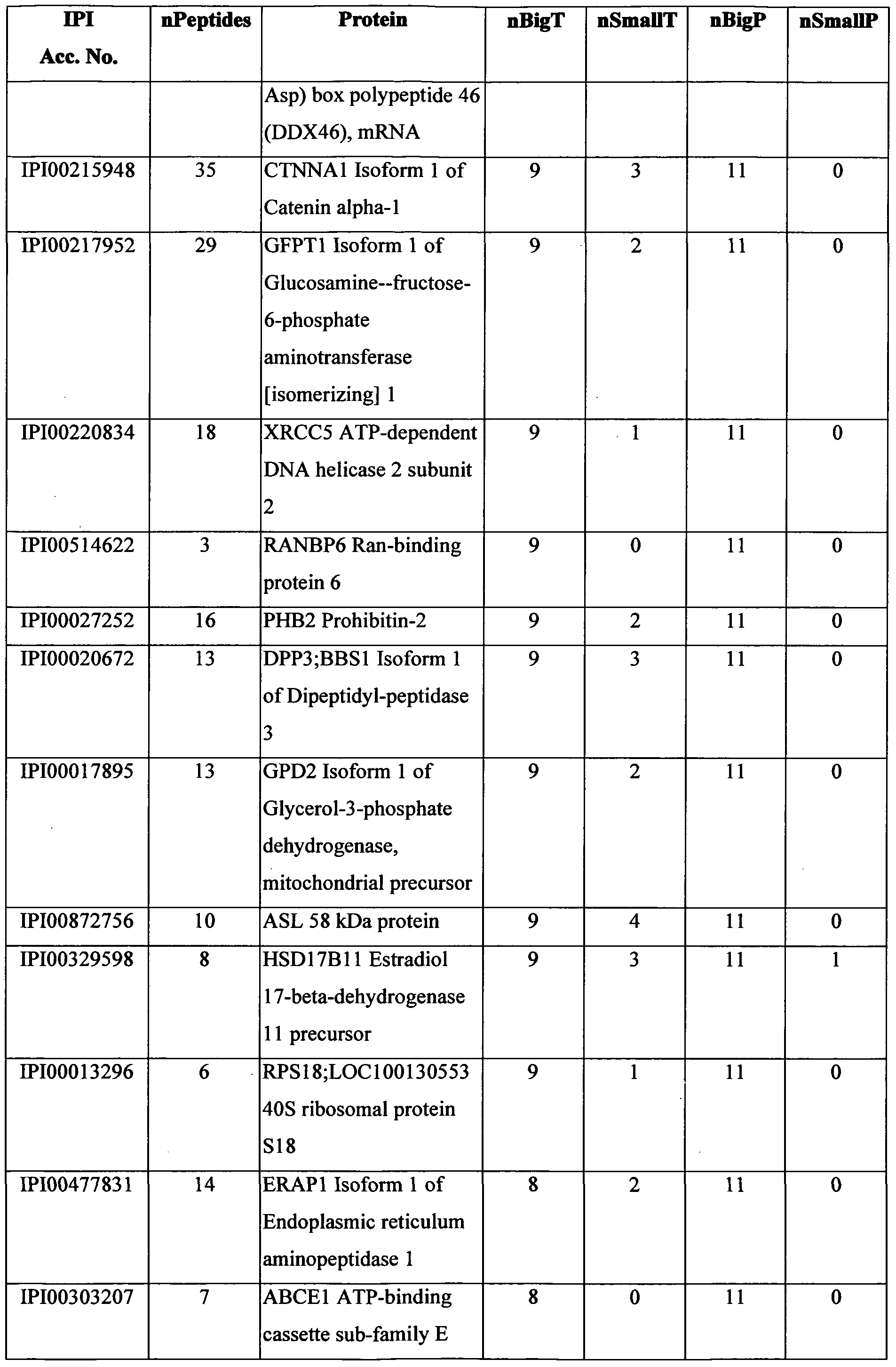
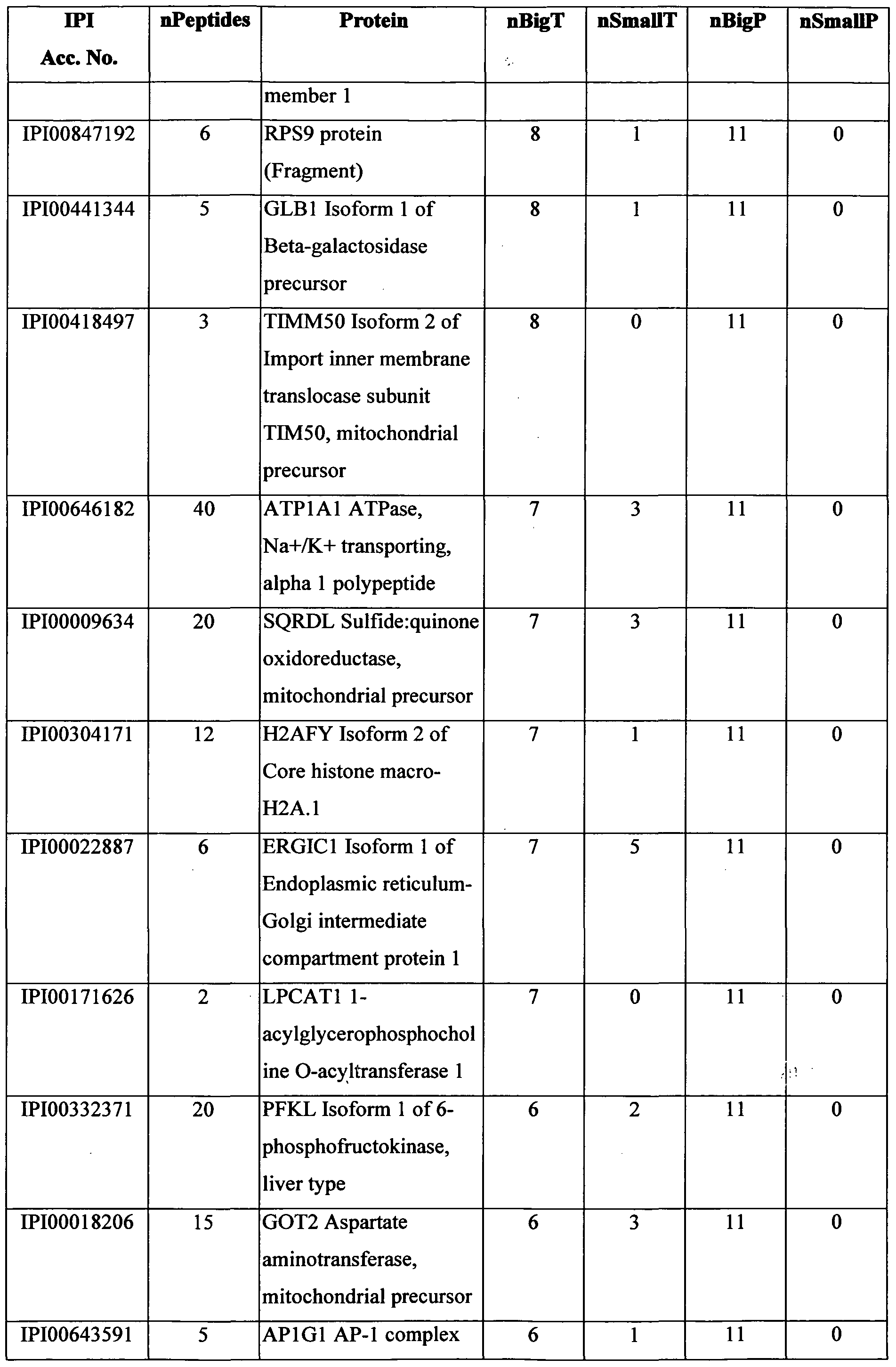


Table 4 lists proteins that were observed to be highly expressed in polyps, whereas in more advanced stages of colorectal cancer these proteins tended to have decreased levels of expression. Accordingly, a diagnostic array of reagents directed to detection of at least some of the proteins in this group may be used as a screening test for very early detection of colorectal cancer. Such a screening test could identify susceptible at-risk individuals, even prior to the stage at which polyp visualization is possible by endoscopic techniques. Table 4.


Example 4. Cancer associated proteins identified by isotopic labeling.
The proteins identified by isotopic labeling included those listed in Table 5. All of the listed proteins were found to be present in colon cancer tissue at levels that were at least 2.5- fold greater than the level of the same protein in healthy colon tissue from the same subject, as indicated by the median ratios of the protein levels (i.e. cancerous tissue:healthy tissue) listed in the column denoted "MED".
Table 5.


IPI Ace. No, accession number in IPI database; nPEP, number of identified peptides in all the samples, MED, median of the ratio tumor/healthy from all the patients; #>2.5, number of patients in which tumor/healthy >2.5.
Example 5. Cancer associated proteins identified by multidimensional chromatography.
The proteins identified on the basis of the peptides detected by the multidimensional chromatography technique include those listed in Table 6, and reflects the abundance of these proteins in cancerous tissues. None of the proteins listed in Table 6 were identified by the isotopic labeling technique. The proteins listed in Tables 3 and 6 appear to be specifically expressed in colorectal cancer tissue, as they were substantially undetectable in all healthy colorectal tissues obtained from the colorectal cancer patients.
Table 6.


Example 6. Cancer associated proteins as potential therapeutic targets.
Table 7 lists proteins that are considered potential targets for development of cytotoxic reagents specifically directed to these proteins, for example, specific antibody or antibody fragments conjugated to toxic moieties for targeted elimination of cancer cells. As well as being highly expressed in early stage polyps and in tumors, these proteins are generally exposed, and considered vulnerable to attack by targeted cytotoxic reagents.
Table 7.


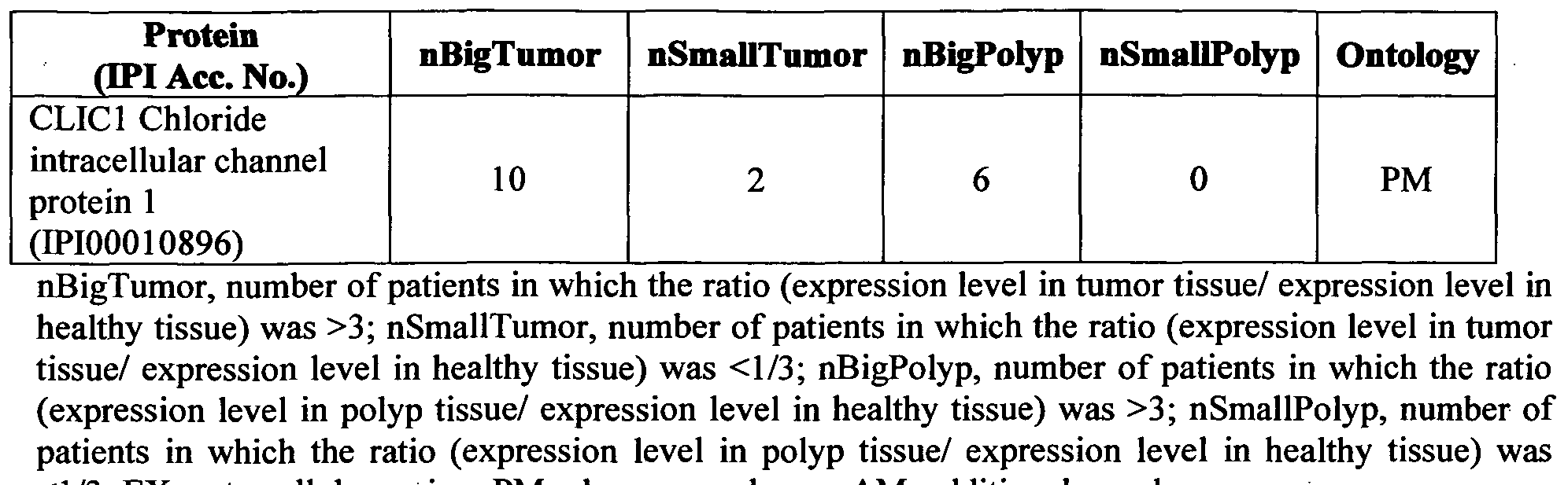
References cited
Aebersold, R. and M. Mann (2003). "Mass spectrometry-based proteomics." Nature 422(6928): 198-207.
Beer, L, E. et al. (2004). "Improving large-scale proteomics by clustering of mass spectrometry data." Proteomics 4(4): 950-60.
Eng, J., et al. (1994). "An approach to correlate tandem mass spectral data of peptides with amino acid sequences in a protein database." J. Amer. Soc. Mass. Spect. 5: 976-989. Hsu, J. L., et al. (2003). "Stable-isotope dimethyl labeling for quantitative proteomics." Anal Chem 75(24): 6843-52.
Ishihama, Y., et al. (2002). "Microcolumns with self-assembled particle frits for proteomics." J Chromatogr A 979(1-2): 233-9.
Link, A. J. (2002). "Multidimensional peptide separations in proteomics." Trends Biotechnol 20(12 Suppl): S8-13.
Link, A. J. et al. (1999). "Direct analysis of protein complexes using mass spectrometry." Nat. Biotechnol. 17(7): 676-82.
Ong, S. E. and M. Mann (2005). "Mass spectrometry-based proteomics turns quantitative." Nat Chem Biol 1(5): 252-62. Perkins, D. N. et al. (1999). "Probability-based protein identification by searching sequence databases using mass spectrometry data." Electrophoresis 20(18): 3551-67.
Regnier, F. E. and S. Julka (2006). "Primary amine coding as a path to comparative proteomics." Proteomics 6(14): 3968-79.
Ross, P. L. et al. (2004). "Multiplexed protein quantitation in Saccharomyces cerevisiae using amine-reactive isobaric tagging reagents." MoI Cell Proteomics 3(12): 1154- 69.
The foregoing description of the specific embodiments will so fully reveal the general nature of the invention that others can, by applying current knowledge, readily modify and/or adapt for various applications such specific embodiments without undue experimentation and without departing from the brand concept, and, therefore, such adaptations and modifications should and are intended to be comprehended within the meaning and range of equivalents of the disclosed embodiments. It is to be understood that the phraseology or terminology employed herein is for the purpose of description and not of limitation. The means, materials, and steps for carrying out various disclosed functions may take a variety of alternative forms without departing from the invention.
Claims
1. A method of detecting cancer in a subject, the method comprising:
(i) detecting in a biological sample from the subject at least one protein selected from the group consisting of KIAAOl 52 (SEQ ID NO:1), NAMPT (SEQ ID
NO:2), PYCRl (SEQ ID NO:3), GPX2 (SEQ ID NO:4), PRKDC (SEQ ID NO:5),
ALDHl 8Al (SEQ ID NO:6), OCIAD2 (SEQ ID NO:7), GCSl (SEQ ID NO:8),
GMDS (SEQ ID NO:9), ARF4 (SEQ ID NO: 10), ARF5 (SEQ ID NO:11),
LRPPRC (SEQ ID NO: 12), CTNNBl (SEQ ID NO: 13), ARF3 (SEQ ID NO: 14), GCNlLl (SEQ ID NO: 15), BDHl (SEQ ID NO:16), RPL9 (SEQ ID NO: 17),
UGCGLl (SEQ ID NO: 18), FAM3D (SEQ ID NO: 19) and CCT4 (SEQ ID
NO:20), so as to determine the level of the at least one protein; and
(ii) comparing the level determined in (i) to a reference level of the same at least one protein, wherein detection of a level of said at least one protein in the biological sample which is significantly different from the reference level, is indicative of cancer in the subject.
2. The method according to claim 1, wherein the biological sample is selected from the group consisting of blood, serum, nipple aspirate fluid, lymph node aspirate, a biopsy sample, a tumor sample, a tissue sample, mucosal fluid, cervical wash, lacrimal duct fluid, urine, saliva, pleural effusion and sputum.
3. The method according to claim 2, wherein the tissue sample comprises gastrointestinal tissue.
4. The method according to claim 1, wherein the cancer is colorectal cancer.
5. The method according to claim 4, wherein the colorectal cancer is selected from the group consisting of pre-cancerous polyps, early stage colorectal cancer and advanced stage colorectal cancer.
6. The method according to claim 4, comprising detecting at least one of KIAAO 152 (SEQ ID NO:1) and NAMPT (SEQ ID NO:2).
7. The method according to claim 4, further comprising detecting at least one protein selected from the group consisting of CPT2 (SEQ ID NO:21), ARLl (SEQ ID
NO:22), PFKL (SEQ ID NO:23), GOT2 (SEQ ID NO:24), APlGl (SEQ ID
NO:25), STRBP (SEQ ID NO:26), CLCAl (SEQ ID NO:27), CYFIPl (SEQ ID NO:28), COQ9 (SEQ ID NO:29), NDUFA9 (SEQ ID NO:30), ALDH7A1 (SEQ ID NO:31), HMGCSl (SEQ ID NO:32), NNT (SEQ ID NO:33), PRDX5 (SEQ ID NO:34), PCCB (SEQ ID NO:35), COPZl (SEQ ID NO:36), BAX (SEQ ID NO:37), ACAD9 (SEQ ID NO:38), UBXD8 (SEQ ID NO:39), HMGCS2 (SEQ ID NO:40), SLC25A3 (SEQ ID NO:41), SLC25A11 (SEQ ID NO:42), PDCD6
(SEQ ID NO:43), UCRC (SEQ ID NO:44), DEFA6 (SEQ ID NO:45), DYNClHl (SEQ ID NO:46), HKl (SEQ ID NO:47), CYFIP2 (SEQ ID NO:48), DCI (SEQ ID NO:49) and CISDl (SEQ ID NO:50).
8. The method according to claim 7, wherein the colorectal cancer comprises pre- cancerous polyps or early stage colorectal cancer.
9. The method according to claim 3, wherein the gastrointestinal tissue is obtained from the subject by a procedure selected from the group consisting of biopsy, flexible endoscopy, double balloon endoscopy and surgical colorectal re- sectioning.
10. The method according to claim 3, wherein the gastrointestinal tissue is assessed in vivo.
11. The method according to claim 10, comprising contacting gastrointestinal tissue with at least one of a pharmaceutical composition and an endoscopy apparatus.
12. The method according to claim 11, comprising administering the pharmaceutical composition to the subject by a route selected from the group consisting of oral, parenteral, subcutaneous, intramuscular, intrathoracic and intraarticular.
13. The method according to any one of claims 1, 7, 9 or 10, comprising use of at least one reagent suitable for detecting the level of said at least one protein.
14. The method according to claim 11, wherein the pharmaceutical composition or ' endoscopy apparatus comprise at least one reagent suitable for detecting the level of at said at least one protein.
15. The method according to claim 14, wherein said at least one reagent is suitable for detecting the level of at least one of KIAAOl 52 (SEQ ID NO:1) and NAMPT (SEQ ID NO:2).
16. The method according to claim 13, wherein said at least one reagent specifically interacts with said at least one protein or with nucleic acid encoding said at least one protein or a fragment thereof.
17. The method according to claim 13, wherein said reagent is selected from an antibody, an antibody mimetic and a nucleic acid.
18. The method according to claim 14, wherein the at least one reagent comprises a multiplicity of reagents, wherein each reagent of the multiplicity has specifically for a distinct protein.
19. The method according to claim 16, wherein said at least one reagent comprises a detectable label.
20. The method according to claim 1, comprising use of an assay system selected from the group consisting of an immunoassay, a nucleic acid hybridization assay, a binding assay, an array, a phage display library and a combination thereof.
21. The method according to claim 3, comprising contacting gastrointestinal tissue with at least one of a pharmaceutical composition and an endoscopy apparatus, and further comprising use of an external monitoring system, wherein the external monitoring system is configured to display the level of the at least one protein detected.
22. The method according to claim 4, wherein the level of the at least one protein in the sample is increased by at least at least 3-fold relative to the reference level, and wherein the reference level is representative of the level of the same protein in non-diseased tissue.
23. A method for determining the stage of a cancerous or pre-cancerous growth in a subject, the method comprising:
(i) detecting in a test sample from the subject at least one protein selected from the group consisting of KIAAO 152 (SEQ ID NO:1), NAMPT (SEQ ID NO:2), PYCRl (SEQ ID NO:3), GPX2 (SEQ ID NO:4), PRKDC (SEQ ID NO:5),
ALDH18A1 (SEQ ID NO:6), OCIAD2 (SEQ ID NO:7), GCSl (SEQ ID NO:8), GMDS (SEQ ID NO:9), ARF4 (SEQ ID NO:10), ARF5 (SEQ ID NO:11), LRPPRC (SEQ ID NO: 12), CTNNBl (SEQ ID NO: 13), ARF3 (SEQ ID NO: 14), GCNlLl (SEQ ID NO:15), BDHl (SEQ ID NO:16), RPL9 (SEQ ID NO.17), UGCGLl (SEQ ID NO: 18), FAM3D (SEQ ID NO: 19) and CCT4 (SEQ ID
NO:20), so as to determine the level of the at least one protein; and (ii) comparing the level determined in (i) to a reference level of the same at least one protein; wherein the level detected in the test sample is indicative of the stage of the growth.
24. The method according to claim 23, wherein the cancerous or pre-cancerous growth is in gastrointestinal tissue.
25. The method according to claim 24, wherein the growth is selected from the group consisting of pre-cancerous polyps, early stage colorectal cancer and advanced stage colorectal cancer.
26. The method according to claim 24, comprising detecting at least one of KIAA0152 (SEQ ID NO:1) and NAMPT (SEQ ID NO:2).
27. The method according to claim 26, further comprising detecting at least one protein selected from the group consisting of CPT2 (SEQ ID NO:21), ARLl (SEQ ID NO:22), PFKL (SEQ ID NO:23), GOT2 (SEQ ID NO:24), APlGl (SEQ ID NO:25), STRBP (SEQ ID NO:26), CLCAl (SEQ ID NO:27), CYFIPl (SEQ ID NO:28), COQ9 (SEQ ID NO:29), NDUFA9 (SEQ ID NO:30), ALDH7A1
(SEQ ID NO:31), HMGCSl (SEQ ID NO:32), NNT (SEQ ID NO:33), PRDX5 (SEQ ID NO:34), PCCB (SEQ ID NO:35), COPZl (SEQ ID NO:36), BAX (SEQ ID NO:37), ACAD9 (SEQ ID NO:38), UBXD8 (SEQ ID NO:39), HMGCS2 (SEQ ID NO:40), SLC25A3 (SEQ ID NO:41), SLC25A11 (SEQ ID NO:42), PDCD6 (SEQ ID NO:43), UCRC (SEQ ID NO:44), DEFA6 (SEQ ID NO:45),
DYNClHl (SEQ ID NO:46), HKl (SEQ ID NO:47), CYFIP2 (SEQ ID NO:48), DCI (SEQ ID NO:49) and CISDl (SEQ ID NO:50).
28. The method according to claim 27, wherein the growth comprises pre-cancerous polyps or early stage colorectal cancer.
29. The method according to claim 24, wherein the growth is obtained from the subject by a procedure selected from the group consisting of biopsy, flexible endoscopy, double balloon endoscopy and surgical colorectal re-sectioning.
30. The method according to claim 24, wherein the gastrointestinal tissue is accessed in vivo.
31. The method according to claim 30, comprising contacting gastrointestinal tissue with at least one of a pharmaceutical composition and an endoscopy apparatus.
32. The method according to claim 31, comprising administering the pharmaceutical composition to the subject by a route selected from the group consisting of oral, parenteral, subcutaneous, intramuscular, intrathoracic and intraarticular.
33. The method according to any one of claims 23 or 27, comprising use of at least one reagent suitable for detecting the level of said at least one protein.
34. The method according to claim 31, wherein the pharmaceutical composition or endoscopy apparatus comprise at least one reagent suitable for detecting the level of at said at least one protein.
35. The method according to claim 34, wherein said at least one reagent is suitable for detecting the level of at least one of KIAA0152 (SEQ ID NO:1) and NAMPT
(SEQ ID NO:2).
36. The method according to claim 23, wherein said at least one reagent specifically interacts with said at least one protein or with nucleic acid encoding said at least one protein or a fragment thereof, and wherein said reagent is selected from an antibody, an antibody mimetic and a nucleic acid.
37. The method according to claim 33, comprising use of an assay system selected from the group consisting of an immunoassay, a nucleic acid hybridization assay, a binding assay, an array, a phage display library and a combination thereof.
38. The method according to claim 24, wherein the level of the at least one protein in the sample is increased by at least at least 3-fold relative to the reference level, and wherein the reference level is representative of the level of the same protein in non-diseased gastrointestinal tissue.
39. An antigen composition comprising at least one protein selected from the group consisting of KIAA0152 (SEQ ID NO:1), NAMPT (SEQ ID NO:2), PYCRl (SEQ ID NO:3), GPX2 (SEQ ID NO:4), PRKDC (SEQ ID NO:5), ALDHl 8Al
(SEQ ID NO:6), OCIAD2 (SEQ ID NO:7), GCSl (SEQ ID NO:8), GMDS (SEQ ID NO:9), ARF4 (SEQ ID NO:10), ARF5 (SEQ ID NO:11), LRPPRC (SEQ ID NO:12), CTNNBl (SEQ ID NO:13), ARF3 (SEQ ID NO:14), GCNlLl (SEQ ID NO:15), BDHl (SEQ ID NO:16), RPL9 (SEQ ID NO.17), UGCGLl (SEQ ID NO: 18), FAM3D (SEQ ID NO: 19), CCT4 (SEQ ID NO:20), CPT2 (SEQ ID
NO:21), ARLl (SEQ ID NO:22), PFKL (SEQ ID NO:23), GOT2 (SEQ ID NO:24), APlGl (SEQ ID NO:25), STRBP (SEQ ID NO:26), CLCAl (SEQ ID NO:27), CYFIPl (SEQ ID NO:28), COQ9 (SEQ ID NO:29), NDUFA9 (SEQ ID NO:30), ALDH7A1 (SEQ ID NO:31), HMGCSl (SEQ ID NO:32), NNT (SEQ ID NO:33), PRDX5 (SEQ ID NO:34), PCCB (SEQ ID NO:35), COPZl (SEQ ID NO:36), BAX (SEQ ID NO:37), ACAD9 (SEQ ID NO:38), UBXD8 (SEQ ID NO:39), HMGCS2 (SEQ ID NO:40), SLC25A3 (SEQ ID NO:41), SLC25A11
(SEQ ID NO:42), PDCD6 (SEQ ID NO:43), UCRC (SEQ ID NO:44), DEFA6 (SEQ ID NO:45), DYNClHl (SEQ ID NO:46), HKl (SEQ ID NO:47), CYFIP2 (SEQ ID NO:48), DCI (SEQ ID NO:49), CISDl (SEQ ID NO:50), LYZ (SEQ ID NO:54): LOC442497;SLC3A2 (SEQ ID NO:61), DMBTl (SEQ ID NO:84), NUCBl (SEQ ID NO:.85), GGH (SEQ ID NO:86), AGR3 (SEQ ID NO:87),
TM9SF2 (SEQ ID NO: 88), SYK (SEQ ID NO:89), GCA (SEQ ID NO:90), HDLBP (SEQ ID NO:91), ClQBP (SEQ ID NO:92) and CLICl (SEQ ID NO:93), or an immunogenic fragment of said at least one protein.
40. The composition according to claim 39, wherein the at least one protein is selected from the group consisting of KIAAO 152 (SEQ ID NO:1), LYZ (SEQ ID
NO:54), LOC442497;SLC3A2 (SEQ ID NO:61), DMBTl (SEQ ID NO:84), NUCBl (SEQ ID NO:.85), GGH (SEQ ID NO:86), AGR3 (SEQ ID NO:87), TM9SF2 (SEQ ID NO:88), SYK (SEQ ID NO:89), GCA (SEQ ID NO:90), HDLBP (SEQ ID NO:91), ClQBP (SEQ ID NO:92) and CLICl (SEQ ID NO:93), or an immunogenic fragment thereof.
41. The composition according to claim 39, wherein the at least one protein is KIAAOl 52 (SEQ ID NO: 1).
42. A method of preventing or treating cancer, comprising administering to a subject in need thereof, an effective amount of the composition according to any one of claims 39-41.
43. The method according to claim 42, wherein the cancer is colorectal cancer.
44. A kit for detecting, staging or prognosing cancer in a biological sample from a subject, the kit comprising (i) at least one reagent suitable for detecting the level of at least one protein selected from the group consisting of KIAAO 152 (SEQ ID NO:1), NAMPT (SEQ ID NO:2), PYCRl (SEQ ID NO:3), GPX2 (SEQ ID
NO:4), PRKDC (SEQ ID NO:5), ALDHl 8Al (SEQ ID NO:6), OCIAD2 (SEQ ID NO:7), GCSl (SEQ ID NO:8), GMDS (SEQ ID NO:9), ARF4 (SEQ ID NO:10), ARF5 (SEQ ID NO: 11), LRPPRC (SEQ ID NO: 12), CTNNBl (SEQ ID NO:13), ARF3 (SEQ ID NO: 14), GCNlLl (SEQ ID NO: 15), BDHl (SEQ ID NO: 16), RPL9 (SEQ ID NO: 17), UGCGLl (SEQ ID NO:18), FAM3D (SEQ ID NO: 19), CCT4 (SEQ ID NO:20), CPT2 (SEQ ID NO:21), ARLl (SEQ ID NO:22), PFKL (SEQ ID NO:23), GOT2 (SEQ ID NO:24), APlGl (SEQ ID NO:25), STRBP
(SEQ ID NO:26), CLCAl (SEQ ID NO:27), CYFIPl (SEQ ID NO:28), C0Q9 (SEQ ID NO:29), NDUFA9 (SEQ ID NO:30), ALDH7A1 (SEQ ID NO:31), HMGCSl (SEQ ID NO:32), NNT (SEQ ID NO:33), PRDX5 (SEQ ID NO:34), PCCB (SEQ ID NO:35), COPZl (SEQ ID NO:36), BAX (SEQ ID NO:37), ACAD9 (SEQ ID NO:38), UBXD8 (SEQ ID NO:39), HMGCS2 (SEQ ID
NO:40), SLC25A3 (SEQ ID NO:41), SLC25A11 (SEQ ID NO:42), PDCD6 (SEQ ID NO:43), UCRC (SEQ ID NO:44), DEFA6 (SEQ ID NO:45), DYNClHl (SEQ ID NO:46), HKl (SEQ ID NO:47), CYFIP2 (SEQ ID NO:48), DCI (SEQ ID NO:49) and CISDl (SEQ ID NO:50); (ii) a control biological sample for comparison to a biological sample from a subject, and (iii) instructional material comprising a protocol suitable for use in detection and quantification of the at least one protein in a biological sample from a subject.
45. The kit according to claim 44, comprising at least one reagent suitable for detecting the level of at least one protein selected from the group consisting of CPT2 (SEQ ID NO:21), ARLl (SEQ ID NO:22), PFKL (SEQ ID NO:23), GOT2
(SEQ ID NO:24), APlGl (SEQ ID NO:25), STRBP (SEQ ID NO:26), CLCAl (SEQ ID NO:27), CYFIPl (SEQ ID NO:28), COQ9 (SEQ ID NO:29), NDUFA9 (SEQ ID NO:30), ALDH7A1 (SEQ ID NO:31), HMGCSl (SEQ ID NO:32), NNT (SEQ ID NO:33), PRDX5 (SEQ ID NO:34), PCCB (SEQ ID NO:35), COPZl (SEQ ID NO:36), BAX (SEQ ID NO:37), ACAD9 (SEQ ID NO:38),
UBXD8 (SEQ ID NO:39), HMGCS2 (SEQ ID NO:40), SLC25A3 (SEQ ID NO:41), SLC25A11 (SEQ ID NO:42), PDCD6 (SEQ ID NO:43), UCRC (SEQ ID NO:44), DEFA6 (SEQ ID NO:45), DYNClHl (SEQ ID NO:46), HKl (SEQ ID NO:47), CYFIP2 (SEQ ID NO:48), DCI (SEQ ID NO:49) and CISDl (SEQ ID NO:50).
46. The kit according to claim 44, comprising a reagent suitable for detecting the level of KIAAOl 52 (SEQ ID NO:1).
Priority Applications (1)
| Application Number | Priority Date | Filing Date | Title |
|---|---|---|---|
| US13/266,567 US8741581B2 (en) | 2009-04-27 | 2010-04-27 | Markers for cancer detection |
Applications Claiming Priority (2)
| Application Number | Priority Date | Filing Date | Title |
|---|---|---|---|
| US17280009P | 2009-04-27 | 2009-04-27 | |
| US61/172,800 | 2009-04-27 |
Publications (2)
| Publication Number | Publication Date |
|---|---|
| WO2010125566A2 true WO2010125566A2 (en) | 2010-11-04 |
| WO2010125566A3 WO2010125566A3 (en) | 2013-05-10 |
Family
ID=42582600
Family Applications (1)
| Application Number | Title | Priority Date | Filing Date |
|---|---|---|---|
| PCT/IL2010/000342 WO2010125566A2 (en) | 2009-04-27 | 2010-04-27 | Markers for cancer detection |
Country Status (2)
| Country | Link |
|---|---|
| US (1) | US8741581B2 (en) |
| WO (1) | WO2010125566A2 (en) |
Cited By (8)
| Publication number | Priority date | Publication date | Assignee | Title |
|---|---|---|---|---|
| WO2013035095A1 (en) * | 2011-09-08 | 2013-03-14 | Ben-Gurion University Of The Negev Research And Development Authority | Methods for diagnosing cancer |
| WO2013010140A3 (en) * | 2011-07-13 | 2013-03-14 | Cognosci, Inc | Methods of diagnosing cancer |
| EP2751561A4 (en) * | 2011-08-31 | 2015-08-12 | Oncocyte Corp | Methods and compositions for the treatment and diagnosis of colorectal cancer |
| JP2016101161A (en) * | 2014-11-14 | 2016-06-02 | 東芝メディカルシステムズ株式会社 | Gene marker, method, reporter vector, primer set and assay kit for examining cellular property of test cells, and apparatus for examining cellular property |
| CN106093429A (en) * | 2016-06-02 | 2016-11-09 | 滨州医学院 | A kind of test kit detecting stomach organization |
| CN110078811A (en) * | 2018-01-25 | 2019-08-02 | 中国医学科学院医药生物技术研究所 | A kind of polypeptide IMB-P1 with anti-tumor activity and its application |
| RU2713907C1 (en) * | 2019-11-26 | 2020-02-11 | Игорь Игоревич Быков | Method of personalization of medical care for patients with stomach cancer |
| WO2023230578A3 (en) * | 2022-05-25 | 2024-02-01 | Flagship Pioneering Innovations Vii, Llc | Compositions and methods for modulating circulating factors |
Families Citing this family (22)
| Publication number | Priority date | Publication date | Assignee | Title |
|---|---|---|---|---|
| US8980269B2 (en) | 2009-07-14 | 2015-03-17 | Temple University Of The Commonwealth System Of Higher Education | G-protein coupled receptor-associated sorting protein 1 as a cancer biomarker |
| EP2727935A3 (en) * | 2009-07-14 | 2014-07-30 | Temple University Of The Commonwealth System Of Higher Education | Serum markers associated with early and other stages of breast cancer |
| US20140322705A1 (en) * | 2010-08-20 | 2014-10-30 | Thomas Jefferson University | Cancer diagnostic and cancer therapeutic |
| US9534058B2 (en) | 2012-02-08 | 2017-01-03 | Abbvie Stemcentrx Llc | Anti-CD324 monoclonal antibodies and uses thereof |
| WO2014025810A1 (en) | 2012-08-07 | 2014-02-13 | The Henry M. Jackson Foundation For The Advancement Of Military Medicine, Inc. | Prostate cancer gene expression profiles |
| WO2014028939A2 (en) * | 2012-08-17 | 2014-02-20 | California Institute Of Technology | Targeting phosphofructokinase and its glycosylation form for cancer |
| WO2014172627A1 (en) | 2013-04-19 | 2014-10-23 | Thomas Jefferson University | Caveolin-1 related methods for treating glioblastoma with temozolomide |
| WO2015042115A1 (en) * | 2013-09-17 | 2015-03-26 | The Board Of Trustees Of The Leland Stanford Junior University | Biomarkers for ovarian cancer |
| KR20150039484A (en) * | 2013-10-02 | 2015-04-10 | 삼성전자주식회사 | Method and apparatus for diagnosing cancer using genetic information |
| US20180057888A1 (en) * | 2015-03-13 | 2018-03-01 | The Board Of Regents Of The University Of Texas System | Kub5/hera as a determinant of sensitivity to dna damage |
| GB201505305D0 (en) | 2015-03-27 | 2015-05-13 | Immatics Biotechnologies Gmbh | Novel Peptides and combination of peptides for use in immunotherapy against various tumors |
| HRP20230878T8 (en) | 2015-03-27 | 2024-02-16 | Immatics Biotechnologies Gmbh | Novel peptides and combination of peptides for use in immunotherapy against various tumors (seq id 25 - mrax5-003) |
| EP3304082B1 (en) * | 2015-06-08 | 2020-05-13 | Arquer Diagnostics Limited | Methods for analysing a urine sample |
| EP4060344A1 (en) * | 2015-06-08 | 2022-09-21 | Arquer Diagnostics Limited | Methods and kits |
| WO2017100789A1 (en) * | 2015-12-11 | 2017-06-15 | Expression Pathology, Inc. | Srm/mrm assays |
| CN109402253B (en) * | 2018-08-22 | 2022-11-08 | 中国人民解放军陆军军医大学第一附属医院 | Use of ALDH18A1 in the treatment and diagnosis of colorectal cancer |
| BR112022007996A2 (en) * | 2019-10-28 | 2022-07-05 | Celgene Corp | METHODS FOR THE TREATMENT AND USE OF BIOMARKERS TO PREDICT CLINICAL SENSITIVITY TO 2-(4-CHLOROPHENYL)-N-((2-(2,6-DIOXOPIPERIDIN-3-IL)-1-OXOISOINDOLIN-5-IL)METHYL) -2,2-DIFLUOROACETAMIDE |
| KR20230107932A (en) * | 2022-01-10 | 2023-07-18 | (의료)길의료재단 | Biomarker for predicting the prognosis of colorectal cancer |
| CN114107515B (en) * | 2022-01-28 | 2022-04-19 | 中国医学科学院北京协和医院 | Early gastric cancer prognosis differential gene and recurrence prediction model |
| CN115612745B (en) * | 2022-12-19 | 2023-03-21 | 北京大学人民医院 | Application of IPO7 in colorectal cancer treatment and prognosis prediction |
| CN116381237A (en) * | 2023-02-28 | 2023-07-04 | 杭州凯保罗生物科技有限公司 | Early thyroid cancer prediction system and application thereof |
| CN116449003A (en) * | 2023-05-06 | 2023-07-18 | 郑州市妇幼保健院 | Application of cervical cancer related antibody protein combination in colloidal gold test strip |
Citations (47)
| Publication number | Priority date | Publication date | Assignee | Title |
|---|---|---|---|---|
| US4816397A (en) | 1983-03-25 | 1989-03-28 | Celltech, Limited | Multichain polypeptides or proteins and processes for their production |
| US4816567A (en) | 1983-04-08 | 1989-03-28 | Genentech, Inc. | Recombinant immunoglobin preparations |
| US4946778A (en) | 1987-09-21 | 1990-08-07 | Genex Corporation | Single polypeptide chain binding molecules |
| WO1991017271A1 (en) | 1990-05-01 | 1991-11-14 | Affymax Technologies N.V. | Recombinant library screening methods |
| WO1992001047A1 (en) | 1990-07-10 | 1992-01-23 | Cambridge Antibody Technology Limited | Methods for producing members of specific binding pairs |
| US5242974A (en) | 1991-11-22 | 1993-09-07 | Affymax Technologies N.V. | Polymer reversal on solid surfaces |
| US5384261A (en) | 1991-11-22 | 1995-01-24 | Affymax Technologies N.V. | Very large scale immobilized polymer synthesis using mechanically directed flow paths |
| US5405783A (en) | 1989-06-07 | 1995-04-11 | Affymax Technologies N.V. | Large scale photolithographic solid phase synthesis of an array of polymers |
| US5412087A (en) | 1992-04-24 | 1995-05-02 | Affymax Technologies N.V. | Spatially-addressable immobilization of oligonucleotides and other biological polymers on surfaces |
| US5424186A (en) | 1989-06-07 | 1995-06-13 | Affymax Technologies N.V. | Very large scale immobilized polymer synthesis |
| US5429807A (en) | 1993-10-28 | 1995-07-04 | Beckman Instruments, Inc. | Method and apparatus for creating biopolymer arrays on a solid support surface |
| US5436327A (en) | 1988-09-21 | 1995-07-25 | Isis Innovation Limited | Support-bound oligonucleotides |
| US5449754A (en) | 1991-08-07 | 1995-09-12 | H & N Instruments, Inc. | Generation of combinatorial libraries |
| US5472672A (en) | 1993-10-22 | 1995-12-05 | The Board Of Trustees Of The Leland Stanford Junior University | Apparatus and method for polymer synthesis using arrays |
| US5527681A (en) | 1989-06-07 | 1996-06-18 | Affymax Technologies N.V. | Immobilized molecular synthesis of systematically substituted compounds |
| US5532128A (en) | 1991-11-19 | 1996-07-02 | Houston Advanced Research Center | Multi-site detection apparatus |
| US5545531A (en) | 1995-06-07 | 1996-08-13 | Affymax Technologies N.V. | Methods for making a device for concurrently processing multiple biological chip assays |
| US5554501A (en) | 1992-10-29 | 1996-09-10 | Beckman Instruments, Inc. | Biopolymer synthesis using surface activated biaxially oriented polypropylene |
| US5556752A (en) | 1994-10-24 | 1996-09-17 | Affymetrix, Inc. | Surface-bound, unimolecular, double-stranded DNA |
| US5561071A (en) | 1989-07-24 | 1996-10-01 | Hollenberg; Cornelis P. | DNA and DNA technology for the construction of networks to be used in chip construction and chip production (DNA-chips) |
| US5571639A (en) | 1994-05-24 | 1996-11-05 | Affymax Technologies N.V. | Computer-aided engineering system for design of sequence arrays and lithographic masks |
| US5578832A (en) | 1994-09-02 | 1996-11-26 | Affymetrix, Inc. | Method and apparatus for imaging a sample on a device |
| US5599695A (en) | 1995-02-27 | 1997-02-04 | Affymetrix, Inc. | Printing molecular library arrays using deprotection agents solely in the vapor phase |
| US5604531A (en) | 1994-01-17 | 1997-02-18 | State Of Israel, Ministry Of Defense, Armament Development Authority | In vivo video camera system |
| US5624711A (en) | 1995-04-27 | 1997-04-29 | Affymax Technologies, N.V. | Derivatization of solid supports and methods for oligomer synthesis |
| US5631734A (en) | 1994-02-10 | 1997-05-20 | Affymetrix, Inc. | Method and apparatus for detection of fluorescently labeled materials |
| US5658734A (en) | 1995-10-17 | 1997-08-19 | International Business Machines Corporation | Process for synthesizing chemical compounds |
| US5693762A (en) | 1988-12-28 | 1997-12-02 | Protein Design Labs, Inc. | Humanized immunoglobulins |
| US5700637A (en) | 1988-05-03 | 1997-12-23 | Isis Innovation Limited | Apparatus and method for analyzing polynucleotide sequences and method of generating oligonucleotide arrays |
| US5712120A (en) | 1994-06-30 | 1998-01-27 | Centro De Immunologia Molecular | Method for obtaining modified immunoglobulins with reduced immunogenicity of murine antibody variable domains, compositions containing them |
| US5965405A (en) | 1988-04-16 | 1999-10-12 | Celltech Limited | Method for producing Fv fragments in eukaryotic cells |
| US6004755A (en) | 1998-04-07 | 1999-12-21 | Incyte Pharmaceuticals, Inc. | Quantitative microarray hybridizaton assays |
| US6475809B1 (en) | 1998-07-14 | 2002-11-05 | Zyomyx, Incorporated | Protein arrays for high-throughput screening |
| US6919176B2 (en) | 2001-05-07 | 2005-07-19 | Amgen Inc. | Polypeptides and nucleic acids associated with cancer |
| US6949339B1 (en) | 1998-05-21 | 2005-09-27 | Diadexus, Inc. | Method of diagnosing, monitoring, and staging colon cancer |
| US7078180B2 (en) | 2001-09-05 | 2006-07-18 | The Children's Hospital Of Philadelphia | Methods and compositions useful for diagnosis, staging, and treatment of cancers and tumors |
| US7098008B2 (en) | 2000-04-25 | 2006-08-29 | Ic&G Do. Ltd. | Selected primers for detection of MAGE or GAGE genes for diagnosis of cancer and methods of use |
| US7115368B2 (en) | 2001-01-02 | 2006-10-03 | Amgen Sf, Llc | Diagnosis and treatment of cancer using mammalian pellino polypeptides and polynucleotides |
| US7129043B1 (en) | 1998-10-22 | 2006-10-31 | Duke University | Methods of screening for risk of proliferative disease and methods for the treatment of proliferative disease |
| US20080146896A1 (en) | 2005-01-31 | 2008-06-19 | Elisha Rabinowitz | Device, system and method for in vivo analysis |
| US7402403B1 (en) | 1998-05-11 | 2008-07-22 | Oncimmune Limited | Tumour markers |
| US7452727B2 (en) | 2000-12-18 | 2008-11-18 | Siemens Healthcare Diagnostics Inc | Method for increasing clinical specificity when detecting tumors and their precursor stages by simultaneously measuring at least two different molecular markers |
| US7468044B2 (en) | 2001-01-16 | 2008-12-23 | Given Imaging Ltd. | Device, system and method for determining in vivo body lumen conditions |
| US7501242B2 (en) | 2001-02-21 | 2009-03-10 | Novartis Vaccines And Diagnostics, Inc. | Detection of colon or breast cancer by measuring TTK polynucleotide expression |
| US7507541B2 (en) | 1999-10-28 | 2009-03-24 | Agensys, Inc. | 36P6D5: secreted tumor antigen |
| US7515953B2 (en) | 2002-08-01 | 2009-04-07 | The Johns Hopkins University | Techniques for identifying molecular structures and treating cell types lining a body lumen using fluorescence |
| US20090216082A1 (en) | 2005-04-01 | 2009-08-27 | Elisha Rabinovitz | Device, System and Method for In Vivo Magnetic Immunoassay Analysis |
Family Cites Families (7)
| Publication number | Priority date | Publication date | Assignee | Title |
|---|---|---|---|---|
| DE19601488C1 (en) | 1996-01-17 | 1997-05-28 | Itt Ind Gmbh Deutsche | Measuring device manufacturing method for measuring or testing physiological parameter at biocomponent |
| US6573043B1 (en) | 1998-10-07 | 2003-06-03 | Genentech, Inc. | Tissue analysis and kits therefor |
| US20070037204A1 (en) | 2003-08-08 | 2007-02-15 | Hiroyuki ABURANTAI | Gene overexpressed in cancer |
| US7527936B2 (en) * | 2004-01-13 | 2009-05-05 | Rigel Pharmaceuticals Inc. | Modulators of angiogenesis |
| US20080260742A1 (en) * | 2004-04-09 | 2008-10-23 | Takeda Pharmaceutical Company Limited | Preventives/Remedies for Cancer |
| US20090214517A1 (en) * | 2004-07-27 | 2009-08-27 | Justin Wong | Compositions and methods of use for modulators of nectin 4, semaphorin 4b, igsf9, and kiaa0152 in treating disease |
| WO2008086182A2 (en) | 2007-01-04 | 2008-07-17 | University Of Rochester | Use of gene signatures to design novel cancer treatment regimens |
-
2010
- 2010-04-27 WO PCT/IL2010/000342 patent/WO2010125566A2/en active Application Filing
- 2010-04-27 US US13/266,567 patent/US8741581B2/en not_active Expired - Fee Related
Patent Citations (51)
| Publication number | Priority date | Publication date | Assignee | Title |
|---|---|---|---|---|
| US4816397A (en) | 1983-03-25 | 1989-03-28 | Celltech, Limited | Multichain polypeptides or proteins and processes for their production |
| US4816567A (en) | 1983-04-08 | 1989-03-28 | Genentech, Inc. | Recombinant immunoglobin preparations |
| US4946778A (en) | 1987-09-21 | 1990-08-07 | Genex Corporation | Single polypeptide chain binding molecules |
| US5965405A (en) | 1988-04-16 | 1999-10-12 | Celltech Limited | Method for producing Fv fragments in eukaryotic cells |
| US5700637A (en) | 1988-05-03 | 1997-12-23 | Isis Innovation Limited | Apparatus and method for analyzing polynucleotide sequences and method of generating oligonucleotide arrays |
| US5436327A (en) | 1988-09-21 | 1995-07-25 | Isis Innovation Limited | Support-bound oligonucleotides |
| US5693762A (en) | 1988-12-28 | 1997-12-02 | Protein Design Labs, Inc. | Humanized immunoglobulins |
| US5405783A (en) | 1989-06-07 | 1995-04-11 | Affymax Technologies N.V. | Large scale photolithographic solid phase synthesis of an array of polymers |
| US5527681A (en) | 1989-06-07 | 1996-06-18 | Affymax Technologies N.V. | Immobilized molecular synthesis of systematically substituted compounds |
| US5424186A (en) | 1989-06-07 | 1995-06-13 | Affymax Technologies N.V. | Very large scale immobilized polymer synthesis |
| US5445934A (en) | 1989-06-07 | 1995-08-29 | Affymax Technologies N.V. | Array of oligonucleotides on a solid substrate |
| US5561071A (en) | 1989-07-24 | 1996-10-01 | Hollenberg; Cornelis P. | DNA and DNA technology for the construction of networks to be used in chip construction and chip production (DNA-chips) |
| WO1991017271A1 (en) | 1990-05-01 | 1991-11-14 | Affymax Technologies N.V. | Recombinant library screening methods |
| WO1992001047A1 (en) | 1990-07-10 | 1992-01-23 | Cambridge Antibody Technology Limited | Methods for producing members of specific binding pairs |
| US5969108A (en) | 1990-07-10 | 1999-10-19 | Medical Research Council | Methods for producing members of specific binding pairs |
| US5449754A (en) | 1991-08-07 | 1995-09-12 | H & N Instruments, Inc. | Generation of combinatorial libraries |
| US5532128A (en) | 1991-11-19 | 1996-07-02 | Houston Advanced Research Center | Multi-site detection apparatus |
| US5242974A (en) | 1991-11-22 | 1993-09-07 | Affymax Technologies N.V. | Polymer reversal on solid surfaces |
| US5384261A (en) | 1991-11-22 | 1995-01-24 | Affymax Technologies N.V. | Very large scale immobilized polymer synthesis using mechanically directed flow paths |
| US5412087A (en) | 1992-04-24 | 1995-05-02 | Affymax Technologies N.V. | Spatially-addressable immobilization of oligonucleotides and other biological polymers on surfaces |
| US5554501A (en) | 1992-10-29 | 1996-09-10 | Beckman Instruments, Inc. | Biopolymer synthesis using surface activated biaxially oriented polypropylene |
| US5529756A (en) | 1993-10-22 | 1996-06-25 | The Board Of Trustees Of The Leland Stanford Junior University | Apparatus and method for polymer synthesis using arrays |
| US5472672A (en) | 1993-10-22 | 1995-12-05 | The Board Of Trustees Of The Leland Stanford Junior University | Apparatus and method for polymer synthesis using arrays |
| US5429807A (en) | 1993-10-28 | 1995-07-04 | Beckman Instruments, Inc. | Method and apparatus for creating biopolymer arrays on a solid support surface |
| US5604531A (en) | 1994-01-17 | 1997-02-18 | State Of Israel, Ministry Of Defense, Armament Development Authority | In vivo video camera system |
| US5631734A (en) | 1994-02-10 | 1997-05-20 | Affymetrix, Inc. | Method and apparatus for detection of fluorescently labeled materials |
| US5571639A (en) | 1994-05-24 | 1996-11-05 | Affymax Technologies N.V. | Computer-aided engineering system for design of sequence arrays and lithographic masks |
| US5593839A (en) | 1994-05-24 | 1997-01-14 | Affymetrix, Inc. | Computer-aided engineering system for design of sequence arrays and lithographic masks |
| US5712120A (en) | 1994-06-30 | 1998-01-27 | Centro De Immunologia Molecular | Method for obtaining modified immunoglobulins with reduced immunogenicity of murine antibody variable domains, compositions containing them |
| US5578832A (en) | 1994-09-02 | 1996-11-26 | Affymetrix, Inc. | Method and apparatus for imaging a sample on a device |
| US5556752A (en) | 1994-10-24 | 1996-09-17 | Affymetrix, Inc. | Surface-bound, unimolecular, double-stranded DNA |
| US5599695A (en) | 1995-02-27 | 1997-02-04 | Affymetrix, Inc. | Printing molecular library arrays using deprotection agents solely in the vapor phase |
| US5624711A (en) | 1995-04-27 | 1997-04-29 | Affymax Technologies, N.V. | Derivatization of solid supports and methods for oligomer synthesis |
| US5545531A (en) | 1995-06-07 | 1996-08-13 | Affymax Technologies N.V. | Methods for making a device for concurrently processing multiple biological chip assays |
| US5658734A (en) | 1995-10-17 | 1997-08-19 | International Business Machines Corporation | Process for synthesizing chemical compounds |
| US6004755A (en) | 1998-04-07 | 1999-12-21 | Incyte Pharmaceuticals, Inc. | Quantitative microarray hybridizaton assays |
| US7402403B1 (en) | 1998-05-11 | 2008-07-22 | Oncimmune Limited | Tumour markers |
| US6949339B1 (en) | 1998-05-21 | 2005-09-27 | Diadexus, Inc. | Method of diagnosing, monitoring, and staging colon cancer |
| US6475809B1 (en) | 1998-07-14 | 2002-11-05 | Zyomyx, Incorporated | Protein arrays for high-throughput screening |
| US7129043B1 (en) | 1998-10-22 | 2006-10-31 | Duke University | Methods of screening for risk of proliferative disease and methods for the treatment of proliferative disease |
| US7507541B2 (en) | 1999-10-28 | 2009-03-24 | Agensys, Inc. | 36P6D5: secreted tumor antigen |
| US7098008B2 (en) | 2000-04-25 | 2006-08-29 | Ic&G Do. Ltd. | Selected primers for detection of MAGE or GAGE genes for diagnosis of cancer and methods of use |
| US7452727B2 (en) | 2000-12-18 | 2008-11-18 | Siemens Healthcare Diagnostics Inc | Method for increasing clinical specificity when detecting tumors and their precursor stages by simultaneously measuring at least two different molecular markers |
| US7115368B2 (en) | 2001-01-02 | 2006-10-03 | Amgen Sf, Llc | Diagnosis and treatment of cancer using mammalian pellino polypeptides and polynucleotides |
| US7468044B2 (en) | 2001-01-16 | 2008-12-23 | Given Imaging Ltd. | Device, system and method for determining in vivo body lumen conditions |
| US7501242B2 (en) | 2001-02-21 | 2009-03-10 | Novartis Vaccines And Diagnostics, Inc. | Detection of colon or breast cancer by measuring TTK polynucleotide expression |
| US6919176B2 (en) | 2001-05-07 | 2005-07-19 | Amgen Inc. | Polypeptides and nucleic acids associated with cancer |
| US7078180B2 (en) | 2001-09-05 | 2006-07-18 | The Children's Hospital Of Philadelphia | Methods and compositions useful for diagnosis, staging, and treatment of cancers and tumors |
| US7515953B2 (en) | 2002-08-01 | 2009-04-07 | The Johns Hopkins University | Techniques for identifying molecular structures and treating cell types lining a body lumen using fluorescence |
| US20080146896A1 (en) | 2005-01-31 | 2008-06-19 | Elisha Rabinowitz | Device, system and method for in vivo analysis |
| US20090216082A1 (en) | 2005-04-01 | 2009-08-27 | Elisha Rabinovitz | Device, System and Method for In Vivo Magnetic Immunoassay Analysis |
Non-Patent Citations (30)
| Title |
|---|
| AEBERSOLD, R.; M. MANN: "Mass spectrometry-based proteomics", NATURE, vol. 422, no. 6928, 2003, pages 198 - 207 |
| ALTSCHUL ET AL., J. MOL. BIOL., vol. 215, 1990, pages 403 - 10 |
| BEER, L, E. ET AL.: "Improving large-scale proteomics by clustering of mass spectrometry data", PROTEOMICS, vol. 4, no. 4, 2004, pages 950 - 60, XP008124571, DOI: doi:10.1002/PMIC.200300652 |
| CHATURVEDI ET AL., NUCL. ACIDS RES., vol. 24, 1996, pages 2318 - 2323 |
| CLACKSON ET AL., NATURE, vol. 352, 1991, pages 624 - 628 |
| COTE ET AL., PROC. NATL. ACAD. SCI. USA, vol. 80, 1983, pages 2026 30 |
| ENG, J. ET AL.: "An approach to correlate tandem mass spectral data of peptides with amino acid sequences in a protein database", J. AMER. SOC. MASS. SPECT., vol. 5, 1994, pages 976 - 989, XP026531327, DOI: doi:10.1016/1044-0305(94)80016-2 |
| HAGIWARA; YUASA, HUM. ANTIBODIES HYBRIDOMAS, vol. 4, 1993, pages 15 19 |
| HERING ET AL., BIOMED. BIOCHIM. ACTA, vol. 47, 1988, pages 211 16 |
| HSU, J. L. ET AL.: "Stable-isotope dimethyl labeling for quantitative proteomics", ANAL CHEM, vol. 75, no. 24, 2003, pages 6843 - 52, XP001047364, DOI: doi:10.1021/ac0348625 |
| HUSE ET AL., SCIENCE, vol. 246, no. 1275, 1989, pages 81 |
| ISHIHAMA, Y. ET AL.: "Microcolumns with self-assembled particle frits for proteomics", J CHROMATOGR, vol. A 979, no. 1-2, 2002, pages 233 - 9, XP004392262, DOI: doi:10.1016/S0021-9673(02)01402-4 |
| JONES ET AL., NATURE, vol. 321, 1986, pages 522 - 525 |
| KOHLER ET AL., NATURE, vol. 256, 1975, pages 495 |
| KOZBOR ET AL., IMMUNOLOGY TODAY, vol. 4, 1983, pages 72 |
| LINK, A. J. ET AL.: "Direct analysis of protein complexes using mass spectrometry", NAT. BIOTECHNOL., vol. 17, no. 7, 1999, pages 676 - 82, XP002184653, DOI: doi:10.1038/10890 |
| LINK, A. J.: "Multidimensional peptide separations in proteomics", TRENDS BIOTECHNOL, vol. 20, no. 12, 2002, pages 8 - 13 |
| MARKS ET AL., J. MOL. BIOL., vol. 222, 1991, pages 581 - 597 |
| MILSTEIN; CUELLO, NATURE, vol. 305, 1983, pages 537 - 39 |
| MORRISON ET AL., PROC. NATL. ACAD. SCI. USA, vol. 81, 1984, pages 6851 - 6855 |
| NATURE, vol. 256, 1975, pages 495 97 |
| NIWA ET AL., CELL, vol. 99, no. 7, 1999, pages 691 - 702 |
| ONG, S. E.; M. MANN: "Mass spectrometry-based proteomics turns quantitative", NAT CHEM BIOL, vol. 1, no. 5, 2005, pages 252 - 62, XP002430016, DOI: doi:10.1038/nchembio736 |
| PERKINS, D. N. ET AL.: "Probability-based protein identification by searching sequence databases using mass spectrometry data", ELECTROPHORESIS, vol. 20, no. 18, 1999, pages 3551 - 67, XP002319572, DOI: doi:10.1002/(SICI)1522-2683(19991201)20:18<3551::AID-ELPS3551>3.0.CO;2-2 |
| PEYROTTES ET AL., NUCL. ACIDS RES., vol. 24, 1996, pages 1841 - 1848 |
| PRESTA, CURR. OP. STRUCT. BIOL., vol. 2, 1992, pages 593 - 596 |
| REGNIER, F. E.; S. JULKA: "Primary amine coding as a path to comparative proteomics", PROTEOMICS, vol. 6, no. 14, 2006, pages 3968 - 79, XP002482756, DOI: doi:10.1002/pmic.200500553 |
| RIECHMANN ET AL., NATURE, vol. 332, 1988, pages 323 - 329 |
| ROSS, P. L. ET AL.: "Multiplexed protein quantitation in Saccharomyces cerevisiae using amine-reactive isobaric tagging reagents", MOL CELL PROTEOMICS, vol. 3, no. 12, 2004, pages 1154 - 69, XP002328690, DOI: doi:10.1074/mcp.M400129-MCP200 |
| SURESH ET AL., METHODS IN ENZYMOLOGY, vol. 121, 1986, pages 210 |
Cited By (11)
| Publication number | Priority date | Publication date | Assignee | Title |
|---|---|---|---|---|
| WO2013010140A3 (en) * | 2011-07-13 | 2013-03-14 | Cognosci, Inc | Methods of diagnosing cancer |
| US8623601B2 (en) | 2011-07-13 | 2014-01-07 | Cognosci, Inc. | Methods of diagnosing cancer |
| EP2751561A4 (en) * | 2011-08-31 | 2015-08-12 | Oncocyte Corp | Methods and compositions for the treatment and diagnosis of colorectal cancer |
| WO2013035095A1 (en) * | 2011-09-08 | 2013-03-14 | Ben-Gurion University Of The Negev Research And Development Authority | Methods for diagnosing cancer |
| EP2742358A4 (en) * | 2011-09-08 | 2015-08-12 | Univ Ben Gurion | Methods for diagnosing cancer |
| JP2016101161A (en) * | 2014-11-14 | 2016-06-02 | 東芝メディカルシステムズ株式会社 | Gene marker, method, reporter vector, primer set and assay kit for examining cellular property of test cells, and apparatus for examining cellular property |
| CN106093429A (en) * | 2016-06-02 | 2016-11-09 | 滨州医学院 | A kind of test kit detecting stomach organization |
| CN110078811A (en) * | 2018-01-25 | 2019-08-02 | 中国医学科学院医药生物技术研究所 | A kind of polypeptide IMB-P1 with anti-tumor activity and its application |
| CN110078811B (en) * | 2018-01-25 | 2022-02-11 | 中国医学科学院医药生物技术研究所 | Polypeptide IMB-P1 with anti-tumor activity and application thereof |
| RU2713907C1 (en) * | 2019-11-26 | 2020-02-11 | Игорь Игоревич Быков | Method of personalization of medical care for patients with stomach cancer |
| WO2023230578A3 (en) * | 2022-05-25 | 2024-02-01 | Flagship Pioneering Innovations Vii, Llc | Compositions and methods for modulating circulating factors |
Also Published As
| Publication number | Publication date |
|---|---|
| US8741581B2 (en) | 2014-06-03 |
| US20120039811A1 (en) | 2012-02-16 |
| WO2010125566A3 (en) | 2013-05-10 |
Similar Documents
| Publication | Publication Date | Title |
|---|---|---|
| WO2010125566A2 (en) | Markers for cancer detection | |
| JP6630766B2 (en) | Pancreatic cancer diagnostic composition and pancreatic cancer diagnostic method using the same | |
| US8540998B2 (en) | Methods for treating cancer using ephrin type-A receptor 10 antibodies conjugated to cytotoxic agents | |
| JP2007291140A (en) | Antibodies as cancer diagnostic | |
| US8420091B2 (en) | Matriptase protein and uses thereof | |
| CN112345755A (en) | Biomarker of breast cancer and application thereof | |
| US20170146541A1 (en) | Detection of high-risk intraductal papillary mucinous neoplasm and pancreatic adenocarcinoma | |
| JP5422785B2 (en) | Methods and biomarkers for blood detection of multiple carcinomas using mass spectrometry | |
| JP6674889B2 (en) | Methods and arrays for use in detecting biomarkers for prostate cancer | |
| JP2019510974A (en) | Use of nucleosome-transcription factor complex for cancer detection | |
| JP6361943B2 (en) | Pancreatic cancer diagnostic kit comprising an antibody that specifically binds to complement factor B protein and an antibody that specifically binds to sugar chain antigen 19-9 protein | |
| JP2018136122A (en) | Blood plasma biomarker panel for diagnosing pancreas cancer | |
| US20200018758A1 (en) | Methods for differentiating benign prostatic hyperplasia from prostate cancer | |
| KR20090029868A (en) | Annexin 2 as a tumor associated marker of hepatocellular carcinoma in human serum and a hepatocellular carcinoma diagnostic kit using thereof | |
| US20110229480A1 (en) | Pta072 protein | |
| US20210231669A1 (en) | Method for diagnosing pancreatic cancer using methionyl-trna synthetase, and pancreatic cancer diagnostic kit using same | |
| CN115372616A (en) | Gastric cancer related biomarker and application thereof | |
| JP2008122276A (en) | Measuring method of collagen specific molecule shaperon hsp47 | |
| CN112334487B (en) | Compositions and methods for diagnosing and treating cancer | |
| US8652478B2 (en) | Method for treating cancer by administering antibody to ephrin type-A receptor 7 | |
| US20090233295A1 (en) | Trim59 directed diagnostics for neoplastic disease | |
| US20150004621A1 (en) | Biological marker for early cancer detection and methods for cancer detection (bf819) | |
| US20120058131A1 (en) | Pta089 protein | |
| KR20080107326A (en) | Novel biomaker for the diagnosis of lung cancer | |
| WO2015114350A1 (en) | Biomarker |
Legal Events
| Date | Code | Title | Description |
|---|---|---|---|
| 121 | Ep: the epo has been informed by wipo that ep was designated in this application |
Ref document number: 10723348 Country of ref document: EP Kind code of ref document: A2 |
|
| NENP | Non-entry into the national phase |
Ref country code: DE |
|
| WWE | Wipo information: entry into national phase |
Ref document number: 13266567 Country of ref document: US |
|
| 122 | Ep: pct application non-entry in european phase |
Ref document number: 10723348 Country of ref document: EP Kind code of ref document: A2 |