WO2016196237A1 - Anti-ctla-4 antibodies and methods of use thereof - Google Patents
Anti-ctla-4 antibodies and methods of use thereof Download PDFInfo
- Publication number
- WO2016196237A1 WO2016196237A1 PCT/US2016/034508 US2016034508W WO2016196237A1 WO 2016196237 A1 WO2016196237 A1 WO 2016196237A1 US 2016034508 W US2016034508 W US 2016034508W WO 2016196237 A1 WO2016196237 A1 WO 2016196237A1
- Authority
- WO
- WIPO (PCT)
- Prior art keywords
- antibody
- amino acid
- seq
- human
- ctla
- Prior art date
Links
- 238000000034 method Methods 0.000 title claims abstract description 208
- 229940045513 CTLA4 antagonist Drugs 0.000 claims abstract description 251
- 102000008203 CTLA-4 Antigen Human genes 0.000 claims abstract description 175
- 108010021064 CTLA-4 Antigen Proteins 0.000 claims abstract description 175
- 239000008194 pharmaceutical composition Substances 0.000 claims abstract description 155
- 101000889276 Homo sapiens Cytotoxic T-lymphocyte protein 4 Proteins 0.000 claims abstract description 125
- 102000043321 human CTLA4 Human genes 0.000 claims abstract description 124
- 210000004027 cell Anatomy 0.000 claims description 295
- 150000001413 amino acids Chemical class 0.000 claims description 189
- 230000027455 binding Effects 0.000 claims description 133
- 206010028980 Neoplasm Diseases 0.000 claims description 122
- 239000000427 antigen Substances 0.000 claims description 102
- 108091007433 antigens Proteins 0.000 claims description 101
- 102000036639 antigens Human genes 0.000 claims description 101
- 239000013598 vector Substances 0.000 claims description 95
- 102000040430 polynucleotide Human genes 0.000 claims description 92
- 108091033319 polynucleotide Proteins 0.000 claims description 92
- 239000002157 polynucleotide Substances 0.000 claims description 92
- 239000003814 drug Substances 0.000 claims description 85
- 201000011510 cancer Diseases 0.000 claims description 71
- 108090000623 proteins and genes Proteins 0.000 claims description 67
- 229940124597 therapeutic agent Drugs 0.000 claims description 56
- 108090000765 processed proteins & peptides Proteins 0.000 claims description 49
- 210000001744 T-lymphocyte Anatomy 0.000 claims description 47
- 235000018102 proteins Nutrition 0.000 claims description 39
- 230000001965 increasing effect Effects 0.000 claims description 38
- 102000004169 proteins and genes Human genes 0.000 claims description 38
- 229960005486 vaccine Drugs 0.000 claims description 33
- 230000004044 response Effects 0.000 claims description 32
- 108010004889 Heat-Shock Proteins Proteins 0.000 claims description 30
- 102000002812 Heat-Shock Proteins Human genes 0.000 claims description 30
- 229940024606 amino acid Drugs 0.000 claims description 30
- 235000001014 amino acid Nutrition 0.000 claims description 30
- 230000006044 T cell activation Effects 0.000 claims description 28
- 210000004602 germ cell Anatomy 0.000 claims description 28
- 108010047041 Complementarity Determining Regions Proteins 0.000 claims description 27
- 229940127089 cytotoxic agent Drugs 0.000 claims description 26
- 239000000546 pharmaceutical excipient Substances 0.000 claims description 26
- 239000005557 antagonist Substances 0.000 claims description 25
- 208000035473 Communicable disease Diseases 0.000 claims description 23
- 239000002246 antineoplastic agent Substances 0.000 claims description 23
- 239000003937 drug carrier Substances 0.000 claims description 23
- 108010073807 IgG Receptors Proteins 0.000 claims description 22
- 102000009490 IgG Receptors Human genes 0.000 claims description 22
- 239000003795 chemical substances by application Substances 0.000 claims description 21
- 230000035772 mutation Effects 0.000 claims description 20
- 206010006187 Breast cancer Diseases 0.000 claims description 18
- 208000026310 Breast neoplasm Diseases 0.000 claims description 18
- 238000009169 immunotherapy Methods 0.000 claims description 18
- 230000008685 targeting Effects 0.000 claims description 17
- 239000000556 agonist Substances 0.000 claims description 16
- 102000006639 indoleamine 2,3-dioxygenase Human genes 0.000 claims description 16
- 108020004201 indoleamine 2,3-dioxygenase Proteins 0.000 claims description 16
- 230000000890 antigenic effect Effects 0.000 claims description 15
- 208000000102 Squamous Cell Carcinoma of Head and Neck Diseases 0.000 claims description 14
- 239000003112 inhibitor Substances 0.000 claims description 14
- 201000000459 head and neck squamous cell carcinoma Diseases 0.000 claims description 13
- 229960003301 nivolumab Drugs 0.000 claims description 12
- 229960002621 pembrolizumab Drugs 0.000 claims description 12
- 101001047617 Homo sapiens Immunoglobulin kappa variable 3-11 Proteins 0.000 claims description 11
- 101001012157 Homo sapiens Receptor tyrosine-protein kinase erbB-2 Proteins 0.000 claims description 11
- 102100022955 Immunoglobulin kappa variable 3-11 Human genes 0.000 claims description 11
- 102100030086 Receptor tyrosine-protein kinase erbB-2 Human genes 0.000 claims description 11
- 230000004913 activation Effects 0.000 claims description 11
- 210000001165 lymph node Anatomy 0.000 claims description 11
- 101001037141 Homo sapiens Immunoglobulin heavy variable 3-21 Proteins 0.000 claims description 10
- 102100040217 Immunoglobulin heavy variable 3-21 Human genes 0.000 claims description 10
- 238000006467 substitution reaction Methods 0.000 claims description 10
- 239000003053 toxin Substances 0.000 claims description 10
- 231100000765 toxin Toxicity 0.000 claims description 10
- 108700012359 toxins Proteins 0.000 claims description 10
- 101001047619 Homo sapiens Immunoglobulin kappa variable 3-20 Proteins 0.000 claims description 9
- 102100022964 Immunoglobulin kappa variable 3-20 Human genes 0.000 claims description 9
- 210000004443 dendritic cell Anatomy 0.000 claims description 8
- 229910052717 sulfur Inorganic materials 0.000 claims description 8
- 210000003719 b-lymphocyte Anatomy 0.000 claims description 7
- 210000002540 macrophage Anatomy 0.000 claims description 7
- 230000003042 antagnostic effect Effects 0.000 claims description 6
- 229910052757 nitrogen Inorganic materials 0.000 claims description 6
- 229960000575 trastuzumab Drugs 0.000 claims description 6
- ZADWXFSZEAPBJS-SNVBAGLBSA-N (2r)-2-amino-3-(1-methylindol-3-yl)propanoic acid Chemical compound C1=CC=C2N(C)C=C(C[C@@H](N)C(O)=O)C2=C1 ZADWXFSZEAPBJS-SNVBAGLBSA-N 0.000 claims description 5
- YPBKTZBXSBLTDK-PKNBQFBNSA-N (3e)-3-[(3-bromo-4-fluoroanilino)-nitrosomethylidene]-4-[2-(sulfamoylamino)ethylamino]-1,2,5-oxadiazole Chemical group NS(=O)(=O)NCCNC1=NON\C1=C(N=O)/NC1=CC=C(F)C(Br)=C1 YPBKTZBXSBLTDK-PKNBQFBNSA-N 0.000 claims description 5
- FWMNVWWHGCHHJJ-SKKKGAJSSA-N 4-amino-1-[(2r)-6-amino-2-[[(2r)-2-[[(2r)-2-[[(2r)-2-amino-3-phenylpropanoyl]amino]-3-phenylpropanoyl]amino]-4-methylpentanoyl]amino]hexanoyl]piperidine-4-carboxylic acid Chemical compound C([C@H](C(=O)N[C@H](CC(C)C)C(=O)N[C@H](CCCCN)C(=O)N1CCC(N)(CC1)C(O)=O)NC(=O)[C@H](N)CC=1C=CC=CC=1)C1=CC=CC=C1 FWMNVWWHGCHHJJ-SKKKGAJSSA-N 0.000 claims description 5
- YGACXVRLDHEXKY-WXRXAMBDSA-N O[C@H](C[C@H]1c2c(cccc2F)-c2cncn12)[C@H]1CC[C@H](O)CC1 Chemical compound O[C@H](C[C@H]1c2c(cccc2F)-c2cncn12)[C@H]1CC[C@H](O)CC1 YGACXVRLDHEXKY-WXRXAMBDSA-N 0.000 claims description 5
- 229960005395 cetuximab Drugs 0.000 claims description 5
- 230000000973 chemotherapeutic effect Effects 0.000 claims description 5
- 229950006370 epacadostat Drugs 0.000 claims description 5
- SDUQYLNIPVEERB-QPPQHZFASA-N gemcitabine Chemical compound O=C1N=C(N)C=CN1[C@H]1C(F)(F)[C@H](O)[C@@H](CO)O1 SDUQYLNIPVEERB-QPPQHZFASA-N 0.000 claims description 5
- 229960005277 gemcitabine Drugs 0.000 claims description 5
- 229950009034 indoximod Drugs 0.000 claims description 5
- KRTIYQIPSAGSBP-KLAILNCOSA-N linrodostat Chemical compound C1(CCC(CC1)C1=C2C=C(F)C=CC2=NC=C1)[C@@H](C)C(=O)NC1=CC=C(Cl)C=C1 KRTIYQIPSAGSBP-KLAILNCOSA-N 0.000 claims description 5
- 210000001616 monocyte Anatomy 0.000 claims description 5
- 229940126547 T-cell immunoglobulin mucin-3 Drugs 0.000 claims description 3
- 238000012258 culturing Methods 0.000 claims description 3
- 239000000824 cytostatic agent Substances 0.000 claims description 3
- 239000002254 cytotoxic agent Substances 0.000 claims description 3
- 231100000599 cytotoxic agent Toxicity 0.000 claims description 3
- 102100034458 Hepatitis A virus cellular receptor 2 Human genes 0.000 claims description 2
- 101710083479 Hepatitis A virus cellular receptor 2 homolog Proteins 0.000 claims description 2
- 101000914484 Homo sapiens T-lymphocyte activation antigen CD80 Proteins 0.000 claims description 2
- 101000946850 Homo sapiens T-lymphocyte activation antigen CD86 Proteins 0.000 claims description 2
- OUYCCCASQSFEME-QMMMGPOBSA-N L-tyrosine Chemical compound OC(=O)[C@@H](N)CC1=CC=C(O)C=C1 OUYCCCASQSFEME-QMMMGPOBSA-N 0.000 claims description 2
- 210000002889 endothelial cell Anatomy 0.000 claims description 2
- 102000054189 human CD80 Human genes 0.000 claims description 2
- 102000049849 human CD86 Human genes 0.000 claims description 2
- OUYCCCASQSFEME-UHFFFAOYSA-N tyrosine Natural products OC(=O)C(N)CC1=CC=C(O)C=C1 OUYCCCASQSFEME-UHFFFAOYSA-N 0.000 claims description 2
- 125000003275 alpha amino acid group Chemical group 0.000 claims 47
- 229960004441 tyrosine Drugs 0.000 claims 1
- 235000002374 tyrosine Nutrition 0.000 claims 1
- 239000013604 expression vector Substances 0.000 abstract description 27
- 150000007523 nucleic acids Chemical class 0.000 abstract description 25
- 102000039446 nucleic acids Human genes 0.000 abstract description 24
- 108020004707 nucleic acids Proteins 0.000 abstract description 24
- 239000012634 fragment Substances 0.000 description 95
- 238000003556 assay Methods 0.000 description 38
- 230000014509 gene expression Effects 0.000 description 37
- 108010002350 Interleukin-2 Proteins 0.000 description 28
- 238000005516 engineering process Methods 0.000 description 28
- 108060003951 Immunoglobulin Proteins 0.000 description 27
- 102000018358 immunoglobulin Human genes 0.000 description 27
- 102000004196 processed proteins & peptides Human genes 0.000 description 27
- 102000000588 Interleukin-2 Human genes 0.000 description 26
- 229910052805 deuterium Inorganic materials 0.000 description 26
- 210000004408 hybridoma Anatomy 0.000 description 26
- 241000699666 Mus <mouse, genus> Species 0.000 description 25
- YZCKVEUIGOORGS-OUBTZVSYSA-N Deuterium Chemical compound [2H] YZCKVEUIGOORGS-OUBTZVSYSA-N 0.000 description 24
- NFGXHKASABOEEW-UHFFFAOYSA-N 1-methylethyl 11-methoxy-3,7,11-trimethyl-2,4-dodecadienoate Chemical compound COC(C)(C)CCCC(C)CC=CC(C)=CC(=O)OC(C)C NFGXHKASABOEEW-UHFFFAOYSA-N 0.000 description 22
- 239000003446 ligand Substances 0.000 description 21
- 239000000872 buffer Substances 0.000 description 20
- 239000000203 mixture Substances 0.000 description 20
- 208000037265 diseases, disorders, signs and symptoms Diseases 0.000 description 19
- 210000003819 peripheral blood mononuclear cell Anatomy 0.000 description 19
- 206010035226 Plasma cell myeloma Diseases 0.000 description 18
- 239000011324 bead Substances 0.000 description 18
- 238000000684 flow cytometry Methods 0.000 description 18
- 238000002347 injection Methods 0.000 description 18
- 239000007924 injection Substances 0.000 description 18
- 229920001184 polypeptide Chemical group 0.000 description 18
- 239000000243 solution Substances 0.000 description 18
- 210000000612 antigen-presenting cell Anatomy 0.000 description 17
- 208000015181 infectious disease Diseases 0.000 description 17
- 230000000638 stimulation Effects 0.000 description 17
- 150000001875 compounds Chemical class 0.000 description 16
- 239000005089 Luciferase Substances 0.000 description 15
- 108091008874 T cell receptors Proteins 0.000 description 15
- 102000016266 T-Cell Antigen Receptors Human genes 0.000 description 15
- 238000004458 analytical method Methods 0.000 description 15
- 238000000338 in vitro Methods 0.000 description 15
- 238000004519 manufacturing process Methods 0.000 description 15
- 108020004414 DNA Proteins 0.000 description 14
- 108060001084 Luciferase Proteins 0.000 description 14
- 241000700159 Rattus Species 0.000 description 14
- 239000012472 biological sample Substances 0.000 description 14
- 201000000050 myeloid neoplasm Diseases 0.000 description 14
- 239000000725 suspension Substances 0.000 description 14
- 241000191940 Staphylococcus Species 0.000 description 13
- 230000000694 effects Effects 0.000 description 13
- 230000002601 intratumoral effect Effects 0.000 description 13
- 210000001519 tissue Anatomy 0.000 description 13
- 108091028043 Nucleic acid sequence Proteins 0.000 description 12
- 201000010099 disease Diseases 0.000 description 12
- 239000012636 effector Substances 0.000 description 12
- 208000005017 glioblastoma Diseases 0.000 description 12
- 239000001963 growth medium Substances 0.000 description 12
- 229910052739 hydrogen Inorganic materials 0.000 description 12
- 239000001257 hydrogen Substances 0.000 description 12
- 230000004073 interleukin-2 production Effects 0.000 description 12
- 239000002773 nucleotide Substances 0.000 description 12
- 125000003729 nucleotide group Chemical group 0.000 description 12
- 238000002360 preparation method Methods 0.000 description 12
- 102000009109 Fc receptors Human genes 0.000 description 11
- 108010087819 Fc receptors Proteins 0.000 description 11
- 241001465754 Metazoa Species 0.000 description 11
- 108010004729 Phycoerythrin Proteins 0.000 description 11
- 230000000903 blocking effect Effects 0.000 description 11
- 230000005714 functional activity Effects 0.000 description 11
- 238000009396 hybridization Methods 0.000 description 11
- 201000001441 melanoma Diseases 0.000 description 11
- 238000007920 subcutaneous administration Methods 0.000 description 11
- 101710146739 Enterotoxin Proteins 0.000 description 10
- 101000914514 Homo sapiens T-cell-specific surface glycoprotein CD28 Proteins 0.000 description 10
- 102100040678 Programmed cell death protein 1 Human genes 0.000 description 10
- 102100027213 T-cell-specific surface glycoprotein CD28 Human genes 0.000 description 10
- 239000000147 enterotoxin Substances 0.000 description 10
- 231100000655 enterotoxin Toxicity 0.000 description 10
- 230000006870 function Effects 0.000 description 10
- 230000003993 interaction Effects 0.000 description 10
- 210000004962 mammalian cell Anatomy 0.000 description 10
- 239000000047 product Substances 0.000 description 10
- 201000009030 Carcinoma Diseases 0.000 description 9
- 108091026890 Coding region Proteins 0.000 description 9
- 241000588724 Escherichia coli Species 0.000 description 9
- 201000010915 Glioblastoma multiforme Diseases 0.000 description 9
- KDXKERNSBIXSRK-UHFFFAOYSA-N Lysine Natural products NCCCCC(N)C(O)=O KDXKERNSBIXSRK-UHFFFAOYSA-N 0.000 description 9
- 239000004472 Lysine Substances 0.000 description 9
- 239000012980 RPMI-1640 medium Substances 0.000 description 9
- 241000700605 Viruses Species 0.000 description 9
- 239000002299 complementary DNA Substances 0.000 description 9
- 239000012091 fetal bovine serum Substances 0.000 description 9
- 238000010253 intravenous injection Methods 0.000 description 9
- 230000001404 mediated effect Effects 0.000 description 9
- 230000001394 metastastic effect Effects 0.000 description 9
- 206010061289 metastatic neoplasm Diseases 0.000 description 9
- 239000000523 sample Substances 0.000 description 9
- 239000000126 substance Substances 0.000 description 9
- 238000002198 surface plasmon resonance spectroscopy Methods 0.000 description 9
- -1 wherein the antibody Proteins 0.000 description 9
- 206010009944 Colon cancer Diseases 0.000 description 8
- 241000282567 Macaca fascicularis Species 0.000 description 8
- 206010039491 Sarcoma Diseases 0.000 description 8
- 210000004978 chinese hamster ovary cell Anatomy 0.000 description 8
- 230000028993 immune response Effects 0.000 description 8
- 239000003550 marker Substances 0.000 description 8
- 238000003259 recombinant expression Methods 0.000 description 8
- 230000002829 reductive effect Effects 0.000 description 8
- 102000004190 Enzymes Human genes 0.000 description 7
- 108090000790 Enzymes Proteins 0.000 description 7
- WQZGKKKJIJFFOK-GASJEMHNSA-N Glucose Natural products OC[C@H]1OC(O)[C@H](O)[C@@H](O)[C@@H]1O WQZGKKKJIJFFOK-GASJEMHNSA-N 0.000 description 7
- UFHFLCQGNIYNRP-UHFFFAOYSA-N Hydrogen Chemical compound [H][H] UFHFLCQGNIYNRP-UHFFFAOYSA-N 0.000 description 7
- 101710089372 Programmed cell death protein 1 Proteins 0.000 description 7
- FAPWRFPIFSIZLT-UHFFFAOYSA-M Sodium chloride Chemical compound [Na+].[Cl-] FAPWRFPIFSIZLT-UHFFFAOYSA-M 0.000 description 7
- 230000015572 biosynthetic process Effects 0.000 description 7
- 230000001413 cellular effect Effects 0.000 description 7
- 238000001514 detection method Methods 0.000 description 7
- 208000035475 disorder Diseases 0.000 description 7
- 231100000673 dose–response relationship Toxicity 0.000 description 7
- 229940088598 enzyme Drugs 0.000 description 7
- 208000014829 head and neck neoplasm Diseases 0.000 description 7
- 238000001727 in vivo Methods 0.000 description 7
- 239000008176 lyophilized powder Substances 0.000 description 7
- 210000002966 serum Anatomy 0.000 description 7
- 239000007787 solid Substances 0.000 description 7
- 238000003786 synthesis reaction Methods 0.000 description 7
- 238000007910 systemic administration Methods 0.000 description 7
- 238000004448 titration Methods 0.000 description 7
- 230000003612 virological effect Effects 0.000 description 7
- 101000864344 Homo sapiens B- and T-lymphocyte attenuator Proteins 0.000 description 6
- 101000917824 Homo sapiens Low affinity immunoglobulin gamma Fc region receptor II-b Proteins 0.000 description 6
- 125000000539 amino acid group Chemical group 0.000 description 6
- 239000012707 chemical precursor Substances 0.000 description 6
- 239000003153 chemical reaction reagent Substances 0.000 description 6
- 238000010367 cloning Methods 0.000 description 6
- LOKCTEFSRHRXRJ-UHFFFAOYSA-I dipotassium trisodium dihydrogen phosphate hydrogen phosphate dichloride Chemical compound P(=O)(O)(O)[O-].[K+].P(=O)(O)([O-])[O-].[Na+].[Na+].[Cl-].[K+].[Cl-].[Na+] LOKCTEFSRHRXRJ-UHFFFAOYSA-I 0.000 description 6
- 239000000839 emulsion Substances 0.000 description 6
- 238000009472 formulation Methods 0.000 description 6
- RWSXRVCMGQZWBV-WDSKDSINSA-N glutathione Chemical compound OC(=O)[C@@H](N)CCC(=O)N[C@@H](CS)C(=O)NCC(O)=O RWSXRVCMGQZWBV-WDSKDSINSA-N 0.000 description 6
- 230000001900 immune effect Effects 0.000 description 6
- 210000000987 immune system Anatomy 0.000 description 6
- 230000002401 inhibitory effect Effects 0.000 description 6
- 239000000543 intermediate Substances 0.000 description 6
- 125000003588 lysine group Chemical group [H]N([H])C([H])([H])C([H])([H])C([H])([H])C([H])([H])C([H])(N([H])[H])C(*)=O 0.000 description 6
- 239000000463 material Substances 0.000 description 6
- 230000004048 modification Effects 0.000 description 6
- 238000012986 modification Methods 0.000 description 6
- 239000002953 phosphate buffered saline Substances 0.000 description 6
- 238000000746 purification Methods 0.000 description 6
- 239000012146 running buffer Substances 0.000 description 6
- 238000013207 serial dilution Methods 0.000 description 6
- UCSJYZPVAKXKNQ-HZYVHMACSA-N streptomycin Chemical compound CN[C@H]1[C@H](O)[C@@H](O)[C@H](CO)O[C@H]1O[C@@H]1[C@](C=O)(O)[C@H](C)O[C@H]1O[C@@H]1[C@@H](NC(N)=N)[C@H](O)[C@@H](NC(N)=N)[C@H](O)[C@H]1O UCSJYZPVAKXKNQ-HZYVHMACSA-N 0.000 description 6
- 238000012360 testing method Methods 0.000 description 6
- 238000013518 transcription Methods 0.000 description 6
- 230000035897 transcription Effects 0.000 description 6
- 108020004705 Codon Proteins 0.000 description 5
- SHZGCJCMOBCMKK-UHFFFAOYSA-N D-mannomethylose Natural products CC1OC(O)C(O)C(O)C1O SHZGCJCMOBCMKK-UHFFFAOYSA-N 0.000 description 5
- 238000002965 ELISA Methods 0.000 description 5
- LFQSCWFLJHTTHZ-UHFFFAOYSA-N Ethanol Chemical compound CCO LFQSCWFLJHTTHZ-UHFFFAOYSA-N 0.000 description 5
- PNNNRSAQSRJVSB-SLPGGIOYSA-N Fucose Natural products C[C@H](O)[C@@H](O)[C@H](O)[C@H](O)C=O PNNNRSAQSRJVSB-SLPGGIOYSA-N 0.000 description 5
- PEDCQBHIVMGVHV-UHFFFAOYSA-N Glycerine Chemical compound OCC(O)CO PEDCQBHIVMGVHV-UHFFFAOYSA-N 0.000 description 5
- 208000017604 Hodgkin disease Diseases 0.000 description 5
- 208000010747 Hodgkins lymphoma Diseases 0.000 description 5
- 101100166600 Homo sapiens CD28 gene Proteins 0.000 description 5
- 101001042104 Homo sapiens Inducible T-cell costimulator Proteins 0.000 description 5
- 108010091358 Hypoxanthine Phosphoribosyltransferase Proteins 0.000 description 5
- 108010021625 Immunoglobulin Fragments Proteins 0.000 description 5
- 102000008394 Immunoglobulin Fragments Human genes 0.000 description 5
- SHZGCJCMOBCMKK-DHVFOXMCSA-N L-fucopyranose Chemical compound C[C@@H]1OC(O)[C@@H](O)[C@H](O)[C@@H]1O SHZGCJCMOBCMKK-DHVFOXMCSA-N 0.000 description 5
- 206010025323 Lymphomas Diseases 0.000 description 5
- 241001529936 Murinae Species 0.000 description 5
- 241000699670 Mus sp. Species 0.000 description 5
- 108091034117 Oligonucleotide Proteins 0.000 description 5
- 206010033128 Ovarian cancer Diseases 0.000 description 5
- 206010061535 Ovarian neoplasm Diseases 0.000 description 5
- 206010061902 Pancreatic neoplasm Diseases 0.000 description 5
- 239000004698 Polyethylene Substances 0.000 description 5
- 239000002202 Polyethylene glycol Substances 0.000 description 5
- 206010060862 Prostate cancer Diseases 0.000 description 5
- 208000000236 Prostatic Neoplasms Diseases 0.000 description 5
- 208000006265 Renal cell carcinoma Diseases 0.000 description 5
- JLCPHMBAVCMARE-UHFFFAOYSA-N [3-[[3-[[3-[[3-[[3-[[3-[[3-[[3-[[3-[[3-[[3-[[5-(2-amino-6-oxo-1H-purin-9-yl)-3-[[3-[[3-[[3-[[3-[[3-[[5-(2-amino-6-oxo-1H-purin-9-yl)-3-[[5-(2-amino-6-oxo-1H-purin-9-yl)-3-hydroxyoxolan-2-yl]methoxy-hydroxyphosphoryl]oxyoxolan-2-yl]methoxy-hydroxyphosphoryl]oxy-5-(5-methyl-2,4-dioxopyrimidin-1-yl)oxolan-2-yl]methoxy-hydroxyphosphoryl]oxy-5-(6-aminopurin-9-yl)oxolan-2-yl]methoxy-hydroxyphosphoryl]oxy-5-(6-aminopurin-9-yl)oxolan-2-yl]methoxy-hydroxyphosphoryl]oxy-5-(6-aminopurin-9-yl)oxolan-2-yl]methoxy-hydroxyphosphoryl]oxy-5-(6-aminopurin-9-yl)oxolan-2-yl]methoxy-hydroxyphosphoryl]oxyoxolan-2-yl]methoxy-hydroxyphosphoryl]oxy-5-(5-methyl-2,4-dioxopyrimidin-1-yl)oxolan-2-yl]methoxy-hydroxyphosphoryl]oxy-5-(4-amino-2-oxopyrimidin-1-yl)oxolan-2-yl]methoxy-hydroxyphosphoryl]oxy-5-(5-methyl-2,4-dioxopyrimidin-1-yl)oxolan-2-yl]methoxy-hydroxyphosphoryl]oxy-5-(5-methyl-2,4-dioxopyrimidin-1-yl)oxolan-2-yl]methoxy-hydroxyphosphoryl]oxy-5-(6-aminopurin-9-yl)oxolan-2-yl]methoxy-hydroxyphosphoryl]oxy-5-(6-aminopurin-9-yl)oxolan-2-yl]methoxy-hydroxyphosphoryl]oxy-5-(4-amino-2-oxopyrimidin-1-yl)oxolan-2-yl]methoxy-hydroxyphosphoryl]oxy-5-(4-amino-2-oxopyrimidin-1-yl)oxolan-2-yl]methoxy-hydroxyphosphoryl]oxy-5-(4-amino-2-oxopyrimidin-1-yl)oxolan-2-yl]methoxy-hydroxyphosphoryl]oxy-5-(6-aminopurin-9-yl)oxolan-2-yl]methoxy-hydroxyphosphoryl]oxy-5-(4-amino-2-oxopyrimidin-1-yl)oxolan-2-yl]methyl [5-(6-aminopurin-9-yl)-2-(hydroxymethyl)oxolan-3-yl] hydrogen phosphate Polymers Cc1cn(C2CC(OP(O)(=O)OCC3OC(CC3OP(O)(=O)OCC3OC(CC3O)n3cnc4c3nc(N)[nH]c4=O)n3cnc4c3nc(N)[nH]c4=O)C(COP(O)(=O)OC3CC(OC3COP(O)(=O)OC3CC(OC3COP(O)(=O)OC3CC(OC3COP(O)(=O)OC3CC(OC3COP(O)(=O)OC3CC(OC3COP(O)(=O)OC3CC(OC3COP(O)(=O)OC3CC(OC3COP(O)(=O)OC3CC(OC3COP(O)(=O)OC3CC(OC3COP(O)(=O)OC3CC(OC3COP(O)(=O)OC3CC(OC3COP(O)(=O)OC3CC(OC3COP(O)(=O)OC3CC(OC3COP(O)(=O)OC3CC(OC3COP(O)(=O)OC3CC(OC3COP(O)(=O)OC3CC(OC3COP(O)(=O)OC3CC(OC3CO)n3cnc4c(N)ncnc34)n3ccc(N)nc3=O)n3cnc4c(N)ncnc34)n3ccc(N)nc3=O)n3ccc(N)nc3=O)n3ccc(N)nc3=O)n3cnc4c(N)ncnc34)n3cnc4c(N)ncnc34)n3cc(C)c(=O)[nH]c3=O)n3cc(C)c(=O)[nH]c3=O)n3ccc(N)nc3=O)n3cc(C)c(=O)[nH]c3=O)n3cnc4c3nc(N)[nH]c4=O)n3cnc4c(N)ncnc34)n3cnc4c(N)ncnc34)n3cnc4c(N)ncnc34)n3cnc4c(N)ncnc34)O2)c(=O)[nH]c1=O JLCPHMBAVCMARE-UHFFFAOYSA-N 0.000 description 5
- 208000009956 adenocarcinoma Diseases 0.000 description 5
- 150000001412 amines Chemical class 0.000 description 5
- WQZGKKKJIJFFOK-VFUOTHLCSA-N beta-D-glucose Chemical compound OC[C@H]1O[C@@H](O)[C@H](O)[C@@H](O)[C@@H]1O WQZGKKKJIJFFOK-VFUOTHLCSA-N 0.000 description 5
- 230000008859 change Effects 0.000 description 5
- 238000006243 chemical reaction Methods 0.000 description 5
- 238000005859 coupling reaction Methods 0.000 description 5
- 238000010790 dilution Methods 0.000 description 5
- 239000012895 dilution Substances 0.000 description 5
- 201000010536 head and neck cancer Diseases 0.000 description 5
- 230000036541 health Effects 0.000 description 5
- 125000004435 hydrogen atom Chemical group [H]* 0.000 description 5
- 229940072221 immunoglobulins Drugs 0.000 description 5
- 238000010348 incorporation Methods 0.000 description 5
- 208000032839 leukemia Diseases 0.000 description 5
- 238000003468 luciferase reporter gene assay Methods 0.000 description 5
- 208000015486 malignant pancreatic neoplasm Diseases 0.000 description 5
- 210000000822 natural killer cell Anatomy 0.000 description 5
- 201000002528 pancreatic cancer Diseases 0.000 description 5
- 208000008443 pancreatic carcinoma Diseases 0.000 description 5
- 238000002823 phage display Methods 0.000 description 5
- 239000013612 plasmid Substances 0.000 description 5
- 229920001223 polyethylene glycol Polymers 0.000 description 5
- 230000000069 prophylactic effect Effects 0.000 description 5
- 238000011160 research Methods 0.000 description 5
- 230000028327 secretion Effects 0.000 description 5
- 230000011664 signaling Effects 0.000 description 5
- 239000002904 solvent Substances 0.000 description 5
- 230000009870 specific binding Effects 0.000 description 5
- 239000006228 supernatant Substances 0.000 description 5
- 230000000699 topical effect Effects 0.000 description 5
- 238000013519 translation Methods 0.000 description 5
- 241000701161 unidentified adenovirus Species 0.000 description 5
- 108091032973 (ribonucleotides)n+m Proteins 0.000 description 4
- 102100029822 B- and T-lymphocyte attenuator Human genes 0.000 description 4
- 241000894006 Bacteria Species 0.000 description 4
- 206010005003 Bladder cancer Diseases 0.000 description 4
- 102000017420 CD3 protein, epsilon/gamma/delta subunit Human genes 0.000 description 4
- 206010008342 Cervix carcinoma Diseases 0.000 description 4
- 101100125027 Dictyostelium discoideum mhsp70 gene Proteins 0.000 description 4
- 208000032612 Glial tumor Diseases 0.000 description 4
- 206010018338 Glioma Diseases 0.000 description 4
- DHMQDGOQFOQNFH-UHFFFAOYSA-N Glycine Chemical compound NCC(O)=O DHMQDGOQFOQNFH-UHFFFAOYSA-N 0.000 description 4
- 101150031823 HSP70 gene Proteins 0.000 description 4
- XLYOFNOQVPJJNP-ZSJDYOACSA-N Heavy water Chemical compound [2H]O[2H] XLYOFNOQVPJJNP-ZSJDYOACSA-N 0.000 description 4
- 101000611936 Homo sapiens Programmed cell death protein 1 Proteins 0.000 description 4
- 102100029098 Hypoxanthine-guanine phosphoribosyltransferase Human genes 0.000 description 4
- 108010054477 Immunoglobulin Fab Fragments Proteins 0.000 description 4
- 102000001706 Immunoglobulin Fab Fragments Human genes 0.000 description 4
- 102100029204 Low affinity immunoglobulin gamma Fc region receptor II-a Human genes 0.000 description 4
- 102100029205 Low affinity immunoglobulin gamma Fc region receptor II-b Human genes 0.000 description 4
- 206010027476 Metastases Diseases 0.000 description 4
- 208000034578 Multiple myelomas Diseases 0.000 description 4
- 241000699660 Mus musculus Species 0.000 description 4
- 101000859077 Mus musculus Cytotoxic T-lymphocyte protein 4 Proteins 0.000 description 4
- 208000031888 Mycoses Diseases 0.000 description 4
- 208000015914 Non-Hodgkin lymphomas Diseases 0.000 description 4
- 238000012408 PCR amplification Methods 0.000 description 4
- 108020004511 Recombinant DNA Proteins 0.000 description 4
- 101710136122 Tryptophan 2,3-dioxygenase Proteins 0.000 description 4
- 102000057288 Tryptophan 2,3-dioxygenases Human genes 0.000 description 4
- 208000007097 Urinary Bladder Neoplasms Diseases 0.000 description 4
- 208000006105 Uterine Cervical Neoplasms Diseases 0.000 description 4
- 201000005969 Uveal melanoma Diseases 0.000 description 4
- 239000000443 aerosol Substances 0.000 description 4
- 239000012131 assay buffer Substances 0.000 description 4
- 210000004899 c-terminal region Anatomy 0.000 description 4
- 125000003178 carboxy group Chemical group [H]OC(*)=O 0.000 description 4
- 201000010881 cervical cancer Diseases 0.000 description 4
- 238000012512 characterization method Methods 0.000 description 4
- 208000029742 colonic neoplasm Diseases 0.000 description 4
- 230000008878 coupling Effects 0.000 description 4
- 238000010168 coupling process Methods 0.000 description 4
- 239000000539 dimer Substances 0.000 description 4
- 101150052825 dnaK gene Proteins 0.000 description 4
- 238000011156 evaluation Methods 0.000 description 4
- 108020001507 fusion proteins Proteins 0.000 description 4
- 102000037865 fusion proteins Human genes 0.000 description 4
- 238000002372 labelling Methods 0.000 description 4
- 238000002794 lymphocyte assay Methods 0.000 description 4
- 238000013507 mapping Methods 0.000 description 4
- 108020004999 messenger RNA Proteins 0.000 description 4
- 230000009401 metastasis Effects 0.000 description 4
- 239000004005 microsphere Substances 0.000 description 4
- 238000010369 molecular cloning Methods 0.000 description 4
- 238000012544 monitoring process Methods 0.000 description 4
- 208000002154 non-small cell lung carcinoma Diseases 0.000 description 4
- 230000037361 pathway Effects 0.000 description 4
- 238000012545 processing Methods 0.000 description 4
- 238000002864 sequence alignment Methods 0.000 description 4
- 208000000587 small cell lung carcinoma Diseases 0.000 description 4
- 239000011780 sodium chloride Substances 0.000 description 4
- 206010041823 squamous cell carcinoma Diseases 0.000 description 4
- 239000000758 substrate Substances 0.000 description 4
- 238000002560 therapeutic procedure Methods 0.000 description 4
- 201000002510 thyroid cancer Diseases 0.000 description 4
- 238000011830 transgenic mouse model Methods 0.000 description 4
- 208000029729 tumor suppressor gene on chromosome 11 Diseases 0.000 description 4
- 201000005112 urinary bladder cancer Diseases 0.000 description 4
- XLYOFNOQVPJJNP-UHFFFAOYSA-N water Chemical compound O XLYOFNOQVPJJNP-UHFFFAOYSA-N 0.000 description 4
- 241000201370 Autographa californica nucleopolyhedrovirus Species 0.000 description 3
- 208000003950 B-cell lymphoma Diseases 0.000 description 3
- 208000035143 Bacterial infection Diseases 0.000 description 3
- 206010004146 Basal cell carcinoma Diseases 0.000 description 3
- WVDDGKGOMKODPV-UHFFFAOYSA-N Benzyl alcohol Chemical compound OCC1=CC=CC=C1 WVDDGKGOMKODPV-UHFFFAOYSA-N 0.000 description 3
- 210000001266 CD8-positive T-lymphocyte Anatomy 0.000 description 3
- 241000283707 Capra Species 0.000 description 3
- KRKNYBCHXYNGOX-UHFFFAOYSA-K Citrate Chemical compound [O-]C(=O)CC(O)(CC([O-])=O)C([O-])=O KRKNYBCHXYNGOX-UHFFFAOYSA-K 0.000 description 3
- 208000001333 Colorectal Neoplasms Diseases 0.000 description 3
- 241000699802 Cricetulus griseus Species 0.000 description 3
- 101100016370 Danio rerio hsp90a.1 gene Proteins 0.000 description 3
- 101100451497 Dictyostelium discoideum hspB gene Proteins 0.000 description 3
- 101100285708 Dictyostelium discoideum hspD gene Proteins 0.000 description 3
- 206010014733 Endometrial cancer Diseases 0.000 description 3
- 206010014759 Endometrial neoplasm Diseases 0.000 description 3
- 108010024636 Glutathione Proteins 0.000 description 3
- 101150022862 HSC70 gene Proteins 0.000 description 3
- 241000238631 Hexapoda Species 0.000 description 3
- 208000021519 Hodgkin lymphoma Diseases 0.000 description 3
- 101000917826 Homo sapiens Low affinity immunoglobulin gamma Fc region receptor II-a Proteins 0.000 description 3
- 101000917858 Homo sapiens Low affinity immunoglobulin gamma Fc region receptor III-A Proteins 0.000 description 3
- 241000701044 Human gammaherpesvirus 4 Species 0.000 description 3
- DGAQECJNVWCQMB-PUAWFVPOSA-M Ilexoside XXIX Chemical class C[C@@H]1CC[C@@]2(CC[C@@]3(C(=CC[C@H]4[C@]3(CC[C@@H]5[C@@]4(CC[C@@H](C5(C)C)OS(=O)(=O)[O-])C)C)[C@@H]2[C@]1(C)O)C)C(=O)O[C@H]6[C@@H]([C@H]([C@@H]([C@H](O6)CO)O)O)O.[Na+] DGAQECJNVWCQMB-PUAWFVPOSA-M 0.000 description 3
- 108700005091 Immunoglobulin Genes Proteins 0.000 description 3
- 208000008839 Kidney Neoplasms Diseases 0.000 description 3
- 102000003855 L-lactate dehydrogenase Human genes 0.000 description 3
- 108700023483 L-lactate dehydrogenases Proteins 0.000 description 3
- 102100029193 Low affinity immunoglobulin gamma Fc region receptor III-A Human genes 0.000 description 3
- 206010058467 Lung neoplasm malignant Diseases 0.000 description 3
- 206010027480 Metastatic malignant melanoma Diseases 0.000 description 3
- 208000030852 Parasitic disease Diseases 0.000 description 3
- 229930182555 Penicillin Natural products 0.000 description 3
- JGSARLDLIJGVTE-MBNYWOFBSA-N Penicillin G Chemical compound N([C@H]1[C@H]2SC([C@@H](N2C1=O)C(O)=O)(C)C)C(=O)CC1=CC=CC=C1 JGSARLDLIJGVTE-MBNYWOFBSA-N 0.000 description 3
- DNIAPMSPPWPWGF-UHFFFAOYSA-N Propylene glycol Chemical compound CC(O)CO DNIAPMSPPWPWGF-UHFFFAOYSA-N 0.000 description 3
- 239000004365 Protease Substances 0.000 description 3
- 208000015634 Rectal Neoplasms Diseases 0.000 description 3
- 206010038389 Renal cancer Diseases 0.000 description 3
- 108700008625 Reporter Genes Proteins 0.000 description 3
- 240000004808 Saccharomyces cerevisiae Species 0.000 description 3
- 101100071627 Schizosaccharomyces pombe (strain 972 / ATCC 24843) swo1 gene Proteins 0.000 description 3
- 206010041067 Small cell lung cancer Diseases 0.000 description 3
- VMHLLURERBWHNL-UHFFFAOYSA-M Sodium acetate Chemical compound [Na+].CC([O-])=O VMHLLURERBWHNL-UHFFFAOYSA-M 0.000 description 3
- HEMHJVSKTPXQMS-UHFFFAOYSA-M Sodium hydroxide Chemical compound [OH-].[Na+] HEMHJVSKTPXQMS-UHFFFAOYSA-M 0.000 description 3
- 208000021712 Soft tissue sarcoma Diseases 0.000 description 3
- 208000005718 Stomach Neoplasms Diseases 0.000 description 3
- 208000024313 Testicular Neoplasms Diseases 0.000 description 3
- 208000024770 Thyroid neoplasm Diseases 0.000 description 3
- 208000037844 advanced solid tumor Diseases 0.000 description 3
- 230000004075 alteration Effects 0.000 description 3
- 230000000692 anti-sense effect Effects 0.000 description 3
- 239000003963 antioxidant agent Substances 0.000 description 3
- 235000006708 antioxidants Nutrition 0.000 description 3
- 239000012503 blood component Substances 0.000 description 3
- 239000000969 carrier Substances 0.000 description 3
- 239000002738 chelating agent Substances 0.000 description 3
- KRKNYBCHXYNGOX-UHFFFAOYSA-N citric acid Chemical compound OC(=O)CC(O)(C(O)=O)CC(O)=O KRKNYBCHXYNGOX-UHFFFAOYSA-N 0.000 description 3
- 239000000306 component Substances 0.000 description 3
- 238000007796 conventional method Methods 0.000 description 3
- 230000007423 decrease Effects 0.000 description 3
- 230000001419 dependent effect Effects 0.000 description 3
- 239000008121 dextrose Substances 0.000 description 3
- 229940079593 drug Drugs 0.000 description 3
- 210000001671 embryonic stem cell Anatomy 0.000 description 3
- 239000003995 emulsifying agent Substances 0.000 description 3
- 206010017758 gastric cancer Diseases 0.000 description 3
- 239000000499 gel Substances 0.000 description 3
- 239000008103 glucose Substances 0.000 description 3
- 229960003180 glutathione Drugs 0.000 description 3
- 230000002440 hepatic effect Effects 0.000 description 3
- 206010073071 hepatocellular carcinoma Diseases 0.000 description 3
- 230000002519 immonomodulatory effect Effects 0.000 description 3
- 230000008629 immune suppression Effects 0.000 description 3
- 230000036039 immunity Effects 0.000 description 3
- 230000002163 immunogen Effects 0.000 description 3
- 230000016784 immunoglobulin production Effects 0.000 description 3
- 238000011534 incubation Methods 0.000 description 3
- 230000005764 inhibitory process Effects 0.000 description 3
- 238000001990 intravenous administration Methods 0.000 description 3
- 201000010982 kidney cancer Diseases 0.000 description 3
- 230000000670 limiting effect Effects 0.000 description 3
- 238000004020 luminiscence type Methods 0.000 description 3
- 210000004698 lymphocyte Anatomy 0.000 description 3
- 238000004949 mass spectrometry Methods 0.000 description 3
- 230000007246 mechanism Effects 0.000 description 3
- 239000002609 medium Substances 0.000 description 3
- 208000021039 metastatic melanoma Diseases 0.000 description 3
- 208000037843 metastatic solid tumor Diseases 0.000 description 3
- 125000002496 methyl group Chemical group [H]C([H])([H])* 0.000 description 3
- 238000002703 mutagenesis Methods 0.000 description 3
- 231100000350 mutagenesis Toxicity 0.000 description 3
- 230000001338 necrotic effect Effects 0.000 description 3
- 201000008968 osteosarcoma Diseases 0.000 description 3
- 238000007911 parenteral administration Methods 0.000 description 3
- 229940049954 penicillin Drugs 0.000 description 3
- 239000000843 powder Substances 0.000 description 3
- 230000002685 pulmonary effect Effects 0.000 description 3
- 230000002285 radioactive effect Effects 0.000 description 3
- 102000005962 receptors Human genes 0.000 description 3
- 108020003175 receptors Proteins 0.000 description 3
- 206010038038 rectal cancer Diseases 0.000 description 3
- 201000001275 rectum cancer Diseases 0.000 description 3
- 230000001105 regulatory effect Effects 0.000 description 3
- 238000012216 screening Methods 0.000 description 3
- 239000001632 sodium acetate Substances 0.000 description 3
- 235000017281 sodium acetate Nutrition 0.000 description 3
- 239000003381 stabilizer Substances 0.000 description 3
- 230000004936 stimulating effect Effects 0.000 description 3
- 201000011549 stomach cancer Diseases 0.000 description 3
- 229960005322 streptomycin Drugs 0.000 description 3
- 201000003120 testicular cancer Diseases 0.000 description 3
- 230000001225 therapeutic effect Effects 0.000 description 3
- 206010044412 transitional cell carcinoma Diseases 0.000 description 3
- 239000003981 vehicle Substances 0.000 description 3
- YBJHBAHKTGYVGT-ZKWXMUAHSA-N (+)-Biotin Chemical compound N1C(=O)N[C@@H]2[C@H](CCCCC(=O)O)SC[C@@H]21 YBJHBAHKTGYVGT-ZKWXMUAHSA-N 0.000 description 2
- JKMHFZQWWAIEOD-UHFFFAOYSA-N 2-[4-(2-hydroxyethyl)piperazin-1-yl]ethanesulfonic acid Chemical compound OCC[NH+]1CCN(CCS([O-])(=O)=O)CC1 JKMHFZQWWAIEOD-UHFFFAOYSA-N 0.000 description 2
- FPQQSJJWHUJYPU-UHFFFAOYSA-N 3-(dimethylamino)propyliminomethylidene-ethylazanium;chloride Chemical compound Cl.CCN=C=NCCCN(C)C FPQQSJJWHUJYPU-UHFFFAOYSA-N 0.000 description 2
- 208000031261 Acute myeloid leukaemia Diseases 0.000 description 2
- 101000642536 Apis mellifera Venom serine protease 34 Proteins 0.000 description 2
- 206010003445 Ascites Diseases 0.000 description 2
- CIWBSHSKHKDKBQ-JLAZNSOCSA-N Ascorbic acid Chemical compound OC[C@H](O)[C@H]1OC(=O)C(O)=C1O CIWBSHSKHKDKBQ-JLAZNSOCSA-N 0.000 description 2
- 208000023275 Autoimmune disease Diseases 0.000 description 2
- 208000010839 B-cell chronic lymphocytic leukemia Diseases 0.000 description 2
- 108091003079 Bovine Serum Albumin Proteins 0.000 description 2
- 108700031361 Brachyury Proteins 0.000 description 2
- 208000003174 Brain Neoplasms Diseases 0.000 description 2
- 102100029968 Calreticulin Human genes 0.000 description 2
- 108090000549 Calreticulin Proteins 0.000 description 2
- 101100348617 Candida albicans (strain SC5314 / ATCC MYA-2876) NIK1 gene Proteins 0.000 description 2
- 241000282693 Cercopithecidae Species 0.000 description 2
- 241000606161 Chlamydia Species 0.000 description 2
- 208000005243 Chondrosarcoma Diseases 0.000 description 2
- 241000699800 Cricetinae Species 0.000 description 2
- FBPFZTCFMRRESA-FSIIMWSLSA-N D-Glucitol Natural products OC[C@H](O)[C@H](O)[C@@H](O)[C@H](O)CO FBPFZTCFMRRESA-FSIIMWSLSA-N 0.000 description 2
- FBPFZTCFMRRESA-KVTDHHQDSA-N D-Mannitol Chemical compound OC[C@@H](O)[C@@H](O)[C@H](O)[C@H](O)CO FBPFZTCFMRRESA-KVTDHHQDSA-N 0.000 description 2
- FBPFZTCFMRRESA-JGWLITMVSA-N D-glucitol Chemical compound OC[C@H](O)[C@@H](O)[C@H](O)[C@H](O)CO FBPFZTCFMRRESA-JGWLITMVSA-N 0.000 description 2
- KCXVZYZYPLLWCC-UHFFFAOYSA-N EDTA Chemical compound OC(=O)CN(CC(O)=O)CCN(CC(O)=O)CC(O)=O KCXVZYZYPLLWCC-UHFFFAOYSA-N 0.000 description 2
- 238000012286 ELISA Assay Methods 0.000 description 2
- 241000196324 Embryophyta Species 0.000 description 2
- 241000194032 Enterococcus faecalis Species 0.000 description 2
- 208000000461 Esophageal Neoplasms Diseases 0.000 description 2
- 208000006168 Ewing Sarcoma Diseases 0.000 description 2
- 229920001917 Ficoll Polymers 0.000 description 2
- 108090000331 Firefly luciferases Proteins 0.000 description 2
- 206010017533 Fungal infection Diseases 0.000 description 2
- 206010017993 Gastrointestinal neoplasms Diseases 0.000 description 2
- 239000004471 Glycine Substances 0.000 description 2
- 239000007995 HEPES buffer Substances 0.000 description 2
- 241000711549 Hepacivirus C Species 0.000 description 2
- 208000009889 Herpes Simplex Diseases 0.000 description 2
- 101100005713 Homo sapiens CD4 gene Proteins 0.000 description 2
- 241000725303 Human immunodeficiency virus Species 0.000 description 2
- VEXZGXHMUGYJMC-UHFFFAOYSA-N Hydrochloric acid Chemical compound Cl VEXZGXHMUGYJMC-UHFFFAOYSA-N 0.000 description 2
- 102000009786 Immunoglobulin Constant Regions Human genes 0.000 description 2
- 108010009817 Immunoglobulin Constant Regions Proteins 0.000 description 2
- 208000005726 Inflammatory Breast Neoplasms Diseases 0.000 description 2
- 206010021980 Inflammatory carcinoma of the breast Diseases 0.000 description 2
- 241000588747 Klebsiella pneumoniae Species 0.000 description 2
- ZDXPYRJPNDTMRX-VKHMYHEASA-N L-glutamine Chemical compound OC(=O)[C@@H](N)CCC(N)=O ZDXPYRJPNDTMRX-VKHMYHEASA-N 0.000 description 2
- 208000031422 Lymphocytic Chronic B-Cell Leukemia Diseases 0.000 description 2
- 241000124008 Mammalia Species 0.000 description 2
- 229930195725 Mannitol Natural products 0.000 description 2
- 208000002030 Merkel cell carcinoma Diseases 0.000 description 2
- 206010027406 Mesothelioma Diseases 0.000 description 2
- 201000003793 Myelodysplastic syndrome Diseases 0.000 description 2
- 208000033776 Myeloid Acute Leukemia Diseases 0.000 description 2
- 206010029260 Neuroblastoma Diseases 0.000 description 2
- 206010029266 Neuroendocrine carcinoma of the skin Diseases 0.000 description 2
- 206010030155 Oesophageal carcinoma Diseases 0.000 description 2
- 108020005187 Oligonucleotide Probes Proteins 0.000 description 2
- 229910019142 PO4 Inorganic materials 0.000 description 2
- 241001494479 Pecora Species 0.000 description 2
- 102000057297 Pepsin A Human genes 0.000 description 2
- 108090000284 Pepsin A Proteins 0.000 description 2
- 108091005804 Peptidases Proteins 0.000 description 2
- ISWSIDIOOBJBQZ-UHFFFAOYSA-N Phenol Natural products OC1=CC=CC=C1 ISWSIDIOOBJBQZ-UHFFFAOYSA-N 0.000 description 2
- 108010047620 Phytohemagglutinins Proteins 0.000 description 2
- 101710182846 Polyhedrin Proteins 0.000 description 2
- 241000588767 Proteus vulgaris Species 0.000 description 2
- 241000589517 Pseudomonas aeruginosa Species 0.000 description 2
- 108010008281 Recombinant Fusion Proteins Proteins 0.000 description 2
- 102000007056 Recombinant Fusion Proteins Human genes 0.000 description 2
- 108091027981 Response element Proteins 0.000 description 2
- 102100037486 Reverse transcriptase/ribonuclease H Human genes 0.000 description 2
- 102100039270 Ribulose-phosphate 3-epimerase Human genes 0.000 description 2
- 241000606701 Rickettsia Species 0.000 description 2
- 239000008156 Ringer's lactate solution Substances 0.000 description 2
- 101100007329 Saccharomyces cerevisiae (strain ATCC 204508 / S288c) COS1 gene Proteins 0.000 description 2
- 206010061934 Salivary gland cancer Diseases 0.000 description 2
- 108010003723 Single-Domain Antibodies Proteins 0.000 description 2
- 241000191967 Staphylococcus aureus Species 0.000 description 2
- 108091081024 Start codon Proteins 0.000 description 2
- 229930006000 Sucrose Natural products 0.000 description 2
- CZMRCDWAGMRECN-UGDNZRGBSA-N Sucrose Chemical compound O[C@H]1[C@H](O)[C@@H](CO)O[C@@]1(CO)O[C@@H]1[C@H](O)[C@@H](O)[C@H](O)[C@@H](CO)O1 CZMRCDWAGMRECN-UGDNZRGBSA-N 0.000 description 2
- 229940126530 T cell activator Drugs 0.000 description 2
- PZBFGYYEXUXCOF-UHFFFAOYSA-N TCEP Chemical compound OC(=O)CCP(CCC(O)=O)CCC(O)=O PZBFGYYEXUXCOF-UHFFFAOYSA-N 0.000 description 2
- 206010057644 Testis cancer Diseases 0.000 description 2
- AYFVYJQAPQTCCC-UHFFFAOYSA-N Threonine Natural products CC(O)C(N)C(O)=O AYFVYJQAPQTCCC-UHFFFAOYSA-N 0.000 description 2
- 239000004473 Threonine Substances 0.000 description 2
- IQFYYKKMVGJFEH-XLPZGREQSA-N Thymidine Chemical compound O=C1NC(=O)C(C)=CN1[C@@H]1O[C@H](CO)[C@@H](O)C1 IQFYYKKMVGJFEH-XLPZGREQSA-N 0.000 description 2
- 241000723873 Tobacco mosaic virus Species 0.000 description 2
- 102000040945 Transcription factor Human genes 0.000 description 2
- 108091023040 Transcription factor Proteins 0.000 description 2
- 206010053461 Trichosporon infection Diseases 0.000 description 2
- 208000007501 Trichosporonosis Diseases 0.000 description 2
- 241000700647 Variola virus Species 0.000 description 2
- 208000036142 Viral infection Diseases 0.000 description 2
- 206010047741 Vulval cancer Diseases 0.000 description 2
- 208000033559 Waldenström macroglobulinemia Diseases 0.000 description 2
- 238000002441 X-ray diffraction Methods 0.000 description 2
- 230000033289 adaptive immune response Effects 0.000 description 2
- 238000012867 alanine scanning Methods 0.000 description 2
- 230000003321 amplification Effects 0.000 description 2
- 230000005875 antibody response Effects 0.000 description 2
- 230000010056 antibody-dependent cellular cytotoxicity Effects 0.000 description 2
- 230000005888 antibody-dependent cellular phagocytosis Effects 0.000 description 2
- 239000004599 antimicrobial Substances 0.000 description 2
- 238000013459 approach Methods 0.000 description 2
- 239000008135 aqueous vehicle Substances 0.000 description 2
- 230000001580 bacterial effect Effects 0.000 description 2
- 208000022362 bacterial infectious disease Diseases 0.000 description 2
- 230000008901 benefit Effects 0.000 description 2
- 229960000686 benzalkonium chloride Drugs 0.000 description 2
- UREZNYTWGJKWBI-UHFFFAOYSA-M benzethonium chloride Chemical compound [Cl-].C1=CC(C(C)(C)CC(C)(C)C)=CC=C1OCCOCC[N+](C)(C)CC1=CC=CC=C1 UREZNYTWGJKWBI-UHFFFAOYSA-M 0.000 description 2
- 229960001950 benzethonium chloride Drugs 0.000 description 2
- CADWTSSKOVRVJC-UHFFFAOYSA-N benzyl(dimethyl)azanium;chloride Chemical compound [Cl-].C[NH+](C)CC1=CC=CC=C1 CADWTSSKOVRVJC-UHFFFAOYSA-N 0.000 description 2
- 229960000397 bevacizumab Drugs 0.000 description 2
- 238000001574 biopsy Methods 0.000 description 2
- 201000000053 blastoma Diseases 0.000 description 2
- 210000004556 brain Anatomy 0.000 description 2
- 201000003984 candidiasis Diseases 0.000 description 2
- YCIMNLLNPGFGHC-UHFFFAOYSA-N catechol Chemical compound OC1=CC=CC=C1O YCIMNLLNPGFGHC-UHFFFAOYSA-N 0.000 description 2
- OSASVXMJTNOKOY-UHFFFAOYSA-N chlorobutanol Chemical compound CC(C)(O)C(Cl)(Cl)Cl OSASVXMJTNOKOY-UHFFFAOYSA-N 0.000 description 2
- 230000001684 chronic effect Effects 0.000 description 2
- 208000032852 chronic lymphocytic leukemia Diseases 0.000 description 2
- 238000003776 cleavage reaction Methods 0.000 description 2
- 238000012411 cloning technique Methods 0.000 description 2
- 239000002131 composite material Substances 0.000 description 2
- 230000021615 conjugation Effects 0.000 description 2
- 239000013068 control sample Substances 0.000 description 2
- 230000000139 costimulatory effect Effects 0.000 description 2
- 239000006071 cream Substances 0.000 description 2
- 208000017763 cutaneous neuroendocrine carcinoma Diseases 0.000 description 2
- 230000016396 cytokine production Effects 0.000 description 2
- 230000009089 cytolysis Effects 0.000 description 2
- 230000003013 cytotoxicity Effects 0.000 description 2
- 231100000135 cytotoxicity Toxicity 0.000 description 2
- 230000003247 decreasing effect Effects 0.000 description 2
- 230000002950 deficient Effects 0.000 description 2
- 239000008355 dextrose injection Substances 0.000 description 2
- 230000029087 digestion Effects 0.000 description 2
- BFMYDTVEBKDAKJ-UHFFFAOYSA-L disodium;(2',7'-dibromo-3',6'-dioxido-3-oxospiro[2-benzofuran-1,9'-xanthene]-4'-yl)mercury;hydrate Chemical compound O.[Na+].[Na+].O1C(=O)C2=CC=CC=C2C21C1=CC(Br)=C([O-])C([Hg])=C1OC1=C2C=C(Br)C([O-])=C1 BFMYDTVEBKDAKJ-UHFFFAOYSA-L 0.000 description 2
- 239000002270 dispersing agent Substances 0.000 description 2
- 238000010494 dissociation reaction Methods 0.000 description 2
- 230000005593 dissociations Effects 0.000 description 2
- 239000000975 dye Substances 0.000 description 2
- 201000008184 embryoma Diseases 0.000 description 2
- 206010014599 encephalitis Diseases 0.000 description 2
- 201000003914 endometrial carcinoma Diseases 0.000 description 2
- 229940032049 enterococcus faecalis Drugs 0.000 description 2
- 201000004101 esophageal cancer Diseases 0.000 description 2
- 210000003236 esophagogastric junction Anatomy 0.000 description 2
- 235000019441 ethanol Nutrition 0.000 description 2
- GNBHRKFJIUUOQI-UHFFFAOYSA-N fluorescein Chemical compound O1C(=O)C2=CC=CC=C2C21C1=CC=C(O)C=C1OC1=CC(O)=CC=C21 GNBHRKFJIUUOQI-UHFFFAOYSA-N 0.000 description 2
- 201000003444 follicular lymphoma Diseases 0.000 description 2
- 238000004108 freeze drying Methods 0.000 description 2
- 235000011187 glycerol Nutrition 0.000 description 2
- 230000013595 glycosylation Effects 0.000 description 2
- 238000006206 glycosylation reaction Methods 0.000 description 2
- 230000012010 growth Effects 0.000 description 2
- 238000002744 homologous recombination Methods 0.000 description 2
- 230000006801 homologous recombination Effects 0.000 description 2
- 239000005556 hormone Substances 0.000 description 2
- 229940088597 hormone Drugs 0.000 description 2
- 102000053464 human BTLA Human genes 0.000 description 2
- 102000043396 human ICOS Human genes 0.000 description 2
- 210000005260 human cell Anatomy 0.000 description 2
- FDGQSTZJBFJUBT-UHFFFAOYSA-N hypoxanthine Chemical compound O=C1NC=NC2=C1NC=N2 FDGQSTZJBFJUBT-UHFFFAOYSA-N 0.000 description 2
- 230000003053 immunization Effects 0.000 description 2
- 238000002649 immunization Methods 0.000 description 2
- 238000001114 immunoprecipitation Methods 0.000 description 2
- 239000012678 infectious agent Substances 0.000 description 2
- 201000004653 inflammatory breast carcinoma Diseases 0.000 description 2
- 238000003780 insertion Methods 0.000 description 2
- 230000037431 insertion Effects 0.000 description 2
- 230000003834 intracellular effect Effects 0.000 description 2
- 229960005386 ipilimumab Drugs 0.000 description 2
- 239000007951 isotonicity adjuster Substances 0.000 description 2
- 208000022013 kidney Wilms tumor Diseases 0.000 description 2
- JVTAAEKCZFNVCJ-UHFFFAOYSA-N lactic acid Chemical compound CC(O)C(O)=O JVTAAEKCZFNVCJ-UHFFFAOYSA-N 0.000 description 2
- 201000007270 liver cancer Diseases 0.000 description 2
- 208000014018 liver neoplasm Diseases 0.000 description 2
- 239000003589 local anesthetic agent Substances 0.000 description 2
- 229960005015 local anesthetics Drugs 0.000 description 2
- 239000006210 lotion Substances 0.000 description 2
- 210000003141 lower extremity Anatomy 0.000 description 2
- 201000005202 lung cancer Diseases 0.000 description 2
- 208000020816 lung neoplasm Diseases 0.000 description 2
- RLSSMJSEOOYNOY-UHFFFAOYSA-N m-cresol Chemical compound CC1=CC=CC(O)=C1 RLSSMJSEOOYNOY-UHFFFAOYSA-N 0.000 description 2
- 230000014759 maintenance of location Effects 0.000 description 2
- 239000000594 mannitol Substances 0.000 description 2
- 235000010355 mannitol Nutrition 0.000 description 2
- 238000001819 mass spectrum Methods 0.000 description 2
- 210000004379 membrane Anatomy 0.000 description 2
- 239000012528 membrane Substances 0.000 description 2
- 102000006240 membrane receptors Human genes 0.000 description 2
- 208000037819 metastatic cancer Diseases 0.000 description 2
- 208000011575 metastatic malignant neoplasm Diseases 0.000 description 2
- MYWUZJCMWCOHBA-VIFPVBQESA-N methamphetamine Chemical compound CN[C@@H](C)CC1=CC=CC=C1 MYWUZJCMWCOHBA-VIFPVBQESA-N 0.000 description 2
- 229930182817 methionine Natural products 0.000 description 2
- 239000000178 monomer Substances 0.000 description 2
- 239000006199 nebulizer Substances 0.000 description 2
- 230000009871 nonspecific binding Effects 0.000 description 2
- 231100000252 nontoxic Toxicity 0.000 description 2
- 230000003000 nontoxic effect Effects 0.000 description 2
- 238000003199 nucleic acid amplification method Methods 0.000 description 2
- 239000002751 oligonucleotide probe Substances 0.000 description 2
- 238000002515 oligonucleotide synthesis Methods 0.000 description 2
- 238000005457 optimization Methods 0.000 description 2
- 210000001672 ovary Anatomy 0.000 description 2
- 244000045947 parasite Species 0.000 description 2
- 239000002245 particle Substances 0.000 description 2
- 244000052769 pathogen Species 0.000 description 2
- 230000001717 pathogenic effect Effects 0.000 description 2
- AQIXEPGDORPWBJ-UHFFFAOYSA-N pentan-3-ol Chemical compound CCC(O)CC AQIXEPGDORPWBJ-UHFFFAOYSA-N 0.000 description 2
- 229940111202 pepsin Drugs 0.000 description 2
- 230000008823 permeabilization Effects 0.000 description 2
- PHEDXBVPIONUQT-RGYGYFBISA-N phorbol 13-acetate 12-myristate Chemical compound C([C@]1(O)C(=O)C(C)=C[C@H]1[C@@]1(O)[C@H](C)[C@H]2OC(=O)CCCCCCCCCCCCC)C(CO)=C[C@H]1[C@H]1[C@]2(OC(C)=O)C1(C)C PHEDXBVPIONUQT-RGYGYFBISA-N 0.000 description 2
- NBIIXXVUZAFLBC-UHFFFAOYSA-K phosphate Chemical compound [O-]P([O-])([O-])=O NBIIXXVUZAFLBC-UHFFFAOYSA-K 0.000 description 2
- 239000010452 phosphate Substances 0.000 description 2
- 230000001885 phytohemagglutinin Effects 0.000 description 2
- 239000000244 polyoxyethylene sorbitan monooleate Substances 0.000 description 2
- 235000010482 polyoxyethylene sorbitan monooleate Nutrition 0.000 description 2
- 229920000053 polysorbate 80 Polymers 0.000 description 2
- 229940068968 polysorbate 80 Drugs 0.000 description 2
- 239000001267 polyvinylpyrrolidone Substances 0.000 description 2
- 229920000036 polyvinylpyrrolidone Polymers 0.000 description 2
- 235000013855 polyvinylpyrrolidone Nutrition 0.000 description 2
- 230000001323 posttranslational effect Effects 0.000 description 2
- 150000003141 primary amines Chemical group 0.000 description 2
- 125000002924 primary amino group Chemical group [H]N([H])* 0.000 description 2
- 230000000750 progressive effect Effects 0.000 description 2
- QELSKZZBTMNZEB-UHFFFAOYSA-N propylparaben Chemical compound CCCOC(=O)C1=CC=C(O)C=C1 QELSKZZBTMNZEB-UHFFFAOYSA-N 0.000 description 2
- 235000019419 proteases Nutrition 0.000 description 2
- 238000000159 protein binding assay Methods 0.000 description 2
- 230000002797 proteolythic effect Effects 0.000 description 2
- 229940007042 proteus vulgaris Drugs 0.000 description 2
- 238000003908 quality control method Methods 0.000 description 2
- 238000003127 radioimmunoassay Methods 0.000 description 2
- 230000006798 recombination Effects 0.000 description 2
- 230000000306 recurrent effect Effects 0.000 description 2
- 201000003233 renal Wilms' tumor Diseases 0.000 description 2
- GHMLBKRAJCXXBS-UHFFFAOYSA-N resorcinol Chemical compound OC1=CC=CC(O)=C1 GHMLBKRAJCXXBS-UHFFFAOYSA-N 0.000 description 2
- 201000009410 rhabdomyosarcoma Diseases 0.000 description 2
- 230000007017 scission Effects 0.000 description 2
- 239000003352 sequestering agent Substances 0.000 description 2
- 229910052708 sodium Inorganic materials 0.000 description 2
- 239000011734 sodium Substances 0.000 description 2
- 239000000600 sorbitol Substances 0.000 description 2
- 210000004988 splenocyte Anatomy 0.000 description 2
- 239000007921 spray Substances 0.000 description 2
- 208000017572 squamous cell neoplasm Diseases 0.000 description 2
- 238000010186 staining Methods 0.000 description 2
- 238000010561 standard procedure Methods 0.000 description 2
- 238000011146 sterile filtration Methods 0.000 description 2
- 239000008223 sterile water Substances 0.000 description 2
- 238000010254 subcutaneous injection Methods 0.000 description 2
- 239000005720 sucrose Substances 0.000 description 2
- 235000000346 sugar Nutrition 0.000 description 2
- 150000008163 sugars Chemical class 0.000 description 2
- 230000004083 survival effect Effects 0.000 description 2
- 239000000375 suspending agent Substances 0.000 description 2
- 208000024891 symptom Diseases 0.000 description 2
- 125000000341 threoninyl group Chemical class [H]OC([H])(C([H])([H])[H])C([H])(N([H])[H])C(*)=O 0.000 description 2
- 208000008732 thymoma Diseases 0.000 description 2
- 238000011200 topical administration Methods 0.000 description 2
- 230000001052 transient effect Effects 0.000 description 2
- 230000001960 triggered effect Effects 0.000 description 2
- LWIHDJKSTIGBAC-UHFFFAOYSA-K tripotassium phosphate Chemical compound [K+].[K+].[K+].[O-]P([O-])([O-])=O LWIHDJKSTIGBAC-UHFFFAOYSA-K 0.000 description 2
- 230000009385 viral infection Effects 0.000 description 2
- 201000005102 vulva cancer Diseases 0.000 description 2
- HDTRYLNUVZCQOY-UHFFFAOYSA-N α-D-glucopyranosyl-α-D-glucopyranoside Natural products OC1C(O)C(O)C(CO)OC1OC1C(O)C(O)C(O)C(CO)O1 HDTRYLNUVZCQOY-UHFFFAOYSA-N 0.000 description 1
- DIGQNXIGRZPYDK-WKSCXVIASA-N (2R)-6-amino-2-[[2-[[(2S)-2-[[2-[[(2R)-2-[[(2S)-2-[[(2R,3S)-2-[[2-[[(2S)-2-[[2-[[(2S)-2-[[(2S)-2-[[(2R)-2-[[(2S,3S)-2-[[(2R)-2-[[(2S)-2-[[(2S)-2-[[(2S)-2-[[2-[[(2S)-2-[[(2R)-2-[[2-[[2-[[2-[(2-amino-1-hydroxyethylidene)amino]-3-carboxy-1-hydroxypropylidene]amino]-1-hydroxy-3-sulfanylpropylidene]amino]-1-hydroxyethylidene]amino]-1-hydroxy-3-sulfanylpropylidene]amino]-1,3-dihydroxypropylidene]amino]-1-hydroxyethylidene]amino]-1-hydroxypropylidene]amino]-1,3-dihydroxypropylidene]amino]-1,3-dihydroxypropylidene]amino]-1-hydroxy-3-sulfanylpropylidene]amino]-1,3-dihydroxybutylidene]amino]-1-hydroxy-3-sulfanylpropylidene]amino]-1-hydroxypropylidene]amino]-1,3-dihydroxypropylidene]amino]-1-hydroxyethylidene]amino]-1,5-dihydroxy-5-iminopentylidene]amino]-1-hydroxy-3-sulfanylpropylidene]amino]-1,3-dihydroxybutylidene]amino]-1-hydroxy-3-sulfanylpropylidene]amino]-1,3-dihydroxypropylidene]amino]-1-hydroxyethylidene]amino]-1-hydroxy-3-sulfanylpropylidene]amino]-1-hydroxyethylidene]amino]hexanoic acid Chemical compound C[C@@H]([C@@H](C(=N[C@@H](CS)C(=N[C@@H](C)C(=N[C@@H](CO)C(=NCC(=N[C@@H](CCC(=N)O)C(=NC(CS)C(=N[C@H]([C@H](C)O)C(=N[C@H](CS)C(=N[C@H](CO)C(=NCC(=N[C@H](CS)C(=NCC(=N[C@H](CCCCN)C(=O)O)O)O)O)O)O)O)O)O)O)O)O)O)O)N=C([C@H](CS)N=C([C@H](CO)N=C([C@H](CO)N=C([C@H](C)N=C(CN=C([C@H](CO)N=C([C@H](CS)N=C(CN=C(C(CS)N=C(C(CC(=O)O)N=C(CN)O)O)O)O)O)O)O)O)O)O)O)O DIGQNXIGRZPYDK-WKSCXVIASA-N 0.000 description 1
- BXTJCSYMGFJEID-XMTADJHZSA-N (2s)-2-[[(2r,3r)-3-[(2s)-1-[(3r,4s,5s)-4-[[(2s)-2-[[(2s)-2-[6-[3-[(2r)-2-amino-2-carboxyethyl]sulfanyl-2,5-dioxopyrrolidin-1-yl]hexanoyl-methylamino]-3-methylbutanoyl]amino]-3-methylbutanoyl]-methylamino]-3-methoxy-5-methylheptanoyl]pyrrolidin-2-yl]-3-met Chemical compound C([C@H](NC(=O)[C@H](C)[C@@H](OC)[C@@H]1CCCN1C(=O)C[C@H]([C@H]([C@@H](C)CC)N(C)C(=O)[C@@H](NC(=O)[C@H](C(C)C)N(C)C(=O)CCCCCN1C(C(SC[C@H](N)C(O)=O)CC1=O)=O)C(C)C)OC)C(O)=O)C1=CC=CC=C1 BXTJCSYMGFJEID-XMTADJHZSA-N 0.000 description 1
- ICLYJLBTOGPLMC-KVVVOXFISA-N (z)-octadec-9-enoate;tris(2-hydroxyethyl)azanium Chemical compound OCCN(CCO)CCO.CCCCCCCC\C=C/CCCCCCCC(O)=O ICLYJLBTOGPLMC-KVVVOXFISA-N 0.000 description 1
- GVJXGCIPWAVXJP-UHFFFAOYSA-N 2,5-dioxo-1-oxoniopyrrolidine-3-sulfonate Chemical compound ON1C(=O)CC(S(O)(=O)=O)C1=O GVJXGCIPWAVXJP-UHFFFAOYSA-N 0.000 description 1
- QKNYBSVHEMOAJP-UHFFFAOYSA-N 2-amino-2-(hydroxymethyl)propane-1,3-diol;hydron;chloride Chemical compound Cl.OCC(N)(CO)CO QKNYBSVHEMOAJP-UHFFFAOYSA-N 0.000 description 1
- ZBMRKNMTMPPMMK-UHFFFAOYSA-N 2-amino-4-[hydroxy(methyl)phosphoryl]butanoic acid;azane Chemical compound [NH4+].CP(O)(=O)CCC(N)C([O-])=O ZBMRKNMTMPPMMK-UHFFFAOYSA-N 0.000 description 1
- QWZHDKGQKYEBKK-UHFFFAOYSA-N 3-aminochromen-2-one Chemical compound C1=CC=C2OC(=O)C(N)=CC2=C1 QWZHDKGQKYEBKK-UHFFFAOYSA-N 0.000 description 1
- TVZGACDUOSZQKY-LBPRGKRZSA-N 4-aminofolic acid Chemical compound C1=NC2=NC(N)=NC(N)=C2N=C1CNC1=CC=C(C(=O)N[C@@H](CCC(O)=O)C(O)=O)C=C1 TVZGACDUOSZQKY-LBPRGKRZSA-N 0.000 description 1
- ALEVUYMOJKJJSA-UHFFFAOYSA-N 4-hydroxy-2-propylbenzoic acid Chemical class CCCC1=CC(O)=CC=C1C(O)=O ALEVUYMOJKJJSA-UHFFFAOYSA-N 0.000 description 1
- UHPMCKVQTMMPCG-UHFFFAOYSA-N 5,8-dihydroxy-2-methoxy-6-methyl-7-(2-oxopropyl)naphthalene-1,4-dione Chemical compound CC1=C(CC(C)=O)C(O)=C2C(=O)C(OC)=CC(=O)C2=C1O UHPMCKVQTMMPCG-UHFFFAOYSA-N 0.000 description 1
- XZIIFPSPUDAGJM-UHFFFAOYSA-N 6-chloro-2-n,2-n-diethylpyrimidine-2,4-diamine Chemical compound CCN(CC)C1=NC(N)=CC(Cl)=N1 XZIIFPSPUDAGJM-UHFFFAOYSA-N 0.000 description 1
- ZCYVEMRRCGMTRW-UHFFFAOYSA-N 7553-56-2 Chemical compound [I] ZCYVEMRRCGMTRW-UHFFFAOYSA-N 0.000 description 1
- 206010069754 Acquired gene mutation Diseases 0.000 description 1
- 208000024893 Acute lymphoblastic leukemia Diseases 0.000 description 1
- 208000014697 Acute lymphocytic leukaemia Diseases 0.000 description 1
- 102100029457 Adenine phosphoribosyltransferase Human genes 0.000 description 1
- 108010024223 Adenine phosphoribosyltransferase Proteins 0.000 description 1
- 229920000936 Agarose Polymers 0.000 description 1
- 239000012114 Alexa Fluor 647 Substances 0.000 description 1
- 239000012099 Alexa Fluor family Substances 0.000 description 1
- 108700028369 Alleles Proteins 0.000 description 1
- 238000012815 AlphaLISA Methods 0.000 description 1
- 206010061424 Anal cancer Diseases 0.000 description 1
- 206010073478 Anaplastic large-cell lymphoma Diseases 0.000 description 1
- 201000003076 Angiosarcoma Diseases 0.000 description 1
- 208000007860 Anus Neoplasms Diseases 0.000 description 1
- 206010073360 Appendix cancer Diseases 0.000 description 1
- 101001084702 Arabidopsis thaliana Histone H2B.10 Proteins 0.000 description 1
- 239000004475 Arginine Substances 0.000 description 1
- DCXYFEDJOCDNAF-UHFFFAOYSA-N Asparagine Natural products OC(=O)C(N)CC(N)=O DCXYFEDJOCDNAF-UHFFFAOYSA-N 0.000 description 1
- 241001225321 Aspergillus fumigatus Species 0.000 description 1
- 208000036641 Aspergillus infections Diseases 0.000 description 1
- 101001117471 Aspergillus phoenicis Aspergillopepsin-1 Proteins 0.000 description 1
- 206010003571 Astrocytoma Diseases 0.000 description 1
- 238000012935 Averaging Methods 0.000 description 1
- 208000028564 B-cell non-Hodgkin lymphoma Diseases 0.000 description 1
- 208000032791 BCR-ABL1 positive chronic myelogenous leukemia Diseases 0.000 description 1
- 229940125565 BMS-986016 Drugs 0.000 description 1
- 241000193830 Bacillus <bacterium> Species 0.000 description 1
- 241000193738 Bacillus anthracis Species 0.000 description 1
- 235000014469 Bacillus subtilis Nutrition 0.000 description 1
- KPYSYYIEGFHWSV-UHFFFAOYSA-N Baclofen Chemical compound OC(=O)CC(CN)C1=CC=C(Cl)C=C1 KPYSYYIEGFHWSV-UHFFFAOYSA-N 0.000 description 1
- DWRXFEITVBNRMK-UHFFFAOYSA-N Beta-D-1-Arabinofuranosylthymine Natural products O=C1NC(=O)C(C)=CN1C1C(O)C(O)C(CO)O1 DWRXFEITVBNRMK-UHFFFAOYSA-N 0.000 description 1
- 206010004593 Bile duct cancer Diseases 0.000 description 1
- 241000589969 Borreliella burgdorferi Species 0.000 description 1
- 206010006143 Brain stem glioma Diseases 0.000 description 1
- 206010006473 Bronchopulmonary aspergillosis Diseases 0.000 description 1
- 206010006474 Bronchopulmonary aspergillosis allergic Diseases 0.000 description 1
- 208000011691 Burkitt lymphomas Diseases 0.000 description 1
- 101100124795 Caenorhabditis elegans hsp-110 gene Proteins 0.000 description 1
- 241000222120 Candida <Saccharomycetales> Species 0.000 description 1
- 206010007134 Candida infections Diseases 0.000 description 1
- 241000282472 Canis lupus familiaris Species 0.000 description 1
- OKTJSMMVPCPJKN-UHFFFAOYSA-N Carbon Chemical compound [C] OKTJSMMVPCPJKN-UHFFFAOYSA-N 0.000 description 1
- 241000701489 Cauliflower mosaic virus Species 0.000 description 1
- 101150015280 Cel gene Proteins 0.000 description 1
- 108010001857 Cell Surface Receptors Proteins 0.000 description 1
- 102000006303 Chaperonin 60 Human genes 0.000 description 1
- 108010058432 Chaperonin 60 Proteins 0.000 description 1
- 102000019034 Chemokines Human genes 0.000 description 1
- 108010012236 Chemokines Proteins 0.000 description 1
- 241001502567 Chikungunya virus Species 0.000 description 1
- 241000195597 Chlamydomonas reinhardtii Species 0.000 description 1
- 241000195628 Chlorophyta Species 0.000 description 1
- 206010008631 Cholera Diseases 0.000 description 1
- 201000009047 Chordoma Diseases 0.000 description 1
- 208000006332 Choriocarcinoma Diseases 0.000 description 1
- 208000010833 Chronic myeloid leukaemia Diseases 0.000 description 1
- 208000003495 Coccidiosis Diseases 0.000 description 1
- 241000709687 Coxsackievirus Species 0.000 description 1
- 208000009798 Craniopharyngioma Diseases 0.000 description 1
- 208000006081 Cryptococcal meningitis Diseases 0.000 description 1
- 201000007336 Cryptococcosis Diseases 0.000 description 1
- 241001040637 Curvularia geniculata Species 0.000 description 1
- 229920000858 Cyclodextrin Polymers 0.000 description 1
- 102000004127 Cytokines Human genes 0.000 description 1
- 108090000695 Cytokines Proteins 0.000 description 1
- 241000701022 Cytomegalovirus Species 0.000 description 1
- 102100039498 Cytotoxic T-lymphocyte protein 4 Human genes 0.000 description 1
- WQZGKKKJIJFFOK-QTVWNMPRSA-N D-mannopyranose Chemical compound OC[C@H]1OC(O)[C@@H](O)[C@@H](O)[C@@H]1O WQZGKKKJIJFFOK-QTVWNMPRSA-N 0.000 description 1
- 101150074155 DHFR gene Proteins 0.000 description 1
- 230000004544 DNA amplification Effects 0.000 description 1
- 206010011968 Decreased immune responsiveness Diseases 0.000 description 1
- 208000001490 Dengue Diseases 0.000 description 1
- 206010012310 Dengue fever Diseases 0.000 description 1
- 241000725619 Dengue virus Species 0.000 description 1
- 206010012504 Dermatophytosis Diseases 0.000 description 1
- 206010072449 Desmoplastic melanoma Diseases 0.000 description 1
- 239000004375 Dextrin Substances 0.000 description 1
- 229920001353 Dextrin Polymers 0.000 description 1
- BWGNESOTFCXPMA-UHFFFAOYSA-N Dihydrogen disulfide Chemical compound SS BWGNESOTFCXPMA-UHFFFAOYSA-N 0.000 description 1
- 241001115402 Ebolavirus Species 0.000 description 1
- 241001466953 Echovirus Species 0.000 description 1
- 201000009051 Embryonal Carcinoma Diseases 0.000 description 1
- 208000000088 Enchondromatosis Diseases 0.000 description 1
- 241000709661 Enterovirus Species 0.000 description 1
- 241000991587 Enterovirus C Species 0.000 description 1
- 206010014967 Ependymoma Diseases 0.000 description 1
- YQYJSBFKSSDGFO-UHFFFAOYSA-N Epihygromycin Natural products OC1C(O)C(C(=O)C)OC1OC(C(=C1)O)=CC=C1C=C(C)C(=O)NC1C(O)C(O)C2OCOC2C1O YQYJSBFKSSDGFO-UHFFFAOYSA-N 0.000 description 1
- 241000283074 Equus asinus Species 0.000 description 1
- 208000031637 Erythroblastic Acute Leukemia Diseases 0.000 description 1
- 208000036566 Erythroleukaemia Diseases 0.000 description 1
- 108010074860 Factor Xa Proteins 0.000 description 1
- 108010021468 Fc gamma receptor IIA Proteins 0.000 description 1
- 201000008808 Fibrosarcoma Diseases 0.000 description 1
- 241000724791 Filamentous phage Species 0.000 description 1
- 241000710831 Flavivirus Species 0.000 description 1
- 229930091371 Fructose Natural products 0.000 description 1
- 239000005715 Fructose Substances 0.000 description 1
- RFSUNEUAIZKAJO-ARQDHWQXSA-N Fructose Chemical compound OC[C@H]1O[C@](O)(CO)[C@@H](O)[C@@H]1O RFSUNEUAIZKAJO-ARQDHWQXSA-N 0.000 description 1
- 208000014260 Fungal keratitis Diseases 0.000 description 1
- 206010061138 Fungal peritonitis Diseases 0.000 description 1
- 241000233866 Fungi Species 0.000 description 1
- 241000223218 Fusarium Species 0.000 description 1
- 206010051066 Gastrointestinal stromal tumour Diseases 0.000 description 1
- 206010062878 Gastrooesophageal cancer Diseases 0.000 description 1
- 108010010803 Gelatin Proteins 0.000 description 1
- 108700028146 Genetic Enhancer Elements Proteins 0.000 description 1
- 239000004366 Glucose oxidase Substances 0.000 description 1
- 108010015776 Glucose oxidase Proteins 0.000 description 1
- 102100030385 Granzyme B Human genes 0.000 description 1
- 208000031886 HIV Infections Diseases 0.000 description 1
- 208000001258 Hemangiosarcoma Diseases 0.000 description 1
- 241000700721 Hepatitis B virus Species 0.000 description 1
- 102100026122 High affinity immunoglobulin gamma Fc receptor I Human genes 0.000 description 1
- 241000282412 Homo Species 0.000 description 1
- 101001009603 Homo sapiens Granzyme B Proteins 0.000 description 1
- 101000913074 Homo sapiens High affinity immunoglobulin gamma Fc receptor I Proteins 0.000 description 1
- 101000917839 Homo sapiens Low affinity immunoglobulin gamma Fc region receptor III-B Proteins 0.000 description 1
- 101001043564 Homo sapiens Prolow-density lipoprotein receptor-related protein 1 Proteins 0.000 description 1
- 206010062904 Hormone-refractory prostate cancer Diseases 0.000 description 1
- 241000701024 Human betaherpesvirus 5 Species 0.000 description 1
- 241000713772 Human immunodeficiency virus 1 Species 0.000 description 1
- 101100321817 Human parvovirus B19 (strain HV) 7.5K gene Proteins 0.000 description 1
- GRRNUXAQVGOGFE-UHFFFAOYSA-N Hygromycin-B Natural products OC1C(NC)CC(N)C(O)C1OC1C2OC3(C(C(O)C(O)C(C(N)CO)O3)O)OC2C(O)C(CO)O1 GRRNUXAQVGOGFE-UHFFFAOYSA-N 0.000 description 1
- UGQMRVRMYYASKQ-UHFFFAOYSA-N Hypoxanthine nucleoside Natural products OC1C(O)C(CO)OC1N1C(NC=NC2=O)=C2N=C1 UGQMRVRMYYASKQ-UHFFFAOYSA-N 0.000 description 1
- 102000012745 Immunoglobulin Subunits Human genes 0.000 description 1
- 108010079585 Immunoglobulin Subunits Proteins 0.000 description 1
- 108020005350 Initiator Codon Proteins 0.000 description 1
- 201000000512 Intraocular Lymphoma Diseases 0.000 description 1
- 206010023076 Isosporiasis Diseases 0.000 description 1
- 241001466978 Kyasanur forest disease virus Species 0.000 description 1
- ODKSFYDXXFIFQN-BYPYZUCNSA-P L-argininium(2+) Chemical compound NC(=[NH2+])NCCC[C@H]([NH3+])C(O)=O ODKSFYDXXFIFQN-BYPYZUCNSA-P 0.000 description 1
- DCXYFEDJOCDNAF-REOHCLBHSA-N L-asparagine Chemical compound OC(=O)[C@@H](N)CC(N)=O DCXYFEDJOCDNAF-REOHCLBHSA-N 0.000 description 1
- HNDVDQJCIGZPNO-YFKPBYRVSA-N L-histidine Chemical compound OC(=O)[C@@H](N)CC1=CN=CN1 HNDVDQJCIGZPNO-YFKPBYRVSA-N 0.000 description 1
- KDXKERNSBIXSRK-YFKPBYRVSA-N L-lysine Chemical compound NCCCC[C@H](N)C(O)=O KDXKERNSBIXSRK-YFKPBYRVSA-N 0.000 description 1
- FFEARJCKVFRZRR-BYPYZUCNSA-N L-methionine Chemical compound CSCC[C@H](N)C(O)=O FFEARJCKVFRZRR-BYPYZUCNSA-N 0.000 description 1
- FBOZXECLQNJBKD-ZDUSSCGKSA-N L-methotrexate Chemical compound C=1N=C2N=C(N)N=C(N)C2=NC=1CN(C)C1=CC=C(C(=O)N[C@@H](CCC(O)=O)C(O)=O)C=C1 FBOZXECLQNJBKD-ZDUSSCGKSA-N 0.000 description 1
- 102000017578 LAG3 Human genes 0.000 description 1
- GUBGYTABKSRVRQ-QKKXKWKRSA-N Lactose Natural products OC[C@H]1O[C@@H](O[C@H]2[C@H](O)[C@@H](O)C(O)O[C@@H]2CO)[C@H](O)[C@@H](O)[C@H]1O GUBGYTABKSRVRQ-QKKXKWKRSA-N 0.000 description 1
- 101150030213 Lag3 gene Proteins 0.000 description 1
- 208000031671 Large B-Cell Diffuse Lymphoma Diseases 0.000 description 1
- 208000032004 Large-Cell Anaplastic Lymphoma Diseases 0.000 description 1
- 206010023825 Laryngeal cancer Diseases 0.000 description 1
- 206010023856 Laryngeal squamous cell carcinoma Diseases 0.000 description 1
- 241000589248 Legionella Species 0.000 description 1
- 208000007764 Legionnaires' Disease Diseases 0.000 description 1
- 208000018142 Leiomyosarcoma Diseases 0.000 description 1
- 241000222722 Leishmania <genus> Species 0.000 description 1
- ROHFNLRQFUQHCH-UHFFFAOYSA-N Leucine Natural products CC(C)CC(N)C(O)=O ROHFNLRQFUQHCH-UHFFFAOYSA-N 0.000 description 1
- 206010024305 Leukaemia monocytic Diseases 0.000 description 1
- 208000016604 Lyme disease Diseases 0.000 description 1
- 241000721701 Lynx Species 0.000 description 1
- 102000043136 MAP kinase family Human genes 0.000 description 1
- 108091054455 MAP kinase family Proteins 0.000 description 1
- 102000043129 MHC class I family Human genes 0.000 description 1
- 108091054437 MHC class I family Proteins 0.000 description 1
- 241000282553 Macaca Species 0.000 description 1
- 208000025205 Mantle-Cell Lymphoma Diseases 0.000 description 1
- 241000712079 Measles morbillivirus Species 0.000 description 1
- 208000007054 Medullary Carcinoma Diseases 0.000 description 1
- 208000000172 Medulloblastoma Diseases 0.000 description 1
- 206010027209 Meningitis cryptococcal Diseases 0.000 description 1
- 206010027260 Meningitis viral Diseases 0.000 description 1
- 102000003792 Metallothionein Human genes 0.000 description 1
- 108090000157 Metallothionein Proteins 0.000 description 1
- 206010059282 Metastases to central nervous system Diseases 0.000 description 1
- 206010027457 Metastases to liver Diseases 0.000 description 1
- 206010050513 Metastatic renal cell carcinoma Diseases 0.000 description 1
- 102100025825 Methylated-DNA-protein-cysteine methyltransferase Human genes 0.000 description 1
- 241001460074 Microsporum distortum Species 0.000 description 1
- 108010006519 Molecular Chaperones Proteins 0.000 description 1
- 101000686985 Mouse mammary tumor virus (strain C3H) Protein PR73 Proteins 0.000 description 1
- 241000711386 Mumps virus Species 0.000 description 1
- 101001009604 Mus musculus Granzyme B(G,H) Proteins 0.000 description 1
- 241000186359 Mycobacterium Species 0.000 description 1
- 241000204031 Mycoplasma Species 0.000 description 1
- 208000033761 Myelogenous Chronic BCR-ABL Positive Leukemia Diseases 0.000 description 1
- HSHXDCVZWHOWCS-UHFFFAOYSA-N N'-hexadecylthiophene-2-carbohydrazide Chemical compound CCCCCCCCCCCCCCCCNNC(=O)c1cccs1 HSHXDCVZWHOWCS-UHFFFAOYSA-N 0.000 description 1
- QPCDCPDFJACHGM-UHFFFAOYSA-N N,N-bis{2-[bis(carboxymethyl)amino]ethyl}glycine Chemical compound OC(=O)CN(CC(O)=O)CCN(CC(=O)O)CCN(CC(O)=O)CC(O)=O QPCDCPDFJACHGM-UHFFFAOYSA-N 0.000 description 1
- 238000005481 NMR spectroscopy Methods 0.000 description 1
- 241000588653 Neisseria Species 0.000 description 1
- 229930193140 Neomycin Natural products 0.000 description 1
- 201000010133 Oligodendroglioma Diseases 0.000 description 1
- 108010038807 Oligopeptides Proteins 0.000 description 1
- 102000015636 Oligopeptides Human genes 0.000 description 1
- 208000026616 Ollier disease Diseases 0.000 description 1
- 206010031096 Oropharyngeal cancer Diseases 0.000 description 1
- 206010050346 Oropharyngeal candidiasis Diseases 0.000 description 1
- 206010057444 Oropharyngeal neoplasm Diseases 0.000 description 1
- 241000283973 Oryctolagus cuniculus Species 0.000 description 1
- 208000004286 Osteochondrodysplasias Diseases 0.000 description 1
- 229940124060 PD-1 antagonist Drugs 0.000 description 1
- 108090000526 Papain Proteins 0.000 description 1
- 241001631646 Papillomaviridae Species 0.000 description 1
- 206010033767 Paracoccidioides infections Diseases 0.000 description 1
- 201000000301 Paracoccidioidomycosis Diseases 0.000 description 1
- 229930040373 Paraformaldehyde Natural products 0.000 description 1
- 235000019483 Peanut oil Nutrition 0.000 description 1
- 241000235648 Pichia Species 0.000 description 1
- 208000007641 Pinealoma Diseases 0.000 description 1
- 206010035148 Plague Diseases 0.000 description 1
- 208000005384 Pneumocystis Pneumonia Diseases 0.000 description 1
- 206010073755 Pneumocystis jirovecii pneumonia Diseases 0.000 description 1
- 241000710884 Powassan virus Species 0.000 description 1
- 208000006664 Precursor Cell Lymphoblastic Leukemia-Lymphoma Diseases 0.000 description 1
- 241000288906 Primates Species 0.000 description 1
- HCBIBCJNVBAKAB-UHFFFAOYSA-N Procaine hydrochloride Chemical compound Cl.CCN(CC)CCOC(=O)C1=CC=C(N)C=C1 HCBIBCJNVBAKAB-UHFFFAOYSA-N 0.000 description 1
- 102100021923 Prolow-density lipoprotein receptor-related protein 1 Human genes 0.000 description 1
- 108010076504 Protein Sorting Signals Proteins 0.000 description 1
- 206010037075 Protozoal infections Diseases 0.000 description 1
- 208000004430 Pulmonary Aspergillosis Diseases 0.000 description 1
- 241000725643 Respiratory syncytial virus Species 0.000 description 1
- 201000000582 Retinoblastoma Diseases 0.000 description 1
- 102220492414 Ribulose-phosphate 3-epimerase_H35A_mutation Human genes 0.000 description 1
- 208000006257 Rinderpest Diseases 0.000 description 1
- 241000702670 Rotavirus Species 0.000 description 1
- 241000710799 Rubella virus Species 0.000 description 1
- 108010086229 SYN-004 Proteins 0.000 description 1
- 241000235070 Saccharomyces Species 0.000 description 1
- 208000004337 Salivary Gland Neoplasms Diseases 0.000 description 1
- 241000242678 Schistosoma Species 0.000 description 1
- 201000010208 Seminoma Diseases 0.000 description 1
- 229920002684 Sepharose Polymers 0.000 description 1
- MTCFGRXMJLQNBG-UHFFFAOYSA-N Serine Natural products OCC(N)C(O)=O MTCFGRXMJLQNBG-UHFFFAOYSA-N 0.000 description 1
- 108010071390 Serum Albumin Proteins 0.000 description 1
- 102000007562 Serum Albumin Human genes 0.000 description 1
- 241000700584 Simplexvirus Species 0.000 description 1
- 208000000453 Skin Neoplasms Diseases 0.000 description 1
- 102220497176 Small vasohibin-binding protein_T47D_mutation Human genes 0.000 description 1
- 241000256251 Spodoptera frugiperda Species 0.000 description 1
- 206010041736 Sporotrichosis Diseases 0.000 description 1
- 206010041849 Squamous cell carcinoma of the hypopharynx Diseases 0.000 description 1
- 208000036844 Squamous cell carcinoma of the larynx Diseases 0.000 description 1
- 208000035518 Squamous cell carcinoma of the oropharynx Diseases 0.000 description 1
- 241000710888 St. Louis encephalitis virus Species 0.000 description 1
- 241000194017 Streptococcus Species 0.000 description 1
- 241000193998 Streptococcus pneumoniae Species 0.000 description 1
- 241000282887 Suidae Species 0.000 description 1
- NINIDFKCEFEMDL-UHFFFAOYSA-N Sulfur Chemical compound [S] NINIDFKCEFEMDL-UHFFFAOYSA-N 0.000 description 1
- 206010042938 Systemic candida Diseases 0.000 description 1
- 230000006052 T cell proliferation Effects 0.000 description 1
- 206010042971 T-cell lymphoma Diseases 0.000 description 1
- 208000027585 T-cell non-Hodgkin lymphoma Diseases 0.000 description 1
- 206010043376 Tetanus Diseases 0.000 description 1
- 108090000190 Thrombin Proteins 0.000 description 1
- 201000009365 Thymic carcinoma Diseases 0.000 description 1
- 102000006601 Thymidine Kinase Human genes 0.000 description 1
- 108020004440 Thymidine kinase Proteins 0.000 description 1
- 241000710771 Tick-borne encephalitis virus Species 0.000 description 1
- 208000002474 Tinea Diseases 0.000 description 1
- 102000002689 Toll-like receptor Human genes 0.000 description 1
- 108020000411 Toll-like receptor Proteins 0.000 description 1
- 108700019146 Transgenes Proteins 0.000 description 1
- HDTRYLNUVZCQOY-WSWWMNSNSA-N Trehalose Natural products O[C@@H]1[C@@H](O)[C@@H](O)[C@@H](CO)O[C@@H]1O[C@@H]1[C@H](O)[C@@H](O)[C@@H](O)[C@@H](CO)O1 HDTRYLNUVZCQOY-WSWWMNSNSA-N 0.000 description 1
- YZCKVEUIGOORGS-NJFSPNSNSA-N Tritium Chemical compound [3H] YZCKVEUIGOORGS-NJFSPNSNSA-N 0.000 description 1
- 241000223104 Trypanosoma Species 0.000 description 1
- 108060008682 Tumor Necrosis Factor Proteins 0.000 description 1
- 108060008683 Tumor Necrosis Factor Receptor Proteins 0.000 description 1
- 208000002495 Uterine Neoplasms Diseases 0.000 description 1
- 241000700618 Vaccinia virus Species 0.000 description 1
- 206010046914 Vaginal infection Diseases 0.000 description 1
- 201000008100 Vaginitis Diseases 0.000 description 1
- 206010047115 Vasculitis Diseases 0.000 description 1
- 208000014070 Vestibular schwannoma Diseases 0.000 description 1
- 108010006886 Vitrogen Proteins 0.000 description 1
- 208000016025 Waldenstroem macroglobulinemia Diseases 0.000 description 1
- 241000710886 West Nile virus Species 0.000 description 1
- 208000008383 Wilms tumor Diseases 0.000 description 1
- 208000027024 X-linked chronic granulomatous disease Diseases 0.000 description 1
- TVXBFESIOXBWNM-UHFFFAOYSA-N Xylitol Natural products OCCC(O)C(O)C(O)CCO TVXBFESIOXBWNM-UHFFFAOYSA-N 0.000 description 1
- 241000710772 Yellow fever virus Species 0.000 description 1
- 241000607479 Yersinia pestis Species 0.000 description 1
- 240000008042 Zea mays Species 0.000 description 1
- 235000005824 Zea mays ssp. parviglumis Nutrition 0.000 description 1
- 235000002017 Zea mays subsp mays Nutrition 0.000 description 1
- 241000907316 Zika virus Species 0.000 description 1
- 206010061418 Zygomycosis Diseases 0.000 description 1
- 241000222126 [Candida] glabrata Species 0.000 description 1
- 238000002835 absorbance Methods 0.000 description 1
- 208000004064 acoustic neuroma Diseases 0.000 description 1
- 230000003213 activating effect Effects 0.000 description 1
- 239000004480 active ingredient Substances 0.000 description 1
- 239000008186 active pharmaceutical agent Substances 0.000 description 1
- 239000013543 active substance Substances 0.000 description 1
- 230000001154 acute effect Effects 0.000 description 1
- 208000021841 acute erythroid leukemia Diseases 0.000 description 1
- 201000005188 adrenal gland cancer Diseases 0.000 description 1
- 208000024447 adrenal gland neoplasm Diseases 0.000 description 1
- 238000001042 affinity chromatography Methods 0.000 description 1
- 230000002776 aggregation Effects 0.000 description 1
- 238000004220 aggregation Methods 0.000 description 1
- 230000001270 agonistic effect Effects 0.000 description 1
- 125000000217 alkyl group Chemical group 0.000 description 1
- 208000006778 allergic bronchopulmonary aspergillosis Diseases 0.000 description 1
- HDTRYLNUVZCQOY-LIZSDCNHSA-N alpha,alpha-trehalose Chemical compound O[C@@H]1[C@@H](O)[C@H](O)[C@@H](CO)O[C@@H]1O[C@@H]1[C@H](O)[C@@H](O)[C@H](O)[C@@H](CO)O1 HDTRYLNUVZCQOY-LIZSDCNHSA-N 0.000 description 1
- 229940126575 aminoglycoside Drugs 0.000 description 1
- 229960003896 aminopterin Drugs 0.000 description 1
- 229940008421 amivantamab Drugs 0.000 description 1
- 239000003098 androgen Substances 0.000 description 1
- 238000000137 annealing Methods 0.000 description 1
- 230000000507 anthelmentic effect Effects 0.000 description 1
- 230000000844 anti-bacterial effect Effects 0.000 description 1
- 230000003466 anti-cipated effect Effects 0.000 description 1
- 230000003302 anti-idiotype Effects 0.000 description 1
- 230000002924 anti-infective effect Effects 0.000 description 1
- 230000000340 anti-metabolite Effects 0.000 description 1
- 230000000259 anti-tumor effect Effects 0.000 description 1
- 229940088710 antibiotic agent Drugs 0.000 description 1
- 238000009175 antibody therapy Methods 0.000 description 1
- 210000000628 antibody-producing cell Anatomy 0.000 description 1
- 229940121375 antifungal agent Drugs 0.000 description 1
- 230000014102 antigen processing and presentation of exogenous peptide antigen via MHC class I Effects 0.000 description 1
- 229940100197 antimetabolite Drugs 0.000 description 1
- 239000002256 antimetabolite Substances 0.000 description 1
- 239000003443 antiviral agent Substances 0.000 description 1
- 229940121357 antivirals Drugs 0.000 description 1
- 201000011165 anus cancer Diseases 0.000 description 1
- 230000006907 apoptotic process Effects 0.000 description 1
- 208000021780 appendiceal neoplasm Diseases 0.000 description 1
- 239000012062 aqueous buffer Substances 0.000 description 1
- ODKSFYDXXFIFQN-UHFFFAOYSA-N arginine Natural products OC(=O)C(N)CCCNC(N)=N ODKSFYDXXFIFQN-UHFFFAOYSA-N 0.000 description 1
- 235000010323 ascorbic acid Nutrition 0.000 description 1
- 229960005070 ascorbic acid Drugs 0.000 description 1
- 239000011668 ascorbic acid Substances 0.000 description 1
- 229960001230 asparagine Drugs 0.000 description 1
- 235000009582 asparagine Nutrition 0.000 description 1
- 229940091771 aspergillus fumigatus Drugs 0.000 description 1
- 208000005012 aspergillus niger infection Diseases 0.000 description 1
- 208000006673 asthma Diseases 0.000 description 1
- 229960003852 atezolizumab Drugs 0.000 description 1
- 230000003190 augmentative effect Effects 0.000 description 1
- 229950002916 avelumab Drugs 0.000 description 1
- 230000003385 bacteriostatic effect Effects 0.000 description 1
- 235000019445 benzyl alcohol Nutrition 0.000 description 1
- 102000005936 beta-Galactosidase Human genes 0.000 description 1
- 108010005774 beta-Galactosidase Proteins 0.000 description 1
- IQFYYKKMVGJFEH-UHFFFAOYSA-N beta-L-thymidine Natural products O=C1NC(=O)C(C)=CN1C1OC(CO)C(O)C1 IQFYYKKMVGJFEH-UHFFFAOYSA-N 0.000 description 1
- 201000007180 bile duct carcinoma Diseases 0.000 description 1
- 201000009036 biliary tract cancer Diseases 0.000 description 1
- 208000020790 biliary tract neoplasm Diseases 0.000 description 1
- 230000033228 biological regulation Effects 0.000 description 1
- 238000001815 biotherapy Methods 0.000 description 1
- 229960002685 biotin Drugs 0.000 description 1
- 235000020958 biotin Nutrition 0.000 description 1
- 239000011616 biotin Substances 0.000 description 1
- HUTDDBSSHVOYJR-UHFFFAOYSA-H bis[(2-oxo-1,3,2$l^{5},4$l^{2}-dioxaphosphaplumbetan-2-yl)oxy]lead Chemical compound [Pb+2].[Pb+2].[Pb+2].[O-]P([O-])([O-])=O.[O-]P([O-])([O-])=O HUTDDBSSHVOYJR-UHFFFAOYSA-H 0.000 description 1
- 201000001531 bladder carcinoma Diseases 0.000 description 1
- 210000002459 blastocyst Anatomy 0.000 description 1
- 210000000601 blood cell Anatomy 0.000 description 1
- 210000001124 body fluid Anatomy 0.000 description 1
- 239000010839 body fluid Substances 0.000 description 1
- 230000037396 body weight Effects 0.000 description 1
- 201000000220 brain stem cancer Diseases 0.000 description 1
- KQNZDYYTLMIZCT-KQPMLPITSA-N brefeldin A Chemical compound O[C@@H]1\C=C\C(=O)O[C@@H](C)CCC\C=C\[C@@H]2C[C@H](O)C[C@H]21 KQNZDYYTLMIZCT-KQPMLPITSA-N 0.000 description 1
- JUMGSHROWPPKFX-UHFFFAOYSA-N brefeldin-A Natural products CC1CCCC=CC2(C)CC(O)CC2(C)C(O)C=CC(=O)O1 JUMGSHROWPPKFX-UHFFFAOYSA-N 0.000 description 1
- 208000003362 bronchogenic carcinoma Diseases 0.000 description 1
- LRHPLDYGYMQRHN-UHFFFAOYSA-N butyl alcohol Substances CCCCO LRHPLDYGYMQRHN-UHFFFAOYSA-N 0.000 description 1
- 125000000484 butyl group Chemical group [H]C([*])([H])C([H])([H])C([H])([H])C([H])([H])[H] 0.000 description 1
- 238000004364 calculation method Methods 0.000 description 1
- 238000002619 cancer immunotherapy Methods 0.000 description 1
- 208000032343 candida glabrata infection Diseases 0.000 description 1
- 150000001718 carbodiimides Chemical class 0.000 description 1
- 150000001720 carbohydrates Chemical class 0.000 description 1
- 235000014633 carbohydrates Nutrition 0.000 description 1
- 229910052799 carbon Inorganic materials 0.000 description 1
- 238000004113 cell culture Methods 0.000 description 1
- 230000024245 cell differentiation Effects 0.000 description 1
- 230000006037 cell lysis Effects 0.000 description 1
- 238000002659 cell therapy Methods 0.000 description 1
- 201000007455 central nervous system cancer Diseases 0.000 description 1
- 238000005119 centrifugation Methods 0.000 description 1
- 230000014564 chemokine production Effects 0.000 description 1
- 238000000546 chi-square test Methods 0.000 description 1
- 229960004926 chlorobutanol Drugs 0.000 description 1
- 238000004587 chromatography analysis Methods 0.000 description 1
- 210000000349 chromosome Anatomy 0.000 description 1
- 208000036733 chronic X-linked granulomatous disease Diseases 0.000 description 1
- 208000016532 chronic granulomatous disease Diseases 0.000 description 1
- 208000024207 chronic leukemia Diseases 0.000 description 1
- 238000003501 co-culture Methods 0.000 description 1
- 210000001072 colon Anatomy 0.000 description 1
- 238000004440 column chromatography Methods 0.000 description 1
- 238000012875 competitive assay Methods 0.000 description 1
- 230000000295 complement effect Effects 0.000 description 1
- 238000010276 construction Methods 0.000 description 1
- 235000005822 corn Nutrition 0.000 description 1
- 235000005687 corn oil Nutrition 0.000 description 1
- 239000002285 corn oil Substances 0.000 description 1
- 230000004940 costimulation Effects 0.000 description 1
- 235000012343 cottonseed oil Nutrition 0.000 description 1
- 239000002385 cottonseed oil Substances 0.000 description 1
- 150000001896 cresols Chemical class 0.000 description 1
- 238000004132 cross linking Methods 0.000 description 1
- 239000013078 crystal Substances 0.000 description 1
- 238000002425 crystallisation Methods 0.000 description 1
- 230000008025 crystallization Effects 0.000 description 1
- 208000005035 cutaneous candidiasis Diseases 0.000 description 1
- 229940097362 cyclodextrins Drugs 0.000 description 1
- HPXRVTGHNJAIIH-UHFFFAOYSA-N cyclohexanol Chemical compound OC1CCCCC1 HPXRVTGHNJAIIH-UHFFFAOYSA-N 0.000 description 1
- 208000002445 cystadenocarcinoma Diseases 0.000 description 1
- 210000001151 cytotoxic T lymphocyte Anatomy 0.000 description 1
- 238000007405 data analysis Methods 0.000 description 1
- 238000012217 deletion Methods 0.000 description 1
- 230000037430 deletion Effects 0.000 description 1
- 208000025729 dengue disease Diseases 0.000 description 1
- 229950002756 depatuxizumab Drugs 0.000 description 1
- 229950008925 depatuxizumab mafodotin Drugs 0.000 description 1
- 239000007933 dermal patch Substances 0.000 description 1
- 238000013461 design Methods 0.000 description 1
- 235000019425 dextrin Nutrition 0.000 description 1
- 238000003745 diagnosis Methods 0.000 description 1
- 238000013154 diagnostic monitoring Methods 0.000 description 1
- 238000000502 dialysis Methods 0.000 description 1
- 230000004069 differentiation Effects 0.000 description 1
- 238000002050 diffraction method Methods 0.000 description 1
- 208000028919 diffuse intrinsic pontine glioma Diseases 0.000 description 1
- 206010012818 diffuse large B-cell lymphoma Diseases 0.000 description 1
- 238000007865 diluting Methods 0.000 description 1
- 230000003292 diminished effect Effects 0.000 description 1
- 150000002016 disaccharides Chemical class 0.000 description 1
- 208000019164 disseminated candidiasis Diseases 0.000 description 1
- 239000002552 dosage form Substances 0.000 description 1
- 229950009791 durvalumab Drugs 0.000 description 1
- 230000004064 dysfunction Effects 0.000 description 1
- 238000002330 electrospray ionisation mass spectrometry Methods 0.000 description 1
- 230000008030 elimination Effects 0.000 description 1
- 238000003379 elimination reaction Methods 0.000 description 1
- 238000010828 elution Methods 0.000 description 1
- 206010014665 endocarditis Diseases 0.000 description 1
- 201000000330 endometrial stromal sarcoma Diseases 0.000 description 1
- 208000029179 endometrioid stromal sarcoma Diseases 0.000 description 1
- 206010014801 endophthalmitis Diseases 0.000 description 1
- 239000003623 enhancer Substances 0.000 description 1
- 230000002255 enzymatic effect Effects 0.000 description 1
- 208000037828 epithelial carcinoma Diseases 0.000 description 1
- 229950008579 ertumaxomab Drugs 0.000 description 1
- 150000002148 esters Chemical class 0.000 description 1
- 210000003527 eukaryotic cell Anatomy 0.000 description 1
- 231100000573 exposure to toxins Toxicity 0.000 description 1
- 238000001914 filtration Methods 0.000 description 1
- 239000012530 fluid Substances 0.000 description 1
- 239000007850 fluorescent dye Substances 0.000 description 1
- 239000006260 foam Substances 0.000 description 1
- 230000001408 fungistatic effect Effects 0.000 description 1
- 230000005021 gait Effects 0.000 description 1
- 201000006974 gastroesophageal cancer Diseases 0.000 description 1
- 201000011243 gastrointestinal stromal tumor Diseases 0.000 description 1
- 238000001502 gel electrophoresis Methods 0.000 description 1
- 239000008273 gelatin Substances 0.000 description 1
- 229920000159 gelatin Polymers 0.000 description 1
- 235000019322 gelatine Nutrition 0.000 description 1
- 235000011852 gelatine desserts Nutrition 0.000 description 1
- 238000012252 genetic analysis Methods 0.000 description 1
- 230000002068 genetic effect Effects 0.000 description 1
- 238000010353 genetic engineering Methods 0.000 description 1
- 235000001727 glucose Nutrition 0.000 description 1
- 230000007946 glucose deprivation Effects 0.000 description 1
- 229940116332 glucose oxidase Drugs 0.000 description 1
- 235000019420 glucose oxidase Nutrition 0.000 description 1
- ZDXPYRJPNDTMRX-UHFFFAOYSA-N glutamine Natural products OC(=O)C(N)CCC(N)=O ZDXPYRJPNDTMRX-UHFFFAOYSA-N 0.000 description 1
- 229960000789 guanidine hydrochloride Drugs 0.000 description 1
- PJJJBBJSCAKJQF-UHFFFAOYSA-N guanidinium chloride Chemical compound [Cl-].NC(N)=[NH2+] PJJJBBJSCAKJQF-UHFFFAOYSA-N 0.000 description 1
- 229940093915 gynecological organic acid Drugs 0.000 description 1
- 201000009277 hairy cell leukemia Diseases 0.000 description 1
- 210000003128 head Anatomy 0.000 description 1
- 208000025750 heavy chain disease Diseases 0.000 description 1
- 210000002443 helper t lymphocyte Anatomy 0.000 description 1
- 201000002222 hemangioblastoma Diseases 0.000 description 1
- 208000005252 hepatitis A Diseases 0.000 description 1
- 208000002672 hepatitis B Diseases 0.000 description 1
- 208000010710 hepatitis C virus infection Diseases 0.000 description 1
- SPSXSWRZQFPVTJ-ZQQKUFEYSA-N hepatitis b vaccine Chemical compound C([C@H](NC(=O)[C@H]([C@@H](C)O)NC(=O)[C@H]([C@@H](C)O)NC(=O)[C@H](CO)NC(=O)[C@H](CC(N)=O)NC(=O)[C@H](CC=1C2=CC=CC=C2NC=1)NC(=O)[C@H](CCC(N)=O)NC(=O)[C@@H](N)CCSC)C(=O)N[C@@H](CC1N=CN=C1)C(=O)N[C@@H](CCC(N)=O)C(=O)N[C@@H]([C@@H](C)O)C(=O)N[C@@H](CC(C)C)C(=O)N[C@@H](CCC(N)=O)C(=O)N[C@@H](CC(O)=O)C(=O)N1[C@@H](CCC1)C(=O)N[C@@H](CCCNC(N)=N)C(=O)N[C@@H](C(C)C)C(=O)OC(=O)CNC(=O)CNC(=O)[C@H](C)NC(=O)[C@H]1N(CCC1)C(=O)[C@H](CC=1C=CC=CC=1)NC(=O)[C@H](CC=1C=CC(O)=CC=1)NC(=O)[C@H](CC(C)C)NC(=O)CNC(=O)[C@@H](N)CCCNC(N)=N)C1=CC=CC=C1 SPSXSWRZQFPVTJ-ZQQKUFEYSA-N 0.000 description 1
- 229940124736 hepatitis-B vaccine Drugs 0.000 description 1
- 229940022353 herceptin Drugs 0.000 description 1
- 208000029824 high grade glioma Diseases 0.000 description 1
- HNDVDQJCIGZPNO-UHFFFAOYSA-N histidine Natural products OC(=O)C(N)CC1=CN=CN1 HNDVDQJCIGZPNO-UHFFFAOYSA-N 0.000 description 1
- 102000053391 human F Human genes 0.000 description 1
- 108700031895 human F Proteins 0.000 description 1
- 102000048362 human PDCD1 Human genes 0.000 description 1
- 210000005104 human peripheral blood lymphocyte Anatomy 0.000 description 1
- 229920001477 hydrophilic polymer Polymers 0.000 description 1
- 238000012872 hydroxylapatite chromatography Methods 0.000 description 1
- 239000001866 hydroxypropyl methyl cellulose Substances 0.000 description 1
- 235000010979 hydroxypropyl methyl cellulose Nutrition 0.000 description 1
- 229920003088 hydroxypropyl methyl cellulose Polymers 0.000 description 1
- GRRNUXAQVGOGFE-NZSRVPFOSA-N hygromycin B Chemical compound O[C@@H]1[C@@H](NC)C[C@@H](N)[C@H](O)[C@H]1O[C@H]1[C@H]2O[C@@]3([C@@H]([C@@H](O)[C@@H](O)[C@@H](C(N)CO)O3)O)O[C@H]2[C@@H](O)[C@@H](CO)O1 GRRNUXAQVGOGFE-NZSRVPFOSA-N 0.000 description 1
- 229940097277 hygromycin b Drugs 0.000 description 1
- 239000007946 hypodermic tablet Substances 0.000 description 1
- 229940121569 ieramilimab Drugs 0.000 description 1
- 238000003384 imaging method Methods 0.000 description 1
- 229950005646 imgatuzumab Drugs 0.000 description 1
- 230000000899 immune system response Effects 0.000 description 1
- 238000003018 immunoassay Methods 0.000 description 1
- 230000000984 immunochemical effect Effects 0.000 description 1
- 238000003364 immunohistochemistry Methods 0.000 description 1
- 230000001771 impaired effect Effects 0.000 description 1
- 238000011065 in-situ storage Methods 0.000 description 1
- 229910052738 indium Inorganic materials 0.000 description 1
- APFVFJFRJDLVQX-UHFFFAOYSA-N indium atom Chemical compound [In] APFVFJFRJDLVQX-UHFFFAOYSA-N 0.000 description 1
- 230000006698 induction Effects 0.000 description 1
- 230000001939 inductive effect Effects 0.000 description 1
- 208000027866 inflammatory disease Diseases 0.000 description 1
- 206010022000 influenza Diseases 0.000 description 1
- 208000037797 influenza A Diseases 0.000 description 1
- 208000037798 influenza B Diseases 0.000 description 1
- 239000004615 ingredient Substances 0.000 description 1
- 238000002664 inhalation therapy Methods 0.000 description 1
- 230000000977 initiatory effect Effects 0.000 description 1
- 230000015788 innate immune response Effects 0.000 description 1
- 238000007689 inspection Methods 0.000 description 1
- 230000006662 intracellular pathway Effects 0.000 description 1
- 238000010212 intracellular staining Methods 0.000 description 1
- 238000007918 intramuscular administration Methods 0.000 description 1
- 238000010255 intramuscular injection Methods 0.000 description 1
- 238000007912 intraperitoneal administration Methods 0.000 description 1
- 201000010985 invasive ductal carcinoma Diseases 0.000 description 1
- 229910052740 iodine Inorganic materials 0.000 description 1
- 239000011630 iodine Substances 0.000 description 1
- 238000005342 ion exchange Methods 0.000 description 1
- 239000002563 ionic surfactant Substances 0.000 description 1
- 150000002500 ions Chemical class 0.000 description 1
- 230000002262 irrigation Effects 0.000 description 1
- 238000003973 irrigation Methods 0.000 description 1
- 238000005304 joining Methods 0.000 description 1
- 206010023332 keratitis Diseases 0.000 description 1
- 238000012933 kinetic analysis Methods 0.000 description 1
- 239000004310 lactic acid Substances 0.000 description 1
- 235000014655 lactic acid Nutrition 0.000 description 1
- 239000008101 lactose Substances 0.000 description 1
- 229950010860 laprituximab emtansine Drugs 0.000 description 1
- 206010023841 laryngeal neoplasm Diseases 0.000 description 1
- 201000005640 larynx verrucous carcinoma Diseases 0.000 description 1
- 125000001909 leucine group Chemical group [H]N(*)C(C(*)=O)C([H])([H])C(C([H])([H])[H])C([H])([H])[H] 0.000 description 1
- 206010024627 liposarcoma Diseases 0.000 description 1
- 239000007788 liquid Substances 0.000 description 1
- 238000004811 liquid chromatography Methods 0.000 description 1
- 238000004895 liquid chromatography mass spectrometry Methods 0.000 description 1
- 239000006193 liquid solution Substances 0.000 description 1
- 239000006194 liquid suspension Substances 0.000 description 1
- 230000007774 longterm Effects 0.000 description 1
- 238000003670 luciferase enzyme activity assay Methods 0.000 description 1
- HWYHZTIRURJOHG-UHFFFAOYSA-N luminol Chemical compound O=C1NNC(=O)C2=C1C(N)=CC=C2 HWYHZTIRURJOHG-UHFFFAOYSA-N 0.000 description 1
- 201000005296 lung carcinoma Diseases 0.000 description 1
- 208000037829 lymphangioendotheliosarcoma Diseases 0.000 description 1
- 208000012804 lymphangiosarcoma Diseases 0.000 description 1
- 201000004792 malaria Diseases 0.000 description 1
- 208000026037 malignant tumor of neck Diseases 0.000 description 1
- 229950003135 margetuximab Drugs 0.000 description 1
- 201000007924 marginal zone B-cell lymphoma Diseases 0.000 description 1
- 208000021937 marginal zone lymphoma Diseases 0.000 description 1
- 230000000873 masking effect Effects 0.000 description 1
- 201000008749 mast-cell sarcoma Diseases 0.000 description 1
- 208000004396 mastitis Diseases 0.000 description 1
- 239000011159 matrix material Substances 0.000 description 1
- 230000035800 maturation Effects 0.000 description 1
- 229950008001 matuzumab Drugs 0.000 description 1
- 238000002483 medication Methods 0.000 description 1
- 208000023356 medullary thyroid gland carcinoma Diseases 0.000 description 1
- 108020004084 membrane receptors Proteins 0.000 description 1
- 206010027191 meningioma Diseases 0.000 description 1
- 201000011475 meningoencephalitis Diseases 0.000 description 1
- HEBKCHPVOIAQTA-UHFFFAOYSA-N meso ribitol Natural products OCC(O)C(O)C(O)CO HEBKCHPVOIAQTA-UHFFFAOYSA-N 0.000 description 1
- 229910052751 metal Inorganic materials 0.000 description 1
- 239000002184 metal Chemical class 0.000 description 1
- 229910021645 metal ion Inorganic materials 0.000 description 1
- 208000010658 metastatic prostate carcinoma Diseases 0.000 description 1
- 125000001360 methionine group Chemical group N[C@@H](CCSC)C(=O)* 0.000 description 1
- 229960000485 methotrexate Drugs 0.000 description 1
- 239000004292 methyl p-hydroxybenzoate Substances 0.000 description 1
- 235000010270 methyl p-hydroxybenzoate Nutrition 0.000 description 1
- 108040008770 methylated-DNA-[protein]-cysteine S-methyltransferase activity proteins Proteins 0.000 description 1
- 229960002216 methylparaben Drugs 0.000 description 1
- HPNSFSBZBAHARI-UHFFFAOYSA-N micophenolic acid Natural products OC1=C(CC=C(C)CCC(O)=O)C(OC)=C(C)C2=C1C(=O)OC2 HPNSFSBZBAHARI-UHFFFAOYSA-N 0.000 description 1
- 239000011325 microbead Substances 0.000 description 1
- 244000005700 microbiome Species 0.000 description 1
- 238000000386 microscopy Methods 0.000 description 1
- 230000003278 mimic effect Effects 0.000 description 1
- 239000003226 mitogen Substances 0.000 description 1
- 238000012900 molecular simulation Methods 0.000 description 1
- 201000006894 monocytic leukemia Diseases 0.000 description 1
- 150000002772 monosaccharides Chemical class 0.000 description 1
- 210000000214 mouth Anatomy 0.000 description 1
- 201000007524 mucormycosis Diseases 0.000 description 1
- 210000004877 mucosa Anatomy 0.000 description 1
- 210000004400 mucous membrane Anatomy 0.000 description 1
- 238000007837 multiplex assay Methods 0.000 description 1
- 229940028444 muse Drugs 0.000 description 1
- 229960000951 mycophenolic acid Drugs 0.000 description 1
- HPNSFSBZBAHARI-RUDMXATFSA-N mycophenolic acid Chemical compound OC1=C(C\C=C(/C)CCC(O)=O)C(OC)=C(C)C2=C1C(=O)OC2 HPNSFSBZBAHARI-RUDMXATFSA-N 0.000 description 1
- 210000001167 myeloblast Anatomy 0.000 description 1
- 208000001611 myxosarcoma Diseases 0.000 description 1
- 210000004296 naive t lymphocyte Anatomy 0.000 description 1
- 229960000513 necitumumab Drugs 0.000 description 1
- 229960004927 neomycin Drugs 0.000 description 1
- 208000025189 neoplasm of testis Diseases 0.000 description 1
- 230000007935 neutral effect Effects 0.000 description 1
- 229950010203 nimotuzumab Drugs 0.000 description 1
- 239000002687 nonaqueous vehicle Substances 0.000 description 1
- 239000000346 nonvolatile oil Substances 0.000 description 1
- 238000007899 nucleic acid hybridization Methods 0.000 description 1
- 201000002575 ocular melanoma Diseases 0.000 description 1
- 239000002674 ointment Substances 0.000 description 1
- 150000007524 organic acids Chemical class 0.000 description 1
- 235000005985 organic acids Nutrition 0.000 description 1
- 201000006958 oropharynx cancer Diseases 0.000 description 1
- 230000036542 oxidative stress Effects 0.000 description 1
- LXCFILQKKLGQFO-UHFFFAOYSA-N p-hydroxybenzoic acid methyl ester Natural products COC(=O)C1=CC=C(O)C=C1 LXCFILQKKLGQFO-UHFFFAOYSA-N 0.000 description 1
- 238000010979 pH adjustment Methods 0.000 description 1
- 239000006179 pH buffering agent Substances 0.000 description 1
- 229960001972 panitumumab Drugs 0.000 description 1
- 229940055729 papain Drugs 0.000 description 1
- 235000019834 papain Nutrition 0.000 description 1
- 208000004019 papillary adenocarcinoma Diseases 0.000 description 1
- 201000010198 papillary carcinoma Diseases 0.000 description 1
- 229920002866 paraformaldehyde Polymers 0.000 description 1
- 210000003695 paranasal sinus Anatomy 0.000 description 1
- 239000006072 paste Substances 0.000 description 1
- 239000013610 patient sample Substances 0.000 description 1
- 239000000312 peanut oil Substances 0.000 description 1
- 230000002093 peripheral effect Effects 0.000 description 1
- 208000029255 peripheral nervous system cancer Diseases 0.000 description 1
- 229960002087 pertuzumab Drugs 0.000 description 1
- 230000000144 pharmacologic effect Effects 0.000 description 1
- 210000003800 pharynx Anatomy 0.000 description 1
- 150000002989 phenols Chemical class 0.000 description 1
- WVDDGKGOMKODPV-ZQBYOMGUSA-N phenyl(114C)methanol Chemical compound O[14CH2]C1=CC=CC=C1 WVDDGKGOMKODPV-ZQBYOMGUSA-N 0.000 description 1
- 229910052698 phosphorus Inorganic materials 0.000 description 1
- 230000026731 phosphorylation Effects 0.000 description 1
- 238000006366 phosphorylation reaction Methods 0.000 description 1
- 230000035790 physiological processes and functions Effects 0.000 description 1
- 239000002504 physiological saline solution Substances 0.000 description 1
- 229950010773 pidilizumab Drugs 0.000 description 1
- 208000024724 pineal body neoplasm Diseases 0.000 description 1
- 201000004123 pineal gland cancer Diseases 0.000 description 1
- 201000000317 pneumocystosis Diseases 0.000 description 1
- 230000008488 polyadenylation Effects 0.000 description 1
- 229920001451 polypropylene glycol Polymers 0.000 description 1
- 229920000136 polysorbate Polymers 0.000 description 1
- 229910000160 potassium phosphate Inorganic materials 0.000 description 1
- 235000011009 potassium phosphates Nutrition 0.000 description 1
- 230000003389 potentiating effect Effects 0.000 description 1
- 239000003755 preservative agent Substances 0.000 description 1
- 230000002265 prevention Effects 0.000 description 1
- 229960001309 procaine hydrochloride Drugs 0.000 description 1
- 230000008569 process Effects 0.000 description 1
- 230000002035 prolonged effect Effects 0.000 description 1
- 230000001737 promoting effect Effects 0.000 description 1
- 235000010232 propyl p-hydroxybenzoate Nutrition 0.000 description 1
- 239000004405 propyl p-hydroxybenzoate Substances 0.000 description 1
- 229960003415 propylparaben Drugs 0.000 description 1
- GMVPRGQOIOIIMI-DWKJAMRDSA-N prostaglandin E1 Chemical compound CCCCC[C@H](O)\C=C\[C@H]1[C@H](O)CC(=O)[C@@H]1CCCCCCC(O)=O GMVPRGQOIOIIMI-DWKJAMRDSA-N 0.000 description 1
- 230000009145 protein modification Effects 0.000 description 1
- 230000006337 proteolytic cleavage Effects 0.000 description 1
- 208000028172 protozoa infectious disease Diseases 0.000 description 1
- 230000005180 public health Effects 0.000 description 1
- 239000013014 purified material Substances 0.000 description 1
- 238000011002 quantification Methods 0.000 description 1
- 238000004445 quantitative analysis Methods 0.000 description 1
- 238000010791 quenching Methods 0.000 description 1
- 230000000171 quenching effect Effects 0.000 description 1
- 238000001959 radiotherapy Methods 0.000 description 1
- 239000012048 reactive intermediate Substances 0.000 description 1
- 238000010188 recombinant method Methods 0.000 description 1
- 238000005215 recombination Methods 0.000 description 1
- 230000008929 regeneration Effects 0.000 description 1
- 238000011069 regeneration method Methods 0.000 description 1
- 210000003289 regulatory T cell Anatomy 0.000 description 1
- 230000003252 repetitive effect Effects 0.000 description 1
- 230000010076 replication Effects 0.000 description 1
- 238000002271 resection Methods 0.000 description 1
- 210000002345 respiratory system Anatomy 0.000 description 1
- 230000000284 resting effect Effects 0.000 description 1
- 238000012552 review Methods 0.000 description 1
- PYWVYCXTNDRMGF-UHFFFAOYSA-N rhodamine B Chemical compound [Cl-].C=12C=CC(=[N+](CC)CC)C=C2OC2=CC(N(CC)CC)=CC=C2C=1C1=CC=CC=C1C(O)=O PYWVYCXTNDRMGF-UHFFFAOYSA-N 0.000 description 1
- 210000003079 salivary gland Anatomy 0.000 description 1
- 201000003804 salivary gland carcinoma Diseases 0.000 description 1
- 238000007423 screening assay Methods 0.000 description 1
- 201000008407 sebaceous adenocarcinoma Diseases 0.000 description 1
- 230000003248 secreting effect Effects 0.000 description 1
- 239000006152 selective media Substances 0.000 description 1
- 238000003375 selectivity assay Methods 0.000 description 1
- 230000035945 sensitivity Effects 0.000 description 1
- 238000000926 separation method Methods 0.000 description 1
- 125000003607 serino group Chemical group [H]N([H])[C@]([H])(C(=O)[*])C(O[H])([H])[H] 0.000 description 1
- 208000019694 serous adenocarcinoma Diseases 0.000 description 1
- 208000004548 serous cystadenocarcinoma Diseases 0.000 description 1
- 239000008159 sesame oil Substances 0.000 description 1
- 235000011803 sesame oil Nutrition 0.000 description 1
- 230000035939 shock Effects 0.000 description 1
- 238000002741 site-directed mutagenesis Methods 0.000 description 1
- 238000004513 sizing Methods 0.000 description 1
- 210000003491 skin Anatomy 0.000 description 1
- 201000000849 skin cancer Diseases 0.000 description 1
- 201000000307 small intestine lymphoma Diseases 0.000 description 1
- 229960004249 sodium acetate Drugs 0.000 description 1
- WBHQBSYUUJJSRZ-UHFFFAOYSA-M sodium bisulfate Chemical compound [Na+].OS([O-])(=O)=O WBHQBSYUUJJSRZ-UHFFFAOYSA-M 0.000 description 1
- 229910000342 sodium bisulfate Inorganic materials 0.000 description 1
- 239000008354 sodium chloride injection Substances 0.000 description 1
- 239000001509 sodium citrate Substances 0.000 description 1
- NLJMYIDDQXHKNR-UHFFFAOYSA-K sodium citrate Chemical compound O.O.[Na+].[Na+].[Na+].[O-]C(=O)CC(O)(CC([O-])=O)C([O-])=O NLJMYIDDQXHKNR-UHFFFAOYSA-K 0.000 description 1
- 239000001488 sodium phosphate Substances 0.000 description 1
- 229910000162 sodium phosphate Inorganic materials 0.000 description 1
- RPENMORRBUTCPR-UHFFFAOYSA-M sodium;1-hydroxy-2,5-dioxopyrrolidine-3-sulfonate Chemical compound [Na+].ON1C(=O)CC(S([O-])(=O)=O)C1=O RPENMORRBUTCPR-UHFFFAOYSA-M 0.000 description 1
- 239000008137 solubility enhancer Substances 0.000 description 1
- 230000037439 somatic mutation Effects 0.000 description 1
- 229940035044 sorbitan monolaurate Drugs 0.000 description 1
- 238000001179 sorption measurement Methods 0.000 description 1
- 229950007213 spartalizumab Drugs 0.000 description 1
- 241000894007 species Species 0.000 description 1
- 210000000952 spleen Anatomy 0.000 description 1
- 230000010473 stable expression Effects 0.000 description 1
- SFVFIFLLYFPGHH-UHFFFAOYSA-M stearalkonium chloride Chemical compound [Cl-].CCCCCCCCCCCCCCCCCC[N+](C)(C)CC1=CC=CC=C1 SFVFIFLLYFPGHH-UHFFFAOYSA-M 0.000 description 1
- 210000000130 stem cell Anatomy 0.000 description 1
- 239000008174 sterile solution Substances 0.000 description 1
- 150000003431 steroids Chemical class 0.000 description 1
- 239000011550 stock solution Substances 0.000 description 1
- 230000035882 stress Effects 0.000 description 1
- 238000012916 structural analysis Methods 0.000 description 1
- 239000007929 subcutaneous injection Substances 0.000 description 1
- 239000011593 sulfur Substances 0.000 description 1
- 231100000617 superantigen Toxicity 0.000 description 1
- 230000008093 supporting effect Effects 0.000 description 1
- 239000000829 suppository Substances 0.000 description 1
- 201000010965 sweat gland carcinoma Diseases 0.000 description 1
- 206010042863 synovial sarcoma Diseases 0.000 description 1
- 239000006188 syrup Substances 0.000 description 1
- 235000020357 syrup Nutrition 0.000 description 1
- 229910052713 technetium Inorganic materials 0.000 description 1
- GKLVYJBZJHMRIY-UHFFFAOYSA-N technetium atom Chemical compound [Tc] GKLVYJBZJHMRIY-UHFFFAOYSA-N 0.000 description 1
- MPLHNVLQVRSVEE-UHFFFAOYSA-N texas red Chemical compound [O-]S(=O)(=O)C1=CC(S(Cl)(=O)=O)=CC=C1C(C1=CC=2CCCN3CCCC(C=23)=C1O1)=C2C1=C(CCC1)C3=[N+]1CCCC3=C2 MPLHNVLQVRSVEE-UHFFFAOYSA-N 0.000 description 1
- 230000008719 thickening Effects 0.000 description 1
- 239000002562 thickening agent Substances 0.000 description 1
- RTKIYNMVFMVABJ-UHFFFAOYSA-L thimerosal Chemical compound [Na+].CC[Hg]SC1=CC=CC=C1C([O-])=O RTKIYNMVFMVABJ-UHFFFAOYSA-L 0.000 description 1
- 229940033663 thimerosal Drugs 0.000 description 1
- 229960004072 thrombin Drugs 0.000 description 1
- 229940104230 thymidine Drugs 0.000 description 1
- 208000013066 thyroid gland cancer Diseases 0.000 description 1
- 201000004647 tinea pedis Diseases 0.000 description 1
- 231100000331 toxic Toxicity 0.000 description 1
- 230000002588 toxic effect Effects 0.000 description 1
- 230000002103 transcriptional effect Effects 0.000 description 1
- 230000037317 transdermal delivery Effects 0.000 description 1
- 238000012546 transfer Methods 0.000 description 1
- 230000009261 transgenic effect Effects 0.000 description 1
- 229940049679 trastuzumab deruxtecan Drugs 0.000 description 1
- 229960001612 trastuzumab emtansine Drugs 0.000 description 1
- 229940117013 triethanolamine oleate Drugs 0.000 description 1
- 229910052722 tritium Inorganic materials 0.000 description 1
- 210000004881 tumor cell Anatomy 0.000 description 1
- 102000003390 tumor necrosis factor Human genes 0.000 description 1
- 102000003298 tumor necrosis factor receptor Human genes 0.000 description 1
- 241000701447 unidentified baculovirus Species 0.000 description 1
- 241000712461 unidentified influenza virus Species 0.000 description 1
- 241001515965 unidentified phage Species 0.000 description 1
- 208000010570 urinary bladder carcinoma Diseases 0.000 description 1
- 208000023747 urothelial carcinoma Diseases 0.000 description 1
- 206010046766 uterine cancer Diseases 0.000 description 1
- 208000037965 uterine sarcoma Diseases 0.000 description 1
- 235000013311 vegetables Nutrition 0.000 description 1
- 208000008662 verrucous carcinoma Diseases 0.000 description 1
- 201000010044 viral meningitis Diseases 0.000 description 1
- 229960004854 viral vaccine Drugs 0.000 description 1
- 238000005406 washing Methods 0.000 description 1
- 239000008215 water for injection Substances 0.000 description 1
- 239000008136 water-miscible vehicle Substances 0.000 description 1
- 230000003442 weekly effect Effects 0.000 description 1
- 238000001262 western blot Methods 0.000 description 1
- 238000009736 wetting Methods 0.000 description 1
- 239000000080 wetting agent Substances 0.000 description 1
- 238000002424 x-ray crystallography Methods 0.000 description 1
- 239000000811 xylitol Substances 0.000 description 1
- 235000010447 xylitol Nutrition 0.000 description 1
- HEBKCHPVOIAQTA-SCDXWVJYSA-N xylitol Chemical compound OC[C@H](O)[C@@H](O)[C@H](O)CO HEBKCHPVOIAQTA-SCDXWVJYSA-N 0.000 description 1
- 229960002675 xylitol Drugs 0.000 description 1
- 229940051021 yellow-fever virus Drugs 0.000 description 1
- 229950008250 zalutumumab Drugs 0.000 description 1
Classifications
-
- C—CHEMISTRY; METALLURGY
- C07—ORGANIC CHEMISTRY
- C07K—PEPTIDES
- C07K16/00—Immunoglobulins [IGs], e.g. monoclonal or polyclonal antibodies
- C07K16/18—Immunoglobulins [IGs], e.g. monoclonal or polyclonal antibodies against material from animals or humans
- C07K16/28—Immunoglobulins [IGs], e.g. monoclonal or polyclonal antibodies against material from animals or humans against receptors, cell surface antigens or cell surface determinants
- C07K16/2803—Immunoglobulins [IGs], e.g. monoclonal or polyclonal antibodies against material from animals or humans against receptors, cell surface antigens or cell surface determinants against the immunoglobulin superfamily
- C07K16/2818—Immunoglobulins [IGs], e.g. monoclonal or polyclonal antibodies against material from animals or humans against receptors, cell surface antigens or cell surface determinants against the immunoglobulin superfamily against CD28 or CD152
-
- A—HUMAN NECESSITIES
- A61—MEDICAL OR VETERINARY SCIENCE; HYGIENE
- A61K—PREPARATIONS FOR MEDICAL, DENTAL OR TOILETRY PURPOSES
- A61K31/00—Medicinal preparations containing organic active ingredients
- A61K31/33—Heterocyclic compounds
- A61K31/395—Heterocyclic compounds having nitrogen as a ring hetero atom, e.g. guanethidine or rifamycins
- A61K31/435—Heterocyclic compounds having nitrogen as a ring hetero atom, e.g. guanethidine or rifamycins having six-membered rings with one nitrogen as the only ring hetero atom
- A61K31/47—Quinolines; Isoquinolines
-
- A—HUMAN NECESSITIES
- A61—MEDICAL OR VETERINARY SCIENCE; HYGIENE
- A61K—PREPARATIONS FOR MEDICAL, DENTAL OR TOILETRY PURPOSES
- A61K31/00—Medicinal preparations containing organic active ingredients
- A61K31/70—Carbohydrates; Sugars; Derivatives thereof
- A61K31/7042—Compounds having saccharide radicals and heterocyclic rings
- A61K31/7052—Compounds having saccharide radicals and heterocyclic rings having nitrogen as a ring hetero atom, e.g. nucleosides, nucleotides
- A61K31/706—Compounds having saccharide radicals and heterocyclic rings having nitrogen as a ring hetero atom, e.g. nucleosides, nucleotides containing six-membered rings with nitrogen as a ring hetero atom
- A61K31/7064—Compounds having saccharide radicals and heterocyclic rings having nitrogen as a ring hetero atom, e.g. nucleosides, nucleotides containing six-membered rings with nitrogen as a ring hetero atom containing condensed or non-condensed pyrimidines
- A61K31/7068—Compounds having saccharide radicals and heterocyclic rings having nitrogen as a ring hetero atom, e.g. nucleosides, nucleotides containing six-membered rings with nitrogen as a ring hetero atom containing condensed or non-condensed pyrimidines having oxo groups directly attached to the pyrimidine ring, e.g. cytidine, cytidylic acid
-
- A—HUMAN NECESSITIES
- A61—MEDICAL OR VETERINARY SCIENCE; HYGIENE
- A61K—PREPARATIONS FOR MEDICAL, DENTAL OR TOILETRY PURPOSES
- A61K39/00—Medicinal preparations containing antigens or antibodies
- A61K39/395—Antibodies; Immunoglobulins; Immune serum, e.g. antilymphocytic serum
- A61K39/39533—Antibodies; Immunoglobulins; Immune serum, e.g. antilymphocytic serum against materials from animals
- A61K39/3955—Antibodies; Immunoglobulins; Immune serum, e.g. antilymphocytic serum against materials from animals against proteinaceous materials, e.g. enzymes, hormones, lymphokines
-
- A—HUMAN NECESSITIES
- A61—MEDICAL OR VETERINARY SCIENCE; HYGIENE
- A61K—PREPARATIONS FOR MEDICAL, DENTAL OR TOILETRY PURPOSES
- A61K45/00—Medicinal preparations containing active ingredients not provided for in groups A61K31/00 - A61K41/00
- A61K45/06—Mixtures of active ingredients without chemical characterisation, e.g. antiphlogistics and cardiaca
-
- A—HUMAN NECESSITIES
- A61—MEDICAL OR VETERINARY SCIENCE; HYGIENE
- A61K—PREPARATIONS FOR MEDICAL, DENTAL OR TOILETRY PURPOSES
- A61K47/00—Medicinal preparations characterised by the non-active ingredients used, e.g. carriers or inert additives; Targeting or modifying agents chemically bound to the active ingredient
- A61K47/50—Medicinal preparations characterised by the non-active ingredients used, e.g. carriers or inert additives; Targeting or modifying agents chemically bound to the active ingredient the non-active ingredient being chemically bound to the active ingredient, e.g. polymer-drug conjugates
- A61K47/51—Medicinal preparations characterised by the non-active ingredients used, e.g. carriers or inert additives; Targeting or modifying agents chemically bound to the active ingredient the non-active ingredient being chemically bound to the active ingredient, e.g. polymer-drug conjugates the non-active ingredient being a modifying agent
- A61K47/68—Medicinal preparations characterised by the non-active ingredients used, e.g. carriers or inert additives; Targeting or modifying agents chemically bound to the active ingredient the non-active ingredient being chemically bound to the active ingredient, e.g. polymer-drug conjugates the non-active ingredient being a modifying agent the modifying agent being an antibody, an immunoglobulin or a fragment thereof, e.g. an Fc-fragment
- A61K47/6801—Drug-antibody or immunoglobulin conjugates defined by the pharmacologically or therapeutically active agent
- A61K47/6803—Drugs conjugated to an antibody or immunoglobulin, e.g. cisplatin-antibody conjugates
-
- A—HUMAN NECESSITIES
- A61—MEDICAL OR VETERINARY SCIENCE; HYGIENE
- A61K—PREPARATIONS FOR MEDICAL, DENTAL OR TOILETRY PURPOSES
- A61K47/00—Medicinal preparations characterised by the non-active ingredients used, e.g. carriers or inert additives; Targeting or modifying agents chemically bound to the active ingredient
- A61K47/50—Medicinal preparations characterised by the non-active ingredients used, e.g. carriers or inert additives; Targeting or modifying agents chemically bound to the active ingredient the non-active ingredient being chemically bound to the active ingredient, e.g. polymer-drug conjugates
- A61K47/51—Medicinal preparations characterised by the non-active ingredients used, e.g. carriers or inert additives; Targeting or modifying agents chemically bound to the active ingredient the non-active ingredient being chemically bound to the active ingredient, e.g. polymer-drug conjugates the non-active ingredient being a modifying agent
- A61K47/68—Medicinal preparations characterised by the non-active ingredients used, e.g. carriers or inert additives; Targeting or modifying agents chemically bound to the active ingredient the non-active ingredient being chemically bound to the active ingredient, e.g. polymer-drug conjugates the non-active ingredient being a modifying agent the modifying agent being an antibody, an immunoglobulin or a fragment thereof, e.g. an Fc-fragment
- A61K47/6835—Medicinal preparations characterised by the non-active ingredients used, e.g. carriers or inert additives; Targeting or modifying agents chemically bound to the active ingredient the non-active ingredient being chemically bound to the active ingredient, e.g. polymer-drug conjugates the non-active ingredient being a modifying agent the modifying agent being an antibody, an immunoglobulin or a fragment thereof, e.g. an Fc-fragment the modifying agent being an antibody or an immunoglobulin bearing at least one antigen-binding site
- A61K47/6849—Medicinal preparations characterised by the non-active ingredients used, e.g. carriers or inert additives; Targeting or modifying agents chemically bound to the active ingredient the non-active ingredient being chemically bound to the active ingredient, e.g. polymer-drug conjugates the non-active ingredient being a modifying agent the modifying agent being an antibody, an immunoglobulin or a fragment thereof, e.g. an Fc-fragment the modifying agent being an antibody or an immunoglobulin bearing at least one antigen-binding site the antibody targeting a receptor, a cell surface antigen or a cell surface determinant
-
- A—HUMAN NECESSITIES
- A61—MEDICAL OR VETERINARY SCIENCE; HYGIENE
- A61K—PREPARATIONS FOR MEDICAL, DENTAL OR TOILETRY PURPOSES
- A61K51/00—Preparations containing radioactive substances for use in therapy or testing in vivo
- A61K51/02—Preparations containing radioactive substances for use in therapy or testing in vivo characterised by the carrier, i.e. characterised by the agent or material covalently linked or complexing the radioactive nucleus
- A61K51/04—Organic compounds
- A61K51/08—Peptides, e.g. proteins, carriers being peptides, polyamino acids, proteins
- A61K51/10—Antibodies or immunoglobulins; Fragments thereof, the carrier being an antibody, an immunoglobulin or a fragment thereof, e.g. a camelised human single domain antibody or the Fc fragment of an antibody
- A61K51/1027—Antibodies or immunoglobulins; Fragments thereof, the carrier being an antibody, an immunoglobulin or a fragment thereof, e.g. a camelised human single domain antibody or the Fc fragment of an antibody against receptors, cell-surface antigens or cell-surface determinants
-
- A—HUMAN NECESSITIES
- A61—MEDICAL OR VETERINARY SCIENCE; HYGIENE
- A61P—SPECIFIC THERAPEUTIC ACTIVITY OF CHEMICAL COMPOUNDS OR MEDICINAL PREPARATIONS
- A61P31/00—Antiinfectives, i.e. antibiotics, antiseptics, chemotherapeutics
-
- A—HUMAN NECESSITIES
- A61—MEDICAL OR VETERINARY SCIENCE; HYGIENE
- A61P—SPECIFIC THERAPEUTIC ACTIVITY OF CHEMICAL COMPOUNDS OR MEDICINAL PREPARATIONS
- A61P35/00—Antineoplastic agents
-
- A—HUMAN NECESSITIES
- A61—MEDICAL OR VETERINARY SCIENCE; HYGIENE
- A61P—SPECIFIC THERAPEUTIC ACTIVITY OF CHEMICAL COMPOUNDS OR MEDICINAL PREPARATIONS
- A61P37/00—Drugs for immunological or allergic disorders
- A61P37/02—Immunomodulators
- A61P37/04—Immunostimulants
-
- C—CHEMISTRY; METALLURGY
- C07—ORGANIC CHEMISTRY
- C07K—PEPTIDES
- C07K16/00—Immunoglobulins [IGs], e.g. monoclonal or polyclonal antibodies
- C07K16/18—Immunoglobulins [IGs], e.g. monoclonal or polyclonal antibodies against material from animals or humans
- C07K16/28—Immunoglobulins [IGs], e.g. monoclonal or polyclonal antibodies against material from animals or humans against receptors, cell surface antigens or cell surface determinants
- C07K16/2803—Immunoglobulins [IGs], e.g. monoclonal or polyclonal antibodies against material from animals or humans against receptors, cell surface antigens or cell surface determinants against the immunoglobulin superfamily
- C07K16/283—Immunoglobulins [IGs], e.g. monoclonal or polyclonal antibodies against material from animals or humans against receptors, cell surface antigens or cell surface determinants against the immunoglobulin superfamily against Fc-receptors, e.g. CD16, CD32, CD64
-
- C—CHEMISTRY; METALLURGY
- C12—BIOCHEMISTRY; BEER; SPIRITS; WINE; VINEGAR; MICROBIOLOGY; ENZYMOLOGY; MUTATION OR GENETIC ENGINEERING
- C12N—MICROORGANISMS OR ENZYMES; COMPOSITIONS THEREOF; PROPAGATING, PRESERVING, OR MAINTAINING MICROORGANISMS; MUTATION OR GENETIC ENGINEERING; CULTURE MEDIA
- C12N15/00—Mutation or genetic engineering; DNA or RNA concerning genetic engineering, vectors, e.g. plasmids, or their isolation, preparation or purification; Use of hosts therefor
- C12N15/09—Recombinant DNA-technology
- C12N15/63—Introduction of foreign genetic material using vectors; Vectors; Use of hosts therefor; Regulation of expression
- C12N15/79—Vectors or expression systems specially adapted for eukaryotic hosts
- C12N15/85—Vectors or expression systems specially adapted for eukaryotic hosts for animal cells
-
- C—CHEMISTRY; METALLURGY
- C12—BIOCHEMISTRY; BEER; SPIRITS; WINE; VINEGAR; MICROBIOLOGY; ENZYMOLOGY; MUTATION OR GENETIC ENGINEERING
- C12N—MICROORGANISMS OR ENZYMES; COMPOSITIONS THEREOF; PROPAGATING, PRESERVING, OR MAINTAINING MICROORGANISMS; MUTATION OR GENETIC ENGINEERING; CULTURE MEDIA
- C12N5/00—Undifferentiated human, animal or plant cells, e.g. cell lines; Tissues; Cultivation or maintenance thereof; Culture media therefor
- C12N5/06—Animal cells or tissues; Human cells or tissues
- C12N5/0602—Vertebrate cells
- C12N5/0681—Cells of the genital tract; Non-germinal cells from gonads
- C12N5/0682—Cells of the female genital tract, e.g. endometrium; Non-germinal cells from ovaries, e.g. ovarian follicle cells
-
- A—HUMAN NECESSITIES
- A61—MEDICAL OR VETERINARY SCIENCE; HYGIENE
- A61K—PREPARATIONS FOR MEDICAL, DENTAL OR TOILETRY PURPOSES
- A61K39/00—Medicinal preparations containing antigens or antibodies
- A61K2039/505—Medicinal preparations containing antigens or antibodies comprising antibodies
-
- A—HUMAN NECESSITIES
- A61—MEDICAL OR VETERINARY SCIENCE; HYGIENE
- A61K—PREPARATIONS FOR MEDICAL, DENTAL OR TOILETRY PURPOSES
- A61K39/00—Medicinal preparations containing antigens or antibodies
- A61K2039/505—Medicinal preparations containing antigens or antibodies comprising antibodies
- A61K2039/507—Comprising a combination of two or more separate antibodies
-
- C—CHEMISTRY; METALLURGY
- C07—ORGANIC CHEMISTRY
- C07K—PEPTIDES
- C07K2317/00—Immunoglobulins specific features
- C07K2317/20—Immunoglobulins specific features characterized by taxonomic origin
- C07K2317/21—Immunoglobulins specific features characterized by taxonomic origin from primates, e.g. man
-
- C—CHEMISTRY; METALLURGY
- C07—ORGANIC CHEMISTRY
- C07K—PEPTIDES
- C07K2317/00—Immunoglobulins specific features
- C07K2317/30—Immunoglobulins specific features characterized by aspects of specificity or valency
- C07K2317/33—Crossreactivity, e.g. for species or epitope, or lack of said crossreactivity
-
- C—CHEMISTRY; METALLURGY
- C07—ORGANIC CHEMISTRY
- C07K—PEPTIDES
- C07K2317/00—Immunoglobulins specific features
- C07K2317/30—Immunoglobulins specific features characterized by aspects of specificity or valency
- C07K2317/34—Identification of a linear epitope shorter than 20 amino acid residues or of a conformational epitope defined by amino acid residues
-
- C—CHEMISTRY; METALLURGY
- C07—ORGANIC CHEMISTRY
- C07K—PEPTIDES
- C07K2317/00—Immunoglobulins specific features
- C07K2317/40—Immunoglobulins specific features characterized by post-translational modification
- C07K2317/41—Glycosylation, sialylation, or fucosylation
-
- C—CHEMISTRY; METALLURGY
- C07—ORGANIC CHEMISTRY
- C07K—PEPTIDES
- C07K2317/00—Immunoglobulins specific features
- C07K2317/50—Immunoglobulins specific features characterized by immunoglobulin fragments
- C07K2317/51—Complete heavy chain or Fd fragment, i.e. VH + CH1
-
- C—CHEMISTRY; METALLURGY
- C07—ORGANIC CHEMISTRY
- C07K—PEPTIDES
- C07K2317/00—Immunoglobulins specific features
- C07K2317/50—Immunoglobulins specific features characterized by immunoglobulin fragments
- C07K2317/515—Complete light chain, i.e. VL + CL
-
- C—CHEMISTRY; METALLURGY
- C07—ORGANIC CHEMISTRY
- C07K—PEPTIDES
- C07K2317/00—Immunoglobulins specific features
- C07K2317/50—Immunoglobulins specific features characterized by immunoglobulin fragments
- C07K2317/52—Constant or Fc region; Isotype
-
- C—CHEMISTRY; METALLURGY
- C07—ORGANIC CHEMISTRY
- C07K—PEPTIDES
- C07K2317/00—Immunoglobulins specific features
- C07K2317/50—Immunoglobulins specific features characterized by immunoglobulin fragments
- C07K2317/56—Immunoglobulins specific features characterized by immunoglobulin fragments variable (Fv) region, i.e. VH and/or VL
- C07K2317/565—Complementarity determining region [CDR]
-
- C—CHEMISTRY; METALLURGY
- C07—ORGANIC CHEMISTRY
- C07K—PEPTIDES
- C07K2317/00—Immunoglobulins specific features
- C07K2317/50—Immunoglobulins specific features characterized by immunoglobulin fragments
- C07K2317/56—Immunoglobulins specific features characterized by immunoglobulin fragments variable (Fv) region, i.e. VH and/or VL
- C07K2317/567—Framework region [FR]
-
- C—CHEMISTRY; METALLURGY
- C07—ORGANIC CHEMISTRY
- C07K—PEPTIDES
- C07K2317/00—Immunoglobulins specific features
- C07K2317/70—Immunoglobulins specific features characterized by effect upon binding to a cell or to an antigen
- C07K2317/73—Inducing cell death, e.g. apoptosis, necrosis or inhibition of cell proliferation
-
- C—CHEMISTRY; METALLURGY
- C07—ORGANIC CHEMISTRY
- C07K—PEPTIDES
- C07K2317/00—Immunoglobulins specific features
- C07K2317/70—Immunoglobulins specific features characterized by effect upon binding to a cell or to an antigen
- C07K2317/76—Antagonist effect on antigen, e.g. neutralization or inhibition of binding
-
- C—CHEMISTRY; METALLURGY
- C07—ORGANIC CHEMISTRY
- C07K—PEPTIDES
- C07K2317/00—Immunoglobulins specific features
- C07K2317/90—Immunoglobulins specific features characterized by (pharmaco)kinetic aspects or by stability of the immunoglobulin
- C07K2317/92—Affinity (KD), association rate (Ka), dissociation rate (Kd) or EC50 value
-
- C—CHEMISTRY; METALLURGY
- C12—BIOCHEMISTRY; BEER; SPIRITS; WINE; VINEGAR; MICROBIOLOGY; ENZYMOLOGY; MUTATION OR GENETIC ENGINEERING
- C12N—MICROORGANISMS OR ENZYMES; COMPOSITIONS THEREOF; PROPAGATING, PRESERVING, OR MAINTAINING MICROORGANISMS; MUTATION OR GENETIC ENGINEERING; CULTURE MEDIA
- C12N2510/00—Genetically modified cells
Definitions
- the instant disclosure relates to antibodies that specifically bind to human CTLA- 4 and methods of using the same.
- T-lymphocytes are central to the adaptive immune response to antigen. At least two signals are required for full activation of naive T-cells (Bretscher 1999, Proc Natl Acad Sci USA 96:185-90). A first, antigen- specific signal is provided by interaction of the T-cell receptor (TCR) with MHC/peptide complex on an antigen-presenting cell (APC). A second, co-stimulatory signal is provided by the interactions between receptors on the T-cell and their ligands on an antigen presenting cell (APC).
- TCR T-cell receptor
- APC antigen-presenting cell
- T-cell activation via a number of intracellular pathways, including calcium-calcineurin and RAS mitogen-activated protein kinase, and subsequent activation of transcription factors for a number of effector compounds, including cytokines such as IL-2.
- cytokines such as IL-2.
- T H helper T-cell
- CD28 promotes T-cell differentiation into TH1 phenotype cells and enhances antibody production by B cells and activation of T-cells.
- B7-1 and B7-2 expressed on APCs such as dendritic cells (DC) and B cells, have overlapping but distinct functions.
- B7-2 is constitutively expressed and is rapidly upregulated on APCs coincident with TCR/MHC engagement (signal 1).
- B7-1 expression is very low on the resting cell, but is typically induced after prolonged T-cell stimulation.
- CTLA-4 Cytotoxic T-Lymphocyte Antigen 4
- CTLA-4 inhibits the immune response in several ways: it competes with CD28 for the B7 ligands and thus blocks co-stimulation; it negatively signals to inhibit T-cell activation; and it can capture CD80 and CD86 from opposing cells by trans-endocytosis, resulting in impaired costimulation via CD28 (Krummel and Allison, 1995, J Exp Med 182:459-465; Walunas et al., 1994, Immunity 1 :405-413; Qureshi et al., 2011, Science 332:600-603).
- the instant disclosure provides antibodies that specifically bind to human CTLA- 4 and antagonize CTLA-4 function, e.g. , CTLA-4-mediated immune suppression.
- pharmaceutical compositions comprising these antibodies, nucleic acids encoding these antibodies, expression vectors and host cells for making these antibodies, and methods of treating a subject using these antibodies.
- the antibodies disclosed herein are particularly useful for increasing T-cell activation in response to an antigen (e.g., a tumor antigen) and/or decreasing Treg-mediated immune suppression, and hence for treating cancer in a subject.
- the instant disclosure provides an isolated antibody comprising a heavy chain variable region comprising complementarity determining regions CDRHl, CDRH2 and CDRH3 and a light chain variable region comprising complementarity determining regions CDRL1 , CDRL2 and CDRL3, wherein:
- CDRHl comprises the amino acid sequence of SYXiMX 2 (SEQ ID NO: 22), wherein Xi is S or A; and X2 is N or S;
- CDRH2 comprises the amino acid sequence of SISSSSSYIYYADSVKG (SEQ ID NO: 2);
- CDRH3 comprises the amino acid sequence of VGLMGPFXI (SEQ ID NO: 23), wherein X is D or N;
- CDRLl comprises the amino acid sequence of RASQSVXiX 2 YLX 3 (SEQ ID NO: 24), wherein Xi is S or G; X2 is R, S, or T; and X3 is G or A;
- CDRL2 comprises the amino acid sequence of X1X2SX 3 RAT (SEQ ID NO: 25), wherein Xi is G or A; X2 is A or T; and X3 is T, S, R, or N; and
- CDRL3 comprises the amino acid sequence of QQYGXiSPX 2 T (SEQ ID NO: 26), wherein Xi is S or T; and X2 is W or F.
- the CDRH1 comprises an amino acid sequence selected from the group consisting of SEQ ID NOs: 1 and 27.
- the CDRH3 comprises an amino acid sequence selected from the group consisting of SEQ ID NOs: 3 and 28.
- the CDRLl comprises an amino acid sequence selected from the group consisting of SEQ ID NOs: 4, 29, and 30.
- the CDRL2 comprises an amino acid sequence selected from the group consisting of SEQ ID NOs: 5 and 31-35.
- the CDRL3 comprises an amino acid sequence selected from the group consisting of SEQ ID NOs: 6, 36, and 37.
- CDRH1, CDRH2 and CDRH3 comprise the CDRH1, CDRH2 and CDRH3 amino acid sequences, respectively, set forth in SEQ ID NOs: 1, 2, and 3; 27, 2, and 3; or, 27, 2, and 28.
- CDRLl, CDRL2 and CDRL3 comprise the CDRLl, CDRL2 and CDRL3 amino acid sequences, respectively, set forth in SEQ ID NOs: 4, 5, and 6; 29, 32, and 36; 29, 33, and 37; 30, 31, and 6; 29, 34, and 6; or, 29, 35, and 37.
- CDRH1, CDRH2, CDRH3, CDRLl, CDRL2, and CDRL3 comprise the amino acid sequences set forth in SEQ ID NOs: 1, 2, 3, 4, 5, and 6, respectively.
- the instant disclosure provides an isolated antibody that specifically binds to human CTLA-4 protein, comprising a heavy chain variable region comprising complementarity determining regions CDRH1, CDRH2 and CDRH3 and a light chain variable region comprising complementarity determining regions CDRLl, CDRL2 and CDRL3, wherein:
- CDRH1 comprises the amino acid sequence of SYX1MX2 (SEQ ID NO: 22), wherein Xi is S or A; and X2 is N or S;
- CDRH2 comprises the amino acid sequence of SISSSSSYIYYADSVKG (SEQ ID NO: 2);
- CDRH3 comprises the amino acid sequence of VGLMGPFXI (SEQ ID NO: 23), wherein X is D or N;
- CDRLl comprises the amino acid sequence of RASQSVXiX 2 YLX 3 (SEQ ID NO: 24), wherein Xi is S or G; X 2 is R, S, or T; and X 3 is G or A;
- CDRL2 comprises the amino acid sequence of X 1 X 2 SX 3 RAT (SEQ ID NO: 25), wherein Xi is G or A; X 2 is A or T; and X 3 is T, S, R, or N; and
- CDRL3 comprises the amino acid sequence of QQYGX1SPX2T (SEQ ID NO: 26), wherein Xi is S or T; and X 2 is W or F.
- the CDRH1 comprises an amino acid sequence selected from the group consisting of SEQ ID NOs: 1 and 27.
- the CDRH3 comprises an amino acid sequence selected from the group consisting of SEQ ID NOs: 3 and 28.
- the CDRL1 comprises an amino acid sequence selected from the group consisting of SEQ ID NOs: 4, 29, and 30.
- the CDRL2 comprises an amino acid sequence selected from the group consisting of SEQ ID NOs: 5 and 31-35.
- the CDRL3 comprises an amino acid sequence selected from the group consisting of SEQ ID NOs: 6, 36, and 37.
- CDRH1, CDRH2 and CDRH3 comprise the CDRH1, CDRH2 and CDRH3 amino acid sequences, respectively, set forth in SEQ ID NOs: 1, 2, and 3; 27, 2, and 3; or, 27, 2, and 28.
- CDRL1, CDRL2 and CDRL3 comprise the CDRL1, CDRL2 and CDRL3 amino acid sequences, respectively, set forth in SEQ ID NOs: 4, 5, and 6; 29, 32, and 36; 29, 33, and 37; 30, 31, and 6; 29, 34, and 6; or, 29, 35, and 37.
- the instant disclosure provides an isolated antibody that specifically binds to human CTLA-4 protein, comprising a heavy chain variable region comprising complementarity determining regions CDRH1, CDRH2 and CDRH3, and a light chain variable region comprising complementarity determining regions CDRL1, CDRL2 and CDRL3, wherein CDRH1, CDRH2, CDRH3, CDRL1, CDRL2, and CDRL3 comprise the amino acid sequences set forth in SEQ ID NOs: 1, 2, 3, 4, 5, and 6, respectively.
- the antibody comprises a heavy chain variable region comprising the amino acid sequence of SEQ ID NO: 72. In certain embodiments, the antibody comprises a heavy chain variable region comprising an amino acid sequence which is at least 75%, 80%, 85%, 90%, 95%, or 100% identical to an amino acid sequence selected from the group consisting of SEQ ID NOs: 7 and 38-42. In certain embodiments, the heavy chain variable region comprises an amino acid sequence selected from the group consisting of SEQ ID NOs: 7 and 38-42. In certain embodiments, the heavy chain variable region comprises the amino acid sequence of SEQ ID NO: 7. In certain embodiments, the antibody comprises a heavy chain comprising the amino acid sequence of SEQ ID NO: 12.
- the antibody comprises a heavy chain comprising the amino acid sequence of SEQ ID NO: 93. In certain embodiments, the antibody comprises a heavy chain comprising the amino acid sequence of SEQ ID NO: 14. In certain embodiments, the antibody comprises a heavy chain comprising the amino acid sequence of SEQ ID NO: 94. In certain embodiments, the antibody comprises a heavy chain comprising the amino acid sequence of SEQ ID NO: 76. In certain embodiments, the antibody comprises a heavy chain comprising the amino acid sequence of SEQ ID NO: 97.
- the antibody comprises a heavy chain variable region having an amino acid sequence derived from a human IGHV3- 21 germline sequence (e.g., IGHV3-21*01, e.g., having amino acid sequence of SEQ ID NO: 9).
- the antibody comprises a light chain variable region comprising the amino acid sequence of SEQ ID NO: 73. In certain embodiments, the antibody comprises a light chain variable region comprising an amino acid sequence which is at least 75%, 80%, 85%, 90%, 95%, or 100% identical to an amino acid sequence selected from the group consisting of SEQ ID NOs: 8 and 43-47. In certain embodiments, the antibody comprises a light chain variable region comprising an amino acid sequence selected from the group consisting of SEQ ID NOs: 8 and 43-47. In certain embodiments, the antibody comprises a light chain variable region comprising the amino acid sequence of SEQ ID NO: 8. In certain embodiments, the antibody comprises a light chain comprising the amino acid sequence of SEQ ID NO: 13.
- the antibody comprises a light chain comprising the amino acid sequence of SEQ ID NO: 15.
- the antibody comprises a light chain variable region having an amino acid sequence derived from a human IGKV3-20 germline sequence (e.g., IGKV3 -20*01, e.g., having amino acid sequence of SEQ ID NO: 10) or a human IGKV3-11 germline sequence (e.g., IGKV3-11*01, e.g., having amino acid sequence of SEQ ID NO: 11).
- the instant disclosure provides an isolated antibody that specifically binds to human CTLA-4 protein, comprising a heavy chain variable region comprising an amino acid sequence selected from the group consisting of SEQ ID NOs: 7 and 38-42.
- the instant disclosure provides an isolated antibody that specifically binds to human CTLA-4 protein, comprising a light chain variable region comprising an amino acid sequence selected from the group consisting of SEQ ID NOs: 8 and 43-47.
- the instant disclosure provides an isolated antibody that specifically binds to human CTLA-4 protein, comprising a heavy chain variable region and a light chain variable region, wherein the heavy chain variable region and the light chain variable region, respectively, comprise the amino acid sequences set forth in SEQ ID NOs: 7 and 8; 7 and 44; 7 and 45; 38 and 8; 38 and 45; 39 and 43; 39 and 45; 39 and 46; 39 and 47; 40 and 43; 40 and 8; 40 and 44; 40 and 45; 41 and 8; 41 and 44; 41 and 45; 41 and 47; 42 and 43; or, 42 and 45.
- the antibody comprises a heavy chain variable region comprising the amino acid sequence of SEQ ID NO: 7; and a light chain variable region comprising the amino acid sequence of SEQ ID NO: 8.
- the antibody comprises a heavy chain comprising the amino acid sequence of SEQ ID NO: 12; and a light chain comprising the amino acid sequence of SEQ ID NO: 13.
- the antibody comprises a heavy chain comprising the amino acid sequence of SEQ ID NO: 93; and a light chain comprising the amino acid sequence of SEQ ID NO: 13.
- the antibody comprises a heavy chain comprising the amino acid sequence of SEQ ID NO: 14; and a light chain comprising the amino acid sequence of SEQ ID NO: 13.
- the antibody comprises a heavy chain comprising the amino acid sequence of SEQ ID NO: 94; and a light chain comprising the amino acid sequence of SEQ ID NO: 13. In certain embodiments, the antibody comprises a heavy chain comprising the amino acid sequence of SEQ ID NO: 76; and a light chain comprising the amino acid sequence of SEQ ID NO: 13. In certain embodiments, the antibody comprises a heavy chain comprising the amino acid sequence of SEQ ID NO: 97; and a light chain comprising the amino acid sequence of SEQ ID NO: 13.
- the instant disclosure provides an isolated antibody that specifically binds to human CTLA-4 protein, comprising a heavy chain variable region having an amino acid sequence derived from a human IGHV3-21 germline sequence; and a light chain variable region having an amino acid sequence derived from a human IGKV3-20 germline sequence or a human IGKV3-11 germline sequence.
- the instant disclosure provides an isolated antibody that cross- competes for binding to human CTLA-4 protein with an antibody comprising the heavy and light chain variable region amino acid sequences set forth in SEQ ID NOs: 7 and 8, respectively.
- the instant disclosure provides an isolated antibody that binds to the same epitope on human CTLA-4 protein as an antibody comprising the heavy and light chain variable region amino acid sequences set forth in SEQ ID NOs: 7 and 8, respectively.
- the instant disclosure provides an antibody that binds to an epitope of human CTLA-4.
- the antibody binds to an epitope of human CTLA-4 comprising, consisting essentially of, or consisting of residues 140-141 of SEQ ID NO: 77.
- the antibody binds to an epitope of human CTLA-4 consisting of residues 140-141 of SEQ ID NO: 77.
- the antibody binds to at least one residue of human CTLA-4 selected from the group consisting of residues 140 and 141 of SEQ ID NO: 77.
- the antibody binds to an epitope of human CTLA-4 comprising, consisting essentially of, or consisting of residues 140-143 of SEQ ID NO: 77. In certain embodiments, the antibody binds to an epitope of human CTLA- 4 consisting of residues 140-143 of SEQ ID NO: 77. In certain embodiments, the antibody binds to an epitope of human CTLA-4 comprising, consisting essentially of, or consisting of residues 135-143 of SEQ ID NO: 77. In certain embodiments, the antibody binds to an epitope of human CTLA-4 consisting of residues 135-143 of SEQ ID NO: 77.
- the antibody binds to an epitope of human CTLA-4 comprising, consisting essentially of, or consisting of residues 140-149 of SEQ ID NO: 77. In certain embodiments, the antibody binds to an epitope of human CTLA-4 consisting of residues 140-149 of SEQ ID NO: 77. In certain embodiments, the antibody binds to an epitope of human CTLA-4 comprising, consisting essentially of, or consisting of residues 135-149 of SEQ ID NO: 77. In certain embodiments, the antibody binds to an epitope of human CTLA-4 consisting of residues 135-149 of SEQ ID NO: 77.
- the antibody binds to an epitope of human CTLA-4 comprising, consisting essentially of, or consisting of residues 80- 82 of SEQ ID NO: 77. In certain embodiments, the antibody binds to an epitope of human CTLA-4 consisting of residues 80-82 of SEQ ID NO: 77.
- the instant disclosure provides an antibody for which, upon binding of the antibody to human CTLA-4 protein followed by addition of deuterium, the exchange of hydrogen in the human CTLA-4 protein with deuterium in a region comprising residues 140-141 of SEQ ID NO: 77 is substantially reduced relative to the exchange of hydrogen in the human CTLA-4 protein with deuterium in the same region in the absence of the antibody, as determined by hydrogen/deuterium exchange.
- the region of human CTLA-4 comprises residues 140-143 of SEQ ID NO: 77.
- the region of human CTLA-4 comprises residues 135-143 of SEQ ID NO: 77.
- the region of human CTLA-4 comprises residues 140-149 of SEQ ID NO: 77. In certain embodiments, the region of human CTLA-4 comprises residues 135- 149 of SEQ ID NO: 77. In certain embodiments, upon binding of the antibody to human CTLA-4 protein followed by addition of deuterium, the exchange of hydrogen in the human CTLA-4 protein with deuterium in a region comprising residues 80-82 of SEQ ID NO: 77 is substantially reduced relative to the exchange of hydrogen in the human CTLA-4 protein with deuterium in the same region in the absence of the antibody, as determined by hydrogen/deuterium exchange.
- the antibody comprises a heavy chain constant region selected from the group consisting of human IgGi, IgG2, IgG3, IgG 4 , IgAi, and IgA2. In certain embodiments, the heavy chain constant region is IgGi. In certain embodiments, the heavy chain constant region is IgG2. In certain embodiments, the antibody comprises a light chain constant region selected from the group consisting of human IgGK and IgG .
- the antibody comprises a human IgG heavy chain constant region that is a variant of a wild type human IgG heavy chain constant region, wherein the variant human IgG heavy chain constant region binds to human Fc gamma receptors selected from the group consisting of FcyRIIB and FcyRIIA with higher affinity than the wild type human IgG heavy chain constant region binds to the human Fc gamma receptors.
- the antibody comprises a human IgG heavy chain constant region that is a variant of a wild type human IgG heavy chain constant region, wherein the variant human IgG heavy chain constant region binds to human FcyRIIB with higher affinity than the wild type human IgG heavy chain constant region binds to human FcyRIIB.
- the variant human IgG heavy chain constant region is a variant human IgGi, a variant human IgG2, or a variant human IgG 4 heavy chain constant region.
- the variant human IgG heavy chain constant region comprises one or more of the following amino acid mutations, according to the EU numbering system: G236D, P238D, S239D, S267E, L328F, and L328E.
- the variant human IgG heavy chain constant region comprises a set of amino acid mutations selected from the group consisting of: S267E and L328F; P238D and L328E; P238D and one or more substitutions selected from the group consisting of E233D, G237D, H268D, P271 G, and A330R; P238D, E233D, G237D, H268D, P271G, and A330R; G236D and S267E; S239D and S267E; V262E, S267E, and L328F; and V264E, S267E, and L328F, according to the EU numbering system.
- the variant human IgG heavy chain constant region further comprises one or more amino acid mutations that reduce the affinity of the IgG for human FcyRIIIA, human FcyRIIA, or human FcyRI.
- the FcyRIIB is expressed on a cell selected from the group consisting of macrophages, monocytes, B cells, dendritic cells, endothelial cells, and activated T cells.
- the antibody comprises a human IgG heavy chain constant region that is a variant of a wild type human IgG heavy chain constant region, wherein the variant human IgG heavy chain constant region binds to human Fc gamma receptors with higher affinity than the wild type human IgG heavy chain constant region binds to the human Fc gamma receptors.
- the human Fc gamma receptor comprises immunoreceptor tyrosine-based activation motif (ITAM).
- the human Fc gamma receptor is selected from the group consisting of FcyRIIIA, FcyRIIA, and FcyRI.
- the human Fc gamma receptor is FcyRIIIA.
- the variant human IgG heavy chain constant region comprises one or more of the following amino acid mutations, according to the EU numbering system: G236A, S239D, F243L, T256A, K290A, R292P, S298A, Y300L, V305I, A330L, I332E, E333A, K334A, A339T, and P396L.
- the variant human IgG heavy chain constant region comprises a set of amino acid mutations selected from the group consisting of: S239D; T256A; K290A; S298A; I332E; E333A; K334A; A339T; S239D and I332E; S239D, A330L, and I332E; S298A, E333A, and K334A; G236A, S239D, and I332E; and F243L, R292P, Y300L, V305I, and P396L, according to the EU numbering system.
- the variant human IgG heavy chain constant region comprises the following amino acid mutations, according to the EU numbering system: S239D, A330L, and I332E. In certain embodiments, the variant human IgG heavy chain constant region comprises the following amino acid mutations, according to the EU numbering system: S239D and I332E. In certain embodiments, the variant human IgG heavy chain constant region is a variant human IgGi heavy chain constant region comprising the following amino acid mutations, according to the EU numbering system: S239D and I332E. In certain embodiments, the antibody comprises an afucosylated Fc region. In certain embodiments, the antibody is formulated for intratumoral delivery.
- the antibody or pharmaceutical composition is administered intratumorally. In certain embodiments, the antibody is formulated for delivery into a tumor draining lymph node. In certain embodiments, the antibody or pharmaceutical composition is delivered to a tumor draining lymph node. In certain embodiments, the antibody is formulated for subcutaneous delivery. In certain embodiments, the antibody or pharmaceutical composition is delivered subcutaneously.
- the human Fc gamma receptor is FcyRIIc. In certain embodiments, the human Fc gamma receptor is expressed on a cell selected from the group consisting of macrophages, monocytes, B cells, and dendritic cells.
- the antibody is a human antibody. In certain embodiments, the antibody is antagonistic to CTLA-4. In certain embodiments, the antibody inhibits binding of the human CTLA-4 protein to human CD80 or to human CD86.
- the antibody is conjugated to a cytotoxic agent, cytostatic agent, toxin, radionuclide, or detectable label.
- the present invention relates to an antibody of the present invention for use as a medicament.
- the present invention relates to use of an antibody of the present invention for preparing pharmaceutical compositions or medicaments for immunotherapy.
- the immunotherapy is for increasing T-cell activation in response to an antigen in a subject, optionally for treating cancer, or treating or preventing infectious diseases.
- the present invention relates to an antibody of the present invention for use as a diagnostic.
- the present invention relates to the use of an antibody of the present invention for in vitro detection of human CTLA-4 in a biological sample.
- the instant disclosure provides a pharmaceutical composition
- a pharmaceutical composition comprising an anti-CTLA-4 antibody disclosed herein and a pharmaceutically acceptable carrier or excipient.
- the instant disclosure provides an isolated polynucleotide encoding a heavy and/or light chain of an antibody disclosed herein.
- the instant disclosure provides a vector comprising the polynucleotide.
- the instant disclosure provides a recombinant host cell comprising the polynucleotide or the vector.
- the instant disclosure provides a method of producing an antibody that binds to human CTLA-4, the method comprising culturing the host cell so that the polynucleotide is expressed and the antibody is produced.
- the instant disclosure provides a method of increasing T-cell activation in response to an antigen in a subject, the method comprising administering to the subject an effective amount of an anti-CTLA-4 antibody or pharmaceutical composition disclosed herein.
- the instant disclosure provides a method of treating cancer in a subject, the method comprising administering to the subject an effective amount of an anti-CTLA-4 antibody or pharmaceutical composition disclosed herein.
- the anti-CTLA-4 antibody or pharmaceutical composition disclosed herein is administered subcutaneously or intravenously.
- the anti-CTLA-4 antibody or pharmaceutical composition disclosed herein is administered intravenously at 0.1 mg/kg, 0.3 mg/kg, 1 mg/kg, 3 mg/kg, 6 mg/kg, or 10 mg/kg, optionally at an interval of once every three weeks.
- the anti-CTLA-4 antibody or pharmaceutical composition disclosed herein is administered intratumorally.
- the anti-CTLA-4 antibody or pharmaceutical composition disclosed herein is administered intratumorally at 0.01 mg/kg, 0.03 mg/kg, 0.1 mg/kg, 0.3 mg/kg, 1 mg/kg, or 3 mg/kg, optionally at an interval of once every three weeks.
- the anti-CTLA- 4 antibody or pharmaceutical composition disclosed herein is administered intratumorally at 0.03 mg/kg, 0.1 mg/kg, or 0.3 mg/kg, optionally at an interval of once every three weeks.
- the anti-CTLA-4 antibody or pharmaceutical composition disclosed herein is administered intratumorally at a dose that is up to 5-fold, 10-fold, 20-fold, 30-fold, 40-fold, 50-fold, 60-fold, 70-fold, 80-fold, 90-fold, 100-fold, or 200-fold lower than a dose given by systemic administration.
- the anti-CTLA-4 antibody or pharmaceutical composition disclosed herein is administered intratumorally at a dose that is up to 10-fold lower than a dose given by systemic administration.
- the anti-CTLA-4 antibody or pharmaceutical composition disclosed herein is administered intratumorally at a dose that is up to 100-fold lower than a dose given by systemic administration.
- the anti-CTLA-4 antibody or pharmaceutical composition disclosed herein is administered intratumorally and the method further comprises administering an additional therapeutic agent to the subject.
- the additional therapeutic agent is administered systemically.
- the subject has a solid tumor and the additional therapeutic agent is an anti- PD-1 antibody.
- the anti-PD-1 antibody is pembrolizumab or nivolumab.
- the subject has head and neck squamous cell carcinoma and the additional therapeutic agent is an anti-EGFR antibody.
- the anti-EGFR antibody is cetuximab.
- the subject has HER2+ breast cancer and the additional therapeutic agent is an anti-HER2 antibody.
- the anti-HER2 antibody is trastuzumab.
- these methods further comprise administering a chemotherapeutic agent to the subject.
- the chemotherapeutic agent is administered systemically.
- the chemotherapeutic agent is gemcitabine.
- the anti- CTLA-4 antibody or pharmaceutical composition disclosed herein is administered intratumorally and the subject has an advanced or metastatic solid tumor.
- the anti-CTLA-4 antibody or pharmaceutical composition disclosed herein is administered intratumorally and the subject has head and neck cancer (e.g., relapsed/refractory head and neck squamous cell carcinoma).
- the anti-CTLA-4 antibody or pharmaceutical composition disclosed herein is administered intratumorally and the subject has breast cancer (e.g., relapsed/refractory HER2+ breast cancer).
- the anti-CTLA-4 antibody or pharmaceutical composition disclosed herein is delivered to a tumor draining lymph node.
- the anti-CTLA-4 antibody or pharmaceutical composition disclosed herein is delivered via a localized administration (e.g., subcutaneous administration).
- the anti-CTLA-4 antibody or pharmaceutical composition disclosed herein is delivered via a localized administration (e.g., subcutaneous administration) at 0.01 mg/kg, 0.03 mg/kg, 0.1 mg/kg, 0.3 mg/kg, 1 mg/kg, or 3 mg/kg.
- the anti-CTLA-4 antibody or pharmaceutical composition disclosed herein is delivered via a localized administration (e.g., subcutaneous administration) at a dose that is up to 5-fold, 10-fold, 20-fold, 30-fold, 40- fold, 50-fold, 60-fold, 70-fold, 80-fold, 90-fold, 100-fold, or 200-fold lower than a dose given by systemic administration.
- the anti-CTLA-4 antibody or pharmaceutical composition disclosed herein is delivered via a localized administration (e.g., subcutaneous administration) at a dose that is up to 10-fold lower than a dose given by systemic administration. In certain embodiments, the anti-CTLA-4 antibody or pharmaceutical composition disclosed herein is delivered via a localized administration (e.g., subcutaneous administration) at a dose that is up to 100-fold lower than a dose given by systemic administration. In certain embodiments, the anti-CTLA-4 antibody or pharmaceutical composition disclosed herein is delivered via a localized administration (e.g., subcutaneous administration) and the method further comprises administering an additional therapeutic agent to the subject. In certain embodiments, the additional therapeutic agent is a vaccine.
- the vaccine comprises a heat shock protein peptide complex (HSPPC) comprising a heat shock protein complexed with an antigenic peptide.
- HSPPC heat shock protein peptide complex
- the heat shock protein is gp96 protein and is complexed with a tumor- associated antigenic peptide, wherein the HSPPC is derived from a tumor obtained from a subject.
- the heat shock protein is selected from the group consisting of hsc70, hsp70, hsp90, hspl lO, grpl70, gp96, calreticulin, a mutant thereof, and combinations of two or more thereof.
- the heat shock protein is hsc70.
- the heat shock protein is hsp70.
- the antigenic peptide is synthetic.
- the subject has cancer.
- the subject has an infectious disease.
- these methods further comprise administering an additional therapeutic agent to the subject.
- the additional therapeutic agent is a chemotherapeutic or a checkpoint targeting agent.
- the checkpoint targeting agent is selected from the group consisting of an antagonist anti-PD-1 antibody, an antagonist anti-PD-Ll antibody, an antagonist anti-PD-L2 antibody, an antagonist anti-CTLA-4 antibody, an antagonist anti- TIM-3 antibody, an antagonist anti-LAG-3 antibody, an antagonist anti-CEACAMl antibody, an agonist anti-GITR antibody, an agonist anti-OX40 antibody, and an agonist anti-CD 137 antibody, an agonist anti-DR3 antibody, an agonist anti-TNFSF14 antibody, and an agonist anti-CD27 antibody.
- the additional therapeutic agent is radiotherapy.
- the additional therapeutic agent is an inhibitor of indoleamine-2,3-dioxygenase (IDO).
- Suitable IDO inhibitors include, without limitation, epacadostat, F001287, indoximod, and NLG919.
- the additional therapeutic agent is a vaccine.
- the vaccine comprises a heat shock protein peptide complex (HSPPC) comprising a heat shock protein complexed with an antigenic peptide.
- HSPPC heat shock protein peptide complex
- the heat shock protein is gp96 protein and is complexed with a tumor-associated antigenic peptide, wherein the HSPPC is derived from a tumor obtained from a subject.
- the present invention relates to an antibody of the present invention, a polynucleotide of the invention, a vector of the invention, and/or a recombinant host cell of the invention, for use as a medicament.
- the present invention relates to an antibody of the present invention, a polynucleotide of the invention, a vector of the invention, and/or a recombinant host cell of the invention, for use as a diagnostic.
- the present invention relates to the use of an antibody of the present invention, a polynucleotide of the invention, a vector of the invention, and/or a recombinant host cell of the invention, for the in vitro detection of human CTLA-4 in a biological sample.
- a pharmaceutical composition comprising an anti-CTLA-4 antibody disclosed herein and a pharmaceutically acceptable carrier or excipient, for use as a medicament.
- composition comprising an anti-CTLA-4 antibody disclosed herein and a pharmaceutically acceptable carrier or excipient, for use as a diagnostic.
- a pharmaceutical composition comprising an anti-CTLA-4 antibody disclosed herein, a polynucleotide of the invention, a vector of the invention, and/or a recombinant host cell of the invention, and a pharmaceutically acceptable carrier or excipient.
- the pharmaceutical composition is for use as a medicament and/or diagnostic.
- the present invention relates to an antibody, polynucleotide, vector, recombinant host cell, and/or pharmaceutical composition of the present invention, for use in a method for increasing T-cell activation in response to an antigen.
- the present invention relates to an antibody, polynucleotide, vector, recombinant host cell, and/or pharmaceutical composition of the present invention, for use in a method for increasing T-cell activation in response to an antigen in a subject.
- the present invention relates to an antibody, polynucleotide, vector, recombinant host cell, and/or pharmaceutical composition of the present invention, for use in a method for increasing T-cell activation in response to an antigen in a subject comprising administering to the subject an effective amount of an antibody, polynucleotide, vector, recombinant host cell, and/or pharmaceutical composition of the invention.
- the present invention relates to an antibody, polynucleotide, vector, recombinant host cell, and/or pharmaceutical composition of the present invention, for use in a method for the treatment of cancer.
- the present invention relates to an antibody, polynucleotide, vector, recombinant host cell, and/or pharmaceutical composition of the present invention, for use in a method for the treatment of cancer in a subject.
- the present invention relates to an antibody, polynucleotide, vector, recombinant host cell, and/or pharmaceutical composition of the present invention, for use in a method for the treatment of cancer in a subject comprising administering to the subject an effective amount of an antibody, polynucleotide, vector, recombinant host cell, and/or pharmaceutical composition of the invention.
- the present invention relates to (a) an antibody, polynucleotide, vector, recombinant host cell, and/or pharmaceutical composition of the present invention and (b) an additional therapeutic agent, preferably an anti-PD-1 antibody, for use as a medicament.
- the present invention relates to (a) an antibody, polynucleotide, vector, recombinant host cell, and/or pharmaceutical composition of the present invention and (b) an additional therapeutic agent, preferably an anti-PD-1 antibody, for use in a method for the treatment of cancer.
- the cancer is a solid tumor.
- the antibody, polynucleotide, vector, recombinant host cell, and/or pharmaceutical composition of the present invention is administered intratumorally to the subject, preferably administered intratumorally to the subject at 0.01 mg/kg, 0.03 mg/kg, 0.1 mg/kg, 0.3 mg/kg, 1 mg/kg, or 3 mg/kg, optionally at an interval of once every three weeks.
- the present invention relates to a pharmaceutical composition, kit or kit-of-parts comprising (a) an antibody, polynucleotide, vector, recombinant host cell, and/or pharmaceutical composition of the present invention and (b) an additional therapeutic agent, preferably an anti-PD- 1 antibody.
- the present invention relates to (a) an antibody, polynucleotide, vector, recombinant host cell, and/or pharmaceutical composition of the present invention and (b) an anti-EGFR antibody, and optionally (c) a chemotherapeutic agent, for use as a medicament.
- the present invention relates to (a) an antibody, polynucleotide, vector, recombinant host cell, and/or pharmaceutical composition of the present invention and (b) an anti-EGFR antibody, and optionally (c) a chemotherapeutic agent, for use in a method for the treatment of cancer.
- the cancer is head and neck squamous cell carcinoma.
- the antibody, polynucleotide, vector, recombinant host cell, and/or pharmaceutical composition of the present invention is administered intratumorally to the subject, preferably administered intratumorally to the subject at 0.01 mg/kg, 0.03 mg/kg, 0.1 mg/kg, 0.3 mg/kg, 1 mg/kg, or 3 mg/kg, optionally at an interval of once every three weeks.
- the present invention relates to a pharmaceutical composition, kit or kit-of-parts comprising (a) an antibody, polynucleotide, vector, recombinant host cell, and/or pharmaceutical composition of the present invention and (b) an anti-EGFR antibody, and optionally (c) a chemotherapeutic agent.
- the present invention relates to (a) an antibody, polynucleotide, vector, recombinant host cell, and/or pharmaceutical composition of the present invention and (b) an anti-HER2 antibody, and optionally (c) a chemotherapeutic agent, for use as a medicament.
- the present invention relates to (a) an antibody, polynucleotide, vector, recombinant host cell, and/or pharmaceutical composition of the present invention and (b) an anti-HER2 antibody, and optionally (c) a chemotherapeutic agent, for use in a method for the treatment of HER2+ breast cancer.
- the antibody, polynucleotide, vector, recombinant host cell, and/or pharmaceutical composition of the present invention is administered intratumorally to the subject, preferably administered intratumorally to the subject at 0.01 mg/kg, 0.03 mg/kg, 0.1 mg/kg, 0.3 mg/kg, 1 mg/kg, or 3 mg/kg, optionally at an interval of once every three weeks.
- the present invention relates to a pharmaceutical composition, kit or kit-of-parts comprising (a) an antibody, polynucleotide, vector, recombinant host cell, and/or pharmaceutical composition of the present invention and (b) an anti-HER2 antibody, and optionally (c) a chemotherapeutic agent.
- the present invention relates to an antibody, polynucleotide, vector, recombinant host cell, and/or pharmaceutical composition of the present invention, for use in a method for the treatment of cancer, wherein the antibody, polynucleotide, vector, recombinant host cell, and/or pharmaceutical composition of the present invention is administered intratumorally to the subject, preferably administered intratumorally to the subject at 0.01 mg/kg, 0.03 mg/kg, 0.1 mg/kg, 0.3 mg/kg, 1 mg/kg, or 3 mg/kg, optionally at an interval of once every three weeks.
- the present invention relates to an antibody, polynucleotide, vector, recombinant host cell, and/or pharmaceutical composition of the present invention, for use in a method for the treatment of cancer, wherein the antibody, polynucleotide, vector, recombinant host cell, and/or pharmaceutical composition of the present invention is administered subcutaneously or intravenously to the subject, preferably administered intravenously to the subject at 0.1 mg/kg, 0.3 mg/kg, 1 mg/kg, 3 mg/kg, 6 mg/kg, or 10 mg/kg, optionally at an interval of once every three weeks.
- the present invention relates to (a) an antibody, polynucleotide, vector, recombinant host cell, and/or pharmaceutical composition of the present invention and (b) an additional therapeutic agent, for use as a medicament.
- the additional therapeutic agent is a chemotherapeutic agent or a checkpoint targeting agent or an inhibitor of indoleamine-2,3-dioxygenase (IDO) or a vaccine.
- the present invention relates to (a) an antibody, polynucleotide, vector, recombinant host cell, and/or pharmaceutical composition of the present invention and (b) an additional therapeutic agent, for use in a method for the treatment of cancer.
- the additional therapeutic agent is a chemotherapeutic agent or a checkpoint targeting agent or an inhibitor of indoleamine-2,3-dioxygenase (IDO) or a vaccine.
- the present invention relates to a pharmaceutical composition, kit or kit-of-parts comprising (a) an antibody, polynucleotide, vector, recombinant host cell, and/or pharmaceutical composition of the present invention and (b) an additional therapeutic agent.
- the additional therapeutic agent is a chemotherapeutic agent or a checkpoint targeting agent or an inhibitor of indoleamine-2,3-dioxygenase (IDO) or a vaccine.
- the present invention relates to an antibody, polynucleotide, vector, recombinant host cell, and/or pharmaceutical composition of the present invention, for use in a method for the treatment of cancer, and/or for use in a method for increasing T-cell activation in response to an antigen, wherein the antibody, polynucleotide, vector, recombinant host cell, and/or pharmaceutical composition of the present invention is delivered to a tumor draining lymph node.
- the present invention relates to the use of an antibody, polynucleotide, vector, recombinant host cell, and/or pharmaceutical composition of the present invention in a method for the treatment of cancer, and/or in a method for increasing T-cell activation in response to an antigen in a subject, wherein the antibody, polynucleotide, vector, recombinant host cell, and/or pharmaceutical composition of the present invention is delivered to a tumor draining lymph node.
- the present invention relates to the use of an antibody, polynucleotide, vector, recombinant host cell, and/or pharmaceutical composition of the present invention, for preparing medicaments for immunotherapy, for example, for increasing T-cell activation in response to an antigen in a subject, treating cancer, or treating or preventing infectious diseases.
- the present invention relates to the use of an antibody, polynucleotide, vector, recombinant host cell, and/or pharmaceutical composition of the present invention, for preparing medicaments for immunotherapy, for example, for increasing T-cell activation in response to an antigen in a subject, treating cancer, or treating or preventing infectious diseases, wherein the antibody, polynucleotide, vector, recombinant host cell, and/or pharmaceutical composition of the present invention is delivered to a tumor draining lymph node.
- the present invention relates to the use of (a) an antibody, polynucleotide, vector, recombinant host cell, and/or pharmaceutical composition of the present invention and (b) an anti-HER2 antibody, and optionally (c) a chemotherapeutic agent, to prepare a medicament for immunotherapy, for example, for increasing T-cell activation in response to an antigen in a subject, treating cancer, or treating or preventing infectious diseases.
- the present invention relates to the use of (a) an antibody, polynucleotide, vector, recombinant host cell, and/or pharmaceutical composition of the present invention and (b) an anti-HER2 antibody, and optionally (c) a chemotherapeutic agent, to prepare a medicament for immunotherapy, for example, for increasing T-cell activation in response to an antigen in a subject, treating cancer, or treating or preventing infectious diseases, wherein the antibody, polynucleotide, vector, recombinant host cell, and/or pharmaceutical composition of the present invention is delivered to a tumor draining lymph node.
- Figure 1A is a table showing the binding affinities (KD) of the anti-CTLA-4 antibody AGEN1884 and a reference anti-CTLA-4 IgGi antibody to dimeric and monomeric recombinant human, cynomolgus, mouse, and rat CTLA-4 as measured by surface plasmon resonance.
- Figure IB is a graph showing the binding of AGEN1884w, a reference anti- CTLA-4 IgGi antibody, and an IgGi isotype control to CTLA-4-expressing cells, as measured by flow cytometry. The mean fluorescence intensity (MFI) was calculated and plotted against a range of antibody concentrations.
- MFI mean fluorescence intensity
- Figures 1C and ID show the selectivity of AGEN1884 and a reference anti-CTLA-4 IgGi antibody binding to recombinant human (rh) and cynomolgus (rc) CTLA-4, as compared with related family members (recombinant human CD28, ICOS, BTLA, and PD-1, and recombinant cynomolgus PD-1), shown relative to an isotype control IgGi antibody.
- the binding was measured by suspension array technology and the mean fluorescence intensity (MFI) at three different antibody concentrations (10, 100, and 1000 ng/ml) is shown in Figure 1C.
- Figure ID is a summary table of relative binding affinity based on the data shown in Figure 1C.
- Figure 2A shows binding of AGEN1884w, an anti-CTLA-4 reference antibody, and an IgGi isotype control antibody to activated primary human CD4+ T cells. Briefly, freshly isolated CD4+ T cells were stimulated using plate-bound anti-CD3 and anti-CD28 antibodies for five days prior to antibody binding analysis by flow cytometry. The mean fluorescence intensity (MFI) was calculated across a range of antibody concentrations.
- MFI mean fluorescence intensity
- Figure 2B shows intracellular binding of AGEN1884 and an anti-CTLA-4 reference antibody as compared with an IgGi isotype control antibody across a concentration titration using activated (anti-CD3 and anti-CD28 with recombinant IL-2 for three days) primary cynomolgus CD8+ T cells. Data were generated by flow cytometry and are shown as the percent of CTLA-4+ T cells as a proportion of the total CD8+ T cell population.
- Figure 2C shows results from a flow cytometry analysis testing the binding of AGEN1884w and an IgGi isotype control antibody to phytohemagglutinin-stimulated CD4+ human, cynomolgus, rat, or mouse T cells.
- Figure 3A is a schematic of the ligand blocking assay using suspension array technology.
- Figures 3B and 3C show the percent of recombinant CD80-Fc (Figure 3B) and CD86-Fc proteins (Figure 3C) binding to CTLA-4 conjugated beads in the absence (100%) or presence of a dose titration of AGEN1884, a reference anti-CTLA-4 IgGi antibody, or an isotype control (IgGi) antibody.
- Figure 3D shows the percent of recombinant CD80-Fc and CD86-Fc binding to CTLA-4 expressing T cells (Jurkat) in the presence of increasing concentrations of AGEN1884w, a reference anti-CTLA-4 IgGi antibody, or an isotype control (IgGi) antibody.
- Figure 3E shows results of an assay in which CTLA-4 expressing T cells (Jurkat) were incubated with a titrated dose of CD80-Fc or CD86-Fc before a fixed concentration of directly fluorochrome-conjugated AGEN1884w or a reference anti-CTLA-4 IgGi antibody was added. The mean fluorescence intensity (MFI) was calculated and plotted against a range of CD80-Fc or CD86-Fc concentrations.
- MFI mean fluorescence intensity
- Figure 4A depicts the functional activity of anti-CTLA-4 antibodies on cultures of primary human PBMCs upon Staphylococcus Enterotoxin A (SEA) stimulation. Specifically, Figure 4A shows the fold change in IL-2 production in response to a dose- response of AGEN1884w and a reference anti-CTLA-4 IgGi antibody, as compared with an IgGi isotype control. Error bars represent the standard deviation from triplicate wells.
- Figures 4B and 4C are results from an IL-2-luciferase reporter assay showing that blockade of CTLA-4 leads to T cell activation.
- FIG. 4B is a pair of histograms showing the expression of CD80 and CD86 on Raji cells as measured by flow cytometry. TCR signaling was triggered using an anti-CD3 antibody in the presence of increasing concentrations of AGEN1884w or an IgGi isotype control antibody.
- Figure 4C shows fold- increase of luciferase expression, a surrogate marker for IL-2 gene activation, in response to increasing concentrations of AGEN1884w relative to the isotype control antibody.
- Figures 4D and 4E are results from an assay testing the ability of the anti-CTLA-4 antibody AGEN1884w to decrease IFNy production from primary CD8+ T cells in the presence of high concentrations of anti-CD3 antibody.
- Primary human T cells were stimulated with an anti-CD3 antibody (10 ⁇ g/ml) in the presence of increasing concentrations of either soluble ( Figure 4D) or plate-bound (Figure 4E) AGEN1884w or an IgGi isotype control antibody.
- FIG. 4F is a graph showing the functional activity of the anti-CTLA-4 antibody AGEN1884w, either alone or in combination with an anti-LAG-3 antibody AGEN1746, an anti-PD-1 antibody Nivolumab, or an anti-PD-1 antibody Pembrolizumab, on cultures of primary human PBMCs upon Staphylococcus Enterotoxin A (SEA) stimulation. Mean and standard deviation of IL-2 concentration are shown. IC stands for isotype control.
- Figure 5A shows the binding affinities (3 ⁇ 4) of AGEN2041w, a reference anti- CTLA-4 IgGi antibody, and a reference anti-CTLA-4 IgG 2 antibody to dimeric and monomeric recombinant human and cynomolgus CTLA-4 as measured by surface plasmon resonance.
- Figure 5B is a graph showing the binding of AGEN2041w, a reference anti- CTLA-4 IgG 2 antibody, and an IgG 2 isotype control to CTLA-4-expressing cells as measured in a flow cytometry analysis. The mean fluorescence intensity (MFI) was calculated and plotted against different antibody concentrations tested.
- MFI mean fluorescence intensity
- Figure 5C is a graph showing the percentage of CD80 and CD86 binding to CTLA-4 in the presence of increasing concentrations of AGEN2041w or a reference anti-CTLA-4 IgG 2 antibody (12, 37, 111, 333, 1000, 3000, and 9000 ng/ml before adding beads) as measured by suspension array technology.
- Figures 5D and 5E are similar tables to those shown in Figures 1C and ID, respectively, where the selectivity of AGEN2041w and a reference anti-CTLA-4 IgG 2 antibody binding to recombinant human (rh) and cynomolgus (rc) CTLA-4, as compared with related family members (recombinant human CD28, ICOS, BTLA, and PD-1; and recombinant cynomolgus PD-1), was tested relative to an isotype control IgG 2 antibody.
- Figure 5D shows MFI values as measured by suspension array technology and
- Figure 5E is a summary table based on the data shown in Figure 5D.
- Figure 6A depicts the functional activity of anti-CTLA-4 antibodies on cultures of primary human PBMCs upon Staphylococcus Enterotoxin A (SEA) stimulation.
- Figure 6A shows the fold change in IL-2 production in response to a dose-response of AGEN1884w and AGEN2041w, as compared with IgGi and IgG 2 isotype control antibodies. Error bars represent the standard deviation from triplicate wells.
- Figure 6B is a graph showing results from an IL-2-luciferase reporter assay testing AGEN2041w and an IgG 2 isotype control antibody. Fold-increase of luciferase expression, a surrogate marker for IL-2 gene activation, is shown for increasing doses of AGEN2041w and the isotype control antibody.
- Figure 7A is a graph showing the binding of AGEN1884w-105 (AGEN1884w produced by a stable clone 105), a reference anti-CTLA-4 IgGi antibody, and an IgGi isotype control to CTLA-4-expressing cells as measured in a flow cytometry analysis.
- the mean fluorescence intensity (MFI) was calculated and plotted against an antibody titration.
- Figure 7B is the result of an assay examining IL-2 production enhanced by AGEN1884w-105, a reference anti-CTLA-4 IgGi antibody, and an IgGi isotype control following Staphylococcus Enterotoxin A (SEA) stimulation.
- SEA Staphylococcus Enterotoxin A
- Figures 8A, 8B, and 8C are binding curves of a dose titration of antibodies to CHO cells expressing FcyRIIA H131 (Figure 8A), FcyRIIB ( Figure 8B), and FcyRIIIA V158 ( Figure 8C), respectively.
- the mean fluorescence intensity (MFI) of the secondary detection antibody is plotted. Error bars represent the standard deviation from triplicates.
- the antibodies tested were AGEN1884w-FortiCHO, AGEN1884w-PowerCHO, AGEN2041w, a reference anti-CTLA-4 IgGi antibody, and an IgGi isotype control antibody.
- Figures 9A and 9B are graphs showing the mean response (response units, RU) of triplicate samples at a range of antibody concentrations of AGEN1884w-FortiCHO, AGEN1884w-PowerCHO, AGEN2041w, and a reference anti-CTLA-4 IgGi antibody bound to human FcyRIIA ( Figure 9A) or human FcyRIIIA ( Figure 9B) as determined by surface plasmon resonance.
- Figure 10A is a schematic of an Fc gamma receptor IDA (FcyRIIIA) gene reporter assay.
- Figures 10B-10G are results from a reporter assay where the ability of AGEN1884w-105 or AGEN1884w (AGEN1884w-105 in Figures 10B, IOC, 10F, and 10G; AGEN1884w in Figures 10D and 10E) to activate a reporter cell line expressing either the FcyRIIIA V158 variant or the FcyRIIIA F158 variant was compared with that of a reference anti-CTLA-4 IgGi antibody ( Figures 10B and IOC), AGEN2041w ( Figures 10D and 10E), and an AGEN1884w N297A variant ( Figures 10F and 10G).
- the relative light units (RLU) are plotted against a range of antibody concentrations.
- Figure 11 is the result of a reporter assay where AGEN2041w, a reference anti- CTLA-4 IgG 2 antibody, and an IgG 2 isotype control antibody were tested for their ability to activate a reporter cell line expressing the FcyRIIA H131 variant when the antibodies were bound to CTLA-4-expressing target cells.
- the relative light units (RLU) are plotted against a range of antibody concentrations.
- Figure 12A is a graph showing the binding of AGEN1884w-105, an AGEN1884w-N297A variant, and an IgGi isotype control to CTLA-4-expressing cells as measured in a flow cytometry analysis.
- the mean fluorescence intensity (MFI) was calculated and plotted against different antibody concentrations tested.
- Figure 12B is the result of an assay examining IL-2 production induced by AGEN1884w-105, AGEN1884w- N297A, and an IgGi isotype control following Staphylococcus Enterotoxin A (SEA) stimulation.
- SEA Staphylococcus Enterotoxin A
- Figures 13A and 13B are results from an assay examining the impact of blocking FcR engagement on the antagonistic activity of anti-CTLA-4 antibodies.
- Figure 13 A seven biological replicates and mean values (bar) of fold change of IL-2 are shown.
- Figure 13B seven biological replicates of IL-2 production (pg/ml) are shown.
- Figure 14 is a graph showing IL-2 production of human PBMCs upon Staphylococcus Enterotoxin A (SEA) stimulation in response to 10 ⁇ g/ml of AGEN1884w, AGEN1884w-N297A, AGEN1884w-S267E/L328F, AGEN1884w-S239D/A330L/I332E, AGEN2041w, an IgGi isotype control antibody, or an IgG 2 isotype control antibody.
- the mean values (bar) of IL-2 production are shown for each antibody tested.
- Figure 15 is a graph showing percent of target cell lysis in the presence of effector cells together with a dose titration of AGEN1884w, a reference anti-CTLA-4 IgGi antibody, or an isotype control antibody.
- Jurkat cells engineered to constitutively express CTLA-4 on the cell surface were used as target cells and NK92-FCYRIIIA cells were used as effector cells.
- Figure 16 compares the binding affinities (K D ) of CD80-Fc-His, CD86-Fc-His, a reference anti-CTLA-4 IgGi antibody, and AGEN1884 for CTLA-4.
- Walker and Sansom Nat. Rev. Immunology 2011 refers to Walker and Sansom Nat. Rev. Immunol. (2011) l l(12):852-63.
- Xu et al, JI 2012 refers to Xu et al, J Immunol. (2012) 189(9):4470-7.
- Figure 17A is a sequence alignment for human CTLA-4 (SEQ ID NO: 77), cynomolgus monkey CTLA-4 (SEQ ID NO: 84), mouse CTLA-4 (SEQ ID NO: 85), and rat CTLA-4 (SEQ ID NO: 86). Dots represent residues identical to corresponding human residues. An * (asterisk) indicates positions which have a single, fully conserved residue. A : (colon) indicates conservation between groups of strongly similar properties. A . (period) indicates conservation between groups of weakly similar properties.
- Figures 17B and 17C are sequence alignments for human CTLA-4 (SEQ ID NO: 77), cynomolgus monkey CTLA- 4 (SEQ ID NO: 84), human CD28 (SEQ ID NO: 87), human ICOS (SEQ ID NO: 88), human BTLA (SEQ ID NO: 89), and human PD-1 (SEQ ID NO: 90).
- the two regions showing strong decrease in deuterium uptake when human CTLA-4 was bound to AGEN1884w-Fab were underlined in Figures 17A-C: Q 80 VT 82 and Y 135 PPPYYLGIGNGTQI 149 .
- Figure 17D is a composite assembly of the X-ray coordinates in the Protein Data Bank files 3osk and li81.
- This composite structural analysis indicates a daisy-chain clustering of CTLA-4 and B7-1 clusters.
- This provides a model of the human B7-1 dimer along with the human CTLA-4 dimer which is believed to be the simplest oligomeric form of the signaling competent immune complex.
- the site of contact between CTLA-4 and B7-1 includes the
- Figure 17E is a model of the binding of AGEN1884w-Fab to human CTLA-4. All the residues of CTLA-4 recited in the preceding description of Figures 17A-17E are numbered according to SEQ ID NO: 77.
- Figures 18A and 18B are results from a T cell dependent antigen response (TDAR) study in cynomolgus monkeys.
- AGEN1884w (10 mg/kg), a reference anti-CTLA-4 IgGi antibody (3 mg/kg) ( Figure 18A), AGEN2041w (10 mg/kg) ( Figure 18B), or control article ( Figures 18 A and 18B) was given via intravenous administration (slow bolus) together with HBsAg vaccines (30 ⁇ g in the hind leg) on Days 1 and 29.
- Duplicate samples were analyzed for anti-HBsAg (IgG) serum titers using ELISA. Data points represent mean + SEM in each treatment group.
- Figures 19A and 19B are sequence alignments of variable heavy chains BADD411-2354 (SEQ ID NO: 7), BADD412-2356 (SEQ ID NO: 38), BADD412-2357 (SEQ ID NO: 39), BADD412-2358 (SEQ ID NO: 40), BADD412-2359 (SEQ ID NO: 41), and BADD412-2360 (SEQ ID NO: 42), and variable light chains BADD412-2367 (SEQ ID NO: 43), BADD412-2376 (SEQ ID NO: 8), BADD412-2382 (SEQ ID NO: 44), BADD412- 2384 (SEQ ID NO: 45), BADD412-2390 (SEQ ID NO: 46), and BADD412-2393 (SEQ ID NO: 47), respectively. Heavy and light chain CDRs shown are determined according to Kabat.
- Figures 20A-20E are graphs showing the percentage of recombinant CD80-PE or CD86-PE binding to CTLA-4 coupled beads in the presence of a dose titration of 19 anti- CTLA-4 antibodies (AGEN1884-AGEN1902) as measured by suspension array technology. 5. DETAILED DESCRIPTION
- the instant disclosure provides antibodies that specifically bind to human CTLA- 4 and antagonize CTLA-4 function, e.g. , CTLA-4-mediated immune suppression.
- pharmaceutical compositions comprising these antibodies, nucleic acids encoding these antibodies, expression vectors and host cells for making these antibodies, and methods of treating a subject using these antibodies.
- the antibodies disclosed herein are particularly useful for increasing T-cell activation in response to an antigen (e.g., a tumor antigen), and hence for treating cancer, or preventing or treating infectious diseases in a subject.
- the antibodies disclosed herein are also useful for use in pharmaceutical compositions for, or in the manufacture of medicaments for, treating cancer, or preventing or treating infectious diseases.
- CTLA-4" refers to cytotoxic T-lymphocyte-associated protein 4.
- CTLA-4 nucleotide and polypeptide sequences are well known in the art.
- An exemplary human CTLA-4 amino sequence is set forth in GenBank deposit GI: 15778585 and an exemplary mouse CTLA-4 amino sequence is set forth in GenBank deposit GI: 15778586.
- antibody and “antibodies” include full length antibodies, antigen-binding fragments of antibodies, and molecules comprising antibody CDRs, VH regions or VL regions.
- antibodies include monoclonal antibodies, recombinantly produced antibodies, monospecific antibodies, multispecific antibodies (including bispecific antibodies), human antibodies, humanized antibodies, chimeric antibodies, immunoglobulins, synthetic antibodies, tetrameric antibodies comprising two heavy chain and two light chain molecules, an antibody light chain monomer, an antibody heavy chain monomer, an antibody light chain dimer, an antibody heavy chain dimer, an antibody light chain- antibody heavy chain pair, intrabodies, heteroconjugate antibodies, single domain antibodies, monovalent antibodies, single chain antibodies or single-chain Fvs (scFv), camelized antibodies, affybodies, Fab fragments, F(ab') 2 fragments, disulfide-linked Fvs (sdFv), anti- idiotypic (anti-Id) antibodies (including, e.g., anti-Id) antibodies, anti- idioty
- antibodies described herein refer to polyclonal antibody populations.
- Antibodies can be of any type (e.g. , IgG, IgE, IgM, IgD, IgA or IgY), any class (e.g. , IgGi, IgG 2 , IgG 3 , IgG 4 , IgAi or IgA 2 ), or any subclass (e.g. , IgG 2a or IgG 2t> ) of immunoglobulin molecule.
- antibodies described herein are IgG antibodies, or a class (e.g. , human IgGi or IgG 4 ) or subclass thereof.
- the antibody is a humanized monoclonal antibody.
- the antibody is a human monoclonal antibody.
- an antibody described herein is an IgGi or IgG 2 antibody.
- VH region and “VL region” refer to single antibody heavy and light chain variable regions, respectively, comprising FR (Framework Regions) 1 , 2, 3 and 4 and CDR (Complementarity Determining Regions) 1, 2 and 3 (see Kabat et al. (1991) Sequences of Proteins of Immunological Interest. (NIH Publication No. 91-3242, Bethesda).
- CDR complementarity determining region
- CDR complementarity determining region
- CDR is a CDR as defined by Kabat, based on sequence comparisons.
- CDRH1, CDRH2 and CDRH3 denote the heavy chain CDRs, and CDRL1, CDRL2 and CDRL3 denote the light chain CDRs.
- EU numbering system refers to the EU numbering convention for the constant regions of an antibody, as described in Edelman, G.M. et al., Proc. Natl. Acad. USA, 63, 78-85 (1969) and Kabat et al, in "Sequences of Proteins of Immunological Interest", U.S. Dept. Health and Human Services, 5th edition, 1991.
- framework (FR) amino acid residues refers to those amino acids in the framework region of an immunoglobulin chain.
- framework region or "FR region” as used herein, includes the amino acid residues that are part of the variable region, but are not part of the CDRs (e.g., using the Kabat definition of CDRs).
- the term "afucosylation” or "afucosylated” in the context of an Fc refers to a substantial lack of a fucose covalently attached, directly or indirectly, to residue 297 of the human IgGl Fc region, numbered according to the EU index (Kabat et al., Sequences of Proteins of Immunological Interest, 5th Ed. Public Health Service, National Institutes of Health, Bethesda, Md. (1991)), or the corresponding residue in non-IgGl or non- human IgGl immunoglobulins.
- composition comprising a plurality of afucosylated antibodies
- at least 70% of the antibodies will not be fucosylated, directly or indirectly (e.g., via intervening sugars) at residue 297 of the Fc region of the antibodies, and in some embodiments at least 80%, 85%, 90%, 95%, or 99% will not be fucosylated, directly or indirectly, at residue 297 of the Fc region.
- the term “specifically binds to” refers to the ability of an antibody to bind to an antigen with an dissociation constant (Kd) of at least about 1 x 10 ⁇ 6 M, 1 x 10 "7 M, 1 x 10 "8 M, 1 x 10 "9 M, 1 x 10 "10 M, 1 x 10 "11 M, 1 x 10 "12 M, or less, and/or bind to an antigen with an affinity that is at least two-fold greater than its affinity for a nonspecific antigen.
- Kd dissociation constant
- an "epitope" is a term in the art and refers to a localized region of an antigen to which an antibody can specifically bind.
- An epitope can be, for example, contiguous amino acids of a polypeptide (linear or contiguous epitope) or an epitope can, for example, come together from two or more non-contiguous regions of a polypeptide or polypeptides (conformational, non-linear, discontinuous, or non-contiguous epitope).
- the epitope to which an antibody binds can be determined by, e.g., NMR spectroscopy, X-ray diffraction crystallography studies, ELISA assays, hydrogen/deuterium exchange coupled with mass spectrometry (e.g., liquid chromatography electrospray mass spectrometry), array-based oligo-peptide scanning assays, and/or mutagenesis mapping (e.g. , site-directed mutagenesis mapping).
- mass spectrometry e.g., liquid chromatography electrospray mass spectrometry
- array-based oligo-peptide scanning assays e.g., site-directed mutagenesis mapping
- mutagenesis mapping e.g. , site-directed mutagenesis mapping
- Antibody:antigen crystals may be studied using well known X- ray diffraction techniques and may be refined using computer software such as X-PLOR (Yale University, 1992, distributed by Molecular Simulations, Inc.; see e.g.
- the epitope of an antibody or antigen-binding fragment thereof is determined using alanine scanning mutagenesis studies.
- the term “treat,” “treating,” and “treatment” refer to therapeutic or preventative measures described herein.
- the methods of “treatment” employ administration of an antibody to a subject having a disease or disorder, or predisposed to having such a disease or disorder, in order to prevent, cure, delay, reduce the severity of, or ameliorate one or more symptoms of the disease or disorder or recurring disease or disorder, or in order to prolong the survival of a subject beyond that expected in the absence of such treatment.
- the term “effective amount” in the context of the administration of a therapy to a subject refers to the amount of a therapy that achieves a desired prophylactic or therapeutic effect.
- the term "subject” includes any human or non-human animal.
- Hydrogen-deuterium exchange is evaluated using an assay comprising the following steps: (a) taking a solution of CTLA-4 alone or CTLA-4 in combination with an antibody in aqueous buffer and diluting this solution into an approximately 90% volume/volume excess of deuterium oxide labeling buffer for, e.g., 0, 60, 300, 1800, and 7200 seconds; (b) quenching the exchange of hydrogen with deuterium by lowering the pH; (c) subjecting the samples to protease digestion and mass spectrometric analysis; and (d) calculating deuterium incorporation in CTLA-4 proteolytic peptides and comparing deuterium incorporation in the presence or absence of the antibody.
- the exchange of hydrogen with deuterium in a CTLA-4 proteolytic peptide is "reduced” if the hydrogen/deuterium exchange in the presence of an anti-CTLA-4 antibody is reduced by at least 10% relative to the hydrogen/deuterium exchange in the absence of the antibody, as assessed by the foregoing assay.
- the instant disclosure provides antibodies that specifically bind to human CTLA-4 and antagonize CTLA-4 function.
- the amino acid sequences of exemplary antibodies are set forth in Tables 1 -6, herein.
- Germline EIVLTQSPATLSLSPGERATLSCRASQSVSSYLAWYQQKP sequence IGKV3- GQ APRLLIYD AS NRATGIP ARFS GS GS GTDFTLTIS S LEPED 11*01 FAVYYCQQRSNWPP
- sequence X is D or N SEQ Description Amino acid Sequence ID NO:
- sequence Xi is S or G
- X 2 is R, S, or T
- X 3 is G or A
- sequence Xi is G or A
- X 2 is A or T
- X 3 is T, S, R, or N
- sequence Xi is S or T
- X 2 is W or F
- VYYC 65 VL FR3 GIPDRFS GS VS GTDFTLTIS RLEPEDF A VYYC
- VYYC VL FR3 GIPDRFS GS GS GTDFTFTIS
- Xi is V or L
- X 2 is K or Q
- X3 is R or T
- X4 is S or A
- X 5 is N or S
- X 6 is E or V
- X 7 is S or T
- X 8 is D or N SEQ Description Amino acid Sequence ID NO:
- Xi is G or A
- X2 is L, V, or F
- X 3 is S or G
- X4 is R, T, or S
- X 5 is G or A
- X 6 is Q or H
- X 7 is P or V
- Xs is R or S
- X9 is G or A
- Xio is A or T
- X11 is T, R, S, or N;
- X12 is D or A
- Xi 3 is G, V, or A
- Xi4 is L or F
- Xi5 is T or S
- Xi6 is R or S
- Xi7 is S or T
- Xi8 is W or F
- X19 is Q or P
- X20 is E or D
- VH CI )Rs in Table 2 are determined according to Kabat.
- CDRL1 SEQ ID NO:
- CDRL2 SEQ ID NO:
- CDRL3 SEQ ID NO:
- VL CDRs in Table 3 are determinec according to Kabat.
- VH CDRs are determined by Kabat and the framework regions are the amino acid residues surrounding the CDRs in the variable region in the format FRl, CDRHl, FR2, CDRH2, FR3, CDRH3, and FR4.
- VL framework regions described in Table 5 are determined based upon the boundaries of the Kabat numbering system for CDRs.
- the VL CDRs are determined by Kabat and the framework regions are the amino acid residues surrounding the CDRs in the variable region in the format FR1, CDRL1, FR2, CDRL2, FR3, CDRL3, and FR4.
- VTE VC ATTFT
- VKNTLGFLDDPFCS GTFNES RVNLTIQGLR
- the instant disclosure provides an isolated antibody that specifically binds to human CTLA-4 protein, comprising:
- a CDRL1 comprising the amino acid sequence of RASQSVXiX 2 YLX 3 (SEQ ID NO: 24), wherein Xi is S or G; X2 is R, S, or T; and X 3 is G or A; and/or
- a CDRL2 comprising the amino acid sequence of X1X2SX 3 RAT (SEQ ID NO: 25), wherein: Xi is G or A; X2 is A or T; and X 3 is T, S, R or N; and/or
- a CDRL3 comprising the amino acid sequence of QQYGXiSPX 2 T (SEQ ID NO: 26), wherein: Xi is S or T; and X2 is W or F.
- the antibody comprises one, two, or all three of the VH CDRs above.
- the antibody comprises the CDRH1 of one of the antibodies in Table 2.
- the antibody comprises the CDRH2 of one of the antibodies in Table 2.
- the antibody comprises the CDRH3 of one of the antibodies in Table 2.
- the antibody comprises one, two, or all three of VH CDRs of one of the antibodies in Table 2 (e.g. , the VH CDRs in one row of Table 2, for example, all of the VH CDRs are from the antibody AGEN1884w).
- the antibody comprises the VH frameworks described herein.
- the antibody comprises the VH framework regions of an antibody set forth in Table 4 (e.g. , one, two, three, or four of the framework regions in one row of Table 4).
- the antibody comprises one, two, or all three of the VL CDRs above.
- the antibody comprises the CDRL1 of one of the antibodies in Table 3.
- the antibody comprises the CDRL2 of one of the antibodies in Table 3.
- the antibody comprises the CDRL3 of one of the antibodies in Table 3.
- the antibody comprises one, two, or all three of the VL CDRs of one of the antibodies in Table 3 (e.g. , the VL CDRs in one row of Table 3, for example, all of the VL CDRs are from antibody AGEN1884w).
- the antibody comprises the VL framework regions described herein.
- the antibody comprises the VL framework regions (FRs) of an antibody set forth in Table 5 (e.g. , one, two, three, or four of the framework regions in one row of Table 5).
- the instant disclosure provides an isolated antibody that specifically binds to human CTLA-4 protein, comprising a heavy chain variable region comprising complementarity determining regions CDRH1 , CDRH2 and CDRH3 and a light chain variable region comprising complementarity determining regions CDRL1 , CDRL2 and CDRL3 , wherein:
- CDRH1 comprises the amino acid sequence of SYXiMX 2 (SEQ ID NO: 22), wherein Xi is S or A; and X 2 is N or S;
- CDRH2 comprises the amino acid sequence of SISSSSSYIYYADSVKG (SEQ ID NO: 2);
- CDRH3 comprises the amino acid sequence of VGLMGPFXI (SEQ ID NO: 23), wherein X is D or N;
- CDRL1 comprises the amino acid sequence of RASQSVXiX 2 YLX 3 (SEQ ID NO: 24), wherein Xi is S or G; X 2 is R, S, or T; and X 3 is G or A;
- CDRL2 comprises the amino acid sequence of XiX 2 SX 3 RAT (SEQ ID NO: 25), wherein Xi is G or A; X 2 is A or T; and X 3 is T, S, R, or N; and
- CDRL3 comprises the amino acid sequence of QQYGXiSPX 2 T (SEQ ID NO: 26), wherein Xi is S or T; and X 2 is W or F.
- the CDRH1 comprises an amino acid sequence selected from the group consisting of SEQ ID NOs: 1 and 27.
- the CDRH3 comprises an amino acid sequence selected from the group consisting of SEQ ID NOs: 3 and 28.
- CDRLl comprises an amino acid sequence selected from the group consisting of SEQ ID NOs: 4, 29, and 30.
- CDRL2 comprises an amino acid sequence selected from the group consisting of SEQ ID NOs: 5 and 31-35.
- CDRL3 comprises an amino acid sequence selected from the group consisting of SEQ ID NOs: 6, 36, and 37.
- CDRHl, CDRH2, and CDRH3 comprise the CDRHl, CDRH2, and CDRH3 amino acid sequences, respectively, of an antibody in Table 2.
- CDRLl, CDRL2, and CDRL3 comprise the CDRLl, CDRL2, and CDRL3 amino acid sequences, respectively, of an antibody in Table 3.
- the instant disclosure provides an isolated antibody that specifically binds to human CTLA-4 protein, comprising a heavy chain variable region comprising complementarity determining regions CDRHl, CDRH2 and CDRH3, wherein the CDRHl, CDRH2 and CDRH3 comprise the CDRHl, CDRH2 and CDRH3 amino acid sequences, respectively, set forth in SEQ ID NOs: 1, 2, and 3; 27, 2, and 3; or, 27, 2, and 28.
- the instant disclosure provides an isolated antibody that specifically binds to human CTLA-4 protein, comprising a light chain variable region comprising complementarity determining regions CDRLl, CDRL2 and CDRL3, wherein the CDRLl, CDRL2 and CDRL3 comprise the CDRLl, CDRL2 and CDRL3 amino acid sequences, respectively, set forth in SEQ ID NOs: 4, 5, and 6; 29, 32, and 36; 29, 33, and 37; 30, 31, and 6; 29, 34, and 6; or, 29, 35, and 37.
- the instant disclosure provides an isolated antibody that specifically binds to human CTLA-4 protein, comprising a heavy chain variable region comprising complementarity determining regions CDRHl, CDRH2 and CDRH3, and a light chain variable region comprising complementarity determining regions CDRLl, CDRL2 and CDRL3, wherein CDRHl, CDRH2, CDRH3, CDRLl, CDRL2 and CDRL3 comprise the CDRHl, CDRH2, CDRH3, CDRLl, CDRL2 and CDRL3 amino acid sequences, respectively, set forth in SEQ ID NOs: 1, 2, 3, 4, 5, and 6; 1, 2, 3, 29, 32, and 36; 1, 2, 3, 29, 33, and 37; 27, 2, 3, 4, 5, and 6; 27, 2, 3, 29, 33, and 37; 1, 2, 3, 30, 31, and 6; 1, 2, 3, 29, 34, and 6; 1, 2, 3, 29, 35, and 37; 27, 2, 28, 4, 5, and 6; 27, 2, 28, 29, 32, and 36; 27, 2, 28, 29, 33, and 37; or, 27, 2, 28, 29, 35,
- the antibody comprises a heavy chain variable region comprising CDRHl, CDRH2, and CDRH3 regions, and a light chain variable region comprising CDRLl, CDRL2, and CDRL3 regions, wherein the CDRHl, CDRH2, CDRH3, CDRLl, CDRL2, and CDRL3 regions comprise the amino acid sequences set forth in SEQ ID NOs: 1, 2, 3, 4, 5, and 6, respectively.
- the CDRs of an antibody can be determined according to the Chothia numbering scheme, which refers to the location of immunoglobulin structural loops (see, e.g., Chothia C & Lesk AM, (1987), J Mol Biol 196: 901-917; Al-Lazikani B et al, (1997) J Mol Biol 273: 927-948; Chothia C et al, (1992) J Mol Biol 227: 799-817; Tramontano A et al, (1990) J Mol Biol 215(1): 175-82; and U.S. Patent No. 7,709,226).
- Chothia numbering scheme refers to the location of immunoglobulin structural loops
- the Chothia CDRH1 loop is present at heavy chain amino acids 26 to 32, 33, or 34
- the Chothia CDRH2 loop is present at heavy chain amino acids 52 to 56
- the Chothia CDRH3 loop is present at heavy chain amino acids 95 to 102
- the Chothia CDRL1 loop is present at light chain amino acids 24 to 34
- the Chothia CDRL2 loop is present at light chain amino acids 50 to 56
- the Chothia CDRL3 loop is present at light chain amino acids 89 to 97.
- the end of the Chothia CDRH1 loop when numbered using the Kabat numbering convention varies between H32 and H34 depending on the length of the loop (this is because the Kabat numbering scheme places the insertions at H35A and H35B; if neither 35A nor 35B is present, the loop ends at 32; if only 35 A is present, the loop ends at 33; if both 35 A and 35B are present, the loop ends at 34).
- the instant disclosure provides an isolated antibody that specifically binds to human CTLA-4 protein, the antibody comprising the Chothia VL CDRs of a VL of an antibody disclosed in Table 6 herein (e.g., AGEN1884w or AGEN2041w).
- the instant disclosure provides an isolated antibody that specifically binds to human CTLA-4 protein, the antibody comprising the Chothia VH CDRs of an antibody disclosed in Table 6 herein (e.g., AGEN1884w or AGEN2041w).
- the instant disclosure provides an isolated antibody that specifically binds to human CTLA-4 protein, the antibody comprising the Chothia VH CDRs and Chothia VL CDRs of an antibody disclosed in Table 6 herein (e.g., AGEN1884w or AGEN2041w).
- antibodies that specifically bind to human CTLA-4 protein comprise one or more CDRs, in which the Chothia and Kabat CDRs have the same amino acid sequence.
- the instant disclosure provides an isolated antibody that specifically binds to human CTLA-4 protein and comprises combinations of Kabat CDRs and Chothia CDRs.
- the CDRs of an antibody can be determined according to the IMGT numbering system as described in Lefranc M-P, (1999) The Immunologist 7: 132- 136 and Lefranc M-P et al, (1999) Nucleic Acids Res 27: 209-212.
- CDRH1 is at positions 26 to 35
- CDRH2 is at positions 51 to 57
- CDRH3 is at positions 93 to 102
- CDRL1 is at positions 27 to 32
- CDRL2 is at positions 50 to 52
- CDRL3 is at positions 89 to 97.
- the instant disclosure provides antibodies that specifically bind to human CTLA-4 protein and comprise CDRs of an antibody disclosed in Table 6 herein (e.g., AGEN1884w or AGEN2041w) as determined by the IMGT numbering system, for example, as described in Lefranc M-P (1999) supra and Lefranc M-P et al , (1999) supra.
- Table 6 herein (e.g., AGEN1884w or AGEN2041w) as determined by the IMGT numbering system, for example, as described in Lefranc M-P (1999) supra and Lefranc M-P et al , (1999) supra.
- the CDRs of an antibody can be determined according to MacCallum RM et al, (1996) J Mol Biol 262: 732-745. See also, e.g. , Martin A. "Protein Sequence and Structure Analysis of Antibody Variable Domains," in Antibody Engineering, Kontermann and Diibel, eds., Chapter 31, pp. 422-439, Springer- Verlag, Berlin (2001).
- the instant disclosure provides antibodies that specifically bind to human CTLA-4 protein and comprise CDRs of an antibody disclosed in Table 6 herein (e.g., AGEN1884w or AGEN2041w) as determined by the method in MacCallum RM et al.
- the CDRs of an antibody can be determined according to the AbM numbering scheme, which refers AbM hypervariable regions, which represent a compromise between the Kabat CDRs and Chothia structural loops, and are used by Oxford Molecular's AbM antibody modeling software (Oxford Molecular Group, Inc.).
- the instant disclosure provides antibodies that specifically bind to human CTLA-4 protein and comprise CDRs of an antibody disclosed in Table 6 herein (e.g., AGEN1884w or AGEN2041w) as determined by the AbM numbering scheme.
- the instant disclosure provides an isolated antibody that specifically binds to human CTLA-4 protein, wherein the antibody comprises a heavy chain variable region comprising the CDRHl, CDRH2, and CDRH3 region amino acid sequences set forth in SEQ ID NO: 7, and the CDRL1, CDRL2, and CDRL3 region amino acid sequences set forth in SEQ ID NO: 8, wherein each CDR is defined in accordance with the Kabat definition, the Chothia definition, the combination of the Kabat definition and the Chothia definition, the IMGT numbering system, the AbM definition, or the contact definition of CDR.
- the instant disclosure provides an isolated antibody that specifically binds to human CTLA-4 protein, the antibody comprising a heavy chain variable region comprising CDRHl, CDRH2, and CDRH3 regions, and a light chain variable region comprising CDRL1, CDRL2, and CDRL3 regions, wherein the CDRHl, CDRH2, CDRH3, CDRL1 , CDRL2, and CDRL3 regions comprise the amino acid sequences set forth in SEQ ID NOs: 16, 17, 18, 19, 20, and 21, respectively.
- the instant disclosure provides an isolated antibody that specifically binds to human CTLA-4 protein, comprising a heavy chain variable region comprising the amino acid sequence of SEQ ID NO: 72.
- the instant disclosure provides an isolated antibody that specifically binds to human CTLA-4 protein, comprising a heavy chain variable region having an amino acid sequence derived from a human IGHV3-21 germline sequence.
- a human IGHV3-21 germline sequence One or more regions selected from framework 1, framework 2, framework 3, CDRH1, and CDRH2 (e.g., two, three, four or five of these regions) can be derived from a human IGHV3-21 germline sequence.
- framework 1, framework 2, framework 3, CDRH1, and CDRH2 are all derived from a human IGHV3-21 germline sequence.
- the instant disclosure provides an isolated antibody that specifically binds to human CTLA-4 protein, comprising a heavy chain variable region comprising an amino acid sequence that is at least 75%, 80%, 85%, 90%, 95%, or 100% (e.g., at least 86, 87, 88, 89, 90, 91, 92, 93, 94, 95, 96, 97, 98 or 99%) identical to an amino acid sequence selected from the group consisting of SEQ ID NOs: 7 and 38-42.
- the antibody comprises a heavy chain variable region having an amino acid sequence selected from the group consisting of SEQ ID NO: 7 and 38-42.
- the antibody comprises a heavy chain variable region having the amino acid sequence set forth in SEQ ID NO: 7. In certain embodiments, the antibody comprises a heavy chain having the amino acid sequence set forth in SEQ ID NO: 12. In certain embodiments, the antibody comprises a heavy chain having the amino acid sequence set forth in SEQ ID NO: 93. In certain embodiments, the antibody comprises a heavy chain having the amino acid sequence set forth in SEQ ID NO: 14. In certain embodiments, the antibody comprises a heavy chain having the amino acid sequence set forth in SEQ ID NO: 94. In certain embodiments, the antibody comprises a heavy chain having the amino acid sequence set forth in SEQ ID NO: 76. In certain embodiments, the antibody comprises a heavy chain having the amino acid sequence set forth in SEQ ID NO: 97.
- the instant disclosure provides an isolated antibody that specifically binds to human CTLA-4 protein, comprising a light chain variable region comprising the amino acid sequence of SEQ ID NO: 73.
- the instant disclosure provides an isolated antibody that specifically binds to human CTLA-4 protein, comprising a light chain variable region having an amino acid sequence derived from a human IGKV3-20 or IGKV3-11 germline sequence.
- a region selected from framework 1, framework 2, framework 3, CDRL1, and CDRL2 e.g., two, three, four or five of these regions
- framework 1, framework 2, framework 3, CDRLl, and CDRL2 are all derived from a human IGKV3-20 or IGKV3-11 germline sequence.
- the instant disclosure provides an isolated antibody that specifically binds to human CTLA-4 protein, comprising a light chain variable region comprising an amino acid sequence that is at least 75%, 80%, 85%, 90%, 95%, or 100% (e.g., at least 86, 87, 88, 89, 90, 91, 92, 93, 94, 95, 96, 97, 98 or 99%) identical to an amino acid sequence selected from the group consisting of SEQ ID NOs: 8 and 43-47.
- the antibody comprises a light chain variable region having an amino acid sequence selected from the group consisting of SEQ ID NO: 8 and 43-47.
- the antibody comprises a light chain variable region having the amino acid sequence set forth in SEQ ID NO: 8. In certain embodiments, the antibody comprises a light chain having the amino acid sequence set forth in SEQ ID NO: 13. In certain embodiments, the antibody comprises a light chain having the amino acid sequence set forth in SEQ ID NO : 15.
- the instant disclosure provides an isolated antibody that specifically binds to human CTLA-4 protein, comprising a heavy chain variable region having an amino acid sequence derived from a human IGHV3-21 germline sequence, and a light chain variable region having an amino acid sequence derived from a human IGKV3-20 or IGKV3-11 germline sequence.
- One or more regions selected from framework 1, framework 2, framework 3, CDRHl, and CDRH2 can be derived from human IGHV3-21 germline sequence.
- framework 1, framework 2, framework 3, CDRHl, and CDRH2 are all derived from human IGHV3-21 germline sequence.
- One or more regions selected from framework 1, framework 2, framework 3, CDRLl, and CDRL2 can be derived from a human IGKV3-20 or IGKV3-11 germline sequence.
- framework 1, framework 2, framework 3, CDRLl, and CDRL2 are all derived from a human IGKV3-20 or IGKV3-11 germline sequence.
- the instant disclosure provides an isolated antibody that specifically binds to human CTLA-4 protein, comprising a heavy chain variable region comprising an amino acid sequence that is at least 75%, 80%, 85%, 90%, 95%, or 100% (e.g., at least 86, 87, 88, 89, 90, 91, 92, 93, 94, 95, 96, 97, 98 or 99%) identical to an amino acid sequence selected from the group consisting of SEQ ID NO: 7 and 38-42, and a light chain variable region comprising an amino acid sequence that is at least 75%, 80%, 85%, 90%, 95%, or 100% (e.g., at least 86, 87, 88, 89, 90, 91, 92, 93, 94, 95, 96, 97, 98 or 99%) identical to an amino acid sequence selected from the group consisting of SEQ ID NOs: 8 and 43-47.
- the antibody comprises a heavy chain variable region having an amino acid sequence selected from the group consisting of SEQ ID NO: 7 and 38-42, and a light chain variable region having an amino acid sequence selected from the group consisting of SEQ ID NO: 8 and 43-47.
- the antibody comprises the heavy chain variable region and light chain variable region amino acid sequences set forth in SEQ ID NOs: 7 and 8; 7 and 44; 7 and 45; 38 and 8; 38 and 45; 39 and 43; 39 and 45; 39 and 46; 39 and 47; 40 and 43; 40 and 8; 40 and 44; 40 and 45; 41 and 8; 41 and 44; 41 and 45; 41 and 47; 42 and 43; or, 42 and 45, respectively.
- the antibody comprises a heavy chain variable region comprising the amino acid sequence of SEQ ID NO: 7; and a light chain variable region comprising the amino acid sequence of SEQ ID NO: 8.
- the antibody comprises a heavy chain comprising the amino acid sequence of SEQ ID NO: 12; and a light chain comprising the amino acid sequence of SEQ ID NO: 13.
- the antibody comprises a heavy chain comprising the amino acid sequence of SEQ ID NO: 93; and a light chain comprising the amino acid sequence of SEQ ID NO: 13.
- the antibody comprises a heavy chain comprising the amino acid sequence of SEQ ID NO: 14; and a light chain comprising the amino acid sequence of SEQ ID NO: 13.
- the antibody comprises a heavy chain comprising the amino acid sequence of SEQ ID NO: 94; and a light chain comprising the amino acid sequence of SEQ ID NO: 13. In certain embodiments, the antibody comprises a heavy chain comprising the amino acid sequence of SEQ ID NO: 76; and a light chain comprising the amino acid sequence of SEQ ID NO: 13. In certain embodiments, the antibody comprises a heavy chain comprising the amino acid sequence of SEQ ID NO: 97; and a light chain comprising the amino acid sequence of SEQ ID NO: 13.
- the instant disclosure provides an isolated antibody that cross-competes for binding to human CTLA-4 protein with an antibody comprising the heavy and light chain variable region amino acid sequences set forth in SEQ ID NOs: 7 and 8, respectively.
- the instant disclosure provides an isolated antibody that binds to the same epitope on human CTLA-4 protein as an antibody comprising the heavy and light chain variable region amino acid sequences set forth in SEQ ID NOs: 7 and 8, respectively.
- Any Ig constant region can be used in the antibodies disclosed herein.
- the Ig region is a human IgGi or human IgG 2 heavy chain constant region.
- the IgG regions of the antibodies described herein have an increased affinity for CD32B (also known as FcyRIIB or FCGR2B), e.g. , as compared with an antibody with a wild-type Fc region, e.g. , an IgGi Fc.
- CD32B also known as FcyRIIB or FCGR2B
- FcyRIIB FcyRIIB
- FCGR2B wild-type Fc region
- antibodies described herein have a selectively increased affinity for CD32B (FcyRIIB) over both CD32A (FcyRIIA) and CD 16 (FcyRIIIA).
- the antibody comprises a heavy chain constant region, e.g.
- the antibody comprises a heavy chain constant region, e.g., an IgGi constant region, or fragment thereof comprising S267E and L328F substitutions.
- the antibody comprises a heavy chain constant region, e.g.
- the antibody comprises a heavy chain constant region, e.g. , an IgGi constant region, or fragment thereof comprising a P238D substitution and substitution selected from the group consisting of E233D, G237D, H268D, P271G, A330R, and combinations thereof.
- the antibody comprises a heavy chain constant region, e.g. , an IgGi constant region, or fragment thereof comprising P238D, E233D, G237D, H268D, P271G, and A330R substitutions.
- the antibody comprises a heavy chain constant region, e.g.
- the antibody comprises a heavy chain constant region, e.g. , an IgGi constant region, or fragment thereof comprising S239D and S267E. In certain embodiments, the antibody comprises a heavy chain constant region, e.g. , an IgGi constant region, or fragment thereof comprising V262E, S267E, and L328F. In certain embodiments, the antibody comprises a heavy chain constant region, e.g., an IgGi constant region, or fragment thereof comprising V264E, S267E, and L328F.
- the IgG regions of the antibodies described herein have an increased affinity for FcyRIIIA, e.g. , as compared with an antibody with a wild-type Fc region, e.g. , an IgGi Fc.
- Sequence alterations that result in increased affinity for FcyRIIIA are known in the art, for example, in Kellner et al, Methods 65: 105-113 (2014), Lazar et al, Proc Natl Acad Sci 103: 4005-4010 (2006), Shields et al, J Biol Chem. 276(9):6591-6604 (2001), each of which is herein incorporated by reference in its entirety.
- the antibody comprises a heavy chain constant region, e.g. , an IgGi constant region, or fragment thereof comprising a mutation selected from the group consisting of: G236A, S239D, F243L, T256A, K290A, R292P, S298A, Y300L, V305I, A330L, I332E, E333A, K334A, A339T, and P396L, and combinations thereof, numbered according to EU index (Kabat et al , Sequences of Proteins of Immunological Interest, U.S. Department of Health and Human Services, Bethesda (1991)).
- the antibody comprises a heavy chain constant region, e.g.
- the antibody comprises a heavy chain constant region, e.g., an IgGl constant region, or fragment thereof comprising T256A. In certain embodiments, the antibody comprises a heavy chain constant region, e.g., an IgGl constant region, or fragment thereof comprising K290A. In certain embodiments, the antibody comprises a heavy chain constant region, e.g. , an IgGl constant region, or fragment thereof comprising S298A. In certain embodiments, the antibody comprises a heavy chain constant region, e.g. , an IgGl constant region, or fragment thereof comprising I332E.
- the antibody comprises a heavy chain constant region, e.g., an IgGl constant region, or fragment thereof comprising E333A. In certain embodiments, the antibody comprises a heavy chain constant region, e.g. , an IgGl constant region, or fragment thereof comprising K334A. In certain embodiments, the antibody comprises a heavy chain constant region, e.g., an IgGl constant region, or fragment thereof comprising A339T. In certain embodiments, the antibody comprises a heavy chain constant region, e.g., an IgGl constant region, or fragment thereof comprising S239D and I332E. In certain embodiments, the antibody comprises a heavy chain constant region, e.g.
- the antibody comprises a heavy chain constant region, e.g. , an IgGl constant region, or fragment thereof comprising S298A, E333A, and K334A.
- the antibody comprises a heavy chain constant region, e.g., an IgGl constant region, or fragment thereof comprising G236A, S239D, and I332E.
- the antibody comprises a heavy chain constant region, e.g. , an IgGl constant region, or fragment thereof comprising F243L, R292P, Y300L, V305I, and P396L.
- the antibodies described herein exhibit antibody-dependent cellular cytotoxicity (ADCC) activity. In certain embodiments, the antibodies described herein initiate natural killer cell mediated cell depletion. In certain embodiments, the antibodies described herein are used for treating tumor infiltrated with natural killer cells. In certain embodiments, the antibodies described herein exhibit antibody-dependent cellular phagocytosis (ADCP) activity. In certain embodiments, the antibodies described herein initiate macrophage mediated cell depletion. In certain embodiments, the antibodies described herein are used for treating tumor infiltrated with macrophages. In certain embodiments, the antibodies described herein selectively deplete intratumoral regulatory T cells.
- ADCC antibody- dependent cellular cytotoxicity
- an antibody disclosed herein is an activatable antibody that in an activated state binds human CTLA-4 protein.
- the activatable antibody comprises a masking moiety that inhibits the binding of the antibody in an uncleaved state to human CTLA-4 protein, and at least one cleavable moiety coupled to the antibody, e.g., wherein the cleavable moiety is a polypeptide that functions as a substrate for a protease that is enriched in the tumor microenvironment.
- Exemplary activatable antibodies are described, e.g., in U.S. Patent Nos. 8,513,390 and 8,518,404, and U.S. Patent Application Publication Nos.
- the activatable antibody comprises a human IgG heavy chain constant region that is a variant of a wild type human IgG heavy chain constant region, wherein the variant human IgG heavy chain constant region binds to human FcyRIIIA with higher affinity than the wild type human IgG heavy chain constant region binds to human FcyRIIIA.
- compositions comprising an anti-CTLA-4 antibody described herein having the desired degree of purity in a physiologically acceptable carrier, excipient or stabilizer (Remington's Pharmaceutical Sciences (1990) Mack Publishing Co., Easton, PA).
- Acceptable carriers, excipients, or stabilizers are nontoxic to recipients at the dosages and concentrations employed, and include buffers such as phosphate, citrate, and other organic acids; antioxidants including ascorbic acid and methionine; preservatives (such as octadecyldimethylbenzyl ammonium chloride; hexamethonium chloride; benzalkonium chloride, benzethonium chloride; phenol, butyl or benzyl alcohol; alkyl parabens such as methyl or propyl paraben; catechol; resorcinol; cyclohexanol; 3-pentanol; and m-cresol); low molecular weight (less than about 10 residues) polypeptides; proteins, such as serum albumin, gelatin, or immunoglobulins; hydrophilic polymers such as polyvinylpyrrolidone; amino acids such as glycine, glutamine, asparagine, histidine,
- compositions comprise an anti-CTLA-4 antibody described herein, and optionally one or more additional prophylactic or therapeutic agents, in a pharmaceutically acceptable carrier.
- pharmaceutical compositions comprise an effective amount of an antibody or antigen-binding fragment thereof described herein, and optionally one or more additional prophylactic or therapeutic agents, in a pharmaceutically acceptable carrier.
- the antibody is the only active ingredient included in the pharmaceutical composition.
- Pharmaceutical compositions described herein can be useful in inhibiting, CTLA-4 activity and treating a condition, such as cancer or an infectious disease.
- composition comprising an anti-CTLA-4 antibody of the invention and a pharmaceutically acceptable carrier or excipient, for use as a medicament.
- a pharmaceutical composition comprising an anti-CTLA-4 antibody of the invention and a pharmaceutically acceptable carrier or excipient, for use in a method for the treatment of cancer.
- Pharmaceutically acceptable carriers used in parenteral preparations include aqueous vehicles, nonaqueous vehicles, antimicrobial agents, isotonic agents, buffers, antioxidants, local anesthetics, suspending and dispersing agents, emulsifying agents, sequestering or chelating agents and other pharmaceutically acceptable substances.
- aqueous vehicles include Sodium Chloride Injection, Ringers Injection, Isotonic Dextrose Injection, Sterile Water Injection, Dextrose and Lactated Ringers Injection.
- Nonaqueous parenteral vehicles include fixed oils of vegetable origin, cottonseed oil, corn oil, sesame oil and peanut oil.
- Antimicrobial agents in bacteriostatic or fungistatic concentrations can be added to parenteral preparations packaged in multiple-dose containers which include phenols or cresols, mercurials, benzyl alcohol, chlorobutanol, methyl and propyl p-hydroxybenzoic acid esters, thimerosal, benzalkonium chloride and benzethonium chloride.
- Isotonic agents include sodium chloride and dextrose.
- Buffers include phosphate and citrate.
- Antioxidants include sodium bisulfate.
- Local anesthetics include procaine hydrochloride.
- Suspending and dispersing agents include sodium carboxymethylcelluose, hydroxypropyl methylcellulose and polyvinylpyrrolidone.
- Emulsifying agents include Polysorbate 80 (TWEEN ® 80).
- a sequestering or chelating agent of metal ions includes EDTA.
- Pharmaceutical carriers also include ethyl alcohol, polyethylene glycol and propylene glycol for water miscible vehicles; and sodium hydroxide, hydrochloric acid, citric acid or lactic acid for pH adjustment.
- a pharmaceutical composition may be formulated for any route of administration to a subject.
- routes of administration include intranasal, oral, pulmonary, transdermal, intradermal, and parenteral.
- Parenteral administration characterized by either subcutaneous, intramuscular or intravenous injection, is also contemplated herein.
- injectables can be prepared in conventional forms, either as liquid solutions or suspensions, solid forms suitable for solution or suspension in liquid prior to injection, or as emulsions.
- the injectables, solutions and emulsions also contain one or more excipients. Suitable excipients are, for example, water, saline, dextrose, glycerol or ethanol.
- compositions to be administered can also contain minor amounts of non-toxic auxiliary substances such as wetting or emulsifying agents, pH buffering agents, stabilizers, solubility enhancers, and other such agents, such as for example, sodium acetate, sorbitan monolaurate, triethanolamine oleate and cyclodextrins.
- auxiliary substances such as wetting or emulsifying agents, pH buffering agents, stabilizers, solubility enhancers, and other such agents, such as for example, sodium acetate, sorbitan monolaurate, triethanolamine oleate and cyclodextrins.
- Preparations for parenteral administration of an antibody include sterile solutions ready for injection, sterile dry soluble products, such as lyophilized powders, ready to be combined with a solvent just prior to use, including hypodermic tablets, sterile suspensions ready for injection, sterile dry insoluble products ready to be combined with a vehicle just prior to use and sterile emulsions.
- the solutions may be either aqueous or nonaqueous.
- suitable carriers include physiological saline or phosphate buffered saline (PBS), and solutions containing thickening and solubilizing agents, such as glucose, polyethylene glycol, and polypropylene glycol and mixtures thereof.
- PBS physiological saline or phosphate buffered saline
- Topical mixtures comprising an antibody are prepared as described for the local and systemic administration.
- the resulting mixture can be a solution, suspension, emulsions or the like and can be formulated as creams, gels, ointments, emulsions, solutions, elixirs, lotions, suspensions, tinctures, pastes, foams, aerosols, irrigations, sprays, suppositories, bandages, dermal patches or any other formulations suitable for topical administration.
- An anti-CTLA-4 antibody described herein can be formulated as an aerosol for topical application, such as by inhalation (see, e.g., U.S. Patent Nos. 4,044,126, 4,414,209 and 4,364,923, which describe aerosols for delivery of a steroid useful for treatment of inflammatory diseases, particularly asthma).
- These formulations for administration to the respiratory tract can be in the form of an aerosol or solution for a nebulizer, or as a micro fine powder for insufflations, alone or in combination with an inert carrier such as lactose.
- the particles of the formulation will, in one embodiment, have diameters of less than 50 microns, in one embodiment less than 10 microns.
- An anti-CTLA-4 antibody described herein can be formulated for local or topical application, such as for topical application to the skin and mucous membranes, such as in the eye, in the form of gels, creams, and lotions and for application to the eye or for intracisternal or intraspinal application.
- Topical administration is contemplated for transdermal delivery and also for administration to the eyes or mucosa, or for inhalation therapies. Nasal solutions of the antibody alone or in combination with other pharmaceutically acceptable excipients can also be administered.
- Transdermal patches including iontophoretic and electrophoretic devices, are well known to those of skill in the art, and can be used to administer an antibody.
- such patches are disclosed in U.S. Patent Nos. 6,267,983, 6,261,595, 6,256,533, 6,167,301, 6,024,975, 6,010715, 5,985,317, 5,983,134, 5,948,433, and 5,860,957.
- a pharmaceutical composition comprising an antibody or antigen-binding fragment thereof described herein is a lyophilized powder, which can be reconstituted for administration as solutions, emulsions and other mixtures. It may also be reconstituted and formulated as solids or gels.
- the lyophilized powder is prepared by dissolving an antibody or antigen-binding fragment thereof described herein, or a pharmaceutically acceptable derivative thereof, in a suitable solvent.
- the lyophilized powder is sterile.
- the solvent may contain an excipient which improves the stability or other pharmacological component of the powder or reconstituted solution, prepared from the powder.
- Excipients that may be used include, but are not limited to, dextrose, sorbitol, fructose, corn syrup, xylitol, glycerin, glucose, sucrose or other suitable agent.
- the solvent may also contain a buffer, such as citrate, sodium or potassium phosphate or other such buffer known to those of skill in the art at, in one embodiment, about neutral pH.
- a buffer such as citrate, sodium or potassium phosphate or other such buffer known to those of skill in the art at, in one embodiment, about neutral pH.
- Subsequent sterile filtration of the solution followed by lyophilization under standard conditions known to those of skill in the art provides the desired formulation.
- the resulting solution will be apportioned into vials for lyophilization. Each vial will contain a single dosage or multiple dosages of the compound.
- the lyophilized powder can be stored under appropriate conditions, such as at about 4°C to room temperature. Reconstitution of this lyophilized powder with water for injection provides a formulation for use in parenteral administration. For reconstitution, the lyophilized powder is added to sterile water or other suitable carrier. The precise amount depends upon the selected compound. Such amount can be empirically determined.
- the anti-CTLA-4 antibodies described herein and other compositions provided herein can also be formulated to be targeted to a particular tissue, receptor, or other area of the body of the subject to be treated. Many such targeting methods are well known to those of skill in the art. All such targeting methods are contemplated herein for use in the instant compositions. For non-limiting examples of targeting methods, see, e.g., U.S. Patent Nos.
- an antibody or antigen-binding fragment thereof described herein is targeted to a tumor.
- compositions to be used for in vivo administration can be sterile. This is readily accomplished by filtration through, e.g., sterile filtration membranes.
- the instant disclosure provides a method of treating a subject using the anti-CTLA-4 antibodies disclosed herein.
- Any disease or disorder in a subject that would benefit from inhibition of CTLA-4 function can be treated using the anti-CTLA-4 antibodies disclosed herein.
- the anti-CTLA-4 antibodies disclosed herein are particularly useful for inhibiting immune system tolerance to tumors, and accordingly can be used as an immunotherapy for subjects with cancer.
- the instant disclosure provides a method of increasing T-cell activation in response to an antigen in a subject, the method comprising administering to the subject an effective amount of an anti- CTLA-4 antibody or pharmaceutical composition thereof, as disclosed herein.
- the instant disclosure provides a method of treating cancer in a subject, the method comprising administering to the subject an effective amount of the antibody or pharmaceutical composition, as disclosed herein.
- the subject has previously received an immunotherapy.
- the subject has not previously received any immunotherapy.
- the cancer is an advanced or metastatic cancer.
- Cancers that can be treated with the anti-CTLA-4 antibodies or pharmaceutical compositions disclosed herein include, without limitation, solid cancer (e.g., relapsed or refractory solid cancer, and advanced or metastatic solid cancer), carcinoma, sarcoma, melanoma (e.g., stage III or stage IV melanoma), small cell lung cancer, non-small cell lung cancer, urothelial cancer, ovarian cancer, prostate cancer (e.g., metastatic hormone- refractory prostate cancer and progressive metastatic prostate cancer), pancreatic cancer, breast cancer (e.g., HER2 + breast cancer (e.g., relapsed/refractory HER2 + breast cancer)), head and neck cancer (e.g., relapsed/refractory head and neck squamous cell carcinoma (HNSCC)), glioma, malignant glioma, glioblastoma multiforme, brain metastasis, merkel cancer, gastric cancer, gastroesophageal
- the cancer is treated with intratumoral administration of the anti-CTLA-4 antibodies or pharmaceutical compositions disclosed herein.
- Cancers that can be treated with intratumoral administration of the anti-CTLA-4 antibodies or pharmaceutical compositions disclosed herein include, without limitation, solid tumors (e.g., advanced or metastatic solid tumors), head and neck cancer (e.g., relapsed/refractory head and neck squamous cell carcinoma (HNSCC)), and breast cancer (e.g., HER2 + breast cancer (e.g., relapsed/refractory HER2 + breast cancer)).
- solid tumors e.g., advanced or metastatic solid tumors
- head and neck cancer e.g., relapsed/refractory head and neck squamous cell carcinoma (HNSCC)
- breast cancer e.g., HER2 + breast cancer (e.g., relapsed/refractory HER2 + breast cancer)).
- B cell lymphomas e.g. , B cell chronic lymphocytic leukemia, B cell non-Hodgkin lymphoma, cutaneous B cell lymphoma, diffuse large B cell lymphoma
- basal cell carcinoma bladder cancer, blastoma, brain metastasis, breast cancer, Burkitt lymphoma, carcinoma (e.g. , adenocarcinoma (e.g.
- glioblastoma e.g. , glioblastoma multiforme, e.g., newly diagnosed or recurrent
- glioma e.g. , head and neck cancer
- hepatic metastasis Hodgkin' s and non-Hodgkin' s lymphoma
- kidney cancer e.g.
- leukemia e.g. , chronic myelocytic leukemia, hairy cell leukemia
- liver cancer e.g., hepatic carcinoma and hepatoma
- lung cancer e.g. , non-small cell lung cancer and small-cell lung cancer
- lymphblastic lymphoma lymphoma, mantle cell lymphoma
- metastatic brain tumor e.g. , metastatic cancer
- myeloma e.g. , multiple myeloma
- neuroblastoma ocular melanoma
- oropharyngeal cancer e.g.
- pancreatis ductal adenocarcinoma prostate cancer (e.g. , hormone refractory (e.g. , castration resistant), metastatic, metastatic hormone refractory (e.g. , castration resistant, androgen independent)), renal cell carcinoma (e.g. , metastatic), salivary gland carcinoma, sarcoma (e.g., rhabdomyosarcoma), skin cancer (e.g. , melanoma (e.g.
- metastatic melanoma soft tissue sarcoma
- solid tumor solid tumor
- squamous cell carcinoma synovia sarcoma
- testicular cancer thyroid cancer
- transitional cell cancer urothelial cell cancer
- uveal melanoma e.g. , metastatic
- verrucous carcinoma vulval cancer
- Waldenstrom macroglobulinemia Waldenstrom macroglobulinemia
- the cancer treated in accordance with the methods described herein is a human sarcoma or carcinoma, e.g. , fibrosarcoma, myxosarcoma, liposarcoma, chondrosarcoma, osteogenic sarcoma, chordoma, angiosarcoma, endotheliosarcoma, lymphangiosarcoma, lymphangioendotheliosarcoma, synovioma, mesothelioma, Ewing's tumor, leiomyosarcoma, rhabdomyosarcoma, colon carcinoma, pancreatic cancer, breast cancer, ovarian cancer, prostate cancer, squamous cell carcinoma, basal cell carcinoma, adenocarcinoma, sweat gland carcinoma, sebaceous gland carcinoma, papillary carcinoma, papillary adenocarcinomas, cystadenocarcinoma, medullary carcinoma, bronchogenic carcinoma, renal cell carcinoma (e.g. , fibros
- the cancer treated in accordance with the methods described herein is an acute lymphocytic leukemia or acute myelocytic leukemia (e.g. , myeloblasts, promyelocytic, myelo monocytic, monocytic and erythroleukemia); chronic leukemia (chronic myelocytic (granulocytic) leukemia or chronic lymphocytic leukemia); Hodgkin's disease; non-Hodgkin's disease; acute myeloid leukemia; B-cell lymphoma; T-cell lymphoma; anaplastic large cell lymphoma; intraocular lymphoma; follicular lymphoma; small intestine lymphoma; or orsplenic marginal zone lymphoma.
- acute lymphocytic leukemia or acute myelocytic leukemia e.g. , myeloblasts, promyelocytic, myelo monocytic, monocytic and ery
- the cancer treated in accordance with the methods described herein is multiple myeloma, Waldenstrom's macroglobulinemia, heavy chain disease, gastrointestinal stromal tumors, head and/or neck cancer (e.g. , squamous cell carcinoma of the hypopharynx, squamous cell carcinoma of the larynx, cell carcinoma of the oropharynx, or verrucous carcinoma of the larynx), endometrial stromal sarcoma, mast cell sarcoma, adult soft tissue sarcoma, uterine sarcoma, merkel cell carcinoma, urothelial carcinoma, melanoma with brain metastases, uveal melanoma, uveal melanoma with liver metastases, non-small cell lung cancer, rectal cancer, or myelodysplastic syndrome.
- the cancer treated in accordance with the methods is metastatic.
- the cancer treated in accordance with the methods described herein includes prostate cancer, breast cancer, lung cancer, colorectal cancer, melanoma, bronchial cancer, bladder cancer, brain or central nervous system cancer, peripheral nervous system cancer, uterine or endometrial cancer, cancer of the oral cavity or pharynx, non-Hodgkin' s lymphoma, thyroid cancer, kidney cancer, biliary tract cancer, small bowel or appendix cancer, salivary gland cancer, thyroid gland cancer, adrenal gland cancer, squamous cell cancer, mesothelioma, osteocarcinoma, thyoma/thymic carcinoma, glioblastoma, myelodysplastic syndrome, soft tissue sarcoma, DIPG, adenocarcinoma, osteosarcoma, chondrosarcoma, leukemia, or pancreatic cancer.
- the cancer treated in accordance with the methods described herein includes a carcinoma (e.g. , an adenocarcinoma), lymphoma, blastoma, melanoma, sarcoma, or leukemia.
- the cancer treated in accordance with the methods described herein includes squamous cell cancer, small-cell lung cancer, non-small cell lung cancer, gastrointestinal cancer, Hodgkin's lymphoma, non-Hodgkin's lymphoma, pancreatic cancer, glioblastoma, glioma, cervical cancer, ovarian cancer, liver cancer (e.g.
- hepatic carcinoma and hepatoma bladder cancer, breast cancer, inflammatory breast cancer, Merkel cell carcinoma, colon cancer, colorectal cancer, stomach cancer, urinary bladder cancer, endometrial carcinoma, myeloma (e.g. , multiple myeloma), salivary gland, carcinoma, kidney cancer (e.g. , renal cell carcinoma and Wilms' tumors), basal cell carcinoma, melanoma, prostate cancer, vulval cancer, thyroid cancer, testicular cancer, esophageal cancer, serous adenocarcinoma or various types of head and neck cancer.
- myeloma e.g. , multiple myeloma
- salivary gland e.g. , multiple myeloma
- kidney cancer e.g. , renal cell carcinoma and Wilms' tumors
- basal cell carcinoma melanoma
- prostate cancer e.g. vulval cancer
- thyroid cancer testicular cancer, esophageal cancer, se
- the cancer treated in accordance with the methods described herein includes desmoplastic melanoma, inflammatory breast cancer, thymoma, rectal cancer, anal cancer, or surgically treatable or non- surgically treatable brain stem glioma.
- the cancer is a solid tumor.
- the cancer is glioblastoma multiforme.
- the glioblastoma multiforme is recurrent.
- the glioblastoma multiforme is newly diagnosed.
- the glioblastoma multiforme is in a subject having non-methylated MGMT promoters.
- the glioblastoma multiforme is refractory to Bevacizumab therapy.
- the glioblastoma multiforme is in a subject that has not received Bevacizumab therapy.
- the cancer treated in accordance with the methods described herein is metastatic melanoma (e.g., resistant metastatic melanoma), metastatic ovarian cancer, or metastatic renal cell carcinoma.
- the cancer treated in accordance with the methods described herein is melanoma that is resistant to Ipilimumab.
- the cancer treated in accordance with the methods described herein is melanoma that is resistant to Nivolumab or Pembrolizumab.
- the cancer treated in accordance with the methods described herein is melanoma that is resistant to Ipilimumab and Nivolumab or Pembrolizumab.
- the instant disclosure provides a method of preventing or treating an infectious disease in a subject, the method comprising administering to the subject an effective amount of an anti-CTLA-4 antibody or pharmaceutical composition thereof, as disclosed herein.
- an infection e.g. , a viral infection, a bacterial infection, a fungal infection, a protozoal infection, or a parasitic infection.
- the infection prevented and/or treated in accordance with the methods can be caused by an infectious agent identified herein.
- an anti-CTLA-4 antibody described herein or a composition thereof is the only active agent administered to a subject.
- an anti-CTLA-4 antibody described herein or a composition thereof is used in combination with anti-infective interventions (e.g., antivirals, antibacterials, antifungals, or anti-helminthics) for the treatment of infectious diseases.
- anti-infective interventions e.g., antivirals, antibacterials, antifungals, or anti-helminthics
- infectious diseases that can be treated and/or prevented by anti-CTLA-4 antibodies or pharmaceutical compositions disclosed herein are caused by infectious agents including but not limited to bacteria, parasites, fungi, protozae, and viruses.
- infectious disease treated and/or prevented by anti-CTLA-4 antibodies or pharmaceutical compositions disclosed herein is caused by a virus.
- Viral diseases or viral infections that can be prevented and/or treated in accordance with the methods described herein include, but are not limited to, those caused by hepatitis type A, hepatitis type B, hepatitis type C, influenza (e.g.
- influenza A or influenza B varicella, adenovirus, herpes simplex type I (HSV-I), herpes simplex type II (HSV-II), rinderpest, rhinovirus, echovirus, rotavirus, respiratory syncytial virus, papilloma virus, papova virus, cytomegalovirus, echinovirus, arbovirus, huntavirus, coxsackie virus, mumps virus, measles virus, rubella virus, polio virus, small pox, Epstein Barr virus, human immunodeficiency virus type I (HIV- I), human immunodeficiency virus type II (HIV-II), and agents of viral diseases such as viral meningitis, encephalitis, dengue or small pox.
- HSV-I herpes simplex type I
- HSV-III herpes simplex type II
- Bacterial infections that can be prevented and/or treated include infections caused by Escherichia coli, Klebsiella pneumoniae, Staphylococcus aureus, Enterococcus faecalis, Proteus vulgaris, Staphylococcus viridans, and Pseudomonas aeruginosa.
- Bacterial diseases caused by bacteria e.g.
- Escherichia coli, Klebsiella pneumoniae, Staphylococcus aureus, Enterococcus faecalis, Proteus vulgaris, Staphylococcus viridans, and Pseudomonas aeruginosa include, but are not limited to, Mycobacteria rickettsia, Mycoplasma, Neisseria, S.
- pneumonia Borrelia burgdorferi (Lyme disease), Bacillus antracis (anthrax), tetanus, Streptococcus, Staphylococcus, mycobacterium, pertissus, cholera, plague, diptheria, chlamydia, S. aureus and legionella.
- Protozoal diseases or protozoal infections caused by protozoa that can be prevented and/or treated in accordance with the methods described herein include, but are not limited to, leishmania, coccidiosis, trypanosoma schistosoma or malaria.
- Parasitic diseases or parasitic infections caused by parasites that can be prevented and/or treated in accordance with the methods described herein include, but are not limited to, chlamydia and rickettsia.
- Fungal diseases or fungal infections that can be prevented and/or treated in accordance with the methods described herein include, but are not limited to, those caused by Candida infections, zygomycosis, Candida mastitis, progressive disseminated trichosporonosis with latent trichosporonemia, disseminated candidiasis, pulmonary paracoccidioidomycosis, pulmonary aspergillosis, Pneumocystis carinii pneumonia, cryptococcal meningitis, coccidioidal meningoencephalitis and cerebrospinal vasculitis, Aspergillus niger infection, Fusarium keratitis, paranasal sinus mycoses, Aspergillus fumigatus endocarditis, tibial dyschondroplasia, Candida glabrata vaginitis, oropharyngeal candidiasis, X-linked chronic granulomatous disease, tinea pedis, cutaneous candidi
- the infectious disease is acute. In certain embodiments, the infectious disease is chronic. In certain embodiments, the infectious disease is caused by flavivirus, e.g., West Nile virus, Saint Louis encephalitis virus, Powassan virus, tick-borne encephalitis virus, dengue virus, zika virus, Kyasanur Forest disease virus, yellow fever virus, and chikungunya virus. In certain embodiments, the infectious disease is caused by Ebola virus. In certain embodiments, the infectious disease is caused by influenza virus. In certain embodiments, the infectious disease is caused by Human Immunodeficiency Virus (HIV), Hepatitis B virus (HBV) or Hepatitis C virus (HCV). In certain embodiments, the anti- CTLA-4 antibody or pharmaceutical composition thereof, as disclosed herein, promotes viral control. In certain embodiments, the anti-CTLA-4 antibody or pharmaceutical composition thereof, as disclosed herein, eliminates viral reservoirs.
- flavivirus e.g., West Nile virus, Saint Louis encephalitis virus, Powass
- the present invention relates in one aspect to an anti-CTLA-4 antibody of the invention and/or a pharmaceutical composition of the invention comprising an anti-CTLA-4 antibody of the invention and a pharmaceutically acceptable carrier or excipient, for use as a medicament.
- the present invention relates, in one aspect, to an anti-CTLA-4 antibody of the invention, and/or its use in combination with pharmaceutically acceptable carriers or excipients, for preparing pharmaceutical compositions or medicaments for immunotherapy (e.g., an immunotherapy for increasing T-cell activation in response to an antigen in a subject, treating cancer, or treating or preventing infectious diseases).
- immunotherapy e.g., an immunotherapy for increasing T-cell activation in response to an antigen in a subject, treating cancer, or treating or preventing infectious diseases.
- the present invention relates in one aspect to an anti-CTLA-4 antibody of the invention and/or a pharmaceutical composition of the invention comprising an anti-CTLA-4 antibody of the invention and a pharmaceutically acceptable carrier or excipient, for use in a method for the treatment of cancer.
- the present invention relates in one aspect to an anti-CTLA-4 antibody of the invention and/or a pharmaceutical composition of the invention comprising an anti-CTLA-4 antibody of the invention and a pharmaceutically acceptable carrier or excipient, for use in a method for inhibiting immune system tolerance to tumors and/or for immunotherapy for subjects with cancer.
- the present invention relates in one aspect to an anti-CTLA-4 antibody of the invention and/or a pharmaceutical composition of the invention comprising an anti-CTLA-4 antibody of the invention and a pharmaceutically acceptable carrier or excipient, for use in a method for the treatment of an infectious disease.
- these methods further comprise administering an additional therapeutic agent to the subject.
- the additional therapeutic agent is a chemotherapeutic or a checkpoint targeting agent.
- the checkpoint targeting agent is selected from the group consisting of an antagonist anti-PD-1 antibody, an antagonist anti-PD-Ll antibody, an antagonist anti-PD-L2 antibody, an antagonist anti-CTLA-4 antibody, an antagonist anti-TIM-3 antibody, an antagonist anti- LAG-3 antibody, an antagonist anti-CEACAMl antibody, an agonist anti-GITR antibody, an agonist anti-OX40 antibody, an agonist anti-CD 137 antibody, an agonist anti-DR3 antibody, an agonist anti-TNFSF14 antibody, and an agonist anti-CD27 antibody.
- the checkpoint targeting agent is an antagonist anti-PD-1 antibody. In certain embodiments, the checkpoint targeting agent is an antagonist anti-PD-Ll antibody. In certain embodiments, the checkpoint targeting agent is an antagonist anti-LAG-3 antibody. In certain embodiments, the additional therapeutic agent is an agonist to a tumor necrosis factor receptor superfamily member or a tumor necrosis factor superfamily member.
- the present invention relates to (a) an anti-CTLA-4 antibody of the invention and/or a pharmaceutical composition of the invention comprising an anti-CTLA-4 antibody of the invention and a pharmaceutically acceptable carrier or excipient and (b) an additional therapeutic agent, for use as a medicament.
- the additional therapeutic agent is a chemotherapeutic or a checkpoint targeting agent.
- the present invention relates to (a) an anti-CTLA-4 antibody of the invention and/or a pharmaceutical composition of the invention comprising an anti-CTLA-4 antibody of the invention and a pharmaceutically acceptable carrier or excipient and (b) an additional therapeutic agent, for use in a method for the treatment of cancer.
- the present invention relates to (a) an anti-CTLA-4 antibody of the invention and/or a pharmaceutical composition of the invention comprising an anti-CTLA-4 antibody of the invention and a pharmaceutically acceptable carrier or excipient and (b) an additional therapeutic agent, for use in a method for the treatment of an infectious disease.
- an anti-CTLA-4 antibody disclosed herein is administered to a subject in combination with a compound that targets an immunomodulatory enzyme(s) such as IDO (indoleamine-(2,3)-dioxygenase) and/or TDO (tryptophan 2,3- dioxygenase).
- an immunomodulatory enzyme(s) such as IDO (indoleamine-(2,3)-dioxygenase) and/or TDO (tryptophan 2,3- dioxygenase).
- such compound is selected from the group consisting of epacadostat (Incyte Corp; see, e.g., WO 2010/005958 which is incorporated by reference herein in its entirety), F001287 (Flexus Biosciences), indoximod (NewLink Genetics), and NLG919 (NewLink Genetics).
- the compound is epacadostat.
- the compound is F001287.
- the compound is indoximod.
- the compound is NLG91
- the present invention relates to (a) an anti-CTLA-4 antibody of the invention and/or a pharmaceutical composition of the invention comprising an anti-CTLA-4 antibody of the invention and a pharmaceutically acceptable carrier or excipient and (b) a compound that targets an immunomodulatory enzyme, for use as a medicament.
- the compound targets IDO and/or TDO.
- the present invention relates to (a) an anti-CTLA-4 antibody of the invention and/or a pharmaceutical composition of the invention comprising an anti-CTLA-4 antibody of the invention and a pharmaceutically acceptable carrier or excipient and (b) a compound that targets an immunomodulatory enzyme, for use in a method for the treatment of cancer.
- the compound targets IDO and/or TDO.
- an anti-CTLA-4 antibody disclosed herein is administered to a subject in combination with a vaccine.
- the vaccine is a heat shock protein based tumor vaccine or a heat shock protein based pathogen vaccine.
- an anti-CTLA-4 antibody disclosed herein is administered to a subject in combination with a heat shock protein based tumor-vaccine.
- Heat shock proteins are a family of highly conserved proteins found ubiquitously across all species. Their expression can be powerfully induced to much higher levels as a result of heat shock or other forms of stress, including exposure to toxins, oxidative stress or glucose deprivation.
- HSP-110 Five families have been classified according to molecular weight: HSP-110, -90, -70, -60 and -28.
- HSPs deliver immunogenic peptides through the cross-presentation pathway in antigen presenting cells (APCs) such as macrophages and dendritic cells (DCs), leading to T cell activation.
- APCs antigen presenting cells
- DCs dendritic cells
- HSPs function as chaperone carriers of tumor-associated antigenic peptides forming complexes able to induce tumor- specific immunity.
- APCs antigen-presenting cells
- the immunity elicited by HSP complexes derived from tumor preparations is specifically directed against the unique antigenic peptide repertoire expressed by the cancer of each subject.
- a heat shock protein peptide complex is a protein peptide complex consisting of a heat shock protein non-covalently complexed with antigenic peptides. HSPPCs elicit both innate and adaptive immune responses. In a specific embodiment, the antigenic peptide(s) displays antigenicity for the cancer being treated. HSPPCs are efficiently seized by APCs via membrane receptors (mainly CD91) or by binding to Toll- like receptors. HSPPC internalization results in functional maturation of the APCs with chemokine and cytokine production leading to activation of natural killer cells (NK), monocytes and Thl and Th-2-mediated immune responses.
- NK natural killer cells
- HSPPCs used in methods disclosed herein comprise one or more heat shock proteins from the hsp60, hsp70, or hsp90 family of stress proteins complexed with antigenic peptides.
- HSPPCs comprise hsc70, hsp70, hsp90, hspllO, grpl70, gp96, calreticulin, or combinations of two or more thereof.
- an anti-CTLA-4 antibody disclosed herein is administered to a subject in combination with a heat shock protein peptide complex (HSPPC), e.g. , heat shock protein peptide complex-96 (HSPPC-96), to treat cancer.
- HSPPC- 96 comprises a 96 kDa heat shock protein (Hsp), gp96, complexed to antigenic peptides.
- HSPPC-96 is a cancer immunotherapy manufactured from a subject's tumor and contains the cancer's antigenic "fingerprint.”
- this fingerprint contains unique antigens that are present only in that particular subject's specific cancer cells and injection of the vaccine is intended to stimulate the subject' s immune system to recognize and attack any cells with the specific cancer fingerprint.
- the HSPPC e.g. , HSPPC-96
- the HSPPC is produced from the tumor tissue of a subject.
- the HSPPC e.g. , HSPPC-96
- the HSPPC is produced from a tumor of the type of cancer or metastasis thereof being treated.
- the HSPPC is autologous to the subject being treated.
- the tumor tissue is non-necrotic tumor tissue.
- at least 1 gram e.g.
- non-necrotic tumor tissue is used to produce a vaccine regimen.
- non-necrotic tumor tissue is frozen prior to use in vaccine preparation.
- the HSPPC e.g. , HSPPC-96
- the HSPPC is isolated from the tumor tissue by purification techniques, filtered and prepared for an injectable vaccine.
- a subject is administered 6-12 doses of the HSPPC, e.g. , HSPCC-96.
- the HSPPC, e.g. , HSPPC-96 doses may be administered weekly for the first 4 doses and then biweekly for the 2-8 additional doses.
- HSPPCs that may be used in accordance with the methods described herein are disclosed in the following patents and patent applications, which are incorporated herein by reference herein in their entireties, U.S. Patent Nos. 6,391,306, 6,383,492, 6,403,095, 6,410,026, 6,436,404, 6,447,780, 6,447,781 and 6,610,659.
- the present invention relates to (a) an anti-CTLA-4 antibody of the invention and/or a pharmaceutical composition of the invention comprising an anti-CTLA-4 antibody of the invention and a pharmaceutically acceptable carrier or excipient and (b) a vaccine, for use as a medicament.
- the vaccine is a heat shock protein based tumor vaccine or a heat shock protein based pathogen vaccine.
- the vaccine is a heat shock protein based viral vaccine.
- the present invention relates to (a) an anti-CTLA-4 antibody of the invention and/or a pharmaceutical composition of the invention comprising an anti-CTLA-4 antibody of the invention and a pharmaceutically acceptable carrier or excipient and (b) a vaccine, for use in a method for the treatment of cancer.
- the vaccine is a heat shock protein based tumor vaccine.
- the anti-CTLA-4 antibody and the additional therapeutic agent can be administered separately, sequentially or concurrently as separate dosage forms.
- an anti-CTLA-4 antibody is administered parenterally, and an IDO inhibitor is administered orally.
- an anti-CTLA-4 antibody disclosed herein is administered to a subject intratumorally.
- an anti-CTLA-4 antibody disclosed herein is administered to a subject intratumorally in combination with an additional therapeutic agent.
- the additional therapeutic agent is administered systemically.
- the subject has solid tumors.
- the subject has head and neck squamous cell carcinoma (HNSCC).
- HNSCC head and neck squamous cell carcinoma
- the subject has HER2 + breast cancer.
- the additional therapeutic agent that is administered systemically is an anti-PD-1 antibody (e.g., pembrolizumab or nivolumab).
- the additional therapeutic agent that is administered systemically is an anti-EGFR antibody (e.g., cetuximab). In certain embodiments, the additional therapeutic agent that is administered systemically is an anti-HER2 antibody (e.g., trastuzumab). In certain embodiments, the additional therapeutic agent that is administered systemically is a chemotherapeutic agent (e.g., gemcitabine). In certain embodiments, the subject has solid tumors and the additional therapeutic agent that is administered systemically is an anti-PD-1 antibody (e.g., pembrolizumab or nivolumab).
- an anti-EGFR antibody e.g., cetuximab
- the additional therapeutic agent that is administered systemically is an anti-HER2 antibody (e.g., trastuzumab).
- the additional therapeutic agent that is administered systemically is a chemotherapeutic agent (e.g., gemcitabine).
- the subject has solid tumors and the additional therapeutic agent that is administered systemically is an anti-PD-1 antibody (e.g.
- the subject has head and neck squamous cell carcinoma (HNSCC) and the additional therapeutic agent that is administered systemically is an anti-EGFR antibody (e.g., cetuximab).
- HNSCC head and neck squamous cell carcinoma
- the additional therapeutic agent that is administered systemically is an anti-EGFR antibody (e.g., cetuximab).
- the subject has HER2 + breast cancer and the additional therapeutic agent that is administered systemically is an anti-HER2 antibody (e.g., trastuzumab).
- the subject further received a chemotherapeutic agent (e.g., gemcitabine).
- the present invention relates to an anti-CTLA-4 antibody and/or pharmaceutical composition of the present invention, and optionally an additional therapeutic agent, for use in a method for the treatment of cancer, wherein the anti-CTLA-4 antibody and/or pharmaceutical composition of the present invention is administered intratumorally to the subject.
- an additional therapeutic agent is administered to the subject, more preferably, an additional therapeutic agent is administered systemically to the subject.
- an anti-PD-1 antibody is used in methods disclosed herein.
- the anti-PD-1 antibody is Nivolumab, also known as BMS- 936558 or MDX1106, developed by Bristol-Myers Squibb.
- the anti-PD-1 antibody is Pembrolizumab, also known as Lambrolizumab or MK-3475, developed by Merck & Co.
- the anti-PD-1 antibody is Pidilizumab, also known as CT-011, developed by CureTech.
- the anti-PD-1 antibody is MEDI0680, also known as AMP-514, developed by Medimmune.
- the anti-PD-1 antibody is PDR001 developed by Novartis Pharmaceuticals. In certain embodiments, the anti-PD-1 antibody is REGN2810 developed by Regeneron Pharmaceuticals. In certain embodiments, the anti-PD-1 antibody is PF-06801591 developed by Pfizer. In certain embodiments, the anti-PD-1 antibody is BGB-A317 developed by BeiGene. In certain embodiments, the anti-PD-1 antibody is TSR-042 developed by AnaptysBio and Tesaro. In certain embodiments, the anti-PD-1 antibody is SHR-1210 developed by Hengrui.
- an anti-PD-Ll antibody is used in methods disclosed herein.
- the anti-PD-Ll antibody is atezolizumab developed by Genentech.
- the anti-PD-Ll antibody is durvalumab developed by AstraZeneca, Celgene and Medimmune.
- the anti-PD-Ll antibody is avelumab, also known as MSB0010718C, developed by Merck Serono and Pfizer.
- the anti-PD-Ll antibody is MDX-1105 developed by Bristol-Myers Squibb.
- the anti-PD-Ll antibody is AMP-224 developed by Amplimmune and GSK.
- Non-limiting examples of anti-PD-Ll antibodies that may be used in treatment methods disclosed herein are disclosed in the following patents and patent applications, which are incorporated herein by reference in their entireties for all purposes: US Patent No. 7,943,743; US Patent No. 8,168,179; US Patent No. 8,217,149; U.S. Patent No. 8,552,154; U.S. Patent No. 8,779,108; U.S. Patent No. 8,981,063; U.S. Patent No. 9,175,082; U.S. Publication No. US 2010/0203056 Al ; U.S. Publication No. US 2003/0232323 Al ; U.S. Publication No. US 2013/0323249 Al ; U.S.
- an anti-LAG-3 antibody is used in methods disclosed herein.
- the anti-LAG-3 antibody is BMS-986016 developed by Bristol-Myers Squibb.
- the anti-LAG-3 antibody is LAG525 developed by Novartis.
- the anti-LAG-3 antibody is GSK2831781 developed by GSK.
- Non-limiting examples of anti-LAG-3 antibodies that may be used in treatment methods disclosed herein are disclosed in the following patents and patent applications, which are incorporated herein by reference in their entireties for all purposes: US Patent No. 9,244,059; U.S. Publication No. US 2011/0150892 Al; U.S. Publication No. US 2014/0093511 Al ; U.S. Publication No. US 2014/0286935 Al ; U.S. Publication No. US 2015/0259420 Al; and PCT Publication No. WO 2015/042246 Al ; PCT Publication No. WO 2015/116539 Al ; PCT Publication No. WO 2015/200119 Al; and PCT Publication No. WO 2016/028672 Al.
- an anti-EGFR antibody is used in methods disclosed herein.
- the anti-EGFR antibody is cetuximab developed by Bristol- Myers Squibb and ImClone, panitumumab developed by Abgenix and Amgen, nimotuzumab developed by CMI Cuba and YM Biosciences, necitumumab developed by ImClone, zalutumumab developed by Genmab, matuzumab developed by Takeda, Sym004 developed by Merck Serono and Symphogen, imgatuzumab developed by Glycart and Roche, duligotumab developed by Genentech and Roche, depatuxizumab developed by Abbott, depatuxizumab mafodotin developed by Abbvie, MM-151 developed by Adimab and Merrimack, GO 118 developed by Green Cross, AMG 595 developed by Amgen and ImmunoGen, CetuGEX developed by Glycotop
- an anti-HER2 antibody is used in methods disclosed herein.
- the anti-HER2 antibody is trastuzumab developed by Genentech and Roche, trastuzumab emtansine developed by Genentech and Roche, pertuzumab developed by Genentech, ertumaxomab developed by Fresenius, margetuximab developed by MacroGenics, MM-111 developed by Merrimack, CT-P06 developed by Celltrion, PF-05280014 developed by Pfizer, MM-302 developed by Merrimack, SB3 developed by Merck & Co, CMAB302 developed by Shanghai CP Guojian, TrasGEX developed by Glycotope, ARX788 developed by Ambrx and Zhejiang Medicine, SYD985 developed by Synthon, FS102 developed by Bristol-Myers Squibb and f-star, BCD-022 developed by Biocad, ABP 980 developed by Amgen,
- An antibody or pharmaceutical composition described herein may be delivered to a subject by a variety of routes. These include, but are not limited to, parenteral, intranasal, intratracheal, oral, intradermal, topical, intramuscular, intraperitoneal, transdermal, intravenous, intratumoral, conjunctival and subcutaneous routes. Pulmonary administration can also be employed, e.g. , by use of an inhaler or nebulizer, and formulation with an aerosolizing agent for use as a spray. In certain embodiments, the antibody or pharmaceutical composition described herein is delivered subcutaneously or intravenously. In certain embodiments, the antibody or pharmaceutical composition described herein is delivered intratumorally.
- the anti-CTLA-4 antibody or pharmaceutical composition disclosed herein is delivered to a tumor draining lymph node.
- the antibody or pharmaceutical composition described herein is delivered via a localized administration (e.g., subcutaneous administration).
- the anti-CTLA-4 antibody or pharmaceutical composition disclosed herein is delivered systemically.
- the anti-CTLA-4 antibody or pharmaceutical composition disclosed herein is delivered locally.
- the present invention relates to an anti-CTLA-4 antibody and/or pharmaceutical composition of the present invention, and optionally an additional therapeutic agent, for use in a method for the treatment of cancer, wherein the anti-CTLA-4 antibody and/or pharmaceutical composition of the present invention is delivered intratumorally to the subject, is delivered to a tumor draining lymph node of a subject, or is delivered via a localized administration (e.g., subcutaneous administration) to a subject.
- a localized administration e.g., subcutaneous administration
- an antibody or composition which will be effective in the treatment and/or prevention of a condition will depend on the nature of the disease, and can be determined by standard clinical techniques.
- the precise dose to be employed in a composition will also depend on the route of administration, and the seriousness of the infection or disease caused by it, and should be decided according to the judgment of the practitioner and each subject's circumstances.
- effective doses may also vary depending upon means of administration, target site, physiological state of the patient (including age, body weight and health), whether the patient is human or an animal, other medications administered, or whether treatment is prophylactic or therapeutic.
- the patient is a human but non-human mammals including transgenic mammals can also be treated. Treatment dosages are optimally titrated to optimize safety and efficacy.
- an anti-CTLA-4 antibody or pharmaceutical composition described herein is administered to a subject (e.g., via intravenous injection) at 0.1 mg/kg, 0.3 mg/kg, 1 mg/kg, 3 mg/kg, 6 mg/kg, 10 mg/kg, about 0.1 mg/kg, about 0.3 mg/kg, about 1 mg/kg, about 3 mg/kg, about 6 mg/kg, or about 10 mg/kg.
- an anti- CTLA-4 antibody or pharmaceutical composition described herein is administered to a subject (e.g., via intravenous injection) every three weeks at the doses described above.
- the present invention relates to an anti-CTLA-4 antibody and/or pharmaceutical composition of the present invention, and optionally an additional therapeutic agent, for use in a method for the treatment of cancer, wherein the anti-CTLA-4 antibody and/or pharmaceutical composition of the present invention is administered to a subject at 0.1 mg/kg, 0.3 mg/kg, 1 mg/kg, 3 mg/kg, 6 mg/kg, 10 mg/kg, about 0.1 mg/kg, about 0.3 mg/kg, about 1 mg/kg, about 3 mg/kg, about 6 mg/kg, or about 10 mg/kg, more preferably every three weeks.
- an anti-CTLA-4 antibody or pharmaceutical composition described herein is administered to a subject (e.g., via intravenous injection) at 0.1 mg/kg or about 0.1 mg/kg every three weeks. In certain embodiments, an anti-CTLA-4 antibody or pharmaceutical composition described herein is administered to a subject (e.g., via intravenous injection) at 0.3 mg/kg or about 0.3 mg/kg every three weeks. In certain embodiments, an anti-CTLA-4 antibody or pharmaceutical composition described herein is administered to a subject (e.g., via intravenous injection) at 1 mg/kg or about 1 mg/kg every three weeks.
- an anti-CTLA-4 antibody or pharmaceutical composition described herein is administered to a subject (e.g., via intravenous injection) at 3 mg/kg or about 3 mg/kg every three weeks. In certain embodiments, an anti-CTLA-4 antibody or pharmaceutical composition described herein is administered to a subject (e.g., via intravenous injection) at 6 mg/kg or about 6 mg/kg every three weeks. In certain embodiments, an anti-CTLA-4 antibody or pharmaceutical composition described herein is administered to a subject (e.g., via intravenous injection) at 10 mg/kg or about 10 mg/kg every three weeks.
- an anti-CTLA-4 antibody or pharmaceutical composition described herein is administered to a subject via intratumoral injection at 0.01 mg/kg, 0.03 mg/kg, 0.1 mg/kg, 0.3 mg kg, 1 mg/kg, 3 mg/kg, about 0.01 mg/kg, about 0.03 mg/kg, about 0.1 mg kg, about 0.3 mg/kg, about 1 mg/kg, or about 3 mg/kg.
- an anti-CTLA-4 antibody or pharmaceutical composition described herein is administered to a subject via intratumoral injection every three weeks at the doses described above.
- an anti-CTLA-4 antibody or pharmaceutical composition described herein is administered to a subject via intratumoral injection at 0.01 mg/kg or about 0.01 mg/kg every three weeks. In certain embodiments, an anti-CTLA-4 antibody or pharmaceutical composition described herein is administered to a subject via intratumoral injection at 0.03 mg kg or about 0.03 mg/kg every three weeks. In certain embodiments, an anti-CTLA-4 antibody or pharmaceutical composition described herein is administered to a subject via intratumoral injection at 0.1 mg/kg or about 0.1 mg kg every three weeks. In certain embodiments, an anti-CTLA-4 antibody or pharmaceutical composition described herein is administered to a subject via intratumoral injection at 0.3 mg/kg or about 0.3 mg/kg every three weeks.
- an anti-CTLA-4 antibody or pharmaceutical composition described herein is administered to a subject via intratumoral injection at 1 mg kg or about 1 mg/kg every three weeks. In certain embodiments, an anti-CTLA-4 antibody or pharmaceutical composition described herein is administered to a subject via intratumoral injection at 3 mg/kg or about 3 mg/kg every three weeks.
- an anti-CTLA-4 antibody or pharmaceutical composition described herein is administered to a subject via a localized administration (e.g., subcutaneous administration) at 0.01 mg/kg, 0.03 mg/kg, 0.1 mg/kg, 0.3 mg/kg, 1 mg/kg, 3 mg/kg, about 0.01 mg/kg, about 0.03 mg/kg, about 0.1 mg/kg, about 0.3 mg/kg, about 1 mg/kg, or about 3 mg/kg.
- a localized administration e.g., subcutaneous administration
- An anti-CTLA-4 antibody described herein can also be used to assay CTLA-4 protein levels in a biological sample using classical immunohistological methods known to those of skill in the art, including immunoassays, such as the enzyme linked immunosorbent assay (ELISA), immunoprecipitation, or Western blotting.
- immunoassays such as the enzyme linked immunosorbent assay (ELISA), immunoprecipitation, or Western blotting.
- Suitable antibody assay labels include enzyme labels, such as, glucose oxidase; radioisotopes, such as iodine ( 125 I, 121 I), carbon ( 14 C), sulfur ( 35 S), tritium ( 3 H), indium ( 121 In), and technetium ( 99 Tc); luminescent labels, such as luminol; and fluorescent labels, such as fluorescein and rhodamine, and biotin.
- enzyme labels such as, glucose oxidase
- radioisotopes such as iodine ( 125 I, 121 I), carbon ( 14 C), sulfur ( 35 S), tritium ( 3 H), indium ( 121 In), and technetium ( 99 Tc)
- luminescent labels such as luminol
- fluorescent labels such as fluorescein and rhodamine, and biotin.
- Such labels can be used to label an antibody or an antigen-binding fragment thereof described herein.
- a second antibody that recognizes an anti-CTLA-4 antibody or antigen-binding fragment thereof described herein can be labeled and used in combination with an anti-CTLA-4 antibody or antigen-binding fragment thereof to detect CTLA-4 protein levels.
- the present invention relates to the use of an anti-CTLA-4 antibody of the invention, for assaying and/or detecting CTLA-4 protein levels in a biological sample in vitro.
- Assaying for the expression level of CTLA-4 protein is intended to include qualitatively or quantitatively measuring or estimating the level of a CTLA-4 protein in a first biological sample either directly (e.g., by determining or estimating absolute protein level) or relatively (e.g., by comparing to the disease associated protein level in a second biological sample).
- CTLA-4 polypeptide expression level in the first biological sample can be measured or estimated and compared to a standard CTLA-4 protein level, the standard being taken from a second biological sample obtained from an individual not having the disorder or being determined by averaging levels from a population of individuals not having the disorder.
- the "standard" CTLA-4 polypeptide level is known, it can be used repeatedly as a standard for comparison.
- biological sample refers to any biological sample obtained from a subject, cell line, tissue, or other source of cells potentially expressing CTLA-4. Methods for obtaining tissue biopsies and body fluids from animals (e.g., humans) are well known in the art. Biological samples include peripheral mononuclear blood cells.
- An anti-CTLA-4 antibody or antigen-binding fragment thereof described herein can be used for prognostic, diagnostic, monitoring and screening applications, including in vitro and in vivo applications well known and standard to the skilled artisan and based on the present description.
- Prognostic, diagnostic, monitoring and screening assays and kits for in vitro assessment and evaluation of immune system status and/or immune response may be utilized to predict, diagnose and monitor to evaluate patient samples including those known to have or suspected of having an immune system-dysfunction or with regard to an anticipated or desired immune system response, antigen response or vaccine response.
- the assessment and evaluation of immune system status and/or immune response is also useful in determining the suitability of a patient for a clinical trial of a drug or for the administration of a particular chemotherapeutic agent or an antibody or antigen-binding fragment thereof, including combinations thereof, versus a different agent or antibody or antigen-binding fragment thereof.
- This type of prognostic and diagnostic monitoring and assessment is already in practice utilizing antibodies against the HER2 protein in breast cancer (HercepTestTM, Dako) where the assay is also used to evaluate patients for antibody therapy using Herceptin ® .
- In vivo applications include directed cell therapy and immune system modulation and radio imaging of immune responses.
- the present invention relates to an anti-CTLA-4 antibody and/or pharmaceutical composition of the present invention for use as a diagnostic.
- the present invention relates to an anti-CTLA-4 antibody and/or pharmaceutical composition of the present invention for use in a method for the prediction, diagnosis and/or monitoring of an immune system-dysfunction and/or cancer.
- the present invention relates to the use of an anti-CTLA-4 antibody of the invention, for predicting, diagnosing and/or monitoring an immune system- dysfunction and/or cancer in a subject by assaying and/or detecting CTLA-4 protein levels in a biological sample of the subject of in vitro.
- an anti-CTLA-4 antibody or antigen-binding fragment thereof can be used in immunohistochemistry of biopsy samples.
- an anti- CTLA-4 antibody or antigen-binding fragment thereof can be used to detect levels of CTLA- 4, or levels of cells which contain CTLA-4 on their membrane surface, which levels can then be linked to certain disease symptoms.
- Anti-CTLA-4 antibodies or antigen-binding fragments thereof described herein may carry a detectable or functional label.
- fluorescence labels are used, currently available microscopy and fluorescence-activated cell sorter analysis (FACS) or combination of both methods procedures known in the art may be utilized to identify and to quantitate the specific binding members.
- FACS fluorescence-activated cell sorter analysis
- Anti-CTLA-4 antibodies or antigen-binding fragments thereof described herein may carry a fluorescence label.
- fluorescence labels include, for example, reactive and conjugated probes e.g. Aminocoumarin, Fluorescein and Texas red, Alexa Fluor dyes, Cy dyes and DyLight dyes.
- An anti-CTLA-4 antibody or antigen-binding fragment thereof may carry a radioactive label, such as ⁇ the i ⁇ so ⁇ topes 3 H, ,, I C,, 32 ⁇ P, 35 S c , 36 CI, 51, C-,r, 57, C-,o, 5 C,o, 59 ⁇ F-e, 67, C-,u, 90 v Y, 99 T,c, 111 t In, 117 t Lu, 121 I, 124 I, 125 I, 131 I, 198 Au, 211 At, 213 Bi, 225 Ac and 186 Re.
- a radioactive label such as ⁇ the i ⁇ so ⁇ topes 3 H, ,, I C,, 32 ⁇ P, 35 S c , 36 CI, 51, C-,r, 57, C-,o, 5 C,o, 59 ⁇ F-e, 67, C-,u, 90 v Y, 99 T,c, 111
- CTLA-4 e.g., human CTLA-4
- detection may be accomplished by any of the presently utilized colorimetric, spectrophotometric, fluorospectrophotometric, amperometric or gasometric techniques as known in the art. This can be achieved by contacting a sample or a control sample with an anti-CTLA-4 antibody or antigen-binding fragment thereof under conditions that allow for the formation of a complex between the antibody or antigen-binding fragment thereof and CTLA-4.
- any complexes formed between the antibody or antigen-binding fragment thereof and CTLA-4 are detected and compared in the sample and the control.
- the antibodies or antigen-binding fragments thereof can be used to specifically detect CTLA-4 expression on the surface of cells.
- the antibodies or antigen-binding fragments thereof described herein can also be used to purify CTLA-4 via immunoaffinity purification.
- an assay system which may be prepared in the form of a test kit for the quantitative analysis of the extent of the presence of, for instance, CTLA-4 or CTLA-4/CTLA-4 ligand complexes.
- the system or test kit may comprise a labeled component, e.g. , a labeled antibody, and one or more additional immunochemical reagents.
- the present invention relates to an in vitro method for assaying and/or detecting CTLA-4 protein levels in a biological sample comprising (1) contacting a sample and optionally a control sample with an anti-CTLA-4 antibody or antigen-binding fragment thereof of the invention under conditions that allow for the formation of a complex between the antibody or antigen-binding fragment thereof and CTLA-4, and (2) detecting and comparing the complexes formed in the sample and optionally the control.
- polynucleotides comprising a nucleotide sequence encoding an antibody described herein or a fragment thereof (e.g., a light chain variable region and/or heavy chain variable region) that specifically binds to a CTLA-4 (e.g., human CTLA-4) antigen, and vectors, e.g., vectors comprising such polynucleotides for recombinant expression in host cells (e.g. , E. coli and mammalian cells).
- host cells e.g. , E. coli and mammalian cells.
- polynucleotides comprising nucleotide sequences encoding any of the antibodies provided herein, as well as vectors comprising such polynucleotide sequences, e.g. , expression vectors for their efficient expression in host cells, e.g. , mammalian cells.
- an "isolated" polynucleotide or nucleic acid molecule is one which is separated from other nucleic acid molecules which are present in the natural source (e.g. , in a mouse or a human) of the nucleic acid molecule.
- an "isolated" nucleic acid molecule such as a cDNA molecule, can be substantially free of other cellular material, or culture medium when produced by recombinant techniques, or substantially free of chemical precursors or other chemicals when chemically synthesized.
- the language "substantially free” includes preparations of polynucleotide or nucleic acid molecule having less than about 15%, 10%, 5%, 2%, 1%, 0.5%, or 0.1 % (in particular less than about 10%) of other material, e.g. , cellular material, culture medium, other nucleic acid molecules, chemical precursors and/or other chemicals.
- a nucleic acid molecule(s) encoding an antibody described herein is isolated or purified.
- polynucleotides comprising nucleotide sequences encoding antibodies or antigen-binding fragments thereof, which specifically bind to a CTLA-4 polypeptide (e.g., human CTLA-4) and comprises an amino acid sequence as described herein, as well as antibodies which compete with such antibodies for binding to a CTLA-4 polypeptide (e.g. , in a dose-dependent manner), or which binds to the same epitope as that of such antibodies.
- a CTLA-4 polypeptide e.g., human CTLA-4
- antibodies which compete with such antibodies for binding to a CTLA-4 polypeptide (e.g. , in a dose-dependent manner), or which binds to the same epitope as that of such antibodies.
- polynucleotides comprising a nucleotide sequence encoding the light chain or heavy chain of an antibody described herein.
- the polynucleotides can comprise nucleotide sequences encoding a light chain comprising the VL FRs and CDRs of antibodies described herein (see, e.g. , Table 1).
- polynucleotides encoding an anti-CTLA-4 antibody that are optimized, e.g. , by codon/RNA optimization, replacement with heterologous signal sequences, and elimination of mRNA instability elements.
- Methods to generate optimized nucleic acids encoding an anti-CTLA-4 antibody or a fragment thereof (e.g. , light chain, heavy chain, VH domain, or VL domain) for recombinant expression by introducing codon changes and/or eliminating inhibitory regions in the mRNA can be carried out by adapting the optimization methods described in, e.g. , U.S. Patent Nos. 5,965,726; 6,174,666; 6,291,664; 6,414,132; and 6,794,498, accordingly.
- potential splice sites and instability elements within the RNA can be mutated without altering the amino acids encoded by the nucleic acid sequences to increase stability of the RNA for recombinant expression.
- the alterations utilize the degeneracy of the genetic code, e.g. , using an alternative codon for an identical amino acid.
- it can be desirable to alter one or more codons to encode a conservative mutation, e.g. , a similar amino acid with similar chemical structure and properties and/or function as the original amino acid.
- Such methods can increase expression of an anti-CTLA-4 antibody or fragment thereof by at least 1 fold, 2 fold, 3 fold, 4 fold, 5 fold, 10 fold, 20 fold, 30 fold, 40 fold, 50 fold, 60 fold, 70 fold, 80 fold, 90 fold, or 100 fold or more relative to the expression of an anti-CTLA-4 antibody encoded by polynucleotides that have not been optimized.
- an optimized polynucleotide sequence encoding an anti- CTLA-4 antibody described herein or a fragment thereof can hybridize to an antisense (e.g. , complementary) polynucleotide of an unoptimized polynucleotide sequence encoding an anti-CTLA-4 antibody described herein or a fragment thereof (e.g. , VL domain and/or VH domain).
- an antisense e.g. , complementary
- an optimized nucleotide sequence encoding an anti-CTLA-4 antibody described herein or a fragment hybridizes under high stringency conditions to antisense polynucleotide of an unoptimized polynucleotide sequence encoding an anti-CTLA-4 antibody described herein or a fragment thereof.
- an optimized nucleotide sequence encoding an anti-CTLA-4 antibody described herein or a fragment thereof hybridizes under high stringency, intermediate or lower stringency hybridization conditions to an antisense polynucleotide of an unoptimized nucleotide sequence encoding an anti-CTLA-4 antibody described herein or a fragment thereof.
- Information regarding hybridization conditions has been described, see, e.g. , U.S. Patent Application Publication No. US 2005/0048549 (e.g. , paragraphs 72-73), which is incorporated herein by reference.
- the polynucleotides can be obtained, and the nucleotide sequence of the polynucleotides determined, by any method known in the art.
- Nucleotide sequences encoding antibodies described herein, e.g. , antibodies described in Table 1 , and modified versions of these antibodies can be determined using methods well known in the art, i.e., nucleotide codons known to encode particular amino acids are assembled in such a way to generate a nucleic acid that encodes the antibody.
- Such a polynucleotide encoding the antibody can be assembled from chemically synthesized oligonucleotides (e.g., as described in Kutmeier G et al., (1994), Bio Techniques 17: 242-6), which, briefly, involves the synthesis of overlapping oligonucleotides containing portions of the sequence encoding the antibody, annealing and ligating of those oligonucleotides, and then amplification of the ligated oligonucleotides by PCR.
- chemically synthesized oligonucleotides e.g., as described in Kutmeier G et al., (1994), Bio Techniques 17: 242-6), which, briefly, involves the synthesis of overlapping oligonucleotides containing portions of the sequence encoding the antibody, annealing and ligating of those oligonucleotides, and then amplification of the ligated oligonucleotides by PCR
- a polynucleotide encoding an antibody described herein can be generated from nucleic acid from a suitable source (e.g., a hybridoma) using methods well known in the art (e.g. , PCR and other molecular cloning methods). For example, PCR amplification using synthetic primers hybridizable to the 3' and 5 ' ends of a known sequence can be performed using genomic DNA obtained from hybridoma cells producing the antibody of interest. Such PCR amplification methods can be used to obtain nucleic acids comprising the sequence encoding the light chain and/or heavy chain of an antibody.
- a suitable source e.g., a hybridoma
- methods well known in the art e.g. , PCR and other molecular cloning methods.
- PCR amplification using synthetic primers hybridizable to the 3' and 5 ' ends of a known sequence can be performed using genomic DNA obtained from hybridoma cells producing the antibody of interest.
- Such PCR amplification methods can be used to obtain nucleic acids comprising the sequence encoding the variable light chain region and/or the variable heavy chain region of an antibody.
- the amplified nucleic acids can be cloned into vectors for expression in host cells and for further cloning, for example, to generate chimeric and humanized antibodies.
- a nucleic acid encoding the immunoglobulin can be chemically synthesized or obtained from a suitable source (e.g., an antibody cDNA library or a cDNA library generated from, or nucleic acid, preferably poly A+ RNA, isolated from, any tissue or cells expressing the antibody, such as hybridoma cells selected to express an antibody described herein) by PCR amplification using synthetic primers hybridizable to the 3' and 5 ' ends of the sequence or by cloning using an oligonucleotide probe specific for the particular gene sequence to identify, e.g., a cDNA clone from a cDNA library that encodes the antibody. Amplified nucleic acids generated by PCR can then be cloned into replicable cloning vectors using any method well known in the art.
- a suitable source e.g., an antibody cDNA library or a cDNA library generated from, or nucleic acid, preferably poly A+ RNA, isolated from
- DNA encoding anti-CTLA-4 antibodies described herein can be readily isolated and sequenced using conventional procedures (e.g. , by using oligonucleotide probes that are capable of binding specifically to genes encoding the heavy and light chains of the anti- CTLA-4 antibodies).
- Hybridoma cells can serve as a source of such DNA.
- the DNA can be placed into expression vectors, which are then transfected into host cells such as E. coli cells, simian COS cells, Chinese hamster ovary (CHO) cells (e.g. , CHO cells from the CHO GS SystemTM (Lonza)), or myeloma cells that do not otherwise produce immunoglobulin protein, to obtain the synthesis of anti-CTLA-4 antibodies in the recombinant host cells.
- host cells such as E. coli cells, simian COS cells, Chinese hamster ovary (CHO) cells (e.g. , CHO cells from the CHO GS SystemTM (Lonza)), or my
- PCR primers including VH or VL nucleotide sequences, a restriction site, and a flanking sequence to protect the restriction site can be used to amplify the VH or VL sequences in scFv clones.
- the PCR amplified VH domains can be cloned into vectors expressing a heavy chain constant region, e.g., the human gamma 4 constant region, and the PCR amplified VL domains can be cloned into vectors expressing a light chain constant region, e.g. , human kappa or lambda constant regions.
- the vectors for expressing the VH or VL domains comprise an EF-la promoter, a secretion signal, a cloning site for the variable region, constant domains, and a selection marker such as neomycin.
- the VH and VL domains can also be cloned into one vector expressing the necessary constant regions.
- the heavy chain conversion vectors and light chain conversion vectors are then co- transfected into cell lines to generate stable or transient cell lines that express full-length antibodies, e.g., IgG, using techniques known to those of skill in the art.
- the DNA also can be modified, for example, by substituting the coding sequence for human heavy and light chain constant domains in place of the murine sequences, or by covalently joining to the immunoglobulin coding sequence all or part of the coding sequence for a non-immunoglobulin polypeptide.
- polynucleotides described herein hybridize under high stringency, intermediate or lower stringency hybridization conditions to polynucleotides encoding a VH domain and/or VL domain provided herein.
- Hybridization conditions have been described in the art and are known to one of skill in the art.
- hybridization under stringent conditions can involve hybridization to filter-bound DNA in 6x sodium chloride/sodium citrate (SSC) at about 45° C followed by one or more washes in 0.2xSSC/0.1% SDS at about 50-65° C;
- hybridization under highly stringent conditions can involve hybridization to filter-bound nucleic acid in 6xSSC at about 45° C followed by one or more washes in O.lxSSC/0.2% SDS at about 68° C.
- Hybridization under other stringent hybridization conditions are known to those of skill in the art and have been described, see, for example, Ausubel FM et al, eds., (1989) Current Protocols in Molecular Biology, Vol. I, Green Publishing Associates, Inc. and John Wiley & Sons, Inc., New York at pages 6.3.1-6.3.6 and 2.10.3.
- cells e.g. , host cells
- expressing e.g., recombinantly
- antibodies described herein or an antigen-binding fragment thereof
- CTLA-4 e.g., human CTLA-4
- vectors e.g., expression vectors
- host cells comprising such vectors for recombinantly expressing anti- CTLA-4 antibodies described herein (e.g. , human or humanized antibody).
- Recombinant expression of an antibody described herein involves construction of an expression vector containing a polynucleotide that encodes the antibody.
- CTLA-4 e.g. , human CTLA-4
- an expression vector containing a polynucleotide that encodes the antibody Once a polynucleotide encoding an antibody molecule, heavy and/or light chain of an antibody, or a fragment thereof (e.g. , heavy and/or light chain variable regions) described herein has been obtained, the vector for the production of the antibody molecule can be produced by recombinant DNA technology using techniques well known in the art.
- a polynucleotide containing an antibody or antibody fragment e.g. , light chain or heavy chain
- Methods which are well known to those skilled in the art can be used to construct expression vectors containing antibody or antibody fragment (e.g., light chain or heavy chain) coding sequences and appropriate transcriptional and translational control signals. These methods include, for example, in vitro recombinant DNA techniques, synthetic techniques, and in vivo genetic recombination.
- replicable vectors comprising a nucleotide sequence encoding an antibody molecule described herein, a heavy or light chain of an antibody, a heavy or light chain variable region of an antibody or a fragment thereof, or a heavy or light chain CDR, operably linked to a promoter.
- Such vectors can, for example, include the nucleotide sequence encoding the constant region of the antibody molecule (see, e.g., International Publication Nos. WO 86/05807 and WO 89/01036; and U.S. Patent No. 5,122,464) and variable regions of the antibody can be cloned into such a vector for expression of the entire heavy, the entire light chain, or both the entire heavy and light chains.
- An expression vector can be transferred to a cell (e.g. , host cell) by conventional techniques and the resulting cells can then be cultured by conventional techniques to produce an antibody described herein or a fragment thereof.
- a cell e.g. , host cell
- host cells containing a polynucleotide encoding an antibody described herein or fragments thereof, or a heavy or light chain thereof, or fragment thereof, or a single chain antibody described herein, operably linked to a promoter for expression of such sequences in the host cell.
- vectors encoding both the heavy and light chains, individually can be co-expressed in the host cell for expression of the entire immunoglobulin molecule, as detailed below.
- a host cell contains a vector comprising a polynucleotide encoding both the heavy chain and light chain of an antibody described herein, or a fragment thereof.
- a host cell contains two different vectors, a first vector comprising a polynucleotide encoding a heavy chain or a heavy chain variable region of an antibody described herein, or a fragment thereof, and a second vector comprising a polynucleotide encoding a light chain or a light chain variable region of an antibody described herein, or a fragment thereof.
- a first host cell comprises a first vector comprising a polynucleotide encoding a heavy chain or a heavy chain variable region of an antibody described herein, or a fragment thereof
- a second host cell comprises a second vector comprising a polynucleotide encoding a light chain or a light chain variable region of an antibody described herein.
- a heavy chain/heavy chain variable region expressed by a first cell associated with a light chain/light chain variable region of a second cell to form an anti- CTLA-4 antibody described herein or an antigen-binding fragment thereof.
- a population of host cells comprising such first host cell and such second host cell.
- a population of vectors comprising a first vector comprising a polynucleotide encoding a light chain/light chain variable region of an anti-CTLA-4 antibody described herein, and a second vector comprising a polynucleotide encoding a heavy chain/heavy chain variable region of an anti-CTLA-4 antibody described herein.
- host-expression vector systems can be utilized to express antibody molecules described herein (see, e.g., U.S. Patent No. 5,807,715).
- host-expression systems represent vehicles by which the coding sequences of interest can be produced and subsequently purified, but also represent cells which can, when transformed or transfected with the appropriate nucleotide coding sequences, express an antibody molecule described herein in situ.
- microorganisms such as bacteria (e.g., E. coli and B.
- subtilis transformed with recombinant bacteriophage DNA, plasmid DNA or cosmid DNA expression vectors containing antibody coding sequences; yeast (e.g., Saccharomyces Pichia) transformed with recombinant yeast expression vectors containing antibody coding sequences; insect cell systems infected with recombinant virus expression vectors (e.g., baculovirus) containing antibody coding sequences; plant cell systems (e.g., green algae such as Chlamydomonas reinhardtii) infected with recombinant virus expression vectors (e.g., cauliflower mosaic virus, CaMV; tobacco mosaic virus, TMV) or transformed with recombinant plasmid expression vectors (e.g., Ti plasmid) containing antibody coding sequences; or mammalian cell systems (e.g., COS (e.g., COS1 or COS), CHO, BHK, MDCK, HEK 293, NSO, PER.C6, VER
- cells for expressing antibodies described herein or an antigen-binding fragment thereof are CHO cells, for example CHO cells from the CHO GS SystemTM (Lonza).
- cells for expressing antibodies described herein are human cells, e.g., human cell lines.
- a mammalian expression vector is pOptiVECTM or pcDNA3.3.
- bacterial cells such as Escherichia coli, or eukaryotic cells (e.g. , mammalian cells), especially for the expression of whole recombinant antibody molecule, are used for the expression of a recombinant antibody molecule.
- mammalian cells such as Chinese hamster ovary (CHO) cells
- CHO Chinese hamster ovary
- a vector such as the major intermediate early gene promoter element from human cytomegalovirus
- antibodies described herein are produced by CHO cells or NSO cells.
- the expression of nucleotide sequences encoding antibodies described herein which specifically bind CTLA-4 is regulated by a constitutive promoter, inducible promoter or tissue specific promoter.
- a number of expression vectors can be advantageously selected depending upon the use intended for the antibody molecule being expressed. For example, when a large quantity of such an antibody is to be produced, for the generation of pharmaceutical compositions of an antibody molecule, vectors which direct the expression of high levels of fusion protein products that are readily purified can be desirable. Such vectors include, but are not limited to, the E.
- coli expression vector pUR278 (Ruether U & Mueller- Hill B (1983) EMBO J 2: 1791-1794), in which the antibody coding sequence can be ligated individually into the vector in frame with the lac Z coding region so that a fusion protein is produced; pIN vectors (Inouye S & Inouye M (1985) Nuc Acids Res 13: 3101-3109; Van Heeke G & Schuster SM (1989) J Biol Chem 24: 5503-5509); and the like.
- pGEX vectors can also be used to express foreign polypeptides as fusion proteins with glutathione 5-transferase (GST).
- fusion proteins are soluble and can easily be purified from lysed cells by adsorption and binding to matrix glutathione agarose beads followed by elution in the presence of free glutathione.
- the pGEX vectors are designed to include thrombin or factor Xa protease cleavage sites so that the cloned target gene product can be released from the GST moiety.
- Autographa californica nuclear polyhedrosis virus (AcNPV), for example, can be used as a vector to express foreign genes.
- the virus grows in Spodoptera frugiperda cells.
- the antibody coding sequence can be cloned individually into non-essential regions (for example the polyhedrin gene) of the virus and placed under control of an AcNPV promoter (for example the polyhedrin promoter).
- a number of viral-based expression systems can be utilized.
- the antibody coding sequence of interest can be ligated to an adenovirus transcription/translation control complex, e.g., the late promoter and tripartite leader sequence. This chimeric gene can then be inserted in the adenovirus genome by in vitro or in vivo recombination.
- Insertion in a non-essential region of the viral genome will result in a recombinant virus that is viable and capable of expressing the antibody molecule in infected hosts (e.g., see Logan J & Shenk T (1984) PNAS 81(12): 3655-9).
- Specific initiation signals can also be required for efficient translation of inserted antibody coding sequences. These signals include the ATG initiation codon and adjacent sequences. Furthermore, the initiation codon must be in phase with the reading frame of the desired coding sequence to ensure translation of the entire insert.
- These exogenous translational control signals and initiation codons can be of a variety of origins, both natural and synthetic.
- a host cell strain can be chosen which modulates the expression of the inserted sequences, or modifies and processes the gene product in the specific fashion desired. Such modifications (e.g., glycosylation) and processing (e.g., cleavage) of protein products can be important for the function of the protein.
- Different host cells have characteristic and specific mechanisms for the post-translational processing and modification of proteins and gene products. Appropriate cell lines or host systems can be chosen to ensure the correct modification and processing of the foreign protein expressed.
- eukaryotic host cells which possess the cellular machinery for proper processing of the primary transcript, glycosylation, and phosphorylation of the gene product can be used.
- mammalian host cells include but are not limited to CHO, VERO, BHK, Hela, MDCK, HEK 293, NIH 3T3, W138, BT483, Hs578T, HTB2, BT20 and T47D, NSO (a murine myeloma cell line that does not endogenously produce any immunoglobulin chains), CRL7030, COS (e.g.
- anti-CTLA- 4 antibodies described herein are produced in mammalian cells, such as CHO cells.
- the antibodies described herein or antigen-binding fragments thereof have reduced fucose content or no fucose content.
- Such antibodies can be produced using techniques known one skilled in the art.
- the antibodies can be expressed in cells deficient or lacking the ability of to fucosylate.
- cell lines with a knockout of both alleles of al ,6-fucosyltransferase can be used to produce antibodies or antigen-binding fragments thereof with reduced fucose content.
- the Potelligent ® system (Lonza) is an example of such a system that can be used to produce antibodies or antigen-binding fragments thereof with reduced fucose content.
- stable expression cells For long-term, high- yield production of recombinant proteins, stable expression cells can be generated.
- cell lines which stably express an anti-CTLA-4 antibody described herein or an antigen-binding fragment thereof can be engineered.
- a cell provided herein stably expresses a light chain/light chain variable region and a heavy chain/heavy chain variable region which associate to form an antibody described herein or an antigen-binding fragment thereof.
- host cells can be transformed with DNA controlled by appropriate expression control elements (e.g., promoter, enhancer, sequences, transcription terminators, polyadenylation sites, etc.), and a selectable marker.
- appropriate expression control elements e.g., promoter, enhancer, sequences, transcription terminators, polyadenylation sites, etc.
- engineered cells can be allowed to grow for 1-2 days in an enriched media, and then are switched to a selective media.
- the selectable marker in the recombinant plasmid confers resistance to the selection and allows cells to stably integrate the plasmid into their chromosomes and grow to form foci which in turn can be cloned and expanded into cell lines.
- This method can advantageously be used to engineer cell lines which express an anti-CTLA-4 antibody described herein or a fragment thereof.
- Such engineered cell lines can be particularly useful in screening and evaluation of compositions that interact directly or indirectly with the antibody molecule.
- a number of selection systems can be used, including but not limited to the herpes simplex virus thymidine kinase (Wigler M et al, (1977) Cell 11(1): 223-32), hypoxanthineguanine phosphoribosyltransferase (Szybalska EH & Szybalski W (1962) PNAS 48(12): 2026-2034) and adenine phosphoribosyltransferase (Lowy I et al, (1980) Cell 22(3): 817-23) genes in tk-, hgprt- or aprt-cells, respectively.
- antimetabolite resistance can be used as the basis of selection for the following genes: dhfr, which confers resistance to methotrexate (Wigler M et al, (1980) PNAS 77(6): 3567-70; O'Hare K et al, (1981) PNAS 78: 1527-31); gpt, which confers resistance to mycophenolic acid (Mulligan RC & Berg P (1981) PNAS 78(4): 2072-6); neo, which confers resistance to the aminoglycoside G- 418 (Wu GY & Wu CH (1991) Biotherapy 3: 87-95; Tolstoshev P (1993) Ann Rev Pharmacol Toxicol 32: 573-596; Mulligan RC (1993) Science 260: 926-932; and Morgan RA & Anderson WF (1993) Ann Rev Biochem 62: 191-217; Nabel GJ & Feigner PL (1993) Trends Biotechnol 11(5): 211-5);
- the expression levels of an antibody molecule can be increased by vector amplification (for a review, see Bebbington CR & Hentschel CCG, The use of vectors based on gene amplification for the expression of cloned genes in mammalian cells in DNA cloning, Vol. 3 (Academic Press, New York, 1987)).
- vector amplification for a review, see Bebbington CR & Hentschel CCG, The use of vectors based on gene amplification for the expression of cloned genes in mammalian cells in DNA cloning, Vol. 3 (Academic Press, New York, 1987)).
- a marker in the vector system expressing antibody is amplifiable
- increase in the level of inhibitor present in culture of host cell will increase the number of copies of the marker gene. Since the amplified region is associated with the antibody gene, production of the antibody will also increase (Crouse GF et al, (1983) Mol Cell Biol 3 : 257-66).
- the host cell can be co-transfected with two or more expression vectors described herein, the first vector encoding a heavy chain derived polypeptide and the second vector encoding a light chain derived polypeptide.
- the two vectors can contain identical selectable markers which enable equal expression of heavy and light chain polypeptides.
- the host cells can be co-transfected with different amounts of the two or more expression vectors.
- host cells can be transfected with any one of the following ratios of a first expression vector and a second expression vector: 1 :1 , 1 :2, 1 :3, 1 :4, 1 :5, 1 :6, 1 :7, 1 :8, 1 :9, 1 : 10, 1 :12, 1 :15, 1 :20, 1 :25, 1 :30, 1 :35, 1 :40, 1 :45, or 1 :50.
- a single vector can be used which encodes, and is capable of expressing, both heavy and light chain polypeptides.
- the light chain should be placed before the heavy chain to avoid an excess of toxic free heavy chain (Proudfoot NJ (1986) Nature 322: 562-565 ; and Kohler G (1980) PNAS 77: 2197-2199).
- the coding sequences for the heavy and light chains can comprise cDNA or genomic DNA.
- the expression vector can be monocistronic or multicistronic.
- a multicistronic nucleic acid construct can encode 2, 3, 4, 5, 6, 7, 8, 9, 10 or more, or in the range of 2-5, 5-10 or 10-20 genes/nucleotide sequences.
- a bicistronic nucleic acid construct can comprise in the following order a promoter, a first gene (e.g. , heavy chain of an antibody described herein), and a second gene and (e.g., light chain of an antibody described herein).
- a promoter e.g. , heavy chain of an antibody described herein
- a second gene and e.g., light chain of an antibody described herein.
- the transcription of both genes can be driven by the promoter, whereas the translation of the mRNA from the first gene can be by a cap-dependent scanning mechanism and the translation of the mRNA from the second gene can be by a cap-independent mechanism, e.g. , by an IRES.
- an antibody molecule described herein can be purified by any method known in the art for purification of an immunoglobulin molecule, for example, by chromatography (e.g., ion exchange, affinity, particularly by affinity for the specific antigen after Protein A, and sizing column chromatography), centrifugation, differential solubility, or by any other standard technique for the purification of proteins.
- chromatography e.g., ion exchange, affinity, particularly by affinity for the specific antigen after Protein A, and sizing column chromatography
- centrifugation e.g., ion exchange, affinity, particularly by affinity for the specific antigen after Protein A, and sizing column chromatography
- differential solubility e.g., differential solubility, or by any other standard technique for the purification of proteins.
- the antibodies described herein can be fused to heterologous polypeptide sequences described herein or otherwise known in the art to facilitate purification.
- an antibody or an antigen-binding fragment thereof described herein is isolated or purified.
- an isolated antibody is one that is substantially free of other antibodies with different antigenic specificities than the isolated antibody.
- a preparation of an antibody described herein is substantially free of cellular material and/or chemical precursors. The language "substantially free of cellular material” includes preparations of an antibody in which the antibody is separated from cellular components of the cells from which it is isolated or recombinantly produced.
- an antibody that is substantially free of cellular material includes preparations of antibody having less than about 30%, 20%, 10%, 5%, 2%, 1%, 0.5%, or 0.1% (by dry weight) of heterologous protein (also referred to herein as a "contaminating protein") and/or variants of an antibody, for example, different post-translational modified forms of an antibody or other different versions of an antibody (e.g., antibody fragments).
- heterologous protein also referred to herein as a "contaminating protein”
- variants of an antibody for example, different post-translational modified forms of an antibody or other different versions of an antibody (e.g., antibody fragments).
- the antibody is recombinantly produced, it is also generally substantially free of culture medium, i.e., culture medium represents less than about 20%, 10%, 2%, 1%, 0.5%, or 0.1% of the volume of the protein preparation.
- the antibody When the antibody is produced by chemical synthesis, it is generally substantially free of chemical precursors or other chemicals, i.e., it is separated from chemical precursors or other chemicals which are involved in the synthesis of the protein. Accordingly, such preparations of the antibody have less than about 30%, 20%, 10%, or 5% (by dry weight) of chemical precursors or compounds other than the antibody of interest.
- antibodies described herein are isolated or purified.
- Antibodies or fragments thereof that specifically bind to CTLA-4 can be produced by any method known in the art for the synthesis of antibodies, for example, by chemical synthesis or by recombinant expression techniques.
- the methods described herein employs, unless otherwise indicated, conventional techniques in molecular biology, microbiology, genetic analysis, recombinant DNA, organic chemistry, biochemistry, PCR, oligonucleotide synthesis and modification, nucleic acid hybridization, and related fields within the skill of the art. These techniques are described, for example, in the references cited herein and are fully explained in the literature.
- an antibody described herein is an antibody (e.g., recombinant antibody) prepared, expressed, created or isolated by any means that involves creation, e.g. , via synthesis, genetic engineering of DNA sequences.
- an antibody comprises sequences (e.g. , DNA sequences or amino acid sequences) that do not naturally exist within the antibody germline repertoire of an animal or mammal (e.g. , human) in vivo.
- provided herein is a method of making an antibody or an antigen- binding fragment thereof which specifically binds to CTLA-4 (e.g., human CTLA-4) comprising culturing a cell or host cell described herein.
- a method of making an antibody or an antigen-binding fragment thereof which specifically binds to CTLA-4 (e.g. , human CTLA-4) comprising expressing (e.g. , recombinantly expressing) the antibody or antigen-binding fragment thereof using a cell or host cell described herein (e.g. , a cell or a host cell comprising polynucleotides encoding an antibody described herein).
- the cell is an isolated cell.
- the exogenous polynucleotides have been introduced into the cell.
- the method further comprises the step of purifying the antibody or antigen-binding fragment thereof obtained from the cell or host cell. Preferably, the method is performed in vitro.
- Monoclonal antibodies can be prepared using a wide variety of techniques known in the art including the use of hybridoma, recombinant, and phage display technologies, or a combination thereof.
- monoclonal antibodies can be produced using hybridoma techniques including those known in the art and taught, for example, in Harlow E & Lane D, Antibodies: A Laboratory Manual, (Cold Spring Harbor Laboratory Press, 2nd ed. 1988); Hammerling GJ et al, in: Monoclonal Antibodies and T-Cell Hybridomas 563 681 (Elsevier, N.Y., 1981).
- the term "monoclonal antibody” as used herein is not limited to antibodies produced through hybridoma technology.
- monoclonal antibodies can be produced recombinantly from host cells exogenously expressing an antibody described herein or a fragment thereof, for example, light chain and/or heavy chain of such antibody.
- a "monoclonal antibody,” as used herein, is an antibody produced by a single cell (e.g., hybridoma or host cell producing a recombinant antibody), wherein the antibody specifically binds to CTLA-4 (e.g. , human CTLA-4) as determined, e.g., by ELISA or other antigen-binding or competitive binding assay known in the art or in the examples provided herein.
- CTLA-4 e.g. , human CTLA-4
- a monoclonal antibody can be a chimeric antibody or a humanized antibody.
- a monoclonal antibody is a monovalent antibody or multivalent (e.g. , bivalent) antibody.
- a monoclonal antibody is a monospecific or multispecific antibody (e.g. , bispecific antibody).
- Monoclonal antibodies described herein can, for example, be made by the hybridoma method as described in Kohler G & Milstein C (1975) Nature 256: 495 or can, e.g. , be isolated from phage libraries using the techniques as described herein, for example. Other methods for the preparation of clonal cell lines and of monoclonal antibodies expressed thereby are well known in the art (see, for example, Chapter 11 in: Short Protocols in Molecular Biology, (2002) 5th Ed., Ausubel FM et al., supra).
- Methods for producing and screening for specific antibodies using hybridoma technology are routine and well known in the art.
- a mouse or other appropriate host animal such as a sheep, goat, rabbit, rat, hamster or macaque monkey, is immunized to elicit lymphocytes that produce or are capable of producing antibodies that will specifically bind to the protein (e.g. , CTLA-4 (e.g. , human CTLA-4)) used for immunization.
- lymphocytes may be immunized in vitro.
- Lymphocytes then are fused with myeloma cells using a suitable fusing agent, such as polyethylene glycol, to form a hybridoma cell (Goding JW (Ed), Monoclonal Antibodies: Principles and Practice, pp. 59-103 (Academic Press, 1986)). Additionally, a RIMMS (repetitive immunization multiple sites) technique can be used to immunize an animal (Kilpatrick KE et al, (1997) Hybridoma 16:381-9, incorporated by reference in its entirety).
- a suitable fusing agent such as polyethylene glycol
- mice can be immunized with an antigen (e.g., CTLA-4 (e.g., human CTLA-4)) and once an immune response is detected, e.g., antibodies specific for the antigen are detected in the mouse serum, the mouse spleen is harvested and splenocytes isolated. The splenocytes are then fused by well-known techniques to any suitable myeloma cells, for example cells from cell line SP20 available from the American Type Culture Collection (ATCC ® ) (Manassas, VA), to form hybridomas. Hybridomas are selected and cloned by limited dilution.
- lymph nodes of the immunized mice are harvested and fused with NS0 myeloma cells.
- the hybridoma cells thus prepared are seeded and grown in a suitable culture medium that preferably contains one or more substances that inhibit the growth or survival of the unfused, parental myeloma cells.
- a suitable culture medium that preferably contains one or more substances that inhibit the growth or survival of the unfused, parental myeloma cells.
- the culture medium for the hybridomas typically will include hypoxanthine, aminopterin, and thymidine (HAT medium), which substances prevent the growth of HGPRT-deficient cells.
- myeloma cells that fuse efficiently, support stable high-level production of antibody by the selected antibody-producing cells, and are sensitive to a medium such as HAT medium.
- myeloma cell lines are murine myeloma lines, such as NSO cell line or those derived from MOPC-21 and MPC-11 mouse tumors available from the Salk Institute Cell Distribution Center, San Diego, CA, USA, and SP-2 or X63-Ag8.653 cells available from the American Type Culture Collection, Rockville, MD, USA.
- Culture medium in which hybridoma cells are growing is assayed for production of monoclonal antibodies directed against CTLA-4 (e.g., human CTLA-4).
- CTLA-4 e.g., human CTLA-4
- the binding specificity of monoclonal antibodies produced by hybridoma cells is determined by methods known in the art, for example, immunoprecipitation or by an in vitro binding assay, such as radioimmunoassay (RIA) or enzyme-linked immunoabsorbent assay (ELISA).
- RIA radioimmunoassay
- ELISA enzyme-linked immunoabsorbent assay
- the clones may be subcloned by limiting dilution procedures and grown by standard methods (Goding JW (Ed), Monoclonal Antibodies: Principles and Practice, supra). Suitable culture media for this purpose include, for example, D-MEM or RPMI 1640 medium.
- the hybridoma cells may be grown in vivo as ascites tumors in an animal.
- the monoclonal antibodies secreted by the subclones are suitably separated from the culture medium, ascites fluid, or serum by conventional immunoglobulin purification procedures such as, for example, protein A-Sepharose, hydroxylapatite chromatography, gel electrophoresis, dialysis, or affinity chromatography.
- Antibodies described herein include antibody fragments which recognize specific CTLA-4 (e.g., human CTLA-4) and can be generated by any technique known to those of skill in the art.
- Fab and F(ab')2 fragments described herein can be produced by proteolytic cleavage of immunoglobulin molecules, using enzymes such as papain (to produce Fab fragments) or pepsin (to produce F(ab' )2 fragments).
- a Fab fragment corresponds to one of the two identical arms of an antibody molecule and contains the complete light chain paired with the VH and CHI domains of the heavy chain.
- a F(ab' )2 fragment contains the two antigen-binding arms of an antibody molecule linked by disulfide bonds in the hinge region.
- the antibodies described herein or antigen-binding fragments thereof can also be generated using various phage display methods known in the art.
- phage display methods functional antibody domains are displayed on the surface of phage particles which carry the polynucleotide sequences encoding them.
- DNA sequences encoding VH and VL domains are amplified from animal cDNA libraries (e.g., human or murine cDNA libraries of affected tissues).
- the DNA encoding the VH and VL domains are recombined together with a scFv linker by PCR and cloned into a phagemid vector.
- the vector is electroporated in E. coli and the E.
- Phage used in these methods are typically filamentous phage including fd and M13, and the VH and VL domains are usually recombinantly fused to either the phage gene III or gene VIII.
- Phage expressing an antigen binding domain that binds to a particular antigen can be selected or identified with antigen, e.g., using labeled antigen or antigen bound or captured to a solid surface or bead.
- phage display methods that can be used to make the antibodies described herein include those disclosed in Brinkman U et al, (1995) J Immunol Methods 182: 41-50; Ames RS et al, (1995) J Immunol Methods 184: 177-186; Kettleborough CA et al, (1994) Eur J Immunol 24: 952-958; Persic L et al, (1997) Gene 187: 9-18; Burton DR & Barbas CF (1994) Advan Immunol 57: 191-280; PCT Application No. PCT/GB91/001134; International Publication Nos.
- the antibody coding regions from the phage can be isolated and used to generate whole antibodies, including human antibodies, or any other desired antigen binding fragment, and expressed in any desired host, including mammalian cells, insect cells, plant cells, yeast, and bacteria, e.g., as described below.
- Techniques to recombinantly produce antibody fragments such as Fab, Fab' and F(ab')2 fragments can also be employed using methods known in the art such as those disclosed in PCT publication No.
- PCR primers including VH or VL nucleotide sequences, a restriction site, and a flanking sequence to protect the restriction site can be used to amplify the VH or VL sequences from a template, e.g., scFv clones.
- the PCR amplified VH domains can be cloned into vectors expressing a VH constant region
- the PCR amplified VL domains can be cloned into vectors expressing a VL constant region, e.g., human kappa or lambda constant regions.
- the VH and VL domains can also be cloned into one vector expressing the necessary constant regions.
- the heavy chain conversion vectors and light chain conversion vectors are then co-transfected into cell lines to generate stable or transient cell lines that express full-length antibodies, e.g., IgG, using techniques known to those of skill in the art.
- a chimeric antibody is a molecule in which different portions of the antibody are derived from different immunoglobulin molecules.
- a chimeric antibody can contain a variable region of a mouse or rat monoclonal antibody fused to a constant region of a human antibody.
- Methods for producing chimeric antibodies are known in the art. See, e.g., Morrison SL (1985) Science 229: 1202-7; Oi VT & Morrison SL (1986) BioTechniques 4: 214-221 ; Gillies SD et al, (1989) J Immunol Methods 125: 191-202; and U.S. Patent Nos. 5,807,715, 4,816,567, 4,816,397, and 6,331,415.
- a humanized antibody is capable of binding to a predetermined antigen and which comprises a framework region having substantially the amino acid sequence of a human immunoglobulin and CDRs having substantially the amino acid sequence of a non-human immunoglobulin (e.g. , a murine immunoglobulin).
- a humanized antibody also comprises at least a portion of an immunoglobulin constant region (Fc), typically that of a human immunoglobulin.
- the antibody also can include the CHI, hinge, CH2, CH3, and CH4 regions of the heavy chain.
- a humanized antibody can be selected from any class of immunoglobulins, including IgM, IgG, IgD, IgA and IgE, and any isotype, including IgGi, IgG 2 , IgG3 and IgG 4 .
- Humanized antibodies can be produced using a variety of techniques known in the art, including but not limited to, CDR-grafting (European Patent No. EP 239400; International Publication No. WO 91/09967; and U.S. Patent Nos. 5,225,539, 5,530,101, and 5,585,089), veneering or resurfacing (European Patent Nos.
- Single domain antibodies for example, antibodies lacking the light chains, can be produced by methods well known in the art. See Riechmann L & Muyldermans S (1999) J Immunol 231 : 25-38; Nuttall SD et al, (2000) Curr Pharm Biotechnol 1(3): 253-263; Muyldermans S, (2001) J Biotechnol 74(4): 277-302; U.S. Patent No. 6,005,079; and International Publication Nos. WO 94/04678, WO 94/25591 and WO 01/44301.
- antibodies that specifically bind to a CTLA-4 antigen can, in turn, be utilized to generate anti-idiotype antibodies that "mimic" an antigen using techniques well known to those skilled in the art. (See, e.g., Greenspan NS & Bona CA (1989) FASEB J 7(5): 437-444; and Nissinoff A (1991) J Immunol 147(8): 2429-2438).
- an antibody described herein which binds to the same epitope of CTLA-4 (e.g. , human CTLA-4) as an anti-CTLA-4 antibody described herein, is a human antibody or an antigen-binding fragment thereof.
- an antibody described herein which competitively blocks (e.g., in a dose-dependent manner) any one of the antibodies described herein, from binding to CTLA-4 (e.g., human CTLA-4), is a human antibody or an antigen-binding fragment thereof.
- Human antibodies can be produced using any method known in the art. For example, transgenic mice which are incapable of expressing functional endogenous immunoglobulins, but which can express human immunoglobulin genes, can be used.
- the human heavy and light chain immunoglobulin gene complexes can be introduced randomly or by homologous recombination into mouse embryonic stem cells.
- the human variable region, constant region, and diversity region can be introduced into mouse embryonic stem cells in addition to the human heavy and light chain genes.
- the mouse heavy and light chain immunoglobulin genes can be rendered non-functional separately or simultaneously with the introduction of human immunoglobulin loci by homologous recombination.
- homozygous deletion of the 1 ⁇ 2 region prevents endogenous antibody production.
- the modified embryonic stem cells are expanded and microinjected into blastocysts to produce chimeric mice. The chimeric mice are then bred to produce homozygous offspring which express human antibodies.
- the transgenic mice are immunized in the normal fashion with a selected antigen, e.g., all or a portion of an antigen (e.g. , CTLA-4).
- a selected antigen e.g., all or a portion of an antigen (e.g. , CTLA-4).
- Monoclonal antibodies directed against the antigen can be obtained from the immunized, transgenic mice using conventional hybridoma technology.
- the human immunoglobulin trans genes harbored by the transgenic mice rearrange during B cell differentiation, and subsequently undergo class switching and somatic mutation.
- IgG, IgA, IgM and IgE antibodies For an overview of this technology for producing human antibodies, see Lonberg N & Huszar D (1995) Int Rev Immunol 13:65- 93.
- mice capable of producing human antibodies include the XenomouseTM (Abgenix, Inc.; U.S. Patent Nos. 6,075,181 and 6,150,184), the HuAb-MouseTM (Mederex, Inc./Gen Pharm; U.S. Patent Nos. 5,545,806 and 5,569, 825), the Trans Chromo MouseTM (Kirin) and the KM MouseTM (Medarex Kirin).
- Human antibodies which specifically bind to CTLA-4 can be made by a variety of methods known in the art including phage display methods described above using antibody libraries derived from human immunoglobulin sequences. See also U.S. Patent Nos. 4,444,887, 4,716,111, and 5,885,793; and International Publication Nos. WO 98/46645, WO 98/50433, WO 98/24893, WO 98/16654, WO 96/34096, WO 96/33735, and WO 91/10741.
- human antibodies can be produced using mouse-human hybridomas.
- human peripheral blood lymphocytes transformed with Epstein- Barr virus (EBV) can be fused with mouse myeloma cells to produce mouse-human hybridomas secreting human monoclonal antibodies, and these mouse-human hybridomas can be screened to determine ones which secrete human monoclonal antibodies that specifically bind to a target antigen (e.g. , CTLA-4 (e.g. , human CTLA-4)).
- a target antigen e.g. , CTLA-4 (e.g. , human CTLA-4)
- kits comprising one or more antibodies described herein, or pharmaceutical composition or conjugates thereof.
- a pharmaceutical pack or kit comprising one or more containers filled with one or more of the ingredients of the pharmaceutical compositions described herein, such as one or more antibodies provided herein or an antigen-binding fragment thereof.
- the kits contain a pharmaceutical composition described herein and any prophylactic or therapeutic agent, such as those described herein.
- the kits may contain a T-cell mitogen, such as, e.g.
- PHA phytohaemagglutinin
- PMA phorbol myristate acetate
- TCR complex stimulating antibody such as an anti- CD3 antibody and anti-CD28 antibody.
- kits that can be used in the above methods.
- a kit comprises an antibody described herein, preferably a purified antibody, in one or more containers.
- kits described herein contain a substantially isolated CTLA-4 antigen (e.g. , human CTLA-4) as a control.
- the kits described herein further comprise a control antibody which does not react with a CTLA-4 antigen.
- kits described herein contain one or more elements for detecting the binding of an antibody to a CTLA-4 antigen (e.g., the antibody can be conjugated to a detectable substrate such as a fluorescent compound, an enzymatic substrate, a radioactive compound or a luminescent compound, or a second antibody which recognizes the first antibody can be conjugated to a detectable substrate).
- a kit provided herein can include a recombinantly produced or chemically synthesized CTLA-4 antigen.
- the CTLA-4 antigen provided in the kit can also be attached to a solid support.
- the detecting means of the above described kit includes a solid support to which a CTLA-4 antigen is attached.
- Such a kit can also include a non-attached reporter-labeled anti-human antibody or anti-mouse/rat antibody. In this embodiment, binding of the antibody to the CTLA-4 antigen can be detected by binding of the said reporter-labeled antibody.
- the present invention relates to the use of a kit of the present invention for in vitro assaying and/or detection of human CTLA-4 in a biological sample.
- the anti-CTLA-4 antibody AGEN1884 comprises a heavy chain comprising the amino acid sequence of SEQ ID NO: 93 and a light chain comprising the amino acid sequence of SEQ ID NO: 15.
- the antibody AGEN1884 is a human IgGi antibody containing a T109S substitution (i.e., substitution of threonine with serine at position 109 relative to the wild type Fc sequence), numbered according to Kabat, in the light chain constant domain, which facilitates the cloning of the variable region in frame to the constant region.
- AGEN1884w which contains a threonine at position 109, numbered according to Kabat, was also generated.
- the antibody AGEN1884w is a human IgGi antibody comprising a heavy chain of SEQ ID NO: 93 and a light chain of SEQ ID NO: 13.
- CTLA-4 antigen capture was only performed in flow cell 2, whereas in flow cell 1 only the capturing antibody was immobilized. After capture of the anti-CTLA-4 antibodies, the CTLA-4 antigens were run through both flow cells in different amounts.
- recombinant human CTLA-4-Fc (R&D Systems, # 7268-CT), recombinant human CTLA-4 (Sino Biological, # 11159-H08H), recombinant cynomolgus CTLA-4-Fc (Sino Biological, # 90213-C02H), and recombinant cynomolgus CTLA-4 (Sino Biological, # 90213-C08H) were run at 100 nM, 25 nM, and 6.25 nM; and recombinant mouse CTLA-4-Fc (R&D, # 434-CT), recombinant rat CTLA-4-Fc (Sino Biological, # 81069-R02H), and recombinant rat CTLA-4 (Sino Biological, # 81069-R08H) were run at 400 nM, 100 nM, and 25 nM.
- a blank curve (running buffer only) was also included in each run. Association was run for 90 seconds and dissociation for 600 seconds with a flow rate of 10 ⁇ /min. After each run a regeneration step was performed with 10 mM Glycine pH 2.0 (GE Healthcare, BR-1003-55) for 60 seconds with 30 ⁇ /min. Binding curves were evaluated using BIAcore ® T200 evaluation software version 2.0.1 applying a Langmuir 1:1 model with global fit of Rmax. From these values the affinities (3 ⁇ 4) of AGEN1884 and the reference anti-CTLA-4 antibody for various antigens were calculated and shown in Figure 1A.
- AGEN1884w did not show any detectable binding to mouse CTLA-4.
- Jurkat cells over-expressing human CTLA-4 were maintained in RPMI1640 (Life Technologies) containing 10% FBS (Gemini Bio Products), 100 ⁇ Hygromycin B (Gibco), and 500 ⁇ g/ml G418 (Promega). Before staining with antibodies, the cells were washed once in FACS buffer (PBS with 2% FBS). Serial dilutions of AGEN1884w, a reference anti-CTLA-4 IgGi antibody, and an IgGi isotype control starting at 10 ⁇ g/ml were added to duplicate wells on 96-well round bottom plates in a volume of 100 ⁇ and incubated for 30 minutes at 4°C.
- the cells were washed three times with 150 ⁇ FACS buffer and were stained with 100 ⁇ FACS buffer containing 0.25 ⁇ PE conjugated mouse anti-human kappa antibody (Invitrogen) for 30 minutes at 4°C. The cells were then washed twice and re-suspended in 100 ⁇ FACS buffer. The samples were acquired on an LSRFortessa (BD Biosciences). Mean fluorescence intensities (MFI) were analyzed using FlowJo software (FlowJo, LLC) and plotted using Prism 6 (GraphPad Software). As shown in Figure IB, the antibody AGEN1884w bound to human CTLA-4 expressed on the surface of Jurkat cells.
- MFI Mean fluorescence intensities
- AGEN1884 for CTLA-4 was assessed against other members of the immunoglobulin superfamily using suspension array technology as a multiplex assay.
- a number of immunoglobulin superfamily members were chemically coupled to Luminex" microspheres using standard NHS-ester chemistry.
- Purified materials of AGEN1884, a reference anti-CTLA-4 IgGi antibody, and an IgGi isotype control were diluted in assay buffer (Roche 11112589001) to 10 ng/ml, 100 ng/ml, and 1000 ng/ml.
- Luminex ® microspheres were coupled with recombinant human CTLA-4-Fc (R&D Systems, # 7268- CT), recombinant cynomolgus CTLA-4-Fc (Sino Biological, # 90213-C02H), rhCD28-Fc (R&D, # 342-CD-200), rhICOS-Fc (R&D, # 169-CS-050), rhBTLA-Fc (Sino Biological, # 11896-H02H), rhPD-l-Fc (R&D Systems, # 1086-PD), or recombinant cynomolgus PD-l-Fc (produced in-house) via amine coupling with the COOH bead surface.
- Standard curves were generated using duplicates of 25 ⁇ of a human IgGi standard (Sigma, 15154) with 1 :3 dilution series (0.08-540 ng/ml). Detection was carried out using 60 ⁇ of goat anti-human IgG F(ab) 2 labeled with R-PE (2.5 ⁇ g/ml; JIR 109-116-098, AbDSerotec Rapid RPE Antibody Conjugation Kit, LNK022RPE) and another hour of incubation time (20°C, 650 rpm). Plates were analyzed using a Luminex ® 200 system (Millipore). A total of 100 beads were counted per well in a 48 ⁇ sample volume. PE MFI values were used to determine specific or nonspecific binding to the recombinant proteins mentioned above.
- the antibody AGEN1884 demonstrated specific binding to human and cynomolgus CTLA-4. No significant binding to other listed immunoglobulin superfamily members was observed at tested concentrations.
- the cells were washed three times with 150 ⁇ FACS buffer and stained with 100 ⁇ FACS buffer containing 0.25 ⁇ PE labeled mouse anti-human kappa antibody (Invitrogen) and 1 ⁇ of APC-CD4 (BD Bioscience) for 30 minutes at 4°C. The cells were then washed twice and re-suspended in 100 ⁇ of FACS buffer. The samples were acquired on a FACSCanto II System (BD Biosciences). Mean fluorescence intensities (MFI) of PE in CD4+ T cells were analyzed using FlowJo software (FlowJo, LLC). As shown in Figure 2 A, AGEN1884w bound to CTLA-4 expressed on the surface of activated human CD4+ T cells.
- MFI Mean fluorescence intensities
- peripheral blood mononuclear cells from human, cynomolgus monkey (Indochinese), rat, or mouse were aliquoted into wells of a 96-well round bottom tissue culture plate (1 x 10 5 cells/well) and stimulated for 4 days in RPMI-1640 supplemented with 10% fetal bovine serum, 15 mM HEPES, 100 IU/ml penicillin, 100 mg/ml streptomycin, 2 mM L- glutamine, 100 IU/ml IL-2 (Peprotech), and 1 ⁇ g/ml phytohemagglutinin (PHA) (Roche) at 37°C and 5% CO 2 .
- PBMCs peripheral blood mononuclear cells
- Antigen coupling for proteins is a simple two-step carbodiimide procedure during which microsphere carboxyl groups are first activated with EDC (l-Ethyl-3-[3- dimethylaminopropyl]carbodiimide hydrochloride) reagent in the presence of Sulfo-NHS (N- hydroxysulfosuccinimide) to form a sulfo-NHS-ester intermediate. The reactive intermediate is then replaced by reaction with the primary amine of the target molecule (antibody, protein or peptide) to form a covalent amide bond.
- EDC l-Ethyl-3-[3- dimethylaminopropyl]carbodiimide hydrochloride
- Sulfo-NHS N- hydroxysulfosuccinimide
- the coupled beads were incubated with different concentrations of anti-CTLA-4 antibodies (concentrations of 9000 ng/ml to 12 ng/ml in 25 ⁇ assay buffer per well before adding beads) for 1 hour at 20°C and 650 rpm.
- the anti-CTLA- 4 antibodies tested were AGEN1884, a reference anti-CTLA-4 IgGi antibody, and an IgGi isotype control antibody.
- R-PE labeled CD80 R&D Systems, # 140-B1
- CD86 R&D Systems, # 141-B2
- the labeling of the ligand was done in-house using R-PE labeling kits (AbDSerotec, LYNX Rapid RPE Antibody Conjugation Kit, LNK023RPE) according to the manufacturer's protocol. Plates were analyzed using a Luminex ® 200 system (Millipore). 100 beads were counted per well in 50 ⁇ sample volume.
- Ligand blocking potential was calculated using the MFI values of the non-competed signal (100% binding) of the ligand only control.
- a PE detectable signal indicated ligand binding to the antigen.
- AGEN1884 inhibited the binding of CD80 ( Figure 3B) and CD86 ( Figure 3C) to CTLA-4 with an estimated IC50 of 56 ng/ml.
- CTLA-4 expressing T cells Jurkat
- AGEN1884w a reference anti-CTLA-4 IgGi antibody
- IgGi an IgGi isotype control antibody
- the percent of cells bound by CD80-Fc or CD86-Fc was determined by flow cytometry. As shown in Figure 3D, AGEN1884w and a reference anti-CTLA-4 IgGi antibody inhibited the binding of CD80-FC and CD86-Fc to Jurkat cells expressing CTLA-4.
- the anti-CTLA-4 antibody AGEN1884w was examined for its ability to disrupt pre-formed complexes of cell surface expressed CTLA-4 and recombinant CD80 or CD86. Briefly, Jurkat cells engineered to constitutively express CTLA-4 on the cell surface were incubated with serially diluted CD80-Fc or CD86-Fc (R&D Systems) (10 to 0.0002 ⁇ g/ml) for 30 minutes at 4°C in a 96-well round bottom plate. After washing, a fixed concentration (67 nM) of fluorescently labeled (PerCP-CY5.5) AGEN1884w or a reference anti-CTLA-4 IgGi antibody was added and the plate was incubated for 30 minutes at 4°C.
- CD80-Fc or CD86-Fc R&D Systems
- SEA Staphylococcus Enterotoxin A
- Cryopreserved PBMCs (10 5 cells/well) in RPMI1640 supplemented with penicillin, streptomycin, and 10% FBS (Hyclone) were added to 96-well NUNCLON delta surface plates (NUNCTM).
- NUNCTM NUNCLON delta surface plates
- the cells were cultured in the presence of increasing antibody concentrations of 3.2, 16, 80, 400, 2000, 10000, and 50000 ng/ml and 100 ng/ml of SEA (Toxin Technologies) for 5 days at 37°C, 5% CO 2 , and 97% humidity.
- the antibodies tested were AGEN1884w, a reference anti-CTLA-4 IgGi antibody, and an IgGi isotype control. Clarified supernatant was collected and stored at -80°C until analysis. The titers of IL-2 were generated by electrochemiluminescence (MSD).
- AGEN1884w increased IL-2 production of T cells in the presence of SEA stimulation.
- the functional activity of the anti-CTLA-4 antibody AGEN1884w was further analyzed using an IL-2-luciferase reporter assay. Briefly, a human T cell line (Jurkat) that endogenously expressed CD28 was engineered to constitutively express cell surface CTLA-4 and a luciferase reporter gene driven by an IL-2 promoter. The engineered Jurkat cell line was co-cultured with an antigen presenting cell line (Raji) that expressed CD80 and CD86.
- T cell receptor (TCR) triggering (Signal 1) was achieved with a mouse anti-human CD3 antibody and a goat anti-mouse IgG (H+L) antibody; and costimulatory signaling (Signal 2) was provided in trans by CD80 and CD86 expressed on Raji cells.
- the expression of CD80 and CD86 on Raji cells was confirmed by flow cytometry ( Figure 4B). Cross-linking of the TCR on the Jurkat T cell line triggered IL-2 expression leading to luciferase production, a surrogate marker for T cell activation.
- AGEN1884w to mediate CTLA-4 forward signaling was evaluated.
- Primary CD3 -expressing human T cells were isolated from peripheral blood mononuclear cells using Miltenyi Pan T cell microbead kit and cultured with a plate-bound anti-CD3 antibody (clone SP34, 10 ⁇ ) to activate TCR signaling.
- AGEN1884w or an IgGi isotype control antibody was included in the context of TCR activation in either soluble or plate-bound formats (0.003 to 50 ⁇ g/ml).
- the anti-CTLA-4 antibody AGEN1884w was examined for its synergy with an anti-LAG-3 antagonist antibody or anti-PD-1 antagonist antibodies using PBMCs sub- optimally stimulated with Staphylococcus Enterotoxin A (SEA).
- SEA Staphylococcus Enterotoxin A
- cryopreserved human PBMCs were plated at 10 5 cells/well in RPMI1640 supplemented with NormocinTM (Invivogen #ant-nr) and 10% heat-inactivated FBS (Gibco, In vitro gen Corporation) in a 96- well NUNCLON delta surface plates (NUNCTM).
- SEA Toxin Technologies
- 10 ⁇ g/ml soluble AGEN1884w or an IgGi isotype control together with 5 ⁇ g/ml an anti-LAG-3 IgGi antibody AGEN1746, Nivolumab (lot AAB5719, Myoderm), Pembrolizumab (lot 7002688300, Myoderm), an IgGi isotype control, or an IgG 4 isotype control for 5 days at 37°C, 5% CO 2 , and 97% humidity.
- the antibodies tested and their respective concentrations are shown in Figure 4F.
- the anti-LAG-3 antibody AGEN1746 used in this assay was generated based on the variable regions of the antibody 25F7 provided in U.S. Application Publication No. US 2011/0150892 (herein incorporated by reference).
- AGEN1746 comprises a heavy chain of the amino acid sequence of SEQ ID NO: 91 and a light chain of the amino acid sequence of SEQ ID NO: 92 (Table 8). Clarified supernatants were collected and stored at -80°C until analysis. The IL-2 cytokine was detected using AlphaLISA (Perkin Elmer). Mean and standard deviation of IL-2 concentration were calculated.
- AGEN1884w which is a human IgGi antibody, was converted to a human IgG 2 antibody, AGEN2041w.
- the antibody AGEN2041w shares the same heavy chain variable region and the same light chain as AGEN1884w but comprises a human IgG 2 constant region.
- the antibody AGEN2041w comprises a heavy chain sequence of SEQ ID NO: 94 and a light chain sequence of SEQ ID NO: 13.
- AGEN1884w-N297A comprises a heavy chain sequence of SEQ ID NO: 95 and a light chain sequence of SEQ ID NO: 13.
- AGEN1884w-S267E/L328F comprises a heavy chain sequence of SEQ ID NO: 96 and a light chain sequence of SEQ ID NO: 13.
- AGEN1884w-S239D/A330L/I332E comprises a heavy chain sequence of SEQ ID NO: 97 and a light chain sequence of SEQ ID NO: 13. All the residues are numbered according to the EU numbering system.
- the affinity of the anti-CTLA-4 antibody AGEN2041w, a reference anti-CTLA-4 IgGi antibody, and a reference anti-CTLA-4 IgG 2 antibody was analyzed by surface plasmon resonance as described in Section 6.1.1.
- the CTLA-4 antigens tested were recombinant human CTLA-4-Fc (R&D Systems, # 7268-CT), recombinant human CTLA-4 (Sino Biological, # 11159-H08H), recombinant cynomolgus CTLA-4-Fc (Sino Biological, # 90213- C02H), and recombinant cynomolgus CTLA-4 (Sino Biological, # 90213-C08H).
- the affinity values were listed in Figure 5 A.
- AGEN2041w The ligand blocking activity of AGEN2041w, a reference anti-CTLA-4 IgG 2 antibody, and an IgG 2 isotype control was examined using suspension array technology as described in Section 6.1.5. As shown in Figure 5C, AGEN2041w inhibited the binding of CD80 and CD86 to CTLA-4.
- AGEN2041w and a reference anti-CTLA-4 IgG 2 antibody were compared with an IgG 2 isotype control antibody using suspension array technology as described in Section 6.1.3.
- AGEN2041w binds specifically to human and cynomolgus CTLA-4. No significant binding to related family members (recombinant human CD28, ICOS, BTLA, and PD-1; and recombinant cynomolgus PD-1) was observed.
- the functional activities of anti-CTLA-4 antibodies AGEN1884w and AGEN2041w were tested in a primary human PBMC assay for the induction of IL-2.
- the assay was conducted as described in Section 6.1.6. Briefly, human PBMCs were cultured in the presence of increasing antibody concentrations of 0.016, 0.08, 0.4, 2.0, 10, 50, and 250 ⁇ g/ml and 100 ng/ml of SEA (Toxin Technologies) for 5 days.
- the antibodies tested were anti-CTLA-4 antibodies AGEN1884w and AGEN2041w, an IgGi isotype control, and an IgG 2 isotype control.
- the titer of IL-2 was determined by electrochemiluminescence (MSD).
- T cells Jurkat
- a luciferase reporter gene downstream of an IL-2 promoter were added to wells containing increasing concentrations of AGEN2041w or an IgG 2 isotype control.
- Raji cells which endogenously express CD80 and CD86, and were genetically engineered to express "T Cell Activator", were added to cell culture.
- T cell Activator is a proprietary cell surface receptor that binds T cell receptors (TCR) in an antigen independent manner leading to T cell activation. Stimulation of the TCR on the genetically engineered Jurkat cell line triggers IL-2 expression leading to luciferase production. Addition of AGEN2041w to this assay blocks the engagement of CTLA-4 (expressed on Jurkat cells) with its natural ligands CD80 and CD86 (expressed on Raji cells), augmenting luciferase production. Luciferase concentrations were quantified using Bio-GloTM and the resulting data were used to determine maximum fold response values (fold increase with AGEN2041w compared with basal luciferase activity without antibodies).
- AGEN1884w produced by a stable clone 105 was examined for binding to cell-surface expressed CTLA-4 and for functional activity in a primary human PBMC assay.
- AGEN1884w-105 The binding of AGEN1884w-105 to Jurkat cells over-expressing human CTLA-4 was analyzed in a flow cytometry assay similar to the assay described in Section 6.1.2. Briefly, Jurkat cells over-expressing human CTLA-4 (Promega) were stained first with serial dilutions of AGEN1884w-105, a reference anti-CTLA-4 Igd antibody, or an IgGi isotype control and then with PE-conjugated mouse anti-human kappa antibody (Invitrogen). The samples were acquired on a FACSCanto II (BD Biosciences) and mean fluorescence intensities (MFI) were analyzed using FlowJo software (FlowJo, LLC). AGEN1884w-105 bound to Jurkat cells over-expressing human CTLA-4 ( Figure 7A).
- AGEN1884w-105 The ability of AGEN1884w-105 to induce IL-2 production was examined in an assay similar to the assay described in Section 6.1.6. Briefly, human PBMCs were cultured for 5 days in the presence of 100 ng/ml of SEA (Toxin Technologies) and 20 ⁇ g/ml of AGEN1884w-105, a reference anti-CTLA-4 IgGi antibody, or an IgGi isotype control. The titer of IL-2 was determined by electrochemiluminescence (MSD). As shown in Figure 7B, AGEN1884w-105 induced IL-2 production following SEA stimulation.
- SEA Toxin Technologies
- Binding of a dose titration of antibodies ranging from 0.0056-1000 ⁇ g/ml was performed and the primary antibodies were detected with an anti- human F(ab') secondary antibody conjugated with Phycoerythrin (PE) (Jackson Immune Research).
- PE Phycoerythrin
- the samples were tested by flow cytometry and the PE mean fluorescence intensity (MFI) values were analyzed and used to generate binding curves.
- the antibodies tested were AGEN1884w, AGEN2041w, a reference anti-CTLA-4 IgGi antibody, and an IgGi isotype control antibody.
- AGEN1884w produced in two different growth media was used in this study: the growth media CD FortiCHOTM from Life Technologies Inc./Thermo (AGEN1884w-FortiCHO) and the growth media PowerCHOTM 2 from Lonza (AGEN1884w- PowerCHO).
- the reference anti-CTLA-4 IgGi antibody contains a different IgGi allotype from the one in AGEN1884w.
- the IgGi isotype control antibody was only tested at 1000 ⁇ g/ml.
- AGEN2041w exhibited enhanced binding to FcyRIIA H131 -expressing cells than the reference anti-CTLA-4 IgGi antibody did.
- the binding of AGEN1884w-PowerCHO and AGEN1884w-FortiCHO to FcyRIIB-expressing cells was stronger than that of the reference anti-CTLA-4 Igd antibody ( Figure 8B). All the Igd antibodies, AGEN1884w-FortiCHO, AGEN1884w-PowerCHO, and the reference anti-CTLA-4 IgGi antibody, bound similarly to FCYRIIIA V158 expressed on the cell surface ( Figure 8C).
- the direct immobilization method to a CM5 chip was applied using standard amine chemistry.
- the recombinant human FcyRIIA-His protein was diluted in sodium acetate solution, pH 4.0, to a concentration of 10 ⁇ g/ml and immobilized to flow cell 2 and 4 using an injection of 8 minutes at a flow rate of 5 ⁇ /min.
- Flow cell 1 and 3 used as the reference flow cells, were sham-coupled, i.e., amine coupling reagents were run over the flow cells as in flow cell 2 and 4 without FcyRIIA.
- the performance of the immobilized FcyRIIA surfaces was tested using 1 mg/ml of Trastuzumab in HBS-EP Biacore running buffer over each flow cell for an injection of 0.5 minutes at a flow rate of 100 ⁇ /min.
- the capture method was carried out through an immobilized anti-tetra His antibody to a CM5 chip.
- the mouse anti-His antibody was diluted in sodium acetate solution, pH 5.0, to a concentration of 10 ⁇ g/ml and immobilized to all four flow cells using a 5-minute injection at a flow rate of 10 ⁇ /min through the standard amine chemistry.
- the recombinant human FcyRIIIA-His protein was captured to the chip surface by injecting a solution of FcyRIIIA-His at a concentration of 1 ⁇ g/ml for 2 minutes at a flow rate of 5 ⁇ /min.
- test antibodies were buffer exchanged into Biacore running buffer before analysis.
- the protein concentrations of the buffer exchanged samples and intermediate stock solutions were determined by measuring absorbance at 280 nm using a Nanodrop 1000 spectrophotometer.
- target cells 25,000 cells
- CTLA-4 expressing CTLA-4
- 25 ⁇ of serially diluted antibodies starting at 4 ⁇ g/ml in 96-well white flat bottom plates.
- 150,000 effector cells in 25 ⁇ were then added to the well and the plates were incubated at 37°C overnight.
- Binding of the antibody/antigen complex, wherein the antigen is located on the surface of the target cells, to FcyRIIIA signals to the promoter/reporter construct of the effector cells and results in luciferase gene transcription.
- the plates were taken out of the incubator and let cool for 30 minutes.
- the antibodies tested were AGEN2041w, a reference anti-CTLA-4 IgG 2 antibody, and an IgG 2 isotype control antibody. Then, 25 ⁇ of effector cells (6 x 10 6 cells/ml) were added to each well, resulting in a 1 : 1 effector to target ratio. The plates were incubated for 20 hours at 37°C and 5% CO 2 . After this incubation, Bio-Glo Luciferase Assay Reagent (Promega) was thawed at room temperature and 75 ⁇ was added to each well. Within 5-10 minutes, luminescence was measured using the EnVision multilabel plate reader (PerkinElmer). Background luminescence was subtracted from each sample reading and the adjusted relative light units (RLU) were recorded.
- RLU relative light units
- AGEN1884w-105 and AGEN1884w-N297A were compared in a flow cytometry assay similar to the assay described in Section 6.1.2. Briefly, Jurkat cells over-expressing human CTLA-4 (Promega) were stained first with serial dilutions of AGEN1884w-105, AGEN1884w-N297A, or an IgGi isotype control and then with PE-conjugated mouse anti-human kappa antibody (Invitrogen). The samples were acquired on a FACSCanto II (BD Biosciences) and mean fluorescence intensities (MFI) were analyzed using FlowJo software (FlowJo, LLC). AGEN1884w-105 and AGEN1884w-N297A showed similar binding to Jurkat cells over-expressing human CTLA-4 ( Figure 12A).
- FcR Fc receptor
- AGEN1884w-105 20 ⁇ of AGEN1884w-105, AGEN1884w-N297A, or an IgGi isotype control.
- the titer of IL-2 was determined by electrochemiluminescence (MSD).
- AGEN1884w-N297A showed reduced potentiation of IL-2 secretion as compared with that of AGEN1884w-105 in the primary human PBMC assay ( Figure 12B).
- anti-CTLA-4 antibodies were tested for their ability to induce IL-2 secretion in the presence or absence of FcR blockers.
- Cryopreserved PBMCs (10 5 cells/well) in RPMI1640 supplemented with penicillin, streptomycin, and 10% FBS (Hyclone) were added to 96-well NUNCLON delta surface plates (NUNCTM).
- the cells were cultured with 100 ng/ml of SEA (Toxin Technologies) and 10 ⁇ g/ml of anti-CTLA-4 IgGi antibody or human IgGi isotype control, in the presence or absence of FcR blockers for 5 days at 37°C, 5% CO 2 , and 97% humidity.
- the anti-CTLA-4 antibodies tested were a reference IgGi antibody ( Figure 13 A) and AGEN1884w-105 ( Figure 13B).
- the FcR blockers were anti-CD16 (Biolegend #302013), anti-CD32 (R&D # AF1330), and anti-CD64 (R&D #AF1257) antibodies (10 ⁇ each) ( Figure 13A) or anti-CD32 antibody (eBiosciences #16-0329-81) alone ( Figure 13B).
- Figure 13A AGEN1884w-105 and the human IgGi isotype control were also incubated with a mouse IgGi isotype control (Biolegend #400124) as indicated in the figure. Clarified supernatant was collected and stored at -80°C until analysis.
- the titers of IL-2 were generated by electrochemiluminescence (MSD).
- FcR blockade using antibodies against CD16, CD32, and CD64 ( Figure 13A) or using an antibody against CD32 ( Figure 13B), diminished the ability of anti-CTLA-4 antibodies to induce IL-2 secretion in this primary human PBMC assay.
- PBMCs PBMCs
- NormocinTM Invivogen
- heat-inactivated FBS Gibco, Invitrogen Corporation
- the cells were cultured for 5 days at 37°C, 5% CO 2 , and 97% humidity in the presence of 142 ng/ml of SEA (Toxin Technologies) and 10 ⁇ of AGEN1884w, AGEN1884w-N297A, AGEN1884w-S267E/L328F, AGEN1884w- S239D/A330L/I332E, AGEN2041w, an IgGi isotype control antibody, or an IgG 2 isotype control antibody. Clarified supernatant was collected and stored at -80°C until analysis. The concentrations of IL-2 were measured by ELIS A.
- the antibody AGEN1884w was analyzed for its ability to mediate NK cell cytotoxicity.
- Jurkat cells engineered to constitutively express CTLA-4 on the cell surface were cultured at 1 x 10 4 cells/well in 96-well round bottom plates.
- Increasing concentrations of AGEN1884w, a reference anti-CTLA-4 IgGi antibody, or an isotype control antibody (all at 0 to 10 ⁇ g/ml), and 5 x 10 4 NK92-FcyRIIIA cells (effector cells) were added to the target cells.
- lysis of target cells was assessed by photometric quantification of lactate dehydrogenase (LDH) released in the supernatant using a Cytotoxicity Detection Kit (LDH) (Roche) following the manufacturer's instructions. Data were acquired using a SoftMax® Pro Microplate Data Acquisition & Analysis Software. All antibody concentrations were tested in triplicate.
- LDH lactate dehydrogenase
- CTLA-4 extracellular domain alone or in combination with AGEN1884w-Fab, in phosphate buffered saline solution at pH 7.4, was diluted with a tenfold volume of deuterium oxide labeling buffer and incubated for varying periods of time (0, 60, 300, 1800, and 7200 seconds) at room temperature.
- CTLA-4 peptides displayed identical or similar deuterium levels with and without the anti-human CTLA-4 Fab present.
- Two regions, Q 80 VT 82 (SEQ ID NO: 78) and Y 135 PPPYYLGIGNGTQI 149 (SEQ ID NO: 79) experienced strong deuterium protection when human CTLA-4 was bound to Fab. The strongest decrease in deuterium uptake was observed at Y 140 L 141 which thus appeared to be a main feature of the epitope of AGEN1884w on CTLA-4.
- TDAR T-Dependent Antibody Response
- HBsAg Hepatitis B vaccine
- the HBsAg vaccine was administered in three subcutaneous injections at 10 ⁇ g per injection (30 ⁇ g in total) in the hind leg on Day 1 (prime) and Day 29 (boost).
- anti-HBsAg (IgG) analyses were conducted on serum samples collected at selected time points across the study (Day -20, -7, Day +15, +29, +43, +59, and +69). The serum titers were measured by a colorimetric ELISA assay.
- the following 19 anti-CTLA-4 antibodies were characterized for binding and ligand blocking: AGEN1884, AGEN1885, AGEN1886, AGEN1887, AGEN1888, AGEN1889, AGEN1890, AGEN1891, AGEN1892, AGEN1893, AGEN1894, AGEN1895, AGEN1896, AGEN1897, AGEN1898, AGEN1899, AGEN1900, AGEN1901, and AGEN1902.
- the variable heavy chain and variable light chain sequences of these antibodies were disclosed in Table 6.
- Figures 19A and 19B show sequence alignment of the variable heavy chains and variable light chains, respectively.
- the affinity of the 19 anti-CTLA-4 antibodies described above was analyzed by surface plasmon resonance in an assay similar to the one described in Section 6.1.1.
- the CTLA-4 antigen tested was recombinant human CTLA-4-Fc (R&D Systems, # 7268-CT).
- the anti-CTLA-4 antibodies (6 ⁇ g/ml in running buffer) were captured to the chip surface of a CM5 sensor chip.
Abstract
Description


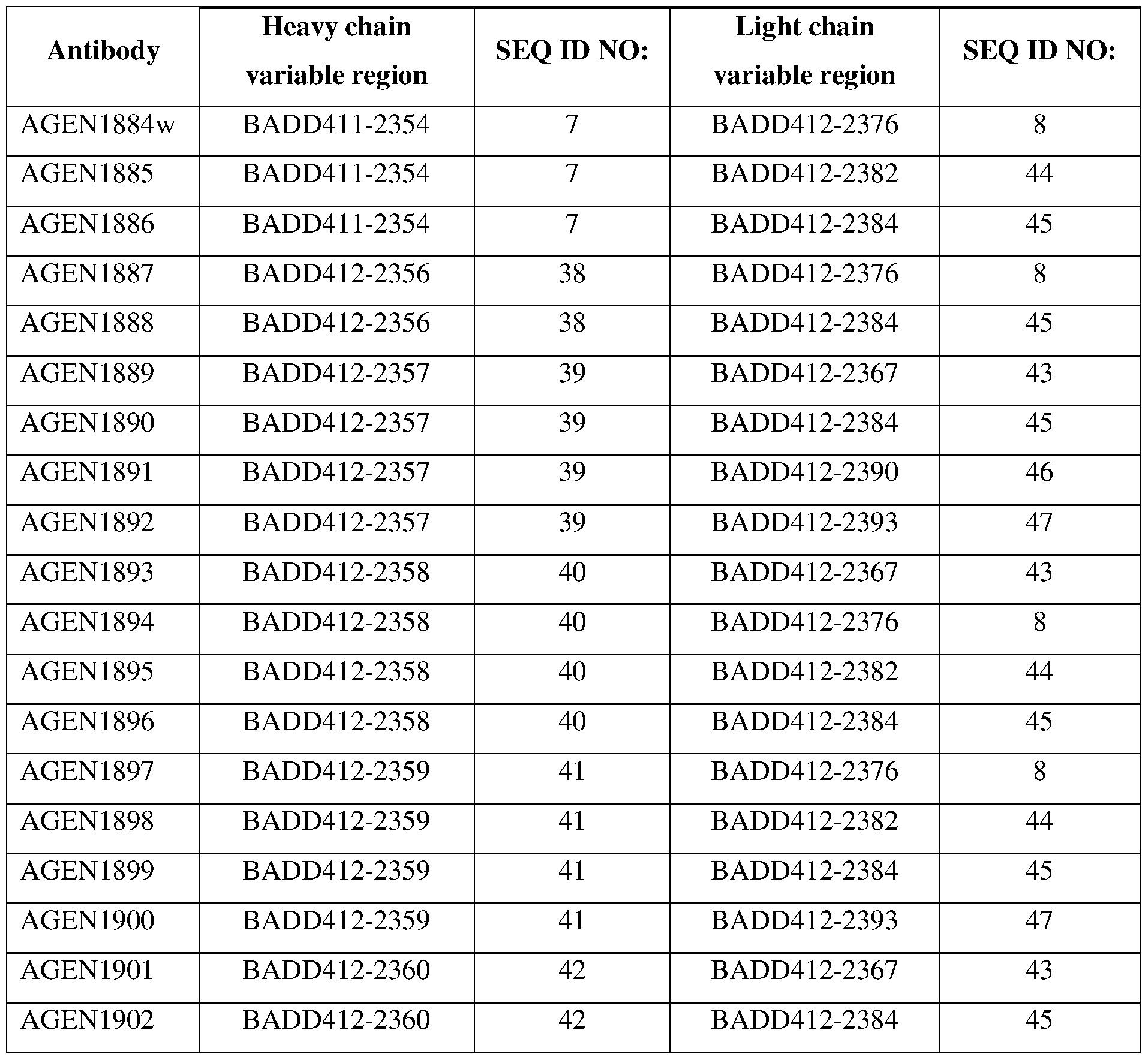

Claims
Priority Applications (24)
| Application Number | Priority Date | Filing Date | Title |
|---|---|---|---|
| PL16728509T PL3303394T3 (en) | 2015-05-29 | 2016-05-27 | Anti-ctla-4 antibodies and methods of use thereof |
| DK16728509.7T DK3303394T3 (en) | 2015-05-29 | 2016-05-27 | ANTI-CTLA-4 ANTIBODIES AND PROCEDURES FOR USE THEREOF |
| BR112017025564A BR112017025564B8 (en) | 2015-05-29 | 2016-05-27 | Anti-ctla-4 antibodies and methods of using them |
| SI201630827T SI3303394T1 (en) | 2015-05-29 | 2016-05-27 | Anti-ctla-4 antibodies and methods of use thereof |
| EP16728509.7A EP3303394B1 (en) | 2015-05-29 | 2016-05-27 | Anti-ctla-4 antibodies and methods of use thereof |
| EP20168222.6A EP3736290A1 (en) | 2015-05-29 | 2016-05-27 | Anti-ctla-4 antibodies and methods of use thereof |
| MX2017015046A MX2017015046A (en) | 2015-05-29 | 2016-05-27 | Anti-ctla-4 antibodies and methods of use thereof. |
| JP2017561965A JP6900323B2 (en) | 2015-05-29 | 2016-05-27 | Anti-CTLA-4 antibody and how to use it |
| RS20200805A RS60792B1 (en) | 2015-05-29 | 2016-05-27 | Anti-ctla-4 antibodies and methods of use thereof |
| KR1020237002568A KR20230028478A (en) | 2015-05-29 | 2016-05-27 | Anti-ctla-4 antibodies and methods of use thereof |
| MA44594A MA44594B1 (en) | 2015-05-29 | 2016-05-27 | Anti-ctla-4 Antibodies and Methods of Use thereof |
| KR1020177037545A KR102492532B1 (en) | 2015-05-29 | 2016-05-27 | Anti-CTLA-4 Antibodies and Methods of Using The Same |
| CN201680042887.8A CN107849144B (en) | 2015-05-29 | 2016-05-27 | anti-CTLA-4 antibodies and methods of use thereof |
| LTEP16728509.7T LT3303394T (en) | 2015-05-29 | 2016-05-27 | Anti-ctla-4 antibodies and methods of use thereof |
| EA201792665A EA039322B1 (en) | 2016-04-15 | 2016-05-27 | Anti-ctla-4 antibodies and methods of use thereof |
| AU2016271591A AU2016271591A1 (en) | 2015-05-29 | 2016-05-27 | Anti-CTLA-4 antibodies and methods of use thereof |
| CA2986765A CA2986765A1 (en) | 2015-05-29 | 2016-05-27 | Anti-ctla-4 antibodies and methods of use thereof |
| ES16728509T ES2803655T3 (en) | 2015-05-29 | 2016-05-27 | Anti-ctla-4 antibodies and methods of using them |
| IL255468A IL255468B (en) | 2015-05-29 | 2017-11-06 | Anti-ctla-4 antibodies and methods of use thereof |
| CONC2017/0013398A CO2017013398A2 (en) | 2015-05-29 | 2017-12-26 | Anti-ctla-4 antibodies |
| AU2019204586A AU2019204586C1 (en) | 2015-05-29 | 2019-06-28 | Anti-ctla-4 antibodies and methods of use thereof |
| HRP20201031TT HRP20201031T1 (en) | 2015-05-29 | 2020-06-30 | Anti-ctla-4 antibodies and methods of use thereof |
| AU2021211991A AU2021211991A1 (en) | 2015-05-29 | 2021-08-03 | Anti-ctla-4 antibodies and methods of use thereof |
| IL291711A IL291711A (en) | 2015-05-29 | 2022-03-27 | Anti-ctla-4 antibodies and methods of use thereof |
Applications Claiming Priority (16)
| Application Number | Priority Date | Filing Date | Title |
|---|---|---|---|
| US201562168391P | 2015-05-29 | 2015-05-29 | |
| US62/168,391 | 2015-05-29 | ||
| US201562182363P | 2015-06-19 | 2015-06-19 | |
| US62/182,363 | 2015-06-19 | ||
| US201562190653P | 2015-07-09 | 2015-07-09 | |
| US62/190,653 | 2015-07-09 | ||
| US201562257202P | 2015-11-18 | 2015-11-18 | |
| US62/257,202 | 2015-11-18 | ||
| US201662280263P | 2016-01-19 | 2016-01-19 | |
| US62/280,263 | 2016-01-19 | ||
| US201662292500P | 2016-02-08 | 2016-02-08 | |
| US62/292,500 | 2016-02-08 | ||
| US201662294558P | 2016-02-12 | 2016-02-12 | |
| US62/294,558 | 2016-02-12 | ||
| US201662323226P | 2016-04-15 | 2016-04-15 | |
| US62/323,226 | 2016-04-15 |
Publications (1)
| Publication Number | Publication Date |
|---|---|
| WO2016196237A1 true WO2016196237A1 (en) | 2016-12-08 |
Family
ID=56118035
Family Applications (1)
| Application Number | Title | Priority Date | Filing Date |
|---|---|---|---|
| PCT/US2016/034508 WO2016196237A1 (en) | 2015-05-29 | 2016-05-27 | Anti-ctla-4 antibodies and methods of use thereof |
Country Status (26)
| Country | Link |
|---|---|
| US (4) | US10144779B2 (en) |
| EP (2) | EP3736290A1 (en) |
| JP (2) | JP6900323B2 (en) |
| KR (2) | KR102492532B1 (en) |
| CN (2) | CN113603784A (en) |
| AU (3) | AU2016271591A1 (en) |
| BR (1) | BR112017025564B8 (en) |
| CA (1) | CA2986765A1 (en) |
| CL (1) | CL2017003023A1 (en) |
| CO (1) | CO2017013398A2 (en) |
| DK (1) | DK3303394T3 (en) |
| ES (1) | ES2803655T3 (en) |
| HK (1) | HK1253250A1 (en) |
| HR (1) | HRP20201031T1 (en) |
| HU (1) | HUE050750T2 (en) |
| IL (2) | IL255468B (en) |
| LT (1) | LT3303394T (en) |
| MA (2) | MA53355A (en) |
| MX (2) | MX2017015046A (en) |
| PE (1) | PE20180672A1 (en) |
| PL (1) | PL3303394T3 (en) |
| PT (1) | PT3303394T (en) |
| RS (1) | RS60792B1 (en) |
| SG (1) | SG10201913500TA (en) |
| SI (1) | SI3303394T1 (en) |
| WO (1) | WO2016196237A1 (en) |
Cited By (94)
| Publication number | Priority date | Publication date | Assignee | Title |
|---|---|---|---|---|
| WO2017220988A1 (en) | 2016-06-20 | 2017-12-28 | Kymab Limited | Multispecific antibodies for immuno-oncology |
| WO2018081621A1 (en) | 2016-10-28 | 2018-05-03 | Bristol-Myers Squibb Company | Methods of treating urothelial carcinoma using an anti-pd-1 antibody |
| WO2018106862A1 (en) * | 2016-12-07 | 2018-06-14 | Agenus Inc. | Anti-ctla-4 antibodies and methods of use thereof |
| WO2018106864A1 (en) * | 2016-12-07 | 2018-06-14 | Agenus Inc. | Antibodies and methods of use thereof |
| US10055540B2 (en) | 2015-12-16 | 2018-08-21 | Gritstone Oncology, Inc. | Neoantigen identification, manufacture, and use |
| WO2018152396A1 (en) | 2017-02-17 | 2018-08-23 | Innate Tumor Immunity, Inc. | Substituted imidazo-quinolines as nlrp3 modulators |
| WO2018160536A1 (en) * | 2017-02-28 | 2018-09-07 | Bristol-Myers Squibb Company | Use of anti-ctla-4 antibodies with enhanced adcc to enhance immune response to a vaccine |
| WO2018183928A1 (en) | 2017-03-31 | 2018-10-04 | Bristol-Myers Squibb Company | Methods of treating tumor |
| WO2018187613A2 (en) | 2017-04-07 | 2018-10-11 | Bristol-Myers Squibb Company | Anti-icos agonist antibodies and uses thereof |
| US10144779B2 (en) | 2015-05-29 | 2018-12-04 | Agenus Inc. | Anti-CTLA-4 antibodies and methods of use thereof |
| WO2018222711A2 (en) | 2017-05-30 | 2018-12-06 | Bristol-Myers Squibb Company | Compositions comprising a combination of an anti-lag-3 antibody, a pd-1 pathway inhibitor, and an immunotherapeutic agent |
| WO2019014402A1 (en) | 2017-07-14 | 2019-01-17 | Innate Tumor Immunity, Inc. | Nlrp3 modulators |
| WO2019023482A1 (en) * | 2017-07-27 | 2019-01-31 | Regeneron Pharmaceuticals, Inc. | Anti-ctla-4 antibodies and uses thereof |
| WO2019056281A1 (en) * | 2017-09-21 | 2019-03-28 | Eucure (Beijing) Biopharma Co., Ltd | Anti-ctla4 antibodies and uses thereof |
| WO2019075468A1 (en) | 2017-10-15 | 2019-04-18 | Bristol-Myers Squibb Company | Methods of treating tumor |
| WO2019097369A1 (en) | 2017-11-14 | 2019-05-23 | Pfizer Inc. | Ezh2 inhibitor combination therapies |
| US10323091B2 (en) | 2015-09-01 | 2019-06-18 | Agenus Inc. | Anti-PD-1 antibodies and methods of use thereof |
| WO2019144098A1 (en) | 2018-01-22 | 2019-07-25 | Bristol-Myers Squibb Company | Compositions and methods of treating cancer |
| WO2019191676A1 (en) | 2018-03-30 | 2019-10-03 | Bristol-Myers Squibb Company | Methods of treating tumor |
| WO2019209896A1 (en) | 2018-04-25 | 2019-10-31 | Innate Tumor Immunity, Inc. | Nlrp3 modulators |
| CN110545844A (en) * | 2017-02-21 | 2019-12-06 | 瑞美德生物医药科技有限公司 | Cancer treatment using antibodies that bind cytotoxic T lymphocyte antigen-4 (CTLA-4) |
| WO2020033587A1 (en) * | 2018-08-08 | 2020-02-13 | Dragonfly Therapeutics, Inc. | Multi-specific binding proteins that bind her2, nkg2d, and cd16, and methods of use |
| WO2020037091A1 (en) | 2018-08-16 | 2020-02-20 | Innate Tumor Immunity, Inc. | Imidazo[4,5-c]quinoline derived nlrp3-modulators |
| WO2020037094A1 (en) | 2018-08-16 | 2020-02-20 | Innate Tumor Immunity, Inc. | Substitued 4-amino-1h-imidazo[4,5-c]quinoline compounds and improved methods for their preparation |
| WO2020037092A1 (en) | 2018-08-16 | 2020-02-20 | Innate Tumor Immunity, Inc. | Imidazo[4,5-c]quinoline derived nlrp3-modulators |
| WO2020058762A1 (en) * | 2018-09-20 | 2020-03-26 | Tikcro Technologies Ltd | Antibodies specific to ctla-4 and uses thereof |
| WO2020086724A1 (en) | 2018-10-23 | 2020-04-30 | Bristol-Myers Squibb Company | Methods of treating tumor |
| WO2020092155A1 (en) * | 2018-10-31 | 2020-05-07 | Bioatla, Llc | Anti-ctla4 antibodies, antibody fragments, their immunoconjugates and uses thereof |
| WO2020102501A1 (en) | 2018-11-16 | 2020-05-22 | Bristol-Myers Squibb Company | Anti-nkg2a antibodies and uses thereof |
| WO2020102728A1 (en) | 2018-11-16 | 2020-05-22 | Neoimmunetech, Inc. | Method of treating a tumor with a combination of il-7 protein and an immune checkpoint inhibitor |
| WO2020127377A1 (en) | 2018-12-21 | 2020-06-25 | Ose Immunotherapeutics | Bifunctional anti-pd-1/il-7 molecule |
| WO2020136235A1 (en) | 2018-12-28 | 2020-07-02 | Transgene Sa | M2-defective poxvirus |
| JP2020520655A (en) * | 2017-05-19 | 2020-07-16 | ウーシー バイオロジクス(シャンハイ)カンパニー リミテッド | Novel monoclonal antibody against cytotoxic T lymphocyte associated protein 4 (CTLA-4) |
| WO2020150114A1 (en) | 2019-01-14 | 2020-07-23 | Innate Tumor Immunity, Inc. | Heterocyclic nlrp3 modulators, for use in the treatment of cancer |
| WO2020150113A1 (en) | 2019-01-14 | 2020-07-23 | Innate Tumor Immunity, Inc. | Substituted quinazolines as nlrp3 modulators, for use in the treatment of cancer |
| WO2020150116A1 (en) | 2019-01-14 | 2020-07-23 | Innate Tumor Immunity, Inc. | Nlrp3 modulators |
| WO2020150115A1 (en) | 2019-01-14 | 2020-07-23 | Innate Tumor Immunity, Inc. | Nlrp3 modulators |
| WO2020165374A1 (en) | 2019-02-14 | 2020-08-20 | Ose Immunotherapeutics | Bifunctional molecule comprising il-15ra |
| WO2020198672A1 (en) | 2019-03-28 | 2020-10-01 | Bristol-Myers Squibb Company | Methods of treating tumor |
| WO2020198676A1 (en) | 2019-03-28 | 2020-10-01 | Bristol-Myers Squibb Company | Methods of treating tumor |
| WO2020243563A1 (en) | 2019-05-30 | 2020-12-03 | Bristol-Myers Squibb Company | Multi-tumor gene signatures for suitability to immuno-oncology therapy |
| WO2020243570A1 (en) | 2019-05-30 | 2020-12-03 | Bristol-Myers Squibb Company | Cell localization signature and combination therapy |
| WO2020243568A1 (en) | 2019-05-30 | 2020-12-03 | Bristol-Myers Squibb Company | Methods of identifying a subject suitable for an immuno-oncology (i-o) therapy |
| CN112218892A (en) * | 2018-03-19 | 2021-01-12 | 上海药明生物技术有限公司 | Novel anti-CTLA-4 antibody polypeptides |
| US20210020264A1 (en) * | 2018-03-20 | 2021-01-21 | Cytomx Therapeutics, Inc. | Systems and methods for quantitative pharmacological modeling of activatable antibody species in mammalian subjects |
| WO2021055994A1 (en) | 2019-09-22 | 2021-03-25 | Bristol-Myers Squibb Company | Quantitative spatial profiling for lag-3 antagonist therapy |
| WO2021092380A1 (en) | 2019-11-08 | 2021-05-14 | Bristol-Myers Squibb Company | Lag-3 antagonist therapy for melanoma |
| WO2021092220A1 (en) | 2019-11-06 | 2021-05-14 | Bristol-Myers Squibb Company | Methods of identifying a subject with a tumor suitable for a checkpoint inhibitor therapy |
| WO2021092221A1 (en) | 2019-11-06 | 2021-05-14 | Bristol-Myers Squibb Company | Methods of identifying a subject with a tumor suitable for a checkpoint inhibitor therapy |
| EP3766901A4 (en) * | 2018-03-14 | 2021-06-23 | Shanghai Pharmaexplorer Co., Ltd. | Antibody targeting ctla-4 , preparation method therefor and use thereof |
| WO2021127554A1 (en) | 2019-12-19 | 2021-06-24 | Bristol-Myers Squibb Company | Combinations of dgk inhibitors and checkpoint antagonists |
| WO2021122866A1 (en) | 2019-12-17 | 2021-06-24 | Ose Immunotherapeutics | Bifunctional molecules comprising an il-7 variant |
| WO2021142203A1 (en) | 2020-01-10 | 2021-07-15 | Innate Tumor Immunity, Inc. | Nlrp3 modulators |
| US11071290B2 (en) | 2016-08-31 | 2021-07-27 | Biocytogen Pharmaceuticals (Beijing) Co., Ltd. | Genetically modified non-human animal with human or chimeric CTLA-4 |
| WO2021158938A1 (en) | 2020-02-06 | 2021-08-12 | Bristol-Myers Squibb Company | Il-10 and uses thereof |
| US11242393B2 (en) | 2018-03-23 | 2022-02-08 | Bristol-Myers Squibb Company | Antibodies against MICA and/or MICB and uses thereof |
| US11264117B2 (en) | 2017-10-10 | 2022-03-01 | Gritstone Bio, Inc. | Neoantigen identification using hotspots |
| WO2022047189A1 (en) | 2020-08-28 | 2022-03-03 | Bristol-Myers Squibb Company | Lag-3 antagonist therapy for hepatocellular carcinoma |
| WO2022047412A1 (en) | 2020-08-31 | 2022-03-03 | Bristol-Myers Squibb Company | Cell localization signature and immunotherapy |
| WO2022087402A1 (en) | 2020-10-23 | 2022-04-28 | Bristol-Myers Squibb Company | Lag-3 antagonist therapy for lung cancer |
| WO2022093888A1 (en) * | 2020-10-28 | 2022-05-05 | Janssen Biotech, Inc. | Compositions and methods for modulating delta gamma chain mediated immunity |
| WO2022112198A1 (en) | 2020-11-24 | 2022-06-02 | Worldwide Innovative Network | Method to select the optimal immune checkpoint therapies |
| WO2022120179A1 (en) | 2020-12-03 | 2022-06-09 | Bristol-Myers Squibb Company | Multi-tumor gene signatures and uses thereof |
| WO2022146947A1 (en) | 2020-12-28 | 2022-07-07 | Bristol-Myers Squibb Company | Antibody compositions and methods of use thereof |
| WO2022146948A1 (en) | 2020-12-28 | 2022-07-07 | Bristol-Myers Squibb Company | Subcutaneous administration of pd1/pd-l1 antibodies |
| KR20220119433A (en) | 2019-12-27 | 2022-08-29 | 추가이 세이야쿠 가부시키가이샤 | Anti-CTLA-4 antibodies and uses thereof |
| RU2779312C2 (en) * | 2017-09-21 | 2022-09-06 | Юкьюр (Бэйцзин) Байофарма Ко., Лтд | Antibodies against ctla4 and their use |
| WO2022212876A1 (en) | 2021-04-02 | 2022-10-06 | The Regents Of The University Of California | Antibodies against cleaved cdcp1 and uses thereof |
| US11464876B2 (en) | 2016-08-31 | 2022-10-11 | Biocytogen Pharmaceuticals (Beijing) Co., Ltd. | Genetically modified mouse comprising a chimeric TIGIT |
| WO2022214652A1 (en) | 2021-04-09 | 2022-10-13 | Ose Immunotherapeutics | Scaffold for bifunctioanl molecules comprising pd-1 or cd28 and sirp binding domains |
| WO2022214653A1 (en) | 2021-04-09 | 2022-10-13 | Ose Immunotherapeutics | New scaffold for bifunctional molecules with improved properties |
| US11542332B2 (en) | 2016-03-26 | 2023-01-03 | Bioatla, Inc. | Anti-CTLA4 antibodies, antibody fragments, their immunoconjugates and uses thereof |
| WO2023077090A1 (en) | 2021-10-29 | 2023-05-04 | Bristol-Myers Squibb Company | Lag-3 antagonist therapy for hematological cancer |
| WO2023077034A1 (en) | 2021-10-28 | 2023-05-04 | Lyell Immunopharma, Inc. | Methods for culturing immune cells |
| US11649290B2 (en) | 2015-11-18 | 2023-05-16 | Merck Sharp & Dohme Llc | CTLA4 binders |
| US11667712B2 (en) | 2019-05-08 | 2023-06-06 | Janssen Biotech, Inc. | Materials and methods for modulating t cell mediated immunity |
| WO2023147371A1 (en) | 2022-01-26 | 2023-08-03 | Bristol-Myers Squibb Company | Combination therapy for hepatocellular carcinoma |
| WO2023164638A1 (en) | 2022-02-25 | 2023-08-31 | Bristol-Myers Squibb Company | Combination therapy for colorectal carcinoma |
| WO2023168404A1 (en) | 2022-03-04 | 2023-09-07 | Bristol-Myers Squibb Company | Methods of treating a tumor |
| WO2023170606A1 (en) | 2022-03-08 | 2023-09-14 | Alentis Therapeutics Ag | Use of anti-claudin-1 antibodies to increase t cell availability |
| WO2023178329A1 (en) | 2022-03-18 | 2023-09-21 | Bristol-Myers Squibb Company | Methods of isolating polypeptides |
| WO2023196987A1 (en) | 2022-04-07 | 2023-10-12 | Bristol-Myers Squibb Company | Methods of treating tumor |
| WO2023213763A1 (en) | 2022-05-02 | 2023-11-09 | Transgene | Poxvirus encoding a binding agent comprising an anti- pd-l1 sdab |
| WO2023213764A1 (en) | 2022-05-02 | 2023-11-09 | Transgene | Fusion polypeptide comprising an anti-pd-l1 sdab and a member of the tnfsf |
| US11834506B2 (en) | 2017-02-08 | 2023-12-05 | Dragonfly Therapeutics, Inc. | Multi-specific binding proteins that bind NKG2D, CD16, and a tumor-associated antigen for activation of natural killer cells and therapeutic uses thereof to treat cancer |
| WO2023235847A1 (en) | 2022-06-02 | 2023-12-07 | Bristol-Myers Squibb Company | Antibody compositions and methods of use thereof |
| WO2024003360A1 (en) | 2022-07-01 | 2024-01-04 | Institut Curie | Biomarkers and uses thereof for the treatment of neuroblastoma |
| WO2024003353A1 (en) | 2022-07-01 | 2024-01-04 | Transgene | Fusion protein comprising a surfactant-protein-d and a member of the tnfsf |
| US11884733B2 (en) | 2018-02-08 | 2024-01-30 | Dragonfly Therapeutics, Inc. | Antibody variable domains targeting the NKG2D receptor |
| US11885815B2 (en) | 2017-11-22 | 2024-01-30 | Gritstone Bio, Inc. | Reducing junction epitope presentation for neoantigens |
| US11884732B2 (en) | 2017-02-20 | 2024-01-30 | Dragonfly Therapeutics, Inc. | Proteins binding HER2, NKG2D and CD16 |
| WO2024028386A1 (en) | 2022-08-02 | 2024-02-08 | Ose Immunotherapeutics | Multifunctional molecule directed against cd28 |
| KR20240024255A (en) | 2021-06-25 | 2024-02-23 | 추가이 세이야쿠 가부시키가이샤 | Use of anti-CTLA-4 antibodies |
| KR20240024213A (en) | 2021-06-25 | 2024-02-23 | 추가이 세이야쿠 가부시키가이샤 | Anti-CTLA-4 antibody |
Families Citing this family (9)
| Publication number | Priority date | Publication date | Assignee | Title |
|---|---|---|---|---|
| CN116478289A (en) * | 2017-05-19 | 2023-07-25 | 上海药明生物技术有限公司 | Novel CTLA-4 monoclonal antibody |
| CA3073953A1 (en) * | 2017-08-31 | 2019-03-07 | Io Therapeutics, Inc. | Rar selective agonists in combination with immune modulators for cancer immunotherapy |
| EP3902832A2 (en) * | 2018-12-26 | 2021-11-03 | Xilio Development, Inc. | Anti-ctla4 antibodies and methods of use thereof |
| MX2021007848A (en) * | 2018-12-27 | 2021-10-26 | Gigagen Inc | Anti-ctla-4 binding proteins and methods of use thereof. |
| US20220363778A1 (en) * | 2019-08-28 | 2022-11-17 | The Research Foundation For The State University Of New York | Targeted delivery of tumor matrix modifying enzymes |
| IL297442A (en) | 2020-04-22 | 2022-12-01 | Iovance Biotherapeutics Inc | Systems and methods for coordinating manufacturing of cells for patient-specific immunotherapy |
| WO2023130081A1 (en) | 2021-12-30 | 2023-07-06 | Neoimmunetech, Inc. | Method of treating a tumor with a combination of il-7 protein and vegf antagonist |
| CN114437220B (en) * | 2022-01-24 | 2023-04-14 | 广东医科大学 | anti-CTLA-4 nano antibody, encoding gene, recombinant nano antibody, recombinant vector, recombinant bacterium and application thereof |
| CN115845039A (en) * | 2022-07-01 | 2023-03-28 | 可蓝赛生物医药(上海)有限公司 | Broad-spectrum polypeptide tumor vaccine composition |
Citations (94)
| Publication number | Priority date | Publication date | Assignee | Title |
|---|---|---|---|---|
| US4044126A (en) | 1972-04-20 | 1977-08-23 | Allen & Hanburys Limited | Steroidal aerosol compositions and process for the preparation thereof |
| US4364923A (en) | 1972-04-20 | 1982-12-21 | Allen & Hanburs Limited | Chemical compounds |
| US4444887A (en) | 1979-12-10 | 1984-04-24 | Sloan-Kettering Institute | Process for making human antibody producing B-lymphocytes |
| WO1986005807A1 (en) | 1985-04-01 | 1986-10-09 | Celltech Limited | Transformed myeloma cell-line and a process for the expression of a gene coding for a eukaryotic polypeptide employing same |
| US4716111A (en) | 1982-08-11 | 1987-12-29 | Trustees Of Boston University | Process for producing human antibodies |
| WO1989001036A1 (en) | 1987-07-23 | 1989-02-09 | Celltech Limited | Recombinant dna expression vectors |
| US4816397A (en) | 1983-03-25 | 1989-03-28 | Celltech, Limited | Multichain polypeptides or proteins and processes for their production |
| US4816567A (en) | 1983-04-08 | 1989-03-28 | Genentech, Inc. | Recombinant immunoglobin preparations |
| WO1990002809A1 (en) | 1988-09-02 | 1990-03-22 | Protein Engineering Corporation | Generation and selection of recombinant varied binding proteins |
| WO1991010741A1 (en) | 1990-01-12 | 1991-07-25 | Cell Genesys, Inc. | Generation of xenogeneic antibodies |
| WO1991010737A1 (en) | 1990-01-11 | 1991-07-25 | Molecular Affinities Corporation | Production of antibodies using gene libraries |
| WO1992001047A1 (en) | 1990-07-10 | 1992-01-23 | Cambridge Antibody Technology Limited | Methods for producing members of specific binding pairs |
| US5122464A (en) | 1986-01-23 | 1992-06-16 | Celltech Limited, A British Company | Method for dominant selection in eucaryotic cells |
| WO1992018619A1 (en) | 1991-04-10 | 1992-10-29 | The Scripps Research Institute | Heterodimeric receptor libraries using phagemids |
| WO1992022324A1 (en) | 1991-06-14 | 1992-12-23 | Xoma Corporation | Microbially-produced antibody fragments and their conjugates |
| WO1993011236A1 (en) | 1991-12-02 | 1993-06-10 | Medical Research Council | Production of anti-self antibodies from antibody segment repertoires and displayed on phage |
| US5223409A (en) | 1988-09-02 | 1993-06-29 | Protein Engineering Corp. | Directed evolution of novel binding proteins |
| WO1994004678A1 (en) | 1992-08-21 | 1994-03-03 | Casterman Cecile | Immunoglobulins devoid of light chains |
| WO1994025591A1 (en) | 1993-04-29 | 1994-11-10 | Unilever N.V. | PRODUCTION OF ANTIBODIES OR (FUNCTIONALIZED) FRAGMENTS THEREOF DERIVED FROM HEAVY CHAIN IMMUNOGLOBULINS OF $i(CAMELIDAE) |
| US5413923A (en) | 1989-07-25 | 1995-05-09 | Cell Genesys, Inc. | Homologous recombination for universal donor cells and chimeric mammalian hosts |
| WO1995015982A2 (en) | 1993-12-08 | 1995-06-15 | Genzyme Corporation | Process for generating specific antibodies |
| US5427908A (en) | 1990-05-01 | 1995-06-27 | Affymax Technologies N.V. | Recombinant library screening methods |
| WO1995020401A1 (en) | 1994-01-31 | 1995-08-03 | Trustees Of Boston University | Polyclonal antibody libraries |
| US5516637A (en) | 1994-06-10 | 1996-05-14 | Dade International Inc. | Method involving display of protein binding pairs on the surface of bacterial pili and bacteriophage |
| US5545806A (en) | 1990-08-29 | 1996-08-13 | Genpharm International, Inc. | Ransgenic non-human animals for producing heterologous antibodies |
| US5569825A (en) | 1990-08-29 | 1996-10-29 | Genpharm International | Transgenic non-human animals capable of producing heterologous antibodies of various isotypes |
| WO1996034096A1 (en) | 1995-04-28 | 1996-10-31 | Abgenix, Inc. | Human antibodies derived from immunized xenomice |
| WO1996033735A1 (en) | 1995-04-27 | 1996-10-31 | Abgenix, Inc. | Human antibodies derived from immunized xenomice |
| WO1997013844A1 (en) | 1995-10-06 | 1997-04-17 | Cambridge Antibody Technology Limited | Specific binding members for human transforming growth factor beta; materials and methods |
| US5625126A (en) | 1990-08-29 | 1997-04-29 | Genpharm International, Inc. | Transgenic non-human animals for producing heterologous antibodies |
| US5633425A (en) | 1990-08-29 | 1997-05-27 | Genpharm International, Inc. | Transgenic non-human animals capable of producing heterologous antibodies |
| US5661016A (en) | 1990-08-29 | 1997-08-26 | Genpharm International Inc. | Transgenic non-human animals capable of producing heterologous antibodies of various isotypes |
| US5698426A (en) | 1990-09-28 | 1997-12-16 | Ixsys, Incorporated | Surface expression libraries of heteromeric receptors |
| US5709874A (en) | 1993-04-14 | 1998-01-20 | Emory University | Device for local drug delivery and methods for using the same |
| US5733743A (en) | 1992-03-24 | 1998-03-31 | Cambridge Antibody Technology Limited | Methods for producing members of specific binding pairs |
| WO1998016654A1 (en) | 1996-10-11 | 1998-04-23 | Japan Tobacco, Inc. | Production of a multimeric protein by cell fusion method |
| US5750753A (en) | 1996-01-24 | 1998-05-12 | Chisso Corporation | Method for manufacturing acryloxypropysilane |
| US5759542A (en) | 1994-08-05 | 1998-06-02 | New England Deaconess Hospital Corporation | Compositions and methods for the delivery of drugs by platelets for the treatment of cardiovascular and other diseases |
| WO1998024893A2 (en) | 1996-12-03 | 1998-06-11 | Abgenix, Inc. | TRANSGENIC MAMMALS HAVING HUMAN IG LOCI INCLUDING PLURAL VH AND Vλ REGIONS AND ANTIBODIES PRODUCED THEREFROM |
| US5780225A (en) | 1990-01-12 | 1998-07-14 | Stratagene | Method for generating libaries of antibody genes comprising amplification of diverse antibody DNAs and methods for using these libraries for the production of diverse antigen combining molecules |
| US5807715A (en) | 1984-08-27 | 1998-09-15 | The Board Of Trustees Of The Leland Stanford Junior University | Methods and transformed mammalian lymphocyte cells for producing functional antigen-binding protein including chimeric immunoglobulin |
| US5814318A (en) | 1990-08-29 | 1998-09-29 | Genpharm International Inc. | Transgenic non-human animals for producing heterologous antibodies |
| US5821047A (en) | 1990-12-03 | 1998-10-13 | Genentech, Inc. | Monovalent phage display |
| WO1998046645A2 (en) | 1997-04-14 | 1998-10-22 | Micromet Gesellschaft Für Biomedizinische Forschung Mbh | Method for the production of antihuman antigen receptors and uses thereof |
| WO1998050433A2 (en) | 1997-05-05 | 1998-11-12 | Abgenix, Inc. | Human monoclonal antibodies to epidermal growth factor receptor |
| US5837242A (en) | 1992-12-04 | 1998-11-17 | Medical Research Council | Multivalent and multispecific binding proteins, their manufacture and use |
| US5840674A (en) | 1990-11-01 | 1998-11-24 | Oregon Health Sciences University | Covalent microparticle-drug conjugates for biological targeting |
| US5860957A (en) | 1997-02-07 | 1999-01-19 | Sarcos, Inc. | Multipathway electronically-controlled drug delivery system |
| US5869620A (en) | 1986-09-02 | 1999-02-09 | Enzon, Inc. | Multivalent antigen-binding proteins |
| US5900252A (en) | 1990-04-17 | 1999-05-04 | Eurand International S.P.A. | Method for targeted and controlled release of drugs in the intestinal tract and more particularly in the colon |
| US5948433A (en) | 1997-08-21 | 1999-09-07 | Bertek, Inc. | Transdermal patch |
| US5965726A (en) | 1992-03-27 | 1999-10-12 | The United States Of America As Represented By The Department Of Health And Human Services | Method of eliminating inhibitory/ instability regions of mRNA |
| US5972366A (en) | 1994-11-28 | 1999-10-26 | The Unites States Of America As Represented By The Secretary Of The Army | Drug releasing surgical implant or dressing material |
| US5983134A (en) | 1995-04-23 | 1999-11-09 | Electromagnetic Bracing Systems Inc. | Electrophoretic cuff apparatus drug delivery system |
| US5985317A (en) | 1996-09-06 | 1999-11-16 | Theratech, Inc. | Pressure sensitive adhesive matrix patches for transdermal delivery of salts of pharmaceutical agents |
| US5985307A (en) | 1993-04-14 | 1999-11-16 | Emory University | Device and method for non-occlusive localized drug delivery |
| US5989830A (en) | 1995-10-16 | 1999-11-23 | Unilever Patent Holdings Bv | Bifunctional or bivalent antibody fragment analogue |
| US6004534A (en) | 1993-07-23 | 1999-12-21 | Massachusetts Institute Of Technology | Targeted polymerized liposomes for improved drug delivery |
| US6005079A (en) | 1992-08-21 | 1999-12-21 | Vrije Universiteit Brussels | Immunoglobulins devoid of light chains |
| US6010715A (en) | 1992-04-01 | 2000-01-04 | Bertek, Inc. | Transdermal patch incorporating a polymer film incorporated with an active agent |
| US6024975A (en) | 1992-04-08 | 2000-02-15 | Americare International Diagnostics, Inc. | Method of transdermally administering high molecular weight drugs with a polymer skin enhancer |
| US6039975A (en) | 1995-10-17 | 2000-03-21 | Hoffman-La Roche Inc. | Colon targeted delivery system |
| US6048736A (en) | 1998-04-29 | 2000-04-11 | Kosak; Kenneth M. | Cyclodextrin polymers for carrying and releasing drugs |
| US6060082A (en) | 1997-04-18 | 2000-05-09 | Massachusetts Institute Of Technology | Polymerized liposomes targeted to M cells and useful for oral or mucosal drug delivery |
| US6071495A (en) | 1989-12-22 | 2000-06-06 | Imarx Pharmaceutical Corp. | Targeted gas and gaseous precursor-filled liposomes |
| US6075181A (en) | 1990-01-12 | 2000-06-13 | Abgenix, Inc. | Human antibodies derived from immunized xenomice |
| US6120751A (en) | 1997-03-21 | 2000-09-19 | Imarx Pharmaceutical Corp. | Charged lipids and uses for the same |
| US6131570A (en) | 1998-06-30 | 2000-10-17 | Aradigm Corporation | Temperature controlling device for aerosol drug delivery |
| US6132992A (en) | 1993-02-01 | 2000-10-17 | Bristol-Myers Squibb Co. | Expression vectors encoding bispecific fusion proteins and methods of producing biologically active bispecific fusion proteins in a mammalian cell |
| US6139865A (en) | 1996-10-01 | 2000-10-31 | Eurand America, Inc. | Taste-masked microcapsule compositions and methods of manufacture |
| US6150184A (en) | 1996-08-20 | 2000-11-21 | Ramtron International Corporation | Method of fabricating partially or completely encapsulated top electrode of a ferroelectric capacitor |
| US6167301A (en) | 1995-08-29 | 2000-12-26 | Flower; Ronald J. | Iontophoretic drug delivery device having high-efficiency DC-to-DC energy conversion circuit |
| WO2001044301A1 (en) | 1999-11-29 | 2001-06-21 | Unilever Plc | Immobilized single domain antigen-binding molecules |
| US6253872B1 (en) | 1996-05-29 | 2001-07-03 | Gmundner Fertigteile Gesellschaft M.B.H & Co., Kg | Track soundproofing arrangement |
| US6256533B1 (en) | 1999-06-09 | 2001-07-03 | The Procter & Gamble Company | Apparatus and method for using an intracutaneous microneedle array |
| US6261595B1 (en) | 2000-02-29 | 2001-07-17 | Zars, Inc. | Transdermal drug patch with attached pocket for controlled heating device |
| US6267983B1 (en) | 1997-10-28 | 2001-07-31 | Bando Chemical Industries, Ltd. | Dermatological patch and process for producing thereof |
| US6271359B1 (en) | 1999-04-14 | 2001-08-07 | Musc Foundation For Research Development | Tissue-specific and pathogen-specific toxic agents and ribozymes |
| US6274552B1 (en) | 1993-03-18 | 2001-08-14 | Cytimmune Sciences, Inc. | Composition and method for delivery of biologically-active factors |
| US6316652B1 (en) | 1995-06-06 | 2001-11-13 | Kosta Steliou | Drug mitochondrial targeting agents |
| WO2002043478A2 (en) * | 2000-11-30 | 2002-06-06 | Medarex, Inc. | Transgenic transchromosomal rodents for making human antibodies |
| EP1262193A1 (en) * | 2001-05-23 | 2002-12-04 | Pfizer Products Inc. | Use of human anti-CTLA-4 antibodies for treatment of cancer |
| US20040014194A1 (en) | 2002-03-27 | 2004-01-22 | Schering Corporation | Beta-secretase crystals and methods for preparing and using the same |
| US20060240006A1 (en) * | 2005-04-20 | 2006-10-26 | Chishih Chu | Novel antibody structures derived from human germline sequences |
| US7183076B2 (en) | 1997-05-02 | 2007-02-27 | Genentech, Inc. | Method for making multispecific antibodies having heteromultimeric and common components |
| WO2008100562A2 (en) * | 2007-02-14 | 2008-08-21 | Medical College Of Georgia Research Institute, Inc. | Indoleamine 2,3-dioxygenase, pd-1/pd-l pathways, and ctla4 pathways in the activation of regulatory t cells |
| WO2009100140A1 (en) * | 2008-02-04 | 2009-08-13 | Medarex, Inc. | Anti-clta-4 antibodies with reduced blocking of binding of ctla-4 to b7 and uses thereof |
| US7709226B2 (en) | 2001-07-12 | 2010-05-04 | Arrowsmith Technology Licensing Llc | Method of humanizing antibodies by matching canonical structure types CDRs |
| US7951917B1 (en) | 1997-05-02 | 2011-05-31 | Genentech, Inc. | Method for making multispecific antibodies having heteromultimeric and common components |
| US8227577B2 (en) | 2007-12-21 | 2012-07-24 | Hoffman-La Roche Inc. | Bivalent, bispecific antibodies |
| WO2012120125A1 (en) * | 2011-03-09 | 2012-09-13 | Antitope Ltd | Humanised anti ctla-4 antibodies |
| WO2012162277A1 (en) * | 2011-05-25 | 2012-11-29 | Merck Sharp & Dohme Corp. | METHOD FOR PREPARING Fc-CONTAINING POLYPEPTIDES HAVING IMPROVED PROPERTIES |
| US8586713B2 (en) | 2009-06-26 | 2013-11-19 | Regeneron Pharmaceuticals, Inc. | Readily isolated bispecific antibodies with native immunoglobulin format |
| WO2014144960A2 (en) * | 2013-03-15 | 2014-09-18 | Abbvie Biotherapeutics Inc. | Fc variants |
Family Cites Families (181)
| Publication number | Priority date | Publication date | Assignee | Title |
|---|---|---|---|---|
| US5225539A (en) | 1986-03-27 | 1993-07-06 | Medical Research Council | Recombinant altered antibodies and methods of making altered antibodies |
| GB8607679D0 (en) | 1986-03-27 | 1986-04-30 | Winter G P | Recombinant dna product |
| US5530101A (en) | 1988-12-28 | 1996-06-25 | Protein Design Labs, Inc. | Humanized immunoglobulins |
| GB8928874D0 (en) | 1989-12-21 | 1990-02-28 | Celltech Ltd | Humanised antibodies |
| DE69233482T2 (en) | 1991-05-17 | 2006-01-12 | Merck & Co., Inc. | Method for reducing the immunogenicity of antibody variable domains |
| DE69233254T2 (en) | 1991-06-14 | 2004-09-16 | Genentech, Inc., South San Francisco | Humanized Heregulin antibody |
| US5851795A (en) | 1991-06-27 | 1998-12-22 | Bristol-Myers Squibb Company | Soluble CTLA4 molecules and uses thereof |
| US5565332A (en) | 1991-09-23 | 1996-10-15 | Medical Research Council | Production of chimeric antibodies - a combinatorial approach |
| DE69233204T2 (en) | 1991-12-13 | 2004-07-15 | Xoma Corp., Berkeley | METHOD AND MATERIALS FOR THE PRODUCTION OF MODIFIED VARIABLE ANTIBODY DOMAINS AND THEIR THERAPEUTIC USE |
| GB9203459D0 (en) | 1992-02-19 | 1992-04-08 | Scotgen Ltd | Antibodies with germ-line variable regions |
| US5639641A (en) | 1992-09-09 | 1997-06-17 | Immunogen Inc. | Resurfacing of rodent antibodies |
| US5997873A (en) | 1994-01-13 | 1999-12-07 | Mount Sinai School Of Medicine Of The City University Of New York | Method of preparation of heat shock protein 70-peptide complexes |
| US6719972B1 (en) | 1994-06-03 | 2004-04-13 | Repligen Corporation | Methods of inhibiting T cell proliferation or IL-2 accumulation with CTLA4- specific antibodies |
| US5811097A (en) | 1995-07-25 | 1998-09-22 | The Regents Of The University Of California | Blockade of T lymphocyte down-regulation associated with CTLA-4 signaling |
| US5935576A (en) | 1995-09-13 | 1999-08-10 | Fordham University | Compositions and methods for the treatment and prevention of neoplastic diseases using heat shock proteins complexed with exogenous antigens |
| US20060034844A1 (en) | 1996-12-04 | 2006-02-16 | The Regents Of The University Of California | Stimulation of T cells against self antigens using CTLA-4 blocking agents |
| US6017540A (en) | 1997-02-07 | 2000-01-25 | Fordham University | Prevention and treatment of primary and metastatic neoplastic diseases and infectious diseases with heat shock/stress protein-peptide complexes |
| AU6703198A (en) | 1997-03-21 | 1998-10-20 | Brigham And Women's Hospital | Immunotherapeutic ctla-4 binding peptides |
| US5948646A (en) | 1997-12-11 | 1999-09-07 | Fordham University | Methods for preparation of vaccines against cancer comprising heat shock protein-peptide complexes |
| WO1999047558A2 (en) | 1998-03-19 | 1999-09-23 | Heska Corporation | T cell costimulatory proteins, sequences and uses thereof |
| EP1137436B1 (en) | 1998-12-03 | 2008-06-04 | The Regents Of The University Of California | Stimulation of T-cells against self antigens using CTLA-4 blocking agents |
| US6682736B1 (en) | 1998-12-23 | 2004-01-27 | Abgenix, Inc. | Human monoclonal antibodies to CTLA-4 |
| EP2360254A1 (en) | 1999-08-23 | 2011-08-24 | Dana-Farber Cancer Institute, Inc. | Assays for screening anti-pd-1 antibodies and uses thereof |
| US7605238B2 (en) | 1999-08-24 | 2009-10-20 | Medarex, Inc. | Human CTLA-4 antibodies and their uses |
| EP3225632B1 (en) | 1999-11-30 | 2020-05-06 | Mayo Foundation for Medical Education and Research | Antibodies binding to b7-h1, a novel immunoregulatory molecule |
| WO2001054732A1 (en) | 2000-01-27 | 2001-08-02 | Genetics Institute, Llc. | Antibodies against ctla4 (cd152), conjugates comprising same, and uses thereof |
| US20030086932A1 (en) | 2000-04-12 | 2003-05-08 | Jeffrey A. Bluestone | Surface-bound antigen binding portions of antibodies that bind to CTLA-4 and CD28 and uses therefor |
| WO2003042402A2 (en) | 2001-11-13 | 2003-05-22 | Dana-Farber Cancer Institute, Inc. | Agents that modulate immune cell activation and methods of use thereof |
| AUPS054702A0 (en) | 2002-02-14 | 2002-03-07 | Immunaid Pty Ltd | Cancer therapy |
| US20040132101A1 (en) | 2002-09-27 | 2004-07-08 | Xencor | Optimized Fc variants and methods for their generation |
| NZ536420A (en) | 2002-04-12 | 2008-04-30 | Medarex Inc | Methods of treatment using CTLA-4 antibodies |
| US20060093612A1 (en) | 2002-05-02 | 2006-05-04 | Srivastava Pramod K | Use of heat shock proteins to enhance efficacy of antibody therapeutics |
| IL149820A0 (en) | 2002-05-23 | 2002-11-10 | Curetech Ltd | Humanized immunomodulatory monoclonal antibodies for the treatment of neoplastic disease or immunodeficiency |
| US7595048B2 (en) | 2002-07-03 | 2009-09-29 | Ono Pharmaceutical Co., Ltd. | Method for treatment of cancer by inhibiting the immunosuppressive signal induced by PD-1 |
| AU2003260871A1 (en) | 2002-09-30 | 2004-04-19 | Pfizer Products Inc. | Hybridomas producing high levels of human sequence antibody |
| BR0316880A (en) | 2002-12-23 | 2005-10-25 | Wyeth Corp | Pd-1 Antibodies and Uses |
| US9068234B2 (en) | 2003-01-21 | 2015-06-30 | Ptc Therapeutics, Inc. | Methods and agents for screening for compounds capable of modulating gene expression |
| US8388955B2 (en) | 2003-03-03 | 2013-03-05 | Xencor, Inc. | Fc variants |
| US7465446B2 (en) | 2003-05-30 | 2008-12-16 | Medarex, Inc. | Surrogate therapeutic endpoint for anti-CTLA4-based immunotherapy of disease |
| CA2537055A1 (en) | 2003-08-22 | 2005-04-21 | Medimmune, Inc. | Humanization of antibodies |
| CN1964739A (en) | 2004-03-26 | 2007-05-16 | 辉瑞产品公司 | Uses of anti-CTLA-4 antibodies |
| US7494779B2 (en) | 2004-06-14 | 2009-02-24 | Li-Te Chin | Method for producing human antibodies to human CD152 with properties of agonist, antagonist, or inverse agonist |
| US20060057626A1 (en) | 2004-09-03 | 2006-03-16 | Nichol Geoffrey M | Assessment of CTLA-4 polymorphisms in CTLA-4 blockade therapy |
| WO2006029219A2 (en) | 2004-09-08 | 2006-03-16 | Ohio State University Research Foundation | Human monoclonal anti-ctla4 antibodies in cancer treatment |
| DE102004063494A1 (en) | 2004-12-23 | 2006-07-13 | Tegenero Ag | antibody |
| US9487581B2 (en) | 2005-03-08 | 2016-11-08 | Pfizer Inc. | Anti-CTLA-4 antibody compositions |
| EP1896582A4 (en) | 2005-05-09 | 2009-04-08 | Ono Pharmaceutical Co | Human monoclonal antibodies to programmed death 1(pd-1) and methods for treating cancer using anti-pd-1 antibodies alone or in combination with other immunotherapeutics |
| JP2006327937A (en) | 2005-05-23 | 2006-12-07 | Ritsutoku Kin | Human antibody structure equipped with human germ cell line gene structure and derived fragment of the same |
| LT2397156T (en) | 2005-06-08 | 2017-02-27 | Dana-Farber Cancer Institute, Inc. | Methods and compositions for the treatment of persistent infections and cancer by inhibiting the programmed cell death 1 (PD-1)pathway |
| CN101248089A (en) | 2005-07-01 | 2008-08-20 | 米德列斯公司 | Human monoclonal antibodies to programmed death ligand 1(PD-L1) |
| US20070243184A1 (en) | 2005-11-08 | 2007-10-18 | Steven Fischkoff | Prophylaxis and treatment of enterocolitis associated with anti-ctla-4 antibody therapy |
| JP2009518446A (en) | 2005-12-07 | 2009-05-07 | メダレックス インコーポレーティッド | CTLA-4 antibody dose escalation regimen |
| NZ569108A (en) | 2005-12-20 | 2011-09-30 | Bristol Myers Squibb Co | Formulations of CTLA4Ig stabilised using sugars |
| EP2013348A4 (en) | 2006-03-30 | 2009-09-02 | Univ California | Methods and compositions for localized secretion of anti-ctla-4 antibodies |
| US7919079B2 (en) | 2006-03-31 | 2011-04-05 | Biosante Pharmaceuticals, Inc. | Cancer immunotherapy compositions and methods of use |
| CA2647282A1 (en) | 2006-04-05 | 2007-10-11 | Pfizer Products Inc. | Ctla4 antibody combination therapy |
| CN101074264B (en) | 2006-05-17 | 2011-08-17 | 上海抗体药物国家工程研究中心有限公司 | Recombinant anti-CTLA4 monoclonal antibody, its production and use |
| AU2007331672A1 (en) | 2006-12-15 | 2008-06-19 | Ablynx N.V. | Amino acid sequences that modulate the interaction between cells of the immune system |
| US9244059B2 (en) | 2007-04-30 | 2016-01-26 | Immutep Parc Club Orsay | Cytotoxic anti-LAG-3 monoclonal antibody and its use in the treatment or prevention of organ transplant rejection and autoimmune disease |
| KR20100054780A (en) | 2007-06-18 | 2010-05-25 | 엔.브이.오가논 | Antibodies to human programmed death receptor pd-1 |
| EP2175884B8 (en) | 2007-07-12 | 2017-02-22 | GITR, Inc. | Combination therapies employing gitr binding molecules |
| PE20120259A1 (en) | 2007-08-09 | 2012-04-04 | Boehringer Ingelheim Int | ANTI-CD37 ANTIBODIES |
| AU2008289441A1 (en) | 2007-08-22 | 2009-02-26 | Cytomx Therapeutics, Inc. | Activatable binding polypeptides and methods of identification and use thereof |
| US7968300B2 (en) | 2007-11-21 | 2011-06-28 | Li-Te Chin | Method of isolating regulatory T cells from human samples |
| US8449886B2 (en) | 2008-01-08 | 2013-05-28 | Bristol-Myers Squibb Company | Combination of anti-CTLA4 antibody with tubulin modulating agents for the treatment of proliferative diseases |
| JP2011512332A (en) | 2008-02-11 | 2011-04-21 | キュアー テック リミテッド | Monoclonal antibodies for tumor therapy |
| US8168757B2 (en) | 2008-03-12 | 2012-05-01 | Merck Sharp & Dohme Corp. | PD-1 binding proteins |
| TWI453204B (en) | 2008-07-08 | 2014-09-21 | Incyte Corp | 1,2,5-oxadiazoles as inhibitors of indoleamine 2,3-dioxygenase |
| WO2010014784A2 (en) | 2008-08-01 | 2010-02-04 | Bristol-Myers Squibb Company | Combination of anti-ctla4 antibody with diverse therapeutic regimens for the synergistic treatment of proliferative diseases |
| AR072999A1 (en) | 2008-08-11 | 2010-10-06 | Medarex Inc | HUMAN ANTIBODIES THAT JOIN GEN 3 OF LYMPHOCYTARY ACTIVATION (LAG-3) AND THE USES OF THESE |
| AU2009288730B2 (en) | 2008-08-25 | 2013-06-20 | Amplimmune, Inc. | Compositions of PD-1 antagonists and methods of use |
| CA2736829C (en) | 2008-09-12 | 2018-02-27 | Isis Innovation Limited | Pd-1 specific antibodies and uses thereof |
| EP3530672A1 (en) | 2008-09-26 | 2019-08-28 | Dana Farber Cancer Institute, Inc. | Human anti-pd-1, pd-l1, and pd-l2 antibodies and uses thereof |
| CN102245640B (en) | 2008-12-09 | 2014-12-31 | 霍夫曼-拉罗奇有限公司 | Anti-PD-L1 antibodies and their use to enhance T-cell function |
| CN106995495A (en) | 2009-01-12 | 2017-08-01 | 希托马克斯医疗有限责任公司 | Modified antibodies composition and its preparation and application |
| GB0903325D0 (en) | 2009-02-26 | 2009-04-08 | Univ Aberdeen | Antibody molecules |
| ES2629167T3 (en) * | 2009-07-20 | 2017-08-07 | Bristol-Myers Squibb Company | Combination of anti-CTLA4 antibody with etoposide for the synergistic treatment of proliferative diseases |
| WO2011020024A2 (en) | 2009-08-13 | 2011-02-17 | The Johns Hopkins University | Methods of modulating immune function |
| AU2010308030B2 (en) | 2009-10-12 | 2014-05-29 | Pfizer Inc. | Cancer treatment |
| GB0920258D0 (en) | 2009-11-19 | 2010-01-06 | Alligator Bioscience Ab | New medical agents and use thereof |
| NZ599405A (en) | 2009-11-24 | 2014-09-26 | Medimmune Ltd | Targeted binding agents against b7-h1 |
| CN102134276A (en) | 2010-01-22 | 2011-07-27 | 上海抗体药物国家工程研究中心有限公司 | Chimeric antibody resisting CTLA-4 (Cytotoxic T Lymphocyte Antigen) |
| WO2011100841A1 (en) | 2010-02-16 | 2011-08-25 | Valorisation-Recherche, Limited Partnership | Pd-1 modulation and uses thereof for modulating hiv replication |
| US8993731B2 (en) | 2010-03-11 | 2015-03-31 | Ucb Biopharma Sprl | PD-1 antibody |
| JP2013523098A (en) | 2010-03-29 | 2013-06-17 | ザイムワークス,インコーポレイテッド | Antibody with enhanced or suppressed effector function |
| EP2699264B1 (en) | 2011-04-20 | 2018-03-14 | Medlmmune, LLC | Antibodies and other molecules that bind b7-h1 and pd-1 |
| EP2723381A4 (en) | 2011-06-21 | 2015-03-18 | Univ Johns Hopkins | Focused radiation for augmenting immune-based therapies against neoplasms |
| ES2664622T3 (en) | 2011-07-24 | 2018-04-20 | Cure Tech Ltd. | Humanized immunomodulatory monoclonal antibody variants |
| EP2742953B1 (en) | 2011-08-11 | 2021-09-22 | ONO Pharmaceutical Co., Ltd. | Therapeutic agent for autoimmune diseases comprising pd-1 agonist |
| US8920075B2 (en) | 2011-09-01 | 2014-12-30 | Halo Maritime Defense Systems, Inc. | Marine barrier and gate |
| WO2013043569A1 (en) | 2011-09-20 | 2013-03-28 | Vical Incorporated | Synergistic anti-tumor efficacy using alloantigen combination immunotherapy |
| AU2012344260B2 (en) | 2011-11-28 | 2017-09-07 | Merck Patent Gmbh | Anti-PD-L1 antibodies and uses thereof |
| WO2013126809A1 (en) | 2012-02-23 | 2013-08-29 | Sloan-Kettering Institute For Cancer Research | Prediction of responsiveness to treatment with immunomodulatory therapeutics and method of monitoring abscopal effects during such treatment |
| US20150079100A1 (en) | 2012-03-23 | 2015-03-19 | Bristol-Myers Squibb Company | Methods of treatments using ctla-4 antibodies |
| PE20142406A1 (en) | 2012-05-04 | 2015-01-23 | Pfizer | ANTIGENS ASSOCIATED WITH PROSTATE AND VACCINE-BASED IMMUNOTHERAPY REGIMES |
| US9863935B2 (en) | 2012-05-08 | 2018-01-09 | H. Lee Moffitt Cancer And Research Institute, Inc. | Predictive biomarkers for CTLA-4 blockade therapy and for PD-1 blockade therapy |
| AU2013263076B2 (en) | 2012-05-15 | 2017-08-31 | Bristol-Myers Squibb Company | Cancer immunotherapy by disrupting PD-1/PD-L1 signaling |
| KR102163408B1 (en) | 2012-05-31 | 2020-10-08 | 소렌토 쎄라퓨틱스, 인코포레이티드 | Antigen binding proteins that bind pd-l1 |
| WO2013192546A1 (en) | 2012-06-22 | 2013-12-27 | Cytomx Therapeutics, Inc. | Activatable antibodies having non-binding steric moieties and mehtods of using the same |
| AU2013278075B2 (en) | 2012-06-22 | 2018-05-17 | Cytomx Therapeutics, Inc. | Anti-jagged 1/Jagged 2 cross-reactive antibodies, activatable anti-Jagged antibodies and methods of use thereof |
| AR091649A1 (en) | 2012-07-02 | 2015-02-18 | Bristol Myers Squibb Co | OPTIMIZATION OF ANTIBODIES THAT FIX THE LYMPHOCYTE ACTIVATION GEN 3 (LAG-3) AND ITS USES |
| CN111499755A (en) | 2012-08-03 | 2020-08-07 | 丹娜法伯癌症研究院 | anti-PD-L1 and PD-L2 double-binding antibody single reagents and methods of use thereof |
| KR101947702B1 (en) | 2012-10-04 | 2019-02-14 | 다나-파버 캔서 인스티튜트 인크. | Human monoclonal anti-pd-l1 antibodies and methods of use |
| EP2912063A1 (en) | 2012-10-23 | 2015-09-02 | Bristol-Myers Squibb Company | Combination of anti-kir and anti-ctla-4 antibodies to treat cancer |
| US10034939B2 (en) | 2012-10-26 | 2018-07-31 | The University Of Chicago | Synergistic combination of immunologic inhibitors for the treatment of cancer |
| AR093984A1 (en) | 2012-12-21 | 2015-07-01 | Merck Sharp & Dohme | ANTIBODIES THAT JOIN LEGEND 1 OF SCHEDULED DEATH (PD-L1) HUMAN |
| US10261083B2 (en) | 2013-01-04 | 2019-04-16 | Cytomx Therapeutics, Inc. | Compositions and methods for detecting protease activity in biological systems |
| US10344088B2 (en) | 2013-03-15 | 2019-07-09 | Glaxosmithkline Intellectual Property Development Limited | Antigen binding proteins |
| EP2992086B1 (en) | 2013-04-30 | 2020-09-30 | La Jolla Institute for Allergy and Immunology | Modulation of regulatory t cell function via protein kinase c-eta |
| HUE053069T2 (en) | 2013-05-02 | 2021-06-28 | Anaptysbio Inc | Antibodies directed against programmed death-1 (pd-1) |
| WO2014194302A2 (en) | 2013-05-31 | 2014-12-04 | Sorrento Therapeutics, Inc. | Antigen binding proteins that bind pd-1 |
| US20160145355A1 (en) | 2013-06-24 | 2016-05-26 | Biomed Valley Discoveries, Inc. | Bispecific antibodies |
| CN104250302B (en) | 2013-06-26 | 2017-11-14 | 上海君实生物医药科技股份有限公司 | The anti-antibody of PD 1 and its application |
| ES2819209T3 (en) | 2013-07-16 | 2021-04-15 | Hoffmann La Roche | Cancer treatment procedures using PD-1 axis binding antagonists and TIGIT inhibitors |
| EP3027210A1 (en) | 2013-08-02 | 2016-06-08 | Aduro Biotech Holdings, Europe B.V. | Combining cd27 agonists and immune checkpoint inhibition for immune stimulation |
| BR112016003528A8 (en) | 2013-08-22 | 2018-01-30 | Council Queensland Inst Medical Res | immune receptor modulation for cancer and viral infection treatment |
| WO2015035112A1 (en) | 2013-09-05 | 2015-03-12 | The Johns Hopkins University | Cancer therapy via a combination of epigenetic modulation and immune modulation |
| WO2015036394A1 (en) | 2013-09-10 | 2015-03-19 | Medimmune Limited | Antibodies against pd-1 and uses thereof |
| EP4130044A1 (en) | 2013-09-13 | 2023-02-08 | BeiGene Switzerland GmbH | Anti-pd1 antibodies and their use as therapeutics and diagnostics |
| ES2947575T3 (en) | 2013-09-20 | 2023-08-11 | Bristol Myers Squibb Co | Combination of anti-LAG-3 antibodies and anti-PD-1 antibodies to treat tumors |
| WO2015048312A1 (en) | 2013-09-26 | 2015-04-02 | Costim Pharmaceuticals Inc. | Methods for treating hematologic cancers |
| MX2016004736A (en) | 2013-10-18 | 2016-07-26 | Regeneron Pharma | Methods and compositions comprising a combination of a vegf antagonist and an anti-ctla-4 antibody. |
| CA2926856A1 (en) | 2013-10-25 | 2015-04-30 | Dana-Farber Cancer Institute, Inc. | Anti-pd-l1 monoclonal antibodies and fragments thereof |
| CN104558177B (en) | 2013-10-25 | 2020-02-18 | 苏州思坦维生物技术股份有限公司 | Monoclonal antibody for antagonizing and inhibiting programmed death receptor PD-1and ligand combination thereof, and coding sequence and application thereof |
| MX2016005925A (en) | 2013-11-05 | 2016-11-28 | Cognate Bioservices Inc | Combinations of checkpoint inhibitors and therapeutics to treat cancer. |
| SG11201604738TA (en) | 2013-12-12 | 2016-07-28 | Shanghai Hengrui Pharm Co Ltd | Pd-1 antibody, antigen-binding fragment thereof, and medical application thereof |
| PT3094351T (en) | 2014-01-15 | 2022-02-22 | Kadmon Corp Llc | Immunomodulatory agents |
| TWI680138B (en) | 2014-01-23 | 2019-12-21 | 美商再生元醫藥公司 | Human antibodies to pd-l1 |
| WO2015116539A1 (en) | 2014-01-28 | 2015-08-06 | Bristol-Myers Squibb Company | Anti-lag-3 antibodies to treat hematological malignancies |
| DK3105246T3 (en) | 2014-02-10 | 2021-06-14 | Merck Patent Gmbh | Targeted TGF-beta inhibition |
| CN106255510A (en) | 2014-03-05 | 2016-12-21 | 百时美施贵宝公司 | Use the combined therapy renal carcinoma of anti-PD 1 antibody and another anticarcinogen |
| TWI777174B (en) | 2014-03-14 | 2022-09-11 | 瑞士商諾華公司 | Antibody molecules to lag-3 and uses thereof |
| WO2015153820A1 (en) | 2014-04-02 | 2015-10-08 | Felder Mitchell S | Ctla-4 blockade with metronomic chemotherapy for the treatment of cancer |
| RU2695332C2 (en) | 2014-05-15 | 2019-07-23 | Бристол-Маерс Сквибб Компани | Treating lung cancer by combination of anti-pd-1 and other anti-cancer agents |
| WO2015181342A1 (en) | 2014-05-29 | 2015-12-03 | Spring Bioscience Corporation | Pd-l1 antibodies and uses thereof |
| WO2015195163A1 (en) | 2014-06-20 | 2015-12-23 | R-Pharm Overseas, Inc. | Pd-l1 antagonist fully human antibody |
| TWI693232B (en) | 2014-06-26 | 2020-05-11 | 美商宏觀基因股份有限公司 | Covalently bonded diabodies having immunoreactivity with pd-1 and lag-3, and methods of use thereof |
| TWI726608B (en) | 2014-07-03 | 2021-05-01 | 英屬開曼群島商百濟神州有限公司 | Anti-pd-l1 antibodies and their use as therapeutics and diagnostics |
| US10555981B2 (en) | 2014-07-16 | 2020-02-11 | Transgene S.A. | Oncolytic virus for expression of immune checkpoint modulators |
| CN105330740B (en) | 2014-07-30 | 2018-08-17 | 珠海市丽珠单抗生物技术有限公司 | Anti- PD-1 antibody and its application |
| CN105296433B (en) | 2014-08-01 | 2018-02-09 | 中山康方生物医药有限公司 | A kind of CTLA4 antibody, its medical composition and its use |
| US9982053B2 (en) | 2014-08-05 | 2018-05-29 | MabQuest, SA | Immunological reagents |
| JO3663B1 (en) | 2014-08-19 | 2020-08-27 | Merck Sharp & Dohme | Anti-lag3 antibodies and antigen-binding fragments |
| WO2016030455A1 (en) | 2014-08-28 | 2016-03-03 | Medimmune Limited | Anti-b7-h1 and anti-ctla-4 antibodies for treating non-small lung cancer |
| PL3186283T3 (en) | 2014-08-29 | 2020-05-18 | F. Hoffmann-La Roche Ag | Combination therapy of tumor-targeted il-2 variant immunocytokines and antibodies against human pd-l1 |
| MX2017003227A (en) | 2014-09-13 | 2017-12-04 | Novartis Ag | Combination therapies of alk inhibitors. |
| ES2774448T3 (en) | 2014-10-03 | 2020-07-21 | Novartis Ag | Combination therapies |
| TWI716362B (en) | 2014-10-14 | 2021-01-21 | 瑞士商諾華公司 | Antibody molecules to pd-l1 and uses thereof |
| US20190076452A1 (en) | 2014-11-11 | 2019-03-14 | Medimmune Limited | Therapeutic combinations for treating neoplasia |
| US20170313775A1 (en) | 2014-11-13 | 2017-11-02 | The Johns Hopkins University | Checkpoint Blockade and Microsatellite Instability |
| MX2017007390A (en) | 2014-12-16 | 2017-11-06 | Bristol Myers Squibb Co | Use of immune checkpoint inhibitors in central nervous systems neoplasms. |
| EP3233918A1 (en) | 2014-12-19 | 2017-10-25 | Novartis AG | Combination therapies |
| WO2016109668A1 (en) | 2014-12-31 | 2016-07-07 | Anthrogenesis Corporation | Methods of treating hematological disorders, solid tumors, or infectious diseases using natural killer cells |
| US10202453B2 (en) | 2015-02-13 | 2019-02-12 | Sorrento Therapeutics, Inc. | Antibody therapeutics that bind CTLA4 |
| WO2016149387A1 (en) | 2015-03-18 | 2016-09-22 | The Johns Hopkins University | Androgen deprivation with immune checkpoint blockade delays the development of castration resistant prostate cancer |
| US20180094058A1 (en) | 2015-04-17 | 2018-04-05 | Bioxcel Corporation | Compositions and methods for preventing tumor growth and treating cancer by targeting lectin galactoside-binding soluble 3 binding protein |
| US20180134771A1 (en) | 2015-05-07 | 2018-05-17 | Bioxcel Corporation | Novel immunomodulatory therapeutic strategies targeting tumors in cancer |
| US20180256715A1 (en) | 2015-05-13 | 2018-09-13 | The Board Of Regents Of The University Of Texas System | Anti-ctla-4 blockade |
| US20160347848A1 (en) | 2015-05-28 | 2016-12-01 | Medimmune Limited | Therapeutic combinations and methods for treating neoplasia |
| HUE050750T2 (en) | 2015-05-29 | 2021-01-28 | Agenus Inc | Anti-ctla-4 antibodies and methods of use thereof |
| WO2016196389A1 (en) | 2015-05-29 | 2016-12-08 | Bristol-Myers Squibb Company | Treatment of renal cell carcinoma |
| US10995140B2 (en) | 2015-06-05 | 2021-05-04 | H. Lee Moffitt Cancer Center And Research Institute, Inc. | GM-CSF/CD40L vaccine and checkpoint inhibitor combination therapy |
| EP3322731B1 (en) | 2015-07-14 | 2021-01-13 | Bristol-Myers Squibb Company | Method of treating cancer using immune checkpoint inhibitor; antibody that binds to programmed death-1 receptor (pd-1) or programmed death ligand 1 (pd-l1) |
| US20180222990A1 (en) | 2015-08-04 | 2018-08-09 | Glaxosmithkline Intellectual Property Development Limited | Combination Treatments and Uses and Methods Thereof |
| US20180230431A1 (en) | 2015-08-07 | 2018-08-16 | Glaxosmithkline Intellectual Property Development Limited | Combination Therapy |
| WO2017062615A2 (en) | 2015-10-08 | 2017-04-13 | Macrogenics, Inc. | Combination therapy for the treatment of cancer |
| WO2017079303A1 (en) | 2015-11-02 | 2017-05-11 | The Cleveland Clinic Foundation | Sequentially orchestrated immune checkpoint therapy for the treatment and prevention of cancer |
| AU2016370648B2 (en) | 2015-12-15 | 2023-03-09 | OncoC4, Inc. | Chimeric and humanized anti-human CTLA4 monoclonal antibodies and uses thereof |
| TWI808934B (en) | 2015-12-31 | 2023-07-21 | 美商伯克利之光生命科技公司 | Tumor infilitrating cells engineered to express a pro-inflammatory polypeptide |
| MA43587A (en) | 2016-01-10 | 2018-11-14 | Modernatx Inc | THERAPEUTIC RNA CODING FOR ANTI-CTLA-4 ANTIBODIES |
| EP4035681A1 (en) | 2016-01-28 | 2022-08-03 | Institut National de la Santé et de la Recherche Médicale (INSERM) | Methods and pharmaceutical composition for the treatment of cancer |
| US20170233476A1 (en) | 2016-02-12 | 2017-08-17 | Sorrento Therapeutics, Inc. | Antibody Therapeutics That Bind CTLA4 |
| US10358496B2 (en) | 2016-03-01 | 2019-07-23 | Ralf Kleef | Low dose immune checkpoint blockade in metastatic cancer |
| FI3423087T3 (en) | 2016-03-04 | 2023-12-15 | Io Biotech Aps | Combination therapy against cancer |
| WO2017156349A1 (en) | 2016-03-10 | 2017-09-14 | Cold Genesys, Inc. | Methods of treating solid or lymphatic tumors by combination therapy |
| US20190077856A1 (en) | 2016-03-15 | 2019-03-14 | Memorial Sloan Kettering Cancer Center | Method of treating diseases using kinase modulators |
| SG10201603721TA (en) | 2016-05-10 | 2017-12-28 | Agency Science Tech & Res | Anti-CTLA-4 Antibodies |
| CN107400166A (en) | 2016-05-19 | 2017-11-28 | 苏州康宁杰瑞生物科技有限公司 | for CTLA4 single domain antibody and its derived protein |
| SG11201809669RA (en) | 2016-05-20 | 2018-11-29 | Biohaven Pharm Holding Co Ltd | Use of glutamate modulating agents with immunotherapies to treat cancer |
| MA50949B1 (en) * | 2016-12-07 | 2023-12-29 | Memorial Sloan Kettering Cancer Center | ANTI-CTLA-4 ANTIBODIES AND METHODS OF USE THEREOF |
| KR102603681B1 (en) | 2016-12-07 | 2023-11-17 | 아게누스 인코포레이티드 | Antibodies and methods of using them |
-
2016
- 2016-05-27 HU HUE16728509A patent/HUE050750T2/en unknown
- 2016-05-27 CN CN202111057251.XA patent/CN113603784A/en active Pending
- 2016-05-27 WO PCT/US2016/034508 patent/WO2016196237A1/en active Application Filing
- 2016-05-27 MX MX2017015046A patent/MX2017015046A/en unknown
- 2016-05-27 EP EP20168222.6A patent/EP3736290A1/en active Pending
- 2016-05-27 CN CN201680042887.8A patent/CN107849144B/en active Active
- 2016-05-27 RS RS20200805A patent/RS60792B1/en unknown
- 2016-05-27 PT PT167285097T patent/PT3303394T/en unknown
- 2016-05-27 AU AU2016271591A patent/AU2016271591A1/en not_active Abandoned
- 2016-05-27 PL PL16728509T patent/PL3303394T3/en unknown
- 2016-05-27 CA CA2986765A patent/CA2986765A1/en active Pending
- 2016-05-27 BR BR112017025564A patent/BR112017025564B8/en active IP Right Grant
- 2016-05-27 MA MA053355A patent/MA53355A/en unknown
- 2016-05-27 PE PE2017002485A patent/PE20180672A1/en unknown
- 2016-05-27 DK DK16728509.7T patent/DK3303394T3/en active
- 2016-05-27 ES ES16728509T patent/ES2803655T3/en active Active
- 2016-05-27 KR KR1020177037545A patent/KR102492532B1/en active IP Right Grant
- 2016-05-27 JP JP2017561965A patent/JP6900323B2/en active Active
- 2016-05-27 LT LTEP16728509.7T patent/LT3303394T/en unknown
- 2016-05-27 US US15/166,305 patent/US10144779B2/en active Active
- 2016-05-27 SG SG10201913500TA patent/SG10201913500TA/en unknown
- 2016-05-27 MA MA44594A patent/MA44594B1/en unknown
- 2016-05-27 EP EP16728509.7A patent/EP3303394B1/en active Active
- 2016-05-27 KR KR1020237002568A patent/KR20230028478A/en not_active Application Discontinuation
- 2016-05-27 SI SI201630827T patent/SI3303394T1/en unknown
-
2017
- 2017-11-06 IL IL255468A patent/IL255468B/en unknown
- 2017-11-23 MX MX2023000818A patent/MX2023000818A/en unknown
- 2017-11-28 CL CL2017003023A patent/CL2017003023A1/en unknown
- 2017-12-26 CO CONC2017/0013398A patent/CO2017013398A2/en unknown
-
2018
- 2018-09-29 HK HK18112539.3A patent/HK1253250A1/en unknown
- 2018-10-19 US US16/165,833 patent/US10479833B2/en active Active
-
2019
- 2019-06-28 AU AU2019204586A patent/AU2019204586C1/en active Active
-
2020
- 2020-04-24 US US16/858,195 patent/US11267889B2/en active Active
- 2020-06-30 HR HRP20201031TT patent/HRP20201031T1/en unknown
-
2021
- 2021-06-16 JP JP2021100134A patent/JP7456976B2/en active Active
- 2021-08-03 AU AU2021211991A patent/AU2021211991A1/en active Pending
-
2022
- 2022-01-24 US US17/582,839 patent/US20220267443A1/en active Pending
- 2022-03-27 IL IL291711A patent/IL291711A/en unknown
Patent Citations (107)
| Publication number | Priority date | Publication date | Assignee | Title |
|---|---|---|---|---|
| US4044126A (en) | 1972-04-20 | 1977-08-23 | Allen & Hanburys Limited | Steroidal aerosol compositions and process for the preparation thereof |
| US4364923A (en) | 1972-04-20 | 1982-12-21 | Allen & Hanburs Limited | Chemical compounds |
| US4414209A (en) | 1972-04-20 | 1983-11-08 | Allen & Hanburys Limited | Micronized aerosol steroids |
| US4444887A (en) | 1979-12-10 | 1984-04-24 | Sloan-Kettering Institute | Process for making human antibody producing B-lymphocytes |
| US4716111A (en) | 1982-08-11 | 1987-12-29 | Trustees Of Boston University | Process for producing human antibodies |
| US4816397A (en) | 1983-03-25 | 1989-03-28 | Celltech, Limited | Multichain polypeptides or proteins and processes for their production |
| US6331415B1 (en) | 1983-04-08 | 2001-12-18 | Genentech, Inc. | Methods of producing immunoglobulins, vectors and transformed host cells for use therein |
| US4816567A (en) | 1983-04-08 | 1989-03-28 | Genentech, Inc. | Recombinant immunoglobin preparations |
| US5807715A (en) | 1984-08-27 | 1998-09-15 | The Board Of Trustees Of The Leland Stanford Junior University | Methods and transformed mammalian lymphocyte cells for producing functional antigen-binding protein including chimeric immunoglobulin |
| WO1986005807A1 (en) | 1985-04-01 | 1986-10-09 | Celltech Limited | Transformed myeloma cell-line and a process for the expression of a gene coding for a eukaryotic polypeptide employing same |
| US5122464A (en) | 1986-01-23 | 1992-06-16 | Celltech Limited, A British Company | Method for dominant selection in eucaryotic cells |
| US5869620A (en) | 1986-09-02 | 1999-02-09 | Enzon, Inc. | Multivalent antigen-binding proteins |
| WO1989001036A1 (en) | 1987-07-23 | 1989-02-09 | Celltech Limited | Recombinant dna expression vectors |
| US5571698A (en) | 1988-09-02 | 1996-11-05 | Protein Engineering Corporation | Directed evolution of novel binding proteins |
| US5403484A (en) | 1988-09-02 | 1995-04-04 | Protein Engineering Corporation | Viruses expressing chimeric binding proteins |
| US5223409A (en) | 1988-09-02 | 1993-06-29 | Protein Engineering Corp. | Directed evolution of novel binding proteins |
| WO1990002809A1 (en) | 1988-09-02 | 1990-03-22 | Protein Engineering Corporation | Generation and selection of recombinant varied binding proteins |
| US5413923A (en) | 1989-07-25 | 1995-05-09 | Cell Genesys, Inc. | Homologous recombination for universal donor cells and chimeric mammalian hosts |
| US6071495A (en) | 1989-12-22 | 2000-06-06 | Imarx Pharmaceutical Corp. | Targeted gas and gaseous precursor-filled liposomes |
| WO1991010737A1 (en) | 1990-01-11 | 1991-07-25 | Molecular Affinities Corporation | Production of antibodies using gene libraries |
| US5780225A (en) | 1990-01-12 | 1998-07-14 | Stratagene | Method for generating libaries of antibody genes comprising amplification of diverse antibody DNAs and methods for using these libraries for the production of diverse antigen combining molecules |
| US5939598A (en) | 1990-01-12 | 1999-08-17 | Abgenix, Inc. | Method of making transgenic mice lacking endogenous heavy chains |
| US6075181A (en) | 1990-01-12 | 2000-06-13 | Abgenix, Inc. | Human antibodies derived from immunized xenomice |
| WO1991010741A1 (en) | 1990-01-12 | 1991-07-25 | Cell Genesys, Inc. | Generation of xenogeneic antibodies |
| US5900252A (en) | 1990-04-17 | 1999-05-04 | Eurand International S.P.A. | Method for targeted and controlled release of drugs in the intestinal tract and more particularly in the colon |
| US5427908A (en) | 1990-05-01 | 1995-06-27 | Affymax Technologies N.V. | Recombinant library screening methods |
| US5580717A (en) | 1990-05-01 | 1996-12-03 | Affymax Technologies N.V. | Recombinant library screening methods |
| US5969108A (en) | 1990-07-10 | 1999-10-19 | Medical Research Council | Methods for producing members of specific binding pairs |
| WO1992001047A1 (en) | 1990-07-10 | 1992-01-23 | Cambridge Antibody Technology Limited | Methods for producing members of specific binding pairs |
| US5814318A (en) | 1990-08-29 | 1998-09-29 | Genpharm International Inc. | Transgenic non-human animals for producing heterologous antibodies |
| US5569825A (en) | 1990-08-29 | 1996-10-29 | Genpharm International | Transgenic non-human animals capable of producing heterologous antibodies of various isotypes |
| US5545806A (en) | 1990-08-29 | 1996-08-13 | Genpharm International, Inc. | Ransgenic non-human animals for producing heterologous antibodies |
| US5661016A (en) | 1990-08-29 | 1997-08-26 | Genpharm International Inc. | Transgenic non-human animals capable of producing heterologous antibodies of various isotypes |
| US5625126A (en) | 1990-08-29 | 1997-04-29 | Genpharm International, Inc. | Transgenic non-human animals for producing heterologous antibodies |
| US5633425A (en) | 1990-08-29 | 1997-05-27 | Genpharm International, Inc. | Transgenic non-human animals capable of producing heterologous antibodies |
| US5698426A (en) | 1990-09-28 | 1997-12-16 | Ixsys, Incorporated | Surface expression libraries of heteromeric receptors |
| US5840674A (en) | 1990-11-01 | 1998-11-24 | Oregon Health Sciences University | Covalent microparticle-drug conjugates for biological targeting |
| US5821047A (en) | 1990-12-03 | 1998-10-13 | Genentech, Inc. | Monovalent phage display |
| WO1992018619A1 (en) | 1991-04-10 | 1992-10-29 | The Scripps Research Institute | Heterodimeric receptor libraries using phagemids |
| US5658727A (en) | 1991-04-10 | 1997-08-19 | The Scripps Research Institute | Heterodimeric receptor libraries using phagemids |
| WO1992022324A1 (en) | 1991-06-14 | 1992-12-23 | Xoma Corporation | Microbially-produced antibody fragments and their conjugates |
| WO1993011236A1 (en) | 1991-12-02 | 1993-06-10 | Medical Research Council | Production of anti-self antibodies from antibody segment repertoires and displayed on phage |
| US5885793A (en) | 1991-12-02 | 1999-03-23 | Medical Research Council | Production of anti-self antibodies from antibody segment repertoires and displayed on phage |
| US5733743A (en) | 1992-03-24 | 1998-03-31 | Cambridge Antibody Technology Limited | Methods for producing members of specific binding pairs |
| US6794498B2 (en) | 1992-03-27 | 2004-09-21 | The United States Of America As Represented By The Department Of Health And Human Services | Method of eliminating inhibitory/instability regions of mRNA |
| US6174666B1 (en) | 1992-03-27 | 2001-01-16 | The United States Of America As Represented By The Department Of Health And Human Services | Method of eliminating inhibitory/instability regions from mRNA |
| US6414132B1 (en) | 1992-03-27 | 2002-07-02 | The United States Of America As Represented By The Department Of Health And Human Services | Method of eliminating inhibitory/instability regions of mRNA |
| US5965726A (en) | 1992-03-27 | 1999-10-12 | The United States Of America As Represented By The Department Of Health And Human Services | Method of eliminating inhibitory/ instability regions of mRNA |
| US6291664B1 (en) | 1992-03-27 | 2001-09-18 | The United States Of America As Represented By The Department Of Health And Human Services | Method of eliminating inhibitory/instability regions of mRNA |
| US6010715A (en) | 1992-04-01 | 2000-01-04 | Bertek, Inc. | Transdermal patch incorporating a polymer film incorporated with an active agent |
| US6024975A (en) | 1992-04-08 | 2000-02-15 | Americare International Diagnostics, Inc. | Method of transdermally administering high molecular weight drugs with a polymer skin enhancer |
| US6005079A (en) | 1992-08-21 | 1999-12-21 | Vrije Universiteit Brussels | Immunoglobulins devoid of light chains |
| WO1994004678A1 (en) | 1992-08-21 | 1994-03-03 | Casterman Cecile | Immunoglobulins devoid of light chains |
| US5837242A (en) | 1992-12-04 | 1998-11-17 | Medical Research Council | Multivalent and multispecific binding proteins, their manufacture and use |
| US6132992A (en) | 1993-02-01 | 2000-10-17 | Bristol-Myers Squibb Co. | Expression vectors encoding bispecific fusion proteins and methods of producing biologically active bispecific fusion proteins in a mammalian cell |
| US6274552B1 (en) | 1993-03-18 | 2001-08-14 | Cytimmune Sciences, Inc. | Composition and method for delivery of biologically-active factors |
| US5985307A (en) | 1993-04-14 | 1999-11-16 | Emory University | Device and method for non-occlusive localized drug delivery |
| US5709874A (en) | 1993-04-14 | 1998-01-20 | Emory University | Device for local drug delivery and methods for using the same |
| WO1994025591A1 (en) | 1993-04-29 | 1994-11-10 | Unilever N.V. | PRODUCTION OF ANTIBODIES OR (FUNCTIONALIZED) FRAGMENTS THEREOF DERIVED FROM HEAVY CHAIN IMMUNOGLOBULINS OF $i(CAMELIDAE) |
| US6004534A (en) | 1993-07-23 | 1999-12-21 | Massachusetts Institute Of Technology | Targeted polymerized liposomes for improved drug delivery |
| WO1995015982A2 (en) | 1993-12-08 | 1995-06-15 | Genzyme Corporation | Process for generating specific antibodies |
| WO1995020401A1 (en) | 1994-01-31 | 1995-08-03 | Trustees Of Boston University | Polyclonal antibody libraries |
| US5516637A (en) | 1994-06-10 | 1996-05-14 | Dade International Inc. | Method involving display of protein binding pairs on the surface of bacterial pili and bacteriophage |
| US5759542A (en) | 1994-08-05 | 1998-06-02 | New England Deaconess Hospital Corporation | Compositions and methods for the delivery of drugs by platelets for the treatment of cardiovascular and other diseases |
| US5972366A (en) | 1994-11-28 | 1999-10-26 | The Unites States Of America As Represented By The Secretary Of The Army | Drug releasing surgical implant or dressing material |
| US5983134A (en) | 1995-04-23 | 1999-11-09 | Electromagnetic Bracing Systems Inc. | Electrophoretic cuff apparatus drug delivery system |
| WO1996033735A1 (en) | 1995-04-27 | 1996-10-31 | Abgenix, Inc. | Human antibodies derived from immunized xenomice |
| WO1996034096A1 (en) | 1995-04-28 | 1996-10-31 | Abgenix, Inc. | Human antibodies derived from immunized xenomice |
| US6316652B1 (en) | 1995-06-06 | 2001-11-13 | Kosta Steliou | Drug mitochondrial targeting agents |
| US6167301A (en) | 1995-08-29 | 2000-12-26 | Flower; Ronald J. | Iontophoretic drug delivery device having high-efficiency DC-to-DC energy conversion circuit |
| WO1997013844A1 (en) | 1995-10-06 | 1997-04-17 | Cambridge Antibody Technology Limited | Specific binding members for human transforming growth factor beta; materials and methods |
| US5989830A (en) | 1995-10-16 | 1999-11-23 | Unilever Patent Holdings Bv | Bifunctional or bivalent antibody fragment analogue |
| US6039975A (en) | 1995-10-17 | 2000-03-21 | Hoffman-La Roche Inc. | Colon targeted delivery system |
| US5750753A (en) | 1996-01-24 | 1998-05-12 | Chisso Corporation | Method for manufacturing acryloxypropysilane |
| US6253872B1 (en) | 1996-05-29 | 2001-07-03 | Gmundner Fertigteile Gesellschaft M.B.H & Co., Kg | Track soundproofing arrangement |
| US6150184A (en) | 1996-08-20 | 2000-11-21 | Ramtron International Corporation | Method of fabricating partially or completely encapsulated top electrode of a ferroelectric capacitor |
| US5985317A (en) | 1996-09-06 | 1999-11-16 | Theratech, Inc. | Pressure sensitive adhesive matrix patches for transdermal delivery of salts of pharmaceutical agents |
| US6139865A (en) | 1996-10-01 | 2000-10-31 | Eurand America, Inc. | Taste-masked microcapsule compositions and methods of manufacture |
| WO1998016654A1 (en) | 1996-10-11 | 1998-04-23 | Japan Tobacco, Inc. | Production of a multimeric protein by cell fusion method |
| WO1998024893A2 (en) | 1996-12-03 | 1998-06-11 | Abgenix, Inc. | TRANSGENIC MAMMALS HAVING HUMAN IG LOCI INCLUDING PLURAL VH AND Vλ REGIONS AND ANTIBODIES PRODUCED THEREFROM |
| US5860957A (en) | 1997-02-07 | 1999-01-19 | Sarcos, Inc. | Multipathway electronically-controlled drug delivery system |
| US6120751A (en) | 1997-03-21 | 2000-09-19 | Imarx Pharmaceutical Corp. | Charged lipids and uses for the same |
| WO1998046645A2 (en) | 1997-04-14 | 1998-10-22 | Micromet Gesellschaft Für Biomedizinische Forschung Mbh | Method for the production of antihuman antigen receptors and uses thereof |
| US6060082A (en) | 1997-04-18 | 2000-05-09 | Massachusetts Institute Of Technology | Polymerized liposomes targeted to M cells and useful for oral or mucosal drug delivery |
| US7183076B2 (en) | 1997-05-02 | 2007-02-27 | Genentech, Inc. | Method for making multispecific antibodies having heteromultimeric and common components |
| US7951917B1 (en) | 1997-05-02 | 2011-05-31 | Genentech, Inc. | Method for making multispecific antibodies having heteromultimeric and common components |
| WO1998050433A2 (en) | 1997-05-05 | 1998-11-12 | Abgenix, Inc. | Human monoclonal antibodies to epidermal growth factor receptor |
| US5948433A (en) | 1997-08-21 | 1999-09-07 | Bertek, Inc. | Transdermal patch |
| US6267983B1 (en) | 1997-10-28 | 2001-07-31 | Bando Chemical Industries, Ltd. | Dermatological patch and process for producing thereof |
| US6048736A (en) | 1998-04-29 | 2000-04-11 | Kosak; Kenneth M. | Cyclodextrin polymers for carrying and releasing drugs |
| US6131570A (en) | 1998-06-30 | 2000-10-17 | Aradigm Corporation | Temperature controlling device for aerosol drug delivery |
| US6271359B1 (en) | 1999-04-14 | 2001-08-07 | Musc Foundation For Research Development | Tissue-specific and pathogen-specific toxic agents and ribozymes |
| US6256533B1 (en) | 1999-06-09 | 2001-07-03 | The Procter & Gamble Company | Apparatus and method for using an intracutaneous microneedle array |
| WO2001044301A1 (en) | 1999-11-29 | 2001-06-21 | Unilever Plc | Immobilized single domain antigen-binding molecules |
| US6261595B1 (en) | 2000-02-29 | 2001-07-17 | Zars, Inc. | Transdermal drug patch with attached pocket for controlled heating device |
| WO2002043478A2 (en) * | 2000-11-30 | 2002-06-06 | Medarex, Inc. | Transgenic transchromosomal rodents for making human antibodies |
| EP1262193A1 (en) * | 2001-05-23 | 2002-12-04 | Pfizer Products Inc. | Use of human anti-CTLA-4 antibodies for treatment of cancer |
| US7709226B2 (en) | 2001-07-12 | 2010-05-04 | Arrowsmith Technology Licensing Llc | Method of humanizing antibodies by matching canonical structure types CDRs |
| US20040014194A1 (en) | 2002-03-27 | 2004-01-22 | Schering Corporation | Beta-secretase crystals and methods for preparing and using the same |
| US20060240006A1 (en) * | 2005-04-20 | 2006-10-26 | Chishih Chu | Novel antibody structures derived from human germline sequences |
| WO2008100562A2 (en) * | 2007-02-14 | 2008-08-21 | Medical College Of Georgia Research Institute, Inc. | Indoleamine 2,3-dioxygenase, pd-1/pd-l pathways, and ctla4 pathways in the activation of regulatory t cells |
| US8227577B2 (en) | 2007-12-21 | 2012-07-24 | Hoffman-La Roche Inc. | Bivalent, bispecific antibodies |
| WO2009100140A1 (en) * | 2008-02-04 | 2009-08-13 | Medarex, Inc. | Anti-clta-4 antibodies with reduced blocking of binding of ctla-4 to b7 and uses thereof |
| US8586713B2 (en) | 2009-06-26 | 2013-11-19 | Regeneron Pharmaceuticals, Inc. | Readily isolated bispecific antibodies with native immunoglobulin format |
| WO2012120125A1 (en) * | 2011-03-09 | 2012-09-13 | Antitope Ltd | Humanised anti ctla-4 antibodies |
| WO2012162277A1 (en) * | 2011-05-25 | 2012-11-29 | Merck Sharp & Dohme Corp. | METHOD FOR PREPARING Fc-CONTAINING POLYPEPTIDES HAVING IMPROVED PROPERTIES |
| WO2014144960A2 (en) * | 2013-03-15 | 2014-09-18 | Abbvie Biotherapeutics Inc. | Fc variants |
Non-Patent Citations (122)
| Title |
|---|
| "Current Protocols in Human Genetics", 1994, JOHN WILEY & SONS |
| "Current Protocols in Immunology", 1987, JOHN WILEY & SONS |
| "Current Protocols in Molecular Biology", 1993, JOHN WILEY & SONS |
| "Current Protocols in Molecular Biology", vol. I, 1989, GREEN PUBLISHING ASSOCIATES, INC. AND JOHN WILEY & SONS, INC., pages: 6.3.1 - 6.3.6,2. |
| "Genome Analysis: A Laboratory Manual", 1999, COLD SPRING HARBOR LABORATORY PRESS |
| "Monoclonal Antibodies: Principles and Practice" |
| "Monoclonal Antibodies: Principles and Practice", 1986, ACADEMIC PRESS, pages: 59 - 103 |
| "Oligonucleotide Synthesis: A Practical Approach", 1984, IRL PRESS |
| "Oligonucleotides and Analogues: A Practical Approach", 1991, IRL PRESS |
| "Remington's Pharmaceutical Sciences", 1990, MACK PUBLISHING CO. |
| "Short Protocols in Molecular Biology", 2002 |
| "Short Protocols in Molecular Biology", 2002, JOHN WILEY AND SONS |
| ALEGRE ET AL., NAT REV IMMUNOL, vol. 1, 2001, pages 220 - 228 |
| AL-LAZIKANI B ET AL., J MOL BIOL, vol. 273, 1997, pages 927 - 948 |
| AMES RS ET AL., J IMMUNOL METHODS, vol. 184, 1995, pages 177 - 186 |
| AUSUBEL FM ET AL.: "Current Protocols in Molecular Biology", 1987, JOHN WILEY & SONS |
| BACA M ET AL., J BIOL CHEM, vol. 272, no. 16, 1997, pages 10678 - 84 |
| BEBBINGTON CR; HENTSCHEL CCG: "DNA cloning", vol. 3, 1987, ACADEMIC PRESS, article "The use of vectors based on gene amplification for the expression of cloned genes in mammalian cells" |
| BETTER M ET AL., SCIENCE, vol. 240, 1988, pages 1041 - 1043 |
| BITTER G ET AL., METHODS ENZYMOL., vol. 153, 1987, pages 516 - 544 |
| BRETSCHER, PROC NATL ACAD SCI USA, vol. 96, 1999, pages 185 - 190 |
| BRICOGNE G, ACTA CRYSTALLOGR D BIOL CRYSTALLOGR, vol. 49, 1993, pages 37 - 60 |
| BRICOGNE G: "Meth Enzymol", vol. 276A, 1997, pages: 361 - 423 |
| BRINKMAN U ET AL., J IMMUNOL METHODS, vol. 182, 1995, pages 41 - 50 |
| BRODEUR ET AL.: "Monoclonal Antibody Production Techniques and Applications", 1987, MARCEL DEKKER, INC., pages: 51 - 63 |
| BURTON DR; BARBAS CF, ADVAN IMMUNOL, vol. 57, 1994, pages 191 - 280 |
| CALDAS C ET AL., PROTEIN ENG, vol. 13, no. 5, 2000, pages 353 - 60 |
| CAUDILL M M ET AL: "HSPPC-96: a personalised cancer vaccine", EXPERT OPINION ON BIOLOGICAL THERAPY, INFORMA HEALTHCARE, ASHLEY, LONDON; GB, vol. 1, no. 3, 1 May 2001 (2001-05-01), pages 539 - 547, XP009178564, ISSN: 1471-2598, DOI: 10.1517/14712598.1.3.539 * |
| CHAMPE M ET AL., J BIOL CHEM, vol. 270, 1995, pages 1388 - 1394 |
| CHAYEN NE, STRUCTURE, vol. 5, 1997, pages 1269 - 1274 |
| CHOTHIA C, J MOL BIOL, vol. 227, 1992, pages 799 - 817 |
| CHOTHIA C; LESK AM, J MOL BIOL, vol. 196, 1987, pages 901 - 917 |
| CHOTHIA ET AL., J. MOL. BIOL., vol. 196, 1987, pages 901 - 917 |
| CHU ET AL., MOLECULAR IMMUNOLOGY, vol. 45, 2008, pages 3926 - 3933 |
| COCKETT MI ET AL., BIOTECHNOLOGY, vol. 8, no. 7, 1990, pages 662 - 667 |
| COLBERE-GARAPIN F ET AL., J MOL BIOL, vol. 150, 1981, pages 1 - 14 |
| CORMAC SHERIDAN: "IDO inhibitors move center stage in immuno-oncology", NATURE BIOTECHNOLOGY, vol. 33, no. 4, 7 April 2015 (2015-04-07), pages 321 - 322, XP055286744, ISSN: 1087-0156, DOI: 10.1038/nbt0415-321 * |
| COUTO JR ET AL., CANCER RES, vol. 55, no. 8, 1995, pages 1717 - 22 |
| COUTO JR ET AL., CANCER RES., vol. 55, no. 23, 1995, pages 5973S - 5977S |
| CROUSE GF ET AL., MOL CELL BIOL, vol. 3, 1983, pages 257 - 266 |
| CUNNINGHAM BC; WELLS JA, SCIENCE, vol. 244, 1989, pages 1081 - 1085 |
| EDELMAN, G.M. ET AL., PROC. NATL. ACAD. USA, vol. 63, 1969, pages 78 - 85 |
| FOECKING MK; HOFSTETTER H, GENE, vol. 45, 1986, pages 101 - 105 |
| GIEGE R ET AL., ACTA CRYSTALLOGR D BIOL CRYSTALLOGR, vol. 50, 1994, pages 339 - 350 |
| GILLIES SD ET AL., J IMMUNOL METHODS, vol. 125, 1989, pages 191 - 202 |
| GREENSPAN NS; BONA CA, FASEB J, vol. 7, no. 5, 1989, pages 437 - 444 |
| HAMMERLING GJ ET AL.: "Monoclonal Antibodies and T-Cell Hybridomas", 1981, ELSEVIER, pages: 563 - 681 |
| HARLOW E; LANE D: "Antibodies: A Laboratory Manual", 1988, COLD SPRING HARBOR LABORATORY PRESS |
| INOUYE S; INOUYE M, NUC ACIDS RES, vol. 13, 1985, pages 3101 - 3109 |
| JUSTIN A CARAVELLA ET AL: "Structure-Guided design of antibodies", CURRENT COMPUTER-AIDED DRUG DESIGN, SHARJAH : BENTHAM SC. PUBL, AE, vol. 6, no. 2, 1 June 2010 (2010-06-01), pages 128 - 138, XP002662772, ISSN: 1573-4099, DOI: 10.2174/157340910791202469 * |
| KABAT ET AL., J. BIOL. CHEM., vol. 252, 1977, pages 6609 - 6616 |
| KABAT ET AL.: "Sequences of Proteins of Immunological Interest", 1991, NATIONAL INSTITUTES OF HEALTH |
| KABAT ET AL.: "Sequences of Proteins of Immunological Interest", 1991, NIH PUBLICATION NO. 91-3242 |
| KELLNER ET AL., METHODS, vol. 65, 2014, pages 105 - 113 |
| KETTLEBOROUGH CA ET AL., EUR J IMMUNOL, vol. 24, 1994, pages 952 - 958 |
| KILPATRICK KE ET AL., HYBRIDOMA, vol. 16, 1997, pages 381 - 9 |
| KOHLER G, PNAS, vol. 77, 1980, pages 2197 - 2199 |
| KOHLER G; MILSTEIN C, NATURE, vol. 256, 1975, pages 495 |
| KOZBOR D, J IMMUNOL, vol. 133, 1984, pages 3001 - 5 |
| KRIEGLER M: "Gene Transfer and Expression, A Laboratory Manual", 1990, STOCKTON PRESS |
| KRUMMEL; ALLISON, J EXP MED, vol. 182, 1995, pages 459 - 465 |
| KUTMEIER G ET AL., BIOTECHNIQUES, vol. 17, 1994, pages 242 - 246 |
| LAZAR ET AL., PROC NATL ACAD SCI, vol. 103, 2006, pages 4005 - 4010 |
| LEFRANC M-P ET AL., NUCLEIC ACIDS RES, vol. 27, 1999, pages 209 - 212 |
| LEFRANC M-P, THE IMMUNOLOGIST, vol. 7, 1999, pages 132 - 136 |
| LOGAN J; SHENK T, PNAS, vol. 81, no. 12, 1984, pages 3655 - 3659 |
| LONBERG N; HUSZAR D, INT REV IMMUNOL, vol. 13, 1995, pages 65 - 93 |
| LOWY I ET AL., CELL, vol. 22, no. 3, 1980, pages 817 - 823 |
| MACCALLUM ET AL., J. MOL. BIOL., vol. 262, 1996, pages 732 - 745 |
| MACCALLUM RM ET AL., J MOL BIOL, vol. 262, 1996, pages 732 - 745 |
| MANIATIS T ET AL.: "Molecular Cloning: A Laboratory Manua", 1982, COLD SPRING HARBOR LABORATORY PRESS |
| MARTIN A.: "Antibody Engineering", 2001, SPRINGER-VERLAG, article "Protein Sequence and Structure Analysis of Antibody Variable Domains", pages: 422 - 439 |
| MCPHERSON A, EUR J BIOCHEM, vol. 189, 1990, pages 1 - 23 |
| MCPHERSON A, J BIOL CHEM, vol. 251, 1976, pages 6300 - 6303 |
| METH ENZYMOL, vol. 114 & 11, 1985 |
| MIMOTO ET AL., PROTEIN ENGINEERING, DESIGN & SELECTION, vol. 10, 2013, pages 589 - 598 |
| MOREA V ET AL., METHODS, vol. 20, no. 3, 2000, pages 267 - 79 |
| MORGAN RA; ANDERSON WF, ANN REV BIOCHEM, vol. 62, 1993, pages 191 - 217 |
| MORRISON SL, SCIENCE, vol. 229, 1985, pages 1202 - 7 |
| MULLIGAN RC, SCIENCE, vol. 260, 1993, pages 926 - 932 |
| MULLIGAN RC; BERG P, PNAS, vol. 78, no. 4, 1981, pages 2072 - 6 |
| MULLINAX RL ET AL., BIOTECHNIQUES, vol. 12, no. 6, 1992, pages 864 - 9 |
| MUYLDERMANS S, J BIOTECHNOL, vol. 74, no. 4, 2001, pages 277 - 302 |
| NABEL GJ; FELGNER PL, TRENDS BIOTECHNOL, vol. 11, no. 5, 1993, pages 211 - 215 |
| NAGANAWA Y ET AL., HUMAN ANTIBODIES, vol. 14, 2005, pages 27 - 31 |
| NISSINOFF A, J IMMUNOL, vol. 147, no. 8, 1991, pages 2429 - 2438 |
| NUTTALL SD ET AL., CURR PHARM BIOTECHNOL, vol. 1, no. 3, 2000, pages 253 - 263 |
| O'HARE K ET AL., PNAS, vol. 78, 1981, pages 1527 - 1531 |
| OI VT; MORRISON SL, BIOTECHNIQUES, vol. 4, 1986, pages 214 - 221 |
| PADLAN EA, MOL IMMUNOL, vol. 28, no. 4/5, 1991, pages 489 - 498 |
| PEDERSEN JT ET AL., J MOL BIOL, vol. 235, no. 3, 1994, pages 959 - 73 |
| PERSIC L ET AL., GENE, vol. 187, 1997, pages 9 - 18 |
| PROUDFOOT NJ, NATURE, vol. 322, 1986, pages 562 - 565 |
| QURESHI ET AL., SCIENCE, vol. 332, 2011, pages 600 - 603 |
| RIECHMANN L; MUYLDERMANS S, J IMMUNOL, vol. 231, 1999, pages 25 - 38 |
| ROGUSKA MA ET AL., PNAS, vol. 91, 1994, pages 969 - 973 |
| ROGUSKA MA ET AL., PROTEIN ENG, vol. 9, no. 10, 1996, pages 895 904 |
| ROVERSI P ET AL., ACTA CRYSTALLOGR D BIOL CRYSTALLOGR, vol. 56, 2000, pages 1316 - 1323 |
| RUETHER U; MUELLER-HILL B, EMBO J, vol. 2, 1983, pages 1791 - 1794 |
| SAMBROOK ET AL.: "Molecular Cloning: A Laboratory Manual", 2001, COLD SPRING HARBOR LABORATORY PRESS |
| SAMBROOK J ET AL.: "Molecular Cloning: A Laboratory Manual", 1989, COLD SPRING HARBOR LABORATORY PRESS |
| SANDHU JS, GENE, vol. 150, no. 2, 1994, pages 409 - 10 |
| SANTERRE RF ET AL., GENE, vol. 30, no. 1-3, 1984, pages 147 - 156 |
| SAWAI H ET AL., AM J REPROD IMMUNOL, vol. 34, 1995, pages 26 - 34 |
| SHIELDS ET AL., J BIOL CHEM., vol. 276, no. 9, 2001, pages 6591 - 6604 |
| SHINMOTO H ET AL., CYTOTECHNOLOGY, vol. 46, 2004, pages 19 - 23 |
| STROHL, CURRENT OPINION IN BIOLOGY, vol. 20, 2009, pages 685 - 691 |
| STUDNICKA GM ET AL., PROT ENGINEERING, vol. 7, no. 6, 1994, pages 805 - 814 |
| SZYBALSKA EH; SZYBALSKI W, PNAS, vol. 48, no. 12, 1962, pages 2026 - 2034 |
| TAN P ET AL., J IMMUNOL, vol. 169, 2002, pages 1119 - 25 |
| TOLSTOSHEV P, ANN REV PHARMACOL TOXICOL, vol. 32, 1993, pages 573 - 596 |
| TRAMONTANO A, J MOL BIOL, vol. 215, no. 1, 1990, pages 175 - 182 |
| VAN HEEKE G; SCHUSTER SM, J BIOL CHEM, vol. 24, 1989, pages 5503 - 5509 |
| WALKER; SANSOM, NAT. REV. IMMUNOL., vol. 11, no. 12, 2011, pages 852 - 863 |
| WALKER; SANSOM, NAT. REV. IMMUNOLOGY, 2011 |
| WALUNAS ET AL., IMMUNITY, vol. 1, 1994, pages 405 - 413 |
| WIGLER M ET AL., CELL, vol. 11, no. 1, 1977, pages 223 - 232 |
| WIGLER M ET AL., PNAS, vol. 77, no. 6, 1980, pages 3567 - 3570 |
| WU GY; WU CH, BIOTHERAPY, vol. 3, 1991, pages 87 - 95 |
| X WU ET AL: "1415: Eradication of melanoma by intratumoral injection of attenuated vaccinia virus requires CD8+T cells and combination of anti-CTLA-4 blockade and virotherapy enhances therapeutic efficacy in advanced melanoma", JOURNAL OF INVESTIGATIVE DERMATOLOGY, vol. 133, no. Suppl. 1, 1 May 2013 (2013-05-01), US, pages S241, XP055201058, ISSN: 0022-202X, DOI: 10.1038/jid.2013.105 * |
| XU ET AL., J IMMUNOL., vol. 189, no. 9, 2012, pages 4470 - 4477 |
| XU ET AL., JI, 2012 |
Cited By (136)
| Publication number | Priority date | Publication date | Assignee | Title |
|---|---|---|---|---|
| US10144779B2 (en) | 2015-05-29 | 2018-12-04 | Agenus Inc. | Anti-CTLA-4 antibodies and methods of use thereof |
| US11267889B2 (en) | 2015-05-29 | 2022-03-08 | Agenus Inc. | Anti-CTLA-4 antibodies and methods of use thereof |
| US10479833B2 (en) | 2015-05-29 | 2019-11-19 | Agenus Inc. | Anti-CTLA-4 antibodies and methods of use thereof |
| US10450373B2 (en) | 2015-09-01 | 2019-10-22 | Agenus Inc. | Anti-PD-1 antibodies and methods of use thereof |
| US10323091B2 (en) | 2015-09-01 | 2019-06-18 | Agenus Inc. | Anti-PD-1 antibodies and methods of use thereof |
| US11345755B2 (en) | 2015-09-01 | 2022-05-31 | Agenus Inc. | Anti-PD-1 antibodies and methods of use thereof |
| US11649290B2 (en) | 2015-11-18 | 2023-05-16 | Merck Sharp & Dohme Llc | CTLA4 binders |
| US11183286B2 (en) | 2015-12-16 | 2021-11-23 | Gritstone Bio, Inc. | Neoantigen identification, manufacture, and use |
| US10847253B2 (en) | 2015-12-16 | 2020-11-24 | Gritstone Oncology, Inc. | Neoantigen identification, manufacture, and use |
| US10847252B2 (en) | 2015-12-16 | 2020-11-24 | Gritstone Oncology, Inc. | Neoantigen identification, manufacture, and use |
| US10055540B2 (en) | 2015-12-16 | 2018-08-21 | Gritstone Oncology, Inc. | Neoantigen identification, manufacture, and use |
| US11542332B2 (en) | 2016-03-26 | 2023-01-03 | Bioatla, Inc. | Anti-CTLA4 antibodies, antibody fragments, their immunoconjugates and uses thereof |
| WO2017220990A1 (en) | 2016-06-20 | 2017-12-28 | Kymab Limited | Anti-pd-l1 antibodies |
| WO2017220989A1 (en) | 2016-06-20 | 2017-12-28 | Kymab Limited | Anti-pd-l1 and il-2 cytokines |
| WO2017220988A1 (en) | 2016-06-20 | 2017-12-28 | Kymab Limited | Multispecific antibodies for immuno-oncology |
| US11464876B2 (en) | 2016-08-31 | 2022-10-11 | Biocytogen Pharmaceuticals (Beijing) Co., Ltd. | Genetically modified mouse comprising a chimeric TIGIT |
| US11096383B2 (en) | 2016-08-31 | 2021-08-24 | Biocytogen Pharmaceuticals (Beijing) Co., Ltd. | Method of using a genetically modified mouse that expresses a humanized CTLA-4 gene |
| US11534502B2 (en) | 2016-08-31 | 2022-12-27 | Biocytogen Pharmaceuticals (Beijing) Co., Ltd. | Genetically modified non-human animal with human or chimeric TIGIT |
| US11071290B2 (en) | 2016-08-31 | 2021-07-27 | Biocytogen Pharmaceuticals (Beijing) Co., Ltd. | Genetically modified non-human animal with human or chimeric CTLA-4 |
| WO2018081621A1 (en) | 2016-10-28 | 2018-05-03 | Bristol-Myers Squibb Company | Methods of treating urothelial carcinoma using an anti-pd-1 antibody |
| US11013802B2 (en) | 2016-12-07 | 2021-05-25 | Agenus Inc. | Anti-CTLA-4 antibodies and methods of use thereof |
| WO2018106864A1 (en) * | 2016-12-07 | 2018-06-14 | Agenus Inc. | Antibodies and methods of use thereof |
| US10912831B1 (en) | 2016-12-07 | 2021-02-09 | Agenus Inc. | Anti-CTLA-4 antibodies and methods of use thereof |
| US11638755B2 (en) | 2016-12-07 | 2023-05-02 | Agenus Inc. | Anti-CTLA-4 antibodies and methods of use thereof |
| EP4289484A3 (en) * | 2016-12-07 | 2024-03-06 | Agenus Inc. | Anti-ctla-4 antibodies and methods of use thereof |
| WO2018106862A1 (en) * | 2016-12-07 | 2018-06-14 | Agenus Inc. | Anti-ctla-4 antibodies and methods of use thereof |
| US11834506B2 (en) | 2017-02-08 | 2023-12-05 | Dragonfly Therapeutics, Inc. | Multi-specific binding proteins that bind NKG2D, CD16, and a tumor-associated antigen for activation of natural killer cells and therapeutic uses thereof to treat cancer |
| WO2018152396A1 (en) | 2017-02-17 | 2018-08-23 | Innate Tumor Immunity, Inc. | Substituted imidazo-quinolines as nlrp3 modulators |
| EP3753938A1 (en) | 2017-02-17 | 2020-12-23 | Innate Tumor Immunity, Inc. | Substituted imidazo-quinolines as nlrp3 modulators |
| US11884732B2 (en) | 2017-02-20 | 2024-01-30 | Dragonfly Therapeutics, Inc. | Proteins binding HER2, NKG2D and CD16 |
| EP3585430A4 (en) * | 2017-02-21 | 2020-12-09 | REMD Biotherapeutics, Inc. | Cancer treatment using antibodies that bind cytotoxic t-lymphocyte antigen-4 (ctla-4) |
| JP2020508655A (en) * | 2017-02-21 | 2020-03-26 | アールイーエムディー バイオセラピューティクス,インコーポレイテッドREMD Biotherapeutics,Inc | Cancer therapy using antibodies that bind to cytotoxic T lymphocyte antigen-4 (CTLA-4) |
| CN110545844A (en) * | 2017-02-21 | 2019-12-06 | 瑞美德生物医药科技有限公司 | Cancer treatment using antibodies that bind cytotoxic T lymphocyte antigen-4 (CTLA-4) |
| JP7165995B2 (en) | 2017-02-21 | 2022-11-07 | アールイーエムディー バイオセラピューティクス,インコーポレイテッド | Cancer treatment using antibodies that bind to cytotoxic T lymphocyte antigen-4 (CTLA-4) |
| CN110366565A (en) * | 2017-02-28 | 2019-10-22 | 百时美施贵宝公司 | Purposes of the anti-CTLA-4 antibody enhancing of ADCC with enhancing to the immune response of vaccine |
| WO2018160536A1 (en) * | 2017-02-28 | 2018-09-07 | Bristol-Myers Squibb Company | Use of anti-ctla-4 antibodies with enhanced adcc to enhance immune response to a vaccine |
| JP2020509037A (en) * | 2017-02-28 | 2020-03-26 | ブリストル−マイヤーズ スクイブ カンパニーBristol−Myers Squibb Company | Use of an anti-CTLA-4 antibody with enhanced ADCC to enhance the immune response to a vaccine |
| WO2018183928A1 (en) | 2017-03-31 | 2018-10-04 | Bristol-Myers Squibb Company | Methods of treating tumor |
| WO2018187613A2 (en) | 2017-04-07 | 2018-10-11 | Bristol-Myers Squibb Company | Anti-icos agonist antibodies and uses thereof |
| US11643463B2 (en) | 2017-05-19 | 2023-05-09 | Wuxi Biologics (Shanghai) Co., Ltd. | Monoclonal antibodies to cytotoxic T-lymphocyte-associated protein 4 (CTLA-4) |
| EP3625255A4 (en) * | 2017-05-19 | 2021-02-24 | Wuxi Biologics (Shanghai) Co. Ltd. | Novel monoclonal antibodies to cytotoxic t-lymphocyte-associated protein 4 (ctla-4) |
| JP2020520655A (en) * | 2017-05-19 | 2020-07-16 | ウーシー バイオロジクス(シャンハイ)カンパニー リミテッド | Novel monoclonal antibody against cytotoxic T lymphocyte associated protein 4 (CTLA-4) |
| US11078281B2 (en) | 2017-05-19 | 2021-08-03 | WUXI Biologies (Shanghai) Co., Ltd. | Monoclonal antibodies to cytotoxic T-lymphocyte-associated protein 4 (CTLA-4) |
| JP7173993B2 (en) | 2017-05-19 | 2022-11-17 | ウーシー バイオロジクス(シャンハイ)カンパニー リミテッド | A novel monoclonal antibody against cytotoxic T-lymphocyte-associated protein 4 (CTLA-4) |
| WO2018222711A2 (en) | 2017-05-30 | 2018-12-06 | Bristol-Myers Squibb Company | Compositions comprising a combination of an anti-lag-3 antibody, a pd-1 pathway inhibitor, and an immunotherapeutic agent |
| EP4245375A2 (en) | 2017-05-30 | 2023-09-20 | Bristol-Myers Squibb Company | Compositions comprising a combination of an anti-lag-3 antibody, a pd-1 pathway inhibitor, and an immunotherapeutic agent |
| WO2019014402A1 (en) | 2017-07-14 | 2019-01-17 | Innate Tumor Immunity, Inc. | Nlrp3 modulators |
| CN111133004B (en) * | 2017-07-27 | 2023-12-01 | 瑞泽恩制药公司 | anti-CTLA-4 antibodies and uses thereof |
| TWI799432B (en) * | 2017-07-27 | 2023-04-21 | 美商再生元醫藥公司 | Anti-ctla-4 antibodies and uses thereof |
| US10844137B2 (en) | 2017-07-27 | 2020-11-24 | Regeneron Pharmaceuticals, Inc. | Anti-CTLA-4 antibodies and uses thereof |
| JP7296363B2 (en) | 2017-07-27 | 2023-06-22 | リジェネロン・ファーマシューティカルズ・インコーポレイテッド | Anti-CTLA-4 antibodies and uses thereof |
| CN111133004A (en) * | 2017-07-27 | 2020-05-08 | 瑞泽恩制药公司 | anti-CTLA-4 antibodies and uses thereof |
| WO2019023482A1 (en) * | 2017-07-27 | 2019-01-31 | Regeneron Pharmaceuticals, Inc. | Anti-ctla-4 antibodies and uses thereof |
| JP2020528750A (en) * | 2017-07-27 | 2020-10-01 | リジェネロン・ファーマシューティカルズ・インコーポレイテッドRegeneron Pharmaceuticals, Inc. | Anti-CTLA-4 antibody and its use |
| EP3684812A4 (en) * | 2017-09-21 | 2021-09-08 | Eucure (Beijing) Biopharma Co., Ltd | Anti-ctla4 antibodies and uses thereof |
| CN111406069B (en) * | 2017-09-21 | 2022-12-30 | 祐和医药科技(北京)有限公司 | anti-CTLA 4 antibodies and uses thereof |
| CN111406069A (en) * | 2017-09-21 | 2020-07-10 | 祐和医药科技(北京)有限公司 | anti-CT L A4 antibodies and uses thereof |
| RU2779312C2 (en) * | 2017-09-21 | 2022-09-06 | Юкьюр (Бэйцзин) Байофарма Ко., Лтд | Antibodies against ctla4 and their use |
| US11034764B2 (en) | 2017-09-21 | 2021-06-15 | Eucure (Beijing) Biopharma Co., Ltd | Anti-CTLA4 antibodies and uses thereof |
| WO2019056281A1 (en) * | 2017-09-21 | 2019-03-28 | Eucure (Beijing) Biopharma Co., Ltd | Anti-ctla4 antibodies and uses thereof |
| US11264117B2 (en) | 2017-10-10 | 2022-03-01 | Gritstone Bio, Inc. | Neoantigen identification using hotspots |
| WO2019075468A1 (en) | 2017-10-15 | 2019-04-18 | Bristol-Myers Squibb Company | Methods of treating tumor |
| US11919957B2 (en) | 2017-10-15 | 2024-03-05 | Bristol-Myers Squibb Company | Methods of treating tumor |
| WO2019097369A1 (en) | 2017-11-14 | 2019-05-23 | Pfizer Inc. | Ezh2 inhibitor combination therapies |
| US11885815B2 (en) | 2017-11-22 | 2024-01-30 | Gritstone Bio, Inc. | Reducing junction epitope presentation for neoantigens |
| WO2019144098A1 (en) | 2018-01-22 | 2019-07-25 | Bristol-Myers Squibb Company | Compositions and methods of treating cancer |
| US11939384B1 (en) | 2018-02-08 | 2024-03-26 | Dragonfly Therapeutics, Inc. | Antibody variable domains targeting the NKG2D receptor |
| US11884733B2 (en) | 2018-02-08 | 2024-01-30 | Dragonfly Therapeutics, Inc. | Antibody variable domains targeting the NKG2D receptor |
| EP3766901A4 (en) * | 2018-03-14 | 2021-06-23 | Shanghai Pharmaexplorer Co., Ltd. | Antibody targeting ctla-4 , preparation method therefor and use thereof |
| JP2021526012A (en) * | 2018-03-14 | 2021-09-30 | シャンハイ ファーマエクスプローラー カンパニー,リミティド | Antibodies targeting CTLA-4, their preparation methods and uses |
| CN112218892A (en) * | 2018-03-19 | 2021-01-12 | 上海药明生物技术有限公司 | Novel anti-CTLA-4 antibody polypeptides |
| US20210020264A1 (en) * | 2018-03-20 | 2021-01-21 | Cytomx Therapeutics, Inc. | Systems and methods for quantitative pharmacological modeling of activatable antibody species in mammalian subjects |
| US11242393B2 (en) | 2018-03-23 | 2022-02-08 | Bristol-Myers Squibb Company | Antibodies against MICA and/or MICB and uses thereof |
| WO2019191676A1 (en) | 2018-03-30 | 2019-10-03 | Bristol-Myers Squibb Company | Methods of treating tumor |
| WO2019209896A1 (en) | 2018-04-25 | 2019-10-31 | Innate Tumor Immunity, Inc. | Nlrp3 modulators |
| WO2020033587A1 (en) * | 2018-08-08 | 2020-02-13 | Dragonfly Therapeutics, Inc. | Multi-specific binding proteins that bind her2, nkg2d, and cd16, and methods of use |
| WO2020037092A1 (en) | 2018-08-16 | 2020-02-20 | Innate Tumor Immunity, Inc. | Imidazo[4,5-c]quinoline derived nlrp3-modulators |
| WO2020037094A1 (en) | 2018-08-16 | 2020-02-20 | Innate Tumor Immunity, Inc. | Substitued 4-amino-1h-imidazo[4,5-c]quinoline compounds and improved methods for their preparation |
| WO2020037091A1 (en) | 2018-08-16 | 2020-02-20 | Innate Tumor Immunity, Inc. | Imidazo[4,5-c]quinoline derived nlrp3-modulators |
| WO2020058762A1 (en) * | 2018-09-20 | 2020-03-26 | Tikcro Technologies Ltd | Antibodies specific to ctla-4 and uses thereof |
| WO2020086724A1 (en) | 2018-10-23 | 2020-04-30 | Bristol-Myers Squibb Company | Methods of treating tumor |
| WO2020092155A1 (en) * | 2018-10-31 | 2020-05-07 | Bioatla, Llc | Anti-ctla4 antibodies, antibody fragments, their immunoconjugates and uses thereof |
| IL282610A (en) * | 2018-10-31 | 2021-06-30 | Bioatla Inc | Anti-ctla4 antibodies, antibody fragments, their immunoconjugates and uses thereof |
| WO2020102728A1 (en) | 2018-11-16 | 2020-05-22 | Neoimmunetech, Inc. | Method of treating a tumor with a combination of il-7 protein and an immune checkpoint inhibitor |
| WO2020102501A1 (en) | 2018-11-16 | 2020-05-22 | Bristol-Myers Squibb Company | Anti-nkg2a antibodies and uses thereof |
| WO2020127377A1 (en) | 2018-12-21 | 2020-06-25 | Ose Immunotherapeutics | Bifunctional anti-pd-1/il-7 molecule |
| KR20210110838A (en) | 2018-12-28 | 2021-09-09 | 트랜스진 에스.에이. | M2 defective poxvirus |
| WO2020136235A1 (en) | 2018-12-28 | 2020-07-02 | Transgene Sa | M2-defective poxvirus |
| WO2020150114A1 (en) | 2019-01-14 | 2020-07-23 | Innate Tumor Immunity, Inc. | Heterocyclic nlrp3 modulators, for use in the treatment of cancer |
| WO2020150115A1 (en) | 2019-01-14 | 2020-07-23 | Innate Tumor Immunity, Inc. | Nlrp3 modulators |
| WO2020150116A1 (en) | 2019-01-14 | 2020-07-23 | Innate Tumor Immunity, Inc. | Nlrp3 modulators |
| WO2020150113A1 (en) | 2019-01-14 | 2020-07-23 | Innate Tumor Immunity, Inc. | Substituted quinazolines as nlrp3 modulators, for use in the treatment of cancer |
| WO2020165374A1 (en) | 2019-02-14 | 2020-08-20 | Ose Immunotherapeutics | Bifunctional molecule comprising il-15ra |
| WO2020198672A1 (en) | 2019-03-28 | 2020-10-01 | Bristol-Myers Squibb Company | Methods of treating tumor |
| WO2020198676A1 (en) | 2019-03-28 | 2020-10-01 | Bristol-Myers Squibb Company | Methods of treating tumor |
| US11667712B2 (en) | 2019-05-08 | 2023-06-06 | Janssen Biotech, Inc. | Materials and methods for modulating t cell mediated immunity |
| WO2020243568A1 (en) | 2019-05-30 | 2020-12-03 | Bristol-Myers Squibb Company | Methods of identifying a subject suitable for an immuno-oncology (i-o) therapy |
| WO2020243570A1 (en) | 2019-05-30 | 2020-12-03 | Bristol-Myers Squibb Company | Cell localization signature and combination therapy |
| WO2020243563A1 (en) | 2019-05-30 | 2020-12-03 | Bristol-Myers Squibb Company | Multi-tumor gene signatures for suitability to immuno-oncology therapy |
| WO2021055994A1 (en) | 2019-09-22 | 2021-03-25 | Bristol-Myers Squibb Company | Quantitative spatial profiling for lag-3 antagonist therapy |
| WO2021092221A1 (en) | 2019-11-06 | 2021-05-14 | Bristol-Myers Squibb Company | Methods of identifying a subject with a tumor suitable for a checkpoint inhibitor therapy |
| WO2021092220A1 (en) | 2019-11-06 | 2021-05-14 | Bristol-Myers Squibb Company | Methods of identifying a subject with a tumor suitable for a checkpoint inhibitor therapy |
| WO2021092380A1 (en) | 2019-11-08 | 2021-05-14 | Bristol-Myers Squibb Company | Lag-3 antagonist therapy for melanoma |
| WO2021122866A1 (en) | 2019-12-17 | 2021-06-24 | Ose Immunotherapeutics | Bifunctional molecules comprising an il-7 variant |
| WO2021127554A1 (en) | 2019-12-19 | 2021-06-24 | Bristol-Myers Squibb Company | Combinations of dgk inhibitors and checkpoint antagonists |
| KR20220119433A (en) | 2019-12-27 | 2022-08-29 | 추가이 세이야쿠 가부시키가이샤 | Anti-CTLA-4 antibodies and uses thereof |
| KR20240035914A (en) | 2019-12-27 | 2024-03-18 | 추가이 세이야쿠 가부시키가이샤 | Anti-ctla-4 antibody and use thereof |
| WO2021142203A1 (en) | 2020-01-10 | 2021-07-15 | Innate Tumor Immunity, Inc. | Nlrp3 modulators |
| WO2021158938A1 (en) | 2020-02-06 | 2021-08-12 | Bristol-Myers Squibb Company | Il-10 and uses thereof |
| WO2022047189A1 (en) | 2020-08-28 | 2022-03-03 | Bristol-Myers Squibb Company | Lag-3 antagonist therapy for hepatocellular carcinoma |
| WO2022047412A1 (en) | 2020-08-31 | 2022-03-03 | Bristol-Myers Squibb Company | Cell localization signature and immunotherapy |
| WO2022087402A1 (en) | 2020-10-23 | 2022-04-28 | Bristol-Myers Squibb Company | Lag-3 antagonist therapy for lung cancer |
| WO2022093888A1 (en) * | 2020-10-28 | 2022-05-05 | Janssen Biotech, Inc. | Compositions and methods for modulating delta gamma chain mediated immunity |
| WO2022112198A1 (en) | 2020-11-24 | 2022-06-02 | Worldwide Innovative Network | Method to select the optimal immune checkpoint therapies |
| WO2022120179A1 (en) | 2020-12-03 | 2022-06-09 | Bristol-Myers Squibb Company | Multi-tumor gene signatures and uses thereof |
| WO2022146947A1 (en) | 2020-12-28 | 2022-07-07 | Bristol-Myers Squibb Company | Antibody compositions and methods of use thereof |
| WO2022146948A1 (en) | 2020-12-28 | 2022-07-07 | Bristol-Myers Squibb Company | Subcutaneous administration of pd1/pd-l1 antibodies |
| WO2022212876A1 (en) | 2021-04-02 | 2022-10-06 | The Regents Of The University Of California | Antibodies against cleaved cdcp1 and uses thereof |
| WO2022214653A1 (en) | 2021-04-09 | 2022-10-13 | Ose Immunotherapeutics | New scaffold for bifunctional molecules with improved properties |
| WO2022214652A1 (en) | 2021-04-09 | 2022-10-13 | Ose Immunotherapeutics | Scaffold for bifunctioanl molecules comprising pd-1 or cd28 and sirp binding domains |
| KR20240024213A (en) | 2021-06-25 | 2024-02-23 | 추가이 세이야쿠 가부시키가이샤 | Anti-CTLA-4 antibody |
| KR20240024255A (en) | 2021-06-25 | 2024-02-23 | 추가이 세이야쿠 가부시키가이샤 | Use of anti-CTLA-4 antibodies |
| WO2023077034A1 (en) | 2021-10-28 | 2023-05-04 | Lyell Immunopharma, Inc. | Methods for culturing immune cells |
| WO2023077090A1 (en) | 2021-10-29 | 2023-05-04 | Bristol-Myers Squibb Company | Lag-3 antagonist therapy for hematological cancer |
| WO2023147371A1 (en) | 2022-01-26 | 2023-08-03 | Bristol-Myers Squibb Company | Combination therapy for hepatocellular carcinoma |
| WO2023164638A1 (en) | 2022-02-25 | 2023-08-31 | Bristol-Myers Squibb Company | Combination therapy for colorectal carcinoma |
| WO2023168404A1 (en) | 2022-03-04 | 2023-09-07 | Bristol-Myers Squibb Company | Methods of treating a tumor |
| WO2023170606A1 (en) | 2022-03-08 | 2023-09-14 | Alentis Therapeutics Ag | Use of anti-claudin-1 antibodies to increase t cell availability |
| WO2023178329A1 (en) | 2022-03-18 | 2023-09-21 | Bristol-Myers Squibb Company | Methods of isolating polypeptides |
| WO2023196987A1 (en) | 2022-04-07 | 2023-10-12 | Bristol-Myers Squibb Company | Methods of treating tumor |
| WO2023213763A1 (en) | 2022-05-02 | 2023-11-09 | Transgene | Poxvirus encoding a binding agent comprising an anti- pd-l1 sdab |
| WO2023213764A1 (en) | 2022-05-02 | 2023-11-09 | Transgene | Fusion polypeptide comprising an anti-pd-l1 sdab and a member of the tnfsf |
| WO2023235847A1 (en) | 2022-06-02 | 2023-12-07 | Bristol-Myers Squibb Company | Antibody compositions and methods of use thereof |
| WO2024003353A1 (en) | 2022-07-01 | 2024-01-04 | Transgene | Fusion protein comprising a surfactant-protein-d and a member of the tnfsf |
| WO2024003360A1 (en) | 2022-07-01 | 2024-01-04 | Institut Curie | Biomarkers and uses thereof for the treatment of neuroblastoma |
| WO2024028386A1 (en) | 2022-08-02 | 2024-02-08 | Ose Immunotherapeutics | Multifunctional molecule directed against cd28 |
Also Published As
Similar Documents
| Publication | Publication Date | Title |
|---|---|---|
| US11267889B2 (en) | Anti-CTLA-4 antibodies and methods of use thereof | |
| US11638755B2 (en) | Anti-CTLA-4 antibodies and methods of use thereof | |
| AU2016317915B2 (en) | Anti-PD-1 antibodies and methods of use thereof | |
| EA039322B1 (en) | Anti-ctla-4 antibodies and methods of use thereof | |
| EA044966B1 (en) | ANTIBODIES AGAINST CTLA-4 AND METHODS OF THEIR APPLICATION |
Legal Events
| Date | Code | Title | Description |
|---|---|---|---|
| 121 | Ep: the epo has been informed by wipo that ep was designated in this application |
Ref document number: 16728509 Country of ref document: EP Kind code of ref document: A1 |
|
| WWE | Wipo information: entry into national phase |
Ref document number: 255468 Country of ref document: IL |
|
| WWE | Wipo information: entry into national phase |
Ref document number: 11201709182U Country of ref document: SG |
|
| ENP | Entry into the national phase |
Ref document number: 2986765 Country of ref document: CA |
|
| WWE | Wipo information: entry into national phase |
Ref document number: MX/A/2017/015046 Country of ref document: MX |
|
| ENP | Entry into the national phase |
Ref document number: 2017561965 Country of ref document: JP Kind code of ref document: A |
|
| WWE | Wipo information: entry into national phase |
Ref document number: 122021000512 Country of ref document: BR Ref document number: 002485-2017 Country of ref document: PE |
|
| NENP | Non-entry into the national phase |
Ref country code: DE |
|
| ENP | Entry into the national phase |
Ref document number: 2016271591 Country of ref document: AU Date of ref document: 20160527 Kind code of ref document: A |
|
| WWE | Wipo information: entry into national phase |
Ref document number: NC2017/0013398 Country of ref document: CO |
|
| ENP | Entry into the national phase |
Ref document number: 20177037545 Country of ref document: KR Kind code of ref document: A |
|
| WWE | Wipo information: entry into national phase |
Ref document number: 201792665 Country of ref document: EA |
|
| WWE | Wipo information: entry into national phase |
Ref document number: 2016728509 Country of ref document: EP |
|
| REG | Reference to national code |
Ref country code: BR Ref legal event code: B01A Ref document number: 112017025564 Country of ref document: BR |
|
| ENP | Entry into the national phase |
Ref document number: 112017025564 Country of ref document: BR Kind code of ref document: A2 Effective date: 20171128 |