WO2016149276A1 - Methods of detecting and quantifying il-13 and uses in diagnosing and treating th2-associated diseases - Google Patents
Methods of detecting and quantifying il-13 and uses in diagnosing and treating th2-associated diseases Download PDFInfo
- Publication number
- WO2016149276A1 WO2016149276A1 PCT/US2016/022481 US2016022481W WO2016149276A1 WO 2016149276 A1 WO2016149276 A1 WO 2016149276A1 US 2016022481 W US2016022481 W US 2016022481W WO 2016149276 A1 WO2016149276 A1 WO 2016149276A1
- Authority
- WO
- WIPO (PCT)
- Prior art keywords
- seq
- antibody
- patient
- amino acid
- acid sequence
- Prior art date
Links
Classifications
-
- G—PHYSICS
- G01—MEASURING; TESTING
- G01N—INVESTIGATING OR ANALYSING MATERIALS BY DETERMINING THEIR CHEMICAL OR PHYSICAL PROPERTIES
- G01N33/00—Investigating or analysing materials by specific methods not covered by groups G01N1/00 - G01N31/00
- G01N33/48—Biological material, e.g. blood, urine; Haemocytometers
- G01N33/50—Chemical analysis of biological material, e.g. blood, urine; Testing involving biospecific ligand binding methods; Immunological testing
- G01N33/68—Chemical analysis of biological material, e.g. blood, urine; Testing involving biospecific ligand binding methods; Immunological testing involving proteins, peptides or amino acids
- G01N33/6863—Cytokines, i.e. immune system proteins modifying a biological response such as cell growth proliferation or differentiation, e.g. TNF, CNF, GM-CSF, lymphotoxin, MIF or their receptors
- G01N33/6869—Interleukin
-
- A—HUMAN NECESSITIES
- A61—MEDICAL OR VETERINARY SCIENCE; HYGIENE
- A61P—SPECIFIC THERAPEUTIC ACTIVITY OF CHEMICAL COMPOUNDS OR MEDICINAL PREPARATIONS
- A61P11/00—Drugs for disorders of the respiratory system
-
- A—HUMAN NECESSITIES
- A61—MEDICAL OR VETERINARY SCIENCE; HYGIENE
- A61P—SPECIFIC THERAPEUTIC ACTIVITY OF CHEMICAL COMPOUNDS OR MEDICINAL PREPARATIONS
- A61P11/00—Drugs for disorders of the respiratory system
- A61P11/06—Antiasthmatics
-
- A—HUMAN NECESSITIES
- A61—MEDICAL OR VETERINARY SCIENCE; HYGIENE
- A61P—SPECIFIC THERAPEUTIC ACTIVITY OF CHEMICAL COMPOUNDS OR MEDICINAL PREPARATIONS
- A61P17/00—Drugs for dermatological disorders
-
- A—HUMAN NECESSITIES
- A61—MEDICAL OR VETERINARY SCIENCE; HYGIENE
- A61P—SPECIFIC THERAPEUTIC ACTIVITY OF CHEMICAL COMPOUNDS OR MEDICINAL PREPARATIONS
- A61P29/00—Non-central analgesic, antipyretic or antiinflammatory agents, e.g. antirheumatic agents; Non-steroidal antiinflammatory drugs [NSAID]
-
- A—HUMAN NECESSITIES
- A61—MEDICAL OR VETERINARY SCIENCE; HYGIENE
- A61P—SPECIFIC THERAPEUTIC ACTIVITY OF CHEMICAL COMPOUNDS OR MEDICINAL PREPARATIONS
- A61P43/00—Drugs for specific purposes, not provided for in groups A61P1/00-A61P41/00
-
- C—CHEMISTRY; METALLURGY
- C07—ORGANIC CHEMISTRY
- C07K—PEPTIDES
- C07K16/00—Immunoglobulins [IGs], e.g. monoclonal or polyclonal antibodies
- C07K16/18—Immunoglobulins [IGs], e.g. monoclonal or polyclonal antibodies against material from animals or humans
- C07K16/24—Immunoglobulins [IGs], e.g. monoclonal or polyclonal antibodies against material from animals or humans against cytokines, lymphokines or interferons
- C07K16/244—Interleukins [IL]
-
- G—PHYSICS
- G01—MEASURING; TESTING
- G01N—INVESTIGATING OR ANALYSING MATERIALS BY DETERMINING THEIR CHEMICAL OR PHYSICAL PROPERTIES
- G01N33/00—Investigating or analysing materials by specific methods not covered by groups G01N1/00 - G01N31/00
- G01N33/48—Biological material, e.g. blood, urine; Haemocytometers
- G01N33/50—Chemical analysis of biological material, e.g. blood, urine; Testing involving biospecific ligand binding methods; Immunological testing
- G01N33/53—Immunoassay; Biospecific binding assay; Materials therefor
- G01N33/574—Immunoassay; Biospecific binding assay; Materials therefor for cancer
-
- G—PHYSICS
- G01—MEASURING; TESTING
- G01N—INVESTIGATING OR ANALYSING MATERIALS BY DETERMINING THEIR CHEMICAL OR PHYSICAL PROPERTIES
- G01N33/00—Investigating or analysing materials by specific methods not covered by groups G01N1/00 - G01N31/00
- G01N33/48—Biological material, e.g. blood, urine; Haemocytometers
- G01N33/50—Chemical analysis of biological material, e.g. blood, urine; Testing involving biospecific ligand binding methods; Immunological testing
- G01N33/53—Immunoassay; Biospecific binding assay; Materials therefor
- G01N33/577—Immunoassay; Biospecific binding assay; Materials therefor involving monoclonal antibodies binding reaction mechanisms characterised by the use of monoclonal antibodies; monoclonal antibodies per se are classified with their corresponding antigens
-
- A—HUMAN NECESSITIES
- A61—MEDICAL OR VETERINARY SCIENCE; HYGIENE
- A61K—PREPARATIONS FOR MEDICAL, DENTAL OR TOILETRY PURPOSES
- A61K39/00—Medicinal preparations containing antigens or antibodies
- A61K2039/505—Medicinal preparations containing antigens or antibodies comprising antibodies
-
- A—HUMAN NECESSITIES
- A61—MEDICAL OR VETERINARY SCIENCE; HYGIENE
- A61K—PREPARATIONS FOR MEDICAL, DENTAL OR TOILETRY PURPOSES
- A61K39/00—Medicinal preparations containing antigens or antibodies
- A61K2039/55—Medicinal preparations containing antigens or antibodies characterised by the host/recipient, e.g. newborn with maternal antibodies
-
- C—CHEMISTRY; METALLURGY
- C07—ORGANIC CHEMISTRY
- C07K—PEPTIDES
- C07K2317/00—Immunoglobulins specific features
- C07K2317/20—Immunoglobulins specific features characterized by taxonomic origin
- C07K2317/24—Immunoglobulins specific features characterized by taxonomic origin containing regions, domains or residues from different species, e.g. chimeric, humanized or veneered
-
- C—CHEMISTRY; METALLURGY
- C07—ORGANIC CHEMISTRY
- C07K—PEPTIDES
- C07K2317/00—Immunoglobulins specific features
- C07K2317/50—Immunoglobulins specific features characterized by immunoglobulin fragments
- C07K2317/54—F(ab')2
-
- C—CHEMISTRY; METALLURGY
- C07—ORGANIC CHEMISTRY
- C07K—PEPTIDES
- C07K2317/00—Immunoglobulins specific features
- C07K2317/50—Immunoglobulins specific features characterized by immunoglobulin fragments
- C07K2317/55—Fab or Fab'
-
- C—CHEMISTRY; METALLURGY
- C07—ORGANIC CHEMISTRY
- C07K—PEPTIDES
- C07K2317/00—Immunoglobulins specific features
- C07K2317/70—Immunoglobulins specific features characterized by effect upon binding to a cell or to an antigen
- C07K2317/76—Antagonist effect on antigen, e.g. neutralization or inhibition of binding
-
- G—PHYSICS
- G01—MEASURING; TESTING
- G01N—INVESTIGATING OR ANALYSING MATERIALS BY DETERMINING THEIR CHEMICAL OR PHYSICAL PROPERTIES
- G01N2333/00—Assays involving biological materials from specific organisms or of a specific nature
- G01N2333/435—Assays involving biological materials from specific organisms or of a specific nature from animals; from humans
- G01N2333/52—Assays involving cytokines
- G01N2333/54—Interleukins [IL]
- G01N2333/5437—IL-13
-
- G—PHYSICS
- G01—MEASURING; TESTING
- G01N—INVESTIGATING OR ANALYSING MATERIALS BY DETERMINING THEIR CHEMICAL OR PHYSICAL PROPERTIES
- G01N2800/00—Detection or diagnosis of diseases
- G01N2800/12—Pulmonary diseases
- G01N2800/122—Chronic or obstructive airway disorders, e.g. asthma COPD
-
- G—PHYSICS
- G01—MEASURING; TESTING
- G01N—INVESTIGATING OR ANALYSING MATERIALS BY DETERMINING THEIR CHEMICAL OR PHYSICAL PROPERTIES
- G01N2800/00—Detection or diagnosis of diseases
- G01N2800/52—Predicting or monitoring the response to treatment, e.g. for selection of therapy based on assay results in personalised medicine; Prognosis
-
- G—PHYSICS
- G01—MEASURING; TESTING
- G01N—INVESTIGATING OR ANALYSING MATERIALS BY DETERMINING THEIR CHEMICAL OR PHYSICAL PROPERTIES
- G01N2800/00—Detection or diagnosis of diseases
- G01N2800/56—Staging of a disease; Further complications associated with the disease
Definitions
- Methods of detecting and quantifying IL-13 are provided. Also provided are methods of diagnosing, selecting and identifying patients with Th2-associated diseases for treatment with certain therapeutic agents that are Th2 pathway inhibitors.
- Interleukin (IL)-13 is considered a key mediator of T-helper type 2 (Th2) inflammation and elevated levels of IL-13 have been associated with numerous diseases including, but not limited to, asthma, inflammatory bowel disease, idiopathic pulmonary fibrosis (IPF), chronic obstructive pulmonary disease (COPD) and atopic dermatitis and others (Oh CK, et al., Eur Respir Rev 19:46-54 (2010); Fahy JV, et al., Nat Rev Immunol 15:57-65 [2015]).
- IPF idiopathic pulmonary fibrosis
- COPD chronic obstructive pulmonary disease
- IL-13 is produced by many cell types, including Th2 cells, basophils, eosinophils, and mast cells, as well as airway epithelial cells and Type 2 innate lymphoid cells. IL-13 binds to a heterodimeric receptor, IL-4Ra/IL-13R l that is shared with IL-4 and activates the STAT-6 signaling pathway (Hershey GK, J Allergy Clin Immunol
- Th2 inflammation involves the activity of several cell types in addition to Th2 cells, including Type 2 innate lymphoid cells (ILC2s), "Th2 inflammation” has more recently been referred to in the scientific literature as “Type 2 inflammation.”
- ILC2s In addition to Th2 cells, ILC2s have been identified as important sources of cytokines such as IL-5 and IL- 13. Accordingly, cytokines such as IL-13 and IL-5 that have been previously identified as Th2 cytokines are now also referred to as Type 2 cytokines in the scientific literature.
- Type 2- driven diseases the disease states associated with such cytokines are now also referred to as Type 2- driven diseases or Type 2-associated diseases.
- Type 2- driven diseases the disease states associated with such cytokines.
- Type 2-associated diseases See, e.g., Noonan et al., J. Allergy Clin Immunol., 132(3): 567-574 (2013); Hanania et al., Thorax 70(8): 748-56 (2015); and Cai et al., Bioanalysis 8(4): 323-332 (2016).
- Type 2 asthma in the scientific literature reflects an evolution in the understanding of asthma, and is
- IPF is a specific form of fibrosing interstitial pneumonia of unknown etiology, limited to the lung and is characterized by varying degrees of interstitial fibrosis (Raghu G, et al., Am J Respir Crit Care Med 183 :788-824 [2011]).
- Multiple observations support a role of IL-13 in IPF pathology (Zhu Z, et al., J Clin Invest 103 :779-88 [1999]; Lee CG, et al., J Exp Med 194:809-22 [2001]; Park SW, et al., J Korean Med Sci 24:614-20 [2009];
- Th2 inflammation-associated disease with IL-13 as a key pathogenetic component is atopic dermatitis (AD).
- AD atopic dermatitis
- Increased expression of IL-13 has consistently been reported in AD skin (Hamid Q, et al., J Allergy Clin Immunol 98:225-31 [1996];
- IL-13 and its receptors have therefore become therapeutic targets for the treatment of asthma, IPF and AD, as well as other Th2-associated diseases (Corren J, et al., N Eng J Med 365: 1088-98 [2011]; Scheerens H, et al., Clin Exp Allergy 44:38-46 [2014]; Beck LA, et al., N Eng J Med 371 : 130-9 [2014]).
- IL-13 has been detected at the sites of action of asthma, IPF and AD, including bronchial biopsy, lung biopsy, induced sputum, BAL, nasal lavage fluid, nasopharyngeal aspirates, and skin biopsy.
- Th-2 associated diseases include cytokines such as IL-13, IL-17, IL-5, and IL-4, and their receptors, as well as targets associated with allergy such as IgE.
- cytokines such as IL-13, IL-17, IL-5, and IL-4
- targets associated with allergy such as IgE.
- exemplary therapeutic molecules on the market and therapeutic candidates in development for the treatment of asthma include, but are not limited to, omalizumab (XOLAIR®) (targeting soluble IgE) (see, e.g., Chang et al., J Allergy Clin Immunol. Ill (6): 1203-12 (2006); Winchester et al., N. Engl. J. Med.
- biomarkers as discussed above have demonstrated potential for identifying asthma patients that may be more likely to respond to particular therapeutic treatments, to date none have been validated and approved for such use by regulatory authorities.
- the previously identified biomarkers may have certain practical limitations and confounding factors associated with their use such as a need for a particular device to measure the biomarker, significant intrapatient or interpatient variability, or biomarker levels that may vary during development (e.g., pediatric levels compared to adult levels) or that may vary with concomitant medications.
- circulating levels (serum levels) of IL-13 are typically low and therefore, difficult to measure with currently available methods.
- Currently available methods include a number of different immunoassay methods such as commercially available enzyme-linked immunosorbent assays (ELISA) and bead-based multiplex assays, including two assays that use platforms described as ultrasensitive, the Erenna® platform from ELISA.
- ELISA enzyme-linked immunosorbent assays
- bead-based multiplex assays including two assays that use platforms described as ultrasensitive, the Erenna® platform from
- the invention provides, at least in part, IL-13 immunoassay methods that are highly sensitive, detecting femtogram/mL levels of IL-13 in greater than 98% of samples tested, and are highly specific, as described herein. Also provided herein are methods of using such highly sensitive and highly specific immunoassay methods to select or identify patients with elevated serum IL-13 levels who are more likely to respond to therapeutic treatments that are Th2 pathway inhibitors (also known as Type 2 pathway inhibitors) as well as to identify asthma patients who are more likely to suffer from severe exacerbations.
- Th2 pathway inhibitors also known as Type 2 pathway inhibitors
- the samples are biological samples. In certain embodiments, the samples are serum. In certain embodiments, the samples are human serum. In some embodiments, the sensitivity is determined as a lower limit of quantification (LLOQ). In certain embodiments, the LLOQ is between 0.1 fg/mL and 35 fg/mL or between about 0.1 fg/mL and about 35 fg/mL. In certain embodiments, the LLOQ between 1 fg/mL and 30 fg/mL or between about 1 fg/mL and about 30 fg/mL. In certain embodiments, the LLOQ is between 5 fg/mL and 25 fg/mL or between about 5 fg/mL and about 25 fg/mL.
- LLOQ lower limit of quantification
- the LLOQ is between 10 fg/mL and 20 fg/mL or between about 10 fg/mL and about 20 fg/mL. In certain embodiments, the LLOQ is 14 fg/mL or about 14 fg/mL.
- sandwich immunoassay methods comprise a first monoclonal capture antibody that specifically binds IL-13 and a second monoclonal detection antibody that specifically binds IL-13, wherein the first antibody binds a different epitope than the second antibody.
- the specificity is determined by an antigen depletion method (also referred to as an immunodepletion method) which comprises incubation of the sample with an excess amount of the first antibody prior to performing the immunoassay method.
- antigen in the sample is completely depleted thereby producing a signal below the LLOQ in the immunoassay method.
- the sample comprises soluble IL-13Ra2 and the soluble IL-13Ra2 does not interfere with the sensitivity or specificity of the immunoassay method.
- the immunoassay methods comprise a first antibody comprising a variable region comprising a variable heavy chain region comprising HVR-H1 comprising the amino acid sequence of SEQ ID NO: 5, HVR-H2 comprising the amino acid sequence of SEQ ID NO: 6, and HVR-H3 comprising the amino acid sequence of SEQ ID NO: 7 and a variable light chain region comprising HVR-L1 comprising the amino acid sequence of SEQ ID NO: 8, HVR-L2 comprising the amino acid sequence of SEQ ID NO: 9, and HVR-L3 comprising the amino acid sequence of SEQ ID NO: 10.
- the first antibody comprises a variable region comprising a variable heavy chain region comprising the amino acid sequence of SEQ ID NO: 1 and a variable light chain region comprising the amino acid sequence of SEQ ID NO: 2.
- the first antibody is an antibody fragment.
- the first antibody is an antibody fragment which is F(ab') 2 or Fab.
- the first antibody is an antibody fragment which is Fab, F(ab') 2 , Fab', or Fv.
- the immunoassay methods comprise a second antibody comprising a variable region comprising a variable heavy chain region comprising HVR-Hl comprising the amino acid sequence of SEQ ID NO: 13, HVR- H2 comprising the amino acid sequence of SEQ ID NO: 14, and HVR-H3 comprising the amino acid sequence of SEQ ID NO: 15 and a variable light chain region comprising HVR- Ll comprising the amino acid sequence of SEQ ID NO: 16, HVR-L2 comprising the amino acid sequence of SEQ ID NO: 17, and HVR-L3 comprising the amino acid sequence of SEQ ID NO: 18.
- the second antibody comprises a variable region comprising a variable heavy chain region comprising the amino acid sequence of SEQ ID NO: 12 and a variable light chain region comprising the amino acid sequence of SEQ ID NO: 11.
- the immunoassay methods further comprise a third antibody, wherein the third antibody specifically binds to the second antibody and is detectably labeled.
- the second antibody is labeled with a hapten and the third antibody is an anti-hapten antibody.
- the hapten is
- digoxigenen and the anti-hapten antibody is an anti-digoxigenin monoclonal antibody conjugated with fluorescent latex.
- the methods of treatment and diagnosis as provided herein can be applied to patients suffering from asthma, eosinophilic disorder, respiratory disorders, IL-13 mediated disorder, Th2-associated disorder, and/or IgE-mediated disorder, or symptoms related to those disorders.
- Patients suffering from asthma-like symptoms include patients that have not been diagnosed with asthma may be treated according to the methods provided herein.
- a patient treated according to the methods provided herein suffers from asthma, an eosinophilic disorder, a respiratory disorder, an IL-13 mediated disorder, a Th2-associated disorder (Type-2 associated disorder) and/or an IgE- mediated disorder, or symptoms related to those disorders.
- the patient treated according to the methods provided herein is suffering from asthma, eosinophilic disorder, respiratory disorders, IL-13 mediated disorder, Th2-associated disorder and/or IgE-mediated disorder, or symptoms related to those disorders, and is 2 years old or older, 12 years old or older, 18 years old or older, 19 years old or older, between 2 and 18 years old, between 2 and 17 years old, between 12-17 years old, between 12 and 18 years old, between 2 and 75 years old, between 12 and 75 years old, or between 18 and 75 years old.
- methods of identifying an asthma patient or a Th2- associated disease (Type 2-associated disease) patient who is likely to be responsive to treatment with a Th2 pathway inhibitor are provided.
- the method comprises determining whether the patient has elevated levels of IL-13 using any of the IL-13 immunoassay methods described in the Summary above compared to a reference level, wherein elevated IL-13 indicates that the patient is likely to be responsive to treatment with the Th2 pathway inhibitor.
- the method comprises determining whether the patient has elevated levels of IL-13 using any of the IL-13 immunoassay methods described in the Summary above compared to a reference level, wherein elevated IL-13 indicates that the patient is likely to suffer from an increase in severe exacerbations.
- the methods comprise obtaining a biological sample from the patient, measuring the IL-13 level, comparing the IL- 13 level detected in the sample to a reference level, and predicting that the patient is likely to suffer from severe exacerbations when the IL-13 level measured in the sample is elevated compared to the reference level.
- the methods comprise (a) measuring the IL-13 in a biological sample from the patient; (b) comparing the IL-13 level measured in (a) to a reference level; and (c) identifying the patient as more likely to suffer from severe exacerbations when the IL-13 level measured in (a) is above the reference level.
- the reference level is the median level of IL-13 in a reference population.
- methods of monitoring an asthma patient or a Th2- associated disease (Type 2-associated disease) patient being treated with a Th2 Pathway inhibitor are provided.
- the method comprises determining whether the patient has elevated levels of IL-13 using any of the IL-13 immunoassay methods described in the Summary above.
- the method further comprises determining a treatment regimen for the Th2 pathway inhibitor.
- the determination of IL-13 level indicates continuing therapy with the Th2 pathway inhibitor or discontinuing therapy with the Th2 pathway inhibitor.
- the methods may comprise the steps of a) determining the level of IL-13 in a sample obtained from the patient using any of the IL- 13 immunoassay methods described in the Summary above; and b) comparing the levels of IL-13 determined in step a) to a reference level.
- the methods further comprise c) stratifying said patient into the category of responder or non-responder based on the comparison obtained in step b).
- a method further comprises selecting a therapy comprising a Th2 pathway inhibitor if the patient is a responder.
- methods of predicting the response of a patient suffering from asthma or a Th2-associated disease (Type 2-associated disease) to a therapy comprising a Th2 pathway inhibitor are provided.
- the method comprises obtaining a biological sample from the patient and measuring the IL-13 level in the sample using any of the IL-13 immunoassay methods described in the Summary above.
- the method comprises comparing the IL-13 level detected in the sample to a reference level.
- the method comprises predicting that the patient will respond to the therapy when the IL-13 level measured in the sample is elevated compared to the reference level and predicting that the patient will not respond to the therapy when the IL-13 level measured in the sample is reduced compared to the reference level.
- methods of predicting responsiveness of an asthma patient or a Th2-associated disease patient (Type 2-associated disease) to a Th2 pathway inhibitor (Type 2 pathway inhibitor) treatment are provided.
- the method comprises measuring the IL-13 level in a biological sample from the patient using any of the IL-13 immunoassay methods described in the Summary above.
- an elevated IL-13 level compared to a reference level identifies the patient as one who is likely to respond to the Th2 pathway inhibitor treatment.
- methods of identifying a patient suffering from asthma or a Th2-associated disease (Type 2-associated disease) as likely to respond to a therapy comprising a Th2 pathway inhibitor are provided.
- the method comprises measuring the IL-13 level in a biological sample from the patient using any of the IL-13 immunoassay methods described in the Summary above.
- the method further comprises comparing the measured IL-13 level to a reference level.
- the method comprises identifying the patient as more likely to respond to the therapy comprising the Th2 pathway inhibitor when the measured IL- 13 level is above the reference level.
- the method comprises measuring the IL-13 level in a biological sample from the patient using any of the IL-13 immunoassay methods described in the Summary above. In some embodiments, the method comprises comparing the measured IL-13 level to a reference level. In some embodiments, the method comprises identifying the patient as more likely to respond a therapy comprising a Th2 pathway inhibitor when the measured IL-13 level is above the reference level. In some embodiments, the method comprises administering the therapy when the measured IL-13 level is above the reference level, thereby treating the asthma or Th2- associated disease.
- a method of treating asthma or a Th2-associated disease (Type 2-associated disease) in a patient comprises administering to the patient a
- Th2 pathway inhibitor Type 2 pathway inhibitor
- a method of treating asthma or a Th2-associated disease (Type 2-associated disease) in a patient comprises administering to the patient a
- Th2 pathway inhibitor Type 2 pathway inhibitor
- the reference level may be the median, mean, or average level of IL-13 in a reference population. In any of the embodiments described herein, the reference level may be the median level of IL-13 in a reference population. In any of the embodiments described herein, the reference level may be the mean level of IL-13 in a reference population. In any of the embodiments described herein, the reference level may be the average level of IL-13 in a reference population.
- Nonlimiting exemplary reference populations include patients with asthma, patients with moderate to severe asthma, patients with idiopathic pulmonary fibrosis, patients with atopic dermatitis, healthy individuals, and a group including healthy individuals and any of the aforementioned patients.
- a reference population comprises patients with moderate to severe asthma.
- Further nonlimiting exemplary reference populations include patients with a Th2-associated disease such as asthma, atopic dermatitis, idiopathic pulmonary fibrosis, allergic rhinitis, fibrosis, inflammatory bowel disease, ulcerative colitis, Crohn's disease, chronic obstructive pulmonary disease, and hepatic fibrosis.
- the patient is stratified into the category of responder.
- the biological sample is selected from blood, serum, plasma. In some embodiments, the biological sample is serum. In some embodiments, the biological sample is plasma. In some embodiments, the biological sample is obtained from an asthma patient. In certain embodiments, the patient according to the methods described above is suffering from moderate to severe asthma. In certain embodiments, the asthma or respiratory disorder is uncontrolled on a corticosteroid. In certain embodiments, the corticosteroid is an inhaled corticosteroid. In certain embodiments, the inhaled corticosteroid is Qvar®, Pulmicort®, Symbicort®, Aerobid®, Flovent®, Flonase®, Advair® or
- the patient is also being treated with a second controller.
- the second controller is a long acting bronchial dilator (LABD).
- the LABD is a long-acting beta-2 agonist (LABA), leukotriene receptor antagonist (LTRA), long-acting muscarinic antagonist (LAMA), theophylline, or oral corticosteroids (OCS).
- the LABD is Symbicort®, Advair®, Brovana®, Foradil®, PerforomistTM or Serevent®.
- the patient may be 0-17 years old, 2- 17 years old, 2-6 years old, 6-11 years old, 8-17 years old, 12-17 years old, 2 years old or older, 6 years old or older, or 12 years old or older. In some embodiments, the patient is 18 years or older. In any of the embodiments described herein, the patient may be a human.
- the Th2 pathway inhibitor may inhibit the target ITK, BTK , IL-9 (e.g., MEDI-528), IL-5 (e.g., Mepolizumab, CAS No. 196078-29-2; resilizumab), IL-13 (e.g., EVIA-026, EVIA-638 (also referred to as,
- anrukinzumab INN No. 910649-32-0; QAX-576; IL4/IL13 trap), tralokinumab (also referred to as CAT-354, CAS No. 1044515-88-9); AER-001, ABT-308 (also referred to as humanized 13C5.5 antibody), IL-4 (e.g., AER-001, IL4/IL13 trap), IL-17, OX40L, TSLP, IL-25, IL-33 and IgE (e.g., XOLAIR, QGE-031; MEDI-4212; quilizumab); and receptors such as: IL-9 receptor, IL-5 receptor (e.g., MEDI-563 (benralizumab, CAS No.
- IL- 4receptor alpha e.g., AMG-317, AIR-645, dupilumab
- IL-13receptoralphal e.g., R-1671
- the Th2 pathway inhibitor (Type 2 pathway inhibitor) is an IL-13 inhibitor, an agent that inhibits both IL-13 and IL-4, an agent that inhibits both IL-13 and IL-17, or an anti IgE binding agent.
- the Th2 pathway inhibitor is an anti-IL-13 antibody.
- the anti-IL-13 antibody is an antibody comprising a VH comprising a sequence selected from SEQ ID NOs: 1, 3, and 24, and a VL comprising a sequence selected from SEQ ID NO: 2, 4, and 25; an anti-IL13 antibody comprising HVRH1, HVRH2, HVRH3, HVRL1, HVRL2, and HVRL3, wherein the respective HVRs have the amino acid sequence of SEQ ID NO. : 5, SEQ ID NO. : 6, SEQ ID NO. : 7, SEQ ID NO. : 8, SEQ ID NO. : 9, and SEQ ID NO. : 10; or lebrikizumab.
- the patient is administered a flat dose of 37.5 mg, or 125 mg or 250 mg anti-IL-13 antibody or lebrikizumab every four weeks.
- the anti-IL-13 antibody is administered subcutaneously.
- the anti-IL-13 antibody is administered using a prefilled syringe or autoinjector device.
- the anti-IL-13 antibody is a bispecific antibody. In certain embodiments, the anti-IL-13 antibody is a bispecific antibody that also binds IL-4. In certain embodiments, the anti-IL-13 antibody is a bispecific antibody that also binds IL-17.
- the anti-IL-13 bispecific antibody comprises an anti-IL-13 VH/VL unit comprising a VH comprising a sequence selected from SEQ ID NOs: 1, 3, and 24, and a VL comprising a sequence selected from SEQ ID NO: 2, 4, and 25; or an anti-IL13 VH/VL unit comprising HVRH1, HVRH2, HVRH3, HVRL1, HVRL2, and HVRL3, wherein the respective HVRs have the amino acid sequence of SEQ ID NO.: 5, SEQ ID NO.: 6, SEQ ID NO.: 7, SEQ ID NO.: 8, SEQ ID NO.: 9, and SEQ ID NO.: 10.
- the Th2 pathway inhibitor is an anti-IL-13/anti-IL-17 bispecific antibody.
- the anti-IL-13/anti-IL-17 bispecific antibody comprises an anti-IL-13 VH/VL unit comprising HVRH1, HVRH2, HVRH3, HVRL1, HVRL2, and HVRL3, wherein the respective HVRs have the amino acid sequence of SEQ ID NO.: 5, SEQ ID NO.: 6, SEQ ID NO.: 7, SEQ ID NO.: 8, SEQ ID NO.: 9, and SEQ ID NO.
- anti-IL-13 /anti- IL-17 bispecific antibody comprises an anti-IL-13 VH/VL unit comprising a VH comprising an amino acid sequence selected from SEQ ID NOs: 1, 3, and 24, and a VL comprising an amino acid sequence selected from SEQ ID NO: 2, 4, and 25; and an anti-IL-17 VH/VL unit comprising a VH comprising the amino acid sequence of SEQ ID NO: 32 and a VL comprising the amino acid sequence of SEQ ID NO: 33.
- the Th2 pathway inhibitor may be an anti-IgE antibody.
- the anti-IgE antibody is (i) the XOLAIR® antibody or (ii) an anti-IgE antibody comprising a variable heavy chain region and a variable light chain region, wherein the variable heavy chain region is SEQ ID NO:22 and the variable light chain region is SEQ ID NO:23.
- a patient treated with a Th2 pathway inhibitor (Type 2 pathway inhibitor) according to this invention is also treated with one, two, three or more therapeutic agents.
- the patient is an asthma patient.
- the patient is treated with the Th2 pathway inhibitor and one, two, three or more therapeutic agents, wherein at least one therapeutic agent, other than the Th2 inhibitor, is a corticosteroid, a leukotriene antagonist, a LAB A, a corticosteroid/LABA combination composition, a theophylline, cromolyn sodium, nedocromil sodium, omalizumab, a LAMA, a MABA, a 5 -Lipoxygenase Activating Protein (FLAP) inhibitor, or an enzyme PDE-4 inhibitor.
- a therapeutic agent other than the Th2 inhibitor
- a Th2 pathway inhibitor is administered to an asthma patient diagnosed as having elevated IL-13, wherein the diagnosis comprises the use any of the IL-13 immunoassay methods described in the Summary above.
- the asthma patient is uncontrolled on a corticosteroid prior to the treatment.
- the asthma patient is also being treated with a second controller.
- the second controller is a corticosteroid, a LABA or a leukotriene antagonist.
- the asthma patient is suffering from moderate to severe asthma.
- the patient to be treated with the Th2 pathway inhibitor is a moderate to severe asthma patient who is uncontrolled on a corticosteroid prior to treatment with the Th2 pathway inhibitor, and then is treated with the Th2 pathway inhibitor and one, two, three or more controllers.
- at least one of the controllers is a corticosteroid.
- such patient is treated with a Th2 pathway inhibitor, a corticosteroid and another controller.
- the patient is suffering from mild asthma but is not being treated with a corticosteroid.
- the therapeutic agents may have different treatment cycles as compared with the Th2 inhibitor and, consequently can be administered at different times compared to the Th2 inhibitor as a part of the patient's treatment.
- a method of treatment according to this invention comprises the steps of administering to a patient a Th2 pathway inhibitor and optionally, administering at least one, two or three additional therapeutic agents.
- the Th2 pathway inhibitor is present in a composition with another therapeutic agent.
- the Th2 pathway inhibitor is not present in a composition with another therapeutic agent.
- the invention comprises a method for treating asthma comprising administering an anti-IL-13 antibody comprising a VH comprising a sequence selected from SEQ ID NOs: 1, 3, and 24, and a VL comprising a sequence selected from SEQ ID NO: 2, 4, and 25; an anti-IL13 antibody comprising HVRHl, HVRH2,
- the anti-IL-13 antibody is administered as a flat dose (i.e., not weight dependent) of 37.5 mg, or a flat dose of 125 mg, or a flat dose of 250 mg, by subcutaneous injection once every 4 weeks.
- the patient is diagnosed as having elevated IL-13 using any of the IL-13 immunoassay methods described in the Summary above.
- the patient is additionally diagnosed as having elevated levels of one or more Th2-associated biomarkers selected from periostin, FeNO, eosinophils, and IgE.
- the patient is diagnosed as having elevated IL-13 using any of the IL-13 immunassay methods described in the Summary above and elevated blood eosinophil levels.
- the blood eosinophil levels are determined as 300 cells/microliter or above.
- the patient is diagnosed as having elevated IL-13 using any of the IL-13 immunassay methods described in the Summary above, elevated serum periostin and elevated blood eosinophil levels.
- blood eosinophil levels are determined as 300 cells/microliter or above.
- an anti-IL-13 antibody comprising a VH comprising a sequence selected from SEQ ID NOs: 1, 3, and 24, and a VL comprising a sequence selected from SEQ ID NO: 2, 4, and 25; an anti-IL13 antibody comprising HVRHl, HVRH2, HVRH3, HVRL1, HVRL2, and HVRL3, wherein the respective HVRs have the amino acid sequence of SEQ ID NO.: 5, SEQ ID NO.: 6, SEQ ID NO.: 7, SEQ ID NO.: 8, SEQ ID NO.: 9, and SEQ ID NO.
- the invention comprises a method for treating asthma comprising administering an anti-IL-13 antibody comprising a VH
- HVRHl comprising a sequence selected from SEQ ID NOs: 1, 3, and 24, and a VL comprising a sequence selected from SEQ ID NO: 2, 4, and 25; an anti-IL13 antibody comprising HVRHl, HVRH2, HVRH3, HVRL1, HVRL2, and HVRL3, wherein the respective HVRs have the amino acid sequence of SEQ ID NO.: 5, SEQ ID NO.: 6, SEQ ID NO.: 7, SEQ ID NO.: 8, SEQ ID NO.: 9, and SEQ ID NO. : 10; or lebrikizumab as a flat dose (i.e., not weight dependent) of 37.5 mg, or a flat dose of 125 mg, or a flat dose of 250 mg.
- a flat dose i.e., not weight dependent
- the dose is administered by subcutaneous injection once every 4 weeks for a period of time.
- the period of time is 6 months, one year, two years, five years, ten years, 15 years, 20 years, or the lifetime of the patient.
- the asthma is severe asthma and the patient is inadequately controlled or uncontrolled on inhaled corticosteroids plus a second controller medication.
- the patient is diagnosed as having elevated IL-13 using any of the IL-13 immunoassay methods described in the Summary above and the patient is selected for treatment with an anti-IL13 antibody as described above.
- the method comprises treating an asthma patient with an anti-IL13 antibody as described above where the patient was previously diagnosed with having elevated IL-13 using any of the IL-13 immunoassay methods described in the Summary above.
- the patient was additionally previously diagnosed as having elevated levels of one or more Th2- associated biomarkers selected from periostin, FeNO, eosinophils, and IgE.
- the patient was previously diagnosed as having elevated IL-13 using any of the IL-13 immunassay methods described in the Summary above and elevated blood eosinophil levels.
- the blood eosinophil levels were determined as 300 cells/microliter or above.
- the patient was previously diagnosed as having elevated IL-13 using any of the IL-13 immunassay methods described in the Summary above, elevated serum periostin and elevated blood eosinophil levels.
- blood eosinophil levels were determined as 300 cells/microliter or above.
- the present invention provides a therapeutic agent that is a Th2 pathway inhibitor (Type 2 pathway inhibitor) for use in treating asthma or a Th2-associated disease (Type 2- associated disease) in a patient, wherein the patient has elevated IL-13 levels determined by using any of the IL-13 immunoassay methods described in the Summary above.
- a Th2 pathway inhibitor Type 2 pathway inhibitor
- Type 2- associated disease Type 2- associated disease
- the target for inhibition in the Th2 pathway is selected from: IL-9, IL-5, IL-13, IL-4, IL-17, OX40L, TSLP, IL-25, IL-33 and IgE; and receptors such as: IL-9 receptor, IL-5 receptor, IL-4receptor alpha, IL-13receptoralphal and IL-13receptoralpha2, OX40, TSLP-R, IL-7Ralpha (a co-receptor for TSLP), IL17RB (receptor for IL-25), ST2 (receptor for IL-33), CCR3, CCR4, CRTH2, FcepsilonRI and FcepsilonRII/CD23 (receptors for IgE).
- the patient to be treated according to the methods of the present invention is suffering from mild to severe asthma, optionally moderate to severe asthma, and whose asthma is uncontrolled on a corticosteroid.
- kits for measuring the level of IL-13 in a sample obtained from an asthma patient for stratifying/classifying asthma patients into likely responders and non-responders for therapeutic treatment with a Th2 pathway inhibitor.
- the use comprises the steps of: (a) determining the level of IL-13 in a sample obtained from an asthma patient using any of the IL-13 immunoassay methods described in the Summary above; (b) comparing the level of IL-13 determined in step (a) to a reference level; and (c) stratifying said patient into the category of responder or non- responder based on the comparison obtained in step (b).
- the Th2 pathway inhibitor (Type 2 pathway inhibitor) according to the uses above inhibits the target ITK, BTK , IL-9 (e.g., MEDI-528), IL-5 (e.g., Mepolizumab, CAS No. 196078-29-2; resilizumab), IL-13 (e.g., EVIA-026, EVIA-638 (also referred to as, anrukinzumab, INN No. 910649-32-0; QAX-576; IL4/IL13 trap), tralokinumab (also referred to as CAT-354, CAS No.
- IL-9 e.g., MEDI-528
- IL-5 e.g., Mepolizumab, CAS No. 196078-29-2; resilizumab
- IL-13 e.g., EVIA-026, EVIA-638 (also referred to as, anrukinzumab, INN No. 910649-32-0; QAX
- AER-001, ABT-308 also referred to as humanized 13C5.5 antibody
- IL-4 e.g., AER-001, IL4/IL13 trap
- IL-17 OX40L, TSLP, IL-25, IL-33 and IgE
- receptors such as: IL-9 receptor, IL-5 receptor (e.g., MEDI-563 (benralizumab, CAS No.
- IL-4receptor alpha e.g., AMG-317, AIR-645, dupilumab
- IL-13receptoralphal e.g., R- 1671
- IL-13receptoralpha2 OX40, TSLP-R, IL-7Ralpha (a co-receptor for TSLP), IL17RB (receptor for IL-25), ST2 (receptor for IL-33), CCR3, CCR4, CRTH2 (e.g., AMG- 853, AP768, AP-761, MLN6095, ACT129968), FcepsilonRI, FcepsilonRII/CD23 (receptors for IgE), Flap (e.g., GSK2190915), Syk kinase (R-343, PF3526299); CCR4 (AMG-761), TLR9 (QAX-935), or is a multi-cytokine inhibitor of CCR
- kits for measuring the level of IL-13 in a biological sample obtained from an asthma patient or a patient suffering from a Th2-associated disease are provided.
- the kit comprises instructions for (i) measuring the IL-13 level using any of the IL-13 immunoassay methods described in the Summary above, (ii) comparing the level of IL-13 to a reference level, and (iii) stratifying said patient into the category of responder or non-responder based on the comparison.
- the kit comprises at least one, at least two, or at least three antibodies.
- the kit comprises a first monoclonal capture antibody that specifically binds IL-13 and a second monoclonal detection antibody that specifically binds IL-13, wherein the first antibody binds a different epitope than the second antibody.
- the kit comprises a first antibody comprising a variable region comprising a variable heavy chain region comprising HVR-H1 comprising the amino acid sequence of SEQ ID NO: 5, HVR-H2 comprising the amino acid sequence of SEQ ID NO: 6, and HVR- H3 comprising the amino acid sequence of SEQ ID NO: 7 and a variable light chain region comprising HVR-Ll comprising the amino acid sequence of SEQ ID NO: 8, HVR-L2 comprising the amino acid sequence of SEQ ID NO: 9, and HVR-L3 comprising the amino acid sequence of SEQ ID NO: 10.
- the first antibody comprises a variable region comprising a variable heavy chain region comprising the amino acid sequence of SEQ ID NO: 1 and a variable light chain region comprising the amino acid sequence of SEQ ID NO: 2.
- the first antibody is an antibody fragment.
- the first antibody is an antibody fragment which is F(ab') 2 or Fab.
- the first antibody is antibody fragment which is Fab, F(ab') 2 , Fab', or Fv.
- the immunoassay methods comprise a second antibody comprising a variable region comprising a variable heavy chain region comprising HVR-H1 comprising the amino acid sequence of SEQ ID NO: 13, HVR-H2 comprising the amino acid sequence of SEQ ID NO: 14, and HVR-H3 comprising the amino acid sequence of SEQ ID NO: 15 and a variable light chain region comprising HVR-Ll comprising the amino acid sequence of SEQ ID NO: 16, HVR-L2 comprising the amino acid sequence of SEQ ID NO: 17, and HVR-L3 comprising the amino acid sequence of SEQ ID NO: 18.
- the second antibody comprises a variable region comprising a variable heavy chain region comprising the amino acid sequence of SEQ ID NO: 12 and a variable light chain region comprising the amino acid sequence of SEQ ID NO: 11.
- the kit comprises a third antibody, wherein the third antibody specifically binds to the second antibody and is detectably labeled.
- the second antibody is labeled with a hapten and the third antibody is an anti-hapten antibody.
- the hapten is
- the kit comprises a package insert containing information describing the uses provided above.
- kits for diagnosing an asthma subtype in a patient comprising: (1) determining the level of IL-13 in a serum sample obtained from the patient using any of the IL-13 immunoassay methods described in the Summary above; and (2) instructions for measuring the level of IL-13 in the serum sample, wherein the elevated expression level of IL-13 is indicative of the asthma subtype.
- the kit further comprises a package insert for determining whether an asthma patient or Th2-associated disease (Type 2-associated disease) patient has elevated IL-13 levels or not. In some embodiments, the kit further comprises a package insert for determining whether an asthma patient or Th2-associated disease patient is likely to respond to a Th2 pathway inhibitor. In some embodiments, the kit further comprises a package insert containing information describing any of the uses provided above. In some embodiments, the kit further comprises an empty container to hold a biological sample. In some embodiments, the kit comprises reagents for determining the levels of IL-13.
- methods of treating of a patient suffering from asthma or a Th2-associated disease comprising administering a Th2 pathway inhibitor (Type 2 pathway inhibitor ) to the patient diagnosed as having elevated circulating IL-13 levels.
- the methods comprise the step of diagnosing the patient as having elevated IL-13 levels using any of the IL-13 immunoassay methods described in the Summary above.
- the methods further comprise the step of retreating the patient with the Th2 pathway inhibitor if the patient is determined to have elevated circulating IL-13 levels.
- serum or plasma from the patient is used to determine whether the patient has elevated circulating IL- 13 levels.
- the levels of one or more Th2- associated biomarkers is determined in addition to the IL-13 level.
- the additional Th2-associated biomarker is periostin.
- the additional Th2-associated biomarker is serum periostin.
- the additional Th2-associated biomarker is FeNO.
- the additional Th2-associated biomarker is eosinophils.
- the additional Th2-associated biomarker is blood eosinophils.
- the additional Th2- associated biomarker is IgE.
- FIG. 2A shows three healthy volunteer (HV) serum samples that were measured following the manufacturer's standard protocol (Fig. 2A, left side, HV) and after pre-incubation with excess capture antibody-coated microparticles (Fig. 2A, right side, HV + Capture Ab).
- Fig. 2B shows the same three HV samples measured following dilution 1 : 1 (V/V) with high salt buffer (Fig. 2B, left side, HV) and after pre-incubation with excess capture antibody-coated microparticles (Fig.
- FIG. 2B right side, HV + Capture Ab).
- the dashed line in each of Fig. 2 A and Fig. 2B represents the LLOQ. Note that the LLOQ in Fig. 2B was modified compared to the LLOQ in Fig. 2A to account for the 1 : 1 (V/V) sample dilution with high salt buffer.
- Figures 7A and 7B Mean percentage change at Week 12 compared to baseline in FEVi by IL-13 status as described in Example 4.
- Fig. 7A shows the results for placebo and each of the three dose groups (37.5 mg lebrikizumab every 4 weeks, 125 mg lebrikizumab every 4 weeks, or 250 mg lebrikizumab every 4 weeks) in the serum IL-13 high group, those subjects with serum IL-13 at or above the median at baseline;
- FIG. 8 Asthma exacerbation rate over the placbo-controlled period in the serum IL-13 high group (left 4 bars) and in the serum IL-13 low group (right 4 bars) as described in Example 4.
- the gray arrow indicates the observed exacerbation rate reduction, percentage (95% CI), for lebrikizumab (LEB) for each of the three dose groups (37.5 mg lebrikizumab every 4 weeks, 125 mg lebrikizumab every 4 weeks, or 250 mg lebrikizumab every 4 weeks) versus placebo.
- Ranges provided in the specification and appended claims include both end points and all points between the end points.
- a range of 2.0 to 3.0 includes 2.0, 3.0, and all points between 2.0 and 3.0
- detecting is used herein in the broadest sense to include both qualitative and quantitative measurements of a target molecule. Detecting includes identifying the mere presence of the target molecule in a sample as well as determining whether the target molecule is present in the sample at detectable levels.
- a “capture antibody” refers to an antibody that specifically binds a target molecule in a sample. Under certain conditions, the capture antibody forms a complex with the target molecule such that the antibody-target molecule complex can be separated from the rest of the sample. In certain embodiments, such separation may include washing away substances or material in the sample that did not bind the capture antibody. In certain embodiments, a capture antibody may be attached to a solid support surface, such as, for example but not limited to, a plate or a bead.
- a “detection antibody” refers to an antibody that specifically binds a target molecule in a sample or in a sample-capture antibody combination material. Under certain conditions, the detection antibody forms a complex with the target molecule or with a target molecule-capture antibody complex.
- a detection antibody is capable of being detected either directly through a label, which may be amplified, or indirectly, e.g., through use of another antibody that is labeled (e.g., detectably labeled) and that binds the detection antibody.
- the detection antibody is typically conjugated to a moiety that is detectable by some means, for example, including but not limited to, biotin or ruthenium.
- label refers to any chemical group or moiety that can be linked to a substance that is to be detected or quantitated, e.g., an antibody.
- a label is a detectable label that is suitable for the sensitive detection or quantification of a substance.
- detectable labels include, but are not limited to, luminescent labels, e.g., fluorescent, phosphorescent, chemiluminescent, bioluminescent and
- electrochemiluminescent labels radioactive labels, enzymes, particles, magnetic substances, electroactive species and the like.
- a detectable label may signal its presence by participating in specific binding reactions. Examples of such labels include haptens, antibodies, biotin, streptavidin, his-tag, nitrilotriacetic acid, glutathione S-transferase, glutathione and the like.
- detection means refers to a moiety or technique used to detect the presence of the detectable antibody through signal reporting that is then read out in an assay.
- detection means employ reagents that amplify an immobilized label such as the label captured onto a microtiter plate, e.g., avidin or streptavidin-HRP.
- Photoluminescence refers to a process whereby a material luminesces subsequent to the absorption by that material of light (alternatively termed electromagnetic radiation). Fluorescence and phosphorescence are two different types of photoluminescence.
- “Chemiluminescent” processes involve the creation of the luminescent species by a chemical reaction.
- “Electro-chemiluminescence” or “ECL” is a process whereby a species, e.g., an antibody, luminesces upon the exposure of that species to electrochemical energy in an appropriate surrounding chemical environment.
- sensitivity refers to the ability of an assay to detect an analyte.
- sensitivity is defined by the "lower limit of quantification," or LLOQ.
- LLOQ is the lowest amount of an analyte in a sample that can be quantitatively determined with suitable precision and accuracy.
- high sensitivity means that the assay is capable of detecting sub-pg/mL levels of an analyte. In one embodiment, the assay is capable of detecting fg/mL levels of an analyte.
- the term "specificity" refers to the ability of an assay to detect only the analyte of interest in the presence of similar or related molecules.
- an assay has "high specificity" when at least 10 samples are tested, or at least 20 samples are tested, or at least 30 samples are tested, or at least 50 samples are tested in the assay and at least 90%, or at least 95%, or 100%) of the assay signal in all samples tested is at or below the LLOQ when antigen competition or immunodepletion is performed prior to carrying out the assay as described herein.
- the assay is able to detect only the analyte of interest in the presence of one or more unrelated molecules, which may be present at higher concentrations compared to the analyte of interest.
- the term "at the reference level” refers to a level of the biomarker in the sample from the individual or patient that is essentially identical to the reference level or to a level that differs from the reference level by up to 1%, up to 2%, up to 3%), up to 4%), up to 5%.
- the reference level is the median level of the biomarker in a reference population.
- a reference level of a marker is the mean level of the marker in a reference population.
- a reference level of a marker is the average level of the marker in a reference population.
- Nonlimiting exemplary reference populations include patients with asthma, patients with moderate to severe asthma, patients with idiopathic pulmonary fibrosis, patients with atopic dermatitis, healthy individuals, and a group including healthy individuals and any of the aforementioned patients.
- the term "above the reference level” refers to a level of the biomarker in the sample from the individual or patient above the reference level by at least 5%, 10%, 20%, 25%, 30%, 40%, 50%, 60%, 70%, 80%, 85%, 90%, 95%, 100% or greater, determined by the methods described herein, as compared to the reference level.
- the reference level is the median level in a reference population.
- a reference level of a marker is the mean level of the marker in a reference population.
- a reference level of a marker is the average level of the marker in a reference population.
- Nonlimiting exemplary reference populations include patients with asthma, patients with moderate to severe asthma, patients with idiopathic pulmonary fibrosis, patients with atopic dermatitis, healthy individuals, and a group including healthy individuals and any of the aforementioned patients.
- the term "below the reference level” refers to a level of the biomarker in the sample from the individual or patient below the reference level by at least 5%, 10%, 20%, 25%, 30%, 40%, 50%, 60%, 70%, 80%, 85%, 90%, 95%, 100% or greater, determined by the methods described herein, as compared to the reference level.
- the reference level is the median level in a reference population.
- a reference level of a marker is the mean level of the marker in a reference population.
- a reference level of a marker is the average level of the marker in a reference population.
- Nonlimiting exemplary reference populations include patients with asthma, patients with moderate to severe asthma, patients with idiopathic pulmonary fibrosis, patients with atopic dermatitis, healthy individuals, and a group including healthy individuals and any of the aforementioned patients.
- the terms “marker” and “biomarker” are used interchangeably to refer to a molecule, including a gene, protein, carbohydrate structure, or glycolipid, metabolite, mRNA, miRNA, protein, DNA (cDNA or genomic DNA), DNA copy number, or an epigenetic change, e.g., increased, decreased, or altered DNA methylation (e.g., cytosine methylation, or CpG methylation, non-CpG methylations); histone modification (e.g., (de)acetylation, (de) methylation,(de) phosphorylation, ubiquitination, SUMOylation, ADP-ribosylation); altered nucleosome positioning, the expression or presence of which in or on a mammalian tissue or cell can be detected by standard methods (or methods disclosed herein) and which may be predictive, diagnostic and/or prognostic for a mammalian cell's or tissue' s sensitivity to treatment regimes based on Th2
- a biomarker may also be a biological or clinical attribute that can be measured in a biological sample obtained from a subject, such as for example but not limited to, blood cell count, e.g., blood eosinophil count, FEVi or FeNO.
- the level of such a biomarker is determined to be higher or lower than that observed for a reference population.
- a blood eosinophil count is 200/ ⁇ 1, or 250/ ⁇ 1, or 300/ ⁇ 1, or 400/ ⁇ 1.
- comparing refers to comparing the level of the biomarker in the sample from the individual or patient with the reference level of the biomarker specified elsewhere in this description. It is to be understood that comparing usually refers to a comparison of corresponding parameters or values, e.g., an absolute amount is compared to an absolute reference amount while a concentration is compared to a reference concentration or an intensity signal obtained from the biomarker in a sample is compared to the same type of intensity signal obtained from a reference sample.
- the comparison may be carried out manually or computer assisted. Thus, the comparison may be carried out by a computing device (e.g., of a system disclosed herein).
- the value of the measured or detected level of the biomarker in the sample from the individual or patient and the reference level can be, e.g., compared to each other and the said comparison can be automatically carried out by a computer program executing an algorithm for the comparison.
- the computer program carrying out the said evaluation will provide the desired assessment in a suitable output format.
- the value of the determined amount may be compared to values corresponding to suitable references which are stored in a database by a computer program.
- the computer program may further evaluate the result of the comparison, i.e. automatically provide the desired assessment in a suitable output format.
- the value of the determined amount may be compared to values corresponding to suitable references which are stored in a database by a computer program.
- the computer program may further evaluate the result of the comparison, i.e. automatically provides the desired assessment in a suitable output format.
- the term "measuring" the level of a biomarker refers to the quantification of the biomarker, e.g. to determining the level of the biomarker in the sample, employing
- the term "monitoring the efficacy of a therapy” is used to indicate that a sample is obtained at least once, including serially, from a patient before and/or under therapy and that one or more biomarkers is measured therein to obtain an indication whether the therapy is efficient or not.
- the levels of one or more biomarkers are measured and in some embodiments compared to a reference level for the biomarkers, or, in some embodiments, are compared to the level of the biomarkers in a sample obtained from the same patient at an earlier point in time. In some embodiments, the current levels of one or more biomarker are compared to the levels of the biomarkers in a sample obtained from the same patient before start of a therapy in said patient.
- the phrase "recommending a treatment” refers to using the information or data generated relating to the level or presence of one or more biomarkers described herein in a sample of a patient to identify the patient as suitably treated or not suitably treated with a Th2 pathway inhibitor.
- the phrase "recommending a treatment” may refer to using the information or data generated for proposing or selecting a therapy comprising a Th2 pathway inhibitor for a patient identified or selected as more or less likely to respond to the therapy comprising a Th2 pathway inhibitor.
- the information or data used or generated may be in any form, written, oral or electronic.
- using the information or data generated includes communicating, presenting, reporting, storing, sending, transferring, supplying, transmitting, dispensing, or combinations thereof.
- communicating, presenting, reporting, storing, sending, transferring, supplying, transmitting, dispensing, or combinations thereof are performed by a computing device, analyzer unit or combination thereof.
- communicating, presenting, reporting, storing, sending, transferring, supplying, transmitting, dispensing, or combinations thereof are performed by a laboratory or medical professional.
- the information or data includes a comparison of the levels of one or more markers described herein to a reference level.
- the information or data includes an indication that the patient is suitably treated or not suitably treated with a therapy comprising a Th2 pathway inhibitor, including, in some instances, an indication that the patient is suitably treated or not suitably treated with a therapy comprising a particular Th2 pathway inhibitor, such as an anti-IL13 antibody or an anti-IgE antibody.
- the phrase "selecting a patient” or “identifying a patient” refers to using the information or data generated relating to the levels of one or more markers described herein in a sample of a patient to identify or select the patient as more likely to benefit or less likely to benefit from a therapy comprising a Th2 pathway inhibitor.
- the information or data used or generated may be in any form, written, oral or electronic.
- using the information or data generated includes communicating, presenting, reporting, storing, sending, transferring, supplying, transmitting, dispensing, or combinations thereof.
- communicating, presenting, reporting, storing, sending, transferring, supplying, transmitting, dispensing, or combinations thereof are performed by a computing device, analyzer unit or combination thereof.
- communicating, presenting, reporting, storing, sending, transferring, supplying, transmitting, dispensing, or combinations thereof are performed by a laboratory or medical professional.
- the information or data includes a comparison of the levels of one or more markers described herein to a reference level.
- the information or data includes an indication that the patient is suitably treated or not suitably treated with a therapy comprising a Th2 pathway inhibitor, including, in some instances, an indication that the patient is suitably treated or not suitably treated with a therapy comprising a particular Th2 pathway inhibitor, such as an anti-IL 13 antibody or an IgE antibody.
- selecting a therapy refers to using the information or data generated relating to the level or presence of one or more markers described herein in a sample of a patient to identify or selecting a therapy for a patient.
- the therapy may comprise a Th2 pathway inhibitor.
- the information or data used or generated may be in any form, written, oral or electronic.
- using the information or data generated includes communicating, presenting, reporting, storing, sending, transferring, supplying, transmitting, dispensing, or combinations thereof.
- communicating, presenting, reporting, storing, sending, transferring, supplying, transmitting, dispensing, or combinations thereof are performed by a computing device, analyzer unit or combination thereof.
- communicating, presenting, reporting, storing, sending, transferring, supplying, transmitting, dispensing, or combinations thereof are performed by a laboratory or medical professional.
- the information or data includes an indication that the patient is suitably treated or not suitably treated with a therapy comprising a Th2 pathway inhibitor, including, in some instances, an indication that the patient is suitably treated or not suitably treated with a therapy comprising a particular Th2 pathway inhibitor, such as an anti-IL13 antibody or an IgE antibody.
- biological sample includes, but is not limited to, blood, serum, plasma, peripheral blood mononuclear cells (PBMCs), sputum, tissue biopsies (e.g., lung samples), and nasal samples including nasal swabs or nasal polyps.
- the sample may be taken before treatment, during treatment or post-treatment.
- the sample may be taken from a patient who is suspected of having, or is diagnosed as having asthma or a Th2-associated disease, and hence is likely in need of treatment or from a normal individual who is not suspected of having any disorder.
- the biological sample is serum.
- the biological sample is plasma.
- FENO assay refers to an assay that measures FE N o (fractional exhaled nitric oxide) levels. Such levels can be evaluated using, e.g., a hand-held portable device, NIOX MINO® (Aerocrine, Solna, Sweden), in accordance with guidelines published by the American Thoracic Society (ATS) in 2005.
- FE N0 may be noted in other similar ways, e.g., FeNO or FENO, and it should be understood that all such similar variations have the same meaning.
- Age of Patients to be tested or treated according to the methods provided herein include: all ages. In some embodiments, the ages are 18+ years old. In some embodiments, the ages are 12+ years old. In some embodiments, the ages are 2+ years old. In some embodiments, the ages are 2-18 years old, 12-18 years old, 18-75 year olds, 12-75 year olds or 2-75 year olds.
- Asthma is a complex disorder characterized by variable and recurring symptoms, reversible airflow obstruction (e.g., by bronchodilator) and bronchial hyperresponsiveness which may or may not be associated with underlying inflammation.
- reversible airflow obstruction e.g., by bronchodilator
- bronchial hyperresponsiveness which may or may not be associated with underlying inflammation.
- examples of asthma include aspirin sensitive/exacerbated asthma, atopic asthma, severe asthma, mild asthma, moderate to severe asthma, corticosteroid naive asthma, chronic asthma, corticosteroid resistant asthma, corticosteroid refractory asthma, newly diagnosed and untreated asthma, asthma due to smoking, asthma uncontrolled on corticosteroids and other asthmas as mentioned in J Allergy Clin Immunol (2010) 126(5): 926-938.
- IL-13 mediated disorder means a disorder associated with excess IL-13 levels or activity in which atypical symptoms may manifest due to the levels or activity of IL-13 locally and/or systemically in the body.
- IL-13 mediated disorders include: cancers (e.g., non-Hodgkin's lymphoma, glioblastoma), atopic dermatitis, allergic rhinitis, asthma, fibrosis, inflammatory bowel disease (e.g., ulcerative colitis or Crohn's disease), lung inflammatory disorders (e.g., pulmonary fibrosis such as IPF), COPD, hepatic fibrosis.
- cancers e.g., non-Hodgkin's lymphoma, glioblastoma
- atopic dermatitis e.g., allergic rhinitis, asthma, fibrosis, inflammatory bowel disease (e.g., ulcerative colitis or Crohn's disease)
- lung inflammatory disorders e.g
- IL-4 mediated disorder means: a disorder associated with excess IL4 levels or activity in which atypical symptoms may manifest due to the levels or activity of IL4 locally and/or systemically in the body.
- IL4 mediated disorders include: cancers (e.g., non-Hodgkin's lymphoma, glioblastoma), atopic dermatitis, allergic rhinitis, asthma, fibrosis, inflammatory bowel disease (e.g., ulcerative colitis or Crohn's disease), lung inflammatory disorders (e.g., pulmonary fibrosis such as IPF), COPD, hepatic fibrosis.
- cancers e.g., non-Hodgkin's lymphoma, glioblastoma
- atopic dermatitis e.g., allergic rhinitis, asthma, fibrosis, inflammatory bowel disease (e.g., ulcerative colitis or Crohn's disease)
- lung inflammatory disorders e.
- IL-5 mediated disorder means: a disorder associated with excess IL5 levels or activity in which atypical symptoms may manifest due to the levels or activity of IL5 locally and/or systemically in the body.
- IL5 mediated disorders include: cancers (e.g., non-Hodgkin's lymphoma, glioblastoma), atopic dermatitis, allergic rhinitis, asthma, fibrosis, inflammatory bowel disease (e.g., ulcerative colitis or Crohn's disease), lung inflammatory disorders (e.g., pulmonary fibrosis such as IPF), COPD, hepatic fibrosis.
- cancers e.g., non-Hodgkin's lymphoma, glioblastoma
- atopic dermatitis e.g., allergic rhinitis, asthma, fibrosis, inflammatory bowel disease (e.g., ulcerative colitis or Crohn's disease)
- lung inflammatory disorders e.
- JL-9 mediated disorder means: a disorder associated with excess IL9 levels or activity in which atypical symptoms may manifest due to the levels or activity of IL9 locally and/or systemically in the body.
- IL9 mediated disorders include: cancers (e.g., non-Hodgkin's lymphoma, glioblastoma), atopic dermatitis, allergic rhinitis, asthma, fibrosis, inflammatory bowel disease (e.g., ulcerative colitis or Crohn's disease), lung inflammatory disorders (e.g., pulmonary fibrosis such as IPF), COPD, hepatic fibrosis.
- cancers e.g., non-Hodgkin's lymphoma, glioblastoma
- atopic dermatitis e.g., allergic rhinitis, asthma, fibrosis, inflammatory bowel disease (e.g., ulcerative colitis or Crohn's disease)
- lung inflammatory disorders e.
- TSLP mediated disorder means: a disorder associated with excess TSLP levels or activity in which atypical symptoms may manifest due to the levels or activity of TSLP locally and/or systemically in the body.
- TSLP mediated disorders include: cancers (e.g., non-Hodgkin' s lymphoma, glioblastoma), atopic dermatitis, allergic rhinitis, asthma, fibrosis, inflammatory bowel disease (e.g., ulcerative colitis or Crohn's disease), lung inflammatory disorders (e.g., pulmonary fibrosis such as IPF), COPD, hepatic fibrosis.
- cancers e.g., non-Hodgkin' s lymphoma, glioblastoma
- atopic dermatitis e.g., allergic rhinitis, asthma, fibrosis, inflammatory bowel disease (e.g., ulcerative colitis or Crohn's disease)
- lung inflammatory disorders
- IgE-mediated disorder means: a disorder associated with excess IgE levels or activity in which atypical symptoms may manifest due to levels of IgE locally and/or systemically in the body.
- disorders include, asthma, atopic dermatitis, allergic rhinitis, fibrosis (e.g., pulmonary fibrosis, such as IPF).
- Asthma-Like Symptom includes a symptom selected from the group consisting of shortness of breath, cough (changes in sputum production and/or sputum quality and/or cough frequency), wheezing, chest tightness, bronchioconstriction and nocturnal awakenings ascribed to one of the symptoms above or a combination of these symptoms (Juniper et al (2000) Am. J. Respir. Crit. Care Med., 162(4), 1330-1334.).
- the term "respiratory disorder” include, but is not limited to asthma (e.g., allergic and non-allergic asthma (e.g., due to infection, e.g., with respiratory syncytial virus (RSV), e.g., in younger children)); bronchitis (e.g., chronic bronchitis); chronic obstructive pulmonary disease (COPD) (e.g., emphysema (e.g., cigarette-induced emphysema); conditions involving airway inflammation, eosinophilia, fibrosis and excess mucus production, e.g., cystic fibrosis, pulmonary fibrosis, and allergic rhinitis.
- diseases that can be characterized by airway inflammation, excessive airway secretion, and airway obstruction include asthma, chronic bronchitis, bronchiectasis, and cystic fibrosis.
- Exacerbations are episodes of new or progressive increase in shortness of breath, cough (changes in sputum production and/or sputum quality and/or cough frequency), wheezing, chest tightness, nocturnal awakenings ascribed to one of the symptoms above or a combination of these symptoms. Exacerbations are often characterized by decreases in expiratory airflow (PEF or FEVi). However, PEF variability does not usually increase during an exacerbation, although it may do so leading up to or during the recovery from an exacerbation. The severity of exacerbations ranges from mild to life-threatening and can be evaluated based on both symptoms and lung function.
- Severe asthma exacerbations as described herein include exacerbations that result in any one or combination of the following hospitalization for asthma treatment, high corticosteroid use (e.g., quadrupling the total daily corticosteroid dose or a total daily dose of greater or equal to 500 micrograms of FP or equivalent for three consecutive days or more), or oral/parenteral corticosteroid use.
- high corticosteroid use e.g., quadrupling the total daily corticosteroid dose or a total daily dose of greater or equal to 500 micrograms of FP or equivalent for three consecutive days or more
- oral/parenteral corticosteroid use e.g., oral/parenteral corticosteroid use.
- a Th2 pathway inhibitor also referred to as a Type 2 pathway inhibitor, is an agent that inhibits the Th2 pathway.
- a Th2 pathway inhibitor include inhibitors of the activity of any one of the targets selected from: ITK, BTK , IL-9 (e.g., MEDI-528), IL- 5 (e.g., Mepolizumab, CAS No. 196078-29-2; resilizumab), IL-13 (e.g., EVIA-026, FMA-638 (also referred to as, anrukinzumab, F N No. 910649-32-0; QAX-576; IL4/IL13 trap), tralokinumab (also referred to as CAT-354, CAS No.
- AER-001, ABT-308 also referred to as humanized 13C5.5 antibody
- IL-4 e.g., AER-001, IL4/IL13 trap
- IL-17 OX40L
- TSLP TSLP
- IL-25 IL-33
- soluble IgE e.g., XOLAIR, QGE-031; MEDI-4212
- membrane-bound IgE quilizumab
- receptors such as: IL-9 receptor, IL-5 receptor (e.g., MEDI-563 (benralizumab, CAS No.
- IL-4receptor alpha e.g., AMG-317, AIR-645, dupilumab
- IL-13receptoralphal e.g., R-1671
- IL-13receptoralpha2 OX40, TSLP-R, IL-7Ralpha (a co-receptor for TSLP), IL17RB (receptor for IL-25), ST2 (receptor for IL-33), CCR3, CCR4, CRTH2 (e.g., AMG-853, AP768, AP-761, MLN6095,
- FcepsilonRI FcepsilonRII/CD23 (receptors for IgE), Flap (e.g., GSK2190915), Syk kinase (R-343, PF3526299); CCR4 (AMG-761), TLR9 (QAX-935) and multi-cytokine inhibitor of CCR3, IL5, IL3, GM-CSF (e.g., TPI ASM8).
- Examples of inhibitors of the aforementioned targets are disclosed in, for example, WO2008/086395; WO2006/085938; US 7,615,213; US 7,501,121; WO2006/085938; WO 2007/080174; US 7,807,788;
- a therapeutic agent a provided herein includes an agent that can bind to the target identified herein above, such as a polypeptide(s) (e.g., an antibody, an immunoadhesin or a peptibody), an aptamer or a small molecule that can bind to a protein or a nucleic acid molecule that can bind to a nucleic acid molecule encoding a target identified herein (i.e., siRNA).
- a polypeptide(s) e.g., an antibody, an immunoadhesin or a peptibody
- an aptamer or a small molecule that can bind to a protein or a nucleic acid molecule that can bind to a nucleic acid molecule encoding a target identified herein (i.e., siRNA).
- an anti-IL13/IL4 pathway inhibitor refers to a therapeutic agent that inhibits IL- 13 and/or IL-4 signaling.
- an anti-IL13/IL4 pathway inhibitors includes inhibitors of the interaction of IL13 and/or IL4 with its receptor(s), such inhibitors include, but are not limited to, anti-IL13 binding agents, anti-IL4 binding agents, anti-IL3/IL4 bispecific binding agents, anti-IL4receptoralpha binding agents, anti-IL13receptoralphal binding agents and anti-IL13 receptoralpha2 binding agents.
- Single domain antibodies that can bind IL13, IL4, (including bispecific antibody with a single domain binding IL13 and a single domain binding IL4), IL-13Ralphal, IL-13Ralpha2 or IL-4Ralpha are specifically included as inhibitors. It should be understood that molecules that can bind more than one target are included.
- Anti-IL4 binding agents refers to agent that binds to human IL-4.
- binding agents can include a small molecule, an aptamer or a polypeptide.
- polypeptide can include, but is not limited to, a polypeptide(s) selected from the group consisting of an immunoadhesin, an antibody, a peptibody and a peptide.
- the binding agent binds to a human IL-4 sequence with an affinity between 1 uM - 1 pM.
- anti-IL4 binding agents can include soluble IL4Receptor alpha (e.g., extracellular domain of IL4Receptor fused to a human Fc region), anti-IL4 antibody, and soluble IL13receptoralphal (e.g., extracellular domain of IL13receptoralphal fused to a human Fc region).
- soluble IL4Receptor alpha e.g., extracellular domain of IL4Receptor fused to a human Fc region
- anti-IL4 antibody e.g., anti-IL4 antibody
- soluble IL13receptoralphal e.g., extracellular domain of IL13receptoralphal fused to a human Fc region
- Anti-IL4receptoralpha binding agents refers to an agent that binds to human IL4 receptoralpha.
- binding agents can include a small molecule, an aptamer or a polypeptide.
- polypeptide can include, but is not limited to, a polypeptide(s) selected from the group consisting of an immunoadhesin, an antibody, a peptibody and a peptide.
- the binding agent binds to a human IL-4 receptor alpha sequence with an affinity between 1 uM - 1 pM.
- Specific examples of anti-IL4 receptoralpha binding agents can include anti-IL4 receptor alpha antibodies.
- Anti-IL13 binding agent refers to agent that binds to human IL13.
- binding agents can include a small molecule, aptamer or a polypeptide.
- polypeptide can include, but is not limited to, a polypeptide(s) selected from the group consisting of an
- the binding agent binds to a human IL-13 sequence with an affinity between 1 uM - 1 pM.
- anti-IL13 binding agents can include anti-IL13 antibodies, soluble IL13receptoralpha2 fused to a human Fc, soluble ⁇ L4receptoralpha fused to a human Fc, soluble IL13 receptoralpha fused to a human Fc.
- the anti -IL- 13 antibody comprises a VH comprising a sequence selected from SEQ ID NOs: 1, 3, and 24, and a VL comprising a sequence selected from SEQ ID NO: 2, 4, and 25.
- the anti-IL13 antibody comprises HVRHl, HVRH2, HVRH3, HVRLl, HVRL2, and HVRL3, wherein the respective HVRs have the amino acid sequence of SEQ ID NO. : 5, SEQ ID NO. : 6, SEQ ID NO.: 7, SEQ ID NO.: 8, SEQ ID NO.: 9, and SEQ ID NO : 10.
- the anti-IL-13 antibody is lebrikizumab. According to one embodiment, the antibody is an IgGl antibody. According to another embodiment, the antibody is an IgG4 antibody. According to one embodiment, the IgG4 antibody comprises a S228P mutation in its constant domain. In one embodiment, the anti-IL-13 antibody comprises a Q1E mutation in its variable heavy chain region. In one embodiment, the anti-IL-13 antibody comprises a M4L mutation in its variable light chain region
- Anti-IL13receptoralphal binding agents refers to an agent that specifically binds to human IL13 receptoralpha 1.
- binding agents can include a small molecule, aptamer or a polypeptide.
- polypeptide can include, but is not limited to, a polypeptide(s) selected from the group consisting of an immunoadhesin, an antibody, a peptibody and a peptide.
- the binding agent binds to a human IL-13 receptor alpha 1 sequence with an affinity between 1 uM - 1 pM. Specific examples of anti-IL13
- receptoralpha 1 binding agents can include anti-IL13 receptor alphal antibodies.
- Anti-IL13receptoralpha2 binding agents refers to an agent that specifically binds to human IL13 receptoralpha2.
- binding agents can include a small molecule, an aptamer or a polypeptide.
- polypeptide can include, but is not limited to, a polypeptide(s) selected from the group consisting of an immunoadhesin, an antibody, a peptibody and a peptide.
- the binding agent binds to a human IL-13 receptor alpha2 sequence with an affinity between 1 ⁇ - 1 pM.
- Specific examples of anti-IL13 receptoralpha2 binding agents can include anti-IL13 receptor alpha2 antibodies.
- Anti IgE binding agents refers to an agent that specifically binds to human IgE.
- binding agents can include a small molecule, an aptamer or a polypeptide.
- polypeptide can include, but is not limited to, a polypeptide(s) selected from the group consisting of an immunoadhesin, an antibody, a peptibody and a peptide.
- the anti-IgE antibody comprises a variable heavy chain region and a variable light chain region, wherein the variable heavy chain region is SEQ ID NO:22 and the variable light chain region is SEQ ID NO:23.
- the anti-IgE antibody is the XOLAIR® antibody.
- Th2-associated disease is used interchangeably herein with “Type 2- associated disease” and is one that involves T-helper type 2 cells (Th2) and inflammation, and which may include other pathological and clinical features such as fibrosis or mucus production, that are associated with Th2 cytokines including, for example, but not limited to IL-4, IL-5, IL-9, and IL-13. Additional immune and/or inflammatory cells and cytokines, enzymes and other inflammatory mediators (e.g., histamines, tryptase, leukotrienes, IgE) produced by such cells may contribute to inflammation and/or disease signs and symptoms.
- Th2 cytokines including, for example, but not limited to IL-4, IL-5, IL-9, and IL-13.
- Additional immune and/or inflammatory cells and cytokines, enzymes and other inflammatory mediators e.g., histamines, tryptase, leukotrienes, IgE
- Th2-associated diseases include asthma, atopic asthma, allergic asthma, severe asthma, atopic dermatitis, allergic rhinitis (including seasonal allergic rhinitis), food hypersensitivity, urticaria, bullous skin diseases, chronic eosinophilic pneumonia, allergic bronchopulmonary aspergillosis, celiac disease, Churg-Strauss syndrome (periarteritis nodosa plus atopy), eosinophilic myalgia syndrome, hypereosinophilic syndrome, edematous reactions including episodic angiodema, eosinophilic esophagitis, eosinophilic gastritis, eosinophilic
- small molecule refers to an organic molecule having a molecular weight between 50 Daltons to 2500 Daltons.
- antibody is used in the broadest sense and specifically covers, for example, monoclonal antibodies, polyclonal antibodies, antibodies with polyepitopic specificity, single chain antibodies, multi-specific antibodies, including bispecific antibodies, and antibody fragments so long as they exhibit the desired antigen-binding activity.
- Such antibodies can be chimeric, humanized, human and synthetic.
- uncontrolled refers to the inadequacy of a treatment regimen to minimize a symptom of a disease.
- the term “uncontrollable” refers to the inadequacy of a treatment regimen to minimize a symptom of a disease.
- control status of a patient can be determined by the attending physician based on a number of factors including the patient's clinical history, responsiveness to treatment and level of current treatment prescribed. For example, a physician may consider factors such as FEVi ⁇ 75% predicted or personal best, frequency of need for a SABA in the past 2-4 weeks (e.g., greater than or equal two doses/week), nocturnal awakenings/symptoms in the past 2-4 weeks (e.g., less than or equal to 2 nights/week), limitations on activity in the past 2-4 weeks, daytime symptoms in the past 2-4 weeks
- therapeutic agent refers to any agent that is used to treat a disease.
- controller refers to any therapeutic agent that is used to control asthma inflammation.
- controllers include corticosteroids, leukotriene receptor antagonists (e.g., inhibit the synthesis or activity of leukotrienes such as montelukast, zileuton, pranlukast, zafirlukast), LAB As, corticosteroid/LABA combination compositions , theophylline (including aminophylline), cromolyn sodium, nedocromil sodium, omalizumab, LAMAs, MABA (e.g, bifunctional muscarinic antagonist-beta2 Agonist), 5 -Lipoxygenase Activating Protein (FLAP) inhibitors, and enzyme PDE-4 inhibitor (e.g., roflumilast).
- a “second controller” typically refers to a controller that is not the same as the first controller.
- corticosteroid sparing means the decrease in frequency and/or amount, or the elimination of, corticosteroid used to treat a disease in a patient taking corticosteroids for the treatment of the disease due to the administration of another therapeutic agent.
- a “CS agent” refers to a therapeutic agent that can cause CS in a patient taking a corticosteroid.
- corticosteroid includes, but is not limited to fluticasone (including fluticasone propionate (FP)), beclometasone, budesonide, ciclesonide, mometasone, flunisolide, betamethasone and triamcinolone.
- fluticasone including fluticasone propionate (FP)
- beclometasone including fluticasone propionate (FP)
- beclometasone including fluticasone propionate (FP)
- beclometasone including fluticasone propionate (FP)
- beclometasone budesonide
- ciclesonide ciclesonide
- mometasone flunisolide
- betamethasone betamethasone
- triamcinolone triamcinolone
- corticosteroid that is suitable for delivery by inhalation.
- exemplary inhalable corticosteroids are fluticasone, beclomethasone dipropionate, budenoside, mometasone furoate, ciclesonide, flunisolide, triamcinolone acetonide and any other corticosteroid currently available or becoming available in the future.
- corticosteroids that can be inhaled and are combined with a long-acting beta2-agonist include, but are not limited to:
- corticosteroid/LABA combination drugs examples include fluticasone furoate/vilanterol trifenatate and indacaterol/mometasone.
- LAB A means long-acting beta-2 agonist, which agonist includes, for example, salmeterol, formoterol, bambuterol, albuterol, indacaterol, arformoterol and clenbuterol.
- LAMA long-acting muscarinic antagonist, which agonists include: tiotropium.
- LABA/LAMA combinations include, but are not limited to:
- SABA short-acting beta-2 agonists, which agonists include, but are not limited to, salbutamol, levosalbutamol, fenoterol, terbutaline, pirbuterol, procaterol, bitolterol, rimiterol, carbuterol, tulobuterol and reproterol
- Leukotriene receptor antagonists are drugs that inhibit leukotrienes.
- leukotriene inhibitors include montelukast, zileuton, pranlukast, and zafirlukast.
- FEVi refers to the volume of air exhaled in the first second of a forced expiration. It is a measure of airway obstruction. Provocative concentration of methacholine required to induce a 20% decline in FEVi (PC20) is a measure of airway hyper- responsiveness. FEVi may be noted in other similar ways, e.g., FEVi, and it should be understood that all such similar variations have the same meaning.
- asthma refers to a patient generally experiencing symptoms or exacerbations less than two times a week, nocturnal symptoms less than two times a month, and is asymptomatic between exacerbations. Mild, intermittent asthma is often treated as needed with the following: inhaled bronchodilators (short-acting inhaled beta2- agonists); avoidance of known triggers; annual influenza vaccination; pneumococcal vaccination every 6 to 10 years, and in some cases, an inhaled beta2-agonist, cromolyn, or nedocromil prior to exposure to identified triggers.
- inhaled bronchodilators short-acting inhaled beta2- agonists
- avoidance of known triggers annual influenza vaccination
- pneumococcal vaccination every 6 to 10 years and in some cases, an inhaled beta2-agonist, cromolyn, or nedocromil prior to exposure to identified triggers.
- the patient may require a stepup in therapy.
- short-acting beta2- agonist e.g., uses short-acting beta2-agonist more than three to four times in 1 day for an acute exacerbation or uses more than one canister a month for symptoms
- the patient may require a stepup in therapy.
- the term “moderate asthma” generally refers to asthma in which the patient experiences exacerbations more than two times a week and the exacerbations affect sleep and activity; the patient has nighttime awakenings due to asthma more than two times a month; the patient has chronic asthma symptoms that require short-acting inhaled beta2-agonist daily or every other day; and the patient's pretreatment baseline PEF or FEVi is 60 to 80 percent predicted and PEF variability is 20 to 30 percent.
- severe asthma generally refers to asthma in which the patient has almost continuous symptoms, frequent exacerbations, frequent nighttime awakenings due to the asthma, limited activities, PEF or FEVi baseline less than 60 percent predicted, and PEF variability of 20 to 30 percent.
- rescue medications include albuterol, ventolin and others.
- Resistant refers to a disease that demonstrates little or no clinically significant improvement after treatment with a therapeutic agent.
- asthma which requires treatment with high dose ICS (e.g., quadrupling the total daily corticosteroid dose or a total daily dose of greater or equal to 500 micrograms of FP (or equivalent) for at least three consecutive days or more, or systemic corticosteroid for a two week trial to establish if asthma remains uncontrolled or FEVi does not improve is often considered severe refractory asthma.
- ICS e.g., quadrupling the total daily corticosteroid dose or a total daily dose of greater or equal to 500 micrograms of FP (or equivalent) for at least three consecutive days or more, or systemic corticosteroid for a two week trial to establish if asthma remains uncontrolled or FEVi does not improve is often considered severe refractory asthma.
- a therapeutic agent as provided herein can be administered by any suitable means, including parenteral, subcutaneous, intraperitoneal, intrapulmonary, and intranasal.
- Parenteral infusions include intramuscular, intravenous, intraarterial, intraperitoneal, or subcutaneous administration.
- the therapeutic agent is inhaled.
- the dosing is given by injections, e.g., intravenous or subcutaneous injections.
- the therapeutic agent is administered using a syringe (e.g., prefilled or not) or an autoinjector.
- the appropriate dosage of a therapeutic agent may depend on the type of disease to be treated, the severity and course of the disease, whether the therapeutic agent is administered for preventive or therapeutic purposes, previous therapy, the patient's clinical history and response to the therapeutic agent, and the discretion of the attending physician.
- the therapeutic agent is suitably administered to the patient at one time or over a series of treatments.
- the therapeutic agent composition will be formulated, dosed, and administered in a fashion consistent with good medical practice. Factors for consideration in this context include the particular disorder being treated, the particular mammal being treated, the clinical condition of the individual patient, the cause of the disorder, the site of delivery of the agent, the method of administration, the scheduling of administration, and other factors known to medical practitioners.
- lebrikizumab can be administered 0.1 mg/kg to 100 mg/kg of the patient's body weight.
- the dosage administered to a patient is between 0.1 mg/kg and 20 mg/kg of the patient's body weight.
- the dose is 1 mg/kg to 10 mg/kg of the patient's body weight.
- lebrikizumab can be administered as a flat dose.
- lebrikizumab is administered as a flat dose (i.e., not weight dependent) of between 125-1000 mg, or a flat dose of 37.5 mg, or a flat dose of 125 mg, or a flat dose of 250 mg, or a flat dose of 500 mg, by subcutaneous injection or by intravenous injection, at a frequency of time selected from the group consisting of: every 2 weeks, 3 weeks, 4 weeks, 5 weeks, 6 weeks, 7 weeks, 8 weeks, 9 weeks, 10 weeks, 11 weeks, 12 weeks, 13 weeks, 14 weeks, 15 weeks, 16 weeks, 1 month, 2 months, 3 month or 4 months.
- a flat dose i.e., not weight dependent
- lebrikizumab can be administered, e.g., 125-250 mg at a frequency of 3 times per month.
- the lebrikizumab is administered as a flat dose of 125 mg, 250 mg or 500 mg every 4 weeks.
- the lebrikizumab is administered in a patient >40 kg as a flat dose of 37.5 mg, 125 mg, 250 mg or 500 mg every 4 weeks.
- the patient is 18 years of age or older.
- the asthma patient is age 12 to 17 and lebrikizumab is administered in as a flat dose of 250 mg or a flat dose of 125 mg.
- the asthma patient is age 6 to 11 and lebrikizumab is administered in as a flat dose of 125 mg.
- Patient response or “response” (and grammatical variations thereof) to a therapeutic agent can be assessed using any endpoint indicating a benefit to the patient, including, without limitation, (1) inhibition, to some extent, of disease progression, including slowing down and complete arrest; (2) reduction in the number of disease episodes and/or symptoms; (3) reduction in lesional size; (4) inhibition (i.e., reduction, slowing down or complete stopping) of immune or inflammatory cell infiltration into adjacent peripheral organs and/or tissues; (5) inhibition (i.e.
- Binding affinity refers to the strength of the sum total of noncovalent interactions between a single binding site of a molecule (e.g., an antibody) and its binding partner (e.g., an antigen).
- binding affinity refers to intrinsic binding affinity which reflects a 1 : 1 interaction between members of a binding pair (e.g., antibody and antigen binding arm).
- the affinity of a molecule X for its partner Y can generally be represented by the dissociation constant (Kd). Affinity can be measured by common methods known in the art, including those described herein. Specific illustrative and exemplary embodiments for measuring binding affinity are described in the following.
- An "affinity matured” antibody refers to an antibody with one or more alterations in one or more hypervariable regions (HVRs), compared to a parent antibody which does not possess such alterations, such alterations resulting in an improvement in the affinity of the antibody for antigen.
- HVRs hypervariable regions
- anti-target antibody and "an antibody that binds to target” refer to an antibody that is capable of binding the target with sufficient affinity such that the antibody is useful as a diagnostic and/or therapeutic agent in targeting the target.
- the extent of binding of an anti -target antibody to an unrelated, non-target protein is less than about 10% of the binding of the antibody to target as measured, e.g., by a radioimmunoassay (RIA) or biacore assay.
- RIA radioimmunoassay
- an antibody that binds to a target has a dissociation constant (Kd) of ⁇ ⁇ , ⁇ 100 nM, ⁇ 10 nM, ⁇ 1 nM, ⁇ 0.1 nM, ⁇ 0.01 nM, or ⁇ 0.001 nM (e.g. 10-8 M or less, e.g. from 10-8 M to 10-13 M, e.g., from 10-9 M to 10-13 M).
- Kd dissociation constant
- an anti-target antibody binds to an epitope of a target that is conserved among different species.
- an "antibody fragment” refers to a molecule other than an intact antibody that comprises a portion of an intact antibody that binds the antigen to which the intact antibody binds.
- antibody fragments include but are not limited to single chain Fv, Fab, Fab', Fab'-SH, F(ab')2; diabodies; linear antibodies; single-chain antibody molecules (e.g. scFv); and multispecific antibodies formed from antibody fragments.
- an "antibody that binds to the same epitope" as a reference antibody refers to an antibody that blocks binding of the reference antibody to its antigen in a competition assay by 50% or more, and conversely, the reference antibody blocks binding of the antibody to its antigen in a competition assay by 50% or more.
- competition assays are well-known in the art.
- an "acceptor human framework” for the purposes herein is a framework comprising the amino acid sequence of a light chain variable domain (VL) framework or a heavy chain variable domain (VH) framework derived from a human immunoglobulin framework or a human consensus framework, as defined below.
- An acceptor human framework "derived from” a human immunoglobulin framework or a human consensus framework may comprise the same amino acid sequence thereof, or it may contain amino acid sequence changes. In some embodiments, the number of amino acid changes are 10 or less, 9 or less, 8 or less, 7 or less, 6 or less, 5 or less, 4 or less, 3 or less, or 2 or less.
- the VL acceptor human framework is identical in sequence to the VL human immunoglobulin framework sequence or human consensus framework sequence.
- chimeric antibody refers to an antibody in which a portion of the heavy and/or light chain is derived from a particular source or species, while the remainder of the heavy and/or light chain is derived from a different source or species.
- the "class" of an antibody refers to the type of constant domain or constant region possessed by its heavy chain.
- the heavy chain constant domains that correspond to the different classes of immunoglobulins are called ⁇ , ⁇ , ⁇ , ⁇ , and ⁇ , respectively.
- Effective functions refer to those biological activities attributable to the Fc region of an antibody, which vary with the antibody isotype. Examples of antibody effector functions include: Clq binding and complement dependent cytotoxicity (CDC); Fc receptor binding; antibody-dependent cell-mediated cytotoxicity (ADCC); phagocytosis; down regulation of cell surface receptors (e.g. B cell receptor); and B cell activation.
- An "effective amount” of an agent e.g., a pharmaceutical formulation, refers to an amount effective, at dosages and for periods of time necessary, to achieve the desired therapeutic or prophylactic result.
- Fc region herein is used to define a C-terminal region of an immunoglobulin heavy chain that contains at least a portion of the constant region.
- the term includes native sequence Fc regions and variant Fc regions.
- a human IgG heavy chain Fc region extends from Cys226, or from Pro230, to the carboxyl -terminus of the heavy chain.
- the C-terminal lysine (Lys447) of the Fc region may or may not be present.
- numbering of amino acid residues in the Fc region or constant region is according to the EU numbering system, also called the EU index, as described in Kabat et al., Sequences of Proteins of Immunological Interest, 5th Ed. Public Health Service, National Institutes of Health, Bethesda, MD, 1991.
- FR refers to variable domain residues other than hypervariable region (HVR) residues.
- the FR of a variable domain generally consists of four FR domains: FR1, FR2, FR3, and FR4. Accordingly, the HVR and FR sequences generally appear in the following sequence in VH (or VL): FR1-H1(L1)-FR2-H2(L2)-FR3-H3(L3)-FR4.
- full length antibody “intact antibody,” and “whole antibody” are used herein interchangeably to refer to an antibody having a structure substantially similar to a native antibody structure or having heavy chains that contain an Fc region as defined herein.
- Host cells include “transformants” and “transformed cells,” which include the primary transformed cell and progeny derived therefrom without regard to the number of passages.
- Progeny may not be completely identical in nucleic acid content to a parent cell, but may contain mutations. Mutant progeny that have the same function or biological activity as screened or selected for in the originally transformed cell are included herein.
- a "human antibody” is one which possesses an amino acid sequence which corresponds to that of an antibody produced by a human or a human cell or derived from a non-human source that utilizes human antibody repertoires or other human antibody-encoding sequences. This definition of a human antibody specifically excludes a humanized antibody comprising non-human antigen-binding residues.
- a "human consensus framework” is a framework which represents the most commonly occurring amino acid residues in a selection of human immunoglobulin VL or VH framework sequences. Generally, the selection of human immunoglobulin VL or VH sequences is from a subgroup of variable domain sequences.
- the subgroup of sequences is a subgroup as in Kabat et al., Sequences of Proteins of Immunological Interest, Fifth Edition, NIH Publication 91-3242, Bethesda MD (1991), vols. 1-3.
- the subgroup is subgroup kappa I as in Kabat et al., supra.
- the subgroup is subgroup ⁇ as in Kabat et al., supra.
- a “humanized” antibody refers to a chimeric antibody comprising amino acid residues from non-human HVRs and amino acid residues from human FRs.
- a humanized antibody will comprise substantially all of at least one, and typically two, variable domains, in which all or substantially all of the HVRs (e.g., CDRs) correspond to those of a non-human antibody, and all or substantially all of the FRs correspond to those of a human antibody.
- a humanized antibody optionally may comprise at least a portion of an antibody constant region derived from a human antibody.
- a "humanized form" of an antibody, e.g., a non-human antibody refers to an antibody that has undergone humanization.
- hypervariable region refers to each of the regions of an antibody variable domain which are hypervariable in sequence and/or form structurally defined loops ("hypervariable loops").
- native four-chain antibodies comprise six HVRs; three in the VH (HI, H2, H3), and three in the VL (LI, L2, L3).
- HVRs generally comprise amino acid residues from the hypervariable loops and/or from the "complementarity determining regions" (CDRs), the latter typically being of highest sequence variability and/or involved in antigen recognition.
- CDRs complementarity determining regions
- An HVR region as used herein comprise any number of residues located within positions 24-36 (for HVRL1), 46-56 (for HVRL2), 89-97 (for HVRL3), 26-35B (for HVRHl), 47-65 (for HVRH2), and 93-102 (for HVRH3).
- An "individual” or “patient” or “subject” is a mammal. Mammals include, but are not limited to, domesticated animals (e.g., cows, sheep, cats, dogs, and horses), primates (e.g., humans and non-human primates such as monkeys), rabbits, and rodents (e.g., mice and rats).
- the individual or patient or subject is a human.
- an "individual" or “patient” or “subject” herein is any single human subject eligible for treatment who is experiencing or has experienced one or more signs, symptoms, or other indicators of asthma or a respiratory condition.
- Intended to be included as a subject are any subjects involved in clinical research trials not showing any clinical sign of disease, or subjects involved in epidemiological studies, or subjects once used as controls.
- the subject may have been previously treated with a Th2 pathway inhibitor or another drug, or not so treated.
- the subject may be naive to a Th2 inhibitor when the treatment herein is started, i.e., the subject may not have been previously treated with, for example, a Th2 inhibitor at "baseline" (i.e., at a set point in time before the administration of a first dose of a Th2 inhibitor in the treatment method herein, such as the day of screening the subject before treatment is commenced).
- baseline i.e., at a set point in time before the administration of a first dose of a Th2 inhibitor in the treatment method herein, such as the day of screening the subject before treatment is commenced.
- Such "naive" subjects are generally considered to be candidates for treatment with such drug(s).
- a "pediatric" individual or patient or subject is a human from birth to 18 years old (or 0 to 18 years old). In some embodiments, a pediatric individual or patient or subject is from 2 to 6, 2 to 17, 6 to 11, 6 to 18, 6 to 17, 8 to 17, 12 to 17, or 12 to 18 years old.
- an "isolated" antibody is one which has been separated from a component of its natural environment.
- an antibody is purified to greater than 95% or 99% purity as determined by, for example, electrophoretic (e.g., SDS-PAGE, isoelectric focusing (IEF), capillary electrophoresis) or chromatographic (e.g., ion exchange or reverse phase HPLC).
- electrophoretic e.g., SDS-PAGE, isoelectric focusing (IEF), capillary electrophoresis
- chromatographic e.g., ion exchange or reverse phase HPLC
- An "isolated" nucleic acid refers to a nucleic acid molecule that has been separated from a component of its natural environment.
- An isolated nucleic acid includes a nucleic acid molecule contained in cells that ordinarily contain the nucleic acid molecule, but the nucleic acid molecule is present extrachromosomally or at a chromosomal location that is different from its natural chromosomal location.
- isolated nucleic acid encoding an anti-target antibody refers to one or more nucleic acid molecules encoding antibody heavy and light chains (or fragments thereof), including such nucleic acid molecule(s) in a single vector or separate vectors, and such nucleic acid molecule(s) present at one or more locations in a host cell.
- the term "monoclonal antibody” refers to an antibody obtained from a population of substantially homogeneous antibodies, i.e., the individual antibodies comprising the population are identical and/or bind the same epitope, except for possible variant antibodies, e.g., containing naturally occurring mutations or arising during production of a monoclonal antibody preparation, such variants generally being present in minor amounts.
- polyclonal antibody preparations typically include different antibodies directed against different determinants (epitopes)
- each monoclonal antibody of a monoclonal antibody preparation is directed against a single determinant on an antigen.
- the modifier "monoclonal” indicates the character of the antibody as being obtained from a substantially homogeneous population of antibodies, and is not to be construed as requiring production of the antibody by any particular method.
- the monoclonal antibodies to be used according to the methods provided herein may be made by a variety of techniques, including but not limited to the hybridoma method, recombinant DNA methods, phage-display methods, and methods utilizing transgenic animals containing all or part of the human immunoglobulin loci, such methods and other exemplary methods for making monoclonal antibodies being described herein.
- a “naked antibody” refers to an antibody that is not conjugated to a heterologous moiety (e.g., a cytotoxic moiety) or radiolabel.
- the naked antibody may be present in a pharmaceutical formulation.
- Native antibodies refer to naturally occurring immunoglobulin molecules with varying structures.
- native IgG antibodies are heterotetrameric glycoproteins of about 150,000 daltons, composed of two identical light chains and two identical heavy chains that are disulfide-bonded. From N- to C-terminus, each heavy chain has a variable region (VH), also called a variable heavy domain or a heavy chain variable domain, followed by three constant domains (CHI, CH2, and CH3).
- VH variable region
- VL variable region
- the light chain of an antibody may be assigned to one of two types, called kappa ( ⁇ ) and lambda ( ⁇ ), based on the amino acid sequence of its constant domain.
- package insert is used to refer to instructions customarily included in commercial packages of therapeutic products, that contain information about the indications, usage, dosage, administration, combination therapy, contraindications and/or warnings concerning the use of such therapeutic products.
- packet insert is also used to refer to instructions customarily included in commercial packages of diagnostic products that contain information about the intended use, test principle, preparation and handling of reagents, specimen collection and preparation, calibration of the assay and the assay procedure, performance and precision data such as sensitivity and specificity of the assay.
- Percent (%) amino acid sequence identity with respect to a reference polypeptide sequence is defined as the percentage of amino acid residues in a candidate sequence that are identical with the amino acid residues in the reference polypeptide sequence, after aligning the sequences and introducing gaps, if necessary, to achieve the maximum percent sequence identity, and not considering any conservative substitutions as part of the sequence identity. Alignment for purposes of determining percent amino acid sequence identity can be achieved in various ways that are within the skill in the art, for instance, using publicly available computer software such as BLAST, BLAST-2, ALIGN or Megalign (DNASTAR) software. Those skilled in the art can determine appropriate parameters for aligning sequences, including any algorithms needed to achieve maximal alignment over the full length of the sequences being compared. For purposes herein, however, % amino acid sequence identity values are generated using the sequence
- the ALIGN-2 sequence comparison computer program was authored by Genentech, Inc., and the source code has been filed with user documentation in the U.S. Copyright Office, Washington D.C., 20559, where it is registered under U.S. Copyright Registration No. TXU510087.
- the ALIGN-2 program is publicly available from Genentech, Inc., South San Francisco, California, or may be compiled from the source code.
- the ALIGN-2 program should be compiled for use on a UNLX operating system, including digital UNLX V4.0D. All sequence comparison parameters are set by the ALIGN-2 program and do not vary.
- % amino acid sequence identity of a given amino acid sequence A to, with, or against a given amino acid sequence B is calculated as follows:
- pharmaceutical formulation refers to a preparation which is in such form as to permit the biological activity of an active ingredient contained therein to be effective, and which contains no additional components which are unacceptably toxic to a subject to which the formulation would be administered.
- a "pharmaceutically acceptable carrier” refers to an ingredient in a pharmaceutical formulation, other than an active ingredient, which is nontoxic to a subject.
- pharmaceutically acceptable carrier includes, but is not limited to, a buffer, excipient, stabilizer, or preservative.
- target refers to any native molecule from any vertebrate source, including mammals such as primates (e.g. humans) and rodents (e.g., mice and rats), unless otherwise indicated.
- the term encompasses "full-length,” unprocessed target as well as any form of target that results from processing in the cell.
- the term also encompasses naturally occurring variants of targets, e.g., splice variants or allelic variants.
- treatment refers to clinical intervention in an attempt to alter the natural course of the individual being treated, and can be performed either for prophylaxis or during the course of clinical pathology. Desirable effects of treatment include, but are not limited to, preventing occurrence or recurrence of disease, alleviation of symptoms, diminishment of any direct or indirect pathological consequences of the disease, preventing metastasis, decreasing the rate of disease progression, amelioration or palliation of the disease state, and remission or improved prognosis.
- antibodies are used to delay development of a disease or to slow the progression of a disease.
- variable region refers to the domain of an antibody heavy or light chain that is involved in binding the antibody to antigen.
- the variable domains of the heavy chain and light chain (VH and VL, respectively) of a native antibody generally have similar structures, with each domain comprising four conserved framework regions (FRs) and three hypervariable regions (HVRs).
- FRs conserved framework regions
- HVRs hypervariable regions
- VH or VL domain may be sufficient to confer antigen-binding specificity.
- antibodies that bind a particular antigen may be isolated using a VH or VL domain from an antibody that binds the antigen to screen a library of complementary VL or VH domains, respectively. See, e.g., Portolano et al., J. Immunol. 150:880-887 (1993); Clarkson et al., Nature 352:624-628 (1991).
- vector refers to a nucleic acid molecule capable of propagating another nucleic acid to which it is linked.
- the term includes the vector as a self-replicating nucleic acid structure as well as the vector incorporated into the genome of a host cell into which it has been introduced.
- Certain vectors are capable of directing the expression of nucleic acids to which they are operatively linked. Such vectors are referred to herein as "expression vectors.”
- the invention provides, at least in part, IL-13 immunoassay methods that are highly sensitive, detecting femtogram/mL levels of IL-13 in greater than 98% of samples tested, and are highly specific, as described herein. Also provided herein are methods of using such highly sensitive and highly specific immunoassay methods to select or identify patients with elevated serum IL-13 levels who are more likely to respond to therapeutic treatments that are Th2 pathway inhibitors as well as to identify asthma patients who are more likely to suffer from severe exacerbations.
- the invention provides isolated antibodies that bind to human IL-13.
- anti-IL13 antibodies include, for example, but not limited to, lebrikizumab, EVIA-026, EVIA-638 (also referred to as, anrukinzumab, INN No. 910649-32-0; QAX-576), tralokinumab (also referred to as CAT-354, CAS No. 1044515-88-
- the anti-IL13 antibody is a humanized IgG4 antibody. In one embodiment, the anti-IL13 antibody is lebrikizumab. In one embodiment, the anti-IL13 antibody comprises three heavy chain HVRs, HVR-Hl (SEQ ID NO. : 5), HVR-H2 (SEQ ID NO.: 6), and HVR- H3 (SEQ ID NO.: 7). In one embodiment, the anti-IL13 antibody comprises three light chain HVRS, HVR-L1 (SEQ ID NO.: 8), HVR-L2 (SEQ ID NO.: 9), and HVR-L3 (SEQ ID NO. :
- the anti-IL13 antibody comprises three heavy chain HVRs and three light chain HVRs, HVR-Hl (SEQ ID NO. : 5), HVR-H2 (SEQ ID NO.: 6), HVR-H3 (SEQ ID NO.: 7), HVR-L1 (SEQ ID NO.: 8), HVR-L2 (SEQ ID NO. : 9), and HVR-L3 (SEQ ID NO. : 10).
- the anti-IL13 antibody comprises a variable heavy chain region, VH, having an amino acid sequence selected from SEQ ID NOs. 1, 3, and 24.
- the anti-IL13 antibody comprises a variable light chain region, VL, having an amino acid sequence selected from SEQ ID NOs.: 2, 4, and 25.
- the anti- IL13 antibody comprises a variable heavy chain region, VH, having an amino acid sequence selected from SEQ ID NOs. 1, 3, and 24 and a variable light chain region, VL, having an amino acid sequence selected from SEQ ID NOs.: 2, 4, and 25.
- the antibody comprises the variable region sequences SEQ ID NO: 1 and SEQ ID NO:2. In another embodiment, the antibody comprises the variable region sequences SEQ ID NO: l and SEQ ID NO:4. In another embodiment, the antibody comprises the variable region sequences SEQ ID NO: 1 and SEQ ID NO:25. In another embodiment, the antibody comprises the variable region sequences SEQ ID NO:3 and SEQ ID NO:2. In another embodiment, the antibody comprises the variable region sequences SEQ ID NO:3 and SEQ ID NO:4. In another embodiment, the antibody comprises the variable region sequences SEQ ID NO:3 and SEQ ID NO:25. In another embodiment, the antibody comprises the variable region sequences SEQ ID NO:24 and SEQ ID NO:2. In another embodiment, the antibody comprises the variable region sequences SEQ ID NO:24 and SEQ ID NO:4. In another embodiment, the antibody comprises the variable region sequences SEQ ID NO:24 and SEQ ID NO:25.
- an anti-IL-13 antibody can be humanized.
- an anti-IL-13 antibody comprises HVRs as in any of the above
- an acceptor human framework e.g. a human
- immunoglobulin framework or a human consensus framework.
- an anti-IL-13 antibody comprises a heavy chain variable domain (VH) sequence having at least 90%, 91%, 92%, 93%, 94%, 95%, 96%, 97%, 98%, 99%, or 100%) sequence identity to the amino acid sequence of SEQ ID NO: 1.
- VH heavy chain variable domain
- a VH sequence having at least 90%, 91%, 92%, 93%, 94%, 95%, 96%, 97%, 98%), or 99% identity contains substitutions (e.g., conservative substitutions), insertions, or deletions relative to the reference sequence, but an anti-IL-13 antibody comprising that sequence retains the ability to bind to human IL-13.
- a total of 1 to 10 amino acids have been substituted, altered inserted and/or deleted in SEQ ID NO: 1.
- substitutions, insertions, or deletions occur in regions outside the HVRs (i.e., in the FRs).
- the anti-IL13 antibody comprises the VH sequence in SEQ ID NO: 1, including post-translational modifications of that sequence.
- the anti-IL13 antibody comprises the VH sequence in SEQ ID NO: 3, including post-translational modifications of that sequence.
- the anti-IL13 antibody comprises the VH sequence in SEQ ID NO: 24, including post-translational modifications of that sequence.
- an anti-IL-13 antibody comprising a light chain variable domain (VL) having at least 90%, 91%, 92%, 93%, 94%, 95%, 96%, 97%, 98%, 99%, or 100% sequence identity to the amino acid sequence of SEQ ID NO:2.
- VL light chain variable domain
- a VL sequence having at least 90%, 91%, 92%, 93%, 94%, 95%, 96%, 97%, 98%, or 99% identity contains substitutions (e.g., conservative substitutions), insertions, or deletions relative to the reference sequence, but an anti-IL-13 antibody comprising that sequence retains the ability to bind to IL-13.
- the anti-IL-13 antibody comprises the VL sequence in SEQ ID NO:2, including post-translational modifications of that sequence.
- the anti-IL-13 antibody comprises the VL sequence in SEQ ID NO: 4, including post-translational modifications of that sequence.
- the anti-IL-13 antibody comprises the VL sequence in SEQ ID NO: 25, including post-translational modifications of that sequence.
- an anti-IL-13 antibody comprising a VH as in any of the embodiments provided above, and a VL as in any of the embodiments provided above.
- the invention provides an antibody that binds to the same epitope as an anti-IL-13 antibody provided herein.
- an antibody is provided that binds to the same epitope as or can by competitively inhibited by an anti-IL-13 antibody comprising a VH sequence of SEQ ID NO: 1 and a VL sequence of SEQ ID NO:2.
- an anti-IL-13 antibody according to any of the above embodiment can be a monoclonal antibody, including a chimeric, humanized or human antibody.
- an anti-IL13 antibody is an antibody fragment, e.g., a Fv, Fab, Fab', scFv, diabody, or F(ab')2 fragment.
- the antibody is a full length antibody, e.g., an intact IgGl or IgG4 antibody or other antibody class or isotype as defined herein.
- the antibody is a bispecific antibody.
- the bispecific antibody comprises the HVRs or comprises the VH and VL regions described above.
- the anti-IL13 antibody comprises three heavy chain HVRs, HVR-H1 (SEQ ID NO.: 13), HVR-H2 (SEQ ID NO. : 14), and HVR-H3 (SEQ ID NO. : 15).
- the anti-IL13 antibody comprises three light chain HVRS, HVR-Ll (SEQ ID NO. : 16), HVR-L2 (SEQ ID NO. : 17), and HVR-L3 (SEQ ID NO.: 18).
- the anti-IL13 antibody comprises three heavy chain HVRs and three light chain HVRs, HVR-H1 (SEQ ID NO.: 13), HVR-H2 (SEQ ID NO.
- the anti-IL13 antibody comprises a variable heavy chain region, VH, having the amino acid sequence of SEQ ID NO: 12. In one embodiment, the anti-IL13 antibody comprises a variable light chain region, VL, having the amino acid sequence of SEQ ID NO: 11. In one embodiment, the anti-IL13 antibody comprises a variable heavy chain region, VH, having the amino acid sequence of SEQ ID NO: 12 and a variable light chain region, VL, having the amino acid sequence of SEQ ID NO: 11.
- an anti-IL-13 antibody can be humanized.
- an anti-IL-13 antibody comprises HVRs as in any of the above
- an acceptor human framework e.g. a human
- immunoglobulin framework or a human consensus framework.
- an anti-IL-13 antibody comprises a heavy chain variable domain (VH) sequence having at least 90%, 91%, 92%, 93%, 94%, 95%, 96%, 97%, 98%, 99%, or 100%) sequence identity to the amino acid sequence of SEQ ID NO: 12.
- VH heavy chain variable domain
- a VH sequence having at least 90%, 91%, 92%, 93%, 94%, 95%, 96%, 97%, 98%), or 99% identity contains substitutions (e.g., conservative substitutions), insertions, or deletions relative to the reference sequence, but an anti-IL-13 antibody comprising that sequence retains the ability to bind to human IL-13.
- a total of 1 to 10 amino acids have been substituted, altered inserted and/or deleted in SEQ ID NO: 12.
- substitutions, insertions, or deletions occur in regions outside the HVRs (i.e., in the FRs).
- the anti-IL13 antibody comprises the VH sequence in SEQ ID NO: 12, including post-translational modifications of that sequence.
- an anti-IL-13 antibody comprising a light chain variable domain (VL) having at least 90%, 91%, 92%, 93%, 94%, 95%, 96%, 97%, 98%, 99%, or 100% sequence identity to the amino acid sequence of SEQ ID NO: 11.
- VL light chain variable domain
- a VL sequence having at least 90%, 91%, 92%, 93%, 94%, 95%, 96%, 97%, 98%, or 99% identity contains substitutions (e.g., conservative substitutions), insertions, or deletions relative to the reference sequence, but an anti-IL-13 antibody comprising that sequence retains the ability to bind to IL-13.
- the anti-IL-13 antibody comprises the VL sequence in SEQ ID NO: 11, including post-translational modifications of that sequence.
- an anti-IL-13 antibody comprising a VH as in any of the embodiments provided above, and a VL as in any of the embodiments provided above.
- the invention provides an antibody that binds to the same epitope as an anti-IL-13 antibody provided herein.
- an antibody is provided that binds to the same epitope as or can by competitively inhibited by an anti-IL-13 antibody comprising a VH sequence of SEQ ID NO: 12 and a VL sequence of SEQ ID NO: l l .
- an anti-IL-13 antibody according to any of the above embodiment can be a monoclonal antibody, including a chimeric, humanized or human antibody.
- an anti-IL13 antibody is an antibody fragment, e.g., a Fv, Fab, Fab', scFv, diabody, or F(ab')2 fragment.
- the antibody is a full length antibody, e.g., an intact IgGl or IgG4 antibody or other antibody class or isotype as defined herein.
- the antibody is a bispecific antibody.
- the bispecific antibody comprises the HVRs or comprises the VH and VL regions described above.
- an anti-IL-13 antibody may incorporate any of the features, singly or in combination, as described in Sections 1-7 below:
- the invention provides an anti-IgE antibody comprising a variable heavy chain region comprising the amino acid sequence of SEQ ID NO:22 and a variable light chain region comprising the amino acid sequence of SEQ ID NO:23.
- the anti-IgE antibody is the XOLAIR® antibody.
- the invention provides a bispecific antibody comprising an antigen- binding domain that specifically binds to IL-4 and IL-13.
- a bispecific antibody comprising an antigen- binding domain that specifically binds to IL-4 and IL-13.
- anti-IL-4/anti-IL-13 bispecific antibodies are described in WO 2014/165771.
- the invention provides a bispecific antibody comprising an antigen-binding domain that specifically binds to IL-13 and IL-17.
- a bispecific antibody comprising an antigen-binding domain that specifically binds to IL-13 and IL-17.
- anti-IL- 13 /anti-IL- 17 bispecific antibodies are described in PCT/US2015/017168 and U.S. Application No. 14/629,449.
- the anti-IL-17 antibody binds IL-17A homodimer, IL-17F homodimer, and IL-17AF homodimer.
- the anti-IL- 13/anti-IL- 17 bispecific antibody comprises an anti- IL- 13 VH/VL unit comprising HVRHl, HVRH2, HVRH3, HVRLl, HVRL2, and HVRL3, wherein the respective HVRs have the amino acid sequence of SEQ ID NO. : 5, SEQ ID NO. : 6, SEQ ID NO.: 7, SEQ ID NO.: 8, SEQ ID NO.: 9, and SEQ ID NO. : 10; and an anti-IL-17 VH/VL unit comprising HVRHl, HVRH2, HVRH3, HVRLl, HVRL2, and HVRL3, wherein the respective HVRs have the amino acid sequence of SEQ ID NO. : 26, SEQ ID NO. : 27, SEQ ID NO.: 28, SEQ ID NO.: 29, SEQ ID NO.: 30, and SEQ ID NO : 31.
- anti-IL- 13/anti-IL- 17 bispecific antibody comprises an anti-IL- 13 VH/VL unit comprising a VH comprising an amino acid sequence selected from SEQ ID NOs: 1, 3, and 24, and a VL comprising an amino acid sequence selected from SEQ ID NO: 2, 4, and 25; and an anti-IL-17 VH/VL unit comprising a VH comprising the amino acid sequence of SEQ ID NO: 32 and a VL comprising the amino acid sequence of SEQ ID NO: 33.
- an anti-IL- 13/anti-IL- 17 bispecific antibody may incorporate any of the features, singly or in combination, as described in Sections 1-7 below:
- an antibody provided herein has a dissociation constant (Kd) of ⁇ ⁇ , ⁇ 100 nM, ⁇ 10 nM, ⁇ 1 nM, ⁇ 0.1 nM, ⁇ 0.01 nM, or ⁇ 0.001 nM (e.g. 10-8 M or less, e.g. from 10-8 M to 10-13 M, e.g., from 10-9 M to 10-13 M).
- Kd dissociation constant
- Kd is measured by a radiolabeled antigen binding assay (RIA) performed with the Fab version of an antibody of interest and its antigen as described by the following assay.
- Solution binding affinity of Fabs for antigen is measured by equilibrating Fab with a minimal concentration of (125I)-labeled antigen in the presence of a titration series of unlabeled antigen, then capturing bound antigen with an anti-Fab antibody-coated plate (see, e.g., Chen et al., J. Mol. Biol. 293 :865-881(1999)).
- MICROTITER® multi-well plates (Thermo Scientific) are coated overnight with 5 ⁇ g/ml of a capturing anti-Fab antibody (Cappel Labs) in 50 mM sodium carbonate (pH 9.6), and subsequently blocked with 2% (w/v) bovine serum albumin in PBS for two to five hours at room temperature (approximately 23°C).
- a non-adsorbent plate (Nunc #269620)
- 100 pM or 26 pM [125I]-antigen are mixed with serial dilutions of a Fab of interest (e.g., consistent with assessment of the anti-VEGF antibody, Fab-12, in Presta et al., Cancer Res.
- the Fab of interest is then incubated overnight; however, the incubation may continue for a longer period (e.g., about 65 hours) to ensure that equilibrium is reached. Thereafter, the mixtures are transferred to the capture plate for incubation at room temperature (e.g., for one hour). The solution is then removed and the plate washed eight times with 0.1% polysorbate 20 (TWEEN-20®) in PBS. When the plates have dried, 150 ⁇ /well of scintillant (MICROSCF T-20 TM; Packard) is added, and the plates are counted on a TOPCOUNT TM gamma counter (Packard) for ten minutes. Concentrations of each Fab that give less than or equal to 20% of maximal binding are chosen for use in competitive binding assays.
- Kd is measured using surface plasmon resonance assays using a BIACORE®-2000 or a BIACORE ®-3000 (BIAcore, Inc.,
- CM5 chips Piscataway, NJ
- immobilized antigen CM5 chips at -10 response units (RU).
- carboxymethylated dextran biosensor chips CM5, BIACORE, Inc.
- EDC N-ethyl-N'- (3-dimethylaminopropyl)-carbodiimide hydrochloride
- NHS N- hydroxysuccinimide
- Antigen is diluted with 10 mM sodium acetate, pH 4.8, to 5 ⁇ g/ml (-0.2 ⁇ ) before injection at a flow rate of 5 ⁇ /minute to achieve approximately 10 response units (RU) of coupled protein.
- an antibody provided herein is an antibody fragment.
- Antibody fragments include, but are not limited to, Fab, Fab', Fab'-SH, F(ab')2, Fv, and scFv fragments, and other fragments described below.
- Fab fragment antigen
- Fab' fragment antigen binding domain
- Patent Nos. 5,571,894 and 5,587,458 For discussion of Fab and F(ab')2 fragments comprising salvage receptor binding epitope residues and having increased in vivo half-life, see U.S. Patent No. 5,869,046.
- Diabodies are antibody fragments with two antigen-binding sites that may be bivalent or bispecific. See, for example, EP 404,097; WO 1993/01161; Hudson et al., Nat. Med. 9: 129-134 (2003); and Hollinger et al., Proc. Natl. Acad. Sci. USA 90: 6444-6448 (1993). Triabodies and tetrabodies are also described in Hudson et al., Nat. Med. 9: 129-134 (2003).
- Single-domain antibodies are antibody fragments comprising all or a portion of the heavy chain variable domain or all or a portion of the light chain variable domain of an antibody.
- a single-domain antibody is a human single-domain antibody (Domantis, Inc., Waltham, MA; see, e.g., U.S. Patent No. 6,248,516 Bl).
- Antibody fragments can be made by various techniques, including but not limited to proteolytic digestion of an intact antibody as well as production by recombinant host cells (e.g. E. coli or phage), as described herein.
- recombinant host cells e.g. E. coli or phage
- an antibody provided herein is a chimeric antibody.
- chimeric antibodies are described, e.g., in U.S. Patent No. 4,816,567; and Morrison et al., Proc. Natl. Acad. Sci. USA, 81 :6851-6855 (1984)).
- a chimeric antibody comprises a non-human variable region (e.g., a variable region derived from a mouse, rat, hamster, rabbit, or non-human primate, such as a monkey) and a human constant region.
- a chimeric antibody is a "class switched" antibody in which the class or subclass has been changed from that of the parent antibody. Chimeric antibodies include antigen-binding fragments thereof.
- a chimeric antibody is a humanized antibody.
- a non-human antibody is humanized to reduce immunogenicity to humans, while retaining the specificity and affinity of the parental non-human antibody.
- a humanized antibody comprises one or more variable domains in which HVRs, e.g., CDRs, (or portions thereof) are derived from a non-human antibody, and FRs (or portions thereof) are derived from human antibody sequences.
- HVRs e.g., CDRs, (or portions thereof) are derived from a non-human antibody
- FRs or portions thereof
- a humanized antibody optionally will also comprise at least a portion of a human constant region.
- some FR residues in a humanized antibody are substituted with corresponding residues from a non-human antibody (e.g., the antibody from which the FTVR residues are derived), e.g., to restore or improve antibody specificity or affinity.
- a non-human antibody e.g., the antibody from which the FTVR residues are derived
- Human framework regions that may be used for humanization include but are not limited to: framework regions selected using the "best-fit" method (see, e.g., Sims et al. J. Immunol. 151 :2296 (1993)); framework regions derived from the consensus sequence of human antibodies of a particular subgroup of light or heavy chain variable regions (see, e.g., Carter et al. Proc. Natl. Acad. Sci. USA, 89:4285 (1992); and Presta et al. J. Immunol., 151 :2623 (1993)); human mature (somatically mutated) framework regions or human germline framework regions (see, e.g., Almagro and Fransson, Front. Biosci.
- an antibody provided herein is a human antibody.
- Human antibodies can be produced using various techniques known in the art. Human antibodies are described generally in van Dijk and van de Winkel, Curr. Opin. Pharmacol. 5: 368-74 (2001) and Lonberg, Curr. Opin. Immunol. 20:450-459 (2008).
- Human antibodies may be prepared by administering an immunogen to a transgenic animal that has been modified to produce intact human antibodies or intact antibodies with human variable regions in response to antigenic challenge.
- Such animals typically contain all or a portion of the human immunoglobulin loci, which replace the endogenous immunoglobulin loci, or which are present extrachromosomally or integrated randomly into the animal's chromosomes.
- the endogenous immunoglobulin loci have generally been inactivated.
- Human antibodies can also be made by hybridoma-based methods. Human myeloma and mouse-human heteromyeloma cell lines for the production of human monoclonal antibodies have been described. (See, e.g., Kozbor J. Immunol., 133 : 3001 (1984); Brodeur et al., Monoclonal Antibody Production Techniques and Applications, pp. 51-63 (Marcel Dekker, Inc., New York, 1987); and Boerner et al., J. Immunol., 147: 86 (1991).) Human antibodies generated via human B-cell hybridoma technology are also described in Li et al., Proc. Natl. Acad. Sci.
- Human antibodies may also be generated by isolating Fv clone variable domain sequences selected from human-derived phage display libraries. Such variable domain sequences may then be combined with a desired human constant domain. Techniques for selecting human antibodies from antibody libraries are described below.
- Antibodies of the invention may be isolated by screening combinatorial libraries for antibodies with the desired activity or activities. For example, a variety of methods are known in the art for generating phage display libraries and screening such libraries for antibodies possessing the desired binding characteristics. Such methods are reviewed, e.g., in Hoogenboom et al. in Methods in Molecular Biology 178: 1-37 (O'Brien et al., ed., Human Press, Totowa, NJ, 2001) and further described, e.g., in the McCafferty et al., Nature
- phage display methods repertoires of VH and VL genes are separately cloned by polymerase chain reaction (PCR) and recombined randomly in phage libraries, which can then be screened for antigen-binding phage as described in Winter et al., Ann. Rev. Immunol., 12: 433-455 (1994). Phage typically display antibody fragments, either as single- chain Fv (scFv) fragments or as Fab fragments. Libraries from immunized sources provide high-affinity antibodies to the immunogen without the requirement of constructing
- naive repertoire can be cloned (e.g., from human) to provide a single source of antibodies to a wide range of non-self and also self antigens without any immunization as described by Griffiths et al., EMBO J, 12: 725-734 (1993).
- naive libraries can also be made synthetically by cloning unrearranged V-gene segments from stem cells, and using PCR primers containing random sequence to encode the highly variable CDR3 regions and to accomplish rearrangement in vitro, as described by Hoogenboom and Winter, J. Mol. Biol., 227: 381-388 (1992).
- Patent publications describing human antibody phage libraries include, for example: US Patent No. 5,750,373, and US Patent Publication Nos. 2005/0079574, 2005/0119455, 2005/0266000, 2007/0117126, 2007/0160598,
- Antibodies or antibody fragments isolated from human antibody libraries are considered human antibodies or human antibody fragments herein.
- an antibody provided herein is a multispecific antibody, e.g. a bispecific antibody.
- Multispecific antibodies are monoclonal antibodies that have binding specificities for at least two different sites.
- one of the binding specificities is for IL-13 and the other is for any other antigen.
- bispecific antibodies may bind to two different epitopes of IL-13. Bispecific antibodies may also be used to localize cytotoxic agents to cells. Bispecific antibodies can be prepared as full length antibodies or antibody fragments.
- Multispecific antibodies include, but are not limited to, recombinant co-expression of two immunoglobulin heavy chain-light chain pairs having different specificities (see Milstein and Cuello, Nature 305: 537 (1983)), WO 93/08829, and Traunecker et al., EMBO J. 10: 3655 (1991)), and "knob-in-hole” engineering (see, e.g., U.S. Patent No. 5,731,168). Multi-specific antibodies may also be made by engineering electrostatic steering effects for making antibody Fc-heterodimeric molecules (WO
- Engineered antibodies with three or more functional antigen binding sites are also included herein (see, e.g. US 2006/0025576A1).
- the antibody or fragment herein also includes a "Dual Acting FAb” or “DAF” comprising an antigen binding site that binds to IL-13 as well as another, different antigen (see, US 2008/0069820, for example).
- amino acid sequence variants of the antibodies provided herein are contemplated. For example, it may be desirable to improve the binding affinity and/or other biological properties of the antibody.
- Amino acid sequence variants of an antibody may be prepared by introducing appropriate modifications into the nucleotide sequence encoding the antibody, or by peptide synthesis. Such modifications include, for example, deletions from, and/or insertions into and/or substitutions of residues within the amino acid sequences of the antibody. Any combination of deletion, insertion, and substitution can be made to arrive at the final construct, provided that the final construct possesses the desired characteristics, e.g., antigen-binding.
- antibody variants having one or more amino acid substitutions are provided.
- Sites of interest for substitutional mutagenesis include the HVRs and FRs.
- Conservative substitutions are shown in Table 1 under the heading of "conservative substitutions.” More substantial changes are provided in Table 1 under the heading of "exemplary substitutions,” and as further described below in reference to amino acid side chain classes.
- Amino acid substitutions may be introduced into an antibody of interest and the products screened for a desired activity, e.g., retained/improved antigen binding, decreased immunogenicity, or improved ADCC or CDC.
- Amino acids may be grouped according to common side-chain properties:
- Non-conservative substitutions will entail exchanging a member of one of these classes for another class.
- substitutional variant involves substituting one or more hypervariable region residues of a parent antibody (e.g. a humanized or human antibody).
- a parent antibody e.g. a humanized or human antibody
- the resulting variant(s) selected for further study will have modifications (e.g., improvements) in certain biological properties (e.g., increased affinity, reduced immunogenicity) relative to the parent antibody and/or will have substantially retained certain biological properties of the parent antibody.
- An exemplary substitutional variant is an affinity matured antibody, which may be conveniently generated, e.g., using phage display-based affinity maturation techniques such as those described herein. Briefly, one or more HVR residues are mutated and the variant antibodies displayed on phage and screened for a particular biological activity (e.g. binding affinity).
- Alterations may be made in HVRs, e.g., to improve antibody affinity. Such alterations may be made in HVR "hotspots," i.e., residues encoded by codons that undergo mutation at high frequency during the somatic maturation process (see, e.g., Chowdhury, Methods Mol. Biol. 207: 179-196 (2008)), and/or SDRs (a-CDRs), with the resulting variant VH or VL being tested for binding affinity.
- HVR "hotspots” i.e., residues encoded by codons that undergo mutation at high frequency during the somatic maturation process (see, e.g., Chowdhury, Methods Mol. Biol. 207: 179-196 (2008)
- SDRs a-CDRs
- affinity maturation diversity is introduced into the variable genes chosen for maturation by any of a variety of methods (e.g., error-prone PCR, chain shuffling, or oligonucleotide-directed mutagenesis).
- a secondary library is then created. The library is then screened to identify any antibody variants with the desired affinity.
- Another method to introduce diversity involves HVR-directed approaches, in which several HVR residues (e.g., 4-6 residues at a time) are randomized. HVR residues involved in antigen binding may be specifically identified, e.g., using alanine scanning mutagenesis or modeling. CDR-H3 and CDR-L3 in particular are often targeted.
- substitutions, insertions, or deletions may occur within one or more HVRs so long as such alterations do not substantially reduce the ability of the antibody to bind antigen.
- conservative alterations e.g., conservative substitutions as provided herein
- Such alterations may be outside of HVR "hotspots" or SDRs.
- each HVR either is unaltered, or contains no more than one, two or three amino acid substitutions.
- a useful method for identification of residues or regions of an antibody that may be targeted for mutagenesis is called "alanine scanning mutagenesis" as described by Cunningham and Wells (1989) Science, 244: 1081-1085.
- a residue or group of target residues e.g., charged residues such as arg, asp, his, lys, and glu
- a neutral or negatively charged amino acid e.g., alanine or polyalanine
- a crystal structure of an antigen-antibody complex to identify contact points between the antibody and antigen.
- Such contact residues and neighboring residues may be targeted or eliminated as candidates for substitution.
- Variants may be screened to determine whether they contain the desired properties.
- Amino acid sequence insertions include amino- and/or carboxyl-terminal fusions ranging in length from one residue to polypeptides containing a hundred or more residues, as well as intrasequence insertions of single or multiple amino acid residues.
- terminal insertions include an antibody with an N-terminal methionyl residue.
- Other insertional variants of the antibody molecule include the fusion to the N- or C-terminus of the antibody to an enzyme (e.g. for ADEPT) or a polypeptide which increases the serum half-life of the antibody.
- an antibody provided herein is altered to increase or decrease the extent to which the antibody is glycosylated. Addition or deletion of
- glycosylation sites to an antibody may be conveniently accomplished by altering the amino acid sequence such that one or more glycosylation sites is created or removed.
- the carbohydrate attached thereto may be altered.
- Native antibodies produced by mammalian cells typically comprise a branched, biantennary oligosaccharide that is generally attached by an N-linkage to Asn297 of the CH2 domain of the Fc region. See, e.g., Wright et al. TIBTECH 15:26-32 (1997).
- oligosaccharide may include various carbohydrates, e.g., mannose, N-acetyl glucosamine (GlcNAc), galactose, and sialic acid, as well as a fucose attached to a GlcNAc in the "stem" of the biantennary oligosaccharide structure.
- modifications of the oligosaccharide in an antibody of the invention may be made in order to create antibody variants with certain improved properties.
- antibody variants having a carbohydrate structure that lacks fucose attached (directly or indirectly) to an Fc region.
- the amount of fucose in such antibody may be from 1% to 80%, from 1% to 65%, from 5% to 65% or from 20%) to 40%).
- the amount of fucose is determined by calculating the average amount of fucose within the sugar chain at Asn297, relative to the sum of all glycostructures attached to Asn 297 (e. g. complex, hybrid and high mannose structures) as measured by MALDI-TOF mass spectrometry, as described in WO 2008/077546, for example.
- Asn297 refers to the asparagine residue located at about position 297 in the Fc region (Eu numbering of Fc region residues); however, Asn297 may also be located about ⁇ 3 amino acids upstream or downstream of position 297, i.e., between positions 294 and 300, due to minor sequence variations in antibodies.
- Such fucosylation variants may have improved ADCC function. See, e.g., US Patent Publication Nos. US 2003/0157108 (Presta, L.); US 2004/0093621 (Kyowa Hakko Kogyo Co., Ltd). Examples of publications related to "defucosylated” or "fucose-deficient" antibody variants include: US 2003/0157108; WO 2000/61739; WO 2001/29246; US
- Examples of cell lines capable of producing defucosylated antibodies include Led 3 CHO cells deficient in protein fucosylation (Ripka et al. Arch. Biochem. Biophys.
- knockout cell lines such as alpha-l,6-fucosyltransferase gene, FUT8, knockout CHO cells (see, e.g., Yamane-Ohnuki et al. Biotech. Bioeng. 87: 614 (2004); Kanda, Y. et al., Biotechnol. Bioeng., 94(4):680-688 (2006); and WO2003/085107).
- Antibodies variants are further provided with bisected oligosaccharides, e.g., in which a biantennary oligosaccharide attached to the Fc region of the antibody is bisected by GlcNAc. Such antibody variants may have reduced fucosylation and/or improved ADCC function. Examples of such antibody variants are described, e.g., in WO 2003/011878 (Jean- Mairet et al.); US Patent No. 6,602,684 (Umana et al.); and US 2005/0123546 (Umana et al.). Antibody variants with at least one galactose residue in the oligosaccharide attached to the Fc region are also provided.
- Such antibody variants may have improved CDC function.
- Such antibody variants are described, e.g., in WO 1997/30087 (Patel et al.); WO 1998/58964 (Raju, S.); and WO 1999/22764 (Raju, S.).
- one or more amino acid modifications may be introduced into the Fc region of an antibody provided herein, thereby generating an Fc region variant.
- the Fc region variant may comprise a human Fc region sequence (e.g., a human IgGl, IgG2, IgG3 or IgG4 Fc region) comprising an amino acid modification (e.g. a substitution) at one or more amino acid positions.
- the invention contemplates an antibody variant that possesses some but not all effector functions, which make it a desirable candidate for applications in which the half life of the antibody in vivo is important yet certain effector functions (such as complement and ADCC) are unnecessary or deleterious.
- In vitro and/or in vivo cytotoxicity assays can be conducted to confirm the reduction/depletion of CDC and/or ADCC activities.
- Fc receptor (FcR) binding assays can be conducted to ensure that the antibody lacks FcyR binding (hence likely lacking ADCC activity), but retains FcRn binding ability.
- NK cells express FcyRIII only, whereas monocytes express FcyRI, FcyRII and FcyRIII.
- FcR expression on hematopoietic cells is summarized in Table 3 on page 464 of Ravetch and Kinet, Annu. Rev. Immunol. 9:457-492 (1991).
- Non-limiting examples of in vitro assays to assess ADCC activity of a molecule of interest is described in U.S. Patent No. 5,500,362 (see, e.g. Hellstrom, I. et al. Proc. Nat'l Acad. Sci. USA 83 :7059-7063 (1986)) and Hellstrom, I et al., Proc.
- non-radioactive assays methods may be employed (see, for example, ACTF M non-radioactive cytotoxicity assay for flow cytometry (CellTechnology, Inc. Mountain View, CA; and CytoTox 96® non-radioactive cytotoxicity assay (Promega, Madison, WI).
- Useful effector cells for such assays include peripheral blood mononuclear cells (PBMC) and Natural Killer (NK) cells.
- ADCC activity of the molecule of interest may be assessed in vivo, e.g., in a animal model such as that disclosed in Clynes et al. Proc. Nat'l Acad. Sci. USA 95:652-656 (1998).
- Clq binding assays may also be carried out to confirm that the antibody is unable to bind Clq and hence lacks CDC activity. See, e.g., Clq and C3c binding ELISA in WO 2006/029879 and WO 2005/100402.
- a CDC assay may be performed (see, for example, Gazzano-Santoro et al., J. Immunol.
- FcRn binding and in vivo clearance/half life determinations can also be performed using methods known in the art (see, e.g., Petkova, S.B. et al., Int'l. Immunol. 18(12): 1759-1769 (2006)).
- Antibodies with reduced effector function include those with substitution of one or more of Fc region residues 238, 265, 269, 270, 297, 327 and 329 (U.S. Patent No. 6,737,056).
- Fc mutants include Fc mutants with substitutions at two or more of amino acid positions 265, 269, 270, 297 and 327, including the so-called "DANA" Fc mutant with substitution of residues 265 and 297 to alanine (US Patent No. 7,332,581).
- an antibody variant comprises an Fc region with one or more amino acid substitutions which improve ADCC, e.g., substitutions at positions 298, 333, and/or 334 of the Fc region (EU numbering of residues).
- alterations are made in the Fc region that result in altered (i.e., either improved or diminished) Clq binding and/or Complement Dependent Cytotoxicity (CDC), e.g., as described in US Patent No. 6,194,551, WO 99/51642, and Idusogie et al. J. Immunol. 164: 4178-4184 (2000).
- CDC Complement Dependent Cytotoxicity
- Fc region residues 238, 256, 265, 272, 286, 303, 305, 307, 311, 312, 317, 340, 356, 360, 362, 376, 378, 380, 382, 413, 424 or 434, e.g., substitution of Fc region residue 434 (US Patent No. 7,371,826).
- cysteine engineered antibodies e.g., "thioMAbs”
- one or more residues of an antibody are substituted with cysteine residues.
- the substituted residues occur at accessible sites of the antibody.
- reactive thiol groups are thereby positioned at accessible sites of the antibody and may be used to conjugate the antibody to other moieties, such as drug moieties or linker-drug moieties, to create an immunoconjugate, as described further herein.
- any one or more of the following residues may be substituted with cysteine: V205 (Kabat numbering) of the light chain; Al 18 (EU numbering) of the heavy chain; and S400 (EU numbering) of the heavy chain Fc region.
- Cysteine engineered antibodies may be generated as described, e.g., in U.S. Patent No.
- an antibody provided herein may be further modified to contain additional nonproteinaceous moieties that are known in the art and readily available.
- the moieties suitable for derivatization of the antibody include but are not limited to water soluble polymers.
- Non-limiting examples of water soluble polymers include, but are not limited to, polyethylene glycol (PEG), copolymers of ethylene glycol/propylene glycol,
- polyethylene glycol propionaldehyde may have advantages in manufacturing due to its stability in water.
- the polymer may be of any molecular weight, and may be branched or unbranched.
- the number of polymers attached to the antibody may vary, and if more than one polymer is attached, they can be the same or different molecules. In general, the number and/or type of polymers used for derivatization can be determined based on considerations including, but not limited to, the particular properties or functions of the antibody to be improved, whether the antibody derivative will be used in a therapy under defined conditions, etc.
- conjugates of an antibody and nonproteinaceous moiety that may be selectively heated by exposure to radiation are provided.
- the nonproteinaceous moiety is a carbon nanotube (Kam et al., Proc. Natl. Acad. Sci. USA 102: 11600-11605 (2005)).
- the radiation may be of any wavelength, and includes, but is not limited to, wavelengths that do not harm ordinary cells, but which heat the nonproteinaceous moiety to a temperature at which cells proximal to the antibody-nonproteinaceous moiety are killed.
- Antibodies may be produced using recombinant methods and compositions, e.g., as described in U.S. Patent No. 4,816,567.
- isolated nucleic acid encoding an antibody described herein is provided.
- Such nucleic acid may encode an amino acid sequence comprising the VL and/or an amino acid sequence comprising the VH of the antibody (e.g., the light and/or heavy chains of the antibody).
- one or more vectors e.g., expression vectors
- a host cell comprising such nucleic acid is provided.
- a host cell comprises (e.g., has been transformed with): (1) a vector comprising a nucleic acid that encodes an amino acid sequence comprising the VL of the antibody and an amino acid sequence comprising the VH of the antibody, or (2) a first vector comprising a nucleic acid that encodes an amino acid sequence comprising the VL of the antibody and a second vector comprising a nucleic acid that encodes an amino acid sequence comprising the VH of the antibody.
- the host cell is eukaryotic, e.g. a Chinese Hamster Ovary (CHO) cell or lymphoid cell (e.g., Y0, NS0, Sp20 cell).
- a method of making an antibody comprises culturing a host cell comprising a nucleic acid encoding the antibody, as provided above, under conditions suitable for expression of the antibody, and optionally recovering the antibody from the host cell (or host cell culture medium).
- nucleic acid encoding an antibody is isolated and inserted into one or more vectors for further cloning and/or expression in a host cell.
- nucleic acid may be readily isolated and sequenced using conventional procedures (e.g., by using oligonucleotide probes that are capable of binding specifically to genes encoding the heavy and light chains of the antibody).
- Suitable host cells for cloning or expression of antibody-encoding vectors include prokaryotic or eukaryotic cells described herein.
- antibodies may be produced in bacteria, in particular when glycosylation and Fc effector function are not needed.
- expression of antibody fragments and polypeptides in bacteria see, e.g., U.S. Patent Nos.
- the antibody may be isolated from the bacterial cell paste in a soluble fraction and can be further purified.
- eukaryotic microbes such as filamentous fungi or yeast are suitable cloning or expression hosts for antibody-encoding vectors, including fungi and yeast strains whose glycosylation pathways have been "humanized,” resulting in the production of an antibody with a partially or fully human glycosylation pattern. See Gerngross, Nat. Biotech. 22: 1409-1414 (2004), and Li et al., Nat. Biotech. 24:210-215 (2006).
- Suitable host cells for the expression of glycosylated antibody are also derived from multicellular organisms (invertebrates and vertebrates). Examples of invertebrate cells include plant and insect cells. Numerous baculoviral strains have been identified which may be used in conjunction with insect cells, particularly for transfection of Spodoptera frugiperda cells.
- Plant cell cultures can also be utilized as hosts. See, e.g., US Patent Nos. 5,959,177, 6,040,498, 6,420,548, 7,125,978, and 6,417,429 (describing PLANTIBODIESTM technology for producing antibodies in transgenic plants).
- Vertebrate cells may also be used as hosts.
- mammalian cell lines that are adapted to grow in suspension may be useful.
- useful mammalian host cell lines are monkey kidney CVl line transformed by SV40 (COS-7); human embryonic kidney line (293 or 293 cells as described, e.g., in Graham et al., J. Gen Virol. 36:59 (1977)); baby hamster kidney cells (BHK); mouse Sertoli cells (TM4 cells as described, e.g., in Mather, Biol. Reprod.
- monkey kidney cells (CVl); African green monkey kidney cells (VERO-76); human cervical carcinoma cells (HELA); canine kidney cells (MDCK; buffalo rat liver cells (BRL 3A); human lung cells (W138); human liver cells (Hep G2); mouse mammary tumor (MMT 060562); TRI cells, as described, e.g., in Mather et al., Annals N.Y. Acad. Sci. 383:44-68 (1982); MRC 5 cells; and FS4 cells.
- Other useful mammalian host cell lines include Chinese hamster ovary (CHO) cells, including DHFR- CHO cells (Urlaub et al., Proc. Natl. Acad. Sci.
- the present invention is based, at least in part, on IL-13 immunoassay methods that are highly sensitive, detecting femtogram/mL levels of IL-13 in greater than 98% of samples tested, and are highly specific, as described herein. Also provided herein are methods of using such highly sensitive and highly specific immunoassay methods to select or identify patients with elevated serum IL-13 levels who are more likely to respond to therapeutic treatments that are Th2 pathway inhibitors as well as to identify asthma patients who are more likely to suffer from severe exacerbations.
- the samples are biological samples. In certain embodiments, the samples are serum. In certain embodiments, the samples are human serum. In some embodiments, the sensitivity is determined as a lower limit of quantification (LLOQ). In certain embodiments, the LLOQ is between 0.1 fg/mL and 35 fg/mL or between about 0.1 fg/mL and about 35 fg/mL. In certain embodiments, the LLOQ between 1 fg/mL and 30 fg/mL or between about 1 fg/mL and about 30 fg/mL. In certain embodiments, the LLOQ is between 5 fg/mL and 25 fg/mL or between about 5 fg/mL and about 25 fg/mL.
- LLOQ lower limit of quantification
- the LLOQ is between 10 fg/mL and 20 fg/mL or between about 10 fg/mL and about 20 fg/mL. In certain embodiments, the LLOQ is 14 fg/mL.
- sandwich immunoassay methods comprise a first monoclonal capture antibody that specifically binds IL-13 and a second monoclonal detection antibody that specifically binds IL-13, wherein the first antibody binds a different epitope than the second antibody.
- the specificity is determined by an antigen depletion method (also referred to as an immunodepletion method) which comprises incubation of the sample with an excess amount of the first antibody prior to performing the immunoassay method.
- antigen in the sample is completely depleted thereby producing a signal below the LLOQ in the immunoassay method.
- the sample comprises soluble IL-13Ra2 and the soluble IL-13Ra2 does not interfere with the sensitivity or specificity of the immunoassay method.
- the immunoassay methods comprise a first antibody comprising a variable region comprising a variable heavy chain region comprising HVR-H1 comprising the amino acid sequence of SEQ ID NO: 5, HVR-H2 comprising the amino acid sequence of SEQ ID NO: 6, and HVR-H3 comprising the amino acid sequence of SEQ ID NO: 7 and a variable light chain region comprising HVR-L1 comprising the amino acid sequence of SEQ ID NO: 8, HVR-L2 comprising the amino acid sequence of SEQ ID NO: 9, and HVR-L3 comprising the amino acid sequence of SEQ ID NO: 10.
- the first antibody comprises a variable region comprising a variable heavy chain region comprising the amino acid sequence of SEQ ID NO: 1 and a variable light chain region comprising the amino acid sequence of SEQ ID NO: 2.
- the first antibody is an antibody fragment.
- the first antibody is an antibody fragment which is F(ab') 2 or Fab.
- the first antibody is an antibody fragment which is Fab, F(ab') 2 , Fab', or Fv.
- the immunoassay methods comprise a second antibody comprising a variable region comprising a variable heavy chain region comprising HVR-Hl comprising the amino acid sequence of SEQ ID NO: 13, HVR- H2 comprising the amino acid sequence of SEQ ID NO: 14, and HVR-H3 comprising the amino acid sequence of SEQ ID NO: 15 and a variable light chain region comprising HVR- Ll comprising the amino acid sequence of SEQ ID NO: 16, HVR-L2 comprising the amino acid sequence of SEQ ID NO: 17, and HVR-L3 comprising the amino acid sequence of SEQ ID NO: 18.
- the second antibody comprises a variable region comprising a variable heavy chain region comprising the amino acid sequence of SEQ ID NO: 12 and a variable light chain region comprising the amino acid sequence of SEQ ID NO: 11.
- the immunoassay methods further comprise a third antibody, wherein the third antibody specifically binds to the second antibody and is detectably labeled.
- the second antibody is labeled with a hapten and the third antibody is an anti-hapten antibody.
- the hapten is
- digoxigenen and the anti-hapten antibody is an anti-digoxigenin monoclonal antibody conjugated with fluorescent latex.
- the present invention is also based at least in part on the use of circulating IL-13 to identify subjects more or less likely to respond to therapeutic treatment with a Th2 pathway inhibitor.
- the disclosed methods provide convenient, efficient, and potentially cost- effective means to obtain data and information useful in assessing appropriate or effective therapies for treating patients.
- a sample can be obtained from an asthma patient or a Th2-associated disease patient, and the sample can be examined by the highly sensitive and highly specific IL-13 assay described herein to measure IL-13 and determine whether the expression level of IL-13 has increased or decreased as compared to the expression level in a reference population.
- expression levels of circulating IL-13 in the sample from the patient is greater than or equal to the expression level in a healthy individual, then the patient is likely to benefit from treatment with a Th2 pathway inhibitor.
- the samples are normalized for both differences in the amount of protein assayed and variability in the quality of the protein samples used, and variability between assay runs.
- Normalized expression levels for a protein per tested sample per patient can be expressed as a percentage of the expression level measured in the reference set. The expression level measured in a particular patient sample to be analyzed will fall at some percentile within this range, which can be determined by methods known in the art.
- a biological sample comprising a biomarker can be obtained by methods known in the art.
- the progress of therapy can be monitored more easily by testing such body samples for target genes or gene products.
- Two general methods are available for immunoassay detection; direct and indirect assays.
- binding of antibody to the target antigen is determined directly.
- This direct assay uses a labeled reagent, such as a fluorescent tag or an enzyme- labeled primary antibody, which can be visualized without further antibody interaction.
- a labeled reagent such as a fluorescent tag or an enzyme- labeled primary antibody, which can be visualized without further antibody interaction.
- unconjugated primary antibody binds to the antigen and then a labeled secondary antibody binds to the primary antibody.
- a chromogenic or fluorogenic substrate is added to provide visualization of the antigen. Signal amplification occurs because several secondary antibodies may react with different epitopes on the primary antibody.
- the primary and/or secondary antibody typically will be labeled with a detectable moiety.
- Numerous labels are available which can be generally grouped into the following categories:
- Radioisotopes such as S, C, I, H, and I.
- the antibody can be labeled with the radioisotope using the techniques described in Current Protocols in
- Fluorescent labels including, but are not limited to, rare earth chelates
- fluorescent labels can be conjugated to the antibody using the techniques disclosed in Current Protocols in Immunology, supra, for example. Fluorescence can be quantified using a fluorimeter.
- the enzyme generally catalyzes a chemical alteration of the chromogenic substrate that can be measured using various techniques. For example, the enzyme may catalyze a color change in a substrate, which can be measured spectrophotometrically. Alternatively, the enzyme may alter the
- the chemiluminescent substrate becomes electronically excited by a chemical reaction and may then emit light which can be measured (using a chemiluminometer, for example) or donates energy to a fluorescent acceptor.
- enzymatic labels include luciferases ⁇ e.g., firefly luciferase and bacterial luciferase; U.S. Patent No.
- luciferin 2,3- dihydrophthalazinediones, malate dehydrogenase, urease, peroxidase such as horseradish peroxidase (FIRPO), alkaline phosphatase, ⁇ -galactosidase, glucoamylase, lysozyme, saccharide oxidases ⁇ e.g., glucose oxidase, galactose oxidase, and glucose- 6-phosphate dehydrogenase), heterocyclic oxidases (such as uricase and xanthine oxidase), lactoperoxidase, microperoxidase, and the like.
- peroxidase such as horseradish peroxidase (FIRPO), alkaline phosphatase, ⁇ -galactosidase, glucoamylase, lysozyme, saccharide oxidases ⁇ e.g., glucose oxidase, galact
- enzyme-substrate combinations include, for example:
- HRPO Horseradish peroxidase
- OPD orthophenylene diamine
- TMB 3,3',5,5'-tetramethyl benzidine hydrochloride
- ⁇ -D-galactosidase ( ⁇ -D-Gal) with a chromogenic substrate (e.g., p- nitrophenyl- -D-galactosidase) or fluorogenic substrate (e.g., 4-methylumbelliferyl- - D-galactosidase).
- a chromogenic substrate e.g., p- nitrophenyl- -D-galactosidase
- fluorogenic substrate e.g., 4-methylumbelliferyl- - D-galactosidase
- the label is indirectly conjugated with the antibody.
- the antibody can be conjugated with biotin and any of the four broad categories of labels mentioned above can be conjugated with avidin, or vice versa. Biotin binds selectively to avidin and thus, the label can be conjugated with the antibody in this indirect manner.
- the antibody is conjugated with a small hapten and one of the different types of labels mentioned above is conjugated with an anti-hapten antibody.
- indirect conjugation of the label with the antibody can be achieved.
- the sample is exposed to primary antibody for a sufficient period of time and under suitable conditions such that the primary antibody binds to the target protein antigen in the sample.
- Appropriate conditions for achieving this can be determined by routine experimentation.
- the extent of binding of antibody to the sample is determined by using any one of the detectable labels discussed above.
- the label is an enzymatic label (e.g. HRPO) which catalyzes a chemical alteration of the chromogenic substrate such as 3,3'-diaminobenzidine chromogen.
- the enzymatic label is conjugated to antibody which binds specifically to the primary antibody (e.g. the primary antibody is rabbit polyclonal antibody and secondary antibody is goat anti-rabbit antibody).
- the sample may be contacted with an antibody specific for said biomarker under conditions sufficient for an antibody-biomarker complex to form, and then detecting said complex.
- the presence of the biomarker may be detected in a number of ways, such as by Western blotting and ELISA procedures for assaying a wide variety of tissues and samples, including plasma or serum.
- a wide range of immunoassay techniques using such an assay format are available, see, e.g., U.S. Pat. Nos. 4,016,043, 4,424,279 and 4,018,653. These include both single-site and two-site or "sandwich" assays of the non- competitive types, as well as in the traditional competitive binding assays.
- These assays also include direct binding of a labeled antibody to a target biomarker.
- Sandwich assays are among the most useful and commonly used assays. A number of variations of the sandwich assay technique exist, and all are intended to be encompassed by the present invention. Briefly, in a typical forward assay, an unlabeled antibody is immobilized on a solid substrate, and the sample to be tested brought into contact with the bound molecule. After a suitable period of incubation, for a period of time sufficient to allow formation of an antibody-antigen complex, a second antibody specific to the antigen, labeled with a reporter molecule capable of producing a detectable signal is then added and incubated, allowing time sufficient for the formation of another complex of antibody-antigen- labeled antibody.
- any unreacted material is washed away, and the presence of the antigen is determined by observation of a signal produced by the reporter molecule.
- the results may either be qualitative, by simple observation of the visible signal, or may be quantitated by comparing with a control sample containing known amounts of biomarker.
- Variations on the forward assay include a simultaneous assay, in which both sample and labeled antibody are added simultaneously to the bound antibody. These techniques are known to those skilled in the art, including any minor variations as will be readily apparent.
- a first antibody having specificity for the biomarker is either covalently or passively bound to a solid surface.
- the solid surface is typically glass or a polymer, the most commonly used polymers being cellulose, polyacryl amide, nylon, polystyrene, polyvinyl chloride or polypropylene.
- the solid supports may be in the form of tubes, beads, discs of microplates, or any other surface suitable for conducting an
- the binding processes are well-known in the art and generally consist of cross-linking covalently binding or physically adsorbing, the polymer-antibody complex is washed in preparation for the test sample. An aliquot of the sample to be tested is then added to the solid phase complex and incubated for a period of time sufficient (e.g. 2-40 minutes or overnight if more convenient) and under suitable conditions (e.g. from room temperature to 40°C such as between 25° C and 32° C inclusive) to allow binding of any subunit present in the antibody. Following the incubation period, the antibody subunit solid phase is washed and dried and incubated with a second antibody specific for a portion of the biomarker.
- suitable conditions e.g. from room temperature to 40°C such as between 25° C and 32° C inclusive
- the second antibody is linked to a reporter molecule which is used to indicate the binding of the second antibody to the molecular marker.
- An alternative method involves immobilizing the target biomarkers in the sample and then exposing the immobilized target to specific antibody which may or may not be labeled with a reporter molecule. Depending on the amount of target and the strength of the reporter molecule signal, a bound target may be detectable by direct labelling with the antibody.
- a second labeled antibody, specific to the first antibody is exposed to the target-first antibody complex to form a target-first antibody-second antibody tertiary complex. The complex is detected by the signal emitted by the reporter molecule.
- reporter molecule a molecule which, by its chemical nature, provides an analytically identifiable signal which allows the detection of antigen-bound antibody.
- reporter molecules are either enzymes, fluorophores or radionuclide containing molecules (i.e. radioisotopes) and chemiluminescent molecules.
- an enzyme is conjugated to the second antibody, generally by means of glutaraldehyde or periodate.
- glutaraldehyde or periodate As will be readily recognized, however, a wide variety of different conjugation techniques exist, which are readily available to the skilled artisan. Commonly used enzymes include horseradish peroxidase, glucose oxidase, -galactosidase and alkaline phosphatase, amongst others.
- the substrates to be used with the specific enzymes are generally chosen for the production, upon hydrolysis by the corresponding enzyme, of a detectable color change. Examples of suitable enzymes include alkaline phosphatase and peroxidase.
- fluorogenic substrates which yield a fluorescent product rather than the chromogenic substrates noted above.
- the enzyme-labeled antibody is added to the first antibody-molecular marker complex, allowed to bind, and then the excess reagent is washed away. A solution containing the appropriate substrate is then added to the complex of antibody-antigen-antibody. The substrate will react with the enzyme linked to the second antibody, giving a qualitative visual signal, which may be further quantitated, usually spectrophotometrically, to give an indication of the amount of biomarker which was present in the sample.
- fluorescent compounds such as fluorescein and rhodamine, may be chemically coupled to antibodies without altering their binding capacity.
- the fluorochrome-labeled antibody When activated by illumination with light of a particular wavelength, the fluorochrome-labeled antibody adsorbs the light energy, inducing a state to excitability in the molecule, followed by emission of the light at a characteristic color visually detectable with a light microscope.
- the fluorescent labeled antibody is allowed to bind to the first antibody-molecular marker complex. After washing off the unbound reagent, the remaining tertiary complex is then exposed to the light of the appropriate wavelength, the fluorescence observed indicates the presence of the molecular marker of interest.
- Immunofluorescence and EIA techniques are both very well established in the art. However, other reporter molecules, such as radioisotope, chemiluminescent or bioluminescent molecules, may also be employed.
- the IL-13 status of a patient based on the test results may be provided in a report.
- the report may be in any form of written materials (e.g., in paper or digital form, or on internet) or oral presentation(s) (e.g., either in person (live) or as recorded).
- the report may further indicates to a health professional (e.g., a physician) that the patient may benefit from or is likely to respond to an interferon inhibitor treatment.
- kits of the invention have a number of embodiments.
- a kit comprises a container, a label on said container, and a composition contained within said container; wherein the composition includes one or more primary antibodies that bind to one or more target polypeptide sequences corresponding to one or more biomarkers, including IL- 13, the label on the container indicating that the composition can be used to evaluate the presence of one or more target proteins in at least one type of mammalian cell, and instructions for using the antibodies for evaluating the presence of one or more target proteins in at least one type of mammalian cell.
- the kit can further comprise a set of instructions and materials for preparing a tissue sample and applying antibody and probe to the same section of a tissue sample.
- the kit may include both a primary and secondary antibody, wherein the secondary antibody is conjugated to a label, e.g., an enzymatic label.
- a biological sample comprises a cell or tissue, such as serum, plasma, nasal swabs and sputum.
- compositions of an anti -IL-13 antibody or other Th2 pathway inhibitors as described herein are prepared by mixing such antibody or molecule having the desired degree of purity with one or more optional pharmaceutically acceptable carriers (Remington's Pharmaceutical Sciences 16th edition, Osol, A. Ed. (1980)), in the form of lyophilized formulations or aqueous solutions.
- Pharmaceutically acceptable carriers are generally nontoxic to recipients at the dosages and concentrations employed, and include, but are not limited to: buffers such as phosphate, citrate, and other organic acids; antioxidants including ascorbic acid and methionine; preservatives (such as octadecyldimethylbenzyl ammonium chloride; hexamethonium chloride; benzalkonium chloride; benzethonium chloride; phenol, butyl or benzyl alcohol; alkyl parabens such as methyl or propyl paraben; catechol; resorcinol; cyclohexanol; 3-pentanol; and m-cresol); low molecular weight (less than about 10 residues) polypeptides; proteins, such as serum albumin, gelatin, or immunoglobulins;
- hydrophilic polymers such as polyvinylpyrrolidone; amino acids such as glycine, glutamine, asparagine, histidine, arginine, or lysine; monosaccharides, disaccharides, and other
- Exemplary pharmaceutically acceptable carriers herein further include insterstitial drug dispersion agents such as soluble neutral -active hyaluronidase glycoproteins (sHASEGP), for example, human soluble PH-20 hyaluronidase glycoproteins, such as rHuPH20 (HYLE EX®, Baxter International, Inc.). Certain exemplary sHASEGPs and methods of use, including rHuPH20, are described in US Patent Publication Nos. 2005/0260186 and
- a sHASEGP is combined with one or more additional glycosaminoglycanases such as chondroitinases.
- Aqueous antibody formulations include those described in US Patent No. 6,171,586 and WO2006/044908, the latter formulations including a histidine-acetate buffer.
- the formulation herein may also contain more than one active ingredients as necessary for the particular indication being treated, preferably those with complementary activities that do not adversely affect each other.
- active ingredients are suitably present in combination in amounts that are effective for the purpose intended.
- Active ingredients may be entrapped in microcapsules prepared, for example, by coacervation techniques or by interfacial polymerization, for example, hydroxymethylcellulose or gelatin-mi crocapsules and poly-(methylmethacylate) microcapsules, respectively, in colloidal drug delivery systems (for example, liposomes, albumin microspheres, microemulsions, nano- particles and nanocapsules) or in macroemulsions.
- colloidal drug delivery systems for example, liposomes, albumin microspheres, microemulsions, nano- particles and nanocapsules
- Sustained-release preparations may be prepared. Suitable examples of sustained-release preparations include semipermeable matrices of solid hydrophobic polymers containing the antibody, which matrices are in the form of shaped articles, e.g. films, or microcapsules.
- the formulations to be used for in vivo administration are generally sterile. Sterility may be readily accomplished, e.g., by filtration through sterile filtration membranes.
- Eosinophilic inflammation is associated with a variety of illnesses, both allergic and non-allergic (Gonlugur (2006) Immunol. Invest. 35(l):29-45). Inflammation is a restorative response of living tissues to injury. A characteristic of inflammatory reactions is the
- Eosinophil leukocytes accumulate in a wide variety of conditions such as allergic disorders, helminthic infections, and neoplastic diseases (Kudlacz et al., (2002) Inflammation 26: 111-119). Eosinophil leukocytes, a component of the immune system, are defensive elements of mucosal surfaces. They respond not only to antigens but to parasites, chemicals, and trauma.
- Tissue eosinophilia occurs in skin diseases such as eczema, pemphigus, acute urticaria, and toxic epidermal necrolysis as well as in atopic dermatitis (Rzany et al., Br. J. Dermatol. 135: 6-11 (1996)). Eosinophils accumulate in the tissue and empty granule proteins in IgE-mediated allergic skin reactions (Nielsen et al., Ann. Allergy Asthma Immunol., 85: 489- 494 (2001)). Eosinophils combined with mast cells are likely to cause joint inflammation (Miossec, J. Clin. Rheumatol. 3: 81-83 (1997)).
- Eosinophilic inflammation sometimes accompanies joint trauma.
- Synovial fluid eosinophilia can be associated with diseases such as rheumatoid arthritis, parasitic disease, hypereosinophilic syndrome, Lyme disease, and allergic processes, as well as hemarthrosis and arthrography (Atanes et al., Scand. J. Rheumatol., 25: 183-185 (1996)).
- Eosinophilic inflammation can affect bones as well (Yetiser et al., Int. J. Pediatr. Otorhinolaryngol., 62: 169-173 (2002)).
- eosinophilic muscle disease examples include eosinophilic perimyositis, eosinophilic polymyositis, and focal eosinophilic myositis (Lakhanpal et al., Semin. Arthritis Rheum., 17: 331-231 (1988)).
- Eosinophilic inflammations affecting skeletal muscles may be associated with parasite infections or drugs or features of some systemic disorders of hypereosinophilia (e.g., idiopathic hypereosinophilic syndrome and eosinophilia-myalgia syndrome.
- Eosinophils participate in the inflammatory response to epitopes recognized by autoimmune antibodies (Engineer et al., Cytokine, 13 : 32-38 (2001)). Connective tissue diseases may lead to neutrophilic, eosinophilic, or lymphocytic vascular inflammations (Chen et al., J. Am. Acad. Dermatol., 35: 173-182 (1996)). Tissue and peripheral blood eosinophilia can occur in active rheumatismal diseases. Elevation of serum ECP levels in ankylosing spondylitis, a kind of connective tissue disease, suggests that eosinophils are also involved in the underlying process (Feltelius et al., Ann. Rheum.
- Peripheral blood eosinophilia of at least 400/mm3 can occur in 7% of cases of systemic sclerosis, 31% of cases of localized scleroderma, and 61% of cases of eosinophilic fasciitis (Falanga, et al., J. Am. Acad. Dermatol., 17: 648-656 (1987)).
- Scleroderma yields an inflammatory process closely resembling Meissner's and Auerbach's plexuses and consists of mast cells and eosinophil leukocytes in the gastrointestinal system.
- Eosinophil-derived neurotoxins can contribute to gastrointestinal motor dysfunction, as occurs in scleroderma (DeSchryver-Kecskemeti, et al. Arch. Pathol. Lab Med., 113 : 394-398 (1989)).
- Eosinophils can accompany localized (Varga, et al., Curr. Opin. Rheumatol., 9: 562- 570 (1997)) or systemic (Bouros et al., Am. J. Respir. Crit. Care Med., 165: 1581-1586 (2002)) connective tissue proliferation. They can incite fibrosis by inhibiting proteoglycan degradation in fibroblasts (Hernnas et al., Eur. J. Cell Biol., 59: 352-363 (1992)), and fibroblasts mediate eosinophil survival by secreting GM-CSF (Vancheri et al., Am. J. Respir. Cell Mol.
- Eosinophils can be found in nasal (Bacherct et al., J. allergy Clin. Immunol., 107: 607-614 (2001)), bronchial (Arguelles, et al., Arch. Intern. Med., 143 : 570-571 (1983)), and gastrointestinal polyp tissues (Assarian, et al., Hum. Pathol., 16: 311-312 (1985)).
- eosinophils can be localized in inflammatory pseudotumors (myofibroblastic tumor).
- Eosinophils often accompany inflammatory pseudotumors in the orbital region, in which case the condition can mimic angi oedema or allergic rhinoconjunctivitis (Li et al., Ann. Allergy, 69: 101-105 (1992)).
- Eosinophilic inflammation can be found in tissue trauma (e.g., as a result of surgery or injury). Eosinophilic inflammation can also be associated with cardiovascular illnesses (e.g., eosinophilic myocarditis, eosinophilic coronary arteritis, ischemic heart disease, acute myocardial infarction, cardiac rupture). Necrotic inflammatory processes can also involve eosinophililic inflammation (polymyositis, coronary artery dissection, necrotizing lesions of neuro-Behcet's disease, dementia, cerebral infarction).
- serum periostin serum periostin
- FrONO fractional exhaled nitric oxide
- peripheral blood eosinophil count See Arron et al. (2013) Adv Pharmacol 66: 1-49.
- serum penostin has been advanced as a predictive diagnostic for lebrikizumab because it was the best single predictor of airway eosinophil status (as determined by a composite of sputum and tissue eosinophilia) in the BOBCAT observation study of severe asthma (Jia et al.
- Periostin was initially identified as a product of osteoblasts, the cells that lay down bone matrix. See Horiuchi et al. (1999) J Bone Miner Res 14: 1239-49. Anatomically, periostin expression in bone is localized to sites of endochondral and intramembranous ossification during development, suggesting that periostin expression levels may be correlated with the rate of bone growth. In juvenile mice, systemic periostin levels and markers of bone turnover are elevated, decreasing as animals mature and attaining relatively stable levels from the age of 8 weeks throughout adulthood. See Contie et al. (2010) Calcif Tissue Int 87: 341-5.
- IL-13 Provided herein are methods of identifying patients having elevated circulating IL- 13 levels which is predictive for a response to treatment with a Th2 pathway inhibitor (or that will be responsive to) by measuring levels of IL-13 in a biological sample from a patient using the IMPACT IL-13 assay described herein.
- Also provided herein are methods of treating asthma, a Th2-associated disease, an IL-13 mediated Disorder, an IL4 mediated Disorder, an IL9 mediated Disorder, an IL5 mediated Disorder, an IL33 mediated Disorder, an IL25 mediated Disorder, an TSLP mediated Disorder, an IgE-mediated Disorder or Asthma-Like Symptoms comprising administering a Th2 pathway inhibitor to a patient having elevated circulating IL-13 levels, wherein the patient was diagnosed using an IMPACT IL-13 assay as described herein.
- Also provided are methods of treating asthma comprising administering a therapeutically effective amount of lebrikizumab to the asthma patient, wherein the treatment results in a relative change in FEVi of greater than 5%.
- the FEVi is greater than 6%, 7%, 8%, 9% or 10% FEVi.
- the patient has been diagnosed as having elevated circulating IL-13 using FMPACT IL-13 assay.
- methods of treating asthma comprising administering a therapeutically effective amount of lebrikizumab to the asthma patient, wherein the treatment results in a reduction in exacerbation rate of greater than 35%.
- the treatment results in a reduction in exacerbation rate of greater than 35%.
- other embodiments greater than 36%, 37%, 38%, 39%, 40%, 41%, 42%, 43%, 44%, 45%, 46%, 47%, 48%, 49%, 50%, 51%, up to 85%; another embodiment, wherein the patient has been diagnosed as having elevated circulating IL-13 levels
- methods of treating asthma comprising administering a therapeutically effective amount of lebrikizumab to the asthma patient, wherein the treatment results in a reduction in nocturnal awakenings are provided.
- the patient is diagnosed as having elevated circulating IL-13 levels using FMPACT IL-13 assay.
- the asthma of the patient is uncontrolled on a corticosteroid.
- the patient is diagnosed as having elevated circulating IL-13 levels.
- Also provided are methods of treating asthma comprising administering a therapeutically effective amount of lebrikizumab to the asthma patient, wherein the treatment results in an improvement in asthma controlln one embodiment, the patient is diagnosed as having elevated circulating IL-13 levels using FMPACT IL-13 assay. In another embodiment, the asthma of the patient is uncontrolled on a corticosteroid. In another embodiment, the patient is diagnosed as having elevated circulating IL-13 levels.
- Methods of treating asthma comprising administering a therapeutically effective amount of lebrikizumab to the asthma patient, wherein the treatment results in a reduction of inflammation in the lungs are provided.
- the patient is diagnosed as having elevated circulating IL-13 levels using IMPACT IL-13 assay.
- the asthma of the patient is uncontrolled on a corticosteroid.
- the patient is diagnosed as having elevated circulating IL-13 levels.
- methods of treating Th2-associated disorder in a patient suffering from the Th2-associated disorder and being treated with a corticosteroid comprising administering a therapeutically effective amount of lebrikizumab to the patient, wherein the treatment results in a reduction or elimination of corticosteroid treatment (amount or frequency) used to treat the disease are provided.
- the patient is diagnosed as having elevated circulating IL-13 levels using IMPACT IL-13 assay.
- the asthma of the patient is uncontrolled on a corticosteroid.
- the patient is diagnosed as having elevated circulating IL-13 levels.
- Also provided are methods of treating of a patient suffering from asthma (or Th2- associated disease) comprising diagnosing the patient as having elevated IL-13 levels using IMPACT IL-13 assay, administering a therapeutically effective amount of Th2 pathway inhibitor to the asthma patient, diagnosing the patients IL-13 status, and retreating the patient with the Th2 pathway inhibitor if the IL-13 status is elevated or above the reference level.
- the diagnosis may be made using an immunoassay (e.g., IMPACT IL-13) alone or in combination with FE N0 levels, periostin levels, blood eosinophil levels, or IgE.
- any of the Th2 pathway inhibitors provided herein may be used in therapeutic methods described herein, especially asthma.
- the asthma patient is being treated with a corticosteroid, and has been diagnosed as responsive a Th2 pathway inhibitor using an immunoassay described herein.
- the asthma patient is suffering from moderate to severe asthma.
- the patient is suffering from mild asthma but is not being treated with a corticosteroid.
- An antibody of the invention (and any additional therapeutic agent) can be administered by any suitable means, including parenteral, intrapulmonary, and intranasal, and, if desired for local treatment, intralesional administration.
- Parenteral infusions include
- Dosing can be by any suitable route, e.g. by injections, such as intravenous or subcutaneous injections, depending in part on whether the administration is brief or chronic.
- Various dosing schedules including but not limited to single or multiple administrations over various time- points, bolus administration, and pulse infusion are contemplated herein.
- Antibodies of the invention would be formulated, dosed, and administered in a fashion consistent with good medical practice. Factors for consideration in this context include the particular disorder being treated, the particular mammal being treated, the clinical condition of the individual patient, the cause of the disorder, the site of delivery of the agent, the method of administration, the scheduling of administration, and other factors known to medical practitioners.
- the antibody need not be, but is optionally formulated with one or more agents currently used to prevent or treat the disorder in question. The effective amount of such other agents depends on the amount of antibody present in the formulation, the type of disorder or treatment, and other factors discussed above. These are generally used in the same dosages and with administration routes as described herein, or about from 1 to 99% of the dosages described herein, or in any dosage and by any route that is empirically/clinically determined to be appropriate.
- an antibody of the invention when used alone or in combination with one or more other additional therapeutic agents, will depend on the type of disease to be treated, the type of antibody, the severity and course of the disease, whether the antibody is administered for preventive or therapeutic purposes, previous therapy, the patient's clinical history and response to the antibody, and the discretion of the attending physician.
- the antibody is suitably administered to the patient at one time or over a series of treatments. Depending on the type and severity of the disease, about 1 ⁇ g/kg to 15 mg/kg (e.g. O.
- lmg/kg-lOmg/kg can be an initial candidate dosage for administration to the patient, whether, for example, by one or more separate administrations, or by continuous infusion.
- One typical daily dosage might range from about 1 ⁇ g/kg to 100 mg/kg or more, depending on the factors mentioned above.
- the treatment would generally be sustained until a desired suppression of disease symptoms occurs.
- One exemplary dosage of the antibody would be in the range from about 0.05 mg/kg to about 10 mg/kg.
- one or more doses of about 0.5 mg/kg, 2.0 mg/kg, 4.0 mg/kg or 10 mg/kg (or any combination thereof) may be administered to the patient.
- Such doses may be administered intermittently, e.g.
- an antibody of the invention is administered as a flat dose (i.e., not weight dependent) of 37.5 mg, or a flat dose of 125 mg, or a flat dose of 250 mg.
- the dose is administered by subcutaneous injection once every 4 weeks for a period of time.
- the period of time is 6 months, one year, two years, five years, ten years, 15 years, 20 years, or the lifetime of the patient.
- the asthma is severe asthma and the patient is inadequately controlled or uncontrolled on inhaled corticosteroids plus a second controller medication.
- an article of manufacture containing materials useful for the treatment, prevention and/or diagnosis of the disorders described above comprises a container and a label or package insert on or associated with the container.
- Suitable containers include, for example, bottles, vials, syringes, IV solution bags, etc.
- the containers may be formed from a variety of materials such as glass or plastic.
- the container holds a composition which is by itself or combined with another composition effective for treating, preventing and/or diagnosing the condition and may have a sterile access port (for example the container may be an intravenous solution bag or a vial having a stopper pierceable by a hypodermic injection needle).
- At least one active agent in the composition is an antibody of the invention.
- the label or package insert indicates that the composition is used for treating the condition of choice.
- the article of manufacture may comprise (a) a first container with a composition contained therein, wherein the
- composition comprises an antibody of the invention; and (b) a second container with a composition contained therein, wherein the composition comprises a further cytotoxic or otherwise therapeutic agent.
- the article of manufacture in this embodiment of the invention may further comprise a package insert indicating that the compositions can be used to treat a particular condition.
- the article of manufacture may further comprise a second (or third) container comprising a pharmaceutically-acceptable buffer, such as bacteriostatic water for injection (BWFI), phosphate-buffered saline, Ringer's solution and dextrose solution. It may further include other materials desirable from a commercial and user standpoint, including other buffers, diluents, filters, needles, and syringes.
- any of the above articles of manufacture may include an immunoconjugate in place of or in addition to an anti-target antibody.
- Human Serum Samples For the experiments described in Examples 1-3, we used serum samples from healthy volunteers (HV) to characterize the commercially available Erenna® IL-13 Immunoassay or the IMPACT IL-13 assay, as indicated below. These healthy volunteer samples were obtained from an internal blood donation program (Genentech, Inc., South San Francisco, CA, USA; Roche Professional Diagnostics, Penzberg, Germany). In addition, as indicated below, serum samples from patients with asthma, IPF or atopic dermatitis were purchased from BioreclamationlVT (New York, USA).
- the FMPACT technology is based on a black polystyrene chip with a surface area of about 2.5 x 6 mm manufactured using streptavidin-biotin interactions.
- the chip surface is coated with a streptavidin layer, onto which a first biotinylated capture antibody (which specifically binds to an analyte) is spotted using ink-jet technology. Each spot is about 150 ⁇ in diameter.
- the chip is incubated with specimen samples containing the analyte and a second digoxigenylated monoclonal antibody of different epitopic specificity from the first antibody and that specifically binds the analyte.
- the third detection antibody is an anti-digoxigenen monoclonal antibody coupled with fluorescent latex conjugate. It was previously reported (Claudon et al., Clinical Chemistry 54(9): 1554-1563) that using this label, less than ten individual binding events in a single spot can be detected, resulting in very high sensitivity. Chips are transported into a detection unit and a charge coupled device (CCD) camera generates an image that is transformed into signal intensities using dedicated software, Individual spots are automatically located at predefined positions and quantified by image analysis.
- CCD charge coupled device
- Analytes that have been successfully detected and quantitated using the FMPACT platform include anti-CCP antibodies, antinuclear antibodies, serum C-terminal cross-linked telopeptide of type I collagen, N-terminal propeptide of type I collagen, osteocalcin, and intact parathyroid hormone.
- the reported sensitivities (reported as LLOQ) for these analytes when run as single assays range from 0.087 ⁇ g/L (0.087 ng/mL) to 4.9 ng/L (4.9 pg/mL) (Id ).
- the first anti-IL-13 antibody was lebrikizumab (also referred to as MILR1444A) which has been described previously. See, e.g., WO 2005/062967 and Ultsch et al., J. Mol. Biol. 425 : 1330-9 (2013).
- the second anti-IL-13 antibody was 1 1H4 which has also been described previously, see, e.g., WO 2014/165771.
- the F(ab') 2 of lebrikizumab was prepared and biotinylated using standard procedures that are well known in the art.
- the biotinylated lebrikizumab F(ab') 2 was spotted onto streptavidin-coated polystyrene chips as described above, each chip with 60 identical spots of 160 ⁇ diameter.
- Sample Buffer (pH 7.25) comprised 75 mM dipotassium hydrogen phosphate, 25 mM potassium dihydrogen phosphate, 150 mM NaCl, 0.01% methylisothiazolone, 0.01% Bronidox®, 0.16% Tween®20, 0.08% polydocanol, 2 mM EDTA, 2% BSA, 1% bovine IgG, 5% horse serum (Sigma, Cat. No. H1470). Twelve ⁇ L ⁇ of sample was diluted with 28 ⁇ ⁇ of Sample Buffer and incubated with the chips for 18 minutes at 36°C.
- the reaction was terminated by washing the chip with 0.8 mL Wash Buffer for 5 seconds (Wash Buffer composition was as follows: 10 mM Tris-Cl, pH 8.0, 0.001%) methylisothiazol-hydrochloride (MIT), 0.001% Oxy-Pyrion, 0.01% polydocanol).
- Wash Buffer composition was as follows: 10 mM Tris-Cl, pH 8.0, 0.001%) methylisothiazol-hydrochloride (MIT), 0.001% Oxy-Pyrion, 0.01% polydocanol).
- the second anti-IL-13 antibody, full-length 1 1H4 (IgGl) was labeled with digoxigenin using standard methods that are well known in the art. For detection, digoxigenylated anti-IL-13 1 1H4 antibody and the anti-digoxigenin monoclonal antibody conjugated with fluorescent-tag labeled latex were added sequentially.
- Recombinant human IL-13 (R&D Systems, MN, catalog no. 213-ILB/CF) was used to generate a standard curve ranging from 2 -1000 pg/ml; dilutions of recombinant human IL-13 were made in the following buffer: 20 mM potassium phosphate pH 7.35, 50 mM NaCl, 4.0% sucrose, 2.0% BSA, 0.2% bovine IgG, 0.01% MIT, 0.01% Bronidox®, 0.2% Tween®-20.
- a commercially available IL-13 assay for detecting and quantifying IL-13 levels in human serum and plasma was also used.
- the commercially available IL-13 assay was Erenna® IL-13 Immunoassay Kit from Singulex® (Alameda, CA) (Version 1, Cat# 03-0069- 00, is no longer available; version 2, Cat# 03-0109-xx, which is commercially available, was not used in the experiments described herein and has a reported LLOQ of 0.04 pg/mL
- the specificity of the assay signal for IL-13 was confirmed by pre-incubation with excess capture antibody coated on microparticle beads (25 ⁇ g capture antibody/mg microparticle beads) prior to testing in the assay.
- the IL- 13 levels observed were between 0.78 pg/mL to 1.05 pg/mL in the asthma patient samples, between 0.89 pg/mL to 1.56 pg/mL in the IPF patient samples; only one HV sample had detectable levels of IL-13, 1.25 pg/mL (Fig. 3).
- All antibodies are proprietary Genentech antibodies unless indicated otherwise. All antibodies are murine antibodies, other than MILR1444A, which is a humanized mouse antibody.
- the detection antibody influenced the positive/negative ratio when the capture antibody was kept constant.
- MILR1444A as the capture antibody and different detection antibodies that bind different epitopes with different affinities (i.e., 14C9, 4D7, 8C11, MAB213 and AF-213-NA) yielded a wide range of positive/negative ratios (Table 2).
- Table 2 the pairwise analysis of capture and detection antibodies presented in Table 2 shows that the optimal combination of capture antibody and detection antibody could not have been predicted from information known about the antibodies, e.g., epitope, affinity, prior to carrying out the experiments.
- Sample Buffer see Example 1 for Sample Buffer composition
- Detection Buffer composition (pH 8.5) comprising 80 mM TAPS, 500 mM sodium chloride, 0.01% methyl iso thioazolone, 0.01% Bronidox®, 0.08% Tween®20, 0.04% polydocanol, 0.3%) BSA, 0.25%) bovine IgG and 0.1%> casein
- the optimal anti-IL-13 antibody pair was determined to be lebrikizumab (also referred to as MILR1444A), which in some embodiments is F(ab') 2 and in some embodiments is Fab, for the first
- Intra-assay precision was assessed by measuring samples in 12 duplicate determinations distributed within a run of at least eight hours duration performed on three different instruments. Six human sera samples with native IL-13 levels between 0.17 pg/mL and 7.5 pg/mL were used. These values were obtained using recombinant IL-13 as standards. Two additional sera were spiked with recombinant human IL-13 to examine precision of the 2- and 3-digit pg/mL concentration range. Intra-assay precision ranged from 1.5%>-3.8 %> CV and the inter-assay precision ranged from 3.1-5.1%) (Table 3). Table 3. IMPACT IL-13 Assay Performance Summary
- Serum samples containing native IL-13 were used except where noted
- Serum samples containing native IL-13 were spiked with recombinant IL-13
- LLOQ lower limit of quantitation
- LLOQ is defined as the lowest concentration of IL-13 at which coefficient of variation (CV) is less than or equal to 20%.
- CV coefficient of variation
- IL-13 levels in the serum samples from patients with asthma or IPF and from healthy volunteers were determined using the FMPACT IL-13 assay.
- Bioanalysis 6: 1123-9 [2014]) may be a reflection of the antibody reagents used for capture and detection. It is possible that similar specificity issues may have been observed if the FMPACT IL-13 assay had used utilized the same analytical reagents as the Erenna® IL-13 Immunoassay.
- an important aspect of a biomarker immunoassay method is the ability to detect the native biomarker in a majority of disease state samples. Therefore, a pragmatic alternative approach to direct comparison of both assays is to compare the ability of both methods to detect the native form of the biomarker in the same cohort of patient samples.
- 9 samples showed detectable IL-13 levels above the respective LLOQ of both assays.
- the assay standard curve precision was suboptimal below 125 pg/mL.
- the IL-13 signal was not detectable in any asthma samples (0 out of 25) using the R&D Systems Quantikine® ELISA kit yet was detectable in all of the same asthma samples (25 out of 25) using the IMPACT IL-13 assay (data not shown).
- peripheral IL-13 levels we sought to assess the relationship between peripheral IL-13 levels and other Th2 asthma biomarkers (serum periostin, blood eosinophils, FeNO and serum IgE) as well as to evaluate peripheral IL-13 levels as a lebrikizumab-predictive and disease-prognostic biomarker.
- Th2 asthma biomarkers serum periostin, blood eosinophils, FeNO and serum IgE
- Uncontrolled asthma was defined as an Asthma Control Questionnaire-5 (ACQ-5) score > 1.5 and at least one of the following: symptoms > 2 days/week; night-time awakenings > 1 time/week; use of a short-acting ⁇ agonist as rescue medication > 2 days/week; or interference with normal daily activities. Patients were excluded if they had received maintenance oral corticosteroid treatment within the previous 3 months or treatment with systemic
- corticosteroids within the 4 weeks prior to Visit 1 or at any time during the screening period for any reason, including an acute exacerbation event, or treatment with intraarticular corticosteroids within the 4 weeks prior to Visit 1 or at any time during the screening period; a major episode of infection; known immunodeficiency, including, but not limited to, HIV infection; evidence of acute or chronic hepatitis or known liver cirrhosis; history of cystic fibrosis, COPD, and/or other clinically significant lung disease other than asthma; known current malignancy or current evaluation for a potential malignancy; current smoker, or former smoker with a smoking history of > 10 pack-years; current use of an
- IV intravenous
- FM intramuscular
- the last dose of a short-acting bronchodilator had to be at least 4 hours before testing, the last dose of a LABA at least 12 hours before testing, and the last dose of a LAMA at least 24 hours before testing.
- the visit was rescheduled. Measurement of spirometry was performed on patients who were not properly prepared for testing (e.g. had taken a bronchodilator before arrival).
- a peak flow/eDiary device was used for once daily measurement of peak expiratory flow (PEF) (between 5am and 1 lam) and recording of asthma rescue medication and controller use. Patients were provided with a hand-held peak flow/diary device, Vitalograph ® 2120 In2itive e-Diary (Vitalograph), for once daily PEF measurements and e-Diary recording of asthma rescue and controller medication use during the study.
- PEF peak expiratory flow
- Pre-specified secondary endpoints were relative change in pre-bronchodilator FEVi from baseline to Week 52, time to first asthma exacerbation during the placebo- controlled period, change from baseline to Week 52 in the asthma-specific health-related quality of life measure, Asthma Quality-of-Life Questionnaire (Standardised; [AQLQ(S)]), change in asthma rescue medication use from baseline to Week 52, rate of urgent asthma- related health care utilisation (i.e. hospitalisations, emergency department visits, and acute care visits) during the placebo-controlled period.
- Safety endpoints were the rate and severity of adverse events (AEs) during the placebo-controlled and follow-up periods and the incidence of anti-therapeutic antibodies (ATAs) during the study relative to baseline.
- the immunogenicity of lebrikizumab was assessed using a tiered ATA analysis strategy.
- Biomarker assessments were carried out as follows. FeNO was assessed at baseline and at each subsequent study visit, using a hand-held portable device (NIOX).
- electrochemiluminescence immunoassay using the sandwich principle. Patients, physicians and site staff were blinded to FeNO and periostin values during the study. Hematological assessments, including peripheral blood eosinophil counts and serum IgE levels, were performed according to standard clinical laboratory procedures at screening, baseline, and at each subsequent study visit beginning at Week 4 using a central lab and sites were blinded from randomisation.
- Baseline levels of serum periostin were also well balanced across different treatment arms with a median (Day -7) value across all groups of 47.9 ng/mL.
- Week 12 following lebrikizumab treatment, there was a placebo-corrected decrease of 3.7-8.3% in periostin in periostin-high subjects and little change in periostin-low subjects. There was no clear evidence of dose- dependent changes in periostin levels.
- the IMPACT IL-13 assay was used to determine serum IL-13 levels at baseline (week 0) in a total of 329 patient serum samples from the phase nb studies described above. Each serum sample was measured in duplicate following the IMPACT IL-13 assay methods described above. The final IL-13 level in each sample was reported as the mean concentration of duplicate measurements with percentage coefficient of variation (% CV) ⁇ 15%. The duplicate measurements with % CV > 15% were considered as invalid and excluded from the analysis. In addition, the spike recovery for each sample was evaluated. 30 pg/mL of recombinant human IL-13 was spiked into each endogenous sample aliquot before the assay procedures. The samples with ⁇ 80% spike recovery were considered measurement invalid and excluded from the analysis.
- the differences between the means and the associated two-sided 95% confidence intervals were calculated accordingly.
- the rates of protocol-defined exacerbations of asthma during placebo-controlled treatment period were estimated by dividing the total number of such exacerbations over the treatment period by the total time (years) of the treatment period in each group.
- the exacerbation rates between each treatment group, respectively and placebo group were compared using Poisson regression model with overdispersion.
- the two-sided 95% confidence intervals for exacerbation rate reduction between treatment group and placebo group were reported.
- IL-13 was added as a continuous covariate to the Poisson regression model fit to the data from placebo patients.
- the extreme observation of 42.93 pg/mL IL13 was removed to avoid undue influence.
- a sensitivity analysis removing another influential value of 8.5 pg/mL yielded consistent results.
- the improvement was 3.51% (-4.98, 12.00) in serum IL-13 high versus - 5.1 1% (-1 1.99, 1.77) in serum IL-13 low group.
- the improvement was 9.04% (-0.54, 18.62) in the serum IL-13 high group versus 1.05% (-7.00, 9.1 1) in the serum IL-13 low group.
- the improvement of FEVi in the 125 mg lebribikizumab arm was similar between the serum IL-13 high and serum IL-13 low groups. ( Figure 7A and Figure 7B, Table 6).
- IL-13 high ( ⁇ 0.785 pg/mL)
- IL-13 low ( ⁇ 0.785 pg/mL)
- Figure 8 shows that the estimated exacerbation rate reduction during the placebo- controlled treatment period was greater in the serum IL-13 high group than in the serum IL-13 low group.
- the exacerbation rate reduction was 70% (95% CI: -1% to 93%), 38% (95% CI: -75% to 80%) and 11% (95% CI: -131% to 67%) in 37.5 mg, 125 mg and 250 mg lebrikizumab arms, respectively.
- baseline levels of serum IL-13 in samples from 329 asthma patients in phase lib clinical studies of lebrikizumab were measured using the IMPACT IL-13 assay.
- the IL-13 detection rate in these serum samples was 98.5% (324 out of 329).
- the median level was 0.785 pg/mL.
- the baseline levels of serum IL-13 strongly correlated with blood eosinophils counts, and weakly correlated with serum periostin, FeNO and serum IgE levels.
- serum IL-13 level was predictive of patient responsiveness to lebrikizumab treatment: patients in the high serum IL-13 group demonstrated greater clinical benefit from lebrikizumab treatment as assessed by exacerbation rate reduction and FEVi improvement than those in the low serum IL-13 group. Finally, we also demonstrated that serum IL-13 level is a prognostic biomarker for asthma exacerbations with patients in the placebo arm, high serum IL-13 group showing a higher rate of exacerbations compared to patients in the placebo arm, low serum IL-13 group.
- anti-IgE antibody Asp He Gin Leu Thr Gin Ser Pro Ser Ser Leu Ser light chain Ala Ser Val Gly Asp Arg Val Thr He Thr Cys Arg
Abstract
Description
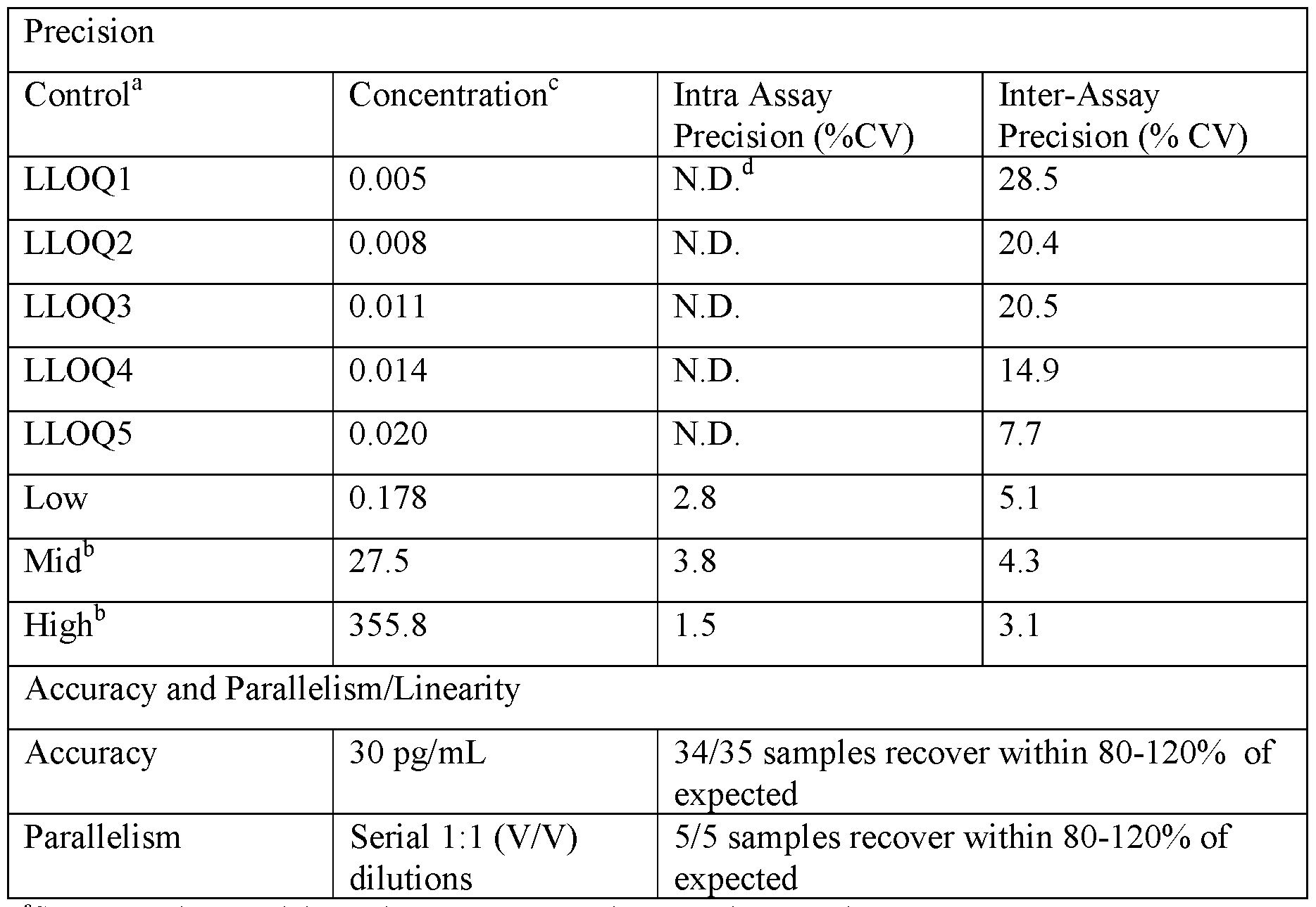
Claims
Priority Applications (10)
| Application Number | Priority Date | Filing Date | Title |
|---|---|---|---|
| CA2977285A CA2977285A1 (en) | 2015-03-16 | 2016-03-15 | Methods of detecting and quantifying il-13 and uses in diagnosing and treating th2-associated diseases |
| CN201680016018.8A CN107430117A (en) | 2015-03-16 | 2016-03-15 | Detection and quantitative IL 13 method and the purposes in diagnosing and treating Th2 relevant diseases |
| JP2017548960A JP2018511797A (en) | 2015-03-16 | 2016-03-15 | IL-13 detection and quantification method and use in diagnosis and treatment of Th2-related diseases |
| AU2016233398A AU2016233398A1 (en) | 2015-03-16 | 2016-03-15 | Methods of detecting and quantifying IL-13 and uses in diagnosing and treating Th2-associated diseases |
| EP16715167.9A EP3271723A1 (en) | 2015-03-16 | 2016-03-15 | Methods of detecting and quantifying il-13 and uses in diagnosing and treating th2-associated diseases |
| MX2017011486A MX2017011486A (en) | 2015-03-16 | 2016-03-15 | Methods of detecting and quantifying il-13 and uses in diagnosing and treating th2-associated diseases. |
| KR1020177028752A KR20170127011A (en) | 2015-03-16 | 2016-03-15 | Methods for detecting and quantifying IL-13 and for diagnosing and treating TH2-related diseases |
| IL254080A IL254080A0 (en) | 2015-03-16 | 2017-08-21 | Methods of detecting and quantifying il-13 and uses in diagnosing and treating th2-associated diseases |
| US15/700,790 US20180356429A1 (en) | 2015-03-16 | 2017-09-11 | Methods of detecting and quantifying il-13 and uses in diagnosing and treating th2-associated diseases |
| HK18106949.9A HK1247287A1 (en) | 2015-03-16 | 2018-05-28 | Methods of detecting and quantifying il-13 and uses in diagnosing and treating th2-associated diseases |
Applications Claiming Priority (2)
| Application Number | Priority Date | Filing Date | Title |
|---|---|---|---|
| US201562133693P | 2015-03-16 | 2015-03-16 | |
| US62/133,693 | 2015-03-16 |
Related Child Applications (1)
| Application Number | Title | Priority Date | Filing Date |
|---|---|---|---|
| US15/700,790 Continuation US20180356429A1 (en) | 2015-03-16 | 2017-09-11 | Methods of detecting and quantifying il-13 and uses in diagnosing and treating th2-associated diseases |
Publications (1)
| Publication Number | Publication Date |
|---|---|
| WO2016149276A1 true WO2016149276A1 (en) | 2016-09-22 |
Family
ID=55697472
Family Applications (1)
| Application Number | Title | Priority Date | Filing Date |
|---|---|---|---|
| PCT/US2016/022481 WO2016149276A1 (en) | 2015-03-16 | 2016-03-15 | Methods of detecting and quantifying il-13 and uses in diagnosing and treating th2-associated diseases |
Country Status (12)
| Country | Link |
|---|---|
| US (1) | US20180356429A1 (en) |
| EP (1) | EP3271723A1 (en) |
| JP (1) | JP2018511797A (en) |
| KR (1) | KR20170127011A (en) |
| CN (1) | CN107430117A (en) |
| AR (1) | AR103935A1 (en) |
| AU (1) | AU2016233398A1 (en) |
| CA (1) | CA2977285A1 (en) |
| HK (1) | HK1247287A1 (en) |
| IL (1) | IL254080A0 (en) |
| MX (1) | MX2017011486A (en) |
| WO (1) | WO2016149276A1 (en) |
Cited By (7)
| Publication number | Priority date | Publication date | Assignee | Title |
|---|---|---|---|---|
| WO2018057849A1 (en) | 2016-09-23 | 2018-03-29 | Genentech, Inc. | Uses of il-13 antagonists for treating atopic dermatitis |
| US10093730B2 (en) | 2014-11-10 | 2018-10-09 | Genentech, Inc. | Anti-interleukin-33 antibodies and uses thereof |
| WO2023287590A1 (en) | 2021-07-16 | 2023-01-19 | Dermira, Inc. | Il-13 antibodies for the treatment of atopic dermatitis |
| WO2023019260A1 (en) | 2021-08-13 | 2023-02-16 | Dermira, Inc. | Il-13 antibodies for the treatment of atopic dermatitis |
| US11708608B2 (en) | 2014-11-10 | 2023-07-25 | Genentech, Inc. | Therapeutic and diagnostic methods for IL-33-mediated disorders |
| US11760797B2 (en) | 2020-03-13 | 2023-09-19 | Genentech, Inc. | Anti-interleukin-33 antibodies and uses thereof |
| WO2023215769A1 (en) | 2022-05-05 | 2023-11-09 | Dermira, Inc. | Il-13 antibodies for the treatment of atopic dermatitis |
Families Citing this family (4)
| Publication number | Priority date | Publication date | Assignee | Title |
|---|---|---|---|---|
| WO2019097534A1 (en) * | 2017-11-15 | 2019-05-23 | Venkata Satya Suresh Attili | A device and method to determine the mean/average control of asthma/copd over time and compliance to treatment |
| KR102003054B1 (en) * | 2018-10-18 | 2019-07-24 | 주식회사 닥터에이 | Method for matching surgical procedure service |
| CN113614538A (en) * | 2019-03-19 | 2021-11-05 | Scm生命科学有限公司 | Method for treating atopic dermatitis using monoclonal stem cells |
| US20220233093A1 (en) * | 2021-01-22 | 2022-07-28 | AsthmaTek, Inc. | Systems and methods to provide a physician interface that enables a physician to assess asthma of a subject and provide therapeutic feedback |
Citations (107)
| Publication number | Priority date | Publication date | Assignee | Title |
|---|---|---|---|---|
| US4016043A (en) | 1975-09-04 | 1977-04-05 | Akzona Incorporated | Enzymatic immunological method for the determination of antigens and antibodies |
| US4018653A (en) | 1971-10-29 | 1977-04-19 | U.S. Packaging Corporation | Instrument for the detection of Neisseria gonorrhoeae without culture |
| US4275149A (en) | 1978-11-24 | 1981-06-23 | Syva Company | Macromolecular environment control in specific receptor assays |
| US4318980A (en) | 1978-04-10 | 1982-03-09 | Miles Laboratories, Inc. | Heterogenous specific binding assay employing a cycling reactant as label |
| US4424279A (en) | 1982-08-12 | 1984-01-03 | Quidel | Rapid plunger immunoassay method and apparatus |
| US4676980A (en) | 1985-09-23 | 1987-06-30 | The United States Of America As Represented By The Secretary Of The Department Of Health And Human Services | Target specific cross-linked heteroantibodies |
| US4737456A (en) | 1985-05-09 | 1988-04-12 | Syntex (U.S.A.) Inc. | Reducing interference in ligand-receptor binding assays |
| EP0404097A2 (en) | 1989-06-22 | 1990-12-27 | BEHRINGWERKE Aktiengesellschaft | Bispecific and oligospecific, mono- and oligovalent receptors, production and applications thereof |
| WO1993001161A1 (en) | 1991-07-11 | 1993-01-21 | Pfizer Limited | Process for preparing sertraline intermediates |
| WO1993008829A1 (en) | 1991-11-04 | 1993-05-13 | The Regents Of The University Of California | Compositions that mediate killing of hiv-infected cells |
| WO1993016185A2 (en) | 1992-02-06 | 1993-08-19 | Creative Biomolecules, Inc. | Biosynthetic binding protein for cancer marker |
| WO1994029351A2 (en) | 1993-06-16 | 1994-12-22 | Celltech Limited | Antibodies |
| US5500362A (en) | 1987-01-08 | 1996-03-19 | Xoma Corporation | Chimeric antibody with specificity to human B cell surface antigen |
| US5571894A (en) | 1991-02-05 | 1996-11-05 | Ciba-Geigy Corporation | Recombinant antibodies specific for a growth factor receptor |
| US5587458A (en) | 1991-10-07 | 1996-12-24 | Aronex Pharmaceuticals, Inc. | Anti-erbB-2 antibodies, combinations thereof, and therapeutic and diagnostic uses thereof |
| US5624821A (en) | 1987-03-18 | 1997-04-29 | Scotgen Biopharmaceuticals Incorporated | Antibodies with altered effector functions |
| US5648237A (en) | 1991-09-19 | 1997-07-15 | Genentech, Inc. | Expression of functional antibody fragments |
| WO1997030087A1 (en) | 1996-02-16 | 1997-08-21 | Glaxo Group Limited | Preparation of glycosylated antibodies |
| US5731168A (en) | 1995-03-01 | 1998-03-24 | Genentech, Inc. | Method for making heteromultimeric polypeptides |
| US5750373A (en) | 1990-12-03 | 1998-05-12 | Genentech, Inc. | Enrichment method for variant proteins having altered binding properties, M13 phagemids, and growth hormone variants |
| US5770429A (en) | 1990-08-29 | 1998-06-23 | Genpharm International, Inc. | Transgenic non-human animals capable of producing heterologous antibodies |
| US5789199A (en) | 1994-11-03 | 1998-08-04 | Genentech, Inc. | Process for bacterial production of polypeptides |
| US5821337A (en) | 1991-06-14 | 1998-10-13 | Genentech, Inc. | Immunoglobulin variants |
| US5840523A (en) | 1995-03-01 | 1998-11-24 | Genetech, Inc. | Methods and compositions for secretion of heterologous polypeptides |
| WO1998058964A1 (en) | 1997-06-24 | 1998-12-30 | Genentech, Inc. | Methods and compositions for galactosylated glycoproteins |
| US5869046A (en) | 1995-04-14 | 1999-02-09 | Genentech, Inc. | Altered polypeptides with increased half-life |
| WO1999022764A1 (en) | 1997-10-31 | 1999-05-14 | Genentech, Inc. | Methods and compositions comprising glycoprotein glycoforms |
| US5959177A (en) | 1989-10-27 | 1999-09-28 | The Scripps Research Institute | Transgenic plants expressing assembled secretory antibodies |
| WO1999051642A1 (en) | 1998-04-02 | 1999-10-14 | Genentech, Inc. | Antibody variants and fragments thereof |
| US6040498A (en) | 1998-08-11 | 2000-03-21 | North Caroline State University | Genetically engineered duckweed |
| US6075181A (en) | 1990-01-12 | 2000-06-13 | Abgenix, Inc. | Human antibodies derived from immunized xenomice |
| WO2000061739A1 (en) | 1999-04-09 | 2000-10-19 | Kyowa Hakko Kogyo Co., Ltd. | Method for controlling the activity of immunologically functional molecule |
| US6150584A (en) | 1990-01-12 | 2000-11-21 | Abgenix, Inc. | Human antibodies derived from immunized xenomice |
| US6171586B1 (en) | 1997-06-13 | 2001-01-09 | Genentech, Inc. | Antibody formulation |
| US6194551B1 (en) | 1998-04-02 | 2001-02-27 | Genentech, Inc. | Polypeptide variants |
| WO2001029246A1 (en) | 1999-10-19 | 2001-04-26 | Kyowa Hakko Kogyo Co., Ltd. | Process for producing polypeptide |
| US6248516B1 (en) | 1988-11-11 | 2001-06-19 | Medical Research Council | Single domain ligands, receptors comprising said ligands methods for their production, and use of said ligands and receptors |
| US6267958B1 (en) | 1995-07-27 | 2001-07-31 | Genentech, Inc. | Protein formulation |
| WO2002031140A1 (en) | 2000-10-06 | 2002-04-18 | Kyowa Hakko Kogyo Co., Ltd. | Cells producing antibody compositions |
| US6420548B1 (en) | 1999-10-04 | 2002-07-16 | Medicago Inc. | Method for regulating transcription of foreign genes |
| US20020164328A1 (en) | 2000-10-06 | 2002-11-07 | Toyohide Shinkawa | Process for purifying antibody |
| WO2003011878A2 (en) | 2001-08-03 | 2003-02-13 | Glycart Biotechnology Ag | Antibody glycosylation variants having increased antibody-dependent cellular cytotoxicity |
| US20030115614A1 (en) | 2000-10-06 | 2003-06-19 | Yutaka Kanda | Antibody composition-producing cell |
| US6602684B1 (en) | 1998-04-20 | 2003-08-05 | Glycart Biotechnology Ag | Glycosylation engineering of antibodies for improving antibody-dependent cellular cytotoxicity |
| US20030157108A1 (en) | 2001-10-25 | 2003-08-21 | Genentech, Inc. | Glycoprotein compositions |
| WO2003085119A1 (en) | 2002-04-09 | 2003-10-16 | Kyowa Hakko Kogyo Co., Ltd. | METHOD OF ENHANCING ACTIVITY OF ANTIBODY COMPOSITION OF BINDING TO FcϜ RECEPTOR IIIa |
| WO2003085107A1 (en) | 2002-04-09 | 2003-10-16 | Kyowa Hakko Kogyo Co., Ltd. | Cells with modified genome |
| WO2003084570A1 (en) | 2002-04-09 | 2003-10-16 | Kyowa Hakko Kogyo Co., Ltd. | DRUG CONTAINING ANTIBODY COMPOSITION APPROPRIATE FOR PATIENT SUFFERING FROM FcϜRIIIa POLYMORPHISM |
| US20040093621A1 (en) | 2001-12-25 | 2004-05-13 | Kyowa Hakko Kogyo Co., Ltd | Antibody composition which specifically binds to CD20 |
| US6737056B1 (en) | 1999-01-15 | 2004-05-18 | Genentech, Inc. | Polypeptide variants with altered effector function |
| US20040109865A1 (en) | 2002-04-09 | 2004-06-10 | Kyowa Hakko Kogyo Co., Ltd. | Antibody composition-containing medicament |
| US20040110282A1 (en) | 2002-04-09 | 2004-06-10 | Kyowa Hakko Kogyo Co., Ltd. | Cells in which activity of the protein involved in transportation of GDP-fucose is reduced or lost |
| WO2004056312A2 (en) | 2002-12-16 | 2004-07-08 | Genentech, Inc. | Immunoglobulin variants and uses thereof |
| US20040132140A1 (en) | 2002-04-09 | 2004-07-08 | Kyowa Hakko Kogyo Co., Ltd. | Production process for antibody composition |
| US20050014934A1 (en) | 2002-10-15 | 2005-01-20 | Hinton Paul R. | Alteration of FcRn binding affinities or serum half-lives of antibodies by mutagenesis |
| WO2005007699A2 (en) | 2003-07-15 | 2005-01-27 | Cambridge Antibody Technology Limited | Human antibody molecules for il-13 |
| US20050079574A1 (en) | 2003-01-16 | 2005-04-14 | Genentech, Inc. | Synthetic antibody phage libraries |
| WO2005035778A1 (en) | 2003-10-09 | 2005-04-21 | Kyowa Hakko Kogyo Co., Ltd. | PROCESS FOR PRODUCING ANTIBODY COMPOSITION BY USING RNA INHIBITING THE FUNCTION OF α1,6-FUCOSYLTRANSFERASE |
| WO2005035586A1 (en) | 2003-10-08 | 2005-04-21 | Kyowa Hakko Kogyo Co., Ltd. | Fused protein composition |
| US20050119455A1 (en) | 2002-06-03 | 2005-06-02 | Genentech, Inc. | Synthetic antibody phage libraries |
| US20050123546A1 (en) | 2003-11-05 | 2005-06-09 | Glycart Biotechnology Ag | Antigen binding molecules with increased Fc receptor binding affinity and effector function |
| WO2005053742A1 (en) | 2003-12-04 | 2005-06-16 | Kyowa Hakko Kogyo Co., Ltd. | Medicine containing antibody composition |
| WO2005062967A2 (en) | 2003-12-23 | 2005-07-14 | Tanox, Inc. | Novel anti-il 13 antibodies and uses thereof |
| WO2005100402A1 (en) | 2004-04-13 | 2005-10-27 | F.Hoffmann-La Roche Ag | Anti-p-selectin antibodies |
| US20050260186A1 (en) | 2003-03-05 | 2005-11-24 | Halozyme, Inc. | Soluble glycosaminoglycanases and methods of preparing and using soluble glycosaminoglycanases |
| US20050266000A1 (en) | 2004-04-09 | 2005-12-01 | Genentech, Inc. | Variable domain library and uses |
| US6982321B2 (en) | 1986-03-27 | 2006-01-03 | Medical Research Council | Altered antibodies |
| US20060025576A1 (en) | 2000-04-11 | 2006-02-02 | Genentech, Inc. | Multivalent antibodies and uses therefor |
| WO2006029879A2 (en) | 2004-09-17 | 2006-03-23 | F.Hoffmann-La Roche Ag | Anti-ox40l antibodies |
| WO2006044908A2 (en) | 2004-10-20 | 2006-04-27 | Genentech, Inc. | Antibody formulation in histidine-acetate buffer |
| US7041870B2 (en) | 2000-11-30 | 2006-05-09 | Medarex, Inc. | Transgenic transchromosomal rodents for making human antibodies |
| US20060104968A1 (en) | 2003-03-05 | 2006-05-18 | Halozyme, Inc. | Soluble glycosaminoglycanases and methods of preparing and using soluble glycosaminogly ycanases |
| US7087409B2 (en) | 1997-12-05 | 2006-08-08 | The Scripps Research Institute | Humanization of murine antibody |
| WO2006085938A2 (en) | 2004-06-17 | 2006-08-17 | Wyeth | Il-13 binding agents |
| US7125978B1 (en) | 1999-10-04 | 2006-10-24 | Medicago Inc. | Promoter for regulating expression of foreign genes |
| US7189826B2 (en) | 1997-11-24 | 2007-03-13 | Institute For Human Genetics And Biochemistry | Monoclonal human natural antibodies |
| US20070061900A1 (en) | 2000-10-31 | 2007-03-15 | Murphy Andrew J | Methods of modifying eukaryotic cells |
| WO2007036745A2 (en) | 2005-09-30 | 2007-04-05 | Medimmune Limited | Interleukin-13 antibody composition |
| WO2007039280A1 (en) | 2005-10-06 | 2007-04-12 | Roche Diagnostics Gmbh | Anti-ccp and antinuclear antibodies in diagnosis of rheumatoid arthritis |
| WO2007045477A2 (en) | 2005-10-21 | 2007-04-26 | Novartis Ag | Human antibodies against il-13 and therapeutic uses |
| US20070117126A1 (en) | 1999-12-15 | 2007-05-24 | Genentech, Inc. | Shotgun scanning |
| US20070160598A1 (en) | 2005-11-07 | 2007-07-12 | Dennis Mark S | Binding polypeptides with diversified and consensus vh/vl hypervariable sequences |
| WO2007082068A2 (en) | 2006-01-11 | 2007-07-19 | Aerovance, Inc. | Methods and compositions for treating asthma in human and non human primates |
| WO2007080174A2 (en) | 2006-01-11 | 2007-07-19 | Glaxo Group Limited | Chimeric and humanised anti-human il-13 antibodies |
| US20070237764A1 (en) | 2005-12-02 | 2007-10-11 | Genentech, Inc. | Binding polypeptides with restricted diversity sequences |
| US20070292936A1 (en) | 2006-05-09 | 2007-12-20 | Genentech, Inc. | Binding polypeptides with optimized scaffolds |
| US20080069820A1 (en) | 2006-08-30 | 2008-03-20 | Genentech, Inc. | Multispecific antibodies |
| US7371826B2 (en) | 1999-01-15 | 2008-05-13 | Genentech, Inc. | Polypeptide variants with altered effector function |
| WO2008077546A1 (en) | 2006-12-22 | 2008-07-03 | F. Hoffmann-La Roche Ag | Antibodies against insulin-like growth factor i receptor and uses thereof |
| WO2008086395A2 (en) | 2007-01-09 | 2008-07-17 | Wyeth | Anti-il-13 antibody formulations and uses thereof |
| WO2008127271A2 (en) | 2006-09-08 | 2008-10-23 | Abbott Laboratories | Interleukin -13 binding proteins |
| WO2008134724A2 (en) | 2007-04-30 | 2008-11-06 | Smithkline Beecham Corporation | Methods for administering anti-il-5 antibodies |
| US20090002360A1 (en) | 2007-05-25 | 2009-01-01 | Innolux Display Corp. | Liquid crystal display device and method for driving same |
| WO2009009775A1 (en) | 2007-07-11 | 2009-01-15 | Aerovance, Inc. | Pharmaceutical polypeptide dry powder aerosol formulation and method of preparation |
| US20090047277A1 (en) | 2003-04-11 | 2009-02-19 | Medimmune, Inc. | Recombinant il-9 antibodies and uses thereof |
| US7501121B2 (en) | 2004-06-17 | 2009-03-10 | Wyeth | IL-13 binding agents |
| US7521541B2 (en) | 2004-09-23 | 2009-04-21 | Genetech Inc. | Cysteine engineered antibodies and conjugates |
| US7527791B2 (en) | 2004-03-31 | 2009-05-05 | Genentech, Inc. | Humanized anti-TGF-beta antibodies |
| WO2009089004A1 (en) | 2008-01-07 | 2009-07-16 | Amgen Inc. | Method for making antibody fc-heterodimeric molecules using electrostatic steering effects |
| WO2009124090A1 (en) | 2008-03-31 | 2009-10-08 | Genentech, Inc. | Compositions and methods for treating and diagnosing asthma |
| US7615213B2 (en) | 2004-06-09 | 2009-11-10 | Wyeth | Antibodies against human interleukin-13 and pharmaceutical compositions thereof |
| WO2010073119A1 (en) | 2008-12-23 | 2010-07-01 | Adocia | Stable pharmaceutical composition containing at least one monoclonal antibody and at least one amphiphilic polysaccharide comprising hydrophobic substituents |
| US20100226923A1 (en) * | 2007-10-15 | 2010-09-09 | Sanofi-Aventis | Antibodies that bind il-4 and/or il-13 and their uses |
| US7807788B2 (en) | 2004-07-01 | 2010-10-05 | Glaxo Group Limited | Chimeric and humanised monoclonal antibodies against Interleukin-13 |
| WO2012083132A2 (en) | 2010-12-16 | 2012-06-21 | Genentech, Inc. | Diagnosis and treatments relating to th2 inhibition |
| US20130243750A1 (en) | 2012-01-31 | 2013-09-19 | Genentech, Inc. | Anti-ige antibodies and methods using same |
| WO2014165771A2 (en) | 2013-04-05 | 2014-10-09 | Genentech, Inc. | Anti-il-4 antibodies and bispecific antibodies and uses thereof |
Family Cites Families (3)
| Publication number | Priority date | Publication date | Assignee | Title |
|---|---|---|---|---|
| US5196324A (en) * | 1989-12-15 | 1993-03-23 | Eli Lilly And Company | Monoclonal antibodies reactive with a human atheroma associated antigen |
| US20090234202A1 (en) * | 2008-03-05 | 2009-09-17 | Goix Philippe J | Method and compositions for highly sensitive detection of molecules |
| KR20150002874A (en) * | 2008-05-05 | 2015-01-07 | 노비뮨 에스 에이 | Anti-il-17a/il-17f cross-reactive antibodies and methods of use thereof |
-
2016
- 2016-03-15 MX MX2017011486A patent/MX2017011486A/en unknown
- 2016-03-15 AR ARP160100684A patent/AR103935A1/en unknown
- 2016-03-15 AU AU2016233398A patent/AU2016233398A1/en not_active Abandoned
- 2016-03-15 CN CN201680016018.8A patent/CN107430117A/en active Pending
- 2016-03-15 KR KR1020177028752A patent/KR20170127011A/en unknown
- 2016-03-15 EP EP16715167.9A patent/EP3271723A1/en not_active Withdrawn
- 2016-03-15 WO PCT/US2016/022481 patent/WO2016149276A1/en active Application Filing
- 2016-03-15 JP JP2017548960A patent/JP2018511797A/en active Pending
- 2016-03-15 CA CA2977285A patent/CA2977285A1/en not_active Abandoned
-
2017
- 2017-08-21 IL IL254080A patent/IL254080A0/en unknown
- 2017-09-11 US US15/700,790 patent/US20180356429A1/en not_active Abandoned
-
2018
- 2018-05-28 HK HK18106949.9A patent/HK1247287A1/en unknown
Patent Citations (111)
| Publication number | Priority date | Publication date | Assignee | Title |
|---|---|---|---|---|
| US4018653A (en) | 1971-10-29 | 1977-04-19 | U.S. Packaging Corporation | Instrument for the detection of Neisseria gonorrhoeae without culture |
| US4016043A (en) | 1975-09-04 | 1977-04-05 | Akzona Incorporated | Enzymatic immunological method for the determination of antigens and antibodies |
| US4318980A (en) | 1978-04-10 | 1982-03-09 | Miles Laboratories, Inc. | Heterogenous specific binding assay employing a cycling reactant as label |
| US4275149A (en) | 1978-11-24 | 1981-06-23 | Syva Company | Macromolecular environment control in specific receptor assays |
| US4424279A (en) | 1982-08-12 | 1984-01-03 | Quidel | Rapid plunger immunoassay method and apparatus |
| US4737456A (en) | 1985-05-09 | 1988-04-12 | Syntex (U.S.A.) Inc. | Reducing interference in ligand-receptor binding assays |
| US4676980A (en) | 1985-09-23 | 1987-06-30 | The United States Of America As Represented By The Secretary Of The Department Of Health And Human Services | Target specific cross-linked heteroantibodies |
| US6982321B2 (en) | 1986-03-27 | 2006-01-03 | Medical Research Council | Altered antibodies |
| US5500362A (en) | 1987-01-08 | 1996-03-19 | Xoma Corporation | Chimeric antibody with specificity to human B cell surface antigen |
| US5624821A (en) | 1987-03-18 | 1997-04-29 | Scotgen Biopharmaceuticals Incorporated | Antibodies with altered effector functions |
| US5648260A (en) | 1987-03-18 | 1997-07-15 | Scotgen Biopharmaceuticals Incorporated | DNA encoding antibodies with altered effector functions |
| US6248516B1 (en) | 1988-11-11 | 2001-06-19 | Medical Research Council | Single domain ligands, receptors comprising said ligands methods for their production, and use of said ligands and receptors |
| EP0404097A2 (en) | 1989-06-22 | 1990-12-27 | BEHRINGWERKE Aktiengesellschaft | Bispecific and oligospecific, mono- and oligovalent receptors, production and applications thereof |
| US6417429B1 (en) | 1989-10-27 | 2002-07-09 | The Scripps Research Institute | Transgenic plants expressing assembled secretory antibodies |
| US5959177A (en) | 1989-10-27 | 1999-09-28 | The Scripps Research Institute | Transgenic plants expressing assembled secretory antibodies |
| US6075181A (en) | 1990-01-12 | 2000-06-13 | Abgenix, Inc. | Human antibodies derived from immunized xenomice |
| US6150584A (en) | 1990-01-12 | 2000-11-21 | Abgenix, Inc. | Human antibodies derived from immunized xenomice |
| US5770429A (en) | 1990-08-29 | 1998-06-23 | Genpharm International, Inc. | Transgenic non-human animals capable of producing heterologous antibodies |
| US5750373A (en) | 1990-12-03 | 1998-05-12 | Genentech, Inc. | Enrichment method for variant proteins having altered binding properties, M13 phagemids, and growth hormone variants |
| US5571894A (en) | 1991-02-05 | 1996-11-05 | Ciba-Geigy Corporation | Recombinant antibodies specific for a growth factor receptor |
| US5821337A (en) | 1991-06-14 | 1998-10-13 | Genentech, Inc. | Immunoglobulin variants |
| WO1993001161A1 (en) | 1991-07-11 | 1993-01-21 | Pfizer Limited | Process for preparing sertraline intermediates |
| US5648237A (en) | 1991-09-19 | 1997-07-15 | Genentech, Inc. | Expression of functional antibody fragments |
| US5587458A (en) | 1991-10-07 | 1996-12-24 | Aronex Pharmaceuticals, Inc. | Anti-erbB-2 antibodies, combinations thereof, and therapeutic and diagnostic uses thereof |
| WO1993008829A1 (en) | 1991-11-04 | 1993-05-13 | The Regents Of The University Of California | Compositions that mediate killing of hiv-infected cells |
| WO1993016185A2 (en) | 1992-02-06 | 1993-08-19 | Creative Biomolecules, Inc. | Biosynthetic binding protein for cancer marker |
| WO1994029351A2 (en) | 1993-06-16 | 1994-12-22 | Celltech Limited | Antibodies |
| US5789199A (en) | 1994-11-03 | 1998-08-04 | Genentech, Inc. | Process for bacterial production of polypeptides |
| US5840523A (en) | 1995-03-01 | 1998-11-24 | Genetech, Inc. | Methods and compositions for secretion of heterologous polypeptides |
| US5731168A (en) | 1995-03-01 | 1998-03-24 | Genentech, Inc. | Method for making heteromultimeric polypeptides |
| US5869046A (en) | 1995-04-14 | 1999-02-09 | Genentech, Inc. | Altered polypeptides with increased half-life |
| US6267958B1 (en) | 1995-07-27 | 2001-07-31 | Genentech, Inc. | Protein formulation |
| WO1997030087A1 (en) | 1996-02-16 | 1997-08-21 | Glaxo Group Limited | Preparation of glycosylated antibodies |
| US6171586B1 (en) | 1997-06-13 | 2001-01-09 | Genentech, Inc. | Antibody formulation |
| WO1998058964A1 (en) | 1997-06-24 | 1998-12-30 | Genentech, Inc. | Methods and compositions for galactosylated glycoproteins |
| WO1999022764A1 (en) | 1997-10-31 | 1999-05-14 | Genentech, Inc. | Methods and compositions comprising glycoprotein glycoforms |
| US7189826B2 (en) | 1997-11-24 | 2007-03-13 | Institute For Human Genetics And Biochemistry | Monoclonal human natural antibodies |
| US7087409B2 (en) | 1997-12-05 | 2006-08-08 | The Scripps Research Institute | Humanization of murine antibody |
| US6194551B1 (en) | 1998-04-02 | 2001-02-27 | Genentech, Inc. | Polypeptide variants |
| WO1999051642A1 (en) | 1998-04-02 | 1999-10-14 | Genentech, Inc. | Antibody variants and fragments thereof |
| US6602684B1 (en) | 1998-04-20 | 2003-08-05 | Glycart Biotechnology Ag | Glycosylation engineering of antibodies for improving antibody-dependent cellular cytotoxicity |
| US6040498A (en) | 1998-08-11 | 2000-03-21 | North Caroline State University | Genetically engineered duckweed |
| US7371826B2 (en) | 1999-01-15 | 2008-05-13 | Genentech, Inc. | Polypeptide variants with altered effector function |
| US6737056B1 (en) | 1999-01-15 | 2004-05-18 | Genentech, Inc. | Polypeptide variants with altered effector function |
| US7332581B2 (en) | 1999-01-15 | 2008-02-19 | Genentech, Inc. | Polypeptide variants with altered effector function |
| WO2000061739A1 (en) | 1999-04-09 | 2000-10-19 | Kyowa Hakko Kogyo Co., Ltd. | Method for controlling the activity of immunologically functional molecule |
| US6420548B1 (en) | 1999-10-04 | 2002-07-16 | Medicago Inc. | Method for regulating transcription of foreign genes |
| US7125978B1 (en) | 1999-10-04 | 2006-10-24 | Medicago Inc. | Promoter for regulating expression of foreign genes |
| WO2001029246A1 (en) | 1999-10-19 | 2001-04-26 | Kyowa Hakko Kogyo Co., Ltd. | Process for producing polypeptide |
| US20070117126A1 (en) | 1999-12-15 | 2007-05-24 | Genentech, Inc. | Shotgun scanning |
| US20060025576A1 (en) | 2000-04-11 | 2006-02-02 | Genentech, Inc. | Multivalent antibodies and uses therefor |
| US20030115614A1 (en) | 2000-10-06 | 2003-06-19 | Yutaka Kanda | Antibody composition-producing cell |
| US20020164328A1 (en) | 2000-10-06 | 2002-11-07 | Toyohide Shinkawa | Process for purifying antibody |
| WO2002031140A1 (en) | 2000-10-06 | 2002-04-18 | Kyowa Hakko Kogyo Co., Ltd. | Cells producing antibody compositions |
| US20070061900A1 (en) | 2000-10-31 | 2007-03-15 | Murphy Andrew J | Methods of modifying eukaryotic cells |
| US7041870B2 (en) | 2000-11-30 | 2006-05-09 | Medarex, Inc. | Transgenic transchromosomal rodents for making human antibodies |
| WO2003011878A2 (en) | 2001-08-03 | 2003-02-13 | Glycart Biotechnology Ag | Antibody glycosylation variants having increased antibody-dependent cellular cytotoxicity |
| US20030157108A1 (en) | 2001-10-25 | 2003-08-21 | Genentech, Inc. | Glycoprotein compositions |
| US20040093621A1 (en) | 2001-12-25 | 2004-05-13 | Kyowa Hakko Kogyo Co., Ltd | Antibody composition which specifically binds to CD20 |
| US20040110704A1 (en) | 2002-04-09 | 2004-06-10 | Kyowa Hakko Kogyo Co., Ltd. | Cells of which genome is modified |
| WO2003085119A1 (en) | 2002-04-09 | 2003-10-16 | Kyowa Hakko Kogyo Co., Ltd. | METHOD OF ENHANCING ACTIVITY OF ANTIBODY COMPOSITION OF BINDING TO FcϜ RECEPTOR IIIa |
| US20040109865A1 (en) | 2002-04-09 | 2004-06-10 | Kyowa Hakko Kogyo Co., Ltd. | Antibody composition-containing medicament |
| WO2003085107A1 (en) | 2002-04-09 | 2003-10-16 | Kyowa Hakko Kogyo Co., Ltd. | Cells with modified genome |
| US20040110282A1 (en) | 2002-04-09 | 2004-06-10 | Kyowa Hakko Kogyo Co., Ltd. | Cells in which activity of the protein involved in transportation of GDP-fucose is reduced or lost |
| WO2003084570A1 (en) | 2002-04-09 | 2003-10-16 | Kyowa Hakko Kogyo Co., Ltd. | DRUG CONTAINING ANTIBODY COMPOSITION APPROPRIATE FOR PATIENT SUFFERING FROM FcϜRIIIa POLYMORPHISM |
| US20040132140A1 (en) | 2002-04-09 | 2004-07-08 | Kyowa Hakko Kogyo Co., Ltd. | Production process for antibody composition |
| US20050119455A1 (en) | 2002-06-03 | 2005-06-02 | Genentech, Inc. | Synthetic antibody phage libraries |
| US20050014934A1 (en) | 2002-10-15 | 2005-01-20 | Hinton Paul R. | Alteration of FcRn binding affinities or serum half-lives of antibodies by mutagenesis |
| WO2004056312A2 (en) | 2002-12-16 | 2004-07-08 | Genentech, Inc. | Immunoglobulin variants and uses thereof |
| US20050079574A1 (en) | 2003-01-16 | 2005-04-14 | Genentech, Inc. | Synthetic antibody phage libraries |
| US20050260186A1 (en) | 2003-03-05 | 2005-11-24 | Halozyme, Inc. | Soluble glycosaminoglycanases and methods of preparing and using soluble glycosaminoglycanases |
| US20060104968A1 (en) | 2003-03-05 | 2006-05-18 | Halozyme, Inc. | Soluble glycosaminoglycanases and methods of preparing and using soluble glycosaminogly ycanases |
| US20090047277A1 (en) | 2003-04-11 | 2009-02-19 | Medimmune, Inc. | Recombinant il-9 antibodies and uses thereof |
| WO2005007699A2 (en) | 2003-07-15 | 2005-01-27 | Cambridge Antibody Technology Limited | Human antibody molecules for il-13 |
| WO2005035586A1 (en) | 2003-10-08 | 2005-04-21 | Kyowa Hakko Kogyo Co., Ltd. | Fused protein composition |
| WO2005035778A1 (en) | 2003-10-09 | 2005-04-21 | Kyowa Hakko Kogyo Co., Ltd. | PROCESS FOR PRODUCING ANTIBODY COMPOSITION BY USING RNA INHIBITING THE FUNCTION OF α1,6-FUCOSYLTRANSFERASE |
| US20050123546A1 (en) | 2003-11-05 | 2005-06-09 | Glycart Biotechnology Ag | Antigen binding molecules with increased Fc receptor binding affinity and effector function |
| WO2005053742A1 (en) | 2003-12-04 | 2005-06-16 | Kyowa Hakko Kogyo Co., Ltd. | Medicine containing antibody composition |
| WO2005062967A2 (en) | 2003-12-23 | 2005-07-14 | Tanox, Inc. | Novel anti-il 13 antibodies and uses thereof |
| US7527791B2 (en) | 2004-03-31 | 2009-05-05 | Genentech, Inc. | Humanized anti-TGF-beta antibodies |
| US20050266000A1 (en) | 2004-04-09 | 2005-12-01 | Genentech, Inc. | Variable domain library and uses |
| WO2005100402A1 (en) | 2004-04-13 | 2005-10-27 | F.Hoffmann-La Roche Ag | Anti-p-selectin antibodies |
| US7615213B2 (en) | 2004-06-09 | 2009-11-10 | Wyeth | Antibodies against human interleukin-13 and pharmaceutical compositions thereof |
| WO2006085938A2 (en) | 2004-06-17 | 2006-08-17 | Wyeth | Il-13 binding agents |
| US7501121B2 (en) | 2004-06-17 | 2009-03-10 | Wyeth | IL-13 binding agents |
| US7807788B2 (en) | 2004-07-01 | 2010-10-05 | Glaxo Group Limited | Chimeric and humanised monoclonal antibodies against Interleukin-13 |
| WO2006029879A2 (en) | 2004-09-17 | 2006-03-23 | F.Hoffmann-La Roche Ag | Anti-ox40l antibodies |
| US7521541B2 (en) | 2004-09-23 | 2009-04-21 | Genetech Inc. | Cysteine engineered antibodies and conjugates |
| WO2006044908A2 (en) | 2004-10-20 | 2006-04-27 | Genentech, Inc. | Antibody formulation in histidine-acetate buffer |
| WO2007036745A2 (en) | 2005-09-30 | 2007-04-05 | Medimmune Limited | Interleukin-13 antibody composition |
| WO2007039280A1 (en) | 2005-10-06 | 2007-04-12 | Roche Diagnostics Gmbh | Anti-ccp and antinuclear antibodies in diagnosis of rheumatoid arthritis |
| WO2007045477A2 (en) | 2005-10-21 | 2007-04-26 | Novartis Ag | Human antibodies against il-13 and therapeutic uses |
| US20070160598A1 (en) | 2005-11-07 | 2007-07-12 | Dennis Mark S | Binding polypeptides with diversified and consensus vh/vl hypervariable sequences |
| US20070237764A1 (en) | 2005-12-02 | 2007-10-11 | Genentech, Inc. | Binding polypeptides with restricted diversity sequences |
| WO2007080174A2 (en) | 2006-01-11 | 2007-07-19 | Glaxo Group Limited | Chimeric and humanised anti-human il-13 antibodies |
| WO2007082068A2 (en) | 2006-01-11 | 2007-07-19 | Aerovance, Inc. | Methods and compositions for treating asthma in human and non human primates |
| US20070292936A1 (en) | 2006-05-09 | 2007-12-20 | Genentech, Inc. | Binding polypeptides with optimized scaffolds |
| US20080069820A1 (en) | 2006-08-30 | 2008-03-20 | Genentech, Inc. | Multispecific antibodies |
| WO2008127271A2 (en) | 2006-09-08 | 2008-10-23 | Abbott Laboratories | Interleukin -13 binding proteins |
| WO2008077546A1 (en) | 2006-12-22 | 2008-07-03 | F. Hoffmann-La Roche Ag | Antibodies against insulin-like growth factor i receptor and uses thereof |
| WO2008086395A2 (en) | 2007-01-09 | 2008-07-17 | Wyeth | Anti-il-13 antibody formulations and uses thereof |
| WO2008134724A2 (en) | 2007-04-30 | 2008-11-06 | Smithkline Beecham Corporation | Methods for administering anti-il-5 antibodies |
| US20090002360A1 (en) | 2007-05-25 | 2009-01-01 | Innolux Display Corp. | Liquid crystal display device and method for driving same |
| WO2009009775A1 (en) | 2007-07-11 | 2009-01-15 | Aerovance, Inc. | Pharmaceutical polypeptide dry powder aerosol formulation and method of preparation |
| US20100226923A1 (en) * | 2007-10-15 | 2010-09-09 | Sanofi-Aventis | Antibodies that bind il-4 and/or il-13 and their uses |
| WO2009089004A1 (en) | 2008-01-07 | 2009-07-16 | Amgen Inc. | Method for making antibody fc-heterodimeric molecules using electrostatic steering effects |
| WO2009124090A1 (en) | 2008-03-31 | 2009-10-08 | Genentech, Inc. | Compositions and methods for treating and diagnosing asthma |
| WO2010073119A1 (en) | 2008-12-23 | 2010-07-01 | Adocia | Stable pharmaceutical composition containing at least one monoclonal antibody and at least one amphiphilic polysaccharide comprising hydrophobic substituents |
| WO2012083132A2 (en) | 2010-12-16 | 2012-06-21 | Genentech, Inc. | Diagnosis and treatments relating to th2 inhibition |
| US20130243750A1 (en) | 2012-01-31 | 2013-09-19 | Genentech, Inc. | Anti-ige antibodies and methods using same |
| WO2014165771A2 (en) | 2013-04-05 | 2014-10-09 | Genentech, Inc. | Anti-il-4 antibodies and bispecific antibodies and uses thereof |
Non-Patent Citations (195)
| Title |
|---|
| ALMAGRO; FRANSSON, FRONT. BIOSCI, vol. 13, 2008, pages 1619 - 1633 |
| ALMAGRO; FRANSSON, FRONT. BIOSCI., vol. 13, 2008, pages 1619 - 1633 |
| AM J RESPIR CRIT CARE MED, vol. 171, 2005, pages 912 - 30 |
| ARGUELLES ET AL., ARCH. INTERN. MED., vol. 143, 1983, pages 570 - 571 |
| ARRON ET AL., ADV PHARMACOL, vol. 66, 2013, pages 1 - 49 |
| ARRON ET AL., ANNALS AM. THORACIC SOC., 2013 |
| ARRON JR ET AL., ANNALSATS, vol. 10, 2013, pages S206 - 13 |
| ASSARIAN ET AL., HUM. PATHOL., vol. 16, 1985, pages 311 - 312 |
| ATANES ET AL., SCAND. J. RHEUMATOL., vol. 25, 1996, pages 183 - 185 |
| BACA ET AL., J. BIOL. CHEM., vol. 272, 1997, pages 10678 - 10684 |
| BACHERCT ET AL., J. ALLERGY CLIN. IMMUNOL., vol. 107, 2001, pages 607 - 614 |
| BARALDO ET AL., EUR RESPIR J, vol. 38, 2011, pages 575 - 83 |
| BECK LA ET AL., N ENG J MED, vol. 371, 2014, pages 130 - 9 |
| BECKER AB, J ALLERGY CLIN IMMUNOL, vol. 109, 2002, pages S533 - 8 |
| BEL ET AL., NEW ENGL. J. MED., vol. 371, no. 13, 2014, pages 1189 - 97 |
| BLANCHARD C ET AL., MUCOSAL IMMUNOL, vol. 1, 2008, pages 289 - 96 |
| BOERNER ET AL., J. IMMUNOL., vol. 147, 1991, pages 86 |
| BOUROS ET AL., AM. J. RESPIR. CRIT. CARE MED., vol. 165, 2002, pages 1581 - 1586 |
| BOUSQUET ET AL., CHEST, vol. 125, no. 4, 2004, pages 1378 - 86 |
| BOYENOH GAYE ET AL: "Development of an ultra-sensitive single molecule counting assay for the detection of interleukin-13 as a marker for asthmatic severity", JOURNAL OF IMMUNOLOGICAL METHODS., vol. 426, 1 November 2015 (2015-11-01), NL, pages 82 - 85, XP055272314, ISSN: 0022-1759, DOI: 10.1016/j.jim.2015.08.006 * |
| BRENNAN ET AL., SCIENCE, vol. 229, 1985, pages 81 |
| BRODEUR ET AL.: "Monoclonal Antibody Production Techniques and Applications", 1987, MARCEL DEKKER, INC., pages: 51 - 63 |
| BRODLIE ET AL., ARCH DIS CHILD, vol. 97, no. 7, 2012, pages 604 - 9 |
| BRUGGEMANN, M. ET AL., J. EXP. MED., vol. 166, 1987, pages 1351 - 1361 |
| CAI ET AL., BIOANALYSIS, vol. 8, no. 4, 2016, pages 323 - 332 |
| CARTER ET AL., PROC. NATL. ACAD. SCI. USA, vol. 89, 1992, pages 4285 |
| CASTRO ET AL., LANCET RESP. MED., vol. 2, no. 11, 2014, pages 879 - 90 |
| CHANDRIANI S ET AL., J IMMUNOL, vol. 193, 2014, pages 111 - 9 |
| CHANG ET AL., ADV IMMUNOL., vol. 93, 2007, pages 63 - 119 |
| CHANG ET AL., J ALLERGY CLIN IMMUNOL., vol. 117, no. 6, 2006, pages 1203 - 12 |
| CHARLTON: "Methods in Molecular Biology", vol. 248, 2003, HUMANA PRESS, pages: 245 - 254 |
| CHEN ET AL., J. AM. ACAD. DERMATOL., vol. 35, 1996, pages 173 - 182 |
| CHEN ET AL., J. MOL. BIOL., vol. 293, 1999, pages 865 - 881 |
| CHOWDHURY, METHODS MOL. BIOL., vol. 207, 2008, pages 179 - 196 |
| CHOY DF ET AL., J ALLERGY CLIN IMMUNO1, vol. 130, 2012, pages 1335 - 43 |
| CLACKSON ET AL., NATURE, vol. 352, 1991, pages 624 - 628 |
| CLARKSON ET AL., NATURE, vol. 352, 1991, pages 624 - 628 |
| CLAUDON ET AL., CLINICAL CHEMISTRY, vol. 54, no. 9, pages 1554 - 1563 |
| CLYNES ET AL., PROC. NAT'L ACAD. SCI. USA, vol. 95, 1998, pages 652 - 656 |
| COLIGEN ET AL.,: "Current Protocols in Immunology", vol. 1, 2, 1991, WILEY-INTERSCIENCE |
| CONTIE ET AL., CALCIF TISSUE INT, vol. 87, 2010, pages 341 - 5 |
| CORREN ET AL., N ENGL J MED, vol. 365, 2011, pages 1088 - 98 |
| CORREN J ET AL., N ENG J MED, vol. 365, 2011, pages 1088 - 98 |
| CORREN J ET AL., N ENGL J MED, vol. 365, 2011, pages 1088 - 98 |
| CRAGG, M.S. ET AL., BLOOD, vol. 101, 2003, pages 1045 - 1052 |
| CRAGG, M.S.; M.J. GLENNIE, BLOOD, vol. 103, 2004, pages 2738 - 2743 |
| CUNNINGHAM; WELLS, SCIENCE, vol. 244, 1989, pages 1081 - 1085 |
| DALL'ACQUA ET AL., METHODS, vol. 36, 2005, pages 43 - 60 |
| DESCHRYVER-KECSKEMETI ET AL., ARCH. PATHOL. LAB MED., vol. 113, 1989, pages 394 - 398 |
| DOUCET J ET AL., DIS MARKERS, vol. 35, 2013, pages 465 - 74 |
| DUNCAN; WINTER, NATURE, vol. 322, 1988, pages 738 - 40 |
| EICKMEIER O ET AL., CYTOKINE, vol. 50, 2010, pages 152 - 157 |
| ENGINEER ET AL., CYTOKINE, vol. 13, 2001, pages 32 - 38 |
| FAHY JV ET AL., NAT REV IMMUNOL, vol. 15, 2015, pages 57 - 65 |
| FALANGA ET AL., J. AM. ACAD. DERMATOL., vol. 17, 1987, pages 648 - 656 |
| FANG CAI ET AL: "Bioanalytical challenges and improved detection of circulating levels of IL-13", BIOANALYSIS, FUTURE SCIENCE, LONDON, UK, vol. 8, no. 4, 1 February 2016 (2016-02-01), pages 323 - 332, XP009190006, ISSN: 1757-6180, DOI: 10.4155/bio.15.254 * |
| FANG CAI ET AL: "LATE-BREAKING ABSTRACT: Serum IL-13 is a peripheral biomarker for Type 2 asthma", EUROPEAN RESPIRATORY JOURNAL, MUNKSGAARD INTERNATIONAL PUBLISHERS, COPENHAGEN, DK, vol. 46, no. Suppl.59, 1 October 2015 (2015-10-01), XP009190028, ISSN: 0903-1936 * |
| FELESZKO W ET AL., J ALLERGY CLIN IMMUNOL, vol. 117, 2006, pages 97 - 102 |
| FELLOUSE, PROC. NATL. ACAD. SCI. USA, vol. 101, no. 34, 2004, pages 12467 - 12472 |
| FELTELIUS ET AL., ANN. RHEUM. DIS., vol. 46, 1987, pages 403 - 407 |
| FISCHER ET AL., THE AAPS JOURNAL, vol. 17, 2015, pages 93 - 101 |
| FISHER ET AL., THE AAPS JOURNAL, vol. 17, 2015, pages 93 - 101 |
| FITZPATRICK AM ET AL., J ALLERGY CLIN IMMUNOL, vol. 125, 2010, pages 851 - 7.E18 |
| FLATMAN ET AL., J. CHROMATOGR. B, vol. 848, 2007, pages 79 - 87 |
| FRASER S ET AL., BIOANALYSIS, vol. 6, 2014, pages 1123 - 9 |
| GAUVREAU GM ET AL., AM J RESP CRIT CARE, vol. 183, 2011, pages 1007 - 14 |
| GAYE ET AL., J. IMMUNOL. METHODS, vol. 426, 2015, pages 82 - 85 |
| GAZZANO-SANTORO ET AL., J. IMMUNOL. METHODS, vol. 202, 1996, pages 163 |
| GERNGROSS, NAT. BIOTECH., vol. 22, 2004, pages 1409 - 1414 |
| GONLUGUR, IMMUNOL. INVEST., vol. 35, no. 1, 2006, pages 29 - 45 |
| GRAHAM ET AL., J. GEN VIROL., vol. 36, 1977, pages 59 |
| GRIFFITHS ET AL., EMBO J, vol. 12, 1993, pages 725 - 734 |
| GRUBER ET AL., J. IMMUNOL., vol. 152, 1994, pages 5368 |
| GUYER ET AL., J. IMMUNOL., vol. 117, 1976, pages 587 |
| HALDAR ET AL., N ENGL J MED., vol. 360, no. 10, 5 March 2009 (2009-03-05), pages 973 - 84 |
| HAMID Q ET AL., J ALLERGY CLIN IMMUNOL, vol. 98, 1996, pages 225 - 31 |
| HANANIA ET AL., JOURNAL OF ALLERGY AND CLINICAL IMMUNOLOGY, vol. 133, no. 2, 2014, pages AB402 |
| HANANIA ET AL., THORAX, vol. 70, no. 8, 2015, pages 748 - 56 |
| HANKINSON ET AL., AM J RESPIR CRIT CARE MED, vol. 159, 1999, pages 179 - 87 |
| HELLSTROM, I ET AL., PROC. NAT'L ACAD. SCI. USA, vol. 82, 1985, pages 1499 - 1502 |
| HELLSTROM, I. ET AL., PROC. NAT'L ACAD. SCI. USA, vol. 83, 1986, pages 7059 - 7063 |
| HERNNAS ET AL., EUR. J. CELL BIOL., vol. 59, 1992, pages 352 - 363 |
| HERSHEY GK, J ALLERGY CLIN IMMUNOL, vol. 111, no. 4, 2003, pages 677 - 90 |
| HOLLINGER ET AL., PROC. NATL. ACAD. SCI. USA, vol. 90, 1993, pages 6444 - 6448 |
| HOOGENBOOM ET AL.: "Methods in Molecular Biology", vol. 178, 2001, HUMAN PRESS, pages: 1 - 37 |
| HOOGENBOOM; WINTER, J. MOL. BIOL., vol. 227, 1992, pages 381 - 388 |
| HORIUCHI ET AL., J BONE MINER RES, vol. 14, 1999, pages 1239 - 49 |
| HORNAUER H ET AL: "Sensitivity And Specificity Challenges In Detecting Circulating Levels Of Interleukin-13", AM J RESPIR CRIT CARE MED,, 1 May 2015 (2015-05-01) - 1 May 2015 (2015-05-01), XP009190007 * |
| HUDSON ET AL., NAT. MED., vol. 9, 2003, pages 129 - 134 |
| IDUSOGIE ET AL., J. IMMUNOL., vol. 164, 2000, pages 4178 - 4184 |
| J ALLERGY CLIN IMMUNOL, vol. 126, no. 5, 2010, pages 926 - 938 |
| JAKUBZICK C ET AL., AM J PATHOL, vol. 164, 2004, pages 1989 - 2001 |
| JEONG CW ET AL., CLIN EXP ALLERGY, vol. 33, 2003, pages 1717 - 24 |
| JIA ET AL., J ALLERGY CLIN IMMUNOL, vol. 130, 2012, pages 647 - 654 |
| JOHANSSON MW ET AL., AM JRESPIR CELL MOL BIOL, vol. 48, 2013, pages 503 - 10 |
| JULIE DOUCET ET AL: "Development and Validation of an ELISA at Acidic pH for the Quantitative Determination of IL-13 in Human Plasma and Serum", DISEASE MARKERS., vol. 77, no. 8, 1 January 2013 (2013-01-01), GB, pages 1627 - 474, XP055272313, ISSN: 0278-0240, DOI: 10.1016/j.jpba.2010.01.033 * |
| JUNIPER ET AL., AM. J. RESPIR. CRIT. CARE MED., vol. 162, no. 4, 2000, pages 1330 - 1334 |
| KABAT ET AL.: "Sequences of Proteins of Immunological Interest, 5th Ed.", 1991, NATIONAL INSTITUTES OF HEALTH |
| KABAT ET AL.: "Sequences of Proteins of Immunological Interest, Fifth Edition,", vol. 1-3, 1991, NIH PUBLICATION 91-3242 |
| KAM ET AL., PROC. NATL. ACAD. SCI. USA, vol. 102, 2005, pages 11600 - 11605 |
| KANDA, Y. ET AL., BIOTECHNOL. BIOENG., vol. 94, no. 4, 2006, pages 680 - 688 |
| KASAIAN ET AL., J. IMMUNOL., vol. 187, 2011, pages 561 - 9 |
| KASHMIRI ET AL., METHODS, vol. 36, 2005, pages 25 - 34 |
| KIM ET AL., J. IMMUNOL., 1994, pages 249 |
| KINDT ET AL.: "Kuby Immunology, 6th ed.,", 2007, W.H. FREEMAN AND CO., pages: 91 |
| KLIMKA ET AL., BR. J. CANCER, vol. 83, 2000, pages 252 - 260 |
| KOSTELNY ET AL., J. IMMUNOL., vol. 148, no. 5, 1992, pages 1547 - 1553 |
| KOZBOR, J. IMMUNOL., vol. 133, 1984, pages 3001 |
| KRUPSKY ET AL., CHEST, vol. 104, 1993, pages 1290 - 1292 |
| KUDLACZ ET AL., INFLAMMATION, vol. 26, 2002, pages 111 - 119 |
| LA GRUTTA S ET AL., ALLERGY, vol. 60, 2005, pages 391 - 5 |
| LAKHANPAL ET AL., SEMIN. ARTHRITIS RHEUM., vol. 17, 1988, pages 331 - 231 |
| LEE CG ET AL., J EXP MED, vol. 194, 2001, pages 809 - 22 |
| LEE ET AL., J. IMMUNOL. METHODS, vol. 284, no. 1-2, 2004, pages 119 - 132 |
| LEE ET AL., J. MOL. BIOL., vol. 340, no. 5, 2004, pages 1073 - 1093 |
| LEE YC ET AL., J ASTHMA, vol. 38, 2001, pages 665 - 71 |
| LI ET AL., ANN. ALLERGY, vol. 69, 1992, pages 101 - 105 |
| LI ET AL., NAT. BIOTECH., vol. 24, 2006, pages 210 - 215 |
| LI ET AL., PROC. NATL. ACAD. SCI. USA, vol. 103, 2006, pages 3557 - 3562 |
| LONBERG, CURR. OPIN. IMMUNOL., vol. 20, 2008, pages 450 - 459 |
| LONBERG, NAT. BIOTECH, vol. 23, 2005, pages 1117 - 1125 |
| M. T. KASAIAN ET AL: "IL-13 Antibodies Influence IL-13 Clearance in Humans by Modulating Scavenger Activity of IL-13R 2", THE JOURNAL OF IMMUNOLOGY, vol. 187, no. 1, 27 May 2011 (2011-05-27), US, pages 561 - 569, XP055272307, ISSN: 0022-1767, DOI: 10.4049/jimmunol.1100467 * |
| MARCH: "Advanced Organic Chemistry Reactions, Mechanisms and Structure 4th ed.,", 1992, JOHN WILEY & SONS |
| MARKS ET AL., J. MOL. BIOL., vol. 222, 1992, pages 581 - 597 |
| MARKS; BRADBURY: "Methods in Molecular Biology", vol. 248, 2003, HUMAN PRESS, pages: 161 - 175 |
| MATHER ET AL., ANNALS N.Y. ACAD. SCI., vol. 383, 1982, pages 44 - 68 |
| MATHER, BIOL. REPROD., vol. 23, 1980, pages 243 - 251 |
| MCCAFFERTY ET AL., NATURE, vol. 348, pages 552 - 554 |
| MILLER ET AL., EUR RESPIR J, vol. 26, 2005, pages 319 - 38 |
| MILSTEIN; CUELLO, NATURE, vol. 305, 1983, pages 537 |
| MIOSSEC, J. CLIN. RHEUMATOL., vol. 3, 1997, pages 81 - 83 |
| MORRISON ET AL., PROC. NATL. ACAD. SCI. USA, vol. 81, 1984, pages 6851 - 6855 |
| NAIR ET AL., N ENGL J MED., vol. 360, no. 10, 5 March 2009 (2009-03-05), pages 985 - 93 |
| NAIR ET AL., NEW ENGL. J. MED., vol. 360, no. 10, 2009, pages 985 - 93 |
| NEIS MM ET AL., J ALLERGY CLIN IMMUNOL, vol. 118, 2006, pages 930 - 7 |
| NI, XIANDAI MIANYIXUE, vol. 26, no. 4, 2006, pages 265 - 268 |
| NIELSEN ET AL., ANN. ALLERGY ASTHMA IMMUNOL., vol. 85, 2001, pages 489 - 494 |
| NOAH TL ET AL., ANN ALLERGY ASTHMA IMMUNOL, vol. 96, 2006, pages 304 - 10 |
| NOONAN ET AL., J. ALLERGY CLIN IMMUNOL., vol. 132, no. 3, 2013, pages 567 - 574 |
| OH CK ET AL., EUR RESPIR REV, vol. 19, 2010, pages 46 - 54 |
| OKAZAKI ET AL., J. MOL. BIOL., vol. 336, 2004, pages 1239 - 1249 |
| ORTEGA ET AL., NEW ENGL. J. MED., vol. 371, no. 13, 2014, pages 1198 - 207 |
| OSBOURN ET AL., METHODS, vol. 36, 2005, pages 61 - 68 |
| OSOL, A.: "Remington's Pharmaceutical Sciences 16th edition,", 1980 |
| O'SULLIVAN ET AL.: "Methods for the Preparation of Enzyme-Antibody Conjugates for use in Enzyme Immunoassay, in Methods in Enzym.", vol. 73, 1981, ACADEMIC PRESS, pages: 147 - 166 |
| O'TOOLE ET AL., CLIN EXP ALLERGY, vol. 38, 2008, pages 594 - 601 |
| PADLAN, MOL. IMMUNOL., vol. 28, 1991, pages 489 - 498 |
| PARK SW ET AL., J KOREAN MED SCI, vol. 24, 2009, pages 614 - 20 |
| PAVORD ET AL., LANCET, vol. 380, no. 9842, 2012, pages 651 - 9 |
| PAVORD ET AL., THE LANCET, vol. 380, no. 9842, pages 651 - 659 |
| PETKOVA, S.B. ET AL., INT'L. IMMUNOL., vol. 18, no. 12, 2006, pages 1759 - 1769 |
| PLUCKTHUN: "The Pharmacology of Monoclonal Antibodies", vol. 113, 1994, SPRINGER-VERLAG, pages: 269 - 315 |
| PORTOLANO ET AL., J. IMMUNOL., vol. 150, 1993, pages 880 - 887 |
| PRESTA ET AL., CANCER RES., vol. 57, 1997, pages 4593 - 4599 |
| PRESTA ET AL., J. IMMUNOL., vol. 151, 1993, pages 2623 |
| PUKELSHEIM K ET AL., PLOS ONE, vol. 5, 2010, pages EL4299 |
| QUEEN ET AL., PROC. NAT'L ACAD. SCI. USA, vol. 86, 1989, pages 10029 - 10033 |
| RAGHU G ET AL., AM J RESPIR CRIT CARE MED, vol. 183, 2011, pages 788 - 824 |
| RAVETCH; KINET, ANNU. REV. IMMUNOL., vol. 9, 1991, pages 457 - 492 |
| RIECHMANN ET AL., NATURE, vol. 332, 1988, pages 323 - 329 |
| RIPKA ET AL., ARCH. BIOCHEM. BIOPHYS., vol. 249, 1986, pages 533 - 545 |
| ROSOK ET AL., J. BIOL. CHEM., vol. 271, 1996, pages 22611 - 22618 |
| RZANY ET AL., BR. J. DERMATOL., vol. 135, 1996, pages 6 - 11 |
| SCHEERENS ET AL., AM J RESPIR CRIT CARE MED, vol. 185, 2012, pages A3960 |
| SCHEERENS ET AL., J RESPIR CRIT CARE MED, vol. 185, 2012, pages A3960 |
| SCHEERENS H ET AL., AM J RESPIR CRIT CARE MED, vol. 185, 2012, pages PA3960 |
| SCHEERENS H ET AL., CLIN EXP ALLERGY, vol. 44, 2014, pages 38 - 46 |
| SCHULMAN, ES, AM JRESPIR CRIT CARE MED, vol. 164, no. 8, 2001, pages 6 - 11 |
| SHIELDS ET AL., J. BIOL. CHEM., vol. 9, no. 2, 2001, pages 6591 - 6604 |
| SIDHU ET AL., J. MOL. BIOL., vol. 338, no. 2, 2004, pages 299 - 310 |
| SILVESTRI ET AL., CLIN. EXP. ALLERGY, vol. 36, 2006, pages 1373 |
| SIMS ET AL., J. IMMUNOL., vol. 151, 1993, pages 2296 |
| SINGLETON ET AL.: "Dictionary of Microbiology and Molecular Biology 2nd ed.,", 1994, J. WILEY & SONS |
| ST LEDGER K ET AL: "Analytical validation of a highly sensitive microparticle-based immunoassay for the quantitation of IL-13 in human serum using the Erenna(R) immunoassay system", JOURNAL OF IMMUNOLOGICAL METHODS, ELSEVIER SCIENCE PUBLISHERS B.V.,AMSTERDAM, NL, vol. 350, no. 1-2, 31 October 2009 (2009-10-31), pages 161 - 170, XP026684073, ISSN: 0022-1759, [retrieved on 20090902], DOI: 10.1016/J.JIM.2009.08.012 * |
| ST. LEDGER ET AL., J. IMM. METHODS, vol. 350, 2009, pages 161 - 70 |
| STEPHANIE FRASER* & CATHERINE SODERSTROM: "Due diligence in the characterization of matrix effects in a total IL-13 Singulex(TM) method", BIOANALYSIS, FUTURE SCIENCE, LONDON, UK, vol. 6, no. 8, 1 April 2014 (2014-04-01), pages 1123 - 1129, XP009190023, ISSN: 1757-6180, DOI: 10.4155/BIO.14.42 * |
| SUAREZ-FARINAS M ET AL., J ALLERGY CLIN IMMUNOL, vol. 132, 2013, pages 361 - 70 |
| TAZAWA T ET AL., ARCH DERMATOL RES, vol. 295, 2004, pages 459 - 64 |
| TRAUNECKER ET AL., EMBO J., vol. 10, 1991, pages 3655 |
| TUTT ET AL., J. IMMUNOL., vol. 147, 1991, pages 60 |
| ULTSCH ET AL., J. MOL. BIOL., vol. 425, 2013, pages 1330 - 9 |
| URLAUB ET AL., PROC. NATL. ACAD. SCI. USA, vol. 77, 1980, pages 4216 |
| VAN DIJK; VAN DE WINKEL, CURR. OPIN. PHARMACOL., vol. 5, 2001, pages 368 - 74 |
| VANCHERI ET AL., AM. J. RESPIR. CELL MOL. BIOL., vol. 1, 1989, pages 289 - 214 |
| VARGA ET AL., CURR. OPIN. RHEUMATOL., vol. 9, 1997, pages 562 - 570 |
| VOLLMERS; BRANDLEIN, HISTOLOGY AND HISTOPATHOLOGY, vol. 20, no. 3, 2005, pages 927 - 937 |
| VOLLMERS; BRANDLEIN, METHODS AND FINDINGS IN EXPERIMENTAL AND CLINICAL PHARMACOLOGY, vol. 27, no. 3, 2005, pages 185 - 91 |
| WINCHESTER ET AL., N ENGL. J MED, vol. 355, no. 12, 2006, pages 1281 - 2 |
| WINTER ET AL., ANN. REV. IMMUNOL., vol. 12, 1994, pages 433 - 455 |
| WRIGHT ET AL., TIBTECH, vol. 15, 1997, pages 26 - 32 |
| YAMANE-OHNUKI ET AL., BIOTECH. BIOENG, vol. 87, 2004, pages 614 |
| YAMANE-OHNUKI ET AL., BIOTECH. BIOENG., vol. 87, 2004, pages 614 |
| YAZAKI; WU: "Methods in Molecular Biology", vol. 248, 2003, HUMANA PRESS, pages: 255 - 268 |
| YETISER ET AL., INT. J. PEDIATR. OTORHINOLARYNGOL., vol. 62, 2002, pages 169 - 173 |
| ZHU Z ET AL., J CLIN INVEST, vol. 103, 1999, pages 779 - 88 |
Cited By (12)
| Publication number | Priority date | Publication date | Assignee | Title |
|---|---|---|---|---|
| US10093730B2 (en) | 2014-11-10 | 2018-10-09 | Genentech, Inc. | Anti-interleukin-33 antibodies and uses thereof |
| US10723795B2 (en) | 2014-11-10 | 2020-07-28 | Genentech, Inc. | Anti-interleukin-33 antibodies and uses thereof |
| US11708608B2 (en) | 2014-11-10 | 2023-07-25 | Genentech, Inc. | Therapeutic and diagnostic methods for IL-33-mediated disorders |
| US11725050B2 (en) | 2014-11-10 | 2023-08-15 | Genentech, Inc. | Anti-interleukin-33 antibodies and uses thereof |
| WO2018057849A1 (en) | 2016-09-23 | 2018-03-29 | Genentech, Inc. | Uses of il-13 antagonists for treating atopic dermatitis |
| JP2019533646A (en) * | 2016-09-23 | 2019-11-21 | ジェネンテック, インコーポレイテッド | Use of IL-13 antagonists to treat atopic dermatitis |
| JP6995844B2 (en) | 2016-09-23 | 2022-02-04 | ジェネンテック, インコーポレイテッド | Use of IL-13 antagonists to treat atopic dermatitis |
| EP4268845A2 (en) | 2016-09-23 | 2023-11-01 | F. Hoffmann-La Roche AG | Uses of il-13 antagonists for treating atopic dermatitis |
| US11760797B2 (en) | 2020-03-13 | 2023-09-19 | Genentech, Inc. | Anti-interleukin-33 antibodies and uses thereof |
| WO2023287590A1 (en) | 2021-07-16 | 2023-01-19 | Dermira, Inc. | Il-13 antibodies for the treatment of atopic dermatitis |
| WO2023019260A1 (en) | 2021-08-13 | 2023-02-16 | Dermira, Inc. | Il-13 antibodies for the treatment of atopic dermatitis |
| WO2023215769A1 (en) | 2022-05-05 | 2023-11-09 | Dermira, Inc. | Il-13 antibodies for the treatment of atopic dermatitis |
Also Published As
| Publication number | Publication date |
|---|---|
| CA2977285A1 (en) | 2016-09-22 |
| JP2018511797A (en) | 2018-04-26 |
| AU2016233398A1 (en) | 2017-09-07 |
| AR103935A1 (en) | 2017-06-14 |
| US20180356429A1 (en) | 2018-12-13 |
| IL254080A0 (en) | 2017-10-31 |
| MX2017011486A (en) | 2018-06-15 |
| EP3271723A1 (en) | 2018-01-24 |
| CN107430117A (en) | 2017-12-01 |
| KR20170127011A (en) | 2017-11-20 |
| HK1247287A1 (en) | 2018-09-21 |
Similar Documents
| Publication | Publication Date | Title |
|---|---|---|
| US20200165679A1 (en) | Methods of diagnosing and treating eosinophilic disorders | |
| US11226341B2 (en) | Method of treating asthma using an IL-13 antibody | |
| US20180356429A1 (en) | Methods of detecting and quantifying il-13 and uses in diagnosing and treating th2-associated diseases | |
| MORIMOTO et al. | Sommaire du brevet 2977285 | |
| AU2013204894B2 (en) | Diagnosis and treatments relating to TH2 inhibition | |
| MORIMOTO et al. | Patent 2977285 Summary |
Legal Events
| Date | Code | Title | Description |
|---|---|---|---|
| 121 | Ep: the epo has been informed by wipo that ep was designated in this application |
Ref document number: 16715167 Country of ref document: EP Kind code of ref document: A1 |
|
| ENP | Entry into the national phase |
Ref document number: 2977285 Country of ref document: CA |
|
| REEP | Request for entry into the european phase |
Ref document number: 2016715167 Country of ref document: EP |
|
| WWE | Wipo information: entry into national phase |
Ref document number: 254080 Country of ref document: IL |
|
| ENP | Entry into the national phase |
Ref document number: 2016233398 Country of ref document: AU Date of ref document: 20160315 Kind code of ref document: A |
|
| WWE | Wipo information: entry into national phase |
Ref document number: MX/A/2017/011486 Country of ref document: MX |
|
| ENP | Entry into the national phase |
Ref document number: 2017548960 Country of ref document: JP Kind code of ref document: A |
|
| NENP | Non-entry into the national phase |
Ref country code: DE |
|
| ENP | Entry into the national phase |
Ref document number: 20177028752 Country of ref document: KR Kind code of ref document: A |