WO2015095423A2 - Combination therapy comprising ox40 binding agonists and pd-1 axis binding antagonists - Google Patents
Combination therapy comprising ox40 binding agonists and pd-1 axis binding antagonists Download PDFInfo
- Publication number
- WO2015095423A2 WO2015095423A2 PCT/US2014/070998 US2014070998W WO2015095423A2 WO 2015095423 A2 WO2015095423 A2 WO 2015095423A2 US 2014070998 W US2014070998 W US 2014070998W WO 2015095423 A2 WO2015095423 A2 WO 2015095423A2
- Authority
- WO
- WIPO (PCT)
- Prior art keywords
- antibody
- binding
- binding antagonist
- agonist
- cancer
- Prior art date
Links
Classifications
-
- C—CHEMISTRY; METALLURGY
- C07—ORGANIC CHEMISTRY
- C07K—PEPTIDES
- C07K16/00—Immunoglobulins [IGs], e.g. monoclonal or polyclonal antibodies
- C07K16/18—Immunoglobulins [IGs], e.g. monoclonal or polyclonal antibodies against material from animals or humans
- C07K16/28—Immunoglobulins [IGs], e.g. monoclonal or polyclonal antibodies against material from animals or humans against receptors, cell surface antigens or cell surface determinants
- C07K16/2803—Immunoglobulins [IGs], e.g. monoclonal or polyclonal antibodies against material from animals or humans against receptors, cell surface antigens or cell surface determinants against the immunoglobulin superfamily
- C07K16/2827—Immunoglobulins [IGs], e.g. monoclonal or polyclonal antibodies against material from animals or humans against receptors, cell surface antigens or cell surface determinants against the immunoglobulin superfamily against B7 molecules, e.g. CD80, CD86
-
- A—HUMAN NECESSITIES
- A61—MEDICAL OR VETERINARY SCIENCE; HYGIENE
- A61K—PREPARATIONS FOR MEDICAL, DENTAL OR TOILETRY PURPOSES
- A61K39/00—Medicinal preparations containing antigens or antibodies
- A61K39/395—Antibodies; Immunoglobulins; Immune serum, e.g. antilymphocytic serum
- A61K39/39533—Antibodies; Immunoglobulins; Immune serum, e.g. antilymphocytic serum against materials from animals
- A61K39/39558—Antibodies; Immunoglobulins; Immune serum, e.g. antilymphocytic serum against materials from animals against tumor tissues, cells, antigens
-
- A—HUMAN NECESSITIES
- A61—MEDICAL OR VETERINARY SCIENCE; HYGIENE
- A61K—PREPARATIONS FOR MEDICAL, DENTAL OR TOILETRY PURPOSES
- A61K38/00—Medicinal preparations containing peptides
- A61K38/16—Peptides having more than 20 amino acids; Gastrins; Somatostatins; Melanotropins; Derivatives thereof
-
- A—HUMAN NECESSITIES
- A61—MEDICAL OR VETERINARY SCIENCE; HYGIENE
- A61K—PREPARATIONS FOR MEDICAL, DENTAL OR TOILETRY PURPOSES
- A61K45/00—Medicinal preparations containing active ingredients not provided for in groups A61K31/00 - A61K41/00
- A61K45/06—Mixtures of active ingredients without chemical characterisation, e.g. antiphlogistics and cardiaca
-
- A—HUMAN NECESSITIES
- A61—MEDICAL OR VETERINARY SCIENCE; HYGIENE
- A61P—SPECIFIC THERAPEUTIC ACTIVITY OF CHEMICAL COMPOUNDS OR MEDICINAL PREPARATIONS
- A61P35/00—Antineoplastic agents
-
- C—CHEMISTRY; METALLURGY
- C07—ORGANIC CHEMISTRY
- C07K—PEPTIDES
- C07K16/00—Immunoglobulins [IGs], e.g. monoclonal or polyclonal antibodies
- C07K16/18—Immunoglobulins [IGs], e.g. monoclonal or polyclonal antibodies against material from animals or humans
- C07K16/28—Immunoglobulins [IGs], e.g. monoclonal or polyclonal antibodies against material from animals or humans against receptors, cell surface antigens or cell surface determinants
- C07K16/2878—Immunoglobulins [IGs], e.g. monoclonal or polyclonal antibodies against material from animals or humans against receptors, cell surface antigens or cell surface determinants against the NGF-receptor/TNF-receptor superfamily, e.g. CD27, CD30, CD40, CD95
-
- A—HUMAN NECESSITIES
- A61—MEDICAL OR VETERINARY SCIENCE; HYGIENE
- A61K—PREPARATIONS FOR MEDICAL, DENTAL OR TOILETRY PURPOSES
- A61K39/00—Medicinal preparations containing antigens or antibodies
- A61K2039/505—Medicinal preparations containing antigens or antibodies comprising antibodies
- A61K2039/507—Comprising a combination of two or more separate antibodies
-
- A—HUMAN NECESSITIES
- A61—MEDICAL OR VETERINARY SCIENCE; HYGIENE
- A61K—PREPARATIONS FOR MEDICAL, DENTAL OR TOILETRY PURPOSES
- A61K39/00—Medicinal preparations containing antigens or antibodies
- A61K2039/55—Medicinal preparations containing antigens or antibodies characterised by the host/recipient, e.g. newborn with maternal antibodies
-
- A—HUMAN NECESSITIES
- A61—MEDICAL OR VETERINARY SCIENCE; HYGIENE
- A61K—PREPARATIONS FOR MEDICAL, DENTAL OR TOILETRY PURPOSES
- A61K2300/00—Mixtures or combinations of active ingredients, wherein at least one active ingredient is fully defined in groups A61K31/00 - A61K41/00
-
- C—CHEMISTRY; METALLURGY
- C07—ORGANIC CHEMISTRY
- C07K—PEPTIDES
- C07K2317/00—Immunoglobulins specific features
- C07K2317/70—Immunoglobulins specific features characterized by effect upon binding to a cell or to an antigen
- C07K2317/75—Agonist effect on antigen
-
- C—CHEMISTRY; METALLURGY
- C07—ORGANIC CHEMISTRY
- C07K—PEPTIDES
- C07K2317/00—Immunoglobulins specific features
- C07K2317/70—Immunoglobulins specific features characterized by effect upon binding to a cell or to an antigen
- C07K2317/76—Antagonist effect on antigen, e.g. neutralization or inhibition of binding
Definitions
- This invention relates to methods of treating cancers by administering a PD-1 axis binding antagonist and an OX40 binding agonist.
- the primary signal or antigen specific signal
- TCR T- cell receptor
- MHC major histocompatibility- complex
- APCs antigen-presenting cells
- T-cells can become refractory to antigen stimulation, do not mount an effective immune response, and further may result in exhaustion or tolerance to foreign antigens.
- T-cells receive both positive and negative secondary co-stimulatory signals.
- the regulation of such positive and negative signals is critical to maximize the host's protective immune responses, while maintaining immune tolerance and preventing autoimmunity.
- Negative secondary signals seem necessary for induction of T-cell tolerance, while positive signals promote T-cell activation.
- a host's immune response is a dynamic process, and co- stimulatory signals can also be provided to antigen-exposed T-cells.
- the mechanism of co-stimulation is of therapeutic interest because the manipulation of co-stimulatory signals has shown to provide a means to either enhance or terminate cell-based immune response.
- T cell dysfunction or anergy occurs concurrently with an induced and sustained expression of the inhibitory receptor, programmed death 1 polypeptide (PD-1).
- PD-1 programmed death 1 polypeptide
- therapeutic targeting of PD-1 and other molecules which signal through interactions with PD-1 such as programmed death ligand 1 (PD-Ll) and programmed death ligand 2 (PD-L2) are an area of intense interest.
- Therapeutic targeting PD-1 and other molecules which signal through interactions with PD-1 are an area of intense interest.
- PD-Ll programmed death ligand 1
- PD-L2 programmed death ligand 2
- An optimal therapeutic treatment may combine blockade of PD-1 receptor/ligand interaction with an agent that directly inhibits tumor growth.
- OX40 also known as CD34, TNFRSF4, or ACT35 antigen
- CD34 a member of the tumor necrosis factor receptor superfamily
- ACT35 antigen a member of the tumor necrosis factor receptor superfamily
- OX40 signaling also enhances memory T cell development and function.
- OX40 is not constitutively expressed on naive T cells, but is induced after engagement of the T cell receptor (TCR).
- TCR T cell receptor
- the ligand for OX40, OX40L is predominantly expressed on antigen presenting cells.
- OX40 is highly expressed by activated CD4+ T cells, activated CD8+ T cells, memory T cells, and regulatory T (Treg) cells.
- Treg regulatory T
- a method for treating or delaying progression of cancer in an individual comprising administering to the individual an effective amount of a human PD-1 axis binding antagonist and an OX40 binding agonist (e.g., an anti- human OX40 agonist antibody).
- an OX40 binding agonist e.g., an anti- human OX40 agonist antibody
- a method of enhancing immune function in an individual having cancer comprising administering an effective amount of a PD-1 axis binding antagonist and an OX40 binding agonist (e.g., an anti- human OX40 agonist antibody).
- an OX40 binding agonist e.g., an anti- human OX40 agonist antibody
- kits for treating infection e.g., with a bacteria or virus or other pathogen.
- the infection is with virus and/or bacteria.
- the infection is with a pathogen.
- the infection is an acute infection.
- the infection is a chronic infection.
- a human PD-1 axis binding antagonist in the manufacture of a medicament for treating or delaying progression of cancer in an individual (or, in some embodiments, treating infection), wherein the medicament comprises the human PD-1 axis binding antagonist and an optional pharmaceutically acceptable carrier, and wherein the treatment comprises administration of the medicament in combination with a composition comprising an OX40 binding agonist (e.g., an anti- human OX40 agonist antibody) and an optional pharmaceutically acceptable carrier.
- an OX40 binding agonist e.g., an anti- human OX40 agonist antibody
- an OX40 binding agonist e.g., an anti- human OX40 agonist antibody
- the medicament comprises the OX40 binding agonist and an optional pharmaceutically acceptable carrier
- the treatment comprises administration of the medicament in combination with a composition comprising a human PD-1 axis binding antagonist and an optional pharmaceutically acceptable carrier.
- composition comprising a human PD-1 axis binding antagonist and an optional pharmaceutically acceptable carrier for use in treating or delaying progression of cancer (or, in some embodiments, treating infection) in an individual, wherein the treatment comprises administration of said composition in combination with a second composition, wherein the second composition comprises an OX40 binding agonist (e.g., an anti- human OX40 agonist antibody) and an optional pharmaceutically acceptable carrier.
- OX40 binding agonist e.g., an anti- human OX40 agonist antibody
- composition comprising an OX40 binding agonist (e.g., an anti- human OX40 agonist antibody) and an optional pharmaceutically acceptable carrier for use in treating or delaying progression of cancer in an individual (or in some embodiments, treating infection), wherein the treatment comprises administration of said composition in combination with a second composition, wherein the second composition comprises a human PD-1 axis binding antagonist and an optional pharmaceutically acceptable carrier.
- OX40 binding agonist e.g., an anti- human OX40 agonist antibody
- an optional pharmaceutically acceptable carrier for use in treating or delaying progression of cancer in an individual (or in some embodiments, treating infection)
- the treatment comprises administration of said composition in combination with a second composition, wherein the second composition comprises a human PD-1 axis binding antagonist and an optional pharmaceutically acceptable carrier.
- kits comprising a medicament comprising a PD-1 axis binding antagonist and an optional pharmaceutically acceptable carrier, and a package insert comprising instructions for administration of the medicament in combination with a composition comprising an OX40 binding agonist (e.g., an anti- human OX40 agonist antibody) and an optional pharmaceutically acceptable carrier for treating or delaying progression of cancer (or, in some embodiments, treating infection) in an individual.
- OX40 binding agonist e.g., an anti- human OX40 agonist antibody
- an optional pharmaceutically acceptable carrier for treating or delaying progression of cancer (or, in some embodiments, treating infection) in an individual.
- kits comprising a first medicament comprising a PD-1 axis binding antagonist and an optional pharmaceutically acceptable carrier, and a second medicament comprising an OX40 binding agonist (e.g., an anti- human OX40 agonist antibody) and an optional pharmaceutically acceptable carrier.
- the kit further comprises a package insert comprising instructions for administration of the first medicament and the second medicament for treating or delaying progression of cancer (or in some embodiments, treating infection) in an individual.
- kits comprising a medicament comprising an OX40 binding agonist (e.g., an anti- human OX40 agonist antibody) and an optional pharmaceutically acceptable carrier, and a package insert comprising instructions for administration of the medicament in combination with a composition comprising a PD-1 axis binding antagonist and an optional pharmaceutically acceptable carrier for treating or delaying progression of cancer (or, in some embodiments, treating infection) in an individual.
- OX40 binding agonist e.g., an anti- human OX40 agonist antibody
- package insert comprising instructions for administration of the medicament in combination with a composition comprising a PD-1 axis binding antagonist and an optional pharmaceutically acceptable carrier for treating or delaying progression of cancer (or, in some embodiments, treating infection) in an individual.
- the cancer is breast cancer, lung cancer, ovarian cancer, gastric cancer, bladder cancer, pancreatic cancer, endometrial cancer, colon cancer, kidney cancer, esophageal cancer, prostate cancer, colorectal cancer, glioblastoma, neuroblastoma, or hepatocellular carcinoma.
- the individual has cancer or has been diagnosed with cancer.
- cancer cells in a sample of the cancer from the individual do not express PD-L1.
- the PD-L1 biomarker is absent from the sample when it comprises 0% of the sample.
- the PD-L1 biomarker expression is determined by protein expression (e.g., by immunohistochemistry (IHC) method).
- cancer cells from a sample of the cancer from the individual express PD-Ll.
- the PD-Ll biomarker is present in the sample when it comprises more than 0% of the sample.
- the PD-Ll biomarker is detected in the sample by protein expression.
- the protein expression is determined by
- the PD-Ll biomarker is detected using an anti- PD-Ll antibody. In some embodiments, the PD-Ll biomarker is detected as a weak staining intensity by IHC, a moderate staining intensity by IHC, or a strong staining intensity by IHC. In some embodiments, the PD-Ll biomarker is detected using an anti-PD-Ll antibody, and wherein the PD-Ll biomarker is detected as a moderate staining intensity by IHC, or a strong staining intensity by IHC.
- the individual has cancer that is resistant to a PD-1 axis binding antagonist. In some embodiments, the individual is refractory to a PD-1 axis binding antagonist. In some embodiments, the patient did not have an effective response to a PD-1 axis binding antagonist.
- the individual has cancer with high T cell infiltrate (e.g., as determined using a diagnostic test). In some embodiments, the individual has cancer with low or essentially undetectable T cell infiltrate (e.g., as determined using a diagnostic test).
- the treatment or administration of the human PD-1 axis binding antagonist and the OX40 binding agonist results in a sustained response in the individual after cessation of the treatment.
- the OX40 binding agonist e.g., an anti- human OX40 agonist antibody
- combination treatment with OX40 binding agonist e.g., anti-human OX40 agonist antibody
- PD-1 axis binding antagonists e.g., anti- PD-1 or anti-PDLl antibody
- OX40 binding agent e.g., anti-human OX40 agonist antibody
- PD-1 axis binding antagonists e.g., anti- PD-1 or anti-PDLl antibody
- the OX40 binding agonist is administered before the PD-1 axis binding antagonist, simultaneous with the PD-1 axis binding antagonist, or after the PD-1 axis binding antagonist.
- the PD-1 axis binding antagonist and the OX40 binding agonist are in the same composition.
- the PD-1 axis binding antagonist and the OX40 binding agonist are in separate compositions.
- the PD-1 axis binding antagonist is selected from the group consisting of a PD-1 binding antagonist, a PDL1 binding antagonist and a PDL2 binding antagonist.
- the PD-1 axis binding antagonist is a PD-1 binding antagonist.
- the PD-1 binding antagonist inhibits the binding of PD-1 to its ligand binding partners.
- the PD-1 binding antagonist inhibits the binding of PD-1 to PDL1.
- the PD-1 binding antagonist inhibits the binding of PD-1 to PDL2.
- PD-1 binding antagonist inhibits the binding of PD-1 to both PDL1 and PDL2.
- the PD-1 binding antagonist is an antibody.
- the PD-1 binding antagonist is nivolumab.
- the PD-1 binding antagonist is pembrolizumab.
- the PD-1 binding antagonist is CT-011.
- the PD-1 binding antagonist is AMP-224.
- the PD-1 axis binding antagonist is a PDL1 binding antagonist.
- the PDL1 binding antagonist inhibits the binding of PDL1 to PD-1.
- PDL1 binding antagonist inhibits the binding of PDL1 to B7-1.
- PDL1 binding antagonist inhibits the binding of PDL1 to both PD-1 and B7-1.
- PDL1 binding antagonist is an anti-PDLl antibody.
- the anti- PDL1 antibody is a monoclonal antibody.
- the anti-PDLl antibody is an antibody fragment selected from the group consisting of Fab, Fab'-SH, Fv, scFv, and (Fab')2 fragments.
- the anti-PDLl antibody is a humanized antibody or a human antibody.
- the PDL1 binding antagonist is selected from the group consisting of: YW243.55.S70, MPDL3280A, MDX-1105, and MEDI4736.
- the antibody comprises a heavy chain comprising HVR-Hl sequence of GFTFSDSWIH (SEQ ID NO: l), HVR-H2 sequence of AWISPYGGSTYYADSVKG (SEQ ID NO:2), and HVR-H3 sequence of
- RHWPGGFDY (SEQ ID NO:3); and a light chain comprising HVR-L1 sequence of
- antibody comprises a heavy chain variable region comprising the amino acid sequence of
- the PD-1 axis binding antagonist is a PDL2 binding antagonist.
- the PDL2 binding antagonist is an antibody.
- the PDL2 binding antagonist is an immunoadhesin.
- the PD-1 axis binding antagonist is an antibody (e.g., anti-PDl antibody, anti-PDLl antibody, or anti-PDL2 antibody) comprising one or more aglycosylation site mutation (e.g., a substitution).
- the substitution mutation includes one or more substitutions at amino acid position N297, L234, L235, and D265 (EU numbering).
- the substitution mutation is selected from the group consisting of N297G, N297A, L234A, L235A, and D265A (EU numbering).
- the antibody is a human IgGl.
- the OX40 binding agonist is selected from the group consisting of an OX40 agonist antibody, an OX40L agonist fragment, an OX40 oligomeric receptor, and an OX40 immunoadhesin.
- the OX40 agonist antibody binds human OX40.
- the OX40 agonist antibody is any one of the anti-human OX40 agonist antibodies disclosed herein (e.g., in paragraphs 198-226).
- the OX40 agonist antibody is MEDI6469, MEDI0562, or MEDI6383. In some embodiments, the OX40 agonist antibody is a full- length IgGl antibody. In some embodiments, the OX40 binding agonist is a trimeric OX40L-Fc protein. In some embodiments, the OX40 binding agonist is a trimeric OX40L fusion proteins described in U.S. Pat. No. 7,959,925. In some embodiments, the OX40 binding agonist comprises one or more extracellular domains of OX40L. In some embodiments that can be combined with any other embodiments, the OX40 binding agonist (e.g., an OX40 agonist antibody) is not MEDI6383.
- the OX40 binding agonist (e.g., an OX40 agonist antibody) is not MEDI0562.
- the OX40 binding agonist e.g., an OX40 agonist antibody
- the OX40 binding agonist is a human and/or humanized antibody.
- the OX40 binding agonist e.g., an OX40 agonist antibody
- is a depleting anti-human OX40 antibody e.g., depletes cells that express human OX40.
- the human OX40 expressing cells are CD4+ effector T cells.
- the human OX40 expressing cells are Treg cells.
- depleting is by ADCC and/or phagocytosis.
- the antibody mediates ADCC by binding FcyR expressed by a human effector cell and activating the human effector cell function.
- the antibody mediates phagocytosis by binding FcyR expressed by a human effector cell and activating the human effector cell function.
- the human effector cell is selected from macrophages, natural killer (NK) cells, monocytes, and neutrophils.
- the human effector cell is a macrophage.
- the OX40 binding agonist e.g., an OX40 agonist antibody
- the effector function of a functional Fc region is ADCC. In some embodiments, the effector function of a functional Fc region is phagocytosis. In some embodiments, the effector function of a functional Fc region is ADCC and phagocytosis. In some embodiments, the Fc region is human IgGl. In some embodiments, the Fc region is human IgG4.
- the PD-1 axis binding antagonist and/or the OX40 binding agonist is administered intravenously, intramuscularly, subcutaneously, topically, orally, transdermally, intraperitoneally, intraorbitally, by implantation, by inhalation, intrathecally, intraventricularly, or intranasally.
- the treatment further comprises administering a chemotherapeutic agent for treating or delaying progression of cancer in an individual.
- the individual has been treated with a chemotherapeutic agent before the combination treatment with the PD-1 axis binding antagonist and the OX40 binding agonist.
- the individual treated with the combination of the PD-1 axis binding antagonist and/or the OX40 binding agonist is refractory to a chemotherapeutic agent treatment.
- CD8 T cells in the individual have enhanced priming, activation, proliferation and/or cytolytic activity relative to prior to the administration of the combination.
- the number of CD8 T cells is elevated relative to prior to administration of the combination.
- the CD8 T cell is an antigen-specific CD8 T cell.
- Treg function is suppressed relative to prior to the administration of the combination.
- T cell exhaustion is decreased relative to prior to the administration of the combination.
- number of Treg cells is decreased relative to prior to the administration of the combination.
- plasma interferon gamma is increased relative to prior to the administration of the combination.
- number of memory T effector cells is increased relative to prior to the administration of the combination.
- memory T effector cell activation and/or proliferation is increased relative to prior to the administration of the combination.
- memory T effector cells are detected in peripheral blood. In some embodiments, detection of memory T effector cells is by detection of CXCR3.
- a sample e.g., peripheral blood
- a sample e.g., peripheral blood
- an OX40 binding agonist e.g., anti-human OX40 agonist antibody
- the one or more marker genes are selected from a T cell marker gene, or a memory T cell marker gene (e.g., a marker of T effector memory cells); and determining the treatment as demonstrating the expression level of one or more marker genes, protein(s) (e.g., a cytokine, e.g., gamma interferon) and/or cellular composition (e.g., percentage of Treg and/or absolute number of Treg; e.g., number of CD8+ effector T cells) in a sample (e.g., peripheral blood) comprising leukocytes obtained from the subject, where the subject has been treated with a PD-1 axis binding antagonist and an OX40 binding agonist (e.g., anti-human OX40 agonist antibody), and where the one or more marker genes are
- pharmacodynamic activity based on the expression level of the one or more marker genes, protein(s) and/or cellular composition in the sample obtained from the subject, as compared with a reference, where an increased expression level of the one or more marker genes as compared with the reference indicates pharmacodynamic activity to the OX40 agonist treatment.
- Expression level of a marker gene, protein and/or cellular composition may be measured by one or more methods as described herein.
- kits for monitoring pharmacodynamic activity of an OX40 agonist treatment and a PD-1 axis binding antagonist combination treatment comprising measuring the level of proliferating CD8+ T cells (e.g., percentage of Ki67+/total CD8+ T cells) in a sample (e.g., a peripheral blood sample) from an individual, wherein an increased level of proliferating CD8+ T cells in the sample as compared to a reference (e.g., a level prior to combination treatment) indicates phamacodynamic activity to the combination treatment.
- a reference e.g., a level prior to combination treatment
- kits for monitoring pharmacodynamic activity of an OX40 agonist treatment and a PD-1 axis binding antagonist combination treatment comprising measuring the level of activated CD8+ T cells (e.g., percentage of CXCR3 +/total CD8+ T cells) in a sample (e.g., a peripheral blood sample) from an individual, wherein an increased level of activated CD8+ T cells in the sample as compared to a reference (e.g., a level prior to combination treatment) indicates pharmacodynamic activity to the combination treatment.
- a sample e.g., a peripheral blood sample
- a reference e.g., a level prior to combination treatment
- a subject to an OX40 agonist treatment by measuring the expression level of one or more marker genes, protein(s) (e.g., a cytokine, e.g., gamma interferon) and/or cellular composition (e.g., percentage of Treg and/or absolute number of Treg; e.g., number of CD8+ effector T cells in peripheral blood samples) in a sample (e.g., peripheral blood) comprising leukocytes obtained from the subject, where the subject has been treated with a PD-1 axis binding antagonist and an OX40 binding agonist (e.g., anti-human OX40 agonist antibody), and where the one or more marker genes are selected from a T cell marker gene, or a memory T cell marker gene (e.g., a marker of T effector memory cells); and classifying the subject as responsive or non-responsive to the treatment based on the expression level of the one or more marker genes, protein(s) and/
- Expression level of a marker gene, protein and/or cellular composition may be measured by one or more methods as described herein.
- methods for monitoring responsiveness of an OX40 agonist treatment and a PD-1 axis binding antagonist combination treatment comprising measuring the level of proliferating CD8+ T cells (e.g., percentage of Ki67+/total CD8+ T cells) in a sample (e.g., a peripheral blood sample) from an individual, wherein an increased level of proliferating CD8+ T cells in the sample as compared to a reference (e.g., a level prior to combination treatment) indicates responsiveness to the combination treatment.
- a reference e.g., a level prior to combination treatment
- kits for monitoring responsiveness of an OX40 agonist treatment and a PD-1 axis binding antagonist combination treatment comprising measuring the level of activated CD8+ T cells (e.g., percentage of CXCR3 +/total CD8+ T cells) in a sample (e.g., a peripheral blood sample) from an individual, wherein an increased level of activated CD8+ T cells in the sample as compared to a reference (e.g., a level prior to combination treatment) indicates responsiveness to the combination treatment.
- a sample e.g., a peripheral blood sample
- a reference e.g., a level prior to combination treatment
- FIG. 1 Tumor infiltrating CD8+T cells express high levels of PD-1 inhibitory receptors in the CT26 colorectal syngeneic tumor model (control treated mice). Approximately half of PD-1 expressing CD8+ TILs also express OX40. Representative flow cytometry dot plots from one of 5 mice, day 2 after start of treatment with control antibody.
- FIGS. 2A and B Treatment with anti-OX40 agonist antibody alone and anti-OX40 agonist antibody in combination with anti-PDLl antagonist antibody significantly reduced proportion of intratumoral Foxp3+ Tregulatory cells (relative to total number of CD45+ cells).
- FIG. 2B Treatment with anti-OX40 agonist antibody alone and anti-OX40 agonist antibody in combination with anti-PDLl antagonist antibody significantly reduced absolute number of intratumoral Foxp3+ T regulatory cells in the CT26 colorectal tumor model.
- data are from day 9 after start of treatment, each symbol represents an individual mouse.
- mice were dosed with control antibody or anti-PDLl antibody at lOmg/kg IV for first dose on day 1, followed by 5mg/kg IP BIW (twice a week).
- Anti-OX40 agonist antibody was dosed at O.lmg/kg IV for the first dose on day 1, followed by O. lmg/kg IP TIW (three times a week).
- FIGS. 3 A and B Treatment with anti-OX40 agonist antibody augmented PDLl expression on (FIG. 3A) intratumoral myeloid (CDl lb+ Gr-llow/intermediate) cells and on (FIG. 3B) tumor cells in the CT26 colorectal syngeneic tumor model. Data are from day 9 after start of treatment. Each dot/square represents one individual mouse. PDLl expression measured by geometric mean fluorescence intensity (geo MFI) by flow cytometry. **p ⁇ 0.01, *p ⁇ 0.05, as calculated by unpaired t- test. Dosing in this experiment was lOmg/kg IV first dose on day 1, followed by 5mg/kg IP BIW for control antibody. Anti-OX40 agonist antibody was dosed at O.lmg/kg IV for first dose on day 1, followed by O.lmg/kg IP TIW.
- FIGS. 4A, 4B Treatment with anti-OX40 agonist antibody and anti-PDLl antagonist antibody demonstrated synergistic combination efficacy in the MC38 colorectal cancer syngeneic tumor model in C57BL/6 mice.
- FIG. 4B Individual tumor volume measurements over time by treatment group. Black lines indicate average of the group. Blue dashed line indicates average of control group. Gray lines are individual animals. Red lines indicate individual animals dropped from study due to ulcerated tumor or excessive tumor size.
- FIGS. 5 A, 5B Treatment with anti-OX40 agonist antibody and anti-PDLl antagonist antibody demonstrated synergistic combination efficacy in the CT26 colorectal syngeneic tumor model in Balb/c mice.
- FIG. 5B Individual tumor volume measurements over time by treatment group.
- Control antibody or anti-PDLl was dosed at lOmg/kg IV for the first dose on day 1, followed by 5mg/kg IP TIW for 3 weeks.
- Anti-OX40 agonist antibody was administered as a single dose at lmg/kg IV on day 1.
- Black lines indicate average of the group. Blue dashed line indicates average of control group.
- Gray lines are individual animals. Red lines indicate individual animals dropped from study due to ulcerated tumor or excessive tumor size.
- FIGS. 6 A, 6B Anti-OX40 agonist antibody single agent treatment shows dose responsiveness in the CT26 colorectal cancer syngeneic tumor model in Balb/c mice.
- FIG. 6B Individual tumor volume measurements over time by treatment group. Black lines indicate average of the group. Blue dashed line indicates average of control group. Gray lines are individual animals. Red lines indicate individual animals dropped from study due to ulcerated tumor or excessive tumor size.
- Control antibody was dosed at lmg/kg IV for first dose on day 1, followed by lmg/kg IP TIW for 3 weeks.
- Anti-OX40 agonist antibody was dosed at O.Olmg/kg, O.lmg/kg, or lmg/kg IV for the first dose on day 1, followed by TIW IP for 3 weeks.
- FIGS. 7 A, 7B Combination treatment with a sub-maximal dose of anti-OX40 agonist antibody plus anti-PDLl antagonist antibody demonstrated synergistic combination efficacy in the CT26 colorectal cancer syngeneic tumor model in Balb/c mice.
- FIG. 7B Individual tumor volume measurements over time by treatment group. Black lines indicate average of the group. Blue dashed line indicates average of control group. Gray lines are individual animals. Red lines indicate individual animals dropped from study due to ulcerated tumor or excessive tumor size.
- Control antibody or anti-PDLl was dosed at lOmg/kg IV for the first dose on day 1, followed by lOmg/kg IP TIW for 3 weeks.
- Anti-OX40 agonist antibody was given O.lmg/kg with the first dose IV on day 1 and subsequent dosing at O.lmg/kg IP TIW for 3 weeks.
- FIGS. 8 A, 8B In a separate experiment, anti-OX40 agonist antibody dosed at a sub-maximal efficacious dose of a single O. lmg/kg IV injection plus anti-PDLl demonstrated synergistic combination efficacy in the CT26 colorectal syngeneic tumor model in Balb/c mice.
- FIG. 8B Individual tumor volume measurements over time by treatment group. Black lines indicate average of the group. Blue dashed line indicates average of control group. Gray lines are individual animals. Red lines indicate individual animals dropped from study due to ulcerated tumor or excessive tumor size.
- Control antibody or anti-PDLl was dosed at lOmg/kg IV for the first dose on day 1, followed by 5mg/kg IP TIW for 3 weeks.
- Anti-OX40 antibody was given O.lmg/kg with the first or single dose IV on day 1 and subsequent dosing at O.lmg/kg IP TIW for 3 weeks.
- FIGS. 9A, B, C & D Effects of combination treatment with OX40 agonist antibody and PDL1 antagonist (anti-PDLl antagonist antibody) on levels of proliferating T cells, Treg cells, plasma interferon-gamma, and activated T cells in peripheral blood. Analysis of peripheral blood taken from combination treated CT26 mice revealed an increase in effector cell proliferation and inflammatory T cell markers. Level of proliferation of CD8+ Tcells (FIG. 9A), Treg cells (FIG. 9B), plasma interferon gamma levels (FIG. 9C) and activated T cells (FIG. 9D) were examined. (FIG.
- FIG. 9C Increased plasma gamma interferon (IFNg) was observed with treatment with the combination of OX40 agonist and PDL1 antagonist.
- FIG. 9D Level of activated T cells (specifically, activated memory Teff cells) was significantly increased in animals treated with the combination of OX40 agonist antibody and PD-L1 antagonist verses treatment with OX40 agonist or PDL1 antagonist alone.
- FIG. 10 shows association of OX40 expression with PDL1 diagnostic status in cancer samples from human patients with urothelial bladder cancer (UBC) and non-small cell lung cancer (NSCLC).
- Tissue samples were from patients participating in phase 1 clinical trials with anti-PD-Ll antibody, MPDL3280A.
- PD-L1 biomarker status of tumor infiltrating immune cells (IC) was determined using IHC as disclosed herein.
- OX40 expression level was determined using rtPCR analysis (Fluidigm). Triangle means that the patient had a partial or complete clinical response; circle means the patient showed stable disease, square means the patient had progressive disease.
- FIGS. 11 A, B, C, D, E, & F show exemplary IHC analysis of control cell samples.
- FIG. 11 A Negative control IHC staining of parental HEK-293 cells
- FIG. 11B IHC staining of HEK-293 cells transfected with recombinant human PD-L1 with weak staining intensity
- FIG. 11C IHC staining of HEK-293 cells transfected with recombinant human PD-L1 with moderate staining intensity
- FIG. 11D IHC staining of HEK-293 cells transfected with recombinant human PD-L1 with strong staining intensity
- FIG. HE Positive tissue control IHC staining of placental tissue sample
- FIG. 11F Positive tissue control IHC staining of tonsil tissue sample. All IHC staining were performed using a proprietary anti-PD-Ll antibody.
- FIGS. 12A, B & C show exemplary PD-L1 positive IHC staining of tumor samples from (FIG. 12A) Triple-Negative Breast Cancer; (FIG. 12B) Malignant Melanoma; (FIG. 12C) NSCLC, adenocarcinoma. DETAILED DESCRIPTION
- kits for treating or delaying progression of cancer in an individual comprising administering an effective amount of a human PD-1 axis binding antagonist and an OX40 binding agonist.
- compositions and uses for enhancing immune function in an individual having cancer comprising administering an effective amount of a human PD- 1 axis binding antagonist and an OX40 binding agonist.
- kits for treating infection comprising administering an effective amount of a human PD-1 axis binding antagonist and an OX40 binding agonist.
- OX40 refers to any native OX40 from any vertebrate source, including mammals such as primates (e.g., humans) and rodents (e.g., mice and rats), unless otherwise indicated.
- the term encompasses "full-length,” unprocessed OX40 as well as any form of OX40 that results from processing in the cell.
- the term also encompasses naturally occurring variants of OX40, for example, splice variants or allelic variants.
- SEQ ID NO:60 The amino acid sequence of an exemplary human OX40 lacking the signal peptide is shown in SEQ ID NO:60
- OX40 activation refers to activation of the OX40 receptor. Generally, OX40 activation results in signal transduction.
- anti-OX40 antibody and "an antibody that binds to OX40” refer to an antibody that is capable of binding OX40 with sufficient affinity such that the antibody is useful as a diagnostic and/or therapeutic agent in targeting OX40.
- the extent of binding of an anti- OX40 antibody to an unrelated, non-OX40 protein is less than about 10% of the binding of the antibody to OX40 as measured, e.g., by a radioimmunoassay (RIA).
- RIA radioimmunoassay
- an antibody that binds to OX40 has a dissociation constant (Kd) of ⁇ ⁇ ⁇ , ⁇ 100 nM, ⁇ 10 nM, ⁇ 1 nM, ⁇ 0.1 nM, ⁇ 0.01 nM, or ⁇ 0.001 nM (e.g., 10 "8 M or less, e.g. from 10 "8 M to 10 "13 M, e.g., from 10 "9 M to 10 "13 M).
- Kd dissociation constant
- an anti-OX40 antibody binds to an epitope of OX40 that is conserved among OX40 from different species.
- PD-1 axis binding antagonist refers to a molecule that inhibits the interaction of a PD-1 axis binding partner with either one or more of its binding partner, so as to remove T-cell dysfunction resulting from signaling on the PD-1 signaling axis - with a result being to restore or enhance T-cell function (e.g. , proliferation, cytokine production, target cell killing).
- a PD-1 axis binding antagonist includes a PD-1 binding antagonist, a PD-Ll binding antagonist and a PD-L2 binding antagonist.
- PD-1 binding antagonist refers to a molecule that decreases, blocks, inhibits, abrogates or interferes with signal transduction resulting from the interaction of PD-1 with one or more of its binding partners, such as PD-Ll , PD-L2.
- the PD-1 binding antagonist is a molecule that inhibits the binding of PD-1 to one or more of its binding partners.
- the PD-1 binding antagonist inhibits the binding of PD-1 to PD-Ll and/or PD-L2.
- PD-1 binding antagonists include anti-PD-1 antibodies, antigen binding fragments thereof, immunoadhesins, fusion proteins, oligopeptides and other molecules that decrease, block, inhibit, abrogate or interfere with signal transduction resulting from the interaction of PD-1 with PD-Ll and/or PD-L2.
- a PD-1 binding antagonist reduces the negative co-stimulatory signal mediated by or through cell surface proteins expressed on T lymphocytes mediated signaling through PD-1 so as render a dysfunctional T-cell less dysfunctional (e.g. , enhancing effector responses to antigen recognition).
- the PD-1 binding antagonist is an anti-PD-1 antibody.
- a PD-1 binding antagonist is MDX-1106 (nivolumab) described herein.
- a PD-1 binding antagonist is MK-3475 (pembrolizumab) described herein.
- a PD-1 binding antagonist is CT-011 (pidilizumab) described herein.
- a PD-1 binding antagonist is AMP-224 described herein.
- PD-L1 binding antagonist refers to a molecule that decreases, blocks, inhibits, abrogates or interferes with signal transduction resulting from the interaction of PD-L1 with either one or more of its binding partners, such as PD-1 , B7-1.
- a PD-L1 binding antagonist is a molecule that inhibits the binding of PD-L1 to its binding partners.
- the PD-L1 binding antagonist inhibits binding of PD-L1 to PD-1 and/or B7-1.
- the PD-L1 binding antagonists include anti-PD-Ll antibodies, antigen binding fragments thereof, immunoadhesins, fusion proteins, oligopeptides and other molecules that decrease, block, inhibit, abrogate or interfere with signal transduction resulting from the interaction of PD-L1 with one or more of its binding partners, such as PD-1 , B7-1.
- a PD-L1 binding antagonist reduces the negative co-stimulatory signal mediated by or through cell surface proteins expressed on T lymphocytes mediated signaling through PD-L1 so as to render a dysfunctional T-cell less dysfunctional (e.g. , enhancing effector responses to antigen recognition).
- a PD-L1 binding antagonist is an anti-PD-Ll antibody.
- an anti-PD-Ll antibody is YW243.55.S70 described herein.
- an anti-PD-Ll antibody is MDX-1105 described herein.
- an anti-PD-Ll antibody is MPDL3280A described herein.
- an anti-PD-Ll antibody is MEDI4736 described herein.
- PD-L2 binding antagonist refers to a molecule that decreases, blocks, inhibits, abrogates or interferes with signal transduction resulting from the interaction of PD-L2 with either one or more of its binding partners, such as PD-1.
- a PD-L2 binding antagonist is a molecule that inhibits the binding of PD-L2 to one or more of its binding partners.
- the PD-L2 binding antagonist inhibits binding of PD-L2 to PD-1.
- the PD-L2 antagonists include anti-PD-L2 antibodies, antigen binding fragments thereof,
- a PD-L2 binding antagonist reduces the negative co-stimulatory signal mediated by or through cell surface proteins expressed on T lymphocytes mediated signaling through PD-L2 so as render a dysfunctional T-cell less dysfunctional (e.g. , enhancing effector responses to antigen recognition).
- a PD-L2 binding antagonist is an immunoadhesin.
- the term "dysfunction" in the context of immune dysfunction refers to a state of reduced immune responsiveness to antigenic stimulation.
- the term includes the common elements of both exhaustion and/or anergy in which antigen recognition may occur, but the ensuing immune response is ineffective to control infection or tumor growth.
- the term “dysfunctional”, as used herein, also includes refractory or unresponsive to antigen recognition, specifically, impaired capacity to translate antigen recognition into down-stream T-cell effector functions, such as proliferation, cytokine production (e.g., IL-2) and/or target cell killing.
- the term "anergy” refers to the state of unresponsiveness to antigen stimulation resulting from incomplete or insufficient signals delivered through the T-cell receptor (e.g. increase in intracellular Ca +2 in the absence of ras-activation). T cell anergy can also result upon stimulation with antigen in the absence of co-stimulation, resulting in the cell becoming refractory to subsequent activation by the antigen even in the context of costimulation.
- the unresponsive state can often be overriden by the presence of Interleukin-2. Anergic T-cells do not undergo clonal expansion and/or acquire effector functions.
- exhaust refers to T cell exhaustion as a state of T cell dysfunction that arises from sustained TCR signaling that occurs during many chronic infections and cancer. It is distinguished from anergy in that it arises not through incomplete or deficient signaling, but from sustained signaling. It is defined by poor effector function, sustained expression of inhibitory receptors and a transcriptional state distinct from that of functional effector or memory T cells.
- Exhaustion prevents optimal control of infection and tumors. Exhaustion can result from both extrinsic negative regulatory pathways (e.g., immunoregulatory cytokines) as well as cell intrinsic negative regulatory (costimulatory) pathways (PD-1 , B7-H3, B7-H4, etc.).
- extrinsic negative regulatory pathways e.g., immunoregulatory cytokines
- costimulatory costimulatory pathways
- Enhancing T-cell function means to induce, cause or stimulate a T-cell to have a sustained or amplified biological function, or renew or reactivate exhausted or inactive T-cells.
- enhancing T-cell function include: increased secretion of gamma-interferon from CD8+ T-cells, increased proliferation, increased antigen responsiveness (e.g., viral, pathogen, or tumor clearance) relative to such levels before the intervention.
- the level of enhancement is as least 50%, alternatively 60%, 70%, 80%, 90%, 100%, 120%, 150%, 200%. The manner of measuring this enhancement is known to one of ordinary skill in the art.
- a "T cell dysfunctional disorder” is a disorder or condition of T-cells characterized by decreased responsiveness to antigenic stimulation.
- a T-cell dysfunctional disorder is a disorder that is specifically associated with inappropriate increased signaling through PD-1.
- a T-cell dysfunctional disorder is one in which T-cells are anergic or have decreased ability to secrete cytokines, proliferate, or execute cytolytic activity.
- the decreased responsiveness results in ineffective control of a pathogen or tumor expressing an immunogen.
- T cell dysfunctional disorders characterized by T-cell dysfunction include unresolved acute infection, chronic infection and tumor immunity.
- Tumor immunity refers to the process in which tumors evade immune recognition and clearance. Thus, as a therapeutic concept, tumor immunity is "treated” when such evasion is attenuated, and the tumors are recognized and attacked by the immune system. Examples of tumor recognition include tumor binding, tumor shrinkage and tumor clearance.
- Immunogenicity refers to the ability of a particular substance to provoke an immune response. Tumors are immunogenic and enhancing tumor immunogenicity aids in the clearance of the tumor cells by the immune response. Examples of enhancing tumor immunogenicity include treatment with a PD-1 axis binding antagonist and an OX40 binding agonist.
- sustained response refers to the sustained effect on reducing tumor growth after cessation of a treatment.
- the tumor size may remain to be the same or smaller as compared to the size at the beginning of the administration phase.
- the sustained response has a duration at least the same as the treatment duration, at least 1.5X, 2.0X, 2.5X, or 3.0X length of the treatment duration.
- pharmaceutical formulation refers to a preparation which is in such form as to permit the biological activity of the active ingredient to be effective, and which contains no additional components which are unacceptably toxic to a subject to which the formulation would be
- compositions are sterile.
- “Pharmaceutically acceptable” excipients are those which can reasonably be administered to a subject mammal to provide an effective dose of the active ingredient employed.
- treatment refers to clinical intervention designed to alter the natural course of the individual or cell being treated during the course of clinical pathology. Desirable effects of treatment include decreasing the rate of disease progression, ameliorating or palliating the disease state, and remission or improved prognosis.
- an individual is successfully "treated” if one or more symptoms associated with cancer are mitigated or eliminated, including, but are not limited to, reducing the proliferation of (or destroying) cancerous cells, decreasing symptoms resulting from the disease, increasing the quality of life of those suffering from the disease, decreasing the dose of other medications required to treat the disease, and/or prolonging survival of individuals.
- delay progression of a disease means to defer, hinder, slow, retard, stabilize, and/or postpone development of the disease (such as cancer). This delay can be of varying lengths of time, depending on the history of the disease and/or individual being treated. As is evident to one skilled in the art, a sufficient or significant delay can, in effect, encompass prevention, in that the individual does not develop the disease. For example, a late stage cancer, such as development of metastasis, may be delayed.
- an "effective amount” is at least the minimum amount required to effect a measurable improvement or prevention of a particular disorder.
- An effective amount herein may vary according to factors such as the disease state, age, sex, and weight of the patient, and the ability of the antibody to elicit a desired response in the individual.
- An effective amount is also one in which any toxic or detrimental effects of the treatment are outweighed by the therapeutically beneficial effects.
- beneficial or desired results include results such as eliminating or reducing the risk, lessening the severity, or delaying the onset of the disease, including biochemical, histological and/or behavioral symptoms of the disease, its complications and intermediate pathological phenotypes presenting during development of the disease.
- beneficial or desired results include clinical results such as decreasing one or more symptoms resulting from the disease, increasing the quality of life of those suffering from the disease, decreasing the dose of other medications required to treat the disease, enhancing effect of another medication such as via targeting, delaying the progression of the disease, and/or prolonging survival.
- an effective amount of the drug may have the effect in reducing the number of cancer cells; reducing the tumor size; inhibiting (i.e. , slow to some extent or desirably stop) cancer cell infiltration into peripheral organs; inhibit (i.e., slow to some extent and desirably stop) tumor metastasis; inhibiting to some extent tumor growth; and/or relieving to some extent one or more of the symptoms associated with the disorder.
- an effective amount can be administered in one or more administrations.
- an effective amount of drug, compound, or pharmaceutical composition is an amount sufficient to accomplish prophylactic or therapeutic treatment either directly or indirectly.
- an effective amount of a drug, compound, or pharmaceutical composition may or may not be achieved in conjunction with another drug, compound, or pharmaceutical composition.
- an "effective amount" may be considered in the context of administering one or more therapeutic agents, and a single agent may be considered to be given in an effective amount if, in conjunction with one or more other agents, a desirable result may be or is achieved.
- in conjunction with refers to administration of one treatment modality in addition to another treatment modality.
- in conjunction with refers to administration of one treatment modality before, during, or after administration of the other treatment modality to the individual.
- a “disorder” is any condition that would benefit from treatment including, but not limited to, chronic and acute disorders or diseases including those pathological conditions which predispose the mammal to the disorder in question.
- cell proliferative disorder and “proliferative disorder” refer to disorders that are associated with some degree of abnormal cell proliferation.
- the cell proliferative disorder is cancer.
- the cell proliferative disorder is a tumor.
- Tumor refers to all neoplastic cell growth and proliferation, whether malignant or benign, and all pre -cancerous and cancerous cells and tissues.
- cancer refers to all neoplastic cell growth and proliferation, whether malignant or benign, and all pre -cancerous and cancerous cells and tissues.
- cancer refers to all neoplastic cell growth and proliferation, whether malignant or benign, and all pre -cancerous and cancerous cells and tissues.
- cancer cancer
- cancer cancer and “cancerous” refer to or describe the physiological condition in mammals that is typically characterized by unregulated cell growth. Examples of cancer include but are not limited to, carcinoma, lymphoma, blastoma, sarcoma, and leukemia or lymphoid
- cancers include, but not limited to, squamous cell cancer (e.g. , epithelial squamous cell cancer), lung cancer including small-cell lung cancer, non-small cell lung cancer, adenocarcinoma of the lung and squamous carcinoma of the lung, cancer of the peritoneum, hepatocellular cancer, gastric or stomach cancer including gastrointestinal cancer and gastrointestinal stromal cancer, pancreatic cancer, glioblastoma, cervical cancer, ovarian cancer, liver cancer, bladder cancer, cancer of the urinary tract, hepatoma, breast cancer, colon cancer, rectal cancer, colorectal cancer, endometrial or uterine carcinoma, salivary gland carcinoma, kidney or renal cancer, prostate cancer, vulval cancer, thyroid cancer, hepatic carcinoma, anal carcinoma, penile carcinoma, melanoma, superficial spreading melanoma, lentigo maligna melanoma, acral lentiginous melanomas,
- lung cancer including small-cell
- grade/follicular non-Hodgkin's lymphoma NHL
- small lymphocytic (SL) NHL NHL
- intermediate grade/follicular NHL intermediate grade diffuse NHL
- high grade immunoblastic NHL high grade lymphoblastic NHL
- high grade small non-cleaved cell NHL bulky disease NHL; mantle cell lymphoma; AIDS-related lymphoma; and Waldenstrom's Macroglobulinemia
- chronic lymphocytic leukemia CLL
- acute lymphoblastic leukemia ALL
- hairy cell leukemia chronic myeloblastic leukemia
- PTLD post-transplant lymphoproliferative disorder
- cancers that are amenable to treatment by the antibodies of the invention include breast cancer, colorectal cancer, rectal cancer, non-small cell lung cancer, glioblastoma, non-Hodgkins lymphoma (NHL), renal cell cancer, prostate cancer, liver cancer, pancreatic cancer, soft-tissue sarcoma, kaposi's sarcoma, carcinoid carcinoma, head and neck cancer, ovarian cancer, mesothelioma, and multiple myeloma.
- the cancer is selected from: small cell lung cancer, glioblastoma, neuroblastomas, melanoma, breast carcinoma, gastric cancer, colorectal cancer (CRC), and hepatocellular carcinoma.
- the cancer is selected from: non-small cell lung cancer, colorectal cancer, glioblastoma and breast carcinoma, including metastatic forms of those cancers.
- Cytotoxic agent refers to any agent that is detrimental to cells (e.g., causes cell death, inhibits proliferation, or otherwise hinders a cellular function). Cytotoxic agents include, but are not limited to, radioactive isotopes (e.g. , At 211 , 1 131 , 1 125 , Y 90 , Re 186 , Re 188 , Sm 153 , Bi 212 , P 32 , Pb 212 and radioactive isotopes of Lu); chemotherapeutic agents; growth inhibitory agents;
- radioactive isotopes e.g. , At 211 , 1 131 , 1 125 , Y 90 , Re 186 , Re 188 , Sm 153 , Bi 212 , P 32 , Pb 212 and radioactive isotopes of Lu
- chemotherapeutic agents e.g. , At 211 , 1 131 , 1 125 , Y 90 , Re 186 , Re 188
- cytotoxic agents can be selected from anti-microtubule agents, platinum coordination complexes, alkylating agents, antibiotic agents, topoisomerase II inhibitors,
- cytotoxic agent is a taxane.
- the taxane is paclitaxel or docetaxel.
- the cytotoxic agent is a platinum agent.
- the cytotoxic agent is an antagonist of EGFR.
- the antagonist of EGFR is N-(3-ethynylphenyl)-6,7-bis(2- methoxyethoxy)quinazolin-4-amine (e.g. , erlotinib).
- the cytotoxic agent is a RAF inhibitor.
- the RAF inhibitor is a BRAF and/or CRAF inhibitor.
- the RAF inhibitor is vemurafenib.
- the cytotoxic agent is a PI3K inhibitor.
- chemotherapeutic agent includes compounds useful in the treatment of cancer.
- chemotherapeutic agents include erlotinib (TARCEVA ® , Genentech/OSI Pharm.), bortezomib (VELCADE ® , Millennium Pharm.), disulfiram, epigallocatechin gallate , salinosporamide A, carfilzomib, 17-AAG (geldanamycin), radicicol, lactate dehydrogenase A (LDH-A), fulvestrant (FASLODEX ® , AstraZeneca), sunitib (SUTENT ® , Pfizer/Sugen), letrozole (FEMARA ® , Novartis), imatinib mesylate (GLEEVEC ® , Novartis), finasunate (VATALANIB ® , Novartis), oxaliplatin (ELOXATIN ® , Sanofi), 5-FU (5-
- spongistatin nitrogen mustards such as chlorambucil, chlomaphazine, chlorophosphamide, estramustine, ifosfamide, mechlorethamine, mechlorethamine oxide hydrochloride, melphalan, novembichin, phenesterine, prednimustine, trofosfamide, uracil mustard; nitrosoureas such as carmustine, chlorozotocin, fotemustine, lomustine, nimustine, and ranimnustine; antibiotics such as the enediyne antibiotics (e.g., calicheamicin, especially calicheamicin ⁇ and calicheamicin ⁇ (Angew Chem. Intl. Ed. Engl. 1994 33: 183-186); dynemicin, including dynemicin A;
- nitrogen mustards such as chlorambucil, chlomaphazine, chlorophosphamide, estramustine
- bisphosphonates such as clodronate; an esperamicin; as well as neocarzinostatin chromophore and related chromoprotein enediyne antibiotic chromophores), aclacinomysins, actinomycin, authramycin, azaserine, bleomycins, cactinomycin, carabicin, caminomycin, carzinophilin, chromomycinis, dactinomycin, daunorubicin, detorubicin, 6-diazo-5-oxo-L-norleucine, ADRIAMYCIN ®
- doxorubicin morpholino-doxorubicin, cyanomorpholino-doxorubicin, 2-pyrrolino-doxorubicin and deoxy doxorubicin
- epirubicin esorubicin, idarubicin, marcellomycin, mitomycins such as mitomycin C, mycophenolic acid, nogalamycin, olivomycins, peplomycin, porfiromycin, puromycin, quelamycin, rodorubicin, streptonigrin, streptozocin, tubercidin, ubenimex, zinostatin, zorubicin
- antimetabolites such as methotrexate and 5-lluorouracil (5-FU); folic acid analogs such as denopterin, methotrexate, pteropterin, trimetrexate; purine analogs such as fludarabine, 6-mercaptopurine, thiamiprine, thioguanine; pyrimidine analogs such as an
- aminoglutethimide aminoglutethimide, mitotane, trilostane
- folic acid replenisher such as frolinic acid
- aceglatone aminoglutethimide, mitotane, trilostane
- folic acid replenisher such as frolinic acid
- aceglatone aminoglutethimide, mitotane, trilostane
- folic acid replenisher such as frolinic acid
- aceglatone aminoglutethimide, mitotane, trilostane
- folic acid replenisher such as frolinic acid
- aceglatone aminoglutethimide, mitotane, trilostane
- folic acid replenisher such as frolinic acid
- aceglatone aminoglutethimide, mitotane, trilostane
- folic acid replenisher such as frolinic acid
- aceglatone aminoglutethimide, mitotane, trilostane
- aldophosphamide glycoside aminolevulinic acid
- eniluracil amsacrine
- bestrabucil bisantrene
- edatraxate def of amine; demecolcine; diaziquone; elf ornithine; elliptinium acetate; an epothilone; etoglucid; gallium nitrate; hydroxyurea; lentinan; lonidainine; maytansinoids such as maytansine and ansamitocins; mitoguazone; mitoxantrone; mopidamnol; nitraerine; pentostatin; phenamet;
- TAXOL paclitaxel; Bristol-Myers Squibb Oncology, Princeton, N.J.
- ABRAXANE ® Cremophor- free
- albumin-engineered nanoparticle formulations of paclitaxel American Pharmaceutical Partners, Schaumberg, 111.
- TAXOTERE ® docetaxel, doxetaxel; Sanofi-Aventis); chloranmbucil;
- GEMZAR ® (gemcitabine); 6-thioguanine; mercaptopurine; methotrexate; platinum analogs such as cisplatin and carboplatin; vinblastine; etoposide (VP-16); ifosfamide; mitoxantrone; vincristine;
- NAVELBINE ® (vinorelbine); novantrone; teniposide; edatrexate; daunomycin; aminopterin;
- capecitabine (XELODA ® ); ibandronate; CPT-11 ; topoisomerase inhibitor RFS 2000;
- DMFO difluoromethylornithine
- retinoids such as retinoic acid
- pharmaceutically acceptable salts, acids and derivatives of any of the above DMFO
- DMFO difluoromethylornithine
- Chemotherapeutic agent also includes (i) anti-hormonal agents that act to regulate or inhibit hormone action on tumors such as anti-estrogens and selective estrogen receptor modulators (SERMs), including, for example, tamoxifen (including NOLVADEX “ ; tamoxifen citrate), raloxifene, droloxifene, iodoxyfene , 4-hydroxytamoxifen, trioxifene, keoxifene, LY117018, onapristone, and FARESTON ® (toremifine citrate); (ii) aromatase inhibitors that inhibit the enzyme aromatase, which regulates estrogen production in the adrenal glands, such as, for example, 4(5)-imidazoles, aminoglutethimide, MEGASE ® (megestrol acetate), AROMASIN ® (exemestane; Pfizer), formestanie, fadrozole, RIVISOR ® (vor
- LEUVECTIN ® LEUVECTIN ® , and VAXID ® ; PROLEUKIN ® , rIL-2; a topoisomerase 1 inhibitor such as
- Chemotherapeutic agent also includes antibodies such as alemtuzumab (Campath), bevacizumab (AVASTIN®, Genentech); cetuximab (ERBITUX®, Imclone); panitumumab
- Additional humanized monoclonal antibodies with therapeutic potential as agents in combination with the compounds of the invention include: apolizumab, aselizumab, atlizumab, bapineuzumab, bivatuzumab mertansine, cantuzumab mertansine, cedelizumab, certolizumab pegol, cidfusituzumab, cidtuzumab, daclizumab, eculizumab, efalizumab, epratuzumab, erlizumab, felvizumab,
- fontolizumab gemtuzumab ozogamicin, inotuzumab ozogamicin, ipilimumab, labetuzumab, lintuzumab, matuzumab, mepolizumab, motavizumab, motovizumab, natalizumab, nimotuzumab, nolovizumab, numavizumab, ocrelizumab, omalizumab, palivizumab, pascolizumab, pecfusituzumab, pectuzumab, pexelizumab, ralivizumab, ranibizumab, reslivizumab, reslizumab, resyvizumab, rovelizumab, ruplizumab, sibrotuzumab, siplizumab, thankuzumab, tacatuzumab tetraxetan
- Chemotherapeutic agent also includes "EGFR inhibitors,” which refers to compounds that bind to or otherwise interact directly with EGFR and prevent or reduce its signaling activity, and is alternatively referred to as an "EGFR antagonist.”
- EGFR inhibitors refers to compounds that bind to or otherwise interact directly with EGFR and prevent or reduce its signaling activity
- Examples of such agents include antibodies and small molecules that bind to EGFR.
- antibodies which bind to EGFR include MAb 579 (ATCC CRL HB 8506), MAb 455 (ATCC CRL HB8507), MAb 225 (ATCC CRL 8508), MAb 528 (ATCC CRL 8509) (see, US Patent No.
- EMD 55900 Stragliotto et al. Eur. J. Cancer 32A:636-640 (1996)
- EMD7200 (matuzumab) a humanized EGFR antibody directed against EGFR that competes with both EGF and TGF-alpha for EGFR binding (EMD/Merck); human EGFR antibody, HuMax-EGFR (GenMab); fully human antibodies known as El.l, E2.4, E2.5, E6.2, E6.4, E2.l l, E6. 3 and E7.6.
- the anti-EGFR antibody may be conjugated with a cytotoxic agent, thus generating an immunoconjugate (see, e.g., EP659439A2, Merck Patent GmbH).
- EGFR antagonists include small molecules such as compounds described in US Patent Nos: 5,616,582, 5,457,105, 5,475,001, 5,654,307, 5,679,683, 6,084,095, 6,265,410, 6,455,534, 6,521,620, 6,596,726, 6,713,484, 5,770,599, 6,140,332, 5,866,572, 6,399,602, 6,344,459, 6,602,863, 6,391,874, 6,344,455, 5,760,041, 6,002,008, and 5,747,498, as well as the following PCT publications: W098/14451, WO98/50038, WO99/09016, and WO99/24037.
- EGFR antagonists include OSI-774 (CP-358774, erlotinib, TARCEVA ® Genentech/OSI Pharmaceuticals); PD 183805 (CI 1033, 2-propenamide, N-[4- [(3-chloro-4-fluorophenyl)amino]-7-[3-(4-morpholinyl)propoxy]-6-quinazolinyl]-, dihydrochloride, Pfizer Inc.); ZD1839, gefitinib (IRESSA®) 4-(3'-Chloro-4'-fluoroanilino)-7-methoxy-6-(3- morpholinopropoxy)quinazoline, AstraZeneca); ZM 105180 ((6-amino-4-(3-methylphenyl-amino)- quinazoline, Zeneca); BIBX-1382 (N8-(3-chloro-4-fluoro-phenyl)-N2-(l-methyl-
- Chemotherapeutic agents also include "tyrosine kinase inhibitors" including the EGFR- targeted drugs noted in the preceding paragraph; small molecule HER2 tyrosine kinase inhibitor such as TAK165 available from Takeda; CP-724,714, an oral selective inhibitor of the ErbB2 receptor tyrosine kinase (Pfizer and OSI); dual-HER inhibitors such as EKB-569 (available from Wyeth) which preferentially binds EGFR but inhibits both HER2 and EGFR-overexpressing cells; lapatinib (GSK572016; available from Glaxo-SmithKline), an oral HER2 and EGFR tyrosine kinase inhibitor; PKI-166 (available from Novartis); pan-HER inhibitors such as canertinib (CI-1033; Pharmacia); Raf- 1 inhibitors such as antisense agent ISIS-5132 available from ISIS Pharmaceuticals which inhibit Raf- 1 signaling; non-
- pyridopyrimidines pyrimidopyrimidines
- pyrrolopyrimidines such as CGP 59326, CGP 60261 and CGP 62706
- pyrazolopyrimidines 4-(phenylamino)-7H-pyrrolo[2,3-d] pyrimidines
- curcumin diiferuloyl methane, 4,5-bis (4-fluoroanilino)phthalimide
- tyrphostines containing nitrothiophene moieties PD-0183805 (Warner-Lamber); antisense molecules (e.g. those that bind to HER-encoding nucleic acid); quinoxalines (US Patent No. 5,804,396); tryphostins (US Patent No. 5,804,396);
- ZD6474 (Astra Zeneca); PTK-787 (Novartis/Schering AG); pan-HER inhibitors such as CI-1033 (Pfizer); Affinitac (ISIS 3521 ; Isis/Lilly); imatinib mesylate (GLEEVEC®); PKI 166 (Novartis); GW2016 (Glaxo SmithKline); CI-1033 (Pfizer); EKB-569 (Wyeth); Semaxinib (Pfizer); ZD6474 (AstraZeneca); PTK-787 (Novartis/Schering AG); INC-lCl l (Imclone), rapamycin (sirolimus, RAPAMUNE®); or as described in any of the following patent publications: US Patent No.
- Chemotherapeutic agents also include dexamethasone, interferons, colchicine, metoprine, cyclosporine, amphotericin, metronidazole, alemtuzumab, alitretinoin, allopurinol, amifostine, arsenic trioxide, asparaginase, BCG live, bevacuzimab, bexarotene, cladribine, clofarabine, darbepoetin alfa, denileukin, dexrazoxane, epoetin alfa, elotinib, filgrastim, histrelin acetate, ibritumomab, interferon alfa-2a, interferon alfa-2b, lenalidomide, levamisole, mesna, methoxsalen, nandrolone, nelarabine, nofetumomab, oprel
- Chemotherapeutic agents also include hydrocortisone, hydrocortisone acetate, cortisone acetate, tixocortol pivalate, triamcinolone acetonide, triamcinolone alcohol, mometasone, amcinonide, budesonide, desonide, fluocinonide, fluocinolone acetonide, betamethasone, betamethasone sodium phosphate, dexamethasone, dexamethasone sodium phosphate, fluocortolone, hydrocortisone- 17- butyrate, hydrocortisone-17-valerate, aclometasone dipropionate, betamethasone valerate, betamethasone dipropionate, prednicarbate, clobetasone-17-butyrate, clobetasol-17-propionate, fluocortolone caproate, fluocortolone pivalate and fluprednidene a
- Interleukin 6 (IL-6) blockers such as tocilizumab (ACTEMERA®); Interleukin 13 (IL-13) blockers such as lebrikizumab; Interferon alpha (IFN) blockers such as Rontalizumab; Beta 7 integrin blockers such as rhuMAb Beta7; IgE pathway blockers such as Anti-Mi prime; Secreted
- IL-6 Interleukin 6
- ACTEMERA® Interleukin 13 (IL-13) blockers
- IFN Interferon alpha
- Beta 7 integrin blockers such as rhuMAb Beta7
- IgE pathway blockers such as Anti-Mi prime; Secreted
- homotrimeric LTa3 and membrane bound heterotrimer LTal/p2 blockers such as Anti-lymphotoxin alpha (LTa); radioactive isotopes (e.g. , At 211 , 1 131 , 1 125 , Y 90 , Re 186 , Re 188 , Sm 153 , Bi 212 , P 32 , Pb 212 and radioactive isotopes of Lu); miscellaneous investigational agents such as thioplatin, PS-341 , phenylbutyrate, ET-18- OCH 3 , or farnesyl transferase inhibitors (L-739749, L-744832); polyphenols such as quercetin, resveratrol, piceatannol, epigallocatechine gallate, theaflavins, flavanols, procyanidins, betulinic acid and derivatives thereof; autophagy inhibitors such as chloroquine; delta- 9-tetrahydrocannabin
- UTORAL® bexarotene
- TARGRETIN® bisphosphonates such as clodronate (for example, BONEFOS® or OSTAC®), etidronate (DIDROCAL®), NE-58095, zoledronic acid/zoledronate (ZOMETA®), alendronate (FOSAMAX®), pamidronate (AREDIA®), tiludronate (SKELID®), or risedronate (ACTONEL®); and epidermal growth factor receptor (EGF-R); vaccines such as
- THERATOPE® vaccine perifosine, COX -2 inhibitor (e.g. celecoxib or etoricoxib), proteosome inhibitor (e.g. PS341); CCI-779; tipifarnib (R11577); orafenib, ABT510; Bcl-2 inhibitor such as oblimersen sodium (GENASENSE®); pixantrone; farnesyltransf erase inhibitors such as lonafarnib (SCH 6636, SARASARTM); and pharmaceutically acceptable salts, acids or derivatives of any of the above; as well as combinations of two or more of the above such as CHOP, an abbreviation for a combined therapy of cyclophosphamide, doxorubicin, vincristine, and prednisolone; and FOLFOX, an abbreviation for a treatment regimen with oxaliplatin (ELOXATINTM) combined with 5-FU and leucovorin.
- COX -2 inhibitor e.g
- Chemotherapeutic agents also include non-steroidal anti-inflammatory drugswith analgesic, antipyretic and anti-inflammatory effects.
- NSAIDs include non-selective inhibitors of the enzyme cyclooxygenase.
- Specific examples of NSAIDs include aspirin, propionic acid derivatives such as ibuprofen, fenoprofen, ketoprofen, flurbiprofen, oxaprozin and naproxen, acetic acid derivatives such as indomethacin, sulindac, etodolac, diclofenac, enolic acid derivatives such as piroxicam, meloxicam, tenoxicam, droxicam, lornoxicam and isoxicam, fenamic acid derivatives such as mefenamic acid, meclofenamic acid, flufenamic acid, tolfenamic acid, and COX-2 inhibitors such as celecoxib, etoricoxib, lumirac
- NSAIDs can be indicated for the symptomatic relief of conditions such as rheumatoid arthritis, osteoarthritis, inflammatory arthropathies, ankylosing spondylitis, psoriatic arthritis, Reiter's syndrome, acute gout, dysmenorrhoea, metastatic bone pain, headache and migraine, postoperative pain, mild-to-moderate pain due to inflammation and tissue injury, pyrexia, ileus, and renal colic.
- conditions such as rheumatoid arthritis, osteoarthritis, inflammatory arthropathies, ankylosing spondylitis, psoriatic arthritis, Reiter's syndrome, acute gout, dysmenorrhoea, metastatic bone pain, headache and migraine, postoperative pain, mild-to-moderate pain due to inflammation and tissue injury, pyrexia, ileus, and renal colic.
- a “growth inhibitory agent” when used herein refers to a compound or composition which inhibits growth of a cell either in vitro or in vivo.
- growth inhibitory agent is growth inhibitory antibody that prevents or reduces proliferation of a cell expressing an antigen to which the antibody binds.
- the growth inhibitory agent may be one which significantly reduces the percentage of cells in S phase. Examples of growth inhibitory agents include agents that block cell cycle progression (at a place other than S phase), such as agents that induce Gl arrest and M-phase arrest.
- Classical M-phase blockers include the vincas (vincristine and vinblastine), taxanes, and topoisomerase II inhibitors such as doxorubicin, epirubicin, daunorubicin, etoposide, and bleomycin.
- Those agents that arrest Gl also spill over into S-phase arrest, for example, DNA alkylating agents such as tamoxifen, prednisone, dacarbazine, mechlorethamine, cisplatin, methotrexate, 5-fluorouracil, and ara-C.
- TAXOTERE® Rhone -Poulenc Rorer
- TAXOL® Bristol-Myers Squibb
- Paclitaxel and docetaxel promote the assembly of microtubules from tubulin dimers and stabilize microtubules by preventing depolymerization, which results in the inhibition of mitosis in cells.
- radiation therapy is meant the use of directed gamma rays or beta rays to induce sufficient damage to a cell so as to limit its ability to function normally or to destroy the cell altogether. It will be appreciated that there will be many ways known in the art to determine the dosage and duration of treatment. Typical treatments are given as a one-time administration and typical dosages range from 10 to 200 units (Grays) per day.
- a "subject” or an “individual” for purposes of treatment refers to any animal classified as a mammal, including humans, domestic and farm animals, and zoo, sports, or pet animals, such as dogs, horses, cats, cows, etc. Preferably, the mammal is human.
- antibody herein is used in the broadest sense and specifically covers monoclonal antibodies (including full length monoclonal antibodies), polyclonal antibodies, multispecific antibodies (e.g. , bispecific antibodies), and antibody fragments so long as they exhibit the desired biological activity.
- An "isolated" antibody is one which has been identified and separated and/or recovered from a component of its natural environment. Contaminant components of its natural environment are materials which would interfere with research, diagnostic or therapeutic uses for the antibody, and may include enzymes, hormones, and other proteinaceous or nonproteinaceous solutes.
- an antibody is purified (1) to greater than 95% by weight of antibody as determined by, for example, the Lowry method, and in some embodiments, to greater than 99% by weight; (2) to a degree sufficient to obtain at least 15 residues of N-terminal or internal amino acid sequence by use of, for example, a spinning cup sequenator, or (3) to homogeneity by SDS-PAGE under reducing or nonreducing conditions using, for example, Coomassie blue or silver stain.
- Isolated antibody includes the antibody in situ within recombinant cells since at least one component of the antibody's natural environment will not be present. Ordinarily, however, isolated antibody will be prepared by at least one purification step.
- “Native antibodies” are usually heterotetrameric glycoproteins of about 150,000 daltons, composed of two identical light (L) chains and two identical heavy (H) chains. Each light chain is linked to a heavy chain by one covalent disulfide bond, while the number of disulfide linkages varies among the heavy chains of different immunoglobulin isotypes. Each heavy and light chain also has regularly spaced intrachain disulfide bridges. Each heavy chain has at one end a variable domain (V H ) followed by a number of constant domains.
- V H variable domain
- Each light chain has a variable domain at one end (V L ) and a constant domain at its other end; the constant domain of the light chain is aligned with the first constant domain of the heavy chain, and the light chain variable domain is aligned with the variable domain of the heavy chain. Particular amino acid residues are believed to form an interface between the light chain and heavy chain variable domains.
- constant domain refers to the portion of an immunoglobulin molecule having a more conserved amino acid sequence relative to the other portion of the immunoglobulin, the variable domain, which contains the antigen binding site.
- the constant domain contains the C H 1, C H 2 and C H 3 domains (collectively, CH) of the heavy chain and the CHL (or CL) domain of the light chain.
- variable region or “variable domain” of an antibody refers to the amino-terminal domains of the heavy or light chain of the antibody.
- the variable domain of the heavy chain may be referred to as "V H .”
- the variable domain of the light chain may be referred to as "V L .” These domains are generally the most variable parts of an antibody and contain the antigen-binding sites.
- variable refers to the fact that certain portions of the variable domains differ extensively in sequence among antibodies and are used in the binding and specificity of each particular antibody for its particular antigen. However, the variability is not evenly distributed throughout the variable domains of antibodies. It is concentrated in three segments called
- variable domains hypervariable regions both in the light-chain and the heavy-chain variable domains.
- the more highly conserved portions of variable domains are called the framework regions (FR).
- the variable domains of native heavy and light chains each comprise four FR regions, largely adopting a beta-sheet configuration, connected by three HVRs, which form loops connecting, and in some cases forming part of, the beta-sheet structure.
- the HVRs in each chain are held together in close proximity by the FR regions and, with the HVRs from the other chain, contribute to the formation of the antigen- binding site of antibodies (see Kabat et al., Sequences of Proteins of Immunological Interest, Fifth Edition, National Institute of Health, Bethesda, Md. (1991)).
- the constant domains are not involved directly in the binding of an antibody to an antigen, but exhibit various effector functions, such as participation of the antibody in antibody-dependent cellular toxicity.
- the "light chains” of antibodies (immunoglobulins) from any mammalian species can be assigned to one of two clearly distinct types, called kappa (" ⁇ ") and lambda (“ ⁇ "), based on the amino acid sequences of their constant domains.
- IgG immunoglobulins defined by the chemical and antigenic characteristics of their constant regions.
- antibodies can be assigned to different classes.
- immunoglobulins There are five major classes of immunoglobulins: IgA, IgD, IgE, IgG, and IgM, and several of these may be further divided into subclasses (isotypes), e.g., IgGi, IgG 2 , IgG 3 , IgG 4 , IgAi, and IgA 2 .
- the heavy chain constant domains that correspond to the different classes of immunoglobulins are called ⁇ , ⁇ , ⁇ , ⁇ , and ⁇ , respectively.
- An antibody may be part of a larger fusion molecule, formed by covalent or non-covalent association of the antibody with one or more other proteins or peptides.
- full length antibody “intact antibody” and “whole antibody” are used herein interchangeably to refer to an antibody in its substantially intact form, not antibody fragments as defined below.
- a "naked antibody” for the purposes herein is an antibody that is not conjugated to a cytotoxic moiety or radiolabel.
- Antibody fragments comprise a portion of an intact antibody, preferably comprising the antigen binding region thereof. In some embodiments, the antibody fragment described herein is an antigen-binding fragment. Examples of antibody fragments include Fab, Fab', F(ab') 2 , and Fv fragments; diabodies; linear antibodies; single-chain antibody molecules; and multispecific antibodies formed from antibody fragments.
- Papain digestion of antibodies produces two identical antigen-binding fragments, called “Fab” fragments, each with a single antigen-binding site, and a residual "Fc” fragment, whose name reflects its ability to crystallize readily. Pepsin treatment yields an F(ab') 2 fragment that has two antigen- combining sites and is still capable of cross-linking antigen.
- Fv is the minimum antibody fragment which contains a complete antigen-binding site.
- a two-chain Fv species consists of a dimer of one heavy- and one light-chain variable domain in tight, non-covalent association.
- one heavy- and one light-chain variable domain can be covalently linked by a flexible peptide linker such that the light and heavy chains can associate in a "dimeric" structure analogous to that in a two-chain Fv species. It is in this configuration that the three HVRs of each variable domain interact to define an antigen-binding site on the surface of the VH-VL dimer.
- HVRs confer antigen- binding specificity to the antibody.
- a single variable domain or half of an Fv comprising only three HVRs specific for an antigen has the ability to recognize and bind antigen, although at a lower affinity than the entire binding site.
- the Fab fragment contains the heavy- and light-chain variable domains and also contains the constant domain of the light chain and the first constant domain (CHI) of the heavy chain.
- Fab' fragments differ from Fab fragments by the addition of a few residues at the carboxy terminus of the heavy chain CHI domain including one or more cysteines from the antibody hinge region.
- Fab'-SH is the designation herein for Fab' in which the cysteine residue(s) of the constant domains bear a free thiol group.
- F(ab') 2 antibody fragments originally were produced as pairs of Fab' fragments which have hinge cysteines between them. Other chemical couplings of antibody fragments are also known.
- Single -chain Fv or “scFv” antibody fragments comprise the VH and VL domains of antibody, wherein these domains are present in a single polypeptide chain.
- the scFv polypeptide further comprises a polypeptide linker between the VH and VL domains which enables the scFv to form the desired structure for antigen binding.
- diabodies refers to antibody fragments with two antigen-binding sites, which fragments comprise a heavy-chain variable domain (VH) connected to a light-chain variable domain (VL) in the same polypeptide chain (VH-VL).
- VH heavy-chain variable domain
- VL light-chain variable domain
- Diabodies may be bivalent or bispecific. Diabodies are described more fully in, for example, EP 404,097; WO 1993/01161 ; Hudson et al., Nat. Med. 9: 129-134 (2003); and Hollinger et al., Proc. Natl Acad. Sci. USA 90: 6444-6448 (1993). Triabodies and tetrabodies are also described in Hudson et al., Nat. Med. 9:129-134 (2003).
- the term "monoclonal antibody” as used herein refers to an antibody obtained from a population of substantially homogeneous antibodies, e.g., the individual antibodies comprising the population are identical except for possible mutations, e.g., naturally occurring mutations, that may be present in minor amounts. Thus, the modifier “monoclonal” indicates the character of the antibody as not being a mixture of discrete antibodies.
- such a monoclonal antibody typically includes an antibody comprising a polypeptide sequence that binds a target, wherein the target-binding polypeptide sequence was obtained by a process that includes the selection of a single target binding polypeptide sequence from a plurality of polypeptide sequences.
- the selection process can be the selection of a unique clone from a plurality of clones, such as a pool of hybridoma clones, phage clones, or recombinant DNA clones.
- a selected target binding sequence can be further altered, for example, to improve affinity for the target, to humanize the target binding sequence, to improve its production in cell culture, to reduce its immunogenicity in vivo, to create a multispecific antibody, etc., and that an antibody comprising the altered target binding sequence is also a monoclonal antibody of this invention.
- each monoclonal antibody of a monoclonal antibody preparation is directed against a single determinant on an antigen.
- monoclonal antibody preparations are advantageous in that they are typically uncontaminated by other immunoglobulins .
- the modifier "monoclonal” indicates the character of the antibody as being obtained from a substantially homogeneous population of antibodies, and is not to be construed as requiring production of the antibody by any particular method.
- the monoclonal antibodies to be used in accordance with the invention may be made by a variety of techniques, including, for example, the hybridoma method (e.g., Kohler and Milstein, Nature, 256:495-97 (1975); Hongo et al., Hybridoma, 14 (3): 253-260 (1995), Harlow et al. , Antibodies: A Laboratory Manual, (Cold Spring Harbor Laboratory Press, 2nd ed.
- the monoclonal antibodies herein specifically include "chimeric" antibodies in which a portion of the heavy and/or light chain is identical with or homologous to corresponding sequences in antibodies derived from a particular species or belonging to a particular antibody class or subclass, while the remainder of the chain(s) is identical with or homologous to corresponding sequences in antibodies derived from another species or belonging to another antibody class or subclass, as well as fragments of such antibodies, so long as they exhibit the desired biological activity (see, e.g. , U.S. Pat. No. 4,816,567; and Morrison et al, Proc. Natl. Acad. Sci. USA 81 :6851-6855 (1984)).
- Chimeric antibodies include PRIMATTZED ® antibodies wherein the antigen-binding region of the antibody is derived from an antibody produced by, e.g., immunizing macaque monkeys with the antigen of interest.
- Humanized forms of non-human (e.g., murine) antibodies are chimeric antibodies that contain minimal sequence derived from non-human immunoglobulin.
- a humanized antibody is a human immunoglobulin (recipient antibody) in which residues from a HVR of the recipient are replaced by residues from a HVR of a non-human species (donor antibody) such as mouse, rat, rabbit, or nonhuman primate having the desired specificity, affinity, and/or capacity.
- donor antibody such as mouse, rat, rabbit, or nonhuman primate having the desired specificity, affinity, and/or capacity.
- FR residues of the human immunoglobulin are replaced by corresponding non-human residues.
- humanized antibodies may comprise residues that are not found in the recipient antibody or in the donor antibody. These modifications may be made to further refine antibody performance.
- a humanized antibody will comprise substantially all of at least one, and typically two, variable domains, in which all or substantially all of the hypervariable loops correspond to those of a non-human immunoglobulin, and all or substantially all of the FRs are those of a human immunoglobulin sequence.
- the humanized antibody optionally will also comprise at least a portion of an immunoglobulin constant region (Fc), typically that of a human immunoglobulin.
- Fc immunoglobulin constant region
- a "human antibody” is one which possesses an amino acid sequence which corresponds to that of an antibody produced by a human and/or has been made using any of the techniques for making human antibodies as disclosed herein. This definition of a human antibody specifically excludes a humanized antibody comprising non-human antigen-binding residues. Human antibodies can be produced using various techniques known in the art, including phage -display libraries.
- Human antibodies can be prepared by administering the antigen to a transgenic animal that has been modified to produce such antibodies in response to antigenic challenge, but whose endogenous loci have been disabled, e.g. , immunized xenomice (see, e.g. , U.S. Pat. Nos. 6,075,181 and 6,150,584 regarding XENOMOUSETM technology). See also, for example, Li et al , Proc. Natl. Acad. Sci. USA, 103:3557-3562 (2006) regarding human antibodies generated via a human B-cell hybridoma technology.
- a "species-dependent antibody” is one which has a stronger binding affinity for an antigen from a first mammalian species than it has for a homologue of that antigen from a second mammalian species.
- the species-dependent antibody "binds specifically" to a human antigen (e.g., has a binding affinity (Kd) value of no more than about lxlO "7 M, preferably no more than about lxlO "8 M and preferably no more than about lxlO "9 M) but has a binding affinity for a homologue of the antigen from a second nonhuman mammalian species which is at least about 50 fold, or at least about 500 fold, or at least about 1000 fold, weaker than its binding affinity for the human antigen.
- the species- dependent antibody can be any of the various types of antibodies as defined above, but preferably is a humanized or human antibody.
- hypervariable region when used herein refers to the regions of an antibody variable domain which are hypervariable in sequence and/or form structurally defined loops.
- antibodies comprise six HVRs; three in the VH (HI, H2, H3), and three in the VL (LI, L2, L3).
- H3 and L3 display the most diversity of the six HVRs, and H3 in particular is believed to play a unique role in conferring fine specificity to antibodies.
- Complementarity Determining Regions are based on sequence variability and are the most commonly used (Kabat et al. , Sequences of Proteins of Immunological Interest, 5th Ed. Public Health Service, National Institutes of Health, Bethesda, Md. (1991)). Chothia refers instead to the location of the structural loops (Chothia and Lesk . Mol. Biol. 196:901-917 (1987)).
- the AbM HVRs represent a compromise between the Kabat HVRs and Chothia structural loops, and are used by Oxford
- HVRs may comprise "extended HVRs" as follows: 24-36 or 24-34 (LI), 46-56 or 50-56 (L2) and 89-97 or 89-96 (L3) in the VL and 26-35 (HI), 50-65 or 49-65 (H2) and 93-102, 94-102, or 95- 102 (H3) in the VH.
- the variable domain residues are numbered according to Kabat et al., supra, for each of these definitions.
- variable domain residue numbering as in Kabat or "amino acid position numbering as in Kabat,” and variations thereof, refers to the numbering system used for heavy chain variable domains or light chain variable domains of the compilation of antibodies in Kabat et al., supra. Using this numbering system, the actual linear amino acid sequence may contain fewer or additional amino acids corresponding to a shortening of, or insertion into, a FR or HVR of the variable domain.
- a heavy chain variable domain may include a single amino acid insert (residue 52a according to Kabat) after residue 52 of H2 and inserted residues (e.g. residues 82a, 82b, and 82c, etc. according to Kabat) after heavy chain FR residue 82.
- the Kabat numbering of residues may be determined for a given antibody by alignment at regions of homology of the sequence of the antibody with a "standard" Kabat numbered sequence.
- the Kabat numbering system is generally used when referring to a residue in the variable domain (approximately residues 1-107 of the light chain and residues 1-113 of the heavy chain) (e.g., Kabat et al , Sequences of Immunological Interest. 5th Ed. Public Health Service, National Institutes of Health, Bethesda, Md. (1991)).
- the "EU numbering system” or "EU index” is generally used when referring to a residue in an immunoglobulin heavy chain constant region (e.g., the EU index reported in Kabat et al , supra).
- the "EU index as in Kabat” refers to the residue numbering of the human IgGl EU antibody.
- linear antibodies refers to the antibodies described in Zapata et al. (1995 Protein Eng, 8(10): 1057-1062). Briefly, these antibodies comprise a pair of tandem Fd segments (VH-CH1-VH-CH1) which, together with complementary light chain polypeptides, form a pair of antigen binding regions. Linear antibodies can be bispecific or monospecific.
- the term “binds”, “specifically binds to” or is “specific for” refers to measurable and reproducible interactions such as binding between a target and an antibody, which is determinative of the presence of the target in the presence of a heterogeneous population of molecules including biological molecules.
- an antibody that binds to or specifically binds to a target is an antibody that binds this target with greater affinity, avidity, more readily, and/or with greater duration than it binds to other targets.
- the extent of binding of an antibody to an unrelated target is less than about 10% of the binding of the antibody to the target as measured, e.g., by a radioimmunoassay (RIA).
- an antibody that specifically binds to a target has a dissociation constant (Kd) of ⁇ ⁇ , ⁇ 100 nM, ⁇ 10 nM, ⁇ 1 nM, or ⁇ 0.1 nM.
- Kd dissociation constant
- an antibody specifically binds to an epitope on a protein that is conserved among the protein from different species.
- specific binding can include, but does not require exclusive binding.
- detection includes any means of detecting, including direct and indirect detection.
- biomarker refers to an indicator, e.g. , predictive, diagnostic, and/or prognostic, which can be detected in a sample.
- the biomarker may serve as an indicator of a particular subtype of a disease or disorder (e.g., cancer) characterized by certain, molecular, pathological, histological, and/or clinical features.
- a biomarker is a gene.
- Biomarkers include, but are not limited to, polynucleotides (e.g. , DNA, and/or RNA), polynucleotide copy number alterations (e.g. , DNA copy numbers), polypeptides, polypeptide and polynucleotide modifications (e.g. posttranslational modifications), carbohydrates, and/or glycolipid-based molecular markers.
- biomarker signature Assays, signature, biomarker expression signature, or
- expression signature are used interchangeably herein and refer to one or a combination of biomarkers whose expression is an indicator, e.g. , predictive, diagnostic, and/or prognostic.
- the biomarker signature may serve as an indicator of a particular subtype of a disease or disorder (e.g., cancer) characterized by certain molecular, pathological, histological, and/or clinical features.
- the biomarker signature is a "gene signature.”
- the term “gene signature” is used interchangeably with “gene expression signature” and refers to one or a combination of
- the biomarker signature is a "protein signature.”
- protein signature is used interchangeably with “protein expression signature” and refers to one or a combination of polypeptides whose expression is an indicator, e.g. , predictive, diagnostic, and/or prognostic.
- the "amount” or “level” of a biomarker associated with an increased clinical benefit to an individual is a detectable level in a biological sample. These can be measured by methods known to one skilled in the art and also disclosed herein. The expression level or amount of biomarker assessed can be used to determine the response to the treatment.
- level of expression or “expression level” in general are used interchangeably and generally refer to the amount of a biomarker in a biological sample. “Expression” generally refers to the process by which information (e.g. , gene-encoded and/or epigenetic) is converted into the structures present and operating in the cell. Therefore, as used herein, “expression” may refer to transcription into a polynucleotide, translation into a polypeptide, or even polynucleotide and/or polypeptide modifications (e.g. , posttranslational modification of a polypeptide).
- Fragments of the transcribed polynucleotide, the translated polypeptide, or polynucleotide and/or polypeptide modifications shall also be regarded as expressed whether they originate from a transcript generated by alternative splicing or a degraded transcript, or from a post-translational processing of the polypeptide, e.g. , by proteolysis.
- "Expressed genes” include those that are transcribed into a polynucleotide as mRNA and then translated into a polypeptide, and also those that are transcribed into RNA but not translated into a polypeptide (for example, transfer and ribosomal RNAs).
- Elevated expression refers to an increased expression or increased levels of a biomarker in an individual relative to a control, such as an individual or individuals who are not suffering from the disease or disorder (e.g. , cancer) or an internal control (e.g. , housekeeping biomarker).
- a control such as an individual or individuals who are not suffering from the disease or disorder (e.g. , cancer) or an internal control (e.g. , housekeeping biomarker).
- Reduced expression refers to a decrease expression or decreased levels of a biomarker in an individual relative to a control, such as an individual or individuals who are not suffering from the disease or disorder (e.g. , cancer) or an internal control (e.g. , housekeeping biomarker). In some embodiments, reduced expression is little or no expression.
- housekeeping biomarker refers to a biomarker or group of biomarkers (e.g. , polynucleotides and/or polypeptides) which are typically similarly present in all cell types.
- the housekeeping biomarker is a "housekeeping gene.”
- a "housekeeping gene” refers herein to a gene or group of genes which encode proteins whose activities are essential for the maintenance of cell function and which are typically similarly present in all cell types.
- Amplification generally refers to the process of producing multiple copies of a desired sequence.
- Multiple copies mean at least two copies.
- a “copy” does not necessarily mean perfect sequence complementarity or identity to the template sequence.
- copies can include nucleotide analogs such as deoxyinosine, intentional sequence alterations (such as sequence alterations introduced through a primer comprising a sequence that is hybridizable, but not complementary, to the template), and/or sequence errors that occur during amplification.
- multiplex-PCR refers to a single PCR reaction carried out on nucleic acid obtained from a single source (e.g. , an individual) using more than one primer set for the purpose of amplifying two or more DNA sequences in a single reaction.
- "Stringent conditions” or “high stringency conditions”, as defined herein, can be identified by those that: (1) employ low ionic strength and high temperature for washing, for example 0.015 M sodium chloride/0.0015 M sodium citrate/0.1% sodium dodecyl sulfate at 50°C; (2) employ during hybridization a denaturing agent, such as formamide, for example, 50% (v/v) formamide with 0.1% bovine serum albumin/0.1 % Ficoll/0.1 % polyvinylpyrrolidone/50mM sodium phosphate buffer at pH 6.5 with 750 mM sodium chloride, 75 mM sodium citrate at 42°C; or (3) overnight hybridization in a solution that employs 50% formamide, 5 x SSC (0.75 M NaCl, 0.075 M sodium citrate), 50 mM sodium phosphate (pH 6.8), 0.1% sodium pyrophosphate, 5 x Denhardt's solution, sonicated salmon sperm DNA (50 ug/ml
- Modely stringent conditions can be identified as described by Sambrook et al., Molecular Cloning: A Laboratory Manual, New York: Cold Spring Harbor Press, 1989, and include the use of washing solution and hybridization conditions (e.g. , temperature, ionic strength and %SDS) less stringent that those described above.
- washing solution and hybridization conditions e.g. , temperature, ionic strength and %SDS
- moderately stringent conditions is overnight incubation at 37°C in a solution comprising: 20% formamide, 5 x SSC (150 mM NaCl, 15 mM trisodium citrate), 50 mM sodium phosphate (pH 7.6), 5 x Denhardt's solution, 10% dextran sulfate, and 20 mg/ml denatured sheared salmon sperm DNA, followed by washing the filters in 1 x SSC at about 37-50°C.
- the skilled artisan will recognize how to adjust the temperature, ionic strength, etc. as necessary to accommodate factors such as probe length and the like.
- PCR polymerase chain reaction
- sequence information from the ends of the region of interest or beyond needs to be available, such that oligonucleotide primers can be designed; these primers will be identical or similar in sequence to opposite strands of the template to be amplified.
- the 5' terminal nucleotides of the two primers may coincide with the ends of the amplified material.
- PCR can be used to amplify specific RNA sequences, specific DNA sequences from total genomic DNA, and cDNA transcribed from total cellular RNA, bacteriophage or plasmid sequences, etc. See generally Mullis et al, Cold Spring Harbor Symp. Quant. Biol., 51 : 263 (1987); Erlich, ed., PCR Technology, (Stockton Press, NY, 1989).
- PCR is considered to be one, but not the only, example of a nucleic acid polymerase reaction method for amplifying a nucleic acid test sample, comprising the use of a known nucleic acid (DNA or RNA) as a primer and utilizes a nucleic acid polymerase to amplify or generate a specific piece of nucleic acid or to amplify or generate a specific piece of nucleic acid which is complementary to a particular nucleic acid.
- DNA or RNA DNA or RNA
- qRT-PCR refers to a form of PCR wherein the amount of PCR product is measured at each step in a PCR reaction. This technique has been described in various publications including Cronin et al, Am. J. Pathol. 164(l):35-42 (2004); and Ma et al, Cancer Cell 5:607-616 (2004).
- microarray refers to an ordered arrangement of hybridizable array elements, preferably polynucleotide probes, on a substrate.
- polynucleotide when used in singular or plural, generally refers to any polyribonucleotide or polydeoxyribonucleotide, which may be unmodified RNA or DNA or modified RNA or DNA.
- polynucleotides as defined herein include, without limitation, single- and double-stranded DNA, DNA including single- and double-stranded regions, single- and double-stranded RNA, and RNA including single- and double-stranded regions, hybrid molecules comprising DNA and RNA that may be single-stranded or, more typically, double-stranded or include single- and double-stranded regions.
- polynucleotide refers to triple- stranded regions comprising RNA or DNA or both RNA and DNA.
- the strands in such regions may be from the same molecule or from different molecules.
- the regions may include all of one or more of the molecules, but more typically involve only a region of some of the molecules.
- One of the molecules of a triple -helical region often is an oligonucleotide.
- polynucleotide specifically includes cDNAs.
- the term includes DNAs (including cDNAs) and RNAs that contain one or more modified bases.
- DNAs or RNAs with backbones modified for stability or for other reasons are “polynucleotides” as that term is intended herein.
- DNAs or RNAs comprising unusual bases, such as inosine, or modified bases, such as tritiated bases are included within the term “polynucleotides” as defined herein.
- polynucleotide embraces all chemically, enzymatically and/or metabolically modified forms of unmodified polynucleotides, as well as the chemical forms of DNA and RNA characteristic of viruses and cells, including simple and complex cells.
- oligonucleotide refers to a relatively short polynucleotide, including, without limitation, single-stranded deoxyribonucleotides, single- or double-stranded ribonucleotides, RNA:DNA hybrids and double- stranded DNAs. Oligonucleotides, such as single- stranded DNA probe oligonucleotides, are often synthesized by chemical methods, for example using automated oligonucleotide synthesizers that are commercially available. However, oligonucleotides can be made by a variety of other methods, including in vitro recombinant DNA-mediated techniques and by expression of DNAs in cells and organisms.
- diagnosis is used herein to refer to the identification or classification of a molecular or pathological state, disease or condition (e.g. , cancer).
- diagnosis may refer to identification of a particular type of cancer.
- Diagnosis may also refer to the classification of a particular subtype of cancer, e.g. , by histopathological criteria, or by molecular features (e.g. , a subtype characterized by expression of one or a combination of biomarkers (e.g. , particular genes or proteins encoded by said genes)).
- a method of aiding diagnosis of a disease or condition can comprise measuring certain biomarkers in a biological sample from an individual.
- sample refers to a composition that is obtained or derived from a subject and/or individual of interest that contains a cellular and/or other molecular entity that is to be characterized and/or identified, for example based on physical, biochemical, chemical and/or physiological characteristics.
- disease sample and variations thereof refers to any sample obtained from a subject of interest that would be expected or is known to contain the cellular and/or molecular entity that is to be characterized.
- Samples include, but are not limited to, primary or cultured cells or cell lines, cell supernatants, cell lysates, platelets, serum, plasma, vitreous fluid, lymph fluid, synovial fluid, follicular fluid, seminal fluid, amniotic fluid, milk, whole blood, blood-derived cells, urine, cerebro-spinal fluid, saliva, sputum, tears, perspiration, mucus, tumor lysates, and tissue culture medium, tissue extracts such as homogenized tissue, tumor tissue, cellular extracts, and combinations thereof.
- tissue sample or “cell sample” is meant a collection of similar cells obtained from a tissue of a subject or individual.
- the source of the tissue or cell sample may be solid tissue as from a fresh, frozen and/or preserved organ, tissue sample, biopsy, and/or aspirate; blood or any blood constituents such as plasma; bodily fluids such as cerebral spinal fluid, amniotic fluid, peritoneal fluid, or interstitial fluid; cells from any time in gestation or development of the subject.
- the tissue sample may also be primary or cultured cells or cell lines.
- the tissue or cell sample is obtained from a disease tissue/organ.
- the tissue sample may contain compounds which are not naturally intermixed with the tissue in nature such as preservatives, anticoagulants, buffers, fixatives, nutrients, antibiotics, or the like.
- a “reference sample”, “reference cell”, “reference tissue”, “control sample”, “control cell”, or “control tissue”, as used herein, refers to a sample, cell, tissue, standard, or level that is used for comparison purposes.
- a reference sample, reference cell, reference tissue, control sample, control cell, or control tissue is obtained from a healthy and/or non-diseased part of the body (e.g. , tissue or cells) of the same subject or individual.
- healthy and/or non-diseased cells or tissue adjacent to the diseased cells or tissue e.g. , cells or tissue adjacent to a tumor.
- a reference sample is obtained from an untreated tissue and/or cell of the body of the same subject or individual.
- a reference sample, reference cell, reference tissue, control sample, control cell, or control tissue is obtained from a healthy and/or non-diseased part of the body (e.g. , tissues or cells) of an individual who is not the subject or individual.
- a reference sample, reference cell, reference tissue, control sample, control cell, or control tissue is obtained from an untreated tissue and/or cell of the body of an individual who is not the subject or individual.
- a "section" of a tissue sample is meant a single part or piece of a tissue sample, e.g. a thin slice of tissue or cells cut from a tissue sample. It is understood that multiple sections of tissue samples may be taken and subjected to analysis, provided that it is understood that the same section of tissue sample may be analyzed at both morphological and molecular levels, or analyzed with respect to both polypeptides and polynucleotides.
- correlate or “correlating” is meant comparing, in any way, the performance and/or results of a first analysis or protocol with the performance and/or results of a second analysis or protocol. For example, one may use the results of a first analysis or protocol in carrying out a second protocols and/or one may use the results of a first analysis or protocol to determine whether a second analysis or protocol should be performed. With respect to the embodiment of polypeptide analysis or protocol, one may use the results of the polypeptide expression analysis or protocol to determine whether a specific therapeutic regimen should be performed. With respect to the embodiment of polynucleotide analysis or protocol, one may use the results of the polynucleotide expression analysis or protocol to determine whether a specific therapeutic regimen should be performed.
- label when used herein refers to a detectable compound or composition.
- the label is typically conjugated or fused directly or indirectly to a reagent, such as a polynucleotide probe or an antibody, and facilitates detection of the reagent to which it is conjugated or fused.
- the label may itself be detectable (e.g. , radioisotope labels or fluorescent labels) or, in the case of an enzymatic label, may catalyze chemical alteration of a substrate compound or composition which results in a detectable product.
- an "effective response" of a patient or a patient's “responsiveness" to treatment with a medicament and similar wording refers to the clinical or therapeutic benefit imparted to a patient at risk for, or suffering from, a disease or disorder, such as cancer.
- a disease or disorder such as cancer.
- such benefit includes any one or more of: extending survival (including overall survival and progression free survival); resulting in an objective response (including a complete response or a partial response); or improving signs or symptoms of cancer.
- a patient who "does not have an effective response" to treatment refers to a patient who does not have any one of extending survival (including overall survival and progression free survival); resulting in an objective response (including a complete response or a partial response); or improving signs or symptoms of cancer.
- ADCC antibody-dependent cell-mediated cytotoxicity
- FcRs Fc receptors
- cytotoxic cells e.g. NK cells, neutrophils, and macrophages
- NK cells e.g. NK cells, neutrophils, and macrophages
- FcRIII The primary cells for mediating ADCC, NK cells, express FcyRIII only, whereas monocytes express FcyRI, FcyRII, and FcyRIII.
- FcR expression on hematopoietic cells is summarized in Table 3 on page 464 of Ravetch and Kinet, Annu. Rev.
- ADCC activity of a molecule of interest an in vitro ADCC assay, such as that described in US Patent No. 5,500,362 or 5,821,337 or U.S. Patent No. 6,737,056 (Presta), may be performed.
- Useful effector cells for such assays include PBMC and NK cells.
- ADCC activity of the molecule of interest may be assessed in vivo, e.g., in an animal model such as that disclosed in Clynes et al. PNAS (USA) 95:652-656 (1998).
- An exemplary assay for assessing ADCC activity is provided in the examples herein.
- CDC complement dependent cytotoxicity
- Activation of the classical complement pathway is initiated by the binding of the first component of the complement system (Clq) to antibodies (of the appropriate subclass), which are bound to their cognate antigen.
- Clq first component of the complement system
- a CDC assay e.g., as described in Gazzano-Santoro et al., J. Immunol. Methods 202: 163 (1996), may be performed.
- Polypeptide variants with altered Fc region amino acid sequences are described, e.g., in US Patent No.
- a "depleting anti-OX40 antibody,” is an anti-OX40 antibody that kills or depletes OX40- expressing cells. Depletion of OX40 expressing cells can be achieved by various mechanisms, such as antibody-dependent cell-mediated cytotoxicity and/or phagocytosis. Depletion of OX40-expressing cells may be assayed in vitro, and exemplary methods for in vitro ADCC and phagocytosis assays are provided herein.
- the OX40-expressing cell is a human CD4+ effector T cell.
- the OX40-expressing cell is a transgenic BT474 cell that expresses human OX40.
- Antibody effector functions refer to those biological activities attributable to the Fc region of an antibody, which vary with the antibody isotype. Examples of antibody effector functions include: Clq binding and complement dependent cytotoxicity (CDC); Fc receptor binding; antibody- dependent cell-mediated cytotoxicity (ADCC); phagocytosis; down regulation of cell surface receptors (e.g. B cell receptor); and B cell activation.
- Fc receptor or “FcR” describes a receptor that binds to the Fc region of an antibody.
- an FcR is a native human FcR.
- an FcR is one which binds an IgG antibody (a gamma receptor) and includes receptors of the FcyRI, FcyRII, and FcyRIII subclasses, including allelic variants and alternatively spliced forms of those receptors.
- FcyRII receptors include FcyRIIA (an "activating receptor") and FcyRIIB (an “inhibiting receptor”), which have similar amino acid sequences that differ primarily in the cytoplasmic domains thereof.
- Activating receptor FcyRIIA contains an immunoreceptor tyrosine-based activation motif (IT AM) in its cytoplasmic domain.
- Inhibiting receptor FcyRIIB contains an immunoreceptor tyrosine-based inhibition motif (ITIM) in its cytoplasmic domain, (see, e.g., Daeron, Annu. Rev. Immunol. 15:203- 234 (1997)).
- FcRs are reviewed, for example, in Ravetch and Kinet, Annu. Rev. Immunol 9:457-92 (1991); Capel et al., Immunomethods 4:25-34 (1994); and de Haas et al., J. Lab. Clin. Med. 126:330- 41 (1995).
- FcR Fc receptor
- FcRn neonatal receptor
- Binding to human FcRn in vivo and serum half life of human FcRn high affinity binding polypeptides can be assayed, e.g., in transgenic mice or transfected human cell lines expressing human FcRn, or in primates to which the polypeptides with a variant Fc region are administered.
- WO 2000/42072 (Presta) describes antibody variants with improved or diminished binding to FcRs. See also, e.g., Shields et al. J. Biol. Chem. 9(2): 6591-6604 (2001).
- a "functional Fc region” possesses an "effector function” of a native sequence Fc region.
- exemplary "effector functions” include Clq binding; CDC; Fc receptor binding; ADCC;
- phagocytosis down regulation of cell surface receptors (e.g. B cell receptor; BCR), etc.
- B cell receptor e.g. B cell receptor
- Such effector functions generally require the Fc region to be combined with a binding domain (e.g., an antibody variable domain) and can be assessed using various assays as disclosed, for example, in definitions herein.
- Human effector cells refer to leukocytes that express one or more FcRs and perform effector functions. In certain embodiments, the cells express at least FcyRIII and perform ADCC effector function(s). Examples of human leukocytes which mediate ADCC include peripheral blood mononuclear cells (PBMC), natural killer (NK) cells, monocytes, cytotoxic T cells, and neutrophils.
- PBMC peripheral blood mononuclear cells
- NK natural killer cells
- monocytes cytotoxic T cells
- neutrophils neutrophils.
- the effector cells may be isolated from a native source, e.g., from blood.
- a cancer or biological sample which "has human effector cells” is one which, in a diagnostic test, has human effector cells present in the sample (e.g., infiltrating human effector cells).
- a cancer or biological sample which "has FcR-expressing cells" is one which, in a diagnostic test, has FcR-expressing present in the sample (e.g., infiltrating FcR-expressing cells).
- FcR is FcyR.
- FcR is an activating FcyR.
- a method for treating or delaying progression of cancer in an individual comprising administering to the individual an effective amount of a PD-1 axis binding antagonist and an OX40 binding agonist. Also provided herein is a method of enhancing immune function in an individual having cancer comprising administering to the individual an effective amount of a PD-1 axis binding antagonist and an OX40 binding agonist.
- a PD-1 axis binding antagonist includes a PD-1 binding antagonist, a PDL1 binding antagonist and a PDL2 binding antagonist.
- Alternative names for "PD-1” include CD279 and SLEB2.
- Alternative names for "PDL1” include B7-H1, B7-4, CD274, and B7-H.
- Alternative names for "PDL2” include B7-DC, Btdc, and CD273.
- PD-1, PDL1, and PDL2 are human PD-1, PDL1 and PDL2.
- the PD-1 binding antagonist is a molecule that inhibits the binding of PD-1 to its ligand binding partners.
- the PD-1 ligand binding partners are PDL1 and/or PDL2.
- a PDL1 binding antagonist is a molecule that inhibits the binding of PDL1 to its binding partners.
- PDL1 binding partners are PD-1 and/or B7-1.
- the PDL2 binding antagonist is a molecule that inhibits the binding of PDL2 to its binding partners.
- a PDL2 binding partner is PD-1.
- the antagonist may be an antibody, an antigen binding fragment thereof, an immunoadhesin, a fusion protein, or oligopeptide.
- the PD-1 binding antagonist is an anti-PD-1 antibody (e.g., a human antibody, a humanized antibody, or a chimeric antibody).
- the anti-PD-1 antibody is selected from the group consisting of nivolumab, pembrolizumab, and CT-011.
- the PD-1 binding antagonist is an immunoadhesin (e.g., an immunoadhesin comprising an extracellular or PD-1 binding portion of PDL1 or PDL2 fused to a constant region (e.g., an Fc region of an immunoglobulin sequence).
- the PD-1 binding antagonist is AMP- 224.
- Nivolumab also known as MDX-1106-04, MDX-1106, ONO-4538, BMS-936558, and
- OPDIVO ® is an anti-PD-1 antibody described in WO2006/121168.
- Pembrolizumab also known as MK-3475, Merck 3475, lambrolizumab, KEYTRUDA ® , and SCH-900475, is an anti-PD-1 antibody described in WO2009/114335.
- CT-011 also known as hBAT or hBAT-1, is an anti-PD-1 antibody described in WO2009/101611.
- AMP-224 also known as B7-DCIg, is a PDL2-Fc fusion soluble receptor described in WO2010/027827 and WO2011/066342.
- the anti-PD-1 antibody is nivolumab (CAS Registry Number: 946414- 94-4).
- an isolated anti-PD-1 antibody comprising a heavy chain variable region comprising the heavy chain variable region amino acid sequence from SEQ ID NO: 10 and/or a light chain variable region comprising the light chain variable region amino acid sequence from SEQ ID NO: 11.
- an isolated anti-PD-1 antibody comprising a heavy chain and/or a light chain sequence, wherein:
- the heavy chain sequence has at least 85%, at least 90%, at least 91%, at least 92%, at least 93%, at least 94%, at least 95%, at least 96%, at least 97%, at least 98%, at least 99% or 100% sequence identity to the heavy chain sequence:
- the light chain sequences has at least 85%, at least 90%, at least 91%, at least 92%, at least 93%, at least 94%, at least 95%, at least 96%, at least 97%, at least 98%, at least 99% or 100% sequence identity to the light chain sequence:
- the anti-PD-1 antibody is pembrolizumab (CAS Registry Number: 1374853-91-4).
- an isolated anti-PD-1 antibody comprising a heavy chain variable region comprising the heavy chain variable region amino acid sequence from SEQ ID NO: 12 and/or a light chain variable region comprising the light chain variable region amino acid sequence from SEQ ID NO: 13.
- an isolated anti-PD-1 antibody comprising a heavy chain and/or a light chain sequence, wherein:
- the heavy chain sequence has at least 85%, at least 90%, at least 91%, at least 92%, at least 93%, at least 94%, at least 95%, at least 96%, at least 97%, at least 98%, at least 99% or 100% sequence identity to the heavy chain sequence:
- the light chain sequences has at least 85%, at least 90%, at least 91%, at least 92%, at least 93%, at least 94%, at least 95%, at least 96%, at least 97%, at least 98%, at least 99% or 100% sequence identity to the light chain sequence:
- the PDL1 binding antagonist is anti-PDLl antibody.
- the anti-PDLl binding antagonist is selected from the group consisting of
- MDX-1105 also known as BMS- 936559
- BMS- 936559 is an anti-PDLl antibody described in WO2007/005874.
- Antibody YW243.55.S70 (heavy and light chain variable region sequences shown in SEQ ID Nos. 20 and 21, respectively) is an anti- PDLl described in WO 2010/077634 Al.
- MEDI4736 is an anti-PDLl antibody described in WO2011/066389 and US2013/034559.
- anti-PDLl antibodies useful for the methods of this invention, and methods for making thereof are described in PCT patent application WO 2010/077634 Al and US Patent No. 8,217,149, which are incorporated herein by reference.
- the PD-1 axis binding antagonist is an anti-PDLl antibody.
- the anti-PDLl antibody is capable of inhibiting binding between PDL1 and PD-1 and/or between PDL1 and B7-1.
- the anti-PDLl antibody is a monoclonal antibody.
- the anti-PDLl antibody is an antibody fragment selected from the group consisting of Fab, Fab'-SH, Fv, scFv, and (Fab') 2 fragments.
- the anti- PDL1 antibody is a humanized antibody.
- the anti-PDLl antibody is a human antibody.
- anti-PDLl antibodies useful in this invention may be used in combination with an OX40 binding agonist to treat cancer.
- the anti-PDLl antibody comprises a heavy chain variable region comprising the amino acid sequence of SEQ ID NO: 7 or 8 and a light chain variable region comprising the amino acid sequence of SEQ ID NO:9.
- the anti-PDLl antibody contains a heavy chain variable region polypeptide comprising an HVR-H1, HVR-H2 and HVR-H3 sequence, wherein:
- HVR-H1 sequence is GFTFSXiSWIH (SEQ ID NO: 14);
- HVR-H2 sequence is AWIX 2 PYGGSX 3 YYADSVKG (SEQ ID NO: 15);
- the HVR-H3 sequence is RHWPGGFDY (SEQ ID NO:3); further wherein: Xi is D or G; X 2 is S or L; X 3 is T or S.
- the polypeptide further comprises variable region heavy chain framework sequences juxtaposed between the HVRs according to the formula: (HC-FR1)-(HVR-H1)-(HC-FR2)-(HVR-H2)-(HC-FR3)-(HVR-H3)-(HC-FR4).
- the framework sequences are derived from human consensus framework sequences.
- the framework sequences are VH subgroup III consensus framework.
- at least one of the framework sequences is the following:
- HC-FR1 is EVQLVESGGGLVQPGGSLRLSCAAS (SEQ ID NO: 16)
- HC-FR2 is WVRQAPGKGLEWV (SEQ ID NO: 17)
- HC-FR3 is RFTIS ADTSKNTAYLQMNSLRAEDTAVYYCAR (SEQ ID NO: 18)
- HC-FR4 is WGQGTLVTVSA (SEQ ID NO: 18)
- the heavy chain polypeptide is further combined with a variable region light chain comprising an HVR-L1, HVR-L2 and HVR-L3, wherein:
- the HVR-L1 sequence is RAS QX 4 X 5 X 6 TX 7 X 8 A (SEQ ID NO:20);
- HVR-L2 sequence is SASX 9 LXi 0 S, (SEQ ID NO:21);
- the HVR-L3 sequence is QQXnXi 2 Xi 3 Xi 4 PXi 5 (SEQ ID NO:22); further wherein: X 4 is D or V; X5 is V or I; X 6 is S or N; X 7 is A or F; X 8 is V or L; X 9 is F or T; X 10 is Y or A; X n is Y, G, F, or S; X i2 is L, Y, F or W; X i3 is Y, N, A, T, G, F or I; X 14 is H, V, P, T or I; X 1S is A, W, R, P or T.
- the light chain further comprises variable region light chain framework sequences juxtaposed between the HVRs according to the formula: (LC- FR1)-(HVR-L1)-(LC-FR2)-(HVR-L2)-(LC-FR3)-(HVR-L3)-(LC-FR4).
- the framework sequences are derived from human consensus framework sequences.
- the framework sequences are VL kappa I consensus framework.
- at least one of the framework sequence is the following:
- LC-FR1 is DIQMTQSPSSLSASVGDRVTITC (SEQ ID NO:23)
- LC-FR2 is WYQQKPGKAPKLLIY (SEQ ID NO:24)
- LC-FR3 is GVPSRFSGSGSGTDFTLTISSLQPEDFATYYC (SEQ ID NO:25)
- LC-FR4 is FGQGTKVEIKR (SEQ ID NO:26).
- an isolated anti-PDLl antibody or antigen binding fragment comprising a heavy chain and a light chain variable region sequence, wherein:
- the heavy chain comprises and HVR-H1, HVR-H2 and HVR-H3, wherein further:
- the HVR-H1 sequence is GFTFSXiSWIH; (SEQ ID NO: 14)
- the HVR-H2 sequence is AWIX 2 PYGGSX 3 YYADSVKG (SEQ ID NO: 15)
- the HVR-H3 sequence is RHWPGGFDY, and (SEQ ID NO:3) the light chain comprises and HVR-L1, HVR-L2 and HVR-L3, wherein further:
- the HVR-L1 sequence is RASQX 4 X 5 X 6 TX 7 X 8 A (SEQ ID NO:20)
- the HVR-L2 sequence is SASX 9 LX 10 S; and (SEQ ID NO:21)
- the HVR-L3 sequence is QQX n X 12 X 13 X 14 PX 15 T; (SEQ ID NO:22)
- the heavy chain variable region comprises one or more framework sequences juxtaposed between the HVRs as: (HC-FR1)-(HVR-H1)-(HC-FR2)-(HVR-H2)-(HC-FR3)- (HVR-H3)-(HC-FR4), and the light chain variable regions comprises one or more framework sequences juxtaposed between the HVRs as: (LC-FR1)-(HVR-L1)-(LC-FR2)-(HVR-L2)-(LC-FR3)- (HVR-L3)-(LC-FR4).
- the framework sequences are derived from human consensus framework sequences.
- the heavy chain framework sequences are derived from a Kabat subgroup I, II, or III sequence. In a still further aspect, the heavy chain framework sequence is a VH subgroup III consensus framework. In a still further aspect, one or more of the heavy chain framework sequences is the following:
- HC-FR1 EVQLVESGGGLVQPGGSLRLSCAAS SEQ ID NO: 16
- HC-FR2 WVRQAPGKGLEWV SEQ ID NO: 17
- HC-FR3 RFTISADTSKNTAYLQMNSLRAEDTAVYYCAR SEQ ID NO: 18
- HC-FR4 WGQGTLVTVSA SEQ ID NO: 19
- the light chain framework sequences are derived from a Kabat kappa I, II, II or IV subgroup sequence. In a still further aspect, the light chain framework sequences are VL kappa I consensus framework. In a still further aspect, one or more of the light chain framework sequences is the following:
- the antibody further comprises a human or murine constant region.
- the human constant region is selected from the group consisting of IgGl, IgG2, IgG2, IgG3, IgG4.
- the human constant region is IgGl.
- the murine constant region is selected from the group consisting of IgGl, IgG2A, IgG2B, IgG3.
- the murine constant region if IgG2A.
- the antibody has reduced or minimal effector function.
- the minimal effector function results from an "effector-less Fc mutation" or aglycosylation.
- the effector-less Fc mutation is an N297A or D265A/N297A substitution in the constant region.
- an anti-PDLl antibody comprising a heavy chain and a light chain variable region sequence, wherein:
- the heavy chain further comprises and HVR-H1, HVR-H2 and an HVR-H3 sequence having at least 85% sequence identity to GFTFSDSWIH (SEQ ID NO: l),
- AWISPYGGSTYYADSVKG SEQ ID NO:2
- RHWPGGFDY SEQ ID NO:3
- the light chain further comprises an HVR-L1, HVR-L2 and an HVR-L3 sequence having at least 85% sequence identity to RASQDVSTAVA (SEQ ID NO:4), SASFLYS (SEQ ID NO:5) and QQYLYHPAT (SEQ ID NO:6), respectively.
- the sequence identity is 86%, 87%, 88%, 89%, 90%, 91%, 92%, 93%, 94%, 95%, 96%, 97%, 98%, 99% or 100%.
- the heavy chain variable region comprises one or more framework sequences juxtaposed between the HVRs as: (HC-FRl)-(HVR- H1)-(HC-FR2)-(HVR-H2)-(HC-FR3)-(HVR-H3)-(HC-FR4)
- the light chain variable regions comprises one or more framework sequences juxtaposed between the HVRs as: (LC-FRl)-(HVR-Ll)- (LC-FR2)-(HVR-L2)-(LC-FR3)-(HVR-L3)-(LC-FR4).
- the framework sequences are derived from human consensus framework sequences.
- the heavy chain framework sequences are derived from a Kabat subgroup I, II, or III sequence.
- the heavy chain framework sequence is a VH subgroup III consensus framework.
- one or more of the heavy chain framework sequences is the following:
- HC-FR3 RFTISADTSKNTAYLQMNSLRAEDTAVYYCAR (SEQ ID NO: 18)
- HC-FR4 WGQGTLVTVSA (SEQ ID NO: 19).
- the light chain framework sequences are derived from a Kabat kappa I, II, II or IV subgroup sequence. In a still further aspect, the light chain framework sequences are VL kappa I consensus framework. In a still further aspect, one or more of the light chain framework sequences is the following:
- the antibody further comprises a human or murine constant region.
- the human constant region is selected from the group consisting of IgGl, IgG2, IgG2, IgG3, IgG4.
- the human constant region is IgGl.
- the murine constant region is selected from the group consisting of IgGl, IgG2A, IgG2B, IgG3.
- the murine constant region if IgG2A.
- the antibody has reduced or minimal effector function.
- the minimal effector function results from an "effector-less Fc mutation" or aglycosylation.
- the effector-less Fc mutation is an N297A or D265A/N297A substitution in the constant region.
- an isolated anti-PDLl antibody comprising a heavy chain and a light chain variable region sequence, wherein:
- the heavy chain sequence has at least 85% sequence identity to the heavy chain sequence: EVQLVESGGGLVQPGGSLRLSCAASGFTFSDSWIHWVRQAPGKGLEWVAWIS PYGGSTYYADSVKGRFTISADTSKNTAYLQMNSLRAEDTAVYYCARRHWPGGFDYWGQGT LVTVSA (SEQ ID NO:28), or
- the light chain sequence has at least 85% sequence identity to the light chain sequence: DIQMTQSPSSLSASVGDRVTITCRASQD VST AV A WYQQKPGKAPKLLIY SASF LYSGVPSRFSGSGSGTDFTLTISSLQPEDFATYYCQQYLYHPATFGQGTKVEIKR (SEQ ID NO: 9).
- the sequence identity is 86%, 87%, 88%, 89%, 90%, 91%, 92%, 93%, 94%, 95%, 96%, 97%, 98%, 99% or 100%.
- the heavy chain variable region comprises one or more framework sequences juxtaposed between the HVRs as: (HC-FRl)-(HVR- H1)-(HC-FR2)-(HVR-H2)-(HC-FR3)-(HVR-H3)-(HC-FR4)
- the light chain variable regions comprises one or more framework sequences juxtaposed between the HVRs as: (LC-FRl)-(HVR-Ll)- (LC-FR2)-(HVR-L2)-(LC-FR3)-(HVR-L3)-(LC-FR4).
- the framework sequences are derived from human consensus framework sequences.
- the heavy chain framework sequences are derived from a Kabat subgroup I, II, or III sequence.
- the heavy chain framework sequence is a VH subgroup III consensus framework.
- one or more of the heavy chain framework sequences is the following:
- HC-FR3 RFTISADTSKNTAYLQMNSLRAEDTAVYYCAR (SEQ ID NO: 18)
- HC-FR4 WGQGTLVTVSA (SEQ ID NO: 19).
- the light chain framework sequences are derived from a Kabat kappa I, II, II or IV subgroup sequence. In a still further aspect, the light chain framework sequences are VL kappa I consensus framework. In a still further aspect, one or more of the light chain framework sequences is the following:
- the antibody further comprises a human or murine constant region.
- the human constant region is selected from the group consisting of IgGl, IgG2, IgG2, IgG3, IgG4.
- the human constant region is IgGl.
- the murine constant region is selected from the group consisting of IgGl, IgG2A, IgG2B, IgG3.
- the murine constant region if IgG2A.
- the antibody has reduced or minimal effector function.
- the minimal effector function results from production in prokaryotic cells.
- the minimal effector function results from an "effector-less Fc mutation" or aglycosylation.
- the effector-less Fc mutation is an N297A or
- an isolated anti-PDLl antibody comprising a heavy chain and a light chain variable region sequence, wherein:
- the heavy chain sequence has at least 85% sequence identity to the heavy chain sequence:EVQLVESGGGLVQPGGSLRLSCAASGFTFSDSWIHWVRQAPGKGLEWVAWISPYG GSTYYADSVKGRFTISADTSKNTAYLQMNSLRAEDTAVYYCARRHWPGGFDYWGQGTLVT VSS (SEQ ID NO:7), or (b) the light chain sequence has at least 85% sequence identity to the light chain sequence: DIQMTQSPSSLSASVGDRVTITCRASQDVSTAVAWYQQKPGKAPKLLIY SASF LYSGVPSRFSGSGSGTDFTLTISSLQPEDFATYYCQQYLYHPATFGQGTKVEIKR (SEQ ID NO: 9).
- an isolated anti-PDLl antibody comprising a heavy chain and a light chain variable region sequence, wherein:
- the heavy chain sequence has at least 85% sequence identity to the heavy chain sequence: EVQLVESGGGLVQPGGSLRLSCAASGFTFSDSWIHWVRQAPGKGLEWVAWI SPYGGSTYYADSVKGRFTISADTSKNTAYLQMNSLRAEDTAVYYCARRHWPGGFDYWGQG TLVTVSSASTK (SEQ ID NO:8), or
- the light chain sequences has at least 85% sequence identity to the light chain sequence: DIQMTQSPSSLSASVGDRVTITCRASQDVSTAVAWYQQKPGKAPKLLIYSASF LYSGVPSRFSGSGSGTDFTLTISSLQPEDFATYYCQQYLYHPATFGQGTKVEIKR (SEQ ID NO: 9).
- the sequence identity is 86%, 87%, 88%, 89%, 90%, 91%, 92%, 93%, 94%, 95%, 96%, 97%, 98%, 99% or 100%.
- the heavy chain variable region comprises one or more framework sequences juxtaposed between the HVRs as: (HC-FRl)-(HVR- H1)-(HC-FR2)-(HVR-H2)-(HC-FR3)-(HVR-H3)-(HC-FR4)
- the light chain variable regions comprises one or more framework sequences juxtaposed between the HVRs as: (LC-FRl)-(HVR-Ll)- (LC-FR2)-(HVR-L2)-(LC-FR3)-(HVR-L3)-(LC-FR4).
- the framework sequences are derived from human consensus framework sequences.
- the heavy chain framework sequences are derived from a Kabat subgroup I, II, or III sequence.
- the heavy chain framework sequence is a VH subgroup III consensus framework.
- one or more of the heavy chain framework sequences is the following:
- HC-FR3 RFTISADTSKNTAYLQMNSLRAEDTAVYYCAR (SEQ ID NO: 18)
- HC-FR4 WGQGTLVTVSS (SEQ ID NO:27).
- the light chain framework sequences are derived from a Kabat kappa I, II, II or IV subgroup sequence. In a still further aspect, the light chain framework sequences are VL kappa I consensus framework. In a still further aspect, one or more of the light chain framework sequences is the following:
- the antibody further comprises a human or murine constant region.
- the human constant region is selected from the group consisting of IgGl, IgG2, IgG2, IgG3, IgG4.
- the human constant region is IgGl.
- the murine constant region is selected from the group consisting of IgGl, IgG2A, IgG2B, IgG3.
- the murine constant region if IgG2A.
- the antibody has reduced or minimal effector function.
- the minimal effector function results from production in prokaryotic cells. In a still further specific aspect the minimal effector function results from an "effector-less Fc mutation" or aglycosylation. In still a further embodiment, the effector-less Fc mutation is an N297A or
- the anti-PDLl antibody is MPDL3280A (CAS Registry Number: 1422185-06-5).
- an isolated anti-PDLl antibody comprising a heavy chain variable region comprising the heavy chain variable region amino acid sequence from EVQLVESGGGLVQPGGSLRLSCAASGFTFSDSWIHWVRQAPGKGLEWVAWISPYGGSTYYA DSVKGRFTISADTSKNTAYLQMNSLRAEDTAVYYCARRHWPGGFDYWGQGTLVTVSS (SEQ ID NO:7) or EVQLVESGGGLVQPGGSLRLSCAASGFTFSDSWIHWVRQAPGKGLEWVAWI SPYGGSTYYADSVKGRFTISADTSKNTAYLQMNSLRAEDTAVYYCARRHWPGGFDYWGQG TLVTVSSASTK (SEQ ID NO: 8) and a light chain variable region comprising the amino acid sequence of DIQMTQSPSSLSASVGDRVTITCRAS
- the heavy chain sequence has at least 85%, at least 90%, at least 91%, at least 92%, at least 93%, at least 94%, at least 95%, at least 96%, at least 97%, at least 98%, at least 99% or 100% sequence identity to the heavy chain sequence:
- HNHYTQKSLSLSPG SEQ ID NO:29
- the light chain sequences has at least 85%, at least 90%, at least 91%, at least 92%, at least 93%, at least 94%, at least 95%, at least 96%, at least 97%, at least 98%, at least 99% or 100% sequence identity to the light chain sequence: DIQMTQSPSSLSASVGDRVTITCRASQDVSTAVAWYQQKPGKAPKLLIYSASFLYSGVPSRFS GSGSGTDFTLTISSLQPEDFATYYCQQYLYHPATFGQGTKVEIKRTVAAPSVFIFPPSDEQLKS GTASVVCLLNNFYPREAKVQWKVDNALQSGNSQESVTEQDSKDSTYSLSSTLTLSKADYEK HKVYACEVTHQGLSSPVTKSFNRGEC (SEQ ID NO:30).
- compositions comprising any of the above described anti-PDLl antibodies in combination with at least one pharmaceutically-acceptable carrier.
- an isolated nucleic acid encoding a light chain or a heavy chain variable region sequence of an anti-PDLl antibody, wherein:
- the heavy chain further comprises and HVR-H1, HVR-H2 and an HVR-H3 sequence having at least 85% sequence identity to GFTFSDSWIH (SEQ ID NO: l),
- AWISPYGGSTYYADSVKG SEQ ID NO:2
- RHWPGGFDY SEQ ID NO:3
- the light chain further comprises an HVR-L1, HVR-L2 and an HVR-L3 sequence having at least 85% sequence identity to RASQDVSTAVA (SEQ ID NO:4), SASFLYS (SEQ ID NO:5) and QQYLYHPAT (SEQ ID NO:6), respectively.
- the sequence identity is 86%, 87%, 88%, 89%, 90%, 91%, 92%, 93%, 94%, 95%, 96%, 97%, 98%, 99% or 100%.
- the heavy chain variable region comprises one or more framework sequences juxtaposed between the HVRs as: (HC-FR1)-(HVR-H1)-(HC-FR2)- (HVR-H2)-(HC-FR3)-(HVR-H3)-(HC-FR4)
- the light chain variable regions comprises one or more framework sequences juxtaposed between the HVRs as: (LC-FR1)-(HVR-L1)-(LC-FR2)-(HVR- L2)-(LC-FR3)-(HVR-L3)-(LC-FR4).
- the framework sequences are derived from human consensus framework sequences.
- the heavy chain framework sequences are derived from a Kabat subgroup I, II, or III sequence.
- the heavy chain framework sequence is a VH subgroup III consensus framework.
- one or more of the heavy chain framework sequences is the following:
- HC-FR3 RFTISADTSKNTAYLQMNSLRAEDTAVYYCAR (SEQ ID NO: 18)
- HC-FR4 WGQGTLVTVSA (SEQ ID NO: 19).
- the light chain framework sequences are derived from a Kabat kappa I, II, II or IV subgroup sequence.
- the light chain framework sequences are VL kappa I consensus framework.
- one or more of the light chain framework sequences is the following:
- the antibody described herein (such as an anti-PD-1 antibody, an anti-PDLl antibody, or an anti-PDL2 antibody) further comprises a human or murine constant region.
- the human constant region is selected from the group consisting of IgGl, IgG2, IgG2, IgG3, IgG4.
- the human constant region is IgGl.
- the murine constant region is selected from the group consisting of IgGl, IgG2A, IgG2B, IgG3.
- the murine constant region if IgG2A.
- the antibody has reduced or minimal effector function.
- the minimal effector function results from production in prokaryotic cells.
- the minimal effector function results from an "effector-less Fc mutation" or aglycosylation.
- the effector-less Fc mutation is an N297A or D265A/N297A substitution in the constant region.
- nucleic acids encoding any of the antibodies described herein.
- the nucleic acid further comprises a vector suitable for expression of the nucleic acid encoding any of the previously described anti-PDLl, anti-PD-1, or anti- PDL2 antibodies.
- the vector further comprises a host cell suitable for expression of the nucleic acid.
- the host cell is a eukaryotic cell or a prokaryotic cell.
- the eukaryotic cell is a mammalian cell, such as Chinese Hamster Ovary (CHO).
- the antibody or antigen binding fragment thereof may be made using methods known in the art, for example, by a process comprising culturing a host cell containing nucleic acid encoding any of the previously described anti-PDLl, anti-PD-1, or anti-PDL2 antibodies or antigen-binding fragment in a form suitable for expression, under conditions suitable to produce such antibody or fragment, and recovering the antibody or fragment.
- the isolated anti-PDLl antibody is aglycosylated.
- Glycosylation of antibodies is typically either N-linked or O-linked.
- N-linked refers to the attachment of the carbohydrate moiety to the side chain of an asparagine residue.
- the tripeptide sequences asparagine- X-serine and asparagine-X-threonine, where X is any amino acid except proline, are the recognition sequences for enzymatic attachment of the carbohydrate moiety to the asparagine side chain.
- X is any amino acid except proline
- O-linked glycosylation refers to the attachment of one of the sugars N-aceylgalactosamine, galactose, or xylose to a hydroxy amino acid, most commonly serine or threonine, although 5- hydroxyproline or 5 -hydroxy lysine may also be used. Removal of glycosylation sites form an antibody is conveniently accomplished by altering the amino acid sequence such that one of the above-described tripeptide sequences (for N-linked glycosylation sites) is removed. The alteration may be made by substitution of an asparagine, serine or threonine residue within the glycosylation site another amino acid residue (e.g., glycine, alanine or a conservative substitution).
- the isolated anti-PDLl antibody can bind to a human PDLl, for example a human PDLl as shown in UniProtKB/Swiss-Prot Accession No.Q9NZQ7.1, or a variant thereof.
- the invention provides for a composition comprising an anti- PDLl, an anti-PD-1, or an anti-PDL2 antibody or antigen binding fragment thereof as provided herein and at least one pharmaceutically acceptable carrier.
- the anti-PDLl, anti-PD- 1 , or anti-PDL2 antibody or antigen binding fragment thereof administered to the individual is a composition comprising one or more pharmaceutically acceptable carrier.
- the anti-PDLl antibody described herein is in a formulation comprising the antibody at an amount of about 60 mg/mL, histidine acetate in a concentration of about 20 mM, sucrose in a concentration of about 120 mM, and polysorbate (e.g., polysorbate 20) in a concentration of 0.04% (w/v), and the formulation has a pH of about 5.8.
- the anti-PDLl antibody described herein is in a formulation comprising the antibody in an amount of about 125 mg/mL, histidine acetate in a concentration of about 20 mM, sucrose is in a concentration of about 240 mM, and polysorbate (e.g., polysorbate 20) in a concentration of 0.02% (w/v), and the formulation has a pH of about 5.5.
- a method for treating or delaying progression of cancer in an individual comprising administering to the individual an effective amount of a PD-1 axis binding antagonist and an OX40 binding agonist. Also provided herein is a method of enhancing immune function in an individual having cancer comprising administering to the individual an effective amount of a PD-1 axis binding antagonist and an OX40 binding agonist.
- An OX40 binding agonist includes, for example, an OX40 agonist antibody (e.g., an anti- human OX40 agonist antibody), an OX40L agonist fragment, an OX40 oligomeric receptor, and an OX40 immunoadhesin.
- an OX40 agonist antibody e.g., an anti- human OX40 agonist antibody
- an OX40L agonist fragment e.g., an OX40L agonist fragment
- OX40 oligomeric receptor e.g., an OX40 immunoadhesin.
- the OX40 agonist antibody increases CD4+ effector T cell proliferation and/or increases cytokine production by the CD4+ effector T cell as compared to proliferation and/or cytokine production prior to treatment with the OX40 agonist antibody.
- the cytokine is IFN- ⁇ .
- the OX40 agonist antibody increases memory T cell proliferation and/or increasing cytokine production by the memory cell.
- the cytokine is IFN- ⁇ .
- the OX40 agonist antibody inhibits Treg suppression of effector T cell function.
- effector T cell function is effector T cell proliferation and/or cytokine production.
- the effector T cell is a CD4+ effector T cell.
- the OX40 agonist antibody increases OX40 signal transduction in a target cell that expresses OX40.
- OX40 signal transduction is detected by monitoring NFkB downstream signaling.
- the anti-human OX40 agonist antibody is a depleting anti-human OX40 antibody (e.g., depletes cells that express human OX40).
- the human OX40 expressing cells are CD4+ effector T cells.
- the human OX40 expressing cells are Treg cells.
- depleting is by ADCC and/or phagocytosis.
- the antibody mediates ADCC by binding FcyR expressed by a human effector cell and activating the human effector cell function.
- the antibody mediates phagocytosis by binding FcyR expressed by a human effector cell and activating the human effector cell function.
- Exemplary human effector cells include, e.g., macrophage, natural killer (NK) cells, monocytes, neutrophils.
- the human effector cell is macrophage.
- the anti-human OX40 agonist antibody has a functional Fc region.
- effector function of a functional Fc region is ADCC.
- effector function of a functional Fc region is phagocytosis.
- effector function of a functional Fc region is ADCC and phagocytosis.
- the Fc region is human IgGl. In some embodiments, the Fc region is human IgG4.
- the anti-human OX40 agonist antibody is a human or humanized antibody.
- the OX40 binding agonist e.g., an OX40 agonist antibody
- the OX40 binding agonist is not MEDI6383.
- the OX40 binding agonist is not MEDI0562.
- the OX40 agonist antibody is an anti-human OX40 agonist antibody described in U.S. Patent No. 7,550,140, which is incorporated herein by reference in its entirety.
- the anti-human OX40 agonist antibody comprises a heavy chain comprising the sequence of
- HEALHNHYTQKSLSLSPGK (SEQ ID NO:31) and/or a light chain comprising the sequence of DIVMTQSPDSLPVTPGEPASISCRSSQSLLHSNGYNYLDWYLQKAGQSPQLLIYLGSNRASGV PDRFSGSGSGTDFTLKISRVEAEDVGVYYCQQYYNHPTTFGQGTKLEIKRTVAAPSVFIFPPSD EQLKSGTASVVCLLNNFYPREAKVQWKVDNALQSGNSQESVTEQDSKDSTYSLSSTLTLSKA D YEKHKV Y ACE VTHQGLS SP VTKSFNRGEC (SEQ ID NO:32).
- the antibody comprises at least one, two, three, four, five, or six hypervariable region (HVR) sequences of antibody 008 as described in U.S. Patent No. 7,550,140. In some embodiments, the antibody comprises a heavy chain variable region sequence and/or a light chain variable region sequence of antibody 008 as described in U.S. Patent No. 7,550,140.
- HVR hypervariable region
- the OX40 agonist antibody is an anti-human OX40 agonist antibody described in U.S. Patent No. 7,550,140.
- the anti-human OX40 agonist antibody comprises the sequence of
- the antibody comprises at least one, two, three, four, five, or six hypervariable region (HVR) sequences of antibody SC02008 as described in U.S. Patent No. 7,550,140.
- the antibody comprises a heavy chain variable region sequence and/or a light chain variable region sequence of antibody SC02008 as described in U.S. Patent No. 7,550,140.
- the OX40 agonist antibody is an anti-human OX40 agonist antibody described in U.S. Patent No. 7,550,140.
- the anti-human OX40 agonist antibody comprises a heavy chain comprising the sequence of
- MHEALHNHYTQKSLSLSPGK (SEQ ID NO:34) and/or a light chain comprising the sequence of
- the antibody comprises at least one, two, three, four, five, or six hypervariable region (HVR) sequences of antibody
- the antibody comprises a heavy chain variable region sequence and/or a light chain variable region sequence of antibody 023 as described in U.S. Patent No. 7,550,140.
- the OX40 agonist antibody is an anti-human OX40 agonist antibody described in U.S. Patent No. 7,960,515, which is incorporated herein by reference in its entirety.
- the anti-human OX40 agonist antibody comprises a heavy chain variable region comprising the sequence of
- the antibody comprises at least one, two, three, four, five, or six hypervariable region (HVR) sequences of antibody 11D4 as described in U.S. Patent No. 7,960,515.
- HVR hypervariable region
- the antibody comprises a heavy chain variable region sequence and/or a light chain variable region sequence of antibody 11D4 as described in U.S. Patent No. 7,960,515.
- the OX40 agonist antibody is an anti-human OX40 agonist antibody described in U.S. Patent No. 7,960,515.
- the anti-human OX40 agonist antibody comprises a heavy chain variable region comprising the sequence of
- the antibody comprises at least one, two, three, four, five, or six hypervariable region (HVR) sequences of antibody 18D8 as described in U.S. Patent No. 7,960,515.
- HVR hypervariable region
- the antibody comprises a heavy chain variable region sequence and/or a light chain variable region sequence of antibody 18D8 as described in U.S. Patent No. 7,960,515.
- the OX40 agonist antibody is an anti-human OX40 agonist antibody described in WO 2012/027328, which is incorporated herein by reference in its entirety.
- the anti-human OX40 agonist antibody comprises a heavy chain variable region comprising the sequence of
- the antibody comprises at least one, two, three, four, five, or six hypervariable region (HVR) sequences of antibody hul06-222 as described in WO 2012/027328.
- the antibody comprises a heavy chain variable region sequence and/or a light chain variable region sequence of antibody hu 106-222 as described in WO 2012/027328.
- the OX40 agonist antibody is an anti-human OX40 agonist antibody described in WO 2012/027328.
- the anti-human OX40 agonist antibody comprises a heavy chain variable region comprising the sequence of
- the antibody comprises at least one, two, three, four, five or six hypervariable region (HVR) sequences of antibody Hul 19-122 as described in WO 2012/027328.
- the antibody comprises a heavy chain variable region sequence and/or a light chain variable region sequence of antibody Hul 19-122 as described in WO 2012/027328.
- the OX40 agonist antibody is an anti-human OX40 agonist antibody described in WO 2013/028231, which is incorporated herein by reference in its entirety.
- the anti-human OX40 agonist antibody comprises a heavy chain comprising the sequence of
- VDKSRWQQGNVFSCSVMHEALHNHYTQKSLSLSPGK (SEQ ID NO:44) and/or a light chain comprising the sequence of
- the anti-human OX40 agonist antibody comprises a heavy chain variable region comprising the sequence of
- the antibody comprises at least one, two, three, four, five, or six hypervariable region (HVR) sequences of antibody Mab CH 119-43-1 as described in WO 2013/028231.
- the antibody comprises a heavy chain variable region sequence and/or a light chain variable region sequence of antibody Mab CH 119-43-1 as described in WO 2013/028231.
- the OX40 agonist antibody is an anti-human OX40 agonist antibody described in WO 2013/038191, which is incorporated herein by reference in its entirety.
- the anti-human OX40 agonist antibody comprises a heavy chain variable region comprising the sequence of
- the antibody comprises at least one, two, three, four, five, or six hypervariable region (HVR) sequences of antibody clone 20E5 as described in WO 2013/038191.
- the antibody comprises a heavy chain variable region sequence and/or a light chain variable region sequence of antibody clone 20E5 as described in WO 2013/038191.
- the OX40 agonist antibody is an anti-human OX40 agonist antibody described in WO 2013/038191.
- the anti-human OX40 agonist antibody comprises a heavy chain variable region comprising the sequence of
- the antibody comprises at least one, two, three, four, five, or six hypervariable region (HVR) sequences of antibody clone 12H3 as described in WO 2013/038191.
- the antibody comprises a heavy chain variable region sequence and/or a light chain variable region sequence of antibody clone 12H3 as described in WO 2013/038191.
- the OX40 agonist antibody is an anti-human OX40 agonist antibody described in WO 2014/148895A1, which is incorporated herein by reference in its entirety.
- the anti-human OX40 agonist antibody comprises a heavy chain variable region comprising the sequence of
- the antibody comprises at least one, two, three, four, five, or six hypervariable region (HVR) sequences of antibody clone 20E5 as described in WO 2014/148895 Al.
- the antibody comprises a heavy chain variable region sequence and/or a light chain variable region sequence of antibody clone 20E5 as described in WO 2014/148895A1.
- the OX40 agonist antibody is an anti-human OX40 agonist antibody described in WO 2014/148895A1.
- the anti-human OX40 agonist antibody comprises a heavy chain variable region comprising the sequence of
- the antibody comprises at least one, two, three, four, five, or six hypervariable region (HVR) sequences of antibody clone 12H3 as described in WO 2014/148895 Al.
- HVR hypervariable region
- the antibody comprises a heavy chain variable region sequence and/or a light chain variable region sequence of antibody clone 12H3 as described in WO 2014/148895A1.
- the OX40 agonist antibody is an anti-human OX40 agonist antibody described in WO 2014/148895A1.
- the anti-human OX40 agonist antibody comprises a heavy chain variable region comprising the sequence of
- the antibody comprises at least one, two, three, four, five, or six hypervariable region (HVR) sequences of antibody clone 12H3 as described in WO 2014/148895 Al.
- HVR hypervariable region
- the antibody comprises a heavy chain variable region sequence and/or a light chain variable region sequence of antibody clone 12H3 as described in WO 2014/148895A1.
- the OX40 agonist antibody is an anti-human OX40 agonist antibody described in WO 2014/148895A1.
- the anti-human OX40 agonist antibody comprises a heavy chain variable region comprising the sequence of
- the antibody comprises at least one, two, three, four, five, or six hypervariable region (HVR) sequences of antibody clone 12H3 as described in WO 2014/148895 Al.
- HVR hypervariable region
- the antibody comprises a heavy chain variable region sequence and/or a light chain variable region sequence of antibody clone 12H3 as described in WO 2014/148895A1.
- the OX40 agonist antibody is an anti-human OX40 agonist antibody described in WO 2014/148895A1.
- the anti-human OX40 agonist antibody comprises a heavy chain variable region comprising the sequence of
- the antibody comprises at least one, two, three, four, five, or six hypervariable region (HVR) sequences of antibody clone 12H3 as described in WO 2014/148895A1.
- the antibody comprises a heavy chain variable region sequence and/or a light chain variable region sequence of antibody clone 12H3 as described in WO 2014/148895A1.
- the agonist anti-human OX40 antibody is LI 06 BD (Pharmingen Product # 340420).
- the antibody comprises at least one, two, three, four, five or six hypervariable region (HVR) sequences of antibody L106 (BD Pharmingen Product # 340420).
- the antibody comprises a heavy chain variable region sequence and/or a light chain variable region sequence of antibody LI 06 (BD Pharmingen Product # 340420).
- the agonist anti-human OX40 antibody is ACT35 (Santa Cruz Biotechnology, Catalog # 20073).
- the antibody comprises at least one, two, three, four, five or six hypervariable region (HVR) sequences of antibody ACT35 (Santa Cruz Biotechnology, Catalog # 20073).
- the antibody comprises a heavy chain variable region sequence and/or a light chain variable region sequence of antibody ACT35 (Santa Cruz Biotechnology, Catalog # 20073).
- the OX40 agonist antibody is MEDI6469.
- the antibody comprises at least one, two, three, four, five, or six hypervariable region (HVR) sequences of antibody MEDI6469.
- the antibody comprises a heavy chain variable region sequence and/or a light chain variable region sequence of antibody MEDI6469.
- the OX40 agonist antibody is MEDI0562.
- the antibody comprises at least one, two, three, four, five, or six hypervariable region (HVR) sequences of antibody MEDI0562.
- the antibody comprises a heavy chain variable region sequence and/or a light chain variable region sequence of antibody MEDI0562.
- the OX40 agonist antibody is an agonist antibody that binds to the same epitope as any one of the OX40 agonist antibodies set forth above.
- the anti-human OX40 agonist antibody has a functional Fc region.
- the Fc region is human IgGl.
- the Fc region is human IgG4.
- the anti-human OX40 agonist antibody is engineered to increase effector function (e.g., compared to effector function in a wild-type IgGl).
- the antibody has increased binding to a Fey receptor.
- the antibody lacks fucose attached (directly or indirectly) to the Fc region.
- the amount of fucose in such antibody may be from 1% to 80%, from 1% to 65%, from 5% to 65% or from 20% to 40%.
- the Fc region comprises bisected oligosaccharides, e.g., in which a biantennary oligosaccharide attached to the Fc region of the antibody is bisected by GlcNAc.
- the antibody comprises an Fc region with one or more amino acid substitutions which improve ADCC, e.g., substitutions at positions 298, 333, and/or 334 of the Fc region (EU numbering of residues).
- OX40 agonists useful for the methods described herein are in no way intended to be limited to antibodies. Non-antibody OX40 agonists are contemplated and well known in the art.
- OX40L also known as CD134L
- agonists that present part or all of OX40L may serve as OX40 agonists.
- an OX40 agonist may include one or more extracellular domains of OX40L. Examples of extracellular domains of OX40L may include OX40-binding domains.
- an OX40 agonist may be a soluble form of OX40L that includes one or more extracellular domains of OX40L but lacks other, insoluble domains of the protein, e.g., transmembrane domains.
- an OX40 agonist is a soluble protein that includes one or more extracellular domains of OX40L able to bind OX40L.
- an OX40 agonist may be linked to another protein domain, e.g., to increase its effectiveness, half -life, or other desired characteristics.
- an OX40 agonist may include one or more extracellular domains of OX40L linked to an immunoglobulin Fc domain.
- an OX40 agonist may be any one of the OX40 agonists described in U.S. Patent No. 7,696,175.
- an OX40 agonist may be an oligomeric or multimeric molecule.
- an OX40 agonist may contain one or more domains (e.g., a leucine zipper domain) that allows proteins to oligomerize.
- an OX40 agonist may include one or more extracellular domains of OX40L linked to one or more leucine zipper domains.
- an OX40 agonist may be any one of the OX40 agonists described in European Patent No. EP0672141 Bl.
- an OX40 agonist may be a trimeric OX40L fusion protein.
- an OX40 agonist may include one or more extracellular domains of OX40L linked to an immunoglobulin Fc domain and a trimerization domain (including without limitation an isoleucine zipper domain).
- an OX40 agonist may be any one of the OX40 agonists described in International Publication No. WO2006/121810, such as an OX40 immunoadhesin.
- the OX40 immunoadhesin may be a trimeric OX40-Fc protein.
- the OX40 agonist is MEDI6383.
- the antibody described herein is prepared using techniques available in the art for generating antibodies, exemplary methods of which are described in more detail in the following sections.
- the antibody is directed against an antigen of interest (i.e. , PD-L1 (such as a human PD-L1), OX40 (such as a human OX40)).
- an antigen of interest i.e. , PD-L1 (such as a human PD-L1), OX40 (such as a human OX40)
- the antigen is a biologically important polypeptide and administration of the antibody to a mammal suffering from a disorder can result in a therapeutic benefit in that mammal.
- an antibody provided herein has a dissociation constant (Kd) of ⁇ ⁇ , ⁇ 150 nM, ⁇ 100 nM, ⁇ 50 nM, ⁇ 10 nM, ⁇ 1 nM, ⁇ 0.1 nM, ⁇ 0.01 nM, or ⁇ 0.001 nM (e.g. 10 ⁇ 8 M or less, e.g. from 10 ⁇ 8 M to 10 "13 M, e.g., from 10 "9 M to 10 "13 M).
- Kd dissociation constant
- Kd is measured by a radiolabeled antigen binding assay (RIA) performed with the Fab version of an antibody of interest and its antigen as described by the following assay.
- Solution binding affinity of Fabs for antigen is measured by equilibrating Fab with a minimal concentration of ( 125 I)-labeled antigen in the presence of a titration series of unlabeled antigen, then capturing bound antigen with an anti-Fab antibody-coated plate (see, e.g., Chen et al., . Mol. Biol. 293:865-881(1999)).
- MICROTITER ® multi-well plates (Thermo Scientific) are coated overnight with 5 ⁇ g/ml of a capturing anti-Fab antibody (Cappel Labs) in 50 mM sodium carbonate (pH 9.6), and subsequently blocked with 2% (w/v) bovine serum albumin in PBS for two to five hours at room temperature (approximately 23 °C).
- a non-adsorbent plate (Nunc #269620)
- 100 pM or 26 pM [ 125 I]-anti gen are mixed with serial dilutions of a Fab of interest.
- the Fab of interest is then incubated overnight; however, the incubation may continue for a longer period ⁇ e.g., about 65 hours) to ensure that equilibrium is reached. Thereafter, the mixtures are transferred to the capture plate for incubation at room temperature ⁇ e.g., for one hour). The solution is then removed and the plate washed eight times with 0.1% polysorbate 20 (TWEEN-20 ® ) in PBS. When the plates have dried, 150 ⁇ /well of scintillant (MICROSCINT-20TM; Packard) is added, and the plates are counted on a TOPCOUNTTM gamma counter (Packard) for ten minutes. Concentrations of each Fab that give less than or equal to 20% of maximal binding are chosen for use in competitive binding assays.
- Kd is measured using surface plasmon resonance assays using a BIACORE ® -2000 or a BIACORE ® -3000 (BIAcore, Inc., Piscataway, NJ) at 25°C with immobilized antigen CM5 chips at -10 response units (RU).
- CM5 carboxymethylated dextran biosensor chips
- EDC N-ethyl-N'- (3-dimethylaminopropyl)- carbodiimide hydrochloride
- NHS N-hydroxysuccinimide
- Antigen is diluted with 10 mM sodium acetate, pH 4.8, to 5 ⁇ g/ml (-0.2 ⁇ ) before injection at a flow rate of 5 ⁇ /minute to achieve approximately 10 response units (RU) of coupled protein. Following the injection of antigen, 1 M ethanolamine is injected to block unreacted groups. For kinetics measurements, two-fold serial dilutions of Fab (0.78 nM to 500 nM) are injected in PBS with 0.05% polysorbate 20 (TWEEN-20TM) surfactant (PBST) at 25°C at a flow rate of approximately 25 ⁇ /min.
- TWEEN-20TM polysorbate 20
- association rates (k on ) and dissociation rates (k Q ff) are calculated using a simple one-to- one Langmuir binding model (BIACORE ® Evaluation Software version 3.2) by simultaneously fitting the association and dissociation sensorgrams.
- the equilibrium dissociation constant (Kd) is calculated as the ratio k 0 /ko n See, e.g., Chen et al., . Mol. Biol. 293:865-881 (1999).
- Soluble antigens or fragments thereof, optionally conjugated to other molecules, can be used as immunogens for generating antibodies.
- immunogens for transmembrane molecules, such as receptors, fragments of these (e.g. the extracellular domain of a receptor) can be used as the immunogen.
- transmembrane molecules such as receptors
- fragments of these e.g. the extracellular domain of a receptor
- cells expressing the transmembrane molecule can be used as the immunogen.
- Such cells can be derived from a natural source (e.g. cancer cell lines) or may be cells which have been transformed by recombinant techniques to express the transmembrane molecule.
- Other antigens and forms thereof useful for preparing antibodies will be apparent to those in the art.
- Polyclonal antibodies are preferably raised in animals by multiple subcutaneous (sc) or intraperitoneal (ip) injections of the relevant antigen and an adjuvant. It may be useful to conjugate the relevant antigen to a protein that is immunogenic in the species to be immunized, e.g., keyhole limpet hemocyanin, serum albumin, bovine thyroglobulin, or soybean trypsin inhibitor using a bifunctional or derivatizing agent, for example, maleimidobenzoyl sulfosuccinimide ester
- Animals are immunized against the antigen, immunogenic conjugates, or derivatives by combining, e.g., 100 ⁇ g or 5 ⁇ g of the protein or conjugate (for rabbits or mice, respectively) with 3 volumes of Freund's complete adjuvant and injecting the solution intradermally at multiple sites.
- the animals are boosted with 1/5 to 1/10 the original amount of peptide or conjugate in Freund's complete adjuvant by subcutaneous injection at multiple sites.
- Seven to 14 days later the animals are bled and the serum is assayed for antibody titer. Animals are boosted until the titer plateaus.
- the animal is boosted with the conjugate of the same antigen, but conjugated to a different protein and/or through a different cross-linking reagent.
- Conjugates also can be made in recombinant cell culture as protein fusions.
- aggregating agents such as alum are suitably used to enhance the immune response.
- Monoclonal antibodies of the invention can be made using the hybridoma method first described by Kohler et al, Nature, 256:495 (1975), and further described, e.g., in Hongo et al , Hybridoma, 14 (3): 253-260 (1995), Harlow et al , Antibodies: A Laboratory Manual, (Cold Spring Harbor Laboratory Press, 2nd ed. 1988); Hammerling et al, in: Monoclonal Antibodies and T-Cell Hybridomas 563-681 (Elsevier, N.Y., 1981), and Ni, Xiandai Mianyixue, 26(4):265-268 (2006) regarding human-human hybridomas.
- Additional methods include those described, for example, in U.S. Pat. No. 7,189,826 regarding production of monoclonal human natural IgM antibodies from hybridoma cell lines.
- Human hybridoma technology Trioma technology
- Vollmers and Brandlein Histology and Histopathology, 20(3):927-937 (2005)
- Vollmers and Brandlein Methods and Findings in Experimental and Clinical Pharmacology, 27(3): 185-91 (2005).
- Antibodies are raised in animals by multiple subcutaneous (sc) or intraperitoneal (ip) injections of a polypeptide of the invention or a fragment thereof, and an adjuvant, such as monophosphoryl lipid A (MPL)/trehalose dicrynomycolate (TDM) (Ribi Immunochem. Research, Inc., Hamilton, Mont.).
- a polypeptide of the invention e.g., antigen
- a polypeptide of the invention e.g., antigen
- Serum from immunized animals is assayed for anti-antigen antibodies, and booster immunizations are optionally administered.
- Lymphocytes from animals producing anti-antigen antibodies are isolated. Alternatively, lymphocytes may be immunized in vitro.
- Lymphocytes are then fused with myeloma cells using a suitable fusing agent, such as polyethylene glycol, to form a hybridoma cell.
- a suitable fusing agent such as polyethylene glycol
- Myeloma cells may be used that fuse efficiently, support stable high-level production of antibody by the selected antibody-producing cells, and are sensitive to a medium such as HAT medium.
- Exemplary myeloma cells include, but are not limited to, murine myeloma lines, such as those derived from MOPC-21 and MPC-11 mouse tumors available from the Salk Institute Cell Distribution Center, San Diego, Calif.
- the hybridoma cells thus prepared are seeded and grown in a suitable culture medium, e.g., a medium that contains one or more substances that inhibit the growth or survival of the unfused, parental myeloma cells.
- a suitable culture medium e.g., a medium that contains one or more substances that inhibit the growth or survival of the unfused, parental myeloma cells.
- the culture medium for the hybridomas typically will include hypoxanthine, aminopterin, and thymidine (HAT medium), which substances prevent the growth of HGPRT -deficient cells.
- serum-free hybridoma cell culture methods are used to reduce use of animal-derived serum such as fetal bovine serum, as described, for example, in Even et al , Trends in Biotechnology, 24(3), 105-108 (2006).
- Oligopeptides as tools for improving productivity of hybridoma cell cultures are described in Franek, Trends in Monoclonal Antibody Research, 111-122 (2005). Specifically, standard culture media are enriched with certain amino acids (alanine, serine, asparagine, proline), or with protein hydrolyzate fractions, and apoptosis may be significantly suppressed by synthetic oligopeptides, constituted of three to six amino acid residues. The peptides are present at millimolar or higher concentrations.
- Culture medium in which hybridoma cells are growing may be assayed for production of monoclonal antibodies that bind to an antibody of the invention.
- the binding specificity of monoclonal antibodies produced by hybridoma cells may be determined by immunoprecipitation or by an in vitro binding assay, such as radioimmunoassay (RIA) or enzyme -linked immunoadsorbent assay (ELISA).
- RIA radioimmunoassay
- ELISA enzyme -linked immunoadsorbent assay
- the binding affinity of the monoclonal antibody can be determined, for example, by Scatchard analysis. See, e.g. , Munson et al., Anal. Biochem., 107:220 (1980).
- hybridoma cells After hybridoma cells are identified that produce antibodies of the desired specificity, affinity, and/or activity, the clones may be subcloned by limiting dilution procedures and grown by standard methods. See, e.g. , Coding, supra. Suitable culture media for this purpose include, for example, D- MEM or RPMI-1640 medium. In addition, hybridoma cells may be grown in vivo as ascites tumors in an animal.
- Monoclonal antibodies secreted by the subclones are suitably separated from the culture medium, ascites fluid, or serum by conventional immunoglobulin purification procedures such as, for example, protein A-Sepharose, hydroxylapatite chromatography, gel electrophoresis, dialysis, or affinity chromatography.
- immunoglobulin purification procedures such as, for example, protein A-Sepharose, hydroxylapatite chromatography, gel electrophoresis, dialysis, or affinity chromatography.
- One procedure for isolation of proteins from hybridoma cells is described in US 2005/176122 and U.S. Pat. No. 6,919,436.
- the method includes using minimal salts, such as lyotropic salts, in the binding process and preferably also using small amounts of organic solvents in the elution process.
- Antibodies of the invention may be isolated by screening combinatorial libraries for antibodies with the desired activity or activities. For example, a variety of methods are known in the art for generating phage display libraries and screening such libraries for antibodies possessing the desired binding characteristics such as the methods described in Example 3. Additional methods are reviewed, e.g., in Hoogenboom et al. in Methods in Molecular Biology 178: 1-37 (O'Brien et al., ed., Human Press, Totowa, NJ, 2001) and further described, e.g., in the McCafferty et al., Nature 348:552- 554; Clackson et al., Nature 352: 624-628 (1991); Marks et al., .
- phage display methods repertoires of VH and VL genes are separately cloned by polymerase chain reaction (PCR) and recombined randomly in phage libraries, which can then be screened for antigen-binding phage as described in Winter et al., Ann. Rev. Immunol , 12: 433-455 (1994). Phage typically display antibody fragments, either as single-chain Fv (scFv) fragments or as Fab fragments. Libraries from immunized sources provide high-affinity antibodies to the immunogen without the requirement of constructing hybridomas.
- PCR polymerase chain reaction
- naive repertoire can be cloned (e.g., from human) to provide a single source of antibodies to a wide range of non-self and also self- antigens without any immunization as described by Griffiths et al., EMBO J, 12: 725-734 (1993).
- naive libraries can also be made synthetically by cloning unrearranged V-gene segments from stem cells, and using PCR primers containing random sequence to encode the highly variable CDR3 regions and to accomplish rearrangement in vitro, as described by Hoogenboom and Winter, . Mol. Biol, 227: 381-388 (1992).
- Patent publications describing human antibody phage libraries include, for example: US Patent No. 5,750,373, and US Patent Publication Nos. 2005/0079574, 2005/0119455, 2005/0266000, 2007/0117126, 2007/0160598, 2007/0237764, 2007/0292936, and 2009/0002360.
- Antibodies or antibody fragments isolated from human antibody libraries are considered human antibodies or human antibody fragments herein.
- an antibody provided herein is a chimeric antibody.
- Certain chimeric antibodies are described, e.g., in U.S. Patent No. 4,816,567; and Morrison et al., Proc. Natl. Acad. Sci. USA, 81 :6851-6855 (1984)).
- a chimeric antibody comprises a non-human variable region (e.g., a variable region derived from a mouse, rat, hamster, rabbit, or non-human primate, such as a monkey) and a human constant region.
- a chimeric antibody is a "class switched" antibody in which the class or subclass has been changed from that of the parent antibody. Chimeric antibodies include antigen-binding fragments thereof.
- a chimeric antibody is a humanized antibody.
- a non- human antibody is humanized to reduce immunogenicity to humans, while retaining the specificity and affinity of the parental non-human antibody.
- a humanized antibody comprises one or more variable domains in which HVRs, e.g., CDRs, (or portions thereof) are derived from a non- human antibody, and FRs (or portions thereof) are derived from human antibody sequences.
- HVRs e.g., CDRs, (or portions thereof) are derived from a non- human antibody
- FRs or portions thereof
- a humanized antibody optionally will also comprise at least a portion of a human constant region.
- some FR residues in a humanized antibody are substituted with corresponding residues from a non-human antibody (e.g., the antibody from which the HVR residues are derived), e.g., to restore or improve antibody specificity or affinity.
- a non-human antibody e.g., the antibody from which the HVR residues are derived
- Humanized antibodies and methods of making them are reviewed, e.g., in Almagro and Fransson, Front. Biosci. 13: 1619-1633 (2008), and are further described, e.g., in Riechmann et al., Nature 332:323-329 (1988); Queen et al., Proc. Nat'l Acad. Sci. USA 86: 10029-10033 (1989); US Patent Nos.
- Human framework regions that may be used for humanization include but are not limited to: framework regions selected using the "best-fit" method (see, e.g., Sims et al. . Immunol. 151:2296 (1993)); framework regions derived from the consensus sequence of human antibodies of a particular subgroup of light or heavy chain variable regions (see, e.g., Carter et al. Proc. Natl. Acad. Sci. USA, 89:4285 (1992); and Presta et al. . Immunol. , 151:2623 (1993)); human mature (somatically mutated) framework regions or human germline framework regions (see, e.g., Almagro and Fransson, Front. Biosci.
- an antibody provided herein is a human antibody.
- Human antibodies can be produced using various techniques known in the art. Human antibodies are described generally in van Dijk and van de Winkel, Curr. Opin. Pharmacol. 5: 368-74 (2001) and Lonberg, Curr. Opin. Immunol. 20:450-459 (2008).
- Human antibodies may be prepared by administering an immunogen to a transgenic animal that has been modified to produce intact human antibodies or intact antibodies with human variable regions in response to antigenic challenge.
- Such animals typically contain all or a portion of the human immunoglobulin loci, which replace the endogenous immunoglobulin loci, or which are present extrachromosomally or integrated randomly into the animal's chromosomes. In such transgenic mice, the endogenous immunoglobulin loci have generally been inactivated.
- Human variable regions from intact antibodies generated by such animals may be further modified, e.g., by combining with a different human constant region.
- Human antibodies can also be made by hybridoma-based methods. Human myeloma and mouse-human heteromyeloma cell lines for the production of human monoclonal antibodies have been described. (See, e.g., Kozbor . Immunol., 133: 3001 (1984); Brodeur et al., Monoclonal Antibody Production Techniques and Applications, pp. 51-63 (Marcel Dekker, Inc., New York, 1987); and Boerner et al., . Immunol., 147: 86 (1991).) Human antibodies generated via human B-cell hybridoma technology are also described in Li et al., Proc. Natl. Acad. Sci.
- Human antibodies may also be generated by isolating Fv clone variable domain sequences selected from human-derived phage display libraries. Such variable domain sequences may then be combined with a desired human constant domain. Techniques for selecting human antibodies from antibody libraries are described below.
- Antibody fragments may be generated by traditional means, such as enzymatic digestion, or by recombinant techniques. In certain circumstances there are advantages of using antibody fragments, rather than whole antibodies. The smaller size of the fragments allows for rapid clearance, and may lead to improved access to solid tumors. For a review of certain antibody fragments, see Hudson et al. (2003) Nat. Med. 9: 129-134.
- F(ab') 2 fragments can be isolated directly from recombinant host cell culture.
- Fab and F(ab') 2 fragment with increased in vivo half -life comprising salvage receptor binding epitope residues are described in U.S. Pat. No.
- an antibody is a single chain Fv fragment (scFv). See WO 93/16185; U.S. Pat. Nos. 5,571,894; and 5,587,458.
- Fv and scFv are the only species with intact combining sites that are devoid of constant regions; thus, they may be suitable for reduced nonspecific binding during in vivo use.
- scFv fusion proteins may be constructed to yield fusion of an effector protein at either the amino or the carboxy terminus of an scFv. See Antibody Engineering, ed.
- the antibody fragment may also be a "linear antibody", e.g., as described in U.S. Pat. No. 5,641,870, for example. Such linear antibodies may be monospecific or bispecific.
- Multispecific antibodies have binding specificities for at least two different epitopes, where the epitopes are usually from different antigens. While such molecules normally will only bind two different epitopes (i.e. bispecific antibodies, BsAbs), antibodies with additional specificities such as trispecific antibodies are encompassed by this expression when used herein.
- Bispecific antibodies can be prepared as full length antibodies or antibody fragments (e.g. F(ab') 2 bispecific antibodies).
- bispecific antibodies One approach known in the art for making bispecific antibodies is the "knobs-into-holes" or "protuberance -into-cavity” approach (see, e.g., US Pat. No. 5,731,168).
- two immunoglobulin polypeptides e.g., heavy chain polypeptides
- An interface of one immunoglobulin polypeptide interacts with a corresponding interface on the other immunoglobulin polypeptide, thereby allowing the two immunoglobulin polypeptides to associate.
- interfaces may be engineered such that a "knob” or “protuberance” (these terms may be used interchangeably herein) located in the interface of one immunoglobulin polypeptide corresponds with a "hole” or “cavity” (these terms may be used interchangeably herein) located in the interface of the other immunoglobulin polypeptide.
- the hole is of identical or similar size to the knob and suitably positioned such that when the two interfaces interact, the knob of one interface is positionable in the corresponding hole of the other interface. Without wishing to be bound to theory, this is thought to stabilize the heteromultimer and favor formation of the heteromultimer over other species, for example homomultimers.
- this approach may be used to promote the heteromultimerization of two different immunoglobulin polypeptides, creating a bispecific antibody comprising two immunoglobulin polypeptides with binding specificities for different epitopes.
- a knob may be constructed by replacing a small amino acid side chain with a larger side chain.
- a hole may be constructed by replacing a large amino acid side chain with a smaller side chain.
- Knobs or holes may exist in the original interface, or they may be introduced synthetically. For example, knobs or holes may be introduced synthetically by altering the nucleic acid sequence encoding the interface to replace at least one "original" amino acid residue with at least one "import" amino acid residue.
- Methods for altering nucleic acid sequences may include standard molecular biology techniques well known in the art.
- the side chain volumes of various amino acid residues are shown in the following table.
- original residues have a small side chain volume (e.g., alanine, asparagine, aspartic acid, glycine, serine, threonine, or valine), and import residues for forming a knob are naturally occurring amino acids and may include arginine, phenylalanine, tyrosine, and tryptophan.
- original residues have a large side chain volume (e.g., arginine, phenylalanine, tyrosine, and tryptophan), and import residues for forming a hole are naturally occurring amino acids and may include alanine, serine, threonine, and valine.
- a large side chain volume e.g., arginine, phenylalanine, tyrosine, and tryptophan
- import residues for forming a hole are naturally occurring amino acids and may include alanine, serine, threonine, and valine.
- original residues for forming a knob or hole are identified based on the three-dimensional structure of the heteromultimer.
- Techniques known in the art for obtaining a three- dimensional structure may include X-ray crystallography and NMR.
- the interface is the CH3 domain of an immunoglobulin constant domain.
- the CH3/CH3 interface of human IgGi involves sixteen residues on each domain located on four anti- parallel ⁇ -strands.
- mutated residues are preferably located on the two central anti-parallel ⁇ -strands to minimize the risk that knobs can be accommodated by the surrounding solvent, rather than the compensatory holes in the partner CH3 domain.
- the mutations forming corresponding knobs and holes in two immunoglobulin polypeptides correspond to one or more pairs provided in the following table.
- Mutations are denoted by the original residue, followed by the position using the Kabat numbering system, and then the import residue (all residues are given in single -letter amino acid code). Multiple mutations are separated by a colon.
- an immunoglobulin polypeptide comprises a CH3 domain comprising one or more amino acid substitutions listed in Table 2 above.
- a bispecific antibody comprises a first immunoglobulin polypeptide comprising a CH3 domain comprising one or more amino acid substitutions listed in the left column of Table 2, and a second immunoglobulin polypeptide comprising a CH3 domain comprising one or more corresponding amino acid substitutions listed in the right column of Table 2.
- polynucleotides encoding modified immunoglobulin polypeptides with one or more corresponding knob- or hole-forming mutations may be expressed and purified using standard recombinant techniques and cell systems known in the art. See, e.g., U.S. Pat. Nos. 5,731,168; 5,807,706; 5,821,333; 7,642,228; 7,695,936; 8,216,805; U.S. Pub. No. 2013/0089553; and Spiess et al., Nature Biotechnology 31 : 753-758, 2013.
- Modified immunoglobulin polypeptides may be produced using prokaryotic host cells, such as E.
- knob- and hole-bearing immunoglobulin polypeptides may be expressed in host cells in co-culture and purified together as a heteromultimer, or they may be expressed in single cultures, separately purified, and assembled in vitro.
- two strains of bacterial host cells one expressing an immunoglobulin polypeptide with a knob, and the other expressing an immunoglobulin polypeptide with a hole
- the two strains may be mixed in a specific ratio, e.g., so as to achieve equal expression levels in culture.
- the two strains may be mixed in a 50:50, 60:40, or 70:30 ratio.
- the cells may be lysed together, and protein may be extracted.
- Standard techniques known in the art that allow for measuring the abundance of homo-multimeric vs. hetero-multimeric species may include size exclusion chromatography.
- each modified immunoglobulin polypeptide is expressed separately using standard recombinant techniques, and they may be assembled together in vitro. Assembly may be achieved, for example, by purifying each modified immunoglobulin polypeptide, mixing and incubating them together in equal mass, reducing disulfides (e.g., by treating with dithiothreitol), concentrating, and reoxidizing the polypeptides.
- bispecific antibodies may be purified using standard techniques including cation-exchange chromatography and measured using standard techniques including size exclusion chromatography. For a more detailed description of these methods, see Speiss et al., Nat Biotechnol 31 :753-8, 2013.
- modified immunoglobulin polypeptides may be expressed separately in CHO cells and assembled in vitro using the methods described above.
- antibody variable domains with the desired binding specificities are fused to immunoglobulin constant domain sequences.
- the fusion preferably is with an immunoglobulin heavy chain constant domain, comprising at least part of the hinge, CH2, and CH3 regions. It is typical to have the first heavy-chain constant region (CHI) containing the site necessary for light chain binding, present in at least one of the fusions.
- CHI first heavy-chain constant region
- immunoglobulin light chain are inserted into separate expression vectors, and are co-transfected into a suitable host organism. This provides for great flexibility in adjusting the mutual proportions of the three polypeptide fragments in embodiments when unequal ratios of the three polypeptide chains used in the construction provide the optimum yields. It is, however, possible to insert the coding sequences for two or all three polypeptide chains in one expression vector when the expression of at least two polypeptide chains in equal ratios results in high yields or when the ratios are of no particular significance.
- the bispecific antibodies are composed of a hybrid immunoglobulin heavy chain with a first binding specificity in one arm, and a hybrid immunoglobulin heavy chain-light chain pair (providing a second binding specificity) in the other arm. It was found that this asymmetric structure facilitates the separation of the desired bispecific compound from unwanted immunoglobulin chain combinations, as the presence of an immunoglobulin light chain in only one half of the bispecific molecule provides for a facile way of separation. This approach is disclosed in WO 94/04690. For further details of generating bispecific antibodies see, for example, Suresh et al., Methods in Enzymology, 121 :210 (1986).
- the interface between a pair of antibody molecules can be engineered to maximize the percentage of heterodimers which are recovered from recombinant cell culture.
- One interface comprises at least a part of the C H 3 domain of an antibody constant domain.
- one or more small amino acid side chains from the interface of the first antibody molecule are replaced with larger side chains (e.g. tyrosine or tryptophan).
- Compensatory "cavities" of identical or similar size to the large side chain(s) are created on the interface of the second antibody molecule by replacing large amino acid side chains with smaller ones (e.g. alanine or threonine). This provides a mechanism for increasing the yield of the heterodimer over other unwanted end-products such as homodimers.
- Bispecific antibodies include cross-linked or "heteroconjugate" antibodies.
- one of the antibodies in the heteroconjugate can be coupled to avidin, the other to biotin.
- Such antibodies have, for example, been proposed to target immune system cells to unwanted cells (U.S. Pat. No. 4,676,980), and for treatment of HIV infection (WO 91/00360, WO 92/200373, and EP 03089).
- Heteroconjugate antibodies may be made using any convenient cross-linking methods. Suitable cross- linking agents are well known in the art, and are disclosed in U.S. Pat. No. 4,676,980, along with a number of cross-linking techniques.
- bispecific antibodies can be prepared using chemical linkage.
- Brennan et al., Science, 229: 81 (1985) describe a procedure wherein intact antibodies are proteolytically cleaved to generate F(ab') 2 fragments. These fragments are reduced in the presence of the dithiol complexing agent sodium arsenite to stabilize vicinal dithiols and prevent intermolecular disulfide formation.
- the Fab' fragments generated are then converted to thionitrobenzoate (TNB) derivatives.
- One of the Fab'-TNB derivatives is then reconverted to the Fab'-thiol by reduction with mercaptoethylamine and is mixed with an equimolar amount of the other Fab'-TNB derivative to form the bispecific antibody.
- the bispecific antibodies produced can be used as agents for the selective immobilization of enzymes.
- bispecific antibodies have been produced using leucine zippers.
- the leucine zipper peptides from the Fos and Jun proteins were linked to the Fab' portions of two different antibodies by gene fusion.
- the antibody homodimers were reduced at the hinge region to form monomers and then re -oxidized to form the antibody heterodimers. This method can also be utilized for the production of antibody homodimers.
- the fragments comprise a heavy-chain variable domain (V H ) connected to a light-chain variable domain (V L ) by a linker which is too short to allow pairing between the two domains on the same chain. Accordingly, the V H and V L domains of one fragment are forced to pair with the complementary V L and V H domains of another fragment, thereby forming two antigen-binding sites.
- V H and V L domains of one fragment are forced to pair with the complementary V L and V H domains of another fragment, thereby forming two antigen-binding sites.
- sFv single -chain Fv
- Antibodies with more than two valencies are contemplated.
- trispecific antibodies can be prepared. Tuft et al. J. Immunol. 147: 60 (1991).
- an antibody of the invention is a single-domain antibody.
- a single-domain antibody is a single polypeptide chain comprising all or a portion of the heavy chain variable domain or all or a portion of the light chain variable domain of an antibody.
- a single -domain antibody is a human single-domain antibody (Domantis, Inc., Waltham, Mass.; see, e.g., U.S. Pat. No. 6,248,516 B l).
- a single-domain antibody consists of all or a portion of the heavy chain variable domain of an antibody.
- amino acid sequence modification(s) of the antibodies described herein are contemplated.
- Amino acid sequence variants of the antibody may be prepared by introducing appropriate changes into the nucleotide sequence encoding the antibody, or by peptide synthesis. Such modifications include, for example, deletions from, and/or insertions into and/or substitutions of, residues within the amino acid sequences of the antibody. Any combination of deletion, insertion, and substitution can be made to arrive at the final construct, provided that the final construct possesses the desired characteristics.
- the amino acid alterations may be introduced in the subject antibody amino acid sequence at the time that sequence is made.
- antibody variants having one or more amino acid substitutions are provided.
- Sites of interest for substitutional mutagenesis include the HVRs and FRs.
- Conservative substitutions are shown in Table 1 under the heading of "conservative substitutions.” More substantial changes are provided in Table 1 under the heading of "exemplary substitutions,” and as further described below in reference to amino acid side chain classes.
- Amino acid substitutions may be introduced into an antibody of interest and the products screened for a desired activity, e.g., retained/improved antigen binding, decreased immunogenicity, or improved ADCC or CDC.
- Amino acids may be grouped according to common side-chain properties:
- Non-conservative substitutions will entail exchanging a member of one of these classes for another class.
- substitutional variant involves substituting one or more hypervariable region residues of a parent antibody (e.g. a humanized or human antibody).
- a parent antibody e.g. a humanized or human antibody
- the resulting variant(s) selected for further study will have modifications (e.g., improvements) in certain biological properties (e.g., increased affinity, reduced immunogenicity) relative to the parent antibody and/or will have substantially retained certain biological properties of the parent antibody.
- An exemplary substitutional variant is an affinity matured antibody, which may be conveniently generated, e.g., using phage display-based affinity maturation techniques such as those described herein. Briefly, one or more HVR residues are mutated and the variant antibodies displayed on phage and screened for a particular biological activity (e.g. binding affinity).
- Alterations may be made in HVRs, e.g., to improve antibody affinity. Such alterations may be made in HVR "hotspots," i.e., residues encoded by codons that undergo mutation at high frequency during the somatic maturation process (see, e.g., Chowdhury, Methods Mol. Biol. 207: 179-196 (2008)), and/or SDRs (a-CDRs), with the resulting variant VH or VL being tested for binding affinity.
- HVR "hotspots” i.e., residues encoded by codons that undergo mutation at high frequency during the somatic maturation process (see, e.g., Chowdhury, Methods Mol. Biol. 207: 179-196 (2008)), and/or SDRs (a-CDRs), with the resulting variant VH or VL being tested for binding affinity.
- Affinity maturation by constructing and reselecting from secondary libraries has been described, e.g., in Hoogenboo
- affinity maturation diversity is introduced into the variable genes chosen for maturation by any of a variety of methods (e.g., error-prone PCR, chain shuffling, or oligonucleotide-directed mutagenesis).
- a secondary library is then created. The library is then screened to identify any antibody variants with the desired affinity.
- Another method to introduce diversity involves HVR-directed approaches, in which several HVR residues (e.g., 4-6 residues at a time) are randomized. HVR residues involved in antigen binding may be specifically identified, e.g., using alanine scanning mutagenesis or modeling. CDR-H3 and CDR-L3 in particular are often targeted.
- substitutions, insertions, or deletions may occur within one or more HVRs so long as such alterations do not substantially reduce the ability of the antibody to bind antigen.
- conservative alterations e.g., conservative substitutions as provided herein
- Such alterations may be outside of HVR "hotspots" or SDRs.
- each HVR either is unaltered, or contains no more than one, two or three amino acid substitutions.
- a useful method for identification of residues or regions of an antibody that may be targeted for mutagenesis is called "alanine scanning mutagenesis" as described by Cunningham and Wells (1989) Science, 244: 1081-1085.
- a residue or group of target residues e.g., charged residues such as arg, asp, his, lys, and glu
- a neutral or negatively charged amino acid e.g., alanine or poly alanine
- Further substitutions may be introduced at the amino acid locations demonstrating functional sensitivity to the initial substitutions.
- a crystal structure of an antigen-antibody complex to identify contact points between the antibody and antigen. Such contact residues and neighboring residues may be targeted or eliminated as candidates for substitution.
- Variants may be screened to determine whether they contain the desired properties.
- Amino acid sequence insertions include amino- and/or carboxyl-terminal fusions ranging in length from one residue to polypeptides containing a hundred or more residues, as well as intrasequence insertions of single or multiple amino acid residues.
- terminal insertions include an antibody with an N-terminal methionyl residue.
- Other insertional variants of the antibody molecule include the fusion to the N- or C-terminus of the antibody to an enzyme (e.g., for ADEPT) or a polypeptide which increases the serum half -life of the antibody.
- an antibody provided herein is altered to increase or decrease the extent to which the antibody is glycosylated.
- Addition or deletion of glycosylation sites to an antibody may be conveniently accomplished by altering the amino acid sequence such that one or more glycosylation sites is created or removed.
- the carbohydrate attached thereto may be altered.
- Native antibodies produced by mammalian cells typically comprise a branched, biantennary oligosaccharide that is generally attached by an N-linkage to Asn297 of the CH2 domain of the Fc region. See, e.g., Wright et al. TIBTECH 15:26-32 (1997).
- the oligosaccharide may include various carbohydrates, e.g., mannose, N-acetyl glucosamine (GlcNAc), galactose, and sialic acid, as well as a fucose attached to a GlcNAc in the "stem" of the biantennary oligosaccharide structure.
- modifications of the oligosaccharide in an antibody of the invention may be made in order to create antibody variants with certain improved properties.
- antibody variants comprising an Fc region wherein a carbohydrate structure attached to the Fc region has reduced fucose or lacks fucose, which may improve ADCC function.
- antibodies are contemplated herein that have reduced fusose relative to the amount of fucose on the same antibody produced in a wild-type CHO cell. That is, they are characterized by having a lower amount of fucose than they would otherwise have if produced by native CHO cells ⁇ e.g., a CHO cell that produce a native glycosylation pattern, such as, a CHO cell containing a native FUT8 gene).
- the antibody is one wherein less than about 50%, 40%, 30%, 20%, 10%, or 5% of the N-linked glycans thereon comprise fucose.
- the amount of fucose in such an antibody may be from 1% to 80%, from 1% to 65%, from 5% to 65% or from 20% to 40%.
- the antibody is one wherein none of the N-linked glycans thereon comprise fucose, i.e., wherein the antibody is completely without fucose, or has no fucose or is afucosylated.
- the amount of fucose is determined by calculating the average amount of fucose within the sugar chain at Asn297, relative to the sum of all glycostructures attached to Asn 297 (e. g. complex, hybrid and high mannose structures) as measured by MALDI-TOF mass spectrometry, as described in WO 2008/077546, for example.
- Asn297 refers to the asparagine residue located at about position 297 in the Fc region (Eu numbering of Fc region residues); however, Asn297 may also be located about ⁇ 3 amino acids upstream or downstream of position 297, i.e., between positions 294 and 300, due to minor sequence variations in antibodies. Such fucosylation variants may have improved ADCC function.
- knockout cell lines such as alpha-l,6-fucosyltransferase gene, FUT8, knockout CHO cells (see, e.g., Yamane-Ohnuki et al. Biotech. Bioeng. 87: 614 (2004); Kanda, Y. et al., Biotechnol. Bioeng., 94(4):680-688 (2006); and WO2003/085107).
- Antibody variants are further provided with bisected oligosaccharides, e.g., in which a biantennary oligosaccharide attached to the Fc region of the antibody is bisected by GlcNAc. Such antibody variants may have reduced fucosylation and/or improved ADCC function. Examples of such antibody variants are described, e.g., in WO 2003/011878 (Jean-Mairet et al.); US Patent No.
- Antibody variants with at least one galactose residue in the oligosaccharide attached to the Fc region are also provided. Such antibody variants may have improved CDC function. Such antibody variants are described, e.g., in WO 1997/30087 (Patel et al.); WO 1998/58964 (Raju, S.); and WO 1999/22764 (Raju, S.).
- the antibody variants comprising an Fc region described herein are capable of binding to an FcyRIII. In certain embodiments, the antibody variants comprising an Fc region described herein have ADCC activity in the presence of human effector cells or have increased ADCC activity in the presence of human effector cells compared to the otherwise same antibody comprising a human wild-type IgGlFc region.
- one or more amino acid modifications may be introduced into the Fc region of an antibody provided herein, thereby generating an Fc region variant.
- the Fc region variant may comprise a human Fc region sequence (e.g., a human IgGl, IgG2, IgG3 or IgG4 Fc region) comprising an amino acid modification (e.g. a substitution) at one or more amino acid positions.
- the invention contemplates an antibody variant that possesses some but not all effector functions, which make it a desirable candidate for applications in which the half life of the antibody in vivo is important yet certain effector functions (such as complement and ADCC) are unnecessary or deleterious.
- In vitro and/or in vivo cytotoxicity assays can be conducted to confirm the reduction/depletion of CDC and/or ADCC activities.
- Fc receptor (FcR) binding assays can be conducted to ensure that the antibody lacks FcyR binding (hence likely lacking ADCC activity), but retains FcRn binding ability.
- NK cells express Fc(RIII only, whereas monocytes express Fc(RI, Fc(RII and Fc(RIII.
- FcR expression on hematopoietic cells is summarized in Table 3 on page 464 of Ravetch and Kinet, Annu. Rev. Immunol. 9:457-492 (1991).
- Non-limiting examples of in vitro assays to assess ADCC activity of a molecule of interest is described in U.S. Patent No. 5,500,362 (see, e.g. Hellstrom, I. et al. Proc. Nat'l Acad. Sci. USA 83:7059-7063 (1986)) and Hellstrom, I et al., Proc.
- nonradioactive assays methods may be employed (see, for example, ACTITM non-radioactive cytotoxicity assay for flow cytometry (CellTechnology, Inc. Mountain View, CA; and CytoTox 96 ® nonradioactive cytotoxicity assay (Promega, Madison, WI).
- Useful effector cells for such assays include peripheral blood mononuclear cells (PBMC) and Natural Killer (NK) cells.
- ADCC activity of the molecule of interest may be assessed in vivo, e.g., in an animal model such as that disclosed in Clynes et al. Proc. Nat'l Acad. Sci. USA 95:652-656 (1998).
- Clq binding assays may also be carried out to confirm that the antibody is unable to bind Clq and hence lacks CDC activity. See, e.g., Clq and C3c binding ELISA in WO 2006/029879 and
- a CDC assay may be performed (see, for example, Gazzano-Santoro et al., J. Immunol. Methods 202: 163 (1996); Cragg, M.S. et al., Blood 101 : 1045-1052 (2003); and Cragg, M.S. and M.J. Glennie, Blood 103:2738-2743 (2004)).
- FcRn binding and in vivo clearance/half life determinations can also be performed using methods known in the art (see, e.g., Petkova, S.B. et al., Int'l. Immunol. 18(12): 1759-1769 (2006)).
- Antibodies with reduced effector function include those with substitution of one or more of Fc region residues 238, 265, 269, 270, 297, 327 and 329 (U.S. Patent No. 6,737,056).
- Fc mutants include Fc mutants with substitutions at two or more of amino acid positions 265, 269, 270, 297 and 327, including the so-called "DANA" Fc mutant with substitution of residues 265 and 297 to alanine (US Patent No. 7,332,581).
- an antibody variant comprises an Fc region with one or more amino acid substitutions which improve ADCC, e.g., substitutions at positions 298, 333, and/or 334 of the Fc region (EU numbering of residues).
- the antibody comprising the following amino acid substitutions in its Fc region: S298A, E333A, and K334A.
- alterations are made in the Fc region that result in altered ⁇ i.e., either improved or diminished) Clq binding and/or Complement Dependent Cytotoxicity (CDC), e.g., as described in US Patent No. 6,194,551, WO 99/51642, and Idusogie et al. . Immunol. 164: 4178-4184 (2000).
- CDC Complement Dependent Cytotoxicity
- Such Fc variants include those with substitutions at one or more of Fc region residues: 238, 256, 265, 272, 286, 303, 305, 307, 311, 312, 317, 340, 356, 360, 362, 376, 378, 380, 382, 413, 424 or 434, e.g., substitution of Fc region residue 434 (US Patent No. 7,371,826). See also Duncan & Winter, Nature 322:738-40 (1988); U.S. Patent No. 5,648,260; U.S. Patent No. 5,624,821; and WO 94/29351 concerning other examples of Fc region variants.
- the antibodies of the invention can be further modified to contain additional
- the moieties suitable for derivatization of the antibody are water soluble polymers.
- water soluble polymers include, but are not limited to, polyethylene glycol (PEG), copolymers of ethylene glycol/propylene glycol, carboxymethylcellulose, dextran, polyvinyl alcohol, polyvinyl pyrrolidone, poly-l,3-dioxolane, poly-l,3,6-trioxane, ethylene/maleic anhydride copolymer, polyaminoacids (either homopolymers or random copolymers), and dextran or poly (n- vinyl pyrrolidone)polyethylene glycol, propropylene glycol homopolymers, prolypropylene oxide/ethylene oxide co-polymers, polyoxyethylated polyols (e.g., glycerol), polyvinyl alcohol, and mixtures thereof.
- PEG polyethylene glycol
- Polyethylene glycol propionaldehyde may have advantages in manufacturing due to its stability in water.
- the polymer may be of any molecular weight, and may be branched or unbranched.
- the number of polymers attached to the antibody may vary, and if more than one polymer are attached, they can be the same or different molecules. In general, the number and/or type of polymers used for derivatization can be determined based on considerations including, but not limited to, the particular properties or functions of the antibody to be improved, whether the antibody derivative will be used in a therapy under defined conditions, etc.
- Antibodies may also be produced using recombinant methods.
- nucleic acid encoding the antibody is isolated and inserted into a replicable vector for further cloning (amplification of the DNA) or for expression.
- DNA encoding the antibody may be readily isolated and sequenced using conventional procedures ⁇ e.g., by using oligonucleotide probes that are capable of binding specifically to genes encoding the heavy and light chains of the antibody).
- Many vectors are available.
- the vector components generally include, but are not limited to, one or more of the following: a signal sequence, an origin of replication, one or more marker genes, an enhancer element, a promoter, and a transcription termination sequence.
- An antibody of the invention may be produced recombinantly not only directly, but also as a fusion polypeptide with a heterologous polypeptide, which is preferably a signal sequence or other polypeptide having a specific cleavage site at the N-terminus of the mature protein or polypeptide.
- a heterologous polypeptide which is preferably a signal sequence or other polypeptide having a specific cleavage site at the N-terminus of the mature protein or polypeptide.
- the heterologous signal sequence selected preferably is one that is recognized and processed ⁇ e.g. , cleaved by a signal peptidase) by the host cell.
- the signal sequence is substituted by a prokaryotic signal sequence selected, for example, from the group of the alkaline phosphatase, penicillinase, lpp, or heat- stable enterotoxin II leaders.
- a prokaryotic signal sequence selected, for example, from the group of the alkaline phosphatase, penicillinase, lpp, or heat- stable enterotoxin II leaders.
- yeast secretion the native signal sequence may be substituted by, e.g., the yeast invertase leader, a factor leader (including Saccharomyces and Kluyveromyces a-f actor leaders), or acid phosphatase leader, the C. albicans glucoamylase leader, or the signal described in WO 90/13646.
- mammalian signal sequences as well as viral secretory leaders for example, the herpes simplex gD signal, are available.
- Both expression and cloning vectors contain a nucleic acid sequence that enables the vector to replicate in one or more selected host cells.
- this sequence is one that enables the vector to replicate independently of the host chromosomal DNA, and includes origins of replication or autonomously replicating sequences.
- origins of replication or autonomously replicating sequences are well known for a variety of bacteria, yeast, and viruses.
- the origin of replication from the plasmid pBR322 is suitable for most Gram-negative bacteria, the 2 ⁇ , plasmid origin is suitable for yeast, and various viral origins (SV40, polyoma, adenovirus, VSV or BPV) are useful for cloning vectors in mammalian cells.
- the origin of replication component is not needed for mammalian expression vectors (the SV40 origin may typically be used only because it contains the early promoter.
- Expression and cloning vectors may contain a selection gene, also termed a selectable marker.
- Typical selection genes encode proteins that (a) confer resistance to antibiotics or other toxins, e.g., ampicillin, neomycin, methotrexate, or tetracycline, (b) complement auxotrophic deficiencies, or (c) supply critical nutrients not available from complex media, e.g. , the gene encoding D-alanine racemase for Bacilli.
- One example of a selection scheme utilizes a drug to arrest growth of a host cell. Those cells that are successfully transformed with a heterologous gene produce a protein conferring drug resistance and thus survive the selection regimen. Examples of such dominant selection use the drugs neomycin, mycophenolic acid and hygromycin.
- Suitable selectable markers for mammalian cells are those that enable the identification of cells competent to take up antibody-encoding nucleic acid, such as DHFR, glutamine synthetase (GS), thymidine kinase, metallothionein-I and -II, preferably primate metallothionein genes, adenosine deaminase, ornithine decarboxylase, etc.
- cells transformed with the DHFR gene are identified by culturing the transformants in a culture medium containing methotrexate (Mtx), a competitive antagonist of DHFR. Under these conditions, the DHFR gene is amplified along with any other co-transformed nucleic acid.
- Mtx methotrexate
- a Chinese hamster ovary (CHO) cell line deficient in endogenous DHFR activity e.g., ATCC CRL-9096 may be used.
- cells transformed with the GS gene are identified by culturing the transformants in a culture medium containing L-methionine sulfoximine (Msx), an inhibitor of GS. Under these conditions, the GS gene is amplified along with any other co-transformed nucleic acid.
- the GS selection/amplification system may be used in combination with the DHFR selection/amplification system described above.
- host cells transformed or co-transformed with DNA sequences encoding an antibody of interest, wild-type DHFR gene, and another selectable marker such as aminoglycoside 3'-phosphotransferase (APH) can be selected by cell growth in medium containing a selection agent for the selectable marker such as an aminoglycosidic antibiotic, e.g. , kanamycin, neomycin, or G418. See U.S. Pat. No. 4,965,199.
- APH aminoglycoside 3'-phosphotransferase
- a suitable selection gene for use in yeast is the trpl gene present in the yeast plasmid YRp7 (Stinchcomb et al , Nature, 282:39 (1979)).
- the trpl gene provides a selection marker for a mutant strain of yeast lacking the ability to grow in tryptophan, for example, ATCC No. 44076 or PEP4-1. Jones, Genetics, 85: 12 (1977).
- the presence of the trpl lesion in the yeast host cell genome then provides an effective environment for detecting transformation by growth in the absence of tryptophan.
- Le «2-deficient yeast strains (ATCC 20,622 or 38,626) are complemented by known plasmids bearing the Leu2 gene.
- vectors derived from the 1.6 ⁇ circular plasmid pKDl can be used for transformation of Kluyveromyces yeasts.
- an expression system for large-scale production of recombinant calf chymosin was reported for K. lactis. Van den Berg, Bio/Technology, 8: 135 (1990).
- Stable multi-copy expression vectors for secretion of mature recombinant human serum albumin by industrial strains of Kluyveromyces have also been disclosed. Fleer et al. , Bio/T echnology, 9:968-975 (1991).
- Expression and cloning vectors generally contain a promoter that is recognized by the host organism and is operably linked to nucleic acid encoding an antibody.
- Promoters suitable for use with prokaryotic hosts include the phoA promoter, ⁇ -lactamase and lactose promoter systems, alkaline phosphatase promoter, a tryptophan (trp) promoter system, and hybrid promoters such as the tac promoter.
- trp tryptophan
- Other known bacterial promoters are suitable. Promoters for use in bacterial systems also will contain a Shine -Dalgarno (S.D.) sequence operably linked to the DNA encoding an antibody.
- S.D. Shine -Dalgarno
- Promoter sequences are known for eukaryotes. Virtually all eukaryotic genes have an AT-rich region located approximately 25 to 30 bases upstream from the site where transcription is initiated. Another sequence found 70 to 80 bases upstream from the start of transcription of many genes is a CNCAAT region where N may be any nucleotide. At the 3' end of most eukaryotic genes is an AATAAA sequence that may be the signal for addition of the poly A tail to the 3' end of the coding sequence. All of these sequences are suitably inserted into eukaryotic expression vectors.
- suitable promoter sequences for use with yeast hosts include the promoters for 3- phosphoglycerate kinase or other glycolytic enzymes, such as enolase, glyceraldehyde-3-phosphate dehydrogenase, hexokinase, pyruvate decarboxylase, phosphofructokinase, glucose-6-phosphate isomerase, 3 -phosphogly cerate mutase, pyruvate kinase, triosephosphate isomerase, phosphoglucose isomerase, and glucokinase.
- promoters for 3- phosphoglycerate kinase or other glycolytic enzymes such as enolase, glyceraldehyde-3-phosphate dehydrogenase, hexokinase, pyruvate decarboxylase, phosphofructokinase, glucose-6-phosphate isomerase, 3 -phosphogly cerate mut
- yeast promoters which are inducible promoters having the additional advantage of transcription controlled by growth conditions, are the promoter regions for alcohol dehydrogenase 2, isocytochrome C, acid phosphatase, degradative enzymes associated with nitrogen metabolism, metallothionein, glyceraldehyde-3 -phosphate dehydrogenase, and enzymes responsible for maltose and galactose utilization.
- Suitable vectors and promoters for use in yeast expression are further described in EP 73,657.
- Yeast enhancers also are advantageously used with yeast promoters.
- Antibody transcription from vectors in mammalian host cells can be controlled, for example, by promoters obtained from the genomes of viruses such as polyoma virus, fowlpox virus, adenovirus (such as Adenovirus 2), bovine papilloma virus, avian sarcoma virus, cytomegalovirus, a retrovirus, hepatitis-B virus, Simian Virus 40 (SV40), or from heterologous mammalian promoters, e.g., the actin promoter or an immunoglobulin promoter, from heat-shock promoters, provided such promoters are compatible with the host cell systems.
- viruses such as polyoma virus, fowlpox virus, adenovirus (such as Adenovirus 2), bovine papilloma virus, avian sarcoma virus, cytomegalovirus, a retrovirus, hepatitis-B virus, Simian Virus 40 (SV40), or from hetero
- the early and late promoters of the SV40 virus are conveniently obtained as an SV40 restriction fragment that also contains the SV40 viral origin of replication.
- the immediate early promoter of the human cytomegalovirus is conveniently obtained as a Hindlll E restriction fragment.
- a system for expressing DNA in mammalian hosts using the bovine papilloma virus as a vector is disclosed in U.S. Pat. No. 4,419,446. A modification of this system is described in U.S. Pat. No. 4,601,978.
- Enhancer sequences are now known from mammalian genes (globin, elastase, albumin, a-fetoprotein, and insulin). Typically, however, one will use an enhancer from a eukaryotic cell virus. Examples include the SV40 enhancer on the late side of the replication origin (bp 100-270), the cytomegalovirus early promoter enhancer, the polyoma enhancer on the late side of the replication origin, and adenovirus enhancers.
- the enhancer may be spliced into the vector at a position 5' or 3' to the antibody-encoding sequence, but is preferably located at a site 5' from the promoter.
- Expression vectors used in eukaryotic host cells will also contain sequences necessary for the termination of transcription and for stabilizing the mRNA. Such sequences are commonly available from the 5' and, occasionally 3', untranslated regions of eukaryotic or viral DNAs or cDNAs. These regions contain nucleotide segments transcribed as polyadenylated fragments in the untranslated portion of the mRNA encoding antibody.
- One useful transcription termination component is the bovine growth hormone polyadenylation region. See W094/11026 and the expression vector disclosed therein.
- Suitable host cells for cloning or expressing the DNA in the vectors herein are the prokaryote, yeast, or higher eukaryote cells described above.
- Suitable prokaryotes for this purpose include eubacteria, such as Gram-negative or Gram-positive organisms, for example, Enterobacteriaceae such as Escherichia, e.g. , E. coli, Enterobacter, Erwinia, Klebsiella, Proteus, Salmonella, e.g., Salmonella typhimurium, Serratia, e.g. , Serratia marcescans, and Shigella, as well as Bacilli such as B. subtilis and B.
- Enterobacteriaceae such as Escherichia, e.g. , E. coli, Enterobacter, Erwinia, Klebsiella, Proteus
- Salmonella e.g., Salmonella typhimurium
- Serratia e.
- E. coli cloning host is E. coli 294 (ATCC 31,446), although other strains such as E. coli B, E. coli X1776 (ATCC 31,537), and E. coli W3110 (ATCC 27,325) are suitable. These examples are illustrative rather than limiting.
- Full length antibody, antibody fusion proteins, and antibody fragments can be produced in bacteria, in particular when glycosylation and Fc effector function are not needed, such as when the therapeutic antibody is conjugated to a cytotoxic agent (e.g., a toxin) that by itself shows effectiveness in tumor cell destruction.
- a cytotoxic agent e.g., a toxin
- Full length antibodies have greater half -life in circulation. Production in E. coli is faster and more cost efficient.
- For expression of antibody fragments and polypeptides in bacteria see, e.g., U.S. Pat. No. 5,648,237 (Carter et. al.), U.S. Pat. No. 5,789,199 (Joly et al.), U.S. Pat. No.
- eukaryotic microbes such as filamentous fungi or yeast are suitable cloning or expression hosts for antibody-encoding vectors.
- Saccharomyces cerevisiae or common baker's yeast, is the most commonly used among lower eukaryotic host microorganisms.
- Kluyveromyces hosts such as, e.g. , K. lactis, K. fragilis (ATCC 12,424), K. bulgaricus (ATCC 16,045), K. wickeramii (ATCC 24,178), K. waltii (ATCC 56,500), K.
- Schwanniomyces such as Schwanniomyces occidentalis
- filamentous fungi such as, e.g. ,
- Certain fungi and yeast strains may be selected in which glycosylation pathways have been "humanized,” resulting in the production of an antibody with a partially or fully human glycosylation pattern. See, e.g., Li et al., Nat. Biotech. 24:210-215 (2006) (describing humanization of the glycosylation pathway in Pichia pastoris); and Gerngross et al., supra.
- Suitable host cells for the expression of glycosylated antibody are also derived from multicellular organisms (invertebrates and vertebrates). Examples of invertebrate cells include plant and insect cells. Numerous baculoviral strains and variants and corresponding permissive insect host cells from hosts such as Spodoptera frugiperda (caterpillar), Aedes aegypti (mosquito), Aedes albopictus (mosquito), Drosophila melanogaster (fruitfly), and Bombyx mori have been identified. A variety of viral strains for transfection are publicly available, e.g.
- the L-l variant of Autographa califomica NPV and the Bm-5 strain of Bombyx mori NPV, and such viruses may be used as the virus herein according to the invention, particularly for transfection of Spodoptera frugiperda cells.
- Plant cell cultures of cotton, corn, potato, soybean, petunia, tomato, duckweed (Leninaceae), alfalfa (M. truncatula), and tobacco can also be utilized as hosts. See, e.g., U.S. Pat. Nos. 5,959,177, 6,040,498, 6,420,548, 7,125,978, and 6,417,429 (describing PL ANTIBODIESTM technology for producing antibodies in transgenic plants).
- Vertebrate cells may be used as hosts, and propagation of vertebrate cells in culture (tissue culture) has become a routine procedure.
- useful mammalian host cell lines are monkey kidney CV1 line transformed by SV40 (COS-7, ATCC CRL 1651); human embryonic kidney line (293 or 293 cells subcloned for growth in suspension culture, Graham et al, J. Gen Virol. 36:59 (1977)); baby hamster kidney cells (BHK, ATCC CCL 10); mouse Sertoli cells (TM4, Mather, Biol. Reprod.
- monkey kidney cells (CV1 ATCC CCL 70); African green monkey kidney cells (VERO-76, ATCC CRL-1587); human cervical carcinoma cells (HELA, ATCC CCL 2); canine kidney cells (MDCK, ATCC CCL 34); buffalo rat liver cells (BRL 3A, ATCC CRL 1442); human lung cells (W138, ATCC CCL 75); human liver cells (Hep G2, HB 8065); mouse mammary tumor (MMT 060562, ATCC CCL51); TRI cells (Mather et al, Annals N. Y. Acad. Sci. 383:44-68 (1982)); MRC 5 cells; FS4 cells; and a human hepatoma line (Hep G2).
- CHO Chinese hamster ovary
- DHFR " CHO cells (Urlaub et al , Proc. Natl. Acad. Sci. USA 77:4216 (1980)); and myeloma cell lines such as NS0 and Sp2/0.
- CHO Chinese hamster ovary
- myeloma cell lines such as NS0 and Sp2/0.
- Yazaki and Wu Methods in Molecular Biology, Vol. 248 (B. K. C. Lo, ed., Humana Press, Totowa, N.J., 2003), pp. 255-268.
- Host cells are transformed with the above -described expression or cloning vectors for antibody production and cultured in conventional nutrient media modified as appropriate for inducing promoters, selecting transformants, or amplifying the genes encoding the desired sequences.
- the host cells used to produce an antibody of this invention may be cultured in a variety of media.
- Commercially available media such as Ham's F10 (Sigma), Minimal Essential Medium ((MEM), (Sigma), RPMI-1640 (Sigma), and Dulbecco's Modified Eagle's Medium ((DMEM), Sigma) are suitable for culturing the host cells.
- any of these media may be supplemented as necessary with hormones and/or other growth factors (such as insulin, transferrin, or epidermal growth factor), salts (such as sodium chloride, calcium, magnesium, and phosphate), buffers (such as HEPES), nucleotides (such as adenosine and thymidine), antibiotics (such as GENTAMYCINTM drug), trace elements (defined as inorganic compounds usually present at final concentrations in the niicromolar range), and glucose or an equivalent energy source. Any other necessary supplements may also be included at appropriate concentrations that would be known to those skilled in the art.
- the culture conditions such as temperature, pH, and the like, are those previously used with the host cell selected for expression, and will be apparent to the ordinarily skilled artisan,
- the antibody can be produced intracellularly, in the periplasmic space, or directly secreted into the medium. If the antibody is produced intracellularly, as a first step, the particulate debris, either host cells or lysed fragments, are removed, for example, by centrifugation or ultrafiltration. Carter et al., Bio/Technology 10: 163-167 (1992) describe a procedure for isolating antibodies which are secreted to the periplasmic space of E. coli. Briefly, cell paste is thawed in the presence of sodium acetate (pH 3.5), EDTA, and phenylmethylsulfonylfluoride (PMSF) over about 30 min.
- sodium acetate pH 3.5
- EDTA EDTA
- PMSF phenylmethylsulfonylfluoride
- Cell debris can be removed by centrifugation.
- supernatants from such expression systems are generally first concentrated using a commercially available protein concentration filter, for example, an Amicon or Millipore Pellicon ultrafiltration unit.
- a protease inhibitor such as PMSF may be included in any of the foregoing steps to inhibit proteolysis and antibiotics may be included to prevent the growth of adventitious contaminants.
- the antibody composition prepared from the cells can be purified using, for example, hydroxylapatite chromatography, hydrophobic interaction chromatography, gel electrophoresis, dialysis, and affinity chromatography, with affinity chromatography being among one of the typically preferred purification steps.
- affinity chromatography is among one of the typically preferred purification steps.
- the suitability of protein A as an affinity ligand depends on the species and isotype of any immunoglobulin Fc domain that is present in the antibody. Protein A can be used to purify antibodies that are based on human ⁇ , y2, or ⁇ 4 heavy chains (Lindmark et al, J. Immunol. Meth. 62: 1-13 (1983)).
- Protein G is recommended for all mouse isotypes and for human ⁇ 3 (Guss et al , EMBO J. 5: 15671575 (1986)).
- the matrix to which the affinity ligand is attached is most often agarose, but other matrices are available. Mechanically stable matrices such as controlled pore glass or poly(styrenedivinyl)benzene allow for faster flow rates and shorter processing times than can be achieved with agarose.
- the antibody comprises a C H 3 domain
- the Bakerbond ABXTM resin J. T. Baker, Phillipsburg, N.J.
- Antibodies produced as described above may be subjected to one or more "biological activity" assays to select an antibody with beneficial properties from a therapeutic perspective or selecting formulations and conditions that retain biological activity of the antibody.
- the antibody may be tested for its ability to bind the antigen against which it was raised.
- methods known in the art such as ELISA, Western Blot, etc. may be used.
- the antigen binding properties of the antibody can be evaluated in an assay that detects the ability to bind to PDL1.
- the binding of the antibody may be determined by saturation binding; ELISA; and/or competition assays (e.g.
- the antibody may be subjected to other biological activity assays, e.g., in order to evaluate its effectiveness as a therapeutic.
- biological activity assays are known in the art and depend on the target antigen and intended use for the antibody.
- assays are known in the art and depend on the target antigen and intended use for the antibody.
- the biological effects of PD-L1 blockade by the antibody can be assessed in CD8+T cells, a lymphocytic choriomeningitis virus (LCMV) mouse model and/or a syngeneic tumor model e.g., as described in US Patent 8,217,149.
- LCMV lymphocytic choriomeningitis virus
- assays are provided for identifying anti-OX40 antibodies thereof having biological activity.
- Biological activity may include, e.g., binding OX40 (e.g., binding human and/or cynomolgus OX40), increasing OX40-mediated signal transduction (e.g., increasing NFkB-mediated transcription), depleting cells that express human OX40 (e.g., T cells), enhancing T effector cell function (e.g., CD4+ effector T cell, CD8+ effector T cell), e.g., by increasing effector T cell proliferation and/or increasing cytokine production (e.g., gamma interferon) by effector T cells, enhancing memory T cell function (e.g., CD4+ memory T cell), e.g., by increasing memory T cell proliferation and/or increasing cytokine production by memory T cells (e.g., gamma interferon), inhibiting regulatory T cell function (e.g., by decreasing Treg suppression
- an antibody of the invention is tested for such biological activity.
- T cell costimulation may be assayed using methods known in the art and exemplary methods are disclosed herein.
- T cells e.g., memory or effector T cells
- peripheral white blood cells e.g., isolated from human whole blood using Ficoll gradient centrifugation.
- Memory T cells e.g., CD4+ memory T cells
- effector T cells e.g. CD4+ Teff cells
- PBMC peripheral white blood cells
- Miltenyi CD4+ memory T cell isolation kit or Miltenyi naive CD4+ T cell isolation kit may be used.
- Isolated T cells are cultured in the presence of antigen presenting cells (e.g., irradiated L cells that express CD32 and CD80), and activated by addition of anti-CD3 antibody in the presence or absence of OX40 agonist antibody.
- Antigen presenting cells e.g., irradiated L cells that express CD32 and CD80
- Effect of agonist OX40 antibody of T cell proliferation may be measured using methods well known in the art. For example, the CellTiter Glo kit (Promega) may be used, and results read on a Multilabel Reader (Perkin Elmer). Effect of agonist OX40 antibody on T cell function may also be determined by analysis of cytokines produced by the T cell.
- production of interferon gamma by CD4+ T cells is determined, e.g., by measurement of interferon gamma in cell culture supernatant. Methods for measuring interferon gamma are well-known in the art.
- Treg cell function may be assayed using methods known in the art and exemplary methods are disclosed herein.
- T cells are isolated from human whole blood using methods known in the art (e.g., isolating memory T cells or naive T cells).
- Purified CD4+ naive T cells are labeled (e.g., with CFSE) and purified Treg cells are labeled with a different reagent.
- Irradiated antigen presenting cells e.g., L cells expressing CD32 and CD80 are co-cultured with the labeled purified naive CD4+ T cells and purified Tregs.
- the co-cultures are activated using anti-CD3 antibody and tested in the presence or absence of agonist OX40 antibody.
- a suitable time e.g., 6 days of coculture
- level of CD4+ naive T cell proliferation is tracked by dye dilution in reduced label staining (e.g., reduced CFSE label staining) using FACS analysis.
- OX40 signaling may be assayed using methods well known in the art and exemplary methods are disclosed herein.
- transgenic cells are generated that express human OX40 and a reporter gene comprising the NFkB promoter fused to a reporter gene (e.g., beta luciferase).
- a reporter gene e.g., beta luciferase
- Addition of OX40 agonist antibody to the cells results in increased NFkB transcription, which is detected using an assay for the reporter gene.
- Phagocytosis may be assayed, e.g., by using monocyte -derived macrophages, or U937 cells (a human histiocytic lymphoma cells line with the morphology and characteristics of mature macrophages). OX40 expressing cells are added to the monocyte-derived macrophages or U937 cells in the presence or absence of anti-OX40 agonist antibody.
- the percentage of phagocytosis is determined by examining percentage of cells that double stain for markers of 1) the macrophage or U937 cell and 2) the OX40 expressing cell, and dividing this by the total number of cells that show markers of the OX40 expressing cell (e.g., GFP). Analysis may be done by flow cytometry. In another embodiment, analysis may be done by fluorescent microscopy analysis.
- ADCC may be assayed, e.g., using methods well known in the art. Exemplary methods are described in the definition section and an exemplary assay is disclosed in the Examples.
- level of OX40 is characterized on an OX40 expressing cell that is used for testing in an ADCC assay.
- the cell may be stained with a detectably labeled anti-OX40 antibody (e.g., PE labeled), then level of fluorescence determined using flow cytometry, and results presented as median fluorescence intensity (MFI).
- MFI median fluorescence intensity
- ADCC may be analyzed by CellTiter Glo assay kit and cell viability/cytotoxicity may be determined by chemioluminescence.
- the binding affinities of various antibodies to FcyRIA, FcyRIIA, FcyRIIB, and two allotypes of FcyRIIIA may be measured in ELISA-based ligand-binding assays using the respective recombinant Fey receptors.
- Purified human Fey receptors are expressed as fusion proteins containing the extracellular domain of the receptor ⁇ chain linked to a Gly/6xHis/glutathione S- transferase (GST) polypeptide tag at the C-terminus.
- GST Gly/6xHis/glutathione S- transferase
- FcyRIIA (CD32A), FcyRIIB (CD32B), and the two allotypes of FcyRIIIA (CD 16), F-158 and V-158, antibodies may be tested as multimers by cross-linking with a F(ab')2 fragment of goat anti-human kappa chain (ICN Biomedical; Irvine, CA) at an approximate molar ratio of 1 :3 antibody :cross-linking F(ab') 2 . Plates are coated with an anti-GST antibody (Genentech) and blocked with bovine serum albumin (BSA).
- BSA bovine serum albumin
- HRP horseradish peroxidase
- TMB tetramethylbenzidine
- the plates are incubated at room temperature for 5-20 minutes, depending on the Fey receptors tested, to allow color development.
- the reaction is terminated with 1 M H 3 P0 4 and absorbance at 450 nm was measured with a microplate reader (SpectraMax ® 190, Molecular Devices; Sunnyvale, CA).
- Dose- response binding curves are generated by plotting the mean absorbance values from the duplicates of antibody dilutions against the concentrations of the antibody. Values for the effective concentration of the antibody at which 50% of the maximum response from binding to the Fey receptor is detected (EC 50 ) were determined after fitting the binding curve with a four-parameter equation using SoftMax Pro (Molecular Devices).
- Cells for use in any of the above in vitro assays include cells or cell lines that naturally express OX40 or that have been engineered to express OX40. Such cells include activated T cells, Treg cells and activated memory T cells that naturally express OX40. Such cells also include cell lines that express OX40 and cell lines that do not normally express OX40 but have been transfected with nucleic acid encoding OX40. Exemplary cell lines provided herein for use in any of the above in vitro assays include transgenic BT474 cells (a human breast cancer cell line) that express human OX40
- any of the above assays may be carried out using anti-OX40 antibody and an additional therapeutic agent (e.g., a PD-1 axis binding agent (e.g., an anti-PD-1 or anti-PD-Ll antibody).
- an additional therapeutic agent e.g., a PD-1 axis binding agent (e.g., an anti-PD-1 or anti-PD-Ll antibody).
- compositions and formulations comprising a PD-1 axis binding antagonist and/or an antibody described herein (such as an anti-PD-Ll antibody, or an anti-human OX40 agonist antibody, and a pharmaceutically acceptable carrier.
- an antibody described herein such as an anti-PD-Ll antibody, or an anti-human OX40 agonist antibody
- compositions and formulations as described herein can be prepared by mixing the active ingredients (such as an antibody or a polypeptide) having the desired degree of purity with one or more optional pharmaceutically acceptable carriers (Remington 's Pharmaceutical Sciences 16th edition, Osol, A. Ed. (1980)), in the form of lyophilized formulations or aqueous solutions.
- active ingredients such as an antibody or a polypeptide
- optional pharmaceutically acceptable carriers Remington 's Pharmaceutical Sciences 16th edition, Osol, A. Ed. (1980)
- Pharmaceutically acceptable carriers are generally nontoxic to recipients at the dosages and concentrations employed, and include, but are not limited to: buffers such as phosphate, citrate, and other organic acids; antioxidants including ascorbic acid and methionine; preservatives (such as octadecyldimethylbenzyl ammonium chloride; hexamethonium chloride; benzalkonium chloride; benzethonium chloride; phenol, butyl or benzyl alcohol; alkyl parabens such as methyl or propyl paraben; catechol; resorcinol; cyclohexanol; 3-pentanol; and m-cresol); low molecular weight (less than about 10 residues) polypeptides; proteins, such as serum albumin, gelatin, or immunoglobulins; hydrophilic polymers such as polyvinylpyrrolidone; amino acids such as glycine, glutamine, asparagine, histidine, arg
- sHASEGP soluble neutral-active hyaluronidase glycoproteins
- rHuPH20 HYLENEX ® , Baxter International, Inc.
- a sHASEGP is combined with one or more additional glycosaminoglycanases such as chondroitinases.
- Exemplary lyophilized antibody formulations are described in US Patent No. 6,267,958.
- Aqueous antibody formulations include those described in US Patent No. 6,171,586 and
- composition and formulation herein may also contain more than one active ingredients as necessary for the particular indication being treated, preferably those with complementary activities that do not adversely affect each other.
- active ingredients are suitably present in combination in amounts that are effective for the purpose intended.
- Active ingredients may be entrapped in microcapsules prepared, for example, by coacervation techniques or by interfacial polymerization, for example, hydroxymethylcellulose or gelatin- microcapsules and poly-(methylmethacylate) microcapsules, respectively, in colloidal drug delivery systems (for example, liposomes, albumin microspheres, microemulsions, nano-particles and nanocapsules) or in macroemulsions.
- colloidal drug delivery systems for example, liposomes, albumin microspheres, microemulsions, nano-particles and nanocapsules
- Sustained-release preparations may be prepared. Suitable examples of sustained-release preparations include semipermeable matrices of solid hydrophobic polymers containing the antibody, which matrices are in the form of shaped articles, e.g. films, or microcapsules.
- the formulations to be used for in vivo administration are generally sterile. Sterility may be readily accomplished, e.g., by filtration through sterile filtration membranes.
- kits for treating or delaying progression of cancer in an individual comprising administering to the individual an effective amount of a PD-1 axis binding antagonist and an OX40 binding agonist (e.g., anti-human OX40 agonist antibody).
- the treatment results in a sustained response in the individual after cessation of the treatment.
- the methods described herein may find use in treating conditions where enhanced immunogenicity is desired such as increasing tumor immunogenicity for the treatment of cancer.
- methods of enhancing immune function in an individual having cancer comprising administering to the individual an effective amount of a PD-1 axis binding antagonist and an OX40 binding agonist (e.g., anti-human OX40 agonist antibody) .
- kits for treating infection e.g., with a bacteria or virus or other pathogen.
- the infection is with virus and/or bacteria.
- the infection is with a pathogen.
- the infection is an acute infection.
- the infection is a chronic infection.
- any of the PD-1 axis binding antagonists and the OX40 binding agonists known in the art or described herein may be used in the methods.
- the individual is a human.
- the individual has been treated with a OX40 binding agonist therapy before the combination treatment with a PD-1 axis binding antagonist and an OX40 binding agonist (e.g., anti-human OX40 agonist antibody).
- a OX40 binding agonist therapy before the combination treatment with a PD-1 axis binding antagonist and an OX40 binding agonist (e.g., anti-human OX40 agonist antibody).
- the individual has cancer that is resistant (has been demonstrated to be resistant) to one or more PD-1 axis antagonists.
- resistance to PD-1 axis antagonist includes recurrence of cancer or refractory cancer. Recurrence may refer to the reappearance of cancer, in the original site or a new site, after treatment.
- resistance to PD-1 axis antagonist includes progression of the cancer during treatment with the PD-1 axis antagonist.
- resistance to PD-1 axis antagonist includes cancer that does not response to treatment. The cancer may be resistant at the beginning of treatment or it may become resistant during treatment. In some embodiments, the cancer is at early stage or at late stage.
- the individual has cancer that expresses (has been shown to express e.g., in a diagnostic test) PD-L1 biomarker.
- the patient's cancer expresses low PD-L1 biomarker.
- the patient's cancer expresses high PD-L1 biomarker.
- the PD-L1 biomarker is absent from the sample when it comprises 0% of the sample.
- the PD-L1 biomarker is present in the sample when it comprises more than 0% of the sample.
- the PD- Ll biomarker is present in at least 1% of the sample. In some embodiments, the PD-L1 biomarker is present in at least 5% of the sample. In some embodiments, the PD-L1 biomarker is present in at least 10% of the sample.
- the PD-L1 biomarker is detected in the sample using a method selected from the group consisting of FACS, Western blot, ELISA, immunoprecipitation, immunohistochemistry, immunofluorescence, radioimmunoassay, dot blotting, immunodetection methods, HPLC, surface plasmon resonance, optical spectroscopy, mass spectrometery, HPLC, qPCR, RT-qPCR, multiplex qPCR or RT-qPCR, RNA-seq, microarray analysis, SAGE, MassARRAY technique, and FISH, and combinations thereof.
- a method selected from the group consisting of FACS, Western blot, ELISA, immunoprecipitation, immunohistochemistry, immunofluorescence, radioimmunoassay, dot blotting, immunodetection methods, HPLC, surface plasmon resonance, optical spectroscopy, mass spectrometery, HPLC, qPCR, RT-qPCR, multiplex qPCR or
- the PD-L1 biomarker is detected in the sample by protein expression.
- protein expression is determined by immunohistochemistry (IHC).
- the PD-L1 biomarker is detected using an anti-PD-Ll antibody.
- the PD-L1 biomarker is detected as a weak staining intensity by IHC.
- the PD-L1 biomarker is detected as a moderate staining intensity by IHC.
- the PD-L1 biomarker is detected as a strong staining intensity by IHC.
- the PD-L1 biomarker is detected on tumor cells, tumor infiltrating immune cells, stromal cells and any combinations thereof.
- the staining is membrane staining, cytoplasmic staining or combinations thereof.
- the absence of the PD-L1 biomarker is detected as absent or no staining in the sample. In some embodiments of any of the methods, assays and/or kits, the presence of the PD-L1 biomarker is detected as any staining in the sample.
- the combination therapy of the invention comprises administration of a PD-1 axis binding antagonist and an OX40 binding agonist (e.g., anti-human OX40 agonist antibody).
- the PD-1 axis binding antagonist and the OX40 binding agonist may be administered in any suitable manner known in the art.
- the PD-1 axis binding antagonist and the OX40 binding agonist may be administered sequentially (at different times) or concurrently (at the same time).
- the PD-1 axis binding antagonist is in a separate composition as the OX40 binding agonist.
- the PD-1 axis binding antagonist is in the same composition as the OX40 binding agonist.
- the PD-1 axis binding antagonist and the OX40 binding agonist may be administered by the same route of administration or by different routes of administration.
- the PD-1 axis binding antagonist is administered
- the OX40 binding agonist is administered intravenously, intramuscularly, subcutaneously, topically, orally, transdermally, intraperitoneally, intraorbitally, by implantation, by inhalation, intrathecally, intraventricularly, or intranasally.
- the OX40 binding agonist is administered intravenously, intramuscularly,
- An effective amount of the PD-1 axis binding antagonist and the OX40 binding agonist may be administered for prevention or treatment of disease.
- the appropriate dosage of the PD-1 axis binding antagonist and/or the OX40 binding agonist e.g., anti-human OX40 agonist antibody
- the appropriate dosage of the PD-1 axis binding antagonist and/or the OX40 binding agonist may be determined based on the type of disease to be treated, the type of the PD-1 axis binding antagonist and the OX40 binding agonist, the severity and course of the disease, the clinical condition of the individual, the individual's clinical history and response to the treatment, and the discretion of the attending physician.
- combination treatment with OX40 binding agonist e.g., anti-human OX40 agonist antibody
- PD- 1 axis binding antagonists e.g., anti- PD-1 or anti-PDLl antibody
- OX40 binding agent e.g., anti-human OX40 agonist antibody
- PD- 1 axis binding antagonists e.g., anti- PD-1 or anti-PDLl antibody
- the therapeutically effective amount of the antibody administered to human will be in the range of about 0.01 to about 50 mg/kg of patient body weight whether by one or more administrations.
- the antibody used is about 0.01 to about 45 mg/kg, about 0.01 to about 40 mg/kg, about 0.01 to about 35 mg/kg, about 0.01 to about 30 mg/kg, about 0.01 to about 25 mg/kg, about 0.01 to about 20 mg/kg, about 0.01 to about 15 mg/kg, about 0.01 to about 10 mg/kg, about 0.01 to about 5 mg/kg, or about 0.01 to about 1 mg/kg administered daily, for example.
- the antibody is administered at 15 mg/kg. However, other dosage regimens may be useful.
- an anti-PDLl antibody described herein is administered to a human at a dose of about 100 mg, about 200 mg, about 300 mg, about 400 mg, about 500 mg, about 600 mg, about 700 mg, about 800 mg, about 900 mg, about 1000 mg, about 1100 mg, about 1200 mg, about 1300 mg or about 1400 mg on day 1 of 21-day cycles.
- the dose may be administered as a single dose or as multiple doses (e.g., 2 or 3 doses), such as infusions.
- the dose of the antibody administered in a combination treatment may be reduced as compared to a single treatment. The progress of this therapy is easily monitored by conventional techniques.
- the methods may further comprise an additional therapy.
- the additional therapy may be radiation therapy, surgery (e.g. , lumpectomy and a mastectomy), chemotherapy, gene therapy, DNA therapy, viral therapy, RNA therapy, immunotherapy, bone marrow transplantation, nanotherapy, monoclonal antibody therapy, or a combination of the foregoing.
- the additional therapy may be in the form of adjuvant or neoadjuvant therapy.
- the additional therapy is the administration of small molecule enzymatic inhibitor or anti-metastatic agent.
- the additional therapy is the administration of side- effect limiting agents (e.g., agents intended to lessen the occurrence and/or severity of side effects of treatment, such as anti-nausea agents, etc.).
- the additional therapy is radiation therapy.
- the additional therapy is surgery.
- the additional therapy is a combination of radiation therapy and surgery.
- the additional therapy is gamma irradiation.
- the additional therapy is therapy targeting PBK/AKT/mTOR pathway, HSP90 inhibitor, tubulin inhibitor, apoptosis inhibitor, and/or chemopreventative agent.
- the additional therapy is CTLA-4 (also known as CD152), e.g., a blocking antibody, ipilimumab (also known as MDX-010, MDX-101, or Yervoy®), tremelimumab (also known as ticilimumab or CP-675,206), an antagonist directed against B7-H3 (also known as CD276), e.g., a blocking antibody, MGA271, an antagonist directed against a TGF beta, e.g., metelimumab (also known as CAT-192), fresolimumab (also known as GC1008), or LY2157299, a treatment comprising adoptive transfer of a T cell (e.g., a cytotoxic T cell or CTL) expressing a chimeric antigen receptor (CAR), a treatment comprising adoptive transfer of a T cell comprising a dominant-negative TGF beta receptor, e.g, a dominant-negative TGF beta type
- CAR
- cobimetinib also known as GDC -0973 or XL-518
- trametinib also known as Mekinist®
- K-Ras an inhibitor of K-Ras
- an inhibitor of c-Met an inhibitor of c-Met, onartuzumab (also known as MetMAb), an inhibitor of Alk
- AF802 also known as CH5424802 or alectinib
- BKM120 idelalisib (also known as GS-1101 or CAL-101), perifosine (also known as KRX-0401), an Akt, MK2206, GSK690693, GDC-0941, an inhibitor of mTOR, sirolimus (also known as rapamycin), temsirolimus (also known as CCI-779 or Torisel®), everolimus (also known as RAD001), ridaforoli
- any of the methods described herein may be tested in various models known in the art, such as clinical or pre -clinical models.
- Suitable pre-clinical models are exemplified herein and further may include without limitation ID8 ovarian cancer, GEM models, B16 melanoma, RENCA renal cell cancer, CT26 colorectal cancer, MC38 colorectal cancer, and Cloudman melanoma models of cancer.
- any of the methods described herein may be tested in a GEM model that develops tumors, including without limitation GEM models of non-small-cell lung cancer, pancreatic ductal adenocarcinoma, or melanoma.
- a GEM model that develops tumors
- mice are randomly recruited into treatment groups receiving combination anti-PDLl and OX40 binding agonist (e.g., anti-human OX40 agonist antibody) treatment or control treatment.
- Tumor size e.g., tumor volume
- overall survival rate is also monitored.
- kits for enhancing immune function in an individual having cancer comprising administering an effective amount of a combination of a PD-1 axis binding antagonist and an OX40 binding agonist.
- the cancer in some embodiments, a sample of the patient's cancer as examined using a diagnostic test
- T cell infiltration of a cancer may refer to the presence of T cells, such as tumor-infiltrating lymphocytes (TILs), within or otherwise associated with the cancer tissue.
- TILs tumor-infiltrating lymphocytes
- T cell infiltration may be associated with improved clinical outcome in certain cancers (see, e.g., Zhang et al, N. Engl. J. Med. 348(3):203-213 (2003)).
- T cell exhaustion is also a major immunological feature of cancer, with many tumor-infiltrating lymphocytes (TILs) expressing high levels of inhibitory co-receptors and lacking the capacity to produce effector cytokines (Wherry, E.J. Nature immunology 12: 492-499 (2011); Rabinovich, G.A., et al, Annual review of immunology 25:267-296 (2007)).
- TILs tumor-infiltrating lymphocytes
- the individual has a T cell dysfunctional disorder.
- the T cell dysfunctional disorder is characterized by T cell anergy or decreased ability to secrete cytokines, proliferate or execute cytolytic activity.
- the T cell dysfunctional disorder is characterized by T cell exhaustion.
- the T cells are CD4+ and CD8+ T cells.
- OX40 binding agonist treatment may increase T cell (e.g., CD4+ T cell, CD8+ T cell, memory T cell) priming, activation and/or proliferation relative to prior to the administration of the combination.
- the T cells are CD4+ and/or CD8+ T cells.
- the cancer in some embodiments, a sample of the patient's cancer is examined using a diagnostic test
- the cancer in some embodiments, a sample of the patient's cancer is examined using a diagnostic test
- the cancer is a non-immunogenic cancer (e.g., non-immunogenic colorectal cancer and/or ovarian cancer).
- OX40 binding agonist treatment may increase T cell (e.g., CD4+ T cell, CD8+ T cell, memory T cell) priming, activation and/or proliferation relative to prior to the administration of the combination.
- activated CD4 and/or CD8 T cells in the individual are characterized by ⁇ -IFN "1" producing CD4 and/or CD8 T cells and/or enhanced cytolytic activity relative to prior to the administration of the combination.
- ⁇ -IFN "1” may be measured by any means known in the art, including, e.g., intracellular cytokine staining (ICS) involving cell fixation, permeabilization, and staining with an antibody against ⁇ -IFN.
- Cytolytic activity may be measured by any means known in the art, e.g., using a cell killing assay with mixed effector and target cells.
- CD8+ T cells are characterized, e.g., by presence of CD8b expression (e.g., by rtPCR using e.g., Fluidigm) (Cd8b is also known as T-cell surface glycoprotein CD8 beta chain; CD8 antigen, alpha polypeptide p37; Accession No. is NM_172213).
- CD8+ T cells are from peripheral blood.
- CD8+ T cells are from tumor.
- Treg cells are characterized, e.g., by presence of Fox3p expression (e.g., by rtPCR e.g., using Fluidigm) (Foxp3 is also known as forkhead box protein P3; scurfin; FOXP3delta7; immunodeficiency, polyendocrinopathy, enteropathy, X-linked; the accession no. is NM_014009).
- Fox3 is also known as forkhead box protein P3; scurfin; FOXP3delta7; immunodeficiency, polyendocrinopathy, enteropathy, X-linked; the accession no. is NM_014009
- Treg are from peripheral blood.
- Treg cells are from tumor.
- inflammatory T cells are characterized, e.g., by presence of Tbet and/or CXCR3 expression (e.g., by rtPCR using, e.g., Fluidigm).
- inflammatory T cells are from peripheral blood.
- inflammatory T cells are from tumor.
- CD4 and/or CD8 T cells exhibit increased release of cytokines selected from the group consisting of IFN- ⁇ , TNF- ⁇ and interleukins. Cytokine release may be measured by any means known in the art, e.g., using Western blot, ELISA, or immunohistochemical assays to detect the presence of released cytokines in a sample containing CD4 and/or CD8 T cells.
- the CD4 and/or CD8 T cells are effector memory T cells.
- the CD4 and/or CD8 effector memory T cells are characterized by having the expression of CD44 hlgh
- CD62L low Expression of CD44 Mgh CD62L low may be detected by any means known in the art, e.g., by preparing single cell suspensions of tissue (e.g., a cancer tissue) and performing surface staining and flow cytometry using commercial antibodies against CD44 and CD62L.
- the CD4 and/or CD8 effector memory T cells are characterized by having expression of CXCR3 (also known as C-X-C chemokine receptor type 3; Mig receptor; IP10 receptor; G protein-coupled receptor 9; interferon-inducible protein 10 receptor; Accession No.
- the CD4 and/or CD8 effector memory T cells are from peripheral blood. In some embodiments, the CD4 and/or CD8 effector memory T cells are from tumor.
- the administration of an effective amount of a human PD-1 axis binding antagonist and an OX40 binding agonist to an individual is characterized by increased inflammatory markers (e.g., CXCR3) on CD8 T cells relative to prior to the administration of the human PD-1 axis binding antagonist and the OX40 binding agonist.
- CXCR3/CD8 T cells may be measured by any means known the art and methods described in the Examples.
- CXCR3/CD8 T cells are from peripheral blood.
- CXCR3/CD8 T cells are from tumor.
- Treg function is suppressed relative to prior to the administration of the combination.
- T cell exhaustion is decreased relative to prior to the administration of the combination.
- number of Treg is decreased relative to prior to the administration of the combination.
- plasma interferon gamma is increased relative to prior to the administration of the combination.
- Treg number may be assessed, e.g., by determining percentage of CD4+Fox3p+ CD45+ cells (e.g., by FACS analysis).
- absolute number of Treg e.g., in a sample, is determined.
- Treg are from peripheral blood.
- Treg are from tumor.
- T cell priming, activation and/or proliferation is increased relative to prior to the administration of the combination.
- the T cells are CD4+ and/or CD8+ T cells.
- T cell proliferation is detected by determining percentage of Ki67+ CD8+ T cells (e.g., by FACS analysis).
- T cell proliferation is detected by determining percentage of Ki67+ CD4+ T cells (e.g., by FACS analysis).
- the T cells are from peripheral blood. In some embodiments, the T cells are from tumor.
- any of the PD-1 axis binding antagonists and the OX40 binding agonists known in the art or described herein may be used in the methods of the present disclosure.
- the sample is obtained prior to treatment with a PD-1 axis binding antagonist (in some embodiments, prior to treatment with OX40 binding agonist, e.g., anti-human OX40 agonist antibody, e.g., treatment in combination with PD-1 axis binding antagonist).
- OX40 binding agonist e.g., anti-human OX40 agonist antibody
- the tissue sample is formalin fixed and paraffin embedded, archival, fresh or frozen
- the sample is whole blood.
- the whole blood comprises immune cells, circulating tumor cells and any combinations thereof.
- Presence and/or expression levels/amount of a biomarker e.g., PD-L1
- Presence and/or expression levels/amount of a biomarker can be determined qualitatively and/or quantitatively based on any suitable criterion known in the art, including but not limited to DNA, mRNA, cDNA, proteins, protein fragments and/or gene copy number.
- presence and/or expression levels/amount of a biomarker in a first sample is increased or elevated as compared to presence/absence and/or expression levels/amount in a second sample.
- presence/absence and/or expression levels/amount of a biomarker in a first sample is decreased or reduced as compared to presence and/or expression levels/amount in a second sample.
- the second sample is a reference sample, reference cell, reference tissue, control sample, control cell, or control tissue. Additional disclosures for determining presence/absence and/or expression levels/amount of a gene are described herein.
- elevated expression refers to an overall increase of about any of 10%, 20%, 30%, 40%, 50%, 60%, 70%, 80%, 90%, 95%, 96%, 97%, 98%, 99% or greater, in the level of biomarker (e.g., protein or nucleic acid (e.g., gene or mRNA)), detected by standard art known methods such as those described herein, as compared to a reference sample, reference cell, reference tissue, control sample, control cell, or control tissue.
- biomarker e.g., protein or nucleic acid (e.g., gene or mRNA)
- the elevated expression refers to the increase in expression level/amount of a biomarker in the sample wherein the increase is at least about any of 1.5X, 1.75X, 2X, 3X, 4X, 5X, 6X, 7X, 8X, 9X, 10X, 25X, 50X, 75X, or 100X the expression level/amount of the respective biomarker in a reference sample, reference cell, reference tissue, control sample, control cell, or control tissue.
- elevated expression refers to an overall increase of greater than about 1.5 fold, about 1.75 fold, about 2 fold, about 2.25 fold, about 2.5 fold, about 2.75 fold, about 3.0 fold, or about 3.25 fold as compared to a reference sample, reference cell, reference tissue, control sample, control cell, control tissue, or internal control (e.g., housekeeping gene).
- reduced expression refers to an overall reduction of about any of 10%, 20%, 30%, 40%, 50%, 60%, 70%, 80%, 90%, 95%, 96%, 97%, 98%, 99% or greater, in the level of biomarker (e.g., protein or nucleic acid (e.g., gene or mRNA)), detected by standard art known methods such as those described herein, as compared to a reference sample, reference cell, reference tissue, control sample, control cell, or control tissue.
- biomarker e.g., protein or nucleic acid (e.g., gene or mRNA)
- reduced expression refers to the decrease in expression level/amount of a biomarker in the sample wherein the decrease is at least about any of 0.9X, 0.8X, 0.7X, 0.6X, 0.5X, 0.4X, 0.3X, 0.2X, 0.1X, 0.05X, or 0.01X the expression level/amount of the respective biomarker in a reference sample, reference cell, reference tissue, control sample, control cell, or control tissue.
- Presence and/or expression level/amount of various biomarkers in a sample can be analyzed by a number of methodologies, many of which are known in the art and understood by the skilled artisan, including, but not limited to, immunohistochemistry ("IHC"), Western blot analysis, immunoprecipitation, molecular binding assays, ELISA, ELIFA, fluorescence activated cell sorting (“FACS”), MassARRAY, proteomics, quantitative blood based assays (as for example Serum
- ELISA biochemical enzymatic activity assays
- in situ hybridization Southern analysis, Northern analysis, whole genome sequencing, polymerase chain reaction (“PCR") including quantitative real time PCR ("qRT-PCR") and other amplification type detection methods, such as, for example, branched DNA, SISBA, TMA and the like
- PCR polymerase chain reaction
- qRT-PCR quantitative real time PCR
- other amplification type detection methods such as, for example, branched DNA, SISBA, TMA and the like
- RNA-Seq RNA-Seq
- FISH RNA-Seq
- microarray analysis gene expression profiling
- SAGE serial analysis of gene expression
- Typical protocols for evaluating the status of genes and gene products are found, for example in Ausubel et al, eds., 1995, Current Protocols In Molecular Biology, Units 2 (Northern Blotting), 4 (Southern Blotting), 15 (Immunoblotting) and 18 (PCR Analysis). Multiplexed immunoassays such as those available from Rules Based Medicine or Meso Scale Discovery (“MSD”) may also be used.
- MSD Meso Scale Discovery
- presence and/or expression level/amount of a biomarker is determined using a method comprising: (a) performing gene expression profiling, PCR (such as rtPCR or qRT- PCR), RNA-seq, microarray analysis, SAGE, MassARRAY technique, or FISH on a sample (such as a subject cancer sample); and b) determining presence and/or expression level/amount of a biomarker in the sample.
- the microarray method comprises the use of a microarray chip having one or more nucleic acid molecules that can hybridize under stringent conditions to a nucleic acid molecule encoding a gene mentioned above or having one or more polypeptides (such as peptides or antibodies) that can bind to one or more of the proteins encoded by the genes mentioned above.
- the PCR method is qRT-PCR.
- the PCR method is multiplex- PCR.
- gene expression is measured by microarray.
- gene expression is measured by qRT-PCR.
- expression is measured by multiplex- PCR.
- Methods for the evaluation of mRNAs in cells include, for example, hybridization assays using complementary DNA probes (such as in situ hybridization using labeled riboprobes specific for the one or more genes, Northern blot and related techniques) and various nucleic acid amplification assays (such as RT-PCR using complementary primers specific for one or more of the genes, and other amplification type detection methods, such as, for example, branched DNA, SISBA, TMA and the like).
- complementary DNA probes such as in situ hybridization using labeled riboprobes specific for the one or more genes, Northern blot and related techniques
- nucleic acid amplification assays such as RT-PCR using complementary primers specific for one or more of the genes, and other amplification type detection methods, such as, for example, branched DNA, SISBA, TMA and the like.
- Samples from mammals can be conveniently assayed for mRNAs using Northern, dot blot or PCR analysis.
- such methods can include one or more steps that allow one to determine the levels of target mRNA in a biological sample ⁇ e.g., by simultaneously examining the levels a comparative control mRNA sequence of a "housekeeping" gene such as an actin family member).
- the sequence of the amplified target cDNA can be determined.
- Optional methods include protocols which examine or detect mRNAs, such as target mRNAs, in a tissue or cell sample by microarray technologies.
- mRNAs such as target mRNAs
- test and control mRNA samples from test and control tissue samples are reverse transcribed and labeled to generate cDNA probes.
- the probes are then hybridized to an array of nucleic acids immobilized on a solid support.
- the array is configured such that the sequence and position of each member of the array is known. For example, a selection of genes whose expression correlates with increased or reduced clinical benefit of anti-angiogenic therapy may be arrayed on a solid support. Hybridization of a labeled probe with a particular array member indicates that the sample from which the probe was derived expresses that gene.
- presence and/or expression level/amount is measured by observing protein expression levels of an aforementioned gene.
- the method comprises contacting the biological sample with antibodies to a biomarker (e.g., anti-PD-Ll antibodies) described herein under conditions permissive for binding of the biomarker, and detecting whether a complex is formed between the antibodies and biomarker.
- a biomarker e.g., anti-PD-Ll antibodies
- Such method may be an in vitro or in vivo method.
- an antibody is used to select subjects eligible for therapy with PD-Ll axis binding antagonist e.g., a biomarker for selection of individuals.
- the presence and/or expression level/amount of biomarker proteins in a sample is examined using IHC and staining protocols. IHC staining of tissue sections has been shown to be a reliable method of determining or detecting presence of proteins in a sample.
- the PD-Ll biomarker is PD-Ll .
- PD-Ll is detected by immunohistochemistry.
- elevated expression of a PD-Ll biomarker in a sample from an individual is elevated protein expression and, in further embodiments, is determined using IHC.
- expression level of biomarker is determined using a method comprising: (a) performing IHC analysis of a sample (such as a subject cancer sample) with an antibody; and b) determining expression level of a biomarker in the sample.
- IHC staining intensity is determined relative to a reference.
- the reference is a reference value.
- the reference is a reference sample (e.g. , control cell line staining sample or tissue sample from non -cancerous patient).
- IHC may be performed in combination with additional techniques such as morphological staining and/or fluorescence in-situ hybridization.
- Two general methods of IHC are available; direct and indirect assays.
- binding of antibody to the target antigen is determined directly.
- This direct assay uses a labeled reagent, such as a fluorescent tag or an enzyme- labeled primary antibody, which can be visualized without further antibody interaction.
- a labeled reagent such as a fluorescent tag or an enzyme- labeled primary antibody, which can be visualized without further antibody interaction.
- unconjugated primary antibody binds to the antigen and then a labeled secondary antibody binds to the primary antibody.
- a chromogenic or fluorogenic substrate is added to provide visualization of the antigen.
- the primary and/or secondary antibody used for IHC typically will be labeled with a detectable moiety.
- Numerous labels are available which can be generally grouped into the following categories: (a) Radioisotopes, such as 35 S, 14 C, 125 I, 3 H, and 131 I; (b) colloidal gold particles; (c) fluorescent labels including, but are not limited to, rare earth chelates (europium chelates), Texas Red, rhodamine, fluorescein, dansyl, Lissamine, umbelliferone, phycocrytherin, phycocyanin, or commercially available fluorophores such SPECTRUM ORANGE7 and SPECTRUM GREEN7 and/or derivatives of any one or more of the above; (d) various enzyme-substrate labels are available and U.S.
- Patent No. 4,275,149 provides a review of some of these.
- Examples of enzymatic labels include luciferases (e.g., firefly luciferase and bacterial luciferase; U.S. Patent No. 4,737,456), luciferin, 2,3-dihydrophthalazinediones, malate dehydrogenase, urease, peroxidase such as horseradish peroxidase (HRPO), alkaline phosphatase, ⁇ -galactosidase, glucoamylase, lysozyme, saccharide oxidases (e.g., glucose oxidase, galactose oxidase, and glucose-6-phosphate
- HRPO horseradish peroxidase
- alkaline phosphatase alkaline phosphatase
- ⁇ -galactosidase glucoamylase
- saccharide oxidases e.g
- dehydrogenase dehydrogenase
- heterocyclic oxidases such as uricase and xanthine oxidase
- lactoperoxidase lactoperoxidase
- microperoxidase and the like.
- enzyme-substrate combinations include, for example, horseradish peroxidase (HRPO) with hydrogen peroxidase as a substrate; alkaline phosphatase (AP) with para-Nitrophenyl phosphate as chromogenic substrate; and ⁇ -D-galactosidase ( ⁇ -D-Gal) with a chromogenic substrate (e.g., p-nitrophenyl- -D-galactosidase) or fluorogenic substrate (e.g., 4-methylumbelliferyl- -D- galactosidase).
- HRPO horseradish peroxidase
- AP alkaline phosphatase
- ⁇ -D-galactosidase ⁇ -D-Gal
- a chromogenic substrate e.g., p-nitrophenyl- -D-galactosidase
- fluorogenic substrate e.g., 4-methylumbelliferyl- -D- gal
- PD-Ll is detected by immunohistochemistry using an anti- PD-Ll diagnostic antibody (i.e., primary antibody).
- the PD-Ll diagnostic antibody specifically binds human PD-Ll.
- the PD-Ll diagnostic antibody is a nonhuman antibody.
- the PD-Ll diagnostic antibody is a rat, mouse, or rabbit antibody.
- the PD-Ll diagnostic antibody is a monoclonal antibody.
- the PD-Ll diagnostic antibody is directly labeled.
- Specimens thus prepared may be mounted and coverslipped. Slide evaluation is then determined, e.g., using a microscope, and staining intensity criteria, routinely used in the art, may be employed.
- staining intensity criteria routinely used in the art, may be employed.
- staining is generally determined or assessed in tumor cell and/or tissue (as opposed to stromal or surrounding tissue that may be present in the sample).
- staining includes determining or assessing in tumor infiltrating immune cells, including intratumoral or peritumoral immune cells.
- the presence of a PD-Ll biomarker is detected by IHC in >0 of the sample, in at least 1% of the sample, in at least 5% of the sample, or in at least 10% of the sample, as described in Table 4 below.
- the presence of a PD-Ll biomarker is detected by IHC in ⁇ 5% of cells.
- the presence of a PD-Ll biomarker is detected by IHC in ⁇ 1 of cells.
- the presence of a PD-Ll biomarker is detected by IHC in 0% of cells.
- the presence of a PD-Ll biomarker is detected by IHC with PD-Ll staining of any intensity.
- the PD-Ll biomarker is detected by IHC as a weak staining intensity.
- the PD-Ll biomarker is detected by IHC as a moderate staining intensity.
- the PD-Ll biomarker is detected by IHC as a strong staining intensity.
- the PD-Ll biomarker is detected by IHC in tumor cells, tumor infiltrating immune cells and combinations thereof.
- Anti-PD-Ll antibodies suitable for use in IHC are well known in the art. One of ordinary skill understands that additional suitable anti-PD-Ll antibodies may be identified and characterized by comparing with anti-PD-Ll antibodies using the IHC protocol disclosed herein, for example.
- Positive tissue controls are exemplified using placenta and tonsil tissues (strong PD-Ll staining intensity); HEK-293 cells transfected with recombinant human PD-Ll (varying degrees of PD-Ll staining intensity from weak, moderate and strong intensity). The following may be referred to for exemplary PD-Ll IHC criteria.
- PDL1 status is diagnosed according to the guidelines provided in Table 4 above.
- the criteria for PD-Ll IHC diagnostic assessment is provided as follows: Table 5
- PDL1 status is diagnosed according to the guidelines provided in Table 5 above.
- a sample with a score of IHC 0 and/or IHC 1 may be considered PDL1 biomarker negative.
- a sample with a score of IHC 2 and/or IHC 3 may be considered PDL1 biomarker positive.
- a sample is diagnosed as IHC 0, IHC 0 and/or 1 , IHC 1 , IHC 1 and/or 2, IHC 2, IHC 2 and/or 3, or IHC 3.
- a tumor or tumor sample may encompass part or all of the tumor area occupied by tumor cells.
- a tumor or tumor sample may further encompass tumor area occupied by tumor associated intratumoral cells and/or tumor associated stroma (e.g., contiguous peri-tumoral desmoplastic stroma).
- Tumor associated intratumoral cells and/or tumor associated stroma may include areas of immune infiltrates (e.g., tumor infiltrating immune cells as described herein) immediately adjacent to and/or contiguous with the main tumor mass.
- PDL1 expression is evaluated on tumor cells.
- PDL1 expression is evaluated on immune cells within the tumor area as described above, such as tumor infiltrating immune cells.
- the sample may be contacted with an antibody specific for said biomarker under conditions sufficient for an antibody-biomarker complex to form, and then detecting said complex.
- the presence of the biomarker may be detected in a number of ways, such as by Western blotting and ELISA procedures for assaying a wide variety of tissues and samples, including plasma or serum.
- a wide range of immunoassay techniques using such an assay format are available, see, e.g., U.S. Pat. Nos. 4,016,043, 4,424,279 and 4,018,653. These include both single-site and two- site or "sandwich" assays of the non-competitive types, as well as in the traditional competitive binding assays.
- These assays also include direct binding of a labeled antibody to a target biomarker.
- Presence and/or expression level/amount of a selected biomarker in a tissue or cell sample may also be examined by way of functional or activity-based assays. For instance, if the biomarker is an enzyme, one may conduct assays known in the art to determine or detect the presence of the given enzymatic activity in the tissue or cell sample.
- the samples are normalized for both differences in the amount of the biomarker assayed and variability in the quality of the samples used, and variability between assay runs.
- normalization may be accomplished by detecting and incorporating the expression of certain normalizing biomarkers, including well known housekeeping genes.
- normalization can be based on the mean or median signal of all of the assayed genes or a large subset thereof (global normalization approach).
- measured normalized amount of a subject tumor mRNA or protein is compared to the amount found in a reference set. Normalized expression levels for each mRNA or protein per tested tumor per subject can be expressed as a percentage of the expression level measured in the reference set. The presence and/or expression level/amount measured in a particular subject sample to be analyzed will fall at some percentile within this range, which can be determined by methods well known in the art.
- the sample is a clinical sample.
- the sample is used in a diagnostic assay.
- the sample is obtained from a primary or metastatic tumor. Tissue biopsy is often used to obtain a representative piece of tumor tissue.
- tumor cells can be obtained indirectly in the form of tissues or fluids that are known or thought to contain the tumor cells of interest. For instance, samples of lung cancer lesions may be obtained by resection, bronchoscopy, fine needle aspiration, bronchial brushings, or from sputum, pleural fluid or blood. Genes or gene products can be detected from cancer or tumor tissue or from other body samples such as urine, sputum, serum or plasma.
- Cancer cells may be sloughed off from cancer lesions and appear in such body samples. By screening such body samples, a simple early diagnosis can be achieved for these cancers. In addition, the progress of therapy can be monitored more easily by testing such body samples for target genes or gene products.
- a reference sample, reference cell, reference tissue, control sample, control cell, or control tissue is a single sample or combined multiple samples from the same subject or individual that are obtained at one or more different time points than when the test sample is obtained.
- a reference sample, reference cell, reference tissue, control sample, control cell, or control tissue is obtained at an earlier time point from the same subject or individual than when the test sample is obtained.
- Such reference sample, reference cell, reference tissue, control sample, control cell, or control tissue may be useful if the reference sample is obtained during initial diagnosis of cancer and the test sample is later obtained when the cancer becomes metastatic.
- a reference sample, reference cell, reference tissue, control sample, control cell, or control tissue is a combined multiple samples from one or more healthy individuals who are not the subject or individual.
- a reference sample, reference cell, reference tissue, control sample, control cell, or control tissue is a combined multiple samples from one or more individuals with a disease or disorder (e.g., cancer) who are not the subject or individual.
- a reference sample, reference cell, reference tissue, control sample, control cell, or control tissue is pooled RNA samples from normal tissues or pooled plasma or serum samples from one or more individuals who are not the subject or individual.
- a reference sample, reference cell, reference tissue, control sample, control cell, or control tissue is pooled RNA samples from tumor tissues or pooled plasma or serum samples from one or more individuals with a disease or disorder (e.g., cancer) who are not the subject or individual.
- a disease or disorder e.g., cancer
- the sample is a tissue sample from the individual. In some embodiments, the sample is a tissue sample from the individual.
- the tissue sample is a tumor tissue sample (e.g. , biopsy tissue).
- the tissue sample is lung tissue.
- the tissue sample is renal tissue.
- the tissue sample is skin tissue.
- the tissue sample is pancreatic tissue.
- the tissue sample is gastric tissue.
- the tissue sample is bladder tissue.
- the tissue sample is esophageal tissue.
- the tissue sample is mesothelial tissue.
- the tissue sample is breast tissue.
- the tissue sample is thyroid tissue.
- the tissue sample is colorectal tissue.
- the tissue sample is head and neck tissue.
- the tissue sample is osteosarcoma tissue.
- the tissue sample is prostate tissue.
- the tissue sample is ovarian tissue, HCC (liver), blood cells, lymph nodes, and/or bone/bone marrow tissue.
- the tissue sample is colon tissue.
- the tissue sample is endometrial tissue.
- the tissue sample is brain tissue (e.g., glioblastoma, neuroblastoma, and so forth).
- tumor tissue sample the term "tumor sample” is used
- a tumor or tumor sample may further encompass tumor area occupied by tumor associated intratumoral cells and/or tumor associated stroma (e.g., contiguous peri-tumoral desmoplastic stroma).
- Tumor associated intratumoral cells and/or tumor associated stroma may include areas of immune infiltrates (e.g., tumor infiltrating immune cells as described herein) immediately adjacent to and/or contiguous with the main tumor mass.
- the disease or disorder is a tumor.
- the tumor is a malignant cancerous tumor (i.e., cancer).
- the tumor and/or cancer is a solid tumor or a non-solid or soft tissue tumor.
- soft tissue tumors include leukemia (e.g. , chronic myelogenous leukemia, acute myelogenous leukemia, adult acute lymphoblastic leukemia, acute myelogenous leukemia, mature B-cell acute lymphoblastic leukemia, chronic lymphocytic leukemia, polymphocytic leukemia, or hairy cell leukemia) or lymphoma (e.g. , non-Hodgkin' s lymphoma, cutaneous T-cell lymphoma, or Hodgkin's disease).
- leukemia e.g. , chronic myelogenous leukemia, acute myelogenous leukemia, adult acute lymphoblastic leukemia, acute myelogenous leukemia, mature B-cell acute lymphoblastic leukemia, chronic lymphocytic leukemia, polymphocytic leukemia, or hairy cell leuk
- a solid tumor includes any cancer of body tissues other than blood, bone marrow, or the lymphatic system. Solid tumors can be further divided into those of epithelial cell origin and those of non- epithelial cell origin. Examples of epithelial cell solid tumors include tumors of the gastrointestinal tract, colon, colorectal (e.g. , basaloid colorectal carcinoma), breast, prostate, lung, kidney, liver, pancreas, ovary (e.g. , endometrioid ovarian carcinoma), head and neck, oral cavity, stomach, duodenum, small intestine, large intestine, anus, gall bladder, labium, nasopharynx, skin, uterus, male genital organ, urinary organs (e.g.
- the cancer isnon-small cell lung cancer (NSCLC).
- the cancer is second-line or third-line locally advanced or metastatic non-small cell lung cancer.
- the cancer is adenocarcinoma.
- the cancer is squamous cell carcinoma.
- the cancer is non-small cell lung cancer (NSCLC), glioblastoma, neuroblastoma, melanoma, breast carcinoma (e.g.
- the cancer is a primary tumor. In some embodiments, the cancer is a metastatic tumor at a second site derived from any of the above types of cancer.
- the cancer displays human effector cells (e.g., is infiltrated by human effector cells).
- Methods for detecting human effector cells are well known in the art, including, e.g., by IHC.
- the cancer display high levels of human effector cells.
- human effector cells are one or more of NK cells, macrophages, monocytes.
- the cancer is any cancer described herein.
- the cancer is non-small cell lung cancer (NSCLC), glioblastoma, neuroblastoma, melanoma, breast carcinoma (e.g. triple-negative breast cancer), gastric cancer, colorectal cancer (CRC), or
- the cancer displays cells expressing FcR (e.g., is infiltrated by cells expressing FcR).
- Methods for detecting FcR are well known in the art, including, e.g., by IHC.
- the cancer display high levels of cells expressing FcR.
- FcR is FcyR.
- FcR is activating FcyR.
- the cancer is non-small cell lung cancer (NSCLC), glioblastoma, neuroblastoma, melanoma, breast carcinoma (e.g. triple-negative breast cancer), gastric cancer, colorectal cancer (CRC), or hepatocellular carcinoma.
- the PD-L1 biomarker is detected in the sample using a method selected from the group consisting of FACS, Western blot, ELISA, immunoprecipitation,
- the PD-L1 biomarker is detected using FACS analysis.
- the PD-L1 biomarker is PD-L1.
- the PD-L1 expression is detected in blood samples.
- the PD-L1 expression is detected on circulating immune cells in blood samples.
- the circulating immune cell is a CD3+/CD8+ T cell.
- the immune cells prior to analysis, are isolated from the blood samples. Any suitable method to isolate/enrich such population of cells may be used including, but not limited to, cell sorting.
- the PD-L1 expression is elevated in samples from individuals that respond to treatment with an inhibitor of the PD-Ll/PD-1 axis pathway, such as an anti-PD-Ll antibody.
- the PD-L1 expression is elevated on the circulating immune cells, such as the CD3+/CD8+ T cells, in blood samples.
- a sample comprising leukocytes obtained from the subject, where the subject has been treated with a PD-1 axis binding antagonist and an OX40 binding agonist (e.g., anti-human OX40 agonist antibody), and where the one or more marker genes are selected from a T cell marker gene, or a memory T cell marker gene (e.g., a marker of T effector memory cells); and determining the treatment as demonstrating pharmacodynamic activity based on the expression level of the one or more marker genes, protein(s) and/or cellular composition in the sample obtained from the subject, as compared
- composition may be measured by one or more methods as described herein.
- PD activity may refer to an effect of a treatment (e.g., an OX40 agonist in combination with a PD-1 axis antagonist treatment) to the subject.
- a PD activity may include modulation of the expression level of one or more genes.
- monitoring PD activity such as by measuring expression of a gene marker, may be advantageous during a clinical trial examining an OX40 agonist and PD-1 axis antagonist. Monitoring PD activity may be used, for example, to monitor response to treatment, toxicity, and the like.
- the expression level of one or more marker genes, proteins and/or cellular composition may be compared to a reference which may include a sample from a subject not receiving a treatment (e.g., an OX40 agonist treatment in combination with a PD-1 axis binding antagonist).
- a reference may include a sample from the same subject before receiving a treatment (e.g., an OX40 agonist treatment in combination with a PD-1 axis binding antagonist).
- a reference may include a reference value from one or more samples of other subjects receiving a treatment (e.g., an OX40 agonist treatment in combination with a PD-1 axis antagonist).
- a population of patients may be treated, and a mean, average, or median value for expression level of one or more genes may be generated from the population as a whole.
- a set of samples obtained from cancers having a shared characteristic e.g., the same cancer type and/or stage, or exposure to a common treatment such as an OX40 agonist in combination with a PD-1 axis binding antagonist
- This set may be used to derive a reference, e.g., a reference number, to which a subject's sample may be compared. Any of the references described herein may be used as a reference for monitoring PD activity.
- a subject to an OX40 agonist treatment by measuring the expression level of one or more marker genes, protein(s) (e.g., a cytokine, e.g., gamma interferon) and/or cellular composition (e.g., percentage of Treg and/or absolute number of Treg; e.g., number of CD8+ effector T cells in peripheral blood samples) in a sample comprising leukocytes obtained from the subject, where the subject has been treated with a PD-1 axis binding antagonist and an OX40 binding agonist (e.g., anti-human OX40 agonist antibody), and where the one or more marker genes are selected from a T cell marker gene, or a memory T cell marker gene (e.g., a marker of T effector memory cells); and classifying the subject as responsive or non-responsive to the treatment based on the expression level of the one or more marker genes, protein(s) and/or cellular composition in the sample obtained from
- a reference for monitoring responsiveness may include a sample from a subject not receiving a treatment (e.g., an OX40 agonist treatment in combination with PD-1 axis binding antagonist). In some embodiments, a reference for monitoring responsiveness may include a sample from the same subject before receiving a treatment (e.g., an OX40 agonist treatment in combination with PD-1 axis binding antagonist). In some embodiments, a reference for monitoring responsiveness may include a reference value from one or more samples of other patients receiving a treatment (e.g., an OX40 agonist treatment in combination with PD-1 axis binding antagonist).
- a population of patients may be treated, and a mean, average, or median value for expression level of one or more genes may be generated from the population as a whole.
- a set of samples obtained from cancers having a shared characteristic e.g., the same cancer type and/or stage, or exposure to a common treatment such as an OX40 agonist
- This set may be used to derive a reference, e.g., a reference number, to which a subject's sample may be compared. Any of the references described herein may be used as a reference for monitoring PD activity.
- a sample may include leukocytes.
- the sample may be a peripheral blood sample (e.g., from a patient having a tumor).
- the sample is a tumor sample.
- a tumor sample may include cancer cells, lymphocytes, leukocytes, stroma, blood vessels, connective tissue, basal lamina, and any other cell type in association with the tumor.
- the sample is a tumor tissue sample containing tumor-infiltrating leukocytes.
- the sample may be processed to separate or isolate one or more cell types (e.g., leukocytes).
- the sample may be used without separating or isolating cell types.
- a tumor sample may be obtained from a subject by any method known in the art, including without limitation a biopsy, endoscopy, or surgical procedure.
- a tumor sample may be prepared by methods such as freezing, fixation (e.g., by using formalin or a similar fixative), and/or embedding in paraffin wax.
- a tumor sample may be sectioned.
- a fresh tumor sample i.e., one that has not been prepared by the methods described above
- a tumor sample may be prepared by incubation in a solution to preserve mRNA and/or protein integrity.
- the sample may be a peripheral blood sample.
- a peripheral blood sample may include white blood cells, PBMCs, and the like. Any technique known in the art for isolating leukocytes from a peripheral blood sample may be used. For example, a blood sample may be drawn, red blood cells may be lysed, and a white blood cell pellet may be isolated and used for the sample. In another example, density gradient separation may be used to separate leukocytes (e.g., PBMCs) from red blood cells.
- a fresh peripheral blood sample i.e., one that has not been prepared by the methods described above may be used.
- a peripheral blood sample may be prepared by incubation in a solution to preserve mRNA and/or protein integrity.
- responsiveness to treatment may refer to any one or more of:
- responsiveness may refer to improvement of one or more factors according to the published set of RECIST guidelines for determining the status of a tumor in a cancer patient, i.e., responding, stabilizing, or progressing.
- RECIST guidelines for determining the status of a tumor in a cancer patient, i.e., responding, stabilizing, or progressing.
- Eisenhauer et al. Eur J Cancer 2009;45: 228-47; Topalian et al., N Engl J Med 2012;366:2443-54; Wolchok et al., Clin Can Res 2009;15:7412-20; and Therasse, P., et al. J. Natl.
- a responsive subject may refer to a subject whose cancer(s) show improvement, e.g., according to one or more factors based on RECIST criteria.
- a non-responsive subject may refer to a subject whose cancer(s) do not show improvement, e.g., according to one or more factors based on RECIST criteria.
- response criteria may not be adequate to characterize the anti-tumor activity of immunotherapeutic agents, which can produce delayed responses that may be preceded by initial apparent radiological progression, including the appearance of new lesions. Therefore, modified response criteria have been developed that account for the possible appearance of new lesions and allow radiological progression to be confirmed at a subsequent assessment. Accordingly, in some embodiments, responsiveness may refer to improvement of one of more factors according to immune- related response criteria2 (irRC). See, e.g., Wolchok et al., Clin Can Res 2009;15:7412 - 20. In some embodiments, new lesions are added into the defined tumor burden and followed, e.g., for radiological progression at a subsequent assessment.
- irRC immune- related response criteria2
- presence of non-target lesions are included in assessment of complete response and not included in assessment of radiological progression.
- radiological progression may be determined only on the basis of measurable disease and/or may be confirmed by a consecutive assessment > 4 weeks from the date first documented.
- responsiveness may include immune activation.
- responsiveness may include treatment efficacy. In some embodiments, responsiveness may include immune activation and treatment efficacy.
- an article of manufacture or a kit comprising a PD-1 axis binding antagonist and/or an OX40 binding agonist (e.g., anti-human OX40 agonist antibody).
- the article of manufacture or kit further comprises package insert comprising instructions for suing the PD-1 axis binding antagonist in conjunction with an OX40 binding agonist to treat or delay progression of cancer in an individual or to enhance immune function of an individual having cancer.
- Any of the PD-1 axis binding antagonist and/or an OX40 binding agonists described herein may be included in the article of manufacture or kits.
- the PD-1 axis binding antagonist and the OX40 binding agonist are in the same container or separate containers.
- Suitable containers include, for example, bottles, vials, bags and syringes.
- the container may be formed from a variety of materials such as glass, plastic (such as polyvinyl chloride or polyolefin), or metal alloy (such as stainless steel or hastelloy).
- the container holds the formulation and the label on, or associated with, the container may indicate directions for use.
- the article of manufacture or kit may further include other materials desirable from a commercial and user standpoint, including other buffers, diluents, filters, needles, syringes, and package inserts with instructions for use.
- the article of manufacture further includes one or more of another agent (e.g., a chemotherapeutic agent, and anti-neoplastic agent).
- Suitable containers for the one or more agent include, for example, bottles, vials, bags and syringes.
- CT26 and MC38 colorectal cell lines were maintained at Genentech.
- CT26 studies 8-10 week old female Balb/c mice (Charles River Laboratories; Hollister, CA) were inoculated subcutaneously in the right unilateral flank with 0.1 million CT26 cells.
- MC38 studies 8-10 week old female C57BL/6 mice (Charles River Laboratories) were inoculated subcutaneously in the right unilateral flank with 0.1 million MC38 cells.
- tumors achieved a mean tumor volume of approximately 150mm3, mice were recruited and randomized into treatment groups and antibody treatment started the following day 1. All animal studies were conducted according to guidelines and regulations stated in the Animals Welfare Act and The Guide for the Care and Use of Laboratory Animals and IACUC Guidelines.
- Murine anti-mouse OX40 monoclonal antibody 0.1 mg/kg IV first dose, followed by
- Antibodies All treatment antibodies were generated at Genentech. Control antibody was anti-gpl20 mouse IgGl, clone 10E7.1D2. The anti-OX40 antibody was clone OX-86 mouse-IgG2a (generated by cloning rat anti-mouse OX40 agonist antibody OX-86 onto a murine IgG2a backbone) and anti-PDLl was clone 6E 11.1.9 mouse IgGl. Dosing schedules were as indicated on the figure legends with first or single doses administered intraveneously (IV) and subsequent doses given intraperitoneally (IP). Antibodies were diluted in either PBS or 20mM histidine acetate, 240 mM sucrose, 0.02 % polysorbate 20, pH 5.5. TIW indicates administration 3 times a week, BIW indicates administration twice a week.
- Tumor processing and flow cytometry Tumors were harvested and minced with a razor blade prior to digesting in RPMI-1640 media with 5% fetal bovine serum plus Collagenase D (Roche; Indianapolis, IN) at 0.25mg/ml and DNAse I (Roche) at 0.1 mg/ml for 15 minutes at 37C on a rocking platform in C-tubes (Miltenyi Biotec; San Diego, CA). After incubation, tumors were processed on a gentleMACS (Miltenyi Biotec), filtered and washed to generate single cell suspensions. Cells were counted on a Vi-Cell counter (Beckman Coulter; Brea, CA).
- Peripheral blood was evaluated for activation and proliferation of T cells by flow cytometry.
- 50 uL blood was stained with commercial antibodies against CD45, CD3, CD4, CD8, CXCR3, (all BD Biosciences) and Ki67 (eBiosciences) per manufacturers' instructions.
- Cells were first stained with Live/Dead Near-Infared viability dye (Life Technologies; Grand Island, NY) in PBS for 30 minutes on ice, then washed. Cells were then Fc receptor blocked with purified anti-CD 16/-CD32 (BD Biosciences; San Jose, CA) prior to subsequent surface staining for 30 minutes on ice in PBS + 0.5% BSA + 2mM EDTA buffer.
- PD1-FITC CD3-PerCp.Cy5.5, CD4-PE-Cy7, CD8 Pacific Blue, CD45 v500, (BD Biosciences); OX40 Alexa Fluor 647 (Genentech, clone 1H1).
- staining was as follows: CDl lb-FITC, Gr-1 PE-Cy7, CD8 Alexa 700, CD45 v500, CD4-PerCp.Cy5.5 (BD Biosciences); PDLl-biotin (Genentech, clone 6F8.2.5) followed by streptavidin-PE (BD Bioscience).
- PD-Ll Immunohistochemistry IHC: Formalin-fixed paraffin-embedded (FFPE) sections of a tumor sample or cancer cell line were analyzed.
- FFPE paraffin- embedded
- tumor cells associated intratumoral, and contiguous peri-tumoral
- tumor cells associated intratumoral, and contiguous peri-tumoral
- Specimen Type Formalin-fixed paraffin embedded (FFPE) section of tissue samples and control cell pellets of varying staining intensities
- Antibody dilution buffer Tris-buffered saline containing carrier protein
- Negative control Naive Rabbit IgG at 6.5 ⁇ g/ml (Cell Signaling) or diluent alone Detection: Optiview or Ultraview Universal DAB Detection kit (Ventana), and amplification kit (if applicable) were used according to manufacturer's instructions (Ventana).
- OX40 is known to be a co-stimulatory molecule expressed on activated CD4 T cells (Teff) and T regulatory (Treg) cells. OX40 is not constitutively expressed on naive T cells, but is induced after engagement of the T cell receptor (TCR). Ligation of OX40 in the presence of TCR stimulation is known to enhance T effector cell function via dual mechanism of potentiating activation of Teff cells and inhibiting Treg cells. Anti-OX40 treatment was found to reduce Treg activity in an in vitro Treg suppression assay. These results demonstrate that OX40 agonist treatment is able to modulate several critical T cell functions.
- the inhibition of PD-L1 signaling has been proposed as a means to enhance T cell immunity for the treatment of cancer (e.g., tumor immunity) and infection, including both acute and chronic (e.g., persistent) infection.
- cancer e.g., tumor immunity
- infection including both acute and chronic (e.g., persistent) infection.
- intratumoral T cells expressed PD-1 and OX40.
- intratumoral CD8+ T cells expressed inhibitory receptors such as PD-1, but a large proportion of these cells also expressed OX40. This result suggests that OX40 stimulation of Teffector cells might counteract the effect of PD-1 and other inhibitory receptors expressed on T cells.
- Treatment with anti-OX40 agonist antibodies significantly reduced the proportion of intratumoral Foxp3+ regulatory T cells relative to total number of CD45+ cells (CD45 defines all hematopoietic cells, such as leukocytes; FIG. 2A), as well as significantly reducing the absolute number of intratumoral Foxp3+ Treg (FIG. 2B).
- treatment with a combination of anti-OX40 agonist antibody and anti-PDLl antagonist antibody significantly reduced the proportion of intratumoral Foxp3+ regulatory T cells relative to total number of CD45+ cells (FIG. 2A) as well as absolute number of intratumoral Foxp3+ Treg (FIG. 2B).
- FIG. 9A Analysis of peripheral blood taken from combination treated CT26 mice revealed an increase in effector cell proliferation and inflammatory T cell markers (FIGS. 9A, B, C, &D).
- Level of proliferation of CD8+ Tcells FIG. 9A
- Treg cells FIG. 9B
- plasma interferon gamma levels FIG. 9C
- activated T cells FIG. 9D
- Increase in proliferation Ki67
- plasma interferon gamma and inflammatory markers (Tbet, CXCR3) in the combination arm (relative to either single agent arm) revealed synergism of aPDLl (checkpoint blockade) and aOX40 (co- stimulation) activities.
- level of proliferating CD8+ T cells was significantly increased in animals treated with the combination of OX40 agonist and PD-Ll antagonist verses treatment with OX40 agonist or PDLl antagonist alone (FIG. 9A).
- Level of proliferating CD8+ T cells in combination-treated animals was greater than the additive effect of single-agent treated populations, demonstrating that a synergistic effect of OX40 agonist treatment in combination with PD-1 axis inhibition could be detected by analysis of peripheral blood markers and cells.
- Chemokine receptor CXCR3 is a Gai protein-coupled receptor in the CXC chemokine receptor family. There are two variants of CXCR3: CXCR3-A binds to the CXC
- CXCL9 MIG
- CXCL10 IP-10
- CXCL11 I-TAC
- CXCR3-B can also bind to CXCL4 in addition to CXCL9, CXCL10, and CXCL11 (Clark-Lewis, I., et al. (2003) . Biol. Chem. 278(l):289-95).
- CXCR3 is expressed primarily on activated T lymphocytes and NK cells, and some epithelial cells.
- CXCR3 and CCR5 are preferentially expressed on Thl cells and upregulated on effector memory CD8 T cells (Groom, J.R. and Luster, A.D. (2011) Exp.
Abstract
Description

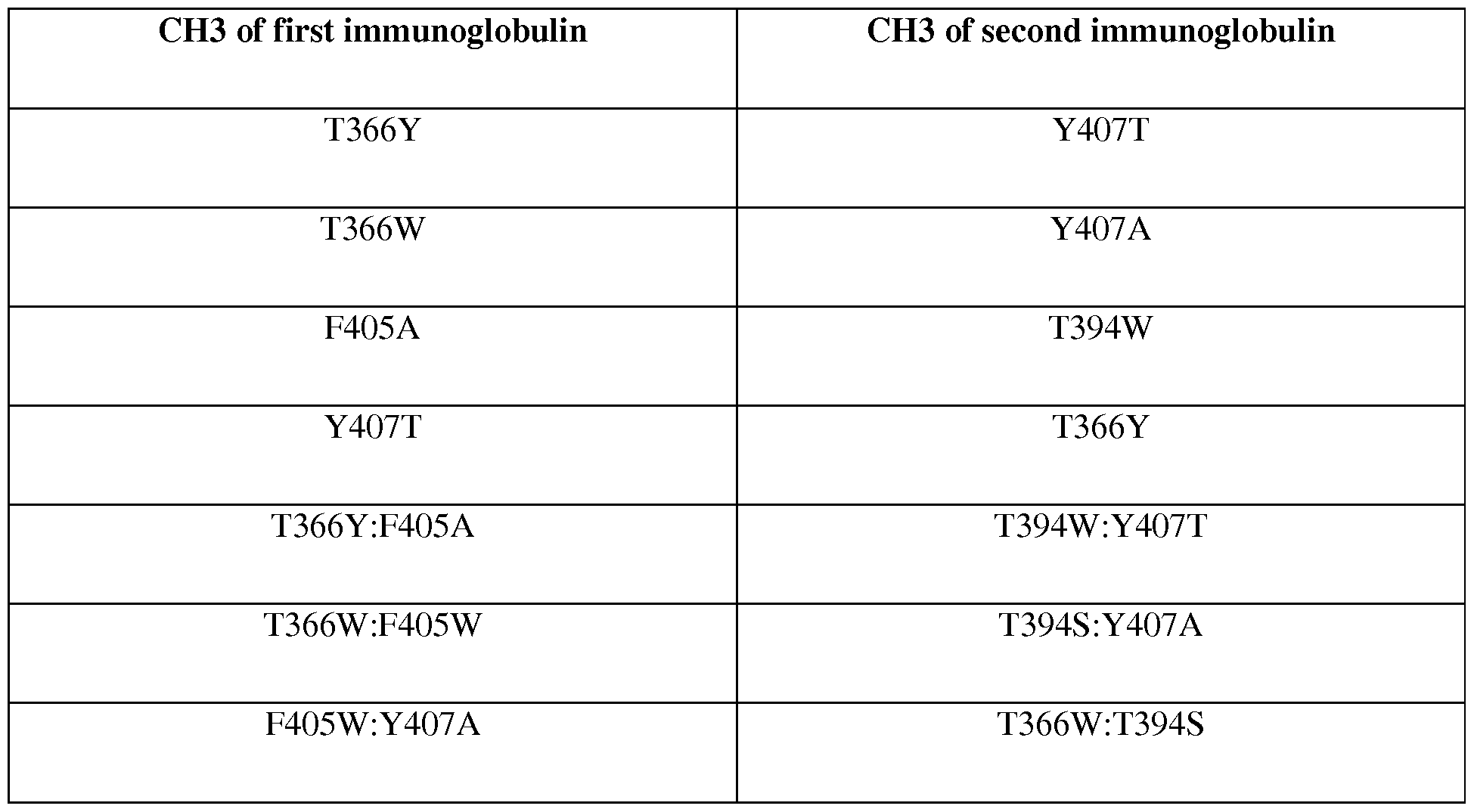


Claims
Priority Applications (11)
| Application Number | Priority Date | Filing Date | Title |
|---|---|---|---|
| CA2934028A CA2934028A1 (en) | 2013-12-17 | 2014-12-17 | Combination therapy comprising ox40 binding agonists and pd-1 axis binding antagonists |
| MX2016007965A MX2016007965A (en) | 2013-12-17 | 2014-12-17 | Combination therapy comprising ox40 binding agonists and pd-1 axis binding antagonists. |
| JP2016540597A JP2017501167A (en) | 2013-12-17 | 2014-12-17 | Combination therapy comprising OX40 binding agonist and PD-1 axis binding antagonist |
| AU2014364606A AU2014364606A1 (en) | 2013-12-17 | 2014-12-17 | Combination therapy comprising OX40 binding agonists and PD-1 axis binding antagonists |
| SG11201604979WA SG11201604979WA (en) | 2013-12-17 | 2014-12-17 | Combination therapy comprising ox40 binding agonists and pd-1 axis binding antagonists |
| EP14824702.6A EP3083687A2 (en) | 2013-12-17 | 2014-12-17 | Combination therapy comprising ox40 binding agonists and pd-1 axis binding antagonists |
| BR112016013963A BR112016013963A2 (en) | 2013-12-17 | 2014-12-17 | combination therapy comprising ox40 binding agonists and pd-1 axis binding antagonists |
| KR1020167018833A KR20160099092A (en) | 2013-12-17 | 2014-12-17 | Combination therapy comprising ox40 binding agonists and pd-1 axis binding antagonists |
| CN201480073061.9A CN106102774A (en) | 2013-12-17 | 2014-12-17 | Comprise OX40 and combine agonist and PD 1 axle combines the combination treatment of antagonist |
| EP18206645.6A EP3527587A1 (en) | 2013-12-17 | 2014-12-17 | Combination therapy comprising ox40 binding agonists and pd-l1 binding antagonists |
| IL246251A IL246251A0 (en) | 2013-12-17 | 2016-06-15 | Combination therapy comprising ox40 binding agonists and pd-1 axis binding antagonists |
Applications Claiming Priority (4)
| Application Number | Priority Date | Filing Date | Title |
|---|---|---|---|
| US201361917264P | 2013-12-17 | 2013-12-17 | |
| US61/917,264 | 2013-12-17 | ||
| US201462080991P | 2014-11-17 | 2014-11-17 | |
| US62/080,991 | 2014-11-17 |
Publications (2)
| Publication Number | Publication Date |
|---|---|
| WO2015095423A2 true WO2015095423A2 (en) | 2015-06-25 |
| WO2015095423A3 WO2015095423A3 (en) | 2015-08-13 |
Family
ID=52293264
Family Applications (1)
| Application Number | Title | Priority Date | Filing Date |
|---|---|---|---|
| PCT/US2014/070998 WO2015095423A2 (en) | 2013-12-17 | 2014-12-17 | Combination therapy comprising ox40 binding agonists and pd-1 axis binding antagonists |
Country Status (12)
| Country | Link |
|---|---|
| US (2) | US20150190506A1 (en) |
| EP (2) | EP3083687A2 (en) |
| JP (1) | JP2017501167A (en) |
| KR (1) | KR20160099092A (en) |
| CN (1) | CN106102774A (en) |
| AU (1) | AU2014364606A1 (en) |
| BR (1) | BR112016013963A2 (en) |
| CA (1) | CA2934028A1 (en) |
| IL (1) | IL246251A0 (en) |
| MX (1) | MX2016007965A (en) |
| SG (1) | SG11201604979WA (en) |
| WO (1) | WO2015095423A2 (en) |
Cited By (92)
| Publication number | Priority date | Publication date | Assignee | Title |
|---|---|---|---|---|
| WO2016073380A1 (en) * | 2014-11-03 | 2016-05-12 | Genentech, Inc. | Method and biomarkers for predicting efficacy and evaluation of an ox40 agonist treatment |
| WO2016054555A3 (en) * | 2014-10-03 | 2016-06-30 | Novartis Ag | Combination therapies |
| WO2016185016A1 (en) * | 2015-05-21 | 2016-11-24 | Alligator Bioscience Ab | Novel polypeptides |
| WO2016189124A1 (en) * | 2015-05-28 | 2016-12-01 | Medimmune Limited | Therapeutic combinations and methods for treating neoplasia |
| WO2016200835A1 (en) * | 2015-06-08 | 2016-12-15 | Genentech, Inc. | Methods of treating cancer using anti-ox40 antibodies and pd-1 axis binding antagonists |
| WO2017021911A1 (en) * | 2015-08-04 | 2017-02-09 | Glaxosmithkline Intellectual Property Development Limited | Combination treatments and uses and methods thereof |
| WO2017021791A1 (en) * | 2015-08-06 | 2017-02-09 | Glaxosmithkline Intellectual Property Development Limited | Combinations of an ox40 antibody and a tlr4 modulator and uses thereof |
| WO2017021910A1 (en) * | 2015-08-04 | 2017-02-09 | Glaxosmithkline Intellectual Property Development Limited | Combination treatments and uses and methods thereof |
| US9605070B2 (en) | 2014-01-31 | 2017-03-28 | Novartis Ag | Antibody molecules to TIM-3 and uses thereof |
| WO2017069291A1 (en) * | 2015-10-23 | 2017-04-27 | Canbas Co., Ltd. | Peptides and peptidomimetics in combination with t cell activating and/or checkpoint inhibiting agents for cancer treatment |
| US9644032B2 (en) | 2015-05-29 | 2017-05-09 | Bristol-Myers Squibb Company | Antibodies against OX40 and uses thereof |
| WO2017096281A1 (en) * | 2015-12-02 | 2017-06-08 | Agenus Inc. | Anti-ox40 antibodies and methods of use thereof |
| WO2017096182A1 (en) * | 2015-12-03 | 2017-06-08 | Agenus Inc. | Anti-ox40 antibodies and methods of use thereof |
| US9683048B2 (en) | 2014-01-24 | 2017-06-20 | Novartis Ag | Antibody molecules to PD-1 and uses thereof |
| EP3243832A1 (en) * | 2016-05-13 | 2017-11-15 | F. Hoffmann-La Roche AG | Antigen binding molecules comprising a tnf family ligand trimer and pd1 binding moiety |
| WO2017201352A1 (en) | 2016-05-18 | 2017-11-23 | Modernatx, Inc. | Mrna combination therapy for the treatment of cancer |
| US9908936B2 (en) | 2014-03-14 | 2018-03-06 | Novartis Ag | Antibody molecules to LAG-3 and uses thereof |
| WO2018045110A1 (en) * | 2016-08-30 | 2018-03-08 | Xencor, Inc. | Bispecific immunomodulatory antibodies that bind costimulatory and checkpoint receptors |
| WO2018056821A1 (en) * | 2016-09-23 | 2018-03-29 | Merus N.V. | Binding molecules that modulate a biological activity expressed by a cell |
| US9975957B2 (en) | 2014-03-31 | 2018-05-22 | Genentech, Inc. | Anti-OX40 antibodies and methods of use |
| US9988452B2 (en) | 2014-10-14 | 2018-06-05 | Novartis Ag | Antibody molecules to PD-L1 and uses thereof |
| JP2018527377A (en) * | 2015-09-16 | 2018-09-20 | ボード・オブ・リージエンツ,ザ・ユニバーシテイ・オブ・テキサス・システム | Combination of topoisomerase-I inhibitor and immunotherapy in the treatment of cancer |
| US10086042B2 (en) | 2015-10-01 | 2018-10-02 | Heat Biologics, Inc. | Compositions and methods for adjoining type I and type II extracellular domains as heterologous chimeric proteins |
| WO2018177220A1 (en) | 2017-03-25 | 2018-10-04 | 信达生物制药(苏州)有限公司 | Anti-ox40 antibody and use thereof |
| CN108884161A (en) * | 2015-12-01 | 2018-11-23 | 葛兰素史密斯克莱知识产权发展有限公司 | Combination therapy and application thereof and method |
| US10155037B2 (en) | 2014-12-09 | 2018-12-18 | Rinat Neuroscience Corp. | Anti-PD-1 antibodies and methods of use thereof |
| CN109196121A (en) * | 2016-02-29 | 2019-01-11 | 基因泰克公司 | Treatment and diagnostic method for cancer |
| CN109476718A (en) * | 2016-05-18 | 2019-03-15 | 莫得纳特斯公司 | The combination of MRNA and application thereof of encoding immune adjusting polypeptide |
| JP2019516710A (en) * | 2016-05-18 | 2019-06-20 | モデルナティーエックス, インコーポレイテッド | Polynucleotide encoding interleukin-12 (IL12) and use thereof |
| WO2019129137A1 (en) | 2017-12-27 | 2019-07-04 | 信达生物制药(苏州)有限公司 | Anti-lag-3 antibody and uses thereof |
| WO2019157124A1 (en) * | 2018-02-08 | 2019-08-15 | Bristol-Myers Squibb Company | Combination of a tetanus toxoid, anti-ox40 antibody and/or anti-pd-1 antibody to treat tumors |
| US10441654B2 (en) | 2014-01-24 | 2019-10-15 | Children's Hospital Of Eastern Ontario Research Institute Inc. | SMC combination therapy for the treatment of cancer |
| WO2019224275A1 (en) | 2018-05-23 | 2019-11-28 | Adc Therapeutics Sa | Molecular adjuvant |
| EP3383430A4 (en) * | 2015-12-02 | 2019-12-18 | Agenus Inc. | Antibodies and methods of use thereof |
| US10513548B2 (en) | 2017-02-27 | 2019-12-24 | Shattuck Labs, Inc. | CSF1R-based chimeric proteins |
| US10526413B2 (en) | 2015-10-02 | 2020-01-07 | Hoffmann-La Roche Inc. | Bispecific antibodies specific for OX40 |
| US10544223B2 (en) | 2017-04-20 | 2020-01-28 | Adc Therapeutics Sa | Combination therapy with an anti-axl antibody-drug conjugate |
| US10570204B2 (en) | 2013-09-26 | 2020-02-25 | The Medical College Of Wisconsin, Inc. | Methods for treating hematologic cancers |
| US10596257B2 (en) | 2016-01-08 | 2020-03-24 | Hoffmann-La Roche Inc. | Methods of treating CEA-positive cancers using PD-1 axis binding antagonists and anti-CEA/anti-CD3 bispecific antibodies |
| US10626181B2 (en) | 2015-05-07 | 2020-04-21 | Agenus Inc. | Nucleic acids encoding anti-OX40 antibodies |
| US10738133B2 (en) | 2013-01-14 | 2020-08-11 | Xencor, Inc. | Heterodimeric proteins |
| US10780121B2 (en) | 2018-08-29 | 2020-09-22 | Shattuck Labs, Inc. | FLT3L-based chimeric proteins |
| US10787518B2 (en) | 2016-06-14 | 2020-09-29 | Xencor, Inc. | Bispecific checkpoint inhibitor antibodies |
| US10793632B2 (en) | 2016-08-30 | 2020-10-06 | Xencor, Inc. | Bispecific immunomodulatory antibodies that bind costimulatory and checkpoint receptors |
| US10800846B2 (en) | 2015-02-26 | 2020-10-13 | Merck Patent Gmbh | PD-1/PD-L1 inhibitors for the treatment of cancer |
| US10845364B2 (en) | 2014-11-03 | 2020-11-24 | Genentech, Inc. | Assays for detecting T cell immune subsets and methods of use thereof |
| US10858417B2 (en) | 2013-03-15 | 2020-12-08 | Xencor, Inc. | Heterodimeric proteins |
| US10869924B2 (en) | 2015-06-16 | 2020-12-22 | Merck Patent Gmbh | PD-L1 antagonist combination treatments |
| US10889653B2 (en) | 2014-11-26 | 2021-01-12 | Xencor, Inc. | Heterodimeric antibodies that bind CD3 and tumor antigens |
| US10899817B2 (en) | 2017-02-27 | 2021-01-26 | Shattuck Labs, Inc. | TIGIT- and light-based chimeric proteins |
| WO2021029368A1 (en) | 2019-08-09 | 2021-02-18 | 国立研究開発法人理化学研究所 | Combined use of artificial adjuvant vector cells and an immunostimulant |
| US10968276B2 (en) | 2013-03-12 | 2021-04-06 | Xencor, Inc. | Optimized anti-CD3 variable regions |
| GB202102396D0 (en) | 2021-02-19 | 2021-04-07 | Adc Therapeutics Sa | Molecular adjuvant |
| US10982006B2 (en) | 2018-04-04 | 2021-04-20 | Xencor, Inc. | Heterodimeric antibodies that bind fibroblast activation protein |
| US10981992B2 (en) | 2017-11-08 | 2021-04-20 | Xencor, Inc. | Bispecific immunomodulatory antibodies that bind costimulatory and checkpoint receptors |
| WO2021104834A1 (en) | 2019-11-27 | 2021-06-03 | Adc Therapeutics Sa | Combination therapy |
| US11053316B2 (en) | 2013-01-14 | 2021-07-06 | Xencor, Inc. | Optimized antibody variable regions |
| US11059893B2 (en) | 2015-04-15 | 2021-07-13 | Bergenbio Asa | Humanized anti-AXL antibodies |
| US11111315B2 (en) | 2014-11-26 | 2021-09-07 | Xencor, Inc. | Heterodimeric antibodies that bind CD3 and tumor antigens |
| US11130810B2 (en) | 2015-10-02 | 2021-09-28 | Hoffmann-La Roche Inc. | Bispecific antibodies specific for PD1 and TIM3 |
| US11192933B2 (en) | 2017-02-27 | 2021-12-07 | Shattuck Labs, Inc. | VSIG8-based chimeric proteins |
| EP3733703A4 (en) * | 2017-12-29 | 2021-12-29 | Nanjing Umab-Biopharma Co., Ltd. | Isolated antibody or antigen binding fragment thereof and use of same in tumor treatment |
| US11225521B2 (en) | 2016-06-28 | 2022-01-18 | Xencor, Inc. | Heterodimeric antibodies that bind somatostatin receptor 2 |
| US11274154B2 (en) | 2016-10-06 | 2022-03-15 | Pfizer Inc. | Dosing regimen of avelumab for the treatment of cancer |
| US11285207B2 (en) | 2017-04-05 | 2022-03-29 | Hoffmann-La Roche Inc. | Bispecific antibodies specifically binding to PD1 and LAG3 |
| US11299554B2 (en) | 2013-03-15 | 2022-04-12 | Xencor, Inc. | Heterodimeric proteins |
| US11300570B2 (en) | 2016-10-06 | 2022-04-12 | Genentech, Inc. | Therapeutic and diagnostic methods for cancer |
| US11312770B2 (en) | 2017-11-08 | 2022-04-26 | Xencor, Inc. | Bispecific and monospecific antibodies using novel anti-PD-1 sequences |
| US11318211B2 (en) | 2017-06-14 | 2022-05-03 | Adc Therapeutics Sa | Dosage regimes for the administration of an anti-CD19 ADC |
| US11319355B2 (en) | 2017-12-19 | 2022-05-03 | Xencor, Inc. | Engineered IL-2 Fc fusion proteins |
| WO2022100659A1 (en) * | 2020-11-12 | 2022-05-19 | Curon Biopharmaceutical (Shanghai) Co., Limited | Combination therapy and methods utilizing the same |
| US11344620B2 (en) | 2014-09-13 | 2022-05-31 | Novartis Ag | Combination therapies |
| US11352442B2 (en) | 2014-11-26 | 2022-06-07 | Xencor, Inc. | Heterodimeric antibodies that bind CD3 and CD38 |
| US11359028B2 (en) | 2016-11-09 | 2022-06-14 | Agenus Inc. | Anti-OX40 antibodies and anti-GITR antibodies |
| US11358999B2 (en) | 2018-10-03 | 2022-06-14 | Xencor, Inc. | IL-12 heterodimeric Fc-fusion proteins |
| US11413331B2 (en) | 2017-04-03 | 2022-08-16 | Hoffmann-La Roche Inc. | Immunoconjugates |
| US11472890B2 (en) | 2019-03-01 | 2022-10-18 | Xencor, Inc. | Heterodimeric antibodies that bind ENPP3 and CD3 |
| US11505595B2 (en) | 2018-04-18 | 2022-11-22 | Xencor, Inc. | TIM-3 targeted heterodimeric fusion proteins containing IL-15/IL-15RA Fc-fusion proteins and TIM-3 antigen binding domains |
| US11524991B2 (en) | 2018-04-18 | 2022-12-13 | Xencor, Inc. | PD-1 targeted heterodimeric fusion proteins containing IL-15/IL-15Ra Fc-fusion proteins and PD-1 antigen binding domains and uses thereof |
| US11564986B2 (en) | 2015-07-16 | 2023-01-31 | Onkosxcel Therapeutics, Llc | Approach for treatment of cancer via immunomodulation by using talabostat |
| US11591401B2 (en) | 2020-08-19 | 2023-02-28 | Xencor, Inc. | Anti-CD28 compositions |
| US11623957B2 (en) | 2015-12-07 | 2023-04-11 | Xencor, Inc. | Heterodimeric antibodies that bind CD3 and PSMA |
| US11674962B2 (en) | 2017-07-21 | 2023-06-13 | Genentech, Inc. | Therapeutic and diagnostic methods for cancer |
| TWI805542B (en) * | 2015-10-23 | 2023-06-21 | 日商坎巴斯有限公司 | Peptides and peptidomimetics in combination with t cell activating and/or checkpoint inhibiting agents for cancer treatment |
| US11725247B2 (en) | 2016-02-29 | 2023-08-15 | Foundation Medicine, Inc. | Methods of treating cancer |
| US11732044B2 (en) | 2017-12-27 | 2023-08-22 | Innovent Biologics (Suzhou) Co., Ltd. | Anti-LAG-3 antibody and use thereof |
| US11739144B2 (en) | 2021-03-09 | 2023-08-29 | Xencor, Inc. | Heterodimeric antibodies that bind CD3 and CLDN6 |
| US11840579B2 (en) | 2014-03-28 | 2023-12-12 | Xencor, Inc. | Bispecific antibodies that bind to CD38 and CD3 |
| US11859012B2 (en) | 2021-03-10 | 2024-01-02 | Xencor, Inc. | Heterodimeric antibodies that bind CD3 and GPC3 |
| US11919956B2 (en) | 2020-05-14 | 2024-03-05 | Xencor, Inc. | Heterodimeric antibodies that bind prostate specific membrane antigen (PSMA) and CD3 |
| EP4061850A4 (en) * | 2019-11-21 | 2024-03-20 | Beigene Ltd | Methods of cancer treatment using anti-ox40 antibodies in combination with anti-pd1 or anti-pdl1 antibodies |
| USRE49908E1 (en) | 2021-06-30 | 2024-04-09 | Medimmune, Llc | Bispecific binding proteins and uses thereof |
Families Citing this family (72)
| Publication number | Priority date | Publication date | Assignee | Title |
|---|---|---|---|---|
| DK2376535T3 (en) | 2008-12-09 | 2017-06-12 | Hoffmann La Roche | ANTI-PD-L1 ANTIBODIES AND THEIR USE TO PROMOTE T CELL FUNCTION |
| PL2855528T3 (en) | 2012-05-31 | 2019-10-31 | Hoffmann La Roche | Methods of treating cancer using pd-l1 axis binding antagonists and vegf antagonists |
| JP2016515544A (en) | 2013-03-18 | 2016-05-30 | バイオセルオーエックス プロダクツ ビー.ブイ. | Humanized anti-CD134 (OX40) antibody and use thereof |
| NZ721364A (en) | 2014-02-10 | 2023-04-28 | Merck Patent Gmbh | Targeted tgfβ inhibition |
| US11186640B2 (en) * | 2014-07-31 | 2021-11-30 | The University Of Western Australia | Method for the identification of immunotherapy-drug combinations using a network approach |
| JP6784687B2 (en) | 2015-02-24 | 2020-11-11 | ザ リージェンツ オブ ザ ユニバーシティ オブ カリフォルニア | Binding-induced transcription switch and how to use it |
| JP7014706B2 (en) | 2015-07-13 | 2022-02-01 | サイトメックス セラピューティクス インコーポレイテッド | Anti-PD-1 antibody, activating anti-PD-1 antibody, and how to use it |
| CN116726183A (en) | 2015-08-03 | 2023-09-12 | Enb治疗公司 | Compositions and methods for treating cancers associated with ETBR activation |
| CN114605548A (en) | 2015-09-01 | 2022-06-10 | 艾吉纳斯公司 | anti-PD-1 antibodies and methods of use thereof |
| WO2017046746A1 (en) | 2015-09-15 | 2017-03-23 | Acerta Pharma B.V. | Therapeutic combinations of a btk inhibitor and a gitr binding molecule, a 4-1bb agonist, or an ox40 agonist |
| RU2731202C2 (en) * | 2015-10-08 | 2020-08-31 | Макродженикс, Инк. | Combined therapy for cancer treatment |
| CA3009075A1 (en) | 2016-01-11 | 2017-07-20 | Inhibrx, Inc. | Multivalent and multispecific ox40-binding fusion proteins |
| BR112018013677A2 (en) | 2016-01-11 | 2019-01-22 | Inhibrx Inc | multivalent and multispecific 41bb binding fusion proteins |
| CN108473587A (en) | 2016-01-25 | 2018-08-31 | 辉瑞公司 | The combination of OX40 agonists and 4-1BB agonist monoclonal antibodies for treating cancer |
| UA126115C2 (en) * | 2016-03-08 | 2022-08-17 | Янссен Байотек, Інк. | Gitr antibodies, methods, and uses |
| DE102016105069A1 (en) | 2016-03-18 | 2017-09-21 | Eberhard Karls Universität Tübingen Medizinische Fakultät | Antiviral immunotherapy by membrane receptor ligation |
| WO2017193081A1 (en) * | 2016-05-06 | 2017-11-09 | Exicure, Inc. | Spherical nucleic acid tlr9 agonists |
| KR20230149857A (en) | 2016-07-07 | 2023-10-27 | 더 보드 어브 트러스티스 어브 더 리랜드 스탠포드 주니어 유니버시티 | Antibody adjuvant conjugates |
| EP3494139B1 (en) | 2016-08-05 | 2022-01-12 | F. Hoffmann-La Roche AG | Multivalent and multiepitopic anitibodies having agonistic activity and methods of use |
| US20190175755A1 (en) * | 2016-08-23 | 2019-06-13 | The Johns Hopkins University | Immunoswitch nanoparticles for reprogrammed t cell responses |
| AU2017335839A1 (en) * | 2016-09-29 | 2019-04-18 | Genentech, Inc. | Combination therapy with a MEK inhibitor, a PD-1 axis inhibitor, and a taxane |
| EA037973B1 (en) * | 2016-10-12 | 2021-06-18 | Янссен Байотек, Инк. | Gitr antibodies, methods and uses |
| TWI788307B (en) | 2016-10-31 | 2023-01-01 | 美商艾歐凡斯生物治療公司 | Engineered artificial antigen presenting cells for tumor infiltrating lymphocyte expansion |
| US10342785B2 (en) | 2016-11-04 | 2019-07-09 | Askat Inc. | Use of EP4 receptor antagonists for the treatment of NASH-associated liver cancer |
| EP3565888A1 (en) | 2017-01-06 | 2019-11-13 | Iovance Biotherapeutics, Inc. | Expansion of tumor infiltrating lymphocytes (tils) with tumor necrosis factor receptor superfamily (tnfrsf) agonists and therapeutic combinations of tils and tnfrsf agonists |
| US11884738B2 (en) * | 2017-03-31 | 2024-01-30 | The University Of North Carolina At Chapel Hill | Methods and compositions for activation of T cells using nanoparticles conjugated with multiple ligands for binding receptors on T cells |
| CA3058478A1 (en) * | 2017-04-14 | 2018-10-18 | Genentech, Inc. | Diagnostic and therapeutic methods for cancer |
| MX2019012465A (en) | 2017-04-20 | 2020-07-27 | Adc Therapeutics Sa | Combination therapy with an anti-cd25 antibody-drug conjugate. |
| EP3635097A1 (en) | 2017-06-05 | 2020-04-15 | Iovance Biotherapeutics, Inc. | Methods of using tumor infiltrating lymphocytes in double-refractory melanoma |
| BR112020009663A2 (en) | 2017-11-17 | 2020-11-10 | Iovance Biotherapeutics, Inc. | method for the expansion of tumor infiltrating lymphocytes (tils) in a therapeutic population of tils, method for the treatment of an individual with cancer, composition |
| US11713446B2 (en) | 2018-01-08 | 2023-08-01 | Iovance Biotherapeutics, Inc. | Processes for generating TIL products enriched for tumor antigen-specific T-cells |
| CA3087771A1 (en) | 2018-01-08 | 2019-07-11 | Iovance Biotherapeutics, Inc. | Processes for generating til products enriched for tumor antigen-specific t-cells |
| WO2019136459A1 (en) | 2018-01-08 | 2019-07-11 | Iovance Biotherapeutics, Inc. | Processes for generating til products enriched for tumor antigen-specific t-cells |
| KR20200119246A (en) | 2018-01-12 | 2020-10-19 | 이엔비 테라퓨틱스, 인크. | Deuterated compounds, compositions, and methods for treating cancer associated with ETBR activation |
| CN110092832B (en) * | 2018-01-29 | 2020-03-31 | 康源博创生物科技(北京)有限公司 | anti-OX 40 antibodies and uses thereof |
| EP3752600A1 (en) | 2018-02-13 | 2020-12-23 | Iovance Biotherapeutics, Inc. | Expansion of tumor infiltrating lymphocytes (tils) with adenosine a2a receptor antagonists and therapeutic combinations of tils and adenosine a2a receptor antagonists |
| CA3098303A1 (en) | 2018-04-27 | 2019-10-31 | Iovance Biotherapeutics, Inc. | Closed process for expansion and gene editing of tumor infiltrating lymphocytes and uses of same in immunotherapy |
| EP3837285A1 (en) | 2018-08-13 | 2021-06-23 | Inhibrx, Inc. | Ox40-binding polypeptides and uses thereof |
| TW202031273A (en) | 2018-08-31 | 2020-09-01 | 美商艾歐凡斯生物治療公司 | Treatment of nsclc patients refractory for anti-pd-1 antibody |
| EP3852524B1 (en) | 2018-09-20 | 2023-06-28 | Iovance Biotherapeutics, Inc. | Expansion of tils from cryopreserved tumor samples |
| CN112969469A (en) | 2018-11-05 | 2021-06-15 | 艾欧凡斯生物治疗公司 | Treatment of anti-PD-1 antibody refractory NSCLC patients |
| JP2022506508A (en) | 2018-11-05 | 2022-01-17 | アイオバンス バイオセラピューティクス,インコーポレイテッド | Expanded culture of TIL using AKT pathway inhibitors |
| TW202039829A (en) | 2018-11-05 | 2020-11-01 | 美商艾歐凡斯生物治療公司 | Selection of improved tumor reactive t-cells |
| CR20210295A (en) | 2018-11-05 | 2021-07-09 | Iovance Biotherapeutics Inc | Processes for production of tumor infiltrating lymphocytes and uses of the same in immunotherapy |
| WO2020131547A1 (en) | 2018-12-19 | 2020-06-25 | Iovance Biotherapeutics, Inc. | Methods of expanding tumor infiltrating lymphocytes using engineered cytokine receptor pairs and uses thereof |
| WO2020128893A1 (en) | 2018-12-21 | 2020-06-25 | Pfizer Inc. | Combination treatments of cancer comprising a tlr agonist |
| AU2020241686A1 (en) | 2019-03-15 | 2021-11-04 | Bolt Biotherapeutics, Inc. | Immunoconjugates targeting HER2 |
| CA3134144A1 (en) | 2019-03-29 | 2020-10-08 | Myst Therapeutics, Llc | Ex vivo methods for producing a t cell therapeutic and related compositions and methods |
| WO2020232029A1 (en) | 2019-05-13 | 2020-11-19 | Iovance Biotherapeutics, Inc. | Methods and compositions for selecting tumor infiltrating lymphocytes and uses of the same in immunotherapy |
| WO2020240360A1 (en) | 2019-05-24 | 2020-12-03 | Pfizer Inc. | Combination therapies using cdk inhibitors |
| US20220241412A1 (en) | 2019-05-24 | 2022-08-04 | Pfizer Inc. | Combination therapies using cdk inhibitors |
| WO2021081378A1 (en) | 2019-10-25 | 2021-04-29 | Iovance Biotherapeutics, Inc. | Gene editing of tumor infiltrating lymphocytes and uses of same in immunotherapy |
| JP2023504042A (en) | 2019-11-27 | 2023-02-01 | ミスト セラピューティクス リミテッド ライアビリティ カンパニー | Methods of generating tumor-reactive T-cell compositions using modulators |
| US20230220341A1 (en) | 2019-12-11 | 2023-07-13 | lovance Biotherapeutics, Inc. | Processes for the production of tumor infiltrating lymphocytes (tils) and methods of using the same |
| JP2023516636A (en) | 2020-02-27 | 2023-04-20 | ミスト セラピューティクス リミテッド ライアビリティ カンパニー | Methods for ex vivo enrichment and expansion of tumor-reactive T cells and related compositions thereof |
| JP2023523855A (en) | 2020-05-04 | 2023-06-07 | アイオバンス バイオセラピューティクス,インコーポレイテッド | Method for producing tumor-infiltrating lymphocytes and their use in immunotherapy |
| JP2023524108A (en) | 2020-05-04 | 2023-06-08 | アイオバンス バイオセラピューティクス,インコーポレイテッド | Selection of improved tumor-reactive T cells |
| WO2022076606A1 (en) | 2020-10-06 | 2022-04-14 | Iovance Biotherapeutics, Inc. | Treatment of nsclc patients with tumor infiltrating lymphocyte therapies |
| EP4225330A1 (en) | 2020-10-06 | 2023-08-16 | Iovance Biotherapeutics, Inc. | Treatment of nsclc patients with tumor infiltrating lymphocyte therapies |
| EP4232163A1 (en) * | 2020-10-22 | 2023-08-30 | The Regents of University of California | Monoamine oxidase blockade therapy for treating cancer through regulating antitumor t cell immunity |
| US20220168293A1 (en) | 2020-12-02 | 2022-06-02 | Pfizer Inc. | Time to resolution of axitinib-related adverse events |
| WO2022125941A1 (en) | 2020-12-11 | 2022-06-16 | Iovance Biotherapeutics, Inc. | Treatment of cancer patients with tumor infiltrating lymphocyte therapies in combination with braf inhibitors and/or mek inhibitors |
| WO2022130206A1 (en) | 2020-12-16 | 2022-06-23 | Pfizer Inc. | TGFβr1 INHIBITOR COMBINATION THERAPIES |
| JP2024500403A (en) | 2020-12-17 | 2024-01-09 | アイオバンス バイオセラピューティクス,インコーポレイテッド | Treatment of cancer with tumor-infiltrating lymphocytes |
| CA3202483A1 (en) | 2020-12-17 | 2022-06-23 | Maria Fardis | Treatment with tumor infiltrating lymphocyte therapies in combination with ctla-4 and pd-1 inhibitors |
| WO2022133169A1 (en) | 2020-12-18 | 2022-06-23 | Century Therapeutics, Inc. | Chimeric antigen receptor systems with adaptable receptor specificity |
| KR20240037185A (en) | 2021-04-19 | 2024-03-21 | 이오반스 바이오테라퓨틱스, 인크. | Chimeric costimulatory receptors, chemokine receptors, and their uses in cellular immunotherapy |
| CA3226942A1 (en) | 2021-07-28 | 2023-02-02 | Iovance Biotherapeutics, Inc. | Treatment of cancer patients with tumor infiltrating lymphocyte therapies in combination with kras inhibitors |
| US20230187042A1 (en) | 2021-10-27 | 2023-06-15 | Iovance Biotherapeutics, Inc. | Systems and methods for coordinating manufacturing of cells for patient-specific immunotherapy |
| WO2023086803A1 (en) | 2021-11-10 | 2023-05-19 | Iovance Biotherapeutics, Inc. | Methods of expansion treatment utilizing cd8 tumor infiltrating lymphocytes |
| WO2023147486A1 (en) | 2022-01-28 | 2023-08-03 | Iovance Biotherapeutics, Inc. | Tumor infiltrating lymphocytes engineered to express payloads |
| WO2024030758A1 (en) | 2022-08-01 | 2024-02-08 | Iovance Biotherapeutics, Inc. | Chimeric costimulatory receptors, chemokine receptors, and the use of same in cellular immunotherapies |
Citations (174)
| Publication number | Priority date | Publication date | Assignee | Title |
|---|---|---|---|---|
| CH119431A (en) | 1926-04-21 | 1927-03-16 | Beck Sigmund | Painter's brush. |
| US4016043A (en) | 1975-09-04 | 1977-04-05 | Akzona Incorporated | Enzymatic immunological method for the determination of antigens and antibodies |
| US4018653A (en) | 1971-10-29 | 1977-04-19 | U.S. Packaging Corporation | Instrument for the detection of Neisseria gonorrhoeae without culture |
| EP0003089A1 (en) | 1978-01-06 | 1979-07-25 | Bernard David | Drier for silkscreen printed sheets |
| US4275149A (en) | 1978-11-24 | 1981-06-23 | Syva Company | Macromolecular environment control in specific receptor assays |
| US4318980A (en) | 1978-04-10 | 1982-03-09 | Miles Laboratories, Inc. | Heterogenous specific binding assay employing a cycling reactant as label |
| USRE30985E (en) | 1978-01-01 | 1982-06-29 | Serum-free cell culture media | |
| EP0073657A1 (en) | 1981-08-31 | 1983-03-09 | Genentech, Inc. | Preparation of hepatitis B surface antigen in yeast |
| US4419446A (en) | 1980-12-31 | 1983-12-06 | The United States Of America As Represented By The Department Of Health And Human Services | Recombinant DNA process utilizing a papilloma virus DNA as a vector |
| US4424279A (en) | 1982-08-12 | 1984-01-03 | Quidel | Rapid plunger immunoassay method and apparatus |
| US4560655A (en) | 1982-12-16 | 1985-12-24 | Immunex Corporation | Serum-free cell culture medium and process for making same |
| EP0183070A2 (en) | 1984-10-30 | 1986-06-04 | Phillips Petroleum Company | Transformation of yeasts of the genus pichia |
| US4601978A (en) | 1982-11-24 | 1986-07-22 | The Regents Of The University Of California | Mammalian metallothionein promoter system |
| WO1987000195A1 (en) | 1985-06-28 | 1987-01-15 | Celltech Limited | Animal cell culture |
| US4657866A (en) | 1982-12-21 | 1987-04-14 | Sudhir Kumar | Serum-free, synthetic, completely chemically defined tissue culture media |
| US4676980A (en) | 1985-09-23 | 1987-06-30 | The United States Of America As Represented By The Secretary Of The Department Of Health And Human Services | Target specific cross-linked heteroantibodies |
| US4683195A (en) | 1986-01-30 | 1987-07-28 | Cetus Corporation | Process for amplifying, detecting, and/or-cloning nucleic acid sequences |
| EP0244234A2 (en) | 1986-04-30 | 1987-11-04 | Alko Group Ltd. | Transformation of trichoderma |
| US4737456A (en) | 1985-05-09 | 1988-04-12 | Syntex (U.S.A.) Inc. | Reducing interference in ligand-receptor binding assays |
| US4767704A (en) | 1983-10-07 | 1988-08-30 | Columbia University In The City Of New York | Protein-free culture medium |
| US4816567A (en) | 1983-04-08 | 1989-03-28 | Genentech, Inc. | Recombinant immunoglobin preparations |
| DD266710A3 (en) | 1983-06-06 | 1989-04-12 | Ve Forschungszentrum Biotechnologie | Process for the biotechnical production of alkaline phosphatase |
| WO1990003430A1 (en) | 1988-09-23 | 1990-04-05 | Cetus Corporation | Cell culture medium for enhanced cell growth, culture longevity and product expression |
| US4927762A (en) | 1986-04-01 | 1990-05-22 | Cell Enterprises, Inc. | Cell culture medium with antioxidant |
| US4943533A (en) | 1984-03-01 | 1990-07-24 | The Regents Of The University Of California | Hybrid cell lines that produce monoclonal antibodies to epidermal growth factor receptor |
| US4965199A (en) | 1984-04-20 | 1990-10-23 | Genentech, Inc. | Preparation of functional human factor VIII in mammalian cells using methotrexate based selection |
| WO1990013646A1 (en) | 1989-04-28 | 1990-11-15 | Transgene S.A. | Application of novel dna fragments as a coding sequence for a signal peptide for the secretion of mature proteins by recombinant yeast, expression cassettes, transformed yeasts and corresponding process for the preparation of proteins |
| EP0402226A1 (en) | 1989-06-06 | 1990-12-12 | Institut National De La Recherche Agronomique | Transformation vectors for yeast yarrowia |
| EP0404097A2 (en) | 1989-06-22 | 1990-12-27 | BEHRINGWERKE Aktiengesellschaft | Bispecific and oligospecific, mono- and oligovalent receptors, production and applications thereof |
| WO1991000360A1 (en) | 1989-06-29 | 1991-01-10 | Medarex, Inc. | Bispecific reagents for aids therapy |
| WO1991010741A1 (en) | 1990-01-12 | 1991-07-25 | Cell Genesys, Inc. | Generation of xenogeneic antibodies |
| US5122469A (en) | 1990-10-03 | 1992-06-16 | Genentech, Inc. | Method for culturing Chinese hamster ovary cells to improve production of recombinant proteins |
| WO1992020373A1 (en) | 1991-05-14 | 1992-11-26 | Repligen Corporation | Heteroconjugate antibodies for treatment of hiv infection |
| WO1993001161A1 (en) | 1991-07-11 | 1993-01-21 | Pfizer Limited | Process for preparing sertraline intermediates |
| WO1993008829A1 (en) | 1991-11-04 | 1993-05-13 | The Regents Of The University Of California | Compositions that mediate killing of hiv-infected cells |
| US5212290A (en) | 1989-09-08 | 1993-05-18 | The Johns Hopkins University | Antibodies specific for type II mutant EGTR |
| WO1993016185A2 (en) | 1992-02-06 | 1993-08-19 | Creative Biomolecules, Inc. | Biosynthetic binding protein for cancer marker |
| WO1994004690A1 (en) | 1992-08-17 | 1994-03-03 | Genentech, Inc. | Bispecific immunoadhesins |
| WO1994011026A2 (en) | 1992-11-13 | 1994-05-26 | Idec Pharmaceuticals Corporation | Therapeutic application of chimeric and radiolabeled antibodies to human b lymphocyte restricted differentiation antigen for treatment of b cell lymphoma |
| WO1994029351A2 (en) | 1993-06-16 | 1994-12-22 | Celltech Limited | Antibodies |
| EP0659439A2 (en) | 1993-12-24 | 1995-06-28 | MERCK PATENT GmbH | Immunoconjugates |
| US5457105A (en) | 1992-01-20 | 1995-10-10 | Zeneca Limited | Quinazoline derivatives useful for treatment of neoplastic disease |
| US5475001A (en) | 1993-07-19 | 1995-12-12 | Zeneca Limited | Quinazoline derivatives |
| WO1996003397A1 (en) | 1994-07-21 | 1996-02-08 | Akzo Nobel N.V. | Cyclic ketone peroxide formulations |
| US5500362A (en) | 1987-01-08 | 1996-03-19 | Xoma Corporation | Chimeric antibody with specificity to human B cell surface antigen |
| US5545807A (en) | 1988-10-12 | 1996-08-13 | The Babraham Institute | Production of antibodies from transgenic animals |
| US5545806A (en) | 1990-08-29 | 1996-08-13 | Genpharm International, Inc. | Ransgenic non-human animals for producing heterologous antibodies |
| WO1996027011A1 (en) | 1995-03-01 | 1996-09-06 | Genentech, Inc. | A method for making heteromultimeric polypeptides |
| WO1996030347A1 (en) | 1995-03-30 | 1996-10-03 | Pfizer Inc. | Quinazoline derivatives |
| US5569825A (en) | 1990-08-29 | 1996-10-29 | Genpharm International | Transgenic non-human animals capable of producing heterologous antibodies of various isotypes |
| WO1996033980A1 (en) | 1995-04-27 | 1996-10-31 | Zeneca Limited | Quinazoline derivatives |
| WO1996033978A1 (en) | 1995-04-27 | 1996-10-31 | Zeneca Limited | Quinazoline derivative |
| WO1996034096A1 (en) | 1995-04-28 | 1996-10-31 | Abgenix, Inc. | Human antibodies derived from immunized xenomice |
| WO1996033735A1 (en) | 1995-04-27 | 1996-10-31 | Abgenix, Inc. | Human antibodies derived from immunized xenomice |
| US5571894A (en) | 1991-02-05 | 1996-11-05 | Ciba-Geigy Corporation | Recombinant antibodies specific for a growth factor receptor |
| WO1996040210A1 (en) | 1995-06-07 | 1996-12-19 | Imclone Systems Incorporated | Antibody and antibody fragments for inhibiting the growth of tumors |
| US5587458A (en) | 1991-10-07 | 1996-12-24 | Aronex Pharmaceuticals, Inc. | Anti-erbB-2 antibodies, combinations thereof, and therapeutic and diagnostic uses thereof |
| US5625126A (en) | 1990-08-29 | 1997-04-29 | Genpharm International, Inc. | Transgenic non-human animals for producing heterologous antibodies |
| US5624821A (en) | 1987-03-18 | 1997-04-29 | Scotgen Biopharmaceuticals Incorporated | Antibodies with altered effector functions |
| US5633425A (en) | 1990-08-29 | 1997-05-27 | Genpharm International, Inc. | Transgenic non-human animals capable of producing heterologous antibodies |
| US5641870A (en) | 1995-04-20 | 1997-06-24 | Genentech, Inc. | Low pH hydrophobic interaction chromatography for antibody purification |
| US5648237A (en) | 1991-09-19 | 1997-07-15 | Genentech, Inc. | Expression of functional antibody fragments |
| US5654307A (en) | 1994-01-25 | 1997-08-05 | Warner-Lambert Company | Bicyclic compounds capable of inhibiting tyrosine kinases of the epidermal growth factor receptor family |
| WO1997030087A1 (en) | 1996-02-16 | 1997-08-21 | Glaxo Group Limited | Preparation of glycosylated antibodies |
| US5661016A (en) | 1990-08-29 | 1997-08-26 | Genpharm International Inc. | Transgenic non-human animals capable of producing heterologous antibodies of various isotypes |
| WO1997038983A1 (en) | 1996-04-12 | 1997-10-23 | Warner-Lambert Company | Irreversible inhibitors of tyrosine kinases |
| WO1998014451A1 (en) | 1996-10-02 | 1998-04-09 | Novartis Ag | Fused pyrazole derivative and process for its preparation |
| US5747498A (en) | 1996-05-28 | 1998-05-05 | Pfizer Inc. | Alkynyl and azido-substituted 4-anilinoquinazolines |
| US5750373A (en) | 1990-12-03 | 1998-05-12 | Genentech, Inc. | Enrichment method for variant proteins having altered binding properties, M13 phagemids, and growth hormone variants |
| US5760041A (en) | 1996-02-05 | 1998-06-02 | American Cyanamid Company | 4-aminoquinazoline EGFR Inhibitors |
| WO1998024893A2 (en) | 1996-12-03 | 1998-06-11 | Abgenix, Inc. | TRANSGENIC MAMMALS HAVING HUMAN IG LOCI INCLUDING PLURAL VH AND Vλ REGIONS AND ANTIBODIES PRODUCED THEREFROM |
| US5770429A (en) | 1990-08-29 | 1998-06-23 | Genpharm International, Inc. | Transgenic non-human animals capable of producing heterologous antibodies |
| US5789199A (en) | 1994-11-03 | 1998-08-04 | Genentech, Inc. | Process for bacterial production of polypeptides |
| US5804396A (en) | 1994-10-12 | 1998-09-08 | Sugen, Inc. | Assay for agents active in proliferative disorders |
| WO1998043960A1 (en) | 1997-04-03 | 1998-10-08 | American Cyanamid Company | Substituted 3-cyano quinolines |
| US5821337A (en) | 1991-06-14 | 1998-10-13 | Genentech, Inc. | Immunoglobulin variants |
| WO1998050038A1 (en) | 1997-05-06 | 1998-11-12 | American Cyanamid Company | Use of quinazoline compounds for the treatment of polycystic kidney disease |
| WO1998050433A2 (en) | 1997-05-05 | 1998-11-12 | Abgenix, Inc. | Human monoclonal antibodies to epidermal growth factor receptor |
| US5840523A (en) | 1995-03-01 | 1998-11-24 | Genetech, Inc. | Methods and compositions for secretion of heterologous polypeptides |
| WO1998058964A1 (en) | 1997-06-24 | 1998-12-30 | Genentech, Inc. | Methods and compositions for galactosylated glycoproteins |
| US5866572A (en) | 1996-02-14 | 1999-02-02 | Zeneca Limited | Quinazoline derivatives |
| US5869046A (en) | 1995-04-14 | 1999-02-09 | Genentech, Inc. | Altered polypeptides with increased half-life |
| WO1999006378A1 (en) | 1997-07-29 | 1999-02-11 | Warner-Lambert Company | Irreversible inhibitors of tyrosine kinases |
| WO1999006396A1 (en) | 1997-07-29 | 1999-02-11 | Warner-Lambert Company | Irreversible bicyclic inhibitors of tyrosine kinases |
| WO1999009016A1 (en) | 1997-08-01 | 1999-02-25 | American Cyanamid Company | Substituted quinazoline derivatives and their use as tyrosine kinase inhibitors |
| US5891996A (en) | 1972-09-17 | 1999-04-06 | Centro De Inmunologia Molecular | Humanized and chimeric monoclonal antibodies that recognize epidermal growth factor receptor (EGF-R); diagnostic and therapeutic use |
| WO1999022764A1 (en) | 1997-10-31 | 1999-05-14 | Genentech, Inc. | Methods and compositions comprising glycoprotein glycoforms |
| WO1999024037A1 (en) | 1997-11-06 | 1999-05-20 | American Cyanamid Company | Use of quinazoline derivatives as tyrosine kinase inhibitors for treating colonic polyps |
| US5959177A (en) | 1989-10-27 | 1999-09-28 | The Scripps Research Institute | Transgenic plants expressing assembled secretory antibodies |
| WO1999051642A1 (en) | 1998-04-02 | 1999-10-14 | Genentech, Inc. | Antibody variants and fragments thereof |
| US6002008A (en) | 1997-04-03 | 1999-12-14 | American Cyanamid Company | Substituted 3-cyano quinolines |
| US6040498A (en) | 1998-08-11 | 2000-03-21 | North Caroline State University | Genetically engineered duckweed |
| US6075181A (en) | 1990-01-12 | 2000-06-13 | Abgenix, Inc. | Human antibodies derived from immunized xenomice |
| US6084095A (en) | 1994-01-25 | 2000-07-04 | Warner-Lambert Company | Substituted pyrido[3,2-d]pyrimidines capable of inhibiting tyrosine kinases of the epidermal growth factor receptor family |
| WO2000042072A2 (en) | 1999-01-15 | 2000-07-20 | Genentech, Inc. | Polypeptide variants with altered effector function |
| WO2000061739A1 (en) | 1999-04-09 | 2000-10-19 | Kyowa Hakko Kogyo Co., Ltd. | Method for controlling the activity of immunologically functional molecule |
| US6140332A (en) | 1995-07-06 | 2000-10-31 | Novartis Ag | Pyrrolopyrimidines and processes for the preparation thereof |
| US6150584A (en) | 1990-01-12 | 2000-11-21 | Abgenix, Inc. | Human antibodies derived from immunized xenomice |
| US6171586B1 (en) | 1997-06-13 | 2001-01-09 | Genentech, Inc. | Antibody formulation |
| US6194551B1 (en) | 1998-04-02 | 2001-02-27 | Genentech, Inc. | Polypeptide variants |
| WO2001029246A1 (en) | 1999-10-19 | 2001-04-26 | Kyowa Hakko Kogyo Co., Ltd. | Process for producing polypeptide |
| US6248516B1 (en) | 1988-11-11 | 2001-06-19 | Medical Research Council | Single domain ligands, receptors comprising said ligands methods for their production, and use of said ligands and receptors |
| US6267958B1 (en) | 1995-07-27 | 2001-07-31 | Genentech, Inc. | Protein formulation |
| US6344455B1 (en) | 1998-11-19 | 2002-02-05 | Warner-Lambert Company | N-[4-(3-chloro-4-fluoro-phenylamino)-7-(3-morpholin-4-yl-propoxy)-quinazolin-6-yl]-acrylamide, and irreversible inhibitor of tyrosine kinases |
| WO2002031140A1 (en) | 2000-10-06 | 2002-04-18 | Kyowa Hakko Kogyo Co., Ltd. | Cells producing antibody compositions |
| US6391874B1 (en) | 1996-07-13 | 2002-05-21 | Smithkline Beecham Corporation | Fused heterocyclic compounds as protein tyrosine kinase inhibitors |
| US6420548B1 (en) | 1999-10-04 | 2002-07-16 | Medicago Inc. | Method for regulating transcription of foreign genes |
| US20020164328A1 (en) | 2000-10-06 | 2002-11-07 | Toyohide Shinkawa | Process for purifying antibody |
| WO2003011878A2 (en) | 2001-08-03 | 2003-02-13 | Glycart Biotechnology Ag | Antibody glycosylation variants having increased antibody-dependent cellular cytotoxicity |
| EP0672141B1 (en) | 1992-10-23 | 2003-05-14 | Immunex Corporation | Methods of preparing soluble, oligomeric proteins |
| US20030115614A1 (en) | 2000-10-06 | 2003-06-19 | Yutaka Kanda | Antibody composition-producing cell |
| US6596726B1 (en) | 1994-01-25 | 2003-07-22 | Warner Lambert Company | Tricyclic compounds capable of inhibiting tyrosine kinases of the epidermal growth factor receptor family |
| US6602684B1 (en) | 1998-04-20 | 2003-08-05 | Glycart Biotechnology Ag | Glycosylation engineering of antibodies for improving antibody-dependent cellular cytotoxicity |
| US20030157108A1 (en) | 2001-10-25 | 2003-08-21 | Genentech, Inc. | Glycoprotein compositions |
| WO2003085119A1 (en) | 2002-04-09 | 2003-10-16 | Kyowa Hakko Kogyo Co., Ltd. | METHOD OF ENHANCING ACTIVITY OF ANTIBODY COMPOSITION OF BINDING TO FcϜ RECEPTOR IIIa |
| WO2003084570A1 (en) | 2002-04-09 | 2003-10-16 | Kyowa Hakko Kogyo Co., Ltd. | DRUG CONTAINING ANTIBODY COMPOSITION APPROPRIATE FOR PATIENT SUFFERING FROM FcϜRIIIa POLYMORPHISM |
| WO2003085107A1 (en) | 2002-04-09 | 2003-10-16 | Kyowa Hakko Kogyo Co., Ltd. | Cells with modified genome |
| US20040093621A1 (en) | 2001-12-25 | 2004-05-13 | Kyowa Hakko Kogyo Co., Ltd | Antibody composition which specifically binds to CD20 |
| US6737056B1 (en) | 1999-01-15 | 2004-05-18 | Genentech, Inc. | Polypeptide variants with altered effector function |
| US20040110282A1 (en) | 2002-04-09 | 2004-06-10 | Kyowa Hakko Kogyo Co., Ltd. | Cells in which activity of the protein involved in transportation of GDP-fucose is reduced or lost |
| US20040109865A1 (en) | 2002-04-09 | 2004-06-10 | Kyowa Hakko Kogyo Co., Ltd. | Antibody composition-containing medicament |
| US20040132140A1 (en) | 2002-04-09 | 2004-07-08 | Kyowa Hakko Kogyo Co., Ltd. | Production process for antibody composition |
| WO2004056312A2 (en) | 2002-12-16 | 2004-07-08 | Genentech, Inc. | Immunoglobulin variants and uses thereof |
| WO2004092219A2 (en) | 2003-04-10 | 2004-10-28 | Protein Design Labs, Inc | Alteration of fcrn binding affinities or serum half-lives of antibodies by mutagenesis |
| US20050014934A1 (en) | 2002-10-15 | 2005-01-20 | Hinton Paul R. | Alteration of FcRn binding affinities or serum half-lives of antibodies by mutagenesis |
| US20050026229A1 (en) | 1997-03-10 | 2005-02-03 | The Regents Of The University Of California | PSCA antibodies and hybridomas producing them |
| US20050079574A1 (en) | 2003-01-16 | 2005-04-14 | Genentech, Inc. | Synthetic antibody phage libraries |
| WO2005035778A1 (en) | 2003-10-09 | 2005-04-21 | Kyowa Hakko Kogyo Co., Ltd. | PROCESS FOR PRODUCING ANTIBODY COMPOSITION BY USING RNA INHIBITING THE FUNCTION OF α1,6-FUCOSYLTRANSFERASE |
| WO2005035586A1 (en) | 2003-10-08 | 2005-04-21 | Kyowa Hakko Kogyo Co., Ltd. | Fused protein composition |
| US20050100546A1 (en) | 1997-05-05 | 2005-05-12 | Abgenix, Inc. | Human monoclonal antibodies to epidermal growth factor receptor |
| US20050119455A1 (en) | 2002-06-03 | 2005-06-02 | Genentech, Inc. | Synthetic antibody phage libraries |
| US20050123546A1 (en) | 2003-11-05 | 2005-06-09 | Glycart Biotechnology Ag | Antigen binding molecules with increased Fc receptor binding affinity and effector function |
| WO2005053742A1 (en) | 2003-12-04 | 2005-06-16 | Kyowa Hakko Kogyo Co., Ltd. | Medicine containing antibody composition |
| US6919436B2 (en) | 1996-08-30 | 2005-07-19 | Upfront Chromatography A/S | Isolation of proteins |
| WO2005100402A1 (en) | 2004-04-13 | 2005-10-27 | F.Hoffmann-La Roche Ag | Anti-p-selectin antibodies |
| US20050260186A1 (en) | 2003-03-05 | 2005-11-24 | Halozyme, Inc. | Soluble glycosaminoglycanases and methods of preparing and using soluble glycosaminoglycanases |
| US20050266000A1 (en) | 2004-04-09 | 2005-12-01 | Genentech, Inc. | Variable domain library and uses |
| US20050287149A1 (en) | 2003-05-21 | 2005-12-29 | Medarex, Inc. | Human monoclonal antibodies against bacillus anthracis protective antigen |
| US6982321B2 (en) | 1986-03-27 | 2006-01-03 | Medical Research Council | Altered antibodies |
| US20060059575A1 (en) | 1999-03-30 | 2006-03-16 | Japan Tobacco, Inc. | Method for preparing monoclonal antibody |
| WO2006029879A2 (en) | 2004-09-17 | 2006-03-23 | F.Hoffmann-La Roche Ag | Anti-ox40l antibodies |
| WO2006044908A2 (en) | 2004-10-20 | 2006-04-27 | Genentech, Inc. | Antibody formulation in histidine-acetate buffer |
| US7041870B2 (en) | 2000-11-30 | 2006-05-09 | Medarex, Inc. | Transgenic transchromosomal rodents for making human antibodies |
| US20060104968A1 (en) | 2003-03-05 | 2006-05-18 | Halozyme, Inc. | Soluble glycosaminoglycanases and methods of preparing and using soluble glycosaminogly ycanases |
| US7078492B2 (en) | 2001-05-16 | 2006-07-18 | Abgenix, Inc. | Human antipneumococcal antibodies from non-human animals |
| US7087409B2 (en) | 1997-12-05 | 2006-08-08 | The Scripps Research Institute | Humanization of murine antibody |
| US7125978B1 (en) | 1999-10-04 | 2006-10-24 | Medicago Inc. | Promoter for regulating expression of foreign genes |
| US20060258841A1 (en) | 2003-01-17 | 2006-11-16 | Josef Michl | Pancreatic cancer associated antigen, antibody thereto, and diagnostic and treatment methods |
| WO2006121168A1 (en) | 2005-05-09 | 2006-11-16 | Ono Pharmaceutical Co., Ltd. | Human monoclonal antibodies to programmed death 1(pd-1) and methods for treating cancer using anti-pd-1 antibodies alone or in combination with other immunotherapeutics |
| WO2006121810A2 (en) | 2005-05-06 | 2006-11-16 | Providence Health System | Trimeric ox40-immunoglobulin fusion protein and methods of use |
| US7153507B2 (en) | 2001-08-23 | 2006-12-26 | Genmab A/S | Human antibodies specific for interleukin 15 (IL-15) |
| WO2007005874A2 (en) | 2005-07-01 | 2007-01-11 | Medarex, Inc. | Human monoclonal antibodies to programmed death ligand 1 (pd-l1) |
| US7189826B2 (en) | 1997-11-24 | 2007-03-13 | Institute For Human Genetics And Biochemistry | Monoclonal human natural antibodies |
| US20070061900A1 (en) | 2000-10-31 | 2007-03-15 | Murphy Andrew J | Methods of modifying eukaryotic cells |
| US20070117126A1 (en) | 1999-12-15 | 2007-05-24 | Genentech, Inc. | Shotgun scanning |
| US20070160598A1 (en) | 2005-11-07 | 2007-07-12 | Dennis Mark S | Binding polypeptides with diversified and consensus vh/vl hypervariable sequences |
| US20070237764A1 (en) | 2005-12-02 | 2007-10-11 | Genentech, Inc. | Binding polypeptides with restricted diversity sequences |
| US20070292936A1 (en) | 2006-05-09 | 2007-12-20 | Genentech, Inc. | Binding polypeptides with optimized scaffolds |
| WO2008077546A1 (en) | 2006-12-22 | 2008-07-03 | F. Hoffmann-La Roche Ag | Antibodies against insulin-like growth factor i receptor and uses thereof |
| US20090002360A1 (en) | 2007-05-25 | 2009-01-01 | Innolux Display Corp. | Liquid crystal display device and method for driving same |
| US7527791B2 (en) | 2004-03-31 | 2009-05-05 | Genentech, Inc. | Humanized anti-TGF-beta antibodies |
| US7550140B2 (en) | 2002-06-13 | 2009-06-23 | Crucell Holland B.V. | Antibody to the human OX40 receptor |
| WO2009101611A1 (en) | 2008-02-11 | 2009-08-20 | Curetech Ltd. | Monoclonal antibodies for tumor treatment |
| WO2009114335A2 (en) | 2008-03-12 | 2009-09-17 | Merck & Co., Inc. | Pd-1 binding proteins |
| WO2010027827A2 (en) | 2008-08-25 | 2010-03-11 | Amplimmune, Inc. | Targeted costimulatory polypeptides and methods of use to treat cancer |
| US7696175B2 (en) | 2004-10-29 | 2010-04-13 | University Of Southern California | Combination cancer immunotherapy with co-stimulatory molecules |
| WO2010077634A1 (en) | 2008-12-09 | 2010-07-08 | Genentech, Inc. | Anti-pd-l1 antibodies and their use to enhance t-cell function |
| WO2011066389A1 (en) | 2009-11-24 | 2011-06-03 | Medimmmune, Limited | Targeted binding agents against b7-h1 |
| WO2011066342A2 (en) | 2009-11-24 | 2011-06-03 | Amplimmune, Inc. | Simultaneous inhibition of pd-l1/pd-l2 |
| US7960515B2 (en) | 2007-12-14 | 2011-06-14 | Bristol-Myers Squibb Company | Binding molecules to the human OX40 receptor |
| WO2012027328A2 (en) | 2010-08-23 | 2012-03-01 | Board Of Regents, The University Of Texas System | Anti-ox40 antibodies and methods of using the same |
| WO2013028231A1 (en) | 2011-08-23 | 2013-02-28 | Board Of Regents, The University Of Texas System | Anti-ox40 antibodies and methods of using the same |
| WO2013038191A2 (en) | 2011-09-16 | 2013-03-21 | Bioceros B.V. | Anti-cd134 (ox40) antibodies and uses thereof |
| WO2014148895A1 (en) | 2013-03-18 | 2014-09-25 | Biocerox Products B.V. | Humanized anti-cd134 (ox40) antibodies and uses thereof |
Family Cites Families (7)
| Publication number | Priority date | Publication date | Assignee | Title |
|---|---|---|---|---|
| DE69132709T2 (en) | 1990-06-29 | 2002-06-20 | Large Scale Biology Corp | MELANINE PRODUCTION BY TRANSFORMED MICROORGANISMS |
| GB9508537D0 (en) | 1995-04-27 | 1995-06-14 | Zeneca Ltd | Quinazoline derivatives |
| MX2011002252A (en) * | 2008-08-25 | 2011-06-24 | Amplimmune Inc | Compositions of pd-1 antagonists and methods of use. |
| KR20190133790A (en) * | 2011-08-01 | 2019-12-03 | 제넨테크, 인크. | Methods of treating cancer using pd-1 axis binding antagonists and mek inhibitors |
| TW201619200A (en) * | 2014-10-10 | 2016-06-01 | 麥迪紐有限責任公司 | Humanized anti-OX40 antibodies and uses thereof |
| JP6877339B2 (en) * | 2014-10-14 | 2021-05-26 | ノバルティス アーゲー | Antibody molecule against PD-L1 and its use |
| TW201718632A (en) * | 2015-08-12 | 2017-06-01 | 梅迪繆思有限公司 | GITRL fusion proteins and uses thereof |
-
2014
- 2014-12-17 EP EP14824702.6A patent/EP3083687A2/en not_active Withdrawn
- 2014-12-17 BR BR112016013963A patent/BR112016013963A2/en not_active Application Discontinuation
- 2014-12-17 JP JP2016540597A patent/JP2017501167A/en active Pending
- 2014-12-17 KR KR1020167018833A patent/KR20160099092A/en not_active Application Discontinuation
- 2014-12-17 MX MX2016007965A patent/MX2016007965A/en unknown
- 2014-12-17 US US14/574,365 patent/US20150190506A1/en not_active Abandoned
- 2014-12-17 CN CN201480073061.9A patent/CN106102774A/en active Pending
- 2014-12-17 WO PCT/US2014/070998 patent/WO2015095423A2/en active Application Filing
- 2014-12-17 CA CA2934028A patent/CA2934028A1/en not_active Abandoned
- 2014-12-17 SG SG11201604979WA patent/SG11201604979WA/en unknown
- 2014-12-17 EP EP18206645.6A patent/EP3527587A1/en not_active Withdrawn
- 2014-12-17 AU AU2014364606A patent/AU2014364606A1/en not_active Abandoned
-
2016
- 2016-06-15 IL IL246251A patent/IL246251A0/en unknown
-
2018
- 2018-02-01 US US15/886,454 patent/US20180303936A1/en not_active Abandoned
Patent Citations (203)
| Publication number | Priority date | Publication date | Assignee | Title |
|---|---|---|---|---|
| CH119431A (en) | 1926-04-21 | 1927-03-16 | Beck Sigmund | Painter's brush. |
| US4018653A (en) | 1971-10-29 | 1977-04-19 | U.S. Packaging Corporation | Instrument for the detection of Neisseria gonorrhoeae without culture |
| US5891996A (en) | 1972-09-17 | 1999-04-06 | Centro De Inmunologia Molecular | Humanized and chimeric monoclonal antibodies that recognize epidermal growth factor receptor (EGF-R); diagnostic and therapeutic use |
| US4016043A (en) | 1975-09-04 | 1977-04-05 | Akzona Incorporated | Enzymatic immunological method for the determination of antigens and antibodies |
| USRE30985E (en) | 1978-01-01 | 1982-06-29 | Serum-free cell culture media | |
| EP0003089A1 (en) | 1978-01-06 | 1979-07-25 | Bernard David | Drier for silkscreen printed sheets |
| US4318980A (en) | 1978-04-10 | 1982-03-09 | Miles Laboratories, Inc. | Heterogenous specific binding assay employing a cycling reactant as label |
| US4275149A (en) | 1978-11-24 | 1981-06-23 | Syva Company | Macromolecular environment control in specific receptor assays |
| US4419446A (en) | 1980-12-31 | 1983-12-06 | The United States Of America As Represented By The Department Of Health And Human Services | Recombinant DNA process utilizing a papilloma virus DNA as a vector |
| EP0073657A1 (en) | 1981-08-31 | 1983-03-09 | Genentech, Inc. | Preparation of hepatitis B surface antigen in yeast |
| US4424279A (en) | 1982-08-12 | 1984-01-03 | Quidel | Rapid plunger immunoassay method and apparatus |
| US4601978A (en) | 1982-11-24 | 1986-07-22 | The Regents Of The University Of California | Mammalian metallothionein promoter system |
| US4560655A (en) | 1982-12-16 | 1985-12-24 | Immunex Corporation | Serum-free cell culture medium and process for making same |
| US4657866A (en) | 1982-12-21 | 1987-04-14 | Sudhir Kumar | Serum-free, synthetic, completely chemically defined tissue culture media |
| US4816567A (en) | 1983-04-08 | 1989-03-28 | Genentech, Inc. | Recombinant immunoglobin preparations |
| DD266710A3 (en) | 1983-06-06 | 1989-04-12 | Ve Forschungszentrum Biotechnologie | Process for the biotechnical production of alkaline phosphatase |
| US4767704A (en) | 1983-10-07 | 1988-08-30 | Columbia University In The City Of New York | Protein-free culture medium |
| US4943533A (en) | 1984-03-01 | 1990-07-24 | The Regents Of The University Of California | Hybrid cell lines that produce monoclonal antibodies to epidermal growth factor receptor |
| US4965199A (en) | 1984-04-20 | 1990-10-23 | Genentech, Inc. | Preparation of functional human factor VIII in mammalian cells using methotrexate based selection |
| EP0183070A2 (en) | 1984-10-30 | 1986-06-04 | Phillips Petroleum Company | Transformation of yeasts of the genus pichia |
| US4737456A (en) | 1985-05-09 | 1988-04-12 | Syntex (U.S.A.) Inc. | Reducing interference in ligand-receptor binding assays |
| WO1987000195A1 (en) | 1985-06-28 | 1987-01-15 | Celltech Limited | Animal cell culture |
| US4676980A (en) | 1985-09-23 | 1987-06-30 | The United States Of America As Represented By The Secretary Of The Department Of Health And Human Services | Target specific cross-linked heteroantibodies |
| US4683195B1 (en) | 1986-01-30 | 1990-11-27 | Cetus Corp | |
| US4683195A (en) | 1986-01-30 | 1987-07-28 | Cetus Corporation | Process for amplifying, detecting, and/or-cloning nucleic acid sequences |
| US6982321B2 (en) | 1986-03-27 | 2006-01-03 | Medical Research Council | Altered antibodies |
| US4927762A (en) | 1986-04-01 | 1990-05-22 | Cell Enterprises, Inc. | Cell culture medium with antioxidant |
| EP0244234A2 (en) | 1986-04-30 | 1987-11-04 | Alko Group Ltd. | Transformation of trichoderma |
| US5500362A (en) | 1987-01-08 | 1996-03-19 | Xoma Corporation | Chimeric antibody with specificity to human B cell surface antigen |
| US5624821A (en) | 1987-03-18 | 1997-04-29 | Scotgen Biopharmaceuticals Incorporated | Antibodies with altered effector functions |
| US5648260A (en) | 1987-03-18 | 1997-07-15 | Scotgen Biopharmaceuticals Incorporated | DNA encoding antibodies with altered effector functions |
| WO1990003430A1 (en) | 1988-09-23 | 1990-04-05 | Cetus Corporation | Cell culture medium for enhanced cell growth, culture longevity and product expression |
| US5545807A (en) | 1988-10-12 | 1996-08-13 | The Babraham Institute | Production of antibodies from transgenic animals |
| US6248516B1 (en) | 1988-11-11 | 2001-06-19 | Medical Research Council | Single domain ligands, receptors comprising said ligands methods for their production, and use of said ligands and receptors |
| WO1990013646A1 (en) | 1989-04-28 | 1990-11-15 | Transgene S.A. | Application of novel dna fragments as a coding sequence for a signal peptide for the secretion of mature proteins by recombinant yeast, expression cassettes, transformed yeasts and corresponding process for the preparation of proteins |
| EP0402226A1 (en) | 1989-06-06 | 1990-12-12 | Institut National De La Recherche Agronomique | Transformation vectors for yeast yarrowia |
| EP0404097A2 (en) | 1989-06-22 | 1990-12-27 | BEHRINGWERKE Aktiengesellschaft | Bispecific and oligospecific, mono- and oligovalent receptors, production and applications thereof |
| WO1991000360A1 (en) | 1989-06-29 | 1991-01-10 | Medarex, Inc. | Bispecific reagents for aids therapy |
| US5212290A (en) | 1989-09-08 | 1993-05-18 | The Johns Hopkins University | Antibodies specific for type II mutant EGTR |
| US6417429B1 (en) | 1989-10-27 | 2002-07-09 | The Scripps Research Institute | Transgenic plants expressing assembled secretory antibodies |
| US5959177A (en) | 1989-10-27 | 1999-09-28 | The Scripps Research Institute | Transgenic plants expressing assembled secretory antibodies |
| US6150584A (en) | 1990-01-12 | 2000-11-21 | Abgenix, Inc. | Human antibodies derived from immunized xenomice |
| US6075181A (en) | 1990-01-12 | 2000-06-13 | Abgenix, Inc. | Human antibodies derived from immunized xenomice |
| WO1991010741A1 (en) | 1990-01-12 | 1991-07-25 | Cell Genesys, Inc. | Generation of xenogeneic antibodies |
| US5545806A (en) | 1990-08-29 | 1996-08-13 | Genpharm International, Inc. | Ransgenic non-human animals for producing heterologous antibodies |
| US5770429A (en) | 1990-08-29 | 1998-06-23 | Genpharm International, Inc. | Transgenic non-human animals capable of producing heterologous antibodies |
| US5661016A (en) | 1990-08-29 | 1997-08-26 | Genpharm International Inc. | Transgenic non-human animals capable of producing heterologous antibodies of various isotypes |
| US5633425A (en) | 1990-08-29 | 1997-05-27 | Genpharm International, Inc. | Transgenic non-human animals capable of producing heterologous antibodies |
| US5569825A (en) | 1990-08-29 | 1996-10-29 | Genpharm International | Transgenic non-human animals capable of producing heterologous antibodies of various isotypes |
| US5625126A (en) | 1990-08-29 | 1997-04-29 | Genpharm International, Inc. | Transgenic non-human animals for producing heterologous antibodies |
| US5122469A (en) | 1990-10-03 | 1992-06-16 | Genentech, Inc. | Method for culturing Chinese hamster ovary cells to improve production of recombinant proteins |
| US5750373A (en) | 1990-12-03 | 1998-05-12 | Genentech, Inc. | Enrichment method for variant proteins having altered binding properties, M13 phagemids, and growth hormone variants |
| US5571894A (en) | 1991-02-05 | 1996-11-05 | Ciba-Geigy Corporation | Recombinant antibodies specific for a growth factor receptor |
| WO1992020373A1 (en) | 1991-05-14 | 1992-11-26 | Repligen Corporation | Heteroconjugate antibodies for treatment of hiv infection |
| US5821337A (en) | 1991-06-14 | 1998-10-13 | Genentech, Inc. | Immunoglobulin variants |
| WO1993001161A1 (en) | 1991-07-11 | 1993-01-21 | Pfizer Limited | Process for preparing sertraline intermediates |
| US5648237A (en) | 1991-09-19 | 1997-07-15 | Genentech, Inc. | Expression of functional antibody fragments |
| US5587458A (en) | 1991-10-07 | 1996-12-24 | Aronex Pharmaceuticals, Inc. | Anti-erbB-2 antibodies, combinations thereof, and therapeutic and diagnostic uses thereof |
| WO1993008829A1 (en) | 1991-11-04 | 1993-05-13 | The Regents Of The University Of California | Compositions that mediate killing of hiv-infected cells |
| US5616582A (en) | 1992-01-20 | 1997-04-01 | Zeneca Limited | Quinazoline derivatives as anti-proliferative agents |
| US5457105A (en) | 1992-01-20 | 1995-10-10 | Zeneca Limited | Quinazoline derivatives useful for treatment of neoplastic disease |
| WO1993016185A2 (en) | 1992-02-06 | 1993-08-19 | Creative Biomolecules, Inc. | Biosynthetic binding protein for cancer marker |
| WO1994004690A1 (en) | 1992-08-17 | 1994-03-03 | Genentech, Inc. | Bispecific immunoadhesins |
| EP0672141B1 (en) | 1992-10-23 | 2003-05-14 | Immunex Corporation | Methods of preparing soluble, oligomeric proteins |
| WO1994011026A2 (en) | 1992-11-13 | 1994-05-26 | Idec Pharmaceuticals Corporation | Therapeutic application of chimeric and radiolabeled antibodies to human b lymphocyte restricted differentiation antigen for treatment of b cell lymphoma |
| WO1994029351A2 (en) | 1993-06-16 | 1994-12-22 | Celltech Limited | Antibodies |
| US5475001A (en) | 1993-07-19 | 1995-12-12 | Zeneca Limited | Quinazoline derivatives |
| EP0659439A2 (en) | 1993-12-24 | 1995-06-28 | MERCK PATENT GmbH | Immunoconjugates |
| US5679683A (en) | 1994-01-25 | 1997-10-21 | Warner-Lambert Company | Tricyclic compounds capable of inhibiting tyrosine kinases of the epidermal growth factor receptor family |
| US5654307A (en) | 1994-01-25 | 1997-08-05 | Warner-Lambert Company | Bicyclic compounds capable of inhibiting tyrosine kinases of the epidermal growth factor receptor family |
| US6521620B1 (en) | 1994-01-25 | 2003-02-18 | Warner-Lambert Company | Bicyclic compounds capable of inhibiting tyrosine kinases of the epidermal growth factor receptor family |
| US6455534B2 (en) | 1994-01-25 | 2002-09-24 | Warner-Lambert Company | Bicyclic compounds capable of inhibiting tyrosine kinases of the epidermal growth factor receptor family |
| US6713484B2 (en) | 1994-01-25 | 2004-03-30 | Warner-Lambert Company | Bicyclic compounds capable of inhibiting tyrosine kinases of the epidermal growth factor receptor family |
| US6265410B1 (en) | 1994-01-25 | 2001-07-24 | Warner-Lambert Company | Bicyclic compounds capable of inhibiting tyrosine kinases of the epidermal growth factor receptor family |
| US6084095A (en) | 1994-01-25 | 2000-07-04 | Warner-Lambert Company | Substituted pyrido[3,2-d]pyrimidines capable of inhibiting tyrosine kinases of the epidermal growth factor receptor family |
| US6596726B1 (en) | 1994-01-25 | 2003-07-22 | Warner Lambert Company | Tricyclic compounds capable of inhibiting tyrosine kinases of the epidermal growth factor receptor family |
| WO1996003397A1 (en) | 1994-07-21 | 1996-02-08 | Akzo Nobel N.V. | Cyclic ketone peroxide formulations |
| US5804396A (en) | 1994-10-12 | 1998-09-08 | Sugen, Inc. | Assay for agents active in proliferative disorders |
| US5789199A (en) | 1994-11-03 | 1998-08-04 | Genentech, Inc. | Process for bacterial production of polypeptides |
| US8216805B2 (en) | 1995-03-01 | 2012-07-10 | Genentech, Inc. | Knobs and holes heteromeric polypeptides |
| US5807706A (en) | 1995-03-01 | 1998-09-15 | Genentech, Inc. | Method for making heteromultimeric polypeptides |
| US20130089553A1 (en) | 1995-03-01 | 2013-04-11 | Genentech, Inc. | Method for making heteromultimeric polypeptides |
| WO1996027011A1 (en) | 1995-03-01 | 1996-09-06 | Genentech, Inc. | A method for making heteromultimeric polypeptides |
| US5821333A (en) | 1995-03-01 | 1998-10-13 | Genetech, Inc. | Method for making heteromultimeric polypeptides |
| US7695936B2 (en) | 1995-03-01 | 2010-04-13 | Genentech, Inc. | Knobs and holes heteromeric polypeptides |
| US7642228B2 (en) | 1995-03-01 | 2010-01-05 | Genentech, Inc. | Method for making heteromultimeric polypeptides |
| US5840523A (en) | 1995-03-01 | 1998-11-24 | Genetech, Inc. | Methods and compositions for secretion of heterologous polypeptides |
| US5731168A (en) | 1995-03-01 | 1998-03-24 | Genentech, Inc. | Method for making heteromultimeric polypeptides |
| WO1996030347A1 (en) | 1995-03-30 | 1996-10-03 | Pfizer Inc. | Quinazoline derivatives |
| US5869046A (en) | 1995-04-14 | 1999-02-09 | Genentech, Inc. | Altered polypeptides with increased half-life |
| US5641870A (en) | 1995-04-20 | 1997-06-24 | Genentech, Inc. | Low pH hydrophobic interaction chromatography for antibody purification |
| WO1996033978A1 (en) | 1995-04-27 | 1996-10-31 | Zeneca Limited | Quinazoline derivative |
| US5770599A (en) | 1995-04-27 | 1998-06-23 | Zeneca Limited | Quinazoline derivatives |
| WO1996033735A1 (en) | 1995-04-27 | 1996-10-31 | Abgenix, Inc. | Human antibodies derived from immunized xenomice |
| WO1996033980A1 (en) | 1995-04-27 | 1996-10-31 | Zeneca Limited | Quinazoline derivatives |
| WO1996034096A1 (en) | 1995-04-28 | 1996-10-31 | Abgenix, Inc. | Human antibodies derived from immunized xenomice |
| WO1996040210A1 (en) | 1995-06-07 | 1996-12-19 | Imclone Systems Incorporated | Antibody and antibody fragments for inhibiting the growth of tumors |
| US6140332A (en) | 1995-07-06 | 2000-10-31 | Novartis Ag | Pyrrolopyrimidines and processes for the preparation thereof |
| US6267958B1 (en) | 1995-07-27 | 2001-07-31 | Genentech, Inc. | Protein formulation |
| US5760041A (en) | 1996-02-05 | 1998-06-02 | American Cyanamid Company | 4-aminoquinazoline EGFR Inhibitors |
| US6399602B1 (en) | 1996-02-14 | 2002-06-04 | Zeneca Limited | Quinazoline derivatives |
| US5866572A (en) | 1996-02-14 | 1999-02-02 | Zeneca Limited | Quinazoline derivatives |
| WO1997030087A1 (en) | 1996-02-16 | 1997-08-21 | Glaxo Group Limited | Preparation of glycosylated antibodies |
| US6602863B1 (en) | 1996-04-12 | 2003-08-05 | Warner-Lambert Company | Irreversible inhibitors of tyrosine kinases |
| US6344459B1 (en) | 1996-04-12 | 2002-02-05 | Warner-Lambert Company | Irreversible inhibitors of tyrosine kinases |
| WO1997038983A1 (en) | 1996-04-12 | 1997-10-23 | Warner-Lambert Company | Irreversible inhibitors of tyrosine kinases |
| US5747498A (en) | 1996-05-28 | 1998-05-05 | Pfizer Inc. | Alkynyl and azido-substituted 4-anilinoquinazolines |
| US6391874B1 (en) | 1996-07-13 | 2002-05-21 | Smithkline Beecham Corporation | Fused heterocyclic compounds as protein tyrosine kinase inhibitors |
| US6919436B2 (en) | 1996-08-30 | 2005-07-19 | Upfront Chromatography A/S | Isolation of proteins |
| US20050176122A1 (en) | 1996-08-30 | 2005-08-11 | Upfront Chromatography A/S | Isolation of proteins |
| WO1998014451A1 (en) | 1996-10-02 | 1998-04-09 | Novartis Ag | Fused pyrazole derivative and process for its preparation |
| WO1998024893A2 (en) | 1996-12-03 | 1998-06-11 | Abgenix, Inc. | TRANSGENIC MAMMALS HAVING HUMAN IG LOCI INCLUDING PLURAL VH AND Vλ REGIONS AND ANTIBODIES PRODUCED THEREFROM |
| US20050026229A1 (en) | 1997-03-10 | 2005-02-03 | The Regents Of The University Of California | PSCA antibodies and hybridomas producing them |
| WO1998043960A1 (en) | 1997-04-03 | 1998-10-08 | American Cyanamid Company | Substituted 3-cyano quinolines |
| US6002008A (en) | 1997-04-03 | 1999-12-14 | American Cyanamid Company | Substituted 3-cyano quinolines |
| US6235883B1 (en) | 1997-05-05 | 2001-05-22 | Abgenix, Inc. | Human monoclonal antibodies to epidermal growth factor receptor |
| US20050100546A1 (en) | 1997-05-05 | 2005-05-12 | Abgenix, Inc. | Human monoclonal antibodies to epidermal growth factor receptor |
| WO1998050433A2 (en) | 1997-05-05 | 1998-11-12 | Abgenix, Inc. | Human monoclonal antibodies to epidermal growth factor receptor |
| US20060183887A1 (en) | 1997-05-05 | 2006-08-17 | Abgenix, Inc. | Human monoclonal antibodies to epidermal growth factor receptor |
| WO1998050038A1 (en) | 1997-05-06 | 1998-11-12 | American Cyanamid Company | Use of quinazoline compounds for the treatment of polycystic kidney disease |
| US6171586B1 (en) | 1997-06-13 | 2001-01-09 | Genentech, Inc. | Antibody formulation |
| WO1998058964A1 (en) | 1997-06-24 | 1998-12-30 | Genentech, Inc. | Methods and compositions for galactosylated glycoproteins |
| WO1999006396A1 (en) | 1997-07-29 | 1999-02-11 | Warner-Lambert Company | Irreversible bicyclic inhibitors of tyrosine kinases |
| WO1999006378A1 (en) | 1997-07-29 | 1999-02-11 | Warner-Lambert Company | Irreversible inhibitors of tyrosine kinases |
| WO1999009016A1 (en) | 1997-08-01 | 1999-02-25 | American Cyanamid Company | Substituted quinazoline derivatives and their use as tyrosine kinase inhibitors |
| WO1999022764A1 (en) | 1997-10-31 | 1999-05-14 | Genentech, Inc. | Methods and compositions comprising glycoprotein glycoforms |
| WO1999024037A1 (en) | 1997-11-06 | 1999-05-20 | American Cyanamid Company | Use of quinazoline derivatives as tyrosine kinase inhibitors for treating colonic polyps |
| US7189826B2 (en) | 1997-11-24 | 2007-03-13 | Institute For Human Genetics And Biochemistry | Monoclonal human natural antibodies |
| US7087409B2 (en) | 1997-12-05 | 2006-08-08 | The Scripps Research Institute | Humanization of murine antibody |
| US6194551B1 (en) | 1998-04-02 | 2001-02-27 | Genentech, Inc. | Polypeptide variants |
| WO1999051642A1 (en) | 1998-04-02 | 1999-10-14 | Genentech, Inc. | Antibody variants and fragments thereof |
| US6602684B1 (en) | 1998-04-20 | 2003-08-05 | Glycart Biotechnology Ag | Glycosylation engineering of antibodies for improving antibody-dependent cellular cytotoxicity |
| US6040498A (en) | 1998-08-11 | 2000-03-21 | North Caroline State University | Genetically engineered duckweed |
| US6344455B1 (en) | 1998-11-19 | 2002-02-05 | Warner-Lambert Company | N-[4-(3-chloro-4-fluoro-phenylamino)-7-(3-morpholin-4-yl-propoxy)-quinazolin-6-yl]-acrylamide, and irreversible inhibitor of tyrosine kinases |
| US7332581B2 (en) | 1999-01-15 | 2008-02-19 | Genentech, Inc. | Polypeptide variants with altered effector function |
| US7371826B2 (en) | 1999-01-15 | 2008-05-13 | Genentech, Inc. | Polypeptide variants with altered effector function |
| US6737056B1 (en) | 1999-01-15 | 2004-05-18 | Genentech, Inc. | Polypeptide variants with altered effector function |
| WO2000042072A2 (en) | 1999-01-15 | 2000-07-20 | Genentech, Inc. | Polypeptide variants with altered effector function |
| US20060059575A1 (en) | 1999-03-30 | 2006-03-16 | Japan Tobacco, Inc. | Method for preparing monoclonal antibody |
| WO2000061739A1 (en) | 1999-04-09 | 2000-10-19 | Kyowa Hakko Kogyo Co., Ltd. | Method for controlling the activity of immunologically functional molecule |
| US7125978B1 (en) | 1999-10-04 | 2006-10-24 | Medicago Inc. | Promoter for regulating expression of foreign genes |
| US6420548B1 (en) | 1999-10-04 | 2002-07-16 | Medicago Inc. | Method for regulating transcription of foreign genes |
| WO2001029246A1 (en) | 1999-10-19 | 2001-04-26 | Kyowa Hakko Kogyo Co., Ltd. | Process for producing polypeptide |
| US20070117126A1 (en) | 1999-12-15 | 2007-05-24 | Genentech, Inc. | Shotgun scanning |
| WO2002031140A1 (en) | 2000-10-06 | 2002-04-18 | Kyowa Hakko Kogyo Co., Ltd. | Cells producing antibody compositions |
| US20020164328A1 (en) | 2000-10-06 | 2002-11-07 | Toyohide Shinkawa | Process for purifying antibody |
| US20030115614A1 (en) | 2000-10-06 | 2003-06-19 | Yutaka Kanda | Antibody composition-producing cell |
| US20070061900A1 (en) | 2000-10-31 | 2007-03-15 | Murphy Andrew J | Methods of modifying eukaryotic cells |
| US7041870B2 (en) | 2000-11-30 | 2006-05-09 | Medarex, Inc. | Transgenic transchromosomal rodents for making human antibodies |
| US7078492B2 (en) | 2001-05-16 | 2006-07-18 | Abgenix, Inc. | Human antipneumococcal antibodies from non-human animals |
| WO2003011878A2 (en) | 2001-08-03 | 2003-02-13 | Glycart Biotechnology Ag | Antibody glycosylation variants having increased antibody-dependent cellular cytotoxicity |
| US7153507B2 (en) | 2001-08-23 | 2006-12-26 | Genmab A/S | Human antibodies specific for interleukin 15 (IL-15) |
| US20030157108A1 (en) | 2001-10-25 | 2003-08-21 | Genentech, Inc. | Glycoprotein compositions |
| US20040093621A1 (en) | 2001-12-25 | 2004-05-13 | Kyowa Hakko Kogyo Co., Ltd | Antibody composition which specifically binds to CD20 |
| WO2003085107A1 (en) | 2002-04-09 | 2003-10-16 | Kyowa Hakko Kogyo Co., Ltd. | Cells with modified genome |
| US20040132140A1 (en) | 2002-04-09 | 2004-07-08 | Kyowa Hakko Kogyo Co., Ltd. | Production process for antibody composition |
| US20040109865A1 (en) | 2002-04-09 | 2004-06-10 | Kyowa Hakko Kogyo Co., Ltd. | Antibody composition-containing medicament |
| WO2003085119A1 (en) | 2002-04-09 | 2003-10-16 | Kyowa Hakko Kogyo Co., Ltd. | METHOD OF ENHANCING ACTIVITY OF ANTIBODY COMPOSITION OF BINDING TO FcϜ RECEPTOR IIIa |
| WO2003084570A1 (en) | 2002-04-09 | 2003-10-16 | Kyowa Hakko Kogyo Co., Ltd. | DRUG CONTAINING ANTIBODY COMPOSITION APPROPRIATE FOR PATIENT SUFFERING FROM FcϜRIIIa POLYMORPHISM |
| US20040110282A1 (en) | 2002-04-09 | 2004-06-10 | Kyowa Hakko Kogyo Co., Ltd. | Cells in which activity of the protein involved in transportation of GDP-fucose is reduced or lost |
| US20040110704A1 (en) | 2002-04-09 | 2004-06-10 | Kyowa Hakko Kogyo Co., Ltd. | Cells of which genome is modified |
| US20050119455A1 (en) | 2002-06-03 | 2005-06-02 | Genentech, Inc. | Synthetic antibody phage libraries |
| US7550140B2 (en) | 2002-06-13 | 2009-06-23 | Crucell Holland B.V. | Antibody to the human OX40 receptor |
| US20050014934A1 (en) | 2002-10-15 | 2005-01-20 | Hinton Paul R. | Alteration of FcRn binding affinities or serum half-lives of antibodies by mutagenesis |
| WO2004056312A2 (en) | 2002-12-16 | 2004-07-08 | Genentech, Inc. | Immunoglobulin variants and uses thereof |
| US20050079574A1 (en) | 2003-01-16 | 2005-04-14 | Genentech, Inc. | Synthetic antibody phage libraries |
| US20060258841A1 (en) | 2003-01-17 | 2006-11-16 | Josef Michl | Pancreatic cancer associated antigen, antibody thereto, and diagnostic and treatment methods |
| US20060104968A1 (en) | 2003-03-05 | 2006-05-18 | Halozyme, Inc. | Soluble glycosaminoglycanases and methods of preparing and using soluble glycosaminogly ycanases |
| US20050260186A1 (en) | 2003-03-05 | 2005-11-24 | Halozyme, Inc. | Soluble glycosaminoglycanases and methods of preparing and using soluble glycosaminoglycanases |
| WO2004092219A2 (en) | 2003-04-10 | 2004-10-28 | Protein Design Labs, Inc | Alteration of fcrn binding affinities or serum half-lives of antibodies by mutagenesis |
| US20050287149A1 (en) | 2003-05-21 | 2005-12-29 | Medarex, Inc. | Human monoclonal antibodies against bacillus anthracis protective antigen |
| WO2005035586A1 (en) | 2003-10-08 | 2005-04-21 | Kyowa Hakko Kogyo Co., Ltd. | Fused protein composition |
| WO2005035778A1 (en) | 2003-10-09 | 2005-04-21 | Kyowa Hakko Kogyo Co., Ltd. | PROCESS FOR PRODUCING ANTIBODY COMPOSITION BY USING RNA INHIBITING THE FUNCTION OF α1,6-FUCOSYLTRANSFERASE |
| US20050123546A1 (en) | 2003-11-05 | 2005-06-09 | Glycart Biotechnology Ag | Antigen binding molecules with increased Fc receptor binding affinity and effector function |
| WO2005053742A1 (en) | 2003-12-04 | 2005-06-16 | Kyowa Hakko Kogyo Co., Ltd. | Medicine containing antibody composition |
| US7527791B2 (en) | 2004-03-31 | 2009-05-05 | Genentech, Inc. | Humanized anti-TGF-beta antibodies |
| US20050266000A1 (en) | 2004-04-09 | 2005-12-01 | Genentech, Inc. | Variable domain library and uses |
| WO2005100402A1 (en) | 2004-04-13 | 2005-10-27 | F.Hoffmann-La Roche Ag | Anti-p-selectin antibodies |
| WO2006029879A2 (en) | 2004-09-17 | 2006-03-23 | F.Hoffmann-La Roche Ag | Anti-ox40l antibodies |
| WO2006044908A2 (en) | 2004-10-20 | 2006-04-27 | Genentech, Inc. | Antibody formulation in histidine-acetate buffer |
| US7696175B2 (en) | 2004-10-29 | 2010-04-13 | University Of Southern California | Combination cancer immunotherapy with co-stimulatory molecules |
| US7959925B2 (en) | 2005-05-06 | 2011-06-14 | Providence Health System | Trimeric OX40-immunoglobulin fusion protein and methods of use |
| WO2006121810A2 (en) | 2005-05-06 | 2006-11-16 | Providence Health System | Trimeric ox40-immunoglobulin fusion protein and methods of use |
| WO2006121168A1 (en) | 2005-05-09 | 2006-11-16 | Ono Pharmaceutical Co., Ltd. | Human monoclonal antibodies to programmed death 1(pd-1) and methods for treating cancer using anti-pd-1 antibodies alone or in combination with other immunotherapeutics |
| WO2007005874A2 (en) | 2005-07-01 | 2007-01-11 | Medarex, Inc. | Human monoclonal antibodies to programmed death ligand 1 (pd-l1) |
| US20070160598A1 (en) | 2005-11-07 | 2007-07-12 | Dennis Mark S | Binding polypeptides with diversified and consensus vh/vl hypervariable sequences |
| US20070237764A1 (en) | 2005-12-02 | 2007-10-11 | Genentech, Inc. | Binding polypeptides with restricted diversity sequences |
| US20070292936A1 (en) | 2006-05-09 | 2007-12-20 | Genentech, Inc. | Binding polypeptides with optimized scaffolds |
| WO2008077546A1 (en) | 2006-12-22 | 2008-07-03 | F. Hoffmann-La Roche Ag | Antibodies against insulin-like growth factor i receptor and uses thereof |
| US20090002360A1 (en) | 2007-05-25 | 2009-01-01 | Innolux Display Corp. | Liquid crystal display device and method for driving same |
| US7960515B2 (en) | 2007-12-14 | 2011-06-14 | Bristol-Myers Squibb Company | Binding molecules to the human OX40 receptor |
| WO2009101611A1 (en) | 2008-02-11 | 2009-08-20 | Curetech Ltd. | Monoclonal antibodies for tumor treatment |
| WO2009114335A2 (en) | 2008-03-12 | 2009-09-17 | Merck & Co., Inc. | Pd-1 binding proteins |
| WO2010027827A2 (en) | 2008-08-25 | 2010-03-11 | Amplimmune, Inc. | Targeted costimulatory polypeptides and methods of use to treat cancer |
| WO2010077634A1 (en) | 2008-12-09 | 2010-07-08 | Genentech, Inc. | Anti-pd-l1 antibodies and their use to enhance t-cell function |
| US8217149B2 (en) | 2008-12-09 | 2012-07-10 | Genentech, Inc. | Anti-PD-L1 antibodies, compositions and articles of manufacture |
| WO2011066342A2 (en) | 2009-11-24 | 2011-06-03 | Amplimmune, Inc. | Simultaneous inhibition of pd-l1/pd-l2 |
| US20130034559A1 (en) | 2009-11-24 | 2013-02-07 | Medlmmune Limited | Targeted Binding Agents Against B7-H1 |
| WO2011066389A1 (en) | 2009-11-24 | 2011-06-03 | Medimmmune, Limited | Targeted binding agents against b7-h1 |
| WO2012027328A2 (en) | 2010-08-23 | 2012-03-01 | Board Of Regents, The University Of Texas System | Anti-ox40 antibodies and methods of using the same |
| WO2013028231A1 (en) | 2011-08-23 | 2013-02-28 | Board Of Regents, The University Of Texas System | Anti-ox40 antibodies and methods of using the same |
| WO2013038191A2 (en) | 2011-09-16 | 2013-03-21 | Bioceros B.V. | Anti-cd134 (ox40) antibodies and uses thereof |
| WO2014148895A1 (en) | 2013-03-18 | 2014-09-25 | Biocerox Products B.V. | Humanized anti-cd134 (ox40) antibodies and uses thereof |
Non-Patent Citations (193)
| Title |
|---|
| "Antibodies, A Laboratory Manual", 1988, COLD SPRING HARBOR LABORATORY |
| "Current Protocols In Molecular Biology", 1995 |
| "PCR Technology", 1989, STOCKTON PRESS |
| "Remington's Pharmaceutical Sciences", 1980 |
| ABBAS ET AL.: "Cellular and Mol. Immunology", 2000, W.B. SAUNDERS, CO. |
| AGUIRRE, A.J. ET AL., GENES DEV., vol. 17, no. 24, 2003, pages 3112 - 26 |
| ALMAGRO; FRANSSON, FRONT. BIOSCI., vol. 13, 2008, pages 1619 - 1633 |
| ANGEW CHEM. INTD. ED. ENGD., vol. 33, 1994, pages 183 - 186 |
| AUSUBEL ET AL.: "Current Protocols in Molecular Biology", 1995, WILEY INTERSCIENCE PUBLISHERS |
| BACA ET AL., J. BIOL. CHEM., vol. 272, 1997, pages 10678 - 10684 |
| BARNES, ANAL. BIOCHEM., vol. 102, 1980, pages 255 |
| BLOOD, vol. 114, no. 8, 2009, pages 1537 |
| BOERNER ET AL., J. 1MMUNOL, vol. 147, 1991, pages 86 |
| BOERNER, J. IMMRNUNOL., vol. 147, no. 1, 1991, pages 86 - 95 |
| BRENNAN ET AL., SCIENCE, vol. 229, 1985, pages 81 |
| BRETSCHER ET AL., SCIENCE, vol. 169, 1970, pages 1042 - 1049 |
| BRETSCHER, P.A., PROC. NAT. ACAD. SCI. USA, vol. 96, 1999, pages 185 - 190 |
| BRODEUR ET AL.: "Monoclonal Antibody Production Techniques and Applications", 1987, MARCEL DEKKER, INC., pages: 51 - 63 |
| BRODEUR: "Monoclonal Antibody Production Techniques and Applications", 1987, MARCEL DEKKER, INC., pages: 51 - 63 |
| BRUGGEMANN, M. ET AL., J. EXP. MED., vol. 166, 1987, pages 1351 - 1361 |
| BRUGGEMANN, YEAR IN IMMUNOL., vol. 7, 1993, pages 33 |
| CAPEL ET AL., IMMUNOMETHODS, vol. 4, 1994, pages 25 - 34 |
| CARTER ET AL., BIO/TECHNOLOGY, vol. 10, 1992, pages 163 - 167 |
| CARTER ET AL., PROC. NAT| ACAD. SCI. USA, vol. 89, 1992, pages 4285 |
| CARTER, BIO/TECHNOLOGY, vol. 10, 1992, pages 163 - 167 |
| CASTERMAN ET AL., NATURE, vol. 363, 1993, pages 446 - 448 |
| CHAMPE ET AL., J. BIOL. CHEM., vol. 270, 1995, pages 1388 - 1394 |
| CHARLTON: "Methods in Molecular Biology", vol. 248, 2003, HUMANA PRESS, pages: 245 - 254 |
| CHEN ET AL., J. MOL. BIOL., vol. 293, 1999, pages 865 - 881 |
| CHOTHIA; LESK, J. MOL. BIOL., vol. 196, 1987, pages 901 - 917 |
| CHOWDHURY, METHODS MOL. BIOL., vol. 207, 2008, pages 179 - 196 |
| CLACKSON ET AL., NATURE, vol. 352, 1991, pages 624 - 628 |
| CLACKSON, NATURE, vol. 352, 1991, pages 624 - 628 |
| CLARK-LEWIS, I. ET AL., J. BIOL. CHEM., vol. 278, no. 1, 2003, pages 289 - 95 |
| CLYNES ET AL., PNAS (USA, vol. 95, 1998, pages 652 - 656 |
| CLYNES ET AL., PROC. NAT'L ACAD. SCI. USA, vol. 95, 1998, pages 652 - 656 |
| COLE: "Monoclonal Antibodies and Cancer Therapy", 1985, ALAN R. LISS, pages: 77 |
| CRAGG, M.S ET AL., BLOOD, vol. 101, 2003, pages 1045 - 1052 |
| CRAGG, M.S; M.J. GLENNIE, BLOOD, vol. 103, 2004, pages 2738 - 2743 |
| CRONIN, AM. J. PATHOL., vol. 164, no. 1, 2004, pages 35 - 42 |
| CUNNINGHAM; WELLS, SCIENCE, vol. 244, 1989, pages 1081 - 1085 |
| DAËRON, ANNU. REV. IMMUNOL, vol. 15, 1997, pages 203 - 234 |
| DALL'ACQUA ET AL., METHODS, vol. 36, 2005, pages 43 - 60 |
| DANKORT, D. ET AL., GENES DEV., vol. 21, no. 4, 2007, pages 379 - 84 |
| DE HAAS ET AL., J. LAB. CLIN. MED., vol. 126, 1995, pages 330 - 41 |
| DUNCAN; WINTER, NATURE, vol. 322, 1988, pages 738 - 40 |
| EISENHAUER ET AL., EUR J CANCER, vol. 45, 2009, pages 228 - 47 |
| EVEN, TRENDS IN BIOTECHNOLOGY, vol. 24, no. 3, 2006, pages 105 - 108 |
| FELLOUSE, PROC. NAT| ACAD. SCI. USA, vol. 101, no. 34, 2004, pages 12467 - 12472 |
| FERRARA ET AL., BIOTECHNOLOGY AND BIOENGINEERING, vol. 93, no. 5, 2006, pages 851 - 861 |
| FISHWILD, NATURE BIOTECHNOL., vol. 14, 1996, pages 845 - 851 |
| FLEER, BIO/TECHNOLOGY, vol. 9, 1991, pages 968 - 975 |
| FRANEK, TRENDS IN MONOCLONAL ANTIBODY RESEARCH,, 2005, pages 111 - 122 |
| GAZZANO-SANTORO ET AL., J. IMMUNOL. METHODS, vol. 202, 1996, pages 163 |
| GERNGROSS, NAT. BIOTECH, vol. 22, 2004, pages 1409 - 1414 |
| GHETIE ET AL., NATURE BIOTECHNOLOGY, vol. 15, no. 7, 1997, pages 637 - 640 |
| GHETIE; WARD, IMMUNOL. TODAY, vol. 18, no. 12, 1997, pages 592 - 598 |
| GODING: "Monoclonal Antibodies: Principles and Practice", 1986, ACADEMIC PRESS, pages: 59 - 103 |
| GRAHAM, J. GEN VIROL., vol. 36, 1977, pages 59 |
| GRIFFITHS ET AL., EMBO J, vol. 12, 1993, pages 725 - 734 |
| GROOM, J.R.; LUSTER, A.D., EXP. CELL RES., vol. 317, no. 5, 2011, pages 620 - 31 |
| GROOM, J.R.; LUSTER, A.D., EXP. CELL RES., vol. 317, no. S, 2011, pages 620 - 31 |
| GRUBER ET AL., J. IMMUNOL, vol. 152, 1994, pages 5368 |
| GUSS, EMBO J., vol. 5, 1986, pages 15671575 |
| GUYER ET AL., J. IMMUNOL., vol. 117, 1976, pages 587 |
| HAM, METH. ENZ., vol. 58, 1979, pages 44 |
| HAMMERLING: "Monoclonal Antibodies and T-Cell Hybridomas", 1981, ELSEVIER, pages: 563 - 681 |
| HARLOW: "Antibodies: A Laboratory Manual", 1988, COLD SPRING HARBOR LABORATORY PRESS |
| HARRIS, BIOCHEM. SOC. TRANSACTIONS, vol. 23, 1995, pages 1035 - 1038 |
| HELLSTROM, I ET AL., PROC. NAT'L ACAD. SCI. USA, vol. 82, 1985, pages 1499 - 1502 |
| HELLSTROM, I. ET AL., PROC. NAT'L ACAD. SCI. USA, vol. 83, 1986, pages 7059 - 7063 |
| HERBST, R.S. ET AL., NATURE, vol. 515, 2014, pages 563 - 7 |
| HINTON ET AL., J. BIOL. CHEM., vol. 279, no. 8, 2004, pages 6213 - 6216 |
| HOLLINGER ET AL., PROC. NAT| ACAD. SCI. USA, vol. 90, 1993, pages 6444 - 6448 |
| HONGO, HYBRIDOMA, vol. 14, no. 3, 1995, pages 253 - 260 |
| HOOGENBOOM ET AL., METHODS IN MOLECULAR BIOLOGY, vol. 178, pages 1 - 37 |
| HOOGENBOOM ET AL.: "Methods in Molecular Biology", vol. 178, 2001, HUMAN PRESS, pages: 1 - 37 |
| HOOGENBOOM; WINTER, J. MOL. BIOD., vol. 227, 1991, pages 381 |
| HOOGENBOOM; WINTER, J. MOL. BIOD., vol. 227, 1992, pages 381 - 388 |
| HUDSON ET AL., NAT. MED, vol. 9, 2003, pages 129 - 134 |
| HUDSON ET AL., NAT. MED., vol. 9, 2003, pages 129 - 134 |
| HURLE; GROSS, CURR. OP. BIOTECH., vol. 5, 1994, pages 428 - 433 |
| IDUSOGIE ET AL., J. IMMUNOL., vol. 164, 2000, pages 4178 - 4184 |
| JACKSON, E.L. ET AL., GENES DEV., vol. 15, no. 24, 2001, pages 3243 - 8 |
| JAKOBOVITS, NATURE, vol. 362, 1993, pages 255 - 258 |
| JAKOBOVITS, PROC. NAT| ACAD. SCI. USA, vol. 90, 1993, pages 2551 |
| JENKINS ET AL., J. EXP. MED., vol. 165, 1987, pages 302 - 319 |
| JOHNS, J. BIOL. CHEM., vol. 279, no. 29, 2004, pages 30375 - 30384 |
| JOHNSON; WU: "Methods in Molecular Biology", vol. 248, 2003, HUMAN PRESS, pages: 1 - 25 |
| JONES, GENETICS, vol. 85, 1977, pages 12 |
| JONES, NATURE, vol. 321, 1986, pages 522 - 525 |
| KABAT ET AL.: "Sequences of Proteins of Immunodogicad Interest", 1991, NATIONAL INSTITUTE OF HEALTH |
| KABAT: "Sequences of Immunodogicad Interest", 1991, NATIONAL INSTITUTES OF HEALTH |
| KABAT: "Sequences of Proteins of Immunodogical Interest", 1991, NATIONAL INSTITUTES OF HEALTH |
| KANDA, Y. ET AL., BIOTECHNOL. BIOENG.,, vol. 94, no. 4, 2006, pages 680 - 688 |
| KASHMIRI ET AL., METHODS, vol. 36, 2005, pages 25 - 34 |
| KEIR ME ET AL., ANNU. REV. IMMUNOL., vol. 26, 2008, pages 677 |
| KIM ET AL., J. IMMUNOL., vol. 24, 1994, pages 249 |
| KLIMKA ET AL., BR. J. CANCER, vol. 83, 2000, pages 252 - 260 |
| KOHLER, NATURE, vol. 256, 1975, pages 495 |
| KOHLER; MILSTEIN, NATURE, vol. 256, 1975, pages 495 - 97 |
| KOSTELNY ET AL., J. IRNRNUNOL.,, vol. 148, no. 5, 1992, pages 1547 - 1553 |
| KOZBOR, J. IRNRNUNOL., vol. 133, 1984, pages 3001 |
| LAFFERTY ET AL., AUST. J. EXP. BIOL. MED. SCI, vol. 53, 1975, pages 27 - 42 |
| LEE ET AL., J. IMMUNOL. METHODS, vol. 284, no. 1-2, 2004, pages 119 - 132 |
| LEE ET AL., J. MOL. BIOL., vol. 340, no. 5, 2004, pages 1073 - 1093 |
| LEE, C.L. ET AL., DIS. MODEL MECH., vol. 5, no. 3, 2012, pages 397 - 402 |
| LEE, J. IMMUNOL. METHODS, vol. 284, no. 1-2, 2004, pages 119 - 132 |
| LEE, J. MOL. BIOL., vol. 340, no. 5, 2004, pages 1073 - 1093 |
| LENSCHOW ET AL., ANN. REV. IMMUNOL., vol. 14, 1996, pages 233 |
| LI ET AL., NAT. BIOTECH, vol. 24, 2006, pages 210 - 215 |
| LI ET AL., PROC. NAT| ACAD. SCI. USA, vol. 103, 2006, pages 3557 - 3562 |
| LI, PROC. NATL. ACAD. SCI. USA, vol. 103, 2006, pages 3557 - 3562 |
| LINDMARK, J. IMMUNOL. METH., vol. 62, 1983, pages 1 - 13 |
| LONBERG ET AL., NATURE, vol. 368, 1994, pages 856 - 859 |
| LONBERG, CURR. OPIN. IMMUNOL., vol. 20, 2008, pages 450 - 459 |
| LONBERG, NAT. BIOTECH, vol. 23, 2005, pages 1117 - 1125 |
| LONBERG; HUSZAR, INTERN. REV. IMMUNOL., vol. 13, 1995, pages 65 - 93 |
| MA, CANCER CELL, vol. 5, 2004, pages 607 - 616 |
| MARKS ET AL., J. MOL. BIOL., vol. 222, 1992, pages 581 - 597 |
| MARKS, BIO/TECHNOLOGY, vol. 10, 1992, pages 779 - 783 |
| MARKS, J. MOL. BIOD., vol. 222, 1991, pages 581 |
| MARKS, J. MOL. BIOL., vol. 222, 1992, pages 581 - 597 |
| MARKS; BRADBURY: "Methods in Molecular Biology", vol. 248, 2003, HUMAN PRESS, pages: 161 - 175 |
| MATHER, BIOD. REPROD., vol. 23, 1980, pages 243 - 251 |
| MATHER: "Annals N.Y. Acad. Sci.", vol. 383, 1982, pages: 44 - 68 |
| MCCAFFERTY ET AL., NATURE, vol. 348, pages 552 - 554 |
| MILLSTEIN ET AL., NATURE, vol. 305, 1983, pages 537 - 539 |
| MORIMOTO ET AL., JOURNAL OF BIOCHEMICAD AND BIOPHYSICAL METHODS, vol. 24, 1992, pages 107 - 117 |
| MORRISON ET AL., PROC. NAT| ACAD. SCI. USA, vol. 81, 1984, pages 6851 - 6855 |
| MORRISON, NATURE, vol. 368, 1994, pages 812 - 813 |
| MORRISON, PROC. NATL ACAD. SCI. USA, vol. 81, 1984, pages 6851 - 6855 |
| MULLIS, COLD SPRING HARBOR SYMP. QUANT. BIO|, vol. 51, 1987, pages 263 |
| MUNSON, ANAL. BIOCHEM., vol. 107, 1980, pages 220 |
| MURAKAMI ET AL.: "The Molecular Basis of Cancer", 1995, W.B. SAUNDERS, article "Cell cycle regulation, oncogenes, and antineoplastic drugs", pages: 13 |
| NEUBERGER, NATURE BIOTECHNOL., vol. 14, 1996, pages 826 |
| NI, XIANDAI MIANYIXUE, vol. 26, no. 4, 2006, pages 265 - 268 |
| OKAZAKI ET AL., J. MOL. BIOL., vol. 336, 2004, pages 1239 - 1249 |
| OKAZAKI T ET AL., INTERN. IMMUN., vol. 19, no. 7, 2007, pages 813 |
| OSBOURN ET AL., METHODS, vol. 36, 2005, pages 61 - 68 |
| PADLAN, MOL. IMMUNOL., vol. 28, 1991, pages 489 - 498 |
| PETKOVA, S.B. ET AL., INT'L. IMMUNOL., vol. 18, no. 12, 2006, pages 1759 - 1769 |
| PLUCKTHÜN: "The Pharmacology of Monoclonal Antibodies", vol. 113, 1994, SPRINGER-VERLAG, pages: 269 - 315 |
| POWLES, T. ET AL., NATURE, vol. 515, 2014, pages 558 - 62 |
| PRESTA ET AL., J. IMMUNOL., vol. 151, 1993, pages 2623 |
| PRESTA, CURR. OP. STRUCT. BIOL., vol. 2, 1992, pages 593 - 596 |
| QUEEN ET AL., PROC. NAT'L ACAD. SCI. USA, vol. 86, 1989, pages 10029 - 10033 |
| RABINOVICH, G.A., ANNUAL REVIEW OF IMMUNOLOGY, vol. 25, 2007, pages 267 - 296 |
| RAVETCH; KINET, ANNU. REV. IMMUNOL, vol. 9, 1991, pages 457 - 92 |
| RAVETCH; KINET, ANNU. REV. IMMUNOL., vol. 9, 1991, pages 457 - 492 |
| REYES, NATURE, vol. 297, 1982, pages 598 - 601 |
| RIECHMANN ET AL., NATURE, vol. 332, 1988, pages 323 - 329 |
| RIECHMANN, NATURE, vol. 332, 1988, pages 323 - 329 |
| RIPKA ET AL., ARCH. BIOCHEM. BIOPHYS, vol. 249, 1986, pages 533 - 545 |
| ROSOK ET AL., J. BIOL. CHEM., vol. 271, 1996, pages 22611 - 22618 |
| SAMBROOK ET AL.: "Molecular Cloning: A Laboratory Manual", 1989, COLD SPRING HARBOR PRESS |
| SHALABY ET AL., J. EXP. MED., vol. 175, 1992, pages 217 - 225 |
| SHAMES, D.S. ET AL., PLOS ONE, vol. 8, 2013, pages E56765 |
| SHARPE ET AL., NAT REV, 2002 |
| SHERIFF ET AL., NATURE STRUCT. BIOL., vol. 3, 1996, pages 733 - 736 |
| SHIELDS ET AL., J. BIOL. CHEM., vol. 9, no. 2, 2001, pages 6591 - 6604 |
| SIDHU ET AL., J. MOL. BIOL., vol. 338, no. 2, 2004, pages 299 - 310 |
| SIDHU, J. MOL. BIOL., vol. 338, no. 2, 2004, pages 299 - 310 |
| SIMS ET AL., J. IMMUNOL., vol. 151, 1993, pages 2296 |
| SPEISS ET AL., NAT BIOTECHNOL, vol. 31, 2013, pages 753 - 8 |
| SPIESS ET AL., NATURE BIOTECHNOLOGY, vol. 31, 2013, pages 753 - 758 |
| STINCHCOMB, NATURE, vol. 282, 1979, pages 39 |
| STRAGLIOTTO, EUR. J. CANCER, vol. 32A, 1996, pages 636 - 640 |
| SURESH ET AL., METHODS IN ENZYMOLOGY, vol. 121, 1986, pages 210 |
| THERASSE, P. ET AL., J. NAT| CANCER INST, vol. 92, 2000, pages 205 - 16 |
| THOMPSON RH ET AL., CANCER RES, vol. 66, no. 7, 2006, pages 3381 |
| TOPALIAN ET AL., N ENGL J MED, vol. 366, 2012, pages 2443 - 54 |
| TRAUNECKER ET AL., EMBO J., vol. 10, 1991, pages 3655 - 3659 |
| TROTMAN, L.C. ET AL., PLOS BIOL., vol. 1, no. 3, 2003, pages E59 |
| TUFT ET AL., J. IMMUNOL., vol. 147, 1991, pages 60 |
| URLAUB, PROC. NAT| ACAD. SCI. USA, vol. 77, 1980, pages 4216 |
| VAN DEN BERG, BIO/TECHNOLOGY, vol. 8, 1990, pages 135 |
| VAN DIJK; VAN DE WINKEL, CURR. OPIN. PHARMACOL., vol. 5, 2001, pages 368 - 74 |
| VASWANI; HAMILTON, ANN. ALLERGY, ASTHMA & IMMUNOL., vol. 1, 1998, pages 105 - 115 |
| VOLLMERS; BRANDLEIN, HISTOLOGY AND HISTOPATHOLOGY, vol. 20, no. 3, 2005, pages 927 - 937 |
| VOLLMERS; BRANDLEIN, METHODS AND FINDINGS IN EXPERIMENTAL AND CLINICAL PHARMACOLOGY, vol. 27, no. 3, 2005, pages 185 - 91 |
| WHERRY, E.J., NATURE IMMUNOLOGY, vol. 12, 2011, pages 492 - 499 |
| WINTER ET AL., ANN. REV. 1MMUNO|., vol. 12, 1994, pages 433 - 455 |
| WOLCHOK ET AL., C|IN CAN RES, vol. 15, 2009, pages 7412 - 20 |
| WOLCHOK ET AL., CLIN CAN RE, vol. 15, 2009, pages 7412 - 20 |
| WRIGHT ET AL., TIBTECH, vol. 15, 1997, pages 26 - 32 |
| XU ET AL., IMMUNITY, vol. 13, 2000, pages 37 - 45 |
| YAMADA ET AL., CANCER SCI, vol. 104, 2013, pages 14 - 21 |
| YAMANE-OHNUKI ET AL., BIOTECH. BIOENG., vol. 87, 2004, pages 614 |
| YANIV, NATURE, vol. 297, 1982, pages 17 - 18 |
| YAZAKI; WU: "Methods in Molecular Biology", vol. 248, 2003, HUMANA PRESS, pages: 255 - 268 |
| ZAPATA ET AL., PROTEIN ENG, vol. 8, no. 10, 1995, pages 1057 - 1062 |
| ZHANG, N. ENGL. J. MED., vol. 348, no. 3, 2003, pages 203 - 213 |
Cited By (172)
| Publication number | Priority date | Publication date | Assignee | Title |
|---|---|---|---|---|
| US11053316B2 (en) | 2013-01-14 | 2021-07-06 | Xencor, Inc. | Optimized antibody variable regions |
| US11634506B2 (en) | 2013-01-14 | 2023-04-25 | Xencor, Inc. | Heterodimeric proteins |
| US10738133B2 (en) | 2013-01-14 | 2020-08-11 | Xencor, Inc. | Heterodimeric proteins |
| US11718667B2 (en) | 2013-01-14 | 2023-08-08 | Xencor, Inc. | Optimized antibody variable regions |
| US10968276B2 (en) | 2013-03-12 | 2021-04-06 | Xencor, Inc. | Optimized anti-CD3 variable regions |
| US11299554B2 (en) | 2013-03-15 | 2022-04-12 | Xencor, Inc. | Heterodimeric proteins |
| US11814423B2 (en) | 2013-03-15 | 2023-11-14 | Xencor, Inc. | Heterodimeric proteins |
| US10858417B2 (en) | 2013-03-15 | 2020-12-08 | Xencor, Inc. | Heterodimeric proteins |
| US11708412B2 (en) | 2013-09-26 | 2023-07-25 | Novartis Ag | Methods for treating hematologic cancers |
| US10570204B2 (en) | 2013-09-26 | 2020-02-25 | The Medical College Of Wisconsin, Inc. | Methods for treating hematologic cancers |
| US9815898B2 (en) | 2014-01-24 | 2017-11-14 | Novartis Ag | Antibody molecules to PD-1 and uses thereof |
| US10441654B2 (en) | 2014-01-24 | 2019-10-15 | Children's Hospital Of Eastern Ontario Research Institute Inc. | SMC combination therapy for the treatment of cancer |
| US11827704B2 (en) | 2014-01-24 | 2023-11-28 | Novartis Ag | Antibody molecules to PD-1 and uses thereof |
| US10752687B2 (en) | 2014-01-24 | 2020-08-25 | Novartis Ag | Antibody molecules to PD-1 and uses thereof |
| US9683048B2 (en) | 2014-01-24 | 2017-06-20 | Novartis Ag | Antibody molecules to PD-1 and uses thereof |
| US11155620B2 (en) | 2014-01-31 | 2021-10-26 | Novartis Ag | Method of detecting TIM-3 using antibody molecules to TIM-3 |
| US10472419B2 (en) | 2014-01-31 | 2019-11-12 | Novartis Ag | Antibody molecules to TIM-3 and uses thereof |
| US9605070B2 (en) | 2014-01-31 | 2017-03-28 | Novartis Ag | Antibody molecules to TIM-3 and uses thereof |
| US9884913B2 (en) | 2014-01-31 | 2018-02-06 | Novartis Ag | Antibody molecules to TIM-3 and uses thereof |
| US10981990B2 (en) | 2014-01-31 | 2021-04-20 | Novartis Ag | Antibody molecules to TIM-3 and uses thereof |
| US9908936B2 (en) | 2014-03-14 | 2018-03-06 | Novartis Ag | Antibody molecules to LAG-3 and uses thereof |
| US11840579B2 (en) | 2014-03-28 | 2023-12-12 | Xencor, Inc. | Bispecific antibodies that bind to CD38 and CD3 |
| US10730951B2 (en) | 2014-03-31 | 2020-08-04 | Genentech, Inc. | Anti-OX40 antibodies and methods of use |
| US9975957B2 (en) | 2014-03-31 | 2018-05-22 | Genentech, Inc. | Anti-OX40 antibodies and methods of use |
| US11344620B2 (en) | 2014-09-13 | 2022-05-31 | Novartis Ag | Combination therapies |
| WO2016054555A3 (en) * | 2014-10-03 | 2016-06-30 | Novartis Ag | Combination therapies |
| CN107106687A (en) * | 2014-10-03 | 2017-08-29 | 诺华股份有限公司 | Combined therapy |
| US10851165B2 (en) | 2014-10-14 | 2020-12-01 | Novartis Ag | Antibody molecules to PD-L1 and methods of treating cancer |
| US9988452B2 (en) | 2014-10-14 | 2018-06-05 | Novartis Ag | Antibody molecules to PD-L1 and uses thereof |
| WO2016073380A1 (en) * | 2014-11-03 | 2016-05-12 | Genentech, Inc. | Method and biomarkers for predicting efficacy and evaluation of an ox40 agonist treatment |
| US10845364B2 (en) | 2014-11-03 | 2020-11-24 | Genentech, Inc. | Assays for detecting T cell immune subsets and methods of use thereof |
| US10767232B2 (en) | 2014-11-03 | 2020-09-08 | Genentech, Inc. | Methods and biomarkers for predicting efficacy and evaluation of an OX40 agonist treatment |
| US11352442B2 (en) | 2014-11-26 | 2022-06-07 | Xencor, Inc. | Heterodimeric antibodies that bind CD3 and CD38 |
| US11111315B2 (en) | 2014-11-26 | 2021-09-07 | Xencor, Inc. | Heterodimeric antibodies that bind CD3 and tumor antigens |
| US10889653B2 (en) | 2014-11-26 | 2021-01-12 | Xencor, Inc. | Heterodimeric antibodies that bind CD3 and tumor antigens |
| US10913803B2 (en) | 2014-11-26 | 2021-02-09 | Xencor, Inc. | Heterodimeric antibodies that bind CD3 and tumor antigens |
| US11225528B2 (en) | 2014-11-26 | 2022-01-18 | Xencor, Inc. | Heterodimeric antibodies that bind CD3 and tumor antigens |
| US11673972B2 (en) | 2014-11-26 | 2023-06-13 | Xencor, Inc. | Heterodimeric antibodies that bind CD3 and tumor antigens |
| US11859011B2 (en) | 2014-11-26 | 2024-01-02 | Xencor, Inc. | Heterodimeric antibodies that bind CD3 and tumor antigens |
| US10155037B2 (en) | 2014-12-09 | 2018-12-18 | Rinat Neuroscience Corp. | Anti-PD-1 antibodies and methods of use thereof |
| US11338035B2 (en) | 2014-12-09 | 2022-05-24 | Rinat Neuroscience Corp. | Anti-PD-1 antibodies and methods of use thereof |
| US10660953B2 (en) | 2014-12-09 | 2020-05-26 | Rinat Neuroscience Corp. | Anti-PD-1 antibodies and methods of use thereof |
| US10800846B2 (en) | 2015-02-26 | 2020-10-13 | Merck Patent Gmbh | PD-1/PD-L1 inhibitors for the treatment of cancer |
| US11059893B2 (en) | 2015-04-15 | 2021-07-13 | Bergenbio Asa | Humanized anti-AXL antibodies |
| US11472883B2 (en) | 2015-05-07 | 2022-10-18 | Agenus Inc. | Methods of administering anti-OX40 antibodies |
| US11332536B2 (en) | 2015-05-07 | 2022-05-17 | Agenus Inc. | Vectors comprising nucleic acids encoding anti-OX40 antibodies |
| US10626181B2 (en) | 2015-05-07 | 2020-04-21 | Agenus Inc. | Nucleic acids encoding anti-OX40 antibodies |
| US11136404B2 (en) | 2015-05-07 | 2021-10-05 | Agenus Inc. | Anti-OX40 antibodies |
| AU2016265447B2 (en) * | 2015-05-21 | 2022-06-16 | Alligator Bioscience Ab | Novel polypeptides |
| US11008396B2 (en) | 2015-05-21 | 2021-05-18 | Alligator Bioscience Ab | Polypeptides |
| WO2016185016A1 (en) * | 2015-05-21 | 2016-11-24 | Alligator Bioscience Ab | Novel polypeptides |
| US11780928B2 (en) | 2015-05-21 | 2023-10-10 | Alligator Bioscience Ab | Polypeptides |
| US10774150B2 (en) | 2015-05-21 | 2020-09-15 | Alligator Bioscience Ab | Polypeptides |
| WO2016189124A1 (en) * | 2015-05-28 | 2016-12-01 | Medimmune Limited | Therapeutic combinations and methods for treating neoplasia |
| US10683357B2 (en) | 2015-05-29 | 2020-06-16 | Bristol-Myers Squibb Company | Antibodies against OX40 and uses thereof |
| US11466092B2 (en) | 2015-05-29 | 2022-10-11 | Bristol-Myers Squibb Company | Antibodies against OX-40 and uses thereof |
| US9644032B2 (en) | 2015-05-29 | 2017-05-09 | Bristol-Myers Squibb Company | Antibodies against OX40 and uses thereof |
| WO2016200835A1 (en) * | 2015-06-08 | 2016-12-15 | Genentech, Inc. | Methods of treating cancer using anti-ox40 antibodies and pd-1 axis binding antagonists |
| US10869924B2 (en) | 2015-06-16 | 2020-12-22 | Merck Patent Gmbh | PD-L1 antagonist combination treatments |
| US11564986B2 (en) | 2015-07-16 | 2023-01-31 | Onkosxcel Therapeutics, Llc | Approach for treatment of cancer via immunomodulation by using talabostat |
| JP2018522044A (en) * | 2015-08-04 | 2018-08-09 | グラクソスミスクライン、インテレクチュアル、プロパティー、ディベロップメント、リミテッドGlaxosmithkline Intellectual Property Development Limited | Combination therapy and use and method thereof |
| WO2017021911A1 (en) * | 2015-08-04 | 2017-02-09 | Glaxosmithkline Intellectual Property Development Limited | Combination treatments and uses and methods thereof |
| AU2016303550B2 (en) * | 2015-08-04 | 2019-06-13 | Glaxosmithkline Intellectual Property Development Limited | Combination treatments and uses and methods thereof |
| WO2017021910A1 (en) * | 2015-08-04 | 2017-02-09 | Glaxosmithkline Intellectual Property Development Limited | Combination treatments and uses and methods thereof |
| CN108290947A (en) * | 2015-08-04 | 2018-07-17 | 葛兰素史密斯克莱知识产权发展有限公司 | Combined therapy and application thereof and method |
| US20180222989A1 (en) * | 2015-08-04 | 2018-08-09 | Glaxosmithkline Intellectual Property Development Limited | Combination treatments and uses and methods thereof |
| WO2017021791A1 (en) * | 2015-08-06 | 2017-02-09 | Glaxosmithkline Intellectual Property Development Limited | Combinations of an ox40 antibody and a tlr4 modulator and uses thereof |
| JP2018527377A (en) * | 2015-09-16 | 2018-09-20 | ボード・オブ・リージエンツ,ザ・ユニバーシテイ・オブ・テキサス・システム | Combination of topoisomerase-I inhibitor and immunotherapy in the treatment of cancer |
| US10894044B2 (en) | 2015-09-16 | 2021-01-19 | Board Of Regents, The University Of Texas System | Combination of topoisomerase-I inhibitors with immunotherapy in the treatment of cancer |
| US10660936B2 (en) | 2015-10-01 | 2020-05-26 | Heat Biologics, Inc. | Compositions and methods for adjoining type I and type II extracellular domains as heterologous chimeric proteins |
| US10183060B2 (en) | 2015-10-01 | 2019-01-22 | Heat Biologics, Inc. | Compositions and methods for adjoining type I and type II extracellular domains as heterologous chimeric proteins |
| US10653748B2 (en) | 2015-10-01 | 2020-05-19 | Heat Biologics, Inc. | Compositions and methods for adjoining type I and type II extracellular domains as heterologous chimeric proteins |
| US11547742B1 (en) | 2015-10-01 | 2023-01-10 | Heat Biologics, Inc. | Compositions and methods for adjoining type I and type II extracellular domains as heterologous chimeric proteins |
| US10086042B2 (en) | 2015-10-01 | 2018-10-02 | Heat Biologics, Inc. | Compositions and methods for adjoining type I and type II extracellular domains as heterologous chimeric proteins |
| US10646545B2 (en) | 2015-10-01 | 2020-05-12 | Heat Biologics, Inc. | Compositions and methods for adjoining type I and type II extracellular domains as heterologous chimeric proteins |
| US10449233B2 (en) | 2015-10-01 | 2019-10-22 | Heat Biologics, Inc. | Compositions and methods for adjoining type I and type II extracellular domains as heterologous chimeric proteins |
| US10493128B2 (en) | 2015-10-01 | 2019-12-03 | Heat Biologics, Inc. | Compositions and methods for adjoining type I and type II extracellular domains as heterologous chimeric proteins |
| US10543253B2 (en) | 2015-10-01 | 2020-01-28 | Heat Biologics, Inc. | Compositions and methods for adjoining type I and type II extracellular domains as heterologous chimeric proteins |
| US10188701B2 (en) | 2015-10-01 | 2019-01-29 | Heat Biologics, Inc. | Compositions and methods for adjoining type I and type II extracellular domains as heterologous chimeric proteins |
| US11654180B2 (en) | 2015-10-01 | 2023-05-23 | Heat Biologies, Inc. | Compositions and methods for adjoining type I and type II extracellular domains as heterologous chimeric proteins |
| US10525102B2 (en) | 2015-10-01 | 2020-01-07 | Heat Biologics, Inc. | Compositions and methods for adjoining type I and type II extracellular domains as heterologous chimeric proteins |
| US10526413B2 (en) | 2015-10-02 | 2020-01-07 | Hoffmann-La Roche Inc. | Bispecific antibodies specific for OX40 |
| US11130810B2 (en) | 2015-10-02 | 2021-09-28 | Hoffmann-La Roche Inc. | Bispecific antibodies specific for PD1 and TIM3 |
| US10149887B2 (en) | 2015-10-23 | 2018-12-11 | Canbas Co., Ltd. | Peptides and peptidomimetics in combination with t cell activating and/or checkpoint inhibiting agents for cancer treatment |
| CN108473534A (en) * | 2015-10-23 | 2018-08-31 | 三井有限公司 | Combine t cell activation and/or the peptide and peptide mimics of checkpoint inhibitor for treating cancer |
| IL258787B (en) * | 2015-10-23 | 2022-09-01 | Canbas Co Ltd | Peptides and peptidomimetics in combination with t cell activating and/or checkpoint inhibiting agents for cancer treatment |
| WO2017069291A1 (en) * | 2015-10-23 | 2017-04-27 | Canbas Co., Ltd. | Peptides and peptidomimetics in combination with t cell activating and/or checkpoint inhibiting agents for cancer treatment |
| AU2016340476B2 (en) * | 2015-10-23 | 2020-06-25 | Canbas Co., Ltd. | Peptides and peptidomimetics in combination with T cell activating and/or checkpoint inhibiting agents for cancer treatment |
| TWI805542B (en) * | 2015-10-23 | 2023-06-21 | 日商坎巴斯有限公司 | Peptides and peptidomimetics in combination with t cell activating and/or checkpoint inhibiting agents for cancer treatment |
| JP2018537420A (en) * | 2015-10-23 | 2018-12-20 | 株式会社 キャンバス | Peptides and peptidomimetics combining T cell activation and / or checkpoint inhibitors for cancer therapy |
| RU2739201C2 (en) * | 2015-10-23 | 2020-12-21 | Кэнбас Ко., Лтд. | Peptides and peptidomimetics in combination with control point inhibiting agents for treating cancer |
| CN108884161A (en) * | 2015-12-01 | 2018-11-23 | 葛兰素史密斯克莱知识产权发展有限公司 | Combination therapy and application thereof and method |
| WO2017096281A1 (en) * | 2015-12-02 | 2017-06-08 | Agenus Inc. | Anti-ox40 antibodies and methods of use thereof |
| US11447557B2 (en) | 2015-12-02 | 2022-09-20 | Agenus Inc. | Antibodies and methods of use thereof |
| EP3383430A4 (en) * | 2015-12-02 | 2019-12-18 | Agenus Inc. | Antibodies and methods of use thereof |
| US10836830B2 (en) | 2015-12-02 | 2020-11-17 | Agenus Inc. | Antibodies and methods of use thereof |
| WO2017096182A1 (en) * | 2015-12-03 | 2017-06-08 | Agenus Inc. | Anti-ox40 antibodies and methods of use thereof |
| US11623957B2 (en) | 2015-12-07 | 2023-04-11 | Xencor, Inc. | Heterodimeric antibodies that bind CD3 and PSMA |
| US10596257B2 (en) | 2016-01-08 | 2020-03-24 | Hoffmann-La Roche Inc. | Methods of treating CEA-positive cancers using PD-1 axis binding antagonists and anti-CEA/anti-CD3 bispecific antibodies |
| US11279767B2 (en) | 2016-02-29 | 2022-03-22 | Genentech, Inc. | Therapeutic and diagnostic methods for cancer |
| CN109196121A (en) * | 2016-02-29 | 2019-01-11 | 基因泰克公司 | Treatment and diagnostic method for cancer |
| US11725247B2 (en) | 2016-02-29 | 2023-08-15 | Foundation Medicine, Inc. | Methods of treating cancer |
| CN109196121B (en) * | 2016-02-29 | 2022-01-04 | 基因泰克公司 | Methods for treatment and diagnosis of cancer |
| EP3243832A1 (en) * | 2016-05-13 | 2017-11-15 | F. Hoffmann-La Roche AG | Antigen binding molecules comprising a tnf family ligand trimer and pd1 binding moiety |
| WO2017194641A1 (en) * | 2016-05-13 | 2017-11-16 | F. Hoffmann-La Roche Ag | Antigen binding molecules comprising a tnf family ligand trimer and pd1 binding moiety |
| CN109563146A (en) * | 2016-05-13 | 2019-04-02 | 豪夫迈·罗氏有限公司 | Antigen binding molecules comprising TNF family ligand trimer and PD1 binding modules |
| JP7246930B2 (en) | 2016-05-18 | 2023-03-28 | モデルナティエックス インコーポレイテッド | Polynucleotides encoding interleukin-12 (IL12) and uses thereof |
| US11344504B1 (en) | 2016-05-18 | 2022-05-31 | Modernatx, Inc. | Combinations of mRNAs encoding immune modulating polypeptides and uses thereof |
| US11596609B2 (en) | 2016-05-18 | 2023-03-07 | Modernatx, Inc. | Combinations of mRNAs encoding immune modulating polypeptides and uses thereof |
| US11571463B2 (en) | 2016-05-18 | 2023-02-07 | Modernatx, Inc. | Polynucleotides encoding interleukin-12 (IL12) and uses thereof |
| CN109476718A (en) * | 2016-05-18 | 2019-03-15 | 莫得纳特斯公司 | The combination of MRNA and application thereof of encoding immune adjusting polypeptide |
| US11185510B2 (en) | 2016-05-18 | 2021-11-30 | Modernatx, Inc. | Combinations of mRNAs encoding immune modulating polypeptides and uses thereof |
| JP2019519517A (en) * | 2016-05-18 | 2019-07-11 | モデルナティーエックス, インコーポレイテッド | Combinations of mRNAs encoding immune modulatory polypeptides and uses thereof |
| JP2019516710A (en) * | 2016-05-18 | 2019-06-20 | モデルナティーエックス, インコーポレイテッド | Polynucleotide encoding interleukin-12 (IL12) and use thereof |
| US11071716B2 (en) | 2016-05-18 | 2021-07-27 | Modernatx, Inc. | Combinations of mRNAs encoding immune modulating polypeptides and uses thereof |
| WO2017201352A1 (en) | 2016-05-18 | 2017-11-23 | Modernatx, Inc. | Mrna combination therapy for the treatment of cancer |
| JP7194594B2 (en) | 2016-05-18 | 2022-12-22 | モデルナティエックス インコーポレイテッド | Combinations of mRNAs encoding immunomodulatory polypeptides and uses thereof |
| US11311602B2 (en) | 2016-05-18 | 2022-04-26 | Modernatx, Inc. | Polynucleotides encoding interleukin-12 (IL12) and uses thereof |
| US11492407B2 (en) | 2016-06-14 | 2022-11-08 | Xencor, Inc. | Bispecific checkpoint inhibitor antibodies |
| US10787518B2 (en) | 2016-06-14 | 2020-09-29 | Xencor, Inc. | Bispecific checkpoint inhibitor antibodies |
| US11236170B2 (en) | 2016-06-14 | 2022-02-01 | Xencor, Inc. | Bispecific checkpoint inhibitor antibodies |
| US11225521B2 (en) | 2016-06-28 | 2022-01-18 | Xencor, Inc. | Heterodimeric antibodies that bind somatostatin receptor 2 |
| WO2018045110A1 (en) * | 2016-08-30 | 2018-03-08 | Xencor, Inc. | Bispecific immunomodulatory antibodies that bind costimulatory and checkpoint receptors |
| US10793632B2 (en) | 2016-08-30 | 2020-10-06 | Xencor, Inc. | Bispecific immunomodulatory antibodies that bind costimulatory and checkpoint receptors |
| WO2018056821A1 (en) * | 2016-09-23 | 2018-03-29 | Merus N.V. | Binding molecules that modulate a biological activity expressed by a cell |
| US11685786B2 (en) | 2016-09-23 | 2023-06-27 | Merus N.V. | Binding molecules that bind CD137 and PD-L1 |
| US11300570B2 (en) | 2016-10-06 | 2022-04-12 | Genentech, Inc. | Therapeutic and diagnostic methods for cancer |
| US11274154B2 (en) | 2016-10-06 | 2022-03-15 | Pfizer Inc. | Dosing regimen of avelumab for the treatment of cancer |
| US11359028B2 (en) | 2016-11-09 | 2022-06-14 | Agenus Inc. | Anti-OX40 antibodies and anti-GITR antibodies |
| US10927159B2 (en) | 2017-02-27 | 2021-02-23 | Shattuck Labs, Inc. | TIGIT- and light-based chimeric proteins |
| US11192933B2 (en) | 2017-02-27 | 2021-12-07 | Shattuck Labs, Inc. | VSIG8-based chimeric proteins |
| US11834488B2 (en) | 2017-02-27 | 2023-12-05 | Shattuck Labs | VSIG8-based chimeric proteins |
| US11332509B2 (en) | 2017-02-27 | 2022-05-17 | Shattuck Labs, Inc. | Methods of making and using extracellular domain-based chimeric proteins |
| US10513548B2 (en) | 2017-02-27 | 2019-12-24 | Shattuck Labs, Inc. | CSF1R-based chimeric proteins |
| US11192934B2 (en) | 2017-02-27 | 2021-12-07 | Shattuck Labs, Inc. | VSIG8-based chimeric proteins |
| US11267857B2 (en) | 2017-02-27 | 2022-03-08 | Shattuck Labs, Inc. | CSF1R-based chimeric proteins |
| US10899817B2 (en) | 2017-02-27 | 2021-01-26 | Shattuck Labs, Inc. | TIGIT- and light-based chimeric proteins |
| US11267856B2 (en) | 2017-02-27 | 2022-03-08 | Shattuck Labs, Inc. | CSF1R-CD40L chimeric proteins |
| WO2018177220A1 (en) | 2017-03-25 | 2018-10-04 | 信达生物制药(苏州)有限公司 | Anti-ox40 antibody and use thereof |
| US11413331B2 (en) | 2017-04-03 | 2022-08-16 | Hoffmann-La Roche Inc. | Immunoconjugates |
| US11285207B2 (en) | 2017-04-05 | 2022-03-29 | Hoffmann-La Roche Inc. | Bispecific antibodies specifically binding to PD1 and LAG3 |
| US10544223B2 (en) | 2017-04-20 | 2020-01-28 | Adc Therapeutics Sa | Combination therapy with an anti-axl antibody-drug conjugate |
| US11318211B2 (en) | 2017-06-14 | 2022-05-03 | Adc Therapeutics Sa | Dosage regimes for the administration of an anti-CD19 ADC |
| US11938192B2 (en) | 2017-06-14 | 2024-03-26 | Medimmune Limited | Dosage regimes for the administration of an anti-CD19 ADC |
| US11674962B2 (en) | 2017-07-21 | 2023-06-13 | Genentech, Inc. | Therapeutic and diagnostic methods for cancer |
| US11312770B2 (en) | 2017-11-08 | 2022-04-26 | Xencor, Inc. | Bispecific and monospecific antibodies using novel anti-PD-1 sequences |
| US10981992B2 (en) | 2017-11-08 | 2021-04-20 | Xencor, Inc. | Bispecific immunomodulatory antibodies that bind costimulatory and checkpoint receptors |
| US11319355B2 (en) | 2017-12-19 | 2022-05-03 | Xencor, Inc. | Engineered IL-2 Fc fusion proteins |
| US11732044B2 (en) | 2017-12-27 | 2023-08-22 | Innovent Biologics (Suzhou) Co., Ltd. | Anti-LAG-3 antibody and use thereof |
| WO2019129137A1 (en) | 2017-12-27 | 2019-07-04 | 信达生物制药(苏州)有限公司 | Anti-lag-3 antibody and uses thereof |
| US11440965B2 (en) | 2017-12-29 | 2022-09-13 | Nanjing Umab-Biopharma Co., Ltd. | Anti-OX40 antibody |
| EP3733703A4 (en) * | 2017-12-29 | 2021-12-29 | Nanjing Umab-Biopharma Co., Ltd. | Isolated antibody or antigen binding fragment thereof and use of same in tumor treatment |
| WO2019157124A1 (en) * | 2018-02-08 | 2019-08-15 | Bristol-Myers Squibb Company | Combination of a tetanus toxoid, anti-ox40 antibody and/or anti-pd-1 antibody to treat tumors |
| US10982006B2 (en) | 2018-04-04 | 2021-04-20 | Xencor, Inc. | Heterodimeric antibodies that bind fibroblast activation protein |
| US11524991B2 (en) | 2018-04-18 | 2022-12-13 | Xencor, Inc. | PD-1 targeted heterodimeric fusion proteins containing IL-15/IL-15Ra Fc-fusion proteins and PD-1 antigen binding domains and uses thereof |
| US11505595B2 (en) | 2018-04-18 | 2022-11-22 | Xencor, Inc. | TIM-3 targeted heterodimeric fusion proteins containing IL-15/IL-15RA Fc-fusion proteins and TIM-3 antigen binding domains |
| WO2019224275A1 (en) | 2018-05-23 | 2019-11-28 | Adc Therapeutics Sa | Molecular adjuvant |
| US10780121B2 (en) | 2018-08-29 | 2020-09-22 | Shattuck Labs, Inc. | FLT3L-based chimeric proteins |
| US11896618B2 (en) | 2018-08-29 | 2024-02-13 | Shattuck Labs, Inc. | FLT3L-based chimeric proteins |
| US11358999B2 (en) | 2018-10-03 | 2022-06-14 | Xencor, Inc. | IL-12 heterodimeric Fc-fusion proteins |
| US11472890B2 (en) | 2019-03-01 | 2022-10-18 | Xencor, Inc. | Heterodimeric antibodies that bind ENPP3 and CD3 |
| WO2021029368A1 (en) | 2019-08-09 | 2021-02-18 | 国立研究開発法人理化学研究所 | Combined use of artificial adjuvant vector cells and an immunostimulant |
| EP4061850A4 (en) * | 2019-11-21 | 2024-03-20 | Beigene Ltd | Methods of cancer treatment using anti-ox40 antibodies in combination with anti-pd1 or anti-pdl1 antibodies |
| WO2021104834A1 (en) | 2019-11-27 | 2021-06-03 | Adc Therapeutics Sa | Combination therapy |
| US11919956B2 (en) | 2020-05-14 | 2024-03-05 | Xencor, Inc. | Heterodimeric antibodies that bind prostate specific membrane antigen (PSMA) and CD3 |
| US11591401B2 (en) | 2020-08-19 | 2023-02-28 | Xencor, Inc. | Anti-CD28 compositions |
| US11919958B2 (en) | 2020-08-19 | 2024-03-05 | Xencor, Inc. | Anti-CD28 compositions |
| WO2022100659A1 (en) * | 2020-11-12 | 2022-05-19 | Curon Biopharmaceutical (Shanghai) Co., Limited | Combination therapy and methods utilizing the same |
| GB202102396D0 (en) | 2021-02-19 | 2021-04-07 | Adc Therapeutics Sa | Molecular adjuvant |
| US11739144B2 (en) | 2021-03-09 | 2023-08-29 | Xencor, Inc. | Heterodimeric antibodies that bind CD3 and CLDN6 |
| US11859012B2 (en) | 2021-03-10 | 2024-01-02 | Xencor, Inc. | Heterodimeric antibodies that bind CD3 and GPC3 |
| USRE49908E1 (en) | 2021-06-30 | 2024-04-09 | Medimmune, Llc | Bispecific binding proteins and uses thereof |
Also Published As
| Publication number | Publication date |
|---|---|
| EP3527587A1 (en) | 2019-08-21 |
| CN106102774A (en) | 2016-11-09 |
| AU2014364606A1 (en) | 2016-07-07 |
| US20150190506A1 (en) | 2015-07-09 |
| IL246251A0 (en) | 2016-07-31 |
| US20180303936A1 (en) | 2018-10-25 |
| EP3083687A2 (en) | 2016-10-26 |
| JP2017501167A (en) | 2017-01-12 |
| CA2934028A1 (en) | 2015-06-25 |
| KR20160099092A (en) | 2016-08-19 |
| WO2015095423A3 (en) | 2015-08-13 |
| MX2016007965A (en) | 2016-10-28 |
| SG11201604979WA (en) | 2016-07-28 |
| BR112016013963A2 (en) | 2017-10-10 |
Similar Documents
| Publication | Publication Date | Title |
|---|---|---|
| US20180303936A1 (en) | Combination therapy comprising ox40 binding agonists and pd-1 axis binding antagonists | |
| US20170290913A1 (en) | Combination therapy comprising ox40 binding agonists and pd-1 axis binding antagonists | |
| EP3083692B1 (en) | Methods of treating her2-positive cancers using pd-1 axis binding antagonists and anti-her2 antibodies | |
| US20190315868A1 (en) | Therapeutic and diagnostic methods for cancer | |
| JP2024028867A (en) | Method of treating lung cancer with PD-1 axis binding antagonists, platinum agents, and topoisomerase II inhibitors | |
| EP3857230B1 (en) | Diagnostic methods for triple-negative breast cancer | |
| US20230114626A1 (en) | Methods and compositions for treating triple-negative breast cancer |
Legal Events
| Date | Code | Title | Description |
|---|---|---|---|
| 121 | Ep: the epo has been informed by wipo that ep was designated in this application |
Ref document number: 14824702 Country of ref document: EP Kind code of ref document: A2 |
|
| REEP | Request for entry into the european phase |
Ref document number: 2014824702 Country of ref document: EP |
|
| WWE | Wipo information: entry into national phase |
Ref document number: 2014824702 Country of ref document: EP |
|
| ENP | Entry into the national phase |
Ref document number: 2934028 Country of ref document: CA |
|
| WWE | Wipo information: entry into national phase |
Ref document number: 246251 Country of ref document: IL |
|
| ENP | Entry into the national phase |
Ref document number: 2016540597 Country of ref document: JP Kind code of ref document: A |
|
| WWE | Wipo information: entry into national phase |
Ref document number: MX/A/2016/007965 Country of ref document: MX |
|
| NENP | Non-entry into the national phase |
Ref country code: DE |
|
| REG | Reference to national code |
Ref country code: BR Ref legal event code: B01A Ref document number: 112016013963 Country of ref document: BR |
|
| ENP | Entry into the national phase |
Ref document number: 2014364606 Country of ref document: AU Date of ref document: 20141217 Kind code of ref document: A |
|
| ENP | Entry into the national phase |
Ref document number: 20167018833 Country of ref document: KR Kind code of ref document: A |
|
| ENP | Entry into the national phase |
Ref document number: 2016128711 Country of ref document: RU Kind code of ref document: A |
|
| ENP | Entry into the national phase |
Ref document number: 112016013963 Country of ref document: BR Kind code of ref document: A2 Effective date: 20160616 |