WO2011060174A1 - Systems and methods for detecting the presence of a biological status using plot - Google Patents

Systems and methods for detecting the presence of a biological status using plot Download PDF

Info

Publication number
WO2011060174A1
WO2011060174A1 PCT/US2010/056387 US2010056387W WO2011060174A1 WO 2011060174 A1 WO2011060174 A1 WO 2011060174A1 US 2010056387 W US2010056387 W US 2010056387W WO 2011060174 A1 WO2011060174 A1 WO 2011060174A1
Authority
WO
WIPO (PCT)
Prior art keywords
sample
staphylococcus aureus
sccmec
meca
target gene
Prior art date
Application number
PCT/US2010/056387
Other languages
French (fr)
Inventor
Jiuliu Lu
Zihua Wang
Antonio Arevalo Reyes
Erik Alan Gustafson
John Steven Riley
Original Assignee
Beckman Coulter, Inc.
Priority date (The priority date is an assumption and is not a legal conclusion. Google has not performed a legal analysis and makes no representation as to the accuracy of the date listed.)
Filing date
Publication date
Application filed by Beckman Coulter, Inc. filed Critical Beckman Coulter, Inc.
Priority to US13/509,347 priority Critical patent/US9317655B2/en
Priority to EP10779420A priority patent/EP2499592A1/en
Priority to JP2012538984A priority patent/JP5907879B2/en
Priority to CN201080051501.2A priority patent/CN102598006B/en
Priority to IN4805DEN2012 priority patent/IN2012DN04805A/en
Priority to BR112012011280A priority patent/BR112012011280A2/en
Publication of WO2011060174A1 publication Critical patent/WO2011060174A1/en

Links

Classifications

    • GPHYSICS
    • G16INFORMATION AND COMMUNICATION TECHNOLOGY [ICT] SPECIALLY ADAPTED FOR SPECIFIC APPLICATION FIELDS
    • G16BBIOINFORMATICS, i.e. INFORMATION AND COMMUNICATION TECHNOLOGY [ICT] SPECIALLY ADAPTED FOR GENETIC OR PROTEIN-RELATED DATA PROCESSING IN COMPUTATIONAL MOLECULAR BIOLOGY
    • G16B45/00ICT specially adapted for bioinformatics-related data visualisation, e.g. displaying of maps or networks
    • GPHYSICS
    • G06COMPUTING; CALCULATING OR COUNTING
    • G06TIMAGE DATA PROCESSING OR GENERATION, IN GENERAL
    • G06T11/002D [Two Dimensional] image generation
    • G06T11/20Drawing from basic elements, e.g. lines or circles
    • G06T11/206Drawing of charts or graphs
    • GPHYSICS
    • G16INFORMATION AND COMMUNICATION TECHNOLOGY [ICT] SPECIALLY ADAPTED FOR SPECIFIC APPLICATION FIELDS
    • G16BBIOINFORMATICS, i.e. INFORMATION AND COMMUNICATION TECHNOLOGY [ICT] SPECIALLY ADAPTED FOR GENETIC OR PROTEIN-RELATED DATA PROCESSING IN COMPUTATIONAL MOLECULAR BIOLOGY
    • G16B20/00ICT specially adapted for functional genomics or proteomics, e.g. genotype-phenotype associations
    • G16B20/20Allele or variant detection, e.g. single nucleotide polymorphism [SNP] detection
    • GPHYSICS
    • G16INFORMATION AND COMMUNICATION TECHNOLOGY [ICT] SPECIALLY ADAPTED FOR SPECIFIC APPLICATION FIELDS
    • G16BBIOINFORMATICS, i.e. INFORMATION AND COMMUNICATION TECHNOLOGY [ICT] SPECIALLY ADAPTED FOR GENETIC OR PROTEIN-RELATED DATA PROCESSING IN COMPUTATIONAL MOLECULAR BIOLOGY
    • G16B40/00ICT specially adapted for biostatistics; ICT specially adapted for bioinformatics-related machine learning or data mining, e.g. knowledge discovery or pattern finding
    • G16B40/20Supervised data analysis
    • GPHYSICS
    • G16INFORMATION AND COMMUNICATION TECHNOLOGY [ICT] SPECIALLY ADAPTED FOR SPECIFIC APPLICATION FIELDS
    • G16BBIOINFORMATICS, i.e. INFORMATION AND COMMUNICATION TECHNOLOGY [ICT] SPECIALLY ADAPTED FOR GENETIC OR PROTEIN-RELATED DATA PROCESSING IN COMPUTATIONAL MOLECULAR BIOLOGY
    • G16B5/00ICT specially adapted for modelling or simulations in systems biology, e.g. gene-regulatory networks, protein interaction networks or metabolic networks
    • GPHYSICS
    • G16INFORMATION AND COMMUNICATION TECHNOLOGY [ICT] SPECIALLY ADAPTED FOR SPECIFIC APPLICATION FIELDS
    • G16BBIOINFORMATICS, i.e. INFORMATION AND COMMUNICATION TECHNOLOGY [ICT] SPECIALLY ADAPTED FOR GENETIC OR PROTEIN-RELATED DATA PROCESSING IN COMPUTATIONAL MOLECULAR BIOLOGY
    • G16B40/00ICT specially adapted for biostatistics; ICT specially adapted for bioinformatics-related machine learning or data mining, e.g. knowledge discovery or pattern finding

Definitions

  • MRSA A Staphylococcus aureus
  • MRSA methicillin-sensitive Staphylococcus aureus
  • MR-CoNS methicillin-resistant coagulase-negative staphylococci
  • MS-CoNS methicillin-sensitive coagulase-negative staphylococci
  • MRSA methicillin resistant Staphylococcus aureus
  • MSSA methicillin sensitive Staphylococcus aureus
  • One such method targets two separate regions of MRSA, the mecA gene of the Staphylococcus cassette chromosome (SCCmec, responsible for methicillin resistance) and spa gene of Staphylococcus aureus (United States Patent 5,702,895, Sinsimer, et al, Journal of Clinical Microbiology, Sept. 2005, 4585-4591).
  • SCCmec is a mobile genetic element that carries the mecA gene and inserts at a specific site, attBscc, at the 3 '-end of the orfX gene.
  • SCCmec The left extremity of SCCmec is contiguous with the non-orfX side of attBscc while the right extremity of SCCmec is contiguous with the orfX side of attBscc (Ito, et al., Antimicrob. Agent Chemother. 2001, 45, pl323-1336; Ito et al., Antimicrob. Agent Chemother. 2004, 48, p2637-2651, Noto, et al., J. Bacteriol. 2008, 190: 1276-1283).
  • This approach infers the presence of the mecA gene from the detection of the SCCmec/orfX junction. This approach requires the use of multiple primers as there have been several different types of SCCmec described.
  • Another approach utilizes one primer in a region of high homology across the different SCCmec types and one primer in the flanking Staphylococcus aureus DNA (Cuny, et al. Clin. Microbiol Infect 2005; 11 :834-837, European Patent 1529847 Bl). This approach is also subject to false positives as the probability of also priming of MSSA is high with primers encompassing this region.
  • Embodiments of the present invention relate to systems and methods for detecting methicillin resistant Staphylococcus aureus (MRSA) in a sample which may additionally contain methicillin sensitive Staphylococcus aureus (MSSA), methicillin resistant coagulase- negative staphylococci (MR-CoNS), and/or other strains of bacteria.
  • MRSA methicillin resistant Staphylococcus aureus
  • MSSA methicillin sensitive Staphylococcus aureus
  • MR-CoNS methicillin resistant coagulase- negative staphylococci
  • Other embodiments of the invention may relate more generically to systems and methods for the analysis of biological entities using plots and boundary functions.
  • One embodiment of the invention is directed to a method for determining the presence of a biological entity in a sample.
  • the method comprises detecting amounts of at least three genetic elements in the sample.
  • the presence of the biological entity in the sample is determined by executing a call algorithm on a digital computer.
  • the call algorithm uses as inputs the measured values (e.g., amounts) associated with at least three genetic elements and combines them to form a vector, which is compared to a boundary function on a 2- dimensional plot. If the vector is within the boundary function, the biological entity is present and if the vector is not within the boundary function, the biological entity is not present.
  • Another embodiment of the invention is directed to a system for determining the presence of a biological entity in a sample.
  • the system comprises a measurement module capable detecting the values (e.g., amounts) of at least three genetic elements in the sample, and a memory storing the detected values (e.g., amounts) from the measurement module.
  • the system also includes a computer readable medium containing computer readable code having instructions for executing a call algorithm, wherein the call algorithm uses as inputs the detected and measured values (e.g., amounts) of the at least three genetic elements and combines them to form a vector. The vector is compared to a boundary function on a 2- dimensional plot.
  • the system also includes a processor to execute the computer readable code on the computer readable medium in order to determine the presence of the biological entity in the sample.
  • Another embodiment of the invention is directed to a computer readable medium comprising code for a call algorithm.
  • the call algorithm uses as inputs detected and measured values (e.g., amounts) associated with the at least three genetic elements and combines them to form a vector.
  • the vector is compared to a boundary function on a 2- dimensional plot. If the vector is within the boundary function, a biological entity is present and if the vector is not within the boundary function, the biological entity is not present.
  • Another embodiment of the invention is directed to a method for creating a model that can be used to determine the presence of a biological entity in an unknown sample.
  • the method includes detecting the presence and values (e.g., amounts) of at least three genetic elements in the known samples and executing a call algorithm on a digital computer for each sample in known samples, wherein the call algorithm creates vectors and uses as inputs for each vector the detected values (e.g., amounts) of the at least three genetic elements. Some vectors are associated with the biological entity and some vectors are not associated with the biological entity.
  • the method further includes plotting the vectors on a 2-dimensional plot, and creating a boundary function separating the vectors that are associated with the biological entities and the vectors that are not associated with the biological entity.
  • a method for determining the presence of methicillin-resistant Staphylococcus aureus (MRSA) in a sample is disclosed.
  • the method subjects the sample to conditions that will expose the nucleic acids of any bacteria present in the sample.
  • the sample may be amplified and the presence and amounts of at least mecA, SCCmec, and a Staphylococcus aureus (SA)-specific target gene sequence in the sample can be detected.
  • SA Staphylococcus aureus
  • the call algorithm may use as inputs the detected and measured amounts of mecA, SCCmec, and the Staphylococcus aureus-specific target gene sequence to determine whether MRSA is present in the sample.
  • the call algorithm can be used to determine that MRSA is present in the sample when the detected amounts of mecA, SCCmec, and the Staphylococcus aureus-specific target gene sequence in the sample are approximately equal as defined by a selected boundary function.
  • the system may comprise a number of components.
  • the system may comprise a
  • the system may further comprise a memory that can store the detected amounts from the measurement module.
  • the system may also comprise a computer readable medium containing computer readable code having instructions for executing a call algorithm. The call algorithm may use as inputs the detected and measured amounts of mecA, SCCmec, and the Staphylococcus aureus-specific target gene sequence to determine whether MRSA is present in the sample.
  • the call algorithm can be used to determine that MRSA is present in the sample when the detected amounts of mecA, SCCmec, and the Staphylococcus aureus-specific target gene sequence in the sample are approximately equal as defined by a selected boundary function.
  • the system may also comprise a processor that can execute the computer readable code on the computer readable medium in order to determine the presence MRSA in the sample.
  • a computer-readable medium is disclosed.
  • the computer-readable medium may comprise code for a call algorithm that can determine the presence of MRSA in a sample.
  • the call algorithm may use as inputs detected and measured amounts of mecA, SCCmec, and a Staphylococcus aureus-specific target gene sequence to determine whether MRSA is present in the sample.
  • the call algorithm can be used to determine that MRSA is present in the sample when the detected amounts of mecA, SCCmec, and the Staphylococcus aureus-specific target gene sequence in the sample are approximately equal as defined by a selected boundary function.
  • the detected and measured amounts of mecA, SCCmec, and the Staphylococcus aureus-specific target gene sequence can be obtained by subjecting the sample to conditions that will expose the nucleic acids of any bacteria present in the sample and amplifying and detecting the presence and amounts of at least mecA, SCCmec, and the Staphylococcus aureus-specific target gene sequence in the sample.
  • a method for creating a model that can be used to determine the presence of methicillin-resistant Staphylococcus aureus (MRSA) in an unknown sample may comprise subjecting a set of known samples to conditions that will expose the nucleic acids of any bacteria present in the known samples. The presence or absence of MRS A is known for each sample in the set of known samples. The method may then amplify and detect the presence and amounts of at least mecA, SCCmec, and a Staphylococcus aureus-specific target gene sequence in the known samples. The method may then execute a call algorithm on a digital computer for each sample in the known samples.
  • MRSA methicillin-resistant Staphylococcus aureus
  • the call algorithm may use as inputs the detected and measured amounts of mecA, SCCmec, and the Staphylococcus aureus-specific target gene sequence.
  • the method may create a model that can be used to determine whether MRS A is present in the unknown sample.
  • the model can be created from the output of the call algorithm executed against the known samples, and the model can be defined by a selected boundary function that defines a boundary between MRSA-positive and MRSA-negative samples.
  • FIG. 1 shows a flowchart illustrating the steps taken in a method according to one embodiment.
  • Fig. 2 shows a flowchart illustrating the steps taken in a method according to one embodiment of the invention.
  • Figs. 3-5 show diagrams illustrating the outputs of call algorithms from various positive and negative samples according to embodiments of the invention.
  • Fig. 6 is a block diagram of a system that can be used to execute various embodiments of the invention.
  • Various embodiments disclose systems and methods for identifying Methicillin resistant strains of Staphylococcus aureus (MRSA) in a sample that are based on the fact that an MRSA-positive sample can have roughly the same copy numbers of mecA, SCCmec, and a Staphylococcus aureus-specific target gene sequence. These systems and methods may further present the three assays simultaneously on a 2-D plot with each axis of the plot 120° angle apart. According to one embodiment, a Y plot is used for the 2-D display. If a given sample has similar values of mecA, SCCmec, and a Staphylococcus aureus-specific target gene sequence, the sample's measured copy numbers of mecA, SCCmec, and the
  • Staphylococcus aureus-specific target gene sequence can plot very close to the origin regardless of the sample's absolute assay readings.
  • a boundary function can be defined that can be used to distinguish MRSA-positive samples from MRSA-negative samples.
  • a "boundary function” can be a mathematical function that is used to determine whether data is associated with a biological status or is not associated with a biological status. Boundary functions may be created in any suitable manner including manually, by the use of neural networks, cost functions, etc. Boundary functions may also be represented by any suitable shape or line, including an ellipse, rectangle, circle, or the like. Boundary functions may also be regular or irregular in shape. [0028] As used herein, a "vector" can relate to value derived from one or more
  • each component having at least a direction, and typically a scalar.
  • genetic element can refer to a subsequence in a genome of interest that is useful as a target in the methods of the invention.
  • the genetic element is an open reading frame or gene, such as, for example, orfX, femA or mecA in Staphylococcus.
  • a genetic element may also be a mobile genetic element, such as the Staphylococcus cassette chromosome, SCCmec, which may or may not comprise the mecA gene.
  • SCCmec SCCmec sequence
  • SCCmec cassette the genetic element known as the Staphylococcus cassette chromosome, which carries the mecA gene and is inserted into Staphylococcus sp. genome as described in Ito et al. (2001, Antimicrob. Agents Chemother. 45:1323-1336).
  • the SCCmec insertion site is referred to as "orfX-ISS/at Bscc” in this application.
  • the insertion site is at the 3' end of a gene referred to herein, as “orfX.”
  • the chromosomal locus where SCCmec insertion takes place is reffered to as “attBscc.”
  • the specific sequence at the insertion site is referred to here as the “orfX-Insertion Site Sequence (orfX-ISS)" or "attBscc core region.” This sequence is known to be a highly conserved sequence in Staphylococcus aureus (Ito, et al., Antomicrob. Agent Chemother. 2001, 45, p323-1336, Noto, et al., J. Bacteriol. 2008, 190:1276-1283).
  • the SCCmec left extremity junction region is referred to as MRSA-LE and the right extremity junction region is referred to as MRSA-RE.
  • the SCCmec sequence is contiguous with the non-orfX side of attBscc.
  • the SCCmec sequence is contiguous with the orfX-side of attBscc.
  • the orfX-ISS/attBscc region is described in detail in Ito et al. (2001 , Antimicrob. Agents Chemother.
  • oligonucleotide is a nucleotide polymer having two or more nucleotide subunits covalently joined together. Oligonucleotides are generally about 10 to about 100 nucleotides.
  • the sugar groups of the nucleotide subunits may be ribose, deoxyribose, or modified derivatives thereof such as OMe.
  • the nucleotide subunits may be joined by linkages such as phosphodiester linkages, modified linkages or by non-nucleotide moieties that do not prevent hybridization of the oligonucleotide to its complementary target nucleotide sequence. Modified linkages include those in which a standard phosphodiester linkage is replaced with a different linkage, such as a phosphorothioate linkage, a
  • a “target nucleic acid” is a nucleic acid comprising a target nucleic acid sequence.
  • a “target nucleic acid sequence,” “target nucleotide sequence” or “target sequence” is a specific deoxyribonucleotide or ribonucleotide sequence that can be hybridized to a complementary oligonucleotide.
  • probe refers to an oligonucleotide which is capable of hybridizing to a target nucleic acid of interest. The hybridization occurs as a result of the probe binding through complementary base pairing to a target nucleic acid of interest. It will be understood by one skilled in the art that probes will typically substantially bind target sequences lacking complete complementarity with the probe sequence depending upon the stringency of the hybridization conditions.
  • the probe may be associated with a suitable label or reporter moiety so that the probe (and therefore its target) can be detected, visualized, measured and/or quantitated.
  • primer refers to an oligonucleotide used to prime nucleic acid synthesis.
  • a primer hybridizes to the template through complementary base pairing and is therefore used to initiate the replication. Hybridization occurs in the same manner as that described for probes, above. In PCR, two primers are used: a "forward primer” that typically hybridizes to the sense strand of a double stranded nucleic acid molecule and a “reverse primer” that typically hybridizes to the antisense strand of the molecule.
  • PCR refers to a technique for exponential amplification of short DNA sequences (usually 50 to 600 bases) within a longer double stranded DNA molecule by enzymatic replication of DNA without using a living organism (Mullis et al. Methods Enzymol. 1987; 155:335-50). Other in vitro amplification
  • LCR Ligase Chain Reaction
  • NASBA Nucleic Acids Sequence Based Amplification
  • SDA Strand Displacement Amplification
  • TMA Transcription Mediated Amplification
  • bDNA Branched DNA technology
  • RCAT Rolling Circle Amplification Technology
  • Real-Time PCR refers to a type of PCR where the amplified DNA is quantified as it accumulates in the reaction in real time after each amplification cycle (Heid et al, Genome Research, 1996 6(10):986-994).
  • a number of probe chemistries for carrying out Real-Time PCR are well known to those of skill.
  • One commonly used method is the the TaqMan® assay ⁇ see, e.g., U.S. Pat. Nos. 5,210,015; 5,487,972; and 5,804,375).
  • Real-Time PCR probe chemistries that can be used and can be purchased commercially include FRET primers, Molecular Beacons, Scorpion primers®, Amplifluor primers®, LUX primers®, Eclipse®, and Ultimate Probe®.
  • FRET primers Fluorescence PCR
  • Molecular Beacons Molecular Beacons
  • Scorpion primers® Amplifluor primers®
  • LUX primers® Eclipse®
  • Ultimate Probe® For a review of Real-Time PCR techniques see Bustin et al., J. Mol. Endocrin. 34:597-601 (2005).
  • the term “Multiplex PCR” refers to a type of PCR where more than one set of primers is included in a reaction allowing two or more different targets to be amplified in a single reaction tube.
  • multiplex PCR also refers to a PCR where multiple primers and probes are used but only one target is amplified.
  • the multiplex PCR of the present invention is a real-time
  • a "biological status" may relate to a particular biological state of a sample derived from a patient. In most cases, the biological status relates to whether or not the sample comprises a particular biological entity, for example, a target disease organism or patient cell associated with disease. For example, one biological status may be that a sample comprises MRSA bacteria, while another biological status may be that the sample does not comprise MRSA bacteria. In other examples, the biological status may relate to whether or not the sample comprises cancer cells. [0040] One embodiment of the invention relates to an assay for detection of MRSA in a sample that may contain MRSA, MSSA, MR-CoNS, or other bacteria. Embodiments of the invention utilize a multiplex PCR for simultaneously amplifying and detecting a combination of multiple genetic elements (e.g., targets).
  • the initial amount of target DNA is measured by the PCR threshold cycle (Ct). For example, a defined signal threshold is determined for a reaction to be analyzed. The number of cycles (Ct) required to reach this signal threshold is determined for the target nucleic acid as well as for a reference or standard nucleic acid. The absolute or relative copy numbers of the target molecule can be determined on the basis of the Ct values obtained for the target nucleic acid as compared to the reference nucleic acid. The Ct value is thus inversely proportional to the amount of initial target DNA, see Heid et al, 1996, Genome Research 6(10):986 for a full discussion of the Ct value which is incorporated herein by reference. Other mathematical approaches can be employed which allow for the extrapolation of the initial amount of a particular target gene based upon the indication of a predetermined set amount or number of genes amplified during one of the identified methods.
  • the present invention is directed to a method of determining the presence of MRSA in a sample, said method comprising subjecting the sample to real-time PCR for a time and under conditions so as to generate a level of amplification product which is sufficient to be detected by fluorescence and is indicative of an initial level of one or more MRS A- specific target sequences in the sample.
  • the amplification is conducted with a set of primers (forward and reverse) and a probe.
  • the probe may be labeled with a fluorogenic reporter molecule at its 5' end and a quenching molecule at its 3' end.
  • the quenching molecule prevents emission of signal from the fluorogenic reporter molecule.
  • the probe hybridizes to a region of the target sequence between the regions to which the forward and reverse primers hybridize. As the polymerase moves along the strand to which the probe has hybridized, the 5' end of the probe is cleaved off by the exonuclease activity of the polymerase thus permitting emission of the fluorogenic signal due to separation of the quenching moiety.
  • the probes of the invention may comprise dual-labeled fluorogenic probes comprising a fluorescent reporter (fluorophore) and a fluorescent or non- fluorescent quencher molecule.
  • the fluorophores of embodiments of the invention may be attached to the probe at any location, including the 5' terminus, the 3' terminus or internal to either termini.
  • the fluorophore and quencher are attached to the 5' and 3' termini of the probe respectively.
  • the examples of fluorophores include, but are not limited to, FAM, ROX, HEX, NED, Cy5, Texas Red, Calfluor Red, CalFluor Orange, Quasar 670, Quasar 705.
  • the examples of quenchers include, but are not limited to,
  • the invention provides a method for detecting and distinguishing MRSA from MSSA, MR-CoNS, or other bacteria utilizing a three target assay, wherein the targets used in the assay include the mecA gene sequence, a Staphylococcus aureus-specific target gene sequence, and an SCCmec gene sequence.
  • the Staphylococcus aureus-specific target gene is femA.
  • femA is often explicitly mentioned as the Staphylococcus aureus-specific target gene sequence; however, other Staphylococcus aureus-specific target gene sequences may also be used according to various embodiments.
  • Yet other targets that can be used to determine the presence of MRSA can include orfX.
  • Various embodiments need not use traditional positive-negative or delta approaches in order to determine the presence of MRSA. Instead, an approach is used that is based on the fact that an MRSA positive sample has roughly the same copy numbers of mecA, femA, and SCCmec. When the copy numbers of mecA, femA, and SCCmec are present in roughly equal amounts, it can be indicative of the presence of MRSA in the sample.
  • the Ct values of mecA, SCCmec, and femA can be analyzed using an assay call algorithm. This call algorithm may further present the three assays simultaneously in a 2-D plot with lines 120 degrees apart.
  • This plot can be referred to as a Y-plot since each of the three axes of the plot, representing the measured amounts of mecA, SCCmec, and femA, appear as a "Y" on a 2-D plot. If a sample has similar readings of mecA, femA, and
  • Fig. 1 illustrates steps that can be used to build a model that can be used to determine whether MRSA is present in a sample according to one embodiment.
  • a boundary function in the model can separate MRSA-positive samples from MRSA-negative samples based on the measured amounts of mecA, SCCmec, and a Staphylococcus aureus-specific target gene sequence.
  • one embodiment of the invention is directed to a method for creating a model that can be used to determine the presence of a biological entity in an unknown sample.
  • the method includes detecting the presence and amounts of at least three targets in the known samples and executing a call algorithm on a digital computer for each sample in known samples, wherein the call algorithm creates vectors and uses as inputs for each vector the detected amounts of the at least three targets. Some vectors are associated with the biological entity and some vectors are not associated with the biological entity.
  • the method further includes plotting the vectors on a 2-dimensional plot, and creating a boundary function separating the vectors that are associated with the biological entities and the vectors that are not associated with the biological entity.
  • a selected number of known samples are subjected to conditions that expose the nucleic acids of bacteria in the samples.
  • a known sample is a sample in which it is already known whether the sample should test positive or negative for MRSA.
  • the known samples can thus be used to build a model that can determine whether a later unknown sample also contains MRSA.
  • MRSA is described in detail, it is understood that the presence of other suitable biological entities could be detected in other embodiments of the invention.
  • a sample may have its temperature raised to separate strands of DNA.
  • Other well-known means for exposing the nucleic acids in a sample may also be used.
  • the presence and amounts of at least three targets are detected for each of the known samples.
  • Ct values may be measured for each of the targets of interest.
  • the Staphylococcus aureus-specific target gene sequence is femA.
  • a multiplex PCR can be used to measure the PCR threshold cycle (Ct) for each target of the measurement. Other measurement techniques may also be used.
  • the determined Ct values can be used to create a Y-Plot.
  • a pre-screening process can be carried out prior to the creation of the Y-Plot.
  • the sample is deemed MRSA negative and will not be shown in the Y-Plot.
  • a sample is deemed negative if mecA > 35, femA > 32, or SCCmec > 32.
  • a sample is deemed negative if mecA > 30, femA > 30, or SCCmeO 30.
  • the sample will be plotted on and analyzed with the Y-Plot after it survives this prescreening process.
  • the thresholds used in the prescreening process can be dependent on instrument and sample preparation.
  • a call algorithm can be applied to each of the measured targets from each of the known samples.
  • the call algorithm may use a Y plot.
  • the call algorithm creates an intermediate vector that can be projected onto a 2-D plot, which may be a Y_plot.
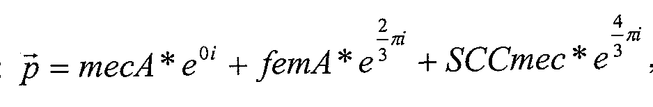
  • the intermediate vector may be the sum of three unit vectors 120 degrees apart from each other and weighted by the mecA, femA, and SCCmec values taken from step 1020.
  • the sum of the vectors can be represented by the formula: , where p is the intermediate vector. femA may
  • SA (representing a value associated with a
  • Staphylococcus aureus-specific target gene sequence Other similar means for representing how the relative amounts of the measured targets may also be used according to other embodiments.
  • the intermediate vectors created during step 1020 for each known sample can be analyzed in order to create a model that can be used to determine whether unknown samples contain MRSA.
  • a boundary function is selected and the parameters of the boundary function are defined using the intermediate vectors of the known samples.
  • the boundary function can be used to determine if MRSA is in an unknown sample or is not in an unknown sample.
  • a clustering analysis can be performed on a Y plot of all of the measured known samples to separate MRSA positive and negative samples.
  • a clustering analysis can group positive samples together because femA, mecA, and SCCmec are single copy targets and there should be equal copies of these targets in MRSA positive samples.
  • a simple boundary function such as a rectangular area that encompasses the positive samples, can be used to cluster the positive samples that are located around the origin of the Y plot.
  • the negative samples may be scattered outside of this boundary.
  • a simple rectangular box can be used to separate positive and negative samples on the Y plot.
  • the boundaries of the rectangular box can be adjusted using the measurements from the known samples.
  • a neural network algorithm could be used to help define the boundaries of the box.
  • Other embodiments may select more complicated boundary functions or use other means to adjust the coefficients of the selected boundary function.
  • boundary functions can be created using other approaches (e.g., a circular gating process, a neural network, or a Gaussian distribution function).
  • Fig. 2 illustrates steps that can be used to determine whether MRSA is present in a sample according to one embodiment.
  • an unknown sample refers to a sample in which it is not known whether MRSA is present in the sample.
  • the steps in Fig. 2 can use a model, such as a model created using the steps from Fig. 1, to determine whether an unknown sample contains MRSA.
  • the unknown sample can have various targets measured, and these measurements can be used to detect the presence of MRSA by analyzing where an intermediate vector created from the measured targets of the unknown sample resides relative to a boundary function of the model.
  • an embodiment of the invention is directed to a method for determining the presence of a biological entity in a sample.
  • the method comprises detecting values (e.g., amounts) of at least three targets in the sample.
  • the presence of the biological entity in the sample is determined by executing a call algorithm on a digital computer.
  • the call algorithm uses as inputs the measured values (e.g., amounts) of at least three targets and combines them to form a vector, which is compared to a boundary function on a 2- dimensional plot. If the vector is within the boundary function, the biological entity is present and if the vector is not within the boundary function, the biological entity is not present.
  • the unknown sample is subjected to conditions that expose the nucleic acids of bacteria in the sample. The same techniques used during step 1000 while building the model can also be used at step 1100.
  • the Ct values at least three targets, mecA, SCCmec, and a
  • Staphylococcus aureus-specific target gene sequence are measured from the unknown sample. The same techniques used during step 1010 can be used to accomplish step 1110.
  • the call algorithm used to build the model can be applied to the measured targets from unknown sample.
  • an intermediate vector can be created from the unknown sample using the same function that was used at step 1020.
  • step 1130 it is determined whether MRSA is present in the unknown sample. According to one embodiment, this can be accomplished by comparing the intermediate vector created for the unknown sample against the boundary function created from the known samples. If the intermediate vector for the unknown sample falls within the boundary that designates positive MRSA samples, then it can be determined that MRSA is present in the unknown sample. If the intermediate vector for the unknown sample falls outside of the positive MRSA boundary, then it can be determined that MRSA is not present in the unknown sample. [0064] Embodiments of the invention are particularly advantageous. As noted below, embodiments of the invention are particularly useful in identifying MRSA in a sample, despite the fact that MRSA can often co-colonize with multiple other related bacteria.
  • samples were collected and analyzed to form a model that can be used according to one embodiment.
  • the samples were known samples, i.e., it was known whether MRSA was present in each of the samples.
  • Two swabs were collected for each sample. These two swabs were then cultured for MRS A independently. If the culture results from two swabs agree, this sample was considered valid.
  • Each valid known sample was then placed in a sample buffer tube with 1ml of Tris, pH 8.0 and ImM EDTA, pH8.0.
  • the sample buffer tube was vortexed for 40 seconds at 3000 rpm to dislodge bacteria from the swab head.
  • 500 ⁇ , of the bacterial suspension i.e., the sample buffer containing bacterial suspension was then transferred into a new tube.
  • a pre-mix was then prepared by mixing together 188 ⁇ , of lysis buffer, 1.0 ⁇ of polyA RNA (10 ⁇ ), and ⁇ of Proteinase K (20 mg/ml) (in 10 mM Tris pH 8.0, 50% glycerol, 5 mM calcium chloride).
  • the pre-mix was then added to the tubes, vortexed for 10 seconds at 1700 RPM, and then incubated in a 70°C water bath for 5 minutes.
  • the tubes were then incubated at room temperature for 2 minutes and then placed on a microcentrifuge tube magnet for 6 minutes to capture the magnetic beads. The supernatant was aspirated off without disturbing the magnetic beads. The tubes were then taken off the magnet and 500 ⁇ , of Wash Buffer (3.3M Guanidine thiocyanate, 1.7% v/v Triton X-100, 167.5 mM sodium citrate) was added.
  • Wash Buffer 3.3M Guanidine thiocyanate, 1.7% v/v Triton X-100, 167.5 mM sodium citrate
  • PCR reagents were first thawed and then maintained on ice during the following steps. Edge wells were not used on a 96-well plate (Rows A and H, columns 1 and 12). One positive and 1 negative control were used per PCR plate. [0079] A reaction mix was then prepared for the required quantity of samples and control. The reaction mix was prepared based on increments of 12 reactions.
  • reaction mix was aliquoted into a Stratagene 96-well PCR plate at a volume of 30 ⁇ . per well.
  • 20 ⁇ _, of SPRI-TE extracted DNA sample was added to a single well.
  • 20 ⁇ , of Negative Control was added to a single well with nuclease free water.
  • 20 ⁇ ⁇ of Positive Control was added to a single well.
  • the sample and reaction mix was then created by pipeting the mixture up and down 10 times, using a pipette set at 45 ⁇ ,.
  • the prepared PCR plate was then sealed with an Optical Adhesive Cover and then centrifuged at 3000 rpm for 3 minutes.
  • the plate was then loaded onto a Stratagene Mx3005P qPCR instrument.
  • the used wells in the plate were selected and the following dyes were selected for fluoresence data collection: CY5, HEX, ROX, FAM.
  • the following cycling conditions were specified: 4' @ 37°C (lx); V @ 95°C (lx); 5" @ 91°C ⁇ >10" @62°C -> 25" @ 58°C (50x).
  • the probes for SCCmec have sequences that are complementary to the right extremity of SCCmec.
  • primers and probes that are substantially identical in sequence and/or length can be used in embodiments of the invention.
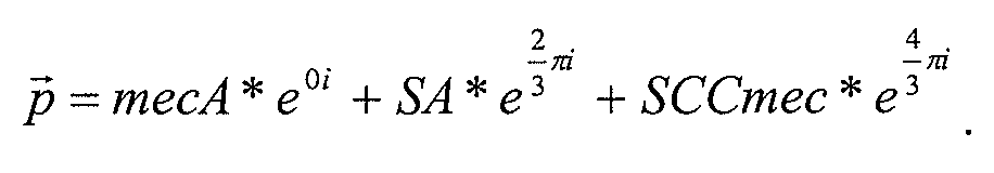
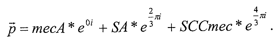
  • each intermediate vector was the sum of three unit vectors 120 degrees apart from each other and weighted by mecA, femA, and SCCmec values respectively.
  • the formula used was: , where p is the intermediate
  • the intermediate vectors created from the MRS A positive samples generally have their endpoints clustered around the origin of the plot.
  • the MRSA negative samples generally have the endpoint of their intermediate vectors residing at locations more distant from the origin.
  • Fig. 4 shows how a simple rectangle box 4000 can be used as a boundary function to separate positive and negative samples.
  • the left, right, top, and bottom edges of the rectangle box are at -3, 2.5, 2, and -1.3.
  • the boundary may be defined by a function that is then tuned using a neural network.
  • the boundary does not need to rectangular; more complicated geometries may yield better fits to the known samples in other models.
  • the known samples can then be analyzed using the newly created model to test how accurately the model categorizes the known samples.
  • a Y-plot-based call algorithm that computes an intermediate vector for each known sample and compares the vector with the boundary function, as described above, resulted in 5 false negatives (FN's) and 0 false positives (FP's) for the 659 samples.
  • FN's false negatives
  • FP's false positives
  • One of these false negatives is highlighted in Fig. 4 at 4010.
  • the swab heads were then sterilely removed from the sample tubes and transferred into 15 ml bacteria culture tubes with 1 ml of Trypic Soy broth (TSB) and 6.5% NaCl.
  • TTB Trypic Soy broth
  • the inoculated bacteria tubes were transferred into a 37°C incubator and incubated overnight with shaking at speed of 200 rpm.
  • CHROMagar MRSA and a BBLTM CHROMagar Staphylococcus aureus plate 500 ⁇ , of the 1200 ⁇ L ⁇ sample solution from each tube was then subjected to DNA isolation procedure as described by Agencourt VirNA kit protocol. This procedure, in brief, began with an amount of CFU S. felis bacteria (or an amount of S. felis without CFU) as a process control. 10 units of Achrompeptidase were added to each tube, mixed well, and incubated in a 70 °C waterbath for 4 minutes. 289 iL of a freshly prepared lysis solution containing 188 ⁇ , of a lysis buffer, 1.0 ⁇ .
  • the supernatant was aspirated off the samples while being careful not to remove any beads during aspiration.
  • 500 ⁇ , of washing buffer were added to the samples and vortexed for 10 seconds to mix.
  • the tubes were then incubated on the magnet for 4 minutes (or until clear).
  • the supernatant was then aspirated off the samples again.
  • 900 ⁇ , of freshly prepared 75% ethanol was then added and the tube vortexed for 10 seconds.
  • the tubes were then incubated on the magnet for 4 minutes until clear.
  • the supernatant was then aspirated off the samples again and the ethanol washing was repeated one more time.
  • the beads were then dried on the magnet for 15-25 minutes. When the ring of the beads started to crack, the sample was eluted.
  • the tubes were taken off the magnet and 25 ⁇ , of nuclease free water was added to elute the DNA. The samples were then vortexed to mix. The tubes were then incubated for 5 minutes at 70°C. The tubes were placed back on the magnet and incubated for 1 minute. The eluate was then transferred to a clean tube for PCR
  • the reagents listed in the Master mix table were prepared on ice. According to the total reaction number, enough Master mix can be prepared by simply adding the indicated volumes of reagents together in a DNA/RNase-free tube. The tubes can be vortexed to mix and then left on ice for later use. 20 ⁇ , of each eluate was added to a Mx3000P 96-well PCR plate (non-skirted) (Stratagene, Cat#401333) (one eluate, one well). 30 ⁇ , Master mix was added to each well filled with the eluate, and then mixed by gently pipetting up and down 8 times or more (a multi-channel might be useful.). The plate was covered tightly with
  • Model creation and analysis [0102] The threshold values of each target are exported and input into a call algorithm as previously described with the first illustrative example.
  • Fig. 6 is a block diagram of a computer system 300 that can be used to execute one embodiment of the invention.
  • the computer system 300 has a number of input modules.
  • a measurement module 301 is used to measure selected targets in a sample. This measurement module may vary between different embodiments of the invention depending on the measurement method selected to measure the target responses. For example, according to one embodiment, the measurement module may conduct a PCR analysis on a sample.
  • the measurement module may be embodied by at least part of the working components of a typical real time PCR apparatus. Also shown are a standard keyboard 302 and mouse 303.
  • the computer system 300 also contains a variety of typical computer components inside the computer. These components include a system bus 304, one or more disk drives 305, RAM
  • Fig. 5 also shows a monitor 308 that allows information to be displayed to a user of the system.
  • a sample is placed in the measurement module 301 where the sample is processed and amounts of the selected targets from the sample are measured. This information is then transferred into the computer system along a system bus 304, and an appropriate call algorithm can be applied to the response data using the processor
  • the instructions the processor 307 executes to implement the call algorithm are stored on a computer readable medium such as the RAM 306 or disk drive 305.
  • the call algorithm can also be stored on this same media.
  • the output of the call algorithm can then be displayed on the monitor 308. For example, if the call algorithm uses a 2-D Y plot to graph the measured amounts of three targets via an intermediate vector, the end point of the call algorithm
  • intermediate vector can be displayed on the monitor 308.
  • Alternative embodiments of the invention can output information via other communications means.
  • the computer system could print the Y plot using a printer or send the Y plot to another computer over a network. The information from the measured sample can then be used to either help build a model or determine whether the sample contains MRS A.
  • the software components, steps, or functions described in this application may be implemented as software code to be executed by one or more processors using any suitable computer language such as, for example, Java, C++ or Perl using, for example, conventional or object-oriented techniques.
  • the software code may be stored as a series of instructions, or commands on a computer readable medium (e.g., a non-transitory computer readable medium), such as a random access memory (RAM), a read only memory (ROM), a magnetic medium such as a hard-drive or a floppy disk, or an optical medium such as a CD-ROM.
  • a computer readable medium e.g., a non-transitory computer readable medium
  • RAM random access memory
  • ROM read only memory
  • magnetic medium such as a hard-drive or a floppy disk
  • optical medium such as a CD-ROM.
  • Any such computer readable medium may also reside on or within a single computational apparatus, and may be present on or within different computational apparatuses within a system or network
  • control logic in software or hardware or a combination of both.
  • the control logic may be stored in an information storage medium as a plurality of instructions adapted to direct an information processing device to perform a set of steps disclosed in an embodiment of the present invention.
  • MRSA Staphylococcus aureus

Abstract

Systems and methods for identifying Methicillin resistant strains of Staphylococcus aureus (MRSA) in a sample are used that are based on the fact that an MRSA positive sample should have roughly the same copy numbers of mecA, SCCmec, and a Staphylococcus aureus-specific target gene sequence. The systems and methods may further present the three assays simultaneously on a 2-D plot with each axis of the plot 120 degrees apart. According to one embodiment, a Y plot is used for the 2-D display. If a given sample has similar readings of mecA, SCCmec, and a Staphylococcus aureus-specific target gene sequence, the sample's measured copy numbers of mecA, SCCmec, and the Staphylococcus aureus-specific target gene sequence can plot close to the origin regardless of the sample's absolute assay readings. With the help of this transformation, a boundary function can be defined that can be used to distinguish MRSA-positive samples from MRSA-negative samples.

Description

SYSTEMS AND METHODS FOR DETECTING THE PRESENCE OF A BIOLOGICAL STATUS USING PLOT
CROSS-REFERENCE TO RELATED APPLICATIONS [0001] This application is a non-provisional of and claims the benefit under 35 U.S.C. 119(e) of U.S. Provisional Patent Application No. 61/261,109, filed on November 13, 2009, entitled "Systems and Methods for Detecting the Presence of a Biological Status Using Plot" (Attorney Docket No. 021594-010300US), which is herein incorporated by reference in its entirety for all purposes. BACKGROUND
[0002] Methicillin resistant strains of Staphylococcus aureus (MRS A) are implicated in infections with serious outcomes including nosocomial outbreaks, and show resistance to a wide range of antibiotics, thus limiting the treatment options. Healthcare associated MRSA is of particular clinical importance because it is not only predictably cross resistant to all penicillins and cephalosporins, but is also typically resistant to multiple other commonly used antibiotics. Treatment of MRSA infections generally require more expensive and often more toxic antibiotics, which are normally used as the last line of defense. Therefore, rapid detection of MRSA is clinically crucial for both treatment and infection control measures.
[0003] Detection of MRSA is further complicated by the fact that MRSA can often co- colonize with multiple other related bacteria, including methicillin-sensitive Staphylococcus aureus (MSSA), methicillin-resistant coagulase-negative staphylococci (MR-CoNS) and/or methicillin-sensitive coagulase-negative staphylococci (MS-CoNS).
[0004] Traditional methods for the detection of MRSA in clinical microbiology laboratories involve culturing the bacteria from a sample as the first step for the isolation and differentiation of MRSA from MSSA and MR-CoNS. This approach is time-consuming and requires a minimum of 20 to 24 hours until a result is known.
[0005] A number of molecular based methods have been published for the detection of methicillin resistant Staphylococcus aureus (MRSA) and differentiating it from methicillin sensitive Staphylococcus aureus (MSSA). One such method targets two separate regions of MRSA, the mecA gene of the Staphylococcus cassette chromosome (SCCmec, responsible for methicillin resistance) and spa gene of Staphylococcus aureus (United States Patent 5,702,895, Sinsimer, et al, Journal of Clinical Microbiology, Sept. 2005, 4585-4591).
Unambiguous detection of MRS A using this approach is hampered by the co-existence of non-Staphylococcus aureus strains such as methicillin resistant coagulase negative
Staphylococci (MR-CoNS) which also harbors the mecA gene for methicillin resistance (Becker, et. al. Journal of Clinical Microbiology, Jan. 2006, p229-231).
[0006] A more recent molecular approach utilizes primers and probes to SCCmec and the flanking region of the Staphylococcus aureus genome (United States Patent 6,156,507, Huletsky, et. al. Journal of Clinical Microbiology, May 2004, pi 875- 1884). SCCmec is a mobile genetic element that carries the mecA gene and inserts at a specific site, attBscc, at the 3 '-end of the orfX gene. The left extremity of SCCmec is contiguous with the non-orfX side of attBscc while the right extremity of SCCmec is contiguous with the orfX side of attBscc (Ito, et al., Antimicrob. Agent Chemother. 2001, 45, pl323-1336; Ito et al., Antimicrob. Agent Chemother. 2004, 48, p2637-2651, Noto, et al., J. Bacteriol. 2008, 190: 1276-1283). This approach infers the presence of the mecA gene from the detection of the SCCmec/orfX junction. This approach requires the use of multiple primers as there have been several different types of SCCmec described. This approach is also subject to false positive results due to the presence of SCCmec cassettes that do not contain the mecA gene (Farley, et. al. Journal of Clinical Microbiology, February 2008, p743-746) and false negative results due to newly emerged SCCmec types not covered by the assay (Heusser, et al., Antimicrob. Agents Chemother. January 2007, p390-393).
[0007] Another approach utilizes one primer in a region of high homology across the different SCCmec types and one primer in the flanking Staphylococcus aureus DNA (Cuny, et al. Clin. Microbiol Infect 2005; 11 :834-837, European Patent 1529847 Bl). This approach is also subject to false positives as the probability of also priming of MSSA is high with primers encompassing this region.
[0008] Finally, a method has been described that positively selects for Staphylococcus aureus using specific antibodies and magnetic beads (Francois, et al. Journal of Clinical Microbiology, January 2003, p254-260; European Patent 1,370,694B1). This approach enriches for Staphylococcus aureus, but requires the use of three primer/probe sets to positively identify MRS A and reduces the possibility of detecting CoNS. The method requires a centrifugation step and a separate lysis step to recover the nucleic acid. [0009] The commercially available MRSA assays target the SCCmec right extremity junction and orfX. Five different types and numerous subtypes of SCCmec have been identified and the potential of emergence of new SCCmec subtypes is high. In addition, it is possible that MSSA derived from MRSA might retain part of the SCCmec sequence without the mecA gene. Therefore, assays targeting the SCCmec right extreme junction with orfX are likely to give false positive results with MRSA-derived MSSA and false negative results with MRSA carrying newly emergent SCCmec types/subtypes.
[0010] Thus, current methods for detection of MRSA are laborious, time-consuming, and unreliable for routine clinical and surveillance purposes. Accordingly, there exists a need for developing an assay that is fast, easy, reliable and capable of detecting and concurrently distinguishing MRSA from other related bacteria, including MSSA, and/or MR-CoNS.
BRIEF SUMMARY
[0011] Embodiments of the present invention relate to systems and methods for detecting methicillin resistant Staphylococcus aureus (MRSA) in a sample which may additionally contain methicillin sensitive Staphylococcus aureus (MSSA), methicillin resistant coagulase- negative staphylococci (MR-CoNS), and/or other strains of bacteria. Other embodiments of the invention may relate more generically to systems and methods for the analysis of biological entities using plots and boundary functions.
[0012] One embodiment of the invention is directed to a method for determining the presence of a biological entity in a sample. The method comprises detecting amounts of at least three genetic elements in the sample. The presence of the biological entity in the sample is determined by executing a call algorithm on a digital computer. The call algorithm uses as inputs the measured values (e.g., amounts) associated with at least three genetic elements and combines them to form a vector, which is compared to a boundary function on a 2- dimensional plot. If the vector is within the boundary function, the biological entity is present and if the vector is not within the boundary function, the biological entity is not present.
[0013] Another embodiment of the invention is directed to a system for determining the presence of a biological entity in a sample. The system comprises a measurement module capable detecting the values (e.g., amounts) of at least three genetic elements in the sample, and a memory storing the detected values (e.g., amounts) from the measurement module. The system also includes a computer readable medium containing computer readable code having instructions for executing a call algorithm, wherein the call algorithm uses as inputs the detected and measured values (e.g., amounts) of the at least three genetic elements and combines them to form a vector. The vector is compared to a boundary function on a 2- dimensional plot. If the vector is within the boundary function, the biological entity is present and if the vector is not within the boundary function, the biological entity is not present. The system also includes a processor to execute the computer readable code on the computer readable medium in order to determine the presence of the biological entity in the sample.
[0014] Another embodiment of the invention is directed to a computer readable medium comprising code for a call algorithm. The call algorithm uses as inputs detected and measured values (e.g., amounts) associated with the at least three genetic elements and combines them to form a vector. The vector is compared to a boundary function on a 2- dimensional plot. If the vector is within the boundary function, a biological entity is present and if the vector is not within the boundary function, the biological entity is not present. [0015] Another embodiment of the invention is directed to a method for creating a model that can be used to determine the presence of a biological entity in an unknown sample. The method includes detecting the presence and values (e.g., amounts) of at least three genetic elements in the known samples and executing a call algorithm on a digital computer for each sample in known samples, wherein the call algorithm creates vectors and uses as inputs for each vector the detected values (e.g., amounts) of the at least three genetic elements. Some vectors are associated with the biological entity and some vectors are not associated with the biological entity. The method further includes plotting the vectors on a 2-dimensional plot, and creating a boundary function separating the vectors that are associated with the biological entities and the vectors that are not associated with the biological entity. [0016] According to one embodiment, a method for determining the presence of methicillin-resistant Staphylococcus aureus (MRSA) in a sample is disclosed. The method subjects the sample to conditions that will expose the nucleic acids of any bacteria present in the sample. Next, the sample may be amplified and the presence and amounts of at least mecA, SCCmec, and a Staphylococcus aureus (SA)-specific target gene sequence in the sample can be detected. The presence of MRSA in the sample can then be determined by executing a call algorithm on a digital computer. The call algorithm may use as inputs the detected and measured amounts of mecA, SCCmec, and the Staphylococcus aureus-specific target gene sequence to determine whether MRSA is present in the sample. The call algorithm can be used to determine that MRSA is present in the sample when the detected amounts of mecA, SCCmec, and the Staphylococcus aureus-specific target gene sequence in the sample are approximately equal as defined by a selected boundary function.
[0017] According to one embodiment, a system for determining the presence of
methicillin-resistant Staphylococcus aureus (MRSA) in a sample is disclosed. The system may comprise a number of components. For instance, the system may comprise a
measurement module that is capable of amplifying and detecting the presence and amounts of at least mecA, SCCmec, and a Staphylococcus aureus-specific target gene sequence in the sample after the sample has been subjected to conditions that will expose the nucleic acids of any bacteria present in the sample. The system may further comprise a memory that can store the detected amounts from the measurement module. The system may also comprise a computer readable medium containing computer readable code having instructions for executing a call algorithm. The call algorithm may use as inputs the detected and measured amounts of mecA, SCCmec, and the Staphylococcus aureus-specific target gene sequence to determine whether MRSA is present in the sample. The call algorithm can be used to determine that MRSA is present in the sample when the detected amounts of mecA, SCCmec, and the Staphylococcus aureus-specific target gene sequence in the sample are approximately equal as defined by a selected boundary function. The system may also comprise a processor that can execute the computer readable code on the computer readable medium in order to determine the presence MRSA in the sample. [0018] According to another embodiment, a computer-readable medium is disclosed. The computer-readable medium may comprise code for a call algorithm that can determine the presence of MRSA in a sample. The call algorithm may use as inputs detected and measured amounts of mecA, SCCmec, and a Staphylococcus aureus-specific target gene sequence to determine whether MRSA is present in the sample. The call algorithm can be used to determine that MRSA is present in the sample when the detected amounts of mecA, SCCmec, and the Staphylococcus aureus-specific target gene sequence in the sample are approximately equal as defined by a selected boundary function. The detected and measured amounts of mecA, SCCmec, and the Staphylococcus aureus-specific target gene sequence can be obtained by subjecting the sample to conditions that will expose the nucleic acids of any bacteria present in the sample and amplifying and detecting the presence and amounts of at least mecA, SCCmec, and the Staphylococcus aureus-specific target gene sequence in the sample.
[0019] According to yet another embodiment, a method for creating a model that can be used to determine the presence of methicillin-resistant Staphylococcus aureus (MRSA) in an unknown sample is disclosed. The method may comprise subjecting a set of known samples to conditions that will expose the nucleic acids of any bacteria present in the known samples. The presence or absence of MRS A is known for each sample in the set of known samples. The method may then amplify and detect the presence and amounts of at least mecA, SCCmec, and a Staphylococcus aureus-specific target gene sequence in the known samples. The method may then execute a call algorithm on a digital computer for each sample in the known samples. The call algorithm may use as inputs the detected and measured amounts of mecA, SCCmec, and the Staphylococcus aureus-specific target gene sequence. Finally, the method may create a model that can be used to determine whether MRS A is present in the unknown sample. The model can be created from the output of the call algorithm executed against the known samples, and the model can be defined by a selected boundary function that defines a boundary between MRSA-positive and MRSA-negative samples.
[0020] These embodiments, as well as other embodiments, will be described in more detail later in this disclosure. BRIEF DESCRIPTION OF THE DRAWINGS
[0021] Fig. 1 shows a flowchart illustrating the steps taken in a method according to one embodiment.
[0022] Fig. 2 shows a flowchart illustrating the steps taken in a method according to one embodiment of the invention. [0023] Figs. 3-5 show diagrams illustrating the outputs of call algorithms from various positive and negative samples according to embodiments of the invention.
[0024] Fig. 6 is a block diagram of a system that can be used to execute various embodiments of the invention.
DETAILED DESCRIPTION [0025] Various embodiments disclose systems and methods for identifying Methicillin resistant strains of Staphylococcus aureus (MRSA) in a sample that are based on the fact that an MRSA-positive sample can have roughly the same copy numbers of mecA, SCCmec, and a Staphylococcus aureus-specific target gene sequence. These systems and methods may further present the three assays simultaneously on a 2-D plot with each axis of the plot 120° angle apart. According to one embodiment, a Y plot is used for the 2-D display. If a given sample has similar values of mecA, SCCmec, and a Staphylococcus aureus-specific target gene sequence, the sample's measured copy numbers of mecA, SCCmec, and the
Staphylococcus aureus-specific target gene sequence can plot very close to the origin regardless of the sample's absolute assay readings. With the help of this transformation, a boundary function can be defined that can be used to distinguish MRSA-positive samples from MRSA-negative samples.
[0026] All scientific and technical terms used in this disclosure have meanings commonly used in the art unless otherwise specified. As used in this disclosure, the following words or phrases have the meanings specified.
[0027] As used herein, a "boundary function" can be a mathematical function that is used to determine whether data is associated with a biological status or is not associated with a biological status. Boundary functions may be created in any suitable manner including manually, by the use of neural networks, cost functions, etc. Boundary functions may also be represented by any suitable shape or line, including an ellipse, rectangle, circle, or the like. Boundary functions may also be regular or irregular in shape. [0028] As used herein, a "vector" can relate to value derived from one or more
components, each component having at least a direction, and typically a scalar.
[0029] The term "genetic element" as used herein can refer to a subsequence in a genome of interest that is useful as a target in the methods of the invention. In some embodiments, the genetic element is an open reading frame or gene, such as, for example, orfX, femA or mecA in Staphylococcus. A genetic element may also be a mobile genetic element, such as the Staphylococcus cassette chromosome, SCCmec, which may or may not comprise the mecA gene.
[0030] Herein, the terms "SCCmec" "SCCmec sequence" and "SCCmec cassette" are used interchangeably to refer to the genetic element known as the Staphylococcus cassette chromosome, which carries the mecA gene and is inserted into Staphylococcus sp. genome as described in Ito et al. (2001, Antimicrob. Agents Chemother. 45:1323-1336).
[0031] The SCCmec insertion site is referred to as "orfX-ISS/at Bscc" in this application. The insertion site is at the 3' end of a gene referred to herein, as "orfX." The chromosomal locus where SCCmec insertion takes place is reffered to as "attBscc." The specific sequence at the insertion site is referred to here as the "orfX-Insertion Site Sequence (orfX-ISS)" or "attBscc core region." This sequence is known to be a highly conserved sequence in Staphylococcus aureus (Ito, et al., Antomicrob. Agent Chemother. 2001, 45, p323-1336, Noto, et al., J. Bacteriol. 2008, 190:1276-1283).
[0032] After insertion into the orfX-ISS/attBscc region of Staphylococcus aureus, the SCCmec left extremity junction region is referred to as MRSA-LE and the right extremity junction region is referred to as MRSA-RE. In the left extremity junction, the SCCmec sequence is contiguous with the non-orfX side of attBscc. In the right extremity junction, the SCCmec sequence is contiguous with the orfX-side of attBscc. The orfX-ISS/attBscc region is described in detail in Ito et al. (2001 , Antimicrob. Agents Chemother. 45 : 1323-1336; Ito et al., Antimicrob. Agent Chemother. 2004, 48, p2637-2651, Noto, et al, J. Bacteriol. 2008, 190: 1276-1283) and in U.S. Pat. No. 6,156,507, all of which are incorporated by reference herein. If the SCCmec insertion is not present, the orfX-ISS/attBscc region is uninterrupted. If the orfX-ISS/attBscc region is identified as intact through an amplification methodology this indicates that the SCCmec cassette has not been inserted. Lack of amplification of the orfX-ISS/attBscc region, however, does not indicate the mecA gene is present. It is known the mecA gene can be lost after the SCCmec cassette becomes inserted. Thus the SCCmec cassette can still prevent amplification of the orfX-ISS/attBscs region even in the absence of mecA.
[0033] An "oligonucleotide" is a nucleotide polymer having two or more nucleotide subunits covalently joined together. Oligonucleotides are generally about 10 to about 100 nucleotides. The sugar groups of the nucleotide subunits may be ribose, deoxyribose, or modified derivatives thereof such as OMe. The nucleotide subunits may be joined by linkages such as phosphodiester linkages, modified linkages or by non-nucleotide moieties that do not prevent hybridization of the oligonucleotide to its complementary target nucleotide sequence. Modified linkages include those in which a standard phosphodiester linkage is replaced with a different linkage, such as a phosphorothioate linkage, a
methylphosphonate linkage, or a neutral peptide linkage. Nitrogenous base analogs also may be components of oligonucleotides in accordance with the invention. A "target nucleic acid" is a nucleic acid comprising a target nucleic acid sequence. A "target nucleic acid sequence," "target nucleotide sequence" or "target sequence" is a specific deoxyribonucleotide or ribonucleotide sequence that can be hybridized to a complementary oligonucleotide.
[0034] As used herein, the term "probe" refers to an oligonucleotide which is capable of hybridizing to a target nucleic acid of interest. The hybridization occurs as a result of the probe binding through complementary base pairing to a target nucleic acid of interest. It will be understood by one skilled in the art that probes will typically substantially bind target sequences lacking complete complementarity with the probe sequence depending upon the stringency of the hybridization conditions. The probe may be associated with a suitable label or reporter moiety so that the probe (and therefore its target) can be detected, visualized, measured and/or quantitated. [0035] As used herein, the term "primer" refers to an oligonucleotide used to prime nucleic acid synthesis. A primer hybridizes to the template through complementary base pairing and is therefore used to initiate the replication. Hybridization occurs in the same manner as that described for probes, above. In PCR, two primers are used: a "forward primer" that typically hybridizes to the sense strand of a double stranded nucleic acid molecule and a "reverse primer" that typically hybridizes to the antisense strand of the molecule.
[0036] As used herein, the term "PCR" refers to a technique for exponential amplification of short DNA sequences (usually 50 to 600 bases) within a longer double stranded DNA molecule by enzymatic replication of DNA without using a living organism (Mullis et al. Methods Enzymol. 1987; 155:335-50). Other in vitro amplification
technologies can be used in the present invention and are well known to those of skill. These methods include, for example, Ligase Chain Reaction (LCR), Nucleic Acids Sequence Based Amplification (NASBA), Strand Displacement Amplification (SDA), Transcription Mediated Amplification (TMA), Branched DNA technology (bDNA) and Rolling Circle Amplification Technology (RCAT).
[0037] As used herein the term "Real-Time PCR" refers to a type of PCR where the amplified DNA is quantified as it accumulates in the reaction in real time after each amplification cycle (Heid et al, Genome Research, 1996 6(10):986-994). A number of probe chemistries for carrying out Real-Time PCR are well known to those of skill. One commonly used method is the the TaqMan® assay {see, e.g., U.S. Pat. Nos. 5,210,015; 5,487,972; and 5,804,375). Other Real-Time PCR probe chemistries that can be used and can be purchased commercially include FRET primers, Molecular Beacons, Scorpion primers®, Amplifluor primers®, LUX primers®, Eclipse®, and Ultimate Probe®. For a review of Real-Time PCR techniques see Bustin et al., J. Mol. Endocrin. 34:597-601 (2005). [0038] As used herein, the term "Multiplex PCR" refers to a type of PCR where more than one set of primers is included in a reaction allowing two or more different targets to be amplified in a single reaction tube. The term "multiplex PCR" also refers to a PCR where multiple primers and probes are used but only one target is amplified. In one embodiment, the multiplex PCR of the present invention is a real-time PCR.
[0039] As used herein, a "biological status" may relate to a particular biological state of a sample derived from a patient. In most cases, the biological status relates to whether or not the sample comprises a particular biological entity, for example, a target disease organism or patient cell associated with disease. For example, one biological status may be that a sample comprises MRSA bacteria, while another biological status may be that the sample does not comprise MRSA bacteria. In other examples, the biological status may relate to whether or not the sample comprises cancer cells. [0040] One embodiment of the invention relates to an assay for detection of MRSA in a sample that may contain MRSA, MSSA, MR-CoNS, or other bacteria. Embodiments of the invention utilize a multiplex PCR for simultaneously amplifying and detecting a combination of multiple genetic elements (e.g., targets).
[0041] According to one embodiment, the initial amount of target DNA is measured by the PCR threshold cycle (Ct). For example, a defined signal threshold is determined for a reaction to be analyzed. The number of cycles (Ct) required to reach this signal threshold is determined for the target nucleic acid as well as for a reference or standard nucleic acid. The absolute or relative copy numbers of the target molecule can be determined on the basis of the Ct values obtained for the target nucleic acid as compared to the reference nucleic acid. The Ct value is thus inversely proportional to the amount of initial target DNA, see Heid et al, 1996, Genome Research 6(10):986 for a full discussion of the Ct value which is incorporated herein by reference. Other mathematical approaches can be employed which allow for the extrapolation of the initial amount of a particular target gene based upon the indication of a predetermined set amount or number of genes amplified during one of the identified methods.
[0042] In one embodiment, the present invention is directed to a method of determining the presence of MRSA in a sample, said method comprising subjecting the sample to real-time PCR for a time and under conditions so as to generate a level of amplification product which is sufficient to be detected by fluorescence and is indicative of an initial level of one or more MRS A- specific target sequences in the sample.
[0043] In another embodiment, the amplification is conducted with a set of primers (forward and reverse) and a probe. The probe may be labeled with a fluorogenic reporter molecule at its 5' end and a quenching molecule at its 3' end. The quenching molecule prevents emission of signal from the fluorogenic reporter molecule. The probe hybridizes to a region of the target sequence between the regions to which the forward and reverse primers hybridize. As the polymerase moves along the strand to which the probe has hybridized, the 5' end of the probe is cleaved off by the exonuclease activity of the polymerase thus permitting emission of the fluorogenic signal due to separation of the quenching moiety.
[0044] In specific embodiments, the probes of the invention may comprise dual-labeled fluorogenic probes comprising a fluorescent reporter (fluorophore) and a fluorescent or non- fluorescent quencher molecule. The fluorophores of embodiments of the invention may be attached to the probe at any location, including the 5' terminus, the 3' terminus or internal to either termini. In an embodiment of the invention, the fluorophore and quencher are attached to the 5' and 3' termini of the probe respectively. The examples of fluorophores include, but are not limited to, FAM, ROX, HEX, NED, Cy5, Texas Red, Calfluor Red, CalFluor Orange, Quasar 670, Quasar 705. The examples of quenchers include, but are not limited to,
TAMARA, Blackhole quenchers BHQ-1, BHQ-2. [0045] In another embodiment, the invention provides a method for detecting and distinguishing MRSA from MSSA, MR-CoNS, or other bacteria utilizing a three target assay, wherein the targets used in the assay include the mecA gene sequence, a Staphylococcus aureus-specific target gene sequence, and an SCCmec gene sequence. In a specific embodiment, the Staphylococcus aureus-specific target gene is femA. In the descriptions below, femA is often explicitly mentioned as the Staphylococcus aureus-specific target gene sequence; however, other Staphylococcus aureus-specific target gene sequences may also be used according to various embodiments. Yet other targets that can be used to determine the presence of MRSA can include orfX.
[0046] Various embodiments need not use traditional positive-negative or delta approaches in order to determine the presence of MRSA. Instead, an approach is used that is based on the fact that an MRSA positive sample has roughly the same copy numbers of mecA, femA, and SCCmec. When the copy numbers of mecA, femA, and SCCmec are present in roughly equal amounts, it can be indicative of the presence of MRSA in the sample. In embodiments of the invention, the Ct values of mecA, SCCmec, and femA can be analyzed using an assay call algorithm. This call algorithm may further present the three assays simultaneously in a 2-D plot with lines 120 degrees apart. This plot can be referred to as a Y-plot since each of the three axes of the plot, representing the measured amounts of mecA, SCCmec, and femA, appear as a "Y" on a 2-D plot. If a sample has similar readings of mecA, femA, and
SCCmec, the sample's measured copy numbers of mecA, femA, and SCCmec will plot close to the origin of the plot regardless of the sample's absolute assay readings. With the help of this transformation, a rectangle gating approach may be sufficient to distinguish MRSA positive and negative samples. Other suitable approaches may use a circular gating process, a neural network, or a Gaussian distribution function. Additional details are provided below. [0047] Fig. 1 illustrates steps that can be used to build a model that can be used to determine whether MRSA is present in a sample according to one embodiment. A boundary function in the model can separate MRSA-positive samples from MRSA-negative samples based on the measured amounts of mecA, SCCmec, and a Staphylococcus aureus-specific target gene sequence. [0048] In more general terms, one embodiment of the invention is directed to a method for creating a model that can be used to determine the presence of a biological entity in an unknown sample. The method includes detecting the presence and amounts of at least three targets in the known samples and executing a call algorithm on a digital computer for each sample in known samples, wherein the call algorithm creates vectors and uses as inputs for each vector the detected amounts of the at least three targets. Some vectors are associated with the biological entity and some vectors are not associated with the biological entity. The method further includes plotting the vectors on a 2-dimensional plot, and creating a boundary function separating the vectors that are associated with the biological entities and the vectors that are not associated with the biological entity. [0049] At step 1000, a selected number of known samples are subjected to conditions that expose the nucleic acids of bacteria in the samples. In this context, a known sample is a sample in which it is already known whether the sample should test positive or negative for MRSA. The known samples can thus be used to build a model that can determine whether a later unknown sample also contains MRSA. Although MRSA is described in detail, it is understood that the presence of other suitable biological entities could be detected in other embodiments of the invention.
[0050] There are many different ways for subjecting a sample to conditions that expose the nucleic acids in the sample. For example, as it is well-known in the art, a sample may have its temperature raised to separate strands of DNA. Other well-known means for exposing the nucleic acids in a sample may also be used.
[0051] At step 1010, the presence and amounts of at least three targets, such as mecA, SCCmec, and a Staphylococcus aureus-specific target gene sequence, are detected for each of the known samples. In some embodiments, Ct values may be measured for each of the targets of interest. According to one embodiment, the Staphylococcus aureus-specific target gene sequence is femA. There are many different ways to measure the genes from a sample. For example, a multiplex PCR can be used to measure the PCR threshold cycle (Ct) for each target of the measurement. Other measurement techniques may also be used. [0052] The determined Ct values can be used to create a Y-Plot. However, prior to the creation of the Y-Plot, a pre-screening process can be carried out. In the prescreening process, if the system does not detect any of three target gene sequences, the sample is deemed MRSA negative and will not be shown in the Y-Plot. In one embodiment, a sample is deemed negative if mecA > 35, femA > 32, or SCCmec > 32. In another embodiment, a sample is deemed negative if mecA > 30, femA > 30, or SCCmeO 30. In some
embodiments, the sample will be plotted on and analyzed with the Y-Plot after it survives this prescreening process. The thresholds used in the prescreening process can be dependent on instrument and sample preparation.
[0053] At step 1020, a call algorithm can be applied to each of the measured targets from each of the known samples. According to one embodiment, the call algorithm may use a Y plot.
[0054] According to one embodiment, for each known sample, the call algorithm creates an intermediate vector that can be projected onto a 2-D plot, which may be a Y_plot. The intermediate vector may be the sum of three unit vectors 120 degrees apart from each other and weighted by the mecA, femA, and SCCmec values taken from step 1020. According to one embodiment, the sum of the vectors can be represented by the formula: , where p is the intermediate vector. femA may
Figure imgf000015_0001
be replaced by the generic variable "SA" (representing a value associated with a
Staphylococcus aureus-specific target gene sequence). Other similar means for representing how the relative amounts of the measured targets may also be used according to other embodiments.
[0055] At step 1030, the intermediate vectors created during step 1020 for each known sample can be analyzed in order to create a model that can be used to determine whether unknown samples contain MRSA. According to one embodiment, a boundary function is selected and the parameters of the boundary function are defined using the intermediate vectors of the known samples. The boundary function can be used to determine if MRSA is in an unknown sample or is not in an unknown sample. [0056] According to one embodiment, a clustering analysis can be performed on a Y plot of all of the measured known samples to separate MRSA positive and negative samples. A clustering analysis can group positive samples together because femA, mecA, and SCCmec are single copy targets and there should be equal copies of these targets in MRSA positive samples. Mathematically, this means if a sample is MRSA positive, then its intermediate vector on Y plot should be near the origin. Thus, a simple boundary function, such as a rectangular area that encompasses the positive samples, can be used to cluster the positive samples that are located around the origin of the Y plot. The negative samples may be scattered outside of this boundary. According to one embodiment, a simple rectangular box can be used to separate positive and negative samples on the Y plot. The boundaries of the rectangular box can be adjusted using the measurements from the known samples. For example, a neural network algorithm could be used to help define the boundaries of the box. Other embodiments may select more complicated boundary functions or use other means to adjust the coefficients of the selected boundary function. As noted above, boundary functions can be created using other approaches (e.g., a circular gating process, a neural network, or a Gaussian distribution function).
[0057] Fig. 2 illustrates steps that can be used to determine whether MRSA is present in a sample according to one embodiment. As used herein, an unknown sample refers to a sample in which it is not known whether MRSA is present in the sample. The steps in Fig. 2 can use a model, such as a model created using the steps from Fig. 1, to determine whether an unknown sample contains MRSA. The unknown sample can have various targets measured, and these measurements can be used to detect the presence of MRSA by analyzing where an intermediate vector created from the measured targets of the unknown sample resides relative to a boundary function of the model. [0058] In more general terms, an embodiment of the invention is directed to a method for determining the presence of a biological entity in a sample. The method comprises detecting values (e.g., amounts) of at least three targets in the sample. The presence of the biological entity in the sample is determined by executing a call algorithm on a digital computer. The call algorithm uses as inputs the measured values (e.g., amounts) of at least three targets and combines them to form a vector, which is compared to a boundary function on a 2- dimensional plot. If the vector is within the boundary function, the biological entity is present and if the vector is not within the boundary function, the biological entity is not present. [0059] At step 1 100, the unknown sample is subjected to conditions that expose the nucleic acids of bacteria in the sample. The same techniques used during step 1000 while building the model can also be used at step 1100.
[0060] At step 1110, the Ct values at least three targets, mecA, SCCmec, and a
Staphylococcus aureus-specific target gene sequence, are measured from the unknown sample. The same techniques used during step 1010 can be used to accomplish step 1110.
[0061] At this point, a similar pre-screening process, as described above, can be used to remove samples that are very likely to not be associated with MRSA.
[0062] At step 1 120, the call algorithm used to build the model can be applied to the measured targets from unknown sample. For example, an intermediate vector can be created from the unknown sample using the same function that was used at step 1020.
[0063] At step 1130, it is determined whether MRSA is present in the unknown sample. According to one embodiment, this can be accomplished by comparing the intermediate vector created for the unknown sample against the boundary function created from the known samples. If the intermediate vector for the unknown sample falls within the boundary that designates positive MRSA samples, then it can be determined that MRSA is present in the unknown sample. If the intermediate vector for the unknown sample falls outside of the positive MRSA boundary, then it can be determined that MRSA is not present in the unknown sample. [0064] Embodiments of the invention are particularly advantageous. As noted below, embodiments of the invention are particularly useful in identifying MRSA in a sample, despite the fact that MRSA can often co-colonize with multiple other related bacteria.
[0065] The various embodiments of the invention are demonstrated further by the following illustrative examples. The examples are offered by way of illustration and are not intended to limit the invention in any manner.
[0066] Illustrative Example 1
[0067] Sample Preparation
[0068] 659 samples were collected and analyzed to form a model that can be used according to one embodiment. The samples were known samples, i.e., it was known whether MRSA was present in each of the samples. Two swabs were collected for each sample. These two swabs were then cultured for MRS A independently. If the culture results from two swabs agree, this sample was considered valid.
[0069] Each valid known sample was then placed in a sample buffer tube with 1ml of Tris, pH 8.0 and ImM EDTA, pH8.0. The sample buffer tube was vortexed for 40 seconds at 3000 rpm to dislodge bacteria from the swab head. 500 μΐ, of the bacterial suspension (i.e., the sample buffer containing bacterial suspension) was then transferred into a new tube.
[0070] 10 μΐ, of Process Control Working Stock was then added to the new tubes. 10 μΐ, of cell wall digesting enzyme was also added to the new tubes. After a brief vortex, the tubes were incubated at 70°C for 5 minutes. Bacterial DNA was then extracted using the
Agencourt VirNA Extraction Kit.
[0071] A pre-mix was then prepared by mixing together 188 μΐ, of lysis buffer, 1.0 μΐ of polyA RNA (10 μ^μΐ), and ΙΟΟμΙ of Proteinase K (20 mg/ml) (in 10 mM Tris pH 8.0, 50% glycerol, 5 mM calcium chloride).
[0072] The pre-mix was then added to the tubes, vortexed for 10 seconds at 1700 RPM, and then incubated in a 70°C water bath for 5 minutes.
[0073] After the tubes were cooled to room temperature, 575 μL· of 100% isopropanol and 10 μΐ. of binding buffer (paramagnetic beads coated with carboxyl groups) were added to the tubes, and the tubes were vortexed for 10 seconds at 1700 RPM.
[0074] The tubes were then incubated at room temperature for 2 minutes and then placed on a microcentrifuge tube magnet for 6 minutes to capture the magnetic beads. The supernatant was aspirated off without disturbing the magnetic beads. The tubes were then taken off the magnet and 500 μΐ, of Wash Buffer (3.3M Guanidine thiocyanate, 1.7% v/v Triton X-100, 167.5 mM sodium citrate) was added.
[0075] The tubes were vortexed for 10 seconds at 1700 RPM then placed back on the magnet for 4 minutes. The supernatant was aspirated off and discarded. The tubes were taken off the magnet and 900 \xL of 75% ethanol was added. The tubes were then vortexed for 10 seconds at 1700 RPM, placed back on the magnet for 6 minutes, and the supernatant was aspirated off. The 75% ethanol wash was repeated for a total of two washes. T he magnetic beads were allowed to dry on the magnet for 15 minutes at room temperature. [0076] To elute the DNA, 25 μΐ. of nuclease free water was added to the dried beads. The tubes were vortexed briefly and then incubated at 70°C for 5 minutes. The tubes were then placed back on magnet for 1 minute. The eluted nucleic acid was used for PCR analysis. [0077] PCR Reaction mix setup
[0078] PCR reagents were first thawed and then maintained on ice during the following steps. Edge wells were not used on a 96-well plate (Rows A and H, columns 1 and 12). One positive and 1 negative control were used per PCR plate. [0079] A reaction mix was then prepared for the required quantity of samples and control. The reaction mix was prepared based on increments of 12 reactions.
[0080] The following volumes were added to a 2.0 ml polypropylene tube: 300 μί, of 5xPCR Reaction Buffer (required volume per reaction * 13), 108.33 μL· of 6x Oligo Mix (required volume per reaction * 13), 2.6 of 1M MgC12 (required volume per reaction * 13), 31.2 μΐ, of 1 unit/μΐ UDG (required volume per reaction * 13), 20.8 μΐ of 12.5 mM dNTPs (required volume per reaction * 13), 1.82 μΐ, of 40 unit/μΐ DNA polymerase
(required volume per reaction *13), and 95.25 μΐ H20 (required volume per reaction * 13).
[0081] The reaction mix was aliquoted into a Stratagene 96-well PCR plate at a volume of 30 μΐ. per well. 20 μΙ_, of SPRI-TE extracted DNA sample was added to a single well. 20 μΐ, of Negative Control was added to a single well with nuclease free water. 20 μΐ^ of Positive Control was added to a single well. The sample and reaction mix was then created by pipeting the mixture up and down 10 times, using a pipette set at 45 μΐ,. The prepared PCR plate was then sealed with an Optical Adhesive Cover and then centrifuged at 3000 rpm for 3 minutes. [0082] Model creation and analysis
[0083] The plate was then loaded onto a Stratagene Mx3005P qPCR instrument. The used wells in the plate were selected and the following dyes were selected for fluoresence data collection: CY5, HEX, ROX, FAM. Finally, the following cycling conditions were specified: 4' @ 37°C (lx); V @ 95°C (lx); 5" @ 91°C ~>10" @62°C -> 25" @ 58°C (50x). Some of the targets monitored are represented in Table 1 below.
Figure imgf000019_0001
Figure imgf000020_0001
Figure imgf000021_0002
[0084] In the table above, the probes for SCCmec have sequences that are complementary to the right extremity of SCCmec.
[0085] In other embodiments, other primers and probes that are substantially identical in sequence and/or length can be used in embodiments of the invention.
[0086] First, all sample wells and the positive control well were selected along with a threshold value. A data analysis is then conducted. The PCR threshold values, in the order of FAM, HEX, ROX, and CY5, were then recorded and placed into an assay call algorithm file. The collected threshold values for the samples correspond to the amount of mecA, femA, and SCCmec in each of the samples.
[0087] As with the embodiment illustrated in Fig. 1 , an intermediate vector was created for each sample. Each intermediate vector was the sum of three unit vectors 120 degrees apart from each other and weighted by mecA, femA, and SCCmec values respectively. The formula used was: , where p is the intermediate
Figure imgf000021_0001
vector. An illustration of the how some of these samples appear when plotted on a 2-D Y plot is shown in Fig. 3.
[0088] As can be seen in Fig. 3, the intermediate vectors created from the MRS A positive samples generally have their endpoints clustered around the origin of the plot. In contrast, the MRSA negative samples generally have the endpoint of their intermediate vectors residing at locations more distant from the origin.
[0089] Fig. 4 shows how a simple rectangle box 4000 can be used as a boundary function to separate positive and negative samples. Using the 659 samples as the basis for the model, the left, right, top, and bottom edges of the rectangle box are at -3, 2.5, 2, and -1.3. Rather than using a simple rectangle to cluster the samples, more sophisticated methods may be applied for other models. For example, the boundary may be defined by a function that is then tuned using a neural network. Also, the boundary does not need to rectangular; more complicated geometries may yield better fits to the known samples in other models. [0090] The known samples can then be analyzed using the newly created model to test how accurately the model categorizes the known samples. A Y-plot-based call algorithm that computes an intermediate vector for each known sample and compares the vector with the boundary function, as described above, resulted in 5 false negatives (FN's) and 0 false positives (FP's) for the 659 samples. A closer look at the 5 FN's revealed that 3 out of 5 samples had at least one assay without a Ct value (Ct = 40). This should not happen with positive samples. For this reason, these 3 samples are not considered to be reliable test points. Thus, after removing these 3 FN's, there are 2 FN's and 0 FP left. One of these false negatives is highlighted in Fig. 4 at 4010. [0091] In contrast, a reference method (BD Geneohm™ MRS A assay) applied to the known samples resulted in 3 FN's and 9 FP. Thus, the new MRSA call algorithm show significant reduction in FP's without increasing FN's as compared to the reference method.
[0092] Illustrative Example 2
[0093] Sample Preparation [0094] 199 Nasal swabs were collected and stored in a Stuart transport medium. The swab heads were removed and each swab head was transferred into a 2 ml sample suspension tube with 1200 \ih of Tris based sample buffer with 10 mM Tris pH 8.0 and 1 mM EDTA, pH 8.0 -lOOmg of 1mm Zirconia/Silica beads. The bacteria on the swab heads were dislodged by vortexing the sample tubes at speed of 3000 rpm for at least 15 seconds. [0095] The swab heads were then sterilely removed from the sample tubes and transferred into 15 ml bacteria culture tubes with 1 ml of Trypic Soy broth (TSB) and 6.5% NaCl. The inoculated bacteria tubes were transferred into a 37°C incubator and incubated overnight with shaking at speed of 200 rpm.
[0096] The presence or absence of Staphylococcus aureus and/or MRSA was then confirmed. 10 μΐ. of each of the overnight culture broths was streaked on BBL™
CHROMagar MRSA and a BBL™ CHROMagar Staphylococcus aureus plate. 500 μΐ, of the 1200 μL· sample solution from each tube was then subjected to DNA isolation procedure as described by Agencourt VirNA kit protocol. This procedure, in brief, began with an amount of CFU S. felis bacteria (or an amount of S. felis without CFU) as a process control. 10 units of Achrompeptidase were added to each tube, mixed well, and incubated in a 70 °C waterbath for 4 minutes. 289 iL of a freshly prepared lysis solution containing 188 μΐ, of a lysis buffer, 1.0 μΐ. of Poly A (600 ug/ml), and 100 μΙ_ of protease K (6.4 mg/ml) was then added to each sample and mixed well. Each sample was then incubated and then allowed to cool for 2 minutes. Then, 10 μΐ. of magnetic beads and 575 μL of 100% isopropanol were added and mixed well by vortexing. The reaction contents were allowed to incubate at room
temperature for 5 minutes, and then the magnetic beads were collected by placing the sample tube on a magnet stand for 6 minutes to separate the magnetic beads from the sample solution until the solution become clear.
[0097] Next, the supernatant was aspirated off the samples while being careful not to remove any beads during aspiration. 500 μΐ, of washing buffer were added to the samples and vortexed for 10 seconds to mix. The tubes were then incubated on the magnet for 4 minutes (or until clear). The supernatant was then aspirated off the samples again. 900 μΐ, of freshly prepared 75% ethanol was then added and the tube vortexed for 10 seconds. The tubes were then incubated on the magnet for 4 minutes until clear. The supernatant was then aspirated off the samples again and the ethanol washing was repeated one more time. The beads were then dried on the magnet for 15-25 minutes. When the ring of the beads started to crack, the sample was eluted. The tubes were taken off the magnet and 25 μΐ, of nuclease free water was added to elute the DNA. The samples were then vortexed to mix. The tubes were then incubated for 5 minutes at 70°C. The tubes were placed back on the magnet and incubated for 1 minute. The eluate was then transferred to a clean tube for PCR
amplification. [0098] PCR primers and probes, PCR cycling conditions
[0099] The reagents listed in the Master mix table were prepared on ice. According to the total reaction number, enough Master mix can be prepared by simply adding the indicated volumes of reagents together in a DNA/RNase-free tube. The tubes can be vortexed to mix and then left on ice for later use. 20 μΐ, of each eluate was added to a Mx3000P 96-well PCR plate (non-skirted) (Stratagene, Cat#401333) (one eluate, one well). 30 μΐ, Master mix was added to each well filled with the eluate, and then mixed by gently pipetting up and down 8 times or more (a multi-channel might be useful.). The plate was covered tightly with
Micro Amp™ optical adhensive film (Applied Biosystems), and then centrifuged at 1100 xg for 3 minutes before it is put into the PCR machine. [0100] The PCR cycling conditions on the Stratagene MX3005P instrument were set as follows: 4' @ 37°C (lx); 1 min @ 95°C (lx); 15sec @ 95°C -» 10 sec @62°C - 30 sec @ 58°C (40x). Some targets that were monitored are described above.
[0101] Model creation and analysis [0102] The threshold values of each target are exported and input into a call algorithm as previously described with the first illustrative example.
[0103] Since the known samples in this example were not collected in exactly same way as the first example, the left, right, top, and bottom edged of the rectangle box in Y-Plot algorithm for the second example were found to be -4, 5.5, 3, and -4. This is illustrated in FIG. 5. When the known samples were re-processed using the model, 3 false positives and 4 false negatives were found. This represented an improvement over a Cephied Xpert MRSA reference method which yielded 1 1 false positives and 2 false negatives.
[0104] Fig. 6 is a block diagram of a computer system 300 that can be used to execute one embodiment of the invention. The computer system 300 has a number of input modules. A measurement module 301 is used to measure selected targets in a sample. This measurement module may vary between different embodiments of the invention depending on the measurement method selected to measure the target responses. For example, according to one embodiment, the measurement module may conduct a PCR analysis on a sample. The measurement module may be embodied by at least part of the working components of a typical real time PCR apparatus. Also shown are a standard keyboard 302 and mouse 303. The computer system 300 also contains a variety of typical computer components inside the computer. These components include a system bus 304, one or more disk drives 305, RAM
306, and a processor 307. Other components can also be present depending on the exact nature of the embodiment. Fig. 5 also shows a monitor 308 that allows information to be displayed to a user of the system.
[0105] In one embodiment of the invention, a sample is placed in the measurement module 301 where the sample is processed and amounts of the selected targets from the sample are measured. This information is then transferred into the computer system along a system bus 304, and an appropriate call algorithm can be applied to the response data using the processor
307. The instructions the processor 307 executes to implement the call algorithm are stored on a computer readable medium such as the RAM 306 or disk drive 305. The call algorithm can also be stored on this same media. The output of the call algorithm can then be displayed on the monitor 308. For example, if the call algorithm uses a 2-D Y plot to graph the measured amounts of three targets via an intermediate vector, the end point of the
intermediate vector can be displayed on the monitor 308. Alternative embodiments of the invention can output information via other communications means. For example, the computer system could print the Y plot using a printer or send the Y plot to another computer over a network. The information from the measured sample can then be used to either help build a model or determine whether the sample contains MRS A.
[0106] As will be apparent to those skilled in the art to which the invention pertains, the present invention may be embodied in forms other than those specifically disclosed above without departing from the spirit or essential characteristics of the invention. The particular embodiments of the invention described above, are, therefore, to be considered as illustrative and not restrictive. The scope of the present invention is as set forth in the appended claims rather than being limited to the examples contained in the foregoing description.
[0107] The software components, steps, or functions described in this application, may be implemented as software code to be executed by one or more processors using any suitable computer language such as, for example, Java, C++ or Perl using, for example, conventional or object-oriented techniques. The software code may be stored as a series of instructions, or commands on a computer readable medium (e.g., a non-transitory computer readable medium), such as a random access memory (RAM), a read only memory (ROM), a magnetic medium such as a hard-drive or a floppy disk, or an optical medium such as a CD-ROM. Any such computer readable medium may also reside on or within a single computational apparatus, and may be present on or within different computational apparatuses within a system or network.
[0108] Some embodiments of the present invention can be implemented in the form of control logic in software or hardware or a combination of both. The control logic may be stored in an information storage medium as a plurality of instructions adapted to direct an information processing device to perform a set of steps disclosed in an embodiment of the present invention. Based on the disclosure and teachings provided herein, a person of ordinary skill in the art will appreciate other ways and/or methods to implement the present invention.
[0109] Any recitation of "a", "an" or "the" is intended to mean "one or more" unless specifically indicated to the contrary.
[0110] REFERENCES
[0111] K. Becker, I. Pagnier et. al. Does nasal cocolonization by Methicillin-resistant coagulase-negative staphylococci and methicillin- susceptible Staphylococcus aureus strains occur frequently enough to represent a risk of false-positive methicillin-resistant S. aureus determination by molecular methods? Journal of Clinical Microbiology, Jan. 2006, p229-231. [0112] C. Cuny and W. Witte. PCR for the identification of Methicillin-resistant
Staphylococcus aureus (MRSA) strains using a single primer pair specific for SCCmec elements and the neighbouring chromosome-borne orfX. Clin. Microbiol Infect 2005;
11 :834-837. [0113] C. Cuny and W. Witte. Method for detecting methicillin resistant Staphylococcus aureus (MRSA). European Patent Application EP 1529847 Al, 11.05.2005.
[0114] P Francois, D. Pittet et.al. Rapid detection of Methicillin-resistant Staphylococcus aureus directly from sterile or nonsterile clinical samples by a new molecular assay. Journal of Clinical Microbiology, January 2003, p254-260. [0115] K. Hiramatsu, T. Ito et. al. Method of identifying methicillin-resistant
Staphylococcus aureus or methicillin-resistant coagulase negative staphylococci. United States Patent 6,156,507, December 5, 2000.
[0116] A. Huletsky, R. Giroux et. al. New real time PCR assays for rapid detection of methicillin-resistant Staphylococcus aureus directly from specimens containing a mixture of staphylococci. Journal of Clinical Microbiology, May 2004, pi 875-1884.
[0117] H. Matsunaga, K. Tsukumo et. al. Method and kit for detecting methicillin resistant Staphylococcus aureus. United States Patent 5,702,895, December 30, 1997.
[0118] J. Schrenzel and P. Francois. Method for direct detection of methicillin- resistant Staphylococcus aureus. United States patent Application Publication US2004/0241824 Al, December 2, 2004.
[0119] D. Sinsimer, S. Leekha et. al. Use of a multiplex molecular beacon platform for rapid detection of methicillin and vancomycin resistance in Staphylococcus aureus. Journal of Clinical Microbiology, Sept. 2005, p4585-4591.
[0120] K. Mckernan, E. Gustafson, et. al. Methods of isolating nucleic acids using multifunctional group coated solid phase carriers. United States Patent Application
Publication US 2006/0177836 Al, August 10, 2006.
[0121] J. Farley, P. Stamper, et. al. Comparison of the BD GeneOhm methicillin-resistant Staphylococcus aureus (MRSA) PCR assay to culture by use of BBL CHROMagar MRSA for detection of MRSA in nasal surveillance cultures from an at-risk community population. Journal of Clinical Microbiology, February 2008, p743-746. [0122] R. Heusser, M. Ender, et al., Mosaic Staphylococcal Cassette Chromosome mec Containing Two Recombinase Loci and a New mec Complex, B2. Antimicrob. Agents Chemother. January 2007, p390-393.
[0123] T. Ito, Y. Katayama, et. AL, Structural comparison of three types of staphylococcal cassette chromosome mec integrated in the chromosome in methicillin-resistant
Staphylococcus aureus. Antimicrob. Agent Chemother. 2001, 45: pi 323- 1336.
[0124] C. Heid, J. Stevens, et al., Real time quantitative PCR. Genome Research, 1996 6(10):986-994.
[0125] K Mullis and F Faloona, Specific synthesis of DNA in vitro via a polymerase- catalyzed chain reaction. Methods Enzymol. 1987;155:335-50.
[0126] T. Ito, X. Ma et al, Novel Type V Staphylococcal Cassette mec Driven by a Novel Cassette Chromosome Recombinase, ccrC. Antimicrob. Agent Chemother. 2004, 48: P2637- 2651.
[0127] M. Noto, B. Kreiswirth, et al., Gene Acquisition at the Insertion Site for SCCmec, the Genomic Island Conferring Methicillin Resistance in Staphylococcus aureus, J. Bactenol. 2008, 190:1276-1283.
[0128] Throughout this application various publications are referenced. The disclosures of these publications in their entireties are hereby incorporated by reference into this application for all purposes. None is admitted to be prior art.

Claims

WHAT IS CLAIMED IS:
1. A method for determining the presence of a biological entity in a sample, the method comprising:
detecting amounts of at least three targets in the sample; and
determining the presence of the biological entity in the sample by executing a call algorithm on a digital computer, wherein the call algorithm uses as inputs measured values associated with at least three genetic elements and combines them to form a vector, which is compared to a boundary function on a 2-dimensional plot, and if the vector is within the boundary function, the biological entity is present and if the vector is not within the boundary function, the biological entity is not present.
2. The method of claim 1 wherein the 2-dimensional plot is a Y-plot and wherein the at least three genetic elements comprise mecA, SCCmec, and a Staphylococcus aureus-specific target gene sequence, and wherein the call algorithm combines the detected amounts of mecA, SCCmec, and the Staphylococcus aureus-specific target gene sequence as a vector, p , on the 2-dimensional Y-plot.
3. The method of claim 2 wherein the vector p is defined by the formula:
Figure imgf000028_0001
4. The method of any of claims 1 to 3 wherein the at least three genetic elements comprise mecA, SCCmec, and femA.,
5. The method of any of claims 1 to 4 wherein the biological entity comprises MRSA.
6. The method of claim 2 further comprising displaying the Y-plot on a display.
7. The method of any of claims 1 to 6 wherein the boundary function is defined by a rectangle gating function, circular gating process, a neural network, or a Gaussian distribution function.
8. The method of any of claims 1 to 8 wherein the boundary function is created from a set of known samples, wherein the presence of MRSA is known for each sample in the set of known samples.
9. A system for determining the presence of a biological entity in a sample, the system comprising:
a measurement module capable of detecting values associated with at least three genetic elements in the sample;
a memory storing the detected values from the measurement module;
a computer readable medium containing computer readable code having instructions for executing a call algorithm, wherein the call algorithm uses as inputs the detected and measured values of the at least three genetic elements and combines them to form a vector, which is compared to a boundary function on a 2-dimensional plot, and if the vector is within the boundary function, the biological entity is present and if the vector is not within the boundary function, the biological entity is not present; and
a processor to execute the computer readable code on the computer readable medium in order to determine the presence of the biological entity in the sample.
10. The system of claim 9 wherein the values are amounts of the at least three targets.
11. The system of claim 9 or 10 wherein the 2-dimensional plot is a 2- dimentional Y-plot, and wherein at least three genetic elements comprise mecA, SCCmec, and a Staphylococcus aureus-specific target gene sequence, and wherein the call algorithm combines the detected amounts of mecA, SCCmec, and the Staphylococcus aureus-specific target gene sequence as a vector, p , on the 2-dimensional Y-plot.
12. The system of claim 11 wherein the vector p is defined by the formula:
Figure imgf000029_0001
13. The system of any of claims 9 to 12 wherein the measurement module comprises a real time PCR apparatus.
14. The system of claim 11 further comprising a display for displaying the 2-dimensional plot.
15. The system of any of claims 9 to 14 wherein the boundary function is defined by a rectangle gating function, circular gating process, a neural network, or a
Gaussian distribution function.
16. A computer readable medium comprising:
code for a call algorithm, wherein the call algorithm uses as inputs detected and measured values of the at least three genetic elements and combines them to form a vector, which is compared to a boundary function on a 2-dimensional plot, and if the vector is within the boundary function, a biological entity is present and if the vector is not within the boundary function, the biological entity is not present.
17. The computer readable medium of claim 16 or 17 wherein the boundary function is defined by a rectangle gating function, circular gating process, a neural network, or a Gaussian distribution function.
18. The computer readable medium of claim 16 wherein the 2-dimensional plot is a 2-dimentional Y-plot, and wherein at least three genetic elements comprise mecA, SCCmec, and a Staphylococcus aureus-specific target gene sequence, and wherein the call algorithm combines the detected amounts of mecA, SCCmec, and the Staphylococcus aureus- specific target gene sequence as a vector, p , on the 2-dimensional Y-plot.
19. The computer readable medium of claim 18 wherein the vector p is defined by the formula:
Figure imgf000030_0001
20. The computer readable medium of claim 18 further comprising code for displaying the 2-dimensional plot.
21. A method for creating a model that can be used to determine the presence of a biological entity in an unknown sample, the method comprising:
detecting the presence and values of at least three genetic elements in known samples;
executing a call algorithm on a digital computer for each sample in the known samples, wherein the call algorithm creates vectors and uses as inputs for each vector the detected values of the at least three genetic elements, wherein some vectors are associated with the biological entity and some vectors are not associated with the biological entity;
plotting the vectors on a 2-dimesional plot; and
creating a boundary function separating the vectors that are associated with the biological entities and the vectors that are not associated with the biological entity.
22. The method of claim 21 wherein the boundary function is defined using a rectangle gating function, circular gating process, a neural network, or a Gaussian distribution function.
23. The method of claim 21 or 22 further comprising displaying the 2- dimensional plot.
24. The method of any of claims 21 to 23 wherein the 2-dimensional plot is a Y-plot and wherein the at least three genetic elements comprise mecA, SCCmec, and a Staphylococcus aureus-specific target gene sequence, and wherein the call algorithm combines the detected amounts of mecA, SCCmec, and the Staphylococcus aureus-specific target gene sequence as a vector, p , on the 2-dimensional Y-plot.
25. The method of claim 24 wherein the vector p is defined by the formula:
Figure imgf000031_0001
26. A method for determining the presence of methicillin-resistant Staphylococcus aureus (MRSA) in a sample, the method comprising:
subjecting the sample to conditions that will expose the nucleic acids of any bacteria present in the sample;
amplifying and detecting the presence and amounts of at least mecA,
SCCmec, and a Staphylococcus aureus (SA)-specific target gene sequence in the sample; and determining the presence of MRSA in the sample by executing a call algorithm on a digital computer, wherein the call algorithm uses as inputs the detected and measured amounts of mecA, SCCmec, and the Staphylococcus aureus-specific target gene sequence, wherein the call algorithm can be used to determine that MRSA is present in the sample when the detected amounts of mecA, SCCmec, and the Staphylococcus aureus- specific target gene sequence in the sample are approximately equal as defined by a selected boundary function.
27. The method of claim 26 wherein the Staphylococcus aureus-specific target gene sequence is femA.
28. The method of claim 26 or 27 wherein the call algorithm combines the detected amounts of mecA, SCCmec, and the Staphylococcus aureus-specific target gene sequence as a vector, j3 , on a 2-dimensional Y-plot, wherein the vector p can be used to determine whether the detected amounts of mecA, SCCmec, and the Staphylococcus aureus- specific target gene sequence are approximately equal as defined by the selected boundary function.
29. The method of claim 28 wherein the vector p is defined by the formula:
Figure imgf000032_0001
30. The method of claim 28 or 29 wherein the 2-dimensional Y-plot is displayed on a display.
31. The method of any of claims 26 to 30 wherein the selected boundary function is defined using a rectangle gating approach, circular gating process, or a Gaussian distribution function.
32. The method of any of claims 26 to 31 wherein the selected boundary function is computed using a neural network.
33. The method of any of claims 26 to 32 wherein the selected boundary function is created from a set of known samples, wherein the presence of MRS A is known for each sample in the set of known samples.
34. The method of any of claims 26 to 33 wherein the presence and amounts of at least mecA, SCCmec, and the Staphylococcus aureus-specific target gene sequence in the sample are detected by real-time PCR.
35 . The method of claim 34 wherein the amounts of at least mecA, SCCmec, and the Staphylococcus aureus-specific target gene sequence are measured in cycles.
36. A system for determining the presence of methicillin-resistant
Staphylococcus aureus (MRSA) in a sample, the system comprising:
a measurement module capable of amplifying and detecting the presence and amounts of at least mecA, SCCmec, and a Staphylococcus aureus (SA)-specific target gene sequence in the sample, wherein the sample has been subjected to conditions that will expose the nucleic acids of any bacteria present in the sample;
a memory for storing the detected amounts from the measurement module; a computer readable medium containing computer readable code having instructions for executing a call algorithm, wherein the call algorithm uses as inputs the detected and measured amounts of mecA, SCCmec, and the Staphylococcus aureus-specific target gene sequence; and
a processor to execute the computer readable code on the computer readable medium in order to determine the presence MRSA in the sample;
wherein the call algorithm determines that MRSA is present in the sample when the detected amounts of mecA, SCCmec, and the Staphylococcus aureus-specific target gene sequence in the sample are approximately equal as defined by a selected boundary function.
37. The system of claim 36 wherein the Staphylococcus aureus-specific target gene sequence is femA.
38. The system of claim 36 or 37 wherein the call algorithm combines the detected amounts of mecA, SCCmec, and the Staphylococcus aureus-specific target gene sequence as a vector, p , on a 2-dimensional Y-plot, wherein the vector p can be used to determine whether the detected amounts of mecA, SCCmec, and the Staphylococcus aureus- specific target gene sequence are approximately equal as defined by the selected boundary function.
39. The system of claim 38 wherein the vector p is defined by the formula:
Figure imgf000033_0001
40. The system of claim 38 or 39 further comprising:
a display capable of displaying information to a user, wherein the 2- dimensional Y-plot is shown on the display.
41. The system of any of claims 36 to 40 wherein the selected boundary function is defined using a rectangle gating approach.
42. The system of any of claims 36 to 41 wherein the selected boundary function is computed using a neural network.
43. The system of any of claims 36 to 42 wherein the selected boundary function is created from a set of known samples, wherein the presence of MRS A is known for each sample in the set of known samples.
44. The system of any of claims 39 to 43 wherein the measurement module uses real-time PCR to detect the presence and amounts of at least mecA, SCCmec, and the Staphylococcus aureus-specific target gene sequence.
45. The system of claim 44 wherein the amounts of at least mecA,
SCCmec, and the Staphylococcus aureus-specific target gene sequence are measured in cycles.
46. A computer-readable medium comprising:
code for a call algorithm that can determine the presence of MRS A in a sample, wherein the call algorithm uses as inputs detected and measured amounts of mecA, SCCmec, and a Staphylococcus aureus (SA)-specific target gene sequence, wherein the call algorithm determines that MRSA is present in the sample when the detected amounts of mecA, SCCmec, and the Staphylococcus aureus-specific target gene sequence in the sample are approximately equal as defined by a selected boundary function;
wherein the detected and measured amounts of mecA, SCCmec, and the Staphylococcus aureus-specific target gene sequence are obtained by subjecting the sample to conditions that will expose the nucleic acids of any bacteria present in the sample and amplifying and detecting the presence and amounts of at least mecA, SCCmec, and the Staphylococcus aureus-specific target gene sequence in the sample.
47. The computer-readable medium of claim 46 wherein the Staphylococcus aureus-specific target gene sequence is femA.
48. The computer-readable medium of claim 46 or 47 wherein the call algorithm combines the detected amounts of mecA, SCCmec, and the Staphylococcus aureus- specific target gene sequence as a vector, j? , on a 2-dimensional Y-plot in order to determine whether the detected amounts of mecA, SCCmec, and the Staphylococcus aureus-specific target gene sequence are approximately equal as defined by the selected boundary function.
49. The computer-readable medium of claim 48 wherein the vector p is defined by the formula:
Figure imgf000035_0001
50. A method for creating a model that can be used to determine the presence of methicillin-resistant Staphylococcus aureus (MRSA) in an unknown sample, the method comprising:
subjecting a set of known samples to conditions that will expose the nucleic acids of any bacteria present in the known samples, wherein the presence of MRSA is known for each sample in the set of known samples;
amplifying and detecting the presence and amounts of at least mecA, SCCmec, and a Staphylococcus aureus (SA)-specific target gene sequence in the known samples;
executing a call algorithm on a digital computer for each sample in the known samples, wherein the call algorithm uses as inputs the detected and measured amounts of mecA, SCCmec, and the Staphylococcus aureus-specific target gene sequence; and
creating a model that can be used to determine whether MRSA is present in the unknown sample, wherein the model is created from the output of the call algorithm executed against the known samples, wherein the model is defined by a selected boundary function that defines a boundary between MRSA-positive and MRSA-negative samples.
51. The method of claim 50 wherein the Staphylococcus aureus-specific target gene sequence is femA.
52. The method of claim 50 wherein the call algorithm combines the detected amounts of mecA, SCCmec, and the Staphylococcus aureus-specific target gene sequence for each sample in the known samples as a vector, p , on a 2-dimensional Y-plot in order to calibrate the selected boundary function.
53. The method of claim 52 wherein the vector p is defined by the formula:
Figure imgf000036_0001
PCT/US2010/056387 2009-11-13 2010-11-11 Systems and methods for detecting the presence of a biological status using plot WO2011060174A1 (en)

Priority Applications (6)

Application Number Priority Date Filing Date Title
US13/509,347 US9317655B2 (en) 2009-11-13 2010-11-11 Systems and methods for detecting the presence of a biological status using plot
EP10779420A EP2499592A1 (en) 2009-11-13 2010-11-11 Systems and methods for detecting the presence of a biological status using plot
JP2012538984A JP5907879B2 (en) 2009-11-13 2010-11-11 System and method for detecting the presence of biological persistence using plots
CN201080051501.2A CN102598006B (en) 2009-11-13 2010-11-11 For the system and method using chart to detect the existence of biological aspect
IN4805DEN2012 IN2012DN04805A (en) 2009-11-13 2010-11-11
BR112012011280A BR112012011280A2 (en) 2009-11-13 2010-11-11 systems and methods for detecting the presence of biological status using graphs

Applications Claiming Priority (2)

Application Number Priority Date Filing Date Title
US26110909P 2009-11-13 2009-11-13
US61/261,109 2009-11-13

Publications (1)

Publication Number Publication Date
WO2011060174A1 true WO2011060174A1 (en) 2011-05-19

Family

ID=43707837

Family Applications (1)

Application Number Title Priority Date Filing Date
PCT/US2010/056387 WO2011060174A1 (en) 2009-11-13 2010-11-11 Systems and methods for detecting the presence of a biological status using plot

Country Status (7)

Country Link
US (1) US9317655B2 (en)
EP (1) EP2499592A1 (en)
JP (2) JP5907879B2 (en)
CN (1) CN102598006B (en)
BR (1) BR112012011280A2 (en)
IN (1) IN2012DN04805A (en)
WO (1) WO2011060174A1 (en)

Families Citing this family (1)

* Cited by examiner, † Cited by third party
Publication number Priority date Publication date Assignee Title
CN107221019B (en) * 2017-03-07 2021-02-26 武汉唯理科技有限公司 Chart conversion method and device

Citations (9)

* Cited by examiner, † Cited by third party
Publication number Priority date Publication date Assignee Title
US5210015A (en) 1990-08-06 1993-05-11 Hoffman-La Roche Inc. Homogeneous assay system using the nuclease activity of a nucleic acid polymerase
US5702895A (en) 1995-01-19 1997-12-30 Wakunaga Seiyaku Kabushiki Kaisha Method and kit for detecting methicillin-resistant Staphylococcus aureus
US6156507A (en) 1996-02-23 2000-12-05 Kainos Laboratories, Inc. Method of identifying methicillin-resistant Staphylococcus aureus or methicillin-resistant coagulase-negative staphylococci
US20020171646A1 (en) * 2001-05-18 2002-11-21 International Business Machines Corporation Multidimensional visualization method
US20030030637A1 (en) * 2001-04-20 2003-02-13 Grinstein Georges G. Method and system for data analysis
US20040241824A1 (en) 2001-03-15 2004-12-02 Jacques Schrenzel Method for the direct detection of methicillin-resistant staphylococcus aureus
EP1529847A1 (en) 2003-11-07 2005-05-11 Federal Rep. of Germany repr. by the Ministry of Health & Soc. Security, the latter repr. by the Pres. of the Robert Koch Ins. Method for detecting methicillin resistant Staphylococcus aureus (MRSA)
US20060177836A1 (en) 2004-07-30 2006-08-10 Mckernan Kevin J Methods of isolating nucleic acids using multifunctional group-coated solid phase carriers
WO2009018000A1 (en) * 2007-07-31 2009-02-05 Quest Diagnostics Investments Incorporated Detection of methicillin-resistant and methicillin-sensitive staphylococcus aureus in biological samples

Family Cites Families (3)

* Cited by examiner, † Cited by third party
Publication number Priority date Publication date Assignee Title
EP1591535A1 (en) * 2004-04-29 2005-11-02 Nederlandse Organisatie voor toegepast-natuurwetenschappelijk onderzoek TNO Classification of organisms based on genome representing arrays
CA2684570A1 (en) 2007-04-19 2008-10-30 Molecular Detection Inc. Methods, compositions and kits for detection and analysis of antibiotic-resistant bacteria
US8581927B2 (en) * 2008-11-04 2013-11-12 Beckman Coulter, Inc. Multidimensional particle analysis data cluster reconstruction

Patent Citations (13)

* Cited by examiner, † Cited by third party
Publication number Priority date Publication date Assignee Title
US5487972A (en) 1990-08-06 1996-01-30 Hoffmann-La Roche Inc. Nucleic acid detection by the 5'-3'exonuclease activity of polymerases acting on adjacently hybridized oligonucleotides
US5804375A (en) 1990-08-06 1998-09-08 Roche Molecular Systems, Inc. Reaction mixtures for detection of target nucleic acids
US5210015A (en) 1990-08-06 1993-05-11 Hoffman-La Roche Inc. Homogeneous assay system using the nuclease activity of a nucleic acid polymerase
US5702895A (en) 1995-01-19 1997-12-30 Wakunaga Seiyaku Kabushiki Kaisha Method and kit for detecting methicillin-resistant Staphylococcus aureus
US6156507A (en) 1996-02-23 2000-12-05 Kainos Laboratories, Inc. Method of identifying methicillin-resistant Staphylococcus aureus or methicillin-resistant coagulase-negative staphylococci
US20040241824A1 (en) 2001-03-15 2004-12-02 Jacques Schrenzel Method for the direct detection of methicillin-resistant staphylococcus aureus
EP1370694B1 (en) 2001-03-15 2007-01-24 Jacques Schrenzel Detection of methicillin-resistant staphylococcus aureus (mrsa)
US20030030637A1 (en) * 2001-04-20 2003-02-13 Grinstein Georges G. Method and system for data analysis
US20020171646A1 (en) * 2001-05-18 2002-11-21 International Business Machines Corporation Multidimensional visualization method
EP1529847A1 (en) 2003-11-07 2005-05-11 Federal Rep. of Germany repr. by the Ministry of Health & Soc. Security, the latter repr. by the Pres. of the Robert Koch Ins. Method for detecting methicillin resistant Staphylococcus aureus (MRSA)
EP1529847B1 (en) 2003-11-07 2006-04-05 Federal Rep. of Germany repr. by the Ministry of Health & Soc. Security, the latter repr. by the Pres. of the Robert Koch Ins. Method for detecting methicillin resistant Staphylococcus aureus (MRSA)
US20060177836A1 (en) 2004-07-30 2006-08-10 Mckernan Kevin J Methods of isolating nucleic acids using multifunctional group-coated solid phase carriers
WO2009018000A1 (en) * 2007-07-31 2009-02-05 Quest Diagnostics Investments Incorporated Detection of methicillin-resistant and methicillin-sensitive staphylococcus aureus in biological samples

Non-Patent Citations (29)

* Cited by examiner, † Cited by third party
Title
BECKER, JOURNAL OF CLINICAL MICROBIOLOGY, January 2006 (2006-01-01), pages 229 - 231
BUSTIN ET AL., J. MOL. ENDOCRIN., vol. 34, 2005, pages 597 - 601
C. HEID; J. STEVENS ET AL.: "Real time quantitative PCR", GENOME RESEARCH, vol. 6, no. 10, 1996, pages 986 - 994
CLIN. MICROBIOL INFECT, vol. 11, 2005, pages 834 - 837
CUNY ET AL., CLIN. MICROBIOL INFECT, vol. 11, 2005, pages 834 - 837
D. SINSIMER; S. LEEKHA: "Use of a multiplex molecular beacon platform for rapid detection of methicillin and vancomycin resistance in Staphylococcus aureus", JOURNAL OF CLINICAL MICROBIOLOGY, September 2005 (2005-09-01), pages 4585 - 4591
FARLEY, JOURNAL OF CLINICAL MICROBIOLOGY, February 2008 (2008-02-01), pages 743 - 746
FRANCOIS ET AL., JOURNAL OF CLINICAL MICROBIOLOGY, January 2003 (2003-01-01), pages 254 - 260
HEID ET AL., GENOME RESEARCH, vol. 6, no. 10, 1996, pages 986
HEID ET AL., GENOME RESEARCH, vol. 6, no. 10, 1996, pages 986 - 994
HEUSSER ET AL., ANTIMICROB. AGENTS CHEMOTHER., January 2007 (2007-01-01), pages 390 - 393
HULETSKY, JOURNAL OF CLINICAL MICROBIOLOGY, May 2004 (2004-05-01), pages 1875 - 1884
ITO ET AL., ANTIMICROB. AGENT CHEMOTHER., vol. 45, 2001, pages 1323 - 1336
ITO ET AL., ANTIMICROB. AGENT CHEMOTHER., vol. 48, 2004, pages 2637 - 2651
ITO ET AL., ANTIMICROB. AGENTS CHEMOTHER., vol. 45, 2001, pages 1323 - 1336
ITO ET AL., ANTOMICROB. AGENT CHEMOTHER., vol. 45, 2001, pages 323 - 1336
J. FARLEY; P. STAMPER: "Comparison of the BD GeneOhm methicillin-resistant Staphylococcus aureus (MRSA) PCR assay to culture by use of BBL CHROMagar MRSA for detection of MRSA in nasal surveillance cultures from an at-risk community population", JOURNAL OF CLINICAL MICROBIOLOGY, February 2008 (2008-02-01), pages 743 - 746
JOURNAL OF CLINICAL MICROBIOLOGY, January 2003 (2003-01-01), pages 254 - 260
JOURNAL OF CLINICAL MICROBIOLOGY, January 2006 (2006-01-01), pages 229 - 231
JOURNAL OF CLINICAL MICROBIOLOGY, May 2004 (2004-05-01), pages 1875 - 1884
K MULLIS; F FALOONA: "Specific synthesis of DNA in vitro via a polymerase- catalyzed chain reaction", METHODS ENZYMOL., vol. 155, 1987, pages 335 - 50
M. NOTO; B. KREISWIRTH ET AL.: "Gene Acquisition at the Insertion Site for SCCmec, the Genomic Island Conferring Methicillin Resistance in Staphylococcus aureus", J. BACTERIOL., vol. 190, 2008, pages 1276 - 1283
MACHADO FILHO O M ET AL: "Visual RBF network design based on Star Coordinates", ADVANCES IN ENGINEERING SOFTWARE, ELSEVIER SCIENCE, OXFORD, GB, vol. 40, no. 9, 1 September 2009 (2009-09-01), pages 913 - 919, XP026095071, ISSN: 0965-9978, [retrieved on 20090507], DOI: DOI:10.1016/J.ADVENGSOFT.2008.12.020 *
MULLIS ET AL., METHODS ENZYMOL., vol. 155, 1987, pages 335 - 50
NOTO ET AL., J. BACTERIOL., vol. 190, 2008, pages 1276 - 1283
R. HEUSSER; M. ENDER ET AL.: "Mosaic Staphylococcal Cassette Chromosome mec Containing Two Recombinase Loci and a New mec Complex, B2", ANTIMICROB. AGENTS CHEMOTHER., January 2007 (2007-01-01), pages 390 - 393
SINSIMER ET AL., JOURNAL OF CLINICAL MICROBIOLOGY, September 2005 (2005-09-01), pages 4585 - 4591
T. ITO; X. MA ET AL.: "Novel Type V Staphylococcal Cassette mec Driven by a Novel Cassette Chromosome Recombinase, ccrC", ANTIMICROB. AGENT CHEMOTHER., vol. 48, 2004, pages 2637 - 2651
T. ITO; Y. KATAYAMA: "Structural comparison of three types of staphylococcal cassette chromosome mec integrated in the chromosome in methicillin-resistant Staphylococcus aureus", ANTIMICROB. AGENT CHEMOTHER., vol. 45, 2001, pages 1323 - 1336

Also Published As

Publication number Publication date
BR112012011280A2 (en) 2019-09-24
JP5907879B2 (en) 2016-04-26
US20120283957A1 (en) 2012-11-08
CN102598006A (en) 2012-07-18
IN2012DN04805A (en) 2015-08-07
JP2013510578A (en) 2013-03-28
JP2015128450A (en) 2015-07-16
CN102598006B (en) 2016-04-20
EP2499592A1 (en) 2012-09-19
US9317655B2 (en) 2016-04-19

Similar Documents

Publication Publication Date Title
US11519032B1 (en) Transposition of native chromatin for personal epigenomics
EP3074539B1 (en) Method for detecting and characterising a microorganism
US20140045706A1 (en) Methods and systems for haplotype determination
US20170140096A1 (en) Systems and methods for detecting the presence of a biological status using clustering
US20170130263A1 (en) Methods and materials for assessing allelic imbalance
US20230170042A1 (en) Structural variation detection in chromosomal proximity experiments
Balachandran et al. Structural variant identification and characterization
US20230072300A1 (en) Chromosomal assessment to differentiate histiocytic malignancy from lymphoma and hemangiosarcoma in dogs
US9317655B2 (en) Systems and methods for detecting the presence of a biological status using plot
Lei et al. Identification of target genes mediated by two-component regulators of Staphylococcus aureus using RNA-seq technology
SAMPLES APPLY CALL ALGORITHM TO KNOWN x1320
WO2012072813A1 (en) Genotyping application

Legal Events

Date Code Title Description
WWE Wipo information: entry into national phase

Ref document number: 201080051501.2

Country of ref document: CN

121 Ep: the epo has been informed by wipo that ep was designated in this application

Ref document number: 10779420

Country of ref document: EP

Kind code of ref document: A1

WWE Wipo information: entry into national phase

Ref document number: 2012538984

Country of ref document: JP

NENP Non-entry into the national phase

Ref country code: DE

WWE Wipo information: entry into national phase

Ref document number: 4805/DELNP/2012

Country of ref document: IN

WWE Wipo information: entry into national phase

Ref document number: 2010779420

Country of ref document: EP

WWE Wipo information: entry into national phase

Ref document number: 13509347

Country of ref document: US

REG Reference to national code

Ref country code: BR

Ref legal event code: B01A

Ref document number: 112012011280

Country of ref document: BR

ENP Entry into the national phase

Ref document number: 112012011280

Country of ref document: BR

Kind code of ref document: A2

Effective date: 20120511