WO2009145181A1 - Primer and probe for detection of mycobacterium intracellulare, and method for detection of mycobacterium intracellulare using the primer or the probe - Google Patents
Primer and probe for detection of mycobacterium intracellulare, and method for detection of mycobacterium intracellulare using the primer or the probe Download PDFInfo
- Publication number
- WO2009145181A1 WO2009145181A1 PCT/JP2009/059593 JP2009059593W WO2009145181A1 WO 2009145181 A1 WO2009145181 A1 WO 2009145181A1 JP 2009059593 W JP2009059593 W JP 2009059593W WO 2009145181 A1 WO2009145181 A1 WO 2009145181A1
- Authority
- WO
- WIPO (PCT)
- Prior art keywords
- seq
- base sequence
- sequence represented
- primer
- nos
- Prior art date
Links
Images


Classifications
-
- G—PHYSICS
- G01—MEASURING; TESTING
- G01N—INVESTIGATING OR ANALYSING MATERIALS BY DETERMINING THEIR CHEMICAL OR PHYSICAL PROPERTIES
- G01N33/00—Investigating or analysing materials by specific methods not covered by groups G01N1/00 - G01N31/00
- G01N33/48—Biological material, e.g. blood, urine; Haemocytometers
- G01N33/50—Chemical analysis of biological material, e.g. blood, urine; Testing involving biospecific ligand binding methods; Immunological testing
- G01N33/53—Immunoassay; Biospecific binding assay; Materials therefor
- G01N33/569—Immunoassay; Biospecific binding assay; Materials therefor for microorganisms, e.g. protozoa, bacteria, viruses
- G01N33/56911—Bacteria
- G01N33/5695—Mycobacteria
-
- C—CHEMISTRY; METALLURGY
- C12—BIOCHEMISTRY; BEER; SPIRITS; WINE; VINEGAR; MICROBIOLOGY; ENZYMOLOGY; MUTATION OR GENETIC ENGINEERING
- C12Q—MEASURING OR TESTING PROCESSES INVOLVING ENZYMES, NUCLEIC ACIDS OR MICROORGANISMS; COMPOSITIONS OR TEST PAPERS THEREFOR; PROCESSES OF PREPARING SUCH COMPOSITIONS; CONDITION-RESPONSIVE CONTROL IN MICROBIOLOGICAL OR ENZYMOLOGICAL PROCESSES
- C12Q1/00—Measuring or testing processes involving enzymes, nucleic acids or microorganisms; Compositions therefor; Processes of preparing such compositions
- C12Q1/68—Measuring or testing processes involving enzymes, nucleic acids or microorganisms; Compositions therefor; Processes of preparing such compositions involving nucleic acids
- C12Q1/6876—Nucleic acid products used in the analysis of nucleic acids, e.g. primers or probes
- C12Q1/6888—Nucleic acid products used in the analysis of nucleic acids, e.g. primers or probes for detection or identification of organisms
- C12Q1/689—Nucleic acid products used in the analysis of nucleic acids, e.g. primers or probes for detection or identification of organisms for bacteria
-
- G—PHYSICS
- G01—MEASURING; TESTING
- G01N—INVESTIGATING OR ANALYSING MATERIALS BY DETERMINING THEIR CHEMICAL OR PHYSICAL PROPERTIES
- G01N2333/00—Assays involving biological materials from specific organisms or of a specific nature
- G01N2333/195—Assays involving biological materials from specific organisms or of a specific nature from bacteria
- G01N2333/35—Assays involving biological materials from specific organisms or of a specific nature from bacteria from Mycobacteriaceae (F)
Definitions
- the present invention relates to a method for detecting or / and identifying Mycobacterium intracellulare (hereinafter sometimes abbreviated as “M. Intracellular”) using nucleic acid amplification and its detection system.
- M. Intracellular Mycobacterium intracellulare
- A represents adenine
- C represents cytosine
- G represents guanine
- T represents thymine
- U represents uracil.
- oligonucleotide may also include “polynucleotide”.
- Nontuberculous mycobacterium is a gram-positive gonococci with anti-acid properties classified as Mycobacterium (hereinafter sometimes abbreviated as M. ). And a kind of acid-fast bacterium other than Mycobacterium leprae . Fifteen to 20% of the cases that tested positive for sputum mycobacterial smears were diagnosed as nontuberculous mycobacteria on subsequent bacterial species identification tests.
- Mycobacterium kansasii Mycobacterium kansasii
- Mycobacterium marinum Mycobacterium Marinamu
- Mycobacterium gordonae Mycobacterium ⁇ Goldnea, Mycobacterium szulgai , Mycobacterium avium , Mycobacterium xenopi , Mycobacterium fortuitum , Mycobacterium chelonei Bacteria celonei), Mycobacterium abscessus , etc. are known.
- M. Intracellular and M.avium are very similar and difficult to distinguish, so M. Intracellular and M.avium are collectively called Mycobacterium avium complex (MAC). Approximately 70% of patients with nontuberculous mycobacteriosis are MAC infections, the next most common is M.kansasii disease, accounting for 20%. The remaining 10% are infections caused by other species.
- MAC Mycobacterium avium complex
- Non-tuberculous mycobacteria are generally said to be less toxic and harmless to healthy people. However, it rarely infects humans. Among them, MAC is known to cause tuberculosis sequelae (pulmonary infection) and to cause opportunistic infections in easily infected patients such as AIDS. Therefore, the rapid and accurate detection of nontuberculous mycobacteria is particularly important for treatment.
- nontuberculous mycobacteria are resistant to antituberculosis drugs. Therefore, if a patient is suspected to have a mycobacterial infection, differential diagnosis of tuberculosis or nontuberculous mycobacterial disease is important for determining the treatment policy. Furthermore, since the treatment method for diseases caused by nontuberculous mycobacteria varies depending on the type of the bacterium, it is very important to determine the type of the bacterium. However, non-tuberculous mycobacteriosis has no specific clinical symptoms. Therefore, it is extremely difficult to distinguish tuberculosis from nontuberculous mycobacteriosis based on clinical findings and histopathological findings, and to identify the type of nontuberculous mycobacteria. Therefore, diagnosis of tuberculosis or nontuberculous mycobacteria must be made by identifying the bacteria.
- a common method for identifying bacteria to diagnose nontuberculous mycobacteriosis is a sputum smear test.
- this test only tells whether the pathogen is “acid-fast bacilli positive”, and cannot distinguish whether the pathogen is tuberculosis or non-tuberculous mycobacteria. Therefore, in general, when the sputum smear powder test is positive, the bacteria are inspected by separating and culturing the bacteria on a medium such as Ogawa's medium to discriminate between tuberculosis or nontuberculous mycobacteria. Then, further biochemical tests are performed to identify the bacterial species.
- the genus Mycobacterium has a slow growth, and, for example, it takes 3 to 4 weeks to isolate and culture bacteria. Further, it takes two to three weeks to obtain the results of various biochemical tests for identifying the bacterial species. For this reason, the conventional basic method of performing a smear test or culture test as described above to obtain a diagnosis result as to whether or not tuberculosis is a time-consuming method.
- M. intracellulare As a method for detecting M. intracellulare using the PCR method, for example, it is specific to two or more of the MacSequevar gene region, the M. avium 19 kilodalton protein (MAV19k) gene region, and the M. intracellular ribosomal protein s1 gene region.
- Patent Document 1 There is a method (Patent Document 1) for detecting the presence or absence of a MAC nucleic acid using a multiple primer set of various oligonucleotide primers.
- this detection method cannot discriminate between M. intracellular and M. avium .
- rps1 primer used a primer designed from the M. intracellular ribosomal protein s1 gene region
- an amplification product was detected even when the sample was an M.avium isolate, There is a problem with specificity to cellular.
- PCR is performed using primers that amplify the DNA base sequence sandwiching the insertion site of the gene insertion sequence IS901, and depending on the chain length of the obtained amplification product, whether it is M. avium or M. intracellulare.
- a determination method (Patent Document 2) is also known.
- Patent Document 2 a primer extension product is obtained regardless of whether the sample is M. avium or M. intracellulare, so this discrimination method is specific to M. intracellulare. It's not a method.
- the method of discriminating both from the chain length of the primer extension product is complicated, and the judgment result may differ depending on the judge, so it cannot be said to be a reliable judgment method.
- Patent Document 3 discloses a method of targeting the 63 nucleotide segment of the BCG85-B gene encoding a part of the ⁇ antigen of mycobacteria.
- a nucleic acid amplification reaction is performed by the SDA method using primers that amplify the target sequence of the BCG85-B gene of both M. intracellulare and M. avium , and This is a method for detecting a MAC.
- the primer used in the method is a primer that amplifies both M. intracellular and M. avium .
- M.I. Primer extension products are obtained both with intracellular and with M.avium . Therefore, MAC can be detected by this method, but M. intracellular cannot be specifically detected. Even when detecting MAC, false positives may appear.
- Patent Document 4 JP-A-2001-103986 discloses an oligonucleotide used as a primer, a capture probe, and a detection probe used for detecting MAC.
- the primer is Amplifying a 48bp target sequence from dnaJ gene which both bacteria have intracellulare and M. avium (M. avium). That is, M. Amplification occurs both in the presence of intracellular and in the presence of M.avium . Therefore, the SDA method can be performed using the primer, the primer extension product can be detected using the supplementary probe and the detection probe, and the MAC can be detected based on the result.
- this method cannot specifically detect only M. intracellular without detecting M. avium .
- Patent Document 6 discloses primers and probes that have been developed with the primary objective of distinguishing and detecting M. intracellulare from M. avium. Further, according to the present invention, M. intracellulare can be specifically and rapidly detected as compared with other Mycobacterium bacteria compared with the conventional method.
- An object of the present invention is to provide a novel M. intracellular detection primer that eliminates diagnostic false positives, and a simple, rapid, and highly accurate M. intracellular detection method using the same.
- the present invention has been made for the purpose of solving the above-described problems, and has the following configuration.
- a primer for detecting M. intracellulare, comprising an oligonucleotide that hybridizes with the base sequence of the M. intracellulare gene.
- an M. intracellular detection probe comprising an oligonucleotide that hybridizes with the base sequence of the M. intracellular gene.
- a method for detecting M. intracellulare comprising using an oligonucleotide that hybridizes with the base sequence of the M. intracellulare gene as a primer or / and a probe.
- a reagent kit for detecting M. intracellulare comprising an oligonucleotide that hybridizes with the base sequence of the M. intracellulare gene as a primer or / and probe.
- MAC Mycobacterium avium complex
- the primer set focusing on the common sequence has low specificity for M. intracellulare.
- PCR is performed using a primer set that amplifies all acid-fast bacteria. Then, it is necessary to perform a two-step operation of hybridizing the obtained amplified DNA fragment with a probe sequence specific to M. intracellular and detecting M. intracellular, which is very complicated. there were.
- the present inventor has conducted earnest research to establish a more excellent M. intracellular detection method based on the invention according to the above-mentioned patent application.
- the M. intracellulare detection method is much faster and more accurate than the conventional method of identifying bacterial species by culture inspection of bacteria. Intracellular detection can be performed.
- M. intracellulare detection method by detecting M. intracellulare by the detection method of the present invention, it becomes possible to eliminate diagnostic false positives compared to the conventional diagnostic method by PCR using primers or / and probes, and more M. intracellular detection and diagnosis can be performed with high accuracy, accuracy, and specificity.
- M. intracellulare cells can be quantified.
- any one of a plurality of serotypes or strains of M. intracellulare can be obtained in a single operation without using a plurality of primer sets. Can be detected.
- the detection operation can be simplified and the time required for diagnosis can be shortened.
- FIG. 5 is an amplification curve obtained by real-time PCR using Mint_02_T7pa Fw1 and primer Mint 02_T7pa Rv1 obtained in Example 6 and using a DNA sample derived from M. intracellulare as a template.
- FIG. 5 is a calibration curve showing the results of real-time PCR detection performed in Example 4 and plotting Ct values (y-axis) against the genome copy number (x-axis, logarithmic value) of each PCR DNA sample.
- the M. intracellulare gene refers to an arbitrary base sequence unit (region) in the entire genome sequence of Mycobacterium intracellulare .
- the complete genome sequence of Mycobacterium intracellulare has not yet been decoded.
- the oligonucleotide of the present invention includes a part or all of the base sequence represented by any of SEQ ID NOs: 1 to 15, or a part of the complementary sequence to the base sequence represented by any of SEQ ID NOs: 1 to 15 or Examples include oligonucleotides that contain all of them and hybridize with the base sequence of the Mycobacterium intracellulare gene (hereinafter, may be abbreviated as “the oligonucleotide of the present invention”).
- Examples of the oligonucleotide containing part or all of the base sequence represented by any one of SEQ ID NOs: 1 to 15 according to the present invention include, for example, (1) represented by any one of SEQ ID NOs: 1 to 15 An oligonucleotide containing a base sequence having a homology of about 70% or more, preferably about 80% or more, more preferably about 90% or more, more preferably about 95% or more, or (2) SEQ ID NO: 1 And oligonucleotides characterized by containing 10 or more bases, preferably 15 bases or more, more preferably 18 bases or more in the base sequence represented by any one of 1 to 15.
- oligonucleotide containing the entire base sequence represented by any one of SEQ ID NOs: 1 to 15 include, for example, an oligonucleotide comprising the base sequence represented by any of SEQ ID NOs: 1 to 15 Or an oligonucleotide containing the base sequence represented by any one of SEQ ID NOs: 1 to 15.
- oligonucleotide containing a part of the base sequence represented by any of SEQ ID NOs: 1 to 15 include, for example, a part or all of the base sequence represented by any of SEQ ID NOs: 16 to 129 To do.
- Preferred examples include oligonucleotides containing 10 or more consecutive bases, preferably 15 or more bases, more preferably 18 or more bases in the base sequence represented by any of SEQ ID NOs: 16 to 129.
- oligonucleotide containing all of the base sequence represented by any of SEQ ID NOs: 16 to 129 include an oligonucleotide comprising the base sequence represented by any of SEQ ID NOs: 16 to 129, or SEQ ID NO: 16 And an oligonucleotide containing a base sequence represented by any one of ⁇ 129.
- oligonucleotide containing a part of the base sequence represented by SEQ ID NO: 1 include, for example, a part or all of the base sequence represented by any of SEQ ID NO: 16 to 23 or SEQ ID NO: 92 to 95 The thing to contain is mentioned.
- oligonucleotide containing a part of the base sequence represented by SEQ ID NO: 2 include, for example, a part or all of the base sequence represented by any of SEQ ID NO: 24-27 or SEQ ID NO: 96-97. The thing to contain is mentioned.
- oligonucleotide containing a part of the base sequence represented by SEQ ID NO: 3 include, for example, a part or all of the base sequence represented by any of SEQ ID NO: 28 to 33 or SEQ ID NO: 98 to 100 The thing to contain is mentioned.
- oligonucleotide containing a part of the base sequence represented by SEQ ID NO: 4 include a part or all of the base sequence represented by SEQ ID NO: 34 to 35 or SEQ ID NO: 101, for example. Things.
- oligonucleotide containing a part of the base sequence represented by SEQ ID NO: 5 include, for example, a part or all of the base sequence represented by any of SEQ ID NO: 36 to 39 or SEQ ID NO: 102 to 103 The thing to contain is mentioned.
- oligonucleotide containing a part of the base sequence represented by SEQ ID NO: 6 include, for example, a part or all of the base sequence represented by any of SEQ ID NO: 40 to 47 or SEQ ID NO: 104 to 107 The thing to contain is mentioned.
- oligonucleotide containing a part of the base sequence represented by SEQ ID NO: 7 include, for example, a part or all of the base sequence represented by any of SEQ ID NO: 48-53 or SEQ ID NO: 108-110. The thing to contain is mentioned.
- oligonucleotide containing a part of the base sequence represented by SEQ ID NO: 8 include, for example, a part or all of the base sequence represented by any of SEQ ID NOs: 54 to 57 or SEQ ID NOs: 111 to 112 The thing to contain is mentioned.
- oligonucleotide containing a part of the base sequence represented by SEQ ID NO: 9 include, for example, a part or all of the base sequence represented by any of SEQ ID NO: 58 to 63 or SEQ ID NO: 113 to 115 The thing to contain is mentioned.
- oligonucleotide containing a part of the base sequence represented by SEQ ID NO: 10 include, for example, a part or all of the base sequence represented by any of SEQ ID NOs: 64-69 or 116-118 The thing to contain is mentioned.
- oligonucleotide containing a part of the base sequence represented by SEQ ID NO: 11 include, for example, part or all of the base sequence represented by any of SEQ ID NO: 70 to 73 or SEQ ID NO: 119 to 120 The thing to contain is mentioned.
- oligonucleotide containing a part of the base sequence represented by SEQ ID NO: 12 include, for example, a part or all of the base sequence represented by any of SEQ ID NOs: 74 to 77 or SEQ ID NOs: 121 to 122 The thing to contain is mentioned.
- oligonucleotide containing a part of the base sequence represented by SEQ ID NO: 13 include, for example, a part or all of the base sequence represented by any of SEQ ID NOs: 78 to 81 or SEQ ID NOs: 123 to 124. The thing to contain is mentioned.
- oligonucleotide containing a part of the base sequence represented by SEQ ID NO: 14 include, for example, part or all of the base sequence represented by any of SEQ ID NO: 82 to 87 or SEQ ID NO: 125 to 127 The thing to contain is mentioned.
- oligonucleotide containing a part of the base sequence represented by SEQ ID NO: 15 include, for example, a part or all of the base sequence represented by any of SEQ ID NO: 88 to 91 or SEQ ID NO: 128 to 129. The thing to contain is mentioned.
- Examples of the oligonucleotide containing a part or all of the complementary sequence to the base sequence represented by any of SEQ ID NOs: 1 to 15 according to the present invention include those represented by any of SEQ ID NOs: 1 to 15 of the present invention. And oligonucleotides containing a part or all of a base sequence that hybridizes with an oligonucleotide having a base sequence.
- the oligonucleotide containing a part or all of the base sequence that hybridizes with the oligonucleotide having the base sequence represented by any one of SEQ ID NOS: 1 to 15 of the present invention is specifically the present invention.
- An oligonucleotide having a base sequence represented by any one of SEQ ID NOs: 1 to 15 and a part or all of a base sequence that hybridizes under high stringent conditions or stringent conditions, etc. Can be mentioned.
- high stringent conditions specifically means, for example, “hybridization at 50 to 70 ° C., preferably 60 to 70 ° C. in 50% formamide, and then 0.2 to 2 ⁇ SSC, The condition is “washing at 25 to 70 ° C. in 0.1% sodium dodecyl sulfate (SDS)”.
- SDS sodium dodecyl sulfate
- “Stringent conditions” specifically refers to, for example, “6 ⁇ SSC or a hybridization solution having a salt concentration equivalent to 6 ⁇ SSC and hybridization at a temperature of 50 to 70 ° C. for 16 hours, This is a condition of “preliminary washing with 6 ⁇ SSC or a solution having a salt concentration equivalent thereto, if necessary, and then washing with 1 ⁇ SSC or a solution having a salt concentration equivalent thereto”.
- oligonucleotides containing part or all of the complementary sequence to the base sequence represented by any of SEQ ID NOs: 1 to 15 include, for example, (1) any of SEQ ID NOs: 1 to 15 An oligonucleotide containing a base sequence having a homology of about 70% or more, preferably about 80% or more, more preferably about 90% or more, and still more preferably about 95% or more with a complementary sequence to the base sequence represented by: Or (2) an oligo containing 10 or more, preferably 15 or more, more preferably 20 or more consecutive nucleotides in a complementary sequence to the nucleotide sequence represented by any of SEQ ID NOs: 1 to 15 And nucleotides.
- oligonucleotide containing all the complementary sequences to the base sequence represented by any of SEQ ID NOs: 1 to 15 include, for example, the base sequence represented by any of SEQ ID NOs: 1 to 15 Examples thereof include an oligonucleotide consisting of a complementary sequence or an oligonucleotide containing a complementary sequence to the base sequence represented by any of SEQ ID NOs: 1 to 15.
- oligonucleotide containing a part of the complementary sequence to the base sequence represented by any of SEQ ID NOs: 1 to 15 include, for example, the sequence complementary to the base sequence represented by any of SEQ ID NOs: 16 to 129 Oligonucleotides containing part or all of them are mentioned.
- an oligonucleotide containing 10 or more consecutive bases, preferably 15 or more bases, and more preferably 18 or more bases in a complementary sequence to the base sequence represented by any of SEQ ID NOs: 16 to 129 can be mentioned.
- oligonucleotide containing the entire complementary sequence to the base sequence represented by any of SEQ ID NO: 16 to SEQ ID NO: 129 include, for example, a complementary sequence to the base sequence represented by any of SEQ ID NO: 16 to 129 Or an oligonucleotide containing a complementary sequence to the base sequence represented by any of SEQ ID NOs: 16 to 129.
- the oligonucleotide that hybridizes with the base sequence of the M. intracellulare gene according to the present invention is the base sequence that hybridizes with the base sequence of the M. intracellular gene as described above under high stringent conditions or stringent conditions. And oligonucleotides having. The highly stringent conditions and stringent conditions are as described above.
- the oligonucleotide of the present invention may be deoxyribonucleic acid (DNA) or ribonucleic acid (RNA).
- DNA deoxyribonucleic acid
- RNA ribonucleic acid
- T thymidine residue
- U uridine residue
- U uridine residue
- U uridine residue
- U uridine residue
- U uridine residue
- U uridine residue
- U RNA containing a thymidine residue in which U at any position is changed to T.
- One or a plurality of nucleotides may be deleted, inserted or substituted.
- One or more nucleotides may be modified nucleotides such as inosine (I).
- the method for obtaining the oligonucleotide of the present invention is not particularly limited, and examples thereof include a method of preparing by a publicly known chemical synthesis method. In this method, it is possible to obtain a certain quality of oligonucleotide easily, in large quantities and at a low cost compared to a method (cloning method) for obtaining an oligonucleotide or polynucleotide by a genetic manipulation method using a vector or the like.
- the present invention can be obtained by synthesizing an oligonucleotide by a normal phosphoramidite method using a DNA synthesizer, which is usually used for DNA synthesis, and purifying it by a conventional method using anion exchange column chromatography. Can be obtained.
- oligonucleotide synthesis may be outsourced to a contractor and purchased from the contractor.
- This method is a method of concentrating candidate sequences by excluding those that have reacted with a genomic DNA-derived fragment group derived from a desired species from the target genomic DNA-derived fragment group.
- microarray method it is possible to search for an oligonucleotide that can achieve the object of the present invention, and to obtain the oligonucleotide of the present invention.
- the outline of the method is as follows.
- a shotgun clone of genomic DNA derived from M. intracellulare is prepared, and DNA is purified from the obtained shotgun clone.
- the purified DNA derived from the shotgun clone is amplified by PCR or the like and then placed on a slide glass to prepare a microarray by a conventional method.
- a group of DNA fragments is prepared by fluorescently labeling (labeling 1) genomic DNA derived from M. intracellulare as a detection target.
- label 2 a DNA fragment group in which genomic DNA derived from the species to be distinguished is fluorescently labeled
- label 1 intracellular genomic DNA-derived fragment group
- label 1 a sequence candidate group that reacts more specifically with the target M. intracellular genomic DNA-derived fragment group (label 1) can be selected (eg, Non-patent Document 1).
- the target oligonucleotide that specifically hybridizes with the base sequence of the M. intracellulare gene can be selected.
- the purified genomic DNA derived from M. intracellulare obtained in (1) above is diluted with an appropriate buffer or the like, and then nebulizer is used, for example, in the presence of glycerol having a final concentration of 20% under a pressure of 5 kPa to 9 kPa. Is used for about 1 to 15 minutes to perform DNA fragmentation.
- the obtained fraction is purified using a commercially available extraction column.
- the obtained fraction (DNA fragment, including the target DNA fragment) is incorporated into vector DNA by ligation according to a conventional method to obtain recombinant DNA (W. Genome® Shotgun® Library of M. Intracellular).
- the vector DNA used for that purpose when the host cell to be transformed later is Escherichia coli, for example, pBS [for example, pBSIIBSsk + vector (Stratagene)], pQE-TRI plasmid (Qiagen), pBluescript, pET , Vectors such as pGEM-3Z and pGEX.
- the DNA fragment may be treated with DNA polymerase in advance and the ends of the DNA fragment may be blunted before ligation.
- a suitable host cell is transformed to obtain a transformant.
- Examples of host cells used for this purpose include Escherichia coli ( E. coli ), preferably JM109, DH5 ⁇ , TOP10 and the like.
- Competent Cell Competent Cell with higher efficiency of introducing plasmid or phage DNA may be used. Examples thereof include E. coli JM109 Competent Cells (manufactured by Takara Bio Inc.).
- Transformation of a host cell can be performed by a conventional method [for example, the method of D.M. Morrison (Method Enzymology, 68, 326-331, 1979) and the like].
- a commercially available Competent® Cell is used, transformation may be performed according to the product protocol.
- a method for selecting a transformant introduced with “recombinant DNA into which a target DNA fragment has been incorporated for example, there is a method utilizing the properties of the vector used for transformation. For example, when a vector containing an ampicillin resistance gene is used, a transformant is cultured on a medium containing ampicillin, and the obtained clone is selected to obtain “recombination incorporating the target DNA fragment”.
- a library of transformants (Whole Genome Shotgun clone Library of genomic DNA derived from M. intracellulare) into which "DNA" has been introduced can be easily obtained.
- microarray production Subsequently, a microarray is produced by the following method.
- DNA is purified according to a conventional method from the library of transformants obtained in (2) above (M. Intracellular genomic DNA Whole Genome Shotgun clone library).
- an appropriate primer [a commercially available primer may be used.
- M13 Primer M1 manufactured by Takara Bio Inc.
- M13 Primer RV manufactured by Takara Bio Inc.
- the PCR amplification product obtained is purified after performing PCR according to a conventional method.
- the purified PCR amplification product is spotted on a glass slide for microarray.
- This UV radiation 60mJ / cm 2 ⁇ 300mJ / cm 2, usually 150 mJ / cm 2 subjected to, PCR amplification products on a slide glass (object, M. Fragmented nucleotide sequence from intracellulare genomic DNA
- a microarray is produced by fixing.
- Fluorescent dye labeling of target genomic DNA M obtained by the method of (1) above by a conventional method such as indirect labeling using hexylamino-UTP Intracellular purified genomic DNA is labeled with a labeling substance. Further, the control genomic DNA (genomic DNA derived from a species desired to be distinguished from M. intracellulare) is labeled with a labeling substance different from the labeling substance for labeling the purified M. intracellulare-derived purified genomic DNA.
- Examples of the labeling substance used for labeling the above-described DNA include those usually used in this field.
- Examples of labeling substances that are widely used include Alexa555 (trade name of Invitrogen) and Alexa647 (trade name of Invitrogen). , Cy3 (Amersham Biosciences Inc. product name), Cy5 (Amersham Biosciences Inc. product name), and the like.
- Examples of a method for labeling the DNA using a labeling substance as described above include an indirect labeling method obtained by modifying a protocol published by the DeRisi laboratory (www.microarrays.org). In this method, first, an enzyme extension reaction is performed to prepare a DNA chain in which ⁇ UTP having an amino group is incorporated into the molecule. And it is a method of labeling DNA by chemically binding a fluorescent dye (succinimide) to the amino group of the DNA chain.
- kits such as BioPrime DNA labeling system (manufactured by Invitrogen) may be used for the preparation of the DNA strand ( ⁇ UTP incorporation) until the DNA is labeled by this method.
- the following is an example of a method for labeling DNA by the above method using Alexa647 or Alexa555.
- the starting material (purified genomic DNA derived from M. intracellulare or control genomic DNA) is heat-denatured according to a conventional method.
- a mixture of DTT, dATP / dCTP / dGTP, dTTP, Ha-dUTP, and Klenow enzyme is added to the heat-denatured product, and an extension reaction is performed at 37 ° C. for about 3 hours.
- the obtained reaction product is placed on an ultrafiltration column and centrifuged at 14000 rpm for about 4 minutes, and then the concentrated solution is collected in a microtube and dried using a vacuum drying centrifuge or the like.
- NaHCO 3 is added to the dried reaction product, mixed, and allowed to stand at room temperature for 2-3 minutes.
- Alexa555 (or Alexa647) dissolved in DMSO (dye Solution Alexa555, dye Solution Alexa647).
- the dye solution Alexa555 is added to the reaction product obtained using the control genomic DNA.
- dye Solution Alexa647 is added to the reaction product obtained using genomic DNA derived from M. intracellulare. Incubate each reaction product at 40 ° C for about 60 minutes in the dark. Further, 4M NH 2 OH is added to each reaction product, and the mixture is incubated for about 15 minutes after stirring to obtain a labeled product of each genomic DNA. Thereafter, the obtained labeled product is placed on an ultrafiltration column and centrifuged at 14000 rpm for about 4 minutes, and then the concentrated solution is collected in a microtube and dried in a vacuum drying centrifuge.
- Each of the resulting Alexa555-labeled product and Alexa647-labeled product is placed on an ultrafiltration column, centrifuged at 14000 rpm for about 4 minutes, and then each concentrated solution is collected in the same microtube, and is completely removed using a vacuum drying centrifuge, etc. Let dry.
- Microarray hybridization The Alexa555 / Alexa647 labeled product prepared in (4) (ii) above on the microarray of the Whole GenomeShotgun clone Library of M. intracellulare-derived genomic DNA obtained in the step (3) above. Place the mixed solution and perform hybridization at approximately 65 ° C for 8 hours or more in the dark. After hybridization, the microarray is washed and centrifuged at 800prm for about 5 minutes to dry.
- DNA chip expression image analysis software In order to quantify the fluorescence signal, commercially available DNA chip expression image analysis software or the like may be used. Then, automatic spot recognition, background calculation, and fluorescence intensity ratio normalization may be performed according to the software operation procedure.
- the Alexa647-labeled product used for hybridization is a group of DNA fragments labeled with M. intracellulare-derived genomic DNA
- the Alexa555-labeled product is a group of DNA fragments labeled with control genomic DNA. . Therefore, as a result of measuring the fluorescence intensity of Alexa555 and Alexa647 at a certain spot on the microarray, when the fluorescence intensity ratio of Alexa647 to Alexa555 is high, the DNA fragment (PCR product) at that spot is the Alexa647 labeled product, It shows that it hybridized more strongly with the genomic DNA derived from M. intracellulare. The DNA fragment (PCR product) is judged to have high specificity for M. intracellulare.
- the DNA fragment (PCR product) of that spot is against M. intracellulare-derived genomic DNA.
- the specificity is low, indicating that a cross-reaction with Alexa555 labeled product, ie, control genomic DNA, was observed.
- the fluorescence intensity of Alexa555 and Alexa647 was comparable, and when neither fluorescence of Alexa555 or Alexa647 was detected, the DNA fragment (PCR amplification product) of the spot was M. It is judged that the specificity for intracellular is low.
- the result is analyzed by creating a scatter plot (scatter plot). Then, a sequence specific to M. intracellulare is screened.
- secondary screening may be performed in order to further select candidate sequences for M. intracellular specific detection from the selected spots.
- genomic DNA derived from a plurality of M. intracellulare strains is labeled with a labeling substance in the same manner as described above and fragmented.
- genomic DNA derived from a species to be distinguished as a control is labeled with a labeling substance different from that of M. intracellulare strain and fragmented.
- the base sequence of the obtained candidate clone is determined according to a conventional method.
- the primer for detecting M. intracellulare of the present invention includes a part or all of the base sequence represented by any of SEQ ID NOs: 1 to 15, or complementation to the base sequence represented by any of SEQ ID NOs: 1 to 15.
- Examples include a primer containing an oligonucleotide that contains part or all of the sequence and hybridizes with the base sequence of the M. intracellulare gene (hereinafter sometimes referred to as the primer of the present invention).
- the primer of the present invention is a part or all of the base sequence represented by any one of SEQ ID NOs: 1 to 15 in accordance with conditions such as nucleic acid amplification reaction such as PCR (including real-time PCR) and nucleic acid hybridization.
- nucleic acid amplification reaction such as PCR (including real-time PCR) and nucleic acid hybridization.
- Tm value dissociation temperature
- 10 to 50 bases Preferably, 10 to 50 bases, more preferably 10 to 35 bases, more preferably 18 to 25 bases, and particularly preferably 18 to 22 bases considered to be necessary for maintaining the specificity as a primer sequence.
- An oligonucleotide having a base length can be mentioned.
- Primers can be designed by using software generally used for primer design, for example, primer design web tool Primer 3 (Whitehead Institute for Biomedical Research).
- oligonucleotide containing the whole and hybridizing with the base sequence of the M. intracellulare gene are the same as those described in the above description of the oligonucleotide of the present invention.
- primer of the present invention include, for example, an oligonucleotide containing a part or all of the base sequence represented by any of SEQ ID NOs: 16 to 129 and hybridizing with the base sequence of the M. intracellulare gene, Alternatively, an oligonucleotide that contains part or all of the complementary sequence to the base sequence represented by any of SEQ ID NOs: 16 to 129 and hybridizes with the base sequence of the M. intracellulare gene can be mentioned.
- an oligonucleotide containing a part or all of the base sequence represented by any of SEQ ID NOs: 16 to 91 and hybridizing with the base sequence of the M. intracellulare gene Or an oligonucleotide that contains part or all of the complementary sequence to the base sequence represented by any of SEQ ID NOs: 16 to 91 and hybridizes with the base sequence of the M. intracellulare gene.
- primer of the present invention include, for example, SEQ ID NOs: 16-19, 24, 25, 28, 29, 34-37, 40-43, 48, 49, 54-59, 64, 65, 70, 71. 74, 75, 78, 79, 82, 83, 88, 89, an oligonucleotide containing a part or all of the base sequence represented by any of the above, and hybridizing with the base sequence of the M.
- intracellulare gene Or SEQ ID NOs: 16-19, 24, 25, 28, 29, 34-37, 40-43, 48, 49, 54-59, 64, 65, 70, 71, 74, 75, 78, 79, 82, 83
- An oligonucleotide that contains a part or all of a complementary sequence to the base sequence represented by any one of 88, 89, and hybridizes with the base sequence of the M. intracellulare gene Can be mentioned.
- More preferable specific examples of the primer of the present invention include, for example, SEQ ID NOs: 16 to 19, 24, 25, 28, 29, 34 to 37, 40 to 43, 48, 49, 58, 59, 64, 65, 70, 71. 74, 75, 78, 79, 82, 83, 88, 89, and an oligonucleotide that hybridizes with the base sequence of the M. intracellulare gene, or SEQ ID NOs: 16 to 19 24, 25, 28, 29, 34 to 37, 40 to 43, 48, 49, 58, 59, 64, 65, 70, 71, 74, 75, 78, 79, 82, 83, 88, 89 And an oligonucleotide that hybridizes to the base sequence of the M. intracellulare gene.
- the primer containing the base sequence represented by SEQ ID NO: 16 to 23 is designed based on the base sequence represented by SEQ ID NO: 1.
- the primer containing the base sequence represented by SEQ ID NO: 24 to 27 was designed based on the base sequence represented by SEQ ID NO: 2.
- the primer containing the base sequence represented by SEQ ID NOs: 28 to 33 was designed based on the base sequence represented by SEQ ID NO: 3.
- the primer containing the base sequence represented by SEQ ID NO: 34 to 35 was designed based on the base sequence represented by SEQ ID NO: 4.
- the primer containing the base sequence represented by SEQ ID NOs: 36 to 39 was designed based on the base sequence represented by SEQ ID NO: 5.
- the primer containing the base sequence represented by SEQ ID NOs: 40 to 47 was designed based on the base sequence represented by SEQ ID NO: 6.
- the primer containing the base sequence represented by SEQ ID NOs: 48 to 53 was designed based on the base sequence represented by SEQ ID NO: 7.
- the primer containing the base sequence represented by SEQ ID NOs: 54 to 57 is designed based on the base sequence represented by SEQ ID NO: 8.
- the primer containing the base sequence represented by SEQ ID NOs: 58 to 63 was designed based on the base sequence represented by SEQ ID NO: 9.
- the primer containing the base sequence represented by SEQ ID NOs: 64 to 69 was designed based on the base sequence represented by SEQ ID NO: 10.
- the primer containing the base sequence represented by SEQ ID NO: 70 to 73 is designed based on the base sequence represented by SEQ ID NO: 11.
- the primer containing the base sequence represented by SEQ ID NOs: 74 to 77 was designed based on the base sequence represented by SEQ ID NO: 12.
- the primer containing the base sequence represented by SEQ ID NO: 78 to 81 was designed based on the base sequence represented by SEQ ID NO: 13.
- the primer containing the base sequence represented by SEQ ID NO: 82 to 87 was designed based on the base sequence represented by SEQ ID NO: 14.
- the primer containing the nucleotide sequence represented by SEQ ID NO: 88 to 91 was designed based on the nucleotide sequence represented by SEQ ID NO: 15.
- Sequence number 16 (Mint 02_T7pa Fw1): 266th-287th, SEQ ID NO: 17 (Mint 02_T7pa Rv1): 361 to 381, Sequence number 18 (Mint 02_T3pa Fw1): 173rd-190th, SEQ ID NO: 19 (Mint 02_T3pa Rv1): positions 324 to 341, Sequence number 20 (Mint 02_con Fw1): 425th to 443rd, Sequence number 21 (Mint 02_con Rv1): 570th-589th, Sequence number 22 (Mint 02_con Fw2): 63rd to 80th, Sequence number 23 (Mint 02_con Rv2): 245nd-262nd.
- sequence number 24 (Mint 04_con Fw1): 40th-59th.
- Sequence number 25 (Mint 04_con Rv1): 187th to 205th positions
- Sequence number 26 (Mint 04_T3pa Fw1): 394th to 412th,
- Sequence number 27 (Mint 04_T3pa Rv1): 519st-538th.
- Sequence number 28 (Mint 06_T3pa Fw1): 458th-475th
- Sequence number 29 (Mint 06_T3pa Rv1): 608th-627th
- Sequence number 30 (Mint 06_con Fw1): 260th to 278th
- Sequence number 31 (Mint 06_con Rv1): 389th-408th
- Sequence number 32 (Mint 06_con Fw3): 153rd-170th
- Sequence number 33 (Mint 06_con Rv3): 301st-318th.
- sequence number 34 (Mint 17_T3pa Fw1): 118th-137th
- Sequence number 35 (Mint 17_T3pa Rv1): 282nd-299th place.
- Sequence number 36 (Mint 07_FWpa Fw1): 106th-123rd
- Sequence number 37 (Mint 07_FWpa Rv1): 202nd-220th
- Sequence number 38 (Mint 07_con Fw1): 362rd-381st
- Sequence number 39 (Mint 07_con Rv1): The 500th-518th place.
- Sequence number 40 (Mint 10_FWpa Fw1): 496th-513th. Sequence number 41 (Mint 10_FWpa Rv1): 613rd-632rd, Sequence number 42 (Mint 10_con Fw2): 750th to 769th, Sequence number 43 (Mint 10_con Rv2): 858th-877th, Sequence number 44 (Mint 10_RVpa Fw1): 184th-201st, Sequence number 45 (Mint 10_RVpa Rv1): 336th to 353rd, Sequence number 46 (Mint 10_con Fw1): 141st-159th, Sequence number 47 (Mint 10_con Rv1): 312th-329th.
- Sequence number 48 (Mint 14_T3pa Fw1): 141st to 160th position
- Sequence number 49 (Mint 14_T3pa Rv1): 249th-266th
- Sequence number 50 (Mint 14_FWpa Fw1): 174th-192nd
- Sequence number 51 (Mint 14_FWpa Rv1): 304rd-323rd
- Sequence number 52 (Mint 14_con Fw1): 401st to 421st
- Sequence number 53 (Mint 14_con Rv1): 513rd-530th.
- Sequence number 54 (Mint 15_RVpa Fw1): 174th-193rd
- Sequence number 55 (Mint 15_RVpa Rv1): 294 thru 312 th
- Sequence number 56 (Mint 15_con Fw1): 374th to 391st
- Sequence number 57 (Mint 15_con Rv1): 522nd-541st position.
- Sequence number 58 (Mint 19_T3pa Fw1): 853rd-872nd. Sequence number 59 (Mint 19_T3pa Rv1): 972nd to 990th, Sequence number 60 (Mint 19_FWpa Fw1): 183rd to 200th, Sequence number 61 (Mint 19_FWpa Rv1): 336th-354th, Sequence number 62 (Mint 19_con Fw1): 512th-530th, Sequence number 63 (Mint 19_con Rv1): 642nd-659th.
- Sequence number 64 (Mint 21_FWpa Fw1): 902 to 921
- Sequence number 65 (Mint 21_FWpa Rv1): 1015th-1032nd
- Sequence number 66 (Mint 21_T3pa Fw1): 178th-197th
- Sequence number 67 (Mint 21_T3pa Rv1): 271st to 290th
- Sequence number 68 (Mint 21_con Fw1): 425th to 443rd
- Sequence number 69 (Mint 21_con Rv1): 589th-608th.
- Sequence number 70 (Mint 23_con Fw1): 360th-379th
- Sequence number 71 (Mint 23_con Rv1): 509th-528th
- Sequence number 72 (Mint 23_FWpa Fw1): 707th-724th
- Sequence number 73 (Mint 23_FWpa Rv1): The 844th place-the 862nd place.
- sequence number 74 (Mint 01con Fw1): 129th-147th
- Sequence number 75 (Mint 01con Rv1): 291st to 313rd
- Sequence number 76 (Mint 01_T7pa Fw1): 6th-23rd
- Sequence number 77 (Mint 01_T7pa Rv1): 170th-189th.
- Sequence number 78 (Mint 03_con Fw1): 405th-424th
- Sequence number 79 (Mint 03_con Rv1): 523rd to 540th
- Sequence number 80 (Mint 03_con Fw2): 142nd-161st place
- Sequence number 81 (Mint 03_con Rv2): 270th-288th.
- Sequence number 82 (Mint 12_FWpa Fw1): 189th-207th
- Sequence number 83 (Mint 12_FWpa Rv1): 343rd to 362rd
- Sequence number 84 (Mint 12_RVpa Fw1): 634th-652nd
- Sequence number 85 (Mint 12_RVpa Rv1): 716rd to 734rd
- Sequence number 86 (Mint 12_con Fw1): 296th-315th
- Sequence number 87 (Mint 12_con Rv1): 468th-485th.
- Sequence number 88 (Mint 18con Fw1): 69th-89th
- Sequence number 89 (Mint 18con Rv1): 225th-242nd
- Sequence number 90 (Mint 18con Fw2): 354rd-373rd
- Sequence number 91 (Mint 18con Rv2): 440th-457th.
- the method for obtaining the primer of the present invention is as described in the above method for obtaining the nucleotide of the present invention.
- primer of the present invention may be labeled with a labeling substance.
- any known labeling substance such as a radioisotope, an enzyme, a fluorescent substance, a luminescent substance, or biotin can be used.
- radioisotopes such as 32 P, 33 P, and 35 S
- enzymes such as alkaline phosphatase, horseradish peroxidase, etc.
- fluorescent substances such as Alexa555, Alexa647 (Invitrogen), Cyanine Dye Cy3, Cy5 (Amersham Bio) Science Co., Ltd.
- fluorescein and the like examples include chemiluminescent reagents including Acridinium Ester.
- a method for labeling the primer of the present invention with a radioisotope when synthesizing the primer, a method for labeling the primer by incorporating a nucleotide labeled with a radioisotope, or after synthesizing the primer, Examples include a method of labeling with an isotope. Specifically, the commonly used random primer method, nick translation method, 5′-end labeling method using T4 polynucleotide kinase, 3′-end labeling method using terminal deoxynucleotide transferase, RNA labeling method, etc. Is mentioned.
- an oligonucleotide labeling method usually performed in this field may be mentioned, and a method may be appropriately selected for each labeling substance.
- a direct labeling method which is a conventional method in this field, such as directly binding an enzyme molecule such as alkaline phosphatase or horseradish peroxidase to a labeling primer, can be mentioned. It is done.
- Examples of the method of labeling the primer of the present invention with a fluorescent substance include a method of incorporating a fluorescein-labeled nucleotide into the primer by a conventional labeling technique in this field.
- nucleotides can be labeled with a fluorescent substance by a method in which nucleotides having linker arms are replaced with oligonucleotides in the sequence (see, for example, Nucleic® Acids® Res., 1986, Vol. 14, p. 6115).
- a uridine having a linker arm at the 5-position is chemically synthesized from deoxyuridine by the synthesis method disclosed in JP-A-60-500717, and a fluorescent substance is introduced into the oligonucleotide chain.
- Examples of the method of labeling the primer of the present invention with a luminescent substance and the method of labeling with biotin include the usual methods for luminescence labeling or biotin labeling of nucleotides that are usually performed in this field.
- the M. intracellular detection probe of the present invention includes a part or all of the base sequence represented by any of SEQ ID NOs: 1 to 15, or the complement to the base sequence represented by any of SEQ ID NOs: 1 to 15.
- Examples include a probe containing an oligonucleotide that contains a part or all of the sequence and hybridizes with the base sequence of the Mycobacterium intracellulare gene (hereinafter sometimes referred to as the probe of the present invention).
- the probe of the present invention contains a part or all of the base sequence represented by any of SEQ ID NOs: 1 to 15 in accordance with conditions such as nucleic acid amplification reaction such as PCR (including real-time PCR) and nucleic acid hybridization. From an oligonucleotide containing a part or all of the complementary sequence to the nucleotide sequence represented by any one of SEQ ID NOS: 1 to 15, considering the dissociation temperature (Tm value), etc. It may be designed by selecting an appropriate length. However, if it is desired to have sufficient specificity for the probe, it is desirable to design in consideration of the number of bases necessary to maintain the specificity as the probe sequence.
- Tm value dissociation temperature
- a probe used for nucleic acid hybridization has a length of 10 to 700 bases, preferably 100 to 600 bases, more preferably 100 to 500 bases, still more preferably 200 to 500 bases.
- the thing which has thickness is mentioned.
- a probe used in a real-time PCR amplification system has a length of 10 to 50 bases, preferably 15 to 40 bases, more preferably 20 to 30 bases. Are listed.
- oligonucleotide containing the whole and hybridizing with the base sequence of the M. intracellulare gene are the same as those described in the above description of the oligonucleotide of the present invention.
- probe of the present invention include, for example, a part or all of the base sequence represented by any of SEQ ID NOs: 16 to 129, or a complementary sequence to the base sequence represented by any of SEQ ID NOs: 16 to 129. And an oligonucleotide that hybridizes with the base sequence of the M. intracellulare gene.
- probe of the present invention include, for example, SEQ ID NOs: 16 to 19, 24, 25, 28, 29, 34 to 37, 40 to 43, 48, 49, 54 to 59, 64, 65, 70, 71. 74, 75, 78, 79, 82, 83, 88, 89, 92, 93, 96, 98, 101, 102, 104, 105, 108, 111, 112, 113, 116, 119, 121, 123, 125 , 128, which contains a part or all of the base sequence represented by any of 128 and hybridizes with the base sequence of the M.
- intracellulare gene or SEQ ID NOs: 16 to 19, 24, 25, 28, 29 , 34 to 37, 40 to 43, 48, 49, 54 to 59, 64, 65, 70, 71, 74, 75, 78, 79, 82, 83, 88, 89, 92, 93, 9 98, 101, 102, 104, 105, 108, 111, 112, 113, 116, 119, 121, 123, 125, 128 part or all of the complementary sequence to the base sequence represented by And oligonucleotides that hybridize with the base sequence of the M. intracellulare gene.
- probe of the present invention include, for example, SEQ ID NOs: 16 to 19, 24, 25, 28, 29, 34 to 37, 40 to 43, 48, 49, 58 to 59, 64, 65, 70, 71, 74, 75, 78, 79, 82, 83, 88, 89, 92, 93, 96, 98, 101, 102, 104, 105, 108, 113, 116, 119, 121, 123, 125, 128
- intracellulare gene or SEQ ID NOS: 16-19, 24, 25, 28, 29, 34- 37, 40-43, 48, 49, 58-59, 64, 65, 70, 71, 74, 75, 78, 79, 82, 83, 88, 89, 92, 93, 96, 98, 1 1, 102, 104, 105, 108, 113, 116, 119, 121, 123, 125, 128 part or all of the complementary sequence to the base sequence, and M.
- Intracellular gene An oligonucleotide that hybridizes with the nucleotide sequence of
- the nucleotide sequence represented by SEQ ID NO: 92 to 129 or the complementary sequence to the nucleotide sequence represented by SEQ ID NO: 92 to 129 is an oligonucleotide amplified by PCR using the primer of the present invention or an oligonucleotide complementary thereto. It is a nucleotide sequence.
- Table 1 also shows combinations of forward primers and reverse primers, and SEQ ID NOs of base sequences amplified by PCR using the primers.
- an oligonucleotide composed of the base sequence represented by SEQ ID NO: 16 was used as a forward primer
- an oligonucleotide composed of the base sequence represented by SEQ ID NO: 17 was used as a reverse primer. This shows the base sequence of the oligonucleotide amplified by PCR.
- the method for obtaining the probe of the present invention is as described in the above method for obtaining the nucleotide of the present invention.
- the probe of the present invention may be labeled with a labeling substance.
- any known labeling substance such as a radioisotope, an enzyme, a fluorescent substance, a luminescent substance, or biotin can be used.
- examples of the labeled probe used in the detection method by real-time PCR described later include those obtained by labeling the probe of the present invention with a labeling substance usually used in the real-time PCR method.
- the 5 ′ end is labeled with a reporter fluorescent substance [carboxyfluorescein (FAM), hexachlorofluorescein (HEX), tetrachlorofluorescein (TET), etc.]
- the 3 ′ end is a quencher dye [for example, carboxytetramethylrhodamine (TAMRA) And non-fluorescent substances such as Black Hole Quencher dye (BHQ), 4-((4- (dimethylamino) phenyl) azo) benzoic acidDA (DABCYL)].
- the above-described labeled probe can also be used in the detection method by TaqMan TM real-time PCR described later.
- Samples (test samples) used for the detection of M. intracellulare according to the present invention include sputum, blood, pharyngeal mucus, gastric fluid, bronchial lavage fluid, transbronchial collection, puncture fluid such as pleural effusion, urine, pus, etc.
- Various clinical materials are listed. Further, it may be a cultured microbial cell isolated and cultured from a specimen, a nucleic acid isolated and purified from these, or a nucleic acid amplified by a nucleic acid amplification detection system or the like.
- the microbial cell is treated with a surfactant such as SDS or a protein denaturant such as guanidine thiocyanate (GTC), and the acid-fast bacterium such as Mycobacterium tuberculosis is treated.
- a surfactant such as SDS or a protein denaturant such as guanidine thiocyanate (GTC)
- GTC guanidine thiocyanate
- a method for destroying the membrane structure, a method for physically crushing bacterial cells with glass beads, and the like are used.
- NALC N-acetyl-L-cysteine Na-NaOH method
- CDC Centers for Disease Control and Prevention
- DNA preparation methods commonly used in this field [phenol / chloroform extraction, ethanol precipitation, precipitation using isopropanol, etc. (R. Boom, C. J. A. SOL, M. M. M. SALIMANS, C L. JANSEN, P. M. E. WERTHEIM-van DILLEN, J. VAN DER NOORDAA, Rapid and Simple Method for Purification of Nucleic Acids, J. Clin. Microbi. ; 28 (3), pp. 495-503)].
- kits for this purpose are commercially available for DNA extraction / purification, and they may be used, or may be precipitated using conventional methods in this field (for example, phenol / chloroform extraction, ethanol, isopropanol, etc.). Or the like).
- DNA extraction and purification may be performed using an ion exchange resin type DNA extraction and purification kit Genomic-tip manufactured by Qiagen Co., Ltd.
- colonies on the Ogawa medium are collected, suspended in sterilized distilled water, centrifuged to collect bacterial cells, and then resuspended in distilled water.
- the cell suspension is autoclaved, and then the cells are pulverized (physically disrupted with glass beads, etc.) and then centrifuged to collect the supernatant.
- DNA may be extracted and purified from the obtained supernatant.
- a part or all of the base sequence represented by any of SEQ ID NOs: 1 to 15, or the base sequence represented by any of SEQ ID NOs: 1 to 15 is used.
- Method using oligonucleotide containing at least part of complementary sequence and hybridizing with base sequence of M. intracellulare gene as primer or / and probe (method using oligonucleotide of the present invention as primer or / and probe) ).
- a method for detecting a primer extension product obtained by performing a nucleic acid amplification reaction using the oligonucleotide of the present invention as a primer (B) a method of using the oligonucleotide of the present invention labeled with a labeling substance as a labeled probe, Etc.
- (A) A method for detecting a primer extension product obtained by performing a nucleic acid amplification reaction using the oligonucleotide of the present invention as a primer and a nucleic acid in a sample as a template.
- the oligonucleotide of the present invention As a method for performing a nucleic acid amplification reaction using a nucleic acid in a sample as a template and using a primer of the present invention as a template and using a nucleic acid in the sample as a template, a nucleic acid amplification reaction using DNA polymerase or the like [ For example, polymerase chain reaction (PCR) method, LAMP (Loop-mediated Isothermal Amplification) method (Tsugunori Notomi et al., Nucleic Acid Res., 28, e63, 2000), ICANTM (Isothermal and Chimeric primer-initiated Amplification of Nucleic acids) Method (clinical pathology, 51 (11), 1061
- the PCR method is the most general method.
- Examples of the PCR method include, for example, real-time amplification detection methods (for example, descriptions in US Pat. No. 5210015 and US Pat. No. 5,538,848). Reference) can be used.
- An example of a detection method using a real-time amplification detection method is, for example, a real-time PCR detection method.
- Examples of the real-time PCR detection method include TaqMan TM real-time PCR method (see, for example, the description in US Pat. No. 5,538,848), MGB Eclipse Probe System method (see, for example, the description in US Pat. No. 5,801,155), Molecular Beacons Probe Technology method (for example, And the LUX Fluorogenic Primer method (Invitrogen Corporation), Quenching probe-PCR (QP) method (see, for example, the description of US Pat. No. 6,492,121), and the like.
- primer of the present invention used in a nucleic acid amplification reaction such as PCR are as described above.
- the combination of No. 1 is “an oligonucleotide containing a base sequence represented by SEQ ID NO: 16 with a forward primer and an oligonucleotide containing a base sequence represented by SEQ ID NO: 17 with a reverse primer” Or a combination wherein the forward primer is an oligonucleotide containing a complementary sequence to the base sequence represented by SEQ ID NO: 16 and the reverse primer is an oligonucleotide containing a complementary sequence to the base sequence represented by SEQ ID NO: 17 A certain combination. "
- preferred forward primer and reverse primer combinations include, for example, combinations shown in Table 2 below.
- particularly preferable combinations include combination numbers 1 to 9 and 12 to 18.
- reagents such as deoxyribonucleoside triphosphates (dATP, dCTP, dGTP, dTTP) and DNA polymerase used in nucleic acid amplification reactions such as real-time PCR using the above primers are those usually used in this field.
- dATP deoxyribonucleoside triphosphates
- dCTP deoxyribonucleoside triphosphates
- dGTP dGTP
- dTTP DNA polymerase used in nucleic acid amplification reactions
- the conditions, techniques, and the like may be performed according to a general protocol for PCR, except that the primer and probe of the present invention are used.
- the method of detecting the primer extension product obtained by the nucleic acid amplification reaction may be a conventional method usually performed in this field, and is not limited.
- intercalator method TaqMan TM real-time PCR method (see, for example, the description in US Pat. No. 5,538,848), MGB Eclipse Probe System method (see, for example, the description in US Pat. No. 5,801,155), Molecular Beacons Probe Technology method (for example, in US Pat. No. 5,925,517) No.), LUX Fluorogenic Primer method (Invitrogen Corporation), Quenching probe-PCR (QP) method (see, for example, the description in US Pat. No.
- nucleic acid amplification reaction and nucleic acid amplification reaction, and the resulting primer extension product
- detection methods such as a method for performing electrophoresis on the basis of the above and determining based on the result, and a method for measuring the label of the primer extension product obtained by performing a nucleic acid amplification reaction using a labeled primer.
- A-1) Intercalator method The usual intercalator method for performing real-time PCR using a known intercalator can be used.
- the intercalator is a reagent that specifically binds to double-stranded DNA and emits fluorescence, and emits fluorescence when irradiated with excitation light.
- the intercalator is incorporated into the DNA, so that it is incorporated into the DNA in proportion to the amount of primer extension product generated. Therefore, the amount of the primer extension product can be known by detecting the fluorescence intensity derived from the intercalator.
- the intercalator binds to all double-stranded DNAs, a melting curve analysis is performed as necessary based on the measurement result of the obtained fluorescence intensity. That is, the fluorescence intensity derived from the intercalator is measured while gradually raising the temperature of the PCR reaction solution after PCR.
- the PCR amplification product forms a double strand, so it fluoresces, but when the temperature of the PCR reaction solution reaches a certain temperature, it dissociates into a single strand, and the intercalator-derived fluorescence rapidly To drop.
- the temperature at this time is the melting temperature (Tm value), which is a value unique to the sequence of the primer extension product. Whether the peak of the melting curve is a peak of a specific product of interest or a peak of a non-specific product can be determined from this Tm value.
- This intercalator method does not require electrophoresis after real-time PCR, so it is an effective method when it is necessary to make a quick determination in the field of clinical examinations.
- any intercalator usually used in this field can be used.
- SYBR TM Green I (trade name of Molecular Probe)
- ethidium bromide fluorene, etc. is there.
- an intercalator eg, SYBR TM Green I
- a purified DNA sample purified from a test sample is used as a template, and real-time PCR is performed using a polymerase such as Taq DNA polymerase.
- a polymerase such as Taq DNA polymerase.
- the fluorescence intensity derived from the intercalator (SYBR TM Green I) that intercalates in correlation with the amplification amount of the primer extension product is measured by the method of raising the temperature described above.
- a melting curve is prepared with the horizontal axis representing the dissociation temperature of the primer extension product (double-stranded DNA) and the vertical axis representing the first derivative (change amount) of the fluorescence intensity.
- a peak is detected by analyzing a melting curve of the primer extension product.
- the test sample is M.P. Intracellular positive (that is, M. intracellulare or its gene is present. The same applies hereinafter).
- the following method is preferable for more accurate determination of M. intracellular.
- the above measurement is performed using a test sample, a melting curve is created, and a peak is detected.
- M. intracellulare type strain reference strain
- a melting curve analysis is performed to detect a peak.
- the test sample is determined to be positive for M. intracellulare.
- a calibration curve can be created according to a conventional method performed in real-time PCR. Intracellular genomic DNA content (copy number) can be obtained.
- the above-described “primer Mint 02_T7pa Fw1” and “primer Mint 02_T7pa Rv1” are used to detect M. intracellular.
- An example of this is as follows.
- a purified DNA sample is obtained from a test sample by a known method.
- an oligonucleotide consisting of the base sequence represented by SEQ ID NO: 16 (Mint 02_T7pa Fw1) and an oligonucleotide consisting of the base sequence represented by SEQ ID NO: 17 (Mint) by the phosphoramidite method Synthesize 02_T7pa Rv1).
- real-time PCR is performed as described below using Mint 02_T7pa Fw1 synthesized above as a forward primer and 02_Rv1 as a reverse primer.
- each primer Mint 02_T7pa Fw1 and primer Mint 02_T7pa Rv1 an intercalator [for example, SYBR TM Green I (trade name of Molecular Probe)] diluted about 5000 to 100,000 times the stock solution, 1.0 to 4.0 mM MgCl 2 , 10 mM Tris-HCl containing KCl, BSA, sodium cholate, 0.005-0.2% TritonX-100, each containing about 0.2 mM dATP, dCTP, dGTP, dTTP, 10-80 units / mL polymerase (eg Taq DNA polymerase)
- a buffer solution (pH 8.9) is prepared and used as a PCR reaction solution.
- a purified DNA sample is added to the PCR reaction solution to obtain a PCR sample.
- real-time PCR is performed using an appropriate real-time PCR detection device or the like.
- the reaction is repeated 30 to 50 times, and the fluorescence intensity derived from an intercalator (for example, SYBR TM Green I) that intercalates in correlation with the amplification amount of the primer extension product is measured every cycle.
- an intercalator for example, SYBR TM Green I
- a melting curve is prepared in which the horizontal axis represents the dissociation temperature of the primer extension product (double-stranded DNA) and the vertical axis represents the first derivative (change amount) of the fluorescence intensity.
- a melting curve analysis of the primer extension product is performed, and a peak is detected. When a single peak is obtained, the test sample is M.P. Intracellular positive.
- the position of the peak obtained by the measurement using the test sample and the melting curve analysis is the same as the above by using the M. intracellular type ⁇ ⁇ strain, and the position of the peak obtained by the melting curve analysis.
- the test sample is determined to be positive for M. intracellulare.
- M Intracellular genomic DNA number (copy number) can be obtained.
- the number is M.M. Since it is proportional to the number of intracellular, the M. You can also know the number of intracellular.
- TaqMan TM real-time PCR method (TaqMan TM probe method)
- the TaqMan TM real-time PCR method is a real-time PCR method using a labeled probe in which the 5 ′ end is labeled with a fluorescent dye (reporter) such as FAM and the 3 ′ end is labeled with a quencher dye such as TAMRA.
- a fluorescent dye reporter
- TAMRA quencher dye
- the present method uses the primer of the present invention and a labeled probe in which the 5 ′ end of the probe of the present invention is labeled with a reporter fluorescent dye and the 3 ′ end is labeled with a quencher dye, and the nucleic acid in the sample is collected.
- PCR is performed as a template to detect the label of the labeling substance released from the labeled probe.
- the principle of the TaqMan TM real-time PCR method is as follows.
- an oligonucleotide probe that hybridizes to a specific region of a target gene, which is labeled with a fluorescent dye (reporter) at the 5 ′ end and a quencher dye at the 3 ′ end, is used.
- the fluorescence of the reporter is suppressed by a quencher dye in a normal state.
- PCR is performed from the outside using DNA polymerase. As the elongation reaction by DNA polymerase proceeds, the exonuclease activity hydrolyzes the fluorescently labeled probe from the 5 ′ end, releases the reporter dye, and emits fluorescence.
- the real-time PCR method is a method for monitoring the fluorescence intensity in real time, whereby the initial amount of template DNA can be accurately quantified.
- the primer of the present invention is used as a forward primer and a reverse primer used in the TaqMan TM real-time PCR detection method according to the present invention.
- Preferred primers include those used in nucleic acid amplification reactions such as the PCR method described above, and preferred specific examples and preferred combinations thereof are also as described above.
- the probe used in the TaqMan TM real-time PCR detection system according to the present invention for the probe labeled with a fluorescent dye (reporter) at the 5 ′ end and a quencher dye at the 3 ′ end may be the probe of the present invention described above. That's fine. Actually, it contains a probe containing the base sequence of the primer extension product predicted to be obtained when real-time PCR is performed with a combination of the selected forward primer and reverse primer, or a base sequence designed from that sequence. A probe is used.
- a probe used when performing real-time PCR using a combination of the above-described primers of Mint 02_T7pa Fw1 and Mint 02_T7pa Rv1 of the present invention is represented by the sequence code 92 expected to be amplified by the real-time PCR.
- Examples thereof include oligonucleotides containing a part or all of the base sequence.
- reporter fluorescent substance for labeling the 5 ′ end of the labeled probe examples include carboxyfluorescein (FAM), hexachlorofluorescein (HEX), tetrachlorofluorescein (TET), Cy5, VIC, etc. Among them, FAM is often used.
- Quencher dyes that label the 3 ′ end include fluorescent substances such as carboxytetramethylrhodamine (TAMRA), Black Hole Quencher dyes (eg BHQ2), 4-((4- (dimethylamino) phenyl) azo) benzoic acid (DABCYL And non-fluorescent substances such as TAMRA is often used.
- TAMRA carboxytetramethylrhodamine
- BHQ2 Black Hole Quencher dyes
- DABYL 4-((4- (dimethylamino) phenyl) azo) benzoic acid
- non-fluorescent substances such as TAMRA is often used.
- reagents such as deoxyribonucleoside triphosphates (dATP, dCTP, dGTP, dTTP) and DNA polymerase used in TaqMan TM real-time PCR detection systems may be those used in normal real-time PCR. This method may be performed according to a general protocol for real-time PCR except that the primer and probe of the present invention are used.
- dATP deoxyribonucleoside triphosphates
- dCTP deoxyribonucleoside triphosphates
- dGTP dGTP
- dTTP DNA polymerase used in TaqMan TM real-time PCR detection systems
- This method may be performed according to a general protocol for real-time PCR except that the primer and probe of the present invention are used.
- the above-described “primer Mint 02_T7pa Fw1” and “primer Mint 02_T7pa Rv1” of the present invention are used to detect M. intracellular.
- An example of this is as follows.
- a purified DNA sample is obtained from a test sample by a known method.
- an oligonucleotide consisting of the base sequence represented by SEQ ID NO: 16 (Mint 02_T7pa Fw1) and an oligonucleotide consisting of the base sequence represented by SEQ ID NO: 17 (Mint) by the phosphoramidite method Synthesize 02_T7pa Rv1).
- a sequence for use as a probe was designed from the nucleotide sequence of SEQ ID NO: 92, which is expected to be amplified by PCR using a primer pair of Mint 02_T7pa Fw1 and Mint 02_T7pa Rv1, and an oligonucleotide having this nucleotide sequence was designed. Synthesize. A reporter dye FAM is bound to the 5 ′ end of the oligonucleotide and a reporter quencher TAMRA is bound to the 3 ′ end by a conventional method to obtain a fluorescently labeled probe.
- real-time PCR is performed as follows using Mint 02_T7pa Fw1 synthesized above as a forward primer and Mint 02_T7pa Rv1 as a reverse primer.
- each 0.1 to 2 ⁇ M preferably each 1 ⁇ M primer Mint 02_T7pa Fw1 and primer Mint 02_T7pa Rv1, 100 to 1000 nM fluorescently labeled probe, 1.0 to 4.0 mM MgCl 2 , KCl, BSA, sodium cholate, 0.005 to 0.2% TritonX -100, 10 mM Tris-HCl buffer (pH 8.9) containing about 0.2 mM each of dATP, dCTP, dGTP, dTTP, 10-80 units / mL Taq DNA polymerase, etc.
- a purified DNA sample is added to the PCR reaction solution to obtain a PCR sample.
- the test sample is determined to be M. intracellular positive.
- the number (copy number) of M. intracellular genomic DNA in the sample can be obtained. Further, since the number is proportional to the number of M. intracellular, the number of M. intracellular in the sample (test sample) can also be known.
- the method for creating a calibration curve may be a conventional method that is usually performed in the real-time PCR method. For example, using a M. intracellulare genomic DNA sample with a known copy number as a standard, a DNA sample for PCR having a dilution series concentration (copy number) is prepared. Next, real-time PCR is performed according to the above-described method using PCR samples of each dilution series, and the fluorescence intensity derived from the reporter dye is measured. For each PCR sample of each dilution series, an amplification curve is prepared by plotting the measured values of fluorescence intensity (Rn, y-axis) with respect to the number of PCR cycles (x-axis).
- Th Threshold line
- Ct threshold cycle
- a calibration curve can be similarly created based on the obtained measurement values.
- an amplification curve is prepared by plotting measured values (Rn, y-axis) of fluorescence intensity derived from an intercalator with respect to each cycle number (x-axis) of PCR.
- Ct values were obtained by the same method as above, and Ct values (y-axis) against logarithmic values (x-axis) of copy numbers of DNA samples for PCR used in real-time PCR were plotted and obtained for each Ct.
- the approximate curve may be used as a calibration curve.
- M. intracellular genomic DNA To quantify the number (copy number) of M. intracellulare genomic DNA in a sample, first isolate and purify the DNA from the test sample, then perform real-time PCR on the obtained DNA sample, and similarly perform an amplification curve. Create Obtain the Ct value where Th when the calibration curve was created and the obtained amplification curve intersected. By applying the Ct value to a calibration curve, the amount of M. intracellular genomic DNA (copy number) in the sample can be obtained.
- This method includes, for example, “including the following steps” M.
- Intracellular detection method (I) part or all of the base sequence represented by any of SEQ ID NOs: 1 to 15, or part or all of the complementary sequence to the base sequence represented by any of SEQ ID NOs: 1 to 15,
- an oligonucleotide that hybridizes with the base sequence of the M. intracellulare gene is used as a primer (primer of the present invention), and a nucleic acid amplification reaction is performed using the nucleic acid in the sample as a template.
- Electrophoresis is performed on the primer extension product obtained in (i) above, and the presence or absence of M. intracellulare is determined based on the result. " Is mentioned.
- nucleic acid amplification reaction Specific examples of the nucleic acid amplification reaction are as described above.
- the electrophoresis method conditions, operation methods, and the like may be in accordance with conventional methods commonly used in this field.
- A-3-1 Method of judging by confirming the fraction of the primer extension product of the desired size (base pair number) For example, first, an appropriate forward primer and reverse primer combination is selected from the primers of the present invention. Then, a nucleic acid amplification reaction such as PCR is performed using it.
- the size (base pair number) of the primer extension product that would be amplified was predicted in advance from the combination of the forward primer and reverse primer used in the nucleic acid amplification reaction, and the obtained electrophoretic fraction was predicted. What is necessary is just to confirm whether it corresponds to a primer extension product of a magnitude
- the obtained primer extension product When electrophoresis is performed and, as a result, an oligonucleotide having a base sequence expected to be amplified by the combination of primers used, or a fraction having a base pair number, the test sample is M.
- the method of determining with positive intracellular is mentioned.
- the method of No. 1 in Table 3 below is “using an oligonucleotide containing the base sequence represented by SEQ ID NO: 16 as the forward primer and the base sequence represented by SEQ ID NO: 17 as the reverse primer.
- the obtained primer extension product was subjected to electrophoresis, and a fraction of 116 base pairs or an oligonucleotide having the base sequence represented by SEQ ID NO: 92 was confirmed.
- oligonucleotide containing a complementary sequence to the base sequence represented by SEQ ID NO: 16 as a forward primer and a complementary sequence to the base sequence represented by SEQ ID NO: 17 as a reverse primer Obtained after PCR using the contained oligonucleotide A method in which the obtained primer extension product is electrophoresed, and a fraction of 116 base pairs or an oligonucleotide containing a complementary sequence to the base sequence represented by SEQ ID NO: 92 is confirmed to be positive. Is.
- more preferable methods include the numbers 1, 2, 5, 7, 10, 11, 13, 14, 17, 20 to 22, 25, 28, 30, 32 in Table 3. 34, 37 methods are mentioned.
- more particularly preferable methods include numbers 1, 2, 5, 7, 10, 11, 13, 14, 17, 22, 25, 28, 30, 32 in Table 3. , 34, and 37.
- the primer extension product obtained by performing a nucleic acid amplification reaction is subjected to electrophoresis.
- the probe of the present invention (having a base sequence predicted to be amplified by the combination of the forward primer and the reverse primer used in the nucleic acid amplification reaction) to a labeled probe labeled with a labeling substance Perform hybridization.
- the test sample is M.P.
- the method of determining with positive intracellular is mentioned.
- the method of No. 1 refers to “an oligonucleotide containing a nucleotide sequence represented by SEQ ID NO: 16 as a forward primer and an oligonucleotide containing a nucleotide sequence represented by SEQ ID NO: 17 as a reverse primer.
- electrophoresis is performed on the obtained primer extension product, and then the base sequence containing a part or all of the base sequence represented by SEQ ID NO: 92 with respect to the obtained fraction
- a method for determining that a fraction hybridized with the labeled probe is positive by performing hybridization with a labeled probe labeled with a labeling substance and detecting a label of the labeled probe.
- nucleotide sequence represented by SEQ ID NO: 16 as a forward primer PCR was carried out using an oligonucleotide containing a complementary sequence against it and using an oligonucleotide containing a complementary sequence to the nucleotide sequence represented by SEQ ID NO: 17 as a reverse primer, and then the resulting primer extension product was subjected to electrophoresis.
- the obtained fraction is hybridized with a labeled probe obtained by labeling an oligonucleotide containing a base sequence containing part or all of the base sequence represented by SEQ ID NO: 92 with a labeling substance, "A method in which a fraction hybridized with the labeled probe is confirmed to be positive by detecting the label of the labeled probe.”
- more preferable methods include numbers 1, 2, 5, 7, 10, 11, 13, 14, 17, 20 to 22, 25, 28, 30, 32 in Table 3. 34, 37 methods are mentioned.
- more particularly preferable methods include numbers 1, 2, 5, 7, 10, 11, 13, 14, 17, 22, 25, 28, 30, 32 in Table 3. , 34, and 37.
- detection is performed by a method for confirming a primer extension product fraction having the desired number of base pairs (the method of Table 3 / No. 1 in (A-3-1) above) will be described below as an example. It is as follows.
- a purified DNA sample is obtained from a test sample by a known method.
- Mint 02_T7pa Fw1 an oligonucleotide consisting of a sequence represented by SEQ ID NO: 16
- Mint 02_T7pa Rv1 an oligonucleotide consisting of a base sequence represented by SEQ ID NO: 17
- PCR is performed using the primer Mint 02_T7pa Fw1 and the primer Mint 02_T7pa Rv1 of the present invention.
- the obtained reaction solution after PCR is subjected to agarose gel electrophoresis. Next, after ethidium bromide staining, fluorescence with ultraviolet light is detected. In addition, the molecular weight marker is also electrophoresed simultaneously with the reaction solution, and the length of the detected DNA fragment is calculated by comparing the relative migration degrees.
- Mint 02_T7pa Fw1 as a forward primer
- Mint 02_T7pa Rv1 as a reverse primer
- a 116 base pair DNA fragment having the base sequence represented by SEQ ID NO: 92
- the test sample may be determined to be positive for M. intracellulare.
- a detection method using an RNA transcript can be applied in the nucleic acid amplification step.
- NASBA nucleic acid sequence based amplification
- 3SR self-sustained sequence sequence
- TAS transcription method based amplification system
- TMA transcriptiontranmediated amplification
- the constant temperature nucleic acid amplification method utilizing the concerted action of reverse transcriptase and RNA polymerase (reacting under conditions where reverse transcriptase and RNA polymerase act cooperatively) is used when automating the measurement system. Is a suitable method.
- (A-4) a method of performing a nucleic acid amplification reaction using a labeled primer and measuring the label of the resulting primer extension product, Using the labeled primer labeled with the above-described method of the primer of the present invention, performing nucleic acid amplification reaction such as PCR using the nucleic acid in the test sample as a template, detecting and measuring the label of the resulting primer extension product, If the label can be detected, a method for determining that the test sample is positive for M. intracellulare is included.
- the forward primer and reverse primer used in this method include those used in the above PCR method, and preferred specific examples and preferred combinations thereof are also as described above.
- the label of the primer extension product is measured after removing the free labeled primer, and when the label can be detected, the test sample is determined to be M. intracellulare positive. Is done.
- the primer extension product in the reaction product obtained by performing the nucleic acid amplification reaction is precipitated by a conventional method for precipitating the nucleic acid (ethanol precipitation method, precipitation method using isopropanol, etc.). And a method of removing the supernatant containing the free labeled primer that has not been precipitated.
- a method of separating a primer extension product and a free labeled primer by treating a reaction product obtained by performing a nucleic acid amplification reaction with gel chromatography under appropriate conditions, and a method of separating by electrophoresis are also included. It is done.
- (B) A method of using the oligonucleotide of the present invention labeled with a labeling substance as a labeled probe.
- a labeled probe As an intracellular detection method, an oligonucleotide of the present invention labeled with a labeling substance was used as a labeled probe, the labeled probe was hybridized with a nucleic acid in a sample, free labeled probe was removed, and then hybridized. A method for detecting the label of the complex is mentioned.
- oligonucleotide of the present invention bound to a solid phase carrier is used as a capture probe and hybridized with a nucleic acid in a test sample, so that the nucleic acid derived from M. intracellulare in the sample is solid phase Detection method immobilized on top (for example, see the description of JP-A-62-265999).
- the oligonucleotide or solid phase carrier of the present invention may be labeled with a labeling substance.
- B-2 An unlabeled capture probe (B-1) and a labeled probe labeled with the probe of the present invention are hybridized with the nucleic acid in the test sample, A method of performing a sandwich assay (for example, see description of JP-A-58-40099) in which a complex of a nucleic acid derived from M. intracellulare and a labeled probe is formed and the label of the labeled probe is measured.
- a sandwich assay for example, see description of JP-A-58-40099
- (B-3) A method of capturing a nucleic acid derived from M. intracellulare in a sample with an avidin-binding carrier after hybridization with a nucleic acid in a test sample using the probe of the present invention labeled with biotin.
- the reagents used in the M. intracellulare detection method of the present invention include reagents usually used in this field, such as buffers, stabilizers, preservatives, etc. Those that do not inhibit the property and do not inhibit the nucleic acid amplification reaction such as PCR or the hybridization reaction can be used. In addition, the concentration may be appropriately selected from a concentration range usually used in this field.
- buffer solutions include, for example, when performing nucleic acid amplification reactions and hybridization reactions such as normal PCR, such as Tris buffer solution, phosphate buffer solution, veronal buffer solution, borate buffer solution, Good buffer solution, etc. All the buffer solutions used are mentioned, and the pH is not particularly limited, but is usually preferably in the range of 5 to 9.
- a nucleic acid synthase DNA polymerase, RNA polymerase, reverse transcriptase, etc.
- a substrate dNTP, rNTP, etc.
- a double-stranded intercalator ethidium bromide, SYBR TM Green, etc.
- Labeled detection substances such as FAM and TAMRA are used.
- the reagent kit for M. intracellular detection includes “a part or all of the base sequence represented by any of SEQ ID NOs: 1 to 15, or the base represented by any of SEQ ID NOs: 1 to 15”.
- An oligonucleotide containing a part or all of the complementary sequence to the sequence and hybridizing with the base sequence of the M. intracellulare gene is included as a primer (primer of the present invention) and / or a probe (probe of the present invention).
- M. Intracellular detection reagent kit. The primer may be labeled with a labeling substance. Specific examples of the labeling substance are as described above.
- primer of the present invention and the probe of the present invention constituting the kit are as described in the explanations of the “primer of the present invention” and “probe of the present invention”.
- the primer of the present invention may be labeled with a labeling substance.
- Specific examples of the labeling substance are as described above.
- the kit comprising the primer of the present invention includes a composition comprising a pair of primer pairs of a forward primer and a reverse primer. Preferred combinations of the primer pairs are as described above.
- the kit may further contain a labeled probe obtained by labeling the oligonucleotide of the present invention with a labeling substance.
- kits of the present invention “a part or all of the base sequence represented by any of SEQ ID NOs: 1 to 15 or a part of the complementary sequence to the base sequence represented by any of SEQ ID NOs: 1 to 15” is used.
- an M. intracellular detection reagent comprising all of the oligonucleotides that hybridize with the base sequence of the M. intracellular gene as a primer (primer of the present invention) and / or a probe (probe of the present invention) Kit.
- the probe may be labeled with a labeling substance.
- the M. intracellulare detection reagent kit of the present invention includes, for example, a buffer, a stabilizer, a preservative, etc., and does not inhibit the stability of coexisting reagents and the like, and a nucleic acid amplification reaction such as PCR. And those that do not inhibit the hybridization reaction may be included.
- the concentration may be appropriately selected from a concentration range usually used in this field.
- the buffer include, for example, Tris buffer, phosphate buffer, veronal buffer, borate buffer, Good buffer, and the like buffers used for carrying out normal PCR and hybridization reactions. All the liquids can be mentioned, and the pH is not particularly limited.
- a nucleic acid synthase DNA polymerase, RNA polymerase, reverse transcriptase, etc.
- a substrate dNTP, rNTP, etc.
- a double-stranded intercalator ethidium bromide, SYBR TM Green, etc.
- FAM label detection substance
- Example 1 Selection of clones derived from M. intracellulare (1) Preparation of DNA samples derived from M. intracellulare First, Mycobacterium intracellulare JCM6384 (distributed by RIKEN) was suspended in purified water. It became cloudy and autoclaved (120 ° C., 2 atm, 20 minutes). Next, the cells were pulverized (physically crushed with glass beads having a diameter of 2 mm) and then centrifuged to obtain a supernatant. From the obtained supernatant, DNA was extracted and purified using an ion exchange resin type DNA extraction purification kit Genomic-tip manufactured by Qiagen Co., Ltd., and purified genomic DNA derived from M. intracellulare ( Mycobacterium intracellulare JCM6384) was obtained. Obtained.
- the obtained purified genomic DNA derived from M. intracellulare was prepared to 400 ng / ⁇ l (10 mM Tris-HCl buffer, pH 8.9) and used as “M. intracellulare-derived DNA sample”. .
- the M. intracellulare-derived DNA sample obtained in the above (1) was subjected to a nebulizer (manufactured by Invitrogen) for about 10 minutes in the presence of glycerol having a final concentration of 20% under a pressure of 5 kPa to 9 kPa.
- the DNA sample from M. intracellulare was fragmented by processing.
- the target fraction (DNA fragment) having a size of 500 to 1000 bp could be efficiently recovered.
- the obtained fraction was purified using an extraction column manufactured by Qiagen.
- the ends of the resulting DNA fragments are smoothed using the 5 ' ⁇ 3'polymerase activity and 3' ⁇ 5'exonuclease activity of T4 DNA Polymerase. Turned into. A ligation reaction was performed between this DNA fragment and a blunt-ended pBSII sk + vector (Stratagene) to prepare a recombinant DNA in which the DNA fragment was incorporated into the pBSII sk + vector ( amp r ).
- E. coli JM109 Competent Cells manufactured by Takara Bio Inc.
- E. coli JM109 Competent Cells were transformed using the recombinant DNA obtained above.
- the obtained transformant was cultured in an LB-agar medium containing 100 ⁇ g / ml ampicillin, 0.2 ⁇ m IPTG, 40 ⁇ g / ml X-Gal. White colonies were picked up to obtain a library of transformants (M. Intracellular genomic DNA Whole Genome Shotgun clone Library) into which "recombinant DNA incorporating the target DNA fragment" was introduced.
- each 1 ⁇ M primer M13 Primer M1 (manufactured by Takara Bio Inc.) and primer M13 Primer RV (manufactured by Takara Bio Inc.), 1.5 mM MgCl 2 , 80 mM KCl, 500 ⁇ g / ml BSA, 0.1% sodium cholate, 0.1% Triton X-100 (trade name of Triton X-100, polyoxyethylene octylphenyl ether, Rohm and Haas), 0.2 mM dATP, dCTP, dGTP, dTTP and Taq DNA polymerase (manufactured by Nippon Gene Co., Ltd.) ) A 10 mM Tris-HCl buffer solution (pH 8.9) containing 40 units / ml was prepared and used as a PCR reaction solution.
- DNA was purified from each of the transformants obtained in (2) above (M. intracellulare-derived genomic DNA WholeDNAShotgun clone) according to a conventional method.
- a product obtained by suspending the purified DNA (used as a template in PCR performed later) in 20 ⁇ l of a PCR reaction solution was prepared and used as a PCR sample.
- PCR was performed for 30 cycles under the following reaction conditions using a DNA thermal cycler (DNA-Engine-PTC200, manufactured by MJ Research-Inc.).
- PCR reaction conditions Thermal denaturation: 94 ° C., 0.5 minutes Annealing: 55 ° C., 1 minute Polymerization reaction: 75 ° C., 0.5 minutes.
- the obtained PCR amplification product was purified and then mixed with an immobilized buffer (final concentration: 3 ⁇ SSC).
- a typing device (GTMAS Stamp II; manufactured by Nihon Laser Electronics Co., Ltd.) that adjusts the final concentration of the PCR amplification product to be 300 ng / ⁇ L and sets the humidity in the device to 55%.
- the PCR amplification product obtained above was spotted on (CMT GAPS-II; manufactured by Corning) (spot diameter 150-250 ⁇ m).
- the slide glass after the spot was transferred to a UV crosslinker (UV Stratalinker1800; manufactured by Stratagene), irradiated with 150 mJ / cm 2 of UV, and the PCR amplification product (the target M. intracellulare-derived genomic DNA base sequence was determined.
- a microarray microarray using the Whole Genome Shotgun clone Library of genomic DNA derived from M. intracellulare, a total of 2000 clones accumulated).
- genomic DNA was extracted and purified from M. abium (IID 585) by a conventional method (control genomic DNA) and treated in the same manner to obtain a sample solution (control).
- Alexa647 manufactured by Invitrogen
- Alexa555 manufactured by Invitrogen
- DMSO dime Solution647 Alexa647, dye Solution Alexa555
- Alexa647 ⁇ 10microliter was added to the said reaction product obtained using the genomic DNA derived from M. intracellulare, and 60 minutes incubation (light-shielding) was performed at 40 degreeC.
- dye Solution Alexa555 10 ⁇ l was added to the above reaction product obtained using control genomic DNA (derived from M. avium), and similarly incubated at 40 ° C for 60 minutes (light-shielded).
- each labeled product ie, M. intracellulare
- a labeled product obtained by labeling the derived genomic DNA with Alexa647 and a labeled product obtained by labeling the genomic DNA derived from M. avium with Alexa555 were obtained.
- Each of the resulting Alexa647-labeled product and Alexa555-labeled product is placed on an ultrafiltration column of Microcon YM-10 (Millipore), centrifuged at 14000 rpm for 4 minutes, and the concentrated solution is recovered in the same microtube. Then, it was completely dried with a vacuum drying centrifuge (CentriVap concentrator; manufactured by LABCONCO). Next, the following reagents were added to the microtube, suspended and mixed, and the dried product of the labeled product was dissolved.
- ArrayHyb Hybridization buffer SIGMA
- 40 ⁇ L salmon sperm DNA 10mg / mL
- 0.5 ⁇ L formamide 5 ⁇ L
- the resulting Alexa555 / Alexa647 labeled product mixed solution was incubated at 95 ° C. for 5 minutes and kept at 70 ° C. until hybridization.
- Microarray hybridization Alexa555Alexa647 prepared in (4) (ii) above on the whole Genome Shotgun clone Library microarray (DNA chip) of M. intracellulare-derived genomic DNA obtained in the step (3).
- the labeled product mixed solution was put all over and covered with a cover glass to prevent bubbles from entering. This was set in a hybrid cassette, sealed on a tapper that had been dampened with Kim towel moistened with distilled water, and allowed to react at 65 ° C. for 8 hours or more in the dark and hybridized.
- the microarray was immersed in a 2 ⁇ SSC-0.1% SDS solution together with a cover glass at room temperature, and the cover was removed by gently shaking the microarray in the solution.
- Quantification of the fluorescence signal was performed using DNASIS TM -Array (DNA chip expression image analysis software) manufactured by Hitachi Software. That is, automatic spot recognition, background calculation, and normalization of the fluorescence intensity ratio were performed according to the software operation procedure. In addition, by defining a reliability limit line and not handling data in a region below that, a normalized and reliable fluorescence intensity ratio was obtained.
- the DNA fragment (PCR product) at that spot hybridizes more strongly with Alexa647-labeled product, ie, genomic DNA derived from M. intracellulare.
- the fluorescence intensity ratio of Alexa 647 to Alexa 555 in a spot is low, the DNA fragment in that spot has low specificity for genomic DNA derived from M. intracellulare and is used for control from Alexa 555 labeled product, ie M. avium. It shows that a cross-reaction with genomic DNA was observed (hybridized with control genomic DNA from M. avium).
- the fluorescence intensity ratio of all the spots in the microarray was calculated.
- a spot having a high fluorescence intensity and a high fluorescence intensity ratio of Alexa647 to Alexa555 was selected.
- a labeled product obtained by labeling M. avium-derived genomic DNA with Alexa555 was obtained in the same manner as in (4) i) to ii) above, and a fragmented product thereof was obtained.
- Primary candidate DNA (primary candidate clone); 2 ⁇ L (100 ng) M13 Primer M1 ; 1 ⁇ L (5pmol) premix ; 8 ⁇ L Deionized sterilized water is added to the above mixture so that the total volume is 20 ⁇ L, and a DNA thermal cycler (DNA Engine PTC200, manufactured by MJ Research Inc.) is used to perform a 30-cycle sequence reaction under the following reaction conditions. It was.
- the names of the candidate sequences, the clone ID numbers, and the sequence numbers of the base sequences determined for each candidate clone are summarized in Table 6 below.
- the “clone ID number” is a clone ID number named by the present inventor.
- Example 2 Evaluation of conservation of candidate sequence A between M. intracellulare strains 1
- primer of the present invention Based on the analysis results of the sequence (base sequence) of candidate clone A (clone ID number: R02_5h) among the candidate clones described in Table 6 determined in Example 1 (9) From the candidate sequence A (SEQ ID NO: 1), using the primer design Web tool Primer3 (Whitehead Institute for Biomedical Research), the base sequence of the primer for use in PCR, that is, “5′-CGTGGTGTAGTAGTCAGCCAGA-3 ′” (SEQ ID NO: 16, hereinafter referred to as “Mint 02_T7pa Fw1”) and “5′-AAAAAACGGATCAGAAGGAGAC-3 ′” (SEQ ID NO: 17, hereinafter referred to as “Mint 02_T7pa Rv1”) were designed.
- an oligonucleotide having a designed base sequence was synthesized by the phosphoramidite method using ABI DNA synthesizer type 392. The synthesis method followed ABI manual. Various oligonucleotides were deprotected by heating an aqueous ammonia solution of the oligonucleotide at 55 ° C. overnight.
- This synthetic oligonucleotide was used as a primer of the present invention.
- M. intracellulare-derived DNA sample According to the preparation method described in Example 1 (1), M. intracellulare strains (10 strains) described in Table 5 above were treated to extract and purify DNA, respectively. Went. Each of the obtained purified DNAs was prepared to a final concentration of 1 ng / ⁇ l (10 mM Tris-HCl buffer, pH 8.9), and used as a DNA sample derived from each M. intracellulare strain.
- genomic DNA was extracted and purified from M. abium (IID585) by a conventional method, and similarly prepared to a final concentration of 1 ng / ⁇ l (10 mM Tris-HCl buffer, pH 8.9). A DNA sample was used.
- This PCR sample is placed in a well of a 96-well reaction plate (Microamp Optical 96-well Reaction Plate, Applied Biosystems Japan), and a TaqMan TM PCR dedicated thermal cycler / detector (ABI 7500, Applied Bio) Real-time PCR was performed using Systems Japan.
- a 96-well reaction plate Microamp Optical 96-well Reaction Plate, Applied Biosystems Japan
- a TaqMan TM PCR dedicated thermal cycler / detector (ABI 7500, Applied Bio) Real-time PCR was performed using Systems Japan.
- nucleotide sequence of the candidate clone A is present in the genomic DNA of each M. intracellulare strain used as a template by the above real-time PCR using the forward primer MintT02_T7pa Fw1 and the reverse primer Mint 02_T7pa ⁇ ⁇ Rv1, It is predicted that a fragment of the sequence represented by 92 (116 bases) is replicated and fluorescence is detected.
- Example 3 Conservation evaluation of nucleotide sequences among M. intracellulare strains of other candidate clones 2 Based on the analysis results of the respective sequences (base sequences) of candidate clones A to O shown in Table 6 determined in Example 1 (9), the primer design Web tool Primer3 ( Each primer sequence for PCR amplification detection was designed using Whitehead Institute for Biomedical Research.
- each candidate sequence the sequence number of the base sequence of the candidate sequence, the name of the primer (named by the inventor) designed based on the base sequence of the candidate clone, and the sequence number of the base sequence, followed by Table 7 summarizes the combinations of forward primer and reverse primer used in the PCR to be performed.
- each oligonucleotide having the designed base sequence was synthesized and purified in the same manner as in Example 2 (1).
- DNA samples were prepared, real-time PCR, and melting in the same manner as in Examples 2 (2) to (4) using combinations of forward and reverse primers listed in Table 7. Curve analysis was performed.
- PCR was performed using a DNA sample obtained from M. avium, which is a genus Mycobacterium other than M. intracellulare, as a template and using the same primers.
- the fluorescence signal generated as a result of nucleic acid amplification could not be confirmed, and all were determined to be M. intracellulare negative.
- Example 4 M Intracellular Specificity Evaluation of Candidate Clone A (1) Synthesis of Primer of the Present Invention The same Mint 02_T7pa Fw1 and primer Mint 02_T7pa Rv1 used in Example 2 (1) above were used in Example 2 (1). The same equipment was used and synthesized in the same way.
- DNA samples derived from each Mycobacterium and Escherichia coli shown below were prepared by the following methods.
- d Mycobacterium marinum (ATCC927)
- e Mycobacterium simiae (ATCC25275)
- M. avium obtained the reference strain (IIID 585) from the Institute of Medical Science, the University of Tokyo, and for other bacteria, strains were obtained from the American Culture Collection (ATCC). Then, DNA was extracted and purified by the following method. Bacteria are all standard strains, and after culturing, the bacterial species have already been differentiated by the shape of the colony and various conventional biochemical tests.
- colonies on the Ogawa medium were first suspended in purified water and autoclaved (120 ° C., 2 atm, 20 minutes). The cells were then pulverized (physically disrupted with glass beads having a diameter of 2 mm) and then centrifuged to obtain a supernatant. From the obtained supernatant, DNA was extracted and purified using an ion exchange resin type DNA extraction and purification kit Genomic-tip manufactured by Qiagen Co., Ltd.
- DNA was extracted and purified according to a conventional method of E. coli DNA extraction.
- Each of the obtained purified DNAs was prepared to a final concentration of 1 ng / ⁇ l (10 mM Tris-HCl buffer, pH 8.9), and used as a DNA sample derived from each bacterium.
- the melting curve analysis of the nucleic acid amplified by real-time PCR in the presence of SYBR Green I using the primer Mint 02_T7pa Fw1 and the primer Mint 02_T7pa Rv1 of the present invention was performed. Only when real-time PCR was performed using a DNA sample derived from the template as a template, a fluorescent signal generated as a result of nucleic acid amplification could be confirmed (FIG. 2: M. intracellulare ), and it was determined that M. intracellulare was positive.
- DNA derived from Escherichia coli which is a bacterium of the genus Mycobacterium other than M. intracellulare or other genus, is used as a template, and the same primer combination is used in real time.
- PCR was performed, the corresponding fluorescence signal could not be confirmed (FIG. 2: other species), and all were determined to be M. intracellulare negative.
- M. intracellulare can be specifically detected by using the oligonucleotide of the present invention as a primer in PCR.
- detection by nucleic acid amplification such as PCR can be expected to have high sensitivity, it is not necessary to isolate bacteria, and clinical materials can be used for detection as they are, and detection is performed after culturing conventional bacteria. It can be seen that the method allows the detection of M. intracellulare, which took several weeks in culture, to be completed within a day at most.
- Example 5 FIG. Evaluation of M. intracellular specificity of other candidate clones 2 (1) Synthesis of primer of the present invention Using the same equipment as in Example 2 (1), in the same manner, oligonucleotides other than primer Mint 02_T7pa Fw1 and primer Mint 02_T7pa Rv1 described in Table 7 above were synthesized. Purified.
- This synthetic oligonucleotide was used as a primer of the present invention.
- DNA samples derived from each bacterium were prepared in the same manner as in Example 4 (2), using the same bacteria as used in Example 4.
- Example 4 Furthermore, as a result of the melting curve analysis when a DNA sample derived from M. intracellulare was used as a template, a single clear peak was obtained as in Example 4, and thus the detection method performed was It can be seen that this is a detection method that is highly specific to M. intracellular.
- M. intracellulare can be specifically detected by using the oligonucleotide of the present invention as a primer for PCR.
- detection by nucleic acid amplification such as PCR can be expected to have high sensitivity, it is not necessary to isolate bacteria, and clinical materials can be used for detection as they are, and detection is performed after culturing conventional bacteria.
- the detection of M. intracellulare, which took several weeks to culture, can be completed within one day at the longest.
- Example 6 Minimum Detection Sensitivity Test Using the real-time PCR detection method, the detection sensitivity was tested when the candidate clone J (clone ID number: R27_4g) was targeted.
- an oligonucleotide having a designed base sequence was synthesized by the phosphoramidite method using ABI DNA synthesizer type 392. The synthesis method followed ABI manual. Various oligonucleotides were deprotected by heating an aqueous ammonia solution of the oligonucleotide at 55 ° C. overnight.
- the absorbance of the prepared M. intracellulare-derived DNA sample was measured to determine the amount of DNA in the sample.
- the amount of genomic DNA was determined. Were determined. 10 8 copies / ⁇ l of genomic DNA was obtained.
- This PCR sample is placed in a well of a 96-well reaction plate (Microamp Optical 96-well Reaction Plate, Applied Biosystems Japan) and TaqMan TM PCR dedicated thermal cycler / detector (ABI7500, Applied Biosystems). (Manufactured by Japan) was used for real-time PCR.
- the fluorescence intensity was calculated
- the Rn portion where the fluorescence intensity was amplified exponentially in the obtained amplification curve (FIG. 3) was selected, and the Threshold line (Th) was subtracted.
- the point at which Th and the fluorescence intensity of each PCR DNA sample intersected was defined as a threshold cycle (Ct) value.
- Ct values y-axis were plotted against the copy number (x-axis, logarithmic value) of the genome of each PCR DNA sample used, and the approximate curve obtained for each Ct was used as a calibration curve.
- the obtained calibration curve is shown in FIG.
- FIG. 4 shows that the real-time PCR method using the primer and probe of the present invention can detect M. intracellulare even under the condition that 5 copies of the genomic DNA of M. intracellulare are present as the initial amount. .
- the amplification efficiency of PCR by this method was calculated to be 98.4%, confirming high reactivity.
- M. intracellular detection can be performed more rapidly and with high accuracy and specifically compared to the conventional detection method. It can be carried out.
- the method for detecting M. intracellulare using the primer of the present invention it is possible to detect the case where any one of a plurality of serotypes or strains of M. intracellulare exists in a single operation. is there. Further, this simplifies the detection operation and shortens the time required for diagnosis, so that it is extremely useful particularly in the field of clinical examinations that require rapid diagnosis.
Abstract
Disclosed is an oligonucleotide which comprises a part or the whole of a nucleotide sequence depicted in any one of SEQ ID NO:1 to SEQ ID NO:15 or a part or the whole of a nucleotide sequence complementary to a nucleotide sequence depicted in any one of SEQ ID NO:1 to SEQ ID NO:15 and is capable of hybridizing with a nucleotide sequence for a gene of Mycobacterium intracellulare. Also disclosed are a primer and a probe for use in the detection of M. intracellulare, each of which comprises the oligonucleotide. Further disclosed is a method for detecting M. intracellulare by using the primer and/or the probe.
Description
本発明は、核酸の増幅及びその検出系を利用した、マイコバクテリウム・イントラセルラー(Mycobacterium intracellulare、以下、「M.イントラセルラー」と略記する場合がある。)を検出又は/及び同定する方法に関するものである。
The present invention relates to a method for detecting or / and identifying Mycobacterium intracellulare (hereinafter sometimes abbreviated as “M. Intracellular”) using nucleic acid amplification and its detection system. Is.
以下の本明細書の記載において、核酸塩基を示す場合のAはアデニン、Cはシトシン、Gはグアニン、Tはチミン、Uはウラシルを表す。また、「オリゴヌクレオチド」という場合、「ポリヌクレオチド」をも含む場合がある。
In the following description of the present specification, A represents adenine, C represents cytosine, G represents guanine, T represents thymine, and U represents uracil. The term “oligonucleotide” may also include “polynucleotide”.
非結核性抗酸菌(nontuberculous mycobacterium)は、マイコバクテリウム(Mycobacterium、以下、単にM.と略記する場合がある。)属に分類される抗酸性の性質を持つグラム陽性桿菌で、結核菌群及びMycobacterium leprae以外の抗酸菌の一種である。喀痰の抗酸菌塗抹検査で陽性となった症例の15~20%は、その後の菌種同定検査で非結核性抗酸菌と診断されている。
Nontuberculous mycobacterium is a gram-positive gonococci with anti-acid properties classified as Mycobacterium (hereinafter sometimes abbreviated as M. ). And a kind of acid-fast bacterium other than Mycobacterium leprae . Fifteen to 20% of the cases that tested positive for sputum mycobacterial smears were diagnosed as nontuberculous mycobacteria on subsequent bacterial species identification tests.
非結核性抗酸菌のうち、臨床上問題となる菌種としては、M.イントラセルラー、Mycobacterium kansasii(マイコバクテリウム・カンサシイ)、Mycobacterium marinum(マイコバクテリウム・マリナム)、Mycobacterium gordonae(マイコバクテリウム・ゴルドネア)、Mycobacterium szulgai(マイコバクテリウム・スズルガイ)、Mycobacterium avium(マイコバクテリウム・アビウム)、Mycobacterium xenopi(マイコバクテリウム・ゼノピ)、Mycobacterium fortuitum(マイコバクテリウム・フォーチュイタム)、Mycobacterium chelonei(マイコバクテリウム・セロネイ)、Mycobacterium abscessus(マイコバクテリウム・アプセッサス)等が知られている。
Among nontuberculous mycobacteria, as a species clinically relevant, M. Intracellulare, Mycobacterium kansasii (Mycobacterium kansasii), Mycobacterium marinum (Mycobacterium Marinamu), Mycobacterium gordonae (Mycobacterium・ Goldnea, Mycobacterium szulgai , Mycobacterium avium , Mycobacterium xenopi , Mycobacterium fortuitum , Mycobacterium chelonei Bacteria celonei), Mycobacterium abscessus , etc. are known.
その中でもよく見られるのが、M.イントラセルラーとM.aviumである。M.イントラセルラーとM.aviumは非常によく似ており、区別が付きにくかったことから、M.イントラセルラーとM.aviumをあわせてMycobacterium avium complex (MAC)と呼ばれる。非結核性抗酸菌症患者のおよそ70%はMAC感染症であり、次に多いのがM.kansasii症で、20%を占める。そして残る10%がその他の菌種による感染症である。
The most common are M. Intracellular and M.avium . M. Intracellular and M.avium are very similar and difficult to distinguish, so M. Intracellular and M.avium are collectively called Mycobacterium avium complex (MAC). Approximately 70% of patients with nontuberculous mycobacteriosis are MAC infections, the next most common is M.kansasii disease, accounting for 20%. The remaining 10% are infections caused by other species.
非結核性抗酸菌は一般には毒力が弱く、健常人に対しては無害であるといわれている。しかし、まれにヒトに対して感染性を示す。中でもMACは、結核後遺症(肺感染症)を引き起こしたり、AIDSなどの易感染患者に対して日和見感染を引き起こすことが知られている。そのため、非結核性抗酸菌を迅速且つ正確に検出することは治療上、特に重要である。
Non-tuberculous mycobacteria are generally said to be less toxic and harmless to healthy people. However, it rarely infects humans. Among them, MAC is known to cause tuberculosis sequelae (pulmonary infection) and to cause opportunistic infections in easily infected patients such as AIDS. Therefore, the rapid and accurate detection of nontuberculous mycobacteria is particularly important for treatment.
また、非結核性抗酸菌症は近年増加傾向にあるため、結核菌と非結核性抗酸菌を短時間で鑑別する方法の開発が強く望まれている。更に、M.イントラセルラー及びM.aviumを核酸増幅検出法で検出・診断する方法が健康保険の適用となったことからも、その診断上の意義は大きい。
In addition, since nontuberculous mycobacteria have been increasing in recent years, development of a method for distinguishing tuberculosis from nontuberculous mycobacteria in a short time is strongly desired. Furthermore, since the method for detecting and diagnosing M. intracellular and M. avium by the nucleic acid amplification detection method has been applied to health insurance, its diagnostic significance is great.
また、非結核性抗酸菌は、多くが抗結核薬に対して抵抗性を示す。そのため、患者に抗酸菌感染症の疑いがある場合、結核症か非結核性抗酸菌症かを鑑別診断することが、治療方針を決定するために重要である。更に、非結核性抗酸菌による病気は、その菌の種類によって治療法がそれぞれ異なるので、その菌種を決めることも非常に重要である。しかし、非結核性抗酸菌症には特異的な臨床症状がない。そのため、臨床的所見、病理組織学的所見から結核症と非結核性抗酸菌症を鑑別すること、更に非結核性抗酸菌の種類を特定することは極めて困難である。そのため、結核症か非結核性抗酸菌症かの診断は菌の同定によってなさなければならない。
Also, many nontuberculous mycobacteria are resistant to antituberculosis drugs. Therefore, if a patient is suspected to have a mycobacterial infection, differential diagnosis of tuberculosis or nontuberculous mycobacterial disease is important for determining the treatment policy. Furthermore, since the treatment method for diseases caused by nontuberculous mycobacteria varies depending on the type of the bacterium, it is very important to determine the type of the bacterium. However, non-tuberculous mycobacteriosis has no specific clinical symptoms. Therefore, it is extremely difficult to distinguish tuberculosis from nontuberculous mycobacteriosis based on clinical findings and histopathological findings, and to identify the type of nontuberculous mycobacteria. Therefore, diagnosis of tuberculosis or nontuberculous mycobacteria must be made by identifying the bacteria.
非結核性抗酸菌症の診断のために行う一般的な菌の同定法は、喀痰塗沫検査である。しかし、この検査では、その病原菌が「抗酸菌陽性」か否かということが判るだけで、その病原菌が結核菌か非結核性抗酸菌かということは鑑別できない。そこで、通常、喀痰塗末検査で陽性だった場合は、小川培地等の培地上で菌を分離培養することによって菌の培養検査を行い、結核菌か非結核性抗酸菌かを鑑別する。そして、さらに生化学的試験を行い、菌種の同定をする。しかしながら、一般にマイコバクテリウム属は発育が遅く、例えば、菌の分離培養だけで3~4週間を要する。そして、菌種を同定するための各種生化学的試験の結果を得るまでに、更に2~3週間を要する。そのため、従来の基本法である、以上のような塗沫検査や培養検査を行って結核症か否かの診断結果を得るという方法は、かなり時間がかかる方法である。
喀 痰 A common method for identifying bacteria to diagnose nontuberculous mycobacteriosis is a sputum smear test. However, this test only tells whether the pathogen is “acid-fast bacilli positive”, and cannot distinguish whether the pathogen is tuberculosis or non-tuberculous mycobacteria. Therefore, in general, when the sputum smear powder test is positive, the bacteria are inspected by separating and culturing the bacteria on a medium such as Ogawa's medium to discriminate between tuberculosis or nontuberculous mycobacteria. Then, further biochemical tests are performed to identify the bacterial species. However, generally, the genus Mycobacterium has a slow growth, and, for example, it takes 3 to 4 weeks to isolate and culture bacteria. Further, it takes two to three weeks to obtain the results of various biochemical tests for identifying the bacterial species. For this reason, the conventional basic method of performing a smear test or culture test as described above to obtain a diagnosis result as to whether or not tuberculosis is a time-consuming method.
一方、近年、遺伝子レベルで菌を検出する技術が開発されてきた。例えばポリメラーゼ連鎖反応(Polymerare Chain Reaction、PCR)等の核酸増幅技術を利用した診断技術が、菌を検出するための有用な手段として検討されている。この方法は感度が高いので、試料中に数個の菌があれば、菌を検出できる。また、短時間(長くても4日)で検出できる(菌種を同定できる)という利点がある。しかし、通常のPCR法では、菌数は判らない。また、生菌でも死菌でも区別無く検出してしまう。更に、試料に菌があれば、菌数の多少に関わらず陽性と判定される。そのため、PCR法では患者の持つ細菌に感染性があるか否かの診断が不確実になる。更にまた、PCR法には、感度が高すぎるため、偽陽性の判定が出やすい等の問題点がある。
On the other hand, in recent years, techniques for detecting bacteria at the gene level have been developed. For example, diagnostic techniques using nucleic acid amplification techniques such as polymerase chain reaction (PolymerarelyChainPCRReaction, PCR) have been studied as useful means for detecting bacteria. Since this method has high sensitivity, the bacteria can be detected if there are several bacteria in the sample. Moreover, there exists an advantage that it can detect (a bacterial species can be identified) in a short time (4 days at the longest). However, the number of bacteria is not known by the normal PCR method. In addition, it is detected without distinction between live and dead bacteria. Furthermore, if there are bacteria in the sample, it is determined to be positive regardless of the number of bacteria. Therefore, in the PCR method, it is uncertain whether or not the patient's bacteria are infectious. Furthermore, the PCR method has a problem that it is easy to make a false positive determination because the sensitivity is too high.
PCR法を利用したM.イントラセルラーの検出方法としては、例えばMacSequevar遺伝子領域、M.avium 19キロダルトンタンパク質(MAV19k)遺伝子領域、及びM.イントラセルラーリボソームタンパク質s1遺伝子領域の二つ以上に特異的なオリゴヌクレオチドプライマーの多重プライマーセットを用いて、MAC核酸の存否を検出する方法(特許文献1)がある。しかしながら、この検出方法ではM.イントラセルラーとM.aviumを判別することはできない。また、使用したrps1プライマー(M.イントラセルラーリボソームタンパク質s1遺伝子領域から設計されたプライマー)を用いたPCRでは、試料がM.avium分離株の場合にも増幅産物が検出されており、M.イントラセルラーに対する特異性に問題がある。
As a method for detecting M. intracellulare using the PCR method, for example, it is specific to two or more of the MacSequevar gene region, the M. avium 19 kilodalton protein (MAV19k) gene region, and the M. intracellular ribosomal protein s1 gene region. There is a method (Patent Document 1) for detecting the presence or absence of a MAC nucleic acid using a multiple primer set of various oligonucleotide primers. However, this detection method cannot discriminate between M. intracellular and M. avium . In addition, in PCR using the rps1 primer used (a primer designed from the M. intracellular ribosomal protein s1 gene region), an amplification product was detected even when the sample was an M.avium isolate, There is a problem with specificity to cellular.
また、遺伝子挿入配列IS901の挿入部位を挟むDNA塩基配列を増幅するプライマーを用いてPCRを行い、得られた増幅産物の鎖長によって、トリ結核菌(M.avium)かM.イントラセルラーかを判定する方法(特許文献2)も知られている。しかし、該プライマーを用いたPCRでは、試料がトリ結核菌(M.avium)の場合でもM.イントラセルラーの場合でもプライマー伸長産物が得られるので、この判別方法はM.イントラセルラーに特異的な方法とはいえない。また、プライマー伸長産物の鎖長によって両者を判別するという方法は、煩雑であるし、判定者によってその判定結果が異なる場合もあり得、確実な判定方法とは言えない。
In addition, PCR is performed using primers that amplify the DNA base sequence sandwiching the insertion site of the gene insertion sequence IS901, and depending on the chain length of the obtained amplification product, whether it is M. avium or M. intracellulare. A determination method (Patent Document 2) is also known. However, in PCR using the primer, a primer extension product is obtained regardless of whether the sample is M. avium or M. intracellulare, so this discrimination method is specific to M. intracellulare. It's not a method. Moreover, the method of discriminating both from the chain length of the primer extension product is complicated, and the judgment result may differ depending on the judge, so it cannot be said to be a reliable judgment method.
PCR法以外に、鎖置換増幅法(SDA法、Strand Displacement Amplification Method)を利用する検出方法もある。例えば、特開平10-4984号(特許文献3)には、マイコバクテリアのα抗原の一部分をコードするBCG85-B遺伝子の63ヌクレオチドセグメントを標的とする方法が開示されている。この方法は、M.イントラセルラーとM.aviumの、両方の菌が持つBCG85-B遺伝子の標的配列を増幅させるプライマーを用いて、SDA法で核酸増幅反応を行い、そして、その結果をもとにMACを検出する方法である。すなわち、該方法に用いられるプライマーは、M.イントラセルラーとM.aviumの両方を増幅させるプライマーである。しかし、この方法では、当然のことながら、試料中にM.イントラセルラーがある場合とM.aviumがある場合の両方の場合でプライマー伸長産物が得られる。そのため、この方法でMACを検出することはできるが、M.イントラセルラーを特異的に検出することはできない。また、MACを検出する際であっても、偽陽性が出現する場合がある。
In addition to the PCR method, there is also a detection method using a strand displacement amplification method (SDA method, Strand Displacement Amplification Method). For example, Japanese Patent Application Laid-Open No. 10-4984 (Patent Document 3) discloses a method of targeting the 63 nucleotide segment of the BCG85-B gene encoding a part of the α antigen of mycobacteria. In this method, a nucleic acid amplification reaction is performed by the SDA method using primers that amplify the target sequence of the BCG85-B gene of both M. intracellulare and M. avium , and This is a method for detecting a MAC. That is, the primer used in the method is a primer that amplifies both M. intracellular and M. avium . However, in this method, as a matter of course, M.I. Primer extension products are obtained both with intracellular and with M.avium . Therefore, MAC can be detected by this method, but M. intracellular cannot be specifically detected. Even when detecting MAC, false positives may appear.
特開2001-103986号公報(特許文献4)には、MACを検出するために用いられるプライマー、捕捉プローブ及び検出用プローブとして使用されるオリゴヌクレオチドが開示されている。しかしながら、該プライマーはM.イントラセルラーと鳥型結核菌(M.avium)の両方の菌が持つdnaJ遺伝子からの48bp標的配列を増幅する。すなわち、試料中にM.イントラセルラーが存在する場合にも、M.aviumが存在する場合にも増幅反応が起こる。従って、該プライマーを用いてSDA法を行い、補足プローブ及び検出用プローブを用いてプライマー伸長産物を検出し、その結果に基づいてMACの検出を行うことはできる。しかし、この方法では、M.aviumを検出せずに、M.イントラセルラーのみを特異的に検出することはできない。
JP-A-2001-103986 (Patent Document 4) discloses an oligonucleotide used as a primer, a capture probe, and a detection probe used for detecting MAC. However, the primer is Amplifying a 48bp target sequence from dnaJ gene which both bacteria have intracellulare and M. avium (M. avium). That is, M. Amplification occurs both in the presence of intracellular and in the presence of M.avium . Therefore, the SDA method can be performed using the primer, the primer extension product can be detected using the supplementary probe and the detection probe, and the MAC can be detected based on the result. However, this method cannot specifically detect only M. intracellular without detecting M. avium .
その他、LAMP(Loop-Mediated Isothermal Amplification)法を利用し、M.イントラセルラーの核酸を増幅する方法(特許文献5)等がある。しかし、LAMP法には、増幅されたDNAの塩基配列を決定することができない、効率よく増幅できるDNAの長さに制限がある、偽陽性が出現する、等の問題がある。
In addition, there is a method of amplifying M. intracellular nucleic acid using a LAMP (Loop-Mediated Isothermal Amplification) method (Patent Document 5). However, the LAMP method has problems that the base sequence of the amplified DNA cannot be determined, the length of the DNA that can be efficiently amplified is limited, and false positives appear.
これらの問題を解決するため、本発明者は、既にPCT/JP2007/059251号を特許出願している(特許文献6)。この出願には、M.イントラセルラーをM.アビウムと区別して識別・検出できる事に主眼をおいて開発されたプライマー及びプローブが開示されている。また、この発明により従来法に比較して、他のマイコバクテリウム属細菌と区別して、M.イントラセルラーを特異的に、かつ迅速に検出できるようになった。
In order to solve these problems, the present inventor has already filed a patent application for PCT / JP2007 / 059251 (Patent Document 6). This application discloses primers and probes that have been developed with the primary objective of distinguishing and detecting M. intracellulare from M. avium. Further, according to the present invention, M. intracellulare can be specifically and rapidly detected as compared with other Mycobacterium bacteria compared with the conventional method.
本発明は、診断上の偽陽性を排除した新規なM.イントラセルラー検出用プライマー、及びこれを用いた簡便、迅速且つ精度の高いM.イントラセルラーの検出方法を提供することを課題とする。
An object of the present invention is to provide a novel M. intracellular detection primer that eliminates diagnostic false positives, and a simple, rapid, and highly accurate M. intracellular detection method using the same.
本発明は上記課題を解決する目的で成されたもので、以下の構成よりなる。
(1)配列番号1~15のいずれかで表される塩基配列の一部若しくは全部、又は配列番号1~15のいずれかで表される塩基配列に対する相補配列の一部若しくは全部を含有し、且つM.イントラセルラー遺伝子の塩基配列とハイブリダイズするオリゴヌクレオチド。
(2)配列番号1~15のいずれかで表される塩基配列の一部若しくは全部、又は配列番号1~15のいずれかで表される塩基配列に対する相補配列の一部若しくは全部を含有し、且つM.イントラセルラー遺伝子の塩基配列とハイブリダイズするオリゴヌクレオチドを含有する、M.イントラセルラー検出用プライマー。
(3)配列番号1~15のいずれかで表される塩基配列の一部若しくは全部、又は配列番号1~15のいずれかで表される塩基配列に対する相補配列の一部若しくは全部を含有し、且つM.イントラセルラー遺伝子の塩基配列とハイブリダイズするオリゴヌクレオチドを含有する、M.イントラセルラー検出用プローブ。
(4)配列番号1~15のいずれかで表される塩基配列の一部若しくは全部、又は配列番号1~15のいずれかで表される塩基配列に対する相補配列の一部若しくは全部を含有し、且つM.イントラセルラー遺伝子の塩基配列とハイブリダイズするオリゴヌクレオチドをプライマー又は/及びプローブとして用いることを特徴とするM.イントラセルラーの検出方法。
(5)配列番号1~15のいずれかで表される塩基配列の一部若しくは全部、又は配列番号1~15のいずれかで表される塩基配列に対する相補配列の一部若しくは全部を含有し、且つM.イントラセルラー遺伝子の塩基配列とハイブリダイズするオリゴヌクレオチドをプライマー又は/及びプローブとして含んでなる、M.イントラセルラー検出用試薬キット。 The present invention has been made for the purpose of solving the above-described problems, and has the following configuration.
(1) part or all of the base sequence represented by any of SEQ ID NOs: 1 to 15, or part or all of the complementary sequence to the base sequence represented by any of SEQ ID NOs: 1 to 15, And an oligonucleotide that hybridizes with the base sequence of the M. intracellulare gene.
(2) part or all of the base sequence represented by any of SEQ ID NOs: 1 to 15, or part or all of the complementary sequence to the base sequence represented by any of SEQ ID NOs: 1 to 15, A primer for detecting M. intracellulare, comprising an oligonucleotide that hybridizes with the base sequence of the M. intracellulare gene.
(3) part or all of the base sequence represented by any of SEQ ID NOs: 1 to 15, or part or all of the complementary sequence to the base sequence represented by any of SEQ ID NOs: 1 to 15, And an M. intracellular detection probe comprising an oligonucleotide that hybridizes with the base sequence of the M. intracellular gene.
(4) part or all of the base sequence represented by any of SEQ ID NOs: 1 to 15, or part or all of the complementary sequence to the base sequence represented by any of SEQ ID NOs: 1 to 15, A method for detecting M. intracellulare, comprising using an oligonucleotide that hybridizes with the base sequence of the M. intracellulare gene as a primer or / and a probe.
(5) part or all of the base sequence represented by any of SEQ ID NOs: 1 to 15, or part or all of the complementary sequence to the base sequence represented by any of SEQ ID NOs: 1 to 15, A reagent kit for detecting M. intracellulare comprising an oligonucleotide that hybridizes with the base sequence of the M. intracellulare gene as a primer or / and probe.
M.イントラセルラーとM.aviumよりなるMycobacterium avium complex (MAC)には、28種類の血清型が存在する事が知られている。そのため、(1) M.イントラセルラーの菌株(strain)間で高頻度に保存された共通配列であり、なおかつ(2) M.aviumを含む他の近縁種からM.イントラセルラーのみを識別・同定するためのマーカー(ターゲット)配列として利用できるDNA領域を特定することは技術的に非常に困難である。
It is known that there are 28 serotypes in Mycobacterium avium complex (MAC) consisting of M. intracellulare and M.avium . Therefore, (1) it is a common sequence conserved frequently among strains of M. intracellulare, and (2) only M. intracellulare is identified from other related species including M.avium. It is technically very difficult to specify a DNA region that can be used as a marker (target) sequence for identification.
そのため、現在知られているプライマーセットのうち、特異性(偽陽性の回避)を追求したものでは多様なM.イントラセルラー菌株間での共通配列を捕まえられない。すなわち、M.aviumを含む他の近縁種から、ある特定の菌株のM.イントラセルラーのみを識別・同定することは可能である。しかし、このプライマーセットを用いてM.イントラセルラーの判定を行った場合、そのプライマーセットで検出可能な、ある特定のM.イントラセルラー菌株に感染している場合しか判定できず、他のM.イントラセルラー菌株に感染している場合は判定できない。
For this reason, among the currently known primer sets, those that pursue specificity (avoidance of false positives) cannot capture common sequences among various M. intracellulare strains. That is, it is possible to identify and identify only M. intracellulare of a specific strain from other related species including M. avium . However, when M. intracellulare is determined using this primer set, it can be determined only when it is infected with a specific M. intracellulare strain that can be detected by the primer set. Cannot be determined if infected with an intracellular strain.
一方、共通配列である事(偽陰性の回避)に重点を置いたプライマーセットは、逆にM.イントラセルラーに対する特異性が低い。
On the other hand, the primer set focusing on the common sequence (avoidance of false negatives) has low specificity for M. intracellulare.
従って、現在のところ、複数の血清型又は菌株のM.イントラセルラーのいずれかに感染しているか否かを判定する場合には、抗酸菌すべてを増幅するプライマーセットを用いてPCRを行う。そして、得られた増幅DNA断片に対し、M.イントラセルラーに特異的なプローブ配列をハイブリダイズさせて、M.イントラセルラーの検出を行うという、二段階の操作を行う必要があり、大変煩雑であった。
Therefore, at present, when determining whether or not one of a plurality of serotypes or strains of M. intracellulare is infected, PCR is performed using a primer set that amplifies all acid-fast bacteria. Then, it is necessary to perform a two-step operation of hybridizing the obtained amplified DNA fragment with a probe sequence specific to M. intracellular and detecting M. intracellular, which is very complicated. there were.
本発明者は、このような状況に鑑み、上記した特許出願に係る発明をもとに、更に優れたM.イントラセルラーの検出方法を確立すべく鋭意研究を行った。
In view of such circumstances, the present inventor has conducted earnest research to establish a more excellent M. intracellular detection method based on the invention according to the above-mentioned patent application.
その結果、M.イントラセルラーに特異的で、他のマイコバクテリウム属と区別して検出できる上に、M.イントラセルラーの複数の株間で高度に保存された配列領域を検出できるプライマー及びプローブを開発した。そして、この開発されたプライマーから選択された一組のプライマーセットを用いたPCRを行うだけで(上記したような二段階の検出操作は不要)、複数の血清型又は菌株(strain)のM.イントラセルラーのいずれかが存在する場合を検出することができることを見出し、本発明を完成するに到った。
As a result, we have developed primers and probes that are specific to M. intracellulare, can be detected separately from other Mycobacterium species, and can detect highly conserved sequence regions among multiple strains of M. intracellulare. did. Then, only by performing PCR using a set of primers selected from the developed primers (the two-step detection operation as described above is unnecessary), M. of a plurality of serotypes or strains can be obtained. The present inventors have found that it is possible to detect the case where any of the intracellular is present, and have completed the present invention.
本発明のプライマー又は/及びプローブを用いたM.イントラセルラーの検出方法によれば、従来の菌の培養検査等により菌種を同定する方法と比較して、はるかに迅速且つ高精度に、M.イントラセルラーの検出を行うことができる。また、本発明の検出方法でM.イントラセルラーの検出を行うことにより、従来のプライマー又は/及びプローブを用いたPCR法による診断方法に比較して、診断上の偽陽性が排除可能となり、より高精度に且つ正確に、しかも特異的にM.イントラセルラーの検出及び診断を行うことができる。更に、本発明の検出方法を用いることにより、M.イントラセルラー菌体の定量を行うこともできる。
According to the M. intracellulare detection method using the primer or / and probe of the present invention, the M. intracellulare detection method is much faster and more accurate than the conventional method of identifying bacterial species by culture inspection of bacteria. Intracellular detection can be performed. In addition, by detecting M. intracellulare by the detection method of the present invention, it becomes possible to eliminate diagnostic false positives compared to the conventional diagnostic method by PCR using primers or / and probes, and more M. intracellular detection and diagnosis can be performed with high accuracy, accuracy, and specificity. Furthermore, by using the detection method of the present invention, M. intracellulare cells can be quantified.
更に、本発明のプライマーを用いたM.イントラセルラーの検出方法によれば、複数のプライマーセットを用いずに、一回の操作で複数の血清型又は菌株(strain)のM.イントラセルラーのいずれかが存在する場合を検出することが可能である。また、それにより検出操作が簡単になり、診断に要する時間も短縮されるという効果も得られる。
Furthermore, according to the method for detecting M. intracellulare using the primer of the present invention, any one of a plurality of serotypes or strains of M. intracellulare can be obtained in a single operation without using a plurality of primer sets. Can be detected. In addition, the detection operation can be simplified and the time required for diagnosis can be shortened.
本発明において、M.イントラセルラー遺伝子とは、Mycobacterium intracellulareの持つ全ゲノム配列における任意の塩基配列単位(領域)をいう。Mycobacterium intracellulareの全ゲノム配列は、まだ解読されていない。
In the present invention, the M. intracellulare gene refers to an arbitrary base sequence unit (region) in the entire genome sequence of Mycobacterium intracellulare . The complete genome sequence of Mycobacterium intracellulare has not yet been decoded.
本発明のオリゴヌクレオチドとしては、配列番号1~15のいずれかで表される塩基配列の一部若しくは全部、又は配列番号1~15のいずれかで表される塩基配列に対する相補配列の一部若しくは全部を含有し、且つマイコバクテリウム・イントラセルラー遺伝子の塩基配列とハイブリダイズするオリゴヌクレオチドが挙げられる(以下、「本発明のオリゴヌクレオチド」と略記する場合がある。)。
The oligonucleotide of the present invention includes a part or all of the base sequence represented by any of SEQ ID NOs: 1 to 15, or a part of the complementary sequence to the base sequence represented by any of SEQ ID NOs: 1 to 15 or Examples include oligonucleotides that contain all of them and hybridize with the base sequence of the Mycobacterium intracellulare gene (hereinafter, may be abbreviated as “the oligonucleotide of the present invention”).
本発明に係る配列番号1~15のいずれかで表される塩基配列の一部若しくは全部を含有するオリゴヌクレオチドの例としては、例えば、(1)配列番号1~15のいずれかで表される塩基配列と約70%以上、好ましくは約80%以上、より好ましくは約90%以上、更に好ましくは約95%以上の相同性を有する塩基配列を含有するオリゴヌクレオチド、又は(2)配列番号1~15のいずれかで表される塩基配列中の、連続する10塩基以上、好ましくは15塩基以上、より好ましくは18塩基以上を含有することを特徴とするオリゴヌクレオチド等が挙げられる。
Examples of the oligonucleotide containing part or all of the base sequence represented by any one of SEQ ID NOs: 1 to 15 according to the present invention include, for example, (1) represented by any one of SEQ ID NOs: 1 to 15 An oligonucleotide containing a base sequence having a homology of about 70% or more, preferably about 80% or more, more preferably about 90% or more, more preferably about 95% or more, or (2) SEQ ID NO: 1 And oligonucleotides characterized by containing 10 or more bases, preferably 15 bases or more, more preferably 18 bases or more in the base sequence represented by any one of 1 to 15.
本発明に係る配列番号1~15のいずれかで表される塩基配列の全部を含有するオリゴヌクレオチドの具体例としては、例えば配列番号1~15のいずれかで表される塩基配列からなるオリゴヌクレオチド、又は配列番号1~15のいずれかで表される塩基配列を含有するオリゴヌクレオチドが挙げられる。
Specific examples of the oligonucleotide containing the entire base sequence represented by any one of SEQ ID NOs: 1 to 15 according to the present invention include, for example, an oligonucleotide comprising the base sequence represented by any of SEQ ID NOs: 1 to 15 Or an oligonucleotide containing the base sequence represented by any one of SEQ ID NOs: 1 to 15.
配列番号1~15のいずれかで表される塩基配列の一部を含有するオリゴヌクレオチドの具体例としては、例えば配列番号16~129のいずれかで表される塩基配列の一部若しくは全部を含有するものが挙げられる。好ましくは配列番号16~129のいずれかで表される塩基配列中の、連続する10塩基以上、好ましくは15塩基以上、より好ましくは18塩基以上を含有するオリゴヌクレオチドが挙げられる。
Specific examples of the oligonucleotide containing a part of the base sequence represented by any of SEQ ID NOs: 1 to 15 include, for example, a part or all of the base sequence represented by any of SEQ ID NOs: 16 to 129 To do. Preferred examples include oligonucleotides containing 10 or more consecutive bases, preferably 15 or more bases, more preferably 18 or more bases in the base sequence represented by any of SEQ ID NOs: 16 to 129.
配列番号16~129のいずれかで表される塩基配列の全部を含有するオリゴヌクレオチドの具体例としては、配列番号16~129のいずれかで表される塩基配列からなるオリゴヌクレオチド、又は配列番号16~129のいずれかで表される塩基配列を含有するオリゴヌクレオチドが挙げられる。
Specific examples of the oligonucleotide containing all of the base sequence represented by any of SEQ ID NOs: 16 to 129 include an oligonucleotide comprising the base sequence represented by any of SEQ ID NOs: 16 to 129, or SEQ ID NO: 16 And an oligonucleotide containing a base sequence represented by any one of ˜129.
配列番号1で表される塩基配列の一部を含有するオリゴヌクレオチドの具体例としては、例えば配列番号16~23又は配列番号92~95のいずれかで表される塩基配列の一部若しくは全部を含有するものが挙げられる。
Specific examples of the oligonucleotide containing a part of the base sequence represented by SEQ ID NO: 1 include, for example, a part or all of the base sequence represented by any of SEQ ID NO: 16 to 23 or SEQ ID NO: 92 to 95 The thing to contain is mentioned.
配列番号2で表される塩基配列の一部を含有するオリゴヌクレオチドの具体例としては、例えば配列番号24~27又は配列番号96~97のいずれかで表される塩基配列の一部若しくは全部を含有するものが挙げられる。
Specific examples of the oligonucleotide containing a part of the base sequence represented by SEQ ID NO: 2 include, for example, a part or all of the base sequence represented by any of SEQ ID NO: 24-27 or SEQ ID NO: 96-97. The thing to contain is mentioned.
配列番号3で表される塩基配列の一部を含有するオリゴヌクレオチドの具体例としては、例えば配列番号28~33又は配列番号98~100のいずれかで表される塩基配列の一部若しくは全部を含有するものが挙げられる。
Specific examples of the oligonucleotide containing a part of the base sequence represented by SEQ ID NO: 3 include, for example, a part or all of the base sequence represented by any of SEQ ID NO: 28 to 33 or SEQ ID NO: 98 to 100 The thing to contain is mentioned.
配列番号4で表される塩基配列の一部を含有するオリゴヌクレオチドの具体例としては、例えば配列番号34~35又は配列番号101のいずれかで表される塩基配列の一部若しくは全部を含有するものが挙げられる。
Specific examples of the oligonucleotide containing a part of the base sequence represented by SEQ ID NO: 4 include a part or all of the base sequence represented by SEQ ID NO: 34 to 35 or SEQ ID NO: 101, for example. Things.
配列番号5で表される塩基配列の一部を含有するオリゴヌクレオチドの具体例としては、例えば配列番号36~39又は配列番号102~103のいずれかで表される塩基配列の一部若しくは全部を含有するものが挙げられる。
Specific examples of the oligonucleotide containing a part of the base sequence represented by SEQ ID NO: 5 include, for example, a part or all of the base sequence represented by any of SEQ ID NO: 36 to 39 or SEQ ID NO: 102 to 103 The thing to contain is mentioned.
配列番号6で表される塩基配列の一部を含有するオリゴヌクレオチドの具体例としては、例えば配列番号40~47又は配列番号104~107のいずれかで表される塩基配列の一部若しくは全部を含有するものが挙げられる。
Specific examples of the oligonucleotide containing a part of the base sequence represented by SEQ ID NO: 6 include, for example, a part or all of the base sequence represented by any of SEQ ID NO: 40 to 47 or SEQ ID NO: 104 to 107 The thing to contain is mentioned.
配列番号7で表される塩基配列の一部を含有するオリゴヌクレオチドの具体例としては、例えば配列番号48~53又は配列番号108~110のいずれかで表される塩基配列の一部若しくは全部を含有するものが挙げられる。
Specific examples of the oligonucleotide containing a part of the base sequence represented by SEQ ID NO: 7 include, for example, a part or all of the base sequence represented by any of SEQ ID NO: 48-53 or SEQ ID NO: 108-110. The thing to contain is mentioned.
配列番号8で表される塩基配列の一部を含有するオリゴヌクレオチドの具体例としては、例えば配列番号54~57又は配列番号111~112のいずれかで表される塩基配列の一部若しくは全部を含有するものが挙げられる。
Specific examples of the oligonucleotide containing a part of the base sequence represented by SEQ ID NO: 8 include, for example, a part or all of the base sequence represented by any of SEQ ID NOs: 54 to 57 or SEQ ID NOs: 111 to 112 The thing to contain is mentioned.
配列番号9で表される塩基配列の一部を含有するオリゴヌクレオチドの具体例としては、例えば配列番号58~63又は配列番号113~115のいずれかで表される塩基配列の一部若しくは全部を含有するものが挙げられる。
Specific examples of the oligonucleotide containing a part of the base sequence represented by SEQ ID NO: 9 include, for example, a part or all of the base sequence represented by any of SEQ ID NO: 58 to 63 or SEQ ID NO: 113 to 115 The thing to contain is mentioned.
配列番号10で表される塩基配列の一部を含有するオリゴヌクレオチドの具体例としては、例えば配列番号64~69又は配列番号116~118のいずれかで表される塩基配列の一部若しくは全部を含有するものが挙げられる。
Specific examples of the oligonucleotide containing a part of the base sequence represented by SEQ ID NO: 10 include, for example, a part or all of the base sequence represented by any of SEQ ID NOs: 64-69 or 116-118 The thing to contain is mentioned.
配列番号11で表される塩基配列の一部を含有するオリゴヌクレオチドの具体例としては、例えば配列番号70~73又は配列番号119~120のいずれかで表される塩基配列の一部若しくは全部を含有するものが挙げられる。
Specific examples of the oligonucleotide containing a part of the base sequence represented by SEQ ID NO: 11 include, for example, part or all of the base sequence represented by any of SEQ ID NO: 70 to 73 or SEQ ID NO: 119 to 120 The thing to contain is mentioned.
配列番号12で表される塩基配列の一部を含有するオリゴヌクレオチドの具体例としては、例えば配列番号74~77又は配列番号121~122のいずれかで表される塩基配列の一部若しくは全部を含有するものが挙げられる。
Specific examples of the oligonucleotide containing a part of the base sequence represented by SEQ ID NO: 12 include, for example, a part or all of the base sequence represented by any of SEQ ID NOs: 74 to 77 or SEQ ID NOs: 121 to 122 The thing to contain is mentioned.
配列番号13で表される塩基配列の一部を含有するオリゴヌクレオチドの具体例としては、例えば配列番号78~81又は配列番号123~124のいずれかで表される塩基配列の一部若しくは全部を含有するものが挙げられる。
Specific examples of the oligonucleotide containing a part of the base sequence represented by SEQ ID NO: 13 include, for example, a part or all of the base sequence represented by any of SEQ ID NOs: 78 to 81 or SEQ ID NOs: 123 to 124. The thing to contain is mentioned.
配列番号14で表される塩基配列の一部を含有するオリゴヌクレオチドの具体例としては、例えば配列番号82~87又は配列番号125~127のいずれかで表される塩基配列の一部若しくは全部を含有するものが挙げられる。
Specific examples of the oligonucleotide containing a part of the base sequence represented by SEQ ID NO: 14 include, for example, part or all of the base sequence represented by any of SEQ ID NO: 82 to 87 or SEQ ID NO: 125 to 127 The thing to contain is mentioned.
配列番号15で表される塩基配列の一部を含有するオリゴヌクレオチドの具体例としては、例えば配列番号88~91又は配列番号128~129のいずれかで表される塩基配列の一部若しくは全部を含有するものが挙げられる。
Specific examples of the oligonucleotide containing a part of the base sequence represented by SEQ ID NO: 15 include, for example, a part or all of the base sequence represented by any of SEQ ID NO: 88 to 91 or SEQ ID NO: 128 to 129. The thing to contain is mentioned.
本発明に係る、配列番号1~15のいずれかで表される塩基配列に対する相補配列の一部若しくは全部を含有するオリゴヌクレオチドとしては、例えば本発明の配列番号1~15のいずれかで表される塩基配列からなるオリゴヌクレオチドとハイブリダイズする塩基配列の、一部若しくは全部を含有するオリゴヌクレオチド等が挙げられる。
Examples of the oligonucleotide containing a part or all of the complementary sequence to the base sequence represented by any of SEQ ID NOs: 1 to 15 according to the present invention include those represented by any of SEQ ID NOs: 1 to 15 of the present invention. And oligonucleotides containing a part or all of a base sequence that hybridizes with an oligonucleotide having a base sequence.
上記の、本発明の配列番号1~15のいずれかで表される塩基配列からなるオリゴヌクレオチドとハイブリダイズする塩基配列の一部若しくは全部を含有するオリゴヌクレオチドとは、具体的には、本発明の配列番号1~15のいずれかで表される塩基配列からなるオリゴヌクレオチドと、ハイストリンジェントな条件又はストリンジェントな条件下でハイブリダイズする塩基配列の、一部若しくは全部を有するオリゴヌクレオチド等が挙げられる。
The oligonucleotide containing a part or all of the base sequence that hybridizes with the oligonucleotide having the base sequence represented by any one of SEQ ID NOS: 1 to 15 of the present invention is specifically the present invention. An oligonucleotide having a base sequence represented by any one of SEQ ID NOs: 1 to 15 and a part or all of a base sequence that hybridizes under high stringent conditions or stringent conditions, etc. Can be mentioned.
尚、ここでいう「ハイストリンジェントな条件」とは、具体的には例えば「50%ホルムアミド中で42~70℃で、好ましくは60~70℃でのハイブリダイゼーション、その後0.2~2×SSC、0.1% ドデシル硫酸ナトリウム(SDS)中で25~70℃で洗浄」という条件である。
The “high stringent conditions” used herein specifically means, for example, “hybridization at 50 to 70 ° C., preferably 60 to 70 ° C. in 50% formamide, and then 0.2 to 2 × SSC, The condition is “washing at 25 to 70 ° C. in 0.1% sodium dodecyl sulfate (SDS)”.
また、「ストリンジェントな条件」とは、具体的には例えば「6×SSC又はこれと同等の塩濃度のハイブリダイゼーション溶液中、50~70℃の温度の条件下で16時間ハイブリダイゼーションを行い、6×SSC又はこれと同等の塩濃度の溶液等で必要に応じて予備洗浄を行った後、1×SSC又はこれと同等の塩濃度の溶液等で洗浄」という条件である。
“Stringent conditions” specifically refers to, for example, “6 × SSC or a hybridization solution having a salt concentration equivalent to 6 × SSC and hybridization at a temperature of 50 to 70 ° C. for 16 hours, This is a condition of “preliminary washing with 6 × SSC or a solution having a salt concentration equivalent thereto, if necessary, and then washing with 1 × SSC or a solution having a salt concentration equivalent thereto”.
本発明に係る配列番号1~15のいずれかで表される塩基配列に対する相補配列の、一部若しくは全部を含有するオリゴヌクレオチドの例としては、例えば、(1)配列番号1~15のいずれかで表される塩基配列に対する相補配列と約70%以上、好ましくは約80%以上、より好ましくは約90%以上、更に好ましくは約95%以上の相同性を有する塩基配列を含有するオリゴヌクレオチド、又は(2)配列番号1~15のいずれかで表される塩基配列に対する相補配列中の、連続する10塩基以上、好ましくは15以上、より好ましくは20塩基以上を含有することを特徴とするオリゴヌクレオチド、等が挙げられる。
Examples of oligonucleotides containing part or all of the complementary sequence to the base sequence represented by any of SEQ ID NOs: 1 to 15 according to the present invention include, for example, (1) any of SEQ ID NOs: 1 to 15 An oligonucleotide containing a base sequence having a homology of about 70% or more, preferably about 80% or more, more preferably about 90% or more, and still more preferably about 95% or more with a complementary sequence to the base sequence represented by: Or (2) an oligo containing 10 or more, preferably 15 or more, more preferably 20 or more consecutive nucleotides in a complementary sequence to the nucleotide sequence represented by any of SEQ ID NOs: 1 to 15 And nucleotides.
本発明に係る配列番号1~15のいずれかで表される塩基配列に対する相補配列の全部を含有するオリゴヌクレオチドの具体例としては、例えば配列番号1~15のいずれかで表される塩基配列に対する相補配列からなるオリゴヌクレオチド、又は配列番号1~15のいずれかで表される塩基配列に対する相補配列を含有するオリゴヌクレオチドが挙げられる。
Specific examples of the oligonucleotide containing all the complementary sequences to the base sequence represented by any of SEQ ID NOs: 1 to 15 according to the present invention include, for example, the base sequence represented by any of SEQ ID NOs: 1 to 15 Examples thereof include an oligonucleotide consisting of a complementary sequence or an oligonucleotide containing a complementary sequence to the base sequence represented by any of SEQ ID NOs: 1 to 15.
配列番号1~15のいずれかで表される塩基配列に対する相補配列の一部を含有するオリゴヌクレオチドの具体例としては、例えば配列番号16~129のいずれかで表される塩基配列に対する相補配列の一部若しくは全部を含有するオリゴヌクレオチドが挙げられる。好ましくは、配列番号16~129のいずれかで表される塩基配列に対する相補配列中の、連続する10塩基以上、好ましくは15塩基以上、更に好ましくは18塩基以上を含有するオリゴヌクレオチドが挙げられる。
Specific examples of the oligonucleotide containing a part of the complementary sequence to the base sequence represented by any of SEQ ID NOs: 1 to 15 include, for example, the sequence complementary to the base sequence represented by any of SEQ ID NOs: 16 to 129 Oligonucleotides containing part or all of them are mentioned. Preferably, an oligonucleotide containing 10 or more consecutive bases, preferably 15 or more bases, and more preferably 18 or more bases in a complementary sequence to the base sequence represented by any of SEQ ID NOs: 16 to 129 can be mentioned.
配列番号16~配列番号129のいずれかで表される塩基配列に対する相補配の全部を含有するオリゴヌクレオチドの具体例としては、例えば配列番号16~129のいずれかで表される塩基配列に対する相補配列からなるオリゴヌクレオチド、又は配列番号16~129のいずれかで表される塩基配列に対する相補配列を含有するオリゴヌクレオチドが挙げられる。
Specific examples of the oligonucleotide containing the entire complementary sequence to the base sequence represented by any of SEQ ID NO: 16 to SEQ ID NO: 129 include, for example, a complementary sequence to the base sequence represented by any of SEQ ID NO: 16 to 129 Or an oligonucleotide containing a complementary sequence to the base sequence represented by any of SEQ ID NOs: 16 to 129.
本発明に係るM.イントラセルラー遺伝子の塩基配列とハイブリダイズするオリゴヌクレオチドとは、上記した、M.イントラセルラー遺伝子の塩基配列とハイストリンジェントな条件又はストリンジェントな条件下でハイブリダイズする塩基配列を有するオリゴヌクレオチド等が挙げられる。そのハイストリンジェントな条件及びストリンジェントな条件は、上記した通りである。
The oligonucleotide that hybridizes with the base sequence of the M. intracellulare gene according to the present invention is the base sequence that hybridizes with the base sequence of the M. intracellular gene as described above under high stringent conditions or stringent conditions. And oligonucleotides having. The highly stringent conditions and stringent conditions are as described above.
尚、本発明のオリゴヌクレオチドはデオキシリボ核酸(DNA)でもリボ核酸(RNA)でもよい。リボ核酸の場合はチミジン残基(T)をウリジン残基(U)と読み替えることは言うまでもない。また合成に際して任意の位置のTをUに変えて合成を行なって得られた、ウリジン残基を含むDNAであってもよい。同様に任意の位置のUをTに変えたチミジン残基を含むRNAであってもよい。また、一つ若しくは複数のヌクレオチドが欠失、挿入或いは置換されていてもよい。一つ若しくは複数のヌクレオチドがイノシン(I)のような修飾ヌクレオチドであってもよい。
The oligonucleotide of the present invention may be deoxyribonucleic acid (DNA) or ribonucleic acid (RNA). In the case of ribonucleic acid, it goes without saying that thymidine residue (T) is read as uridine residue (U). Further, it may be a DNA containing uridine residue obtained by synthesizing by changing T at any position to U at the time of synthesis. Similarly, it may be an RNA containing a thymidine residue in which U at any position is changed to T. One or a plurality of nucleotides may be deleted, inserted or substituted. One or more nucleotides may be modified nucleotides such as inosine (I).
本発明のオリゴヌクレオチドを得る方法としては、特に限定されないが、例えば自体公知の化学合成法により調製する方法が挙げられる。この方法では、ベクター等を用いる遺伝子操作法によりオリゴヌクレオチド又はポリヌクレオチドを得る方法(クローン化法)に比べ、容易、大量且つ安価に一定品質のオリゴヌクレオチドを得ることが可能である。
The method for obtaining the oligonucleotide of the present invention is not particularly limited, and examples thereof include a method of preparing by a publicly known chemical synthesis method. In this method, it is possible to obtain a certain quality of oligonucleotide easily, in large quantities and at a low cost compared to a method (cloning method) for obtaining an oligonucleotide or polynucleotide by a genetic manipulation method using a vector or the like.
例えば、DNAの合成に通常行われている、DNAシンセサイザーを用い、通常のホスホアミダイト法にてオリゴヌクレオチドを合成し、陰イオン交換カラムクロマトグラフィーを用いる常法により精製すれば、目的とする本発明のオリゴヌクレオチドを得ることができる。
For example, the present invention can be obtained by synthesizing an oligonucleotide by a normal phosphoramidite method using a DNA synthesizer, which is usually used for DNA synthesis, and purifying it by a conventional method using anion exchange column chromatography. Can be obtained.
また、オリゴヌクレオチドの合成を業者に委託して、業者から購入しても良い。
Also, oligonucleotide synthesis may be outsourced to a contractor and purchased from the contractor.
本発明の目的を達成し得るオリゴヌクレオチドを探索(スクリーニング)する手段としては、FEMS Microbiology Letters 166: 63-70, 1998 あるいはSystematic and Applied Microbiology 24: 109-112, 2001などに示されているサブトラクション法がある。本法は、標的であるゲノムDNA由来フラグメント群から、区別したい生物種由来のゲノムDNA由来フラグメント群と反応したものを除いて、候補配列を濃縮する方法である。
As a means for searching (screening) oligonucleotides that can achieve the object of the present invention, the subtraction method shown in FEMS Microbiology Letters 166: 63-70, 1998 or Systematic and Applied Microbiology 24: 109-112, 2001, etc. There is. This method is a method of concentrating candidate sequences by excluding those that have reacted with a genomic DNA-derived fragment group derived from a desired species from the target genomic DNA-derived fragment group.
また、標的であるゲノムDNA及び区別したい生物種由来のゲノムDNAからの増幅産物のディファレンシャルディスプレイを作成するといったアプローチがある。すなわち任意にプライムされたポリメラーゼ連鎖反応(AP-PCR)を利用する方法等がある(特開平11-155589号公報)。
There is also an approach of creating a differential display of amplification products from target genomic DNA and genomic DNA derived from the species to be distinguished. That is, there is a method using an arbitrarily primed polymerase chain reaction (AP-PCR) (Japanese Patent Laid-Open No. 11-155589).
更に、いわゆるマイクロアレイ法と呼ばれる方法を利用することによっても、本発明の目的を達成し得るオリゴヌクレオチドを探索することができるし、本発明のオリゴヌクレオチドを得ることができる。その方法の概略は以下の通りである。
Furthermore, by using a so-called microarray method, it is possible to search for an oligonucleotide that can achieve the object of the present invention, and to obtain the oligonucleotide of the present invention. The outline of the method is as follows.
すなわち、例えばM.イントラセルラー由来ゲノムDNAのショットガン・クローンを作成し、得られたショットガン・クローンからDNAを精製する。次いで、そのショットガン・クローン由来の精製DNAを、PCR等により増幅させた後、スライドガラス上に配置させて、常法によりマイクロアレイを作製する。別に、検出対象であるM.イントラセルラー由来のゲノムDNAを蛍光標識(標識1)したDNAフラグメント群を作製する。一方、区別したい生物種由来のゲノムDNAを蛍光標識(標識2)したDNAフラグメント群を別途に作製する。そして、標識1及び標識2の各々を同一反応系で用いる競合ハイブリダイゼーション法を行い、マイクロアレイ上の精製DNAと、標識1及び標識2との反応性(結合性)を検定する。この検定により、標的であるM.イントラセルラーのゲノムDNA由来フラグメント群(標識1)とより特異的に反応する配列候補群を、選定できる(例えば非特許文献1等)。
That is, for example, a shotgun clone of genomic DNA derived from M. intracellulare is prepared, and DNA is purified from the obtained shotgun clone. Next, the purified DNA derived from the shotgun clone is amplified by PCR or the like and then placed on a slide glass to prepare a microarray by a conventional method. Separately, a group of DNA fragments is prepared by fluorescently labeling (labeling 1) genomic DNA derived from M. intracellulare as a detection target. On the other hand, a DNA fragment group in which genomic DNA derived from the species to be distinguished is fluorescently labeled (label 2) is separately prepared. Then, competitive hybridization using each of label 1 and label 2 in the same reaction system is performed, and the reactivity (binding property) between purified DNA on the microarray and label 1 and label 2 is assayed. By this test, a sequence candidate group that reacts more specifically with the target M. intracellular genomic DNA-derived fragment group (label 1) can be selected (eg, Non-patent Document 1).
以上の方法により、目的の、M.イントラセルラー遺伝子の塩基配列と特異的にハイブリダイズするオリゴヌクレオチドを選別することができる。
By the above method, the target oligonucleotide that specifically hybridizes with the base sequence of the M. intracellulare gene can be selected.
以下に、マイクロアレイ法を用いた、本発明のオリゴヌクレオチドの選定方法の一例について詳説する。
Hereinafter, an example of a method for selecting the oligonucleotide of the present invention using the microarray method will be described in detail.
(1)M.イントラセルラー由来精製ゲノムDNAの調製
まず、M.イントラセルラー菌株を、常法(例えばオートクレーブ処理とガラスビーズ等を用いた菌体の粉砕処理)によって破砕処理した後、常法に従ってDNAの抽出,精製を行う。 (1) Preparation of purified genomic DNA derived from M. intracellulare First, the M. intracellulare strain was crushed by a conventional method (for example, autoclaving and pulverization of cells using glass beads), and then according to a conventional method. Extract and purify DNA.
まず、M.イントラセルラー菌株を、常法(例えばオートクレーブ処理とガラスビーズ等を用いた菌体の粉砕処理)によって破砕処理した後、常法に従ってDNAの抽出,精製を行う。 (1) Preparation of purified genomic DNA derived from M. intracellulare First, the M. intracellulare strain was crushed by a conventional method (for example, autoclaving and pulverization of cells using glass beads), and then according to a conventional method. Extract and purify DNA.
(2)Whole Genome Shotgun Libraryの作製
M.イントラセルラーのWhole Genome Shotgun Libraryの作製を行う方法の一例として、Venter et al., Science. 2001 Feb 16;291(5507):1304-1351 に記載のWhole Genome Shotgun法を改変した方法を、以下に説明する。 (2) Production of Whole Genome Shotgun Library As an example of a method for producing M. Intracellular Whole Genome Shotgun Library, Whole as described in Venter et al., Science. 2001 Feb 16; 291 (5507): 1304-1351 The modified method of the Genome Shotgun method is described below.
M.イントラセルラーのWhole Genome Shotgun Libraryの作製を行う方法の一例として、Venter et al., Science. 2001 Feb 16;291(5507):1304-1351 に記載のWhole Genome Shotgun法を改変した方法を、以下に説明する。 (2) Production of Whole Genome Shotgun Library As an example of a method for producing M. Intracellular Whole Genome Shotgun Library, Whole as described in Venter et al., Science. 2001 Feb 16; 291 (5507): 1304-1351 The modified method of the Genome Shotgun method is described below.
まず、上記(1)で得られたM.イントラセルラー由来精製ゲノムDNAを、適当な緩衝液等で希釈した後、例えば終濃度20%のグリセロール存在下で、5kPa~9kPaの圧力下、ネビュライザーを用いて、約1~15分間処理し、DNAの断片化処理を行う。得られた画分を市販の抽出カラムを利用して精製する。
First, the purified genomic DNA derived from M. intracellulare obtained in (1) above is diluted with an appropriate buffer or the like, and then nebulizer is used, for example, in the presence of glycerol having a final concentration of 20% under a pressure of 5 kPa to 9 kPa. Is used for about 1 to 15 minutes to perform DNA fragmentation. The obtained fraction is purified using a commercially available extraction column.
その後、得られた画分(DNA断片。目的のDNA断片を含む。)を、常法に従いライゲーションによってベクターDNAに組み込み、組み換えDNA(M.イントラセルラーのWhole Genome Shotgun Library)を得る。
Thereafter, the obtained fraction (DNA fragment, including the target DNA fragment) is incorporated into vector DNA by ligation according to a conventional method to obtain recombinant DNA (W. Genome® Shotgun® Library of M. Intracellular).
そのために用いられるベクターDNAとしては、後で形質転換する宿主細胞が大腸菌の場合には、例えば、pBS[例えばpBSII sk+ベクター(Stratagene社)]、pQE-TRIプラスミド (Qiagen社製)、pBluescript、pET、pGEM-3Z、pGEX等のベクターが挙げられる。用いるベクターの種類によっては、ライゲーションの前に、予めDNA断片を、DNAポリメラーゼで処理して、DNA断片の末端を平滑化処理してもよい。
As the vector DNA used for that purpose, when the host cell to be transformed later is Escherichia coli, for example, pBS [for example, pBSIIBSsk + vector (Stratagene)], pQE-TRI plasmid (Qiagen), pBluescript, pET , Vectors such as pGEM-3Z and pGEX. Depending on the type of vector used, the DNA fragment may be treated with DNA polymerase in advance and the ends of the DNA fragment may be blunted before ligation.
次いで、得られた組み換えDNAを用いて、適当な宿主細胞を形質転換して形質転換体を得る。
Next, using the obtained recombinant DNA, a suitable host cell is transformed to obtain a transformant.
そのために用いられる宿主細胞としては例えば、大腸菌(E.coli)が挙げられ、好ましくはJM109、DH5α、TOP10等が挙げられる。この他、よりプラスミドやファージDNAの導入効率の高い、Competent Cell(コンピテントセル)を用いても良い。例えば、E. coli JM109 Competent Cells(タカラバイオ社製)等が挙げられる。
Examples of host cells used for this purpose include Escherichia coli ( E. coli ), preferably JM109, DH5α, TOP10 and the like. In addition, Competent Cell (competent cell) with higher efficiency of introducing plasmid or phage DNA may be used. Examples thereof include E. coli JM109 Competent Cells (manufactured by Takara Bio Inc.).
宿主細胞の形質転換は、常法[例えば、D.M.Morrisonの方法(Method in Enzymology, 68, 326-331,1979)等]により行うことができる。また、市販のCompetent Cellを用いる場合には、その製品プロトコールに従って、形質転換を行えばよい。
Transformation of a host cell can be performed by a conventional method [for example, the method of D.M. Morrison (Method Enzymology, 68, 326-331, 1979) and the like]. In addition, when a commercially available Competent® Cell is used, transformation may be performed according to the product protocol.
「目的のDNA断片を組み込んだ組換えDNA」が導入された形質転換体を選別する方法としては、例えば、形質転換のために用いたベクターの性質を利用する方法がある。例えば、アンピシリン耐性遺伝子を含有するベクターを用いた場合にば、アンピシリンを含有する培地上で形質転換体を培養し、得られたクローンを選択することにより、「目的のDNA断片を組み込んだ組換えDNA」が導入された、形質転換体のLibrary(M.イントラセルラー由来ゲノムDNAのWhole Genome Shotgun clone Library)が容易に得られる。
As a method for selecting a transformant introduced with “recombinant DNA into which a target DNA fragment has been incorporated”, for example, there is a method utilizing the properties of the vector used for transformation. For example, when a vector containing an ampicillin resistance gene is used, a transformant is cultured on a medium containing ampicillin, and the obtained clone is selected to obtain “recombination incorporating the target DNA fragment”. A library of transformants (Whole Genome Shotgun clone Library of genomic DNA derived from M. intracellulare) into which "DNA" has been introduced can be easily obtained.
(3)マイクロアレイ作製
続いて、下記の方法でマイクロアレイを作製する。 (3) Microarray production Subsequently, a microarray is produced by the following method.
続いて、下記の方法でマイクロアレイを作製する。 (3) Microarray production Subsequently, a microarray is produced by the following method.
すなわち、上記(2)で得られた形質転換体のLibrary(M.イントラセルラー由来ゲノムDNAのWhole Genome Shotgun clone Library)から常法に従いDNAを精製する。
That is, DNA is purified according to a conventional method from the library of transformants obtained in (2) above (M. Intracellular genomic DNA Whole Genome Shotgun clone library).
精製したDNAを鋳型として用い、適当なプライマー[市販のプライマーで良い。例えばM13 Primer M1(タカラバイオ社製)、M13 Primer RV (タカラバイオ社製)等]を用い、常法に従ってPCRを行った後、得られたPCR増幅産物を精製する。次いで常法に従って、精製したPCR増幅産物をマイクロアレイ用スライドガラス上にスポットする。これにUV照射(60mJ/cm2~300mJ/cm2、通常150mJ/cm2)を行ない、スライドガラス上にPCR増幅産物(目的の、M.イントラセルラー由来ゲノムDNAから断片化された塩基配列を有する。)を固定することにより、マイクロアレイを作製する。
Using purified DNA as a template, an appropriate primer [a commercially available primer may be used. For example, using M13 Primer M1 (manufactured by Takara Bio Inc.), M13 Primer RV (manufactured by Takara Bio Inc.), etc.], the PCR amplification product obtained is purified after performing PCR according to a conventional method. Then, according to a conventional method, the purified PCR amplification product is spotted on a glass slide for microarray. This UV radiation (60mJ / cm 2 ~ 300mJ / cm 2, usually 150 mJ / cm 2) subjected to, PCR amplification products on a slide glass (object, M. Fragmented nucleotide sequence from intracellulare genomic DNA A microarray is produced by fixing.
(4)標的ゲノムDNAの蛍光色素標識
(i)標的ゲノムDNAの蛍光色素標識
例えば、ヘキシルアミノ-UTPを用いた間接標識法等の常法により、例えば上記(1)の方法で得られたM.イントラセルラー由来精製ゲノムDNAを標識物質で標識する。また、対照用ゲノムDNA(M.イントラセルラーと区別したい生物種由来のゲノムDNA)を、上記のM.イントラセルラー由来精製ゲノムDNAを標識する標識物質とは異なる標識物質で標識する。 (4) Fluorescent dye labeling of target genomic DNA (i) Fluorescent dye labeling of target genomic DNA For example, M obtained by the method of (1) above by a conventional method such as indirect labeling using hexylamino-UTP Intracellular purified genomic DNA is labeled with a labeling substance. Further, the control genomic DNA (genomic DNA derived from a species desired to be distinguished from M. intracellulare) is labeled with a labeling substance different from the labeling substance for labeling the purified M. intracellulare-derived purified genomic DNA.
上記のDNAの標識に用いられる標識物質としては、通常この分野で用いられる標識物質が挙げられるが、汎用されている標識物質としては、Alexa555(インビトロジェン社商品名)、Alexa647(インビトロジェン社商品名)、Cy3(アマシャムバイオサイエンス株式会社商品名)、Cy5(アマシャムバイオサイエンス株式会社商品名)等が挙げられる。
Examples of the labeling substance used for labeling the above-described DNA include those usually used in this field. Examples of labeling substances that are widely used include Alexa555 (trade name of Invitrogen) and Alexa647 (trade name of Invitrogen). , Cy3 (Amersham Biosciences Inc. product name), Cy5 (Amersham Biosciences Inc. product name), and the like.
上記したような標識物質を用いて上記DNAを標識する方法としては、例えばDeRisi研究室(www.microarrays.org)が発表したプロトコールを改変した間接標識法が挙げられる。この方法は、まず酵素伸長反応を行い、アミノ基をもつαUTPを分子内に取り込ませたDNA鎖を作製する。そしてそのDNA鎖のアミノ基に蛍光色素(サクシニイミド体)を化学的に結合させることによって、DNAを標識するという方法である。この方法でDNAを標識するまでのDNA鎖(αUTP取り込み)の調製には、BioPrime DNA labeling system(インビトロジェン社製)等の市販キットを用いてもよい。
Examples of a method for labeling the DNA using a labeling substance as described above include an indirect labeling method obtained by modifying a protocol published by the DeRisi laboratory (www.microarrays.org). In this method, first, an enzyme extension reaction is performed to prepare a DNA chain in which αUTP having an amino group is incorporated into the molecule. And it is a method of labeling DNA by chemically binding a fluorescent dye (succinimide) to the amino group of the DNA chain. Commercially available kits such as BioPrime DNA labeling system (manufactured by Invitrogen) may be used for the preparation of the DNA strand (αUTP incorporation) until the DNA is labeled by this method.
以下に、Alexa647やAlexa555を用いた、上記方法によるDNAの標識方法の一例を示す。
The following is an example of a method for labeling DNA by the above method using Alexa647 or Alexa555.
すなわち、まず、出発材料(M.イントラセルラー由来精製ゲノムDNA、又は対照用ゲノムDNA)を、常法に従い熱変性処理する。次いで、熱変性処理物にDTT、dATP/dCTP/dGTPの混合液、dTTP、Ha-dUTP、Klenow酵素を添加し、37℃で3時間程度の伸長反応を行う。得られた反応産物を限外ろ過カラムにのせ14000rpm で4分程度遠心した後、濃縮液をマイクロチューブに回収して、真空乾燥遠心機等を用いて乾燥させる。次に、乾燥させた上記反応産物にNaHCO3 を加えて混合し、2~3 分常温で静置する。
That is, first, the starting material (purified genomic DNA derived from M. intracellulare or control genomic DNA) is heat-denatured according to a conventional method. Next, a mixture of DTT, dATP / dCTP / dGTP, dTTP, Ha-dUTP, and Klenow enzyme is added to the heat-denatured product, and an extension reaction is performed at 37 ° C. for about 3 hours. The obtained reaction product is placed on an ultrafiltration column and centrifuged at 14000 rpm for about 4 minutes, and then the concentrated solution is collected in a microtube and dried using a vacuum drying centrifuge or the like. Next, NaHCO 3 is added to the dried reaction product, mixed, and allowed to stand at room temperature for 2-3 minutes.
別にAlexa555(又はAlexa647)をDMSO に溶かしたものを調製(dye Solution Alexa555、dye Solution Alexa647)する。このdye Solution Alexa555を、対照用ゲノムDNAを用いて得られた上記反応産物に加える。また、dye Solution Alexa647をM.イントラセルラー由来ゲノムDNAを用いて得られた上記反応産物に加える。それぞれの反応産物を40℃で60 分程度、遮光下にインキュベートする。さらに、それぞれの反応産物に4M NH2OHを加え、攪拌後に15 分程度、遮光下にインキュベートして、それぞれのゲノムDNAの標識産物を得る。その後、得られた標識産物を、限外ろ過カラムにのせ14000rpm で4 分程度遠心した後、濃縮液をマイクロチューブに回収して、真空乾燥遠心機で乾燥させる。
Separately, prepare Alexa555 (or Alexa647) dissolved in DMSO (dye Solution Alexa555, dye Solution Alexa647). The dye solution Alexa555 is added to the reaction product obtained using the control genomic DNA. In addition, dye Solution Alexa647 is added to the reaction product obtained using genomic DNA derived from M. intracellulare. Incubate each reaction product at 40 ° C for about 60 minutes in the dark. Further, 4M NH 2 OH is added to each reaction product, and the mixture is incubated for about 15 minutes after stirring to obtain a labeled product of each genomic DNA. Thereafter, the obtained labeled product is placed on an ultrafiltration column and centrifuged at 14000 rpm for about 4 minutes, and then the concentrated solution is collected in a microtube and dried in a vacuum drying centrifuge.
(ii)標識産物の断片化工程
上記(4)i)で得られた乾燥状態の各ゲノムDNAの標識産物に対して、終濃度が0.04M Tris-acetate(pH8.1)、0.1M 酢酸カリウム、0.03M酢酸マグネシウム四水和物の組成の溶液を調製したものを加え、懸濁混和させる。94℃で15 分間程度加熱処理し、100base~300 base の、各ゲノムDNAの標識産物を断片化した断片化を得る(それぞれ、Alexa555標識産物、Alexa647標識産物とする。)。 (Ii) Fragmentation step of labeled product The final concentration of 0.04M Tris-acetate (pH8.1), 0.1M potassium acetate is used for the labeled product of each genomic DNA obtained in (4) i) above. Add a solution of 0.03M magnesium acetate tetrahydrate composition and suspend and mix. Heat treatment is performed at 94 ° C. for about 15 minutes to obtain fragments obtained by fragmenting the labeled products of each genomic DNA of 100 base to 300 base (referred to as Alexa555 labeled product and Alexa647 labeled product, respectively).
上記(4)i)で得られた乾燥状態の各ゲノムDNAの標識産物に対して、終濃度が0.04M Tris-acetate(pH8.1)、0.1M 酢酸カリウム、0.03M酢酸マグネシウム四水和物の組成の溶液を調製したものを加え、懸濁混和させる。94℃で15 分間程度加熱処理し、100base~300 base の、各ゲノムDNAの標識産物を断片化した断片化を得る(それぞれ、Alexa555標識産物、Alexa647標識産物とする。)。 (Ii) Fragmentation step of labeled product The final concentration of 0.04M Tris-acetate (pH8.1), 0.1M potassium acetate is used for the labeled product of each genomic DNA obtained in (4) i) above. Add a solution of 0.03M magnesium acetate tetrahydrate composition and suspend and mix. Heat treatment is performed at 94 ° C. for about 15 minutes to obtain fragments obtained by fragmenting the labeled products of each genomic DNA of 100 base to 300 base (referred to as Alexa555 labeled product and Alexa647 labeled product, respectively).
得られたAlexa555標識産物とAlexa647標識産物の各々を、限外ろ過カラムにのせ、14000rpm で4 分程度遠心した後、各濃縮液を同一のマイクロチューブに回収して、真空乾燥遠心機等で完全に乾燥させる。
Each of the resulting Alexa555-labeled product and Alexa647-labeled product is placed on an ultrafiltration column, centrifuged at 14000 rpm for about 4 minutes, and then each concentrated solution is collected in the same microtube, and is completely removed using a vacuum drying centrifuge, etc. Let dry.
次いで、このマイクロチューブに、salmon sperm DNAと ArrayHyb Hybridization bufferを加えて調製した試薬溶液を加え、上記で得た乾燥物を懸濁混和後、95℃で5 分程度インキュベートし、Alexa555・Alexa647標識産物混合溶液を調製する。
Next, add the reagent solution prepared by adding salmon sperm DNA and ArrayHyb Hybridization buffer to this microtube, mix the dried product obtained above by suspension, and incubate for about 5 程度 at 95 ° C to label the Alexa555 / Alexa647 labeled product. Prepare a mixed solution.
(5)マイクロアレイ・ハイブリダイゼーション
上記(3)の工程で得られた、M.イントラセルラー由来ゲノムDNAのWhole GenomeShotgun clone Libraryのマイクロアレイ上に、上記(4)(ii)で調製したAlexa555・Alexa647標識産物混合溶液をのせ、約65℃で8 時間以上、遮光下に反応させて、ハイブリダイゼーションを行う。ハイブリダイゼーション後、マイクロアレイを洗浄し、800prm で約5 分間遠心を行って乾燥させる。 (5) Microarray hybridization The Alexa555 / Alexa647 labeled product prepared in (4) (ii) above on the microarray of the Whole GenomeShotgun clone Library of M. intracellulare-derived genomic DNA obtained in the step (3) above. Place the mixed solution and perform hybridization at approximately 65 ° C for 8 hours or more in the dark. After hybridization, the microarray is washed and centrifuged at 800prm for about 5 minutes to dry.
上記(3)の工程で得られた、M.イントラセルラー由来ゲノムDNAのWhole GenomeShotgun clone Libraryのマイクロアレイ上に、上記(4)(ii)で調製したAlexa555・Alexa647標識産物混合溶液をのせ、約65℃で8 時間以上、遮光下に反応させて、ハイブリダイゼーションを行う。ハイブリダイゼーション後、マイクロアレイを洗浄し、800prm で約5 分間遠心を行って乾燥させる。 (5) Microarray hybridization The Alexa555 / Alexa647 labeled product prepared in (4) (ii) above on the microarray of the Whole GenomeShotgun clone Library of M. intracellulare-derived genomic DNA obtained in the step (3) above. Place the mixed solution and perform hybridization at approximately 65 ° C for 8 hours or more in the dark. After hybridization, the microarray is washed and centrifuged at 800prm for about 5 minutes to dry.
(6)蛍光強度の測定;シグナル検出から数量化まで
蛍光読み取りスキャナーを用いて、上記(5)で得られたマイクロアレイ・ハイブリダイゼーション処理したマイクロアレイ上の蛍光強度を測定する。この際、Alexa555及びAlexa647の、2チャンネルでの蛍光強度を測定して、蛍光検出データを得る。 (6) Measurement of fluorescence intensity; from signal detection to quantification The fluorescence intensity on the microarray / hybridization microarray obtained in (5) above is measured using a fluorescence reading scanner. In this case, fluorescence detection data is obtained by measuring the fluorescence intensity of Alexa555 and Alexa647 in two channels.
蛍光読み取りスキャナーを用いて、上記(5)で得られたマイクロアレイ・ハイブリダイゼーション処理したマイクロアレイ上の蛍光強度を測定する。この際、Alexa555及びAlexa647の、2チャンネルでの蛍光強度を測定して、蛍光検出データを得る。 (6) Measurement of fluorescence intensity; from signal detection to quantification The fluorescence intensity on the microarray / hybridization microarray obtained in (5) above is measured using a fluorescence reading scanner. In this case, fluorescence detection data is obtained by measuring the fluorescence intensity of Alexa555 and Alexa647 in two channels.
蛍光シグナルの数量化を行うには、市販のDNAチップ発現イメージ解析ソフトウェア等を用いて行えばよいればよい。そして、ソフトの操作手順に従って、スポット自動認識、バックグラウンド計算、蛍光強度比の正規化を行えば良い。
In order to quantify the fluorescence signal, commercially available DNA chip expression image analysis software or the like may be used. Then, automatic spot recognition, background calculation, and fluorescence intensity ratio normalization may be performed according to the software operation procedure.
ハイブリダイゼーションに用いたAlexa647標識産物は、M.イントラセルラー由来ゲノムDNAを標識したDNAの断片(fragment)群であり、Alexa555標識産物は対照用ゲノムDNAを標識したDNAの断片(fragment)群である。そのため、マイクロアレイ上のあるスポットのAlexa555とAlexa647のそれぞれの蛍光強度を測定した結果、Alexa555に対するAlexa647の蛍光強度比が高い場合には、そのスポットのDNA断片(PCR産物)は、Alexa647標識産物、すなわちM.イントラセルラー由来ゲノムDNAとより強くハイブリダイズした、ということを示す。そして、そのDNA断片(PCR産物)は、M.イントラセルラーに対する特異性が高いと判断される。
The Alexa647-labeled product used for hybridization is a group of DNA fragments labeled with M. intracellulare-derived genomic DNA, and the Alexa555-labeled product is a group of DNA fragments labeled with control genomic DNA. . Therefore, as a result of measuring the fluorescence intensity of Alexa555 and Alexa647 at a certain spot on the microarray, when the fluorescence intensity ratio of Alexa647 to Alexa555 is high, the DNA fragment (PCR product) at that spot is the Alexa647 labeled product, It shows that it hybridized more strongly with the genomic DNA derived from M. intracellulare. The DNA fragment (PCR product) is judged to have high specificity for M. intracellulare.
一方、あるスポットのAlexa555とAlexa647のそれぞれの蛍光強度を測定した結果、Alexa555に対するAlexa647の蛍光強度比が低い場合には、そのスポットのDNA断片(PCR産物)は、M.イントラセルラー由来ゲノムDNAに対する特異性が低く、Alexa555標識産物、すなわち対照用ゲノムDNAとの交叉反応が観察されたことを示す。この場合と、Alexa555及びAlexa647の蛍光の強さが同程度だった場合と、Alexa555及びAlexa647のどちらの蛍光も検出されなかった場合には、そのスポットのDNA断片(PCR増幅産物)は、M.イントラセルラーに対する特異性が低いと判断される。
On the other hand, when the fluorescence intensity ratio of Alexa 647 to Alexa 555 is low as a result of measuring the fluorescence intensity of Alexa 555 and Alexa 647 at a certain spot, the DNA fragment (PCR product) of that spot is against M. intracellulare-derived genomic DNA. The specificity is low, indicating that a cross-reaction with Alexa555 labeled product, ie, control genomic DNA, was observed. In this case, when the fluorescence intensity of Alexa555 and Alexa647 was comparable, and when neither fluorescence of Alexa555 or Alexa647 was detected, the DNA fragment (PCR amplification product) of the spot was M. It is judged that the specificity for intracellular is low.
そこで、例えばマイクロアレイ上で検出されたAlexa555/ Alexa647の蛍光強度比(Ratio)を基に、散布図(スキャッタープロット)を作成する等して、結果を解析する。そして、M.イントラセルラーに特異的な配列のスクリーニングを行う。
Therefore, for example, based on the fluorescence intensity ratio (Ratio) of Alexa555 / Alexa647 detected on the microarray, the result is analyzed by creating a scatter plot (scatter plot). Then, a sequence specific to M. intracellulare is screened.
スクリーニングの結果、M.イントラセルラー特異的なシグナルが得られた(Alexa647の蛍光強度が強い場合)スポットを選択し、候補とする。
As a result of screening, select a spot where a signal specific to M. intracellulare was obtained (when the fluorescence intensity of Alexa647 is strong) and select it as a candidate.
尚、選択されたスポットの中から、更にM.イントラセルラー特異的検出のための候補配列を選択するために、二次スクリーニングを行っても良い。
In addition, secondary screening may be performed in order to further select candidate sequences for M. intracellular specific detection from the selected spots.
例えば、(a)複数のM.イントラセルラー菌株由来のゲノムDNAを、上記と同様の方法で標識物質で標識し、断片化する。
For example, (a) genomic DNA derived from a plurality of M. intracellulare strains is labeled with a labeling substance in the same manner as described above and fragmented.
(b)同様に、対照として区別したい生物種由来のゲノムDNAを、M.イントラセルラー菌株とは異なる標識物質で標識し、断片化する。
(B) Similarly, genomic DNA derived from a species to be distinguished as a control is labeled with a labeling substance different from that of M. intracellulare strain and fragmented.
それぞれのM.イントラセルラー菌株について、それぞれこの(a)と(b)の混合溶液、すなわち、該M.イントラセルラー菌株由来ゲノムDNAの標識産物の断片化物と対照菌株由来ゲノムDNAの標識産物の断片化物の混合物を得る。
For each M. intracellulare strain, a mixed solution of (a) and (b), that is, a fragment of the labeled product of the genomic DNA derived from the M. intracellulare strain and a fragment of the labeled product of the genomic DNA derived from the control strain A mixture of compounds is obtained.
それぞれの標識断片の混合物を用いて、一次スクリーニングで選択した候補スポットが乗ったマイクロアレイに対し、競合ハイブリダイゼーションを行う。そして、複数のM.イントラセルラー菌株由来ゲノムDNAの標識断片と重複して反応したスポットを選択する。このスポットのDNA断片を、最終的な候補クローンと決定する。
Using each mixture of labeled fragments, competitive hybridization is performed on the microarray carrying the candidate spots selected in the primary screening. Then, a spot that reacts with a plurality of labeled fragments of genomic DNA derived from a plurality of M. intracellulare strains is selected. The DNA fragment of this spot is determined as the final candidate clone.
次いで、通常この分野で用いられているシークエンサー、例えばABI PRISM310キャピラリーシーケンサー(アプライドバイオ社)等の機器を利用し、常法に従い、得られた候補クローンの塩基配列決定を行う。
Then, using a sequencer usually used in this field, such as an ABI PRISM310 capillary sequencer (Applied Bio), the base sequence of the obtained candidate clone is determined according to a conventional method.
本発明のM.イントラセルラー検出用プライマーとしては、配列番号1~15のいずれかで表される塩基配列の一部若しくは全部、又は配列番号1~15のいずれかで表される塩基配列に対する相補配列の一部若しくは全部を含有し、且つM.イントラセルラー遺伝子の塩基配列とハイブリダイズするオリゴヌクレオチドを含有するプライマーが挙げられる(以下、本発明のプライマーと記載する場合がある。)。
The primer for detecting M. intracellulare of the present invention includes a part or all of the base sequence represented by any of SEQ ID NOs: 1 to 15, or complementation to the base sequence represented by any of SEQ ID NOs: 1 to 15. Examples include a primer containing an oligonucleotide that contains part or all of the sequence and hybridizes with the base sequence of the M. intracellulare gene (hereinafter sometimes referred to as the primer of the present invention).
また、本発明のプライマーは、PCR(リアルタイムPCRを含む)等の核酸増幅反応、核酸ハイブリダイゼーション等の条件に合わせて、配列番号1~15のいずれかで表される塩基配列の一部若しくは全部を含有するオリゴヌクレオチド、又は配列番号1~15のいずれかで表される塩基配列に対する相補配列の一部若しくは全部を含有するオリゴヌクレオチドの中から、解離温度(Tm値)などを考慮して、適当な領域の適当な長さを選択して設計したものでよい。
In addition, the primer of the present invention is a part or all of the base sequence represented by any one of SEQ ID NOs: 1 to 15 in accordance with conditions such as nucleic acid amplification reaction such as PCR (including real-time PCR) and nucleic acid hybridization. In consideration of the dissociation temperature (Tm value), etc., among oligonucleotides containing a part or all of the complementary sequence to the nucleotide sequence represented by any of SEQ ID NOS: 1 to 15, It may be designed by selecting an appropriate length of an appropriate area.
好ましくは、プライマー配列としての特異性を維持するために必要な塩基数と考えられている10~50塩基、より好ましくは10~35塩基、更に好ましくは18~25塩基、特に好ましくは18~22塩基の長さを有しているオリゴヌクレオチドが挙げられる。
Preferably, 10 to 50 bases, more preferably 10 to 35 bases, more preferably 18 to 25 bases, and particularly preferably 18 to 22 bases considered to be necessary for maintaining the specificity as a primer sequence. An oligonucleotide having a base length can be mentioned.
プライマーを設計するには、プライマー設計のために一般に用いられているソフトや、例えばプライマーデザイン用のWebツールPrimer3 (Whitehead Institute for Biomedical Research.)等を用ればよい。
Primers can be designed by using software generally used for primer design, for example, primer design web tool Primer 3 (Whitehead Institute for Biomedical Research).
本発明のプライマーに用いられる、配列番号1~15のいずれかで表される塩基配列の一部若しくは全部、又は配列番号1~15のいずれかで表される塩基配列に対する相補配列の一部若しくは全部を含有し、且つM.イントラセルラー遺伝子の塩基配列とハイブリダイズするオリゴヌクレオチド(本発明のオリゴヌクレオチド)の具体例は、上記の本発明のオリゴヌクレオチドの説明において記載したものと同じである。
A part or all of the base sequence represented by any one of SEQ ID NOs: 1 to 15, or a part of a complementary sequence to the base sequence represented by any of SEQ ID NOs: 1 to 15 used for the primer of the present invention Specific examples of the oligonucleotide containing the whole and hybridizing with the base sequence of the M. intracellulare gene (the oligonucleotide of the present invention) are the same as those described in the above description of the oligonucleotide of the present invention.
本発明のプライマーの具体例としては、例えば配列番号16~129のいずれかで表される塩基配列の一部若しくは全部を含有し、且つM.イントラセルラー遺伝子の塩基配列とハイブリダイズするオリゴヌクレオチド、又は配列番号16~129のいずれかで表される塩基配列に対する相補配列の一部若しくは全部を含有し、且つM.イントラセルラー遺伝子の塩基配列とハイブリダイズするオリゴヌクレオチドが挙げられる。
Specific examples of the primer of the present invention include, for example, an oligonucleotide containing a part or all of the base sequence represented by any of SEQ ID NOs: 16 to 129 and hybridizing with the base sequence of the M. intracellulare gene, Alternatively, an oligonucleotide that contains part or all of the complementary sequence to the base sequence represented by any of SEQ ID NOs: 16 to 129 and hybridizes with the base sequence of the M. intracellulare gene can be mentioned.
本発明のプライマーの好ましい具体例としては、例えば配列番号16~91のいずれかで表される塩基配列の一部若しくは全部を含有し、且つM.イントラセルラー遺伝子の塩基配列とハイブリダイズするオリゴヌクレオチド、又は配列番号16~91のいずれかで表される塩基配列に対する相補配列の一部若しくは全部を含有し、且つM.イントラセルラー遺伝子の塩基配列とハイブリダイズするオリゴヌクレオチドが挙げられる。
As a preferred specific example of the primer of the present invention, for example, an oligonucleotide containing a part or all of the base sequence represented by any of SEQ ID NOs: 16 to 91 and hybridizing with the base sequence of the M. intracellulare gene Or an oligonucleotide that contains part or all of the complementary sequence to the base sequence represented by any of SEQ ID NOs: 16 to 91 and hybridizes with the base sequence of the M. intracellulare gene.
本発明のプライマーのより好ましい具体例としては、例えば配列番号16~19、24、25、28、29、34~37、40~43、48、49、54~59、64、65、70、71、74、75、78、79、82、83、88、89のいずれかで表される塩基配列の一部若しくは全部を含有し、且つM.イントラセルラー遺伝子の塩基配列とハイブリダイズするオリゴヌクレオチド、又は配列番号16~19、24、25、28、29、34~37、40~43、48、49、54~59、64、65、70、71、74、75、78、79、82、83、88、89のいずれかで表される塩基配列に対する相補配列の一部若しくは全部を含有し、且つM.イントラセルラー遺伝子の塩基配列とハイブリダイズするオリゴヌクレオチドが挙げられる。
More preferable specific examples of the primer of the present invention include, for example, SEQ ID NOs: 16-19, 24, 25, 28, 29, 34-37, 40-43, 48, 49, 54-59, 64, 65, 70, 71. 74, 75, 78, 79, 82, 83, 88, 89, an oligonucleotide containing a part or all of the base sequence represented by any of the above, and hybridizing with the base sequence of the M. intracellulare gene, Or SEQ ID NOs: 16-19, 24, 25, 28, 29, 34-37, 40-43, 48, 49, 54-59, 64, 65, 70, 71, 74, 75, 78, 79, 82, 83 An oligonucleotide that contains a part or all of a complementary sequence to the base sequence represented by any one of 88, 89, and hybridizes with the base sequence of the M. intracellulare gene Can be mentioned.
本発明のプライマーの更に好ましい具体例としては、例えば配列番号16~19、24、25、28、29、34~37、40~43、48、49、58、59、64、65、70、71、74、75、78、79、82、83、88、89のいずれかで表される塩基配列からなり、且つM.イントラセルラー遺伝子の塩基配列とハイブリダイズするオリゴヌクレオチド、又は配列番号16~19、24、25、28、29、34~37、40~43、48、49、58、59、64、65、70、71、74、75、78、79、82、83、88、89のいずれかで表される塩基配列に対する相補配列からなり、且つM.イントラセルラー遺伝子の塩基配列とハイブリダイズするオリゴヌクレオチドが挙げられる。
More preferable specific examples of the primer of the present invention include, for example, SEQ ID NOs: 16 to 19, 24, 25, 28, 29, 34 to 37, 40 to 43, 48, 49, 58, 59, 64, 65, 70, 71. 74, 75, 78, 79, 82, 83, 88, 89, and an oligonucleotide that hybridizes with the base sequence of the M. intracellulare gene, or SEQ ID NOs: 16 to 19 24, 25, 28, 29, 34 to 37, 40 to 43, 48, 49, 58, 59, 64, 65, 70, 71, 74, 75, 78, 79, 82, 83, 88, 89 And an oligonucleotide that hybridizes to the base sequence of the M. intracellulare gene.
尚、配列番号16~23で表される塩基配列を含有するプライマーは、配列番号1で表される塩基配列をもとに設計されたものである。
The primer containing the base sequence represented by SEQ ID NO: 16 to 23 is designed based on the base sequence represented by SEQ ID NO: 1.
配列番号24~27で表される塩基配列を含有するプライマーは、配列番号2で表される塩基配列をもとに設計されたものである。
The primer containing the base sequence represented by SEQ ID NO: 24 to 27 was designed based on the base sequence represented by SEQ ID NO: 2.
配列番号28~33で表される塩基配列を含有するプライマーは、配列番号3で表される塩基配列をもとに設計されたものである。
The primer containing the base sequence represented by SEQ ID NOs: 28 to 33 was designed based on the base sequence represented by SEQ ID NO: 3.
配列番号34~35で表される塩基配列を含有するプライマーは、配列番号4で表される塩基配列をもとに設計されたものである。
The primer containing the base sequence represented by SEQ ID NO: 34 to 35 was designed based on the base sequence represented by SEQ ID NO: 4.
配列番号36~39で表される塩基配列を含有するプライマーは、配列番号5で表される塩基配列をもとに設計されたものである。
The primer containing the base sequence represented by SEQ ID NOs: 36 to 39 was designed based on the base sequence represented by SEQ ID NO: 5.
配列番号40~47で表される塩基配列を含有するプライマーは、配列番号6で表される塩基配列をもとに設計されたものである。
The primer containing the base sequence represented by SEQ ID NOs: 40 to 47 was designed based on the base sequence represented by SEQ ID NO: 6.
配列番号48~53で表される塩基配列を含有するプライマーは、配列番号7で表される塩基配列をもとに設計されたものである。
The primer containing the base sequence represented by SEQ ID NOs: 48 to 53 was designed based on the base sequence represented by SEQ ID NO: 7.
配列番号54~57で表される塩基配列を含有するプライマーは、配列番号8で表される塩基配列をもとに設計されたものである。
The primer containing the base sequence represented by SEQ ID NOs: 54 to 57 is designed based on the base sequence represented by SEQ ID NO: 8.
The primer containing the base sequence represented by SEQ ID NOs: 54 to 57 is designed based on the base sequence represented by SEQ ID NO: 8.
配列番号58~63で表される塩基配列を含有するプライマーは、配列番号9で表される塩基配列をもとに設計されたものである。
The primer containing the base sequence represented by SEQ ID NOs: 58 to 63 was designed based on the base sequence represented by SEQ ID NO: 9.
配列番号64~69で表される塩基配列を含有するプライマーは、配列番号10で表される塩基配列をもとに設計されたものである。
The primer containing the base sequence represented by SEQ ID NOs: 64 to 69 was designed based on the base sequence represented by SEQ ID NO: 10.
配列番号70~73で表される塩基配列を含有するプライマーは、配列番号11で表される塩基配列をもとに設計されたものである。
The primer containing the base sequence represented by SEQ ID NO: 70 to 73 is designed based on the base sequence represented by SEQ ID NO: 11.
配列番号74~77で表される塩基配列を含有するプライマーは、配列番号12で表される塩基配列をもとに設計されたものである。
The primer containing the base sequence represented by SEQ ID NOs: 74 to 77 was designed based on the base sequence represented by SEQ ID NO: 12.
配列番号78~81で表される塩基配列を含有するプライマーは、配列番号13で表される塩基配列をもとに設計されたものである。
The primer containing the base sequence represented by SEQ ID NO: 78 to 81 was designed based on the base sequence represented by SEQ ID NO: 13.
配列番号82~87で表される塩基配列を含有するプライマーは、配列番号14で表される塩基配列をもとに設計されたものである。
The primer containing the base sequence represented by SEQ ID NO: 82 to 87 was designed based on the base sequence represented by SEQ ID NO: 14.
配列番号88~91で表される塩基配列を含有するプライマーは、配列番号15で表される塩基配列をもとに設計されたものである。
The primer containing the nucleotide sequence represented by SEQ ID NO: 88 to 91 was designed based on the nucleotide sequence represented by SEQ ID NO: 15.
配列番号1で表される塩基配列上の、プライマーとして設計した配列番号16~23で表される塩基配列の存在位置は、それぞれ次の通りである。
配列番号16(Mint 02_T7pa Fw1):266位~287位、
配列番号17(Mint 02_T7pa Rv1):361位~381位、
配列番号18(Mint 02_T3pa Fw1):173位~190位、
配列番号19(Mint 02_T3pa Rv1):324位~341位、
配列番号20(Mint 02_con Fw1):425位~443位、
配列番号21(Mint 02_con Rv1):570位~589位、
配列番号22(Mint 02_con Fw2):63位~80位、
配列番号23(Mint 02_con Rv2):245位~262位。 The positions of the nucleotide sequences represented by SEQ ID NOs: 16 to 23 designed as primers on the nucleotide sequence represented by SEQ ID NO: 1 are as follows.
Sequence number 16 (Mint 02_T7pa Fw1): 266th-287th,
SEQ ID NO: 17 (Mint 02_T7pa Rv1): 361 to 381,
Sequence number 18 (Mint 02_T3pa Fw1): 173rd-190th,
SEQ ID NO: 19 (Mint 02_T3pa Rv1): positions 324 to 341,
Sequence number 20 (Mint 02_con Fw1): 425th to 443rd,
Sequence number 21 (Mint 02_con Rv1): 570th-589th,
Sequence number 22 (Mint 02_con Fw2): 63rd to 80th,
Sequence number 23 (Mint 02_con Rv2): 245nd-262nd.
配列番号2で表される塩基配列上の、プライマーとして設計した配列番号24~27で表される塩基配列の存在位置は、それぞれ次の通りである。
配列番号24(Mint 04_con Fw1):40位~59位。
配列番号25(Mint 04_con Rv1):187~205位、
配列番号26(Mint 04_T3pa Fw1):394位~412位、
配列番号27(Mint 04_T3pa Rv1):519位~538位。 The positions of the nucleotide sequences represented by SEQ ID NOs: 24-27 designed as primers on the nucleotide sequence represented by SEQ ID NO: 2 are as follows.
Sequence number 24 (Mint 04_con Fw1): 40th-59th.
Sequence number 25 (Mint 04_con Rv1): 187th to 205th positions
Sequence number 26 (Mint 04_T3pa Fw1): 394th to 412th,
Sequence number 27 (Mint 04_T3pa Rv1): 519st-538th.
配列番号3で表される塩基配列上の、プライマーとして設計した配列番号28~33で表される塩基配列の存在位置は、それぞれ次の通りである。
配列番号28(Mint 06_T3pa Fw1):458位~475位、
配列番号29(Mint 06_T3pa Rv1):608位~627位、
配列番号30(Mint 06_con Fw1):260位~278位、
配列番号31(Mint 06_con Rv1):389位~408位、
配列番号32(Mint 06_con Fw3):153位~170位、
配列番号33(Mint 06_con Rv3):301位~318位。 The positions of the base sequences represented by SEQ ID NOs: 28 to 33 designed as primers on the base sequence represented by SEQ ID NO: 3 are as follows.
Sequence number 28 (Mint 06_T3pa Fw1): 458th-475th,
Sequence number 29 (Mint 06_T3pa Rv1): 608th-627th,
Sequence number 30 (Mint 06_con Fw1): 260th to 278th,
Sequence number 31 (Mint 06_con Rv1): 389th-408th,
Sequence number 32 (Mint 06_con Fw3): 153rd-170th,
Sequence number 33 (Mint 06_con Rv3): 301st-318th.
配列番号4で表される塩基配列上の、プライマーとして設計した配列番号34~35で表される塩基配列の存在位置は、それぞれ次の通りである。
配列番号34(Mint 17_T3pa Fw1):118位~137位、
配列番号35(Mint 17_T3pa Rv1):282位~299位。 The positions of the nucleotide sequences represented by SEQ ID NOs: 34 to 35 designed as primers on the nucleotide sequence represented by SEQ ID NO: 4 are as follows.
Sequence number 34 (Mint 17_T3pa Fw1): 118th-137th,
Sequence number 35 (Mint 17_T3pa Rv1): 282nd-299th place.
また、配列番号5で表される塩基配列上の、プライマーとして設計した配列番号36~39で表される塩基配列の存在位置は、それぞれ次の通りである。
配列番号36(Mint 07_FWpa Fw1):106位~123位、
配列番号37(Mint 07_FWpa Rv1):202位~220位、
配列番号38(Mint 07_con Fw1):362位~381位、
配列番号39(Mint 07_con Rv1):500位~518位。 The positions of the base sequences represented by SEQ ID NOs: 36 to 39 designed as primers on the base sequence represented by SEQ ID NO: 5 are as follows.
Sequence number 36 (Mint 07_FWpa Fw1): 106th-123rd,
Sequence number 37 (Mint 07_FWpa Rv1): 202nd-220th,
Sequence number 38 (Mint 07_con Fw1): 362rd-381st,
Sequence number 39 (Mint 07_con Rv1): The 500th-518th place.
配列番号6で表される塩基配列上の、プライマーとして設計した配列番号40~47で表される塩基配列の存在位置は、それぞれ次の通りである。
配列番号40(Mint 10_FWpa Fw1):496位~513位。
配列番号41(Mint 10_FWpa Rv1):613位~632位、
配列番号42(Mint 10_con Fw2):750位~769位、
配列番号43(Mint 10_con Rv2):858位~877位、
配列番号44(Mint 10_RVpa Fw1):184位~201位、
配列番号45(Mint 10_RVpa Rv1):336位~353位、
配列番号46(Mint 10_con Fw1):141位~159位、
配列番号47(Mint 10_con Rv1):312位~329位。 The positions of the base sequences represented by SEQ ID NOs: 40 to 47 designed as primers on the base sequence represented by SEQ ID NO: 6 are as follows.
Sequence number 40 (Mint 10_FWpa Fw1): 496th-513th.
Sequence number 41 (Mint 10_FWpa Rv1): 613rd-632rd,
Sequence number 42 (Mint 10_con Fw2): 750th to 769th,
Sequence number 43 (Mint 10_con Rv2): 858th-877th,
Sequence number 44 (Mint 10_RVpa Fw1): 184th-201st,
Sequence number 45 (Mint 10_RVpa Rv1): 336th to 353rd,
Sequence number 46 (Mint 10_con Fw1): 141st-159th,
Sequence number 47 (Mint 10_con Rv1): 312th-329th.
配列番号7で表される塩基配列上の、プライマーとして設計した配列番号48~53で表される塩基配列の存在位置は、それぞれ次の通りである。
配列番号48(Mint 14_T3pa Fw1):141位~160位、
配列番号49(Mint 14_T3pa Rv1):249位~266位、
配列番号50(Mint 14_FWpa Fw1):174位~192位、
配列番号51(Mint 14_FWpa Rv1):304位~323位、
配列番号52(Mint 14_con Fw1):401位~421位、
配列番号53(Mint 14_con Rv1):513位~530位。 The positions of the base sequences represented by SEQ ID NOs: 48 to 53 designed as primers on the base sequence represented by SEQ ID NO: 7 are as follows.
Sequence number 48 (Mint 14_T3pa Fw1): 141st to 160th position,
Sequence number 49 (Mint 14_T3pa Rv1): 249th-266th,
Sequence number 50 (Mint 14_FWpa Fw1): 174th-192nd,
Sequence number 51 (Mint 14_FWpa Rv1): 304rd-323rd,
Sequence number 52 (Mint 14_con Fw1): 401st to 421st,
Sequence number 53 (Mint 14_con Rv1): 513rd-530th.
配列番号8で表される塩基配列上の、プライマーとして設計した配列番号54~57で表される塩基配列の存在位置は、それぞれ次の通りである。
配列番号54(Mint 15_RVpa Fw1):174位~193位、
配列番号55(Mint 15_RVpa Rv1):294位~312位、
配列番号56(Mint 15_con Fw1):374位~391位、
配列番号57(Mint 15_con Rv1):522位~541位。 The positions of the base sequences represented by SEQ ID NOs: 54 to 57 designed as primers on the base sequence represented by SEQ ID NO: 8 are as follows.
Sequence number 54 (Mint 15_RVpa Fw1): 174th-193rd,
Sequence number 55 (Mint 15_RVpa Rv1): 294 thru 312 th
Sequence number 56 (Mint 15_con Fw1): 374th to 391st,
Sequence number 57 (Mint 15_con Rv1): 522nd-541st position.
配列番号9で表される塩基配列上の、プライマーとして設計した配列番号58~63で表される塩基配列の存在位置は、それぞれ次の通りである。
配列番号58(Mint 19_T3pa Fw1):853位~872位。
配列番号59(Mint 19_T3pa Rv1):972位~990位、
配列番号60(Mint 19_FWpa Fw1):183位~200位、
配列番号61(Mint 19_FWpa Rv1):336位~354位、
配列番号62(Mint 19_con Fw1):512位~530位、
配列番号63(Mint 19_con Rv1):642位~659位。 The positions of the nucleotide sequences represented by SEQ ID NOs: 58 to 63 designed as primers on the nucleotide sequence represented by SEQ ID NO: 9 are as follows.
Sequence number 58 (Mint 19_T3pa Fw1): 853rd-872nd.
Sequence number 59 (Mint 19_T3pa Rv1): 972nd to 990th,
Sequence number 60 (Mint 19_FWpa Fw1): 183rd to 200th,
Sequence number 61 (Mint 19_FWpa Rv1): 336th-354th,
Sequence number 62 (Mint 19_con Fw1): 512th-530th,
Sequence number 63 (Mint 19_con Rv1): 642nd-659th.
配列番号10で表される塩基配列上の、プライマーとして設計した配列番号64~69で表される塩基配列の存在位置は、それぞれ次の通りである。
配列番号64(Mint 21_FWpa Fw1):902位~921位、
配列番号65(Mint 21_FWpa Rv1):1015位~1032位、
配列番号66(Mint 21_T3pa Fw1):178位~197位、
配列番号67(Mint 21_T3pa Rv1):271位~290位、
配列番号68(Mint 21_con Fw1):425位~443位、
配列番号69(Mint 21_con Rv1):589位~608位。 The positions of the base sequences represented by SEQ ID NOs: 64-69 designed as primers on the base sequence represented by SEQ ID NO: 10 are as follows.
Sequence number 64 (Mint 21_FWpa Fw1): 902 to 921,
Sequence number 65 (Mint 21_FWpa Rv1): 1015th-1032nd,
Sequence number 66 (Mint 21_T3pa Fw1): 178th-197th,
Sequence number 67 (Mint 21_T3pa Rv1): 271st to 290th,
Sequence number 68 (Mint 21_con Fw1): 425th to 443rd,
Sequence number 69 (Mint 21_con Rv1): 589th-608th.
配列番号11で表される塩基配列上の、プライマーとして設計した配列番号70~73で表される塩基配列の存在位置は、それぞれ次の通りである。
配列番号70(Mint 23_con Fw1):360位~379位、
配列番号71(Mint 23_con Rv1):509位~528位、
配列番号72(Mint 23_FWpa Fw1):707位~724位、
配列番号73(Mint 23_FWpa Rv1):844位~862位。 The positions of the base sequences represented by SEQ ID NOs: 70 to 73 designed as primers on the base sequence represented by SEQ ID NO: 11 are as follows.
Sequence number 70 (Mint 23_con Fw1): 360th-379th,
Sequence number 71 (Mint 23_con Rv1): 509th-528th,
Sequence number 72 (Mint 23_FWpa Fw1): 707th-724th,
Sequence number 73 (Mint 23_FWpa Rv1): The 844th place-the 862nd place.
配列番号12で表される塩基配列上の、プライマーとして設計した配列番号74~77で表される塩基配列の存在位置は、それぞれ次の通りである。
配列番号74(Mint 01con Fw1):129位~147位、
配列番号75(Mint 01con Rv1):291位~313位、
配列番号76(Mint 01_T7pa Fw1):6位~23、
配列番号77(Mint 01_T7pa Rv1):170位~189位。 The positions of the nucleotide sequences represented by SEQ ID NOs: 74 to 77 designed as primers on the nucleotide sequence represented by SEQ ID NO: 12 are as follows.
Sequence number 74 (Mint 01con Fw1): 129th-147th,
Sequence number 75 (Mint 01con Rv1): 291st to 313rd,
Sequence number 76 (Mint 01_T7pa Fw1): 6th-23rd,
Sequence number 77 (Mint 01_T7pa Rv1): 170th-189th.
また、配列番号13で表される塩基配列上の、プライマーとして設計した配列番号78~81で表される塩基配列の存在位置は、それぞれ次の通りである。
配列番号78(Mint 03_con Fw1):405位~424位、
配列番号79(Mint 03_con Rv1):523位~540位、
配列番号80(Mint 03_con Fw2):142位~161位、
配列番号81(Mint 03_con Rv2):270位~288位。 The positions of the base sequences represented by SEQ ID NOs: 78 to 81 designed as primers on the base sequence represented by SEQ ID NO: 13 are as follows.
Sequence number 78 (Mint 03_con Fw1): 405th-424th,
Sequence number 79 (Mint 03_con Rv1): 523rd to 540th,
Sequence number 80 (Mint 03_con Fw2): 142nd-161st place,
Sequence number 81 (Mint 03_con Rv2): 270th-288th.
配列番号14で表される塩基配列上の、プライマーとして設計した配列番号82~87で表される塩基配列の存在位置は、それぞれ次の通りである。
配列番号82(Mint 12_FWpa Fw1):189位~207位、
配列番号83(Mint 12_FWpa Rv1):343位~362位、
配列番号84(Mint 12_RVpa Fw1):634位~652位、
配列番号85(Mint 12_RVpa Rv1):716位~734位、
配列番号86(Mint 12_con Fw1):296位~315位、
配列番号87(Mint 12_con Rv1):468位~485位。 The positions of the base sequences represented by SEQ ID NOs: 82 to 87 designed as primers on the base sequence represented by SEQ ID NO: 14 are as follows.
Sequence number 82 (Mint 12_FWpa Fw1): 189th-207th,
Sequence number 83 (Mint 12_FWpa Rv1): 343rd to 362rd,
Sequence number 84 (Mint 12_RVpa Fw1): 634th-652nd,
Sequence number 85 (Mint 12_RVpa Rv1): 716rd to 734rd,
Sequence number 86 (Mint 12_con Fw1): 296th-315th,
Sequence number 87 (Mint 12_con Rv1): 468th-485th.
配列番号15で表される塩基配列上の、プライマーとして設計した配列番号88~91で表される塩基配列の存在位置は、それぞれ次の通りである。
配列番号88(Mint 18con Fw1):69位~89位、
配列番号89(Mint 18con Rv1):225位~242位、
配列番号90(Mint 18con Fw2):354位~373位、
配列番号91(Mint 18con Rv2):440位~457位。 The positions of the base sequences represented by SEQ ID NOs: 88 to 91 designed as primers on the base sequence represented by SEQ ID NO: 15 are as follows.
Sequence number 88 (Mint 18con Fw1): 69th-89th,
Sequence number 89 (Mint 18con Rv1): 225th-242nd,
Sequence number 90 (Mint 18con Fw2): 354rd-373rd,
Sequence number 91 (Mint 18con Rv2): 440th-457th.
尚、上記において、各配列番号の後の( )内に、本発明で命名したプライマーの名称を示す。
In the above, the name of the primer named in the present invention is shown in () after each SEQ ID NO.
本発明のプライマーを得る方法は、上記の本発明のヌクレオチドを得る方法において記載した通りである。
The method for obtaining the primer of the present invention is as described in the above method for obtaining the nucleotide of the present invention.
また、本発明のプライマーは、標識物質で標識されていてもよい。
In addition, the primer of the present invention may be labeled with a labeling substance.
本発明のプライマーを標識物質で標識するために用いられる標識物質としては、放射性同位体や酵素、蛍光物質、発光物質、ビオチンなど公知の標識物質であれば何れも用いることができる。
As the labeling substance used for labeling the primer of the present invention with a labeling substance, any known labeling substance such as a radioisotope, an enzyme, a fluorescent substance, a luminescent substance, or biotin can be used.
例えば、放射性同位体としては32P,33P,35S等、酵素としてはアルカリホスファターゼ,西洋ワサビペルオキシダーゼ等、蛍光物質としてはAlexa555、Alexa647(インビトロジェン社)、Cyanine Dye系のCy3,Cy5(アマシャムバイオサイエンス株式会社)、フルオレセイン等が、発光物質としてはAcridinium Esterを含む化学発光試薬等が挙げられる。
For example, radioisotopes such as 32 P, 33 P, and 35 S, enzymes such as alkaline phosphatase, horseradish peroxidase, etc., fluorescent substances such as Alexa555, Alexa647 (Invitrogen), Cyanine Dye Cy3, Cy5 (Amersham Bio) Science Co., Ltd.), fluorescein and the like, and examples of the luminescent substance include chemiluminescent reagents including Acridinium Ester.
本発明のプライマーを放射性同位体により標識する方法としては、プライマーを合成する際に、放射性同位体で標識されたヌクレオチドを取り込ませることによって、プライマーを標識する方法や、プライマーを合成した後、放射性同位体で標識する方法等が挙げられる。具体的には、一般によく用いられているランダムプライマー法、ニックトランスレーション法、T4ポリヌクレオチド キナーゼによる5'-末端標識法、ターミナルデオキシヌクレオチドトランスフェラーゼを用いた3'-末端標識法、RNAラベリング法等が挙げられる。
As a method for labeling the primer of the present invention with a radioisotope, when synthesizing the primer, a method for labeling the primer by incorporating a nucleotide labeled with a radioisotope, or after synthesizing the primer, Examples include a method of labeling with an isotope. Specifically, the commonly used random primer method, nick translation method, 5′-end labeling method using T4 polynucleotide kinase, 3′-end labeling method using terminal deoxynucleotide transferase, RNA labeling method, etc. Is mentioned.
本発明のプライマーを標識物質で標識する方法としては、この分野で通常行われているオリゴヌクレオチドの標識方法が挙げられ、標識物質毎に適宜方法を選択すればよい。
As a method for labeling the primer of the present invention with a labeling substance, an oligonucleotide labeling method usually performed in this field may be mentioned, and a method may be appropriately selected for each labeling substance.
例えば、本発明のプライマーを酵素で標識する方法としては、アルカリホスファターゼ,西洋ワサビペルオキシダーゼ等の酵素分子を、標識するプライマーに直接共有結合させる等の、この分野における常法である直接標識法が挙げられる。
For example, as a method for labeling the primer of the present invention with an enzyme, a direct labeling method which is a conventional method in this field, such as directly binding an enzyme molecule such as alkaline phosphatase or horseradish peroxidase to a labeling primer, can be mentioned. It is done.
本発明のプライマーを蛍光物質で標識する方法としては、例えばフルオレセイン標識したヌクレオチドをこの分野における常法の標識手法によりプライマーに取り込ませる方法が挙げられる。また、リンカーアームを有するヌクレオチドを、配列中のオリゴヌクレオチドと置換する方法(例えば、Nucleic Acids Res.,1986年, 第14巻, p.6115参照)でもヌクレオチドを蛍光物質で標識することができる。その場合、5位にリンカーアームを有するウリジンを特開昭60-500717 号公報に開示された合成法によりデオキシウリジンから化学合成し、上記オリゴヌクレオチド鎖に蛍光物質を導入する方法もある。
Examples of the method of labeling the primer of the present invention with a fluorescent substance include a method of incorporating a fluorescein-labeled nucleotide into the primer by a conventional labeling technique in this field. Alternatively, nucleotides can be labeled with a fluorescent substance by a method in which nucleotides having linker arms are replaced with oligonucleotides in the sequence (see, for example, Nucleic® Acids® Res., 1986, Vol. 14, p. 6115). In that case, there is also a method in which a uridine having a linker arm at the 5-position is chemically synthesized from deoxyuridine by the synthesis method disclosed in JP-A-60-500717, and a fluorescent substance is introduced into the oligonucleotide chain.
本発明のプライマーを発光物質で標識する方法及びビオチンで標識する方法としては、通常この分野で行われているヌクレオチドを発光標識又はビオチン標識する常法が挙げられる。
Examples of the method of labeling the primer of the present invention with a luminescent substance and the method of labeling with biotin include the usual methods for luminescence labeling or biotin labeling of nucleotides that are usually performed in this field.
本発明のM.イントラセルラー検出用プローブとしては、配列番号1~15のいずれかで表される塩基配列の一部若しくは全部、又は配列番号1~15のいずれかで表される塩基配列に対する相補配列の一部若しくは全部を含有し、且つマイコバクテリウム・イントラセルラー遺伝子の塩基配列とハイブリダイズするオリゴヌクレオチドを含有するプローブが挙げられる(以下、本発明のプローブと記載する場合がある。)。
The M. intracellular detection probe of the present invention includes a part or all of the base sequence represented by any of SEQ ID NOs: 1 to 15, or the complement to the base sequence represented by any of SEQ ID NOs: 1 to 15. Examples include a probe containing an oligonucleotide that contains a part or all of the sequence and hybridizes with the base sequence of the Mycobacterium intracellulare gene (hereinafter sometimes referred to as the probe of the present invention).
本発明のプローブは、PCR(リアルタイムPCRを含む)等の核酸増幅反応、核酸ハイブリダイゼーション等の条件に合わせて、配列番号1~15のいずれかで表される塩基配列の一部若しくは全部を含有するオリゴヌクレオチド、又は配列番号1~15のいずれかで表される塩基配列に対する相補配列の一部若しくは全部を含有するオリゴヌクレオチドから、解離温度(Tm値)などを考慮して、適当な領域の適当な長さを選択して設計したものでよい。但し、プローブに十分な特異性を持たせたいのならば、プローブ配列としての特異性を維持するために必要な塩基数を考慮して設計することが望ましい。
The probe of the present invention contains a part or all of the base sequence represented by any of SEQ ID NOs: 1 to 15 in accordance with conditions such as nucleic acid amplification reaction such as PCR (including real-time PCR) and nucleic acid hybridization. From an oligonucleotide containing a part or all of the complementary sequence to the nucleotide sequence represented by any one of SEQ ID NOS: 1 to 15, considering the dissociation temperature (Tm value), etc. It may be designed by selecting an appropriate length. However, if it is desired to have sufficient specificity for the probe, it is desirable to design in consideration of the number of bases necessary to maintain the specificity as the probe sequence.
例えば、核酸ハイブリダイゼーション法(例えばサザン・ハイブリダイゼーション等)等に用いるプローブとしては、10~700塩基、好ましくは100~600塩基、より好ましくは100~500塩基、更に好ましくは200~500塩基の長さを有しているものが挙げられる。
For example, a probe used for nucleic acid hybridization (eg, Southern hybridization) has a length of 10 to 700 bases, preferably 100 to 600 bases, more preferably 100 to 500 bases, still more preferably 200 to 500 bases. The thing which has thickness is mentioned.
また、例えばリアルタイムPCR増幅系(例えばTaqManTM法、Molecular Beacon法等)等に用いるプローブとしては、10~50塩基、好ましくは15~40塩基、更に好ましくは20~30塩基の長さを有しているものが挙げられる。
For example, a probe used in a real-time PCR amplification system (for example, TaqMan ™ method, Molecular Beacon method, etc.) has a length of 10 to 50 bases, preferably 15 to 40 bases, more preferably 20 to 30 bases. Are listed.
本発明のプローブに用いられる、配列番号1~15のいずれかで表される塩基配列の一部若しくは全部、又は配列番号1~15のいずれかで表される塩基配列に対する相補配列の一部若しくは全部を含有し、且つM.イントラセルラー遺伝子の塩基配列とハイブリダイズするオリゴヌクレオチド(本発明のオリゴヌクレオチド)の具体例は、上記の本発明のオリゴヌクレオチドの説明において記載したものと同じである。
A part or all of the base sequence represented by any one of SEQ ID NOs: 1 to 15, or a part of a complementary sequence to the base sequence represented by any of SEQ ID NOs: 1 to 15 used for the probe of the present invention Specific examples of the oligonucleotide containing the whole and hybridizing with the base sequence of the M. intracellulare gene (the oligonucleotide of the present invention) are the same as those described in the above description of the oligonucleotide of the present invention.
本発明のプローブの具体例としては、例えば、配列番号16~129のいずれかで表される塩基配列の一部若しくは全部、又は配列番号16~129のいずれかで表される塩基配列に対する相補配列の一部若しくは全部を含有し、且つM.イントラセルラー遺伝子の塩基配列とハイブリダイズするオリゴヌクレオチドが挙げられる。
Specific examples of the probe of the present invention include, for example, a part or all of the base sequence represented by any of SEQ ID NOs: 16 to 129, or a complementary sequence to the base sequence represented by any of SEQ ID NOs: 16 to 129. And an oligonucleotide that hybridizes with the base sequence of the M. intracellulare gene.
本発明のプローブの好ましい具体例としては、例えば、配列番号16~19、24、25、28、29、34~37、40~43、48、49、54~59、64、65、70、71、74、75、78、79、82、83、88、89、92、93、96、98、101、102、104、105、108、111、112、113、116、119、121、123、125、128のいずれかで表される塩基配列の一部若しくは全部を含有し、且つM.イントラセルラー遺伝子の塩基配列とハイブリダイズするオリゴヌクレオチド、又は配列番号16~19、24、25、28、29、34~37、40~43、48、49、54~59、64、65、70、71、74、75、78、79、82、83、88、89、92、93、96、98、101、102、104、105、108、111、112、113、116、119、121、123、125、128のいずれかで表される塩基配列に対する相補配列の一部若しくは全部を含有し、且つM.イントラセルラー遺伝子の塩基配列とハイブリダイズするオリゴヌクレオチドが挙げられる。
Preferable specific examples of the probe of the present invention include, for example, SEQ ID NOs: 16 to 19, 24, 25, 28, 29, 34 to 37, 40 to 43, 48, 49, 54 to 59, 64, 65, 70, 71. 74, 75, 78, 79, 82, 83, 88, 89, 92, 93, 96, 98, 101, 102, 104, 105, 108, 111, 112, 113, 116, 119, 121, 123, 125 , 128, which contains a part or all of the base sequence represented by any of 128 and hybridizes with the base sequence of the M. intracellulare gene, or SEQ ID NOs: 16 to 19, 24, 25, 28, 29 , 34 to 37, 40 to 43, 48, 49, 54 to 59, 64, 65, 70, 71, 74, 75, 78, 79, 82, 83, 88, 89, 92, 93, 9 98, 101, 102, 104, 105, 108, 111, 112, 113, 116, 119, 121, 123, 125, 128 part or all of the complementary sequence to the base sequence represented by And oligonucleotides that hybridize with the base sequence of the M. intracellulare gene.
本発明のプローブのより好ましい具体例としては、例えば、配列番号16~19、24、25、28、29、34~37、40~43、48、49、58~59、64、65、70、71、74、75、78、79、82、83、88、89、92、93、96、98、101、102、104、105、108、113、116、119、121、123、125、128のいずれかで表される塩基配列の一部若しくは全部を含有し、且つM.イントラセルラー遺伝子の塩基配列とハイブリダイズするオリゴヌクレオチド、又は配列番号16~19、24、25、28、29、34~37、40~43、48、49、58~59、64、65、70、71、74、75、78、79、82、83、88、89、92、93、96、98、101、102、104、105、108、113、116、119、121、123、125、128のいずれかで表される塩基配列に対する相補配列の一部若しくは全部を含有し、且つM.イントラセルラー遺伝子の塩基配列とハイブリダイズするオリゴヌクレオチドが挙げられる。
More preferable specific examples of the probe of the present invention include, for example, SEQ ID NOs: 16 to 19, 24, 25, 28, 29, 34 to 37, 40 to 43, 48, 49, 58 to 59, 64, 65, 70, 71, 74, 75, 78, 79, 82, 83, 88, 89, 92, 93, 96, 98, 101, 102, 104, 105, 108, 113, 116, 119, 121, 123, 125, 128 An oligonucleotide containing a part or all of the base sequence represented by any one and hybridizing with the base sequence of the M. intracellulare gene, or SEQ ID NOS: 16-19, 24, 25, 28, 29, 34- 37, 40-43, 48, 49, 58-59, 64, 65, 70, 71, 74, 75, 78, 79, 82, 83, 88, 89, 92, 93, 96, 98, 1 1, 102, 104, 105, 108, 113, 116, 119, 121, 123, 125, 128 part or all of the complementary sequence to the base sequence, and M. Intracellular gene An oligonucleotide that hybridizes with the nucleotide sequence of
尚、配列番号92~129で表される塩基配列又は配列番号92~129で表される塩基配列に対する相補配列は、本発明のプライマーを用いたPCRにより増幅されるオリゴヌクレオチド又はそれに相補的なオリゴヌクレオチドの塩基配列である。フォワードプライマーとリバースプライマーの組合せと、それを用いたPCRにより増幅される塩基配列の配列番号を表1に併せて示す。例えば、配列番号92で表される塩基配列は、配列番号16で表される塩基配列からなるオリゴヌクレオチドをフォワードプライマーとし、配列番号17で表される塩基配列からなるオリゴヌクレオチドをリバースプライマーとして用いたPCRにより増幅されるオリゴヌクレオチドの塩基配列であることを示す。
The nucleotide sequence represented by SEQ ID NO: 92 to 129 or the complementary sequence to the nucleotide sequence represented by SEQ ID NO: 92 to 129 is an oligonucleotide amplified by PCR using the primer of the present invention or an oligonucleotide complementary thereto. It is a nucleotide sequence. Table 1 also shows combinations of forward primers and reverse primers, and SEQ ID NOs of base sequences amplified by PCR using the primers. For example, in the base sequence represented by SEQ ID NO: 92, an oligonucleotide composed of the base sequence represented by SEQ ID NO: 16 was used as a forward primer, and an oligonucleotide composed of the base sequence represented by SEQ ID NO: 17 was used as a reverse primer. This shows the base sequence of the oligonucleotide amplified by PCR.
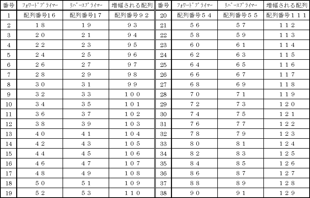
本発明のプローブを得る方法は、上記の本発明のヌクレオチドを得る方法において記載した通りである。
The method for obtaining the probe of the present invention is as described in the above method for obtaining the nucleotide of the present invention.
本発明のプローブは、標識物質で標識されていてもよい。
The probe of the present invention may be labeled with a labeling substance.
本発明のプローブを標識物質で標識するために用いられる標識物質としては、放射性同位体や酵素、蛍光物質、発光物質、ビオチンなど公知の標識物質であれば何れも用いることができる。
As the labeling substance used for labeling the probe of the present invention with a labeling substance, any known labeling substance such as a radioisotope, an enzyme, a fluorescent substance, a luminescent substance, or biotin can be used.
本発明のプローブを標識するために用いられる標識物質の具体例及び標識方法は、本発明のプライマーの標識方法の説明において記載した通りである。
Specific examples of the labeling substance used for labeling the probe of the present invention and the labeling method are as described in the description of the primer labeling method of the present invention.
また、後述するリアルタイムPCRによる検出法において用いられる標識プローブとしては、本発明のプローブを、リアルタイムPCR法において通常用いられている標識物質で標識したものが挙げられる。例えば、5'末端がレポーター蛍光物質[カルボキシフルオレセイン(FAM)、ヘキサクロロフルオレセイン(HEX)、テトラクロロフルオレセイン(TET)等]で標識され、3'末端がクエンチャー色素[例えばカルボキシテトラメチルローダミン(TAMRA)等の蛍光物質、Black Hole Quencher色素(BHQ),4-((4-(dimethylamino) phenyl)azo)benzoic acid (DABCYL)等の非蛍光物質]で標識された本発明のプローブが挙げられる。
Further, examples of the labeled probe used in the detection method by real-time PCR described later include those obtained by labeling the probe of the present invention with a labeling substance usually used in the real-time PCR method. For example, the 5 ′ end is labeled with a reporter fluorescent substance [carboxyfluorescein (FAM), hexachlorofluorescein (HEX), tetrachlorofluorescein (TET), etc.], and the 3 ′ end is a quencher dye [for example, carboxytetramethylrhodamine (TAMRA) And non-fluorescent substances such as Black Hole Quencher dye (BHQ), 4-((4- (dimethylamino) phenyl) azo) benzoic acidDA (DABCYL)].
後述するTaqManTMリアルタイムPCRによる検出法においても、上記した標識プローブを用いることができる。
The above-described labeled probe can also be used in the detection method by TaqMan ™ real-time PCR described later.
本発明に係るM.イントラセルラーの検出に用いられる試料(被検試料)としては、喀痰、血液、咽頭粘液、胃液、気管支洗浄液、経気管支採取物、胸水などの穿刺液、尿、膿等の各種臨床材料が挙げられる。また、検体から単離、培養された培養菌体、これらより単離、精製された核酸、又は核酸増幅検出系等で増幅された核酸でもよい。
Samples (test samples) used for the detection of M. intracellulare according to the present invention include sputum, blood, pharyngeal mucus, gastric fluid, bronchial lavage fluid, transbronchial collection, puncture fluid such as pleural effusion, urine, pus, etc. Various clinical materials are listed. Further, it may be a cultured microbial cell isolated and cultured from a specimen, a nucleic acid isolated and purified from these, or a nucleic acid amplified by a nucleic acid amplification detection system or the like.
上記試料からDNAを抽出・精製するには、検体からの抗酸菌(結核菌)のDNA抽出に用いられる常法に従って行えばよい。
In order to extract and purify DNA from the above sample, it may be carried out according to a conventional method used for DNA extraction of acid-fast bacteria (tuberculosis) from the specimen.
まず、試料中の菌体の細胞壁を破壊する必要がある。その方法としては、例えば菌体を試料とする場合には、例えばSDS等の界面活性剤や、グアニジンチオシアネート(GTC)等の蛋白変性剤で菌体を処理して結核菌等の抗酸菌の膜構造を破壊する方法、菌体をガラスビーズ等によって物理的に破砕する方法等が用いられる。
First, it is necessary to destroy the cell walls of the cells in the sample. As a method, for example, in the case of using a microbial cell as a sample, the microbial cell is treated with a surfactant such as SDS or a protein denaturant such as guanidine thiocyanate (GTC), and the acid-fast bacterium such as Mycobacterium tuberculosis is treated. A method for destroying the membrane structure, a method for physically crushing bacterial cells with glass beads, and the like are used.
喀痰を検体として用いる場合には、まず前処理として、米国疾病管理予防センター(Centers for Disease Control and Prevention、略称CDC)で推奨しているNALC (N-acetyl-L-cysteine) -NaOH法(Kent PT, Kubica GP, Pubric Health Mycobacteriology, A Guide for the Level III Laboratory, U.S.Department of Health and Human Services, Public Health Service, Center for Disease Control, Atlanta, U.S.A., 1985年, p.31-55)による検体の均質化を行うことが望ましい。
When 喀 痰 is used as a specimen, the NALC (N-acetyl-L-cysteine) Na-NaOH method (Kent) recommended by the Centers for Disease Control and Prevention (CDC) is first prepared as a pretreatment. PT, Kubica GP, Pubric Health Mycobacteriology, A Guide for the Level III Laboratory, eUSDepartment of Health and Human Services, Public Health Service, Center for Disease Control, Atlanta, USA, 1985, 1985-31) It is desirable to perform homogenization.
菌体の細胞壁を破壊した後、この分野で一般的なDNAの調製法[フェノール・クロロホルム抽出、エタノール沈殿法、イソプロパノールを用いて沈殿させる方法等(R. Boom, C. J. A. SOL, M. M. M. SALIMANS, C L. JANSEN, P. M. E. WERTHEIM-van DILLEN, J. VAN DER NOORDAA, Rapid and Simple Method for Purification of Nucleic Acids, J. Clin. Microbiol., 1990, Mar;28(3), pp.495-503)]によりDNAの抽出及び精製を行えばよい。
After disrupting the cell walls of the cells, DNA preparation methods commonly used in this field [phenol / chloroform extraction, ethanol precipitation, precipitation using isopropanol, etc. (R. Boom, C. J. A. SOL, M. M. M. SALIMANS, C L. JANSEN, P. M. E. WERTHEIM-van DILLEN, J. VAN DER NOORDAA, Rapid and Simple Method for Purification of Nucleic Acids, J. Clin. Microbi. ; 28 (3), pp. 495-503)].
DNAの抽出・精製には、そのための様々なキットが市販されているので、それを用いてもよいし、この分野における常法(例えば、フェノール・クロロホルム抽出法、エタノールやイソプロパノール等を用いて沈殿させる方法等)に従って行ってもよい。例えば(株)キアゲン製イオン交換樹脂タイプ DNA抽出精製キットGenomic-tip等を用いてDNAの抽出、精製を行えばよい。
Various kits for this purpose are commercially available for DNA extraction / purification, and they may be used, or may be precipitated using conventional methods in this field (for example, phenol / chloroform extraction, ethanol, isopropanol, etc.). Or the like). For example, DNA extraction and purification may be performed using an ion exchange resin type DNA extraction and purification kit Genomic-tip manufactured by Qiagen Co., Ltd.
検体から単離、培養された培養菌体を試料として用いる場合を例にとって示すと、次の通りである。
As an example, the case of using cultured cells isolated and cultured from a specimen as a sample is as follows.
例えば小川培地上のコロニーを採取し、滅菌蒸留水に懸濁、遠心分離して菌体を集めた後、蒸留水に再懸濁する。次いで菌体の懸濁液をオートクレーブ処理した後、菌体の粉砕処理(ガラスビーズによる物理的破砕等)を行い、さらに遠心分離して上清を回収する。得られた上清からDNAを抽出・精製すればよい。
For example, colonies on the Ogawa medium are collected, suspended in sterilized distilled water, centrifuged to collect bacterial cells, and then resuspended in distilled water. Next, the cell suspension is autoclaved, and then the cells are pulverized (physically disrupted with glass beads, etc.) and then centrifuged to collect the supernatant. DNA may be extracted and purified from the obtained supernatant.
本発明に係るM.イントラセルラーの検出方法としては、配列番号1~15のいずれかで表される塩基配列の一部若しくは全部、又は配列番号1~15のいずれかで表される塩基配列に対する相補配列の一部若しくは全部を含有し、且つM.イントラセルラー遺伝子の塩基配列とハイブリダイズするオリゴヌクレオチドをプライマー又は/及びプローブとして用いる方法(本発明のオリゴヌクレオチドをプライマー又は/及びプローブとして用いる方法)が挙げられる。
As a method for detecting M. intracellulare according to the present invention, a part or all of the base sequence represented by any of SEQ ID NOs: 1 to 15, or the base sequence represented by any of SEQ ID NOs: 1 to 15 is used. Method using oligonucleotide containing at least part of complementary sequence and hybridizing with base sequence of M. intracellulare gene as primer or / and probe (method using oligonucleotide of the present invention as primer or / and probe) ).
例えば、
(A)本発明のオリゴヌクレオチドをプライマーとして用いて核酸増幅反応を行い、得られたプライマー伸長産物を検出する方法、
(B)本発明のオリゴヌクレオチドを標識物質で標識したものを標識プローブとして用いる方法、
等が挙げられる。以下に、それぞれの方法について説明する。 For example,
(A) A method for detecting a primer extension product obtained by performing a nucleic acid amplification reaction using the oligonucleotide of the present invention as a primer,
(B) a method of using the oligonucleotide of the present invention labeled with a labeling substance as a labeled probe,
Etc. Each method will be described below.
(A)本発明のオリゴヌクレオチドをプライマーとして用い、試料中の核酸を鋳型として用いて核酸増幅反応を行い、得られたプライマー伸長産物を検出する方法
(A)の方法において、本発明のオリゴヌクレオチドをプライマーとして用い、試料中の核酸を鋳型として用いて核酸増幅反応を行う方法としては、例えば、本発明のプライマーを用い、試料中の核酸を鋳型として用いて、DNAポリメラーゼ等による核酸増幅反応[例えばポリメラーゼ連鎖反応(PCR)法、LAMP(Loop-mediated Isothermal Amplification)法(Tsugunori Notomi et al., Nucleic Acid Res., 28, e63, 2000)、ICANTM(Isothermal and Chimeric primer-initiated Amplification of Nucleic acids)法(臨床病理, 51(11), 1061-1067, 2003, Nov)、LCR(ligase chain reaction)法(特開平4-211399号)、SDA(strand displacement amplification)法(特開平8-19394号)]を行ってプライマー伸長させる方法が挙げられる。これによりM.イントラセルラーの塩基配列の特定の領域の配列を増幅させることができるので、得られたプライマー伸長産物を測定することにより、M.イントラセルラーを検出することができる。 (A) A method for detecting a primer extension product obtained by performing a nucleic acid amplification reaction using the oligonucleotide of the present invention as a primer and a nucleic acid in a sample as a template. In the method of (A), the oligonucleotide of the present invention As a method for performing a nucleic acid amplification reaction using a nucleic acid in a sample as a template and using a primer of the present invention as a template and using a nucleic acid in the sample as a template, a nucleic acid amplification reaction using DNA polymerase or the like [ For example, polymerase chain reaction (PCR) method, LAMP (Loop-mediated Isothermal Amplification) method (Tsugunori Notomi et al., Nucleic Acid Res., 28, e63, 2000), ICANTM (Isothermal and Chimeric primer-initiated Amplification of Nucleic acids) Method (clinical pathology, 51 (11), 1061-1067, 2003, Nov), LCR (ligase chain reaction) method (Japanese Patent Laid-Open No. 4-21399), SDA (strand displacement amplification) method Method of primer extension performed JP 8-19394) and the like. Thereby, since the sequence of a specific region of the base sequence of M. intracellulare can be amplified, M. intracellular can be detected by measuring the obtained primer extension product.
(A)の方法において、本発明のオリゴヌクレオチドをプライマーとして用い、試料中の核酸を鋳型として用いて核酸増幅反応を行う方法としては、例えば、本発明のプライマーを用い、試料中の核酸を鋳型として用いて、DNAポリメラーゼ等による核酸増幅反応[例えばポリメラーゼ連鎖反応(PCR)法、LAMP(Loop-mediated Isothermal Amplification)法(Tsugunori Notomi et al., Nucleic Acid Res., 28, e63, 2000)、ICANTM(Isothermal and Chimeric primer-initiated Amplification of Nucleic acids)法(臨床病理, 51(11), 1061-1067, 2003, Nov)、LCR(ligase chain reaction)法(特開平4-211399号)、SDA(strand displacement amplification)法(特開平8-19394号)]を行ってプライマー伸長させる方法が挙げられる。これによりM.イントラセルラーの塩基配列の特定の領域の配列を増幅させることができるので、得られたプライマー伸長産物を測定することにより、M.イントラセルラーを検出することができる。 (A) A method for detecting a primer extension product obtained by performing a nucleic acid amplification reaction using the oligonucleotide of the present invention as a primer and a nucleic acid in a sample as a template. In the method of (A), the oligonucleotide of the present invention As a method for performing a nucleic acid amplification reaction using a nucleic acid in a sample as a template and using a primer of the present invention as a template and using a nucleic acid in the sample as a template, a nucleic acid amplification reaction using DNA polymerase or the like [ For example, polymerase chain reaction (PCR) method, LAMP (Loop-mediated Isothermal Amplification) method (Tsugunori Notomi et al., Nucleic Acid Res., 28, e63, 2000), ICANTM (Isothermal and Chimeric primer-initiated Amplification of Nucleic acids) Method (clinical pathology, 51 (11), 1061-1067, 2003, Nov), LCR (ligase chain reaction) method (Japanese Patent Laid-Open No. 4-21399), SDA (strand displacement amplification) method Method of primer extension performed JP 8-19394) and the like. Thereby, since the sequence of a specific region of the base sequence of M. intracellulare can be amplified, M. intracellular can be detected by measuring the obtained primer extension product.
上記の核酸増幅反応を行う方法の中でも、PCR法が最も一般的な方法として挙げられ、PCR法の例としては、例えばリアルタイム増幅検出法(例えば米国特許第5210015号、米国特許第5538848号の記載参照)を用いることができる。また、リアルタイム増幅検出法による検出法の例として、例えばリアルタイムPCR検出法が挙げられる。
Among the methods for carrying out the nucleic acid amplification reaction, the PCR method is the most general method. Examples of the PCR method include, for example, real-time amplification detection methods (for example, descriptions in US Pat. No. 5210015 and US Pat. No. 5,538,848). Reference) can be used. An example of a detection method using a real-time amplification detection method is, for example, a real-time PCR detection method.
リアルタイムPCR検出法の例としては、TaqManTMリアルタイムPCR法(例えば米国特許第5538848号の記載参照)、MGB Eclipse Probe System法(例えば米国特許第5,801,155号の記載参照)、Molecular Beacons Probe Technology法(例えば米国特許第5925517号の記載参照)、LUX Fluorogenic Primer法(Invitrogen Corporation)、Quenching probe-PCR(QP)法(例えば米国特許第6,492,121号の記載参照)等が挙げられる。
Examples of the real-time PCR detection method include TaqMan ™ real-time PCR method (see, for example, the description in US Pat. No. 5,538,848), MGB Eclipse Probe System method (see, for example, the description in US Pat. No. 5,801,155), Molecular Beacons Probe Technology method (for example, And the LUX Fluorogenic Primer method (Invitrogen Corporation), Quenching probe-PCR (QP) method (see, for example, the description of US Pat. No. 6,492,121), and the like.
PCR等の核酸増幅反応において用いられる本発明のプライマーの具体例は、上記した通りである。
Specific examples of the primer of the present invention used in a nucleic acid amplification reaction such as PCR are as described above.
また、核酸増幅反応に用いられる、好ましいフォワードプライマーとリバースプライマーの組合せとしては、上記表1で示される組合せが挙げられる。
Moreover, as a preferable combination of the forward primer and the reverse primer used in the nucleic acid amplification reaction, combinations shown in Table 1 above can be mentioned.
例えば、表1において、例えば番号1の組み合わせは、「フォワードプライマーが配列番号16で表される塩基配列を含有するオリゴヌクレオチドで、リバースプライマーが配列番号17で表される塩基配列を含有するオリゴヌクレオチドである組合せ。」又は「フォワードプライマーが配列番号16で表される塩基配列に対する相補配列を含有するオリゴヌクレオチドで、リバースプライマーが配列番号17で表される塩基配列に対する相補配列を含有するオリゴヌクレオチドである組合せ。」を示す。
For example, in Table 1, for example, the combination of No. 1 is “an oligonucleotide containing a base sequence represented by SEQ ID NO: 16 with a forward primer and an oligonucleotide containing a base sequence represented by SEQ ID NO: 17 with a reverse primer” Or a combination wherein the forward primer is an oligonucleotide containing a complementary sequence to the base sequence represented by SEQ ID NO: 16 and the reverse primer is an oligonucleotide containing a complementary sequence to the base sequence represented by SEQ ID NO: 17 A certain combination. "
その中でも好ましいフォワードプライマーとリバースプライマーの組合せとしては、例えば下記表2に記載の組合せが挙げられる。
Among these, preferred forward primer and reverse primer combinations include, for example, combinations shown in Table 2 below.
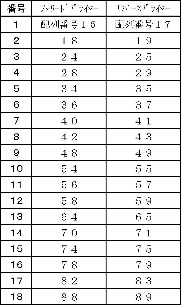
中でも、特に好ましい組合せとしては、組合せの番号1~9、12~18が挙げられる。
Among them, particularly preferable combinations include combination numbers 1 to 9 and 12 to 18.
上記プライマーを用いたリアルタイムPCR等の核酸増幅反応に用いられるその他のデオキシリボヌクレオシド三リン酸(dATP、dCTP、dGTP、dTTP)、DNAポリメラーゼ等の試薬は、通常この分野で用いられているものを用いればよく、その条件、手法等は、本発明のプライマー及びプローブを用いる以外は、PCR法の一般的なプロトコルに従って行えばよい。
Other reagents such as deoxyribonucleoside triphosphates (dATP, dCTP, dGTP, dTTP) and DNA polymerase used in nucleic acid amplification reactions such as real-time PCR using the above primers are those usually used in this field. The conditions, techniques, and the like may be performed according to a general protocol for PCR, except that the primer and probe of the present invention are used.
核酸増幅反応で得られたプライマー伸長産物を検出する方法は、通常この分野で行われている常法で良く、限定されるものではない。
The method of detecting the primer extension product obtained by the nucleic acid amplification reaction may be a conventional method usually performed in this field, and is not limited.
例えばインターカレーター法、TaqManTMリアルタイムPCR法(例えば米国特許第5538848号の記載参照)、MGB Eclipse Probe System法(例えば米国特許第5,801,155号の記載参照)、Molecular Beacons Probe Technology法(例えば米国特許第5925517号の記載参照)、LUX Fluorogenic Primer法(Invitrogen Corporation)、Quenching probe-PCR(QP)法(例えば米国特許第6,492,121号の記載参照)や、核酸増幅反応を行った後、得られたプライマー伸長産物について電気泳動を行い、その結果に基づいて判定を行う方法、標識プライマーを用いた核酸増幅反応を行って得られたプライマー伸長産物の標識を測定する方法等、様々な検出法が挙げられる。
For example, intercalator method, TaqMan ™ real-time PCR method (see, for example, the description in US Pat. No. 5,538,848), MGB Eclipse Probe System method (see, for example, the description in US Pat. No. 5,801,155), Molecular Beacons Probe Technology method (for example, in US Pat. No. 5,925,517) No.), LUX Fluorogenic Primer method (Invitrogen Corporation), Quenching probe-PCR (QP) method (see, for example, the description in US Pat. No. 6,492,121), and nucleic acid amplification reaction, and the resulting primer extension product There are various detection methods such as a method for performing electrophoresis on the basis of the above and determining based on the result, and a method for measuring the label of the primer extension product obtained by performing a nucleic acid amplification reaction using a labeled primer.
これらのうち、一般によく用いられる方法としては、例えば、以下の方法が挙げられる。
Of these, examples of methods that are commonly used include the following methods.
(A-1)インターカレーター法、
(A-2)TaqManTMリアルタイムPCR法、
(A-3)核酸増幅反応を行った後、得られたプライマー伸長産物について電気泳動を行い、その結果に基づいて(判定を)行う方法、
(A-4)標識プライマーを用いた核酸増幅反応を行って得られたプライマー伸長産物の標識を測定する方法。 (A-1) Intercalator method,
(A-2) TaqMan ™ real-time PCR method,
(A-3) A method of performing electrophoresis on the obtained primer extension product after performing a nucleic acid amplification reaction, and performing (determination) based on the result,
(A-4) A method for measuring the label of a primer extension product obtained by performing a nucleic acid amplification reaction using a labeled primer.
以下に、それぞれの方法について説明する。
(A-1)インターカレーター法
公知のインターカレーターを利用してリアルタイムPCRを行う、通常のインターカレーター法が利用できる。 Each method will be described below.
(A-1) Intercalator method The usual intercalator method for performing real-time PCR using a known intercalator can be used.
(A-1)インターカレーター法
公知のインターカレーターを利用してリアルタイムPCRを行う、通常のインターカレーター法が利用できる。 Each method will be described below.
(A-1) Intercalator method The usual intercalator method for performing real-time PCR using a known intercalator can be used.
例えば、本発明のプライマーと、インターカレーターを用い、通常のインターカレーター法を利用したリアルタイムPCRを行う方法が挙げられる。
For example, a method of performing real-time PCR using a normal intercalator method using the primer of the present invention and an intercalator can be mentioned.
すなわち、インターカレーターは、二本鎖DNAに特異的に結合して蛍光を発する試薬であり、励起光を照射すると蛍光を発する。PCRによって増幅を繰り返してDNAが増えると、インターカレーターがそのDNAに取り込まれるので、プライマー伸長産物の生成量に比例して、DNAに取り込まれていく。そこで、インターカレーターに由来する蛍光強度を検出することにより、プライマー伸長産物の量を知ることができる。
That is, the intercalator is a reagent that specifically binds to double-stranded DNA and emits fluorescence, and emits fluorescence when irradiated with excitation light. When amplification is repeated by PCR and the DNA increases, the intercalator is incorporated into the DNA, so that it is incorporated into the DNA in proportion to the amount of primer extension product generated. Therefore, the amount of the primer extension product can be known by detecting the fluorescence intensity derived from the intercalator.
但しインターカレーターは全ての二本鎖DNAに結合するので、得られた蛍光強度の測定結果を基に、必要に応じ、融解曲線分析を行う。すなわち、PCR後にPCR反応液の温度を徐々に上げながら、インターカレーター由来の蛍光強度を測定する。最初はPCR増幅産物は二本鎖を形成しているので蛍光を発しているが、PCR反応液の温度がある一定の温度に達すると一本鎖に解離するので、インターカレーター由来の蛍光は急激に低下する。この時の温度が融解温度(Tm値)であり、プライマー伸長産物の配列に固有の値である。融解曲線のピークが、目的とする特異産物のピークか、又は非特異産物のピークかについては、このTm値から判定することができる。
However, since the intercalator binds to all double-stranded DNAs, a melting curve analysis is performed as necessary based on the measurement result of the obtained fluorescence intensity. That is, the fluorescence intensity derived from the intercalator is measured while gradually raising the temperature of the PCR reaction solution after PCR. At first, the PCR amplification product forms a double strand, so it fluoresces, but when the temperature of the PCR reaction solution reaches a certain temperature, it dissociates into a single strand, and the intercalator-derived fluorescence rapidly To drop. The temperature at this time is the melting temperature (Tm value), which is a value unique to the sequence of the primer extension product. Whether the peak of the melting curve is a peak of a specific product of interest or a peak of a non-specific product can be determined from this Tm value.
このインターカレーター法は、リアルタイムPCRの後に電気泳動を行う必要がないので、臨床検査の分野等において、迅速に判定を行う必要がある場合には、有効な方法である。
This intercalator method does not require electrophoresis after real-time PCR, so it is an effective method when it is necessary to make a quick determination in the field of clinical examinations.
本発明に用いられるインターカレーターとしては、通常この分野で用いられているインターカレーターであれば、何でも用いることができるが、例えばSYBRTM Green I (Molecular Probe社商品名)、エチジウムブロマイド、フルオレン等がある。
As the intercalator used in the present invention, any intercalator usually used in this field can be used. For example, SYBR ™ Green I (trade name of Molecular Probe), ethidium bromide, fluorene, etc. is there.
本発明に係る「インターカレーター法を利用したM.イントラセルラーの検出方法」の例を説明すると、以下の通りである。
An example of “M. Intracellular detection method using intercalator method” according to the present invention will be described as follows.
本発明のプライマーと、インターカレーター(例えばSYBRTM Green I)を用い、被検試料から精製した精製DNA試料を鋳型として用いて、Taq DNA ポリメラーゼ等のポリメラーゼを用いたリアルタイムPCRを行う。そして上記した温度を上げる方法で、プライマー伸長産物の増幅量と相関してインターカレーションするインターカレーター(SYBRTM Green I)由来の蛍光強度を測定する。
Using the primer of the present invention and an intercalator (eg, SYBR ™ Green I), a purified DNA sample purified from a test sample is used as a template, and real-time PCR is performed using a polymerase such as Taq DNA polymerase. Then, the fluorescence intensity derived from the intercalator (SYBR ™ Green I) that intercalates in correlation with the amplification amount of the primer extension product is measured by the method of raising the temperature described above.
次いで、横軸をプライマー伸長産物(二本鎖DNA)の解離温度、縦軸に蛍光強度の1次微分(変化量)をとった融解曲線を作成する。これを用いて、プライマー伸長産物の融解曲線解析を行うことによって、ピークの検出を行う。そして、単一のピークが得られた場合に、被検試料はM.イントラセルラー陽性(すなわち、M.イントラセルラー菌、又はその遺伝子が存在する。以下同じ。)と判定される。
Next, a melting curve is prepared with the horizontal axis representing the dissociation temperature of the primer extension product (double-stranded DNA) and the vertical axis representing the first derivative (change amount) of the fluorescence intensity. Using this, a peak is detected by analyzing a melting curve of the primer extension product. When a single peak is obtained, the test sample is M.P. Intracellular positive (that is, M. intracellulare or its gene is present. The same applies hereinafter).
また、M.イントラセルラーの判定をより正確に行うには、以下の方法が好ましい。
In addition, the following method is preferable for more accurate determination of M. intracellular.
すなわち、被検試料を用いて上記の測定を行い、融解曲線を作成し、ピークの検出を行う。別に、M.イントラセルラーのtype strain(基準株)を用いて上記と同様の測定を行い、融解曲線解析を行って、ピークの検出を行う。そして、被検試料を用いて得られた結果、単一のピークが得られ、かつそのピークの位置がM.イントラセルラーのtype strainを用いて得られたピークの位置と同じかそれと近い位置に出現した場合に、被検試料はM.イントラセルラー陽性と判定する方法である。
That is, the above measurement is performed using a test sample, a melting curve is created, and a peak is detected. Separately, the same measurement as described above is performed using M. intracellulare type strain (reference strain), and a melting curve analysis is performed to detect a peak. As a result of using the test sample, a single peak is obtained, and the position of the peak is the same as or close to the position of the peak obtained using M. intracellular type strain. When it appears, the test sample is determined to be positive for M. intracellulare.
尚、M.イントラセルラーのtype strainを用いて予め測定を行い、ピークの位置を確認しておけば、被検試料の測定の度に、type strainについても同様に測定を行ってピークの位置を確認する必要はない。
In addition, if the measurement is performed in advance using the M. intracellular typetrainstrain and the peak position is confirmed, the same measurement is performed for the type strain every time the sample is measured. There is no need to check.
また、インターカレーター法を利用した方法で得られた測定値をもとに、リアルタイムPCRにおいて行われる常法に従って、検量線を作成することもできるので、その検量線を用いて試料中にあるM.イントラセルラーのゲノムDNA量(コピー数)を得ることができる。
In addition, based on the measurement value obtained by the method using the intercalator method, a calibration curve can be created according to a conventional method performed in real-time PCR. Intracellular genomic DNA content (copy number) can be obtained.
検量線の作成方法及びそれを用いたM.イントラセルラーの定量方法は後記する。
A method for preparing a calibration curve and a method for quantifying M. intracellular using the calibration curve will be described later.
本発明に係るインターカレーターを用いたリアルタイムPCR検出法によるM.イントラセルラーの検出方法の一例として、上記した「プライマーMint 02_T7pa Fw1」と「プライマーMint 02_T7pa Rv1」を用いて、M.イントラセルラーを検出する場合を例にとって説明すると、以下の通りである。
As an example of the M. intracellular detection method by the real-time PCR detection method using the intercalator according to the present invention, the above-described “primer Mint 02_T7pa Fw1” and “primer Mint 02_T7pa Rv1” are used to detect M. intracellular. An example of this is as follows.
まず、公知の方法により、被検試料中から精製DNA試料を得る。
First, a purified DNA sample is obtained from a test sample by a known method.
別に、例えばDNAシンセサイザーを用いて、ホスホアミダイト法にて、配列番号16で表される塩基配列からなるオリゴヌクレオチド(Mint 02_T7pa Fw1)、及び配列番号17で表される塩基配列からなるオリゴヌクレオチド(Mint 02_T7pa Rv1)を合成する。
Separately, for example, using a DNA synthesizer, an oligonucleotide consisting of the base sequence represented by SEQ ID NO: 16 (Mint 02_T7pa Fw1) and an oligonucleotide consisting of the base sequence represented by SEQ ID NO: 17 (Mint) by the phosphoramidite method Synthesize 02_T7pa Rv1).
上記で合成したMint 02_T7pa Fw1をフォワードプライマーとして、02_Rv1をリバースプライマーとして用い、例えば下記の通りリアルタイムPCRを行う。
For example, real-time PCR is performed as described below using Mint 02_T7pa Fw1 synthesized above as a forward primer and 02_Rv1 as a reverse primer.
すなわち、各50~2000nMのプライマーMint 02_T7pa Fw1及びプライマーMint 02_T7pa Rv1、インターカレーター[例えばSYBRTM Green I (Molecular Probe社商品名)]を原液の約5000~100000倍希釈、1.0~4.0mM MgCl2、KCl、BSA、コール酸ナトリウム、0.005~0.2% TritonX-100、それぞれ0.2mM程度のdATP、dCTP、dGTP、dTTP、10~80単位/mLのポリメラーゼ(例えばTaq DNA ポリメラーゼ)を含有する10mM Tris-HCl緩衝液(pH8.9)を調製し、PCR用反応液とする。このPCR用反応液に、精製DNA試料を加え、PCR用試料とする。このPCR用試料を用い、適当なリアルタイムPCR検出装置等を用いてリアルタイムPCRを行う。反応は30~50回サイクル繰り返し、1サイクル毎にプライマー伸長産物の増幅量に相関してインターカレーションするインターカレーター(例えばSYBRTM Green I)由来の蛍光強度を測定する。
That is, 50 to 2000 nM of each primer Mint 02_T7pa Fw1 and primer Mint 02_T7pa Rv1, an intercalator [for example, SYBR ™ Green I (trade name of Molecular Probe)] diluted about 5000 to 100,000 times the stock solution, 1.0 to 4.0 mM MgCl 2 , 10 mM Tris-HCl containing KCl, BSA, sodium cholate, 0.005-0.2% TritonX-100, each containing about 0.2 mM dATP, dCTP, dGTP, dTTP, 10-80 units / mL polymerase (eg Taq DNA polymerase) A buffer solution (pH 8.9) is prepared and used as a PCR reaction solution. A purified DNA sample is added to the PCR reaction solution to obtain a PCR sample. Using this PCR sample, real-time PCR is performed using an appropriate real-time PCR detection device or the like. The reaction is repeated 30 to 50 times, and the fluorescence intensity derived from an intercalator (for example, SYBR ™ Green I) that intercalates in correlation with the amplification amount of the primer extension product is measured every cycle.
次いで、横軸をプライマー伸長産物(二本鎖DNA)の解離温度、縦軸に蛍光強度の一次微分(変化量)をとった融解曲線を作成する。これを用いて、プライマー伸長産物の融解曲線解析を行い、ピークの検出を行う。そして、単一のピークが得られた場合に、被検試料はM.イントラセルラー陽性と判定される。
Next, a melting curve is prepared in which the horizontal axis represents the dissociation temperature of the primer extension product (double-stranded DNA) and the vertical axis represents the first derivative (change amount) of the fluorescence intensity. Using this, a melting curve analysis of the primer extension product is performed, and a peak is detected. When a single peak is obtained, the test sample is M.P. Intracellular positive.
より好ましくは、被検試料を用いた測定、融解曲線解析で得られたピークの位置が、M.イントラセルラーのtype strainを用いて上記と同様の測定、融解曲線解析で得られたピークの位置と同じかそれと近い位置に出現した場合に、被検試料はM.イントラセルラー陽性と判定される。
More preferably, the position of the peak obtained by the measurement using the test sample and the melting curve analysis is the same as the above by using the M. intracellular type ラ ー strain, and the position of the peak obtained by the melting curve analysis. When the sample appears at the same position as or close to it, the test sample is determined to be positive for M. intracellulare.
また、対照として、M.イントラセルラー以外のマイコバクテリウム属菌由来DNAを常法により抽出・精製し、これを鋳型として用いる以外は、上記と同様の方法でリアルタイムPCRを行い、同様にSYBRTM Green Iの蛍光強度を測定し、融解曲線解析を行ってもよい。この場合は、試料中にM.イントラセルラー由来の塩基配列を持つポリヌクレオチドがないので、融解曲線解析でピークは出現しないはずである。M.イントラセルラーの有無の判定をより確実にするために、上記した対照実験を一緒に行ってもよい。
In addition, as a control, real-time PCR was performed in the same manner as described above except that DNA derived from Mycobacterium other than M. intracellulare was extracted and purified by a conventional method and used as a template. Similarly, SYBR ™ The fluorescence intensity of Green I may be measured and a melting curve analysis may be performed. In this case, since there is no polynucleotide having a base sequence derived from M. intracellulare in the sample, no peak should appear in the melting curve analysis. In order to more reliably determine the presence or absence of M. intracellular, the control experiment described above may be performed together.
更に、検量線を作成することによって、試料中のM.イントラセルラーのゲノムDNAの数(コピー数)を得ることができる。また、その数はM.イントラセルラーの数に比例するので、試料(被検試料)中のM.イントラセルラーの数も知ることができる。
Furthermore, by creating a calibration curve, M. Intracellular genomic DNA number (copy number) can be obtained. The number is M.M. Since it is proportional to the number of intracellular, the M. You can also know the number of intracellular.
(A-2)TaqManTMリアルタイムPCR法(TaqManTMプローブ法)
TaqManTMリアルタイムPCR法は、5'末端を例えばFAM等の蛍光色素(レポーター)で、3'末端を例えばTAMRA等のクエンチャー色素で標識した標識プローブを用いたリアルタイムPCR法で、目的の微量なDNAを高感度且つ定量的に検出する方法である(例えば米国特許第5,538,848号の記載参照)。 (A-2) TaqMan ™ real-time PCR method (TaqMan ™ probe method)
The TaqMan ™ real-time PCR method is a real-time PCR method using a labeled probe in which the 5 ′ end is labeled with a fluorescent dye (reporter) such as FAM and the 3 ′ end is labeled with a quencher dye such as TAMRA. This is a method for highly sensitive and quantitative detection of DNA (see, for example, the description of US Pat. No. 5,538,848).
すなわち、本法は、本発明のプライマーと、本発明のプローブの5'末端がレポーター蛍光色素で標識され、3'末端がクエンチャー色素で標識された標識プローブを用いて、試料中の核酸を鋳型としてPCRを行い、該標識プローブから遊離された標識物質の標識を検出する方法である。
That is, the present method uses the primer of the present invention and a labeled probe in which the 5 ′ end of the probe of the present invention is labeled with a reporter fluorescent dye and the 3 ′ end is labeled with a quencher dye, and the nucleic acid in the sample is collected. In this method, PCR is performed as a template to detect the label of the labeling substance released from the labeled probe.
TaqManTMリアルタイムPCR法の原理は以下の通りである。
The principle of the TaqMan ™ real-time PCR method is as follows.
この方法には、5'末端を蛍光色素(レポーター)で、3'末端をクエンチャー色素で標識した、目的遺伝子の特定領域にハイブリダイズするオリゴヌクレオチドプローブが使用される。該プローブは、通常の状態ではクエンチャー色素によってレポーターの蛍光が抑制されている。この蛍光標識プローブを目的遺伝子に完全にハイブリダイズさせた状態で、その外側からDNAポリメラーゼを用いてPCRを行う。DNAポリメラーゼによる伸長反応が進むと、そのエキソヌクレアーゼ活性により蛍光標識プローブが5'末端から加水分解され、レポーター色素が遊離し、蛍光を発する。リアルタイムPCR法は、この蛍光強度をリアルタイムでモニタリングする方法であり、これにより、鋳型DNAの初期量を正確に定量することができる。
In this method, an oligonucleotide probe that hybridizes to a specific region of a target gene, which is labeled with a fluorescent dye (reporter) at the 5 ′ end and a quencher dye at the 3 ′ end, is used. In the probe, the fluorescence of the reporter is suppressed by a quencher dye in a normal state. In a state in which this fluorescently labeled probe is completely hybridized to the target gene, PCR is performed from the outside using DNA polymerase. As the elongation reaction by DNA polymerase proceeds, the exonuclease activity hydrolyzes the fluorescently labeled probe from the 5 ′ end, releases the reporter dye, and emits fluorescence. The real-time PCR method is a method for monitoring the fluorescence intensity in real time, whereby the initial amount of template DNA can be accurately quantified.
本発明に係るTaqManTMリアルタイムPCR検出法に用いられるフォワードプライマー及びリバースプライマーには、本発明のプライマーが用いられる。好ましいプライマーとしては、上記したPCR法等の核酸増幅反応において用いられるものが挙げられ、その好ましい具体例及び好ましい組合せも上記した通りである。
The primer of the present invention is used as a forward primer and a reverse primer used in the TaqMan ™ real-time PCR detection method according to the present invention. Preferred primers include those used in nucleic acid amplification reactions such as the PCR method described above, and preferred specific examples and preferred combinations thereof are also as described above.
本発明に係るTaqManTMリアルタイムPCR検出系に用いられる5'末端を蛍光色素(レポーター)で、3'末端をクエンチャー色素で標識したプローブに用いられるプローブとしては、上記した本発明のプローブであればよい。実際には、選択したフォワードプライマーとリバースプライマーの組合せでリアルタイムPCRを行った場合に得られると予測されるプライマー伸長産物の塩基配列を含有するプローブ、又は更にその配列から設計される塩基配列を含有するプローブが用いられる。
The probe used in the TaqMan ™ real-time PCR detection system according to the present invention for the probe labeled with a fluorescent dye (reporter) at the 5 ′ end and a quencher dye at the 3 ′ end may be the probe of the present invention described above. That's fine. Actually, it contains a probe containing the base sequence of the primer extension product predicted to be obtained when real-time PCR is performed with a combination of the selected forward primer and reverse primer, or a base sequence designed from that sequence. A probe is used.
例えば、上記した本発明のMint 02_T7pa Fw1とMint 02_T7pa Rv1のプライマーの組み合わせを用いてリアルタイムPCRを行う場合に用いられるプローブは、そのリアルタイムPCRで増幅されると予想される配列暗号92で表される塩基配列の一部若しくは全部を含有するオリゴヌクレオチドが挙げられる。
For example, a probe used when performing real-time PCR using a combination of the above-described primers of Mint 02_T7pa Fw1 and Mint 02_T7pa Rv1 of the present invention is represented by the sequence code 92 expected to be amplified by the real-time PCR. Examples thereof include oligonucleotides containing a part or all of the base sequence.
標識プローブの5'末端を標識するレポーター蛍光物質としては、カルボキシフルオレセイン(FAM)、ヘキサクロロフルオレセイン(HEX)、テトラクロロフルオレセイン(TET)、Cy5、VIC等が挙げられるが、中でもFAMがよく用いられる。
Examples of the reporter fluorescent substance for labeling the 5 ′ end of the labeled probe include carboxyfluorescein (FAM), hexachlorofluorescein (HEX), tetrachlorofluorescein (TET), Cy5, VIC, etc. Among them, FAM is often used.
3'末端を標識するクエンチャー色素としては、カルボキシテトラメチルローダミン(TAMRA)等の蛍光物質、Black Hole Quencher色素(例えばBHQ2),4-((4-(dimethylamino) phenyl)azo)benzoic acid (DABCYL)等の非蛍光物質が挙げられるが、中でもTAMRAがよく用いられる。
Quencher dyes that label the 3 ′ end include fluorescent substances such as carboxytetramethylrhodamine (TAMRA), Black Hole Quencher dyes (eg BHQ2), 4-((4- (dimethylamino) phenyl) azo) benzoic acid (DABCYL And non-fluorescent substances such as TAMRA is often used.
TaqManTMリアルタイムPCR検出系に用いられるその他のデオキシリボヌクレオシド三リン酸(dATP、dCTP、dGTP、dTTP)、DNAポリメラーゼ等の試薬は、通常のリアルタイムPCRで用いられているものを用いればよく、リアルタイムPCRの手法は、本発明のプライマー及びプローブを用いる以外は、リアルタイムPCRの一般的なプロトコルに従って行えばよい。
Other reagents such as deoxyribonucleoside triphosphates (dATP, dCTP, dGTP, dTTP) and DNA polymerase used in TaqMan TM real-time PCR detection systems may be those used in normal real-time PCR. This method may be performed according to a general protocol for real-time PCR except that the primer and probe of the present invention are used.
本発明に係るTaqManTMリアルタイムPCR検出法によるM.イントラセルラーの検出方法の一例として、上記した本発明の「プライマーMint 02_T7pa Fw1」と「プライマーMint 02_T7pa Rv1」を用いて、M.イントラセルラーを検出する場合を例にとって説明すると、以下の通りである。
As an example of the method for detecting M. intracellular by the TaqMan ™ real-time PCR detection method according to the present invention, the above-described “primer Mint 02_T7pa Fw1” and “primer Mint 02_T7pa Rv1” of the present invention are used to detect M. intracellular. An example of this is as follows.
まず、公知の方法により、被検試料中から精製DNA試料を得る。
First, a purified DNA sample is obtained from a test sample by a known method.
別に、例えばDNAシンセサイザーを用いて、ホスホアミダイト法にて、配列番号16で表される塩基配列からなるオリゴヌクレオチド(Mint 02_T7pa Fw1)、及び配列番号17で表される塩基配列からなるオリゴヌクレオチド(Mint 02_T7pa Rv1)を合成する。
Separately, for example, using a DNA synthesizer, an oligonucleotide consisting of the base sequence represented by SEQ ID NO: 16 (Mint 02_T7pa Fw1) and an oligonucleotide consisting of the base sequence represented by SEQ ID NO: 17 (Mint) by the phosphoramidite method Synthesize 02_T7pa Rv1).
また、Mint 02_T7pa Fw1とMint 02_T7pa Rv1のプライマー対を用いたPCRで増幅されると予想される配列番号92の塩基配列から、プローブとして利用するための配列を設計し、この塩基配列のオリゴヌクレオチドを合成する。このオリゴヌクレオチドの5'末端にレポーター色素のFAMを、3'末端にレポーター消光体のTAMRAを常法により結合し、蛍光標識プローブを得る。
In addition, a sequence for use as a probe was designed from the nucleotide sequence of SEQ ID NO: 92, which is expected to be amplified by PCR using a primer pair of Mint 02_T7pa Fw1 and Mint 02_T7pa Rv1, and an oligonucleotide having this nucleotide sequence was designed. Synthesize. A reporter dye FAM is bound to the 5 ′ end of the oligonucleotide and a reporter quencher TAMRA is bound to the 3 ′ end by a conventional method to obtain a fluorescently labeled probe.
上記で合成したMint 02_T7pa Fw1をフォワードプライマーとして、Mint 02_T7pa Rv1をリバースプライマーとして用い、例えば下記の通りリアルタイムPCRを行う。
For example, real-time PCR is performed as follows using Mint 02_T7pa Fw1 synthesized above as a forward primer and Mint 02_T7pa Rv1 as a reverse primer.
すなわち、各0.1~2μM、好ましくは各1μMのプライマーMint 02_T7pa Fw1及びプライマーMint 02_T7pa Rv1、100~1000nMの蛍光標識プローブ、1.0~4.0mM MgCl2 、KCl、BSA、コール酸ナトリウム、0.005~0.2% TritonX-100、それぞれ0.2mM程度のdATP、dCTP、dGTP、dTTP、10~80単位/mLのTaq DNA ポリメラーゼ等のポリメラーゼを含有する10mM Tris-HCl緩衝液(pH8.9)を調製し、PCR用反応液とする。このPCR用反応液に精製DNA試料を加え、PCR用試料を得る。
That is, each 0.1 to 2 μM, preferably each 1 μM primer Mint 02_T7pa Fw1 and primer Mint 02_T7pa Rv1, 100 to 1000 nM fluorescently labeled probe, 1.0 to 4.0 mM MgCl 2 , KCl, BSA, sodium cholate, 0.005 to 0.2% TritonX -100, 10 mM Tris-HCl buffer (pH 8.9) containing about 0.2 mM each of dATP, dCTP, dGTP, dTTP, 10-80 units / mL Taq DNA polymerase, etc. Use liquid. A purified DNA sample is added to the PCR reaction solution to obtain a PCR sample.
このPCR用試料を用い、適当なリアルタイムPCR検出装置等を用いてリアルタイムPCRを行う。反応は30~50回サイクル繰り返し、1サイクル毎にレポーター色素由来の蛍光を測定する。
Using this PCR sample, real-time PCR is performed using an appropriate real-time PCR detection device or the like. The reaction is repeated 30-50 times, and the fluorescence derived from the reporter dye is measured every cycle.
この場合のM.イントラセルラー検出方法としては、レポーター色素の蛍光が測定された場合に、被検試料はM.イントラセルラー陽性と判定される。
As the M. intracellular detection method in this case, when the fluorescence of the reporter dye is measured, the test sample is determined to be M. intracellular positive.
また、リアルタイムPCR法では、検量線を作成することができるので、試料中のM.イントラセルラーのゲノムDNAの数(コピー数)を得ることがでる。また、その数はM.イントラセルラーの数に比例するので、試料(被検試料)中のM.イントラセルラーの数も知ることができる。
In addition, since a calibration curve can be created in the real-time PCR method, the number (copy number) of M. intracellular genomic DNA in the sample can be obtained. Further, since the number is proportional to the number of M. intracellular, the number of M. intracellular in the sample (test sample) can also be known.
検量線の作成方法は、リアルタイムPCR法において通常行われている常法に従えばよい。例えば、標準としてコピー数既知のM.イントラセルラーのゲノムDNA試料を用い、希釈系列の濃度(コピー数)のPCR用DNA試料を調製する。次いで各希釈系列のPCR用DNA試料を用いて上記方法に従いリアルタイムPCRを行い、レポーター色素由来の蛍光強度を測定する。各希釈系列のPCR用DNA試料毎に、PCRの各サイクル数(x軸)に対する、測定した蛍光強度の測定値(Rn、y軸)をプロットした増幅曲線を作成する。次いで、蛍光強度が指数関数的に増幅しているRn部を選択し、Threshold line(Th)を引く。Thと各PCR用DNA試料の増幅曲線が交差した点をThreshold cycle(Ct)値とする。次いで用いた各PCR用DNA試料のコピー数の対数値(x軸)に対するCt値(y軸)をプロットし、各Ctに対して得られた近似曲線を検量線とすればよい。
The method for creating a calibration curve may be a conventional method that is usually performed in the real-time PCR method. For example, using a M. intracellulare genomic DNA sample with a known copy number as a standard, a DNA sample for PCR having a dilution series concentration (copy number) is prepared. Next, real-time PCR is performed according to the above-described method using PCR samples of each dilution series, and the fluorescence intensity derived from the reporter dye is measured. For each PCR sample of each dilution series, an amplification curve is prepared by plotting the measured values of fluorescence intensity (Rn, y-axis) with respect to the number of PCR cycles (x-axis). Next, the Rn part where the fluorescence intensity is amplified exponentially is selected, and Threshold line (Th) is drawn. The point at which Th and the amplification curve of each DNA sample for PCR intersect is defined as the threshold cycle (Ct) value. Next, the Ct value (y axis) against the logarithmic value (x axis) of the copy number of each PCR DNA sample used may be plotted, and the approximate curve obtained for each Ct may be used as a calibration curve.
先に記載した、インターカレーター法によるリアルタイムPCRを行った場合も、得られた測定値を基に同様に検量線を作成することができる。例えば、PCRの各サイクル数(x軸)に対するインターカレーター由来の蛍光強度の測定値(Rn、y軸)をプロットした増幅曲線を作成する。次いで、上記と同じ方法でCt値を得、リアルタイムPCRに用いた各PCR用DNA試料のコピー数の対数値(x軸)に対するCt値(y軸)をプロットし、各Ctに対して得られた近似曲線を検量線とすればよい。
Even when the real-time PCR by the intercalator method described above is performed, a calibration curve can be similarly created based on the obtained measurement values. For example, an amplification curve is prepared by plotting measured values (Rn, y-axis) of fluorescence intensity derived from an intercalator with respect to each cycle number (x-axis) of PCR. Next, Ct values were obtained by the same method as above, and Ct values (y-axis) against logarithmic values (x-axis) of copy numbers of DNA samples for PCR used in real-time PCR were plotted and obtained for each Ct. The approximate curve may be used as a calibration curve.
試料中のM.イントラセルラーのゲノムDNAの数(コピー数)を定量するには、先ず被検試料中からDNAを分離精製した後、得られたDNA試料についてリアルタイムPCRを行い、同様に増幅曲線を作成する。検量線を作成したときのThと得られた増幅曲線が交差したCt値を得る。そのCt値を検量線に当てはめることにより、試料中のM.イントラセルラーのゲノムDNA量(コピー数)を得ることができる。
To quantify the number (copy number) of M. intracellulare genomic DNA in a sample, first isolate and purify the DNA from the test sample, then perform real-time PCR on the obtained DNA sample, and similarly perform an amplification curve. Create Obtain the Ct value where Th when the calibration curve was created and the obtained amplification curve intersected. By applying the Ct value to a calibration curve, the amount of M. intracellular genomic DNA (copy number) in the sample can be obtained.
(A-3)核酸増幅反応を行った後、得られたプライマー伸長産物について電気泳動を行い、その結果に基づいて(判定を)行う方法
この方法としては、例えば
「下記工程を包含することを特徴とするM.イントラセルラーの検出方法、
(i)配列番号1~15のいずれかで表される塩基配列の一部若しくは全部、又は配列番号1~15のいずれかで表される塩基配列に対する相補配列の一部若しくは全部を含有し、且つM.イントラセルラー遺伝子の塩基配列とハイブリダイズするオリゴヌクレオチドをプライマー(本発明のプライマー)として用い、試料中の核酸を鋳型として核酸増幅反応を行う、
(ii)上記(i)で得られたプライマー伸長産物について電気泳動を行い、その結果に基づいてM.イントラセルラーの有無を判定する。」
が挙げられる。 (A-3) A method of performing electrophoresis on the obtained primer extension product after performing a nucleic acid amplification reaction and performing (determination) based on the result. This method includes, for example, “including the following steps” M. Intracellular detection method,
(I) part or all of the base sequence represented by any of SEQ ID NOs: 1 to 15, or part or all of the complementary sequence to the base sequence represented by any of SEQ ID NOs: 1 to 15, In addition, an oligonucleotide that hybridizes with the base sequence of the M. intracellulare gene is used as a primer (primer of the present invention), and a nucleic acid amplification reaction is performed using the nucleic acid in the sample as a template.
(Ii) Electrophoresis is performed on the primer extension product obtained in (i) above, and the presence or absence of M. intracellulare is determined based on the result. "
Is mentioned.
核酸増幅反応の具体例は、上記した通りである。
Specific examples of the nucleic acid amplification reaction are as described above.
電気泳動を行い、その結果に基づいて、M.イントラセルラーの有無を判定する方法としては、例えば
(A-3-1)目的とする大きさ(塩基対数)のプライマー伸長産物画分を確認することにより判定する方法、
(A-3-2)標識プローブを用いたハイブリダイゼーションにより判定する方法、
等が挙げられる。 As a method for determining the presence or absence of M. intracellulare based on the result of electrophoresis, for example, (A-3-1) confirming the primer extension product fraction of the desired size (base pair number) A method of judging by
(A-3-2) a method for determination by hybridization using a labeled probe,
Etc.
電気泳動法の条件、操作方法等は、この分野で通常行われている常法に従えばよい。
The electrophoresis method conditions, operation methods, and the like may be in accordance with conventional methods commonly used in this field.
以下に、(A-3-1)及び(A-3-2)の方法について説明する。
The methods (A-3-1) and (A-3-2) will be described below.
(A-3-1)目的とする大きさ(塩基対数)のプライマー伸長産物画分を確認することにより判定する方法
例えば、まず本発明のプライマーから、適当なフォワードプライマーとリバースプライマーの組合せを選択し、それを用いてPCR等の核酸増幅反応を行う。 (A-3-1) Method of judging by confirming the fraction of the primer extension product of the desired size (base pair number) For example, first, an appropriate forward primer and reverse primer combination is selected from the primers of the present invention. Then, a nucleic acid amplification reaction such as PCR is performed using it.
例えば、まず本発明のプライマーから、適当なフォワードプライマーとリバースプライマーの組合せを選択し、それを用いてPCR等の核酸増幅反応を行う。 (A-3-1) Method of judging by confirming the fraction of the primer extension product of the desired size (base pair number) For example, first, an appropriate forward primer and reverse primer combination is selected from the primers of the present invention. Then, a nucleic acid amplification reaction such as PCR is performed using it.
次いで、得られたプライマー伸長産物について電気泳動を行う。予め、核酸増幅反応に用いたフォワードプライマーとリバースプライマーの組合せから、増幅されるであろうプライマー伸長産物の大きさ(塩基対数)を予測しておき、得られた電気泳動画分が予測された大きさのプライマー伸長産物に該当するか否かを、常法により確認すればよい。例えば、得られた電気泳動画分をエチジウムブロマイド等で染色して核酸種を視覚化するといった方法で、該画分を染色し、そのプライマー伸長産物の大きさ(塩基対数)を確認する等の方法が挙げられる。そして、目的とする塩基対数のプライマー伸長産物が確認された場合、被検試料はM.イントラセルラー陽性であると判定する。
Next, electrophoresis is performed on the obtained primer extension product. The size (base pair number) of the primer extension product that would be amplified was predicted in advance from the combination of the forward primer and reverse primer used in the nucleic acid amplification reaction, and the obtained electrophoretic fraction was predicted. What is necessary is just to confirm whether it corresponds to a primer extension product of a magnitude | size by a conventional method. For example, by staining the obtained electrophoretic fraction with ethidium bromide and visualizing nucleic acid species, the fraction is stained and the size (base pair number) of the primer extension product is confirmed. A method is mentioned. When a primer extension product having the desired number of base pairs is confirmed, it is determined that the test sample is M. intracellulare positive.
(A-3-1)の方法による具体的な判定方法としては、例えば上記の表1に記載されたフォワードプライマーとリバースプライマーの組合せを用いてPCRを行った後、得られたプライマー伸長産物について電気泳動を行い、その結果、使用したプライマーの組合せで増幅されると予想される塩基配列のオリゴヌクレオチド、又はその塩基対数の大きさの画分が確認された場合に、被検試料はM.イントラセルラー陽性と判定する方法が挙げられる。
As a specific determination method by the method of (A-3-1), for example, after performing PCR using the combination of the forward primer and the reverse primer described in Table 1 above, the obtained primer extension product When electrophoresis is performed and, as a result, an oligonucleotide having a base sequence expected to be amplified by the combination of primers used, or a fraction having a base pair number, the test sample is M. The method of determining with positive intracellular is mentioned.
(A-3-1)の方法の具体例を、下記表3にまとめて示す。
Specific examples of the method (A-3-1) are summarized in Table 3 below.
すなわち、例えば下記表3における番号1の方法とは、「フォワードプライマーとして配列番号16で表される塩基配列を含有するオリゴヌクレオチドを用い、リバースプライマーとして配列番号17で表される塩基配列を含有するオリゴヌクレオチドを用いてPCRを行った後、得られたプライマー伸長産物について電気泳動を行い、116塩基対の画分又は配列番号92で表される塩基配列を持つオリゴヌクレオチドの画分が確認されたものを陽性と判定する方法。」又は「フォワードプライマーとして配列番号16で表される塩基配列に対する相補配列を含有するオリゴヌクレオチドを用い、リバースプライマーとして配列番号17で表される塩基配列に対する相補配列を含有するオリゴヌクレオチドを用いてPCRを行った後、得られたプライマー伸長産物について電気泳動を行い、116塩基対の画分又は配列番号92で表される塩基配列に対する相補配列を含有するオリゴヌクレオチドの画分が確認されたものを陽性と判定する方法。」である。
That is, for example, the method of No. 1 in Table 3 below is “using an oligonucleotide containing the base sequence represented by SEQ ID NO: 16 as the forward primer and the base sequence represented by SEQ ID NO: 17 as the reverse primer. After performing PCR using the oligonucleotide, the obtained primer extension product was subjected to electrophoresis, and a fraction of 116 base pairs or an oligonucleotide having the base sequence represented by SEQ ID NO: 92 was confirmed. A method for judging a positive thing. "Or" An oligonucleotide containing a complementary sequence to the base sequence represented by SEQ ID NO: 16 as a forward primer and a complementary sequence to the base sequence represented by SEQ ID NO: 17 as a reverse primer. Obtained after PCR using the contained oligonucleotide A method in which the obtained primer extension product is electrophoresed, and a fraction of 116 base pairs or an oligonucleotide containing a complementary sequence to the base sequence represented by SEQ ID NO: 92 is confirmed to be positive. Is.

上記表3に記載の方法の中でも、より好ましい方法としては、表3の番号1、2、5、7、10、11、13、14、17、20~22、25、28、30、32、34,37の方法が挙げられる。
Among the methods described in Table 3 above, more preferable methods include the numbers 1, 2, 5, 7, 10, 11, 13, 14, 17, 20 to 22, 25, 28, 30, 32 in Table 3. 34, 37 methods are mentioned.
また、上記表3に記載の方法の中でも、更に特に好ましい方法としては、表3の番号1、2、5、7、10、11、13、14、17、22、25、28、30、32、34,37の方法が挙げられる。
Further, among the methods described in Table 3 above, more particularly preferable methods include numbers 1, 2, 5, 7, 10, 11, 13, 14, 17, 22, 25, 28, 30, 32 in Table 3. , 34, and 37.
(A-3-2)標識プローブを用いたハイブリダイゼーションにより判定する方法
例えば核酸増幅反応を行って得られたプライマー伸長産物について、電気泳動を行う。得られた電気泳動画分について、本発明のプローブ(核酸増幅反応に用いたフォワードプライマーとリバースプライマーの組合せで増幅されると予測される塩基配列を持つ。)を標識物質で標識した標識プローブに対するハイブリダイゼーションを行う。該標識プローブの標識を検出することによって、該標識プローブとハイブリダイズした画分の存在が確認された場合に、その被検試料は、M.イントラセルラー陽性と判定する方法が挙げられる。 (A-3-2) Determination method by hybridization using a labeled probe For example, the primer extension product obtained by performing a nucleic acid amplification reaction is subjected to electrophoresis. For the obtained electrophoretic fraction, the probe of the present invention (having a base sequence predicted to be amplified by the combination of the forward primer and the reverse primer used in the nucleic acid amplification reaction) to a labeled probe labeled with a labeling substance Perform hybridization. When the presence of a fraction hybridized with the labeled probe is confirmed by detecting the label of the labeled probe, the test sample is M.P. The method of determining with positive intracellular is mentioned.
例えば核酸増幅反応を行って得られたプライマー伸長産物について、電気泳動を行う。得られた電気泳動画分について、本発明のプローブ(核酸増幅反応に用いたフォワードプライマーとリバースプライマーの組合せで増幅されると予測される塩基配列を持つ。)を標識物質で標識した標識プローブに対するハイブリダイゼーションを行う。該標識プローブの標識を検出することによって、該標識プローブとハイブリダイズした画分の存在が確認された場合に、その被検試料は、M.イントラセルラー陽性と判定する方法が挙げられる。 (A-3-2) Determination method by hybridization using a labeled probe For example, the primer extension product obtained by performing a nucleic acid amplification reaction is subjected to electrophoresis. For the obtained electrophoretic fraction, the probe of the present invention (having a base sequence predicted to be amplified by the combination of the forward primer and the reverse primer used in the nucleic acid amplification reaction) to a labeled probe labeled with a labeling substance Perform hybridization. When the presence of a fraction hybridized with the labeled probe is confirmed by detecting the label of the labeled probe, the test sample is M.P. The method of determining with positive intracellular is mentioned.
用いられるプローブ及びプローブを標識する標識物質の具体例、並びにプローブの標識方法は、上記した通りである。
Specific examples of the probe used and the labeling substance for labeling the probe, and the method for labeling the probe are as described above.
これらの方法の好ましい具体例を、下記表4にまとめて示す。
Preferred specific examples of these methods are summarized in Table 4 below.
例えば、表4において、番号1の方法とは、「フォワードプライマーとして配列番号16で表される塩基配列を含有するオリゴヌクレオチドを用い、リバースプライマーとして配列番号17で表される塩基配列を含有するオリゴヌクレオチドを用いてPCRを行った後、得られたプライマー伸長産物について電気泳動を行う。次いで、得られた画分について、配列番号92で表される塩基配列の一部又は全部を含有する塩基配列を含有するオリゴヌクレオチドを標識物質で標識した標識プローブに対するハイブリダイゼーションを行い、該標識プローブの標識を検出することによって該標識プローブとハイブリダイズした画分が確認されたものを陽性と判定する方法。」又は「フォワードプライマーとして配列番号16で表される塩基配列に対する相補配列を含有するオリゴヌクレオチドを用い、リバースプライマーとして配列番号17で表される塩基配列に対する相補配列を含有するオリゴヌクレオチドを用いてPCRを行った後、得られたプライマー伸長産物について電気泳動を行う。次いで、得られた画分について、配列番号92で表される塩基配列の一部又は全部を含有する塩基配列を含有するオリゴヌクレオチドを標識物質で標識した標識プローブに対するハイブリダイゼーションを行い、該標識プローブの標識を検出することによって該標識プローブとハイブリダイズした画分が確認されたものを陽性と判定する方法。」である。
For example, in Table 4, the method of No. 1 refers to “an oligonucleotide containing a nucleotide sequence represented by SEQ ID NO: 16 as a forward primer and an oligonucleotide containing a nucleotide sequence represented by SEQ ID NO: 17 as a reverse primer. After performing PCR using nucleotides, electrophoresis is performed on the obtained primer extension product, and then the base sequence containing a part or all of the base sequence represented by SEQ ID NO: 92 with respect to the obtained fraction A method for determining that a fraction hybridized with the labeled probe is positive by performing hybridization with a labeled probe labeled with a labeling substance and detecting a label of the labeled probe. Or the nucleotide sequence represented by SEQ ID NO: 16 as a forward primer PCR was carried out using an oligonucleotide containing a complementary sequence against it and using an oligonucleotide containing a complementary sequence to the nucleotide sequence represented by SEQ ID NO: 17 as a reverse primer, and then the resulting primer extension product was subjected to electrophoresis. Next, the obtained fraction is hybridized with a labeled probe obtained by labeling an oligonucleotide containing a base sequence containing part or all of the base sequence represented by SEQ ID NO: 92 with a labeling substance, "A method in which a fraction hybridized with the labeled probe is confirmed to be positive by detecting the label of the labeled probe."

上記表4に記載の方法の中でも、より好ましい方法としては、表3の番号1、2、5、7、10、11、13、14、17、20~22、25、28、30、32、34,37の方法が挙げられる。
Among the methods described in Table 4 above, more preferable methods include numbers 1, 2, 5, 7, 10, 11, 13, 14, 17, 20 to 22, 25, 28, 30, 32 in Table 3. 34, 37 methods are mentioned.
また、上記表4に記載の方法の中でも、更に特に好ましい方法としては、表3の番号1、2、5、7、10、11、13、14、17、22、25、28、30、32、34,37の方法が挙げられる。
Further, among the methods described in Table 4 above, more particularly preferable methods include numbers 1, 2, 5, 7, 10, 11, 13, 14, 17, 22, 25, 28, 30, 32 in Table 3. , 34, and 37.
(A-3)の方法による、本発明のM.イントラセルラーの検出方法の詳細を、例えばMint 02_T7pa Fw1をフォワードプライマーとして用い、Mint 02_T7pa Rv1をリバースプライマーとして用いたPCR、及び電気泳動を行った後、目的とする塩基対数のプライマー伸長産物画分を確認する方法によって検出する場合(上記の(A-3-1)の、表3・番号1の方法)を例に挙げて説明すると、以下の通りである。
The details of the M. intracellular detection method of the present invention by the method of (A-3), for example, PCR using Mint 02_T7pa Fw1 as a forward primer and Mint 02_T7pa Rv1 as a reverse primer and electrophoresis were performed. The case where detection is performed by a method for confirming a primer extension product fraction having the desired number of base pairs (the method of Table 3 / No. 1 in (A-3-1) above) will be described below as an example. It is as follows.
まず、公知の方法により、被検試料中から精製DNA試料を得る。
First, a purified DNA sample is obtained from a test sample by a known method.
別に、例えばDNAシンセサイザーを用いてホスホアミダイト法にて、Mint 02_T7pa Fw1(配列番号16で表される配列からなるオリゴヌクレオチド)及びMint 02_T7pa Rv1(配列番号17で表される塩基配列からなるオリゴヌクレオチド)を合成する。
Separately, for example, a phosphoramidite method using a DNA synthesizer, Mint 02_T7pa Fw1 (an oligonucleotide consisting of a sequence represented by SEQ ID NO: 16) and Mint 02_T7pa Rv1 (an oligonucleotide consisting of a base sequence represented by SEQ ID NO: 17) Is synthesized.
本発明のプライマーMint 02_T7pa Fw1及びプライマーMint 02_T7pa Rv1を用い、PCRを行う。
PCR is performed using the primer Mint 02_T7pa Fw1 and the primer Mint 02_T7pa Rv1 of the present invention.
得られたPCR後の反応液を、アガロースゲル電気泳動する。次いでエチジウムブロマイド染色した後、紫外線での蛍光を検出する。また、分子量マーカーも反応液と同時に電気泳動し、相対泳動度の比較により、検出されたDNA断片の長さを算出する。フォワードプライマーとしてMint 02_T7pa Fw1、及びリバースプライマーとしてMint 02_T7pa Rv1を用いたPCRでは、M.イントラセルラーの塩基配列中の116塩基対のDNA断片(配列番号92で表される塩基配列を持つ。)が複製されると予測される(表3・番号1参照)。そこで、116塩基対の大きさの蛍光バンドが確認された場合に、被検試料はM.イントラセルラー陽性と判定すればよい。
The obtained reaction solution after PCR is subjected to agarose gel electrophoresis. Next, after ethidium bromide staining, fluorescence with ultraviolet light is detected. In addition, the molecular weight marker is also electrophoresed simultaneously with the reaction solution, and the length of the detected DNA fragment is calculated by comparing the relative migration degrees. In PCR using Mint 02_T7pa Fw1 as a forward primer and Mint 02_T7pa Rv1 as a reverse primer, a 116 base pair DNA fragment (having the base sequence represented by SEQ ID NO: 92) in the base sequence of M. intracellulare. It is predicted to be duplicated (see Table 3, No. 1). Therefore, when a fluorescent band having a size of 116 base pairs is confirmed, the test sample may be determined to be positive for M. intracellulare.
また本発明は、核酸増幅工程において、RNA転写産物を利用した検出法を適用する事ができる。例えば、NASBA(nucleic acid sequence based amplification)法(特許第2650159号)、3SR(self-sustained sequence replication)法(特公平7-114718号)、TAS(transcription based amplification system)法(特表平2-500565号:国際公開WO88/10315号)、TMA(transcription mediated amplification)法(特開平11-46778号)などが挙げられる。中でも逆転写酵素及びRNAポリメラーゼの協奏的作用(逆転写酵素及びRNAポリメラーゼが協奏的に作用するような条件下で反応させる。)を利用する一定温度核酸増幅法は、測定系を自動化する場合には適した方法である。
In the present invention, a detection method using an RNA transcript can be applied in the nucleic acid amplification step. For example, NASBA (nucleic acid sequence based amplification) method (Patent No. 2650159), 3SR (self-sustained sequence sequence) method (Japanese Patent Publication No. 7-11718), TAS (transcription method based amplification system) method 500565: International Publication WO88 / 10315), TMA (transcriptiontranmediated amplification) method (Japanese Patent Laid-Open No. 11-46778), and the like. In particular, the constant temperature nucleic acid amplification method utilizing the concerted action of reverse transcriptase and RNA polymerase (reacting under conditions where reverse transcriptase and RNA polymerase act cooperatively) is used when automating the measurement system. Is a suitable method.
(A-4)標識プライマーを用いた核酸増幅反応を行い、得られたプライマー伸長産物の標識を測定する方法、
本発明のプライマーを上記した方法で標識した標識プライマーを用い、被検試料中の核酸を鋳型として用いてPCR等の核酸増幅反応を行い、得られたプライマー伸長産物の標識を検出・測定し、標識を検出できた場合には、その被検試料M.イントラセルラー陽性である、と判定する方法が挙げられる。 (A-4) a method of performing a nucleic acid amplification reaction using a labeled primer and measuring the label of the resulting primer extension product,
Using the labeled primer labeled with the above-described method of the primer of the present invention, performing nucleic acid amplification reaction such as PCR using the nucleic acid in the test sample as a template, detecting and measuring the label of the resulting primer extension product, If the label can be detected, a method for determining that the test sample is positive for M. intracellulare is included.
この方法に用いられるフォワードプライマー及びリバースプライマーとしては、上記のPCR法において用いられるものが挙げられ、その好ましい具体例及び好ましい組合せも上記した通りである。
The forward primer and reverse primer used in this method include those used in the above PCR method, and preferred specific examples and preferred combinations thereof are also as described above.
上記方法の場合、核酸増幅反応を行ったのち、遊離の標識プライマーを除き、プライマー伸長産物の標識を測定し、標識を検出できた場合に、被検試料はM.イントラセルラー陽性であると判定される。
In the case of the above method, after carrying out the nucleic acid amplification reaction, the label of the primer extension product is measured after removing the free labeled primer, and when the label can be detected, the test sample is determined to be M. intracellulare positive. Is done.
遊離の標識プライマーを除く方法としては、核酸増幅反応反応を行って得られた反応物中のプライマー伸長産物を、核酸を沈殿させる常法(エタノール沈殿法、イソプロパノールを用いた沈殿法等)により沈殿させた後、沈殿しなかった遊離の標識プライマーを含有する上清を除去する方法等が挙げられる。
As a method for removing the free labeled primer, the primer extension product in the reaction product obtained by performing the nucleic acid amplification reaction is precipitated by a conventional method for precipitating the nucleic acid (ethanol precipitation method, precipitation method using isopropanol, etc.). And a method of removing the supernatant containing the free labeled primer that has not been precipitated.
また、核酸増幅反応を行って得られた反応物を適当な条件下、ゲルクロマトグラフィーで処理して、プライマー伸長産物と遊離の標識プライマーを分離する方法、電気泳動法により分離する方法等も挙げられる。
In addition, a method of separating a primer extension product and a free labeled primer by treating a reaction product obtained by performing a nucleic acid amplification reaction with gel chromatography under appropriate conditions, and a method of separating by electrophoresis are also included. It is done.
(B)本発明のオリゴヌクレオチドを標識物質で標識したものを標識プローブとして用いる方法
更に、本発明のM.イントラセルラーの検出方法として、本発明のオリゴヌクレオチドを標識物質で標識したものを標識プローブとして用い、該標識プローブを試料中の核酸とハイブリダイゼーションさせ、遊離の標識プローブを除いた後、ハイブリダイズした複合体の標識を検出する方法が挙げられる。 (B) A method of using the oligonucleotide of the present invention labeled with a labeling substance as a labeled probe. As an intracellular detection method, an oligonucleotide of the present invention labeled with a labeling substance was used as a labeled probe, the labeled probe was hybridized with a nucleic acid in a sample, free labeled probe was removed, and then hybridized. A method for detecting the label of the complex is mentioned.
更に、本発明のM.イントラセルラーの検出方法として、本発明のオリゴヌクレオチドを標識物質で標識したものを標識プローブとして用い、該標識プローブを試料中の核酸とハイブリダイゼーションさせ、遊離の標識プローブを除いた後、ハイブリダイズした複合体の標識を検出する方法が挙げられる。 (B) A method of using the oligonucleotide of the present invention labeled with a labeling substance as a labeled probe. As an intracellular detection method, an oligonucleotide of the present invention labeled with a labeling substance was used as a labeled probe, the labeled probe was hybridized with a nucleic acid in a sample, free labeled probe was removed, and then hybridized. A method for detecting the label of the complex is mentioned.
具体的には、例えば下記のような方法が挙げられる。
Specific examples include the following methods.
(B-1)本発明のオリゴヌクレオチドを固相担体に結合させたものを捕捉プローブとして用い、被検試料中の核酸とハイブリダイゼーションさせて、試料中のM.イントラセルラー由来の核酸を固相上に固定化させる検出法(例えば、特開昭62-265999号の記載参照)。
(B-1) An oligonucleotide of the present invention bound to a solid phase carrier is used as a capture probe and hybridized with a nucleic acid in a test sample, so that the nucleic acid derived from M. intracellulare in the sample is solid phase Detection method immobilized on top (for example, see the description of JP-A-62-265999).
この方法の場合、本発明のオリゴヌクレオチドあるいは固相担体が、標識物質で標識されていてもよい。
In this method, the oligonucleotide or solid phase carrier of the present invention may be labeled with a labeling substance.
(B-2)標識されていない(B-1)の捕捉プローブと、本発明のプローブを標識した標識プローブを、被検試料中の核酸とハイブリダイゼーションさせて、固相担体上に補足プローブとM.イントラセルラー由来の核酸と標識プローブの複合体を形成させて、標識プローブの標識を測定するサンドイッチアッセイ(例えば、特開昭58-40099号の記載参照)を行う方法。
(B-2) An unlabeled capture probe (B-1) and a labeled probe labeled with the probe of the present invention are hybridized with the nucleic acid in the test sample, A method of performing a sandwich assay (for example, see description of JP-A-58-40099) in which a complex of a nucleic acid derived from M. intracellulare and a labeled probe is formed and the label of the labeled probe is measured.
(B-3)ビオチンで標識した本発明のプローブを用い、被検試料中の核酸とハイブリダイゼーション後、試料中のM.イントラセルラー由来の核酸をアビジン結合担体で捕捉する方法。
(B-3) A method of capturing a nucleic acid derived from M. intracellulare in a sample with an avidin-binding carrier after hybridization with a nucleic acid in a test sample using the probe of the present invention labeled with biotin.
尚、本発明のM.イントラセルラーの検出方法に用いられる試薬中には、通常この分野で用いられる試薬類、例えば緩衝剤、安定化剤、防腐剤等であって、共存する試薬等の安定性を阻害せず、PCR等の核酸増幅反応やハイブリダイゼーション反応を阻害しないものを用いることができる。また、その濃度も、通常この分野で通常用いられる濃度範囲から適宜選択すればよい。
The reagents used in the M. intracellulare detection method of the present invention include reagents usually used in this field, such as buffers, stabilizers, preservatives, etc. Those that do not inhibit the property and do not inhibit the nucleic acid amplification reaction such as PCR or the hybridization reaction can be used. In addition, the concentration may be appropriately selected from a concentration range usually used in this field.
緩衝液の具体例を挙げると、例えばトリス緩衝液、リン酸緩衝液、ベロナール緩衝液、ホウ酸緩衝液、グッド緩衝液等、通常のPCR等の核酸増幅反応やハイブリダイゼーション反応を実施する場合に用いられている緩衝液は全て挙げられ、そのpHも特に限定されないが、通常5~9の範囲が好ましい。
Specific examples of buffer solutions include, for example, when performing nucleic acid amplification reactions and hybridization reactions such as normal PCR, such as Tris buffer solution, phosphate buffer solution, veronal buffer solution, borate buffer solution, Good buffer solution, etc. All the buffer solutions used are mentioned, and the pH is not particularly limited, but is usually preferably in the range of 5 to 9.
また、必要に応じて核酸合成酵素(DNAポリメラーゼ、RNAポリメラーゼ、逆転写酵素など)、酵素に応じた基質(dNTP、rNTPなど)、また二本鎖インターカレーター(エチジウムブロマイド、SYBRTM Greenなど)あるいはFAMやTAMRA等の標識検出物質などが用いられる。
If necessary, a nucleic acid synthase (DNA polymerase, RNA polymerase, reverse transcriptase, etc.), a substrate (dNTP, rNTP, etc.) according to the enzyme, a double-stranded intercalator (ethidium bromide, SYBR ™ Green, etc.) or Labeled detection substances such as FAM and TAMRA are used.
本発明に係るM.イントラセルラー検出用試薬キットとしては、「配列番号1~15のいずれかで表される塩基配列の一部若しくは全部、又は配列番号1~15のいずれかで表される塩基配列に対する相補配列の一部若しくは全部を含有し、且つM.イントラセルラー遺伝子の塩基配列とハイブリダイズするオリゴヌクレオチドをプライマー(本発明のプライマー)又は/及びプローブ(本発明のプローブ)として含んでなるM.イントラセルラー検出用試薬キット。」が挙げられる。プライマーは標識物質で標識されたものであってもよい。その標識物質の具体例は上記した通りである。
The reagent kit for M. intracellular detection according to the present invention includes “a part or all of the base sequence represented by any of SEQ ID NOs: 1 to 15, or the base represented by any of SEQ ID NOs: 1 to 15”. An oligonucleotide containing a part or all of the complementary sequence to the sequence and hybridizing with the base sequence of the M. intracellulare gene is included as a primer (primer of the present invention) and / or a probe (probe of the present invention). M. Intracellular detection reagent kit. " The primer may be labeled with a labeling substance. Specific examples of the labeling substance are as described above.
上記キットを構成する本発明のプライマー及び本発明のプローブの具体例は、上記した「本発明のプライマー」、「本発明のプローブ」についての説明に記載した通りである。
Specific examples of the primer of the present invention and the probe of the present invention constituting the kit are as described in the explanations of the “primer of the present invention” and “probe of the present invention”.
本発明のプライマーは標識物質で標識されたものであってもよい。その標識物質の具体例は上記した通りである。
The primer of the present invention may be labeled with a labeling substance. Specific examples of the labeling substance are as described above.
本発明のプライマーを含んでなるキットには、フォワードプライマーとリバースプライマーの一組のプライマー対を含む組成も含まれる。そのプライマー対の好ましい組合せは、上記した通りである。
The kit comprising the primer of the present invention includes a composition comprising a pair of primer pairs of a forward primer and a reverse primer. Preferred combinations of the primer pairs are as described above.
また、上記キットは、更に、本発明のオリゴヌクレオチドを標識物質で標識したものを標識プローブとして含んでいてもよい。
The kit may further contain a labeled probe obtained by labeling the oligonucleotide of the present invention with a labeling substance.
更に、本発明のキットとして、「配列番号1~15のいずれかで表される塩基配列の一部若しくは全部、又は配列番号1~15のいずれかで表される塩基配列に対する相補配列の一部若しくは全部を含有し、且つM.イントラセルラー遺伝子の塩基配列とハイブリダイズするオリゴヌクレオチドをプライマー(本発明のプライマー)又は/及びプローブ(本発明のプローブ)として含んでなるM.イントラセルラー検出用試薬キット。」が挙げられる。該プローブは標識物質で標識されたものであってもよい。
Furthermore, as a kit of the present invention, “a part or all of the base sequence represented by any of SEQ ID NOs: 1 to 15 or a part of the complementary sequence to the base sequence represented by any of SEQ ID NOs: 1 to 15” is used. Alternatively, an M. intracellular detection reagent comprising all of the oligonucleotides that hybridize with the base sequence of the M. intracellular gene as a primer (primer of the present invention) and / or a probe (probe of the present invention) Kit. " The probe may be labeled with a labeling substance.
これらのキットを構成する構成試薬の好ましい態様及び具体例は上記した通りである。
Preferred embodiments and specific examples of the constituent reagents constituting these kits are as described above.
尚、本発明のM.イントラセルラーの検出用試薬キットには、例えば緩衝剤、安定化剤、防腐剤等であって、共存する試薬等の安定性を阻害せず、PCR等の核酸増幅反応やハイブリダイゼーション反応を阻害しないものが含まれていてもよい。また、その濃度も、通常この分野で通常用いられる濃度範囲から適宜選択すればよい。
The M. intracellulare detection reagent kit of the present invention includes, for example, a buffer, a stabilizer, a preservative, etc., and does not inhibit the stability of coexisting reagents and the like, and a nucleic acid amplification reaction such as PCR. And those that do not inhibit the hybridization reaction may be included. In addition, the concentration may be appropriately selected from a concentration range usually used in this field.
緩衝液の具体例を挙げると、例えばトリス緩衝液、リン酸緩衝液、ベロナール緩衝液、ホウ酸緩衝液、グッド緩衝液等、通常のPCRやハイブリダイゼーション反応を実施する場合に用いられている緩衝液は全て挙げられ、そのpHも特に限定されないが、通常5~9の範囲が好ましい。
Specific examples of the buffer include, for example, Tris buffer, phosphate buffer, veronal buffer, borate buffer, Good buffer, and the like buffers used for carrying out normal PCR and hybridization reactions. All the liquids can be mentioned, and the pH is not particularly limited.
また、必要に応じて核酸合成酵素(DNAポリメラーゼ、RNAポリメラーゼ、逆転写酵素など)、酵素に応じた基質(dNTP、rNTPなど)、また二本鎖インターカレーター(エチジウムブロマイド、SYBRTM Greenなど)あるいはFAMやTAMRA等の標識検出物質などを含んでいてもよい。
If necessary, a nucleic acid synthase (DNA polymerase, RNA polymerase, reverse transcriptase, etc.), a substrate (dNTP, rNTP, etc.) according to the enzyme, a double-stranded intercalator (ethidium bromide, SYBR ™ Green, etc.) or It may contain a label detection substance such as FAM or TAMRA.
以下に実施例を挙げて、本発明を更に具体的に説明するが、本発明はこれらにより何等限定されるものではない。
Hereinafter, the present invention will be described more specifically with reference to examples. However, the present invention is not limited to these examples.
尚、実施例で用いられる細菌はいずれも臨床分離株であり、培養後、コロニーの形状や従来の各種生化学的試験などによって菌種がすでに鑑別されているものである。
In addition, all the bacteria used in the Examples are clinical isolates, and after culturing, the bacterial species have already been differentiated by the shape of colonies or various conventional biochemical tests.
実施例1.M.イントラセルラー由来のクローンの選択
(1)M.イントラセルラー由来DNA試料の調製
まず、M.イントラセルラーの基準株であるMycobacterium intracellulare JCM6384 (理化学研究所より分譲された。)を精製水に懸濁し、オートクレーブ処理(120℃・2気圧、20分)した。次いで、菌体を粉砕処理(直径2mmガラスビーズによる物理的破砕)した後、遠心分離し、上清を得た。得られた上清から、(株)キアゲン製のイオン交換樹脂タイプ DNA抽出精製キットGenomic-tipを用いてDNAの抽出、精製を行い、M.イントラセルラー(Mycobacterium intracellulare JCM6384)由来の精製ゲノムDNAを得た。 Example 1. Selection of clones derived from M. intracellulare (1) Preparation of DNA samples derived from M. intracellulare First, Mycobacterium intracellulare JCM6384 (distributed by RIKEN) was suspended in purified water. It became cloudy and autoclaved (120 ° C., 2 atm, 20 minutes). Next, the cells were pulverized (physically crushed with glass beads having a diameter of 2 mm) and then centrifuged to obtain a supernatant. From the obtained supernatant, DNA was extracted and purified using an ion exchange resin type DNA extraction purification kit Genomic-tip manufactured by Qiagen Co., Ltd., and purified genomic DNA derived from M. intracellulare ( Mycobacterium intracellulare JCM6384) was obtained. Obtained.
得られたM.イントラセルラー由来精製ゲノムDNAを、最終400ng/μl(10mM Tris-HCl緩衝液、pH8.9)になるように調製し、それを「M.イントラセルラー由来DNA試料」として用いた。
The obtained purified genomic DNA derived from M. intracellulare was prepared to 400 ng / μl (10 mM Tris-HCl buffer, pH 8.9) and used as “M. intracellulare-derived DNA sample”. .
(2)Whole Genome Shotgun Libraryの作製
上記(1)で得られたM.イントラセルラー由来DNA試料24μgを材料として用い、以下の方法(Science. 2001 Feb 16;291(5507):1304-1351 Venter et al.に記載のWhole Genome Shotgun法を改変)で、Whole Genome Shotgun Libraryの作製を行った。 (2) Preparation of Whole Genome Shotgun Library The following method (Science. 2001 Feb 16; 291 (5507): 1304-1351 Venter et. Al. Was used using 24 μg of M. intracellulare-derived DNA sample obtained in (1) above as a material. The Whole Genome Shotgun Library was prepared by modifying the Whole Genome Shotgun method described in al.
上記(1)で得られたM.イントラセルラー由来DNA試料24μgを材料として用い、以下の方法(Science. 2001 Feb 16;291(5507):1304-1351 Venter et al.に記載のWhole Genome Shotgun法を改変)で、Whole Genome Shotgun Libraryの作製を行った。 (2) Preparation of Whole Genome Shotgun Library The following method (Science. 2001 Feb 16; 291 (5507): 1304-1351 Venter et. Al. Was used using 24 μg of M. intracellulare-derived DNA sample obtained in (1) above as a material. The Whole Genome Shotgun Library was prepared by modifying the Whole Genome Shotgun method described in al.
まず、上記(1)で得られたM.イントラセルラー由来DNA試料を、終濃度20%のグリセロール存在下で、、5kPa~9kPaの圧力下、ネビュライザー(インビトロジェン社製)を用いて約10分間処理して、M.イントラセルラー由来DNA試料を断片化した。この処理により、目的とする500~1000bpのサイズの画分(DNA断片)を効率よく回収する事ができた。得られた分画を(株)キアゲン製の抽出カラムを利用して精製した。
First, the M. intracellulare-derived DNA sample obtained in the above (1) was subjected to a nebulizer (manufactured by Invitrogen) for about 10 minutes in the presence of glycerol having a final concentration of 20% under a pressure of 5 kPa to 9 kPa. The DNA sample from M. intracellulare was fragmented by processing. By this treatment, the target fraction (DNA fragment) having a size of 500 to 1000 bp could be efficiently recovered. The obtained fraction was purified using an extraction column manufactured by Qiagen.
次に、DNA Blunting Kit(タカラバイオ(株)製)を用い、T4 DNA Polymeraseの5'→3'polymerase活性と3'→5'exonuclease活性を利用して、得られたDNA断片の末端を平滑化した。このDNA断片と、平滑末端処理済みpBSII sk+ベクター(Stratagene社)とでライゲーション反応を行い、DNA断片をpBSII sk+ベクター(amp
r)に組み込んだ組み換えDNAを作製した。
Next, using the DNA Blunting Kit (manufactured by Takara Bio Inc.), the ends of the resulting DNA fragments are smoothed using the 5 '→ 3'polymerase activity and 3' → 5'exonuclease activity of T4 DNA Polymerase. Turned into. A ligation reaction was performed between this DNA fragment and a blunt-ended pBSII sk + vector (Stratagene) to prepare a recombinant DNA in which the DNA fragment was incorporated into the pBSII sk + vector ( amp r ).
E. coli JM109 Competent Cells(タカラバイオ(株)製)を用い、その製品プロトコールに従って、上記で得られた組み換えDNAを用いてE. coli JM109 Competent Cellsの形質転換を行った。
Using E. coli JM109 Competent Cells (manufactured by Takara Bio Inc.), according to the product protocol, E. coli JM109 Competent Cells were transformed using the recombinant DNA obtained above.
得られた形質転換体を100μg/mlのアンピシリン、0.2 mM IPTG、40μg/ml X-Galを含むLB-寒天培地で培養した。白色コロニーをピックアップし、「目的のDNA断片を組み込んだ組み換えDNA」が導入された、形質転換体のLibrary(M.イントラセルラー由来ゲノムDNAのWhole Genome Shotgun clone Library)を得た。
The obtained transformant was cultured in an LB-agar medium containing 100 μg / ml ampicillin, 0.2 μm IPTG, 40 μg / ml X-Gal. White colonies were picked up to obtain a library of transformants (M. Intracellular genomic DNA Whole Genome Shotgun clone Library) into which "recombinant DNA incorporating the target DNA fragment" was introduced.
(3)マイクロアレイ作製
上記(2)で得られた形質転換体のLibrary(M.イントラセルラー由来ゲノムDNAのWhole Genome Shotgun cline Library)を用い、下記の方法でPCRを行って、スライドガラス上に固定するプローブ材料を調製した。 (3) Preparation of microarray Using the library of transformants obtained in (2) (Whole Genome Shotgun cline Library of genomic DNA derived from M. intracellulare), PCR was performed by the following method and fixed on a slide glass. A probe material was prepared.
上記(2)で得られた形質転換体のLibrary(M.イントラセルラー由来ゲノムDNAのWhole Genome Shotgun cline Library)を用い、下記の方法でPCRを行って、スライドガラス上に固定するプローブ材料を調製した。 (3) Preparation of microarray Using the library of transformants obtained in (2) (Whole Genome Shotgun cline Library of genomic DNA derived from M. intracellulare), PCR was performed by the following method and fixed on a slide glass. A probe material was prepared.
まず、各1μMのプライマーM13 Primer M1(タカラバイオ(株)製)及びプライマーM13 Primer RV(タカラバイオ(株)製)、1.5mM MgCl2、80mM KCl、500μg/ml BSA、0.1% コール酸ナトリウム、0.1% Triton X-100(トリトンX-100、ポリオキシエチレンオクチルフェニルエーテル、ローム アンド ハース社商品名)、それぞれ0.2mM のdATP、dCTP、dGTP、dTTPおよびTaq DNA ポリメラーゼ((株)ニッポン・ジーン製)40単位/ml を含有する10mM Tris-HCl緩衝液(pH8.9)を調製し、PCR用反応液とした。
First, each 1 μM primer M13 Primer M1 (manufactured by Takara Bio Inc.) and primer M13 Primer RV (manufactured by Takara Bio Inc.), 1.5 mM MgCl 2 , 80 mM KCl, 500 μg / ml BSA, 0.1% sodium cholate, 0.1% Triton X-100 (trade name of Triton X-100, polyoxyethylene octylphenyl ether, Rohm and Haas), 0.2 mM dATP, dCTP, dGTP, dTTP and Taq DNA polymerase (manufactured by Nippon Gene Co., Ltd.) ) A 10 mM Tris-HCl buffer solution (pH 8.9) containing 40 units / ml was prepared and used as a PCR reaction solution.
上記(2)で得られた形質転換体(M.イントラセルラー由来ゲノムDNAのWhole Genome Shotgun clone)のそれぞれから、常法に従いDNAを精製した。この精製したDNA(後で行うPCRで、鋳型として用いられる。)をPCR用反応液20μlに懸濁添加したものを調製し、PCR用試料とした。このPCR試料を用い、DNAサーマルサイクラー(DNA Engine PTC200、MJ Research Inc.製)を使用して、下記の反応条件で30サイクル PCRを行った。
DNA was purified from each of the transformants obtained in (2) above (M. intracellulare-derived genomic DNA WholeDNAShotgun clone) according to a conventional method. A product obtained by suspending the purified DNA (used as a template in PCR performed later) in 20 μl of a PCR reaction solution was prepared and used as a PCR sample. Using this PCR sample, PCR was performed for 30 cycles under the following reaction conditions using a DNA thermal cycler (DNA-Engine-PTC200, manufactured by MJ Research-Inc.).
PCR反応条件:
熱変性: 94℃、0.5分
アニーリング:55℃、1分
重合反応: 75℃、0.5分。 PCR reaction conditions:
Thermal denaturation: 94 ° C., 0.5 minutes Annealing: 55 ° C., 1 minute Polymerization reaction: 75 ° C., 0.5 minutes.
熱変性: 94℃、0.5分
アニーリング:55℃、1分
重合反応: 75℃、0.5分。 PCR reaction conditions:
Thermal denaturation: 94 ° C., 0.5 minutes Annealing: 55 ° C., 1 minute Polymerization reaction: 75 ° C., 0.5 minutes.
得られたPCR増幅産物を精製後、固定化Buffer(終濃度3x SSC)と混合した。
The obtained PCR amplification product was purified and then mixed with an immobilized buffer (final concentration: 3 × SSC).
スポットされるPCR増幅産物の終濃度が300ng/μLとなるように調整し、装置内の湿度を55%に設定したタイピング用装置(GTMAS Stamp II; 日本レーザ電子社製)を使用し、スライドガラス(CMT GAPS-II; Corning社製)上に、上記で得られたPCR増幅産物をスポットした(スポット径150-250μm)。スポットが終了したスライドガラスをUVクロスリンカー(UV Stratalinker1800; Stratagene社製)に移し、150mJ/cm2のUV照射を行なって、PCR増幅産物(目的の、M.イントラセルラー由来ゲノムDNAの塩基配列を持つ。)をスライドガラス上に固定化し、マイクロアレイ(M.イントラセルラー由来ゲノムDNAのWhole Genome Shotgun clone Libraryを材料としたマイクロアレイ、合計2000クローンを集積したもの)を作製した。
Use a typing device (GTMAS Stamp II; manufactured by Nihon Laser Electronics Co., Ltd.) that adjusts the final concentration of the PCR amplification product to be 300 ng / μL and sets the humidity in the device to 55%. The PCR amplification product obtained above was spotted on (CMT GAPS-II; manufactured by Corning) (spot diameter 150-250 μm). The slide glass after the spot was transferred to a UV crosslinker (UV Stratalinker1800; manufactured by Stratagene), irradiated with 150 mJ / cm 2 of UV, and the PCR amplification product (the target M. intracellulare-derived genomic DNA base sequence was determined. Was immobilized on a slide glass to prepare a microarray (microarray using the Whole Genome Shotgun clone Library of genomic DNA derived from M. intracellulare, a total of 2000 clones accumulated).
(4)標的ゲノムDNAの蛍光色素標識
(i)標的ゲノムDNAの蛍光色素標識
BioPrime DNA labeling system(インビトロジェン社製)を利用し、標的ゲノムDNAの蛍光色素標識を行った。 (4) Fluorescent dye labeling of target genomic DNA (i) Fluorescent dye labeling of target genomic DNA Using BioPrime DNA labeling system (manufactured by Invitrogen), target genomic DNA was labeled with fluorescent dye.
まず、上記(1)で得られたM.イントラセルラー由来精製ゲノムDNA 2μgに、製品中のrandom primer solution 20μLを混合した後、熱変性(95℃、5分間)処理を行い、サンプル溶液を得た。別に、M.アビウム(IID 585)から常法によりゲノムDNAを抽出・精製し(対照用ゲノムDNA)、同様に処理を行い、サンプル溶液を得た(対照)。
First, after mixing 20μL of random primer solution in the product with 2μg of purified genomic DNA derived from M. intracellulare obtained in (1) above, heat denaturation (95 ℃, 5 minutes) is performed to obtain a sample solution It was. Separately, genomic DNA was extracted and purified from M. abium (IID 585) by a conventional method (control genomic DNA) and treated in the same manner to obtain a sample solution (control).
次いで、得られたサンプル溶液それぞれに、0.1M DTT 2μl、dATP/dCTP/dGTP(各5mM)の混合液 2μl、2.5mM dTTP 0.8μl、5mM Ha-dUTP 1.6μl、Klenow酵素(40U/μL) 1μlを添加し、total volume=50μLとなるように脱イオン化滅菌水を加え、37℃で3時間の伸長反応を行った。
Next, 0.1M DTT 2 μl, mixed solution of dATP / dCTP / dGTP (each 5 mM) 2 μl, 2.5 mM dTTP 0.8 μl, 5 mM Ha-dUTP 1.6 μl, Klenow enzyme (40 U / μL) 1 μl Was added, deionized sterilized water was added so that the total volume = 50 μL, and an extension reaction was performed at 37 ° C. for 3 hours.
マイクロコンYM-30(ミリポア社製)の限外ろ過カラムを付属の1.5ml チューブにセットし、上記で得られた反応産物をカラムにのせ、14,000rpm で4 分遠心した後、濃縮液をマイクロチューブに回収して、真空乾燥遠心機(CentriVap concentrator; LABCONCO社製)で完全に乾燥させた。
Set the ultrafiltration column of Microcon YM-30 (Millipore) in the attached 1.5 ml tube, place the reaction product obtained above on the column, centrifuge at 14,000 rpm for 4 minutes, and then concentrate the concentrated solution. They were collected in a tube and completely dried with a vacuum drying centrifuge (CentriVap® concentrator; manufactured by LABCONCO).
乾燥させた上記反応産物に50mM NaHCO3 を10μl 加え混合し、2~3 分常温で静置した。
To the dried reaction product, 10 μl of 50 mM NaHCO 3 was added and mixed, and allowed to stand at room temperature for 2 to 3 minutes.
別に、1mg のAlexa647(インビトロジェン社製)またはAlexa555(インビトロジェン社製)を105μl のDMSO に溶かしたものを調製した(dye Solution Alexa647、 dye Solution Alexa555)。この、dye Solution Alexa647 10μl をM.イントラセルラー由来ゲノムDNAを用いて得られた上記反応産物に加え、40℃で60 分インキュベート(遮光)を行った。また、dye Solution Alexa555 10μl を対照用ゲノムDNA(M.アビウム由来)を用いて得られた上記反応産物に加え、同様に40℃で60 分インキュベート(遮光)を行った。
Separately, 1 mg of Alexa647 (manufactured by Invitrogen) or Alexa555 (manufactured by Invitrogen) was dissolved in 105 μl of DMSO (dye Solution647 Alexa647, dye Solution Alexa555). This dye | Solution | Solution | Alexa647 ~ 10microliter was added to the said reaction product obtained using the genomic DNA derived from M. intracellulare, and 60 minutes incubation (light-shielding) was performed at 40 degreeC. In addition, dye Solution Alexa555 10μl was added to the above reaction product obtained using control genomic DNA (derived from M. avium), and similarly incubated at 40 ° C for 60 minutes (light-shielded).
さらに、インキュベート後の、それぞれの上記反応産物に、4M NH2OH(使う直前に作製する)を10μl 加え、攪拌後、15 分インキュベート(遮光)を行い、それぞれの標識産物、すなわちM.イントラセルラー由来ゲノムDNAをAlexa647で標識した標識産物、及びM.アビウム由来ゲノムDNAをAlexa555で標識した標識産物を得た。
Furthermore, 10 μl of 4M NH 2 OH (prepared immediately before use) is added to each reaction product after incubation, and after stirring, incubated for 15 minutes (light-shielded). Each labeled product, ie, M. intracellulare, A labeled product obtained by labeling the derived genomic DNA with Alexa647 and a labeled product obtained by labeling the genomic DNA derived from M. avium with Alexa555 were obtained.
マイクロコンYM-30(ミリポア社製)の限外ろ過カラムを付属の1.5ml チューブにセットし、上記で得られた各ゲノムDNAの標識産物をカラムにのせ、14,000rpmで4分遠心した後、濃縮液をマイクロチューブに回収して、真空乾燥遠心機(CentriVap concentrator; LABCONCO社製)で完全に乾燥させた。
Set the ultrafiltration column of Microcon YM-30 (Millipore) in the attached 1.5 ml tube, place the labeled product of each genomic DNA obtained above on the column, centrifuge at 14,000 rpm for 4 minutes, The concentrated solution was collected in a microtube and completely dried with a vacuum drying centrifuge (CentriVap® concentrator; manufactured by LABCONCO).
(ii)標識産物の断片化工程
上記(4)i)で得られた乾燥状態の各ゲノムDNAの標識産物に対して、終濃度が0.04M Tris-acetate(pH8.1)、0.1M 酢酸カリウム、0.03M酢酸マグネシウム四水和物の組成の溶液40μLを調製したものを加え、懸濁混和させた。次いで94℃で15 分間加熱処理し、100base~300 base の、各ゲノムDNAの標識産物を断片化した生成物を得た(それぞれ、「Alexa555標識産物」、「Alexa647標識産物」とする。)。 (Ii) Fragmentation step of labeled product The final concentration of 0.04M Tris-acetate (pH8.1), 0.1M potassium acetate is used for the labeled product of each genomic DNA obtained in (4) i) above. A solution prepared by preparing 40 μL of a 0.03M magnesium acetate tetrahydrate composition was added and suspended and mixed. Subsequently, heat treatment was carried out at 94 ° C. for 15 minutes to obtain products obtained by fragmenting the labeled products of 100 base to 300 base of each genomic DNA (referred to as “Alexa555 labeled product” and “Alexa647 labeled product”, respectively).
上記(4)i)で得られた乾燥状態の各ゲノムDNAの標識産物に対して、終濃度が0.04M Tris-acetate(pH8.1)、0.1M 酢酸カリウム、0.03M酢酸マグネシウム四水和物の組成の溶液40μLを調製したものを加え、懸濁混和させた。次いで94℃で15 分間加熱処理し、100base~300 base の、各ゲノムDNAの標識産物を断片化した生成物を得た(それぞれ、「Alexa555標識産物」、「Alexa647標識産物」とする。)。 (Ii) Fragmentation step of labeled product The final concentration of 0.04M Tris-acetate (pH8.1), 0.1M potassium acetate is used for the labeled product of each genomic DNA obtained in (4) i) above. A solution prepared by preparing 40 μL of a 0.03M magnesium acetate tetrahydrate composition was added and suspended and mixed. Subsequently, heat treatment was carried out at 94 ° C. for 15 minutes to obtain products obtained by fragmenting the labeled products of 100 base to 300 base of each genomic DNA (referred to as “Alexa555 labeled product” and “Alexa647 labeled product”, respectively).
なお、間接標識法を利用してラベル化効率(base/dye)を調べた結果、Alexa647に関しては、約100~200塩基にdye1分子が取り込まれることを確認している。また、Alexa555に関しては、約150塩基にdye 1分子が取り込まれることを確認している。
In addition, as a result of examining the labeling efficiency (base / dye) using the indirect labeling method, it has been confirmed that one dye molecule is incorporated into about 100 to 200 bases for Alexa647. As for Alexa555, it has been confirmed that one dye molecule is incorporated into about 150 bases.
得られたAlexa647標識産物及びAlexa555標識産物の各々をマイクロコンYM-10(ミリポア社製)の限外ろ過カラムにのせ、14000rpm で4 分遠心した後、濃縮液を同一のマイクロチューブに回収して、真空乾燥遠心機(CentriVap concentrator; LABCONCO社製)で完全に乾燥させた。次いで、このマイクロチューブに以下の試薬を加え、懸濁混和させ、標識産物の乾燥物を溶解させた。
Each of the resulting Alexa647-labeled product and Alexa555-labeled product is placed on an ultrafiltration column of Microcon YM-10 (Millipore), centrifuged at 14000 rpm for 4 minutes, and the concentrated solution is recovered in the same microtube. Then, it was completely dried with a vacuum drying centrifuge (CentriVap concentrator; manufactured by LABCONCO). Next, the following reagents were added to the microtube, suspended and mixed, and the dried product of the labeled product was dissolved.
ArrayHyb Hybridization buffer(SIGMA社製);40μL
salmon sperm DNA(10mg/mL) ;0.5μL
formamide ;5μL
Total 40~50μL ArrayHyb Hybridization buffer (SIGMA); 40μL
salmon sperm DNA (10mg / mL); 0.5μL
formamide ; 5μL
Total 40-50μL
salmon sperm DNA(10mg/mL) ;0.5μL
formamide ;5μL
Total 40~50μL ArrayHyb Hybridization buffer (SIGMA); 40μL
salmon sperm DNA (10mg / mL); 0.5μL
formamide ; 5μL
Total 40-50μL
以上の操作により、M.イントラセルラー由来ゲノムDNA Alexa647標識産物の断片化生成物と、M.アビウム由来の対照用ゲノムDNA Alexa555標識産物の断片化生成物の、Alexa555・Alexa647標識産物混合溶液が得られた。
By the above operation, a mixed solution of the Alexa555 / Alexa647 labeled product of the fragmented product of the M. intracellulare-derived genomic DNA Alexa647 labeled product and the fragmented product of the control genomic DNA Alexa555 labeled product derived from M. avium is obtained. It was.
By the above operation, a mixed solution of the Alexa555 / Alexa647 labeled product of the fragmented product of the M. intracellulare-derived genomic DNA Alexa647 labeled product and the fragmented product of the control genomic DNA Alexa555 labeled product derived from M. avium is obtained. It was.
得られたAlexa555・Alexa647標識産物混合溶液を95℃で5 分インキュベートし、ハイブリダイゼーションまで70℃に保っておいた。
The resulting Alexa555 / Alexa647 labeled product mixed solution was incubated at 95 ° C. for 5 minutes and kept at 70 ° C. until hybridization.
(5)マイクロアレイ・ハイブリダイゼーション
前記(3)の工程で得られたM.イントラセルラー由来ゲノムDNAのWhole Genome Shotgun clone Libraryのマイクロアレイ(DNAチップ)上に、上記(4)(ii)で調製したAlexa555Alexa647標識産物混合溶液を全てのせ、気泡が入らないようにカバーガラスをかぶせた。これをハイブリカセットにセットし、タッパーに蒸留水で湿らせたキムタオルをひいたものの上にのせて密閉し、遮光下に65℃で8 時間以上反応させてハイブリダイゼーションを行った。ハイブリダイゼーション後、マイクロアレイをカバーグラスごと2×SSC-0.1%SDS 溶液に室温で浸し、溶液中でマイクロアレイを静かに揺らしてカバーがラスをはずした。次いで1×SSC、0.03%SDS溶液(60℃)で10 分間洗浄、 0.2×SSC 溶液(42℃)で10 分間洗浄、0.05×SSC溶液(室温)で10 分間洗浄した後、新しい乾いたラックにマイクロアレイをすばやく移し、すぐに800prm で5 分間遠心を行って乾燥させた。 (5) Microarray hybridization Alexa555Alexa647 prepared in (4) (ii) above on the whole Genome Shotgun clone Library microarray (DNA chip) of M. intracellulare-derived genomic DNA obtained in the step (3). The labeled product mixed solution was put all over and covered with a cover glass to prevent bubbles from entering. This was set in a hybrid cassette, sealed on a tapper that had been dampened with Kim towel moistened with distilled water, and allowed to react at 65 ° C. for 8 hours or more in the dark and hybridized. After hybridization, the microarray was immersed in a 2 × SSC-0.1% SDS solution together with a cover glass at room temperature, and the cover was removed by gently shaking the microarray in the solution. Then wash with 1x SSC, 0.03% SDS solution (60 ° C) for 10 minutes, wash with 0.2x SSC solution (42 ° C) for 10 minutes, and then wash with 0.05x SSC solution (room temperature) for 10 minutes. The microarray was quickly transferred and immediately dried by centrifugation at 800prm for 5 minutes.
前記(3)の工程で得られたM.イントラセルラー由来ゲノムDNAのWhole Genome Shotgun clone Libraryのマイクロアレイ(DNAチップ)上に、上記(4)(ii)で調製したAlexa555Alexa647標識産物混合溶液を全てのせ、気泡が入らないようにカバーガラスをかぶせた。これをハイブリカセットにセットし、タッパーに蒸留水で湿らせたキムタオルをひいたものの上にのせて密閉し、遮光下に65℃で8 時間以上反応させてハイブリダイゼーションを行った。ハイブリダイゼーション後、マイクロアレイをカバーグラスごと2×SSC-0.1%SDS 溶液に室温で浸し、溶液中でマイクロアレイを静かに揺らしてカバーがラスをはずした。次いで1×SSC、0.03%SDS溶液(60℃)で10 分間洗浄、 0.2×SSC 溶液(42℃)で10 分間洗浄、0.05×SSC溶液(室温)で10 分間洗浄した後、新しい乾いたラックにマイクロアレイをすばやく移し、すぐに800prm で5 分間遠心を行って乾燥させた。 (5) Microarray hybridization Alexa555Alexa647 prepared in (4) (ii) above on the whole Genome Shotgun clone Library microarray (DNA chip) of M. intracellulare-derived genomic DNA obtained in the step (3). The labeled product mixed solution was put all over and covered with a cover glass to prevent bubbles from entering. This was set in a hybrid cassette, sealed on a tapper that had been dampened with Kim towel moistened with distilled water, and allowed to react at 65 ° C. for 8 hours or more in the dark and hybridized. After hybridization, the microarray was immersed in a 2 × SSC-0.1% SDS solution together with a cover glass at room temperature, and the cover was removed by gently shaking the microarray in the solution. Then wash with 1x SSC, 0.03% SDS solution (60 ° C) for 10 minutes, wash with 0.2x SSC solution (42 ° C) for 10 minutes, and then wash with 0.05x SSC solution (room temperature) for 10 minutes. The microarray was quickly transferred and immediately dried by centrifugation at 800prm for 5 minutes.
(6)蛍光強度の測定:シグナル検出から数量化まで
蛍光読み取りスキャナー GenePix 4000B(Axon Instruments Inc.製)を用いて、上記(5)で得られた、マイクロアレイ・ハイブリダイゼーション処理したマイクロアレイ上の蛍光強度を測定した。この際、Alexa555標識産物とAlexa647標識産物の競合ハイブリダイゼーションの結果を解析するため、2チャンネル、すなわち2ch(Alexa555、Alexa647)の蛍光を検出した。 (6) Measurement of fluorescence intensity: from signal detection to quantification Fluorescence intensity on microarray-hybridized microarray obtained in (5) above using fluorescence reading scanner GenePix 4000B (Axon Instruments Inc.) Was measured. At this time, in order to analyze the result of competitive hybridization between the Alexa555-labeled product and the Alexa647-labeled product, fluorescence of 2 channels, that is, 2ch (Alexa555, Alexa647) was detected.
蛍光読み取りスキャナー GenePix 4000B(Axon Instruments Inc.製)を用いて、上記(5)で得られた、マイクロアレイ・ハイブリダイゼーション処理したマイクロアレイ上の蛍光強度を測定した。この際、Alexa555標識産物とAlexa647標識産物の競合ハイブリダイゼーションの結果を解析するため、2チャンネル、すなわち2ch(Alexa555、Alexa647)の蛍光を検出した。 (6) Measurement of fluorescence intensity: from signal detection to quantification Fluorescence intensity on microarray-hybridized microarray obtained in (5) above using fluorescence reading scanner GenePix 4000B (Axon Instruments Inc.) Was measured. At this time, in order to analyze the result of competitive hybridization between the Alexa555-labeled product and the Alexa647-labeled product, fluorescence of 2 channels, that is, 2ch (Alexa555, Alexa647) was detected.
蛍光シグナルの数量化は日立ソフト社製のDNASISTM-Array(DNAチップ発現イメージ解析ソフトウェア)を用いて行った。すなわち、ソフトの操作手順に従って、スポット自動認識、バックグラウンド計算、蛍光強度比の正規化を行った。また、信頼性限界ラインを定め、それ以下の領域のデータは扱わないようにする事で、正規化され信頼性のある蛍光強度比を求めた。
Quantification of the fluorescence signal was performed using DNASIS ™ -Array (DNA chip expression image analysis software) manufactured by Hitachi Software. That is, automatic spot recognition, background calculation, and normalization of the fluorescence intensity ratio were performed according to the software operation procedure. In addition, by defining a reliability limit line and not handling data in a region below that, a normalized and reliable fluorescence intensity ratio was obtained.
さらに、マイクロアレイ上で検出されたAlexa555/ Alexa647の蛍光強度比(Ratio)を基に、常法に従い、散布図(スキャッタープロット)解析を行った。
Further, based on the fluorescence intensity ratio (Ratio) of Alexa555 / Alexa647 detected on the microarray, a scatter plot (scatter plot) analysis was performed according to a conventional method.
すなわち、あるマイクロアレイ上のスポットのAlexa555に対するAlexa647の蛍光強度比が高い場合には、そのスポットのDNA断片(PCR産物)は、Alexa647標識産物、即ちM.イントラセルラー由来のゲノムDNAとより強くハイブリダイズしたことを示す。他方、あるスポットのAlexa555に対するAlexa647の蛍光強度比が低い場合は、そのスポットのDNA断片は、M.イントラセルラー由来のゲノムDNAに対する特異性が低く、Alexa555標識産物、すなわちM.アビウム由来の対照用ゲノムDNAとの交叉反応が観察された(M.アビウム由来の対照用ゲノムDNAとハイブリダイズした)ことを示す。
That is, when the fluorescence intensity ratio of Alexa647 to Alexa555 at a spot on a microarray is high, the DNA fragment (PCR product) at that spot hybridizes more strongly with Alexa647-labeled product, ie, genomic DNA derived from M. intracellulare. Indicates that On the other hand, when the fluorescence intensity ratio of Alexa 647 to Alexa 555 in a spot is low, the DNA fragment in that spot has low specificity for genomic DNA derived from M. intracellulare and is used for control from Alexa 555 labeled product, ie M. avium. It shows that a cross-reaction with genomic DNA was observed (hybridized with control genomic DNA from M. avium).
この方法で、マイクロアレイの全てのスポットの蛍光強度比を算出した。そして、蛍光強度が高く、且つAlexa555に対するAlexa647の蛍光強度比が高いスポットを選択した。
Using this method, the fluorescence intensity ratio of all the spots in the microarray was calculated. A spot having a high fluorescence intensity and a high fluorescence intensity ratio of Alexa647 to Alexa555 was selected.
(7)その他のM.イントラセルラー株(strain)を用いた二次スクリーニング
下記表5に記載のM.イントラセルラー菌株各種(日本細菌学会より分譲された)を用い、上記(1)と同様の方法で、各菌株から「M.イントラセルラー由来DNA試料」を調製した。 (7) Secondary screening using other M. intracellulare strains (strain) Using the various M. intracellulare strains (distributed by the Japanese Bacteriological Society) described in Table 5 below, the same as the above (1) By the method, “M. intracellulare-derived DNA sample” was prepared from each strain.
下記表5に記載のM.イントラセルラー菌株各種(日本細菌学会より分譲された)を用い、上記(1)と同様の方法で、各菌株から「M.イントラセルラー由来DNA試料」を調製した。 (7) Secondary screening using other M. intracellulare strains (strain) Using the various M. intracellulare strains (distributed by the Japanese Bacteriological Society) described in Table 5 below, the same as the above (1) By the method, “M. intracellulare-derived DNA sample” was prepared from each strain.

次いで、上記(4)i)~ii)と同様の方法で、各M.イントラセルラー菌株由来ゲノムDNAをAlexa647で標識した標識産物を得、その断片化物を得た。
Then, in the same manner as in (4) i) to ii) above, a labeled product obtained by labeling genomic DNA derived from each M. intracellulare strain with Alexa647 was obtained, and a fragmented product thereof was obtained.
また、上記(4)i)~ii)と同様の方法で、M.アビウム由来ゲノムDNAをAlexa555で標識した標識産物を得、その断片化物を得た。
Further, a labeled product obtained by labeling M. avium-derived genomic DNA with Alexa555 was obtained in the same manner as in (4) i) to ii) above, and a fragmented product thereof was obtained.
それから、上記(4)i)~ii)と同様の方法で、各M.イントラセルラー菌株由来ゲノムDNA Alexa647標識産物の断片化物と、M.アビウム由来の対照用ゲノムDNA Alexa555標識産物の断片化物の、Alexa555Alexa647標識産物混合溶液を得た。
Then, in the same manner as in (4) i) to ii) above, the fragment of each M. intracellulare strain-derived genomic DNA Alexa647-labeled product and the fragment of the M. abium-derived control genomic DNA Alexa555-labeled product. Then, Alexa555Alexa647 labeled product mixed solution was obtained.
得られた各Alexa555Alexa647標識産物混合溶液を用い、実施例1(3)で得られたM.イントラセルラー由来ゲノムのWhole Genome Shotgun cloneのマイクロアレイに対する、Alexa647標識産物とAlexa555標識産物の競合ハイブリダイゼーション、及び蛍光強度の測定を、上記(5)~(6)と同様の方法で行った。
Competitive hybridization of Alexa647-labeled product and Alexa555-labeled product to the whole-genome-derived genome Whole Genome Shotgun clone microarray obtained in Example 1 (3) using each Alexa555Alexa647-labeled product mixture solution, and The fluorescence intensity was measured by the same method as in the above (5) to (6).
さらに、上記(6)と同様の方法で、マイクロアレイ上で検出されたAlexa555/ Alexa647の蛍光強度比(Ratio)を基に、常法に従い、散布図(スキャッタープロット)解析を行った。
Furthermore, in the same manner as in (6) above, based on the fluorescence intensity ratio (Ratio) of Alexa555 / Alexa647 detected on the microarray, scatter plot (scatter plot) analysis was performed according to a conventional method.
得られた解析結果に基づいて、上記(6)と同様の方法で、マイクロアレイの全てのスポットの蛍光強度比を算出し、蛍光強度が高く、且つAlexa555に対するAlexa647の蛍光強度比が高いスポットを選択した。
Based on the analysis results obtained, calculate the fluorescence intensity ratio of all spots in the microarray by the same method as in (6) above, and select spots with high fluorescence intensity and a high fluorescence intensity ratio of Alexa647 to Alexa555. did.
(8)候補クローンの選択
以上の結果を基に、コンセンサス配列としての候補を選択する上での一つの目安として、7株以上のM.イントラセルラー菌株とハイブリダイズし、且つM.アビウムとはハイブリダイズしなかったスポットをM.イントラセルラー由来ゲノムのマイクロアレイ上から選択した。その結果、15のスポット(候補クローン)を選択した。 (8) Selection of candidate clones Based on the above results, as one guideline for selecting candidates as consensus sequences, it hybridizes with 7 or more M. intracellulare strains, and what is M. avium? Spots that did not hybridize were selected on a microarray of M. intracellulare derived genomes. As a result, 15 spots (candidate clones) were selected.
以上の結果を基に、コンセンサス配列としての候補を選択する上での一つの目安として、7株以上のM.イントラセルラー菌株とハイブリダイズし、且つM.アビウムとはハイブリダイズしなかったスポットをM.イントラセルラー由来ゲノムのマイクロアレイ上から選択した。その結果、15のスポット(候補クローン)を選択した。 (8) Selection of candidate clones Based on the above results, as one guideline for selecting candidates as consensus sequences, it hybridizes with 7 or more M. intracellulare strains, and what is M. avium? Spots that did not hybridize were selected on a microarray of M. intracellulare derived genomes. As a result, 15 spots (candidate clones) were selected.
(9)候補クローンの塩基配列決定
次に、上記(8)で選択された候補15クローンについて、下記の方法でシークエンス解析を行い、それぞれのクローンの塩基配列決定を行った。 (9) Determination of base sequence of candidate clones Next, the sequence analysis of the 15 candidate clones selected in the above (8) was performed by the following method, and the base sequence of each clone was determined.
次に、上記(8)で選択された候補15クローンについて、下記の方法でシークエンス解析を行い、それぞれのクローンの塩基配列決定を行った。 (9) Determination of base sequence of candidate clones Next, the sequence analysis of the 15 candidate clones selected in the above (8) was performed by the following method, and the base sequence of each clone was determined.
即ち、Big Dye Terminatorキット(アプライドバイオシステムズ社製)を使用し、製品プロトコールに従い以下の手順でシークエンス解析を行った。
That is, using the Big Dye Terminator kit (Applied Biosystems), sequence analysis was performed according to the following procedure according to the product protocol.
一次候補DNA(一次候補クローン) ;2μL(100ng)
M13 Primer M1 ;1μL(5pmol)
premix ;8μL
上記の混合物に、総volume=20μLとなるように脱イオン化滅菌水を加え、DNAサーマルサイクラー(DNA Engine PTC200、MJ Research Inc.製)を使用し、下記の反応条件で30サイクルのシークエンス反応を行った。 Primary candidate DNA (primary candidate clone); 2 μL (100 ng)
M13 Primer M1 ; 1μL (5pmol)
premix ; 8μL
Deionized sterilized water is added to the above mixture so that the total volume is 20 μL, and a DNA thermal cycler (DNA Engine PTC200, manufactured by MJ Research Inc.) is used to perform a 30-cycle sequence reaction under the following reaction conditions. It was.
96℃ 2 min → (96℃ 10sec→50℃ 5sec→60℃ 4min)×25 →4℃
得られたPCR産物をQIAGEN社製ゲルろ過カラムで精製後、シークエンサー(BaseStation、MJ Research Inc.製)を用い、機器付属の手順書に従い、候補配列すべてのシークエンス解読を完了した。 96 ℃ 2 min → (96 ℃ 10sec → 50 ℃ 5sec → 60 ℃ 4min) × 25 → 4 ℃
After the obtained PCR product was purified with a gel filtration column manufactured by QIAGEN, sequencing of all candidate sequences was completed using a sequencer (BaseStation, manufactured by MJ Research Inc.) according to the procedure attached to the instrument.
得られた結果をデータベース(NCBI BLAST及びCHEMICAL ABSTRACT)で検索した結果、M.イントラセルラーがゲノム配列未解読の生物種であるにも起因するが、データベース上では候補の15クローンの塩基配列は、未登録の新規な配列であることが推定できた。
As a result of searching the obtained results in the databases (NCBI BLAST and CHEMICAL ABSTRACT), M. Intracellular is also due to the fact that the genome sequence is undeciphered, but the base sequences of the 15 candidate clones on the database are: It was possible to presume that it was an unregistered new sequence.
決定された各候補クローンの候補配列の名称、クローンID番号および塩基配列の配列番号を下記表6にまとめて示す。尚、「クローンID番号」とは、本発明者が命名したクローンID番号である。
The names of the candidate sequences, the clone ID numbers, and the sequence numbers of the base sequences determined for each candidate clone are summarized in Table 6 below. The “clone ID number” is a clone ID number named by the present inventor.

実施例2.候補配列AのM.イントラセルラー株間保存性評価1
(1)本発明のプライマーの合成
実施例1(9)で決定された表6記載の候補クローンのうち、候補クローンA(クローンID番号:R02_5h)のシークエンス(塩基配列)の各解析結果に基づき、その候補配列A(配列番号1)から、プライマーデザイン用のWebツールPrimer3(Whitehead Institute for Biomedical Research)を用いて、PCRに用いるためのプライマーの塩基配列、すなわち「5'- CGTGGTGTAGTAGTCAGCCAGA-3'」(配列番号16、以下、「Mint 02_T7pa Fw1」と呼ぶ。)、及び「5'- AAAAACGGATCAGAAGGAGAC-3'」(配列番号17,以下、「Mint 02_T7pa Rv1」と呼ぶ。)を設計した。 Example 2 Evaluation of conservation of candidate sequence A between M. intracellulare strains 1
(1) Synthesis of primer of the present invention Based on the analysis results of the sequence (base sequence) of candidate clone A (clone ID number: R02_5h) among the candidate clones described in Table 6 determined in Example 1 (9) From the candidate sequence A (SEQ ID NO: 1), using the primer design Web tool Primer3 (Whitehead Institute for Biomedical Research), the base sequence of the primer for use in PCR, that is, “5′-CGTGGTGTAGTAGTCAGCCAGA-3 ′” (SEQ ID NO: 16, hereinafter referred to as “Mint 02_T7pa Fw1”) and “5′-AAAACGGATCAGAAGGAGAC-3 ′” (SEQ ID NO: 17, hereinafter referred to as “Mint 02_T7pa Rv1”) were designed.
次に、ABI社DNAシンセサイザー392型を用いて、ホスホアミダイト法にて、設計した塩基配列のオリゴヌクレオチドを合成した。合成手法はABI社のマニュアルに従った。各種オリゴヌクレオチドの脱保護はオリゴヌクレオチドのアンモニア水溶液を55℃で一夜加熱することにより実施した。
Next, an oligonucleotide having a designed base sequence was synthesized by the phosphoramidite method using ABI DNA synthesizer type 392. The synthesis method followed ABI manual. Various oligonucleotides were deprotected by heating an aqueous ammonia solution of the oligonucleotide at 55 ° C. overnight.
次いでファルマシア社製FPLCを用いた陰イオン交換カラムクロマトグラフィーを行い、合成オリゴヌクレオチドを精製した。
Next, anion exchange column chromatography using FPLC manufactured by Pharmacia was performed to purify the synthetic oligonucleotide.
この合成オリゴヌクレオチドを、本発明のプライマーとして用いた。
This synthetic oligonucleotide was used as a primer of the present invention.
(2)M.イントラセルラー由来DNA試料の調製
実施例1(1)に記載の調製方法に従って、上記表5に記載のM.イントラセルラー菌株(10菌株)を処理し、それぞれDNAの抽出、精製を行った。得られたそれぞれの精製DNAを、最終1ng/μl(10mM Tris-HCl緩衝液、pH8.9)になるように調製し、各M.イントラセルラー菌株由来のDNA試料とした。 (2) Preparation of M. intracellulare-derived DNA sample According to the preparation method described in Example 1 (1), M. intracellulare strains (10 strains) described in Table 5 above were treated to extract and purify DNA, respectively. Went. Each of the obtained purified DNAs was prepared to a final concentration of 1 ng / μl (10 mM Tris-HCl buffer, pH 8.9), and used as a DNA sample derived from each M. intracellulare strain.
実施例1(1)に記載の調製方法に従って、上記表5に記載のM.イントラセルラー菌株(10菌株)を処理し、それぞれDNAの抽出、精製を行った。得られたそれぞれの精製DNAを、最終1ng/μl(10mM Tris-HCl緩衝液、pH8.9)になるように調製し、各M.イントラセルラー菌株由来のDNA試料とした。 (2) Preparation of M. intracellulare-derived DNA sample According to the preparation method described in Example 1 (1), M. intracellulare strains (10 strains) described in Table 5 above were treated to extract and purify DNA, respectively. Went. Each of the obtained purified DNAs was prepared to a final concentration of 1 ng / μl (10 mM Tris-HCl buffer, pH 8.9), and used as a DNA sample derived from each M. intracellulare strain.
別に、M.アビウム(IID585)から常法によりゲノムDNAを抽出、精製し、同様に最終1ng/μl(10mM Tris-HCl緩衝液、pH8.9)になるように調製し、M.アビウム由来のDNA試料とした。
Separately, genomic DNA was extracted and purified from M. abium (IID585) by a conventional method, and similarly prepared to a final concentration of 1 ng / μl (10 mM Tris-HCl buffer, pH 8.9). A DNA sample was used.
(3)リアルタイムPCR
(i)PCR用反応液の調製
上記(1)で合成したプライマーMint 02_T7pa Fw1及びプライマーMint 02_T7pa Rv1を各300nM、SYBRTM Green I (Molecular Probe社商品名)を最終濃度で原液の30000倍となるように、1.5mM MgCl2 、80mM KCl、500μg/ml BSA、0.1% コール酸ナトリウム、0.1% TritonX-100、それぞれ0.2mMのdATP、dCTP、dGTP、dTTP、及びTaq DNA ポリメラーゼ(ニッポンジーン製)40単位/ml を含有する10mM Tris-HCl(pH8.9)を調製し、PCR用反応液とした。 (3) Real-time PCR
(I) Preparation of reaction solution for PCR The primer Mint 02_T7pa Fw1 and the primer Mint 02_T7pa Rv1 synthesized in (1) above are 300 nM each, and SYBR ™ Green I (trade name of Molecular Probe) is 30000 times the stock solution at the final concentration. 1.5 mM MgCl 2 , 80 mM KCl, 500 μg / ml BSA, 0.1% sodium cholate, 0.1% TritonX-100, 0.2 mM each of dATP, dCTP, dGTP, dTTP, and Taq DNA polymerase (manufactured by Nippon Gene) 40 units 10 mM Tris-HCl (pH 8.9) containing / ml was prepared and used as a reaction solution for PCR.
(ii)リアルタイムPCR
PCRにおける増幅ターゲットとなる鋳型DNAとして、上記(2)で調製した各M.イントラセルラー菌株由来のDNA試料を用い、以下の通り、インターカレーション法によるリアルタイムPCRを行い、蛍光の定量モニタリングを行った。 (Ii) Real-time PCR
Using the DNA samples derived from each M. intracellulare strain prepared in (2) above as the template DNA that becomes the amplification target in PCR, real-time PCR is performed by the intercalation method as follows, and quantitative fluorescence monitoring is performed. It was.
まず、上記(3)(i)で調製したPCR用反応液20μlに、上記(2)で調製したDNA試料1μL(1ng)を添加し、PCR用試料とした。
First, 1 μL (1 ng) of the DNA sample prepared in (2) above was added to 20 μl of the PCR reaction solution prepared in (3) (i) above to obtain a PCR sample.
このPCR用試料を、96 穴反応プレート(マイクロアンプ・オプチカル・96ウェル・リアクション・プレート、アプライドバイオシステムズジャパン社製)のウェルに入れ、TaqManTM PCR専用サーマルサイクラー・検出器(ABI 7500、アプライドバイオシステムズジャパン社製)を用いてリアルタイムPCRを行った。
This PCR sample is placed in a well of a 96-well reaction plate (Microamp Optical 96-well Reaction Plate, Applied Biosystems Japan), and a TaqMan ™ PCR dedicated thermal cycler / detector (ABI 7500, Applied Bio) Real-time PCR was performed using Systems Japan.
すなわち、95℃ で10分間保温した後、95℃で15秒間、60℃ で1分間の反応を40サイクル繰り返し、プライマー伸長産物の増幅量と相関してインターカレーションするSYBRTM Green Iの蛍光強度を測定した。
That is, after incubation at 95 ° C. for 10 minutes, the reaction of 95 ° C. for 15 seconds and 60 ° C. for 1 minute is repeated 40 cycles, and the fluorescence intensity of SYBR ™ Green I intercalating in correlation with the amount of amplification of the primer extension product Was measured.
尚、フォワードプライマーMint 02_T7pa Fw1とリバースプライマーMint 02_T7pa Rv1を用いた上記のリアルタイムPCRにより、鋳型として用いた各M.イントラセルラー菌株のゲノムDNA中に候補クローンAの塩基配列が存在すれば、配列番号92で表される配列(116塩基)の断片が複製され、蛍光が検出されると予測される。
If the nucleotide sequence of the candidate clone A is present in the genomic DNA of each M. intracellulare strain used as a template by the above real-time PCR using the forward primer MintT02_T7pa Fw1 and the reverse primer Mint 02_T7pa 配 列 Rv1, It is predicted that a fragment of the sequence represented by 92 (116 bases) is replicated and fluorescence is detected.
(4)融解曲線解析
各M.イントラセルラー菌株由来の各DNA試料を鋳型として用いたPCRにより得られた測定結果を基に、それぞれ横軸をプライマー伸長産物(2本鎖DNA)の解離温度、縦軸に蛍光強度の1次微分(変化量)をとった融解曲線を作成し、ピークの検出を行った。 (4) Melting curve analysis Based on the measurement results obtained by PCR using each DNA sample derived from each M. intracellulare strain as a template, the horizontal axis represents the dissociation temperature of the primer extension product (double-stranded DNA), A melting curve with the first-order derivative (change amount) of fluorescence intensity taken on the vertical axis was created, and peaks were detected.
各M.イントラセルラー菌株由来の各DNA試料を鋳型として用いたPCRにより得られた測定結果を基に、それぞれ横軸をプライマー伸長産物(2本鎖DNA)の解離温度、縦軸に蛍光強度の1次微分(変化量)をとった融解曲線を作成し、ピークの検出を行った。 (4) Melting curve analysis Based on the measurement results obtained by PCR using each DNA sample derived from each M. intracellulare strain as a template, the horizontal axis represents the dissociation temperature of the primer extension product (double-stranded DNA), A melting curve with the first-order derivative (change amount) of fluorescence intensity taken on the vertical axis was created, and peaks were detected.
(5)結果
各M.イントラセルラー菌株由来の各DNA試料を用いて得られた融解曲線解析の結果を1つのグラフにまとめて、図1に示す。 (5) Results The results of melting curve analysis obtained using each DNA sample derived from each M. intracellulare strain are summarized in one graph and shown in FIG.
各M.イントラセルラー菌株由来の各DNA試料を用いて得られた融解曲線解析の結果を1つのグラフにまとめて、図1に示す。 (5) Results The results of melting curve analysis obtained using each DNA sample derived from each M. intracellulare strain are summarized in one graph and shown in FIG.
図1の結果から明らかな如く、本発明のプライマーMint 02_T7pa Fw1及びプライマーMint 02_T7pa Rv1を用い、10種類のM.イントラセルラー菌株から得られたDNA試料それぞれを鋳型として用いてリアルタイムPCRを行い、SYBR Green I存在下で増幅された核酸の融解曲線解析を行った結果、いずれの場合でも、核酸増幅の結果生じる蛍光シグナルが確認でき(図1:M. intracellulare)、M.イントラセルラー陽性と判定できた。しかも、得られたシグナルのピークはいずれも単一ピークであった。更にピークの位置は、ほぼ重っていた。
As is clear from the results of FIG. 1, real-time PCR was performed using the primers Mint 試 料 02_T7pa Fw1 and the primer Mint 02_T7pa Rv1 of the present invention, using each of the DNA samples obtained from 10 M. intracellulare strains as templates, and SYBR As a result of melting curve analysis of nucleic acid amplified in the presence of Green I, in any case, the fluorescence signal resulting from nucleic acid amplification can be confirmed (Fig. 1: M. intracellulare), and M. intracellular positive can be determined. It was. In addition, all of the obtained signal peaks were single peaks. Furthermore, the peak positions were almost overlapping.
これに対し、上記(1)~(4)と同様の方法で、M.イントラセルラー以外のマイコバクテリウム属菌であるM.アビウムから得られたDNA試料を鋳型として用い、同じプライマーを用いて、同様の条件でPCRを行った。この場合には、核酸増幅の結果生じる蛍光シグナルが確認できず(図1:M.avium)、M.イントラセルラー陰性であると判定できた。
In contrast, in the same manner as in (1) to (4) above, a DNA sample obtained from M. avium, which is a genus Mycobacterium other than M. intracellulare, was used as a template, and the same primers were used. PCR was performed under the same conditions. In this case, the fluorescence signal resulting from the nucleic acid amplification could not be confirmed (FIG. 1: M. avium), and it was determined that the cell was negative for M. intracellulare.
以上のことから、本発明のプライマーMint 02_T7pa Fw1及びプライマーMint 02_T7pa Rv1を用いてPCRを行えば、上記10種類のM.イントラセルラー菌株の何れかが存在していればその検出が可能であり、しかもM.イントラセルラー菌種に特異的な検出が行えることが判る。そして、このことからターゲットとした候補配列Aは、M.イントラセルラーのコンセンサス配列である可能性が高いことも示唆された。
From the above, if PCR is performed using the primer Mint 02_T7pa Fw1 and the primer Mint 02_T7pa Rv1 of the present invention, it can be detected if any of the ten types of M. intracellulare strains is present. Moreover, it can be seen that detection specific to M. intracellulare can be performed. This also suggested that the candidate sequence A as a target is likely to be a consensus sequence of M. intracellulare.
実施例3.その他の候補クローンのM.イントラセルラー株間における塩基配列の保存性評価2
実施例1(9)で決定された表6記載の候補クローンA~Oのそれぞれのシークエンス(塩基配列)の解析結果に基づき、その各候補クローンの塩基配列から、プライマーデザイン用のWebツールPrimer3(Whitehead Institute for Biomedical Research.)を用いて、PCR増幅検出のためのプライマー配列をそれぞれ設計した。 Example 3 Conservation evaluation of nucleotide sequences among M. intracellulare strains of other candidate clones 2
Based on the analysis results of the respective sequences (base sequences) of candidate clones A to O shown in Table 6 determined in Example 1 (9), the primer design Web tool Primer3 ( Each primer sequence for PCR amplification detection was designed using Whitehead Institute for Biomedical Research.
各候補配列の名称、その候補配列の塩基配列の配列番号、その候補クローンの塩基配列をもとに設計したプライマーの名称(発明者が命名した。)及びその塩基配列の配列番号、更に続いて行うPCRで用いた、フォワードプライマーとリバースプライマーの組み合わせを、表7にまとめて示す。
The name of each candidate sequence, the sequence number of the base sequence of the candidate sequence, the name of the primer (named by the inventor) designed based on the base sequence of the candidate clone, and the sequence number of the base sequence, followed by Table 7 summarizes the combinations of forward primer and reverse primer used in the PCR to be performed.

次いで、実施例2(1)と同様の方法で、設計した塩基配列の各オリゴヌクレオチドを合成、精製した。この合成オリゴヌクレオチドを本発明のプライマーとして用い、表7記載のフォワードプライマーとリバースプライマーの組み合わせで、実施例2(2)~(4)と同様の方法で、DNA試料の調製、リアルタイムPCR、融解曲線解析を行った。
Next, each oligonucleotide having the designed base sequence was synthesized and purified in the same manner as in Example 2 (1). Using this synthetic oligonucleotide as a primer of the present invention, DNA samples were prepared, real-time PCR, and melting in the same manner as in Examples 2 (2) to (4) using combinations of forward and reverse primers listed in Table 7. Curve analysis was performed.
その結果、いずれのプライマーの組み合わせを用いてリアルタイムPCRを行った場合も、実施例2の図1と同様の融解曲線が得られた。すなわち、表7記載のプライマーの組み合わせを用い、表5記載の10種類のM.イントラセルラー菌株から得られたDNA試料それぞれを鋳型として用いてリアルタイムPCRを行い、SYBR Green I存在下で増幅された核酸の融解曲線解析を行った。その結果、いずれの場合でも、核酸増幅の結果生じる蛍光シグナルが確認でき、M.イントラセルラー陽性と判定できた。しかも、得られたシグナルのピークはいずれも単一ピークであった。しかもピークの位置は、ほぼ重っていた。
As a result, the same melting curve as in FIG. 1 of Example 2 was obtained when real-time PCR was performed using any combination of primers. That is, using the primer combinations shown in Table 7, real-time PCR was performed using each of the DNA samples obtained from 10 types of M. intracellulare strains shown in Table 5 as templates, and amplified in the presence of SYBR Green I. Nucleic acid melting curve analysis was performed. As a result, in any case, a fluorescent signal generated as a result of nucleic acid amplification could be confirmed, and M. intracellular positive could be determined. In addition, all of the obtained signal peaks were single peaks. Moreover, the peak positions were almost overlapping.
また、実施例2と同様に、M.イントラセルラー以外のマイコバクテリウム属菌であるM.アビウムから得られたDNA試料を鋳型として用い、同じプライマーを用いてPCRを行った。この場合には、核酸増幅の結果生じる蛍光シグナルが確認できず、すべてM.イントラセルラー陰性と判定できた。
Further, in the same manner as in Example 2, PCR was performed using a DNA sample obtained from M. avium, which is a genus Mycobacterium other than M. intracellulare, as a template and using the same primers. In this case, the fluorescence signal generated as a result of nucleic acid amplification could not be confirmed, and all were determined to be M. intracellulare negative.
以上のことから、表7に記載された本発明のプライマーを用いてPCRを行えば、上記10種類のM.イントラセルラー菌株の何れかが存在していればその検出が可能であり、しかもM.イントラセルラー菌種に特異的な検出が行えることが判る。そして、このことからターゲットとした候補配列A~Oは、いずれもM.イントラセルラーのコンセンサス配列である可能性が高いことも示唆された。
From the above, if PCR is carried out using the primers of the present invention described in Table 7, if any of the 10 types of M. intracellulare strains is present, its detection is possible, and M It can be seen that the detection is specific to Intracellular strains. This suggests that the candidate sequences A to O targeted are likely to be M. intracellular consensus sequences.
実施例4.候補クローンAのM.イントラセルラー特異性評価
(1)本発明のプライマーの合成
上記実施例2(1)で用いたのと同じMint 02_T7pa Fw1及びプライマーMint 02_T7pa Rv1を、実施例2(1)と同じ機器を用い、同様の方法で合成した。 Example 4 M. Intracellular Specificity Evaluation of Candidate Clone A (1) Synthesis of Primer of the Present Invention The same Mint 02_T7pa Fw1 and primer Mint 02_T7pa Rv1 used in Example 2 (1) above were used in Example 2 (1). The same equipment was used and synthesized in the same way.
これを、本発明のプライマーとして用いた。
This was used as a primer of the present invention.
(2)各細菌由来のDNA試料の調製
以下に示す各マイコバクテリウム属細菌及びEscherichia coli由来のDNA試料を、それぞれ下記の方法で調製した。
a:Escherichia coli (E. coli、大腸菌)(ATCC11775)
b:Mycobacterium tuberculosis(マイコバクテリウム・ツベルクローシス、ヒト型結核菌)(TMC102[H37Rv])
c:Mycobacterium kansasii(M.カンサシ)(ATCC12478)
d:Mycobacterium marinum(マイコバクテリウム・マリナム)(ATCC927)
e:Mycobacterium simiae(マイコバクテリウム・シミアエ)(ATCC25275)
f:Mycobacterium scrofulaceum(マイコバクテリウム・スクロフラセウム)(ATCC19981)
g:Mycobacterium gordonae(マイコバクテリウム・ゴルドネア)(ATCC14470)
h:Mycobacterium szulgai(マイコバクテリウム・スズルガイ)(ATCC35799)
i:M.アビウム(IIID 585)
j:M.イントラセルラー(マイコバクテリウム・イントラセルラー)(ATCC13950)
k:Mycobacterium gastri(マイコバクテリウム・ガストリ)(ATCC15754)
l:Mycobacterium xenopi(マイコバクテリウム・ゼノピ)(ATCC19250)
m:Mycobacterium nonchromogenicum(マイコバクテリウム・ノンクロモゲニカム)(ATCC19530)
n:Mycobacterium terrae(マイコバクテリウム・テレ)(ATCC15755)
o:Mycobacterium triviale(マイコバクテリウム・トリビアレ)(ATCC23292)
p:Mycobacterium fortuitum(マイコバクテリウム・フォーチュイタム)(ATCC6841)
q:Mycobacterium chelonei(マイコバクテリウム・セロネイ)(ATCC35752)
r:Mycobacterium abscessus(マイコバクテリウム・アプセッサス)(ATCC19977)
s:Mycobacterium peregrinum(マイコバクテリウム・ペレグリナム)(ATCC14467)
まず、Mycobacterium tuberculosisは、Mycos Research, LLCから精製ゲノムDNAを入手し、それを精製DNAとして用いた。 (2) Preparation of DNA samples derived from each bacterium DNA samples derived from each Mycobacterium and Escherichia coli shown below were prepared by the following methods.
a: Escherichia coli ( E. coli ) (ATCC11775)
b: Mycobacterium tuberculosis ( Mycobacterium tuberculosis, Mycobacterium tuberculosis ) (TMC102 [H37Rv])
c: Mycobacterium kansasii (ATCC12478)
d: Mycobacterium marinum (ATCC927)
e: Mycobacterium simiae (ATCC25275)
f: Mycobacterium scrofulaceum (ATCC19981)
g: Mycobacterium gordonae (ATCC14470)
h: Mycobacterium szulgai (ATCC35799)
i: M. Abium (IIID 585)
j: M. Intracellular (Mycobacterium Intracellular) (ATCC13950)
k: Mycobacterium gastri (ATCC15754)
l: Mycobacterium xenopi (ATCC19250)
m: Mycobacterium nonchromogenicum (ATCC19530)
n: Mycobacterium terrae (ATCC15755)
o: Mycobacterium triviale (ATCC23292)
p: Mycobacterium fortuitum (ATCC6841)
q: Mycobacterium chelonei (ATCC35752)
r: Mycobacterium abscessus (ATCC19977)
s: Mycobacterium peregrinum (ATCC14467)
First, Mycobacterium tuberculosis obtained purified genomic DNA from Mycos Research, LLC and used it as purified DNA.
M.アビウムは、東京大学医科学研究所から基準株(IIID 585)を入手し、それ以外の細菌については、American Type Culture Collection (ATCC)から菌株を入手した。そして、下記の方法でDNAを抽出・精製した。細菌はいずれも基準株であり、培養後、コロニーの形状や従来の各種生化学的試験などによって菌種がすでに鑑別されているものである。
M. avium obtained the reference strain (IIID 585) from the Institute of Medical Science, the University of Tokyo, and for other bacteria, strains were obtained from the American Culture Collection (ATCC). Then, DNA was extracted and purified by the following method. Bacteria are all standard strains, and after culturing, the bacterial species have already been differentiated by the shape of the colony and various conventional biochemical tests.
すなわち、マイコバクテリウム(Mycobacterium)属細菌については、まず、小川培地上のコロニーを精製水に懸濁し、オートクレーブ処理(120℃・2気圧、20分)した。次いで菌体を粉砕処理(直径2mmガラスビーズによる物理的破砕)した後、遠心分離し、上清を得た。得られた上清から、(株)キアゲン製のイオン交換樹脂タイプ DNA抽出精製キットGenomic-tipを用いてDNAの抽出、精製を行った。
That is, for bacteria belonging to the genus Mycobacterium, colonies on the Ogawa medium were first suspended in purified water and autoclaved (120 ° C., 2 atm, 20 minutes). The cells were then pulverized (physically disrupted with glass beads having a diameter of 2 mm) and then centrifuged to obtain a supernatant. From the obtained supernatant, DNA was extracted and purified using an ion exchange resin type DNA extraction and purification kit Genomic-tip manufactured by Qiagen Co., Ltd.
また、大腸菌については、大腸菌のDNA抽出方法の常法に従い、DNAを抽出、精製した。
In addition, for E. coli, DNA was extracted and purified according to a conventional method of E. coli DNA extraction.
得られたそれぞれの精製DNAを、最終1ng/μl(10mM Tris-HCl緩衝液、pH8.9)になるように調製し、各細菌由来のDNA試料とした。
Each of the obtained purified DNAs was prepared to a final concentration of 1 ng / μl (10 mM Tris-HCl buffer, pH 8.9), and used as a DNA sample derived from each bacterium.
(3)リアルタイムPCR
上記(2)で調製した各細菌由来のDNAを鋳型として用いる以外は、実施例2(3)と同様の方法でリアルタイムPCRを行った。 (3) Real-time PCR
Real-time PCR was performed in the same manner as in Example 2 (3), except that the DNA derived from each bacterium prepared in (2) above was used as a template.
上記(2)で調製した各細菌由来のDNAを鋳型として用いる以外は、実施例2(3)と同様の方法でリアルタイムPCRを行った。 (3) Real-time PCR
Real-time PCR was performed in the same manner as in Example 2 (3), except that the DNA derived from each bacterium prepared in (2) above was used as a template.
(4)融解曲線解析
実施例2(4)と同様の方法で、各細菌由来のDNA試料を鋳型として用いたPCRにより得られた測定結果を基に、それぞれ横軸をプライマー伸長産物(2本鎖DNA)の解離温度、縦軸に蛍光強度の1次微分(変化量)をとった融解曲線を作成し、ピークの検出を行った。 (4) Melting curve analysis Based on the measurement results obtained by PCR using a DNA sample derived from each bacterium as a template in the same manner as in Example 2 (4), the horizontal axis represents the primer extension product (two A melting curve was prepared by taking the dissociation temperature of the strand DNA) and taking the first derivative (change amount) of the fluorescence intensity on the vertical axis, and the peak was detected.
実施例2(4)と同様の方法で、各細菌由来のDNA試料を鋳型として用いたPCRにより得られた測定結果を基に、それぞれ横軸をプライマー伸長産物(2本鎖DNA)の解離温度、縦軸に蛍光強度の1次微分(変化量)をとった融解曲線を作成し、ピークの検出を行った。 (4) Melting curve analysis Based on the measurement results obtained by PCR using a DNA sample derived from each bacterium as a template in the same manner as in Example 2 (4), the horizontal axis represents the primer extension product (two A melting curve was prepared by taking the dissociation temperature of the strand DNA) and taking the first derivative (change amount) of the fluorescence intensity on the vertical axis, and the peak was detected.
(5)結果
各細菌由来のDNA試料を用いて得られた融解曲線解析の結果を1つのグラフにまとめて、図2に示す。 (5) Results The results of melting curve analysis obtained using DNA samples derived from each bacterium are summarized in one graph and shown in FIG.
各細菌由来のDNA試料を用いて得られた融解曲線解析の結果を1つのグラフにまとめて、図2に示す。 (5) Results The results of melting curve analysis obtained using DNA samples derived from each bacterium are summarized in one graph and shown in FIG.
図2の結果から明らかな如く、本発明のプライマーMint 02_T7pa Fw1及びプライマーMint 02_T7pa Rv1を用い、SYBR Green I存在下でリアルタイムPCRにより増幅された核酸の融解曲線解析を行った結果、M.イントラセルラー由来のDNA試料を鋳型として用いてリアルタイムPCRを行った場合のみに、核酸増幅の結果生じる蛍光シグナルが確認でき(図2:M. intracellulare)、M.イントラセルラー陽性であると判定できた。
As is clear from the results of FIG. 2, the melting curve analysis of the nucleic acid amplified by real-time PCR in the presence of SYBR Green I using the primer Mint 02_T7pa Fw1 and the primer Mint 02_T7pa Rv1 of the present invention was performed. Only when real-time PCR was performed using a DNA sample derived from the template as a template, a fluorescent signal generated as a result of nucleic acid amplification could be confirmed (FIG. 2: M. intracellulare ), and it was determined that M. intracellulare was positive.
これに対し、図2から明らかな如く、M.イントラセルラー以外のマイコバクテリウム属細菌や他の属の細菌である大腸菌由来のDNAを鋳型として用いて、同じプライマーの組合せを用いて同様にリアルタイムPCRを行った場合には、該当する蛍光シグナルが確認できず(図2:other species)、すべてM.イントラセルラー陰性と判定できた。
On the other hand, as is clear from FIG. 2, DNA derived from Escherichia coli, which is a bacterium of the genus Mycobacterium other than M. intracellulare or other genus, is used as a template, and the same primer combination is used in real time. When PCR was performed, the corresponding fluorescence signal could not be confirmed (FIG. 2: other species), and all were determined to be M. intracellulare negative.
On the other hand, as is clear from FIG. 2, DNA derived from Escherichia coli, which is a bacterium of the genus Mycobacterium other than M. intracellulare or other genus, is used as a template, and the same primer combination is used in real time. When PCR was performed, the corresponding fluorescence signal could not be confirmed (FIG. 2: other species), and all were determined to be M. intracellulare negative.
更に、図2から明らかな如く、M.イントラセルラー由来のDNA試料を鋳型として用いた場合の融解曲線解析の結果、単一の明瞭なピークが得られたことから、行った検出法は、M.イントラセルラーに極めて特異性の高い、検出方法であることが判る。
Further, as apparent from FIG. 2, since a single clear peak was obtained as a result of the melting curve analysis when a DNA sample derived from M. intracellulare was used as a template, the detection method performed was M It can be seen that this is a detection method that is highly specific to intracellular.
以上のことから、本発明のオリゴヌクレオチドをプライマーとしてPCRに用いることにより、M.イントラセルラーを特異的に検出することが出来ることが判る。また、PCRなどの核酸増幅による検出は高感度が期待できるため、細菌を単離する必要がなく、臨床材料をそのまま検出に用いることが可能であるため、従来の細菌を培養してから検出する方法では培養に数週間かかっていたM.イントラセルラーの検出を、長くても1日以内に終わらせることができることが判る。
From the above, it can be seen that M. intracellulare can be specifically detected by using the oligonucleotide of the present invention as a primer in PCR. In addition, since detection by nucleic acid amplification such as PCR can be expected to have high sensitivity, it is not necessary to isolate bacteria, and clinical materials can be used for detection as they are, and detection is performed after culturing conventional bacteria. It can be seen that the method allows the detection of M. intracellulare, which took several weeks in culture, to be completed within a day at most.
実施例5.その他の候補クローンのM.イントラセルラー特異性評価2
(1)本発明のプライマーの合成
実施例2(1)と同じ機器を用い、同様の方法で、上記表7に記載された、プライマーMint 02_T7pa Fw1及びプライマーMint 02_T7pa Rv1以外のオリゴヌクレオチドを合成、精製した。 Example 5 FIG. Evaluation of M. intracellular specificity of other candidate clones 2
(1) Synthesis of primer of the present invention Using the same equipment as in Example 2 (1), in the same manner, oligonucleotides other than primer Mint 02_T7pa Fw1 and primer Mint 02_T7pa Rv1 described in Table 7 above were synthesized. Purified.
この合成オリゴヌクレオチドを、本発明のプライマーとして用いた。
This synthetic oligonucleotide was used as a primer of the present invention.
(2)各細菌由来のDNA試料の調製
実施例4で用いたのと同じ細菌を用い、実施例4(2)と同様の方法で、各細菌由来のDNA試料を調製した。 (2) Preparation of DNA samples derived from each bacterium DNA samples derived from each bacterium were prepared in the same manner as in Example 4 (2), using the same bacteria as used in Example 4.
実施例4で用いたのと同じ細菌を用い、実施例4(2)と同様の方法で、各細菌由来のDNA試料を調製した。 (2) Preparation of DNA samples derived from each bacterium DNA samples derived from each bacterium were prepared in the same manner as in Example 4 (2), using the same bacteria as used in Example 4.
(3)リアルタイムPCR
上記(1)で設計、合成したプライマーを、上記表7の組み合わせで用い、上記(2)で調製した各細菌由来のDNA試料を鋳型として用いる以外は、実施例2(3)と同様の方法で、リアルタイムPCRを行った。 (3) Real-time PCR
The same method as in Example 2 (3) except that the primers designed and synthesized in (1) above were used in the combinations shown in Table 7 above, and the DNA samples derived from each bacterium prepared in (2) were used as templates. Real-time PCR was performed.
(4)融解曲線解析
実施例2(4)と同様の方法で、各細菌由来のDNA試料を鋳型として用いたPCRにより得られた測定結果を基に、それぞれ横軸をプライマー伸長産物(2本鎖DNA)の解離温度、縦軸に蛍光強度の1次微分(変化量)をとった融解曲線を作成し、ピークの検出を行った。 (4) Melting curve analysis Based on the measurement results obtained by PCR using a DNA sample derived from each bacterium as a template in the same manner as in Example 2 (4), the horizontal axis represents the primer extension product (two A melting curve was prepared by taking the dissociation temperature of the strand DNA) and taking the first derivative (change amount) of the fluorescence intensity on the vertical axis, and the peak was detected.
実施例2(4)と同様の方法で、各細菌由来のDNA試料を鋳型として用いたPCRにより得られた測定結果を基に、それぞれ横軸をプライマー伸長産物(2本鎖DNA)の解離温度、縦軸に蛍光強度の1次微分(変化量)をとった融解曲線を作成し、ピークの検出を行った。 (4) Melting curve analysis Based on the measurement results obtained by PCR using a DNA sample derived from each bacterium as a template in the same manner as in Example 2 (4), the horizontal axis represents the primer extension product (two A melting curve was prepared by taking the dissociation temperature of the strand DNA) and taking the first derivative (change amount) of the fluorescence intensity on the vertical axis, and the peak was detected.
(5)結果
実施例4の結果と同様、上記表7に記載された本発明のプライマーを用い、SYBR Green I存在下でリアルタイムPCRにより増幅された核酸の融解曲線解析を行った結果、表7記載のどのプライマーの組み合わせを用いた場合でも、M.イントラセルラー由来のDNA試料を鋳型として用いてリアルタイムPCRを行った場合のみに、核酸増幅の結果生じる蛍光シグナルが確認でき、M.イントラセルラー陽性と判定できた。 (5) Results As with the results of Example 4, melting curves of nucleic acids amplified by real-time PCR in the presence of SYBR Green I were analyzed using the primers of the present invention described in Table 7 above. When any of the described primer combinations is used, a fluorescence signal resulting from nucleic acid amplification can be confirmed only when real-time PCR is performed using a DNA sample derived from M. intracellulare as a template. I was able to judge.
実施例4の結果と同様、上記表7に記載された本発明のプライマーを用い、SYBR Green I存在下でリアルタイムPCRにより増幅された核酸の融解曲線解析を行った結果、表7記載のどのプライマーの組み合わせを用いた場合でも、M.イントラセルラー由来のDNA試料を鋳型として用いてリアルタイムPCRを行った場合のみに、核酸増幅の結果生じる蛍光シグナルが確認でき、M.イントラセルラー陽性と判定できた。 (5) Results As with the results of Example 4, melting curves of nucleic acids amplified by real-time PCR in the presence of SYBR Green I were analyzed using the primers of the present invention described in Table 7 above. When any of the described primer combinations is used, a fluorescence signal resulting from nucleic acid amplification can be confirmed only when real-time PCR is performed using a DNA sample derived from M. intracellulare as a template. I was able to judge.
これに対し、M.イントラセルラー以外のマイコバクテリウム属細菌や他の属の細菌である大腸菌由来のDNAを鋳型として用いて、表7記載のどの同じプライマーの組合せを用いて同様にリアルタイムPCRを行った場合にも、該当する蛍光シグナルが確認できず、すべてM.イントラセルラー陰性と判定できた。
In contrast, real-time PCR was similarly performed using any of the same primer combinations listed in Table 7 using DNA derived from Escherichia coli, which is a bacterium of the genus Mycobacterium other than M. intracellulare and bacteria of other genera, as a template. Even when the test was carried out, the corresponding fluorescent signal could not be confirmed, and all were determined to be M. intracellulare negative.
更に、M.イントラセルラー由来のDNA試料を鋳型として用いた場合の融解曲線解析の結果、実施例4の場合と同様、単一の明瞭なピークが得られたことから、行った検出法は、M.イントラセルラーに極めて特異性の高い、検出方法であることが分かる。
Furthermore, as a result of the melting curve analysis when a DNA sample derived from M. intracellulare was used as a template, a single clear peak was obtained as in Example 4, and thus the detection method performed was It can be seen that this is a detection method that is highly specific to M. intracellular.
以上のことから、本発明のオリゴヌクレオチドをプライマーとしてPCRに用いることにより、M.イントラセルラーを特異的に検出することが出来ることが判った。また、PCRなどの核酸増幅による検出は高感度が期待できるため、細菌を単離する必要がなく、臨床材料をそのまま検出に用いることが可能であるため、従来の細菌を培養してから検出する方法では培養に数週間かかっていたM.イントラセルラーの検出を、長くても1日以内に終わらせることができる。
From the above, it was found that M. intracellulare can be specifically detected by using the oligonucleotide of the present invention as a primer for PCR. In addition, since detection by nucleic acid amplification such as PCR can be expected to have high sensitivity, it is not necessary to isolate bacteria, and clinical materials can be used for detection as they are, and detection is performed after culturing conventional bacteria. In the method, the detection of M. intracellulare, which took several weeks to culture, can be completed within one day at the longest.
実施例6.最小検出感度試験
リアルタイムPCR検出法を利用し、候補クローンJ(クローンID番号:R27_4g)の塩基配列をターゲットとした場合の検出感度の検定を行った。 Example 6 Minimum Detection Sensitivity Test Using the real-time PCR detection method, the detection sensitivity was tested when the candidate clone J (clone ID number: R27_4g) was targeted.
リアルタイムPCR検出法を利用し、候補クローンJ(クローンID番号:R27_4g)の塩基配列をターゲットとした場合の検出感度の検定を行った。 Example 6 Minimum Detection Sensitivity Test Using the real-time PCR detection method, the detection sensitivity was tested when the candidate clone J (clone ID number: R27_4g) was targeted.
(1)本発明のプライマーの合成
実施例1(9)で決定された表6記載の候補クローンのうち、候補クローンJ(クローンID番号:R27_4g)のシークエンス(塩基配列)の各解析結果に基づき、その候補配列J(配列番号10)から、プライマーデザイン用のWebツールPrimer3(Whitehead Institute for Biomedical Research.)を用いて、PCRに用いるためのプライマーの塩基配列、すなわち「5'-CAGCGACCGTGTGTTCTTAC-3'」(配列番号64、以下、「Mint 21_FWpa Fw1」と呼ぶ。)、及び「5'-GGAAGTGGGCGGTATCCT-3'」(配列番号65,以下、「Mint 21_FWpa Rv1」と呼ぶ。)を設計した。 (1) Synthesis of primer of the present invention Based on the analysis results of the sequence (base sequence) of candidate clone J (clone ID number: R27_4g) among the candidate clones described in Table 6 determined in Example 1 (9) From the candidate sequence J (SEQ ID NO: 10), using the primer design Web tool Primer3 (Whitehead Institute for Biomedical Research.), The base sequence of the primer for use in PCR, that is, “5′-CAGCGACCGTGTGTTCTTAC-3 ′ (SEQ ID NO: 64, hereinafter referred to as “Mint 21_FWpa Fw1”) and “5′-GGAAGTGGGCGGTATCCT-3 ′” (SEQ ID NO: 65, hereinafter referred to as “Mint 21_FWpa Rv1”) were designed.
実施例1(9)で決定された表6記載の候補クローンのうち、候補クローンJ(クローンID番号:R27_4g)のシークエンス(塩基配列)の各解析結果に基づき、その候補配列J(配列番号10)から、プライマーデザイン用のWebツールPrimer3(Whitehead Institute for Biomedical Research.)を用いて、PCRに用いるためのプライマーの塩基配列、すなわち「5'-CAGCGACCGTGTGTTCTTAC-3'」(配列番号64、以下、「Mint 21_FWpa Fw1」と呼ぶ。)、及び「5'-GGAAGTGGGCGGTATCCT-3'」(配列番号65,以下、「Mint 21_FWpa Rv1」と呼ぶ。)を設計した。 (1) Synthesis of primer of the present invention Based on the analysis results of the sequence (base sequence) of candidate clone J (clone ID number: R27_4g) among the candidate clones described in Table 6 determined in Example 1 (9) From the candidate sequence J (SEQ ID NO: 10), using the primer design Web tool Primer3 (Whitehead Institute for Biomedical Research.), The base sequence of the primer for use in PCR, that is, “5′-CAGCGACCGTGTGTTCTTAC-3 ′ (SEQ ID NO: 64, hereinafter referred to as “Mint 21_FWpa Fw1”) and “5′-GGAAGTGGGCGGTATCCT-3 ′” (SEQ ID NO: 65, hereinafter referred to as “Mint 21_FWpa Rv1”) were designed.
次に、ABI社DNAシンセサイザー392型を用いて、ホスホアミダイト法にて、設計した塩基配列のオリゴヌクレオチドを合成した。合成手法はABI社のマニュアルに従った。各種オリゴヌクレオチドの脱保護はオリゴヌクレオチドのアンモニア水溶液を55℃で一夜加熱することにより実施した。
Next, an oligonucleotide having a designed base sequence was synthesized by the phosphoramidite method using ABI DNA synthesizer type 392. The synthesis method followed ABI manual. Various oligonucleotides were deprotected by heating an aqueous ammonia solution of the oligonucleotide at 55 ° C. overnight.
次いでファルマシア社製FPLCを用いた陰イオン交換カラムクロマトグラフィーを行い、合成オリゴヌクレオチドを精製した。この合成オリゴヌクレオチドを、本発明のプライマーとして用いた。
Next, anion exchange column chromatography using FPLC manufactured by Pharmacia was performed to purify the synthetic oligonucleotide. This synthetic oligonucleotide was used as a primer of the present invention.
(2)PCR用DNA試料の調製
実施例1(1)に記載の調製方法に従って、M.イントラセルラー(M. intracellulare JCM6384)から、M.イントラセルラー由来精製ゲノムDNAを取得し、M.イントラセルラー由来DNA試料を調製した。 (2) Preparation of DNA sample for PCR According to the preparation method described in Example 1 (1), purified genomic DNA derived from M. intracellulare was obtained from M. intracellulare ( M. intracellulare JCM6384). A derived DNA sample was prepared.
実施例1(1)に記載の調製方法に従って、M.イントラセルラー(M. intracellulare JCM6384)から、M.イントラセルラー由来精製ゲノムDNAを取得し、M.イントラセルラー由来DNA試料を調製した。 (2) Preparation of DNA sample for PCR According to the preparation method described in Example 1 (1), purified genomic DNA derived from M. intracellulare was obtained from M. intracellulare ( M. intracellulare JCM6384). A derived DNA sample was prepared.
調製したM.イントラセルラー由来DNA試料の吸光度を測定して、該試料中のDNA量を測定した。得られたDNA量を、濃度既知のM. intracellulare JCM6384のゲノムDNAを試料として同様に吸光度を測定して得られた測定値と比較することにより、試料中のゲノムDNA量(ゲノムコピー数)を決定した。108コピー/μlのゲノムDNAが得られた。
The absorbance of the prepared M. intracellulare-derived DNA sample was measured to determine the amount of DNA in the sample. By comparing the obtained amount of DNA with the measured value obtained by measuring the absorbance in the same manner using the genomic DNA of known concentration of M. intracellulare JCM6384 as a sample, the amount of genomic DNA (genomic copy number) in the sample was determined. Were determined. 10 8 copies / μl of genomic DNA was obtained.
次いで10mM Tris-HCl緩衝液、pH8.9を用いてDNA試料を105, 104, 103, 102, 10, 5,コピー/μLの希釈系列に希釈したものを調製し、PCR用DNA試料とした。
Next, prepare a DNA sample diluted in 10 5 , 10 4 , 10 3 , 10 2 , 10, 5, 5 copies / μL dilution series using 10 mM Tris-HCl buffer, pH 8.9, and PCR DNA A sample was used.
(3)リアルタイムPCR
(i)PCR用反応液の調製
上記(1)で得られたプライマーMint 21_FWpa Fw1及びプライマーMint 21_FWpa Rv1を各300nM、SYBR Green I (Molecular Probe社)を最終濃度で原液の30000倍希釈となるように、1.5mM MgCl2、80mM KCl、500μg/ml BSA、0.1% コール酸ナトリウム、0.1%TritonX-100、それぞれ0.2mM のdATP、dCTP、dGTP、dTTP、及びTaq DNA ポリメラーゼ(ニッポンジーン製)40単位/ml を含有する10mM Tris-HCl(pH8.9)を調製し、PCR用反応液とした。 (3) Real-time PCR
(I) Preparation of reaction solution for PCR Primer Mint 21_FWpa Fw1 and primer Mint 21_FWpa Rv1 obtained in (1) above are each 300 nM and SYBR Green I (Molecular Probe) is diluted 30000 times the stock solution at the final concentration. 1.5 mM MgCl 2 , 80 mM KCl, 500 μg / ml BSA, 0.1% sodium cholate, 0.1% TritonX-100, 0.2 mM dATP, dCTP, dGTP, dTTP, and Taq DNA polymerase (Nippon Gene) 40 units / 10 mM Tris-HCl (pH 8.9) containing ml was prepared and used as a reaction solution for PCR.
(ii)リアルタイムPCR
上記(3)(i)で調製したPCR用反応液20μlに、上記(2)で調製したPCR用DNA試料1μLを添加したものを、PCR用試料とした。 (Ii) Real-time PCR
A PCR sample was prepared by adding 1 μL of the PCR DNA sample prepared in (2) above to 20 μl of the PCR reaction solution prepared in (3) (i) above.
上記(3)(i)で調製したPCR用反応液20μlに、上記(2)で調製したPCR用DNA試料1μLを添加したものを、PCR用試料とした。 (Ii) Real-time PCR
A PCR sample was prepared by adding 1 μL of the PCR DNA sample prepared in (2) above to 20 μl of the PCR reaction solution prepared in (3) (i) above.
このPCR用試料を、96 穴反応プレート( マイクロアンプ・オプチカル・96 ウェル・リアクション・プレート、アプライドバイオシステムズジャパン社製)のウェルに入れ、TaqManTM PCR 専用サーマルサイクラー・検出器(ABI7500、アプライドバイオシステムズジャパン社製) を用いてリアルタイムPCRを行った。
This PCR sample is placed in a well of a 96-well reaction plate (Microamp Optical 96-well Reaction Plate, Applied Biosystems Japan) and TaqMan ™ PCR dedicated thermal cycler / detector (ABI7500, Applied Biosystems). (Manufactured by Japan) was used for real-time PCR.
すなわち、95℃ で10分間保温の後、95℃で15秒間、60℃ で1分間の反応を40サイクル繰り返し、プライマー伸長産物の増幅量と相関してインターカレーションするSYBR Green Iの蛍光強度を測定した。
That is, after incubation at 95 ° C for 10 minutes, the reaction of 95 ° C for 15 seconds and 60 ° C for 1 minute was repeated 40 cycles, and the fluorescence intensity of SYBR Green I intercalated in correlation with the amount of amplification of the primer extension product It was measured.
尚、蛍光強度は、測定に供した96穴反応プレート1プレート毎に、測定に用いたサーマルサイクラーの、相対的な蛍光強度比を数値化する機能を用いて求めた。
In addition, the fluorescence intensity was calculated | required using the function which digitizes the relative fluorescence intensity ratio of the thermal cycler used for the measurement for every 96-well reaction plate used for the measurement.
(4)結果
得られた実験データから、リアルタイムPCR法において行われている常法に従い、各濃度のPCR用DNA試料毎に、PCRのサイクル数(x軸)に対するSYBR Green I由来の蛍光強度(Rn、y軸)をプロットした増幅曲線を作成した。
得られた増幅曲線を図3に示す。 (4) Results From the obtained experimental data, the fluorescence intensity derived from SYBR Green I with respect to the number of PCR cycles (x-axis) for each DNA sample for PCR at each concentration according to a conventional method performed in the real-time PCR method ( An amplification curve in which Rn, y-axis) was plotted was prepared.
The obtained amplification curve is shown in FIG.
次いで、得られた増幅曲線から、下記の方法によって、検量線を作成した。
Next, a calibration curve was created from the obtained amplification curve by the following method.
すなわち、得られた増幅曲線(図3)の、蛍光強度が指数関数的に増幅しているRn部を選択し、Threshold line(Th)を引いた。Thと各PCR用DNA試料の蛍光強度が交差した点をThreshold cycle(Ct)値とした。次いで用いた各PCR用DNA試料のゲノムのコピー数(x軸、対数値)に対するCt値(y軸)をプロットし、各Ctに対して得られた近似曲線を検量線とした。得られた検量線を図4に示す。
That is, the Rn portion where the fluorescence intensity was amplified exponentially in the obtained amplification curve (FIG. 3) was selected, and the Threshold line (Th) was subtracted. The point at which Th and the fluorescence intensity of each PCR DNA sample intersected was defined as a threshold cycle (Ct) value. Next, Ct values (y-axis) were plotted against the copy number (x-axis, logarithmic value) of the genome of each PCR DNA sample used, and the approximate curve obtained for each Ct was used as a calibration curve. The obtained calibration curve is shown in FIG.
y=-3.356x+35.57
R2=0.999 y = -3.356x + 35.57
R 2 = 0.999
R2=0.999 y = -3.356x + 35.57
R 2 = 0.999
以上の結果、まずリアルタイムPCRで蛍光が検出されたことから、本発明に係るオリゴヌクレオチドをプライマーとして用い、リアルタイムPCRを行えば、M.イントラセルラーが検出できることが判った。
From the above results, fluorescence was first detected by real-time PCR, and it was found that M. intracellulare can be detected by performing real-time PCR using the oligonucleotide according to the present invention as a primer.
また、検量線が作成できたことより、本発明のプライマー及びプローブを用いたリアルタイムPCR法によれば、M.イントラセルラーの定量が可能であることが判った。更に、図4より、本発明のプライマー及びプローブを用いたリアルタイムPCR法では、M.イントラセルラーのゲノムDNAが初期量として5コピー存在する条件でもM.イントラセルラーの検出が可能である事がわかる。
In addition, since a calibration curve was created, it was found that M. intracellular quantification was possible by the real-time PCR method using the primer and probe of the present invention. Further, FIG. 4 shows that the real-time PCR method using the primer and probe of the present invention can detect M. intracellulare even under the condition that 5 copies of the genomic DNA of M. intracellulare are present as the initial amount. .
また、本法によるPCRの増幅効率は、計算上98.4%となり、高い反応性を確認できた。
Moreover, the amplification efficiency of PCR by this method was calculated to be 98.4%, confirming high reactivity.
更に、上記表7に記載された他のプライマーの組み合わせを用いて同様に実験を行った結果、ほぼ同等の成績を得ることが出来た。以上のことから、候補配列A~Oを標的としてリアルタイムPCRを行うことにより、M.イントラセルラーの検出及び定量を行うことが出来ることが明らかになった。
Furthermore, as a result of conducting an experiment in the same manner using other primer combinations described in Table 7, almost the same results could be obtained. From the above, it was revealed that M. intracellulare can be detected and quantified by performing real-time PCR using candidate sequences A to O as targets.
本発明のプライマー又は/及びプローブを用いたM.イントラセルラーの検出方法によれば、従来の検出方法と比較して、はるかに迅速且つ高精度に、且つ特異的にM.イントラセルラーの検出を行うことができる。
According to the M. intracellulare detection method using the primer or / and probe of the present invention, M. intracellular detection can be performed more rapidly and with high accuracy and specifically compared to the conventional detection method. It can be carried out.
また、本発明のプライマーを用いたM.イントラセルラーの検出方法によれば、一回の操作で複数の血清型又は菌株のM.イントラセルラーのいずれかが存在する場合を検出することが可能である。また、それにより検出操作が簡単になり、診断に要する時間も短縮されるので、特に診断の迅速性が要求される臨床検査の分野に於いて、極めて有用である。
Moreover, according to the method for detecting M. intracellulare using the primer of the present invention, it is possible to detect the case where any one of a plurality of serotypes or strains of M. intracellulare exists in a single operation. is there. Further, this simplifies the detection operation and shortens the time required for diagnosis, so that it is extremely useful particularly in the field of clinical examinations that require rapid diagnosis.
Claims (19)
- 配列番号1~15のいずれかで表される塩基配列(但し、Aはアデニン、Cはシトシン、Gはグアニン、Tはチミンを表す。また、任意の位置のTはウラシル(U)と置換されていてもよい。以下同じ。)の一部若しくは全部、又は配列番号1~15のいずれかで表される塩基配列に対する相補配列の一部若しくは全部を含有し、且つマイコバクテリウム・イントラセルラー(Mycobacterium intracellulare)遺伝子の塩基配列とハイブリダイズするオリゴヌクレオチド。 A nucleotide sequence represented by any one of SEQ ID NOs: 1 to 15 (where A represents adenine, C represents cytosine, G represents guanine, T represents thymine, and T at any position is substituted with uracil (U)). Part or all of the above, or a part or all of the complementary sequence to the base sequence represented by any of SEQ ID NOS: 1 to 15, and Mycobacterium intracellulare ( Mycobacterium intracellulare ) An oligonucleotide that hybridizes with the base sequence of the gene.
- 配列番号1~15のいずれかで表される塩基配列の一部を含有するオリゴヌクレオチドが、配列番号16~129のいずれかで表される塩基配列の一部若しくは全部を含有するものであり、
配列番号1~15のいずれかで表される塩基配列に対する相補配列の一部を含有するオリゴヌクレオチドが、配列番号16~129のいずれかで表される塩基配列に対する相補配列の一部若しくは全部を含有するものである、請求項1に記載のオリゴヌクレオチド。 The oligonucleotide containing a part of the base sequence represented by any of SEQ ID NOs: 1 to 15 contains a part or all of the base sequence represented by any of SEQ ID NOs: 16 to 129,
The oligonucleotide containing a part of the complementary sequence to the base sequence represented by any of SEQ ID NOs: 1 to 15 is a part or all of the complementary sequence to the base sequence represented by any of SEQ ID NOs: 16 to 129. The oligonucleotide according to claim 1, which is contained. - 配列番号1で表される塩基配列の一部を含有するオリゴヌクレオチドが、配列番号16~23若しくは配列番号92~95のいずれかで表される塩基配列の、一部若しくは全部を含有するものであり、
配列番号2で表される塩基配列の一部を含有するオリゴヌクレオチドが、配列番号24~27若しくは配列番号96~97のいずれかで表される塩基配列の、一部若しくは全部を含有するものであり、
配列番号3で表される塩基配列の一部を含有するオリゴヌクレオチドが、配列番号28~33若しくは配列番号98~100のいずれかで表される塩基配列の、一部若しくは全部を含有するものであり、
配列番号4で表される塩基配列の一部を含有するオリゴヌクレオチドが、配列番号34~35若しくは配列番号101のいずれかで表される塩基配列の、一部若しくは全部を含有するものであり、
配列番号5で表される塩基配列の一部を含有するオリゴヌクレオチドが、配列番号36~39若しくは配列番号102~103のいずれかで表される塩基配列の、一部若しくは全部を含有するものであり、
配列番号6で表される塩基配列の一部を含有するオリゴヌクレオチドが、配列番号40~47若しくは配列番号104~107のいずれかで表される塩基配列の、一部若しくは全部を含有するものであり、
配列番号7で表される塩基配列の一部を含有するオリゴヌクレオチドが、配列番号48~53若しくは配列番号108~110のいずれかで表される塩基配列の、一部若しくは全部を含有するものであり、
配列番号8で表される塩基配列の一部を含有するオリゴヌクレオチドが、配列番号54~57若しくは配列番号111~112のいずれかで表される塩基配列の、一部若しくは全部を含有するものであり、
配列番号9で表される塩基配列の一部を含有するオリゴヌクレオチドが、配列番号58~63若しくは配列番号113~115のいずれかで表される塩基配列の、一部若しくは全部を含有するものであり、
配列番号10で表される塩基配列の一部を含有するオリゴヌクレオチドが、配列番号64~69若しくは配列番号116~118のいずれかで表される塩基配列の、一部若しくは全部を含有するものであり、
配列番号11で表される塩基配列の一部を含有するオリゴヌクレオチドが、配列番号70~73若しくは配列番号119~120のいずれかで表される塩基配列の、一部若しくは全部を含有するものであり、
配列番号12で表される塩基配列の一部を含有するオリゴヌクレオチドが、配列番号74~77若しくは配列番号121~122のいずれかで表される塩基配列の、一部若しくは全部を含有するものであり、
配列番号13で表される塩基配列の一部を含有するオリゴヌクレオチドが、配列番号78~81若しくは配列番号123~124のいずれかで表される塩基配列の、一部若しくは全部を含有するものであり、
配列番号14で表される塩基配列の一部を含有するオリゴヌクレオチドが、配列番号82~87若しくは配列番号125~127のいずれかで表される塩基配列の、一部若しくは全部を含有するものであり、
配列番号15で表される塩基配列の一部を含有するオリゴヌクレオチドが、配列番号88~91若しくは配列番号128~129のいずれかで表される塩基配列の、一部若しくは全部を含有するものであり、
配列番号1~15のいずれかで表される塩基配列に対する相補配列の一部を含有するオリゴヌクレオチドが、配列番号16~129のいずれかで表される塩基配列に対する相補配列の一部若しくは全部を含有するものである、
請求項1に記載のオリゴヌクレオチド。 The oligonucleotide containing a part of the base sequence represented by SEQ ID NO: 1 contains a part or all of the base sequence represented by any of SEQ ID NO: 16 to 23 or SEQ ID NO: 92 to 95 Yes,
The oligonucleotide containing a part of the base sequence represented by SEQ ID NO: 2 contains a part or all of the base sequence represented by any of SEQ ID NO: 24-27 or SEQ ID NO: 96-97 Yes,
An oligonucleotide containing a part of the base sequence represented by SEQ ID NO: 3 contains a part or all of the base sequence represented by any of SEQ ID NO: 28 to 33 or SEQ ID NO: 98 to 100 Yes,
The oligonucleotide containing a part of the base sequence represented by SEQ ID NO: 4 contains a part or all of the base sequence represented by either SEQ ID NO: 34 to 35 or SEQ ID NO: 101,
The oligonucleotide containing a part of the base sequence represented by SEQ ID NO: 5 contains a part or all of the base sequence represented by any of SEQ ID NO: 36 to 39 or SEQ ID NO: 102 to 103 Yes,
The oligonucleotide containing a part of the base sequence represented by SEQ ID NO: 6 contains a part or all of the base sequence represented by any of SEQ ID NO: 40 to 47 or SEQ ID NO: 104 to 107 Yes,
The oligonucleotide containing a part of the base sequence represented by SEQ ID NO: 7 contains a part or all of the base sequence represented by any of SEQ ID NO: 48-53 or SEQ ID NO: 108-110 Yes,
The oligonucleotide containing a part of the base sequence represented by SEQ ID NO: 8 contains a part or all of the base sequence represented by any of SEQ ID NO: 54 to 57 or SEQ ID NO: 111 to 112 Yes,
The oligonucleotide containing a part of the base sequence represented by SEQ ID NO: 9 contains a part or all of the base sequence represented by any of SEQ ID NO: 58 to 63 or SEQ ID NO: 113 to 115 Yes,
The oligonucleotide containing a part of the base sequence represented by SEQ ID NO: 10 contains a part or all of the base sequence represented by any of SEQ ID NOs: 64-69 or 116-118 Yes,
The oligonucleotide containing a part of the base sequence represented by SEQ ID NO: 11 contains part or all of the base sequence represented by any of SEQ ID NO: 70 to 73 or SEQ ID NO: 119 to 120 Yes,
An oligonucleotide containing a part of the base sequence represented by SEQ ID NO: 12 contains a part or all of the base sequence represented by any of SEQ ID NOs: 74 to 77 or 121 to 122 Yes,
The oligonucleotide containing a part of the base sequence represented by SEQ ID NO: 13 contains part or all of the base sequence represented by any of SEQ ID NO: 78-81 or SEQ ID NO: 123-124 Yes,
The oligonucleotide containing a part of the base sequence represented by SEQ ID NO: 14 contains a part or all of the base sequence represented by any of SEQ ID NO: 82-87 or SEQ ID NO: 125-127 Yes,
The oligonucleotide containing a part of the base sequence represented by SEQ ID NO: 15 contains a part or all of the base sequence represented by any of SEQ ID NO: 88 to 91 or SEQ ID NO: 128 to 129. Yes,
The oligonucleotide containing a part of the complementary sequence to the base sequence represented by any of SEQ ID NOs: 1 to 15 is a part or all of the complementary sequence to the base sequence represented by any of SEQ ID NOs: 16 to 129. Contains
The oligonucleotide according to claim 1. - 配列番号1~15のいずれかで表される塩基配列の一部若しくは全部、又は配列番号1~15のいずれかで表される塩基配列に対する相補配列の一部若しくは全部を含有し、且つマイコバクテリウム・イントラセルラー遺伝子の塩基配列とハイブリダイズするオリゴヌクレオチドを含有する、マイコバクテリウム・イントラセルラー検出用プライマー。 A part or all of a base sequence represented by any one of SEQ ID NOs: 1 to 15 or a part or all of a complementary sequence to the base sequence represented by any of SEQ ID NOs: 1 to 15; A primer for detection of Mycobacterium intracellulare, comprising an oligonucleotide that hybridizes with the base sequence of the Um intracellulare gene.
- 配列番号1で表される塩基配列の一部を含有するオリゴヌクレオチドが、配列番号16~23若しくは配列番号92~95のいずれかで表される塩基配列を含有するものであり、
配列番号2で表される塩基配列の一部を含有するオリゴヌクレオチドが、配列番号24~27若しくは配列番号96~97のいずれかで表される塩基配列を含有するものであり、
配列番号3で表される塩基配列の一部を含有するオリゴヌクレオチドが、配列番号28~33若しくは配列番号98~100のいずれかで表される塩基配列を含有するものであり、
配列番号4で表される塩基配列の一部を含有するオリゴヌクレオチドが、配列番号34~35若しくは配列番号101のいずれかで表される塩基配列を含有するものであり、
配列番号5で表される塩基配列の一部を含有するオリゴヌクレオチドが、配列番号36~39若しくは配列番号102~103のいずれかで表される塩基配列を含有するものであり、
配列番号6で表される塩基配列の一部を含有するオリゴヌクレオチドが、配列番号40~47若しくは配列番号104~107のいずれかで表される塩基配列を含有するものであり、
配列番号7で表される塩基配列の一部を含有するオリゴヌクレオチドが、配列番号48~53若しくは配列番号108~110のいずれかで表される塩基配列を含有するものであり、
配列番号8で表される塩基配列の一部を含有するオリゴヌクレオチドが、配列番号54~57若しくは配列番号111~112のいずれかで表される塩基配列を含有するものであり、
配列番号9で表される塩基配列の一部を含有するオリゴヌクレオチドが、配列番号58~63若しくは配列番号113~115のいずれかで表される塩基配列を含有するものであり、
配列番号10で表される塩基配列の一部を含有するオリゴヌクレオチドが、配列番号64~69若しくは配列番号116~118のいずれかで表される塩基配列を含有するものであり、
配列番号11で表される塩基配列の一部を含有するオリゴヌクレオチドが、配列番号70~73若しくは配列番号119~120のいずれかで表される塩基配列を含有するものであり、
配列番号12で表される塩基配列の一部を含有するオリゴヌクレオチドが、配列番号74~77若しくは配列番号121~122のいずれかで表される塩基配列を含有するものであり、
配列番号13で表される塩基配列の一部を含有するオリゴヌクレオチドが、配列番号78~81若しくは配列番号123~124のいずれかで表される塩基配列を含有するものであり、
配列番号14で表される塩基配列の一部を含有するオリゴヌクレオチドが、配列番号82~87若しくは配列番号125~127のいずれかで表される塩基配列を含有するものであり、
配列番号15で表される塩基配列の一部を含有するオリゴヌクレオチドが、配列番号88~91若しくは配列番号128~129のいずれかで表される塩基配列を含有するものであり、
配列番号1~15のいずれかで表される塩基配列に対する相補配列の一部を含有するオリゴヌクレオチドが、配列番号16~129のいずれかで表される塩基配列に対する相補配列を含有するものである、
請求項4に記載のプライマー。 The oligonucleotide containing a part of the base sequence represented by SEQ ID NO: 1 contains the base sequence represented by any of SEQ ID NO: 16 to 23 or SEQ ID NO: 92 to 95,
The oligonucleotide containing a part of the base sequence represented by SEQ ID NO: 2 contains the base sequence represented by any of SEQ ID NO: 24-27 or SEQ ID NO: 96-97,
The oligonucleotide containing a part of the base sequence represented by SEQ ID NO: 3 contains the base sequence represented by any of SEQ ID NO: 28 to 33 or SEQ ID NO: 98 to 100,
The oligonucleotide containing a part of the base sequence represented by SEQ ID NO: 4 contains the base sequence represented by any of SEQ ID NO: 34 to 35 or SEQ ID NO: 101,
The oligonucleotide containing a part of the base sequence represented by SEQ ID NO: 5 contains the base sequence represented by any of SEQ ID NO: 36 to 39 or SEQ ID NO: 102 to 103,
The oligonucleotide containing a part of the base sequence represented by SEQ ID NO: 6 contains the base sequence represented by any of SEQ ID NO: 40 to 47 or SEQ ID NO: 104 to 107,
The oligonucleotide containing a part of the base sequence represented by SEQ ID NO: 7 contains the base sequence represented by any of SEQ ID NO: 48-53 or SEQ ID NO: 108-110,
The oligonucleotide containing a part of the base sequence represented by SEQ ID NO: 8 contains the base sequence represented by any of SEQ ID NO: 54 to 57 or SEQ ID NO: 111 to 112,
The oligonucleotide containing a part of the base sequence represented by SEQ ID NO: 9 contains the base sequence represented by any of SEQ ID NO: 58 to 63 or SEQ ID NO: 113 to 115,
The oligonucleotide containing a part of the base sequence represented by SEQ ID NO: 10 contains the base sequence represented by any of SEQ ID NO: 64-69 or SEQ ID NO: 116-118,
The oligonucleotide containing a part of the base sequence represented by SEQ ID NO: 11 contains the base sequence represented by any of SEQ ID NO: 70 to 73 or SEQ ID NO: 119 to 120,
The oligonucleotide containing a part of the base sequence represented by SEQ ID NO: 12 contains the base sequence represented by any of SEQ ID NO: 74 to 77 or SEQ ID NO: 121 to 122,
The oligonucleotide containing a part of the base sequence represented by SEQ ID NO: 13 contains the base sequence represented by any of SEQ ID NO: 78 to 81 or SEQ ID NO: 123 to 124,
The oligonucleotide containing a part of the base sequence represented by SEQ ID NO: 14 contains the base sequence represented by any of SEQ ID NO: 82-87 or SEQ ID NO: 125-127,
The oligonucleotide containing a part of the base sequence represented by SEQ ID NO: 15 contains the base sequence represented by any of SEQ ID NO: 88 to 91 or SEQ ID NO: 128 to 129,
The oligonucleotide containing a part of the complementary sequence to the base sequence represented by any of SEQ ID NOs: 1 to 15 contains the complementary sequence to the base sequence represented by any of SEQ ID NOs: 16 to 129 ,
The primer according to claim 4. - プライマーを構成するヌクレオチドの数が10~50個である、請求項4に記載のプライマー。 The primer according to claim 4, wherein the number of nucleotides constituting the primer is 10 to 50.
- 標識物質で標識された、請求項4に記載のプライマー。 The primer according to claim 4, which is labeled with a labeling substance.
- 標識物質が放射性同位体、酵素、蛍光物質、発光物質又はビオチンから選択されるものである、請求項7に記載のプライマー。 The primer according to claim 7, wherein the labeling substance is selected from a radioisotope, an enzyme, a fluorescent substance, a luminescent substance, or biotin.
- 配列番号1~15のいずれかで表される塩基配列の一部若しくは全部、又は配列番号1~15のいずれかで表される塩基配列に対する相補配列の一部若しくは全部を含有し、且つマイコバクテリウム・イントラセルラー遺伝子の塩基配列とハイブリダイズするオリゴヌクレオチドを含有する、マイコバクテリウム・イントラセルラー検出用プローブ。 A part or all of a base sequence represented by any one of SEQ ID NOs: 1 to 15 or a part or all of a complementary sequence to the base sequence represented by any of SEQ ID NOs: 1 to 15; A probe for detection of Mycobacterium intracellulare, comprising an oligonucleotide that hybridizes with the base sequence of the Um intracellulare gene.
- 標識物質で標識された、請求項9に記載のプローブ。 The probe according to claim 9, which is labeled with a labeling substance.
- 標識物質が放射性同位体、酵素、蛍光物質、発光物質又はビオチンから選択されるものである、請求項10に記載のプローブ。 The probe according to claim 10, wherein the labeling substance is selected from a radioisotope, an enzyme, a fluorescent substance, a luminescent substance, or biotin.
- 5'末端がレポーター蛍光色素で標識され、3'末端がクエンチャー色素で標識された、請求項9に記載のプローブ。 The probe according to claim 9, wherein the 5 'end is labeled with a reporter fluorescent dye and the 3' end is labeled with a quencher dye.
- 配列番号1~15のいずれかで表される塩基配列の一部若しくは全部、又は配列番号1~15のいずれかで表される塩基配列に対する相補配列の一部若しくは全部を含有し、且つマイコバクテリウム・イントラセルラー遺伝子の塩基配列とハイブリダイズするオリゴヌクレオチドをプライマー又は/及びプローブとして用いることを特徴とするマイコバクテリウム・イントラセルラーの検出方法。 A part or all of a base sequence represented by any one of SEQ ID NOs: 1 to 15 or a part or all of a complementary sequence to the base sequence represented by any of SEQ ID NOs: 1 to 15; A method for detecting Mycobacterium intracellulare, comprising using, as a primer or / and a probe, an oligonucleotide that hybridizes with a nucleotide sequence of a um intracellular gene.
- 配列番号1~15のいずれかで表される塩基配列の一部若しくは全部、又は配列番号1~15のいずれかで表される塩基配列に対する相補配列の一部若しくは全部を含有し、且つマイコバクテリウム・イントラセルラー遺伝子の塩基配列とハイブリダイズするオリゴヌクレオチドをプライマーとして用い、試料中の核酸を鋳型として核酸増幅反応を行い、得られたプライマー伸長産物を検出することを特徴とする、請求項13に記載の検出方法。 A part or all of a base sequence represented by any one of SEQ ID NOs: 1 to 15 or a part or all of a complementary sequence to the base sequence represented by any of SEQ ID NOs: 1 to 15; The oligonucleotide extension product is detected using an oligonucleotide that hybridizes with the nucleotide sequence of the U. intracellulare gene as a primer, using a nucleic acid in the sample as a template, and detecting the resulting primer extension product. The detection method according to.
- 更に、標識物質で標識された標識プローブを用いる、請求項14に記載の検出方法。 Furthermore, the detection method of Claim 14 using the labeled probe labeled with the labeling substance.
- 下記工程を包含することを特徴とする、請求項13に記載の検出方法、
(1)配列番号1~15のいずれかで表される塩基配列の一部若しくは全部、又は配列番号1~15のいずれかで表される塩基配列に対する相補配列の一部若しくは全部を含有し、且つマイコバクテリウム・イントラセルラー遺伝子の塩基配列とハイブリダイズするオリゴヌクレオチドをプライマーとして用い、試料中の核酸を鋳型として核酸増幅反応を行う、
(2)(1)で得られたプライマー伸長産物について電気泳動を行い、その結果に基づいてマイコバクテリウム・イントラセルラーの有無を判定する。 The detection method according to claim 13, comprising the following steps:
(1) part or all of the base sequence represented by any of SEQ ID NOs: 1 to 15, or part or all of the complementary sequence to the base sequence represented by any of SEQ ID NOs: 1 to 15, In addition, an oligonucleotide that hybridizes with the base sequence of the Mycobacterium intracellulare gene is used as a primer, and a nucleic acid amplification reaction is performed using the nucleic acid in the sample as a template.
(2) The primer extension product obtained in (1) is subjected to electrophoresis, and the presence or absence of Mycobacterium intracellulare is determined based on the result. - プライマーが標識物質で標識されており、当該プライマーを用いて試料中の核酸を鋳型としたポリメラーゼ連鎖反応を行い、得られたプライマー伸長産物の標識を測定する、請求項13に記載のマイコバクテリウム・イントラセルラーの検出方法。 The mycobacterium according to claim 13, wherein the primer is labeled with a labeling substance, a polymerase chain reaction is performed using the nucleic acid in the sample as a template, and the label of the obtained primer extension product is measured. -Intracellular detection method.
- 配列番号1~15のいずれかで表される塩基配列の一部若しくは全部、又は配列番号1~15のいずれかで表される塩基配列に対する相補配列の一部若しくは全部を含有し、且つマイコバクテリウム・イントラセルラー遺伝子の塩基配列とハイブリダイズするオリゴヌクレオチドを標識物質で標識したものを標識プローブとして用い、該標識プローブを試料中の核酸とハイブリダイゼーションさせ、遊離の標識プローブを除いた後、ハイブリダイズした複合体の標識を検出することを特徴とする、請求項13に記載のマイコバクテリウム・イントラセルラーの検出方法。 A part or all of a base sequence represented by any one of SEQ ID NOs: 1 to 15 or a part or all of a complementary sequence to the base sequence represented by any of SEQ ID NOs: 1 to 15; An oligonucleotide that hybridizes with the nucleotide sequence of the um intracellular gene labeled with a labeling substance is used as a labeled probe. The labeled probe is hybridized with a nucleic acid in the sample to remove the free labeled probe, and then hybridized. The method for detecting Mycobacterium intracellulare according to claim 13, wherein the label of the soy complex is detected.
- 配列番号1~15のいずれかで表される塩基配列の一部若しくは全部、又は配列番号1~15のいずれかで表される塩基配列に対する相補配列の一部若しくは全部を含有し、且つマイコバクテリウム・イントラセルラー遺伝子の塩基配列とハイブリダイズするオリゴヌクレオチドをプライマー又は/及びプローブとして含んでなる、マイコバクテリウム・イントラセルラー検出用試薬キット。 A part or all of a base sequence represented by any one of SEQ ID NOs: 1 to 15 or a part or all of a complementary sequence to the base sequence represented by any of SEQ ID NOs: 1 to 15; A reagent kit for detection of Mycobacterium intracellulare comprising an oligonucleotide that hybridizes with the nucleotide sequence of the U. intracellulars gene as a primer or / and probe.
Priority Applications (5)
| Application Number | Priority Date | Filing Date | Title |
|---|---|---|---|
| EP09754689A EP2284262A4 (en) | 2008-05-28 | 2009-05-26 | Primer and probe for detection of mycobacterium intracellulare, and method for detection of mycobacterium intracellulare using the primer or the probe |
| JP2010514489A JPWO2009145181A1 (en) | 2008-05-28 | 2009-05-26 | Primer and probe for detecting Mycobacterium intracellulare, and method for detecting Mycobacterium intracellulare using the same |
| US12/994,751 US10359424B2 (en) | 2008-05-28 | 2009-05-26 | Primer and probe for detection of Mycobacterium intracellulare, and method for detection of Mycobacterium intracellulare using the primer or the probe |
| CN2009801194533A CN102046788B (en) | 2008-05-28 | 2009-05-26 | Primer and probe for detection of mycobacterium intracellulare, and method for detection of mycobacterium intracellulare using the primer or the probe |
| HK11111617.7A HK1157395A1 (en) | 2008-05-28 | 2011-10-27 | Primer and probe for detection of mycobacterium intracellulare, and method for detection of mycobacterium intracellulare using the primer or the probe |
Applications Claiming Priority (2)
| Application Number | Priority Date | Filing Date | Title |
|---|---|---|---|
| JP2008-139105 | 2008-05-28 | ||
| JP2008139105 | 2008-05-28 |
Publications (1)
| Publication Number | Publication Date |
|---|---|
| WO2009145181A1 true WO2009145181A1 (en) | 2009-12-03 |
Family
ID=41377054
Family Applications (1)
| Application Number | Title | Priority Date | Filing Date |
|---|---|---|---|
| PCT/JP2009/059593 WO2009145181A1 (en) | 2008-05-28 | 2009-05-26 | Primer and probe for detection of mycobacterium intracellulare, and method for detection of mycobacterium intracellulare using the primer or the probe |
Country Status (7)
| Country | Link |
|---|---|
| US (1) | US10359424B2 (en) |
| EP (2) | EP2284262A4 (en) |
| JP (2) | JPWO2009145181A1 (en) |
| CN (2) | CN102046788B (en) |
| ES (1) | ES2650066T3 (en) |
| HK (1) | HK1157395A1 (en) |
| WO (1) | WO2009145181A1 (en) |
Cited By (2)
| Publication number | Priority date | Publication date | Assignee | Title |
|---|---|---|---|---|
| JP2014195461A (en) * | 2008-05-28 | 2014-10-16 | 和光純薬工業株式会社 | Primer and probe for detecting mycobacterium intracellulare, and method for detecting mycobacterium intracellulare using the primer and the probe |
| WO2021124960A1 (en) * | 2019-12-18 | 2021-06-24 | 富士フイルム和光純薬株式会社 | Primer set for detecting mycobacterium tuberculosis, mycobacterium avium, and mycobacterium intracellulare and method using same, and reagent kit therefor |
Families Citing this family (2)
| Publication number | Priority date | Publication date | Assignee | Title |
|---|---|---|---|---|
| WO2015002978A2 (en) * | 2013-07-02 | 2015-01-08 | The Trustees Of The University Of Pennsylvania | Methods for rapid ribonucleic acid fluorescence in situ hybridization |
| KR102525206B1 (en) | 2021-01-07 | 2023-04-24 | 경상국립대학교산학협력단 | Primers set for diagnosing infection of Mycobacterium intracellulare and uses thereof |
Citations (25)
| Publication number | Priority date | Publication date | Assignee | Title |
|---|---|---|---|---|
| JPS5840099A (en) | 1981-07-17 | 1983-03-08 | アモコ・コ−ポレ−ション | Hydrid diagnosis of light projecting polynucleotide |
| JPS60500717A (en) | 1982-09-30 | 1985-05-16 | シンジーン,インコーポレイテッド | A single-stranded oligonucleotide of a certain sequence containing a reporter group and its chemical synthesis method |
| JPS62265999A (en) | 1986-03-05 | 1987-11-18 | モレキユラ−・ダイアグノステイツクス・インコ−ポレ−テツド | Detection of bacteria in nucleic acid-containing specimen |
| WO1988010315A1 (en) | 1987-06-19 | 1988-12-29 | Siska Diagnostics, Inc. | Transcription-based nucleic acid amplification/detection systems |
| JPH04211399A (en) | 1990-01-26 | 1992-08-03 | Abbott Lab | Method for magnifying nucleic acid |
| US5210015A (en) | 1990-08-06 | 1993-05-11 | Hoffman-La Roche Inc. | Homogeneous assay system using the nuclease activity of a nucleic acid polymerase |
| JPH07114718B2 (en) | 1991-01-31 | 1995-12-13 | ベクトン・ディッキンソン・アンド・カンパニー | Strand displacement amplification method |
| JPH0819394A (en) | 1993-06-04 | 1996-01-23 | Becton Dickinson & Co | Simultaneous amplification of plurality of target |
| US5538848A (en) | 1994-11-16 | 1996-07-23 | Applied Biosystems Division, Perkin-Elmer Corp. | Method for detecting nucleic acid amplification using self-quenching fluorescence probe |
| JP2650159B2 (en) | 1988-02-24 | 1997-09-03 | アクゾ・ノベル・エヌ・ベー | Nucleic acid amplification method |
| JPH104984A (en) | 1996-03-15 | 1998-01-13 | Becton Dickinson & Co | Amplification of complex species of mycobacterium avicum and detection |
| US5801155A (en) | 1995-04-03 | 1998-09-01 | Epoch Pharmaceuticals, Inc. | Covalently linked oligonucleotide minor grove binder conjugates |
| JPH10323189A (en) * | 1997-05-23 | 1998-12-08 | Toyobo Co Ltd | Oligonucleotide for detection or strain identification of acid-fast bacterium and its application |
| JPH1146778A (en) | 1989-07-11 | 1999-02-23 | Gen Probe Inc | Probe for detecting oligonucleotide |
| JPH1169999A (en) | 1997-06-25 | 1999-03-16 | Ortho Clinical Diagnostics Inc | Multiplexed pcr assay for detecting disseminated mycobacterium avium complex infection |
| JPH11155589A (en) | 1997-09-25 | 1999-06-15 | Becton Dickinson & Co | Identification of dna region latently effective in detection of mycobecterium kansasii |
| US5925517A (en) | 1993-11-12 | 1999-07-20 | The Public Health Research Institute Of The City Of New York, Inc. | Detectably labeled dual conformation oligonucleotide probes, assays and kits |
| JP3111213B2 (en) | 1994-08-18 | 2000-11-20 | 農林水産省家畜衛生試験場長 | Identification of Mycobacterium avium complex |
| JP2001103986A (en) | 1995-08-28 | 2001-04-17 | Becton Dickinson & Co | Amplification and detection of mycobacterium avium complex species |
| US6492121B2 (en) | 1999-04-20 | 2002-12-10 | Japan Bioindustry Association | Method for determining a concentration of target nucleic acid molecules, nucleic acid probes for the method, and method for analyzing data obtained by the method |
| JP2003284565A (en) * | 2002-03-29 | 2003-10-07 | Tosoh Corp | Oligonucleotide for detecting mycobacterium intracellulare of atypical mycobacterium and method for detecting the same |
| JP2005204582A (en) | 2004-01-23 | 2005-08-04 | Asahi Kasei Corp | Oligonucleotide and method for detecting atypical acid-fast bacterium group by using the same |
| WO2006029014A2 (en) * | 2004-09-02 | 2006-03-16 | The Regents Of The University Of Colorado | rRNA OLIGONUCLEOTIDE PROBES FOR SPECIFIC DETECTION OF MYCOBACTERIA AND METHODS OF USE THEREOF |
| JP2007059251A (en) | 2005-08-25 | 2007-03-08 | Sercomm Corp | Telescopic power plug |
| WO2007129628A1 (en) | 2006-05-02 | 2007-11-15 | Wako Pure Chemical Industries, Ltd. | Primer and probe for detection of mycobacterium intracellulare, and method for detection of mycobacterium intracellulare by using the primer and probe |
Family Cites Families (21)
| Publication number | Priority date | Publication date | Assignee | Title |
|---|---|---|---|---|
| US5541308A (en) * | 1986-11-24 | 1996-07-30 | Gen-Probe Incorporated | Nucleic acid probes for detection and/or quantitation of non-viral organisms |
| US6582908B2 (en) * | 1990-12-06 | 2003-06-24 | Affymetrix, Inc. | Oligonucleotides |
| US5474796A (en) * | 1991-09-04 | 1995-12-12 | Protogene Laboratories, Inc. | Method and apparatus for conducting an array of chemical reactions on a support surface |
| US7115364B1 (en) * | 1993-10-26 | 2006-10-03 | Affymetrix, Inc. | Arrays of nucleic acid probes on biological chips |
| US6458366B1 (en) * | 1995-09-01 | 2002-10-01 | Corixa Corporation | Compounds and methods for diagnosis of tuberculosis |
| DE69838210T2 (en) * | 1997-12-15 | 2008-05-15 | Csl Behring Gmbh | Labeled primer, suitable for the detection of nucleic acids |
| US6294328B1 (en) * | 1998-06-24 | 2001-09-25 | The Institute For Genomic Research | DNA sequences for strain analysis in Mycobacterium tuberculosis |
| KR100316092B1 (en) * | 1999-05-29 | 2001-12-12 | 김정준 | Oligonucleotide for detection and identification of mycobacteria |
| JP2001299354A (en) | 2000-04-21 | 2001-10-30 | Srl Inc | Nucleic acid for detecting bacterium belonging to genus mycobacterium |
| EP1352086B1 (en) * | 2000-05-01 | 2008-11-12 | Gen-Probe Incorporated | Polynucleotide probes for detection and quantitation of candida species |
| KR20020065471A (en) * | 2000-08-30 | 2002-08-13 | 산코 준야쿠 가부시키가이샤 | Gene detecting method |
| AU2002306849A1 (en) * | 2001-03-21 | 2002-10-08 | Elitra Pharmaceuticals, Inc. | Identification of essential genes in microorganisms |
| JP2003135099A (en) | 2001-10-31 | 2003-05-13 | Mitsubishi Kagaku Bio-Clinical Laboratories Inc | Method for identifying acid-fast bacterium by phylogenetic analysis |
| WO2004067702A2 (en) | 2003-01-30 | 2004-08-12 | The University Of Warwick | Methods, oligonucleotides and kits for the identification of mycobacterium in a sample |
| DE602004021136D1 (en) * | 2003-12-23 | 2009-06-25 | All India Inst Med | PROCESS FOR DETECTING MYCOBACTERIUM TUBERCULOSIS |
| US7842800B2 (en) * | 2004-04-02 | 2010-11-30 | Rosetta Genomics Ltd. | Bioinformatically detectable group of novel regulatory bacterial and bacterial associated oligonucleotides and uses thereof |
| WO2005103249A1 (en) | 2004-04-26 | 2005-11-03 | Wako Pure Chemical Industries, Ltd. | Probe and primer for tubercle bacillus detection, and method of detecting human tubercle bacillus therewith |
| JP4769041B2 (en) | 2004-07-28 | 2011-09-07 | 株式会社ビー・エム・エル | Mycobacterial bacteria identification kit |
| DE602006019850D1 (en) * | 2005-05-13 | 2011-03-10 | Wako Pure Chem Ind Ltd | PRIMERS, PROBES, METHODS AND USES THEREOF FOR DETECTING MYCOBACTERIUM KANSASII |
| US9297803B2 (en) * | 2006-11-01 | 2016-03-29 | Immport Therapeutics, Inc. | Compositions and methods for immunodominant antigens |
| ES2650066T3 (en) * | 2008-05-28 | 2018-01-16 | Wako Pure Chemical Industries, Ltd. | Primer and probe for the detection of Mycobacterium intracellulare, and method for the detection of Mycobacterium intracellulare using the primer or probe |
-
2009
- 2009-05-26 ES ES14191337.6T patent/ES2650066T3/en active Active
- 2009-05-26 WO PCT/JP2009/059593 patent/WO2009145181A1/en active Application Filing
- 2009-05-26 CN CN2009801194533A patent/CN102046788B/en not_active Expired - Fee Related
- 2009-05-26 EP EP09754689A patent/EP2284262A4/en not_active Withdrawn
- 2009-05-26 EP EP14191337.6A patent/EP2853594B1/en not_active Not-in-force
- 2009-05-26 CN CN2013101493035A patent/CN103224932A/en active Pending
- 2009-05-26 JP JP2010514489A patent/JPWO2009145181A1/en active Pending
- 2009-05-26 US US12/994,751 patent/US10359424B2/en not_active Expired - Fee Related
-
2011
- 2011-10-27 HK HK11111617.7A patent/HK1157395A1/en not_active IP Right Cessation
-
2014
- 2014-06-16 JP JP2014123266A patent/JP5958498B2/en active Active
Patent Citations (26)
| Publication number | Priority date | Publication date | Assignee | Title |
|---|---|---|---|---|
| JPS5840099A (en) | 1981-07-17 | 1983-03-08 | アモコ・コ−ポレ−ション | Hydrid diagnosis of light projecting polynucleotide |
| JPS60500717A (en) | 1982-09-30 | 1985-05-16 | シンジーン,インコーポレイテッド | A single-stranded oligonucleotide of a certain sequence containing a reporter group and its chemical synthesis method |
| JPS62265999A (en) | 1986-03-05 | 1987-11-18 | モレキユラ−・ダイアグノステイツクス・インコ−ポレ−テツド | Detection of bacteria in nucleic acid-containing specimen |
| WO1988010315A1 (en) | 1987-06-19 | 1988-12-29 | Siska Diagnostics, Inc. | Transcription-based nucleic acid amplification/detection systems |
| JPH02500565A (en) | 1987-06-19 | 1990-03-01 | アクゾ・ノベル・ナムローゼ・フェンノートシャップ | Transcription-based nucleic acid amplification/detection system |
| JP2650159B2 (en) | 1988-02-24 | 1997-09-03 | アクゾ・ノベル・エヌ・ベー | Nucleic acid amplification method |
| JPH1146778A (en) | 1989-07-11 | 1999-02-23 | Gen Probe Inc | Probe for detecting oligonucleotide |
| JPH04211399A (en) | 1990-01-26 | 1992-08-03 | Abbott Lab | Method for magnifying nucleic acid |
| US5210015A (en) | 1990-08-06 | 1993-05-11 | Hoffman-La Roche Inc. | Homogeneous assay system using the nuclease activity of a nucleic acid polymerase |
| JPH07114718B2 (en) | 1991-01-31 | 1995-12-13 | ベクトン・ディッキンソン・アンド・カンパニー | Strand displacement amplification method |
| JPH0819394A (en) | 1993-06-04 | 1996-01-23 | Becton Dickinson & Co | Simultaneous amplification of plurality of target |
| US5925517A (en) | 1993-11-12 | 1999-07-20 | The Public Health Research Institute Of The City Of New York, Inc. | Detectably labeled dual conformation oligonucleotide probes, assays and kits |
| JP3111213B2 (en) | 1994-08-18 | 2000-11-20 | 農林水産省家畜衛生試験場長 | Identification of Mycobacterium avium complex |
| US5538848A (en) | 1994-11-16 | 1996-07-23 | Applied Biosystems Division, Perkin-Elmer Corp. | Method for detecting nucleic acid amplification using self-quenching fluorescence probe |
| US5801155A (en) | 1995-04-03 | 1998-09-01 | Epoch Pharmaceuticals, Inc. | Covalently linked oligonucleotide minor grove binder conjugates |
| JP2001103986A (en) | 1995-08-28 | 2001-04-17 | Becton Dickinson & Co | Amplification and detection of mycobacterium avium complex species |
| JPH104984A (en) | 1996-03-15 | 1998-01-13 | Becton Dickinson & Co | Amplification of complex species of mycobacterium avicum and detection |
| JPH10323189A (en) * | 1997-05-23 | 1998-12-08 | Toyobo Co Ltd | Oligonucleotide for detection or strain identification of acid-fast bacterium and its application |
| JPH1169999A (en) | 1997-06-25 | 1999-03-16 | Ortho Clinical Diagnostics Inc | Multiplexed pcr assay for detecting disseminated mycobacterium avium complex infection |
| JPH11155589A (en) | 1997-09-25 | 1999-06-15 | Becton Dickinson & Co | Identification of dna region latently effective in detection of mycobecterium kansasii |
| US6492121B2 (en) | 1999-04-20 | 2002-12-10 | Japan Bioindustry Association | Method for determining a concentration of target nucleic acid molecules, nucleic acid probes for the method, and method for analyzing data obtained by the method |
| JP2003284565A (en) * | 2002-03-29 | 2003-10-07 | Tosoh Corp | Oligonucleotide for detecting mycobacterium intracellulare of atypical mycobacterium and method for detecting the same |
| JP2005204582A (en) | 2004-01-23 | 2005-08-04 | Asahi Kasei Corp | Oligonucleotide and method for detecting atypical acid-fast bacterium group by using the same |
| WO2006029014A2 (en) * | 2004-09-02 | 2006-03-16 | The Regents Of The University Of Colorado | rRNA OLIGONUCLEOTIDE PROBES FOR SPECIFIC DETECTION OF MYCOBACTERIA AND METHODS OF USE THEREOF |
| JP2007059251A (en) | 2005-08-25 | 2007-03-08 | Sercomm Corp | Telescopic power plug |
| WO2007129628A1 (en) | 2006-05-02 | 2007-11-15 | Wako Pure Chemical Industries, Ltd. | Primer and probe for detection of mycobacterium intracellulare, and method for detection of mycobacterium intracellulare by using the primer and probe |
Non-Patent Citations (12)
| Title |
|---|
| D. M. MORRISON'S METHOD (METHOD IN ENZYMOLOGY, vol. 68, 1979, pages 326 - 331 |
| DE BEENHOUWER H. ET AL.: "Detection and Identification of Mycobacteria by DNA Amplification and Oligonucleotide-Specific Capture Plate Hybridization.", J.CLIN.MICROBIOL., vol. 33, no. 11, 1995, pages 2994 - 2998, XP002287320 * |
| F. POLY ET AL., J. BACTERIOLOGY, vol. 186, no. 14, 2004, pages 4781 - 4795 |
| FEMS MICROBIOLOGY LETTERS, vol. 166, 1998, pages 63 - 70 |
| KENT PT; KUBICA GP: "Pubric Health Mycobacteriology, A Guide for the Level III Laboratory", 1985, U.S.DEPARTMENT OF HEALTH AND HUMAN SERVICES, PUBLIC HEALTH SERVICE, pages: 31 - 55 |
| R. BOOM; C. J. A. SOL; M. M. M. SALIMANS; C. L. JANSEN; P. M. E. WERTHEIM-VAN DILLEN; J. VAN DER NOORDAA: "Rapid and simple method for purification of nucleic acids", J. CLIN. MICROBIOL., vol. 28, no. 3, March 1990 (1990-03-01), pages 495 - 503 |
| RINSHO BYORI, CLINICAL PATHOLOGY, vol. 51, no. 11, November 2003 (2003-11-01), pages 1061 - 1067 |
| See also references of EP2284262A4 * |
| SYSTEMATIC AND APPLIED MICROBIOLOGY, vol. 24, 2001, pages 109 - 112 |
| TSUGUNORI NOTOMI ET AL., NUCLEIC ACID RES., vol. 28, pages 2000 |
| VENTER ET AL., SCIENCE, vol. 291, no. 5507, 16 February 2001 (2001-02-16), pages 1304 - 1351 |
| VENTER, SCIENCE, vol. 291, no. 5507, 16 February 2001 (2001-02-16), pages 1304 - 1351 |
Cited By (2)
| Publication number | Priority date | Publication date | Assignee | Title |
|---|---|---|---|---|
| JP2014195461A (en) * | 2008-05-28 | 2014-10-16 | 和光純薬工業株式会社 | Primer and probe for detecting mycobacterium intracellulare, and method for detecting mycobacterium intracellulare using the primer and the probe |
| WO2021124960A1 (en) * | 2019-12-18 | 2021-06-24 | 富士フイルム和光純薬株式会社 | Primer set for detecting mycobacterium tuberculosis, mycobacterium avium, and mycobacterium intracellulare and method using same, and reagent kit therefor |
Also Published As
| Publication number | Publication date |
|---|---|
| JP2014195461A (en) | 2014-10-16 |
| CN102046788B (en) | 2013-07-10 |
| EP2853594B1 (en) | 2017-10-25 |
| CN103224932A (en) | 2013-07-31 |
| EP2284262A1 (en) | 2011-02-16 |
| EP2853594A1 (en) | 2015-04-01 |
| ES2650066T3 (en) | 2018-01-16 |
| US10359424B2 (en) | 2019-07-23 |
| JP5958498B2 (en) | 2016-08-02 |
| US20110079512A1 (en) | 2011-04-07 |
| CN102046788A (en) | 2011-05-04 |
| EP2284262A4 (en) | 2012-02-22 |
| JPWO2009145181A1 (en) | 2011-10-13 |
| HK1157395A1 (en) | 2012-06-29 |
Similar Documents
| Publication | Publication Date | Title |
|---|---|---|
| JP6448715B2 (en) | Primer and probe for detecting Mycobacterium avium, and method for detecting Mycobacterium abium using the same | |
| JP5299548B2 (en) | Primer and probe for detecting Mycobacterium intracellulare, and method for detecting Mycobacterium intracellulare using the same | |
| JP5076894B2 (en) | Primer and probe for detecting mycobacterium kansasi, and method for detecting mycobacterium kansasi using the same | |
| JP5958498B2 (en) | Primer and probe for detecting Mycobacterium intracellulare, and method for detecting Mycobacterium intracellulare using the same | |
| WO2009099037A1 (en) | Primer and probe for detecting chlamydophila caviae, as well as a chlamydophila caviae detection method using the same |
Legal Events
| Date | Code | Title | Description |
|---|---|---|---|
| WWE | Wipo information: entry into national phase |
Ref document number: 200980119453.3 Country of ref document: CN |
|
| 121 | Ep: the epo has been informed by wipo that ep was designated in this application |
Ref document number: 09754689 Country of ref document: EP Kind code of ref document: A1 |
|
| WWE | Wipo information: entry into national phase |
Ref document number: 2010514489 Country of ref document: JP |
|
| WWE | Wipo information: entry into national phase |
Ref document number: 2009754689 Country of ref document: EP |
|
| NENP | Non-entry into the national phase |
Ref country code: DE |