US9168140B2 - Perforated osteochondral allograft compositions - Google Patents
Perforated osteochondral allograft compositions Download PDFInfo
- Publication number
- US9168140B2 US9168140B2 US14/210,111 US201414210111A US9168140B2 US 9168140 B2 US9168140 B2 US 9168140B2 US 201414210111 A US201414210111 A US 201414210111A US 9168140 B2 US9168140 B2 US 9168140B2
- Authority
- US
- United States
- Prior art keywords
- osteochondral
- cartilage
- perforated
- graft composition
- tissue
- Prior art date
- Legal status (The legal status is an assumption and is not a legal conclusion. Google has not performed a legal analysis and makes no representation as to the accuracy of the status listed.)
- Active
Links
Images


Classifications
-
- A—HUMAN NECESSITIES
- A61—MEDICAL OR VETERINARY SCIENCE; HYGIENE
- A61F—FILTERS IMPLANTABLE INTO BLOOD VESSELS; PROSTHESES; DEVICES PROVIDING PATENCY TO, OR PREVENTING COLLAPSING OF, TUBULAR STRUCTURES OF THE BODY, e.g. STENTS; ORTHOPAEDIC, NURSING OR CONTRACEPTIVE DEVICES; FOMENTATION; TREATMENT OR PROTECTION OF EYES OR EARS; BANDAGES, DRESSINGS OR ABSORBENT PADS; FIRST-AID KITS
- A61F2/00—Filters implantable into blood vessels; Prostheses, i.e. artificial substitutes or replacements for parts of the body; Appliances for connecting them with the body; Devices providing patency to, or preventing collapsing of, tubular structures of the body, e.g. stents
- A61F2/02—Prostheses implantable into the body
- A61F2/30—Joints
- A61F2/30756—Cartilage endoprostheses
-
- A—HUMAN NECESSITIES
- A61—MEDICAL OR VETERINARY SCIENCE; HYGIENE
- A61F—FILTERS IMPLANTABLE INTO BLOOD VESSELS; PROSTHESES; DEVICES PROVIDING PATENCY TO, OR PREVENTING COLLAPSING OF, TUBULAR STRUCTURES OF THE BODY, e.g. STENTS; ORTHOPAEDIC, NURSING OR CONTRACEPTIVE DEVICES; FOMENTATION; TREATMENT OR PROTECTION OF EYES OR EARS; BANDAGES, DRESSINGS OR ABSORBENT PADS; FIRST-AID KITS
- A61F2/00—Filters implantable into blood vessels; Prostheses, i.e. artificial substitutes or replacements for parts of the body; Appliances for connecting them with the body; Devices providing patency to, or preventing collapsing of, tubular structures of the body, e.g. stents
- A61F2/02—Prostheses implantable into the body
- A61F2/28—Bones
-
- A—HUMAN NECESSITIES
- A61—MEDICAL OR VETERINARY SCIENCE; HYGIENE
- A61L—METHODS OR APPARATUS FOR STERILISING MATERIALS OR OBJECTS IN GENERAL; DISINFECTION, STERILISATION OR DEODORISATION OF AIR; CHEMICAL ASPECTS OF BANDAGES, DRESSINGS, ABSORBENT PADS OR SURGICAL ARTICLES; MATERIALS FOR BANDAGES, DRESSINGS, ABSORBENT PADS OR SURGICAL ARTICLES
- A61L27/00—Materials for grafts or prostheses or for coating grafts or prostheses
- A61L27/36—Materials for grafts or prostheses or for coating grafts or prostheses containing ingredients of undetermined constitution or reaction products thereof, e.g. transplant tissue, natural bone, extracellular matrix
- A61L27/3604—Materials for grafts or prostheses or for coating grafts or prostheses containing ingredients of undetermined constitution or reaction products thereof, e.g. transplant tissue, natural bone, extracellular matrix characterised by the human or animal origin of the biological material, e.g. hair, fascia, fish scales, silk, shellac, pericardium, pleura, renal tissue, amniotic membrane, parenchymal tissue, fetal tissue, muscle tissue, fat tissue, enamel
- A61L27/3612—Cartilage, synovial fluid
-
- A—HUMAN NECESSITIES
- A61—MEDICAL OR VETERINARY SCIENCE; HYGIENE
- A61L—METHODS OR APPARATUS FOR STERILISING MATERIALS OR OBJECTS IN GENERAL; DISINFECTION, STERILISATION OR DEODORISATION OF AIR; CHEMICAL ASPECTS OF BANDAGES, DRESSINGS, ABSORBENT PADS OR SURGICAL ARTICLES; MATERIALS FOR BANDAGES, DRESSINGS, ABSORBENT PADS OR SURGICAL ARTICLES
- A61L27/00—Materials for grafts or prostheses or for coating grafts or prostheses
- A61L27/36—Materials for grafts or prostheses or for coating grafts or prostheses containing ingredients of undetermined constitution or reaction products thereof, e.g. transplant tissue, natural bone, extracellular matrix
- A61L27/38—Materials for grafts or prostheses or for coating grafts or prostheses containing ingredients of undetermined constitution or reaction products thereof, e.g. transplant tissue, natural bone, extracellular matrix containing added animal cells
- A61L27/3804—Materials for grafts or prostheses or for coating grafts or prostheses containing ingredients of undetermined constitution or reaction products thereof, e.g. transplant tissue, natural bone, extracellular matrix containing added animal cells characterised by specific cells or progenitors thereof, e.g. fibroblasts, connective tissue cells, kidney cells
-
- A—HUMAN NECESSITIES
- A61—MEDICAL OR VETERINARY SCIENCE; HYGIENE
- A61L—METHODS OR APPARATUS FOR STERILISING MATERIALS OR OBJECTS IN GENERAL; DISINFECTION, STERILISATION OR DEODORISATION OF AIR; CHEMICAL ASPECTS OF BANDAGES, DRESSINGS, ABSORBENT PADS OR SURGICAL ARTICLES; MATERIALS FOR BANDAGES, DRESSINGS, ABSORBENT PADS OR SURGICAL ARTICLES
- A61L27/00—Materials for grafts or prostheses or for coating grafts or prostheses
- A61L27/50—Materials characterised by their function or physical properties, e.g. injectable or lubricating compositions, shape-memory materials, surface modified materials
- A61L27/56—Porous materials, e.g. foams or sponges
-
- A—HUMAN NECESSITIES
- A61—MEDICAL OR VETERINARY SCIENCE; HYGIENE
- A61F—FILTERS IMPLANTABLE INTO BLOOD VESSELS; PROSTHESES; DEVICES PROVIDING PATENCY TO, OR PREVENTING COLLAPSING OF, TUBULAR STRUCTURES OF THE BODY, e.g. STENTS; ORTHOPAEDIC, NURSING OR CONTRACEPTIVE DEVICES; FOMENTATION; TREATMENT OR PROTECTION OF EYES OR EARS; BANDAGES, DRESSINGS OR ABSORBENT PADS; FIRST-AID KITS
- A61F2/00—Filters implantable into blood vessels; Prostheses, i.e. artificial substitutes or replacements for parts of the body; Appliances for connecting them with the body; Devices providing patency to, or preventing collapsing of, tubular structures of the body, e.g. stents
- A61F2/02—Prostheses implantable into the body
- A61F2/30—Joints
- A61F2/3094—Designing or manufacturing processes
-
- A—HUMAN NECESSITIES
- A61—MEDICAL OR VETERINARY SCIENCE; HYGIENE
- A61F—FILTERS IMPLANTABLE INTO BLOOD VESSELS; PROSTHESES; DEVICES PROVIDING PATENCY TO, OR PREVENTING COLLAPSING OF, TUBULAR STRUCTURES OF THE BODY, e.g. STENTS; ORTHOPAEDIC, NURSING OR CONTRACEPTIVE DEVICES; FOMENTATION; TREATMENT OR PROTECTION OF EYES OR EARS; BANDAGES, DRESSINGS OR ABSORBENT PADS; FIRST-AID KITS
- A61F2/00—Filters implantable into blood vessels; Prostheses, i.e. artificial substitutes or replacements for parts of the body; Appliances for connecting them with the body; Devices providing patency to, or preventing collapsing of, tubular structures of the body, e.g. stents
- A61F2/02—Prostheses implantable into the body
- A61F2/30—Joints
- A61F2/30756—Cartilage endoprostheses
- A61F2002/30759—Mosaicplasty, i.e. using a plurality of individual cartilage plugs for filling a substantial cartilage defect
-
- A—HUMAN NECESSITIES
- A61—MEDICAL OR VETERINARY SCIENCE; HYGIENE
- A61F—FILTERS IMPLANTABLE INTO BLOOD VESSELS; PROSTHESES; DEVICES PROVIDING PATENCY TO, OR PREVENTING COLLAPSING OF, TUBULAR STRUCTURES OF THE BODY, e.g. STENTS; ORTHOPAEDIC, NURSING OR CONTRACEPTIVE DEVICES; FOMENTATION; TREATMENT OR PROTECTION OF EYES OR EARS; BANDAGES, DRESSINGS OR ABSORBENT PADS; FIRST-AID KITS
- A61F2/00—Filters implantable into blood vessels; Prostheses, i.e. artificial substitutes or replacements for parts of the body; Appliances for connecting them with the body; Devices providing patency to, or preventing collapsing of, tubular structures of the body, e.g. stents
- A61F2/02—Prostheses implantable into the body
- A61F2/30—Joints
- A61F2/30756—Cartilage endoprostheses
- A61F2002/30762—Means for culturing cartilage
-
- A—HUMAN NECESSITIES
- A61—MEDICAL OR VETERINARY SCIENCE; HYGIENE
- A61F—FILTERS IMPLANTABLE INTO BLOOD VESSELS; PROSTHESES; DEVICES PROVIDING PATENCY TO, OR PREVENTING COLLAPSING OF, TUBULAR STRUCTURES OF THE BODY, e.g. STENTS; ORTHOPAEDIC, NURSING OR CONTRACEPTIVE DEVICES; FOMENTATION; TREATMENT OR PROTECTION OF EYES OR EARS; BANDAGES, DRESSINGS OR ABSORBENT PADS; FIRST-AID KITS
- A61F2/00—Filters implantable into blood vessels; Prostheses, i.e. artificial substitutes or replacements for parts of the body; Appliances for connecting them with the body; Devices providing patency to, or preventing collapsing of, tubular structures of the body, e.g. stents
- A61F2/02—Prostheses implantable into the body
- A61F2/30—Joints
- A61F2/30756—Cartilage endoprostheses
- A61F2002/30764—Cartilage harvest sites
-
- A—HUMAN NECESSITIES
- A61—MEDICAL OR VETERINARY SCIENCE; HYGIENE
- A61F—FILTERS IMPLANTABLE INTO BLOOD VESSELS; PROSTHESES; DEVICES PROVIDING PATENCY TO, OR PREVENTING COLLAPSING OF, TUBULAR STRUCTURES OF THE BODY, e.g. STENTS; ORTHOPAEDIC, NURSING OR CONTRACEPTIVE DEVICES; FOMENTATION; TREATMENT OR PROTECTION OF EYES OR EARS; BANDAGES, DRESSINGS OR ABSORBENT PADS; FIRST-AID KITS
- A61F2/00—Filters implantable into blood vessels; Prostheses, i.e. artificial substitutes or replacements for parts of the body; Appliances for connecting them with the body; Devices providing patency to, or preventing collapsing of, tubular structures of the body, e.g. stents
- A61F2/02—Prostheses implantable into the body
- A61F2/30—Joints
- A61F2/30767—Special external or bone-contacting surface, e.g. coating for improving bone ingrowth
- A61F2/30771—Special external or bone-contacting surface, e.g. coating for improving bone ingrowth applied in original prostheses, e.g. holes or grooves
- A61F2002/30772—Apertures or holes, e.g. of circular cross section
- A61F2002/30784—Plurality of holes
-
- A—HUMAN NECESSITIES
- A61—MEDICAL OR VETERINARY SCIENCE; HYGIENE
- A61F—FILTERS IMPLANTABLE INTO BLOOD VESSELS; PROSTHESES; DEVICES PROVIDING PATENCY TO, OR PREVENTING COLLAPSING OF, TUBULAR STRUCTURES OF THE BODY, e.g. STENTS; ORTHOPAEDIC, NURSING OR CONTRACEPTIVE DEVICES; FOMENTATION; TREATMENT OR PROTECTION OF EYES OR EARS; BANDAGES, DRESSINGS OR ABSORBENT PADS; FIRST-AID KITS
- A61F2/00—Filters implantable into blood vessels; Prostheses, i.e. artificial substitutes or replacements for parts of the body; Appliances for connecting them with the body; Devices providing patency to, or preventing collapsing of, tubular structures of the body, e.g. stents
- A61F2/02—Prostheses implantable into the body
- A61F2/30—Joints
- A61F2/30767—Special external or bone-contacting surface, e.g. coating for improving bone ingrowth
- A61F2/30771—Special external or bone-contacting surface, e.g. coating for improving bone ingrowth applied in original prostheses, e.g. holes or grooves
- A61F2002/3082—Grooves
- A61F2002/30827—Plurality of grooves
-
- A—HUMAN NECESSITIES
- A61—MEDICAL OR VETERINARY SCIENCE; HYGIENE
- A61F—FILTERS IMPLANTABLE INTO BLOOD VESSELS; PROSTHESES; DEVICES PROVIDING PATENCY TO, OR PREVENTING COLLAPSING OF, TUBULAR STRUCTURES OF THE BODY, e.g. STENTS; ORTHOPAEDIC, NURSING OR CONTRACEPTIVE DEVICES; FOMENTATION; TREATMENT OR PROTECTION OF EYES OR EARS; BANDAGES, DRESSINGS OR ABSORBENT PADS; FIRST-AID KITS
- A61F2/00—Filters implantable into blood vessels; Prostheses, i.e. artificial substitutes or replacements for parts of the body; Appliances for connecting them with the body; Devices providing patency to, or preventing collapsing of, tubular structures of the body, e.g. stents
- A61F2/02—Prostheses implantable into the body
- A61F2/30—Joints
- A61F2/3094—Designing or manufacturing processes
- A61F2/30942—Designing or manufacturing processes for designing or making customized prostheses, e.g. using templates, CT or NMR scans, finite-element analysis or CAD-CAM techniques
- A61F2002/3096—Designing or manufacturing processes for designing or making customized prostheses, e.g. using templates, CT or NMR scans, finite-element analysis or CAD-CAM techniques trimmed or cut to a customised size
-
- A—HUMAN NECESSITIES
- A61—MEDICAL OR VETERINARY SCIENCE; HYGIENE
- A61F—FILTERS IMPLANTABLE INTO BLOOD VESSELS; PROSTHESES; DEVICES PROVIDING PATENCY TO, OR PREVENTING COLLAPSING OF, TUBULAR STRUCTURES OF THE BODY, e.g. STENTS; ORTHOPAEDIC, NURSING OR CONTRACEPTIVE DEVICES; FOMENTATION; TREATMENT OR PROTECTION OF EYES OR EARS; BANDAGES, DRESSINGS OR ABSORBENT PADS; FIRST-AID KITS
- A61F2/00—Filters implantable into blood vessels; Prostheses, i.e. artificial substitutes or replacements for parts of the body; Appliances for connecting them with the body; Devices providing patency to, or preventing collapsing of, tubular structures of the body, e.g. stents
- A61F2/02—Prostheses implantable into the body
- A61F2/30—Joints
- A61F2/3094—Designing or manufacturing processes
- A61F2002/3097—Designing or manufacturing processes using laser
-
- A—HUMAN NECESSITIES
- A61—MEDICAL OR VETERINARY SCIENCE; HYGIENE
- A61L—METHODS OR APPARATUS FOR STERILISING MATERIALS OR OBJECTS IN GENERAL; DISINFECTION, STERILISATION OR DEODORISATION OF AIR; CHEMICAL ASPECTS OF BANDAGES, DRESSINGS, ABSORBENT PADS OR SURGICAL ARTICLES; MATERIALS FOR BANDAGES, DRESSINGS, ABSORBENT PADS OR SURGICAL ARTICLES
- A61L2430/00—Materials or treatment for tissue regeneration
- A61L2430/24—Materials or treatment for tissue regeneration for joint reconstruction
Definitions
- Cartilage tissue can be found throughout the human anatomy.
- the cells within cartilage tissue are called chondrocytes. These cells generate proteins (e.g., collagen, proteoglycan, and elastin) that are involved in the formation and maintenance of the cartilage.
- proteins e.g., collagen, proteoglycan, and elastin
- Hyaline cartilage is present on certain bone surfaces, where it is commonly referred to as articular cartilage.
- Such cartilage contains significant amounts of collagen (e.g., two-thirds of the dry weight), and cross-linking of the collagen imparts a high material strength and firmness to the tissue. These mechanical properties are important to the proper performance of the articular cartilage within the body.
- Osteochondral allografting (“OATS”) is a desirable treatment option to repair large articular defects, providing functional restoration of the affected joint.
- OEM Osteochondral allografting
- Microfracture surgery is an articular cartilage repair surgical technique that works by creating tiny holes, or fractures, in the bone underlying the cartilage. Blood and bone marrow flow into the damaged area to form a “super-clot”, from which new cartilage develops.
- this technique has several limitations, including lack of efficacy for large and deep osteochondral defects, limited availability, non-filled spaces between the circular grafts, and incomplete integration of the donor and recipient cartilage.
- perforated osteochondral graft compositions are provided.
- the perforated osteochondral graft composition comprises: a continuous portion of an osteochondral tissue, wherein the continuous portion of osteochondral tissue comprises a cartilage component and a bone component, and wherein the osteochondral graft composition comprises one or more perforations in the cartilage component and/or the bone component.
- the osteochondral tissue is harvested from a human cadaveric donor.
- the one or more perforations have an average diameter from about 100 ⁇ m to about 3 mm. In some embodiments, the one or more perforations extend entirely through the depth of the continuous portion of osteochondral tissue. In some embodiments, the one or more perforations extend partially through the depth of the continuous portion of osteochondral tissue.
- At least a portion of the surface of the perforated osteochondral graft composition is coated with a biological adhesive. In some embodiments, at least a portion of the surface of the perforated osteochondral graft composition is seeded with stem cells.
- methods of treating a cartilage or bone defect in a subject comprising administering to the subject a perforated osteochondral graft composition as described herein.
- the subject has a full-thickness or nearly full-thickness cartilage defect.
- the subject has a Grade 3 or Grade 4 articular cartilage lesion.
- kits for treating a cartilage or bone defect in a subject comprise a perforated osteochondral graft composition as described herein in a biocompatible medium.
- the biocompatible medium comprises a growth medium.
- the biocompatible carrier e.g., growth medium
- methods of manufacturing a perforated osteochondral graft composition are provided.
- the method comprises:
- the continuous portion of osteochondral tissue comprises a bone component and a cartilage component
- the cutting step comprises cutting the osteochondral tissue with a laser cutter, with a mechanical blade, or with a mechanical press. In some embodiments, the cutting step comprises cutting the osteochondral tissue with a laser cutter. In some embodiments, the cutting step comprises cutting the osteochondral tissue with the laser cutter at a speed from about 5% to about 15%, a power from about 35% to about 100%, and a frequency from about 1500 Hz to about 5000 Hz.
- the one or more perforations that are cut into the osteochondral tissue extend entirely through the depth of the continuous portion of osteochondral tissue. In some embodiments, the one or more perforations that are cut into the osteochondral tissue extend partially through the depth of the continuous portion of osteochondral tissue. In some embodiments, the one or more perforations have an average diameter from about 100 ⁇ m to about 3 mm.
- the method further comprises coating at least a portion of the osteochondral graft with a biological adhesive. In some embodiments, following the cutting step, the method further comprises seeding stem cells onto at least a portion of the osteochondral graft.
- the method further comprises suspending the osteochondral graft in a biocompatible medium.
- FIG. 1 An osteochondral graft having a cartilage component and a bone component (e.g., a graft that is cylindrical in shape) can be harvested from a cadaveric donor.
- the graft assembly can be processed as described herein to produce perforations in the graft.
- the perforated graft can then be implanted in the recipient patient.
- FIG. 2 An osteochondral graft can be processed to have multiple perforations (“micro holes”) in the cartilage component as well as the bone component.
- FIG. 3 An osteochondral graft can be processed to have multiple perforations in the cartilage component (upper set of bars) as well as the bone component (lower set of bars).
- the perforations can extend entirely through the depth of the graft, or partially to a depth within the graft.
- FIG. 4 An osteochondral graft can be processed to have multiple perforations (“micro holes”) throughout the surface of the graft.
- FIG. 5 An osteochondral graft embodiment in a dowel configuration comprising circular cut perforations.
- the outer circle represents the outer edge (circumference) of the dowel-shaped graft.
- the inner two circles represent circular cut perforations within the osteochondral tissue.
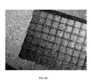
- FIG. 6 Top view (A) and side view (B-C) of osteochondral tissue cut with a laser to produce perforations (microperforations) forming a grid pattern of lines.
- B-C 2 ⁇ magnification (B) and 3 ⁇ magnification (C) of a laser cut graft. The perforations extend partially through the depth of the osteochondral tissue.
- the blue arrow in (B) shows the subchondral bone layer.
- the present application relates to osteochondral allografts comprising a bone component and a cartilage component, and comprising one or more perforations in the bone component and/or cartilage component of the osteochondral allograft.
- perforations in an osteochondral allograft enhances cell migration and transfusion of nutrients from the allograft into a treatment site, resulting in an improved clinical outcome, such as faster healing of a defect (e.g., cartilage or bone defect) as compared to an osteochondral allograft lacking perforations.
- the osteochondral allografts described herein can be prepared or utilized for any type of tissue that is dense and/or hard to heal, including but not limited to cartilage, tendon, cortical bone, etc.
- the osteochondral graft compositions described herein can be used to treat medium to large articular cartilage lesions, complex or multiple cartilage lesions, or subjects with bone loss.
- cartilage repair techniques e.g., using cartilage grafts which lack a bone component
- perforated osteochondral graft compositions are provided.
- the composition comprises a continuous portion of an osteochondral tissue, wherein the continuous portion of osteochondral tissue comprises a bone component and a cartilage component, and wherein the osteochondral graft composition comprises one or more perforations in the bone component and/or the cartilage component.
- the term “continuous portion of an osteochondral tissue” refers to a single sheet or piece of osteochondral tissue comprising both cartilage tissue and bone tissue within the single sheet or piece.
- a continuous portion of an osteochondral tissue is a piece of osteochondral tissue harvested from a humeral head or a femoral condyle.
- the osteochondral graft can comprise any osteochondral tissue.
- the osteochondral graft can comprise osteochondral tissue from the humerus (e.g., humeral head), femur (e.g., femoral condyle), tibia, ilium, fibula, radius, ulna, trochlea, patella, talus, or ankle
- the osteochondral graft can have any suitable shape.
- the osteochondral graft can have a cylinder or “dowel” shape, a cube or rectangular shape, or an irregular shape.
- the osteochondral graft comprises a cylindrical or dowel shape. See, e.g., FIG. 1 .
- the osteochondral graft has a diameter of about 10 mm, about 11 mm, about 12 mm, about 13 mm, about 14 mm, about 15 mm, about 16 mm, about 17 mm, about 18 mm, about 19 mm, or about 20 mm.
- the osteochondral graft has a length of about 10 mm, about 15 mm, about 20 mm, about 25 mm, or about 30 mm.
- the osteochondral tissue is obtained from a cadaveric donor.
- the donor is an adult cadaveric donor that is 18 years of age or older at the time of the donation.
- the donor is an adult cadaveric donor that is between the ages of 15 and 45 at the time of the donation.
- the osteochondral tissue is from a human juvenile cadaveric donor.
- the donor is a juvenile cadaveric donor that is between the ages of 3 and 12 at the time of the donation.
- the osteochondral allograft compositions comprise 1, 2, 3, 4, 5, 6, 7, 8, 9, 10, 15, 20, 25, 30, 35, 40, 45, 50, 60, 70, 80, 90, 100, 150, 200 or more perforations in the osteochondral tissue.
- the one or more perforations have an average diameter that is about 100 ⁇ m, about 150 ⁇ m, about 200 ⁇ m, about 250 ⁇ m, about 300 ⁇ m, about 350 ⁇ m, about 400 ⁇ m, about 450 ⁇ m, about 500 ⁇ m, about 550 ⁇ m, about 600 ⁇ m, about 650 ⁇ m, about 700 ⁇ m, about 750 ⁇ m, about 800 ⁇ m, about 850 ⁇ m, about 900 ⁇ m, about 950 ⁇ m, about 1 mm, about 1.5 mm, about 2 mm, about 2.5 mm, or about 3 mm. In some embodiments, the one or more perforations have an average diameter from about 100 ⁇ m to about 3 mm.
- the one or more perforations have an average diameter from about 100 ⁇ m to about 2 mm. In some embodiments, the one or more perforations have an average diameter that is from about 100 ⁇ m to about 1000 ⁇ m. In some embodiments, the one or more perforations have an average diameter that is from about 100 ⁇ m to about 500 ⁇ m. In some embodiments, the one or more perforations have an average diameter than is less than about 200 ⁇ m, e.g., about 175 ⁇ m, about 150 ⁇ m, about 125 ⁇ m, about 100 ⁇ m or less.
- the one or more perforations have an average diameter that is from about 0.5 mm to about 3 mm, from about 0.5 mm to about 2 mm, or from about 0.5 mm to about 1 mm.
- the osteochondral allograft composition comprises a plurality of perforations, and the perforations vary in their average diameter.
- the osteochondral allograft composition can have two, three, four, or more sizes of perforations.
- the osteochondral allograft composition comprises a plurality of perforations that are cut into the osteochondral tissue in such a way as to form a line or lines in the osteochondral tissue.
- a series of perforations e.g., microperforations
- a series of perforations e.g., microperforations
- the perforations forming the line or lines can extend partially or entirely through the depth of the osteochondral tissue.
- the osteochondral allograft composition comprises one or more perforations that extend partially through the depth of the osteochondral tissue.
- one or more perforations extend partially through the cartilage component and does not extend into the bone component.
- the one or more perforations extend entirely through the depth of the cartilage component but do not extend into the bone component.
- the one or more perforations extend entirely through the depth of the cartilage component and extend partially through the depth of the bone component.
- the one or more perforations extend entirely through the depth of both the cartilage component and the bone component.
- the osteochondral allograft composition comprises a plurality of perforations, and the perforations vary in their depth through the osteochondral tissue.
- the composition comprises at least one perforation that extends entirely through the osteochondral tissue (i.e., entirely through the depth of the cartilage component and the bone component), and at least one perforation that only extends partially through the osteochondral tissue (e.g., extends partially through the cartilage component, extends entirely through the cartilage component but does not extend into the bone component, or extends entirely through the cartilage component and extends partially into the bone component).
- the perforated osteochondral allograft composition further comprises a biocompatible medium in which the osteochondral allograft is stored.
- the biocompatible medium comprises a growth medium. Suitable examples of growth medium include, but are not limited to, Dulbecco's Modified Eagle's Medium (DMEM) with 5% Fetal Bovine Serum (FBS).
- growth medium includes a high glucose DMEM.
- the biocompatible carrier e.g., growth medium
- the biocompatible medium comprises a cryopreservation medium.
- the biocompatible medium comprises one or more cryoprotective agents such as, but not limited to, glycerol, DMSO, hydroxyethyl starch, polyethylene glycol, propanediol, ethylene glycol, butanediol, polyvinylpyrrolidone, or alginate.
- the perforated osteochondral allograft composition is combined with one or more other biological components.
- at least a portion of the osteochondral allograft composition is coated with a biological adhesive.
- suitable biological adhesives include, but are not limited to, fibrin, fibrinogen, thrombin, fibrin glue (e.g., TISSEEL), polysaccharide gel, cyanoacrylate glue, gelatin-resorcin-formalin adhesive, collagen gel, synthetic acrylate-based adhesive, cellulose-based adhesive, basement membrane matrix (e.g., MATRIGEL®, BD Biosciences, San Jose, Calif.), laminin, elastin, proteoglycans, autologous glue, and combinations thereof.
- fibrin glue e.g., TISSEEL
- polysaccharide gel e.g., cyanoacrylate glue
- gelatin-resorcin-formalin adhesive e.g., gelatin-resorcin-formalin
- the perforated osteochondral allograft composition is combined with cells such as stem cells.
- the osteochondral allograft composition is seeded with stem cells, such as mesenchymal stem cells.
- stem cells such as mesenchymal stem cells.
- Mesenchymal stem cells can be obtained from a variety of tissues, including but not limited to bone marrow tissue, adipose tissue, muscle tissue, birth tissue (e.g., amnion, amniotic fluid, or umbilical cord tissue), skin tissue, bone tissue, and dental tissue.
- the mesenchymal stem cells are derived from a tissue (e.g., adipose tissue) that has been processed (i.e., digested) to form a cell suspension comprising mesenchymal stem cells and non-mesenchymal stem cells that is seeded onto the osteochondral allograft, and wherein the mesenchymal stem cells are not cultured ex vivo (e.g., on a plastic dish) prior to seeding the cell suspension on the osteochondral allograft.
- a tissue e.g., adipose tissue
- the mesenchymal stem cells are not cultured ex vivo (e.g., on a plastic dish) prior to seeding the cell suspension on the osteochondral allograft.
- Stem cell-seeded bone and cartilage substrates and methods of preparing such substrates are described in published application US 2010/0124776 and in U.S. application Ser. No. 12/965,335, the contents of each
- Suitable growth factors include, but are not limited to, transforming growth factor-beta (TGF ⁇ ), fibroblast growth factor (FGF) (e.g., FGF2, FGF5), bone morphogenetic protein (BMP) (e.g., BMP2, BMP4, BMP6, BMP7), platelet derived growth factor (PDGF), and insulin-related growth factor (IGF) (e.g., IGF1, IGF2).
- TGF ⁇ transforming growth factor-beta
- FGF fibroblast growth factor
- BMP bone morphogenetic protein
- PDGF platelet derived growth factor
- IGF insulin-related growth factor
- the osteochondral allograft composition is at least partially coated with a biological adhesive prior to adding the one or more growth factors and/or seeding the cells (e.g., mesenchymal stem cells) on the osteochondral allograft.
- a biological adhesive prior to adding the one or more growth factors and/or seeding the cells (e.g., mesenchymal stem cells) on the osteochondral allograft.
- the perforated osteochondral allograft composition is combined with one or more growth factors.
- methods of manufacturing perforated osteochondral allograft compositions are provided.
- the method comprises:
- the continuous portion of osteochondral tissue comprises a bone component and a cartilage component
- the osteochondral tissue is harvested from an adult cadaveric donor. In some embodiments, the osteochondral tissue is harvested from an adult cadaveric donor that is between the ages of 15 and 45 at the time of the donation. In some embodiments, the osteochondral tissue is harvested from a juvenile cadaveric donor.
- the perforation techniques can be used with any size of graft tissue piece or construct, and may be used with any of a variety of osteochondral tissues.
- the graft comprises osteochondral tissue from the humerus (e.g., humeral head), femur (e.g., femoral condyle), tibia, ilium, fibula, radius, ulna, trochlea, patella, talus, or ankle.
- the graft comprises a cylinder or “dowel” shape, a cube or rectangular shape, or an irregular shape.
- Perforations can include, for example, microperforations, bores, apertures, and the like. In some embodiments, perforations may be on the order of tens of microns in dimension, or less. In some embodiments, perforations may be on the order of millimeters in dimension, or less.
- the perforations have an average diameter of about 100 ⁇ m, about 150 ⁇ m, about 200 ⁇ m, about 250 ⁇ m, about 300 ⁇ m, about 350 ⁇ m, about 400 ⁇ m, about 450 ⁇ m, about 500 ⁇ m, about 550 ⁇ m, about 600 ⁇ m, about 650 ⁇ m, about 700 ⁇ m, about 750 ⁇ m, about 800 ⁇ m, about 850 ⁇ m, about 900 ⁇ m, about 950 ⁇ m, about 1 mm, about 1.5 mm, about 2 mm, about 2.5 mm, or about 3 mm.
- perforation techniques can involve the formation of holes having a diameter of about 0.10 mm, or smaller. In some embodiments, 1, 2, 3, 4, 5, 6, 7, 8, 9, 10, 15, 20, 25, 30, 35, 40, 45, 50, 60, 70, 80, 90, 100, 150, 200 or more perforations are cut into the osteochondral tissue.
- the osteochondral tissue is cut using a cutting mechanism.
- the cutting mechanism is a laser cutting apparatus, a mechanical blade, a manual cutting apparatus, a manual pressing apparatus, or the like.
- the cutting mechanism comprises a pneumatic press, such as an air press or an oil press, or a screw press.
- the osteochondral tissue is cut using a laser cutting apparatus.
- the laser cutting apparatus is a laser engraver.
- suitable engraving lasers include CO 2 engraving lasers, such as the Epilog Zing 30 Watt CO 2 engraving laser.
- the cutting step comprises cutting the cartilage tissue with the laser cutting apparatus at a speed from about 5% to about 15% (e.g., about 5%, about 6%, about 7%, about 8%, about 9%, about 10%, about 11%, about 12%, about 13%, about 14%, or about 15%), at a power of about 35% to about 100% (e.g., about 35%, about 40%, about 45%, about 50%, about 55%, about 60%, about 65%, about 70%, about 75%, about 80%, about 85%, about 90%, about 95%, or about 100%), and a frequency of about 1500 Hz to about 5000 Hz (e.g., about 1500 Hz, about 1600 Hz, about 1700 Hz, about 1800 Hz, about 1900 Hz, about 2000 Hz, about 2100 Hz, about 2200 Hz, about 2300 Hz, about 2400 Hz, about 2500 Hz, about 2600 Hz, about 2700 Hz, about 2800 Hz, about 2900 Hz, about 3
- the cutting step comprises cutting the cartilage tissue with the laser cutting apparatus at a speed of about 10%, a power of about 100%, and a frequency of about 5000 Hz. In some embodiments, the cutting step comprises cutting the cartilage tissue with the laser cutting apparatus at a speed of about 5%, a power of about 100%, and a frequency of about 5000 Hz. In some embodiments, the cutting step comprises cutting the cartilage tissue with the laser cutting apparatus at a speed of about 10%, a power of about 65%, and a frequency of about 4000 Hz. In some embodiments, the cutting step comprises cutting the cartilage tissue with the laser cutting apparatus at a speed of about 15%, a power of about 35%, and a frequency of about 1500 Hz.
- the perforation techniques disclosed herein can operate without subjecting the tissue to undue pressure which would otherwise cause the tissue to deform, tear, or otherwise become distorted.
- Laser perforation techniques can efficiently ablate the tissue, and leave a clean and intact hole without distorting the tissue. It has been found that the laser does not burn the cells as it bores through the material. Rather, the cells remain viable and are observed to grow out of or passage through the perforated tissue.
- the osteochondral allograft prior to the cutting step, is washed with a saline solution.
- the perforated osteochondral allograft is suspended in a biocompatible medium.
- the biocompatible medium comprises a growth medium. Suitable examples of growth medium include, but are not limited to, Dulbecco's Modified Eagle's Medium (DMEM) with 5% Fetal Bovine Serum (FBS).
- growth medium includes a high glucose DMEM.
- the biocompatible carrier e.g., growth medium
- the biocompatible medium comprises one or more antibiotics.
- the biocompatible medium comprises a cryopreservation medium.
- the biocompatible medium comprises one or more cryoprotective agents such as, but not limited to, glycerol, DMSO, hydroxyethyl starch, polyethylene glycol, propanediol, ethylene glycol, butanediol, polyvinylpyrrolidone, or alginate.
- cryoprotective agents such as, but not limited to, glycerol, DMSO, hydroxyethyl starch, polyethylene glycol, propanediol, ethylene glycol, butanediol, polyvinylpyrrolidone, or alginate.
- the perforated osteochondral allograft is not subjected to an additional processing step prior to suspending the osteochondral allograft in the biocompatible medium.
- the osteochondral allograft can be subjected to one or more additional processing steps prior to suspending the osteochondral allograft in the biocompatible medium.
- the osteochondral allograft is washed with a saline solution.
- the osteochondral allograft is treated with one or more enzymes that promote the release of chondrocyte cells from cartilage matrix in the osteochondral allograft.
- collagenase can be applied to help release chondrocyte cells from the cartilage matrix in the osteochondral allograft.
- the osteochondral allograft is not enzymatically digested, such as with a collagenase.
- Suitable biological adhesives include, but are not limited to, fibrin, fibrinogen, thrombin, fibrin glue (e.g., TISSEEL), polysaccharide gel, cyanoacrylate glue, gelatin-resorcin-formalin adhesive, collagen gel, synthetic acrylate-based adhesive, cellulose-based adhesive, basement membrane matrix (e.g., MATRIGEL®, BD Biosciences, San Jose, Calif.), laminin, elastin, proteoglycans, autologous glue, and combinations thereof.
- fibrin glue e.g., TISSEEL
- polysaccharide gel e.g., cyanoacrylate glue
- gelatin-resorcin-formalin adhesive e.g., gelatin-resorcin-formalin adhesive
- collagen gel e.g., synthetic acrylate-based adhesive
- cellulose-based adhesive e.g., cellulose-based adhesive
- basement membrane matrix e.g., MATRIG
- Suitable growth factors include, but are not limited to, transforming growth factor-beta (TGF ⁇ ), fibroblast growth factor (FGF) (e.g., FGF2, FGF5), bone morphogenetic protein (BMP) (e.g., BMP2, BMP4, BMP6, BMP7), platelet derived growth factor (PDGF), and insulin-related growth factor (IGF) (e.g., IGF1, IGF2).
- TGF ⁇ transforming growth factor-beta
- FGF fibroblast growth factor
- BMP bone morphogenetic protein
- PDGF platelet derived growth factor
- IGF insulin-related growth factor
- the method further comprises: digesting a tissue (e.g., adipose tissue) to form a cell suspension comprising mesenchymal stem cells and non-mesenchymal stem cells; seeding the cell suspension onto the osteochondral allograft, wherein the mesenchymal stem cells are not cultured ex vivo (e.g., on a plastic dish) prior to seeding the cell suspension on the osteochondral allograft.
- a tissue e.g., adipose tissue
- mesenchymal stem cells are not cultured ex vivo (e.g., on a plastic dish) prior to seeding the cell suspension on the osteochondral allograft.
- At least a portion of the osteochondral allograft composition is with a biological adhesive (e.g., fibrin glue) prior to adding the one or more growth factors and/or seeding the cells (e.g., mesenchymal stem cells) on the osteochondral allograft.
- a biological adhesive e.g., fibrin glue
- the cells to be seeded e.g., mesenchymal stem cells
- the biological adhesive e.g., fibrin glue
- a perforated osteochondral graft composition as described herein can be used to treat bone and cartilage defects in a subject.
- perforations e.g., holes or microperforations
- These tissue treatment techniques can be applied to any tissue type which is dense and hard to heal, such as cortical bone, cartilage, tendon, and the like.
- the osteochondral grafts described herein can be used to treat large and deep osteochondral defects, and to prevent or inhibit the presence of non-filled spaces between circular grafts and incomplete integration of the donor and recipient cartilage. Additionally, the osteochondral grafts described herein preserve the natural biomechanical characteristics of the osteochondral product. By keeping the native characteristics of the allograft (e.g., biomechanical properties and natural osteoinductive and cartilaginous growth factors present in the osteochondral graft), administration of the perforated osteochondral graft results in a better chance of successful incorporation and defect repair, for example as compared to a more flexible allograft or a graft lacking a bone component.
- the native characteristics of the allograft e.g., biomechanical properties and natural osteoinductive and cartilaginous growth factors present in the osteochondral graft
- administration of the perforated osteochondral graft results in a better chance of successful incorporation and defect repair, for example as
- a perforated osteochondral graft composition as described herein e.g., a composition comprising a continuous portion of an osteochondral tissue, wherein the continuous portion of osteochondral tissue comprises a cartilage component and a bone component, and wherein the osteochondral graft composition comprises one or more perforations in the cartilage component and/or the bone component
- the perforated osteochondral graft composition is administered at a site of defect or injury in cartilage, bone, ligament, tendon, meniscus, or joint.
- the defect is a large or deep osteochondral defect (e.g., at a joint, e.g., a knee joint).
- the subject has osteoarthritis.
- the subject has a degenerative osteochondral defect or injury.
- a perforated osteochondral graft composition as described herein is used to treat a subject having a full-thickness or nearly full-thickness cartilage defect.
- the Outerbridge classification of articular cartilage injuries is the most widely used to describe the size of a cartilage lesion. See, Outerbridge R E, J Bone Joint Surg Br 43:752-757 (1961). This classification provides a distinction between a partial (Grades 1 and 2) versus nearly full or full-thickness cartilage defect (Grades 3 and 4); between a small (Grade 2) and larger (Grade 3) lesion; and describes a complete loss of cartilage (Grade 4).
- Osteochondral grafts such as the perforated graft compositions described herein, are particularly suitable for treating nearly full or full-thickness cartilage defects (Grades 3 and 4) and medium to large (Grade 3) articular cartilage lesions.
- Osteochondral grafts such as the perforated graft compositions described herein, are also particularly suitable for treating osteochondritis dissecans, focal chondral defects >2-3 cm 2 in femoral condyles or patellofemoral defects, or for treatments in which there is misalignment, meniscal deficiency, or ligament insufficiency.
- a perforated osteochondral graft composition as described herein is administered locally to the subject.
- the perforated osteochondral graft composition is implanted in the subject.
- the perforated osteochondral graft composition is administered in a minimally invasive procedure, e.g., arthroscopy.
- perforated osteochondral graft composition is administered by osteochondral allograft transplantation surgery (OATS).
- a perforated osteochondral graft composition as described herein is administered (e.g., implanted) “fresh,” i.e., within 7 days of harvesting the osteochondral tissue.
- fresh osteochondral graft compositions are believed to have a high amount of chondrocyte viability at the time of administration.
- a perforated osteochondral graft composition as described herein can be shaped into a smaller piece by the user (e.g., surgeon) to an appropriate size to suit the size of the defect being treated.
- kits comprising a perforated osteochondral graft composition as described herein are provided.
- the kit comprises a perforated osteochondral graft composition comprising a continuous portion of an osteochondral tissue, wherein the continuous portion of osteochondral tissue comprises a cartilage component and a bone component, and wherein the osteochondral graft composition comprises one or more perforations in the cartilage component and/or the bone component; and a biocompatible medium.
- the biocompatible medium comprises a growth medium.
- growth medium include, but are not limited to, Dulbecco's Modified Eagle's Medium (DMEM) with 5% Fetal Bovine Serum (FBS).
- DMEM Dulbecco's Modified Eagle's Medium
- FBS Fetal Bovine Serum
- growth medium includes a high glucose DMEM.
- the biocompatible carrier e.g., growth medium
- kits are used for treating a subject having a defect in cartilage, bone, ligament, tendon, meniscus, or joint. In some embodiments, the kits are used for treating a subject having a degenerative defect or injury in cartilage, bone, ligament, tendon, meniscus, or joint. In some embodiments, the kits are used for treating a subject having osteoarthritis.
- a kit comprises a perforated osteochondral graft composition as described herein packaged in a container for storage and/or shipment. In some embodiments, the kit further comprises instructions for administering the composition.
- the kit further comprises biological adhesive components (e.g., fibrinogen and thrombin, for fibrin glue).
- biological adhesive components e.g., fibrinogen and thrombin, for fibrin glue.
- the perforated osteochondral graft composition and the biological adhesive components are packaged separately, and a user (e.g., a surgeon) adds the biological adhesive components to the surgery site prior to placement or implantation of the perforated osteochondral graft composition.
- the perforated osteochondral graft composition is combined with the biological adhesive (e.g., fibrin glue) prior to packaging in the kit.
- the kit further comprises cells (e.g., stem cells).
- the kit comprises biological adhesive and cells (e.g., stem cells), and the perforated osteochondral graft composition is combined with the biological adhesive prior to seeding the cells on the osteochondral graft.
- Osteochondral graft compositions were processed using an Epilog Zing 30 Watt CO2 engraving laser to cut a plurality of perforations into an osteochondral allograft (hemi-condyle allograft containing cartilage and bone).
- Table 1 shows the results of the tissue cutting experiments at varying speeds, powers, and frequencies.
- FIGS. 1-6 depict exemplary perforated osteochondral grafts.
- an osteochondral graft having a bone component and a cartilage component can be harvested from a cadaveric donor.
- the graft assembly can be treated according to any of the techniques described herein, so as to produce perforations (e.g., holes or microperforations) in the graft.
- the processed graft assembly can then be implanted in the recipient patient.
- FIG. 2 depicts an osteochondral graft processed to have multiple micro-holes in the cartilage component as well as the bone component.
- FIG. 3 depicts an osteochondral graft processed to have multiple micro-holes in the cartilage component as well as the bone component.
- the holes can extend entirely through the depth of the graft, or partially to a depth within the graft.
- FIG. 4 depicts another osteochondral graft processed so that multiple micro-holes are formed in the graft.
- FIG. 5 depicts an osteochondral graft embodiment in a dowel configuration, which includes circular cut perforations.
- the outer circle represents the outer diameter of the dowel.
- the inner two circles represent circular cut perforations within the dowel material.
- FIG. 6 depicts osteochondral tissue cut with a laser to produce perforations (microperforations) forming a grid pattern of lines.
- the perforations extend partially through the depth of the osteochondral tissue, through the cartilage portion of the osteochondral tissue and into the subchondral bone layer of the osteochondral tissue.
Abstract
Description
| TABLE 1 | ||||
| Speed | 10% | 5% | 10% | 15% |
| Power | 100% | 100% | 65% | 35% |
| Frequency | 5000 Hz | 5000 Hz | 4000 Hz | 1500 Hz |
| Results | Cut through | Cut through | Cut through | Cut through |
| entire cartilage | cartilage and | cartilage and | cartilage and | |
| layer and several | several mm | subchondral | subchondral | |
| mm of bone | of bone | bone | bone | |
Claims (27)
Priority Applications (2)
| Application Number | Priority Date | Filing Date | Title |
|---|---|---|---|
| US14/210,111 US9168140B2 (en) | 2013-03-15 | 2014-03-13 | Perforated osteochondral allograft compositions |
| US14/858,386 US9603710B2 (en) | 2013-03-15 | 2015-09-18 | Methods of manufacturing perforated osteochondral allograft compositions |
Applications Claiming Priority (2)
| Application Number | Priority Date | Filing Date | Title |
|---|---|---|---|
| US201361792074P | 2013-03-15 | 2013-03-15 | |
| US14/210,111 US9168140B2 (en) | 2013-03-15 | 2014-03-13 | Perforated osteochondral allograft compositions |
Related Child Applications (1)
| Application Number | Title | Priority Date | Filing Date |
|---|---|---|---|
| US14/858,386 Division US9603710B2 (en) | 2013-03-15 | 2015-09-18 | Methods of manufacturing perforated osteochondral allograft compositions |
Publications (2)
| Publication Number | Publication Date |
|---|---|
| US20140271570A1 US20140271570A1 (en) | 2014-09-18 |
| US9168140B2 true US9168140B2 (en) | 2015-10-27 |
Family
ID=51527930
Family Applications (2)
| Application Number | Title | Priority Date | Filing Date |
|---|---|---|---|
| US14/210,111 Active US9168140B2 (en) | 2013-03-15 | 2014-03-13 | Perforated osteochondral allograft compositions |
| US14/858,386 Active US9603710B2 (en) | 2013-03-15 | 2015-09-18 | Methods of manufacturing perforated osteochondral allograft compositions |
Family Applications After (1)
| Application Number | Title | Priority Date | Filing Date |
|---|---|---|---|
| US14/858,386 Active US9603710B2 (en) | 2013-03-15 | 2015-09-18 | Methods of manufacturing perforated osteochondral allograft compositions |
Country Status (7)
| Country | Link |
|---|---|
| US (2) | US9168140B2 (en) |
| EP (1) | EP2967874B1 (en) |
| KR (1) | KR102138399B1 (en) |
| AU (1) | AU2014236705B2 (en) |
| CA (1) | CA2895140C (en) |
| CL (1) | CL2015002752A1 (en) |
| WO (1) | WO2014151939A1 (en) |
Cited By (10)
| Publication number | Priority date | Publication date | Assignee | Title |
|---|---|---|---|---|
| US20160008134A1 (en) * | 2013-03-15 | 2016-01-14 | Allosource | Perforated Osteochondral Allograft Compositions |
| WO2017027481A1 (en) | 2015-08-07 | 2017-02-16 | Allosource | Rapid allograft treatment systems and methods |
| US9700415B2 (en) | 2013-02-22 | 2017-07-11 | Allosource | Cartilage mosaic compositions and methods |
| WO2017161121A1 (en) | 2016-03-18 | 2017-09-21 | Allosource | Composite medical grafts and methods of use and manufacture |
| WO2017161115A1 (en) | 2016-03-18 | 2017-09-21 | Allosource | Composite medical grafts and methods of use and manufacture |
| MD1177Z (en) * | 2017-04-11 | 2018-03-31 | Государственный Медицинский И Фармацевтический Университет "Nicolae Testemitanu" Республики Молдова | Graft for restoration of osteochondral defect |
| US10251751B2 (en) | 2014-03-11 | 2019-04-09 | The Trustees Of Columbia University In The City Of New York | Customized bendable osteochondral allografts |
| WO2019135216A1 (en) | 2018-01-02 | 2019-07-11 | Cartiheal (2009) Ltd. | Implantation tool and protocol for optimized solid substrates promoting cell and tissue growth |
| US11202674B2 (en) | 2018-04-03 | 2021-12-21 | Convergent Dental, Inc. | Laser system for surgical applications |
| US11452796B2 (en) | 2017-06-30 | 2022-09-27 | Allosource | Cellular bone grafts, and methods of manufacture and use |
Families Citing this family (5)
| Publication number | Priority date | Publication date | Assignee | Title |
|---|---|---|---|---|
| EP2919794B1 (en) * | 2012-11-15 | 2021-01-20 | AlloSource | Minced cartilage systems and methods |
| US10279081B2 (en) | 2014-10-24 | 2019-05-07 | Allosource | Composite grafts, systems, and methods |
| US11285177B2 (en) | 2018-01-03 | 2022-03-29 | Globus Medical, Inc. | Allografts containing viable cells and methods thereof |
| CN111671978B (en) * | 2020-07-08 | 2021-09-07 | 四川大学 | Costal cartilage-based 3D printing biological ink and preparation method and application thereof |
| KR20220134422A (en) | 2021-03-26 | 2022-10-05 | 에이템즈 주식회사 | Method for Cryopreservation and thawing of cartilage tissue |
Citations (52)
| Publication number | Priority date | Publication date | Assignee | Title |
|---|---|---|---|---|
| US4932973A (en) * | 1983-09-30 | 1990-06-12 | El Gendler | Cartilage and bone induction by artificially perforated organic bone matrix |
| US5582752A (en) | 1993-12-17 | 1996-12-10 | Laser Industries, Ltd. | Method and apparatus for applying laser beams to a working surface, particularly for ablating tissue |
| US6050991A (en) | 1994-09-29 | 2000-04-18 | Lokki S.A. (Societeanonyme) | Pulsed-emission laser for use in the medical field |
| US6607524B1 (en) | 1997-08-07 | 2003-08-19 | Pharos Optics, Inc. | Surgical laser and method of ablating hard biological materials |
| US6638271B2 (en) | 1998-04-17 | 2003-10-28 | Visx, Inc. | Multiple beam laser sculpting system and method |
| US20030229400A1 (en) * | 2002-05-13 | 2003-12-11 | Koichi Masuda | Tissue engineered osteochondral implant |
| US6712822B2 (en) | 2001-10-01 | 2004-03-30 | Scandius Biomedical, Inc. | Apparatus and method for the repair of articular cartilage defects |
| US20040078090A1 (en) | 2002-10-18 | 2004-04-22 | Francois Binette | Biocompatible scaffolds with tissue fragments |
| US6858042B2 (en) | 1999-12-15 | 2005-02-22 | Zimmer Orthobiologics, Inc. | Preparation for repairing cartilage defects or cartilage/bone defects in human or animal joints |
| WO2005058207A1 (en) | 2003-12-11 | 2005-06-30 | Isto Technologies, Inc. | Particulate cartilage system |
| US20060275273A1 (en) | 2004-02-20 | 2006-12-07 | Seyedin Mitchell S | Intervertebral Disc Repair, Methods and Devices Therefor |
| US20070265705A1 (en) | 2004-10-27 | 2007-11-15 | Christoph Gaissmaier | Implant for repairing a cartilage defect |
| US20070276506A1 (en) * | 2006-05-25 | 2007-11-29 | Biomet Manufacturing Corp. | Demineralized osteochondral plug |
| US20070299517A1 (en) | 2006-06-21 | 2007-12-27 | Howmedica Osteonics Corp. | Articular cartilage implant |
| US7361195B2 (en) | 2001-07-16 | 2008-04-22 | Depuy Products, Inc. | Cartilage repair apparatus and method |
| US20080160496A1 (en) | 2005-02-22 | 2008-07-03 | Victor Rzepakovsky | Preserved Viable Cartilage, Method for Its Preservation, and System and Devices Used Therefor |
| US20080269895A1 (en) | 2005-09-20 | 2008-10-30 | Steinwachs Matthias R | Implant for the Repair of a Cartilage Defect and Method for Manufacturing the Implant |
| US20090024223A1 (en) | 2007-07-16 | 2009-01-22 | Chen Silvia S | Crafting of cartilage |
| US7550007B2 (en) | 2005-10-26 | 2009-06-23 | Biomet Sports Medicine, Llc | Osteochondral allografts |
| US20100049322A1 (en) | 2008-08-19 | 2010-02-25 | Warsaw Orthopedic, Inc. | Osteochondral repair implants and methods |
| US20100124776A1 (en) | 2008-11-20 | 2010-05-20 | Allosource | Allografts combined with tissue derived stem cells for bone healing |
| US7758643B2 (en) | 2007-02-26 | 2010-07-20 | Biomet Sports Medicine, Llc | Stable cartilage defect repair plug |
| US20100274362A1 (en) | 2009-01-15 | 2010-10-28 | Avner Yayon | Cartilage particle tissue mixtures optionally combined with a cancellous construct |
| US7838040B2 (en) | 2004-03-08 | 2010-11-23 | Theodore Malinin | Method for regenerating cartilage |
| US7875296B2 (en) | 2003-11-26 | 2011-01-25 | Depuy Mitek, Inc. | Conformable tissue repair implant capable of injection delivery |
| US20110045044A1 (en) | 2005-10-12 | 2011-02-24 | Lifenet Health | Compositions for repair of defects in tissues, and methods of making the same |
| USRE42208E1 (en) | 2003-04-29 | 2011-03-08 | Musculoskeletal Transplant Foundation | Glue for cartilage repair |
| US7931692B2 (en) | 2001-02-14 | 2011-04-26 | Osteotech, Inc. | Implant derived from bone |
| US20110177134A1 (en) | 2003-12-05 | 2011-07-21 | Depuy Mitek, Inc. | Viable Tissue Repair Implants and Methods of Use |
| US20110182961A1 (en) | 2010-01-25 | 2011-07-28 | Warsaw Orthopedic, Inc. | Osteogenic cell delivery matrix |
| US20110196355A1 (en) | 2008-11-18 | 2011-08-11 | Precise Light Surgical, Inc. | Flash vaporization surgical systems |
| US8012206B2 (en) | 2002-09-20 | 2011-09-06 | Arthrex, Inc. | Preformed implants for osteochondral repair |
| US8043450B2 (en) | 2000-01-27 | 2011-10-25 | 3F Therapeutics, Inc. | Method of cutting tissue using a laser |
| US20110274729A1 (en) | 2008-11-24 | 2011-11-10 | Collins Daniel P | Implantable compositions for repairing osteochondral defects |
| US20120107384A1 (en) | 2007-04-12 | 2012-05-03 | Zimmer, Inc. | Apparatus for forming an implant |
| US8173162B2 (en) | 2003-02-26 | 2012-05-08 | Zimmer Orthobiologics, Inc. | Preparation for repairing cartilage tissue, especially articular cartilage defects |
| US8221500B2 (en) | 2003-05-16 | 2012-07-17 | Musculoskeletal Transplant Foundation | Cartilage allograft plug |
| US8435551B2 (en) | 2007-03-06 | 2013-05-07 | Musculoskeletal Transplant Foundation | Cancellous construct with support ring for repair of osteochondral defects |
| US20130122095A1 (en) | 2009-03-05 | 2013-05-16 | Biomimetic Therapeutics, Inc. | Platelet-derived growth factor compositions and methods for the treatment of osteochondral defects |
| US8497121B2 (en) | 2006-12-20 | 2013-07-30 | Zimmer Orthobiologics, Inc. | Method of obtaining viable small tissue particles and use for tissue repair |
| US20130197654A1 (en) | 2007-08-27 | 2013-08-01 | Kent M. Samuelson | Systems and methods for providing a femoral component |
| US20130204392A1 (en) | 2012-01-31 | 2013-08-08 | Agency For Science, Technology And Research | Method for promoting stem cell chondrogenesis |
| US20130344114A1 (en) | 2012-06-22 | 2013-12-26 | Nai-Jen Chang | Composition for repairing cartilage tissue and method for making the same |
| US20140012393A1 (en) | 2011-03-31 | 2014-01-09 | Inje University Industry-Academic Cooperation Foundation | Complex support body for regenerating bone-cartilage, method for manufacturing thereof, and composition for treating bone and cartilage related diseases comprising same as active ingredient |
| US20140017283A1 (en) | 2012-07-11 | 2014-01-16 | Osiris Therapeutics, Inc. | Porated cartilage products |
| US20140024115A1 (en) | 2009-11-04 | 2014-01-23 | Allosource | Methods of combining mesenchymal stem cells and cartilage containing allografts, and products of combined mesenchymal stem cells and cartilage containing allografts |
| US20140039621A1 (en) | 2009-04-02 | 2014-02-06 | Sevika Holding AG | Monolithic orthopedic implant with an articular finished surface |
| US8652214B2 (en) | 2003-10-13 | 2014-02-18 | Tetec Tissue Engineering Technologies Ag | Cartilage replacement implant and method for producing a cartilage replacement implant |
| US20140134212A1 (en) | 2012-11-15 | 2014-05-15 | Allosource | Minced cartilage systems and methods |
| US20140222159A1 (en) | 2008-06-16 | 2014-08-07 | Rti Surgical, Inc. | Assembled cartilage repair graft |
| US20140255506A1 (en) | 2003-12-31 | 2014-09-11 | Warsaw Orthopedic, Inc. | Osteoinductive demineralized cancellous bone |
| US8883210B1 (en) | 2010-05-14 | 2014-11-11 | Musculoskeletal Transplant Foundation | Tissue-derived tissuegenic implants, and methods of fabricating and using same |
Family Cites Families (157)
| Publication number | Priority date | Publication date | Assignee | Title |
|---|---|---|---|---|
| IL68218A (en) | 1983-03-23 | 1985-12-31 | Univ Ramot | Compositions for cartilage repair comprising embryonal chondrocytes |
| US4627853A (en) | 1985-05-29 | 1986-12-09 | American Hospital Supply Corporation | Method of producing prostheses for replacement of articular cartilage and prostheses so produced |
| US5290558A (en) | 1989-09-21 | 1994-03-01 | Osteotech, Inc. | Flowable demineralized bone powder composition and its use in bone repair |
| US5073373A (en) | 1989-09-21 | 1991-12-17 | Osteotech, Inc. | Flowable demineralized bone powder composition and its use in bone repair |
| US5131850A (en) | 1989-11-03 | 1992-07-21 | Cryolife, Inc. | Method for cryopreserving musculoskeletal tissues |
| US5236456A (en) | 1989-11-09 | 1993-08-17 | Osteotech, Inc. | Osteogenic composition and implant containing same |
| US5314476A (en) | 1992-02-04 | 1994-05-24 | Osteotech, Inc. | Demineralized bone particles and flowable osteogenic composition containing same |
| US5531791A (en) | 1993-07-23 | 1996-07-02 | Bioscience Consultants | Composition for repair of defects in osseous tissues, method of making, and prosthesis |
| US5507813A (en) | 1993-12-09 | 1996-04-16 | Osteotech, Inc. | Shaped materials derived from elongate bone particles |
| US5769899A (en) | 1994-08-12 | 1998-06-23 | Matrix Biotechnologies, Inc. | Cartilage repair unit |
| US6180606B1 (en) | 1994-09-28 | 2001-01-30 | Gensci Orthobiologics, Inc. | Compositions with enhanced osteogenic potential, methods for making the same and uses thereof |
| US5749874A (en) | 1995-02-07 | 1998-05-12 | Matrix Biotechnologies, Inc. | Cartilage repair unit and method of assembling same |
| US6669685B1 (en) | 1997-11-06 | 2003-12-30 | Biolase Technology, Inc. | Tissue remover and method |
| ATE322233T1 (en) | 1996-01-17 | 2006-04-15 | Osteotech Inc | METHOD FOR PRODUCING FLEXIBLE LAYERS FROM DEMINARALIZED, ELONGATED BONE PARTICLES |
| US5788941A (en) | 1996-01-31 | 1998-08-04 | Steris Corporation | Method of sterilization of bone tussue |
| US5897987A (en) | 1996-03-25 | 1999-04-27 | Advanced Reproduction Technologies, Inc. | Use of arabinogalactan in cell cryopreservation media |
| US5755791A (en) | 1996-04-05 | 1998-05-26 | Purdue Research Foundation | Perforated submucosal tissue graft constructs |
| US6283955B1 (en) | 1996-05-13 | 2001-09-04 | Edwards Lifesciences Corp. | Laser ablation device |
| US5895426A (en) | 1996-09-06 | 1999-04-20 | Osteotech, Inc. | Fusion implant device and method of use |
| US5676146B1 (en) | 1996-09-13 | 2000-04-18 | Osteotech Inc | Surgical implant containing a resorbable radiopaque marker and method of locating such within a body |
| AU732421B2 (en) | 1996-10-23 | 2001-04-26 | Warsaw Orthopedic, Inc. | Spinal spacer |
| US20010016646A1 (en) | 1998-03-20 | 2001-08-23 | David C. Rueger | Osteogenic devices and methods of use thereof for repair of endochondral bone, osteochondral and chondral defects |
| EP1018987B1 (en) | 1997-04-04 | 2014-10-29 | Barnes-Jewish Hospital | Neocartilage and methods of use |
| DE69714035T2 (en) | 1997-08-14 | 2003-03-06 | Sulzer Innotec Ag | Composition and device for repairing cartilage tissue in vivo consisting of nanocapsules with osteoinductive and / or chondroinductive factors |
| US6511958B1 (en) | 1997-08-14 | 2003-01-28 | Sulzer Biologics, Inc. | Compositions for regeneration and repair of cartilage lesions |
| FR2767675B1 (en) | 1997-08-26 | 1999-12-03 | Materiel Orthopedique En Abreg | INTERSOMATIC IMPLANT AND ANCILLARY OF PREPARATION SUITABLE FOR ALLOWING ITS POSITION |
| WO1999009914A1 (en) | 1997-08-27 | 1999-03-04 | University Of Florida Tissue Bank, Inc. | Cortical bone cervical smith-robinson fusion implant |
| US6511509B1 (en) | 1997-10-20 | 2003-01-28 | Lifenet | Textured bone allograft, method of making and using same |
| US6090998A (en) | 1997-10-27 | 2000-07-18 | University Of Florida | Segmentally demineralized bone implant |
| US5899939A (en) | 1998-01-21 | 1999-05-04 | Osteotech, Inc. | Bone-derived implant for load-supporting applications |
| US6482233B1 (en) | 1998-01-29 | 2002-11-19 | Synthes(U.S.A.) | Prosthetic interbody spacer |
| US6123731A (en) | 1998-02-06 | 2000-09-26 | Osteotech, Inc. | Osteoimplant and method for its manufacture |
| US20030147860A1 (en) | 2002-02-07 | 2003-08-07 | Marchosky J. Alexander | Compositions and methods for forming and strengthening bone |
| US6497726B1 (en) | 2000-01-11 | 2002-12-24 | Regeneration Technologies, Inc. | Materials and methods for improved bone tendon bone transplantation |
| JP2002532159A (en) | 1998-12-14 | 2002-10-02 | オステオテック インコーポレーテッド | Bone graft and guided bone regeneration |
| US6200347B1 (en) | 1999-01-05 | 2001-03-13 | Lifenet | Composite bone graft, method of making and using same |
| ES2209820T3 (en) | 1999-02-04 | 2004-07-01 | Sdgi Holdings, Inc. | HIGHLY MINERALIZED OSTEOGENIC SPONGE COMPOSITIONS, AND USES OF THE SAME. |
| US6656489B1 (en) | 1999-02-10 | 2003-12-02 | Isotis N.V. | Scaffold for tissue engineering cartilage having outer surface layers of copolymer and ceramic material |
| US8133421B2 (en) | 1999-02-23 | 2012-03-13 | Warsaw Orthopedic, Inc. | Methods of making shaped load-bearing osteoimplant |
| US6294187B1 (en) | 1999-02-23 | 2001-09-25 | Osteotech, Inc. | Load-bearing osteoimplant, method for its manufacture and method of repairing bone using same |
| US6696073B2 (en) | 1999-02-23 | 2004-02-24 | Osteotech, Inc. | Shaped load-bearing osteoimplant and methods of making same |
| US6709269B1 (en) | 2000-04-14 | 2004-03-23 | Gregory B. Altshuler | Apparatus and method for the processing of solid materials, including hard tissues |
| CA2377747C (en) | 1999-07-08 | 2009-09-29 | Cap Biotechnology, Inc. | Calcium-containing structures and methods of making and using the same |
| US20030228288A1 (en) | 1999-10-15 | 2003-12-11 | Scarborough Nelson L. | Volume maintaining osteoinductive/osteoconductive compositions |
| US20010032017A1 (en) | 1999-12-30 | 2001-10-18 | Alfaro Arthur A. | Intervertebral implants |
| US6626945B2 (en) | 2000-03-14 | 2003-09-30 | Chondrosite, Llc | Cartilage repair plug |
| AR027685A1 (en) | 2000-03-22 | 2003-04-09 | Synthes Ag | METHOD AND METHOD FOR CARRYING OUT |
| CA2405345C (en) | 2000-04-25 | 2012-09-18 | Osiris Therapeutics, Inc. | Joint repair using mesenchymal stem cells |
| US6340477B1 (en) | 2000-04-27 | 2002-01-22 | Lifenet | Bone matrix composition and methods for making and using same |
| AU7015301A (en) | 2000-07-03 | 2002-01-14 | Osteotech Inc | Osteogenic implants derived from bone |
| US6863694B1 (en) | 2000-07-03 | 2005-03-08 | Osteotech, Inc. | Osteogenic implants derived from bone |
| FR2811543B1 (en) | 2000-07-12 | 2003-07-04 | Spine Next Sa | INTERSOMATIC IMPLANT |
| DK177997B1 (en) | 2000-07-19 | 2015-02-23 | Ed Geistlich Söhne Ag Für Chemische Ind | Bone material and collagen combination for healing of damaged joints |
| US6685626B2 (en) | 2001-02-02 | 2004-02-03 | Regeneration Technologies, Inc. | Compositions, devices, methods, and kits for induction of adhesions |
| US6652593B2 (en) | 2001-02-28 | 2003-11-25 | Synthes (Usa) | Demineralized bone implants |
| WO2002096268A2 (en) | 2001-05-25 | 2002-12-05 | Imaging Therapeutics, Inc. | Methods and compositions for articular resurfacing |
| US7288086B1 (en) | 2001-06-21 | 2007-10-30 | Biolase Technology, Inc. | High-efficiency, side-pumped diode laser system |
| US8025896B2 (en) | 2001-07-16 | 2011-09-27 | Depuy Products, Inc. | Porous extracellular matrix scaffold and method |
| US7018412B2 (en) | 2001-08-20 | 2006-03-28 | Ebi, L.P. | Allograft spinal implant |
| US7371409B2 (en) | 2001-09-06 | 2008-05-13 | Wright Medical Technology, Inc. | Bone graft substitute composition |
| US6478825B1 (en) | 2001-11-28 | 2002-11-12 | Osteotech, Inc. | Implant, method of making same and use of the implant for the treatment of bone defects |
| US6911045B2 (en) | 2002-04-04 | 2005-06-28 | Osteotech, Inc. | Bio-implant insertion instrument |
| WO2003101529A2 (en) | 2002-05-31 | 2003-12-11 | Duke University Office Of Science And Technology | Method and apparatus for infrared tissue ablation |
| US7744597B2 (en) | 2002-06-26 | 2010-06-29 | Lifenet Health | Device and process for producing fiber products and fiber products produced thereby |
| US7498040B2 (en) | 2005-10-12 | 2009-03-03 | Lifenet Health | Compositions for repair of defects in osseous tissues, and methods of making the same |
| US7582309B2 (en) | 2002-11-15 | 2009-09-01 | Etex Corporation | Cohesive demineralized bone compositions |
| WO2004069172A2 (en) | 2003-01-30 | 2004-08-19 | The Government of the United States of America as represented by the Department of Veterans Affairs | Multilineage-inducible cells and uses thereof |
| EP1599139B1 (en) | 2003-02-20 | 2009-08-12 | Manoa Medical, Inc. | Bendable cutting device |
| US8197837B2 (en) | 2003-03-07 | 2012-06-12 | Depuy Mitek, Inc. | Method of preparation of bioabsorbable porous reinforced tissue implants and implants thereof |
| US20090291112A1 (en) * | 2003-05-16 | 2009-11-26 | Truncale Katherine G | Allograft osteochondral plug combined with cartilage particle mixture |
| US7351241B2 (en) | 2003-06-02 | 2008-04-01 | Carl Zeiss Meditec Ag | Method and apparatus for precision working of material |
| US7351262B2 (en) | 2003-06-05 | 2008-04-01 | Warsaw Orthopedic, Inc. | Bone implants and methods of making same |
| NZ544050A (en) | 2003-06-11 | 2009-03-31 | Osteotech Inc | Osteoimplants and methods for their manufacture |
| WO2005032612A2 (en) | 2003-10-02 | 2005-04-14 | Lostec, Inc. | A transplantable particulate bone composition and methods for making and using same |
| ATE494777T1 (en) | 2003-10-09 | 2011-01-15 | Core Dynamics Ltd | METHOD FOR FREEZING, THAWING AND TRANSPLANTING VIBABLE CARTILAGE |
| US7338495B2 (en) | 2003-10-22 | 2008-03-04 | Medtronic Xomed, Inc. | Angled tissue cutting instruments having flexible inner tubular members of tube and sleeve construction |
| WO2005040348A2 (en) | 2003-10-23 | 2005-05-06 | Georgetown University | Method for two- and three-dimensional microassembly of patterns and structures |
| KR100531922B1 (en) * | 2003-12-23 | 2005-11-29 | 주식회사 셀론텍 | a composition for cartilage therapeutics and a using method thereof |
| US20050288796A1 (en) | 2004-06-23 | 2005-12-29 | Hani Awad | Native soft tissue matrix for therapeutic applications |
| EP1784145B1 (en) | 2004-08-30 | 2011-08-10 | Neville Alleyne | Implant for the treatment of ligaments and tendons |
| US7837740B2 (en) | 2007-01-24 | 2010-11-23 | Musculoskeletal Transplant Foundation | Two piece cancellous construct for cartilage repair |
| US20090319045A1 (en) | 2004-10-12 | 2009-12-24 | Truncale Katherine G | Cancellous constructs, cartilage particles and combinations of cancellous constructs and cartilage particles |
| US7763072B2 (en) | 2005-03-04 | 2010-07-27 | Rti Biologics, Inc. | Intermediate bone block and its use in bone block assemblies and assembled bone-tendon-bone grafts |
| US7763071B2 (en) | 2005-03-04 | 2010-07-27 | Rti Biologics, Inc. | Bone block assemblies and their use in assembled bone-tendon-bone grafts |
| US20060235542A1 (en) | 2005-04-15 | 2006-10-19 | Zimmer Technology, Inc. | Flexible segmented bearing implant |
| EP1868539A2 (en) | 2005-04-15 | 2007-12-26 | Musculoskeletal Transplant Foundation | Vertebral disc repair |
| US8403991B2 (en) | 2005-05-06 | 2013-03-26 | Titan Spine Llc | Implant with critical ratio of load bearing surface area to central opening area |
| US8551176B2 (en) | 2005-05-06 | 2013-10-08 | Titan Spine, Llc | Spinal implant having a passage for enhancing contact between bone graft material and cortical endplate bone |
| US8585766B2 (en) | 2005-05-06 | 2013-11-19 | Titan Spine, Llc | Endplate-preserving spinal implant with an integration plate having durable connectors |
| US7815926B2 (en) | 2005-07-11 | 2010-10-19 | Musculoskeletal Transplant Foundation | Implant for articular cartilage repair |
| AU2006282754A1 (en) | 2005-08-26 | 2007-03-01 | Zimmer, Inc. | Implants and methods for repair, replacement and treatment of joint disease |
| US7498041B2 (en) | 2005-10-12 | 2009-03-03 | Lifenet Health | Composition for repair of defects in osseous tissues |
| US7427293B2 (en) | 2006-03-28 | 2008-09-23 | Sdgi Holdings, Inc. | Osteochondral plug graft, kit and method |
| US20070185231A1 (en) | 2006-01-23 | 2007-08-09 | Liu Y K | Bone cement composite containing particles in a non-uniform spatial distribution and devices for implementation |
| US20070179607A1 (en) | 2006-01-31 | 2007-08-02 | Zimmer Technology, Inc. | Cartilage resurfacing implant |
| US7785634B2 (en) | 2006-02-27 | 2010-08-31 | Globus Medical, Inc. | Bone graft materials derived from mineralized gelatin |
| US8585753B2 (en) | 2006-03-04 | 2013-11-19 | John James Scanlon | Fibrillated biodegradable prosthesis |
| US20090010982A1 (en) | 2006-04-18 | 2009-01-08 | Endomedix, Inc. | Biocompatible adherent sheet for tissue sealing |
| US7838022B2 (en) | 2006-05-01 | 2010-11-23 | Warsaw Orthopedic, Inc | Malleable implants containing demineralized bone matrix |
| US8002837B2 (en) | 2006-05-19 | 2011-08-23 | Pioneer Surgical Technology | Spinal stabilization device and methods |
| US8083755B2 (en) | 2006-06-22 | 2011-12-27 | Novus Scientific Pte. Ltd. | Mesh implant for use in reconstruction of soft tissue defects |
| US20080014179A1 (en) | 2006-07-11 | 2008-01-17 | Ferree Bret A | Tissue transplantation compositions and methods |
| US9066994B2 (en) | 2006-08-31 | 2015-06-30 | Warsaw Orthopedic, Inc. | Demineralized cancellous strip DBM graft |
| US8275594B2 (en) | 2006-10-30 | 2012-09-25 | The Regents Of The University Of Michigan | Engineered scaffolds for intervertebral disc repair and regeneration and for articulating joint repair and regeneration |
| PT2097116E (en) * | 2006-12-22 | 2012-12-06 | Medidom Lab | In situ system for intra-articular chondral and osseous tissue repair |
| WO2008101090A2 (en) | 2007-02-14 | 2008-08-21 | Conformis, Inc. | Implant device and method for manufacture |
| US20080233203A1 (en) | 2007-03-21 | 2008-09-25 | Jennifer Woodell-May | Porous orthapedic materials coated with demineralized bone matrix |
| WO2008122595A2 (en) | 2007-04-05 | 2008-10-16 | Cinvention Ag | Biodegradable therapeutic implant for bone or cartilage repair |
| US8574825B2 (en) | 2007-06-01 | 2013-11-05 | Bacterin International, Inc. | Process for demineralization of bone matrix with preservation of natural growth factors |
| EP2167147B1 (en) | 2007-06-15 | 2017-03-29 | Warsaw Orthopedic, Inc. | Bone matrix compositions and methods |
| HUP0700524A2 (en) | 2007-08-10 | 2010-01-28 | Pecsi Tudomanyegyetem | Cartilage allograft for replacement of cartilage damages, and process and accessories for producing thereof |
| US7883511B2 (en) | 2007-09-12 | 2011-02-08 | Fernyhough Jeffrey C | Method and composition for use in reinforcing bone |
| ES2446544T3 (en) | 2007-10-19 | 2014-03-10 | Warsaw Orthopedic, Inc. | Demineralized bone matrix compositions and methods |
| FR2931657B1 (en) | 2008-05-27 | 2011-12-16 | Medicrea International | INTERVERTEBRAL IMPLANT INTENDED TO ENABLE TO IMMOBILIZE A VERTEBRA IN RELATION TO ANOTHER |
| US8608801B2 (en) | 2008-07-06 | 2013-12-17 | The Trustees Of Columbia University In The City Of New York | Osteochondral implants, arthroplasty methods, devices, and systems |
| AU2009308191A1 (en) | 2008-10-24 | 2010-04-29 | Warsaw Orthopedic, Inc. | Compositions and methods for promoting bone formation |
| AU2009312479A1 (en) | 2008-11-07 | 2010-05-14 | Sofradim Production | Medical device including a bacterial cellulose sheet, perforated or microperforated as a mesh |
| US8865199B2 (en) | 2008-11-17 | 2014-10-21 | Ingeneron, Inc. | Biomatrix composition and methods of biomatrix seeding |
| WO2010071624A1 (en) | 2008-12-19 | 2010-06-24 | C. R. Bard, Inc. | Implantable prosthesis |
| US20100168869A1 (en) | 2008-12-31 | 2010-07-01 | Howmedica Osteonics Corp. | Tissue integration implant |
| KR100978562B1 (en) | 2008-12-31 | 2010-08-27 | 주식회사 코리아본뱅크 | Cancellous bone graft substitute and its process |
| US20120128641A1 (en) | 2009-07-20 | 2012-05-24 | The General Hospital Corporation D/B/A Massachusetts General Hospital | Methods and compositions for improving the viability of cryopreserved cells |
| US20120230966A1 (en) | 2009-09-30 | 2012-09-13 | Parcell Laboratories Llc | Tissue transplant compositions and methods for use |
| CA2778202C (en) | 2009-10-19 | 2019-03-12 | The Governors Of The University Of Alberta | Cryopreservation of articular cartilage |
| JP5960057B2 (en) | 2009-11-12 | 2016-08-02 | スミス アンド ネフュー インコーポレーテッド | Method for fabricating a controlled random porous structure |
| US8343229B2 (en) | 2010-07-16 | 2013-01-01 | Ebi, Llc | Textured bone block implants |
| WO2012047290A1 (en) | 2010-10-05 | 2012-04-12 | Theracell, Inc. & Yissum Research Development Company Of The Hebrew University Of Jerusalem, Ltd. | Oxygenated demineralized bone matrix for use in bone growth |
| WO2012071477A2 (en) | 2010-11-22 | 2012-05-31 | The Regents Of The University Of California | Micro-structured biomaterials and fabrication methods therefor |
| WO2012097506A1 (en) | 2011-01-19 | 2012-07-26 | 北京大学第三医院 | Matrix for repairing and regenerating articular cartilage and method for preparing the matrix |
| US20120244498A1 (en) | 2011-03-23 | 2012-09-27 | Zimmer Dental, Inc. | Formable resorbable biomaterial interface for dental implant devices |
| US20120251609A1 (en) | 2011-03-29 | 2012-10-04 | Yen-Chen Huang | Demineralized cancellous bone matrix |
| US20140271454A1 (en) | 2011-04-21 | 2014-09-18 | Cytograft Tissue Engineering, Inc. | Cell-synthesized particles |
| WO2012155110A1 (en) | 2011-05-11 | 2012-11-15 | Massachusetts Institute Of Technology | Microgels and microtissues for use in tissue engineering |
| SG10201606745XA (en) | 2011-09-23 | 2016-10-28 | Cell Ideas Pty Ltd | Therapeutics using adipose cells and cell secretions |
| KR101345704B1 (en) | 2011-09-29 | 2013-12-27 | (주)시지바이오 | Process for preparing demineralized bone matrix in form of fibers |
| KR101398406B1 (en) | 2011-09-29 | 2014-05-28 | (주)시지바이오 | Bone-repair composition |
| US20130189338A1 (en) | 2012-01-19 | 2013-07-25 | Warsaw Orthopedic, Inc. | Bone fibers having extended length |
| US20130218125A1 (en) | 2012-02-16 | 2013-08-22 | Covidien Lp | Implantable Devices Including A Mesh And A Perforated Film |
| US8771368B2 (en) | 2012-04-24 | 2014-07-08 | William F. McKay | Interspinous bone implant device |
| US20140099709A1 (en) | 2012-06-19 | 2014-04-10 | Organovo, Inc. | Engineered three-dimensional connective tissue constructs and methods of making the same |
| US20140005793A1 (en) | 2012-06-21 | 2014-01-02 | Keith Cameron Koford | Novel biological implant compositions, implants and methods |
| WO2014026052A1 (en) | 2012-08-08 | 2014-02-13 | Vanderbilt University | Composition with biofilm dispersal agents |
| KR20150042836A (en) | 2012-08-17 | 2015-04-21 | 알로소스 | Adipose composition systems and methods |
| US20140093543A1 (en) | 2012-10-02 | 2014-04-03 | Vittorio M. Morreale | Method and vacuum container for preparing a bone graft |
| US10172651B2 (en) | 2012-10-25 | 2019-01-08 | Warsaw Orthopedic, Inc. | Cortical bone implant |
| US8859007B2 (en) | 2013-01-13 | 2014-10-14 | Theracell, Inc. | Oxygenated demineralized bone matrix for bone growth |
| US9675645B2 (en) | 2013-01-22 | 2017-06-13 | Warsaw Orthopedic, Inc. | Method of preparing bone material having enhanced osteoinductivity |
| US20140212499A1 (en) | 2013-01-25 | 2014-07-31 | Biomet Manufacturing, Llc | Compositions and methods for repairing bone |
| US10071120B2 (en) | 2013-01-28 | 2018-09-11 | Warsaw Orthopedic, Inc. | Bone fiber compositions |
| KR102215401B1 (en) | 2013-02-22 | 2021-02-10 | 알로소스 | Cartilage mosaic compositions and methods |
| US20140342013A1 (en) | 2013-03-14 | 2014-11-20 | Skeletal Kinetics, Llc | Calcium phosphate cement compositions that set into high strength porous structures |
| US9442105B2 (en) | 2013-03-15 | 2016-09-13 | Organovo, Inc. | Engineered liver tissues, arrays thereof, and methods of making the same |
| WO2014151939A1 (en) | 2013-03-15 | 2014-09-25 | Allosource | Perforated osteochondral allograft compositions |
| AU2014253753B2 (en) | 2013-04-19 | 2017-03-30 | Theracell, Inc. | Demineralized bone fibers having controlled geometry and shapes and methods thereof |
| WO2014184391A1 (en) | 2013-05-17 | 2014-11-20 | Ospedale San Raffaele Srl | Composite scaffold for tissue repair |
| US10173375B2 (en) | 2014-03-05 | 2019-01-08 | Bacterin International, Inc. | Shaped fiber-based products and method of manufacture thereof |
| US9364583B2 (en) | 2014-04-25 | 2016-06-14 | Warsaw Orthopedic, Inc. | Osteoinductive demineralized bone implant |
-
2014
- 2014-03-13 WO PCT/US2014/026703 patent/WO2014151939A1/en active Application Filing
- 2014-03-13 KR KR1020157024849A patent/KR102138399B1/en active IP Right Grant
- 2014-03-13 US US14/210,111 patent/US9168140B2/en active Active
- 2014-03-13 EP EP14767334.7A patent/EP2967874B1/en active Active
- 2014-03-13 AU AU2014236705A patent/AU2014236705B2/en active Active
- 2014-03-13 CA CA2895140A patent/CA2895140C/en active Active
-
2015
- 2015-09-15 CL CL2015002752A patent/CL2015002752A1/en unknown
- 2015-09-18 US US14/858,386 patent/US9603710B2/en active Active
Patent Citations (67)
| Publication number | Priority date | Publication date | Assignee | Title |
|---|---|---|---|---|
| US4932973A (en) * | 1983-09-30 | 1990-06-12 | El Gendler | Cartilage and bone induction by artificially perforated organic bone matrix |
| US5582752A (en) | 1993-12-17 | 1996-12-10 | Laser Industries, Ltd. | Method and apparatus for applying laser beams to a working surface, particularly for ablating tissue |
| US6050991A (en) | 1994-09-29 | 2000-04-18 | Lokki S.A. (Societeanonyme) | Pulsed-emission laser for use in the medical field |
| US6607524B1 (en) | 1997-08-07 | 2003-08-19 | Pharos Optics, Inc. | Surgical laser and method of ablating hard biological materials |
| US6638271B2 (en) | 1998-04-17 | 2003-10-28 | Visx, Inc. | Multiple beam laser sculpting system and method |
| US6858042B2 (en) | 1999-12-15 | 2005-02-22 | Zimmer Orthobiologics, Inc. | Preparation for repairing cartilage defects or cartilage/bone defects in human or animal joints |
| US8043450B2 (en) | 2000-01-27 | 2011-10-25 | 3F Therapeutics, Inc. | Method of cutting tissue using a laser |
| US7931692B2 (en) | 2001-02-14 | 2011-04-26 | Osteotech, Inc. | Implant derived from bone |
| US7361195B2 (en) | 2001-07-16 | 2008-04-22 | Depuy Products, Inc. | Cartilage repair apparatus and method |
| US6712822B2 (en) | 2001-10-01 | 2004-03-30 | Scandius Biomedical, Inc. | Apparatus and method for the repair of articular cartilage defects |
| US20030229400A1 (en) * | 2002-05-13 | 2003-12-11 | Koichi Masuda | Tissue engineered osteochondral implant |
| US8012206B2 (en) | 2002-09-20 | 2011-09-06 | Arthrex, Inc. | Preformed implants for osteochondral repair |
| US20040078090A1 (en) | 2002-10-18 | 2004-04-22 | Francois Binette | Biocompatible scaffolds with tissue fragments |
| US20110091517A1 (en) | 2002-10-18 | 2011-04-21 | Depuy Mitek, Inc. | Biocompatible scaffolds with tissue fragments |
| US8173162B2 (en) | 2003-02-26 | 2012-05-08 | Zimmer Orthobiologics, Inc. | Preparation for repairing cartilage tissue, especially articular cartilage defects |
| US20140058527A1 (en) | 2003-04-29 | 2014-02-27 | Musculoskeletal Transplant Foundation | Cartilage repair mixture containing allograft chondrocytes |
| USRE42208E1 (en) | 2003-04-29 | 2011-03-08 | Musculoskeletal Transplant Foundation | Glue for cartilage repair |
| US8221500B2 (en) | 2003-05-16 | 2012-07-17 | Musculoskeletal Transplant Foundation | Cartilage allograft plug |
| US8652214B2 (en) | 2003-10-13 | 2014-02-18 | Tetec Tissue Engineering Technologies Ag | Cartilage replacement implant and method for producing a cartilage replacement implant |
| US7875296B2 (en) | 2003-11-26 | 2011-01-25 | Depuy Mitek, Inc. | Conformable tissue repair implant capable of injection delivery |
| US20110177134A1 (en) | 2003-12-05 | 2011-07-21 | Depuy Mitek, Inc. | Viable Tissue Repair Implants and Methods of Use |
| US8641775B2 (en) | 2003-12-05 | 2014-02-04 | Depuy Mitek, Llc | Viable tissue repair implants and methods of use |
| WO2005058207A1 (en) | 2003-12-11 | 2005-06-30 | Isto Technologies, Inc. | Particulate cartilage system |
| US8652507B2 (en) | 2003-12-11 | 2014-02-18 | Zimmer, Inc. | Juvenile cartilage composition |
| US7824711B2 (en) | 2003-12-11 | 2010-11-02 | Isto Technologies, Inc. | Particulate cartilage system |
| US20140255506A1 (en) | 2003-12-31 | 2014-09-11 | Warsaw Orthopedic, Inc. | Osteoinductive demineralized cancellous bone |
| US20060275273A1 (en) | 2004-02-20 | 2006-12-07 | Seyedin Mitchell S | Intervertebral Disc Repair, Methods and Devices Therefor |
| US7838040B2 (en) | 2004-03-08 | 2010-11-23 | Theodore Malinin | Method for regenerating cartilage |
| US8318212B2 (en) | 2004-03-08 | 2012-11-27 | University Of Miami | Cartilage composition and process for producing the cartilage composition |
| US20070265705A1 (en) | 2004-10-27 | 2007-11-15 | Christoph Gaissmaier | Implant for repairing a cartilage defect |
| US20080160496A1 (en) | 2005-02-22 | 2008-07-03 | Victor Rzepakovsky | Preserved Viable Cartilage, Method for Its Preservation, and System and Devices Used Therefor |
| US8945535B2 (en) | 2005-09-20 | 2015-02-03 | Zimmer Orthobiologics, Inc. | Implant for the repair of a cartilage defect and method for manufacturing the implant |
| US20080269895A1 (en) | 2005-09-20 | 2008-10-30 | Steinwachs Matthias R | Implant for the Repair of a Cartilage Defect and Method for Manufacturing the Implant |
| US20110045044A1 (en) | 2005-10-12 | 2011-02-24 | Lifenet Health | Compositions for repair of defects in tissues, and methods of making the same |
| US7550007B2 (en) | 2005-10-26 | 2009-06-23 | Biomet Sports Medicine, Llc | Osteochondral allografts |
| US20070276506A1 (en) * | 2006-05-25 | 2007-11-29 | Biomet Manufacturing Corp. | Demineralized osteochondral plug |
| US20070299517A1 (en) | 2006-06-21 | 2007-12-27 | Howmedica Osteonics Corp. | Articular cartilage implant |
| US8497121B2 (en) | 2006-12-20 | 2013-07-30 | Zimmer Orthobiologics, Inc. | Method of obtaining viable small tissue particles and use for tissue repair |
| US8142502B2 (en) | 2007-02-26 | 2012-03-27 | Biomet Sports Medicine, Llc | Stable cartilage defect repair plug |
| US7758643B2 (en) | 2007-02-26 | 2010-07-20 | Biomet Sports Medicine, Llc | Stable cartilage defect repair plug |
| US8435551B2 (en) | 2007-03-06 | 2013-05-07 | Musculoskeletal Transplant Foundation | Cancellous construct with support ring for repair of osteochondral defects |
| US20120107384A1 (en) | 2007-04-12 | 2012-05-03 | Zimmer, Inc. | Apparatus for forming an implant |
| US20130030528A1 (en) | 2007-07-16 | 2013-01-31 | Lifenet Health | Implantation of Cartilage |
| US20090024223A1 (en) | 2007-07-16 | 2009-01-22 | Chen Silvia S | Crafting of cartilage |
| US20130197654A1 (en) | 2007-08-27 | 2013-08-01 | Kent M. Samuelson | Systems and methods for providing a femoral component |
| US20140222159A1 (en) | 2008-06-16 | 2014-08-07 | Rti Surgical, Inc. | Assembled cartilage repair graft |
| US20100049322A1 (en) | 2008-08-19 | 2010-02-25 | Warsaw Orthopedic, Inc. | Osteochondral repair implants and methods |
| US20110196355A1 (en) | 2008-11-18 | 2011-08-11 | Precise Light Surgical, Inc. | Flash vaporization surgical systems |
| US20100124776A1 (en) | 2008-11-20 | 2010-05-20 | Allosource | Allografts combined with tissue derived stem cells for bone healing |
| US20110274729A1 (en) | 2008-11-24 | 2011-11-10 | Collins Daniel P | Implantable compositions for repairing osteochondral defects |
| US20100274362A1 (en) | 2009-01-15 | 2010-10-28 | Avner Yayon | Cartilage particle tissue mixtures optionally combined with a cancellous construct |
| US20130122095A1 (en) | 2009-03-05 | 2013-05-16 | Biomimetic Therapeutics, Inc. | Platelet-derived growth factor compositions and methods for the treatment of osteochondral defects |
| US20140039621A1 (en) | 2009-04-02 | 2014-02-06 | Sevika Holding AG | Monolithic orthopedic implant with an articular finished surface |
| US20140024115A1 (en) | 2009-11-04 | 2014-01-23 | Allosource | Methods of combining mesenchymal stem cells and cartilage containing allografts, and products of combined mesenchymal stem cells and cartilage containing allografts |
| US20110182961A1 (en) | 2010-01-25 | 2011-07-28 | Warsaw Orthopedic, Inc. | Osteogenic cell delivery matrix |
| US20130035676A1 (en) | 2010-04-22 | 2013-02-07 | Precise Light Surgical, Inc. | Flash vaporization surgical systems |
| US8883210B1 (en) | 2010-05-14 | 2014-11-11 | Musculoskeletal Transplant Foundation | Tissue-derived tissuegenic implants, and methods of fabricating and using same |
| US20140012393A1 (en) | 2011-03-31 | 2014-01-09 | Inje University Industry-Academic Cooperation Foundation | Complex support body for regenerating bone-cartilage, method for manufacturing thereof, and composition for treating bone and cartilage related diseases comprising same as active ingredient |
| US20130204392A1 (en) | 2012-01-31 | 2013-08-08 | Agency For Science, Technology And Research | Method for promoting stem cell chondrogenesis |
| US20130344114A1 (en) | 2012-06-22 | 2013-12-26 | Nai-Jen Chang | Composition for repairing cartilage tissue and method for making the same |
| US20140017292A1 (en) | 2012-07-11 | 2014-01-16 | Osiris Therapeutics, Inc. | Methods of manufacturing cartilage products |
| US20140030309A1 (en) | 2012-07-11 | 2014-01-30 | Osiris Therapeutics, Inc. | Disrupted cartilage products |
| US20140017283A1 (en) | 2012-07-11 | 2014-01-16 | Osiris Therapeutics, Inc. | Porated cartilage products |
| US20150004211A1 (en) | 2012-07-11 | 2015-01-01 | Osiris Therapeutics, Inc. | Methods of Manufacturing Cartilage Products |
| US20150017222A1 (en) | 2012-07-11 | 2015-01-15 | Osiris Therapeutics, Inc. | Disrupted Cartilage Products |
| US20150140057A1 (en) | 2012-07-11 | 2015-05-21 | Osiris Therapeutics, Inc. | Porated cartilage products |
| US20140134212A1 (en) | 2012-11-15 | 2014-05-15 | Allosource | Minced cartilage systems and methods |
Non-Patent Citations (13)
| Title |
|---|
| Albrecht, F., et al. "Closure of Osteochondral Lesions Using Chondral Fragments and Fibrin Adhesive." Archives of Orthopaedic and Traumatic Surgery, vol. 101 (1983): pp. 213-217. |
| Allosource. (Sep. 2012). "DeNovo® NT: Natural Tissue Graft" [Brochure]. Centennial, CO. AlloSource. 2 pages. |
| Eyre, D. Collagen of articular cartilage, Arthritis Res. 4:30-35 (2002). |
| Farr, J., et al. "Chondral defect repair with particulated juvenile cartilage allograft." Electronic Poster. International Cartilage Repair Society, Sep. 26-29, 2010, Sitges/Barcelona, Spain. Retrieved from <http://posters.webges.com/icrs/epos> on Oct. 11, 2010. 14 pages. |
| Farr, J., et al. "Chondral defect repair with particulated juvenile cartilage allograft." Electronic Poster. International Cartilage Repair Society, Sep. 26-29, 2010, Sitges/Barcelona, Spain. Retrieved from on Oct. 11, 2010. 14 pages. |
| Frisbie, D. D., et al. "In Vivo Evaluation of a One Step Autologous Cartilage Resurfacing Technique in a Long Term Equine Model." Poster #1355. 51st Annual Meeting of the Orthopaedic Research Society, May 20-23, 2005. One page. |
| Lu, Y., et al., "Minced Cartilage without Cell Culture Serves as an Effective Intraoperative Cell Source for Cartilage Repair." Journal of Orthopaedic Research (Jun. 2006): pp. 1261-1270. |
| Mour, et al. "Advances in Porus Biomaterials for Dental and Orthopaedic Applications," Materials www.mdpi.com/journal/materials published Apr. 2010, 28 pages. |
| Outerbridge, RE, "The Etiology of Chondromalacia Patellae," J Bone Joint Surg Br, 43:752-757 (1961). |
| Peterson, et al. "Osteoinductivity of Commercially Available Demineralized Bone Matrix," The Journal of Bone and Joint Surgery, Incorporated. Published 2004, 8 pages. |
| Petrochenko, et al."Novel Approaches to Bone Grafting: Porosity, Bone Morphogenetic Proteins, Stem Cells, and the Periosteum," National Institutes of Health Public Access, published 2010, 14 pages. |
| Stone, K. R., et al. "Articular Cartilage Paste Grafting to Full-Thickness Articular Cartilage Knee Joint Lesions: A 2-to 12-Year Follow-up." Arthroscopy: The Journal of Arthroscopc and Related Surgery, vol. 22, No. 3 (Mar. 2006): pp. 291-299. |
| Zimmer, "Articular Cartilage Repair: Basic Science." Zimmer Technical Memo. (2009) Zimmer, Inc. 3 pages. |
Cited By (13)
| Publication number | Priority date | Publication date | Assignee | Title |
|---|---|---|---|---|
| US11123193B2 (en) | 2013-02-22 | 2021-09-21 | Allosource | Cartilage mosaic compositions and methods |
| US9700415B2 (en) | 2013-02-22 | 2017-07-11 | Allosource | Cartilage mosaic compositions and methods |
| US10335281B2 (en) | 2013-02-22 | 2019-07-02 | Allosource | Cartilage mosaic compositions and methods |
| US9603710B2 (en) * | 2013-03-15 | 2017-03-28 | Allosource | Methods of manufacturing perforated osteochondral allograft compositions |
| US20160008134A1 (en) * | 2013-03-15 | 2016-01-14 | Allosource | Perforated Osteochondral Allograft Compositions |
| US10251751B2 (en) | 2014-03-11 | 2019-04-09 | The Trustees Of Columbia University In The City Of New York | Customized bendable osteochondral allografts |
| WO2017027481A1 (en) | 2015-08-07 | 2017-02-16 | Allosource | Rapid allograft treatment systems and methods |
| WO2017161121A1 (en) | 2016-03-18 | 2017-09-21 | Allosource | Composite medical grafts and methods of use and manufacture |
| WO2017161115A1 (en) | 2016-03-18 | 2017-09-21 | Allosource | Composite medical grafts and methods of use and manufacture |
| MD1177Z (en) * | 2017-04-11 | 2018-03-31 | Государственный Медицинский И Фармацевтический Университет "Nicolae Testemitanu" Республики Молдова | Graft for restoration of osteochondral defect |
| US11452796B2 (en) | 2017-06-30 | 2022-09-27 | Allosource | Cellular bone grafts, and methods of manufacture and use |
| WO2019135216A1 (en) | 2018-01-02 | 2019-07-11 | Cartiheal (2009) Ltd. | Implantation tool and protocol for optimized solid substrates promoting cell and tissue growth |
| US11202674B2 (en) | 2018-04-03 | 2021-12-21 | Convergent Dental, Inc. | Laser system for surgical applications |
Also Published As
| Publication number | Publication date |
|---|---|
| CL2015002752A1 (en) | 2016-04-08 |
| KR20150132152A (en) | 2015-11-25 |
| WO2014151939A1 (en) | 2014-09-25 |
| AU2014236705A1 (en) | 2015-07-02 |
| US20140271570A1 (en) | 2014-09-18 |
| EP2967874B1 (en) | 2019-11-20 |
| KR102138399B1 (en) | 2020-07-27 |
| US20160008134A1 (en) | 2016-01-14 |
| CA2895140A1 (en) | 2014-09-25 |
| US9603710B2 (en) | 2017-03-28 |
| EP2967874A1 (en) | 2016-01-20 |
| AU2014236705B2 (en) | 2018-04-12 |
| EP2967874A4 (en) | 2016-11-16 |
| CA2895140C (en) | 2021-07-13 |
Similar Documents
| Publication | Publication Date | Title |
|---|---|---|
| US9603710B2 (en) | Methods of manufacturing perforated osteochondral allograft compositions | |
| US20210322173A1 (en) | Crafting of cartilage | |
| US8795361B2 (en) | Osteochondral plug graft, kit and method | |
| US7427293B2 (en) | Osteochondral plug graft, kit and method | |
| US7837740B2 (en) | Two piece cancellous construct for cartilage repair | |
| EP1691727B1 (en) | Particulate cartilage system | |
| Angele et al. | Chondral and osteochondral operative treatment in early osteoarthritis | |
| JP4570951B2 (en) | Implantable tissue repair device and method for manufacturing the same | |
| US20080077251A1 (en) | Cleaning and devitalization of cartilage | |
| Cokelaere et al. | Cartilage defect repair in horses: current strategies and recent developments in regenerative medicine of the equine joint with emphasis on the surgical approach | |
| WO2012112723A2 (en) | Thin shell graft for cartilage resurfacing | |
| Nehrer et al. | CaReS®, cartilage regeneration system: autologous chondrocyte transplantation in a collagen gel | |
| Chanlalit et al. | Autologous Chondrocyte Implantation (ACI) | |
| Cook | The meniscus. | |
| Nehrer et al. | and Thomas Luksch |
Legal Events
| Date | Code | Title | Description |
|---|---|---|---|
| AS | Assignment |
Owner name: ALLOSOURCE, COLORADO Free format text: ASSIGNMENT OF ASSIGNORS INTEREST;ASSIGNORS:SHI, YALING;BARRETT, CAROLYN;REEL/FRAME:033052/0916 Effective date: 20140515 |
|
| STCF | Information on status: patent grant |
Free format text: PATENTED CASE |
|
| MAFP | Maintenance fee payment |
Free format text: PAYMENT OF MAINTENANCE FEE, 4TH YR, SMALL ENTITY (ORIGINAL EVENT CODE: M2551); ENTITY STATUS OF PATENT OWNER: SMALL ENTITY Year of fee payment: 4 |
|
| MAFP | Maintenance fee payment |
Free format text: PAYMENT OF MAINTENANCE FEE, 8TH YR, SMALL ENTITY (ORIGINAL EVENT CODE: M2552); ENTITY STATUS OF PATENT OWNER: SMALL ENTITY Year of fee payment: 8 |