This application is a continuation of application Ser. No. 08/349,131, filed Dec. 2, 1994, now U.S. Pat. No. 5,871,974, which is a continuation of application Ser. No. 08/120,648, filed Sep. 13, 1993, now abandoned, which is a continuation of application Ser. No. 07/767,136, filed Sep. 27, 1991, now abandoned which is a continuation-in-part of application Ser. No. 07/590,219, filed Sep. 28, 1990, now abandoned.
BACKGROUND OF THE INVENTION
This invention relates generally to recombinant expression of heteromeric receptors and, more particularly, to expression of such receptors on the surface of filamentous bacteriophage.
Antibodies are heteromeric receptors generated by a vertebrates organism's immune system which bind to an antigen. The molecules are composed of two heavy and two light chains disulfide bonded together. Antibodies have the appearance of a “Y”-shaped structure and the antigen binding portion being located at the end of both short arms of the Y. The region on the heavy and light chain polypeptides which corresponds to the antigen binding portion is known as variable region. The differences between antibodies within this region are primarily responsible for the variation in binding specificities between antibody molecules. The binding specificities are a composite of the antigen interactions with both heavy and light chain polypeptides.
The immune system has the capability of generating an almost infinite number of different antibodies. Such a large diversity is generated primarily through recombination to form the variable regions of each chain and through differential pairing of heavy and light chains. The ability to mimic the natural immune system and generate antibodies that bind to any desired molecule is valuable because such antibodies can be used for diagnostic and therapeutic purposes.
Until recently, generation of antibodies against a desired molecule was accomplished only through manipulation of natural immune responses. Methods included classical immunization techniques of laboratory animals and monoclonal antibody production. Generation of monoclonal antibodies is laborious and time consuming. It involves a series of different techniques and is only performed on animal cells. Animal cells have relatively long generation times and require extra precautions to be taken compared to procaryotic cells to ensure viability of the cultures.
A method for the generation of a large repertoire of diverse antibody molecules in bacteria has been described, Huse et al., Science, 246, 1275-1281 (1989), which is herein incorporated by reference. The method uses the bacteriophage lambda as the vector. The lambda vector is a long, linear double-stranded DNA molecule. Production of antibodies using this vector involves the cloning of heavy and light chain populations of DNA sequences into separate vectors. The vectors are subsequently combined randomly to form a single vector which directs the coexpression of heavy and light chains to form antibody fragments. A disadvantage to this method is that undesired combinations of vector portions are brought together when generating the coexpression vector. Although these undesired combinations do not produce viable phage, they do however, result in a significant loss of sequences from the population and, therefore, a loss in diversity of the number of different combinations which can be obtained between heavy and light chains. Additionally, the size of the lambda phage gen is large compared to the genes that encode the antibody segments. This makes the lambda system inherently more difficult to manipulate as compared to other available vector systems.
There thus exists a need for a method to generate diverse populations of heteromeric receptors which mimics the natural immune system, which is fast and efficient and results in only desired combinations without loss of diversity. The present invention satisfies these needs and provides related advantages as well.
SUMMARY OF THE INVENTION
The invention relates to a plurality of cells containing diverse combinations of first and second DNA sequences encoding first and second polypeptides which form a heteromeric receptor, said heteromeric receptors being expressed on the surface of a cell, preferably one which produces filamentous bacteriophage, such as M13. Vectors, cloning systems and methods of making and screening the heteromeric receptors are also provided.
BRIEF DESCRIPTION OF THE DRAWINGS
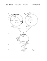
FIG. 1 is a schematic diagram of the two vectors used for surface expression library construction from heavy and light chain libraries. M13IX30 (FIG. 1A) is the vector used to clone the heavy chain sequences (open box). The single-headed arrow represents the Lac p/o expression sequences and the double-headed arrow represents the portion of M13IX30 which is to be combined with M13IX11. The amber stop codon and relevant restriction sites are also shown. M13IX11 (FIG. 1B) is the vector used to clone the light chain sequences (hatched box). Thick lines represent the pseudo-wild type (ψ gVIII) and wild type (gVIII) gene VIII sequences. The double-headed arrow represents the portion of M13IX11 which is to be combined with M13IX30. Relevant restriction sites are also shown. FIG. 1C shows the joining of vector population from heavy and light chain libraries to form the functional surface expression vector M13IXHL. FIG. 1D shows the generation of a surface expression library in a non-suppressor strain and the production of phage. The phage are used to infect a suppressor strain (FIG. 1E) for surface expression and screening of the library.
FIGS. 2A, 2B and 2C is the nucleotide sequence of M13IX30 (SEQ ID NO: 1).
FIGS. 3A, 3B and 3C is the nucleotide sequence of M13IX11 (SEQ ID NO:2).
FIGS. 4A, 4B and 4C is the nucleotide sequence of M13IX34 (SEQ ID NO: 3).
FIGS. 5A, 5B and 5C is the nucleotide sequence of M13IX13 (SEQ ID NO: 4).
FIGS. 6A, 6B and 6C is the nucleotide sequence of M13IX60 (SEQ ID NO: 5).
DETAILED DESCRIPTION OF THE INVENTION
This invention is directed to simple and efficient methods to generate a large repertoire of diverse combinations of heteromeric receptors. The method is advantageous in that only proper combinations of vector portions are randomly brought together for the coexpression of different DNA sequences without loss of population size or diversity. The receptors can be expressed on the surface of cells, such as those producing filamentous bacteriophage, which can be screened in large numbers. The nucleic acid sequences encoding the receptors can be readily characterized because the filamentous bacteriophage produce single strand DNA for efficient sequencing and mutagenesis methods. The heteromeric receptors so produced are useful in an unlimited number of diagnostic and therapeutic procedures.
In one embodiment, two populations of diverse heavy (Hc) and light (Lc) chain sequences are synthesized by polymerase chain reaction (PCR). These populations are cloned into separate M13-based vectors containing elements necessary for expression. The heavy chain vector contains a gene VIII (gVIII) coat protein sequence so that translation of the Hc sequences produces gVIII-Hc fusion proteins. The populations of the two vectors are randomly combined such that only the vector portions containing the Hc and Lc sequences are joined into a single circular vector. The combined vector directs the coexpression of both Hc and Lc sequences for assembly of the two polypeptides and surface expression on M13. A mechanism also exists to control the expression of gVIII-Hc fusion proteins during library construction and screening.
As used herein, the term “heteromeric receptors” refers to proteins composed of two or more subunits which together exhibit binding activity toward particular molecules. It is understood that the term includes the subunit fragments so long as assembly of the polypeptides and function of the assembled complex is retained. Heteromeric receptors include, for example, antibodies and fragments thereof such as Fab and (Fab)2 portions, T cell receptors, integrins, hormone receptors and transmitter receptors.
As used herein, the term “preselected molecule” refers to a molecule which is chosen from a number of choices. The molecule can be, for example, a protein or peptide, or an organic molecule such as a drug. Benzodiazapam is a specific example of a preselected molecule.
As used herein, the term “coexpression” refers to the expression of two or more nucleic acid sequences usually expressed as separate polypeptides. For heteromeric receptors, the coexpressed polypeptides assemble to form the heteromer. Therefore, “expression elements” as used herein, refers to sequences necessary for the transcription, translation, regulation and sorting of the expressed polypeptides which mak up the heteromeric receptors. The term also includes the expression of two subunit polypeptides which are linked but are able to assemble into a heteromeric receptor. A specific example of coexpression of linked polypeptides is where Hc and Lc polypeptides are expressed with a flexible peptide or polypeptide linker joining the two subunits into a single chain. The linker is flexible enough to allow association of Hc and Lc portions into a functional Fab fragment.
The invention provides for a composition of matter comprising a plurality of procaryotic cells containing diverse combinations of first and second DNA sequences encoding first and second polypeptides which form a heteromeric receptor exhibiting binding activity toward a preselected molecule, said heteromeric receptors being expressed on the surface of filamentous bacteriophage.
DNA sequences encoding the polypeptides of heteromeric receptors are obtained by methods known to one skilled in the art. Such methods include, for example, cDNA synthesis and polymerase chain reaction (PCR). The need will determine which method or combination of methods is to be used to obtain the desired populations of sequences. Expression can be performed in any compatible vector/host system. Such systems include, for example, plasmids or phagemids in procaryotes such as E. coli, yeast systems and other eucaryotic systems such as mammalian cells, but will be described herein in context with its presently preferred embodiment, i.e. expression on the surface of filamentous bacteriophage. Filamentous bacteriophage include, for example, M13, fl and fd. Additionally, the heteromeric receptors can also be expressed in soluble or secreted form depending on the need and the vector/host system employed.
Expression of heteromeric receptors such as antibodies or functional fragments thereof on the surface of M13 can be accomplished, for example, using the vector system shown in FIG. 1. Construction of the vectors enabling one of ordinary skill to make them are explicitly set out in Example I. The complete nucleotide sequences are given in FIGS. 2A, 2B and 2C and FIGS. 3A, 3B and 3C (SEQ ID NOS: 1 and 2). This system produces randomly combined populations of heavy (Hc) and light (Lc) chain antibody fragments functionally linked to expression elements. The Hc polypeptide is produced as a fusion protein with the M13 coat protein encoded by gene VIII. The gVIII-Hc fusion protein therefore anchors the assembled Hc and Lc polypeptides on the surface of M13. The diversity of Hc and Lc combinations obtained by this system can be 5×10⁷ or greater. Diversity of less than 5×10⁷ can also be obtained and will be determined by the need and type of heteromeric receptor to be expressed.
Populations of Hc and Lc encoding sequences to be combined into a vector for coexpression are each cloned into separate vectors. For the vectors shown in FIG. 1, diverse populations of sequences encoding Hc polypeptides are cloned into M13IX30 (SEQ ID NO: 1). Sequences encoding Lc polypeptides are cloned into M13IX11 (SEQ ID NO: 2). The populations are inserted between the Xho I-Spe I or Stu I restriction enzyme sites in M13IX30 and between the Sac I-Xba I or Eco RV sites in M13IX11 (FIGS. 1A and B, respectively).
The populations of Hc and Lc sequences inserted into the vectors can be synthesized with appropriate restriction recognition sequences flanking opposite ends of the encoding sequences but this is not necessary. The sites allow annealing and ligation in-frame with expression elements of these sequences into a double-stranded vector restricted with the appropriate restriction enzyme. Alternatively, and a preferred embodiment, the Hc and Lc sequences can be inserted into the vector without restriction of the DNA. This method of cloning is beneficial because naturally encoded restriction enzyme sites may be present within the sequences, thus, causing destruction of the sequence when treated with a restriction enzyme. For cloning without restriction, the sequences are treated briefly with a 3′ to 5′ exonuclease such as T4 DNA polymerase or exonuclease III. A 5′ to 3′ exonuclease will also accomplish the same function. The protruding 5′ termini which remains should be complementary to single-stranded overhangs within the vector which remain after restriction at the cloning site and treatment with exonuclease. The exonuclease treated inserts are annealed with the restricted vector by methods known to one skilled in the art. The exonuclease method decreases background and is easier to perform.
The vector used for Hc populations, M13IX30 (FIG. 1A; SEQ ID NO: 1) contains, in addition to expression elements, a sequence encoding the pseudo-wild type gVIII product downstream and in frame with the cloning sites. This gene encodes the wild type M13 gVIII amino acid sequence but has been changed at the nucleotide level to reduce homologous recombination with the wild type gVIII contained on the same vector. The wild type gVIII is present to ensure that at least some functional, non-fusion coat protein will be produced. The inclusion of a wild type gVIII therefore reduces the possibility of non-viable phage production and biological selection against certain peptide fusion proteins. Differential regulation of the two genes can also be used to control the relative ratio of the pseudo and wild type proteins.
Also contained downstream and in frame with the cloning sites is an amber stop codon. The stop codon is located between the inserted Hc sequences and the gVIII sequence and is in frame. As was the function of the wild type gVIII, the amber stop codon also reduces biological selection when combining vector portions to produce functional surface expression vectors. This is accomplished by using a non-suppressor (sup O) host strain because the non-suppressor strains will terminate expression after the Hc sequences but before the pseudo gVIII sequences. Therefore, the pseudo gVIII will essentially never be expressed on the phage surface under these circumstances. Instead, only soluble Hc polypeptides will be produced. Expression in a non-suppressor host strain can be advantageously utilized when one wishes to produce large populations of antibody fragments. Stop codons other than amber, such as opal and ochre, or molecular switches, such as inducible repressor elements, can also be used to unlink peptide expression from surface expression.
The vector used for Lc populations, M13IX11 (SEQ ID NO: 2), contains necessary expression elements and cloning sites for the Lc sequences, FIG. 1B. As with M13IX30, upstream and in frame with the cloning sites is a leader sequence for sorting to the phage surface. Additionally, a ribosome binding site and Lac Z promoter/operator elements are also present for transcription and translation of the DNA sequences.
Both vectors contain two pairs of Mlu I-Hind III restriction enzyme sites (FIGS. 1A and B) for joining together the Hc and Lc encoding sequences and their associated vector sequences. Mlu I and Hind III are non-compatible restriction sites. The two pairs are symmetrically orientated about the cloning site so that only the vector portions containing the sequences to be expressed are exactly combined into a single vector. The two pairs of sites are oriented identically with respect to one another on both vectors and the DNA between the two sites must be homologous enough between both vectors to allow annealing. This orientation allows cleavage of each circular vector into two portions and combination of essential components within each vector into a single circular vector where the encoded polypeptides can be coexpressed (FIG. 1C).
Any two pairs of restriction enzyme sites can be used so long as they are symmetrically orientated about the cloning site and identically orientated on both vectors. The sites within each pair, however, should be non-identical or able to be made differentially recognized as a cleavage substrate. For example, the two pairs of restriction sites contained within the vectors shown in FIG. 1 are Mlu I and Hind III. The sites are differentially cleavable by Mlu I and Hind III respectively. One skilled in the art knows how to substitute alternative pairs of restriction enzyme sites for the Mlu I-Hind III pairs described above. Also, instead of two Hind III and two Mlu I sites, a Hind III and Not I site can be paired with a Mlu I and a Sal I site, for example.
The combining step randomly brings together different Hc and Lc encoding sequences within the two diverse populations into a single vector (FIG. 1C; M13IXHL). The vector sequences donated from each independent vector, M13IX30 and M13IX11, are necessary for production of viable phage. Also, since the pseudo gVIII sequences are contained in M13IX30, coexpression of functional antibody fragments as Lc associated gVIII-Hc fusion proteins cannot be accomplished on the phage surface until the vector sequences are linked as shown in M13IXHL.
The combining step is performed by restricting each population of Hc and LC containing vectors with Mlu I and Hind III, respectively. The 3′ termini of each restricted vector population is digested with a 3′ to 5′ exonuclease as described above for inserting sequences into the cloning sites. The vector populations are mixed, allowed to anneal and introduced into an appropriate host. A non-suppressor host (FIG. 1D) is preferably used during initial construction of the library to ensure that sequences are not selected against due to expression as fusion proteins. Phage isolated from the library constructed in a non-suppressor strain can be used to infect a suppressor strain for surface expression of antibody fragments.
A method for selecting a heteromeric receptor exhibiting binding activity toward a preselected molecule from a population of diverse heteromeric receptors, comprising: (a) operationally linking to a first vector a first population of diverse DNA sequences encoding a diverse population of first polypeptides, said first vector having two pairs of restriction sites symmetrically oriented about a cloning site; (b) operationally linking to a second vector a second population of diverse DNA sequences encoding a diverse population of second polypeptides, said second vector having two pairs of restriction sites symmetrically oriented about a cloning site in an identical orientation to that of the first vector: (c) combining the vector products of step (a) and (b) under conditions which allow only the operational combination of vector sequences containing said first and second DNA sequences; (d) introducing said population of combined vectors into a compatible host under conditions sufficient for expressing said population of first and second DNA sequences; and (e) determining the heteromeric receptors which bind to said preselected molecule. The invention also provides for determining the nucleic acid sequences encoding such polypeptides as well.
Surface expression of the antibody library is performed in an amber suppressor strain. As described above, the amber stop codon between the Hc sequence and the gVIII sequence unlinks the two components in a non-suppressor strain. Isolating the phage produced from the non-suppressor strain and infecting a suppressor strain will link the Hc sequences to the gVIII sequence during expression (FIG. 1E). Culturing the suppressor strain after infection allows the coexpression on the surface of M13 of all antibody species within the library as gVIII fusion proteins (gVIII-Fab fusion proteins). Alternatively, the DNA can be isolated from the non-suppressor strain and then introduced into a suppressor strain to accomplish the same effect.
The level of expression of gVIII-Fab fusion proteins can additionally be controlled at the transcriptional level. Both polypeptides of the gVIII-Fab fusion proteins are under the inducible control of the Lac Z promoter/operator system. Other inducible promoters can work as well and are known by one skilled in the art. For high levels of surface expression, the suppressor library is cultured in an inducer of the Lac Z promoter such as isopropylthio-β-galactoside (IPTG). Inducible control is beneficial because biological selection against non-functional gVIII-Fab fusion proteins can be minimized by culturing the library under non-expressing conditions. Expression can then be induced only at the time of screening to ensure that the entire population of antibodies within the library are accurately represented on the phage surface. Also, this can be used to control the valency of the antibody on the phage surface.
The surface expression library is screened for specific Fab fragments which bind preselected molecules by standard affinity isolation procedures. Such methods include, for example, panning, affinity chromatography and solid phase blotting procedures. Panning as described by Parmley and Smith, Gene 73:305-318 (1988), which is incorporated herein by reference, is preferred because high titers of phage can be screened easily, quickly and in small volumes. Furthermore, this procedure can select minor Fab fragments species within the population, which otherwise would have been undetectable, and amplified to substantially homogenous populations. The selected Fab fragments can be characterized by sequencing the nucleic acids encoding the polypeptides after amplification of the phage population.
The following examples are intended to illustrate but not limit the invention.
EXAMPLE I
Construction, Expression and Screening of Antibody Fragments on the Surface of M13
This example shows the synthesis of a diverse population of heavy (Hc) and light (Lc) chain antibody fragments and their expression on the surface of M13 as gene VIII-Fab fusion proteins. The expressed antibodies derive from the random mixing and coexpression of a Hc and Lc pair. Also demonstrated is the isolation and characterization of the expressed Fab fragments which bind benzodiazapam (BDP) and their corresponding nucleotide sequence.
Isolation of mRNA and PCR Anplification of Antibody Fragments
The surface expression library is constructed from mRNA isolated from a mouse that had been immunized with KLH-coupled benzodiazapam (BDP). BDP was coupled to keyhole limpet hemocyanin (KLH) using the techniques described in Antibodies: A Laboratory Manual, Harlow and Lane, eds., Cold Spring Harbor, N.Y. (1988), which is incorporated herein by reference. Briefly, 10.0 milligrams (mg) of keyhole limpet hemocyanin and 0.5 mg of BDP with a glutaryl spacer arm N-hydroxysuccinimide linker appendages. Coupling was performed as in Jonda et al., Science, 241:1188 (1988), which is incorporated herein by reference. The KLH-BDP conjugate was removed by gel filtration chromatography through Sephadex G-25.
The KLH-BDP conjugate was prepared for injection into mice by adding 100 μg of the conjugate to 250 μl of phosphate buffered saline (PBS). An equal volume of complete Freund's adjuvant was added and emulsified the entire solution for 5 minutes. Mice were injected with 300 μl of the emulsion. Injections were given subcutaneously at several sites using a 21 gauge needle. A second immunization with BDP was given two weeks later. This injection was prepared as follows: 50 μg of BDP was diluted in 250 μl of PBS and an equal volume of alum was mixed with the solution. The mice were injected intraperitoneally with 500 μl of the solution using a 23 gauge needle. One month later the mice were given a final injection of 50 μg of the conjugate diluted to 200 μl in PBS. This injection was given intravenously in the lateral tail vein using a 30 gauge needle. Five days after this final injection the mice were sacrificed and total cellular RNA was isolated from their spleens.
Total RNA was isolated from the spleen of a single mouse immunized as described above by the method of Chomczynski and Sacchi, Anal. Biochem., 162:156-159 (1987), which is incorporated herein by reference. Briefly, immediately after removing the spleen from the immunized mouse, the tissue was homogenized in 10 ml of a denaturing solution containing 4.0 H guanine isothiocyanate, 0.25 M sodium citrate at pH 7.0, and 0.1 M 2-mercaptoethanol using a glass homogenizer. One ml of sodium acetate at a concentration of 2 M at pH 4.0 was mixed with the homogenized spleen. One ml of saturated phenol was also mixed with the denaturing solution containing the homogenized spleen. Two ml of a chloroform:isoamyl alcohol (24:1 v/v) mixture was added to this homogenate. The homogenate was mixed vigorously for ten seconds and maintained on ice for 15 minutes. The homogenate was then transferred to a thick-walled 50 ml polypropylene centrifuge tube (Fisher Scientific Company, Pittsburgh, Pa.). The solution was centrifuged at 10,000×g for 20 minutes at 4° C. The upper RNA-containing aqueous layer was transferred to a fresh 50 ml polypropylene centrifuge tube and mixed with an equal volume of isopropyl alcohol. This solution was maintained at −20° C. for at least one hour to precipitate the RNA. The solution containing the precipitated RNA was centrifuged at 10,000×g for twenty minutes at 4° C. The pelleted total cellular RNA was collected and dissolved in 3 ml of the denaturing solution described above. Three mls of isopropyl alcohol was added to the resuspended total cellular RNA and vigorously mixed. This solution was maintained at −20° C. for at least 1 hour to precipitate the RNA. The solution containing the precipitated RNA was centrifuged at 10,000×g for ten minutes at 4° C. The pelleted RNA was washed once with a solution containing 75% ethanol. The pelleted RNA was dried under vacuum for 15 minutes and then resuspended in dimethyl pyrocarbonate (DEPC) treated (DEPC-H2O) H2O.
Poly A30 RNA for use in first strand cDNA synthesis was prepared from the above isolated total RNA using a spin-column kit (Pharmacia, Piscataway, N.J.) as recommended by the manufacturer. The basic methodology has been described by Aviv and Leder, Proc. Natl. Acad. Sci., USA, 69:1408-1412 (1972), which is incorporated herein by reference. Briefly, one half of the total RNA isolated from a single immunized mouse spleen prepared as described above was resuspended in one ml of DEPC-treated dH2O and maintained at 65° C. for five minutes. One ml of 2×high salt loading buffer (100 mM Tris-HCL at pH 7.5, 1 M sodium chloride, 2.0 mM disodium ethylene diamine tetraacetic acid (EDTA) at pH 8.0, and 0.2% sodium dodecyl sulfate (SDS)) was added to the resuspended RNA and the mixture was allowed to cool to room temperature. The mixture was then applied to an oligo-dT (Collaborative Research Type 2 or Type 3 Bedford, Mass.) column that was previously prepared by washing the oligo-dT with a solution containing 0.1 M sodium hydroxide and 5 mM EDTA and then equilibrating the column with DEPC-treated dH2O. The eluate was collected in a sterile polypropylene tube and reapplied to the same column after heating the eluate for 5 minutes at 65° C. The oligo dT column was then washed with 2 ml of high salt loading buffer consisting of 50 mM Tris-HCL at pH 7.5, 500 mM sodium chloride, 1 mM EDTA at pH 8.0 and 0.1% SDS. The oligo dT column was then washed with 2 ml of 1×medium salt buffer (50 mM Tris-HCL at pH 7.5, 100 mM sodium chloride, 1 mM EDTA at pH 8.0 and 0.1% SDS). The mRNA was eluted with 1 ml of buffer consisting of 10 mM Tris-HCL at pH 7.5, 1 mM EDTA at pH 8.0 and 0.05% SDS. The messenger RNA was purified by extracting this solution with phenol/chloroform followed by a single extraction with 100% chloroform, ethanol precipitated and resuspended in DEPC treated dh2O.
In preparation for PCR amplification, mRNA was used as a template for cDNA synthesis. In a typical 250 μl reverse transcription reaction mixture, 5-10 μg of spleen mRNA in water was first annealed with 500 ng (0.5 pmol) of either the 3′ VN primer (primer 12, Table I) or the 3′ VL primer (primer 9, Table II) at 65° C. for 5 minutes. Subsequently, the mixture was adjusted to contain 0.8 mM dATP, 0.8 mM dCTP, 0.8 mM dGTP, 0.8 mM dTTP, 100 mM Tris-HCL (pH 8.6), 10 mM MgCl2, 40 mM KCl, and 20 mM 2-ME. Moloney-Murin Leukemia Virus (Bethesda Research Laboratories (BRL), Gaithersburg, Md.) Reverse transcriptase, 26 units, was added and the solution was incubated for 1 hour at 40° C. The resultant first strand cDNA was phenol extracted, ethanol precipitated and then used in the polymerase chain reaction (PCR) procedures described below for amplification of heavy and light chain sequences.
Primers used for amplification of heavy chain Fd fragments for construction of the M13IX30 library is shown in Table I. Amplification was performed in eight separate reactions, as described by Saiki et al., Science, 239:487-491 (1988), which is incorporated herein by reference, each reaction containing one of the 5′ primers (primers 2 to 9; SEQ ID NOS: 7 through 14, respectively) and one of the 3′ primers (primer 12; SEQ ID NO: 17) listed in Table I. The remaining 5′ primers, used for amplification in a single reaction, are either a degenerate primer (primer 1; SEQ ID NO: 6) or a primer that incorporates inosine at four degenerate positions (primer 10; SEQ ID NO: 15). The remaining 3′ primer (primer 11; SEQ ID NO: 16) was used to construct Fv fragments. The underlined portion of the 5′ primers incorporates an Xho I site and that of the 3′ primer an Spe I restriction site for cloning the amplified fragments into the M13IX30 vector in a predetermined reading frame for expression.
| TABLE I |
| |
| HEAVY CHAIN PRIMERS |
| |
| |
| |
CC G G T |
| 1) |
5′-AGGT A CT CTCGAGTC GG-3′ |
| |
GA A T A |
| 2) |
5′-AGGTCCAGCTGCTCGAGTCTGG-3′ |
| 3) |
5′-AGGTCCAGCTGCTCGAGTCAGG-3′ |
| 4) |
5′-AGGTCCAGCTTCTCGAGTCTGG-3′ |
| 5) |
5′-AGGTCCAGCTTCTCGAGTCAGG-3′ |
| 6) |
5′-AGGTCCAACTGCTCGAGTCTGG-3′ |
| 7) |
5′-AGGTCCAACTGCTCGAGTCAGG-3′ |
| 8) |
5′-AGGTCCAACTTCTCGAGTCTGG-3′ |
| 9) |
5′-AGGTCCAACTTCTCGAGTCAGG-3′ |
| |
T |
| 10) |
5′-AGGTIIAICTICTCGAGTC GG-3′ |
| |
A |
| 11) |
5′-CTATTAACTACTAACGGTAACAGTGGTGCCTTGCCCCA-3′ |
| 12) |
5′-AGGCTTACTAGTACAATCCCTGGGCACAAT-3′ |
| |
Primers used for amplification of mouse kappa light chain sequences for construction of the M13IX11 library are shown in Table II. These primers were chosen to contain restriction sites which were compatible with vector and not present in the conserved sequences of the mouse light chain mRNA. Amplification was performed as described above in five separate reactions, each containing one of the 5′ primers (primers 3 to 7; SEQ ID NOS: 20 through 24, respectively) and one of the 3′ primers (primer 9; SEQ ID NO: 26) listed in, Table II. The remaining 3′ primer (primer 8; SEQ ID NO: 25) was used to construct Fv fragments. The underlined portion of the 5′ primers depicts a Sac I restriction site and that of the 3′ primers an Xba I restriction site for cloning of the amplified fragments into the M13IX11 vector in a predetermined reading frame for expression.
| TABLE II |
| |
| LIGHT CHAIN PRIMERS |
| |
| |
| 1) |
5′-CCAGTTCCGAGCTCGTTGTGACTCAGGAATCT-3′ |
| 2) |
5′-CCAGTTCCGAGCTCGTGTTGACGCAGCCGCCC-3′ |
| 3) |
5′-CCAGTTCCGAGCTCGTGCTCACCCAGTCTCCA-3′ |
| 4) |
5′-CCAGTTCCGAGCTCCAGATGACCCAGTCTCCA-3′ |
| 5) |
5′-CCAGATGTGAGCTCGTGATGACCCAGACTCCA-3′ |
| 6) |
5′-CCAGATGTGAGCTCGTCATGACCCAGTCTCCA-3′ |
| 7) |
5′-CCAGTTCCGAGCTCGTGATGACACAGTCTCCA-3′ |
| 8) |
5′-GCAGCATTCTAGAGTTTCAGCTCCAGCTTGCC-3′ |
| 9) |
5′-GCGCCGTCTAGAATTAACACTCATTCCTGTTGAA-3′ |
| |
PCR amplification for heavy and light chain fragments was performed in a 100 μl reaction mixture containing the above described products of the reverse transcription reaction (≈5 μg of the cDNA-RNA hybrid), 300 nmol of 3′ VN primer (primer 12, Table I; SEQ ID NO: 17), and one of the 5′ VN primers (primers 2-9, Table I; SEQ ID NOS: 7 through 14, respectively) for heavy chain amplification, or, 300 nmol of 3′ VL primer (primer 9, Table II; SEQ ID NO: 26), and one of the 5′ VL primers (primers 3-7, Table II: SEQ ID NOS: 20 through 24, respectively) for each light chain amplification, a mixture of dNTPs at 200 mM, 50 mM KCl, 10 mM Tris-HCl (pH 8.3), 15 mM MgCl2, 0.1% gelatin, and 2 units of Thermus aquaticus DNA polymerase. The reaction mixture was overlaid with mineral oil and subjected to 40 cycles of amplification. Each amplification cycle involved denaturation at 92° C. for 1 minute, annealing at 52° C. for 2 minutes, and elongation at 72° C. for 1.5 minutes. The amplified samples were extracted twice with phenol/CHCl3 and once with CHCl3, ethanol-precipitated, and stored at −70° C. in 10 mM Tris-HCl, pH 7.5 1 mM EDTA. The resultant products were used in constructing the M13IX30 and M13IX11 libraries (see below).
Vector Construction
Two M13-based vectors, M13IX30 (SEQ ID NO: 1) and M13IX11 (SEQ ID NO: 2), were constructed for the cloning and propagation of Hc and Lc populations of antibody fragments, respectively. The vectors were constructed to facilitate the random joining and subsequent surface expression of antibody fragment populations.
M13IX30 (SEQ ID NO: 1), or the Hc vector, was constructed to harbor diverse populations of Hc antibody fragments. M13mp19 (Pharmacia, Piscataway, N.J.) was the starting vector. This vector was modified to contain, in addition to the encoded wild type M13 gen VIII: (1) a pseudo-wild type gene VIII sequence with an amber stop codon between it and the restriction sites for cloning oligonucleotides; (2) Stu I restriction site for insertion of sequences by hybridization and, Spe I and Xho I restriction sites in-frame with the pseudo-wild type gene VIII for cloning Hc sequences; (3) sequences necessary for expression, such as a promoter, signal sequence and translation initiation signals; (4) two pairs of Hind III-Mlu I sites for random joining of Hc and Lc vector portions, and (5) various other mutations to remove redundant restriction sites and the amino terminal portion of Lac Z.
Construction of M13IX30 was performed in four steps. In the first step, an M13-based vector containing the pseudo gVIII and various other mutations was constructed, M13IX01F. The second step involved the construction of a small cloning site in a separate M13mp18 vector to yield M13IX03. This vector was then expanded to contain expression sequences and restriction sites for Hc sequences to form M13IX04B. The fourth and final step involved the incorporation of the newly constructed sequences in M13IX04B into M13IX01F to yield M13IX30.
Construction of M13IX01F first involved the generation of a pseudo wild-type gVIII sequence for surface expression of antibody fragments. The pseudo-wild type gene encodes the identical amino acid sequence as that of the wild type gene; however, the nucleotide sequence has been altered so that only 63% identity exists between this gene and the encoded wild type gene VIII. Modification of the gene VIII nucleotide sequence used for surface expression reduces the possibility of homologous recombination with the wild type gene VIII contained on the same vector. Additionally, the wild type M13 gene VIII was retained in the vector system to ensure that at least some functional, non-fusion coat protein would be produced. The inclusion of wild type gene VIII facilitates the growth of phage under conditions where there is surface expression of the polypeptides and therefore reduces the possibility of non-viable phage production from the fusion genes.
The pseudo-wild type gene VIII was constructed by chemically synthesizing a series of oligonucleotides which encode both strands of the gene. The oligonucleotides are presented in Table III.
| TABLE III |
| |
| Pseudo-Wild Type Gene VIII Oligonucleotide Series |
| Top Strand |
|
| Oligonucleotides |
| VIII 03 |
GATCC TAG GCT GAA GGC |
| |
GAT GAC CCT GCT AAG GCT |
| |
GC |
| VIII 04 |
A TTC AAT AGT TTA CAG |
| |
GCA AGT GCT ACT GAG TAC |
| |
A |
| VIII 05 |
TT GGC TAC GCT TGG GCT |
| |
ATG GTA GTA GTT ATA GTT |
| VIII 06 |
GGT GCT ACC ATA GGG ATT |
| |
AAA TTA TTC AAA AAG TT |
| VIII 07 |
T ACG AGC AAG GCT TCT |
| |
TA |
| Bottom Strand |
| Oligonucleotides |
| VIII 08 |
AGC TTA AGA AGC CTT GCT |
| |
CGT AAA CTT TTT GAA TAA |
| |
TTT |
| VIII 09 |
AAT CCC TAT GGT AGC ACC |
| |
AAC TAT AAC TAC TAC CAT |
| VIII |
| 10 |
AGC CCA AGC GTA GCC AAT |
| |
GTA CTC AGT AGC ACT TG |
| VIII 11 |
C CTG TAA ACT ATT GAA |
| |
TGC AGC CTT AGC AGG GTC |
| VIII 12 |
ATC GCC TTC AGC CTA G |
| |
Except for the terminal oligonucleotides VIII 03 (SEQ ID NO: 27) and VIII 08 (SEQ ID NO: 32), the above oligonucleotides (oligonucleotides VIII 04-07 (SEQ ID NOS: 28 through 31, respectively) and VIII 09-12 (SEQ ID NOS: 33 through 36, respectively)) were mixed at 200 ng each in 10 μl final volume, phosphorylated with T4 polynucleotide Kinase (Pharmacia) and 1 mM ATP at 37° C. for 1 hour, heated to 70° C. for 5 minutes, and annealed into double-stranded form by heating to 65° C. for 3 minutes, followed by cooling to room temperature over a period of 30 minutes. The reactions were treated with 1.0 U of T4 DNA ligase (BRL) and 1 mM ATP at room temperature for 1 hour, followed by heating to 70° C. for 5 minutes. Terminal oligonucleotides were then annealed to the ligated oligonucleotides. The annealed and ligated oligonucleotides yielded a double-stranded DNA flanked by a Bam HI site at its 5′ end and by a Hind III site at its 3′ end. A translational stop codon (amber) immediately follows the Bam HI site. The gene VIII sequence begins with the codon GAA (Glu) two codons 3′ to the stop codon. The double-stranded insert was cloned in frame with the Eco RI and Sac I sites within the M13 polylinker. To do so, M13mp19 was digested with Bam HI (New England Biolabs, Beverley, Mass.) and Hind III (New England Biolabs) and combined at a molar ratio of 1:10 with the double-stranded insert. The ligations were performed at room temperature overnight in 1×ligase buffer (50 mM Tris-HCl, pH 7.8, 10 mM MgCl2, 20 mM DTT, 1 mM ATP, 50 μg/ml BSA) containing 1.0 U of T4 DNA ligase (New England Biolabs). The ligation mixture was transformed into a host and screened for positive clones using standard procedures in the art.
Several mutations were generated within the construct to yield functional M13IX01F. The mutations were generated using the method of Kunkel et al., Meth. Enzymol. 154:367-382 (1987), which is incorporated herein by reference, for site-directed mutagenesis. The reagents, strains and protocols were obtained from a Bio Rad Mutagenesis kit (Bio Rad, Richmond, Calif.) and mutagenesis was performed as recommended by the manufacturer.
Two Fok I sites were removed from the vector as well as the Hind III site at the end of the pseudo gene VIII sequence using the mutant oligonucleotides 5′-CATTTTTGCAGATGGCTTAGA-3′ (SEQ ID NO: 37) and 5′-TAGCATTAACGTCCAATA-3′ (SEQ ID NO: 38). New Hind III and Mlu I sites were also introduced at position 3919 and 3951 of M13IX01F. The oligonucleotides used for this mutagenesis had the sequences 5′-ATATATTTTAGTAAGCTTCATCTTCT-3′ (SEQ ID NO: 39), and 5′-GACAAAGAACGCGTGAAAACTTT-3′ (SEQ ID NO: 40), respectively. The amino terminal portion of Lac Z was deleted by oligonucleotide-directed mutagenesis using the mutant oligonucleotide 5′-GCGGGCCTCTTCGCTATTGCTTAAGAAGCCTTGCT-3′ (SEQ ID NO: 41). In constructing the above mutations, all changes made in a M13 coding region were performed such that the amino acid sequence remained unaltered. The resultant vector, M13IX01F, was used in the final step to construct M13IX30 (see below).
In the second step, M13mp18 was mutated to remove the 5′ end of Lac Z up to the Lac i binding site and including the Lac Z ribosome binding site and start codon. Additionally, the polylinker was removed and a Mlu I site was introduced in the coding region of Lac Z. A single oligonucleotide was used for these mutagenesis and had the sequence 5′-AAACGACGGCCAGTGCCAAGTGACGCGTGTGAAATTGTTATCC-3′ (SEQ ID NO: 42). Restriction enzyme sites for Hind III and Eco RI were introduced downstream of the Mlu I site using the oligonucleotide 5′-GGCGAAAGGGAATTCTGCAAGGCGATTAAGCTTGGGTAACGCC-3′ (SEQ ID NO. 43). These modifications of M13mp18 yielded the precursor vector M13IX03.
The expression sequences and cloning sites were introduced into M13IX03 by chemically synthesizing a series of oligonucleotides which encode both strands of the desired sequence. The oligonucleotides are presented in Table IV and correspond to oligonucleotides 084, 027, 028, 029, 085, 031, 032 and 033 as SEQ ID NOS:44-51, respectively.
| TABLE IV |
| |
| M13IX30 Oligonucleotide Series |
| Top Strand |
|
| Oligo- |
| nucleotides |
| 084 |
GGCGTTACCCAAGCTTTGTACATGGAGAAAATAAAG |
| 027 |
TGAAACAAAGCACTATTGCACTGGCACTCTTACCGT |
| |
TACCGT |
| 028 |
TACTGTTTACCCCTGTGACAAAAGCCCCCCAGGTCC |
| |
AGCTGC |
| 029 |
TCGAGTCAGGCCTATTGTGCCCAGGGATTGTACTAG |
| |
TGGATCCG |
| Bottom |
| Oligo- |
| nucleotides |
| 085 |
TGGCGAAAGGGAATTCGGATCCACTAGTACAATCCCTG |
| 031 |
GGCACAATAGGCCTGACTCGAGCAGCTGGACCAGGGCG |
| |
GCTT |
| 032 |
TTGTCACAGGGGTAAACAGTAACGGTAACGGTAAGTGT |
| |
GCCA |
| 033 |
GTGCAATAGTGCTTTGTTTCACTTTATTTTCTCCATGT |
| |
ACAA |
| |
The above oligonucleotides of Table IV, except for the terminal oligonucleotides 084 (SEQ ID NO: 44) and 085 (SEQ ID NO: 48), were mixed, phosphorylated, annealed and ligated to form a double-stranded insert as described in Example I. However, instead of cloning directly into the intermediate vector the insert was first amplified by PCR. The terminal oligonucleotides were used as primers for PCR. Oligonucleotide 084 (SEQ ID NO: 44) contains a Hind III site, 10 nucleotides internal to its 5′ end and oligonucleotide 085 (SEQ ID NO: 48) has an Eco RI site at its 5′ end. Following amplification, the products were restricted with Hind III and Eco RI and ligated, as described in Example I, into the polylinker of M13mp18 digested with the same two enzymes. The resultant double stranded insert contained a ribosome binding site, a translation initiation codon followed by a leader sequence and three restriction enzyme sites for cloning random oligonucleotides (Xho I, Stu I, Spe I). The intermediate vector was named M13IX04.
During cloning of the double-stranded insert, it was found that one of the GCC codons in oligonucleotides 028 and its complement in 031 was deleted. Since this deletion did not affect function, the final construct is missing one of the two GCC codons. Additionally, oligonucleotide 032 (SEQ ID NO: 50) contained a GTG codon where a GAG codon was needed. Mutagenesis was performed using the oligonucleotide 5′-TAACGGTAAGAGTGCCAGTGC-3′ (SEQ ID NO: 52) to convert the codon to the desired sequence. The resultant vector is named M13IX04B.
The third step in constructing M13IX30 involved inserting the expression and cloning sequences from M13IX04B upstream of the pseudo wild-type gVIII in M13IX01F. This was accomplished by digesting M13IX04B with Dra III and Bam HI and gel isolating the 700 base pair insert containing the sequences of interest. M13IX01F was likewise digested with Dra III and Bam HI. The insert was combined with the double digested vector at a molar ratio of 1:1 and ligated as described in Example I. The sequence of the final construct M13IX30, is shown in FIGS. 2A, 2B and 2C (SEQ ID NO: 1). FIG. 1A also shows M13IX30 where each of the elements necessary for surface expression of Hc fragments is marked. It should be noted during modification of the vectors, certain sequences differed from the published sequence of M13mp18. The new sequences are incorporated into the sequences recorded herein.
M13IX11 (SEQ ID NO: 2), or the Lc vector, was constructed to harbor diverse populations of Lc antibody fragments. This vector was also constructed from M13mp19 and contains: (1) sequences necessary for expression, such as a promoter, signal sequence and translation initiation signals; (2) Eco RV restriction site for insertion of sequences by hybridization and Sac I and Xba I restriction sites for cloning of Lc sequences; (3) two pairs of Hind III-Mlu I sites for random joining of Hc and Lc vector portions, and (4) various other mutation to remove redundant restriction sites.
The expression, translation initiation signals, cloning sites, and one of the Mlu I sites were constructed by annealing of overlapping oligonucleotides as described above to produce a double-stranded insert containing a 5′ Eco RI site and a 3′ Hind III site. The overlapping oligonucleotides are shown in Table V and correspond to oligonucleotides 082, 015, 016, 017, 018, 019, 083, 021, 022 and 023 as SEQ ID NOS:53-62, respectively and were ligated as a double-stranded insert between the Eco RI and Hind III sites of M13mp18 as described for the expression sequences inserted into M13IX03. The ribosome binding site (AGGAGAC) is located in oligonucleotide 015 and the translation initiation codon (ATG) is the second to fourth nucleotides of oligonucleotide 016 (SEQ ID NO: 55).
| TABLE V |
| |
| Oligonucleotide Series for Construction of |
| Translation Signals in M13IX11 |
| Oligonucleotide | Sequence (5′ to 3′) |
| |
| 082 | CACC TTCATG AATTC GGC AAG |
| | GAGACA GTCAT |
| 015 | AATT C GCC AAG GAG ACA GTC AT |
| 016 | AATG AAA TAC CTA TTG CCT ACC |
| | GCA GCC GCT CGA TTG TT |
| 017 | ATTA CTC GCT GCC CAA CCA GCC |
| | ATG GCC GAG CTC GTG AT |
| 018 | GACC CAG ACT CCA GATATC CAA |
| | CAG GAA TGA GTG TTA AT |
| 019 | TCT AGA ACG CGT C |
| 083 | TTCAGGTTGAAGC TTA CGC GTT |
| | CTA GAA TTA ACA CTC ATT |
| | CCTGT |
| 021 | TG GAT ATC TGG AGT CTG GGT |
| | CAT CAC GAG CTC GGC CAT G |
| 022 | GC TGG TTG GGC AGC GAG TAA |
| | TAA CAA TCC AGC GGC TGC C |
| 023 | GT AGG CAA TAG GTA TTT CAT |
| | TAT GAC TGT CCT TGG CG |
| |
Oligonucleotide 017 (SEQ ID NO: 56) contained a Sac I restriction site 67 nucleotides downstream from the ATG codon. The naturally occurring Eco RI site was removed and new Eco RI and Hind III sites were introduced downstream from the Sac I. Oligonucleotides 5′-TGACTGTCTCCTTGGCGTGTGAAATTGTTA-3′ (SEQ ID NO: 63) and 5′-TAACACTCATTCCGGATGGAATTCTGGAGTCTGGGT-3′ (SEQ ID NO: 64) were used to generate each of the mutations, respectively. The Lac Z ribosome binding site was removed when the original Eco RI site in M13mp19 was mutated. Additionally, when the new Eco RI and Hind III sites were generated, a spontaneous 100 bp deletion was found just 3′ to these sites. Since the deletion does not affect the function, it was retained in the final vector.
In addition to the above mutations, a variety of other modifications were made to incorporate or remove certain sequences. The Hind III site used to ligate the double-stranded insert was removed with the oligonucleotide 5′-GCCAGTGCCAAGTGACGCGTTCTA-3′ (SEQ ID NO: 65). Second Hind III and Mlu I sites were introduced at positions 3922 and 3952, respectively, using the oligonucleotides 5′-ATATATTTTAGTAAGCTTCATCTTCT-3′ (SEQ ID NO: 66) for the Hind III mutagenesis and 5′-GACAAAGAACGCGTGAAAACTTT-3′ (SEQ ID NO: 67) for the Mlu I mutagenesis. Again, mutations within the coding region did not alter the amino acid sequence.
The sequence of the resultant vector, M13IX11, is shown in FIGS. 3A, 3B and 3C (SEQ ID NO: 2). FIG. 1B also shows M13IX11 where each of the elements necessary for producing a surface expression library between Lc fragments is marked.
Library Construction
Each population of Hc and Lc sequences synthesized by PCR above are separately cloned into M13IX30 and M13IX11, respectively, to create Hc and Lc libraries.
The Hc and Lc products (5 μg) are mixed, ethanol precipitated and resuspended in 20 μl of NaOAc buffer (33 mM Tris acetate, pH 7.9, 10 mM Mg-acetate, 66 mM K-acetate, 0.5 mM DTT). Five units of T4 DNA polymerase is added and the reactions incubated at 30° C. for 5 minutes to remove 3′ termini by exonuclease digestion. Reactions are stopped by heating at 70° C. for 5 minutes. M13IX30 is digested with Stu I and M13IX11 is digested with Eco RV. Both vectors are treated with T4 DNA polymerase as described above and combined with the appropriate PCR products at a 1:1 molar ratio at 10 ng/μl to anneal in the above buffer at room temperature overnight. DNA from each annealing is electroporated into MK30-3 (Boehringer, Indianapolis, Ind.), as described below, to generate the Hc and Lc libraries.
E. coli MK30-3 is electroporated as described by Smith et al., Focus 12:38-40 (1990) which is incorporated herein by reference. The cells are prepared by inoculating a fresh colony of MK30-3 into 5 mls of SOB without magnesium (20 g bacto-tryptone, 5 g bacto-yeast extract, 0.584 g NaCl, 0.186 g KCl, dH2O to 1,000 mls) and grown with vigorous aeration overnight at 37° C. SOB without magnesium (500 ml) is inoculated at 1:1000 with the overnight culture and grown with vigorous aeration at 37° C. until the OD550 is 0.8 (about 2 to 3 h). The cells are harvested by centrifugation at 5,000 rpm (2,600×g) in a GS3 rotor (Sorvall, Newtown, Conn.) at 4° C. for 10 minutes, resuspended in 500 ml of ice-cold 10% (v/v) sterile glycerol, centrifuged and resuspended a second time in the same manner. After a third centrifugation, the cells are resuspended in 10% sterile glycerol at a final volume of about 2 ml, such that the OD550 of the suspension was 200 to 300. Usually, resuspension is achieved in the 10% glycerol that remained in the bottle after pouring off the supernate. Cells are frozen in 40 μl aliquots in microcentrifuge tubes using a dry ice-ethanol bath and stored frozen at −70° C.
Frozen cells are electroporated by thawing slowly on ice before use and mixing with about 10 pg to 500 ng of vector per 40 μl of cell suspension. A 40 μl aliquot is placed in an 0.1 cm electroporation chamber (Bio-Rad, Richmond, Calif.) and pulsed once at 0° C. using 4 kΩ parallel resistor 25 μF, 1.88 KV, which gives a puls length (τ) of −4 ms. A 10 μl aliquot of the pulsed cells are diluted into 1 ml SOC (98 mls SOB plus 1 ml of 2 M MgCl2 and 1 ml of 2 M glucose) in a 12-×75-mm culture tube, and the culture is shaken at 37° C. for 1 hour prior to culturing in selective media, (see below).
Each of the libraries are cultured using methods known to one skilled in the art. Such methods can be found in Sanbrook et al., Molecular Cloning: A Laboratory Manuel, Cold Spring Harbor Laboratory, Cold Spring Harbor, 1989, and in Ausubel et al., Current Protocols in Molecular Biology, John Wiley and Sons, New York, 1989, both of which are incorporated herein by reference. Briefly, the above 1 ml library cultures are grown up by diluting 50-fold into 2XYT media (16 g tryptone, 10 g yeast extract, 5 g NaCl) and culturing at 37° C. for 5-8 hours. The bacteria are pelleted by centrifugation at 10,000×g. The supernatant containing phage is transferred to a sterile tube and stored at 4° C.
Double strand vector DNA containing Hc and Lc antibody fragments are isolated from the cell pellet of each library. Briefly, the pellet is washed in TE (10 mM Tris, pH 8.0, 1 mM EDTA) and recollected by centrifugation at 7,000 rpm for 5′ in a Sorval centrifuge (Newtown, Conn.). Pellets are resuspended in 6 mls of 10% Sucrose, 50 mM Tris, pH 8.0. 3.0 ml of 10 mg/μl lysozyme is added and incubated on ice for 20 minutes. 12 mls of 0.2 M NaOH, 1% SDS is added followed by 10 minutes on ice. The suspensions are then incubated on ice for 20 minutes after addition of 7.5 mls of 3 M NaOAc, pH 4.6. The samples are centrifuged at 15,000 rpm for 15 minutes at 4° C., RNased and extracted with phenol/chloroform, followed by ethanol precipitation. The pellets are resuspended, weighed and an equal weight of CsCl2 is dissolved into each tube until a density of 1.60 g/ml is achieved. EtBr is added to 600 μg/ml and the double-stranded DNA is isolated by equilibrium centrifugation in a TV-1665 rotor (Sorval) at 50,000 rpm for 6 hours. These DNAs from each right and left half sublibrary are used to generate forty libraries in which the right and left halves of the randomized oligonucleotides have been randomly joined together.
The surface expression library is formed by the random joining of the Hc containing portion of M13IX30 with the Lc containing portion of M13IX11 . The DNAs isolated from each library was digested separately with an excess amount of restriction enzyme. The Lc population (5 μg) is digested with Hind III. The Hc (5 μg) population is digested with Mlu I. The reactions are stopped by phenol/chloroform extraction followed by ethanol precipitation. The pellets are washed in 70% ethanol and resuspended in 20 μl of NaOAc buffer. Five units of T4 DNA polymerase (Pharmacia) is added and the reactions incubated at 30° C. for 5 minutes. Reactions are stopped by heating at 70° C. for 5 minutes. The Hc and Lc DNAs are mixed to a final concentration of 10 ng each vector/μl and allowed to anneal at room temperature overnight. The mixture is electroporated into MK30-3 cells as described above.
Screening of Surface Expression Libraries
Purified phage are prepared from 50 ml liquid cultures of XL1 Blue™ cells (Stratagene, La Jolla, Calif.) which had been infected at a m.o.i. of 10 from the phage stocks stored at 4° C. The cultures are induced with 2 mM IPTG. Supernatants are cleared by two centrifugations, and the phage are precipitated by adding 1/7.5 volumes of PEG solution (25% PEG-8000, 2.5 M NaCl), followed by incubation at 4° C. overnight. The precipitate is recovered by centrifugation for 90 minutes at 10,000×g. Phage pellets are resuspended in 25 ml of 0.01 M Tris-HCl, pH 7.6, 1.0 mM EDTA, and 0.1% Sarkosyl and then shaken slowly at room temperature for 30 minutes. The solutions are adjusted to 0.5 M NaCl and to a final concentration of 5% polyethylene glycol. After 2 hours at 4° C., the precipitates containing the phage are recovered by centrifugation for 1 hour at 15,000×g. The precipitates are resuspended in 10 ml of NET buffer (0.1 M NaCl, 1.0 mM EDTA, and 0.01 M Tris-HCl, pH 7.6), mixed well, and the phage repelleted by centrifugation at 170,000×g for 3 hours. The phage pellets are resuspended overnight in 2 ml of NET buffer and subjected to cesium chloride centrifugation for 18 hours at 110,000×g (3.86 g of cesium chloride in 10 ml of buffer). Phage bands are collected, diluted 7-hold with NET buffer, recentrifuged at 170,000×g for 3 hours, resuspended, and stored at 4° C. in 0.3 ml of NET buffer containing 0.1 mM sodium azide.
The BDP used for panning on streptavidin coated dishes is first biotinylated and then absorbed against UV-inactivated blocking phage (see below). The biotinylating reagents are dissolved in dimethylformamide at a ratio of 2.4 mg solid NHS-SS-Biotin (sulfosuccinimidyl 2-(biotinamido)ethyl-1,3′ -dithiopropionate; Pierce, Rockford, Ill.) to 1 ml solvent and used as recommended by the manufacturer. Small-scale reactions are accomplished by mixing 1 μl dissolved reagent with 43 μl of 1 mg/ml BDP diluted in sterile bicarbonate buffer (0.1 M NaHCO3, pH 8.6). After 2 hours at 25° C., residual biotinylating reagent is reacted with 500 μl 1 M ethanolamine (pH adjusted to 9 with HCl) for an additional 2 hours. The entire sample is diluted with 1 ml TBS containing 1 mg/ml BSA, concentrated to about 50 μl on a Centricon 30 ultra-filter (Amicon), and washed on the same filter three times with 2 ml TBS and once with 1 ml TBS containing 0.02% NaN3 and 7×10¹² UV-inactivated blocking phage (see below); the final retentate (60-80 μl ) is stored at 4° C. BDP biotinylated with the NHS-SS-Biotin reagent is linked to biotin via a disulfide-containing chain.
UV-irradiated M13 phage are used for blocking any biotinylated BDP which fortuitously binds filamentous phage in general. M13mp8 (Messing and Vieira, Gene 19: 262-276 (1982), which is incorporated herein by reference) is chosen because it carries two amber mutations, which ensure that the few phage surviving irradiation will not grow in the sup O strains used to titer the surface expression library. A 5 ml sample containing 5×10¹³ M13mp8 phage, purified as described above, is placed in a small petri plate and irradiated with a germicidal lamp at a distance of two feet for 7 minutes (flux 150 μW/cm2). NaN3 is added to 0.02% and phage particles concentrated to 1014 particles/ml on a Centricon 30-kDa ultrafilter (Amicon).
For panning, polystyrene petri plates (60×15 mm) are incubated with 1 ml of 1 mg/ml of streptavidin (BRL) in 0.1 M NaHCO3 pH 8.6-0.02% NaN3 in a small, air-tight plastic box overnight in a cold room. The next day streptavidin is removed and replaced with at least 10 ml blocking solution (29 mg/ml of BSA; 3 μg/ml of streptavidin; 0.1 M NaHCO3 pH 8.6-0.02% NaN3) and incubated at least 1 hour at room temperature. The blocking solution is removed and plates are washed rapidly three times with Tris buffered saline containing 0.5% Tween 20 (TBS-0.5% Tween 20).
Selection of phage expressing antibody fragments which bind BDP is performed with 5 μl (2.7 μg BDP) of blocked biotinylated BDP reacted with a 50 μl portion of the library. Each mixture is incubated overnight at 4° C., diluted with 1 ml TBS-0.5% Tween 20, and transferred to a streptavidin-coated petri plate prepared as described above. After rocking 10 minutes at room temperature, unbound phage are removed and plates washed ten times with TBS-0.5% Tween 20 over a period of 30-90 minutes. Bound phage are eluted from plates with 800 μl sterile elution buffer (1 mg/ml BSA, 0.1 M HCl, pH adjusted to 2.2 with glycerol) for 15 minutes and eluates neutralized with 48 μl 2 M Tris (pH unadjusted). A 20 μl portion of each eluate is titered on MK30-3 concentrated cells with dilutions of input phage.
A second round of panning is performed by treating 750 μl of first eluate from the library with 5 mM DTT for 10 minutes to break disulfide bonds linking biotin groups to residual biotinylated binding proteins. The treated eluate is concentrated on a Centricon 30 ultrafilter (Amicon), washed three times with TBS-0.5% Tween 20, and concentrated to a final volume of about 50 μl. Final retentate is transferred to a tube containing 5.0 μl (2.7 μg BDP) blocked biotinylated BDP and incubated overnight. The solution is diluted with 1 ml TBS-0.5% Tween 20, panned, and eluted as described above on fresh streptavidin-coated petri plates. The entire second eluate (800 μl) is neutralized with 48 μl 2 M Tris, and 20 μl is titered simultaneously with the first eluate and dilutions of the input phage. If necessary, further rounds of panning can be performed to obtain homogeneous populations of phage. Additionally, phage can be plaque purified if reagents are available for detection.
Template Preparation and Sequencing
Templates are prepared for sequencing by inoculating a 1 ml culture of 2XYT containing a 1:100 dilution of an overnight culture of XL1 with an individual plaque from the purified population. The plaques are picked using a sterile toothpick. The culture is incubated at 37° C. for 5-6 hours with shaking and then transferred to a 1.5 ml microfuge tube. 200 μl of PEG solution is added, followed by vortexing and placed on ice for 10 minutes. The phage precipitate is recovered by centrifugation in a microfuge at 12,000×g for 5 minutes. The supernatant is discarded and the pellet is resuspended in 230 μl of TE (10 mM Tris-HCl, pH 7.5, 1 mM EDTA) by gently pipeting with a yellow pipet tip. Phenol (200 μl ) is added, followed by a brief vortex and microfuged to separate the phases. The aqueous phase is transferred to a separate tube and extracted with 200 μl of phenol/chloroform (1:1) as described above for the phenol extraction. A 0.1 volume of 3 M NaOAc is added, followed by addition of 2.5 volumes of ethanol and precipated at −20° C. for 20 minutes. The precipated templates are recovered by centrifugation in a microfuge at 12,000×g for 8 minutes. The pellet is washed in 70% ethanol, dried and resuspended in 25 μl TE. Sequencing was performed using a Sequenase™ sequencing kit following the protocol supplied by the manufacturer (U.S. Biochemical, Cleveland, Ohio).
EXAMPLE II
Cloning of Heavy and Light Chain Sequences Without Restriction Enzyme Digestion
This example shows the simultaneous incorporation of antibody heavy and light chain fragment encoding sequences into a M13IXHL-type vector without use of restriction endonucleases.
For the simultaneous incorporation of heavy and light chain encoding sequences into a single coexpression vector, a M13IXHL vector was produced that contained heavy and light chain encoding sequences for a mouse monoclonal antibody (DAN-18H4; Biosite, San Diego, Calif.). The inserted antibody fragment sequences are used as complementary sequences for the hybridization and incorporation of Hc and Lc sequences by site-directed mutagenesis. The genes encoding the heavy and light chain polypeptides were inserted into M13IX30 (SEQ ID NO: 1) and M13IX11 (SEQ ID NO: 2), respectively, and combined into a single surface expression vector as described in Example I. The resultant M13IXHL-type vector is termed M13IX50.
The combinations were performed under conditions that facilitate the formation of one Hc and one Lc vector half into a single circularized vector. Briefly, the overhangs generated between the pairs of restriction sites after restriction with Mlu I or Hind III and exonuclease digestion are unequal (i.e., 64 nucleotides compared to 32 nucleotides). These unequal lengths result in differential hybridization temperatures for specific annealing of the complementary ends from each vector. The specific hybridization of each end of each vector half was accomplished by first annealing at 65° C. in a small volume (about 100 μg/μl) to form a dimer of one Hc vector half and one Lc vector half. The dimers were circularized by diluting the mixture (to about 20 μg/μl ) and lowering the temperature to about 25-37° C. to allow annealing. T4 ligase was present to covalently close the circular vectors.
M13IX50 was modified such that it did not produce a functional polypeptide for the DAN monoclonal antibody. To do this, about eight amino acids were changed within the variable region of each chain by mutagenesis. The Lc variable region was mutagenized using the oligonucleotide 5′-CTGAACCTGTCTGGGACCACAGTTGATGCTATAGGATCAGATCTAGAATTCATTTAGAGACTGGCCTGGCTTCTGC-3′ (SEQ ID NO: 68). The Hc sequence was mutagenized with the oligonucleotide 5′-TCGACCGTTGGTAGGAATAATGCAATTAATGGAGTAGCTCTAAATTCAGAATTCATCTACACCCAGTGCATCCAGTAGCT-3′ (SEQ ID NO: 69). An additional mutation was also introduced into M13IX50 to yield the final form of the vector. During construction of an intermediate to X13IX50 (M13IX04 described in Example I), a six nucleotide sequence was duplicated in oligonucleotide 027 and its complement 032. This sequence, 5′TTACCG-3′ was deleted by mutagenesis using the oligonucleotide 5′-GGTAAACAGTAACGGTAAGAGTGCCAG-3′ (SEQ ID NO: 70). The resultant vector was designated M13IX53.
M13IX53 can be produced as a single stranded form and contains all the functional elements of the previously described M13IXHL vector except that it does not express functional antibody heteromers. The single-stranded vector can be hybridized to populations of single-stranded Hc and Lc encoding sequences for their incorporation into the vector by mutagenesis. Populations of single-stranded Hc and Lc encoding sequences can be produced by one skilled in the art from the PCR products described in Example I or by other methods known to one skilled in the art using the primers and teachings described therein. The resultant vectors with Hc and Lc encoding sequences randomly incorporated are propagated and screened for desired binding specificities as described in Example I.
Other vectors similar to M13IX53 and the vectors it's derived from, M13IX11 and M13IX30, have also been produced for the incorporation of Hc and Lc encoding sequences without restriction. In contrast to M13IX53, these vectors contain human antibody sequences for the efficient hybridization and incorporation of populations of human Hc and Lc sequences. These vectors are briefly described below. The starting vectors were either the Hc vector (M13IX30) or the Lc vector (M13IX11) previously described.
M13IX32 was generated from M13IX30 by removing the six nucleotide redundant sequence 5′-TTACCG-3′ described above and mutation of the leader sequence to increase secretion of the product. The oligonucleotide used to remove the redundant sequence is the same as that given above. The mutation in the leader sequence was generated using the oligonucleotide 5′GGGCTTTTCCACAGGGGT-3′ (SEQ ID NO: 76 ). This mutagenesis resulted in the A residue at position 6353 of M13IX30 being changed to a G residue.
A decapeptide tag for affinity purification of antibody fragments was incorporated in the proper reading frame at the carboxy-terminal end of the Hc expression site in M13IX32. The oligonucleotide used for this mutagenesis was 5′-CGCCTTCAGCCTAAGAAGCGTAGTCCGGAACGTCGTACGGGTAGGATCCACTAG-3′ (SEQ ID NO: 71). The resultant vector was designated M13IX33. Modifications to this or other vectors are envisioned which include various features known to one skilled in the art. For example, a peptidase cleavage site can be incorporated following the decapeptide tag which allows the antibody to be cleaved from the gene VIII portion of the fusion protein.
M13IX34 (SEQ ID NO: 3) was created from M13IX33 by cloning in the gene encoding a human IgG1 heavy chain. The reading frame of the variable region was changed and a stop codon was introduced to ensure that a functional polypeptide would not be produced. The oligonucleotide used for the mutagenesis of the variable region was 5′-CACCGGTTCGGGGAATTAGTCTTGACCAGGCAGCCCAGGGC-3′ (SEQ ID NO: 72). The complete nucleotide sequence of this vector is shown in FIGS. 4A, 4B and 4C (SEQ ID NO: 3).
Several vectors of the M13IX11 series were also generated to contain similar modifications as that described for the vectors M13IX53 and M13IX34. The promoter region in M13IX11 was mutated to conform to the 35 consensus sequence to generate M13IX12. The oligonucleotide used for this mutgenesis was 5′-ATTCCACACATTATACGAGCCCGGAAGCATAAAGTGCAAGCCTGGGGTGCC-3′ (SEQ ID NO: 73). A human kappa light chain sequence was cloned into M13IX12 and the variable region subsequently deleted to generate M13IX13 (SEQ ID NO: 4). The complete nucleotide sequence of this vector is shown in FIGS. 5A, 5B and 5C (SEQ ID NO: 4). A similar vector, designated M13IX14, was also generated in which the human lambda light chain was inserted into M13IX12 followed by deletion of the variable region. The oligonucleotides used for the variable region deletion of M13IX13 and M13IX14 were 5′-CTGCTCATCAGATGGCGGGAAGAGCTCGGCCATGGCTGGTTG-3′ (SEQ ID NO: 74) and 5′-GAACAGAGTGACCGAGGGGGCGAGCTCGGCCATGGCTGGTTG-3′ (SEQ ID NO: 75), respectively.
The Hc and Lc vectors or modified forms thereof can be combined using the methods described in Example I to produce a single vector similar to M13IX53 that allows the efficient incorporation of human Hc and Lc encoding sequences by mutagenesis. An example of such a vector is the combination of M13IX13 with M13IX34. The complete nucleotide sequence of this vector, M13IX60, is shown in FIGS. 6A, 6B and 6C (SEQ ID NO: 5).
Additional modifications to any of the previously described vectors can also be performed to generate vectors which allow the efficient incorporation and surface expression of Hc and Lc sequences. For example, to alleviate the use of uracil selection against wild-type template during mutagenesis procedures, the variable region locations within the vectors can be substituted by a set of palindromic restriction enzyme sites (i.e., two similar sites in opposite orientation). The palindromic sites will loop out and hybridize together during the mutagenesis and thus form a double-stranded substrate for restriction endonuclease digestion. Cleavage of the site results in the destruction of the wild-type template. The variable region of the inserted Hc or Lc sequences will not be affected since they will be in single stranded form.
Following the methods of Example I, single-stranded Hc or Lc populations can be produced by a variety of methods known to one skilled in the art. For example, the PCR primers described in Example I can be used in asymmetric PCR to generate such populations. Gelfand et al., “PCR Protocols: A Guide to Methods and Applications”, Ed by M. A. Innis (1990), which is incorporated herein by reference. Asymmetric PCR is a PCR method that differentially amplifies only a single strand of the double stranded template. Such differential amplification is accomplished by decreasing the primer amount for the undesirable strand about 10-fold compared to that for the desirable strand. Alternatively, single-stranded populations can be produced from double-stranded PCR products generated as described in Example I except that the primer(s) used to generate the undesirable strand of the double-stranded products is first phosphorylated at its 5′ end with a kinase. The resultant products can then be treated with a 5′ to 3′ exonuclease, such as lambda exonuclease (BRL, Bethesda, Md.) to digest away the unwanted strand.
Single-stranded Hc and Lc populations generated by the methods described above or by others known to one skilled in the art are hybridized to complementary sequences encoded in the previously described vectors. The population of the sequences are subsequently incorporated into a double-stranded form of the vector by polymerase extension of the hybridized templates. Propagation and surface expression of the randomly combined Hc and Lc sequences are performed as described in Example I.
Although the invention has been described with reference to the presently preferred embodiment, it should be understood that various modifications can be made without departing from the spirit of the invention. Accordingly, the invention is limited only by the