US6231834B1 - Methods for ultrasound imaging involving the use of a contrast agent and multiple images and processing of same - Google Patents
Methods for ultrasound imaging involving the use of a contrast agent and multiple images and processing of same Download PDFInfo
- Publication number
- US6231834B1 US6231834B1 US08/982,829 US98282997A US6231834B1 US 6231834 B1 US6231834 B1 US 6231834B1 US 98282997 A US98282997 A US 98282997A US 6231834 B1 US6231834 B1 US 6231834B1
- Authority
- US
- United States
- Prior art keywords
- lipid
- gas
- vesicles
- phospholipid
- gaseous precursor
- Prior art date
- Legal status (The legal status is an assumption and is not a legal conclusion. Google has not performed a legal analysis and makes no representation as to the accuracy of the status listed.)
- Expired - Lifetime
Links
- 238000000034 method Methods 0.000 title claims abstract description 270
- 239000002872 contrast media Substances 0.000 title claims abstract description 89
- 238000012285 ultrasound imaging Methods 0.000 title claims abstract description 9
- 238000012545 processing Methods 0.000 title claims description 9
- 150000002632 lipids Chemical class 0.000 claims abstract description 312
- 108090000623 proteins and genes Proteins 0.000 claims abstract description 110
- 102000004169 proteins and genes Human genes 0.000 claims abstract description 108
- 229920000642 polymer Polymers 0.000 claims abstract description 96
- 238000002604 ultrasonography Methods 0.000 claims abstract description 85
- 239000008365 aqueous carrier Substances 0.000 claims abstract description 6
- -1 N-substituted acrylamides Chemical class 0.000 claims description 204
- 239000002243 precursor Substances 0.000 claims description 185
- 150000001875 compounds Chemical class 0.000 claims description 56
- 229920001223 polyethylene glycol Polymers 0.000 claims description 50
- 150000003904 phospholipids Chemical class 0.000 claims description 42
- 239000002502 liposome Substances 0.000 claims description 34
- TXEYQDLBPFQVAA-UHFFFAOYSA-N tetrafluoromethane Chemical compound FC(F)(F)F TXEYQDLBPFQVAA-UHFFFAOYSA-N 0.000 claims description 29
- 229960004692 perflenapent Drugs 0.000 claims description 24
- NJCBUSHGCBERSK-UHFFFAOYSA-N perfluoropentane Chemical compound FC(F)(F)C(F)(F)C(F)(F)C(F)(F)C(F)(F)F NJCBUSHGCBERSK-UHFFFAOYSA-N 0.000 claims description 24
- 238000009835 boiling Methods 0.000 claims description 23
- KAVGMUDTWQVPDF-UHFFFAOYSA-N perflubutane Chemical compound FC(F)(F)C(F)(F)C(F)(F)C(F)(F)F KAVGMUDTWQVPDF-UHFFFAOYSA-N 0.000 claims description 23
- 229950003332 perflubutane Drugs 0.000 claims description 23
- 230000002107 myocardial effect Effects 0.000 claims description 22
- 229920001577 copolymer Polymers 0.000 claims description 21
- 229920001477 hydrophilic polymer Polymers 0.000 claims description 21
- QYSGYZVSCZSLHT-UHFFFAOYSA-N octafluoropropane Chemical compound FC(F)(F)C(F)(F)C(F)(F)F QYSGYZVSCZSLHT-UHFFFAOYSA-N 0.000 claims description 21
- 229960004065 perflutren Drugs 0.000 claims description 21
- 239000002202 Polyethylene glycol Substances 0.000 claims description 20
- 102000009027 Albumins Human genes 0.000 claims description 16
- 108010088751 Albumins Proteins 0.000 claims description 16
- 239000000693 micelle Substances 0.000 claims description 16
- LYCAIKOWRPUZTN-UHFFFAOYSA-N Ethylene glycol Chemical compound OCCO LYCAIKOWRPUZTN-UHFFFAOYSA-N 0.000 claims description 14
- 229920001451 polypropylene glycol Polymers 0.000 claims description 14
- 229920001059 synthetic polymer Polymers 0.000 claims description 14
- KILNVBDSWZSGLL-KXQOOQHDSA-N 1,2-dihexadecanoyl-sn-glycero-3-phosphocholine Chemical compound CCCCCCCCCCCCCCCC(=O)OC[C@H](COP([O-])(=O)OCC[N+](C)(C)C)OC(=O)CCCCCCCCCCCCCCC KILNVBDSWZSGLL-KXQOOQHDSA-N 0.000 claims description 12
- 229910018503 SF6 Inorganic materials 0.000 claims description 12
- 229920003229 poly(methyl methacrylate) Polymers 0.000 claims description 12
- 229920002239 polyacrylonitrile Polymers 0.000 claims description 12
- 239000004926 polymethyl methacrylate Substances 0.000 claims description 12
- 239000002356 single layer Substances 0.000 claims description 12
- SFZCNBIFKDRMGX-UHFFFAOYSA-N sulfur hexafluoride Chemical compound FS(F)(F)(F)(F)F SFZCNBIFKDRMGX-UHFFFAOYSA-N 0.000 claims description 12
- 229960000909 sulfur hexafluoride Drugs 0.000 claims description 12
- 229920003171 Poly (ethylene oxide) Polymers 0.000 claims description 11
- 229960004624 perflexane Drugs 0.000 claims description 11
- ZJIJAJXFLBMLCK-UHFFFAOYSA-N perfluorohexane Chemical compound FC(F)(F)C(F)(F)C(F)(F)C(F)(F)C(F)(F)C(F)(F)F ZJIJAJXFLBMLCK-UHFFFAOYSA-N 0.000 claims description 11
- 230000000747 cardiac effect Effects 0.000 claims description 8
- 150000008104 phosphatidylethanolamines Chemical class 0.000 claims description 8
- JZNWSCPGTDBMEW-UHFFFAOYSA-N Glycerophosphorylethanolamin Natural products NCCOP(O)(=O)OCC(O)CO JZNWSCPGTDBMEW-UHFFFAOYSA-N 0.000 claims description 7
- 239000000178 monomer Substances 0.000 claims description 7
- BCCOBQSFUDVTJQ-UHFFFAOYSA-N octafluorocyclobutane Chemical compound FC1(F)C(F)(F)C(F)(F)C1(F)F BCCOBQSFUDVTJQ-UHFFFAOYSA-N 0.000 claims description 7
- 235000019407 octafluorocyclobutane Nutrition 0.000 claims description 7
- HGINCPLSRVDWNT-UHFFFAOYSA-N Acrolein Chemical compound C=CC=O HGINCPLSRVDWNT-UHFFFAOYSA-N 0.000 claims description 6
- NLHHRLWOUZZQLW-UHFFFAOYSA-N Acrylonitrile Chemical compound C=CC#N NLHHRLWOUZZQLW-UHFFFAOYSA-N 0.000 claims description 6
- AEMRFAOFKBGASW-UHFFFAOYSA-N Glycolic acid Chemical compound OCC(O)=O AEMRFAOFKBGASW-UHFFFAOYSA-N 0.000 claims description 6
- PPBRXRYQALVLMV-UHFFFAOYSA-N Styrene Chemical compound C=CC1=CC=CC=C1 PPBRXRYQALVLMV-UHFFFAOYSA-N 0.000 claims description 6
- IISBACLAFKSPIT-UHFFFAOYSA-N bisphenol A Chemical compound C=1C=C(O)C=CC=1C(C)(C)C1=CC=C(O)C=C1 IISBACLAFKSPIT-UHFFFAOYSA-N 0.000 claims description 6
- JVTAAEKCZFNVCJ-UHFFFAOYSA-N lactic acid Chemical compound CC(O)C(O)=O JVTAAEKCZFNVCJ-UHFFFAOYSA-N 0.000 claims description 6
- 239000004952 Polyamide Substances 0.000 claims description 5
- UKACHOXRXFQJFN-UHFFFAOYSA-N heptafluoropropane Chemical compound FC(F)C(F)(F)C(F)(F)F UKACHOXRXFQJFN-UHFFFAOYSA-N 0.000 claims description 5
- WMIYKQLTONQJES-UHFFFAOYSA-N hexafluoroethane Chemical compound FC(F)(F)C(F)(F)F WMIYKQLTONQJES-UHFFFAOYSA-N 0.000 claims description 5
- WTJKGGKOPKCXLL-RRHRGVEJSA-N phosphatidylcholine Chemical compound CCCCCCCCCCCCCCCC(=O)OC[C@H](COP([O-])(=O)OCC[N+](C)(C)C)OC(=O)CCCCCCCC=CCCCCCCCC WTJKGGKOPKCXLL-RRHRGVEJSA-N 0.000 claims description 5
- 229920000747 poly(lactic acid) Polymers 0.000 claims description 5
- 229920002647 polyamide Polymers 0.000 claims description 5
- PORPENFLTBBHSG-MGBGTMOVSA-N 1,2-dihexadecanoyl-sn-glycerol-3-phosphate Chemical compound CCCCCCCCCCCCCCCC(=O)OC[C@H](COP(O)(O)=O)OC(=O)CCCCCCCCCCCCCCC PORPENFLTBBHSG-MGBGTMOVSA-N 0.000 claims description 4
- NRJAVPSFFCBXDT-HUESYALOSA-N 1,2-distearoyl-sn-glycero-3-phosphocholine Chemical compound CCCCCCCCCCCCCCCCCC(=O)OC[C@H](COP([O-])(=O)OCC[N+](C)(C)C)OC(=O)CCCCCCCCCCCCCCCCC NRJAVPSFFCBXDT-HUESYALOSA-N 0.000 claims description 4
- NIXOWILDQLNWCW-UHFFFAOYSA-N 2-Propenoic acid Natural products OC(=O)C=C NIXOWILDQLNWCW-UHFFFAOYSA-N 0.000 claims description 4
- WHNWPMSKXPGLAX-UHFFFAOYSA-N N-Vinyl-2-pyrrolidone Chemical compound C=CN1CCCC1=O WHNWPMSKXPGLAX-UHFFFAOYSA-N 0.000 claims description 4
- 229920002125 Sokalan® Polymers 0.000 claims description 4
- 238000001802 infusion Methods 0.000 claims description 4
- LGUZHRODIJCVOC-UHFFFAOYSA-N perfluoroheptane Chemical compound FC(F)(F)C(F)(F)C(F)(F)C(F)(F)C(F)(F)C(F)(F)C(F)(F)F LGUZHRODIJCVOC-UHFFFAOYSA-N 0.000 claims description 4
- YVBBRRALBYAZBM-UHFFFAOYSA-N perfluorooctane Chemical compound FC(F)(F)C(F)(F)C(F)(F)C(F)(F)C(F)(F)C(F)(F)C(F)(F)C(F)(F)F YVBBRRALBYAZBM-UHFFFAOYSA-N 0.000 claims description 4
- ACZNIBVNGPLHAC-ONEGZZNKSA-N (2e)-penta-2,4-dien-1-ol Chemical compound OC\C=C\C=C ACZNIBVNGPLHAC-ONEGZZNKSA-N 0.000 claims description 3
- SMZOUWXMTYCWNB-UHFFFAOYSA-N 2-(2-methoxy-5-methylphenyl)ethanamine Chemical compound COC1=CC=C(C)C=C1CCN SMZOUWXMTYCWNB-UHFFFAOYSA-N 0.000 claims description 3
- QLIBJPGWWSHWBF-UHFFFAOYSA-N 2-aminoethyl methacrylate Chemical class CC(=C)C(=O)OCCN QLIBJPGWWSHWBF-UHFFFAOYSA-N 0.000 claims description 3
- KGIGUEBEKRSTEW-UHFFFAOYSA-N 2-vinylpyridine Chemical compound C=CC1=CC=CC=N1 KGIGUEBEKRSTEW-UHFFFAOYSA-N 0.000 claims description 3
- XTHDRUMOXKOSLN-UHFFFAOYSA-N 4-(3-phenylprop-1-enyl)aniline Chemical compound C1=CC(N)=CC=C1C=CCC1=CC=CC=C1 XTHDRUMOXKOSLN-UHFFFAOYSA-N 0.000 claims description 3
- LBSXSAXOLABXMF-UHFFFAOYSA-N 4-Vinylaniline Chemical compound NC1=CC=C(C=C)C=C1 LBSXSAXOLABXMF-UHFFFAOYSA-N 0.000 claims description 3
- YFGSHAIDEAXREE-UHFFFAOYSA-N 6,6-dimethyl-1,3,5,2,4-trioxadisilepane Chemical compound CC1(C)CO[SiH2]O[SiH2]O1 YFGSHAIDEAXREE-UHFFFAOYSA-N 0.000 claims description 3
- HRPVXLWXLXDGHG-UHFFFAOYSA-N Acrylamide Chemical compound NC(=O)C=C HRPVXLWXLXDGHG-UHFFFAOYSA-N 0.000 claims description 3
- NOWKCMXCCJGMRR-UHFFFAOYSA-N Aziridine Chemical compound C1CN1 NOWKCMXCCJGMRR-UHFFFAOYSA-N 0.000 claims description 3
- 229920001651 Cyanoacrylate Polymers 0.000 claims description 3
- BRLQWZUYTZBJKN-UHFFFAOYSA-N Epichlorohydrin Chemical compound ClCC1CO1 BRLQWZUYTZBJKN-UHFFFAOYSA-N 0.000 claims description 3
- JIGUQPWFLRLWPJ-UHFFFAOYSA-N Ethyl acrylate Chemical compound CCOC(=O)C=C JIGUQPWFLRLWPJ-UHFFFAOYSA-N 0.000 claims description 3
- WOBHKFSMXKNTIM-UHFFFAOYSA-N Hydroxyethyl methacrylate Chemical compound CC(=C)C(=O)OCCO WOBHKFSMXKNTIM-UHFFFAOYSA-N 0.000 claims description 3
- CERQOIWHTDAKMF-UHFFFAOYSA-N Methacrylic acid Chemical compound CC(=C)C(O)=O CERQOIWHTDAKMF-UHFFFAOYSA-N 0.000 claims description 3
- MWCLLHOVUTZFKS-UHFFFAOYSA-N Methyl cyanoacrylate Chemical compound COC(=O)C(=C)C#N MWCLLHOVUTZFKS-UHFFFAOYSA-N 0.000 claims description 3
- VVQNEPGJFQJSBK-UHFFFAOYSA-N Methyl methacrylate Chemical compound COC(=O)C(C)=C VVQNEPGJFQJSBK-UHFFFAOYSA-N 0.000 claims description 3
- 229920002845 Poly(methacrylic acid) Polymers 0.000 claims description 3
- 229920002873 Polyethylenimine Polymers 0.000 claims description 3
- GOOHAUXETOMSMM-UHFFFAOYSA-N Propylene oxide Chemical compound CC1CO1 GOOHAUXETOMSMM-UHFFFAOYSA-N 0.000 claims description 3
- XTXRWKRVRITETP-UHFFFAOYSA-N Vinyl acetate Chemical compound CC(=O)OC=C XTXRWKRVRITETP-UHFFFAOYSA-N 0.000 claims description 3
- LDHQCZJRKDOVOX-NSCUHMNNSA-N crotonic acid Chemical compound C\C=C\C(O)=O LDHQCZJRKDOVOX-NSCUHMNNSA-N 0.000 claims description 3
- 239000004205 dimethyl polysiloxane Substances 0.000 claims description 3
- MWRBNPKJOOWZPW-CLFAGFIQSA-N dioleoyl phosphatidylethanolamine Chemical compound CCCCCCCC\C=C/CCCCCCCC(=O)OCC(COP(O)(=O)OCCN)OC(=O)CCCCCCC\C=C/CCCCCCCC MWRBNPKJOOWZPW-CLFAGFIQSA-N 0.000 claims description 3
- 239000003822 epoxy resin Substances 0.000 claims description 3
- 235000014655 lactic acid Nutrition 0.000 claims description 3
- 239000004310 lactic acid Substances 0.000 claims description 3
- 210000005246 left atrium Anatomy 0.000 claims description 3
- PNJWIWWMYCMZRO-UHFFFAOYSA-N pent‐4‐en‐2‐one Natural products CC(=O)CC=C PNJWIWWMYCMZRO-UHFFFAOYSA-N 0.000 claims description 3
- 229920000435 poly(dimethylsiloxane) Polymers 0.000 claims description 3
- 239000004584 polyacrylic acid Substances 0.000 claims description 3
- 229920001610 polycaprolactone Polymers 0.000 claims description 3
- 229920000647 polyepoxide Polymers 0.000 claims description 3
- 239000004626 polylactic acid Substances 0.000 claims description 3
- 229920001296 polysiloxane Polymers 0.000 claims description 3
- MNCGMVDMOKPCSQ-UHFFFAOYSA-M sodium;2-phenylethenesulfonate Chemical compound [Na+].[O-]S(=O)(=O)C=CC1=CC=CC=C1 MNCGMVDMOKPCSQ-UHFFFAOYSA-M 0.000 claims description 3
- BUJXOJKSMCPEEB-UHFFFAOYSA-N sodium;2-sulfooxyethyl 2-methylprop-2-enoate Chemical compound [Na].CC(=C)C(=O)OCCOS(O)(=O)=O BUJXOJKSMCPEEB-UHFFFAOYSA-N 0.000 claims description 3
- LDHQCZJRKDOVOX-UHFFFAOYSA-N trans-crotonic acid Natural products CC=CC(O)=O LDHQCZJRKDOVOX-UHFFFAOYSA-N 0.000 claims description 3
- PAPBSGBWRJIAAV-UHFFFAOYSA-N ε-Caprolactone Chemical compound O=C1CCCCCO1 PAPBSGBWRJIAAV-UHFFFAOYSA-N 0.000 claims description 3
- UVWPNDVAQBNQBG-UHFFFAOYSA-N 1,1,1,2,2,3,3,4,4,5,5,6,6,7,7,8,8,9,9,9-icosafluorononane Chemical compound FC(F)(F)C(F)(F)C(F)(F)C(F)(F)C(F)(F)C(F)(F)C(F)(F)C(F)(F)C(F)(F)F UVWPNDVAQBNQBG-UHFFFAOYSA-N 0.000 claims description 2
- CITHEXJVPOWHKC-UUWRZZSWSA-N 1,2-di-O-myristoyl-sn-glycero-3-phosphocholine Chemical compound CCCCCCCCCCCCCC(=O)OC[C@H](COP([O-])(=O)OCC[N+](C)(C)C)OC(=O)CCCCCCCCCCCCC CITHEXJVPOWHKC-UUWRZZSWSA-N 0.000 claims description 2
- SNKAWJBJQDLSFF-NVKMUCNASA-N 1,2-dioleoyl-sn-glycero-3-phosphocholine Chemical compound CCCCCCCC\C=C/CCCCCCCC(=O)OC[C@H](COP([O-])(=O)OCC[N+](C)(C)C)OC(=O)CCCCCCC\C=C/CCCCCCCC SNKAWJBJQDLSFF-NVKMUCNASA-N 0.000 claims description 2
- NLXLAEXVIDQMFP-UHFFFAOYSA-N Ammonium chloride Substances [NH4+].[Cl-] NLXLAEXVIDQMFP-UHFFFAOYSA-N 0.000 claims description 2
- 229960003724 dimyristoylphosphatidylcholine Drugs 0.000 claims description 2
- SLKDGVPOSSLUAI-PGUFJCEWSA-N 1,2-dihexadecanoyl-sn-glycero-3-phosphoethanolamine zwitterion Chemical compound CCCCCCCCCCCCCCCC(=O)OC[C@H](COP(O)(=O)OCCN)OC(=O)CCCCCCCCCCCCCCC SLKDGVPOSSLUAI-PGUFJCEWSA-N 0.000 claims 2
- 239000002691 unilamellar liposome Substances 0.000 claims 2
- 239000004094 surface-active agent Substances 0.000 abstract description 29
- 230000001976 improved effect Effects 0.000 abstract description 12
- 230000002526 effect on cardiovascular system Effects 0.000 abstract description 6
- 239000000203 mixture Substances 0.000 description 261
- 239000007789 gas Substances 0.000 description 239
- 230000008685 targeting Effects 0.000 description 164
- 239000003446 ligand Substances 0.000 description 143
- 239000000463 material Substances 0.000 description 106
- 235000018102 proteins Nutrition 0.000 description 90
- 239000007788 liquid Substances 0.000 description 89
- 108090000765 processed proteins & peptides Proteins 0.000 description 74
- 210000001519 tissue Anatomy 0.000 description 61
- 102000004196 processed proteins & peptides Human genes 0.000 description 46
- 230000000087 stabilizing effect Effects 0.000 description 45
- 230000015572 biosynthetic process Effects 0.000 description 39
- 230000007704 transition Effects 0.000 description 39
- 210000004027 cell Anatomy 0.000 description 36
- 239000003795 chemical substances by application Substances 0.000 description 36
- 239000012071 phase Substances 0.000 description 34
- 239000012867 bioactive agent Substances 0.000 description 33
- 125000000217 alkyl group Chemical group 0.000 description 31
- 230000005298 paramagnetic effect Effects 0.000 description 27
- 239000000126 substance Substances 0.000 description 27
- 239000001257 hydrogen Substances 0.000 description 26
- 229910052739 hydrogen Inorganic materials 0.000 description 26
- 239000012528 membrane Substances 0.000 description 26
- 210000004379 membrane Anatomy 0.000 description 26
- 230000008569 process Effects 0.000 description 26
- XLYOFNOQVPJJNP-UHFFFAOYSA-N water Substances O XLYOFNOQVPJJNP-UHFFFAOYSA-N 0.000 description 26
- 238000003384 imaging method Methods 0.000 description 25
- 239000002245 particle Substances 0.000 description 24
- 238000002360 preparation method Methods 0.000 description 24
- 102000005962 receptors Human genes 0.000 description 24
- 108020003175 receptors Proteins 0.000 description 24
- 210000002889 endothelial cell Anatomy 0.000 description 23
- 239000000243 solution Substances 0.000 description 23
- 229910001868 water Inorganic materials 0.000 description 23
- 125000004435 hydrogen atom Chemical group [H]* 0.000 description 21
- UQSXHKLRYXJYBZ-UHFFFAOYSA-N iron oxide Inorganic materials [Fe]=O UQSXHKLRYXJYBZ-UHFFFAOYSA-N 0.000 description 21
- 239000002904 solvent Substances 0.000 description 21
- 230000001965 increasing effect Effects 0.000 description 20
- 230000003993 interaction Effects 0.000 description 20
- 239000007864 aqueous solution Substances 0.000 description 19
- HVYWMOMLDIMFJA-DPAQBDIFSA-N cholesterol Chemical compound C1C=C2C[C@@H](O)CC[C@]2(C)[C@@H]2[C@@H]1[C@@H]1CC[C@H]([C@H](C)CCCC(C)C)[C@@]1(C)CC2 HVYWMOMLDIMFJA-DPAQBDIFSA-N 0.000 description 19
- 239000012634 fragment Substances 0.000 description 19
- PEDCQBHIVMGVHV-UHFFFAOYSA-N Glycerine Chemical compound OCC(O)CO PEDCQBHIVMGVHV-UHFFFAOYSA-N 0.000 description 18
- DNIAPMSPPWPWGF-UHFFFAOYSA-N Propylene glycol Chemical compound CC(O)CO DNIAPMSPPWPWGF-UHFFFAOYSA-N 0.000 description 18
- 125000002947 alkylene group Chemical group 0.000 description 18
- 229940024606 amino acid Drugs 0.000 description 18
- 150000003839 salts Chemical class 0.000 description 18
- 235000001014 amino acid Nutrition 0.000 description 17
- 238000002591 computed tomography Methods 0.000 description 17
- 150000001413 amino acids Chemical class 0.000 description 16
- 238000001727 in vivo Methods 0.000 description 16
- 208000007536 Thrombosis Diseases 0.000 description 15
- 150000001720 carbohydrates Chemical class 0.000 description 15
- 208000037265 diseases, disorders, signs and symptoms Diseases 0.000 description 15
- 229910052757 nitrogen Inorganic materials 0.000 description 15
- IJGRMHOSHXDMSA-UHFFFAOYSA-N Atomic nitrogen Chemical compound N#N IJGRMHOSHXDMSA-UHFFFAOYSA-N 0.000 description 14
- 239000003570 air Substances 0.000 description 14
- 238000004132 cross linking Methods 0.000 description 14
- 238000002059 diagnostic imaging Methods 0.000 description 14
- 239000003814 drug Substances 0.000 description 14
- 230000006870 function Effects 0.000 description 14
- 210000002216 heart Anatomy 0.000 description 14
- NNPPMTNAJDCUHE-UHFFFAOYSA-N isobutane Chemical compound CC(C)C NNPPMTNAJDCUHE-UHFFFAOYSA-N 0.000 description 14
- 125000005647 linker group Chemical group 0.000 description 14
- 230000033001 locomotion Effects 0.000 description 14
- 230000001225 therapeutic effect Effects 0.000 description 14
- 206010028980 Neoplasm Diseases 0.000 description 13
- 239000002253 acid Substances 0.000 description 13
- 235000013980 iron oxide Nutrition 0.000 description 13
- 238000002595 magnetic resonance imaging Methods 0.000 description 13
- 150000003431 steroids Chemical class 0.000 description 13
- 125000003277 amino group Chemical group 0.000 description 12
- 229940079593 drug Drugs 0.000 description 12
- 238000007710 freezing Methods 0.000 description 12
- 230000008014 freezing Effects 0.000 description 12
- 229910052760 oxygen Inorganic materials 0.000 description 12
- 235000013855 polyvinylpyrrolidone Nutrition 0.000 description 12
- 229920000036 polyvinylpyrrolidone Polymers 0.000 description 12
- 239000007787 solid Substances 0.000 description 12
- 238000000527 sonication Methods 0.000 description 12
- IYMAXBFPHPZYIK-BQBZGAKWSA-N Arg-Gly-Asp Chemical compound NC(N)=NCCC[C@H](N)C(=O)NCC(=O)N[C@@H](CC(O)=O)C(O)=O IYMAXBFPHPZYIK-BQBZGAKWSA-N 0.000 description 11
- 101800001288 Atrial natriuretic factor Proteins 0.000 description 11
- 101800001890 Atrial natriuretic peptide Proteins 0.000 description 11
- 102400001282 Atrial natriuretic peptide Human genes 0.000 description 11
- 108010022425 Platelet Glycoprotein GPIIb-IIIa Complex Proteins 0.000 description 11
- FAPWRFPIFSIZLT-UHFFFAOYSA-M Sodium chloride Chemical group [Na+].[Cl-] FAPWRFPIFSIZLT-UHFFFAOYSA-M 0.000 description 11
- 230000004913 activation Effects 0.000 description 11
- 239000000427 antigen Substances 0.000 description 11
- 102000036639 antigens Human genes 0.000 description 11
- 108091007433 antigens Proteins 0.000 description 11
- NSQLIUXCMFBZME-MPVJKSABSA-N carperitide Chemical compound C([C@H]1C(=O)NCC(=O)NCC(=O)N[C@@H](CCCNC(N)=N)C(=O)N[C@@H](CCSC)C(=O)N[C@@H](CC(O)=O)C(=O)N[C@@H](CCCNC(N)=N)C(=O)N[C@H](C(NCC(=O)N[C@@H](C)C(=O)N[C@@H](CCC(N)=O)C(=O)N[C@@H](CO)C(=O)NCC(=O)N[C@@H](CC(C)C)C(=O)NCC(=O)N[C@@H](CSSC[C@@H](C(=O)N1)NC(=O)[C@H](CO)NC(=O)[C@H](CO)NC(=O)[C@H](CCCNC(N)=N)NC(=O)[C@H](CCCNC(N)=N)NC(=O)[C@H](CC(C)C)NC(=O)[C@@H](N)CO)C(=O)N[C@@H](CC(N)=O)C(=O)N[C@@H](CO)C(=O)N[C@@H](CC=1C=CC=CC=1)C(=O)N[C@@H](CCCNC(N)=N)C(=O)N[C@@H](CC=1C=CC(O)=CC=1)C(O)=O)=O)[C@@H](C)CC)C1=CC=CC=C1 NSQLIUXCMFBZME-MPVJKSABSA-N 0.000 description 11
- 210000002919 epithelial cell Anatomy 0.000 description 11
- 239000000499 gel Substances 0.000 description 11
- 239000010410 layer Substances 0.000 description 11
- 230000005291 magnetic effect Effects 0.000 description 11
- 229920001282 polysaccharide Polymers 0.000 description 11
- 239000005017 polysaccharide Substances 0.000 description 11
- 150000004804 polysaccharides Chemical class 0.000 description 11
- 229920002451 polyvinyl alcohol Polymers 0.000 description 11
- 235000019422 polyvinyl alcohol Nutrition 0.000 description 11
- 210000005166 vasculature Anatomy 0.000 description 11
- 239000011800 void material Substances 0.000 description 11
- XKRFYHLGVUSROY-UHFFFAOYSA-N Argon Chemical compound [Ar] XKRFYHLGVUSROY-UHFFFAOYSA-N 0.000 description 10
- XEEYBQQBJWHFJM-UHFFFAOYSA-N Iron Chemical compound [Fe] XEEYBQQBJWHFJM-UHFFFAOYSA-N 0.000 description 10
- 239000004372 Polyvinyl alcohol Substances 0.000 description 10
- 150000001412 amines Chemical class 0.000 description 10
- 239000012736 aqueous medium Substances 0.000 description 10
- 235000012000 cholesterol Nutrition 0.000 description 10
- 230000008878 coupling Effects 0.000 description 10
- 238000010168 coupling process Methods 0.000 description 10
- 238000005859 coupling reaction Methods 0.000 description 10
- 230000007423 decrease Effects 0.000 description 10
- 238000009472 formulation Methods 0.000 description 10
- 125000000524 functional group Chemical group 0.000 description 10
- 238000010438 heat treatment Methods 0.000 description 10
- 229910052717 sulfur Inorganic materials 0.000 description 10
- 238000003260 vortexing Methods 0.000 description 10
- 206010003658 Atrial Fibrillation Diseases 0.000 description 9
- 206010009944 Colon cancer Diseases 0.000 description 9
- 229910002092 carbon dioxide Inorganic materials 0.000 description 9
- 229940107161 cholesterol Drugs 0.000 description 9
- PXBRQCKWGAHEHS-UHFFFAOYSA-N dichlorodifluoromethane Chemical compound FC(F)(Cl)Cl PXBRQCKWGAHEHS-UHFFFAOYSA-N 0.000 description 9
- 235000014113 dietary fatty acids Nutrition 0.000 description 9
- 239000000839 emulsion Substances 0.000 description 9
- 150000002148 esters Chemical class 0.000 description 9
- 238000001125 extrusion Methods 0.000 description 9
- 229930195729 fatty acid Natural products 0.000 description 9
- 239000000194 fatty acid Substances 0.000 description 9
- 210000005003 heart tissue Anatomy 0.000 description 9
- 239000004005 microsphere Substances 0.000 description 9
- 239000000725 suspension Substances 0.000 description 9
- 230000002792 vascular Effects 0.000 description 9
- 208000037260 Atherosclerotic Plaque Diseases 0.000 description 8
- 239000002616 MRI contrast agent Substances 0.000 description 8
- 150000007513 acids Chemical class 0.000 description 8
- 125000004429 atom Chemical group 0.000 description 8
- 230000001588 bifunctional effect Effects 0.000 description 8
- 201000010099 disease Diseases 0.000 description 8
- 230000000694 effects Effects 0.000 description 8
- 238000002565 electrocardiography Methods 0.000 description 8
- 229920002521 macromolecule Polymers 0.000 description 8
- 239000011572 manganese Substances 0.000 description 8
- 230000007935 neutral effect Effects 0.000 description 8
- 125000002467 phosphate group Chemical group [H]OP(=O)(O[H])O[*] 0.000 description 8
- 239000001267 polyvinylpyrrolidone Substances 0.000 description 8
- 125000006850 spacer group Chemical group 0.000 description 8
- IAJILQKETJEXLJ-UHFFFAOYSA-N Galacturonsaeure Natural products O=CC(O)C(O)C(O)C(O)C(O)=O IAJILQKETJEXLJ-UHFFFAOYSA-N 0.000 description 7
- WQZGKKKJIJFFOK-GASJEMHNSA-N Glucose Natural products OC[C@H]1OC(O)[C@H](O)[C@@H](O)[C@@H]1O WQZGKKKJIJFFOK-GASJEMHNSA-N 0.000 description 7
- DHMQDGOQFOQNFH-UHFFFAOYSA-N Glycine Chemical compound NCC(O)=O DHMQDGOQFOQNFH-UHFFFAOYSA-N 0.000 description 7
- HTTJABKRGRZYRN-UHFFFAOYSA-N Heparin Chemical compound OC1C(NC(=O)C)C(O)OC(COS(O)(=O)=O)C1OC1C(OS(O)(=O)=O)C(O)C(OC2C(C(OS(O)(=O)=O)C(OC3C(C(O)C(O)C(O3)C(O)=O)OS(O)(=O)=O)C(CO)O2)NS(O)(=O)=O)C(C(O)=O)O1 HTTJABKRGRZYRN-UHFFFAOYSA-N 0.000 description 7
- 108090001090 Lectins Proteins 0.000 description 7
- 102000004856 Lectins Human genes 0.000 description 7
- 238000013019 agitation Methods 0.000 description 7
- 239000003146 anticoagulant agent Substances 0.000 description 7
- 229940127219 anticoagulant drug Drugs 0.000 description 7
- 230000002763 arrhythmic effect Effects 0.000 description 7
- WQZGKKKJIJFFOK-VFUOTHLCSA-N beta-D-glucose Chemical compound OC[C@H]1O[C@@H](O)[C@H](O)[C@@H](O)[C@@H]1O WQZGKKKJIJFFOK-VFUOTHLCSA-N 0.000 description 7
- 210000001736 capillary Anatomy 0.000 description 7
- 238000006243 chemical reaction Methods 0.000 description 7
- 208000035475 disorder Diseases 0.000 description 7
- 230000002209 hydrophobic effect Effects 0.000 description 7
- 239000001282 iso-butane Substances 0.000 description 7
- 239000002523 lectin Substances 0.000 description 7
- WTWWXOGTJWMJHI-UHFFFAOYSA-N perflubron Chemical compound FC(F)(F)C(F)(F)C(F)(F)C(F)(F)C(F)(F)C(F)(F)C(F)(F)C(F)(F)Br WTWWXOGTJWMJHI-UHFFFAOYSA-N 0.000 description 7
- 229960001217 perflubron Drugs 0.000 description 7
- 229920000233 poly(alkylene oxides) Polymers 0.000 description 7
- 238000002310 reflectometry Methods 0.000 description 7
- 229920002477 rna polymer Polymers 0.000 description 7
- 229920006395 saturated elastomer Polymers 0.000 description 7
- 230000001954 sterilising effect Effects 0.000 description 7
- 238000004659 sterilization and disinfection Methods 0.000 description 7
- 238000003786 synthesis reaction Methods 0.000 description 7
- VEXZGXHMUGYJMC-UHFFFAOYSA-M Chloride anion Chemical compound [Cl-] VEXZGXHMUGYJMC-UHFFFAOYSA-M 0.000 description 6
- HEDRZPFGACZZDS-UHFFFAOYSA-N Chloroform Chemical compound ClC(Cl)Cl HEDRZPFGACZZDS-UHFFFAOYSA-N 0.000 description 6
- RGHNJXZEOKUKBD-SQOUGZDYSA-N D-gluconic acid Chemical compound OC[C@@H](O)[C@@H](O)[C@H](O)[C@@H](O)C(O)=O RGHNJXZEOKUKBD-SQOUGZDYSA-N 0.000 description 6
- SRBFZHDQGSBBOR-IOVATXLUSA-N D-xylopyranose Chemical compound O[C@@H]1COC(O)[C@H](O)[C@H]1O SRBFZHDQGSBBOR-IOVATXLUSA-N 0.000 description 6
- 108020004414 DNA Proteins 0.000 description 6
- 102000053602 DNA Human genes 0.000 description 6
- RTZKZFJDLAIYFH-UHFFFAOYSA-N Diethyl ether Chemical compound CCOCC RTZKZFJDLAIYFH-UHFFFAOYSA-N 0.000 description 6
- 108091006905 Human Serum Albumin Proteins 0.000 description 6
- 102000008100 Human Serum Albumin Human genes 0.000 description 6
- OKKJLVBELUTLKV-UHFFFAOYSA-N Methanol Chemical compound OC OKKJLVBELUTLKV-UHFFFAOYSA-N 0.000 description 6
- QPCDCPDFJACHGM-UHFFFAOYSA-N N,N-bis{2-[bis(carboxymethyl)amino]ethyl}glycine Chemical compound OC(=O)CN(CC(O)=O)CCN(CC(=O)O)CCN(CC(O)=O)CC(O)=O QPCDCPDFJACHGM-UHFFFAOYSA-N 0.000 description 6
- 108010035766 P-Selectin Proteins 0.000 description 6
- 102100023472 P-selectin Human genes 0.000 description 6
- OFBQJSOFQDEBGM-UHFFFAOYSA-N Pentane Chemical compound CCCCC OFBQJSOFQDEBGM-UHFFFAOYSA-N 0.000 description 6
- 239000004698 Polyethylene Substances 0.000 description 6
- NINIDFKCEFEMDL-UHFFFAOYSA-N Sulfur Chemical compound [S] NINIDFKCEFEMDL-UHFFFAOYSA-N 0.000 description 6
- QTBSBXVTEAMEQO-UHFFFAOYSA-N acetic acid Substances CC(O)=O QTBSBXVTEAMEQO-UHFFFAOYSA-N 0.000 description 6
- 125000003275 alpha amino acid group Chemical group 0.000 description 6
- PYMYPHUHKUWMLA-UHFFFAOYSA-N arabinose Natural products OCC(O)C(O)C(O)C=O PYMYPHUHKUWMLA-UHFFFAOYSA-N 0.000 description 6
- 239000012298 atmosphere Substances 0.000 description 6
- QVGXLLKOCUKJST-UHFFFAOYSA-N atomic oxygen Chemical compound [O] QVGXLLKOCUKJST-UHFFFAOYSA-N 0.000 description 6
- 239000002585 base Substances 0.000 description 6
- SRBFZHDQGSBBOR-UHFFFAOYSA-N beta-D-Pyranose-Lyxose Natural products OC1COC(O)C(O)C1O SRBFZHDQGSBBOR-UHFFFAOYSA-N 0.000 description 6
- 230000008859 change Effects 0.000 description 6
- 230000001419 dependent effect Effects 0.000 description 6
- VHJLVAABSRFDPM-QWWZWVQMSA-N dithiothreitol Chemical compound SC[C@@H](O)[C@H](O)CS VHJLVAABSRFDPM-QWWZWVQMSA-N 0.000 description 6
- 238000001035 drying Methods 0.000 description 6
- 230000003511 endothelial effect Effects 0.000 description 6
- 150000004665 fatty acids Chemical class 0.000 description 6
- 229940050410 gluconate Drugs 0.000 description 6
- 239000008103 glucose Substances 0.000 description 6
- 229930182480 glucuronide Natural products 0.000 description 6
- 150000002430 hydrocarbons Chemical group 0.000 description 6
- 125000002887 hydroxy group Chemical group [H]O* 0.000 description 6
- 238000004519 manufacturing process Methods 0.000 description 6
- 210000004165 myocardium Anatomy 0.000 description 6
- 229920005615 natural polymer Polymers 0.000 description 6
- 239000002773 nucleotide Substances 0.000 description 6
- 125000003729 nucleotide group Chemical group 0.000 description 6
- 239000001301 oxygen Substances 0.000 description 6
- 229960003330 pentetic acid Drugs 0.000 description 6
- 125000002924 primary amino group Chemical group [H]N([H])* 0.000 description 6
- 235000013772 propylene glycol Nutrition 0.000 description 6
- 125000006239 protecting group Chemical group 0.000 description 6
- 238000007363 ring formation reaction Methods 0.000 description 6
- 230000006641 stabilisation Effects 0.000 description 6
- 238000011105 stabilization Methods 0.000 description 6
- 235000000346 sugar Nutrition 0.000 description 6
- PMJWDPGOWBRILU-UHFFFAOYSA-N (2,5-dioxopyrrolidin-1-yl) 4-[4-(2,5-dioxopyrrol-1-yl)phenyl]butanoate Chemical compound O=C1CCC(=O)N1OC(=O)CCCC(C=C1)=CC=C1N1C(=O)C=CC1=O PMJWDPGOWBRILU-UHFFFAOYSA-N 0.000 description 5
- 102000000905 Cadherin Human genes 0.000 description 5
- 108050007957 Cadherin Proteins 0.000 description 5
- 208000024172 Cardiovascular disease Diseases 0.000 description 5
- 102000016289 Cell Adhesion Molecules Human genes 0.000 description 5
- 108010067225 Cell Adhesion Molecules Proteins 0.000 description 5
- FBPFZTCFMRRESA-KVTDHHQDSA-N D-Mannitol Chemical compound OC[C@@H](O)[C@@H](O)[C@H](O)[C@H](O)CO FBPFZTCFMRRESA-KVTDHHQDSA-N 0.000 description 5
- SXRSQZLOMIGNAQ-UHFFFAOYSA-N Glutaraldehyde Chemical compound O=CCCCC=O SXRSQZLOMIGNAQ-UHFFFAOYSA-N 0.000 description 5
- 102000003886 Glycoproteins Human genes 0.000 description 5
- 108090000288 Glycoproteins Proteins 0.000 description 5
- XUJNEKJLAYXESH-REOHCLBHSA-N L-Cysteine Chemical compound SC[C@H](N)C(O)=O XUJNEKJLAYXESH-REOHCLBHSA-N 0.000 description 5
- 229930195725 Mannitol Natural products 0.000 description 5
- 241000183024 Populus tremula Species 0.000 description 5
- VYPSYNLAJGMNEJ-UHFFFAOYSA-N Silicium dioxide Chemical compound O=[Si]=O VYPSYNLAJGMNEJ-UHFFFAOYSA-N 0.000 description 5
- CZMRCDWAGMRECN-UGDNZRGBSA-N Sucrose Chemical compound O[C@H]1[C@H](O)[C@@H](CO)O[C@@]1(CO)O[C@@H]1[C@H](O)[C@@H](O)[C@H](O)[C@@H](CO)O1 CZMRCDWAGMRECN-UGDNZRGBSA-N 0.000 description 5
- 229930006000 Sucrose Natural products 0.000 description 5
- WDLRUFUQRNWCPK-UHFFFAOYSA-N Tetraxetan Chemical compound OC(=O)CN1CCN(CC(O)=O)CCN(CC(O)=O)CCN(CC(O)=O)CC1 WDLRUFUQRNWCPK-UHFFFAOYSA-N 0.000 description 5
- 230000009471 action Effects 0.000 description 5
- 150000001408 amides Chemical class 0.000 description 5
- 125000000539 amino acid group Chemical group 0.000 description 5
- 229910052786 argon Inorganic materials 0.000 description 5
- 230000008901 benefit Effects 0.000 description 5
- 210000004204 blood vessel Anatomy 0.000 description 5
- 230000036760 body temperature Effects 0.000 description 5
- 230000015556 catabolic process Effects 0.000 description 5
- 125000002091 cationic group Chemical group 0.000 description 5
- 239000003638 chemical reducing agent Substances 0.000 description 5
- 239000008139 complexing agent Substances 0.000 description 5
- 239000010949 copper Substances 0.000 description 5
- 125000004122 cyclic group Chemical group 0.000 description 5
- 235000018417 cysteine Nutrition 0.000 description 5
- XUJNEKJLAYXESH-UHFFFAOYSA-N cysteine Natural products SCC(N)C(O)=O XUJNEKJLAYXESH-UHFFFAOYSA-N 0.000 description 5
- 238000006731 degradation reaction Methods 0.000 description 5
- 229940039227 diagnostic agent Drugs 0.000 description 5
- 239000000032 diagnostic agent Substances 0.000 description 5
- 238000002405 diagnostic procedure Methods 0.000 description 5
- 238000012631 diagnostic technique Methods 0.000 description 5
- 238000002091 elastography Methods 0.000 description 5
- 230000009881 electrostatic interaction Effects 0.000 description 5
- 210000003038 endothelium Anatomy 0.000 description 5
- 210000000981 epithelium Anatomy 0.000 description 5
- 238000001914 filtration Methods 0.000 description 5
- 238000010348 incorporation Methods 0.000 description 5
- 150000002500 ions Chemical class 0.000 description 5
- 229910052742 iron Inorganic materials 0.000 description 5
- VBMVTYDPPZVILR-UHFFFAOYSA-N iron(2+);oxygen(2-) Chemical class [O-2].[Fe+2] VBMVTYDPPZVILR-UHFFFAOYSA-N 0.000 description 5
- 239000000594 mannitol Substances 0.000 description 5
- 235000010355 mannitol Nutrition 0.000 description 5
- 229910021645 metal ion Inorganic materials 0.000 description 5
- 238000009206 nuclear medicine Methods 0.000 description 5
- 230000010355 oscillation Effects 0.000 description 5
- 230000002093 peripheral effect Effects 0.000 description 5
- 108091033319 polynucleotide Proteins 0.000 description 5
- 102000040430 polynucleotide Human genes 0.000 description 5
- 230000005855 radiation Effects 0.000 description 5
- 230000002829 reductive effect Effects 0.000 description 5
- 238000011160 research Methods 0.000 description 5
- 239000005720 sucrose Substances 0.000 description 5
- 239000011593 sulfur Substances 0.000 description 5
- 238000011282 treatment Methods 0.000 description 5
- 229910000859 α-Fe Inorganic materials 0.000 description 5
- HDTRYLNUVZCQOY-UHFFFAOYSA-N α-D-glucopyranosyl-α-D-glucopyranoside Natural products OC1C(O)C(O)C(CO)OC1OC1C(O)C(O)C(O)C(CO)O1 HDTRYLNUVZCQOY-UHFFFAOYSA-N 0.000 description 4
- KIUKXJAPPMFGSW-DNGZLQJQSA-N (2S,3S,4S,5R,6R)-6-[(2S,3R,4R,5S,6R)-3-Acetamido-2-[(2S,3S,4R,5R,6R)-6-[(2R,3R,4R,5S,6R)-3-acetamido-2,5-dihydroxy-6-(hydroxymethyl)oxan-4-yl]oxy-2-carboxy-4,5-dihydroxyoxan-3-yl]oxy-5-hydroxy-6-(hydroxymethyl)oxan-4-yl]oxy-3,4,5-trihydroxyoxane-2-carboxylic acid Chemical compound CC(=O)N[C@H]1[C@H](O)O[C@H](CO)[C@@H](O)[C@@H]1O[C@H]1[C@H](O)[C@@H](O)[C@H](O[C@H]2[C@@H]([C@@H](O[C@H]3[C@@H]([C@@H](O)[C@H](O)[C@H](O3)C(O)=O)O)[C@H](O)[C@@H](CO)O2)NC(C)=O)[C@@H](C(O)=O)O1 KIUKXJAPPMFGSW-DNGZLQJQSA-N 0.000 description 4
- LJRDOKAZOAKLDU-UDXJMMFXSA-N (2s,3s,4r,5r,6r)-5-amino-2-(aminomethyl)-6-[(2r,3s,4r,5s)-5-[(1r,2r,3s,5r,6s)-3,5-diamino-2-[(2s,3r,4r,5s,6r)-3-amino-4,5-dihydroxy-6-(hydroxymethyl)oxan-2-yl]oxy-6-hydroxycyclohexyl]oxy-4-hydroxy-2-(hydroxymethyl)oxolan-3-yl]oxyoxane-3,4-diol;sulfuric ac Chemical compound OS(O)(=O)=O.N[C@@H]1[C@@H](O)[C@H](O)[C@H](CN)O[C@@H]1O[C@H]1[C@@H](O)[C@H](O[C@H]2[C@@H]([C@@H](N)C[C@@H](N)[C@@H]2O)O[C@@H]2[C@@H]([C@@H](O)[C@H](O)[C@@H](CO)O2)N)O[C@@H]1CO LJRDOKAZOAKLDU-UDXJMMFXSA-N 0.000 description 4
- VXNZUUAINFGPBY-UHFFFAOYSA-N 1-Butene Chemical compound CCC=C VXNZUUAINFGPBY-UHFFFAOYSA-N 0.000 description 4
- 239000004475 Arginine Substances 0.000 description 4
- 102000012421 C-Type Natriuretic Peptide Human genes 0.000 description 4
- 101800000060 C-type natriuretic peptide Proteins 0.000 description 4
- 201000009030 Carcinoma Diseases 0.000 description 4
- 229920002101 Chitin Polymers 0.000 description 4
- FBPFZTCFMRRESA-FSIIMWSLSA-N D-Glucitol Natural products OC[C@H](O)[C@H](O)[C@@H](O)[C@H](O)CO FBPFZTCFMRRESA-FSIIMWSLSA-N 0.000 description 4
- FBPFZTCFMRRESA-JGWLITMVSA-N D-glucitol Chemical compound OC[C@H](O)[C@@H](O)[C@H](O)[C@H](O)CO FBPFZTCFMRRESA-JGWLITMVSA-N 0.000 description 4
- WQZGKKKJIJFFOK-QTVWNMPRSA-N D-mannopyranose Chemical compound OC[C@H]1OC(O)[C@@H](O)[C@@H](O)[C@@H]1O WQZGKKKJIJFFOK-QTVWNMPRSA-N 0.000 description 4
- KCXVZYZYPLLWCC-UHFFFAOYSA-N EDTA Chemical compound OC(=O)CN(CC(O)=O)CCN(CC(O)=O)CC(O)=O KCXVZYZYPLLWCC-UHFFFAOYSA-N 0.000 description 4
- 102000001301 EGF receptor Human genes 0.000 description 4
- 108060006698 EGF receptor Proteins 0.000 description 4
- QUSNBJAOOMFDIB-UHFFFAOYSA-N Ethylamine Chemical compound CCN QUSNBJAOOMFDIB-UHFFFAOYSA-N 0.000 description 4
- 229930091371 Fructose Natural products 0.000 description 4
- 239000005715 Fructose Substances 0.000 description 4
- RFSUNEUAIZKAJO-ARQDHWQXSA-N Fructose Chemical compound OC[C@H]1O[C@](O)(CO)[C@@H](O)[C@@H]1O RFSUNEUAIZKAJO-ARQDHWQXSA-N 0.000 description 4
- 239000004471 Glycine Substances 0.000 description 4
- 108010021625 Immunoglobulin Fragments Proteins 0.000 description 4
- 102000008394 Immunoglobulin Fragments Human genes 0.000 description 4
- ODKSFYDXXFIFQN-BYPYZUCNSA-P L-argininium(2+) Chemical compound NC(=[NH2+])NCCC[C@H]([NH3+])C(O)=O ODKSFYDXXFIFQN-BYPYZUCNSA-P 0.000 description 4
- KDXKERNSBIXSRK-YFKPBYRVSA-N L-lysine Chemical compound NCCCC[C@H](N)C(O)=O KDXKERNSBIXSRK-YFKPBYRVSA-N 0.000 description 4
- 102000007330 LDL Lipoproteins Human genes 0.000 description 4
- 108010007622 LDL Lipoproteins Proteins 0.000 description 4
- KDXKERNSBIXSRK-UHFFFAOYSA-N Lysine Natural products NCCCCC(N)C(O)=O KDXKERNSBIXSRK-UHFFFAOYSA-N 0.000 description 4
- 239000004472 Lysine Substances 0.000 description 4
- 241001465754 Metazoa Species 0.000 description 4
- 229910019142 PO4 Inorganic materials 0.000 description 4
- DLRVVLDZNNYCBX-UHFFFAOYSA-N Polydextrose Polymers OC1C(O)C(O)C(CO)OC1OCC1C(O)C(O)C(O)C(O)O1 DLRVVLDZNNYCBX-UHFFFAOYSA-N 0.000 description 4
- 108010039918 Polylysine Proteins 0.000 description 4
- ATUOYWHBWRKTHZ-UHFFFAOYSA-N Propane Chemical compound CCC ATUOYWHBWRKTHZ-UHFFFAOYSA-N 0.000 description 4
- QQONPFPTGQHPMA-UHFFFAOYSA-N Propene Chemical compound CC=C QQONPFPTGQHPMA-UHFFFAOYSA-N 0.000 description 4
- MTCFGRXMJLQNBG-UHFFFAOYSA-N Serine Natural products OCC(N)C(O)=O MTCFGRXMJLQNBG-UHFFFAOYSA-N 0.000 description 4
- CDBYLPFSWZWCQE-UHFFFAOYSA-L Sodium Carbonate Chemical compound [Na+].[Na+].[O-]C([O-])=O CDBYLPFSWZWCQE-UHFFFAOYSA-L 0.000 description 4
- HDTRYLNUVZCQOY-WSWWMNSNSA-N Trehalose Natural products O[C@@H]1[C@@H](O)[C@@H](O)[C@@H](CO)O[C@@H]1O[C@@H]1[C@H](O)[C@@H](O)[C@@H](O)[C@@H](CO)O1 HDTRYLNUVZCQOY-WSWWMNSNSA-N 0.000 description 4
- 108010000134 Vascular Cell Adhesion Molecule-1 Proteins 0.000 description 4
- 125000002252 acyl group Chemical group 0.000 description 4
- OIRDTQYFTABQOQ-KQYNXXCUSA-N adenosine Chemical compound C1=NC=2C(N)=NC=NC=2N1[C@@H]1O[C@H](CO)[C@@H](O)[C@H]1O OIRDTQYFTABQOQ-KQYNXXCUSA-N 0.000 description 4
- 239000004964 aerogel Substances 0.000 description 4
- 235000010443 alginic acid Nutrition 0.000 description 4
- 229920000615 alginic acid Polymers 0.000 description 4
- 239000000783 alginic acid Substances 0.000 description 4
- 229960001126 alginic acid Drugs 0.000 description 4
- 150000004781 alginic acids Chemical class 0.000 description 4
- HDTRYLNUVZCQOY-LIZSDCNHSA-N alpha,alpha-trehalose Chemical compound O[C@@H]1[C@@H](O)[C@H](O)[C@@H](CO)O[C@@H]1O[C@@H]1[C@H](O)[C@@H](O)[C@H](O)[C@@H](CO)O1 HDTRYLNUVZCQOY-LIZSDCNHSA-N 0.000 description 4
- JINBYESILADKFW-UHFFFAOYSA-N aminomalonic acid Chemical compound OC(=O)C(N)C(O)=O JINBYESILADKFW-UHFFFAOYSA-N 0.000 description 4
- ODKSFYDXXFIFQN-UHFFFAOYSA-N arginine Natural products OC(=O)C(N)CCCNC(N)=N ODKSFYDXXFIFQN-UHFFFAOYSA-N 0.000 description 4
- 235000009697 arginine Nutrition 0.000 description 4
- 210000001367 artery Anatomy 0.000 description 4
- MSWZFWKMSRAUBD-UHFFFAOYSA-N beta-D-galactosamine Natural products NC1C(O)OC(CO)C(O)C1O MSWZFWKMSRAUBD-UHFFFAOYSA-N 0.000 description 4
- KDKYADYSIPSCCQ-UHFFFAOYSA-N but-1-yne Chemical compound CCC#C KDKYADYSIPSCCQ-UHFFFAOYSA-N 0.000 description 4
- IAQRGUVFOMOMEM-UHFFFAOYSA-N but-2-ene Chemical compound CC=CC IAQRGUVFOMOMEM-UHFFFAOYSA-N 0.000 description 4
- PFKFTWBEEFSNDU-UHFFFAOYSA-N carbonyldiimidazole Chemical compound C1=CN=CN1C(=O)N1C=CN=C1 PFKFTWBEEFSNDU-UHFFFAOYSA-N 0.000 description 4
- 150000007942 carboxylates Chemical group 0.000 description 4
- 238000010968 computed tomography angiography Methods 0.000 description 4
- 239000000562 conjugate Substances 0.000 description 4
- 229940039231 contrast media Drugs 0.000 description 4
- 125000000151 cysteine group Chemical group N[C@@H](CS)C(=O)* 0.000 description 4
- 238000000502 dialysis Methods 0.000 description 4
- 238000009792 diffusion process Methods 0.000 description 4
- 239000002270 dispersing agent Substances 0.000 description 4
- 238000002592 echocardiography Methods 0.000 description 4
- 229960001484 edetic acid Drugs 0.000 description 4
- 230000008030 elimination Effects 0.000 description 4
- 238000003379 elimination reaction Methods 0.000 description 4
- 238000005187 foaming Methods 0.000 description 4
- 239000007792 gaseous phase Substances 0.000 description 4
- 230000002496 gastric effect Effects 0.000 description 4
- 229960002897 heparin Drugs 0.000 description 4
- 229920000669 heparin Polymers 0.000 description 4
- 125000005842 heteroatom Chemical group 0.000 description 4
- 229920002674 hyaluronan Polymers 0.000 description 4
- 229960003160 hyaluronic acid Drugs 0.000 description 4
- 230000001939 inductive effect Effects 0.000 description 4
- 238000002347 injection Methods 0.000 description 4
- 239000007924 injection Substances 0.000 description 4
- 238000009434 installation Methods 0.000 description 4
- QWTDNUCVQCZILF-UHFFFAOYSA-N isopentane Chemical compound CCC(C)C QWTDNUCVQCZILF-UHFFFAOYSA-N 0.000 description 4
- 235000018977 lysine Nutrition 0.000 description 4
- 230000007246 mechanism Effects 0.000 description 4
- 239000002609 medium Substances 0.000 description 4
- 150000002772 monosaccharides Chemical class 0.000 description 4
- CRSOQBOWXPBRES-UHFFFAOYSA-N neopentane Chemical compound CC(C)(C)C CRSOQBOWXPBRES-UHFFFAOYSA-N 0.000 description 4
- 210000000056 organ Anatomy 0.000 description 4
- 229960001639 penicillamine Drugs 0.000 description 4
- 229920000573 polyethylene Polymers 0.000 description 4
- 229920000656 polylysine Polymers 0.000 description 4
- 239000002157 polynucleotide Substances 0.000 description 4
- 239000011148 porous material Substances 0.000 description 4
- 230000002265 prevention Effects 0.000 description 4
- 238000004513 sizing Methods 0.000 description 4
- 239000011780 sodium chloride Substances 0.000 description 4
- 239000000600 sorbitol Substances 0.000 description 4
- 238000003860 storage Methods 0.000 description 4
- PJVWKTKQMONHTI-UHFFFAOYSA-N warfarin Chemical compound OC=1C2=CC=CC=C2OC(=O)C=1C(CC(=O)C)C1=CC=CC=C1 PJVWKTKQMONHTI-UHFFFAOYSA-N 0.000 description 4
- BQPPJGMMIYJVBR-UHFFFAOYSA-N (10S)-3c-Acetoxy-4.4.10r.13c.14t-pentamethyl-17c-((R)-1.5-dimethyl-hexen-(4)-yl)-(5tH)-Delta8-tetradecahydro-1H-cyclopenta[a]phenanthren Natural products CC12CCC(OC(C)=O)C(C)(C)C1CCC1=C2CCC2(C)C(C(CCC=C(C)C)C)CCC21C BQPPJGMMIYJVBR-UHFFFAOYSA-N 0.000 description 3
- MTCFGRXMJLQNBG-REOHCLBHSA-N (2S)-2-Amino-3-hydroxypropansäure Chemical compound OC[C@H](N)C(O)=O MTCFGRXMJLQNBG-REOHCLBHSA-N 0.000 description 3
- LZLVZIFMYXDKCN-QJWFYWCHSA-N 1,2-di-O-arachidonoyl-sn-glycero-3-phosphocholine Chemical compound CCCCC\C=C/C\C=C/C\C=C/C\C=C/CCCC(=O)OC[C@H](COP([O-])(=O)OCC[N+](C)(C)C)OC(=O)CCC\C=C/C\C=C/C\C=C/C\C=C/CCCCC LZLVZIFMYXDKCN-QJWFYWCHSA-N 0.000 description 3
- OWEGMIWEEQEYGQ-UHFFFAOYSA-N 100676-05-9 Natural products OC1C(O)C(O)C(CO)OC1OCC1C(O)C(O)C(O)C(OC2C(OC(O)C(O)C2O)CO)O1 OWEGMIWEEQEYGQ-UHFFFAOYSA-N 0.000 description 3
- WFDIJRYMOXRFFG-UHFFFAOYSA-N Acetic anhydride Chemical compound CC(=O)OC(C)=O WFDIJRYMOXRFFG-UHFFFAOYSA-N 0.000 description 3
- CSCPPACGZOOCGX-UHFFFAOYSA-N Acetone Chemical compound CC(C)=O CSCPPACGZOOCGX-UHFFFAOYSA-N 0.000 description 3
- GUBGYTABKSRVRQ-XLOQQCSPSA-N Alpha-Lactose Chemical compound O[C@@H]1[C@@H](O)[C@@H](O)[C@@H](CO)O[C@H]1O[C@@H]1[C@@H](CO)O[C@H](O)[C@H](O)[C@H]1O GUBGYTABKSRVRQ-XLOQQCSPSA-N 0.000 description 3
- 201000001320 Atherosclerosis Diseases 0.000 description 3
- 229920002134 Carboxymethyl cellulose Polymers 0.000 description 3
- 102000008186 Collagen Human genes 0.000 description 3
- 108010035532 Collagen Proteins 0.000 description 3
- RYGMFSIKBFXOCR-UHFFFAOYSA-N Copper Chemical compound [Cu] RYGMFSIKBFXOCR-UHFFFAOYSA-N 0.000 description 3
- 229920000858 Cyclodextrin Polymers 0.000 description 3
- 102000004127 Cytokines Human genes 0.000 description 3
- 108090000695 Cytokines Proteins 0.000 description 3
- GUBGYTABKSRVRQ-CUHNMECISA-N D-Cellobiose Chemical compound O[C@@H]1[C@@H](O)[C@H](O)[C@@H](CO)O[C@H]1O[C@@H]1[C@@H](CO)OC(O)[C@H](O)[C@H]1O GUBGYTABKSRVRQ-CUHNMECISA-N 0.000 description 3
- YTBSYETUWUMLBZ-UHFFFAOYSA-N D-Erythrose Natural products OCC(O)C(O)C=O YTBSYETUWUMLBZ-UHFFFAOYSA-N 0.000 description 3
- WQZGKKKJIJFFOK-CBPJZXOFSA-N D-Gulose Chemical compound OC[C@H]1OC(O)[C@H](O)[C@H](O)[C@H]1O WQZGKKKJIJFFOK-CBPJZXOFSA-N 0.000 description 3
- WQZGKKKJIJFFOK-WHZQZERISA-N D-aldose Chemical compound OC[C@H]1OC(O)[C@@H](O)[C@@H](O)[C@H]1O WQZGKKKJIJFFOK-WHZQZERISA-N 0.000 description 3
- WQZGKKKJIJFFOK-IVMDWMLBSA-N D-allopyranose Chemical compound OC[C@H]1OC(O)[C@H](O)[C@H](O)[C@@H]1O WQZGKKKJIJFFOK-IVMDWMLBSA-N 0.000 description 3
- LKDRXBCSQODPBY-JDJSBBGDSA-N D-allulose Chemical compound OCC1(O)OC[C@@H](O)[C@@H](O)[C@H]1O LKDRXBCSQODPBY-JDJSBBGDSA-N 0.000 description 3
- YTBSYETUWUMLBZ-IUYQGCFVSA-N D-erythrose Chemical compound OC[C@@H](O)[C@@H](O)C=O YTBSYETUWUMLBZ-IUYQGCFVSA-N 0.000 description 3
- RGHNJXZEOKUKBD-SQOUGZDYSA-M D-gluconate Chemical compound OC[C@@H](O)[C@@H](O)[C@H](O)[C@@H](O)C([O-])=O RGHNJXZEOKUKBD-SQOUGZDYSA-M 0.000 description 3
- RGHNJXZEOKUKBD-UHFFFAOYSA-N D-gluconic acid Natural products OCC(O)C(O)C(O)C(O)C(O)=O RGHNJXZEOKUKBD-UHFFFAOYSA-N 0.000 description 3
- HMFHBZSHGGEWLO-SOOFDHNKSA-N D-ribofuranose Chemical compound OC[C@H]1OC(O)[C@H](O)[C@@H]1O HMFHBZSHGGEWLO-SOOFDHNKSA-N 0.000 description 3
- ZAQJHHRNXZUBTE-NQXXGFSBSA-N D-ribulose Chemical compound OC[C@@H](O)[C@@H](O)C(=O)CO ZAQJHHRNXZUBTE-NQXXGFSBSA-N 0.000 description 3
- ZAQJHHRNXZUBTE-UHFFFAOYSA-N D-threo-2-Pentulose Natural products OCC(O)C(O)C(=O)CO ZAQJHHRNXZUBTE-UHFFFAOYSA-N 0.000 description 3
- YTBSYETUWUMLBZ-QWWZWVQMSA-N D-threose Chemical compound OC[C@@H](O)[C@H](O)C=O YTBSYETUWUMLBZ-QWWZWVQMSA-N 0.000 description 3
- ZAQJHHRNXZUBTE-WUJLRWPWSA-N D-xylulose Chemical compound OC[C@@H](O)[C@H](O)C(=O)CO ZAQJHHRNXZUBTE-WUJLRWPWSA-N 0.000 description 3
- 229920002307 Dextran Polymers 0.000 description 3
- 102100023471 E-selectin Human genes 0.000 description 3
- 102000012085 Endoglin Human genes 0.000 description 3
- 108010036395 Endoglin Proteins 0.000 description 3
- 206010056474 Erythrosis Diseases 0.000 description 3
- 108090000379 Fibroblast growth factor 2 Proteins 0.000 description 3
- 102000003974 Fibroblast growth factor 2 Human genes 0.000 description 3
- WSFSSNUMVMOOMR-UHFFFAOYSA-N Formaldehyde Chemical compound O=C WSFSSNUMVMOOMR-UHFFFAOYSA-N 0.000 description 3
- 229910052688 Gadolinium Inorganic materials 0.000 description 3
- WHUUTDBJXJRKMK-UHFFFAOYSA-N Glutamic acid Natural products OC(=O)C(N)CCC(O)=O WHUUTDBJXJRKMK-UHFFFAOYSA-N 0.000 description 3
- 229930186217 Glycolipid Natural products 0.000 description 3
- 241000701806 Human papillomavirus Species 0.000 description 3
- AVXURJPOCDRRFD-UHFFFAOYSA-N Hydroxylamine Chemical class ON AVXURJPOCDRRFD-UHFFFAOYSA-N 0.000 description 3
- DGAQECJNVWCQMB-PUAWFVPOSA-M Ilexoside XXIX Chemical compound C[C@@H]1CC[C@@]2(CC[C@@]3(C(=CC[C@H]4[C@]3(CC[C@@H]5[C@@]4(CC[C@@H](C5(C)C)OS(=O)(=O)[O-])C)C)[C@@H]2[C@]1(C)O)C)C(=O)O[C@H]6[C@@H]([C@H]([C@@H]([C@H](O6)CO)O)O)O.[Na+] DGAQECJNVWCQMB-PUAWFVPOSA-M 0.000 description 3
- 108060003951 Immunoglobulin Proteins 0.000 description 3
- 102000000588 Interleukin-2 Human genes 0.000 description 3
- 108010002350 Interleukin-2 Proteins 0.000 description 3
- WQZGKKKJIJFFOK-VSOAQEOCSA-N L-altropyranose Chemical compound OC[C@@H]1OC(O)[C@H](O)[C@@H](O)[C@H]1O WQZGKKKJIJFFOK-VSOAQEOCSA-N 0.000 description 3
- CKLJMWTZIZZHCS-REOHCLBHSA-N L-aspartic acid Chemical compound OC(=O)[C@@H](N)CC(O)=O CKLJMWTZIZZHCS-REOHCLBHSA-N 0.000 description 3
- WHUUTDBJXJRKMK-VKHMYHEASA-N L-glutamic acid Chemical compound OC(=O)[C@@H](N)CCC(O)=O WHUUTDBJXJRKMK-VKHMYHEASA-N 0.000 description 3
- QIVBCDIJIAJPQS-VIFPVBQESA-N L-tryptophane Chemical compound C1=CC=C2C(C[C@H](N)C(O)=O)=CNC2=C1 QIVBCDIJIAJPQS-VIFPVBQESA-N 0.000 description 3
- GUBGYTABKSRVRQ-QKKXKWKRSA-N Lactose Natural products OC[C@H]1O[C@@H](O[C@H]2[C@H](O)[C@@H](O)C(O)O[C@@H]2CO)[C@H](O)[C@@H](O)[C@H]1O GUBGYTABKSRVRQ-QKKXKWKRSA-N 0.000 description 3
- 239000000232 Lipid Bilayer Substances 0.000 description 3
- GUBGYTABKSRVRQ-PICCSMPSSA-N Maltose Natural products O[C@@H]1[C@@H](O)[C@H](O)[C@@H](CO)O[C@@H]1O[C@@H]1[C@@H](CO)OC(O)[C@H](O)[C@H]1O GUBGYTABKSRVRQ-PICCSMPSSA-N 0.000 description 3
- WAEMQWOKJMHJLA-UHFFFAOYSA-N Manganese(2+) Chemical compound [Mn+2] WAEMQWOKJMHJLA-UHFFFAOYSA-N 0.000 description 3
- MWUXSHHQAYIFBG-UHFFFAOYSA-N Nitric oxide Chemical compound O=[N] MWUXSHHQAYIFBG-UHFFFAOYSA-N 0.000 description 3
- FVJZSBGHRPJMMA-IOLBBIBUSA-N PG(18:0/18:0) Chemical compound CCCCCCCCCCCCCCCCCC(=O)OC[C@H](COP(O)(=O)OC[C@@H](O)CO)OC(=O)CCCCCCCCCCCCCCCCC FVJZSBGHRPJMMA-IOLBBIBUSA-N 0.000 description 3
- PYMYPHUHKUWMLA-LMVFSUKVSA-N Ribose Natural products OC[C@@H](O)[C@@H](O)[C@@H](O)C=O PYMYPHUHKUWMLA-LMVFSUKVSA-N 0.000 description 3
- 235000021355 Stearic acid Nutrition 0.000 description 3
- 229930182558 Sterol Natural products 0.000 description 3
- 108010090804 Streptavidin Proteins 0.000 description 3
- 108090000373 Tissue Plasminogen Activator Proteins 0.000 description 3
- 102000003978 Tissue Plasminogen Activator Human genes 0.000 description 3
- ZMANZCXQSJIPKH-UHFFFAOYSA-N Triethylamine Chemical compound CCN(CC)CC ZMANZCXQSJIPKH-UHFFFAOYSA-N 0.000 description 3
- DTQVDTLACAAQTR-UHFFFAOYSA-M Trifluoroacetate Chemical compound [O-]C(=O)C(F)(F)F DTQVDTLACAAQTR-UHFFFAOYSA-M 0.000 description 3
- 108060008682 Tumor Necrosis Factor Proteins 0.000 description 3
- 108010073929 Vascular Endothelial Growth Factor A Proteins 0.000 description 3
- 102000005789 Vascular Endothelial Growth Factors Human genes 0.000 description 3
- 108010019530 Vascular Endothelial Growth Factors Proteins 0.000 description 3
- 102100023543 Vascular cell adhesion protein 1 Human genes 0.000 description 3
- 208000027418 Wounds and injury Diseases 0.000 description 3
- 238000010521 absorption reaction Methods 0.000 description 3
- 230000003213 activating effect Effects 0.000 description 3
- 239000000654 additive Substances 0.000 description 3
- IAJILQKETJEXLJ-QTBDOELSSA-N aldehydo-D-glucuronic acid Chemical compound O=C[C@H](O)[C@@H](O)[C@H](O)[C@H](O)C(O)=O IAJILQKETJEXLJ-QTBDOELSSA-N 0.000 description 3
- 125000003342 alkenyl group Chemical group 0.000 description 3
- 125000003545 alkoxy group Chemical group 0.000 description 3
- HMFHBZSHGGEWLO-UHFFFAOYSA-N alpha-D-Furanose-Ribose Natural products OCC1OC(O)C(O)C1O HMFHBZSHGGEWLO-UHFFFAOYSA-N 0.000 description 3
- WQZGKKKJIJFFOK-PHYPRBDBSA-N alpha-D-galactose Chemical compound OC[C@H]1O[C@H](O)[C@H](O)[C@@H](O)[C@H]1O WQZGKKKJIJFFOK-PHYPRBDBSA-N 0.000 description 3
- SRBFZHDQGSBBOR-STGXQOJASA-N alpha-D-lyxopyranose Chemical compound O[C@@H]1CO[C@H](O)[C@@H](O)[C@H]1O SRBFZHDQGSBBOR-STGXQOJASA-N 0.000 description 3
- 239000004037 angiogenesis inhibitor Substances 0.000 description 3
- 229940121369 angiogenesis inhibitor Drugs 0.000 description 3
- 238000013459 approach Methods 0.000 description 3
- 239000007900 aqueous suspension Substances 0.000 description 3
- PYMYPHUHKUWMLA-WDCZJNDASA-N arabinose Chemical compound OC[C@@H](O)[C@@H](O)[C@H](O)C=O PYMYPHUHKUWMLA-WDCZJNDASA-N 0.000 description 3
- 108010072041 arginyl-glycyl-aspartic acid Proteins 0.000 description 3
- GUBGYTABKSRVRQ-QUYVBRFLSA-N beta-maltose Chemical compound OC[C@H]1O[C@H](O[C@H]2[C@H](O)[C@@H](O)[C@H](O)O[C@@H]2CO)[C@H](O)[C@@H](O)[C@@H]1O GUBGYTABKSRVRQ-QUYVBRFLSA-N 0.000 description 3
- 229920000249 biocompatible polymer Polymers 0.000 description 3
- 210000004369 blood Anatomy 0.000 description 3
- 239000008280 blood Substances 0.000 description 3
- 239000000872 buffer Substances 0.000 description 3
- 201000011510 cancer Diseases 0.000 description 3
- 235000014633 carbohydrates Nutrition 0.000 description 3
- 125000004432 carbon atom Chemical group C* 0.000 description 3
- 235000010948 carboxy methyl cellulose Nutrition 0.000 description 3
- 239000001768 carboxy methyl cellulose Substances 0.000 description 3
- 239000008112 carboxymethyl-cellulose Substances 0.000 description 3
- 235000010418 carrageenan Nutrition 0.000 description 3
- 229920001525 carrageenan Polymers 0.000 description 3
- 239000000679 carrageenan Substances 0.000 description 3
- 229940113118 carrageenan Drugs 0.000 description 3
- 229920002678 cellulose Polymers 0.000 description 3
- 239000001913 cellulose Substances 0.000 description 3
- 239000011248 coating agent Substances 0.000 description 3
- 238000000576 coating method Methods 0.000 description 3
- 229920001436 collagen Polymers 0.000 description 3
- 230000000295 complement effect Effects 0.000 description 3
- 229910052802 copper Inorganic materials 0.000 description 3
- 239000003431 cross linking reagent Substances 0.000 description 3
- 125000000753 cycloalkyl group Chemical group 0.000 description 3
- 230000006378 damage Effects 0.000 description 3
- 238000001514 detection method Methods 0.000 description 3
- XNMQEEKYCVKGBD-UHFFFAOYSA-N dimethylacetylene Natural products CC#CC XNMQEEKYCVKGBD-UHFFFAOYSA-N 0.000 description 3
- 238000009826 distribution Methods 0.000 description 3
- 239000003995 emulsifying agent Substances 0.000 description 3
- 239000000066 endothelium dependent relaxing factor Substances 0.000 description 3
- 238000005516 engineering process Methods 0.000 description 3
- 125000002573 ethenylidene group Chemical group [*]=C=C([H])[H] 0.000 description 3
- 229950003499 fibrin Drugs 0.000 description 3
- 239000012530 fluid Substances 0.000 description 3
- 229910052731 fluorine Inorganic materials 0.000 description 3
- NBVXSUQYWXRMNV-UHFFFAOYSA-N fluoromethane Chemical compound FC NBVXSUQYWXRMNV-UHFFFAOYSA-N 0.000 description 3
- 239000006260 foam Substances 0.000 description 3
- 230000004927 fusion Effects 0.000 description 3
- UIWYJDYFSGRHKR-UHFFFAOYSA-N gadolinium atom Chemical compound [Gd] UIWYJDYFSGRHKR-UHFFFAOYSA-N 0.000 description 3
- 229930182830 galactose Natural products 0.000 description 3
- 210000001035 gastrointestinal tract Anatomy 0.000 description 3
- 239000000174 gluconic acid Substances 0.000 description 3
- 235000012208 gluconic acid Nutrition 0.000 description 3
- 229940097043 glucuronic acid Drugs 0.000 description 3
- 235000013922 glutamic acid Nutrition 0.000 description 3
- 239000004220 glutamic acid Substances 0.000 description 3
- 239000003102 growth factor Substances 0.000 description 3
- 229910052734 helium Inorganic materials 0.000 description 3
- 239000001307 helium Substances 0.000 description 3
- SWQJXJOGLNCZEY-UHFFFAOYSA-N helium atom Chemical compound [He] SWQJXJOGLNCZEY-UHFFFAOYSA-N 0.000 description 3
- 238000000265 homogenisation Methods 0.000 description 3
- 229930195733 hydrocarbon Natural products 0.000 description 3
- 235000010979 hydroxypropyl methyl cellulose Nutrition 0.000 description 3
- 239000001866 hydroxypropyl methyl cellulose Substances 0.000 description 3
- 229920003088 hydroxypropyl methyl cellulose Polymers 0.000 description 3
- UFVKGYZPFZQRLF-UHFFFAOYSA-N hydroxypropyl methyl cellulose Chemical compound OC1C(O)C(OC)OC(CO)C1OC1C(O)C(O)C(OC2C(C(O)C(OC3C(C(O)C(O)C(CO)O3)O)C(CO)O2)O)C(CO)O1 UFVKGYZPFZQRLF-UHFFFAOYSA-N 0.000 description 3
- 125000002951 idosyl group Chemical class C1([C@@H](O)[C@H](O)[C@@H](O)[C@H](O1)CO)* 0.000 description 3
- 102000018358 immunoglobulin Human genes 0.000 description 3
- 230000009878 intermolecular interaction Effects 0.000 description 3
- SZVJSHCCFOBDDC-UHFFFAOYSA-N iron(II,III) oxide Inorganic materials O=[Fe]O[Fe]O[Fe]=O SZVJSHCCFOBDDC-UHFFFAOYSA-N 0.000 description 3
- BJHIKXHVCXFQLS-PQLUHFTBSA-N keto-D-tagatose Chemical compound OC[C@@H](O)[C@H](O)[C@H](O)C(=O)CO BJHIKXHVCXFQLS-PQLUHFTBSA-N 0.000 description 3
- 239000008101 lactose Substances 0.000 description 3
- 235000010445 lecithin Nutrition 0.000 description 3
- 239000000787 lecithin Substances 0.000 description 3
- 210000000265 leukocyte Anatomy 0.000 description 3
- 239000012705 liquid precursor Substances 0.000 description 3
- 210000004072 lung Anatomy 0.000 description 3
- 230000005381 magnetic domain Effects 0.000 description 3
- 230000003211 malignant effect Effects 0.000 description 3
- 239000011159 matrix material Substances 0.000 description 3
- 201000001441 melanoma Diseases 0.000 description 3
- 229910044991 metal oxide Inorganic materials 0.000 description 3
- 150000004706 metal oxides Chemical class 0.000 description 3
- 229920000609 methyl cellulose Polymers 0.000 description 3
- 125000002496 methyl group Chemical group [H]C([H])([H])* 0.000 description 3
- 239000001923 methylcellulose Substances 0.000 description 3
- 150000007522 mineralic acids Chemical class 0.000 description 3
- 238000002156 mixing Methods 0.000 description 3
- VLKZOEOYAKHREP-UHFFFAOYSA-N n-Hexane Chemical compound CCCCCC VLKZOEOYAKHREP-UHFFFAOYSA-N 0.000 description 3
- 229910052754 neon Inorganic materials 0.000 description 3
- GKAOGPIIYCISHV-UHFFFAOYSA-N neon atom Chemical compound [Ne] GKAOGPIIYCISHV-UHFFFAOYSA-N 0.000 description 3
- PXHVJJICTQNCMI-UHFFFAOYSA-N nickel Substances [Ni] PXHVJJICTQNCMI-UHFFFAOYSA-N 0.000 description 3
- 229920001778 nylon Polymers 0.000 description 3
- QIQXTHQIDYTFRH-UHFFFAOYSA-N octadecanoic acid Chemical compound CCCCCCCCCCCCCCCCCC(O)=O QIQXTHQIDYTFRH-UHFFFAOYSA-N 0.000 description 3
- 235000021313 oleic acid Nutrition 0.000 description 3
- 150000007524 organic acids Chemical class 0.000 description 3
- 229920001277 pectin Polymers 0.000 description 3
- 239000001814 pectin Substances 0.000 description 3
- 235000010987 pectin Nutrition 0.000 description 3
- 150000003905 phosphatidylinositols Chemical class 0.000 description 3
- 229920000724 poly(L-arginine) polymer Polymers 0.000 description 3
- 229920002627 poly(phosphazenes) Polymers 0.000 description 3
- 229920002401 polyacrylamide Polymers 0.000 description 3
- 108010011110 polyarginine Proteins 0.000 description 3
- 229920001184 polypeptide Polymers 0.000 description 3
- 150000003254 radicals Chemical class 0.000 description 3
- 239000011541 reaction mixture Substances 0.000 description 3
- 230000002441 reversible effect Effects 0.000 description 3
- 229910052708 sodium Inorganic materials 0.000 description 3
- 235000015424 sodium Nutrition 0.000 description 3
- 239000011734 sodium Substances 0.000 description 3
- 241000894007 species Species 0.000 description 3
- 238000001694 spray drying Methods 0.000 description 3
- 238000011146 sterile filtration Methods 0.000 description 3
- 235000003702 sterols Nutrition 0.000 description 3
- 210000002784 stomach Anatomy 0.000 description 3
- 150000003505 terpenes Chemical class 0.000 description 3
- 238000002560 therapeutic procedure Methods 0.000 description 3
- 239000002562 thickening agent Substances 0.000 description 3
- 150000007970 thio esters Chemical class 0.000 description 3
- 150000003573 thiols Chemical class 0.000 description 3
- 230000002537 thrombolytic effect Effects 0.000 description 3
- 229960000187 tissue plasminogen activator Drugs 0.000 description 3
- 231100000331 toxic Toxicity 0.000 description 3
- 230000002588 toxic effect Effects 0.000 description 3
- 210000004881 tumor cell Anatomy 0.000 description 3
- 210000003462 vein Anatomy 0.000 description 3
- 229960005080 warfarin Drugs 0.000 description 3
- 229920001285 xanthan gum Polymers 0.000 description 3
- 235000010493 xanthan gum Nutrition 0.000 description 3
- 239000000230 xanthan gum Substances 0.000 description 3
- 229940082509 xanthan gum Drugs 0.000 description 3
- 229910052724 xenon Inorganic materials 0.000 description 3
- FHNFHKCVQCLJFQ-UHFFFAOYSA-N xenon atom Chemical compound [Xe] FHNFHKCVQCLJFQ-UHFFFAOYSA-N 0.000 description 3
- UHVMMEOXYDMDKI-JKYCWFKZSA-L zinc;1-(5-cyanopyridin-2-yl)-3-[(1s,2s)-2-(6-fluoro-2-hydroxy-3-propanoylphenyl)cyclopropyl]urea;diacetate Chemical compound [Zn+2].CC([O-])=O.CC([O-])=O.CCC(=O)C1=CC=C(F)C([C@H]2[C@H](C2)NC(=O)NC=2N=CC(=CC=2)C#N)=C1O UHVMMEOXYDMDKI-JKYCWFKZSA-L 0.000 description 3
- YBJHBAHKTGYVGT-ZKWXMUAHSA-N (+)-Biotin Chemical compound N1C(=O)N[C@@H]2[C@H](CCCCC(=O)O)SC[C@@H]21 YBJHBAHKTGYVGT-ZKWXMUAHSA-N 0.000 description 2
- OILXMJHPFNGGTO-UHFFFAOYSA-N (22E)-(24xi)-24-methylcholesta-5,22-dien-3beta-ol Natural products C1C=C2CC(O)CCC2(C)C2C1C1CCC(C(C)C=CC(C)C(C)C)C1(C)CC2 OILXMJHPFNGGTO-UHFFFAOYSA-N 0.000 description 2
- RQOCXCFLRBRBCS-UHFFFAOYSA-N (22E)-cholesta-5,7,22-trien-3beta-ol Natural products C1C(O)CCC2(C)C(CCC3(C(C(C)C=CCC(C)C)CCC33)C)C3=CC=C21 RQOCXCFLRBRBCS-UHFFFAOYSA-N 0.000 description 2
- GMBQZIIUCVWOCD-UQHLGXRBSA-N (25R)-5beta-spirostan-3beta-ol Chemical compound O([C@@H]1[C@@H]([C@]2(CC[C@@H]3[C@@]4(C)CC[C@H](O)C[C@H]4CC[C@H]3[C@@H]2C1)C)[C@@H]1C)[C@]11CC[C@@H](C)CO1 GMBQZIIUCVWOCD-UQHLGXRBSA-N 0.000 description 2
- OEANUJAFZLQYOD-CXAZCLJRSA-N (2r,3s,4r,5r,6r)-6-[(2r,3r,4r,5r,6r)-5-acetamido-3-hydroxy-2-(hydroxymethyl)-6-methoxyoxan-4-yl]oxy-4,5-dihydroxy-3-methoxyoxane-2-carboxylic acid Chemical compound CC(=O)N[C@H]1[C@H](OC)O[C@H](CO)[C@H](O)[C@@H]1O[C@H]1[C@H](O)[C@@H](O)[C@H](OC)[C@H](C(O)=O)O1 OEANUJAFZLQYOD-CXAZCLJRSA-N 0.000 description 2
- ZFTFOHBYVDOAMH-XNOIKFDKSA-N (2r,3s,4s,5r)-5-[[(2r,3s,4s,5r)-5-[[(2r,3s,4s,5r)-3,4-dihydroxy-2,5-bis(hydroxymethyl)oxolan-2-yl]oxymethyl]-3,4-dihydroxy-2-(hydroxymethyl)oxolan-2-yl]oxymethyl]-2-(hydroxymethyl)oxolane-2,3,4-triol Chemical class O[C@H]1[C@H](O)[C@@H](CO)O[C@@]1(CO)OC[C@@H]1[C@@H](O)[C@H](O)[C@](CO)(OC[C@@H]2[C@H]([C@H](O)[C@@](O)(CO)O2)O)O1 ZFTFOHBYVDOAMH-XNOIKFDKSA-N 0.000 description 2
- WXPZDDCNKXMOMC-AVGNSLFASA-N (2s)-1-[(2s)-2-[[(2s)-1-(2-aminoacetyl)pyrrolidine-2-carbonyl]amino]-5-(diaminomethylideneamino)pentanoyl]pyrrolidine-2-carboxylic acid Chemical compound NCC(=O)N1CCC[C@H]1C(=O)N[C@@H](CCCNC(N)=N)C(=O)N1[C@H](C(O)=O)CCC1 WXPZDDCNKXMOMC-AVGNSLFASA-N 0.000 description 2
- NTEDOEBWPRVVSG-XUXIUFHCSA-N (2s)-1-[(2s)-2-[[(2s)-2-[[2-[[(2s)-2-[(2-aminoacetyl)amino]-5-(diaminomethylideneamino)pentanoyl]amino]acetyl]amino]-3-carboxypropanoyl]amino]-3-hydroxypropanoyl]pyrrolidine-2-carboxylic acid Chemical compound NC(N)=NCCC[C@H](NC(=O)CN)C(=O)NCC(=O)N[C@@H](CC(O)=O)C(=O)N[C@@H](CO)C(=O)N1CCC[C@H]1C(O)=O NTEDOEBWPRVVSG-XUXIUFHCSA-N 0.000 description 2
- CHGIKSSZNBCNDW-UHFFFAOYSA-N (3beta,5alpha)-4,4-Dimethylcholesta-8,24-dien-3-ol Natural products CC12CCC(O)C(C)(C)C1CCC1=C2CCC2(C)C(C(CCC=C(C)C)C)CCC21 CHGIKSSZNBCNDW-UHFFFAOYSA-N 0.000 description 2
- NNRFRJQMBSBXGO-CIUDSAMLSA-N (3s)-3-[[2-[[(2s)-2-amino-5-(diaminomethylideneamino)pentanoyl]amino]acetyl]amino]-4-[[(1s)-1-carboxy-2-hydroxyethyl]amino]-4-oxobutanoic acid Chemical compound NC(N)=NCCC[C@H](N)C(=O)NCC(=O)N[C@@H](CC(O)=O)C(=O)N[C@@H](CO)C(O)=O NNRFRJQMBSBXGO-CIUDSAMLSA-N 0.000 description 2
- 125000004169 (C1-C6) alkyl group Chemical group 0.000 description 2
- WRIDQFICGBMAFQ-UHFFFAOYSA-N (E)-8-Octadecenoic acid Chemical group CCCCCCCCCC=CCCCCCCC(O)=O WRIDQFICGBMAFQ-UHFFFAOYSA-N 0.000 description 2
- KWXGJTSJUKTDQU-UHFFFAOYSA-N 1,1,1,2,2,3,3,4,4,5,5,6,6,7,7,8,8-heptadecafluoro-8-iodooctane Chemical compound FC(F)(F)C(F)(F)C(F)(F)C(F)(F)C(F)(F)C(F)(F)C(F)(F)C(F)(F)I KWXGJTSJUKTDQU-UHFFFAOYSA-N 0.000 description 2
- YFMFNYKEUDLDTL-UHFFFAOYSA-N 1,1,1,2,3,3,3-heptafluoropropane Chemical compound FC(F)(F)C(F)C(F)(F)F YFMFNYKEUDLDTL-UHFFFAOYSA-N 0.000 description 2
- PWMJXZJISGDARB-UHFFFAOYSA-N 1,1,2,2,3,3,4,4,5,5-decafluorocyclopentane Chemical compound FC1(F)C(F)(F)C(F)(F)C(F)(F)C1(F)F PWMJXZJISGDARB-UHFFFAOYSA-N 0.000 description 2
- GQUXQQYWQKRCPL-UHFFFAOYSA-N 1,1,2,2,3,3-hexafluorocyclopropane Chemical compound FC1(F)C(F)(F)C1(F)F GQUXQQYWQKRCPL-UHFFFAOYSA-N 0.000 description 2
- SCYULBFZEHDVBN-UHFFFAOYSA-N 1,1-Dichloroethane Chemical compound CC(Cl)Cl SCYULBFZEHDVBN-UHFFFAOYSA-N 0.000 description 2
- MYRTYDVEIRVNKP-UHFFFAOYSA-N 1,2-Divinylbenzene Chemical compound C=CC1=CC=CC=C1C=C MYRTYDVEIRVNKP-UHFFFAOYSA-N 0.000 description 2
- AHFMSNDOYCFEPH-UHFFFAOYSA-N 1,2-difluoroethane Chemical compound FCCF AHFMSNDOYCFEPH-UHFFFAOYSA-N 0.000 description 2
- VKJLDXGFBJBTRQ-UHFFFAOYSA-N 1,2-dimethylcyclopropane Chemical compound CC1CC1C VKJLDXGFBJBTRQ-UHFFFAOYSA-N 0.000 description 2
- YFWHNAWEOZTIPI-DIPNUNPCSA-N 1,2-dioctadecanoyl-sn-glycerol-3-phosphate Chemical compound CCCCCCCCCCCCCCCCCC(=O)OC[C@H](COP(O)(O)=O)OC(=O)CCCCCCCCCCCCCCCCC YFWHNAWEOZTIPI-DIPNUNPCSA-N 0.000 description 2
- TZCPCKNHXULUIY-RGULYWFUSA-N 1,2-distearoyl-sn-glycero-3-phosphoserine Chemical compound CCCCCCCCCCCCCCCCCC(=O)OC[C@H](COP(O)(=O)OC[C@H](N)C(O)=O)OC(=O)CCCCCCCCCCCCCCCCC TZCPCKNHXULUIY-RGULYWFUSA-N 0.000 description 2
- BDNKZNFMNDZQMI-UHFFFAOYSA-N 1,3-diisopropylcarbodiimide Chemical compound CC(C)N=C=NC(C)C BDNKZNFMNDZQMI-UHFFFAOYSA-N 0.000 description 2
- RFCAUADVODFSLZ-UHFFFAOYSA-N 1-Chloro-1,1,2,2,2-pentafluoroethane Chemical compound FC(F)(F)C(F)(F)Cl RFCAUADVODFSLZ-UHFFFAOYSA-N 0.000 description 2
- RYCNUMLMNKHWPZ-SNVBAGLBSA-N 1-acetyl-sn-glycero-3-phosphocholine Chemical class CC(=O)OC[C@@H](O)COP([O-])(=O)OCC[N+](C)(C)C RYCNUMLMNKHWPZ-SNVBAGLBSA-N 0.000 description 2
- FCBJLBCGHCTPAQ-UHFFFAOYSA-N 1-fluorobutane Chemical compound CCCCF FCBJLBCGHCTPAQ-UHFFFAOYSA-N 0.000 description 2
- VBICKXHEKHSIBG-UHFFFAOYSA-N 1-monostearoylglycerol Chemical compound CCCCCCCCCCCCCCCCCC(=O)OCC(O)CO VBICKXHEKHSIBG-UHFFFAOYSA-N 0.000 description 2
- XYTLYKGXLMKYMV-UHFFFAOYSA-N 14alpha-methylzymosterol Natural products CC12CCC(O)CC1CCC1=C2CCC2(C)C(C(CCC=C(C)C)C)CCC21C XYTLYKGXLMKYMV-UHFFFAOYSA-N 0.000 description 2
- VUZNLSBZRVZGIK-UHFFFAOYSA-N 2,2,6,6-Tetramethyl-1-piperidinol Chemical compound CC1(C)CCCC(C)(C)N1O VUZNLSBZRVZGIK-UHFFFAOYSA-N 0.000 description 2
- KWVJHCQQUFDPLU-YEUCEMRASA-N 2,3-bis[[(z)-octadec-9-enoyl]oxy]propyl-trimethylazanium Chemical compound CCCCCCCC\C=C/CCCCCCCC(=O)OCC(C[N+](C)(C)C)OC(=O)CCCCCCC\C=C/CCCCCCCC KWVJHCQQUFDPLU-YEUCEMRASA-N 0.000 description 2
- YHHSONZFOIEMCP-UHFFFAOYSA-N 2-(trimethylazaniumyl)ethyl hydrogen phosphate Chemical class C[N+](C)(C)CCOP(O)([O-])=O YHHSONZFOIEMCP-UHFFFAOYSA-N 0.000 description 2
- JHALWMSZGCVVEM-UHFFFAOYSA-N 2-[4,7-bis(carboxymethyl)-1,4,7-triazonan-1-yl]acetic acid Chemical compound OC(=O)CN1CCN(CC(O)=O)CCN(CC(O)=O)CC1 JHALWMSZGCVVEM-UHFFFAOYSA-N 0.000 description 2
- MSWZFWKMSRAUBD-GASJEMHNSA-N 2-amino-2-deoxy-D-galactopyranose Chemical compound N[C@H]1C(O)O[C@H](CO)[C@H](O)[C@@H]1O MSWZFWKMSRAUBD-GASJEMHNSA-N 0.000 description 2
- MSWZFWKMSRAUBD-IVMDWMLBSA-N 2-amino-2-deoxy-D-glucopyranose Chemical compound N[C@H]1C(O)O[C@H](CO)[C@@H](O)[C@@H]1O MSWZFWKMSRAUBD-IVMDWMLBSA-N 0.000 description 2
- ATEBGNALLCMSGS-UHFFFAOYSA-N 2-chloro-1,1-difluoroethane Chemical compound FC(F)CCl ATEBGNALLCMSGS-UHFFFAOYSA-N 0.000 description 2
- LQJBNNIYVWPHFW-UHFFFAOYSA-N 20:1omega9c fatty acid Chemical group CCCCCCCCCCC=CCCCCCCCC(O)=O LQJBNNIYVWPHFW-UHFFFAOYSA-N 0.000 description 2
- USCSRAJGJYMJFZ-UHFFFAOYSA-N 3-methyl-1-butyne Chemical group CC(C)C#C USCSRAJGJYMJFZ-UHFFFAOYSA-N 0.000 description 2
- FPTJELQXIUUCEY-UHFFFAOYSA-N 3beta-Hydroxy-lanostan Natural products C1CC2C(C)(C)C(O)CCC2(C)C2C1C1(C)CCC(C(C)CCCC(C)C)C1(C)CC2 FPTJELQXIUUCEY-UHFFFAOYSA-N 0.000 description 2
- CQTRUFMMCCOKTA-UHFFFAOYSA-N 4-amino-4-methylpentan-2-one Chemical compound CC(=O)CC(C)(C)N CQTRUFMMCCOKTA-UHFFFAOYSA-N 0.000 description 2
- OQMZNAMGEHIHNN-UHFFFAOYSA-N 7-Dehydrostigmasterol Natural products C1C(O)CCC2(C)C(CCC3(C(C(C)C=CC(CC)C(C)C)CCC33)C)C3=CC=C21 OQMZNAMGEHIHNN-UHFFFAOYSA-N 0.000 description 2
- QSBYPNXLFMSGKH-UHFFFAOYSA-N 9-Heptadecensaeure Chemical group CCCCCCCC=CCCCCCCCC(O)=O QSBYPNXLFMSGKH-UHFFFAOYSA-N 0.000 description 2
- 102400001318 Adrenomedullin Human genes 0.000 description 2
- 101800004616 Adrenomedullin Proteins 0.000 description 2
- 229920000936 Agarose Polymers 0.000 description 2
- ATRRKUHOCOJYRX-UHFFFAOYSA-N Ammonium bicarbonate Chemical compound [NH4+].OC([O-])=O ATRRKUHOCOJYRX-UHFFFAOYSA-N 0.000 description 2
- 229920000945 Amylopectin Polymers 0.000 description 2
- 229920000856 Amylose Polymers 0.000 description 2
- 206010003210 Arteriosclerosis Diseases 0.000 description 2
- DCXYFEDJOCDNAF-UHFFFAOYSA-N Asparagine Natural products OC(=O)C(N)CC(N)=O DCXYFEDJOCDNAF-UHFFFAOYSA-N 0.000 description 2
- 206010003662 Atrial flutter Diseases 0.000 description 2
- 206010003671 Atrioventricular Block Diseases 0.000 description 2
- 206010006187 Breast cancer Diseases 0.000 description 2
- 208000026310 Breast neoplasm Diseases 0.000 description 2
- KAKZBPTYRLMSJV-UHFFFAOYSA-N Butadiene Chemical compound C=CC=C KAKZBPTYRLMSJV-UHFFFAOYSA-N 0.000 description 2
- FERIUCNNQQJTOY-UHFFFAOYSA-M Butyrate Chemical compound CCCC([O-])=O FERIUCNNQQJTOY-UHFFFAOYSA-M 0.000 description 2
- 239000002126 C01EB10 - Adenosine Substances 0.000 description 2
- VTYYLEPIZMXCLO-UHFFFAOYSA-L Calcium carbonate Chemical compound [Ca+2].[O-]C([O-])=O VTYYLEPIZMXCLO-UHFFFAOYSA-L 0.000 description 2
- 108010045403 Calcium-Binding Proteins Proteins 0.000 description 2
- 102000005701 Calcium-Binding Proteins Human genes 0.000 description 2
- OKTJSMMVPCPJKN-UHFFFAOYSA-N Carbon Chemical compound [C] OKTJSMMVPCPJKN-UHFFFAOYSA-N 0.000 description 2
- CURLTUGMZLYLDI-UHFFFAOYSA-N Carbon dioxide Chemical compound O=C=O CURLTUGMZLYLDI-UHFFFAOYSA-N 0.000 description 2
- 108091006146 Channels Proteins 0.000 description 2
- BHYOQNUELFTYRT-UHFFFAOYSA-N Cholesterol sulfate Natural products C1C=C2CC(OS(O)(=O)=O)CCC2(C)C2C1C1CCC(C(C)CCCC(C)C)C1(C)CC2 BHYOQNUELFTYRT-UHFFFAOYSA-N 0.000 description 2
- JPVYNHNXODAKFH-UHFFFAOYSA-N Cu2+ Chemical compound [Cu+2] JPVYNHNXODAKFH-UHFFFAOYSA-N 0.000 description 2
- 108010069514 Cyclic Peptides Proteins 0.000 description 2
- 102000001189 Cyclic Peptides Human genes 0.000 description 2
- PMPVIKIVABFJJI-UHFFFAOYSA-N Cyclobutane Chemical compound C1CCC1 PMPVIKIVABFJJI-UHFFFAOYSA-N 0.000 description 2
- DSLZVSRJTYRBFB-LLEIAEIESA-N D-glucaric acid Chemical compound OC(=O)[C@@H](O)[C@@H](O)[C@H](O)[C@@H](O)C(O)=O DSLZVSRJTYRBFB-LLEIAEIESA-N 0.000 description 2
- MNQZXJOMYWMBOU-VKHMYHEASA-N D-glyceraldehyde Chemical group OC[C@@H](O)C=O MNQZXJOMYWMBOU-VKHMYHEASA-N 0.000 description 2
- AEMOLEFTQBMNLQ-VANFPWTGSA-N D-mannopyranuronic acid Chemical compound OC1O[C@H](C(O)=O)[C@@H](O)[C@H](O)[C@@H]1O AEMOLEFTQBMNLQ-VANFPWTGSA-N 0.000 description 2
- VVNCNSJFMMFHPL-VKHMYHEASA-N D-penicillamine Chemical compound CC(C)(S)[C@@H](N)C(O)=O VVNCNSJFMMFHPL-VKHMYHEASA-N 0.000 description 2
- 229920000045 Dermatan sulfate Polymers 0.000 description 2
- 239000004375 Dextrin Substances 0.000 description 2
- 229920001353 Dextrin Polymers 0.000 description 2
- QOSSAOTZNIDXMA-UHFFFAOYSA-N Dicylcohexylcarbodiimide Chemical compound C1CCCCC1N=C=NC1CCCCC1 QOSSAOTZNIDXMA-UHFFFAOYSA-N 0.000 description 2
- ROSDSFDQCJNGOL-UHFFFAOYSA-N Dimethylamine Chemical compound CNC ROSDSFDQCJNGOL-UHFFFAOYSA-N 0.000 description 2
- 239000004131 EU approved raising agent Substances 0.000 description 2
- 102400000686 Endothelin-1 Human genes 0.000 description 2
- 101800004490 Endothelin-1 Proteins 0.000 description 2
- DNVPQKQSNYMLRS-NXVQYWJNSA-N Ergosterol Natural products CC(C)[C@@H](C)C=C[C@H](C)[C@H]1CC[C@H]2C3=CC=C4C[C@@H](O)CC[C@]4(C)[C@@H]3CC[C@]12C DNVPQKQSNYMLRS-NXVQYWJNSA-N 0.000 description 2
- OTMSDBZUPAUEDD-UHFFFAOYSA-N Ethane Chemical compound CC OTMSDBZUPAUEDD-UHFFFAOYSA-N 0.000 description 2
- 239000005977 Ethylene Substances 0.000 description 2
- PIICEJLVQHRZGT-UHFFFAOYSA-N Ethylenediamine Chemical compound NCCN PIICEJLVQHRZGT-UHFFFAOYSA-N 0.000 description 2
- CWYNVVGOOAEACU-UHFFFAOYSA-N Fe2+ Chemical compound [Fe+2] CWYNVVGOOAEACU-UHFFFAOYSA-N 0.000 description 2
- VTLYFUHAOXGGBS-UHFFFAOYSA-N Fe3+ Chemical compound [Fe+3] VTLYFUHAOXGGBS-UHFFFAOYSA-N 0.000 description 2
- 102000008857 Ferritin Human genes 0.000 description 2
- 108050000784 Ferritin Proteins 0.000 description 2
- 238000008416 Ferritin Methods 0.000 description 2
- 102000009123 Fibrin Human genes 0.000 description 2
- 108010073385 Fibrin Proteins 0.000 description 2
- BWGVNKXGVNDBDI-UHFFFAOYSA-N Fibrin monomer Chemical compound CNC(=O)CNC(=O)CN BWGVNKXGVNDBDI-UHFFFAOYSA-N 0.000 description 2
- 108090000386 Fibroblast Growth Factor 1 Proteins 0.000 description 2
- 102000003971 Fibroblast Growth Factor 1 Human genes 0.000 description 2
- XPDWGBQVDMORPB-UHFFFAOYSA-N Fluoroform Chemical compound FC(F)F XPDWGBQVDMORPB-UHFFFAOYSA-N 0.000 description 2
- 229920002670 Fructan Polymers 0.000 description 2
- 229920000855 Fucoidan Polymers 0.000 description 2
- BKLIAINBCQPSOV-UHFFFAOYSA-N Gluanol Natural products CC(C)CC=CC(C)C1CCC2(C)C3=C(CCC12C)C4(C)CCC(O)C(C)(C)C4CC3 BKLIAINBCQPSOV-UHFFFAOYSA-N 0.000 description 2
- 229920001503 Glucan Polymers 0.000 description 2
- ZWZWYGMENQVNFU-UHFFFAOYSA-N Glycerophosphorylserin Natural products OC(=O)C(N)COP(O)(=O)OCC(O)CO ZWZWYGMENQVNFU-UHFFFAOYSA-N 0.000 description 2
- 229920002527 Glycogen Polymers 0.000 description 2
- 108010017213 Granulocyte-Macrophage Colony-Stimulating Factor Proteins 0.000 description 2
- 102100039620 Granulocyte-macrophage colony-stimulating factor Human genes 0.000 description 2
- ZRALSGWEFCBTJO-UHFFFAOYSA-N Guanidine Chemical compound NC(N)=N ZRALSGWEFCBTJO-UHFFFAOYSA-N 0.000 description 2
- 208000010271 Heart Block Diseases 0.000 description 2
- 102000001554 Hemoglobins Human genes 0.000 description 2
- 108010054147 Hemoglobins Proteins 0.000 description 2
- 229920002971 Heparan sulfate Polymers 0.000 description 2
- 101000935040 Homo sapiens Integrin beta-2 Proteins 0.000 description 2
- VEXZGXHMUGYJMC-UHFFFAOYSA-N Hydrochloric acid Chemical group Cl VEXZGXHMUGYJMC-UHFFFAOYSA-N 0.000 description 2
- MHAJPDPJQMAIIY-UHFFFAOYSA-N Hydrogen peroxide Chemical compound OO MHAJPDPJQMAIIY-UHFFFAOYSA-N 0.000 description 2
- 102100025390 Integrin beta-2 Human genes 0.000 description 2
- 102000000589 Interleukin-1 Human genes 0.000 description 2
- 108010002352 Interleukin-1 Proteins 0.000 description 2
- 108090001007 Interleukin-8 Proteins 0.000 description 2
- 102000004890 Interleukin-8 Human genes 0.000 description 2
- 229920001202 Inulin Polymers 0.000 description 2
- RRHGJUQNOFWUDK-UHFFFAOYSA-N Isoprene Chemical compound CC(=C)C=C RRHGJUQNOFWUDK-UHFFFAOYSA-N 0.000 description 2
- 208000007976 Ketosis Diseases 0.000 description 2
- LKDRXBCSQODPBY-AMVSKUEXSA-N L-(-)-Sorbose Chemical compound OCC1(O)OC[C@H](O)[C@@H](O)[C@@H]1O LKDRXBCSQODPBY-AMVSKUEXSA-N 0.000 description 2
- 102000004016 L-Type Calcium Channels Human genes 0.000 description 2
- 108090000420 L-Type Calcium Channels Proteins 0.000 description 2
- DCXYFEDJOCDNAF-REOHCLBHSA-N L-asparagine Chemical compound OC(=O)[C@@H](N)CC(N)=O DCXYFEDJOCDNAF-REOHCLBHSA-N 0.000 description 2
- ZDXPYRJPNDTMRX-VKHMYHEASA-N L-glutamine Chemical compound OC(=O)[C@@H](N)CCC(N)=O ZDXPYRJPNDTMRX-VKHMYHEASA-N 0.000 description 2
- HNDVDQJCIGZPNO-YFKPBYRVSA-N L-histidine Chemical compound OC(=O)[C@@H](N)CC1=CN=CN1 HNDVDQJCIGZPNO-YFKPBYRVSA-N 0.000 description 2
- FFFHZYDWPBMWHY-VKHMYHEASA-N L-homocysteine Chemical compound OC(=O)[C@@H](N)CCS FFFHZYDWPBMWHY-VKHMYHEASA-N 0.000 description 2
- AYFVYJQAPQTCCC-GBXIJSLDSA-N L-threonine Chemical compound C[C@@H](O)[C@H](N)C(O)=O AYFVYJQAPQTCCC-GBXIJSLDSA-N 0.000 description 2
- OUYCCCASQSFEME-QMMMGPOBSA-N L-tyrosine Chemical compound OC(=O)[C@@H](N)CC1=CC=C(O)C=C1 OUYCCCASQSFEME-QMMMGPOBSA-N 0.000 description 2
- LOPKHWOTGJIQLC-UHFFFAOYSA-N Lanosterol Natural products CC(CCC=C(C)C)C1CCC2(C)C3=C(CCC12C)C4(C)CCC(C)(O)C(C)(C)C4CC3 LOPKHWOTGJIQLC-UHFFFAOYSA-N 0.000 description 2
- 235000010643 Leucaena leucocephala Nutrition 0.000 description 2
- 240000007472 Leucaena leucocephala Species 0.000 description 2
- 102000004895 Lipoproteins Human genes 0.000 description 2
- 108090001030 Lipoproteins Proteins 0.000 description 2
- 102000004083 Lymphotoxin-alpha Human genes 0.000 description 2
- 108090000542 Lymphotoxin-alpha Proteins 0.000 description 2
- 241000124008 Mammalia Species 0.000 description 2
- PWHULOQIROXLJO-UHFFFAOYSA-N Manganese Chemical compound [Mn] PWHULOQIROXLJO-UHFFFAOYSA-N 0.000 description 2
- 229920000057 Mannan Polymers 0.000 description 2
- 241000699666 Mus <mouse, genus> Species 0.000 description 2
- NTNWOCRCBQPEKQ-YFKPBYRVSA-N N(omega)-methyl-L-arginine Chemical compound CN=C(N)NCCC[C@H](N)C(O)=O NTNWOCRCBQPEKQ-YFKPBYRVSA-N 0.000 description 2
- IMNFDUFMRHMDMM-UHFFFAOYSA-N N-Heptane Chemical compound CCCCCCC IMNFDUFMRHMDMM-UHFFFAOYSA-N 0.000 description 2
- NTNWOCRCBQPEKQ-UHFFFAOYSA-N NG-mono-methyl-L-arginine Natural products CN=C(N)NCCCC(N)C(O)=O NTNWOCRCBQPEKQ-UHFFFAOYSA-N 0.000 description 2
- CAHGCLMLTWQZNJ-UHFFFAOYSA-N Nerifoliol Natural products CC12CCC(O)C(C)(C)C1CCC1=C2CCC2(C)C(C(CCC=C(C)C)C)CCC21C CAHGCLMLTWQZNJ-UHFFFAOYSA-N 0.000 description 2
- DFPAKSUCGFBDDF-UHFFFAOYSA-N Nicotinamide Chemical compound NC(=O)C1=CC=CN=C1 DFPAKSUCGFBDDF-UHFFFAOYSA-N 0.000 description 2
- GQPLMRYTRLFLPF-UHFFFAOYSA-N Nitrous Oxide Chemical compound [O-][N+]#N GQPLMRYTRLFLPF-UHFFFAOYSA-N 0.000 description 2
- 239000004677 Nylon Substances 0.000 description 2
- 239000004341 Octafluorocyclobutane Substances 0.000 description 2
- 239000005642 Oleic acid Chemical group 0.000 description 2
- ZQPPMHVWECSIRJ-UHFFFAOYSA-N Oleic acid Chemical group CCCCCCCCC=CCCCCCCCC(O)=O ZQPPMHVWECSIRJ-UHFFFAOYSA-N 0.000 description 2
- 229920002230 Pectic acid Polymers 0.000 description 2
- 108010043958 Peptoids Proteins 0.000 description 2
- 102000004211 Platelet factor 4 Human genes 0.000 description 2
- 108090000778 Platelet factor 4 Proteins 0.000 description 2
- RVGRUAULSDPKGF-UHFFFAOYSA-N Poloxamer Chemical compound C1CO1.CC1CO1 RVGRUAULSDPKGF-UHFFFAOYSA-N 0.000 description 2
- 229920001100 Polydextrose Polymers 0.000 description 2
- 239000004743 Polypropylene Substances 0.000 description 2
- 239000004793 Polystyrene Substances 0.000 description 2
- 208000000418 Premature Cardiac Complexes Diseases 0.000 description 2
- 102000007327 Protamines Human genes 0.000 description 2
- 108010007568 Protamines Proteins 0.000 description 2
- 229920001218 Pullulan Polymers 0.000 description 2
- 239000004373 Pullulan Substances 0.000 description 2
- 235000017276 Salvia Nutrition 0.000 description 2
- 240000007164 Salvia officinalis Species 0.000 description 2
- GMBQZIIUCVWOCD-WWASVFFGSA-N Sarsapogenine Chemical compound O([C@@H]1[C@@H]([C@]2(CC[C@@H]3[C@@]4(C)CC[C@H](O)C[C@H]4CC[C@H]3[C@@H]2C1)C)[C@@H]1C)[C@]11CC[C@H](C)CO1 GMBQZIIUCVWOCD-WWASVFFGSA-N 0.000 description 2
- 208000004301 Sinus Arrhythmia Diseases 0.000 description 2
- PXIPVTKHYLBLMZ-UHFFFAOYSA-N Sodium azide Chemical compound [Na+].[N-]=[N+]=[N-] PXIPVTKHYLBLMZ-UHFFFAOYSA-N 0.000 description 2
- 229920002472 Starch Polymers 0.000 description 2
- RAHZWNYVWXNFOC-UHFFFAOYSA-N Sulphur dioxide Chemical compound O=S=O RAHZWNYVWXNFOC-UHFFFAOYSA-N 0.000 description 2
- JVHROZDXPAUZFK-UHFFFAOYSA-N TETA Chemical compound OC(=O)CN1CCCN(CC(O)=O)CCN(CC(O)=O)CCCN(CC(O)=O)CC1 JVHROZDXPAUZFK-UHFFFAOYSA-N 0.000 description 2
- AYFVYJQAPQTCCC-UHFFFAOYSA-N Threonine Natural products CC(O)C(N)C(O)=O AYFVYJQAPQTCCC-UHFFFAOYSA-N 0.000 description 2
- 239000004473 Threonine Substances 0.000 description 2
- 102000002938 Thrombospondin Human genes 0.000 description 2
- 108060008245 Thrombospondin Proteins 0.000 description 2
- 102100031372 Thymidine phosphorylase Human genes 0.000 description 2
- 108700023160 Thymidine phosphorylases Proteins 0.000 description 2
- RTMWIZOXNKJHRE-UHFFFAOYSA-N Tigogenin Natural products CC1COC2CC(C)(OC12)C3CCC4C5CCC6CC(O)CCC6(C)C5CCC34C RTMWIZOXNKJHRE-UHFFFAOYSA-N 0.000 description 2
- 102000004887 Transforming Growth Factor beta Human genes 0.000 description 2
- 108090001012 Transforming Growth Factor beta Proteins 0.000 description 2
- 102400001320 Transforming growth factor alpha Human genes 0.000 description 2
- 101800004564 Transforming growth factor alpha Proteins 0.000 description 2
- DTQVDTLACAAQTR-UHFFFAOYSA-N Trifluoroacetic acid Chemical compound OC(=O)C(F)(F)F DTQVDTLACAAQTR-UHFFFAOYSA-N 0.000 description 2
- 239000007983 Tris buffer Substances 0.000 description 2
- QIVBCDIJIAJPQS-UHFFFAOYSA-N Tryptophan Natural products C1=CC=C2C(CC(N)C(O)=O)=CNC2=C1 QIVBCDIJIAJPQS-UHFFFAOYSA-N 0.000 description 2
- 102000000852 Tumor Necrosis Factor-alpha Human genes 0.000 description 2
- HCHKCACWOHOZIP-UHFFFAOYSA-N Zinc Chemical compound [Zn] HCHKCACWOHOZIP-UHFFFAOYSA-N 0.000 description 2
- PTFCDOFLOPIGGS-UHFFFAOYSA-N Zinc dication Chemical compound [Zn+2] PTFCDOFLOPIGGS-UHFFFAOYSA-N 0.000 description 2
- LBAFWCXLYPHUPY-UHFFFAOYSA-N acetic acid;n-(2-aminoethyl)-n-benzylhydroxylamine Chemical compound CC(O)=O.CC(O)=O.NCCN(O)CC1=CC=CC=C1 LBAFWCXLYPHUPY-UHFFFAOYSA-N 0.000 description 2
- OIPILFWXSMYKGL-UHFFFAOYSA-N acetylcholine Chemical compound CC(=O)OCC[N+](C)(C)C OIPILFWXSMYKGL-UHFFFAOYSA-N 0.000 description 2
- 229960004373 acetylcholine Drugs 0.000 description 2
- 229910052768 actinide Inorganic materials 0.000 description 2
- 150000001255 actinides Chemical class 0.000 description 2
- 229960005305 adenosine Drugs 0.000 description 2
- WNLRTRBMVRJNCN-UHFFFAOYSA-N adipic acid Chemical compound OC(=O)CCCCC(O)=O WNLRTRBMVRJNCN-UHFFFAOYSA-N 0.000 description 2
- ULCUCJFASIJEOE-NPECTJMMSA-N adrenomedullin Chemical compound C([C@@H](C(=O)N[C@@H](CCC(N)=O)C(=O)NCC(=O)N[C@@H]([C@@H](C)CC)C(=O)N[C@@H](CCCNC(N)=N)C(=O)N[C@@H](CO)C(=O)N[C@@H](CC=1C=CC=CC=1)C(=O)NCC(=O)N[C@@H]1C(N[C@@H](CCCNC(N)=N)C(=O)N[C@@H](CC=2C=CC=CC=2)C(=O)NCC(=O)N[C@H](C(=O)N[C@@H](CSSC1)C(=O)N[C@@H]([C@@H](C)O)C(=O)N[C@@H](C(C)C)C(=O)N[C@@H](CCC(N)=O)C(=O)N[C@@H](CCCCN)C(=O)N[C@@H](CC(C)C)C(=O)N[C@@H](C)C(=O)N[C@@H](CC=1NC=NC=1)C(=O)N[C@@H](CCC(N)=O)C(=O)N[C@@H]([C@@H](C)CC)C(=O)N[C@@H](CC=1C=CC(O)=CC=1)C(=O)N[C@@H](CCC(N)=O)C(=O)N[C@@H](CC=1C=CC=CC=1)C(=O)N[C@@H]([C@@H](C)O)C(=O)N[C@@H](CC(O)=O)C(=O)N[C@@H](CCCCN)C(=O)N[C@@H](CC(O)=O)C(=O)N[C@@H](CCCCN)C(=O)N[C@@H](CC(O)=O)C(=O)N[C@@H](CC(N)=O)C(=O)N[C@@H](C(C)C)C(=O)N[C@@H](C)C(=O)N1[C@@H](CCC1)C(=O)N[C@@H](CCCNC(N)=N)C(=O)N[C@@H](CO)C(=O)N[C@@H](CCCCN)C(=O)N[C@@H]([C@@H](C)CC)C(=O)N[C@@H](CO)C(=O)N1[C@@H](CCC1)C(=O)N[C@@H](CCC(N)=O)C(=O)NCC(=O)N[C@@H](CC=1C=CC(O)=CC=1)C(N)=O)[C@@H](C)O)=O)NC(=O)[C@H](CC(N)=O)NC(=O)[C@H](CC(N)=O)NC(=O)[C@H](CCSC)NC(=O)[C@H](CO)NC(=O)[C@H](CCC(N)=O)NC(=O)[C@H](CCCNC(N)=N)NC(=O)[C@@H](N)CC=1C=CC(O)=CC=1)C1=CC=CC=C1 ULCUCJFASIJEOE-NPECTJMMSA-N 0.000 description 2
- 230000004520 agglutination Effects 0.000 description 2
- 230000002776 aggregation Effects 0.000 description 2
- 238000004220 aggregation Methods 0.000 description 2
- 125000003295 alanine group Chemical group N[C@@H](C)C(=O)* 0.000 description 2
- 150000001298 alcohols Chemical class 0.000 description 2
- IAJILQKETJEXLJ-RSJOWCBRSA-N aldehydo-D-galacturonic acid Chemical compound O=C[C@H](O)[C@@H](O)[C@@H](O)[C@H](O)C(O)=O IAJILQKETJEXLJ-RSJOWCBRSA-N 0.000 description 2
- 150000001323 aldoses Chemical class 0.000 description 2
- 239000001099 ammonium carbonate Substances 0.000 description 2
- 230000000964 angiostatic effect Effects 0.000 description 2
- YZXBAPSDXZZRGB-DOFZRALJSA-N arachidonic acid Chemical group CCCCC\C=C/C\C=C/C\C=C/C\C=C/CCCC(O)=O YZXBAPSDXZZRGB-DOFZRALJSA-N 0.000 description 2
- 206010003119 arrhythmia Diseases 0.000 description 2
- 230000006793 arrhythmia Effects 0.000 description 2
- 208000011775 arteriosclerosis disease Diseases 0.000 description 2
- 235000009582 asparagine Nutrition 0.000 description 2
- 229960001230 asparagine Drugs 0.000 description 2
- 235000003704 aspartic acid Nutrition 0.000 description 2
- 230000001580 bacterial effect Effects 0.000 description 2
- 230000004888 barrier function Effects 0.000 description 2
- 125000001797 benzyl group Chemical group [H]C1=C([H])C([H])=C(C([H])=C1[H])C([H])([H])* 0.000 description 2
- OQFSQFPPLPISGP-UHFFFAOYSA-N beta-carboxyaspartic acid Natural products OC(=O)C(N)C(C(O)=O)C(O)=O OQFSQFPPLPISGP-UHFFFAOYSA-N 0.000 description 2
- 239000000560 biocompatible material Substances 0.000 description 2
- 230000008827 biological function Effects 0.000 description 2
- WTEOIRVLGSZEPR-UHFFFAOYSA-N boron trifluoride Chemical compound FB(F)F WTEOIRVLGSZEPR-UHFFFAOYSA-N 0.000 description 2
- QNRMTGGDHLBXQZ-UHFFFAOYSA-N buta-1,2-diene Chemical compound CC=C=C QNRMTGGDHLBXQZ-UHFFFAOYSA-N 0.000 description 2
- LLCSWKVOHICRDD-UHFFFAOYSA-N buta-1,3-diyne Chemical compound C#CC#C LLCSWKVOHICRDD-UHFFFAOYSA-N 0.000 description 2
- 239000001273 butane Substances 0.000 description 2
- 239000011612 calcitriol Substances 0.000 description 2
- 229910052799 carbon Inorganic materials 0.000 description 2
- 125000005587 carbonate group Chemical group 0.000 description 2
- BVKZGUZCCUSVTD-UHFFFAOYSA-N carbonic acid Chemical compound OC(O)=O BVKZGUZCCUSVTD-UHFFFAOYSA-N 0.000 description 2
- JJWKPURADFRFRB-UHFFFAOYSA-N carbonyl sulfide Chemical compound O=C=S JJWKPURADFRFRB-UHFFFAOYSA-N 0.000 description 2
- 125000003178 carboxy group Chemical group [H]OC(*)=O 0.000 description 2
- 150000001732 carboxylic acid derivatives Chemical class 0.000 description 2
- 210000000170 cell membrane Anatomy 0.000 description 2
- 230000004663 cell proliferation Effects 0.000 description 2
- 238000005119 centrifugation Methods 0.000 description 2
- 229940106189 ceramide Drugs 0.000 description 2
- 150000001783 ceramides Chemical class 0.000 description 2
- 239000013522 chelant Substances 0.000 description 2
- 238000001311 chemical methods and process Methods 0.000 description 2
- OSASVXMJTNOKOY-UHFFFAOYSA-N chlorobutanol Chemical compound CC(C)(O)C(Cl)(Cl)Cl OSASVXMJTNOKOY-UHFFFAOYSA-N 0.000 description 2
- 235000019406 chloropentafluoroethane Nutrition 0.000 description 2
- AFYPFACVUDMOHA-UHFFFAOYSA-N chlorotrifluoromethane Chemical compound FC(F)(F)Cl AFYPFACVUDMOHA-UHFFFAOYSA-N 0.000 description 2
- BHYOQNUELFTYRT-DPAQBDIFSA-N cholesterol sulfate Chemical compound C1C=C2C[C@@H](OS(O)(=O)=O)CC[C@]2(C)[C@@H]2[C@@H]1[C@@H]1CC[C@H]([C@H](C)CCCC(C)C)[C@@]1(C)CC2 BHYOQNUELFTYRT-DPAQBDIFSA-N 0.000 description 2
- OEYIOHPDSNJKLS-UHFFFAOYSA-N choline Chemical group C[N+](C)(C)CCO OEYIOHPDSNJKLS-UHFFFAOYSA-N 0.000 description 2
- 229910017052 cobalt Inorganic materials 0.000 description 2
- 239000010941 cobalt Substances 0.000 description 2
- GUTLYIVDDKVIGB-UHFFFAOYSA-N cobalt atom Chemical compound [Co] GUTLYIVDDKVIGB-UHFFFAOYSA-N 0.000 description 2
- 210000001072 colon Anatomy 0.000 description 2
- 238000007596 consolidation process Methods 0.000 description 2
- 238000001816 cooling Methods 0.000 description 2
- 208000035250 cutaneous malignant susceptibility to 1 melanoma Diseases 0.000 description 2
- UFULAYFCSOUIOV-UHFFFAOYSA-N cysteamine Chemical compound NCCS UFULAYFCSOUIOV-UHFFFAOYSA-N 0.000 description 2
- 230000009089 cytolysis Effects 0.000 description 2
- MWKFXSUHUHTGQN-UHFFFAOYSA-N decan-1-ol Chemical compound CCCCCCCCCCO MWKFXSUHUHTGQN-UHFFFAOYSA-N 0.000 description 2
- 230000003247 decreasing effect Effects 0.000 description 2
- 229960000958 deferoxamine Drugs 0.000 description 2
- 239000007857 degradation product Substances 0.000 description 2
- 230000000994 depressogenic effect Effects 0.000 description 2
- 239000003599 detergent Substances 0.000 description 2
- 235000019425 dextrin Nutrition 0.000 description 2
- 239000008121 dextrose Substances 0.000 description 2
- 238000003745 diagnosis Methods 0.000 description 2
- 239000012954 diazonium Substances 0.000 description 2
- RWRIWBAIICGTTQ-UHFFFAOYSA-N difluoromethane Chemical compound FCF RWRIWBAIICGTTQ-UHFFFAOYSA-N 0.000 description 2
- QBSJHOGDIUQWTH-UHFFFAOYSA-N dihydrolanosterol Natural products CC(C)CCCC(C)C1CCC2(C)C3=C(CCC12C)C4(C)CCC(C)(O)C(C)(C)C4CC3 QBSJHOGDIUQWTH-UHFFFAOYSA-N 0.000 description 2
- BGRWYRAHAFMIBJ-UHFFFAOYSA-N diisopropylcarbodiimide Natural products CC(C)NC(=O)NC(C)C BGRWYRAHAFMIBJ-UHFFFAOYSA-N 0.000 description 2
- 239000000539 dimer Substances 0.000 description 2
- FRTGEIHSCHXMTI-UHFFFAOYSA-N dimethyl octanediimidate Chemical compound COC(=N)CCCCCCC(=N)OC FRTGEIHSCHXMTI-UHFFFAOYSA-N 0.000 description 2
- 230000003292 diminished effect Effects 0.000 description 2
- IZEKFCXSFNUWAM-UHFFFAOYSA-N dipyridamole Chemical compound C=12N=C(N(CCO)CCO)N=C(N3CCCCC3)C2=NC(N(CCO)CCO)=NC=1N1CCCCC1 IZEKFCXSFNUWAM-UHFFFAOYSA-N 0.000 description 2
- 229960002768 dipyridamole Drugs 0.000 description 2
- 150000002016 disaccharides Chemical class 0.000 description 2
- ZGSPNIOCEDOHGS-UHFFFAOYSA-L disodium [3-[2,3-di(octadeca-9,12-dienoyloxy)propoxy-oxidophosphoryl]oxy-2-hydroxypropyl] 2,3-di(octadeca-9,12-dienoyloxy)propyl phosphate Chemical compound [Na+].[Na+].CCCCCC=CCC=CCCCCCCCC(=O)OCC(OC(=O)CCCCCCCC=CCC=CCCCCC)COP([O-])(=O)OCC(O)COP([O-])(=O)OCC(OC(=O)CCCCCCCC=CCC=CCCCCC)COC(=O)CCCCCCCC=CCC=CCCCCC ZGSPNIOCEDOHGS-UHFFFAOYSA-L 0.000 description 2
- 239000006185 dispersion Substances 0.000 description 2
- XJWSAJYUBXQQDR-UHFFFAOYSA-M dodecyltrimethylammonium bromide Chemical compound [Br-].CCCCCCCCCCCC[N+](C)(C)C XJWSAJYUBXQQDR-UHFFFAOYSA-M 0.000 description 2
- 239000002961 echo contrast media Substances 0.000 description 2
- 108010078513 eel ventricular natriuretic peptide Proteins 0.000 description 2
- 230000001804 emulsifying effect Effects 0.000 description 2
- UQPHVQVXLPRNCX-UHFFFAOYSA-N erythrulose Chemical compound OCC(O)C(=O)CO UQPHVQVXLPRNCX-UHFFFAOYSA-N 0.000 description 2
- 210000003238 esophagus Anatomy 0.000 description 2
- FJKIXWOMBXYWOQ-UHFFFAOYSA-N ethenoxyethane Chemical compound CCOC=C FJKIXWOMBXYWOQ-UHFFFAOYSA-N 0.000 description 2
- AEOCXXJPGCBFJA-UHFFFAOYSA-N ethionamide Chemical compound CCC1=CC(C(N)=S)=CC=N1 AEOCXXJPGCBFJA-UHFFFAOYSA-N 0.000 description 2
- 238000001704 evaporation Methods 0.000 description 2
- 230000008020 evaporation Effects 0.000 description 2
- 239000003925 fat Substances 0.000 description 2
- 235000019197 fats Nutrition 0.000 description 2
- 239000002319 fibrinogen receptor antagonist Substances 0.000 description 2
- 125000001153 fluoro group Chemical group F* 0.000 description 2
- 235000010855 food raising agent Nutrition 0.000 description 2
- 238000002194 freeze distillation Methods 0.000 description 2
- 238000004108 freeze drying Methods 0.000 description 2
- BTCSSZJGUNDROE-UHFFFAOYSA-N gamma-aminobutyric acid Chemical class NCCCC(O)=O BTCSSZJGUNDROE-UHFFFAOYSA-N 0.000 description 2
- 210000001654 germ layer Anatomy 0.000 description 2
- 229960002442 glucosamine Drugs 0.000 description 2
- ZDXPYRJPNDTMRX-UHFFFAOYSA-N glutamine Natural products OC(=O)C(N)CCC(N)=O ZDXPYRJPNDTMRX-UHFFFAOYSA-N 0.000 description 2
- 235000004554 glutamine Nutrition 0.000 description 2
- 229940096919 glycogen Drugs 0.000 description 2
- 108010053299 glycyl-arginyl-glycyl-aspartyl-seryl-proline Proteins 0.000 description 2
- 108010017446 glycyl-prolyl-arginyl-proline Proteins 0.000 description 2
- 229920000140 heteropolymer Polymers 0.000 description 2
- BXWNKGSJHAJOGX-UHFFFAOYSA-N hexadecan-1-ol Chemical compound CCCCCCCCCCCCCCCCO BXWNKGSJHAJOGX-UHFFFAOYSA-N 0.000 description 2
- HNDVDQJCIGZPNO-UHFFFAOYSA-N histidine Natural products OC(=O)C(N)CC1=CN=CN1 HNDVDQJCIGZPNO-UHFFFAOYSA-N 0.000 description 2
- 235000014304 histidine Nutrition 0.000 description 2
- 229920001519 homopolymer Polymers 0.000 description 2
- JYGXADMDTFJGBT-VWUMJDOOSA-N hydrocortisone Chemical compound O=C1CC[C@]2(C)[C@H]3[C@@H](O)C[C@](C)([C@@](CC4)(O)C(=O)CO)[C@@H]4[C@@H]3CCC2=C1 JYGXADMDTFJGBT-VWUMJDOOSA-N 0.000 description 2
- 229920003063 hydroxymethyl cellulose Polymers 0.000 description 2
- 229940031574 hydroxymethyl cellulose Drugs 0.000 description 2
- 239000012216 imaging agent Substances 0.000 description 2
- 229940072221 immunoglobulins Drugs 0.000 description 2
- 230000006872 improvement Effects 0.000 description 2
- 238000011065 in-situ storage Methods 0.000 description 2
- 230000000266 injurious effect Effects 0.000 description 2
- 208000014674 injury Diseases 0.000 description 2
- XKTZWUACRZHVAN-VADRZIEHSA-N interleukin-8 Chemical compound C([C@H](NC(=O)[C@H](CC(O)=O)NC(=O)[C@H](CC=1C2=CC=CC=C2NC=1)NC(=O)[C@@H](NC(C)=O)CCSC)C(=O)N[C@@H](CC(O)=O)C(=O)N[C@@H](CC(O)=O)C(=O)N[C@@H](CC(C)C)C(=O)N[C@@H](CC(N)=O)C(=O)N[C@@H](CC=1C=CC=CC=1)C(=O)N[C@@H]([C@@H](C)O)C(=O)NCC(=O)N[C@@H](CCSC)C(=O)N1[C@H](CCC1)C(=O)N1[C@H](CCC1)C(=O)N[C@@H](C)C(=O)N[C@H](CC(O)=O)C(=O)N[C@H](CCC(O)=O)C(=O)N[C@H](CC(O)=O)C(=O)N[C@H](CC=1C=CC(O)=CC=1)C(=O)N[C@H](CO)C(=O)N1[C@H](CCC1)C(N)=O)C1=CC=CC=C1 XKTZWUACRZHVAN-VADRZIEHSA-N 0.000 description 2
- 229940096397 interleukin-8 Drugs 0.000 description 2
- 210000000936 intestine Anatomy 0.000 description 2
- 238000001990 intravenous administration Methods 0.000 description 2
- 238000010253 intravenous injection Methods 0.000 description 2
- 229940029339 inulin Drugs 0.000 description 2
- JYJIGFIDKWBXDU-MNNPPOADSA-N inulin Chemical compound O[C@H]1[C@H](O)[C@@H](CO)O[C@@]1(CO)OC[C@]1(OC[C@]2(OC[C@]3(OC[C@]4(OC[C@]5(OC[C@]6(OC[C@]7(OC[C@]8(OC[C@]9(OC[C@]%10(OC[C@]%11(OC[C@]%12(OC[C@]%13(OC[C@]%14(OC[C@]%15(OC[C@]%16(OC[C@]%17(OC[C@]%18(OC[C@]%19(OC[C@]%20(OC[C@]%21(OC[C@]%22(OC[C@]%23(OC[C@]%24(OC[C@]%25(OC[C@]%26(OC[C@]%27(OC[C@]%28(OC[C@]%29(OC[C@]%30(OC[C@]%31(OC[C@]%32(OC[C@]%33(OC[C@]%34(OC[C@]%35(OC[C@]%36(O[C@@H]%37[C@@H]([C@@H](O)[C@H](O)[C@@H](CO)O%37)O)[C@H]([C@H](O)[C@@H](CO)O%36)O)[C@H]([C@H](O)[C@@H](CO)O%35)O)[C@H]([C@H](O)[C@@H](CO)O%34)O)[C@H]([C@H](O)[C@@H](CO)O%33)O)[C@H]([C@H](O)[C@@H](CO)O%32)O)[C@H]([C@H](O)[C@@H](CO)O%31)O)[C@H]([C@H](O)[C@@H](CO)O%30)O)[C@H]([C@H](O)[C@@H](CO)O%29)O)[C@H]([C@H](O)[C@@H](CO)O%28)O)[C@H]([C@H](O)[C@@H](CO)O%27)O)[C@H]([C@H](O)[C@@H](CO)O%26)O)[C@H]([C@H](O)[C@@H](CO)O%25)O)[C@H]([C@H](O)[C@@H](CO)O%24)O)[C@H]([C@H](O)[C@@H](CO)O%23)O)[C@H]([C@H](O)[C@@H](CO)O%22)O)[C@H]([C@H](O)[C@@H](CO)O%21)O)[C@H]([C@H](O)[C@@H](CO)O%20)O)[C@H]([C@H](O)[C@@H](CO)O%19)O)[C@H]([C@H](O)[C@@H](CO)O%18)O)[C@H]([C@H](O)[C@@H](CO)O%17)O)[C@H]([C@H](O)[C@@H](CO)O%16)O)[C@H]([C@H](O)[C@@H](CO)O%15)O)[C@H]([C@H](O)[C@@H](CO)O%14)O)[C@H]([C@H](O)[C@@H](CO)O%13)O)[C@H]([C@H](O)[C@@H](CO)O%12)O)[C@H]([C@H](O)[C@@H](CO)O%11)O)[C@H]([C@H](O)[C@@H](CO)O%10)O)[C@H]([C@H](O)[C@@H](CO)O9)O)[C@H]([C@H](O)[C@@H](CO)O8)O)[C@H]([C@H](O)[C@@H](CO)O7)O)[C@H]([C@H](O)[C@@H](CO)O6)O)[C@H]([C@H](O)[C@@H](CO)O5)O)[C@H]([C@H](O)[C@@H](CO)O4)O)[C@H]([C@H](O)[C@@H](CO)O3)O)[C@H]([C@H](O)[C@@H](CO)O2)O)[C@@H](O)[C@H](O)[C@@H](CO)O1 JYJIGFIDKWBXDU-MNNPPOADSA-N 0.000 description 2
- 238000002955 isolation Methods 0.000 description 2
- QXJSBBXBKPUZAA-UHFFFAOYSA-N isooleic acid Chemical group CCCCCCCC=CCCCCCCCCC(O)=O QXJSBBXBKPUZAA-UHFFFAOYSA-N 0.000 description 2
- ULYZAYCEDJDHCC-UHFFFAOYSA-N isopropyl chloride Chemical compound CC(C)Cl ULYZAYCEDJDHCC-UHFFFAOYSA-N 0.000 description 2
- KXCLCNHUUKTANI-RBIYJLQWSA-N keratan Chemical compound CC(=O)N[C@@H]1[C@@H](O)C[C@@H](COS(O)(=O)=O)O[C@H]1O[C@@H]1[C@@H](O)[C@H](O[C@@H]2[C@H](O[C@@H](O[C@H]3[C@H]([C@@H](COS(O)(=O)=O)O[C@@H](O)[C@@H]3O)O)[C@H](NC(C)=O)[C@H]2O)COS(O)(=O)=O)O[C@H](COS(O)(=O)=O)[C@@H]1O KXCLCNHUUKTANI-RBIYJLQWSA-N 0.000 description 2
- BQINXKOTJQCISL-GRCPKETISA-N keto-neuraminic acid Chemical compound OC(=O)C(=O)C[C@H](O)[C@@H](N)[C@@H](O)[C@H](O)[C@H](O)CO BQINXKOTJQCISL-GRCPKETISA-N 0.000 description 2
- 150000002584 ketoses Chemical class 0.000 description 2
- 229940058690 lanosterol Drugs 0.000 description 2
- 229910052747 lanthanoid Inorganic materials 0.000 description 2
- 150000002602 lanthanoids Chemical class 0.000 description 2
- 230000003902 lesion Effects 0.000 description 2
- AIHDCSAXVMAMJH-GFBKWZILSA-N levan Chemical compound O[C@H]1[C@H](O)[C@@H](CO)O[C@@]1(CO)OC[C@@H]1[C@@H](O)[C@H](O)[C@](CO)(CO[C@@H]2[C@H]([C@H](O)[C@@](O)(CO)O2)O)O1 AIHDCSAXVMAMJH-GFBKWZILSA-N 0.000 description 2
- 239000007791 liquid phase Substances 0.000 description 2
- 210000001165 lymph node Anatomy 0.000 description 2
- 230000005415 magnetization Effects 0.000 description 2
- 125000005439 maleimidyl group Chemical group C1(C=CC(N1*)=O)=O 0.000 description 2
- 229910052748 manganese Inorganic materials 0.000 description 2
- LUEWUZLMQUOBSB-GFVSVBBRSA-N mannan Chemical class O[C@H]1[C@@H](O)[C@H](O)[C@@H](CO)O[C@H]1O[C@@H]1[C@@H](CO)O[C@@H](O[C@@H]2[C@H](O[C@@H](O[C@H]3[C@H](O[C@@H](O)[C@@H](O)[C@H]3O)CO)[C@@H](O)[C@H]2O)CO)[C@H](O)[C@H]1O LUEWUZLMQUOBSB-GFVSVBBRSA-N 0.000 description 2
- 238000002844 melting Methods 0.000 description 2
- 230000008018 melting Effects 0.000 description 2
- 102000006240 membrane receptors Human genes 0.000 description 2
- 229960003151 mercaptamine Drugs 0.000 description 2
- 229910052751 metal Inorganic materials 0.000 description 2
- 239000002184 metal Substances 0.000 description 2
- VNWKTOKETHGBQD-UHFFFAOYSA-N methane Chemical compound C VNWKTOKETHGBQD-UHFFFAOYSA-N 0.000 description 2
- 125000000250 methylamino group Chemical group [H]N(*)C([H])([H])[H] 0.000 description 2
- BDJAEZRIGNCQBZ-UHFFFAOYSA-N methylcyclobutane Chemical compound CC1CCC1 BDJAEZRIGNCQBZ-UHFFFAOYSA-N 0.000 description 2
- LXCFILQKKLGQFO-UHFFFAOYSA-N methylparaben Chemical compound COC(=O)C1=CC=C(O)C=C1 LXCFILQKKLGQFO-UHFFFAOYSA-N 0.000 description 2
- 230000004048 modification Effects 0.000 description 2
- 238000012986 modification Methods 0.000 description 2
- 210000002433 mononuclear leukocyte Anatomy 0.000 description 2
- IJDNQMDRQITEOD-UHFFFAOYSA-N n-butane Chemical compound CCCC IJDNQMDRQITEOD-UHFFFAOYSA-N 0.000 description 2
- CERZMXAJYMMUDR-UHFFFAOYSA-N neuraminic acid Natural products NC1C(O)CC(O)(C(O)=O)OC1C(O)C(O)CO CERZMXAJYMMUDR-UHFFFAOYSA-N 0.000 description 2
- 239000002353 niosome Substances 0.000 description 2
- 125000004433 nitrogen atom Chemical group N* 0.000 description 2
- 238000001208 nuclear magnetic resonance pulse sequence Methods 0.000 description 2
- 239000002777 nucleoside Substances 0.000 description 2
- 125000003835 nucleoside group Chemical group 0.000 description 2
- GLDOVTGHNKAZLK-UHFFFAOYSA-N octadecan-1-ol Chemical compound CCCCCCCCCCCCCCCCCCO GLDOVTGHNKAZLK-UHFFFAOYSA-N 0.000 description 2
- OQCDKBAXFALNLD-UHFFFAOYSA-N octadecanoic acid Natural products CCCCCCCC(C)CCCCCCCCC(O)=O OQCDKBAXFALNLD-UHFFFAOYSA-N 0.000 description 2
- ZQPPMHVWECSIRJ-KTKRTIGZSA-N oleic acid Chemical group CCCCCCCC\C=C/CCCCCCCC(O)=O ZQPPMHVWECSIRJ-KTKRTIGZSA-N 0.000 description 2
- 229960002969 oleic acid Drugs 0.000 description 2
- 238000012634 optical imaging Methods 0.000 description 2
- 235000005985 organic acids Nutrition 0.000 description 2
- 230000008520 organization Effects 0.000 description 2
- 230000003647 oxidation Effects 0.000 description 2
- 238000007254 oxidation reaction Methods 0.000 description 2
- 238000007911 parenteral administration Methods 0.000 description 2
- 208000008510 paroxysmal tachycardia Diseases 0.000 description 2
- 230000036961 partial effect Effects 0.000 description 2
- 239000013618 particulate matter Substances 0.000 description 2
- LCLHHZYHLXDRQG-ZNKJPWOQSA-N pectic acid Chemical compound O[C@@H]1[C@@H](O)[C@@H](O)O[C@H](C(O)=O)[C@@H]1OC1[C@H](O)[C@@H](O)[C@@H](OC2[C@@H]([C@@H](O)[C@@H](O)[C@H](O2)C(O)=O)O)[C@@H](C(O)=O)O1 LCLHHZYHLXDRQG-ZNKJPWOQSA-N 0.000 description 2
- XYJRXVWERLGGKC-UHFFFAOYSA-D pentacalcium;hydroxide;triphosphate Chemical compound [OH-].[Ca+2].[Ca+2].[Ca+2].[Ca+2].[Ca+2].[O-]P([O-])([O-])=O.[O-]P([O-])([O-])=O.[O-]P([O-])([O-])=O XYJRXVWERLGGKC-UHFFFAOYSA-D 0.000 description 2
- YWAKXRMUMFPDSH-UHFFFAOYSA-N pentene Chemical compound CCCC=C YWAKXRMUMFPDSH-UHFFFAOYSA-N 0.000 description 2
- QMMOXUPEWRXHJS-UHFFFAOYSA-N pentene-2 Natural products CCC=CC QMMOXUPEWRXHJS-UHFFFAOYSA-N 0.000 description 2
- 229950008618 perfluamine Drugs 0.000 description 2
- 229950011087 perflunafene Drugs 0.000 description 2
- UWEYRJFJVCLAGH-IJWZVTFUSA-N perfluorodecalin Chemical compound FC1(F)C(F)(F)C(F)(F)C(F)(F)[C@@]2(F)C(F)(F)C(F)(F)C(F)(F)C(F)(F)[C@@]21F UWEYRJFJVCLAGH-IJWZVTFUSA-N 0.000 description 2
- RVZRBWKZFJCCIB-UHFFFAOYSA-N perfluorotributylamine Chemical compound FC(F)(F)C(F)(F)C(F)(F)C(F)(F)N(C(F)(F)C(F)(F)C(F)(F)C(F)(F)F)C(F)(F)C(F)(F)C(F)(F)C(F)(F)F RVZRBWKZFJCCIB-UHFFFAOYSA-N 0.000 description 2
- JAJLKEVKNDUJBG-UHFFFAOYSA-N perfluorotripropylamine Chemical compound FC(F)(F)C(F)(F)C(F)(F)N(C(F)(F)C(F)(F)C(F)(F)F)C(F)(F)C(F)(F)C(F)(F)F JAJLKEVKNDUJBG-UHFFFAOYSA-N 0.000 description 2
- 125000001997 phenyl group Chemical group [H]C1=C([H])C([H])=C(*)C([H])=C1[H] 0.000 description 2
- NBIIXXVUZAFLBC-UHFFFAOYSA-K phosphate Chemical compound [O-]P([O-])([O-])=O NBIIXXVUZAFLBC-UHFFFAOYSA-K 0.000 description 2
- 239000010452 phosphate Substances 0.000 description 2
- 150000008103 phosphatidic acids Chemical class 0.000 description 2
- 150000008105 phosphatidylcholines Chemical class 0.000 description 2
- 229940067626 phosphatidylinositols Drugs 0.000 description 2
- 150000008106 phosphatidylserines Chemical class 0.000 description 2
- 230000010118 platelet activation Effects 0.000 description 2
- 235000013856 polydextrose Nutrition 0.000 description 2
- 239000001259 polydextrose Substances 0.000 description 2
- 229940035035 polydextrose Drugs 0.000 description 2
- 239000010318 polygalacturonic acid Substances 0.000 description 2
- 229920002704 polyhistidine Polymers 0.000 description 2
- 229920001155 polypropylene Polymers 0.000 description 2
- 229920002223 polystyrene Polymers 0.000 description 2
- 229920001343 polytetrafluoroethylene Polymers 0.000 description 2
- 239000004810 polytetrafluoroethylene Substances 0.000 description 2
- 229920002635 polyurethane Polymers 0.000 description 2
- 239000004814 polyurethane Substances 0.000 description 2
- 229920000915 polyvinyl chloride Polymers 0.000 description 2
- 239000004800 polyvinyl chloride Substances 0.000 description 2
- 229920000131 polyvinylidene Polymers 0.000 description 2
- BWHMMNNQKKPAPP-UHFFFAOYSA-L potassium carbonate Chemical compound [K+].[K+].[O-]C([O-])=O BWHMMNNQKKPAPP-UHFFFAOYSA-L 0.000 description 2
- TYJJADVDDVDEDZ-UHFFFAOYSA-M potassium hydrogencarbonate Chemical compound [K+].OC([O-])=O TYJJADVDDVDEDZ-UHFFFAOYSA-M 0.000 description 2
- 239000000047 product Substances 0.000 description 2
- 239000001294 propane Substances 0.000 description 2
- QELSKZZBTMNZEB-UHFFFAOYSA-N propylparaben Chemical compound CCCOC(=O)C1=CC=C(O)C=C1 QELSKZZBTMNZEB-UHFFFAOYSA-N 0.000 description 2
- GMVPRGQOIOIIMI-DWKJAMRDSA-N prostaglandin E1 Chemical compound CCCCC[C@H](O)\C=C\[C@H]1[C@H](O)CC(=O)[C@@H]1CCCCCCC(O)=O GMVPRGQOIOIIMI-DWKJAMRDSA-N 0.000 description 2
- 229940048914 protamine Drugs 0.000 description 2
- 235000019423 pullulan Nutrition 0.000 description 2
- 235000021251 pulses Nutrition 0.000 description 2
- 230000002285 radioactive effect Effects 0.000 description 2
- 238000010188 recombinant method Methods 0.000 description 2
- 210000000664 rectum Anatomy 0.000 description 2
- GHMLBKRAJCXXBS-UHFFFAOYSA-N resorcinol Chemical compound OC1=CC=CC(O)=C1 GHMLBKRAJCXXBS-UHFFFAOYSA-N 0.000 description 2
- 230000033764 rhythmic process Effects 0.000 description 2
- 210000000518 sarcolemma Anatomy 0.000 description 2
- 235000003441 saturated fatty acids Nutrition 0.000 description 2
- 150000004671 saturated fatty acids Chemical class 0.000 description 2
- 238000000926 separation method Methods 0.000 description 2
- 239000000377 silicon dioxide Substances 0.000 description 2
- 229960001866 silicon dioxide Drugs 0.000 description 2
- 210000005122 simple epithelium Anatomy 0.000 description 2
- 210000000813 small intestine Anatomy 0.000 description 2
- 239000012279 sodium borohydride Substances 0.000 description 2
- 229910000033 sodium borohydride Inorganic materials 0.000 description 2
- 229910000029 sodium carbonate Inorganic materials 0.000 description 2
- LPXPTNMVRIOKMN-UHFFFAOYSA-M sodium nitrite Chemical compound [Na+].[O-]N=O LPXPTNMVRIOKMN-UHFFFAOYSA-M 0.000 description 2
- 239000008107 starch Substances 0.000 description 2
- 235000019698 starch Nutrition 0.000 description 2
- 229940032147 starch Drugs 0.000 description 2
- 229960004274 stearic acid Drugs 0.000 description 2
- 239000008223 sterile water Substances 0.000 description 2
- 125000005346 substituted cycloalkyl group Chemical group 0.000 description 2
- 150000008163 sugars Chemical class 0.000 description 2
- 239000000375 suspending agent Substances 0.000 description 2
- 235000007586 terpenes Nutrition 0.000 description 2
- 125000005931 tert-butyloxycarbonyl group Chemical group [H]C([H])([H])C(OC(*)=O)(C([H])([H])[H])C([H])([H])[H] 0.000 description 2
- HLZKNKRTKFSKGZ-UHFFFAOYSA-N tetradecan-1-ol Chemical compound CCCCCCCCCCCCCCO HLZKNKRTKFSKGZ-UHFFFAOYSA-N 0.000 description 2
- ZRKFYGHZFMAOKI-QMGMOQQFSA-N tgfbeta Chemical compound C([C@H](NC(=O)[C@H](C(C)C)NC(=O)CNC(=O)[C@H](CCC(O)=O)NC(=O)[C@H](CCCNC(N)=N)NC(=O)[C@H](CC(N)=O)NC(=O)[C@H](CC(C)C)NC(=O)[C@H]([C@@H](C)O)NC(=O)[C@H](CCC(O)=O)NC(=O)[C@H]([C@@H](C)O)NC(=O)[C@H](CC(C)C)NC(=O)CNC(=O)[C@H](C)NC(=O)[C@H](CO)NC(=O)[C@H](CCC(N)=O)NC(=O)[C@@H](NC(=O)[C@H](C)NC(=O)[C@H](C)NC(=O)[C@@H](NC(=O)[C@H](CC(C)C)NC(=O)[C@@H](N)CCSC)C(C)C)[C@@H](C)CC)C(=O)N[C@@H]([C@@H](C)O)C(=O)N[C@@H](C(C)C)C(=O)N[C@@H](CC=1C=CC=CC=1)C(=O)N[C@@H](C)C(=O)N1[C@@H](CCC1)C(=O)N[C@@H]([C@@H](C)O)C(=O)N[C@@H](CC(N)=O)C(=O)N[C@@H](CCC(O)=O)C(=O)N[C@@H](C)C(=O)N[C@@H](CC=1C=CC=CC=1)C(=O)N[C@@H](CCCNC(N)=N)C(=O)N[C@@H](C)C(=O)N[C@@H](CC(C)C)C(=O)N1[C@@H](CCC1)C(=O)N1[C@@H](CCC1)C(=O)N[C@@H](CCCNC(N)=N)C(=O)N[C@@H](CCC(O)=O)C(=O)N[C@@H](CCCNC(N)=N)C(=O)N[C@@H](CO)C(=O)N[C@@H](CCCNC(N)=N)C(=O)N[C@@H](CC(C)C)C(=O)N[C@@H](CC(C)C)C(O)=O)C1=CC=C(O)C=C1 ZRKFYGHZFMAOKI-QMGMOQQFSA-N 0.000 description 2
- 238000010257 thawing Methods 0.000 description 2
- 229920001169 thermoplastic Polymers 0.000 description 2
- 125000003396 thiol group Chemical group [H]S* 0.000 description 2
- 238000003325 tomography Methods 0.000 description 2
- 238000012876 topography Methods 0.000 description 2
- 230000001988 toxicity Effects 0.000 description 2
- 231100000419 toxicity Toxicity 0.000 description 2
- 238000012546 transfer Methods 0.000 description 2
- CYRMSUTZVYGINF-UHFFFAOYSA-N trichlorofluoromethane Chemical compound FC(Cl)(Cl)Cl CYRMSUTZVYGINF-UHFFFAOYSA-N 0.000 description 2
- VMPHSYLJUKZBJJ-UHFFFAOYSA-N trilaurin Chemical compound CCCCCCCCCCCC(=O)OCC(OC(=O)CCCCCCCCCCC)COC(=O)CCCCCCCCCCC VMPHSYLJUKZBJJ-UHFFFAOYSA-N 0.000 description 2
- 239000013638 trimer Substances 0.000 description 2
- DUXYWXYOBMKGIN-UHFFFAOYSA-N trimyristin Chemical compound CCCCCCCCCCCCCC(=O)OCC(OC(=O)CCCCCCCCCCCCC)COC(=O)CCCCCCCCCCCCC DUXYWXYOBMKGIN-UHFFFAOYSA-N 0.000 description 2
- PVNIQBQSYATKKL-UHFFFAOYSA-N tripalmitin Chemical compound CCCCCCCCCCCCCCCC(=O)OCC(OC(=O)CCCCCCCCCCCCCCC)COC(=O)CCCCCCCCCCCCCCC PVNIQBQSYATKKL-UHFFFAOYSA-N 0.000 description 2
- LENZDBCJOHFCAS-UHFFFAOYSA-N tris Chemical compound OCC(N)(CO)CO LENZDBCJOHFCAS-UHFFFAOYSA-N 0.000 description 2
- DCXXMTOCNZCJGO-UHFFFAOYSA-N tristearoylglycerol Chemical compound CCCCCCCCCCCCCCCCCC(=O)OCC(OC(=O)CCCCCCCCCCCCCCCCC)COC(=O)CCCCCCCCCCCCCCCCC DCXXMTOCNZCJGO-UHFFFAOYSA-N 0.000 description 2
- OUYCCCASQSFEME-UHFFFAOYSA-N tyrosine Natural products OC(=O)C(N)CC1=CC=C(O)C=C1 OUYCCCASQSFEME-UHFFFAOYSA-N 0.000 description 2
- 235000002374 tyrosine Nutrition 0.000 description 2
- 235000021122 unsaturated fatty acids Nutrition 0.000 description 2
- 150000004670 unsaturated fatty acids Chemical class 0.000 description 2
- 238000013022 venting Methods 0.000 description 2
- 230000002861 ventricular Effects 0.000 description 2
- 238000012800 visualization Methods 0.000 description 2
- 239000011782 vitamin Substances 0.000 description 2
- 229940088594 vitamin Drugs 0.000 description 2
- 235000013343 vitamin Nutrition 0.000 description 2
- 229930003231 vitamin Natural products 0.000 description 2
- 239000001993 wax Substances 0.000 description 2
- 239000000080 wetting agent Substances 0.000 description 2
- 230000029663 wound healing Effects 0.000 description 2
- 229920001221 xylan Polymers 0.000 description 2
- 150000004823 xylans Chemical class 0.000 description 2
- 239000011701 zinc Substances 0.000 description 2
- 229910052725 zinc Inorganic materials 0.000 description 2
- NGGMYCMLYOUNGM-UHFFFAOYSA-N (-)-fumagillin Natural products O1C(CC=C(C)C)C1(C)C1C(OC)C(OC(=O)C=CC=CC=CC=CC(O)=O)CCC21CO2 NGGMYCMLYOUNGM-UHFFFAOYSA-N 0.000 description 1
- FXYPGCIGRDZWNR-UHFFFAOYSA-N (2,5-dioxopyrrolidin-1-yl) 3-[[3-(2,5-dioxopyrrolidin-1-yl)oxy-3-oxopropyl]disulfanyl]propanoate Chemical compound O=C1CCC(=O)N1OC(=O)CCSSCCC(=O)ON1C(=O)CCC1=O FXYPGCIGRDZWNR-UHFFFAOYSA-N 0.000 description 1
- OQQOAWVKVDAJOI-UHFFFAOYSA-N (2-dodecanoyloxy-3-hydroxypropyl) dodecanoate Chemical compound CCCCCCCCCCCC(=O)OCC(CO)OC(=O)CCCCCCCCCCC OQQOAWVKVDAJOI-UHFFFAOYSA-N 0.000 description 1
- FROLUYNBHPUZQU-IIZJPUEISA-N (2R,3R,4S,5R)-2-(hydroxymethyl)-6-[3-[3-[(3R,4S,5R,6R)-3,4,5-trihydroxy-6-(hydroxymethyl)oxan-2-yl]oxypropoxy]propoxy]oxane-3,4,5-triol Chemical compound OC[C@H]1OC(OCCCOCCCOC2O[C@H](CO)[C@H](O)[C@H](O)[C@H]2O)[C@H](O)[C@@H](O)[C@H]1O FROLUYNBHPUZQU-IIZJPUEISA-N 0.000 description 1
- SQGBPXZJXZCRMK-HBGLRBBFSA-N (2R,3R,4S,5R,6S)-2-(aminomethyl)-6-[6-[[(3S,8S,9S,10R,13R,14S,17R)-10,13-dimethyl-17-[(2R)-6-methylheptan-2-yl]-2,3,4,7,8,9,11,12,14,15,16,17-dodecahydro-1H-cyclopenta[a]phenanthren-3-yl]oxy]hexylsulfanyl]oxane-3,4,5-triol Chemical compound O([C@@H]1CC2=CC[C@H]3[C@@H]4CC[C@@H]([C@]4(CC[C@@H]3[C@@]2(C)CC1)C)[C@H](C)CCCC(C)C)CCCCCCS[C@@H]1O[C@H](CN)[C@H](O)[C@H](O)[C@H]1O SQGBPXZJXZCRMK-HBGLRBBFSA-N 0.000 description 1
- JNYAEWCLZODPBN-JGWLITMVSA-N (2r,3r,4s)-2-[(1r)-1,2-dihydroxyethyl]oxolane-3,4-diol Polymers OC[C@@H](O)[C@H]1OC[C@H](O)[C@H]1O JNYAEWCLZODPBN-JGWLITMVSA-N 0.000 description 1
- BDJLMNAHVITKNE-DVKNGEFBSA-N (2r,3r,4s,5s,6r)-2-bromo-6-(hydroxymethyl)oxane-3,4,5-triol Chemical compound OC[C@H]1O[C@H](Br)[C@H](O)[C@@H](O)[C@@H]1O BDJLMNAHVITKNE-DVKNGEFBSA-N 0.000 description 1
- OFBLZCXWVROESG-PKPIPKONSA-N (2s)-1,2,3-trihydroxyheptan-4-one Chemical compound CCCC(=O)C(O)[C@@H](O)CO OFBLZCXWVROESG-PKPIPKONSA-N 0.000 description 1
- NMDDZEVVQDPECF-LURJTMIESA-N (2s)-2,7-diaminoheptanoic acid Chemical compound NCCCCC[C@H](N)C(O)=O NMDDZEVVQDPECF-LURJTMIESA-N 0.000 description 1
- WPWJFABXGZAMQI-SFHVURJKSA-N (2s)-2-(hexadecanoylamino)-4-sulfanylbutanoic acid Chemical compound CCCCCCCCCCCCCCCC(=O)N[C@H](C(O)=O)CCS WPWJFABXGZAMQI-SFHVURJKSA-N 0.000 description 1
- WNWNYJSOBYTXFA-BZIARYSWSA-N (2s,3s,4s,5r,6r)-6-[(2r,3r,4r,5r)-2-amino-4,5,6-trihydroxy-1-oxohexan-3-yl]oxy-3,4,5-trihydroxyoxane-2-carboxylic acid Chemical compound OC[C@@H](O)[C@@H](O)[C@@H]([C@H](C=O)N)O[C@@H]1O[C@H](C(O)=O)[C@@H](O)[C@H](O)[C@H]1O WNWNYJSOBYTXFA-BZIARYSWSA-N 0.000 description 1
- YWUIUNGMQOICND-UHFFFAOYSA-N (2z)-2-diazo-1,1,1-trifluoroethane Chemical compound FC(F)(F)C=[N+]=[N-] YWUIUNGMQOICND-UHFFFAOYSA-N 0.000 description 1
- AODPIQQILQLWGS-UHFFFAOYSA-N (3alpa,5beta,11beta,17alphaOH)-form-3,11,17,21-Tetrahydroxypregnan-20-one, Natural products C1C(O)CCC2(C)C3C(O)CC(C)(C(CC4)(O)C(=O)CO)C4C3CCC21 AODPIQQILQLWGS-UHFFFAOYSA-N 0.000 description 1
- NTWLPZMPTFQYQI-UHFFFAOYSA-N (3alpha)-olean-12-ene-3,23-diol Natural products C1CC(O)C(C)(CO)C2CCC3(C)C4(C)CCC5(C)CCC(C)(C)CC5C4=CCC3C21C NTWLPZMPTFQYQI-UHFFFAOYSA-N 0.000 description 1
- MZOFCQQQCNRIBI-VMXHOPILSA-N (3s)-4-[[(2s)-1-[[(2s)-1-[[(1s)-1-carboxy-2-hydroxyethyl]amino]-4-methyl-1-oxopentan-2-yl]amino]-5-(diaminomethylideneamino)-1-oxopentan-2-yl]amino]-3-[[2-[[(2s)-2,6-diaminohexanoyl]amino]acetyl]amino]-4-oxobutanoic acid Chemical compound OC[C@@H](C(O)=O)NC(=O)[C@H](CC(C)C)NC(=O)[C@H](CCCN=C(N)N)NC(=O)[C@H](CC(O)=O)NC(=O)CNC(=O)[C@@H](N)CCCCN MZOFCQQQCNRIBI-VMXHOPILSA-N 0.000 description 1
- NMWKYTGJWUAZPZ-WWHBDHEGSA-N (4S)-4-[[(4R,7S,10S,16S,19S,25S,28S,31R)-31-[[(2S)-2-[[(1R,6R,9S,12S,18S,21S,24S,27S,30S,33S,36S,39S,42R,47R,53S,56S,59S,62S,65S,68S,71S,76S,79S,85S)-47-[[(2S)-2-[[(2S)-4-amino-2-[[(2S)-2-[[(2S)-2-[[(2S)-2-[[(2S)-2-[[(2S)-2-amino-3-methylbutanoyl]amino]-3-methylbutanoyl]amino]-3-hydroxypropanoyl]amino]-3-(1H-imidazol-4-yl)propanoyl]amino]-3-phenylpropanoyl]amino]-4-oxobutanoyl]amino]-3-carboxypropanoyl]amino]-18-(4-aminobutyl)-27,68-bis(3-amino-3-oxopropyl)-36,71,76-tribenzyl-39-(3-carbamimidamidopropyl)-24-(2-carboxyethyl)-21,56-bis(carboxymethyl)-65,85-bis[(1R)-1-hydroxyethyl]-59-(hydroxymethyl)-62,79-bis(1H-imidazol-4-ylmethyl)-9-methyl-33-(2-methylpropyl)-8,11,17,20,23,26,29,32,35,38,41,48,54,57,60,63,66,69,72,74,77,80,83,86-tetracosaoxo-30-propan-2-yl-3,4,44,45-tetrathia-7,10,16,19,22,25,28,31,34,37,40,49,55,58,61,64,67,70,73,75,78,81,84,87-tetracosazatetracyclo[40.31.14.012,16.049,53]heptaoctacontane-6-carbonyl]amino]-3-methylbutanoyl]amino]-7-(3-carbamimidamidopropyl)-25-(hydroxymethyl)-19-[(4-hydroxyphenyl)methyl]-28-(1H-imidazol-4-ylmethyl)-10-methyl-6,9,12,15,18,21,24,27,30-nonaoxo-16-propan-2-yl-1,2-dithia-5,8,11,14,17,20,23,26,29-nonazacyclodotriacontane-4-carbonyl]amino]-5-[[(2S)-1-[[(2S)-1-[[(2S)-3-carboxy-1-[[(2S)-1-[[(2S)-1-[[(1S)-1-carboxyethyl]amino]-4-methyl-1-oxopentan-2-yl]amino]-4-methyl-1-oxopentan-2-yl]amino]-1-oxopropan-2-yl]amino]-1-oxopropan-2-yl]amino]-3-(1H-imidazol-4-yl)-1-oxopropan-2-yl]amino]-5-oxopentanoic acid Chemical compound CC(C)C[C@H](NC(=O)[C@H](CC(C)C)NC(=O)[C@H](CC(O)=O)NC(=O)[C@H](C)NC(=O)[C@H](Cc1c[nH]cn1)NC(=O)[C@H](CCC(O)=O)NC(=O)[C@@H]1CSSC[C@H](NC(=O)[C@@H](NC(=O)[C@@H]2CSSC[C@@H]3NC(=O)[C@H](Cc4ccccc4)NC(=O)[C@H](CCC(N)=O)NC(=O)[C@@H](NC(=O)[C@H](Cc4c[nH]cn4)NC(=O)[C@H](CO)NC(=O)[C@H](CC(O)=O)NC(=O)[C@@H]4CCCN4C(=O)[C@H](CSSC[C@H](NC(=O)[C@@H](NC(=O)CNC(=O)[C@H](Cc4c[nH]cn4)NC(=O)[C@H](Cc4ccccc4)NC3=O)[C@@H](C)O)C(=O)N[C@@H](CCCNC(N)=N)C(=O)N[C@@H](Cc3ccccc3)C(=O)N[C@@H](CC(C)C)C(=O)N[C@@H](C(C)C)C(=O)N[C@@H](CCC(N)=O)C(=O)N[C@@H](CCC(O)=O)C(=O)N[C@@H](CC(O)=O)C(=O)N[C@@H](CCCCN)C(=O)N3CCC[C@H]3C(=O)N[C@@H](C)C(=O)N2)NC(=O)[C@H](CC(O)=O)NC(=O)[C@H](CC(N)=O)NC(=O)[C@H](Cc2ccccc2)NC(=O)[C@H](Cc2c[nH]cn2)NC(=O)[C@H](CO)NC(=O)[C@@H](NC(=O)[C@@H](N)C(C)C)C(C)C)[C@@H](C)O)C(C)C)C(=O)N[C@@H](Cc2c[nH]cn2)C(=O)N[C@@H](CO)C(=O)NCC(=O)N[C@@H](Cc2ccc(O)cc2)C(=O)N[C@@H](C(C)C)C(=O)NCC(=O)N[C@@H](C)C(=O)N[C@@H](CCCNC(N)=N)C(=O)N1)C(=O)N[C@@H](C)C(O)=O NMWKYTGJWUAZPZ-WWHBDHEGSA-N 0.000 description 1
- ALSTYHKOOCGGFT-KTKRTIGZSA-N (9Z)-octadecen-1-ol Chemical compound CCCCCCCC\C=C/CCCCCCCCO ALSTYHKOOCGGFT-KTKRTIGZSA-N 0.000 description 1
- WSJULBMCKQTTIG-OWOJBTEDSA-N (e)-1,1,1,2,3,4,4,4-octafluorobut-2-ene Chemical compound FC(F)(F)C(/F)=C(\F)C(F)(F)F WSJULBMCKQTTIG-OWOJBTEDSA-N 0.000 description 1
- FFJCNSLCJOQHKM-CLFAGFIQSA-N (z)-1-[(z)-octadec-9-enoxy]octadec-9-ene Chemical compound CCCCCCCC\C=C/CCCCCCCCOCCCCCCCC\C=C/CCCCCCCC FFJCNSLCJOQHKM-CLFAGFIQSA-N 0.000 description 1
- AFTRQVQLPMYCGG-UHFFFAOYSA-N 1,1,1,2,2,3,3-heptafluoro-3-nitropropane Chemical compound [O-][N+](=O)C(F)(F)C(F)(F)C(F)(F)F AFTRQVQLPMYCGG-UHFFFAOYSA-N 0.000 description 1
- GZFDAWWYGPIJCP-UHFFFAOYSA-N 1,1,1,2,2,3,3-heptafluoro-3-nitrosopropane Chemical compound FC(F)(F)C(F)(F)C(F)(F)N=O GZFDAWWYGPIJCP-UHFFFAOYSA-N 0.000 description 1
- SUAMPXQALWYDBK-UHFFFAOYSA-N 1,1,1,2,2,3-hexafluoropropane Chemical compound FCC(F)(F)C(F)(F)F SUAMPXQALWYDBK-UHFFFAOYSA-N 0.000 description 1
- NYAZEAIBIYGMKL-UHFFFAOYSA-N 1,1,1,2,2-pentafluoro-2-nitroethane Chemical compound [O-][N+](=O)C(F)(F)C(F)(F)F NYAZEAIBIYGMKL-UHFFFAOYSA-N 0.000 description 1
- FGUGXWGCSKSQFY-UHFFFAOYSA-N 1,1,1,2,2-pentafluoro-2-nitrosoethane Chemical compound FC(F)(F)C(F)(F)N=O FGUGXWGCSKSQFY-UHFFFAOYSA-N 0.000 description 1
- NSGXIBWMJZWTPY-UHFFFAOYSA-N 1,1,1,3,3,3-hexafluoropropane Chemical compound FC(F)(F)CC(F)(F)F NSGXIBWMJZWTPY-UHFFFAOYSA-N 0.000 description 1
- UJPMYEOUBPIPHQ-UHFFFAOYSA-N 1,1,1-trifluoroethane Chemical compound CC(F)(F)F UJPMYEOUBPIPHQ-UHFFFAOYSA-N 0.000 description 1
- WXGNWUVNYMJENI-UHFFFAOYSA-N 1,1,2,2-tetrafluoroethane Chemical compound FC(F)C(F)F WXGNWUVNYMJENI-UHFFFAOYSA-N 0.000 description 1
- ZVJOQYFQSQJDDX-UHFFFAOYSA-N 1,1,2,3,3,4,4,4-octafluorobut-1-ene Chemical compound FC(F)=C(F)C(F)(F)C(F)(F)F ZVJOQYFQSQJDDX-UHFFFAOYSA-N 0.000 description 1
- PBWHQPOHADDEFU-UHFFFAOYSA-N 1,1,2,3,3,4,4,5,5,5-decafluoropent-1-ene Chemical compound FC(F)=C(F)C(F)(F)C(F)(F)C(F)(F)F PBWHQPOHADDEFU-UHFFFAOYSA-N 0.000 description 1
- LGPPATCNSOSOQH-UHFFFAOYSA-N 1,1,2,3,4,4-hexafluorobuta-1,3-diene Chemical compound FC(F)=C(F)C(F)=C(F)F LGPPATCNSOSOQH-UHFFFAOYSA-N 0.000 description 1
- AJDIZQLSFPQPEY-UHFFFAOYSA-N 1,1,2-Trichlorotrifluoroethane Chemical compound FC(F)(Cl)C(F)(Cl)Cl AJDIZQLSFPQPEY-UHFFFAOYSA-N 0.000 description 1
- PWWKERINVYVSIE-UHFFFAOYSA-N 1,1,3,3-tetrafluoropropa-1,2-diene Chemical compound FC(F)=C=C(F)F PWWKERINVYVSIE-UHFFFAOYSA-N 0.000 description 1
- BAMUEXIPKSRTBS-UHFFFAOYSA-N 1,1-dichloro-1,2,2,2-tetrafluoroethane Chemical compound FC(F)(F)C(F)(Cl)Cl BAMUEXIPKSRTBS-UHFFFAOYSA-N 0.000 description 1
- OIQOUHIUUREZOM-UHFFFAOYSA-N 1,1-dichloro-1,2-difluoroethane Chemical compound FCC(F)(Cl)Cl OIQOUHIUUREZOM-UHFFFAOYSA-N 0.000 description 1
- RGZHHTKDHXSAQQ-UHFFFAOYSA-N 1,1-dichloro-2-fluoroethane Chemical compound FCC(Cl)Cl RGZHHTKDHXSAQQ-UHFFFAOYSA-N 0.000 description 1
- ZORQXIQZAOLNGE-UHFFFAOYSA-N 1,1-difluorocyclohexane Chemical compound FC1(F)CCCCC1 ZORQXIQZAOLNGE-UHFFFAOYSA-N 0.000 description 1
- PBIJFSCPEFQXBB-UHFFFAOYSA-N 1,1-dimethylcyclopropane Chemical compound CC1(C)CC1 PBIJFSCPEFQXBB-UHFFFAOYSA-N 0.000 description 1
- DHBXNPKRAUYBTH-UHFFFAOYSA-N 1,1-ethanedithiol Chemical compound CC(S)S DHBXNPKRAUYBTH-UHFFFAOYSA-N 0.000 description 1
- JFBCSFJKETUREV-UHFFFAOYSA-N 1,2 ditetradecanoylglycerol Chemical compound CCCCCCCCCCCCCC(=O)OCC(CO)OC(=O)CCCCCCCCCCCCC JFBCSFJKETUREV-UHFFFAOYSA-N 0.000 description 1
- YBMDPYAEZDJWNY-UHFFFAOYSA-N 1,2,3,3,4,4,5,5-octafluorocyclopentene Chemical compound FC1=C(F)C(F)(F)C(F)(F)C1(F)F YBMDPYAEZDJWNY-UHFFFAOYSA-N 0.000 description 1
- QVHWOZCZUNPZPW-UHFFFAOYSA-N 1,2,3,3,4,4-hexafluorocyclobutene Chemical compound FC1=C(F)C(F)(F)C1(F)F QVHWOZCZUNPZPW-UHFFFAOYSA-N 0.000 description 1
- JSEUKVSKOHVLOV-UHFFFAOYSA-N 1,2-dichloro-1,1,2,3,3,3-hexafluoropropane Chemical compound FC(F)(F)C(F)(Cl)C(F)(F)Cl JSEUKVSKOHVLOV-UHFFFAOYSA-N 0.000 description 1
- LMHAGAHDHRQIMB-UHFFFAOYSA-N 1,2-dichloro-1,2,3,3,4,4-hexafluorocyclobutane Chemical compound FC1(F)C(F)(F)C(F)(Cl)C1(F)Cl LMHAGAHDHRQIMB-UHFFFAOYSA-N 0.000 description 1
- IJFVSSZAOYLHEE-SSEXGKCCSA-N 1,2-dilauroyl-sn-glycero-3-phosphocholine Chemical compound CCCCCCCCCCCC(=O)OC[C@H](COP([O-])(=O)OCC[N+](C)(C)C)OC(=O)CCCCCCCCCCC IJFVSSZAOYLHEE-SSEXGKCCSA-N 0.000 description 1
- AFSHUZFNMVJNKX-LLWMBOQKSA-N 1,2-dioleoyl-sn-glycerol Chemical compound CCCCCCCC\C=C/CCCCCCCC(=O)OC[C@H](CO)OC(=O)CCCCCCC\C=C/CCCCCCCC AFSHUZFNMVJNKX-LLWMBOQKSA-N 0.000 description 1
- UHKAJLSKXBADFT-UHFFFAOYSA-N 1,3-indandione Chemical class C1=CC=C2C(=O)CC(=O)C2=C1 UHKAJLSKXBADFT-UHFFFAOYSA-N 0.000 description 1
- YNGDWRXWKFWCJY-UHFFFAOYSA-N 1,4-Dihydropyridine Chemical compound C1C=CNC=C1 YNGDWRXWKFWCJY-UHFFFAOYSA-N 0.000 description 1
- RUAUPNFNQOGIFF-UHFFFAOYSA-N 1-(4-tert-butyl-2,5-dimethoxyphenyl)propan-2-amine Chemical compound COC1=CC(C(C)(C)C)=C(OC)C=C1CC(C)N RUAUPNFNQOGIFF-UHFFFAOYSA-N 0.000 description 1
- JQZFYIGAYWLRCC-UHFFFAOYSA-N 1-chloro-1,1,2,2-tetrafluoroethane Chemical compound FC(F)C(F)(F)Cl JQZFYIGAYWLRCC-UHFFFAOYSA-N 0.000 description 1
- KPEMHIJREABZQQ-UHFFFAOYSA-N 1-chloro-1,2,2,3,3,4,4-heptafluorocyclobutane Chemical compound FC1(F)C(F)(F)C(F)(Cl)C1(F)F KPEMHIJREABZQQ-UHFFFAOYSA-N 0.000 description 1
- ZJSKEGAHBAHFON-UHFFFAOYSA-N 1-ethenyl-3-fluorobenzene Chemical compound FC1=CC=CC(C=C)=C1 ZJSKEGAHBAHFON-UHFFFAOYSA-N 0.000 description 1
- QAQSNXHKHKONNS-UHFFFAOYSA-N 1-ethyl-2-hydroxy-4-methyl-6-oxopyridine-3-carboxamide Chemical compound CCN1C(O)=C(C(N)=O)C(C)=CC1=O QAQSNXHKHKONNS-UHFFFAOYSA-N 0.000 description 1
- JRHNUZCXXOTJCA-UHFFFAOYSA-N 1-fluoropropane Chemical compound CCCF JRHNUZCXXOTJCA-UHFFFAOYSA-N 0.000 description 1
- HOHSBFCSOARUBF-UHFFFAOYSA-N 1-isothiocyanato-2,4-dimethylbenzene Chemical compound CC1=CC=C(N=C=S)C(C)=C1 HOHSBFCSOARUBF-UHFFFAOYSA-N 0.000 description 1
- IXPNQXFRVYWDDI-UHFFFAOYSA-N 1-methyl-2,4-dioxo-1,3-diazinane-5-carboximidamide Chemical compound CN1CC(C(N)=N)C(=O)NC1=O IXPNQXFRVYWDDI-UHFFFAOYSA-N 0.000 description 1
- IIZPXYDJLKNOIY-JXPKJXOSSA-N 1-palmitoyl-2-arachidonoyl-sn-glycero-3-phosphocholine Chemical compound CCCCCCCCCCCCCCCC(=O)OC[C@H](COP([O-])(=O)OCC[N+](C)(C)C)OC(=O)CCC\C=C/C\C=C/C\C=C/C\C=C/CCCCC IIZPXYDJLKNOIY-JXPKJXOSSA-N 0.000 description 1
- FUFLCEKSBBHCMO-UHFFFAOYSA-N 11-dehydrocorticosterone Natural products O=C1CCC2(C)C3C(=O)CC(C)(C(CC4)C(=O)CO)C4C3CCC2=C1 FUFLCEKSBBHCMO-UHFFFAOYSA-N 0.000 description 1
- XDOFQFKRPWOURC-UHFFFAOYSA-N 16-methylheptadecanoic acid Chemical class CC(C)CCCCCCCCCCCCCCC(O)=O XDOFQFKRPWOURC-UHFFFAOYSA-N 0.000 description 1
- SFFUEHODRAXXIA-UHFFFAOYSA-N 2,2,2-trifluoroacetonitrile Chemical compound FC(F)(F)C#N SFFUEHODRAXXIA-UHFFFAOYSA-N 0.000 description 1
- OHMHBGPWCHTMQE-UHFFFAOYSA-N 2,2-dichloro-1,1,1-trifluoroethane Chemical compound FC(F)(F)C(Cl)Cl OHMHBGPWCHTMQE-UHFFFAOYSA-N 0.000 description 1
- YZXSQDNPKVBDOG-UHFFFAOYSA-N 2,2-difluoropropane Chemical compound CC(C)(F)F YZXSQDNPKVBDOG-UHFFFAOYSA-N 0.000 description 1
- VPTSQBHQXVQANU-UHFFFAOYSA-N 2,3,6-trimethylpiperidine Chemical compound CC1CCC(C)C(C)N1 VPTSQBHQXVQANU-UHFFFAOYSA-N 0.000 description 1
- LDGWQMRUWMSZIU-LQDDAWAPSA-M 2,3-bis[(z)-octadec-9-enoxy]propyl-trimethylazanium;chloride Chemical compound [Cl-].CCCCCCCC\C=C/CCCCCCCCOCC(C[N+](C)(C)C)OCCCCCCCC\C=C/CCCCCCCC LDGWQMRUWMSZIU-LQDDAWAPSA-M 0.000 description 1
- KSXTUUUQYQYKCR-LQDDAWAPSA-M 2,3-bis[[(z)-octadec-9-enoyl]oxy]propyl-trimethylazanium;chloride Chemical compound [Cl-].CCCCCCCC\C=C/CCCCCCCC(=O)OCC(C[N+](C)(C)C)OC(=O)CCCCCCC\C=C/CCCCCCCC KSXTUUUQYQYKCR-LQDDAWAPSA-M 0.000 description 1
- CHHHXKFHOYLYRE-UHFFFAOYSA-M 2,4-Hexadienoic acid, potassium salt (1:1), (2E,4E)- Chemical compound [K+].CC=CC=CC([O-])=O CHHHXKFHOYLYRE-UHFFFAOYSA-M 0.000 description 1
- VOZKAJLKRJDJLL-UHFFFAOYSA-N 2,4-diaminotoluene Chemical compound CC1=CC=C(N)C=C1N VOZKAJLKRJDJLL-UHFFFAOYSA-N 0.000 description 1
- OMGHIGVFLOPEHJ-UHFFFAOYSA-N 2,5-dihydro-1h-pyrrol-1-ium-2-carboxylate Chemical compound OC(=O)C1NCC=C1 OMGHIGVFLOPEHJ-UHFFFAOYSA-N 0.000 description 1
- JNYAEWCLZODPBN-UHFFFAOYSA-N 2-(1,2-dihydroxyethyl)oxolane-3,4-diol Polymers OCC(O)C1OCC(O)C1O JNYAEWCLZODPBN-UHFFFAOYSA-N 0.000 description 1
- GXVUZYLYWKWJIM-UHFFFAOYSA-N 2-(2-aminoethoxy)ethanamine Chemical compound NCCOCCN GXVUZYLYWKWJIM-UHFFFAOYSA-N 0.000 description 1
- HZAXFHJVJLSVMW-UHFFFAOYSA-N 2-Aminoethan-1-ol Chemical compound NCCO HZAXFHJVJLSVMW-UHFFFAOYSA-N 0.000 description 1
- FKOKUHFZNIUSLW-UHFFFAOYSA-N 2-Hydroxypropyl stearate Chemical compound CCCCCCCCCCCCCCCCCC(=O)OCC(C)O FKOKUHFZNIUSLW-UHFFFAOYSA-N 0.000 description 1
- CSRWGJAILKTYLJ-PWGAQZMISA-N 2-[(5s,11s,14r)-11-[3-(diaminomethylideneamino)propyl]-14-ethyl-12-methyl-4,7,10,13,16-pentaoxo-3,6,9,12,15-pentazabicyclo[15.3.1]henicosa-1(21),17,19-trien-5-yl]acetic acid;methanesulfonic acid Chemical compound CS(O)(=O)=O.C1NC(=O)[C@H](CC(O)=O)NC(=O)CNC(=O)[C@H](CCCN=C(N)N)N(C)C(=O)[C@@H](CC)NC(=O)C2=CC=CC1=C2 CSRWGJAILKTYLJ-PWGAQZMISA-N 0.000 description 1
- LJARBVLDSOWRJT-UHFFFAOYSA-O 2-[2,3-di(pentadecanoyloxy)propoxy-hydroxyphosphoryl]oxyethyl-trimethylazanium Chemical compound CCCCCCCCCCCCCCC(=O)OCC(COP(O)(=O)OCC[N+](C)(C)C)OC(=O)CCCCCCCCCCCCCC LJARBVLDSOWRJT-UHFFFAOYSA-O 0.000 description 1
- OIALAIQRYISUEV-UHFFFAOYSA-N 2-[2-[2-[2-[2-[2-[2-[2-[2-[2-[2-[2-[2-[2-[2-[2-[2-[2-[2-[2-[2-[2-[2-[2-[2-[2-[2-[2-[2-[2-[2-[2-[2-[2-[2-[2-[2-[2-[2-[2-[2-[2-[2-[2-[2-[2-[2-[2-[2-(2-hydroxyethoxy)ethoxy]ethoxy]ethoxy]ethoxy]ethoxy]ethoxy]ethoxy]ethoxy]ethoxy]ethoxy]ethoxy]ethoxy]ethoxy]e Polymers CCCCCCCCCCCCCCCCCC(=O)OCCOCCOCCOCCOCCOCCOCCOCCOCCOCCOCCOCCOCCOCCOCCOCCOCCOCCOCCOCCOCCOCCOCCOCCOCCOCCOCCOCCOCCOCCOCCOCCOCCOCCOCCOCCOCCOCCOCCOCCOCCOCCOCCOCCOCCOCCOCCOCCOCCOCCO OIALAIQRYISUEV-UHFFFAOYSA-N 0.000 description 1
- HHLZCENAOIROSL-UHFFFAOYSA-N 2-[4,7-bis(carboxymethyl)-1,4,7,10-tetrazacyclododec-1-yl]acetic acid Chemical compound OC(=O)CN1CCNCCN(CC(O)=O)CCN(CC(O)=O)CC1 HHLZCENAOIROSL-UHFFFAOYSA-N 0.000 description 1
- MNJGAOVIZFAAHG-UHFFFAOYSA-N 2-[4-[2-(2-methylprop-2-enoyloxy)ethoxy]phenoxy]ethyl 2-methylprop-2-enoate Chemical compound CC(=C)C(=O)OCCOC1=CC=C(OCCOC(=O)C(C)=C)C=C1 MNJGAOVIZFAAHG-UHFFFAOYSA-N 0.000 description 1
- LRYZPFWEZHSTHD-HEFFAWAOSA-O 2-[[(e,2s,3r)-2-formamido-3-hydroxyoctadec-4-enoxy]-hydroxyphosphoryl]oxyethyl-trimethylazanium Chemical class CCCCCCCCCCCCC\C=C\[C@@H](O)[C@@H](NC=O)COP(O)(=O)OCC[N+](C)(C)C LRYZPFWEZHSTHD-HEFFAWAOSA-O 0.000 description 1
- HAIGSNHRJAADFJ-UHFFFAOYSA-N 2-bromobutanal Chemical compound CCC(Br)C=O HAIGSNHRJAADFJ-UHFFFAOYSA-N 0.000 description 1
- WGUGKPSUOVFVFB-UHFFFAOYSA-N 2-fluorobutane Chemical compound [CH2]C(F)CC WGUGKPSUOVFVFB-UHFFFAOYSA-N 0.000 description 1
- XLMXUUQMSMKFMH-UZRURVBFSA-N 2-hydroxyethyl (z,12r)-12-hydroxyoctadec-9-enoate Chemical compound CCCCCC[C@@H](O)C\C=C/CCCCCCCC(=O)OCCO XLMXUUQMSMKFMH-UZRURVBFSA-N 0.000 description 1
- CHSWGAUDLZXDSJ-UHFFFAOYSA-N 2-isocyano-3-methylbutane Chemical compound CC(C)C(C)[N+]#[C-] CHSWGAUDLZXDSJ-UHFFFAOYSA-N 0.000 description 1
- RMGHERXMTMUMMV-UHFFFAOYSA-N 2-methoxypropane Chemical compound COC(C)C RMGHERXMTMUMMV-UHFFFAOYSA-N 0.000 description 1
- MHNNAWXXUZQSNM-UHFFFAOYSA-N 2-methylbut-1-ene Chemical compound CCC(C)=C MHNNAWXXUZQSNM-UHFFFAOYSA-N 0.000 description 1
- BMLMGCPTLHPWPY-UHFFFAOYSA-N 2-oxo-1,3-thiazolidine-4-carboxylic acid Chemical compound OC(=O)C1CSC(=O)N1 BMLMGCPTLHPWPY-UHFFFAOYSA-N 0.000 description 1
- 125000003903 2-propenyl group Chemical group [H]C([*])([H])C([H])=C([H])[H] 0.000 description 1
- NINCFRRECKAMLC-UHFFFAOYSA-N 24alpha-methylzymosterol acetate Natural products CC12CCC(OC(C)=O)CC1CCC1=C2CCC2(C)C(C(CCC=C(C)C)C)CCC21C NINCFRRECKAMLC-UHFFFAOYSA-N 0.000 description 1
- JMTMSDXUXJISAY-UHFFFAOYSA-N 2H-benzotriazol-4-ol Chemical compound OC1=CC=CC2=C1N=NN2 JMTMSDXUXJISAY-UHFFFAOYSA-N 0.000 description 1
- PRDFNJUWGIQQBW-UHFFFAOYSA-N 3,3,3-trifluoroprop-1-yne Chemical compound FC(F)(F)C#C PRDFNJUWGIQQBW-UHFFFAOYSA-N 0.000 description 1
- WZHHYIOUKQNLQM-UHFFFAOYSA-N 3,4,5,6-tetrachlorophthalic acid Chemical compound OC(=O)C1=C(Cl)C(Cl)=C(Cl)C(Cl)=C1C(O)=O WZHHYIOUKQNLQM-UHFFFAOYSA-N 0.000 description 1
- ZJGBFJBMTKEFNQ-UHFFFAOYSA-N 3-(2,5-dioxopyrrol-1-yl)benzoic acid Chemical compound OC(=O)C1=CC=CC(N2C(C=CC2=O)=O)=C1 ZJGBFJBMTKEFNQ-UHFFFAOYSA-N 0.000 description 1
- 108010046716 3-Methyl-2-Oxobutanoate Dehydrogenase (Lipoamide) Proteins 0.000 description 1
- MAUHKVNIHVSMSP-UHFFFAOYSA-N 3-bromopent-1-ene Chemical compound CCC(Br)C=C MAUHKVNIHVSMSP-UHFFFAOYSA-N 0.000 description 1
- LPSWJRSLXCPGBK-UHFFFAOYSA-N 3-chlorocyclopentene Chemical compound ClC1CCC=C1 LPSWJRSLXCPGBK-UHFFFAOYSA-N 0.000 description 1
- MLTKZVHOZAKJMV-UHFFFAOYSA-N 3-ethyl-3-methyldiaziridine Chemical compound CCC1(C)NN1 MLTKZVHOZAKJMV-UHFFFAOYSA-N 0.000 description 1
- YHQXBTXEYZIYOV-UHFFFAOYSA-N 3-methylbut-1-ene Chemical compound CC(C)C=C YHQXBTXEYZIYOV-UHFFFAOYSA-N 0.000 description 1
- MIJYXULNPSFWEK-GTOFXWBISA-N 3beta-hydroxyolean-12-en-28-oic acid Chemical compound C1C[C@H](O)C(C)(C)[C@@H]2CC[C@@]3(C)[C@]4(C)CC[C@@]5(C(O)=O)CCC(C)(C)C[C@H]5C4=CC[C@@H]3[C@]21C MIJYXULNPSFWEK-GTOFXWBISA-N 0.000 description 1
- UPMLOUAZCHDJJD-UHFFFAOYSA-N 4,4'-Diphenylmethane Diisocyanate Chemical compound C1=CC(N=C=O)=CC=C1CC1=CC=C(N=C=O)C=C1 UPMLOUAZCHDJJD-UHFFFAOYSA-N 0.000 description 1
- NKOTXYPTXKUCDL-UHFFFAOYSA-N 4-(trifluoromethyl)pyrimidin-2-amine Chemical compound NC1=NC=CC(C(F)(F)F)=N1 NKOTXYPTXKUCDL-UHFFFAOYSA-N 0.000 description 1
- QGHDLJAZIIFENW-UHFFFAOYSA-N 4-[1,1,1,3,3,3-hexafluoro-2-(4-hydroxy-3-prop-2-enylphenyl)propan-2-yl]-2-prop-2-enylphenol Chemical group C1=C(CC=C)C(O)=CC=C1C(C(F)(F)F)(C(F)(F)F)C1=CC=C(O)C(CC=C)=C1 QGHDLJAZIIFENW-UHFFFAOYSA-N 0.000 description 1
- XIIQDZOQBSLDBF-UHFFFAOYSA-N 4-[1,3-di(hexadecanoyloxy)propan-2-yloxy]-4-oxobutanoic acid Chemical compound CCCCCCCCCCCCCCCC(=O)OCC(OC(=O)CCC(O)=O)COC(=O)CCCCCCCCCCCCCCC XIIQDZOQBSLDBF-UHFFFAOYSA-N 0.000 description 1
- VXIXUWQIVKSKSA-UHFFFAOYSA-N 4-hydroxycoumarin Chemical class C1=CC=CC2=C1OC(=O)C=C2O VXIXUWQIVKSKSA-UHFFFAOYSA-N 0.000 description 1
- SVSVJWIVXPYKRJ-RVBZMBCESA-N 5-[(3aR,6S,6aS)-3-(2,5-dioxopyrrolidin-1-yl)-2-oxo-3a,4,6,6a-tetrahydro-1H-thieno[3,4-d]imidazol-6-yl]pentanoic acid Chemical compound C1(CCC(N1N1[C@H]2CS[C@@H](CCCCC(O)=O)[C@H]2NC1=O)=O)=O SVSVJWIVXPYKRJ-RVBZMBCESA-N 0.000 description 1
- NOZSINCHJDHLKM-LNIMBWABSA-N 5-[(3as,4s,6ar)-2-oxo-1,3,3a,4,6,6a-hexahydrothieno[3,4-d]imidazol-4-yl]-2-(4-nitrophenyl)pentanoic acid Chemical compound C([C@H]1[C@H]2NC(=O)N[C@H]2CS1)CCC(C(=O)O)C1=CC=C([N+]([O-])=O)C=C1 NOZSINCHJDHLKM-LNIMBWABSA-N 0.000 description 1
- ODHCTXKNWHHXJC-VKHMYHEASA-N 5-oxo-L-proline Chemical compound OC(=O)[C@@H]1CCC(=O)N1 ODHCTXKNWHHXJC-VKHMYHEASA-N 0.000 description 1
- SLXKOJJOQWFEFD-UHFFFAOYSA-N 6-aminohexanoic acid Chemical class NCCCCCC(O)=O SLXKOJJOQWFEFD-UHFFFAOYSA-N 0.000 description 1
- XZIIFPSPUDAGJM-UHFFFAOYSA-N 6-chloro-2-n,2-n-diethylpyrimidine-2,4-diamine Chemical compound CCN(CC)C1=NC(N)=CC(Cl)=N1 XZIIFPSPUDAGJM-UHFFFAOYSA-N 0.000 description 1
- LPEKGGXMPWTOCB-UHFFFAOYSA-N 8beta-(2,3-epoxy-2-methylbutyryloxy)-14-acetoxytithifolin Natural products COC(=O)C(C)O LPEKGGXMPWTOCB-UHFFFAOYSA-N 0.000 description 1
- 101710103970 ADP,ATP carrier protein Proteins 0.000 description 1
- 101710133192 ADP,ATP carrier protein, mitochondrial Proteins 0.000 description 1
- RZVAJINKPMORJF-UHFFFAOYSA-N Acetaminophen Chemical compound CC(=O)NC1=CC=C(O)C=C1 RZVAJINKPMORJF-UHFFFAOYSA-N 0.000 description 1
- 229920001817 Agar Polymers 0.000 description 1
- XUCHENWTTBFODJ-FXQIFTODSA-N Ala-Met-Ala Chemical compound [H]N[C@@H](C)C(=O)N[C@@H](CCSC)C(=O)N[C@@H](C)C(O)=O XUCHENWTTBFODJ-FXQIFTODSA-N 0.000 description 1
- 229920001450 Alpha-Cyclodextrin Polymers 0.000 description 1
- 238000003691 Amadori rearrangement reaction Methods 0.000 description 1
- 108090000531 Amidohydrolases Proteins 0.000 description 1
- 102000004092 Amidohydrolases Human genes 0.000 description 1
- QGZKDVFQNNGYKY-UHFFFAOYSA-O Ammonium Chemical compound [NH4+] QGZKDVFQNNGYKY-UHFFFAOYSA-O 0.000 description 1
- 229910000013 Ammonium bicarbonate Inorganic materials 0.000 description 1
- 102100022987 Angiogenin Human genes 0.000 description 1
- 102000008873 Angiotensin II receptor Human genes 0.000 description 1
- 108050000824 Angiotensin II receptor Proteins 0.000 description 1
- 108020004491 Antisense DNA Proteins 0.000 description 1
- 102000007592 Apolipoproteins Human genes 0.000 description 1
- 108010071619 Apolipoproteins Proteins 0.000 description 1
- AOJYORNRFWWEIV-IHRRRGAJSA-N Arg-Tyr-Asp Chemical compound NC(N)=NCCC[C@H](N)C(=O)N[C@H](C(=O)N[C@@H](CC(O)=O)C(O)=O)CC1=CC=C(O)C=C1 AOJYORNRFWWEIV-IHRRRGAJSA-N 0.000 description 1
- 241001225321 Aspergillus fumigatus Species 0.000 description 1
- 241000416162 Astragalus gummifer Species 0.000 description 1
- 238000011725 BALB/c mouse Methods 0.000 description 1
- 229910015900 BF3 Inorganic materials 0.000 description 1
- 102000006734 Beta-Globulins Human genes 0.000 description 1
- 108010087504 Beta-Globulins Proteins 0.000 description 1
- BVKZGUZCCUSVTD-UHFFFAOYSA-M Bicarbonate Chemical compound OC([O-])=O BVKZGUZCCUSVTD-UHFFFAOYSA-M 0.000 description 1
- 239000004604 Blowing Agent Substances 0.000 description 1
- CKDZWMVGDHGMFR-UHFFFAOYSA-N Buttersaeure-cholesterylester Natural products C12CCC3(C)C(C(C)CCCC(C)C)CCC3C2CC=C2C1(C)CCC(OC(=O)CCC)C2 CKDZWMVGDHGMFR-UHFFFAOYSA-N 0.000 description 1
- 239000004255 Butylated hydroxyanisole Substances 0.000 description 1
- 239000004322 Butylated hydroxytoluene Substances 0.000 description 1
- NLZUEZXRPGMBCV-UHFFFAOYSA-N Butylhydroxytoluene Chemical compound CC1=CC(C(C)(C)C)=C(O)C(C(C)(C)C)=C1 NLZUEZXRPGMBCV-UHFFFAOYSA-N 0.000 description 1
- ONAIRGOTKJCYEY-XXDXYRHBSA-N CCCCCCCCCCCCCCCCCC(O)=O.O[C@H]1[C@H](O)[C@@H](CO)O[C@@]1(CO)O[C@@H]1[C@H](O)[C@@H](O)[C@H](O)[C@@H](CO)O1 Chemical compound CCCCCCCCCCCCCCCCCC(O)=O.O[C@H]1[C@H](O)[C@@H](CO)O[C@@]1(CO)O[C@@H]1[C@H](O)[C@@H](O)[C@H](O)[C@@H](CO)O1 ONAIRGOTKJCYEY-XXDXYRHBSA-N 0.000 description 1
- 102100024158 Cadherin-10 Human genes 0.000 description 1
- 101710196887 Cadherin-10 Proteins 0.000 description 1
- 102100024155 Cadherin-11 Human genes 0.000 description 1
- 102100029758 Cadherin-4 Human genes 0.000 description 1
- 102100029761 Cadherin-5 Human genes 0.000 description 1
- 102100029756 Cadherin-6 Human genes 0.000 description 1
- 102100025329 Cadherin-7 Human genes 0.000 description 1
- 101710097553 Cadherin-7 Proteins 0.000 description 1
- 102100025331 Cadherin-8 Human genes 0.000 description 1
- 101710097574 Cadherin-8 Proteins 0.000 description 1
- 102100025332 Cadherin-9 Human genes 0.000 description 1
- 101710097558 Cadherin-9 Proteins 0.000 description 1
- OYPRJOBELJOOCE-UHFFFAOYSA-N Calcium Chemical compound [Ca] OYPRJOBELJOOCE-UHFFFAOYSA-N 0.000 description 1
- KXDHJXZQYSOELW-UHFFFAOYSA-M Carbamate Chemical compound NC([O-])=O KXDHJXZQYSOELW-UHFFFAOYSA-M 0.000 description 1
- 239000004215 Carbon black (E152) Substances 0.000 description 1
- BVKZGUZCCUSVTD-UHFFFAOYSA-L Carbonate Chemical compound [O-]C([O-])=O BVKZGUZCCUSVTD-UHFFFAOYSA-L 0.000 description 1
- 102000013602 Cardiac Myosins Human genes 0.000 description 1
- 108010051609 Cardiac Myosins Proteins 0.000 description 1
- 108010001857 Cell Surface Receptors Proteins 0.000 description 1
- 206010057248 Cell death Diseases 0.000 description 1
- 108010075016 Ceruloplasmin Proteins 0.000 description 1
- 102100023321 Ceruloplasmin Human genes 0.000 description 1
- CXRFDZFCGOPDTD-UHFFFAOYSA-M Cetrimide Chemical compound [Br-].CCCCCCCCCCCCCC[N+](C)(C)C CXRFDZFCGOPDTD-UHFFFAOYSA-M 0.000 description 1
- LZZYPRNAOMGNLH-UHFFFAOYSA-M Cetrimonium bromide Chemical compound [Br-].CCCCCCCCCCCCCCCC[N+](C)(C)C LZZYPRNAOMGNLH-UHFFFAOYSA-M 0.000 description 1
- 108010023798 Charybdotoxin Proteins 0.000 description 1
- 206010008469 Chest discomfort Diseases 0.000 description 1
- ZAMOUSCENKQFHK-UHFFFAOYSA-N Chlorine atom Chemical compound [Cl] ZAMOUSCENKQFHK-UHFFFAOYSA-N 0.000 description 1
- VOPWNXZWBYDODV-UHFFFAOYSA-N Chlorodifluoromethane Chemical compound FC(F)Cl VOPWNXZWBYDODV-UHFFFAOYSA-N 0.000 description 1
- XWCDCDSDNJVCLO-UHFFFAOYSA-N Chlorofluoromethane Chemical compound FCCl XWCDCDSDNJVCLO-UHFFFAOYSA-N 0.000 description 1
- 239000004340 Chloropentafluoroethane Substances 0.000 description 1
- IJLBJBCDNYOWPJ-MVMUGOIMSA-N Cholesterol glucuronide Chemical compound O([C@@H]1CC2=CC[C@H]3[C@@H]4CC[C@@H]([C@]4(CC[C@@H]3[C@@]2(C)CC1)C)[C@H](C)CCCC(C)C)[C@@H]1O[C@H](C(O)=O)[C@@H](O)[C@H](O)[C@H]1O IJLBJBCDNYOWPJ-MVMUGOIMSA-N 0.000 description 1
- 229940124073 Complement inhibitor Drugs 0.000 description 1
- MFYSYFVPBJMHGN-ZPOLXVRWSA-N Cortisone Chemical compound O=C1CC[C@]2(C)[C@H]3C(=O)C[C@](C)([C@@](CC4)(O)C(=O)CO)[C@@H]4[C@@H]3CCC2=C1 MFYSYFVPBJMHGN-ZPOLXVRWSA-N 0.000 description 1
- MFYSYFVPBJMHGN-UHFFFAOYSA-N Cortisone Natural products O=C1CCC2(C)C3C(=O)CC(C)(C(CC4)(O)C(=O)CO)C4C3CCC2=C1 MFYSYFVPBJMHGN-UHFFFAOYSA-N 0.000 description 1
- 241000557626 Corvus corax Species 0.000 description 1
- 239000004971 Cross linker Substances 0.000 description 1
- LVZWSLJZHVFIQJ-UHFFFAOYSA-N Cyclopropane Chemical compound C1CC1 LVZWSLJZHVFIQJ-UHFFFAOYSA-N 0.000 description 1
- PMATZTZNYRCHOR-CGLBZJNRSA-N Cyclosporin A Chemical compound CC[C@@H]1NC(=O)[C@H]([C@H](O)[C@H](C)C\C=C\C)N(C)C(=O)[C@H](C(C)C)N(C)C(=O)[C@H](CC(C)C)N(C)C(=O)[C@H](CC(C)C)N(C)C(=O)[C@@H](C)NC(=O)[C@H](C)NC(=O)[C@H](CC(C)C)N(C)C(=O)[C@H](C(C)C)NC(=O)[C@H](CC(C)C)N(C)C(=O)CN(C)C1=O PMATZTZNYRCHOR-CGLBZJNRSA-N 0.000 description 1
- 108010036949 Cyclosporine Proteins 0.000 description 1
- 150000008574 D-amino acids Chemical class 0.000 description 1
- 108010046211 DMP 728 Proteins 0.000 description 1
- 239000004287 Dehydroacetic acid Substances 0.000 description 1
- UCTLRSWJYQTBFZ-UHFFFAOYSA-N Dehydrocholesterol Natural products C1C(O)CCC2(C)C(CCC3(C(C(C)CCCC(C)C)CCC33)C)C3=CC=C21 UCTLRSWJYQTBFZ-UHFFFAOYSA-N 0.000 description 1
- ZNZYKNKBJPZETN-WELNAUFTSA-N Dialdehyde 11678 Chemical compound N1C2=CC=CC=C2C2=C1[C@H](C[C@H](/C(=C/O)C(=O)OC)[C@@H](C=C)C=O)NCC2 ZNZYKNKBJPZETN-WELNAUFTSA-N 0.000 description 1
- 239000004338 Dichlorodifluoromethane Substances 0.000 description 1
- 240000001879 Digitalis lutea Species 0.000 description 1
- BWGNESOTFCXPMA-UHFFFAOYSA-N Dihydrogen disulfide Chemical compound SS BWGNESOTFCXPMA-UHFFFAOYSA-N 0.000 description 1
- 201000010046 Dilated cardiomyopathy Diseases 0.000 description 1
- LCGLNKUTAGEVQW-UHFFFAOYSA-N Dimethyl ether Chemical compound COC LCGLNKUTAGEVQW-UHFFFAOYSA-N 0.000 description 1
- QMMFVYPAHWMCMS-UHFFFAOYSA-N Dimethyl sulfide Chemical compound CSC QMMFVYPAHWMCMS-UHFFFAOYSA-N 0.000 description 1
- JRWZLRBJNMZMFE-UHFFFAOYSA-N Dobutamine Chemical compound C=1C=C(O)C(O)=CC=1CCNC(C)CCC1=CC=C(O)C=C1 JRWZLRBJNMZMFE-UHFFFAOYSA-N 0.000 description 1
- 208000000059 Dyspnea Diseases 0.000 description 1
- 206010013975 Dyspnoeas Diseases 0.000 description 1
- 108010024212 E-Selectin Proteins 0.000 description 1
- 102000017914 EDNRA Human genes 0.000 description 1
- 208000005189 Embolism Diseases 0.000 description 1
- 102000002045 Endothelin Human genes 0.000 description 1
- 108050009340 Endothelin Proteins 0.000 description 1
- 108010090549 Endothelin A Receptor Proteins 0.000 description 1
- JKLISIRFYWXLQG-UHFFFAOYSA-N Epioleonolsaeure Natural products C1CC(O)C(C)(C)C2CCC3(C)C4(C)CCC5(C(O)=O)CCC(C)(C)CC5C4CCC3C21C JKLISIRFYWXLQG-UHFFFAOYSA-N 0.000 description 1
- 108090000371 Esterases Proteins 0.000 description 1
- VGGSQFUCUMXWEO-UHFFFAOYSA-N Ethene Chemical compound C=C VGGSQFUCUMXWEO-UHFFFAOYSA-N 0.000 description 1
- JOYRKODLDBILNP-UHFFFAOYSA-N Ethyl urethane Chemical compound CCOC(N)=O JOYRKODLDBILNP-UHFFFAOYSA-N 0.000 description 1
- KMTRUDSVKNLOMY-UHFFFAOYSA-N Ethylene carbonate Chemical compound O=C1OCCO1 KMTRUDSVKNLOMY-UHFFFAOYSA-N 0.000 description 1
- 241000239366 Euphausiacea Species 0.000 description 1
- 206010063560 Excessive granulation tissue Diseases 0.000 description 1
- 108091008794 FGF receptors Proteins 0.000 description 1
- MBMLMWLHJBBADN-UHFFFAOYSA-N Ferrous sulfide Chemical class [Fe]=S MBMLMWLHJBBADN-UHFFFAOYSA-N 0.000 description 1
- 102000044168 Fibroblast Growth Factor Receptor Human genes 0.000 description 1
- PXGOKWXKJXAPGV-UHFFFAOYSA-N Fluorine Chemical compound FF PXGOKWXKJXAPGV-UHFFFAOYSA-N 0.000 description 1
- 108010010803 Gelatin Proteins 0.000 description 1
- PDAWDNVHMUKWJR-ZETCQYMHSA-N Gly-Gly-His Chemical compound NCC(=O)NCC(=O)N[C@H](C(O)=O)CC1=CNC=N1 PDAWDNVHMUKWJR-ZETCQYMHSA-N 0.000 description 1
- MVORZMQFXBLMHM-QWRGUYRKSA-N Gly-His-Lys Chemical compound NCCCC[C@@H](C(O)=O)NC(=O)[C@@H](NC(=O)CN)CC1=CN=CN1 MVORZMQFXBLMHM-QWRGUYRKSA-N 0.000 description 1
- 244000068988 Glycine max Species 0.000 description 1
- 235000010469 Glycine max Nutrition 0.000 description 1
- 229920002907 Guar gum Polymers 0.000 description 1
- 102000015779 HDL Lipoproteins Human genes 0.000 description 1
- 108010010234 HDL Lipoproteins Proteins 0.000 description 1
- GCGBHJLBFAPRDB-UHFFFAOYSA-N Hederagenin Natural products CC1(C)CCC2(CCC3(C)C4CCC5C(C)(CO)C(O)CCC5(C)C4CC=C3C2C1)C(=O)O GCGBHJLBFAPRDB-UHFFFAOYSA-N 0.000 description 1
- 102000007625 Hirudins Human genes 0.000 description 1
- 108010007267 Hirudins Proteins 0.000 description 1
- 241000282412 Homo Species 0.000 description 1
- 101000689685 Homo sapiens Alpha-1A adrenergic receptor Proteins 0.000 description 1
- 101000689696 Homo sapiens Alpha-1D adrenergic receptor Proteins 0.000 description 1
- 101000918352 Homo sapiens Coagulation factor XIII A chain Proteins 0.000 description 1
- 101001047746 Homo sapiens Lamina-associated polypeptide 2, isoform alpha Proteins 0.000 description 1
- 101001047731 Homo sapiens Lamina-associated polypeptide 2, isoforms beta/gamma Proteins 0.000 description 1
- 101000829980 Homo sapiens Ral guanine nucleotide dissociation stimulator Proteins 0.000 description 1
- 101001012157 Homo sapiens Receptor tyrosine-protein kinase erbB-2 Proteins 0.000 description 1
- 102000003839 Human Proteins Human genes 0.000 description 1
- 108090000144 Human Proteins Proteins 0.000 description 1
- UFHFLCQGNIYNRP-UHFFFAOYSA-N Hydrogen Chemical compound [H][H] UFHFLCQGNIYNRP-UHFFFAOYSA-N 0.000 description 1
- 239000004354 Hydroxyethyl cellulose Substances 0.000 description 1
- 229920000663 Hydroxyethyl cellulose Polymers 0.000 description 1
- PMMYEEVYMWASQN-DMTCNVIQSA-N Hydroxyproline Chemical compound O[C@H]1CN[C@H](C(O)=O)C1 PMMYEEVYMWASQN-DMTCNVIQSA-N 0.000 description 1
- 206010020772 Hypertension Diseases 0.000 description 1
- 206010020850 Hyperthyroidism Diseases 0.000 description 1
- 208000001953 Hypotension Diseases 0.000 description 1
- 102000001706 Immunoglobulin Fab Fragments Human genes 0.000 description 1
- 108010054477 Immunoglobulin Fab Fragments Proteins 0.000 description 1
- 206010061218 Inflammation Diseases 0.000 description 1
- 108010008212 Integrin alpha4beta1 Proteins 0.000 description 1
- 238000012695 Interfacial polymerization Methods 0.000 description 1
- 102000006992 Interferon-alpha Human genes 0.000 description 1
- 108010047761 Interferon-alpha Proteins 0.000 description 1
- 102000015696 Interleukins Human genes 0.000 description 1
- 108010063738 Interleukins Proteins 0.000 description 1
- 229910021578 Iron(III) chloride Inorganic materials 0.000 description 1
- AYRXSINWFIIFAE-SCLMCMATSA-N Isomaltose Natural products OC[C@H]1O[C@H](OC[C@@H](O)[C@@H](O)[C@H](O)[C@@H](O)C=O)[C@@H](O)[C@@H](O)[C@@H]1O AYRXSINWFIIFAE-SCLMCMATSA-N 0.000 description 1
- 108010056045 K cadherin Proteins 0.000 description 1
- QUOGESRFPZDMMT-UHFFFAOYSA-N L-Homoarginine Natural products OC(=O)C(N)CCCCNC(N)=N QUOGESRFPZDMMT-UHFFFAOYSA-N 0.000 description 1
- ONIBWKKTOPOVIA-BYPYZUCNSA-N L-Proline Chemical compound OC(=O)[C@@H]1CCCN1 ONIBWKKTOPOVIA-BYPYZUCNSA-N 0.000 description 1
- 108010092694 L-Selectin Proteins 0.000 description 1
- QNAYBMKLOCPYGJ-REOHCLBHSA-N L-alanine Chemical group C[C@H](N)C(O)=O QNAYBMKLOCPYGJ-REOHCLBHSA-N 0.000 description 1
- 150000008575 L-amino acids Chemical class 0.000 description 1
- QAQJMLQRFWZOBN-LAUBAEHRSA-N L-ascorbyl-6-palmitate Chemical compound CCCCCCCCCCCCCCCC(=O)OC[C@H](O)[C@H]1OC(=O)C(O)=C1O QAQJMLQRFWZOBN-LAUBAEHRSA-N 0.000 description 1
- 239000011786 L-ascorbyl-6-palmitate Substances 0.000 description 1
- QUOGESRFPZDMMT-YFKPBYRVSA-N L-homoarginine Chemical compound OC(=O)[C@@H](N)CCCCNC(N)=N QUOGESRFPZDMMT-YFKPBYRVSA-N 0.000 description 1
- COLNVLDHVKWLRT-QMMMGPOBSA-N L-phenylalanine Chemical compound OC(=O)[C@@H](N)CC1=CC=CC=C1 COLNVLDHVKWLRT-QMMMGPOBSA-N 0.000 description 1
- 102000016551 L-selectin Human genes 0.000 description 1
- DZLNHFMRPBPULJ-VKHMYHEASA-N L-thioproline Chemical compound OC(=O)[C@@H]1CSCN1 DZLNHFMRPBPULJ-VKHMYHEASA-N 0.000 description 1
- KZSNJWFQEVHDMF-BYPYZUCNSA-N L-valine Chemical group CC(C)[C@H](N)C(O)=O KZSNJWFQEVHDMF-BYPYZUCNSA-N 0.000 description 1
- 102100023981 Lamina-associated polypeptide 2, isoform alpha Human genes 0.000 description 1
- 229920000161 Locust bean gum Polymers 0.000 description 1
- GPJGFSFYBJGYRX-YUMQZZPRSA-N Lys-Gly-Asp Chemical compound NCCCC[C@H](N)C(=O)NCC(=O)N[C@H](C(O)=O)CC(O)=O GPJGFSFYBJGYRX-YUMQZZPRSA-N 0.000 description 1
- PEEHTFAAVSWFBL-UHFFFAOYSA-N Maleimide Chemical compound O=C1NC(=O)C=C1 PEEHTFAAVSWFBL-UHFFFAOYSA-N 0.000 description 1
- 229910021380 Manganese Chloride Inorganic materials 0.000 description 1
- GLFNIEUTAYBVOC-UHFFFAOYSA-L Manganese chloride Chemical compound Cl[Mn]Cl GLFNIEUTAYBVOC-UHFFFAOYSA-L 0.000 description 1
- 102000012750 Membrane Glycoproteins Human genes 0.000 description 1
- 108010090054 Membrane Glycoproteins Proteins 0.000 description 1
- 101710127721 Membrane protein Proteins 0.000 description 1
- 206010027476 Metastases Diseases 0.000 description 1
- MSFSPUZXLOGKHJ-UHFFFAOYSA-N Muraminsaeure Natural products OC(=O)C(C)OC1C(N)C(O)OC(CO)C1O MSFSPUZXLOGKHJ-UHFFFAOYSA-N 0.000 description 1
- 235000021360 Myristic acid Nutrition 0.000 description 1
- SECXISVLQFMRJM-UHFFFAOYSA-N N-Methylpyrrolidone Chemical compound CN1CCCC1=O SECXISVLQFMRJM-UHFFFAOYSA-N 0.000 description 1
- UBQYURCVBFRUQT-UHFFFAOYSA-N N-benzoyl-Ferrioxamine B Chemical compound CC(=O)N(O)CCCCCNC(=O)CCC(=O)N(O)CCCCCNC(=O)CCC(=O)N(O)CCCCCN UBQYURCVBFRUQT-UHFFFAOYSA-N 0.000 description 1
- CHJJGSNFBQVOTG-UHFFFAOYSA-N N-methyl-guanidine Natural products CNC(N)=N CHJJGSNFBQVOTG-UHFFFAOYSA-N 0.000 description 1
- 229910020889 NaBH3 Inorganic materials 0.000 description 1
- 206010028813 Nausea Diseases 0.000 description 1
- 102000003729 Neprilysin Human genes 0.000 description 1
- 108090000028 Neprilysin Proteins 0.000 description 1
- GRYLNZFGIOXLOG-UHFFFAOYSA-N Nitric acid Chemical compound O[N+]([O-])=O GRYLNZFGIOXLOG-UHFFFAOYSA-N 0.000 description 1
- 102000007399 Nuclear hormone receptor Human genes 0.000 description 1
- LUZWXMGSPXIQHB-UHFFFAOYSA-N OC(=O)CC(C1=CC=C(C=C1)[N+]([O-])=O)C1=C(O)C2=C(OC1=O)C=CC=C2 Chemical compound OC(=O)CC(C1=CC=C(C=C1)[N+]([O-])=O)C1=C(O)C2=C(OC1=O)C=CC=C2 LUZWXMGSPXIQHB-UHFFFAOYSA-N 0.000 description 1
- REYJJPSVUYRZGE-UHFFFAOYSA-N Octadecylamine Chemical compound CCCCCCCCCCCCCCCCCCN REYJJPSVUYRZGE-UHFFFAOYSA-N 0.000 description 1
- YBRJHZPWOMJYKQ-UHFFFAOYSA-N Oleanolic acid Natural products CC1(C)CC2C3=CCC4C5(C)CCC(O)C(C)(C)C5CCC4(C)C3(C)CCC2(C1)C(=O)O YBRJHZPWOMJYKQ-UHFFFAOYSA-N 0.000 description 1
- MIJYXULNPSFWEK-UHFFFAOYSA-N Oleanolinsaeure Natural products C1CC(O)C(C)(C)C2CCC3(C)C4(C)CCC5(C(O)=O)CCC(C)(C)CC5C4=CCC3C21C MIJYXULNPSFWEK-UHFFFAOYSA-N 0.000 description 1
- 206010033128 Ovarian cancer Diseases 0.000 description 1
- 206010073269 Ovarian endometrioid carcinoma Diseases 0.000 description 1
- 235000021314 Palmitic acid Nutrition 0.000 description 1
- BBJQPKLGPMQWBU-UHFFFAOYSA-N Palmitinsaeurecholesterylester Natural products C12CCC3(C)C(C(C)CCCC(C)C)CCC3C2CC=C2C1(C)CCC(OC(=O)CCCCCCCCCCCCCCC)C2 BBJQPKLGPMQWBU-UHFFFAOYSA-N 0.000 description 1
- 235000019483 Peanut oil Nutrition 0.000 description 1
- 102000057297 Pepsin A Human genes 0.000 description 1
- 108090000284 Pepsin A Proteins 0.000 description 1
- 102000035195 Peptidases Human genes 0.000 description 1
- 108091005804 Peptidases Proteins 0.000 description 1
- 108010013639 Peptidoglycan Proteins 0.000 description 1
- 206010035226 Plasma cell myeloma Diseases 0.000 description 1
- 102000010752 Plasminogen Inactivators Human genes 0.000 description 1
- 108010077971 Plasminogen Inactivators Proteins 0.000 description 1
- 229920002508 Poloxamer 181 Polymers 0.000 description 1
- 229920002565 Polyethylene Glycol 400 Polymers 0.000 description 1
- 229920001030 Polyethylene Glycol 4000 Polymers 0.000 description 1
- 229920002675 Polyoxyl Polymers 0.000 description 1
- 229920002669 Polyoxyl 20 Cetostearyl Ether Polymers 0.000 description 1
- 229920002685 Polyoxyl 35CastorOil Polymers 0.000 description 1
- 229920002701 Polyoxyl 40 Stearate Polymers 0.000 description 1
- 229920001213 Polysorbate 20 Polymers 0.000 description 1
- 229920001219 Polysorbate 40 Polymers 0.000 description 1
- 229920001214 Polysorbate 60 Polymers 0.000 description 1
- 229940127316 Potassium Channel Antagonists Drugs 0.000 description 1
- 241001415846 Procellariidae Species 0.000 description 1
- ONIBWKKTOPOVIA-UHFFFAOYSA-N Proline Natural products OC(=O)C1CCCN1 ONIBWKKTOPOVIA-UHFFFAOYSA-N 0.000 description 1
- XBDQKXXYIPTUBI-UHFFFAOYSA-M Propionate Chemical compound CCC([O-])=O XBDQKXXYIPTUBI-UHFFFAOYSA-M 0.000 description 1
- HDSBZMRLPLPFLQ-UHFFFAOYSA-N Propylene glycol alginate Chemical compound OC1C(O)C(OC)OC(C(O)=O)C1OC1C(O)C(O)C(C)C(C(=O)OCC(C)O)O1 HDSBZMRLPLPFLQ-UHFFFAOYSA-N 0.000 description 1
- ODHCTXKNWHHXJC-GSVOUGTGSA-N Pyroglutamic acid Natural products OC(=O)[C@H]1CCC(=O)N1 ODHCTXKNWHHXJC-GSVOUGTGSA-N 0.000 description 1
- 108010086890 R-cadherin Proteins 0.000 description 1
- 102100023320 Ral guanine nucleotide dissociation stimulator Human genes 0.000 description 1
- 102100030086 Receptor tyrosine-protein kinase erbB-2 Human genes 0.000 description 1
- 229930001406 Ryanodine Natural products 0.000 description 1
- 235000019485 Safflower oil Nutrition 0.000 description 1
- GCGBHJLBFAPRDB-KCVAUKQGSA-N Scutellaric acid Natural products CC1(C)CC[C@@]2(CC[C@@]3(C)[C@@H]4CC[C@H]5[C@@](C)(CO)[C@H](O)CC[C@]5(C)[C@H]4CC=C3[C@@H]2C1)C(=O)O GCGBHJLBFAPRDB-KCVAUKQGSA-N 0.000 description 1
- 108090000184 Selectins Proteins 0.000 description 1
- 102000003800 Selectins Human genes 0.000 description 1
- 229920002684 Sepharose Polymers 0.000 description 1
- 102000008847 Serpin Human genes 0.000 description 1
- 108050000761 Serpin Proteins 0.000 description 1
- VMHLLURERBWHNL-UHFFFAOYSA-M Sodium acetate Chemical compound [Na+].CC([O-])=O VMHLLURERBWHNL-UHFFFAOYSA-M 0.000 description 1
- UIIMBOGNXHQVGW-DEQYMQKBSA-M Sodium bicarbonate-14C Chemical compound [Na+].O[14C]([O-])=O UIIMBOGNXHQVGW-DEQYMQKBSA-M 0.000 description 1
- DWAQJAXMDSEUJJ-UHFFFAOYSA-M Sodium bisulfite Chemical compound [Na+].OS([O-])=O DWAQJAXMDSEUJJ-UHFFFAOYSA-M 0.000 description 1
- DBMJMQXJHONAFJ-UHFFFAOYSA-M Sodium laurylsulphate Chemical compound [Na+].CCCCCCCCCCCCOS([O-])(=O)=O DBMJMQXJHONAFJ-UHFFFAOYSA-M 0.000 description 1
- IYFATESGLOUGBX-YVNJGZBMSA-N Sorbitan monopalmitate Chemical compound CCCCCCCCCCCCCCCC(=O)OC[C@@H](O)[C@H]1OC[C@H](O)[C@H]1O IYFATESGLOUGBX-YVNJGZBMSA-N 0.000 description 1
- HVUMOYIDDBPOLL-XWVZOOPGSA-N Sorbitan monostearate Chemical compound CCCCCCCCCCCCCCCCCC(=O)OC[C@@H](O)[C@H]1OC[C@H](O)[C@H]1O HVUMOYIDDBPOLL-XWVZOOPGSA-N 0.000 description 1
- 206010041865 Squamous cell carcinoma of the tongue Diseases 0.000 description 1
- 241000191967 Staphylococcus aureus Species 0.000 description 1
- 208000005718 Stomach Neoplasms Diseases 0.000 description 1
- 108010023197 Streptokinase Proteins 0.000 description 1
- KDYFGRWQOYBRFD-UHFFFAOYSA-N Succinic acid Natural products OC(=O)CCC(O)=O KDYFGRWQOYBRFD-UHFFFAOYSA-N 0.000 description 1
- 239000005864 Sulphur Substances 0.000 description 1
- 108091008874 T cell receptors Proteins 0.000 description 1
- 102000016266 T-Cell Antigen Receptors Human genes 0.000 description 1
- GKLVYJBZJHMRIY-OUBTZVSYSA-N Technetium-99 Chemical compound [99Tc] GKLVYJBZJHMRIY-OUBTZVSYSA-N 0.000 description 1
- 108090000190 Thrombin Proteins 0.000 description 1
- 229920001615 Tragacanth Polymers 0.000 description 1
- 102000004338 Transferrin Human genes 0.000 description 1
- 108090000901 Transferrin Proteins 0.000 description 1
- GSEJCLTVZPLZKY-UHFFFAOYSA-N Triethanolamine Chemical compound OCCN(CCO)CCO GSEJCLTVZPLZKY-UHFFFAOYSA-N 0.000 description 1
- OKJPEAGHQZHRQV-UHFFFAOYSA-N Triiodomethane Natural products IC(I)I OKJPEAGHQZHRQV-UHFFFAOYSA-N 0.000 description 1
- 108090000435 Urokinase-type plasminogen activator Proteins 0.000 description 1
- 102000003990 Urokinase-type plasminogen activator Human genes 0.000 description 1
- XZTUSOXSLKTKJQ-UHFFFAOYSA-N Uzarigenin Natural products CC12CCC(C3(CCC(O)CC3CC3)C)C3C1(O)CCC2C1=CC(=O)OC1 XZTUSOXSLKTKJQ-UHFFFAOYSA-N 0.000 description 1
- KZSNJWFQEVHDMF-UHFFFAOYSA-N Valine Chemical group CC(C)C(N)C(O)=O KZSNJWFQEVHDMF-UHFFFAOYSA-N 0.000 description 1
- QYKIQEUNHZKYBP-UHFFFAOYSA-N Vinyl ether Chemical compound C=COC=C QYKIQEUNHZKYBP-UHFFFAOYSA-N 0.000 description 1
- 206010052428 Wound Diseases 0.000 description 1
- GCSPRLPXTPMSTL-IBDNADADSA-N [(2s,3r,4s,5s,6r)-2-[(2s,3s,4s,5r)-3,4-dihydroxy-2,5-bis(hydroxymethyl)oxolan-2-yl]-3,4,5-trihydroxy-6-(hydroxymethyl)oxan-2-yl] dodecanoate Chemical compound CCCCCCCCCCCC(=O)O[C@@]1([C@]2(CO)[C@H]([C@H](O)[C@@H](CO)O2)O)O[C@H](CO)[C@@H](O)[C@H](O)[C@H]1O GCSPRLPXTPMSTL-IBDNADADSA-N 0.000 description 1
- BQPPJGMMIYJVBR-VBGFMNGASA-N [(3s,5r,10s,13r,14r,17r)-4,4,10,13,14-pentamethyl-17-[(2r)-6-methylhept-5-en-2-yl]-2,3,5,6,7,11,12,15,16,17-decahydro-1h-cyclopenta[a]phenanthren-3-yl] acetate Chemical compound C([C@@]12C)C[C@H](OC(C)=O)C(C)(C)[C@@H]1CCC1=C2CC[C@]2(C)[C@@H]([C@@H](CCC=C(C)C)C)CC[C@]21C BQPPJGMMIYJVBR-VBGFMNGASA-N 0.000 description 1
- PUUPGXQPWDWTPH-GTPODGLVSA-N [(3s,8s,9s,10r,13r,14s,17r)-10,13-dimethyl-17-[(2r)-6-methylheptan-2-yl]-2,3,4,7,8,9,11,12,14,15,16,17-dodecahydro-1h-cyclopenta[a]phenanthren-3-yl] 2-methylpropanoate Chemical compound C1C=C2C[C@@H](OC(=O)C(C)C)CC[C@]2(C)[C@@H]2[C@@H]1[C@@H]1CC[C@H]([C@H](C)CCCC(C)C)[C@@]1(C)CC2 PUUPGXQPWDWTPH-GTPODGLVSA-N 0.000 description 1
- CKDZWMVGDHGMFR-GTPODGLVSA-N [(3s,8s,9s,10r,13r,14s,17r)-10,13-dimethyl-17-[(2r)-6-methylheptan-2-yl]-2,3,4,7,8,9,11,12,14,15,16,17-dodecahydro-1h-cyclopenta[a]phenanthren-3-yl] butanoate Chemical compound C([C@@H]12)C[C@]3(C)[C@@H]([C@H](C)CCCC(C)C)CC[C@H]3[C@@H]1CC=C1[C@]2(C)CC[C@H](OC(=O)CCC)C1 CKDZWMVGDHGMFR-GTPODGLVSA-N 0.000 description 1
- ATBOMIWRCZXYSZ-XZBBILGWSA-N [1-[2,3-dihydroxypropoxy(hydroxy)phosphoryl]oxy-3-hexadecanoyloxypropan-2-yl] (9e,12e)-octadeca-9,12-dienoate Chemical compound CCCCCCCCCCCCCCCC(=O)OCC(COP(O)(=O)OCC(O)CO)OC(=O)CCCCCCC\C=C\C\C=C\CCCCC ATBOMIWRCZXYSZ-XZBBILGWSA-N 0.000 description 1
- JOOSFXXMIOXKAZ-UHFFFAOYSA-H [Au+3].[Au+3].[O-]C(=O)CC(S)C([O-])=O.[O-]C(=O)CC(S)C([O-])=O.[O-]C(=O)CC(S)C([O-])=O Chemical compound [Au+3].[Au+3].[O-]C(=O)CC(S)C([O-])=O.[O-]C(=O)CC(S)C([O-])=O.[O-]C(=O)CC(S)C([O-])=O JOOSFXXMIOXKAZ-UHFFFAOYSA-H 0.000 description 1
- XLLNINGEDIOQGQ-UHFFFAOYSA-N [acetyloxy(hydroxy)phosphoryl] acetate Chemical compound CC(=O)OP(O)(=O)OC(C)=O XLLNINGEDIOQGQ-UHFFFAOYSA-N 0.000 description 1
- BHIIGRBMZRSDRI-UHFFFAOYSA-N [chloro(phenoxy)phosphoryl]oxybenzene Chemical compound C=1C=CC=CC=1OP(=O)(Cl)OC1=CC=CC=C1 BHIIGRBMZRSDRI-UHFFFAOYSA-N 0.000 description 1
- 230000002159 abnormal effect Effects 0.000 description 1
- 230000005856 abnormality Effects 0.000 description 1
- 229960002054 acenocoumarol Drugs 0.000 description 1
- 125000003668 acetyloxy group Chemical group [H]C([H])([H])C(=O)O[*] 0.000 description 1
- ODHCTXKNWHHXJC-UHFFFAOYSA-N acide pyroglutamique Natural products OC(=O)C1CCC(=O)N1 ODHCTXKNWHHXJC-UHFFFAOYSA-N 0.000 description 1
- 230000002378 acidificating effect Effects 0.000 description 1
- 150000001252 acrylic acid derivatives Chemical class 0.000 description 1
- 239000011149 active material Substances 0.000 description 1
- 206010000891 acute myocardial infarction Diseases 0.000 description 1
- 125000004442 acylamino group Chemical group 0.000 description 1
- 208000009956 adenocarcinoma Diseases 0.000 description 1
- 239000000951 adrenergic alpha-1 receptor antagonist Substances 0.000 description 1
- 230000002411 adverse Effects 0.000 description 1
- 239000008272 agar Substances 0.000 description 1
- 235000010419 agar Nutrition 0.000 description 1
- 235000004279 alanine Nutrition 0.000 description 1
- 108010036006 albolabrin Proteins 0.000 description 1
- 125000003172 aldehyde group Chemical group 0.000 description 1
- 150000001299 aldehydes Chemical class 0.000 description 1
- HAXFWIACAGNFHA-UHFFFAOYSA-N aldrithiol Chemical compound C=1C=CC=NC=1SSC1=CC=CC=N1 HAXFWIACAGNFHA-UHFFFAOYSA-N 0.000 description 1
- 150000001335 aliphatic alkanes Chemical class 0.000 description 1
- 125000001931 aliphatic group Chemical group 0.000 description 1
- 150000001338 aliphatic hydrocarbons Chemical class 0.000 description 1
- 229910052783 alkali metal Inorganic materials 0.000 description 1
- 229930013930 alkaloid Natural products 0.000 description 1
- 150000003797 alkaloid derivatives Chemical class 0.000 description 1
- 150000008055 alkyl aryl sulfonates Chemical class 0.000 description 1
- 125000005600 alkyl phosphonate group Chemical group 0.000 description 1
- 150000001361 allenes Chemical class 0.000 description 1
- 230000002009 allergenic effect Effects 0.000 description 1
- HSFWRNGVRCDJHI-UHFFFAOYSA-N alpha-acetylene Natural products C#C HSFWRNGVRCDJHI-UHFFFAOYSA-N 0.000 description 1
- 150000001370 alpha-amino acid derivatives Chemical class 0.000 description 1
- 235000008206 alpha-amino acids Nutrition 0.000 description 1
- 229940043377 alpha-cyclodextrin Drugs 0.000 description 1
- HFHDHCJBZVLPGP-RWMJIURBSA-N alpha-cyclodextrin Chemical compound OC[C@H]([C@H]([C@@H]([C@H]1O)O)O[C@H]2O[C@@H]([C@@H](O[C@H]3O[C@H](CO)[C@H]([C@@H]([C@H]3O)O)O[C@H]3O[C@H](CO)[C@H]([C@@H]([C@H]3O)O)O[C@H]3O[C@H](CO)[C@H]([C@@H]([C@H]3O)O)O3)[C@H](O)[C@H]2O)CO)O[C@@H]1O[C@H]1[C@H](O)[C@@H](O)[C@@H]3O[C@@H]1CO HFHDHCJBZVLPGP-RWMJIURBSA-N 0.000 description 1
- AWUCVROLDVIAJX-UHFFFAOYSA-N alpha-glycerophosphate Natural products OCC(O)COP(O)(O)=O AWUCVROLDVIAJX-UHFFFAOYSA-N 0.000 description 1
- 230000004075 alteration Effects 0.000 description 1
- WMGSQTMJHBYJMQ-UHFFFAOYSA-N aluminum;magnesium;silicate Chemical compound [Mg+2].[Al+3].[O-][Si]([O-])([O-])[O-] WMGSQTMJHBYJMQ-UHFFFAOYSA-N 0.000 description 1
- 239000012080 ambient air Substances 0.000 description 1
- 125000003368 amide group Chemical group 0.000 description 1
- 150000001409 amidines Chemical class 0.000 description 1
- 150000003862 amino acid derivatives Chemical class 0.000 description 1
- 229960002684 aminocaproic acid Drugs 0.000 description 1
- 235000012538 ammonium bicarbonate Nutrition 0.000 description 1
- 235000012501 ammonium carbonate Nutrition 0.000 description 1
- 150000003863 ammonium salts Chemical class 0.000 description 1
- 238000004873 anchoring Methods 0.000 description 1
- 239000002870 angiogenesis inducing agent Substances 0.000 description 1
- 108010072788 angiogenin Proteins 0.000 description 1
- 238000002583 angiography Methods 0.000 description 1
- 239000003945 anionic surfactant Substances 0.000 description 1
- XRCFXMGQEVUZFC-UHFFFAOYSA-N anisindione Chemical compound C1=CC(OC)=CC=C1C1C(=O)C2=CC=CC=C2C1=O XRCFXMGQEVUZFC-UHFFFAOYSA-N 0.000 description 1
- 229960002138 anisindione Drugs 0.000 description 1
- 239000005557 antagonist Substances 0.000 description 1
- 210000002159 anterior chamber Anatomy 0.000 description 1
- 230000000922 anti-bactericidal effect Effects 0.000 description 1
- 230000001621 anti-mitogenic effect Effects 0.000 description 1
- 230000000692 anti-sense effect Effects 0.000 description 1
- 230000010100 anticoagulation Effects 0.000 description 1
- 239000002518 antifoaming agent Substances 0.000 description 1
- 239000002246 antineoplastic agent Substances 0.000 description 1
- 229940041181 antineoplastic drug Drugs 0.000 description 1
- 239000003816 antisense DNA Substances 0.000 description 1
- 239000004019 antithrombin Substances 0.000 description 1
- 210000002403 aortic endothelial cell Anatomy 0.000 description 1
- 229920006187 aquazol Polymers 0.000 description 1
- 229940114079 arachidonic acid Drugs 0.000 description 1
- 235000021342 arachidonic acid Nutrition 0.000 description 1
- 108010089975 arginyl-glycyl-aspartyl-serine Proteins 0.000 description 1
- 108010084758 arginyl-tyrosyl-aspartic acid Proteins 0.000 description 1
- 238000010420 art technique Methods 0.000 description 1
- 125000003118 aryl group Chemical group 0.000 description 1
- 125000005002 aryl methyl group Chemical group 0.000 description 1
- 235000010385 ascorbyl palmitate Nutrition 0.000 description 1
- 229940091771 aspergillus fumigatus Drugs 0.000 description 1
- 239000012752 auxiliary agent Substances 0.000 description 1
- IVRMZWNICZWHMI-UHFFFAOYSA-N azide group Chemical group [N-]=[N+]=[N-] IVRMZWNICZWHMI-UHFFFAOYSA-N 0.000 description 1
- 239000003899 bactericide agent Substances 0.000 description 1
- 229910052788 barium Inorganic materials 0.000 description 1
- DSAJWYNOEDNPEQ-UHFFFAOYSA-N barium atom Chemical compound [Ba] DSAJWYNOEDNPEQ-UHFFFAOYSA-N 0.000 description 1
- 210000002469 basement membrane Anatomy 0.000 description 1
- 238000010009 beating Methods 0.000 description 1
- 239000000440 bentonite Substances 0.000 description 1
- 229910000278 bentonite Inorganic materials 0.000 description 1
- SVPXDRXYRYOSEX-UHFFFAOYSA-N bentoquatam Chemical compound O.O=[Si]=O.O=[Al]O[Al]=O SVPXDRXYRYOSEX-UHFFFAOYSA-N 0.000 description 1
- DLNWMWYCSOQYSQ-UHFFFAOYSA-M benzyl-hexadecyl-dimethylazanium;bromide Chemical compound [Br-].CCCCCCCCCCCCCCCC[N+](C)(C)CC1=CC=CC=C1 DLNWMWYCSOQYSQ-UHFFFAOYSA-M 0.000 description 1
- UCMIRNVEIXFBKS-UHFFFAOYSA-N beta-alanine Chemical group NCCC(O)=O UCMIRNVEIXFBKS-UHFFFAOYSA-N 0.000 description 1
- WHGYBXFWUBPSRW-FOUAGVGXSA-N beta-cyclodextrin Chemical compound OC[C@H]([C@H]([C@@H]([C@H]1O)O)O[C@H]2O[C@@H]([C@@H](O[C@H]3O[C@H](CO)[C@H]([C@@H]([C@H]3O)O)O[C@H]3O[C@H](CO)[C@H]([C@@H]([C@H]3O)O)O[C@H]3O[C@H](CO)[C@H]([C@@H]([C@H]3O)O)O[C@H]3O[C@H](CO)[C@H]([C@@H]([C@H]3O)O)O3)[C@H](O)[C@H]2O)CO)O[C@@H]1O[C@H]1[C@H](O)[C@@H](O)[C@@H]3O[C@@H]1CO WHGYBXFWUBPSRW-FOUAGVGXSA-N 0.000 description 1
- 229960004853 betadex Drugs 0.000 description 1
- 230000003115 biocidal effect Effects 0.000 description 1
- 230000004071 biological effect Effects 0.000 description 1
- 229960002685 biotin Drugs 0.000 description 1
- 235000020958 biotin Nutrition 0.000 description 1
- 239000011616 biotin Substances 0.000 description 1
- SIPUZPBQZHNSDW-UHFFFAOYSA-N bis(2-methylpropyl)aluminum Chemical compound CC(C)C[Al]CC(C)C SIPUZPBQZHNSDW-UHFFFAOYSA-N 0.000 description 1
- 229960000182 blood factors Drugs 0.000 description 1
- 230000036770 blood supply Effects 0.000 description 1
- 210000001124 body fluid Anatomy 0.000 description 1
- 239000010839 body fluid Substances 0.000 description 1
- 210000004556 brain Anatomy 0.000 description 1
- 210000000481 breast Anatomy 0.000 description 1
- 238000005282 brightening Methods 0.000 description 1
- YNKZSBSRKWVMEZ-UHFFFAOYSA-N bromo-chloro-fluoromethane Chemical compound FC(Cl)Br YNKZSBSRKWVMEZ-UHFFFAOYSA-N 0.000 description 1
- FDTGWJBCRVYHGY-UHFFFAOYSA-N bromo-difluoro-nitrosomethane Chemical compound FC(F)(Br)N=O FDTGWJBCRVYHGY-UHFFFAOYSA-N 0.000 description 1
- LHMHCLYDBQOYTO-UHFFFAOYSA-N bromofluoromethane Chemical compound FCBr LHMHCLYDBQOYTO-UHFFFAOYSA-N 0.000 description 1
- RJCQBQGAPKAMLL-UHFFFAOYSA-N bromotrifluoromethane Chemical compound FC(F)(F)Br RJCQBQGAPKAMLL-UHFFFAOYSA-N 0.000 description 1
- 230000005587 bubbling Effects 0.000 description 1
- 239000007975 buffered saline Substances 0.000 description 1
- 239000008366 buffered solution Substances 0.000 description 1
- BDJOPCBWIOGONH-UHFFFAOYSA-N butane;2-methylbutane Chemical compound CCCC.CCC(C)C BDJOPCBWIOGONH-UHFFFAOYSA-N 0.000 description 1
- KDYFGRWQOYBRFD-NUQCWPJISA-N butanedioic acid Chemical compound O[14C](=O)CC[14C](O)=O KDYFGRWQOYBRFD-NUQCWPJISA-N 0.000 description 1
- 125000004369 butenyl group Chemical group C(=CCC)* 0.000 description 1
- WFYPICNXBKQZGB-UHFFFAOYSA-N butenyne Chemical group C=CC#C WFYPICNXBKQZGB-UHFFFAOYSA-N 0.000 description 1
- QQHZPQUHCAKSOL-UHFFFAOYSA-N butyl nitrate Chemical compound CCCCO[N+]([O-])=O QQHZPQUHCAKSOL-UHFFFAOYSA-N 0.000 description 1
- 235000019282 butylated hydroxyanisole Nutrition 0.000 description 1
- 229940043253 butylated hydroxyanisole Drugs 0.000 description 1
- CZBZUDVBLSSABA-UHFFFAOYSA-N butylated hydroxyanisole Chemical compound COC1=CC=C(O)C(C(C)(C)C)=C1.COC1=CC=C(O)C=C1C(C)(C)C CZBZUDVBLSSABA-UHFFFAOYSA-N 0.000 description 1
- 235000010354 butylated hydroxytoluene Nutrition 0.000 description 1
- 229940095259 butylated hydroxytoluene Drugs 0.000 description 1
- 239000006227 byproduct Substances 0.000 description 1
- 210000004899 c-terminal region Anatomy 0.000 description 1
- 108010018828 cadherin 5 Proteins 0.000 description 1
- GMRQFYUYWCNGIN-NKMMMXOESA-N calcitriol Chemical compound C1(/[C@@H]2CC[C@@H]([C@]2(CCC1)C)[C@@H](CCCC(C)(C)O)C)=C\C=C1\C[C@@H](O)C[C@H](O)C1=C GMRQFYUYWCNGIN-NKMMMXOESA-N 0.000 description 1
- 239000011575 calcium Substances 0.000 description 1
- 229910052791 calcium Inorganic materials 0.000 description 1
- 235000001465 calcium Nutrition 0.000 description 1
- 229910000019 calcium carbonate Inorganic materials 0.000 description 1
- 235000010216 calcium carbonate Nutrition 0.000 description 1
- 159000000007 calcium salts Chemical class 0.000 description 1
- 238000004364 calculation method Methods 0.000 description 1
- 230000009702 cancer cell proliferation Effects 0.000 description 1
- 239000000828 canola oil Substances 0.000 description 1
- 235000019519 canola oil Nutrition 0.000 description 1
- TTZWEOINXHJHCY-UHFFFAOYSA-N carapanaubine Natural products O=C1NC(C(=C(OC)C=C2)OC)=C2C11CCN2CC3C(C)OC=C(C(=O)OC)C3CC21 TTZWEOINXHJHCY-UHFFFAOYSA-N 0.000 description 1
- 150000004657 carbamic acid derivatives Chemical class 0.000 description 1
- 229960001631 carbomer Drugs 0.000 description 1
- 239000001569 carbon dioxide Substances 0.000 description 1
- 239000011203 carbon fibre reinforced carbon Substances 0.000 description 1
- 150000004649 carbonic acid derivatives Chemical class 0.000 description 1
- 125000002915 carbonyl group Chemical group [*:2]C([*:1])=O 0.000 description 1
- 125000002843 carboxylic acid group Chemical group 0.000 description 1
- 206010061592 cardiac fibrillation Diseases 0.000 description 1
- 210000004413 cardiac myocyte Anatomy 0.000 description 1
- 239000000969 carrier Substances 0.000 description 1
- 239000004359 castor oil Substances 0.000 description 1
- 235000019438 castor oil Nutrition 0.000 description 1
- 238000006555 catalytic reaction Methods 0.000 description 1
- 229920006317 cationic polymer Polymers 0.000 description 1
- 239000003093 cationic surfactant Substances 0.000 description 1
- 230000020411 cell activation Effects 0.000 description 1
- 230000001413 cellular effect Effects 0.000 description 1
- 229930183167 cerebroside Natural products 0.000 description 1
- 150000001784 cerebrosides Chemical class 0.000 description 1
- 208000019065 cervical carcinoma Diseases 0.000 description 1
- 229960000541 cetyl alcohol Drugs 0.000 description 1
- 229940115457 cetyldimethylethylammonium bromide Drugs 0.000 description 1
- 230000000739 chaotic effect Effects 0.000 description 1
- 125000003636 chemical group Chemical group 0.000 description 1
- 239000003153 chemical reaction reagent Substances 0.000 description 1
- 239000000460 chlorine Substances 0.000 description 1
- 229910052801 chlorine Inorganic materials 0.000 description 1
- 125000001309 chloro group Chemical group Cl* 0.000 description 1
- ZRZHWNKHBGRJKD-UHFFFAOYSA-N chloro(dinitro)methane Chemical compound [O-][N+](=O)C(Cl)[N+]([O-])=O ZRZHWNKHBGRJKD-UHFFFAOYSA-N 0.000 description 1
- OIXGHJWWNQEQIQ-UHFFFAOYSA-N chloro-difluoro-nitromethane Chemical compound [O-][N+](=O)C(F)(F)Cl OIXGHJWWNQEQIQ-UHFFFAOYSA-N 0.000 description 1
- 229960004926 chlorobutanol Drugs 0.000 description 1
- HRYZWHHZPQKTII-UHFFFAOYSA-N chloroethane Chemical compound CCCl HRYZWHHZPQKTII-UHFFFAOYSA-N 0.000 description 1
- XHRPOTDGOASDJS-UHFFFAOYSA-N cholesterol n-octadecanoate Natural products C12CCC3(C)C(C(C)CCCC(C)C)CCC3C2CC=C2C1(C)CCC(OC(=O)CCCCCCCCCCCCCCCCC)C2 XHRPOTDGOASDJS-UHFFFAOYSA-N 0.000 description 1
- 150000001841 cholesterols Chemical class 0.000 description 1
- WLNARFZDISHUGS-MIXBDBMTSA-N cholesteryl hemisuccinate Chemical compound C1C=C2C[C@@H](OC(=O)CCC(O)=O)CC[C@]2(C)[C@@H]2[C@@H]1[C@@H]1CC[C@H]([C@H](C)CCCC(C)C)[C@@]1(C)CC2 WLNARFZDISHUGS-MIXBDBMTSA-N 0.000 description 1
- BBJQPKLGPMQWBU-JADYGXMDSA-N cholesteryl palmitate Chemical compound C([C@@H]12)C[C@]3(C)[C@@H]([C@H](C)CCCC(C)C)CC[C@H]3[C@@H]1CC=C1[C@]2(C)CC[C@H](OC(=O)CCCCCCCCCCCCCCC)C1 BBJQPKLGPMQWBU-JADYGXMDSA-N 0.000 description 1
- XHRPOTDGOASDJS-XNTGVSEISA-N cholesteryl stearate Chemical compound C([C@@H]12)C[C@]3(C)[C@@H]([C@H](C)CCCC(C)C)CC[C@H]3[C@@H]1CC=C1[C@]2(C)CC[C@H](OC(=O)CCCCCCCCCCCCCCCCC)C1 XHRPOTDGOASDJS-XNTGVSEISA-N 0.000 description 1
- 229960001231 choline Drugs 0.000 description 1
- 238000004587 chromatography analysis Methods 0.000 description 1
- BFGKITSFLPAWGI-UHFFFAOYSA-N chromium(3+) Chemical compound [Cr+3] BFGKITSFLPAWGI-UHFFFAOYSA-N 0.000 description 1
- 229960001265 ciclosporin Drugs 0.000 description 1
- 238000005354 coacervation Methods 0.000 description 1
- 210000002314 coated vesicle Anatomy 0.000 description 1
- XLJKHNWPARRRJB-UHFFFAOYSA-N cobalt(2+) Chemical compound [Co+2] XLJKHNWPARRRJB-UHFFFAOYSA-N 0.000 description 1
- 239000002442 collagenase inhibitor Substances 0.000 description 1
- 239000000084 colloidal system Substances 0.000 description 1
- 208000029742 colonic neoplasm Diseases 0.000 description 1
- 238000004040 coloring Methods 0.000 description 1
- 230000000052 comparative effect Effects 0.000 description 1
- 230000002860 competitive effect Effects 0.000 description 1
- 230000024203 complement activation Effects 0.000 description 1
- 239000004074 complement inhibitor Substances 0.000 description 1
- 102000006834 complement receptors Human genes 0.000 description 1
- 108010047295 complement receptors Proteins 0.000 description 1
- 230000001010 compromised effect Effects 0.000 description 1
- 238000013170 computed tomography imaging Methods 0.000 description 1
- 210000002808 connective tissue Anatomy 0.000 description 1
- 239000000470 constituent Substances 0.000 description 1
- 239000000356 contaminant Substances 0.000 description 1
- 230000008602 contraction Effects 0.000 description 1
- 229910001431 copper ion Inorganic materials 0.000 description 1
- 235000005687 corn oil Nutrition 0.000 description 1
- 239000002285 corn oil Substances 0.000 description 1
- 208000029078 coronary artery disease Diseases 0.000 description 1
- 229960004544 cortisone Drugs 0.000 description 1
- 229940072645 coumadin Drugs 0.000 description 1
- 239000007822 coupling agent Substances 0.000 description 1
- NKKMVIVFRUYPLQ-NSCUHMNNSA-N crotononitrile Chemical compound C\C=C\C#N NKKMVIVFRUYPLQ-NSCUHMNNSA-N 0.000 description 1
- CNVQLPPZGABUCM-LIGYZCPXSA-N ctx toxin Chemical compound C([C@@H](C(=O)N[C@H](C(=O)N[C@@H](CC(N)=O)C(=O)N[C@H](C(=O)N[C@@H](CO)C(=O)N[C@H]1CSSC[C@H]2C(=O)N[C@H](C(N[C@@H](CC(N)=O)C(=O)N[C@@H](CCCCN)C(=O)N[C@@H](CCCCN)C(=O)N[C@H]3CSSC[C@@H](C(N[C@@H](CC=4C5=CC=CC=C5NC=4)C(=O)N[C@@H](CO)C(=O)N[C@H](C(=O)N[C@@H](CSSC[C@H](NC(=O)[C@H](CCCNC(N)=N)NC3=O)C(=O)N[C@@H](CC=3C=CC(O)=CC=3)C(=O)N[C@@H](CO)C(O)=O)C(=O)N[C@@H](CCC(N)=O)C(=O)N[C@@H](CCCNC(N)=N)C(=O)N[C@@H](CC(C)C)C(=O)N[C@@H](CC=3NC=NC=3)C(=O)N[C@@H](CC(N)=O)C(=O)N[C@@H]([C@@H](C)O)C(=O)N[C@@H](CO)C(=O)N[C@@H](CCCNC(N)=N)C(=O)NCC(=O)N[C@@H](CCCCN)C(=O)N2)C(C)C)=O)NC(=O)[C@H](CCC(O)=O)NC(=O)[C@H](CCCCN)NC(=O)[C@H](CO)NC(=O)[C@H]([C@@H](C)O)NC(=O)[C@H]([C@@H](C)O)NC1=O)=O)CCSC)C(C)C)[C@@H](C)O)NC(=O)[C@H]1NC(=O)CC1)C1=CC=CC=C1 CNVQLPPZGABUCM-LIGYZCPXSA-N 0.000 description 1
- ATDGTVJJHBUTRL-UHFFFAOYSA-N cyanogen bromide Chemical compound BrC#N ATDGTVJJHBUTRL-UHFFFAOYSA-N 0.000 description 1
- 108010031322 cyclo(Trp-Asp-Pro-Val-Leu) Proteins 0.000 description 1
- 229940097362 cyclodextrins Drugs 0.000 description 1
- 125000000582 cycloheptyl group Chemical group [H]C1([H])C([H])([H])C([H])([H])C([H])([H])C([H])(*)C([H])([H])C1([H])[H] 0.000 description 1
- NZNMSOFKMUBTKW-UHFFFAOYSA-N cyclohexanecarboxylic acid Chemical compound OC(=O)C1CCCCC1 NZNMSOFKMUBTKW-UHFFFAOYSA-N 0.000 description 1
- 125000000113 cyclohexyl group Chemical group [H]C1([H])C([H])([H])C([H])([H])C([H])(*)C([H])([H])C1([H])[H] 0.000 description 1
- 125000001511 cyclopentyl group Chemical group [H]C1([H])C([H])([H])C([H])([H])C([H])(*)C1([H])[H] 0.000 description 1
- 229930182912 cyclosporin Natural products 0.000 description 1
- LEVWYRKDKASIDU-IMJSIDKUSA-N cystine group Chemical group C([C@@H](C(=O)O)N)SSC[C@@H](C(=O)O)N LEVWYRKDKASIDU-IMJSIDKUSA-N 0.000 description 1
- 108091007930 cytoplasmic receptors Proteins 0.000 description 1
- 230000001086 cytosolic effect Effects 0.000 description 1
- GVJHHUAWPYXKBD-UHFFFAOYSA-N d-alpha-tocopherol Natural products OC1=C(C)C(C)=C2OC(CCCC(C)CCCC(C)CCCC(C)C)(C)CCC2=C1C GVJHHUAWPYXKBD-UHFFFAOYSA-N 0.000 description 1
- YKGMKSIHIVVYKY-UHFFFAOYSA-N dabrafenib mesylate Chemical compound CS(O)(=O)=O.S1C(C(C)(C)C)=NC(C=2C(=C(NS(=O)(=O)C=3C(=CC=CC=3F)F)C=CC=2)F)=C1C1=CC=NC(N)=N1 YKGMKSIHIVVYKY-UHFFFAOYSA-N 0.000 description 1
- 125000003493 decenyl group Chemical group [H]C([*])=C([H])C([H])([H])C([H])([H])C([H])([H])C([H])([H])C([H])([H])C([H])([H])C([H])([H])C([H])([H])[H] 0.000 description 1
- 125000002704 decyl group Chemical group [H]C([H])([H])C([H])([H])C([H])([H])C([H])([H])C([H])([H])C([H])([H])C([H])([H])C([H])([H])C([H])([H])C([H])([H])* 0.000 description 1
- 230000007547 defect Effects 0.000 description 1
- 238000007872 degassing Methods 0.000 description 1
- 235000019258 dehydroacetic acid Nutrition 0.000 description 1
- 229940061632 dehydroacetic acid Drugs 0.000 description 1
- JEQRBTDTEKWZBW-UHFFFAOYSA-N dehydroacetic acid Chemical compound CC(=O)C1=C(O)OC(C)=CC1=O JEQRBTDTEKWZBW-UHFFFAOYSA-N 0.000 description 1
- PGRHXDWITVMQBC-UHFFFAOYSA-N dehydroacetic acid Natural products CC(=O)C1C(=O)OC(C)=CC1=O PGRHXDWITVMQBC-UHFFFAOYSA-N 0.000 description 1
- 239000008367 deionised water Substances 0.000 description 1
- 229910021641 deionized water Inorganic materials 0.000 description 1
- 238000004925 denaturation Methods 0.000 description 1
- 230000036425 denaturation Effects 0.000 description 1
- 230000008021 deposition Effects 0.000 description 1
- 238000011161 development Methods 0.000 description 1
- 230000003205 diastolic effect Effects 0.000 description 1
- RRTXHTFVAFHSKQ-UHFFFAOYSA-N diazanium;2-aminopropanedioate Chemical compound [NH4+].[NH4+].[O-]C(=O)C(N)C([O-])=O RRTXHTFVAFHSKQ-UHFFFAOYSA-N 0.000 description 1
- 150000001989 diazonium salts Chemical class 0.000 description 1
- AZSZCFSOHXEJQE-UHFFFAOYSA-N dibromodifluoromethane Chemical compound FC(F)(Br)Br AZSZCFSOHXEJQE-UHFFFAOYSA-N 0.000 description 1
- RNPXCFINMKSQPQ-UHFFFAOYSA-N dicetyl hydrogen phosphate Chemical compound CCCCCCCCCCCCCCCCOP(O)(=O)OCCCCCCCCCCCCCCCC RNPXCFINMKSQPQ-UHFFFAOYSA-N 0.000 description 1
- 229940093541 dicetylphosphate Drugs 0.000 description 1
- 235000019404 dichlorodifluoromethane Nutrition 0.000 description 1
- UMNKXPULIDJLSU-UHFFFAOYSA-N dichlorofluoromethane Chemical compound FC(Cl)Cl UMNKXPULIDJLSU-UHFFFAOYSA-N 0.000 description 1
- 229940099364 dichlorofluoromethane Drugs 0.000 description 1
- 229960001912 dicoumarol Drugs 0.000 description 1
- DOBMPNYZJYQDGZ-UHFFFAOYSA-N dicoumarol Chemical compound C1=CC=CC2=C1OC(=O)C(CC=1C(OC3=CC=CC=C3C=1O)=O)=C2O DOBMPNYZJYQDGZ-UHFFFAOYSA-N 0.000 description 1
- HIZKPJUTKKJDGA-UHFFFAOYSA-N dicumarol Natural products O=C1OC2=CC=CC=C2C(=O)C1CC1C(=O)C2=CC=CC=C2OC1=O HIZKPJUTKKJDGA-UHFFFAOYSA-N 0.000 description 1
- ZBCBWPMODOFKDW-UHFFFAOYSA-N diethanolamine Chemical compound OCCNCCO ZBCBWPMODOFKDW-UHFFFAOYSA-N 0.000 description 1
- 229940043237 diethanolamine Drugs 0.000 description 1
- 230000004069 differentiation Effects 0.000 description 1
- YSLFMGDEEXOKHF-UHFFFAOYSA-N difluoro(iodo)methane Chemical compound FC(F)I YSLFMGDEEXOKHF-UHFFFAOYSA-N 0.000 description 1
- 230000029087 digestion Effects 0.000 description 1
- XZTUSOXSLKTKJQ-CESUGQOBSA-N digitoxigenin Chemical compound C1([C@H]2CC[C@]3(O)[C@H]4[C@@H]([C@]5(CC[C@H](O)C[C@H]5CC4)C)CC[C@@]32C)=CC(=O)OC1 XZTUSOXSLKTKJQ-CESUGQOBSA-N 0.000 description 1
- QONQRTHLHBTMGP-UHFFFAOYSA-N digitoxigenin Natural products CC12CCC(C3(CCC(O)CC3CC3)C)C3C11OC1CC2C1=CC(=O)OC1 QONQRTHLHBTMGP-UHFFFAOYSA-N 0.000 description 1
- 108010009297 diglycyl-histidine Proteins 0.000 description 1
- HESHRHUZIWVEAJ-JGRZULCMSA-N dihydroergotamine Chemical compound C([C@H]1C(=O)N2CCC[C@H]2[C@]2(O)O[C@@](C(N21)=O)(C)NC(=O)[C@H]1CN([C@H]2[C@@H](C3=CC=CC4=NC=C([C]34)C2)C1)C)C1=CC=CC=C1 HESHRHUZIWVEAJ-JGRZULCMSA-N 0.000 description 1
- 229960004704 dihydroergotamine Drugs 0.000 description 1
- UFIVEPVSAGBUSI-UHFFFAOYSA-N dihydroorotic acid Chemical compound OC(=O)C1CC(=O)NC(=O)N1 UFIVEPVSAGBUSI-UHFFFAOYSA-N 0.000 description 1
- UGMCXQCYOVCMTB-UHFFFAOYSA-K dihydroxy(stearato)aluminium Chemical compound CCCCCCCCCCCCCCCCCC(=O)O[Al](O)O UGMCXQCYOVCMTB-UHFFFAOYSA-K 0.000 description 1
- UYAAVKFHBMJOJZ-UHFFFAOYSA-N diimidazo[1,3-b:1',3'-e]pyrazine-5,10-dione Chemical compound O=C1C2=CN=CN2C(=O)C2=CN=CN12 UYAAVKFHBMJOJZ-UHFFFAOYSA-N 0.000 description 1
- 201000011304 dilated cardiomyopathy 1A Diseases 0.000 description 1
- 239000003085 diluting agent Substances 0.000 description 1
- CSNHNGDROQRZKT-DSVPTQILSA-M dimethyl-[(2,3,4,5,6-pentadeuteriophenyl)methyl]-tetradecylazanium bromide Chemical compound [Br-].C(C1=C(C(=C(C(=C1[2H])[2H])[2H])[2H])[2H])[N+](CCCCCCCCCCCCCC)(C)C CSNHNGDROQRZKT-DSVPTQILSA-M 0.000 description 1
- SWSQBOPZIKWTGO-UHFFFAOYSA-N dimethylaminoamidine Natural products CN(C)C(N)=N SWSQBOPZIKWTGO-UHFFFAOYSA-N 0.000 description 1
- YOTZYFSGUCFUKA-UHFFFAOYSA-N dimethylphosphine Chemical compound CPC YOTZYFSGUCFUKA-UHFFFAOYSA-N 0.000 description 1
- XEYBRNLFEZDVAW-ARSRFYASSA-N dinoprostone Chemical compound CCCCC[C@H](O)\C=C\[C@H]1[C@H](O)CC(=O)[C@@H]1C\C=C/CCCC(O)=O XEYBRNLFEZDVAW-ARSRFYASSA-N 0.000 description 1
- YGAXQIGUCMEUMK-UHFFFAOYSA-L disodium;2-aminopropanedioate Chemical compound [Na+].[Na+].[O-]C(=O)C(N)C([O-])=O YGAXQIGUCMEUMK-UHFFFAOYSA-L 0.000 description 1
- 238000004090 dissolution Methods 0.000 description 1
- 125000002228 disulfide group Chemical group 0.000 description 1
- AFOSIXZFDONLBT-UHFFFAOYSA-N divinyl sulfone Chemical group C=CS(=O)(=O)C=C AFOSIXZFDONLBT-UHFFFAOYSA-N 0.000 description 1
- PMMYEEVYMWASQN-UHFFFAOYSA-N dl-hydroxyproline Natural products OC1C[NH2+]C(C([O-])=O)C1 PMMYEEVYMWASQN-UHFFFAOYSA-N 0.000 description 1
- 229960001089 dobutamine Drugs 0.000 description 1
- LQZZUXJYWNFBMV-UHFFFAOYSA-N dodecan-1-ol Chemical compound CCCCCCCCCCCCO LQZZUXJYWNFBMV-UHFFFAOYSA-N 0.000 description 1
- POULHZVOKOAJMA-UHFFFAOYSA-M dodecanoate Chemical compound CCCCCCCCCCCC([O-])=O POULHZVOKOAJMA-UHFFFAOYSA-M 0.000 description 1
- 125000005066 dodecenyl group Chemical group C(=CCCCCCCCCCC)* 0.000 description 1
- ODQWQRRAPPTVAG-GZTJUZNOSA-N doxepin Chemical compound C1OC2=CC=CC=C2C(=C/CCN(C)C)/C2=CC=CC=C21 ODQWQRRAPPTVAG-GZTJUZNOSA-N 0.000 description 1
- 230000009977 dual effect Effects 0.000 description 1
- 230000002500 effect on skin Effects 0.000 description 1
- 229920001971 elastomer Polymers 0.000 description 1
- 108010070423 elegantin Proteins 0.000 description 1
- 239000008387 emulsifying waxe Substances 0.000 description 1
- 238000005538 encapsulation Methods 0.000 description 1
- 206010014665 endocarditis Diseases 0.000 description 1
- 229940124642 endogenous agent Drugs 0.000 description 1
- 201000009609 endometrioid ovary carcinoma Diseases 0.000 description 1
- ZUBDGKVDJUIMQQ-UBFCDGJISA-N endothelin-1 Chemical compound C([C@@H](C(=O)N[C@@H](CC(C)C)C(=O)N[C@@H](CC(O)=O)C(=O)N[C@@H]([C@@H](C)CC)C(=O)N[C@@H]([C@@H](C)CC)C(=O)N[C@@H](CC=1C2=CC=CC=C2NC=1)C(O)=O)NC(=O)[C@H]1NC(=O)[C@H](CC=2C=CC=CC=2)NC(=O)[C@@H](CC=2C=CC(O)=CC=2)NC(=O)[C@H](C(C)C)NC(=O)[C@H]2CSSC[C@@H](C(N[C@H](CO)C(=O)N[C@@H](CO)C(=O)N[C@H](CC(C)C)C(=O)N[C@@H](CCSC)C(=O)N[C@H](CC(O)=O)C(=O)N[C@@H](CCCCN)C(=O)N[C@@H](CCC(O)=O)C(=O)N2)=O)NC(=O)[C@@H](CO)NC(=O)[C@H](N)CSSC1)C1=CNC=N1 ZUBDGKVDJUIMQQ-UBFCDGJISA-N 0.000 description 1
- 230000002255 enzymatic effect Effects 0.000 description 1
- 230000008508 epithelial proliferation Effects 0.000 description 1
- DNVPQKQSNYMLRS-SOWFXMKYSA-N ergosterol Chemical compound C1[C@@H](O)CC[C@]2(C)[C@H](CC[C@]3([C@H]([C@H](C)/C=C/[C@@H](C)C(C)C)CC[C@H]33)C)C3=CC=C21 DNVPQKQSNYMLRS-SOWFXMKYSA-N 0.000 description 1
- OJDWINNMESMCGK-UHFFFAOYSA-N ergosterol palmitate Natural products C12CCC3(C)C(C(C)C=CC(C)C(C)C)CCC3C2=CC=C2C1(C)CCC(OC(=O)CCCCCCCCCCCCCCC)C2 OJDWINNMESMCGK-UHFFFAOYSA-N 0.000 description 1
- OJDWINNMESMCGK-NXCSPJMSSA-N ergosteryl palmitate Chemical compound C([C@@H]12)C[C@]3(C)[C@@H]([C@H](C)\C=C\[C@H](C)C(C)C)CC[C@H]3C1=CC=C1[C@]2(C)CC[C@H](OC(=O)CCCCCCCCCCCCCCC)C1 OJDWINNMESMCGK-NXCSPJMSSA-N 0.000 description 1
- 210000003743 erythrocyte Anatomy 0.000 description 1
- BEFDCLMNVWHSGT-UHFFFAOYSA-N ethenylcyclopentane Chemical compound C=CC1CCCC1 BEFDCLMNVWHSGT-UHFFFAOYSA-N 0.000 description 1
- 150000002170 ethers Chemical class 0.000 description 1
- 125000005677 ethinylene group Chemical group [*:2]C#C[*:1] 0.000 description 1
- 229960003750 ethyl chloride Drugs 0.000 description 1
- 125000001495 ethyl group Chemical group [H]C([H])([H])C([H])([H])* 0.000 description 1
- DNJIEGIFACGWOD-UHFFFAOYSA-N ethyl mercaptane Natural products CCS DNJIEGIFACGWOD-UHFFFAOYSA-N 0.000 description 1
- UKAJDOBPPOAZSS-UHFFFAOYSA-N ethyl(trimethyl)silane Chemical compound CC[Si](C)(C)C UKAJDOBPPOAZSS-UHFFFAOYSA-N 0.000 description 1
- VUFOSBDICLTFMS-UHFFFAOYSA-M ethyl-hexadecyl-dimethylazanium;bromide Chemical compound [Br-].CCCCCCCCCCCCCCCC[N+](C)(C)CC VUFOSBDICLTFMS-UHFFFAOYSA-M 0.000 description 1
- FOTXAJDDGPYIFU-UHFFFAOYSA-N ethylcyclopropane Chemical compound CCC1CC1 FOTXAJDDGPYIFU-UHFFFAOYSA-N 0.000 description 1
- STVZJERGLQHEKB-UHFFFAOYSA-N ethylene glycol dimethacrylate Chemical class CC(=C)C(=O)OCCOC(=O)C(C)=C STVZJERGLQHEKB-UHFFFAOYSA-N 0.000 description 1
- 229940012017 ethylenediamine Drugs 0.000 description 1
- IFQUWYZCAGRUJN-UHFFFAOYSA-N ethylenediaminediacetic acid Chemical compound OC(=O)CNCCNCC(O)=O IFQUWYZCAGRUJN-UHFFFAOYSA-N 0.000 description 1
- 230000005284 excitation Effects 0.000 description 1
- 230000007717 exclusion Effects 0.000 description 1
- 210000003414 extremity Anatomy 0.000 description 1
- 150000002191 fatty alcohols Chemical class 0.000 description 1
- 230000002349 favourable effect Effects 0.000 description 1
- 230000005293 ferrimagnetic effect Effects 0.000 description 1
- 239000000835 fiber Substances 0.000 description 1
- 230000002600 fibrillogenic effect Effects 0.000 description 1
- 206010016766 flatulence Diseases 0.000 description 1
- 239000011737 fluorine Substances 0.000 description 1
- UHCBBWUQDAVSMS-UHFFFAOYSA-N fluoroethane Chemical compound CCF UHCBBWUQDAVSMS-UHFFFAOYSA-N 0.000 description 1
- 238000011010 flushing procedure Methods 0.000 description 1
- NGGMYCMLYOUNGM-CSDLUJIJSA-N fumagillin Chemical compound C([C@H]([C@H]([C@@H]1[C@]2(C)[C@H](O2)CC=C(C)C)OC)OC(=O)\C=C\C=C\C=C\C=C\C(O)=O)C[C@@]21CO2 NGGMYCMLYOUNGM-CSDLUJIJSA-N 0.000 description 1
- 229960000936 fumagillin Drugs 0.000 description 1
- 230000002538 fungal effect Effects 0.000 description 1
- MXZROTBGJUUXID-UHFFFAOYSA-K gadobenic acid Chemical compound [H+].[H+].[Gd+3].[O-]C(=O)CN(CC([O-])=O)CCN(CC(=O)[O-])CCN(CC([O-])=O)C(C([O-])=O)COCC1=CC=CC=C1 MXZROTBGJUUXID-UHFFFAOYSA-K 0.000 description 1
- RJOJUSXNYCILHH-UHFFFAOYSA-N gadolinium(3+) Chemical compound [Gd+3] RJOJUSXNYCILHH-UHFFFAOYSA-N 0.000 description 1
- 229960003692 gamma aminobutyric acid Drugs 0.000 description 1
- 108010074605 gamma-Globulins Proteins 0.000 description 1
- GDSRMADSINPKSL-HSEONFRVSA-N gamma-cyclodextrin Chemical compound OC[C@H]([C@H]([C@@H]([C@H]1O)O)O[C@H]2O[C@@H]([C@@H](O[C@H]3O[C@H](CO)[C@H]([C@@H]([C@H]3O)O)O[C@H]3O[C@H](CO)[C@H]([C@@H]([C@H]3O)O)O[C@H]3O[C@H](CO)[C@H]([C@@H]([C@H]3O)O)O[C@H]3O[C@H](CO)[C@H]([C@@H]([C@H]3O)O)O[C@H]3O[C@H](CO)[C@H]([C@@H]([C@H]3O)O)O3)[C@H](O)[C@H]2O)CO)O[C@@H]1O[C@H]1[C@H](O)[C@@H](O)[C@@H]3O[C@@H]1CO GDSRMADSINPKSL-HSEONFRVSA-N 0.000 description 1
- 229940080345 gamma-cyclodextrin Drugs 0.000 description 1
- QPJBWNIQKHGLAU-IQZHVAEDSA-N ganglioside GM1 Chemical compound O[C@@H]1[C@@H](O)[C@H](OC[C@H](NC(=O)CCCCCCCCCCCCCCCCC)[C@H](O)\C=C\CCCCCCCCCCCCC)O[C@H](CO)[C@H]1O[C@H]1[C@H](O)[C@@H](O[C@]2(O[C@H]([C@H](NC(C)=O)[C@@H](O)C2)[C@H](O)[C@H](O)CO)C(O)=O)[C@@H](O[C@H]2[C@@H]([C@@H](O[C@H]3[C@@H]([C@@H](O)[C@@H](O)[C@@H](CO)O3)O)[C@@H](O)[C@@H](CO)O2)NC(C)=O)[C@@H](CO)O1 QPJBWNIQKHGLAU-IQZHVAEDSA-N 0.000 description 1
- GIVLTTJNORAZON-HDBOBKCLSA-N ganglioside GM2 (18:0) Chemical compound O[C@@H]1[C@@H](O)[C@H](OC[C@H](NC(=O)CCCCCCCCCCCCCCCCC)[C@H](O)\C=C\CCCCCCCCCCCCC)O[C@H](CO)[C@H]1O[C@H]1[C@H](O)[C@@H](O[C@]2(O[C@H]([C@H](NC(C)=O)[C@@H](O)C2)[C@H](O)[C@H](O)CO)C(O)=O)[C@@H](O[C@H]2[C@@H]([C@@H](O)[C@@H](O)[C@@H](CO)O2)NC(C)=O)[C@@H](CO)O1 GIVLTTJNORAZON-HDBOBKCLSA-N 0.000 description 1
- 206010017758 gastric cancer Diseases 0.000 description 1
- 229920000159 gelatin Polymers 0.000 description 1
- 239000008273 gelatin Substances 0.000 description 1
- 235000019322 gelatine Nutrition 0.000 description 1
- 235000011852 gelatine desserts Nutrition 0.000 description 1
- 230000000762 glandular Effects 0.000 description 1
- 229960004580 glibenclamide Drugs 0.000 description 1
- 229960003681 gluconolactone Drugs 0.000 description 1
- 125000002791 glucosyl group Chemical group C1([C@H](O)[C@@H](O)[C@H](O)[C@H](O1)CO)* 0.000 description 1
- ZNNLBTZKUZBEKO-UHFFFAOYSA-N glyburide Chemical compound COC1=CC=C(Cl)C=C1C(=O)NCCC1=CC=C(S(=O)(=O)NC(=O)NC2CCCCC2)C=C1 ZNNLBTZKUZBEKO-UHFFFAOYSA-N 0.000 description 1
- UHUSDOQQWJGJQS-UHFFFAOYSA-N glycerol 1,2-dioctadecanoate Chemical compound CCCCCCCCCCCCCCCCCC(=O)OCC(CO)OC(=O)CCCCCCCCCCCCCCCCC UHUSDOQQWJGJQS-UHFFFAOYSA-N 0.000 description 1
- ZEMPKEQAKRGZGQ-XOQCFJPHSA-N glycerol triricinoleate Natural products CCCCCC[C@@H](O)CC=CCCCCCCCC(=O)OC[C@@H](COC(=O)CCCCCCCC=CC[C@@H](O)CCCCCC)OC(=O)CCCCCCCC=CC[C@H](O)CCCCCC ZEMPKEQAKRGZGQ-XOQCFJPHSA-N 0.000 description 1
- 150000002314 glycerols Chemical class 0.000 description 1
- 150000002321 glycerophosphoglycerophosphoglycerols Chemical class 0.000 description 1
- 229940075507 glyceryl monostearate Drugs 0.000 description 1
- 150000002339 glycosphingolipids Chemical class 0.000 description 1
- 108010038983 glycyl-histidyl-lysine Proteins 0.000 description 1
- 210000001126 granulation tissue Anatomy 0.000 description 1
- 230000005484 gravity Effects 0.000 description 1
- 235000010417 guar gum Nutrition 0.000 description 1
- 239000000665 guar gum Substances 0.000 description 1
- 229960002154 guar gum Drugs 0.000 description 1
- 210000003128 head Anatomy 0.000 description 1
- 201000010536 head and neck cancer Diseases 0.000 description 1
- 208000014829 head and neck neoplasm Diseases 0.000 description 1
- 230000035876 healing Effects 0.000 description 1
- 210000002837 heart atrium Anatomy 0.000 description 1
- 208000019622 heart disease Diseases 0.000 description 1
- PGOYMURMZNDHNS-MYPRUECHSA-N hederagenin Chemical compound C1C[C@H](O)[C@@](C)(CO)[C@@H]2CC[C@@]3(C)[C@]4(C)CC[C@@]5(C(O)=O)CCC(C)(C)C[C@H]5C4=CC[C@@H]3[C@]21C PGOYMURMZNDHNS-MYPRUECHSA-N 0.000 description 1
- 230000000004 hemodynamic effect Effects 0.000 description 1
- 125000003187 heptyl group Chemical group [H]C([*])([H])C([H])([H])C([H])([H])C([H])([H])C([H])([H])C([H])([H])C([H])([H])[H] 0.000 description 1
- SELIRUAKCBWGGE-UHFFFAOYSA-N hexadecanoic acid;octadecanoic acid Chemical group CCCCCCCCCCCCCCCC(O)=O.CCCCCCCCCCCCCCCCCC(O)=O SELIRUAKCBWGGE-UHFFFAOYSA-N 0.000 description 1
- WBCLXFIDEDJGCC-UHFFFAOYSA-N hexafluoro-2-butyne Chemical compound FC(F)(F)C#CC(F)(F)F WBCLXFIDEDJGCC-UHFFFAOYSA-N 0.000 description 1
- VBZWSGALLODQNC-UHFFFAOYSA-N hexafluoroacetone Chemical compound FC(F)(F)C(=O)C(F)(F)F VBZWSGALLODQNC-UHFFFAOYSA-N 0.000 description 1
- 125000004051 hexyl group Chemical group [H]C([H])([H])C([H])([H])C([H])([H])C([H])([H])C([H])([H])C([H])([H])* 0.000 description 1
- 231100000171 higher toxicity Toxicity 0.000 description 1
- WQPDUTSPKFMPDP-OUMQNGNKSA-N hirudin Chemical compound C([C@@H](C(=O)N[C@@H](CCC(O)=O)C(=O)N[C@@H](CCC(O)=O)C(=O)N[C@@H]([C@@H](C)CC)C(=O)N1[C@@H](CCC1)C(=O)N[C@@H](CCC(O)=O)C(=O)N[C@@H](CCC(O)=O)C(=O)N[C@@H](CC=1C=CC(OS(O)(=O)=O)=CC=1)C(=O)N[C@@H](CC(C)C)C(=O)N[C@@H](CCC(N)=O)C(O)=O)NC(=O)[C@H](CC(O)=O)NC(=O)CNC(=O)[C@H](CC(O)=O)NC(=O)[C@H](CC(N)=O)NC(=O)[C@H](CC=1NC=NC=1)NC(=O)[C@H](CO)NC(=O)[C@H](CCC(N)=O)NC(=O)[C@H]1N(CCC1)C(=O)[C@H](CCCCN)NC(=O)[C@H]1N(CCC1)C(=O)[C@@H](NC(=O)CNC(=O)[C@H](CCC(O)=O)NC(=O)CNC(=O)[C@@H](NC(=O)[C@@H](NC(=O)[C@H]1NC(=O)[C@H](CCC(N)=O)NC(=O)[C@H](CC(N)=O)NC(=O)[C@H](CCCCN)NC(=O)[C@H](CCC(O)=O)NC(=O)CNC(=O)[C@H](CC(O)=O)NC(=O)[C@H](CO)NC(=O)CNC(=O)[C@H](CC(C)C)NC(=O)[C@H]([C@@H](C)CC)NC(=O)[C@@H]2CSSC[C@@H](C(=O)N[C@@H](CCC(O)=O)C(=O)NCC(=O)N[C@@H](CO)C(=O)N[C@@H](CC(N)=O)C(=O)N[C@H](C(=O)N[C@H](C(NCC(=O)N[C@@H](CCC(N)=O)C(=O)NCC(=O)N[C@@H](CC(N)=O)C(=O)N[C@@H](CCCCN)C(=O)N2)=O)CSSC1)C(C)C)NC(=O)[C@H](CC(C)C)NC(=O)[C@H]1NC(=O)[C@H](CC(C)C)NC(=O)[C@H](CC(N)=O)NC(=O)[C@H](CCC(N)=O)NC(=O)CNC(=O)[C@H](CO)NC(=O)[C@H](CCC(O)=O)NC(=O)[C@H]([C@@H](C)O)NC(=O)[C@@H](NC(=O)[C@H](CC(O)=O)NC(=O)[C@@H](NC(=O)[C@H](CC=2C=CC(O)=CC=2)NC(=O)[C@@H](NC(=O)[C@@H](N)C(C)C)C(C)C)[C@@H](C)O)CSSC1)C(C)C)[C@@H](C)O)[C@@H](C)O)C1=CC=CC=C1 WQPDUTSPKFMPDP-OUMQNGNKSA-N 0.000 description 1
- 229940006607 hirudin Drugs 0.000 description 1
- 125000000487 histidyl group Chemical group [H]N([H])C(C(=O)O*)C([H])([H])C1=C([H])N([H])C([H])=N1 0.000 description 1
- 229940088597 hormone Drugs 0.000 description 1
- 239000005556 hormone Substances 0.000 description 1
- 210000004408 hybridoma Anatomy 0.000 description 1
- 230000036571 hydration Effects 0.000 description 1
- 238000006703 hydration reaction Methods 0.000 description 1
- 229960000890 hydrocortisone Drugs 0.000 description 1
- 230000007062 hydrolysis Effects 0.000 description 1
- 238000006460 hydrolysis reaction Methods 0.000 description 1
- WGCNASOHLSPBMP-UHFFFAOYSA-N hydroxyacetaldehyde Natural products OCC=O WGCNASOHLSPBMP-UHFFFAOYSA-N 0.000 description 1
- NPZTUJOABDZTLV-UHFFFAOYSA-N hydroxybenzotriazole Substances O=C1C=CC=C2NNN=C12 NPZTUJOABDZTLV-UHFFFAOYSA-N 0.000 description 1
- 235000019447 hydroxyethyl cellulose Nutrition 0.000 description 1
- 229910052588 hydroxylapatite Inorganic materials 0.000 description 1
- 229960002591 hydroxyproline Drugs 0.000 description 1
- 230000005364 hyperfine coupling Effects 0.000 description 1
- 206010020718 hyperplasia Diseases 0.000 description 1
- 208000021822 hypotensive Diseases 0.000 description 1
- 230000001077 hypotensive effect Effects 0.000 description 1
- UWNADWZGEHDQAB-UHFFFAOYSA-N i-Pr2C2H4i-Pr2 Natural products CC(C)CCC(C)C UWNADWZGEHDQAB-UHFFFAOYSA-N 0.000 description 1
- 210000000987 immune system Anatomy 0.000 description 1
- 230000003053 immunization Effects 0.000 description 1
- 238000000338 in vitro Methods 0.000 description 1
- APFVFJFRJDLVQX-AHCXROLUSA-N indium-111 Chemical compound [111In] APFVFJFRJDLVQX-AHCXROLUSA-N 0.000 description 1
- 230000006698 induction Effects 0.000 description 1
- 239000011261 inert gas Substances 0.000 description 1
- 230000004054 inflammatory process Effects 0.000 description 1
- 239000003112 inhibitor Substances 0.000 description 1
- 108010044426 integrins Proteins 0.000 description 1
- 102000006495 integrins Human genes 0.000 description 1
- 229940047122 interleukins Drugs 0.000 description 1
- 230000000968 intestinal effect Effects 0.000 description 1
- 238000007918 intramuscular administration Methods 0.000 description 1
- 238000007919 intrasynovial administration Methods 0.000 description 1
- INQOMBQAUSQDDS-UHFFFAOYSA-N iodomethane Chemical compound IC INQOMBQAUSQDDS-UHFFFAOYSA-N 0.000 description 1
- 239000002563 ionic surfactant Substances 0.000 description 1
- 230000005865 ionizing radiation Effects 0.000 description 1
- RBTARNINKXHZNM-UHFFFAOYSA-K iron trichloride Chemical compound Cl[Fe](Cl)Cl RBTARNINKXHZNM-UHFFFAOYSA-K 0.000 description 1
- DLRVVLDZNNYCBX-RTPHMHGBSA-N isomaltose Chemical compound O[C@@H]1[C@@H](O)[C@H](O)[C@@H](CO)O[C@@H]1OC[C@@H]1[C@@H](O)[C@H](O)[C@@H](O)C(O)O1 DLRVVLDZNNYCBX-RTPHMHGBSA-N 0.000 description 1
- 125000001449 isopropyl group Chemical group [H]C([H])([H])C([H])(*)C([H])([H])[H] 0.000 description 1
- JJWLVOIRVHMVIS-UHFFFAOYSA-N isopropylamine Chemical compound CC(C)N JJWLVOIRVHMVIS-UHFFFAOYSA-N 0.000 description 1
- JJTUDXZGHPGLLC-UHFFFAOYSA-N lactide Chemical compound CC1OC(=O)C(C)OC1=O JJTUDXZGHPGLLC-UHFFFAOYSA-N 0.000 description 1
- 229940099367 lanolin alcohols Drugs 0.000 description 1
- CAHGCLMLTWQZNJ-RGEKOYMOSA-N lanosterol Chemical compound C([C@]12C)C[C@@H](O)C(C)(C)[C@H]1CCC1=C2CC[C@]2(C)[C@H]([C@H](CCC=C(C)C)C)CC[C@@]21C CAHGCLMLTWQZNJ-RGEKOYMOSA-N 0.000 description 1
- 210000002429 large intestine Anatomy 0.000 description 1
- 150000002605 large molecules Chemical class 0.000 description 1
- 229940070765 laurate Drugs 0.000 description 1
- 229940067606 lecithin Drugs 0.000 description 1
- 208000032839 leukemia Diseases 0.000 description 1
- 125000003473 lipid group Chemical group 0.000 description 1
- 239000006193 liquid solution Substances 0.000 description 1
- 239000012280 lithium aluminium hydride Substances 0.000 description 1
- XGZVUEUWXADBQD-UHFFFAOYSA-L lithium carbonate Chemical compound [Li+].[Li+].[O-]C([O-])=O XGZVUEUWXADBQD-UHFFFAOYSA-L 0.000 description 1
- 229910052808 lithium carbonate Inorganic materials 0.000 description 1
- HQRPHMAXFVUBJX-UHFFFAOYSA-M lithium;hydrogen carbonate Chemical compound [Li+].OC([O-])=O HQRPHMAXFVUBJX-UHFFFAOYSA-M 0.000 description 1
- 235000010420 locust bean gum Nutrition 0.000 description 1
- 239000000711 locust bean gum Substances 0.000 description 1
- 150000004668 long chain fatty acids Chemical class 0.000 description 1
- 239000003055 low molecular weight heparin Substances 0.000 description 1
- 229940127215 low-molecular weight heparin Drugs 0.000 description 1
- 210000002751 lymph Anatomy 0.000 description 1
- 210000004324 lymphatic system Anatomy 0.000 description 1
- 150000002678 macrocyclic compounds Chemical class 0.000 description 1
- 210000002540 macrophage Anatomy 0.000 description 1
- QWDJLDTYWNBUKE-UHFFFAOYSA-L magnesium bicarbonate Chemical compound [Mg+2].OC([O-])=O.OC([O-])=O QWDJLDTYWNBUKE-UHFFFAOYSA-L 0.000 description 1
- 239000002370 magnesium bicarbonate Substances 0.000 description 1
- 235000014824 magnesium bicarbonate Nutrition 0.000 description 1
- 229910000022 magnesium bicarbonate Inorganic materials 0.000 description 1
- ZLNQQNXFFQJAID-UHFFFAOYSA-L magnesium carbonate Chemical compound [Mg+2].[O-]C([O-])=O ZLNQQNXFFQJAID-UHFFFAOYSA-L 0.000 description 1
- 239000001095 magnesium carbonate Substances 0.000 description 1
- 229910000021 magnesium carbonate Inorganic materials 0.000 description 1
- 235000014380 magnesium carbonate Nutrition 0.000 description 1
- 150000002696 manganese Chemical class 0.000 description 1
- 229940071125 manganese acetate Drugs 0.000 description 1
- 229940093474 manganese carbonate Drugs 0.000 description 1
- 239000011656 manganese carbonate Substances 0.000 description 1
- 235000006748 manganese carbonate Nutrition 0.000 description 1
- 239000011565 manganese chloride Substances 0.000 description 1
- 235000002867 manganese chloride Nutrition 0.000 description 1
- 229940099607 manganese chloride Drugs 0.000 description 1
- 239000011683 manganese gluconate Substances 0.000 description 1
- 235000014012 manganese gluconate Nutrition 0.000 description 1
- 229940072543 manganese gluconate Drugs 0.000 description 1
- AMWRITDGCCNYAT-UHFFFAOYSA-L manganese oxide Inorganic materials [Mn].O[Mn]=O.O[Mn]=O AMWRITDGCCNYAT-UHFFFAOYSA-L 0.000 description 1
- OXHQNTSSPHKCPB-IYEMJOQQSA-L manganese(2+);(2r,3s,4r,5r)-2,3,4,5,6-pentahydroxyhexanoate Chemical compound [Mn+2].OC[C@@H](O)[C@@H](O)[C@H](O)[C@@H](O)C([O-])=O.OC[C@@H](O)[C@@H](O)[C@H](O)[C@@H](O)C([O-])=O OXHQNTSSPHKCPB-IYEMJOQQSA-L 0.000 description 1
- UOGMEBQRZBEZQT-UHFFFAOYSA-L manganese(2+);diacetate Chemical compound [Mn+2].CC([O-])=O.CC([O-])=O UOGMEBQRZBEZQT-UHFFFAOYSA-L 0.000 description 1
- WPBNNNQJVZRUHP-UHFFFAOYSA-L manganese(2+);methyl n-[[2-(methoxycarbonylcarbamothioylamino)phenyl]carbamothioyl]carbamate;n-[2-(sulfidocarbothioylamino)ethyl]carbamodithioate Chemical compound [Mn+2].[S-]C(=S)NCCNC([S-])=S.COC(=O)NC(=S)NC1=CC=CC=C1NC(=S)NC(=O)OC WPBNNNQJVZRUHP-UHFFFAOYSA-L 0.000 description 1
- PPNAOCWZXJOHFK-UHFFFAOYSA-N manganese(2+);oxygen(2-) Chemical class [O-2].[Mn+2] PPNAOCWZXJOHFK-UHFFFAOYSA-N 0.000 description 1
- 229910000016 manganese(II) carbonate Inorganic materials 0.000 description 1
- XMWCXZJXESXBBY-UHFFFAOYSA-L manganese(ii) carbonate Chemical compound [Mn+2].[O-]C([O-])=O XMWCXZJXESXBBY-UHFFFAOYSA-L 0.000 description 1
- 239000003550 marker Substances 0.000 description 1
- 230000001404 mediated effect Effects 0.000 description 1
- FRQMUZJSZHZSGN-HBNHAYAOSA-N medroxyprogesterone Chemical compound C([C@@]12C)CC(=O)C=C1[C@@H](C)C[C@@H]1[C@@H]2CC[C@]2(C)[C@@](O)(C(C)=O)CC[C@H]21 FRQMUZJSZHZSGN-HBNHAYAOSA-N 0.000 description 1
- 229960004616 medroxyprogesterone Drugs 0.000 description 1
- 230000028161 membrane depolarization Effects 0.000 description 1
- 108020004084 membrane receptors Proteins 0.000 description 1
- QSHDDOUJBYECFT-UHFFFAOYSA-N mercury Chemical compound [Hg] QSHDDOUJBYECFT-UHFFFAOYSA-N 0.000 description 1
- 229910052753 mercury Inorganic materials 0.000 description 1
- 210000001704 mesoblast Anatomy 0.000 description 1
- 230000002503 metabolic effect Effects 0.000 description 1
- 150000002734 metacrylic acid derivatives Chemical class 0.000 description 1
- 229910052976 metal sulfide Inorganic materials 0.000 description 1
- 150000002739 metals Chemical class 0.000 description 1
- QARBMVPHQWIHKH-UHFFFAOYSA-N methanesulfonyl chloride Chemical compound CS(Cl)(=O)=O QARBMVPHQWIHKH-UHFFFAOYSA-N 0.000 description 1
- DNMZJIGSDQVGSA-UHFFFAOYSA-N methoxymethane;hydrochloride Chemical compound Cl.COC DNMZJIGSDQVGSA-UHFFFAOYSA-N 0.000 description 1
- 229940057867 methyl lactate Drugs 0.000 description 1
- BLLFVUPNHCTMSV-UHFFFAOYSA-N methyl nitrite Chemical compound CON=O BLLFVUPNHCTMSV-UHFFFAOYSA-N 0.000 description 1
- 235000010270 methyl p-hydroxybenzoate Nutrition 0.000 description 1
- 239000004292 methyl p-hydroxybenzoate Substances 0.000 description 1
- XJRBAMWJDBPFIM-UHFFFAOYSA-N methyl vinyl ether Chemical compound COC=C XJRBAMWJDBPFIM-UHFFFAOYSA-N 0.000 description 1
- VNXBKJFUJUWOCW-UHFFFAOYSA-N methylcyclopropane Chemical compound CC1CC1 VNXBKJFUJUWOCW-UHFFFAOYSA-N 0.000 description 1
- 125000001570 methylene group Chemical group [H]C([H])([*:1])[*:2] 0.000 description 1
- 229960002216 methylparaben Drugs 0.000 description 1
- 239000011859 microparticle Substances 0.000 description 1
- 210000004088 microvessel Anatomy 0.000 description 1
- 230000005012 migration Effects 0.000 description 1
- 238000013508 migration Methods 0.000 description 1
- 239000002395 mineralocorticoid Substances 0.000 description 1
- DYKFCLLONBREIL-KVUCHLLUSA-N minocycline Chemical compound C([C@H]1C2)C3=C(N(C)C)C=CC(O)=C3C(=O)C1=C(O)[C@@]1(O)[C@@H]2[C@H](N(C)C)C(O)=C(C(N)=O)C1=O DYKFCLLONBREIL-KVUCHLLUSA-N 0.000 description 1
- 229960004023 minocycline Drugs 0.000 description 1
- 238000012544 monitoring process Methods 0.000 description 1
- 239000001788 mono and diglycerides of fatty acids Substances 0.000 description 1
- RIEABXYBQSLTFR-UHFFFAOYSA-N monobutyrin Chemical compound CCCC(=O)OCC(O)CO RIEABXYBQSLTFR-UHFFFAOYSA-N 0.000 description 1
- 125000002950 monocyclic group Chemical group 0.000 description 1
- 210000001616 monocyte Anatomy 0.000 description 1
- PJUIMOJAAPLTRJ-UHFFFAOYSA-N monothioglycerol Chemical compound OCC(O)CS PJUIMOJAAPLTRJ-UHFFFAOYSA-N 0.000 description 1
- 201000010879 mucinous adenocarcinoma Diseases 0.000 description 1
- 210000004877 mucosa Anatomy 0.000 description 1
- 230000003551 muscarinic effect Effects 0.000 description 1
- 210000003205 muscle Anatomy 0.000 description 1
- 201000000050 myeloid neoplasm Diseases 0.000 description 1
- 230000010016 myocardial function Effects 0.000 description 1
- 208000010125 myocardial infarction Diseases 0.000 description 1
- 229940043348 myristyl alcohol Drugs 0.000 description 1
- 208000009091 myxoma Diseases 0.000 description 1
- JWDVRIOQNXFNES-UHFFFAOYSA-N n,1,1,1-tetrafluoro-n-(trifluoromethyl)methanamine Chemical compound FC(F)(F)N(F)C(F)(F)F JWDVRIOQNXFNES-UHFFFAOYSA-N 0.000 description 1
- ZIUHHBKFKCYYJD-UHFFFAOYSA-N n,n'-methylenebisacrylamide Chemical compound C=CC(=O)NCNC(=O)C=C ZIUHHBKFKCYYJD-UHFFFAOYSA-N 0.000 description 1
- DAZXVJBJRMWXJP-UHFFFAOYSA-N n,n-dimethylethylamine Chemical compound CCN(C)C DAZXVJBJRMWXJP-UHFFFAOYSA-N 0.000 description 1
- GKTNLYAAZKKMTQ-UHFFFAOYSA-N n-[bis(dimethylamino)phosphinimyl]-n-methylmethanamine Chemical group CN(C)P(=N)(N(C)C)N(C)C GKTNLYAAZKKMTQ-UHFFFAOYSA-N 0.000 description 1
- 125000004108 n-butyl group Chemical group [H]C([H])([H])C([H])([H])C([H])([H])C([H])([H])* 0.000 description 1
- 125000000740 n-pentyl group Chemical group [H]C([H])([H])C([H])([H])C([H])([H])C([H])([H])C([H])([H])* 0.000 description 1
- SNMVRZFUUCLYTO-UHFFFAOYSA-N n-propyl chloride Chemical compound CCCCl SNMVRZFUUCLYTO-UHFFFAOYSA-N 0.000 description 1
- 125000004123 n-propyl group Chemical group [H]C([H])([H])C([H])([H])C([H])([H])* 0.000 description 1
- 239000002105 nanoparticle Substances 0.000 description 1
- 229940031182 nanoparticles iron oxide Drugs 0.000 description 1
- 230000008693 nausea Effects 0.000 description 1
- 239000002858 neurotransmitter agent Substances 0.000 description 1
- 229910052759 nickel Inorganic materials 0.000 description 1
- NQNBVCBUOCNRFZ-UHFFFAOYSA-N nickel ferrite Chemical compound [Ni]=O.O=[Fe]O[Fe]=O NQNBVCBUOCNRFZ-UHFFFAOYSA-N 0.000 description 1
- 229960003966 nicotinamide Drugs 0.000 description 1
- 235000005152 nicotinamide Nutrition 0.000 description 1
- 239000011570 nicotinamide Substances 0.000 description 1
- HYIMSNHJOBLJNT-UHFFFAOYSA-N nifedipine Chemical compound COC(=O)C1=C(C)NC(C)=C(C(=O)OC)C1C1=CC=CC=C1[N+]([O-])=O HYIMSNHJOBLJNT-UHFFFAOYSA-N 0.000 description 1
- 229960001597 nifedipine Drugs 0.000 description 1
- 238000006396 nitration reaction Methods 0.000 description 1
- 229910017604 nitric acid Inorganic materials 0.000 description 1
- 125000000449 nitro group Chemical group [O-][N+](*)=O 0.000 description 1
- 239000001272 nitrous oxide Substances 0.000 description 1
- 229910052756 noble gas Inorganic materials 0.000 description 1
- 150000002835 noble gases Chemical class 0.000 description 1
- PQTWNYIRLGTLFQ-UHFFFAOYSA-N non-1-en-3-yne Chemical compound CCCCCC#CC=C PQTWNYIRLGTLFQ-UHFFFAOYSA-N 0.000 description 1
- RKSFWNXSXBDVII-UHFFFAOYSA-N nonadecane-1,2,3-tricarboxylic acid Chemical compound CCCCCCCCCCCCCCCCC(C(O)=O)C(C(O)=O)CC(O)=O RKSFWNXSXBDVII-UHFFFAOYSA-N 0.000 description 1
- 239000002736 nonionic surfactant Substances 0.000 description 1
- 125000001400 nonyl group Chemical group [H]C([*])([H])C([H])([H])C([H])([H])C([H])([H])C([H])([H])C([H])([H])C([H])([H])C([H])([H])C([H])([H])[H] 0.000 description 1
- 230000000269 nucleophilic effect Effects 0.000 description 1
- 125000002347 octyl group Chemical group [H]C([*])([H])C([H])([H])C([H])([H])C([H])([H])C([H])([H])C([H])([H])C([H])([H])C([H])([H])[H] 0.000 description 1
- 239000003921 oil Substances 0.000 description 1
- 235000019198 oils Nutrition 0.000 description 1
- QNDVLZJODHBUFM-WFXQOWMNSA-N okadaic acid Chemical compound C([C@H](O1)[C@H](C)/C=C/[C@H]2CC[C@@]3(CC[C@H]4O[C@@H](C([C@@H](O)[C@@H]4O3)=C)[C@@H](O)C[C@H](C)[C@@H]3[C@@H](CC[C@@]4(OCCCC4)O3)C)O2)C(C)=C[C@]21O[C@H](C[C@@](C)(O)C(O)=O)CC[C@H]2O QNDVLZJODHBUFM-WFXQOWMNSA-N 0.000 description 1
- VEFJHAYOIAAXEU-UHFFFAOYSA-N okadaic acid Natural products CC(CC(O)C1OC2CCC3(CCC(O3)C=CC(C)C4CC(=CC5(OC(CC(C)(O)C(=O)O)CCC5O)O4)C)OC2C(O)C1C)C6OC7(CCCCO7)CCC6C VEFJHAYOIAAXEU-UHFFFAOYSA-N 0.000 description 1
- 229940100243 oleanolic acid Drugs 0.000 description 1
- 150000002889 oleic acids Chemical class 0.000 description 1
- 229940055577 oleyl alcohol Drugs 0.000 description 1
- XMLQWXUVTXCDDL-UHFFFAOYSA-N oleyl alcohol Natural products CCCCCCC=CCCCCCCCCCCO XMLQWXUVTXCDDL-UHFFFAOYSA-N 0.000 description 1
- 150000002482 oligosaccharides Polymers 0.000 description 1
- 239000004006 olive oil Substances 0.000 description 1
- 235000008390 olive oil Nutrition 0.000 description 1
- 230000003287 optical effect Effects 0.000 description 1
- 239000011368 organic material Substances 0.000 description 1
- 239000003960 organic solvent Substances 0.000 description 1
- 108010000953 osteoblast cadherin Proteins 0.000 description 1
- 208000030806 ovarian endometrioid adenocarcinoma Diseases 0.000 description 1
- 210000001672 ovary Anatomy 0.000 description 1
- 125000004430 oxygen atom Chemical group O* 0.000 description 1
- QVGXLLKOCUKJST-OUBTZVSYSA-N oxygen-17 atom Chemical compound [17O] QVGXLLKOCUKJST-OUBTZVSYSA-N 0.000 description 1
- 229940094443 oxytocics prostaglandins Drugs 0.000 description 1
- 235000019629 palatability Nutrition 0.000 description 1
- IPCSVZSSVZVIGE-UHFFFAOYSA-N palmitic acid group Chemical group C(CCCCCCCCCCCCCCC)(=O)O IPCSVZSSVZVIGE-UHFFFAOYSA-N 0.000 description 1
- 210000000496 pancreas Anatomy 0.000 description 1
- 230000001314 paroxysmal effect Effects 0.000 description 1
- 230000007170 pathology Effects 0.000 description 1
- 239000000312 peanut oil Substances 0.000 description 1
- 230000000149 penetrating effect Effects 0.000 description 1
- 230000035515 penetration Effects 0.000 description 1
- QYZLKGVUSQXAMU-UHFFFAOYSA-N penta-1,4-diene Chemical compound C=CCC=C QYZLKGVUSQXAMU-UHFFFAOYSA-N 0.000 description 1
- 125000002255 pentenyl group Chemical group C(=CCCC)* 0.000 description 1
- 229940111202 pepsin Drugs 0.000 description 1
- 239000000813 peptide hormone Substances 0.000 description 1
- 238000010647 peptide synthesis reaction Methods 0.000 description 1
- 239000000816 peptidomimetic Substances 0.000 description 1
- XEIJMVGQZDKEPZ-UHFFFAOYSA-N perfluoroethanamine Chemical compound FN(F)C(F)(F)C(F)(F)F XEIJMVGQZDKEPZ-UHFFFAOYSA-N 0.000 description 1
- 230000010412 perfusion Effects 0.000 description 1
- 230000002572 peristaltic effect Effects 0.000 description 1
- 230000002085 persistent effect Effects 0.000 description 1
- 239000000546 pharmaceutical excipient Substances 0.000 description 1
- 238000005191 phase separation Methods 0.000 description 1
- 125000000951 phenoxy group Chemical group [H]C1=C([H])C([H])=C(O*)C([H])=C1[H] 0.000 description 1
- 229960004923 phenprocoumon Drugs 0.000 description 1
- DQDAYGNAKTZFIW-UHFFFAOYSA-N phenprocoumon Chemical compound OC=1C2=CC=CC=C2OC(=O)C=1C(CC)C1=CC=CC=C1 DQDAYGNAKTZFIW-UHFFFAOYSA-N 0.000 description 1
- COLNVLDHVKWLRT-UHFFFAOYSA-N phenylalanine Natural products OC(=O)C(N)CC1=CC=CC=C1 COLNVLDHVKWLRT-UHFFFAOYSA-N 0.000 description 1
- 239000008363 phosphate buffer Substances 0.000 description 1
- 239000008055 phosphate buffer solution Substances 0.000 description 1
- 125000001095 phosphatidyl group Chemical group 0.000 description 1
- 229940067605 phosphatidylethanolamines Drugs 0.000 description 1
- 125000002525 phosphocholine group Chemical class OP(=O)(OCC[N+](C)(C)C)O* 0.000 description 1
- 230000010399 physical interaction Effects 0.000 description 1
- 238000000053 physical method Methods 0.000 description 1
- 230000035479 physiological effects, processes and functions Effects 0.000 description 1
- 239000002504 physiological saline solution Substances 0.000 description 1
- 239000002797 plasminogen activator inhibitor Substances 0.000 description 1
- 229920001983 poloxamer Polymers 0.000 description 1
- 229960000502 poloxamer Drugs 0.000 description 1
- 229940085692 poloxamer 181 Drugs 0.000 description 1
- 229940116406 poloxamer 184 Drugs 0.000 description 1
- 229920001993 poloxamer 188 Polymers 0.000 description 1
- 229940044519 poloxamer 188 Drugs 0.000 description 1
- 229920002432 poly(vinyl methyl ether) polymer Polymers 0.000 description 1
- 229920000570 polyether Polymers 0.000 description 1
- 239000002861 polymer material Substances 0.000 description 1
- 238000006116 polymerization reaction Methods 0.000 description 1
- 235000010486 polyoxyethylene sorbitan monolaurate Nutrition 0.000 description 1
- 239000000256 polyoxyethylene sorbitan monolaurate Substances 0.000 description 1
- 235000010482 polyoxyethylene sorbitan monooleate Nutrition 0.000 description 1
- 239000000244 polyoxyethylene sorbitan monooleate Substances 0.000 description 1
- 235000010483 polyoxyethylene sorbitan monopalmitate Nutrition 0.000 description 1
- 239000000249 polyoxyethylene sorbitan monopalmitate Substances 0.000 description 1
- 235000010989 polyoxyethylene sorbitan monostearate Nutrition 0.000 description 1
- 239000001818 polyoxyethylene sorbitan monostearate Substances 0.000 description 1
- 229920002503 polyoxyethylene-polyoxypropylene Polymers 0.000 description 1
- 229940099429 polyoxyl 40 stearate Drugs 0.000 description 1
- 229920000136 polysorbate Polymers 0.000 description 1
- 229950008882 polysorbate Drugs 0.000 description 1
- 229940068977 polysorbate 20 Drugs 0.000 description 1
- 229940101027 polysorbate 40 Drugs 0.000 description 1
- 229940113124 polysorbate 60 Drugs 0.000 description 1
- 229940068968 polysorbate 80 Drugs 0.000 description 1
- 229920000053 polysorbate 80 Polymers 0.000 description 1
- 150000004033 porphyrin derivatives Chemical class 0.000 description 1
- 230000009090 positive inotropic effect Effects 0.000 description 1
- XAEFZNCEHLXOMS-UHFFFAOYSA-M potassium benzoate Chemical compound [K+].[O-]C(=O)C1=CC=CC=C1 XAEFZNCEHLXOMS-UHFFFAOYSA-M 0.000 description 1
- 235000010235 potassium benzoate Nutrition 0.000 description 1
- 239000004300 potassium benzoate Substances 0.000 description 1
- 229940103091 potassium benzoate Drugs 0.000 description 1
- 239000011736 potassium bicarbonate Substances 0.000 description 1
- 235000015497 potassium bicarbonate Nutrition 0.000 description 1
- 229910000028 potassium bicarbonate Inorganic materials 0.000 description 1
- 229910000027 potassium carbonate Inorganic materials 0.000 description 1
- 235000011181 potassium carbonates Nutrition 0.000 description 1
- RWPGFSMJFRPDDP-UHFFFAOYSA-L potassium metabisulfite Chemical compound [K+].[K+].[O-]S(=O)S([O-])(=O)=O RWPGFSMJFRPDDP-UHFFFAOYSA-L 0.000 description 1
- 229940043349 potassium metabisulfite Drugs 0.000 description 1
- 235000010263 potassium metabisulphite Nutrition 0.000 description 1
- 235000010241 potassium sorbate Nutrition 0.000 description 1
- 239000004302 potassium sorbate Substances 0.000 description 1
- 229940069338 potassium sorbate Drugs 0.000 description 1
- 230000003389 potentiating effect Effects 0.000 description 1
- 229940069328 povidone Drugs 0.000 description 1
- IENZQIKPVFGBNW-UHFFFAOYSA-N prazosin Chemical compound N=1C(N)=C2C=C(OC)C(OC)=CC2=NC=1N(CC1)CCN1C(=O)C1=CC=CO1 IENZQIKPVFGBNW-UHFFFAOYSA-N 0.000 description 1
- 229960001289 prazosin Drugs 0.000 description 1
- 239000002244 precipitate Substances 0.000 description 1
- 125000001844 prenyl group Chemical group [H]C([*])([H])C([H])=C(C([H])([H])[H])C([H])([H])[H] 0.000 description 1
- 238000012746 preparative thin layer chromatography Methods 0.000 description 1
- 239000003755 preservative agent Substances 0.000 description 1
- 230000001737 promoting effect Effects 0.000 description 1
- 235000010409 propane-1,2-diol alginate Nutrition 0.000 description 1
- 239000000770 propane-1,2-diol alginate Substances 0.000 description 1
- 230000000069 prophylactic effect Effects 0.000 description 1
- 235000010232 propyl p-hydroxybenzoate Nutrition 0.000 description 1
- 239000004405 propyl p-hydroxybenzoate Substances 0.000 description 1
- 229940116423 propylene glycol diacetate Drugs 0.000 description 1
- 229940093625 propylene glycol monostearate Drugs 0.000 description 1
- 229960003415 propylparaben Drugs 0.000 description 1
- MWWATHDPGQKSAR-UHFFFAOYSA-N propyne Chemical compound CC#C MWWATHDPGQKSAR-UHFFFAOYSA-N 0.000 description 1
- HZLWUYJLOIAQFC-UHFFFAOYSA-N prosapogenin PS-A Natural products C12CC(C)(C)CCC2(C(O)=O)CCC(C2(CCC3C4(C)C)C)(C)C1=CCC2C3(C)CCC4OC1OCC(O)C(O)C1O HZLWUYJLOIAQFC-UHFFFAOYSA-N 0.000 description 1
- 150000003180 prostaglandins Chemical class 0.000 description 1
- 235000019833 protease Nutrition 0.000 description 1
- 230000009145 protein modification Effects 0.000 description 1
- 230000002685 pulmonary effect Effects 0.000 description 1
- 238000010926 purge Methods 0.000 description 1
- 238000000746 purification Methods 0.000 description 1
- WHMDPDGBKYUEMW-UHFFFAOYSA-N pyridine-2-thiol Chemical compound SC1=CC=CC=N1 WHMDPDGBKYUEMW-UHFFFAOYSA-N 0.000 description 1
- WHOMFKWHIQZTHY-UHFFFAOYSA-N pyridoxine 5'-phosphate Chemical compound CC1=NC=C(COP(O)(O)=O)C(CO)=C1O WHOMFKWHIQZTHY-UHFFFAOYSA-N 0.000 description 1
- 125000004076 pyridyl group Chemical group 0.000 description 1
- 230000005610 quantum mechanics Effects 0.000 description 1
- 150000003242 quaternary ammonium salts Chemical class 0.000 description 1
- 238000010791 quenching Methods 0.000 description 1
- 230000000171 quenching effect Effects 0.000 description 1
- 238000006268 reductive amination reaction Methods 0.000 description 1
- 230000004213 regulation of atrial cardiomyocyte membrane depolarization Effects 0.000 description 1
- 230000001105 regulatory effect Effects 0.000 description 1
- 230000002336 repolarization Effects 0.000 description 1
- 230000029058 respiratory gaseous exchange Effects 0.000 description 1
- 230000004044 response Effects 0.000 description 1
- 208000037803 restenosis Diseases 0.000 description 1
- 230000000717 retained effect Effects 0.000 description 1
- 208000004124 rheumatic heart disease Diseases 0.000 description 1
- 229910052701 rubidium Inorganic materials 0.000 description 1
- IGLNJRXAVVLDKE-UHFFFAOYSA-N rubidium atom Chemical compound [Rb] IGLNJRXAVVLDKE-UHFFFAOYSA-N 0.000 description 1
- JJSYXNQGLHBRRK-SFEDZAPPSA-N ryanodine Chemical compound O([C@@H]1[C@]([C@@]2([C@]3(O)[C@]45O[C@@]2(O)C[C@]([C@]4(CC[C@H](C)[C@H]5O)O)(C)[C@@]31O)C)(O)C(C)C)C(=O)C1=CC=CN1 JJSYXNQGLHBRRK-SFEDZAPPSA-N 0.000 description 1
- 235000005713 safflower oil Nutrition 0.000 description 1
- 239000003813 safflower oil Substances 0.000 description 1
- 229930182490 saponin Natural products 0.000 description 1
- 235000017709 saponins Nutrition 0.000 description 1
- 150000007949 saponins Chemical class 0.000 description 1
- 239000003001 serine protease inhibitor Substances 0.000 description 1
- 125000003607 serino group Chemical group [H]N([H])[C@]([H])(C(=O)[*])C(O[H])([H])[H] 0.000 description 1
- 210000002151 serous membrane Anatomy 0.000 description 1
- 210000002966 serum Anatomy 0.000 description 1
- 230000009528 severe injury Effects 0.000 description 1
- 150000004666 short chain fatty acids Chemical class 0.000 description 1
- 235000021391 short chain fatty acids Nutrition 0.000 description 1
- 208000013220 shortness of breath Diseases 0.000 description 1
- 150000004756 silanes Chemical class 0.000 description 1
- 239000000741 silica gel Substances 0.000 description 1
- 229910002027 silica gel Inorganic materials 0.000 description 1
- 235000012239 silicon dioxide Nutrition 0.000 description 1
- 229950002323 smilagenin Drugs 0.000 description 1
- 239000001632 sodium acetate Substances 0.000 description 1
- 235000017281 sodium acetate Nutrition 0.000 description 1
- 235000010413 sodium alginate Nutrition 0.000 description 1
- 239000000661 sodium alginate Substances 0.000 description 1
- 229940005550 sodium alginate Drugs 0.000 description 1
- WXMKPNITSTVMEF-UHFFFAOYSA-M sodium benzoate Chemical compound [Na+].[O-]C(=O)C1=CC=CC=C1 WXMKPNITSTVMEF-UHFFFAOYSA-M 0.000 description 1
- 235000010234 sodium benzoate Nutrition 0.000 description 1
- 239000004299 sodium benzoate Substances 0.000 description 1
- 229940001607 sodium bisulfite Drugs 0.000 description 1
- BEOOHQFXGBMRKU-UHFFFAOYSA-N sodium cyanoborohydride Chemical compound [Na+].[B-]C#N BEOOHQFXGBMRKU-UHFFFAOYSA-N 0.000 description 1
- 235000010267 sodium hydrogen sulphite Nutrition 0.000 description 1
- 235000019333 sodium laurylsulphate Nutrition 0.000 description 1
- 235000010288 sodium nitrite Nutrition 0.000 description 1
- RYYKJJJTJZKILX-UHFFFAOYSA-M sodium octadecanoate Chemical compound [Na+].CCCCCCCCCCCCCCCCCC([O-])=O RYYKJJJTJZKILX-UHFFFAOYSA-M 0.000 description 1
- 229940080350 sodium stearate Drugs 0.000 description 1
- 238000000935 solvent evaporation Methods 0.000 description 1
- 235000010199 sorbic acid Nutrition 0.000 description 1
- 239000004334 sorbic acid Substances 0.000 description 1
- 229940075582 sorbic acid Drugs 0.000 description 1
- JNYAEWCLZODPBN-CTQIIAAMSA-N sorbitan Polymers OCC(O)C1OCC(O)[C@@H]1O JNYAEWCLZODPBN-CTQIIAAMSA-N 0.000 description 1
- 229940035044 sorbitan monolaurate Drugs 0.000 description 1
- 235000011069 sorbitan monooleate Nutrition 0.000 description 1
- 239000001593 sorbitan monooleate Substances 0.000 description 1
- 229940035049 sorbitan monooleate Drugs 0.000 description 1
- 235000011071 sorbitan monopalmitate Nutrition 0.000 description 1
- 239000001570 sorbitan monopalmitate Substances 0.000 description 1
- 229940031953 sorbitan monopalmitate Drugs 0.000 description 1
- 235000011076 sorbitan monostearate Nutrition 0.000 description 1
- 239000001587 sorbitan monostearate Substances 0.000 description 1
- 229940035048 sorbitan monostearate Drugs 0.000 description 1
- 238000001228 spectrum Methods 0.000 description 1
- 150000003408 sphingolipids Chemical class 0.000 description 1
- 210000000278 spinal cord Anatomy 0.000 description 1
- 210000004989 spleen cell Anatomy 0.000 description 1
- 230000002269 spontaneous effect Effects 0.000 description 1
- 239000007921 spray Substances 0.000 description 1
- 206010041823 squamous cell carcinoma Diseases 0.000 description 1
- 229910001220 stainless steel Inorganic materials 0.000 description 1
- 239000010935 stainless steel Substances 0.000 description 1
- 238000010561 standard procedure Methods 0.000 description 1
- 239000007858 starting material Substances 0.000 description 1
- 239000008117 stearic acid Substances 0.000 description 1
- 125000003696 stearoyl group Chemical group O=C([*])C([H])([H])C([H])([H])C([H])([H])C([H])([H])C([H])([H])C([H])([H])C([H])([H])C([H])([H])C([H])([H])C([H])([H])C([H])([H])C([H])([H])C([H])([H])C([H])([H])C([H])([H])C([H])([H])C([H])([H])[H] 0.000 description 1
- 238000012414 sterilization procedure Methods 0.000 description 1
- 239000003270 steroid hormone Substances 0.000 description 1
- 150000003432 sterols Chemical class 0.000 description 1
- 201000011549 stomach cancer Diseases 0.000 description 1
- 238000013517 stratification Methods 0.000 description 1
- 210000005127 stratified epithelium Anatomy 0.000 description 1
- 229960005202 streptokinase Drugs 0.000 description 1
- 238000007920 subcutaneous administration Methods 0.000 description 1
- TYFQFVWCELRYAO-UHFFFAOYSA-N suberic acid Chemical compound OC(=O)CCCCCCC(O)=O TYFQFVWCELRYAO-UHFFFAOYSA-N 0.000 description 1
- 125000001424 substituent group Chemical group 0.000 description 1
- 125000003107 substituted aryl group Chemical group 0.000 description 1
- 238000006467 substitution reaction Methods 0.000 description 1
- 229960002317 succinimide Drugs 0.000 description 1
- 125000004434 sulfur atom Chemical group 0.000 description 1
- 229940044609 sulfur dioxide Drugs 0.000 description 1
- QHMQWEPBXSHHLH-UHFFFAOYSA-N sulfur tetrafluoride Chemical compound FS(F)(F)F QHMQWEPBXSHHLH-UHFFFAOYSA-N 0.000 description 1
- 235000010269 sulphur dioxide Nutrition 0.000 description 1
- 239000006228 supernatant Substances 0.000 description 1
- 238000011477 surgical intervention Methods 0.000 description 1
- 230000008961 swelling Effects 0.000 description 1
- 208000024891 symptom Diseases 0.000 description 1
- 206010042772 syncope Diseases 0.000 description 1
- 229920002994 synthetic fiber Polymers 0.000 description 1
- 230000009885 systemic effect Effects 0.000 description 1
- 125000000999 tert-butyl group Chemical group [H]C([H])([H])C(*)(C([H])([H])[H])C([H])([H])[H] 0.000 description 1
- AODPIQQILQLWGS-GXBDJPPSSA-N tetrahydrocortisol Chemical compound C1[C@H](O)CC[C@]2(C)[C@H]3[C@@H](O)C[C@](C)([C@@](CC4)(O)C(=O)CO)[C@@H]4[C@@H]3CC[C@@H]21 AODPIQQILQLWGS-GXBDJPPSSA-N 0.000 description 1
- CZDYPVPMEAXLPK-UHFFFAOYSA-N tetramethylsilane Chemical compound C[Si](C)(C)C CZDYPVPMEAXLPK-UHFFFAOYSA-N 0.000 description 1
- 229940124597 therapeutic agent Drugs 0.000 description 1
- 238000004809 thin layer chromatography Methods 0.000 description 1
- 150000003568 thioethers Chemical class 0.000 description 1
- 210000000779 thoracic wall Anatomy 0.000 description 1
- 229960004072 thrombin Drugs 0.000 description 1
- 230000001732 thrombotic effect Effects 0.000 description 1
- 235000010384 tocopherol Nutrition 0.000 description 1
- 229960001295 tocopherol Drugs 0.000 description 1
- 229930003799 tocopherol Natural products 0.000 description 1
- 239000011732 tocopherol Substances 0.000 description 1
- 201000002743 tongue squamous cell carcinoma Diseases 0.000 description 1
- 235000010487 tragacanth Nutrition 0.000 description 1
- 239000000196 tragacanth Substances 0.000 description 1
- 229940116362 tragacanth Drugs 0.000 description 1
- FGMPLJWBKKVCDB-UHFFFAOYSA-N trans-L-hydroxy-proline Natural products ON1CCCC1C(O)=O FGMPLJWBKKVCDB-UHFFFAOYSA-N 0.000 description 1
- BWHOZHOGCMHOBV-BQYQJAHWSA-N trans-benzylideneacetone Chemical compound CC(=O)\C=C\C1=CC=CC=C1 BWHOZHOGCMHOBV-BQYQJAHWSA-N 0.000 description 1
- 239000012581 transferrin Substances 0.000 description 1
- 230000001052 transient effect Effects 0.000 description 1
- 229910001428 transition metal ion Inorganic materials 0.000 description 1
- 238000011277 treatment modality Methods 0.000 description 1
- VPYJNCGUESNPMV-UHFFFAOYSA-N triallylamine Chemical compound C=CCN(CC=C)CC=C VPYJNCGUESNPMV-UHFFFAOYSA-N 0.000 description 1
- 229940029284 trichlorofluoromethane Drugs 0.000 description 1
- GEGIENATMYITAP-UHFFFAOYSA-N trifluoro(nitro)methane Chemical compound [O-][N+](=O)C(F)(F)F GEGIENATMYITAP-UHFFFAOYSA-N 0.000 description 1
- PGOMVYSURVZIIW-UHFFFAOYSA-N trifluoro(nitroso)methane Chemical compound FC(F)(F)N=O PGOMVYSURVZIIW-UHFFFAOYSA-N 0.000 description 1
- BPXRXDJNYFWRDI-UHFFFAOYSA-N trifluoro(trifluoromethylperoxy)methane Chemical compound FC(F)(F)OOC(F)(F)F BPXRXDJNYFWRDI-UHFFFAOYSA-N 0.000 description 1
- OFHCXWMZXQBQMH-UHFFFAOYSA-N trifluoro(trifluoromethylsulfanyl)methane Chemical compound FC(F)(F)SC(F)(F)F OFHCXWMZXQBQMH-UHFFFAOYSA-N 0.000 description 1
- VPAYJEUHKVESSD-UHFFFAOYSA-N trifluoroiodomethane Chemical compound FC(F)(F)I VPAYJEUHKVESSD-UHFFFAOYSA-N 0.000 description 1
- GRGCWBWNLSTIEN-UHFFFAOYSA-N trifluoromethanesulfonyl chloride Chemical compound FC(F)(F)S(Cl)(=O)=O GRGCWBWNLSTIEN-UHFFFAOYSA-N 0.000 description 1
- SLVAEVYIJHDKRO-UHFFFAOYSA-N trifluoromethanesulfonyl fluoride Chemical compound FC(F)(F)S(F)(=O)=O SLVAEVYIJHDKRO-UHFFFAOYSA-N 0.000 description 1
- WVLBCYQITXONBZ-UHFFFAOYSA-N trimethyl phosphate Chemical compound COP(=O)(OC)OC WVLBCYQITXONBZ-UHFFFAOYSA-N 0.000 description 1
- WDIWAJVQNKHNGJ-UHFFFAOYSA-N trimethyl(propan-2-yl)silane Chemical compound CC(C)[Si](C)(C)C WDIWAJVQNKHNGJ-UHFFFAOYSA-N 0.000 description 1
- WNWMJFBAIXMNOF-UHFFFAOYSA-N trimethyl(propyl)silane Chemical compound CCC[Si](C)(C)C WNWMJFBAIXMNOF-UHFFFAOYSA-N 0.000 description 1
- 229960004418 trolamine Drugs 0.000 description 1
- 102000003390 tumor necrosis factor Human genes 0.000 description 1
- NXHILIPIEUBEPD-UHFFFAOYSA-H tungsten hexafluoride Chemical compound F[W](F)(F)(F)(F)F NXHILIPIEUBEPD-UHFFFAOYSA-H 0.000 description 1
- 231100000402 unacceptable toxicity Toxicity 0.000 description 1
- 150000003672 ureas Chemical class 0.000 description 1
- VBEQCZHXXJYVRD-GACYYNSASA-N uroanthelone Chemical compound C([C@@H](C(=O)N[C@H](C(=O)N[C@@H](CS)C(=O)N[C@@H](CC(N)=O)C(=O)N[C@@H](CS)C(=O)N[C@H](C(=O)N[C@@H]([C@@H](C)CC)C(=O)NCC(=O)N[C@@H](CC=1C=CC(O)=CC=1)C(=O)N[C@@H](CO)C(=O)NCC(=O)N[C@@H](CC(O)=O)C(=O)N[C@@H](CCCNC(N)=N)C(=O)N[C@@H](CS)C(=O)N[C@@H](CCC(N)=O)C(=O)N[C@@H]([C@@H](C)O)C(=O)N[C@@H](CCCNC(N)=N)C(=O)N[C@@H](CC(O)=O)C(=O)N[C@@H](CC(C)C)C(=O)N[C@@H](CCCNC(N)=N)C(=O)N[C@@H](CC=1C2=CC=CC=C2NC=1)C(=O)N[C@@H](CC=1C2=CC=CC=C2NC=1)C(=O)N[C@@H](CCC(O)=O)C(=O)N[C@@H](CC(C)C)C(=O)N[C@@H](CCCNC(N)=N)C(O)=O)C(C)C)[C@@H](C)O)NC(=O)[C@H](CO)NC(=O)[C@H](CC(O)=O)NC(=O)[C@H](CC(C)C)NC(=O)[C@H](CO)NC(=O)[C@H](CCC(O)=O)NC(=O)[C@@H](NC(=O)[C@H](CC=1NC=NC=1)NC(=O)[C@H](CCSC)NC(=O)[C@H](CS)NC(=O)[C@@H](NC(=O)CNC(=O)CNC(=O)[C@H](CC(N)=O)NC(=O)[C@H](CC(C)C)NC(=O)[C@H](CS)NC(=O)[C@H](CC=1C=CC(O)=CC=1)NC(=O)CNC(=O)[C@H](CC(O)=O)NC(=O)[C@H](CC=1C=CC(O)=CC=1)NC(=O)[C@H](CO)NC(=O)[C@H](CO)NC(=O)[C@H]1N(CCC1)C(=O)[C@H](CS)NC(=O)CNC(=O)[C@H]1N(CCC1)C(=O)[C@H](CC=1C=CC(O)=CC=1)NC(=O)[C@H](CO)NC(=O)[C@@H](N)CC(N)=O)C(C)C)[C@@H](C)CC)C1=CC=C(O)C=C1 VBEQCZHXXJYVRD-GACYYNSASA-N 0.000 description 1
- 229960005356 urokinase Drugs 0.000 description 1
- 238000001291 vacuum drying Methods 0.000 description 1
- 239000004474 valine Chemical group 0.000 description 1
- 238000009834 vaporization Methods 0.000 description 1
- 230000008016 vaporization Effects 0.000 description 1
- 230000002227 vasoactive effect Effects 0.000 description 1
- 239000005526 vasoconstrictor agent Substances 0.000 description 1
- 230000001196 vasorelaxation Effects 0.000 description 1
- 201000002282 venous insufficiency Diseases 0.000 description 1
- 230000007332 vesicle formation Effects 0.000 description 1
- 125000000391 vinyl group Chemical group [H]C([*])=C([H])[H] 0.000 description 1
- 229920002554 vinyl polymer Polymers 0.000 description 1
- 239000004034 viscosity adjusting agent Substances 0.000 description 1
- QYSXJUFSXHHAJI-YRZJJWOYSA-N vitamin D3 Chemical class C1(/[C@@H]2CC[C@@H]([C@]2(CCC1)C)[C@H](C)CCCC(C)C)=C\C=C1\C[C@@H](O)CCC1=C QYSXJUFSXHHAJI-YRZJJWOYSA-N 0.000 description 1
- 238000010792 warming Methods 0.000 description 1
- 239000010457 zeolite Substances 0.000 description 1
- GVJHHUAWPYXKBD-IEOSBIPESA-N α-tocopherol Chemical compound OC1=C(C)C(C)=C2O[C@@](CCC[C@H](C)CCC[C@H](C)CCCC(C)C)(C)CCC2=C1C GVJHHUAWPYXKBD-IEOSBIPESA-N 0.000 description 1
- DGVVWUTYPXICAM-UHFFFAOYSA-N β‐Mercaptoethanol Chemical compound OCCS DGVVWUTYPXICAM-UHFFFAOYSA-N 0.000 description 1
- 229910006297 γ-Fe2O3 Inorganic materials 0.000 description 1
Images


Classifications
-
- B—PERFORMING OPERATIONS; TRANSPORTING
- B82—NANOTECHNOLOGY
- B82Y—SPECIFIC USES OR APPLICATIONS OF NANOSTRUCTURES; MEASUREMENT OR ANALYSIS OF NANOSTRUCTURES; MANUFACTURE OR TREATMENT OF NANOSTRUCTURES
- B82Y5/00—Nanobiotechnology or nanomedicine, e.g. protein engineering or drug delivery
-
- A—HUMAN NECESSITIES
- A61—MEDICAL OR VETERINARY SCIENCE; HYGIENE
- A61K—PREPARATIONS FOR MEDICAL, DENTAL OR TOILETRY PURPOSES
- A61K49/00—Preparations for testing in vivo
- A61K49/22—Echographic preparations; Ultrasound imaging preparations ; Optoacoustic imaging preparations
- A61K49/222—Echographic preparations; Ultrasound imaging preparations ; Optoacoustic imaging preparations characterised by a special physical form, e.g. emulsions, liposomes
- A61K49/223—Microbubbles, hollow microspheres, free gas bubbles, gas microspheres
-
- A—HUMAN NECESSITIES
- A61—MEDICAL OR VETERINARY SCIENCE; HYGIENE
- A61K—PREPARATIONS FOR MEDICAL, DENTAL OR TOILETRY PURPOSES
- A61K49/00—Preparations for testing in vivo
- A61K49/22—Echographic preparations; Ultrasound imaging preparations ; Optoacoustic imaging preparations
- A61K49/222—Echographic preparations; Ultrasound imaging preparations ; Optoacoustic imaging preparations characterised by a special physical form, e.g. emulsions, liposomes
- A61K49/227—Liposomes, lipoprotein vesicles, e.g. LDL or HDL lipoproteins, micelles, e.g. phospholipidic or polymeric
-
- A—HUMAN NECESSITIES
- A61—MEDICAL OR VETERINARY SCIENCE; HYGIENE
- A61K—PREPARATIONS FOR MEDICAL, DENTAL OR TOILETRY PURPOSES
- A61K49/00—Preparations for testing in vivo
- A61K49/22—Echographic preparations; Ultrasound imaging preparations ; Optoacoustic imaging preparations
- A61K49/222—Echographic preparations; Ultrasound imaging preparations ; Optoacoustic imaging preparations characterised by a special physical form, e.g. emulsions, liposomes
- A61K49/228—Host-guest complexes, clathrates, chelates
Abstract
Description
| TABLE 1 |
| Saturated Diacyl-sn-Glycero-3-Phosphocholines: |
| Main Chain Melting Transition Temperatures |
| Main Phase | |||
| Number of Carbons in Acyl | Transition | ||
| Chains | Temperature (° C.) | ||
| 1,2-(12:0) | −1.0 | ||
| 1,2-(13:0) | 13.7 | ||
| 1,2-(14:0) | 23.5 | ||
| 1,2-(15:0) | 34.5 | ||
| 1,2-(16:0) | 41.4 | ||
| 1,2-(17:0) | 48.2 | ||
| 1,2-(18:0) | 55.1 | ||
| 1,2-(19:0) | 61.8 | ||
| 1,2-(20:0) | 64.5 | ||
| 1,2-(21:0) | 71.1 | ||
| 1,2-(22:0) | 74.0 | ||
| 1,2-(23:0) | 79.5 | ||
| 1,2-(24:0) | 80.1 | ||


| TABLE 2 |
| Physical Characteristics of Gaseous Precursors and |
| Diameter of Emulsified Droplet to Form a 10 μm Vesicle* |
| Diameter (μm) of | ||||
| emulsified droplet | ||||
| Molecular | Boiling Point | to make 10 micron | ||
| Compound | Weight | (° C.) | Density | vesicle |
| perfluoro | 288.04 | 28.5 | 1.7326 | 2.9 |
| pentane | ||||
| 1- | 76.11 | 32.5 | 0.67789 | 1.2 |
| fluorobutane | ||||
| 2-methyl | 72.15 | 27.8 | 0.6201 | 2.6 |
| butane | ||||
| (isopentane) | ||||
| 2-methyl 1- | 70.13 | 31.2 | 0.6504 | 2.5 |
| butene | ||||
| 2-methyl-2- | 70.13 | 38.6 | 0.6623 | 2.5 |
| butene | ||||
| 1-butene-3- | 66.10 | 34.0 | 0.6801 | 2.4 |
| yne-2-methyl | ||||
| 3-methyl-1- | 68.12 | 29.5 | 0.6660 | 2.5 |
| butyne | ||||
| octafluoro | 200.04 | −5.8 | 1.48 | 2.8 |
| cyclobutane | ||||
| decafluoro | 238.04 | −2 | 1.517 | 3.0 |
| butane | ||||
| hexafluoro | 138.01 | −78.1 | 1.607 | 2.7 |
| ethane | ||||
| *Source: Chemical Rubber Company Handbook of Chemistry and Physics, Robert C. Weast and David R. Lide, eds., CRC Press, Inc. Boca Raton, Florida (1989-1990). | ||||


| Duty Cycles | ISPTA(mW/cm2) | ||
| 5.3% | 290 | ||
| 10.4% | 100 | ||
| 21.9% | 70 | ||
| Continuous Wave | 35 | ||


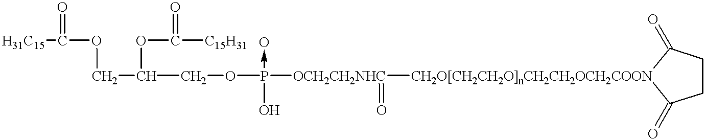
| TABLE 3 | |||
| Binding Study | Binding | ||
| (A) | no | ||
| (B) | no | ||
| (C) | no | ||
| (D) | no | ||
| (E) | yes | ||


| Rat | Dose pSVβgal (μg) | Dose Cationic lipid (μg) |
| (A) | 5 | 30 |
| (B) | 5 | 30 |
| (C) | 25 | 150 |
| TABLE 4 |
| Videodensitometric Analysis of Targeted Vesicles- |
| Ultrasound Backscatter Quantitation |
| Targeted | Non-Targeted | Targeted | Non-Targeted | ||
| Vesicles | Vesicles | Vesicles | Vesicles (after | ||
| (before wash) | (before wash) | (after wash) | wash) | ||
| CONTINUOUS IMAGING |
| 28.46 | 17.64 | 46.01 | 16.5 | |
| 18.44 | 13.36 | 39.48 | 14.44 | |
| 14.44 | 7.62 | 17.22 | 7.58 | |
| 40.94 | 16.97 | 32.85 | 12.95 | |
| 22.86 | 6.81 | 19.52 | 8.08 | |
| Mean | 25.57 | 13.9 | 33.89 | 12.87 |
| StDev | 11.82 | 4.59 | 12.34 | 3.81 |
| INTERMITTENT IMAGING |
| 16.6 | 12.06 | 17.83 | 12.14 | |
| 15.47 | 16.55 | 20.35 | 13.7 | |
| 19.32 | 16.13 | 21.79 | 16.35 | |
| 20.12 | — | 22.41 | 8.68 | |
| 17.29 | 13.7 | 24.15 | 13.41 | |
| Mean | 17.88 | 14.91 | 20.6 | 12.72 |
| StDev | 2.2 | 2.48 | 2.04 | 3.2 |


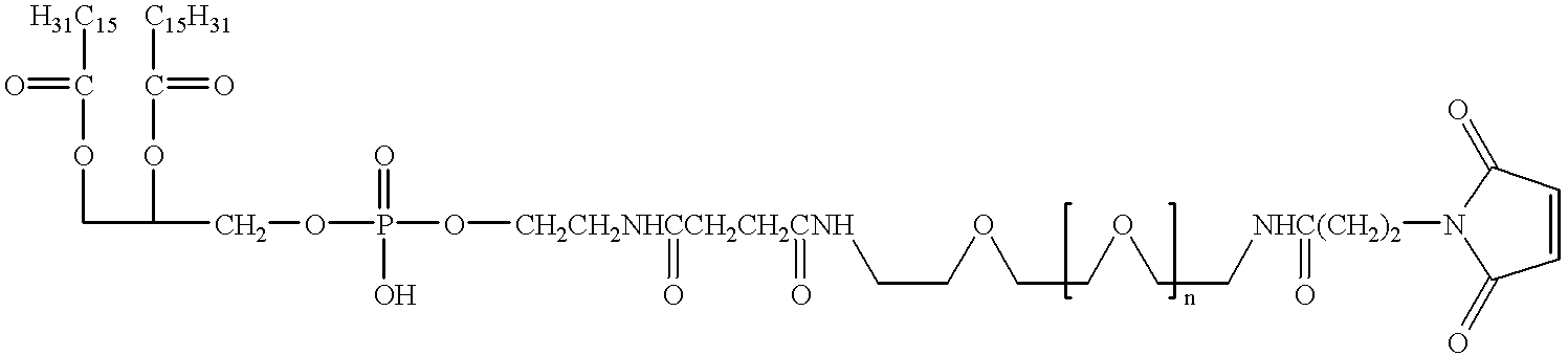


Claims (115)
Priority Applications (1)
| Application Number | Priority Date | Filing Date | Title |
|---|---|---|---|
| US08/982,829 US6231834B1 (en) | 1995-06-07 | 1997-12-02 | Methods for ultrasound imaging involving the use of a contrast agent and multiple images and processing of same |
Applications Claiming Priority (6)
| Application Number | Priority Date | Filing Date | Title |
|---|---|---|---|
| US49768495A | 1995-06-07 | 1995-06-07 | |
| US64046496A | 1996-05-01 | 1996-05-01 | |
| US66003296A | 1996-06-06 | 1996-06-06 | |
| US08/666,129 US6033645A (en) | 1996-06-19 | 1996-06-19 | Methods for diagnostic imaging by regulating the administration rate of a contrast agent |
| US08/932,273 US6139819A (en) | 1995-06-07 | 1997-09-17 | Targeted contrast agents for diagnostic and therapeutic use |
| US08/982,829 US6231834B1 (en) | 1995-06-07 | 1997-12-02 | Methods for ultrasound imaging involving the use of a contrast agent and multiple images and processing of same |
Related Parent Applications (1)
| Application Number | Title | Priority Date | Filing Date |
|---|---|---|---|
| US08/932,273 Continuation-In-Part US6139819A (en) | 1995-06-07 | 1997-09-17 | Targeted contrast agents for diagnostic and therapeutic use |
Publications (1)
| Publication Number | Publication Date |
|---|---|
| US6231834B1 true US6231834B1 (en) | 2001-05-15 |
Family
ID=27541782
Family Applications (1)
| Application Number | Title | Priority Date | Filing Date |
|---|---|---|---|
| US08/982,829 Expired - Lifetime US6231834B1 (en) | 1995-06-07 | 1997-12-02 | Methods for ultrasound imaging involving the use of a contrast agent and multiple images and processing of same |
Country Status (1)
| Country | Link |
|---|---|
| US (1) | US6231834B1 (en) |
Cited By (110)
| Publication number | Priority date | Publication date | Assignee | Title |
|---|---|---|---|---|
| US20010034528A1 (en) * | 1994-09-16 | 2001-10-25 | Foerster Seth A. | Methods and devices for defining and marking tissue |
| US20010049481A1 (en) * | 1998-06-22 | 2001-12-06 | Artemis Medical, Inc. | Biopsy localization method and device |
| US20020018896A1 (en) * | 2000-03-22 | 2002-02-14 | Akira Fukunaga | Composite metallic ultrafine particles and process for producing the same |
| US20020035324A1 (en) * | 1998-12-24 | 2002-03-21 | Sirimanne D. Laksen | Subcutaneous cavity marking device and method |
| US20020058882A1 (en) * | 1998-06-22 | 2002-05-16 | Artemis Medical, Incorporated | Biopsy localization method and device |
| US6438196B1 (en) | 2001-06-28 | 2002-08-20 | General Electric Company | EKG driven CT image reconstruction for cardiac imaging |
| US6439236B1 (en) * | 1999-10-25 | 2002-08-27 | The Board Of Regents Of The University Of Nebraska | Methods for inducing atrial and ventricular rhythms using ultrasound and microbubbles |
| US6461303B2 (en) * | 2000-01-19 | 2002-10-08 | Bjorn Angelsen | Method of detecting ultrasound contrast agent in soft tissue, and quantitating blood perfusion through regions of tissue |
| US20020159952A1 (en) * | 1997-05-13 | 2002-10-31 | Unger Evan C. | Novel acoustically active drug delivery systems |
| US20030003055A1 (en) * | 1989-12-22 | 2003-01-02 | Unger Evan C. | Methods of preparing gaseous precursor-filled microspheres |
| US20030012735A1 (en) * | 1996-06-19 | 2003-01-16 | Unger Evan C. | Methods for diagnostic imaging by regulating the administration rate of contrast agent |
| US6527718B1 (en) * | 1999-08-20 | 2003-03-04 | Brian G Connor | Ultrasound system for continuous imaging and delivery of an encapsulated agent |
| US20030044354A1 (en) * | 2001-08-16 | 2003-03-06 | Carpenter Alan P. | Gas microsphere liposome composites for ultrasound imaging and ultrasound stimulated drug release |
| US6548048B1 (en) * | 1998-04-28 | 2003-04-15 | Amersham Health As | Lipopeptide stabilized microbubbles as diagnostic/therapeutic agents |
| WO2003049617A1 (en) * | 2001-12-13 | 2003-06-19 | Koninklijke Philips Electronics N.V. | Device for producing an on-line image of a body part into which a contrasting agent has been introduced |
| WO2003061593A2 (en) * | 2002-01-23 | 2003-07-31 | Imarx Therapeutics, Inc. | Novel targeted compositions for diagnostic and therapeutic use |
| US6628982B1 (en) * | 2000-03-30 | 2003-09-30 | The Regents Of The University Of Michigan | Internal marker device for identification of biological substances |
| US20040018974A1 (en) * | 2002-03-01 | 2004-01-29 | Christophe Arbogast | Multivalent constructs for therapeutic and diagnostic applications |
| US20040057991A1 (en) * | 1998-01-14 | 2004-03-25 | Hui Poh K. | Preparation of a lipid blend and a phospholipid suspension containing the lipid blend |
| US20040066881A1 (en) * | 2002-07-23 | 2004-04-08 | Reddy Shankara B. | Methods and apparatus for detecting structural, perfusion, and functional abnormalities |
| US20040101969A1 (en) * | 2002-09-11 | 2004-05-27 | Duke University | Methods and compositions for blood pool identification, drug distribution quantification and drug release verification |
| US6743779B1 (en) * | 1994-11-29 | 2004-06-01 | Imarx Pharmaceutical Corp. | Methods for delivering compounds into a cell |
| US20040106870A1 (en) * | 2001-05-29 | 2004-06-03 | Mast T. Douglas | Method for monitoring of medical treatment using pulse-echo ultrasound |
| US20040157082A1 (en) * | 2002-07-22 | 2004-08-12 | Ritter Rogers C. | Coated magnetically responsive particles, and embolic materials using coated magnetically responsive particles |
| US20040210041A1 (en) * | 2002-03-01 | 2004-10-21 | Christophe Arbogast | Multivalent constructs for therapeutic and diagnostic applications |
| US20040241090A1 (en) * | 2003-05-29 | 2004-12-02 | Pettegrew Jay W. | Compounds, compositions and methods for medical imaging of neuropsychiatric disorders |
| US20040258762A1 (en) * | 2003-06-17 | 2004-12-23 | Boppart Stephen A. | Optical contrast agents for optically modifying incident radiation |
| US20040258759A1 (en) * | 2003-06-17 | 2004-12-23 | Suslick Kenneth S. | Surface modified protein microparticles |
| US20050015008A1 (en) * | 2003-07-18 | 2005-01-20 | Munn Charles S. | Enhanced cardiac radionuclide imaging techniques |
| US20050043614A1 (en) * | 2003-08-21 | 2005-02-24 | Huizenga Joel T. | Automated methods and systems for vascular plaque detection and analysis |
| US20050100963A1 (en) * | 2002-03-01 | 2005-05-12 | Dyax Corporation | KDR and VEGF/KDR binding peptides and their use in diagnosis and therapy |
| US20050124897A1 (en) * | 2003-12-03 | 2005-06-09 | Scimed Life Systems, Inc. | Apparatus and methods for delivering acoustic energy to body tissue |
| US20050147555A1 (en) * | 2002-03-01 | 2005-07-07 | Hong Fan | Methods for preparing multivalent constructs for therapeutic and diagnostic applications and methods of preparing the same |
| US20050171433A1 (en) * | 2004-01-08 | 2005-08-04 | Boppart Stephen A. | Multi-functional plasmon-resonant contrast agents for optical coherence tomography |
| US20050228286A1 (en) * | 2004-04-07 | 2005-10-13 | Messerly Jeffrey D | Medical system having a rotatable ultrasound source and a piercing tip |
| US20050234438A1 (en) * | 2004-04-15 | 2005-10-20 | Mast T D | Ultrasound medical treatment system and method |
| US20050240125A1 (en) * | 2004-04-16 | 2005-10-27 | Makin Inder Raj S | Medical system having multiple ultrasound transducers or an ultrasound transducer and an RF electrode |
| US20050240124A1 (en) * | 2004-04-15 | 2005-10-27 | Mast T D | Ultrasound medical treatment system and method |
| US20050240123A1 (en) * | 2004-04-14 | 2005-10-27 | Mast T D | Ultrasound medical treatment system and method |
| US20050250700A1 (en) * | 2002-03-01 | 2005-11-10 | Sato Aaron K | KDR and VEGF/KDR binding peptides |
| US20050256405A1 (en) * | 2004-05-17 | 2005-11-17 | Makin Inder Raj S | Ultrasound-based procedure for uterine medical treatment |
| US20050261586A1 (en) * | 2004-05-18 | 2005-11-24 | Makin Inder R S | Medical system having an ultrasound source and an acoustic coupling medium |
| US20050261587A1 (en) * | 2004-05-20 | 2005-11-24 | Makin Inder R S | Ultrasound medical system and method |
| US20050261585A1 (en) * | 2004-05-20 | 2005-11-24 | Makin Inder Raj S | Ultrasound medical system |
| US20050261610A1 (en) * | 2004-05-21 | 2005-11-24 | Mast T D | Transmit apodization of an ultrasound transducer array |
| US7024024B1 (en) | 2000-11-14 | 2006-04-04 | Axle International | System for contrast echo analysis |
| US20060079805A1 (en) * | 2004-10-13 | 2006-04-13 | Miller Michael E | Site marker visable under multiple modalities |
| US20060088614A1 (en) * | 2002-02-07 | 2006-04-27 | Pettegrew Jay W | Compounds, compositions and methods for treating neuropsychiatric disorders |
| US20060109953A1 (en) * | 2004-11-19 | 2006-05-25 | Deborah Walter | Ct colonography system |
| US20060257842A1 (en) * | 2003-05-29 | 2006-11-16 | Pettegrew Jay W | Cryopreservation media and molecules |
| US20060285635A1 (en) * | 2005-04-15 | 2006-12-21 | Boppart Stephen A | Contrast enhanced spectroscopic optical coherence tomography |
| US20060292547A1 (en) * | 2002-02-07 | 2006-12-28 | Pettegrew Jay W | Method and system for diagnosis of neuropsychiatric disorders including chronic alcoholism |
| WO2007027584A2 (en) * | 2005-08-30 | 2007-03-08 | University Of Virginia Patent Foundation | Deposit contrast agents and related methods thereof |
| US20070066892A1 (en) * | 2005-09-01 | 2007-03-22 | Gabriel Haras | Method and system for providing tomographic pictures of a patient by using contrast medium injections |
| US20070093726A1 (en) * | 2004-10-13 | 2007-04-26 | Leopold Phillip M | Site marker visible under multiple modalities |
| EP1793919A2 (en) * | 2004-09-03 | 2007-06-13 | Imarx Therapeutics, Inc. | Apparatus and method to prepare a microsphere-forming composition |
| US20070156068A1 (en) * | 2005-12-29 | 2007-07-05 | Intrapartum Ventures, Llc; | Cervimetry control apparatus |
| US7247141B2 (en) | 2004-03-08 | 2007-07-24 | Ethicon Endo-Surgery, Inc. | Intra-cavitary ultrasound medical system and method |
| US20070197904A1 (en) * | 2002-09-11 | 2007-08-23 | Duke University | MRI imageable liposomes for the evaluation of treatment efficacy, thermal distribution, and demonstration of dose painting |
| US20070239197A1 (en) * | 2006-04-10 | 2007-10-11 | Intrapartum Ventures, Llc | Method for cervical dilation and/or measurement |
| US20070255185A1 (en) * | 2005-12-29 | 2007-11-01 | Intrapartum Ventures, Llc. | Cervical dilation measurement apparatus |
| WO2008018054A2 (en) | 2006-08-08 | 2008-02-14 | Keter Medical Ltd. | Imaging system |
| US20080101532A1 (en) * | 2006-10-30 | 2008-05-01 | John Eric Tkaczyk | Method and apparatus of ct cardiac diagnostic imaging using motion a priori information from 3d ultrasound and ecg gating |
| US20080107607A1 (en) * | 2002-03-01 | 2008-05-08 | Bracco International B.V. | Targeting vector-phospholipid conjugates |
| US20080152594A1 (en) * | 2002-03-01 | 2008-06-26 | Philippe Bussat | Targeting vector-phospholipid conjugates |
| US20080188768A1 (en) * | 2001-09-10 | 2008-08-07 | Sascha Zarins | Biopsy marker delivery system |
| US20080269603A1 (en) * | 2004-10-13 | 2008-10-30 | Nicoson Zachary R | Site marker visible under multiple modalities |
| US20090112097A1 (en) * | 2007-10-24 | 2009-04-30 | Sei Kato | Ultrasound imaging apparatus and ultrasound imaging method |
| US20090221920A1 (en) * | 2008-01-18 | 2009-09-03 | Boppart Stephen A | Low-coherence interferometry and optical coherence tomography for image-guided surgical treatment of solid tumors |
| US7586618B2 (en) | 2005-02-28 | 2009-09-08 | The Board Of Trustees Of The University Of Illinois | Distinguishing non-resonant four-wave-mixing noise in coherent stokes and anti-stokes Raman scattering |
| US7599465B2 (en) | 2004-11-19 | 2009-10-06 | General Electric Company | Detection of thrombi in CT using energy discrimination |
| US7623908B2 (en) | 2003-01-24 | 2009-11-24 | The Board Of Trustees Of The University Of Illinois | Nonlinear interferometric vibrational imaging |
| US20100081938A1 (en) * | 2008-09-29 | 2010-04-01 | Sei Kato | Ultrasonic diagnostic apparatus |
| US20100106007A1 (en) * | 2002-05-17 | 2010-04-29 | Wacker Frank K | Double-contrast technique for mri-guided vascular interventions |
| US7751057B2 (en) | 2008-01-18 | 2010-07-06 | The Board Of Trustees Of The University Of Illinois | Magnetomotive optical coherence tomography |
| US7787129B2 (en) | 2006-01-31 | 2010-08-31 | The Board Of Trustees Of The University Of Illinois | Method and apparatus for measurement of optical properties in tissue |
| US20100234726A1 (en) * | 1998-12-24 | 2010-09-16 | Sirimanne D Laksen | Device and method for safe location and marking of a biopsy cavity |
| US7806892B2 (en) | 2001-05-29 | 2010-10-05 | Ethicon Endo-Surgery, Inc. | Tissue-retaining system for ultrasound medical treatment |
| US7806839B2 (en) | 2004-06-14 | 2010-10-05 | Ethicon Endo-Surgery, Inc. | System and method for ultrasound therapy using grating lobes |
| US20110097275A1 (en) * | 2002-03-01 | 2011-04-28 | Bracco Suisse Sa | Kdr and vegf/kdr binding peptides and their use in diagnosis and therapy |
| US20110184289A1 (en) * | 2008-06-05 | 2011-07-28 | Hitachi Medical Corporation | Ultrasonic diagnostic apparatus |
| US20110200525A1 (en) * | 2008-10-09 | 2011-08-18 | Patz Jr Edward F | Vhh antibody fragments for use in the detection and treatment of cancer |
| US20110218517A1 (en) * | 2009-10-09 | 2011-09-08 | Ogle Matthew F | In vivo chemical stabilization of vulnerable plaque |
| US8115934B2 (en) | 2008-01-18 | 2012-02-14 | The Board Of Trustees Of The University Of Illinois | Device and method for imaging the ear using optical coherence tomography |
| WO2012047317A2 (en) | 2010-10-08 | 2012-04-12 | The University Of North Carolina At Charlotte | Tumor specific antibodies and uses therefor |
| US8207651B2 (en) | 2009-09-16 | 2012-06-26 | Tyco Healthcare Group Lp | Low energy or minimum disturbance method for measuring frequency response functions of ultrasonic surgical devices in determining optimum operating point |
| US8280486B2 (en) | 2004-10-13 | 2012-10-02 | Suros Surgical Systems, Inc. | Site marker visable under multiple modalities |
| US8939909B2 (en) | 2011-10-28 | 2015-01-27 | Decision Sciences International Corporation | Spread spectrum coded waveforms in ultrasound imaging |
| US20150037250A1 (en) * | 2013-07-31 | 2015-02-05 | Kabushiki Kaisha Toshiba | Contrast agent |
| US20160279200A1 (en) * | 2001-04-27 | 2016-09-29 | Cormend Technologies, Llc | Prevention of myocardial infarction induced ventricular expansion and remodeling |
| CN105999314A (en) * | 2016-07-11 | 2016-10-12 | 四川省人民医院 | Ultrasonic contrast agent and preparing method thereof |
| US9669113B1 (en) | 1998-12-24 | 2017-06-06 | Devicor Medical Products, Inc. | Device and method for safe location and marking of a biopsy cavity |
| US9789210B1 (en) | 2016-07-06 | 2017-10-17 | Lantheus Medical Imaging, Inc. | Methods for making ultrasound contrast agents |
| US9844359B2 (en) | 2013-09-13 | 2017-12-19 | Decision Sciences Medical Company, LLC | Coherent spread-spectrum coded waveforms in synthetic aperture image formation |
| CN108175864A (en) * | 2018-01-12 | 2018-06-19 | 中山大学 | A kind of multi-modality imaging nano-probe and its synthetic method |
| US10022460B2 (en) | 2014-12-31 | 2018-07-17 | Lantheus Medical Imaging, Inc. | Lipid-encapsulated gas microsphere compositions and related methods |
| US10035009B2 (en) | 2013-04-15 | 2018-07-31 | The Board Of Trustees Of The Leland Stanford Junior University | Systems and methods for treating pancreatic cancer |
| US10052394B2 (en) | 2014-11-21 | 2018-08-21 | General Electric Company | Microbubble tether for diagnostic and therapeutic applications |
| US10588988B2 (en) | 2016-05-04 | 2020-03-17 | Lantheus Medical Imaging, Inc. | Methods and devices for preparation of ultrasound contrast agents |
| US10743838B2 (en) | 2015-02-25 | 2020-08-18 | Decision Sciences Medical Company, LLC | Acoustic signal transmission couplants and coupling mediums |
| US20200281661A1 (en) * | 2019-03-05 | 2020-09-10 | Biosense Webster (Israel) Ltd. | Showing catheter in brain |
| US10881310B2 (en) | 2012-08-25 | 2021-01-05 | The Board Of Trustees Of The Leland Stanford Junior University | Motion artifact mitigation methods and devices for pulse photoplethysmography |
| EP3783034A1 (en) | 2014-07-23 | 2021-02-24 | Ohio State Innovation Foundation | Methods and compositions related to antibody fragments that bind to tumor-associated glycoprotein 72 (tag-72) |
| US20210252265A1 (en) * | 2018-07-20 | 2021-08-19 | Suono Bio, Inc. | Devices and methods for ultrasonic delivery of an agent within an oral cavity |
| CN113521318A (en) * | 2021-07-29 | 2021-10-22 | 济南市中心医院 | Simethicone microbubble ultrasonic contrast agent and preparation method and application thereof |
| US11154274B2 (en) | 2019-04-23 | 2021-10-26 | Decision Sciences Medical Company, LLC | Semi-rigid acoustic coupling articles for ultrasound diagnostic and treatment applications |
| US11304676B2 (en) | 2015-01-23 | 2022-04-19 | The University Of North Carolina At Chapel Hill | Apparatuses, systems, and methods for preclinical ultrasound imaging of subjects |
| US11520043B2 (en) | 2020-11-13 | 2022-12-06 | Decision Sciences Medical Company, LLC | Systems and methods for synthetic aperture ultrasound imaging of an object |
| US20230094387A1 (en) * | 2021-09-26 | 2023-03-30 | Henan Agricultural University | Ultrasonic-assisted heat press machine and uses thereof |
| US11737726B2 (en) | 2015-10-08 | 2023-08-29 | Decision Sciences Medical Company, LLC | Acoustic orthopedic tracking system and methods |
Citations (224)
| Publication number | Priority date | Publication date | Assignee | Title |
|---|---|---|---|---|
| US3015128A (en) | 1960-08-18 | 1962-01-02 | Southwest Res Inst | Encapsulating apparatus |
| GB1044680A (en) | 1962-12-21 | 1966-10-05 | Dow Chemical Co | Expandable thermoplastic polymer particles containing volatile fluid foaming agent and method of making the same |
| US3291843A (en) | 1963-10-08 | 1966-12-13 | Du Pont | Fluorinated vinyl ethers and their preparation |
| US3293114A (en) | 1964-04-03 | 1966-12-20 | Dow Chemical Co | Method of forming paper containing gaseous filled spheres of thermoplastic resins and paper thereof |
| US3401475A (en) | 1966-07-18 | 1968-09-17 | Dow Chemical Co | Label and labelled container |
| US3479811A (en) | 1967-11-29 | 1969-11-25 | Dow Chemical Co | Yarn and method of making the same |
| US3488714A (en) | 1966-09-19 | 1970-01-06 | Dow Chemical Co | Formed laminate structure and method of preparation |
| US3532500A (en) | 1967-07-25 | 1970-10-06 | Eastman Kodak Co | Light sensitive vesicular composition comprising an azido-s-triazine compound |
| US3557294A (en) | 1967-10-12 | 1971-01-19 | Allied Chem | Fluorinated ethers as inhalation convulsants |
| US3594326A (en) | 1964-12-03 | 1971-07-20 | Ncr Co | Method of making microscopic capsules |
| US3615972A (en) | 1967-04-28 | 1971-10-26 | Dow Chemical Co | Expansible thermoplastic polymer particles containing volatile fluid foaming agent and method of foaming the same |
| US3650831A (en) | 1969-03-10 | 1972-03-21 | Armour Dial Inc | Method of cleaning surfaces |
| US3732172A (en) | 1968-02-28 | 1973-05-08 | Ncr Co | Process for making minute capsules and prefabricated system useful therein |
| US3873564A (en) | 1971-03-03 | 1975-03-25 | Synvar Ass | 2-Imidazolinyl-3-oxide-1-oxypropionic acid |
| US3945956A (en) | 1975-06-23 | 1976-03-23 | The Dow Chemical Company | Polymerization of styrene acrylonitrile expandable microspheres |
| US3960583A (en) | 1974-05-02 | 1976-06-01 | Philadelphia Quartz Company | Method of preparing modified hollow, largely spherical particles by spray drying |
| US3968203A (en) | 1965-10-01 | 1976-07-06 | Jerome G. Spitzer | Aerosol astringent composition |
| DE2521003C2 (en) | 1975-05-12 | 1977-03-24 | Schaefer, Philipp, 3000 Hannover | Cream, ointment or the like |
| US4027007A (en) | 1970-12-09 | 1977-05-31 | Colgate-Palmolive Company | Antiperspirants formulated with borax |
| US4089801A (en) | 1974-07-19 | 1978-05-16 | Battelle Memorial Institute | Process for the preparation of liposomes |
| US4108806A (en) | 1971-12-06 | 1978-08-22 | The Dow Chemical Company | Thermoplastic expandable microsphere process and product |
| US4138383A (en) | 1975-11-24 | 1979-02-06 | California Institute Of Technology | Preparation of small bio-compatible microspheres |
| US4162282A (en) | 1976-04-22 | 1979-07-24 | Coulter Electronics, Inc. | Method for producing uniform particles |
| US4179546A (en) | 1972-08-28 | 1979-12-18 | The Dow Chemical Company | Method for expanding microspheres and expandable composition |
| US4192859A (en) | 1978-09-29 | 1980-03-11 | E. R. Squibb & Sons, Inc. | Contrast media containing liposomes as carriers |
| US4224179A (en) | 1977-08-05 | 1980-09-23 | Battelle Memorial Institute | Process for the preparation of liposomes in aqueous solution |
| US4229360A (en) | 1977-08-05 | 1980-10-21 | Battelle Memorial Institute | Process for the dehydration of a colloidal dispersion of lipsomes |
| US4265251A (en) | 1979-06-28 | 1981-05-05 | Rasor Associates, Inc. | Method of determining pressure within liquid containing vessel |
| US4276885A (en) | 1979-05-04 | 1981-07-07 | Rasor Associates, Inc | Ultrasonic image enhancement |
| US4310506A (en) | 1979-02-22 | 1982-01-12 | California Institute Of Technology | Means of preparation and applications of liposomes containing high concentrations of entrapped ionic species |
| US4310505A (en) | 1979-11-08 | 1982-01-12 | California Institute Of Technology | Lipid vesicles bearing carbohydrate surfaces as lymphatic directed vehicles for therapeutic and diagnostic substances |
| US4315514A (en) | 1980-05-08 | 1982-02-16 | William Drewes | Method and apparatus for selective cell destruction |
| US4331654A (en) | 1980-06-13 | 1982-05-25 | Eli Lilly And Company | Magnetically-localizable, biodegradable lipid microspheres |
| US4342826A (en) | 1980-02-04 | 1982-08-03 | Collaborative Research, Inc. | Immunoassay products and methods |
| US4344929A (en) | 1980-04-25 | 1982-08-17 | Alza Corporation | Method of delivering drug with aid of effervescent activity generated in environment of use |
| US4420442A (en) | 1981-04-13 | 1983-12-13 | Pq Corporation | Manufacturing process for hollow microspheres |
| US4421562A (en) | 1980-04-13 | 1983-12-20 | Pq Corporation | Manufacturing process for hollow microspheres |
| US4427330A (en) | 1981-03-03 | 1984-01-24 | Carter Charles P | Automatic fence picket stock conveyor for fence picket pointing machine |
| US4427649A (en) | 1976-03-19 | 1984-01-24 | Imperial Chemical Industries Limited | Pharmaceutical compositions |
| US4428924A (en) | 1977-02-02 | 1984-01-31 | Linton Medical Services Limited | Preparation for diagnostic radiology |
| US4442843A (en) | 1980-11-17 | 1984-04-17 | Schering, Ag | Microbubble precursors and methods for their production and use |
| US4466442A (en) | 1981-10-16 | 1984-08-21 | Schering Aktiengesellschaft | Carrier liquid solutions for the production of gas microbubbles, preparation thereof, and use thereof as contrast medium for ultrasonic diagnostics |
| US4530360A (en) | 1981-11-19 | 1985-07-23 | Duarte Luiz R | Method for healing bone fractures with ultrasound |
| US4533254A (en) | 1981-04-17 | 1985-08-06 | Biotechnology Development Corporation | Apparatus for forming emulsions |
| US4534899A (en) | 1981-07-20 | 1985-08-13 | Lipid Specialties, Inc. | Synthetic phospholipid compounds |
| US4540629A (en) | 1982-04-08 | 1985-09-10 | Pq Corporation | Hollow microspheres with organosilicon-silicate walls |
| US4544545A (en) | 1983-06-20 | 1985-10-01 | Trustees University Of Massachusetts | Liposomes containing modified cholesterol for organ targeting |
| US4549892A (en) | 1982-09-22 | 1985-10-29 | Baker Alfred G | Process for producing hollow, bilayered silicate microspheres |
| EP0052575B1 (en) | 1980-11-17 | 1986-01-08 | Schering Aktiengesellschaft | Composition generating microbubbles |
| US4569836A (en) | 1981-08-27 | 1986-02-11 | Gordon Robert T | Cancer treatment by intracellular hyperthermia |
| US4572203A (en) | 1983-01-27 | 1986-02-25 | Feinstein Steven B | Contact agents for ultrasonic imaging |
| US4586512A (en) | 1981-06-26 | 1986-05-06 | Thomson-Csf | Device for localized heating of biological tissues |
| US4603044A (en) | 1983-01-06 | 1986-07-29 | Technology Unlimited, Inc. | Hepatocyte Directed Vesicle delivery system |
| US4615879A (en) | 1983-11-14 | 1986-10-07 | Vanderbilt University | Particulate NMR contrast agents for gastrointestinal application |
| US4620546A (en) | 1984-06-30 | 1986-11-04 | Kabushiki Kaisha Toshiba | Ultrasound hyperthermia apparatus |
| US4646756A (en) | 1982-10-26 | 1987-03-03 | The University Of Aberdeen | Ultra sound hyperthermia device |
| US4657756A (en) | 1980-11-17 | 1987-04-14 | Schering Aktiengesellschaft | Microbubble precursors and apparatus for their production and use |
| US4658828A (en) | 1984-05-03 | 1987-04-21 | Jacques Dory | Apparatus for examining and localizing tumors using ultra sounds, comprising a device for localized hyperthermia treatment |
| US4663161A (en) | 1985-04-22 | 1987-05-05 | Mannino Raphael J | Liposome methods and compositions |
| US4675310A (en) | 1984-03-23 | 1987-06-23 | Biocompatibles Limited | Liposome composition as gas transport agents |
| US4681119A (en) | 1980-11-17 | 1987-07-21 | Schering Aktiengesellschaft | Method of production and use of microbubble precursors |
| US4684479A (en) | 1985-08-14 | 1987-08-04 | Arrigo Joseph S D | Surfactant mixtures, stable gas-in-liquid emulsions, and methods for the production of such emulsions from said mixtures |
| US4689986A (en) | 1985-03-13 | 1987-09-01 | The University Of Michigan | Variable frequency gas-bubble-manipulating apparatus and method |
| US4693999A (en) | 1984-07-30 | 1987-09-15 | Aktiebolaget Draco | Liposomes containing steroid esters |
| EP0243947A1 (en) | 1986-04-30 | 1987-11-04 | Dornier Medizintechnik Gmbh | Acoustic shock wave targeting of drug delivery in patients |
| US4718433A (en) | 1983-01-27 | 1988-01-12 | Feinstein Steven B | Contrast agents for ultrasonic imaging |
| GB2193095A (en) | 1986-07-29 | 1988-02-03 | Univ Ramot | Contrast agent for NMR imaging |
| US4728578A (en) | 1986-08-13 | 1988-03-01 | The Lubrizol Corporation | Compositions containing basic metal salts and/or non-Newtonian colloidal disperse systems and vinyl aromatic containing polymers |
| US4728575A (en) | 1984-04-27 | 1988-03-01 | Vestar, Inc. | Contrast agents for NMR imaging |
| US4731239A (en) | 1983-01-10 | 1988-03-15 | Gordon Robert T | Method for enhancing NMR imaging; and diagnostic use |
| US4737323A (en) | 1986-02-13 | 1988-04-12 | Liposome Technology, Inc. | Liposome extrusion method |
| US4775522A (en) | 1983-03-04 | 1988-10-04 | Children's Hospital Research Foundation, A Division Of Children's Hospital Medical Center | NMR compositions for indirectly detecting a dissolved gas in an animal |
| US4776991A (en) | 1986-08-29 | 1988-10-11 | The United States Of America As Represented By The Secretary Of The Navy | Scaled-up production of liposome-encapsulated hemoglobin |
| US4781871A (en) | 1986-09-18 | 1988-11-01 | Liposome Technology, Inc. | High-concentration liposome processing method |
| US4789501A (en) | 1984-11-19 | 1988-12-06 | The Curators Of The University Of Missouri | Glass microspheres |
| US4790891A (en) | 1986-11-04 | 1988-12-13 | Aeci Limited | Process for the production of a cartridged explosive with entrapped bubbles |
| US4822534A (en) | 1987-03-05 | 1989-04-18 | Lencki Robert W J | Method of producing microspheres |
| US4830858A (en) | 1985-02-11 | 1989-05-16 | E. R. Squibb & Sons, Inc. | Spray-drying method for preparing liposomes and products produced thereby |
| US4834964A (en) | 1986-03-07 | 1989-05-30 | M.R.I., Inc. | Use of charged nitroxides as NMR image enhancing agents for CSF |
| US4844882A (en) | 1987-12-29 | 1989-07-04 | Molecular Biosystems, Inc. | Concentrated stabilized microbubble-type ultrasonic imaging agent |
| AU3035189A (en) | 1988-02-05 | 1989-08-25 | Schering Aktiengesellschaft Berlin Und Bergkamen | Ultrasonic contrast agents, process for producing them and their use as diagnostic and therapeutic agents |
| US4863717A (en) | 1986-11-10 | 1989-09-05 | The State Of Oregon Acting By And Through The State Board Of Higher Education On Behalf Of The University Of Oregon | Methods for circumventing the problem of free radial reduction associated with the use of stable nitroxide free radicals as contrast agents for magnetic reasonance imaging |
| US4865836A (en) | 1986-01-14 | 1989-09-12 | Fluoromed Pharmaceutical, Inc. | Brominated perfluorocarbon emulsions for internal animal use for contrast enhancement and oxygen transport |
| US4877561A (en) | 1986-04-02 | 1989-10-31 | Takeda Chemical Industries, Ltd. | Method of producing liposome |
| US4893624A (en) | 1988-06-21 | 1990-01-16 | Massachusetts Institute Of Technology | Diffuse focus ultrasound hyperthermia system |
| US4895719A (en) | 1985-05-22 | 1990-01-23 | Liposome Technology, Inc. | Method and apparatus for administering dehydrated liposomes by inhalation |
| US4898734A (en) | 1988-02-29 | 1990-02-06 | Massachusetts Institute Of Technology | Polymer composite for controlled release or membrane formation |
| US4900540A (en) | 1983-06-20 | 1990-02-13 | Trustees Of The University Of Massachusetts | Lipisomes containing gas for ultrasound detection |
| US4918065A (en) | 1982-07-07 | 1990-04-17 | Schering Aktiengesellschaft | Corticoid-containing preparation for topical application |
| US4919895A (en) | 1986-09-23 | 1990-04-24 | Alpha Therapeutic Corporation | Apparatus for oxygenation of liquid state dissolved oxygen-carrying formulation |
| US4921706A (en) | 1984-11-20 | 1990-05-01 | Massachusetts Institute Of Technology | Unilamellar lipid vesicles and method for their formation |
| US4927623A (en) | 1986-01-14 | 1990-05-22 | Alliance Pharmaceutical Corp. | Dissolution of gas in a fluorocarbon liquid |
| US4933121A (en) | 1986-12-10 | 1990-06-12 | Ciba Corning Diagnostics Corp. | Process for forming liposomes |
| US4938947A (en) | 1985-11-01 | 1990-07-03 | Centre National De La Recherche Scientifique (Cnrs) | Aerosol composition for in vivo imaging |
| US4957656A (en) | 1988-09-14 | 1990-09-18 | Molecular Biosystems, Inc. | Continuous sonication method for preparing protein encapsulated microbubbles |
| US4981692A (en) | 1983-03-24 | 1991-01-01 | The Liposome Company, Inc. | Therapeutic treatment by intramammary infusion |
| US4985550A (en) | 1986-07-29 | 1991-01-15 | Alliance Pharmaceutical Corp. | Polyhydroxylated and highly fluorinated compounds, their preparation and their use as surfactants |
| US4984573A (en) | 1987-06-23 | 1991-01-15 | Hafslund Nycomed Innovation Ab | Method of electron spin resonance enhanced magnetic resonance imaging |
| US4987154A (en) | 1986-01-14 | 1991-01-22 | Alliance Pharmaceutical Corp. | Biocompatible, stable and concentrated fluorocarbon emulsions for contrast enhancement and oxygen transport in internal animal use |
| EP0216730B1 (en) | 1985-08-12 | 1991-01-23 | Battelle Memorial Institute | Porous spherical glass filtrating beads and method for the manufacturing thereof |
| US4993415A (en) | 1988-08-19 | 1991-02-19 | Alliance Pharmaceutical Corp. | Magnetic resonance imaging with perfluorocarbon hydrides |
| US4996041A (en) | 1988-08-19 | 1991-02-26 | Toshiyuki Arai | Method for introducing oxygen-17 into tissue for imaging in a magnetic resonance imaging system |
| US5000960A (en) | 1987-03-13 | 1991-03-19 | Micro-Pak, Inc. | Protein coupling to lipid vesicles |
| US5004611A (en) | 1984-03-08 | 1991-04-02 | Phares Pharmaceutical Research Nv | Pro-liposome compositions |
| US5008050A (en) | 1984-06-20 | 1991-04-16 | The Liposome Company, Inc. | Extrusion technique for producing unilamellar vesicles |
| US5008109A (en) | 1984-05-25 | 1991-04-16 | Vestar, Inc. | Vesicle stabilization |
| US5013556A (en) | 1989-10-20 | 1991-05-07 | Liposome Technology, Inc. | Liposomes with enhanced circulation time |
| US5019370A (en) | 1989-07-10 | 1991-05-28 | University Of Kentucky Research Foundation | Biodegradable, low biological toxicity radiographic contrast medium and method of x-ray imaging |
| US5045304A (en) | 1988-08-31 | 1991-09-03 | Wayne State University | Contras agent having an imaging agent coupled to viable granulocytes for use in magnetic resonance imaging of abscess and a method of preparing and using same |
| US5049388A (en) | 1986-11-06 | 1991-09-17 | Research Development Foundation | Small particle aerosol liposome and liposome-drug combinations for medical use |
| US5078994A (en) | 1990-04-12 | 1992-01-07 | Eastman Kodak Company | Microgel drug delivery system |
| US5088499A (en) | 1989-12-22 | 1992-02-18 | Unger Evan C | Liposomes as contrast agents for ultrasonic imaging and methods for preparing the same |
| US5107842A (en) | 1991-02-22 | 1992-04-28 | Molecular Biosystems, Inc. | Method of ultrasound imaging of the gastrointestinal tract |
| US5114703A (en) | 1989-05-30 | 1992-05-19 | Alliance Pharmaceutical Corp. | Percutaneous lymphography using particulate fluorocarbon emulsions |
| US5123414A (en) | 1989-12-22 | 1992-06-23 | Unger Evan C | Liposomes as contrast agents for ultrasonic imaging and methods for preparing the same |
| US5135000A (en) | 1990-09-19 | 1992-08-04 | Raizot University Authority For Applied Research & Industrial Development Ltd. | Method of measuring regional tissue blood flow |
| US5137928A (en) | 1990-04-26 | 1992-08-11 | Hoechst Aktiengesellschaft | Ultrasonic contrast agents, processes for their preparation and the use thereof as diagnostic and therapeutic agents |
| US5141738A (en) | 1983-04-15 | 1992-08-25 | Schering Aktiengesellschaft | Ultrasonic contrast medium comprising gas bubbles and solid lipophilic surfactant-containing microparticles and use thereof |
| US5147631A (en) | 1991-04-30 | 1992-09-15 | Du Pont Merck Pharmaceutical Company | Porous inorganic ultrasound contrast agents |
| US5149319A (en) | 1990-09-11 | 1992-09-22 | Unger Evan C | Methods for providing localized therapeutic heat to biological tissues and fluids |
| US5171755A (en) | 1988-04-29 | 1992-12-15 | Hemagen/Pfc | Emulsions of highly fluorinated organic compounds |
| EP0338971B1 (en) | 1988-04-16 | 1993-01-20 | Schering Aktiengesellschaft | Process for preparing liposomes and/or biological cells containing ingredients, and their use |
| US5186922A (en) | 1985-03-15 | 1993-02-16 | See/Shell Biotechnology, Inc. | Use of biodegradable microspheres labeled with imaging energy constrast materials |
| US5190766A (en) | 1990-04-16 | 1993-03-02 | Ken Ishihara | Method of controlling drug release by resonant sound wave |
| US5190982A (en) | 1990-04-26 | 1993-03-02 | Hoechst Aktiengesellschaft | Ultrasonic contrast agents, processes for their preparation and the use thereof as diagnostic and therapeutic agents |
| US5192549A (en) | 1988-09-28 | 1993-03-09 | Yissum Research Development Company Of The Hebrew University Of Jerusalem | Method of amphiphatic drug loading in liposomes by pH gradient |
| US5194266A (en) | 1989-08-08 | 1993-03-16 | Liposome Technology, Inc. | Amphotericin B/cholesterol sulfate composition and method |
| US5195520A (en) | 1986-11-05 | 1993-03-23 | Schering Aktiengesellschaft | Ultrasonic manometry process in a fluid by means of microbubbles |
| US5196183A (en) | 1991-12-04 | 1993-03-23 | Sterling Winthrop Inc. | Contrast agents for ultrasound imaging |
| US5196348A (en) | 1990-06-11 | 1993-03-23 | Air Products And Chemicals, Inc. | Perfluoro-crown ethers in fluorine magnetic resonance spectroscopy of biopsied tissue |
| US5198225A (en) | 1988-10-03 | 1993-03-30 | Lvmh Recherche | Hydrated lipidic lamellar phases or liposomes based on ecdysteroids |
| EP0231091B1 (en) | 1986-01-24 | 1993-03-31 | Children's Hospital Medical Center | Stable emulsions of highly fluorinated organic compound |
| US5205290A (en) | 1991-04-05 | 1993-04-27 | Unger Evan C | Low density microspheres and their use as contrast agents for computed tomography |
| US5205287A (en) | 1990-04-26 | 1993-04-27 | Hoechst Aktiengesellschaft | Ultrasonic contrast agents, processes for their preparation and the use thereof as diagnostic and therapeutic agents |
| US5209720A (en) | 1989-12-22 | 1993-05-11 | Unger Evan C | Methods for providing localized therapeutic heat to biological tissues and fluids using gas filled liposomes |
| US5215680A (en) | 1990-07-10 | 1993-06-01 | Cavitation-Control Technology, Inc. | Method for the production of medical-grade lipid-coated microbubbles, paramagnetic labeling of such microbubbles and therapeutic uses of microbubbles |
| US5219538A (en) | 1987-03-13 | 1993-06-15 | Micro-Pak, Inc. | Gas and oxygen carrying lipid vesicles |
| US5228446A (en) | 1989-12-22 | 1993-07-20 | Unger Evan C | Gas filled liposomes and their use as ultrasonic contrast agents |
| US5230882A (en) | 1989-12-22 | 1993-07-27 | Unger Evan C | Liposomes as contrast agents for ultrasonic imaging and methods for preparing the same |
| AU641363B2 (en) | 1988-08-23 | 1993-09-23 | Schering Aktiengesellschaft | Contrast preparation consisting of cavitate- or clathrate- forming host/guest complexes |
| US5247935A (en) | 1992-03-19 | 1993-09-28 | General Electric Company | Magnetic resonance guided focussed ultrasound surgery |
| EP0324938B1 (en) | 1987-12-29 | 1993-11-18 | Molecular Biosystems, Inc. | Concentrated stabilized microbubble-type ultrasonic imaging agent and method of production |
| US5271928A (en) | 1990-04-02 | 1993-12-21 | Sintetica S.A. | Stable microbubbles suspensions injectable into living organisms |
| US5305757A (en) | 1989-12-22 | 1994-04-26 | Unger Evan C | Gas filled liposomes and their use as ultrasonic contrast agents |
| US5310540A (en) | 1990-10-05 | 1994-05-10 | Sintetica Sa | Method for the preparation of stable suspensions of hollow gas-filled microspheres suitable for ultrasonic echography |
| US5312617A (en) | 1991-05-23 | 1994-05-17 | Unger Evan C | Liposoluble compounds useful as magnetic resonance imaging agents |
| US5315998A (en) | 1991-03-22 | 1994-05-31 | Katsuro Tachibana | Booster for therapy of diseases with ultrasound and pharmaceutical liquid composition containing the same |
| US5315997A (en) | 1990-06-19 | 1994-05-31 | Molecular Biosystems, Inc. | Method of magnetic resonance imaging using diamagnetic contrast |
| US5334381A (en) | 1989-12-22 | 1994-08-02 | Unger Evan C | Liposomes as contrast agents for ultrasonic imaging and methods for preparing the same |
| US5339814A (en) | 1992-04-14 | 1994-08-23 | Lasker Sigmund E | Process for visualizing tissue metabolism using oxygen-17 |
| US5344930A (en) | 1989-06-22 | 1994-09-06 | Alliance Pharmaceutical Corp. | Fluorine and phosphorous-containing amphiphilic molecules with surfactant properties |
| US5352435A (en) | 1989-12-22 | 1994-10-04 | Unger Evan C | Ionophore containing liposomes for ultrasound imaging |
| US5354549A (en) | 1987-07-24 | 1994-10-11 | Nycomed Imaging As | Iodinated esters |
| US5358702A (en) | 1990-04-10 | 1994-10-25 | Unger Evan C | Methoxylated gel particle contrast media for improved diagnostic imaging |
| US5362477A (en) | 1991-10-25 | 1994-11-08 | Mallinckrodt Medical, Inc. | 19F magnetic resonance imaging agents which include a nitroxide moiety |
| US5362478A (en) | 1993-03-26 | 1994-11-08 | Vivorx Pharmaceuticals, Inc. | Magnetic resonance imaging with fluorocarbons encapsulated in a cross-linked polymeric shell |
| US5393524A (en) | 1991-09-17 | 1995-02-28 | Sonus Pharmaceuticals Inc. | Methods for selecting and using gases as ultrasound contrast media |
| FR2700952B1 (en) | 1993-01-29 | 1995-03-17 | Oreal | New cosmetic or dermopharmaceutical compositions in the form of aqueous gels modified by the addition of expanded microspheres. |
| US5410516A (en) | 1988-09-01 | 1995-04-25 | Schering Aktiengesellschaft | Ultrasonic processes and circuits for performing them |
| US5413774A (en) | 1992-01-23 | 1995-05-09 | Bracco International B.V. | Long-lasting aqueous dispersions or suspensions of pressure-resistant gas-filled microvesicles and methods for the preparation thereof |
| US5425366A (en) | 1988-02-05 | 1995-06-20 | Schering Aktiengesellschaft | Ultrasonic contrast agents for color Doppler imaging |
| US5433204A (en) | 1993-11-16 | 1995-07-18 | Camilla Olson | Method of assessing placentation |
| US5445813A (en) | 1992-11-02 | 1995-08-29 | Bracco International B.V. | Stable microbubble suspensions as enhancement agents for ultrasound echography |
| US5460800A (en) | 1993-03-16 | 1995-10-24 | Alliance Pharmaceutical Corp. | Labeled fluorocarbon compositions |
| US5466467A (en) | 1994-03-30 | 1995-11-14 | The United States Of America As Represented By The Secretary Of The Navy | Liposomes containing polymerized lipids for non-covalent immobilization of proteins and enzymes |
| US5470582A (en) | 1992-02-07 | 1995-11-28 | Syntex (U.S.A.) Inc. | Controlled delivery of pharmaceuticals from preformed porous polymeric microparticles |
| US5469854A (en) | 1989-12-22 | 1995-11-28 | Imarx Pharmaceutical Corp. | Methods of preparing gas-filled liposomes |
| US5485839A (en) | 1992-02-28 | 1996-01-23 | Kabushiki Kaisha Toshiba | Method and apparatus for ultrasonic wave medical treatment using computed tomography |
| US5487390A (en) | 1990-10-05 | 1996-01-30 | Massachusetts Institute Of Technology | Gas-filled polymeric microbubbles for ultrasound imaging |
| US5496535A (en) | 1991-04-12 | 1996-03-05 | Alliance Pharmaceutical Corp. | Fluorocarbon contrast media for use with MRI and radiographic imaging |
| US5498421A (en) | 1993-02-22 | 1996-03-12 | Vivorx Pharmaceuticals, Inc. | Composition useful for in vivo delivery of biologics and methods employing same |
| US5498601A (en) | 1992-07-09 | 1996-03-12 | Nippon Steel Corporation | Platelet aggregation-inhibiting peptides |
| US5501863A (en) | 1990-02-09 | 1996-03-26 | Schering Aktiengesellschaft | Contrast media synthesized from polyaldehydes |
| US5502094A (en) | 1994-05-20 | 1996-03-26 | Minnesota Mining And Manufacturing Company | Physiologically acceptable emulsions containing perfluorocarbon ether hydrides and methods for use |
| US5529766A (en) | 1991-03-28 | 1996-06-25 | Nycomed Imaging As | Contrast agents |
| US5536489A (en) | 1993-06-04 | 1996-07-16 | Molecular Biosystems, Inc. | Emulsions as contrast agents and method of use |
| US5536490A (en) | 1991-03-28 | 1996-07-16 | Nycomed Imaging As | Contrast agents |
| US5540909A (en) | 1994-09-28 | 1996-07-30 | Alliance Pharmaceutical Corp. | Harmonic ultrasound imaging with microbubbles |
| US5542935A (en) | 1989-12-22 | 1996-08-06 | Imarx Pharmaceutical Corp. | Therapeutic delivery systems related applications |
| US5545396A (en) | 1994-04-08 | 1996-08-13 | The Research Foundation Of State University Of New York | Magnetic resonance imaging using hyperpolarized noble gases |
| US5552155A (en) | 1992-12-04 | 1996-09-03 | The Liposome Company, Inc. | Fusogenic lipsomes and methods for making and using same |
| US5552133A (en) | 1993-07-02 | 1996-09-03 | Molecular Biosystems, Inc. | Method of making encapsulated gas microspheres useful as an ultrasonic imaging agent |
| US5556372A (en) | 1995-02-15 | 1996-09-17 | Exogen, Inc. | Apparatus for ultrasonic bone treatment |
| US5556610A (en) | 1992-01-24 | 1996-09-17 | Bracco Research S.A. | Gas mixtures useful as ultrasound contrast media, contrast agents containing the media and method |
| US5558854A (en) | 1991-09-17 | 1996-09-24 | Sonus Pharmaceuticals | Ultrasound contrast media comprising perfluoropentane and perfluorohexane gas |
| US5558092A (en) | 1995-06-06 | 1996-09-24 | Imarx Pharmaceutical Corp. | Methods and apparatus for performing diagnostic and therapeutic ultrasound simultaneously |
| US5558094A (en) | 1991-09-17 | 1996-09-24 | Sonus Pharmaceuticals, Inc. | Methods for using persistent gases as ultrasound contrast media |
| US5558856A (en) | 1992-01-09 | 1996-09-24 | Nycomed Imaging As | Microbubble-generating contrast agents for ultrasound and magnetic resonance imaging |
| US5558855A (en) | 1993-01-25 | 1996-09-24 | Sonus Pharmaceuticals | Phase shift colloids as ultrasound contrast agents |
| US5558853A (en) | 1993-01-25 | 1996-09-24 | Sonus Pharmaceuticals | Phase shift colloids as ultrasound contrast agents |
| US5560364A (en) | 1995-05-12 | 1996-10-01 | The Board Of Regents Of The University Of Nebraska | Suspended ultra-sound induced microbubble cavitation imaging |
| US5562893A (en) | 1994-08-02 | 1996-10-08 | Molecular Biosystems, Inc. | Gas-filled microspheres with fluorine-containing shells |
| US5562608A (en) | 1989-08-28 | 1996-10-08 | Biopulmonics, Inc. | Apparatus for pulmonary delivery of drugs with simultaneous liquid lavage and ventilation |
| US5565215A (en) | 1993-07-23 | 1996-10-15 | Massachusettes Institute Of Technology | Biodegradable injectable particles for imaging |
| US5569448A (en) | 1995-01-24 | 1996-10-29 | Nano Systems L.L.C. | Sulfated nonionic block copolymer surfactants as stabilizer coatings for nanoparticle compositions |
| US5569449A (en) | 1991-06-03 | 1996-10-29 | Nycomed Imaging As | Gaseous microparticles as ultrasound contrast agents |
| US5571797A (en) | 1994-05-11 | 1996-11-05 | Arch Development Corporation | Method of inducing gene expression by ionizing radiation |
| US5578292A (en) | 1991-11-20 | 1996-11-26 | Bracco International B.V. | Long-lasting aqueous dispersions or suspensions of pressure-resistant gas-filled microvesicles and methods for the preparation thereof |
| US5580575A (en) | 1989-12-22 | 1996-12-03 | Imarx Pharmaceutical Corp. | Therapeutic drug delivery systems |
| US5585112A (en) | 1989-12-22 | 1996-12-17 | Imarx Pharmaceutical Corp. | Method of preparing gas and gaseous precursor-filled microspheres |
| US5605673A (en) | 1993-07-30 | 1997-02-25 | Alliance Pharmaceutical Corp. | Stabilized microbubble compositions for ultrasound |
| US5606973A (en) | 1995-06-07 | 1997-03-04 | Molecular Biosystems, Inc. | Liquid core microdroplets for ultrasound imaging |
| US5612318A (en) | 1990-12-20 | 1997-03-18 | Weichselbaum; Ralph R. | Control of gene expression by ionizing radiation |
| US5612057A (en) | 1992-04-09 | 1997-03-18 | Northwestern University | Acoustically reflective liposomes and methods to make and use same |
| US5614169A (en) | 1992-01-09 | 1997-03-25 | Nycomed Imaging As | Contrast agents, consisting of galactose particles and an amphilic carboxylic acid |
| US5620689A (en) | 1989-10-20 | 1997-04-15 | Sequus Pharmaceuuticals, Inc. | Liposomes for treatment of B-cell and T-cell disorders |
| US5648095A (en) | 1991-08-01 | 1997-07-15 | Danbiosyst Uk Limited | Preparation of microparticles |
| US5672585A (en) | 1990-04-06 | 1997-09-30 | La Jolla Cancer Research Foundation | Method and composition for treating thrombosis |
| US5676928A (en) | 1994-03-28 | 1997-10-14 | Nycomed Imaging As | Liposomes |
| US5679459A (en) | 1992-06-03 | 1997-10-21 | Alliance Pharmaceutical Corp. | Perfluorinated amphiphilic phosphorous compounds: liposomal compositions |
| US5686102A (en) | 1992-06-26 | 1997-11-11 | Lancaster Group Ag | Pharmacological composition for topical administration |
| EP0107559B2 (en) | 1982-10-15 | 1997-12-17 | Parfums Christian Dior | Process for homogenising dispersions of hydrated lipidic lamellar phases |
| US5701899A (en) | 1993-05-12 | 1997-12-30 | The Board Of Regents Of The University Of Nebraska | Perfluorobutane ultrasound contrast agent and methods for its manufacture and use |
| US5711933A (en) | 1990-05-18 | 1998-01-27 | Bracco International B.V. | Method of making polymeric gas or air filled microballoons for ultrasonic echography |
| US5732707A (en) | 1994-05-03 | 1998-03-31 | Molecular Biosystems, Inc. | Method of ultrasonically quantitating myocardial perfusion using as intravenously injected tracer |
| US5736121A (en) | 1994-05-23 | 1998-04-07 | Imarx Pharmaceutical Corp. | Stabilized homogenous suspensions as computed tomography contrast agents |
| US5804162A (en) | 1995-06-07 | 1998-09-08 | Alliance Pharmaceutical Corp. | Gas emulsions stabilized with fluorinated ethers having low Ostwald coefficients |
| EP0272091B2 (en) | 1986-12-15 | 1998-10-28 | Nexstar Pharmaceuticals, Inc. | Delivery vehicles with amphiphile-associated active ingredient |
| US5840023A (en) | 1996-01-31 | 1998-11-24 | Oraevsky; Alexander A. | Optoacoustic imaging for medical diagnosis |
| US5855865A (en) | 1993-07-02 | 1999-01-05 | Molecular Biosystems, Inc. | Method for making encapsulated gas microspheres from heat denatured protein in the absence of oxygen gas |
| US5858399A (en) | 1992-04-09 | 1999-01-12 | Northwestern University | Acoustically reflective liposomes and methods to make and use the same |
| US5874062A (en) | 1991-04-05 | 1999-02-23 | Imarx Pharmaceutical Corp. | Methods of computed tomography using perfluorocarbon gaseous filled microspheres as contrast agents |
| US5897851A (en) | 1995-06-07 | 1999-04-27 | Sonus Pharmaceuticals, Inc. | Nucleation and activation of a liquid-in-liquid emulsion for use in ultrasound imaging |
| US5976501A (en) | 1996-06-07 | 1999-11-02 | Molecular Biosystems, Inc. | Use of pressure resistant protein microspheres encapsulating gases as ultrasonic imaging agents for vascular perfusion |
-
1997
- 1997-12-02 US US08/982,829 patent/US6231834B1/en not_active Expired - Lifetime
Patent Citations (261)
| Publication number | Priority date | Publication date | Assignee | Title |
|---|---|---|---|---|
| US3015128A (en) | 1960-08-18 | 1962-01-02 | Southwest Res Inst | Encapsulating apparatus |
| GB1044680A (en) | 1962-12-21 | 1966-10-05 | Dow Chemical Co | Expandable thermoplastic polymer particles containing volatile fluid foaming agent and method of making the same |
| US3291843A (en) | 1963-10-08 | 1966-12-13 | Du Pont | Fluorinated vinyl ethers and their preparation |
| US3293114A (en) | 1964-04-03 | 1966-12-20 | Dow Chemical Co | Method of forming paper containing gaseous filled spheres of thermoplastic resins and paper thereof |
| US3594326A (en) | 1964-12-03 | 1971-07-20 | Ncr Co | Method of making microscopic capsules |
| US3968203A (en) | 1965-10-01 | 1976-07-06 | Jerome G. Spitzer | Aerosol astringent composition |
| US3401475A (en) | 1966-07-18 | 1968-09-17 | Dow Chemical Co | Label and labelled container |
| US3488714A (en) | 1966-09-19 | 1970-01-06 | Dow Chemical Co | Formed laminate structure and method of preparation |
| US3615972A (en) | 1967-04-28 | 1971-10-26 | Dow Chemical Co | Expansible thermoplastic polymer particles containing volatile fluid foaming agent and method of foaming the same |
| US3532500A (en) | 1967-07-25 | 1970-10-06 | Eastman Kodak Co | Light sensitive vesicular composition comprising an azido-s-triazine compound |
| US3557294A (en) | 1967-10-12 | 1971-01-19 | Allied Chem | Fluorinated ethers as inhalation convulsants |
| US3479811A (en) | 1967-11-29 | 1969-11-25 | Dow Chemical Co | Yarn and method of making the same |
| US3732172A (en) | 1968-02-28 | 1973-05-08 | Ncr Co | Process for making minute capsules and prefabricated system useful therein |
| US3650831A (en) | 1969-03-10 | 1972-03-21 | Armour Dial Inc | Method of cleaning surfaces |
| US4027007A (en) | 1970-12-09 | 1977-05-31 | Colgate-Palmolive Company | Antiperspirants formulated with borax |
| US3873564A (en) | 1971-03-03 | 1975-03-25 | Synvar Ass | 2-Imidazolinyl-3-oxide-1-oxypropionic acid |
| US4108806A (en) | 1971-12-06 | 1978-08-22 | The Dow Chemical Company | Thermoplastic expandable microsphere process and product |
| US4179546A (en) | 1972-08-28 | 1979-12-18 | The Dow Chemical Company | Method for expanding microspheres and expandable composition |
| US3960583A (en) | 1974-05-02 | 1976-06-01 | Philadelphia Quartz Company | Method of preparing modified hollow, largely spherical particles by spray drying |
| US4089801A (en) | 1974-07-19 | 1978-05-16 | Battelle Memorial Institute | Process for the preparation of liposomes |
| DE2521003C2 (en) | 1975-05-12 | 1977-03-24 | Schaefer, Philipp, 3000 Hannover | Cream, ointment or the like |
| US3945956A (en) | 1975-06-23 | 1976-03-23 | The Dow Chemical Company | Polymerization of styrene acrylonitrile expandable microspheres |
| US4138383A (en) | 1975-11-24 | 1979-02-06 | California Institute Of Technology | Preparation of small bio-compatible microspheres |
| US4427649A (en) | 1976-03-19 | 1984-01-24 | Imperial Chemical Industries Limited | Pharmaceutical compositions |
| US4162282A (en) | 1976-04-22 | 1979-07-24 | Coulter Electronics, Inc. | Method for producing uniform particles |
| US4428924A (en) | 1977-02-02 | 1984-01-31 | Linton Medical Services Limited | Preparation for diagnostic radiology |
| US4224179A (en) | 1977-08-05 | 1980-09-23 | Battelle Memorial Institute | Process for the preparation of liposomes in aqueous solution |
| US4229360A (en) | 1977-08-05 | 1980-10-21 | Battelle Memorial Institute | Process for the dehydration of a colloidal dispersion of lipsomes |
| US4229360B1 (en) | 1977-08-05 | 1991-11-05 | Liposome Co Inc | |
| US4192859A (en) | 1978-09-29 | 1980-03-11 | E. R. Squibb & Sons, Inc. | Contrast media containing liposomes as carriers |
| US4310506A (en) | 1979-02-22 | 1982-01-12 | California Institute Of Technology | Means of preparation and applications of liposomes containing high concentrations of entrapped ionic species |
| US4276885A (en) | 1979-05-04 | 1981-07-07 | Rasor Associates, Inc | Ultrasonic image enhancement |
| US4265251A (en) | 1979-06-28 | 1981-05-05 | Rasor Associates, Inc. | Method of determining pressure within liquid containing vessel |
| US4310505A (en) | 1979-11-08 | 1982-01-12 | California Institute Of Technology | Lipid vesicles bearing carbohydrate surfaces as lymphatic directed vehicles for therapeutic and diagnostic substances |
| US4342826A (en) | 1980-02-04 | 1982-08-03 | Collaborative Research, Inc. | Immunoassay products and methods |
| US4421562A (en) | 1980-04-13 | 1983-12-20 | Pq Corporation | Manufacturing process for hollow microspheres |
| US4344929A (en) | 1980-04-25 | 1982-08-17 | Alza Corporation | Method of delivering drug with aid of effervescent activity generated in environment of use |
| US4315514A (en) | 1980-05-08 | 1982-02-16 | William Drewes | Method and apparatus for selective cell destruction |
| US4331654A (en) | 1980-06-13 | 1982-05-25 | Eli Lilly And Company | Magnetically-localizable, biodegradable lipid microspheres |
| US4442843A (en) | 1980-11-17 | 1984-04-17 | Schering, Ag | Microbubble precursors and methods for their production and use |
| EP0052575B1 (en) | 1980-11-17 | 1986-01-08 | Schering Aktiengesellschaft | Composition generating microbubbles |
| US4681119A (en) | 1980-11-17 | 1987-07-21 | Schering Aktiengesellschaft | Method of production and use of microbubble precursors |
| US4657756A (en) | 1980-11-17 | 1987-04-14 | Schering Aktiengesellschaft | Microbubble precursors and apparatus for their production and use |
| US4427330A (en) | 1981-03-03 | 1984-01-24 | Carter Charles P | Automatic fence picket stock conveyor for fence picket pointing machine |
| US4420442A (en) | 1981-04-13 | 1983-12-13 | Pq Corporation | Manufacturing process for hollow microspheres |
| US4533254A (en) | 1981-04-17 | 1985-08-06 | Biotechnology Development Corporation | Apparatus for forming emulsions |
| US4586512A (en) | 1981-06-26 | 1986-05-06 | Thomson-Csf | Device for localized heating of biological tissues |
| US4534899A (en) | 1981-07-20 | 1985-08-13 | Lipid Specialties, Inc. | Synthetic phospholipid compounds |
| US4569836A (en) | 1981-08-27 | 1986-02-11 | Gordon Robert T | Cancer treatment by intracellular hyperthermia |
| US4466442A (en) | 1981-10-16 | 1984-08-21 | Schering Aktiengesellschaft | Carrier liquid solutions for the production of gas microbubbles, preparation thereof, and use thereof as contrast medium for ultrasonic diagnostics |
| US4530360A (en) | 1981-11-19 | 1985-07-23 | Duarte Luiz R | Method for healing bone fractures with ultrasound |
| US4540629A (en) | 1982-04-08 | 1985-09-10 | Pq Corporation | Hollow microspheres with organosilicon-silicate walls |
| US4918065A (en) | 1982-07-07 | 1990-04-17 | Schering Aktiengesellschaft | Corticoid-containing preparation for topical application |
| US4549892A (en) | 1982-09-22 | 1985-10-29 | Baker Alfred G | Process for producing hollow, bilayered silicate microspheres |
| EP0107559B2 (en) | 1982-10-15 | 1997-12-17 | Parfums Christian Dior | Process for homogenising dispersions of hydrated lipidic lamellar phases |
| US4646756A (en) | 1982-10-26 | 1987-03-03 | The University Of Aberdeen | Ultra sound hyperthermia device |
| US4603044A (en) | 1983-01-06 | 1986-07-29 | Technology Unlimited, Inc. | Hepatocyte Directed Vesicle delivery system |
| US4731239A (en) | 1983-01-10 | 1988-03-15 | Gordon Robert T | Method for enhancing NMR imaging; and diagnostic use |
| US4572203A (en) | 1983-01-27 | 1986-02-25 | Feinstein Steven B | Contact agents for ultrasonic imaging |
| US4718433A (en) | 1983-01-27 | 1988-01-12 | Feinstein Steven B | Contrast agents for ultrasonic imaging |
| US4775522A (en) | 1983-03-04 | 1988-10-04 | Children's Hospital Research Foundation, A Division Of Children's Hospital Medical Center | NMR compositions for indirectly detecting a dissolved gas in an animal |
| US4981692A (en) | 1983-03-24 | 1991-01-01 | The Liposome Company, Inc. | Therapeutic treatment by intramammary infusion |
| US5141738A (en) | 1983-04-15 | 1992-08-25 | Schering Aktiengesellschaft | Ultrasonic contrast medium comprising gas bubbles and solid lipophilic surfactant-containing microparticles and use thereof |
| US4544545A (en) | 1983-06-20 | 1985-10-01 | Trustees University Of Massachusetts | Liposomes containing modified cholesterol for organ targeting |
| US4900540A (en) | 1983-06-20 | 1990-02-13 | Trustees Of The University Of Massachusetts | Lipisomes containing gas for ultrasound detection |
| US4615879A (en) | 1983-11-14 | 1986-10-07 | Vanderbilt University | Particulate NMR contrast agents for gastrointestinal application |
| US5004611A (en) | 1984-03-08 | 1991-04-02 | Phares Pharmaceutical Research Nv | Pro-liposome compositions |
| US4675310A (en) | 1984-03-23 | 1987-06-23 | Biocompatibles Limited | Liposome composition as gas transport agents |
| US4728575A (en) | 1984-04-27 | 1988-03-01 | Vestar, Inc. | Contrast agents for NMR imaging |
| US4658828A (en) | 1984-05-03 | 1987-04-21 | Jacques Dory | Apparatus for examining and localizing tumors using ultra sounds, comprising a device for localized hyperthermia treatment |
| US5008109A (en) | 1984-05-25 | 1991-04-16 | Vestar, Inc. | Vesicle stabilization |
| US5008050A (en) | 1984-06-20 | 1991-04-16 | The Liposome Company, Inc. | Extrusion technique for producing unilamellar vesicles |
| US4620546A (en) | 1984-06-30 | 1986-11-04 | Kabushiki Kaisha Toshiba | Ultrasound hyperthermia apparatus |
| US4693999A (en) | 1984-07-30 | 1987-09-15 | Aktiebolaget Draco | Liposomes containing steroid esters |
| US4789501A (en) | 1984-11-19 | 1988-12-06 | The Curators Of The University Of Missouri | Glass microspheres |
| US4921706A (en) | 1984-11-20 | 1990-05-01 | Massachusetts Institute Of Technology | Unilamellar lipid vesicles and method for their formation |
| US4830858A (en) | 1985-02-11 | 1989-05-16 | E. R. Squibb & Sons, Inc. | Spray-drying method for preparing liposomes and products produced thereby |
| US4689986A (en) | 1985-03-13 | 1987-09-01 | The University Of Michigan | Variable frequency gas-bubble-manipulating apparatus and method |
| US5186922A (en) | 1985-03-15 | 1993-02-16 | See/Shell Biotechnology, Inc. | Use of biodegradable microspheres labeled with imaging energy constrast materials |
| US4663161A (en) | 1985-04-22 | 1987-05-05 | Mannino Raphael J | Liposome methods and compositions |
| US4895719A (en) | 1985-05-22 | 1990-01-23 | Liposome Technology, Inc. | Method and apparatus for administering dehydrated liposomes by inhalation |
| EP0216730B1 (en) | 1985-08-12 | 1991-01-23 | Battelle Memorial Institute | Porous spherical glass filtrating beads and method for the manufacturing thereof |
| US4684479A (en) | 1985-08-14 | 1987-08-04 | Arrigo Joseph S D | Surfactant mixtures, stable gas-in-liquid emulsions, and methods for the production of such emulsions from said mixtures |
| US4938947A (en) | 1985-11-01 | 1990-07-03 | Centre National De La Recherche Scientifique (Cnrs) | Aerosol composition for in vivo imaging |
| US4774958A (en) | 1985-12-05 | 1988-10-04 | Feinstein Steven B | Ultrasonic imaging agent and method of preparation |
| US4865836A (en) | 1986-01-14 | 1989-09-12 | Fluoromed Pharmaceutical, Inc. | Brominated perfluorocarbon emulsions for internal animal use for contrast enhancement and oxygen transport |
| US4987154A (en) | 1986-01-14 | 1991-01-22 | Alliance Pharmaceutical Corp. | Biocompatible, stable and concentrated fluorocarbon emulsions for contrast enhancement and oxygen transport in internal animal use |
| US4927623A (en) | 1986-01-14 | 1990-05-22 | Alliance Pharmaceutical Corp. | Dissolution of gas in a fluorocarbon liquid |
| EP0231091B1 (en) | 1986-01-24 | 1993-03-31 | Children's Hospital Medical Center | Stable emulsions of highly fluorinated organic compound |
| US4737323A (en) | 1986-02-13 | 1988-04-12 | Liposome Technology, Inc. | Liposome extrusion method |
| US4834964A (en) | 1986-03-07 | 1989-05-30 | M.R.I., Inc. | Use of charged nitroxides as NMR image enhancing agents for CSF |
| US4877561A (en) | 1986-04-02 | 1989-10-31 | Takeda Chemical Industries, Ltd. | Method of producing liposome |
| EP0243947A1 (en) | 1986-04-30 | 1987-11-04 | Dornier Medizintechnik Gmbh | Acoustic shock wave targeting of drug delivery in patients |
| GB2193095A (en) | 1986-07-29 | 1988-02-03 | Univ Ramot | Contrast agent for NMR imaging |
| US4985550A (en) | 1986-07-29 | 1991-01-15 | Alliance Pharmaceutical Corp. | Polyhydroxylated and highly fluorinated compounds, their preparation and their use as surfactants |
| US4728578A (en) | 1986-08-13 | 1988-03-01 | The Lubrizol Corporation | Compositions containing basic metal salts and/or non-Newtonian colloidal disperse systems and vinyl aromatic containing polymers |
| US4776991A (en) | 1986-08-29 | 1988-10-11 | The United States Of America As Represented By The Secretary Of The Navy | Scaled-up production of liposome-encapsulated hemoglobin |
| US4781871A (en) | 1986-09-18 | 1988-11-01 | Liposome Technology, Inc. | High-concentration liposome processing method |
| US4919895A (en) | 1986-09-23 | 1990-04-24 | Alpha Therapeutic Corporation | Apparatus for oxygenation of liquid state dissolved oxygen-carrying formulation |
| US4790891A (en) | 1986-11-04 | 1988-12-13 | Aeci Limited | Process for the production of a cartridged explosive with entrapped bubbles |
| US5195520A (en) | 1986-11-05 | 1993-03-23 | Schering Aktiengesellschaft | Ultrasonic manometry process in a fluid by means of microbubbles |
| US5049388A (en) | 1986-11-06 | 1991-09-17 | Research Development Foundation | Small particle aerosol liposome and liposome-drug combinations for medical use |
| US4863717A (en) | 1986-11-10 | 1989-09-05 | The State Of Oregon Acting By And Through The State Board Of Higher Education On Behalf Of The University Of Oregon | Methods for circumventing the problem of free radial reduction associated with the use of stable nitroxide free radicals as contrast agents for magnetic reasonance imaging |
| US4933121A (en) | 1986-12-10 | 1990-06-12 | Ciba Corning Diagnostics Corp. | Process for forming liposomes |
| EP0272091B2 (en) | 1986-12-15 | 1998-10-28 | Nexstar Pharmaceuticals, Inc. | Delivery vehicles with amphiphile-associated active ingredient |
| US4822534A (en) | 1987-03-05 | 1989-04-18 | Lencki Robert W J | Method of producing microspheres |
| US5000960A (en) | 1987-03-13 | 1991-03-19 | Micro-Pak, Inc. | Protein coupling to lipid vesicles |
| US5219538A (en) | 1987-03-13 | 1993-06-15 | Micro-Pak, Inc. | Gas and oxygen carrying lipid vesicles |
| US4984573A (en) | 1987-06-23 | 1991-01-15 | Hafslund Nycomed Innovation Ab | Method of electron spin resonance enhanced magnetic resonance imaging |
| US5354549A (en) | 1987-07-24 | 1994-10-11 | Nycomed Imaging As | Iodinated esters |
| US4844882A (en) | 1987-12-29 | 1989-07-04 | Molecular Biosystems, Inc. | Concentrated stabilized microbubble-type ultrasonic imaging agent |
| EP0324938B1 (en) | 1987-12-29 | 1993-11-18 | Molecular Biosystems, Inc. | Concentrated stabilized microbubble-type ultrasonic imaging agent and method of production |
| AU3035189A (en) | 1988-02-05 | 1989-08-25 | Schering Aktiengesellschaft Berlin Und Bergkamen | Ultrasonic contrast agents, process for producing them and their use as diagnostic and therapeutic agents |
| EP0586875A1 (en) | 1988-02-05 | 1994-03-16 | Schering Aktiengesellschaft | Ultrasonic contrast agents, process for producing them and their use as diagnostic and therapeutic agents |
| US5425366A (en) | 1988-02-05 | 1995-06-20 | Schering Aktiengesellschaft | Ultrasonic contrast agents for color Doppler imaging |
| US4898734A (en) | 1988-02-29 | 1990-02-06 | Massachusetts Institute Of Technology | Polymer composite for controlled release or membrane formation |
| EP0338971B1 (en) | 1988-04-16 | 1993-01-20 | Schering Aktiengesellschaft | Process for preparing liposomes and/or biological cells containing ingredients, and their use |
| US5350571A (en) | 1988-04-29 | 1994-09-27 | Hemagen/Pfc | Emulsions of highly fluorinated organic compounds |
| US5171755A (en) | 1988-04-29 | 1992-12-15 | Hemagen/Pfc | Emulsions of highly fluorinated organic compounds |
| US4893624A (en) | 1988-06-21 | 1990-01-16 | Massachusetts Institute Of Technology | Diffuse focus ultrasound hyperthermia system |
| US4996041A (en) | 1988-08-19 | 1991-02-26 | Toshiyuki Arai | Method for introducing oxygen-17 into tissue for imaging in a magnetic resonance imaging system |
| US4993415A (en) | 1988-08-19 | 1991-02-19 | Alliance Pharmaceutical Corp. | Magnetic resonance imaging with perfluorocarbon hydrides |
| AU641363B2 (en) | 1988-08-23 | 1993-09-23 | Schering Aktiengesellschaft | Contrast preparation consisting of cavitate- or clathrate- forming host/guest complexes |
| US5045304A (en) | 1988-08-31 | 1991-09-03 | Wayne State University | Contras agent having an imaging agent coupled to viable granulocytes for use in magnetic resonance imaging of abscess and a method of preparing and using same |
| US5410516A (en) | 1988-09-01 | 1995-04-25 | Schering Aktiengesellschaft | Ultrasonic processes and circuits for performing them |
| US4957656A (en) | 1988-09-14 | 1990-09-18 | Molecular Biosystems, Inc. | Continuous sonication method for preparing protein encapsulated microbubbles |
| EP0361894B1 (en) | 1988-09-28 | 1994-11-09 | Yissum Research And Development Company Of The Hebrew University Of Jerusalem | Loading and controlled release of amphiphatic molecules to and from liposomes |
| US5316771A (en) | 1988-09-28 | 1994-05-31 | Yissum Research Development Company Of The Hebrew University Of Jerusalem | Method of amphiphatic drug loading in liposomes by ammonium ion gradient |
| US5192549A (en) | 1988-09-28 | 1993-03-09 | Yissum Research Development Company Of The Hebrew University Of Jerusalem | Method of amphiphatic drug loading in liposomes by pH gradient |
| US5198225A (en) | 1988-10-03 | 1993-03-30 | Lvmh Recherche | Hydrated lipidic lamellar phases or liposomes based on ecdysteroids |
| US5114703A (en) | 1989-05-30 | 1992-05-19 | Alliance Pharmaceutical Corp. | Percutaneous lymphography using particulate fluorocarbon emulsions |
| US5344930A (en) | 1989-06-22 | 1994-09-06 | Alliance Pharmaceutical Corp. | Fluorine and phosphorous-containing amphiphilic molecules with surfactant properties |
| US5019370A (en) | 1989-07-10 | 1991-05-28 | University Of Kentucky Research Foundation | Biodegradable, low biological toxicity radiographic contrast medium and method of x-ray imaging |
| US5194266A (en) | 1989-08-08 | 1993-03-16 | Liposome Technology, Inc. | Amphotericin B/cholesterol sulfate composition and method |
| US5707352A (en) | 1989-08-28 | 1998-01-13 | Alliance Pharmaceutical Corp. | Pulmonary delivery of therapeutic agent |
| US5562608A (en) | 1989-08-28 | 1996-10-08 | Biopulmonics, Inc. | Apparatus for pulmonary delivery of drugs with simultaneous liquid lavage and ventilation |
| US5013556A (en) | 1989-10-20 | 1991-05-07 | Liposome Technology, Inc. | Liposomes with enhanced circulation time |
| US5213804A (en) | 1989-10-20 | 1993-05-25 | Liposome Technology, Inc. | Solid tumor treatment method and composition |
| US5620689A (en) | 1989-10-20 | 1997-04-15 | Sequus Pharmaceuuticals, Inc. | Liposomes for treatment of B-cell and T-cell disorders |
| US5305757A (en) | 1989-12-22 | 1994-04-26 | Unger Evan C | Gas filled liposomes and their use as ultrasonic contrast agents |
| US5542935A (en) | 1989-12-22 | 1996-08-06 | Imarx Pharmaceutical Corp. | Therapeutic delivery systems related applications |
| US5230882A (en) | 1989-12-22 | 1993-07-27 | Unger Evan C | Liposomes as contrast agents for ultrasonic imaging and methods for preparing the same |
| US5209720A (en) | 1989-12-22 | 1993-05-11 | Unger Evan C | Methods for providing localized therapeutic heat to biological tissues and fluids using gas filled liposomes |
| US5585112A (en) | 1989-12-22 | 1996-12-17 | Imarx Pharmaceutical Corp. | Method of preparing gas and gaseous precursor-filled microspheres |
| US5088499A (en) | 1989-12-22 | 1992-02-18 | Unger Evan C | Liposomes as contrast agents for ultrasonic imaging and methods for preparing the same |
| US5469854A (en) | 1989-12-22 | 1995-11-28 | Imarx Pharmaceutical Corp. | Methods of preparing gas-filled liposomes |
| US5580575A (en) | 1989-12-22 | 1996-12-03 | Imarx Pharmaceutical Corp. | Therapeutic drug delivery systems |
| US5123414A (en) | 1989-12-22 | 1992-06-23 | Unger Evan C | Liposomes as contrast agents for ultrasonic imaging and methods for preparing the same |
| US5352435A (en) | 1989-12-22 | 1994-10-04 | Unger Evan C | Ionophore containing liposomes for ultrasound imaging |
| US5334381A (en) | 1989-12-22 | 1994-08-02 | Unger Evan C | Liposomes as contrast agents for ultrasonic imaging and methods for preparing the same |
| US5228446A (en) | 1989-12-22 | 1993-07-20 | Unger Evan C | Gas filled liposomes and their use as ultrasonic contrast agents |
| US5501863A (en) | 1990-02-09 | 1996-03-26 | Schering Aktiengesellschaft | Contrast media synthesized from polyaldehydes |
| US5380519A (en) | 1990-04-02 | 1995-01-10 | Bracco International B.V. | Stable microbubbles suspensions injectable into living organisms |
| US5531980A (en) | 1990-04-02 | 1996-07-02 | Bracco International Bv | Stable microbubbles suspensions injectable into living organisms |
| US5567414A (en) | 1990-04-02 | 1996-10-22 | Bracco International B.V. | Stable microbubbles suspensions injectable into living organisms |
| US5271928A (en) | 1990-04-02 | 1993-12-21 | Sintetica S.A. | Stable microbubbles suspensions injectable into living organisms |
| US5643553A (en) | 1990-04-02 | 1997-07-01 | Bracco International B.V. | Stable microbubbles suspensions injectable into living organisms |
| US5672585A (en) | 1990-04-06 | 1997-09-30 | La Jolla Cancer Research Foundation | Method and composition for treating thrombosis |
| US5358702A (en) | 1990-04-10 | 1994-10-25 | Unger Evan C | Methoxylated gel particle contrast media for improved diagnostic imaging |
| US5078994A (en) | 1990-04-12 | 1992-01-07 | Eastman Kodak Company | Microgel drug delivery system |
| US5190766A (en) | 1990-04-16 | 1993-03-02 | Ken Ishihara | Method of controlling drug release by resonant sound wave |
| US5137928A (en) | 1990-04-26 | 1992-08-11 | Hoechst Aktiengesellschaft | Ultrasonic contrast agents, processes for their preparation and the use thereof as diagnostic and therapeutic agents |
| US5205287A (en) | 1990-04-26 | 1993-04-27 | Hoechst Aktiengesellschaft | Ultrasonic contrast agents, processes for their preparation and the use thereof as diagnostic and therapeutic agents |
| US5190982A (en) | 1990-04-26 | 1993-03-02 | Hoechst Aktiengesellschaft | Ultrasonic contrast agents, processes for their preparation and the use thereof as diagnostic and therapeutic agents |
| US5711933A (en) | 1990-05-18 | 1998-01-27 | Bracco International B.V. | Method of making polymeric gas or air filled microballoons for ultrasonic echography |
| US5196348A (en) | 1990-06-11 | 1993-03-23 | Air Products And Chemicals, Inc. | Perfluoro-crown ethers in fluorine magnetic resonance spectroscopy of biopsied tissue |
| US5315997A (en) | 1990-06-19 | 1994-05-31 | Molecular Biosystems, Inc. | Method of magnetic resonance imaging using diamagnetic contrast |
| US5215680A (en) | 1990-07-10 | 1993-06-01 | Cavitation-Control Technology, Inc. | Method for the production of medical-grade lipid-coated microbubbles, paramagnetic labeling of such microbubbles and therapeutic uses of microbubbles |
| US5149319A (en) | 1990-09-11 | 1992-09-22 | Unger Evan C | Methods for providing localized therapeutic heat to biological tissues and fluids |
| US5135000A (en) | 1990-09-19 | 1992-08-04 | Raizot University Authority For Applied Research & Industrial Development Ltd. | Method of measuring regional tissue blood flow |
| US5487390A (en) | 1990-10-05 | 1996-01-30 | Massachusetts Institute Of Technology | Gas-filled polymeric microbubbles for ultrasound imaging |
| US5310540A (en) | 1990-10-05 | 1994-05-10 | Sintetica Sa | Method for the preparation of stable suspensions of hollow gas-filled microspheres suitable for ultrasonic echography |
| US5612318A (en) | 1990-12-20 | 1997-03-18 | Weichselbaum; Ralph R. | Control of gene expression by ionizing radiation |
| US5107842A (en) | 1991-02-22 | 1992-04-28 | Molecular Biosystems, Inc. | Method of ultrasound imaging of the gastrointestinal tract |
| US5315998A (en) | 1991-03-22 | 1994-05-31 | Katsuro Tachibana | Booster for therapy of diseases with ultrasound and pharmaceutical liquid composition containing the same |
| US5536490A (en) | 1991-03-28 | 1996-07-16 | Nycomed Imaging As | Contrast agents |
| US5529766C1 (en) | 1991-03-28 | 2002-06-04 | Nycomed Imaging As | Contrast agents |
| US5529766A (en) | 1991-03-28 | 1996-06-25 | Nycomed Imaging As | Contrast agents |
| US5567413A (en) | 1991-03-28 | 1996-10-22 | Nycomed Imaging As | Flexible amphiphilic microbubbles for ultrasound |
| US5547656B1 (en) | 1991-04-05 | 1999-07-20 | Imarx Pharmaceutical Corp | Low density microspheres and their use as contrast agents for computed tomography and in other applications |
| US5527521B1 (en) | 1991-04-05 | 1999-11-09 | Imarx Pharmaceutical Corp | Low density microspheres and suspensions and their use as contrast agents for computed tomography and in other applications |
| US5874062A (en) | 1991-04-05 | 1999-02-23 | Imarx Pharmaceutical Corp. | Methods of computed tomography using perfluorocarbon gaseous filled microspheres as contrast agents |
| US5281408A (en) | 1991-04-05 | 1994-01-25 | Unger Evan C | Low density microspheres and their use as contrast agents for computed tomography |
| US5456900A (en) | 1991-04-05 | 1995-10-10 | Unger; Evan C. | Low density microspheres and their use as contrast agents for computed tomography |
| US5205290A (en) | 1991-04-05 | 1993-04-27 | Unger Evan C | Low density microspheres and their use as contrast agents for computed tomography |
| US5527521A (en) | 1991-04-05 | 1996-06-18 | Imarx Pharmaceutical Corp. | Low density microspheres and suspensions and their use as contrast agents for computed tomography and in other applications |
| US5547656A (en) | 1991-04-05 | 1996-08-20 | Imarx Pharmaceutical Corp. | Low density microspheres and their use as contrast agents for computed tomography, and in other applications |
| US5496535A (en) | 1991-04-12 | 1996-03-05 | Alliance Pharmaceutical Corp. | Fluorocarbon contrast media for use with MRI and radiographic imaging |
| US5147631A (en) | 1991-04-30 | 1992-09-15 | Du Pont Merck Pharmaceutical Company | Porous inorganic ultrasound contrast agents |
| US5312617A (en) | 1991-05-23 | 1994-05-17 | Unger Evan C | Liposoluble compounds useful as magnetic resonance imaging agents |
| US5569449A (en) | 1991-06-03 | 1996-10-29 | Nycomed Imaging As | Gaseous microparticles as ultrasound contrast agents |
| US5648095A (en) | 1991-08-01 | 1997-07-15 | Danbiosyst Uk Limited | Preparation of microparticles |
| US5558854A (en) | 1991-09-17 | 1996-09-24 | Sonus Pharmaceuticals | Ultrasound contrast media comprising perfluoropentane and perfluorohexane gas |
| US5558094B1 (en) | 1991-09-17 | 1999-01-19 | Sonus Pharma Inc | Methods for using persistent gases as ultrasound contrast media |
| US5573751A (en) | 1991-09-17 | 1996-11-12 | Sonus Pharmaceuticals, Inc. | Persistent gaseous bubbles as ultrasound contrast media |
| US5558094A (en) | 1991-09-17 | 1996-09-24 | Sonus Pharmaceuticals, Inc. | Methods for using persistent gases as ultrasound contrast media |
| US5573751B1 (en) | 1991-09-17 | 1999-03-09 | Sonuc Pharmaceuticals Inc | Persistent gaseous bubbles as ultrasound contrast media |
| US5409688A (en) | 1991-09-17 | 1995-04-25 | Sonus Pharmaceuticals, Inc. | Gaseous ultrasound contrast media |
| US5393524A (en) | 1991-09-17 | 1995-02-28 | Sonus Pharmaceuticals Inc. | Methods for selecting and using gases as ultrasound contrast media |
| US5362477A (en) | 1991-10-25 | 1994-11-08 | Mallinckrodt Medical, Inc. | 19F magnetic resonance imaging agents which include a nitroxide moiety |
| US5578292A (en) | 1991-11-20 | 1996-11-26 | Bracco International B.V. | Long-lasting aqueous dispersions or suspensions of pressure-resistant gas-filled microvesicles and methods for the preparation thereof |
| US5196183A (en) | 1991-12-04 | 1993-03-23 | Sterling Winthrop Inc. | Contrast agents for ultrasound imaging |
| US5558856A (en) | 1992-01-09 | 1996-09-24 | Nycomed Imaging As | Microbubble-generating contrast agents for ultrasound and magnetic resonance imaging |
| US5614169A (en) | 1992-01-09 | 1997-03-25 | Nycomed Imaging As | Contrast agents, consisting of galactose particles and an amphilic carboxylic acid |
| US5413774A (en) | 1992-01-23 | 1995-05-09 | Bracco International B.V. | Long-lasting aqueous dispersions or suspensions of pressure-resistant gas-filled microvesicles and methods for the preparation thereof |
| US5556610A (en) | 1992-01-24 | 1996-09-17 | Bracco Research S.A. | Gas mixtures useful as ultrasound contrast media, contrast agents containing the media and method |
| US5470582A (en) | 1992-02-07 | 1995-11-28 | Syntex (U.S.A.) Inc. | Controlled delivery of pharmaceuticals from preformed porous polymeric microparticles |
| US5485839A (en) | 1992-02-28 | 1996-01-23 | Kabushiki Kaisha Toshiba | Method and apparatus for ultrasonic wave medical treatment using computed tomography |
| US5247935A (en) | 1992-03-19 | 1993-09-28 | General Electric Company | Magnetic resonance guided focussed ultrasound surgery |
| US5612057A (en) | 1992-04-09 | 1997-03-18 | Northwestern University | Acoustically reflective liposomes and methods to make and use same |
| US5858399A (en) | 1992-04-09 | 1999-01-12 | Northwestern University | Acoustically reflective liposomes and methods to make and use the same |
| US5339814A (en) | 1992-04-14 | 1994-08-23 | Lasker Sigmund E | Process for visualizing tissue metabolism using oxygen-17 |
| US5679459A (en) | 1992-06-03 | 1997-10-21 | Alliance Pharmaceutical Corp. | Perfluorinated amphiphilic phosphorous compounds: liposomal compositions |
| US5686102A (en) | 1992-06-26 | 1997-11-11 | Lancaster Group Ag | Pharmacological composition for topical administration |
| US5498601A (en) | 1992-07-09 | 1996-03-12 | Nippon Steel Corporation | Platelet aggregation-inhibiting peptides |
| US5445813A (en) | 1992-11-02 | 1995-08-29 | Bracco International B.V. | Stable microbubble suspensions as enhancement agents for ultrasound echography |
| US5686060A (en) | 1992-11-02 | 1997-11-11 | Bracco International Bv | Stable microbubble suspensions comprising saturated phospholipids for ultrasound echography |
| US5552155A (en) | 1992-12-04 | 1996-09-03 | The Liposome Company, Inc. | Fusogenic lipsomes and methods for making and using same |
| US5558853A (en) | 1993-01-25 | 1996-09-24 | Sonus Pharmaceuticals | Phase shift colloids as ultrasound contrast agents |
| US5707606A (en) | 1993-01-25 | 1998-01-13 | Sonus Pharmaceuticals, Inc. | Phase shift colloids as ultrasound contrast agents |
| US5707607A (en) | 1993-01-25 | 1998-01-13 | Sonus Pharmaceuticals, Inc. | Phase shift colloids as ultrasound contrast agents |
| US5595723A (en) | 1993-01-25 | 1997-01-21 | Sonus Pharmaceuticals | Method for preparing storage stable colloids |
| US5558855A (en) | 1993-01-25 | 1996-09-24 | Sonus Pharmaceuticals | Phase shift colloids as ultrasound contrast agents |
| FR2700952B1 (en) | 1993-01-29 | 1995-03-17 | Oreal | New cosmetic or dermopharmaceutical compositions in the form of aqueous gels modified by the addition of expanded microspheres. |
| US5593680A (en) | 1993-01-29 | 1997-01-14 | L'oreal | New cosmetic or dermopharmaceutical compositions in the form of aqueous gels modified by the addition of expanded microspheres |
| US5498421A (en) | 1993-02-22 | 1996-03-12 | Vivorx Pharmaceuticals, Inc. | Composition useful for in vivo delivery of biologics and methods employing same |
| US5460800A (en) | 1993-03-16 | 1995-10-24 | Alliance Pharmaceutical Corp. | Labeled fluorocarbon compositions |
| US5362478A (en) | 1993-03-26 | 1994-11-08 | Vivorx Pharmaceuticals, Inc. | Magnetic resonance imaging with fluorocarbons encapsulated in a cross-linked polymeric shell |
| US5505932A (en) | 1993-03-26 | 1996-04-09 | Vivorx Pharmaceuticals, Inc. | Method for the preparation of fluorocarbon-containing polymeric shells for medical imaging |
| US5508021A (en) | 1993-03-26 | 1996-04-16 | Vivorx Pharmaceuticals, Inc. | Non-fluorinated polymeric shells for medical imaging |
| US5701899A (en) | 1993-05-12 | 1997-12-30 | The Board Of Regents Of The University Of Nebraska | Perfluorobutane ultrasound contrast agent and methods for its manufacture and use |
| US5536489A (en) | 1993-06-04 | 1996-07-16 | Molecular Biosystems, Inc. | Emulsions as contrast agents and method of use |
| US5716597A (en) | 1993-06-04 | 1998-02-10 | Molecular Biosystems, Inc. | Emulsions as contrast agents and method of use |
| US5855865A (en) | 1993-07-02 | 1999-01-05 | Molecular Biosystems, Inc. | Method for making encapsulated gas microspheres from heat denatured protein in the absence of oxygen gas |
| US5552133A (en) | 1993-07-02 | 1996-09-03 | Molecular Biosystems, Inc. | Method of making encapsulated gas microspheres useful as an ultrasonic imaging agent |
| US5565215A (en) | 1993-07-23 | 1996-10-15 | Massachusettes Institute Of Technology | Biodegradable injectable particles for imaging |
| US5605673A (en) | 1993-07-30 | 1997-02-25 | Alliance Pharmaceutical Corp. | Stabilized microbubble compositions for ultrasound |
| US5639443A (en) | 1993-07-30 | 1997-06-17 | Alliance Pharmaceutical Corp. | Stabilized microbubble compositions |
| US5626833A (en) | 1993-07-30 | 1997-05-06 | Alliance Pharmaceutical Corp. | Ultrasound imaging method using microbubbles |
| US5433204A (en) | 1993-11-16 | 1995-07-18 | Camilla Olson | Method of assessing placentation |
| US5676928A (en) | 1994-03-28 | 1997-10-14 | Nycomed Imaging As | Liposomes |
| US5466467A (en) | 1994-03-30 | 1995-11-14 | The United States Of America As Represented By The Secretary Of The Navy | Liposomes containing polymerized lipids for non-covalent immobilization of proteins and enzymes |
| US5545396A (en) | 1994-04-08 | 1996-08-13 | The Research Foundation Of State University Of New York | Magnetic resonance imaging using hyperpolarized noble gases |
| US5732707A (en) | 1994-05-03 | 1998-03-31 | Molecular Biosystems, Inc. | Method of ultrasonically quantitating myocardial perfusion using as intravenously injected tracer |
| US5571797A (en) | 1994-05-11 | 1996-11-05 | Arch Development Corporation | Method of inducing gene expression by ionizing radiation |
| US5567765A (en) | 1994-05-20 | 1996-10-22 | Minnesota Mining And Manufacturing Company | Physiologically acceptable emulsions containing perfluorocarbon ether hydrides and methods of use |
| US5502094A (en) | 1994-05-20 | 1996-03-26 | Minnesota Mining And Manufacturing Company | Physiologically acceptable emulsions containing perfluorocarbon ether hydrides and methods for use |
| US5736121A (en) | 1994-05-23 | 1998-04-07 | Imarx Pharmaceutical Corp. | Stabilized homogenous suspensions as computed tomography contrast agents |
| US5562893A (en) | 1994-08-02 | 1996-10-08 | Molecular Biosystems, Inc. | Gas-filled microspheres with fluorine-containing shells |
| US5540909A (en) | 1994-09-28 | 1996-07-30 | Alliance Pharmaceutical Corp. | Harmonic ultrasound imaging with microbubbles |
| US5733527A (en) | 1994-09-28 | 1998-03-31 | Alliance Pharmaceutical Corp. | Methods for harmonic imaging with ultrasound |
| US5569448A (en) | 1995-01-24 | 1996-10-29 | Nano Systems L.L.C. | Sulfated nonionic block copolymer surfactants as stabilizer coatings for nanoparticle compositions |
| US5556372A (en) | 1995-02-15 | 1996-09-17 | Exogen, Inc. | Apparatus for ultrasonic bone treatment |
| US5740807A (en) | 1995-05-12 | 1998-04-21 | The Board Of Regents Of The University Of Nebraska | Suspended ultra-sound induced microbubble cavitation imaging |
| US5560364A (en) | 1995-05-12 | 1996-10-01 | The Board Of Regents Of The University Of Nebraska | Suspended ultra-sound induced microbubble cavitation imaging |
| US5558092A (en) | 1995-06-06 | 1996-09-24 | Imarx Pharmaceutical Corp. | Methods and apparatus for performing diagnostic and therapeutic ultrasound simultaneously |
| US5606973A (en) | 1995-06-07 | 1997-03-04 | Molecular Biosystems, Inc. | Liquid core microdroplets for ultrasound imaging |
| US5804162A (en) | 1995-06-07 | 1998-09-08 | Alliance Pharmaceutical Corp. | Gas emulsions stabilized with fluorinated ethers having low Ostwald coefficients |
| US5897851A (en) | 1995-06-07 | 1999-04-27 | Sonus Pharmaceuticals, Inc. | Nucleation and activation of a liquid-in-liquid emulsion for use in ultrasound imaging |
| US5840023A (en) | 1996-01-31 | 1998-11-24 | Oraevsky; Alexander A. | Optoacoustic imaging for medical diagnosis |
| US5976501A (en) | 1996-06-07 | 1999-11-02 | Molecular Biosystems, Inc. | Use of pressure resistant protein microspheres encapsulating gases as ultrasonic imaging agents for vascular perfusion |
Non-Patent Citations (175)
| Title |
|---|
| "Concise Encyclopedia of Polymer Science and Engineering," J. Kroschwitz, ed., John Wiley & Sons, New York, pp. 12-13 (1990). |
| "Encyclopedia of Polymer Science and Engineering," John Wiley and Sons, New York, 1:164-169 (1985). |
| "Freon' Fluorocarbons: Properties and Applications" in DuPont Technical Bulletin G-1 (E.I. DuPont de Nemours and Company, Wilmington, DE), pp. 1-10 (1987). |
| "Properties and Applications of the ‘Freon’ Fluorocarbons" in DuPont Freon Technical Bulletin B-2 (E.I. DuPont de Nemours and Company, Wilmington, DE), pp. 1-10 (1987). |
| A. G. Belykh, Farmakol Toksikol. (MOSC), vol. 44(3), pp. 322-326 (1981) (abstract). |
| Acoustic Imaging AI5200; Convex Curved Linear Array Ultrasound Transducers Operator's Manual, Nov. 20, 1989, 4700-0003-1C, p. 4. |
| Allcock, H.R., "Covalent Linkage of Proteins to Surface-Modified Poly(organophosphazenes) : Immobilization of Glucose-6-Phosphate Dehydrogenase and Trypsin", Macromolecules, 1986, 19, 1502-1508. |
| Allcock, H.R., "Schiff Base Coupling of Cyclic and High-Polymeric Phosphazenes to Aldehydes and Amines: Chemotherapeutic Models", Macromolecules, 1981, 14, 1616-1622. |
| Anderson et al., "Manganese (III) Complexes in Oxidative Decarboxylation of Acids", J. Am. Chem. Soc., vol. 92, No.8, pp. 2450-2460 (1970). |
| Aronberg, "Techniques", Computed Body Tomography, Kee, et al., eds., Raven Press, New York, Chapter 2, pp. 9-36 (1988). |
| Bangham et al., "Diffusion of Univalent Ions across the Lamellae of Swollen Phospholipids", J. Mol. Biol., 1965, 13:238-252. |
| Barnhart et al., "Characteristics of Albunex™: Air-Filled Microspheres for Echocardiography Contrast Enhancement," Investigative Radiology, 25:S162-164 (1990). |
| Bloemberger, N., "Proton Relaxation Times in Paramagnetic Solutions", J. Chem. Phys., 1957, 27(2), 572-573 and 595-596. |
| Brochure, Experience, Sonicator(TM), Heat Systems-Ultrasonics, Inc. (1987). |
| Brochure, Experience, Sonicator™, Heat Systems-Ultrasonics, Inc. (1987). |
| Brown and Langer, Annual Review Medicine, 1988, 39:221 29, Annual Review, Inc., "Transdermal Delivery of Drugs", pp. 221-229. |
| Canfield et al., "Incorporation of β-Carotene into Mixed Micelles", Methods in Enzymology, 1990, 189, 418-422. |
| Carson et al., Ultrasound in Med. & Biol. 3, 1978, 341-350. |
| Chang et al., "Semipermeable Aqueous Microcapsules", Canadian J. of Phys. and Pharm., 1966, 44, 115-128. |
| Chang, "Semipermeable Microcapsules", Science, 1964, 146, 524-525. |
| Chapman D., "Physiochemical Properties of Phospholipids and Lipid Water Systems", Liposome Technology, Gregoriadis, G., ed., vol. 1, pp. 1-18 (CRC Press, Boca Raton, FL, 1984). |
| Chapman et al., "Biomembrane Phase Transitions", J. Biol. Chem., 1974, 249:2512-2521. |
| Chemical Rubber Company Handbook of Chemistry and Physics, Robert C. Weast and David R. Lide (eds.), CRC Press, Inc., Boca Raton, FL, 1989-1990. |
| Cheng, et al., "The Production and Evaluation of Contrast-Carrying Liposomes Made with an Automatic High Pressure System", Investigative Radiology, vol. 22, pp. 47-55 (1987). |
| Chortkoff, B.S. et al., "Pharmacokinetics Do Not Explain the Absence of an Anesthetic Effect of Perfluoropropane or Perfluoropentane," Anesth. Analg., 1994, 79, 234-237. |
| Concise Encyclopedia of Biochemistry, Second Edition, Walter de Gruyter & Co., New York, 1988, 282-283. |
| Crowe et al., Archives of Biochemistry and Biophysics, vol. 220, pp. 477-484 (1983). |
| Crowe et al., Archives of Biochemistry and Biophysics, vol. 242, pp. 240-247 (1985). |
| De Jager, R. et al., "Current Status of Cancer Immunodetection with Radiolabeled Human Monoclonal Antibodies", Seminars in Nuclear Med., 1993, 23(2), 165-179. |
| Deamer et al., "Permeability of Lipid Bilayers to Water and Ionic Solutes", Chemistry and Physics of Lipids, vol. 40, pp. 167-188 (1986). |
| Deasy, Microencapsulation and Related Drug Processes, 1983, vol. 20, Chs. 9 and 10, 195-240 (Marcel Dekker, Inc., NY). |
| deGier et al., "Relations Between Liposomes and Biomembranes", Annals of The New York Academy of Sciences, 1978, 308:85-99. |
| Desir et al., "Assessment of regional myocardial perfusion with myocardial contrast echocardiography in a canine model of varying degrees of coronary stenosis", American Heart Journal, vol. 127, No. 1, pp. 56-63 (Jan. 1994). |
| Ding, X.C., "Scavenging effect of EDTA-fluorocarbon microspheres on 210 lead,"Chung Kuto Yao Li Hsueh Pao, 1989, 10(5), 473-475 (abstract only). |
| Dittrich, "Cardiac Muscle Ischemia and Infarction", The Second Annual International Symposium on Contrast Agents in Diagnostic Ultrasound, Atlantic City, NJ (May 7, 1996) (abstract). |
| Dorland's Illustrated Medical Dictionary, p. 946, 27th ed. (W.B. Saunders Company, Philadelphia 1988). |
| Elgorab et al., "Solubilization of β-Carotene and Retinol into Aqueous Solutions of Mixed Micelles", Biochem. Biophys. Acta., 1973, 306, 58-66. |
| Feigenbaum et al., Circulation, "Identification of Ultrasound Echoes from the Left Ventricle by Use of Intracardiac Injections of Indocyanine Green", vol. XL1, pp. 615-631 (1970). |
| Feinstein et al., "Two-Dimensional Contrast Echocardiography, I: In Vitro Development and Quantitative Analysis of Echo Contrast Agents", JACC, vol. 3, No. 1, pp. 14-20 (1984). |
| Feinstein, Steven B., "Myocardial Perfusion Imaging: Contrast Echocardiography Today and Tomorrow," Journal of the American College of Cardiology, 8(1) :251-253 (1986). |
| Felgner et al., "Lipofection: A highly efficient, lipid-mediated DNA-transfection procedure", Proc. Natl. Acad. Sci., 1987, 84:7413-7417. |
| Fitzpatrick, et al., "Metal Ion Catalyzed Decarboxylation: Kinetics and Mechanism of the Oxidative Decarboxylation of Copper (II) Complexes of Aminomalonic Acid in Aqueous Solution", Inorganic Chemistry, vol. 13, No. 3, pp. 568-574 (1974). |
| Frézard, et al., "Fluorinated Phospholipid-Based Vesicles as Potential Drug Carriers: Encapsulation/Sustaining of Drugs and Stability in Human Serum", Art, Cells, Blood Subs., and Immob. Biotech., 22(4), pp. 1403-1408 (1994). |
| Frézard, et al., "Permeability and stability in buffer and in human serum of fluorinated phospholipid-based liposomes", Biochimica et Biophysica Acta, 1192, pp. 61-70 (1994). |
| Fritzsch et al., "Preclinical and Clinical Results with an Ultrasonic Contrast Agent", Inv. Rad., vol. 23, pp. S302-S305, Sep. 1988. |
| Fukuda et al., "Polymer-Encased Vesicles Derived from Diotadecyldimenthylammonium Methacrylate", J. Am. Chem. Soc., vol. 108, pp. 2321-2327 (1986). |
| Gabizon et al., "Liposome formulations with prolonged circulation time in blood and enhanced uptake by tumors", Proc. Natl. Acad. Sci., 1988, 85:6949-6953. |
| Gardner et al., "A Survey of Intraocular Gas Use in North America", Arch. Ophthalmol., vol. 106, pp. 1188-1189, Sep. 1988. |
| Garelli, et al., Biochimica et Biophysica Acta, vol. 1127:41-48 (1992). |
| Gioanni, J. et al., "Characterization of a New Surface Epitope Specific for Human Epithelial Cells Defined by a Monoclonal Antibody and Application to Tumor Diagnosis", Cancer Res., 1987, 47, 4417-4424. |
| Goldberger, A.L., "Electrocardiography", Harrison's Principles of Internal Medicine, Thirteenth Ed., McGraw-Hill, Inc., 1994, Ch. 189, 954-966. |
| Gramiak et al., Radiology, "Detection of Intracardiac Blood Flow by Pulsed Echo-Ranging", pp. 15-418 (1971). |
| Gutknecht et al., "Diffusion of Carbon Dioxide Through Lipid Bilayer Membranes: Effect of Carbonic Anhydrase, Bicarbonate, and Unstirred Layers", Chemical Abstracts, 87:34772q (1977). |
| Handbook of Pharmaceutical Excipients, American Pharmaceutical Association, Washington, D.C. and The Pharmaceutical Society of Great Britain, London, England, pp. 181-183 (1986). |
| Hope et al., "Generation of Multilamellar and Unilamellar Phospholipid Vesicles", Chemistry and Physics of Lipids, vol. 40, pp. 89-107 (1986). |
| Hope et al., "Production of Large Unilamellar Vesicles by a Rapid Extrusion Procedure: Characterization of Size Distribution, Trapped Volume, and Ability to Maintain a Membrane Potential", Biochimica et Biophysica Acta, 812: 55-65 (1985). |
| Hug et al., "Liposomes for the Transformation of Eukaryotic Cells", Biochimica et Biophysica Acta, 1991, 1097:1-17. |
| Hynynen et al., "The Usefulness of a Contrast Agent and Gradient Recalled Acquistion in a Steady-State Imaging Sequence for Magnetic Resonance Imaging-Guided Noninvasive Ultrasound Surgery", Investigative Radiology, 1994, 29(10), 897-903. |
| J. Vion-Dury et al., J. Pharmacol. Exper. Ther., vol. 250(3), pp. 1113-1118 (1989) (abstract). |
| Jackson et al., "Effect of ultrasound therapy on the repair of Achilles tendon injuries in rats", Med. Sci. Sports Exercise, 1991, 23(2), 171-176. |
| Jacobs, "Intraocular gas measurement using A-scan ultrasound", Current Eye Research, vol. 5, No. 8, pp. 575-578 (1986). |
| Jain, et al., Introduction to Biological Membranes, Ch. 9, pp. 192-231 (J. Wiley and Sons, N.Y. 1980). |
| Kawabata, K. et al., "Effect of second-harmonic superimposition on efficient induction of sonochemical effect", Ultrasonics Sonochemistry, 1966, 3, 1-5. |
| Kawai et al., "New Procedure for DNA Transfection with Polycation and Dimethyl Sulfoxide", Molecular and Cellular Biology, 1984, 4:1172-1174. |
| Kean, D.M. and Smith, M.A., Magnetic Resonance Imaging: Principles and Applications, William and Wilkins, Baltimore, 1986. |
| Keller et al., "Successful Left Ventricular Opacification Following Peripheral Venous Injection of Sonicated Contrast Agent: An Experimental Evaluation", LV Contrast Echocardiography, vol. 114, No. 3, pp. 570-575 (1987). |
| Keller et al., "The Behavior of Sonicated Albumin Microbubbles Within the Microcirulation: A Basis for Their Use During Myocardial Contrast Echocardiography", Circulation Res., 65(2) :458-465 (1989). |
| Kohler,G. et al., "Continuous cultures of fused celles secreting antibody of predefined specificity", Nature, 1975, 256, 495-497. |
| Kornmesser et al., "Gastrointestinal Contrast Enhancement in MRI: First Clinical Experience with Gadolinium-DTPA", Magnetic Resonance Imaging, 1988, 6(1), 124. |
| Kost et al., Polymers in Medicine II: Biomedical and Pharmaceutical Applications, "Ultrasonic Modulated Drug Delivery Systems", Chiellini et al., ed., (Plenum Press, New York and London), pp. 387-396 (1985). |
| Kuo et al., "Metallocene Antitumor Agents. Solution and Solid-State Molybdenocene Coordination . . . ", J. Am. Chem. Soc., 1991, 113:9027-9045. |
| Lejbkowicz et al., "The response of normal and malignant cells to ultrasound in vitro", Database BIOSIS, No. 1993:95122245 (abstract only). |
| Levene et al., "Characterization of Albunex™," J. Acoust. Soc. Am., 87(Suppl.1) :569-70 (1990). |
| Lincoff et al., "Intravitreal Expansion of Perfluorocarbon Bubbles", Arch. Ophthalmol., vol. 98, p. 1646, Sep. 1980. |
| Lincoff et al., "Intravitreal Longevity of Three Perfluorocarbon Gases", Arch. Ophthalmol., vol. 98, pp. 1610-1611, Sep. 1980. |
| Lincoff et al., "Perfluoro-n-butane: A Gas for Maximum Duration Retinal Tamponade," Arch Ophthalmology, 101:460-462 (1983). |
| Lincoff et al., "The Perfluorocarbon Gases in the Treatment of Retinal Detachment", Ophthalmology, vol. 90, No. 5, pp. 546-551, May 1983. |
| Lindner et al., "Myocardial Perfusion Characteristics and Hemodynamic Profile of MRX-115, a Venous Echocardiographic Contrast Agent, During Acute Myocardial Infarction", J. Am. Soc. Echocardiography, 1998, 11(1), 36-46. |
| Liposome Technology, Gregoriadis, G., ed., vol. I, pp. 30-35, 51-65 and 79-107 (CRC Press Inc., Boca Raton, FL, (1984). |
| Lundblad, R.L., "The Chemical Cross-Linking of Peptide Chains", Techniques in Protein Modification, CRC Press, Chapter 15, 249-267. |
| M. Ostro, "Liposomes", Marcel Dekker, New York, pp. 102-103 (1983). |
| M.R. Zalutsky et al., Invest. Radiol., vol. 22(2), pp. 141-147 (1987). |
| MacDonald, Mammalian Cell Biotechnology: A Practical Approach, M. Butler, ed., 1991 (Oxford University Press, New York), p. 57-70. |
| MacNaughton et al., "Effects of Gaseous Anesthetics and Inert Gases on the Phase Transition in Smectic Mesophases of Dipalmitoyl Phosphatidylcholine", Biochimica et Biophysica Acta, vol. 597, pp. 193-198 (1980). |
| Madden et al., Chemistry and Physics of Lipids, vol. 53, pp. 37-46 (1990). |
| Mann et al., "Formation of Iron Oxides in Unilamellar Vesicles", Journal of Colloid and Interface Science, vol. 122, No.2, pp. 326-335 (1988). |
| Marsh, CRC Handbook of Lipid Bilayers (CRC Press, Boca Raton, FL 1990) pp. 139-141. |
| Matheson Company, Inc., Matheson Gas Data Book, 1966. |
| Mathiowitz et al., "Photochemical Rupture of Microcapsules: A Model System", Journal of Applied Polymer Science, 1981, 27:809-822. |
| Mathiowitz et al., "Polyanhydride Microspheres as Drug Carriers", Journal of Applied Polymer Science, vol. 35, pp. 755-774 (1988). |
| Mattrey et al., "Perfluorochemicals as US Contrast Agents for Tumor-Imaging and Hepatosplenography: Preliminary Clinical Results", Radiology, vol. 163, pp. 339-343 (1987). |
| Mattrey et al., "Perfluoroctylbromide: A Liver/Spleen-Specific and Tumor Imaging Ultrasound Contrast Material", Radiology, vol. 145, pp. 759-762 (1982). |
| Mattrey et al., Gas Emulsions as Ultrasound Contrast Agents; Preliminary Results in Rabbits and Dogs, Investigative Radiology, vol. 29, Jun. Supp. 2, pp. S139-S141, 1994. |
| Maxwell, "Therapeutic Ultrasound: Its Effects on the Cellular and Molecular Mechanisms of Inflammation and Repair", Physiotherapy, 1992, 78(6), 421-426. |
| May et al., "Cationic Liposomes Enable Bovine Herpesvirus Type 2 DNA to Infect Cells", Acta virol., 1991, 35:107. |
| Mayer et al., "Vesicles of Variable Size Produced by a Rapid Extrusion Procedure", Biochimica et Biophysica Acta, vol. 858, pp. 161-168 (1986). |
| Mayhew et al., "Characterization of Liposomes Prepared Using a Microemulsifier", Biochimica et Biophysica Acta, vol. 775, pp. 169-174 (1984). |
| Mayhew et al., "High-Pressure Continuous-Flow System for Drug Entrapment in Liposomes", Methods in Enzymology, vol. 149, pp. 64-77 (1987). |
| McAvoy et al., IEEE Engineering, Ultrasonics Symposium Proceedings, vol. 2, pp. 677-1248 (1989) (abstract). |
| Meltzer et al., Transmission of Ultrasonic Contrast Through the Lungs, Ultrasound in Med. & Biol., vol. 7, No. 4, 377-384, 1981. |
| Merimsky, O. et al., "Antigens and Antibodies in Malignant Melanoma", Tumor Biol., 1994, 15, 188-202. |
| Miller et al., "Physiochemical Approaches to the Mode of Action of General Anesthetics," J. Amer. Soc. Anesthesiologists, 36(4) :339-351 (1972). |
| Miller, Ultrasonics (Sep. 1981), "Ultrasonic detection of resonant cavitation bubbles in a flow tube by their second-harmonic emissions," pp. 217-224. |
| Moseley, et al., Microbubbles: A Novel MR Susceptibility Contrast Agent, abstract, 1991 Napa, California Meeting of the Society for Magnetic Resonance in Medicine. |
| Mousa, S.A. et al., "Intravenous Antiplatelet Efficacy and Safety of the Platelet GPIIb/IIIa Antagonist, DMP 728 in Anesthetized Dogs", Thrombosis Res., 1994, 76(2), 109-119. |
| Muhlradt et al., "Vitamin B6 Analogs: An Improved Synthesis of 5-Deoxypyridoxal", New Compounds, vol. 10, pp. 129-130 (1967). |
| Nayar et al., "Generation of Large Unilamellar Vesicles From Long-chain Saturated Phosphatidylcholines by Extrusion Techniques", Biochimica et Biophysica Acta, vol. 986, pp. 200-206 (1989). |
| Nicol, L. et al., "Immunoscintigraphie Des Mélanomes Malins", Pathologie Biologie, 1990, 38(8), 866-869 (Summary of article in English). |
| Nomura et al., "US Contrast Enhancement of Hepatic Tumor with Helium Gas Microbubbles: A Preliminary Report", Jpn. J. Med. Ultrasonics, 1991, 18(5), (Japanese with English language abstract). |
| Ophir et al., "Contrast Agents in Diagnostic Ultrasound", Ultrasound in Med. & Biol., vol. 15, No. 4, pp. 319-333 (1989). |
| Pantely, "Intravenous Contrast Echocardiography-Tissue Imaging & Quantification of Coronary Blood Flow", The Second Annual International Symposium on Contrast Agents in Diagnostic Ultrasound, Atlantic City, NJ (May 7, 1996) (abstract). |
| Pietersen, "A New Warning System for Fires of Electrical Origin", CERN European Organization for Nuclear Research, Health and Safety Division, pp. 1-5 (Mar. 1977). |
| Poznansky et al., "Biological Approaches to the Controlled Delivery of Drugs: A Critical Review", Pharmacol, Rev., 1984, 36:277-336. |
| PR Newswire, Apr. 1, 1986. |
| Reexamination of U.S. Patent No. 5,527,521, Reexam Control No. 90/004, 719. |
| Reexamination of U.S. Patent No. 5,547,656, Reexam Control No. 90/004,720. |
| Regen et al., "Polymerized Phosphatidylcholine Vesicles. Synthesis and Characterization", J. Am. Chem. Soc., 1982, 104(3), 191-195. |
| Regen, "Polymerized Vesicles", J. Am. Chem. Soc., vol. 102, pp. 6638-6640 (1989). |
| Remington's Pharmaceutical Sciences, John Hoover, managing ed., Mack Publishing Company, Easton, PA, pp. 295-298; 736; 1242-1244 (1975). |
| Rose, A. and Rose, E., "The Condensed Chemical Dictionary", Reinhold Publishing, New York, pp. 728 and 743 (1966). |
| Sankaram et al., "Cholesterol-Induced Fluid-Phase Immiscibility in Membranes", Proc. Natl. Acad. Sci., vol. 88, pp. 8686-8690 (1991). |
| Santaella, et al., FEBS 13463, "Extended In Vivo Blood Circulation Time of Fluorinated Liposomes", vol. 336, No. 3, pp. 481-484 (1993). |
| Sato et al., "Recent Aspects in the Use of Liposomes in Biotechnology and Medicine", Prog. Lipid Res. 1992, 4:345-372. |
| Scarpa et al., "Cation Permeability of Liposomes as a Function of the Chemical Composition of the Lipid Bilayers", Biochimica et Biophysica Acta, vol. 241, pp. 789-797 (1971). |
| Schutt et al., "Osmotically Stabilized Microbubble Sonographic Contrast Agents", Acad. Radiol., vol. 3, Suppl. 2, pp. S188-S190 (Aug. 1996). |
| Scientific Apparatus Catalog 92/93 (VWR Scientific, 1991). "Syringes", pp. 1511-1513; "Filtration, Syringe Filters", pp. 766-768; "Filtration, Membranes", pp. 750-753; "Filtration, Filter Holders", p. 744. |
| Sekins et al., "Lung Cancer Hyperthermia via Ultrasound and PFC Liquids", Published in Proceedings of 5th International Symposium on Hyperthermic Oncology, Kyoto, Japan, (3 pages) (Aug. 29-Sep. 3, 1998). |
| Shahinian, S. et al., "A novel strategy affords high-yield coupling of antibody Fab' fragments to liposomes", Biochimica et Biophysica Acta, 1995, 1239, 157-167. |
| Sharma, S.K. et al., "Emulsification Methods For Perfluorochemicals," Drug Develop. Indust. Pharm., 1988, 14(15-17), 2371-2376. |
| Shiina et al., "Hyperthermiably Low-frequency Synthesized Ultrasound", IEEE Engineering, pp. 879-880, vol. 2 (1988) (abstract). |
| Shinoda, K., et al., "The Formation of Micelles", Colloidal Surfactant, Academic Press, New York, 1963, Chapter 1, 1-88. |
| Siegall, "Targeted Toxins as Anticancer Agents", Cancer, 1994, 74(3), 1006-1012. |
| Sigel, H., ed., Metal Ions in Biological Systems: Antibiotics and Their Complexes, vol. 19 (Marcel Dekker, N.Y. 1985). |
| Simons et al., "Antisense c-myb oligonucleotides inhibit intimal arterial smooth muscle cell accumulation in vivo", Nature, 1992, 359:67-70. |
| Sinkula et al., J. Pharm. Sci., vol. 64, pp. 181-210 (1975). |
| Skorton, D.J. et al., "Ultrasonic Characterization of Cardiovascular Tissue", Cardiac Imaging: A Companion to Braunwald's Heart Disease, Marcus et al. (eds.), W.B. Saunders Co., Harcourt Brace Jovanovich, Inc., Philadelphia, Pennsylvania, 1991, 536-556. |
| Solomon, I., "Relaxation Processes in a System of Two Spins", Phys. Rev., 1955, 99(2), 559-565. |
| Stelmashok et al., Koordinatsionnaya Khimiya, vol. 3, No. 4, pp. 524-527 (1977) (Russian and English language versions). |
| Sutherland et al., "Color Doppler Myocardial Imaging: A New Technique for the Assessment of Myocardial Function", J. Am. Soc of Echocardiogr, 1994, 7(5), 441-458. |
| Swanson et al., Chapter 22, "Enhancement Agents for Ultrasound: Fundamentals", Pharmaceuticals in Medical Imaging, pp. 682-687 (1990). |
| Szoka et al., "Procedure for Preparation of Liposomes With Large Internal Aqueous Space . . .", Proc. Natl. Acad. Sci. 1978, 75:4194-4198. |
| Tagliaferri, P. et al., "Pharmacological modulation of peptide growth factor receptor expression on tumor cells as a basis for cancer therapy", Anti-Cancer Drugs, 1994, 5, 379-393. |
| Ten Cate et al., "Two-Dimensional Contrast Echocardiography, II: Transpulmonary Studies", JACC, vol. 3, No. 1, pp. 21-27 (1984). |
| Ter-Pogossian Tomography, Kee, et al., n, "Physical Principles and Instrumentation", Computed Body eds., Raven Press, New York, Chapter 1, pp. 1-7 (1988). |
| Thanassi, "Aminomalonic Acid: Spontaneous Decarboxylation and Reaction with 5-Deoxypyridoxal", Biochemistry, vol. 9, No. 3, pp. 525-532 (1970). |
| The Merck Index, An Encyclopedia of Chemicals, Drugs, and Biologicals, 10th Ed., Martha Windholz et al. (eds.), Merck & Co., Inc., 1983, 489-490. |
| Thompson, Larry, "At Age 2, Gene Therapy Enters a Growth Phase", Science 1992, 258:744-746. |
| Thorpe, P.E. et al., "Antibody-directed targeting of the vasculature of solid tumors", Breast Cancer Res. and Treatment, 1995, 36, 237-251. |
| Tilcock et al., "Liposomal Gd-DTPA: Preparation and Characterization of Relaxivity", Radiology, vol. 171, pp. 77-80 (1989). |
| Tilcock, C. et al., "99mTc-labeling of Lipid Vesicles Containing the Lipophilic Chelator PE-DTTA: Effect of Tin-to-chelate Ratio, Chelate Content and Surface Polymer on Labeling Efficiency and Biodistribution Behavior," Nucl. Med. Biol., 1994, 21(1), 89-96. |
| Tilcock, C. et al., "PEG-coated Lipid Vesicles with Encapsulated Technetium-99m as Blood Pool Agents for Nuclear Medicine," Nucl. Med. Biol., 1994, 21(2),. 165-170. |
| Trubetskoy et al. "Cationic liposomes enhance targeted delivery and expression of exogenous DNA . . . ", Biochimica et Biophysica Acta 1992, 131:311-313. |
| Tsuji, Y. et al., "Identification of Two Different Surface Epitopes of Human Ovarian Epithelial Carcinomas by Monoclonal Antibodies", Cancer Res., 1985, 45, 2358-2362. |
| Tuncay et al., "Expression of Genes Associated with Tissue Remodeling Upon Ultrasound Perturbation in the Gingival Fibroblast", J. Dental Res., 1996, 75, 143, (abstract only). |
| Ulendorf, "Physics of Ultrasound Contrast Imaging: Scattering in the Linear Range", IEEE Transactions on Ultrasonics, Ferroelectrics, and Frequency Control, 1994, 41(1), 70-79. |
| Umemura et al., "Studies on Sonodynamic Cancer Therapy", IEEE, 1992, O-7803-0785, pp. 354-355. |
| Unger et al., "Hepatic Metastases: Liposomal Gd-DTPA-enhanced MR Imaging", Radiology, vol. 171, pp. 81-85 (1989). |
| Unger et al., "Liposomal MR Contrast Agents", J. Liposome Research, 4(2), pp. 811-834 (1994). |
| van Boeckel, C.A.A. et al., "Synthesis of Phosphatidyl-α-Glucosyl Glycerol Containing a Dioleoyl Phosphatidyl Moiety. Application of the Tetraisopropyldisiloxane-1, 3-Diyl (Tips) Protecting Group in Sugar Chemistry. Part III", Tetrahedron, 1985, 41, 4545. |
| Van Dongen et al., "Progress in radioimmunotherapy of head and neck cancer", Oncology Reports, 1994, 1, 259-264. |
| Villanueva et al., "Characterization of Spatial Patters of Flow Within the Reperfused Myocardium by Myocardial Contrast Echocardiography", Circulation, vol. 88, No. 6, pp. 2596-2606 (Dec. 1993). |
| Violante et al., "Particulate Suspensions as Ultrasonic Contrast Agents for Liver and Spleen", Inv. Rad., vol. 23, pp. S294-S297, Sep. 1988. |
| Wang et al., "Low Intensity Ultrasound Treatment Increase Strength in a Rat Femoral Fracture Model", J. Orthopaedic Res., 1994, 12(1), 40-47. |
| Wei et al., "Quantification of Myocardial Blood Flow with Ultrasound-Induced Destruction of Microbubbles Administered as a Constant Venous Infusion", Circulation, 1998, 97, 473-483. |
| Wheatley et al., "Contrast Agents for Diagnostic Ultrasound: Development and Evaluation of Polymer-Coated Microbubbles," Biomaterials, 11:713-717 (1990). |
| Williams, "Human Gene Therapy: Searching for Better Vectors", ASM News [American Society for Microbiology] 1992, 58:67-69. |
| Woodle et al., "Versatility in lipid compositions showing prolonged circulation . . . ", Biochimica et Biophysica Acta 1992, 1105:193-200. |
| Wu, T.Z., "Immunology of the human papilloma virus in relation to cancer", Curr. Opin. In Immunol., 1994, 6, 746-754. |
| Xueyong, Z. et al., "Use of MG Series Monoclonal Antibodies in the Diagnosis and Experimental Targeting Therapy of Gastric Cancer", Chin Med. Sci. J., 1991, 6(1), 56-59. |
| Yang et al., "Exposure to Low-Intensity Ultrasound Increases Aggrecan Gene Expression in a Rat Femur Fracture Model", J. Orthopaedic Res., 1996, 14(5), 802-809. |
| Yeung et al., "Preparation of Microencapsulated Liposomes", J. Microencapsulation, 1988, 5, 331-337. |
| Young et al., "Effect of therapeutic ultrasound on the healing of full-thickness excised skin lesions", Ultrasonics, 1990, 28(3), 175-180. |
| Young et al., "The Effect of Therapeutic Ultrasound on Angiogenesis", Ultrasound Med. Biol., 1990, 16(3), 261-269. |
| Zarif, L.et al., "Synergistic Stabilization of Perfluorocarbon-Pluronic F-68 Emulsion by Perfluoroalkylated Polyhydroxylated Surfactants," JAOCS, 1989, 66(10), 1515-1523. |
| Zhou et al., "Targeted delivery of DNA by liposomes and polymers", J. of Controlled Release 1992, 19:269-274. |
Cited By (243)
| Publication number | Priority date | Publication date | Assignee | Title |
|---|---|---|---|---|
| US20030003055A1 (en) * | 1989-12-22 | 2003-01-02 | Unger Evan C. | Methods of preparing gaseous precursor-filled microspheres |
| US20020193815A1 (en) * | 1994-09-16 | 2002-12-19 | Foerster Seth A. | Methods and devices for defining and marking tissue |
| US20010034528A1 (en) * | 1994-09-16 | 2001-10-25 | Foerster Seth A. | Methods and devices for defining and marking tissue |
| US8277391B2 (en) | 1994-09-16 | 2012-10-02 | Devicor Medical Products, Inc. | Methods and devices for defining and marking tissue |
| US6743779B1 (en) * | 1994-11-29 | 2004-06-01 | Imarx Pharmaceutical Corp. | Methods for delivering compounds into a cell |
| US20030012735A1 (en) * | 1996-06-19 | 2003-01-16 | Unger Evan C. | Methods for diagnostic imaging by regulating the administration rate of contrast agent |
| US20020159952A1 (en) * | 1997-05-13 | 2002-10-31 | Unger Evan C. | Novel acoustically active drug delivery systems |
| US9545457B2 (en) | 1998-01-14 | 2017-01-17 | Lantheus Medical Imaging, Inc. | Preparation of a lipid blend and a phospholipid suspension containing the lipid blend |
| US8658205B2 (en) | 1998-01-14 | 2014-02-25 | Lantheus Medical Imaging, Inc. | Preparation of a lipid blend and a phospholipid suspension containing the lipid blend |
| US20040057991A1 (en) * | 1998-01-14 | 2004-03-25 | Hui Poh K. | Preparation of a lipid blend and a phospholipid suspension containing the lipid blend |
| US8685441B2 (en) | 1998-01-14 | 2014-04-01 | Lantheus Medical Imaging, Inc. | Preparation of a lipid blend and a phospholipid suspension containing the lipid blend |
| US8084056B2 (en) | 1998-01-14 | 2011-12-27 | Lantheus Medical Imaging, Inc. | Preparation of a lipid blend and a phospholipid suspension containing the lipid blend |
| US8747892B2 (en) | 1998-01-14 | 2014-06-10 | Lantheus Medical Imaging, Inc. | Preparation of a lipid blend and a phospholipid suspension containing the lipid blend |
| US6548048B1 (en) * | 1998-04-28 | 2003-04-15 | Amersham Health As | Lipopeptide stabilized microbubbles as diagnostic/therapeutic agents |
| US20050033195A1 (en) * | 1998-06-22 | 2005-02-10 | Fulton Richard E. | Biopsy localization method and device |
| US20050045192A1 (en) * | 1998-06-22 | 2005-03-03 | Artemis Medical, Inc. | Biopsy localization method and device |
| US8292822B2 (en) | 1998-06-22 | 2012-10-23 | Devicor Medical Products, Inc. | Biopsy localization method and device |
| US20040267155A1 (en) * | 1998-06-22 | 2004-12-30 | Fulton Richard Eustis | Biopsy localization method and device |
| US20060079829A1 (en) * | 1998-06-22 | 2006-04-13 | Fulton Richard E | Biopsy localization method and device |
| US20020058882A1 (en) * | 1998-06-22 | 2002-05-16 | Artemis Medical, Incorporated | Biopsy localization method and device |
| US20040210160A1 (en) * | 1998-06-22 | 2004-10-21 | Fulton Richard E. | Biopsy localization method and device |
| US20040204660A1 (en) * | 1998-06-22 | 2004-10-14 | Artemis Medical, Inc. | Biopsy localization method and device |
| US6699205B2 (en) * | 1998-06-22 | 2004-03-02 | Artemis Medical, Inc. | Biopsy localization method and device |
| US10010380B2 (en) | 1998-06-22 | 2018-07-03 | Devicor Medical Products, Inc. | Biopsy localization method and device |
| US20010049481A1 (en) * | 1998-06-22 | 2001-12-06 | Artemis Medical, Inc. | Biopsy localization method and device |
| US8320993B2 (en) | 1998-12-24 | 2012-11-27 | Devicor Medical Products, Inc. | Subcutaneous cavity marking device |
| US20050059888A1 (en) * | 1998-12-24 | 2005-03-17 | Sirimanne D. Laksen | Biopsy cavity marking device and method |
| US20060079770A1 (en) * | 1998-12-24 | 2006-04-13 | Sirimanne D L | Biopsy site marker |
| US9380998B2 (en) | 1998-12-24 | 2016-07-05 | Devicor Medical Products, Inc. | Subcutaneous cavity marking device and method |
| US20100234726A1 (en) * | 1998-12-24 | 2010-09-16 | Sirimanne D Laksen | Device and method for safe location and marking of a biopsy cavity |
| US20020035324A1 (en) * | 1998-12-24 | 2002-03-21 | Sirimanne D. Laksen | Subcutaneous cavity marking device and method |
| US20020107437A1 (en) * | 1998-12-24 | 2002-08-08 | Sirimanne D. Laksen | Subcutaneous cavity marking device and method |
| US7668582B2 (en) | 1998-12-24 | 2010-02-23 | Ethicon Endo-Surgery, Inc. | Biopsy site marker |
| US9492570B2 (en) | 1998-12-24 | 2016-11-15 | Devicor Medical Products, Inc. | Device and method for safe location and marking of a biopsy cavity |
| US8320994B2 (en) | 1998-12-24 | 2012-11-27 | Devicor Medical Products, Inc. | Biopsy cavity marking device and method |
| US9669113B1 (en) | 1998-12-24 | 2017-06-06 | Devicor Medical Products, Inc. | Device and method for safe location and marking of a biopsy cavity |
| US9986974B2 (en) * | 1998-12-24 | 2018-06-05 | Devicor Medical Products, Inc. | Biopsy cavity marking device |
| US8600481B2 (en) | 1998-12-24 | 2013-12-03 | Devicor Medical Products, Inc. | Subcutaneous cavity marking device |
| US8306602B2 (en) | 1998-12-24 | 2012-11-06 | Devicor Medical Products, Inc. | Biopsy cavity marking device |
| US20050085724A1 (en) * | 1998-12-24 | 2005-04-21 | Vivant Medical, Inc. | Biopsy cavity marking device and method |
| US20050080337A1 (en) * | 1998-12-24 | 2005-04-14 | Vivant Medical, Inc. | Biopsy site marker |
| US20050080339A1 (en) * | 1998-12-24 | 2005-04-14 | Vivant Medical, Inc. | Biopsy cavity marking device |
| US6527718B1 (en) * | 1999-08-20 | 2003-03-04 | Brian G Connor | Ultrasound system for continuous imaging and delivery of an encapsulated agent |
| US6439236B1 (en) * | 1999-10-25 | 2002-08-27 | The Board Of Regents Of The University Of Nebraska | Methods for inducing atrial and ventricular rhythms using ultrasound and microbubbles |
| US6461303B2 (en) * | 2000-01-19 | 2002-10-08 | Bjorn Angelsen | Method of detecting ultrasound contrast agent in soft tissue, and quantitating blood perfusion through regions of tissue |
| US6871773B2 (en) | 2000-03-22 | 2005-03-29 | Ebara Corp. | Composite metallic ultrafine particles and process for producing the same |
| US20020018896A1 (en) * | 2000-03-22 | 2002-02-14 | Akira Fukunaga | Composite metallic ultrafine particles and process for producing the same |
| US20040173666A1 (en) * | 2000-03-22 | 2004-09-09 | Ebara Corporation | Composite metallic ultrafine particles and process for producing the same |
| US6743395B2 (en) * | 2000-03-22 | 2004-06-01 | Ebara Corporation | Composite metallic ultrafine particles and process for producing the same |
| US6628982B1 (en) * | 2000-03-30 | 2003-09-30 | The Regents Of The University Of Michigan | Internal marker device for identification of biological substances |
| US7024024B1 (en) | 2000-11-14 | 2006-04-04 | Axle International | System for contrast echo analysis |
| US9931375B2 (en) * | 2001-04-27 | 2018-04-03 | Cormend Technologies, Llc | Prevention of myocardial infarction induced ventricular expansion and remodeling |
| US20160279200A1 (en) * | 2001-04-27 | 2016-09-29 | Cormend Technologies, Llc | Prevention of myocardial infarction induced ventricular expansion and remodeling |
| US7846096B2 (en) | 2001-05-29 | 2010-12-07 | Ethicon Endo-Surgery, Inc. | Method for monitoring of medical treatment using pulse-echo ultrasound |
| US9005144B2 (en) | 2001-05-29 | 2015-04-14 | Michael H. Slayton | Tissue-retaining systems for ultrasound medical treatment |
| US9261596B2 (en) | 2001-05-29 | 2016-02-16 | T. Douglas Mast | Method for monitoring of medical treatment using pulse-echo ultrasound |
| US7806892B2 (en) | 2001-05-29 | 2010-10-05 | Ethicon Endo-Surgery, Inc. | Tissue-retaining system for ultrasound medical treatment |
| US20040106870A1 (en) * | 2001-05-29 | 2004-06-03 | Mast T. Douglas | Method for monitoring of medical treatment using pulse-echo ultrasound |
| US6438196B1 (en) | 2001-06-28 | 2002-08-20 | General Electric Company | EKG driven CT image reconstruction for cardiac imaging |
| US20030044354A1 (en) * | 2001-08-16 | 2003-03-06 | Carpenter Alan P. | Gas microsphere liposome composites for ultrasound imaging and ultrasound stimulated drug release |
| US20080188768A1 (en) * | 2001-09-10 | 2008-08-07 | Sascha Zarins | Biopsy marker delivery system |
| US6692442B2 (en) | 2001-12-13 | 2004-02-17 | Koninklijke Philips Electronics N.V. | Device for producing an on-line image of a body part into which a contrasting agent has been introduced |
| WO2003049617A1 (en) * | 2001-12-13 | 2003-06-19 | Koninklijke Philips Electronics N.V. | Device for producing an on-line image of a body part into which a contrasting agent has been introduced |
| WO2003061593A2 (en) * | 2002-01-23 | 2003-07-31 | Imarx Therapeutics, Inc. | Novel targeted compositions for diagnostic and therapeutic use |
| WO2003061593A3 (en) * | 2002-01-23 | 2003-12-04 | Imarx Therapeutics Inc | Novel targeted compositions for diagnostic and therapeutic use |
| US20060088614A1 (en) * | 2002-02-07 | 2006-04-27 | Pettegrew Jay W | Compounds, compositions and methods for treating neuropsychiatric disorders |
| US7407778B2 (en) | 2002-02-07 | 2008-08-05 | Pettegrew Jay W | Compounds, compositions and methods for treating neuropsychiatric disorders |
| US7700074B2 (en) | 2002-02-07 | 2010-04-20 | Pettegrew Jay W | Method and system for diagnosis of neuropsychiatric disorders including chronic alcoholism |
| US20060292547A1 (en) * | 2002-02-07 | 2006-12-28 | Pettegrew Jay W | Method and system for diagnosis of neuropsychiatric disorders including chronic alcoholism |
| US9446155B2 (en) | 2002-03-01 | 2016-09-20 | Bracco Suisse Sa | KDR and VEGF/KDR binding peptides and their use in diagnosis and therapy |
| US20050027105A9 (en) * | 2002-03-01 | 2005-02-03 | Christophe Arbogast | Multivalent constructs for therapeutic and diagnostic applications |
| US9295737B2 (en) | 2002-03-01 | 2016-03-29 | Bracco Suisse Sa | Targeting vector-phospholipid conjugates |
| US7794693B2 (en) | 2002-03-01 | 2010-09-14 | Bracco International B.V. | Targeting vector-phospholipid conjugates |
| US20040018974A1 (en) * | 2002-03-01 | 2004-01-29 | Christophe Arbogast | Multivalent constructs for therapeutic and diagnostic applications |
| US20100233090A1 (en) * | 2002-03-01 | 2010-09-16 | Bracco International B.V. | Targeting vector-phospholipid conjugates |
| US9056138B2 (en) | 2002-03-01 | 2015-06-16 | Bracco Suisse Sa | Multivalent constructs for therapeutic and diagnostic applications |
| US9381258B2 (en) | 2002-03-01 | 2016-07-05 | Bracco Suisse S.A. | Targeting vector-phospholipid conjugates |
| US9408926B2 (en) | 2002-03-01 | 2016-08-09 | Bracco Suisse S.A. | KDR and VEGF/KDR binding peptides and their use in diagnosis and therapy |
| US20040210041A1 (en) * | 2002-03-01 | 2004-10-21 | Christophe Arbogast | Multivalent constructs for therapeutic and diagnostic applications |
| US7666979B2 (en) | 2002-03-01 | 2010-02-23 | Bracco International B.V. | Methods for preparing multivalent constructs for therapeutic and diagnostic applications and methods of preparing the same |
| US20050250700A1 (en) * | 2002-03-01 | 2005-11-10 | Sato Aaron K | KDR and VEGF/KDR binding peptides |
| US8663603B2 (en) | 2002-03-01 | 2014-03-04 | Bracco Suisse Sa | Multivalent constructs for therapeutic and diagnostic applications |
| US7211240B2 (en) | 2002-03-01 | 2007-05-01 | Bracco International B.V. | Multivalent constructs for therapeutic and diagnostic applications |
| US20100003195A1 (en) * | 2002-03-01 | 2010-01-07 | Sato Aaron K | Kdr and vegf/kdr binding peptides and their use in diagnosis and therapy |
| US9629934B2 (en) | 2002-03-01 | 2017-04-25 | Dyax Corp. | KDR and VEGF/KDR binding peptides and their use in diagnosis and therapy |
| US8642010B2 (en) | 2002-03-01 | 2014-02-04 | Dyax Corp. | KDR and VEGF/KDR binding peptides and their use in diagnosis and therapy |
| US8632753B2 (en) | 2002-03-01 | 2014-01-21 | Bracco Suisse Sa | Multivalent constructs for therapeutic and diagnostic applications |
| US8623822B2 (en) | 2002-03-01 | 2014-01-07 | Bracco Suisse Sa | KDR and VEGF/KDR binding peptides and their use in diagnosis and therapy |
| US7854919B2 (en) | 2002-03-01 | 2010-12-21 | Bracco, Suisse SA | Multivalent constructs for therapeutic and diagnostic applications |
| US7910088B2 (en) | 2002-03-01 | 2011-03-22 | Bracco Suisse Sa | Multivalent constructs for therapeutic and diagnostic applications |
| US7261876B2 (en) | 2002-03-01 | 2007-08-28 | Bracco International Bv | Multivalent constructs for therapeutic and diagnostic applications |
| US20050147555A1 (en) * | 2002-03-01 | 2005-07-07 | Hong Fan | Methods for preparing multivalent constructs for therapeutic and diagnostic applications and methods of preparing the same |
| US8551450B2 (en) | 2002-03-01 | 2013-10-08 | Philippe Bussat | Targeting vector-phospholipid conjugates |
| US20050100963A1 (en) * | 2002-03-01 | 2005-05-12 | Dyax Corporation | KDR and VEGF/KDR binding peptides and their use in diagnosis and therapy |
| US20110070165A1 (en) * | 2002-03-01 | 2011-03-24 | Christophe Arbogast | Multivalent constructs for therapeutic and diagnostic applications |
| US20110097275A1 (en) * | 2002-03-01 | 2011-04-28 | Bracco Suisse Sa | Kdr and vegf/kdr binding peptides and their use in diagnosis and therapy |
| US7985402B2 (en) | 2002-03-01 | 2011-07-26 | Bracco Suisse Sa | Targeting vector-phospholipid conjugates |
| US20080107607A1 (en) * | 2002-03-01 | 2008-05-08 | Bracco International B.V. | Targeting vector-phospholipid conjugates |
| US20080152594A1 (en) * | 2002-03-01 | 2008-06-26 | Philippe Bussat | Targeting vector-phospholipid conjugates |
| US7996062B2 (en) * | 2002-05-17 | 2011-08-09 | Case Western Reserve University | Double-contrast technique for MRI-guided vascular interventions |
| US20100106007A1 (en) * | 2002-05-17 | 2010-04-29 | Wacker Frank K | Double-contrast technique for mri-guided vascular interventions |
| US20040157082A1 (en) * | 2002-07-22 | 2004-08-12 | Ritter Rogers C. | Coated magnetically responsive particles, and embolic materials using coated magnetically responsive particles |
| US6836528B2 (en) | 2002-07-23 | 2004-12-28 | Ge Medical Systems Global Technology Company, Llc | Methods and apparatus for detecting structural, perfusion, and functional abnormalities |
| US20040066881A1 (en) * | 2002-07-23 | 2004-04-08 | Reddy Shankara B. | Methods and apparatus for detecting structural, perfusion, and functional abnormalities |
| US20040101969A1 (en) * | 2002-09-11 | 2004-05-27 | Duke University | Methods and compositions for blood pool identification, drug distribution quantification and drug release verification |
| US7672704B2 (en) * | 2002-09-11 | 2010-03-02 | Duke University | Methods and compositions for blood pool identification, drug distribution quantification and drug release verification |
| US20070197904A1 (en) * | 2002-09-11 | 2007-08-23 | Duke University | MRI imageable liposomes for the evaluation of treatment efficacy, thermal distribution, and demonstration of dose painting |
| US7769423B2 (en) | 2002-09-11 | 2010-08-03 | Duke University | MRI imageable liposomes for the evaluation of treatment efficacy, thermal distribution, and demonstration of dose painting |
| US7623908B2 (en) | 2003-01-24 | 2009-11-24 | The Board Of Trustees Of The University Of Illinois | Nonlinear interferometric vibrational imaging |
| US7815894B2 (en) | 2003-05-29 | 2010-10-19 | Jay W. Pettegrew | Compounds, compositions and methods for medical imaging of neuropsychiatric disorders |
| US20040241090A1 (en) * | 2003-05-29 | 2004-12-02 | Pettegrew Jay W. | Compounds, compositions and methods for medical imaging of neuropsychiatric disorders |
| US20060257842A1 (en) * | 2003-05-29 | 2006-11-16 | Pettegrew Jay W | Cryopreservation media and molecules |
| US20070184119A1 (en) * | 2003-06-17 | 2007-08-09 | Suslick Kenneth S | Surface modified protein microparticles |
| US20040258759A1 (en) * | 2003-06-17 | 2004-12-23 | Suslick Kenneth S. | Surface modified protein microparticles |
| US7217410B2 (en) | 2003-06-17 | 2007-05-15 | The Board Of Trustees Of The Universtiy Of Illinois | Surface modified protein microparticles |
| US20040258762A1 (en) * | 2003-06-17 | 2004-12-23 | Boppart Stephen A. | Optical contrast agents for optically modifying incident radiation |
| US20060121123A9 (en) * | 2003-06-17 | 2006-06-08 | Boppart Stephen A | Optical contrast agents for optically modifying incident radiation |
| US7198777B2 (en) | 2003-06-17 | 2007-04-03 | The Board Of Trustees Of The University Of Illinois | Optical contrast agents for optically modifying incident radiation |
| WO2005013801A3 (en) * | 2003-07-18 | 2006-05-04 | Charles S Munn | Enhanced cardiac radionuclide imaging techniques |
| US7526331B2 (en) | 2003-07-18 | 2009-04-28 | Munn Charles S | Enhanced cardiac radionuclide imaging techniques |
| WO2005013801A2 (en) * | 2003-07-18 | 2005-02-17 | Munn Charles S | Enhanced cardiac radionuclide imaging techniques |
| US20050015008A1 (en) * | 2003-07-18 | 2005-01-20 | Munn Charles S. | Enhanced cardiac radionuclide imaging techniques |
| US7657299B2 (en) | 2003-08-21 | 2010-02-02 | Ischem Corporation | Automated methods and systems for vascular plaque detection and analysis |
| EA009382B1 (en) * | 2003-08-21 | 2007-12-28 | Искем Корпорейшн | Automated methods and systems for vascular plaque detection and analysis |
| US20050043614A1 (en) * | 2003-08-21 | 2005-02-24 | Huizenga Joel T. | Automated methods and systems for vascular plaque detection and analysis |
| WO2005020790A3 (en) * | 2003-08-21 | 2006-08-24 | Ischem Corp | Automated methods and systems for vascular plaque detection and analysis |
| US8068894B2 (en) | 2003-08-21 | 2011-11-29 | Ischem Corporation | Automated methods and systems for vascular plaque detection and analysis |
| US20100049101A1 (en) * | 2003-12-03 | 2010-02-25 | Boston Scientific Scimed Inc. | Apparatus and Methods for Delivering Acoustic Energy to Body Tissue |
| US20050124897A1 (en) * | 2003-12-03 | 2005-06-09 | Scimed Life Systems, Inc. | Apparatus and methods for delivering acoustic energy to body tissue |
| US20050171433A1 (en) * | 2004-01-08 | 2005-08-04 | Boppart Stephen A. | Multi-functional plasmon-resonant contrast agents for optical coherence tomography |
| US7610074B2 (en) | 2004-01-08 | 2009-10-27 | The Board Of Trustees Of The University Of Illinois | Multi-functional plasmon-resonant contrast agents for optical coherence tomography |
| US7247141B2 (en) | 2004-03-08 | 2007-07-24 | Ethicon Endo-Surgery, Inc. | Intra-cavitary ultrasound medical system and method |
| US20050228286A1 (en) * | 2004-04-07 | 2005-10-13 | Messerly Jeffrey D | Medical system having a rotatable ultrasound source and a piercing tip |
| US20050240123A1 (en) * | 2004-04-14 | 2005-10-27 | Mast T D | Ultrasound medical treatment system and method |
| US20050234438A1 (en) * | 2004-04-15 | 2005-10-20 | Mast T D | Ultrasound medical treatment system and method |
| US20050240124A1 (en) * | 2004-04-15 | 2005-10-27 | Mast T D | Ultrasound medical treatment system and method |
| US20050240125A1 (en) * | 2004-04-16 | 2005-10-27 | Makin Inder Raj S | Medical system having multiple ultrasound transducers or an ultrasound transducer and an RF electrode |
| US20050256405A1 (en) * | 2004-05-17 | 2005-11-17 | Makin Inder Raj S | Ultrasound-based procedure for uterine medical treatment |
| US7883468B2 (en) | 2004-05-18 | 2011-02-08 | Ethicon Endo-Surgery, Inc. | Medical system having an ultrasound source and an acoustic coupling medium |
| US20050261586A1 (en) * | 2004-05-18 | 2005-11-24 | Makin Inder R S | Medical system having an ultrasound source and an acoustic coupling medium |
| US7951095B2 (en) | 2004-05-20 | 2011-05-31 | Ethicon Endo-Surgery, Inc. | Ultrasound medical system |
| US20050261585A1 (en) * | 2004-05-20 | 2005-11-24 | Makin Inder Raj S | Ultrasound medical system |
| US20050261587A1 (en) * | 2004-05-20 | 2005-11-24 | Makin Inder R S | Ultrasound medical system and method |
| US7695436B2 (en) * | 2004-05-21 | 2010-04-13 | Ethicon Endo-Surgery, Inc. | Transmit apodization of an ultrasound transducer array |
| US20050261610A1 (en) * | 2004-05-21 | 2005-11-24 | Mast T D | Transmit apodization of an ultrasound transducer array |
| US9132287B2 (en) | 2004-06-14 | 2015-09-15 | T. Douglas Mast | System and method for ultrasound treatment using grating lobes |
| US7806839B2 (en) | 2004-06-14 | 2010-10-05 | Ethicon Endo-Surgery, Inc. | System and method for ultrasound therapy using grating lobes |
| EP1793919A2 (en) * | 2004-09-03 | 2007-06-13 | Imarx Therapeutics, Inc. | Apparatus and method to prepare a microsphere-forming composition |
| EP1793919A4 (en) * | 2004-09-03 | 2009-12-23 | Imarx Therapeutics Inc | Apparatus and method to prepare a microsphere-forming composition |
| US20070093726A1 (en) * | 2004-10-13 | 2007-04-26 | Leopold Phillip M | Site marker visible under multiple modalities |
| US8352014B2 (en) | 2004-10-13 | 2013-01-08 | Suros Surgical Systems, Inc. | Site marker visible under multiple modalities |
| US8060183B2 (en) | 2004-10-13 | 2011-11-15 | Suros Surgical Systems, Inc. | Site marker visible under multiple modalities |
| US20080269603A1 (en) * | 2004-10-13 | 2008-10-30 | Nicoson Zachary R | Site marker visible under multiple modalities |
| US8442623B2 (en) | 2004-10-13 | 2013-05-14 | Suros Surgical Systems, Inc. | Site marker visible under multiple modalities |
| US20060079805A1 (en) * | 2004-10-13 | 2006-04-13 | Miller Michael E | Site marker visable under multiple modalities |
| US8280486B2 (en) | 2004-10-13 | 2012-10-02 | Suros Surgical Systems, Inc. | Site marker visable under multiple modalities |
| US7209536B2 (en) * | 2004-11-19 | 2007-04-24 | General Electric Company | CT colonography system |
| US7599465B2 (en) | 2004-11-19 | 2009-10-06 | General Electric Company | Detection of thrombi in CT using energy discrimination |
| US20060109953A1 (en) * | 2004-11-19 | 2006-05-25 | Deborah Walter | Ct colonography system |
| US7586618B2 (en) | 2005-02-28 | 2009-09-08 | The Board Of Trustees Of The University Of Illinois | Distinguishing non-resonant four-wave-mixing noise in coherent stokes and anti-stokes Raman scattering |
| US20060285635A1 (en) * | 2005-04-15 | 2006-12-21 | Boppart Stephen A | Contrast enhanced spectroscopic optical coherence tomography |
| US7725169B2 (en) | 2005-04-15 | 2010-05-25 | The Board Of Trustees Of The University Of Illinois | Contrast enhanced spectroscopic optical coherence tomography |
| WO2007027584A3 (en) * | 2005-08-30 | 2007-09-27 | Univ Virginia | Deposit contrast agents and related methods thereof |
| WO2007027584A2 (en) * | 2005-08-30 | 2007-03-08 | University Of Virginia Patent Foundation | Deposit contrast agents and related methods thereof |
| US7672711B2 (en) * | 2005-09-01 | 2010-03-02 | Siemens Aktiengesellschaft | Method and system for providing tomographic pictures of a patient by using contrast medium injections |
| US20070066892A1 (en) * | 2005-09-01 | 2007-03-22 | Gabriel Haras | Method and system for providing tomographic pictures of a patient by using contrast medium injections |
| US7749176B2 (en) | 2005-12-29 | 2010-07-06 | Intrapartum, Llc | Cervical dilation measurement apparatus |
| US20080188774A1 (en) * | 2005-12-29 | 2008-08-07 | Intrapartum Ventures, Llc | Cervical dilation measurement apparatus |
| US7811239B2 (en) | 2005-12-29 | 2010-10-12 | Intrapartum, Llc | Cervical dilation measurement apparatus |
| US20070255185A1 (en) * | 2005-12-29 | 2007-11-01 | Intrapartum Ventures, Llc. | Cervical dilation measurement apparatus |
| US20070156067A1 (en) * | 2005-12-29 | 2007-07-05 | Dharmesh Dubey | Cervimeter |
| US20070156068A1 (en) * | 2005-12-29 | 2007-07-05 | Intrapartum Ventures, Llc; | Cervimetry control apparatus |
| US7654970B2 (en) | 2005-12-29 | 2010-02-02 | Intrapartum, Llc | Cervical dilation measurement apparatus |
| US20080114268A1 (en) * | 2005-12-29 | 2008-05-15 | Intrapartum Ventures, Llc | Cervical dilation measurement apparatus |
| US7527601B2 (en) | 2005-12-29 | 2009-05-05 | Intrapartum Ventures, Llc | Cervimeter |
| US7787129B2 (en) | 2006-01-31 | 2010-08-31 | The Board Of Trustees Of The University Of Illinois | Method and apparatus for measurement of optical properties in tissue |
| US7713216B2 (en) | 2006-04-10 | 2010-05-11 | Intrapartum, Llc | Method for cervical dilation and/or measurement |
| US20070239197A1 (en) * | 2006-04-10 | 2007-10-11 | Intrapartum Ventures, Llc | Method for cervical dilation and/or measurement |
| WO2008018054A2 (en) | 2006-08-08 | 2008-02-14 | Keter Medical Ltd. | Imaging system |
| US20080101532A1 (en) * | 2006-10-30 | 2008-05-01 | John Eric Tkaczyk | Method and apparatus of ct cardiac diagnostic imaging using motion a priori information from 3d ultrasound and ecg gating |
| US7415093B2 (en) * | 2006-10-30 | 2008-08-19 | General Electric Company | Method and apparatus of CT cardiac diagnostic imaging using motion a priori information from 3D ultrasound and ECG gating |
| US20090112097A1 (en) * | 2007-10-24 | 2009-04-30 | Sei Kato | Ultrasound imaging apparatus and ultrasound imaging method |
| US8115934B2 (en) | 2008-01-18 | 2012-02-14 | The Board Of Trustees Of The University Of Illinois | Device and method for imaging the ear using optical coherence tomography |
| US20090221920A1 (en) * | 2008-01-18 | 2009-09-03 | Boppart Stephen A | Low-coherence interferometry and optical coherence tomography for image-guided surgical treatment of solid tumors |
| US8983580B2 (en) | 2008-01-18 | 2015-03-17 | The Board Of Trustees Of The University Of Illinois | Low-coherence interferometry and optical coherence tomography for image-guided surgical treatment of solid tumors |
| US11779219B2 (en) | 2008-01-18 | 2023-10-10 | The Board Of Trustees Of The University Of Illinois | Low-coherence interferometry and optical coherence tomography for image-guided surgical treatment of solid tumors |
| US7751057B2 (en) | 2008-01-18 | 2010-07-06 | The Board Of Trustees Of The University Of Illinois | Magnetomotive optical coherence tomography |
| US20110184289A1 (en) * | 2008-06-05 | 2011-07-28 | Hitachi Medical Corporation | Ultrasonic diagnostic apparatus |
| US20100081938A1 (en) * | 2008-09-29 | 2010-04-01 | Sei Kato | Ultrasonic diagnostic apparatus |
| US20110200525A1 (en) * | 2008-10-09 | 2011-08-18 | Patz Jr Edward F | Vhh antibody fragments for use in the detection and treatment of cancer |
| US8207651B2 (en) | 2009-09-16 | 2012-06-26 | Tyco Healthcare Group Lp | Low energy or minimum disturbance method for measuring frequency response functions of ultrasonic surgical devices in determining optimum operating point |
| US8390169B2 (en) | 2009-09-16 | 2013-03-05 | Covidien Lp | Low energy or minimum disturbance method for measuring frequency response functions of ultrasonic surgical devices in determining optimum operating point |
| US8569925B2 (en) | 2009-09-16 | 2013-10-29 | Covidien Lp | Low energy or minimum disturbance method for measuring frequency response functions of ultrasonic surgical devices in determining optimum operating point |
| US20110218517A1 (en) * | 2009-10-09 | 2011-09-08 | Ogle Matthew F | In vivo chemical stabilization of vulnerable plaque |
| WO2012047317A2 (en) | 2010-10-08 | 2012-04-12 | The University Of North Carolina At Charlotte | Tumor specific antibodies and uses therefor |
| US11957516B2 (en) | 2011-10-28 | 2024-04-16 | Decision Sciences International Corporation | Spread spectrum coded waveforms in ultrasound diagnostics |
| US9420999B2 (en) | 2011-10-28 | 2016-08-23 | Decision Sciences International Corporation | Spread spectrum coded waveforms in ultrasound diagnostics |
| US9872667B2 (en) | 2011-10-28 | 2018-01-23 | Decision Sciences International Corporation | Spread spectrum coded waveforms in ultrasound diagnostics |
| US10993699B2 (en) | 2011-10-28 | 2021-05-04 | Decision Sciences International Corporation | Spread spectrum coded waveforms in ultrasound diagnostics |
| US8939909B2 (en) | 2011-10-28 | 2015-01-27 | Decision Sciences International Corporation | Spread spectrum coded waveforms in ultrasound imaging |
| US10085722B2 (en) | 2011-10-28 | 2018-10-02 | Decision Sciences International Corporation | Spread spectrum coded waveforms in ultrasound diagnostics |
| US11596388B2 (en) | 2011-10-28 | 2023-03-07 | Decision Sciences International Corporation | Spread spectrum coded waveforms in ultrasound diagnostics |
| US10881310B2 (en) | 2012-08-25 | 2021-01-05 | The Board Of Trustees Of The Leland Stanford Junior University | Motion artifact mitigation methods and devices for pulse photoplethysmography |
| US10035009B2 (en) | 2013-04-15 | 2018-07-31 | The Board Of Trustees Of The Leland Stanford Junior University | Systems and methods for treating pancreatic cancer |
| US20150037250A1 (en) * | 2013-07-31 | 2015-02-05 | Kabushiki Kaisha Toshiba | Contrast agent |
| US9844359B2 (en) | 2013-09-13 | 2017-12-19 | Decision Sciences Medical Company, LLC | Coherent spread-spectrum coded waveforms in synthetic aperture image formation |
| US11607192B2 (en) | 2013-09-13 | 2023-03-21 | Decision Sciences International Corporation | Coherent spread-spectrum coded waveforms in synthetic aperture image formation |
| US11096661B2 (en) | 2013-09-13 | 2021-08-24 | Decision Sciences International Corporation | Coherent spread-spectrum coded waveforms in synthetic aperture image formation |
| US10321889B2 (en) | 2013-09-13 | 2019-06-18 | Decision Sciences International Corporation | Coherent spread-spectrum coded waveforms in synthetic aperture image formation |
| EP3783034A1 (en) | 2014-07-23 | 2021-02-24 | Ohio State Innovation Foundation | Methods and compositions related to antibody fragments that bind to tumor-associated glycoprotein 72 (tag-72) |
| US10052394B2 (en) | 2014-11-21 | 2018-08-21 | General Electric Company | Microbubble tether for diagnostic and therapeutic applications |
| US10751429B2 (en) | 2014-11-21 | 2020-08-25 | General Electric Company | Microbubble tether for diagnostic and therapeutic applications |
| US11007285B2 (en) | 2014-11-21 | 2021-05-18 | General Electric Company | Microbubble tether for diagnostic and therapeutic applications |
| US10583207B2 (en) | 2014-12-31 | 2020-03-10 | Lantheus Medical Imaging, Inc. | Lipid-encapsulated gas microsphere compositions and related methods |
| US10022460B2 (en) | 2014-12-31 | 2018-07-17 | Lantheus Medical Imaging, Inc. | Lipid-encapsulated gas microsphere compositions and related methods |
| US11395856B2 (en) | 2014-12-31 | 2022-07-26 | Lantheus Medical Imaging, Inc. | Lipid-encapsulated gas microsphere compositions and related methods |
| US11304676B2 (en) | 2015-01-23 | 2022-04-19 | The University Of North Carolina At Chapel Hill | Apparatuses, systems, and methods for preclinical ultrasound imaging of subjects |
| US11191521B2 (en) | 2015-02-25 | 2021-12-07 | Decision Sciences Medical Company, LLC | Acoustic signal transmission couplants and coupling mediums |
| US11839512B2 (en) | 2015-02-25 | 2023-12-12 | Decision Sciences Medical Company, LLC | Acoustic signal transmission couplants and coupling mediums |
| US10743838B2 (en) | 2015-02-25 | 2020-08-18 | Decision Sciences Medical Company, LLC | Acoustic signal transmission couplants and coupling mediums |
| US11737726B2 (en) | 2015-10-08 | 2023-08-29 | Decision Sciences Medical Company, LLC | Acoustic orthopedic tracking system and methods |
| US10588988B2 (en) | 2016-05-04 | 2020-03-17 | Lantheus Medical Imaging, Inc. | Methods and devices for preparation of ultrasound contrast agents |
| US11266750B2 (en) | 2016-07-06 | 2022-03-08 | Lantheus Medical Imaging, Inc. | Methods for making ultrasound contrast agents |
| US11857646B2 (en) | 2016-07-06 | 2024-01-02 | Lantheus Medical Imaging, Inc. | Methods for making ultrasound contrast agents |
| US11925695B2 (en) | 2016-07-06 | 2024-03-12 | Lantheus Medical Imaging, Inc. | Methods for making ultrasound contrast agents |
| US10220104B2 (en) | 2016-07-06 | 2019-03-05 | Lantheus Medical Imaging, Inc. | Methods for making ultrasound contrast agents |
| US11266749B2 (en) | 2016-07-06 | 2022-03-08 | Lantheus Medical Imaging, Inc. | Methods for making ultrasound contrast agents |
| US10583208B2 (en) | 2016-07-06 | 2020-03-10 | Lantheus Medical Imaging, Inc. | Methods for making ultrasound contrast agents |
| US11344636B2 (en) | 2016-07-06 | 2022-05-31 | Lantheus Medical Imaging, Inc. | Methods for making ultrasound contrast agents |
| US9913919B2 (en) | 2016-07-06 | 2018-03-13 | Lantheus Medical Imaging, Inc. | Methods for making ultrasound contrast agents |
| US9789210B1 (en) | 2016-07-06 | 2017-10-17 | Lantheus Medical Imaging, Inc. | Methods for making ultrasound contrast agents |
| US11529431B2 (en) | 2016-07-06 | 2022-12-20 | Lantheus Medical Imaging, Inc. | Methods for making ultrasound contrast agents |
| CN105999314B (en) * | 2016-07-11 | 2018-12-18 | 四川省人民医院 | A kind of acoustic contrast agent and preparation method thereof |
| CN105999314A (en) * | 2016-07-11 | 2016-10-12 | 四川省人民医院 | Ultrasonic contrast agent and preparing method thereof |
| CN108175864A (en) * | 2018-01-12 | 2018-06-19 | 中山大学 | A kind of multi-modality imaging nano-probe and its synthetic method |
| US20210252265A1 (en) * | 2018-07-20 | 2021-08-19 | Suono Bio, Inc. | Devices and methods for ultrasonic delivery of an agent within an oral cavity |
| US11642172B2 (en) * | 2019-03-05 | 2023-05-09 | Biosense Webster (Israel) Ltd. | Showing catheter in brain |
| US20200281661A1 (en) * | 2019-03-05 | 2020-09-10 | Biosense Webster (Israel) Ltd. | Showing catheter in brain |
| US11154274B2 (en) | 2019-04-23 | 2021-10-26 | Decision Sciences Medical Company, LLC | Semi-rigid acoustic coupling articles for ultrasound diagnostic and treatment applications |
| US11520043B2 (en) | 2020-11-13 | 2022-12-06 | Decision Sciences Medical Company, LLC | Systems and methods for synthetic aperture ultrasound imaging of an object |
| CN113521318A (en) * | 2021-07-29 | 2021-10-22 | 济南市中心医院 | Simethicone microbubble ultrasonic contrast agent and preparation method and application thereof |
| US20230094387A1 (en) * | 2021-09-26 | 2023-03-30 | Henan Agricultural University | Ultrasonic-assisted heat press machine and uses thereof |
| US11866673B2 (en) * | 2021-09-26 | 2024-01-09 | Henan Agricultural University | Ultrasonic-assisted heat press machine and uses thereof |
Similar Documents
| Publication | Publication Date | Title |
|---|---|---|
| US6231834B1 (en) | Methods for ultrasound imaging involving the use of a contrast agent and multiple images and processing of same | |
| US6139819A (en) | Targeted contrast agents for diagnostic and therapeutic use | |
| EP0831932B1 (en) | Novel targeted compositions for diagnostic and therapeutic use | |
| US6521211B1 (en) | Methods of imaging and treatment with targeted compositions | |
| US7452551B1 (en) | Targeted compositions for diagnostic and therapeutic use | |
| EP1039834B1 (en) | Optoacoustic contrast agents and methods for their use | |
| US6120751A (en) | Charged lipids and uses for the same | |
| US6071494A (en) | Methods for diagnostic imaging using a contrast agent and a renal vasodilator | |
| AU2003210631B2 (en) | Novel targeted compositions for diagnostic and therapeutic use | |
| US5846517A (en) | Methods for diagnostic imaging using a renal contrast agent and a vasodilator | |
| US6090800A (en) | Lipid soluble steroid prodrugs | |
| AU2002213285A1 (en) | Novel targeted compositions for diagnostic and therapeutic use | |
| EP0901793A1 (en) | Thermal preactivation of gaseous precursor filled compositions | |
| US20050019266A1 (en) | Novel targeted compositions for diagnostic and therapeutic use |
Legal Events
| Date | Code | Title | Description |
|---|---|---|---|
| AS | Assignment |
Owner name: IMARX PHARMACEUTICAL CORP., ARIZONA Free format text: ASSIGNMENT OF ASSIGNORS INTEREST;ASSIGNORS:UNGER, EVAN C.;FRITZ, THOMAS A.;GERTZ, EDWARD W.;REEL/FRAME:009043/0642 Effective date: 19980305 |
|
| AS | Assignment |
Owner name: DUPONT PHARMACEUTICALS COMPANY, DELAWARE Free format text: CHANGE OF NAME;ASSIGNOR:IMARX PHARMACEUTICAL CORP;REEL/FRAME:011541/0215 Effective date: 19991007 |
|
| STCF | Information on status: patent grant |
Free format text: PATENTED CASE |
|
| AS | Assignment |
Owner name: DUPONT CONTRAST IMAGING INC., NEW JERSEY Free format text: MERGER/CHANGE OF NAME;ASSIGNOR:IMARX PHARMACEUTICAL CORP.;REEL/FRAME:012653/0112 Effective date: 19991007 Owner name: BRISTOL-MYERS SQUIBB MEDICAL IMAGING, INC., NEW JE Free format text: CHANGE OF NAME;ASSIGNOR:DUPONT CONTRAST IMAGING INC.;REEL/FRAME:012683/0297 Effective date: 20011002 Owner name: BRISTOL-MYERS SQUIBB MEDICAL IMAGING, INC., NEW JE Free format text: CHANGE OF NAME;ASSIGNOR:DUPONT CONTRAST IMAGING INC.;REEL/FRAME:013774/0920 Effective date: 20011002 |
|
| FEPP | Fee payment procedure |
Free format text: PAT HOLDER NO LONGER CLAIMS SMALL ENTITY STATUS, ENTITY STATUS SET TO UNDISCOUNTED (ORIGINAL EVENT CODE: STOL); ENTITY STATUS OF PATENT OWNER: LARGE ENTITY |
|
| REFU | Refund |
Free format text: REFUND - SURCHARGE, PETITION TO ACCEPT PYMT AFTER EXP, UNINTENTIONAL (ORIGINAL EVENT CODE: R2551); ENTITY STATUS OF PATENT OWNER: LARGE ENTITY |
|
| CC | Certificate of correction | ||
| FPAY | Fee payment |
Year of fee payment: 4 |
|
| AS | Assignment |
Owner name: IMARX THERAPEUTICS, INC., ARIZONA Free format text: ASSIGNMENT OF ASSIGNORS INTEREST;ASSIGNOR:IMARX PHARMACEUTICAL CORPORATION;REEL/FRAME:017626/0618 Effective date: 20060517 |
|
| AS | Assignment |
Owner name: ABLECO FINANCE LLC, AS COLLATERAL AGENT, NEW YORK Free format text: GRANT OF SECURITY INTEREST;ASSIGNOR:BRISTOL-MYERS SQUIBB MEDICAL IMAGING, INC.;REEL/FRAME:020371/0523 Effective date: 20080108 |
|
| AS | Assignment |
Owner name: LANTHEUS MEDICAL IMAGING, INC., MASSACHUSETTS Free format text: CHANGE OF NAME;ASSIGNOR:BRISTOL-MYERS SQUIBB MEDICAL IMAGING, INC.;REEL/FRAME:020609/0497 Effective date: 20080214 |
|
| REMI | Maintenance fee reminder mailed | ||
| FPAY | Fee payment |
Year of fee payment: 8 |
|
| SULP | Surcharge for late payment |
Year of fee payment: 7 |
|
| AS | Assignment |
Owner name: LANTHEUS MEDICAL IMAGING, INC.,MASSACHUSETTS Free format text: RELEASE OF PATENT SECURITY AGREEMENT;ASSIGNOR:ABLECO FINANCE LLC;REEL/FRAME:024380/0363 Effective date: 20100510 Owner name: HARRIS N.A., AS COLLATERAL AGENT,ILLINOIS Free format text: SECURITY AGREEMENT;ASSIGNOR:LANTHEUS MEDICAL IMAGING, INC.;REEL/FRAME:024390/0733 Effective date: 20100510 Owner name: HARRIS N.A., AS COLLATERAL AGENT, ILLINOIS Free format text: SECURITY AGREEMENT;ASSIGNOR:LANTHEUS MEDICAL IMAGING, INC.;REEL/FRAME:024390/0733 Effective date: 20100510 Owner name: LANTHEUS MEDICAL IMAGING, INC., MASSACHUSETTS Free format text: RELEASE OF PATENT SECURITY AGREEMENT;ASSIGNOR:ABLECO FINANCE LLC;REEL/FRAME:024380/0363 Effective date: 20100510 |
|
| FPAY | Fee payment |
Year of fee payment: 12 |
|
| AS | Assignment |
Owner name: WELLS FARGO BANK, NATIONAL ASSOCIATION, AS ASSIGNE Free format text: ASSIGNMENT OF SECURITY INTEREST IN PATENTS;ASSIGNOR:BMO HARRIS BANK N.A.;REEL/FRAME:030740/0335 Effective date: 20130703 |
|
| AS | Assignment |
Owner name: LANTHEUS MI REAL ESTATE, LLC, MASSACHUSETTS Free format text: RELEASE OF SECURITY INTEREST IN CERTAIN PATENTS;ASSIGNOR:WELLS FARGO BANK, NATIONAL ASSOCIATION;REEL/FRAME:042115/0715 Effective date: 20170330 Owner name: LANTHEUS MEDICAL IMAGING, INC., MASSACHUSETTS Free format text: RELEASE OF SECURITY INTEREST IN CERTAIN PATENTS;ASSIGNOR:WELLS FARGO BANK, NATIONAL ASSOCIATION;REEL/FRAME:042115/0715 Effective date: 20170330 Owner name: LANTHEUS HOLDINGS, INC., MASSACHUSETTS Free format text: RELEASE OF SECURITY INTEREST IN CERTAIN PATENTS;ASSIGNOR:WELLS FARGO BANK, NATIONAL ASSOCIATION;REEL/FRAME:042115/0715 Effective date: 20170330 |
|
| AS | Assignment |
Owner name: CITIZENS BANK, N.A., MASSACHUSETTS Free format text: SECURITY INTEREST;ASSIGNORS:LANTHEUS MEDICAL IMAGING, INC.;MOLECULAR INSIGHT PHARMACEUTICALS, INC.;PSMA DEVELOPMENT COMPANY, LLC;AND OTHERS;REEL/FRAME:062047/0960 Effective date: 20221202 |