US6033854A - Quantitative PCR using blocking oligonucleotides - Google Patents
Quantitative PCR using blocking oligonucleotides Download PDFInfo
- Publication number
- US6033854A US6033854A US09/014,065 US1406598A US6033854A US 6033854 A US6033854 A US 6033854A US 1406598 A US1406598 A US 1406598A US 6033854 A US6033854 A US 6033854A
- Authority
- US
- United States
- Prior art keywords
- primer
- oligonucleotide
- nucleic acid
- consecutive nucleotides
- dna
- Prior art date
- Legal status (The legal status is an assumption and is not a legal conclusion. Google has not performed a legal analysis and makes no representation as to the accuracy of the status listed.)
- Expired - Fee Related
Links
- 108091034117 Oligonucleotide Proteins 0.000 title claims description 58
- JLCPHMBAVCMARE-UHFFFAOYSA-N [3-[[3-[[3-[[3-[[3-[[3-[[3-[[3-[[3-[[3-[[3-[[5-(2-amino-6-oxo-1H-purin-9-yl)-3-[[3-[[3-[[3-[[3-[[3-[[5-(2-amino-6-oxo-1H-purin-9-yl)-3-[[5-(2-amino-6-oxo-1H-purin-9-yl)-3-hydroxyoxolan-2-yl]methoxy-hydroxyphosphoryl]oxyoxolan-2-yl]methoxy-hydroxyphosphoryl]oxy-5-(5-methyl-2,4-dioxopyrimidin-1-yl)oxolan-2-yl]methoxy-hydroxyphosphoryl]oxy-5-(6-aminopurin-9-yl)oxolan-2-yl]methoxy-hydroxyphosphoryl]oxy-5-(6-aminopurin-9-yl)oxolan-2-yl]methoxy-hydroxyphosphoryl]oxy-5-(6-aminopurin-9-yl)oxolan-2-yl]methoxy-hydroxyphosphoryl]oxy-5-(6-aminopurin-9-yl)oxolan-2-yl]methoxy-hydroxyphosphoryl]oxyoxolan-2-yl]methoxy-hydroxyphosphoryl]oxy-5-(5-methyl-2,4-dioxopyrimidin-1-yl)oxolan-2-yl]methoxy-hydroxyphosphoryl]oxy-5-(4-amino-2-oxopyrimidin-1-yl)oxolan-2-yl]methoxy-hydroxyphosphoryl]oxy-5-(5-methyl-2,4-dioxopyrimidin-1-yl)oxolan-2-yl]methoxy-hydroxyphosphoryl]oxy-5-(5-methyl-2,4-dioxopyrimidin-1-yl)oxolan-2-yl]methoxy-hydroxyphosphoryl]oxy-5-(6-aminopurin-9-yl)oxolan-2-yl]methoxy-hydroxyphosphoryl]oxy-5-(6-aminopurin-9-yl)oxolan-2-yl]methoxy-hydroxyphosphoryl]oxy-5-(4-amino-2-oxopyrimidin-1-yl)oxolan-2-yl]methoxy-hydroxyphosphoryl]oxy-5-(4-amino-2-oxopyrimidin-1-yl)oxolan-2-yl]methoxy-hydroxyphosphoryl]oxy-5-(4-amino-2-oxopyrimidin-1-yl)oxolan-2-yl]methoxy-hydroxyphosphoryl]oxy-5-(6-aminopurin-9-yl)oxolan-2-yl]methoxy-hydroxyphosphoryl]oxy-5-(4-amino-2-oxopyrimidin-1-yl)oxolan-2-yl]methyl [5-(6-aminopurin-9-yl)-2-(hydroxymethyl)oxolan-3-yl] hydrogen phosphate Polymers Cc1cn(C2CC(OP(O)(=O)OCC3OC(CC3OP(O)(=O)OCC3OC(CC3O)n3cnc4c3nc(N)[nH]c4=O)n3cnc4c3nc(N)[nH]c4=O)C(COP(O)(=O)OC3CC(OC3COP(O)(=O)OC3CC(OC3COP(O)(=O)OC3CC(OC3COP(O)(=O)OC3CC(OC3COP(O)(=O)OC3CC(OC3COP(O)(=O)OC3CC(OC3COP(O)(=O)OC3CC(OC3COP(O)(=O)OC3CC(OC3COP(O)(=O)OC3CC(OC3COP(O)(=O)OC3CC(OC3COP(O)(=O)OC3CC(OC3COP(O)(=O)OC3CC(OC3COP(O)(=O)OC3CC(OC3COP(O)(=O)OC3CC(OC3COP(O)(=O)OC3CC(OC3COP(O)(=O)OC3CC(OC3COP(O)(=O)OC3CC(OC3CO)n3cnc4c(N)ncnc34)n3ccc(N)nc3=O)n3cnc4c(N)ncnc34)n3ccc(N)nc3=O)n3ccc(N)nc3=O)n3ccc(N)nc3=O)n3cnc4c(N)ncnc34)n3cnc4c(N)ncnc34)n3cc(C)c(=O)[nH]c3=O)n3cc(C)c(=O)[nH]c3=O)n3ccc(N)nc3=O)n3cc(C)c(=O)[nH]c3=O)n3cnc4c3nc(N)[nH]c4=O)n3cnc4c(N)ncnc34)n3cnc4c(N)ncnc34)n3cnc4c(N)ncnc34)n3cnc4c(N)ncnc34)O2)c(=O)[nH]c1=O JLCPHMBAVCMARE-UHFFFAOYSA-N 0.000 title description 4
- 238000003753 real-time PCR Methods 0.000 title description 2
- 230000000903 blocking effect Effects 0.000 title 1
- 150000007523 nucleic acids Chemical class 0.000 claims abstract description 95
- 102000039446 nucleic acids Human genes 0.000 claims abstract description 70
- 108020004707 nucleic acids Proteins 0.000 claims abstract description 70
- 238000000034 method Methods 0.000 claims abstract description 46
- 230000003321 amplification Effects 0.000 claims abstract description 41
- 238000003199 nucleic acid amplification method Methods 0.000 claims abstract description 41
- 108091028043 Nucleic acid sequence Proteins 0.000 claims abstract description 37
- 210000002700 urine Anatomy 0.000 claims description 39
- 239000002773 nucleotide Substances 0.000 claims description 37
- 125000003729 nucleotide group Chemical group 0.000 claims description 37
- 210000002966 serum Anatomy 0.000 claims description 31
- 230000037452 priming Effects 0.000 claims description 27
- 239000013049 sediment Substances 0.000 claims description 27
- 230000000295 complement effect Effects 0.000 claims description 19
- 239000012530 fluid Substances 0.000 claims description 16
- 210000002381 plasma Anatomy 0.000 claims description 10
- 210000001175 cerebrospinal fluid Anatomy 0.000 claims description 7
- 230000028327 secretion Effects 0.000 claims description 7
- 210000004243 sweat Anatomy 0.000 claims description 7
- 210000001138 tear Anatomy 0.000 claims description 7
- 230000005284 excitation Effects 0.000 claims description 6
- 238000012544 monitoring process Methods 0.000 claims description 5
- 238000002165 resonance energy transfer Methods 0.000 claims description 4
- 230000008859 change Effects 0.000 claims description 3
- 108020004414 DNA Proteins 0.000 description 103
- 239000000523 sample Substances 0.000 description 50
- 206010028980 Neoplasm Diseases 0.000 description 43
- 238000003752 polymerase chain reaction Methods 0.000 description 43
- 108090000723 Insulin-Like Growth Factor I Proteins 0.000 description 32
- 102000004218 Insulin-Like Growth Factor I Human genes 0.000 description 32
- 210000004027 cell Anatomy 0.000 description 31
- 241000282414 Homo sapiens Species 0.000 description 26
- 238000004458 analytical method Methods 0.000 description 23
- 238000006243 chemical reaction Methods 0.000 description 20
- 102100025064 Cellular tumor antigen p53 Human genes 0.000 description 18
- 101000721661 Homo sapiens Cellular tumor antigen p53 Proteins 0.000 description 18
- 108700020463 BRCA1 Proteins 0.000 description 16
- 102000036365 BRCA1 Human genes 0.000 description 16
- 101150072950 BRCA1 gene Proteins 0.000 description 16
- 208000007097 Urinary Bladder Neoplasms Diseases 0.000 description 16
- 102000006311 Cyclin D1 Human genes 0.000 description 14
- 108010058546 Cyclin D1 Proteins 0.000 description 14
- 230000002159 abnormal effect Effects 0.000 description 14
- 210000000349 chromosome Anatomy 0.000 description 14
- 238000012360 testing method Methods 0.000 description 14
- 210000001124 body fluid Anatomy 0.000 description 12
- 238000012217 deletion Methods 0.000 description 12
- 230000037430 deletion Effects 0.000 description 12
- 239000003550 marker Substances 0.000 description 12
- 230000004075 alteration Effects 0.000 description 11
- 201000011510 cancer Diseases 0.000 description 11
- 108091092878 Microsatellite Proteins 0.000 description 10
- 210000001519 tissue Anatomy 0.000 description 9
- 238000003556 assay Methods 0.000 description 8
- 238000012546 transfer Methods 0.000 description 8
- 208000026310 Breast neoplasm Diseases 0.000 description 7
- 239000013060 biological fluid Substances 0.000 description 6
- 238000002560 therapeutic procedure Methods 0.000 description 6
- 201000005112 urinary bladder cancer Diseases 0.000 description 6
- 206010005003 Bladder cancer Diseases 0.000 description 5
- 206010006187 Breast cancer Diseases 0.000 description 5
- 206010060862 Prostate cancer Diseases 0.000 description 5
- 208000000236 Prostatic Neoplasms Diseases 0.000 description 5
- 230000005856 abnormality Effects 0.000 description 5
- 238000007846 asymmetric PCR Methods 0.000 description 5
- 238000001514 detection method Methods 0.000 description 5
- 238000002474 experimental method Methods 0.000 description 5
- 102000001742 Tumor Suppressor Proteins Human genes 0.000 description 4
- 108010040002 Tumor Suppressor Proteins Proteins 0.000 description 4
- 230000007423 decrease Effects 0.000 description 4
- 210000000265 leukocyte Anatomy 0.000 description 4
- 238000007857 nested PCR Methods 0.000 description 4
- 210000002307 prostate Anatomy 0.000 description 4
- 108090000623 proteins and genes Proteins 0.000 description 4
- 208000000461 Esophageal Neoplasms Diseases 0.000 description 3
- 206010058467 Lung neoplasm malignant Diseases 0.000 description 3
- 208000001894 Nasopharyngeal Neoplasms Diseases 0.000 description 3
- 238000002105 Southern blotting Methods 0.000 description 3
- 108700025716 Tumor Suppressor Genes Proteins 0.000 description 3
- 102000044209 Tumor Suppressor Genes Human genes 0.000 description 3
- 210000000481 breast Anatomy 0.000 description 3
- 210000001072 colon Anatomy 0.000 description 3
- 208000029742 colonic neoplasm Diseases 0.000 description 3
- 238000010790 dilution Methods 0.000 description 3
- 239000012895 dilution Substances 0.000 description 3
- 201000005202 lung cancer Diseases 0.000 description 3
- 208000020816 lung neoplasm Diseases 0.000 description 3
- 230000036210 malignancy Effects 0.000 description 3
- 238000005259 measurement Methods 0.000 description 3
- 238000011160 research Methods 0.000 description 3
- 210000004881 tumor cell Anatomy 0.000 description 3
- 208000019206 urinary tract infection Diseases 0.000 description 3
- 206010009944 Colon cancer Diseases 0.000 description 2
- 238000007400 DNA extraction Methods 0.000 description 2
- 208000031448 Genomic Instability Diseases 0.000 description 2
- 101000599951 Homo sapiens Insulin-like growth factor I Proteins 0.000 description 2
- 102100037852 Insulin-like growth factor I Human genes 0.000 description 2
- TWRXJAOTZQYOKJ-UHFFFAOYSA-L Magnesium chloride Chemical compound [Mg+2].[Cl-].[Cl-] TWRXJAOTZQYOKJ-UHFFFAOYSA-L 0.000 description 2
- 206010061306 Nasopharyngeal cancer Diseases 0.000 description 2
- 206010030155 Oesophageal carcinoma Diseases 0.000 description 2
- 108700020796 Oncogene Proteins 0.000 description 2
- 238000012408 PCR amplification Methods 0.000 description 2
- 238000002944 PCR assay Methods 0.000 description 2
- 108010006785 Taq Polymerase Proteins 0.000 description 2
- 230000008901 benefit Effects 0.000 description 2
- 210000004369 blood Anatomy 0.000 description 2
- 239000008280 blood Substances 0.000 description 2
- 239000010839 body fluid Substances 0.000 description 2
- 230000002759 chromosomal effect Effects 0.000 description 2
- 201000003146 cystitis Diseases 0.000 description 2
- 230000003247 decreasing effect Effects 0.000 description 2
- 208000037265 diseases, disorders, signs and symptoms Diseases 0.000 description 2
- 201000004101 esophageal cancer Diseases 0.000 description 2
- GNBHRKFJIUUOQI-UHFFFAOYSA-N fluorescein Chemical compound O1C(=O)C2=CC=CC=C2C21C1=CC=C(O)C=C1OC1=CC(O)=CC=C21 GNBHRKFJIUUOQI-UHFFFAOYSA-N 0.000 description 2
- 238000011065 in-situ storage Methods 0.000 description 2
- 238000012417 linear regression Methods 0.000 description 2
- 230000003211 malignant effect Effects 0.000 description 2
- INAAIJLSXJJHOZ-UHFFFAOYSA-N pibenzimol Chemical compound C1CN(C)CCN1C1=CC=C(N=C(N2)C=3C=C4NC(=NC4=CC=3)C=3C=CC(O)=CC=3)C2=C1 INAAIJLSXJJHOZ-UHFFFAOYSA-N 0.000 description 2
- 230000008569 process Effects 0.000 description 2
- 208000023958 prostate neoplasm Diseases 0.000 description 2
- 238000004445 quantitative analysis Methods 0.000 description 2
- 238000010186 staining Methods 0.000 description 2
- 230000000638 stimulation Effects 0.000 description 2
- MPLHNVLQVRSVEE-UHFFFAOYSA-N texas red Chemical compound [O-]S(=O)(=O)C1=CC(S(Cl)(=O)=O)=CC=C1C(C1=CC=2CCCN3CCCC(C=23)=C1O1)=C2C1=C(CCC1)C3=[N+]1CCCC3=C2 MPLHNVLQVRSVEE-UHFFFAOYSA-N 0.000 description 2
- 210000003932 urinary bladder Anatomy 0.000 description 2
- QKNYBSVHEMOAJP-UHFFFAOYSA-N 2-amino-2-(hydroxymethyl)propane-1,3-diol;hydron;chloride Chemical compound Cl.OCC(N)(CO)CO QKNYBSVHEMOAJP-UHFFFAOYSA-N 0.000 description 1
- 108700028369 Alleles Proteins 0.000 description 1
- 108010007570 Amelogenin Proteins 0.000 description 1
- 102100039088 Amelogenin, X isoform Human genes 0.000 description 1
- 102100039109 Amelogenin, Y isoform Human genes 0.000 description 1
- 208000010693 Charcot-Marie-Tooth Disease Diseases 0.000 description 1
- 238000007399 DNA isolation Methods 0.000 description 1
- 108010014303 DNA-directed DNA polymerase Proteins 0.000 description 1
- 102000016928 DNA-directed DNA polymerase Human genes 0.000 description 1
- 208000000398 DiGeorge Syndrome Diseases 0.000 description 1
- 101100378962 Homo sapiens AMELX gene Proteins 0.000 description 1
- 101100378966 Homo sapiens AMELY gene Proteins 0.000 description 1
- 101000663444 Homo sapiens Transcription elongation factor SPT4 Proteins 0.000 description 1
- 208000026350 Inborn Genetic disease Diseases 0.000 description 1
- 208000005615 Interstitial Cystitis Diseases 0.000 description 1
- 229910017917 NH4 Cl Inorganic materials 0.000 description 1
- 206010029260 Neuroblastoma Diseases 0.000 description 1
- 102000043276 Oncogene Human genes 0.000 description 1
- 240000004808 Saccharomyces cerevisiae Species 0.000 description 1
- 102100038997 Transcription elongation factor SPT4 Human genes 0.000 description 1
- 229920004890 Triton X-100 Polymers 0.000 description 1
- 239000013504 Triton X-100 Substances 0.000 description 1
- 210000001766 X chromosome Anatomy 0.000 description 1
- 210000002593 Y chromosome Anatomy 0.000 description 1
- 230000001919 adrenal effect Effects 0.000 description 1
- 230000005824 bladder abnormality Effects 0.000 description 1
- 210000004556 brain Anatomy 0.000 description 1
- 210000002939 cerumen Anatomy 0.000 description 1
- 239000007795 chemical reaction product Substances 0.000 description 1
- 230000000973 chemotherapeutic effect Effects 0.000 description 1
- 201000010099 disease Diseases 0.000 description 1
- 208000035475 disorder Diseases 0.000 description 1
- 108700020302 erbB-2 Genes Proteins 0.000 description 1
- 238000004299 exfoliation Methods 0.000 description 1
- 238000000605 extraction Methods 0.000 description 1
- 208000016361 genetic disease Diseases 0.000 description 1
- 230000036541 health Effects 0.000 description 1
- 238000009396 hybridization Methods 0.000 description 1
- 238000007901 in situ hybridization Methods 0.000 description 1
- 210000003734 kidney Anatomy 0.000 description 1
- 210000000867 larynx Anatomy 0.000 description 1
- 210000004072 lung Anatomy 0.000 description 1
- 229910001629 magnesium chloride Inorganic materials 0.000 description 1
- 239000000463 material Substances 0.000 description 1
- 230000007246 mechanism Effects 0.000 description 1
- 230000001404 mediated effect Effects 0.000 description 1
- 238000012986 modification Methods 0.000 description 1
- 230000004048 modification Effects 0.000 description 1
- 238000010606 normalization Methods 0.000 description 1
- 108700042657 p16 Genes Proteins 0.000 description 1
- 210000000496 pancreas Anatomy 0.000 description 1
- 238000006116 polymerization reaction Methods 0.000 description 1
- 238000002360 preparation method Methods 0.000 description 1
- 238000012545 processing Methods 0.000 description 1
- 239000000047 product Substances 0.000 description 1
- 102000004169 proteins and genes Human genes 0.000 description 1
- 210000004915 pus Anatomy 0.000 description 1
- 238000007894 restriction fragment length polymorphism technique Methods 0.000 description 1
- 210000003296 saliva Anatomy 0.000 description 1
- 230000035945 sensitivity Effects 0.000 description 1
- 210000003765 sex chromosome Anatomy 0.000 description 1
- 239000000243 solution Substances 0.000 description 1
- 238000010561 standard procedure Methods 0.000 description 1
- 238000005406 washing Methods 0.000 description 1
- XLYOFNOQVPJJNP-UHFFFAOYSA-N water Substances O XLYOFNOQVPJJNP-UHFFFAOYSA-N 0.000 description 1
Images


Classifications
-
- C—CHEMISTRY; METALLURGY
- C12—BIOCHEMISTRY; BEER; SPIRITS; WINE; VINEGAR; MICROBIOLOGY; ENZYMOLOGY; MUTATION OR GENETIC ENGINEERING
- C12Q—MEASURING OR TESTING PROCESSES INVOLVING ENZYMES, NUCLEIC ACIDS OR MICROORGANISMS; COMPOSITIONS OR TEST PAPERS THEREFOR; PROCESSES OF PREPARING SUCH COMPOSITIONS; CONDITION-RESPONSIVE CONTROL IN MICROBIOLOGICAL OR ENZYMOLOGICAL PROCESSES
- C12Q1/00—Measuring or testing processes involving enzymes, nucleic acids or microorganisms; Compositions therefor; Processes of preparing such compositions
- C12Q1/68—Measuring or testing processes involving enzymes, nucleic acids or microorganisms; Compositions therefor; Processes of preparing such compositions involving nucleic acids
- C12Q1/6844—Nucleic acid amplification reactions
- C12Q1/6851—Quantitative amplification
Landscapes
- Chemical & Material Sciences (AREA)
- Life Sciences & Earth Sciences (AREA)
- Organic Chemistry (AREA)
- Engineering & Computer Science (AREA)
- Zoology (AREA)
- Wood Science & Technology (AREA)
- Proteomics, Peptides & Aminoacids (AREA)
- Health & Medical Sciences (AREA)
- Biophysics (AREA)
- Chemical Kinetics & Catalysis (AREA)
- Immunology (AREA)
- Microbiology (AREA)
- Molecular Biology (AREA)
- Analytical Chemistry (AREA)
- Physics & Mathematics (AREA)
- Biotechnology (AREA)
- Biochemistry (AREA)
- Bioinformatics & Cheminformatics (AREA)
- General Engineering & Computer Science (AREA)
- General Health & Medical Sciences (AREA)
- Genetics & Genomics (AREA)
- Measuring Or Testing Involving Enzymes Or Micro-Organisms (AREA)
Abstract
Disclosed is a method for determining the number of copies of a target nucleic acid relative to the number of copies of a reference nucleic acid in a biological specimen. The method includes amplifying the target nucleic acid, measuring the amplification of the target nucleic acid, amplifying the reference nucleic acid, measuring the amplification of the target nucleic acid, and comparing the amplification of the target nucleic acid sequence to the amplification of the reference nucleic acid sequence.
Description
This research has been funded by grants from the National Institutes of Health. The federal government may have certain rights in this invention.
This application is a continuation-in-part of application Ser. No. 08/434,474, filed May 4, 1995, U.S. Pat. No. 5,712,386, which is a divisional application of application Ser. No. 08/250,849, filed May 26, 1994, now issued as U.S. Pat. No. 5,567,583, which is a continuation-in-part of application Ser. No. 07/808,463, filed Dec. 16, 1991, now issued as U.S. Pat. No. 5,348,853.
The invention relates to a method for determining the copy number of a nucleic acid sequence.
Variation in the copy number of particular nucleic acid sequences, including amplification or deletion, underlies a variety of human disorders. In cancer, amplification of oncogenes or deletion of tumor suppressor genes is found frequently in primary tumors. Currently, detection of variations in the copy number of a particular DNA sequence can be accomplished by one of several methods, such as identifying deletions by loss of heterozygosity (LOH) analyses, and identifying amplifications by Southern blotting analyses.
LOH studies rely on informative sequence variations occurring near the particular DNA sequence of interest and can utilize either polymerase chain reaction (PCR) or Southern blot hybridization analysis to identify the loss of a genomic DNA sequence variant. LOH studies are consequently restricted by the need for polymorphic markers. Particular markers can be uninformative in a given case and can also be located some distance from the DNA sequence of interest.
Other methods, such as quantitative Southern blotting and fluorescence in situ hybridization can detect both deletions and amplifications of a target sequence without the heterozygosity requirements of RFLP or PCR typing methods. However, these methods offer limited resolution, can be technically difficult, labor-intensive, and may require large amounts of material or specialized tissue samples.
The invention features a method for quickly and sensitively determining the copy number of a target nucleic acid sequence relative to the copy number of a reference nucleic acid sequence in a small sample of a biological fluid.
One aspect of the invention relates to a method for determining the number of copies of a target nucleic acid relative to the number of copies of a reference nucleic acid in a biological specimen, which includes the steps of: (i) amplifying the target nucleic acid in the specimen with a polymerase, a first primer specific for the target nucleic acid with or without a segment noncontiguous to a first priming sequence, a second primer specific for the target nucleic acid with or without a segment noncontiguous to a second priming sequence in the presence of a first oligonucleotide which is incapable of acting as a primer for the polymerase, wherein the first oligonucleotide has at least 5 consecutive nucleotides fully complementary to at least 5 consecutive nucleotides of the first primer; (ii) measuring the amplification of the target nucleic acid; (iii) amplifying the reference nucleic acid in the specimen with a polymerase, a third primer specific for the reference nucleic acid with or without a segment noncontiguous to a third priming sequence, and a fourth primer specific for the reference nucleic acid sequence with or without a segment noncontiguous to a second priming sequence in the presence of a second oligonucleotide which is incapable of acting as a primer for the polymerase, wherein the second oligonucleotide has at least 5 consecutive nucleotides fully complementary to at least 5 consecutive nucleotides of the third primer; (iv) measuring the amplification of the target nucleic acid; and (v) comparing the amplification of the target nucleic acid sequence to the amplification of the reference nucleic acid sequence in the specimen.
The polymerase is generally a DNA polymerase, e.g., a thermostable polymerase.
As used herein, the "target nucleic acid sequence" is a genomic region, e.g., a portion of a gene defined by PCR primers, whose copy number is suspected of being altered, i.e., deleted or amplified. The "reference nucleic acid sequence" is a genomic region whose copy number is known, e.g., an autosomic marker such as IGF-1, or a sex chromosome marker such as the amelogenin genes AMGX and AMGY on the X and Y chromosomes, respectively. The reference nucleic acid can also be a sequence suspected or known to be amplified when the target nucleic acid corresponds to a nucleic acid sequence suspected or known to be deleted.
The biological specimen can be any biological tissue or fluid which contains, or is suspected of containing, DNA. Thus, a biological fluid includes a cell-free fluid, i.e., a fluid containing no cells or only a residual amount of cells. Examples of biological fluids can include, therefore, serum, plasma, cerebrospinal fluid, bronchial washings, a serous secretion, urine or urine sediment, sweat, tears, lavage of various tissues, saliva, tears, cerumen, pus, and stool. Among these fluids, serum, plasma, cerebrospinal fluid, serous secretions, urine, sweat, and tears are considered to be cell-free biological fluids. The biological specimen can also include nail clippings, hair, and skin.
In a preferred embodiment, four fluorophores are covalently attached to the first and third primers, and to the first and second oligonucleotides, respectively. One of the two fluorophores for the first primer and the first oligonucleotide is a donor fluorophore, and the other is an acceptor fluorophore, so that when the first primer and the first oligonucleotide are hybridized, the donor fluorophore and the acceptor fluorophore are in close proximity to allow resonance energy transfer therebetween. Similarly, one of the two fluorophores for the third primer and the second oligonucleotide is a donor fluorophore and the other is an acceptor fluorophore, so that when the third primer and the second oligonucleotide are hybridized, the donor fluorophore and the acceptor fluorophore are in close proximity to allow resonance energy transfer therebetween.
When fluorophores are attached to the primers, the detecting step is performed by monitoring the fluorescent emission change of the acceptor fluorophore upon irradiation of the donor fluorophore with an excitation light. The fluorescence decrement is directly proportional to the amount of PCR occurring, where the decrement is a function of the extent of the primer or primers being dissociated from their corresponding oligonucleotides and being incorporated into amplification products.
The amplifying steps can optionally be performed in the presence of a third oligonucleotide and a fourth oligonucleotide which are incapable of acting as primers for the polymerase, wherein the additional third oligonucleotide has at least 5 consecutive nucleotides fully complementary to at least 5 consecutive nucleotides of the second primer, and the fourth oligonucleotide has at least 5 consecutive nucleotides fully complementary to at least 5 consecutive nucleotides of the fourth primer.
In another aspect, the invention provides a kit for detecting the copy number of a target nucleic acid relative to a reference nucleic acid. The kit can include (i) a first primer with or without a segment noncontiguous to a first priming sequence, (ii) a second primer with or without a segment noncontiguous to a second priming sequence, in which the first and second primers are to be used with a polymerase for the amplification of the target nucleic acid; (iii) a third primer with or without a segment noncontiguous to a third priming sequence, (iv) a fourth primer with or without a segment noncontiguous to a fourth priming sequence, in which the third and fourth primers are to be used with a polymerase for the amplification of the reference nucleic acid, (v) a first oligonucleotide which is incapable of acting as a primer for the polymerase and has at least 5 consecutive nucleotides fully complementary to at least 5 consecutive nucleotides of the first primer; and (vi) a second oligonucleotide which is incapable of acting as a primer for the polymerase and has at least 5 consecutive nucleotides fully complementary to at least 5 consecutive nucleotides of the third primer, wherein each of the first primer, the second primer, the third primer, fourth primer, first oligonucleotide and second oligonucleotide contains 10-50 nucleotides. Preferably, the first oligonucleotide and second oligonucleotide have at least 8 consecutive nucleotides fully complementary to at least 8 consecutive nucleotides of the first primer. The kit can optionally include an additional third oligonucleotide and fourth oligonucleotide which are incapable of acting as a primer for the polymerase, wherein the third and fourth oligonucleotides contain 10-50 nucleotides, and have at least 8 consecutive nucleotides fully complementary to at least 8 consecutive nucleotides of the second primer and the fourth primer, respectively.
The methods described herein can be used in any situation in which it is desirable to determine the copy number of a target nucleic acid relative to a reference nucleic acid sequence. Thus, the method can be used to identify alterations, e.g. deletions or amplifications, of a target sequence that are indicative of the presence of cancer. The method can also be used to identify genetic disorders due to dosage anomalies, such as those occurring in Charcot-Marie Tooth Disease and Di George syndrome.
The method of determining the relative copy number can be performed rapidly, noninvasively, and is sufficiently sensitive to detect abnormal genotypes in small amounts of DNA, e.g., about 10-100 copies of a sequence to be amplified. Moreover, the technique can be applied to DNA readily obtainable biological fluids such as those described above.
A further advantage of the method is that all subjects and unique marker sequences are informative, thus eliminating the need to identify polymorphic markers in a subject. In addition, the assay efficiently detects both amplifications and deletions of a target sequence.
Other features and advantages of the present invention will be apparent from the following detailed description of the invention, and from the claims.
The present invention offers a direct way to measure the relative copy number of a target nucleic acid sequence and a reference nucleic acid sequence in DNA from any source (henceforth, a "test DNA sample"). The test DNA sample can be from (but is not limited to) a tissue, a tumor, bodily fluid, a preparation of any type containing cells, or DNA from lysed cells. The relative copy number of the target and reference nucleic acid sequences is preferably performed by following a quantitative decrement in fluorescence occurring after PCR ("QPCR"), which is described in U.S. Pat. Nos. 5,567,583 and 5,348,853, on the target and nucleic acid sample. In QPCR, one of the primers used for each PCR reaction is attached to an oligonucleotide duplex that carries a fluorescent molecule on each of the two strands. The fluorescent molecules are set up so that the emission frequency of the first fluorophore (e.g., fluorescein; F) on strand 1 is equal to the excitation frequency of the second fluorophore (e.g., Texas Red; T) on strand 2. F is excited by a light source whose output is at the excitation frequency of F; F then emits at a frequency which equals the excitation frequency of T. Since T is so close to F, T becomes excited by the emission of F, and then T emits. The net result is energy transfer between F and T: the complex containing the strand with F, close to the strand with T, is stimulated by the light source so that F, and then T, emits.
If PCR does occur, then the strand carrying T is displaced from the strand carrying F by the PCR-mediated elongation of the strand complementary to the F-carrying strand. In this case, energy transfer is lost as F and T are now too far apart for energy transfer to occur. As a result, stimulation of F yields an emission by F which cannot excite T, as T is too far away from F (energy transfer depends on the sixth power of the distance between F and T). No excitation of T means that T does not emit. Thus, successful occurrence of PCR yields a diminution of the fluorescent emission by T, no longer linked to F, upon stimulation by the laser. The extent of disruption (and hence the emission by T) depends directly on the amount of PCR that is generated. Thus, PCR can be quantified directly and accurately by measuring the decrement in fluorescence that results from decreased energy transfer.
Primers containing donor and acceptor fluorophores can be provided in a PCR reaction by adding a partially duplex oligonucleotide, e.g., an AMPLISENSOR™ fluorogenic signal duplex oligonucleotide (Biotronics Corp., Lowell, Mass.). The donor and acceptor fluorophores are in the duplex region of the AMPLISENSOR™ oligonucleotide, while the single-stranded region of the AMPLISENSOR™ oligonucleotide is a primer for the PCR reactions, i.e., the priming strand.
Fluorescence can be measured in a well of a 96 well microtiter dish. Since this measurement is made before a significant amount of the PCR template is generated, it is possible to determine the degree of PCR that has occurred in real time (after any given number of PCR cycles). PCR is measured in subsequent cycles by monitoring the fluorescence decrement after any cycle. Determination of the amount of PCR at any given cycle number allows PCR to be monitored in situ in the same microtiter dish in which PCR occurs. Thus, post-PCR handling does not occur. In addition, PCR is measured in real time at different time points, permitting a quantitative analysis of the PCR process.
Fluorescence can be analyzed using signal processing systems known in the art, e.g., ASAP™ software (Biotronics Corp., Lowell, Mass.); and a AG-9600 AMPLISENSOR™ Analyzer (Biotronics Corp., Lowell, Mass.). In this system, the fluorescence of each reaction is detected via a bottom reading mechanism. The signal is registered in digital format and processed according to a default algorithm, which converts the raw reading into a normalized data set for standard curve interpolation. The fluorescent-PCR assay requires both the base reading data set and the assay cycle data set for quantitation. The base reading data set represents the initial signal reading acquired prior to any detectable PCR-induced signal changes. The assay cycle data set consists of signal readings measured after any number of user-selected PCR cycles (the microtiter dish with the samples undergoing PCR is alternately placed in a thermocycler and the fluorescence plate reader, thereby allowing for alternate polymerization and reading). The readings of each data set are normalized, allowing determination of PCR by the decrease in fluorescence resulting from PCR. The well-to-well signal variation intrinsic to each data point comes mainly from factors such as geometric irregularity of the microplate wells and pipetting related inaccuracy. This signal interference can be eliminated through a normalization process using the base reading which takes into account both the background noise and the energy transfer coefficient. The detection index derived from the normalized data is equivalent to the signal departure of each sample from that of the negative standard. Thus, the value of the detection index reflects the relative amount of amplified target.
Quantitative analysis yields the amount of target sequence present in the sample based on the linear regression curve derived from a set (at least three dilutions) of positive standards. Linear regression fitting is based on the principle of least mean square analysis to correlate the quantity and detection index using the linear equation derived from the positive standard set. To optimize the fitting, the program intentionally discards those standards that deviate the most from linearity (ASAP software; Biotronics Corp., Lowell, Mass.). Since both sequences are investigated in both the test sample of bodily fluid and in a control DNA sample, the quantity of DNA from a given cell line does not factor into the final result. This obviates the need to perform precise measurement of the DNA concentrations from the cell lines to be analyzed.
To determine the relative number of the target and reference nucleic acid sequences, two PCR amplifications can be performed using the QPCR method on the test DNA sample. Preferably, sets of PCR primers are used that are able to amplify each nucleic acid sequence in a sequential asymmetric and nested PCR procedure as described in U.S. Pat. No. 5,567,583. The test and control samples are first subjected to the asymmetric PCR procedure, in which one of the primers is present in excess relative to the second primer in order to generate sufficient template strand. This strand is the strand complementary to the single-stranded region of the partially duplex AMPLISENSOR™ primer, which constitutes the third primer in the reaction.
Comparing the amount of amplification of the two DNA sequences following QPCR reveals the relative copy number of the DNA sequences in the test DNA sample. For example, the relative copy numbers of the target and reference nucleic acids can be derived by dividing the fluorescence decrement of the target nucleic acid sequence by the fluorescence decrement of the reference second nucleic acid sequence following QPCR on each sequence.
To ensure that the variation in fluorescence decrement between the two sequences is due to differences in the copy numbers of the respective sequences, and not due to differences in the ability of either of the sequences to be amplified, two PCR reactions (amplifying the two sequences) are also preferably performed on a control DNA sample, in which the relative copy number of the first marker to the second marker is known. An example of a control DNA sample is white blood cell DNA from a normal individual. QPCR is performed on the target and reference nucleic acid from the control DNA sample, and the ratio of the reaction products following QPCR for the control DNA sample are compared as above. When the ratio of target to reference nucleic acids in the test DNA sample is divided by the ratio of the target to reference nucleic acid in the control DNA sample, a final ratio is obtained which gives the desired relative copy numbers of the two nucleic acid sequences in the test DNA sample of interest. Dividing the ratios in this manner normalizes the relative efficiencies of the PCR reactions on the target and reference nucleic acids and thus removes variation in PCR amplification due to the relative amplifiability of the two loci.
Thus, in a preferred method of determining the relative copy number of the target and reference nucleic acids in the test DNA sample, a total of four QPCR reactions can be performed using two different sources of DNA. These four reactions include QPCR of target and reference nucleic acids of the test DNA sample, corresponding to the biological specimen taken from a subject, and QPCR of target and reference nucleic acids from the control DNA sample.
When the ratio is calculated by dividing the QCPR of the target nucleic acid by the QPCR of the reference nucleic acid, a ratio of 0.8-1.3 in general means that the two sequences are present in the same relative copy number. A ratio of less than 0.8 indicates the target nucleic acid sequence is present in lower copy number than the reference nucleic acid. A ratio of greater than 1.3 is indicative of an amplification of the target DNA sequence relative to the reference nucleic acid sequence.
To increase the sensitivity of the assay, the first and second DNA sequences can be selected to maximize quantitative differences between the two sequences. For example, the target nucleic acid can correspond to a sequence whose copy number is known to be or suspected of being amplified, and the reference nucleic acid can be one whose copy number is known or suspected of being deleted in the test sample.
In some applications it may be desirable to first perform QPCR using a probe specific for only the target nucleic acid sequence. One probe PCR is desirable in situations where amplification of the target nucleic sequence probe is indicative of an abnormal state due to the presence of a significant amount of nucleic acid in a sample of a bodily fluid that is normally substantially acellular. Thus, one probe PCR is desirable when very low amounts of DNA, e.g., less than about 50-100 copies of a target sequence, are expected to be present in a normal, i.e., disease free, sample. A negative PCR signal following one-probe PCR suggests the test nucleic sequence is not amplified, obviating the need to perform additional QPCR. Conversely, an abnormally high signal suggests the presence of unusually large numbers of cells, which can be either noncancerous or cancerous.
When the bodily fluid is urine sediment, for example, a high number of cells may be due to the presence of interstitial cystitis, comprising normal cells, or to the presence of cells from a bladder cancer. If the PCR result using the target nucleic acids alone suggests the presence of abnormal cells, the sample can then be further processed for QPCR using the reference nucleic acid. By performing a first QPCR specific for the target nucleic acid, resources can be conserved in not further testing samples that do not show amplification of the target nucleic acid.
QPCR detects a useful quantitative abnormality in virtually every tumor using a small number of probes. One use of this technique is therefore to determine the presence of cancer cells before, during, and after various cancer-treating regimens (e.g., surgical, radiological, chemotherapeutic protocols, or a combination of these) by measuring alterations in the ratio of the target and reference nucleic acids in a bodily fluid such as serum during the course of treatment. The combination of target and reference nucleic acids appropriate for monitoring therapy of a particular cancer in any given individual can be chosen by first determining the relative ratios of target and reference nucleic acids in DNA isolated from the tumor whose treatment is to be monitored.
In general, the DNA to be investigated can be derived from any biological specimen suspected of manifesting an alteration in copy number, e.g. (but not limited to), tumor tissues, excreted cells or body fluids. For example, because of the potential presence of tumor DNA in serum/plasma, and the preferential exfoliation of tumor cells into urine sediment, it is possible to detect differences in the DNA of these samples, if such differences are present, using QPCR.
Standard methods well known in the art can be used to isolate genomic DNA from bodily fluids, tumors, and cell lines. For example, DNA from biological fluids can be isolated using an ACUGEN™ DNA Extraction kit (Biotronics Corp., Lowell, Mass.).
Because DNAs from tumor samples and bodily fluids are usually contaminated with DNA from normal cells (whose DNA will not demonstrate copy number alterations), genome instability in cancer cells can actually be underestimated. Nevertheless, the QPCR assay is sufficiently sensitive to detect copy number changes in body fluids such as serum, plasma or urine sediment, as well as in tumors.
Without further elaboration, it is believed that one skilled in the art can, based on the description herein, utilize the present invention to its fullest extent. All publications cited herein are hereby incorporated by reference. The following specific examples are, therefore, to be construed as merely illustrative, and not limitive of the remainder of the disclosure.
The QPCR Assay Shows Test Markers Have Eusomic Copy Numbers in Normal DNA
QPCR was performed in a 96 well plate (PTC-100, MJ Research) as described in U.S. Pat. No. 5,567,583, and directly analyzed in situ for energy transfer using a microplate fluorometer controlled by a workstation (Biotronics Corp.; Wang et al., 1995). To quantify the decrease in energy transfer, fluorescence readings from each reaction were compared to readings from a control reaction without polymerase. This control provided a reference for the maximum initial fluorescent signal, since PCR did not occur. To compensate for well-to-well variations among the PCR reactions in the plate, all data were normalized using an initial reading (base reading) taken one cycle after adding the AMPLISENSOR™ oligonucleotide detection duplex. Each reaction was repeated multiple times.
Only water was added to the blank well. For the Apex well, everything except Taq DNA polymerase was added. For the negative well, everything except genomic DNA was added.
Asymmetric PCR reactions were performed to generate sufficient template for the second (nested) PCR reaction. Use of a nested PCR system also increases the fidelity of the PCR reaction. The nested PCR reaction was initiated by adding a nested third, partially duplex, primer carrying an AMPLISENSOR™ oligonucleotide duplex (Wang et al., 1995) with fluorescein on one strand and Texas Red on the other strand. The initial asymmetric PCR was performed in 10 μl of 50 mM Tris-HCl, pH 8.7, 50 mM KCl, 5 mM NH4 Cl, 5 mM MgCl2, 1 mM DTT, 0.1% Triton X-100, 0.2 U of Taq DNA polymerase, 90 ng of excess primer and 12 ng of limiting primer.
The genomic DNAs were present in the range of 5×10² to 1×10⁴ copies per μl of the DNA sequence to be amplified, using 50 ng/ml of yeast tRNA as the dilution solution. An aliquot of 2.5 μl from each dilution was used for PCR. The asymmetric PCR profile for the reaction was followed dynamically by monitoring fluorescence following the addition of the AMPLISENSOR™ oligonucleotide duplex. The reaction is performed as two sequential nested PCR amplifications to increase the specificity and to enable enough sequence to accumulate in the first reaction to enable quantitative PCR following the addition of the fluorescent AMPLISENSOR™ oligonucleotide duplex.
Fluorescence signals were analyzed using ASAP™ software (Biotronics Corp., Lowell, Mass.) and a AG-9600 AMPLISENSOR™ Analyzer (Biotronics Corp., Lowell, Mass.).
Genomic DNA Isolation
Genomic DNAs were isolated using ACUGEN™ Extraction kits (Biotronics Corp., Lowell, Mass.) or QIAMP™ DNA extraction kits (QIAGEN Inc., Valencia, Calif.). DNAs were isolated from normal white blood cells, from tumor samples, from urine sediments and from serum in these examples that illustrate the potential of the technique which could be applied to any cell type or fluid from which DNA can be isolated. When isolating genomic DNAs, it was necessary to remove contaminating protein to accurately perform QPCR.
PCR Primers for Measurement of Genomic Sequence Copy Number
The copy number of D9S1752 (p16; Mao et al., Science 271:659, 1996), p53 (Lee et al., Cell 81:1013, 1995), BRCA1 (Futreal et al., Science 266:120, 1994; Miki et al., Science 266:66, 1994), erbB-2 (Slamon et al., Science 244:707,1989) cyclin D1 (Arnold, J. Invest. Med. 43:543, 1995), and UT40 (Kalikin et al., Genes Chromosome Cancer 17:64, 1996) were determined using QPCR with the following primers.
(1) BRCA1 (chromosome 17)
limiting: 5'-CCCAGAGTCAGCTCGTGTTG-3' (SEQ ID NO:1)
excess: 5'-GGGTCAGGCCAGACACCACCATGG-3' (SEQ ID NO:2)
AMPLISENSOR™ priming strand:
5'-AATTGACAGCTTCAACAGAAAGG-3' (SEQ ID NO:3)
(2) SUPT4H (chromosome 17):
limiting: 5'-AAGGTGGCAGCTGAGTGG-3' (SEQ ID NO:4)
excess: 5'-CTGAGTCTGAATTGGAGG-3' (SEQ ID NO:5)
AMPLISENSOR™ priming strand:
5'-CTGCTTATTTCTTGTTCTGG-3' (SEQ ID NO:6)
(3) erbB-2 (chromosome 17):
limiting: 5'-ACTGAAAGCCTTAGGGAAGC-3' (SEQ ID NO:7)
excess: 5'-AAGCACTCTGTACAAAGCCTGG-3' (SEQ ID NO:8)
AMPLISENSOR™ priming strand:
5'-TACTGCCCCCCATGAGGAAGGAAC-3' (SEQ ID NO:9)
(4) IGF-1 (chromosome 12):
limiting: 5'-GATGAGGCAAAGACTATGCCG-3' (SEQ ID NO:10)
excess: 5'-CCCAGGTACCCTTCTCCCAGAGTGG-3' (SEQ ID NO:11)
AMPLISENSOR™ priming strand: 5'-TACTAGGCTGCCTGTCACTGTC-3' (SEQ ID NO:12)
(5) p53 (chromosome 17)
limiting: 5'-CTTTTCACCCATCTACAGTCC-3' (SEQ ID NO:13)
excess: 5'-GGCCAGGCATTGAAGTCTCATGG-3' (SEQ ID NO:14)
AMPLISENSOR™ priming strand: 5'-TCTGTGACTTGCACGGTCAGTTGC-3' (SEQ ID NO:15)
(6) cyclin D1 (chromosome 11)
limiting: 5'-AGACCTCCAGCATCCAGGTGG-3' (SEQ ID NO:16)
AMPLISENSOR™ priming strand: 5'-GCATCGGGGTAZGCGCGGCGGATGG-3' (SEQ ID NO:17)
excess: 5'-CCACTCCTGTGCTGCGAAGTGG-3' (SEQ ID NO:18)
(7) D9S1752
limiting: 5'-TCTGATGTGTCCTACTCCAC-3' (SEQ ID NO:19)
excess: 5'-GCAAGTCATAAGGGGATTTC-3' (SEQ ID NO:20)
AMPLISENSOR™ priming strand: 5'-GTTACAATTGCTCTCACTCCACTCC-3' (SEQ ID NO:21)
(8) UT40
limiting: 5'-AGCTGGGGAATAGAGTGAGATTC-3' (SEQ ID NO:22)
excess: 5'-AAATTGCATGTCTCTGGGGTACGGA-3' (SEQ ID NO:23)
AMPLISENSOR™ priming strand:
5'-AGAAAGAAAGATAGACAGACAAACG-3' (SEQ ID NO:24)
For the experiments shown in Table 1, QPCR was performed using primers specific for the indicated genes or loci as target nucleic acids in normal human DNA. The copy number of these nucleic acids was then compared to determine the copy number of the reference marker IGF-1 following QPCR. Markers examined included p53, BRCA1, cyclin D1, and erbB-2, and the marker D9S1752, which is located 10 kb from the p16 gene.
As is demonstrated in Table 1, the ratio of the indicated target nucleic acids to the reference IGF-1 nucleic acid ranged from 0.8-1.3. Because p53, BRCA1, D9S1752, erbB-2, cyclin D1, and IGF-1 are all found on autosomes, all the sequences will occur in approximately the same copy number in normal DNA. Thus, the data in Table 1 establish the reproducibility of the QPCR procedure and document the invariance of copy number in genomic DNA derived from a normal source. They also establish the normal variability of the assay, and in subsequent experiments values from 0.8-1.3 were not classified as abnormal.
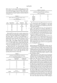
              TABLE 1
______________________________________
QPCR Ratios of Selected Target Nucleic Acids to Reference Nucleic
Acid IGF-1 in DNA from White Blood Cells of Normal Individuals
SAMPLE p53      BRCA1    D9S1752
                                erbB-2 cyclin D1
______________________________________
1      0.8      1        0.9    1.1    1
2      1.3      0.9      1.1    1.2    1.1
3      0.9      1        1.2    1      1.1
4      1        1.1      0.9    0.8    0.9
5      0.9      ND       ND     1.1    1.2
6      0.9      1.1      0.8    ND     ND
7      0.8      0.8      0.8    1.1    1.2
8      0.8      0.9      0.9    0.9    1
9      1        0.9      ND     0.9    1
10     1        0.9      0.8    1.2    1.1
Average
       0.94     0.96     0.93   1.03   1.07
Std. Dev.
       0.15     0.15     0.15   0.14   0.1
QPCR                                   0.9-1.2
Range  0.8-1.3  0.8-1.3  0.8-1.2
                                0.8-1.2
______________________________________
 ND = Not Determined
Relative QPCR Reveals DNA Sequence Copy Number Alterations in DNA From Cancer Tissues
The relative copy numbers of tumor suppressor markers (p53, D9S1752 (p16), or BRCA1) and/or an oncogene (erbB-2 or cyclin D1) were determined using the conditions described in Example I using QPCR in DNA isolated from esophageal, breast, prostate, colon, cervical, adrenal, bladder, brain, kidney, larynx, pancreas, and neuroblastoma tumors. The marker UT40 was also used to mark the distal part of the long arm of chromosome 17 in some experiments.
The results using DNA from breast, prostate and colon tumors are shown in Table 2. The tumor suppressor loci p53 and BRCA1 were present in lower copy numbers relative to the reference marker (IGF-1) in the breast tumor tissues. The lowered copy number was not simply due to aneusomy for chromosome 17 because the quantities of p53, BRCA1 and UT40, all of which reside on chromosome 17, were often discordant in a given sample. Amplifications of these loci relative to IGF-1 are more rare in breast cancer than in other malignancies investigated, but amplifications of these tumor suppressor markers have been observed in samples from other breast cancers.
Table 2 also demonstrates variations in the copy number of p53 and BRCA1 relative to IGF-1 in DNA isolated from prostate cancer tissues. In the examples shown, p53 sequences were found to be amplified, while both amplifications and deletions of BRCA1 relative to IGF-1 were detected. The lack of correspondence between the types of quantitative alterations observed for these two tumor suppressor markers on chromosome 17 is further evidence that the quantitative alterations reflect amplification or deletion involving specific regions of the chromosome rather than resulting from simple aneusomy of an entire chromosome.
              TABLE 2
______________________________________
QPCR Ratios of Selected Target Nucleic Acids to
Reference Nucleic Acid IGF-1 in
DNA from Tumor Cells (ND = Not Determined)
______________________________________
BREAST
TUMORS
Marker and
chromosomal
location  p53         BRCA1    cyclin D1
                                      UT40
Sample    17p13       17q11.2  11q13  17q25
______________________________________
1         0.8         0.6      0.5    1.1
2         0.6         0.5      0.2    1.1
3         0.7         0.9      1.5    0.9
4         0.7         0.7      0.7    1.3
5         0.4         1.2      0.4    1.7
______________________________________
PROSTATE
TUMORS
Marker and
chromosomal
location  LOH Analysis for
                      p53      BRCA1  UT40
Sample    p53         17p13    17q11.2
                                      17q25
______________________________________
6         Uninformative
                      1.3      1.4    1
7         Uninformative
                      1.8      0.4    1.1
8         No LOH      2.6      1      0.8
9         No LOH      2.1      0.6    ND
10        No LOH      1.6      4.2    1.2
11        No LOH      4        4      1.1
12        No LOH      7.2      0.1    ND
13        No LOH      5.7      0.2    0.6
______________________________________
COLON
TUMORS
                      p53      17q11.2
                                      UT40
SAMPLE    LOH Analysis
                      17p13    BRCA1  17q25
______________________________________
14        ND          1.3      3.7    1
15        ND          0.9      0.8    1.2
16        No LOH      2.3      0.9    1
17        No LOH      1.9      2      0.9
18        Uninformative
                      2.1      ND     1
______________________________________
Table 2 also shows the results of QPCR using DNA isolated from colon cancer tissues using p53 and BRCA1 as the target probes. p53 sequences were amplified in three of five samples, while BRCA1 was amplified in two of four samples examined. However, amplification of the p53 locus did not correlate with amplification of BRCA1. In addition, UT40 (a marker at 17q25) was usually present at normal copy number, suggesting again that amplification of larger regions of chromosome 17 was not responsible for the alterations in sequence copy number seen in the colon cancer tumors.
Loss of heterozygosity (LOH) was also monitored in some of the prostate and colon samples subjected to QPCR analysis. LOH was not observed in several samples although anomalies (e.g., relative amplifications of p53 relative to IGF-1) were observed. This also illustrates that amplification can occur in the absence of LOH, underscoring that relative amplification of tumor suppressor genes can occur earlier than LOH. Thus, QPCR is a more sensitive indicator than LOH of the genome instability that occurs in cancer cells but not in normal cells.
QPCR was also used to examine sequence copy number in bladder tumors. In these experiments, the copy number of the D9S1752 locus was examined relative to the copy number of IGF-1 in DNA from 70 primary bladder tumors. The D9S1752 locus is located is located 10 kb from the tumor suppressor gene, p16, which is frequently deleted in bladder tumors (Cairns et al., Nature Genetics 11,210, 1995). In these experiments, QPCR results were also compared to conventional PCR-based microsatellite analysis, which detects differences in sizes of small repeat units in the genome (see Lasko et al., Ann. Rev. Genet. 25,:281, (1991). In addition, some tumors were monitored for LOH.
Of the 70 tumors, eleven were not informative by microsatellite analysis. Of the remaining tumors, 39 of 59 (66%) demonstrated a deletion at D9S1752 by conventional microsatellite analysis. Of these 39, 22 were homozygous deletion (HD) cases and 19 were LOH. In 53 of the 59 informative tumors, there was concordance between results obtained using QPCR and microsatellite analysis. In particular, the two techniques agreed on all 39 samples in which standard polymorphism analysis showed either HD or LOH. In 14 samples, both QPCR analysis for D9S1752/IGF-1 and microsatellite analysis for D9S1752 indicated that the samples were normal.
Seventeen samples were normal or uninformative by microsatellite analysis but were scored as abnormal by QPCR. Of these 17 samples, eleven tumors were not polymorphic at D9S1752 and could not be evaluated by LOH analysis. However, because QPCR does not require polymorphism, it could be used to evaluate these 11 tumors. In all these cases, the QPCR analysis was informative and revealed an abnormal copy number of D9S1752 relative to IGF-1. Ten samples showed deletion of D9S1752, while one had an amplification of D9S1752.
Three of the 17 samples informative by QPCR but not by microsatellite analysis showed amplification of D9S1752. Further, QPCR revealed a decrease of the D9S1752/IGF-1 ratio sequences when microsatellite analysis showed 2 alleles for this locus in four samples. Use of another autosomal probe, D21S100β, showed that the IGF-1 locus was amplified in these samples.
Overall, QPCR was sufficiently sensitive to detect all abnormalities detected by microsatellite analysis. In addition, QPCR detected both decreases and increases in the D9S1752/IGF-1 ratio in samples for which microsatellite analysis of D9S1752 was not informative.
Relative QPCR Reveals Specific DNA Sequence Copy Number Alterations in Nucleic Acids Isolated From Bodily Fluids of Patients With Tumors.
Urine sediment can be used to monitor the presence of bladder tumor cells because cells from bladder tumors are often exfoliated into urine sediment. Accordingly, QPCR was performed on DNA isolated from urine sediment in order to determine the relative copy number of the D9S1752 locus to IGF-1 using the conditions described in Example I.
Table 3 shows the copy number of D9S1752 relative to IGF-1 in urine sediment from eight individuals diagnosed with bladder cancer. All of the individuals had an abnormal D9S1752/IGF-1 ratio, with seven showing deletions of D9S1752 and one showing amplification.
For some of the subjects, the D9S1752/IGF-1 ratio was also determined in DNA taken from the bladder tumor or from plasma. The bladder tumor samples were obtained from patients whose urine sediment results were informative. The bladder tumor samples similarly showed a decreased copy number of D9S1752 relative to IGF-1. LOH studies were also performed on some of the subjects shown in Table 3 and yielded results consistent with the results obtained in DNA from urine sediment: three samples showing LOH or HD also had a reduced copy number of D9S1752. One sample, which had an amplified D9S1752/IGF-1 ratio of 1.5, was normal by LOH analysis. For all the blood samples shown in Table 3, the D9S1752/IGF-1 ratio was in the normal range as expected. These results demonstrate that relative QPCR performed on DNA isolated from urine sediment can measure sequence alterations indicative of bladder cancer.
              TABLE 3
______________________________________
QPCR in DNA from Urine Sediment, Bladder Tumors,
and Serum
(ND = Not Determined)
                  D9S1752/IGF-       D9S1752/IG
                  1                  F-1
Probe D9S1752/IGF-1
                  Urine      LOH studies
                                     Blood
Samples
      Bladder tumor
                  sediment   of D9S1752
                                     samples
______________________________________
1     ND          0.2        LOH     ND
2     ND          0.2        HD      ND
3     ND          1.5        NO LOH  ND
4     ND          0.6        HD      1
5     0.3         0.2        ND        0.8
6     0.1         0.4        ND      1
7     ND          0.1        HD      1
8     0.2         0.4        ND      1
______________________________________
Relative QPCR was also used to examine sequence alterations in DNA from bodily fluids of patients suffering from bladder, breast, lung, nasopharyngeal, esophageal, or prostate cancer. Shown in Table 4 are the erbB-2/IGF-1 or D9S1752/cyclin D1 ratios following QPCR on DNA isolated from the serum of prostate cancer patients, as well as DNA isolated from some of the corresponding prostate tumors. Eight of ten samples isolated from serum DNA showed abnormal erbB-2/IGF-1 ratios, which ranged from 1.7 to 4.5 in the samples tested. In DNA isolated from the tumors of the two patients with normal serum erbB-2/IGF-1 ratios, the corresponding erbB-2/IGF-1 ratio was abnormal.
Sequence abnormalities were also measured by following the D9S1752/cyclin D1 ratio following QPCR in DNA isolated from the serum of an additional five prostate cancer patients. Four of the five patients showed an elevation in the D9S1752/cyclin D1 ratio. For the tumor showing normal D9S1752/cyclin D1 serum levels, DNA from the corresponding tumor showed an altered ratio.
              TABLE 4
______________________________________
Comparison of Various QPCR Ratios in DNA from
Serum and Tumor Samples of Prostate Cancer Patients
(ND = Not Determined)
______________________________________
            SERUM DNA    TUMOR DNA
SAMPLE      erbB-2/IGF1  erbB-2/IGF1
______________________________________
1           4.5          ND
2           3.2          1.5
3           4.5          ND
4           3.3          ND
5           3.5          ND
6           4.2          ND
7           3.9          2.5
8           0.9          3
9           1.7          ND
10          1.1          2
______________________________________
                         TUMOR
            SERUM        D9S1752/
SAMPLE      D9S1752/cyclin D1
                         cyclin D1
______________________________________
11          1.8          6.7
12          2            3
13          4            4.3
14          2            5.7
15          1.2          6.2
NORMAL      1.2          ND
SERUM
NORMAL      1.1          ND
SERUM
NORMAL      1.1          ND
WBC
______________________________________
The data demonstrate that DNA isolated from serum may have a normal ratio, which corresponds to a low tumor burden, or an altered ratio, corresponding to a high tumor burden, when the QPCR ratio in DNA isolated from the tumor itself is abnormal.
Serum was also measured for five of the patients listed in Table 4 that initially had abnormal erbB-2/IGF-1 or D9S1752/cyclin D1 ratios at six months following therapy. For four of the patients, the D9S1752/cyclin D1 ratios were in the normal range, the exceptional patient had a ratio of 1.6. Thus, QPCR can be used on bodily fluids such as serum to screen for aggressive cancer that has seeded the bloodstream prior to, during and after therapy.
QPCR was also performed using probes specific for the erbB-2 and IGF-1 loci on DNA isolated from serum of patients with breast cancer, lung cancer, nasopharyngeal cancer or esophageal cancer. Six of ten samples from sera of patients with lung cancer showed abnormal QPCR ratios, while abnormal ratios were detected in 11 of 13 samples from nasopharyngeal cancers, and in 13 of 20 samples from sera of esophageal cancers.
QPCR Using One Probe Distinguishes Cell-free and Cell-containing Samples in DNA Isolated From Urine Sediment.
To detect the presence of DNA, which is indicative of the presence of cells, in urine sediment, QPCR was performed on DNA isolated from urine sediment using primers specific for the erbB-2 gene. Although this assay does not distinguish between normal and abnormal cell types, it provides a rapid and quantitative indication that cells are present in a fluid that is normally cell-free.
QPCR using a probe for the erbB-2 locus was performed on DNA isolated from urine sediment in healthy individuals, individuals with non-malignant bladder conditions such as cystitis and urinary tract infections, and individuals with bladder cancer. The amount of DNA present in urine sediment was then compared to the underlying bladder condition, if present. The data obtained from urine sediment could be grouped into three classes, according to how much DNA was isolated: (a) low amounts of DNA (less than about 50-100 copies of a target DNA sequence in cells sedimented from 50 μl of urine), which was seen only in normals without significant number of cells in the urine; (b) medium amounts of DNA (about 50-100 copies of the target DNA sequence in cells sedimented from 50 μl of urine), which were seen with either condition; and (c) high amounts of DNA (more than about 50-100 copies of the target DNA sequence in cells sedimented from 50 μl of urine), which were only seen in individuals with increased numbers of cells, either benign or malignant, in the urine. The samples were also stained with the dye Hoechst 33258; however, staining with this dye did not correlate with the presence of a bladder condition. This result demonstrates that Hoechst 33258 staining of urine sediment was not accurate in identifying abnormalities in these conditions, which examined only a small number of cells.
These results demonstrate that one-probe QPCR can be used to screen urine sediment for abnormalities such as cystitis, urinary tract infections, or bladder cancer. Thus, the one-probe PCR assay can be used to screen groups at risk for bladder abnormalities. Further, in the case of individuals with previous malignancies, low or high values of one-probe QPCR can be used as an efficacious way to evaluate the success of therapy and the need for further QPCR analysis using the second reference probe. Successful therapy is associated a low one-probe QPCR value. Failure of therapy is associated with a high QPCR reading, reflecting continued presence of the malignancy with or without an associated urinary tract infection. These two explanations can be distinguished in subsequent two-probe QPCR using a reference nucleic acid.
From the above description, the essential characteristics of the present invention can be ascertained. Without departing from the spirit and scope thereof, various changes and modifications of the invention can be made to adapt it to various usages and conditions. Thus, other embodiments are also within the claims.
__________________________________________________________________________
#             SEQUENCE LISTING
- <160> NUMBER OF SEQ ID NOS: 24
- <210> SEQ ID NO 1
<211> LENGTH: 20
<212> TYPE: DNA
<213> ORGANISM: Homo sapiens
- <400> SEQUENCE: 1
# 20               gttg
- <210> SEQ ID NO 2
<211> LENGTH: 24
<212> TYPE: DNA
<213> ORGANISM: Homo sapiens
- <400> SEQUENCE: 2
#                24cacc atgg
- <210> SEQ ID NO 3
<211> LENGTH: 23
<212> TYPE: DNA
<213> ORGANISM: Homo sapiens
- <400> SEQUENCE: 3
#                23agaa agg
- <210> SEQ ID NO 4
<211> LENGTH: 18
<212> TYPE: DNA
<213> ORGANISM: Homo sapiens
- <400> SEQUENCE: 4
#  18              gg
- <210> SEQ ID NO 5
<211> LENGTH: 18
<212> TYPE: DNA
<213> ORGANISM: Homo sapiens
- <400> SEQUENCE: 5
#  18              gg
- <210> SEQ ID NO 6
<211> LENGTH: 20
<212> TYPE: DNA
<213> ORGANISM: Homo sapiens
- <400> SEQUENCE: 6
# 20               ctgg
- <210> SEQ ID NO 7
<211> LENGTH: 20
<212> TYPE: DNA
<213> ORGANISM: Homo sapiens
- <400> SEQUENCE: 7
# 20               aagc
- <210> SEQ ID NO 8
<211> LENGTH: 22
<212> TYPE: DNA
<213> ORGANISM: Homo sapiens
- <400> SEQUENCE: 8
#                 22cct gg
- <210> SEQ ID NO 9
<211> LENGTH: 24
<212> TYPE: DNA
<213> ORGANISM: Homo sapiens
- <400> SEQUENCE: 9
#                24gaag gaac
- <210> SEQ ID NO 10
<211> LENGTH: 21
<212> TYPE: DNA
<213> ORGANISM: Homo sapiens
- <400> SEQUENCE: 10
#21                tgcc g
- <210> SEQ ID NO 11
<211> LENGTH: 25
<212> TYPE: DNA
<213> ORGANISM: Homo sapiens
- <400> SEQUENCE: 11
#               25 ccag agtgg
- <210> SEQ ID NO 12
<211> LENGTH: 22
<212> TYPE: DNA
<213> ORGANISM: Homo sapiens
- <400> SEQUENCE: 12
#                 22ctg tc
- <210> SEQ ID NO 13
<211> LENGTH: 21
<212> TYPE: DNA
<213> ORGANISM: Homo sapiens
- <400> SEQUENCE: 13
#21                agtc c
- <210> SEQ ID NO 14
<211> LENGTH: 23
<212> TYPE: DNA
<213> ORGANISM: Homo sapiens
- <400> SEQUENCE: 14
#                23ctca tgg
- <210> SEQ ID NO 15
<211> LENGTH: 24
<212> TYPE: DNA
<213> ORGANISM: Homo sapiens
- <400> SEQUENCE: 15
#                24tcag ttgc
- <210> SEQ ID NO 16
<211> LENGTH: 21
<212> TYPE: DNA
<213> ORGANISM: Homo sapiens
- <400> SEQUENCE: 16
#21                ggtg g
- <210> SEQ ID NO 17
<211> LENGTH: 25
<212> TYPE: DNA
<213> ORGANISM: Homo sapiens
<220> FEATURE:
<221> NAME/KEY: misc.sub.-- feature
<222> LOCATION: (1)...(25)
<223> OTHER INFORMATION: n = A,T,C or G
- <400> SEQUENCE: 17
#               25 ggcg gatgg
- <210> SEQ ID NO 18
<211> LENGTH: 22
<212> TYPE: DNA
<213> ORGANISM: Homo sapiens
- <400> SEQUENCE: 18
#                 22agt gg
- <210> SEQ ID NO 19
<211> LENGTH: 20
<212> TYPE: DNA
<213> ORGANISM: Homo sapiens
- <400> SEQUENCE: 19
# 20               ccac
- <210> SEQ ID NO 20
<211> LENGTH: 20
<212> TYPE: DNA
<213> ORGANISM: Homo sapiens
- <400> SEQUENCE: 20
# 20               tttc
- <210> SEQ ID NO 21
<211> LENGTH: 25
<212> TYPE: DNA
<213> ORGANISM: Homo sapiens
- <400> SEQUENCE: 21
#               25 ctcc actcc
- <210> SEQ ID NO 22
<211> LENGTH: 23
<212> TYPE: DNA
<213> ORGANISM: Homo sapiens
- <400> SEQUENCE: 22
#                23gaga ttc
- <210> SEQ ID NO 23
<211> LENGTH: 25
<212> TYPE: DNA
<213> ORGANISM: Homo sapiens
- <400> SEQUENCE: 23
#               25 gggt acgga
- <210> SEQ ID NO 24
<211> LENGTH: 25
<212> TYPE: DNA
<213> ORGANISM: Homo sapiens
- <400> SEQUENCE: 24
#               25 agac aaacg
__________________________________________________________________________
Claims (23)
1. A method for determining the number of copies of a target nucleic acid relative to the number of copies of a reference nucleic acid in a biological specimen, which method comprises the steps of:
(a) amplifying the target nucleic acid in said specimen with a polymerase, a first primer specific for said target nucleic acid with or without a segment noncontiguous to a first priming sequence, a second primer specific for said target nucleic acid with or without a segment noncontiguous to a second priming sequence in the presence of a first oligonucleotide which is incapable of acting as a primer for said polymerase, wherein said first oligonucleotide has at least 5 consecutive nucleotides fully complementary to at least 5 consecutive nucleotides of said first primer;
(b) measuring the amplification of the target nucleic acid;
(c) amplifying the reference nucleic acid in said specimen with a polymerase, a third primer specific for said reference nucleic acid with or without a segment noncontiguous to a third priming sequence, and a fourth primer specific for said reference nucleic acid sequence with or without a segment noncontiguous to a fourth priming sequence in the presence of a second oligonucleotide which is incapable of acting as a primer for said polymerase, wherein said second oligonucleotide has at least 5 consecutive nucleotides fully complementary to at least 5 consecutive nucleotides of said third primer;
(d) measuring the amplification of the reference nucleic acid; and
(e) comparing the amplification of said target nucleic acid sequence to the amplification of said reference nucleic acid sequence in said specimen to determine the number of copies of the target nucleic acid relative to the number of copies of the reference nucleic acid.
2. The method of claim 1, wherein said biological specimen is a cell-free fluid selected from the group consisting of serum, plasma, cerebrospinal fluid, a serous secretion, urine, sweat, and tears.
3. The method of claim 2, wherein said fluid is serum.
4. The method of claim 1, wherein said biological specimen is urine sediment.
5. The method of claim 1, wherein four fluorophores are covalently attached to said first primer, said third primer, said first oligonucleotide, and said second oligonucleotide, respectively, with one of said four fluorophores for said first primer and said first oligonucleotide being a donor fluorophore and the other being an acceptor fluorophore, and with one of said two fluorophores for said third primer and said second oligonucleotide being a donor fluorophore and the other being an acceptor fluorophore, so that when said first primer and said first oligonucleotide are hybridized, said donor fluorophore and said acceptor fluorophore are in close proximity to allow resonance energy transfer therebetween, and when said third primer and said second oligonucleotide are hybridized, said donor fluorophore and said acceptor fluorophore are in close proximity to allow resonance energy transfer therebetween; and, further, said measuring steps are performed by monitoring fluorescent emission change of said acceptor fluorophore upon irradiation of said donor fluorophore with an excitation light, said change being a function of the extent of said first primer being dissociated from said first oligonucleotide and being incorporated into said amplificaton product of the target nucleic acid.
6. The method of claim 5, wherein said biological specimen is a cell-free fluid selected from the group consisting of serum, plasma, cerebrospinal fluid, a serous secretion, urine, sweat, and tears.
7. The method of claim 6, wherein said fluid is serum.
8. The method of claim 5, wherein said biological specimen is urine sediment.
9. The method of claim 1, wherein said first primer and said third primer include segments noncontiguous to said first priming sequence and said third priming sequences, respectively.
10. The method of claim 9, wherein said biological specimen is a cell-free fluid selected from the group consisting of serum, plasma, cerebrospinal fluid, a serous secretion, urine, sweat, and tears.
11. The method of claim 10, wherein said fluid is serum.
12. The method of claim 9, wherein said biological specimen is urine sediment.
13. The method of claim 9, wherein said first oligonucleotide and said second oligonucleotide have at least 5 consecutive nucleotides fully complementary to at least 5 consecutive nucleotides in said noncontiguous segment of said first primer and said third primer, respectively.
14. The method of claim 13, wherein said biological specimen is a cell-free fluid selected from the group consisting of serum, plasma, cerebrospinal fluid, a serous secretion, urine, sweat, and tears.
15. The method of claim 14, wherein said fluid is serum.
16. The method of claim 13, wherein said biological specimen is urine sediment.
17. The method of claim 1, wherein said amplifying steps are performed in the presence of an additional third oligonucleotide and fourth oligonucleotide which are incapable of acting as primers for said polymerase, wherein said additional third oligonucleotide has at least 5 consecutive nucleotides fully complementary to at least 5 consecutive nucleotides of said second primer, and said additional fourth oligonucleotide has at least 5 consecutive nucleotides fully complementary to at least 5 consecutive nucleotides of said fourth primer.
18. The method of claim 17, wherein said biological specimen is a cell-free fluid selected from the group consisting of serum, plasma, cerebrospinal fluid, a serous secretion, urine, sweat, and tears.
19. The method of claim 18, wherein said fluid is serum.
20. The method of claim 17, wherein said biological specimen is urine sediment.
21. A kit for detecting the copy number of a target nucleic acid relative to a reference nucleic acid, said kit comprising:
a first primer with or without a segment noncontiguous to a first priming sequence,
a second primer with or without a segment noncontiguous to a second priming sequence, which said first and second primers are to be used with a polymerase for the amplification of the target nucleic acid;
a third primer with or without a segment noncontiguous to a third priming sequence,
a fourth primer with or without a segment noncontiguous to a fourth priming sequence, which said third and fourth primers are to be used with a polymerase for the amplification of the reference nucleic acid;
a first oligonucleotide which is incapable of acting as a primer for said polymerase and has at least 5 consecutive nucleotides fully complementary to at least 5 consecutive nucleotides of said first primer; and
a second oligonucleotide which is incapable of acting as a primer for said polymerase and has at least 5 consecutive nucleotides fully complementary to at least 5 consecutive nucleotides of said third primer,
wherein each of said first primer, said second primer, said third primer, said fourth primer, said first oligonucleotide, and said second oligonucleotide contains 10-50 nucleotides.
22. The kit of claim 21, wherein said first oligonucleotide and said second oligonucleotide have at least 8 consecutive nucleoteotides fully complementary to at least 8 consecutive nucleotides of said first primer.
23. The kit of claim 21, further comprising an additional third oligonucleotide and fourth oligonucleotide which are incapable of acting as a primer for said polymerase, contain 10-50 nucleotides, and have at least 8 consecutive nucleotides fully complementary to at least 8 consecutive nucleotides of said second primer and said fourth primer, respectively.
Priority Applications (1)
| Application Number | Priority Date | Filing Date | Title |
|---|---|---|---|
| US09/014,065 US6033854A (en) | 1991-12-16 | 1998-01-27 | Quantitative PCR using blocking oligonucleotides |
Applications Claiming Priority (4)
| Application Number | Priority Date | Filing Date | Title |
|---|---|---|---|
| US07/808,463 US5348853A (en) | 1991-12-16 | 1991-12-16 | Method for reducing non-specific priming in DNA amplification |
| US08/250,849 US5567583A (en) | 1991-12-16 | 1994-05-26 | Methods for reducing non-specific priming in DNA detection |
| US08/434,474 US5712386A (en) | 1991-12-16 | 1995-05-04 | Kits for detecting a target nucleic acid with blocking oligonucleotides |
| US09/014,065 US6033854A (en) | 1991-12-16 | 1998-01-27 | Quantitative PCR using blocking oligonucleotides |
Related Parent Applications (1)
| Application Number | Title | Priority Date | Filing Date |
|---|---|---|---|
| US08/434,474 Continuation-In-Part US5712386A (en) | 1991-12-16 | 1995-05-04 | Kits for detecting a target nucleic acid with blocking oligonucleotides |
Publications (1)
| Publication Number | Publication Date |
|---|---|
| US6033854A true US6033854A (en) | 2000-03-07 |
Family
ID=27400378
Family Applications (1)
| Application Number | Title | Priority Date | Filing Date |
|---|---|---|---|
| US09/014,065 Expired - Fee Related US6033854A (en) | 1991-12-16 | 1998-01-27 | Quantitative PCR using blocking oligonucleotides |
Country Status (1)
| Country | Link |
|---|---|
| US (1) | US6033854A (en) |
Cited By (80)
| Publication number | Priority date | Publication date | Assignee | Title |
|---|---|---|---|---|
| WO2002066686A1 (en) * | 2001-02-19 | 2002-08-29 | Id-Lelystad, Instituut Voor Dierhouderij En Diergezondheid B.V. | Feline infectious peritonitis viruses (fipv) diagnosis |
| US20030017482A1 (en) * | 2001-03-02 | 2003-01-23 | Godfrey Tony E. | PCR method |
| US6548251B1 (en) | 2000-09-05 | 2003-04-15 | Fidelity Systems, Inc. | Inhibition of molecular and biological processes using modified oligonucleotides |
| US20030204331A1 (en) * | 2001-11-20 | 2003-10-30 | Duncan Whitney | Automated sample preparation methods and devices |
| WO2004018998A2 (en) * | 2002-08-20 | 2004-03-04 | University Of Maryland | Method using real-time pcr |
| US20040058331A1 (en) * | 2001-06-13 | 2004-03-25 | Tum Gene, Inc. | Blood testing method, testing chip, and testing device |
| WO2005112544A2 (en) | 2004-02-19 | 2005-12-01 | The Governors Of The University Of Alberta | Leptin promoter polymorphisms and uses thereof |
| US20060068433A1 (en) * | 2004-09-20 | 2006-03-30 | Godfrey Tony E | Multiple mode multiplex reaction quenching method |
| US20080102495A1 (en) * | 2006-06-20 | 2008-05-01 | Lynn Kozma | Multi-stage amplification reactions by control of sequence replication times |
| EP2110439A1 (en) | 2004-05-06 | 2009-10-21 | F. Hoffmann-Roche AG | SENP1 as a marker for cancer |
| US20100015613A1 (en) * | 2003-03-18 | 2010-01-21 | Foley Leigh Shaw Marquess | Systems and Methods for Improving Protein and Milk Production of Dairy Herds |
| EP2292795A2 (en) | 2003-10-21 | 2011-03-09 | Orion Genomics, LLC | Differential enzymatic fragmatation |
| EP2339025A1 (en) | 2002-06-26 | 2011-06-29 | Cold Spring Harbor Laboratory | Methods and compositions for determining methylation profiles |
| US20110177512A1 (en) * | 2010-01-19 | 2011-07-21 | Predictive Biosciences, Inc. | Method for assuring amplification of an abnormal nucleic acid in a sample |
| US20110223180A1 (en) * | 2010-03-11 | 2011-09-15 | Aldape Kenneth D | Methods and compositions related to a multi-methylation assay to predict patient outcome |
| EP2390352A1 (en) | 2003-03-18 | 2011-11-30 | Quantum Genetics Ireland Limited | Systems and methods for improving protein and milk production of dairy herds |
| WO2011154139A2 (en) | 2010-06-07 | 2011-12-15 | Roche Diagnostics Gmbh | Gene expression markers for predicting response to interleukin-6 receptor-inhibiting monoclonal antibody drug treatment |
| US20120003633A1 (en) * | 2008-11-05 | 2012-01-05 | Taco Willem Kuijpers | Means and methods for investigating nucleic acid sequences |
| WO2012034007A2 (en) | 2010-09-10 | 2012-03-15 | Bio-Rad Laboratories, Inc. | Size selection of dna for chromatin analysis |
| WO2012034170A1 (en) | 2010-09-13 | 2012-03-22 | Clinical Genomics Pty. Ltd | Epigenetic markers of colorectal cancers and diagnostic methods using the same |
| WO2012112606A1 (en) | 2011-02-15 | 2012-08-23 | Bio-Rad Laboratories, Inc. | Detecting methylati0n in a subpopulation of genomic dna |
| EP2518160A1 (en) | 2011-04-29 | 2012-10-31 | Universitätsklinikum Freiburg | Mitochondrial nucleic acid as a marker for autoimmune and autoinflammatory diseases |
| WO2013019960A1 (en) | 2011-08-03 | 2013-02-07 | Bio-Rad Laboratories, Inc. | Filtering small nucleic acids using permeabilized cells |
| WO2013033380A1 (en) | 2011-08-31 | 2013-03-07 | Genentech, Inc. | Diagnostic markers |
| WO2013055530A1 (en) | 2011-09-30 | 2013-04-18 | Genentech, Inc. | Diagnostic methylation markers of epithelial or mesenchymal phenotype and response to egfr kinase inhibitor in tumours or tumour cells |
| WO2013152307A1 (en) | 2012-04-05 | 2013-10-10 | The Regents Of The University Of California | Gene expression panel for breast cancer prognosis |
| WO2013170191A1 (en) | 2012-05-11 | 2013-11-14 | Genentech, Inc. | Methods of using antagonists of nad biosynthesis from nicotinamide |
| WO2013166558A1 (en) | 2012-05-11 | 2013-11-14 | Clinical Genomics Pty Ltd | Diagnostic gene marker panel for colorectal cancer |
| WO2013170314A1 (en) | 2012-05-18 | 2013-11-21 | Clinical Genomics Pty. Ltd. | A method of screening for colorectal cancer |
| EP2700723A2 (en) | 2007-08-06 | 2014-02-26 | Orion Genomics, LLC | Novel single nucleotide polymorphisms and combinations of novel and known polymorphisms for determining the allele-specific expression of the IGF2 gene |
| WO2014071281A1 (en) | 2012-11-02 | 2014-05-08 | The Johns Hopkins University | Dna methylation biomarkers of post-partum depression risk |
| EP2759307A2 (en) | 2006-03-29 | 2014-07-30 | Merial Limited | Vaccine against Streptococci |
| WO2014135669A1 (en) | 2013-03-08 | 2014-09-12 | Roche Diagnostics Gmbh | Egfr mutation blood testing |
| WO2014151551A1 (en) | 2013-03-15 | 2014-09-25 | Baylor Research Institute | Ulcerative colitis (uc)-associated colorectal neoplasia markers |
| WO2014176599A1 (en) | 2013-04-26 | 2014-10-30 | Gnubio, Inc. | A method for blocking polymerase extension of 3 prime dna ends by stem-loop structure |
| WO2015006645A1 (en) | 2013-07-11 | 2015-01-15 | The Johns Hopkins University | A dna methylation and genotype specific biomarker of suicide attempt and/or suicide ideation |
| WO2015085230A1 (en) | 2013-12-06 | 2015-06-11 | Bio-Rad Laboratories, Inc. | Fusion polymerases |
| WO2015089243A1 (en) | 2013-12-11 | 2015-06-18 | The Regents For Of The University Of California | Methods for labeling dna fragments to recontruct physical linkage and phase |
| WO2015168470A2 (en) | 2014-05-02 | 2015-11-05 | Malaysian Palm Oil Board | Mantle phenotype detection in palm |
| WO2015175530A1 (en) | 2014-05-12 | 2015-11-19 | Gore Athurva | Methods for detecting aneuploidy |
| EP2955234A1 (en) | 2009-02-11 | 2015-12-16 | Orion Genomics, LLC | Combinations of polymorphisms for determining allele-specific expression of igf2 |
| WO2016014493A1 (en) | 2014-07-22 | 2016-01-28 | Bio-Rad Laboratories, Inc. | Buffers for use with polymerases |
| WO2016115530A1 (en) | 2015-01-18 | 2016-07-21 | The Regents Of The University Of California | Method and system for determining cancer status |
| US9411930B2 (en) | 2013-02-01 | 2016-08-09 | The Regents Of The University Of California | Methods for genome assembly and haplotype phasing |
| US9715573B2 (en) | 2015-02-17 | 2017-07-25 | Dovetail Genomics, Llc | Nucleic acid sequence assembly |
| WO2017176652A2 (en) | 2016-04-04 | 2017-10-12 | Sinopia Biosciences, Inc. | Treating extrapyramidal syndrome using trapidil |
| US9914975B2 (en) | 2013-03-08 | 2018-03-13 | Roche Molecular Systems, Inc. | EGFR blood monitoring |
| US10089437B2 (en) | 2013-02-01 | 2018-10-02 | The Regents Of The University Of California | Methods for genome assembly and haplotype phasing |
| WO2018177325A1 (en) | 2017-03-29 | 2018-10-04 | Crown Bioscience Inc. (Taicang) | System and method for determining cetuximab sensitivity on gastric cancer |
| US10093986B2 (en) | 2016-07-06 | 2018-10-09 | Youhealth Biotech, Limited | Leukemia methylation markers and uses thereof |
| WO2018219997A1 (en) | 2017-05-30 | 2018-12-06 | Roche Diagnostics Gmbh | Catalysts for reversing formaldehyde adducts and crosslinks |
| WO2019020732A1 (en) | 2017-07-27 | 2019-01-31 | INSERM (Institut National de la Santé et de la Recherche Médicale) | Methods for determining whether a patient suffering from rhabdomyolysis achieves a response with a tlr9 antagonist |
| US10202650B2 (en) | 2016-05-31 | 2019-02-12 | Youhealth Biotech, Limited | Methods for monitoring ELOVL2, KLF14 and PENK gene expression following treatment with vitamin C |
| US10202637B2 (en) | 2013-03-14 | 2019-02-12 | Molecular Loop Biosolutions, Llc | Methods for analyzing nucleic acid |
| EP3498862A2 (en) | 2013-03-14 | 2019-06-19 | HudsonAlpha Institute For Biotechnology | Differential methylation level of cpg loci that are determinative of a biochemical reoccurrence of prostate cancer |
| US10370710B2 (en) | 2011-10-17 | 2019-08-06 | Good Start Genetics, Inc. | Analysis methods |
| US10429399B2 (en) | 2014-09-24 | 2019-10-01 | Good Start Genetics, Inc. | Process control for increased robustness of genetic assays |
| US10457934B2 (en) | 2015-10-19 | 2019-10-29 | Dovetail Genomics, Llc | Methods for genome assembly, haplotype phasing, and target independent nucleic acid detection |
| US10513739B2 (en) | 2017-03-02 | 2019-12-24 | Youhealth Oncotech, Limited | Methylation markers for diagnosing hepatocellular carcinoma and lung cancer |
| US10526641B2 (en) | 2014-08-01 | 2020-01-07 | Dovetail Genomics, Llc | Tagging nucleic acids for sequence assembly |
| US10544467B2 (en) | 2016-07-06 | 2020-01-28 | Youhealth Oncotech, Limited | Solid tumor methylation markers and uses thereof |
| WO2020104482A1 (en) | 2018-11-20 | 2020-05-28 | INSERM (Institut National de la Santé et de la Recherche Médicale) | Methods for predicting metastatic potential in patients suffering from sdhb-mutated paraganglioma |
| US10851414B2 (en) | 2013-10-18 | 2020-12-01 | Good Start Genetics, Inc. | Methods for determining carrier status |
| US10947579B2 (en) | 2016-05-13 | 2021-03-16 | Dovetail Genomics, Llc | Recovering long-range linkage information from preserved samples |
| US10975417B2 (en) | 2016-02-23 | 2021-04-13 | Dovetail Genomics, Llc | Generation of phased read-sets for genome assembly and haplotype phasing |
| US11041851B2 (en) | 2010-12-23 | 2021-06-22 | Molecular Loop Biosciences, Inc. | Methods for maintaining the integrity and identification of a nucleic acid template in a multiplex sequencing reaction |
| US11072828B2 (en) | 2014-10-06 | 2021-07-27 | The Johns Hopkins University | DNA methylation and genotype specific biomarker for predicting post-traumatic stress disorder |
| US11149308B2 (en) | 2012-04-04 | 2021-10-19 | Invitae Corporation | Sequence assembly |
| US11332795B2 (en) | 2008-05-14 | 2022-05-17 | Dermtech, Inc. | Diagnosis of melanoma and solar lentigo by nucleic acid analysis |
| US11396678B2 (en) | 2016-07-06 | 2022-07-26 | The Regent Of The University Of California | Breast and ovarian cancer methylation markers and uses thereof |
| US11408024B2 (en) | 2014-09-10 | 2022-08-09 | Molecular Loop Biosciences, Inc. | Methods for selectively suppressing non-target sequences |
| US11433075B2 (en) | 2017-06-22 | 2022-09-06 | Triact Therapeutics, Inc. | Methods of treating glioblastoma |
| US11578373B2 (en) | 2019-03-26 | 2023-02-14 | Dermtech, Inc. | Gene classifiers and uses thereof in skin cancers |
| US11628144B2 (en) | 2017-09-29 | 2023-04-18 | Triact Therapeutics, Inc. | Iniparib formulations and uses thereof |
| US11680284B2 (en) | 2015-01-06 | 2023-06-20 | Moledular Loop Biosciences, Inc. | Screening for structural variants |
| WO2023187386A1 (en) | 2022-03-30 | 2023-10-05 | University Of Newcastle Upon Tyne | Methods for determining mitochondrial dna damage |
| US11807896B2 (en) | 2015-03-26 | 2023-11-07 | Dovetail Genomics, Llc | Physical linkage preservation in DNA storage |
| US11840730B1 (en) | 2009-04-30 | 2023-12-12 | Molecular Loop Biosciences, Inc. | Methods and compositions for evaluating genetic markers |
| WO2024044578A1 (en) | 2022-08-22 | 2024-02-29 | University Of Virginia | Dna methylation biomarkers of premenstrual dysphoric disorder and perimenopausal depression |
| WO2024059649A1 (en) | 2022-09-13 | 2024-03-21 | The Saban Research Institute; Children's Hospital Los Angeles | Dna methylation signature of retinoblastoma |
Citations (18)
| Publication number | Priority date | Publication date | Assignee | Title |
|---|---|---|---|---|
| EP0200362A2 (en) * | 1985-03-28 | 1986-11-05 | F. Hoffmann-La Roche Ag | Process for amplifying, detecting, and/or cloning nucleic acid sequences |
| EP0201184A2 (en) * | 1985-03-28 | 1986-11-12 | F. Hoffmann-La Roche Ag | Process for amplifying nucleic acid sequences |
| US4683195A (en) * | 1986-01-30 | 1987-07-28 | Cetus Corporation | Process for amplifying, detecting, and/or-cloning nucleic acid sequences |
| US4683194A (en) * | 1984-05-29 | 1987-07-28 | Cetus Corporation | Method for detection of polymorphic restriction sites and nucleic acid sequences |
| EP0236069A2 (en) * | 1986-02-25 | 1987-09-09 | The Perkin-Elmer Corporation | Apparatus and method for performing automated amplification of nucleic acid sequences and assays using heating and cooling steps |
| US4725537A (en) * | 1985-09-19 | 1988-02-16 | Allied Corporation | Assay, reagent and kit employing nucleic acid strand displacement and restriction endonuclease cleavage |
| US4800159A (en) * | 1986-02-07 | 1989-01-24 | Cetus Corporation | Process for amplifying, detecting, and/or cloning nucleic acid sequences |
| US4822733A (en) * | 1985-05-28 | 1989-04-18 | Amoco Corporation | Lifetime-resolved assay procedures |
| EP0333465A2 (en) * | 1988-03-18 | 1989-09-20 | Baylor College Of Medicine | Mutation detection by competitive oligonucleotide priming |
| WO1989009835A1 (en) * | 1988-04-08 | 1989-10-19 | The Salk Institute For Biological Studies | Ligase-based amplification method |
| EP0382433A2 (en) * | 1989-02-07 | 1990-08-16 | Zeneca Limited | Detection of nucleic acid sequences using fluorescence polarisation |
| US4965188A (en) * | 1986-08-22 | 1990-10-23 | Cetus Corporation | Process for amplifying, detecting, and/or cloning nucleic acid sequences using a thermostable enzyme |
| US4996143A (en) * | 1985-12-23 | 1991-02-26 | Syngene, Inc. | Fluorescent stokes shift probes for polynucleotide hybridization |
| US5038852A (en) * | 1986-02-25 | 1991-08-13 | Cetus Corporation | Apparatus and method for performing automated amplification of nucleic acid sequences and assays using heating and cooling steps |
| US5210015A (en) * | 1990-08-06 | 1993-05-11 | Hoffman-La Roche Inc. | Homogeneous assay system using the nuclease activity of a nucleic acid polymerase |
| EP0320308B1 (en) * | 1987-12-11 | 1993-11-03 | Abbott Laboratories | Method for detecting a target nucleic acid sequence |
| US5348853A (en) * | 1991-12-16 | 1994-09-20 | Biotronics Corporation | Method for reducing non-specific priming in DNA amplification |
| US5567583A (en) * | 1991-12-16 | 1996-10-22 | Biotronics Corporation | Methods for reducing non-specific priming in DNA detection |
-
1998
- 1998-01-27 US US09/014,065 patent/US6033854A/en not_active Expired - Fee Related
Patent Citations (19)
| Publication number | Priority date | Publication date | Assignee | Title |
|---|---|---|---|---|
| US4683194A (en) * | 1984-05-29 | 1987-07-28 | Cetus Corporation | Method for detection of polymorphic restriction sites and nucleic acid sequences |
| EP0201184A2 (en) * | 1985-03-28 | 1986-11-12 | F. Hoffmann-La Roche Ag | Process for amplifying nucleic acid sequences |
| EP0200362A2 (en) * | 1985-03-28 | 1986-11-05 | F. Hoffmann-La Roche Ag | Process for amplifying, detecting, and/or cloning nucleic acid sequences |
| US4822733A (en) * | 1985-05-28 | 1989-04-18 | Amoco Corporation | Lifetime-resolved assay procedures |
| US4725537A (en) * | 1985-09-19 | 1988-02-16 | Allied Corporation | Assay, reagent and kit employing nucleic acid strand displacement and restriction endonuclease cleavage |
| US4996143A (en) * | 1985-12-23 | 1991-02-26 | Syngene, Inc. | Fluorescent stokes shift probes for polynucleotide hybridization |
| US4683195B1 (en) * | 1986-01-30 | 1990-11-27 | Cetus Corp | |
| US4683195A (en) * | 1986-01-30 | 1987-07-28 | Cetus Corporation | Process for amplifying, detecting, and/or-cloning nucleic acid sequences |
| US4800159A (en) * | 1986-02-07 | 1989-01-24 | Cetus Corporation | Process for amplifying, detecting, and/or cloning nucleic acid sequences |
| EP0236069A2 (en) * | 1986-02-25 | 1987-09-09 | The Perkin-Elmer Corporation | Apparatus and method for performing automated amplification of nucleic acid sequences and assays using heating and cooling steps |
| US5038852A (en) * | 1986-02-25 | 1991-08-13 | Cetus Corporation | Apparatus and method for performing automated amplification of nucleic acid sequences and assays using heating and cooling steps |
| US4965188A (en) * | 1986-08-22 | 1990-10-23 | Cetus Corporation | Process for amplifying, detecting, and/or cloning nucleic acid sequences using a thermostable enzyme |
| EP0320308B1 (en) * | 1987-12-11 | 1993-11-03 | Abbott Laboratories | Method for detecting a target nucleic acid sequence |
| EP0333465A2 (en) * | 1988-03-18 | 1989-09-20 | Baylor College Of Medicine | Mutation detection by competitive oligonucleotide priming |
| WO1989009835A1 (en) * | 1988-04-08 | 1989-10-19 | The Salk Institute For Biological Studies | Ligase-based amplification method |
| EP0382433A2 (en) * | 1989-02-07 | 1990-08-16 | Zeneca Limited | Detection of nucleic acid sequences using fluorescence polarisation |
| US5210015A (en) * | 1990-08-06 | 1993-05-11 | Hoffman-La Roche Inc. | Homogeneous assay system using the nuclease activity of a nucleic acid polymerase |
| US5348853A (en) * | 1991-12-16 | 1994-09-20 | Biotronics Corporation | Method for reducing non-specific priming in DNA amplification |
| US5567583A (en) * | 1991-12-16 | 1996-10-22 | Biotronics Corporation | Methods for reducing non-specific priming in DNA detection |
Non-Patent Citations (22)
| Title |
|---|
| Augenlicht et al., "Low-Level c-myc Amplification in Human Colonic Carcinoma Cell Lines and Tumors: A Frequent , p53-independent Mutation Associated with Improved Outcome in a Randomized Multi-institutional Trial," Cancer Research, 57:1769-1775, 1997. |
| Augenlicht et al., Low Level c myc Amplification in Human Colonic Carcinoma Cell Lines and Tumors: A Frequent , p53 independent Mutation Associated with Improved Outcome in a Randomized Multi institutional Trial, Cancer Research, 57:1769 1775, 1997. * |
| Blanco et al., "Highly Effecient DNA Synthesis by the Phage .O slashed.29 DNA Polymerase," J. Biol. Chem., 264:8935-8940, 1989. |
| Blanco et al., Highly Effecient DNA Synthesis by the Phage 29 DNA Polymerase, J. Biol. Chem., 264:8935 8940, 1989. * |
| Cairns et al., Clin. Cancer Res. 4(2), 441 444, 1998. * |
| Cairns et al., Clin. Cancer Res. 4(2), 441-444, 1998. |
| Chiang et al., "Use of a Fluorescent-PCR Reaction to Detect Genomic Sequence Copy Number and Transcriptional Abundance," Genome Research,6:1013-1026, 1996. |
| Chiang et al., Cytog. Cell Genet. 79(1 2), 50, 1997. * |
| Chiang et al., Cytog. Cell Genet. 79(1-2), 50, 1997. |
| Chiang et al., Use of a Fluorescent PCR Reaction to Detect Genomic Sequence Copy Number and Transcriptional Abundance, Genome Research, 6:1013 1026, 1996. * |
| Gibson et al., "A Novel Method for Real Time Quantitative RT-PCR," Genome Research,6:995-1001, 1996. |
| Gibson et al., A Novel Method for Real Time Quantitative RT PCR, Genome Research, 6:995 1001, 1996. * |
| Heid et al., "Real Time Quantitative PCR," Genome Research, 6:986-994, 1996. |
| Heid et al., Real Time Quantitative PCR, Genome Research, 6:986 994, 1996. * |
| Holland et al., "Doctrine of Specific-polymerace Chain Reaction Product . . . ", Proc. Natl. Sci. Acad. USA, 88:7276-7280, 1991. |
| Holland et al., Doctrine of Specific polymerace Chain Reaction Product . . . , Proc. Natl. Sci. Acad. USA, 88:7276 7280, 1991. * |
| Morrison et al., "Solution-phase Detection of Polynucleotides Using Interacting Fluorescent Labels and Competitive Hybridization," Analytical Biochemistry, 183:231-244, 1989. |
| Morrison et al., Solution phase Detection of Polynucleotides Using Interacting Fluorescent Labels and Competitive Hybridization, Analytical Biochemistry, 183:231 244, 1989. * |
| von Eggeling et al., "Rapid Detection of Trisomy 21 by Quantitative PCR," Human Genetics, 91:567-570, 1993. |
| von Eggeling et al., Rapid Detection of Trisomy 21 by Quantitative PCR, Human Genetics, 91:567 570, 1993. * |
| Walker et al., "Isothermal In Vitro Amplification of DNA by a Restriction Enzyme/DNA Polymerase System", Proc. Natl. Acad. Sci. USA, 89:392-390, 1996. |
| Walker et al., Isothermal In Vitro Amplification of DNA by a Restriction Enzyme/DNA Polymerase System , Proc. Natl. Acad. Sci. USA, 89:392 390, 1996. * |
Cited By (121)
| Publication number | Priority date | Publication date | Assignee | Title |
|---|---|---|---|---|
| US6548251B1 (en) | 2000-09-05 | 2003-04-15 | Fidelity Systems, Inc. | Inhibition of molecular and biological processes using modified oligonucleotides |
| WO2002066686A1 (en) * | 2001-02-19 | 2002-08-29 | Id-Lelystad, Instituut Voor Dierhouderij En Diergezondheid B.V. | Feline infectious peritonitis viruses (fipv) diagnosis |
| US7101663B2 (en) * | 2001-03-02 | 2006-09-05 | University of Pittsburgh—of the Commonwealth System of Higher Education | PCR method |
| US20030017482A1 (en) * | 2001-03-02 | 2003-01-23 | Godfrey Tony E. | PCR method |
| US20080199867A1 (en) * | 2001-03-02 | 2008-08-21 | Godfrey Tony E | Multiple mode multiplex reaction quenching method |
| US20040058331A1 (en) * | 2001-06-13 | 2004-03-25 | Tum Gene, Inc. | Blood testing method, testing chip, and testing device |
| US20030204331A1 (en) * | 2001-11-20 | 2003-10-30 | Duncan Whitney | Automated sample preparation methods and devices |
| EP2339025A1 (en) | 2002-06-26 | 2011-06-29 | Cold Spring Harbor Laboratory | Methods and compositions for determining methylation profiles |
| WO2004018998A3 (en) * | 2002-08-20 | 2004-06-24 | Univ Maryland | Method using real-time pcr |
| WO2004018998A2 (en) * | 2002-08-20 | 2004-03-04 | University Of Maryland | Method using real-time pcr |
| EP2390352A1 (en) | 2003-03-18 | 2011-11-30 | Quantum Genetics Ireland Limited | Systems and methods for improving protein and milk production of dairy herds |
| US20100015613A1 (en) * | 2003-03-18 | 2010-01-21 | Foley Leigh Shaw Marquess | Systems and Methods for Improving Protein and Milk Production of Dairy Herds |
| EP2292795A2 (en) | 2003-10-21 | 2011-03-09 | Orion Genomics, LLC | Differential enzymatic fragmatation |
| WO2005112544A2 (en) | 2004-02-19 | 2005-12-01 | The Governors Of The University Of Alberta | Leptin promoter polymorphisms and uses thereof |
| EP2110439A1 (en) | 2004-05-06 | 2009-10-21 | F. Hoffmann-Roche AG | SENP1 as a marker for cancer |
| US20060068433A1 (en) * | 2004-09-20 | 2006-03-30 | Godfrey Tony E | Multiple mode multiplex reaction quenching method |
| EP2759307A2 (en) | 2006-03-29 | 2014-07-30 | Merial Limited | Vaccine against Streptococci |
| EP3167900A1 (en) | 2006-03-29 | 2017-05-17 | Merial Limited | Vaccine against streptococci |
| US20080102495A1 (en) * | 2006-06-20 | 2008-05-01 | Lynn Kozma | Multi-stage amplification reactions by control of sequence replication times |
| US8119352B2 (en) | 2006-06-20 | 2012-02-21 | Cepheld | Multi-stage amplification reactions by control of sequence replication times |
| EP2952588A1 (en) | 2007-08-06 | 2015-12-09 | Orion Genomics, LLC | Single nucleotide polymorphism in igf2 for determining the allele-specific expression of the igf2 gene |
| EP2700723A2 (en) | 2007-08-06 | 2014-02-26 | Orion Genomics, LLC | Novel single nucleotide polymorphisms and combinations of novel and known polymorphisms for determining the allele-specific expression of the IGF2 gene |
| US11332795B2 (en) | 2008-05-14 | 2022-05-17 | Dermtech, Inc. | Diagnosis of melanoma and solar lentigo by nucleic acid analysis |
| US11753687B2 (en) | 2008-05-14 | 2023-09-12 | Dermtech, Inc. | Diagnosis of melanoma and solar lentigo by nucleic acid analysis |
| US9017971B2 (en) * | 2008-11-05 | 2015-04-28 | Stichting Sanquin Bloedvoorziening | Means and methods for investigating nucleic acid sequences |
| US20120003633A1 (en) * | 2008-11-05 | 2012-01-05 | Taco Willem Kuijpers | Means and methods for investigating nucleic acid sequences |
| EP2955234A1 (en) | 2009-02-11 | 2015-12-16 | Orion Genomics, LLC | Combinations of polymorphisms for determining allele-specific expression of igf2 |
| US11840730B1 (en) | 2009-04-30 | 2023-12-12 | Molecular Loop Biosciences, Inc. | Methods and compositions for evaluating genetic markers |
| US20110177512A1 (en) * | 2010-01-19 | 2011-07-21 | Predictive Biosciences, Inc. | Method for assuring amplification of an abnormal nucleic acid in a sample |
| US20110223180A1 (en) * | 2010-03-11 | 2011-09-15 | Aldape Kenneth D | Methods and compositions related to a multi-methylation assay to predict patient outcome |
| WO2011154139A2 (en) | 2010-06-07 | 2011-12-15 | Roche Diagnostics Gmbh | Gene expression markers for predicting response to interleukin-6 receptor-inhibiting monoclonal antibody drug treatment |
| WO2012034007A2 (en) | 2010-09-10 | 2012-03-15 | Bio-Rad Laboratories, Inc. | Size selection of dna for chromatin analysis |
| EP3034628A1 (en) | 2010-09-13 | 2016-06-22 | Clinical Genomics Pty Ltd | Diagnosis of cancer by means of methylation marker |
| EP3508589A1 (en) | 2010-09-13 | 2019-07-10 | Clinical Genomics Pty. Ltd. | Epigenetic markers of colorectal cancers and diagnostic methods using the same |
| WO2012034170A1 (en) | 2010-09-13 | 2012-03-22 | Clinical Genomics Pty. Ltd | Epigenetic markers of colorectal cancers and diagnostic methods using the same |
| EP3034629A1 (en) | 2010-09-13 | 2016-06-22 | Clinical Genomics Pty Ltd | Diagnosis of cancer by means of methylation marker |
| US11768200B2 (en) | 2010-12-23 | 2023-09-26 | Molecular Loop Biosciences, Inc. | Methods for maintaining the integrity and identification of a nucleic acid template in a multiplex sequencing reaction |
| US11041852B2 (en) | 2010-12-23 | 2021-06-22 | Molecular Loop Biosciences, Inc. | Methods for maintaining the integrity and identification of a nucleic acid template in a multiplex sequencing reaction |
| US11041851B2 (en) | 2010-12-23 | 2021-06-22 | Molecular Loop Biosciences, Inc. | Methods for maintaining the integrity and identification of a nucleic acid template in a multiplex sequencing reaction |
| WO2012112606A1 (en) | 2011-02-15 | 2012-08-23 | Bio-Rad Laboratories, Inc. | Detecting methylati0n in a subpopulation of genomic dna |
| WO2012146783A1 (en) | 2011-04-29 | 2012-11-01 | Universitätsklinikum Freiburg | Mitochondrial nucleic acid as a marker for autoimmune and autoinflammatory diseases |
| EP2518160A1 (en) | 2011-04-29 | 2012-10-31 | Universitätsklinikum Freiburg | Mitochondrial nucleic acid as a marker for autoimmune and autoinflammatory diseases |
| WO2013019960A1 (en) | 2011-08-03 | 2013-02-07 | Bio-Rad Laboratories, Inc. | Filtering small nucleic acids using permeabilized cells |
| WO2013033380A1 (en) | 2011-08-31 | 2013-03-07 | Genentech, Inc. | Diagnostic markers |
| WO2013055530A1 (en) | 2011-09-30 | 2013-04-18 | Genentech, Inc. | Diagnostic methylation markers of epithelial or mesenchymal phenotype and response to egfr kinase inhibitor in tumours or tumour cells |
| US10370710B2 (en) | 2011-10-17 | 2019-08-06 | Good Start Genetics, Inc. | Analysis methods |
| US11667965B2 (en) | 2012-04-04 | 2023-06-06 | Invitae Corporation | Sequence assembly |
| US11149308B2 (en) | 2012-04-04 | 2021-10-19 | Invitae Corporation | Sequence assembly |
| US11155863B2 (en) | 2012-04-04 | 2021-10-26 | Invitae Corporation | Sequence assembly |
| WO2013152307A1 (en) | 2012-04-05 | 2013-10-10 | The Regents Of The University Of California | Gene expression panel for breast cancer prognosis |
| EP3594366A1 (en) | 2012-05-11 | 2020-01-15 | Clinical Genomics Pty. Ltd. | Diagnostic gene marker panel |
| EP4001436A1 (en) | 2012-05-11 | 2022-05-25 | Clinical Genomics Pty. Ltd. | Diagnostic gene marker panel |
| WO2013166558A1 (en) | 2012-05-11 | 2013-11-14 | Clinical Genomics Pty Ltd | Diagnostic gene marker panel for colorectal cancer |
| WO2013170191A1 (en) | 2012-05-11 | 2013-11-14 | Genentech, Inc. | Methods of using antagonists of nad biosynthesis from nicotinamide |
| US11254985B2 (en) | 2012-05-11 | 2022-02-22 | Clinical Genomics Pty. Ltd. | Diagnostic gene marker panel for colorectal cancer |
| EP3608421A1 (en) | 2012-05-18 | 2020-02-12 | Clinical Genomics Pty. Ltd. | A method of screening for colorectal cancer |
| WO2013170314A1 (en) | 2012-05-18 | 2013-11-21 | Clinical Genomics Pty. Ltd. | A method of screening for colorectal cancer |
| EP4116437A1 (en) | 2012-05-18 | 2023-01-11 | Clinical Genomics Pty. Ltd. | A method of screening for colorectal cancer |
| WO2014071281A1 (en) | 2012-11-02 | 2014-05-08 | The Johns Hopkins University | Dna methylation biomarkers of post-partum depression risk |
| US9910955B2 (en) | 2013-02-01 | 2018-03-06 | The Regents Of The University Of California | Methods for genome assembly and haplotype phasing |
| US10825553B2 (en) | 2013-02-01 | 2020-11-03 | The Regents Of The University Of California | Methods for genome assembly and haplotype phasing |
| EP3885446A1 (en) | 2013-02-01 | 2021-09-29 | The Regents of The University of California | Methods for genome assembly and haplotype phasing |
| US10089437B2 (en) | 2013-02-01 | 2018-10-02 | The Regents Of The University Of California | Methods for genome assembly and haplotype phasing |
| US11935626B2 (en) | 2013-02-01 | 2024-03-19 | The Regents Of The University Of California | Methods for genome assembly and haplotype phasing |
| US10529443B2 (en) | 2013-02-01 | 2020-01-07 | The Regents Of The University Of California | Methods for genome assembly and haplotype phasing |
| US11081209B2 (en) | 2013-02-01 | 2021-08-03 | The Regents Of The University Of California | Methods for genome assembly and haplotype phasing |
| US9411930B2 (en) | 2013-02-01 | 2016-08-09 | The Regents Of The University Of California | Methods for genome assembly and haplotype phasing |
| US9873913B2 (en) | 2013-03-08 | 2018-01-23 | Roche Molecular Systems, Inc. | Mutation testing |
| US9914975B2 (en) | 2013-03-08 | 2018-03-13 | Roche Molecular Systems, Inc. | EGFR blood monitoring |
| WO2014135669A1 (en) | 2013-03-08 | 2014-09-12 | Roche Diagnostics Gmbh | Egfr mutation blood testing |
| US10202637B2 (en) | 2013-03-14 | 2019-02-12 | Molecular Loop Biosolutions, Llc | Methods for analyzing nucleic acid |
| EP3498862A2 (en) | 2013-03-14 | 2019-06-19 | HudsonAlpha Institute For Biotechnology | Differential methylation level of cpg loci that are determinative of a biochemical reoccurrence of prostate cancer |
| EP3366785A2 (en) | 2013-03-15 | 2018-08-29 | Baylor Research Institute | Ulcerative colitis (uc)-associated colorectal neoplasia markers |
| WO2014151551A1 (en) | 2013-03-15 | 2014-09-25 | Baylor Research Institute | Ulcerative colitis (uc)-associated colorectal neoplasia markers |
| WO2014176599A1 (en) | 2013-04-26 | 2014-10-30 | Gnubio, Inc. | A method for blocking polymerase extension of 3 prime dna ends by stem-loop structure |
| WO2015006645A1 (en) | 2013-07-11 | 2015-01-15 | The Johns Hopkins University | A dna methylation and genotype specific biomarker of suicide attempt and/or suicide ideation |
| US10851414B2 (en) | 2013-10-18 | 2020-12-01 | Good Start Genetics, Inc. | Methods for determining carrier status |
| WO2015085230A1 (en) | 2013-12-06 | 2015-06-11 | Bio-Rad Laboratories, Inc. | Fusion polymerases |
| EP3540074A1 (en) | 2013-12-11 | 2019-09-18 | The Regents of the University of California | Method of tagging internal regions of nucleic acid molecules |
| WO2015089243A1 (en) | 2013-12-11 | 2015-06-18 | The Regents For Of The University Of California | Methods for labeling dna fragments to recontruct physical linkage and phase |
| WO2015168470A2 (en) | 2014-05-02 | 2015-11-05 | Malaysian Palm Oil Board | Mantle phenotype detection in palm |
| US11632922B2 (en) | 2014-05-02 | 2023-04-25 | Malaysian Palm Oil Board | Mantle phenotype detection in palm |
| US9984200B2 (en) | 2014-05-02 | 2018-05-29 | Malaysian Palm Oil Board | Mantle phenotype detection in palm |
| WO2015175530A1 (en) | 2014-05-12 | 2015-11-19 | Gore Athurva | Methods for detecting aneuploidy |
| US11053548B2 (en) | 2014-05-12 | 2021-07-06 | Good Start Genetics, Inc. | Methods for detecting aneuploidy |
| WO2016014493A1 (en) | 2014-07-22 | 2016-01-28 | Bio-Rad Laboratories, Inc. | Buffers for use with polymerases |
| US10526641B2 (en) | 2014-08-01 | 2020-01-07 | Dovetail Genomics, Llc | Tagging nucleic acids for sequence assembly |
| EP4219710A2 (en) | 2014-08-01 | 2023-08-02 | Dovetail Genomics, LLC | Tagging nucleic acids for sequence assembly |
| US11408024B2 (en) | 2014-09-10 | 2022-08-09 | Molecular Loop Biosciences, Inc. | Methods for selectively suppressing non-target sequences |
| US10429399B2 (en) | 2014-09-24 | 2019-10-01 | Good Start Genetics, Inc. | Process control for increased robustness of genetic assays |
| US11072828B2 (en) | 2014-10-06 | 2021-07-27 | The Johns Hopkins University | DNA methylation and genotype specific biomarker for predicting post-traumatic stress disorder |
| US11680284B2 (en) | 2015-01-06 | 2023-06-20 | Moledular Loop Biosciences, Inc. | Screening for structural variants |
| US9984201B2 (en) | 2015-01-18 | 2018-05-29 | Youhealth Biotech, Limited | Method and system for determining cancer status |
| WO2016115530A1 (en) | 2015-01-18 | 2016-07-21 | The Regents Of The University Of California | Method and system for determining cancer status |
| US10318706B2 (en) | 2015-02-17 | 2019-06-11 | Dovetail Genomics, Llc | Nucleic acid sequence assembly |
| US11600361B2 (en) | 2015-02-17 | 2023-03-07 | Dovetail Genomics, Llc | Nucleic acid sequence assembly |
| US9715573B2 (en) | 2015-02-17 | 2017-07-25 | Dovetail Genomics, Llc | Nucleic acid sequence assembly |
| US11807896B2 (en) | 2015-03-26 | 2023-11-07 | Dovetail Genomics, Llc | Physical linkage preservation in DNA storage |
| US10457934B2 (en) | 2015-10-19 | 2019-10-29 | Dovetail Genomics, Llc | Methods for genome assembly, haplotype phasing, and target independent nucleic acid detection |
| US10975417B2 (en) | 2016-02-23 | 2021-04-13 | Dovetail Genomics, Llc | Generation of phased read-sets for genome assembly and haplotype phasing |
| US10350212B2 (en) | 2016-04-04 | 2019-07-16 | Sinopia Biosciences, Inc. | Treating extrapyramidal syndrome using trapidil |
| WO2017176652A2 (en) | 2016-04-04 | 2017-10-12 | Sinopia Biosciences, Inc. | Treating extrapyramidal syndrome using trapidil |
| US11628170B2 (en) | 2016-04-04 | 2023-04-18 | Sinopia Biosciences, Inc. | Treating extrapyramtdal syndrome using trapidil |
| US10947579B2 (en) | 2016-05-13 | 2021-03-16 | Dovetail Genomics, Llc | Recovering long-range linkage information from preserved samples |
| US10202650B2 (en) | 2016-05-31 | 2019-02-12 | Youhealth Biotech, Limited | Methods for monitoring ELOVL2, KLF14 and PENK gene expression following treatment with vitamin C |
| US11279978B2 (en) | 2016-05-31 | 2022-03-22 | The Regents Of The University Of California | Methods for evaluating, monitoring, and modulating aging process |
| US10093986B2 (en) | 2016-07-06 | 2018-10-09 | Youhealth Biotech, Limited | Leukemia methylation markers and uses thereof |
| US11396678B2 (en) | 2016-07-06 | 2022-07-26 | The Regent Of The University Of California | Breast and ovarian cancer methylation markers and uses thereof |
| US10829821B2 (en) | 2016-07-06 | 2020-11-10 | Youhealth Oncotech, Limited | Leukemia methylation markers and uses thereof |
| US10544467B2 (en) | 2016-07-06 | 2020-01-28 | Youhealth Oncotech, Limited | Solid tumor methylation markers and uses thereof |
| US10513739B2 (en) | 2017-03-02 | 2019-12-24 | Youhealth Oncotech, Limited | Methylation markers for diagnosing hepatocellular carcinoma and lung cancer |
| WO2018177325A1 (en) | 2017-03-29 | 2018-10-04 | Crown Bioscience Inc. (Taicang) | System and method for determining cetuximab sensitivity on gastric cancer |
| WO2018219997A1 (en) | 2017-05-30 | 2018-12-06 | Roche Diagnostics Gmbh | Catalysts for reversing formaldehyde adducts and crosslinks |
| US11433075B2 (en) | 2017-06-22 | 2022-09-06 | Triact Therapeutics, Inc. | Methods of treating glioblastoma |
| WO2019020732A1 (en) | 2017-07-27 | 2019-01-31 | INSERM (Institut National de la Santé et de la Recherche Médicale) | Methods for determining whether a patient suffering from rhabdomyolysis achieves a response with a tlr9 antagonist |
| US11628144B2 (en) | 2017-09-29 | 2023-04-18 | Triact Therapeutics, Inc. | Iniparib formulations and uses thereof |
| WO2020104482A1 (en) | 2018-11-20 | 2020-05-28 | INSERM (Institut National de la Santé et de la Recherche Médicale) | Methods for predicting metastatic potential in patients suffering from sdhb-mutated paraganglioma |
| US11578373B2 (en) | 2019-03-26 | 2023-02-14 | Dermtech, Inc. | Gene classifiers and uses thereof in skin cancers |
| WO2023187386A1 (en) | 2022-03-30 | 2023-10-05 | University Of Newcastle Upon Tyne | Methods for determining mitochondrial dna damage |
| WO2024044578A1 (en) | 2022-08-22 | 2024-02-29 | University Of Virginia | Dna methylation biomarkers of premenstrual dysphoric disorder and perimenopausal depression |
| WO2024059649A1 (en) | 2022-09-13 | 2024-03-21 | The Saban Research Institute; Children's Hospital Los Angeles | Dna methylation signature of retinoblastoma |
Similar Documents
| Publication | Publication Date | Title |
|---|---|---|
| US6033854A (en) | Quantitative PCR using blocking oligonucleotides | |
| Bièche et al. | Novel approach to quantitative polymerase chain reaction using real‐time detection: application to the detection of gene amplification in breast cancer | |
| Ginzinger | Gene quantification using real-time quantitative PCR: an emerging technology hits the mainstream | |
| US5994057A (en) | Method of detecting aneuploidy by amplified short-tandem repeats | |
| US6180349B1 (en) | Quantitative PCR method to enumerate DNA copy number | |
| Choi et al. | Genetic classification of colorectal cancer based on chromosomal loss and microsatellite instability predicts survival | |
| Ishmael et al. | Principles and applications of polymerase chain reaction: basic science for the practicing physician | |
| US20040005551A1 (en) | Circulating epstein-barr virus DNA in the serum or plasma of patients for the prediction and detection of epstein-barr virus associated cancers apart from head, neck and lymphoid malignancies | |
| WO2016115967A1 (en) | Use of methylation sites in y chromosome as prostate cancer diagnosis marker | |
| EP2569624B1 (en) | Detection of chromosomal abnormalities associated with endometrial cancer | |
| Karlsen et al. | PCR‐RFLP studies on chromosome 3p in formaldehyde‐fixed, paraffin‐embedded cervical cancer tissues | |
| Mohapatra et al. | Glioma test array for use with formalin-fixed, paraffin-embedded tissue: array comparative genomic hybridization correlates with loss of heterozygosity and fluorescence in situ hybridization | |
| JP2008500034A (en) | Method for performing quantitative and / or comparative assessment of mRNA expression levels in small biological samples | |
| US20230175070A1 (en) | Tumor detection reagent and kit | |
| US6261781B1 (en) | Direct detection and mutation analysis of low copy number nucleic acids | |
| CN108949923B (en) | Method for amplifying MSF1 gene, kit and application | |
| Aldape et al. | Real‐time quantitative polymerase chain reaction: a potential tool for genetic analysis in neuropathology | |
| WO2010101696A1 (en) | A snp marker of breast and ovarian cancer risk | |
| US8367814B2 (en) | Assay for BCR/ABL gene rearrangement | |
| US20230054587A1 (en) | Multiplexed Assay Using Differential Fragment Size to Identify Cancer Specific Cell-Free DNA | |
| US20120196284A1 (en) | Method of Measuring Telomere Length | |
| US11629375B2 (en) | Methods and kits for determining the efficiency of plasma separation from whole blood | |
| CA2473458A1 (en) | The combination of circulating epstein-barr virus (ebv) dna in the serum or plasma of patients and a method to assess ebv subtypes for the prediction and detection of epstein-barrvirus associated cancers | |
| CN114250286B (en) | Combination for nucleic acid detection, kit and application thereof | |
| WO2023112946A1 (en) | Data collection method and kit for determining likelihood of developing alzheimer's disease |
Legal Events
| Date | Code | Title | Description |
|---|---|---|---|
| REMI | Maintenance fee reminder mailed | ||
| LAPS | Lapse for failure to pay maintenance fees | ||
| FP | Lapsed due to failure to pay maintenance fee |
Effective date: 20040307 |
|
| STCH | Information on status: patent discontinuation |
Free format text: PATENT EXPIRED DUE TO NONPAYMENT OF MAINTENANCE FEES UNDER 37 CFR 1.362 |