US5788641A - Device for estimating central venous pressure - Google Patents
Device for estimating central venous pressure Download PDFInfo
- Publication number
- US5788641A US5788641A US08/951,432 US95143297A US5788641A US 5788641 A US5788641 A US 5788641A US 95143297 A US95143297 A US 95143297A US 5788641 A US5788641 A US 5788641A
- Authority
- US
- United States
- Prior art keywords
- bar
- pointer
- longitudinal axis
- neck
- positioning
- Prior art date
- Legal status (The legal status is an assumption and is not a legal conclusion. Google has not performed a legal analysis and makes no representation as to the accuracy of the status listed.)
- Expired - Fee Related
Links
Images


Classifications
-
- A—HUMAN NECESSITIES
- A61—MEDICAL OR VETERINARY SCIENCE; HYGIENE
- A61B—DIAGNOSIS; SURGERY; IDENTIFICATION
- A61B5/00—Measuring for diagnostic purposes; Identification of persons
- A61B5/02—Detecting, measuring or recording pulse, heart rate, blood pressure or blood flow; Combined pulse/heart-rate/blood pressure determination; Evaluating a cardiovascular condition not otherwise provided for, e.g. using combinations of techniques provided for in this group with electrocardiography or electroauscultation; Heart catheters for measuring blood pressure
- A61B5/021—Measuring pressure in heart or blood vessels
Definitions
- the present invention relates to a novel device for estimating central venous pressure.
- CVP central venous pressure
- CVP cardiac pressure
- the most reliable and accurate method of measurement involves the insertion of a catheter into one of the major veins in the circulatory system. The catheter is threaded through the vein until it reaches a position near the heart where a pressure measurement is taken. Such a procedure is invasive, medically risky, and highly traumatic for the patient.
- the physiological basis for the method is that in a healthy individual who is standing or sitting the blood pressure in the internal jugular vein, which is located behind the sternomastoid muscle in the neck, is lower than atmospheric pressure because gravity aids the movement of blood toward the heart.
- the internal jugular vein in this situation is normally partially collapsed.
- the visible pulsations begin at the base of the neck and progress upward as CVP increases.
- a measurement of the height of the highest location on the neck where pulsations are visible provides a simple and non-invasive method for estimating CVP.
- the internal jugular vein is thereby used as a manometer to estimate CVP.
- the above-mentioned procedure comprised the following steps: the head of the patient's bed is placed at an elevated angle, pulsations in the internal jugular vein are located, and the highest point at which these pulsations are visible is measured.
- the height of this point is measured from the sternal angle, also called the angle of Louis, which is a reference point on the sternum.
- the sternal angle is roughly 5 centimeters above the right atrium.
- the height measurement is taken by placing the base of a centimeter ruler on the sternal angle while the ruler is held in a vertical orientation.
- a tongue depressor or other straight object is then placed at a right angle with respect to the ruler and is used to locate the highest visible pulsations.
- a physician may also merely estimate the height visually. If the highest visible pulsations are more than a specified number of centimeters above the sternal angle then the CVP is considered elevated.
- a diagram indicating this method is shown in FIG. 5.
- a device is comprised of a substantially straight bar having a bottom portion and a top portion.
- a graduated scale is afixed to or etched into the surface of the bar beginning approximately at the bottom portion.
- a pointer comprising a pivoting base, a telescoping member and a distal end is movably attached to said bar at its pivoting end such that the pointer slides along the length of the bar.
- the pointer further comprises a bubble-type indicator means formed integrally therewith.
- the distal end of the pointer extends in a straight manner from the bar and is extended by the telescoping member.
- the pointer is rotatable with respect to the bar such that the angle between the bar and the pointer is adjustable. Detents in the pivoting base fix and hold the pointer at several preset angles.
- FIG. 1 illustrates a perspective view of the device
- FIG. 2 illustrates a front view and a side view of the device
- FIG. 3 illustrates two views of the detent bracket
- FIG. 4 illustrates two views of the pivoting base of the pointer
- FIG. 5 illustrates the conventional method of estimating central venous pressure in a non-invasive manner
- FIG. 6 illustrates a sectional view of the bar and detent bracket
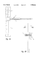
- FIG. 7 illustrates a front view of the device with the telescoping pointer at an angle.
- FIG. 1 A perspective view of a device according to the invention is shown in FIG. 1.
- a substantially straight bar 1 supports a telescoping pointer 2 which is rotatably attached to the bar.
- Bubble-type indicating means 3 is integrally formed within the telescoping pointer.
- FIG. 2 shows a front view and a side view of the device.
- Bar 1 has a slot 9 integrally formed therein, and a graduated scale in centimeters is etched or otherwise permanently affixed to an edge of bar 1.
- the graduated scale begins at a bottom portion of the bar and increases along the length of the bar toward a top portion.
- Pointer 2 is shown in FIG. 1 and FIG. 2 and is comprised of a telescoping member with a distal end, a pivoting base 10, and bubble-type indicating means 3 formed integrally therewith.
- the bubble-type indicating means indicates when the pointer 2 is in a horizontal position.
- the telescoping distal portion is comprised of a plurality of interfitting cylinders of progressively decreasing diameter, wherein each adjacent cylinder fits inside the preceding cylinder. The last cylinder extends furthest from the pivoting base and is formed with a blunt cap on the furthest distal end.
- Pivoting base 10 shown in FIG. 4 has a threaded hole 12 in the center thereof and a plurality of circular depressions 11 spaced equidistantly from the center hole 12 along a circular centerline.
- the depressions face detent bracket 8 which is slidably positioned in slot 9, shown in FIG. 2.
- the detent bracket detailed in FIG. 3, is integrally formed with rudder 15 which intersects slot 9 in slidable fashion such that the detent bracket may slide along the length of bar 1.
- Detent 13 is formed inside the detent bracket and is comprised of a hole inside the bracket in which a spring is set which forces a ball bearing toward the surface of the detent bracket such that the ball bearing protrudes slightly through the surface of the bracket.
- Detent 13 faces depressions 11 when pivoting base 10 is held against detent bracket 8 as shown in FIG. 2.
- the detent bracket 8 and the base 10 are urged against each other by retaining screw 7 which is slidably received both in slot 9 and in hole 16 of bracket 8 and fixedly received in threaded hole 12 of pivoting base 10.
- the retaining screw head is held in place by spring 6 and washer 5 on the side of the bar opposite that on which pivoting base 10 is located.
- Spring 6 biases the retaining screw head away from bar 1 such that pivoting base 10 and detent bracket 8 are held against bar 1 in a rigid fashion.
- Detent 13 intersects with one of depressions 11 to rigidly maintain the pivoting base and pointer 2 at a prescribed angle with respect to bar 1. The positioning of depressions 11 define where the detent will engage and therefore the angles at which pointer 2 may be held.
- Detent bracket 8 and pivoting base 10 together may be pushed along the length of the slot to adjust the height of the pointer.
- the force of spring 6 holds this assembly in its final position.
- Detent 13 may be disengaged from pivoting base 10 by pulling the pivoting base outward such that spring 6 is compressed.
- the pivoting base may be rotated about the axis of retaining screw 7 until the desired angle is reached at which point the pivoting base is released allowing spring 6 to force the rigid engagement of detent 13 and one of depressions 11.
- a physician first places the pointer at an angle, typically 45 degrees with respect to the bar, and the bar is placed parallel to the frame of a bed. The head of the bed is adjusted until the bubble-type indicating means indicates that the pointer is horizontal and the head of the bed is elevated at a 45 degree angle.
- the pointer is placed at a 90 degree angle with respect to the bar by engaging the detent at the 90 degree recess and the bottom portion of the bar is positioned on the sternal angle such that the bubble-type indicating means indicates that the pointer is horizontal and the bar is vertical.
- the telescoping distal end of the pointer is then extended to a position close to the patient's neck and the height of the pointer is adjusted by sliding the pivoting base along the length of the graduated bar.
- the distance between the sternal angle and the height of highest visible pulsations in the internal jugular vein is then gauged according to the graduated scale.
- the device is intended for external, non-invasive use only and should not come in contact with bodily fluids during the above-described procedure. For that reason the device may be used repeatedly without need of sterilization. Should the device come into contact with any form of contaminant a simple alcohol wash or other sterilization method would allow the immediate reuse of the device.
- the device may be constructed of any rigid material which can withstand the stresses of the above-described procedure and maintain structural integrity to enable accurate estimations of central venous pressure.
- the device material should also be able to withstand a simple alcohol wash or other sterilization procedure.
- the bar, the base, and the detent bracket may be made of rigid plastic.
- the interfitting cylinders of the pointer may be comprised of metal. Those skilled in the art will recognize many other materials which would meet the structural and durability requirements of the device.
- the depressions 11 fixing the angle at which the telescoping pointer may be oriented with respect to the base can be set at various intervals.
- the method of estimating CVP described above may take place while the upper torso of the patient is at any substantial angle with respect the the horizontal.
- Dr. Bates, in A Guide to Physical Examination and History Taking, indicates that the measurement may be taken with the patient elevated at an angle of fifteen degrees or more.
- the depressions may therefore be set at fifteen, thirty, forty-five, sixty, and ninety degrees or any combination thereof, or in any combination of angles between these such that the device is most useful as a diagnostic tool.
- the mechanical means for linking the telescoping pointer to the bar may be comprised in various ways such that the pivoting base of the pointer is movable along the length of the bar and such that the pointer may be set at various angles with respect to the bar.
- there may be an indentation rather than a slot in the bar such that the pivoting base of said pointer clamps onto the bar and is movable along the length of the bar.
- the vertical measurement along the graduated scale affixed to the bar may be aided by an electronic sensor indicating the height of the pointer with respect to the bottom portion of the bar.
Abstract
A device capable of providing non-invasive, accurate estimates of central venous pressure. This diagnostic device is particularly useful with patients suffering from cardiac disease.
Description
This application is a division of U.S. patent application Ser. No. 08/565,444, filed Nov. 30, 1995.
1. Field of the Invention
The present invention relates to a novel device for estimating central venous pressure.
2. Description of the Related Art
An accurate, quick, and non-invasive means for estimating central venous pressure (hereinafter CVP) is a valuable diagnostic tool for patients suffering from cardiac disease. In a patient suffering from cardiac disease the heart fails to pump sufficient volume through the circulatory system, and this causes blood to "back up" in the veins returning blood to the heart. Excess blood volume which is not pumped through by the heart muscle causes blood pressure in the veins, or venous pressure, to increase which results in distension or expansion of the patient's veins.
A precise measurement of CVP, or the blood pressure in the right atrium of the heart, is often necessary for the diagnosis of a patient with cardiac disease. The most reliable and accurate method of measurement involves the insertion of a catheter into one of the major veins in the circulatory system. The catheter is threaded through the vein until it reaches a position near the heart where a pressure measurement is taken. Such a procedure is invasive, medically risky, and highly traumatic for the patient.
Invasive measures which establish CVP are used only when an accurate measurement is absolutely necessary. Non-invasive methods are used when an estimate of CVP is sufficient for diagnosis. A simple method for estimating CVP is described in B. Bates, L. Bickley & R. Hoekelman, A Guide to Physical Examination and History Taking 270-271 (1995) and in J. Constant, Bedside Cardiology 80-86 (1985). The cited pages are incorporated herein by way of reference.
The physiological basis for the method is that in a healthy individual who is standing or sitting the blood pressure in the internal jugular vein, which is located behind the sternomastoid muscle in the neck, is lower than atmospheric pressure because gravity aids the movement of blood toward the heart. The internal jugular vein in this situation is normally partially collapsed. When blood "backs up" in the veins due to cardiac disease the internal jugular vein expands and pulsations in the vein are visible on the surface of the neck. The visible pulsations begin at the base of the neck and progress upward as CVP increases. A measurement of the height of the highest location on the neck where pulsations are visible provides a simple and non-invasive method for estimating CVP. The internal jugular vein is thereby used as a manometer to estimate CVP.
The above-mentioned procedure comprised the following steps: the head of the patient's bed is placed at an elevated angle, pulsations in the internal jugular vein are located, and the highest point at which these pulsations are visible is measured. Conventionally, the height of this point is measured from the sternal angle, also called the angle of Louis, which is a reference point on the sternum. The sternal angle is roughly 5 centimeters above the right atrium. The height measurement is taken by placing the base of a centimeter ruler on the sternal angle while the ruler is held in a vertical orientation. A tongue depressor or other straight object is then placed at a right angle with respect to the ruler and is used to locate the highest visible pulsations. A physician may also merely estimate the height visually. If the highest visible pulsations are more than a specified number of centimeters above the sternal angle then the CVP is considered elevated. A diagram indicating this method is shown in FIG. 5.
Several other methods or devices may be used to estimate CVP. Rockwell, in U.S. Pat. No. 3,413,970, discloses an integral, one piece arm which supports a slidable member. The slidable member is extended to a prescribed position under a patient to establish the vertical position of the patient's superior vena cava, after which a scale on the arm may be used to set a baseline for a manometer. The arm includes a level indicating means afixed thereto. The CVP measurement is invasive. Sackner, in U.S. Pat. Nos. 4,452,252, 4,456,015, 4,986,277, and 5,040,540, discloses several transducers which are wrapped around a patient's neck to measure changes in the cross-sectional area of the neck. Those changes are related to CVP. Allocca, in U.S. Pat. No. 4,204,547, discloses a method of monitoring intracranial pressure by occluding the jugular vein at a particular point and measuring the rate of change of blood pressure upstream of that point. Soviet Patent No. SU-452332-A discloses the measurement of venous pressure while the patient's inclined position is raised.
The need remains for an accurate and non-invasive method for estimating central venous pressure.
The object of this invention is to provide a device which enables an accurate and non-invasive estimation of central venous pressure. A device according to the invention is comprised of a substantially straight bar having a bottom portion and a top portion. A graduated scale is afixed to or etched into the surface of the bar beginning approximately at the bottom portion. A pointer comprising a pivoting base, a telescoping member and a distal end is movably attached to said bar at its pivoting end such that the pointer slides along the length of the bar. The pointer further comprises a bubble-type indicator means formed integrally therewith. The distal end of the pointer extends in a straight manner from the bar and is extended by the telescoping member. The pointer is rotatable with respect to the bar such that the angle between the bar and the pointer is adjustable. Detents in the pivoting base fix and hold the pointer at several preset angles.
FIG. 1 illustrates a perspective view of the device;
FIG. 2 illustrates a front view and a side view of the device;
FIG. 3 illustrates two views of the detent bracket;
FIG. 4 illustrates two views of the pivoting base of the pointer;
FIG. 5 illustrates the conventional method of estimating central venous pressure in a non-invasive manner;
FIG. 6 illustrates a sectional view of the bar and detent bracket;
FIG. 7 illustrates a front view of the device with the telescoping pointer at an angle.
A perspective view of a device according to the invention is shown in FIG. 1. A substantially straight bar 1 supports a telescoping pointer 2 which is rotatably attached to the bar. Bubble-type indicating means 3 is integrally formed within the telescoping pointer.
FIG. 2 shows a front view and a side view of the device. Bar 1 has a slot 9 integrally formed therein, and a graduated scale in centimeters is etched or otherwise permanently affixed to an edge of bar 1. The graduated scale begins at a bottom portion of the bar and increases along the length of the bar toward a top portion.
Pivoting base 10 shown in FIG. 4 has a threaded hole 12 in the center thereof and a plurality of circular depressions 11 spaced equidistantly from the center hole 12 along a circular centerline. The depressions face detent bracket 8 which is slidably positioned in slot 9, shown in FIG. 2. The detent bracket, detailed in FIG. 3, is integrally formed with rudder 15 which intersects slot 9 in slidable fashion such that the detent bracket may slide along the length of bar 1. Detent 13 is formed inside the detent bracket and is comprised of a hole inside the bracket in which a spring is set which forces a ball bearing toward the surface of the detent bracket such that the ball bearing protrudes slightly through the surface of the bracket. When pressure is exerted on the ball bearing against the force of the spring the ball bearing recedes below the surface of the bracket. The ball bearing returns to protrude from the surface when said pressure is released. Detent 13 faces depressions 11 when pivoting base 10 is held against detent bracket 8 as shown in FIG. 2.
The detent bracket 8 and the base 10 are urged against each other by retaining screw 7 which is slidably received both in slot 9 and in hole 16 of bracket 8 and fixedly received in threaded hole 12 of pivoting base 10. As shown in FIG. 6 the retaining screw head is held in place by spring 6 and washer 5 on the side of the bar opposite that on which pivoting base 10 is located. Spring 6 biases the retaining screw head away from bar 1 such that pivoting base 10 and detent bracket 8 are held against bar 1 in a rigid fashion. Detent 13 intersects with one of depressions 11 to rigidly maintain the pivoting base and pointer 2 at a prescribed angle with respect to bar 1. The positioning of depressions 11 define where the detent will engage and therefore the angles at which pointer 2 may be held. Detent bracket 8 and pivoting base 10 together may be pushed along the length of the slot to adjust the height of the pointer. The force of spring 6 holds this assembly in its final position. Detent 13 may be disengaged from pivoting base 10 by pulling the pivoting base outward such that spring 6 is compressed. The pivoting base may be rotated about the axis of retaining screw 7 until the desired angle is reached at which point the pivoting base is released allowing spring 6 to force the rigid engagement of detent 13 and one of depressions 11.
To use the device, a physician first places the pointer at an angle, typically 45 degrees with respect to the bar, and the bar is placed parallel to the frame of a bed. The head of the bed is adjusted until the bubble-type indicating means indicates that the pointer is horizontal and the head of the bed is elevated at a 45 degree angle. Next, the pointer is placed at a 90 degree angle with respect to the bar by engaging the detent at the 90 degree recess and the bottom portion of the bar is positioned on the sternal angle such that the bubble-type indicating means indicates that the pointer is horizontal and the bar is vertical.
This is the critical step in the procedure in that an accurate estimate of the height of visible pulsations is based on the bar being in a vertical orientation and the pointer being in a horizontal orientation. The bubble-type indicating means enables the user to fix the pointer along a true horizontal and the 90 degree detent rigidly holds the pointer at a 90 degree angle with respect to the bar.
The telescoping distal end of the pointer is then extended to a position close to the patient's neck and the height of the pointer is adjusted by sliding the pivoting base along the length of the graduated bar. The distance between the sternal angle and the height of highest visible pulsations in the internal jugular vein is then gauged according to the graduated scale. The device enables a more accurate and repeatable estimation of central venous pressure in a non-invasive, risk-free manner, resulting in improved patient care.
The device is intended for external, non-invasive use only and should not come in contact with bodily fluids during the above-described procedure. For that reason the device may be used repeatedly without need of sterilization. Should the device come into contact with any form of contaminant a simple alcohol wash or other sterilization method would allow the immediate reuse of the device.
The device may be constructed of any rigid material which can withstand the stresses of the above-described procedure and maintain structural integrity to enable accurate estimations of central venous pressure. The device material should also be able to withstand a simple alcohol wash or other sterilization procedure. For example, the bar, the base, and the detent bracket may be made of rigid plastic. The interfitting cylinders of the pointer may be comprised of metal. Those skilled in the art will recognize many other materials which would meet the structural and durability requirements of the device.
While the above description contains many specifics, these specifics should not be construed as limitations on the scope of the invention, but merely as exemplifications of preferred embodiments thereof. Those skilled in the art will envision many other possible variations that are within the scope and spirit of the invention as defined by the claims appended hereto.
For example, the depressions 11 fixing the angle at which the telescoping pointer may be oriented with respect to the base can be set at various intervals. The method of estimating CVP described above may take place while the upper torso of the patient is at any substantial angle with respect the the horizontal. Dr. Bates, in A Guide to Physical Examination and History Taking, indicates that the measurement may be taken with the patient elevated at an angle of fifteen degrees or more. The depressions may therefore be set at fifteen, thirty, forty-five, sixty, and ninety degrees or any combination thereof, or in any combination of angles between these such that the device is most useful as a diagnostic tool.
In other embodiments within the scope of the invention the mechanical means for linking the telescoping pointer to the bar may be comprised in various ways such that the pivoting base of the pointer is movable along the length of the bar and such that the pointer may be set at various angles with respect to the bar. For example, there may be an indentation rather than a slot in the bar such that the pivoting base of said pointer clamps onto the bar and is movable along the length of the bar.
The vertical measurement along the graduated scale affixed to the bar may be aided by an electronic sensor indicating the height of the pointer with respect to the bottom portion of the bar.
Claims (15)
1. A method for estimating central venous pressure in a human patient having a neck, an internal jugular vein in the neck, and a chest, the method comprising
elevating the neck of the patient;
placing an apparatus for estimating central venous pressure on a reference point on the chest, wherein the apparatus comprises:
an elongated bar having a longitudinal axis and a bottom portion on the longitudinal axis, a pointer comprising an elongated base member having a longitudinal axis and an elongated end member having a longitudinal axis, the base member being movably mounted to the bar and the end member being supported for selected movement along a path substantially parallel to the base member longitudinal axis, and a horizontal indicator mounted on the pointer;
positioning the base member longitudinal axis at a ninety degree angle with respect to the bar longitudinal axis;
placing the pointer in a horizontal attitude indicated by the horizontal indicator;
positioning the bottom portion of the bar on the reference point;
identifying a point of highest visible pulsations in the internal jugular vein;
extending the end member from the base member and moving the pointer relative to the bar such that a distal end of the end member is proximate to the point of highest visible pulsations in the internal jugular vein;
measuring a vertical distance between the reference point and an intersection of the pointer and the bar to estimate the central venous pressure.
2. The methods of claim 1 wherein the step of positioning the bottom portion of the bar comprises positioning the bottom portion of the bar on a sternal angle of the chest.
3. A method for estimation central venous pressure in a human patient having a neck, and internal jugular vein in the neck, and a chest, the method comprising:
elevating the neck of the patient above the chest;
providing an apparatus having an elongated bar with a longitudinal axis and a bottom portion and an extendable pointer translatably mounted to the bar;
placing the apparatus on a reference point on the chest of the patient;
positioning a longitudinal axis of the pointer at a ninety degree angle with respect to the bar longitudinal axis;
placing said pointer in a horizontal attitude;
identifying a point of highest visible pulsations in the internal jugular vein;
positioning the bottom portion of the bar on the reference point;
extending the pointer such that a distal end of the pointer is proximate to the neck;
moving the pointer relative to the bar to a position such that the distal end is proximate to the point of highest visible pulsations in the internal jugular vein;
measuring a vertical distance between the reference point and a position of the pointer along the longitudinal axis of the bar to estimate central venous pressure.
4. The method of claim 3 wherein the step of positioning the bottom portion comprises positioning the bottom portion of the bar on a sternal angle on the chest.
5. A method for estimating central venous pressure in a human patient having a neck, an internal jugular vein in the neck, and a chest having a sternal angle, the method comprising;
providing a device having an elongated bar with a longitudinal axis and a bottom portion, the bar having a first cross-sectional area in a plane perpendicular to its longitudinal axis, the bottom portion being on the bar longitudinal axis, the device including a pointer having an elongated base member with a longitudinal axis and an elongated end member with a longitudinal axis, the end member having a second cross-sectional area in a plane perpendicular to its longitudinal axis which is less than the first cross-sectional area, the base member being mounted to the bar, the end member being supported by the base member and being extendible from a retracted position to an extended position substantially parallel to the base member longitudinal axis, the device also including a level indicator mounted on the pointer, the level indicator being structured to indicate when the pointer is in a horizontal attitude;
situating the patient in a reclining position such that the neck is elevated from the sternal angle;
positioning the bottom portion of the bar proximate to the sternal angle, observing a point of highest visible pulsations in the internal jugular vein;
positioning the pointer in a horizontal attitude;
extending the end member to a location proximate to the neck;
moving the base member with respect to the bar to a position such that the end member is proximate to the point of highest visible pulsations in the internal jugular vein; and
gauging a distance along the bar longitudinal axis between the sternal angle and the pointer to estimate the central venous pressure.
6. The method of claim 5 wherein the step of positioning the pointer in a horizontal attitude comprises:
positioning the base member longitudinal axis at a ninety degree angle with respect to the bar longitudinal axis; and
positioning the bar such that the level indicator indicates that the pointer is in a horizontal attitude.
7. The method of claim 6 wherein the step of extending the end member comprises extending the end member substantially parallel to the base member longitudinal axis to a position such that a distal end of the end member is proximate to the neck.
8. The method of claim 7 wherein the step of moving the base member comprises sliding the base member along the bar longitudinal axis until the distal end of the end member is proximate to the point of highest visible pulsations in the internal jugular vein.
9. The method of claim 8 wherein the step of gauging a distance further comprises reading a graduated scale between the bottom portion of the bar and an intersection between the base member and the bar.
10. A method for estimating central venous pressure in a human patient having a neck, an internal jugular vein in the neck, and a chest, the method comprising:
elevating the neck above the chest;
locating a bottom portion of an elongated bar at a selected location on the chest;
positioning an elongated pointer translatably mounted to the bar in a horizontal attitude and at a right angle with respect to a longitudinal axis of the bar;
observing a point of highest visible pulsations in the internal jugular vein;
extending the pointer to a location proximate to the neck;
moving the pointer relative to the bar so that the pointer is proximate to the point of highest visible pulsations in the internal jugular vein; and
gauging a distance between the selected location on the chest and an intersection between the pointer and the bar to estimate the central venous pressure of the patient.
11. The method of claim 10 wherein the step of locating a bottom portion of an elongated bar comprises locating a bottom portion of an elongated bar proximate to a sternal angle of the patient.
12. The method of claim 11 wherein the step of positioning an elongated pointer comprises:
positioning a longitudinal axis of the pointer at a ninety degree angle with respect to the bar longitudinal axis; and
positioning the bar such that a level indicator mounted on the pointer indicates that the pointer is in a horizontal attitude.
13. The method of claim 12 wherein the step of extending the pointer comprises extending an elongated end member of the pointer substantially parallel to the pointer longitudinal axis to a position such that a distal end of the end member is proximate to the neck.
14. The method of claim 13 wherein the step of moving the pointer comprises moving the pointer relative to the bar such that the distal end of the end member is proximate to the point of highest visible pulsations in the internal jugular vein.
15. The method of claim 11 wherein the step of estimating a distance comprises estimating a vertical distance along the bar longitudinal axis between the sternal angle and an intersection between the pointer and the bar.
Priority Applications (1)
| Application Number | Priority Date | Filing Date | Title |
|---|---|---|---|
| US08/951,432 US5788641A (en) | 1995-11-30 | 1997-10-16 | Device for estimating central venous pressure |
Applications Claiming Priority (2)
| Application Number | Priority Date | Filing Date | Title |
|---|---|---|---|
| US08/565,444 US5904142A (en) | 1995-11-30 | 1995-11-30 | Device for estimating central venous pressure |
| US08/951,432 US5788641A (en) | 1995-11-30 | 1997-10-16 | Device for estimating central venous pressure |
Related Parent Applications (1)
| Application Number | Title | Priority Date | Filing Date |
|---|---|---|---|
| US08/565,444 Division US5904142A (en) | 1995-11-30 | 1995-11-30 | Device for estimating central venous pressure |
Publications (1)
| Publication Number | Publication Date |
|---|---|
| US5788641A true US5788641A (en) | 1998-08-04 |
Family
ID=24258628
Family Applications (2)
| Application Number | Title | Priority Date | Filing Date |
|---|---|---|---|
| US08/565,444 Expired - Fee Related US5904142A (en) | 1995-11-30 | 1995-11-30 | Device for estimating central venous pressure |
| US08/951,432 Expired - Fee Related US5788641A (en) | 1995-11-30 | 1997-10-16 | Device for estimating central venous pressure |
Family Applications Before (1)
| Application Number | Title | Priority Date | Filing Date |
|---|---|---|---|
| US08/565,444 Expired - Fee Related US5904142A (en) | 1995-11-30 | 1995-11-30 | Device for estimating central venous pressure |
Country Status (1)
| Country | Link |
|---|---|
| US (2) | US5904142A (en) |
Cited By (15)
| Publication number | Priority date | Publication date | Assignee | Title |
|---|---|---|---|---|
| GB2408935A (en) * | 2003-12-08 | 2005-06-15 | Rizwan Uppal | Central venous pressure meter |
| US20070239041A1 (en) * | 2006-03-28 | 2007-10-11 | The Johns Hopkins University | Non-invasive Venous Pressure Measurement |
| US20070270720A1 (en) * | 2006-05-04 | 2007-11-22 | Fry William R | Noninvasive physiologic pressure measurement |
| US20080200784A1 (en) * | 2007-02-16 | 2008-08-21 | Xuefeng Cheng | Method and device for measuring parameters of cardiac function |
| US20080294070A1 (en) * | 2007-05-27 | 2008-11-27 | Michael Kinori | Jugular venous pressure gauge |
| US7591074B1 (en) * | 2009-01-20 | 2009-09-22 | Richard Allen Potts | Medical laser vertical alignment system |
| US20100094141A1 (en) * | 2008-10-14 | 2010-04-15 | Amal Lesly Puswella | Jugular venous pressure ruler |
| US8918153B2 (en) | 2007-02-16 | 2014-12-23 | Mespere Lifesciences Inc. | Method and device for measuring parameters of cardiac function |
| WO2018039625A1 (en) * | 2016-08-26 | 2018-03-01 | Biro Mark Phillip | Jugular venous distention measurement device |
| CN108618767A (en) * | 2017-03-24 | 2018-10-09 | 扬州大学附属医院 | Noninvasive centre venous pressure measuring rule |
| US10149624B2 (en) * | 2014-11-06 | 2018-12-11 | Koninklijke Philips N.V. | Method and device for measuring intracranial pressure, ICP, in a subject |
| US10398364B2 (en) | 2013-02-13 | 2019-09-03 | Mespere Lifesciences Inc. | Method and device for measuring venous blood oxygenation |
| US11096598B2 (en) | 2015-10-08 | 2021-08-24 | Mespere Lifesciences Inc. | System and method for non-invasive monitoring of central venous pressure |
| US11234643B2 (en) | 2015-06-21 | 2022-02-01 | Yaakov Nahmias | Jugular venous assessment |
| US11872328B2 (en) | 2017-08-30 | 2024-01-16 | Hadasit Medical Research Services And Development Ltd. | Devices, kits and methods for reducing and/or preventing intra-abdominal adhesions |
Families Citing this family (6)
| Publication number | Priority date | Publication date | Assignee | Title |
|---|---|---|---|---|
| US7299560B2 (en) * | 2005-02-15 | 2007-11-27 | Topline Innovarions, Llc | Multi-purpose tool |
| US9316475B2 (en) * | 2014-02-04 | 2016-04-19 | Chia-Hsin Liu | Height measurement device |
| CN103815887B (en) * | 2014-02-27 | 2015-12-30 | 河南科技大学第一附属医院 | Centre venous pressure measuring rule |
| US10080528B2 (en) * | 2015-05-19 | 2018-09-25 | Google Llc | Optical central venous pressure measurement |
| CN108309269B (en) * | 2018-04-12 | 2020-10-27 | 青岛大学附属医院 | Vernier ruler for measuring central venous pressure |
| IT201900010248A1 (en) | 2019-06-27 | 2020-12-27 | Tre Esse Progettazione Biomedica S R L | DEVICE FOR THE DETECTION OF VENOUS PRESSURE |
Citations (22)
| Publication number | Priority date | Publication date | Assignee | Title |
|---|---|---|---|---|
| US3413970A (en) * | 1967-04-27 | 1968-12-03 | Paul E. Rockwell | Accessory instrument for the measurement of central venous pressure |
| US3602214A (en) * | 1968-03-05 | 1971-08-31 | Seymour B London | Method of phantom level sensing in a central venous pressure monitoring system |
| SU452332A1 (en) * | 1973-05-16 | 1974-12-05 | Войсковая Часть 64688 | Method for measuring venous pressure in the strap vein system |
| US4204547A (en) * | 1978-11-13 | 1980-05-27 | Allocca John A | Method and apparatus for noninvasive monitoring of intracranial pressure |
| US4348815A (en) * | 1980-11-13 | 1982-09-14 | Hurt Alfred A | Multi-purpose square |
| US4399616A (en) * | 1980-10-17 | 1983-08-23 | Olle Humble | Telescoping measuring rule |
| US4451993A (en) * | 1983-07-05 | 1984-06-05 | Yauk Alvin R | Sliding rule and protractor |
| US4452252A (en) * | 1981-05-26 | 1984-06-05 | Respitrace Corporation | Non-invasive method for monitoring cardiopulmonary parameters |
| US4456015A (en) * | 1981-05-26 | 1984-06-26 | Respitrace Corporation | Non-invasive method for semiquantitative measurement of neck volume changes |
| US4554746A (en) * | 1984-07-03 | 1985-11-26 | Luis Echeverria | Self-locking-and-measuring ruler for computer-printout forms |
| US4566462A (en) * | 1983-11-21 | 1986-01-28 | School Of Medicine Texas Tech. Univ. Health Servcs. Ctr. | Venous pressure measuring method and apparatus |
| US4679567A (en) * | 1986-02-04 | 1987-07-14 | Deseret Medical, Inc. | Pressure transducer |
| US4798588A (en) * | 1984-12-03 | 1989-01-17 | Rene Aillon | Central venous pressure catheter and method for using |
| US4813149A (en) * | 1987-12-01 | 1989-03-21 | Herkimer Robert D | Level device |
| SU1477377A1 (en) * | 1985-05-20 | 1989-05-07 | Предприятие П/Я Р-6681 | Device for measuring venous pressure |
| EP0359972A1 (en) * | 1988-08-12 | 1990-03-28 | A. Nattermann & Cie. GmbH | Device for non-invasively determining flow parameters in human limbs |
| US4986277A (en) * | 1988-08-24 | 1991-01-22 | Sackner Marvin A | Method and apparatus for non-invasive monitoring of central venous pressure |
| US5040540A (en) * | 1988-08-24 | 1991-08-20 | Nims, Inc. | Method and apparatus for non-invasive monitoring of central venous pressure, and improved transducer therefor |
| WO1992022871A1 (en) * | 1991-06-10 | 1992-12-23 | Akinyemi Samuel Nathaniel Olal | Non-invasive measurement of blood flow parameters |
| US5280789A (en) * | 1992-01-31 | 1994-01-25 | Potts Richard A | Apparatus and method for measuring fluid pressure in a medical patient |
| US5353509A (en) * | 1993-07-27 | 1994-10-11 | Black Daniel C | Multi-purpose T-square and level combination tool |
| US5446969A (en) * | 1993-06-23 | 1995-09-05 | Terenzoni; Robert | Combination square and multi-purpose hand tool |
-
1995
- 1995-11-30 US US08/565,444 patent/US5904142A/en not_active Expired - Fee Related
-
1997
- 1997-10-16 US US08/951,432 patent/US5788641A/en not_active Expired - Fee Related
Patent Citations (22)
| Publication number | Priority date | Publication date | Assignee | Title |
|---|---|---|---|---|
| US3413970A (en) * | 1967-04-27 | 1968-12-03 | Paul E. Rockwell | Accessory instrument for the measurement of central venous pressure |
| US3602214A (en) * | 1968-03-05 | 1971-08-31 | Seymour B London | Method of phantom level sensing in a central venous pressure monitoring system |
| SU452332A1 (en) * | 1973-05-16 | 1974-12-05 | Войсковая Часть 64688 | Method for measuring venous pressure in the strap vein system |
| US4204547A (en) * | 1978-11-13 | 1980-05-27 | Allocca John A | Method and apparatus for noninvasive monitoring of intracranial pressure |
| US4399616A (en) * | 1980-10-17 | 1983-08-23 | Olle Humble | Telescoping measuring rule |
| US4348815A (en) * | 1980-11-13 | 1982-09-14 | Hurt Alfred A | Multi-purpose square |
| US4452252A (en) * | 1981-05-26 | 1984-06-05 | Respitrace Corporation | Non-invasive method for monitoring cardiopulmonary parameters |
| US4456015A (en) * | 1981-05-26 | 1984-06-26 | Respitrace Corporation | Non-invasive method for semiquantitative measurement of neck volume changes |
| US4451993A (en) * | 1983-07-05 | 1984-06-05 | Yauk Alvin R | Sliding rule and protractor |
| US4566462A (en) * | 1983-11-21 | 1986-01-28 | School Of Medicine Texas Tech. Univ. Health Servcs. Ctr. | Venous pressure measuring method and apparatus |
| US4554746A (en) * | 1984-07-03 | 1985-11-26 | Luis Echeverria | Self-locking-and-measuring ruler for computer-printout forms |
| US4798588A (en) * | 1984-12-03 | 1989-01-17 | Rene Aillon | Central venous pressure catheter and method for using |
| SU1477377A1 (en) * | 1985-05-20 | 1989-05-07 | Предприятие П/Я Р-6681 | Device for measuring venous pressure |
| US4679567A (en) * | 1986-02-04 | 1987-07-14 | Deseret Medical, Inc. | Pressure transducer |
| US4813149A (en) * | 1987-12-01 | 1989-03-21 | Herkimer Robert D | Level device |
| EP0359972A1 (en) * | 1988-08-12 | 1990-03-28 | A. Nattermann & Cie. GmbH | Device for non-invasively determining flow parameters in human limbs |
| US4986277A (en) * | 1988-08-24 | 1991-01-22 | Sackner Marvin A | Method and apparatus for non-invasive monitoring of central venous pressure |
| US5040540A (en) * | 1988-08-24 | 1991-08-20 | Nims, Inc. | Method and apparatus for non-invasive monitoring of central venous pressure, and improved transducer therefor |
| WO1992022871A1 (en) * | 1991-06-10 | 1992-12-23 | Akinyemi Samuel Nathaniel Olal | Non-invasive measurement of blood flow parameters |
| US5280789A (en) * | 1992-01-31 | 1994-01-25 | Potts Richard A | Apparatus and method for measuring fluid pressure in a medical patient |
| US5446969A (en) * | 1993-06-23 | 1995-09-05 | Terenzoni; Robert | Combination square and multi-purpose hand tool |
| US5353509A (en) * | 1993-07-27 | 1994-10-11 | Black Daniel C | Multi-purpose T-square and level combination tool |
Non-Patent Citations (4)
| Title |
|---|
| Bates, Barbara et al., A Guide to Physical Examination and History Taking, Sixth Edition, 1995, pp. 270 271. * |
| Bates, Barbara et al., A Guide to Physical Examination and History Taking, Sixth Edition, 1995, pp. 270-271. |
| Constant, Jules M.D., Bedside Cardiology, Third Edition, 1985, pp. 80 86. * |
| Constant, Jules M.D., Bedside Cardiology, Third Edition, 1985, pp. 80-86. |
Cited By (20)
| Publication number | Priority date | Publication date | Assignee | Title |
|---|---|---|---|---|
| GB2408935B (en) * | 2003-12-08 | 2008-09-10 | Rizwan Uppal | Central venous pressure meter |
| GB2408935A (en) * | 2003-12-08 | 2005-06-15 | Rizwan Uppal | Central venous pressure meter |
| US20070239041A1 (en) * | 2006-03-28 | 2007-10-11 | The Johns Hopkins University | Non-invasive Venous Pressure Measurement |
| US20070270720A1 (en) * | 2006-05-04 | 2007-11-22 | Fry William R | Noninvasive physiologic pressure measurement |
| EP2120689A4 (en) * | 2007-02-16 | 2011-06-29 | Mespere Lifesciences Inc | Method and device for measuring parameters of cardiac function |
| US20080200784A1 (en) * | 2007-02-16 | 2008-08-21 | Xuefeng Cheng | Method and device for measuring parameters of cardiac function |
| US8918153B2 (en) | 2007-02-16 | 2014-12-23 | Mespere Lifesciences Inc. | Method and device for measuring parameters of cardiac function |
| EP2120689A1 (en) * | 2007-02-16 | 2009-11-25 | Mespere Lifescience Inc. | Method and device for measuring parameters of cardiac function |
| US20090326352A1 (en) * | 2007-02-16 | 2009-12-31 | Xuefeng Cheng | Method and device for measuring parameters of cardiac function |
| US8417306B2 (en) | 2007-02-16 | 2013-04-09 | Mespere Lifesciences Inc. | Method and device for measuring parameters of cardiac function |
| US20080294070A1 (en) * | 2007-05-27 | 2008-11-27 | Michael Kinori | Jugular venous pressure gauge |
| US20100094141A1 (en) * | 2008-10-14 | 2010-04-15 | Amal Lesly Puswella | Jugular venous pressure ruler |
| US7591074B1 (en) * | 2009-01-20 | 2009-09-22 | Richard Allen Potts | Medical laser vertical alignment system |
| US10398364B2 (en) | 2013-02-13 | 2019-09-03 | Mespere Lifesciences Inc. | Method and device for measuring venous blood oxygenation |
| US10149624B2 (en) * | 2014-11-06 | 2018-12-11 | Koninklijke Philips N.V. | Method and device for measuring intracranial pressure, ICP, in a subject |
| US11234643B2 (en) | 2015-06-21 | 2022-02-01 | Yaakov Nahmias | Jugular venous assessment |
| US11096598B2 (en) | 2015-10-08 | 2021-08-24 | Mespere Lifesciences Inc. | System and method for non-invasive monitoring of central venous pressure |
| WO2018039625A1 (en) * | 2016-08-26 | 2018-03-01 | Biro Mark Phillip | Jugular venous distention measurement device |
| CN108618767A (en) * | 2017-03-24 | 2018-10-09 | 扬州大学附属医院 | Noninvasive centre venous pressure measuring rule |
| US11872328B2 (en) | 2017-08-30 | 2024-01-16 | Hadasit Medical Research Services And Development Ltd. | Devices, kits and methods for reducing and/or preventing intra-abdominal adhesions |
Also Published As
| Publication number | Publication date |
|---|---|
| US5904142A (en) | 1999-05-18 |
Similar Documents
| Publication | Publication Date | Title |
|---|---|---|
| US5788641A (en) | Device for estimating central venous pressure | |
| Adams et al. | An electronic inclinometer technique for measuring lumbar curvature | |
| US10524672B2 (en) | Diastolic blood pressure measurement calibration | |
| Hunyor et al. | Comparison of performance of various sphygmomanometers with intra-arterial blood-pressure readings. | |
| US6132383A (en) | Apparatus for holding and positioning an arterial pulse pressure sensor | |
| EP2567656B1 (en) | Method and apparatus for determining the absolute value of intracranial pressure | |
| US8328727B2 (en) | Method and apparatus for assessing hemodynamic parameters within the circulatory system of a living subject | |
| US4483075A (en) | Apparatus and method for measuring deformed areas of skin surface | |
| EP3263020B1 (en) | Optical fiber type continuous blood pressure detection sensor and wearable device thereof | |
| US20030149369A1 (en) | Method and apparatus for non-invasively measuring hemodynamic parameters using parametrics | |
| US20070270720A1 (en) | Noninvasive physiologic pressure measurement | |
| US5235988A (en) | Device for evaluating surface contour | |
| JP2021191455A (en) | Device and method for detecting enhancement of at least one of expansion and movement of brain | |
| KR102206785B1 (en) | Method and apparatus for estimating aortic pulse wave transmission time from the time interval measured between the reference points of the heart trajectory | |
| US5156162A (en) | Scoliosis measurement instrument with midline leg member | |
| US5068886A (en) | Catheter or cannula position indicator for use in hemodynamic monitoring and the like | |
| KR20100126127A (en) | Blood pressure estimating apparatus and method by using variable characteristic ratio | |
| Eichna et al. | CAPILLARY BLOOD PRESSURE IN MAN. COMPARISON OF DIRECT AND INDIRECT METHODS OF MEASUREMENT. | |
| CN208740960U (en) | The measuring device of central venous pressure | |
| KR20100052608A (en) | Measuring apparatus and method for pulse wave | |
| WO2004058066A1 (en) | Device for measurement of tissue hardness | |
| EP3247279B1 (en) | Noninvasive fluid and electrolyte balance monitor | |
| WO1993004625A1 (en) | Noninvasive temporal artery blood pressure sensor assembly | |
| CN214965457U (en) | Pressure testing device for measuring soft tissue swelling degree | |
| CN219763373U (en) | Bone mineral density measuring position marking device |
Legal Events
| Date | Code | Title | Description |
|---|---|---|---|
| REMI | Maintenance fee reminder mailed | ||
| LAPS | Lapse for failure to pay maintenance fees | ||
| STCH | Information on status: patent discontinuation |
Free format text: PATENT EXPIRED DUE TO NONPAYMENT OF MAINTENANCE FEES UNDER 37 CFR 1.362 |
|
| FP | Lapsed due to failure to pay maintenance fee |
Effective date: 20020804 |