US4502159A - Tubular prostheses prepared from pericardial tissue - Google Patents
Tubular prostheses prepared from pericardial tissue Download PDFInfo
- Publication number
- US4502159A US4502159A US06/407,529 US40752982A US4502159A US 4502159 A US4502159 A US 4502159A US 40752982 A US40752982 A US 40752982A US 4502159 A US4502159 A US 4502159A
- Authority
- US
- United States
- Prior art keywords
- prosthesis
- pericardial tissue
- thread
- sheet
- tubular
- Prior art date
- Legal status (The legal status is an assumption and is not a legal conclusion. Google has not performed a legal analysis and makes no representation as to the accuracy of the status listed.)
- Expired - Fee Related
Links
Images


Classifications
-
- A—HUMAN NECESSITIES
- A61—MEDICAL OR VETERINARY SCIENCE; HYGIENE
- A61F—FILTERS IMPLANTABLE INTO BLOOD VESSELS; PROSTHESES; DEVICES PROVIDING PATENCY TO, OR PREVENTING COLLAPSING OF, TUBULAR STRUCTURES OF THE BODY, e.g. STENTS; ORTHOPAEDIC, NURSING OR CONTRACEPTIVE DEVICES; FOMENTATION; TREATMENT OR PROTECTION OF EYES OR EARS; BANDAGES, DRESSINGS OR ABSORBENT PADS; FIRST-AID KITS
- A61F2/00—Filters implantable into blood vessels; Prostheses, i.e. artificial substitutes or replacements for parts of the body; Appliances for connecting them with the body; Devices providing patency to, or preventing collapsing of, tubular structures of the body, e.g. stents
- A61F2/02—Prostheses implantable into the body
- A61F2/04—Hollow or tubular parts of organs, e.g. bladders, tracheae, bronchi or bile ducts
- A61F2/06—Blood vessels
-
- A—HUMAN NECESSITIES
- A61—MEDICAL OR VETERINARY SCIENCE; HYGIENE
- A61F—FILTERS IMPLANTABLE INTO BLOOD VESSELS; PROSTHESES; DEVICES PROVIDING PATENCY TO, OR PREVENTING COLLAPSING OF, TUBULAR STRUCTURES OF THE BODY, e.g. STENTS; ORTHOPAEDIC, NURSING OR CONTRACEPTIVE DEVICES; FOMENTATION; TREATMENT OR PROTECTION OF EYES OR EARS; BANDAGES, DRESSINGS OR ABSORBENT PADS; FIRST-AID KITS
- A61F2/00—Filters implantable into blood vessels; Prostheses, i.e. artificial substitutes or replacements for parts of the body; Appliances for connecting them with the body; Devices providing patency to, or preventing collapsing of, tubular structures of the body, e.g. stents
- A61F2/02—Prostheses implantable into the body
- A61F2/04—Hollow or tubular parts of organs, e.g. bladders, tracheae, bronchi or bile ducts
- A61F2002/048—Ureters
-
- Y—GENERAL TAGGING OF NEW TECHNOLOGICAL DEVELOPMENTS; GENERAL TAGGING OF CROSS-SECTIONAL TECHNOLOGIES SPANNING OVER SEVERAL SECTIONS OF THE IPC; TECHNICAL SUBJECTS COVERED BY FORMER USPC CROSS-REFERENCE ART COLLECTIONS [XRACs] AND DIGESTS
- Y10—TECHNICAL SUBJECTS COVERED BY FORMER USPC
- Y10S—TECHNICAL SUBJECTS COVERED BY FORMER USPC CROSS-REFERENCE ART COLLECTIONS [XRACs] AND DIGESTS
- Y10S623/00—Prosthesis, i.e. artificial body members, parts thereof, or aids and accessories therefor
- Y10S623/915—Method or apparatus for preparing biological material
- Y10S623/916—Blood vessel
Definitions
- Tubular prostheses made from natural tissue have been widely used in recent years in the surgical repair and replacement of diseased or damaged blood vessels in human patients.
- Natural tissue prostheses fall into three general classes. Autogenous material tissue prostheses are prepared from tissues taken from the patient's own body (e.g., saphenous vein grafts). Use of such prostheses eliminates the possibility of rejection of the implanted prosthesis, but requires a more extensive and time-consuming surgical intervention with attendant risks to the patient.
- Homologous natural tissue prostheses are prepared from tissue taken from another human, while heterologous natural tissue prostheses are prepared from tissue taken from another species.
- the use of homologous and heterologous umblical cord vessels as, e.g., vascular and ureteral prostheses is disclosed in U.S. Pat. Nos. 3,894,530; 3,974,526 and 3,988,782.
- the necessary characteristics of a tubular vascular prosthesis are biological compatibility, adequate strength, resistance to infection, resistance to biological degradation, non-thrombogenicity and lack of a tendency to promote aneurysm formation.
- biological compatibility means that the prosthesis is non-toxic in the in vivo environment of its intended use, and is not rejected by the patient's physiological system (i.e. is non-antigenic).
- the prosthesis be capable of production at an economical cost in a wide variety of lengths, diameters and shapes (e.g., straight, curved, bifurcated), be readily anastomosed to the patient's body and to other tubular prostheses of the same or different type, exhibit dimensional stability in use, and, in order to minimize hemodynamic turbulence and trama to the native vessel, have a compliance comparable to that of the patient's natural vessel that it is repairing or replacing (see discussion of compliance in U.S. Pat. No. 4,173,689).
- it is disadvantageous because of the risk of kinking to implant a tubular prosthesis that is too long for the intended application.
- a novel tubular prosthesis which comprises a sheet of pericardial tissue having opposed edges sewn together by means including a thread to form a longitudinal seam, with the thread being disposed in a configuration including a plurality of stitches extending along the seam, wherein the configuration of the thread is such that the prosthesis can be cut transversely between its ends, thereby severing the thread, without substantial damage to the seam (e.g., without causing the thread to unravel).
- each of said stitches may be secured with a knot tied in the thread after the stitch.
- the opposed edges of the sheet of pericardial tissue are sewn together to form an everted seam, and the smooth mesothelial side of the tissue is disposed luminally.
- the novel prosthesis of the invention may be of either the autogenous, homologous or heterologous type, with the latter preferred.
- the tubular prosthesis of the present invention preferably comprises a sheet of bovine pericardial tissue having opposed edges sewn together to form a longitudinal seam.
- the range of compliances of bovine pericardial tissue tubular prostheses is approximately comparable to the range in human arteries and veins, while porcine pericardial tissue tubular prostheses tend to be too compliant.
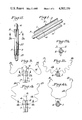
- FIGS. 1 to 8 illustrate several steps in the manufacture of a tubular vascular prosthesis 10 of the invention from a sheet of bovine pericardial tissue
- FIGS. 2A to 8A are transverse cross-sectional views of the prosthesis at the various steps in the manufacture thereof illustrated in FIGS. 2 to 8;
- FIG. 9 is a perspective view of the finished tubular prosthesis 10.
- FIG. 10 is an end elevation view of the finished tubular prosthesis 10
- FIG. 11 is an exploded front elevation view of a bifurcated mandrel for use in preparing a bifurcated tubular prosthesis of the invention.
- FIG. 12 is a front elevation view of a longitudinally tapered mandrel for use in preparing a longitudinally tapered prosthesis of the invention.
- the starting material in the manufacture of a tubular vascular prosthesis 10 of the invention is a roughly cut strip of bovine pericardial tissue. After excision, the strip is cleaned of fat, fibers and extraneous debris and may then be placed in phosphate buffered saline solution for temporary storage. Subsequently, the strip is neatly trimmed into the shape of a rectangular sheet 1 having opposed long edges 2 and 3 slightly longer than the desired length of the prosthesis. The length of opposed short edges 4 and 5 of rectangular sheet 1 is determined by the desired inner diameter of prosthesis 10; for example, when the desired inner diameter is 4 mm., short edges 4 and 5 are at least 1.5 cm. long.
- unfixed sheet 1 is wrapped around a cylindrical glass rod 6, with the smooth mesothelial side 7 of the sheet facing the rod. Edges 2 and 3 are temporarily held together during the sewing operation in an everted seam (see FIGS. 2 and 2A) by means of three evenly spaced single temporary sutures 11, 12 and 13 tied in square knots.
- Glass rod 6 serves as a mandrel and thus has a diameter of 4 mm., the desired inner diameter of prosthesis 10.
- the diameter of the resulting cylindrical prosthesis may be infinitely varied.
- the mandrel (and hence the resulting prosthesis) may be, e.g., bifurcated or tapered with decreasing cross-sectional area longitudinally (see FIGS. 11 and 12).
- the mandrel may have non-circular cross-sections in transverse planes, but a circular cross-section is usually preferred.
- Suture 14 is provided with suture needles 15 and 16 at its ends and is thus of the double-armed type. Preferably, it is a double-armed C-1, 6-0 monofilament polypropylene suture (Ethicon, Inc.; Somerville, N.J.). Edges 2 and 3 are permanently sewn together in such a manner that suture 14 is disposed in a configuration including a plurality of stitches extending along seam 20, with each of the stitches being secured with a knot tied in suture 14 after the stitch.
- FIGS. 3 to 8 and 3A to 8A One technique for creating such a configuration is illustrated in FIGS. 3 to 8 and 3A to 8A.
- one of the ends of suture 14 is passed through the two juxtaposed everted portions of sheet 1 and then interlaced with the other end of suture 14 in the familiar "left-over-right" pattern (or vice-versa) to form the first winding of a square knot (see FIGS. 4 and 4A).
- This first winding is then snugly tightened against the long edges of sheet 1 by pulling the ends of suture 14 apart.
- the ends of suture 14 are then interlaced (see FIGS.
- Stitch 18 is then secured with a triple knot tied in the manner described above with reference to knot 17.
- Sutures 11 and 13 are then removed and the short edges 4 and 5 of sheet 1 trimmed just short of knots 17 and 21, respectively. As can be seen in FIGS.
- Prosthesis 10 is adapted to be readily anastomosed to another like or different prosthesis or to the natural tissue in the patient's body. If desired, several prostheses such as prosthesis 10 may be anastomosed together end-to-end.
- prosthesis 10 is manufactured such that the smooth mesothelial side 7 of sheet 1 is disposed luminally and such that longitudinal seam 20 is everted. These dispositions are preferred in order to reduce surface irregularities on the inner wall of the prosthesis and thus minimize the risk of thrombus formation.
- FIGS. 3 to 8 and 3A to 8A The configuration of stitches and securing knots illustrated in FIGS. 3 to 8 and 3A to 8A is adapted to be sewn by hand. Other configurations of stitches and securing knots may of course be employed.
- the thread used to sew the opposed edges of the sheet of percardial tissue together to form a longitudinal seam is disposed in a configuration including a plurality of stitches extending along said seam, with said configuration being any such that the resulting tubular prosthesis can be cut transversely between its ends without substantially damaging the seam.
- the opposed edges of the sheet of pericardial tissue may be sewn together with a sewing machine, for example in a double overlock stitch.
- One commercially available sewing machine that may be used to obtain a double overlock stitch is an Elna Model 68SU (Tavaro S.A., Geneva, Switzerland; Disc. No. 163, Stitch Selector-7, Stitch Length Dial-S, Stitch Width Selector-O or other than O). Since the tubular prosthesis can be cut transversely between its ends without substantially damaging the seam, its length can be adjusted as necessary to adapt to a particular surgical situation. Thus the risks associated with having to implant a prosthesis longer or shorter than desired are eliminated.
- the pericardial tissue be fixed with a cross-linking agent while it is on the mandrel, after the sewing operation has been completed.
- the purpose of the cross-linking agent treatment is to increase the strength and resistance to biological degradation of the prosthesis, and to insure that the prosthesis retains its desired dimensions in vivo after implanation.
- the cross-section of the lumen of the implanted prosthesis will tend to deform into a pear-like shape, with the seam at the V-shaped tip of the pear, and the possibility of thrombus formation will be increased.
- the desired prosthesis dimensions would have to be maintained during the fixation step by another means, e.g., internal pressurization of the prosthesis (see, e.g., U.S. Pat. No. 4,050,893).
- the sheet of pericardial tissue is fixed while on the mandrel by contacting the sewn prosthesis with an aqueous solution of a cross-linking agent for the pericardial tissue, e.g. by placing the prosthesis and mandrel for at least seven days at room temperature in a bath containing a 0.5 wt. percent aqueous solution of glutaraldehyde.
- a cross-linking agent for the pericardial tissue
- entrapped air bubbles are removed from between the mandrel and the sheet of pericardial tissue, e.g. by removing the prosthesis and then slowly replacing it on the mandrel. If it is desired to produce a prosthesis that is curved in its longitudinal plane, i.e.
- the prosthesis is first sewn upon a straight mandrel as illustrated in FIGS. 1 to 8 and then removed from that mandrel and placed upon a curved mandrel having the same diameter and the desired curvature, with the seam at the inside of the curve, for the cross-linking agent fixation step.
- the sheet of pericardial tissue may also be treated with one or more antithrombogenic agents (e.g. heparin, albumin or a covalently bonded compound of heparin and albumin), fibrinolytic enzymes (e.g. urokinase) or antibiotics, which substances are then retained upon the surface of the sheet.
- antithrombogenic agents e.g. heparin, albumin or a covalently bonded compound of heparin and albumin
- fibrinolytic enzymes e.g. urokinase
- antibiotics antibiotics
- heparin upon the surface of the prosthesis.
- Treatment with a fibrinolytic enzyme must take place after the pericardial tissue fixation step has been performed and residual cross-linking agent utilized in said step has been removed, e.g. by washing.
- the retention of fibrinolytic enzymes on the surface of the pericardial tissue may be improved by a treatment with a cross-linking agent, such as 1-ethyl(3,3-dimethylaminopropyl)carbodiimide hydrochloride, that reacts with free carboxyl groups on the surface of the pericardial tissue.
- a cross-linking agent such as 1-ethyl(3,3-dimethylaminopropyl)carbodiimide hydrochloride
- the sheet of pericardial tissue is treated with one or more of the above-indicated substances after it has been sewn into a tube on the mandrel.
- the sewn tubular prosthesis is preferably removed from the mandrel so that its luminal surface is exposed, treated with e.g. heparin in an aqueous solution bath, and then slowly replaced upon the mandrel for the pericardial tissue fixation step.
- the tissue fixation step is conveniently performed by adding the tissue cross-linking agent to the same bath and replacing the tubular prosthesis therein.
- the sewn and fixed tubular prosthesis is preferably removed from the mandrel and washed with aqueous saline solution to remove residual amounts of, e.g., glutaraldehyde, treated with a cross-linking agent that reacts with free carboxyl groups on the surface of the pericardial tissue, and then placed into an aqueous fibrinolytic enzyme solution bath.
- aqueous saline solution to remove residual amounts of, e.g., glutaraldehyde, treated with a cross-linking agent that reacts with free carboxyl groups on the surface of the pericardial tissue, and then placed into an aqueous fibrinolytic enzyme solution bath.
- the prosthesis may be tested for major leaks by closing its ends and pressurizing it with a saline filled syringe under hand pressure. Additionally, the tubular prosthesis may be encased in a tubular mesh of, for example, 32 gauge Dacron in order to further reduce the risk of aneurysm formation in vivo on the exterior surface of the prosthesis.
- the assembly of the tubular prosthesis and the mandrel, e.g. the glass rod, carrying it may be placed for at least 24 hours at room temperature in a tubular sterile glass container containing about 150 ml. of a filtered 4 wt. percent aqueous solution of formaldehyde.
- the prosthesis/mandrel assembly is then transferred to a second tubular sterile glass container containing about 150 ml. of a filtered 4 wt. percent aqueous solution of formaldehyde, and the glass container sealed with a screw top followed by a heat shrink seal.
- the prosthesis/mandrel assembly is supplied to users in this sealed glass container.
- the assembly of the tubular prosthesis and the mandrel carrying it may be sealed in a glass container in saline solution and sterilized with gamma radiation. Radiation sterilization must be utilized when the tubular prosthesis has been treated with a fibrinolytic enzyme.
- the sealed container is opened, the tubular prosthesis is pushed off the mandrel (in the case of a bifurcated prosthesis a mandrel is provided having at least one arm separable by unscrewing) and the tubular prosthesis is then thoroughly washed with a heparinized saline solution.
- a portion or the entirety of said vessel is surgically excised.
- the tubular prosthesis is anastomosed to the patient's body to re-establish a closed circuit and the repaired, replaced or augmented vessel is then re-opened.
- a cylindrical tubular prosthesis prepared from a sheet of bovine pericardial tissue and having a diameter of not more than about 4 mm. is particularly suited for use as a ureteral prosthesis.
Abstract
Description
Claims (4)
Priority Applications (1)
| Application Number | Priority Date | Filing Date | Title |
|---|---|---|---|
| US06/407,529 US4502159A (en) | 1982-08-12 | 1982-08-12 | Tubular prostheses prepared from pericardial tissue |
Applications Claiming Priority (1)
| Application Number | Priority Date | Filing Date | Title |
|---|---|---|---|
| US06/407,529 US4502159A (en) | 1982-08-12 | 1982-08-12 | Tubular prostheses prepared from pericardial tissue |
Publications (1)
| Publication Number | Publication Date |
|---|---|
| US4502159A true US4502159A (en) | 1985-03-05 |
Family
ID=23612454
Family Applications (1)
| Application Number | Title | Priority Date | Filing Date |
|---|---|---|---|
| US06/407,529 Expired - Fee Related US4502159A (en) | 1982-08-12 | 1982-08-12 | Tubular prostheses prepared from pericardial tissue |
Country Status (1)
| Country | Link |
|---|---|
| US (1) | US4502159A (en) |
Cited By (99)
| Publication number | Priority date | Publication date | Assignee | Title |
|---|---|---|---|---|
| DE3835237C1 (en) * | 1988-10-15 | 1989-12-28 | B. Braun Melsungen Ag, 3508 Melsungen, De | |
| WO1990000395A1 (en) * | 1988-07-11 | 1990-01-25 | Purdue Research Foundation | Tissue graft composition and method |
| US4956178A (en) * | 1988-07-11 | 1990-09-11 | Purdue Research Foundation | Tissue graft composition |
| US5380299A (en) * | 1993-08-30 | 1995-01-10 | Med Institute, Inc. | Thrombolytic treated intravascular medical device |
| US5662702A (en) * | 1995-04-20 | 1997-09-02 | Keranen; Victor J. | Intravascular graft and catheter |
| WO1998025546A1 (en) * | 1996-12-10 | 1998-06-18 | Cook Biotech, Inc. | Tubular grafts from purified submucosa |
| EP0853465A1 (en) * | 1995-09-01 | 1998-07-22 | Emory University | Endovascular support device and method of use |
| US5865723A (en) * | 1995-12-29 | 1999-02-02 | Ramus Medical Technologies | Method and apparatus for forming vascular prostheses |
| US5891196A (en) * | 1997-04-16 | 1999-04-06 | Baxter International Inc. | Method for actively binding heparin to crosslinked biological tissues |
| US5902228A (en) * | 1996-10-11 | 1999-05-11 | Cornell Research Foundation, Inc. | Method and apparatus for support and tubularization of surgical grafts |
| WO1999062425A2 (en) * | 1998-06-05 | 1999-12-09 | Organogenesis Inc. | Bioengineered vascular graft prostheses |
| WO1999062424A2 (en) * | 1998-06-05 | 1999-12-09 | Organogenesis Inc. | Bioengineered tubular graft prostheses |
| US6042605A (en) * | 1995-12-14 | 2000-03-28 | Gore Enterprose Holdings, Inc. | Kink resistant stent-graft |
| US6045555A (en) * | 1994-11-09 | 2000-04-04 | Osteonics Corp. | Bone graft delivery system and method |
| US6077217A (en) * | 1997-06-25 | 2000-06-20 | Ramus Medical Technologies, Inc. | System and method for assembling graft structures |
| US6206917B1 (en) * | 1997-05-02 | 2001-03-27 | St. Jude Medical, Inc. | Differential treatment of prosthetic devices |
| US6224627B1 (en) * | 1998-06-15 | 2001-05-01 | Gore Enterprise Holdings, Inc. | Remotely removable covering and support |
| GB2355728A (en) * | 1999-10-27 | 2001-05-02 | Anson Medical Ltd | Tubular medical implants and methods of manufacture |
| US6254627B1 (en) | 1997-09-23 | 2001-07-03 | Diseno Y Desarrollo Medico S.A. De C.V. | Non-thrombogenic stent jacket |
| US6254636B1 (en) | 1998-06-26 | 2001-07-03 | St. Jude Medical, Inc. | Single suture biological tissue aortic stentless valve |
| US6331188B1 (en) | 1994-08-31 | 2001-12-18 | Gore Enterprise Holdings, Inc. | Exterior supported self-expanding stent-graft |
| US6334872B1 (en) | 1994-02-18 | 2002-01-01 | Organogenesis Inc. | Method for treating diseased or damaged organs |
| US6350279B1 (en) * | 1998-01-26 | 2002-02-26 | Ave Connaught | Endoluminal stents and their manufacture |
| US6352561B1 (en) | 1996-12-23 | 2002-03-05 | W. L. Gore & Associates | Implant deployment apparatus |
| US6352553B1 (en) | 1995-12-14 | 2002-03-05 | Gore Enterprise Holdings, Inc. | Stent-graft deployment apparatus and method |
| US20020040246A1 (en) * | 1991-08-12 | 2002-04-04 | Bonutti Peter M. | Tissue press and system |
| US20020055755A1 (en) * | 1990-06-28 | 2002-05-09 | Bonutti Peter M. | Apparatus and method for tissue removal |
| US20020099436A1 (en) * | 1996-12-23 | 2002-07-25 | Troy Thornton | Kink-resistant bifurcated prosthesis |
| US20020103542A1 (en) * | 2000-09-18 | 2002-08-01 | Bilbo Patrick R. | Methods for treating a patient using a bioengineered flat sheet graft prostheses |
| US6468300B1 (en) | 1997-09-23 | 2002-10-22 | Diseno Y Desarrollo Medico, S.A. De C.V. | Stent covered heterologous tissue |
| US6494904B1 (en) | 1996-12-27 | 2002-12-17 | Ramus Medical Technologies | Method and apparatus for forming vascular prostheses |
| US20030028244A1 (en) * | 1995-06-07 | 2003-02-06 | Cook Incorporated | Coated implantable medical device |
| US20030026787A1 (en) * | 1999-08-06 | 2003-02-06 | Fearnot Neal E. | Tubular graft construct |
| US20030036794A1 (en) * | 1995-06-07 | 2003-02-20 | Cook Incorporated | Coated implantable medical device |
| US20030050708A1 (en) * | 1991-08-12 | 2003-03-13 | Bonutti Peter M. | Tissue grafting material |
| US6558396B1 (en) * | 1999-05-06 | 2003-05-06 | Kanji Inoue | Apparatus for folding instrument and use of the same apparatus |
| US6572650B1 (en) | 1998-06-05 | 2003-06-03 | Organogenesis Inc. | Bioengineered vascular graft support prostheses |
| US20030130747A1 (en) * | 1998-06-05 | 2003-07-10 | Organogenesis, Inc. | Bioengineered flat sheet graft prostheses |
| US20030158607A1 (en) * | 1995-04-07 | 2003-08-21 | Carr Robert M. | Tissue repair fabric |
| US6613072B2 (en) | 1994-09-08 | 2003-09-02 | Gore Enterprise Holdings, Inc. | Procedures for introducing stents and stent-grafts |
| US6685625B2 (en) * | 2000-09-26 | 2004-02-03 | Shlomo Gabbay | Curved implantable sheath and method of making same |
| US20040063613A1 (en) * | 1998-06-23 | 2004-04-01 | James Rolke | Methods and compositions for sealing tissue leaks |
| AU774634B2 (en) * | 1996-12-10 | 2004-07-01 | Purdue Research Foundation | Tubular submucosal graft constructs |
| US20050013841A1 (en) * | 2001-05-01 | 2005-01-20 | Peter Phillips | Method for manufacturing stent-grafts |
| WO2005046526A1 (en) | 2003-11-08 | 2005-05-26 | Cook Incorporated | Aorta and branch vessel stent grafts, system and methods |
| US20050143806A1 (en) * | 1998-01-26 | 2005-06-30 | Phillips Peter W. | Reinforced graft and method of deployment |
| US20050220848A1 (en) * | 2004-03-31 | 2005-10-06 | Bates Brian L | Graft material, stent graft and method |
| US20050278021A1 (en) * | 2000-10-31 | 2005-12-15 | Med Institute, Inc. | Coated medical device |
| US20060002972A1 (en) * | 1994-08-16 | 2006-01-05 | Children's Medical Center Corporation | Reconstruction of urological structures with polymeric matrices |
| US20060009835A1 (en) * | 2004-07-07 | 2006-01-12 | Osborne Thomas A | Graft, stent graft and method |
| US6990982B1 (en) | 1990-06-28 | 2006-01-31 | Bonutti Ip, Llc | Method for harvesting and processing cells from tissue fragments |
| US20060136046A1 (en) * | 2004-12-17 | 2006-06-22 | William A. Cook Australia Pty. Ltd. | Stented side branch graft |
| US7087089B2 (en) * | 2001-06-28 | 2006-08-08 | Cook Biotech Incorporated | Graft prosthesis devices containing renal capsule collagen |
| US20060212073A1 (en) * | 1997-08-01 | 2006-09-21 | Bonutti Peter M | Method and apparatus for securing a suture |
| US20070050010A1 (en) * | 1995-06-07 | 2007-03-01 | Cook Incorporated | Coated implantable medical device |
| US20070203520A1 (en) * | 1995-06-07 | 2007-08-30 | Dennis Griffin | Endovascular filter |
| WO2007140566A2 (en) * | 2006-06-06 | 2007-12-13 | Luiz Gonzaga Granja Filho | Prothesis for laparoscopic anastomosis |
| US20070293937A1 (en) * | 2006-01-03 | 2007-12-20 | Med Institute, Inc. | Endoluminal medical device for local delivery of cathepsin inhibitors, method of making and treating |
| US20080046065A1 (en) * | 2006-08-18 | 2008-02-21 | William A. Cook Australia Pty. Ltd. | Iliac extension with flared cuff |
| US20080102033A1 (en) * | 2001-03-26 | 2008-05-01 | Ulrich Speck | Preparation for the prophylaxis of restenosis |
| US20080107665A1 (en) * | 2005-10-27 | 2008-05-08 | University Of Notre Dame Du Lac | Extracellular matrix materials as vaccine adjuvants for diseases associated with infectious pathogens or toxins |
| US20080147166A1 (en) * | 1995-06-07 | 2008-06-19 | Bates Brian L | Coated implantable medical device |
| US20080183268A1 (en) * | 1995-06-07 | 2008-07-31 | Cook Incorporated | Coated implantable medical device |
| US20080215138A1 (en) * | 1995-06-07 | 2008-09-04 | Bates Brian L | Coated implantable medical device |
| US20080260800A1 (en) * | 2005-10-27 | 2008-10-23 | Suckow Mark A | Extracellular matrix cancer vaccine adjuvant |
| US20080274184A1 (en) * | 2004-03-31 | 2008-11-06 | Hunt James B | Ecm-Based Graft Material |
| US20080288044A1 (en) * | 2005-10-31 | 2008-11-20 | Osborne Thomas A | Composite Stent Graft |
| US20090043371A1 (en) * | 2005-12-29 | 2009-02-12 | Fearnot Neal E | Endoluminal device including a mechanism for proximal or distal fixation, and sealing and methods of use thereof |
| US20090069880A1 (en) * | 2006-02-03 | 2009-03-12 | Design & Performance - Cyprus Limited | Implantable graft assembly and aneurysm treatment |
| US20090132043A1 (en) * | 2007-11-15 | 2009-05-21 | George Stephanie A | Prosthesis with Bladder that Adjusts Girth |
| US20090220461A1 (en) * | 2008-02-28 | 2009-09-03 | University Of Notre Dame | Metastasis inhibition preparations and methods |
| US20090248144A1 (en) * | 2002-08-20 | 2009-10-01 | Cook Biotech Incorporated | Endoluminal device with extracellular matrix material and methods |
| WO2010071776A1 (en) | 2008-12-17 | 2010-06-24 | Med Institute, Inc. | Tapered stent and flexible prosthesis |
| US20100160722A1 (en) * | 2008-12-23 | 2010-06-24 | Ams Research Corporation | Penile prosthesis implantation device |
| US20100228228A1 (en) * | 2002-09-20 | 2010-09-09 | Ulrich Speck | Medical device for dispersing medicaments |
| US20100233214A1 (en) * | 2005-10-27 | 2010-09-16 | University Of Notre Dame Du Lac | Extracellular matrix cancer vaccine adjuvant |
| US20110076329A1 (en) * | 1996-08-23 | 2011-03-31 | Cook William A | Graft prosthesis, material and methods |
| US20110150934A1 (en) * | 2009-12-18 | 2011-06-23 | University Of Notre Dame | Ovarian Tumor Tissue Cell Preparations/Vaccines for the Treatment/Inhibition of Ovarian Tumors and Ovarian Cancer |
| EP2417942A1 (en) | 2010-08-13 | 2012-02-15 | Cook Medical Technologies LLC | Precannulated fenestration |
| EP2471498A1 (en) | 2010-12-31 | 2012-07-04 | Cook Medical Technologies LLC | Conformable prosthesis delivery system and method for deployment thereof |
| US8257715B1 (en) | 2004-08-26 | 2012-09-04 | University Of Notre Dame | Tissue vaccines and uses thereof |
| EP2591751A1 (en) | 2006-06-06 | 2013-05-15 | Cook Medical Technologies LLC | Stent with a crush-resistant zone |
| EP2606851A1 (en) | 2011-12-22 | 2013-06-26 | Blayne A. Roeder | Preloaded Wire for Endoluminal Device |
| US20140142681A1 (en) * | 2012-08-10 | 2014-05-22 | W. L. Gore & Associates, Inc. | Systems and methods of deployment of endoluminal devices |
| US8747439B2 (en) | 2000-03-13 | 2014-06-10 | P Tech, Llc | Method of using ultrasonic vibration to secure body tissue with fastening element |
| EP2745813A1 (en) | 2012-12-18 | 2014-06-25 | Cook Medical Technologies LLC | Preloaded wire for endoluminal device |
| US8778362B2 (en) | 2005-10-27 | 2014-07-15 | University Of Notre Dame | Anti-tumor/cancer heterologous acellular collagenous preparations and uses thereof |
| US8808329B2 (en) | 1998-02-06 | 2014-08-19 | Bonutti Skeletal Innovations Llc | Apparatus and method for securing a portion of a body |
| US8814902B2 (en) | 2000-05-03 | 2014-08-26 | Bonutti Skeletal Innovations Llc | Method of securing body tissue |
| US8845687B2 (en) | 1996-08-19 | 2014-09-30 | Bonutti Skeletal Innovations Llc | Anchor for securing a suture |
| US8846059B2 (en) | 2009-12-08 | 2014-09-30 | University Of Notre Dame | Extracellular matrix adjuvant and methods for prevention and/or inhibition of ovarian tumors and ovarian cancer |
| US8845699B2 (en) | 1999-08-09 | 2014-09-30 | Bonutti Skeletal Innovations Llc | Method of securing tissue |
| US9089426B2 (en) | 2012-03-21 | 2015-07-28 | Ams Research Corporation | Automated implantable penile prosthesis pump system |
| US9101455B2 (en) | 2010-08-13 | 2015-08-11 | Cook Medical Technologies Llc | Preloaded wire for endoluminal device |
| CN106580529A (en) * | 2016-12-26 | 2017-04-26 | 微创心脉医疗科技(上海)有限公司 | Delivery device, installation method and release method for intraoperative stent and delivery device |
| US9770238B2 (en) | 2001-12-03 | 2017-09-26 | P Tech, Llc | Magnetic positioning apparatus |
| EP3315101A1 (en) | 2016-10-27 | 2018-05-02 | Cook Medical Technologies LLC | Preloaded branch wire loop constraint |
| DE102004039980B4 (en) | 2004-08-12 | 2019-08-01 | Aesculap Ag | Textile vascular prosthesis with a longitudinal bend |
| US20190239879A1 (en) * | 2003-04-28 | 2019-08-08 | Neograft Technologies, Inc. | Graft apparatus |
Citations (4)
| Publication number | Priority date | Publication date | Assignee | Title |
|---|---|---|---|---|
| US2978787A (en) * | 1957-04-18 | 1961-04-11 | Meadox Medicals Inc | Synthetic vascular implants and the manufacture thereof |
| US3142067A (en) * | 1958-11-21 | 1964-07-28 | William J Liebig | Synthetic vascular implants |
| US4209859A (en) * | 1978-03-29 | 1980-07-01 | Meadox Medicals, Inc. | Ligament and tendon prosthesis of polyethylene terephthalate and method of preparing same |
| US4222377A (en) * | 1977-06-27 | 1980-09-16 | American Medical Systems, Inc. | Pressure regulated artificial sphincter systems |
-
1982
- 1982-08-12 US US06/407,529 patent/US4502159A/en not_active Expired - Fee Related
Patent Citations (4)
| Publication number | Priority date | Publication date | Assignee | Title |
|---|---|---|---|---|
| US2978787A (en) * | 1957-04-18 | 1961-04-11 | Meadox Medicals Inc | Synthetic vascular implants and the manufacture thereof |
| US3142067A (en) * | 1958-11-21 | 1964-07-28 | William J Liebig | Synthetic vascular implants |
| US4222377A (en) * | 1977-06-27 | 1980-09-16 | American Medical Systems, Inc. | Pressure regulated artificial sphincter systems |
| US4209859A (en) * | 1978-03-29 | 1980-07-01 | Meadox Medicals, Inc. | Ligament and tendon prosthesis of polyethylene terephthalate and method of preparing same |
Non-Patent Citations (10)
| Title |
|---|
| "Polystan Bioprostheses", Polystan A/S, Copenhagen, Denmark, Oct. 1979 Brochure. |
| Allen R. et al., "Modified Blalock Shunts Utilizing Pericardial Tube Grafts", Jour. Pediatr. Surg., 12(3), pp. 287-294 (1977). |
| Allen R. et al., Modified Blalock Shunts Utilizing Pericardial Tube Grafts , Jour. Pediatr. Surg., 12(3), pp. 287 294 (1977). * |
| Ornvold K. et al., "Structural Changes of Stabilized Porcine Pericardium After Experimental and Clinical Implantation", in Proc. Eur. Soc. for Artif. Organs, vol. VI, pp. 248-257, Geneva, Switzerland (1979). |
| Ornvold K. et al., Structural Changes of Stabilized Porcine Pericardium After Experimental and Clinical Implantation , in Proc. Eur. Soc. for Artif. Organs, vol. VI, pp. 248 257, Geneva, Switzerland (1979). * |
| Polystan Bioprostheses , Polystan A/S, Copenhagen, Denmark, Oct. 1979 Brochure. * |
| Sako, "Prevent. of Dilation in Autogenous Venous and Pericardial Grafts in the Thoracic Aorta", Surgery, 30, pp. 148-160 (1951). |
| Sako, Prevent. of Dilation in Autogenous Venous and Pericardial Grafts in the Thoracic Aorta , Surgery, 30, pp. 148 160 (1951). * |
| Zapolanski A. et al., "Pericardial Graft for Intraoperative Balloon Pump Insertion", Ann. Thoracic Surg., 33(5), pp. 516-517 (May 1982). |
| Zapolanski A. et al., Pericardial Graft for Intraoperative Balloon Pump Insertion , Ann. Thoracic Surg., 33(5), pp. 516 517 (May 1982). * |
Cited By (253)
| Publication number | Priority date | Publication date | Assignee | Title |
|---|---|---|---|---|
| WO1990000395A1 (en) * | 1988-07-11 | 1990-01-25 | Purdue Research Foundation | Tissue graft composition and method |
| US4902508A (en) * | 1988-07-11 | 1990-02-20 | Purdue Research Foundation | Tissue graft composition |
| US4956178A (en) * | 1988-07-11 | 1990-09-11 | Purdue Research Foundation | Tissue graft composition |
| US5413798A (en) * | 1988-10-15 | 1995-05-09 | B. Braun Melsungen Aktiengesellschaft | Process for preparing bovine pericard materials and use thereof |
| EP0364871A1 (en) | 1988-10-15 | 1990-04-25 | B. Braun Melsungen AG | Method for making bovine pericardium materials, and use thereof |
| DE3835237C1 (en) * | 1988-10-15 | 1989-12-28 | B. Braun Melsungen Ag, 3508 Melsungen, De | |
| US6990982B1 (en) | 1990-06-28 | 2006-01-31 | Bonutti Ip, Llc | Method for harvesting and processing cells from tissue fragments |
| US7134437B2 (en) | 1990-06-28 | 2006-11-14 | Bonutti Ip, Llc | Method for utilizing human tissue |
| US20030009147A1 (en) * | 1990-06-28 | 2003-01-09 | Bonutti Peter M. | Biodegradable sac and method of using same |
| US20020055755A1 (en) * | 1990-06-28 | 2002-05-09 | Bonutti Peter M. | Apparatus and method for tissue removal |
| US7896880B2 (en) | 1990-06-28 | 2011-03-01 | P Tech, Llc | Apparatus and method for tissue removal |
| US6630000B1 (en) * | 1991-08-12 | 2003-10-07 | Bonutti 2003 Trust-A | Method of using body tissue |
| US20060106464A1 (en) * | 1991-08-12 | 2006-05-18 | Bonutti Peter M | Method for tissue grafting |
| US7462200B2 (en) | 1991-08-12 | 2008-12-09 | Marctec, Llc | Method for tissue grafting |
| US6905517B2 (en) | 1991-08-12 | 2005-06-14 | Bonutti Ip, Llp | Tissue grafting material |
| US20040172033A1 (en) * | 1991-08-12 | 2004-09-02 | Bonutti Peter M. | Tissue stabilization device and method |
| US7070557B2 (en) | 1991-08-12 | 2006-07-04 | Marctec, Llc | Tissue graft material and method of making |
| US20020040246A1 (en) * | 1991-08-12 | 2002-04-04 | Bonutti Peter M. | Tissue press and system |
| US7727283B2 (en) | 1991-08-12 | 2010-06-01 | P Tech, Llc. | Tissue stabilizing implant method |
| US20040169311A1 (en) * | 1991-08-12 | 2004-09-02 | Bonutti Peter M. | Tissue graft material and method of making |
| US20030130744A1 (en) * | 1991-08-12 | 2003-07-10 | Bonutti Peter M. | Tissue cage |
| US6989029B2 (en) | 1991-08-12 | 2006-01-24 | Bonutti Ip, Llc | Tissue cage |
| US20040172140A1 (en) * | 1991-08-12 | 2004-09-02 | Bonutti Peter M. | Tissue stabilizing implant |
| US20030050708A1 (en) * | 1991-08-12 | 2003-03-13 | Bonutti Peter M. | Tissue grafting material |
| US5380299A (en) * | 1993-08-30 | 1995-01-10 | Med Institute, Inc. | Thrombolytic treated intravascular medical device |
| US6890351B2 (en) | 1994-02-18 | 2005-05-10 | Organogenesis Inc. | Method for treating diseased or damaged organs |
| US6334872B1 (en) | 1994-02-18 | 2002-01-01 | Organogenesis Inc. | Method for treating diseased or damaged organs |
| US20060002972A1 (en) * | 1994-08-16 | 2006-01-05 | Children's Medical Center Corporation | Reconstruction of urological structures with polymeric matrices |
| US7811332B2 (en) * | 1994-08-16 | 2010-10-12 | Children's Medical Center Corporation | Reconstruction method for urological structures utilizing polymeric matrices |
| US6517570B1 (en) | 1994-08-31 | 2003-02-11 | Gore Enterprise Holdings, Inc. | Exterior supported self-expanding stent-graft |
| US8623065B2 (en) | 1994-08-31 | 2014-01-07 | W. L. Gore & Associates, Inc. | Exterior supported self-expanding stent-graft |
| US6331188B1 (en) | 1994-08-31 | 2001-12-18 | Gore Enterprise Holdings, Inc. | Exterior supported self-expanding stent-graft |
| US6613072B2 (en) | 1994-09-08 | 2003-09-02 | Gore Enterprise Holdings, Inc. | Procedures for introducing stents and stent-grafts |
| US20030208260A1 (en) * | 1994-09-08 | 2003-11-06 | Lilip Lau | Procedures for introducing stents and stent-grafts |
| US6142998A (en) * | 1994-11-09 | 2000-11-07 | Howmedica Osteonics Corp. | Bone graft delivery surgical instruments |
| US6309395B1 (en) | 1994-11-09 | 2001-10-30 | Howmedica Osteonics Corp. | Bone graft delivery surgical instruments |
| US6045555A (en) * | 1994-11-09 | 2000-04-04 | Osteonics Corp. | Bone graft delivery system and method |
| US20030158607A1 (en) * | 1995-04-07 | 2003-08-21 | Carr Robert M. | Tissue repair fabric |
| US7060103B2 (en) | 1995-04-07 | 2006-06-13 | Organogenesis Inc. | Tissue repair fabric |
| US7909886B2 (en) | 1995-04-07 | 2011-03-22 | Organogenesis, Inc. | Tissue repair fabric |
| US5662702A (en) * | 1995-04-20 | 1997-09-02 | Keranen; Victor J. | Intravascular graft and catheter |
| US20080183268A1 (en) * | 1995-06-07 | 2008-07-31 | Cook Incorporated | Coated implantable medical device |
| US7550005B2 (en) | 1995-06-07 | 2009-06-23 | Cook Incorporated | Coated implantable medical device |
| US20080145396A1 (en) * | 1995-06-07 | 2008-06-19 | Bates Brian L | Coated implantable medical device |
| US8945206B2 (en) | 1995-06-07 | 2015-02-03 | Cook Medical Technologies Llc | Methods for making implantable medical devices |
| US20080145394A1 (en) * | 1995-06-07 | 2008-06-19 | Bates Brian L | Coated implantable medical device |
| US20080145398A1 (en) * | 1995-06-07 | 2008-06-19 | Bates Brian L | Coated implantable medical device |
| US20070203520A1 (en) * | 1995-06-07 | 2007-08-30 | Dennis Griffin | Endovascular filter |
| US20070168012A1 (en) * | 1995-06-07 | 2007-07-19 | Med Institute, Inc. | Coated implantable medical device |
| US20030028244A1 (en) * | 1995-06-07 | 2003-02-06 | Cook Incorporated | Coated implantable medical device |
| US20080147166A1 (en) * | 1995-06-07 | 2008-06-19 | Bates Brian L | Coated implantable medical device |
| US20080145399A1 (en) * | 1995-06-07 | 2008-06-19 | Bates Brian L | Coated implantable medical device |
| US20070050010A1 (en) * | 1995-06-07 | 2007-03-01 | Cook Incorporated | Coated implantable medical device |
| US20030036794A1 (en) * | 1995-06-07 | 2003-02-20 | Cook Incorporated | Coated implantable medical device |
| US7410665B2 (en) | 1995-06-07 | 2008-08-12 | Cook Incorporated | Coated implantable medical device |
| US20080215138A1 (en) * | 1995-06-07 | 2008-09-04 | Bates Brian L | Coated implantable medical device |
| US7445628B2 (en) | 1995-06-07 | 2008-11-04 | Cook Incorporated | Method of treating a patient with a coated implantable medical device |
| US20060195176A1 (en) * | 1995-06-07 | 2006-08-31 | Cook Incorporated | Coated implantable medical device |
| US8758428B2 (en) | 1995-06-07 | 2014-06-24 | Cook Medical Technologies Llc | Coated implantable medical device |
| US20080132992A1 (en) * | 1995-06-07 | 2008-06-05 | Cook Incorporated | Coated implantable medical device |
| US7611532B2 (en) | 1995-06-07 | 2009-11-03 | Cook Incorporated | Coated implantable medical device |
| US7611533B2 (en) | 1995-06-07 | 2009-11-03 | Cook Incorporated | Coated implantable medical device |
| US20090285975A1 (en) * | 1995-06-07 | 2009-11-19 | Bates Brian L | Methods for making implantable medical devices |
| US7799070B2 (en) | 1995-06-07 | 2010-09-21 | Cook Incorporated | Coated implantable medical device |
| US8556962B2 (en) | 1995-06-07 | 2013-10-15 | Cook Medical Technologies Llc | Coated implantable medical device |
| US8469943B2 (en) | 1995-06-07 | 2013-06-25 | Cook Medical Technologies Llc | Coated implantable medical device |
| US7811622B2 (en) | 1995-06-07 | 2010-10-12 | Cook Incorporated | Coated implantable medical device |
| US8257433B2 (en) | 1995-06-07 | 2012-09-04 | Cook Medical Technologies Llc | Coated implantable medical device |
| US20050222677A1 (en) * | 1995-06-07 | 2005-10-06 | Bates Brian L | Coated implantable medical device |
| US20110196479A1 (en) * | 1995-06-07 | 2011-08-11 | Cook Incorporated | Coated implantable medical device |
| US7846202B2 (en) | 1995-06-07 | 2010-12-07 | Cook Incorporated | Coated implantable medical device |
| US7862605B2 (en) | 1995-06-07 | 2011-01-04 | Med Institute, Inc. | Coated implantable medical device |
| US7867275B2 (en) | 1995-06-07 | 2011-01-11 | Cook Incorporated | Coated implantable medical device method |
| US7901453B2 (en) | 1995-06-07 | 2011-03-08 | Cook Incorporated | Coated implantable medical device |
| US20110046723A1 (en) * | 1995-06-07 | 2011-02-24 | Bates Brian L | Coated implantable medical device |
| US7896914B2 (en) | 1995-06-07 | 2011-03-01 | Cook Incorporated | Coated implantable medical device |
| US20040243225A1 (en) * | 1995-06-07 | 2004-12-02 | Ragheb Anthony O. | Coated implantable medical device |
| EP0853465A1 (en) * | 1995-09-01 | 1998-07-22 | Emory University | Endovascular support device and method of use |
| EP0853465A4 (en) * | 1995-09-01 | 1999-10-27 | Univ Emory | Endovascular support device and method of use |
| US8323328B2 (en) | 1995-12-14 | 2012-12-04 | W. L. Gore & Associates, Inc. | Kink resistant stent-graft |
| US6361637B2 (en) | 1995-12-14 | 2002-03-26 | Gore Enterprise Holdings, Inc. | Method of making a kink resistant stent-graft |
| US6042605A (en) * | 1995-12-14 | 2000-03-28 | Gore Enterprose Holdings, Inc. | Kink resistant stent-graft |
| US6520986B2 (en) | 1995-12-14 | 2003-02-18 | Gore Enterprise Holdings, Inc. | Kink resistant stent-graft |
| US6352553B1 (en) | 1995-12-14 | 2002-03-05 | Gore Enterprise Holdings, Inc. | Stent-graft deployment apparatus and method |
| US20030130721A1 (en) * | 1995-12-14 | 2003-07-10 | Martin Gerald Ray | Kink resistant stent-graft |
| AU720362B2 (en) * | 1995-12-29 | 2000-06-01 | Ramus Medical Technologies | Method and apparatus for forming vascular prostheses |
| US5865723A (en) * | 1995-12-29 | 1999-02-02 | Ramus Medical Technologies | Method and apparatus for forming vascular prostheses |
| US8845687B2 (en) | 1996-08-19 | 2014-09-30 | Bonutti Skeletal Innovations Llc | Anchor for securing a suture |
| US8920516B2 (en) | 1996-08-23 | 2014-12-30 | Cook Biotech Incorporated | Graft prosthesis, material and methods |
| US20110076329A1 (en) * | 1996-08-23 | 2011-03-31 | Cook William A | Graft prosthesis, material and methods |
| US5902228A (en) * | 1996-10-11 | 1999-05-11 | Cornell Research Foundation, Inc. | Method and apparatus for support and tubularization of surgical grafts |
| US6358284B1 (en) | 1996-12-10 | 2002-03-19 | Med Institute, Inc. | Tubular grafts from purified submucosa |
| AU728848B2 (en) * | 1996-12-10 | 2001-01-18 | Purdue Research Foundation | Tubular submucosal graft constructs |
| AU774634B2 (en) * | 1996-12-10 | 2004-07-01 | Purdue Research Foundation | Tubular submucosal graft constructs |
| WO1998025546A1 (en) * | 1996-12-10 | 1998-06-18 | Cook Biotech, Inc. | Tubular grafts from purified submucosa |
| WO1998025543A1 (en) * | 1996-12-10 | 1998-06-18 | Purdue Research Foundation | Tubular submucosal graft constructs |
| EP2065014A1 (en) * | 1996-12-23 | 2009-06-03 | Gore Enterprise Holdings, Inc. | Implant deployment apparatus |
| US7682380B2 (en) | 1996-12-23 | 2010-03-23 | Gore Enterprise Holdings, Inc. | Kink-resistant bifurcated prosthesis |
| US20090138066A1 (en) * | 1996-12-23 | 2009-05-28 | Leopold Eric W | Implant Deployment Apparatus |
| US6352561B1 (en) | 1996-12-23 | 2002-03-05 | W. L. Gore & Associates | Implant deployment apparatus |
| US20020099436A1 (en) * | 1996-12-23 | 2002-07-25 | Troy Thornton | Kink-resistant bifurcated prosthesis |
| US6551350B1 (en) | 1996-12-23 | 2003-04-22 | Gore Enterprise Holdings, Inc. | Kink resistant bifurcated prosthesis |
| US20100145434A1 (en) * | 1996-12-23 | 2010-06-10 | Troy Thornton | Kink resistant bifurcated prosthesis |
| US6494904B1 (en) | 1996-12-27 | 2002-12-17 | Ramus Medical Technologies | Method and apparatus for forming vascular prostheses |
| US5891196A (en) * | 1997-04-16 | 1999-04-06 | Baxter International Inc. | Method for actively binding heparin to crosslinked biological tissues |
| US6206917B1 (en) * | 1997-05-02 | 2001-03-27 | St. Jude Medical, Inc. | Differential treatment of prosthetic devices |
| US6497725B2 (en) | 1997-05-02 | 2002-12-24 | St. Jude Medical, Inc. | Differential treatment of prosthetic devices |
| US6077217A (en) * | 1997-06-25 | 2000-06-20 | Ramus Medical Technologies, Inc. | System and method for assembling graft structures |
| US20060212073A1 (en) * | 1997-08-01 | 2006-09-21 | Bonutti Peter M | Method and apparatus for securing a suture |
| US7108717B2 (en) | 1997-09-23 | 2006-09-19 | Design & Performance - Cyprus Limited | Stent covered with heterologous tissue |
| US8882822B2 (en) | 1997-09-23 | 2014-11-11 | Design & Performance-Cyprus Limited | Non-thrombogenic stent jacket |
| US6254627B1 (en) | 1997-09-23 | 2001-07-03 | Diseno Y Desarrollo Medico S.A. De C.V. | Non-thrombogenic stent jacket |
| US6468300B1 (en) | 1997-09-23 | 2002-10-22 | Diseno Y Desarrollo Medico, S.A. De C.V. | Stent covered heterologous tissue |
| US20050143806A1 (en) * | 1998-01-26 | 2005-06-30 | Phillips Peter W. | Reinforced graft and method of deployment |
| US6350279B1 (en) * | 1998-01-26 | 2002-02-26 | Ave Connaught | Endoluminal stents and their manufacture |
| US7520890B2 (en) | 1998-01-26 | 2009-04-21 | Phillips Peter W | Reinforced graft and method of deployment |
| US8808329B2 (en) | 1998-02-06 | 2014-08-19 | Bonutti Skeletal Innovations Llc | Apparatus and method for securing a portion of a body |
| US7041131B2 (en) | 1998-06-05 | 2006-05-09 | Organogenesis, Inc. | Bioengineered vascular graft support prostheses |
| WO1999062424A2 (en) * | 1998-06-05 | 1999-12-09 | Organogenesis Inc. | Bioengineered tubular graft prostheses |
| AU753773B2 (en) * | 1998-06-05 | 2002-10-31 | Organogenesis Inc. | Bioengineered vascular graft prostheses |
| AU754437B2 (en) * | 1998-06-05 | 2002-11-14 | Organogenesis Inc. | Bioengineered tubular graft prostheses |
| WO1999062424A3 (en) * | 1998-06-05 | 2000-06-15 | Organogenesis Inc | Bioengineered tubular graft prostheses |
| US20060100717A1 (en) * | 1998-06-05 | 2006-05-11 | Organogenesis, Inc. | Bioengineered vascular graft prostheses |
| WO1999062425A3 (en) * | 1998-06-05 | 2000-01-27 | Organogenesis Inc | Bioengineered vascular graft prostheses |
| US6986735B2 (en) | 1998-06-05 | 2006-01-17 | Organogenesis Inc. | Method of making a bioremodelable vascular graft prosthesis |
| US7214242B2 (en) | 1998-06-05 | 2007-05-08 | Organogenesis, Inc. | Bioengineered tubular graft prostheses |
| US20030195618A1 (en) * | 1998-06-05 | 2003-10-16 | Organogenesis, Inc. | Bioengineered vascular graft support prostheses |
| US20030171824A1 (en) * | 1998-06-05 | 2003-09-11 | Organogenesis, Inc. | Bioengineered tubular graft prostheses |
| US6572650B1 (en) | 1998-06-05 | 2003-06-03 | Organogenesis Inc. | Bioengineered vascular graft support prostheses |
| WO1999062425A2 (en) * | 1998-06-05 | 1999-12-09 | Organogenesis Inc. | Bioengineered vascular graft prostheses |
| US20030167088A1 (en) * | 1998-06-05 | 2003-09-04 | Organogenesis, Inc. | Bioengineered vascular graft prostheses |
| US7121999B2 (en) | 1998-06-05 | 2006-10-17 | Organogenesis Inc. | Method of preparing layered graft prostheses |
| US20030130747A1 (en) * | 1998-06-05 | 2003-07-10 | Organogenesis, Inc. | Bioengineered flat sheet graft prostheses |
| US6224627B1 (en) * | 1998-06-15 | 2001-05-01 | Gore Enterprise Holdings, Inc. | Remotely removable covering and support |
| US20050037960A1 (en) * | 1998-06-23 | 2005-02-17 | James Rolke | Methods and compositions for sealing tissue leaks |
| US20050079999A1 (en) * | 1998-06-23 | 2005-04-14 | James Wilkie | Methods for controlling the viscosity of polymer-based tissue sealants and adhesives |
| US20040063613A1 (en) * | 1998-06-23 | 2004-04-01 | James Rolke | Methods and compositions for sealing tissue leaks |
| US20040072756A1 (en) * | 1998-06-23 | 2004-04-15 | James Wilkie | Primers for use with tissue sealants and adhesives and methods for using the same |
| US6558417B2 (en) | 1998-06-26 | 2003-05-06 | St. Jude Medical, Inc. | Single suture biological tissue aortic stentless valve |
| US6254636B1 (en) | 1998-06-26 | 2001-07-03 | St. Jude Medical, Inc. | Single suture biological tissue aortic stentless valve |
| US6558396B1 (en) * | 1999-05-06 | 2003-05-06 | Kanji Inoue | Apparatus for folding instrument and use of the same apparatus |
| US7485138B2 (en) | 1999-08-06 | 2009-02-03 | Cook Biotech Incorporated | Tubular graft construct |
| US8652191B2 (en) | 1999-08-06 | 2014-02-18 | Cook Biotech Incorporated | Tubular graft construct |
| US20030026787A1 (en) * | 1999-08-06 | 2003-02-06 | Fearnot Neal E. | Tubular graft construct |
| US20090187257A1 (en) * | 1999-08-06 | 2009-07-23 | Fearnot Neal E | Tubular graft construct |
| US8845699B2 (en) | 1999-08-09 | 2014-09-30 | Bonutti Skeletal Innovations Llc | Method of securing tissue |
| US7185597B1 (en) | 1999-10-27 | 2007-03-06 | Anson Medical Limited | Method for manufacturing a medical implant |
| GB2355728A (en) * | 1999-10-27 | 2001-05-02 | Anson Medical Ltd | Tubular medical implants and methods of manufacture |
| WO2001030269A1 (en) | 1999-10-27 | 2001-05-03 | Anson Medical Limited | Method for manufacturing a medical implant |
| US8747439B2 (en) | 2000-03-13 | 2014-06-10 | P Tech, Llc | Method of using ultrasonic vibration to secure body tissue with fastening element |
| US8814902B2 (en) | 2000-05-03 | 2014-08-26 | Bonutti Skeletal Innovations Llc | Method of securing body tissue |
| US20020103542A1 (en) * | 2000-09-18 | 2002-08-01 | Bilbo Patrick R. | Methods for treating a patient using a bioengineered flat sheet graft prostheses |
| US6685625B2 (en) * | 2000-09-26 | 2004-02-03 | Shlomo Gabbay | Curved implantable sheath and method of making same |
| US20100049309A1 (en) * | 2000-10-31 | 2010-02-25 | Bates Brian L | Coated medical device |
| US20050278021A1 (en) * | 2000-10-31 | 2005-12-15 | Med Institute, Inc. | Coated medical device |
| US8673387B2 (en) | 2000-10-31 | 2014-03-18 | Cook Medical Technologies Llc | Coated medical device |
| US8974522B2 (en) | 2000-10-31 | 2015-03-10 | Cook Medical Technologies Llc | Coated medical device |
| US8172793B2 (en) | 2000-10-31 | 2012-05-08 | Cook Medical Technologies Llc | Coated medical device |
| US9694162B2 (en) | 2000-10-31 | 2017-07-04 | Cook Medical Technologies Llc | Coated medical device |
| US9814865B2 (en) | 2000-10-31 | 2017-11-14 | Cook Medical Technologies Llc | Coated medical device |
| US20090136560A1 (en) * | 2000-10-31 | 2009-05-28 | Bates Brian L | Coated medical device |
| US9066990B2 (en) | 2001-03-26 | 2015-06-30 | Bayer Intellectual Property Gmbh | Preparation for restenosis prevention |
| US8389043B2 (en) | 2001-03-26 | 2013-03-05 | Bayer Pharma Aktiengesellschaft | Preparation for restenosis prevention |
| US7750041B2 (en) | 2001-03-26 | 2010-07-06 | Bayer Schering Pharma Aktiengesellschaft | Preparation for the prophylaxis of restenosis |
| US20100278997A1 (en) * | 2001-03-26 | 2010-11-04 | Ulrich Speck | Preparation for restenosis prevention |
| US20080102033A1 (en) * | 2001-03-26 | 2008-05-01 | Ulrich Speck | Preparation for the prophylaxis of restenosis |
| US7290494B2 (en) | 2001-05-01 | 2007-11-06 | Anson Medical Limited | Method for manufacturing stent-grafts |
| US20060213416A1 (en) * | 2001-05-01 | 2006-09-28 | Peter Phillips | Method for manufacturing stent-grafts |
| US7073456B2 (en) * | 2001-05-01 | 2006-07-11 | Anson Medical Limited | Method for manufacturing stent-grafts |
| US20050013841A1 (en) * | 2001-05-01 | 2005-01-20 | Peter Phillips | Method for manufacturing stent-grafts |
| US20070037283A1 (en) * | 2001-06-28 | 2007-02-15 | Cook Biotech Incorporated | Graft prosthesis devices containing renal capsule collagen |
| US7745217B2 (en) | 2001-06-28 | 2010-06-29 | Cook Biotech Incorporated | Graft prosthesis devices containing renal capsule collagen |
| US7087089B2 (en) * | 2001-06-28 | 2006-08-08 | Cook Biotech Incorporated | Graft prosthesis devices containing renal capsule collagen |
| US9770238B2 (en) | 2001-12-03 | 2017-09-26 | P Tech, Llc | Magnetic positioning apparatus |
| US7731685B2 (en) | 2002-07-12 | 2010-06-08 | Cook Incorporated | Coated medical device |
| US20110015725A1 (en) * | 2002-07-12 | 2011-01-20 | Bates Brian L | Coated medical device |
| US10532190B2 (en) | 2002-07-12 | 2020-01-14 | Cook Medical Technologies Llc | Coated medical device |
| US20060020331A1 (en) * | 2002-07-12 | 2006-01-26 | Cook Incorporated | Coated medical device |
| US7803149B2 (en) | 2002-07-12 | 2010-09-28 | Cook Incorporated | Coated medical device |
| US20090248144A1 (en) * | 2002-08-20 | 2009-10-01 | Cook Biotech Incorporated | Endoluminal device with extracellular matrix material and methods |
| US7887576B2 (en) | 2002-08-20 | 2011-02-15 | Cook Incorporated | Endoluminal device with extracellular matrix material and methods |
| US9649476B2 (en) | 2002-09-20 | 2017-05-16 | Bayer Intellectual Property Gmbh | Medical device for dispersing medicaments |
| US20100228228A1 (en) * | 2002-09-20 | 2010-09-09 | Ulrich Speck | Medical device for dispersing medicaments |
| US8257305B2 (en) | 2002-09-20 | 2012-09-04 | Bayer Pharma Aktiengesellschaft | Medical device for dispensing medicaments |
| US8439868B2 (en) | 2002-09-20 | 2013-05-14 | Bayer Pharma AG | Medical device for dispersing medicaments |
| US10595868B2 (en) * | 2003-04-28 | 2020-03-24 | Vascular Graft Colutions Ltd. | Graft apparatus |
| US20190239879A1 (en) * | 2003-04-28 | 2019-08-08 | Neograft Technologies, Inc. | Graft apparatus |
| EP3424463A1 (en) | 2003-11-08 | 2019-01-09 | Cook Medical Technologies LLC | Aorta and branch vessel stent grafts and system |
| WO2005046526A1 (en) | 2003-11-08 | 2005-05-26 | Cook Incorporated | Aorta and branch vessel stent grafts, system and methods |
| US7244444B2 (en) | 2004-03-31 | 2007-07-17 | Cook Incorporated | Graft material, stent graft and method |
| US20050220848A1 (en) * | 2004-03-31 | 2005-10-06 | Bates Brian L | Graft material, stent graft and method |
| US20080274184A1 (en) * | 2004-03-31 | 2008-11-06 | Hunt James B | Ecm-Based Graft Material |
| US20060009835A1 (en) * | 2004-07-07 | 2006-01-12 | Osborne Thomas A | Graft, stent graft and method |
| DE102004039980B4 (en) | 2004-08-12 | 2019-08-01 | Aesculap Ag | Textile vascular prosthesis with a longitudinal bend |
| US8257715B1 (en) | 2004-08-26 | 2012-09-04 | University Of Notre Dame | Tissue vaccines and uses thereof |
| US8864819B2 (en) | 2004-12-17 | 2014-10-21 | Cook Medical Technologies Llc | Stented side branch graft |
| US20060136046A1 (en) * | 2004-12-17 | 2006-06-22 | William A. Cook Australia Pty. Ltd. | Stented side branch graft |
| US20080260800A1 (en) * | 2005-10-27 | 2008-10-23 | Suckow Mark A | Extracellular matrix cancer vaccine adjuvant |
| US9220770B2 (en) | 2005-10-27 | 2015-12-29 | The University Of Notre Dame | Extracellular matrix materials as vaccine adjuvants for diseases associated with infectious pathogens or toxins |
| US20100136050A1 (en) * | 2005-10-27 | 2010-06-03 | University Of Notre Dame Du Lac | Extracellular Matrix Materials as Vaccine Adjuvants for Diseases Associated with Infectious Pathogens or Toxins |
| US20080107665A1 (en) * | 2005-10-27 | 2008-05-08 | University Of Notre Dame Du Lac | Extracellular matrix materials as vaccine adjuvants for diseases associated with infectious pathogens or toxins |
| US9308252B2 (en) | 2005-10-27 | 2016-04-12 | Cook Biotech, Inc. | Extracellular matrix materials as vaccine adjuvants for diseases associated with infectious pathogens or toxins |
| US8802113B2 (en) | 2005-10-27 | 2014-08-12 | University Of Notre Dame | Extracellular matrix cancer vaccine adjuvant |
| US20110076305A1 (en) * | 2005-10-27 | 2011-03-31 | University Of Notre Dame Du Lac | Extracellular matrix materials as vaccine adjuvants for diseases associated with infectious pathogens or toxins |
| US20100233214A1 (en) * | 2005-10-27 | 2010-09-16 | University Of Notre Dame Du Lac | Extracellular matrix cancer vaccine adjuvant |
| US8778360B2 (en) | 2005-10-27 | 2014-07-15 | University Of Notre Dame | Extracellular matrix cancer vaccine adjuvant |
| US8778362B2 (en) | 2005-10-27 | 2014-07-15 | University Of Notre Dame | Anti-tumor/cancer heterologous acellular collagenous preparations and uses thereof |
| US8343204B2 (en) | 2005-10-31 | 2013-01-01 | Cook Medical Technologies Llc | Composite stent graft |
| US20080288044A1 (en) * | 2005-10-31 | 2008-11-20 | Osborne Thomas A | Composite Stent Graft |
| US8728144B2 (en) | 2005-12-29 | 2014-05-20 | Cook Medical Technologies Llc | Endoluminal device including a mechanism for proximal or distal fixation, and sealing and methods of use thereof |
| US20090043371A1 (en) * | 2005-12-29 | 2009-02-12 | Fearnot Neal E | Endoluminal device including a mechanism for proximal or distal fixation, and sealing and methods of use thereof |
| US20070293937A1 (en) * | 2006-01-03 | 2007-12-20 | Med Institute, Inc. | Endoluminal medical device for local delivery of cathepsin inhibitors, method of making and treating |
| US20090069880A1 (en) * | 2006-02-03 | 2009-03-12 | Design & Performance - Cyprus Limited | Implantable graft assembly and aneurysm treatment |
| WO2007140566A3 (en) * | 2006-06-06 | 2008-08-28 | Filho Luiz Gonzaga Granja | Prothesis for laparoscopic anastomosis |
| EP2591751A1 (en) | 2006-06-06 | 2013-05-15 | Cook Medical Technologies LLC | Stent with a crush-resistant zone |
| US20100191263A1 (en) * | 2006-06-06 | 2010-07-29 | Luiz Gonzaga Granja Filho | Prosthesis for laparoscopic anastomosis |
| WO2007140566A2 (en) * | 2006-06-06 | 2007-12-13 | Luiz Gonzaga Granja Filho | Prothesis for laparoscopic anastomosis |
| US8021412B2 (en) | 2006-08-18 | 2011-09-20 | William A. Cook Australia Pty. Ltd. | Iliac extension with flared cuff |
| US20080046065A1 (en) * | 2006-08-18 | 2008-02-21 | William A. Cook Australia Pty. Ltd. | Iliac extension with flared cuff |
| US7918782B2 (en) | 2007-11-15 | 2011-04-05 | Ams Research Corporation | Prosthesis with bladder that adjusts girth |
| US20090132043A1 (en) * | 2007-11-15 | 2009-05-21 | George Stephanie A | Prosthesis with Bladder that Adjusts Girth |
| US20090220461A1 (en) * | 2008-02-28 | 2009-09-03 | University Of Notre Dame | Metastasis inhibition preparations and methods |
| US9283266B2 (en) | 2008-02-28 | 2016-03-15 | University Of Notre Dame | Metastasis inhibition preparations and methods |
| WO2010071776A1 (en) | 2008-12-17 | 2010-06-24 | Med Institute, Inc. | Tapered stent and flexible prosthesis |
| US9314327B2 (en) | 2008-12-17 | 2016-04-19 | Cook Medical Technologies Llc | Tapered stent and flexible prosthesis |
| US9402723B2 (en) | 2008-12-23 | 2016-08-02 | Ams Research, Llc | Penile prosthesis implantation device |
| US9005111B2 (en) | 2008-12-23 | 2015-04-14 | Ams Research Corporation | Penile prosthesis implantation device |
| US20100160722A1 (en) * | 2008-12-23 | 2010-06-24 | Ams Research Corporation | Penile prosthesis implantation device |
| US8545391B2 (en) | 2008-12-23 | 2013-10-01 | Ams Research Corporation | Penile prosthesis implantation device |
| US8864651B2 (en) | 2008-12-23 | 2014-10-21 | Ams Research Corporation | Penile prosthesis implantation device |
| US8846059B2 (en) | 2009-12-08 | 2014-09-30 | University Of Notre Dame | Extracellular matrix adjuvant and methods for prevention and/or inhibition of ovarian tumors and ovarian cancer |
| US20110150934A1 (en) * | 2009-12-18 | 2011-06-23 | University Of Notre Dame | Ovarian Tumor Tissue Cell Preparations/Vaccines for the Treatment/Inhibition of Ovarian Tumors and Ovarian Cancer |
| US9918861B2 (en) | 2010-08-13 | 2018-03-20 | Cook Medical Technologies Llc | Precannulated fenestration |
| US10821012B2 (en) | 2010-08-13 | 2020-11-03 | Cook Medical Technologies Llc | Precannulated fenestration |
| EP2417942A1 (en) | 2010-08-13 | 2012-02-15 | Cook Medical Technologies LLC | Precannulated fenestration |
| US10806563B2 (en) | 2010-08-13 | 2020-10-20 | Cook Medical Technologies Llc | Preloaded wire for endoluminal device |
| EP3192475A1 (en) | 2010-08-13 | 2017-07-19 | Cook Medical Technologies LLC | Precannulated fenestration |
| US9101455B2 (en) | 2010-08-13 | 2015-08-11 | Cook Medical Technologies Llc | Preloaded wire for endoluminal device |
| US9855130B2 (en) | 2010-08-13 | 2018-01-02 | Cook Medical Technologies Llc | Preloaded wire for endoluminal device |
| EP2471498A1 (en) | 2010-12-31 | 2012-07-04 | Cook Medical Technologies LLC | Conformable prosthesis delivery system and method for deployment thereof |
| EP2606851A1 (en) | 2011-12-22 | 2013-06-26 | Blayne A. Roeder | Preloaded Wire for Endoluminal Device |
| EP2985007A2 (en) | 2011-12-22 | 2016-02-17 | Cook Medical Technologies LLC | Preloaded wire for endoluminal device |
| US9889010B2 (en) | 2012-03-21 | 2018-02-13 | Boston Scientific Scimed, Inc. | Automated implantable penile prosthesis pump system |
| US10285815B2 (en) | 2012-03-21 | 2019-05-14 | Boston Scientific Scimed, Inc. | Automated implantable penile prosthesis pump system |
| US9089426B2 (en) | 2012-03-21 | 2015-07-28 | Ams Research Corporation | Automated implantable penile prosthesis pump system |
| US9522065B2 (en) | 2012-03-21 | 2016-12-20 | Boston Scientific Scimed, Inc. | Automated implantable penile prosthesis pump system |
| US20140142681A1 (en) * | 2012-08-10 | 2014-05-22 | W. L. Gore & Associates, Inc. | Systems and methods of deployment of endoluminal devices |
| US9486349B2 (en) * | 2012-08-10 | 2016-11-08 | W. L. Gore & Associates, Inc. | Systems and methods of deployment of endoluminal devices |
| EP2745813A1 (en) | 2012-12-18 | 2014-06-25 | Cook Medical Technologies LLC | Preloaded wire for endoluminal device |
| EP3315101A1 (en) | 2016-10-27 | 2018-05-02 | Cook Medical Technologies LLC | Preloaded branch wire loop constraint |
| US10500079B2 (en) | 2016-10-27 | 2019-12-10 | Cook Medical Technologies Llc | Preloaded branch wire loop constraint |
| CN106580529B (en) * | 2016-12-26 | 2018-08-24 | 微创心脉医疗科技(上海)有限公司 | The installation method and conveying device of holder in art |
| CN106580529A (en) * | 2016-12-26 | 2017-04-26 | 微创心脉医疗科技(上海)有限公司 | Delivery device, installation method and release method for intraoperative stent and delivery device |
Similar Documents
| Publication | Publication Date | Title |
|---|---|---|
| US4502159A (en) | Tubular prostheses prepared from pericardial tissue | |
| US5131908A (en) | Tubular prosthesis for vascular reconstructive surgery and process for preparing same | |
| US4990131A (en) | Tubular prostheses for vascular reconstructive surgery and process for preparing same | |
| JP3765828B2 (en) | Biologically reorganizable collagen graft prosthesis | |
| CA2267408C (en) | Tubular submucosal graft constructs | |
| US6890351B2 (en) | Method for treating diseased or damaged organs | |
| US3988782A (en) | Non-antigenic, non-thrombogenic infection-resistant grafts from umbilical cord vessels and process for preparing and using same | |
| DE60133377T2 (en) | DEZELLULARIZED VASCULAR TRESES | |
| US5037377A (en) | Means for improving biocompatibility of implants, particularly of vascular grafts | |
| US6126686A (en) | Artificial vascular valves | |
| CA1055843A (en) | Non-antigenic, non-thrombogenic infection-resistant grafts from umbilical cord vessels and process for preparing and using same | |
| GB2063675A (en) | Medical reinforcing material | |
| Baier et al. | Physiochemical properties of stabilized umbilical vein | |
| Noishiki et al. | Development of a growable vascular graft | |
| Noishiki | Fibrocollagenous tissue tube as a small arterial substitute | |
| AU2004216679B2 (en) | Tubular submucosal graft constructs | |
| AU752591B2 (en) | Implant material | |
| Bengu et al. | Experimental studies on arterial reconstruction with bovine pericardial tube | |
| Daniel | Experimental microvascular polytetrafluoroethylene grafts: 6-month patency | |
| MXPA99004526A (en) | Tubular submucosal graft constructs |
Legal Events
| Date | Code | Title | Description |
|---|---|---|---|
| AS | Assignment |
Owner name: SHILEY INCORPORATED 17600 GILLETTE AVE IRVINE,CA A Free format text: ASSIGNMENT OF ASSIGNORS INTEREST.;ASSIGNORS:WOODROOF, E. AUBREY;YANG, PHILIP S.;REEL/FRAME:004034/0438 Effective date: 19820805 |
|
| FEPP | Fee payment procedure |
Free format text: PAYOR NUMBER ASSIGNED (ORIGINAL EVENT CODE: ASPN); ENTITY STATUS OF PATENT OWNER: LARGE ENTITY |
|
| FPAY | Fee payment |
Year of fee payment: 4 |
|
| FEPP | Fee payment procedure |
Free format text: PAYOR NUMBER ASSIGNED (ORIGINAL EVENT CODE: ASPN); ENTITY STATUS OF PATENT OWNER: LARGE ENTITY Free format text: PAYER NUMBER DE-ASSIGNED (ORIGINAL EVENT CODE: RMPN); ENTITY STATUS OF PATENT OWNER: LARGE ENTITY |
|
| REMI | Maintenance fee reminder mailed | ||
| REMI | Maintenance fee reminder mailed | ||
| AS | Assignment |
Owner name: SORIN BIOMEDICAL INC., CALIFORNIA Free format text: ASSIGNMENT OF ASSIGNORS INTEREST.;ASSIGNOR:SHILEY INCORPORATED;REEL/FRAME:006289/0029 Effective date: 19920226 |
|
| FPAY | Fee payment |
Year of fee payment: 8 |
|
| SULP | Surcharge for late payment | ||
| REMI | Maintenance fee reminder mailed | ||
| LAPS | Lapse for failure to pay maintenance fees | ||
| FP | Lapsed due to failure to pay maintenance fee |
Effective date: 19970305 |
|
| STCH | Information on status: patent discontinuation |
Free format text: PATENT EXPIRED DUE TO NONPAYMENT OF MAINTENANCE FEES UNDER 37 CFR 1.362 |