EP2898900A1 - Polymer conjugates of ziconotide - Google Patents
Polymer conjugates of ziconotide Download PDFInfo
- Publication number
- EP2898900A1 EP2898900A1 EP14200659.2A EP14200659A EP2898900A1 EP 2898900 A1 EP2898900 A1 EP 2898900A1 EP 14200659 A EP14200659 A EP 14200659A EP 2898900 A1 EP2898900 A1 EP 2898900A1
- Authority
- EP
- European Patent Office
- Prior art keywords
- peptide
- mpeg
- conjugate
- therapeutic
- therapeutic peptide
- Prior art date
- Legal status (The legal status is an assumption and is not a legal conclusion. Google has not performed a legal analysis and makes no representation as to the accuracy of the status listed.)
- Granted
Links
- 0 CCC*(*)C(CCCC(*(C)c(cc1)cc(C(COC(O*(C(CC2)=*)C2=*)=O)c2c3)c1-c2ccc3C(CC(CCCC(C)=*)=*)=*)=O)=O Chemical compound CCC*(*)C(CCCC(*(C)c(cc1)cc(C(COC(O*(C(CC2)=*)C2=*)=O)c2c3)c1-c2ccc3C(CC(CCCC(C)=*)=*)=*)=O)=O 0.000 description 8
- IQDHYEGCSCFDDB-UHFFFAOYSA-N COc(cc1)ccc1OC([N+](C(CC1)=O)(C1=O)[O-])=O Chemical compound COc(cc1)ccc1OC([N+](C(CC1)=O)(C1=O)[O-])=O IQDHYEGCSCFDDB-UHFFFAOYSA-N 0.000 description 1
- DNBAGDUKLKPASO-UHFFFAOYSA-N COc(cc1)ccc1OC([N+](CCC1)(C1=O)[O-])=O Chemical compound COc(cc1)ccc1OC([N+](CCC1)(C1=O)[O-])=O DNBAGDUKLKPASO-UHFFFAOYSA-N 0.000 description 1
Images


Classifications
-
- A—HUMAN NECESSITIES
- A61—MEDICAL OR VETERINARY SCIENCE; HYGIENE
- A61K—PREPARATIONS FOR MEDICAL, DENTAL OR TOILETRY PURPOSES
- A61K38/00—Medicinal preparations containing peptides
- A61K38/04—Peptides having up to 20 amino acids in a fully defined sequence; Derivatives thereof
- A61K38/10—Peptides having 12 to 20 amino acids
-
- A—HUMAN NECESSITIES
- A61—MEDICAL OR VETERINARY SCIENCE; HYGIENE
- A61K—PREPARATIONS FOR MEDICAL, DENTAL OR TOILETRY PURPOSES
- A61K38/00—Medicinal preparations containing peptides
- A61K38/16—Peptides having more than 20 amino acids; Gastrins; Somatostatins; Melanotropins; Derivatives thereof
-
- A—HUMAN NECESSITIES
- A61—MEDICAL OR VETERINARY SCIENCE; HYGIENE
- A61K—PREPARATIONS FOR MEDICAL, DENTAL OR TOILETRY PURPOSES
- A61K38/00—Medicinal preparations containing peptides
- A61K38/16—Peptides having more than 20 amino acids; Gastrins; Somatostatins; Melanotropins; Derivatives thereof
- A61K38/17—Peptides having more than 20 amino acids; Gastrins; Somatostatins; Melanotropins; Derivatives thereof from animals; from humans
- A61K38/22—Hormones
- A61K38/28—Insulins
-
- A—HUMAN NECESSITIES
- A61—MEDICAL OR VETERINARY SCIENCE; HYGIENE
- A61K—PREPARATIONS FOR MEDICAL, DENTAL OR TOILETRY PURPOSES
- A61K47/00—Medicinal preparations characterised by the non-active ingredients used, e.g. carriers or inert additives; Targeting or modifying agents chemically bound to the active ingredient
- A61K47/50—Medicinal preparations characterised by the non-active ingredients used, e.g. carriers or inert additives; Targeting or modifying agents chemically bound to the active ingredient the non-active ingredient being chemically bound to the active ingredient, e.g. polymer-drug conjugates
- A61K47/51—Medicinal preparations characterised by the non-active ingredients used, e.g. carriers or inert additives; Targeting or modifying agents chemically bound to the active ingredient the non-active ingredient being chemically bound to the active ingredient, e.g. polymer-drug conjugates the non-active ingredient being a modifying agent
- A61K47/56—Medicinal preparations characterised by the non-active ingredients used, e.g. carriers or inert additives; Targeting or modifying agents chemically bound to the active ingredient the non-active ingredient being chemically bound to the active ingredient, e.g. polymer-drug conjugates the non-active ingredient being a modifying agent the modifying agent being an organic macromolecular compound, e.g. an oligomeric, polymeric or dendrimeric molecule
- A61K47/59—Medicinal preparations characterised by the non-active ingredients used, e.g. carriers or inert additives; Targeting or modifying agents chemically bound to the active ingredient the non-active ingredient being chemically bound to the active ingredient, e.g. polymer-drug conjugates the non-active ingredient being a modifying agent the modifying agent being an organic macromolecular compound, e.g. an oligomeric, polymeric or dendrimeric molecule obtained otherwise than by reactions only involving carbon-to-carbon unsaturated bonds, e.g. polyureas or polyurethanes
- A61K47/60—Medicinal preparations characterised by the non-active ingredients used, e.g. carriers or inert additives; Targeting or modifying agents chemically bound to the active ingredient the non-active ingredient being chemically bound to the active ingredient, e.g. polymer-drug conjugates the non-active ingredient being a modifying agent the modifying agent being an organic macromolecular compound, e.g. an oligomeric, polymeric or dendrimeric molecule obtained otherwise than by reactions only involving carbon-to-carbon unsaturated bonds, e.g. polyureas or polyurethanes the organic macromolecular compound being a polyoxyalkylene oligomer, polymer or dendrimer, e.g. PEG, PPG, PEO or polyglycerol
-
- A—HUMAN NECESSITIES
- A61—MEDICAL OR VETERINARY SCIENCE; HYGIENE
- A61K—PREPARATIONS FOR MEDICAL, DENTAL OR TOILETRY PURPOSES
- A61K47/00—Medicinal preparations characterised by the non-active ingredients used, e.g. carriers or inert additives; Targeting or modifying agents chemically bound to the active ingredient
- A61K47/50—Medicinal preparations characterised by the non-active ingredients used, e.g. carriers or inert additives; Targeting or modifying agents chemically bound to the active ingredient the non-active ingredient being chemically bound to the active ingredient, e.g. polymer-drug conjugates
- A61K47/51—Medicinal preparations characterised by the non-active ingredients used, e.g. carriers or inert additives; Targeting or modifying agents chemically bound to the active ingredient the non-active ingredient being chemically bound to the active ingredient, e.g. polymer-drug conjugates the non-active ingredient being a modifying agent
- A61K47/56—Medicinal preparations characterised by the non-active ingredients used, e.g. carriers or inert additives; Targeting or modifying agents chemically bound to the active ingredient the non-active ingredient being chemically bound to the active ingredient, e.g. polymer-drug conjugates the non-active ingredient being a modifying agent the modifying agent being an organic macromolecular compound, e.g. an oligomeric, polymeric or dendrimeric molecule
- A61K47/61—Medicinal preparations characterised by the non-active ingredients used, e.g. carriers or inert additives; Targeting or modifying agents chemically bound to the active ingredient the non-active ingredient being chemically bound to the active ingredient, e.g. polymer-drug conjugates the non-active ingredient being a modifying agent the modifying agent being an organic macromolecular compound, e.g. an oligomeric, polymeric or dendrimeric molecule the organic macromolecular compound being a polysaccharide or a derivative thereof
-
- A—HUMAN NECESSITIES
- A61—MEDICAL OR VETERINARY SCIENCE; HYGIENE
- A61P—SPECIFIC THERAPEUTIC ACTIVITY OF CHEMICAL COMPOUNDS OR MEDICINAL PREPARATIONS
- A61P3/00—Drugs for disorders of the metabolism
- A61P3/08—Drugs for disorders of the metabolism for glucose homeostasis
- A61P3/10—Drugs for disorders of the metabolism for glucose homeostasis for hyperglycaemia, e.g. antidiabetics
Definitions
- the present invention relates to conjugates comprising a therapeutic peptide moiety covalently attached to one or more water-soluble polymers.
- peptides are naturally occurring molecules made up of amino acid building blocks, and are involved in countless physiological processes. With 20 naturally occurring amino acids, and any number of non-naturally occurring amino acids, a nearly endless variety of peptides may be generated. Additionally, peptides display a high degree of selectivity and potency, and may not suffer from potential adverse drug-drug interactions or other negative side effects. Moreover, recent advances in peptide synthesis techniques have made the synthesis of peptides practical and economically viable. Thus peptides hold great promise as a highly diverse, highly potent, and highly selective class of therapeutic molecules with low toxicity.
- the present invention provides conjugates comprising a therapeutic peptide moiety covalently attached to one or more water-soluble polymers.
- the water-soluble polymer may be stably bound to the therapeutic peptide moiety, or it may be releasably attached to the therapeutic peptide moiety.
- the invention further provides methods of synthesizing such therapeutic peptide polymer conjugates and compositions comprising such conjugates.
- the invention further provides methods of treating, preventing, or ameliorating a disease, disorder or condition in a mammal comprising administering a therapeutically effective amount of a therapeutic peptide polymer conjugate of the invention.
- therapeutic peptide and “therapeutic peptides” mean one or more peptides having demonstrated or potential use in treating, preventing, or ameliorating one or more diseases, disorders, or conditions in a subject in need thereof, as well as related peptides. These terms may be used to refer to therapeutic peptides prior to conjugation to a water-soluble polymer as well as following the conjugation.
- Therapeutic peptides include, but are not limited to, those disclosed herein, including in Table 1.
- Therapeutic peptides include peptides found to have use in treating, preventing, or ameliorating one or more diseases, disorders, or conditions after the time of filing of this application.
- peptides include fragments of therapeutic peptides, therapeutic peptide variants, and therapeutic peptide derivatives that retain some or all of the therapeutic activities of the therapeutic peptide.
- modifications may be made to peptides that do not alter, or only partially abrogate, the properties and activities of those peptides. In some instances, modifications may be made that result in an increase in therapeutic activities.
- therapeutic peptide or “therapeutic peptides” are meant to encompass modifications to the therapeutic peptides defined and/or disclosed herein that do not alter, only partially abrogate, or increase the therapeutic activities of the parent peptide.
- therapeutic activity refers to a demonstrated or potential biological activity whose effect is consistent with a desirable therapeutic outcome in humans, or to desired effects in non-human mammals or in other species or organisms.
- a given therapeutic peptide may have one or more therapeutic activities, however the term “therapeutic activities” as used herein may refer to a single therapeutic activity or multiple therapeutic activites.
- “Therapeutic activity” includes the ability to induce a response in vitro, and may be measured in vivo or in vitro. For example, a desirable effect may be assayed in cell culture, or by clinical evaluation, EC 50 assays, IC 50 assays, or dose response curves.
- Therapeutic activity includes treatment, which may be prophylactic or ameliorative, or prevention of a disease, disorder, or condition.
- Treatment of a disease, disorder or condition can include improvement of a disease, disorder or condition by any amount, including elimination of a disease, disorder or condition.
- peptide refers to polymers comprised of amino acid monomers linked by amide bonds.
- Peptides may include the standard 20 ⁇ -amino acids that are used in protein synthesis by cells (i.e. natural amino acids), as well as non-natural amino acids (non-natural amino acids nay be found in nature, but not used in protein synthesis by cells, e.g ., ornithine, citrulline, and sarcosine, or may be chemically synthesized), amino acid analogs, and peptidomimetics.
- the amino acids may be D- or L-optical isomers.
- Peptides may be formed by a condensation or coupling reaction between the ⁇ -carbon carboxyl group of one amino acid and the amino group of another amino acid.
- the terminal amino acid at one end of the chain (amino terminal) therefore has a free amino group, while the terminal amino acid at the other end of the chain (carboxy terminal) has a free carboxyl group.
- the peptides may be non-linear, branched peptides or cyclic peptides.
- the peptides may optionally be modified or protected with a variety of functional groups or protecting groups, including on the amino and/or carboxy terminus.
- Amino acid residues in peptides are abbreviated as follows: Phenylalanine is Phe or F; Leucine is Leu or L; Isoleucine is Ile or I; Methionine is Met or M; Valine is Val or V; Serine is Ser or S; Proline is Pro or P; Threonine is Thr or T; Alanine is Ala or A; Tyrosine is Tyr or Y; Histidine is His or H; Glutamine is Gln or Q; Asparagine is Asn or N; Lysine is Lys or K; Aspartic Acid is Asp or D; Glutamic Acid is Glu or E; Cysteine is Cys or C; Tryptophan is Trp or W; Arginine is Arg or R; and Glycine is Gly or G.
- therapeutic peptide fragment refers to a polypeptide that comprises a truncation at the amino-terminus and/or a truncation at the carboxyl-terminus of a therapeutic peptide as defined herein.
- therapeutic peptide fragment or “fragments of therapeutic peptides” also encompasses amino-terminal and/or carboxyl-terminal truncations of therapeutic peptide variants and therapeutic peptide derivatives.
- Therapeutic peptide fragments may be produced by synthetic techniques known in the art or may arise from in vivo protease activity on longer peptide sequences. It will be understood that therapeutic peptide fragments retain some or all of the therapeutic activities of the therapeutic peptides.
- therapeutic peptide variants or “variants of therapeutic peptides” refer to therapeutic peptides having one or more amino acid substitutions, including conservative substitutions and non-conservative substitutions, amino acid deletions (either internal deletions and/or C - and/or N - terminal truncations), amino acid additions (either internal additions and/or C - and/or N- terminal additions, e.g., fusion peptides), or any combination thereof.
- Variants may be naturally occurring (e.g . homologs or orthologs), or non-natural in origin.
- therapeutic peptide variants may also be used to refer to therapeutic peptides incorporating one or more non-natural amino acids, amino acid analogs, and peptidomimetics. It will be understood that, in accordance with the invention, therapeutic peptide fragments retain some or all of the therapeutic activities of the therapeutic peptides.
- therapeutic peptide derivatives or “derivatives of therapeutic peptides” as used herein refer to therapeutic peptides, therapeutic peptide fragments, and therapeutic peptide variants that have been chemically altered other than through covalent attachment of a water-soluble polymer. It will be understood that, in accordance with the invention, therapeutic peptide derivatives retain some or all of the therapeutic activities of the therapeutic peptides.
- amino terminus protecting group or “N-terminal protecting group,” “carboxy terminus protecting group” or “C-terminal protecting group;” or “side chain protecting group” refer to any chemical moiety capable of addition to and optionally removal from a functional group on a peptide (e.g., the N -terminus, the C-terminus, or a functional group associated with the side chain of an amino acid located within the peptide) to allow for chemical manipulation of the peptide.
- PEG polyethylene glycol
- poly(ethylene glycol) are interchangeable and encompass any nonpeptidic water-soluble poly(ethylene oxide).
- PEGs for use in accordance with the invention comprise the following structure "-(OCH 2 CH 2 ) n -" where (n) is 2 to 4000.
- PEG also includes "-CH 2 CH 2 -O(CH 2 CH 2 O) n -CH 2 CH 2 -” and “-(OCH 2 CH 2 ) n O-,” depending upon whether or not the terminal oxygens have been displaced.
- PEG also means a polymer that contains a majority, that is to say, greater than 50%, of -OCH 2 CH 2 - repeating subunits.
- the PEG can take any number of a variety of molecular weights, as well as structures or geometries such as “branched,” “linear,” “forked,” “multifunctional,” and the like, to be described in greater detail below.
- end-capped and terminal capped are interchangeably used herein to refer to a terminal or endpoint of a polymer having an end-capping moiety.
- the end-capping moiety comprises a hydroxy or C 1-20 alkoxy group, more preferably a C 1-10 alkoxy group, and still more preferably a C 1-5 alkoxy group.
- examples of end-capping moieties include alkoxy ( e.g ., methoxy, ethoxy and benzyloxy), as well as aryl, heteroaryl, cyclo, heterocyclo, and the like.
- the end-capping moiety may include one or more atoms of the terminal monomer in the polymer [ e.g ., the end-capping moiety "methoxy" in CH 3 O(CH 2 CH 2 O) n - and CH 3 (OCH 2 CH 2 ) n -].
- the end-capping group can also be a silane.
- the end-capping group can also advantageously comprise a detectable label.
- the amount or location of the polymer and/or the moiety (e.g ., active agent) to which the polymer is coupled can be determined by using a suitable detector.
- suitable detectors include photometers, films, spectrometers, and the like.
- the end-capping group can also advantageously comprise a phospholipid.
- phospholipids include, without limitation, those selected from the class of phospholipids called phosphatidylcholines.
- Specific phospholipids include, without limitation, those selected from the group consisting of dilauroylphosphatidylcholine, dioleylphosphatidylcholine, dipalmitoylphosphatidylcholine, disteroylphosphatidylcholine, behenoylphosphatidylcholine, arachidoylphosphatidylcholine, and lecithin.
- targeting moiety is used herein to refer to a molecular structure that helps the conjugates of the invention to localize to a targeting area, e.g., help enter a cell, or bind a receptor.
- the targeting moiety comprises of vitamin, antibody, antigen, receptor, DNA, RNA, sialyl Lewis X antigen, hyaluronic acid, sugars, cell specific lectins, steroid or steroid derivative, RGD peptide, ligand for a cell surface receptor, serum component, or combinatorial molecule directed against various intra- or extracellular receptors.
- the targeting moiety may also comprise a lipid or a phospholipid.
- Exemplary phospholipids include, without limitation, phosphatidylcholines, phospatidylserine, phospatidylinositol, phospatidylglycerol, and phospatidylethanolamine. These lipids may be in the form of micelles or liposomes and the like.
- the targeting moiety may further comprise a detectable label or alternately a detectable label may serve as a targeting moiety.
- the conjugate has a targeting group comprising a detectable label
- the amount and/or distribution/location of the polymer and/or the moiety (e.g., active agent) to which the polymer is coupled can be determined by using a suitable detector.
- Such labels include, without limitation, fluorescers, chemiluminescers, moieties used in enzyme labeling, colorimetric (e.g., dyes), metal ions, radioactive moieties, gold particles, quantum dots, and the like.
- Non-naturally occurring with respect to a polymer as described herein, means a polymer that in its entirety is not found in nature.
- a non-naturally occurring polymer of the invention may, however, contain one or more monomers or segments of monomers that are naturally occurring, so long as the overall polymer structure is not found in nature.
- water soluble as in a “water-soluble polymer” is any polymer that is soluble in water at room temperature.
- a water-soluble polymer will transmit at least about 75%, more preferably at least about 95%, of light transmitted by the same solution after filtering.
- a water-soluble polymer will preferably be at least about 35% (by weight) soluble in water, more preferably at least about 50% (by weight) soluble in water, still more preferably about 70% (by weight) soluble in water, and still more preferably about 85% (by weight) soluble in water. It is most preferred, however, that the water-soluble polymer is about 95% (by weight) soluble in water or completely soluble in water.
- Hydrophilic e.g, in reference to a “hydrophilic polymer,” refers to a polymer that is characterized by its solubility in and compatability with water. In non-cross linked form, a hydrophilic polymer is able to dissolve in, or be dispersed in water.
- a hydrophilic polymer possesses a polymer backbone composed of carbon and hydrogen, and generally possesses a high percentage of oxygen in either the main polymer backbone or in pendent groups substituted along the polymer backbone, thereby leading to its "water-loving" nature.
- the water-soluble polymers of the present invention are typically hydrophilic, e.g., non-naturally occurring hydrophilic.
- Molecular weight in the context of a water-soluble polymer can be expressed as either a number average molecular weight or a weight average molecular weight. Unless otherwise indicated, all references to molecular weight herein refer to the weight average molecular weight. Both molecular weight determinations, number average and weight average, can be measured using gel permeation chromatography or other liquid chromatography techniques. Other methods for measuring molecular weight values can also be used, such as the use of end-group analysis or the measurement of colligative properties (e.g., freezing-point depression, boiling-point elevation, and osmotic pressure) to determine number average molecular weight, or the use of light scattering techniques, ultracentrifugation or viscometry to determine weight average molecular weight.
- colligative properties e.g., freezing-point depression, boiling-point elevation, and osmotic pressure
- the polymers of the invention are typically polydisperse (i.e., number average molecular weight and weight average molecular weight of the polymers are not equal), possessing low polydispersity values of preferably less than about 1.2, more preferably less than about 1.15, still more preferably less than about 1.10, yet still more preferably less than about 1.05, and most preferably less than about 1.03.
- active when used in conjunction with a particular functional group refers to a reactive functional group that reacts readily with an electrophile or a nucleophile on another molecule. This is in contrast to those groups that require strong catalysts or highly impractical reaction conditions in order to react (i.e., a "non-reactive” or “inert” group).
- spacer moiety refers to an atom or a collection of atoms optionally used to link interconnecting moieties such as a terminus of a polymer segment and a therapeutic peptide or an electrophile or nucleophile of a therapeutic peptide.
- the spacer moiety may be hydrolytically stable or may include a physiologically hydrolyzable or enzymatically degradable linkage. Unless the context clearly dictates otherwise, a spacer moiety optionally exists between any two elements of a compound (e.g., the provided conjugates comprising a residue of a therapeutic peptide and a water-soluble polymer that can be attached directly or indirectly through a spacer moiety).
- Alkyl refers to a hydrocarbon, typically ranging from about 1 to 15 atoms in length. Such hydrocarbons are preferably but not necessarily saturated and may be branched or straight chain, although typically straight chain is preferred. Exemplary alkyl groups include methyl, ethyl, propyl, butyl, pentyl, 2-methylbutyl, 2-ethylpropyl, 3-methylpentyl, and the like. As used herein, "alkyl” includes cycloalkyl as well as cycloalkylene-containing alkyl.
- “Lower alkyl” refers to an alkyl group containing from 1 to 6 carbon atoms, and may be straight chain or branched, as exemplified by methyl, ethyl, n -butyl, i -butyl, and t -butyl.
- Cycloalkyl refers to a saturated or unsaturated cyclic hydrocarbon chain, including bridged, fused, or spiro cyclic compounds, preferably made up of 3 to about 12 carbon atoms, more preferably 3 to about 8 carbon atoms.
- Cycloalkylene refers to a cycloalkyl group that is inserted into an alkyl chain by bonding of the chain at any two carbons in the cyclic ring system.
- Alkoxy refers to an -O-R group, wherein R is alkyl or substituted alkyl, preferably C 1-6 alkyl (e.g., methoxy, ethoxy, propyloxy, and so forth).
- substituted refers to a moiety (e.g., an alkyl group) substituted with one or more noninterfering substituents, such as, but not limited to: alkyl; C 3-8 cycloalkyl, e.g., cyclopropyl, cyclobutyl, and the like; halo, e.g., fluoro, chloro, bromo, and iodo; cyano; alkoxy, lower phenyl; substituted phenyl; and the like.
- “Substituted aryl” is aryl having one or more noninterfering groups as a substituent. For substitutions on a phenyl ring, the substituents may be in any orientation ( i . e ., ortho, meta, or para).
- Noninterfering substituents are those groups that, when present in a molecule, are typically nonreactive with other functional groups contained within the molecule.
- Aryl means one or more aromatic rings, each of 5 or 6 core carbon atoms.
- Aryl includes multiple aryl rings that may be fused, as in naphthyl or unfused, as in biphenyl.
- Aryl rings may also be fused or unfused with one or more cyclic hydrocarbon, heteroaryl, or heterocyclic rings.
- aryl includes heteroaryl.
- Heteroaryl is an aryl group containing from one to four heteroatoms, preferably sulfur, oxygen, or nitrogen, or a combination thereof. Heteroaryl rings may also be fused with one or more cyclic hydrocarbon, heterocyclic, aryl, or heteroaryl rings.
- Heterocycle or “heterocyclic” means one or more rings of 5-12 atoms, preferably 5-7 atoms, with or without unsaturation or aromatic character and having at least one ring atom that is not a carbon.
- Preferred heteroatoms include sulfur, oxygen, and nitrogen.
- Substituted heteroaryl is heteroaryl having one or more noninterfering groups as substituents.
- Substituted heterocycle is a heterocycle having one or more side chains formed from noninterfering substituents.
- An "organic radical” as used herein shall include alkyl, substituted alkyl, alkenyl, substituted alkenyl, alkynyl, substituted alkynyl, aryl, and substituted aryl.
- Electrophile and “electrophilic group” refer to an ion or atom or collection of atoms, that may be ionic, having an electrophilic center, i.e., a center that is electron seeking, capable of reacting with a nucleophile.
- Nucleophile and nucleophilic group refers to an ion or atom or collection of atoms that may be ionic having a nucleophilic center, i.e., a center that is seeking an electrophilic center or with an electrophile.
- a “physiologically cleavable” or “hydrolyzable” or “degradable” bond is a bond that reacts with water (i.e., is hydrolyzed) under physiological conditions.
- the tendency of a bond to hydrolyze in water will depend not only on the general type of linkage connecting two central atoms but also on the substituents attached to these central atoms.
- Appropriate hydrolytically unstable or weak linkages include but are not limited to carboxylate ester, phosphate ester, anhydrides, acetals, ketals, acyloxyalkyl ether, imines, orthoesters, peptides and oligonucleotides.
- Releasably attached e.g., in reference to a therapeutic peptide releasably attached to a water-soluble polymer, refers to a therapeutic peptide that is covalently attached via a linker that includes a degradable linkage as disclosed herein, wherein upon degradation ( e.g ., hydrolysis), the therapeutic peptide is released.
- the therapeutic peptide thus released will typically correspond to the unmodified parent or native therapeutic peptide, or may be slightly altered, e.g., possessing a short organic tag.
- the unmodified parent therapeutic peptide is released.
- An “enzymatically degradable linkage” means a linkage that is subject to degradation by one or more enzymes.
- hydrolytically stable linkage or bond refers to a chemical bond, typically a covalent bond, that is substantially stable in water, that is to say, does not undergo hydrolysis under physiological conditions to any appreciable extent over an extended period of time.
- hydrolytically stable linkages include, but are not limited to, the following: carbon-carbon bonds (e.g., in aliphatic chains), ethers, amides, urethanes, and the like.
- a hydrolytically stable linkage is one that exhibits a rate of hydrolysis of less than about 1-2% per day under physiological conditions. Hydrolysis rates of representative chemical bonds can be found in most standard chemistry textbooks.
- linkages can be hydrolytically stable or hydrolyzable, depending upon (for example) adjacent and neighboring atoms and ambient conditions.
- One of ordinary skill in the art can determine whether a given linkage or bond is hydrolytically stable or hydrolyzable in a given context by, for example, placing a linkage-containing molecule of interest under conditions of interest and testing for evidence of hydrolysis (e.g ., the presence and amount of two molecules resulting from the cleavage of a single molecule).
- Other approaches known to those of ordinary skill in the art for determining whether a given linkage or bond is hydrolytically stable or hydrolyzable can also be used.
- pharmaceutically acceptable excipient and “pharmaceutically acceptable carrier” refer to an excipient that may optionally be included in the compositions of the invention and that causes no significant adverse toxicological effects to the patient.
- “Pharmacologically effective amount,” “physiologically effective amount,” and “therapeutically effective amount” are used interchangeably herein to mean the amount of a polymer-(therapeutic peptide) conjugate that is needed to provide a desired level of the conjugate (or corresponding unconjugated therapeutic peptide) in the bloodstream or in the target tissue.
- the precise amount will depend upon numerous factors, e.g ., the particular therapeutic peptide, the components and physical characteristics of the therapeutic composition, intended patient population, individual patient considerations, and the like, and can readily be determined by one skilled in the art, based upon the information provided herein.
- Multi-functional means a polymer having three or more functional groups contained therein, where the functional groups may be the same or different.
- Multi-functional polymeric reagents of the invention will typically contain from about 3-100 functional groups, or from 3-50 functional groups, or from 3-25 functional groups, or from 3-15 functional groups, or from 3 to 10 functional groups, or will contain 3, 4, 5, 6, 7, 8, 9 or 10 functional groups within the polymer backbone.
- a "difunctional” polymer means a polymer having two functional groups contained therein, either the same ( i.e ., homodifunctional) or different ( i.e., heterodifunctional).
- subject refers to a vertebrate, preferably a mammal.
- Mammals include, but are not limited to, murines, rodents, simians, humans, farm animals, sport animals, and pets.
- substantially means nearly totally or completely, for instance, satisfying one or more of the following: greater than 50%, 51% or greater, 75% or greater, 80% or greater, 90% or greater, and 95% or greater of the condition.
- conjugates comprising a therapeutic peptide covalently attached (either directly or through a spacer moiety or linker) to a water-soluble polymer.
- the conjugates generally have the following formula: PEP-[-X-POLY] k wherein PEP is a therapeutic peptide as defined herein, X is a covalent bond or is a spacer moiety or linker, POLY is a water soluble polymer, and k in an integer ranging from 1-10, preferably 1-5, and more preferably 1-3.
- the conjugates of the invention comprise a therapeutic peptide as disclosed and/or defined herein.
- Therapeutic peptides include those currently known to have demonstrated or potential use in treating, preventing, or ameliorating one or more diseases, disorders, or conditions in a subject in need thereof as well as those discovered after the filing of this application.
- Therapeutic peptides also include related peptides.
- PEP is a therapeutic peptide selected from the group consisting of carperitide; alpha-neoendorphin; 348U87; A-3847; A-4114; A-68552; A-75998; A-84861; AN-1792; AAMP-1; exenatide; AC-625; ACE-inhibitors, Aventis; ACE-inhibitors, SRI; ACTH, Amgen; ruprintrivir; AI-102; AI-202; NeuroVax; AI-402; AI-502; AIDS therapeutic vaccine, Repl; AIDS therapy, Inst Pasteur; AIDS vaccine, J&J; AIDS vaccine, Liposome Co; AIDS vaccine, Arana; AIDS vaccine, Peptimmune; AIDS vaccine, Sanofi Past-3; AIDS vaccine, Protherics; AIDS vaccine, SSVI; AIDS vaccine, SWFBR; AIDS vaccine, United-1; AIDS vaccine, United-2; AIDS vaccine-2, Yokohama;
- PEP is a therapeutic peptide selected from the therapeutic peptides listed in Table 1.
- the therapeutic peptides are selected from the group consisting of peptide G, OTS-102, Angiocol (antiangiogenic peptide group), ABT-510 (antiangiogenic peptide group), A6 (antiangiogenic peptide group), islet neogenesis gene associated protein (INGAP), tendamistat, recombinant human carperitide (alpha-atrial natriuretic peptide) (natriuretic peptide group), urodilatin (natriuretic peptide group), desirudin, Obestatin, ITF-1697, oxyntomodulin, cholecystokinin, bactericidal permeability increasing (BPI) protein, C-peptide, Prosaptide TX14(A), sermorelin acetate (GHRFA group), pralmorelin (GHRFA group), growth hormone releasing factor (GHRFA group), examorelin (GHRFA group), gonadorelin (LH-
- the therapeutic peptides of the invention may comprise any of the 20 natural amino acids, and/or non-natural amino acids, amino acid analogs, and peptidomimetics, in any combination.
- the peptides may be composed of D- amino acids or L-amino acids, or a combination of both in any proportion.
- the therapeutic peptides may contain, or may be modified to include, 1, 2, 3, 4, 5, 6, 7, 8, 9, 10, 15, 20, 25, or more non-natural amino acids.
- non-natural amino acids and amino acid analogs that can be use with the invention include, but are not limited to, 2-aminobutyric acid, 2-aminoisobutyric acid, 3-(1-naphthyl)alanine, 3-(2-naphthyl)alanine, 3-methylhistidine, 3-pyridylalanine, 4-chlorophenylalanine, 4-fluorophenylalanine, 4-hydroxyproline, 5-hydroxylysine, alloisoleucine, citrulline, dehydroalanine, homoarginine, homocysteine, homoserine, hydroxyproline, N-acetylserine, N-formylmethionine, N-methylglycine, N-methylisoleucine, norleucine, N- ⁇ -methylarginine, O-phosphoserine, ornithine, phenylglycine, pipecolinic acid, piperazic acid, pyroglutamine, sarcosine,
- the therapeutic peptides may be, or may be modified to be, linear, branched, or cyclic, with our without branching.
- the therapeutic peptides may optionally be modified or protected with a variety of functional groups or protecting groups, including amino terminus protecting groups and/or carboxy terminus protecting groups.
- Protecting groups, and the manner in which they are introduced and removed are described, for example, in “ Protective Groups in Organic Chemistry,” Plenum Press, London, N.Y. 1973 ; and. Greene et al., "PROTECTIVE GROUPS IN ORGANIC SYNTHESIS” 3rd Edition, John Wiley and Sons, Inc., New York, 1999 . Numerous protecting groups are known in the art.
- protecting groups includes methyl, formyl, ethyl, acetyl, t-butyl, anisyl, benzyl, trifluoroacetyl, N-hydroxysuccinimide, t-butoxycarbonyl, benzoyl, 4-methylbenzyl, thioanizyl, thiocresyl, benzyloxymethyl, 4-nitrophenyl, benzyloxycarbonyl, 2-nitrobenzoyl, 2-nitrophenylsulphenyl, 4-toluenesulphonyl, pentafluorophenyl, diphenylmethyl, 2-chlorobenzyloxycarbonyl, 2,4,5-trichlorophenyl, 2-bromobenzyloxycarbonyl, 9-fluorenylmethyloxycarbonyl, triphenylmethyl, and 2,2,5,7,8-pentamethyl-chroman-6-sulphonyl.
- the therapeutic peptides contain, or may be modified to contain, functional groups to which a water-soluble polymer may be attached, either directly or through a spacer moiety or linker.
- Functional groups include, but are not limited to, the N -terminus of the therapeutic peptide, the C-terminus of the therapeutic peptide, and any functional groups on the side chain of an amino acid, e.g. lysine, cysteine, histidine, aspartic acid, glutamic acid, tyrosine, arginine, serine, methionine, and threonine, present in the therapeutic peptide.
- the therapeutic peptides can be prepared by any means known in the art, including non-recombinant and recombinant methods, or they may, in some instances, be commercially available. Chemical or non-recombinant methods include, but are not limited to, solid phase peptide synthesis (SPPS), solution phase peptide synthesis, native chemical ligation, intein-mediated protein ligation, and chemical ligation, or a combination thereof.
- SPPS solid phase peptide synthesis
- solution phase peptide synthesis native chemical ligation
- intein-mediated protein ligation and chemical ligation, or a combination thereof.
- the therapeutic peptides are synthesized using standard SPPS, either manually or by using commercially available automated SPPS synthesizers.
- the subsequent amino acid to be added to the peptide chain is protected on its amino terminus with Boc, Fmoc, or other suitable protecting group, and its carboxy terminus is activated with a standard coupling reagent.
- the free amino terminus of the support-bound amino acid is allowed to react with the carboxy-terminus of the subsequent amino acid, coupling the two amino acids.
- the amino terminus of the growing peptide chain is deprotected, and the process is repeated until the desired polypeptide is completed.
- Side chain protecting groups may be utilized as needed.
- the therapeutic peptides may be prepared recombinantly.
- Exemplary recombinant methods used to prepare therapeutic peptides include the following, among others, as will be apparent to one skilled in the art.
- a therapeutic peptide as defined and/or described herein is prepared by constructing the nucleic acid encoding the desired peptide or fragment, cloning the nucleic acid into an expression vector, transforming a host cell (e.g., plant, bacteria such as Escherichia coli, yeast such as Saccharomyces cerevisiae, or mammalian cell such as Chinese hamster ovary cell or baby hamster kidney cell), and expressing the nucleic acid to produce the desired peptide or fragment.
- a host cell e.g., plant, bacteria such as Escherichia coli, yeast such as Saccharomyces cerevisiae, or mammalian cell such as Chinese hamster ovary cell or baby hamster kidney cell
- the expression can occur via exogenous expression or via endogenous expression (when the host cell naturally contains the desired genetic coding).
- Methods for producing and expressing recombinant polypeptides in vitro and in prokaryotic and eukaryotic host cells are known to those of ordinary skill in the art. See, for example, U.S. Patent No. 4,868,122 , and Sambrook et al., Molecular Cloning--A Laboratory Manual, Cold Spring Harbor Laboratory Press (1989 )..
- nucleic acid sequences that encode an epitope tag or other affinity binding sequence can be inserted or added in-frame with the coding sequence, thereby producing a fusion peptide comprised of the desired therapeutic peptide and a peptide suited for binding.
- Fusion peptides can be identified and purified by first running a mixture containing the fusion peptide through an affinity column bearing binding moieties (e.g., antibodies) directed against the epitope tag or other binding sequence in the fusion peptide, thereby binding the fusion peptide within the column. Thereafter, the fusion peptide can be recovered by washing the column with the appropriate solution (e.g., acid) to release the bound fusion peptide.
- binding moieties e.g., antibodies
- the tag may subsequently be removed by techniques known in the art.
- the recombinant peptide can also be identified and purified by lysing the host cells, separating the peptide, e.g., by size exclusion chromatography, and collecting the peptide. These and other methods for identifying and purifying recombinant peptides are known to those of ordinary skill in the art.
- therapeutic peptides are used herein in a manner to include not only the therapeutic peptides defined and/or disclosed herein, but also related peptides, i.e. peptides that contain one or more modifications relative to the therapeutic peptides defined and/or disclosed herein, wherein the modification(s) do not alter, only partially abrogate, or increase the therapeutic activities as compared to the parent peptide.
- Related peptides include, but are not limited to, fragments of therapeutic peptides, therapeutic peptide variants, and therapeutic peptide derivatives. Related peptides also include any and all combinations of these modifications.
- a related peptide may be a fragment of a therapeutic peptide as disclosed herein having one or more amino acid substitutions.
- any reference to a particular type of related peptide is not limited to a therapeutic peptide having only that particular modification, but rather encompasses a therapeutic peptide having that particular modification and optionally any other modification.
- Related peptides may be prepared by action on a parent peptide or a parent protein (e.g. proteolytic digestion to generate fragments) or through de novo preparation (e.g. solid phase synthesis of a peptide having a conservative amino acid substitution relative to the parent peptide).
- Related peptides may arise by natural processes (e.g. processing and other posttranslational modifications) or may be made by chemical modification techniques. Such modifications are well-known to those of skill in the art.
- a related peptide may have a single alteration or multiple alterations relative to the parent peptide. Where multiple alterations are present, the alterations may be of the same type or a given related peptide may contain different types of modifications. Furthermore, modifications can occur anywhere in a polypeptide, including the peptide backbone, the amino acid side-chains, and the N- or C- termini.
- related peptides include fragments of the therapeutic peptides defined and/or disclosed herein, wherein the fragment retains some of or all of at least one therapeutic activity of the parent peptide.
- the fragment may also exhibit an increase in at least one therapeutic activity of the parent peptide.
- therapeutic peptides include related peptides having at least 2, 3, 4, 5, 6, 7, 8, 9, 10, 11, 12, 13, 14, 15, 20, 25, 30, 35, 40, 45, 50, 60, 70, 80, 90, or 100 contiguous amino acid residues, or more than 125 contiguous amino acid residues, of any of the therapeutic peptides disclosed, herein, including in Table 1.
- therapeutic peptides include related peptides having 0, 1, 2, 3, 4, 5, 6, 7, 8, 9, 10, 15, 20, 25, 30, 35, 40, 45, or 50 amino acid residues deleted from the N -terminus and/or having 0, 1, 2, 3, 4, 5, 6, 7, 8, 9, 10, 15, 20, 25, 30, 35, 40, 45, or 50 amino acid residues deleted from the C-terminus of any of the therapeutic peptides disclosed herein, including in Table 1.
- Related peptides also include variants of the therapeutic peptides defined and/or disclosed herein, wherein the variant retains some of or all of at least one therapeutic activity of the parent peptide.
- the variant may also exhibit an increase in at least one therapeutic activity of the parent peptide.
- therapeutic peptides include variants having 1, 2, 3, 4, 5, 6, 7, 8, 9, 10, 15, 20, 25, 30, 35, 40, 45, or 50 conservative and/or non-conservative amino acid substitutions relative to the therapeutic peptides disclosed herein, including in Table 1. Desired amino acid substitutions, whether conservative or non-conservative, can be determined by those skilled in the art.
- therapeutic peptides include variants having conservative amino substitutions; these substitutions will produce a therapeutic peptide having functional and chemical characteristics similar to those of the parent peptide.
- therapeutic peptides include variants having non-conservative amino substitutions; these substitutions will produce a therapeutic peptide having functional and chemical characteristics that may differ substantially from those of the parent peptide.
- therapeutic peptide variants have both conservative and non-conservative amino acid substitutions. In other embodiments, each amino acid residue may be substituted with alanine.
- Natural amino acids may be divided into classes based on common side chain properties: nonpolar (Gly, Ala, Val, Leu, Ile, Met); polar neutral (Cys, Ser, Thr, Pro, Asn, Gln); acidic (Asp, Glu); basic (His, Lys, Arg); and aromatic (Trp, Tyr, Phe).
- nonpolar Gly, Ala, Val, Leu, Ile, Met
- polar neutral Cys, Ser, Thr, Pro, Asn, Gln
- acidic Asp, Glu
- basic His, Lys, Arg
- aromatic Trp, Tyr, Phe
- amino acid substitutions are conservative.
- Conservative amino acid substitutions may involve the substitution of an amino acid of one class for that of the same class.
- Conservative amino acid substitutions may also encompass non-natural amino acid residues, including peptidomimetics and other atypical forms of amino acid moieties, and may be incorporated through chemical peptide synthesis,
- Amino acid substitutions may be made with consideration to the hydropathic index of amino acids.
- the importance of the hydropathic amino acid index in conferring interactive biological function on a protein is generally understood in the art ( Kyte et al., 1982, J. Mol. Biol. 157:105-31 ). Each amino acid has been assigned a hydropathic index on the basis of its hydrophobicity and charge characteristics.
- the hydropathic indices are: isoleucine (+4.5); valine (+4.2); leucine (+3.8); phenylalanine (+2.8); cysteine/cystine (+2.5); methionine (+1.9); alanine (+1.8); glycine (-0.4); threonine (-0.7); serine (-0.8); tryptophan (-0.9); tyrosine (-1.3); proline (-1.6); histidine (-3.2); glutamate (-3.5); glutamine (-3.5); aspartate (-3.5); asparagine (-3.5); lysine (-3.9); and arginine (-4.5).
- amino acids may be substituted for other amino acids having a similar hydropathic index or score and still retain a similar biological activity.
- substitution of amino acids whose hydropathic indices are within ⁇ 2 is preferred, those which are within ⁇ 1 are particularly preferred, and those within ⁇ 0.5 are even more particularly preferred.
- hydrophilicity values have been assigned to these amino acid residues: arginine (+3.0); lysine (+3.0); aspartate (+3.0 ⁇ 1); glutamate (+3.0 ⁇ 1); serine (+0.3); asparagine (+0.2); glutamine (+0.2); glycine (0); threonine (-0.4); proline (-0.5 ⁇ 1); alanine (-0.5); histidine (-0.5); cysteine (-1.0); methionine (-1.3); valine (-1.5); leucine (-1.8); isoleucine (-1.8); tyrosine (-2.3); phenylalanine (-2.5); and tryptophan (-3.4).
- the substitution of amino acids whose hydrophilicity values are within ⁇ 2 is preferred, those which are within ⁇ 1 are particularly preferred, and those within ⁇ 0.5 are even more particularly preferred.
- therapeutic peptides include variants having 1, 2, 3, 4, 5, 6, 7, 8, 9, 10, 15, 20, 25, 30, 35, 40, 45, or 50 amino acid deletions relative to the therapeutic peptides disclosed herein, including in Table 1.
- the deleted amino acid(s) may be at the N- or C- terminus of the peptide, at both termini, at an internal location or locations within the peptide, or both internally and at one or both termini.
- the deletions may be of contiguous amino acids or of amino acids at different locations within the primary amino acid sequence of the parent peptide.
- therapeutic peptides include variants having 1, 2, 3, 4, 5, 6, 7, 8, 9, 10, 15, 20, 25, 30, 35, 40, 45, or 50 amino acid additions relative to the therapeutic peptides disclosed herein, including in Table 1.
- the added amino acid(s) may be at the N- or C- terminus of the peptide, at both termini, at an internal location or locations within the peptide, or both internally and at one or both termini.
- the amino acids may be added contiguously, or the amino acids may be added at different locations within the primary amino acid sequence of the parent peptide.
- Addition variants also include fusion peptides. Fusions can be made either at the N -terminus or at the C -terminus of the therapeutic peptides disclosed herein, including in Table 1. In certain embodiments, the fusion peptides have 1, 2, 3, 4, 5, 6, 7, 8, 9, 10, 15, 20, 25, 30, 35, 40, 45, or 50 amino acid additions relative to the therapeutic peptides disclosed herein, including in Table 1. Fusions may be attached directly to the therapeutic peptide with no connector molecule or may be through a connector molecule. As used in this context, a connector molecule may be an atom or a collection of atoms optionally used to link a therapeutic peptide to another peptide. Alternatively, the connector may be an amino acid sequence designed for cleavage by a protease to allow for the separation of the fused peptides.
- the therapeutic peptides of the invention may be fused to peptides designed to improve certain qualities of the therapeutic peptide, such as therapeutic activity, circulation time, or reduced aggregation.
- Therapeutic peptides may be fused to an immunologically active domain, e.g. an antibody epitope, to facilitate purification of the peptide, or to increase the in vivo half life of the peptide.
- therapeutic peptides may be fused to known functional domains, cellular localization sequences, or peptide permeant motifs known to improve membrane transfer properties.
- therapeutic peptides also include variants incorporating one or more non-natural amino acids, amino acid analogs, and peptidomimetics.
- the present invention encompasses compounds structurally similar to the therapeutic peptides defined and/or disclosed herein, which are formulated to mimic the key portions of the therapeutic peptides of the present invention. Such compounds may be used in the same manner as the therapeutic peptides of the invention. Certain mimetics that mimic elements of protein secondary and tertiary structure have been previously described. Johnson et al., Biotechnology and Pharmacy, Pezzuto et al. (Eds.), Chapman and Hall, NY, 1993.
- peptide mimetics The underlying rationale behind the use of peptide mimetics is that the peptide backbone of proteins exists chiefly to orient amino acid side chains in such a way as to facilitate molecular interactions. A peptide mimetic is thus designed to permit molecular interactions similar to the parent peptide. Mimetics can be constructed to achieve a similar spatial orientation of the essential elements of the amino acid side chains. Methods for generating specific structures have been disclosed in the art. For example, U.S. Patent Nos.
- related peptides comprise or consist of a peptide sequence that is at least 70% identical to any of the therapeutic peptides disclosed herein, including in Table 1.
- related peptides are at least 75% identical, at least 80% identical, at least 85% identical, 90% identical, at least 91% identical, at least 92% identical, 93% identical, at least 94% identical, at least 95% identical, 96% identical, at least 97% identical, at least 98% identical, or at least 99% identical to any of the therapeutic peptides disclosed herein, including in Table 1.
- Sequence identity (also known as % homology) of related polypeptides can be readily calculated by known methods. Such methods include, but are not limited to those described in Computational Molecular Biology (A.M. Lesk, ed., Oxford University Press 1988 ); Biocomputing: Informatics and Genome Projects (D.W. Smith, ed., Academic Press 1993 ); Computer Analysis of Sequence Data (Part 1, A.M. Griffin and H.G. Griffin, eds., Humana Press 1994 ); G. von Heinle, Sequence Analysis in Molecular Biology (Academic Press 1987 ); Sequence Analysis Primer (M. Gribskov and J. Devereux, eds., M. Stockton Press 1991 ); and Carillo et al., 1988, SIAM J. Applied Math., 48:1073 .
- Preferred methods to determine sequence identity and/or similarity are designed to give the largest match between the sequences tested. Methods to determine sequence identity are described in publicly available computer programs. Preferred computer program methods to determine identity and similarity between two sequences include, but are not limited to, the GCG program package, including GAP ( Devereux et al., 1984, Nucleic Acids Res. 12:387; Genetics Computer Group, University of Wisconsin, Madison, WI ), BLASTP, BLASTN, and FASTA ( Altschul et al., 1990, J. Mol. Biol. 215:403-10 ).
- GAP Devereux et al., 1984, Nucleic Acids Res. 12:387; Genetics Computer Group, University of Wisconsin, Madison, WI
- BLASTP BLASTN
- FASTA Altschul et al., 1990, J. Mol. Biol. 215:403-10 .
- the BLASTX program is publicly available from the National Center for Biotechnology Information (NCBI) and other sources ( Altschul et al., BLAST Manual (NCB NLM NIH, Bethesda, MD ); Altschul et al., 1990, supra ).
- NCBI National Center for Biotechnology Information
- NCB NLM NIH Bethesda
- MD Altschul et al.
- the well-known Smith Waterman algorithm may also be used to determine identity.
- GAP Genetics Computer Group, University of Wisconsin, Madison, WI
- two polypeptides for which the percent sequence identity is to be determined are aligned for optimal matching of their respective amino acids (the "matched span," as determined by the algorithm).
- a gap opening penalty (which is calculated as 3X the average diagonal; the "average diagonal” is the average of the diagonal of the comparison matrix being used; the “diagonal” is the score or number assigned to each perfect amino acid match by the particular comparison matrix) and a gap extension penalty (which is usually 0.1X the gap opening penalty), as well as a comparison matrix such as PAM 250 or BLOSUM 62 are used in conjunction with the algorithm.
- Related peptides also include derivatives of the therapeutic peptides defined and/or disclosed herein, wherein the variant retains some of or all of at least one therapeutic activity of the parent peptide.
- the derivative may also exhibit an increase in at least one therapeutic activity of the parent peptide.
- Chemical alterations of therapeutic peptide derivatives include, but are not limited to, acetylation, acylation, ADP-ribosylation, amidation, biotinylation, covalent attachment of flavin, covalent attachment of a heme moiety, covalent attachment of a nucleotide or nucleotide derivative, covalent attachment of a lipid or lipid derivative, covalent attachment of phosphotidylinositol, cross-linking, cyclization, disulfide bond formation, demethylation, formation of covalent cross-links, formation of cysteine, formation of pyroglutamate, formylation, gamma-carboxylation, glycosylation, GPI anchor formation, hydroxylation, iodination, methylation, myristoylation, oxidation, proteolytic processing, phosphorylation, prenylation, racemization, selenoylation, sulfation, transfer-RNA mediated addition of amino acids to proteins such as arginy
- Therapeutic peptide derivatives also include molecules formed by the deletion of one or more chemical groups from the parent peptide. Methods for preparing chemically modified derivatives of the therapeutic peptides defined and/or disclosed herein are known to one of skill in the art.
- the therapeutic peptides may be modified with one or more methyl or other lower alkyl groups at one or more positions of the therapeutic peptide sequence.
- groups include methyl, ethyl, propyl, isopropyl, butyl, isobutyl, pentyl, etc.
- arginine, lysine, and histidine residues of the therapeutic peptides are modified with methyl or other lower alkyl groups.
- the therapeutic peptides may be modified with one or more glycoside moieties relative to the parent peptide.
- any glycoside can be used, in certain preferred embodiments the therapeutic peptide is modified by introduction of a monosaccharide, a disaccharide, or a trisaccharide or it may contain a glycosylation sequence found in natural peptides or proteins in any mammal.
- the saccharide may be introduced at any position, and more than one glycoside may be introduced. Glycosylation may occur on a naturally occurring amino acid residue in the therapeutic peptide, or alternatively, an amino acid may be substituted with another for modification with the saccharide.
- Glycosylated therapeutic peptides may be prepared using conventional Fmoc chemistry and solid phase peptide synthesis techniques, e.g ., on resin, where the desired protected glycoamino acids are prepared prior to peptide synthesis and then introduced into the peptide chain at the desired position during peptide synthesis.
- the therapeutic peptide polymer conjugates may be conjugated in vitro. The glycosylation may occur before deprotection. Preparation of aminoacid glycosides is described in U.S. Patent No. 5,767,254 , WO 2005/097158 , and Doores, K., et al., Chem. Commun., 1401-1403, 2006 , which are incorporated herein by reference in their entireties.
- alpha and beta selective glycosylations of serine and threonine residues are carried out using the Koenigs-Knorr reaction and Lemieux's in situ anomerization methodology with Schiff base intermediates. Deprotection of the Schiff base glycoside is then carried out using mildly acidic conditions or hydrogenolysis.
- a composition comprising a glycosylated therapeutic peptide conjugate made by stepwise solid phase peptide synthesis involving contacting a growing peptide chain with protected amino acids in a stepwise manner, wherein at least one of the protected amino acids is glycosylated, followed by water-soluble polymer conjugation, may have a purity of at least 95%, such as at least 97%, or at least 98%, of a single species of the glycosylated and conjugated therapeutic peptide.
- Monosaccharides that may by used for introduction at one or more amino acid residues of the therapeutic peptides defined and/or disclosed herein include glucose (dextrose), fructose, galactose, and ribose. Additional monosaccharides suitable for use include glyceraldehydes, dihydroxyacetone, erythrose, threose, erythrulose, arabinose, lyxose, xylose, ribulose, xylulose, allose, altrose, mannose, N-Acetylneuraminic acid, fucose, N-Acetylgalactosamine, and N-Acetylglucosamine, as well as others.
- Glycosides such as mono-, di-, and trisaccharides for use in modifying a therapeutic peptide, may be naturally occurring or may be synthetic.
- Disaccharides that may by used for introduction at one or more amino acid residues of the therapeutic peptides defined and/or disclosed herein include sucrose, lactose, maltose, trehalose, melibiose, and cellobiose, among others.
- Trisaccharides include acarbose, raffinose, and melezitose.
- the therapeutic peptides defined and/or disclosed herein may be chemically coupled to biotin.
- the biotin/thereapeutic peptide molecules can then to bind to avidin.
- modifications may be made to the therapeutic peptides defined and/or disclosed herein that do not alter, or only partially abrogate, the properties and activities of these therapeutic peptides. In some instances, modifications may be made that result in an increase in therapeutic activity.
- modifications to the therapeutic peptides disclosed herein including in Table 1, that retain at least 1%, at least 5%, at least 10%, at least 20%, at least 30%, at least 40%, at least 50%, at least 55%, at least 60%, at least 65%, at least 70%, at least 75%, at least 80%, at least 81%, at least 82%, at least 83%, at least 84%, at least 85%, at least 86%, at least 87%, at least 88%, at least 89%, at least 90%, at least 91%, at least 92%, at least 93%, at least 94%, at least 95%, at least 96%, at least 97%, at least 98%, or at least 99%, and any range derivable therein, such as, for example, at least 70% to at least 80%, and more preferably at least 81% to at least 90%; or even more preferably, between at least 91% and at least 99% of the therapeutic activity relative to the unmodified therapeutic peptide.
- modification to the therapeutic peptides disclosed herein including in Table 1, that have greater than 100%, greater than 110%, greater than 125%, greater than 150%, greater than 200%, or greater than 300%, or greater than 10-fold or greater than 100-fold, and any range derivable therein, of the therapeutic activity relative to the unmodified therapeutic peptide.
- the level of therapeutic activity of a given therapeutic peptide, or a modified therapeutic peptide may be determined by any suitable in vivo or in vitro assay.
- therapeutic activity may be assayed in cell culture, or by clinical evaluation, EC 50 assays, IC 50 assays, or dose response curves.
- In vitro or cell culture assays are commonly available and known to one of skill in the art for many therapeutic peptides as disclosed herein, including in Table 1. It will be understood by one of skill in the art that the percent activity of a modified therapeutic peptide relative to its unmodified parent can be readily ascertained through a comparison of the activity of each as determined through the assays disclosed herein or as known to one of skill in the art.
- One of skill in the art will be able to determine appropriate modifications to the therapeutic peptides defined and/or disclosed herein, including those disclosed herein, including in Table 1.
- suitable areas of the therapeutic peptides that may be changed without abrogating their therapeutic activities, one of skill in the art may target areas not believed to be essential for activity.
- one of skill in the art may compare those amino acid sequences to identify residues that are conserved among similar peptides. It will be understood that changes in areas of a therapeutic peptide that are not conserved relative to similar peptides would be less likely to adversely affect the thereapeutic activity.
- one of skill in the art can review structure-function studies identifying residues in similar peptides that are important for activity or structure. In view of such a comparison, one can predict the importance of an amino acid residue in a therapeutic peptide that corresponds to an amino acid residue that is important for activity or structure in similar peptides. One of skill in the art may opt for amino acid substitutions within the same class of amino acids for such predicted important amino acid residues of the therapeutic peptides.
- one of skill in the art can also analyze the three-dimensional structure and amino acid sequence in relation to that structure in similar peptides. In view of such information, one of skill in the art may predict the alignment of amino acid residues of a therapeutic peptide with respect to its three dimensional structure. One of skill in the art may choose not to make significant changes to amino acid residues predicted to be on the surface of the peptide, since such residues may be involved in important interactions with other molecules. Moreover, one of skill in the art may generate variants containing a single amino acid substitution at each amino acid residue for test purposes. The variants could be screened using therapeutic activity assays known to those with skill in the art. Such variants could be used to gather information about suitable modifications.
- Additional methods of predicting secondary structure include “threading” ( Jones, 1997, Curr. Opin. Struct. Biol. 7:377-87 ; Sippl et al., 1996, Structure 4:15-19 ), “profile analysis” ( Bowie et al., 1991, Science, 253:164-70 ; Gribskov et al., 1990, Methods Enzymol. 183:146-59 ; Gribskov et al., 1987, Proc. Nat. Acad. Sci. U.S.A. 84:4355-58 ), and "evolutionary linkage” ( See Holm et al., supra, and Brenner et al., supra ).
- a conjugate of the invention comprises a water-soluble polymer covalently attached (either directly or through a spacer moiety or linker) to a therapeutic peptide.
- a water-soluble polymer covalently attached (either directly or through a spacer moiety or linker) to a therapeutic peptide.
- there will be about one to five water-soluble polymers covalently attached to a therapeutic peptide wherein for each water-soluble polymer, the water-soluble polymer can be attached either directly to the therapeutic peptide or through a spacer moiety).
- a therapeutic peptide conjugate of the invention typically has about 1, 2, 3, or 4 water-soluble polymers individually attached to a therapeutic peptide. That is to say, in certain embodiments, a conjugate of the invention will possess about 4 water-soluble polymers individually attached to a therapeutic peptide, or about 3 water-soluble polymers individually attached to a therapeutic peptide, or about 2 water-soluble polymers individually attached to a therapeutic peptide, or about 1 water-soluble polymer attached to a therapeutic peptide.
- the structure of each of the water-soluble polymers attached to the therapeutic peptide may be the same or different.
- One therapeutic peptide conjugate in accordance with the invention is one having a water-soluble polymer releasably attached to the therapeutic peptide, particularly at the N -terminus of the therapeutic peptide.
- Another therapeutic peptide conjugate in accordance with the invention is one having a water-soluble polymer stably attached to the therapeutic peptide, particularly at the N -terminus of the therapeutic peptide.
- Another therapeutic peptide conjugate is one having a water-soluble polymer releasably attached to the therapeutic peptide, particularly at the C-terminus of the therapeutic peptide.
- Another therapeutic peptide conjugate in accordance with the invention is one having a water-soluble polymer stably attached to the therapeutic peptide, particularly at the C-terminus of the therapeutic peptide.
- Other therapeutic peptide conjugates in accordance with the invention are those having a water-soluble polymer releasably or stably attached to an amino acid within the therapeutic peptide. Additional water-soluble polymers may be releasably or stably attached to other sites on the therapeutic peptide, e.g., such as one or more additional sites.
- a therapeutic peptide conjugate having a water-soluble polymer releasably attached to the N -terminus may additionally possess a water-soluble polymer stably attached to a lysine residue.
- one or more amino acids may be inserted, at the N- or C-terminus, or within the peptide to releasably or stably attach a water soluble polymer.
- a mono-therapeutic peptide polymer conjugate i.e., a therapeutic peptide having one water-soluble polymer covalently attached thereto.
- the water-soluble polymer is one that is attached to the therapeutic peptide at its N -terminus.
- a therapeutic peptide polymer conjugate of the invention is absent a metal ion, i.e., the therapeutic peptide is not chelated to a metal ion.
- the therapeutic peptide may optionally possess one or more N -methyl substituents.
- the therapeutic peptide may be glycosylated, e.g., having a mono- or disaccharide, or naturally-occuring amino acid glycosylation covalently attached to one or more sites thereof.
- the compounds of the present invention may be made by various methods and techniques known and available to those skilled in the art.
- a conjugate of the invention comprises a therapeutic peptide attached, stably or releasably, to a water-soluble polymer.
- the water-soluble polymer is typically hydrophilic, nonpeptidic, and biocompatible.
- a substance is considered biocompatible if the beneficial effects associated with use of the substance alone or with another substance (e.g., an active agent such a therapeutic peptide) in connection with living tissues (e.g., administration to a patient) outweighs any deleterious effects as evaluated by a clinician, e.g ., a physician.
- a substance is considered nonimmunogenic if the intended use of the substance in vivo does not produce an undesired immune response ( e .
- the water-soluble polymer is hydrophilic, biocompatible and nonimmunogenic.
- water-soluble polymer is typically characterized as having from 2 to about 300 termini, preferably from 2 to 100 termini, and more preferably from about 2 to 50 termini.
- poly(alkylene glycols) such as polyethylene glycol (PEG), poly(propylene glycol) ("PPG"), copolymers of ethylene glycol and propylene glycol and the like, poly(oxyethylated polyol), poly(olefinic alcohol), poly(vinylpyrrolidone), poly(hydroxyalkylmethacrylamide), poly(hydroxyalkylmethacrylate), poly(saccharides), poly( ⁇ -hydroxy acid), poly(vinyl alcohol), polyphosphazene, polyoxazoline, poly(N-acryloylmorpholine), and combinations of any of the foregoing, including copolymers and terpolymers thereof.
- PEG polyethylene glycol
- PPG poly(propylene glycol)
- the water-soluble polymer is not limited to a particular structure and may possess a linear architecture (e.g., alkoxy PEG or bifunctional PEG), or a non-linear architecture, such as branched, forked, multi-armed (e.g., PEGs attached to a polyol core), or dendritic ( i.e. having a densely branched structure with numerous end groups).
- the polymer subunits can be organized in any number of different patterns and can be selected, e.g., from homopolymer, alternating copolymer, random copolymer, block copolymer, alternating tripolymer, random tripolymer, and block tripolymer.
- a PEG used to prepare a therapeutic peptide polymer conjugate of the invention is "activated” or reactive. That is to say, the activated PEG (and other activated water-soluble polymers collectively referred to herein as "polymeric reagents") used to form a therapeutic peptide conjugate comprises an activated functional group suitable for coupling to a desired site or sites on the therapeutic peptide.
- a polymeric reagent for use in preparing a therapeutic peptide conjugate includes a functional group for reaction with the therapeutic peptide.
- Representative polymeric reagents and methods for conjugating such polymers to an active moiety are known in the art, and are, e.g., described in Harris, J.M. and Zalipsky, S., eds, Poly(ethylene glycol), Chemistry and Biological Applications, ACS, Washington, 1997 ; Veronese, F., and J.M Harris, eds., Peptide and Protein PEGylation, Advanced Drug Delivery Reviews, 54(4); 453-609 (2002 ); Zalipsky, S., et al., "Use of Functionalized Poly(Ethylene Glycols) for Modification of Polypeptides" in Polyethylene Glycol Chemistry: Biotechnical and Biomedical Applications, J. M. Harris, ed., Plenus Press, New York (1992 ); Zalipsky (1995) Advanced Drug Reviews16:157-182 , and in Roberts, et al., Adv. Drug Delivery Reviews, 54, 459-476 (2002 ).
- PEG reagents suitable for use in forming a conjugate of the invention and methods of conjugation are described in the Pasut. G., et al., Expert Opin. Ther. Patents (2004), 14(5 ).
- PEG reagents suitable for use in the present invention also include those available from NOF Corporation, as described generally on the NOF website (http://nofamerica.net/store/). Products listed therein and their chemical structures are expressly incorporated herein by reference.
- Additional PEGs for use in forming a therapeutic peptide conjugate of the invention include those available from Polypure (Norway) and from QuantaBioDesign LTD (Ohio), where the contents of their online catalogs (2006) with respect to available PEG reagents are expressly incorporated herein by reference.
- water soluble polymer reagents useful for preparing peptide conjugates of the invention can be prepared synthetically. Descriptions of the water soluble polymer reagent synthesis can be found in, for example, U.S. Patent Nos. 5,252,714 , 5,650,234 , 5,739,208 , 5,932,462 , 5,629,384 , 5,672,662 , 5,990,237 , 6,448,369 , 6,362,254 , 6,495,659 , 6,413,507 , 6,376,604 , 6,348,558 , 6,602,498 , and 7,026,440 .
- the weight-average molecular weight of the water-soluble polymer in the conjugate is from about 100 Daltons to about 150,000 Daltons. Exemplary ranges include weight-average molecular weights in the range of from about 250 Daltons to about 80,000 Daltons, from 500 Daltons to about 80,000 Daltons, from about 500 Daltons to about 65,000 Daltons, from about 500 Daltons to about 40,000 Daltons, from about 750 Daltons to about 40,000 Daltons, from about 1000 Daltons to about 30,000 Daltons. In a preferred embodiment, the weight average molecular weight of the water-soluble polymer in the conjugate ranges from about 1000 Daltons to about 10,000 Daltons.
- the range is from about 1000 Daltons to about 5000 Daltons, from about 5000 Daltons to about 10,000 Daltons, from about 2500 Daltons to about 7500 Daltons, from about 1000 Daltons to about 3000 Daltons, from about 3000 Daltons to about 7000 Daltons, or from about 7000 Daltons to about 10,000 Daltons.
- the weight average molecular weight of the water-soluble polymer in the conjugate ranges from about 20,000 Daltons to about 40,000 Daltons.
- the range is from about 20,000 Daltons to about 30,000 Daltons, from about 30,000 Daltons to about 40,000 Daltons, from about 25,000 Daltons to about 35,000 Daltons, from about 20,000 Daltons to about 26,000 Daltons, from about 26,000 Daltons to about 34,000 Daltons, or from about 34,000 Daltons to about 40,000 Daltons.
- a molecular weight in one or more of these ranges is typical.
- a therapeutic peptide conjugate in accordance with the invention when intended for subcutaneous or intravenous administration, will comprise a PEG or other suitable water-soluble polymer having a weight average molecular weight of about 20,000 Daltons or greater, while a therapeutic peptide conjugate intended for pulmonary administration will generally, although not necessarily, comprise a PEG polymer having a weight average molecular weight of about 20,000 Daltons or less.
- Exemplary weight-average molecular weights for the water-soluble polymer include about 100 Daltons, about 200 Daltons, about 300 Daltons, about 400 Daltons, about 500 Daltons, about 600 Daltons, about 700 Daltons, about 750 Daltons, about 800 Daltons, about 900 Daltons, about 1,000 Daltons, about 1,500 Daltons, about 2,000 Daltons, about 2,200 Daltons, about 2,500 Daltons, about 3,000 Daltons, about 4,000 Daltons, about 4,400 Daltons, about 4,500 Daltons, about 5,000 Daltons, about 5,500 Daltons, about 6,000 Daltons, about 7,000 Daltons, about 7,500 Daltons, about 8,000 Daltons, about 9,000 Daltons, about 10,000 Daltons, about 11,000 Daltons, about 12,000 Daltons, about 13,000 Daltons, about 14,000 Daltons, about 15,000 Daltons, about 20,000 Daltons, about 22,500 Daltons, about 25,000 Daltons, about 30,000 Daltons, about 35,000 Daltons, about 40,000 Daltons, about 45,000 Daltons, about 50,000 Daltons, about 55,000 Daltons,
- Branched versions of the water-soluble polymer e.g., a branched 40,000 Dalton water-soluble polymer comprised of two 20,000 Dalton polymers or the like
- the conjugate is one that does not have one or more attached PEG moieties having a weight-average molecular weight of less than about 6,000 Daltons.
- the PEG will typically comprise a number of (OCH 2 CH 2 ) monomers.
- the number of repeat units is typically identified by the subscript " n " in, for example, "(OCH 2 CH 2 ) n .”
- the value of (n) typically falls within one or more of the following ranges: from 2 to about 3400, from about 100 to about 2300, from about 100 to about 2270, from about 136 to about 2050, from about 225 to about 1930, from about 450 to about 1930, from about 1200 to about 1930, from about 568 to about 2727, from about 660 to about 2730, from about 795 to about 2730, from about 795 to about 2730, from about 909 to about 2730, and from about 1,200 to about 1,900.
- n examples include from about 10 to about 700, and from about 10 to about 1800.
- n the number of repeating units
- the conjugate comprises a therapeutic peptide covalently attached to a water-soluble polymer having a molecular weight greater than about 2,000 Daltons.
- a polymer for use in the invention may be end-capped, that is, a polymer having at least one terminus capped with a relatively inert group, such as a lower alkoxy group (i.e., a C 1-6 alkoxy group) or a hydroxyl group.
- a relatively inert group such as a lower alkoxy group (i.e., a C 1-6 alkoxy group) or a hydroxyl group.
- mPEG methoxy-PEG
- -OCH 3 methoxy-OCH 3
- the -PEG- symbol used in the foregoing generally represents the following structural unit: -CH 2 CH 2 O-(CH 2 CH 2 O) n -CH 2 CH 2 -, where (n) generally ranges from about zero to about 4,000.
- Multi-armed or branched PEG molecules are also suitable for use in the present invention.
- the PEG may be described generally according to the structure: where poly a and poly b are PEG backbones (either the same or different), such as methoxy poly(ethylene glycol); R" is a non-reactive moiety, such as H, methyl or a PEG backbone; and P and Q are non-reactive linkages.
- the branched PEG molecule is one that includes a lysine residue, such as the following reactive PEG suitable for use in forming a therapeutic peptide conjugate.
- the branched PEG below is shown with a reactive succinimidyl group, this represents only one of a myriad of reactive functional groups suitable for reacting with a therapeutic peptide.
- the polymeric reagent (as well as the corresponding conjugate prepared from the polymeric reagent) may lack a lysine residue in which the polymeric portions are connected to amine groups of the lysine via a "-OCH 2 CONHCH 2 CO-" group.
- the polymeric reagent (as well as the corresponding conjugate prepared from the polymeric reagent) may lack a branched water-soluble polymer that includes a lysine residue (wherein the lysine residue is used to effect branching).
- Additional branched-PEGs for use in forming a therapeutic peptide conjugate of the present invention include those described in co-owned U.S. Patent Application Publication No. 2005/0009988 .
- Representative branched polymers described therein include those having the following generalized structure: where POLY 1 is a water-soluble polymer; POLY 2 is a water-soluble polymer; (a) is 0, 1, 2 or 3; (b) is 0, 1, 2 or 3; (e) is 0, 1, 2 or 3; (f') is 0, 1, 2 or 3; (g') is 0, 1, 2 or 3; (h) is 0, 1, 2 or 3; (j) is 0 to 20; each R 1 is independently H or an organic radical selected from alkyl, substituted alkyl, alkenyl, substituted alkenyl, alkynyl, substituted alkynyl, aryl and substituted aryl; X 1 , when present, is a spacer moiety; X 2 , when present, is a spacer moiety
- a preferred branched polymer falling into the above classification suitable for use in the present invention is: where (m) is 2 to 4000, and (f) is 0 to 6 and (n) is 0 to 20.
- Branched polymers suitable for preparing a conjugate of the invention also include those represented more generally by the formula R(POLY) y , where R is a central or core molecule from which extends 2 or more POLY arms such as PEG.
- the variable y represents the number of POLY arms, where each of the polymer arms can independently be end-capped or alternatively, possess a reactive functional group at its terminus.
- a more explicit structure in accordance with this embodiment of the invention possesses the structure, R(POLY-Z) y , where each Z is independently an end-capping group or a reactive group, e . g ., suitable for reaction with a therapeutic peptide.
- the resulting linkage can be hydrolytically stable, or alternatively, may be degradable, i.e ., hydrolyzable.
- at least one polymer arm possesses a terminal functional group suitable for reaction with, e.g., a therapeutic peptide.
- Branched PEGs such as those represented generally by the formula, R(PEG) y above possess 2 polymer arms to about 300 polymer arms ( i.e., n ranges from 2 to about 300).
- such branched PEGs typically possess from 2 to about 25 polymer arms, such as from 2 to about 20 polymer arms, from 2 to about 15 polymer arms, or from 3 to about 15 polymer arms.
- Multi-armed polymers include those having 3, 4, 5, 6, 7 or 8 arms.
- Core molecules in branched PEGs as described above include polyols, which are then further functionalized.
- Such polyols include aliphatic polyols having from 1 to 10 carbon atoms and from 1 to 10 hydroxyl groups, including ethylene glycol, alkane diols, alkyl glycols, alkylidene alkyl diols, alkyl cycloalkane diols, 1,5-decalindiol, 4,8-bis(hydroxymethyl)tricyclodecane, cycloalkylidene diols, dihydroxyalkanes, trihydroxyalkanes, and the like.
- Cycloaliphatic polyols may also be employed, including straight chained or closed-ring sugars and sugar alcohols, such as mannitol, sorbitol, inositol, xylitol, quebrachitol, threitol, arabitol, erythritol, adonitol, ducitol, facose, ribose, arabinose, xylose, lyxose, rhamnose, galactose, glucose, fructose, sorbose, mannose, pyranose, altrose, talose, tagitose, pyranosides, sucrose, lactose, maltose, and the like.
- sugar alcohols such as mannitol, sorbitol, inositol, xylitol, quebrachitol, threitol, arabitol, erythritol,
- Additional aliphatic polyols include derivatives of glyceraldehyde, glucose, ribose, mannose, galactose, and related stereoisomers.
- Other core polyols that may be used include crown ether, cyclodextrins, dextrins and other carbohydrates such as starches and amylose.
- Typical polyols include glycerol, pentaerythritol, sorbitol, and trimethylolpropane.
- linkage is degradable, designated herein as L D , that is to say, contains at least one bond or moiety that hydrolyzes under physiological conditions, e.g., an ester, hydrolyzable carbamate, carbonate, or other such group.
- L D degradable, designated herein as L D , that is to say, contains at least one bond or moiety that hydrolyzes under physiological conditions, e.g., an ester, hydrolyzable carbamate, carbonate, or other such group.
- the linkage is hydrolytically stable.
- Multi-armed activated polymers for use in the method of the invention include those corresponding to the following structure, where E represents a reactive group suitable for reaction with a reactive group on the therapeutic peptide.
- E represents a reactive group suitable for reaction with a reactive group on the therapeutic peptide.
- E is an - OH (for reaction with a therapeutic peptide carboxy group or equivalent), a carboxylic acid or equivalaent (such as an active ester), a carbonic acid (for reaction with therapeutic peptide -OH groups), or an amino group.
- PEG is -(CH 2 CH 2 O) n CH 2 CH 2 -, and m is selected from 3, 4, 5, 6, 7, and 8.
- typical linkages are ester, carboxyl and hydrolyzable carbamate, such that the polymer-portion of the conjugate is hydrolyzed in vivo to release the therapeutic peptide from the intact polymer conjugate.
- the linker L is designated as L D .
- the polymer may possess an overall forked structure as described in U.S. Patent No. 6,362,254 .
- This type of polymer segment is useful for reaction with two therapeutic peptide moieties, where the two therapeutic peptide moieties are positioned a precise or predetermined distance apart.
- one or more degradable linkages may additionally be contained in the polymer segment, POLY, to allow generation in vivo of a conjugate having a smaller PEG chain than in the initially administered conjugate.
- Appropriate physiologically cleavable (i.e ., releasable) linkages include but are not limited to ester, carbonate ester, carbamate, sulfate, phosphate, acyloxyalkyl ether, acetal, and ketal. Such linkages when contained in a given polymer segment will often be stable upon storage and upon initial administration.
- the PEG polymer used to prepare a therapeutic peptide polymer conjugate may comprise a pendant PEG molecule having reactive groups, such as carboxyl or amino, covalently attached along the length of the PEG rather than at the end of the PEG chain(s).
- the pendant reactive groups can be attached to the PEG directly or through a spacer moiety, such as an alkylene group.
- a therapeutic peptide polymer conjugate according to one aspect of the invention is one comprising a therapeutic peptide releasably attached, preferably at its N-terminus, to a water-soluble polymer.
- Hydrolytically degradable linkages useful not only as a degradable linkage within a polymer backbone, but also, in the case of certain embodiments of the invention, for covalently attaching a water-soluble polymer to a therapeutic peptide, include: carbonate; imine resulting, for example, from reaction of an amine and an aldehyde (see, e.g., Ouchi et al.
- phosphate ester formed, for example, by reacting an alcohol with a phosphate group
- hydrazone e.g ., formed by reaction of a hydrazide and an aldehyde
- acetal e.g ., formed by reaction of an aldehyde and an alcohol
- orthoester formed, for example, by reaction between a formate and an alcohol
- esters and certain urethane (carbamate) linkages.
- Additional PEG reagents for use in the invention include hydrolyzable and/or releasable PEGs and linkers such as those described in U.S. Patent Application Publication No. 2006-0293499 .
- the therapeutic peptide and the polymer are each covalently attached to different positions of the aromatic scaffold, e.g., Fmoc or FMS, structure, and are releasable under physiological conditions.
- the aromatic scaffold e.g., Fmoc or FMS
- one such polymeric reagent comprises the following structure: where POLY 1 is a first water-soluble polymer; POLY 2 is a second water-soluble polymer; X 1 is a first spacer moiety; X 2 is a second spacer moiety; is an aromatic-containing moiety bearing an ionizable hydrogen atom, H ⁇ ; R 1 is H or an organic radical; R 2 is H or an organic radical; and (FG) is a functional group capable of reacting with an amino group of an active agent to form a releasable linkage, such as a carbamate linkage (such as N-succinimidyloxy, 1-benzotriazolyloxy, oxycarbonylimidazole, -O-C(O)-Cl, O-C(O)-Br, unsubstituted aromatic carbonate radicals and substituted aromatic carbonate radicals).
- the polymeric reagent can include one, two, three, four or more electron altering groups attached to the aromatic-containing moiety.
- Preferred aromatic-containing moieties are bicyclic and tricyclic aromatic hydrocarbons.
- Fused bicyclic and tricyclic aromatics include pentalene, indene, naphthalene, azulene, heptalene, biphenylene, as-indacene, s-indacene, acenaphthylene, fluorene, phenalene, phenanthrene, anthracene, and fluoranthene.
- a preferred polymer reagent possesses the following structure, where mPEG corresponds to CH 3 O-(CH 2 CH 2 O) n CH 2 CH 2 -, X 1 and X 2 are each independently a spacer moiety having an atom length of from about 1 to about 18 atoms, n ranges from 10 to 1800, p is an integer ranging from 1 to 8, R 1 is H or lower alkyl, R 2 is H or lower alkyl, and Ar is an aromatic hydrodrocarbon, preferably a bicyclic or tricyclic aromatic hydrocarbon.
- FG is as defined above.
- FG corresponds to an activated carbonate ester suitable for reaction with an amino group on therapeutic peptide.
- Preferred spacer moieties, X 1 and X 2 include NH-C(O)-CH 2 -O-, -NH-C(O)-(CH 2 ) q -O-, NH-C(O)-(CH 2 ) q -C(O)-NH-, -NH-C(O)-(CH 2 ) q -, and-C(O)-NH-, where q is selected from 2, 3, 4, and 5.
- the nitrogen in the preceding spacers is proximal to the PEG rather than to the aromatic moiety.
- Another such branched (2-armed) polymeric reagent comprised of two electron altering groups comprises the following structure: wherein each of POLY 1 , POLY 2 , X 1 , X 2 , R 1 , R 2 , and (FG) is as defined immediately above, and R e1 is a first electron altering group; and R e2 is a second electron altering group.
- An electron altering group is a group that is either electron donating (and therefore referred to as an "electron donating group"), or electron withdrawing (and therefore referred to as an "electron withdrawing group").
- an electron donating group is a group having the ability to position electrons away from itself and closer to or within the aromatic-containing moiety.
- an electron withdrawing group is a group having the ability to position electrons toward itself and away from the aromatic-containing moiety. Hydrogen is used as the standard for comparison in the determination of whether a given group positions electrons away or toward itself.
- Preferred electron altering groups include, but are not limited to, -CF 3 , -CH 2 CF 3 , -CH 2 C 6 F 5 , -CN, -NO 2 , - S(O)R, -S(O)Aryl, -S(O 2 )R, -S(O 2 )Aryl, -S(O 2 )OR, -S(O 2 )OAryl,, -S(O 2 )NHR, -S(O 2 )NHAryl, -C(O)R, -C(O)Aryl, -C(O)OR, -C(O)NHR, and the like, wherein R is H or an organic radical.
- An additional branched polymeric reagent suitable for use in the present invention comprises the following structure: where POLY 1 is a first water-soluble polymer; POLY 2 is a second water-soluble polymer; X 1 is a first spacer moiety; X 2 is a second spacer moiety; Ar 1 is a first aromatic moiety; Ar 2 is a second aromatic moiety; H ⁇ is an ionizable hydrogen atom; R 1 is H or an organic radical; R 2 is H or an organic radical; and (FG) is a functional group capable of reacting with an amino group of therapeutic peptide to form a releasable linkage, such as carbamate linkage.
- Another exemplary polymeric reagent comprises the following structure: wherein each of POLY 1 , POLY 2 , X 1 , X 2 , Ar 1 , Ar 2 -, H ⁇ , R 1 , R 2 , and (FG) is as previously defined, and R e1 is a first electron altering group. While stereochemistry is not specifically shown in any structure provided herein, the provided structures contemplate both enantiomers, as well as compositions comprising mixtures of each enantiomer in equal amounts ( i.e., a racemic mixture) and unequal amounts.
- an additional polymeric reagent for use in preparing a therapeutic peptide conjugate possesses the following structure: wherein each of POLY 1 , POLY 2 , X 1 , X 2 , Ar 1 , Ar 2 , H ⁇ , R 1 , R 2 , and (FG) is as previously defined, and R e1 is a first electron altering group; and R e2 is a second electron altering group.
- a preferred polymeric reagent comprises the following structure: wherein each of POLY 1 , POLY 2 , X 1 , X 2 , R 1 , R 2 , H ⁇ and (FG) is as previously defined, and, as can be seen from the structure above, the aromatic moiety is a fluorene.
- the POLY arms substituted on the fluorene can be in any position in each of their respective phenyl rings, i.e., POLY 1 -X 1 - can be positioned at any one of carbons 1, 2, 3, and 4, and POLY 2 -X 2 - can be in any one of positions 5, 6, 7, and 8.
- Yet another preferred fluorene-based polymeric reagent comprises the following structure: wherein each of POLY 1 , POLY 2 , X 1 , X 2 , R 1 , R 2 , H ⁇ and (FG) is as previously defined, and R e1 is a first electron altering group; and R e2 is a second electron altering group as described above.
- Yet another exemplary polymeric reagent for conjugating to a therapeutic peptide comprises the following fluorene-based structure: wherein each of POLY 1 , POLY 2 , X 1 , X 2 , R 1 , R 2 , H ⁇ and (FG) is as previously defined, and R e1 is a first electron altering group; and R e2 is a second electron altering group.
- fluorene-based polymeric reagents for forming a releasable therapeutic peptide polymer conjugate in accordance with the invention include the following: and
- Still another exemplary polymeric reagent comprises the following structure: wherein each of POLY 1 , POLY 2 , X 1 , X 2 , R 1 , R 2 , H ⁇ and (FG) is as previously defined, and R e1 is a first electron altering group; and R e2 is a second electron altering group.
- Branched reagents suitable for preparing a releasable therapeutic peptide conjugate include N- ⁇ di(mPEG(20,000)oxymethylcarbonylamino)fluoren-9-ylmethoxycarbonyloxy ⁇ succinimide, N-[2,7 di(4mPEG(10,000)aminocarbonylbutyrylamino)fluoren-9 ylmethoxycarbonyloxy]-succinimide ("G2PEG2Fmoc 20k -NHS”), and PEG2-CAC-Fmoc 4k -BTC.
- PEGs of any molecular weight as set forth herein may be employed in the above structures, and the particular activating groups described above are not meant to be limiting in any respect, and may be substituted by any other suitable activating group suitable for reaction with a reactive group present on the therapeutic peptide.
- polymeric reagent generally refers to an entire molecule, which can comprise a water-soluble polymer segment, as well as additional spacers and functional groups.
- the particular linkage between the therapeutic peptide and the water-soluble polymer depends on a number of factors. Such factors include, for example, the particular linkage chemistry employed, the particular spacer moieties utilized, if any, the particular therapeutic peptide, the available functional groups within the therapeutic peptide (either for attachment to a polymer or conversion to a suitable attachment site), and the possible presence of additional reactive functional groups or absence of functional groups within the therapeutic peptide due to modifications made to the peptide such as methylation and/or glycosylation, and the like.
- the linkage between the therapeutic peptide and the water-soluble polymer is a releasable linkage. That is, the water-soluble polymer is cleaved (either through hydrolysis, an enzymatic processes, or otherwise), thereby resulting in an unconjugated therapeutic peptide.
- the releasable linkage is a hydrolytically degradable linkage, where upon hydrolysis, the therapeutic peptide, or a slightly modified version thereof, is released.
- the releasable linkage may result in the water-soluble polymer (and any spacer moiety) detaching from the therapeutic peptide in vivo (and in vitro ) without leaving any fragment of the water-soluble polymer (and/or any spacer moiety or linker) attached to the therapeutic peptide.
- exemplary releasable linkages include carbonate, carboxylate ester, phosphate ester, thiolester, anhydrides, acetals, ketals, acyloxyalkyl ether, imines, carbamates, and orthoesters. Such linkages can be readily formed by reaction of the therapeutic peptide and/or the polymeric reagent using coupling methods commonly employed in the art.
- Hydrolyzable linkages are often readily formed by reaction of a suitably activated polymer with a non-modified functional group contained within the therapeutic peptide.
- Preferred positions for covalent attachment of a water-soluble polymer includece the N-terminal, the C-terminal, as well as the internal lysines.
- Preferred releasable linkages include carbamate and ester.
- a preferred therapeutic peptide conjugate of the invention will possess the following generalized structure: where POLY is a water-soluble polymer such as any of the illustrative polymeric reagents provided in Tables 2-4 herein, X is a linker, and in some embodiments a hydrolyzable linkage (L D ), and k is an integer selected from 1, 2, and 3, and in some instances 4, 5, 6, 7, 8, 9 and 10.
- POLY is a water-soluble polymer such as any of the illustrative polymeric reagents provided in Tables 2-4 herein
- X is a linker, and in some embodiments a hydrolyzable linkage (L D )
- k is an integer selected from 1, 2, and 3, and in some instances 4, 5, 6, 7, 8, 9 and 10.
- L D refers to the hydrolyzable linkage per se (e.g., a carbamate or an ester linkage), while "POLY" is meant to include the polymer repeat units, e.g., CH 3 (OCH 2 CH 2 ) n , -.
- POLY is meant to include the polymer repeat units, e.g., CH 3 (OCH 2 CH 2 ) n , -.
- at least one of the water-soluble polymer molecules is covalently attached to the N-terminus of therapeutic peptide.
- k equals 1 and X is -O-C(O)-NH-, where the -NH- is part of the therapeutic peptide residue and represents an amino group thereof.
- the linkage between the therapeutic peptide and the water-soluble polymer (or the linker moiety that is attached to the polymer) may be a hydrolytically stable linkage, such as an amide, a urethane (also known as carbamate), amine, thioether (also known as sulfide), or urea (also known as carbamide).
- a hydrolytically stable linkage such as an amide, a urethane (also known as carbamate), amine, thioether (also known as sulfide), or urea (also known as carbamide).
- One such embodiment of the invention comprises a therapeutic peptide having a water-soluble polymer such as PEG covalently attached at the N-terminus of therapeutic peptide. In such instances, alkylation of the N-terminal residue permits retention of the charge on the N-terminal nitrogen.
- a conjugate in one or more embodiments of the invention, comprises a therapeutic peptide covalently attached at an amino acid residue, either directly or through a linker comprised of one or more atoms, to a water-soluble polymer.
- the conjugates may or may not possess a measurable degree of therapeutic peptide activity. That is to say, a conjugate in accordance with the invention will typically possess anywhere from about 0% to about 100% or more of the therapeutic activity of the unmodified parent therapeutic peptide.
- compounds possessing little or no therapeutic activity contain a releasable linkage connecting the polymer to the therapeutic peptide, so that regardless of the lack of therapeutic activity in the conjugate, the active parent molecule (or a derivative thereof having therapeutic activity) is released by cleavage of the linkage (e.g., hydrolysis upon aqueous-induced cleavage of the linkage).
- Such activity may be determined using a suitable in vivo or in vitro model, depending upon the known activity of the particular moiety having therapeutic peptide activity employed.
- cleavage of a linkage is facilitated through the use of hydrolytically cleavable and/or enzymatically cleavable linkages such as urethane, amide, certain carbamate, carbonate or ester-containing linkages.
- hydrolytically cleavable and/or enzymatically cleavable linkages such as urethane, amide, certain carbamate, carbonate or ester-containing linkages.
- clearance of the conjugate via cleavage of individual water-soluble polymer(s) can be modulated by selecting the polymer molecular size and the type of functional group for providing the desired clearance properties.
- a mixture of polymer conjugates is employed where the polymers possess structural or other differences effective to alter the release (e.g., hydrolysis rate) of the therapeutic peptide, such that one can achieve a desired sustained delivery profile.
- One of ordinary skill in the art can determine the proper molecular size of the polymer as well as the cleavable functional group, depending upon several factors including the mode of administration. For example, one of ordinary skill in the art, using routine experimentation, can determine a proper molecular size and cleavable functional group by first preparing a variety of polymer-(therapeutic peptide) conjugates with different weight-average molecular weights, degradable functional groups, and chemical structures, and then obtaining the clearance profile for each conjugate by administering the conjugate to a patient and taking periodic blood and/or urine samples. Once a series of clearance profiles has been obtained for each tested conjugate, a conjugate or mixture of conjugates having the desired clearance profile(s) can be determined.
- conjugates possessing a hydrolytically stable linkage that couples the therapeutic peptide to the water-soluble polymer will typically possess a measurable degree of therapeutic activity.
- such conjugates are typically characterized as having a therapeutic activity satisfying one or more of the following percentages relative to that of the unconjugated therapeutic peptide: at least 2%, at least 5%, at least 10%, at least 15%, at least 25%, at least 30%, at least 40%, at least 50%, at least 60%, at least 80%, at least 85%, at least 90%, at least 95%, at least 97%, at least 100%, more than 105%, more than 10-fold, or more than 100-fold (when measured in a suitable model, such as those presented here and/or known in the art).
- conjugates having a hydrolytically stable linkage e.g., an amide linkage
- a therapeutic peptide provides a point of attachment between the therapeutic peptide and the water-soluble polymer.
- a therapeutic peptide may comprise one or more lysine residues, each lysine residue containing an ⁇ -amino group that may be available for conjugation, as well as one amino terminus.
- the amino group extending from the therapeutic peptide designation " ⁇ NH -PEP" represents the residue of the therapeutic peptide itself in which the ⁇ NH- is an amino group of the therapeutic peptide.
- One preferred site of attachment for the polymeric reagents shown below is the N -terminus.
- conjugates in Tables 2-4 herein illustrate a single water-soluble polymer covalently attached to a therapeutic peptide
- the conjugate structures on the right are meant to also encompass conjugates having more than one of such water-soluble polymer molecules covalently attached to therapeutic peptide, e.g., 2, 3, or 4 water-soluble polymer molecules.
- Conjugation of a polymeric reagent to an amine group of a therapeutic peptide can be accomplished by a variety of techniques.
- a therapeutic peptide is conjugated to a polymeric reagent functionalized with an active ester such as a succinimidyl derivative (e.g., an N-hydroxysuccinimide ester).
- an active ester such as a succinimidyl derivative (e.g., an N-hydroxysuccinimide ester).
- the polymeric reagent bearing the reactive ester is reacted with the therapeutic peptide in aqueous media under appropriate pH conditions, e.g., from pHs ranging from about 3 to about 8, about 3 to about 7, or about 4 to about 6.5.
- Most polymer active esters can couple to a target peptide such as therapeutic peptide at physiological pH, e.g., at 7.0.
- activated PEGs can be attached to a peptide such as therapeutic peptide at pHs from about 7.0 to about 10.0 for covalent attachment to an internal lysine.
- lower pHs are used, e.g ., 4 to about 5.75, for preferential covalent attachment to the N-terminus.
- different reaction conditions e.g ., different pHs or different temperatures
- PEG water-soluble polymer
- Coupling reactions can often be carried out at room temperature, although lower temperatures may be required for particularly labile therapeutic peptide moieties. Reaction times are typically on the order of minutes, e.g ., 30 minutes, to hours, e.g., from about 1 to about 36 hours), depending upon the pH and temperature of the reaction.
- N-terminal PEGylation e.g ., with a PEG reagent bearing an aldehyde group, is typically conducted under mild conditions, pHs from about 5-10, for about 6 to 36 hours.
- Varying ratios of polymeric reagent to therapeutic peptide may be employed, e.g ., from an equimolar ratio up to a 10-fold molar excess of polymer reagent. Typically, up to a 5-fold molar excess of polymer reagent will suffice.
- the PEG reagent may be incorporated at a desired position of the therapeutic peptide during peptide synthesis. In this way, site-selective introduction of one or more PEGs can be achieved. See, e.g ., International Patent Publication No. WO 95/00162 , which describes the site selective synthesis of conjugated peptides.
- Exemplary conjugates that can be prepared using, for example, polymeric reagents containing a reactive ester for coupling to an amino group of therapeutic peptide, comprise the following alpha-branched structure: where POLY is a water-soluble polymer, (a) is either zero or one; X 1 , when present, is a spacer moiety comprised of one or more atoms; R 1 is hydrogen an organic radical; and " ⁇ NH-PEP" represents a residue of a therapeutic peptide, where the underlined amino group represents an amino group of the therapeutic peptide.
- any of the water-soluble polymers provided herein can be defined as POLY, any of the spacer moieties provided herein can be defined as X 1 (when present), any of the organic radicals provided herein can be defined as R 1 (in instances where R 1 is not hydrogen), and any of the therapeutic peptides provided herein can be employed.
- POLY is a poly(ethylene glycol) such as H 3 CO(CH 2 CH 2 O) n -, wherein (n) is an integer having a value of from 3 to 4000, more preferably from 10 to about 1800; (a) is one;
- X 1 is a C 1-6 alkylene, such as one selected from methylene ( i.e., -CH 2 -), ethylene ( i.e ., -CH 2 -CH 2 -) and propylene ( i.e., -CH 2 -CH 2 -CH 2 -);
- R 1 is H or lower alkyl such as methyl or ethyl; and PEP corresponds to any therapeutic peptide disclosed herein, including in Table 1.
- Typical of another approach for conjugating a therapeutic peptide to a polymeric reagent is reductive amination.
- reductive amination is employed to conjugate a primary amine of a therapeutic peptide with a polymeric reagent functionalized with a ketone, aldehyde or a hydrated form thereof (e.g ., ketone hydrate and aldehyde hydrate).
- the primary amine from the therapeutic peptide e.g ., the N-terminus
- the Schiff base in turn, is then reductively converted to a stable conjugate through use of a reducing agent such as sodium borohydride or any other suitable reducing agent.
- a reducing agent such as sodium borohydride or any other suitable reducing agent.
- Exemplary conjugates that can be prepared using, for example, polymeric reagents containing an aldehyde (or aldehyde hydrate) or ketone or (ketone hydrate) possess the following structure: where POLY is a water-soluble polymer; (d) is either zero or one; X 2 , when present, is a spacer moiety comprised of one or more atoms; (b) is an integer having a value of one through ten; (c) is an integer having a value of one through ten; R 2 , in each occurrence, is independently H or an organic radical; R 3 , in each occurrence, is independently H or an organic radical; and " ⁇ NH- PEP" represents a residue of a therapeutic peptide, where the underlined amino group represents an amino group of the therapeutic peptide.
- Yet another illustrative conjugate of the invention possesses the structure: where k ranges from 1 to 3, and n ranges from 10 to about 1800.
- any of the water-soluble polymers provided herein can be defined as POLY
- any of the spacer moieties provided herein can be defined as X 2 (when present)
- any of the organic radicals provided herein can be independently defined as R 2 and R 3 (in instances where R 2 and R 3 are independently not hydrogen)
- any of the PEP moieties provided herein can be defined as a therapeutic peptide.
- POLY is a poly(ethylene glycol) such as H 3 CO(CH 2 CH 2 O) n -, wherein (n) is an integer having a value of from 3 to 4000, more preferably from 10 to about 1800; (d) is one; X 1 is amide [ e.g ., -C(O)NH-]; (b) is 2 through 6, such as 4; (c) is 2 through 6, such as 4; each of R 2 and R 3 are independently H or lower alkyl, such as methyl when lower alkyl; and PEP is therapeutic peptide.
- H 3 CO(CH 2 CH 2 O) n - wherein (n) is an integer having a value of from 3 to 4000, more preferably from 10 to about 1800; (d) is one; X 1 is amide [ e.g ., -C(O)NH-]; (b) is 2 through 6, such as 4; (c) is 2 through 6, such as 4; each of R 2 and R 3 are independently H or lower alkyl
- a therapeutic peptide conjugate in accordance with the invention has the following structure: wherein each (n) is independently an integer having a value of from 3 to 4000, preferably from 10 to 1800; X 2 is as previously defined; (b) is 2 through 6; (c) is 2 through 6; R 2 , in each occurrence, is independently H or lower alkyl; and " ⁇ NH -PEP" represents a residue of a therapeutic peptide, where the underlined amino group represents an amino group of the therapeutic peptide.
- Additional therapeutic peptide polymer conjugates resulting from reaction of a water-soluble polymer with an amino group of therapeutic peptide are provided below.
- the following conjugate structures are releasable.
- One such structure corresponds to: where mPEG is CH 3 O-(CH 2 CH 2 O) n CH 2 CH 2 -, n ranges from 10 to 1800, p is an integer ranging from 1 to 8, R 1 is H or lower alkyl, R 2 is H or lower alkyl, Ar is an aromatic hydrocarbon, such as a fused bicyclic or tricyclic aromatic hydrocarbon, X 1 and X 2 are each independently a spacer moiety having an atom length of from about 1 to about 18 atoms, ⁇ NH-PEP is as previously described, and k is an integer selected from 1, 2, and 3.
- the value of k indicates the number of water-soluble polymer molecules attached to different sites on the therapeutic peptide.
- R 1 and R 2 are both H.
- the spacer moieties, X 1 and X 2 preferably each contain one amide bond.
- X 1 and X 2 are the same.
- Preferred spacers include -NH-C(O)-CH 2 -O-, -NH-C(O)-(CH 2 ) q -O-, -NH-C(O)-(CH 2 ) q -C(O)-NH-, -NH-C(O)-(CH 2 ) q -, and -C(O)-NH-, where q is selected from 2, 3, 4, and 5.
- the spacers can be in either orientation, preferably, the nitrogen is proximal to the PEG rather than to the aromatic moiety.
- Illustrative aromatic moieties include pentalene, indene, naphthalene, indacene, acenaphthylene, and fluorene.
- Additional therapeutic peptide conjugates resulting from covalent attachment to amino groups of therapeutic peptide that are also releasable include the following: where X is either -O- or -NH-C(O)-, Ar 1 is an aromatic group, e.g ., ortho , meta , or para- substituted phenyl, and k is an integer selected from 1, 2, and 3. Particular conjugates of this type include: where n ranges from about 10 to about 1800.
- Additional releasable conjugates in accordance with the invention are prepared using water-soluble polymer reagents such as those described in U.S. Patent No. 6,214,966 .
- water-soluble polymers result in a releasable linkage following conjugation, and possess at least one releasable ester linkage close to the covalent attachment to the active agent.
- the resulting released active agent e.g ., therapeutic peptide, will possess a short tag resulting from hydrolysis of the ester functionality of the polymer reagent.
- Illustrative releasable conjugates of this type include: mPEG-O-(CH 2 ) b -COOCH 2 C(O)-NH-therapeutic peptide, and mPEG-O-(CH 2 ) b -COO-CH(CH 3 )-CH 2 -C(O)-NH-therapeutic peptide, where the number of water-soluble polymers attached to therapeutic peptide can be anywhere from 1 to 4, or more preferably, from 1 to 3.
- Carboxyl groups represent another functional group that can serve as a point of attachment to the therapeutic peptide.
- the conjugate will have the following structure: PEP- (CO) -X-POLY where PEP-C(O) ⁇ corresponds to a residue of a therapeutic peptide where the carbonyl is a carbonyl (derived from the carboxy group) of the therapeutic peptide, X is a spacer moiety, such as a heteroatom selected from O, N(H), and S, and POLY is a water-soluble polymer such as PEG, optionally terminating in an end-capping moiety.
- the C(O)-X linkage results from the reaction between a polymeric derivative bearing a terminal functional group and a carboxyl-containing therapeutic peptide.
- the specific linkage will depend on the type of functional group utilized. If the polymer is end-functionalized or "activated" with a hydroxyl group, the resulting linkage will be a carboxylic acid ester and X will be O. If the polymer backbone is functionalized with a thiol group, the resulting linkage will be a thioester and X will be S.
- the C(O)X moiety, and in particular the X moiety may be relatively more complex and may include a longer linker structure.
- Polymeric reagents containing a hydrazide moiety are also suitable for conjugation at a carbonyl.
- a carbonyl moiety can be introduced by reducing any carboxylic acid functionality (e.g ., the C-terminal carboxylic acid).
- Specific examples of polymeric reagents comprising a hydrazide moiety, along with the corresponding conjugates, are provided in Table 3, below.
- any polymeric reagent comprising an activated ester e.g ., a succinimidyl group
- an activated ester e.g ., a succinimidyl group
- a hydrazide moiety by reacting the polymer activated ester with hydrazine (NH 2 -NH 2 ) or tert-butyl carbamate [NH 2 NHCO 2 C(CH 3 ) 3 ].
- the hydrazone linkage can be reduced using a suitable reducing agent.
- Thiol groups contained within the therapeutic peptide can serve as effective sites of attachment for the water-soluble polymer.
- the thiol groups contained in cysteine residues of the therapeutic peptide can be reacted with an activated PEG that is specific for reaction with thiol groups, e.g ., an N-maleimidyl polymer or other derivative, as described in, for example, U.S. Patent No. 5,739,208 , WO 01/62827 , and in Table 4 below.
- cysteine residues may be introduced in the therapeutic peptide and may be used to attach a water-soluble polymer.
- the corresponding maleamic acid form(s) of the water-soluble polymer can also react with the therapeutic peptide.
- the maleimide ring will "open” to form the corresponding maleamic acid.
- the maleamic acid in turn, can react with an amine or thiol group of a therapeutic peptide.
- Exemplary maleamic acid-based reactions are schematically shown below.
- POLY represents the water-soluble polymer
- ⁇ S-PEP represents a residue of a therapeutic peptide, where the S is derived from a thiol group of the therapeutic peptide.
- Thiol PEGylation is specific for free thiol groups on the therapeutic peptide.
- a polymer maleimide is conjugated to a sulfhydryl-containing therapeutic peptide at pHs ranging from about 6-9 (e.g., at 6, 6.5, 7, 7.5, 8, 8.5, or 9), more preferably at pHs from about 7-9, and even more preferably at pHs from about 7 to 8.
- a slight molar excess of polymer maleimide is employed, for example, a 1.5 to 15-fold molar excess, preferably a 2-fold to 10 fold molar excess.
- Reaction times generally range from about 15 minutes to several hours, e.g., 8 or more hours, at room temperature.
- Thiol-selective conjugation is preferably conducted at pHs around 7. Temperatures for conjugation reactions are typically, although not necessarily, in the range of from about 0 °C to about 40 °C; conjugation is often carried out at room temperature or less. Conjugation reactions are often carried out in a buffer such as a phosphate or acetate buffer or similar system.
- an excess of the polymeric reagent is typically combined with the therapeutic peptide.
- the conjugation reaction is allowed to proceed until substantially no further conjugation occurs, which can generally be determined by monitoring the progress of the reaction over time.
- the reaction can be monitored by withdrawing aliquots from the reaction mixture at various time points and analyzing the reaction mixture by SDS-PAGE or MALDI-TOF mass spectrometry or any other suitable analytical method. Once a plateau is reached with respect to the amount of conjugate formed or the amount of unconjugated polymer remaining, the reaction is assumed to be complete. Typically, the conjugation reaction takes anywhere from minutes to several hours (e.g., from 5 minutes to 24 hours or more).
- the resulting product mixture is preferably, but not necessarily purified, to separate out excess reagents, unconjugated reactants (e.g., therapeutic peptide) undesired multi-conjugated species, and free or unreacted polymer.
- the resulting conjugates can then be further characterized using analytical methods such as MALDI, capillary electrophoresis, gel electrophoresis, and/or chromatography.
- An illustrative therapeutic peptide conjugate formed by reaction with one or more therapeutic peptide thiol groups may possess the following structure: POLY-X 0,1 -C(O)Z-Y-S-S-(PEP) where POLY is a water-soluble polymer, X is an optional linker, Z is a heteroatom selected from the group consisting of O, NH, and S, and Y is selected from the group consisting of C 2-10 alkyl, C 2-10 substituted alkyl, aryl, and substituted aryl, and -S-PEP is a residue of a therapeutic peptide, where the S represents the residue of a therapeutic peptide thiol group.
- Such polymeric reagents suitable for reaction with a therapeutic peptide to result in this type of conjugate are described in U.S. Patent Application Publication No. 2005/0014903 , which is incorporated herein by reference.
- polymeric reagents suitable for reacting with a therapeutic peptide thiol group those described here and elsewhere can be obtained from commercial sources.
- methods for preparing polymeric reagents are described in the literature.
- the attachment between the therapeutic peptide and water-soluble polymer can be direct, wherein no intervening atoms are located between the therapeutic peptide and the polymer, or indirect, wherein one or more atoms are located between the therapeutic peptide and polymer.
- a "spacer moiety or linker" serves as a link between the therapeutic peptide and the water-soluble polymer.
- the one or more atoms making up the spacer moiety can include one or more of carbon atoms, nitrogen atoms, sulfur atoms, oxygen atoms, and combinations thereof.
- the spacer moiety can comprise an amide, secondary amine, carbamate, thioether, and/or disulfide group.
- specific spacer moieties include those selected from the group consisting of -O-, -S-, -S-S-, -C(O)-, -C(O)O-, -OC(O)-, -CH 2 -C(O)O-, -CH 2 -OC(O)-, -C(O)O-CH 2 -, -OC(O)-CH 2 -, -C(O)-NH-, -NH-C(O)-NH-, -O-C(O)-NH-, -C(S)-, -CH 2 -, -CH 2 -CH 2 -, -CH 2 -CH 2 -CH 2 -, -CH 2 -CH 2 -CH 2 -CH 2 -CH 2 -CH 2 -CH 2 -CH 2 -CH
- spacer moieties have the following structures: -C(O)-NH-(CH 2 ) 1-6 -NH-C(O)-, -NH-C(O)-NH-(CH 2 ) 1-6 -NH-C(O)-, and -O-C(O)-NH-(CH 2 ) 1-6 -NH-C(O)-, wherein the subscript values following each methylene indicate the number of methylenes contained in the structure, e.g., (CH 2 ) 1-6 means that the structure can contain 1, 2, 3, 4, 5 or 6 methylenes.
- any of the above spacer moieties may further include an ethylene oxide oligomer chain comprising 1 to 20 ethylene oxide monomer units [ i.e., -(CH 2 CH 2 O) 1-20 ]. That is, the ethylene oxide oligomer chain can occur before or after the spacer moiety, and optionally in between any two atoms of a spacer moiety comprised of two or more atoms. Also, the oligomer chain would not be considered part of the spacer moiety if the oligomer is adjacent to a polymer segment and merely represent an extension of the polymer segment.
- the water-soluble polymer-(PEP) conjugate will include a non-linear water-soluble polymer.
- a non-linear water-soluble polymer encompasses a branched water-soluble polymer (although other non linear water-soluble polymers are also contemplated).
- the conjugate comprises a therapeutic peptide covalently attached, either directly or through a spacer moiety comprised of one or more atoms, to a branched water-soluble polymer, at in a non-limiting example, an internal or N -terminal amine.
- an internal amine is an amine that is not part of the N -terminal amino acid (meaning not only the N -terminal amine, but any amine on the side chain of the N -terminal amino acid).
- conjugates include a branched water-soluble polymer attached (either directly or through a spacer moiety) to a therapeutic peptide at an internal amino acid of the therapeutic peptide
- additional branched water-soluble polymers can also be attached to the same therapeutic peptide at other locations as well.
- a conjugate including a branched water-soluble polymer attached (either directly or through a spacer moiety) to a therapeutic peptide at an internal amino acid of the therapeutic peptide can further include an additional branched water-soluble polymer covalently attached, either directly or through a spacer moiety comprised of one or more atoms, to the N -terminal amino acid residue, such as at the N -terminal amine.
- One preferred branched water-soluble polymer comprises the following structure: wherein each (n) is independently an integer having a value of from 3 to 4000, or more preferably, from about 10 to 1800.
- multi-armed polymer conjugates comprising a polymer scaffold having 3 or more polymer arms each suitable for capable of covalent attachment of a therapeutic peptide.
- exemplary conjugates in accordance with this embodiment of the invention will generally comprise the following structure: R( ⁇ POLY-X-PEP ) y wherein R is a core molecule as previously described, POLY is a water-soluble polymer, X is a cleavable, e.g. , hydrolyzable linkage, and y ranges from about 3 to 15.
- such a conjugate may comprise the structure: where m is selected from 3, 4, 5, 6, 7, and 8.
- the therapeutic peptide conjugate may correspond to the structure: where R is a core molecule as previously described, X is -NH-P-Z-C(O) P is a spacer, Z is -O-, - NH-, or -CH 2 -, -O-PEP is a hydroxyl residue of a therapeutic peptide, and y is 3 to 15.
- X is a residue of an amino acid.
- the therapeutic peptide polymer conjugates described herein can be purified to obtain/isolate different conjugate species. Specifically, a product mixture can be purified to obtain an average of anywhere from one, two, or three or even more PEGs per therapeutic peptide. In one embodiment of the invention, preferred therapeutic peptide conjugates are mono-conjugates.
- the strategy for purification of the final conjugate reaction mixture will depend upon a number of factors, including, for example, the molecular weight of the polymeric reagent employed, the therapeutic peptide, and the desired characteristics of the product - e.g ., monomer, dimer, particular positional isomers, etc.
- conjugates having different molecular weights can be isolated using gel filtration chromatography and/or ion exchange chromatography.
- Gel filtration chromatography may be used to fractionate different therapeutic peptide conjugates (e.g ., 1-mer, 2-mer, 3-mer, and so forth, wherein “1-mer” indicates one polymer molecule per therapeutic peptide, "2-mer” indicates two polymers attached to therapeutic peptide, and so on) on the basis of their differing molecular weights (where the difference corresponds essentially to the average molecular weight of the water-soluble polymer).
- Gel filtration columns suitable for carrying out this type of separation include SuperdexTM and SephadexTM columns available from Amersham Biosciences (Piscataway, NJ). Selection of a particular column will depend upon the desired fractionation range desired. Elution is generally carried out using a suitable buffer, such as phosphate, acetate, or the like.
- the collected fractions may be analyzed by a number of different methods, for example, (i) optical density (OD) at 280 nm for protein content, (ii) bovine serum albumin (BSA) protein analysis, (iii) iodine testing for PEG content ( Sims et al. (1980) Anal. Biochem, 107:60-63 ), and (iv) sodium dodecyl sulfate polyacrylamide gel electrophoresis (SDS PAGE), followed by staining with barium iodide.
- OD optical density
- BSA bovine serum albumin
- SDS PAGE sodium dodecyl sulfate poly
- Separation of positional isomers is typically carried out by reverse phase chromatography using a reverse phase-high performance liquid chromatography (RP-HPLC) C18 column (Amersham Biosciences or Vydac) or by ion exchange chromatography using an ion exchange column, e.g ., a DEAE- or CM-SepharoseTM ion exchange column available from Amersham Biosciences. Either approach can be used to separate polymer-therapeutic peptide isomers having the same molecular weight (positional isomers).
- RP-HPLC reverse phase-high performance liquid chromatography
- ion exchange column e.g a DEAE- or CM-SepharoseTM ion exchange column available from Amersham Biosciences.
- Either approach can be used to separate polymer-therapeutic peptide isomers having the same molecular weight (positional isomers).
- compositions are preferably substantially free of the nonconjugated therapeutic peptide.
- compositions preferably are substantially free of all other non-covalently attached water-soluble polymers.
- compositions of conjugate isomers are provided.
- compositions comprising any one or more of the therapeutic peptide polymer conjugates described herein.
- the composition will comprise a plurality of therapeutic peptide polymer conjugates.
- such a composition may comprise a mixture of therapeutic peptide polymer conjugates having one, two, three and/or even four water-soluble polymer molecules covalently attached to sites on the therapeutic peptide.
- a composition of the invention may comprise a mixture of monomer, dimer, and possibly even trimer or 4-mer.
- the composition may possess only mono-conjugates, or only di-conjugates, etc.
- a mono-conjugate therapeutic peptide composition will typically comprise therapeutic peptide moieties having only a single polymer covalently attached thereto, e.g., preferably releasably attached.
- a mono-conjugate composition may comprise only a single positional isomer, or may comprise a mixture of different positional isomers having polymer covalently attached to different sites within the therapeutic peptide.
- a therapeutic peptide conjugate may possess multiple therapeutic peptides covalently attached to a single multi-armed polymer having 3 or more polymer arms.
- the therapeutic peptide moieties are each attached at the same therapeutic peptide amino acid site, e.g., the N-terminus.
- the composition will typically satisfy one or more of the following characteristics: at least about 85% of the conjugates in the composition will have from one to four polymers attached to the therapeutic peptide; at least about 85% of the conjugates in the composition will have from one to three polymers attached to the therapeutic peptide; at least about 85% of the conjugates in the composition will have from one to two polymers attached to the therapeutic peptide; or at least about 85% of the conjugates in the composition will have one polymer attached to the therapeutic peptide (e.g., be monoPEGylated); at least about 95% of the conjugates in the composition will have from one to four polymers attached to the therapeutic peptide; at least about 95% of the conjugates in the composition will have from one to three polymers attached to the therapeutic peptide; at least about 95% of the conjugates in the composition will have from one to two polymers attached to the therapeutic peptide; at least about 95% of the conjugates in the composition will have one polymers attached to the therapeutic peptide;
- the conjugate-containing composition is free or substantially free of albumin.
- a pharmaceutical composition comprising a conjugate comprising a therapeutic peptide covalently attached, e.g., releasably, to a water-soluble polymer, wherein the water-soluble polymer has a weight-average molecular weight of greater than about 2,000 Daltons; and a pharmaceutically acceptable excipient.
- Control of the desired number of polymers for covalent attachment to therapeutic peptide is achieved by selecting the proper polymeric reagent, the ratio of polymeric reagent to the Therapeutic peptide, temperature, pH conditions, and other aspects of the conjugation reaction.
- reduction or elimination of the undesired conjugates can be achieved through purification mean as previously described.
- the water-soluble polymer-(therapeutic peptide) conjugates can be purified to obtain/isolate different conjugated species.
- the product mixture can be purified to obtain an average of anywhere from one, two, three, or four PEGs per therapeutic peptide, typically one, two or three PEGs per therapeutic peptide.
- the product comprises one PEG per therapeutic peptide, where PEG is releasably (via hydrolysis) attached to PEG polymer, e.g., a branched or straight chain PEG polymer.
- a therapeutic peptide conjugate composition of the invention will comprise, in addition to the therapeutic peptide conjugate, a pharmaceutically acceptable excipient. More specifically, the composition may further comprise excipients, solvents, stabilizers, membrane penetration enhancers, etc., depending upon the particular mode of administration and dosage form.
- compositions of the invention encompass all types of formulations and in particular those that are suited for injection, e.g., powders or lyophilates that can be reconstituted as well as liquids, as well as for inhalation.
- suitable diluents for reconstituting solid compositions prior to injection include bacteriostatic endotoxin-free water for injection, dextrose 5% in water, phosphate-buffered saline, Ringer's solution, saline, sterile water, deionized water, and combinations thereof.
- solutions and suspensions are envisioned.
- Exemplary pharmaceutically acceptable excipients include, without limitation, carbohydrates, inorganic salts, antimicrobial agents, antioxidants, surfactants, buffers, acids, bases, and combinations thereof.
- Representative carbohydrates for use in the compositions of the present invention include sugars, derivatized sugars such as alditols, aldonic acids, esterified sugars, and sugar polymers.
- Exemplary carbohydrate excipients suitable for use in the present invention include, for example, monosaccharides such as fructose, maltose, galactose, glucose, D-mannose, sorbose, and the like; disaccharides, such as lactose, sucrose, trehalose, cellobiose, and the like; polysaccharides, such as raffinose, melezitose, maltodextrins, dextrans, starches, and the like; and alditols, such as mannitol, xylitol, maltitol, lactitol, xylitol sorbitol (glucitol), pyranosyl sorbitol, myoinositol and the like
- non-reducing sugars are non-reducing sugars, sugars that can form a substantially dry amorphous or glassy phase when combined with the composition of the present invention, and sugars possessing relatively high glass transition temperatures, or Tgs (e.g ., Tgs greater than 40°C, or greater than 50°C, or greater than 60°C, or greater than 70°C, or having Tgs of 80°C and above).
- Tgs relatively high glass transition temperatures
- excipients may be considered glass-forming excipients.
- Additional excipients include amino acids, peptides and particularly oligomers comprising 2-9 amino acids, or 2-5 mers, and polypeptides, all of which may be homo or hetero species.
- Exemplary protein excipients include albumins such as human serum albumin (HSA), recombinant human albumin (rHA), gelatin, casein, hemoglobin, and the like.
- the compositions may also include a buffer or a pH-adjusting agent, typically but not necessarily a salt prepared from an organic acid or base.
- Representative buffers include organic acid salts of citric acid, ascorbic acid, gluconic acid, carbonic acid, tartaric acid, succinic acid, acetic acid, or phthalic acid.
- Other suitable buffers include Tris, tromethamine hydrochloride, borate, glycerol phosphate, and phosphate. Amino acids such as glycine are also suitable.
- compositions of the present invention may also include one or more additional polymeric excipients/additives, e.g ., polyvinylpyrrolidones, derivatized celluloses such as hydroxymethylcellulose, hydroxyethylcellulose, and hydroxypropylmethylcellulose, FICOLLs (a polymeric sugar), hydroxyethylstarch (HES), dextrates (e.g ., cyclodextrins, such as 2-hydroxypropyl- ⁇ -cyclodextrin and sulfobutylether- ⁇ -cyclodextrin), polyethylene glycols, and pectin.
- additional polymeric excipients/additives e.g ., polyvinylpyrrolidones, derivatized celluloses such as hydroxymethylcellulose, hydroxyethylcellulose, and hydroxypropylmethylcellulose, FICOLLs (a polymeric sugar), hydroxyethylstarch (HES), dextrates (e.
- compositions may further include flavoring agents, taste-masking agents, inorganic salts (e.g., sodium chloride), antimicrobial agents (e.g., benzalkonium chloride), sweeteners, antioxidants, antistatic agents, surfactants (e.g ., polysorbates such as "TWEEN 20" and “TWEEN 80,” and pluronics such as F68 and F88, available from BASF), sorbitan esters, lipids (e.g., phospholipids such as lecithin and other phosphatidylcholines, phosphatidylethanolamines, although preferably not in liposomal form), fatty acids and fatty esters, steroids (e.g., cholesterol), and chelating agents (e.g ., zinc and other such suitable cations).
- inorganic salts e.g., sodium chloride
- antimicrobial agents e.g., benzalkonium chloride
- sweeteners e.g., benzalkonium chlor
- compositions according to the present invention are listed in “ Remington: The Science & Practice of Pharmacy,” 21st ed., Williams & Williams, (2005 ), and in the “ Physician's Desk Reference,” 60th ed., Medical Economics, Montvale, N.J. (2006 ).
- the amount of the therapeutic peptide conjugate (i.e ., the conjugate formed between the active agent and the polymeric reagent) in the composition will vary depending on a number of factors, but will optimally be a therapeutically effective amount when the composition is stored in a unit dose container (e.g. , a vial).
- a pharmaceutical preparation if in solution form, can be housed in a syringe.
- a therapeutically effective amount can be determined experimentally by repeated administration of increasing amounts of the conjugate in order to determine which amount produces a clinically desired endpoint.
- the amount of any individual excipient in the composition will vary depending on the activity of the excipient and particular needs of the composition.
- the optimal amount of any individual excipient is determined through routine experimentation, i.e., by preparing compositions containing varying amounts of the excipient (ranging from low to high), examining the stability and other parameters, and then determining the range at which optimal performance is attained with no significant adverse effects.
- the excipient or excipients will be present in the composition in an amount of about 1% to about 99% by weight, from about 5% to about 98% by weight, from about 15 to about 95% by weight of the excipient, or with concentrations less than 30% by weight. In general, a high concentration of the therapeutic peptide is desired in the final pharmaceutical formulation.
- a composition of the invention may also comprise a mixture of water-soluble polymer-(therapeutic peptide) conjugates and unconjugated therapeutic peptide, to thereby provide a mixture of fast-acting and long-acting therapeutic peptide.
- compositions in accordance with the invention include those comprising, in addition to an extended-action therapeutic peptide water-soluble polymer conjugate as described herein, a rapid acting therapeutic peptide polymer conjugate where the water-soluble polymer is releasably attached to a suitable location on the therapeutic peptide.
- the therapeutic peptide conjugates of the invention can be administered by any of a number of routes including without limitation, oral, rectal, nasal, topical (including transdermal, aerosol, buccal and sublingual), vaginal, parenteral (including subcutaneous, intramuscular, intravenous and intradermal), intrathecal, and pulmonary.
- routes including without limitation, oral, rectal, nasal, topical (including transdermal, aerosol, buccal and sublingual), vaginal, parenteral (including subcutaneous, intramuscular, intravenous and intradermal), intrathecal, and pulmonary.
- Preferred forms of administration include parenteral and pulmonary.
- Suitable formulation types for parenteral administration include ready-for-injection solutions, dry powders for combination with a solvent prior to use, suspensions ready for injection, dry insoluble compositions for combination with a vehicle prior to use, and emulsions and liquid concentrates for dilution prior to administration, among others.
- compositions comprising the peptide-polymer conjugates may further be incorporated into a suitable delivery vehicle.
- delivery vehicles may provide controlled and/or continuous release of the conjugates and may also serve as a targeting moiety.
- Non-limiting examples of delivery vehicles include, adjuvants, synthetic adjuvants, microcapsules, microparticles, liposomes, and yeast cell wall particles.
- Yeast cells walls may be variously processed to selectively remove protein component, glucan, or mannan layers, and are referred to as whole glucan particles (WGP), yeast beta-glucan mannan particles (YGMP), yeast glucan particles (YGP), ⁇ Rhodotorula yeast cell particles (YCP).
- Yeast cells such as S.cerevisiae and Rhodotorula sp. are preferred; however, any yeast cell may be used. These yeast cells exhibit different properties in terms of hydrodynamic volume and also differ in the target organ where they may release their contents. The methods of manufacture and characterization of these particles are described in US Patent Nos. 5,741,495 ; 4,810,646 ; 4,992,540 ; 5,028,703 ; 5,607,677 , and US Patent Applications Nos. 2005/0281781 , and 2008/0044438 .
- a method comprising delivering a conjugate to a patient, the method comprising the step of administering to the patient a pharmaceutical composition comprising a therapeutic peptide polymer conjugate as provided herein.
- Administration can be effected by any of the routes herein described.
- the method may be used to treat a patient suffering from a condition that is responsive to treatment with therapeutic peptide by administering a therapeutically effective amount of the pharmaceutical composition.
- the method of delivering a therapeutic peptide polymer conjugate as provided herein may be used to treat a patient having a condition that can be remedied or prevented by administration of therapeutic peptide.
- conjugates of the invention include those effective to release the therapeutic peptide, e.g., by hydrolysis, over a period of several hours or even days (e.g., 2-7 days, 2-6 days, 3-6 days, 3-4 days) when evaluated in a suitable in - vivo model.
- days e.g., 2-7 days, 2-6 days, 3-6 days, 3-4 days
- a conjugate of the invention will be delivered such that plasma levels of a therapeutic peptide are within a range of about 0.5 picomoles/liter to about 500 picomoles/liter.
- the conjugate of the invention will be delivered such that plasma leves of a therapeutic peptide are within a range of about 1 picomoles/liter to about 400 picomoles/liter, a range of about 2.5 picomoles/liter to about 250 picornoles/liter, a range of about 5 picomoles/liter to about 200 picomoles/liter, or a range of about 10 picomoles/liter to about 100 picomoles/liter.
- a therapeutically effective dosage amount of a therapeutic peptide conjugate as described herein will range from about 0.01 mg per day to about 1000 mg per day for an adult.
- dosages may range from about 0.1 mg per day to about 100 mg per day, or from about 1.0 mg per day to about 10 mg/day.
- corresponding doses based on international units of activity can be calculated by one of ordinary skill in the art.
- the unit dosage of any given conjugate (again, such as provided as part of a pharmaceutical composition) can be administered in a variety of dosing schedules depending on the judgment of the clinician, needs of the patient, and so forth.
- the specific dosing schedule will be known by those of ordinary skill in the art or can be determined experimentally using routine methods.
- Exemplary dosing schedules include, without limitation, administration five times a day, four times a day, three times a day, twice daily, once daily, three times weekly, twice weekly, once weekly, twice monthly, once monthly, and any combination thereof. Once the clinical endpoint has been achieved, dosing of the composition is halted.
- a water-soluble polymer reagent is used in the preparation of peptide conjugates of the invention.
- a water-soluble polymer reagent is a water-soluble polymer-containing compound having at least one functional group that can react with a functional group on a peptide (e.g., the N -terminus, the C -terminus, a functional group associated with the side chain of an amino acid located within the peptide) to create a covalent bond.
- Representative polymeric reagents and methods for conjugating such polymers to an active moiety are known in the art, and are, e.g ., described in Harris, J.M. and Zalipsky, S., eds, Poly(ethylene glycol), Chemistry and Biological Applications, ACS, Washington, 1997 ; Veronese, F., and J.M Harris, eds., Peptide and Protein PEGylation, Advanced Drug Delivery Reviews, 54(4); 453-609 (2002 ); Zalipsky, S., et al., "Use of Functionalized Poly(Ethylene Glycols) for Modification of Polypeptides" in Polyethylene Glycol Chemistry: Biotechnical and Biomedical Applications, J. M. Harris, ed., Plenus Press, New York (1992 ); Zalipsky (1995) Advanced Drug Reviews 16:157-182 , and in Roberts, et al., Adv. Drug Delivery Reviews, 54, 459-476 (2002 ).
- PEG reagents suitable for use in forming a conjugate of the invention are described in Shearwater Corporation, Catalog 2001 ; Shearwater Polymers, Inc., Catalogs, 2000 and 1997-1998 , and in Pasut. G., et al., Expert Opin. Ther. Patents (2004), 14(5 ).
- PEG reagents suitable for use in the present invention also include those available from NOF Corporation (Tokyo, Japan), as described generally on the NOF website (2006) under Products, High Purity PEGs and Activated PEGs. Products listed therein and their chemical structures are expressly incorporated herein by reference.
- Additional PEGs for use in forming a GLP-1 conjugate of the invention include those available from Polypure (Norway) and from QuantaBioDesign LTD (Powell, Ohio), where the contents of their online catalogs (2006) with respect to available PEG reagents are expressly incorporated herein by reference.
- water-soluble polymer reagents useful for preparing peptide conjugates of the invention is prepared synthetically. Descriptions of the water-soluble polymer reagent synthesis can be found in, for example, U.S. Patent Nos. 5,252,714 , 5,650,234 , 5,739,208 , 5,932,462 , 5,629,384 , 5,672,662 , 5,990,237 , 6,448,369 , 6,362,254 , 6,495,659 , 6,413,507 , 6,376,604 , 6,348,558 , 6,602,498 , and 7,026,440 .
- Peptide G is an amino acid synthetic peptide containing residues 161-189 of the 40 kDa laminin binding domain of 67LR, which has been found to inhibit laminin-coated melanoma cells from attaching to endothelial cells that express the 67 kDa laminin receptor ( Gastronovo et al., J. Biol. Chem. 1991, 266, 20440-6 .
- the 20 amino acid sequence is Ile-Pro-Cys-Asn-Asn-Lys-Gly-Ala-His-Ser-Val-Gly-Leu-Met-Trp-Trp-Met-Leu-Ala-Arg, has been proposed as potential new antimetastatic agent. ( Gastronovo et al., Cancer Res. 1991, 51, 5672-8 ).
- Peptide G is prepared and purified according to standard automated peptide synthesis or recombinant techniques known to those skilled in the art.
- An illustrative polymeric reagent, mPEG-SPC reagent, is covalently attached to the N-terminus of Peptide G, to provide a N ter -conjugate form of the peptide.
- mPEG-SPC 20 kDa stored at -20 °C under argon, is warmed to ambient temperature. The reaction is performed at room temperature. About 3-5-fold molar excess of mPEG-SPC 20 kDa reagent is used based upon absolute peptide content.
- the mPEG-SPC reagent is weighed into a glass vial containing a magnetic stirrer bar.
- a solution of Peptide G prepared in phosphate buffered saline, PBS, pH 7.4 is added and the mixture is stirred using a magnetic stirrer until the mPEG-SPC is fully dissolved.
- the stirring speed is reduced and the reaction is allowed to proceed to formation of conjugate product.
- the reaction is optionally quenched to terminate the reaction.
- the pH of the conjugate solution at the end of the reaction is measured and further acidified by addition of 0.1M HCl, if necessary, to bring the pH of the final solution to about 5.5.
- the conjugate solution is then analyzed by SDS-PAGE and RP-HPLC (C18) to determine the extent of mPEG- N ter -Peptide G conjugate formation.
- An illustrative polymeric reagent, mPEG-NH 2 reagent is covalently attached to the C-terminus of Peptide G, to provide a C ter -conjugate form of the peptide.
- a protected Peptide G (Prot-Peptide G, e.g , Fmoc-Ile-Pro-Cys(tBu)-Asn-Asn-Lys(Fmoc)-Gly-Ala-His-Ser(Dmab)-Val-Gly-Leu-Met-Trp-Trp-Met-Leu-Ala-Arg(Tos)) is prepared and purified according to standard automated peptide synthesis techniques known to those skilled in the art.
- mPEG-NH 2 20 kDa stored at -20 °C under argon, is warmed to ambient temperature. The reaction is performed at room temperature. About 3-5-fold molar excess of mPEG-NH 2 , PyBOP (benzotriazol-1-yloxy)tripyrrolidinonophosphonium hexafluorophosphate), and 1-hydroxybenzotriazole (HOBt) are used, based upon absolute peptide content. The mPEG-NH 2 , PyBOP, HOBt are weighed into a glass vial containing a magnetic stirrer bar.
- a solution of Prot-Peptide G is prepared in N, N -dimethylformamide is added and the mixture is stirred using a magnetic stirrer until the mPEG-NH 2 is fully dissolved. The stirring speed is reduced and the reaction is allowed to proceed to formation of conjugate product. The conjugate solution is then analyzed by SDS-PAGE and RP-HPLC (C18) to determine the extent of Prot-Peptide G- C ter -mPEG conjugate formation. The remaining protecting groups are removed under standard deprotection conditions to yield the Peptide G-C ter - mPEG conjugate.
- mPEG-Maleimide having a molecular weight of 5 kDa and having the basic structure shown below:
- Peptide G which has a thiol-containing cysteine residue, is dissolved in buffer. To this peptide solution is added a 3-5 fold molar excess of mPEG-MAL, 5 kDa. The mixture is stirred at room temperature under an inert atmosphere for several hours. Analysis of the reaction mixture reveals successful conjugation of this peptide.
- mPEG-N-Hydroxysuccinimide having a molecular weight of 5 kDa and having the basic structure shown below:
- mPEG-SMB 5kDa, stored at -20 °C under argon, is warmed to ambient temperature.
- a five-fold excess (relative to the amount of the peptide) of the warmed mPEG-SMB is dissolved in buffer to form a 10% reagent solution.
- the 10% reagent solution is quickly added to the aliquot of a stock Peptide G solution and mixed well.
- the pH of the reaction mixture is determined and adjusted to 6.7 to 6.8 using conventional techniques.
- reaction solution is stirred for several hours (e.g., 5 hours) at room temperature in the dark or stirred overnight at 3-8 °C in a cold room, thereby resulting in a conjugate solution.
- the reaction is quenched with a 20-fold molar excess (with respect to the peptide) of Tris buffer.
- OTS-102 is an angiogenesis inhibitor for cancer treatment consisting of KDR169, the nine amino acid sequence starting at residue 169 of VEGFR2.
- KDR169 activates CD8-positive CTL's in an HLA-A2402 dependent manner. Augmented CTL exerts cytotoxicity to tumor-associated neovascular endothelial cells expressing KDR (VEGF receptor), and shows anti-tumor activity ( see, U.S. Patent Application No. 2006/216301 A1 and OncoTherapy Sciences, Inc web site, http://www.oncotherapy.co.jp/eng/rd/page3.html).
- KDR169 has the sequence, Arg-Phe-Val-Pro-Asp-Gly-Asn-Arg-Ile (RFVPDGNRI) ( see, Seq. No. 8, in US2006/216301A1 ).
- the 9-aa KDR169 peptide is prepared and purified according to standard automated peptide synthesis or recombinant techniques known to those skilled in the art.
- An illustrative polymeric reagent, mPEG-SPC reagent, is covalently attached to the N-terminus of KDR169, to provide a N ter -conjugate form of the peptide.
- mPEG-SPC 20 kDa stored at -20 °C under argon, is warmed to ambient temperature. The reaction is performed at room temperature. About 3-5-fold molar excess of mPEG-SPC 20 kDa reagent is used based upon absolute peptide content.
- the mPEG-SPC reagent is weighed into a glass vial containing a magnetic stirrer bar.
- a solution of KDR169 prepared in phosphate buffered saline, PBS, pH 7.4 is added and the mixture is stirred using a magnetic stirrer until the mPEG-SPC is fully dissolved.
- the stirring speed is reduced and the reaction is allowed to proceed to formation of conjugate product.
- the reaction is optionally quenched to terminate the reaction.
- the pH of the conjugate solution at the end of the reaction is measured and further acidified by addition of 0.1M HCl, if necessary, to bring the pH of the final solution to about 5.5.
- the conjugate solution is then analyzed by SDS-PAGE and RP-HPLC (C18) to determine the extent of mPEG- N ter -OTS102 conjugate formation.
- mPEG-NH 2 reagent is covalently attached to the C-terminus of KDR169, to provide a C ter -conjugate form of the peptide.
- a protected KDR169 (Prot-KDR169, e.g ., Fmoc-Arg(Tos)-Phe-Val-Pro-Asp(OBz)-Gly-Asn-Arg(Tos)-Ile-OH) is prepared and purified according to standard automated peptide synthesis techniques known to those skilled in the art.
- mPEG-NH 2 20 kDa stored at -20 °C under argon, is warmed to ambient temperature.
- the reaction is performed at room temperature. About 3-5-fold molar excess of mPEG-NH 2 , PyBOP (benzotriazol-1-yloxy)tripyrrolidinonophosphonium hexafluorophosphate), and 1-hydroxybenzotriazole (HOBt) are used, based upon absolute peptide content.
- the mPEG-NH 2 , PyBOP, HOBt are weighed into a glass vial containing a magnetic stirrer bar. A solution of Prot-KDR169 is prepared in N, N- dimethylformamide is added and the mixture is stirred using a magnetic stirrer until the mPEG-NH 2 is fully dissolved. The stirring speed is reduced and the reaction is allowed to proceed to formation of conjugate product.
- the conjugate solution is then analyzed by SDS-PAGE and RP-HPLC (C18) to determine the extent of Prot-KDR169- C ter - mPEG conjugate formation.
- the remaining protecting groups are removed under standard deprotection conditions to yield the OTS102-C ter -mPEG conjugate.
- An illustrative polymeric reagent, mPEG-NH 2 reagent is covalently attached to the Asp residue of KDR169, to provide a Asp-conjugate form of the peptide.
- a protected KDR169 (Prot2-KDR169, e.g. , Fmoc-Arg(Tos)-Phe-Val-Pro-Asp(OBz)-Gly-Asn-Arg(Tos)-Ile-O(tBu)
- a protected KDR169 (Prot2-KDR169, e.g. , Fmoc-Arg(Tos)-Phe-Val-Pro-Asp(OBz)-Gly-Asn-Arg(Tos)-Ile-O(tBu)
- Fmoc-Arg(Tos)-Phe-Val-Pro-Asp(OBz)-Gly-Asn-Arg(Tos)-Ile-O(tBu) is prepared and purified according to standard automated peptid
- a 5-fold molar excess of mPEG-NH 2 , PyBOP (benzotriazol-1-yloxy)tripyrrolidinonophosphonium hexafluorophosphate), and 1-hydroxybenzotriazole (HOBt) are used, based upon absolute peptide content.
- the mPEG-NH 2 , PyBOP, HOBt are weighed into a glass vial containing a magnetic stirrer bar.
- a solution of Prot3-KDR169 is prepared in N, N- dimethylformamide is added and the mixture is stirred using a magnetic stirrer until the mPEG-NH 2 is fully dissolved. The stirring speed is reduced and the reaction is allowed to proceed to formation of conjugate product.
- the conjugate solution is then analyzed by SDS-PAGE and RP-HPLC (C18) to determine the extent of Prot3-KDR169-(Asp-O-mPEG) conjugate formation.
- the remaining protecting groups are removed under standard deprotection conditions to yield the OTS102-Asp (O- m PEG) conjugate.
- mPEG-SMB 5kDa, stored at -20 °C under argon, is warmed to ambient temperature.
- a five-fold excess (relative to the amount of the peptide) of the warmed mPEG-SMB is dissolved in buffer to form a 10% reagent solution.
- the 10% reagent solution is quickly added to the aliquot of a stock OTS102 solution and mixed well.
- the pH of the reaction mixture is determined and adjusted to 6.7 to 6.8 using conventional techniques.
- reaction solution is stirred for several hours (e.g., 5 hours) at room temperature in the dark or stirred overnight at 3-8 °C in a cold room, thereby resulting in a conjugate solution.
- the reaction is quenched with a 20-fold molar excess (with respect to the peptide) of Tris buffer.
- AngiocolTM is a recombinant protein derived from the non-collagenous domain (alpha-2) of type IV collagen, which has been shown in preclinical studies to inhibit macrovascular endothelial cell proliferation (new blood vessel growth), as well as tumour growth, in in vitro and in vivo models by targeting the assembly and organization of the vascular basal lamina.
- AngiocolTM has been proposed for the treatment of retinal neovascularization ( Coleman et al., Microcirculation 2004, 11, 530 ).
- AngiocolTM is prepared and purified according to standard automated peptide synthesis or recombinant techniques known to those skilled in the art.
- An illustrative polymeric reagent, mPEG-SPC reagent is covalently attached to the N-terminus of AngiocolTM, to provide a N ter -conjugate form of the peptide.
- mPEG-SPC 20 kDa stored at -20 °C under argon, is warmed to ambient temperature. The reaction is performed at room temperature. About 3-5-fold molar excess of mPEG-SPC 20 kDa reagent is used based upon absolute peptide content.
- the mPEG-SPC reagent is weighed into a glass vial containing a magnetic stirrer bar.
- a solution of AngiocolTM prepared in phosphate buffered saline, PBS, pH 7.4 is added and the mixture is stirred using a magnetic stirrer until the mPEG-SPC is fully dissolved.
- the stirring speed is reduced and the reaction is allowed to proceed to formation of conjugate product.
- the reaction is optionally quenched to terminate the reaction.
- the pH of the conjugate solution at the end of the reaction is measured and further acidified by addition of 0.1 M HCl, if necessary, to bring the pH of the final solution to about 5.5.
- the conjugate solution is then analyzed by SDS-PAGE and RP-HPLC (C18) to determine the extent of mPEG- N ter - Angiocol conjugate formation.
- An illustrative polymeric reagent, mPEG-NH 2 reagent is covalently attached to the C-terminus of AngiocolTM, to provide a C ter -conjugate form of the peptide.
- a protected AngiocolTM Prot- AngiocolTM
- mPEG-NH 2 20 kDa stored at -20 °C under argon, is warmed to ambient temperature. The reaction is performed at room temperature.
- mPEG-NH 2 3-5-fold molar excess of mPEG-NH 2 , PyBOP (benzotriazol-1-yloxy)tripyrrolidinonophosphonium hexafluorophosphate), and 1-hydroxybenzotriazole (HOBt) are used, based upon absolute peptide content.
- the mPEG-NH 2 , PyBOP, HOBt are weighed into a glass vial containing a magnetic stirrer bar.
- a solution of Prot- AngiocolTM is prepared in N, N -dimethylformamide is added and the mixture is stirred using a magnetic stirrer until the mPEG-NH 2 is fully dissolved. The stirring speed is reduced and the reaction is allowed to proceed to formation of conjugate product.
- the conjugate solution is then analyzed by SDS-PAGE and RP-HPLC (C18) to determine the extent of Prot- Angiocol- C ter -mPEG conjugate formation.
- the remaining protecting groups are removed under standard deprotection conditions to yield the Angiocol- C ter - mPEG conjugate.
- mPEG-N-Hydroxysuccinimide having a molecular weight of 5 kDa and having the basic structure shown below:
- mPEG-SMB 5kDa, stored at -20 °C under argon, is warmed to ambient temperature.
- a five-fold excess (relative to the amount of the peptide) of the warmed mPEG-SMB is dissolved in buffer to form a 10% reagent solution.
- the 10% reagent solution is quickly added to the aliquot of a stock AngiocolTM solution and mixed well.
- the pH of the reaction mixture is determined and adjusted to 6.7 to 6.8 using conventional techniques.
- reaction solution is stirred for several hours (e.g., 5 hours) at room temperature in the dark or stirred overnight at 3-8 °C in a cold room, thereby resulting in a conjugate solution.
- the reaction is quenched with a 20-fold molar excess (with respect to the peptide) of Tris buffer.
- ABT-510 antiangiogenic peptide group
- ABT-510 is nonapeptide analogue that mimics the anti-angiogenic activity of the endogenous protein thrombospondin-1 (TSP-1) which is in development for treatment of advanced malignancies.
- ABT-510 blocks the actions of multiple pro-angiogenic growth factors known to play a role in cancer related blood vessel growth, such as VEGF, bFGF, HGF, and IL-8 ( Haviv et al., J. Med. Chem. 2005, 48, 2838 ; Baker et al., J. Clin. Oncol. 2005, 23, 9013 ).
- VEGF endogenous protein thrombospondin-1
- HGF thrombospondin-1
- IL-8 anti-angiogenic growth factors known to play a role in cancer related blood vessel growth, such as VEGF, bFGF, HGF, and IL-8 ( Haviv et al., J. Med. Chem. 2005, 48, 2838 ; Baker et al., J. Clin. Oncol.
- ABT-510 is prepared and purified according to standard automated peptide synthesis or recombinant techniques known to those skilled in the art, without the N-terminal acetyl group (NH 2 -ABT-510).
- An illustrative polymeric reagent, mPEG-SPC reagent, is covalently attached to the N-terminus of NH 2 -ABT-510, to provide a N ter -conjugate form of the peptide.
- mPEG-SPC 20 kDa stored at -20 °C under argon, is warmed to ambient temperature. The reaction is performed at room temperature.
- mPEG-SPC 20 kDa reagent About 3-5-fold molar excess of mPEG-SPC 20 kDa reagent is used based upon absolute peptide content.
- the mPEG-SPC reagent is weighed into a glass vial containing a magnetic stirrer bar.
- a solution of NH 2 -ABT-510 prepared in phosphate buffered saline, PBS, pH 7.4 is added and the mixture is stirred using a magnetic stirrer until the mPEG-SPC is fully dissolved.
- the stirring speed is reduced and the reaction is allowed to proceed to formation of conjugate product.
- the reaction is optionally quenched to terminate the reaction.
- the pH of the conjugate solution at the end of the reaction is measured and further acidified by addition of 0.1 M HCl, if necessary, to bring the pH of the final solution to about 5.5.
- the conjugate solution is then analyzed by SDS-PAGE and RP-HPLC (C18) to determine the extent of mPEG- N ter -ABT-510 conjugate formation.
- An illustrative polymeric reagent, mPEG-NH 2 reagent is covalently attached to the C-terminus of ABT-510, to provide a C ter -conjugate form of the peptide.
- a protected ABT-510, lacking the C-terminal ethyl amide (Prot-ABT-510, e.g., NAc-Sar(tBu)-Gly-Val-(d-allo-Ile)-Thr(tBu)-Nva-Ile-Arg(Tos)-Pro-OH) is prepared and purified according to standard automated peptide synthesis techniques known to those skilled in the art.
- mPEG-NH 2 20 kDa stored at -20 °C under argon, is warmed to ambient temperature. The reaction is performed at room temperature. About 3-5-fold molar excess of mPEG-NH 2 , PyBOP (benzotriazol-1-yloxy)tripyrrolidinonophosphonium hexafluorophosphate), and 1-hydroxybenzotriazole (HOBt) are used, based upon absolute peptide content. The mPEG-NH 2 , PyBOP, HOBt are weighed into a glass vial containing a magnetic stirrer bar.
- a solution of Prot-ABT-510 is prepared in N, N -dimethylformamide is added and the mixture is stirred using a magnetic stirrer until the mPEG-NH 2 is fully dissolved. The stirring speed is reduced and the reaction is allowed to proceed to formation of conjugate product. The conjugate solution is then analyzed by SDS-PAGE and RP-HPLC (C18) to determine the extent of Prot-ABT-510- C ter - mPEG conjugate formation. The remaining protecting groups are removed under standard deprotection conditions to yield the ABT-510- C ter - mPEG conjugate.
- mPEG-SMB 5kDa, stored at -20 °C under argon, is warmed to ambient temperature.
- a five-fold excess (relative to the amount of the peptide) of the warmed mPEG-SMB is dissolved in buffer to form a 10% reagent solution.
- the 10% reagent solution is quickly added to the aliquot of a stock NH 2 - ABT-510 (as in Example 4a) solution and mixed well.
- the pH of the reaction mixture is determined and adjusted to 6.7 to 6.8 using conventional techniques.
- reaction solution is stirred for several hours ( e.g ., 5 hours) at room temperature in the dark or stirred overnight at 3-8 °C in a cold room, thereby resulting in a conjugate solution.
- the reaction is quenched with a 20-fold molar excess (with respect to the peptide) of Tris buffer.
- A6 is a urokinase-derived eight amino-acid peptide, NAc-Lys-Pro-Ser-Ser-Pro-Pro-Glu-Glu-NH 2 , with anti-angiogenic properties which has been shown to suppres metastases and prolong the life span of prostate tumor-bearing mice ( Boyd et al., Am. J. Pathology 2003, 162. 619 ).
- A6 is prepared and purified according to standard automated peptide synthesis or recombinant techniques known to those skilled in the art, without the N-terminal acetyl group (NH 2 -A6).
- An illustrative polymeric reagent, mPEG-SPC reagent is covalently attached to the N-terminus of NH 2 -A6, to provide a N ter -conjugate form of the peptide.
- mPEG-SPC 20 kDa stored at -20 °C under argon, is warmed to ambient temperature. The reaction is performed at room temperature. About 3-5-fold molar excess of mPEG-SPC 20 kDa reagent is used based upon absolute peptide content.
- the mPEG-SPC reagent is weighed into a glass vial containing a magnetic stirrer bar.
- a solution of NH 2 -A6 prepared in phosphate buffered saline, PBS, pH 7.4 is added and the mixture is stirred using a magnetic stirrer until the mPEG-SPC is fully dissolved.
- the stirring speed is reduced and the reaction is allowed to proceed to formation of conjugate product.
- the reaction is optionally quenched to terminate the reaction.
- the pH of the conjugate solution at the end of the reaction is measured and further acidified by addition of 0.1M HCl, if necessary, to bring the pH of the final solution to about 5.5.
- the conjugate solution is then analyzed by SDS-PAGE and RP-HPLC (C18) to determine the extent of mPEG- N ter -A6 conjugate formation.
- An illustrative polymeric reagent, mPEG-NH 2 reagent is covalently attached to the C-terminus of A6, to provide a C ter -conjugate form of the peptide.
- a protected A6, lacking the C-terminal amide (Prot-A6, e.g., NAc-Lys(Fmoc)-Pro-Ser(tBu)-Ser(tBu)-Pro-Pro-Glu(tBu)-Glu(tBu)-OH) is prepared and purified according to standard automated peptide synthesis techniques known to those skilled in the art.
- mPEG-NH 2 20 kDa stored at -20 °C under argon, is warmed to ambient temperature.
- the reaction is performed at room temperature. About 3-5-fold molar excess of mPEG-NH 2 , PyBOP (benzotriazol-1-yloxy)tripyrrolidinonophosphonium hexafluorophosphate), and 1-hydroxybenzotriazole (HOBt) are used, based upon absolute peptide content.
- the mPEG-NH 2 , PyBOP, HOBt are weighed into a glass vial containing a magnetic stirrer bar. A solution of Prot-A6 is prepared in N, N -dimethylformamide is added and the mixture is stirred using a magnetic stirrer until the mPEG-NH 2 is fully dissolved. The stirring speed is reduced and the reaction is allowed to proceed to formation of conjugate product.
- the conjugate solution is then analyzed by SDS-PAGE and RP-HPLC (C18) to determine the extent of Prot-A6- C ter -mPEG conjugate formation.
- the remaining protecting groups are removed under standard deprotection conditions to yield the ABT-510- C ter -mPEG conjugate.
- mPEG-N-Hydroxysuccinimide having a molecular weight of 5 kDa and having the basic structure shown below: mPEG-SMB, 5kDa, stored at -20 °C under argon, is warmed to ambient temperature. A five-fold excess (relative to the amount of the peptide) of the warmed mPEG-SMB is dissolved in buffer to form a 10% reagent solution. The 10% reagent solution is quickly added to the aliquot of a stock NH 2 -A6 (as in Example 4a) solution and mixed well. After the addition of the mPEG-SMB, the pH of the reaction mixture is determined and adjusted to 6.7 to 6.8 using conventional techniques.
- reaction solution is stirred for several hours ( e.g ., 5 hours) at room temperature in the dark or stirred overnight at 3-8 °C in a cold room, thereby resulting in a conjugate solution.
- the reaction is quenched with a 20-fold molar excess (with respect to the peptide) of Tris buffer.
- An illustrative polymeric reagent, mPEG-NH 2 reagent is covalently attached to the Glu residue of A6, to provide a Glu-conjugate form of the peptide.
- a protected A6 e.g., NAc-Lys(Fmoc)-Pro-Ser(tBu)-Ser(tBu)-Pro-Pro-Glu(OBz)-Glu(tBu)-O(tBu)
- a protected A6 (Prot2-A6, e.g., NAc-Lys(Fmoc)-Pro-Ser(tBu)-Ser(tBu)-Pro-Pro-Glu(OBz)-Glu(tBu)-O(tBu)
- NAc-Lys(Fmoc)-Pro-Ser(tBu)-Ser(tBu)-Pro-Pro-Glu(OBz)-Glu(tBu)-O(tBu) is prepared and purified according to standard automated peptide synthesis techniques known to those
- a 5-fold molar excess of mPEG-NH 2 , PyBOP (benzotriazol-1-yloxy)tripyrrolidinonophosphonium hexafluorophosphate), and 1-hydroxybenzotriazole (HOBt) are used, based upon absolute peptide content.
- the mPEG-NH 2 , PyBOP, HOBt are weighed into a glass vial containing a magnetic stirrer bar.
- a solution of Prot3-A6 is prepared in N, N- dimethylformamide is added and the mixture is stirred using a magnetic stirrer until the mPEG-NH 2 is fully dissolved. The stirring speed is reduced and the reaction is allowed to proceed to formation of conjugate product.
- the conjugate solution is then analyzed by SDS-PAGE and RP-HPLC (C18) to determine the extent of Prot3-A6-(Glu-O-mPEG) conjugate formation.
- the remaining protecting groups are removed under standard deprotection conditions to yield the A6-Glu ( O-m PEG) conjugate.
- Islet Neogenesis-Associated Protein is a member of the Reg family of proteins implicated in various settings of endogenous pancreatic regeneration. The expression of INGAP and other RegIII proteins has also been linked with the induction of islet neogenesis in animal models of disease and regeneration. Administration of a peptide fragment of INGAP (INGAP peptide) has been demonstrated to reverse chemically induced diabetes as well as improve glycemic control and survival in an animal model of type 1 diabetes. ( Lipsett et al., Cell Biochem. Biophys. 2007, 48, 127 ).
- INGAP peptide is a 15 amino acid sequence contained within the 175 amino acid INGAP ( see, amino acids 103-117 of SEQ ID. NO: 2 of U.S. Pat. No. 5,834,590 ): Ile-Gly-Leu-His-Asp-Pro-Ser-His-Gly-Thr-Leu-Pro-Asn-Gly-Ser.
- INGAPP is prepared and purified according to standard automated peptide synthesis or recombinant techniques known to those skilled in the art.
- An illustrative polymeric reagent, mPEG-SPC reagent, is covalently attached to the N-terminus of INGAPP, to provide a N ter -conjugate form of the peptide.
- mPEG-SPC 20 kDa stored at -20 °C under argon, is warmed to ambient temperature. The reaction is performed at room temperature. About 3-5-fold molar excess of mPEG-SPC 20 kDa reagent is used based upon absolute peptide content.
- the mPEG-SPC reagent is weighed into a glass vial containing a magnetic stirrer bar.
- a solution of INGAPP prepared in phosphate buffered saline, PBS, pH 7.4 is added and the mixture is stirred using a magnetic stirrer until the mPEG-SPC is fully dissolved.
- the stirring speed is reduced and the reaction is allowed to proceed to formation of conjugate product.
- the reaction is optionally quenched to terminate the reaction.
- the pH of the conjugate solution at the end of the reaction is measured and further acidified by addition of 0.1M HCl, if necessary, to bring the pH of the final solution to about 5.5.
- the conjugate solution is then analyzed by SDS-PAGE and RP-HPLC (C18) to determine the extent of mPEG- N ter -INGAPP conjugate formation.
- An illustrative polymeric reagent, mPEG-NH 2 reagent is covalently attached to the C-terminus of INGAPP, to provide a C ter -conjugate form of the peptide.
- a protected INGAPP Prot-INGAPP, e.g Fmoc-Ile-Gly-Leu-His-Asp(tBu)-Pro-Ser(tBu)-His-Gly-Thr(tBu)-Leu-Pro-Asn-Gly-Ser(tBu)-OH
- a protected INGAPP Prot-INGAPP, e.g Fmoc-Ile-Gly-Leu-His-Asp(tBu)-Pro-Ser(tBu)-His-Gly-Thr(tBu)-Leu-Pro-Asn-Gly-Ser(tBu)-OH
- mPEG-NH 2 20 kDa stored at -20 °C under argon, is warmed to ambient temperature. The reaction is performed at room temperature. About 3-5-fold molar excess of mPEG-NH 2 , PyBOP (benzotriazol-1-yloxy)tripyrrolidinonophosphonium hexafluorophosphate), and 1-hydroxybenzotriazole (HOBt) are used, based upon absolute peptide content. The mPEG-NH 2 , PyBOP, HOBt are weighed into a glass vial containing a magnetic stirrer bar.
- a solution of Prot-INGAPP is prepared in N, N -dimethylformamide is added and the mixture is stirred using a magnetic stirrer until the mPEG-NH 2 is fully dissolved. The stirring speed is reduced and the reaction is allowed to proceed to formation of conjugate product. The conjugate solution is then analyzed by SDS-PAGE and RP-HPLC (C18) to determine the extent of Prot-INGAPP- C ter - mPEG conjugate formation. The remaining protecting groups are removed under standard deprotection conditions to yield the INGAPP-C ter -mPEG conjugate.
- mPEG-N-Hydroxysuccinimide having a molecular weight of 5,000 Daltons and having the basic structure shown below: mPEG-SMB, 5kDa, stored at -20 °C under argon, is warmed to ambient temperature. A five-fold excess (relative to the amount of the peptide) of the warmed mPEG-SMB is dissolved in buffer to form a 10% reagent solution. The 10% reagent solution is quickly added to the aliquot of a stock INGAPP solution and mixed well. After the addition of the mPEG-SMB, the pH of the reaction mixture is determined and adjusted to 6.7 to 6.8 using conventional techniques.
- reaction solution is stirred for several hours (e.g., 5 hours) at room temperature in the dark or stirred overnight at 3-8 °C in a cold room, thereby resulting in a conjugate solution.
- the reaction is quenched with a 20-fold molar excess (with respect to the peptide) of Tris buffer.
- An illustrative polymeric reagent, mPEG-NH 2 reagent is covalently attached to the Asp residue of INGAPP, to provide a Asp-conjugate form of the peptide.
- a protected INGAPP Prot2-INGAPP, e.g., Fmoc-Ile-Gly-Leu-His-Asp(OBz)-Pro-Ser(tBu)-His-Gly-Thr(tBu)-Leu-Pro-Asn-Gly-Ser(tBu)-O(tBu)
- a protected INGAPP Prot2-INGAPP, e.g., Fmoc-Ile-Gly-Leu-His-Asp(OBz)-Pro-Ser(tBu)-His-Gly-Thr(tBu)-Leu-Pro-Asn-Gly-Ser(tBu)-O(tBu)
- a 5-fold molar excess of mPEG-NH 2 , PyBOP (benzotriazol-1-yloxy)tripyrrolidinonophosphonium hexafluorophosphate), and 1-hydroxybenzotriazole (HOBt) are used, based upon absolute peptide content.
- the mPEG-NH 2 , PyBOP, HOBt are weighed into a glass vial containing a magnetic stirrer bar.
- a solution of Prot3-INGAPP is prepared in N, N -dimethylformamide is added and the mixture is stirred using a magnetic stirrer until the mPEG-NH 2 is fully dissolved. The stirring speed is reduced and the reaction is allowed to proceed to formation of conjugate product.
- the conjugate solution is then analyzed by SDS-PAGE and RP-HPLC (C18) to determine the extent of Prot3- INGAPP -(Asp-O-mPEG) conjugate formation.
- the remaining protecting groups are removed under standard deprotection conditions to yield the INGAPP- Asp (O- m PEG) conjugate.
- Tendamistat (HOE 467) is 74 residue alpha-amylase inactivator which effectively attenuates starch digestion ( Meyer et al., S. Afr. Med. J. 1984, 66, 222 ), having the sequence, Asp-Thr-Thr-Val-Ser-Glu-Pro-Ala-Pro-Ser-Cys-Val-Thr-Leu-Tyr-Gln-Ser-Trp-Arg-Tyr-Ser-Gln-Ala-Asp-Asp-Gly-Cys-Ala-Glu-Thr-Val-Thr-Val-Lys-Val-Val-Tyr-Glu-Asp-Asp-Thr-Glu-Gly-Leu-Cys-Tyr-Ala-Val-Ala-Pro-Gly-Gln-Ile-Thr-Thr-Val-Gly-Asp-Gly-Tyr-Ile-Gly-Ser-His-Gly-His-
- Tendamistat is prepared and purified according to standard automated peptide synthesis or recombinant techniques known to those skilled in the art.
- An illustrative polymeric reagent, mPEG-SPC reagent is covalently attached to the N-terminus of Tendamistat, to provide a N ter -conjugate form of the peptide.
- mPEG-SPC 20 kDa stored at -20 °C under argon, is warmed to ambient temperature. The reaction is performed at room temperature. About 3-5-fold molar excess of mPEG-SPC 20 kDa reagent is used based upon absolute peptide content.
- the mPEG-SPC reagent is weighed into a glass vial containing a magnetic stirrer bar.
- a solution of Tendamistat prepared in phosphate buffered saline, PBS, pH 7.4 is added and the mixture is stirred using a magnetic stirrer until the mPEG-SPC is fully dissolved.
- the stirring speed is reduced and the reaction is allowed to proceed to formation of conjugate product.
- the reaction is optionally quenched to terminate the reaction.
- the pH of the conjugate solution at the end of the reaction is measured and further acidified by addition of 0.1M HCl, if necessary, to bring the pH of the final solution to about 5.5.
- the conjugate solution is then analyzed by SDS-PAGE and RP-HPLC (C18) to determine the extent of mPEG- N ter - Tendamistat conjugate formation.
- An illustrative polymeric reagent, mPEG-NH 2 reagent is covalently attached to the C-terminus of Tendamistat, to provide a C ter -conjugate form of the peptide.
- a protected Tendamistat Prot-Tendamistat, e.g., Fmoc-Asp(tBu)-Thr(tBu)-Thr(tBu)-Val-Ser(tBu)-Glu(tBu)-Pro-Ala-Pro-Ser(tBu)-Cys(tBu)-Val-Thr(tBu)-Leu-Tyr(tBu)-Gln-Ser(tBu)-Trp-Arg(Tos)-Tyr-Ser(tBu)-Gln-Ala-Asp(tBu)-Asp(tBu)-Gly-Cys(tBu)-Ala-Glu(tBu)-Thr(tBu)-Val-Thr
- mPEG-NH 2 20 kDa stored at -20 °C under argon, is warmed to ambient temperature. The reaction is performed at room temperature. About 3-5-fold molar excess of mPEG-NH 2 , PyBOP (benzotriazol-1-yloxy)tripyrrolidinonophosphonium hexafluorophosphate), and 1-hydroxybenzotriazole (HOBt) are used, based upon absolute peptide content. The mPEG-NH 2 , PyBOP, HOBt are weighed into a glass vial containing a magnetic stirrer bar.
- a solution of Prot-Tendamistat is prepared in N, N -dimethylformamide is added and the mixture is stirred using a magnetic stirrer until the mPEG-NH 2 is fully dissolved. The stirring speed is reduced and the reaction is allowed to proceed to formation of conjugate product. The conjugate solution is then analyzed by SDS-PAGE and RP-HPLC (C18) to determine the extent of Prot-Tendamistat- C ter -mPEG conjugate formation. The remaining protecting groups are removed under standard deprotection conditions to yield the Tendamistat- C ter -mPEG conjugate.
- Tendamistat which has a thiol-containing cysteine residue, is dissolved in buffer. To this peptide solution is added a 3-5 fold molar excess of mPEG-MAL, 5 kDa. The mixture is stirred at room temperature under an inert atmosphere for several hours. Analysis of the reaction mixture reveals successful conjugation of this peptide.
- mPEG-SMB 5kDa, stored at -20 °C under argon, is warmed to ambient temperature.
- a five-fold excess (relative to the amount of the peptide) of the warmed mPEG-SMB is dissolved in buffer to form a 10% reagent solution.
- the 10% reagent solution is quickly added to the aliquot of a stock Tendamistat solution and mixed well.
- the pH of the reaction mixture is determined and adjusted to 6.7 to 6.8 using conventional techniques.
- reaction solution is stirred for several hours (e.g., 5 hours) at room temperature in the dark or stirred overnight at 3-8 °C in a cold room, thereby resulting in a conjugate solution.
- the reaction is quenched with a 20-fold molar excess (with respect to the peptide) of Tris buffer.
- An illustrative polymeric reagent, mPEG-NH 2 reagent is covalently attached to the Glu residue of Tendamistat, to provide a Glu-conjugate form of the peptide.
- a protected Tendamistat Prot2-Tendamistat, e.g, Fmoc-Asp(tBu)-Thr(tBu)-Thr(tBu)-Val-Ser(tBu)-Glu(OBz)-Pro-Ala-Pro-Ser(tBu)-Cys(tBu)-Val-Thr(tBu)-Leu-Tyr(tBu)-Gln-Ser(tBu)-Trp-Arg(Tos)-Tyr-Ser(tBu)-Gln-Ala-Asp(tBu)-Asp(tBu)-Gly-Cys(tBu)-Ala-Glu(tBu)-Thr(tBu)-Val-Thr(tBu)-
- the reaction is performed at room temperature.
- a 5-fold molar excess of mPEG-NH 2 , PyBOP (benzotriazol-1-yloxy)tripyrrolidinonophosphonium hexafluorophosphate), and 1-hydroxybenzotriazole (HOBt) are used, based upon absolute peptide content.
- the mPEG-NH 2 , PyBOP, HOBt are weighed into a glass vial containing a magnetic stirrer bar.
- a solution of Prot3-Tendamistat is prepared in N, N -dimethylformamide is added and the mixture is stirred using a magnetic stirrer until the mPEG-NH 2 is fully dissolved.
- the stirring speed is reduced and the reaction is allowed to proceed to formation of conjugate product.
- the conjugate solution is then analyzed by SDS-PAGE and RP-HPLC (C18) to determine the extent of Prot3-Tendamistat-(Glu-O-mPEG) conjugate formation.
- the remaining protecting groups are removed under standard deprotection conditions to yield the Tendamistat-Glu(O-mPEG) conjugate.
- Carperitide ( ⁇ -atriopeptin) is secreted by the heart, is a member of the natriuretic peptide family which is comprised of peptides secreted by various organs. Carperitide is has been proposed for the treatment of acute heart failure and shown therapeutic potential to treat peripheral arterial diseases refractory to conventional therapies ( Park et al., Endocrinology 2008, 149, 483 ).
- Carperitide has the amino acid sequence Ser-Leu-Arg-Arg-Ser-Ser-Cys-Phe-Gly-Gly-Arg-Met-Asp-Arg-Ile-Gly-Ala-Gln-Ser-Gly-Leu-Gly-Cys-Asn-Ser-Phe-Arg-Tyr (SLRRSSCFGGRMDRIGAQSGLGCNSFRY).
- Carperitide can be prepared and purified according to standard automated peptide synthesis or recombinant techniques known to those skilled in the art.
- An illustrative polymeric reagent, mPEG-SPC reagent is covalently attached to the N-terminus of Carperitide, to provide a N ter -conjugate form of the peptide.
- mPEG-SPC 20 kDa stored at -20 °C under argon, is warmed to ambient temperature. The reaction is performed at room temperature. About 3-5-fold molar excess of mPEG-SPC 20 kDa reagent is used, based upon absolute peptide content.
- the mPEG-SPC reagent is weighed into a glass vial containing a magnetic stirrer bar.
- a solution of Carperitide prepared in phosphate buffered saline, PBS, pH 7.4 is added and the mixture is rapidly stirred using a magnetic stirrer until the mPEG-SPC is fully dissolved.
- the stirring speed is reduced and the reaction is allowed to proceed to formation of conjugate product.
- the reaction is optionally quenched to terminate the reaction.
- the pH of the conjugate solution at the end of the reaction is measured and further acidified by addition of 0.1M HCl, if necessary, to bring the pH of the final solution to about 5.5.
- the conjugate solution is then analyzed by SDS-PAGE and RP-HPLC (C18) to determine the extent of mPEG- N ter - Carperitide conjugate formation.
- conjugates can be prepared using mPEG derivatives having other weight-average molecular weights that also bear an N-hydroxysuccinimide moiety.
- An illustrative polymeric reagent, mPEG-NH 2 reagent is covalently attached to the C-terminus of Carperitide, to provide a C ter -conjugate form of the peptide.
- a protected Carperitide (Prot-Carperitide, e.g ., Fmoc-Ser(tBu)-Leu-Arg(Tos)-Arg(Tos)-Ser(tBu)-Ser(tBu)-Cys(tBu)-Phe-Gly-Gly-Arg(Tos)-Met-Asp(tBu)-Arg(Tos)-Ile-Gly-Ala-Gln-Ser(tBu)-Gly-Leu-Gly-Cys(tBu)-Asn-Ser(tBu)-Phe-Arg(Tos)-Tyr(tBu)-OH) can be prepared and purified according to standard automated peptide synthesis or
- mPEG-NH 2 20 kDa stored at -20 °C under argon, is warmed to ambient temperature. The reaction is performed at room temperature. About 3-5-fold molar excess of mPEG-NH 2 , PyBOP (benzotriazol-1-yloxy)tripyrrolidinonophosphonium hexafluorophosphate), and 1-hydroxybenzotriazole (HOBt) are used, based upon absolute peptide content. The mPEG-NH 2 , PyBOP, HOBt are weighed into a glass vial containing a magnetic stirrer bar.
- a solution of Prot- Carperitide is prepared in N,N-dimethylformamide is added and the mixture is rapidly stirred using a magnetic stirrer until the mPEG-NH 2 is fully dissolved. The stirring speed is reduced and the reaction is allowed to proceed to formation of conjugate product. The conjugate solution is then analyzed by SDS-PAGE and RP-HPLC (C 18) to determine the extent of Prot- Carperitide - C ter - mPEG conjugate formation. The remaining protecting groups are removed under standard deprotection conditions to yield the Carperitide - C ter -mPEG conjugate.
- conjugates can be prepared using mPEG derivatives having other weight-average molecular weights that also bear an amino moiety.
- Carperitide which has a thiol-containing cysteine residue, is dissolved in buffer. To this peptide solution is added a 3-5 fold molar excess of mPEG-MAL, 5 kDa. The mixture is stirred at room temperature under an inert atmosphere for several hours. Analysis of the reaction mixture reveals successful conjugation of this peptide.
- mPEG-SMB 5kDa, stored at -20 °C under argon, is warmed to ambient temperature.
- a five-fold excess (relative to the amount of the peptide) of the warmed mPEG-SMB is dissolved in buffer to form a 10% reagent solution.
- the 10% reagent solution is quickly added to the aliquot of a stock Carperitide solution and mixed well.
- the pH of the reaction mixture is determined and adjusted to 6.7 to 6.8 using conventional techniques.
- reaction solution is stirred for several hours (e.g., 5 hours) at room temperature in the dark or stirred overnight at 3-8 °C in a cold room, thereby resulting in a conjugate solution.
- the reaction is quenched with a 20-fold molar excess (with respect to the peptide) of Tris buffer.
- conjugates can be prepared using mPEG derivatives having other weight-average molecular weights that also bear an N-hydroxysuccinimide moiety.
- An illustrative polymeric reagent, mPEG-NH 2 reagent is covalently attached to the Asp residue of Carperitide, to provide a Asp-conjugate form of the peptide.
- a protected Carperitide (Prot2-Carperitide, e.g., Fmoc-Ser(tBu)-Leu-Arg(Tos)-Arg(Tos)-Ser(tBu)-Ser(tBu)-Cys(tBu)-Phe-Gly-Gly-Arg(Tos)-Met-Asp(OBz)-Arg(Tos)-Ile-Gly-Ala-Gln-Ser(tBu)-Gly-Leu-Gly-Cys(tBu)-Asn-Ser(tBu)-Phe-Arg(Tos)-Tyr(tBu)-O(tBu)) is prepared and purified according to standard automated peptide synthesis techniques known to those
- mPEG-NH 2 20 kDa stored at -20 °C under argon, is warmed to ambient temperature. The reaction is performed at room temperature. A 5-fold molar excess of mPEG-NH 2 , PyBOP (benzotriazol-1-yloxy)tripyrrolidinonophosphonium hexafluorophosphate), and 1-hydroxybenzotriazole (HOBt) are used, based upon absolute peptide content. The mPEG-NH 2 , PyBOP, HOBt are weighed into a glass vial containing a magnetic stirrer bar.
- a solution of Prot3-Carperitide is prepared in N,N-dimethylformamide is added and the mixture is rapidly stirred using a magnetic stirrer until the mPEG-NH 2 is fully dissolved. The stirring speed is reduced and the reaction is allowed to proceed to formation of conjugate product. The conjugate solution is then analyzed by SDS-PAGE and RP-HPLC (C18) to determine the extent of Prot3-Carperitide-(Asp-O-mPEG) conjugate formation. The remaining protecting groups are removed under standard deprotection conditions to yield the Carperitide- Asp(O-mPEG) conjugate.
- Urodilatin is a member of the natriuretic peptide family which is comprised of peptides secreted by various organs, has been studied for use in treating various conditions, including renal failure or congestive heart failure (see, e.g., U.S. Pat. Nos. 5,571,789 and 6,831,064 ; Kentsch et al., Eur. J. Clin. Invest. 1992, 22, 662 ; Kentsch et al., Eur. J. Clin. Invest. 1995, 25, 281 ; Elsner et al., Am. Heart J.
- Urodilatin has the amino acid sequence set forth in GenBank Accession No. 1506430A; Thr-Ala-Pro-Arg-Ser-Leu-Arg-Arg-Ser-Ser-Cys-Phe-Gly-Gly-Arg-Met-Asp-Arg-Ile-Gly-Ala-Gln-Ser-Gly-Leu-Gly-Cys-Asn-Ser-Phe-Arg-Tyr (TAPRSLRRSS CFGGRMDRIG AQSGLGCNSF RY). Urodilatin is also the 95-126 fragment [ANP(95-126)] of atrial natriuretic peptide (ANP).
- Urodilatin is prepared and purified according to standard automated peptide synthesis or recombinant techniques known to those skilled in the art.
- An illustrative polymeric reagent, mPEG-SPC reagent is covalently attached to the N-terminus of Urodilatin, to provide a N ter -conjugate form of the peptide.
- mPEG-SPC 20 kDa stored at -20 °C under argon, is warmed to ambient temperature. The reaction is performed at room temperature. About 3-5-fold molar excess of mPEG-SPC 20 kDa reagent is used based upon absolute peptide content.
- the mPEG-SPC reagent is weighed into a glass vial containing a magnetic stirrer bar.
- a solution of Urodilatin prepared in phosphate buffered saline, PBS, pH 7.4 is added and the mixture is stirred using a magnetic stirrer until the mPEG-SPC is fully dissolved.
- the stirring speed is reduced and the reaction is allowed to proceed to formation of conjugate product.
- the reaction is optionally quenched to terminate the reaction.
- the pH of the conjugate solution at the end of the reaction is measured and further acidified by addition of 0.1M HCl, if necessary, to bring the pH of the final solution to about 5.5.
- the conjugate solution is then analyzed by SDS-PAGE and RP-HPLC (C18) to determine the extent of mPEG- N ter - Urodilatin conjugate formation.
- An illustrative polymeric reagent, mPEG-NH 2 reagent is covalently attached to the C-terminus of Urodilatin, to provide a C ter -conjugate form of the peptide.
- a protected Urodilatin Prot-Urodilatin, e.g., Fmoc-Thr(tBu) -Ala-Pro-Arg(Tos)-Ser(tBu)-Leu-Arg(Tos)-Arg(Tos)-Ser(tBu)-Ser(tBu)-Cys(tBu)-Phe-Gly-Gly-Arg(Tos)-Met-Asp(tBu)-Arg(Tos)-Ile-Gly-Ala-Gln-Ser(tBu)-Gly-Leu-Gly-Cys(tBu)-Asn-Ser(tBu)-Phe-Arg(Tos)-Tyr(tBu)-OH)
- mPEG-NH 2 20 kDa stored at -20 °C under argon, is warmed to ambient temperature. The reaction is performed at room temperature. About 3-5-fold molar excess of mPEG-NH 2 , PyBOP (benzotriazol-1-yloxy)tripyrrolidinonophosphonium hexafluorophosphate), and 1-hydroxybenzotriazole (HOBt) are used, based upon absolute peptide content. The mPEG-NH 2 , PyBOP, HOBt are weighed into a glass vial containing a magnetic stirrer bar.
- a solution of Prot-Urodilatin is prepared in N, N- dimethylformamide is added and the mixture is stirred using a magnetic stirrer until the mPEG-NH 2 is fully dissolved. The stirring speed is reduced and the reaction is allowed to proceed to formation of conjugate product. The conjugate solution is then analyzed by SDS-PAGE and RP-HPLC (C18) to determine the extent of Prot-Urodilatin- C ter -mPEG conjugate formation. The remaining protecting groups are removed under standard deprotection conditions to yield the Urodilatin -C ter -mPEG conjugate.
- mPEG-SMB 5kDa, stored at -20 °C under argon, is warmed to ambient temperature.
- a five-fold excess (relative to the amount of the peptide) of the warmed mPEG-SMB is dissolved in buffer to form a 10% reagent solution.
- the 10% reagent solution is quickly added to the aliquot of a stock Urodilatin solution and mixed well.
- the pH of the reaction mixture is determined and adjusted to 6.7 to 6.8 using conventional techniques.
- reaction solution is stirred for several hours (e.g., 5 hours) at room temperature in the dark or stirred overnight at 3-8 °C in a cold room, thereby resulting in a conjugate solution.
- the reaction is quenched with a 20-fold molar excess (with respect to the peptide) of Tris buffer.
- An illustrative polymeric reagent, mPEG-NH 2 reagent is covalently attached to the Asp residue of Urodilatin, to provide a Asp-conjugate form of the peptide.
- a protected Urodilatin (Prot2-Urodilatin, e.g ., Fmoc-Thr(tBu)-Ala-Pro-Arg(Tos)-Ser(tBu)-Leu-Arg(Tos)-Arg(Tos)-Ser(tBu)-Ser(tBu)-Cys(tBu)-Phe-Gly-Gly-Arg(Tos)-Met-Asp(OBz)-Arg(Tos)-Ile-Gly-Ala-Gln-Ser(tBu)-Gly-Leu-Gly-Cys(tBu)-Asn-Ser(tBu)-Phe-Arg(Tos)-Tyr(tBu)-NH 2 ) is prepared.
- mPEG-NH 2 20 kDa stored at -20 °C under argon, is warmed to ambient temperature. The reaction is performed at room temperature. A 5-fold molar excess of mPEG-NH 2 , PyBOP (benzotriazol-1-yloxy)tripyrrolidinonophosphonium hexafluorophosphate), and 1-hydroxybenzotriazole (HOBt) are used, based upon absolute peptide content. The mPEG-NH 2 , PyBOP, HOBt are weighed into a glass vial containing a magnetic stirrer bar.
- a solution of Prot3-Urodilatin is prepared in N, N -dimethylformamide is added and the mixture is stirred using a magnetic stirrer until the mPEG-NH 2 is fully dissolved. The stirring speed is reduced and the reaction is allowed to proceed to formation of conjugate product. The conjugate solution is then analyzed by SDS-PAGE and RP-HPLC (C18) to determine the extent of Prot3-Urodilatin-(Asp-O-mPEG) conjugate formation. The remaining protecting groups are removed under standard deprotection conditions to yield the Urodilatin-Asp(O- m PEG) conjugate.
- Desirudin a recombinant hirudin, is a member of a class of anticoagulants that act by directly inhibiting thrombin. Desirudin acts via a bivalent binding arrangement with both the active site and fibrinogen-binding site (exosite 1) of thombin, and has been shown to be useful in the prevention and management of thromboembolic disease, reducing the incidence of deep vein thrombosis (DVT) in patients undergoing elective hip replacement, preventing restenosis after coronary angioplasty for unstable angina, and in the treatment of acute coronary syndromes for patients in whom heparin therapy is not a viable option ( Matheson and Goa, Drugs 2000, 60, 679 ).
- DVDTT deep vein thrombosis
- Desirudin has the primary sequence Val-Val-Tyr-Thr-Asp-Cys-Thr-Glu-Ser-Gly-Gln-Asn-Leu-Cys-Leu-Cys-Glu-Gly-Ser-Asn-Val-Cys-Gly-Gln-Gly-Asn-Lys-Cys-Ile-Leu-Gly-Ser-Asp-Gly-Glu-Lys-Asn-Gln-Cys-Val-Thr-Gly-Glu-Gly-Thr-Pro-Lys-Pro-Gln-Ser-His-Asn-Asp-Gly-Asp-Phe-Glu-Glu-Ile-Pro-Glu-Glu-Glu-Tyr-Leu-Gln.
- Desirudin is prepared and purified according to standard automated peptide synthesis or recombinant techniques known to those skilled in the art.
- An illustrative polymeric reagent, mPEG-SPC reagent is covalently attached to the N-terminus of Desirudin, to provide a N ter -conjugate form of the peptide.
- mPEG-SPC 20 kDa stored at -20 °C under argon, is warmed to ambient temperature. The reaction is performed at room temperature. About 3-5-fold molar excess of mPEG-SPC 20 kDa reagent is used based upon absolute peptide content.
- the mPEG-SPC reagent is weighed into a glass vial containing a magnetic stirrer bar.
- a solution of Desirudin prepared in phosphate buffered saline, PBS, pH 7.4 is added and the mixture is stirred using a magnetic stirrer until the mPEG-SPC is fully dissolved.
- the stirring speed is reduced and the reaction is allowed to proceed to formation of conjugate product.
- the reaction is optionally quenched to terminate the reaction.
- the pH of the conjugate solution at the end of the reaction is measured and further acidified by addition of 0.1M HCl, if necessary, to bring the pH of the final solution to about 5.5.
- the conjugate solution is then analyzed by SDS-PAGE and RP-HPLC (C18) to determine the extent of mPEG- N ter - Desirudin conjugate formation.
- An illustrative polymeric reagent, mPEG-NH 2 reagent is covalently attached to the C-terminus of Desirudin, to provide a C ter -conjugate form of the peptide.
- a protected Desirudin Prot-Val-Val-Tyr(tBu)-Thr(tBu)-Asp(tBu)-Cys(tBu)-Thr(tBu)-Glu(tBu)-Ser(tBu)-Gly-Gln-Asn-Leu-Cys(tBu)-Leu-Cys-Glu(tBu)-Gly-Ser(tBu)-Asn-Val-Cys(tBu)-Gly-Gln-Gly-Asn-Lys(Fmoc)-Cys(tBu)-Ile-Leu-Gly-Ser(tBu)-Asp(tBu)-Gly-Glu(tBu)-L
- mPEG-NH 2 20 kDa stored at -20 °C under argon, is warmed to ambient temperature. The reaction is performed at room temperature. About 3-5-fold molar excess of mPEG-NH 2 , PyBOP (benzotriazol-1-yloxy)tripyrrolidinonophosphonium hexafluorophosphate), and 1-hydroxybenzotriazole (HOBt) are used, based upon absolute peptide content. The mPEG-NH 2 , PyBOP, HOBt are weighed into a glass vial containing a magnetic stirrer bar.
- a solution of Prot- Desirudin is prepared in N, N- dimethylformamide is added and the mixture is stirred using a magnetic stirrer until the mPEG-NH 2 is fully dissolved. The stirring speed is reduced and the reaction is allowed to proceed to formation of conjugate product. The conjugate solution is then analyzed by SDS-PAGE and RP-HPLC (C18) to determine the extent of Prot- Desirudin -C ter -mPEG conjugate formation. The remaining protecting groups are removed under standard deprotection conditions to yield the Desirudin -C ter -mPEG conjugate.
- Desirudin which has a thiol-containing cysteine residue, is dissolved in buffer. To this peptide solution is added a 3-5 fold molar excess of mPEG-MAL, 5 kDa. The mixture is stirred at room temperature under an inert atmosphere for several hours. Analysis of the reaction mixture reveals successful conjugation of this peptide.
- mPEG-N-Hydroxysuccinimide having a molecular weight of 5 kDa and having the basic structure shown below: (mPEG-Succinimidyl ⁇ -Methylbutanoate Derivative, 5kDa (“mPEG-SMB”))
- mPEG-SMB 5kDa, stored at -20 °C under argon, is warmed to ambient temperature.
- a five-fold excess (relative to the amount of the peptide) of the warmed mPEG-SMB is dissolved in buffer to form a 10% reagent solution.
- the 10% reagent solution is quickly added to the aliquot of a stock Desirudin solution and mixed well.
- the pH of the reaction mixture is determined and adjusted to 6.7 to 6.8 using conventional techniques.
- reaction solution is stirred for several hours (e.g., 5 hours) at room temperature in the dark or stirred overnight at 3-8 °C in a cold room, thereby resulting in a conjugate solution.
- the reaction is quenched with a 20-fold molar excess (with respect to the peptide) of Tris buffer.
- An illustrative polymeric reagent, mPEG-NH 2 reagent is covalently attached to the Asp residue of Desirudin, to provide a Asp-conjugate form of the peptide.
- a protected Desirudin Prot2-Desirudin, e.g.
- mPEG-NH 2 20 kDa stored at -20 °C under argon, is warmed to ambient temperature. The reaction is performed at room temperature. A 5-fold molar excess of mPEG-NH 2 , PyBOP (benzotriazol-1-yloxy)tripyrrolidinonophosphonium hexafluorophosphate), and 1-hydroxybenzotriazole (HOBt) are used, based upon absolute peptide content. The mPEG-NH 2 , PyBOP, HOBt are weighed into a glass vial containing a magnetic stirrer bar.
- a solution of Prot3- Desirudin is prepared in N, N -dimethylformamide is added and the mixture is stirred using a magnetic stirrer until the mPEG-NH 2 is fully dissolved. The stirring speed is reduced and the reaction is allowed to proceed to formation of conjugate product. The conjugate solution is then analyzed by SDS-PAGE and RP-HPLC (C18) to determine the extent of Prot3- Desirudin-(Asp-O-mPEG) conjugate formation. The remaining protecting groups are removed under standard deprotection conditions to yield the Desirudin - Asp(O-mPEG) conjugate.
- Obestatin is 28-amino acid, acylated, orexigenic peptide that is a ligand for growth hormone secretagogue receptors and is encoded by the same gene that also encodes ghrelin, a peptide hormone that increases appetite.
- Treatment of rats with obestatin suppressed food intake, inhibited jejunal contraction, and decreased body-weight gain ( Zhang et al., Science 2005, 310, 996 ).
- Synthetic human obestatin is available from California Peptide Research, Inc (Napa, CA), having the sequence, Phe-Asn-Ala-Pro-Phe-Asp-Val-Gly-Ile-Lys-Leu-Ser-Gly-Val-Gln-Tyr-Gln-Gln-His-Ser-Gln-Ala-Leu-NH 2 (PubChem Substance ID: 47205412).
- mPEG-SPC reagent An illustrative polymeric reagent, mPEG-SPC reagent, is covalently attached to the N-terminus of Obestatin, to provide a N ter -conjugate form of the peptide.
- mPEG-SPC 20 kDa stored at -20 °C under argon, is warmed to ambient temperature. The reaction is performed at room temperature. About 3-5-fold molar excess of mPEG-SPC 20 kDa reagent is used based upon absolute peptide content.
- the mPEG-SPC reagent is weighed into a glass vial containing a magnetic stirrer bar.
- a solution of Obestatin prepared in phosphate buffered saline, PBS, pH 7.4 is added and the mixture is stirred using a magnetic stirrer until the mPEG-SPC is fully dissolved. The stirring speed is reduced and the reaction is allowed to proceed to formation of conjugate product. The reaction is optionally quenched to terminate the reaction. The pH of the conjugate solution at the end of the reaction is measured and further acidified by addition of 0.1M HCl, if necessary, to bring the pH of the final solution to about 5.5. The conjugate solution is then analyzed by SDS-PAGE and RP-HPLC (C18) to determine the extent of mPEG- N ter -Obestatin conjugate formation.
- An illustrative polymeric reagent, mPEG-NH 2 reagent is covalently attached to the C-terminus of Obestatin, to provide a C ter -conjugate form of the peptide.
- a protected Obestatin lacking the C-terminus amide (Prot-Obestatin, e.g., Fmoc-Phe-Asn-Ala-Pro-Phe-Asp(tBu)-Val-Gly-Ile-Lys(Fmoc)-Leu-Ser(tBu)-Gly-Val-Gln-Tyr(tBu)-Gln-Gln-His-Ser(tBu)-Gln-Ala-Leu-OH) is prepared and purified according to standard automated peptide synthesis techniques known to those skilled in the art.
- mPEG-NH 2 20 kDa stored at -20 °C under argon, is warmed to ambient temperature. The reaction is performed at room temperature. About 3-5-fold molar excess of mPEG-NH 2 , PyBOP (benzotriazol-1-yloxy)tripyrrolidinonophosphonium hexafluorophosphate), and 1-hydroxybenzotriazole (HOBt) are used, based upon absolute peptide content. The mPEG-NH 2 , PyBOP, HOBt are weighed into a glass vial containing a magnetic stirrer bar.
- a solution of Prot-Obestatin is prepared in N, N- dimethylformamide is added and the mixture is stirred using a magnetic stirrer until the mPEG-NH 2 is fully dissolved. The stirring speed is reduced and the reaction is allowed to proceed to formation of conjugate product. The conjugate solution is then analyzed by SDS-PAGE and RP-HPLC (C18) to determine the extent of Prot-Obestatin- C ter - mPEG conjugate formation. The remaining protecting groups are removed under standard deprotection conditions to yield the Obestatin- C ter - mPEG conjugate.
- Obestatin which has a thiol-containing cysteine residue, is dissolved in buffer. To this peptide solution is added a 3-5 fold molar excess of mPEG-MAL, 5 kDa. The mixture is stirred at room temperature under an inert atmosphere for several hours. Analysis of the reaction mixture reveals successful conjugation of this peptide.
- mPEG-SMB 5kDa, stored at -20 °C under argon, is warmed to ambient temperature.
- a five-fold excess (relative to the amount of the peptide) of the warmed mPEG-SMB is dissolved in buffer to form a 10% reagent solution.
- the 10% reagent solution is quickly added to the aliquot of a stock Obestatin solution and mixed well.
- the pH of the reaction mixture is determined and adjusted to 6.7 to 6.8 using conventional techniques.
- reaction solution is stirred for several hours (e.g., 5 hours) at room temperature in the dark or stirred overnight at 3-8 °C in a cold room, thereby resulting in a conjugate solution.
- the reaction is quenched with a 20-fold molar excess (with respect to the peptide) of Tris buffer.
- PEG-SMB 5kDa, stored at -20 °C under argon, is warmed to ambient temperature. A five-fold excess (relative to the amount of the peptide) of the warmed mPEG-SMB is dissolved in buffer to form a 10% reagent solution.
- the 10% reagent solution is quickly added to the aliquot of a stock protected Obestatin (e.g ., Fmoc-Phe-Asn-Ala-Pro-Phe-Asp(tBu)-Val-Gly-Ile-Lys-Leu-Ser(tBu)-Gly-Val-Gln-Tyr(tBu)-Gln-Gln-His-Ser(tBu)-Gln-Ala-Leu-NH 2 ) solution and mixed well.
- a stock protected Obestatin e.g ., Fmoc-Phe-Asn-Ala-Pro-Phe-Asp(tBu)-Val-Gly-Ile-Lys-Leu-Ser(tBu)-Gly-Val-Gln-Tyr(tBu)-Gln-Gln-His-Ser(tBu)-Gln-Ala-Leu-NH 2
- the reaction solution is stirred for several hours ( e.g ., 5 hours) at room temperature in the dark or stirred overnight at 3-8 °C in a cold room, thereby resulting in a conjugate solution.
- the reaction is quenched with a 20-fold molar excess (with respect to the peptide) of Tris buffer.
- the remaining protecting groups are removed under standard deprotection conditions to yield the Obestatin-Lys(O- m PEG) conjugate.
- ITF-1697 is a tetrapeptide, Gly-(N-Et)Lys-Pro-Arg (PubChem Compound ID: 216295), which reduces mortality and tissue damage in lipopolysaccharide (LPS)-induced systemic endotoxemia and coronary ischemia and ischemia/reperfusion (see, I nter national Patent Application Publication WO 1995/10531 .).
- LPS lipopolysaccharide
- ITF-1697 is prepared and purified according to standard automated peptide synthesis or recombinant techniques known to those skilled in the art.
- An illustrative polymeric reagent, mPEG-SPC reagent, is covalently attached to the N-terminus of ITF-1697, to provide a N ter -conjugate form of the peptide.
- mPEG-SPC 20 kDa stored at -20 °C under argon, is warmed to ambient temperature. The reaction is performed at room temperature. About 3-5-fold molar excess of mPEG-SPC 20 kDa reagent is used based upon absolute peptide content.
- the mPEG-SPC reagent is weighed into a glass vial containing a magnetic stirrer bar.
- a solution of ITF-1697 prepared in phosphate buffered saline, PBS, pH 7.4 is added and the mixture is stirred using a magnetic stirrer until the mPEG-SPC is fully dissolved.
- the stirring speed is reduced and the reaction is allowed to proceed to formation of conjugate product.
- the reaction is optionally quenched to terminate the reaction.
- the pH of the conjugate solution at the end of the reaction is measured and further acidified by addition of 0.1M HCl, if necessary, to bring the pH of the final solution to about 5.5.
- the conjugate solution is then analyzed by SDS-PAGE and RP-HPLC (C18) to determine the extent of mPEG- N ter -ITF-1697 conjugate formation.
- An illustrative polymeric reagent, mPEG-NH 2 reagent is covalently attached to the C-terminus of ITF-1697, to provide a C ter -conjugate form of the peptide.
- a protected ITF-1697 (Prot-ITF-1697, e.g ., Fmoc-Gly-(N-Et)Lys(Fmoc)-Pro-Arg(Tos)-OH) is prepared and purified according to standard automated peptide synthesis techniques known to those skilled in the art.
- mPEG-NH 2 20 kDa stored at -20 °C under argon, is warmed to ambient temperature. The reaction is performed at room temperature.
- mPEG-NH 2 3-5-fold molar excess of mPEG-NH 2 , PyBOP (benzotriazol-1-yloxy)tripyrrolidinonophosphonium hexafluorophosphate), and 1-hydroxybenzotriazole (HOBt) are used, based upon absolute peptide content.
- the mPEG-NH 2 , PyBOP, HOBt are weighed into a glass vial containing a magnetic stirrer bar.
- a solution of Prot-ITF-1697 is prepared in N, N -dimethylformamide is added and the mixture is stirred using a magnetic stirrer until the mPEG-NH 2 is fully dissolved. The stirring speed is reduced and the reaction is allowed to proceed to formation of conjugate product.
- the conjugate solution is then analyzed by SDS-PAGE and RP-HPLC (C18) to determine the extent of Prot-ITF-1697- C ter -mPEG conjugate formation.
- the remaining protecting groups are removed under standard deprotection conditions to yield the ITF-1697 - C ter -mPEG conjugate.
- mPEG-SMB 5kDa, stored at -20 °C under argon, is warmed to ambient temperature.
- a five-fold excess (relative to the amount of the peptide) of the warmed mPEG-SMB is dissolved in buffer to form a 10% reagent solution.
- the 10% reagent solution is quickly added to the aliquot of a stock ITF-1697 solution and mixed well.
- the pH of the reaction mixture is determined and adjusted to 6.7 to 6.8 using conventional techniques.
- reaction solution is stirred for several hours ( e.g ., 5 hours) at room temperature in the dark or stirred overnight at 3-8 °C in a cold room, thereby resulting in a conjugate solution.
- the reaction is quenched with a 20-fold molar excess (with respect to the peptide) of Tris buffer.
- PEG-SMB 5kDa, stored at -20 °C under argon, is warmed to ambient temperature.
- a five-fold excess (relative to the amount of the peptide) of the warmed mPEG-SMB is dissolved in buffer to form a 10% reagent solution.
- the 10% reagent solution is quickly added to the aliquot of a stock protected ITF-1697 (e.g ., Fmoc-Gly-(N-Et)Lys-Pro-Arg(Tos)-O(tBu)) solution and mixed well.
- a stock protected ITF-1697 e.g ., Fmoc-Gly-(N-Et)Lys-Pro-Arg(Tos)-O(tBu)
- the reaction solution is stirred for several hours (e.g., 5 hours) at room temperature in the dark or stirred overnight at 3-8 °C in a cold room, thereby resulting in a conjugate solution.
- the reaction is quenched with a 20-fold molar excess (with respect to the peptide) of Tris buffer.
- the remaining protecting groups are removed under standard deprotection conditions to yield the ITF-1697-Lys(O- m PEG) conjugate.
- Oxyntomodulin (Amylin) is a 37-amino acid peptide derived from proglucagon found in the colon, produced by the oxyntic (fundic) cells of the oxyntic mucosa and is known to bind both the Glucagon-like peptide-1 (GLP-1) and the glucagon receptors.
- GLP-1 Glucagon-like peptide-1
- Oxyntomodulin suppresses appetite and food intake ( Cohen et al., J. Clin. Endocrin. Met. 2003, 88, 4696 ).
- Oxyntomodulin is commercially available from GenScript Corporation (Piscataway, NJ; Cat. No.
- mPEG-SPC reagent An illustrative polymeric reagent, mPEG-SPC reagent, is covalently attached to the N-terminus of Oxyntomodulin, to provide a N ter -conjugate form of the peptide.
- mPEG-SPC 20 kDa stored at -20 °C under argon, is warmed to ambient temperature. The reaction is performed at room temperature. About 3-5-fold molar excess of mPEG-SPC 20 kDa reagent is used based upon absolute peptide content.
- the mPEG-SPC reagent is weighed into a glass vial containing a magnetic stirrer bar.
- a solution of Oxyntomodulin prepared in phosphate buffered saline, PBS, pH 7.4 is added and the mixture is stirred using a magnetic stirrer until the mPEG-SPC is fully dissolved. The stirring speed is reduced and the reaction is allowed to proceed to formation of conjugate product. The reaction is optionally quenched to terminate the reaction. The pH of the conjugate solution at the end of the reaction is measured and further acidified by addition of 0.1M HCl, if necessary, to bring the pH of the final solution to about 5.5. The conjugate solution is then analyzed by SDS-PAGE and RP-HPLC (C18) to determine the extent of mPEG- N ter -Oxyntomodulin conjugate formation.
- An illustrative polymeric reagent, mPEG-NH 2 reagent is covalently attached to the C-terminus of Oxyntomodulin, to provide a C ter -conjugate form of the peptide.
- a protected Oxyntomodulin lacking the C-terminus amide (Prot-Oxyntomodulin, e.g.
- Fmoc-Lys(Fmoc)-Cys(tBu)-Asn-Thr(tBu)-Ala-Thr(tBu)-Cys(tBu)-Ala-Thr(tBu)-Gln-Arg(Tos)-Leu-Ala-Asn-Phe-Leu-Val-His-Ser(tBu)-Ser(tBu)-Asn-Asn-Phe-Gly-Ala-Ile-Leu-Ser(tBu)-Ser(tBu)-Thr(tBu)-Asn-Val-Gly-Ser(tBu)-Asn-Thr(tBu)-Tyr(tBu)-OH) is prepared and purified according to standard automated peptide synthesis techniques known to those skilled in the art.
- mPEG-NH 2 20 kDa stored at -20 °C under argon, is warmed to ambient temperature. The reaction is performed at room temperature. About 3-5-fold molar excess of mPEG-NH 2 , PyBOP (benzotriazol-1-yloxy)tripyrrolidinonophosphonium hexafluorophosphate), and 1-hydroxybenzotriazole (HOBt) are used, based upon absolute peptide content. The mPEG-NH 2 , PyBOP, HOBt are weighed into a glass vial containing a magnetic stirrer bar.
- a solution of Prot-Oxyntomodulin is prepared in N, N -dimethylformamide is added and the mixture is stirred using a magnetic stirrer until the mPEG-NH 2 is fully dissolved. The stirring speed is reduced and the reaction is allowed to proceed to formation of conjugate product. The conjugate solution is then analyzed by SDS-PAGE and RP-HPLC (C18) to determine the extent of Prot-Oxyntomodulin- C ter - mPEG conjugate formation. The remaining protecting groups are removed under standard deprotection conditions to yield the Oxyntomodulin- C ter - mPEG conjugate.
- Oxyntomodulin which has a thiol-containing cysteine residue, is dissolved in buffer. To this peptide solution is added a 3-5 fold molar excess of mPEG-MAL, 5 kDa. The mixture is stirred at room temperature under an inert atmosphere for several hours. Analysis of the reaction mixture reveals successful conjugation of this peptide.
- mPEG-SMB 5kDa, stored at -20 °C under argon, is warmed to ambient temperature.
- a five-fold excess (relative to the amount of the peptide) of the warmed mPEG-SMB is dissolved in buffer to form a 10% reagent solution.
- the 10% reagent solution is quickly added to the aliquot of a stock Oxyntomodulin solution and mixed well.
- the pH of the reaction mixture is determined and adjusted to 6.7 to 6.8 using conventional techniques.
- reaction solution is stirred for several hours ( e.g ., 5 hours) at room temperature in the dark or stirred overnight at 3-8 °C in a cold room, thereby resulting in a conjugate solution.
- the reaction is quenched with a 20-fold molar excess (with respect to the peptide) of Tris buffer.
- PEG-SMB 5kDa, stored at -20 °C under argon, is warmed to ambient temperature. A five-fold excess (relative to the amount of the peptide) of the warmed mPEG-SMB is dissolved in buffer to form a 10% reagent solution.
- the 10% reagent solution is quickly added to the aliquot of a stock protected Oxyntomodulin (e.g ., Fmoc-Lys-Cys(tBu)-Asn-Thr(tBu)-Ala-Thr(tBu)-Cys(tBu)-Ala-Thr(tBu)-Gln-Arg(Tos)-Leu-Ala-Asn-Phe-Leu-Val-His-Ser(tBu)-Ser(tBu)-Asn-Asn-Phe-Gly-Ala-Ile-Leu-Ser(tBu)-Ser(tBu)-Thr(tBu)-Asn-Val-Gly-Ser(tBu)-Asn-Thr(tBu)-Tyr(tBu)-O(tBu)) solution and mixed well.
- Oxyntomodulin e.g ., Fmoc-Lys-Cys(tBu)-Asn-
- the pH of the reaction mixture is determined and adjusted to 6.7 to 6.8 using conventional techniques.
- the reaction solution is stirred for several hours (e.g., 5 hours) at room temperature in the dark or stirred overnight at 3-8 °C in a cold room, thereby resulting in a conjugate solution.
- the reaction is quenched with a 20-fold molar excess (with respect to the peptide) of Tris buffer.
- the remaining protecting groups are removed under standard deprotection conditions to yield the Oxyntomodulin-Lys(O- m PEG) conjugate.
- Cholecystokinin is a peptide hormone secreted by the upper intestinal mucosa which increases gallbladder contraction, release of pancreatic exocrine (or digestive) enzymes, and is responsible for stimulating the digestion of fat and proteins. Cholecystokinin has also been shown to be a physiologic regulator of gastric emptying in humans ( Liddle et al., J. Clin. Invest. 1986, 77, 992 ).
- Cholecystokinin has the sequence, Met-Asn-Ser-Gly-Val-Cys-Leu-Cys-Val-Leu-Met-Ala-Val-Leu-Ala-Ala-Gly-Ala-Leu-Thr-Gln-Pro-Val-Pro-Pro-Ala-Asp-Pro-Ala-Gly-Ser-Gly-Leu-Gln-Arg-Ala-Glu-Glu-Ala-Pro-Arg-Arg-Gln-Leu-Arg-Val-Ser-Gln-Arg-Thr-Asp-Gly-Glu-Ser-Arg-Ala-His-Leu-Gly-Ala-Leu-Leu-Ala-Arg-Tyr-Ile-Gln-Gln-Ala-Arg-Lys-Ala-Pro-Ser-Gly-Arg-Met-Ser-Ile-Val-Lys-As-As-As-Leu-Gl
- Cholecystokinin is prepared and purified according to standard automated peptide synthesis or recombinant techniques known to those skilled in the art.
- An illustrative polymeric reagent, mPEG-SPC reagent, is covalently attached to the N-terminus of Cholecystokinin, to provide a N ter -conjugate form of the peptide.
- mPEG-SPC 20 kDa stored at -20 °C under argon, is warmed to ambient temperature. The reaction is performed at room temperature. About 3-5-fold molar excess of mPEG-SPC 20 kDa reagent is used based upon absolute peptide content.
- the mPEG-SPC reagent is weighed into a glass vial containing a magnetic stirrer bar.
- a solution of Cholecystokinin prepared in phosphate buffered saline, PBS, pH 7.4 is added and the mixture is stirred using a magnetic stirrer until the mPEG-SPC is fully dissolved.
- the stirring speed is reduced and the reaction is allowed to proceed to formation of conjugate product.
- the reaction is optionally quenched to terminate the reaction.
- the pH of the conjugate solution at the end of the reaction is measured and further acidified by addition of 0.1M HCl, if necessary, to bring the pH of the final solution to about 5.5.
- the conjugate solution is then analyzed by SDS-PAGE and RP-HPLC (C18) to determine the extent of mPEG- N ter - Cholecystokinin conjugate formation.
- An illustrative polymeric reagent, mPEG-NH 2 reagent is covalently attached to the C-terminus of Cholecystokinin, to provide a C ter -conjugate form of the peptide.
- a protected Cholecystokinin Prot-Cholecystokinin, e.g ., Fmoc-Met-Asn-Ser(tBu)-Gly-Val-Cys(tBu)-Leu-Cys(tBu)-Val-Leu-Met-Ala-Val-Leu-Ala-Ala-Gly-Ala-Leu-Thr(tBu)-Gln-Pro-Val-Pro-Pro-Ala-Asp(tBu)-Pro-Ala-Gly-Ser(tBu)-Gly-Leu-Gln-Arg(Tos)-Ala-Glu(tBu)-Glu(tBu)-Ala-Pro-
- mPEG-NH 2 20 kDa stored at -20 °C under argon, is warmed to ambient temperature. The reaction is performed at room temperature. About 3-5-fold molar excess of mPEG-NH 2 , PyBOP (benzotriazol-1-yloxy)tripyrrolidinonophosphonium hexafluorophosphate), and 1-hydroxybenzotriazole (HOBt) are used, based upon absolute peptide content. The mPEG-NH 2 , PyBOP, HOBt are weighed into a glass vial containing a magnetic stirrer bar.
- a solution of Prot-Cholecystokinin is prepared in N, N -dimethylformamide is added and the mixture is stirred using a magnetic stirrer until the mPEG-NH 2 is fully dissolved. The stirring speed is reduced and the reaction is allowed to proceed to formation of conjugate product. The conjugate solution is then analyzed by SDS-PAGE and RP-HPLC (C18) to determine the extent of Prot-Cholecystokinin- C ter - mPEG conjugate formation. The remaining protecting groups are removed under standard deprotection conditions to yield the Cholecystokinin- C ter - mPEG conjugate.
- Cholecystokinin which has a thiol-containing cysteine residue, is dissolved in buffer. To this peptide solution is added a 3-5 fold molar excess of mPEG-MAL, 5 kDa. The mixture is stirred at room temperature under an inert atmosphere for several hours. Analysis of the reaction mixture reveals successful conjugation of this peptide.
- mPEG-SMB 5kDa, stored at -20 °C under argon, is warmed to ambient temperature.
- a five-fold excess (relative to the amount of the peptide) of the warmed mPEG-SMB is dissolved in buffer to form a 10% reagent solution.
- the 10% reagent solution is quickly added to the aliquot of a stock Cholecystokinin solution and mixed well.
- the pH of the reaction mixture is determined and adjusted to 6.7 to 6.8 using conventional techniques.
- reaction solution is stirred for several hours ( e.g ., 5 hours) at room temperature in the dark or stirred overnight at 3-8 °C in a cold room, thereby resulting in a conjugate solution.
- the reaction is quenched with a 20-fold molar excess (with respect to the peptide) of Tris buffer.
- An illustrative polymeric reagent, mPEG-NH 2 reagent is covalently attached to the Glu residue of Cholecystokinin, to provide a Glu-conjugate form of the peptide.
- a protected Cholecystokinin Prot2-Cholecystokinin, e.g., Fmoc-Met-Asn-Ser(tBu)-Gly-Val-Cys(tBu)-Leu-Cys(tBu)-Val-Leu-Met-Ala-Val-Leu-Ala-Ala-Gly-Ala-Leu-Thr(tBu)-Gln-Pro-Val-Pro-Pro-Ala-Asp(tBu)-Pro-Ala-Gly-Ser(tBu)-Gly-Leu-Gln-Arg(Tos)-Ala-Glu(OBz)-Glu(tBu)-Ala-Pro-Arg(Tos)-Ala-Glu(OB
- the reaction is performed at room temperature.
- a 5-fold molar excess of mPEG-NH 2 , PyBOP (benzotriazol-1-yloxy)tripyrrolidinonophosphonium hexafluorophosphate), and 1-hydroxybenzotriazole (HOBt) are used, based upon absolute peptide content.
- the mPEG-NH 2 , PyBOP, HOBt are weighed into a glass vial containing a magnetic stirrer bar.
- a solution of Prot3-Cholecystokinin is prepared in N, N -dimethylformamide is added and the mixture is stirred using a magnetic stirrer until the mPEG-NH 2 is fully dissolved.
- the stirring speed is reduced and the reaction is allowed to proceed to formation of conjugate product.
- the conjugate solution is then analyzed by SDS-PAGE and RP-HPLC (C18) to determine the extent of Prot3-Cholecystokinin -(Glu-O-mPEG) conjugate formation.
- the remaining protecting groups are removed under standard deprotection conditions to yield the Cholecystokinin-Glu(O- m PEG) conjugate.
- BPI Bactericidal permeability increasing
- Bactericidal permeability increasing protein is a 487 residue ( ⁇ 50kDa) protein which is part of the innate immune system and which displays selective cytotoxicity toward gram-negative bacteria through binding to lipopolysaccharides produced by the ba cteri a.
- BPI has the sequence, MRENMARGPC NAPRWVSLMV LVAIGTAVTA AVNPGVWRI SQKGLDYASQ QGTAALQKEL KRIKIPDYSD SFKIKHLGKG HYSFYSMDIR EFQLPSSQIS MVPNVGLKFS ISNANIKISG KWKAQKRFLK MSGNFDLSIE GMSISADLKL GSNPTSGKPT ITCSSCSSHI NSVHVHISKS KVGWLIQLFH KKIESALRNK MNSQVCEKVT NSVSSKLQPY FQTLPVMTKI DSVAGINYGL VAPPATTAET LDVQMKGEFY SENHHNPPPF APPVMEFPAA HDRMVYLGLS DYFFNTAGLV YQEAGVLKMT LRDDMIPKES KFRLTTKFFG TFLPEVAKKF PNMKIQIHVS ASTPPHLSVQ PTGLTFYPAV DVQAFAVLPN SSLASLFLIG MHTTGS
- BPI is prepared and purified according to standard recombinant techniques known to those skilled in the art.
- An illustrative polymeric reagent, mPEG-SPC reagent is covalently attached to the N-terminus of BPI, to provide a N ter -conjugate form of the peptide.
- mPEG-SPC 20 kDa stored at -20 °C under argon, is warmed to ambient temperature. The reaction is performed at room temperature. About 3-5-fold molar excess of mPEG-SPC 20 kDa reagent is used based upon absolute peptide content.
- the mPEG-SPC reagent is weighed into a glass vial containing a magnetic stirrer bar.
- a solution of BPI prepared in phosphate buffered saline, PBS, pH 7.4 is added and the mixture is stirred using a magnetic stirrer until the mPEG-SPC is fully dissolved. The stirring speed is reduced and the reaction is allowed to proceed to formation of conjugate product. The reaction is optionally quenched to terminate the reaction. The pH of the conjugate solution at the end of the reaction is measured and further acidified by addition of 0.1M HCl, if necessary, to bring the pH of the final solution to about 5.5. The conjugate solution is then analyzed by SDS-PAGE and RP-HPLC (C18) to determine the extent of mPEG- N ter - BPI conjugate formation.
- mPEG-NH 2 reagent is covalently attached to the C-terminus of BPI, to provide a C ter -conjugate form of the peptide.
- mPEG-NH 2 20 kDa stored at -20 °C under argon, is warmed to ambient temperature. The reaction is performed at room temperature. About 3-5-fold molar excess of mPEG-NH 2 , PyBOP (benzotriazol-1-yloxy)tripyrrolidinonophosphonium hexafluorophosphate), and 1-hydroxybenzotriazole (HOBt) are used, based upon absolute peptide content.
- the mPEG-NH 2 , PyBOP, HOBt are weighed into a glass vial containing a magnetic stirrer bar.
- a solution of BPI is prepared in N, N- dimethylformamide is added and the mixture is stirred using a magnetic stirrer until the mPEG-NH 2 is fully dissolved.
- the stirring speed is reduced and the reaction is allowed to proceed to formation of conjugate product.
- the conjugate solution is then analyzed by SDS-PAGE and RP-HPLC (C18) to determine the extent of BPI- C ter - mPEG conjugate formation.
- the C ter conjugate is isolated and purified according the the general procedure outlined above.
- BPI which has a thiol-containing cysteine residue
- buffer To this peptide solution is added a 3-5 fold molar excess of mPEG-MAL, 5 kDa. The mixture is stirred at room temperature under an inert atmosphere for several hours. Analysis of the reaction mixture reveals successful conjugation of this peptide.
- mPEG-SMB 5kDa, stored at -20 °C under argon, is warmed to ambient temperature.
- a five-fold excess (relative to the amount of the peptide) of the warmed mPEG-SMB is dissolved in buffer to form a 10% reagent solution.
- the 10% reagent solution is quickly added to the aliquot of a stock BPI solution and mixed well.
- the pH of the reaction mixture is determined and adjusted to 6.7 to 6.8 using conventional techniques.
- the reaction solution is stirred for several hours (e.g., 5 hours) at room temperature in the dark or stirred overnight at 3-8 °C in a cold room, thereby resulting in a conjugate solution.
- the reaction is quenched with a 20-fold molar excess (with respect to the peptide) of Tris buffer.
- the N ter conjugate is isolated and purified according the the general procedure outlined above.
- PEG-SMB 5kDa, stored at -20 °C under argon, is warmed to ambient temperature. A five-fold excess (relative to the amount of the peptide) of the warmed mPEG-SMB is dissolved in buffer to form a 10% reagent solution. The 10% reagent solution is quickly added to the aliquot of a stock BPI solution and mixed well. After the addition of the mPEG-SMB, the pH of the reaction mixture is determined and adjusted to 6.7 to 6.8 using conventional techniques.
- the reaction solution is stirred for several hours (e.g., 5 hours) at room temperature in the dark or stirred overnight at 3-8 °C in a cold room, thereby resulting in a conjugate solution.
- the reaction is quenched with a 20-fold molar excess (with respect to the peptide) of Tris buffer.
- the Lys conjugate is isolated and purified according the the general procedure outlined above to yield the BPI-Lys- mPEG conjugate.
- C-peptide is a product of the cleavage of proinsulin, consisting of the B and A chains of insulin linked together via a connecting C-peptide, produced when proinsulin is released into the blood stream in response to a rise in serum glucose.
- C-peptide has the sequence, Glu-Ala-Glu-Asp-Leu-Gln-Val-Gly-Gln-Val-Glu-Leu-Gly-Gly-Gly-Pro-Gly-Ala-Gly-Ser-Leu-Gln-Pro-Leu-Ala-Leu-Glu-Gly-Ser-Leu-Gln ( US Pat. No. 6,610,649 ).
- C-peptide alone has been proposed for the treatment of diabetes ( EP 132 769 ); insulin in combination with C-peptide can be administered for the prevention of diabetic complications ( SE 460334 ).
- C-peptide is prepared and purified according to standard automated peptide synthesis or recombinant techniques known to those skilled in the art.
- An illustrative polymeric reagent, mPEG-SPC reagent is covalently attached to the N-terminus of C-peptide, to provide a N ter -conjugate form of the peptide.
- mPEG-SPC 20 kDa stored at -20 °C under argon, is warmed to ambient temperature. The reaction is performed at room temperature. About 3-5-fold molar excess of mPEG-SPC 20 kDa reagent is used based upon absolute peptide content.
- the mPEG-SPC reagent is weighed into a glass vial containing a magnetic stirrer bar.
- a solution of C-peptide prepared in phosphate buffered saline, PBS, pH 7.4 is added and the mixture is stirred using a magnetic stirrer until the mPEG-SPC is fully dissolved.
- the stirring speed is reduced and the reaction is allowed to proceed to formation of conjugate product.
- the reaction is optionally quenched to terminate the reaction.
- the pH of the conjugate solution at the end of the reaction is measured and further acidified by addition of 0.1M HCl, if necessary, to bring the pH of the final solution to about 5.5.
- the conjugate solution is then analyzed by SDS-PAGE and RP-HPLC (C 18) to determine the extent of mPEG- N ter - C-peptide conjugate formation.
- An illustrative polymeric reagent, mPEG-NH 2 reagent is covalently attached to the C-terminus of C-peptide, to provide a C ter -conjugate form of the peptide.
- a protected C-peptide (Prot-C-peptide, e.g.
- mPEG-NH 2 3-5-fold molar excess of mPEG-NH 2 , PyBOP (benzotriazol-1-yloxy)tripyrrolidinonophosphonium hexafluorophosphate), and 1-hydroxybenzotriazole (HOBt) are used, based upon absolute peptide content.
- the mPEG-NH 2 , PyBOP, HOBt are weighed into a glass vial containing a magnetic stirrer bar.
- a solution of Prot-C-peptide is prepared in N, N- dimethylformamide is added and the mixture is stirred using a magnetic stirrer until the mPEG-NH 2 is fully dissolved. The stirring speed is reduced and the reaction is allowed to proceed to formation of conjugate product.
- the conjugate solution is then analyzed by SDS-PAGE and RP-HPLC (C18) to determine the extent of Prot-C-peptide- C ter - mPEG conjugate formation.
- the remaining protecting groups are removed under standard deprotection conditions to yield the C-peptide- C ter -mPEG conjugate.
- mPEG-SMB 5kDa, stored at -20 °C under argon, is warmed to ambient temperature.
- a five-fold excess (relative to the amount of the peptide) of the warmed mPEG-SMB is dissolved in buffer to form a 10% reagent solution.
- the 10% reagent solution is quickly added to the aliquot of a stock C-peptide solution and mixed well.
- the pH of the reaction mixture is determined and adjusted to 6.7 to 6.8 using conventional techniques.
- reaction solution is stirred for several hours (e.g., 5 hours) at room temperature in the dark or stirred overnight at 3-8 °C in a cold room, thereby resulting in a conjugate solution.
- the reaction is quenched with a 20-fold molar excess (with respect to the peptide) of Tris buffer.
- An illustrative polymeric reagent, mPEG-NH 2 reagent is covalently attached to the Glu residue of C-peptide, to provide a Glu-conjugate form of the peptide.
- a protected C-peptide (Prot2 C-peptide, e.g.
- the reaction is performed at room temperature.
- a 5-fold molar excess of mPEG-NH 2 , PyBOP (benzotriazol-1-yloxy)tripyrrolidinonophosphonium hexafluorophosphate), and 1-hydroxybenzotriazole (HOBt) are used, based upon absolute peptide content.
- the mPEG-NH 2 , PyBOP, HOBt are weighed into a glass vial containing a magnetic stirrer bar.
- a solution of Prot3-C-peptide is prepared in N, N -dimethylformamide is added and the mixture is stirred using a magnetic stirrer until the mPEG-NH 2 is fully dissolved.
- the stirring speed is reduced and the reaction is allowed to proceed to formation of conjugate product.
- the conjugate solution is then analyzed by SDS-PAGE and RP-HPLC (C18) to determine the extent of Prot3-C-peptide-(Glu-O-mPEG) conjugate formation.
- the remaining protecting groups are removed under standard deprotection conditions to yield the C-peptide-Glu(O-mPEG) conjugate.
- Prosaptide TX14(A) is a 14-mer amino acid sequence derived from the active neurotrophic region in the amino-terminal portion of the saposin C domain. Prosaptides are active on a variety of neuronal cells, stimulating sulfatide synthesis and increasing sulfatide concentration in Schwann cells and oligodendrocytes. This indicates that prosaposin and prosaptides are trophic factors for myelin formation. Prosaptide TX14(A) may have potential for therapeutic use in neuropathic pain syndromes in humans ( Otero et al. Neurosci. Lett. 1999, 270, 29 ).
- Prosaptide TX14(A) is commercially available from AnaSpec (San Jose, CA) with the sequence, Thr-(D-Ala)-Leu-Ile-Asp-Asn-Asn-Ala-Thr-Glu-Glu-Ile-Leu-Tyr.
- mPEG-SPC reagent An illustrative polymeric reagent, mPEG-SPC reagent, is covalently attached to the N-terminus of Prosaptide TX14(A), to provide a N ter -conjugate form of the peptide.
- mPEG-SPC 20 kDa stored at -20 °C under argon, is warmed to ambient temperature. The reaction is performed at room temperature. About 3-5-fold molar excess of mPEG-SPC 20 kDa reagent is used based upon absolute peptide content.
- the mPEG-SPC reagent is weighed into a glass vial containing a magnetic stirrer bar.
- a solution of Prosaptide TX14(A) prepared in phosphate buffered saline, PBS, pH 7.4 is added and the mixture is stirred using a magnetic stirrer until the mPEG-SPC is fully dissolved. The stirring speed is reduced and the reaction is allowed to proceed to formation of conjugate product. The reaction is optionally quenched to terminate the reaction. The pH of the conjugate solution at the end of the reaction is measured and further acidified by addition of 0.1M HCl, if necessary, to bring the pH of the final solution to about 5.5. The conjugate solution is then analyzed by SDS-PAGE and RP-HPLC (C18) to determine the extent of mPEG- N ter -Prosaptide TX14(A) conjugate formation.
- An illustrative polymeric reagent, mPEG-NH 2 reagent is covalently attached to the C-terminus of Prosaptide TX14(A), to provide a C ter -conjugate form of the peptide.
- a protected Prosaptide TX14(A) (Prot-Prosaptide TX14(A), e.g., Fmoc-Thr(tBu)-(D-Ala)-Leu-Ile-Asp(tBu)-Asn-Asn-Ala-Thr(tBu)-Glu(tBu)-Glu(tBu)-Ile-Leu-Tyr(tBu) is prepared and purified according to standard automated peptide synthesis techniques known to those skilled in the art.
- mPEG-NH 2 20 kDa stored at -20 °C under argon, is warmed to ambient temperature. The reaction is performed at room temperature. About 3-5-fold molar excess of mPEG-NH 2 , PyBOP (benzotriazol-1-yloxy)tripyrrolidinonophosphonium hexafluorophosphate), and 1-hydroxybenzotriazole (HOBt) are used, based upon absolute peptide content. The mPEG-NH 2 , PyBOP, HOBt are weighed into a glass vial containing a magnetic stirrer bar.
- a solution of Prot-Prosaptide TX14(A) is prepared in N, N- dimethylformamide is added and the mixture is stirred using a magnetic stirrer until the mPEG-NH 2 is fully dissolved. The stirring speed is reduced and the reaction is allowed to proceed to formation of conjugate product. The conjugate solution is then analyzed by SDS-PAGE and RP-HPLC (C18) to determine the extent of Prot-Prosaptide TX14(A)- C ter - mPEG conjugate formation. The remaining protecting groups are removed under standard deprotection conditions to yield the Prosaptide TX14(A)- C ter - mPEG conjugate.
- mPEG-SMB 5kDa, stored at -20 °C under argon, is warmed to ambient temperature.
- a five-fold excess (relative to the amount of the peptide) of the warmed mPEG-SMB is dissolved in buffer to form a 10% reagent solution.
- the 10% reagent solution is quickly added to the aliquot of a stock Prosaptide TX14(A) solution and mixed well.
- the pH of the reaction mixture is determined and adjusted to 6.7 to 6.8 using conventional techniques.
- reaction solution is stirred for several hours ( e.g ., 5 hours) at room temperature in the dark or stirred overnight at 3-8 °C in a cold room, thereby resulting in a conjugate solution.
- the reaction is quenched with a 20-fold molar excess (with respect to the peptide) of Tris buffer.
- An illustrative polymeric reagent, mPEG-NH 2 reagent is covalently attached to the Glu residue of Prosaptide TX14(A), to provide a Glu-conjugate form of the peptide.
- a protected Prosaptide TX14(A) (Prot2-Prosaptide TX14(A), e.g., Fmoc-Thr(tBu)-(D-Ala)-Leu-Ile-Asp(tBu)-Asn-Asn-Ala-Thr(tBu)-Glu(OBz)-Glu(tBu)-Ile-Leu-Tyr(tBu)-O(tBu)) is prepared and purified according to standard automated peptide synthesis techniques known to those skilled in the art.
- a 5-fold molar excess of mPEG-NH 2 , PyBOP (benzotriazol-1-yloxy)tripyrrolidinonophosphonium hexafluorophosphate), and 1-hydroxybenzotriazole (HOBt) are used, based upon absolute peptide content.
- the mPEG-NH 2 , PyBOP, HOBt are weighed into a glass vial containing a magnetic stirrer bar.
- a solution of Prot3-Prosaptide TX14(A) is prepared in N, N -dimethylformamide is added and the mixture is stirred using a magnetic stirrer until the mPEG-NH 2 is fully dissolved. The stirring speed is reduced and the reaction is allowed to proceed to formation of conjugate product.
- the conjugate solution is then analyzed by SDS-PAGE and RP-HPLC (C18) to determine the extent of Prot3-Prosaptide TX14(A-(Glu-O-mPEG) conjugate formation.
- the remaining protecting groups are removed under standard deprotection conditions to yield the Prosaptide TX14(A)- Glu (O- m PEG) conjugate.
- Sermorelin is the biologically active fragment of human growth hormone-releasing factor, consisting of GHRH (1-29)-amide, which can be used as a provocative test of growth hormone deficiency ( Prakash and Goa, Biodrugs 1999, 12, 139 ). Sermoline may also increase IGF-1 levels and improve body composition (increased lean mass and reduced truncal and visceral fat) in patients with HIV ( Koutkia et al, JAMA 2004, 292, 210 ).
- Synthetic sermorelin acetate is commercially available from Gelacs Innovation (Hangzhou, China) with the sequence, Tyr-Ala-Asp-Ala-Ile-Phe-Thr-Asn-Ser-Tyr-Arg-Lys-Val-Leu-Gly-Gln-Leu-Ser-Ala-Arg-Lys-Leu-Leu-Gln-Asp-Ile-Met-Ser-Arg-NH 2
- mPEG-SPC reagent An illustrative polymeric reagent, mPEG-SPC reagent, is covalently attached to the N-terminus of Sermorelin, to provide a N ter -conjugate form of the peptide.
- mPEG-SPC 20 kDa stored at -20 °C under argon, is warmed to ambient temperature. The reaction is performed at room temperature. About 3-5-fold molar excess of mPEG-SPC 20 kDa reagent is used based upon absolute peptide content.
- the mPEG-SPC reagent is weighed into a glass vial containing a magnetic stirrer bar.
- a solution of Sermorelin prepared in phosphate buffered saline, PBS, pH 7.4 is added and the mixture is stirred using a magnetic stirrer until the mPEG-SPC is fully dissolved. The stirring speed is reduced and the reaction is allowed to proceed to formation of conjugate product. The reaction is optionally quenched to terminate the reaction. The pH of the conjugate solution at the end of the reaction is measured and further acidified by addition of 0.1M HCl, if necessary, to bring the pH of the final solution to about 5.5. The conjugate solution is then analyzed by SDS-PAGE and RP-HPLC (C18) to determine the extent of mPEG- N ter - Sermorelin conjugate formation.
- An illustrative polymeric reagent, mPEG-NH 2 reagent is covalently attached to the C-terminus of Sermorelin, to provide a C ter -conjugate form of the peptide.
- a protected Sermorelin lacking the C-terminus amide (Prot-Sermorelin, e.g., Fmoc-Tyr-Ala-Asp(tBu)-Ala-Ile-Phe-Thr-Asn-Ser(tBu)-Tyr(tBu)-Arg(Tos)-Lys-Val-Leu-Gly-Gln-Leu-Ser(tBu)-Ala-Arg(Tos)-Lys(Fmoc)-Leu-Leu-Gln-Asp(tBu)-Ile-Met-Ser(tBu)-Arg(Tos)-OH) is prepared and purified according to standard automated peptide synthesis techniques known to
- mPEG-NH 2 20 kDa stored at -20 °C under argon, is warmed to ambient temperature. The reaction is performed at room temperature. About 3-5-fold molar excess of mPEG-NH 2 , PyBOP (benzotriazol-1-yloxy)tripyrrolidinonophosphonium hexafluorophosphate), and 1-hydroxybenzotriazole (HOBt) are used, based upon absolute peptide content. The mPEG-NH 2 , PyBOP, HOBt are weighed into a glass vial containing a magnetic stirrer bar.
- a solution of Prot-Sermorelin is prepared in N, N -dimethylformamide is added and the mixture is stirred using a magnetic stirrer until the mPEG-NH 2 is fully dissolved. The stirring speed is reduced and the reaction is allowed to proceed to formation of conjugate product. The conjugate solution is then analyzed by SDS-PAGE and RP-HPLC (C18) to determine the extent of Prot-Sermorelin -C ter -mPEG conjugate formation. The remaining protecting groups are removed under standard deprotection conditions to yield the Sermorelin- C ter -mPEG conjugate.
- mPEG-SMB 5kDa, stored at -20 °C under argon, is warmed to ambient temperature.
- a five-fold excess (relative to the amount of the peptide) of the warmed mPEG-SMB is dissolved in buffer to form a 10% reagent solution.
- the 10% reagent solution is quickly added to the aliquot of a stock Sermorelin solution and mixed well.
- the pH of the reaction mixture is determined and adjusted to 6.7 to 6.8 using conventional techniques.
- reaction solution is stirred for several hours ( e.g ., 5 hours) at room temperature in the dark or stirred overnight at 3-8 °C in a cold room, thereby resulting in a conjugate solution.
- the reaction is quenched with a 20-fold molar excess (with respect to the peptide) of Tris buffer.
- PEG-SMB 5kDa, stored at -20 °C under argon, is warmed to ambient temperature. A five-fold excess (relative to the amount of the peptide) of the warmed mPEG-SMB is dissolved in buffer to form a 10% reagent solution.
- the 10% reagent solution is quickly added to the aliquot of a stock protected Sermorelin (e.g ., Fmoc-Tyr-Ala-Asp(tBu)-Ala-Ile-Phe-Thr-Asn-Ser(tBu)-Tyr(tBu)-Arg(Tos)-Lys-Val-Leu-Gly-Gln-Leu-Ser(tBu)-Ala-Arg(Tos)-Lys-Leu-Leu-Gln-Asp(tBu)-Ile-Met-Ser(tBu)-Arg(Tos)-NH 2 ) solution and mixed well.
- Sermorelin e.g ., Fmoc-Tyr-Ala-Asp(tBu)-Ala-Ile-Phe-Thr-Asn-Ser(tBu)-Tyr(tBu)-Arg(Tos)-Lys-Val-Leu-Gly-Gln
- the pH of the reaction mixture is determined and adjusted to 6.7 to 6.8 using conventional techniques.
- the reaction solution is stirred for several hours (e.g., 5 hours) at room temperature in the dark or stirred overnight at 3-8 °C in a cold room, thereby resulting in a conjugate solution.
- the reaction is quenched with a 20-fold molar excess (with respect to the peptide) of Tris buffer.
- the remaining protecting groups are removed under standard deprotection conditions to yield the Sermorelin-Lys(O-mPEG) conjugate.
- Pralmorelin is a growth-hormone releasing peptide having the composition, D-Ala-([3-(naphthalen-2-yl)]-D-Ala)-Ala-Trp-(D-Phe)-Lys-NH 2 .
- Pralmorelin has been proposed for the diagnosis of serious growth hormone deficiency and for treatment of short stature ( Furata et al. Arz.-Forsch. 2004, 54, 868 ), and fro treating acute heart failure, chronic heart failure at a phase of acute exacerbation, and heart failure at a phase of transition to chronic heart failure ( US Patent No. 6,878,689 ).
- Pralmorelin is prepared and purified according to standard automated peptide synthesis or recombinant techniques known to those skilled in the art.
- An illustrative polymeric reagent, mPEG-SPC reagent is covalently attached to the N-terminus of Pralmorelin, to provide a N ter -conjugate form of the peptide.
- mPEG-SPC 20 kDa stored at -20 °C under argon, is warmed to ambient temperature. The reaction is performed at room temperature. About 3-5-fold molar excess of mPEG-SPC 20 kDa reagent is used based upon absolute peptide content.
- the mPEG-SPC reagent is weighed into a glass vial containing a magnetic stirrer bar.
- a solution of Pralmorelin prepared in phosphate buffered saline, PBS, pH 7.4 is added and the mixture is stirred using a magnetic stirrer until the mPEG-SPC is fully dissolved.
- the stirring speed is reduced and the reaction is allowed to proceed to formation of conjugate product.
- the reaction is optionally quenched to terminate the reaction.
- the pH of the conjugate solution at the end of the reaction is measured and further acidified by addition of 0.1M HCl, if necessary, to bring the pH of the final solution to about 5.5.
- the conjugate solution is then analyzed by SDS-PAGE and RP-HPLC (C18) to determine the extent of mPEG- N ter - Pralmorelin conjugate formation.
- An illustrative polymeric reagent, mPEG-NH 2 reagent is covalently attached to the C-terminus of Pralmorelin, to provide a C ter -conjugate form of the peptide.
- a protected Pralmorelin lacking the C-terminus amide (Prot-Pralmorelin, e.g., Fmoc-D-Ala-([3-(naphthalen-2-yl)]-D-Ala)-Ala-Trp-(D-Phe)-Lys(Fmoc)-OH) is prepared and purified according to standard automated peptide synthesis techniques known to those skilled in the art.
- mPEG-NH 2 20 kDa stored at -20 °C under argon, is warmed to ambient temperature. The reaction is performed at room temperature. About 3-5-fold molar excess of mPEG-NH 2 , PyBOP (benzotriazol-1-yloxy)tripyrrolidinonophosphonium hexafluorophosphate), and 1-hydroxybenzotriazole (HOBt) are used, based upon absolute peptide content. The mPEG-NH 2 , PyBOP, HOBt are weighed into a glass vial containing a magnetic stirrer bar.
- a solution of Prot-Pralmorelin is prepared in N, N -dimethylformamide is added and the mixture is stirred using a magnetic stirrer until the mPEG-NH 2 is fully dissolved. The stirring speed is reduced and the reaction is allowed to proceed to formation of conjugate product. The conjugate solution is then analyzed by SDS-PAGE and RP-HPLC (C18) to determine the extent of Prot-Pralmorelin- C ter - mPEG conjugate formation. The remaining protecting groups are removed under standard deprotection conditions to yield the Pralmorelin -C ter -mPEG conjugate.
- mPEG-SMB 5kDa, stored at -20 °C under argon, is warmed to ambient temperature.
- a five-fold excess (relative to the amount of the peptide) of the warmed mPEG-SMB is dissolved in buffer to form a 10% reagent solution.
- the 10% reagent solution is quickly added to the aliquot of a stock Pralmorelin solution and mixed well.
- the pH of the reaction mixture is determined and adjusted to 6.7 to 6.8 using conventional techniques.
- reaction solution is stirred for several hours ( e.g ., 5 hours) at room temperature in the dark or stirred overnight at 3-8 °C in a cold room, thereby resulting in a conjugate solution.
- the reaction is quenched with a 20-fold molar excess (with respect to the peptide) of Tris buffer.
- PEG-SMB 5kDa, stored at -20 °C under argon, is warmed to ambient temperature.
- a five-fold excess (relative to the amount of the peptide) of the warmed mPEG-SMB is dissolved in buffer to form a 10% reagent solution.
- the 10% reagent solution is quickly added to the aliquot of a stock protected Pralmorelin (e.g ., Fmoc-D-Ala-([3-(naphthalen-2-yl)]-D-Ala)-Ala-Trp-(D-Phe)-Lys-NH 2 ) solution and mixed well.
- Pralmorelin e.g ., Fmoc-D-Ala-([3-(naphthalen-2-yl)]-D-Ala)-Ala-Trp-(D-Phe)-Lys-NH 2
- the pH of the reaction mixture is determined and adjusted to 6.7 to 6.8 using conventional techniques.
- the reaction solution is stirred for several hours ( e.g., 5 hours) at room temperature in the dark or stirred overnight at 3-8 °C in a cold room, thereby resulting in a conjugate solution.
- the reaction is quenched with a 20-fold molar excess (with respect to the peptide) of Tris buffer.
- the remaining protecting groups are removed under standard deprotection conditions to yield the Pralmorelin-Lys(O- m PEG) conjugate.
- GHRFA group Growth hormone releasing factor (GHRFA group)-mPEG conjugates
- Growth hormone-releasing factor is a hypothalamic peptide which positively regulates the synthesis and secretion of growth hormone in the anter ior pituitary.
- Growth hormone releasing factor is commercially available from GenScript Corporation (Piscataway, NJ; Cat. No.
- mPEG-SPC reagent An illustrative polymeric reagent, mPEG-SPC reagent, is covalently attached to the N-terminus of GHRF, to provide a N ter -conjugate form of the peptide.
- mPEG-SPC 20 kDa stored at -20 °C under argon, is warmed to ambient temperature. The reaction is performed at room temperature. About 3-5-fold molar excess of mPEG-SPC 20 kDa reagent is used based upon absolute peptide content.
- the mPEG-SPC reagent is weighed into a glass vial containing a magnetic stirrer bar.
- a solution of GHRF prepared in phosphate buffered saline, PBS, pH 7.4 is added and the mixture is stirred using a magnetic stirrer until the mPEG-SPC is fully dissolved. The stirring speed is reduced and the reaction is allowed to proceed to formation of conjugate product. The reaction is optionally quenched to terminate the reaction. The pH of the conjugate solution at the end of the reaction is measured and further acidified by addition of 0.1 M HCl, if necessary, to bring the pH of the final solution to about 5.5. The conjugate solution is then analyzed by SDS-PAGE and RP-HPLC (C18) to determine the extent of mPEG- N ter -GHRF conjugate formation.
- An illustrative polymeric reagent, mPEG-NH 2 reagent is covalently attached to the C-terminus of GHRF, to provide a C ter -conjugate form of the peptide.
- a protected GHRF lacking the C-terminus amide Prot-GHRF, e.g., Fmoc-Tyr(tBu)-Ala-Asp(tBu)-Ala-Ile-Phe-Thr(tBu)-Asn-Ser(tBu)-Tyr(tBu)-Arg(Tos)-Lys(Fmoc)-Val-Leu-Gly-Gln-Leu-Ser(tBu)-Ala-Arg(Tos)-Lys(Fmoc)-Leu-Leu-Gln-Asp(tBu)-Ile-Met-Ser(tBu)-Arg(Tos)-Gln-Gln-Gly-Gly
- mPEG-NH 2 20 kDa stored at -20 °C under argon, is warmed to ambient temperature. The reaction is performed at room temperature. About 3-5-fold molar excess of mPEG-NH 2 , PyBOP (benzotriazol-1-yloxy)tripyrrolidinonophosphonium hexafluorophosphate), and 1-hydroxybenzotriazole (HOBt) are used, based upon absolute peptide content. The mPEG-NH 2 , PyBOP, HOBt are weighed into a glass vial containing a magnetic stirrer bar.
- a solution of Prot-GHRF is prepared in N, N- dimethylformamide is added and the mixture is stirred using a magnetic stirrer until the mPEG-NH 2 is fully dissolved. The stirring speed is reduced and the reaction is allowed to proceed to formation of conjugate product. The conjugate solution is then analyzed by SDS-PAGE and RP-HPLC (C18) to determine the extent of Prot-GHRF -C ter -mPEG conjugate formation. The remaining protecting groups are removed under standard deprotection conditions to yield the GHRF- C ter -mPEG conjugate.
- PEG-SMB 5kDa, stored at -20 °C under argon, is warmed to ambient temperature. A five-fold excess (relative to the amount of the peptide) of the warmed mPEG-SMB is dissolved in buffer to form a 10% reagent solution. The 10% reagent solution is quickly added to the aliquot of a stock GHRF solution and mixed well. After the addition of the mPEG-SMB, the pH of the reaction mixture is determined and adjusted to 6.7 to 6.8 using conventional techniques.
- reaction solution is stirred for several hours ( e.g., 5 hours) at room temperature in the dark or stirred overnight at 3-8 °C in a cold room, thereby resulting in a conjugate solution.
- the reaction is quenched with a 20-fold molar excess (with respect to the peptide) of Tris buffer.
- PEG-SMB 5kDa, stored at -20 °C under argon, is warmed to ambient temperature. A five-fold excess (relative to the amount of the peptide) of the warmed mPEG-SMB is dissolved in buffer to form a 10% reagent solution.
- the 10% reagent solution is quickly added to the aliquot of a stock protected GHRF (e.g .,Fmoc-Tyr(tBu)-Ala-Asp(tBu)-Ala-Ile-Phe-Thr(tBu)-Asn-Ser(tBu)-Tyr(tBu)-Arg(Tos)-Lys(Fmoc)-Val-Leu-Gly-Gln-Leu-Ser(tBu)-Ala-Arg(Tos)-Lys-Leu-Leu-Gln-Asp(tBu)-Ile-Met-Ser(tBu)-Arg(Tos)-Gln-Gln-Gly-Glu(tBu)-Ser(tBu)-Asn-Gln-Glu(tBu)-Arg(Tos)-Gly-Ala-Arg(Tos)-Leu-NH 2 ) solution and mixed well.
- a stock protected GHRF e.
- the pH of the reaction mixture is determined and adjusted to 6.7 to 6.8 using conventional techniques.
- the reaction solution is stirred for several hours (e.g., 5 hours) at room temperature in the dark or stirred overnight at 3-8 °C in a cold room, thereby resulting in a conjugate solution.
- the reaction is quenched with a 20-fold molar excess (with respect to the peptide) of Tris buffer. The remaining protecting groups are removed under standard deprotection conditions to yield the GHRF-Lys(O- m PEG) conjugate.
- Examorelin is a synthetic growth hormone releasing peptide; which has been found to reverse the worsening of cardiac dysfunction in growth hormone deficient rats ( Colonna et al., Eur. J. Pharmacol. 1997, 334, 201 ), and has been suggested for the normalization of cardiac pressure and treating heart disease in humans ( US Patent No. 5,932,548 ).
- the sequence of Examorelin is His-(D-2-methyl-Trp)-Ala-Trp-(D-Phe)-Lys-NH 2 .
- Examorelin is prepared and purified according to standard automated peptide synthesis or recombinant techniques known to those skilled in the art.
- An illustrative polymeric reagent, mPEG-SPC reagent is covalently attached to the N-terminus of Examorelin, to provide a N ter -conjugate form of the peptide.
- mPEG-SPC 20 kDa stored at -20 °C under argon, is warmed to ambient temperature. The reaction is performed at room temperature. About 3-5-fold molar excess of mPEG-SPC 20 kDa reagent is used based upon absolute peptide content.
- the mPEG-SPC reagent is weighed into a glass vial containing a magnetic stirrer bar.
- a solution of Examorelin prepared in phosphate buffered saline, PBS, pH 7.4 is added and the mixture is stirred using a magnetic stirrer until the mPEG-SPC is fully dissolved.
- the stirring speed is reduced and the reaction is allowed to proceed to formation of conjugate product.
- the reaction is optionally quenched to terminate the reaction.
- the pH of the conjugate solution at the end of the reaction is measured and further acidified by addition of 0.1M HCl, if necessary, to bring the pH of the final solution to about 5.5.
- the conjugate solution is then analyzed by SDS-PAGE and RP-HPLC (C18) to determine the extent of mPEG- N ter -Examorelin conjugate formation.
- An illustrative polymeric reagent, mPEG-NH 2 reagent is covalently attached to the C-terminus of Examorelin, to provide a C ter -conjugate form of the peptide.
- a protected Examorelin lacking the C-terminus amide (Prot-Examorelin, e.g., Fmoc-His-(D-2-methyl-Trp)-Ala-Trp-(D-Phe)-Lys(Fmoc)-OH) is prepared and purified according to standard automated peptide synthesis techniques known to those skilled in the art.
- mPEG-NH 2 20 kDa stored at -20 °C under argon, is warmed to ambient temperature. The reaction is performed at room temperature. About 3-5-fold molar excess of mPEG-NH 2 , PyBOP (benzotriazol-1-yloxy)tripyrrolidinonophosphonium hexafluorophosphate), and 1-hydroxybenzotriazole (HOBt) are used, based upon absolute peptide content. The mPEG-NH 2 , PyBOP, HOBt are weighed into a glass vial containing a magnetic stirrer bar.
- a solution of Prot-Examorelin is prepared in N, N -dimethylformamide is added and the mixture is stirred using a magnetic stirrer until the mPEG-NH 2 is fully dissolved. The stirring speed is reduced and the reaction is allowed to proceed to formation of conjugate product. The conjugate solution is then analyzed by SDS-PAGE and RP-HPLC (C18) to determine the extent of Prot-Examorelin- C ter - mPEG conjugate formation. The remaining protecting groups are removed under standard deprotection conditions to yield the Examorelin- C ter - mPEG conjugate.
- mPEG-SMB 5kDa, stored at -20 °C under argon, is warmed to ambient temperature.
- a five-fold excess (relative to the amount of the peptide) of the warmed mPEG-SMB is dissolved in buffer to form a 10% reagent solution.
- the 10% reagent solution is quickly added to the aliquot of a stock Examorelin solution and mixed well.
- the pH of the reaction mixture is determined and adjusted to 6.7 to 6.8 using conventional techniques.
- reaction solution is stirred for several hours (e.g., 5 hours) at room temperature in the dark or stirred overnight at 3-8 °C in a cold room, thereby resulting in a conjugate solution.
- the reaction is quenched with a 20-fold molar excess (with respect to the peptide) of Tris buffer.
- PEG-SMB 5kDa, stored at -20 °C under argon, is warmed to ambient temperature.
- a five-fold excess (relative to the amount of the peptide) of the warmed mPEG-SMB is dissolved in buffer to form a 10% reagent solution.
- the 10% reagent solution is quickly added to the aliquot of a stock protected Examorelin (e.g ., Fmoc-His-(D-2-methyl-Trp)-Ala-Trp-(D-Phe)-Lys-NH 2 ) solution and mixed well.
- the pH of the reaction mixture is determined and adjusted to 6.7 to 6.8 using conventional techniques.
- the reaction solution is stirred for several hours (e.g., 5 hours) at room temperature in the dark or stirred overnight at 3-8 °C in a cold room, thereby resulting in a conjugate solution.
- the reaction is quenched with a 20-fold molar excess (with respect to the peptide) of Tris buffer.
- the remaining protecting groups are removed under standard deprotection conditions to yield the Examorelin-Lys(O- m PEG) conjugate.
- Gonadorelin is a decapeptide that stimulates the synthesis and secretion of both pituitary gonadotropins, luteinizing hormone and follicle stimulating hormone. GnRH is produced by neurons in the septum preoptic area of the hypothalamus and released into the pituitary portal blood, leading to stimulation of gonadotrophs in the anterior pituitary gland. Gonadorelin has been proposed for treating benign prostatic hyperplasia ( US Patent No 4,321,260 ), prostatic hypertrophy ( US Patent No. 5,610,136 ), treating malignant neoplasia and acquired immune deficiency syndrome ( US Patent No.
- An illustrative polymeric reagent, mPEG-NH 2 reagent is covalently attached to the C-terminus of GnRH, to provide a C ter -conjugate form of the peptide.
- a protected GnRH lacking the C-terminus amide (Prot-GnRH, e.g., Glp-His-Trp-Ser(tBu)-Tyr(tBu)-Gly-Leu-Arg(Tos)-Pro-Gly-OH) is prepared and purified according to standard automated peptide synthesis or recombinant techniques known to those skilled in the art.
- mPEG-NH 2 20 kDa stored at -20 °C under argon, is warmed to ambient temperature. The reaction is performed at room temperature. About 3-5-fold molar excess of mPEG-NH 2 , PyBOP (benzotriazol-1-yloxy)tripyrrolidinonophosphonium hexafluorophosphate), and 1-hydroxybenzotriazole (HOBt) are used, based upon absolute peptide content. The mPEG-NH 2 , PyBOP, HOBt are weighed into a glass vial containing a magnetic stirrer bar.
- a solution of Prot-GnRH is prepared in N, N -dimethylformamide is added and the mixture is stirred using a magnetic stirrer until the mPEG-NH 2 is fully dissolved. The stirring speed is reduced and the reaction is allowed to proceed to formation of conjugate product. The conjugate solution is then analyzed by SDS-PAGE and RP-HPLC (C18) to determine the extent of Prot-GnRH- C ter - mPEG conjugate formation. The remaining protecting groups are removed under standard deprotection conditions to yield the GnRH- C ter - mPEG conjugate.
- Corticoliberin is a 41-amino acid peptide hormone and neutrotransmitter involved in the stress response having the sequence, Ser-Gln-Glu-Pro-Pro-Ile-Ser-Leu-Asp-Leu-Thr-Phe-His-Leu-Leu-Arg-Glu-Val-Leu-Glu-Met-Thr-Lys-Ala-Asp-Gln-Leu-Ala-Gln-Gln-Ala-His-Ser-Asn-Arg-Lys-Leu-Leu-Asp-Ile-Ala ( Vale et al., Science 1981, 4514, 1394 ).
- CRH In humans, CRH regulates, via release of proopiomelanocortin, ACTH secretion from the anterior pituitary and has several direct actions on central and peripheral tissues. Corticoliberin has also been found to have direct anti-inflammatory properties. Thus, corticoliberin has found therapeutic uses inhibiting inflammatory response ( U.S. Pat. No. 4,801,612 ), and reduction of edema for brain and musculature injury ( U.S. Pat. No. 5,137,871 ), i.e., the use of CRH to decrease the leakage of blood components into tissues produced by various adverse medical conditions, and thus to treat a patient for injury to or disease of the brain, central nervous system or musculature in which edema is a factor.
- Corticoliberin is prepared and purified according to standard automated peptide synthesis or recombinant techniques known to those skilled in the art.
- An illustrative polymeric reagent, mPEG-SPC reagent is covalently attached to the N-terminus of Corticoliberin, to provide a N ter -conjugate form of the peptide.
- mPEG-SPC 20 kDa stored at -20 °C under argon, is warmed to ambient temperature. The reaction is performed at room temperature. About 3-5-fold molar excess of mPEG-SPC 20 kDa reagent is used based upon absolute peptide content.
- the mPEG-SPC reagent is weighed into a glass vial containing a magnetic stirrer bar.
- a solution of Corticoliberin prepared in phosphate buffered saline, PBS, pH 7.4 is added and the mixture is stirred using a magnetic stirrer until the mPEG-SPC is fully dissolved.
- the stirring speed is reduced and the reaction is allowed to proceed to formation of conjugate product.
- the reaction is optionally quenched to terminate the reaction.
- the pH of the conjugate solution at the end of the reaction is measured and further acidified by addition of 0.1 M HCl, if necessary, to bring the pH of the final solution to about 5.5.
- the conjugate solution is then analyzed by SDS-PAGE and RP-HPLC (C18) to determine the extent of mPEG- N ter -Corticoliberin conjugate formation.
- An illustrative polymeric reagent, mPEG-NH 2 reagent is covalently attached to the C-terminus of Corticoliberin, to provide a C ter -conjugate form of the peptide.
- a protected Corticoliberin Prot-Corticoliberin, e.g ., Fmoc-Ser(tBu)-Gln-Glu(tBu)-Pro-Pro-Ile-Ser(tBu)-Leu-Asp(tBu)-Leu-Thr(tBu)-Phe-His-Leu-Leu-Arg(Tos)-Glu(tBu)-Val-Leu-Glu(tBu)-Met-Thr(tBu)-Lys(Fmoc)-Ala-Asp(tBu)-Gln-Leu-Ala-Gln-Gln-Ala-His-Ser(tBu)-Asn-Arg(Tos)--
- mPEG-NH 2 20 kDa stored at -20 °C under argon, is warmed to ambient temperature. The reaction is performed at room temperature. About 3-5-fold molar excess of mPEG-NH 2 , PyBOP (benzotriazol-1-yloxy)tripyrrolidinonophosphonium hexafluorophosphate), and 1-hydroxybenzotriazole (HOBt) are used, based upon absolute peptide content. The mPEG-NH 2 , PyBOP, HOBt are weighed into a glass vial containing a magnetic stirrer bar.
- a solution of Prot-Corticoliberin is prepared in N, N -dimethylformamide is added and the mixture is stirred using a magnetic stirrer until the mPEG-NH 2 is fully dissolved. The stirring speed is reduced and the reaction is allowed to proceed to formation of conjugate product. The conjugate solution is then analyzed by SDS-PAGE and RP-HPLC (C18) to determine the extent of Prot-Corticoliberin -C ter -mPEG conjugate formation. The remaining protecting groups are removed under standard deprotection conditions to yield the Corticoliberin- C ter -mPEG conjugate.
- mPEG-SMB 5kDa, stored at -20 °C under argon, is warmed to ambient temperature.
- a five-fold excess (relative to the amount of the peptide) of the warmed mPEG-SMB is dissolved in buffer to form a 10% reagent solution.
- the 10% reagent solution is quickly added to the aliquot of a stock Corticoliberin solution nd mixed well.
- the pH of the reaction mixture is determined and adjusted to 6.7 to 6.8 using conventional techniques.
- reaction solution is stirred for several hours (e.g., 5 hours) at room temperature in the dark or stirred overnight at 3-8 °C in a cold room, thereby resulting in a conjugate solution.
- the reaction is quenched with a 20-fold molar excess (with respect to the peptide) of Tris buffer.
- PEG-SMB 5kDa, stored at -20 °C under argon, is warmed to ambient temperature. A five-fold excess (relative to the amount of the peptide) of the warmed mPEG-SMB is dissolved in buffer to form a 10% reagent solution.
- the 10% reagent solution is quickly added to the aliquot of a stock protected Corticoliberin (e.g ., Fmoc-Ser(tBu)-Gln-Glu(tBu)-Pro-Pro-Ile-Ser(tBu)-Leu-Asp(tBu)-Leu-Thr(tBu)-Phe-His-Leu-Leu-Arg(Tos)-Glu(tBu)-Val-Leu-Glu(tBu)-Met-Thr(tBu)-Lys-Ala-Asp(tBu)-Gln-Leu-Ala-Gln-Gln-Ala-His-Ser(tBu)-Asn-Arg(Tos)-Lys(Fmoc)-Leu-Leu-Asp(tBu)-Ile-Ala-O(tBu)) solution and mixed well.
- Corticoliberin e.g ., Fmoc-Ser(tBu)-G
- the pH of the reaction mixture is determined and adjusted to 6.7 to 6.8 using conventional techniques.
- the reaction solution is stirred for several hours (e.g., 5 hours) at room temperature in the dark or stirred overnight at 3-8 °C in a cold room, thereby resulting in a conjugate solution.
- the reaction is quenched with a 20-fold molar excess (with respect to the peptide) of Tris buffer.
- the remaining protecting groups are removed under standard deprotection conditions to yield the Corticoliberin-Lys(O- m PEG) conjugate.
- Atrial natriuretic peptide is a peptide hormone secreted by muscle cells in the upper atria of the heart, in response to high blood pressure. It is involved in the homeostatic control of body water, sodium, potassium and adiposity. ANP acts to reduce the water, sodium and adipose loads on the circulatory system, thereby reducing blood pressure ( Needleman and Greenwald, N. Engl. J. Med. 1986, 314, 828 ). Human atrial natriuretic peptide is commercially available from GenScript Corporation (Piscataway, NJ; Cat. No.
- mPEG-SPC reagent An illustrative polymeric reagent, mPEG-SPC reagent, is covalently attached to the N-terminus of ANP, to provide a N ter -conjugate form of the peptide.
- mPEG-SPC 20 kDa stored at -20 °C under argon, is warmed to ambient temperature. The reaction is performed at room temperature. About 3-5-fold molar excess of mPEG-SPC 20 kDa reagent is used based upon absolute peptide content.
- the mPEG-SPC reagent is weighed into a glass vial containing a magnetic stirrer bar.
- a solution of ANP prepared in phosphate buffered saline, PBS, pH 7.4 is added and the mixture is stirred using a magnetic stirrer until the mPEG-SPC is fully dissolved. The stirring speed is reduced and the reaction is allowed to proceed to formation of conjugate product. The reaction is optionally quenched to terminate the reaction. The pH of the conjugate solution at the end of the reaction is measured and further acidified by addition of 0.1M HCl, if necessary, to bring the pH of the final solution to about 5.5. The conjugate solution is then analyzed by SDS-PAGE and RP-HPLC (C18) to determine the extent of mPEG- N ter -ANP conjugate formation.
- An illustrative polymeric reagent, mPEG-NH 2 reagent is covalently attached to the C-terminus of ANP, to provide a C ter -conjugate form of the peptide.
- a protected ANP Prot-ANP, e.g., Fmoc-Ser(tBu)-Leu-Arg(Tos)-Arg(Tos)-Ser(tBu)-Ser(tBu)-Cys(tBu)-Phe-Gly-Gly-Arg(Tos)-Met-Asp(tBu)-Arg(Tos)-Ile-Gly-Ala-Gln-Ser(tBu)-Gly-Leu-Gly-Cys(tBu)-Asn-Ser(tBu)-Phe-Arg(Tos)-Tyr(tBu)-OH) is prepared and purified according to standard automated peptide synthesis techniques known to those skilled in the art.
- mPEG-NH 2 20 kDa stored at -20 °C under argon, is warmed to ambient temperature. The reaction is performed at room temperature. About 3-5-fold molar excess of mPEG-NH 2 , PyBOP (benzotriazol-1-yloxy)tripyrrolidinonophosphonium hexafluorophosphate), and 1-hydroxybenzotriazole (HOBt) are used, based upon absolute peptide content. The mPEG-NH 2 , PyBOP, HOBt are weighed into a glass vial containing a magnetic stirrer bar.
- a solution of Prot-ANP is prepared in N, N -dimethylformamide is added and the mixture is stirred using a magnetic stirrer until the mPEG-NH 2 is fully dissolved. The stirring speed is reduced and the reaction is allowed to proceed to formation of conjugate product. The conjugate solution is then analyzed by SDS-PAGE and RP-HPLC (C18) to determine the extent of Prot-ANP- C ter - mPEG conjugate formation. The remaining protecting groups are removed under standard deprotection conditions to yield the ANP- C ter -mPEG conjugate.
- ANP which has a thiol-containing cysteine residue
- buffer To this peptide solution is added a 3-5 fold molar excess of mPEG-MAL, 5 kDa. The mixture is stirred at room temperature under an inert atmosphere for several hours. Analysis of the reaction mixture reveals successful conjugation of this peptide.
- mPEG-SMB 5kDa, stored at -20 °C under argon, is warmed to ambient temperature.
- a five-fold excess (relative to the amount of the peptide) of the warmed mPEG-SMB is dissolved in buffer to form a 10% reagent solution.
- the 10% reagent solution is quickly added to the aliquot of a stock ANP solution and mixed well.
- the pH of the reaction mixture is determined and adjusted to 6.7 to 6.8 using conventional techniques.
- reaction solution is stirred for several hours ( e.g., 5 hours) at room temperature in the dark or stirred overnight at 3-8 °C in a cold room, thereby resulting in a conjugate solution.
- the reaction is quenched with a 20-fold molar excess (with respect to the peptide) of Tris buffer.
- An illustrative polymeric reagent, m PEG-NH 2 reagent is covalently attached to the Asp residue of ANP, to provide a Asp-conjugate form of the peptide.
- a protected ANP Prot2-ANP, e.g.
- m PEG-NH 2 20 kDa stored at -20 °C under argon, is warmed to ambient temperature. The reaction is performed at room temperature. A 5-fold molar excess of mPEG-NH 2 , PyBOP (benzotriazol-1-yloxy)tripyrrolidinonophosphonium hexafluorophosphate), and 1-hydroxybenzotriazole (HOBt) are used, based upon absolute peptide content. The mPEG-NH 2 , PyBOP, HOBt are weighed into a glass vial containing a magnetic stirrer bar.
- a solution of Prot3-ANP is prepared in N, N -dimethylformamide is added and the mixture is stirred using a magnetic stirrer until the mPEG-NH 2 is fully dissolved. The stirring speed is reduced and the reaction is allowed to proceed to formation of conjugate product. The conjugate solution is then analyzed by SDS-PAGE and RP-HPLC (C18) to determine the extent of Prot3-ANP-(Asp-O-mPEG) conjugate formation. The remaining protecting groups are removed under standard deprotection conditions to yield the ANP- Asp (O- m PEG) conjugate.
- AnergiX is a T cell inhibitor which has been proposed for the treatment of rheumatoid arthritis comprising soluble Major Histocompatibility Complex (MHC) molecules linked to antigenic peptides recognized by specific subsets of T cells ( US Pat. No. 5,468,481 ).
- MHC Major Histocompatibility Complex
- AnergiX is prepared and purified according to standard automated peptide synthesis or recombinant techniques known to those skilled in the art.
- An illustrative polymeric reagent, mPEG-SPC reagent is covalently attached to the N-terminus of AnergiX, to provide a N ter -conjugate form of the peptide.
- mPEG-SPC 20 kDa stored at -20 °C under argon, is warmed to ambient temperature. The reaction is performed at room temperature. About 3-5-fold molar excess of mPEG-SPC 20 kDa reagent is used based upon absolute peptide content.
- the mPEG-SPC reagent is weighed into a glass vial containing a magnetic stirrer bar.
- a solution of AnergiX prepared in phosphate buffered saline, PBS, pH 7.4 is added and the mixture is stirred using a magnetic stirrer until the mPEG-SPC is fully dissolved.
- the stirring speed is reduced and the reaction is allowed to proceed to formation of conjugate product.
- the reaction is optionally quenched to terminate the reaction.
- the pH of the conjugate solution at the end of the reaction is measured and further acidified by addition of 0.1M HCl, if necessary, to bring the pH of the final solution to about 5.5.
- the conjugate solution is then analyzed by SDS-PAGE and RP-HPLC (C18) to determine the extent of mPEG- N ter - AnergiX conjugate formation.
- An illustrative polymeric reagent, mPEG-NH 2 reagent is covalently attached to the C-terminus of AnergiX, to provide a C ter -conjugate form of the peptide.
- a protected AnergiX Prot- AnergiX
- mPEG-NH 2 20 kDa stored at -20 °C under argon, is warmed to ambient temperature. The reaction is performed at room temperature.
- mPEG-NH 2 3-5-fold molar excess of mPEG-NH 2 , PyBOP (benzotriazol-1-yloxy)tripyrrolidinonophosphonium hexafluorophosphate), and 1-hydroxybenzotriazole (HOBt) are used, based upon absolute peptide content.
- the mPEG-NH 2 , PyBOP, HOBt are weighed into a glass vial containing a magnetic stirrer bar.
- a solution of Prot- AnergiX is prepared in N, N- dimethylformamide is added and the mixture is stirred using a magnetic stirrer until the mPEG-NH 2 is fully dissolved. The stirring speed is reduced and the reaction is allowed to proceed to formation of conjugate product.
- the conjugate solution is then analyzed by SDS-PAGE and RP-HPLC (C18) to determine the extent of Prot- AnergiX -C ter -mPEG conjugate formation.
- the remaining protecting groups are removed under standard deprotection conditions to yield the AnergiX-C ter -mPEG conjugate.
- mPEG-N-Hydroxysuccinimide having a molecular weight of 5 kDa and having the basic structure shown below:
- mPEG-SMB 5kDa, stored at -20 °C under argon, is warmed to ambient temperature.
- a five-fold excess (relative to the amount of the peptide) of the warmed mPEG-SMB is dissolved in buffer to form a 10% reagent solution.
- the 10% reagent solution is quickly added to the aliquot of a stock AnergiX solution and mixed well.
- the pH of the reaction mixture is determined and adjusted to 6.7 to 6.8 using conventional techniques.
- reaction solution is stirred for several hours (e.g., 5 hours) at room temperature in the dark or stirred overnight at 3-8 °C in a cold room, thereby resulting in a conjugate solution.
- the reaction is quenched with a 20-fold molar excess (with respect to the peptide) of Tris buffer.
- Somatostatin is a peptide hormone that regulates the endocrine system and affects neurotransmission and cell proliferation via interaction with G-protein-coupled somatostatin receptors (of which five different subtypes have been characterized) and inhibition of the release of numerous secondary hormones. Binding to the different types of somatostatin subtypes have been associated with the treatment of various conditions and/or diseases. ( Raynor et al., Molecular Pharmacol. 1993, 43, 838 ; Lloyd, et al., Am. J. Physiol.
- Indications associated with activation of the somatostatin receptor subtypes are inhibition of insulin and/or glucagon for treating diabetes mellitus, angiopathy, proliferative retinopathy, dawn phenomenon and nephropathy; inhibition of gastric acid secretion and more particularly peptic ulcers, enter ocutaneous and pancreaticocutaneous fistula, irritable bowel syndrome, Dumping syndrome, watery diarrhea syndrome, AIDS related diarrhea, chemotherapy-induced diarrhea, acute or chronic pancreatitis and gastrointestinal hormone secreting tumors; treatment of cancer such as hepatoma; inhibition of angiogenesis, treatment of inflammatory disorders such as arthritis; retinopathy; chronic allograft rejection; angioplasty; preventing graft vessel and gastrointestinal bleeding.
- Somatostatin is commercially available from Gelacs Innovation (Hangzhou, China)) with the sequence, His-Ser-Asp-Gly-Thr-Phe-Thr-Ser-Glu-Leu-Ser-Arg-Leu-Arg-Asp-Ser-Ala-Arg-Leu-Gln-Arg-Leu-Leu-Gln-Gly-Leu-Val-NH 2
- mPEG-SPC reagent An illustrative polymeric reagent, mPEG-SPC reagent, is covalently attached to the N-terminus of Somatostatin, to provide a N ter -conjugate form of the peptide.
- mPEG-SPC 20 kDa stored at -20 °C under argon, is warmed to ambient temperature. The reaction is performed at room temperature. About 3-5-fold molar excess of mPEG-SPC 20 kDa reagent is used based upon absolute peptide content.
- the mPEG-SPC reagent is weighed into a glass vial containing a magnetic stirrer bar.
- a solution of Somatostatin prepared in phosphate buffered saline, PBS, pH 7.4 is added and the mixture is stirred using a magnetic stirrer until the mPEG-SPC is fully dissolved. The stirring speed is reduced and the reaction is allowed to proceed to formation of conjugate product. The reaction is optionally quenched to terminate the reaction. The pH of the conjugate solution at the end of the reaction is measured and further acidified by addition of 0.1M HCl, if necessary, to bring the pH of the final solution to about 5.5. The conjugate solution is then analyzed by SDS-PAGE and RP-HPLC (C18) to determine the extent of mPEG- N ter -Somatostatin conjugate formation.
- An illustrative polymeric reagent, mPEG-NH 2 reagent is covalently attached to the C-terminus of Somatostatin, to provide a C ter -conjugate form of the peptide.
- a protected Somatostatin lacking the C-terminus amide(Prot-Somatostatin, e.g., Fmoc-His-Ser(tBu)-Asp(tBu)-Gly-Thr(tBu)-Phe-Thr(tBu)-Ser(tBu)-Glu(tBu)-Leu-Ser(tBu)-Arg(Tos)-Leu-Arg(Tos)-Asp(tBu)-Ser(tBu)-Ala-Arg(Tos)-Leu-Gln-Arg(Tos)-Leu-Leu-Gln-Gly-Leu-Val-OH) is prepared and purified according to standard automated peptide synthesis techniques known to those skilled in the
- mPEG-NH 2 20 kDa stored at -20 °C under argon, is warmed to ambient temperature. The reaction is performed at room temperature. About 3-5-fold molar excess of mPEG-NH 2 , PyBOP (benzotriazol-1-yloxy)tripyrrolidinonophosphonium hexafluorophosphate), and 1-hydroxybenzotriazole (HOBt) are used, based upon absolute peptide content. The mPEG-NH 2 , PyBOP, HOBt are weighed into a glass vial containing a magnetic stirrer bar.
- a solution of Prot-Somatostatin is prepared in N, N -dimethylformamide is added and the mixture is stirred using a magnetic stirrer until the mPEG-NH 2 is fully dissolved. The stirring speed is reduced and the reaction is allowed to proceed to formation of conjugate product. The conjugate solution is then analyzed by SDS-PAGE and RP-HPLC (C18) to determine the extent of Prot-Somatostatin - C ter - mPEG conjugate formation. The remaining protecting groups are removed under standard deprotection conditions to yield the Somatostatin- C ter -mPEG conjugate.
- mPEG-SMB 5kDa, stored at -20 °C under argon, is warmed to ambient temperature.
- a five-fold excess (relative to the amount of the peptide) of the warmed mPEG-SMB is dissolved in buffer to form a 10% reagent solution.
- the 10% reagent solution is quickly added to the aliquot of a stock Somatostatin solution and mixed well.
- the pH of the reaction mixture is determined and adjusted to 6.7 to 6.8 using conventional techniques.
- reaction solution is stirred for several hours (e.g., 5 hours) at room temperature in the dark or stirred overnight at 3-8 °C in a cold room, thereby resulting in a conjugate solution.
- the reaction is quenched with a 20-fold molar excess (with respect to the peptide) of Tris buffer.
- An illustrative polymeric reagent, mPEG-NH 2 reagent is covalently attached to the Asp residue of Somatostatin, to provide a Asp-conjugate form of the peptide.
- a protected Somatostatin Prot2-Somatostatin, e.g ., Fmoc-His-Ser(tBu)-Asp(OBz)-Gly-Thr(tBu)-Phe-Thr(tBu)-Ser(tBu)-Glu(tBu)-Leu-Ser(tBu)-Arg(Tos)-Leu-Arg(Tos)-Asp(tBu)-Ser(tBu)-Ala-Arg(Tos)-Leu-Gln-Arg(Tos)-Leu-Leu-Gln-Gly-Leu-Val-NH 2 ) is prepared and purified according to standard automated peptide synthesis techniques known to those skilled in the art.
- mPEG-NH 2 20 kDa stored at -20 °C under argon, is warmed to ambient temperature. The reaction is performed at room temperature. A 5-fold molar excess of mPEG-NH 2 , PyBOP (benzotriazol-1-yloxy)tripyrrolidinonophosphonium hexafluorophosphate), and 1-hydroxybenzotriazole (HOBt) are used, based upon absolute peptide content. The mPEG-NH 2 , PyBOP, HOBt are weighed into a glass vial containing a magnetic stirrer bar.
- a solution of Prot3-Somatostatin is prepared in N, N -dimethylformamide is added and the mixture is stirred using a magnetic stirrer until the mPEG-NH 2 is fully dissolved. The stirring speed is reduced and the reaction is allowed to proceed to formation of conjugate product. The conjugate solution is then analyzed by SDS-PAGE and RP-HPLC (C18) to determine the extent of Prot3-Somatostatin-(Asp-O-mPEG) conjugate formation. The remaining protecting groups are removed under standard deprotection conditions to yield the Somatostatin- Asp (O- m PEG) conjugate.
- GHRH stimulates growth hormone secretion from the anterior pituitary gland. GHRH can increase the height velocity in children with growth disorders (idiopathic growth hormone deficiency). In addition, GHRH can help production of muscle mass and stimulate fat breakdown by stimulating indirectly the production of IGF-1 via inducing the release of growth hormone. Most patients with idiopathic growth hormone deficiency have a deficit in hypothalamic GHRH synthesis or release rather than in growth hormone itself, so treatment with GHRH is considered a logical approach in the management of these patients. GHRH has a very short half-life (10-20 min) due to rapid proteolysis and glomerular filtration.
- the 29-amino acid peptide of GHRH (“GHRH-29”) has the sequence Tyr - Ala - Asp - Ala - Ile - Phe - Thr - Asn - Ser - Tyr - Arg - Lys - Val - Leu - Gly - Glu - Leu - Ser - Ala - Arg - Lys - Leu - Leu - Gln - Asp - Ile - Met - Ser - Arg.
- GHRH-29 is prepared and purified according to standard automated peptide synthesis or recombinant techniques known to those skilled in the art.
- An illustrative polymeric reagent, mPEG-SPC reagent, is covalently attached to the N-terminus of GHRH-29, to provide a N ter -conjugate form of the peptide.
- mPEG-SPC 20 kDa stored at -20 °C under argon, is warmed to ambient temperature. The reaction is performed at room temperature. About 3-5-fold molar excess of mPEG-SPC 20 kDa reagent is used based upon absolute peptide content.
- the mPEG-SPC reagent is weighed into a glass vial containing a magnetic stirrer bar.
- a solution of GHRH-29 prepared in phosphate buffered saline, PBS, pH 7.4 is added and the mixture is stirred using a magnetic stirrer until the mPEG-SPC is fully dissolved.
- the stirring speed is reduced and the reaction is allowed to proceed to formation of conjugate product.
- the reaction is optionally quenched to terminate the reaction.
- the pH of the conjugate solution at the end of the reaction is measured and further acidified by addition of 0.1 M HCl, if necessary, to bring the pH of the final solution to about 5.5.
- the conjugate solution is then analyzed by SDS-PAGE and RP-HPLC (C18) to determine the extent of mPEG- N ter - GHRH-29 conjugate formation.
- mPEG-NH 2 reagent is covalently attached to the C-terminus of GHRH-29, to provide a C ter -conjugate form of the peptide.
- a protected GHRH-29 (Prot- GHRH-29) is prepared and purified according to standard automated peptide synthesis techniques known to those skilled in the art.
- mPEG-NH 2 20 kDa stored at -20 °C under argon, is warmed to ambient temperature. The reaction is performed at room temperature.
- mPEG-NH 2 , PyBOP (benzotriazol-1-yloxy)tripyrrolidinonophosphonium hexafluorophosphate), and 1-hydroxybenzotriazole (HOBt) are used, based upon absolute peptide content.
- the mPEG-NH 2 , PyBOP, HOBt are weighed into a glass vial containing a magnetic stirrer bar.
- a solution of Prot- GHRH-29 is prepared in N, N -dimethylformamide is added and the mixture is stirred using a magnetic stirrer until the mPEG-NH 2 is fully dissolved. The stirring speed is reduced and the reaction is allowed to proceed to formation of conjugate product.
- the conjugate solution is then analyzed by SDS-PAGE and RP-HPLC (C18) to determine the extent of Prot- GHRH-29- C ter - mPEG conjugate formation.
- the remaining protecting groups are removed under standard deprotection conditions to yield the GHRH-29- C ter - mPEG conjugate.
- mPEG-N-Hydroxysuccinimide having a molecular weight of 5 kDa and having the basic structure shown below:
- mPEG-SMB 5kDa, stored at -20 °C under argon, is warmed to ambient temperature.
- a five-fold excess (relative to the amount of the peptide) of the warmed mPEG-SMB is dissolved in buffer to form a 10% reagent solution.
- the 10% reagent solution is quickly added to the aliquot of a stock GHRH-29 solution and mixed well.
- the pH of the reaction mixture is determined and adjusted to 6.7 to 6.8 using conventional techniques.
- the reaction solution is stirred for several hours (e.g.
- Bremelanotide is a cyclic hepta-peptide lactam analog of alpha-melanocyte-stimulating hormone (alpha-MSH) that activates the melanocortin receptors MC3-R and MC4-R in the central nervous system. It has been proposed for use in treating sexual dysfunction in men (erectile dysfunction or impotence) as well as sexual dysfunction in women (sexual arousal disorder). Bremelanotide has the sequence (NAc-Nle)- cyclo [Asp-His-(D-Phe)-Arg-Trp-Lys]-OH ( US Pat Nos. 6,579,968 and 6,794,489 ).
- Bremelanotide lacking the N-terminus acetyl group (NH 2 -Bremelanotide) is prepared and purified according to standard automated peptide synthesis or recombinant techniques known to those skilled in the art.
- An illustrative polymeric reagent, mPEG-SPC reagent is covalently attached to the N-terminus of Bremelanotide, to provide a N ter -conjugate form of the peptide.
- mPEG-SPC 20 kDa stored at -20 °C under argon, is warmed to ambient temperature. The reaction is performed at room temperature.
- mPEG-SPC 20 kDa reagent About 3-5-fold molar excess of mPEG-SPC 20 kDa reagent is used based upon absolute peptide content.
- the mPEG-SPC reagent is weighed into a glass vial containing a magnetic stirrer bar.
- a solution of NH 2 -Bremelanotide prepared in phosphate buffered saline, PBS, pH 7.4 is added and the mixture is stirred using a magnetic stirrer until the mPEG-SPC is fully dissolved.
- the stirring speed is reduced and the reaction is allowed to proceed to formation of conjugate product.
- the reaction is optionally quenched to terminate the reaction.
- the pH of the conjugate solution at the end of the reaction is measured and further acidified by addition of 0.1M HCl, if necessary, to bring the pH of the final solution to about 5.5.
- the conjugate solution is then analyzed by SDS-PAGE and RP-HPLC (C18) to determine the extent of mPEG- N ter -Bremelanotide conjugate formation.
- An illustrative polymeric reagent, mPEG-NH 2 reagent is covalently attached to the C-terminus of Bremelanotide, to provide a C ter -conjugate form of the peptide.
- a protected Bremelanotide e.g., sequence (NAc-Nle)- cyclo [Asp-His-(D-Phe)-Arg(Tos)-Trp-Lys]-OH
- a protected Bremelanotide e.g., sequence (NAc-Nle)- cyclo [Asp-His-(D-Phe)-Arg(Tos)-Trp-Lys]-OH
- mPEG-NH 2 20 kDa stored at -20 °C under argon, is warmed to ambient temperature. The reaction is performed at room temperature. About 3-5-fold molar excess of mPEG-NH 2 , PyBOP (benzotriazol-1-yloxy)tripyrrolidinonophosphonium hexafluorophosphate), and 1-hydroxybenzotriazole (HOBt) are used, based upon absolute peptide content. The mPEG-NH 2 , PyBOP, HOBt are weighed into a glass vial containing a magnetic stirrer bar.
- a solution of Prot-Bremelanotide is prepared in N, N -dimethylformamide is added and the mixture is stirred using a magnetic stirrer until the mPEG-NH 2 is fully dissolved. The stirring speed is reduced and the reaction is allowed to proceed to formation of conjugate product. The conjugate solution is then analyzed by SDS-PAGE and RP-HPLC (C18) to determine the extent of Prot-Bremelanotide- C ter - mPEG conjugate formation. The remaining protecting groups are removed under standard deprotection conditions to yield the Bremelanotide -C ter -mPEG conjugate.
- mPEG-SMB 5kDa, stored at -20 °C under argon, is warmed to ambient temperature.
- a five-fold excess (relative to the amount of the peptide) of the warmed mPEG-SMB is dissolved in buffer to form a 10% reagent solution.
- the 10% reagent solution is quickly added to the aliquot of a stock Bremelanotide solution and mixed well.
- the pH of the reaction mixture is determined and adjusted to 6.7 to 6.8 using conventional techniques.
- reaction solution is stirred for several hours ( e.g ., 5 hours) at room temperature in the dark or stirred overnight at 3-8 °C in a cold room, thereby resulting in a conjugate solution.
- the reaction is quenched with a 20-fold molar excess (with respect to the peptide) of Tris buffer.
- melanocortin peptidomimetic compound (melanocortin agonist group)-mPEG conjugates
- Melanocortins are a group of pituitary peptide hormones that include adrenocortinotropin (ACTH) and the alpha, beta and gamma melanocyte-stimulating hormones (MSH) that derive from the prohormone proopiomelanocortin.
- ACTH adrenocortinotropin
- MSH gamma melanocyte-stimulating hormones
- Melanocortins act through several melanocortin receptors designated MC-1 through MC5.
- MC-1 melanocortin receptors
- Several synthetic melanocortins are in development including Palatin Technologies' bremelanotide for erectile dysfunction and sexual arousal disorder.
- Bremelanotide is a cyclic hepta-peptide lactam analog of alpha-melanocyte-stimulating hormone that activates MC-3 and MC-4.
- Bremelanotide acts within the central nervous system rather than the vascular system (blood flow) to eleicit arousal.
- the peptide has the amino acid sequence Ac-Nle- cyclo [Asp-His-D-Phe-Arg-Trp-Lys]-OH.
- Bremelanotide is prepared and purified according to standard automated peptide synthesis or recombinant techniques known to those skilled in the art.
- An illustrative polymeric reagent, mPEG-SPC reagent is covalently attached to the N-terminus of Bremelanotide, to provide a N ter -conjugate form of the peptide.
- mPEG-SPC 20 kDa stored at -20 °C under argon, is warmed to ambient temperature. The reaction is performed at room temperature. About 3-5-fold molar excess of mPEG-SPC 20 kDa reagent is used based upon absolute peptide content.
- the mPEG-SPC reagent is weighed into a glass vial containing a magnetic stirrer bar.
- a solution of Bremelanotide prepared in phosphate buffered saline, PBS, pH 7.4 is added and the mixture is stirred using a magnetic stirrer until the mPEG-SPC is fully dissolved.
- the stirring speed is reduced and the reaction is allowed to proceed to formation of conjugate product.
- the reaction is optionally quenched to terminate the reaction.
- the pH of the conjugate solution at the end of the reaction is measured and further acidified by addition of 0.1M HCl, if necessary, to bring the pH of the final solution to about 5.5.
- the conjugate solution is then analyzed by SDS-PAGE and RP-HPLC (C18) to determine the extent of mPEG- N ter - Bremelanotide conjugate formation.
- mPEG-NH 2 reagent is covalently attached to the C-terminus of Bremelanotide, to provide a C ter -conjugate form of the peptide.
- a protected Bremelanotide (Prot- Bremelanotide) is prepared and purified according to standard automated peptide synthesis techniques known to those skilled in the art.
- mPEG-NH 2 20 kDa stored at -20 °C under argon, is warmed to ambient temperature. The reaction is performed at room temperature.
- mPEG-NH 2 , PyBOP (benzotriazol-1-yloxy)tripyrrolidinonophosphonium hexafluorophosphate), and 1-hydroxybenzotriazole (HOBt) are used, based upon absolute peptide content.
- the mPEG-NH 2 , PyBOP, HOBt are weighed into a glass vial containing a magnetic stirrer bar.
- a solution of Prot- Bremelanotide is prepared in N, N -dimethylformamide is added and the mixture is stirred using a magnetic stirrer until the mPEG-NH 2 is fully dissolved. The stirring speed is reduced and the reaction is allowed to proceed to formation of conjugate product.
- the conjugate solution is then analyzed by SDS-PAGE and RP-HPLC (C18) to determine the extent of Prot-Bremelanotide -C ter -mPEG conjugate formation.
- the remaining protecting groups are removed under standard deprotection conditions to yield the Bremelanotide -C ter -mPEG conjugate.
- mPEG-N-Hydroxysuccinimide having a molecular weight of 5 kDa and having the basic structure shown below: (mPEG-Succinimidyl ⁇ -Methylbutanoate Derivative, 5kDa (“mPEG-SMB”))
- mPEG-SMB 5kDa, stored at -20 °C under argon, is warmed to ambient temperature.
- a five-fold excess (relative to the amount of the peptide) of the warmed mPEG-SMB is dissolved in buffer to form a 10% reagent solution.
- the 10% reagent solution is quickly added to the aliquot of a stock Bremelanotide solution and mixed well.
- the pH of the reaction mixture is determined and adjusted to 6.7 to 6.8 using conventional techniques.
- reaction solution is stirred for several hours (e.g., 5 hours) at room temperature in the dark or stirred overnight at 3-8 °C in a cold room, thereby resulting in a conjugate solution.
- the reaction is quenched with a 20-fold molar excess (with respect to the peptide) of Tris buffer.
- LH appears to play an important role in both male and female reproduction.
- an LH surge is associated with ovulation and with the initiation of the conversion of the residual follicle into a corpus luteum that, in turn, produces progesterone to prepare the endometrium for a possible implantation.
- LH is responsible for the production of testosterone.
- LH is a glycoprotein composed of two subunits attached via two disulfide bonds. The two subunits are comprised of 92 and 121 amino acids.
- LH is prepared and purified according to standard automated peptide synthesis or recombinant techniques known to those skilled in the art.
- An illustrative polymeric reagent, mPEG-SPC reagent is covalently attached to the N-terminus of LH, to provide a N ter -conjugate form of the peptide.
- mPEG-SPC 20 kDa stored at -20 °C under argon, is warmed to ambient temperature. The reaction is performed at room temperature. About 3-5-fold molar excess of mPEG-SPC 20 kDa reagent is used based upon absolute peptide content.
- the mPEG-SPC reagent is weighed into a glass vial containing a magnetic stirrer bar.
- a solution of LH prepared in phosphate buffered saline, PBS, pH 7.4 is added and the mixture is stirred using a magnetic stirrer until the mPEG-SPC is fully dissolved.
- the stirring speed is reduced and the reaction is allowed to proceed to formation of conjugate product.
- the reaction is optionally quenched to terminate the reaction.
- the pH of the conjugate solution at the end of the reaction is measured and further acidified by addition of 0.1M HCl, if necessary, to bring the pH of the final solution to about 5.5.
- the conjugate solution is then analyzed by SDS-PAGE and RP-HPLC (C18) to determine the extent of mPEG- N ter - LH conjugate formation.
- mPEG-NH 2 reagent is covalently attached to the C-terminus of LH, to provide a C ter -conjugate form of the peptide.
- a protected LH (Prot- LH) is prepared and purified according to standard automated peptide synthesis techniques known to those skilled in the art.
- mPEG-NH 2 20 kDa stored at -20 °C under argon, is warmed to ambient temperature. The reaction is performed at room temperature.
- mPEG-NH 2 , PyBOP (benzotriazol-1-yloxy)tripyrrolidinonophosphonium hexafluorophosphate), and 1-hydroxybenzotriazole (HOBt) are used, based upon absolute peptide content.
- the mPEG-NH 2 , PyBOP, HOBt are weighed into a glass vial containing a magnetic stirrer bar.
- a solution of Prot- LH is prepared in N, N- dimethylformamide is added and the mixture is stirred using a magnetic stirrer until the mPEG-NH 2 is fully dissolved. The stirring speed is reduced and the reaction is allowed to proceed to formation of conjugate product.
- the conjugate solution is then analyzed by SDS-PAGE and RP-HPLC (C18) to determine the extent of Prot- LH - C ter - mPEG conjugate formation.
- the remaining protecting groups are removed under standard deprotection conditions to yield the LH -C ter -mPEG conjugate.
- mPEG-N-Hydroxysuccinimide having a molecular weight of 5 kDa and having the basic structure shown below: (mPEG-Succinimidyl ⁇ -Methylbutanoate Derivative, 5kDa (“mPEG-SMB”))
- mPEG-SMB 5kDa, stored at -20 °C under argon, is warmed to ambient temperature.
- a five-fold excess (relative to the amount of the peptide) of the warmed mPEG-SMB is dissolved in buffer to form a 10% reagent solution.
- the 10% reagent solution is quickly added to the aliquot of a stock LH solution and mixed well.
- the pH of the reaction mixture is determined and adjusted to 6.7 to 6.8 using conventional techniques.
- reaction solution is stirred for several hours (e.g., 5 hours) at room temperature in the dark or stirred overnight at 3-8 °C in a cold room, thereby resulting in a conjugate solution.
- the reaction is quenched with a 20-fold molar excess (with respect to the peptide) of Tris buffer.
- Terlipressin is an analogue of vasopressin used as a vasoactive drug in the management of hypotension. It has been found to be effective when norepinephrine does not help. Indications for use include norepinephrine-resistant septic shock ( O'Brien et al., Lancet, 2002, 359, 1209 ), hepatorenal syndrome (Gluud et al ., Cochrane Database of Systematic Reviews 2006, Issue 3. Art. No.: CD005162. DOI: 10.1002/14651858.CD005162.pub2) and bleeding esophageal varices ( Sicilnou et al., Cochrane Database of Systematic Reviews 2003, Issue 1 . Art.
- Terlipressin has the sequence, Gly-Gly-Gly-cyclo-[Cys-Tyr-Phe-Gln-Asp-Cys]-Pro-Lys-GlyNH 2 .
- Terlipressin is prepared and purified according to standard automated peptide synthesis or recombinant techniques known to those skilled in the art.
- An illustrative polymeric reagent, mPEG-SPC reagent is covalently attached to the N-terminus of Terlipressin, to provide a N ter -conjugate form of the peptide.
- mPEG-SPC 20 kDa stored at -20 °C under argon, is warmed to ambient temperature. The reaction is performed at room temperature. About 3-5-fold molar excess of mPEG-SPC 20 kDa reagent is used based upon absolute peptide content.
- the mPEG-SPC reagent is weighed into a glass vial containing a magnetic stirrer bar.
- a solution of Terlipressin prepared in phosphate buffered saline, PBS, pH 7.4 is added and the mixture is stirred using a magnetic stirrer until the mPEG-SPC is fully dissolved.
- the stirring speed is reduced and the reaction is allowed to proceed to formation of conjugate product.
- the reaction is optionally quenched to terminate the reaction.
- the pH of the conjugate solution at the end of the reaction is measured and further acidified by addition of 0.1M HCl, if necessary, to bring the pH of the final solution to about 5.5.
- the conjugate solution is then analyzed by SDS-PAGE and RP-HPLC (C 18) to determine the extent of mPEG - N ter -Terlipressin conjugate formation.
- An illustrative polymeric reagent, mPEG-NH 2 reagent is covalently attached to the C-terminus of Terlipressin, to provide a C ter -conjugate form of the peptide.
- a protected Terlipressin lacking the C-terminus amide (Prot-Terlipressin, e.g., Fmoc-Gly-Gly-Gly-Cys(tBu)-Tyr(tBu)-Phe-Gln-Asp(tBu)-Cys(tBu)-Pro-Lys(Fmoc)-Gly-OH) is prepared and purified according to standard automated peptide synthesis techniques known to those skilled in the art.
- mPEG-NH 2 20 kDa stored at -20 °C under argon, is warmed to ambient temperature. The reaction is performed at room temperature. About 3-5-fold molar excess of mPEG-NH 2 , PyBOP (benzotriazol-1-yloxy)tripyrrolidinonophosphonium hexafluorophosphate), and 1-hydroxybenzotriazole (HOBt) are used, based upon absolute peptide content. The mPEG-NH 2 , PyBOP, HOBt are weighed into a glass vial containing a magnetic stirrer bar.
- a solution of Prot-Terlipressin is prepared in N, N -dimethylformamide is added and the mixture is stirred using a magnetic stirrer until the mPEG-NH 2 is fully dissolved. The stirring speed is reduced and the reaction is allowed to proceed to formation of conjugate product. The conjugate solution is then analyzed by SDS-PAGE and RP-HPLC (C18) to determine the extent of Prot-Terlipressin -C ter -mPEG conjugate formation. The remaining protecting groups are removed under standard deprotection conditions to yield the Terlipressin- C ter - mPEG conjugate.
- Terlipressin which has a thiol-containing cysteine residue, is dissolved in buffer. To this peptide solution is added a 3-5 fold molar excess of mPEG-MAL, 5 kDa. The mixture is stirred at room temperature under an inert atmosphere for several hours. Analysis of the reaction mixture reveals successful conjugation of this peptide.
- m PEG-SMB 5kDa, stored at -20 °C under argon, is warmed to ambient temperature.
- a five-fold excess (relative to the amount of the peptide) of the warmed mPEG-SMB is dissolved in buffer to form a 10% reagent solution.
- the 10% reagent solution is quickly added to the aliquot of a stock Terlipressin solution and mixed well.
- the pH of the reaction mixture is determined and adjusted to 6.7 to 6.8 using conventional techniques.
- reaction solution is stirred for several hours (e.g., 5 hours) at room temperature in the dark or stirred overnight at 3-8 °C in a cold room, thereby resulting in a conjugate solution.
- the reaction is quenched with a 20-fold molar excess (with respect to the peptide) of Tris buffer.
- PEG-SMB 5kDa, stored at -20 °C under argon, is warmed to ambient temperature. A five-fold excess (relative to the amount of the peptide) of the warmed mPEG-SMB is dissolved in buffer to form a 10% reagent solution. The 10% reagent solution is quickly added to the aliquot of a stock protected Terlipressin (e.g ., Fmoc-Gly-Gly-Gly-Cys(tBu)-Tyr(tBu)-Phe-Gln-Asp(tBu)-Cys(tBu)-Pro-Lys-Gly-NH 2 ) solution and mixed well.
- a stock protected Terlipressin e.g ., Fmoc-Gly-Gly-Gly-Cys(tBu)-Tyr(tBu)-Phe-Gln-Asp(tBu)-Cys(tBu)-Pro-Lys-Gly
- the pH of the reaction mixture is determined and adjusted to 6.7 to 6.8 using conventional techniques.
- the reaction solution is stirred for several hours ( e.g. , 5 hours) at room temperature in the dark or stirred overnight at 3-8 °C in a cold room, thereby resulting in a conjugate solution.
- the reaction is quenched with a 20-fold molar excess (with respect to the peptide) of Tris buffer. The remaining protecting groups are removed under standard deprotection conditions to yield the Terlipressin-Lys(O- m PEG) conjugate.
- Ecallantide is a 60-amino acid peptide which is an inhibitor of the protein kallikrein used for hereditary angioedema and in the prevention of blood loss in cardiothoracic surgery ( Lehmann, Expert Opin. Biol. Ther. 2008, 8, 1187 ). It has been shown to inhibit kallikrein in a laboratory investigation known as phage display (Lehmann, 2008).
- Ecallantide has the sequence Glu-Ala-Met-His-Ser-Phe-Cys-Ala-Phe-Lys-Ala-Asp-Asp-Gly-Pro-Cys-Arg-Ala-Ala-His-Pro-Arg-Trp-Phe-Phe-Asn-Ile-Phe-Thr-Arg-Gln-Cys-Glu-Glu-Phe-Ile-Tyr-Gly-Gly-Cys-Glu-Gly-Asn-Gln-Asn-Arg-Phe-Glu-Ser-Leu-Glu-Glu-Cys-Lys-Lys-Met-Cys-Thr-Arg-Asp ( U.S. Pat. Appl. Pub. No. 20070213275 ).
Abstract
Description
- This application claims the benefit of priority under 35 U.S.C. §119(e) to
U.S. Provisional Patent Application Serial No. 61/153,966, filed 19 February 2009 U.S. Provisional Patent Application Serial No. 61/208,089, filed 18 February 2009 U.S. Provisional Patent Application Serial No. 61/192,672, filed 19 September 2009 - Among other things, the present invention relates to conjugates comprising a therapeutic peptide moiety covalently attached to one or more water-soluble polymers.
- In many ways, the chemical and biological properties of peptides make them very attractive candidates for use as therapeutic agents. Peptides are naturally occurring molecules made up of amino acid building blocks, and are involved in countless physiological processes. With 20 naturally occurring amino acids, and any number of non-naturally occurring amino acids, a nearly endless variety of peptides may be generated. Additionally, peptides display a high degree of selectivity and potency, and may not suffer from potential adverse drug-drug interactions or other negative side effects. Moreover, recent advances in peptide synthesis techniques have made the synthesis of peptides practical and economically viable. Thus peptides hold great promise as a highly diverse, highly potent, and highly selective class of therapeutic molecules with low toxicity.
- A number of peptides have been identified as therapeutically promising; however in vitro results have often not proven to bear out in vivo. Significantly, peptides suffer from a short in vivo half life, sometimes mere minutes, making them generally impractical, in their native form, for therapeutic administration. Thus there exists a need in the art for modified therapeutic peptides having an enhanced half-life and/or reduced clearance as well as additional therapeutic advantages as compared to the therapeutic peptides in their unmodified form.
- Accordingly, the present invention provides conjugates comprising a therapeutic peptide moiety covalently attached to one or more water-soluble polymers. The water-soluble polymer may be stably bound to the therapeutic peptide moiety, or it may be releasably attached to the therapeutic peptide moiety.
- The invention further provides methods of synthesizing such therapeutic peptide polymer conjugates and compositions comprising such conjugates. The invention further provides methods of treating, preventing, or ameliorating a disease, disorder or condition in a mammal comprising administering a therapeutically effective amount of a therapeutic peptide polymer conjugate of the invention.
- Additional embodiments of the present conjugates, compositions, methods, and the like will be apparent from the following description, examples, and claims. As can be appreciated from the foregoing and following description, each and every feature described herein, and each and every combination of two or more of such features, is included within the scope of the present disclosure provided that the features included in such a combination are not mutually inconsistent. In addition, any feature or combination of features may be specifically excluded from any embodiment of the present invention. Additional aspects and advantages of the present invention are set forth in the following description and claims, particularly when considered in conjunction with the accompanying examples and drawings.
-
- Figure KISS 1.1: Cation exchange purification of the PEGylation reaction mixture.
- Figure KISS 1.2: RP-HPLC analysis of purified [mono]-[mPEG-ButyrALD-30K]-[Kisspeptin-13].
- Figure KISS 1.3 MALDI-TOF spectrum of purified [mono]-[mPEG-ButyrALD-30K]-[Kisspeptin-13].
- Figure KISS2.1. Typical reversed phase purification profile of [mono]-[mPEG-ButyAldehyde-10K]-[Kisspeptin-10].
- Figure KISS2.2 Purity analysis of mono-[ButyrAldehyde-10K]-[Kisspeptin-10] by Reversed Phase HPLC.
- Figure KISS2.3. MALDI-TOF spectrum of purified mono-[mPEG-butyraldehyde-10k]-[Kisspeptin-10].
- Figure KISS3.1. Typical reversed phase purification profile of [mono]-[mPEG-ButyAldehyde-30K]-[Kisspeptin-10].
- Figure KISS3.2. Purity analysis of mono-[ButyrAldehyde-30K]-[Kisspeptin-1] by Reversed Phase HPLC.
- Figure KISS3.3. MALDI-TOF spectrum of purified mono-[mPEG-Butyraldehyde-30K]-[Kisspeptin-10].
- Figure KISS4.1. Typical reversed phase purification profile of mono-[mPEG2-CAC-FMOC-40K]-[Kisspeptin-10].
- Figure KISS4.2. Purity analysis of [mono]-[CAC-PEG2-FOMC-40K]-[Kisspeptin-10] by Reversed Phase HPLC.
- Figure KISS4.3. MALDI-TOF spectrum of purified mono-[CAC-PEG2-FMOC-40K]-[Kisspeptin-10].
- Figure KISS5.1. Typical reversed phase purification profile of mono-[mPEG-SBC-30K]-[Kisspeptin-10].
- Figure KISS5.2. SDS-PAGE, with Coomassie blue staining) of purified mono-[mPEG-SBC-30K]-[Kisspeptin-10].
- Figure KISS5.3. Purity analysis of mono-[mPEG-SBC-30K]-[Kisspeptin-10] by Reversed Phase HPLC.
- Figure KISS5.4. MALDI-TOF spectrum of purified mono-[mPEG-SBC-30k]-[Kisspeptin-10].
- Figure KISS6.1 Typical cation exchange purification profile of mono-[mPEG2-ButyrAldehyde-40K]-[Kisspeptin-54].
- Figure KISS6.2. Purity analysis of [mono]-[mPEG2-ButyrAldehyde-40K]-[Kisspeptin-54] conjugate by Reversed Phase HPLC.
- Figure KISS6.3. SDS-PAGE with Coomassie staining of purified [mono]-[mPEG2-ButyrAldehyde-40K]-[Kisspeptin-54].
- Figure KISS6.4. MALDI-TOF spectrum of purified [mono]-[mPEG2-ButyrAldehyde-40K]-[Kisspeptin-54].
- Figure KISS8.1. Agonist activity at GPR54 for stable PEG conjugates of Kisspeptin 10, Kisspeptin 13, and Kisspeptin 54.
- Figure KISS8.2. Agonist activity at GPR54 for releasable PEG conjugate of Kisspeptin 10.
- Figure KISS8.3. Agonist activity at GPR54 for releasable PEG conjugate of Kisspeptin 10.
- Figure ZIC2.1: Cation exchange purification of mono-mPEG-C2-FMOC-20K-ziconotide from the PEGylation reaction mixture.
- Figure ZIC2.2: RP-HPLC analysis of purified mono-mPEG-C2-FMOC-20K-ziconotide.
- Figure ZIC2.3: MALDI-TOF analysis of purified mono-mPEG-C2-FMOC-20K-ziconotide.
- Figure ZIC3.1: Cation exchange purification of mono-mPEG-CAC-FMOC-40K-ziconotide from the PEGylation reaction mixture.
- Figure ZIC3.2: RP-HPLC analysis of purified mono-mPEG-CAC-FMOC-40K-ziconotide.
- Figure ZIC3.3: MALDI-TOF analysis of purified mono-mPEG-CAC-FMOC-40K-ziconotide.
- Figure ZIC4.1: Cation exchange purification of mono-mPEG-SBA-30K-ziconotide from the PEGylation reaction mixture.
- Figure ZIC4.2: RP-HPLC analysis of purified mono-mPEG-SBA-30K -ziconotide.
- Figure ZIC4.3: MALDI-TOF analysis of purified mono-mPEG-SBA-30K-ziconotide.
- Figure ZIC5.1: Cation exchange FPLC chromatography of the PEGylation reaction mixture between ziconotide and mPEG-SBC-30K-NHS.
- Figure ZIC6.1. Mean (± SEM) percent specific binding of ziconotide conjugates to calcium channel, N-type, in rat cortical membranes.
- Figure BIP2.1: (SPA-2K)2-biphalin purification with CG-71S resin.
- Figure BIP2.2: RP-HPLC analysis of reconstituted (SPA-2K)2-biphalin.
- Figure BIP2.3. MALDI TOF MS analysis of reconstituted (SPA-2K)2-biphalin.
- Figure BIP3.1: (C2-20K)2-biphalin purification with CG-71 S resin.
- Figure BIP3.2: RP-HPLC analysis of reconstituted (C2-20K)2-biphalin.
- Figure BIP3.3 MALDI-TOF analysis of reconstituted (C2-20K)2-biphalin.
- Figure BIP4.1: (CAC-20K)2-biphalin purification with CG-71 S resin.
- Figure BIP4.2: (CAC-20K)2-biphalin re-purification with CG-71S resin.
- Figure BIP4.3: RP-HPLC analysis of reconstituted (CAC-20K)2-biphalin.
- BIP4.4: MALDI-TOF analysis of reconstituted (CAC-20K)2-biphalin.
- Figure BIP5.1: RP-HPLC analysis of SBC-30K and biphalin conjugation reaction mixture.
- Figure BIP5.2. The purification of (SBC-30K)2-biphalin from the reaction mixture.
- Figure BIP6.1. Competition binding assay of biphalin and di-CAC-20K-biphalin conjugate at human (A) µ opioid and (B) δ opioid receptors.
- Figure BIP6.2. Competition binding assay of biphalin and di-C2-20K-biphalin, di-SBC-30K-biphalin, and di-SPA-2K-biphalin conjugate at human (A) µ opioid and (B) δ opioid receptors.
- Fig. BNP2.1. PEGylation rate of BNP-32 with mPEG2-40kDa Butyr-ALD.
- Fig. BNP2.2. Typical purification profile for the 40 kDa mPEG2-Butyr-ALD mono-PEG conjugate of BNP-32.
- Fig BNP2.3. HPLC analysis of the 40 kDa mPEG2-Butyr-ALD mono-PEG conjugate of BNP-32.
- Fig. BNP2.4. MALDI-TOF analysis of the 40 kDa mPEG2-Butyr-ALD mono-PEG conjugate of BNP-32.
- Fig. BNP2.5. SDS-PAGE (4-12% Bis-Tris-Nu-PAGE, Invitrogen) analysis of BNP-32 and purified [mono]-[mPEG2-Butyr-ALD-40K]-[BNP-32] conjugate..
- Fig. BNP4.1. Typical cation-exchange purification profile of [mono]-[mPEG-Butyr-ALD-10K]-[BNP-32].
- Fig. BNP4.2. SDS-PAGE analysis of BNP-32 and the purified [mono]-[mPEG2-Butyr-ALD-40K]-[BNP-32] conjugate..
- Fig BNP4.3. RP-HPLC analysis of the purified [mono]-[mPEG-Butyr-ALD-10K]-[BNP-32] conjugate..
- Fig. BNP4.4. MALDI-TOF analysis of the purified [mono]-[mPEG-Butyr-ALD-10K]-[BNP-32] conjugate.
- Fig. BNP5.1. Typical first cation-exchange purification profile for [mono]-[mPEG-SBC-30K]-[BNP-32].
- Fig. BNP5.2. SDS-PAGE analysis of the purified [mono]-[mPEG-SBC-30K]-[BNP-32] conjugate.
- Fig BNP5.3. RP-HPLC analysis of the purified [mono]-[mPEG-SBC-30K]-[BNP-32] conjugate.
- Fig. BNP5.4. MALDI-TOF analysis of the purified [mono]-[mPEG-SBC-30K]-[BNP-32] conjugate.
- Fig. BNP6.1. Typical first cation-exchange purification profile of [mPEG2-C2-fmoc-NHS-40K].
- Fig. BNP6.2. SDS-PAGE analysis of the purified [mPEG2-C2-fmoc-NHS-40K]-[BNP-32] conjugate.
- Fig BNP6.3. RP-HPLC analysis of the purified [mPEG2-C2-fmoc-NHS-40K]-[BNP-32] conjugate..
- Fig. BNP6.4. MALDI-TOF analysis of the purified [mPEG2-C2-fmoc-NHS-40K]-[BNP-32] conjugate.
- Figure BNP7.1 shows the mean plasma concentration-time profiles of for C2-FMOC-PEG2-40K-BNP, its corresponding metabolite and released BNP.
- Figure BNP7.2 shows the non-released PEG-BNP levels after the administration of the two non-releasable PEG constructs (ButyrALD-40K-BNP, ButyrALD-10K-BNP).
- Figure PRO2.1. Typical cation exchange purification profile of mono-[mPEG2-CAC-FMOC-40K]-[PG-1].
- Figure PRO2.2. SDS-PAGE of purified [mono]-[CAC-PEG2-FOMC-NHS-40K]-[Protegrin-1].
- Figure PRO2.3. Purity analysis of [mono]-[CAC-PEG2-FOMC- 40K]-[Protegrin-1] by RP-HPLC.
- Figure PRO2.4. MALDI-TOF spectrum of purified mono-[CAC-PEG2-FMOC- 40K]-[Protegrin-1].
- Figure PRO3.1 Typical cation exchange purification profile of mono-[mPEG-SBC-30K]-[PG-1].
- Figure PRO3.2. SDS-PAGE of purified [mono]-[mPEG-SBC-30K-]-[Protegrin-1].
- Figure PRO3.3. Purity analysis of [mono]-[mPEG-SBC-30K-]-[Protegrin-1] by RP-HPLC.
- Figure PRO3.4. MALDI-TOF spectrum of purified [mono]-[mPEG-SBC-30K-]-[Protegrin-1].
- Figure PRO4.1 Typical reversed phase purification profile of [Protegrin-1]-[PEG-di-ButyrAldehyde-5K]-[Protegrin-1].
- Figure PRO4.2. SDS-PAGE of purified [Protegrin-1]-[PEG-di-butyraldehyde-5K]-[Protegrin-1]..
- Figure PRO4.3. Purity analysis of [Protegrin-1]-[PEG-di-butyraldehyde-5K]-[Protegrin-1] by reversed phase HPLC.
- Figure PRO4.4. MALDI-TOF spectrum of [Protegrin-1]-[PEG-di-butyraldehyde-5K]-[Protegrin-1].
- Figure PRO5.1. Typical cation-exchange chromatography profile of dextran-butryaldehyde-40K-protegrin-1.
- Figure PRO5.2. SDS-PAGE analysis (4-12% gel) of purified dextran-butryraldehyde-40K-protegrin-1.
- Figure PRO6.1: PG-1 and (ALD)22K conjugates purification with CM Sepharose FF resin.
- Figure PRO6.2: RP-HPLC analysis of (PG-1)-(ALD)22K-(PG-1).
- Figure PRO6.3: MALDI analysis of (PG-1)-(ALD)22K-(PG-1).
- Figure PRO7.1.1 and 7.1.2: ALD40K-PG-1 purification with SP Sepharose HP resin.
- Figure PRO7.2. SDS-PAGE of the purified and concentrated ALD40K-PG-1.
- Figure PRO7.3: RP-HPLC analysis of ALD40K-PG-1 (lot # YW-pgALD40K-01).
- Figure PRO7.4: MALDI analysis of ALD40K-PG-1 (lot # YW-pgALD40K-01).
- Figure PRO8.1: CG40K-PG-1 purification with SP Sepharose HP resin.
- Figure PRO8.2: RP-HPLC analysis of purified CG40K-PG-1.
- Figure PRO8.3: MALDI-TOF analysis of purified CG40K-PG-1.
- Figure PRO9.1. Hemolysis relative to the 100% hemolysis produced by 0.25% Triton X-100.
- Figure PRO9.1. Hemolysis by PEG reagent controls.
- Figure PRO9.3. Hemolysis at the maximum concentration.
- Figure PRO9.4: Hemolytic activities of PG-1Figure PRO10.1 and PRO10.2 show the mean plasma concentration-time profiles for CG-PEG2-FMOC-40K-PG-1 and CAC-PEG2-FMOC-40K-PG-1, their corresponding PEG-metabolite and released Protegrin-1.
- Figure PRO10.3 shows the released Protegrin-1 levels after the administration of the two releasable PEG constructs versus the level of Protegrin-1 given as native protein at the same dose (mg/kg).
- Figure PRO10.4 shows the mean plasma concentration-time profiles for mPEG2-PG-1, PG-1[PEG2k-PG-1, PG-1-PEG5k-PG-1.
- Figure V2.1. Typical cation-exchange purification profile of [mPEG2-NHS-20K]-[V681(V13AD)].
- Figure V2.2. SDS-PAGE analysis of V681(V13AD) PEGylation.
- Figure V2.3. Purity analysis of [mono]-[mPEG2-
NHS 20K]-[V681(V13AD)] conjugate by reverse phase HPLC. - Figure V2.4. MALDI-TOF spectra for [mono]-[mPEG2-
NHS 20K]-[V681(V13AD)]. - Figure V3.1. Typical cation-exchange purification profile of [mPEG-SMB-30K]-[V681(V13AD)].
- Figure V3.2. SDS-PAGE analysis of V681(V13AD) PEGylation and purification on the SP ion-exchange column.
- Figure V3.3. Purity analysis of [mono]-[mPEG-SMB-30K]-[V681(V13AD)] conjugate by reverse phase HPLC.
- Figure V3.4. MALDI-TOF spectra for [mono]-[mPEG-
SMB 30K]-[V681(V13AD)]. - Figure V4.1 shows the mean plasma concentration-time profiles for V681 (V13AD), SMB-30K-V681 (V13AD), and NHS-20K-V681 (V13AD).
- Figure V5.1. Hemolysis relative to the 100% hemolysis produced by 0.25% Triton X-100.
-
Figure C-PEP 2.1 . Typical anion-exchange chromatography profile of [[mono]-[mPEG-ru-MAL-30K]-[C-peptide(S20C)]. -
Figure C-PEP 2.2 . Purity analysis of [[mono]-[mPEG-ru-MAL-30K]-[C-peptide(S20C)] by reversed phase HPLC. -
Figure C-PEP2.3 . MALDI-TOF spectrum for [mono]-[mPEG-ru-MAL-30K]-[C-peptide(S20C)]. -
Figure C-PEP3.1 . Typical anion-exchange chromatography profile of [[mono]-[mPEG-Butyraldehyde-30K]-[C-peptide(S20C)]. -
Figure C-PEP3.2 . Purity analysis of [mono]-[mPEG- Butyraldehyde -30K]-[C-peptide(S20C)] by reversed phase HPLC. -
Figure C-PEP3.3 . MALDI-TOF spectrum for [mono]-[mPEG-Butyraldehyde-30K]-[C-peptide(S20C)] . -
Figure C-PEP4.1 . Typical anion-exchange chromatography profile of [mono]-[C2-PEG2-FMOC-40K]-[C-peptide(S20C)]. -
Figure C-PEP4.2 . Purity analysis of [[mono]-[C2-PEG2-FMOC-40K]-[C-peptide(S20C)] by reversed phase HPLC. -
Figure C-PEP4.3 . MALDI-TOF spectrum for [mono]-[C2-PEG2-FMOC-40K]-[C-peptide(S20C)]. -
Figure C-PEP5.1 . Typical anion-exchange purification profile of [[mono]-[CAC-PEG2-FMOC-40K]-[C-peptide(S20C)]. -
Figure C-PEP5.2 . Purity analysis of [mono]-[CAC-PEG2-FMOC-40K]-[C-peptide(S20C)] by reversed phase HPLC. -
Figure C-PEP6.1 Typical anion-exchange chromatography profile of dextran-butryaldehyde-40K-C-peptide(S20C). -
Figure C-PEP6.2 . Concentration of fraction II from the anion-exchange chromatogram shown inFigure c-pep6.1 by a second anion-exchange chromatography run. -
Figure C-PEP6.3 . Purity analysis of [[mono]-[Dextran-40K]-[C-peptide(S20C)] by reversed phase HPLC. -
Figure C-PEP6.4 . MALDI-TOF spectrum for [mono]-[Dextran-40K]-[C-peptide(S20C)]. - Figure OGF2.1. Typical CG71 S reversed phase purification profile of mono-[mPEG2-CAC-FMOC-40K]-[OGF].
- Figure OGF2.2. Purity analysis of [mono]-[CAC-PEG2-FOMC-40K]-[OGF] by reversed phase HPLC.
- Figure OGF2.3. MALDI-TOF spectrum of purified mono-[mPEG2-FMOC-CAC-40K]-[OGF].
- Figure OGF3.1. Typical CG71 S reverse phase purification profile of mono-[mPEG2-C2-FMOC-40K]-[OGF].
- Figure OGF3.2. Purity analysis ofmono-[mPEG2-FMOC-C2-40K]-[OGF] by reversed phase HPLC.
- Figure OGF3.3. MALDI-TOF spectrum of purified mono-[mPEG2-FMOC-C2-40K]-[OGF].
- Figure OGF4.1. Typical CG71S reversed phase purification profile of mono-[mPEG-Butyraldehyde-30K]-[OGF].
- Figure OGF4.2. Purity analysis ofmono-[mPEG-ButyrAldehyde-30K]-[OGF] by reversed phase HPLC.
- Figure OGF5.1. Typical CG71S reversed phase purification profile of mono-[mPEG-epoxide-5K]-[OGF].
- Figure OGF5.2. Purity analysis of mono-[mPEG-epoxide-5K]-[OGF] by reversed phase HPLC.
- Figure OGF6.1. Typical CG71 S reversed phase purification profile of mono-[mPEG-Butyraldehyde-10K]-[OGF].
- Figure OGF6.2. Purity analysis of mono-[mPEG-ButyrAldehyde-10K]-[OGF] by reversed phase HPLC.
- Figure OGF7.1. Competition binding assay of OGF at human (A) µ opioid and (B) δ opioid receptors: effects of incubation treatment conditions.
- Figure OGF7.2. Competition binding assay of OGF and PEG-OGF conjugates (released and unreleased) at human (A) µ opioid and (B) δ opioid receptors.
- Figure OGF7.3. Competition binding assay of OGF and free PEGs at human (A) µ opioid and (B) δ opioid receptors.
- Fig. INS1.1 Typical anion-exchange chromatography profile of the conjugation reaction mixture with partially acetylated insulin.
- Fig. INS1.2 SDS-PAGE analysis of fractions containing dextran-butyrALD-40K-insulin collected from anion-exchange chromatography.
- Fig. INS 1.3 Concentration of purified dextran-butyrALD-40K-insulin by anion-exchange chromatography.
- Fig. INS1.4. SDS-PAGE analysis of purified dextran-butyrALD-40K-insulin.
- Fig. INS1.5 Typical anion-exchange chromatography profile of the conjugation reaction mixture with non-acetylated insulin.
- In vitro binding of the Insulin-dextran conjugate.
- Figure INS3.1. Glucose levels after compound administration (0-8hr).
- As used in this specification and the intended claims, the singular forms "a," "an," and "the" include plural referents unless the context clearly dictates otherwise. Thus, for example, reference to "a polymer" includes a single polymer as well as two or more of the same or different polymers; reference to "an optional excipient" or to "a pharmaceutically acceptable excipient" refers to a single optional excipient as well as two or more of the same or different optional excipients, and the like.
- In describing and claiming one or more embodiments of the present invention, the following terminology will be used in accordance with the definitions described below.
- As used herein, the terms "therapeutic peptide" and "therapeutic peptides" mean one or more peptides having demonstrated or potential use in treating, preventing, or ameliorating one or more diseases, disorders, or conditions in a subject in need thereof, as well as related peptides. These terms may be used to refer to therapeutic peptides prior to conjugation to a water-soluble polymer as well as following the conjugation. Therapeutic peptides include, but are not limited to, those disclosed herein, including in Table 1. Therapeutic peptides include peptides found to have use in treating, preventing, or ameliorating one or more diseases, disorders, or conditions after the time of filing of this application. Related peptides include fragments of therapeutic peptides, therapeutic peptide variants, and therapeutic peptide derivatives that retain some or all of the therapeutic activities of the therapeutic peptide. As will be known to one of skill in the art, as a general principle, modifications may be made to peptides that do not alter, or only partially abrogate, the properties and activities of those peptides. In some instances, modifications may be made that result in an increase in therapeutic activities. Thus, in the spirit of the invention, the terms "therapeutic peptide" or "therapeutic peptides" are meant to encompass modifications to the therapeutic peptides defined and/or disclosed herein that do not alter, only partially abrogate, or increase the therapeutic activities of the parent peptide.
- The term "therapeutic activity" as used herein refers to a demonstrated or potential biological activity whose effect is consistent with a desirable therapeutic outcome in humans, or to desired effects in non-human mammals or in other species or organisms. A given therapeutic peptide may have one or more therapeutic activities, however the term "therapeutic activities" as used herein may refer to a single therapeutic activity or multiple therapeutic activites. "Therapeutic activity" includes the ability to induce a response in vitro, and may be measured in vivo or in vitro. For example, a desirable effect may be assayed in cell culture, or by clinical evaluation, EC50 assays, IC50 assays, or dose response curves. In vitro or cell culture assays, for example, are commonly available and known to one of skill in the art for many therapeutic peptides as defined and/or disclosed herein. Therapeutic activity includes treatment, which may be prophylactic or ameliorative, or prevention of a disease, disorder, or condition. Treatment of a disease, disorder or condition can include improvement of a disease, disorder or condition by any amount, including elimination of a disease, disorder or condition.
- As used herein, the terms "peptide," "polypeptide," and "protein," refer to polymers comprised of amino acid monomers linked by amide bonds. Peptides may include the standard 20 α-amino acids that are used in protein synthesis by cells (i.e. natural amino acids), as well as non-natural amino acids (non-natural amino acids nay be found in nature, but not used in protein synthesis by cells, e.g., ornithine, citrulline, and sarcosine, or may be chemically synthesized), amino acid analogs, and peptidomimetics. Spatola, (1983) in Chemistry and Biochemistry of Amino Acids, Peptides, and Proteins, Weinstein, ed., Marcel Dekker, New York, p. 267. The amino acids may be D- or L-optical isomers. Peptides may be formed by a condensation or coupling reaction between the α-carbon carboxyl group of one amino acid and the amino group of another amino acid. The terminal amino acid at one end of the chain (amino terminal) therefore has a free amino group, while the terminal amino acid at the other end of the chain (carboxy terminal) has a free carboxyl group. Alternatively, the peptides may be non-linear, branched peptides or cyclic peptides. Moreover, the peptides may optionally be modified or protected with a variety of functional groups or protecting groups, including on the amino and/or carboxy terminus.
- Amino acid residues in peptides are abbreviated as follows: Phenylalanine is Phe or F; Leucine is Leu or L; Isoleucine is Ile or I; Methionine is Met or M; Valine is Val or V; Serine is Ser or S; Proline is Pro or P; Threonine is Thr or T; Alanine is Ala or A; Tyrosine is Tyr or Y; Histidine is His or H; Glutamine is Gln or Q; Asparagine is Asn or N; Lysine is Lys or K; Aspartic Acid is Asp or D; Glutamic Acid is Glu or E; Cysteine is Cys or C; Tryptophan is Trp or W; Arginine is Arg or R; and Glycine is Gly or G.
- The terms "therapeutic peptide fragment" or "fragments of therapeutic peptides" refer to a polypeptide that comprises a truncation at the amino-terminus and/or a truncation at the carboxyl-terminus of a therapeutic peptide as defined herein. The terms "therapeutic peptide fragment" or "fragments of therapeutic peptides" also encompasses amino-terminal and/or carboxyl-terminal truncations of therapeutic peptide variants and therapeutic peptide derivatives. Therapeutic peptide fragments may be produced by synthetic techniques known in the art or may arise from in vivo protease activity on longer peptide sequences. It will be understood that therapeutic peptide fragments retain some or all of the therapeutic activities of the therapeutic peptides.
- As used herein, the terms "therapeutic peptide variants" or "variants of therapeutic peptides" refer to therapeutic peptides having one or more amino acid substitutions, including conservative substitutions and non-conservative substitutions, amino acid deletions (either internal deletions and/or C- and/or N- terminal truncations), amino acid additions (either internal additions and/or C- and/or N- terminal additions, e.g., fusion peptides), or any combination thereof. Variants may be naturally occurring (e.g. homologs or orthologs), or non-natural in origin. The term "therapeutic peptide variants" may also be used to refer to therapeutic peptides incorporating one or more non-natural amino acids, amino acid analogs, and peptidomimetics. It will be understood that, in accordance with the invention, therapeutic peptide fragments retain some or all of the therapeutic activities of the therapeutic peptides.
- The terms "therapeutic peptide derivatives" or "derivatives of therapeutic peptides" as used herein refer to therapeutic peptides, therapeutic peptide fragments, and therapeutic peptide variants that have been chemically altered other than through covalent attachment of a water-soluble polymer. It will be understood that, in accordance with the invention, therapeutic peptide derivatives retain some or all of the therapeutic activities of the therapeutic peptides.
- As used herein, the terms "amino terminus protecting group" or "N-terminal protecting group," "carboxy terminus protecting group" or "C-terminal protecting group;" or "side chain protecting group" refer to any chemical moiety capable of addition to and optionally removal from a functional group on a peptide (e.g., the N-terminus, the C-terminus, or a functional group associated with the side chain of an amino acid located within the peptide) to allow for chemical manipulation of the peptide.
- "PEG," "polyethylene glycol" and "poly(ethylene glycol)" as used herein, are interchangeable and encompass any nonpeptidic water-soluble poly(ethylene oxide). Typically, PEGs for use in accordance with the invention comprise the following structure "-(OCH2CH2)n-" where (n) is 2 to 4000. As used herein, PEG also includes "-CH2CH2-O(CH2CH2O)n-CH2CH2-" and "-(OCH2CH2)nO-," depending upon whether or not the terminal oxygens have been displaced. Throughout the specification and claims, it should be remembered that the term "PEG" includes structures having various terminal or "end capping" groups and so forth. The term "PEG" also means a polymer that contains a majority, that is to say, greater than 50%, of -OCH2CH2- repeating subunits. With respect to specific forms, the PEG can take any number of a variety of molecular weights, as well as structures or geometries such as "branched," "linear," "forked," "multifunctional," and the like, to be described in greater detail below.
- The terms "end-capped" and "terminally capped" are interchangeably used herein to refer to a terminal or endpoint of a polymer having an end-capping moiety. Typically, although not necessarily, the end-capping moiety comprises a hydroxy or C1-20 alkoxy group, more preferably a C1-10 alkoxy group, and still more preferably a C1-5 alkoxy group. Thus, examples of end-capping moieties include alkoxy (e.g., methoxy, ethoxy and benzyloxy), as well as aryl, heteroaryl, cyclo, heterocyclo, and the like. It must be remembered that the end-capping moiety may include one or more atoms of the terminal monomer in the polymer [e.g., the end-capping moiety "methoxy" in CH3O(CH2CH2O)n- and CH3(OCH2CH2)n-]. In addition, saturated, unsaturated, substituted and unsubstituted forms of each of the foregoing are envisioned. Moreover, the end-capping group can also be a silane. The end-capping group can also advantageously comprise a detectable label. When the polymer has an end-capping group comprising a detectable label, the amount or location of the polymer and/or the moiety (e.g., active agent) to which the polymer is coupled can be determined by using a suitable detector. Such labels include, without limitation, fluorescers, chemiluminescers, moieties used in enzyme labeling, colorimetric (e.g., dyes), metal ions, radioactive moieties, gold particles, quantum dots, and the like. Suitable detectors include photometers, films, spectrometers, and the like. The end-capping group can also advantageously comprise a phospholipid. When the polymer has an end-capping group comprising a phospholipid, unique properties are imparted to the polymer and the resulting conjugate. Exemplary phospholipids include, without limitation, those selected from the class of phospholipids called phosphatidylcholines. Specific phospholipids include, without limitation, those selected from the group consisting of dilauroylphosphatidylcholine, dioleylphosphatidylcholine, dipalmitoylphosphatidylcholine, disteroylphosphatidylcholine, behenoylphosphatidylcholine, arachidoylphosphatidylcholine, and lecithin.
- The term "targeting moiety" is used herein to refer to a molecular structure that helps the conjugates of the invention to localize to a targeting area, e.g., help enter a cell, or bind a receptor. Preferably, the targeting moiety comprises of vitamin, antibody, antigen, receptor, DNA, RNA, sialyl Lewis X antigen, hyaluronic acid, sugars, cell specific lectins, steroid or steroid derivative, RGD peptide, ligand for a cell surface receptor, serum component, or combinatorial molecule directed against various intra- or extracellular receptors. The targeting moiety may also comprise a lipid or a phospholipid. Exemplary phospholipids include, without limitation, phosphatidylcholines, phospatidylserine, phospatidylinositol, phospatidylglycerol, and phospatidylethanolamine. These lipids may be in the form of micelles or liposomes and the like. The targeting moiety may further comprise a detectable label or alternately a detectable label may serve as a targeting moiety. When the conjugate has a targeting group comprising a detectable label, the amount and/or distribution/location of the polymer and/or the moiety (e.g., active agent) to which the polymer is coupled can be determined by using a suitable detector. Such labels include, without limitation, fluorescers, chemiluminescers, moieties used in enzyme labeling, colorimetric (e.g., dyes), metal ions, radioactive moieties, gold particles, quantum dots, and the like.
- "Non-naturally occurring" with respect to a polymer as described herein, means a polymer that in its entirety is not found in nature. A non-naturally occurring polymer of the invention may, however, contain one or more monomers or segments of monomers that are naturally occurring, so long as the overall polymer structure is not found in nature.
- The term "water soluble" as in a "water-soluble polymer" is any polymer that is soluble in water at room temperature. Typically, a water-soluble polymer will transmit at least about 75%, more preferably at least about 95%, of light transmitted by the same solution after filtering. On a weight basis, a water-soluble polymer will preferably be at least about 35% (by weight) soluble in water, more preferably at least about 50% (by weight) soluble in water, still more preferably about 70% (by weight) soluble in water, and still more preferably about 85% (by weight) soluble in water. It is most preferred, however, that the water-soluble polymer is about 95% (by weight) soluble in water or completely soluble in water.
- "Hydrophilic," e.g, in reference to a "hydrophilic polymer," refers to a polymer that is characterized by its solubility in and compatability with water. In non-cross linked form, a hydrophilic polymer is able to dissolve in, or be dispersed in water. Typically, a hydrophilic polymer possesses a polymer backbone composed of carbon and hydrogen, and generally possesses a high percentage of oxygen in either the main polymer backbone or in pendent groups substituted along the polymer backbone, thereby leading to its "water-loving" nature. The water-soluble polymers of the present invention are typically hydrophilic, e.g., non-naturally occurring hydrophilic.
- Molecular weight in the context of a water-soluble polymer, such as PEG, can be expressed as either a number average molecular weight or a weight average molecular weight. Unless otherwise indicated, all references to molecular weight herein refer to the weight average molecular weight. Both molecular weight determinations, number average and weight average, can be measured using gel permeation chromatography or other liquid chromatography techniques. Other methods for measuring molecular weight values can also be used, such as the use of end-group analysis or the measurement of colligative properties (e.g., freezing-point depression, boiling-point elevation, and osmotic pressure) to determine number average molecular weight, or the use of light scattering techniques, ultracentrifugation or viscometry to determine weight average molecular weight. The polymers of the invention are typically polydisperse (i.e., number average molecular weight and weight average molecular weight of the polymers are not equal), possessing low polydispersity values of preferably less than about 1.2, more preferably less than about 1.15, still more preferably less than about 1.10, yet still more preferably less than about 1.05, and most preferably less than about 1.03.
- The term "active" or "activated" when used in conjunction with a particular functional group refers to a reactive functional group that reacts readily with an electrophile or a nucleophile on another molecule. This is in contrast to those groups that require strong catalysts or highly impractical reaction conditions in order to react (i.e., a "non-reactive" or "inert" group).
- As used herein, the term "functional group" or any synonym thereof is meant to encompass protected forms thereof as well as unprotected forms.
- The terms "spacer moiety," "linkage" and "linker" are used herein to refer to an atom or a collection of atoms optionally used to link interconnecting moieties such as a terminus of a polymer segment and a therapeutic peptide or an electrophile or nucleophile of a therapeutic peptide. The spacer moiety may be hydrolytically stable or may include a physiologically hydrolyzable or enzymatically degradable linkage. Unless the context clearly dictates otherwise, a spacer moiety optionally exists between any two elements of a compound (e.g., the provided conjugates comprising a residue of a therapeutic peptide and a water-soluble polymer that can be attached directly or indirectly through a spacer moiety).
- A "monomer" or "mono-conjugate," in reference to a polymer conjugate of a therapeutic peptide, refers to a therapeutic peptide having only one water-soluble polymer molecule covalently attached thereto, whereas a therapeutic peptide "dimer" or "di-conjugate" is a polymer conjugate of a therapeutic peptide having two water-soluble polymer molecules covalently attached thereto, and so forth.
- "Alkyl" refers to a hydrocarbon, typically ranging from about 1 to 15 atoms in length. Such hydrocarbons are preferably but not necessarily saturated and may be branched or straight chain, although typically straight chain is preferred. Exemplary alkyl groups include methyl, ethyl, propyl, butyl, pentyl, 2-methylbutyl, 2-ethylpropyl, 3-methylpentyl, and the like. As used herein, "alkyl" includes cycloalkyl as well as cycloalkylene-containing alkyl.
- "Lower alkyl" refers to an alkyl group containing from 1 to 6 carbon atoms, and may be straight chain or branched, as exemplified by methyl, ethyl, n-butyl, i-butyl, and t-butyl.
- "Cycloalkyl" refers to a saturated or unsaturated cyclic hydrocarbon chain, including bridged, fused, or spiro cyclic compounds, preferably made up of 3 to about 12 carbon atoms, more preferably 3 to about 8 carbon atoms. "Cycloalkylene" refers to a cycloalkyl group that is inserted into an alkyl chain by bonding of the chain at any two carbons in the cyclic ring system.
- "Alkoxy" refers to an -O-R group, wherein R is alkyl or substituted alkyl, preferably C1-6 alkyl (e.g., methoxy, ethoxy, propyloxy, and so forth).
- The term "substituted" as in, for example, "substituted alkyl," refers to a moiety (e.g., an alkyl group) substituted with one or more noninterfering substituents, such as, but not limited to: alkyl; C3-8 cycloalkyl, e.g., cyclopropyl, cyclobutyl, and the like; halo, e.g., fluoro, chloro, bromo, and iodo; cyano; alkoxy, lower phenyl; substituted phenyl; and the like. "Substituted aryl" is aryl having one or more noninterfering groups as a substituent. For substitutions on a phenyl ring, the substituents may be in any orientation (i.e., ortho, meta, or para).
- "Noninterfering substituents" are those groups that, when present in a molecule, are typically nonreactive with other functional groups contained within the molecule.
- "Aryl" means one or more aromatic rings, each of 5 or 6 core carbon atoms. Aryl includes multiple aryl rings that may be fused, as in naphthyl or unfused, as in biphenyl. Aryl rings may also be fused or unfused with one or more cyclic hydrocarbon, heteroaryl, or heterocyclic rings. As used herein, "aryl" includes heteroaryl.
- "Heteroaryl" is an aryl group containing from one to four heteroatoms, preferably sulfur, oxygen, or nitrogen, or a combination thereof. Heteroaryl rings may also be fused with one or more cyclic hydrocarbon, heterocyclic, aryl, or heteroaryl rings.
- "Heterocycle" or "heterocyclic" means one or more rings of 5-12 atoms, preferably 5-7 atoms, with or without unsaturation or aromatic character and having at least one ring atom that is not a carbon. Preferred heteroatoms include sulfur, oxygen, and nitrogen.
- "Substituted heteroaryl" is heteroaryl having one or more noninterfering groups as substituents.
- "Substituted heterocycle" is a heterocycle having one or more side chains formed from noninterfering substituents.
- An "organic radical" as used herein shall include alkyl, substituted alkyl, alkenyl, substituted alkenyl, alkynyl, substituted alkynyl, aryl, and substituted aryl.
- "Electrophile" and "electrophilic group" refer to an ion or atom or collection of atoms, that may be ionic, having an electrophilic center, i.e., a center that is electron seeking, capable of reacting with a nucleophile.
- "Nucleophile" and "nucleophilic group" refers to an ion or atom or collection of atoms that may be ionic having a nucleophilic center, i.e., a center that is seeking an electrophilic center or with an electrophile.
- A "physiologically cleavable" or "hydrolyzable" or "degradable" bond is a bond that reacts with water (i.e., is hydrolyzed) under physiological conditions. The tendency of a bond to hydrolyze in water will depend not only on the general type of linkage connecting two central atoms but also on the substituents attached to these central atoms. Appropriate hydrolytically unstable or weak linkages include but are not limited to carboxylate ester, phosphate ester, anhydrides, acetals, ketals, acyloxyalkyl ether, imines, orthoesters, peptides and oligonucleotides.
- "Releasably attached," e.g., in reference to a therapeutic peptide releasably attached to a water-soluble polymer, refers to a therapeutic peptide that is covalently attached via a linker that includes a degradable linkage as disclosed herein, wherein upon degradation (e.g., hydrolysis), the therapeutic peptide is released. The therapeutic peptide thus released will typically correspond to the unmodified parent or native therapeutic peptide, or may be slightly altered, e.g., possessing a short organic tag. Preferably, the unmodified parent therapeutic peptide is released.
- An "enzymatically degradable linkage" means a linkage that is subject to degradation by one or more enzymes.
- A "hydrolytically stable" linkage or bond refers to a chemical bond, typically a covalent bond, that is substantially stable in water, that is to say, does not undergo hydrolysis under physiological conditions to any appreciable extent over an extended period of time. Examples of hydrolytically stable linkages include, but are not limited to, the following: carbon-carbon bonds (e.g., in aliphatic chains), ethers, amides, urethanes, and the like. Generally, a hydrolytically stable linkage is one that exhibits a rate of hydrolysis of less than about 1-2% per day under physiological conditions. Hydrolysis rates of representative chemical bonds can be found in most standard chemistry textbooks. It must be pointed out that some linkages can be hydrolytically stable or hydrolyzable, depending upon (for example) adjacent and neighboring atoms and ambient conditions. One of ordinary skill in the art can determine whether a given linkage or bond is hydrolytically stable or hydrolyzable in a given context by, for example, placing a linkage-containing molecule of interest under conditions of interest and testing for evidence of hydrolysis (e.g., the presence and amount of two molecules resulting from the cleavage of a single molecule). Other approaches known to those of ordinary skill in the art for determining whether a given linkage or bond is hydrolytically stable or hydrolyzable can also be used.
- The terms "pharmaceutically acceptable excipient" and "pharmaceutically acceptable carrier" refer to an excipient that may optionally be included in the compositions of the invention and that causes no significant adverse toxicological effects to the patient.
- "Pharmacologically effective amount," "physiologically effective amount," and "therapeutically effective amount" are used interchangeably herein to mean the amount of a polymer-(therapeutic peptide) conjugate that is needed to provide a desired level of the conjugate (or corresponding unconjugated therapeutic peptide) in the bloodstream or in the target tissue. The precise amount will depend upon numerous factors, e.g., the particular therapeutic peptide, the components and physical characteristics of the therapeutic composition, intended patient population, individual patient considerations, and the like, and can readily be determined by one skilled in the art, based upon the information provided herein.
- "Multi-functional" means a polymer having three or more functional groups contained therein, where the functional groups may be the same or different. Multi-functional polymeric reagents of the invention will typically contain from about 3-100 functional groups, or from 3-50 functional groups, or from 3-25 functional groups, or from 3-15 functional groups, or from 3 to 10 functional groups, or will contain 3, 4, 5, 6, 7, 8, 9 or 10 functional groups within the polymer backbone. A "difunctional" polymer means a polymer having two functional groups contained therein, either the same (i.e., homodifunctional) or different (i.e., heterodifunctional).
- The terms "subject," "individual," or "patient" are used interchangeably herein and refer to a vertebrate, preferably a mammal. Mammals include, but are not limited to, murines, rodents, simians, humans, farm animals, sport animals, and pets.
- "Optional" or "optionally" means that the subsequently described circumstance may or may not occur, so that the description includes instances where the circumstance occurs and instances where it does not.
- "Substantially" (unless specifically defined for a particular context elsewhere or the context clearly dictates otherwise) means nearly totally or completely, for instance, satisfying one or more of the following: greater than 50%, 51% or greater, 75% or greater, 80% or greater, 90% or greater, and 95% or greater of the condition.
- Unless the context clearly dictates otherwise, when the term "about" precedes a numerical value, the numerical value is understood to mean the stated numerical value and also ± 10% of the stated numerical value.
- Turning now to one or more aspects of the invention, conjugates are provided, the conjugates comprising a therapeutic peptide covalently attached (either directly or through a spacer moiety or linker) to a water-soluble polymer. The conjugates generally have the following formula:
PEP-[-X-POLY]k
wherein PEP is a therapeutic peptide as defined herein, X is a covalent bond or is a spacer moiety or linker, POLY is a water soluble polymer, and k in an integer ranging from 1-10, preferably 1-5, and more preferably 1-3. - As previously stated, the conjugates of the invention comprise a therapeutic peptide as disclosed and/or defined herein. Therapeutic peptides include those currently known to have demonstrated or potential use in treating, preventing, or ameliorating one or more diseases, disorders, or conditions in a subject in need thereof as well as those discovered after the filing of this application. Therapeutic peptides also include related peptides.
- In some embodiments of the invention, PEP is a therapeutic peptide selected from the group consisting of carperitide; alpha-neoendorphin; 348U87; A-3847; A-4114; A-68552; A-75998; A-84861; AN-1792; AAMP-1; exenatide; AC-625; ACE-inhibitors, Aventis; ACE-inhibitors, SRI; ACTH, Amgen; ruprintrivir; AI-102; AI-202; NeuroVax; AI-402; AI-502; AIDS therapeutic vaccine, Repl; AIDS therapy, Inst Pasteur; AIDS vaccine, J&J; AIDS vaccine, Liposome Co; AIDS vaccine, Arana; AIDS vaccine, Peptimmune; AIDS vaccine, Sanofi Past-3; AIDS vaccine, Protherics; AIDS vaccine, SSVI; AIDS vaccine, SWFBR; AIDS vaccine, United-1; AIDS vaccine, United-2; AIDS vaccine-2, Yokohama; AIDS vaccine-3, NIH; AIDS vaccine-4, NIH; AIT-083; teduglutide; Skelite; Allotrap-2702; Alzheimer's imaging agent, Dia; AM-425; AN-238; AnergiX.RA; AnervaX.RA; AS-109; AV-9; AZM-134; addressin, Lilly; allergy vaccine, BioResearch; ambamustine; amylin antagonists, Amylin; anaritide analogues, Bio-Mega; anaritide, Bayer; anaritide, Bristol; anaritide, Aventis-2; anaritide, Astellas; anaritide, GlaxoSmithKline-2; anaritide, Aventis-1; anaritide, Mitsubishi Tanabe; anaritide, Novartis; anaritide, OmniGene; anaritide, Sankyo; anaritide, Scios; angiotensin II antagonists; anti-inflammatories, Affymax; anti-inflammatory peptide, BTG; anti-integrin peptides, Burnha; anti-TCR vaccines; antiallergy peptides, Ajin; antiallergy vaccine, Acambis-1; anticancer matrix, Telios; anticancer peptides,Micrologix; antiflammins; antifungal peptides, BTG; antifungal tripeptides, BTG; antiGnRH immunogen, Aphton; Gastrimmune; antirenin vaccine; antirheumatic peptides,Acambis; antithrombin polypeptides; antiviral peptide, Bio-Mega; antiviral peptides, Non-indust; antiviral peptides, Yeda; apolipoprotein, NeuroSearch; apoptosis technology, Receptag; BCH-143; arthritis antigen; atrial natriuretic peptide, Ph; atrial natriuretic peptide,Ra; avorelin; B-956; BCH-2687; BCH-2763; frakefamide; BIM-22015; BIM-26028; BIM-44002; BIO-1006; BIO-1211; Bio-Flow; BPC-15; Britistatin; BST-2001; bivalirudin; bombesin antagonist; brain natriuretic peptide; brain natriuretic peptide,Phar; C-peptide analogues, UCB; C5a antagonist, Abbott; C68-22; Casocidin, Pharis; CBT-101; CCK(27-32), Organon; CD4, Genelabs; CD4-liposome conjugate, Sumito; CEE-04-420; CEP-079; CEP-903; CETP vaccine, Avant; mifamurtide; CGRP analogues, Asahi Chemical; CGRP, CSL; CGRP, Celltech; CGRP, Novartis; CGRP, Asahi Kasei; CGRP, SmithKline Beecham; CGRP, Unigene; rusalatide acetate; CI-782; CKS-17; CMV peptides, City of Hope; CNTF, Fidia; CP-95253; corticorelin acetate; CT-112, BTG; CT-1508; CTAP-III, Creative; CTP-37; PMD-2850; CVFM; CVT-857; CY-725; CY-726; CYC101; CYC103; CYC102; calcitonin, Peptitrol; calcitonin, Rockefeller; calciseptine; calcitonin analogues, SB; calcitonin, Amgen; calcitonin, Armour; calcitonin, Beaufour; calcitonin, Inhale; calcitonin, Bridgelock; calcitonin, microspheres; calcitonin, Nazdel; calcitonin, Novartis; calcitonin, nasal, Novartis; calcitonin, oral, Mannkind; calcitonin, Panoderm; calcitonin, Pharma Bissendorf; calcitonin, Pharmos; calcitonin, Anesiva; calcitonin, Aventis; calcitonin, Teijin; calcitonin, Teikoku; calcitonin, TheraTech; calcitonin, Yissum; calf thymus derived peptides; calpain inhibitors, ResCo; calphobindin I; cancer vaccines, Argonex; cargutocin; casokefamide; cekropin-P; chemokines, Dompe; tasidotin hydrochloride; ceruletide diethylamine; ceruletide, Fukuoka; cetrorelix acetate; chimaeric peptides, NIH; cholecystokinin, Ferring; collagenase IV inhibitors; collamers; contraceptive vaccine, Cephalo; contraceptive vaccine, Novarti; corplatin S compounds; corticoliberin, Pharma Bissend; corticoliberin, Salk; corticoliberin, Unigene; corticoliberin, Vanderbilt; D-21775; D-22213; Demegel; DAP inhibitors; DP-640; DP-107; DSIP; DU-728; Dynorphin A; daniplestim; defensins, LSB; desirudin; detirelix; dialytic oligopeptides; disagregin; E-2078; ECE inhibitor, SmithKline; ELS-1; EMD-73495; Enhancins; ecallantide; ES-1005; ES-305; echistatin; efegatran; eglin derivatives; elafin derivatives; elcatonin; eledoisin; encapsulated insulin, INSERM; endorphin, ß-, Antigenics; endorphin, pancreatic; endorphin, ß-, Mitsubishi; endorphin, ß-, Amgen; endothelial cell growth factor; endothelin antagonists, ResCo; eptifibatide; examorelin; Factor VIII fragments, Pharma; FG-002; FG-003; FG-004; FG-005; FR-113680; FTS-Zn; fibrin-binding peptides, ISIS; fibronectin inhibitors, AstraZ; fibronectin-related peptide; follicular regulatory protein; G-4120; GAG-V3-VDP vaccine, Vern; GDL-peptides, Cytogen; EP-51216; GLP-1 + exendin-4, NIH; GLP-1, Amylin; GLP-1, TheraTech; GM-1986; GM-CSF blocker, Hospira; GnRH-associated peptide; GPCR antagonists, NIH; GPIIb/IIIa antags, Selectide; GRF1-44; GRF, Lilly; GT2342; GT2501; GYKI-14451; galanin; gastrin antagonists; gastrin, Novo; glaspimod; glicentin; glucagon antagonists, Synvista; glucagon, Lilly; glucagon, ZymoGenetics; glucagon-121; glycoprotein 1balpha fragments; gonadorelin analogues, Syntex; gonadorelin antagonist, Ortho; gonadorelin preparations; gonadorelin, Arana; gonadorelin-MDP vaccine; goralatide; gp120-V3 peptides; growth factor peptides, Biothe; ENMD-0996; H-142, AstraZeneca; Her-2/Neu peptides, GSK; herpes simplex vaccine, Wistar; AIDS vaccine, Cel-Sci; HP-101; vitespen; HSV vaccine, Cel-Sci; HSV-1gD/vaccinia vaccine; heparin binding peptides, NCI; hepatitis-B receptor; hepatitis-B vaccine, Tokyo; hepatitis-B vaccine,Protherics; hepatitis-B vaccine-2, BTG; hirugen; I5B2; iseganan hydrochloride; IgE peptides; IgG binding factor, Hoechst Ma; netamiftide; Insulin Aspart; Zorcell; icrocaptide; icatibant; immunomodulating peptides, Bio; infertility, E-TRANS; influenza vaccine, GSK-1; influenza vaccine, Yeda; instimulin; insulin analogue, Lilly; insulin analogues, Lilly; insulin analogues, Scios; insulin formulation, Pasteur; insulin glargine; insulin, Nektar, inhaled; insulin molecules, Novo; insulin oral, Inovax; insulin transdermal; insulin, Organon; insulin, ocular; insulin, AERx; insulin, AutoImmune; insulin, BEODAS; insulin, Biobras; insulin, Ferring Pharma; insulin, CJ Corp; insulin, Chiron; insulin, Chong Kun Dang; insulin, Sanofi Pasteur; insulin, Di-Arg, Hoechst Mario; insulin, E-TRANS; insulin, Forest; insulin, Hoechst, semisynth; insulin, Lilly, iodinated; insulin, Genentech, recombi; insulin, Provalis; insulin, Novartis; insulin, nasal; insulin, Ohio; insulin, Nazdel; insulin, Novo, synthetic; insulin, nasal, Novo Nordisk; insulin, oral; insulin, buccal, Generex; insulin, Arana; insulin, Anesiva; insulin, Procter & Gamble; insulin, Qmax; insulin, Innovata; insulin, Roche; insulin, recombinant, Aventis; insulin, Shionogi; insulin, Shire; insulin, Spiros; insulin, SRI; insulin, Structured Biological; insulin, semisynthetic, Biobra; insulin, synthetic, Powerpatch; insulin, Zymo, recombinant; insulin, monocomponent, Novo; IL-1 receptor antagonist,Affym; interleukin-1ß, Sclavo; interleukin-8 antags, Select; J015X; J018X; AG-1776; KNI-549; pralmorelin; KPI-022; katacalcin; ketomethylureas; L-346670; L-364210; L-659837; L-693549; L-709049; L-75; L-761191; L-histidyl peptides; LDV-containing peptides,Antiso; LEAPS-101; LHRH antagonists, Abbott; PD-6735; Lys-Phe; hLF1-11; lagatide; laminin A peptides, NIH; laminin technology, NIH; lanreotide; leuprolide acetate, Atrigel; leuprorelin, Takeda; leuprorelin, Merck Serono; leuprorelin, DUROS; leuprorelin, Powerpatch; lipid-linked anchor technology; lysozyme metabolites, SPA; MCI-826; omiganan pentahydrochloride; MBP, ImmuLogic; MCI-536; MDL-104238; MDL-28050; Metascan; MMP inhibitors, NIH; MN-10006; MOL-376; MR-988; MSH derivatives; MUC-1 vaccine, Pittsburgh; malaria vaccine, Axis; malaria vaccine, Vernalis; malaria vaccine, Cel-Sci; malaria vaccine, Roche; melanoma vaccine, Nobilon; meningitis vaccine, Acambis; mertiatide; metkephamide; metorphamide; monocyte chemotactic factor; montirelin hydrate; motyline; murabutide; muramyl dipeptide derivatives; myelopid; N-acetyl[Leu-28Leu-31]NPY24-36; N-carbobenzoxy peptides; NAGA; tiplimotide; opebecan; insulin detemir; liraglutide; Nona CCK; NP-06; NPC-18545; Nva-FMDP; nacartocin; natural peptide BPC, Pliva; nerve growth factor, Synergen; nesiritide citrate; neuropeptides, Protherics; neuropeptides, Pfizer; neurotensin, Merck; neurotrophic factors,CereMedix; nifalatide; CL22, Innovata; nootropic, Yakult; nociceptin, Euroscreen; Org-2766; Org-30035; OSA peptides, Osteopharm; octreotide; opioid peptides, Unigene; osteogenic growth peptide; osteoporosis peptides, Telios; oxyntomodulin; P-113, Demegen; PACAP 27; PAPP; PD-83176; PD-122264; PD-132002; PEP-F; Penetratin; Peptigen agents; Phe-X-Gly, ResCo; PL-030; PN1 antagonists, Allelix; POL-443; POL-509; PPA, ResCo; PR-39; Prodaptin-M technology; PSP; tigapotide triflutate; PT-14; PT-5; semparatide; PTL-78968; parathyroid hormone fragments; pancreastatin; papillomavirus vaccine constru; parathyroid antagonist, Merck; enfuvirtide; peptide heterodimers, Cortech; peptide imaging, Diatide; pentapeptide 6A; pentigetide; peptide analogues, ResCo; peptide 6, NY Medical College; peptide G, Arana; peptide inhibitors, ICRT; peptide T analogue, Carl; peptide T analogues; peptide T, Arana; peptide/drug vehicle, BTG; peptides, Sanofi-Aventis; peptides, Scios; peptides, Yeda; peptomers, NIH; pertussis vaccine-1, TRION; ph-914; ph-921; ph-9313; phospholipase inhibitors, Poli; prolactin, Genzyme; pramlintide; pranlukast; proinsulin, Lilly; proinsulin-2, Novartis; progenitor cell inhibitor, RCT; proinsulin fragments, Lilly; proinsulin analogues, Lilly; proinsulin, Genentech; prostate cancer vaccine,United; prostate cancer vaccine, GSK; protirelin; protirelin, Takeda; Pseudomonas elastase inhibitor; QRS-10-001; QRS-5-005; Quilimmune-M; Retropep; RGD peptides; RHAMM targeting peptides,Cange; Ro-25-1553; RP-128; RSV vaccine, Avant; RSV vaccine, Acambis; RWJ-51438; TRH, Ferring; renin inhibitors, Pfizer-2; relaxin, Novartis; renin inhibitors, INSERM; romurtide; rubella vaccine, Protherics; S-17162; S-2441; SC-40476; SC-44900; SDZ-CO-611; SIDR-1204; SK&F-101926; SK&F-110679; SLPI, Synergen; edotreotide; SP-1; SPAAT; SR-41476; SR-42128; SR-42654; SRIF-A; Streptococcus A vaccine, ID; Streptococcus A vaccine, SIGA; calcitonin, PPL; salmon calcitonin, Therapicon; sermorelin, Kabi; saralasin acetate; secretin, Eisai; secretin, Ferring; secretin, Wakunaga; sermorelin, Novartis; sermorelin peptides,Sanofi-Ave; sermorelin, Antigenics; sermorelin, Molecular Genetics; sermorelin acetate, Merck Ser; sermorelin, Sanofi-Aventis; sermorelin, Unigene; sinapultide; sleep inducing peptide, Bissen; small peptides, Centocor; somatoliberin, Takeda; PTR-3173; somatostatin analogue, Shira; somatostatin analogues, Merck; somatostatin analogues, Tulane; somatostatin derivatives; somatostatin, Merck Serono; somatostatin, Ferring; somatostatin, Arana; somatostatin,Sanofi-Synthelabo; somatostatin,BayerScheringPhar; T-205; Streptococcus A vaccine,Active; sulglicotide; syndyphalin; synthetic p16, Dundee; synthetic peptide BPC, Pliva; synthetic peptides, ICRT; T cell receptor peptide vaccin; T-118; T-786; T-cell receptor peptides, Xoma; T22; TA-3712; TASP inhibitors; TCMP-80; Tc-99m P215; Tc-99m P483H; Tc-99m P773; Tc-99m depreotide; Tc-99m-P280; TEI-1345; THF, Pfizer; Theradigm-HBV; Theradigm-HIV; Theratides; Stimuvax; ThGRF 1-29; tesamorelin acetate; ThromboScan; TIMP, Creative BioMolecules; TIMP, Sanofi-Aventis; TJN-135; TNF inhibitor, Genelabs; TP-9201; TRH analogues, Roche; TRH, Daiichi; TRH, Japan Tobacco; TRH, Medicis; TRH, Arana; TRH-R, Medical Research Counci; TT-235; tabilautide; tendamistat; terlipressin; terlipressin, Nordic; teverelix; INKP-2001; thymic peptide; thymoleptic peptides; thymopentin; thymopentin analogues; thymosin alpha-2; thymosin ß4; thymosin fraction 5; tolerizing peptide, Acambis; trefoil peptides, ICRT; triletide; tuftsin, Abic; tuftsin, Sclavo; Type I diabetes vaccine, RCT; tyrosine kinase antags, ICRT; tyrosine-containing dipeptides; UA 1041; UA 1155; UA 1248; Uroguanylin, Pharis; urodilatin; V.F.; VIC, Astellas; VIP analogues, TRION; VIP derivative, Eisai; VIP fusion protein, Kabi; vapreotide, immediate-release; varicella vaccine, ResCo; vitronectin receptor antag; vicalcins; Mycoprex; YIGSR-Stealth; Yissum Project No. 11607; Pharmaprojects No. 1088; Pharmaprojects No. 1113; Pharmaprojects No. 1269; Pharmaprojects No. 1448; Pharmaprojects No. 1507; Pharmaprojects No. 1573; Pharmaprojects No. 1583; Pharmaprojects No. 1626; Pharmaprojects No. 1779; Pharmaprojects No. 1797; Pharmaprojects No. 1843; Pharmaprojects No. 1876; Pharmaprojects No. 1913; Pharmaprojects No. 1939; Pharmaprojects No. 1994; Pharmaprojects No. 2043; Pharmaprojects No. 2044; Pharmaprojects No. 2063; Pharmaprojects No. 2100; Pharmaprojects No. 2122; Pharmaprojects No. 2202; Pharmaprojects No. 2363; Pharmaprojects No. 2388; Pharmaprojects No. 2425; Pharmaprojects No. 2476; Pharmaprojects No. 2527; Pharmaprojects No. 2560; Pharmaprojects No. 2571; Pharmaprojects No. 2825; Pharmaprojects No. 2866; C-type natriuretic peptide,Sun; Pharmaprojects No. 2909; Pharmaprojects No. 2912; Pharmaprojects No. 2913; Pharmaprojects No. 3009; Pharmaprojects No. 3020; Pharmaprojects No. 3051; Pharmaprojects No. 3127; Pharmaprojects No. 3284; Pharmaprojects No. 3341; Pharmaprojects No. 3392; Pharmaprojects No. 3393; Pharmaprojects No. 3400; Pharmaprojects No. 3415; Pharmaprojects No. 3472; Pharmaprojects No. 3503; Pharmaprojects No. 3581; Pharmaprojects No. 3597; Pharmaprojects No. 3654; Pharmaprojects No. 3667; Pharmaprojects No. 3777; Pharmaprojects No. 3862; Pharmaprojects No. 3863; Pharmaprojects No. 3891; Pharmaprojects No. 3903; Pharmaprojects No. 3939; Pharmaprojects No. 3963; Pharmaprojects No. 3989; Pharmaprojects No. 4004; Pharmaprojects No. 4093; Pharmaprojects No. 4098; Pharmaprojects No. 4113; Pharmaprojects No. 4182; Pharmaprojects No. 4209; Pharmaprojects No. 4246; Pharmaprojects No. 4251; Pharmaprojects No. 4300; Pharmaprojects No. 4323; Pharmaprojects No. 4347; Pharmaprojects No. 4367; Pharmaprojects No. 4385; Pharmaprojects No. 4402; Pharmaprojects No. 4445; Pharmaprojects No. 4544; Pharmaprojects No. 4625; Pharmaprojects No. 4626; Pharmaprojects No. 4643; Pharmaprojects No. 4705; Pharmaprojects No. 4708; Pharmaprojects No. 4766; GHRP-1, QLT; Pharmaprojects No. 4865; Pharmaprojects No. 491; Pharmaprojects No. 4915; Pharmaprojects No. 4936; Pharmaprojects No. 494; Hematide; Pharmaprojects No. 4975; Pharmaprojects No. 5048; Pharmaprojects No. 5055; Pharmaprojects No. 5076; anti-HER2/neu mimetic,Cyclacel; Pharmaprojects No. 5131; Pharmaprojects No. 5173; Pharmaprojects No. 5181; Pharmaprojects No. 5200; Pharmaprojects No. 5216; Pharmaprojects No. 5292; Pharmaprojects No. 5348; Pharmaprojects No. 5356; Pharmaprojects No. 5412; DMP-444; Pharmaprojects No. 5657; Pharmaprojects No. 5728; Pharmaprojects No. 5839; Pharmaprojects No. 5910; TGF-ß antagonists,Inspiraplex; Pharmaprojects No. 5961; Pharmaprojects No. 5991; Pharmaprojects No. 6021; Pharmaprojects No. 6063; Pharmaprojects No. 6083; PI-0824; RIP-3, Rigel; NBI-6024; Pharmaprojects No. 892; Pharmaprojects No. 955; IR-501; A6, Angstrom; leuprolide, ProMaxx; Orolip DP; edratide; 131-I-TM-601; Prosaptide TX14(A), Savient; insulin, Flamel; p1025; NIH; protein kinase R antags, NIH; GLP-1, Daiichi; EMD-249590; secretin, RepliGen; RANTES inhibitor, Milan; Pharmaprojects No. 6236; NY ESO-1/CAG-3 antigen, NIH; BILN-504 SE; NIPs, RCT; insulin, Biphasix; ZRXL peptides, Novartis; BIM-23190; leuprorelin, TheriForm; ß-amyloid peptides, CeNeS; oglufanide disodium; amyloid inhibiting peptides,Ax; iprP13; PN-277; differentiation inducers, Topo; immune privilege factor,Proneu; TASP-V; anticancer vaccine, NIH; Pharmaprojects No. 6281; HAV peptide matrix, Adherex; calcitonin, oral, Biocon; analgesic, Nobex; PTH 1-34, Biocon; insulin, oral, Biocon-2; BLS-0597; leuprorelin, Depocore; IDPS; AIDS vaccine, Hollis-Eden; insulin, NovaDel; insulin, Orasome; Pharmaprojects No. 6310; TRP-2. NIH; Pharmaprojects No. 6320; Re-188 P2045; calcitonin, Inovio; golotimod; angiotensin-II, topical, Trine; ETRX-101; antiallergy vaccine, Acambis-2; Tc-99m-P424; Tc-99m-P1666; insulin, Transfersome; Yissum Project No. 11649; SP(V5.2)C; melanoma vaccine, Therion-2; insulin Aspart, biphasic, Novo; Tat peptide analogues, NIH; Pharmaprojects No. 6365; Pharmaprojects No. 6373; Ramot project No. 981; ESP-24218; Pharmaprojects No. 6395; calcitonin, oral, Emisphere; omiganan, topical; AIDS vaccine, United-3; leuprorelin, Archimedes; HPV16 E6 + E7 vaccine, NIH; peptide vaccine, NCI; Chlamydia vaccines, Argonex; delmitide acetate; RSV vaccine, Pierre Fabre-2; F-50040; CPI-1500; AIDS vaccine, BioQuest; insulin, BioSante, inhaled; antiangiogenics, GPC; TNF degradation product, Oncot; insulin, Emisphere; ozarelix; bremelanotide; Pseudomonas vaccine, Millenium; AIDS vaccine, CIBG; AIDS vaccine, Wyeth Vaccines-3; HCV serine protease inhib, BI; insulin, Wockhardt; cat PAD, Circassia; NOV-002; PPI-3088; insulin 24hr, Altea; AP-811; hNLP, Pharis; ANUP-1, Pharis; serine protease inhibs, Pharis; Pharmaprojects No. 6523; respiratory mucus inhibitor,Em; CLX-0100; AIDS vaccine, Panacos; SPHERE peptide vaccine,Genzyme; P-16 peptide, Transition; EP-51389; insulin, ProMaxx; ET-642; P-50 peptide, Transition Ther; Famoxin; insulin, Alkermes, inhaled; GPCR peptide ligand, Synaptic; DiaPep227; alpha-1-antitrypsin, Cortech; IC-41; tuberculosis vaccine,Intercell; immunosuppressant vaccine,Aixl; malaria vaccine, NYU-2; netupitant; AG-702; insulin, AeroDose; anti-inflammatory, TapImmune; insulin glulisine; GPG-NH2; hepatitis-B therapy, Tripep; Staphylococcus therapy, Tripep; angiogenesis inhibitor, Tripep; bone marrow inhibitor, Tripep; melanoma vaccine, Biovector; lipopeptides, Cubist; ABT-510; parathyroid analogue, Unigene; Adageon-E; A-443654; CJC-1131; FE200 665; insulin, TranXenoGen; Gilatide; TFPI, EntreMed; desmopressin, Unigene; leuprorelin, oral, Unigene; antimicrobials, Isogenica; insulin, oral, Unigene; metastin; TRI-1144; DBI-4022; HM-9239; insulin, Bentley, intranasal; F-992; ZP-10; E1-INT; DEBIO-0513; spinal cord injury vacc, Weizm; DAC:GLP-2; uPAR inhibitors, Message; MBP-8298; PL-14736; anaritide peptides, BTG; SP-1000, Samaritan; leuprorelin, Ardana; melanocyte modulators, IsoTis; HF-1020; leucocyte immobilizing peptide; Dentonin; MET-1000; SGS-111; 5-Helix; HPV vaccine, Ludwig; caries vaccine, Forsyth; taltobulin; ATN-161; T05; LY-307161; S pneumoniae vaccine, Milleniu; Alphastatin; anticancer peptides, Wockhardt; PGN-0052; INNO-201; leuprolide, Nektar; insulin, BioSante, oral; ADD-9903; viral vaccines, Bio-Virus; AOD-9604; calcitonin, oral, Pfizer; insulin, INJEX; ETD-XXXX; analgesic, Sigyn; anti-infectives, AM-Pharma; human AMPs, AM-Pharma; INGAP peptide; osteomyelitis peptides,AM-Phar; XOMA-629; XMP-293 derivatives; BlockAide/VP; EradicAide; BlockAide/CR; VAC-12; leuprolide, oral, DOR BioPharm; synthetic erythropoiesis pro; ß-amyloid vaccine, Intellect; CEL-1000; sincalide; PankoPep; albiglutide; insulin, Bharat; leuprorelin, Norwood; Reversin 121, Solvo; SB-144; SB-29, STiL; cancer vaccine, Sedac; SDT-021; malaria vaccine, Sedac, ther; malaria vaccine, Seda, prophyl; hepatitis-C cellular ther,Seda; Factor XIIIa inhib, Curacyte; insulin, Micronix; AIDS vaccine,Antigen Express-1; exenatide LAR; AIDS vaccine, Bionor Immuno-1; GV-1002; GV-1001; MSI vaccine, GemVax; PEP-14; PV-267; antibacterials, Provid; hepatitis-B vaccine, Innovata; BA-058; BIM-51077; malaria vaccine, Immunogenics; TM-701; VG-104; AC-162352; antivirals, Genencor; leuprolide acetate, Voyager; calcitonin, nasal, Archimedes; insulin, nasal, West; calcitonin, oral, Unigene; calcitonin, nasal, Unigene; IMX-735; IMX-775; PPI-01; anti-IgE peptide, Allergy Ther; BZK-111; TH-0318; Enkastim; antibiotics, Bayer; Cerebrolysin; colorectal cancer therapy, IDM; wound growth factor, NephRx; JPD-105; osteoporosis drugs, Ferring; PN-951; CZEN-002; ZP-120; pasireotide; HerVac; CTT; LLG peptide, CTT; Pharmaprojects No. 6779; meptides, Senexis; Q-8008; FX-06; PhG-alpha-1; insulin, oral, Biocon; PP-0102; GTP-010; PAR-2 antagonists, EntreMed; parathyroid analogue, Zelos; K-1020; CTCE-9908; CTCE-0214; urocortin-II, Neurocrine; telomerase vaccine, Dendreon; AKL-0707; PYY3-36, Nastech; prostate cancer vaccine,Pepsca; AEZS-130; LYN-001; CUV-1647; AL-108; AL-309; HNTP-15; BIM-28131; CSF-G agonists, Affymax; IL-5 antagonists, Affymax; TRAIL agonists, Affymax; IgE inhibitors, Affymax; TM-801; TM-901; BN-054; APTA-01; HB-107; AVE cancer vaccine; PxSR; STD peptides, Helix; CF anti-infectives, Helix; HB-50; Homspera; S-0373; PYY3-36, oral, Emisphere; XG-101; XG-201CS; XG-102; insulin, oral, Coremed; Alzheimer's vaccine, Prana; AIDS vaccine, Bionor Immuno; leuprolide acetate, ALZAmer; AUX-202; AR-H044178; PYY3-36, Thiakis; lanreotide SR; malaria vaccine, Pevion; Alzheimer's vaccine, Pevion; melanoma vaccine, Antigen Expr; melanoma vaccine, Pevion; OGP-(10-14)-L; ABS-13; ABS-17; cancer therapeutics, Argolyn; substance P-saporin; diabetes therapeutic, Thera; CGX-1051; OTS-102; Xen-2174; insulin, inhaled, Coremed; WP9QY; osteoporosis treatment, Fulcr; AHNP, Fulcrum; insulin, Technosphere,Mannkind; FX-07; CBP-501; E7 vaccine, Neovacs; LSI-518P; aviptadil, Mondobiotech; anticancer peptide, OrthoLogic; AL-209; OP-145; AT-001; AT-008; CHP-105; AMEP, BioAlliance; cardiovascular ther, Argolyn; TEIPP-03; mental retardation ther, Argol; IMX-002; IMX-942; NLC-001; octreotide, Indevus; DRF-7295; opioid peptide derivatives, Ka; CDX-110; ALT-212; desmopressin, Orexo; IMA-901; obinepitide; TM-30335; HIV therapy, OyaGen-1; calcitonin, oral, ThioMatrix; insulin, oral, ThioMatrix; BRX-00585; Insulin Aspart, biphasic-2, No; CG-55069-11; GLP-1, Emisphere; linaclotide acetate; NPT-002; terlipressin, Orphan Therapeut; ZT-153; SciClone; FGLL; Syn-1002; MIP-160; PI-2301; PI-3101; BDM-E; insulin, Medtronic; ST-03; TH-0312; hepatitis-C vaccines, Kochi; cetrorelix acetate,once-weekly; RPI-MN; neurodegenerative ther,Recepto; RPI-78M; ß-amyloid inhibitor, Alzhyme; DMI-3798; DMI-4983; ruzam; CT-319; EN-122004; glyponectin; EN-122001; EN-122002; KAI-9803; insulin, Advancell; larazotide acetate; calcitonin, oral, Bone Medical; parathyroid hormone, Bone Medi; calcitonin, Merrion; desmopressin, Merrion; acyline, Merrion; IMX-503; AP-214; Streptococcus vaccine, Vaccine; cytomegalovirus vaccine, Vacc; RHS-08; AG-707; antiallergics, Phylogica; PYC-36S; anticancers, Phylogica; Glypromate; NNZ-4945; calcitonin, intranasal, ITI; Peptide T, Advanced Immuni T; APTA-02; CGRP, Akela; TKS-1225; GalR2 peptide agonist, NeuroTa; botulinum vaccine, Emergent; HIV fusion inhibitors, Sequoia; AL-208; APP-018; BKT-RP3; smallpox vaccine, Antigen Expr; CMLVAX-100; INNO-105; insulin, Intravail; leptin, Intravail; calcitonin, Intravail; somatropin, Intravail; heparin, Intravail; erythropoietin, Intravail; CT-201; telomerase variants, GemVax; INT, transplantation; INT-3; SPI-1620; BIO-037; anticancers, Bracco; BIO-023; ZT-100; MC-4R agonists, Lilly; LT-1951; PTH (1-34), IGI; CGRP, VasoGenix; BIO-145; BIO-142; stem cell factor, Affymax; VEGFR-2 antagonist, Affymax; KGF receptor agonist, Affymax; YM-216391; AT-007; AT-011; EK2700; EK900-1800; EK900-12; FGLm; ABS-201; Mdbt-12; autoimmune therapy, Antigen; VX-001; IPP-102199; IPP-201101; CTA1-DD; Factor VIIa inhibitor, ProTher; antiangiogenic, ProTherapeutic; IMT-1012; colon cancer vaccine, Immunoto; prohanin, ProTherapeutics; smallpox vaccine, BioDefense; heart failure therapy, ElaCor; PA-401; 802-2; insulin, nasal, Nastech; SEN-304; IMA-920; IMA-940; IMA-910; influenza vaccine,Antigen,H5N1; Primacoll; octreotide, PR Pharmaceuticals; female infertility th, Vyteris; FAR-404; athlete's foot therapy, Helix; leishmaniasis ther, Helix; INNO-305; ALS-02; sNN-0465; NN-5401; TRI-999; Org-214444; Org-33409; IMA-930; YH-APC; PYC-35B; Rev-D4F; insulin, Phosphagenics; coeliac disease ther, Nexpep; coeliac disease therapy, BTG; exendin-4, PC-DAC; exenatide, nasal spray; CAP-232; ACE-011; Cardeva; BL-3020; FM-TP-2000; GGTI-2418; TM-30339; DP-74; DP-68; PPH ther, GeoPharma; MPL-TLB100; AZX-100; Alloferon; S2; S3; S4; PAC-G31P; PAC-745; PAC-525; PAC-113; VEBv; lipopeptide, Combinature; mondopeptide-1; mondopeptide-2; mondopeptide-2+mondopeptide-3; mondopeptide-4; MLIF; carfilzomib; Affitope AD-01; LT-ZP001; LT-ZMP001; CGX-1204; C3d, Enkam; C5a antagonist, Eucodis; adenocarcinoma vacc, ImmvaRx; insulin, oral, Apollo; renin inhibitors, Servier; Factor VIIa, GTC; ABS-212; NAFB001; NAFB002; insulin, MediVas; ZT-181; anti-inflammatory, Forbes; labour inhibitor,Theratechnolo; glaucoma therapy,Theratechnolo; AG-EM-0040; MS therapy, AplaGen; interleukin-2 mimetic, AplaGen; CNS therapy, AplaGen; Mesd-based peptides, Raptor; paratohormone, Sidus; asthma therapy, Synairgen; dekafin-1; anticancer vaccine, Ulm; BT-15; cancer imaging agent, Speci; cardiovascular imaging, Speci; E-75; Prothyx; anticancer, Prothyx, Stealthyx; IL12-NGR; allergy vaccine, China Bio; amylin mimetic, 2nd-gen,Amylin; influenza vaccines, Variation; VLP-0012M; PLT-101; AL-408; anticancers, Aileron; antivirals, Ambrx; hSPN-16; HDL, Cerenis; enterostatin; BSc-2118; SB-006; antimicrobials, Spider Biotech; peptide therapy, Angioblast; octreotide, Ambrilia; GAP-134; Alzheimer's therapy, Il Dong; BL-4020; von Willebrand factor, Baxter; IL-laQb; POT-4; gamma-secretase inhibitors, BMS; ISCOMATRIX; enfuvirtide, needle-free; connexin modulators, NeuroSol; BT-25; BT-20; AmpTide; HepTide; antimicrobial peptides, Helix; NPY2 agonists, Bayer; ragweed PAD, Circassia; dust mite PAD, Circassia; grass PAD, Circassia; transplant rejection PAD; insulin, oral, Oramed; cardiac ischaemia therapy, Phy; PYC-18; antidiabetics, Phylogica; PEP-35; ACE-041; ACE-031; ovarian cancer vaccine,Generex; ATX-MS-1467; iATX FVIII; diabetes vaccine, Apitope; allergy vaccine, Apitope; FX-06 analogue; PR-22G; PR-21, Pharmaxon; LT-1945; LT-1942; XG-414; XG-517; AC-163794; MDPTQ; B27PD; AC-2307; sedatives, ProTherapeutics; L-Type Ca channel blocker, Pro; phospholipase A2 inhibitor,Pro; PGL-3001; PGL-1001; influenza vaccine,Variation-2; Homspera nanoparticle, Immune; CVX-096; COR-1; survivin-2B; imMucin; GLP-1, PharmaIN; atherosclerosis vaccine, Affir; adeptide; somatostatin antagonists, Preg; Casimax; CD-NP, Nile; PRX-111; ACT1-C; PRX-102; ACT1-G; AIDS vaccine, ITS; influenza vaccine, ITS; hepatitis-C vaccine, ITS; ALTY-0601; BGLP-40; somatropin, INB; trypansomal vaccine, INB; RU-COH, Pantarhei; LH-COH, Pantarhei; GLP-1 analogues, Unigene; Polyfensin; VIR-576; Xen-0568; Xen-0495; Xen-0468; LEKTI-6; leukaemia vaccine, MD Anderson; Met receptor agonists, MRCT; insulin HDV, short-acting, Dia; glucagon antagonists, CoGene; GLP-1 agonists, CoGenesys; insulin HDV, oral, Diasome; insulin HDV, long-acting, Dia; glucagon,Particle Therapeutics; GLP-1, Mannkind; insulin,next-generation,Flamel; Ostabolin-C, topical; DAC:HIV; antiviral, HepTide; Insulin Aspart, biphasic-3, No; Innotide; influenza vaccine, Bionor; HPV vaccine,Bionor Immuno; hepatitis-C vaccine, Bionor; Affitope AD-02; Affitope AD-03; RHS-02; RHS-03; insulin, Access; inherbins, Enkam; Dekafin-2; BL-4050; ALS vaccine, Amorfix; cancer vaccine, Canopus; relaxin, Corthera; rhNRG-1; rhErbB3-f; hepatitis-C vacc,Green Cross-3; androgen receptor antag, CRT; GLP-1 analogue CR, OctoPlus; AIDS vaccine, Sanofi Past-12; insulin, Diabetology; Combulin; AIDS vaccine, Sanofi Past-11; AnergiX.MG; AnergiX.MS; insulin, CritiTech; YP-20; NDR/NCE-18; CLT-002; CLT-007; CLT-008, Charlesson; CLT-009; PYC-38; AIM-101; AIM-102; AIM-501; APL-180; metabolic disease therapy, Xen; NP-213; NP-339; antimicrobial peptides, NovaB; lung anti-infectives, NovaBiot; c-peptide analogue, Diabetology; CGEN-855; NN-1250; NN-9535; insulin, rectal, Oramed; insulin, 12hr, Altea; pancreatic cancer vaccine,Onco; SB-101; L-glutamine, Emmaus; glucagon antagonists, Kisspeptin-54; Kisspeptin-14; Kisspeptin-13; Kisspeptin-10; Ziconotide; Biphalin; Nesiritide; Protegrin-1; Protegrin-2; Protegrin-3; Protegrin-4; Protegrin-5; Preprotegrin; V681; V681 (V13AD); GLP-2; GLP-2 (A2G); GLP-2 (A2G/C34); AOD-9604; Ac-AOD-9604(S8K); Ac-AOD-9604(K17); C-peptide; CR845; and Marcadia.
- In certain embodiments of the invention, PEP is a therapeutic peptide selected from the therapeutic peptides listed in Table 1.


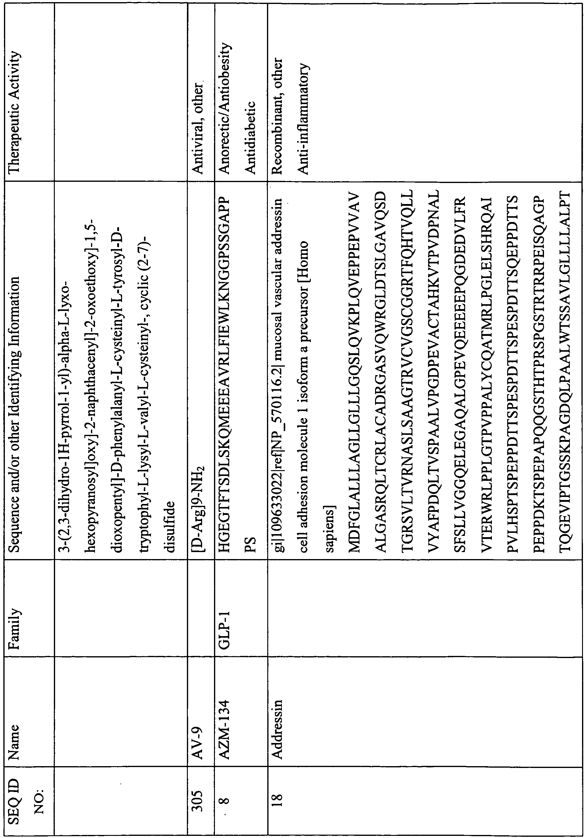


- In other embodiments, the therapeutic peptides are selected from the group consisting of peptide G, OTS-102, Angiocol (antiangiogenic peptide group), ABT-510 (antiangiogenic peptide group), A6 (antiangiogenic peptide group), islet neogenesis gene associated protein (INGAP), tendamistat, recombinant human carperitide (alpha-atrial natriuretic peptide) (natriuretic peptide group), urodilatin (natriuretic peptide group), desirudin, Obestatin, ITF-1697, oxyntomodulin, cholecystokinin, bactericidal permeability increasing (BPI) protein, C-peptide, Prosaptide TX14(A), sermorelin acetate (GHRFA group), pralmorelin (GHRFA group), growth hormone releasing factor (GHRFA group), examorelin (GHRFA group), gonadorelin (LH-related peptide group), corticoliberin, atrial natriuretic peptide (natriuretic peptide group), anergix, somatostatin (GHRFA group), 29-amino-acid peptide growth hormone releasing hormone (GHRH) analogue (GHRFA group), bremelanotide (melanocortin agonist group), melanocortin peptidomimetic compound (melanocortin agonist group), antiprogestogens - GnRH antagonists (LH-related peptide group), recombinant LH (luteinizing hormone) (LH-related peptide group), terlipressin, Ecallantide - 60-amino-acid recombinant peptide kallikrein inhibitor, calphobindin I, tiplimotide, osteogenic growth peptide, myelin basic protein, dynorphin A, anaritide (natriuretic peptide group), secretin , GLP-2, and gastrin.
- The therapeutic peptides of the invention may comprise any of the 20 natural amino acids, and/or non-natural amino acids, amino acid analogs, and peptidomimetics, in any combination. The peptides may be composed of D- amino acids or L-amino acids, or a combination of both in any proportion. In addition to natural amino acids, the therapeutic peptides may contain, or may be modified to include, 1, 2, 3, 4, 5, 6, 7, 8, 9, 10, 15, 20, 25, or more non-natural amino acids. Exemplary non-natural amino acids and amino acid analogs that can be use with the invention include, but are not limited to, 2-aminobutyric acid, 2-aminoisobutyric acid, 3-(1-naphthyl)alanine, 3-(2-naphthyl)alanine, 3-methylhistidine, 3-pyridylalanine, 4-chlorophenylalanine, 4-fluorophenylalanine, 4-hydroxyproline, 5-hydroxylysine, alloisoleucine, citrulline, dehydroalanine, homoarginine, homocysteine, homoserine, hydroxyproline, N-acetylserine, N-formylmethionine, N-methylglycine, N-methylisoleucine, norleucine, N-α-methylarginine, O-phosphoserine, ornithine, phenylglycine, pipecolinic acid, piperazic acid, pyroglutamine, sarcosine, valanine, β-alanine, and β-cyclohexylalanine.
- The therapeutic peptides may be, or may be modified to be, linear, branched, or cyclic, with our without branching.
- Additionally, the therapeutic peptides may optionally be modified or protected with a variety of functional groups or protecting groups, including amino terminus protecting groups and/or carboxy terminus protecting groups. Protecting groups, and the manner in which they are introduced and removed are described, for example, in "Protective Groups in Organic Chemistry," Plenum Press, London, N.Y. 1973; and. Greene et al., "PROTECTIVE GROUPS IN ORGANIC SYNTHESIS" 3rd Edition, John Wiley and Sons, Inc., New York, 1999. Numerous protecting groups are known in the art. An illustrative, non-limiting list of protecting groups includes methyl, formyl, ethyl, acetyl, t-butyl, anisyl, benzyl, trifluoroacetyl, N-hydroxysuccinimide, t-butoxycarbonyl, benzoyl, 4-methylbenzyl, thioanizyl, thiocresyl, benzyloxymethyl, 4-nitrophenyl, benzyloxycarbonyl, 2-nitrobenzoyl, 2-nitrophenylsulphenyl, 4-toluenesulphonyl, pentafluorophenyl, diphenylmethyl, 2-chlorobenzyloxycarbonyl, 2,4,5-trichlorophenyl, 2-bromobenzyloxycarbonyl, 9-fluorenylmethyloxycarbonyl, triphenylmethyl, and 2,2,5,7,8-pentamethyl-chroman-6-sulphonyl. For discussions of various different types of amino- and carboxy-protecting groups, see, for example,
U.S. Pat. No. 5,221,736 (issued Jun. 22, 1993 );U.S. Pat. No. 5,256,549 (issued Oct. 26, 1993 );U.S. Pat. No. 5,049,656 (issued Sep. 17, 1991 ); andU.S. Pat. No. 5,521,184 (issued May 28, 1996 ). - The therapeutic peptides contain, or may be modified to contain, functional groups to which a water-soluble polymer may be attached, either directly or through a spacer moiety or linker. Functional groups include, but are not limited to, the N-terminus of the therapeutic peptide, the C-terminus of the therapeutic peptide, and any functional groups on the side chain of an amino acid, e.g. lysine, cysteine, histidine, aspartic acid, glutamic acid, tyrosine, arginine, serine, methionine, and threonine, present in the therapeutic peptide.
- The therapeutic peptides can be prepared by any means known in the art, including non-recombinant and recombinant methods, or they may, in some instances, be commercially available. Chemical or non-recombinant methods include, but are not limited to, solid phase peptide synthesis (SPPS), solution phase peptide synthesis, native chemical ligation, intein-mediated protein ligation, and chemical ligation, or a combination thereof. In a preferred embodiment, the therapeutic peptides are synthesized using standard SPPS, either manually or by using commercially available automated SPPS synthesizers.
- SPPS has been known in the art since the early 1960's (Merrifield, R. B., J. Am. Chem. Soc., 85:2149-2154 (1963)), and is widely employed. (See also, Bodanszky, Principles of Peptide Synthesis, Springer-Verlag, Heidelberg (1984)). There are several known variations on the general approach. (See, for example, "Peptide Synthesis, Structures, and Applications" © 1995 by Academic Press, and White (2003) Fmoc Solid Phase Peptide Synthesis, A practical Approach, Oxford University Press, Oxford). Very briefly, in solid phase peptide synthesis, the desired C-terminal amino acid residue is coupled to a solid support. The subsequent amino acid to be added to the peptide chain is protected on its amino terminus with Boc, Fmoc, or other suitable protecting group, and its carboxy terminus is activated with a standard coupling reagent. The free amino terminus of the support-bound amino acid is allowed to react with the carboxy-terminus of the subsequent amino acid, coupling the two amino acids. The amino terminus of the growing peptide chain is deprotected, and the process is repeated until the desired polypeptide is completed. Side chain protecting groups may be utilized as needed.
- Alternatively, the therapeutic peptides may be prepared recombinantly. Exemplary recombinant methods used to prepare therapeutic peptides include the following, among others, as will be apparent to one skilled in the art. Typically, a therapeutic peptide as defined and/or described herein is prepared by constructing the nucleic acid encoding the desired peptide or fragment, cloning the nucleic acid into an expression vector, transforming a host cell (e.g., plant, bacteria such as Escherichia coli, yeast such as Saccharomyces cerevisiae, or mammalian cell such as Chinese hamster ovary cell or baby hamster kidney cell), and expressing the nucleic acid to produce the desired peptide or fragment. The expression can occur via exogenous expression or via endogenous expression (when the host cell naturally contains the desired genetic coding). Methods for producing and expressing recombinant polypeptides in vitro and in prokaryotic and eukaryotic host cells are known to those of ordinary skill in the art. See, for example,
U.S. Patent No. 4,868,122 , and Sambrook et al., Molecular Cloning--A Laboratory Manual, Cold Spring Harbor Laboratory Press (1989).. - To facilitate identification and purification of the recombinant peptide, nucleic acid sequences that encode an epitope tag or other affinity binding sequence can be inserted or added in-frame with the coding sequence, thereby producing a fusion peptide comprised of the desired therapeutic peptide and a peptide suited for binding. Fusion peptides can be identified and purified by first running a mixture containing the fusion peptide through an affinity column bearing binding moieties (e.g., antibodies) directed against the epitope tag or other binding sequence in the fusion peptide, thereby binding the fusion peptide within the column. Thereafter, the fusion peptide can be recovered by washing the column with the appropriate solution (e.g., acid) to release the bound fusion peptide. Optionally, the tag may subsequently be removed by techniques known in the art. The recombinant peptide can also be identified and purified by lysing the host cells, separating the peptide, e.g., by size exclusion chromatography, and collecting the peptide. These and other methods for identifying and purifying recombinant peptides are known to those of ordinary skill in the art.
- It will be appreciated and understood by one of skill in the art that certain modifications can be made to the therapeutic peptides defined and/or disclosed herein that do not alter, or only partially abrogate, the properties and activities of these therapeutic peptides. In some instances, modifications may be made that result in an increase in therapeutic activities. Additionally, modifications may be made that increase certain biological and chemical properties of the therapeutic peptides in a beneficial way, e.g. increased in vivo half life, increased stability, decreased susceptibility to proteolytic cleavage, etc. Thus, in the spirit and scope of the invention, the term "therapeutic peptide" is used herein in a manner to include not only the therapeutic peptides defined and/or disclosed herein, but also related peptides, i.e. peptides that contain one or more modifications relative to the therapeutic peptides defined and/or disclosed herein, wherein the modification(s) do not alter, only partially abrogate, or increase the therapeutic activities as compared to the parent peptide.
- Related peptides include, but are not limited to, fragments of therapeutic peptides, therapeutic peptide variants, and therapeutic peptide derivatives. Related peptides also include any and all combinations of these modifications. In a non-limiting example, a related peptide may be a fragment of a therapeutic peptide as disclosed herein having one or more amino acid substitutions. Thus it will be understood that any reference to a particular type of related peptide is not limited to a therapeutic peptide having only that particular modification, but rather encompasses a therapeutic peptide having that particular modification and optionally any other modification.
- Related peptides may be prepared by action on a parent peptide or a parent protein (e.g. proteolytic digestion to generate fragments) or through de novo preparation (e.g. solid phase synthesis of a peptide having a conservative amino acid substitution relative to the parent peptide). Related peptides may arise by natural processes (e.g. processing and other posttranslational modifications) or may be made by chemical modification techniques. Such modifications are well-known to those of skill in the art.
- A related peptide may have a single alteration or multiple alterations relative to the parent peptide. Where multiple alterations are present, the alterations may be of the same type or a given related peptide may contain different types of modifications. Furthermore, modifications can occur anywhere in a polypeptide, including the peptide backbone, the amino acid side-chains, and the N- or C- termini.
- As previously noted, related peptides include fragments of the therapeutic peptides defined and/or disclosed herein, wherein the fragment retains some of or all of at least one therapeutic activity of the parent peptide. The fragment may also exhibit an increase in at least one therapeutic activity of the parent peptide. In certain embodiments of the invention, therapeutic peptides include related peptides having at least 2, 3, 4, 5, 6, 7, 8, 9, 10, 11, 12, 13, 14, 15, 20, 25, 30, 35, 40, 45, 50, 60, 70, 80, 90, or 100 contiguous amino acid residues, or more than 125 contiguous amino acid residues, of any of the therapeutic peptides disclosed, herein, including in Table 1. In other embodiments of the invention, therapeutic peptides include related peptides having 0, 1, 2, 3, 4, 5, 6, 7, 8, 9, 10, 15, 20, 25, 30, 35, 40, 45, or 50 amino acid residues deleted from the N-terminus and/or having 0, 1, 2, 3, 4, 5, 6, 7, 8, 9, 10, 15, 20, 25, 30, 35, 40, 45, or 50 amino acid residues deleted from the C-terminus of any of the therapeutic peptides disclosed herein, including in Table 1.
- Related peptides also include variants of the therapeutic peptides defined and/or disclosed herein, wherein the variant retains some of or all of at least one therapeutic activity of the parent peptide. The variant may also exhibit an increase in at least one therapeutic activity of the parent peptide. In certain embodiments of the invention, therapeutic peptides include variants having 1, 2, 3, 4, 5, 6, 7, 8, 9, 10, 15, 20, 25, 30, 35, 40, 45, or 50 conservative and/or non-conservative amino acid substitutions relative to the therapeutic peptides disclosed herein, including in Table 1. Desired amino acid substitutions, whether conservative or non-conservative, can be determined by those skilled in the art.
- In certain embodiments of the invention, therapeutic peptides include variants having conservative amino substitutions; these substitutions will produce a therapeutic peptide having functional and chemical characteristics similar to those of the parent peptide. In other embodiments, therapeutic peptides include variants having non-conservative amino substitutions; these substitutions will produce a therapeutic peptide having functional and chemical characteristics that may differ substantially from those of the parent peptide. In certain embodiments of the invention, therapeutic peptide variants have both conservative and non-conservative amino acid substitutions. In other embodiments, each amino acid residue may be substituted with alanine.
- Natural amino acids may be divided into classes based on common side chain properties: nonpolar (Gly, Ala, Val, Leu, Ile, Met); polar neutral (Cys, Ser, Thr, Pro, Asn, Gln); acidic (Asp, Glu); basic (His, Lys, Arg); and aromatic (Trp, Tyr, Phe). By way of example, non-conservative amino acid substitutions may involve the substitution of an amino acid of one class for that of another, and may be introduced in regions of the peptide not critical for therapeutic activity.
- Preferably, amino acid substitutions are conservative. Conservative amino acid substitutions may involve the substitution of an amino acid of one class for that of the same class. Conservative amino acid substitutions may also encompass non-natural amino acid residues, including peptidomimetics and other atypical forms of amino acid moieties, and may be incorporated through chemical peptide synthesis,
- Amino acid substitutions may be made with consideration to the hydropathic index of amino acids. The importance of the hydropathic amino acid index in conferring interactive biological function on a protein is generally understood in the art (Kyte et al., 1982, J. Mol. Biol. 157:105-31). Each amino acid has been assigned a hydropathic index on the basis of its hydrophobicity and charge characteristics. The hydropathic indices are: isoleucine (+4.5); valine (+4.2); leucine (+3.8); phenylalanine (+2.8); cysteine/cystine (+2.5); methionine (+1.9); alanine (+1.8); glycine (-0.4); threonine (-0.7); serine (-0.8); tryptophan (-0.9); tyrosine (-1.3); proline (-1.6); histidine (-3.2); glutamate (-3.5); glutamine (-3.5); aspartate (-3.5); asparagine (-3.5); lysine (-3.9); and arginine (-4.5).
- It is known that certain amino acids may be substituted for other amino acids having a similar hydropathic index or score and still retain a similar biological activity. In making changes based upon the hydropathic index, the substitution of amino acids whose hydropathic indices are within ±2 is preferred, those which are within ±1 are particularly preferred, and those within ±0.5 are even more particularly preferred.
- It is also understood in the art that the substitution of like amino acids can be made effectively on the basis of hydrophilicity. The greatest local average hydrophilicity of a protein, as governed by the hydrophilicity of its adjacent amino acids, correlates with its biological properties. According to
U.S. Patent No. 4,554,101 , incorporated herein by reference, the following hydrophilicity values have been assigned to these amino acid residues: arginine (+3.0); lysine (+3.0); aspartate (+3.0 ± 1); glutamate (+3.0 ± 1); serine (+0.3); asparagine (+0.2); glutamine (+0.2); glycine (0); threonine (-0.4); proline (-0.5 ± 1); alanine (-0.5); histidine (-0.5); cysteine (-1.0); methionine (-1.3); valine (-1.5); leucine (-1.8); isoleucine (-1.8); tyrosine (-2.3); phenylalanine (-2.5); and tryptophan (-3.4). In making changes based upon similar hydrophilicity values, the substitution of amino acids whose hydrophilicity values are within ±2 is preferred, those which are within ±1 are particularly preferred, and those within ±0.5 are even more particularly preferred. - In certain embodiments of the invention, therapeutic peptides include variants having 1, 2, 3, 4, 5, 6, 7, 8, 9, 10, 15, 20, 25, 30, 35, 40, 45, or 50 amino acid deletions relative to the therapeutic peptides disclosed herein, including in Table 1. The deleted amino acid(s) may be at the N- or C- terminus of the peptide, at both termini, at an internal location or locations within the peptide, or both internally and at one or both termini. Where the variant has more than one amino acid deletion, the deletions may be of contiguous amino acids or of amino acids at different locations within the primary amino acid sequence of the parent peptide.
- In other embodiments of the invention, therapeutic peptides include variants having 1, 2, 3, 4, 5, 6, 7, 8, 9, 10, 15, 20, 25, 30, 35, 40, 45, or 50 amino acid additions relative to the therapeutic peptides disclosed herein, including in Table 1. The added amino acid(s) may be at the N- or C- terminus of the peptide, at both termini, at an internal location or locations within the peptide, or both internally and at one or both termini. Where the variant has more than one amino acid addition, the amino acids may be added contiguously, or the amino acids may be added at different locations within the primary amino acid sequence of the parent peptide.
- Addition variants also include fusion peptides. Fusions can be made either at the N-terminus or at the C-terminus of the therapeutic peptides disclosed herein, including in Table 1. In certain embodiments, the fusion peptides have 1, 2, 3, 4, 5, 6, 7, 8, 9, 10, 15, 20, 25, 30, 35, 40, 45, or 50 amino acid additions relative to the therapeutic peptides disclosed herein, including in Table 1. Fusions may be attached directly to the therapeutic peptide with no connector molecule or may be through a connector molecule. As used in this context, a connector molecule may be an atom or a collection of atoms optionally used to link a therapeutic peptide to another peptide. Alternatively, the connector may be an amino acid sequence designed for cleavage by a protease to allow for the separation of the fused peptides.
- The therapeutic peptides of the invention may be fused to peptides designed to improve certain qualities of the therapeutic peptide, such as therapeutic activity, circulation time, or reduced aggregation. Therapeutic peptides may be fused to an immunologically active domain, e.g. an antibody epitope, to facilitate purification of the peptide, or to increase the in vivo half life of the peptide. Additionally, therapeutic peptides may be fused to known functional domains, cellular localization sequences, or peptide permeant motifs known to improve membrane transfer properties.
- In certain embodiments of the invention, therapeutic peptides also include variants incorporating one or more non-natural amino acids, amino acid analogs, and peptidomimetics. Thus the present invention encompasses compounds structurally similar to the therapeutic peptides defined and/or disclosed herein, which are formulated to mimic the key portions of the therapeutic peptides of the present invention. Such compounds may be used in the same manner as the therapeutic peptides of the invention. Certain mimetics that mimic elements of protein secondary and tertiary structure have been previously described. Johnson et al., Biotechnology and Pharmacy, Pezzuto et al. (Eds.), Chapman and Hall, NY, 1993. The underlying rationale behind the use of peptide mimetics is that the peptide backbone of proteins exists chiefly to orient amino acid side chains in such a way as to facilitate molecular interactions. A peptide mimetic is thus designed to permit molecular interactions similar to the parent peptide. Mimetics can be constructed to achieve a similar spatial orientation of the essential elements of the amino acid side chains. Methods for generating specific structures have been disclosed in the art. For example,
U.S. Patent Nos. 5,446,128 ,5,710,245 ,5,840,833 ,5,859,184 ,5,440,013 ;5,618,914 ,5,670,155 ,5,475,085 ,5,929,237 ,5,672,681 and5,674,976 , the contents of which are hereby incorporated by reference, all disclose peptidomimetics structures that may have improved properties over the parent peptide, for example they may be conformationally restricted, be more thermally stable, exhibit increased resistance to degredation, etc. - In another embodiment, related peptides comprise or consist of a peptide sequence that is at least 70% identical to any of the therapeutic peptides disclosed herein, including in Table 1. In additional embodiments, related peptides are at least 75% identical, at least 80% identical, at least 85% identical, 90% identical, at least 91% identical, at least 92% identical, 93% identical, at least 94% identical, at least 95% identical, 96% identical, at least 97% identical, at least 98% identical, or at least 99% identical to any of the therapeutic peptides disclosed herein, including in Table 1.
- Sequence identity (also known as % homology) of related polypeptides can be readily calculated by known methods. Such methods include, but are not limited to those described in Computational Molecular Biology (A.M. Lesk, ed., Oxford University Press 1988); Biocomputing: Informatics and Genome Projects (D.W. Smith, ed., Academic Press 1993); Computer Analysis of Sequence Data (); G. von Heinle, Sequence Analysis in Molecular Biology (Academic Press 1987); Sequence Analysis Primer (M. Gribskov and J. Devereux, eds., M. Stockton Press 1991); and Carillo et al., 1988, SIAM J. Applied Math., 48:1073.
- Preferred methods to determine sequence identity and/or similarity are designed to give the largest match between the sequences tested. Methods to determine sequence identity are described in publicly available computer programs. Preferred computer program methods to determine identity and similarity between two sequences include, but are not limited to, the GCG program package, including GAP (Devereux et al., 1984, Nucleic Acids Res. 12:387; Genetics Computer Group, University of Wisconsin, Madison, WI), BLASTP, BLASTN, and FASTA (Altschul et al., 1990, J. Mol. Biol. 215:403-10). The BLASTX program is publicly available from the National Center for Biotechnology Information (NCBI) and other sources (Altschul et al., BLAST Manual (NCB NLM NIH, Bethesda, MD); Altschul et al., 1990, supra). The well-known Smith Waterman algorithm may also be used to determine identity.
- For example, using the computer algorithm GAP (Genetics Computer Group, University of Wisconsin, Madison, WI), two polypeptides for which the percent sequence identity is to be determined are aligned for optimal matching of their respective amino acids (the "matched span," as determined by the algorithm). A gap opening penalty (which is calculated as 3X the average diagonal; the "average diagonal" is the average of the diagonal of the comparison matrix being used; the "diagonal" is the score or number assigned to each perfect amino acid match by the particular comparison matrix) and a gap extension penalty (which is usually 0.1X the gap opening penalty), as well as a comparison matrix such as
PAM 250 or BLOSUM 62 are used in conjunction with the algorithm. A standard comparison matrix is also used by the algorithm (see Dayhoff et al., 5 Atlas of Protein Sequence and Structure (Supp. 3 1978)(PAM250 comparison matrix); Henikoff et al., 1992, Proc. Natl. Acad. Sci USA 89:10915-19 (BLOSUM 62 comparison matrix)). The particular choices to be made with regard to algorithms, gap opening penalties, gap extension penalties, comparison matrices, and thresholds of similarity will be readily apparent to those of skill in the art and will depend on the specific comparison to be made. - Related peptides also include derivatives of the therapeutic peptides defined and/or disclosed herein, wherein the variant retains some of or all of at least one therapeutic activity of the parent peptide. The derivative may also exhibit an increase in at least one therapeutic activity of the parent peptide. Chemical alterations of therapeutic peptide derivatives include, but are not limited to, acetylation, acylation, ADP-ribosylation, amidation, biotinylation, covalent attachment of flavin, covalent attachment of a heme moiety, covalent attachment of a nucleotide or nucleotide derivative, covalent attachment of a lipid or lipid derivative, covalent attachment of phosphotidylinositol, cross-linking, cyclization, disulfide bond formation, demethylation, formation of covalent cross-links, formation of cysteine, formation of pyroglutamate, formylation, gamma-carboxylation, glycosylation, GPI anchor formation, hydroxylation, iodination, methylation, myristoylation, oxidation, proteolytic processing, phosphorylation, prenylation, racemization, selenoylation, sulfation, transfer-RNA mediated addition of amino acids to proteins such as arginylation, and ubiquitination. (See, for instance, T. E. Creighton, Proteins, Structure and Molecular Properties, 2nd ed., W.H. Freeman and Company, New York (1993); Posttranslational Covalent Modification of Proteins, B. C. Johnson, ed., Academic Press, New York, pgs. 1-12 (1983); Seifter et al., Meth. Enzymol 182:626-46 (1990); Rattan et al., Ann. N.Y. Acad. Sci. 663:48-62,1992).
- Therapeutic peptide derivatives also include molecules formed by the deletion of one or more chemical groups from the parent peptide. Methods for preparing chemically modified derivatives of the therapeutic peptides defined and/or disclosed herein are known to one of skill in the art.
- In some embodiments of the invention, the therapeutic peptides may be modified with one or more methyl or other lower alkyl groups at one or more positions of the therapeutic peptide sequence. Examples of such groups include methyl, ethyl, propyl, isopropyl, butyl, isobutyl, pentyl, etc. In certain preferred embodiments, arginine, lysine, and histidine residues of the therapeutic peptides are modified with methyl or other lower alkyl groups.
- In other embodiments of the invention, the therapeutic peptides may be modified with one or more glycoside moieties relative to the parent peptide. Although any glycoside can be used, in certain preferred embodiments the therapeutic peptide is modified by introduction of a monosaccharide, a disaccharide, or a trisaccharide or it may contain a glycosylation sequence found in natural peptides or proteins in any mammal. The saccharide may be introduced at any position, and more than one glycoside may be introduced. Glycosylation may occur on a naturally occurring amino acid residue in the therapeutic peptide, or alternatively, an amino acid may be substituted with another for modification with the saccharide.
- Glycosylated therapeutic peptides may be prepared using conventional Fmoc chemistry and solid phase peptide synthesis techniques, e.g., on resin, where the desired protected glycoamino acids are prepared prior to peptide synthesis and then introduced into the peptide chain at the desired position during peptide synthesis. Thus, the therapeutic peptide polymer conjugates may be conjugated in vitro. The glycosylation may occur before deprotection. Preparation of aminoacid glycosides is described in
U.S. Patent No. 5,767,254 ,WO 2005/097158 , and Doores, K., et al., Chem. Commun., 1401-1403, 2006, which are incorporated herein by reference in their entireties. For example, alpha and beta selective glycosylations of serine and threonine residues are carried out using the Koenigs-Knorr reaction and Lemieux's in situ anomerization methodology with Schiff base intermediates. Deprotection of the Schiff base glycoside is then carried out using mildly acidic conditions or hydrogenolysis. A composition, comprising a glycosylated therapeutic peptide conjugate made by stepwise solid phase peptide synthesis involving contacting a growing peptide chain with protected amino acids in a stepwise manner, wherein at least one of the protected amino acids is glycosylated, followed by water-soluble polymer conjugation, may have a purity of at least 95%, such as at least 97%, or at least 98%, of a single species of the glycosylated and conjugated therapeutic peptide. - Monosaccharides that may by used for introduction at one or more amino acid residues of the therapeutic peptides defined and/or disclosed herein include glucose (dextrose), fructose, galactose, and ribose. Additional monosaccharides suitable for use include glyceraldehydes, dihydroxyacetone, erythrose, threose, erythrulose, arabinose, lyxose, xylose, ribulose, xylulose, allose, altrose, mannose, N-Acetylneuraminic acid, fucose, N-Acetylgalactosamine, and N-Acetylglucosamine, as well as others. Glycosides, such as mono-, di-, and trisaccharides for use in modifying a therapeutic peptide, may be naturally occurring or may be synthetic. Disaccharides that may by used for introduction at one or more amino acid residues of the therapeutic peptides defined and/or disclosed herein include sucrose, lactose, maltose, trehalose, melibiose, and cellobiose, among others. Trisaccharides include acarbose, raffinose, and melezitose.
- In further embodiments of the invention, the therapeutic peptides defined and/or disclosed herein may be chemically coupled to biotin. The biotin/thereapeutic peptide molecules can then to bind to avidin.
- As previously noted, modifications may be made to the therapeutic peptides defined and/or disclosed herein that do not alter, or only partially abrogate, the properties and activities of these therapeutic peptides. In some instances, modifications may be made that result in an increase in therapeutic activity. Thus, included in the scope of the invention are modifications to the therapeutic peptides disclosed herein, including in Table 1, that retain at least 1%, at least 5%, at least 10%, at least 20%, at least 30%, at least 40%, at least 50%, at least 55%, at least 60%, at least 65%, at least 70%, at least 75%, at least 80%, at least 81%, at least 82%, at least 83%, at least 84%, at least 85%, at least 86%, at least 87%, at least 88%, at least 89%, at least 90%, at least 91%, at least 92%, at least 93%, at least 94%, at least 95%, at least 96%, at least 97%, at least 98%, or at least 99%, and any range derivable therein, such as, for example, at least 70% to at least 80%, and more preferably at least 81% to at least 90%; or even more preferably, between at least 91% and at least 99% of the therapeutic activity relative to the unmodified therapeutic peptide. Also included in the scope of the invention are modification to the therapeutic peptides disclosed herein, including in Table 1, that have greater than 100%, greater than 110%, greater than 125%, greater than 150%, greater than 200%, or greater than 300%, or greater than 10-fold or greater than 100-fold, and any range derivable therein, of the therapeutic activity relative to the unmodified therapeutic peptide.
- The level of therapeutic activity of a given therapeutic peptide, or a modified therapeutic peptide, may be determined by any suitable in vivo or in vitro assay. For example, therapeutic activity may be assayed in cell culture, or by clinical evaluation, EC50 assays, IC50 assays, or dose response curves. In vitro or cell culture assays, for example, are commonly available and known to one of skill in the art for many therapeutic peptides as disclosed herein, including in Table 1. It will be understood by one of skill in the art that the percent activity of a modified therapeutic peptide relative to its unmodified parent can be readily ascertained through a comparison of the activity of each as determined through the assays disclosed herein or as known to one of skill in the art.
- One of skill in the art will be able to determine appropriate modifications to the therapeutic peptides defined and/or disclosed herein, including those disclosed herein, including in Table 1. For identifying suitable areas of the therapeutic peptides that may be changed without abrogating their therapeutic activities, one of skill in the art may target areas not believed to be essential for activity. For example, when similar peptides with comparable activities exist from the same species or across other species, one of skill in the art may compare those amino acid sequences to identify residues that are conserved among similar peptides. It will be understood that changes in areas of a therapeutic peptide that are not conserved relative to similar peptides would be less likely to adversely affect the thereapeutic activity. One skilled in the art would also know that, even in relatively conserved regions, one may substitute chemically similar amino acids while retaining therapeutic activity. Therefore, even areas that may be important for biological activity and/or for structure may be subject to amino acid substitutions without destroying the therapeutic activity or without adversely affecting the peptide structure.
- Additionally, as appropriate, one of skill in the art can review structure-function studies identifying residues in similar peptides that are important for activity or structure. In view of such a comparison, one can predict the importance of an amino acid residue in a therapeutic peptide that corresponds to an amino acid residue that is important for activity or structure in similar peptides. One of skill in the art may opt for amino acid substitutions within the same class of amino acids for such predicted important amino acid residues of the therapeutic peptides.
- Also, as appropriate, one of skill in the art can also analyze the three-dimensional structure and amino acid sequence in relation to that structure in similar peptides. In view of such information, one of skill in the art may predict the alignment of amino acid residues of a therapeutic peptide with respect to its three dimensional structure. One of skill in the art may choose not to make significant changes to amino acid residues predicted to be on the surface of the peptide, since such residues may be involved in important interactions with other molecules. Moreover, one of skill in the art may generate variants containing a single amino acid substitution at each amino acid residue for test purposes. The variants could be screened using therapeutic activity assays known to those with skill in the art. Such variants could be used to gather information about suitable modifications. For example, where a change to a particular amino acid residue resulted in abrogated, undesirably reduced, or unsuitable activity, variants with such a modification would be avoided. In other words, based on information gathered from routine experimentation, one of skill in the art can readily determine the amino acids where further modifications should be avoided either alone or in combination with other modifications.
- One of skill in the art may also select suitable modifications based on secondary structure predication. A number of scientific publications have been devoted to the prediction of secondary structure. See Moult, 1996, Curr. Opin. Biotechnol. 7:422-27; Chou et al., 1974, Biochemistry 13:222-45; Chou et al., 1974, Biochemistry 113:211-22; Chou et al., 1978, Adv. Enzymol. Relat. Areas Mol. Biol. 47:45-48; Chou et al., 1978, Ann. Rev. Biochem. 47:251-276; and Chou et al., 1979, Biophys. J. 26:367-84. Moreover, computer programs are currently available to assist with predicting secondary structure. One method of predicting secondary structure is based upon homology modeling. For example, two peptides or proteins which have a sequence identity of greater than 30%, or similarity greater than 40%, often have similar structural topologies. Recent growth of the protein structural database (PDB, http://www.rcsb.org/pdb/home/home.do) has provided enhanced predictability of secondary, tertiary, and quarternary structure, including the potential number of folds within the structure of a peptide or protein. See Holm et al., 1999, Nucleic Acids Res. 27:244-47. It has been suggested that there are a limited number of folds in a given peptide or protein and that once a critical number of structures have been resolved, structural prediction will become dramatically more accurate (Brenner et al., 1997, Curr. Opin. Struct. Biol. 7:369-76).
- Additional methods of predicting secondary structure include "threading" (Jones, 1997, Curr. Opin. Struct. Biol. 7:377-87; Sippl et al., 1996, Structure 4:15-19), "profile analysis" (Bowie et al., 1991, Science, 253:164-70; Gribskov et al., 1990, Methods Enzymol. 183:146-59; Gribskov et al., 1987, Proc. Nat. Acad. Sci. U.S.A. 84:4355-58), and "evolutionary linkage" (See Holm et al., supra, and Brenner et al., supra).
- As described above, a conjugate of the invention comprises a water-soluble polymer covalently attached (either directly or through a spacer moiety or linker) to a therapeutic peptide. Typically, for any given conjugate, there will be about one to five water-soluble polymers covalently attached to a therapeutic peptide (wherein for each water-soluble polymer, the water-soluble polymer can be attached either directly to the therapeutic peptide or through a spacer moiety).
- To elaborate, a therapeutic peptide conjugate of the invention typically has about 1, 2, 3, or 4 water-soluble polymers individually attached to a therapeutic peptide. That is to say, in certain embodiments, a conjugate of the invention will possess about 4 water-soluble polymers individually attached to a therapeutic peptide, or about 3 water-soluble polymers individually attached to a therapeutic peptide, or about 2 water-soluble polymers individually attached to a therapeutic peptide, or about 1 water-soluble polymer attached to a therapeutic peptide. The structure of each of the water-soluble polymers attached to the therapeutic peptide may be the same or different. One therapeutic peptide conjugate in accordance with the invention is one having a water-soluble polymer releasably attached to the therapeutic peptide, particularly at the N-terminus of the therapeutic peptide. Another therapeutic peptide conjugate in accordance with the invention is one having a water-soluble polymer stably attached to the therapeutic peptide, particularly at the N-terminus of the therapeutic peptide. Another therapeutic peptide conjugate is one having a water-soluble polymer releasably attached to the therapeutic peptide, particularly at the C-terminus of the therapeutic peptide. Another therapeutic peptide conjugate in accordance with the invention is one having a water-soluble polymer stably attached to the therapeutic peptide, particularly at the C-terminus of the therapeutic peptide. Other therapeutic peptide conjugates in accordance with the invention are those having a water-soluble polymer releasably or stably attached to an amino acid within the therapeutic peptide. Additional water-soluble polymers may be releasably or stably attached to other sites on the therapeutic peptide, e.g., such as one or more additional sites. For example, a therapeutic peptide conjugate having a water-soluble polymer releasably attached to the N-terminus may additionally possess a water-soluble polymer stably attached to a lysine residue. In one embodiment, one or more amino acids may be inserted, at the N- or C-terminus, or within the peptide to releasably or stably attach a water soluble polymer. One preferred embodiment of the present invention is a mono-therapeutic peptide polymer conjugate, i.e., a therapeutic peptide having one water-soluble polymer covalently attached thereto. In an even more preferred embodiment, the water-soluble polymer is one that is attached to the therapeutic peptide at its N-terminus.
- Preferably, a therapeutic peptide polymer conjugate of the invention is absent a metal ion, i.e., the therapeutic peptide is not chelated to a metal ion.
- For the therapeutic peptide polymer conjugates described herein, the therapeutic peptide may optionally possess one or more N-methyl substituents. Alternatively, for the therapeutic peptide polymer conjugates described herein, the therapeutic peptide may be glycosylated, e.g., having a mono- or disaccharide, or naturally-occuring amino acid glycosylation covalently attached to one or more sites thereof.
- As discussed herein, the compounds of the present invention may be made by various methods and techniques known and available to those skilled in the art.
- A conjugate of the invention comprises a therapeutic peptide attached, stably or releasably, to a water-soluble polymer. The water-soluble polymer is typically hydrophilic, nonpeptidic, and biocompatible. A substance is considered biocompatible if the beneficial effects associated with use of the substance alone or with another substance (e.g., an active agent such a therapeutic peptide) in connection with living tissues (e.g., administration to a patient) outweighs any deleterious effects as evaluated by a clinician, e.g., a physician. A substance is considered nonimmunogenic if the intended use of the substance in vivo does not produce an undesired immune response (e.g., the formation of antibodies) or, if an immune response is produced, that such a response is not deemed clinically significant or important as evaluated by a clinician. Typically, the water-soluble polymer is hydrophilic, biocompatible and nonimmunogenic.
- Further the water-soluble polymer is typically characterized as having from 2 to about 300 termini, preferably from 2 to 100 termini, and more preferably from about 2 to 50 termini. Examples of such polymers include, but are not limited to, poly(alkylene glycols) such as polyethylene glycol (PEG), poly(propylene glycol) ("PPG"), copolymers of ethylene glycol and propylene glycol and the like, poly(oxyethylated polyol), poly(olefinic alcohol), poly(vinylpyrrolidone), poly(hydroxyalkylmethacrylamide), poly(hydroxyalkylmethacrylate), poly(saccharides), poly(α-hydroxy acid), poly(vinyl alcohol), polyphosphazene, polyoxazoline, poly(N-acryloylmorpholine), and combinations of any of the foregoing, including copolymers and terpolymers thereof.
- The water-soluble polymer is not limited to a particular structure and may possess a linear architecture (e.g., alkoxy PEG or bifunctional PEG), or a non-linear architecture, such as branched, forked, multi-armed (e.g., PEGs attached to a polyol core), or dendritic (i.e. having a densely branched structure with numerous end groups). Moreover, the polymer subunits can be organized in any number of different patterns and can be selected, e.g., from homopolymer, alternating copolymer, random copolymer, block copolymer, alternating tripolymer, random tripolymer, and block tripolymer.
- One particularly preferred type of water-soluble polymer is a polyalkylene oxide, and in particular, polyethylene glycol (or PEG). Generally, a PEG used to prepare a therapeutic peptide polymer conjugate of the invention is "activated" or reactive. That is to say, the activated PEG (and other activated water-soluble polymers collectively referred to herein as "polymeric reagents") used to form a therapeutic peptide conjugate comprises an activated functional group suitable for coupling to a desired site or sites on the therapeutic peptide. Thus, a polymeric reagent for use in preparing a therapeutic peptide conjugate includes a functional group for reaction with the therapeutic peptide.
- Representative polymeric reagents and methods for conjugating such polymers to an active moiety are known in the art, and are, e.g., described in Harris, J.M. and Zalipsky, S., eds, Poly(ethylene glycol), Chemistry and Biological Applications, ACS, Washington, 1997; Veronese, F., and J.M Harris, eds., Peptide and Protein PEGylation, Advanced Drug Delivery Reviews, 54(4); 453-609 (2002); Zalipsky, S., et al., "Use of Functionalized Poly(Ethylene Glycols) for Modification of Polypeptides" in Polyethylene Glycol Chemistry: Biotechnical and Biomedical Applications, J. M. Harris, ed., Plenus Press, New York (1992); Zalipsky (1995) Advanced Drug Reviews16:157-182, and in Roberts, et al., Adv. Drug Delivery Reviews, 54, 459-476 (2002).
- Additional PEG reagents suitable for use in forming a conjugate of the invention, and methods of conjugation are described in the Pasut. G., et al., Expert Opin. Ther. Patents (2004), 14(5). PEG reagents suitable for use in the present invention also include those available from NOF Corporation, as described generally on the NOF website (http://nofamerica.net/store/). Products listed therein and their chemical structures are expressly incorporated herein by reference. Additional PEGs for use in forming a therapeutic peptide conjugate of the invention include those available from Polypure (Norway) and from QuantaBioDesign LTD (Ohio), where the contents of their online catalogs (2006) with respect to available PEG reagents are expressly incorporated herein by reference. In addition, water soluble polymer reagents useful for preparing peptide conjugates of the invention can be prepared synthetically. Descriptions of the water soluble polymer reagent synthesis can be found in, for example,
U.S. Patent Nos. 5,252,714 ,5,650,234 ,5,739,208 ,5,932,462 ,5,629,384 , 5,672,662 ,5,990,237 ,6,448,369 ,6,362,254 ,6,495,659 ,6,413,507 ,6,376,604 ,6,348,558 ,6,602,498 , and7,026,440 . - Typically, the weight-average molecular weight of the water-soluble polymer in the conjugate is from about 100 Daltons to about 150,000 Daltons. Exemplary ranges include weight-average molecular weights in the range of from about 250 Daltons to about 80,000 Daltons, from 500 Daltons to about 80,000 Daltons, from about 500 Daltons to about 65,000 Daltons, from about 500 Daltons to about 40,000 Daltons, from about 750 Daltons to about 40,000 Daltons, from about 1000 Daltons to about 30,000 Daltons. In a preferred embodiment, the weight average molecular weight of the water-soluble polymer in the conjugate ranges from about 1000 Daltons to about 10,000 Daltons. In certain other preferred embodiments, the range is from about 1000 Daltons to about 5000 Daltons, from about 5000 Daltons to about 10,000 Daltons, from about 2500 Daltons to about 7500 Daltons, from about 1000 Daltons to about 3000 Daltons, from about 3000 Daltons to about 7000 Daltons, or from about 7000 Daltons to about 10,000 Daltons. In a further preferred embodiment, the weight average molecular weight of the water-soluble polymer in the conjugate ranges from about 20,000 Daltons to about 40,000 Daltons. In other preferred embodiments, the range is from about 20,000 Daltons to about 30,000 Daltons, from about 30,000 Daltons to about 40,000 Daltons, from about 25,000 Daltons to about 35,000 Daltons, from about 20,000 Daltons to about 26,000 Daltons, from about 26,000 Daltons to about 34,000 Daltons, or from about 34,000 Daltons to about 40,000 Daltons.
- For any given water-soluble polymer, a molecular weight in one or more of these ranges is typical. Generally, a therapeutic peptide conjugate in accordance with the invention, when intended for subcutaneous or intravenous administration, will comprise a PEG or other suitable water-soluble polymer having a weight average molecular weight of about 20,000 Daltons or greater, while a therapeutic peptide conjugate intended for pulmonary administration will generally, although not necessarily, comprise a PEG polymer having a weight average molecular weight of about 20,000 Daltons or less.
- Exemplary weight-average molecular weights for the water-soluble polymer include about 100 Daltons, about 200 Daltons, about 300 Daltons, about 400 Daltons, about 500 Daltons, about 600 Daltons, about 700 Daltons, about 750 Daltons, about 800 Daltons, about 900 Daltons, about 1,000 Daltons, about 1,500 Daltons, about 2,000 Daltons, about 2,200 Daltons, about 2,500 Daltons, about 3,000 Daltons, about 4,000 Daltons, about 4,400 Daltons, about 4,500 Daltons, about 5,000 Daltons, about 5,500 Daltons, about 6,000 Daltons, about 7,000 Daltons, about 7,500 Daltons, about 8,000 Daltons, about 9,000 Daltons, about 10,000 Daltons, about 11,000 Daltons, about 12,000 Daltons, about 13,000 Daltons, about 14,000 Daltons, about 15,000 Daltons, about 20,000 Daltons, about 22,500 Daltons, about 25,000 Daltons, about 30,000 Daltons, about 35,000 Daltons, about 40,000 Daltons, about 45,000 Daltons, about 50,000 Daltons, about 55,000 Daltons, about 60,000 Daltons, about 65,000 Daltons, about 70,000 Daltons, and about 75,000 Daltons.
- Branched versions of the water-soluble polymer (e.g., a branched 40,000 Dalton water-soluble polymer comprised of two 20,000 Dalton polymers or the like) having a total molecular weight of any of the foregoing can also be used. In one or more particular embodiments, depending upon the other features of the subject therapeutic peptide polymer conjugate, the conjugate is one that does not have one or more attached PEG moieties having a weight-average molecular weight of less than about 6,000 Daltons.
- In instances in which the water-soluble polymer is a PEG, the PEG will typically comprise a number of (OCH2CH2) monomers. As used herein, the number of repeat units is typically identified by the subscript "n" in, for example, "(OCH2CH2)n." Thus, the value of (n) typically falls within one or more of the following ranges: from 2 to about 3400, from about 100 to about 2300, from about 100 to about 2270, from about 136 to about 2050, from about 225 to about 1930, from about 450 to about 1930, from about 1200 to about 1930, from about 568 to about 2727, from about 660 to about 2730, from about 795 to about 2730, from about 795 to about 2730, from about 909 to about 2730, and from about 1,200 to about 1,900. Preferred ranges of n include from about 10 to about 700, and from about 10 to about 1800. For any given polymer in which the molecular weight is known, it is possible to determine the number of repeating units (i.e., "n") by dividing the total weight-average molecular weight of the polymer by the molecular weight of the repeating monomer.
- With regard to the molecular weight of the water-soluble polymer, in one or more embodiments of the invention, depending upon the other features of the particular therapeutic peptide conjugate, the conjugate comprises a therapeutic peptide covalently attached to a water-soluble polymer having a molecular weight greater than about 2,000 Daltons.
- A polymer for use in the invention may be end-capped, that is, a polymer having at least one terminus capped with a relatively inert group, such as a lower alkoxy group (i.e., a C1-6 alkoxy group) or a hydroxyl group. One frequently employed end-capped polymer is methoxy-PEG (commonly referred to as mPEG), wherein one terminus of the polymer is a methoxy (-OCH3) group. The -PEG- symbol used in the foregoing generally represents the following structural unit: -CH2CH2O-(CH2CH2O)n-CH2CH2-, where (n) generally ranges from about zero to about 4,000.
- Multi-armed or branched PEG molecules, such as those described in
U.S. Patent No. 5,932,462 , are also suitable for use in the present invention. For example, the PEG may be described generally according to the structure:

- In some instances, the polymeric reagent (as well as the corresponding conjugate prepared from the polymeric reagent) may lack a lysine residue in which the polymeric portions are connected to amine groups of the lysine via a "-OCH2CONHCH2CO-" group. In still other instances, the polymeric reagent (as well as the corresponding conjugate prepared from the polymeric reagent) may lack a branched water-soluble polymer that includes a lysine residue (wherein the lysine residue is used to effect branching).
- Additional branched-PEGs for use in forming a therapeutic peptide conjugate of the present invention include those described in co-owned
U.S. Patent Application Publication No. 2005/0009988 . Representative branched polymers described therein include those having the following generalized structure:
- A preferred branched polymer falling into the above classification suitable for use in the present invention is:

- Branched polymers suitable for preparing a conjugate of the invention also include those represented more generally by the formula R(POLY)y, where R is a central or core molecule from which extends 2 or more POLY arms such as PEG. The variable y represents the number of POLY arms, where each of the polymer arms can independently be end-capped or alternatively, possess a reactive functional group at its terminus. A more explicit structure in accordance with this embodiment of the invention possesses the structure, R(POLY-Z)y, where each Z is independently an end-capping group or a reactive group, e.g., suitable for reaction with a therapeutic peptide. In yet a further embodiment when Z is a reactive group, upon reaction with a therapeutic peptide, the resulting linkage can be hydrolytically stable, or alternatively, may be degradable, i.e., hydrolyzable. Typically, at least one polymer arm possesses a terminal functional group suitable for reaction with, e.g., a therapeutic peptide. Branched PEGs such as those represented generally by the formula, R(PEG)y above possess 2 polymer arms to about 300 polymer arms (i.e., n ranges from 2 to about 300). Preferably, such branched PEGs typically possess from 2 to about 25 polymer arms, such as from 2 to about 20 polymer arms, from 2 to about 15 polymer arms, or from 3 to about 15 polymer arms. Multi-armed polymers include those having 3, 4, 5, 6, 7 or 8 arms.
- Core molecules in branched PEGs as described above include polyols, which are then further functionalized. Such polyols include aliphatic polyols having from 1 to 10 carbon atoms and from 1 to 10 hydroxyl groups, including ethylene glycol, alkane diols, alkyl glycols, alkylidene alkyl diols, alkyl cycloalkane diols, 1,5-decalindiol, 4,8-bis(hydroxymethyl)tricyclodecane, cycloalkylidene diols, dihydroxyalkanes, trihydroxyalkanes, and the like. Cycloaliphatic polyols may also be employed, including straight chained or closed-ring sugars and sugar alcohols, such as mannitol, sorbitol, inositol, xylitol, quebrachitol, threitol, arabitol, erythritol, adonitol, ducitol, facose, ribose, arabinose, xylose, lyxose, rhamnose, galactose, glucose, fructose, sorbose, mannose, pyranose, altrose, talose, tagitose, pyranosides, sucrose, lactose, maltose, and the like. Additional aliphatic polyols include derivatives of glyceraldehyde, glucose, ribose, mannose, galactose, and related stereoisomers. Other core polyols that may be used include crown ether, cyclodextrins, dextrins and other carbohydrates such as starches and amylose. Typical polyols include glycerol, pentaerythritol, sorbitol, and trimethylolpropane.
- As will be described in more detail in the linker section below, although any of a number of linkages can be used to covalently attach a polymer to a therapeutic peptide, in certain instances, the linkage is degradable, designated herein as LD, that is to say, contains at least one bond or moiety that hydrolyzes under physiological conditions, e.g., an ester, hydrolyzable carbamate, carbonate, or other such group. In other instances, the linkage is hydrolytically stable.
- Illustrative multi-armed PEGs having 3 arms, 4 arms, and 8 arms are known and are available commercially and/or can be prepared following techniques known to those skilled in the art. Multi-armed activated polymers for use in the method of the invention include those corresponding to the following structure, where E represents a reactive group suitable for reaction with a reactive group on the therapeutic peptide. In one or more embodiments, E is an - OH (for reaction with a therapeutic peptide carboxy group or equivalent), a carboxylic acid or equivalaent (such as an active ester), a carbonic acid (for reaction with therapeutic peptide -OH groups), or an amino group.

- In the structure above, PEG is -(CH2CH2O)nCH2CH2-, and m is selected from 3, 4, 5, 6, 7, and 8. In certain embodiments, typical linkages are ester, carboxyl and hydrolyzable carbamate, such that the polymer-portion of the conjugate is hydrolyzed in vivo to release the therapeutic peptide from the intact polymer conjugate. In such instances, the linker L is designated as LD.
- Alternatively, the polymer may possess an overall forked structure as described in
U.S. Patent No. 6,362,254 . This type of polymer segment is useful for reaction with two therapeutic peptide moieties, where the two therapeutic peptide moieties are positioned a precise or predetermined distance apart. - In any of the representative structures provided herein, one or more degradable linkages may additionally be contained in the polymer segment, POLY, to allow generation in vivo of a conjugate having a smaller PEG chain than in the initially administered conjugate. Appropriate physiologically cleavable (i.e., releasable) linkages include but are not limited to ester, carbonate ester, carbamate, sulfate, phosphate, acyloxyalkyl ether, acetal, and ketal. Such linkages when contained in a given polymer segment will often be stable upon storage and upon initial administration.
- The PEG polymer used to prepare a therapeutic peptide polymer conjugate may comprise a pendant PEG molecule having reactive groups, such as carboxyl or amino, covalently attached along the length of the PEG rather than at the end of the PEG chain(s). The pendant reactive groups can be attached to the PEG directly or through a spacer moiety, such as an alkylene group.
- In certain embodiments, a therapeutic peptide polymer conjugate according to one aspect of the invention is one comprising a therapeutic peptide releasably attached, preferably at its N-terminus, to a water-soluble polymer. Hydrolytically degradable linkages, useful not only as a degradable linkage within a polymer backbone, but also, in the case of certain embodiments of the invention, for covalently attaching a water-soluble polymer to a therapeutic peptide, include: carbonate; imine resulting, for example, from reaction of an amine and an aldehyde (see, e.g., Ouchi et al. (1997) Polymer Preprints 38(1):582-3); phosphate ester, formed, for example, by reacting an alcohol with a phosphate group; hydrazone, e.g., formed by reaction of a hydrazide and an aldehyde; acetal, e.g., formed by reaction of an aldehyde and an alcohol; orthoester, formed, for example, by reaction between a formate and an alcohol; and esters, and certain urethane (carbamate) linkages.
- Illustrative PEG reagents for use in preparing a releasable therapeutic peptide conjugate in accordance with the invention are described in
U.S. Patent Nos. 6,348,558 ,5,612,460 ,5,840,900 ,5,880,131 , and6,376,470 . - Additional PEG reagents for use in the invention include hydrolyzable and/or releasable PEGs and linkers such as those described in
U.S. Patent Application Publication No. 2006-0293499 . In the resulting conjugate, the therapeutic peptide and the polymer are each covalently attached to different positions of the aromatic scaffold, e.g., Fmoc or FMS, structure, and are releasable under physiological conditions. Generalized structures corresponding to the polymers described therein are provided below. - For example, one such polymeric reagent comprises the following structure:


- Preferred aromatic-containing moieties are bicyclic and tricyclic aromatic hydrocarbons. Fused bicyclic and tricyclic aromatics include pentalene, indene, naphthalene, azulene, heptalene, biphenylene, as-indacene, s-indacene, acenaphthylene, fluorene, phenalene, phenanthrene, anthracene, and fluoranthene.
- A preferred polymer reagent possesses the following structure,

- Another such branched (2-armed) polymeric reagent comprised of two electron altering groups comprises the following structure:


- An additional branched polymeric reagent suitable for use in the present invention comprises the following structure:

- Another exemplary polymeric reagent comprises the following structure:

- Yet an additional polymeric reagent for use in preparing a therapeutic peptide conjugate possesses the following structure:

- A preferred polymeric reagent comprises the following structure:

carbons positions - Yet another preferred fluorene-based polymeric reagent comprises the following structure:

- Yet another exemplary polymeric reagent for conjugating to a therapeutic peptide comprises the following fluorene-based structure:

- Particular fluorene-based polymeric reagents for forming a releasable therapeutic peptide polymer conjugate in accordance with the invention include the following:


- Still another exemplary polymeric reagent comprises the following structure:

- Those of ordinary skill in the art will recognize that the foregoing discussion describing water-soluble polymers for use in forming a therapeutic peptide conjugate is by no means exhaustive and is merely illustrative, and that all polymeric materials having the qualities described above are contemplated. As used herein, the term "polymeric reagent" generally refers to an entire molecule, which can comprise a water-soluble polymer segment, as well as additional spacers and functional groups.
- The particular linkage between the therapeutic peptide and the water-soluble polymer depends on a number of factors. Such factors include, for example, the particular linkage chemistry employed, the particular spacer moieties utilized, if any, the particular therapeutic peptide, the available functional groups within the therapeutic peptide (either for attachment to a polymer or conversion to a suitable attachment site), and the possible presence of additional reactive functional groups or absence of functional groups within the therapeutic peptide due to modifications made to the peptide such as methylation and/or glycosylation, and the like.
- In one or more embodiments of the invention, the linkage between the therapeutic peptide and the water-soluble polymer is a releasable linkage. That is, the water-soluble polymer is cleaved (either through hydrolysis, an enzymatic processes, or otherwise), thereby resulting in an unconjugated therapeutic peptide. Preferably, the releasable linkage is a hydrolytically degradable linkage, where upon hydrolysis, the therapeutic peptide, or a slightly modified version thereof, is released. The releasable linkage may result in the water-soluble polymer (and any spacer moiety) detaching from the therapeutic peptide in vivo (and in vitro) without leaving any fragment of the water-soluble polymer (and/or any spacer moiety or linker) attached to the therapeutic peptide. Exemplary releasable linkages include carbonate, carboxylate ester, phosphate ester, thiolester, anhydrides, acetals, ketals, acyloxyalkyl ether, imines, carbamates, and orthoesters. Such linkages can be readily formed by reaction of the therapeutic peptide and/or the polymeric reagent using coupling methods commonly employed in the art. Hydrolyzable linkages are often readily formed by reaction of a suitably activated polymer with a non-modified functional group contained within the therapeutic peptide. Preferred positions for covalent attachment of a water-soluble polymer incluce the N-terminal, the C-terminal, as well as the internal lysines. Preferred releasable linkages include carbamate and ester.
- Generally speaking, a preferred therapeutic peptide conjugate of the invention will possess the following generalized structure:

instances - Although releasable linkages are exemplary, the linkage between the therapeutic peptide and the water-soluble polymer (or the linker moiety that is attached to the polymer) may be a hydrolytically stable linkage, such as an amide, a urethane (also known as carbamate), amine, thioether (also known as sulfide), or urea (also known as carbamide). One such embodiment of the invention comprises a therapeutic peptide having a water-soluble polymer such as PEG covalently attached at the N-terminus of therapeutic peptide. In such instances, alkylation of the N-terminal residue permits retention of the charge on the N-terminal nitrogen.
- With regard to linkages, in one or more embodiments of the invention, a conjugate is provided that comprises a therapeutic peptide covalently attached at an amino acid residue, either directly or through a linker comprised of one or more atoms, to a water-soluble polymer.
- The conjugates (as opposed to an unconjugated therapeutic peptide) may or may not possess a measurable degree of therapeutic peptide activity. That is to say, a conjugate in accordance with the invention will typically possess anywhere from about 0% to about 100% or more of the therapeutic activity of the unmodified parent therapeutic peptide. Typically, compounds possessing little or no therapeutic activity contain a releasable linkage connecting the polymer to the therapeutic peptide, so that regardless of the lack of therapeutic activity in the conjugate, the active parent molecule (or a derivative thereof having therapeutic activity) is released by cleavage of the linkage (e.g., hydrolysis upon aqueous-induced cleavage of the linkage). Such activity may be determined using a suitable in vivo or in vitro model, depending upon the known activity of the particular moiety having therapeutic peptide activity employed.
- Optimally, cleavage of a linkage is facilitated through the use of hydrolytically cleavable and/or enzymatically cleavable linkages such as urethane, amide, certain carbamate, carbonate or ester-containing linkages. In this way, clearance of the conjugate via cleavage of individual water-soluble polymer(s) can be modulated by selecting the polymer molecular size and the type of functional group for providing the desired clearance properties. In certain instances, a mixture of polymer conjugates is employed where the polymers possess structural or other differences effective to alter the release (e.g., hydrolysis rate) of the therapeutic peptide, such that one can achieve a desired sustained delivery profile.
- One of ordinary skill in the art can determine the proper molecular size of the polymer as well as the cleavable functional group, depending upon several factors including the mode of administration. For example, one of ordinary skill in the art, using routine experimentation, can determine a proper molecular size and cleavable functional group by first preparing a variety of polymer-(therapeutic peptide) conjugates with different weight-average molecular weights, degradable functional groups, and chemical structures, and then obtaining the clearance profile for each conjugate by administering the conjugate to a patient and taking periodic blood and/or urine samples. Once a series of clearance profiles has been obtained for each tested conjugate, a conjugate or mixture of conjugates having the desired clearance profile(s) can be determined.
- For conjugates possessing a hydrolytically stable linkage that couples the therapeutic peptide to the water-soluble polymer, the conjugate will typically possess a measurable degree of therapeutic activity. For instance, such conjugates are typically characterized as having a therapeutic activity satisfying one or more of the following percentages relative to that of the unconjugated therapeutic peptide: at least 2%, at least 5%, at least 10%, at least 15%, at least 25%, at least 30%, at least 40%, at least 50%, at least 60%, at least 80%, at least 85%, at least 90%, at least 95%, at least 97%, at least 100%, more than 105%, more than 10-fold, or more than 100-fold (when measured in a suitable model, such as those presented here and/or known in the art). Often, conjugates having a hydrolytically stable linkage (e.g., an amide linkage) will possess at least some degree of the therapeutic activity of the unmodified parent therapeutic peptide.
- Exemplary conjugates in accordance with the invention will now be described. Amino groups on a therapeutic peptide provide a point of attachment between the therapeutic peptide and the water-soluble polymer. For example, a therapeutic peptide may comprise one or more lysine residues, each lysine residue containing an ε-amino group that may be available for conjugation, as well as one amino terminus.
- There are a number of examples of suitable water-soluble polymeric reagents useful for forming covalent linkages with available amines of a therapeutic peptide. Certain specific examples, along with the corresponding conjugates, are provided in Table 2 below. In the table, the variable (n) represents the number of repeating monomeric units and "PEP" represents a therapeutic peptide following conjugation to the water-soluble polymer. While each polymeric portion [e.g., (OCH2CH2)n or (CH2CH2O)n] presented in Table 2 terminates in a "CH3" group, other groups (such as H and benzyl) can be substituted therefore.
- As will be clearly understood by one skilled in the art, for conjugates such as those set forth below resulting from reaction with a therapeutic peptide amino group, the amino group extending from the therapeutic peptide designation "∼NH-PEP" represents the residue of the therapeutic peptide itself in which the ∼NH- is an amino group of the therapeutic peptide. One preferred site of attachment for the polymeric reagents shown below is the N-terminus. Further, although the conjugates in Tables 2-4 herein illustrate a single water-soluble polymer covalently attached to a therapeutic peptide, it will be understood that the conjugate structures on the right are meant to also encompass conjugates having more than one of such water-soluble polymer molecules covalently attached to therapeutic peptide, e.g., 2, 3, or 4 water-soluble polymer molecules.
Table 2 Amine-Specific Polymeric Reagents and the Therapeutic Peptide Conjugates Formed Therefrom Polymeric Reagent Corresponding Conjugate 

mPEG-Oxycarbonylimidazole Reagent Carbamate Linkage 

mPEG Nitrophenyl Reagent Carbamate Linkage 

mPEG-Trichlorophenyl Carbonate Reagent Carbamate Linkage 

Fmoc-NHS Reagent Carbamate Linkage 

Fmoc-NHS Reagent Carbamate Linkage Polymeric Reagent Corresponding Conjugate 

Fmoc-NHS Reagent Carbamate Linkage 

Fmoc-BTC Reagen Carbamate Linkage 

mPEG-Succinimidyl Reagent Amide Linkage 

Homobifunctional PEG-Succinimidyl Reagent Amide Linkages 

Heterobifunctional PEG-Succinimidyl Reagent Amide Linkage 

mPEG-Succinimidyl Reagent Amide Linkage 

mPEG-Succinimdyl Reagent Amide Linkage 

mPEG Succinimidyl Reagent Amide Linkage 

mPEG-Succinimidyl Reagent Amide Linkage 

mPEG-Benzotriazole Carbonate Reagent Carbamate Linkage 

mPEG-Succinimidyl Reagent Carbamate Linkage 

mPEG-Succinimidyl Reagent Amide Linkage 

mPEG Succinimidyl Reagent Amide Linkage 

Branched mPEG2-N-Hydroxysuccinimide Reagent Amide Linkage 

Branched mPEG2-Aldehyde Reagent Secondary Amine Linkage 

mPEG-Succinimidyl Reagent Amide Linkage 

mPEG-Succinimidyl Reagent Amide Linkage 

Homobifunctional PEG-Succinimidyl Reagent Amide Linkages 

mPEG-Succinimidyl Reagent Amide Linkage 

Homobifunctional PEG-Succinimidyl Propionate Reagent Amide Linkages 

mPEG-Succinimidyl Reagent Amide Linkage 

Branched mPEG2-N-Hydroxysuccinimide Reagent Amide Linkage 

Branched mPEG2-N-Hydroxysuccinimide Reagent Amide Linkage 

mPEG-Thioester Reagent Amide Linkage 

Homobifunctional PEG Propionaldehyde Reagent Secondary Amine Linkages 

mPEG Propionaldehyde Reagent Secondary Amine Linkage 

Homobifunctional PEG Butyraldehyde Reagent Secondary Amine Linkages 

mPEG Butryaldehyde Reagent Secondary Amine Linkage 

mPEG Butryaldehyde Reagent Secondary Amine Linkage 

Homobifunctional PEG Butryaldehyde Reagent Secondary Amine Linkages 

Branched mPEG2 Butyraldehyde Reagent Secondary Amine Linkage 

Branched mPEG2 Butyraldehyde Reagent Secondary Amine Linkage 

mPEG Acetal Reagent Secondary Amine Linkage 

mPEG Piperidone Reagent Secondary Amine Linkage (to a secondary carbon) 

mPEG Methylketone Reagent secondary amine linkage (to a secondary carbon) 

mPEG Tresylate Reagent Secondary Amine Linkage 

mPEG Maleimide Reagent (under certain reaction conditions such as pH > 8) Secondary Amine Linkage 

mPEG Maleimide Reagent (under certain reaction conditions such as pH > 8) Secondary Amine Linkage 

mPEG Maleimide Reagent (under certain reaction conditions such as pH > 8) Secondary Amine Linkage 

mPEG Forked Maleimide Reagent (under certain reaction conditions such as pH > 8) Secondary Amine Linkages 

Branched mPEG2 Maleimide Reagent (under certain reaction conditions such as pH > 8) Secondary Amine Linkage - Conjugation of a polymeric reagent to an amine group of a therapeutic peptide can be accomplished by a variety of techniques. In one approach, a therapeutic peptide is conjugated to a polymeric reagent functionalized with an active ester such as a succinimidyl derivative (e.g., an N-hydroxysuccinimide ester). In this approach, the polymeric reagent bearing the reactive ester is reacted with the therapeutic peptide in aqueous media under appropriate pH conditions, e.g., from pHs ranging from about 3 to about 8, about 3 to about 7, or about 4 to about 6.5. Most polymer active esters can couple to a target peptide such as therapeutic peptide at physiological pH, e.g., at 7.0. However, less reactive derivatives may require a different pH. Typically, activated PEGs can be attached to a peptide such as therapeutic peptide at pHs from about 7.0 to about 10.0 for covalent attachment to an internal lysine. Typically, lower pHs are used, e.g., 4 to about 5.75, for preferential covalent attachment to the N-terminus. Thus, different reaction conditions (e.g., different pHs or different temperatures) can result in the attachment of a water-soluble polymer such as PEG to different locations on the therapeutic peptide (e.g., internal lysines versus the N-terminus). Coupling reactions can often be carried out at room temperature, although lower temperatures may be required for particularly labile therapeutic peptide moieties. Reaction times are typically on the order of minutes, e.g., 30 minutes, to hours, e.g., from about 1 to about 36 hours), depending upon the pH and temperature of the reaction. N-terminal PEGylation, e.g., with a PEG reagent bearing an aldehyde group, is typically conducted under mild conditions, pHs from about 5-10, for about 6 to 36 hours. Varying ratios of polymeric reagent to therapeutic peptide may be employed, e.g., from an equimolar ratio up to a 10-fold molar excess of polymer reagent. Typically, up to a 5-fold molar excess of polymer reagent will suffice.
- In certain instances, it may be preferable to protect certain amino acids from reaction with a particular polymeric reagent if site specific or site selective covalent attachment is desired using commonly employed protection/deprotection methodologies such as those well known in the art.
- In an alternative approach to direct coupling reactions, the PEG reagent may be incorporated at a desired position of the therapeutic peptide during peptide synthesis. In this way, site-selective introduction of one or more PEGs can be achieved. See, e.g., International Patent Publication No.
WO 95/00162 - Exemplary conjugates that can be prepared using, for example, polymeric reagents containing a reactive ester for coupling to an amino group of therapeutic peptide, comprise the following alpha-branched structure:

- With respect to the structure corresponding to that referred to in the immediately preceding paragraph, any of the water-soluble polymers provided herein can be defined as POLY, any of the spacer moieties provided herein can be defined as X1 (when present), any of the organic radicals provided herein can be defined as R1 (in instances where R1 is not hydrogen), and any of the therapeutic peptides provided herein can be employed. In one or more embodiments corresponding to the structure referred to in the immediately preceding paragraph, POLY is a poly(ethylene glycol) such as H3CO(CH2CH2O)n-, wherein (n) is an integer having a value of from 3 to 4000, more preferably from 10 to about 1800; (a) is one; X1 is a C1-6 alkylene, such as one selected from methylene (i.e., -CH2-), ethylene (i.e., -CH2-CH2-) and propylene (i.e., -CH2-CH2-CH2-); R1 is H or lower alkyl such as methyl or ethyl; and PEP corresponds to any therapeutic peptide disclosed herein, including in Table 1.
- Typical of another approach for conjugating a therapeutic peptide to a polymeric reagent is reductive amination. Typically, reductive amination is employed to conjugate a primary amine of a therapeutic peptide with a polymeric reagent functionalized with a ketone, aldehyde or a hydrated form thereof (e.g., ketone hydrate and aldehyde hydrate). In this approach, the primary amine from the therapeutic peptide (e.g., the N-terminus) reacts with the carbonyl group of the aldehyde or ketone (or the corresponding hydroxy-containing group of a hydrated aldehyde or ketone), thereby forming a Schiff base. The Schiff base, in turn, is then reductively converted to a stable conjugate through use of a reducing agent such as sodium borohydride or any other suitable reducing agent. Selective reactions (e.g., at the N-terminus) are possible, particularly with a polymer functionalized with a ketone or an alpha-methyl branched aldehyde and/or under specific reaction conditions (e.g., reduced pH).
- Exemplary conjugates that can be prepared using, for example, polymeric reagents containing an aldehyde (or aldehyde hydrate) or ketone or (ketone hydrate) possess the following structure:

- Yet another illustrative conjugate of the invention possesses the structure:

- With respect to the structure corresponding to that referred to in immediately preceding paragraph, any of the water-soluble polymers provided herein can be defined as POLY, any of the spacer moieties provided herein can be defined as X2 (when present), any of the organic radicals provided herein can be independently defined as R2 and R3 (in instances where R2 and R3 are independently not hydrogen), and any of the PEP moieties provided herein can be defined as a therapeutic peptide. In one or more embodiments of the structure referred to in the immediately preceding paragraph, POLY is a poly(ethylene glycol) such as H3CO(CH2CH2O)n-, wherein (n) is an integer having a value of from 3 to 4000, more preferably from 10 to about 1800; (d) is one; X1 is amide [e.g., -C(O)NH-]; (b) is 2 through 6, such as 4; (c) is 2 through 6, such as 4; each of R2 and R3 are independently H or lower alkyl, such as methyl when lower alkyl; and PEP is therapeutic peptide.
- Another example of a therapeutic peptide conjugate in accordance with the invention has the following structure:

- Additional therapeutic peptide polymer conjugates resulting from reaction of a water-soluble polymer with an amino group of therapeutic peptide are provided below. The following conjugate structures are releasable. One such structure corresponds to:

- Particularly preferred conjugates of this type are provided below.


- Additional therapeutic peptide conjugates resulting from covalent attachment to amino groups of therapeutic peptide that are also releasable include the following:


- Additional releasable conjugates in accordance with the invention are prepared using water-soluble polymer reagents such as those described in
U.S. Patent No. 6,214,966 . Such water-soluble polymers result in a releasable linkage following conjugation, and possess at least one releasable ester linkage close to the covalent attachment to the active agent. The polymers generally possess the following structure, PEG-W-CO2-NHS or an equivalent activated ester, where
W =
-O2C-(CH2)b-O- b = 1-5
-O-(CH2)bCO2-(CH2)c- b = 1-5, c = 2-5
-O-(CH2)b-CO2-(CH2)c-O- b = 1-5, c = 2-5
and NHS is N-hydroxysuccinimidyl. Upon hydrolysis, the resulting released active agent, e.g., therapeutic peptide, will possess a short tag resulting from hydrolysis of the ester functionality of the polymer reagent. Illustrative releasable conjugates of this type include: mPEG-O-(CH2)b-COOCH2C(O)-NH-therapeutic peptide, and mPEG-O-(CH2)b-COO-CH(CH3)-CH2-C(O)-NH-therapeutic peptide, where the number of water-soluble polymers attached to therapeutic peptide can be anywhere from 1 to 4, or more preferably, from 1 to 3. - Carboxyl groups represent another functional group that can serve as a point of attachment to the therapeutic peptide. The conjugate will have the following structure:
PEP-(CO)-X-POLY
where PEP-C(O) ∼ corresponds to a residue of a therapeutic peptide where the carbonyl is a carbonyl (derived from the carboxy group) of the therapeutic peptide, X is a spacer moiety, such as a heteroatom selected from O, N(H), and S, and POLY is a water-soluble polymer such as PEG, optionally terminating in an end-capping moiety. - The C(O)-X linkage results from the reaction between a polymeric derivative bearing a terminal functional group and a carboxyl-containing therapeutic peptide. As discussed above, the specific linkage will depend on the type of functional group utilized. If the polymer is end-functionalized or "activated" with a hydroxyl group, the resulting linkage will be a carboxylic acid ester and X will be O. If the polymer backbone is functionalized with a thiol group, the resulting linkage will be a thioester and X will be S. When certain multi-arm, branched or forked polymers are employed, the C(O)X moiety, and in particular the X moiety, may be relatively more complex and may include a longer linker structure.
- Polymeric reagents containing a hydrazide moiety are also suitable for conjugation at a carbonyl. To the extent that the therapeutic peptide does not contain a carbonyl moiety, a carbonyl moiety can be introduced by reducing any carboxylic acid functionality (e.g., the C-terminal carboxylic acid). Specific examples of polymeric reagents comprising a hydrazide moiety, along with the corresponding conjugates, are provided in Table 3, below. In addition, any polymeric reagent comprising an activated ester (e.g., a succinimidyl group) can be converted to contain a hydrazide moiety by reacting the polymer activated ester with hydrazine (NH2-NH2) or tert-butyl carbamate [NH2NHCO2C(CH3)3]. In the table, the variable (n) represents the number of repeating monomeric units and "=C-(PEP)" represents a residue of a therapeutic peptide following conjugation to the polymeric reagent were the underlined C is part of the therapeutic peptide. Optionally, the hydrazone linkage can be reduced using a suitable reducing agent. While each polymeric portion [e.g., (OCH2CH2)n or (CH2CH2O)n] presented in Table 3 terminates in a "CH3" group, other groups (such as H and benzyl) can be substituted therefor.
Table 3 Carboxyl-Specific Polymeric Reagents and the GM-Therapeutic Peptide Conjugates Formed Therefrom Polymeric Reagent Corresponding Conjugate 

mPEG-Hydrazine Reagent Hydrazone Linkage 

mPEG-Hydrazine Reagent Hydrazone Linkage 

mPEG-Hydrazine Reagent Hydrazone Linkage 

mPEG-Hydrazine Reagent Hydrazone Linkage 

mPEG-Hydrazine Reagent Hydrazone Linkage 

mPEG-Hydrazine Reagent Hydrazone Linkage 

mPEG-Hydrazine Reagent Hydrazone Linkage 

mPEG-Hydrazine Reagent Hydrazone Linkage - Thiol groups contained within the therapeutic peptide can serve as effective sites of attachment for the water-soluble polymer. The thiol groups contained in cysteine residues of the therapeutic peptide can be reacted with an activated PEG that is specific for reaction with thiol groups, e.g., an N-maleimidyl polymer or other derivative, as described in, for example,
U.S. Patent No. 5,739,208 ,WO 01/62827 - Specific examples of the reagents themselves, along with the corresponding conjugates, are provided in Table 4 below. In the table, the variable (n) represents the number of repeating monomeric units and "-S-(PEP)" represents a residue of a therapeutic peptide following conjugation to the water-soluble polymer, where the S represents the residue of a therapeutic peptide thiol group. While each polymeric portion [e.g., (OCH2CH2)n or (CH2CH2O)n] presented in Table 4 terminates in a "CH3" group, other end-capping groups (such as H and benzyl) or reactive groups may be used as well.
Table 4 Thiol-Specific Polymeric Reagents and the Therapeutic peptide Conjugates Formed Therefrom Polymeric Reagent Corresponding Conjugate 

mPEG Maleimide Reagent Thioether Linkage 

mPEG Maleimide Reagent Thioether Linkage 

mPEG Maleimide Reagent Thioether Linkage 

Homobifunctional mPEG Maleimide Reagent Thioether Linkages 

mPEG Maleimide Reagent Thioether Linkage 

mPEG Maleimide Reagent Thioether Linkage 

mPEG Forked Maleimide Reagent Thioether Linkage 

Branched mPEG2 Maleimide Reagent Thioether Linkage 

Branched mPEG2 Maleimide Reagent Thioether Linkage 

Branched mPEG2 Forked Maleimide Reagent Thioether Linkages 

Branched mPEG2 Forked Maleimide Reagent Thioether Linkages 

mPEG Vinyl Sulfone Reagent Thioether Linkage 

mPEG Thiol Reagent Disulfide Linkage 

Homobifunctional PEG Thiol Reagent Disulfide Linkages 

mPEG Disulfide Reagent Disulfide Linkage 

Homobifunctional PEG Disulfide Reagent Disulfide Linkages - With respect to conjugates formed from water-soluble polymers bearing one or more maleimide functional groups (regardless of whether the maleimide reacts with an amine or thiol group on the therapeutic peptide), the corresponding maleamic acid form(s) of the water-soluble polymer can also react with the therapeutic peptide. Under certain conditions (e.g., a pH of about 7-9 and in the presence of water), the maleimide ring will "open" to form the corresponding maleamic acid. The maleamic acid, in turn, can react with an amine or thiol group of a therapeutic peptide. Exemplary maleamic acid-based reactions are schematically shown below. POLY represents the water-soluble polymer, and ∼S-PEP represents a residue of a therapeutic peptide, where the S is derived from a thiol group of the therapeutic peptide.

- Thiol PEGylation is specific for free thiol groups on the therapeutic peptide. Tyically, a polymer maleimide is conjugated to a sulfhydryl-containing therapeutic peptide at pHs ranging from about 6-9 (e.g., at 6, 6.5, 7, 7.5, 8, 8.5, or 9), more preferably at pHs from about 7-9, and even more preferably at pHs from about 7 to 8. Generally, a slight molar excess of polymer maleimide is employed, for example, a 1.5 to 15-fold molar excess, preferably a 2-fold to 10 fold molar excess. Reaction times generally range from about 15 minutes to several hours, e.g., 8 or more hours, at room temperature. For sterically hindered sulfhydryl groups, required reaction times may be significantly longer. Thiol-selective conjugation is preferably conducted at pHs around 7. Temperatures for conjugation reactions are typically, although not necessarily, in the range of from about 0 °C to about 40 °C; conjugation is often carried out at room temperature or less. Conjugation reactions are often carried out in a buffer such as a phosphate or acetate buffer or similar system.
- With respect to reagent concentration, an excess of the polymeric reagent is typically combined with the therapeutic peptide. The conjugation reaction is allowed to proceed until substantially no further conjugation occurs, which can generally be determined by monitoring the progress of the reaction over time.
- Progress of the reaction can be monitored by withdrawing aliquots from the reaction mixture at various time points and analyzing the reaction mixture by SDS-PAGE or MALDI-TOF mass spectrometry or any other suitable analytical method. Once a plateau is reached with respect to the amount of conjugate formed or the amount of unconjugated polymer remaining, the reaction is assumed to be complete. Typically, the conjugation reaction takes anywhere from minutes to several hours (e.g., from 5 minutes to 24 hours or more). The resulting product mixture is preferably, but not necessarily purified, to separate out excess reagents, unconjugated reactants (e.g., therapeutic peptide) undesired multi-conjugated species, and free or unreacted polymer. The resulting conjugates can then be further characterized using analytical methods such as MALDI, capillary electrophoresis, gel electrophoresis, and/or chromatography.
- An illustrative therapeutic peptide conjugate formed by reaction with one or more therapeutic peptide thiol groups may possess the following structure:
POLY-X0,1-C(O)Z-Y-S-S-(PEP)
where POLY is a water-soluble polymer, X is an optional linker, Z is a heteroatom selected from the group consisting of O, NH, and S, and Y is selected from the group consisting of C2-10 alkyl, C2-10 substituted alkyl, aryl, and substituted aryl, and -S-PEP is a residue of a therapeutic peptide, where the S represents the residue of a therapeutic peptide thiol group. Such polymeric reagents suitable for reaction with a therapeutic peptide to result in this type of conjugate are described inU.S. Patent Application Publication No. 2005/0014903 , which is incorporated herein by reference. - With respect to polymeric reagents suitable for reacting with a therapeutic peptide thiol group, those described here and elsewhere can be obtained from commercial sources. In addition, methods for preparing polymeric reagents are described in the literature.
- As is the case for any therapeutic peptide polymer conjugate of the invention, the attachment between the therapeutic peptide and water-soluble polymer can be direct, wherein no intervening atoms are located between the therapeutic peptide and the polymer, or indirect, wherein one or more atoms are located between the therapeutic peptide and polymer. With respect to the indirect attachment, a "spacer moiety or linker" serves as a link between the therapeutic peptide and the water-soluble polymer. The one or more atoms making up the spacer moiety can include one or more of carbon atoms, nitrogen atoms, sulfur atoms, oxygen atoms, and combinations thereof. The spacer moiety can comprise an amide, secondary amine, carbamate, thioether, and/or disulfide group. Nonlimiting examples of specific spacer moieties (including "X", X1 , X2, and X3) include those selected from the group consisting of -O-, -S-, -S-S-, -C(O)-, -C(O)O-, -OC(O)-, -CH2-C(O)O-, -CH2-OC(O)-, -C(O)O-CH2-, -OC(O)-CH2-, -C(O)-NH-, -NH-C(O)-NH-, -O-C(O)-NH-, -C(S)-, -CH2-, -CH2-CH2-, -CH2-CH2-CH2-, -CH2-CH2-CH2-CH2-, -O-CH2-, -CH2-O-, -O-CH2-CH2-, -CH2-O-CH2-, -CH2-CH2-O-, -O-CH2-CH2-CH2-, -CH2-O-CH2-CH2-, -CH2-CH2-O-CH2-, -CH2-CH2-CH2-O-, -O-CH2-CH2-CH2-CH2-, -CH2-O-CH2-CH2-CH2-, -CH2-CH2-O-CH2-CH2-, -CH2-CH2-CH2-O-CH2-, -CH2-CH2-CH2-CH2-O-, -C(O)-NH-CH2-, -C(O)-NH-CH2-CH2-, -CH2-C(O)-NH-CH2-, -CH2-CH2-C(O)-NH-, -C(O)-NH-CH2-CH2-CH2-, -CH2-C(O)-NH-CH2-CH2-, -CH2-CH2-C(O)-NH-CH2-, -CH2-CH2-CH2-C(O)-NH-, -C(O)-NH-CH2-CH2-CH2-CH2-, -CH2-C(O)-NH-CH2-CH2-CH2-, -CH2-CH2-C(O)-NH-CH2-CH2-, -CH2-CH2-CH2-C(O)-NH-CH2-, -CH2-CH2-CH2-C(O)-NH-CH2-CH2-, -CH2-CH2-CH2-CH2-C(O)-NH-, -C(O)-O-CH2-, -CH2-C(O)-O-CH2-, -CH2-CH2-C(O)-O-CH2-, -C(O)-O-CH2-CH2-, -NH-C(O)-CH2-, -CH2-NH-C(O)-CH2-, -CH2-CH2-NH-C(O)-CH2-, -NH-C(O)-CH2-CH2-, -CH2-NH-C(O)-CH2-CH2-, -CH2-CH2-NH-C(O)-CH2-CH2-, -C(O)-NH-CH2-, -C(O)-NH-CH2-CH2-, -O-C(O)-NH-CH2-, -O-C(O)-NH-CH2-CH2-, -NH-CH2-, -NH-CH2-CH2-, -CH2-NH-CH2-, -CH2-CH2-NH-CH2-, -C(O)-CH2-, -C(O)-CH2-CH2-, -CH2-C(O)-CH2-, -CH2-CH2-C(O)-CH2-, -CH2-CH2-C(O)-CH2-CH2-, -CH2-CH2-C(O)-, -CH2-CH2-CH2-C(O)-NH-CH2-CH2-NH-, -CH2-CH2-CH2-C(O)-NH-CH2-CH2-NH-C(O)-, -CH2-CH2-CH2-C(O)-NH-CH2-CH2-NH-C(O)-CH2-, -CH2-CH2-CH2-C(O)-NH-CH2-CH2-NH-C(O)-CH2-CH2-, -O-C(O)-NH-[CH2]h-(OCH2CH2)j-, bivalent cycloalkyl group, -O-, -S-, an amino acid, -N(R6)-, and combinations of two or more of any of the foregoing, wherein R6 is H or an organic radical selected from the group consisting of alkyl, substituted alkyl, alkenyl, substituted alkenyl, alkynyl, substituted alkynyl, aryl and substituted aryl, (h) is zero to six, and (j) is zero to 20. Other specific spacer moieties have the following structures: -C(O)-NH-(CH2)1-6-NH-C(O)-, -NH-C(O)-NH-(CH2)1-6-NH-C(O)-, and -O-C(O)-NH-(CH2)1-6-NH-C(O)-, wherein the subscript values following each methylene indicate the number of methylenes contained in the structure, e.g., (CH2)1-6 means that the structure can contain 1, 2, 3, 4, 5 or 6 methylenes. Additionally, any of the above spacer moieties may further include an ethylene oxide oligomer chain comprising 1 to 20 ethylene oxide monomer units [i.e., -(CH2CH2O)1-20]. That is, the ethylene oxide oligomer chain can occur before or after the spacer moiety, and optionally in between any two atoms of a spacer moiety comprised of two or more atoms. Also, the oligomer chain would not be considered part of the spacer moiety if the oligomer is adjacent to a polymer segment and merely represent an extension of the polymer segment.
- As indicated above, in some instances the water-soluble polymer-(PEP) conjugate will include a non-linear water-soluble polymer. Such a non-linear water-soluble polymer encompasses a branched water-soluble polymer (although other non linear water-soluble polymers are also contemplated). Thus, in one or more embodiments of the invention, the conjugate comprises a therapeutic peptide covalently attached, either directly or through a spacer moiety comprised of one or more atoms, to a branched water-soluble polymer, at in a non-limiting example, an internal or N-terminal amine. As used herein, an internal amine is an amine that is not part of the N-terminal amino acid (meaning not only the N-terminal amine, but any amine on the side chain of the N-terminal amino acid).
- Although such conjugates include a branched water-soluble polymer attached (either directly or through a spacer moiety) to a therapeutic peptide at an internal amino acid of the therapeutic peptide, additional branched water-soluble polymers can also be attached to the same therapeutic peptide at other locations as well. Thus, for example, a conjugate including a branched water-soluble polymer attached (either directly or through a spacer moiety) to a therapeutic peptide at an internal amino acid of the therapeutic peptide, can further include an additional branched water-soluble polymer covalently attached, either directly or through a spacer moiety comprised of one or more atoms, to the N-terminal amino acid residue, such as at the N-terminal amine.
- One preferred branched water-soluble polymer comprises the following structure:

- Also forming part of the invention are multi-armed polymer conjugates comprising a polymer scaffold having 3 or more polymer arms each suitable for capable of covalent attachment of a therapeutic peptide.
Exemplary conjugates in accordance with this embodiment of the invention will generally comprise the following structure: R(̵POLY-X-PEP )y wherein R is a core molecule as previously described, POLY is a water-soluble polymer, X is a cleavable, e.g., hydrolyzable linkage, and y ranges from about 3 to 15. - More particularly, such a conjugate may comprise the structure:

- In yet a related embodiment, the therapeutic peptide conjugate may correspond to the structure:

- The therapeutic peptide polymer conjugates described herein can be purified to obtain/isolate different conjugate species. Specifically, a product mixture can be purified to obtain an average of anywhere from one, two, or three or even more PEGs per therapeutic peptide. In one embodiment of the invention, preferred therapeutic peptide conjugates are mono-conjugates. The strategy for purification of the final conjugate reaction mixture will depend upon a number of factors, including, for example, the molecular weight of the polymeric reagent employed, the therapeutic peptide, and the desired characteristics of the product - e.g., monomer, dimer, particular positional isomers, etc.
- If desired, conjugates having different molecular weights can be isolated using gel filtration chromatography and/or ion exchange chromatography. Gel filtration chromatography may be used to fractionate different therapeutic peptide conjugates (e.g., 1-mer, 2-mer, 3-mer, and so forth, wherein "1-mer" indicates one polymer molecule per therapeutic peptide, "2-mer" indicates two polymers attached to therapeutic peptide, and so on) on the basis of their differing molecular weights (where the difference corresponds essentially to the average molecular weight of the water-soluble polymer). While this approach can be used to separate PEG and other therapeutic peptide polymer conjugates having different molecular weights, this approach is generally ineffective for separating positional isomers having different polymer attachment sites within the therapeutic peptide. For example, gel filtration chromatography can be used to separate from each other mixtures of PEG 1-mers, 2-mers, 3-mers, and so forth, although each of the recovered PEG-mer compositions may contain PEGs attached to different reactive amino groups (e.g., lysine residues) or other functional groups of the therapeutic peptide.
- Gel filtration columns suitable for carrying out this type of separation include Superdex™ and Sephadex™ columns available from Amersham Biosciences (Piscataway, NJ). Selection of a particular column will depend upon the desired fractionation range desired. Elution is generally carried out using a suitable buffer, such as phosphate, acetate, or the like. The collected fractions may be analyzed by a number of different methods, for example, (i) optical density (OD) at 280 nm for protein content, (ii) bovine serum albumin (BSA) protein analysis, (iii) iodine testing for PEG content (Sims et al. (1980) Anal. Biochem, 107:60-63 ), and (iv) sodium dodecyl sulfate polyacrylamide gel electrophoresis (SDS PAGE), followed by staining with barium iodide.
- Separation of positional isomers is typically carried out by reverse phase chromatography using a reverse phase-high performance liquid chromatography (RP-HPLC) C18 column (Amersham Biosciences or Vydac) or by ion exchange chromatography using an ion exchange column, e.g., a DEAE- or CM-Sepharose™ ion exchange column available from Amersham Biosciences. Either approach can be used to separate polymer-therapeutic peptide isomers having the same molecular weight (positional isomers).
- The resulting purified compositions are preferably substantially free of the nonconjugated therapeutic peptide. In addition, the compositions preferably are substantially free of all other non-covalently attached water-soluble polymers.
- Also provided herein are compositions comprising any one or more of the therapeutic peptide polymer conjugates described herein. In certain instances, the composition will comprise a plurality of therapeutic peptide polymer conjugates. For instance, such a composition may comprise a mixture of therapeutic peptide polymer conjugates having one, two, three and/or even four water-soluble polymer molecules covalently attached to sites on the therapeutic peptide. That is to say, a composition of the invention may comprise a mixture of monomer, dimer, and possibly even trimer or 4-mer. Alternatively, the composition may possess only mono-conjugates, or only di-conjugates, etc. A mono-conjugate therapeutic peptide composition will typically comprise therapeutic peptide moieties having only a single polymer covalently attached thereto, e.g., preferably releasably attached. A mono-conjugate composition may comprise only a single positional isomer, or may comprise a mixture of different positional isomers having polymer covalently attached to different sites within the therapeutic peptide.
- In yet another embodiment, a therapeutic peptide conjugate may possess multiple therapeutic peptides covalently attached to a single multi-armed polymer having 3 or more polymer arms. Typically, the therapeutic peptide moieties are each attached at the same therapeutic peptide amino acid site, e.g., the N-terminus.
- With respect to the conjugates in the composition, the composition will typically satisfy one or more of the following characteristics: at least about 85% of the conjugates in the composition will have from one to four polymers attached to the therapeutic peptide; at least about 85% of the conjugates in the composition will have from one to three polymers attached to the therapeutic peptide; at least about 85% of the conjugates in the composition will have from one to two polymers attached to the therapeutic peptide; or at least about 85% of the conjugates in the composition will have one polymer attached to the therapeutic peptide (e.g., be monoPEGylated); at least about 95% of the conjugates in the composition will have from one to four polymers attached to the therapeutic peptide; at least about 95% of the conjugates in the composition will have from one to three polymers attached to the therapeutic peptide; at least about 95% of the conjugates in the composition will have from one to two polymers attached to the therapeutic peptide; at least about 95% of the conjugates in the composition will have one polymers attached to the therapeutic peptide; at least about 99% of the conjugates in the composition will have from one to four polymers attached to the therapeutic peptide; at least about 99% of the conjugates in the composition will have from one to three polymers attached to the therapeutic peptide; at least about 99% of the conjugates in the composition will have from one to two polymers attached to the therapeutic peptide; and at least about 99% of the conjugates in the composition will have one polymer attached to the therapeutic peptide (e.g., be monoPEGylated).
- In one or more embodiments, the conjugate-containing composition is free or substantially free of albumin.
- In one or more embodiments of the invention, a pharmaceutical composition is provided comprising a conjugate comprising a therapeutic peptide covalently attached, e.g., releasably, to a water-soluble polymer, wherein the water-soluble polymer has a weight-average molecular weight of greater than about 2,000 Daltons; and a pharmaceutically acceptable excipient.
- Control of the desired number of polymers for covalent attachment to therapeutic peptide is achieved by selecting the proper polymeric reagent, the ratio of polymeric reagent to the Therapeutic peptide, temperature, pH conditions, and other aspects of the conjugation reaction. In addition, reduction or elimination of the undesired conjugates (e.g., those conjugates having four or more attached polymers) can be achieved through purification mean as previously described.
- For example, the water-soluble polymer-(therapeutic peptide) conjugates can be purified to obtain/isolate different conjugated species. Specifically, the product mixture can be purified to obtain an average of anywhere from one, two, three, or four PEGs per therapeutic peptide, typically one, two or three PEGs per therapeutic peptide. In one or more embodiments, the product comprises one PEG per therapeutic peptide, where PEG is releasably (via hydrolysis) attached to PEG polymer, e.g., a branched or straight chain PEG polymer.
- Optionally, a therapeutic peptide conjugate composition of the invention will comprise, in addition to the therapeutic peptide conjugate, a pharmaceutically acceptable excipient. More specifically, the composition may further comprise excipients, solvents, stabilizers, membrane penetration enhancers, etc., depending upon the particular mode of administration and dosage form.
- Pharmaceutical compositions of the invention encompass all types of formulations and in particular those that are suited for injection, e.g., powders or lyophilates that can be reconstituted as well as liquids, as well as for inhalation. Examples of suitable diluents for reconstituting solid compositions prior to injection include bacteriostatic endotoxin-free water for injection,
dextrose 5% in water, phosphate-buffered saline, Ringer's solution, saline, sterile water, deionized water, and combinations thereof. With respect to liquid pharmaceutical compositions, solutions and suspensions are envisioned. - Exemplary pharmaceutically acceptable excipients include, without limitation, carbohydrates, inorganic salts, antimicrobial agents, antioxidants, surfactants, buffers, acids, bases, and combinations thereof.
- Representative carbohydrates for use in the compositions of the present invention include sugars, derivatized sugars such as alditols, aldonic acids, esterified sugars, and sugar polymers. Exemplary carbohydrate excipients suitable for use in the present invention include, for example, monosaccharides such as fructose, maltose, galactose, glucose, D-mannose, sorbose, and the like; disaccharides, such as lactose, sucrose, trehalose, cellobiose, and the like; polysaccharides, such as raffinose, melezitose, maltodextrins, dextrans, starches, and the like; and alditols, such as mannitol, xylitol, maltitol, lactitol, xylitol sorbitol (glucitol), pyranosyl sorbitol, myoinositol and the like. Preferred, in particular for formulations intended for inhalation, are non-reducing sugars, sugars that can form a substantially dry amorphous or glassy phase when combined with the composition of the present invention, and sugars possessing relatively high glass transition temperatures, or Tgs (e.g., Tgs greater than 40°C, or greater than 50°C, or greater than 60°C, or greater than 70°C, or having Tgs of 80°C and above). Such excipients may be considered glass-forming excipients.
- Additional excipients include amino acids, peptides and particularly oligomers comprising 2-9 amino acids, or 2-5 mers, and polypeptides, all of which may be homo or hetero species.
- Exemplary protein excipients include albumins such as human serum albumin (HSA), recombinant human albumin (rHA), gelatin, casein, hemoglobin, and the like. The compositions may also include a buffer or a pH-adjusting agent, typically but not necessarily a salt prepared from an organic acid or base. Representative buffers include organic acid salts of citric acid, ascorbic acid, gluconic acid, carbonic acid, tartaric acid, succinic acid, acetic acid, or phthalic acid. Other suitable buffers include Tris, tromethamine hydrochloride, borate, glycerol phosphate, and phosphate. Amino acids such as glycine are also suitable.
- The compositions of the present invention may also include one or more additional polymeric excipients/additives, e.g., polyvinylpyrrolidones, derivatized celluloses such as hydroxymethylcellulose, hydroxyethylcellulose, and hydroxypropylmethylcellulose, FICOLLs (a polymeric sugar), hydroxyethylstarch (HES), dextrates (e.g., cyclodextrins, such as 2-hydroxypropyl-β-cyclodextrin and sulfobutylether-β-cyclodextrin), polyethylene glycols, and pectin.
- The compositions may further include flavoring agents, taste-masking agents, inorganic salts (e.g., sodium chloride), antimicrobial agents (e.g., benzalkonium chloride), sweeteners, antioxidants, antistatic agents, surfactants (e.g., polysorbates such as "
TWEEN 20" and "TWEEN 80," and pluronics such as F68 and F88, available from BASF), sorbitan esters, lipids (e.g., phospholipids such as lecithin and other phosphatidylcholines, phosphatidylethanolamines, although preferably not in liposomal form), fatty acids and fatty esters, steroids (e.g., cholesterol), and chelating agents (e.g., zinc and other such suitable cations). The use of certain di-substituted phosphatidylcholines for producing perforated microstructures (i.e., hollow, porous microspheres) may also be employed. - Other pharmaceutical excipients and/or additives suitable for use in the compositions according to the present invention are listed in "Remington: The Science & Practice of Pharmacy," 21st ed., Williams & Williams, (2005), and in the "Physician's Desk Reference," 60th ed., Medical Economics, Montvale, N.J. (2006).
- The amount of the therapeutic peptide conjugate (i.e., the conjugate formed between the active agent and the polymeric reagent) in the composition will vary depending on a number of factors, but will optimally be a therapeutically effective amount when the composition is stored in a unit dose container (e.g., a vial). In addition, a pharmaceutical preparation, if in solution form, can be housed in a syringe. A therapeutically effective amount can be determined experimentally by repeated administration of increasing amounts of the conjugate in order to determine which amount produces a clinically desired endpoint.
- The amount of any individual excipient in the composition will vary depending on the activity of the excipient and particular needs of the composition. Typically, the optimal amount of any individual excipient is determined through routine experimentation, i.e., by preparing compositions containing varying amounts of the excipient (ranging from low to high), examining the stability and other parameters, and then determining the range at which optimal performance is attained with no significant adverse effects.
- Generally, however, the excipient or excipients will be present in the composition in an amount of about 1% to about 99% by weight, from about 5% to about 98% by weight, from about 15 to about 95% by weight of the excipient, or with concentrations less than 30% by weight. In general, a high concentration of the therapeutic peptide is desired in the final pharmaceutical formulation.
- A composition of the invention may also comprise a mixture of water-soluble polymer-(therapeutic peptide) conjugates and unconjugated therapeutic peptide, to thereby provide a mixture of fast-acting and long-acting therapeutic peptide.
- Additional pharmaceutical compositions in accordance with the invention include those comprising, in addition to an extended-action therapeutic peptide water-soluble polymer conjugate as described herein, a rapid acting therapeutic peptide polymer conjugate where the water-soluble polymer is releasably attached to a suitable location on the therapeutic peptide.
- The therapeutic peptide conjugates of the invention can be administered by any of a number of routes including without limitation, oral, rectal, nasal, topical (including transdermal, aerosol, buccal and sublingual), vaginal, parenteral (including subcutaneous, intramuscular, intravenous and intradermal), intrathecal, and pulmonary. Preferred forms of administration include parenteral and pulmonary. Suitable formulation types for parenteral administration include ready-for-injection solutions, dry powders for combination with a solvent prior to use, suspensions ready for injection, dry insoluble compositions for combination with a vehicle prior to use, and emulsions and liquid concentrates for dilution prior to administration, among others.
- In some embodiments of the invention, the compositions comprising the peptide-polymer conjugates may further be incorporated into a suitable delivery vehicle. Such delivery vehicles may provide controlled and/or continuous release of the conjugates and may also serve as a targeting moiety. Non-limiting examples of delivery vehicles include, adjuvants, synthetic adjuvants, microcapsules, microparticles, liposomes, and yeast cell wall particles. Yeast cells walls may be variously processed to selectively remove protein component, glucan, or mannan layers, and are referred to as whole glucan particles (WGP), yeast beta-glucan mannan particles (YGMP), yeast glucan particles (YGP), \Rhodotorula yeast cell particles (YCP). Yeast cells such as S.cerevisiae and Rhodotorula sp. are preferred; however, any yeast cell may be used. These yeast cells exhibit different properties in terms of hydrodynamic volume and also differ in the target organ where they may release their contents. The methods of manufacture and characterization of these particles are described in
US Patent Nos. 5,741,495 ;4,810,646 ;4,992,540 ;5,028,703 ;5,607,677 , andUS Patent Applications Nos. 2005/0281781 , and2008/0044438 . - In one or more embodiments of the invention, a method is provided, the method comprising delivering a conjugate to a patient, the method comprising the step of administering to the patient a pharmaceutical composition comprising a therapeutic peptide polymer conjugate as provided herein. Administration can be effected by any of the routes herein described. The method may be used to treat a patient suffering from a condition that is responsive to treatment with therapeutic peptide by administering a therapeutically effective amount of the pharmaceutical composition.
- As previously stated, the method of delivering a therapeutic peptide polymer conjugate as provided herein may be used to treat a patient having a condition that can be remedied or prevented by administration of therapeutic peptide.
- Certain conjugates of the invention, e.g., releasable conjugates, include those effective to release the therapeutic peptide, e.g., by hydrolysis, over a period of several hours or even days (e.g., 2-7 days, 2-6 days, 3-6 days, 3-4 days) when evaluated in a suitable in-vivo model.
- The actual dose of the therapeutic peptide conjugate to be administered will vary depending upon the age, weight, and general condition of the subject as well as the severity of the condition being treated, the judgment of the health care professional, and conjugate being administered. Therapeutically effective amounts are known to those skilled in the art and/or are described in the pertinent reference texts and literature. Generally, a conjugate of the invention will be delivered such that plasma levels of a therapeutic peptide are within a range of about 0.5 picomoles/liter to about 500 picomoles/liter. In certain embodiments the conjugate of the invention will be delivered such that plasma leves of a therapeutic peptide are within a range of about 1 picomoles/liter to about 400 picomoles/liter, a range of about 2.5 picomoles/liter to about 250 picornoles/liter, a range of about 5 picomoles/liter to about 200 picomoles/liter, or a range of about 10 picomoles/liter to about 100 picomoles/liter.
- On a weight basis, a therapeutically effective dosage amount of a therapeutic peptide conjugate as described herein will range from about 0.01 mg per day to about 1000 mg per day for an adult. For example, dosages may range from about 0.1 mg per day to about 100 mg per day, or from about 1.0 mg per day to about 10 mg/day. On an activity basis, corresponding doses based on international units of activity can be calculated by one of ordinary skill in the art.
- The unit dosage of any given conjugate (again, such as provided as part of a pharmaceutical composition) can be administered in a variety of dosing schedules depending on the judgment of the clinician, needs of the patient, and so forth. The specific dosing schedule will be known by those of ordinary skill in the art or can be determined experimentally using routine methods. Exemplary dosing schedules include, without limitation, administration five times a day, four times a day, three times a day, twice daily, once daily, three times weekly, twice weekly, once weekly, twice monthly, once monthly, and any combination thereof. Once the clinical endpoint has been achieved, dosing of the composition is halted.
- It is to be understood that while the invention has been described in conjunction with the preferred specific embodiments thereof, the foregoing description as well as the examples that follow are intended to illustrate and not limit the scope of the invention. Other aspects, advantages and modifications within the scope of the invention will be apparent to those skilled in the art to which the invention pertains.
- All articles, books, patents and other publications referenced herein are hereby incorporated by reference in their entireties.
- The practice of the invention will employ, unless otherwise indicated, conventional techniques of organic synthesis and the like, which are within the skill of the art. Such techniques are fully explained in the literature. Reagents and materials are commercially available unless specifically stated to the contrary. See, for example, J. March, Advanced Organic Chemistry: Reactions Mechanisms and Structure, 4th Ed. (New York: Wiley-Interscience, 1992), supra.
- In the following examples, efforts have been made to ensure accuracy with respect to numbers used (e.g., amounts, temperatures, etc.) but some experimental error and deviation should be accounted for. Unless indicated otherwise, temperature is in degrees C and pressure is at or near atmospheric pressure at sea level.
- Although other abbreviations known by one having ordinary skill in the art will be referenced, other reagents and materials will be used, and other methods known by one having ordinary skill in the art will be used, the following list and methods description is provided for the sake of convenience.
-
- mPEG-SPA
- mPEG-succinimidyl propionate
- mPEG-SBA
- mPEG-succinimidyl butanoate
- mPEG-SPC
- mPEG-succinimidyl phenyl carbonate
- mPEG-OPSS
- mPEG-orthopyridyl-disulfide
- mPEG-MAL
- mPEG-maleimide, CH3O-(CH2CH2O)n-CH2CH2-MAL
- mPEG-SMB
- mPEG-succinimidyl α-methylbutanoate, CH3O-(CH2CH2O)n-CH2CH2-CH(CH3)-C(O)-O-succinimide
- mPEG-ButyrALD
- H3O-(CH2CH2O)n-CH2CH2-O-C(O)-NH-(CH2CH2O)4-CH2CH2CH2C(O)H
- mPEG-PIP
- CH3O-(CH2CH2O)n-CH2CH2-C(O)-piperidin-4-one
- mPEG-CM
- CH3O-(CH2CH2O)n-CH2CH2-O-CH2-C(O)-OH)
- anh.
- Anhydrous
- CV
- column volume
- Fmoc
- 9-fluorenylmethoxycarbonyl
- NaCNBH3
- sodium cyanoborohydride
- HCl
- hydrochloric acid
- HEPES
- 4-(2-hydroxyethyl)-1-piperazineethanesulfonic acid
- NMR
- nuclear magnetic resonance
- DCC
- 1,3-dicyclohexylcarbodiimide
- DMF
- dimethylformamide
- DMSO
- dimethyl sulfoxide
- DI
- deionized
- MW
- molecular weight
- K or kDa
- kilodaltons
- SEC
- Size exclusion chromatography
- HPLC
- high performance liquid chromatography
- FPLC
- fast protein liquid chromatography
- SDS-PAGE
- sodium dodecyl sulfate-polyacrylamide gel electrophoresis
- MALDI-TOF
- Matrix Assisted Laser Desorption Ionization Time-of-Flight
- TLC
- Thin Layer Chromatography
- THF
- Tetrahydrofuran
- All PEG reagents referred to in the appended examples are commercially available unless otherwise indicated.
- Typically, a water-soluble polymer reagent is used in the preparation of peptide conjugates of the invention. For purposes of the present invention, a water-soluble polymer reagent is a water-soluble polymer-containing compound having at least one functional group that can react with a functional group on a peptide (e.g., the N-terminus, the C-terminus, a functional group associated with the side chain of an amino acid located within the peptide) to create a covalent bond. Taking into account the known reactivity of the functional group(s) associated with the water-soluble polymer reagent, it is possible for one of ordinary skill in the art to determine whether a given water-soluble polymer reagent will form a covalent bond with the functional group(s) of a peptide.
- Representative polymeric reagents and methods for conjugating such polymers to an active moiety are known in the art, and are, e.g., described in Harris, J.M. and Zalipsky, S., eds, Poly(ethylene glycol), Chemistry and Biological Applications, ACS, Washington, 1997; Veronese, F., and J.M Harris, eds., Peptide and Protein PEGylation, Advanced Drug Delivery Reviews, 54(4); 453-609 (2002); Zalipsky, S., et al., "Use of Functionalized Poly(Ethylene Glycols) for Modification of Polypeptides" in Polyethylene Glycol Chemistry: Biotechnical and Biomedical Applications, J. M. Harris, ed., Plenus Press, New York (1992); Zalipsky (1995) Advanced Drug Reviews 16:157-182, and in Roberts, et al., Adv. Drug Delivery Reviews, 54, 459-476 (2002).
- Additional PEG reagents suitable for use in forming a conjugate of the invention, and methods of conjugation are described in Shearwater Corporation, Catalog 2001; Shearwater Polymers, Inc., Catalogs, 2000 and 1997-1998, and in Pasut. G., et al., Expert Opin. Ther. Patents (2004), 14(5). PEG reagents suitable for use in the present invention also include those available from NOF Corporation (Tokyo, Japan), as described generally on the NOF website (2006) under Products, High Purity PEGs and Activated PEGs. Products listed therein and their chemical structures are expressly incorporated herein by reference. Additional PEGs for use in forming a GLP-1 conjugate of the invention include those available from Polypure (Norway) and from QuantaBioDesign LTD (Powell, Ohio), where the contents of their online catalogs (2006) with respect to available PEG reagents are expressly incorporated herein by reference.
- In addition, water-soluble polymer reagents useful for preparing peptide conjugates of the invention is prepared synthetically. Descriptions of the water-soluble polymer reagent synthesis can be found in, for example,
U.S. Patent Nos. 5,252,714 ,5,650,234 ,5,739,208 ,5,932,462 ,5,629,384 ,5,672,662 ,5,990,237 ,6,448,369 ,6,362,254 ,6,495,659 ,6,413,507 ,6,376,604 ,6,348,558 ,6,602,498 , and7,026,440 . - Peptide G is an amino acid synthetic peptide containing residues 161-189 of the 40 kDa laminin binding domain of 67LR, which has been found to inhibit laminin-coated melanoma cells from attaching to endothelial cells that express the 67 kDa laminin receptor (Gastronovo et al., J. Biol. Chem. 1991, 266, 20440-6. The 20 amino acid sequence is Ile-Pro-Cys-Asn-Asn-Lys-Gly-Ala-His-Ser-Val-Gly-Leu-Met-Trp-Trp-Met-Leu-Ala-Arg, has been proposed as potential new antimetastatic agent. (Gastronovo et al., Cancer Res. 1991, 51, 5672-8).
- Peptide G is prepared and purified according to standard automated peptide synthesis or recombinant techniques known to those skilled in the art. An illustrative polymeric reagent, mPEG-SPC reagent,

SPC 20 kDa, stored at -20 °C under argon, is warmed to ambient temperature. The reaction is performed at room temperature. About 3-5-fold molar excess of mPEG-SPC 20 kDa reagent is used based upon absolute peptide content. The mPEG-SPC reagent is weighed into a glass vial containing a magnetic stirrer bar. A solution of Peptide G prepared in phosphate buffered saline, PBS, pH 7.4 is added and the mixture is stirred using a magnetic stirrer until the mPEG-SPC is fully dissolved. The stirring speed is reduced and the reaction is allowed to proceed to formation of conjugate product. The reaction is optionally quenched to terminate the reaction. The pH of the conjugate solution at the end of the reaction is measured and further acidified by addition of 0.1M HCl, if necessary, to bring the pH of the final solution to about 5.5. The conjugate solution is then analyzed by SDS-PAGE and RP-HPLC (C18) to determine the extent of mPEG-Nter -Peptide G conjugate formation. - Using this same approach, other conjugates are prepared using mPEG derivatives having other weight-average molecular weights that also bear an N-hydroxysuccinimide moiety.
- An illustrative polymeric reagent, mPEG-NH2 reagent is covalently attached to the C-terminus of Peptide G, to provide a Cter -conjugate form of the peptide. For coupling to the C-terminus, a protected Peptide G (Prot-Peptide G, e.g, Fmoc-Ile-Pro-Cys(tBu)-Asn-Asn-Lys(Fmoc)-Gly-Ala-His-Ser(Dmab)-Val-Gly-Leu-Met-Trp-Trp-Met-Leu-Ala-Arg(Tos)) is prepared and purified according to standard automated peptide synthesis techniques known to those skilled in the art. mPEG-
NH 2 20 kDa, stored at -20 °C under argon, is warmed to ambient temperature. The reaction is performed at room temperature. About 3-5-fold molar excess of mPEG-NH2, PyBOP (benzotriazol-1-yloxy)tripyrrolidinonophosphonium hexafluorophosphate), and 1-hydroxybenzotriazole (HOBt) are used, based upon absolute peptide content. The mPEG-NH2, PyBOP, HOBt are weighed into a glass vial containing a magnetic stirrer bar. A solution of Prot-Peptide G is prepared in N, N-dimethylformamide is added and the mixture is stirred using a magnetic stirrer until the mPEG-NH2 is fully dissolved. The stirring speed is reduced and the reaction is allowed to proceed to formation of conjugate product. The conjugate solution is then analyzed by SDS-PAGE and RP-HPLC (C18) to determine the extent of Prot-Peptide G-Cter-mPEG conjugate formation. The remaining protecting groups are removed under standard deprotection conditions to yield the Peptide G-Cter -mPEG conjugate. - Using this same approach, other conjugates are prepared using mPEG derivatives having other weight-average molecular weights that also bear an amino moiety.
- mPEG-Maleimide is obtained having a molecular weight of 5 kDa and having the basic structure shown below:

- Peptide G, which has a thiol-containing cysteine residue, is dissolved in buffer. To this peptide solution is added a 3-5 fold molar excess of mPEG-MAL, 5 kDa. The mixture is stirred at room temperature under an inert atmosphere for several hours. Analysis of the reaction mixture reveals successful conjugation of this peptide.
- Using this same approach, other conjugates are prepared using mPEG-MAL having other weight average molecular weights.
- An mPEG-N-Hydroxysuccinimide is obtained having a molecular weight of 5 kDa and having the basic structure shown below:

- mPEG-SMB, 5kDa, stored at -20 °C under argon, is warmed to ambient temperature. A five-fold excess (relative to the amount of the peptide) of the warmed mPEG-SMB is dissolved in buffer to form a 10% reagent solution. The 10% reagent solution is quickly added to the aliquot of a stock Peptide G solution and mixed well. After the addition of the mPEG-SMB, the pH of the reaction mixture is determined and adjusted to 6.7 to 6.8 using conventional techniques. To allow for coupling of the mPEG-SMB to the peptide via an amide linkage, the reaction solution is stirred for several hours (e.g., 5 hours) at room temperature in the dark or stirred overnight at 3-8 °C in a cold room, thereby resulting in a conjugate solution. The reaction is quenched with a 20-fold molar excess (with respect to the peptide) of Tris buffer.
- Using this same approach, other conjugates are prepared using mPEG derivatives having other weight-average molecular weights that also bear an N-hydroxysuccinimide moiety.
- OTS-102 is an angiogenesis inhibitor for cancer treatment consisting of KDR169, the nine amino acid sequence starting at residue 169 of VEGFR2. KDR169 activates CD8-positive CTL's in an HLA-A2402 dependent manner. Augmented CTL exerts cytotoxicity to tumor-associated neovascular endothelial cells expressing KDR (VEGF receptor), and shows anti-tumor activity (see,
U.S. Patent Application No. 2006/216301 A1 and OncoTherapy Sciences, Inc web site, http://www.oncotherapy.co.jp/eng/rd/page3.html). KDR169 has the sequence, Arg-Phe-Val-Pro-Asp-Gly-Asn-Arg-Ile (RFVPDGNRI) (see, Seq. No. 8, inUS2006/216301A1 ). - The 9-aa KDR169 peptide is prepared and purified according to standard automated peptide synthesis or recombinant techniques known to those skilled in the art. An illustrative polymeric reagent, mPEG-SPC reagent, is covalently attached to the N-terminus of KDR169, to provide a Nter -conjugate form of the peptide. mPEG-
SPC 20 kDa, stored at -20 °C under argon, is warmed to ambient temperature. The reaction is performed at room temperature. About 3-5-fold molar excess of mPEG-SPC 20 kDa reagent is used based upon absolute peptide content. The mPEG-SPC reagent is weighed into a glass vial containing a magnetic stirrer bar. A solution of KDR169 prepared in phosphate buffered saline, PBS, pH 7.4 is added and the mixture is stirred using a magnetic stirrer until the mPEG-SPC is fully dissolved. The stirring speed is reduced and the reaction is allowed to proceed to formation of conjugate product. The reaction is optionally quenched to terminate the reaction. The pH of the conjugate solution at the end of the reaction is measured and further acidified by addition of 0.1M HCl, if necessary, to bring the pH of the final solution to about 5.5. The conjugate solution is then analyzed by SDS-PAGE and RP-HPLC (C18) to determine the extent of mPEG-Nter -OTS102 conjugate formation. - Using this same approach, other conjugates are prepared using mPEG derivatives having other weight-average molecular weights that also bear an N-hydroxysuccinimide moiety.
- An illustrative polymeric reagent, mPEG-NH2 reagent is covalently attached to the C-terminus of KDR169, to provide a Cter -conjugate form of the peptide. For coupling to the C-terminus, a protected KDR169 (Prot-KDR169, e.g., Fmoc-Arg(Tos)-Phe-Val-Pro-Asp(OBz)-Gly-Asn-Arg(Tos)-Ile-OH) is prepared and purified according to standard automated peptide synthesis techniques known to those skilled in the art. mPEG-
NH 2 20 kDa, stored at -20 °C under argon, is warmed to ambient temperature. The reaction is performed at room temperature. About 3-5-fold molar excess of mPEG-NH2, PyBOP (benzotriazol-1-yloxy)tripyrrolidinonophosphonium hexafluorophosphate), and 1-hydroxybenzotriazole (HOBt) are used, based upon absolute peptide content. The mPEG-NH2, PyBOP, HOBt are weighed into a glass vial containing a magnetic stirrer bar. A solution of Prot-KDR169 is prepared in N, N-dimethylformamide is added and the mixture is stirred using a magnetic stirrer until the mPEG-NH2 is fully dissolved. The stirring speed is reduced and the reaction is allowed to proceed to formation of conjugate product. The conjugate solution is then analyzed by SDS-PAGE and RP-HPLC (C18) to determine the extent of Prot-KDR169-Cter -mPEG conjugate formation. The remaining protecting groups are removed under standard deprotection conditions to yield the OTS102-Cter-mPEG conjugate. - Using this same approach, other conjugates are prepared using mPEG derivatives having other weight-average molecular weights that also bear an amino moiety.
- An illustrative polymeric reagent, mPEG-NH2 reagent is covalently attached to the Asp residue of KDR169, to provide a Asp-conjugate form of the peptide. For coupling to the Asp residue, a protected KDR169 (Prot2-KDR169, e.g., Fmoc-Arg(Tos)-Phe-Val-Pro-Asp(OBz)-Gly-Asn-Arg(Tos)-Ile-O(tBu)) is prepared and purified according to standard automated peptide synthesis techniques known to those skilled in the art. Deprotection of the Asp(OBz) residue (H2/Pd) yields the free-Asp carboxylate for subsequent coupling (Prot3-KDR169, e.g., Fmoc-Arg(Tos)-Phe-Val-Pro-Asp(OH)-Gly-Asn-Arg(Tos)-Ile-O(tBu)). mPEG-
NH 2 20 kDa, stored at -20 °C under argon, is warmed to ambient temperature. The reaction is performed at room temperature. A 5-fold molar excess of mPEG-NH2, PyBOP (benzotriazol-1-yloxy)tripyrrolidinonophosphonium hexafluorophosphate), and 1-hydroxybenzotriazole (HOBt) are used, based upon absolute peptide content. The mPEG-NH2, PyBOP, HOBt are weighed into a glass vial containing a magnetic stirrer bar. A solution of Prot3-KDR169 is prepared in N, N-dimethylformamide is added and the mixture is stirred using a magnetic stirrer until the mPEG-NH2 is fully dissolved. The stirring speed is reduced and the reaction is allowed to proceed to formation of conjugate product. The conjugate solution is then analyzed by SDS-PAGE and RP-HPLC (C18) to determine the extent of Prot3-KDR169-(Asp-O-mPEG) conjugate formation. The remaining protecting groups are removed under standard deprotection conditions to yield the OTS102-Asp(O-mPEG) conjugate. - Using this same approach, other conjugates are prepared using mPEG derivatives having other weight-average molecular weights that also bear an amino moiety.
- mPEG-SMB, 5kDa, stored at -20 °C under argon, is warmed to ambient temperature. A five-fold excess (relative to the amount of the peptide) of the warmed mPEG-SMB is dissolved in buffer to form a 10% reagent solution. The 10% reagent solution is quickly added to the aliquot of a stock OTS102 solution and mixed well. After the addition of the mPEG-SMB, the pH of the reaction mixture is determined and adjusted to 6.7 to 6.8 using conventional techniques. To allow for coupling of the mPEG-SMB to the peptide via an amide linkage, the reaction solution is stirred for several hours (e.g., 5 hours) at room temperature in the dark or stirred overnight at 3-8 °C in a cold room, thereby resulting in a conjugate solution. The reaction is quenched with a 20-fold molar excess (with respect to the peptide) of Tris buffer.
- Using this same approach, other conjugates are prepared using mPEG derivatives having other weight-average molecular weights that also bear an N-hydroxysuccinimide moiety.
- Angiocol™ is a recombinant protein derived from the non-collagenous domain (alpha-2) of type IV collagen, which has been shown in preclinical studies to inhibit macrovascular endothelial cell proliferation (new blood vessel growth), as well as tumour growth, in in vitro and in vivo models by targeting the assembly and organization of the vascular basal lamina. Angiocol™ has been proposed for the treatment of retinal neovascularization (Coleman et al., ).
- Angiocol™ is prepared and purified according to standard automated peptide synthesis or recombinant techniques known to those skilled in the art. An illustrative polymeric reagent, mPEG-SPC reagent,

SPC 20 kDa, stored at -20 °C under argon, is warmed to ambient temperature. The reaction is performed at room temperature. About 3-5-fold molar excess of mPEG-SPC 20 kDa reagent is used based upon absolute peptide content. The mPEG-SPC reagent is weighed into a glass vial containing a magnetic stirrer bar. A solution of Angiocol™ prepared in phosphate buffered saline, PBS, pH 7.4 is added and the mixture is stirred using a magnetic stirrer until the mPEG-SPC is fully dissolved. The stirring speed is reduced and the reaction is allowed to proceed to formation of conjugate product. The reaction is optionally quenched to terminate the reaction. The pH of the conjugate solution at the end of the reaction is measured and further acidified by addition of 0.1 M HCl, if necessary, to bring the pH of the final solution to about 5.5. The conjugate solution is then analyzed by SDS-PAGE and RP-HPLC (C18) to determine the extent of mPEG-Nter - Angiocol conjugate formation. - Using this same approach, other conjugates are prepared using mPEG derivatives having other weight-average molecular weights that also bear an N-hydroxysuccinimide moiety.
- An illustrative polymeric reagent, mPEG-NH2 reagent is covalently attached to the C-terminus of Angiocol™, to provide a Cter -conjugate form of the peptide. For coupling to the C-terminus, a protected Angiocol™ (Prot- Angiocol™) is prepared and purified according to standard automated peptide synthesis techniques known to those skilled in the art. mPEG-
NH 2 20 kDa, stored at -20 °C under argon, is warmed to ambient temperature. The reaction is performed at room temperature. About 3-5-fold molar excess of mPEG-NH2, PyBOP (benzotriazol-1-yloxy)tripyrrolidinonophosphonium hexafluorophosphate), and 1-hydroxybenzotriazole (HOBt) are used, based upon absolute peptide content. The mPEG-NH2, PyBOP, HOBt are weighed into a glass vial containing a magnetic stirrer bar. A solution of Prot- Angiocol™ is prepared in N, N-dimethylformamide is added and the mixture is stirred using a magnetic stirrer until the mPEG-NH2 is fully dissolved. The stirring speed is reduced and the reaction is allowed to proceed to formation of conjugate product. The conjugate solution is then analyzed by SDS-PAGE and RP-HPLC (C18) to determine the extent of Prot- Angiocol-Cter-mPEG conjugate formation. The remaining protecting groups are removed under standard deprotection conditions to yield the Angiocol-Cter -mPEG conjugate. - Using this same approach, other conjugates are prepared using mPEG derivatives having other weight-average molecular weights that also bear an amino moiety.
- An mPEG-N-Hydroxysuccinimide is obtained having a molecular weight of 5 kDa and having the basic structure shown below:

- mPEG-SMB, 5kDa, stored at -20 °C under argon, is warmed to ambient temperature. A five-fold excess (relative to the amount of the peptide) of the warmed mPEG-SMB is dissolved in buffer to form a 10% reagent solution. The 10% reagent solution is quickly added to the aliquot of a stock Angiocol™ solution and mixed well. After the addition of the mPEG-SMB, the pH of the reaction mixture is determined and adjusted to 6.7 to 6.8 using conventional techniques. To allow for coupling of the mPEG-SMB to the peptide via an amide linkage, the reaction solution is stirred for several hours (e.g., 5 hours) at room temperature in the dark or stirred overnight at 3-8 °C in a cold room, thereby resulting in a conjugate solution. The reaction is quenched with a 20-fold molar excess (with respect to the peptide) of Tris buffer.
- Using this same approach, other conjugates are prepared using mPEG derivatives having other weight-average molecular weights that also bear an N-hydroxysuccinimide moiety.
- ABT-510 is nonapeptide analogue that mimics the anti-angiogenic activity of the endogenous protein thrombospondin-1 (TSP-1) which is in development for treatment of advanced malignancies. ABT-510 blocks the actions of multiple pro-angiogenic growth factors known to play a role in cancer related blood vessel growth, such as VEGF, bFGF, HGF, and IL-8 (Haviv et al., J. Med. Chem. 2005, 48, 2838; Baker et al., J. Clin. Oncol. 2005, 23, 9013). In human studies, ABT-510 was found to be safe and have efficacy in phase I trials in combination regimens (Gietema et al., Ann. Oncol. 2006, 17, 1320-7). NAc-Sar-Gly-Val-(d-allo-Ile)-Thr-Nva-Ile-Arg-ProNEt (PubChem Substance ID: 12015488)
- ABT-510 is prepared and purified according to standard automated peptide synthesis or recombinant techniques known to those skilled in the art, without the N-terminal acetyl group (NH2-ABT-510). An illustrative polymeric reagent, mPEG-SPC reagent, is covalently attached to the N-terminus of NH2-ABT-510, to provide a Nter -conjugate form of the peptide. mPEG-
SPC 20 kDa, stored at -20 °C under argon, is warmed to ambient temperature. The reaction is performed at room temperature. About 3-5-fold molar excess of mPEG-SPC 20 kDa reagent is used based upon absolute peptide content. The mPEG-SPC reagent is weighed into a glass vial containing a magnetic stirrer bar. A solution of NH2-ABT-510 prepared in phosphate buffered saline, PBS, pH 7.4 is added and the mixture is stirred using a magnetic stirrer until the mPEG-SPC is fully dissolved. The stirring speed is reduced and the reaction is allowed to proceed to formation of conjugate product. The reaction is optionally quenched to terminate the reaction. The pH of the conjugate solution at the end of the reaction is measured and further acidified by addition of 0.1 M HCl, if necessary, to bring the pH of the final solution to about 5.5. The conjugate solution is then analyzed by SDS-PAGE and RP-HPLC (C18) to determine the extent of mPEG-Nter -ABT-510 conjugate formation. - Using this same approach, other conjugates are prepared using mPEG derivatives having other weight-average molecular weights that also bear an N-hydroxysuccinimide moiety.
- An illustrative polymeric reagent, mPEG-NH2 reagent is covalently attached to the C-terminus of ABT-510, to provide a Cter -conjugate form of the peptide. For coupling to the C-terminus, a protected ABT-510, lacking the C-terminal ethyl amide (Prot-ABT-510, e.g., NAc-Sar(tBu)-Gly-Val-(d-allo-Ile)-Thr(tBu)-Nva-Ile-Arg(Tos)-Pro-OH) is prepared and purified according to standard automated peptide synthesis techniques known to those skilled in the art. mPEG-
NH 2 20 kDa, stored at -20 °C under argon, is warmed to ambient temperature. The reaction is performed at room temperature. About 3-5-fold molar excess of mPEG-NH2, PyBOP (benzotriazol-1-yloxy)tripyrrolidinonophosphonium hexafluorophosphate), and 1-hydroxybenzotriazole (HOBt) are used, based upon absolute peptide content. The mPEG-NH2, PyBOP, HOBt are weighed into a glass vial containing a magnetic stirrer bar. A solution of Prot-ABT-510 is prepared in N, N-dimethylformamide is added and the mixture is stirred using a magnetic stirrer until the mPEG-NH2 is fully dissolved. The stirring speed is reduced and the reaction is allowed to proceed to formation of conjugate product. The conjugate solution is then analyzed by SDS-PAGE and RP-HPLC (C18) to determine the extent of Prot-ABT-510-Cter -mPEG conjugate formation. The remaining protecting groups are removed under standard deprotection conditions to yield the ABT-510-Cter -mPEG conjugate. - Using this same approach, other conjugates are prepared using mPEG derivatives having other weight-average molecular weights that also bear an amino moiety.
- mPEG-SMB, 5kDa, stored at -20 °C under argon, is warmed to ambient temperature. A five-fold excess (relative to the amount of the peptide) of the warmed mPEG-SMB is dissolved in buffer to form a 10% reagent solution. The 10% reagent solution is quickly added to the aliquot of a stock NH2-ABT-510 (as in Example 4a) solution and mixed well. After the addition of the mPEG-SMB, the pH of the reaction mixture is determined and adjusted to 6.7 to 6.8 using conventional techniques. To allow for coupling of the mPEG-SMB to the peptide via an amide linkage, the reaction solution is stirred for several hours (e.g., 5 hours) at room temperature in the dark or stirred overnight at 3-8 °C in a cold room, thereby resulting in a conjugate solution. The reaction is quenched with a 20-fold molar excess (with respect to the peptide) of Tris buffer.
- Using this same approach, other conjugates are prepared using mPEG derivatives having other weight-average molecular weights that also bear an N-hydroxysuccinimide moiety.
- A6 is a urokinase-derived eight amino-acid peptide, NAc-Lys-Pro-Ser-Ser-Pro-Pro-Glu-Glu-NH2, with anti-angiogenic properties which has been shown to suppres metastases and prolong the life span of prostate tumor-bearing mice (Boyd et al., Am. J. Pathology 2003, 162. 619).
- A6 is prepared and purified according to standard automated peptide synthesis or recombinant techniques known to those skilled in the art, without the N-terminal acetyl group (NH2-A6). An illustrative polymeric reagent, mPEG-SPC reagent, is covalently attached to the N-terminus of NH2-A6, to provide a Nter -conjugate form of the peptide. mPEG-
SPC 20 kDa, stored at -20 °C under argon, is warmed to ambient temperature. The reaction is performed at room temperature. About 3-5-fold molar excess of mPEG-SPC 20 kDa reagent is used based upon absolute peptide content. The mPEG-SPC reagent is weighed into a glass vial containing a magnetic stirrer bar. A solution of NH2-A6 prepared in phosphate buffered saline, PBS, pH 7.4 is added and the mixture is stirred using a magnetic stirrer until the mPEG-SPC is fully dissolved. The stirring speed is reduced and the reaction is allowed to proceed to formation of conjugate product. The reaction is optionally quenched to terminate the reaction. The pH of the conjugate solution at the end of the reaction is measured and further acidified by addition of 0.1M HCl, if necessary, to bring the pH of the final solution to about 5.5. The conjugate solution is then analyzed by SDS-PAGE and RP-HPLC (C18) to determine the extent of mPEG-Nter -A6 conjugate formation. - Using this same approach, other conjugates are prepared using mPEG derivatives having other weight-average molecular weights that also bear an N-hydroxysuccinimide moiety.
- An illustrative polymeric reagent, mPEG-NH2 reagent is covalently attached to the C-terminus of A6, to provide a Cter -conjugate form of the peptide. For coupling to the C-terminus, a protected A6, lacking the C-terminal amide (Prot-A6, e.g., NAc-Lys(Fmoc)-Pro-Ser(tBu)-Ser(tBu)-Pro-Pro-Glu(tBu)-Glu(tBu)-OH) is prepared and purified according to standard automated peptide synthesis techniques known to those skilled in the art. mPEG-
NH 2 20 kDa, stored at -20 °C under argon, is warmed to ambient temperature. The reaction is performed at room temperature. About 3-5-fold molar excess of mPEG-NH2, PyBOP (benzotriazol-1-yloxy)tripyrrolidinonophosphonium hexafluorophosphate), and 1-hydroxybenzotriazole (HOBt) are used, based upon absolute peptide content. The mPEG-NH2, PyBOP, HOBt are weighed into a glass vial containing a magnetic stirrer bar. A solution of Prot-A6 is prepared in N, N-dimethylformamide is added and the mixture is stirred using a magnetic stirrer until the mPEG-NH2 is fully dissolved. The stirring speed is reduced and the reaction is allowed to proceed to formation of conjugate product. The conjugate solution is then analyzed by SDS-PAGE and RP-HPLC (C18) to determine the extent of Prot-A6-Cter-mPEG conjugate formation. The remaining protecting groups are removed under standard deprotection conditions to yield the ABT-510-Cter-mPEG conjugate. - Using this same approach, other conjugates are prepared using mPEG derivatives having other weight-average molecular weights that also bear an amino moiety.
- An mPEG-N-Hydroxysuccinimide is obtained having a molecular weight of 5 kDa and having the basic structure shown below: mPEG-SMB, 5kDa, stored at -20 °C under argon, is warmed to ambient temperature. A five-fold excess (relative to the amount of the peptide) of the warmed mPEG-SMB is dissolved in buffer to form a 10% reagent solution. The 10% reagent solution is quickly added to the aliquot of a stock NH2 -A6 (as in Example 4a) solution and mixed well. After the addition of the mPEG-SMB, the pH of the reaction mixture is determined and adjusted to 6.7 to 6.8 using conventional techniques. To allow for coupling of the mPEG-SMB to the peptide via an amide linkage, the reaction solution is stirred for several hours (e.g., 5 hours) at room temperature in the dark or stirred overnight at 3-8 °C in a cold room, thereby resulting in a conjugate solution. The reaction is quenched with a 20-fold molar excess (with respect to the peptide) of Tris buffer.
- Using this same approach, other conjugates are prepared using mPEG derivatives having other weight-average molecular weights that also bear an N-hydroxysuccinimide moiety.
- An illustrative polymeric reagent, mPEG-NH2 reagent is covalently attached to the Glu residue of A6, to provide a Glu-conjugate form of the peptide. For coupling to the Glu residue, a protected A6 (Prot2-A6, e.g., NAc-Lys(Fmoc)-Pro-Ser(tBu)-Ser(tBu)-Pro-Pro-Glu(OBz)-Glu(tBu)-O(tBu)) is prepared and purified according to standard automated peptide synthesis techniques known to those skilled in the art. Deprotection of the Glu(OBz) residue (H2/Pd) yields the free-Glu carboxylate for subsequent coupling (Prot3-A6, e.g., NAc-Lys(Fmoc)-Pro-Ser(tBu)-Ser(tBu)-Pro-Pro-Glu-Glu(tBu)-O(tBu)) mPEG-
NH 2 20 kDa, stored at -20 °C under argon, is warmed to ambient temperature. The reaction is performed at room temperature. A 5-fold molar excess of mPEG-NH2, PyBOP (benzotriazol-1-yloxy)tripyrrolidinonophosphonium hexafluorophosphate), and 1-hydroxybenzotriazole (HOBt) are used, based upon absolute peptide content. The mPEG-NH2, PyBOP, HOBt are weighed into a glass vial containing a magnetic stirrer bar. A solution of Prot3-A6 is prepared in N, N-dimethylformamide is added and the mixture is stirred using a magnetic stirrer until the mPEG-NH2 is fully dissolved. The stirring speed is reduced and the reaction is allowed to proceed to formation of conjugate product. The conjugate solution is then analyzed by SDS-PAGE and RP-HPLC (C18) to determine the extent of Prot3-A6-(Glu-O-mPEG) conjugate formation. The remaining protecting groups are removed under standard deprotection conditions to yield the A6-Glu(O-mPEG) conjugate. - Using this same approach, other conjugates are prepared using mPEG derivatives having other weight-average molecular weights that also bear an amino moiety.
- Islet Neogenesis-Associated Protein (INGAP) is a member of the Reg family of proteins implicated in various settings of endogenous pancreatic regeneration. The expression of INGAP and other RegIII proteins has also been linked with the induction of islet neogenesis in animal models of disease and regeneration. Administration of a peptide fragment of INGAP (INGAP peptide) has been demonstrated to reverse chemically induced diabetes as well as improve glycemic control and survival in an animal model of
type 1 diabetes. (Lipsett et al., Cell Biochem. Biophys. 2007, 48, 127). INGAP peptide (INGAPP) is a 15 amino acid sequence contained within the 175 amino acid INGAP (see, amino acids 103-117 of SEQ ID. NO: 2 ofU.S. Pat. No. 5,834,590 ): Ile-Gly-Leu-His-Asp-Pro-Ser-His-Gly-Thr-Leu-Pro-Asn-Gly-Ser. - INGAPP is prepared and purified according to standard automated peptide synthesis or recombinant techniques known to those skilled in the art. An illustrative polymeric reagent, mPEG-SPC reagent, is covalently attached to the N-terminus of INGAPP, to provide a Nter -conjugate form of the peptide. mPEG-
SPC 20 kDa, stored at -20 °C under argon, is warmed to ambient temperature. The reaction is performed at room temperature. About 3-5-fold molar excess of mPEG-SPC 20 kDa reagent is used based upon absolute peptide content. The mPEG-SPC reagent is weighed into a glass vial containing a magnetic stirrer bar. A solution of INGAPP prepared in phosphate buffered saline, PBS, pH 7.4 is added and the mixture is stirred using a magnetic stirrer until the mPEG-SPC is fully dissolved. The stirring speed is reduced and the reaction is allowed to proceed to formation of conjugate product. The reaction is optionally quenched to terminate the reaction. The pH of the conjugate solution at the end of the reaction is measured and further acidified by addition of 0.1M HCl, if necessary, to bring the pH of the final solution to about 5.5. The conjugate solution is then analyzed by SDS-PAGE and RP-HPLC (C18) to determine the extent of mPEG-Nter -INGAPP conjugate formation. - Using this same approach, other conjugates are prepared using mPEG derivatives having other weight-average molecular weights that also bear an N-hydroxysuccinimide moiety.
- An illustrative polymeric reagent, mPEG-NH2 reagent is covalently attached to the C-terminus of INGAPP, to provide a Cter -conjugate form of the peptide. For coupling to the C-terminus, a protected INGAPP (Prot-INGAPP, e.g Fmoc-Ile-Gly-Leu-His-Asp(tBu)-Pro-Ser(tBu)-His-Gly-Thr(tBu)-Leu-Pro-Asn-Gly-Ser(tBu)-OH) is prepared and purified according to standard automated peptide synthesis techniques known to those skilled in the art. mPEG-
NH 2 20 kDa, stored at -20 °C under argon, is warmed to ambient temperature. The reaction is performed at room temperature. About 3-5-fold molar excess of mPEG-NH2, PyBOP (benzotriazol-1-yloxy)tripyrrolidinonophosphonium hexafluorophosphate), and 1-hydroxybenzotriazole (HOBt) are used, based upon absolute peptide content. The mPEG-NH2, PyBOP, HOBt are weighed into a glass vial containing a magnetic stirrer bar. A solution of Prot-INGAPP is prepared in N, N-dimethylformamide is added and the mixture is stirred using a magnetic stirrer until the mPEG-NH2 is fully dissolved. The stirring speed is reduced and the reaction is allowed to proceed to formation of conjugate product. The conjugate solution is then analyzed by SDS-PAGE and RP-HPLC (C18) to determine the extent of Prot-INGAPP-Cter -mPEG conjugate formation. The remaining protecting groups are removed under standard deprotection conditions to yield the INGAPP-Cter-mPEG conjugate. - Using this same approach, other conjugates are prepared using mPEG derivatives having other weight-average molecular weights that also bear an amino moiety.
- An mPEG-N-Hydroxysuccinimide is obtained having a molecular weight of 5,000 Daltons and having the basic structure shown below: mPEG-SMB, 5kDa, stored at -20 °C under argon, is warmed to ambient temperature. A five-fold excess (relative to the amount of the peptide) of the warmed mPEG-SMB is dissolved in buffer to form a 10% reagent solution. The 10% reagent solution is quickly added to the aliquot of a stock INGAPP solution and mixed well. After the addition of the mPEG-SMB, the pH of the reaction mixture is determined and adjusted to 6.7 to 6.8 using conventional techniques. To allow for coupling of the mPEG-SMB to the peptide via an amide linkage, the reaction solution is stirred for several hours (e.g., 5 hours) at room temperature in the dark or stirred overnight at 3-8 °C in a cold room, thereby resulting in a conjugate solution. The reaction is quenched with a 20-fold molar excess (with respect to the peptide) of Tris buffer.
- Using this same approach, other conjugates are prepared using mPEG derivatives having other weight-average molecular weights that also bear an N-hydroxysuccinimide moiety.
- An illustrative polymeric reagent, mPEG-NH2 reagent is covalently attached to the Asp residue of INGAPP, to provide a Asp-conjugate form of the peptide. For coupling to the Asp residue, a protected INGAPP (Prot2-INGAPP, e.g., Fmoc-Ile-Gly-Leu-His-Asp(OBz)-Pro-Ser(tBu)-His-Gly-Thr(tBu)-Leu-Pro-Asn-Gly-Ser(tBu)-O(tBu)) is prepared and purified according to standard automated peptide synthesis techniques known to those skilled in the art. Deprotection of the Asp(OBz) residue (H2/Pd) yields the free-Asp carboxylate for subsequent coupling (Prot3-INGAPP, e.g., Fmoc-Ile-Gly-Leu-His-Asp(OBz)-Pro-Ser(tBu)-His-Gly-Thr(tBu)-Leu-Pro-Asn-Gly-Ser(tBu)-O(tBu)). mPEG-
NH 2 20 kDa, stored at -20 °C under argon, is warmed to ambient temperature. The reaction is performed at room temperature. A 5-fold molar excess of mPEG-NH2, PyBOP (benzotriazol-1-yloxy)tripyrrolidinonophosphonium hexafluorophosphate), and 1-hydroxybenzotriazole (HOBt) are used, based upon absolute peptide content. The mPEG-NH2, PyBOP, HOBt are weighed into a glass vial containing a magnetic stirrer bar. A solution of Prot3-INGAPP is prepared in N, N-dimethylformamide is added and the mixture is stirred using a magnetic stirrer until the mPEG-NH2 is fully dissolved. The stirring speed is reduced and the reaction is allowed to proceed to formation of conjugate product. The conjugate solution is then analyzed by SDS-PAGE and RP-HPLC (C18) to determine the extent of Prot3-INGAPP-(Asp-O-mPEG) conjugate formation. The remaining protecting groups are removed under standard deprotection conditions to yield the INGAPP-Asp(O-mPEG) conjugate. - Using this same approach, other conjugates are prepared using mPEG derivatives having other weight-average molecular weights that also bear an amino moiety.
- Tendamistat (HOE 467) is 74 residue alpha-amylase inactivator which effectively attenuates starch digestion (Meyer et al., S. Afr. Med. J. 1984, 66, 222), having the sequence, Asp-Thr-Thr-Val-Ser-Glu-Pro-Ala-Pro-Ser-Cys-Val-Thr-Leu-Tyr-Gln-Ser-Trp-Arg-Tyr-Ser-Gln-Ala-Asp-Asp-Gly-Cys-Ala-Glu-Thr-Val-Thr-Val-Lys-Val-Val-Tyr-Glu-Asp-Asp-Thr-Glu-Gly-Leu-Cys-Tyr-Ala-Val-Ala-Pro-Gly-Gln-Ile-Thr-Thr-Val-Gly-Asp-Gly-Tyr-Ile-Gly-Ser-His-Gly-His-Ala-Arg-Tyr-Leu-Ala-Arg-Cys-Leu (DTTVSEPAPS CVTLYQSWRY SQADNGCAET VTVKVVYEDD TEGLCYAVAP GQITTVGDGY IGSHGHARYL ARCL) (PubChem Protein Accession No. CAA00655)
- Tendamistat is prepared and purified according to standard automated peptide synthesis or recombinant techniques known to those skilled in the art. An illustrative polymeric reagent, mPEG-SPC reagent, is covalently attached to the N-terminus of Tendamistat, to provide a Nter -conjugate form of the peptide. mPEG-
SPC 20 kDa, stored at -20 °C under argon, is warmed to ambient temperature. The reaction is performed at room temperature. About 3-5-fold molar excess of mPEG-SPC 20 kDa reagent is used based upon absolute peptide content. The mPEG-SPC reagent is weighed into a glass vial containing a magnetic stirrer bar. A solution of Tendamistat prepared in phosphate buffered saline, PBS, pH 7.4 is added and the mixture is stirred using a magnetic stirrer until the mPEG-SPC is fully dissolved. The stirring speed is reduced and the reaction is allowed to proceed to formation of conjugate product. The reaction is optionally quenched to terminate the reaction. The pH of the conjugate solution at the end of the reaction is measured and further acidified by addition of 0.1M HCl, if necessary, to bring the pH of the final solution to about 5.5. The conjugate solution is then analyzed by SDS-PAGE and RP-HPLC (C18) to determine the extent of mPEG-Nter - Tendamistat conjugate formation. - Using this same approach, other conjugates are prepared using mPEG derivatives having other weight-average molecular weights that also bear an N-hydroxysuccinimide moiety.
- An illustrative polymeric reagent, mPEG-NH2 reagent is covalently attached to the C-terminus of Tendamistat, to provide a Cter -conjugate form of the peptide. For coupling to the C-terminus, a protected Tendamistat (Prot-Tendamistat, e.g., Fmoc-Asp(tBu)-Thr(tBu)-Thr(tBu)-Val-Ser(tBu)-Glu(tBu)-Pro-Ala-Pro-Ser(tBu)-Cys(tBu)-Val-Thr(tBu)-Leu-Tyr(tBu)-Gln-Ser(tBu)-Trp-Arg(Tos)-Tyr-Ser(tBu)-Gln-Ala-Asp(tBu)-Asp(tBu)-Gly-Cys(tBu)-Ala-Glu(tBu)-Thr(tBu)-Val-Thr(tBu)-Val-Lys(Fmoc)-Val-Val-Tyr(tBu)-Glu(tBu)-Asp(tBu)-Asp(tBu)-Thr(tBu)-Glu(tBu)-Gly-Leu-Cys(tBu)-Tyr(tBu)-Ala-Val-Ala-Pro-Gly-Gln-Ile-Thr(tBu)-Thr(tBu)-Val-Gly-Asp(tBu)-Gly-Tyr(tBu)-Ile-Gly-Ser(tBu)-His-Gly-His-Ala-Arg(Tos)-Tyr(tBu)-Leu-Ala-Arg(Tos)-Cys(tBu)-Leu) is prepared and purified according to standard automated peptide synthesis techniques known to those skilled in the art. mPEG-
NH 2 20 kDa, stored at -20 °C under argon, is warmed to ambient temperature. The reaction is performed at room temperature. About 3-5-fold molar excess of mPEG-NH2, PyBOP (benzotriazol-1-yloxy)tripyrrolidinonophosphonium hexafluorophosphate), and 1-hydroxybenzotriazole (HOBt) are used, based upon absolute peptide content. The mPEG-NH2, PyBOP, HOBt are weighed into a glass vial containing a magnetic stirrer bar. A solution of Prot-Tendamistat is prepared in N, N-dimethylformamide is added and the mixture is stirred using a magnetic stirrer until the mPEG-NH2 is fully dissolved. The stirring speed is reduced and the reaction is allowed to proceed to formation of conjugate product. The conjugate solution is then analyzed by SDS-PAGE and RP-HPLC (C18) to determine the extent of Prot-Tendamistat-Cter -mPEG conjugate formation. The remaining protecting groups are removed under standard deprotection conditions to yield the Tendamistat-Cter -mPEG conjugate. - Using this same approach, other conjugates are prepared using mPEG derivatives having other weight-average molecular weights that also bear an amino moiety.
- Tendamistat, which has a thiol-containing cysteine residue, is dissolved in buffer. To this peptide solution is added a 3-5 fold molar excess of mPEG-MAL, 5 kDa. The mixture is stirred at room temperature under an inert atmosphere for several hours. Analysis of the reaction mixture reveals successful conjugation of this peptide.
- Using this same approach, other conjugates are prepared using mPEG-MAL having other weight average molecular weights.
- mPEG-SMB, 5kDa, stored at -20 °C under argon, is warmed to ambient temperature. A five-fold excess (relative to the amount of the peptide) of the warmed mPEG-SMB is dissolved in buffer to form a 10% reagent solution. The 10% reagent solution is quickly added to the aliquot of a stock Tendamistat solution and mixed well. After the addition of the mPEG-SMB, the pH of the reaction mixture is determined and adjusted to 6.7 to 6.8 using conventional techniques. To allow for coupling of the mPEG-SMB to the peptide via an amide linkage, the reaction solution is stirred for several hours (e.g., 5 hours) at room temperature in the dark or stirred overnight at 3-8 °C in a cold room, thereby resulting in a conjugate solution. The reaction is quenched with a 20-fold molar excess (with respect to the peptide) of Tris buffer.
- Using this same approach, other conjugates are prepared using mPEG derivatives having other weight-average molecular weights that also bear an N-hydroxysuccinimide moiety.
- An illustrative polymeric reagent, mPEG-NH2 reagent is covalently attached to the Glu residue of Tendamistat, to provide a Glu-conjugate form of the peptide. For coupling to the Glu residue, a protected Tendamistat (Prot2-Tendamistat, e.g, Fmoc-Asp(tBu)-Thr(tBu)-Thr(tBu)-Val-Ser(tBu)-Glu(OBz)-Pro-Ala-Pro-Ser(tBu)-Cys(tBu)-Val-Thr(tBu)-Leu-Tyr(tBu)-Gln-Ser(tBu)-Trp-Arg(Tos)-Tyr-Ser(tBu)-Gln-Ala-Asp(tBu)-Asp(tBu)-Gly-Cys(tBu)-Ala-Glu(tBu)-Thr(tBu)-Val-Thr(tBu)-Val-Lys(Fmoc)-Val-Val-Tyr(tBu)-Glu(tBu)-Asp(tBu)-Asp(tBu)-Thr(tBu)-Glu(tBu)-Gly-Leu-Cys(tBu)-Tyr(tBu)-Ala-Val-Ala-Pro-Gly-Gln-Ile-Thr(tBu)-Thr(tBu)-Val-Gly-Asp(tBu)-Gly-Tyr(tBu)-Ile-Gly-Ser(tBu)-His-Gly-His-Ala-Arg(Tos)-Tyr(tBu)-Leu-Ala-Arg(Tos)-Cys(tBu)-Leu(OtBu)) is prepared and purified according to standard automated peptide synthesis techniques known to those skilled in the art. Deprotection of the Glu(OBz) residue (H2/Pd) yields the free-Glu carboxylate for subsequent coupling (Prot3-Tendamistat, e.g., Fmoc-Asp(tBu)-Thr(tBu)-Thr(tBu)-Val-Ser(tBu)-Glu-Pro-Ala-Pro-Ser(tBu)-Cys(tBu)-Val-Thr(tBu)-Leu-Tyr(tBu)-Gln-Ser(tBu)-Trp-Arg(Tos)-Tyr-Ser(tBu)-Gln-Ala-Asp(tBu)-Asp(tBu)-Gly-Cys(tBu)-Ala-Glu(tBu)-Thr(tBu)-Val-Thr(tBu)-Val-Lys(Fmoc)-Val-Val-Tyr(tBu)-Glu(tBu)-Asp(tBu)-Asp(tBu)-Thr(tBu)-Glu(tBu)-Gly-Leu-Cys(tBu)-Tyr(tBu)-Ala-Val-Ala-Pro-Gly-Gln-Ile-Thr(tBu)-Thr(tBu)-Val-Gly-Asp(tBu)-Gly-Tyr(tBu)-Ile-Gly-Ser(tBu)-His-Gly-His-Ala-Arg(Tos)-Tyr(tBu)-Leu-Ala-Arg(Tos)-Cys(tBu)-Leu(OtBu)) mPEG-NH2 20 kDa, stored at -20 °C under argon, is warmed to ambient temperature. The reaction is performed at room temperature. A 5-fold molar excess of mPEG-NH2, PyBOP (benzotriazol-1-yloxy)tripyrrolidinonophosphonium hexafluorophosphate), and 1-hydroxybenzotriazole (HOBt) are used, based upon absolute peptide content. The mPEG-NH2, PyBOP, HOBt are weighed into a glass vial containing a magnetic stirrer bar. A solution of Prot3-Tendamistat is prepared in N, N-dimethylformamide is added and the mixture is stirred using a magnetic stirrer until the mPEG-NH2 is fully dissolved. The stirring speed is reduced and the reaction is allowed to proceed to formation of conjugate product. The conjugate solution is then analyzed by SDS-PAGE and RP-HPLC (C18) to determine the extent of Prot3-Tendamistat-(Glu-O-mPEG) conjugate formation. The remaining protecting groups are removed under standard deprotection conditions to yield the Tendamistat-Glu(O-mPEG) conjugate.
- Using this same approach, other conjugates are prepared using mPEG derivatives having other weight-average molecular weights that also bear an amino moiety.
- Carperitide (α-atriopeptin) is secreted by the heart, is a member of the natriuretic peptide family which is comprised of peptides secreted by various organs. Carperitide is has been proposed for the treatment of acute heart failure and shown therapeutic potential to treat peripheral arterial diseases refractory to conventional therapies (Park et al., ). Carperitide has the amino acid sequence Ser-Leu-Arg-Arg-Ser-Ser-Cys-Phe-Gly-Gly-Arg-Met-Asp-Arg-Ile-Gly-Ala-Gln-Ser-Gly-Leu-Gly-Cys-Asn-Ser-Phe-Arg-Tyr (SLRRSSCFGGRMDRIGAQSGLGCNSFRY).
- Carperitide can be prepared and purified according to standard automated peptide synthesis or recombinant techniques known to those skilled in the art. An illustrative polymeric reagent, mPEG-SPC reagent, is covalently attached to the N-terminus of Carperitide, to provide a Nter-conjugate form of the peptide. mPEG-
SPC 20 kDa, stored at -20 °C under argon, is warmed to ambient temperature. The reaction is performed at room temperature. About 3-5-fold molar excess of mPEG-SPC 20 kDa reagent is used, based upon absolute peptide content. The mPEG-SPC reagent is weighed into a glass vial containing a magnetic stirrer bar. A solution of Carperitide prepared in phosphate buffered saline, PBS, pH 7.4 is added and the mixture is rapidly stirred using a magnetic stirrer until the mPEG-SPC is fully dissolved. The stirring speed is reduced and the reaction is allowed to proceed to formation of conjugate product. The reaction is optionally quenched to terminate the reaction. The pH of the conjugate solution at the end of the reaction is measured and further acidified by addition of 0.1M HCl, if necessary, to bring the pH of the final solution to about 5.5. The conjugate solution is then analyzed by SDS-PAGE and RP-HPLC (C18) to determine the extent of mPEG-Nter - Carperitide conjugate formation. - Using this same approach, other conjugates can be prepared using mPEG derivatives having other weight-average molecular weights that also bear an N-hydroxysuccinimide moiety.
- An illustrative polymeric reagent, mPEG-NH2 reagent is covalently attached to the C-terminus of Carperitide, to provide a Cter-conjugate form of the peptide. For coupling to the C-terminus, a protected Carperitide (Prot-Carperitide, e.g., Fmoc-Ser(tBu)-Leu-Arg(Tos)-Arg(Tos)-Ser(tBu)-Ser(tBu)-Cys(tBu)-Phe-Gly-Gly-Arg(Tos)-Met-Asp(tBu)-Arg(Tos)-Ile-Gly-Ala-Gln-Ser(tBu)-Gly-Leu-Gly-Cys(tBu)-Asn-Ser(tBu)-Phe-Arg(Tos)-Tyr(tBu)-OH) can be prepared and purified according to standard automated peptide synthesis or recombinant techniques known to those skilled in the art. mPEG-
NH 2 20 kDa, stored at -20 °C under argon, is warmed to ambient temperature. The reaction is performed at room temperature. About 3-5-fold molar excess of mPEG-NH2, PyBOP (benzotriazol-1-yloxy)tripyrrolidinonophosphonium hexafluorophosphate), and 1-hydroxybenzotriazole (HOBt) are used, based upon absolute peptide content. The mPEG-NH2, PyBOP, HOBt are weighed into a glass vial containing a magnetic stirrer bar. A solution of Prot- Carperitide is prepared in N,N-dimethylformamide is added and the mixture is rapidly stirred using a magnetic stirrer until the mPEG-NH2 is fully dissolved. The stirring speed is reduced and the reaction is allowed to proceed to formation of conjugate product. The conjugate solution is then analyzed by SDS-PAGE and RP-HPLC (C 18) to determine the extent of Prot- Carperitide -Cter -mPEG conjugate formation. The remaining protecting groups are removed under standard deprotection conditions to yield the Carperitide - Cter-mPEG conjugate. - Using this same approach, other conjugates can be prepared using mPEG derivatives having other weight-average molecular weights that also bear an amino moiety.
- Carperitide, which has a thiol-containing cysteine residue, is dissolved in buffer. To this peptide solution is added a 3-5 fold molar excess of mPEG-MAL, 5 kDa. The mixture is stirred at room temperature under an inert atmosphere for several hours. Analysis of the reaction mixture reveals successful conjugation of this peptide.
- Using this same approach, other conjugates can be prepared using mPEG-MAL having other weight average molecular weights.
- mPEG-SMB, 5kDa, stored at -20 °C under argon, is warmed to ambient temperature. A five-fold excess (relative to the amount of the peptide) of the warmed mPEG-SMB is dissolved in buffer to form a 10% reagent solution. The 10% reagent solution is quickly added to the aliquot of a stock Carperitide solution and mixed well. After the addition of the mPEG-SMB, the pH of the reaction mixture is determined and adjusted to 6.7 to 6.8 using conventional techniques. To allow for coupling of the mPEG-SMB to the peptide via an amide linkage, the reaction solution is stirred for several hours (e.g., 5 hours) at room temperature in the dark or stirred overnight at 3-8 °C in a cold room, thereby resulting in a conjugate solution. The reaction is quenched with a 20-fold molar excess (with respect to the peptide) of Tris buffer.
- Using this same approach, other conjugates can be prepared using mPEG derivatives having other weight-average molecular weights that also bear an N-hydroxysuccinimide moiety.
- An illustrative polymeric reagent, mPEG-NH2 reagent is covalently attached to the Asp residue of Carperitide, to provide a Asp-conjugate form of the peptide. For coupling to the Asp residue, a protected Carperitide (Prot2-Carperitide, e.g., Fmoc-Ser(tBu)-Leu-Arg(Tos)-Arg(Tos)-Ser(tBu)-Ser(tBu)-Cys(tBu)-Phe-Gly-Gly-Arg(Tos)-Met-Asp(OBz)-Arg(Tos)-Ile-Gly-Ala-Gln-Ser(tBu)-Gly-Leu-Gly-Cys(tBu)-Asn-Ser(tBu)-Phe-Arg(Tos)-Tyr(tBu)-O(tBu)) is prepared and purified according to standard automated peptide synthesis techniques known to those skilled in the art. Deprotection of the Asp(OBz) residue (H2/Pd) yields the free-Asp carboxylate for subsequent coupling (Prot3-Carperitide, e.g., Fmoc-Ser(tBu)-Leu-Arg(Tos)-Arg(Tos)-Ser(tBu)-Ser(tBu)-Cys(tBu)-Phe-Gly-Gly-Arg(Tos)-Met-Asp-Arg(Tos)-Ile-Gly-Ala-Gln-Ser(tBu)-Gly-Leu-Gly-Cys(tBu)-Asn-Ser(tBu)-Phe-Arg(Tos)-Tyr(tBu)-O(tBu)). mPEG-
NH 2 20 kDa, stored at -20 °C under argon, is warmed to ambient temperature. The reaction is performed at room temperature. A 5-fold molar excess of mPEG-NH2, PyBOP (benzotriazol-1-yloxy)tripyrrolidinonophosphonium hexafluorophosphate), and 1-hydroxybenzotriazole (HOBt) are used, based upon absolute peptide content. The mPEG-NH2 , PyBOP, HOBt are weighed into a glass vial containing a magnetic stirrer bar. A solution of Prot3-Carperitide is prepared in N,N-dimethylformamide is added and the mixture is rapidly stirred using a magnetic stirrer until the mPEG-NH2 is fully dissolved. The stirring speed is reduced and the reaction is allowed to proceed to formation of conjugate product. The conjugate solution is then analyzed by SDS-PAGE and RP-HPLC (C18) to determine the extent of Prot3-Carperitide-(Asp-O-mPEG) conjugate formation. The remaining protecting groups are removed under standard deprotection conditions to yield the Carperitide-Asp(O-mPEG) conjugate. - Using this same approach, other conjugates is prepared using mPEG derivatives having other weight-average molecular weights that also bear an amino moiety.
- Urodilatin is a member of the natriuretic peptide family which is comprised of peptides secreted by various organs, has been studied for use in treating various conditions, including renal failure or congestive heart failure (see, e.g.,
U.S. Pat. Nos. 5,571,789 and6,831,064 ; Kentsch et al., Eur. J. Clin. Invest. 1992, 22, 662; Kentsch et al., Eur. J. Clin. Invest. 1995, 25, 281; Elsner et al., Am. Heart J. 1995, 129, 766; Forssmann et al., Clinical Pharmacology and ; andUS Patent Application Publication No. 2006/0264376A1 ). Urodilatin has the amino acid sequence set forth in GenBank Accession No. 1506430A; Thr-Ala-Pro-Arg-Ser-Leu-Arg-Arg-Ser-Ser-Cys-Phe-Gly-Gly-Arg-Met-Asp-Arg-Ile-Gly-Ala-Gln-Ser-Gly-Leu-Gly-Cys-Asn-Ser-Phe-Arg-Tyr (TAPRSLRRSS CFGGRMDRIG AQSGLGCNSF RY). Urodilatin is also the 95-126 fragment [ANP(95-126)] of atrial natriuretic peptide (ANP). - Urodilatin is prepared and purified according to standard automated peptide synthesis or recombinant techniques known to those skilled in the art. An illustrative polymeric reagent, mPEG-SPC reagent, is covalently attached to the N-terminus of Urodilatin, to provide a Nter -conjugate form of the peptide. mPEG-
SPC 20 kDa, stored at -20 °C under argon, is warmed to ambient temperature. The reaction is performed at room temperature. About 3-5-fold molar excess of mPEG-SPC 20 kDa reagent is used based upon absolute peptide content. The mPEG-SPC reagent is weighed into a glass vial containing a magnetic stirrer bar. A solution of Urodilatin prepared in phosphate buffered saline, PBS, pH 7.4 is added and the mixture is stirred using a magnetic stirrer until the mPEG-SPC is fully dissolved. The stirring speed is reduced and the reaction is allowed to proceed to formation of conjugate product. The reaction is optionally quenched to terminate the reaction. The pH of the conjugate solution at the end of the reaction is measured and further acidified by addition of 0.1M HCl, if necessary, to bring the pH of the final solution to about 5.5. The conjugate solution is then analyzed by SDS-PAGE and RP-HPLC (C18) to determine the extent of mPEG-Nter - Urodilatin conjugate formation. - Using this same approach, other conjugates are prepared using mPEG derivatives having other weight-average molecular weights that also bear an N-hydroxysuccinimide moiety.
- An illustrative polymeric reagent, mPEG-NH2 reagent is covalently attached to the C-terminus of Urodilatin, to provide a Cter -conjugate form of the peptide. For coupling to the C-terminus, a protected Urodilatin (Prot-Urodilatin, e.g., Fmoc-Thr(tBu) -Ala-Pro-Arg(Tos)-Ser(tBu)-Leu-Arg(Tos)-Arg(Tos)-Ser(tBu)-Ser(tBu)-Cys(tBu)-Phe-Gly-Gly-Arg(Tos)-Met-Asp(tBu)-Arg(Tos)-Ile-Gly-Ala-Gln-Ser(tBu)-Gly-Leu-Gly-Cys(tBu)-Asn-Ser(tBu)-Phe-Arg(Tos)-Tyr(tBu)-OH) is prepared and purified according to standard automated peptide synthesis techniques known to those skilled in the art. mPEG-
NH 2 20 kDa, stored at -20 °C under argon, is warmed to ambient temperature. The reaction is performed at room temperature. About 3-5-fold molar excess of mPEG-NH2, PyBOP (benzotriazol-1-yloxy)tripyrrolidinonophosphonium hexafluorophosphate), and 1-hydroxybenzotriazole (HOBt) are used, based upon absolute peptide content. The mPEG-NH2, PyBOP, HOBt are weighed into a glass vial containing a magnetic stirrer bar. A solution of Prot-Urodilatin is prepared in N, N-dimethylformamide is added and the mixture is stirred using a magnetic stirrer until the mPEG-NH2 is fully dissolved. The stirring speed is reduced and the reaction is allowed to proceed to formation of conjugate product. The conjugate solution is then analyzed by SDS-PAGE and RP-HPLC (C18) to determine the extent of Prot-Urodilatin-Cter-mPEG conjugate formation. The remaining protecting groups are removed under standard deprotection conditions to yield the Urodilatin -Cter-mPEG conjugate. - Using this same approach, other conjugates are prepared using mPEG derivatives having other weight-average molecular weights that also bear an amino moiety.
- Urodilatin, which has a thiol-containing cysteine residue, is dissolved in buffer. To this peptide solution is added a 3-5 fold molar excess of mPEG-MAL, 5 kDa. The mixture is stirred at room temperature under an inert atmosphere for several hours. Analysis of the reaction mixture reveals successful conjugation of this peptide.
- Using this same approach, other conjugates are prepared using mPEG-MAL having other weight average molecular weights.
- mPEG-SMB, 5kDa, stored at -20 °C under argon, is warmed to ambient temperature. A five-fold excess (relative to the amount of the peptide) of the warmed mPEG-SMB is dissolved in buffer to form a 10% reagent solution. The 10% reagent solution is quickly added to the aliquot of a stock Urodilatin solution and mixed well. After the addition of the mPEG-SMB, the pH of the reaction mixture is determined and adjusted to 6.7 to 6.8 using conventional techniques. To allow for coupling of the mPEG-SMB to the peptide via an amide linkage, the reaction solution is stirred for several hours (e.g., 5 hours) at room temperature in the dark or stirred overnight at 3-8 °C in a cold room, thereby resulting in a conjugate solution. The reaction is quenched with a 20-fold molar excess (with respect to the peptide) of Tris buffer.
- Using this same approach, other conjugates are prepared using mPEG derivatives having other weight-average molecular weights that also bear an N-hydroxysuccinimide moiety.
- An illustrative polymeric reagent, mPEG-NH2 reagent is covalently attached to the Asp residue of Urodilatin, to provide a Asp-conjugate form of the peptide. For coupling to the Asp residue, a protected Urodilatin (Prot2-Urodilatin, e.g., Fmoc-Thr(tBu)-Ala-Pro-Arg(Tos)-Ser(tBu)-Leu-Arg(Tos)-Arg(Tos)-Ser(tBu)-Ser(tBu)-Cys(tBu)-Phe-Gly-Gly-Arg(Tos)-Met-Asp(OBz)-Arg(Tos)-Ile-Gly-Ala-Gln-Ser(tBu)-Gly-Leu-Gly-Cys(tBu)-Asn-Ser(tBu)-Phe-Arg(Tos)-Tyr(tBu)-NH2) is prepared and purified according to standard automated peptide synthesis techniques known to those skilled in the art. Deprotection of the Asp(OBz) residue (H2/Pd) yields the free-Asp carboxylate for subsequent coupling (Prot3-Urodilatin, e.g. Fmoc-Thr(tBu)-Ala-Pro-Arg(Tos)-Ser(tBu)-Leu-Arg(Tos)-Arg(Tos)-Ser(tBu)-Ser(tBu)-Cys(tBu)-Phe-Gly-Gly-Arg(Tos)-Met-Asp-Arg(Tos)-Ile-Gly-Ala-Gln-Ser(tBu)-Gly-Leu-Gly-Cys(tBu)-Asn-Ser(tBu)-Phe-Arg(Tos)-Tyr(tBu)-NH2). mPEG-
NH 2 20 kDa, stored at -20 °C under argon, is warmed to ambient temperature. The reaction is performed at room temperature. A 5-fold molar excess of mPEG-NH2, PyBOP (benzotriazol-1-yloxy)tripyrrolidinonophosphonium hexafluorophosphate), and 1-hydroxybenzotriazole (HOBt) are used, based upon absolute peptide content. The mPEG-NH2, PyBOP, HOBt are weighed into a glass vial containing a magnetic stirrer bar. A solution of Prot3-Urodilatin is prepared in N, N-dimethylformamide is added and the mixture is stirred using a magnetic stirrer until the mPEG-NH2 is fully dissolved. The stirring speed is reduced and the reaction is allowed to proceed to formation of conjugate product. The conjugate solution is then analyzed by SDS-PAGE and RP-HPLC (C18) to determine the extent of Prot3-Urodilatin-(Asp-O-mPEG) conjugate formation. The remaining protecting groups are removed under standard deprotection conditions to yield the Urodilatin-Asp(O-mPEG) conjugate. - Using this same approach, other conjugates are prepared using mPEG derivatives having other weight-average molecular weights that also bear an amino moiety.
- Desirudin, a recombinant hirudin, is a member of a class of anticoagulants that act by directly inhibiting thrombin. Desirudin acts via a bivalent binding arrangement with both the active site and fibrinogen-binding site (exosite 1) of thombin, and has been shown to be useful in the prevention and management of thromboembolic disease, reducing the incidence of deep vein thrombosis (DVT) in patients undergoing elective hip replacement, preventing restenosis after coronary angioplasty for unstable angina, and in the treatment of acute coronary syndromes for patients in whom heparin therapy is not a viable option (Matheson and Goa, ). Desirudin has the primary sequence Val-Val-Tyr-Thr-Asp-Cys-Thr-Glu-Ser-Gly-Gln-Asn-Leu-Cys-Leu-Cys-Glu-Gly-Ser-Asn-Val-Cys-Gly-Gln-Gly-Asn-Lys-Cys-Ile-Leu-Gly-Ser-Asp-Gly-Glu-Lys-Asn-Gln-Cys-Val-Thr-Gly-Glu-Gly-Thr-Pro-Lys-Pro-Gln-Ser-His-Asn-Asp-Gly-Asp-Phe-Glu-Glu-Ile-Pro-Glu-Glu-Tyr-Leu-Gln.
- Desirudin is prepared and purified according to standard automated peptide synthesis or recombinant techniques known to those skilled in the art. An illustrative polymeric reagent, mPEG-SPC reagent,

SPC 20 kDa, stored at -20 °C under argon, is warmed to ambient temperature. The reaction is performed at room temperature. About 3-5-fold molar excess of mPEG-SPC 20 kDa reagent is used based upon absolute peptide content. The mPEG-SPC reagent is weighed into a glass vial containing a magnetic stirrer bar. A solution of Desirudin prepared in phosphate buffered saline, PBS, pH 7.4 is added and the mixture is stirred using a magnetic stirrer until the mPEG-SPC is fully dissolved. The stirring speed is reduced and the reaction is allowed to proceed to formation of conjugate product. The reaction is optionally quenched to terminate the reaction. The pH of the conjugate solution at the end of the reaction is measured and further acidified by addition of 0.1M HCl, if necessary, to bring the pH of the final solution to about 5.5. The conjugate solution is then analyzed by SDS-PAGE and RP-HPLC (C18) to determine the extent of mPEG-Nter - Desirudin conjugate formation. - Using this same approach, other conjugates are prepared using mPEG derivatives having other weight-average molecular weights that also bear an N-hydroxysuccinimide moiety.
- An illustrative polymeric reagent, mPEG-NH2 reagent is covalently attached to the C-terminus of Desirudin, to provide a Cter -conjugate form of the peptide. For coupling to the C-terminus, a protected Desirudin (Prot-Val-Val-Tyr(tBu)-Thr(tBu)-Asp(tBu)-Cys(tBu)-Thr(tBu)-Glu(tBu)-Ser(tBu)-Gly-Gln-Asn-Leu-Cys(tBu)-Leu-Cys-Glu(tBu)-Gly-Ser(tBu)-Asn-Val-Cys(tBu)-Gly-Gln-Gly-Asn-Lys(Fmoc)-Cys(tBu)-Ile-Leu-Gly-Ser(tBu)-Asp(tBu)-Gly-Glu(tBu)-Lys(Fmoc)-Asn-Gln-Cys(tBu)-Val-Thr(tBu)-Gly-Glu(tBu)-Gly-Thr(tBu)-Pro-Lys(Fmoc)-Pro-Gln-Ser(tBu)-His-Asn-Asp(tBu)-Gly-Asp(tBu)-Phe-Glu(tBu)-Glu(tBu)-Ile-Pro-Glu(tBu)-Glu(tBu)-Tyr(tBu)-Leu-Gln-OH) is prepared and purified according to standard automated peptide synthesis techniques known to those skilled in the art. mPEG-
NH 2 20 kDa, stored at -20 °C under argon, is warmed to ambient temperature. The reaction is performed at room temperature. About 3-5-fold molar excess of mPEG-NH2, PyBOP (benzotriazol-1-yloxy)tripyrrolidinonophosphonium hexafluorophosphate), and 1-hydroxybenzotriazole (HOBt) are used, based upon absolute peptide content. The mPEG-NH2, PyBOP, HOBt are weighed into a glass vial containing a magnetic stirrer bar. A solution of Prot- Desirudin is prepared in N, N-dimethylformamide is added and the mixture is stirred using a magnetic stirrer until the mPEG-NH2 is fully dissolved. The stirring speed is reduced and the reaction is allowed to proceed to formation of conjugate product. The conjugate solution is then analyzed by SDS-PAGE and RP-HPLC (C18) to determine the extent of Prot- Desirudin -Cter-mPEG conjugate formation. The remaining protecting groups are removed under standard deprotection conditions to yield the Desirudin -Cter-mPEG conjugate. - Using this same approach, other conjugates are prepared using mPEG derivatives having other weight-average molecular weights that also bear an amino moiety.
- Desirudin, which has a thiol-containing cysteine residue, is dissolved in buffer. To this peptide solution is added a 3-5 fold molar excess of mPEG-MAL, 5 kDa. The mixture is stirred at room temperature under an inert atmosphere for several hours. Analysis of the reaction mixture reveals successful conjugation of this peptide.
- Using this same approach, other conjugates are prepared using mPEG-MAL having other weight average molecular weights.
- An mPEG-N-Hydroxysuccinimide is obtained having a molecular weight of 5 kDa and having the basic structure shown below:

- mPEG-SMB, 5kDa, stored at -20 °C under argon, is warmed to ambient temperature. A five-fold excess (relative to the amount of the peptide) of the warmed mPEG-SMB is dissolved in buffer to form a 10% reagent solution. The 10% reagent solution is quickly added to the aliquot of a stock Desirudin solution and mixed well. After the addition of the mPEG-SMB, the pH of the reaction mixture is determined and adjusted to 6.7 to 6.8 using conventional techniques. To allow for coupling of the mPEG-SMB to the peptide via an amide linkage, the reaction solution is stirred for several hours (e.g., 5 hours) at room temperature in the dark or stirred overnight at 3-8 °C in a cold room, thereby resulting in a conjugate solution. The reaction is quenched with a 20-fold molar excess (with respect to the peptide) of Tris buffer.
- Using this same approach, other conjugates are prepared using mPEG derivatives having other weight-average molecular weights that also bear an N-hydroxysuccinimide moiety.
- An illustrative polymeric reagent, mPEG-NH2 reagent is covalently attached to the Asp residue of Desirudin, to provide a Asp-conjugate form of the peptide. For coupling to the Asp residue, a protected Desirudin (Prot2-Desirudin, e.g. Fmoc-Val-Val-Tyr(tBu)-Thr(tBu)-Asp(OBz)-Cys(tBu)-Thr(tBu)-Glu(tBu)-Ser(tBu)-Gly-Gln-Asn-Leu-Cys(tBu)-Leu-Cys-Glu(tBu)-Gly-Ser(tBu)-Asn-Val-Cys(tBu)-Gly-Gln-Gly-Asn-Lys(Fmoc)-Cys(tBu)-Ile-Leu-Gly-Ser(tBu)-Asp(tBu)-Gly-Glu(tBu)-Lys(Fmoc)-Asn-Gln-Cys(tBu)-Val-Thr(tBu)-Gly-Glu(tBu)-Gly-Thr(tBu)-Pro-Lys(Fmoc)-Pro-Gln-Ser(tBu)-His-Asn-Asp(tBu)-Gly-Asp(tBu)-Phe-Glu(tBu)-Glu(tBu)-Ile-Pro-Glu(tBu)-Glu(tBu)-Tyr(tBu)-Leu-Gln-NH2) is prepared and purified according to standard automated peptide synthesis techniques known to those skilled in the art. Deprotection of the Asp(OBz) residue (H2/Pd) yields the free-Asp carboxylate for subsequent coupling (Prot3-Desirudin, e.g. Fmoc-Val-Val-Tyr(tBu)-Thr(tBu)-Asp-Cys(tBu)-Thr(tBu)-Glu(tBu)-Ser(tBu)-Gly-Gln-Asn-Leu-Cys(tBu)-Leu-Cys-Glu(tBu)-Gly-Ser(tBu)-Asn-Val-Cys(tBu)-Gly-Gln-Gly-Asn-Lys(Fmoc)-Cys(tBu)-Ile-Leu-Gly-Ser(tBu)-Asp(tBu)-Gly-Glu(tBu)-Lys(Fmoc)-Asn-Gln-Cys(tBu)-Val-Thr(tBu)-Gly-Glu(tBu)-Gly-Thr(tBu)-Pro-Lys(Fmoc)-Pro-Gln-Ser(tBu)-His-Asn-Asp(tBu)-Gly-Asp(tBu)-Phe-Glu(tBu)-Glu(tBu)-Ile-Pro-Glu(tBu)-Glu(tBu)-Tyr(tBu)-Leu-Gln-NH2). mPEG-
NH 2 20 kDa, stored at -20 °C under argon, is warmed to ambient temperature. The reaction is performed at room temperature. A 5-fold molar excess of mPEG-NH2, PyBOP (benzotriazol-1-yloxy)tripyrrolidinonophosphonium hexafluorophosphate), and 1-hydroxybenzotriazole (HOBt) are used, based upon absolute peptide content. The mPEG-NH2, PyBOP, HOBt are weighed into a glass vial containing a magnetic stirrer bar. A solution of Prot3- Desirudin is prepared in N, N-dimethylformamide is added and the mixture is stirred using a magnetic stirrer until the mPEG-NH2 is fully dissolved. The stirring speed is reduced and the reaction is allowed to proceed to formation of conjugate product. The conjugate solution is then analyzed by SDS-PAGE and RP-HPLC (C18) to determine the extent of Prot3- Desirudin-(Asp-O-mPEG) conjugate formation. The remaining protecting groups are removed under standard deprotection conditions to yield the Desirudin - Asp(O-mPEG) conjugate. - Obestatin is 28-amino acid, acylated, orexigenic peptide that is a ligand for growth hormone secretagogue receptors and is encoded by the same gene that also encodes ghrelin, a peptide hormone that increases appetite. Treatment of rats with obestatin suppressed food intake, inhibited jejunal contraction, and decreased body-weight gain (Zhang et al., Science 2005, 310, 996). Synthetic human obestatin is available from California Peptide Research, Inc (Napa, CA), having the sequence, Phe-Asn-Ala-Pro-Phe-Asp-Val-Gly-Ile-Lys-Leu-Ser-Gly-Val-Gln-Tyr-Gln-Gln-His-Ser-Gln-Ala-Leu-NH2 (PubChem Substance ID: 47205412).
- An illustrative polymeric reagent, mPEG-SPC reagent, is covalently attached to the N-terminus of Obestatin, to provide a Nter -conjugate form of the peptide. mPEG-
SPC 20 kDa, stored at -20 °C under argon, is warmed to ambient temperature. The reaction is performed at room temperature. About 3-5-fold molar excess of mPEG-SPC 20 kDa reagent is used based upon absolute peptide content. The mPEG-SPC reagent is weighed into a glass vial containing a magnetic stirrer bar. A solution of Obestatin prepared in phosphate buffered saline, PBS, pH 7.4 is added and the mixture is stirred using a magnetic stirrer until the mPEG-SPC is fully dissolved. The stirring speed is reduced and the reaction is allowed to proceed to formation of conjugate product. The reaction is optionally quenched to terminate the reaction. The pH of the conjugate solution at the end of the reaction is measured and further acidified by addition of 0.1M HCl, if necessary, to bring the pH of the final solution to about 5.5. The conjugate solution is then analyzed by SDS-PAGE and RP-HPLC (C18) to determine the extent of mPEG-Nter -Obestatin conjugate formation. - Using this same approach, other conjugates are prepared using mPEG derivatives having other weight-average molecular weights that also bear an N-hydroxysuccinimide moiety.
- An illustrative polymeric reagent, mPEG-NH2 reagent is covalently attached to the C-terminus of Obestatin, to provide a Cter -conjugate form of the peptide. For coupling to the C-terminus, a protected Obestatin lacking the C-terminus amide (Prot-Obestatin, e.g., Fmoc-Phe-Asn-Ala-Pro-Phe-Asp(tBu)-Val-Gly-Ile-Lys(Fmoc)-Leu-Ser(tBu)-Gly-Val-Gln-Tyr(tBu)-Gln-Gln-His-Ser(tBu)-Gln-Ala-Leu-OH) is prepared and purified according to standard automated peptide synthesis techniques known to those skilled in the art. mPEG-
NH 2 20 kDa, stored at -20 °C under argon, is warmed to ambient temperature. The reaction is performed at room temperature. About 3-5-fold molar excess of mPEG-NH2, PyBOP (benzotriazol-1-yloxy)tripyrrolidinonophosphonium hexafluorophosphate), and 1-hydroxybenzotriazole (HOBt) are used, based upon absolute peptide content. The mPEG-NH2, PyBOP, HOBt are weighed into a glass vial containing a magnetic stirrer bar. A solution of Prot-Obestatin is prepared in N, N-dimethylformamide is added and the mixture is stirred using a magnetic stirrer until the mPEG-NH2 is fully dissolved. The stirring speed is reduced and the reaction is allowed to proceed to formation of conjugate product. The conjugate solution is then analyzed by SDS-PAGE and RP-HPLC (C18) to determine the extent of Prot-Obestatin-Cter -mPEG conjugate formation. The remaining protecting groups are removed under standard deprotection conditions to yield the Obestatin-Cter -mPEG conjugate. - Using this same approach, other conjugates are prepared using mPEG derivatives having other weight-average molecular weights that also bear an amino moiety.
- Obestatin, which has a thiol-containing cysteine residue, is dissolved in buffer. To this peptide solution is added a 3-5 fold molar excess of mPEG-MAL, 5 kDa. The mixture is stirred at room temperature under an inert atmosphere for several hours. Analysis of the reaction mixture reveals successful conjugation of this peptide.
- Using this same approach, other conjugates are prepared using mPEG-MAL having other weight average molecular weights.
- mPEG-SMB, 5kDa, stored at -20 °C under argon, is warmed to ambient temperature. A five-fold excess (relative to the amount of the peptide) of the warmed mPEG-SMB is dissolved in buffer to form a 10% reagent solution. The 10% reagent solution is quickly added to the aliquot of a stock Obestatin solution and mixed well. After the addition of the mPEG-SMB, the pH of the reaction mixture is determined and adjusted to 6.7 to 6.8 using conventional techniques. To allow for coupling of the mPEG-SMB to the peptide via an amide linkage, the reaction solution is stirred for several hours (e.g., 5 hours) at room temperature in the dark or stirred overnight at 3-8 °C in a cold room, thereby resulting in a conjugate solution. The reaction is quenched with a 20-fold molar excess (with respect to the peptide) of Tris buffer.
- Using this same approach, other conjugates are prepared using mPEG derivatives having other weight-average molecular weights that also bear an N-hydroxysuccinimide moiety.
- PEG-SMB, 5kDa, stored at -20 °C under argon, is warmed to ambient temperature. A five-fold excess (relative to the amount of the peptide) of the warmed mPEG-SMB is dissolved in buffer to form a 10% reagent solution. The 10% reagent solution is quickly added to the aliquot of a stock protected Obestatin (e.g., Fmoc-Phe-Asn-Ala-Pro-Phe-Asp(tBu)-Val-Gly-Ile-Lys-Leu-Ser(tBu)-Gly-Val-Gln-Tyr(tBu)-Gln-Gln-His-Ser(tBu)-Gln-Ala-Leu-NH2) solution and mixed well. After the addition of the mPEG-SMB, the pH of the reaction mixture is determined and adjusted to 6.7 to 6.8 using conventional techniques. To allow for coupling of the mPEG-SMB to the peptide via an amide linkage, the reaction solution is stirred for several hours (e.g., 5 hours) at room temperature in the dark or stirred overnight at 3-8 °C in a cold room, thereby resulting in a conjugate solution. The reaction is quenched with a 20-fold molar excess (with respect to the peptide) of Tris buffer. The remaining protecting groups are removed under standard deprotection conditions to yield the Obestatin-Lys(O-mPEG) conjugate.
- Using this same approach, other conjugates are prepared using mPEG derivatives having other weight-average molecular weights that also bear an N-hydroxysuccinimide moiety.
- ITF-1697 is a tetrapeptide, Gly-(N-Et)Lys-Pro-Arg (PubChem Compound ID: 216295), which reduces mortality and tissue damage in lipopolysaccharide (LPS)-induced systemic endotoxemia and coronary ischemia and ischemia/reperfusion (see, International Patent Application Publication
WO 1995/10531 - ITF-1697 is prepared and purified according to standard automated peptide synthesis or recombinant techniques known to those skilled in the art. An illustrative polymeric reagent, mPEG-SPC reagent, is covalently attached to the N-terminus of ITF-1697, to provide a Nter -conjugate form of the peptide. mPEG-
SPC 20 kDa, stored at -20 °C under argon, is warmed to ambient temperature. The reaction is performed at room temperature. About 3-5-fold molar excess of mPEG-SPC 20 kDa reagent is used based upon absolute peptide content. The mPEG-SPC reagent is weighed into a glass vial containing a magnetic stirrer bar. A solution of ITF-1697 prepared in phosphate buffered saline, PBS, pH 7.4 is added and the mixture is stirred using a magnetic stirrer until the mPEG-SPC is fully dissolved. The stirring speed is reduced and the reaction is allowed to proceed to formation of conjugate product. The reaction is optionally quenched to terminate the reaction. The pH of the conjugate solution at the end of the reaction is measured and further acidified by addition of 0.1M HCl, if necessary, to bring the pH of the final solution to about 5.5. The conjugate solution is then analyzed by SDS-PAGE and RP-HPLC (C18) to determine the extent of mPEG-Nter -ITF-1697 conjugate formation. - Using this same approach, other conjugates are prepared using mPEG derivatives having other weight-average molecular weights that also bear an N-hydroxysuccinimide moiety.
- An illustrative polymeric reagent, mPEG-NH2 reagent is covalently attached to the C-terminus of ITF-1697, to provide a Cter -conjugate form of the peptide. For coupling to the C-terminus, a protected ITF-1697 (Prot-ITF-1697, e.g., Fmoc-Gly-(N-Et)Lys(Fmoc)-Pro-Arg(Tos)-OH) is prepared and purified according to standard automated peptide synthesis techniques known to those skilled in the art. mPEG-
NH 2 20 kDa, stored at -20 °C under argon, is warmed to ambient temperature. The reaction is performed at room temperature. About 3-5-fold molar excess of mPEG-NH2, PyBOP (benzotriazol-1-yloxy)tripyrrolidinonophosphonium hexafluorophosphate), and 1-hydroxybenzotriazole (HOBt) are used, based upon absolute peptide content. The mPEG-NH2, PyBOP, HOBt are weighed into a glass vial containing a magnetic stirrer bar. A solution of Prot-ITF-1697 is prepared in N, N-dimethylformamide is added and the mixture is stirred using a magnetic stirrer until the mPEG-NH2 is fully dissolved. The stirring speed is reduced and the reaction is allowed to proceed to formation of conjugate product. The conjugate solution is then analyzed by SDS-PAGE and RP-HPLC (C18) to determine the extent of Prot-ITF-1697-Cter-mPEG conjugate formation. The remaining protecting groups are removed under standard deprotection conditions to yield the ITF-1697 - Cter -mPEG conjugate. - Using this same approach, other conjugates are prepared using mPEG derivatives having other weight-average molecular weights that also bear an amino moiety.
- mPEG-SMB, 5kDa, stored at -20 °C under argon, is warmed to ambient temperature. A five-fold excess (relative to the amount of the peptide) of the warmed mPEG-SMB is dissolved in buffer to form a 10% reagent solution. The 10% reagent solution is quickly added to the aliquot of a stock ITF-1697 solution and mixed well. After the addition of the mPEG-SMB, the pH of the reaction mixture is determined and adjusted to 6.7 to 6.8 using conventional techniques. To allow for coupling of the mPEG-SMB to the peptide via an amide linkage, the reaction solution is stirred for several hours (e.g., 5 hours) at room temperature in the dark or stirred overnight at 3-8 °C in a cold room, thereby resulting in a conjugate solution. The reaction is quenched with a 20-fold molar excess (with respect to the peptide) of Tris buffer.
- Using this same approach, other conjugates are prepared using mPEG derivatives having other weight-average molecular weights that also bear an N-hydroxysuccinimide moiety.
- PEG-SMB, 5kDa, stored at -20 °C under argon, is warmed to ambient temperature. A five-fold excess (relative to the amount of the peptide) of the warmed mPEG-SMB is dissolved in buffer to form a 10% reagent solution. The 10% reagent solution is quickly added to the aliquot of a stock protected ITF-1697 (e.g., Fmoc-Gly-(N-Et)Lys-Pro-Arg(Tos)-O(tBu)) solution and mixed well. After the addition of the mPEG-SMB, the pH of the reaction mixture is determined and adjusted to 6.7 to 6.8 using conventional techniques. To allow for coupling of the mPEG-SMB to the peptide via an amide linkage, the reaction solution is stirred for several hours (e.g., 5 hours) at room temperature in the dark or stirred overnight at 3-8 °C in a cold room, thereby resulting in a conjugate solution. The reaction is quenched with a 20-fold molar excess (with respect to the peptide) of Tris buffer. The remaining protecting groups are removed under standard deprotection conditions to yield the ITF-1697-Lys(O-mPEG) conjugate.
- Using this same approach, other conjugates are prepared using mPEG derivatives having other weight-average molecular weights that also bear an N-hydroxysuccinimide moiety.
- Oxyntomodulin (Amylin) is a 37-amino acid peptide derived from proglucagon found in the colon, produced by the oxyntic (fundic) cells of the oxyntic mucosa and is known to bind both the Glucagon-like peptide-1 (GLP-1) and the glucagon receptors. A randomized, double-blind, placebo-controlled, cross-over study in humans has shown Oxyntomodulin suppresses appetite and food intake (Cohen et al., J. Clin. Endocrin. Met. 2003, 88, 4696). Oxyntomodulin is commercially available from GenScript Corporation (Piscataway, NJ; Cat. No. RP11278) with the sequence, Lys-Cys-Asn-Thr-Ala-Thr-Cys-Ala-Thr-Gln-Arg-Leu-Ala-Asn-Phe-Leu-Val-His-Ser-Ser-Asn-Asn-Phe-Gly-Ala-Ile-Leu-Ser-Ser-Thr-Asn-Val-Gly-Ser-Asn-Thr-Tyr-NH2 (KCNTATCATQ RLANFLVHSS NNFGAILSSTNVGSNTY-NH2).
- An illustrative polymeric reagent, mPEG-SPC reagent, is covalently attached to the N-terminus of Oxyntomodulin, to provide a Nter -conjugate form of the peptide. mPEG-
SPC 20 kDa, stored at -20 °C under argon, is warmed to ambient temperature. The reaction is performed at room temperature. About 3-5-fold molar excess of mPEG-SPC 20 kDa reagent is used based upon absolute peptide content. The mPEG-SPC reagent is weighed into a glass vial containing a magnetic stirrer bar. A solution of Oxyntomodulin prepared in phosphate buffered saline, PBS, pH 7.4 is added and the mixture is stirred using a magnetic stirrer until the mPEG-SPC is fully dissolved. The stirring speed is reduced and the reaction is allowed to proceed to formation of conjugate product. The reaction is optionally quenched to terminate the reaction. The pH of the conjugate solution at the end of the reaction is measured and further acidified by addition of 0.1M HCl, if necessary, to bring the pH of the final solution to about 5.5. The conjugate solution is then analyzed by SDS-PAGE and RP-HPLC (C18) to determine the extent of mPEG-Nter -Oxyntomodulin conjugate formation. - Using this same approach, other conjugates are prepared using mPEG derivatives having other weight-average molecular weights that also bear an N-hydroxysuccinimide moiety.
- An illustrative polymeric reagent, mPEG-NH2 reagent is covalently attached to the C-terminus of Oxyntomodulin, to provide a Cter -conjugate form of the peptide. For coupling to the C-terminus, a protected Oxyntomodulin lacking the C-terminus amide (Prot-Oxyntomodulin, e.g., Fmoc-Lys(Fmoc)-Cys(tBu)-Asn-Thr(tBu)-Ala-Thr(tBu)-Cys(tBu)-Ala-Thr(tBu)-Gln-Arg(Tos)-Leu-Ala-Asn-Phe-Leu-Val-His-Ser(tBu)-Ser(tBu)-Asn-Asn-Phe-Gly-Ala-Ile-Leu-Ser(tBu)-Ser(tBu)-Thr(tBu)-Asn-Val-Gly-Ser(tBu)-Asn-Thr(tBu)-Tyr(tBu)-OH) is prepared and purified according to standard automated peptide synthesis techniques known to those skilled in the art. mPEG-
NH 2 20 kDa, stored at -20 °C under argon, is warmed to ambient temperature. The reaction is performed at room temperature. About 3-5-fold molar excess of mPEG-NH2, PyBOP (benzotriazol-1-yloxy)tripyrrolidinonophosphonium hexafluorophosphate), and 1-hydroxybenzotriazole (HOBt) are used, based upon absolute peptide content. The mPEG-NH2, PyBOP, HOBt are weighed into a glass vial containing a magnetic stirrer bar. A solution of Prot-Oxyntomodulin is prepared in N, N-dimethylformamide is added and the mixture is stirred using a magnetic stirrer until the mPEG-NH2 is fully dissolved. The stirring speed is reduced and the reaction is allowed to proceed to formation of conjugate product. The conjugate solution is then analyzed by SDS-PAGE and RP-HPLC (C18) to determine the extent of Prot-Oxyntomodulin-Cter -mPEG conjugate formation. The remaining protecting groups are removed under standard deprotection conditions to yield the Oxyntomodulin-Cter -mPEG conjugate. - Using this same approach, other conjugates are prepared using mPEG derivatives having other weight-average molecular weights that also bear an amino moiety.
- Oxyntomodulin, which has a thiol-containing cysteine residue, is dissolved in buffer. To this peptide solution is added a 3-5 fold molar excess of mPEG-MAL, 5 kDa. The mixture is stirred at room temperature under an inert atmosphere for several hours. Analysis of the reaction mixture reveals successful conjugation of this peptide.
- Using this same approach, other conjugates are prepared using mPEG-MAL having other weight average molecular weights.
- mPEG-SMB, 5kDa, stored at -20 °C under argon, is warmed to ambient temperature. A five-fold excess (relative to the amount of the peptide) of the warmed mPEG-SMB is dissolved in buffer to form a 10% reagent solution. The 10% reagent solution is quickly added to the aliquot of a stock Oxyntomodulin solution and mixed well. After the addition of the mPEG-SMB, the pH of the reaction mixture is determined and adjusted to 6.7 to 6.8 using conventional techniques. To allow for coupling of the mPEG-SMB to the peptide via an amide linkage, the reaction solution is stirred for several hours (e.g., 5 hours) at room temperature in the dark or stirred overnight at 3-8 °C in a cold room, thereby resulting in a conjugate solution. The reaction is quenched with a 20-fold molar excess (with respect to the peptide) of Tris buffer.
- Using this same approach, other conjugates are prepared using mPEG derivatives having other weight-average molecular weights that also bear an N-hydroxysuccinimide moiety.
- PEG-SMB, 5kDa, stored at -20 °C under argon, is warmed to ambient temperature. A five-fold excess (relative to the amount of the peptide) of the warmed mPEG-SMB is dissolved in buffer to form a 10% reagent solution. The 10% reagent solution is quickly added to the aliquot of a stock protected Oxyntomodulin (e.g., Fmoc-Lys-Cys(tBu)-Asn-Thr(tBu)-Ala-Thr(tBu)-Cys(tBu)-Ala-Thr(tBu)-Gln-Arg(Tos)-Leu-Ala-Asn-Phe-Leu-Val-His-Ser(tBu)-Ser(tBu)-Asn-Asn-Phe-Gly-Ala-Ile-Leu-Ser(tBu)-Ser(tBu)-Thr(tBu)-Asn-Val-Gly-Ser(tBu)-Asn-Thr(tBu)-Tyr(tBu)-O(tBu)) solution and mixed well. After the addition of the mPEG-SMB, the pH of the reaction mixture is determined and adjusted to 6.7 to 6.8 using conventional techniques. To allow for coupling of the mPEG-SMB to the peptide via an amide linkage, the reaction solution is stirred for several hours (e.g., 5 hours) at room temperature in the dark or stirred overnight at 3-8 °C in a cold room, thereby resulting in a conjugate solution. The reaction is quenched with a 20-fold molar excess (with respect to the peptide) of Tris buffer. The remaining protecting groups are removed under standard deprotection conditions to yield the Oxyntomodulin-Lys(O-mPEG) conjugate.
- Using this same approach, other conjugates are prepared using mPEG derivatives having other weight-average molecular weights that also bear an N-hydroxysuccinimide moiety.
- Cholecystokinin is a peptide hormone secreted by the upper intestinal mucosa which increases gallbladder contraction, release of pancreatic exocrine (or digestive) enzymes, and is responsible for stimulating the digestion of fat and proteins. Cholecystokinin has also been shown to be a physiologic regulator of gastric emptying in humans (Liddle et al., J. Clin. Invest. 1986, 77, 992). Cholecystokinin has the sequence, Met-Asn-Ser-Gly-Val-Cys-Leu-Cys-Val-Leu-Met-Ala-Val-Leu-Ala-Ala-Gly-Ala-Leu-Thr-Gln-Pro-Val-Pro-Pro-Ala-Asp-Pro-Ala-Gly-Ser-Gly-Leu-Gln-Arg-Ala-Glu-Glu-Ala-Pro-Arg-Arg-Gln-Leu-Arg-Val-Ser-Gln-Arg-Thr-Asp-Gly-Glu-Ser-Arg-Ala-His-Leu-Gly-Ala-Leu-Leu-Ala-Arg-Tyr-Ile-Gln-Gln-Ala-Arg-Lys-Ala-Pro-Ser-Gly-Arg-Met-Ser-Ile-Val-Lys-Asn-Leu-Gln-Asn-Leu-Asp-Pro-Ser-His-Arg-Ile-Ser-Asp-Arg-Asp-Tyr-Met-Gly-Trp-Met-Asp-Phe-Gly-Arg-Arg-Ser-Ala-Glu-Glu-Tyr-Glu-Tyr-Pro-Ser (MNSGVCLCVL MAVLAAGALT QPVPPADPAG SGLQRAEEAP RRQLRVSQRT DGESRAHLGA LLARYIQQAR KAPSGRMSIV KNLQNLDPSH RISDRDYMGW MDFGRRSAEE YEYPS; PubChem Protein Accession No. AAA53094; Takahashi et al., Gene, 1986, 50, 353).
- Cholecystokinin is prepared and purified according to standard automated peptide synthesis or recombinant techniques known to those skilled in the art. An illustrative polymeric reagent, mPEG-SPC reagent, is covalently attached to the N-terminus of Cholecystokinin, to provide a Nter -conjugate form of the peptide. mPEG-
SPC 20 kDa, stored at -20 °C under argon, is warmed to ambient temperature. The reaction is performed at room temperature. About 3-5-fold molar excess of mPEG-SPC 20 kDa reagent is used based upon absolute peptide content. The mPEG-SPC reagent is weighed into a glass vial containing a magnetic stirrer bar. A solution of Cholecystokinin prepared in phosphate buffered saline, PBS, pH 7.4 is added and the mixture is stirred using a magnetic stirrer until the mPEG-SPC is fully dissolved. The stirring speed is reduced and the reaction is allowed to proceed to formation of conjugate product. The reaction is optionally quenched to terminate the reaction. The pH of the conjugate solution at the end of the reaction is measured and further acidified by addition of 0.1M HCl, if necessary, to bring the pH of the final solution to about 5.5. The conjugate solution is then analyzed by SDS-PAGE and RP-HPLC (C18) to determine the extent of mPEG-Nter - Cholecystokinin conjugate formation. - Using this same approach, other conjugates are prepared using mPEG derivatives having other weight-average molecular weights that also bear an N-hydroxysuccinimide moiety.
- An illustrative polymeric reagent, mPEG-NH2 reagent is covalently attached to the C-terminus of Cholecystokinin, to provide a Cter -conjugate form of the peptide. For coupling to the C-terminus, a protected Cholecystokinin (Prot-Cholecystokinin, e.g., Fmoc-Met-Asn-Ser(tBu)-Gly-Val-Cys(tBu)-Leu-Cys(tBu)-Val-Leu-Met-Ala-Val-Leu-Ala-Ala-Gly-Ala-Leu-Thr(tBu)-Gln-Pro-Val-Pro-Pro-Ala-Asp(tBu)-Pro-Ala-Gly-Ser(tBu)-Gly-Leu-Gln-Arg(Tos)-Ala-Glu(tBu)-Glu(tBu)-Ala-Pro-Arg(Tos)-Arg(Tos)-Gln-Leu-Arg(Tos)-Val-Ser(tBu)-Gln-Arg(Tos)-Thr(tBu)-Asp(tBu)-Gly-Glu(tBu)-Ser(tBu)-Arg(Tos)-Ala-His-Leu-Gly-Ala-Leu-Leu-Ala-Arg(Tos)-Tyr(tBu)-Ile-Gln-Gln-Ala-Arg(Tos)-Lys(Fmoc)-Ala-Pro-Ser(tBu)-Gly-Arg(Tos)-Met-Ser(tBu)-Ile-Val-Lys(Fmoc)-Asn-Leu-Gln-Asn-Leu-Asp(tBu)-Pro-Ser(tBu)-His-Arg(Tos)-Ile-Ser(tBu)-Asp(tBu)-Arg(Tos)-Asp(tBu)-Tyr(tBu)-Met-Gly-Trp-Met-Asp(tBu)-Phe-Gly-Arg(Tos)-Arg(Tos)-Ser(tBu)-Ala-Glu(tBu)-Glu(tBu)-Tyr(tBu)-Glu(tBu)-Tyr(tBu)-Pro-Ser(tBu)) is prepared and purified according to standard automated peptide synthesis techniques known to those skilled in the art. mPEG-
NH 2 20 kDa, stored at -20 °C under argon, is warmed to ambient temperature. The reaction is performed at room temperature. About 3-5-fold molar excess of mPEG-NH2, PyBOP (benzotriazol-1-yloxy)tripyrrolidinonophosphonium hexafluorophosphate), and 1-hydroxybenzotriazole (HOBt) are used, based upon absolute peptide content. The mPEG-NH2, PyBOP, HOBt are weighed into a glass vial containing a magnetic stirrer bar. A solution of Prot-Cholecystokinin is prepared in N, N-dimethylformamide is added and the mixture is stirred using a magnetic stirrer until the mPEG-NH2 is fully dissolved. The stirring speed is reduced and the reaction is allowed to proceed to formation of conjugate product. The conjugate solution is then analyzed by SDS-PAGE and RP-HPLC (C18) to determine the extent of Prot-Cholecystokinin-Cter -mPEG conjugate formation. The remaining protecting groups are removed under standard deprotection conditions to yield the Cholecystokinin-Cter -mPEG conjugate. - Using this same approach, other conjugates are prepared using mPEG derivatives having other weight-average molecular weights that also bear an amino moiety.
- Cholecystokinin, which has a thiol-containing cysteine residue, is dissolved in buffer. To this peptide solution is added a 3-5 fold molar excess of mPEG-MAL, 5 kDa. The mixture is stirred at room temperature under an inert atmosphere for several hours. Analysis of the reaction mixture reveals successful conjugation of this peptide.
- Using this same approach, other conjugates are prepared using mPEG-MAL having other weight average molecular weights.
- mPEG-SMB, 5kDa, stored at -20 °C under argon, is warmed to ambient temperature. A five-fold excess (relative to the amount of the peptide) of the warmed mPEG-SMB is dissolved in buffer to form a 10% reagent solution. The 10% reagent solution is quickly added to the aliquot of a stock Cholecystokinin solution and mixed well. After the addition of the mPEG-SMB, the pH of the reaction mixture is determined and adjusted to 6.7 to 6.8 using conventional techniques. To allow for coupling of the mPEG-SMB to the peptide via an amide linkage, the reaction solution is stirred for several hours (e.g., 5 hours) at room temperature in the dark or stirred overnight at 3-8 °C in a cold room, thereby resulting in a conjugate solution. The reaction is quenched with a 20-fold molar excess (with respect to the peptide) of Tris buffer.
- Using this same approach, other conjugates are prepared using mPEG derivatives having other weight-average molecular weights that also bear an N-hydroxysuccinimide moiety.
- An illustrative polymeric reagent, mPEG-NH2 reagent is covalently attached to the Glu residue of Cholecystokinin, to provide a Glu-conjugate form of the peptide. For coupling to the Glu residue, a protected Cholecystokinin (Prot2-Cholecystokinin, e.g., Fmoc-Met-Asn-Ser(tBu)-Gly-Val-Cys(tBu)-Leu-Cys(tBu)-Val-Leu-Met-Ala-Val-Leu-Ala-Ala-Gly-Ala-Leu-Thr(tBu)-Gln-Pro-Val-Pro-Pro-Ala-Asp(tBu)-Pro-Ala-Gly-Ser(tBu)-Gly-Leu-Gln-Arg(Tos)-Ala-Glu(OBz)-Glu(tBu)-Ala-Pro-Arg(Tos)-Arg(Tos)-Gln-Leu-Arg(Tos)-Val-Ser(tBu)-Gln-Arg(Tos)-Thr(tBu)-Asp(tBu)-Gly-Glu(tBu)-Ser(tBu)-Arg(Tos)-Ala-His-Leu-Gly-Ala-Leu-Leu-Ala-Arg(Tos)-Tyr(tBu)-Ile-Gln-Gln-Ala-Arg(Tos)-Lys(Fmoc)-Ala-Pro-Ser(tBu)-Gly-Arg(Tos)-Met-Ser(tBu)-Ile-Val-Lys(Fmoc)-Asn-Leu-Gln-Asn-Leu-Asp(tBu)-Pro-Ser(tBu)-His-Arg(Tos)-Ile-Ser(tBu)-Asp(tBu)-Arg(Tos)-Asp(tBu)-Tyr(tBu)-Met-Gly-Trp-Met-Asp(tBu)-Phe-Gly-Arg(Tos)-Arg(Tos)-Ser(tBu)-Ala-Glu(tBu)-Glu(tBu)-Tyr(tBu)-Glu(tBu)-Tyr(tBu)-Pro-Ser(tBu)-O(tBu)) is prepared and purified according to standard automated peptide synthesis techniques known to those skilled in the art. Deprotection of the Glu(OBz) residue (H2/Pd) yields the free-Glu carboxylate for subsequent coupling (Prot3-Cholecystokinin, e.g., Fmoc-Met-Asn-Ser(tBu)-Gly-Val-Cys(tBu)-Leu-Cys(tBu)-Val-Leu-Met-Ala-Val-Leu-Ala-Ala-Gly-Ala-Leu-Thr(tBu)-Gln-Pro-Val-Pro-Pro-Ala-Asp(tBu)-Pro-Ala-Gly-Ser(tBu)-Gly-Leu-Gln-Arg(Tos)-Ala-Glu-Glu(tBu)-Ala-Pro-Arg(Tos)-Arg(Tos)-Gln-Leu-Arg(Tos)-Val-Ser(tBu)-Gln-Arg(Tos)-Thr(tBu)-Asp(tBu)-Gly-Glu(tBu)-Ser(tBu)-Arg(Tos)-Ala-His-Leu-Gly-Ala-Leu-Leu-Ala-Arg(Tos)-Tyr(tBu)-Ile-Gln-Gln-Ala-Arg(Tos)-Lys(Fmoc)-Ala-Pro-Ser(tBu)-Gly-Arg(Tos)-Met-Ser(tBu)-Ile-Val-Lys(Fmoc)-Asn-Leu-Gln-Asn-Leu-Asp(tBu)-Pro-Ser(tBu)-His-Arg(Tos)-Ile-Ser(tBu)-Asp(tBu)-Arg(Tos)-Asp(tBu)-Tyr(tBu)-Met-Gly-Trp-Met-Asp(tBu)-Phe-Gly-Arg(Tos)-Arg(Tos)-Ser(tBu)-Ala-Glu(tBu)-Glu(tBu)-Tyr(tBu)-Glu(tBu)-Tyr(tBu)-Pro-Ser(tBu)-O(tBu)) mPEG-NH2 20 kDa, stored at -20 °C under argon, is warmed to ambient temperature. The reaction is performed at room temperature. A 5-fold molar excess of mPEG-NH2, PyBOP (benzotriazol-1-yloxy)tripyrrolidinonophosphonium hexafluorophosphate), and 1-hydroxybenzotriazole (HOBt) are used, based upon absolute peptide content. The mPEG-NH2, PyBOP, HOBt are weighed into a glass vial containing a magnetic stirrer bar. A solution of Prot3-Cholecystokinin is prepared in N, N-dimethylformamide is added and the mixture is stirred using a magnetic stirrer until the mPEG-NH2 is fully dissolved. The stirring speed is reduced and the reaction is allowed to proceed to formation of conjugate product. The conjugate solution is then analyzed by SDS-PAGE and RP-HPLC (C18) to determine the extent of Prot3-Cholecystokinin -(Glu-O-mPEG) conjugate formation. The remaining protecting groups are removed under standard deprotection conditions to yield the Cholecystokinin-Glu(O-mPEG) conjugate.
- Using this same approach, other conjugates are prepared using mPEG derivatives having other weight-average molecular weights that also bear an amino moiety.
- Bactericidal permeability increasing protein (BPI) is a 487 residue (∼50kDa) protein which is part of the innate immune system and which displays selective cytotoxicity toward gram-negative bacteria through binding to lipopolysaccharides produced by the bacteria. BPI has the sequence, MRENMARGPC NAPRWVSLMV LVAIGTAVTA AVNPGVWRI SQKGLDYASQ QGTAALQKEL KRIKIPDYSD SFKIKHLGKG HYSFYSMDIR EFQLPSSQIS MVPNVGLKFS ISNANIKISG KWKAQKRFLK MSGNFDLSIE GMSISADLKL GSNPTSGKPT ITCSSCSSHI NSVHVHISKS KVGWLIQLFH KKIESALRNK MNSQVCEKVT NSVSSKLQPY FQTLPVMTKI DSVAGINYGL VAPPATTAET LDVQMKGEFY SENHHNPPPF APPVMEFPAA HDRMVYLGLS DYFFNTAGLV YQEAGVLKMT LRDDMIPKES KFRLTTKFFG TFLPEVAKKF PNMKIQIHVS ASTPPHLSVQ PTGLTFYPAV DVQAFAVLPN SSLASLFLIG MHTTGSMEVS AESNRLVGEL KLDRLLLELK HSNIGPFPVE LLQDIMNYIV PILVLPRVNE KLQKGFPLPT PARVQLYNVV LQPHQNFLLF GADVVYK (PubChem Protein Accession No. AAA51841; Gray et al., J. Biol. Chem. 1989, 264, 9505).
- BPI is prepared and purified according to standard recombinant techniques known to those skilled in the art. An illustrative polymeric reagent, mPEG-SPC reagent, is covalently attached to the N-terminus of BPI, to provide a Nter -conjugate form of the peptide. mPEG-
SPC 20 kDa, stored at -20 °C under argon, is warmed to ambient temperature. The reaction is performed at room temperature. About 3-5-fold molar excess of mPEG-SPC 20 kDa reagent is used based upon absolute peptide content. The mPEG-SPC reagent is weighed into a glass vial containing a magnetic stirrer bar. A solution of BPI prepared in phosphate buffered saline, PBS, pH 7.4 is added and the mixture is stirred using a magnetic stirrer until the mPEG-SPC is fully dissolved. The stirring speed is reduced and the reaction is allowed to proceed to formation of conjugate product. The reaction is optionally quenched to terminate the reaction. The pH of the conjugate solution at the end of the reaction is measured and further acidified by addition of 0.1M HCl, if necessary, to bring the pH of the final solution to about 5.5. The conjugate solution is then analyzed by SDS-PAGE and RP-HPLC (C18) to determine the extent of mPEG-Nter - BPI conjugate formation. - Using this same approach, other conjugates are prepared using mPEG derivatives having other weight-average molecular weights that also bear an N-hydroxysuccinimide moiety.
- An illustrative polymeric reagent, mPEG-NH2 reagent is covalently attached to the C-terminus of BPI, to provide a Cter -conjugate form of the peptide. mPEG-
NH 2 20 kDa, stored at -20 °C under argon, is warmed to ambient temperature. The reaction is performed at room temperature. About 3-5-fold molar excess of mPEG-NH2, PyBOP (benzotriazol-1-yloxy)tripyrrolidinonophosphonium hexafluorophosphate), and 1-hydroxybenzotriazole (HOBt) are used, based upon absolute peptide content. The mPEG-NH2, PyBOP, HOBt are weighed into a glass vial containing a magnetic stirrer bar. A solution of BPI is prepared in N, N-dimethylformamide is added and the mixture is stirred using a magnetic stirrer until the mPEG-NH2 is fully dissolved. The stirring speed is reduced and the reaction is allowed to proceed to formation of conjugate product. The conjugate solution is then analyzed by SDS-PAGE and RP-HPLC (C18) to determine the extent of BPI-Cter -mPEG conjugate formation. The Cter conjugate is isolated and purified according the the general procedure outlined above. - Using this same approach, other conjugates are prepared using mPEG derivatives having other weight-average molecular weights that also bear an amino moiety.
- BPI, which has a thiol-containing cysteine residue, is dissolved in buffer. To this peptide solution is added a 3-5 fold molar excess of mPEG-MAL, 5 kDa. The mixture is stirred at room temperature under an inert atmosphere for several hours. Analysis of the reaction mixture reveals successful conjugation of this peptide.
- Using this same approach, other conjugates are prepared using mPEG-MAL having other weight average molecular weights.
- mPEG-SMB, 5kDa, stored at -20 °C under argon, is warmed to ambient temperature. A five-fold excess (relative to the amount of the peptide) of the warmed mPEG-SMB is dissolved in buffer to form a 10% reagent solution. The 10% reagent solution is quickly added to the aliquot of a stock BPI solution and mixed well. After the addition of the mPEG-SMB, the pH of the reaction mixture is determined and adjusted to 6.7 to 6.8 using conventional techniques. To allow for coupling of the mPEG-SMB to the peptide via an amide linkage, the reaction solution is stirred for several hours (e.g., 5 hours) at room temperature in the dark or stirred overnight at 3-8 °C in a cold room, thereby resulting in a conjugate solution. The reaction is quenched with a 20-fold molar excess (with respect to the peptide) of Tris buffer. The Nter conjugate is isolated and purified according the the general procedure outlined above.
- Using this same approach, other conjugates are prepared using mPEG derivatives having other weight-average molecular weights that also bear an N-hydroxysuccinimide moiety.
- PEG-SMB, 5kDa, stored at -20 °C under argon, is warmed to ambient temperature. A five-fold excess (relative to the amount of the peptide) of the warmed mPEG-SMB is dissolved in buffer to form a 10% reagent solution. The 10% reagent solution is quickly added to the aliquot of a stock BPI solution and mixed well. After the addition of the mPEG-SMB, the pH of the reaction mixture is determined and adjusted to 6.7 to 6.8 using conventional techniques. To allow for coupling of the mPEG-SMB to the peptide via an amide linkage, the reaction solution is stirred for several hours (e.g., 5 hours) at room temperature in the dark or stirred overnight at 3-8 °C in a cold room, thereby resulting in a conjugate solution. The reaction is quenched with a 20-fold molar excess (with respect to the peptide) of Tris buffer. The Lys conjugate is isolated and purified according the the general procedure outlined above to yield the BPI-Lys-mPEG conjugate.
- Using this same approach, other conjugates are prepared using mPEG derivatives having other weight-average molecular weights that also bear an N-hydroxysuccinimide moiety.
- C-peptide is a product of the cleavage of proinsulin, consisting of the B and A chains of insulin linked together via a connecting C-peptide, produced when proinsulin is released into the blood stream in response to a rise in serum glucose. C-peptide has the sequence, Glu-Ala-Glu-Asp-Leu-Gln-Val-Gly-Gln-Val-Glu-Leu-Gly-Gly-Gly-Pro-Gly-Ala-Gly-Ser-Leu-Gln-Pro-Leu-Ala-Leu-Glu-Gly-Ser-Leu-Gln (
US Pat. No. 6,610,649 ). C-peptide alone has been proposed for the treatment of diabetes (EP 132 769 SE 460334 - C-peptide is prepared and purified according to standard automated peptide synthesis or recombinant techniques known to those skilled in the art. An illustrative polymeric reagent, mPEG-SPC reagent, is covalently attached to the N-terminus of C-peptide, to provide a Nter -conjugate form of the peptide. mPEG-
SPC 20 kDa, stored at -20 °C under argon, is warmed to ambient temperature. The reaction is performed at room temperature. About 3-5-fold molar excess of mPEG-SPC 20 kDa reagent is used based upon absolute peptide content. The mPEG-SPC reagent is weighed into a glass vial containing a magnetic stirrer bar. A solution of C-peptide prepared in phosphate buffered saline, PBS, pH 7.4 is added and the mixture is stirred using a magnetic stirrer until the mPEG-SPC is fully dissolved. The stirring speed is reduced and the reaction is allowed to proceed to formation of conjugate product. The reaction is optionally quenched to terminate the reaction. The pH of the conjugate solution at the end of the reaction is measured and further acidified by addition of 0.1M HCl, if necessary, to bring the pH of the final solution to about 5.5. The conjugate solution is then analyzed by SDS-PAGE and RP-HPLC (C 18) to determine the extent of mPEG-Nter - C-peptide conjugate formation. - Using this same approach, other conjugates are prepared using mPEG derivatives having other weight-average molecular weights that also bear an N-hydroxysuccinimide moiety.
- An illustrative polymeric reagent, mPEG-NH2 reagent is covalently attached to the C-terminus of C-peptide, to provide a Cter -conjugate form of the peptide. For coupling to the C-terminus, a protected C-peptide (Prot-C-peptide, e.g., Fmoc-Glu(tBu)-Ala-Glu(tBu)-Asp(tBu)-Leu-Gln-Val-Gly-Gln-Val-Glu(tBu)-Leu-Gly-Gly-Gly-Pro-Gly-Ala-Gly-Ser(tBu)-Leu-Gln-Pro-Leu-Ala-Leu-Glu(tBu)-Gly-Ser(tBu)-Leu-Gln) is prepared and purified according to standard automated peptide synthesis techniques known to those skilled in the art. mPEG-
NH 2 20 kDa, stored at -20 °C under argon, is warmed to ambient temperature. The reaction is performed at room temperature. About 3-5-fold molar excess of mPEG-NH2, PyBOP (benzotriazol-1-yloxy)tripyrrolidinonophosphonium hexafluorophosphate), and 1-hydroxybenzotriazole (HOBt) are used, based upon absolute peptide content. The mPEG-NH2, PyBOP, HOBt are weighed into a glass vial containing a magnetic stirrer bar. A solution of Prot-C-peptide is prepared in N, N-dimethylformamide is added and the mixture is stirred using a magnetic stirrer until the mPEG-NH2 is fully dissolved. The stirring speed is reduced and the reaction is allowed to proceed to formation of conjugate product. The conjugate solution is then analyzed by SDS-PAGE and RP-HPLC (C18) to determine the extent of Prot-C-peptide-Cter -mPEG conjugate formation. The remaining protecting groups are removed under standard deprotection conditions to yield the C-peptide-Cter-mPEG conjugate. - Using this same approach, other conjugates are prepared using mPEG derivatives having other weight-average molecular weights that also bear an amino moiety.
- mPEG-SMB, 5kDa, stored at -20 °C under argon, is warmed to ambient temperature. A five-fold excess (relative to the amount of the peptide) of the warmed mPEG-SMB is dissolved in buffer to form a 10% reagent solution. The 10% reagent solution is quickly added to the aliquot of a stock C-peptide solution and mixed well. After the addition of the mPEG-SMB, the pH of the reaction mixture is determined and adjusted to 6.7 to 6.8 using conventional techniques. To allow for coupling of the mPEG-SMB to the peptide via an amide linkage, the reaction solution is stirred for several hours (e.g., 5 hours) at room temperature in the dark or stirred overnight at 3-8 °C in a cold room, thereby resulting in a conjugate solution. The reaction is quenched with a 20-fold molar excess (with respect to the peptide) of Tris buffer.
- Using this same approach, other conjugates are prepared using mPEG derivatives having other weight-average molecular weights that also bear an N-hydroxysuccinimide moiety.
- An illustrative polymeric reagent, mPEG-NH2 reagent is covalently attached to the Glu residue of C-peptide, to provide a Glu-conjugate form of the peptide. For coupling to the Glu residue, a protected C-peptide (Prot2 C-peptide, e.g., Fmoc-Glu(tBu)-Ala-Glu(tBu)-Asp(tBu)-Leu-Gln-Val-Gly-Gln-Val-Glu(OBz)-Leu-Gly-Gly-Gly-Pro-Gly-Ala-Gly-Ser(tBu)-Leu-Gln-Pro-Leu-Ala-Leu-Glu(tBu)-Gly-Ser(tBu)-Leu-Gln)-O(tBu)) is prepared and purified according to standard automated peptide synthesis techniques known to those skilled in the art. Deprotection of the Glu(OBz) residue (H2/Pd) yields the free-Glu carboxylate for subsequent coupling (Prot3-C-peptide, e.g., Fmoc-Glu(tBu)-Ala-Glu(tBu)-Asp(tBu)-Leu-Gln-Val-Gly-Gln-Val-Glu-Leu-Gly-Gly-Gly-Pro-Gly-Ala-Gly-Ser(tBu)-Leu-Gln-Pro-Leu-Ala-Leu-Glu(tBu)-Gly-Ser(tBu)-Leu-Gln)-O(tBu)) mPEG-
NH 2 20 kDa, stored at -20 °C under argon, is warmed to ambient temperature. The reaction is performed at room temperature. A 5-fold molar excess of mPEG-NH2, PyBOP (benzotriazol-1-yloxy)tripyrrolidinonophosphonium hexafluorophosphate), and 1-hydroxybenzotriazole (HOBt) are used, based upon absolute peptide content. The mPEG-NH2, PyBOP, HOBt are weighed into a glass vial containing a magnetic stirrer bar. A solution of Prot3-C-peptide is prepared in N, N-dimethylformamide is added and the mixture is stirred using a magnetic stirrer until the mPEG-NH2 is fully dissolved. The stirring speed is reduced and the reaction is allowed to proceed to formation of conjugate product. The conjugate solution is then analyzed by SDS-PAGE and RP-HPLC (C18) to determine the extent of Prot3-C-peptide-(Glu-O-mPEG) conjugate formation. The remaining protecting groups are removed under standard deprotection conditions to yield the C-peptide-Glu(O-mPEG) conjugate. - Using this same approach, other conjugates are prepared using mPEG derivatives having other weight-average molecular weights that also bear an amino moiety.
- Prosaptide TX14(A) is a 14-mer amino acid sequence derived from the active neurotrophic region in the amino-terminal portion of the saposin C domain. Prosaptides are active on a variety of neuronal cells, stimulating sulfatide synthesis and increasing sulfatide concentration in Schwann cells and oligodendrocytes. This indicates that prosaposin and prosaptides are trophic factors for myelin formation. Prosaptide TX14(A) may have potential for therapeutic use in neuropathic pain syndromes in humans (Otero et al. Neurosci. Lett. 1999, 270, 29). Prosaptide TX14(A) is commercially available from AnaSpec (San Jose, CA) with the sequence, Thr-(D-Ala)-Leu-Ile-Asp-Asn-Asn-Ala-Thr-Glu-Glu-Ile-Leu-Tyr.
- An illustrative polymeric reagent, mPEG-SPC reagent, is covalently attached to the N-terminus of Prosaptide TX14(A), to provide a Nter -conjugate form of the peptide. mPEG-
SPC 20 kDa, stored at -20 °C under argon, is warmed to ambient temperature. The reaction is performed at room temperature. About 3-5-fold molar excess of mPEG-SPC 20 kDa reagent is used based upon absolute peptide content. The mPEG-SPC reagent is weighed into a glass vial containing a magnetic stirrer bar. A solution of Prosaptide TX14(A) prepared in phosphate buffered saline, PBS, pH 7.4 is added and the mixture is stirred using a magnetic stirrer until the mPEG-SPC is fully dissolved. The stirring speed is reduced and the reaction is allowed to proceed to formation of conjugate product. The reaction is optionally quenched to terminate the reaction. The pH of the conjugate solution at the end of the reaction is measured and further acidified by addition of 0.1M HCl, if necessary, to bring the pH of the final solution to about 5.5. The conjugate solution is then analyzed by SDS-PAGE and RP-HPLC (C18) to determine the extent of mPEG-Nter -Prosaptide TX14(A) conjugate formation. - Using this same approach, other conjugates are prepared using mPEG derivatives having other weight-average molecular weights that also bear an N-hydroxysuccinimide moiety.
- An illustrative polymeric reagent, mPEG-NH2 reagent is covalently attached to the C-terminus of Prosaptide TX14(A), to provide a Cter -conjugate form of the peptide. For coupling to the C-terminus, a protected Prosaptide TX14(A) (Prot-Prosaptide TX14(A), e.g., Fmoc-Thr(tBu)-(D-Ala)-Leu-Ile-Asp(tBu)-Asn-Asn-Ala-Thr(tBu)-Glu(tBu)-Glu(tBu)-Ile-Leu-Tyr(tBu) is prepared and purified according to standard automated peptide synthesis techniques known to those skilled in the art. mPEG-
NH 2 20 kDa, stored at -20 °C under argon, is warmed to ambient temperature. The reaction is performed at room temperature. About 3-5-fold molar excess of mPEG-NH2, PyBOP (benzotriazol-1-yloxy)tripyrrolidinonophosphonium hexafluorophosphate), and 1-hydroxybenzotriazole (HOBt) are used, based upon absolute peptide content. The mPEG-NH2, PyBOP, HOBt are weighed into a glass vial containing a magnetic stirrer bar. A solution of Prot-Prosaptide TX14(A) is prepared in N, N-dimethylformamide is added and the mixture is stirred using a magnetic stirrer until the mPEG-NH2 is fully dissolved. The stirring speed is reduced and the reaction is allowed to proceed to formation of conjugate product. The conjugate solution is then analyzed by SDS-PAGE and RP-HPLC (C18) to determine the extent of Prot-Prosaptide TX14(A)-Cter -mPEG conjugate formation. The remaining protecting groups are removed under standard deprotection conditions to yield the Prosaptide TX14(A)-Cter -mPEG conjugate. - Using this same approach, other conjugates are prepared using mPEG derivatives having other weight-average molecular weights that also bear an amino moiety.
- mPEG-SMB, 5kDa, stored at -20 °C under argon, is warmed to ambient temperature. A five-fold excess (relative to the amount of the peptide) of the warmed mPEG-SMB is dissolved in buffer to form a 10% reagent solution. The 10% reagent solution is quickly added to the aliquot of a stock Prosaptide TX14(A) solution and mixed well. After the addition of the mPEG-SMB, the pH of the reaction mixture is determined and adjusted to 6.7 to 6.8 using conventional techniques. To allow for coupling of the mPEG-SMB to the peptide via an amide linkage, the reaction solution is stirred for several hours (e.g., 5 hours) at room temperature in the dark or stirred overnight at 3-8 °C in a cold room, thereby resulting in a conjugate solution. The reaction is quenched with a 20-fold molar excess (with respect to the peptide) of Tris buffer.
- Using this same approach, other conjugates are prepared using mPEG derivatives having other weight-average molecular weights that also bear an N-hydroxysuccinimide moiety.
- An illustrative polymeric reagent, mPEG-NH2 reagent is covalently attached to the Glu residue of Prosaptide TX14(A), to provide a Glu-conjugate form of the peptide. For coupling to the Glu residue, a protected Prosaptide TX14(A) (Prot2-Prosaptide TX14(A), e.g., Fmoc-Thr(tBu)-(D-Ala)-Leu-Ile-Asp(tBu)-Asn-Asn-Ala-Thr(tBu)-Glu(OBz)-Glu(tBu)-Ile-Leu-Tyr(tBu)-O(tBu)) is prepared and purified according to standard automated peptide synthesis techniques known to those skilled in the art. Deprotection of the Glu(OBz) residue (H2/Pd) yields the free-Glu carboxylate for subsequent coupling (Prot3-Prosaptide TX14(A), e.g., Fmoc-Thr(tBu)-(D-Ala)-Leu-Ile-Asp(tBu)-Asn-Asn-Ala-Thr(tBu)-Glu-Glu(tBu)-Ile-Leu-Tyr(tBu)-O(tBu)) mPEG-
NH 2 20 kDa, stored at -20 °C under argon, is warmed to ambient temperature. The reaction is performed at room temperature. A 5-fold molar excess of mPEG-NH2, PyBOP (benzotriazol-1-yloxy)tripyrrolidinonophosphonium hexafluorophosphate), and 1-hydroxybenzotriazole (HOBt) are used, based upon absolute peptide content. The mPEG-NH2, PyBOP, HOBt are weighed into a glass vial containing a magnetic stirrer bar. A solution of Prot3-Prosaptide TX14(A) is prepared in N, N-dimethylformamide is added and the mixture is stirred using a magnetic stirrer until the mPEG-NH2 is fully dissolved. The stirring speed is reduced and the reaction is allowed to proceed to formation of conjugate product. The conjugate solution is then analyzed by SDS-PAGE and RP-HPLC (C18) to determine the extent of Prot3-Prosaptide TX14(A-(Glu-O-mPEG) conjugate formation. The remaining protecting groups are removed under standard deprotection conditions to yield the Prosaptide TX14(A)-Glu(O-mPEG) conjugate. - Using this same approach, other conjugates are prepared using mPEG derivatives having other weight-average molecular weights that also bear an amino moiety.
- Sermorelin is the biologically active fragment of human growth hormone-releasing factor, consisting of GHRH (1-29)-amide, which can be used as a provocative test of growth hormone deficiency (Prakash and Goa, ). Sermoline may also increase IGF-1 levels and improve body composition (increased lean mass and reduced truncal and visceral fat) in patients with HIV (Koutkia et al, JAMA 2004, 292, 210). Synthetic sermorelin acetate is commercially available from Gelacs Innovation (Hangzhou, China) with the sequence, Tyr-Ala-Asp-Ala-Ile-Phe-Thr-Asn-Ser-Tyr-Arg-Lys-Val-Leu-Gly-Gln-Leu-Ser-Ala-Arg-Lys-Leu-Leu-Gln-Asp-Ile-Met-Ser-Arg-NH2
- An illustrative polymeric reagent, mPEG-SPC reagent, is covalently attached to the N-terminus of Sermorelin, to provide a Nter -conjugate form of the peptide. mPEG-
SPC 20 kDa, stored at -20 °C under argon, is warmed to ambient temperature. The reaction is performed at room temperature. About 3-5-fold molar excess of mPEG-SPC 20 kDa reagent is used based upon absolute peptide content. The mPEG-SPC reagent is weighed into a glass vial containing a magnetic stirrer bar. A solution of Sermorelin prepared in phosphate buffered saline, PBS, pH 7.4 is added and the mixture is stirred using a magnetic stirrer until the mPEG-SPC is fully dissolved. The stirring speed is reduced and the reaction is allowed to proceed to formation of conjugate product. The reaction is optionally quenched to terminate the reaction. The pH of the conjugate solution at the end of the reaction is measured and further acidified by addition of 0.1M HCl, if necessary, to bring the pH of the final solution to about 5.5. The conjugate solution is then analyzed by SDS-PAGE and RP-HPLC (C18) to determine the extent of mPEG-Nter - Sermorelin conjugate formation. - Using this same approach, other conjugates are prepared using mPEG derivatives having other weight-average molecular weights that also bear an N-hydroxysuccinimide moiety.
- An illustrative polymeric reagent, mPEG-NH2 reagent is covalently attached to the C-terminus of Sermorelin, to provide a Cter -conjugate form of the peptide. For coupling to the C-terminus, a protected Sermorelin lacking the C-terminus amide (Prot-Sermorelin, e.g., Fmoc-Tyr-Ala-Asp(tBu)-Ala-Ile-Phe-Thr-Asn-Ser(tBu)-Tyr(tBu)-Arg(Tos)-Lys-Val-Leu-Gly-Gln-Leu-Ser(tBu)-Ala-Arg(Tos)-Lys(Fmoc)-Leu-Leu-Gln-Asp(tBu)-Ile-Met-Ser(tBu)-Arg(Tos)-OH) is prepared and purified according to standard automated peptide synthesis techniques known to those skilled in the art. mPEG-
NH 2 20 kDa, stored at -20 °C under argon, is warmed to ambient temperature. The reaction is performed at room temperature. About 3-5-fold molar excess of mPEG-NH2, PyBOP (benzotriazol-1-yloxy)tripyrrolidinonophosphonium hexafluorophosphate), and 1-hydroxybenzotriazole (HOBt) are used, based upon absolute peptide content. The mPEG-NH2, PyBOP, HOBt are weighed into a glass vial containing a magnetic stirrer bar. A solution of Prot-Sermorelin is prepared in N, N-dimethylformamide is added and the mixture is stirred using a magnetic stirrer until the mPEG-NH2 is fully dissolved. The stirring speed is reduced and the reaction is allowed to proceed to formation of conjugate product. The conjugate solution is then analyzed by SDS-PAGE and RP-HPLC (C18) to determine the extent of Prot-Sermorelin -Cter-mPEG conjugate formation. The remaining protecting groups are removed under standard deprotection conditions to yield the Sermorelin-Cter-mPEG conjugate. - Using this same approach, other conjugates are prepared using mPEG derivatives having other weight-average molecular weights that also bear an amino moiety.
- mPEG-SMB, 5kDa, stored at -20 °C under argon, is warmed to ambient temperature. A five-fold excess (relative to the amount of the peptide) of the warmed mPEG-SMB is dissolved in buffer to form a 10% reagent solution. The 10% reagent solution is quickly added to the aliquot of a stock Sermorelin solution and mixed well. After the addition of the mPEG-SMB, the pH of the reaction mixture is determined and adjusted to 6.7 to 6.8 using conventional techniques. To allow for coupling of the mPEG-SMB to the peptide via an amide linkage, the reaction solution is stirred for several hours (e.g., 5 hours) at room temperature in the dark or stirred overnight at 3-8 °C in a cold room, thereby resulting in a conjugate solution. The reaction is quenched with a 20-fold molar excess (with respect to the peptide) of Tris buffer.
- Using this same approach, other conjugates are prepared using mPEG derivatives having other weight-average molecular weights that also bear an N-hydroxysuccinimide moiety.
- PEG-SMB, 5kDa, stored at -20 °C under argon, is warmed to ambient temperature. A five-fold excess (relative to the amount of the peptide) of the warmed mPEG-SMB is dissolved in buffer to form a 10% reagent solution. The 10% reagent solution is quickly added to the aliquot of a stock protected Sermorelin (e.g., Fmoc-Tyr-Ala-Asp(tBu)-Ala-Ile-Phe-Thr-Asn-Ser(tBu)-Tyr(tBu)-Arg(Tos)-Lys-Val-Leu-Gly-Gln-Leu-Ser(tBu)-Ala-Arg(Tos)-Lys-Leu-Leu-Gln-Asp(tBu)-Ile-Met-Ser(tBu)-Arg(Tos)-NH2) solution and mixed well. After the addition of the mPEG-SMB, the pH of the reaction mixture is determined and adjusted to 6.7 to 6.8 using conventional techniques. To allow for coupling of the mPEG-SMB to the peptide via an amide linkage, the reaction solution is stirred for several hours (e.g., 5 hours) at room temperature in the dark or stirred overnight at 3-8 °C in a cold room, thereby resulting in a conjugate solution. The reaction is quenched with a 20-fold molar excess (with respect to the peptide) of Tris buffer. The remaining protecting groups are removed under standard deprotection conditions to yield the Sermorelin-Lys(O-mPEG) conjugate.
- Using this same approach, other conjugates are prepared using mPEG derivatives having other weight-average molecular weights that also bear an N-hydroxysuccinimide moiety.
- Pralmorelin (GHRP-2) is a growth-hormone releasing peptide having the composition, D-Ala-([3-(naphthalen-2-yl)]-D-Ala)-Ala-Trp-(D-Phe)-Lys-NH2. Pralmorelin has been proposed for the diagnosis of serious growth hormone deficiency and for treatment of short stature (Furata et al. Arz.-Forsch. 2004, 54, 868), and fro treating acute heart failure, chronic heart failure at a phase of acute exacerbation, and heart failure at a phase of transition to chronic heart failure (
US Patent No. 6,878,689 ). - Pralmorelin is prepared and purified according to standard automated peptide synthesis or recombinant techniques known to those skilled in the art. An illustrative polymeric reagent, mPEG-SPC reagent, is covalently attached to the N-terminus of Pralmorelin, to provide a Nter -conjugate form of the peptide. mPEG-
SPC 20 kDa, stored at -20 °C under argon, is warmed to ambient temperature. The reaction is performed at room temperature. About 3-5-fold molar excess of mPEG-SPC 20 kDa reagent is used based upon absolute peptide content. The mPEG-SPC reagent is weighed into a glass vial containing a magnetic stirrer bar. A solution of Pralmorelin prepared in phosphate buffered saline, PBS, pH 7.4 is added and the mixture is stirred using a magnetic stirrer until the mPEG-SPC is fully dissolved. The stirring speed is reduced and the reaction is allowed to proceed to formation of conjugate product. The reaction is optionally quenched to terminate the reaction. The pH of the conjugate solution at the end of the reaction is measured and further acidified by addition of 0.1M HCl, if necessary, to bring the pH of the final solution to about 5.5. The conjugate solution is then analyzed by SDS-PAGE and RP-HPLC (C18) to determine the extent of mPEG-Nter - Pralmorelin conjugate formation. - Using this same approach, other conjugates are prepared using mPEG derivatives having other weight-average molecular weights that also bear an N-hydroxysuccinimide moiety.
- An illustrative polymeric reagent, mPEG-NH2 reagent is covalently attached to the C-terminus of Pralmorelin, to provide a Cter -conjugate form of the peptide. For coupling to the C-terminus, a protected Pralmorelin lacking the C-terminus amide (Prot-Pralmorelin, e.g., Fmoc-D-Ala-([3-(naphthalen-2-yl)]-D-Ala)-Ala-Trp-(D-Phe)-Lys(Fmoc)-OH) is prepared and purified according to standard automated peptide synthesis techniques known to those skilled in the art. mPEG-
NH 2 20 kDa, stored at -20 °C under argon, is warmed to ambient temperature. The reaction is performed at room temperature. About 3-5-fold molar excess of mPEG-NH2, PyBOP (benzotriazol-1-yloxy)tripyrrolidinonophosphonium hexafluorophosphate), and 1-hydroxybenzotriazole (HOBt) are used, based upon absolute peptide content. The mPEG-NH2, PyBOP, HOBt are weighed into a glass vial containing a magnetic stirrer bar. A solution of Prot-Pralmorelin is prepared in N, N-dimethylformamide is added and the mixture is stirred using a magnetic stirrer until the mPEG-NH2 is fully dissolved. The stirring speed is reduced and the reaction is allowed to proceed to formation of conjugate product. The conjugate solution is then analyzed by SDS-PAGE and RP-HPLC (C18) to determine the extent of Prot-Pralmorelin-Cter -mPEG conjugate formation. The remaining protecting groups are removed under standard deprotection conditions to yield the Pralmorelin -Cter-mPEG conjugate. - Using this same approach, other conjugates are prepared using mPEG derivatives having other weight-average molecular weights that also bear an amino moiety.
- mPEG-SMB, 5kDa, stored at -20 °C under argon, is warmed to ambient temperature. A five-fold excess (relative to the amount of the peptide) of the warmed mPEG-SMB is dissolved in buffer to form a 10% reagent solution. The 10% reagent solution is quickly added to the aliquot of a stock Pralmorelin solution and mixed well. After the addition of the mPEG-SMB, the pH of the reaction mixture is determined and adjusted to 6.7 to 6.8 using conventional techniques. To allow for coupling of the mPEG-SMB to the peptide via an amide linkage, the reaction solution is stirred for several hours (e.g., 5 hours) at room temperature in the dark or stirred overnight at 3-8 °C in a cold room, thereby resulting in a conjugate solution. The reaction is quenched with a 20-fold molar excess (with respect to the peptide) of Tris buffer.
- Using this same approach, other conjugates are prepared using mPEG derivatives having other weight-average molecular weights that also bear an N-hydroxysuccinimide moiety.
- PEG-SMB, 5kDa, stored at -20 °C under argon, is warmed to ambient temperature. A five-fold excess (relative to the amount of the peptide) of the warmed mPEG-SMB is dissolved in buffer to form a 10% reagent solution. The 10% reagent solution is quickly added to the aliquot of a stock protected Pralmorelin (e.g., Fmoc-D-Ala-([3-(naphthalen-2-yl)]-D-Ala)-Ala-Trp-(D-Phe)-Lys-NH2) solution and mixed well. After the addition of the mPEG-SMB, the pH of the reaction mixture is determined and adjusted to 6.7 to 6.8 using conventional techniques. To allow for coupling of the mPEG-SMB to the peptide via an amide linkage, the reaction solution is stirred for several hours (e.g., 5 hours) at room temperature in the dark or stirred overnight at 3-8 °C in a cold room, thereby resulting in a conjugate solution. The reaction is quenched with a 20-fold molar excess (with respect to the peptide) of Tris buffer. The remaining protecting groups are removed under standard deprotection conditions to yield the Pralmorelin-Lys(O-mPEG) conjugate.
- Using this same approach, other conjugates are prepared using mPEG derivatives having other weight-average molecular weights that also bear an N-hydroxysuccinimide moiety.
- Growth hormone-releasing factor (GHRF) is a hypothalamic peptide which positively regulates the synthesis and secretion of growth hormone in the anterior pituitary. Growth hormone releasing factor is commercially available from GenScript Corporation (Piscataway, NJ; Cat. No. RP10734) with the sequence, Tyr-Ala-Asp-Ala-Ile-Phe-Thr-Asn-Ser-Tyr-Arg-Lys-Val-Leu-Gly-Gln-Leu-Ser-Ala-Arg-Lys-Leu-Leu-Gln-Asp-Ile-Met-Ser-Arg-Gln-Gln-Gly-Glu-Ser-Asn-Gln-Glu-Arg-Gly-Ala-Arg-Ala-Arg-Leu-NH2 (YADAIFTNSY RKVLGQLSAR KLLQDIMSRQ QGESNQERGA RARL-NH2)
- An illustrative polymeric reagent, mPEG-SPC reagent, is covalently attached to the N-terminus of GHRF, to provide a Nter -conjugate form of the peptide. mPEG-
SPC 20 kDa, stored at -20 °C under argon, is warmed to ambient temperature. The reaction is performed at room temperature. About 3-5-fold molar excess of mPEG-SPC 20 kDa reagent is used based upon absolute peptide content. The mPEG-SPC reagent is weighed into a glass vial containing a magnetic stirrer bar. A solution of GHRF prepared in phosphate buffered saline, PBS, pH 7.4 is added and the mixture is stirred using a magnetic stirrer until the mPEG-SPC is fully dissolved. The stirring speed is reduced and the reaction is allowed to proceed to formation of conjugate product. The reaction is optionally quenched to terminate the reaction. The pH of the conjugate solution at the end of the reaction is measured and further acidified by addition of 0.1 M HCl, if necessary, to bring the pH of the final solution to about 5.5. The conjugate solution is then analyzed by SDS-PAGE and RP-HPLC (C18) to determine the extent of mPEG-Nter -GHRF conjugate formation. - Using this same approach, other conjugates are prepared using mPEG derivatives having other weight-average molecular weights that also bear an N-hydroxysuccinimide moiety.
- An illustrative polymeric reagent, mPEG-NH2 reagent is covalently attached to the C-terminus of GHRF, to provide a Cter -conjugate form of the peptide. For coupling to the C-terminus, a protected GHRF lacking the C-terminus amide (Prot-GHRF, e.g., Fmoc-Tyr(tBu)-Ala-Asp(tBu)-Ala-Ile-Phe-Thr(tBu)-Asn-Ser(tBu)-Tyr(tBu)-Arg(Tos)-Lys(Fmoc)-Val-Leu-Gly-Gln-Leu-Ser(tBu)-Ala-Arg(Tos)-Lys(Fmoc)-Leu-Leu-Gln-Asp(tBu)-Ile-Met-Ser(tBu)-Arg(Tos)-Gln-Gln-Gly-Glu(tBu)-Ser(tBu)-Asn-Gln-Glu(tBu)-Arg(Tos)-Gly-Ala-Arg(Tos)-Ala-Arg(Tos)-Leu-OH) is prepared and purified according to standard automated peptide synthesis techniques known to those skilled in the art. mPEG-
NH 2 20 kDa, stored at -20 °C under argon, is warmed to ambient temperature. The reaction is performed at room temperature. About 3-5-fold molar excess of mPEG-NH2, PyBOP (benzotriazol-1-yloxy)tripyrrolidinonophosphonium hexafluorophosphate), and 1-hydroxybenzotriazole (HOBt) are used, based upon absolute peptide content. The mPEG-NH2, PyBOP, HOBt are weighed into a glass vial containing a magnetic stirrer bar. A solution of Prot-GHRF is prepared in N, N-dimethylformamide is added and the mixture is stirred using a magnetic stirrer until the mPEG-NH2 is fully dissolved. The stirring speed is reduced and the reaction is allowed to proceed to formation of conjugate product. The conjugate solution is then analyzed by SDS-PAGE and RP-HPLC (C18) to determine the extent of Prot-GHRF -Cter-mPEG conjugate formation. The remaining protecting groups are removed under standard deprotection conditions to yield the GHRF-Cter-mPEG conjugate. - Using this same approach, other conjugates are prepared using mPEG derivatives having other weight-average molecular weights that also bear an amino moiety.
- PEG-SMB, 5kDa, stored at -20 °C under argon, is warmed to ambient temperature. A five-fold excess (relative to the amount of the peptide) of the warmed mPEG-SMB is dissolved in buffer to form a 10% reagent solution. The 10% reagent solution is quickly added to the aliquot of a stock GHRF solution and mixed well. After the addition of the mPEG-SMB, the pH of the reaction mixture is determined and adjusted to 6.7 to 6.8 using conventional techniques. To allow for coupling of the mPEG-SMB to the peptide via an amide linkage, the reaction solution is stirred for several hours (e.g., 5 hours) at room temperature in the dark or stirred overnight at 3-8 °C in a cold room, thereby resulting in a conjugate solution. The reaction is quenched with a 20-fold molar excess (with respect to the peptide) of Tris buffer.
- Using this same approach, other conjugates are prepared using mPEG derivatives having other weight-average molecular weights that also bear an N-hydroxysuccinimide moiety.
- PEG-SMB, 5kDa, stored at -20 °C under argon, is warmed to ambient temperature. A five-fold excess (relative to the amount of the peptide) of the warmed mPEG-SMB is dissolved in buffer to form a 10% reagent solution. The 10% reagent solution is quickly added to the aliquot of a stock protected GHRF (e.g.,Fmoc-Tyr(tBu)-Ala-Asp(tBu)-Ala-Ile-Phe-Thr(tBu)-Asn-Ser(tBu)-Tyr(tBu)-Arg(Tos)-Lys(Fmoc)-Val-Leu-Gly-Gln-Leu-Ser(tBu)-Ala-Arg(Tos)-Lys-Leu-Leu-Gln-Asp(tBu)-Ile-Met-Ser(tBu)-Arg(Tos)-Gln-Gln-Gly-Glu(tBu)-Ser(tBu)-Asn-Gln-Glu(tBu)-Arg(Tos)-Gly-Ala-Arg(Tos)-Ala-Arg(Tos)-Leu-NH2) solution and mixed well. After the addition of the mPEG-SMB, the pH of the reaction mixture is determined and adjusted to 6.7 to 6.8 using conventional techniques. To allow for coupling of the mPEG-SMB to the peptide via an amide linkage, the reaction solution is stirred for several hours (e.g., 5 hours) at room temperature in the dark or stirred overnight at 3-8 °C in a cold room, thereby resulting in a conjugate solution. The reaction is quenched with a 20-fold molar excess (with respect to the peptide) of Tris buffer. The remaining protecting groups are removed under standard deprotection conditions to yield the GHRF-Lys(O-mPEG) conjugate.
- Using this same approach, other conjugates are prepared using mPEG derivatives having other weight-average molecular weights that also bear an N-hydroxysuccinimide moiety.
- Examorelin is a synthetic growth hormone releasing peptide; which has been found to reverse the worsening of cardiac dysfunction in growth hormone deficient rats (Colonna et al., Eur. J. Pharmacol. 1997, 334, 201), and has been suggested for the normalization of cardiac pressure and treating heart disease in humans (
US Patent No. 5,932,548 ). The sequence of Examorelin is His-(D-2-methyl-Trp)-Ala-Trp-(D-Phe)-Lys-NH2. - Examorelin is prepared and purified according to standard automated peptide synthesis or recombinant techniques known to those skilled in the art. An illustrative polymeric reagent, mPEG-SPC reagent, is covalently attached to the N-terminus of Examorelin, to provide a Nter -conjugate form of the peptide. mPEG-
SPC 20 kDa, stored at -20 °C under argon, is warmed to ambient temperature. The reaction is performed at room temperature. About 3-5-fold molar excess of mPEG-SPC 20 kDa reagent is used based upon absolute peptide content. The mPEG-SPC reagent is weighed into a glass vial containing a magnetic stirrer bar. A solution of Examorelin prepared in phosphate buffered saline, PBS, pH 7.4 is added and the mixture is stirred using a magnetic stirrer until the mPEG-SPC is fully dissolved. The stirring speed is reduced and the reaction is allowed to proceed to formation of conjugate product. The reaction is optionally quenched to terminate the reaction. The pH of the conjugate solution at the end of the reaction is measured and further acidified by addition of 0.1M HCl, if necessary, to bring the pH of the final solution to about 5.5. The conjugate solution is then analyzed by SDS-PAGE and RP-HPLC (C18) to determine the extent of mPEG-Nter -Examorelin conjugate formation. - Using this same approach, other conjugates are prepared using mPEG derivatives having other weight-average molecular weights that also bear an N-hydroxysuccinimide moiety.
- An illustrative polymeric reagent, mPEG-NH2 reagent is covalently attached to the C-terminus of Examorelin, to provide a Cter -conjugate form of the peptide. For coupling to the C-terminus, a protected Examorelin lacking the C-terminus amide (Prot-Examorelin, e.g., Fmoc-His-(D-2-methyl-Trp)-Ala-Trp-(D-Phe)-Lys(Fmoc)-OH) is prepared and purified according to standard automated peptide synthesis techniques known to those skilled in the art. mPEG-
NH 2 20 kDa, stored at -20 °C under argon, is warmed to ambient temperature. The reaction is performed at room temperature. About 3-5-fold molar excess of mPEG-NH2, PyBOP (benzotriazol-1-yloxy)tripyrrolidinonophosphonium hexafluorophosphate), and 1-hydroxybenzotriazole (HOBt) are used, based upon absolute peptide content. The mPEG-NH2, PyBOP, HOBt are weighed into a glass vial containing a magnetic stirrer bar. A solution of Prot-Examorelin is prepared in N, N-dimethylformamide is added and the mixture is stirred using a magnetic stirrer until the mPEG-NH2 is fully dissolved. The stirring speed is reduced and the reaction is allowed to proceed to formation of conjugate product. The conjugate solution is then analyzed by SDS-PAGE and RP-HPLC (C18) to determine the extent of Prot-Examorelin-Cter -mPEG conjugate formation. The remaining protecting groups are removed under standard deprotection conditions to yield the Examorelin-Cter -mPEG conjugate. - Using this same approach, other conjugates are prepared using mPEG derivatives having other weight-average molecular weights that also bear an amino moiety.
- mPEG-SMB, 5kDa, stored at -20 °C under argon, is warmed to ambient temperature. A five-fold excess (relative to the amount of the peptide) of the warmed mPEG-SMB is dissolved in buffer to form a 10% reagent solution. The 10% reagent solution is quickly added to the aliquot of a stock Examorelin solution and mixed well. After the addition of the mPEG-SMB, the pH of the reaction mixture is determined and adjusted to 6.7 to 6.8 using conventional techniques. To allow for coupling of the mPEG-SMB to the peptide via an amide linkage, the reaction solution is stirred for several hours (e.g., 5 hours) at room temperature in the dark or stirred overnight at 3-8 °C in a cold room, thereby resulting in a conjugate solution. The reaction is quenched with a 20-fold molar excess (with respect to the peptide) of Tris buffer.
- Using this same approach, other conjugates are prepared using mPEG derivatives having other weight-average molecular weights that also bear an N-hydroxysuccinimide moiety.
- PEG-SMB, 5kDa, stored at -20 °C under argon, is warmed to ambient temperature. A five-fold excess (relative to the amount of the peptide) of the warmed mPEG-SMB is dissolved in buffer to form a 10% reagent solution. The 10% reagent solution is quickly added to the aliquot of a stock protected Examorelin (e.g., Fmoc-His-(D-2-methyl-Trp)-Ala-Trp-(D-Phe)-Lys-NH2) solution and mixed well. After the addition of the mPEG-SMB, the pH of the reaction mixture is determined and adjusted to 6.7 to 6.8 using conventional techniques. To allow for coupling of the mPEG-SMB to the peptide via an amide linkage, the reaction solution is stirred for several hours (e.g., 5 hours) at room temperature in the dark or stirred overnight at 3-8 °C in a cold room, thereby resulting in a conjugate solution. The reaction is quenched with a 20-fold molar excess (with respect to the peptide) of Tris buffer. The remaining protecting groups are removed under standard deprotection conditions to yield the Examorelin-Lys(O-mPEG) conjugate.
- Using this same approach, other conjugates are prepared using mPEG derivatives having other weight-average molecular weights that also bear an N-hydroxysuccinimide moiety.
- Gonadorelin (GnRH) is a decapeptide that stimulates the synthesis and secretion of both pituitary gonadotropins, luteinizing hormone and follicle stimulating hormone. GnRH is produced by neurons in the septum preoptic area of the hypothalamus and released into the pituitary portal blood, leading to stimulation of gonadotrophs in the anterior pituitary gland. Gonadorelin has been proposed for treating benign prostatic hyperplasia (
US Patent No 4,321,260 ), prostatic hypertrophy (US Patent No. 5,610,136 ), treating malignant neoplasia and acquired immune deficiency syndrome (US Patent No. 4,966,753 ), management of prostate and breast carcinoma, endometriosis and uterine leiomyomata, precocious puberty and nontumorous ovarian hyperandrogenic syndromes (Pace et al., Am. Fam. ). Synthetic gonadorelin is commercially available from Gelacs Innovation (Hangzhou, China) with the sequence, Glp-His-Trp-Ser-Tyr-Gly-Leu-Arg-Pro-Gly-NH2. - An illustrative polymeric reagent, mPEG-NH2 reagent is covalently attached to the C-terminus of GnRH, to provide a Cter -conjugate form of the peptide. For coupling to the C-terminus, a protected GnRH lacking the C-terminus amide (Prot-GnRH, e.g., Glp-His-Trp-Ser(tBu)-Tyr(tBu)-Gly-Leu-Arg(Tos)-Pro-Gly-OH) is prepared and purified according to standard automated peptide synthesis or recombinant techniques known to those skilled in the art. mPEG-
NH 2 20 kDa, stored at -20 °C under argon, is warmed to ambient temperature. The reaction is performed at room temperature. About 3-5-fold molar excess of mPEG-NH2, PyBOP (benzotriazol-1-yloxy)tripyrrolidinonophosphonium hexafluorophosphate), and 1-hydroxybenzotriazole (HOBt) are used, based upon absolute peptide content. The mPEG-NH2, PyBOP, HOBt are weighed into a glass vial containing a magnetic stirrer bar. A solution of Prot-GnRH is prepared in N, N-dimethylformamide is added and the mixture is stirred using a magnetic stirrer until the mPEG-NH2 is fully dissolved. The stirring speed is reduced and the reaction is allowed to proceed to formation of conjugate product. The conjugate solution is then analyzed by SDS-PAGE and RP-HPLC (C18) to determine the extent of Prot-GnRH-Cter -mPEG conjugate formation. The remaining protecting groups are removed under standard deprotection conditions to yield the GnRH-Cter -mPEG conjugate. - Using this same approach, other conjugates are prepared using mPEG derivatives having other weight-average molecular weights that also bear an amino moiety.
- Corticoliberin is a 41-amino acid peptide hormone and neutrotransmitter involved in the stress response having the sequence, Ser-Gln-Glu-Pro-Pro-Ile-Ser-Leu-Asp-Leu-Thr-Phe-His-Leu-Leu-Arg-Glu-Val-Leu-Glu-Met-Thr-Lys-Ala-Asp-Gln-Leu-Ala-Gln-Gln-Ala-His-Ser-Asn-Arg-Lys-Leu-Leu-Asp-Ile-Ala (Vale et al., Science 1981, 4514, 1394). In humans, CRH regulates, via release of proopiomelanocortin, ACTH secretion from the anterior pituitary and has several direct actions on central and peripheral tissues. Corticoliberin has also been found to have direct anti-inflammatory properties. Thus, corticoliberin has found therapeutic uses inhibiting inflammatory response (
U.S. Pat. No. 4,801,612 ), and reduction of edema for brain and musculature injury (U.S. Pat. No. 5,137,871 ), i.e., the use of CRH to decrease the leakage of blood components into tissues produced by various adverse medical conditions, and thus to treat a patient for injury to or disease of the brain, central nervous system or musculature in which edema is a factor. - Corticoliberin is prepared and purified according to standard automated peptide synthesis or recombinant techniques known to those skilled in the art. An illustrative polymeric reagent, mPEG-SPC reagent, is covalently attached to the N-terminus of Corticoliberin, to provide a Nter -conjugate form of the peptide. mPEG-
SPC 20 kDa, stored at -20 °C under argon, is warmed to ambient temperature. The reaction is performed at room temperature. About 3-5-fold molar excess of mPEG-SPC 20 kDa reagent is used based upon absolute peptide content. The mPEG-SPC reagent is weighed into a glass vial containing a magnetic stirrer bar. A solution of Corticoliberin prepared in phosphate buffered saline, PBS, pH 7.4 is added and the mixture is stirred using a magnetic stirrer until the mPEG-SPC is fully dissolved. The stirring speed is reduced and the reaction is allowed to proceed to formation of conjugate product. The reaction is optionally quenched to terminate the reaction. The pH of the conjugate solution at the end of the reaction is measured and further acidified by addition of 0.1 M HCl, if necessary, to bring the pH of the final solution to about 5.5. The conjugate solution is then analyzed by SDS-PAGE and RP-HPLC (C18) to determine the extent of mPEG-Nter -Corticoliberin conjugate formation. - Using this same approach, other conjugates are prepared using mPEG derivatives having other weight-average molecular weights that also bear an N-hydroxysuccinimide moiety.
- An illustrative polymeric reagent, mPEG-NH2 reagent is covalently attached to the C-terminus of Corticoliberin, to provide a Cter -conjugate form of the peptide. For coupling to the C-terminus, a protected Corticoliberin (Prot-Corticoliberin, e.g., Fmoc-Ser(tBu)-Gln-Glu(tBu)-Pro-Pro-Ile-Ser(tBu)-Leu-Asp(tBu)-Leu-Thr(tBu)-Phe-His-Leu-Leu-Arg(Tos)-Glu(tBu)-Val-Leu-Glu(tBu)-Met-Thr(tBu)-Lys(Fmoc)-Ala-Asp(tBu)-Gln-Leu-Ala-Gln-Gln-Ala-His-Ser(tBu)-Asn-Arg(Tos)-Lys(Fmoc)-Leu-Leu-Asp(tBu)-Ile-Ala) is prepared and purified according to standard automated peptide synthesis techniques known to those skilled in the art. mPEG-
NH 2 20 kDa, stored at -20 °C under argon, is warmed to ambient temperature. The reaction is performed at room temperature. About 3-5-fold molar excess of mPEG-NH2, PyBOP (benzotriazol-1-yloxy)tripyrrolidinonophosphonium hexafluorophosphate), and 1-hydroxybenzotriazole (HOBt) are used, based upon absolute peptide content. The mPEG-NH2, PyBOP, HOBt are weighed into a glass vial containing a magnetic stirrer bar. A solution of Prot-Corticoliberin is prepared in N, N-dimethylformamide is added and the mixture is stirred using a magnetic stirrer until the mPEG-NH2 is fully dissolved. The stirring speed is reduced and the reaction is allowed to proceed to formation of conjugate product. The conjugate solution is then analyzed by SDS-PAGE and RP-HPLC (C18) to determine the extent of Prot-Corticoliberin -Cter-mPEG conjugate formation. The remaining protecting groups are removed under standard deprotection conditions to yield the Corticoliberin-Cter-mPEG conjugate. - Using this same approach, other conjugates are prepared using mPEG derivatives having other weight-average molecular weights that also bear an amino moiety.
- mPEG-SMB, 5kDa, stored at -20 °C under argon, is warmed to ambient temperature. A five-fold excess (relative to the amount of the peptide) of the warmed mPEG-SMB is dissolved in buffer to form a 10% reagent solution. The 10% reagent solution is quickly added to the aliquot of a stock Corticoliberin solution nd mixed well. After the addition of the mPEG-SMB, the pH of the reaction mixture is determined and adjusted to 6.7 to 6.8 using conventional techniques. To allow for coupling of the mPEG-SMB to the peptide via an amide linkage, the reaction solution is stirred for several hours (e.g., 5 hours) at room temperature in the dark or stirred overnight at 3-8 °C in a cold room, thereby resulting in a conjugate solution. The reaction is quenched with a 20-fold molar excess (with respect to the peptide) of Tris buffer.
- Using this same approach, other conjugates are prepared using mPEG derivatives having other weight-average molecular weights that also bear an N-hydroxysuccinimide moiety.
- PEG-SMB, 5kDa, stored at -20 °C under argon, is warmed to ambient temperature. A five-fold excess (relative to the amount of the peptide) of the warmed mPEG-SMB is dissolved in buffer to form a 10% reagent solution. The 10% reagent solution is quickly added to the aliquot of a stock protected Corticoliberin (e.g., Fmoc-Ser(tBu)-Gln-Glu(tBu)-Pro-Pro-Ile-Ser(tBu)-Leu-Asp(tBu)-Leu-Thr(tBu)-Phe-His-Leu-Leu-Arg(Tos)-Glu(tBu)-Val-Leu-Glu(tBu)-Met-Thr(tBu)-Lys-Ala-Asp(tBu)-Gln-Leu-Ala-Gln-Gln-Ala-His-Ser(tBu)-Asn-Arg(Tos)-Lys(Fmoc)-Leu-Leu-Asp(tBu)-Ile-Ala-O(tBu)) solution and mixed well. After the addition of the mPEG-SMB, the pH of the reaction mixture is determined and adjusted to 6.7 to 6.8 using conventional techniques. To allow for coupling of the mPEG-SMB to the peptide via an amide linkage, the reaction solution is stirred for several hours (e.g., 5 hours) at room temperature in the dark or stirred overnight at 3-8 °C in a cold room, thereby resulting in a conjugate solution. The reaction is quenched with a 20-fold molar excess (with respect to the peptide) of Tris buffer. The remaining protecting groups are removed under standard deprotection conditions to yield the Corticoliberin-Lys(O-mPEG) conjugate.
- Using this same approach, other conjugates are prepared using mPEG derivatives having other weight-average molecular weights that also bear an N-hydroxysuccinimide moiety.
- Atrial natriuretic peptide (ANP; atriopeptin) is a peptide hormone secreted by muscle cells in the upper atria of the heart, in response to high blood pressure. It is involved in the homeostatic control of body water, sodium, potassium and adiposity. ANP acts to reduce the water, sodium and adipose loads on the circulatory system, thereby reducing blood pressure (Needleman and Greenwald, N. Engl. J. Med. 1986, 314, 828). Human atrial natriuretic peptide is commercially available from GenScript Corporation (Piscataway, NJ; Cat. No. RP11927) with the sequence, Ser-Leu-Arg-Arg-Ser-Ser-Cys-Phe-Gly-Gly-Arg-Met-Asp-Arg-Ile-Gly-Ala-Gln-Ser-Gly-Leu-Gly-Cys-Asn-Ser-Phe-Arg-Tyr (SLRRSSCFGG RMDRIGAQSG LGCNSFRY).
- An illustrative polymeric reagent, mPEG-SPC reagent, is covalently attached to the N-terminus of ANP, to provide a Nter -conjugate form of the peptide. mPEG-
SPC 20 kDa, stored at -20 °C under argon, is warmed to ambient temperature. The reaction is performed at room temperature. About 3-5-fold molar excess of mPEG-SPC 20 kDa reagent is used based upon absolute peptide content. The mPEG-SPC reagent is weighed into a glass vial containing a magnetic stirrer bar. A solution of ANP prepared in phosphate buffered saline, PBS, pH 7.4 is added and the mixture is stirred using a magnetic stirrer until the mPEG-SPC is fully dissolved. The stirring speed is reduced and the reaction is allowed to proceed to formation of conjugate product. The reaction is optionally quenched to terminate the reaction. The pH of the conjugate solution at the end of the reaction is measured and further acidified by addition of 0.1M HCl, if necessary, to bring the pH of the final solution to about 5.5. The conjugate solution is then analyzed by SDS-PAGE and RP-HPLC (C18) to determine the extent of mPEG-Nter -ANP conjugate formation. - Using this same approach, other conjugates are prepared using mPEG derivatives having other weight-average molecular weights that also bear an N-hydroxysuccinimide moiety.
- An illustrative polymeric reagent, mPEG-NH2 reagent is covalently attached to the C-terminus of ANP, to provide a Cter -conjugate form of the peptide. For coupling to the C-terminus, a protected ANP (Prot-ANP, e.g., Fmoc-Ser(tBu)-Leu-Arg(Tos)-Arg(Tos)-Ser(tBu)-Ser(tBu)-Cys(tBu)-Phe-Gly-Gly-Arg(Tos)-Met-Asp(tBu)-Arg(Tos)-Ile-Gly-Ala-Gln-Ser(tBu)-Gly-Leu-Gly-Cys(tBu)-Asn-Ser(tBu)-Phe-Arg(Tos)-Tyr(tBu)-OH) is prepared and purified according to standard automated peptide synthesis techniques known to those skilled in the art. mPEG-
NH 2 20 kDa, stored at -20 °C under argon, is warmed to ambient temperature. The reaction is performed at room temperature. About 3-5-fold molar excess of mPEG-NH2, PyBOP (benzotriazol-1-yloxy)tripyrrolidinonophosphonium hexafluorophosphate), and 1-hydroxybenzotriazole (HOBt) are used, based upon absolute peptide content. The mPEG-NH2, PyBOP, HOBt are weighed into a glass vial containing a magnetic stirrer bar. A solution of Prot-ANP is prepared in N, N-dimethylformamide is added and the mixture is stirred using a magnetic stirrer until the mPEG-NH2 is fully dissolved. The stirring speed is reduced and the reaction is allowed to proceed to formation of conjugate product. The conjugate solution is then analyzed by SDS-PAGE and RP-HPLC (C18) to determine the extent of Prot-ANP-Cter -mPEG conjugate formation. The remaining protecting groups are removed under standard deprotection conditions to yield the ANP-Cter-mPEG conjugate. - Using this same approach, other conjugates are prepared using mPEG derivatives having other weight-average molecular weights that also bear an amino moiety.
- ANP, which has a thiol-containing cysteine residue, is dissolved in buffer. To this peptide solution is added a 3-5 fold molar excess of mPEG-MAL, 5 kDa. The mixture is stirred at room temperature under an inert atmosphere for several hours. Analysis of the reaction mixture reveals successful conjugation of this peptide.
- Using this same approach, other conjugates are prepared using mPEG-MAL having other weight average molecular weights.
- mPEG-SMB, 5kDa, stored at -20 °C under argon, is warmed to ambient temperature. A five-fold excess (relative to the amount of the peptide) of the warmed mPEG-SMB is dissolved in buffer to form a 10% reagent solution. The 10% reagent solution is quickly added to the aliquot of a stock ANP solution and mixed well. After the addition of the mPEG-SMB, the pH of the reaction mixture is determined and adjusted to 6.7 to 6.8 using conventional techniques. To allow for coupling of the mPEG-SMB to the peptide via an amide linkage, the reaction solution is stirred for several hours (e.g., 5 hours) at room temperature in the dark or stirred overnight at 3-8 °C in a cold room, thereby resulting in a conjugate solution. The reaction is quenched with a 20-fold molar excess (with respect to the peptide) of Tris buffer.
- Using this same approach, other conjugates are prepared using mPEG derivatives having other weight-average molecular weights that also bear an N-hydroxysuccinimide moiety.
- An illustrative polymeric reagent, mPEG-NH2 reagent is covalently attached to the Asp residue of ANP, to provide a Asp-conjugate form of the peptide. For coupling to the Asp residue, a protected ANP (Prot2-ANP, e.g., Fmoc-Ser(tBu)-Leu-Arg(Tos)-Arg(Tos)-Ser(tBu)-Ser(tBu)-Cys(tBu)-Phe-Gly-Gly-Arg(Tos)-Met-Asp(OBz)-Arg(Tos)-Ile-Gly-Ala-Gln-Ser(tBu)-Gly-Leu-Gly-Cys(tBu)-Asn-Ser(tBu)-Phe-Arg(Tos)-Tyr(tBu)-O(tBu)) is prepared and purified according to standard automated peptide synthesis techniques known to those skilled in the art. Deprotection of the Asp(OBz) residue (H2/Pd) yields the free-Asp carboxylate for subsequent coupling (Prot3-ANP, e.g., Fmoc-Ser(tBu)-Leu-Arg(Tos)-Arg(Tos)-Ser(tBu)-Ser(tBu)-Cys(tBu)-Phe-Gly-Gly-Arg(Tos)-Met-Asp-Arg(Tos)-Ile-Gly-Ala-Gln-Ser(tBu)-Gly-Leu-Gly-Cys(tBu)-Asn-Ser(tBu)-Phe-Arg(Tos)-Tyr(tBu)-O(tBu)). mPEG-
NH 2 20 kDa, stored at -20 °C under argon, is warmed to ambient temperature. The reaction is performed at room temperature. A 5-fold molar excess of mPEG-NH2, PyBOP (benzotriazol-1-yloxy)tripyrrolidinonophosphonium hexafluorophosphate), and 1-hydroxybenzotriazole (HOBt) are used, based upon absolute peptide content. The mPEG-NH2, PyBOP, HOBt are weighed into a glass vial containing a magnetic stirrer bar. A solution of Prot3-ANP is prepared in N, N-dimethylformamide is added and the mixture is stirred using a magnetic stirrer until the mPEG-NH2 is fully dissolved. The stirring speed is reduced and the reaction is allowed to proceed to formation of conjugate product. The conjugate solution is then analyzed by SDS-PAGE and RP-HPLC (C18) to determine the extent of Prot3-ANP-(Asp-O-mPEG) conjugate formation. The remaining protecting groups are removed under standard deprotection conditions to yield the ANP-Asp(O-mPEG) conjugate. - Using this same approach, other conjugates are prepared using mPEG derivatives having other weight-average molecular weights that also bear an amino moiety.
- AnergiX is a T cell inhibitor which has been proposed for the treatment of rheumatoid arthritis comprising soluble Major Histocompatibility Complex (MHC) molecules linked to antigenic peptides recognized by specific subsets of T cells (
US Pat. No. 5,468,481 ). - AnergiX is prepared and purified according to standard automated peptide synthesis or recombinant techniques known to those skilled in the art. An illustrative polymeric reagent, mPEG-SPC reagent,

SPC 20 kDa, stored at -20 °C under argon, is warmed to ambient temperature. The reaction is performed at room temperature. About 3-5-fold molar excess of mPEG-SPC 20 kDa reagent is used based upon absolute peptide content. The mPEG-SPC reagent is weighed into a glass vial containing a magnetic stirrer bar. A solution of AnergiX prepared in phosphate buffered saline, PBS, pH 7.4 is added and the mixture is stirred using a magnetic stirrer until the mPEG-SPC is fully dissolved. The stirring speed is reduced and the reaction is allowed to proceed to formation of conjugate product. The reaction is optionally quenched to terminate the reaction. The pH of the conjugate solution at the end of the reaction is measured and further acidified by addition of 0.1M HCl, if necessary, to bring the pH of the final solution to about 5.5. The conjugate solution is then analyzed by SDS-PAGE and RP-HPLC (C18) to determine the extent of mPEG-Nter - AnergiX conjugate formation. - Using this same approach, other conjugates are prepared using mPEG derivatives having other weight-average molecular weights that also bear an N-hydroxysuccinimide moiety.
- An illustrative polymeric reagent, mPEG-NH2 reagent is covalently attached to the C-terminus of AnergiX, to provide a Cter -conjugate form of the peptide. For coupling to the C-terminus, a protected AnergiX (Prot- AnergiX) is prepared and purified according to standard automated peptide synthesis techniques known to those skilled in the art. mPEG-
NH 2 20 kDa, stored at -20 °C under argon, is warmed to ambient temperature. The reaction is performed at room temperature. About 3-5-fold molar excess of mPEG-NH2, PyBOP (benzotriazol-1-yloxy)tripyrrolidinonophosphonium hexafluorophosphate), and 1-hydroxybenzotriazole (HOBt) are used, based upon absolute peptide content. The mPEG-NH2, PyBOP, HOBt are weighed into a glass vial containing a magnetic stirrer bar. A solution of Prot- AnergiX is prepared in N, N-dimethylformamide is added and the mixture is stirred using a magnetic stirrer until the mPEG-NH2 is fully dissolved. The stirring speed is reduced and the reaction is allowed to proceed to formation of conjugate product. The conjugate solution is then analyzed by SDS-PAGE and RP-HPLC (C18) to determine the extent of Prot- AnergiX -Cter-mPEG conjugate formation. The remaining protecting groups are removed under standard deprotection conditions to yield the AnergiX-Cter-mPEG conjugate. - Using this same approach, other conjugates are prepared using mPEG derivatives having other weight-average molecular weights that also bear an amino moiety.
- An mPEG-N-Hydroxysuccinimide is obtained having a molecular weight of 5 kDa and having the basic structure shown below:

- mPEG-SMB, 5kDa, stored at -20 °C under argon, is warmed to ambient temperature. A five-fold excess (relative to the amount of the peptide) of the warmed mPEG-SMB is dissolved in buffer to form a 10% reagent solution. The 10% reagent solution is quickly added to the aliquot of a stock AnergiX solution and mixed well. After the addition of the mPEG-SMB, the pH of the reaction mixture is determined and adjusted to 6.7 to 6.8 using conventional techniques. To allow for coupling of the mPEG-SMB to the peptide via an amide linkage, the reaction solution is stirred for several hours (e.g., 5 hours) at room temperature in the dark or stirred overnight at 3-8 °C in a cold room, thereby resulting in a conjugate solution. The reaction is quenched with a 20-fold molar excess (with respect to the peptide) of Tris buffer.
- Using this same approach, other conjugates are prepared using mPEG derivatives having other weight-average molecular weights that also bear an N-hydroxysuccinimide moiety.
- Somatostatin is a peptide hormone that regulates the endocrine system and affects neurotransmission and cell proliferation via interaction with G-protein-coupled somatostatin receptors (of which five different subtypes have been characterized) and inhibition of the release of numerous secondary hormones. Binding to the different types of somatostatin subtypes have been associated with the treatment of various conditions and/or diseases. (Raynor et al., Molecular Pharmacol. 1993, 43, 838 ; Lloyd, et al., Am. J. Physiol. 1995, 268, G102) Indications associated with activation of the somatostatin receptor subtypes are inhibition of insulin and/or glucagon for treating diabetes mellitus, angiopathy, proliferative retinopathy, dawn phenomenon and nephropathy; inhibition of gastric acid secretion and more particularly peptic ulcers, enterocutaneous and pancreaticocutaneous fistula, irritable bowel syndrome, Dumping syndrome, watery diarrhea syndrome, AIDS related diarrhea, chemotherapy-induced diarrhea, acute or chronic pancreatitis and gastrointestinal hormone secreting tumors; treatment of cancer such as hepatoma; inhibition of angiogenesis, treatment of inflammatory disorders such as arthritis; retinopathy; chronic allograft rejection; angioplasty; preventing graft vessel and gastrointestinal bleeding. Somatostatin is commercially available from Gelacs Innovation (Hangzhou, China)) with the sequence, His-Ser-Asp-Gly-Thr-Phe-Thr-Ser-Glu-Leu-Ser-Arg-Leu-Arg-Asp-Ser-Ala-Arg-Leu-Gln-Arg-Leu-Leu-Gln-Gly-Leu-Val-NH2
- An illustrative polymeric reagent, mPEG-SPC reagent, is covalently attached to the N-terminus of Somatostatin, to provide a Nter -conjugate form of the peptide. mPEG-
SPC 20 kDa, stored at -20 °C under argon, is warmed to ambient temperature. The reaction is performed at room temperature. About 3-5-fold molar excess of mPEG-SPC 20 kDa reagent is used based upon absolute peptide content. The mPEG-SPC reagent is weighed into a glass vial containing a magnetic stirrer bar. A solution of Somatostatin prepared in phosphate buffered saline, PBS, pH 7.4 is added and the mixture is stirred using a magnetic stirrer until the mPEG-SPC is fully dissolved. The stirring speed is reduced and the reaction is allowed to proceed to formation of conjugate product. The reaction is optionally quenched to terminate the reaction. The pH of the conjugate solution at the end of the reaction is measured and further acidified by addition of 0.1M HCl, if necessary, to bring the pH of the final solution to about 5.5. The conjugate solution is then analyzed by SDS-PAGE and RP-HPLC (C18) to determine the extent of mPEG-Nter -Somatostatin conjugate formation. - Using this same approach, other conjugates are prepared using mPEG derivatives having other weight-average molecular weights that also bear an N-hydroxysuccinimide moiety.
- An illustrative polymeric reagent, mPEG-NH2 reagent is covalently attached to the C-terminus of Somatostatin, to provide a Cter -conjugate form of the peptide. For coupling to the C-terminus, a protected Somatostatin lacking the C-terminus amide(Prot-Somatostatin, e.g., Fmoc-His-Ser(tBu)-Asp(tBu)-Gly-Thr(tBu)-Phe-Thr(tBu)-Ser(tBu)-Glu(tBu)-Leu-Ser(tBu)-Arg(Tos)-Leu-Arg(Tos)-Asp(tBu)-Ser(tBu)-Ala-Arg(Tos)-Leu-Gln-Arg(Tos)-Leu-Leu-Gln-Gly-Leu-Val-OH) is prepared and purified according to standard automated peptide synthesis techniques known to those skilled in the art. mPEG-
NH 2 20 kDa, stored at -20 °C under argon, is warmed to ambient temperature. The reaction is performed at room temperature. About 3-5-fold molar excess of mPEG-NH2, PyBOP (benzotriazol-1-yloxy)tripyrrolidinonophosphonium hexafluorophosphate), and 1-hydroxybenzotriazole (HOBt) are used, based upon absolute peptide content. The mPEG-NH2, PyBOP, HOBt are weighed into a glass vial containing a magnetic stirrer bar. A solution of Prot-Somatostatin is prepared in N, N-dimethylformamide is added and the mixture is stirred using a magnetic stirrer until the mPEG-NH2 is fully dissolved. The stirring speed is reduced and the reaction is allowed to proceed to formation of conjugate product. The conjugate solution is then analyzed by SDS-PAGE and RP-HPLC (C18) to determine the extent of Prot-Somatostatin -Cter -mPEG conjugate formation. The remaining protecting groups are removed under standard deprotection conditions to yield the Somatostatin-Cter-mPEG conjugate. - Using this same approach, other conjugates are prepared using mPEG derivatives having other weight-average molecular weights that also bear an amino moiety.
- mPEG-SMB, 5kDa, stored at -20 °C under argon, is warmed to ambient temperature. A five-fold excess (relative to the amount of the peptide) of the warmed mPEG-SMB is dissolved in buffer to form a 10% reagent solution. The 10% reagent solution is quickly added to the aliquot of a stock Somatostatin solution and mixed well. After the addition of the mPEG-SMB, the pH of the reaction mixture is determined and adjusted to 6.7 to 6.8 using conventional techniques. To allow for coupling of the mPEG-SMB to the peptide via an amide linkage, the reaction solution is stirred for several hours (e.g., 5 hours) at room temperature in the dark or stirred overnight at 3-8 °C in a cold room, thereby resulting in a conjugate solution. The reaction is quenched with a 20-fold molar excess (with respect to the peptide) of Tris buffer.
- Using this same approach, other conjugates are prepared using mPEG derivatives having other weight-average molecular weights that also bear an N-hydroxysuccinimide moiety.
- An illustrative polymeric reagent, mPEG-NH2 reagent is covalently attached to the Asp residue of Somatostatin, to provide a Asp-conjugate form of the peptide. For coupling to the Asp residue, a protected Somatostatin (Prot2-Somatostatin, e.g., Fmoc-His-Ser(tBu)-Asp(OBz)-Gly-Thr(tBu)-Phe-Thr(tBu)-Ser(tBu)-Glu(tBu)-Leu-Ser(tBu)-Arg(Tos)-Leu-Arg(Tos)-Asp(tBu)-Ser(tBu)-Ala-Arg(Tos)-Leu-Gln-Arg(Tos)-Leu-Leu-Gln-Gly-Leu-Val-NH2) is prepared and purified according to standard automated peptide synthesis techniques known to those skilled in the art. Deprotection of the Asp(OBz) residue (H2/Pd) yields the free-Asp carboxylate for subsequent coupling (Prot3-Somatostatin, e.g., Fmoc-His-Ser(tBu)-Asp-Gly-Thr(tBu)-Phe-Thr(tBu)-Ser(tBu)-Glu(tBu)-Leu-Ser(tBu)-Arg(Tos)-Leu-Arg(Tos)-Asp(tBu)-Ser(tBu)-Ala-Arg(Tos)-Leu-Gln-Arg(Tos)-Leu-Leu-Gln-Gly-Leu-Val-NH2). mPEG-
NH 2 20 kDa, stored at -20 °C under argon, is warmed to ambient temperature. The reaction is performed at room temperature. A 5-fold molar excess of mPEG-NH2, PyBOP (benzotriazol-1-yloxy)tripyrrolidinonophosphonium hexafluorophosphate), and 1-hydroxybenzotriazole (HOBt) are used, based upon absolute peptide content. The mPEG-NH2, PyBOP, HOBt are weighed into a glass vial containing a magnetic stirrer bar. A solution of Prot3-Somatostatin is prepared in N, N-dimethylformamide is added and the mixture is stirred using a magnetic stirrer until the mPEG-NH2 is fully dissolved. The stirring speed is reduced and the reaction is allowed to proceed to formation of conjugate product. The conjugate solution is then analyzed by SDS-PAGE and RP-HPLC (C18) to determine the extent of Prot3-Somatostatin-(Asp-O-mPEG) conjugate formation. The remaining protecting groups are removed under standard deprotection conditions to yield the Somatostatin-Asp(O-mPEG) conjugate. - Using this same approach, other conjugates are prepared using mPEG derivatives having other weight-average molecular weights that also bear an amino moiety.
- GHRH stimulates growth hormone secretion from the anterior pituitary gland. GHRH can increase the height velocity in children with growth disorders (idiopathic growth hormone deficiency). In addition, GHRH can help production of muscle mass and stimulate fat breakdown by stimulating indirectly the production of IGF-1 via inducing the release of growth hormone. Most patients with idiopathic growth hormone deficiency have a deficit in hypothalamic GHRH synthesis or release rather than in growth hormone itself, so treatment with GHRH is considered a logical approach in the management of these patients. GHRH has a very short half-life (10-20 min) due to rapid proteolysis and glomerular filtration. The 29-amino acid peptide of GHRH ("GHRH-29") has the sequence Tyr - Ala - Asp - Ala - Ile - Phe - Thr - Asn - Ser - Tyr - Arg - Lys - Val - Leu - Gly - Glu - Leu - Ser - Ala - Arg - Lys - Leu - Leu - Gln - Asp - Ile - Met - Ser - Arg.
- GHRH-29is prepared and purified according to standard automated peptide synthesis or recombinant techniques known to those skilled in the art. An illustrative polymeric reagent, mPEG-SPC reagent,

SPC 20 kDa, stored at -20 °C under argon, is warmed to ambient temperature. The reaction is performed at room temperature. About 3-5-fold molar excess of mPEG-SPC 20 kDa reagent is used based upon absolute peptide content. The mPEG-SPC reagent is weighed into a glass vial containing a magnetic stirrer bar. A solution of GHRH-29 prepared in phosphate buffered saline, PBS, pH 7.4 is added and the mixture is stirred using a magnetic stirrer until the mPEG-SPC is fully dissolved. The stirring speed is reduced and the reaction is allowed to proceed to formation of conjugate product. The reaction is optionally quenched to terminate the reaction. The pH of the conjugate solution at the end of the reaction is measured and further acidified by addition of 0.1 M HCl, if necessary, to bring the pH of the final solution to about 5.5. The conjugate solution is then analyzed by SDS-PAGE and RP-HPLC (C18) to determine the extent of mPEG-Nter - GHRH-29 conjugate formation. - Using this same approach, other conjugates are prepared using mPEG derivatives having other weight-average molecular weights that also bear an N-hydroxysuccinimide moiety.
- An illustrative polymeric reagent, mPEG-NH2 reagent is covalently attached to the C-terminus of GHRH-29, to provide a Cter -conjugate form of the peptide. For coupling to the C-terminus, a protected GHRH-29 (Prot- GHRH-29) is prepared and purified according to standard automated peptide synthesis techniques known to those skilled in the art. mPEG-
NH 2 20 kDa, stored at -20 °C under argon, is warmed to ambient temperature. The reaction is performed at room temperature. About 3-5-fold molar excess of mPEG-NH2, PyBOP (benzotriazol-1-yloxy)tripyrrolidinonophosphonium hexafluorophosphate), and 1-hydroxybenzotriazole (HOBt) are used, based upon absolute peptide content. The mPEG-NH2, PyBOP, HOBt are weighed into a glass vial containing a magnetic stirrer bar. A solution of Prot- GHRH-29is prepared in N, N-dimethylformamide is added and the mixture is stirred using a magnetic stirrer until the mPEG-NH2 is fully dissolved. The stirring speed is reduced and the reaction is allowed to proceed to formation of conjugate product. The conjugate solution is then analyzed by SDS-PAGE and RP-HPLC (C18) to determine the extent of Prot- GHRH-29-Cter -mPEG conjugate formation. The remaining protecting groups are removed under standard deprotection conditions to yield the GHRH-29-Cter -mPEG conjugate. - Using this same approach, other conjugates are prepared using mPEG derivatives having other weight-average molecular weights that also bear an amino moiety.
- An mPEG-N-Hydroxysuccinimide is obtained having a molecular weight of 5 kDa and having the basic structure shown below:

- mPEG-SMB, 5kDa, stored at -20 °C under argon, is warmed to ambient temperature. A five-fold excess (relative to the amount of the peptide) of the warmed mPEG-SMB is dissolved in buffer to form a 10% reagent solution. The 10% reagent solution is quickly added to the aliquot of a stock GHRH-29 solution and mixed well. After the addition of the mPEG-SMB, the pH of the reaction mixture is determined and adjusted to 6.7 to 6.8 using conventional techniques. To allow for coupling of the mPEG-SMB to the peptide via an amide linkage, the reaction solution is stirred for several hours (e.g., 5 hours) at room temperature in the dark or stirred overnight at 3-8 °C in a cold room, thereby resulting in a conjugate solution. The reaction is quenched with a 20-fold molar excess (with respect to the peptide) of Tris buffer.
- Using this same approach, other conjugates are prepared using mPEG derivatives having other weight-average molecular weights that also bear an N-hydroxysuccinimide moiety.
- Bremelanotide is a cyclic hepta-peptide lactam analog of alpha-melanocyte-stimulating hormone (alpha-MSH) that activates the melanocortin receptors MC3-R and MC4-R in the central nervous system. It has been proposed for use in treating sexual dysfunction in men (erectile dysfunction or impotence) as well as sexual dysfunction in women (sexual arousal disorder). Bremelanotide has the sequence (NAc-Nle)-cyclo[Asp-His-(D-Phe)-Arg-Trp-Lys]-OH (
US Pat Nos. 6,579,968 and6,794,489 ). - Bremelanotide lacking the N-terminus acetyl group (NH2-Bremelanotide)is prepared and purified according to standard automated peptide synthesis or recombinant techniques known to those skilled in the art. An illustrative polymeric reagent, mPEG-SPC reagent, is covalently attached to the N-terminus of Bremelanotide, to provide a Nter -conjugate form of the peptide. mPEG-
SPC 20 kDa, stored at -20 °C under argon, is warmed to ambient temperature. The reaction is performed at room temperature. About 3-5-fold molar excess of mPEG-SPC 20 kDa reagent is used based upon absolute peptide content. The mPEG-SPC reagent is weighed into a glass vial containing a magnetic stirrer bar. A solution of NH2-Bremelanotide prepared in phosphate buffered saline, PBS, pH 7.4 is added and the mixture is stirred using a magnetic stirrer until the mPEG-SPC is fully dissolved. The stirring speed is reduced and the reaction is allowed to proceed to formation of conjugate product. The reaction is optionally quenched to terminate the reaction. The pH of the conjugate solution at the end of the reaction is measured and further acidified by addition of 0.1M HCl, if necessary, to bring the pH of the final solution to about 5.5. The conjugate solution is then analyzed by SDS-PAGE and RP-HPLC (C18) to determine the extent of mPEG-Nter -Bremelanotide conjugate formation. - Using this same approach, other conjugates are prepared using mPEG derivatives having other weight-average molecular weights that also bear an N-hydroxysuccinimide moiety.
- An illustrative polymeric reagent, mPEG-NH2 reagent is covalently attached to the C-terminus of Bremelanotide, to provide a Cter -conjugate form of the peptide. For coupling to the C-terminus, a protected Bremelanotide (Prot-Bremelanotide, e.g., sequence (NAc-Nle)-cyclo[Asp-His-(D-Phe)-Arg(Tos)-Trp-Lys]-OH) is prepared and purified according to standard automated peptide synthesis techniques known to those skilled in the art. mPEG-
NH 2 20 kDa, stored at -20 °C under argon, is warmed to ambient temperature. The reaction is performed at room temperature. About 3-5-fold molar excess of mPEG-NH2, PyBOP (benzotriazol-1-yloxy)tripyrrolidinonophosphonium hexafluorophosphate), and 1-hydroxybenzotriazole (HOBt) are used, based upon absolute peptide content. The mPEG-NH2, PyBOP, HOBt are weighed into a glass vial containing a magnetic stirrer bar. A solution of Prot-Bremelanotide is prepared in N, N-dimethylformamide is added and the mixture is stirred using a magnetic stirrer until the mPEG-NH2 is fully dissolved. The stirring speed is reduced and the reaction is allowed to proceed to formation of conjugate product. The conjugate solution is then analyzed by SDS-PAGE and RP-HPLC (C18) to determine the extent of Prot-Bremelanotide-Cter -mPEG conjugate formation. The remaining protecting groups are removed under standard deprotection conditions to yield the Bremelanotide -Cter-mPEG conjugate. - Using this same approach, other conjugates are prepared using mPEG derivatives having other weight-average molecular weights that also bear an amino moiety.
- mPEG-SMB, 5kDa, stored at -20 °C under argon, is warmed to ambient temperature. A five-fold excess (relative to the amount of the peptide) of the warmed mPEG-SMB is dissolved in buffer to form a 10% reagent solution. The 10% reagent solution is quickly added to the aliquot of a stock Bremelanotide solution and mixed well. After the addition of the mPEG-SMB, the pH of the reaction mixture is determined and adjusted to 6.7 to 6.8 using conventional techniques. To allow for coupling of the mPEG-SMB to the peptide via an amide linkage, the reaction solution is stirred for several hours (e.g., 5 hours) at room temperature in the dark or stirred overnight at 3-8 °C in a cold room, thereby resulting in a conjugate solution. The reaction is quenched with a 20-fold molar excess (with respect to the peptide) of Tris buffer.
- Using this same approach, other conjugates are prepared using mPEG derivatives having other weight-average molecular weights that also bear an N-hydroxysuccinimide moiety.
- Melanocortins are a group of pituitary peptide hormones that include adrenocortinotropin (ACTH) and the alpha, beta and gamma melanocyte-stimulating hormones (MSH) that derive from the prohormone proopiomelanocortin. Melanocortins act through several melanocortin receptors designated MC-1 through MC5. Several synthetic melanocortins are in development including Palatin Technologies' bremelanotide for erectile dysfunction and sexual arousal disorder. Bremelanotide is a cyclic hepta-peptide lactam analog of alpha-melanocyte-stimulating hormone that activates MC-3 and MC-4. Bremelanotide acts within the central nervous system rather than the vascular system (blood flow) to eleicit arousal. The peptide has the amino acid sequence Ac-Nle-cyclo[Asp-His-D-Phe-Arg-Trp-Lys]-OH.
- Bremelanotide is prepared and purified according to standard automated peptide synthesis or recombinant techniques known to those skilled in the art. An illustrative polymeric reagent, mPEG-SPC reagent,

SPC 20 kDa, stored at -20 °C under argon, is warmed to ambient temperature. The reaction is performed at room temperature. About 3-5-fold molar excess of mPEG-SPC 20 kDa reagent is used based upon absolute peptide content. The mPEG-SPC reagent is weighed into a glass vial containing a magnetic stirrer bar. A solution of Bremelanotide prepared in phosphate buffered saline, PBS, pH 7.4 is added and the mixture is stirred using a magnetic stirrer until the mPEG-SPC is fully dissolved. The stirring speed is reduced and the reaction is allowed to proceed to formation of conjugate product. The reaction is optionally quenched to terminate the reaction. The pH of the conjugate solution at the end of the reaction is measured and further acidified by addition of 0.1M HCl, if necessary, to bring the pH of the final solution to about 5.5. The conjugate solution is then analyzed by SDS-PAGE and RP-HPLC (C18) to determine the extent of mPEG-Nter - Bremelanotide conjugate formation. - Using this same approach, other conjugates are prepared using mPEG derivatives having other weight-average molecular weights that also bear an N-hydroxysuccinimide moiety.
- An illustrative polymeric reagent, mPEG-NH2 reagent is covalently attached to the C-terminus of Bremelanotide, to provide a Cter -conjugate form of the peptide. For coupling to the C-terminus, a protected Bremelanotide (Prot- Bremelanotide) is prepared and purified according to standard automated peptide synthesis techniques known to those skilled in the art. mPEG-
NH 2 20 kDa, stored at -20 °C under argon, is warmed to ambient temperature. The reaction is performed at room temperature. About 3-5-fold molar excess of mPEG-NH2, PyBOP (benzotriazol-1-yloxy)tripyrrolidinonophosphonium hexafluorophosphate), and 1-hydroxybenzotriazole (HOBt) are used, based upon absolute peptide content. The mPEG-NH2, PyBOP, HOBt are weighed into a glass vial containing a magnetic stirrer bar. A solution of Prot- Bremelanotide is prepared in N, N-dimethylformamide is added and the mixture is stirred using a magnetic stirrer until the mPEG-NH2 is fully dissolved. The stirring speed is reduced and the reaction is allowed to proceed to formation of conjugate product. The conjugate solution is then analyzed by SDS-PAGE and RP-HPLC (C18) to determine the extent of Prot-Bremelanotide -Cter-mPEG conjugate formation. The remaining protecting groups are removed under standard deprotection conditions to yield the Bremelanotide -Cter-mPEG conjugate. - Using this same approach, other conjugates are prepared using mPEG derivatives having other weight-average molecular weights that also bear an amino moiety.
- An mPEG-N-Hydroxysuccinimide is obtained having a molecular weight of 5 kDa and having the basic structure shown below:

- mPEG-SMB, 5kDa, stored at -20 °C under argon, is warmed to ambient temperature. A five-fold excess (relative to the amount of the peptide) of the warmed mPEG-SMB is dissolved in buffer to form a 10% reagent solution. The 10% reagent solution is quickly added to the aliquot of a stock Bremelanotide solution and mixed well. After the addition of the mPEG-SMB, the pH of the reaction mixture is determined and adjusted to 6.7 to 6.8 using conventional techniques. To allow for coupling of the mPEG-SMB to the peptide via an amide linkage, the reaction solution is stirred for several hours (e.g., 5 hours) at room temperature in the dark or stirred overnight at 3-8 °C in a cold room, thereby resulting in a conjugate solution. The reaction is quenched with a 20-fold molar excess (with respect to the peptide) of Tris buffer.
- Using this same approach, other conjugates are prepared using mPEG derivatives having other weight-average molecular weights that also bear an N-hydroxysuccinimide moiety.
- LH appears to play an important role in both male and female reproduction. In females, an LH surge is associated with ovulation and with the initiation of the conversion of the residual follicle into a corpus luteum that, in turn, produces progesterone to prepare the endometrium for a possible implantation. In males, through the Leydig cell of the testes, LH is responsible for the production of testosterone. LH is a glycoprotein composed of two subunits attached via two disulfide bonds. The two subunits are comprised of 92 and 121 amino acids.
- LH is prepared and purified according to standard automated peptide synthesis or recombinant techniques known to those skilled in the art. An illustrative polymeric reagent, mPEG-SPC reagent,

SPC 20 kDa, stored at -20 °C under argon, is warmed to ambient temperature. The reaction is performed at room temperature. About 3-5-fold molar excess of mPEG-SPC 20 kDa reagent is used based upon absolute peptide content. The mPEG-SPC reagent is weighed into a glass vial containing a magnetic stirrer bar. A solution of LH prepared in phosphate buffered saline, PBS, pH 7.4 is added and the mixture is stirred using a magnetic stirrer until the mPEG-SPC is fully dissolved. The stirring speed is reduced and the reaction is allowed to proceed to formation of conjugate product. The reaction is optionally quenched to terminate the reaction. The pH of the conjugate solution at the end of the reaction is measured and further acidified by addition of 0.1M HCl, if necessary, to bring the pH of the final solution to about 5.5. The conjugate solution is then analyzed by SDS-PAGE and RP-HPLC (C18) to determine the extent of mPEG-Nter - LH conjugate formation. - Using this same approach, other conjugates are prepared using mPEG derivatives having other weight-average molecular weights that also bear an N-hydroxysuccinimide moiety.
- An illustrative polymeric reagent, mPEG-NH2 reagent is covalently attached to the C-terminus of LH, to provide a Cter -conjugate form of the peptide. For coupling to the C-terminus, a protected LH (Prot- LH) is prepared and purified according to standard automated peptide synthesis techniques known to those skilled in the art. mPEG-NH2 20 kDa, stored at -20 °C under argon, is warmed to ambient temperature. The reaction is performed at room temperature. About 3-5-fold molar excess of mPEG-NH2, PyBOP (benzotriazol-1-yloxy)tripyrrolidinonophosphonium hexafluorophosphate), and 1-hydroxybenzotriazole (HOBt) are used, based upon absolute peptide content. The mPEG-NH2, PyBOP, HOBt are weighed into a glass vial containing a magnetic stirrer bar. A solution of Prot- LH is prepared in N, N-dimethylformamide is added and the mixture is stirred using a magnetic stirrer until the mPEG-NH2 is fully dissolved. The stirring speed is reduced and the reaction is allowed to proceed to formation of conjugate product. The conjugate solution is then analyzed by SDS-PAGE and RP-HPLC (C18) to determine the extent of Prot- LH -Cter -mPEG conjugate formation. The remaining protecting groups are removed under standard deprotection conditions to yield the LH -Cter-mPEG conjugate.
- Using this same approach, other conjugates are prepared using mPEG derivatives having other weight-average molecular weights that also bear an amino moiety.
- An mPEG-N-Hydroxysuccinimide is obtained having a molecular weight of 5 kDa and having the basic structure shown below:

- mPEG-SMB, 5kDa, stored at -20 °C under argon, is warmed to ambient temperature. A five-fold excess (relative to the amount of the peptide) of the warmed mPEG-SMB is dissolved in buffer to form a 10% reagent solution. The 10% reagent solution is quickly added to the aliquot of a stock LH solution and mixed well. After the addition of the mPEG-SMB, the pH of the reaction mixture is determined and adjusted to 6.7 to 6.8 using conventional techniques. To allow for coupling of the mPEG-SMB to the peptide via an amide linkage, the reaction solution is stirred for several hours (e.g., 5 hours) at room temperature in the dark or stirred overnight at 3-8 °C in a cold room, thereby resulting in a conjugate solution. The reaction is quenched with a 20-fold molar excess (with respect to the peptide) of Tris buffer.
- Using this same approach, other conjugates are prepared using mPEG derivatives having other weight-average molecular weights that also bear an N-hydroxysuccinimide moiety.
- Terlipressin is an analogue of vasopressin used as a vasoactive drug in the management of hypotension. It has been found to be effective when norepinephrine does not help. Indications for use include norepinephrine-resistant septic shock (O'Brien et al., Lancet, 2002, 359, 1209), hepatorenal syndrome (Gluud et al., Cochrane Database of Systematic Reviews 2006,
Issue 3. Art. No.: CD005162. DOI: 10.1002/14651858.CD005162.pub2) and bleeding esophageal varices (Ioannou et al., Cochrane Database of Systematic Reviews 2003, . Art. No.: CD002147. DOI: 10.1002/14651858.CD002147). Terlipressin has the sequence, Gly-Gly-Gly-cyclo-[Cys-Tyr-Phe-Gln-Asp-Cys]-Pro-Lys-GlyNH2. - Terlipressin is prepared and purified according to standard automated peptide synthesis or recombinant techniques known to those skilled in the art. An illustrative polymeric reagent, mPEG-SPC reagent, is covalently attached to the N-terminus of Terlipressin, to provide a Nter -conjugate form of the peptide. mPEG-
SPC 20 kDa, stored at -20 °C under argon, is warmed to ambient temperature. The reaction is performed at room temperature. About 3-5-fold molar excess of mPEG-SPC 20 kDa reagent is used based upon absolute peptide content. The mPEG-SPC reagent is weighed into a glass vial containing a magnetic stirrer bar. A solution of Terlipressin prepared in phosphate buffered saline, PBS, pH 7.4 is added and the mixture is stirred using a magnetic stirrer until the mPEG-SPC is fully dissolved. The stirring speed is reduced and the reaction is allowed to proceed to formation of conjugate product. The reaction is optionally quenched to terminate the reaction. The pH of the conjugate solution at the end of the reaction is measured and further acidified by addition of 0.1M HCl, if necessary, to bring the pH of the final solution to about 5.5. The conjugate solution is then analyzed by SDS-PAGE and RP-HPLC (C 18) to determine the extent of mPEG-Nter -Terlipressin conjugate formation. - Using this same approach, other conjugates are prepared using mPEG derivatives having other weight-average molecular weights that also bear an N-hydroxysuccinimide moiety.
- An illustrative polymeric reagent, mPEG-NH2 reagent is covalently attached to the C-terminus of Terlipressin, to provide a Cter -conjugate form of the peptide. For coupling to the C-terminus, a protected Terlipressin lacking the C-terminus amide (Prot-Terlipressin, e.g., Fmoc-Gly-Gly-Gly-Cys(tBu)-Tyr(tBu)-Phe-Gln-Asp(tBu)-Cys(tBu)-Pro-Lys(Fmoc)-Gly-OH) is prepared and purified according to standard automated peptide synthesis techniques known to those skilled in the art. mPEG-
NH 2 20 kDa, stored at -20 °C under argon, is warmed to ambient temperature. The reaction is performed at room temperature. About 3-5-fold molar excess of mPEG-NH2, PyBOP (benzotriazol-1-yloxy)tripyrrolidinonophosphonium hexafluorophosphate), and 1-hydroxybenzotriazole (HOBt) are used, based upon absolute peptide content. The mPEG-NH2, PyBOP, HOBt are weighed into a glass vial containing a magnetic stirrer bar. A solution of Prot-Terlipressin is prepared in N, N-dimethylformamide is added and the mixture is stirred using a magnetic stirrer until the mPEG-NH2 is fully dissolved. The stirring speed is reduced and the reaction is allowed to proceed to formation of conjugate product. The conjugate solution is then analyzed by SDS-PAGE and RP-HPLC (C18) to determine the extent of Prot-Terlipressin -Cter-mPEG conjugate formation. The remaining protecting groups are removed under standard deprotection conditions to yield the Terlipressin-Cter -mPEG conjugate. - Using this same approach, other conjugates are prepared using mPEG derivatives having other weight-average molecular weights that also bear an amino moiety.
- Terlipressin, which has a thiol-containing cysteine residue, is dissolved in buffer. To this peptide solution is added a 3-5 fold molar excess of mPEG-MAL, 5 kDa. The mixture is stirred at room temperature under an inert atmosphere for several hours. Analysis of the reaction mixture reveals successful conjugation of this peptide.
- Using this same approach, other conjugates are prepared using mPEG-MAL having other weight average molecular weights.
- mPEG-SMB, 5kDa, stored at -20 °C under argon, is warmed to ambient temperature. A five-fold excess (relative to the amount of the peptide) of the warmed mPEG-SMB is dissolved in buffer to form a 10% reagent solution. The 10% reagent solution is quickly added to the aliquot of a stock Terlipressin solution and mixed well. After the addition of the mPEG-SMB, the pH of the reaction mixture is determined and adjusted to 6.7 to 6.8 using conventional techniques. To allow for coupling of the mPEG-SMB to the peptide via an amide linkage, the reaction solution is stirred for several hours (e.g., 5 hours) at room temperature in the dark or stirred overnight at 3-8 °C in a cold room, thereby resulting in a conjugate solution. The reaction is quenched with a 20-fold molar excess (with respect to the peptide) of Tris buffer.
- Using this same approach, other conjugates are prepared using mPEG derivatives having other weight-average molecular weights that also bear an N-hydroxysuccinimide moiety.
- PEG-SMB, 5kDa, stored at -20 °C under argon, is warmed to ambient temperature. A five-fold excess (relative to the amount of the peptide) of the warmed mPEG-SMB is dissolved in buffer to form a 10% reagent solution. The 10% reagent solution is quickly added to the aliquot of a stock protected Terlipressin (e.g., Fmoc-Gly-Gly-Gly-Cys(tBu)-Tyr(tBu)-Phe-Gln-Asp(tBu)-Cys(tBu)-Pro-Lys-Gly-NH2) solution and mixed well. After the addition of the mPEG-SMB, the pH of the reaction mixture is determined and adjusted to 6.7 to 6.8 using conventional techniques. To allow for coupling of the mPEG-SMB to the peptide via an amide linkage, the reaction solution is stirred for several hours (e.g., 5 hours) at room temperature in the dark or stirred overnight at 3-8 °C in a cold room, thereby resulting in a conjugate solution. The reaction is quenched with a 20-fold molar excess (with respect to the peptide) of Tris buffer. The remaining protecting groups are removed under standard deprotection conditions to yield the Terlipressin-Lys(O-mPEG) conjugate.
- Using this same approach, other conjugates are prepared using mPEG derivatives having other weight-average molecular weights that also bear an N-hydroxysuccinimide moiety.
- Ecallantide is a 60-amino acid peptide which is an inhibitor of the protein kallikrein used for hereditary angioedema and in the prevention of blood loss in cardiothoracic surgery (Lehmann, Expert Opin. Biol. Ther. 2008, 8, 1187). It has been shown to inhibit kallikrein in a laboratory investigation known as phage display (Lehmann, 2008). Ecallantide has the sequence Glu-Ala-Met-His-Ser-Phe-Cys-Ala-Phe-Lys-Ala-Asp-Asp-Gly-Pro-Cys-Arg-Ala-Ala-His-Pro-Arg-Trp-Phe-Phe-Asn-Ile-Phe-Thr-Arg-Gln-Cys-Glu-Glu-Phe-Ile-Tyr-Gly-Gly-Cys-Glu-Gly-Asn-Gln-Asn-Arg-Phe-Glu-Ser-Leu-Glu-Glu-Cys-Lys-Lys-Met-Cys-Thr-Arg-Asp (
U.S. Pat. Appl. Pub. No. 20070213275 ). - Ecallantide is prepared and purified according to standard automated peptide synthesis or recombinant techniques known to those skilled in the art. An illustrative polymeric reagent, mPEG-SPC reagent, is covalently attached to the N-terminus of Ecallantide, to provide a Nter -conjugate form of the peptide. mPEG-
SPC 20 kDa, stored at -20 °C under argon, is warmed to ambient temperature. The reaction is performed at room temperature. About 3-5-fold molar excess of mPEG-SPC 20 kDa reagent is used based upon absolute peptide content. The mPEG-SPC reagent is weighed into a glass vial containing a magnetic stirrer bar. A solution of Ecallantide prepared in phosphate buffered saline, PBS, pH 7.4 is added and the mixture is stirred using a magnetic stirrer until the mPEG-SPC is fully dissolved. The stirring speed is reduced and the reaction is allowed to proceed to formation of conjugate product. The reaction is optionally quenched to terminate the reaction. The pH of the conjugate solution at the end of the reaction is measured and further acidified by addition of 0.1M HCl, if necessary, to bring the pH of the final solution to about 5.5. The conjugate solution is then analyzed by SDS-PAGE and RP-HPLC (C18) to determine the extent of mPEG-Nter -Ecallantide conjugate formation. - Using this same approach, other conjugates are prepared using mPEG derivatives having other weight-average molecular weights that also bear an N-hydroxysuccinimide moiety.
- An illustrative polymeric reagent, mPEG-NH2 reagent is covalently attached to the C-terminus of Ecallantide, to provide a Cter -conjugate form of the peptide. For coupling to the C-terminus, a protected Ecallantide (Prot-Ecallantide, e.g., Fmoc-Glu(tBu)-Ala-Met-His-Ser(tBu)-Phe-Cys(tBu)-Ala-Phe-Lys(Fmoc)-Ala-Asp(tBu)-Asp(tBu)-Gly-Pro-Cys(tBu)-Arg(Tos)-Ala-Ala-His-Pro-Arg(Tos)-Trp-Phe-Phe-Asn-Ile-Phe-Thr(tBu)-Arg(Tos)-Gln-Cys(tBu)-Glu(tBu)-Glu(tBu)-Phe-Ile-Tyr(tBu)-Gly-Gly-Cys(tBu)-Glu(tBu)-Gly-Asn-Gln-Asn-Arg(Tos)-Phe-Glu(tBu)-Ser(tBu)-Leu-Glu(tBu)-Glu(tBu)-Cys(tBu)-Lys(Fmoc)-Lys(Fmoc)-Met-Cys(tBu)-Thr(tBu)-Arg(Tos)-Asp(tBu)-OH) is prepared and purified according to standard automated peptide synthesis techniques known to those skilled in the art. mPEG-
NH 2 20 kDa, stored at -20 °C under argon, is warmed to ambient temperature. The reaction is performed at room temperature. About 3-5-fold molar excess of mPEG-NH2, PyBOP (benzotriazol-1-yloxy)tripyrrolidinonophosphonium hexafluorophosphate), and 1-hydroxybenzotriazole (HOBt) are used, based upon absolute peptide content. The mPEG-NH2, PyBOP, HOBt are weighed into a glass vial containing a magnetic stirrer bar. A solution of Prot-Ecallantide is prepared in N, N-dimethylformamide is added and the mixture is stirred using a magnetic stirrer until the mPEG-NH2 is fully dissolved. The stirring speed is reduced and the reaction is allowed to proceed to formation of conjugate product. The conjugate solution is then analyzed by SDS-PAGE and RP-HPLC (C18) to determine the extent of Prot-Ecallantide -Cter-mPEG conjugate formation. The remaining protecting groups are removed under standard deprotection conditions to yield the Ecallantide -Cter-mPEG conjugate. - Using this same approach, other conjugates are prepared using mPEG derivatives having other weight-average molecular weights that also bear an amino moiety.
- Ecallantide, which has a thiol-containing cysteine residue, is dissolved in buffer. To this peptide solution is added a 3-5 fold molar excess of mPEG-MAL, 5 kDa. The mixture is stirred at room temperature under an inert atmosphere for several hours. Analysis of the reaction mixture reveals successful conjugation of this peptide.
- Using this same approach, other conjugates are prepared using mPEG-MAL having other weight average molecular weights.
- mPEG-SMB, 5kDa, stored at -20 °C under argon, is warmed to ambient temperature. A five-fold excess (relative to the amount of the peptide) of the warmed mPEG-SMB is dissolved in buffer to form a 10% reagent solution. The 10% reagent solution is quickly added to the aliquot of a stock Ecallantide solution and mixed well. After the addition of the mPEG-SMB, the pH of the reaction mixture is determined and adjusted to 6.7 to 6.8 using conventional techniques. To allow for coupling of the mPEG-SMB to the peptide via an amide linkage, the reaction solution is stirred for several hours (e.g., 5 hours) at room temperature in the dark or stirred overnight at 3-8 °C in a cold room, thereby resulting in a conjugate solution. The reaction is quenched with a 20-fold molar excess (with respect to the peptide) of Tris buffer.
- Using this same approach, other conjugates are prepared using mPEG derivatives having other weight-average molecular weights that also bear an N-hydroxysuccinimide moiety.
- PEG-SMB, 5kDa, stored at -20 °C under argon, is warmed to ambient temperature. A five-fold excess (relative to the amount of the peptide) of the warmed mPEG-SMB is dissolved in buffer to form a 10% reagent solution. The 10% reagent solution is quickly added to the aliquot of a stock protected Ecallantide (e.g., Fmoc-Glu(tBu)-Ala-Met-His-Ser(tBu)-Phe-Cys(tBu)-Ala-Phe-Lys-Ala-Asp(tBu)-Asp(tBu)-Gly-Pro-Cys(tBu)-Arg(Tos)-Ala-Ala-His-Pro-Arg(Tos)-Trp-Phe-Phe-Asn-Ile-Phe-Thr(tBu)-Arg(Tos)-Gln-Cys(tBu)-Glu(tBu)-Glu(tBu)-Phe-Ile-Tyr(tBu)-Gly-Gly-Cys(tBu)-Glu(tBu)-Gly-Asn-Gln-Asn-Arg(Tos)-Phe-Glu(tBu)-Ser(tBu)-Leu-Glu(tBu)-Glu(tBu)-Cys(tBu)-Lys(Fmoc)-Lys(Fmoc)-Met-Cys(tBu)-Thr(tBu)-Arg(Tos)-Asp(tBu)-NH2) solution and mixed well. After the addition of the mPEG-SMB, the pH of the reaction mixture is determined and adjusted to 6.7 to 6.8 using conventional techniques. To allow for coupling of the mPEG-SMB to the peptide via an amide linkage, the reaction solution is stirred for several hours (e.g., 5 hours) at room temperature in the dark or stirred overnight at 3-8 °C in a cold room, thereby resulting in a conjugate solution. The reaction is quenched with a 20-fold molar excess (with respect to the peptide) of Tris buffer. The remaining protecting groups are removed under standard deprotection conditions to yield the Ecallantide-Lys(O-mPEG) conjugate.
- Using this same approach, other conjugates are prepared using mPEG derivatives having other weight-average molecular weights that also bear an N-hydroxysuccinimide moiety.
- Calphobindin I (CPB-I, annexin V) is an anticoagulant protein purified from human placenta; it is a member of the annexin family that binds phospholipids in a calcium-dependent manner. CPB-I helps reepithelialization through the promotion of both uPA synthesis and migration of keratinocytes without stimulating their proliferation (Nakao et al., Eur. J. Biochem. 2005, 223, 901). Calphobindin I has the sequence, Ala Gln Val Leu Arg Gly Thr Val Thr Asp Phe Pro Gly Phe Asp Glu Arg Ala Asp Ala Glu Thr Leu Arg Lys Ala Met Lys Gly Leu Gly Thr Asp Glu Glu Ser Ile Leu Thr Leu Leu Thr Ser Arg Ser Asn Ala Gln Arg Gln Glu Ile Ser Ala Ala Phe Lys Thr Leu Phe Gly Arg Asp Leu Leu Asp Asp Leu Lys Ser Glu Leu Thr Gly Lys Phe Glu Lys Leu Ile Val Ala Leu Met Lys Pro Ser Arg Leu Tyr Asp Ala Tyr Glu Leu Lys His Ala Leu Lys Gly Ala Gly Thr Asn Glu Lys Val Leu Thr Glu Ile Ile Ala Ser Arg Thr Pro Glu Glu Leu Arg Ala Ile Lys Gln Val Tyr Glu Glu Glu Tyr Gly Ser Ser Leu Glu Asp Asp Val Val Gly Asp Thr Ser Gly Tyr Tyr Gln Arg Met Leu Val Val Leu Leu Gln Ala Asn Arg Asp Pro Asp Ala Gly Ile Asp Glu Ala Gln Val Glu Gln Asp Ala Gln Ala Leu Phe Gln Ala Gly Glu Leu Lys Trp Gly Thr Asp Glu Glu Lys Phe Ile Thr Ile Phe Gly Thr Arg Ser Val Ser His Leu Arg Lys Val Phe Asp Lys Tyr Met Thr Ile Ser Gly Phe Gln Ile Glu Glu Thr Ile Asp Arg Glu Thr Ser Gly Asn Leu Glu Gln Leu Leu Leu Ala Val Val Lys Ser Ile Arg Ser Ile Pro Ala Tyr Leu Ala Glu Thr Leu Tyr Tyr Ala Met Lys Gly Ala Gly Thr Asp Asp His Thr Leu Ile Arg Val Met Val Ser Arg Ser Glu Ile Asp Leu Phe Asn Ile Arg Lys Glu Phe Arg Lys Asn Phe Ala Thr Ser Leu Tyr Ser Met Ile Lys Gly Asp Thr Ser Gly Asp Tyr Lys Lys Ala Leu Leu Leu Leu Cys Gly Glu Asp Asp (
U.S. Pat. No. 7,393,833 ). - CPB-I is prepared and purified according to standard recombinant techniques known to those skilled in the art. An illustrative polymeric reagent, mPEG-SPC reagent, is covalently attached to the N-terminus of CPB-I, to provide a Nter -conjugate form of the peptide. mPEG-
SPC 20 kDa, stored at -20 °C under argon, is warmed to ambient temperature. The reaction is performed at room temperature. About 3-5-fold molar excess of mPEG-SPC 20 kDa reagent is used based upon absolute peptide content. The mPEG-SPC reagent is weighed into a glass vial containing a magnetic stirrer bar. A solution of CPB-I prepared in phosphate buffered saline, PBS, pH 7.4 is added and the mixture is stirred using a magnetic stirrer until the mPEG-SPC is fully dissolved. The stirring speed is reduced and the reaction is allowed to proceed to formation of conjugate product. The reaction is optionally quenched to terminate the reaction. The pH of the conjugate solution at the end of the reaction is measured and further acidified by addition of 0.1M HCl, if necessary, to bring the pH of the final solution to about 5.5. The conjugate solution is then analyzed by SDS-PAGE and RP-HPLC (C18) to determine the extent of mPEG-Nter -CPB-I conjugate formation. - Using this same approach, other conjugates are prepared using mPEG derivatives having other weight-average molecular weights that also bear an N-hydroxysuccinimide moiety.
- An illustrative polymeric reagent, mPEG-NH2 reagent is covalently attached to the C-terminus of CPB-I, to provide a Cter -conjugate form of the peptide. mPEG-
NH 2 20 kDa, stored at -20 °C under argon, is warmed to ambient temperature. The reaction is performed at room temperature. About 3-5-fold molar excess of mPEG-NH2, PyBOP (benzotriazol-1-yloxy)tripyrrolidinonophosphonium hexafluorophosphate), and 1-hydroxybenzotriazole (HOBt) are used, based upon absolute peptide content. The mPEG-NH2, PyBOP, HOBt are weighed into a glass vial containing a magnetic stirrer bar. A solution of CPB-I is prepared in N, N-dimethylformamide is added and the mixture is stirred using a magnetic stirrer until the mPEG-NH2 is fully dissolved. The stirring speed is reduced and the reaction is allowed to proceed to formation of conjugate product. The conjugate solution is then analyzed by SDS-PAGE and RP-HPLC (C18) to determine the extent of CPB-I-Cter -mPEG conjugate formation. The Cter conjugate is isolated and purified according the the general procedure outlined above. - Using this same approach, other conjugates are prepared using mPEG derivatives having other weight-average molecular weights that also bear an amino moiety.
- CPB-I, which has a thiol-containing cysteine residue, is dissolved in buffer. To this peptide solution is added a 3-5 fold molar excess of mPEG-MAL, 5 kDa. The mixture is stirred at room temperature under an inert atmosphere for several hours. Analysis of the reaction mixture reveals successful conjugation of this peptide.
- Using this same approach, other conjugates are prepared using mPEG-MAL having other weight average molecular weights.
- mPEG-SMB, 5kDa, stored at -20 °C under argon, is warmed to ambient temperature. A five-fold excess (relative to the amount of the peptide) of the warmed mPEG-SMB is dissolved in buffer to form a 10% reagent solution. The 10% reagent solution is quickly added to the aliquot of a stock CPB-I solution and mixed well. After the addition of the mPEG-SMB, the pH of the reaction mixture is determined and adjusted to 6.7 to 6.8 using conventional techniques. To allow for coupling of the mPEG-SMB to the peptide via an amide linkage, the reaction solution is stirred for several hours (e.g., 5 hours) at room temperature in the dark or stirred overnight at 3-8 °C in a cold room, thereby resulting in a conjugate solution. The reaction is quenched with a 20-fold molar excess (with respect to the peptide) of Tris buffer.
- Using this same approach, other conjugates are prepared using mPEG derivatives having other weight-average molecular weights that also bear an N-hydroxysuccinimide moiety.
- PEG-SMB, 5kDa, stored at -20 °C under argon, is warmed to ambient temperature. A five-fold excess (relative to the amount of the peptide) of the warmed mPEG-SMB is dissolved in buffer to form a 10% reagent solution. The 10% reagent solution is quickly added to the aliquot of a stock CPB-I solution and mixed well. After the addition of the mPEG-SMB, the pH of the reaction mixture is determined and adjusted to 6.7 to 6.8 using conventional techniques. To allow for coupling of the mPEG-SMB to the peptide via an amide linkage, the reaction solution is stirred for several hours (e.g., 5 hours) at room temperature in the dark or stirred overnight at 3-8 °C in a cold room, thereby resulting in a conjugate solution. The reaction is quenched with a 20-fold molar excess (with respect to the peptide) of Tris buffer. The Lys conjugate is isolated and purified according the the general procedure outlined above to yield the CPB-I-Lys-mPEG conjugate.
- Using this same approach, other conjugates are prepared using mPEG derivatives having other weight-average molecular weights that also bear an N-hydroxysuccinimide moiety.
- Tiplimotide is the myelin basic protein peptide, amino acid sequence 83-99, D-Ala-Lys-Pro-Val-Val-His-Leu-Phe-Ala-Asn-Ile-Val-Thr-Pro-Arg-Thr-Pro, (
US Pat. No. 6,379,670 ). Subcutaneous administration of tiplimotide in multiple sclerosis patients can induce an APL-reactive immune response in which T lymphocytes cross-reactive with the immunodominant neuroantigen MBP secrete anti-inflammatory cytokines (Crowe et al., Ann. Neurol. 2000, 48, 758). - Tiplimotide is prepared and purified according to standard automated peptide synthesis or recombinant techniques known to those skilled in the art. An illustrative polymeric reagent, mPEG-SPC reagent, is covalently attached to the N-terminus of Tiplimotide, to provide a Nter -conjugate form of the peptide. mPEG-
SPC 20 kDa, stored at -20 °C under argon, is warmed to ambient temperature. The reaction is performed at room temperature. About 3-5-fold molar excess of mPEG-SPC 20 kDa reagent is used based upon absolute peptide content. The mPEG-SPC reagent is weighed into a glass vial containing a magnetic stirrer bar. A solution of Tiplimotide prepared in phosphate buffered saline, PBS, pH 7.4 is added and the mixture is stirred using a magnetic stirrer until the mPEG-SPC is fully dissolved. The stirring speed is reduced and the reaction is allowed to proceed to formation of conjugate product. The reaction is optionally quenched to terminate the reaction. The pH of the conjugate solution at the end of the reaction is measured and further acidified by addition of 0.1M HCl, if necessary, to bring the pH of the final solution to about 5.5. The conjugate solution is then analyzed by SDS-PAGE and RP-HPLC (C18) to determine the extent of mPEG-Nter -Tiplimotide conjugate formation. - Using this same approach, other conjugates are prepared using mPEG derivatives having other weight-average molecular weights that also bear an N-hydroxysuccinimide moiety.
- An illustrative polymeric reagent, mPEG-NH2 reagent is covalently attached to the C-terminus of Tiplimotide, to provide a Cter -conjugate form of the peptide. For coupling to the C-terminus, a protected Tiplimotide (Prot-Tiplimotide, e.g., Fmoc-D-Ala-Lys(Fmoc)-Pro-Val-Val-His-Leu-Phe-Ala-Asn-Ile-Val-Thr(tBu)-Pro-Arg(Tos)-Thr(tBu)-Pro) is prepared and purified according to standard automated peptide synthesis techniques known to those skilled in the art. mPEG-
NH 2 20 kDa, stored at -20 °C under argon, is warmed to ambient temperature. The reaction is performed at room temperature. About 3-5-fold molar excess of mPEG-NH2, PyBOP (benzotriazol-1-yloxy)tripyrrolidinonophosphonium hexafluorophosphate), and 1-hydroxybenzotriazole (HOBt) are used, based upon absolute peptide content. The mPEG-NH2, PyBOP, HOBt are weighed into a glass vial containing a magnetic stirrer bar. A solution of Prot-Tiplimotide is prepared in N, N-dimethylformamide is added and the mixture is stirred using a magnetic stirrer until the mPEG-NH2 is fully dissolved. The stirring speed is reduced and the reaction is allowed to proceed to formation of conjugate product. The conjugate solution is then analyzed by SDS-PAGE and RP-HPLC (C18) to determine the extent of Prot-Tiplimotide-Cter -mPEG conjugate formation. The remaining protecting groups are removed under standard deprotection conditions to yield the Tiplimotide -Cter-mPEG conjugate. - Using this same approach, other conjugates are prepared using mPEG derivatives having other weight-average molecular weights that also bear an amino moiety.
- mPEG-SMB, 5kDa, stored at -20 °C under argon, is warmed to ambient temperature. A five-fold excess (relative to the amount of the peptide) of the warmed mPEG-SMB is dissolved in buffer to form a 10% reagent solution. The 10% reagent solution is quickly added to the aliquot of a stock Tiplimotide solution and mixed well. After the addition of the mPEG-SMB, the pH of the reaction mixture is determined and adjusted to 6.7 to 6.8 using conventional techniques. To allow for coupling of the mPEG-SMB to the peptide via an amide linkage, the reaction solution is stirred for several hours (e.g., 5 hours) at room temperature in the dark or stirred overnight at 3-8 °C in a cold room, thereby resulting in a conjugate solution. The reaction is quenched with a 20-fold molar excess (with respect to the peptide) of Tris buffer.
- Using this same approach, other conjugates are prepared using mPEG derivatives having other weight-average molecular weights that also bear an N-hydroxysuccinimide moiety.
- PEG-SMB, 5kDa, stored at -20 °C under argon, is warmed to ambient temperature. A five-fold excess (relative to the amount of the peptide) of the warmed mPEG-SMB is dissolved in buffer to form a 10% reagent solution. The 10% reagent solution is quickly added to the aliquot of a stock protected Tiplimotide (e.g., Fmoc-D-Ala-Lys-Pro-Val-Val-His-Leu-Phe-Ala-Asn-Ile-Val-Thr(tBu)-Pro-Arg(Tos)-Thr(tBu)-Pro-O(tBu)) solution and mixed well. After the addition of the mPEG-SMB, the pH of the reaction mixture is determined and adjusted to 6.7 to 6.8 using conventional techniques. To allow for coupling of the mPEG-SMB to the peptide via an amide linkage, the reaction solution is stirred for several hours (e.g., 5 hours) at room temperature in the dark or stirred overnight at 3-8 °C in a cold room, thereby resulting in a conjugate solution. The reaction is quenched with a 20-fold molar excess (with respect to the peptide) of Tris buffer. The remaining protecting groups are removed under standard deprotection conditions to yield the Tiplimotide-Lys(O-mPEG) conjugate.
- Using this same approach, other conjugates are prepared using mPEG derivatives having other weight-average molecular weights that also bear an N-hydroxysuccinimide moiety.
- Osteogenic growth peptide (OGP) is a circulating stimulator of osteoblastic activity; identical to the C-terminus of histone H4 having the sequence, Ala-Leu-Lys-Arg-Gln-Gly-Arg-Thr-Leu-Tyr-Gly-Phe-Gly-Gly (PubChem Compound ID: 16132186). In particular, osteogenic growth peptide has been shown to have a regulatory role in bone formation and hemopoiesis (Bab and Chorev, ).
- OGP is prepared and purified according to standard automated peptide synthesis or recombinant techniques known to those skilled in the art. An illustrative polymeric reagent, mPEG-SPC reagent, is covalently attached to the N-terminus of OGP, to provide a Nter -conjugate form of the peptide. mPEG-
SPC 20 kDa, stored at -20 °C under argon, is warmed to ambient temperature. The reaction is performed at room temperature. About 3-5-fold molar excess of mPEG-SPC 20 kDa reagent is used based upon absolute peptide content. The mPEG-SPC reagent is weighed into a glass vial containing a magnetic stirrer bar. A solution of OGP prepared in phosphate buffered saline, PBS, pH 7.4 is added and the mixture is stirred using a magnetic stirrer until the mPEG-SPC is fully dissolved. The stirring speed is reduced and the reaction is allowed to proceed to formation of conjugate product. The reaction is optionally quenched to terminate the reaction. The pH of the conjugate solution at the end of the reaction is measured and further acidified by addition of 0.1M HCl, if necessary, to bring the pH of the final solution to about 5.5. The conjugate solution is then analyzed by SDS-PAGE and RP-HPLC (C 18) to determine the extent of mPEG-Nter -OGP conjugate formation. - Using this same approach, other conjugates are prepared using mPEG derivatives having other weight-average molecular weights that also bear an N-hydroxysuccinimide moiety.
- An illustrative polymeric reagent, mPEG-NH2 reagent is covalently attached to the C-terminus of OGP, to provide a Cter -conjugate form of the peptide. For coupling to the C-terminus, a protected OGP (Prot-OGP, e.g., Fmoc-Ala-Leu-Lys(Fmoc)-Arg(Tos)-Gln-Gly-Arg(Boc)-Thr(tBu)-Leu-Tyr(tBu)-Gly-Phe-Gly-Gly) is prepared and purified according to standard automated peptide synthesis techniques known to those skilled in the art. mPEG-
NH 2 20 kDa, stored at -20 °C under argon, is warmed to ambient temperature. The reaction is performed at room temperature. About 3-5-fold molar excess of mPEG-NH2, PyBOP (benzotriazol-1-yloxy)tripyrrolidinonophosphonium hexafluorophosphate), and 1-hydroxybenzotriazole (HOBt) are used, based upon absolute peptide content. The mPEG-NH2, PyBOP, HOBt are weighed into a glass vial containing a magnetic stirrer bar. A solution of Prot-OGP is prepared in N, N-dimethylformamide is added and the mixture is stirred using a magnetic stirrer until the mPEG-NH2 is fully dissolved. The stirring speed is reduced and the reaction is allowed to proceed to formation of conjugate product. The conjugate solution is then analyzed by SDS-PAGE and RP-HPLC (C18) to determine the extent of Prot-OGP-Cter-mPEG conjugate formation. The remaining protecting groups are removed under standard deprotection conditions to yield the OGP-Cter-mPEG conjugate. - Using this same approach, other conjugates are prepared using mPEG derivatives having other weight-average molecular weights that also bear an amino moiety.
- mPEG-SMB, 5kDa, stored at -20 °C under argon, is warmed to ambient temperature. A five-fold excess (relative to the amount of the peptide) of the warmed mPEG-SMB is dissolved in buffer to form a 10% reagent solution. The 10% reagent solution is quickly added to the aliquot of a stock OGP solution and mixed well. After the addition of the mPEG-SMB, the pH of the reaction mixture is determined and adjusted to 6.7 to 6.8 using conventional techniques. To allow for coupling of the mPEG-SMB to the peptide via an amide linkage, the reaction solution is stirred for several hours (e.g., 5 hours) at room temperature in the dark or stirred overnight at 3-8 °C in a cold room, thereby resulting in a conjugate solution. The reaction is quenched with a 20-fold molar excess (with respect to the peptide) of Tris buffer.
- Using this same approach, other conjugates are prepared using mPEG derivatives having other weight-average molecular weights that also bear an N-hydroxysuccinimide moiety.
- PEG-SMB, 5kDa, stored at -20 °C under argon, is warmed to ambient temperature. A five-fold excess (relative to the amount of the peptide) of the warmed mPEG-SMB is dissolved in buffer to form a 10% reagent solution. The 10% reagent solution is quickly added to the aliquot of a stock protected OGP (e.g., Fmoc-Ala-Leu-Lys-Arg(Tos)-Gln-Gly-Arg(Boc)-Thr(tBu)-Leu-Tyr(tBu)-Gly-Phe-Gly-Gly-O(tBu)) solution and mixed well. After the addition of the mPEG-SMB, the pH of the reaction mixture is determined and adjusted to 6.7 to 6.8 using conventional techniques. To allow for coupling of the mPEG-SMB to the peptide via an amide linkage, the reaction solution is stirred for several hours (e.g., 5 hours) at room temperature in the dark or stirred overnight at 3-8 °C in a cold room, thereby resulting in a conjugate solution. The reaction is quenched with a 20-fold molar excess (with respect to the peptide) of Tris buffer. The remaining protecting groups are removed under standard deprotection conditions to yield the OGP-Lys(O-mPEG) conjugate.
- Using this same approach, other conjugates are prepared using mPEG derivatives having other weight-average molecular weights that also bear an N-hydroxysuccinimide moiety.
- Myelin basic protein (MBP) is believed to be important in the process of myelination of nerves in the central nervous system. Myelin basic protein (MBP) binds to the cytosolic surface of oligodendrocyte membranes via negatively charged lipids and is responsible for adhesion of these surfaces in the multilayered myelin sheath (Musse and Harauz, Int. Rev. Neurobiol. 2007, 79, 149). Human myelin basic protein has the sequence, MASQKRPSQR HGSKYLATAS TMDHARHGFL PRHRDTGILD SIGRFFGGDR GAPKRGSGKV PWLKPGRSPL PSHARSQPGL CNMYKDSHHP ARTAHYGSLP QKSHGRTQDE NPWHFFKNI VTPRTPPPSQ GKGRGLSLSR FSWGAEGQRP GFGYGGRASD YKSAHKGFKG VDAQGTLSKI FKLGGRDSRS GSPMARR (PubChem Protein Accession No. CAA351749; Streicher and Stoffel, Biol. Chem. Hoppe-Seyler 1989, 370 (5), 503).
- MBP is prepared and purified according to standard recombinant techniques known to those skilled in the art. An illustrative polymeric reagent, mPEG-SPC reagent, is covalently attached to the N-terminus of MBP, to provide a Nter -conjugate form of the peptide. mPEG-
SPC 20 kDa, stored at -20 °C under argon, is warmed to ambient temperature. The reaction is performed at room temperature. About 3-5-fold molar excess of mPEG-SPC 20 kDa reagent is used based upon absolute peptide content. The mPEG-SPC reagent is weighed into a glass vial containing a magnetic stirrer bar. A solution of MBP prepared in phosphate buffered saline, PBS, pH 7.4 is added and the mixture is stirred using a magnetic stirrer until the mPEG-SPC is fully dissolved. The stirring speed is reduced and the reaction is allowed to proceed to formation of conjugate product. The reaction is optionally quenched to terminate the reaction. The pH of the conjugate solution at the end of the reaction is measured and further acidified by addition of 0.1M HCl, if necessary, to bring the pH of the final solution to about 5.5. The conjugate solution is then analyzed by SDS-PAGE and RP-HPLC (C18) to determine the extent of mPEG-Nter -MBP conjugate formation. - Using this same approach, other conjugates are prepared using mPEG derivatives having other weight-average molecular weights that also bear an N-hydroxysuccinimide moiety.
- An illustrative polymeric reagent, mPEG-NH2 reagent is covalently attached to the C-terminus of MBP, to provide a Cter -conjugate form of the peptide. For coupling to the C-terminus, mPEG-
NH 2 20 kDa, stored at -20 °C under argon, is warmed to ambient temperature. The reaction is performed at room temperature. About 3-5-fold molar excess of mPEG-NH2, PyBOP (benzotriazol-1-yloxy)tripyrrolidinonophosphonium hexafluorophosphate), and 1-hydroxybenzotriazole (HOBt) are used, based upon absolute peptide content. The mPEG-NH2, PyBOP, HOBt are weighed into a glass vial containing a magnetic stirrer bar. A solution of MBP is prepared in N, N-dimethylformamide is added and the mixture is stirred using a magnetic stirrer until the mPEG-NH2 is fully dissolved. The stirring speed is reduced and the reaction is allowed to proceed to formation of conjugate product. The conjugate solution is then analyzed by SDS-PAGE and RP-HPLC (C18) to determine the extent of MBP-Cter -mPEG conjugate formation. The Cter conjugate is isolated and purified according the the general procedure outlined above. - Using this same approach, other conjugates are prepared using mPEG derivatives having other weight-average molecular weights that also bear an amino moiety.
- MBP, which has a thiol-containing cysteine residue, is dissolved in buffer. To this peptide solution is added a 3-5 fold molar excess of mPEG-MAL, 5 kDa. The mixture is stirred at room temperature under an inert atmosphere for several hours. Analysis of the reaction mixture reveals successful conjugation of this peptide.
- Using this same approach, other conjugates are prepared using mPEG-MAL having other weight average molecular weights.
- mPEG-SMB, 5kDa, stored at -20 °C under argon, is warmed to ambient temperature. A five-fold excess (relative to the amount of the peptide) of the warmed mPEG-SMB is dissolved in buffer to form a 10% reagent solution. The 10% reagent solution is quickly added to the aliquot of a stock MBP solution and mixed well. After the addition of the mPEG-SMB, the pH of the reaction mixture is determined and adjusted to 6.7 to 6.8 using conventional techniques. To allow for coupling of the mPEG-SMB to the peptide via an amide linkage, the reaction solution is stirred for several hours (e.g., 5 hours) at room temperature in the dark or stirred overnight at 3-8 °C in a cold room, thereby resulting in a conjugate solution. The reaction is quenched with a 20-fold molar excess (with respect to the peptide) of Tris buffer.
- Using this same approach, other conjugates are prepared using mPEG derivatives having other weight-average molecular weights that also bear an N-hydroxysuccinimide moiety.
- PEG-SMB, 5kDa, stored at -20 °C under argon, is warmed to ambient temperature. A five-fold excess (relative to the amount of the peptide) of the warmed mPEG-SMB is dissolved in buffer to form a 10% reagent solution. The 10% reagent solution is quickly added to the aliquot of a stock MBP solution and mixed well. After the addition of the mPEG-SMB, the pH of the reaction mixture is determined and adjusted to 6.7 to 6.8 using conventional techniques. To allow for coupling of the mPEG-SMB to the peptide via an amide linkage, the reaction solution is stirred for several hours (e.g., 5 hours) at room temperature in the dark or stirred overnight at 3-8 °C in a cold room, thereby resulting in a conjugate solution. The reaction is quenched with a 20-fold molar excess (with respect to the peptide) of Tris buffer. The Lys conjugate is isolated and purified according the the general procedure outlined above to yield the MBP-Lys-mPEG conjugate.
- Using this same approach, other conjugates are prepared using mPEG derivatives having other weight-average molecular weights that also bear an N-hydroxysuccinimide moiety.
- Dynorphin A is a member of a class of opiod peptides that arise from cleavage of a precursor protein, prodynorphin and is a 17 amino acid peptide having the sequence, Tyr-Gly-Gly-Phe-Leu-Arg-Arg-Ile-Arg-Pro-Lys-Leu-Lys-Trp-Asp-Asn-Gln (PubChem Substance ID No. 4731). Dynorphins primarily exert their effects through the κ-opioid receptor (KOR), a G-protein coupled receptor and have been shown to play a role as central nervous system transmitters. Dynorphin A has been proposed for uses including the suppression of the cytotoxic activity of mammalian Natural Killer (NK) cells in recipients of transplanted tissue and individuals suffering from autoimmune diseases (
US Pat. No 5,817,628 ). - Dynorphin A is prepared and purified according to standard automated peptide synthesis or recombinant techniques known to those skilled in the art. An illustrative polymeric reagent, mPEG-SPC reagent, is covalently attached to the N-terminus of Dynorphin A, to provide a Nter -conjugate form of the peptide. mPEG-
SPC 20 kDa, stored at -20 °C under argon, is warmed to ambient temperature. The reaction is performed at room temperature. About 3-5-fold molar excess of mPEG-SPC 20 kDa reagent is used based upon absolute peptide content. The mPEG-SPC reagent is weighed into a glass vial containing a magnetic stirrer bar. A solution of Dynorphin A prepared in phosphate buffered saline, PBS, pH 7.4 is added and the mixture is stirred using a magnetic stirrer until the mPEG-SPC is fully dissolved. The stirring speed is reduced and the reaction is allowed to proceed to formation of conjugate product. The reaction is optionally quenched to terminate the reaction. The pH of the conjugate solution at the end of the reaction is measured and further acidified by addition of 0.1M HCl, if necessary, to bring the pH of the final solution to about 5.5. The conjugate solution is then analyzed by SDS-PAGE and RP-HPLC (C18) to determine the extent of mPEG-Nter -Dynorphin A conjugate formation. - Using this same approach, other conjugates are prepared using mPEG derivatives having other weight-average molecular weights that also bear an N-hydroxysuccinimide moiety.
- An illustrative polymeric reagent, mPEG-NH2 reagent is covalently attached to the C-terminus of Dynorphin A, to provide a Cter -conjugate form of the peptide. For coupling to the C-terminus, a protected Dynorphin A (Prot-Dynorphin A, e.g., Fmoc-Tyr(tBu)-Gly-Gly-Phe-Leu-Arg(Tos)-Arg(Tos)-Ile-Arg(Tos)-Pro-Lys(Fmoc)-Leu-Lys(Fmoc)-Trp-Asp(tBu)-Asn-Gln) is prepared and purified according to standard automated peptide synthesis techniques known to those skilled in the art. mPEG-
NH 2 20 kDa, stored at -20 °C under argon, is warmed to ambient temperature. The reaction is performed at room temperature. About 3-5-fold molar excess of mPEG-NH2, PyBOP (benzotriazol-1-yloxy)tripyrrolidinonophosphonium hexafluorophosphate), and 1-hydroxybenzotriazole (HOBt) are used, based upon absolute peptide content. The mPEG-NH2, PyBOP, HOBt are weighed into a glass vial containing a magnetic stirrer bar. A solution of Prot-Dynorphin A is prepared in N, N-dimethylformamide is added and the mixture is stirred using a magnetic stirrer until the mPEG-NH2 is fully dissolved. The stirring speed is reduced and the reaction is allowed to proceed to formation of conjugate product. The conjugate solution is then analyzed by SDS-PAGE and RP-HPLC (C18) to determine the extent of Prot-Dynorphin A-Cter-mPEG conjugate formation. The remaining protecting groups are removed under standard deprotection conditions to yield the Dynorphin A-Cter-mPEG conjugate. - Using this same approach, other conjugates are prepared using mPEG derivatives having other weight-average molecular weights that also bear an amino moiety.
- mPEG-SMB, 5kDa, stored at -20 °C under argon, is warmed to ambient temperature. A five-fold excess (relative to the amount of the peptide) of the warmed mPEG-SMB is dissolved in buffer to form a 10% reagent solution. The 10% reagent solution is quickly added to the aliquot of a stock Dynorphin A solution and mixed well. After the addition of the mPEG-SMB, the pH of the reaction mixture is determined and adjusted to 6.7 to 6.8 using conventional techniques. To allow for coupling of the mPEG-SMB to the peptide via an amide linkage, the reaction solution is stirred for several hours (e.g., 5 hours) at room temperature in the dark or stirred overnight at 3-8 °C in a cold room, thereby resulting in a conjugate solution. The reaction is quenched with a 20-fold molar excess (with respect to the peptide) of Tris buffer.
- Using this same approach, other conjugates are prepared using mPEG derivatives having other weight-average molecular weights that also bear an N-hydroxysuccinimide moiety.
- PEG-SMB, 5kDa, stored at -20 °C under argon, is warmed to ambient temperature. A five-fold excess (relative to the amount of the peptide) of the warmed mPEG-SMB is dissolved in buffer to form a 10% reagent solution. The 10% reagent solution is quickly added to the aliquot of a stock protected Dynorphin A (e.g., Fmoc-Tyr(tBu)-Gly-Gly-Phe-Leu-Arg(Tos)-Arg(Tos)-Ile-Arg(Tos)-Pro-Lys-Leu-Lys(Fmoc)-Trp-Asp(tBu)-Asn-Gln-O(tBu)) solution and mixed well. After the addition of the mPEG-SMB, the pH of the reaction mixture is determined and adjusted to 6.7 to 6.8 using conventional techniques. To allow for coupling of the mPEG-SMB to the peptide via an amide linkage, the reaction solution is stirred for several hours (e.g., 5 hours) at room temperature in the dark or stirred overnight at 3-8 °C in a cold room, thereby resulting in a conjugate solution. The reaction is quenched with a 20-fold molar excess (with respect to the peptide) of Tris buffer. The remaining protecting groups are removed under standard deprotection conditions to yield the Dynorphin A-Lys(O-mPEG) conjugate.
- Using this same approach, other conjugates are prepared using mPEG derivatives having other weight-average molecular weights that also bear an N-hydroxysuccinimide moiety.
- Anaritide is an antihypertensive 25-amino-acid synthetic form of atrial natriuretic peptide used in the treatment of oliguric acute renal failure having the sequence, Arg-Ser-Ser-cyclo-(Cys-Phe-Gly-Gly-Arg-Met-Asp-Arg-Ile-Gly-Ala-Gln-Ser-Gly-Leu-Gly-Cys)-Asn-Ser-Phe-Arg-Tyr.
- Anaritide is prepared and purified according to standard automated peptide synthesis or recombinant techniques known to those skilled in the art. An illustrative polymeric reagent, mPEG-SPC reagent, is covalently attached to the N-terminus of Anaritide, to provide a Nter -conjugate form of the peptide. mPEG-
SPC 20 kDa, stored at -20 °C under argon, is warmed to ambient temperature. The reaction is performed at room temperature. About 3-5-fold molar excess of mPEG-SPC 20 kDa reagent is used based upon absolute peptide content. The mPEG-SPC reagent is weighed into a glass vial containing a magnetic stirrer bar. A solution of Anaritide prepared in phosphate buffered saline, PBS, pH 7.4 is added and the mixture is stirred using a magnetic stirrer until the mPEG-SPC is fully dissolved. The stirring speed is reduced and the reaction is allowed to proceed to formation of conjugate product. The reaction is optionally quenched to terminate the reaction. The pH of the conjugate solution at the end of the reaction is measured and further acidified by addition of 0.1 M HCl, if necessary, to bring the pH of the final solution to about 5.5. The conjugate solution is then analyzed by SDS-PAGE and RP-HPLC (C18) to determine the extent of mPEG-Nter - Anaritide conjugate formation. - Using this same approach, other conjugates are prepared using mPEG derivatives having other weight-average molecular weights that also bear an N-hydroxysuccinimide moiety.
- An illustrative polymeric reagent, mPEG-NH2 reagent is covalently attached to the C-terminus of Anaritide, to provide a Cter -conjugate form of the peptide. For coupling to the C-terminus, a protected Anaritide (Prot-Anaritide, e.g., Fmoc-Arg(Tos)-Ser(tBu)-Ser(tBu)-Cys(tBu)-Phe-Gly-Gly-Arg(Tos)-Met-Asp(tBu)-Arg(Tos)-Ile-Gly-Ala-Gln-Ser(tBu)-Gly-Leu-Gly-Cys(tBu)-Asn-Ser(tBu)-Phe-Arg(Tos)-Tyr(tBu)-OH) is prepared and purified according to standard automated peptide synthesis techniques known to those skilled in the art. mPEG-
NH 2 20 kDa, stored at -20 °C under argon, is warmed to ambient temperature. The reaction is performed at room temperature. About 3-5-fold molar excess of mPEG-NH2, PyBOP (benzotriazol-1-yloxy)tripyrrolidinonophosphonium hexafluorophosphate), and 1-hydroxybenzotriazole (HOBt) are used, based upon absolute peptide content. The mPEG-NH2, PyBOP, HOBt are weighed into a glass vial containing a magnetic stirrer bar. A solution of Prot-Anaritide is prepared in N, N-dimethylformamide is added and the mixture is stirred using a magnetic stirrer until the mPEG-NH2 is fully dissolved. The stirring speed is reduced and the reaction is allowed to proceed to formation of conjugate product. The conjugate solution is then analyzed by SDS-PAGE and RP-HPLC (C18) to determine the extent of Prot-Anaritide-Cter -mPEG conjugate formation. The remaining protecting groups are removed under standard deprotection conditions to yield the Anaritide-Cter -mPEG conjugate. - Using this same approach, other conjugates are prepared using mPEG derivatives having other weight-average molecular weights that also bear an amino moiety.
- Anaritide, which has a thiol-containing cysteine residue, is dissolved in buffer. To this peptide solution is added a 3-5 fold molar excess of mPEG-MAL, 5 kDa. The mixture is stirred at room temperature under an inert atmosphere for several hours. Analysis of the reaction mixture reveals successful conjugation of this peptide.
- Using this same approach, other conjugates are prepared using mPEG-MAL having other weight average molecular weights.
- mPEG-SMB, 5kDa, stored at -20 °C under argon, is warmed to ambient temperature. A five-fold excess (relative to the amount of the peptide) of the warmed mPEG-SMB is dissolved in buffer to form a 10% reagent solution. The 10% reagent solution is quickly added to the aliquot of a stock Anaritide solution and mixed well. After the addition of the mPEG-SMB, the pH of the reaction mixture is determined and adjusted to 6.7 to 6.8 using conventional techniques. To allow for coupling of the mPEG-SMB to the peptide via an amide linkage, the reaction solution is stirred for several hours (e.g., 5 hours) at room temperature in the dark or stirred overnight at 3-8 °C in a cold room, thereby resulting in a conjugate solution. The reaction is quenched with a 20-fold molar excess (with respect to the peptide) of Tris buffer.
- Using this same approach, other conjugates are prepared using mPEG derivatives having other weight-average molecular weights that also bear an N-hydroxysuccinimide moiety.
- An illustrative polymeric reagent, mPEG-NH2 reagent is covalently attached to the Asp residue of Anaritide, to provide an Asp-conjugate form of the peptide. For coupling to the Asp residue, a protected Anaritide (Prot2-Anaritide, e.g., Fmoc-Arg(Tos)-Ser(tBu)-Ser(tBu)-Cys(tBu)-Phe-Gly-Gly-Arg(Tos)-Met-Asp(OBz)-Arg(Tos)-Ile-Gly-Ala-Gln-Ser(tBu)-Gly-Leu-Gly-Cys(tBu)-Asn-Ser(tBu)-Phe-Arg(Tos)-Tyr(tBu)-O(tBu)) is prepared and purified according to standard automated peptide synthesis techniques known to those skilled in the art. Deprotection of the Asp(OBz) residue (H2/Pd) yields the free-Asp carboxylate for subsequent coupling (Prot3-Anaritide, e.g., Fmoc-Arg(Tos)-Ser(tBu)-Ser(tBu)-Cys(tBu)-Phe-Gly-Gly-Arg(Tos)-Met-Asp-Arg(Tos)-Ile-Gly-Ala-Gln-Ser(tBu)-Gly-Leu-Gly-Cys(tBu)-Asn-Ser(tBu)-Phe-Arg(Tos)-Tyr(tBu)-O(tBu)). mPEG-
NH 2 20 kDa, stored at -20 °C under argon, is warmed to ambient temperature. The reaction is performed at room temperature. A 5-fold molar excess of mPEG-NH2, PyBOP (benzotriazol-1-yloxy)tripyrrolidinonophosphonium hexafluorophosphate), and 1-hydroxybenzotriazole (HOBt) are used, based upon absolute peptide content. The mPEG-NH2, PyBOP, HOBt are weighed into a glass vial containing a magnetic stirrer bar. A solution of Prot3-Anaritide is prepared in N, N-dimethylformamide is added and the mixture is stirred using a magnetic stirrer until the mPEG-NH2 is fully dissolved. The stirring speed is reduced and the reaction is allowed to proceed to formation of conjugate product. The conjugate solution is then analyzed by SDS-PAGE and RP-HPLC (C18) to determine the extent of Prot3-Anaritide-(Asp-O-mPEG) conjugate formation. The remaining protecting groups are removed under standard deprotection conditions to yield the Anaritide-Asp(O-mPEG) conjugate. - Using this same approach, other conjugates are prepared using mPEG derivatives having other weight-average molecular weights that also bear an amino moiety
- Secretin a 27 amino acid peptide hormone produced in the S cells of the duodenum in the crypts of Lieberkühn to primarily regulate the pH of the duodenal contents via the control of gastric acid secretion and buffering with bicarbonate. Secretin is commercially available from Gelacs Innovation (Hangzhou, China) with the sequence, His-Ser-Asp-Gly-Thr-Phe-Thr-Ser-Glu-Leu-Ser-Arg-Leu-Arg-Asp-Ser-Ala-Arg-Leu-Gln-Arg-Leu-Leu-Gln-Gly-Leu-Val-NH2.
- Secretin is prepared and purified according to standard automated peptide synthesis or recombinant techniques known to those skilled in the art. An illustrative polymeric reagent, mPEG-SPC reagent, is covalently attached to the N-terminus of Secretin, to provide a Nter -conjugate form of the peptide. mPEG-
SPC 20 kDa, stored at -20 °C under argon, is warmed to ambient temperature. The reaction is performed at room temperature. About 3-5-fold molar excess of mPEG-SPC 20 kDa reagent is used based upon absolute peptide content. The mPEG-SPC reagent is weighed into a glass vial containing a magnetic stirrer bar. A solution of Secretin prepared in phosphate buffered saline, PBS, pH 7.4 is added and the mixture is stirred using a magnetic stirrer until the mPEG-SPC is fully dissolved. The stirring speed is reduced and the reaction is allowed to proceed to formation of conjugate product. The reaction is optionally quenched to terminate the reaction. The pH of the conjugate solution at the end of the reaction is measured and further acidified by addition of 0.1M HCl, if necessary, to bring the pH of the final solution to about 5.5. The conjugate solution is then analyzed by SDS-PAGE and RP-HPLC (C18) to determine the extent of mPEG-Nter -Secretin conjugate formation. - Using this same approach, other conjugates are prepared using mPEG derivatives having other weight-average molecular weights that also bear an N-hydroxysuccinimide moiety.
- An illustrative polymeric reagent, mPEG-NH2 reagent is covalently attached to the C-terminus of Secretin, to provide a Cter -conjugate form of the peptide. For coupling to the C-terminus, a protected Secretin (Prot-Secretin, e.g., Fmoc-His-Ser(tBu)-Asp(tBu)-Gly-Thr(tBu)-Phe-Thr(tBu)-Ser(tBu)-Glu(tBu)-Leu-Ser(tBu)-Arg(Tos)-Leu-Arg(Tos)-Asp(tBu)-Ser(tBu)-Ala-Arg(Tos)-Leu-Gln-Arg(Tos)-Leu-Leu-Gln-Gly-Leu-Val-OH) is prepared and purified according to standard automated peptide synthesis or recombinant techniques known to those skilled in the art. mPEG-
NH 2 20 kDa, stored at -20 °C under argon, is warmed to ambient temperature. The reaction is performed at room temperature. About 3-5-fold molar excess of mPEG-NH2, PyBOP (benzotriazol-1-yloxy)tripyrrolidinonophosphonium hexafluorophosphate), and 1-hydroxybenzotriazole (HOBt) are used, based upon absolute peptide content. The mPEG-NH2, PyBOP, HOBt are weighed into a glass vial containing a magnetic stirrer bar. A solution of Prot-Secretin is prepared in N, N-dimethylformamide is added and the mixture is stirred using a magnetic stirrer until the mPEG-NH2 is fully dissolved. The stirring speed is reduced and the reaction is allowed to proceed to formation of conjugate product. The conjugate solution is then analyzed by SDS-PAGE and RP-HPLC (C18) to determine the extent of Prot-Secretin-Cter -mPEG conjugate formation. The remaining protecting groups are removed under standard deprotection conditions to yield the Secretin-Cter -mPEG conjugate. - Using this same approach, other conjugates are prepared using mPEG derivatives having other weight-average molecular weights that also bear an amino moiety.
- mPEG-SMB, 5kDa, stored at -20 °C under argon, is warmed to ambient temperature. A five-fold excess (relative to the amount of the peptide) of the warmed mPEG-SMB is dissolved in buffer to form a 10% reagent solution. The 10% reagent solution is quickly added to the aliquot of a stock Secretin solution and mixed well. After the addition of the mPEG-SMB, the pH of the reaction mixture is determined and adjusted to 6.7 to 6.8 using conventional techniques. To allow for coupling of the mPEG-SMB to the peptide via an amide linkage, the reaction solution is stirred for several hours (e.g., 5 hours) at room temperature in the dark or stirred overnight at 3-8 °C in a cold room, thereby resulting in a conjugate solution. The reaction is quenched with a 20-fold molar excess (with respect to the peptide) of Tris buffer.
- Using this same approach, other conjugates are prepared using mPEG derivatives having other weight-average molecular weights that also bear an N-hydroxysuccinimide moiety.
- An illustrative polymeric reagent, mPEG-NH2 reagent is covalently attached to the Asp residue of Secretin to provide a Asp-conjugate form of the peptide. For coupling to the Asp residue, a protected Secretin (Prot2-Secretin, e.g., Fmoc-His-Ser(tBu)-Asp(OBz)-Gly-Thr(tBu)-Phe-Thr(tBu)-Ser(tBu)-Glu(tBu)-Leu-Ser(tBu)-Arg(Tos)-Leu-Arg(Tos)-Asp(tBu)-Ser(tBu)-Ala-Arg(Tos)-Leu-Gln-Arg(Tos)-Leu-Leu-Gln-Gly-Leu-Val-NH2) is prepared and purified according to standard automated peptide synthesis techniques known to those skilled in the art. Deprotection of the Asp(OBz) residue (H2/Pd) yields the free-Asp carboxylate for subsequent coupling (Prot3-Secretin, e.g., Fmoc-His-Ser(tBu)-Asp-Gly-Thr(tBu)-Phe-Thr(tBu)-Ser(tBu)-Glu(tBu)-Leu-Ser(tBu)-Arg(Tos)-Leu-Arg(Tos)-Asp(tBu)-Ser(tBu)-Ala-Arg(Tos)-Leu-Gln-Arg(Tos)-Leu-Leu-Gln-Gly-Leu-Val-NH2). mPEG-
NH 2 20 kDa, stored at -20 °C under argon, is warmed to ambient temperature. The reaction is performed at room temperature. A 5-fold molar excess of mPEG-NH2, PyBOP (benzotriazol-1-yloxy)tripyrrolidinonophosphonium hexafluorophosphate), and 1-hydroxybenzotriazole (HOBt) are used, based upon absolute peptide content. The mPEG-NH2, PyBOP, HOBt are weighed into a glass vial containing a magnetic stirrer bar. A solution of Prot3-Secretin is prepared in N, N-dimethylformamide is added and the mixture is stirred using a magnetic stirrer until the mPEG-NH2 is fully dissolved. The stirring speed is reduced and the reaction is allowed to proceed to formation of conjugate product. The conjugate solution is then analyzed by SDS-PAGE and RP-HPLC (C18) to determine the extent of Prot3-Secretin-(Asp-O-mPEG) conjugate formation. The remaining protecting groups are removed under standard deprotection conditions to yield the Secretin-Asp(O-mPEG) conjugate. - Using this same approach, other conjugates are prepared using mPEG derivatives having other weight-average molecular weights that also bear an amino moiety
- GLP-2 is a 33 amino acid peptide with the sequence His-Ala-Asp-Gly-Ser-Phe-Ser-Asp-Glu-Met-Asn-Thr-Ile-Leu-Asp-Asn-Leu-Ala-Ala-Arg-Asp-Phe-Ile-Asn-Trp-Leu-Ile-Gln-Thr-Lys-Ile-Tyr-Asp (HADGSFSDEM NTILDNLAAR DFINWLIQTK ITDR, available from GenScript Corporation, Piscataway, NJ; Cat. No. RP10774) which is produced by the post-translational cleavage or proglucagon in the intestinal endocrine L cells and neurons of the central nervous system. GLP-2 has been proposed for treatments for short bowel syndrome (Jeppesen et al., ), Crohn's disease (Peyrin-Biroulet et al., ) and osteroporosis (
US Pat. No. 6,943,151 ). - GLP-2 is prepared and purified according to standard automated peptide synthesis or recombinant techniques known to those skilled in the art. An illustrative polymeric reagent, mPEG-SPC reagent, is covalently attached to the N-terminus of GLP-2, to provide a Nter -conjugate form of the peptide. mPEG-
SPC 20 kDa, stored at -20 °C under argon, is warmed to ambient temperature. The reaction is performed at room temperature. About 3-5-fold molar excess of mPEG-SPC 20 kDa reagent is used based upon absolute peptide content. The mPEG-SPC reagent is weighed into a glass vial containing a magnetic stirrer bar. A solution of GLP-2 prepared in phosphate buffered saline, PBS, pH 7.4 is added and the mixture is stirred using a magnetic stirrer until the mPEG-SPC is fully dissolved. The stirring speed is reduced and the reaction is allowed to proceed to formation of conjugate product. The reaction is optionally quenched to terminate the reaction. The pH of the conjugate solution at the end of the reaction is measured and further acidified by addition of 0.1M HCl, if necessary, to bring the pH of the final solution to about 5.5. The conjugate solution is then analyzed by SDS-PAGE and RP-HPLC (C18) to determine the extent of mPEG-Nter - GLP-2 conjugate formation. - Using this same approach, other conjugates are prepared using mPEG derivatives having other weight-average molecular weights that also bear an N-hydroxysuccinimide moiety.
- An illustrative polymeric reagent, mPEG-NH2 reagent is covalently attached to the C-terminus of GLP-2, to provide a Cter -conjugate form of the peptide. For coupling to the C-terminus, a protected GLP-2 (Prot-GLP-2, e.g., Fmoc-His-Ala-Asp(tBu)-Gly-Ser(tBu)-Phe-Ser(tBu)-Asp(tBu)-Glu(tBu)-Met-Asn-Thr(tBu)-Ile-Leu-Asp(tBu)-Asn-Leu-Ala-Ala-Arg(Tos)-Asp(tBu)-Phe-Ile-Asn-Trp-Leu-Ile-Gln-Thr(tBu)-Lys(Fmoc)-Ile-Tyr(tBu)-Asp(tBu)-OH) is prepared and purified according to standard automated peptide synthesis techniques known to those skilled in the art. mPEG-
NH 2 20 kDa, stored at -20 °C under argon, is warmed to ambient temperature. The reaction is performed at room temperature. About 3-5-fold molar excess of mPEG-NH2, PyBOP (benzotriazol-1-yloxy)tripyrrolidinonophosphonium hexafluorophosphate), and 1-hydroxybenzotriazole (HOBt) are used, based upon absolute peptide content. The mPEG-NH2, PyBOP, HOBt are weighed into a glass vial containing a magnetic stirrer bar. A solution of Prot-GLP-2 is prepared in N, N-dimethylformamide is added and the mixture is stirred using a magnetic stirrer until the mPEG-NH2 is fully dissolved. The stirring speed is reduced and the reaction is allowed to proceed to formation of conjugate product. The conjugate solution is then analyzed by SDS-PAGE and RP-HPLC (C18) to determine the extent of Prot-GLP-2-Cter -mPEG conjugate formation. The remaining protecting groups are removed under standard deprotection conditions to yield the GLP-2-Cter -mPEG conjugate. - Using this same approach, other conjugates are prepared using mPEG derivatives having other weight-average molecular weights that also bear an amino moiety.
- mPEG-SMB, 5kDa, stored at -20 °C under argon, is warmed to ambient temperature. A five-fold excess (relative to the amount of the peptide) of the warmed mPEG-SMB is dissolved in buffer to form a 10% reagent solution. The 10% reagent solution is quickly added to the aliquot of a stock GLP-2 solution and mixed well. After the addition of the mPEG-SMB, the pH of the reaction mixture is determined and adjusted to 6.7 to 6.8 using conventional techniques. To allow for coupling of the mPEG-SMB to the peptide via an amide linkage, the reaction solution is stirred for several hours (e.g., 5 hours) at room temperature in the dark or stirred overnight at 3-8 °C in a cold room, thereby resulting in a conjugate solution. The reaction is quenched with a 20-fold molar excess (with respect to the peptide) of Tris buffer.
- Using this same approach, other conjugates are prepared using mPEG derivatives having other weight-average molecular weights that also bear an N-hydroxysuccinimide moiety.
- An illustrative polymeric reagent, mPEG-NH2 reagent is covalently attached to the Glu residue of GLP-2, to provide a Glu-conjugate form of the peptide. For coupling to the Glu residue, a protected GLP-2 (Prot2-GLP-2, e.g., Fmoc-His-Ala-Asp(tBu)-Gly-Ser(tBu)-Phe-Ser(tBu)-Asp(tBu)-Glu(OBz)-Met-Asn-Thr(tBu)-Ile-Leu-Asp(tBu)-Asn-Leu-Ala-Ala-Arg(Tos)-Asp(tBu)-Phe-Ile-Asn-Trp-Leu-Ile-Gln-Thr(tBu)-Lys(Fmoc)-Ile-Tyr(tBu)-Asp(tBu)-O(tBu)) is prepared and purified according to standard automated peptide synthesis techniques known to those skilled in the art. Deprotection of the Glu(OBz) residue (H2/Pd) yields the free-Glu carboxylate for subsequent coupling (Prot3-GLP-2, e.g., Fmoc-His-Ala-Asp(tBu)-Gly-Ser(tBu)-Phe-Ser(tBu)-Asp(tBu)-Glu-Met-Asn-Thr(tBu)-Ile-Leu-Asp(tBu)-Asn-Leu-Ala-Ala-Arg(Tos)-Asp(tBu)-Phe-Ile-Asn-Trp-Leu-Ile-Gln-Thr(tBu)-Lys(Fmoc)-Ile-Tyr(tBu)-Asp(tBu)-O(tBu)) mPEG-
NH 2 20 kDa, stored at -20 °C under argon, is warmed to ambient temperature. The reaction is performed at room temperature. A 5-fold molar excess of mPEG-NH2, PyBOP (benzotriazol-1-yloxy)tripyrrolidinonophosphonium hexafluorophosphate), and 1-hydroxybenzotriazole (HOBt) are used, based upon absolute peptide content. The mPEG-NH2, PyBOP, HOBt are weighed into a glass vial containing a magnetic stirrer bar. A solution of Prot3-GLP-2 is prepared in N, N-dimethylformamide is added and the mixture is stirred using a magnetic stirrer until the mPEG-NH2 is fully dissolved. The stirring speed is reduced and the reaction is allowed to proceed to formation of conjugate product. The conjugate solution is then analyzed by SDS-PAGE and RP-HPLC (C18) to determine the extent of Prot3-GLP-2-(Glu-O-mPEG) conjugate formation. The remaining protecting groups are removed under standard deprotection conditions to yield the GLP-2-Glu(O-mPEG) conjugate. - Using this same approach, other conjugates are prepared using mPEG derivatives having other weight-average molecular weights that also bear an amino moiety.
- Gastrin is a hormone secreted by the G cells of the duodenum and in the pyloric antrum of the stomach which stimulate the secretion of gastric acid by the parietal cells of the stomach in response to stomach distension, vagal stimulation, partially digested proteins, and hypercalcemia. Gastrin is a heptadecapeptide of the sequence, pGlu-Gly-Pro-Trp-Leu-Glu-Glu- Glu-Glu-Glu-Ala-Tyr-Gly-Trp-Met-Asp-Phe-NH2 (PyrGPWLEEEEEA YGWMDF-NH2, available from GenScript Corporation, Piscataway, NJ; Cat. No. RP12740)
- Gastrin is prepared and purified according to standard automated peptide synthesis techniques known to those skilled in the art. An illustrative polymeric reagent, mPEG-SPC reagent, is covalently attached to the N-terminus of Gastrin, to provide a Nter -conjugate form of the peptide. mPEG-
SPC 20 kDa, stored at -20 °C under argon, is warmed to ambient temperature. The reaction is performed at room temperature. About 3-5-fold molar excess of mPEG-SPC 20 kDa reagent is used based upon absolute peptide content. The mPEG-SPC reagent is weighed into a glass vial containing a magnetic stirrer bar. A solution of Gastrin prepared in phosphate buffered saline, PBS, pH 7.4 is added and the mixture is stirred using a magnetic stirrer until the mPEG-SPC is fully dissolved. The stirring speed is reduced and the reaction is allowed to proceed to formation of conjugate product. The reaction is optionally quenched to terminate the reaction. The pH of the conjugate solution at the end of the reaction is measured and further acidified by addition of 0.1 M HCl, if necessary, to bring the pH of the final solution to about 5.5. The conjugate solution is then analyzed by SDS-PAGE and RP-HPLC (C18) to determine the extent of mPEG-Nter -Gastrin conjugate formation. - Using this same approach, other conjugates are prepared using mPEG derivatives having other weight-average molecular weights that also bear an N-hydroxysuccinimide moiety.
- An illustrative polymeric reagent, mPEG-NH2 reagent is covalently attached to the C-terminus of Gastrin, to provide a Cter -conjugate form of the peptide. For coupling to the C-terminus, a protected Gastrin (Prot-Gastrin, e.g., Fmoc-Glu(tBu)-Gly-Pro-Trp-Leu-Glu(tBu)-Glu(tBu)-Glu(tBu)-Glu(tBu)-Glu(tBu)-Ala-Tyr(tBu)-Gly-Trp-Met-Asp(tBu)-Phe-OH) is prepared and purified according to standard automated peptide synthesis or recombinant techniques known to those skilled in the art. mPEG-
NH 2 20 kDa, stored at -20 °C under argon, is warmed to ambient temperature. The reaction is performed at room temperature. About 3-5-fold molar excess of mPEG-NH2, PyBOP (benzotriazol-1-yloxy)tripyrrolidinonophosphonium hexafluorophosphate), and 1-hydroxybenzotriazole (HOBt) are used, based upon absolute peptide content. The mPEG-NH2, PyBOP, HOBt are weighed into a glass vial containing a magnetic stirrer bar. A solution of Prot-Gastrin is prepared in N, N-dimethylformamide is added and the mixture is stirred using a magnetic stirrer until the mPEG-NH2 is fully dissolved. The stirring speed is reduced and the reaction is allowed to proceed to formation of conjugate product. The conjugate solution is then analyzed by SDS-PAGE and RP-HPLC (C18) to determine the extent of Prot-Gastrin-Cter -mPEG conjugate formation. The remaining protecting groups are removed under standard deprotection conditions to yield the Gastrin-Cter -mPEG conjugate. - Using this same approach, other conjugates are prepared using mPEG derivatives having other weight-average molecular weights that also bear an amino moiety.
- mPEG-SMB, 5kDa, stored at -20 °C under argon, is warmed to ambient temperature. A five-fold excess (relative to the amount of the peptide) of the warmed mPEG-SMB is dissolved in buffer to form a 10% reagent solution. The 10% reagent solution is quickly added to the aliquot of a stock Gastrin solution and mixed well. After the addition of the mPEG-SMB, the pH of the reaction mixture is determined and adjusted to 6.7 to 6.8 using conventional techniques. To allow for coupling of the mPEG-SMB to the peptide via an amide linkage, the reaction solution is stirred for several hours (e.g., 5 hours) at room temperature in the dark or stirred overnight at 3-8 °C in a cold room, thereby resulting in a conjugate solution. The reaction is quenched with a 20-fold molar excess (with respect to the peptide) of Tris buffer.
- Using this same approach, other conjugates are prepared using mPEG derivatives having other weight-average molecular weights that also bear an N-hydroxysuccinimide moiety.
- An illustrative polymeric reagent, mPEG-NH2 reagent is covalently attached to the Glu residue of Gastrin, to provide a Glu-conjugate form of the peptide. For coupling to the Glu residue, a protected Gastrin (Prot2-Gastrin, e.g., Fmoc-Glu(OBz)-Gly-Pro-Trp-Leu-Glu(tBu)-Glu(tBu)-Glu(tBu)-Glu(tBu)-Glu(tBu)-Ala-Tyr(tBu)-Gly-Trp-Met-Asp(tBu)-Phe-NH2) is prepared and purified according to standard automated peptide synthesis techniques known to those skilled in the art. Deprotection of the Glu(OBz) residue (H2/Pd) yields the free-Glu carboxylate for subsequent coupling (Prot3-Gastrin, e.g., Fmoc-Glu-Gly-Pro-Trp-Leu-Glu(tBu)-Glu(tBu)-Glu(tBu)-Glu(tBu)-Glu(tBu)-Ala-Tyr(tBu)-Gly-Trp-Met-Asp(tBu)-Phe-NH2) mPEG-
NH 2 20 kDa, stored at -20 °C under argon, is warmed to ambient temperature. The reaction is performed at room temperature. A 5-fold molar excess of mPEG-NH2, PyBOP (benzotriazol-1-yloxy)tripyrrolidinonophosphonium hexafluorophosphate), and 1-hydroxybenzotriazole (HOBt) are used, based upon absolute peptide content. The mPEG-NH2, PyBOP, HOBt are weighed into a glass vial containing a magnetic stirrer bar. A solution of Prot3-Gastrin is prepared in N, N-dimethylformamide is added and the mixture is stirred using a magnetic stirrer until the mPEG-NH2 is fully dissolved. The stirring speed is reduced and the reaction is allowed to proceed to formation of conjugate product. The conjugate solution is then analyzed by SDS-PAGE and RP-HPLC (C18) to determine the extent of Prot3-Gastrin-(Glu-O-mPEG) conjugate formation. The remaining protecting groups are removed under standard deprotection conditions to yield the Gastrin-Glu(O-mPEG) conjugate. - Using this same approach, other conjugates are prepared using mPEG derivatives having other weight-average molecular weights that also bear an amino moiety.
- PEGylation of Kisspeptin-13 with mPEG-ButyrALD-30K (linear)

- Kisspeptin-13 stock solution (KP-13; 0.454 mL of a 26.4 mg/mL stock solution) in 50 mM sodium acetate, pH 4.0, and 2.907 mL of 50 mM sodium acetate, pH 4.0, were mixed in a 50 mL polypropylene low endotoxin conical tube. PEG solution (three mol equivalents to the amount of peptide) was freshly prepared by dissolving 880 mg of linear mPEG-ButyrALD-30K PEG in 8.8
mL 50 mM sodium acetate, pH 4.0. After vigorous vortexing and 0.22 µm filtration, 8.292 mL of PEG solution was added drop-wise within 30 seconds to the peptide solution while stirring. After 15 minutes, a freshly prepared solution of sodium cyanoborohydride (0.347 mL of 50 mg/mL sodium cyanoborohydride in Milli-Q H2O) was added (ten mol equivalents to PEG). The reaction mixture was allowed to gently stir at room temperature for 17 hours. The reaction was diluted 1:5 with 10 mM sodium acetate, pH 4.0, and purified by cation exchange chromatography (HiTrap SP SEPHAROSE HP; 2 x 5 mL columns connected in series). Multiple loadings were necessary for purification as the resin had a low binding capacity for the PEGylated peptide. A linear gradient (Figure KISS 1.1) separated the mono-conjugate from the non-conjugated peptide. Purification buffers were as follows: A: 10 mM sodium acetate, pH 4.0, and B: 10 mM sodium acetate, 1.0 M sodium chloride, pH 4.0. The diluted reaction mixture was loaded at 1 mL/min with a two column volume wash after the load. The linear gradient consisted of 0 to 60% B over ten column volumes at an elution flow rate of 1 mL/min. The purified mono-conjugate was determined to be 100% pure by reversed phase HPLC (Figure KISS1.2 and Table KISS1.1). MALDI-TOF analysis (Figure KISS1.3), indicated the expected mass (34,017 Da) for Kisspeptin-13 mono-PEGylated with a 30 kD PEG. Final conjugate concentration was determined to be 5.17 mg/mL using a standard curve of Kisspeptin-13 with analytical RP-HPLC.Table KISS1.1: Analytical RP-HPLC method. Symmetry C18, 3.5 µm, 3.6 x 75 mm. Mobile Phase A: 0.08% TFA/H2O and B: 0.07% TFA/CH3CN. TIME (min) %B Flow rate (mL/min) 0.0 25 0.4 3.0 25 0.4 28.0 70 0.4 28.01 100 0.4 31.00 100 0.4 31.01 25 1.0 35.00 25 1.0 -

- Stock solutions of 2.0 mg/mL KP-10 and 200 mg/mL mPEG-ButyrALD10K were prepared in 2 mM HCl. To initiate a reaction, the two stock solutions and a 1 M sodium acetate, pH 4.0, stock solution were brought to 25°C, and the three stock solutions were mixed (PEG reagent added last) to give final concentrations of 1.0 mg/mL KP-10 (0.75 mM), 50 mM sodium acetate and a 6-fold molar excess of mPEG-ButyrALD10K over KP-10. After 15 minutes reaction, a 10-fold molar excess of NaBH3CN over PEG was added and the reaction was allowed to continue for an additional 16 hours at 25 °C. After 16
hr 50 min total reaction time, the reaction was quenched with 100 mM glycine in 100 mM HCl (10 mM final glycine concentration) for 1 hour, after which glacial acetic acid was added to a final concentration of 5 % (v/v). - The mono-PEGylated conjugate was purified from the reaction mixture by reversed phase chromatography using a column packed with CG71 S media (Rohm Haas) on an
AKTA Explorer 100 system (GE Healthcare).. Buffer A was 5 % acetic acid/20 % acetonitrile/75 % H2O (v/v), and Buffer B was 5 % acetic acid/95 % acetonitrile (v/v). The AKTA Explorer plumbing system and the CG 71S resin were sanitized with 1 M HCl and 1 M NaOH and the resin was equilibrated with 10 column volumes Buffer A prior to sample loading. After loading, the resin was washed with 6 CV of buffer A, and the PEGylated and nonPEGylated peptides were eluted using a linear gradient from 100% A/0% B to 0% A/100 % B over 15 column volume with a linear flow rate of 90 cm/hour. - Fractions collected during reversed phase chromatography with the CG71 S resin were analyzed using analytical reversed-phase HPLC, The mobile phases were: A, 0.08% TFA in water, and B, 0.05% TFA in acetonitrile. A Waters Symmetry C18 column (4.6
mm X 75 mm) was used with a flow rate of 0.5 ml/min and a column temperature of 60 °C. Detection was carried out at 280 nm. The column was equilibrated in 25% B and conjugate separation was achieved using the gradient timetable shown in Table KISS2.1.Step Time (min) % Mobile phase B 1 0.00 25.0 2 3.00 25.0 3 21.50 60.0 4 21.60 100.0 5 24.60 100.0 6 24.70 25.0 - Fractions containing pure [mono]-[mPEG-ButyAldehyde-10K]-[Kisspeptin-10] as determined by analytical RP-HPLC were pooled, lyophilized and stored at -80 °C.
- A typical reversed phase CG71S chromatogram is shown in Figure 2.1. RP-HPLC analysis of the purified conjugate is shown in
Figure 2.2 , and MALDI-TOF analysis of the purified conjugate is shown in Figure 2.3. The purity of the mono-PEG-conjugate was 98% by RP-HPLC analysis. The mass as determined by MALDI-TOF was within the expected range. -

- Stock solutions of 2.0 mg/mL KP-10 and 200 mg/mL mPEG-butyrALD30K were prepared in 2 mM HCl. To initiate a reaction, the two stock solutions and a 1 M sodium acetate, pH 4.0, stock solution were brought to 25°C, and the three stock solutions were mixed (PEG reagent added last) to give final concentrations of 1.0 mg/mL KP-10 (0.75 mM), 50 mM sodium acetate and a 6-fold molar excess of mPEG-butyrALD30K over KP-10. After 15 min reaction, a 10-fold molar excess of NaBH3CN over PEG was added and the reaction was allowed to continue for an additional 16 hours at 25 °C. After 16
hr 50 min total reaction time, the reaction was quenched with 100 mM glycine in 100 mM HCl (10 mM final glycine concentration) for 1 hour, after which glacial acetic acid was added to a final concentration of 5 % (v/v). - The mono-PEGylated conjugate was purified from the reaction mixture by reversed phase chromatography using a column packed with CG71 S media (Rohm Haas) on an
AKTA Explorer 100 system (GE Healthcare). Buffer A was 5 % acetic acid/95 % H2O (v/v), and Buffer B was 5 % acetic acid/95 % acetonitrile (v/v). The AKTA Explorer plumbing system and the CG71 S resin were sanitized with 1 M HCl and 1 M NaOH and the resin was equilibrated with 10 column volumes Buffer A prior to sample loading. After loading, the resin was washed with 6 CV of 80% Buffer A/20 % Buffer B and the PEGylated and nonPEGylated peptides were eluted using a linear gradient from 80 % A/20% B to 40% A/60 % B over 15 column volume with a linear flow rate of 90 cm/hour. - Fractions collected during reversed phase chromatography with the CG71 S resin were analyzed using reversed-phase HPLC. The mobile phases were: A, 0.08% TFA in water, and B, 0.05% TFA in acetonitrile. A Waters Symmetry C18 column (4.6
mm X 75 mm) was used with a flow rate of 0.5 ml/min and a column temperature of 60 °C. Detection was carried out at 280 nm. The column was equilibrated in 25% B and conjugate separation was achieved using the gradient timetable shown in Table KISS3.1Step Time (min) % Mobile phase B 1 0.00 25.0 2 3.00 25.0 3 21.50 60.0 4 21.60 100.0 5 24.60 100.0 6 24.70 25.0 - Fractions containing pure [mono]-[mPEG-ButyAldehyde30K]-[Kisspeptin-10] as determined by analytical RP-HPLC were pooled, lyophilized and stored at -80 °C.
A typical reversed phase CG71S chromatogram is shown in Figure KISS3.1. RP-HPLC analysis of the purified conjugate is shown in Figure KISS3.2, and MALDI-TOF analysis of the purified conjugate is shown in Figure KISS3.3. The purity of the mono-PEG-conjugate was 99.2% by RP-HPLC analysis. The mass as determined by MALDI-TOF was within the expected range. The peak at 33.5 kDa is within the expected range for the molecular weight of the mono-PEG-conjugate. The peak at 66.1 kDa may represent the single charged mono-[mPEG-Butyraldehyde-30K]-[Kisspeptin-10] dimer formed during MALDI-TOF analysis. -

- Stock solutions of 2.0 mg/mL KP-10 and 200 mg/mL mPEG2-CAC-FMOC-NHS-40K were prepared in 2 mM HCl. To initiate a reaction, the two stock solutions and a 1 M MES, pH 6.0, stock solution were brought to 25°C, and the three stock solutions were mixed (PEG reagent added last) to give final concentrations of 1.0 mg/mL KP-10 (0.75 mM), 50 mM MES and a 6-fold molar excess of mPEG-butyrALD30K over KP-10. The reaction was allowed to proceed for 2.5 hours at 25 °C. After 2.5 hr, the reaction was quenched with 100 mM glycine in 100 mM HCl (10 mM final glycine concentration) for 10 minutes, after which glacial acetic acid was added to a final concentration of 5 % (v/v).
- The mono-PEGylated conjugate was purified from the reaction mixture by reversed phase chromatography using a column packed with CG71 S media (Roam Haas) on an
AKTA Explorer 100 system (GE Healthcare). Buffer A was 5 % acetic acid/95 % H2O (v/v), Buffer B1 was 5 % acetic acid/95 % ethanol (v/v), and Buffer B2 was 5 % acetic acid/95 % acetonitrile (v/v). The AKTA Explorer plumbing system and CG71 S were sanitized with 1 M HCl and 1 M NaOH and the resin was equilibrated with 10 column volumes Buffer A prior to sample loading. After loading, unreacted PEG reagent was eluted with a linear gradient from 100% A/0% B1 to 0% A/100 % B1 over 10 column volumes with a linear flow rate of 90 cm/hour, followed by a 100 % Buffer A wash over 4 column volumes. The PEGylated and nonPEGylated peptides were eluted using a linear gradient from 100% A/0% B2 to 40% A/60 % B2 over 15 column volumes with a linear flow rate of 90 cm/hour.
Fractions collected during reversed phase chromatography with the CG71 S resin were analyzed using analytical reversed-phase HPLC. The mobile phases were: A, 0.08% TFA in water, and B, 0.05% TFA in acetonitrile. A Waters Symmetry C18 column (4.6mm X 75 mm) was used with a flow rate of 0.5 ml/min and a column temperature of 60 °C. Detection was carried out at 280 nm. The column was equilibrated in 25% B and conjugate separation was achieved using the gradient timetable shown in Table KISS4.1.Step Time (min) % Mobile phase B 1 0.00 25.0 2 3.00 25.0 3 21.50 60.0 4 21.60 100.0 5 24.60 100.0 6 24.70 25.0 - Fractions containing pure mono-[mPEG2-CAC-FMOC-40K]-[Kisspeptin-10] as determined by RP-HPLC were pooled, lyophilized and stored at -80 °C.
A typical reversed phase CG71S chromatogram is shown in Figure KISS4.1. RP-HPLC analysis of the purified conjugate is shown in Figure KISS4.3, and MALDI-TOF analysis of the purified conjugate is shown in Figure KISS4.3.
The purity of the mono-PEG-conjugate was 99.6% by RP-HPLC analysis. The mass as determined by MALDI-TOF was within the expected range. -

- A stock solution of 2.0 mg/mL KP-10 was prepared in 2 mM HCl. To initiate a reaction, the KP-10 stock solution was brought to 25°C, a 15-fold molar excess of SBC-30K lyophilized powder was added with stirring followed immediately with the addition of 1 M MES,
pH 6, to give final concentrations of 1.0 mg/mL KP10 (0.75 mM) and 50 mM MES. The reaction was allowed to proceed for 20 minutes at 25 °C. After 20 min, the reaction was quenched with 100 mM glycine in 100 mM HCl (10 mM final glycine concentration) for 10 minutes, after which glacial acetic acid was added to a final concentration of 5 % (v/v). - The mono-PEGylated conjugate was purified from the reaction mixture by reversed phase chromatography using a column packed with CG71 S media (Rohm Haas) on an
AKTA Explorer 100 system (GE Healthcare). Buffer A was 5 % acetic acid/95 % H2O (v/v), Buffer B1 was 5 % acetic acid/95 % ethanol (v/v), and Buffer B2 was 5 % acetic acid/95 % acetonitrile (v/v). The AKTA Explorer plumbing system and the CG71 S resin were sanitized with 1 M HCl and 1 M NaOH and the resin was equilibrated with 10 column volumes Buffer A prior to sample loading. After loading, unreacted PEG reagent was eluted with a linear gradient from 100% A/0% B1 to 0% A/100 % B1 over 10 column volumes with a linear flow rate of 90 cm/hour, followed by a 100 % A wash over 4 column volumes. The PEGylated and nonPEGylated peptides were eluted using a linear gradient from 100% A/0% B2 to 40% A/60 % B2 over 15 column volumes with a linear flow rate of 90 cm/hour. - Fractions collected during reversed phase chromatography with the CG71 S resin were analyzed using analytical reversed-phase HPLC. The mobile phases were: A, 0.08% TFA in water, and B, 0.05% TFA in acetonitrile. A
Waters Symmetry C 18 column (4.6mm X 75 mm) was used with a flow rate of 0.5 ml/min and a column temperature of 60 °C. Detection was carried out at 280 nm. The column was equilibrated in 25% B and conjugate separation was achieved using the gradient timetable shown in Table KISS5.1.Step Time (min) % Mobile phase B 1 0.00 25.0 2 3.00 25.0 3 21.50 60.0 4 21.60 100.0 5 24.60 100.0 6 24.70 25.0 - Fractions containing pure mono-[mPEG-SBC-30K]-[Kisspeptin-10] as determined by RP-HPLC were pooled, lyophilized and stored at -80 °C.
A typical reversed phase CG71S chromatogram is shown in Figure KISS5.1. SDS-PAGE analysis of purified mono-[mPEG-SBC-30K]-[Kisspeptin-10] is shown in Figure KISS5.2. RP-HPLC analysis of the purified conjugate is shown in Figure KISS5.3, and MALDI-TOF analysis of the purified conjugate is shown in Figure KISS5.4. The purity of the mono-PEG-conjugate was >95% by SDS-PAGE and 95.4% by RP-HPLC analysis. The mass as determined by MALDI-TOF was within the expected range. -

- Stock solutions of 2.0 mg/mL KP-54 and 200 mg/mL mPEG-butyrALD40K were prepared in 2 mM HCl. To initiate a reaction, the two stock solutions and a 1 M MES, pH 6.0, stock solution were brought to 25°C, and the three stock solutions were mixed (PEG reagent added last) to give final concentrations of 1.0 mg/mL KP-54 (0.15 mM), 50 mM MES and a 6-fold molar excess of mPEG-butyrALD40K over KP-54. After 15 min reaction, a 10-fold molar excess of NaBH3CN over PEG was added and the reaction was allowed to continue for an additional 16 hours at 25 °C. After 16
hr 15 min total reaction time" the reaction was quenched with 100 mM glycine in 100 mM HCl (10 mM final glycine concentration) for 10 minutes. The reaction mixture was diluted with sterile deionized H2O until the conductivity was below 1.0 mS/cm and the pH was then adjusted to 6.0 with 1 M Na2CO3/NaHCO3, pH 10.0. - The mono-PEGylated conjugate was purified from the reaction mixture by cation exchange chromatography using SPHP media (GE Healthcare) on an
AKTA Explorer 100 system (GE Healthcare). Buffer A was 20 mM MES, pH 6.0, Buffer B was 20 mM MES and 1 M NaCl, pH 6.0. The AKTA Explorer plumbing system and SPHP resin were sanitized with 1 M HCl and 1 M NaOH and the SPHP resin was equilibrated with 10 column volumes Buffer A prior to sample loading. After loading and a column wash with 5 column volumes Buffer A, the PEGylated and nonPEGylated peptides were eluted using a linear gradient from 100% A/0% B to 0% A/100 % B over 15 column volumes with a linear flow rate of 90 cm/hour. - Fractions collected during cation exchange chromatography were analyzed using analytical reversed-phase HPLC. The mobile phases were: A, 0.08% TFA in water, and B, 0.05% TFA in acetonitrile. A Waters Symmetry C18 column (4.6
mm X 75 mm) was used with a flow rate of 0.5 ml/min and a column temperature of 60 °C. Detection was carried out at 280 nm. The column was equilibrated in 25% B and conjugate separation was achieved using the gradient timetable shown in Table KISS6.1.Step Time (min) % Mobile phase B 1 0.00 25.0 2 3.00 25.0 3 21.50 60.0 4 21.60 100.0 5 24.60 100.0 6 24.70 25.0 - Fractions containing pure mono-[mPEG2-ButyrAldehyde-40K]-[Kisspeptin-54] as determined by RP-HPLC were pooled and concentrated over a reversed phase CG71 S column. The column was washed with 5% acetic acid in acetonitrile and equilibrated with 5% acetic acid prior to loading. After loading, the column was washed with 5% acetic acid and the PEGylated peptide was eluted with a linear gradient from 5 % acetic acid to 5% acetic acid/95 % acetonitrile (v/v) over 5 column volumes. Fractions containing the conjugate were pooled, lyophilized and stored at -80 °C.
- A typical cation exchange SPHP chromatogram is shown in Figure KISS6.1. SDS-PAGE analysis of purified mono-[mPEG2-ButyrAldehyde-40K]-[Kisspeptin-54] is shown in Figure KISS6.2. RP-HPLC analysis of the purified conjugate is shown in Figure KISS6.3, and MALDI-TOF analysis of the purified conjugate is shown in Figure KISS6.4.
The purity of the mono-PEG-conjugate was >95% by SDS-PAGE and 100 % by RP-HPLC analysis. The mass as determined by MALDI-TOF was within the expected range. The major peak at 49 kDa is within the expected range for the molecular weight of [mono]-[mPEG2-ButyrAldehyde-40K]-[Kisspeptin-54]. The peak at 24 kDa represents the double charged conjugate and the peak at 97 kDa may represent the single charged conjugate dimer formed during MALDI-TOF analysis. - KISS1 is prepared and purified according to standard automated peptide synthesis or recombinant techniques known to those skilled in the art. An illustrative polymeric reagent, mPEG-SPC reagent,

SPC 20 kDa, stored at -20 °C under argon, is warmed to ambient temperature. The reaction is performed at room temperature. An X-fold molar excess of mPEG-SPC 20 kDa reagent is used based upon absolute peptide content. The mPEG-SPC reagent is weighed into a glass vial containing a magnetic stirrer bar. A solution of KISS1 prepared in phosphate buffered saline, PBS, pH 7.4 is added and the mixture is stirred using a magnetic stirrer until the mPEG-SPC is fully dissolved. The stirring speed is reduced and the reaction is allowed to proceed to formation of conjugate product. The reaction is optionally quenched to terminate the reaction. The pH of the conjugate solution at the end of the reaction is measured and further acidified by addition of 0.1 M HCl, if necessary, to bring the pH of the final solution to about 5.5. The conjugate solution is then analyzed by SDS-PAGE and RP-HPLC (C18) to determine the extent of mPEG-Nter -KISS1 conjugate formation. - Using this same approach, other conjugates are prepared using mPEG derivatives having other weight-average molecular weights that also bear an N-hydroxysuccinimide moiety.
- An illustrative polymeric reagent, mPEG-NH2 reagent is covalently attached to the C-terminus of
KISS 1, to provide a Cter -conjugate form of the peptide. For coupling to the C-terminus, a protected KISS1 (Prot-KISS1, e.g, Fmoc-Ile-Pro-Cys(tBu)-Asn-Asn-Lys(Fmoc)-Gly-Ala-His-Ser(Dmab)-Val-Gly-Leu-Met-Trp-Trp-Met-Leu-Ala-Arg(Tos)) is prepared and purified according to standard automated peptide synthesis techniques known to those skilled in the art. mPEG-NH 2 20 kDa, stored at -20 °C under argon, is warmed to ambient temperature. The reaction is performed at room temperature. A X-fold molar excess of mPEG-NH2, PyBOP (benzotriazol-1-yloxy)tripyrrolidinonophosphonium hexafluorophosphate), and 1-hydroxybenzotriazole (HOBt) are used, based upon absolute peptide content. The mPEG-NH2, PyBOP, HOBt are weighed into a glass vial containing a magnetic stirrer bar. A solution of Prot-KISS 1 is prepared in N, N-dimethylformamide is added and the mixture is stirred using a magnetic stirrer until the mPEG-NH2 is fully dissolved. The stirring speed is reduced and the reaction is allowed to proceed to formation of conjugate product. The conjugate solution is then analyzed by SDS-PAGE and RP-HPLC (C18) to determine the extent of Prot-KISS1-Cter -mPEG conjugate formation. The remaining protecting groups are removed under standard deprotection conditions to yield the KISS1-Cter-mPEG conjugate. - Using this same approach, other conjugates are prepared using mPEG derivatives having other weight-average molecular weights that also bear an amino moiety.
- mPEG-Maleimide is obtained having a molecular weight of 5 kDa and having the basic structure shown below:

- KISS1, which has a thiol-containing cysteine residue, is dissolved in buffer. To this peptide solution is added a 3-5 fold molar excess of mPEG-MAL, 5 kDa. The mixture is stirred at room temperature under an inert atmosphere for several hours. Analysis of the reaction mixture reveals successful conjugation of this peptide.
- Using this same approach, other conjugates are prepared using mPEG-MAL having other weight average molecular weights.
- An mPEG-N-Hydroxysuccinimide is obtained having a molecular weight of 5 kDa and having the basic structure shown below:

- mPEG-SMB, 5kDa, stored at -20 °C under argon, is warmed to ambient temperature. A five-fold excess (relative to the amount of the peptide) of the warmed mPEG-SMB is dissolved in buffer to form a 10% reagent solution. The 10% reagent solution is quickly added to the aliquot of a stock KISS1 solution and mixed well. After the addition of the mPEG-SMB, the pH of the reaction mixture is determined and adjusted to 6.7 to 6.8 using conventional techniques. To allow for coupling of the mPEG-SMB to the peptide via an amide linkage, the reaction solution is stirred for several hours (e.g., 5 hours) at room temperature in the dark or stirred overnight at 3-8 °C in a cold room, thereby resulting in a conjugate solution. The reaction is quenched with a 20-fold molar excess (with respect to the peptide) of Tris buffer.
- Using this same approach, other conjugates are prepared using mPEG derivatives having other weight-average molecular weights that also bear an N-hydroxysuccinimide moiety.
- An illustrative polymeric reagent, mPEG-NH2 reagent is covalently attached to the Glu residue of
KISS 1, to provide a Glu-conjugate form of the peptide. For coupling to the Glu residue, a protected KISS1 (Prot2-KISS1) is prepared and purified according to standard automated peptide synthesis techniques known to those skilled in the art. Deprotection of the Glu(OBz) residue (H2/Pd) yields the free-Glu carboxylate for subsequent coupling (Prot3-KISS1) mPEG-NH 2 20 kDa, stored at -20 °C under argon, is warmed to ambient temperature. The reaction is performed at room temperature. A 5-fold molar excess of mPEG-NH2, PyBOP (benzotriazol-1-yloxy)tripyrrolidinonophosphonium hexafluorophosphate), and 1-hydroxybenzotriazole (HOBt) are used, based upon absolute peptide content. The mPEG-NH2, PyBOP, HOBt are weighed into a glass vial containing a magnetic stirrer bar. A solution of Prot3-KISS1 is prepared in N, N-dimethylformamide is added and the mixture is stirred using a magnetic stirrer until the mPEG-NH2 is fully dissolved. The stirring speed is reduced and the reaction is allowed to proceed to formation of conjugate product. The conjugate solution is then analyzed by SDS-PAGE and RP-HPLC (C18) to determine the extent of Prot3-KISS1-(Glu-O-mPEG) conjugate formation. The remaining protecting groups are removed under standard deprotection conditions to yield the KISS1-Glu(O-mPEG) conjugate. - Using this same approach, other conjugates are prepared using mPEG derivatives having other weight-average molecular weights that also bear an amino moiety.
- A FLIPR assay was conducted to screen Kisspeptin and PEG-Kisspeptin peptides for dose-dependent agonist activities on the GPR54 G-Protein coupled receptor. EC50 potency values were determined for each compound on the GPR54 GPCR, and Metastin 45-54 (Kisspeptin 10) was used as the reference agonist.
- Sample preparation: Sample compounds are listed in Table KISS8.1. Prior to assay, CAC-PEG2-FMOC-NHS-40K-
Kisspeptin 10 and mono-mPEG-SBC-30K-Kisspeptin 10 (provided in 2 mM HCl) were diluted 1:1 in 200 mM or 10 mM HEPES buffer,pH 7, respectively, and incubated at 37°C for 0, 24, 48, and 96 h for CAC-PEG2-FMOC-NHS-40 K-Kisspeptin 10; 0, and 2h for mono-mPEG-SBC-30 K-Kisspeptin 10). All compounds were diluted in their storage solvents to produce 250X (of the top dose listed below) master stock solutions. Compounds were then transferred from their master stock solutions into a daughter plate that was used in the assay. Each 250X solution was diluted into assay buffer (lx HBSS with 20 mM HEPES and 2.5 mM Probenecid) to obtain the final top test concentration. - Calcium flux agonist assay: Chemicon's cloned human GPR54-expressing cell line is made in the Chem-1 host, which supports high levels of recombinant GPR54 expression on the cell surface and contains high levels of the promiscuous G protein Gα15 to couple the receptor to the calcium signaling pathway. Sample compounds were plated in an eight-point, four-fold serial dilution series with a top concentration of 0.375 µM (except for CAC-PEG2-FMOC-NHS-40K-
Kisspeptin 10, top concentration of 1.25 µM). Reference agonist was handled as mentioned above, serving as assay control. Assay was read for 180 seconds using the FLIPRTETRA. All plates were subjected to appropriate baseline corrections. Once baseline corrections were processed, maximum fluorescence values were exported and data manipulated to calculate percentage activation and Z'. Dose response curves were generated using GraphPad Prism. The curves were fit by utilizing sigmoidal dose response (variable slope) fitting with the bottom parameter fixed at 0 (Figures KISS8.1-KISS8.3).Table KISS8.1 stable releasable half-life of release Dose response top dose KP10 n/a 0.375 µM mPEG-ALD10K-KP10 n/a 0.375 µM mPEG-ALD30K-KP10 n/a 0.375 µM CAC-PEG2-Fmoc-NHS-40K-KP10 32 h 1.25 µM mPEG-SBC30K- KP10 27 min 0.375 µM KP13 n/a 0.375 µM mPEG-ALD30K-KP13 n/a 0.375 µM KP54 n/a 0.375 µM mPEG-ALD40K-KP54 n/a 0.375 µM - Figure KISS8.1. Agonist activity at GPR54 for stable PEG conjugates of
Kisspeptin 10,Kisspeptin 13, andKisspeptin 54. - Figure KISS8.2. Agonist activity at GPR54 for releasable PEG conjugate of
Kisspeptin 10. - Figure KISS8.3. Agonist activity at GPR54 for releasable PEG conjugate of
Kisspeptin 10.Table KISS8.2. Summary of EC50 values of agonist activation at GPR54. compound Time of release EC50 (nM) Fold change compared to metastin KP10 n/a 10 1 Ald10K-KP10 No activity - Ald30K-KP10 No activity - SBC-30K- KP10 0h 280 23 SBC-30K- KP10 2h 200 17 CAC-40K-KP10 0h 1700 155 CAC-40K- KP10 24h 120 9 CAC-40K-KP10 48h 74 6 CAC-40K-KP10 96h 47 4 KP13 n/a 11 1 Ald30K-KP13 No activity - KP54 190 16 Ald40K-KP54 No activity - Metastin (cntl) 10-14* - *varied depending on the individual test plate (samples received in different buffers were tested against metastin control in the same buffer) - Stable PEG conjugates of
Kisspeptin 10,Kisspeptin 13, andKisspeptin 54 do not retain agonist activity at the GPR54 receptor, whereas bothKisspeptin 10 releasable conjugates show partial activity after release in buffer at pH 7.0 (Table KISS8.2). The SBC-30K Kisspeptin 10 conjugate has a half-life release rate of 27 minutes, and the activity at 0h and after 2h of release were similar, EC50= 280 and 200 nM, respectively, about 23- and 17-fold less than the metastin control. The activity exhibited by SBC-30K Kisspeptin 10 (0 hr) is believed to be due to release of the peptide from the conjugate prior to assay. The CAC-40K Kisspeptin 10 conjugate, with a half-life of release of 32 h, had EC50 values of 1600, 120, 74, and 47 nM after 0, 24, 48, and 96h release, and showed 155-fold, 9-fold, 6-fold, and 4-fold less activity compared to metastin after 0, 24, 48, and 96h release, respectively. We did not test the activity of Kisspeptin 10 (metastin) after incubation at 37°C for an equivalent time. - Ziconotide conjutgate strategy: The N-terminal amine and four ε-amine groups on lysine residues are the targeted positions for PEGylation. The chemistry of ziconotide PEGylation with the non-releasable mSBA-30K PEG reagent is illustrated.

- PEGylation with releasable PEG reagents such as phenyl carbamate are also performed. Figure shows the PEGylation of ziconotide with a releasable mSBC-30K PEG reagent and the potential pathway to regenerate the parent drug from the conjugate.

- Example of the formation of a carbamate PEG drug conjugate and a possible pathway of regenerating the parent drug under physiological conditions.
- PEGylation with releasable PEG reagents such as fluorenylmethyl chloroformate (FMOC) are also performed. Figure below shows the PEGylations of zinconotide with releasable C2-20K-FMOC and CAC-40K-FMOC PEG reagents and the potential pathways to regenerate the parent drug from the conjugates. By fine tuning the PEG reagent structures, the PEG release rate from the conjugate parent drug can be altered.

- Example of the formation of a C2-FMOC-PEG drug conjugate and a possible pathway of regenerating the parent drug under physiological conditions.

- Example of the formation of a CAC-FMOC-PEG drug conjugate and a possible pathway of regenerating the parent drug under physiological conditions.
-

- mono-mPEG-C2-FMOC-20K-ziconotide was produced in a 2.4-mL reaction mixture consisting of 0.44 mL water, 0.096 mL 0.5 M HEPES, pH 7.4, 0.12 mL of 100 mg/ml ziconotide and 2.14 ml of 100 mg/mL mPEG-C2-FMOC-20K. The molar ratio between ziconotide and PEG reagent was 1:2 after the correction of purity of the PEG reagent. mPEG-C2-FMOC-20K, the last reagent added to the mixture, was dissolved in 2 mM HCl to a final concentration of 100 mg/mL immediately before addition. The dissolved PEG reagent was added to the reaction mixture with stirring. The reaction mixture was incubated at 25°C with stirring for 45 minutes. After 45minutes, 0.126 mL 0.2 M glycine (unbuffered) was added into the reaction mixture to quench the unreacted PEG reagent. After an additional 30 minutes of stirring at 25 °C, the pH of the reaction mixture was adjusted to 5.0 at room temperature with acetic acid. The reaction mixture was diluted 1:10 with 20 mM sodium acetate, pH 5.0, and purified by cation exchange chromatography (HiTrap SP Sepharose HP; 5 mL). A linear salt gradient (Figure ZIC2.1) separated the mono-conjugate from the di-and high PEGylated products and unrereacted peptide. Purification buffers were as follows: A: 20 mM sodium acetate, pH 5.0, and B: 20 mM sodium acetate, 1.0 M sodium chloride, pH 5.0. The diluted reaction mixture was loaded at 0.4 mL/min with a two column volume wash after the load. The linear gradient consisted of 0 to 60% B over twenty column volumes at an elution flow rate of 0.4 mL/min. The purified mono-conjugate was determined to be 98% pure by reversed phase HPLC (Figure ZIC2.2 and Table ZIC2.1). MALDI-TOF analysis indicated the expected mass (23.9 kDa) for ziconotide mono-PEGylated with a 20 kDa PEG (Figure ZIC2.3). The final conjugate concentration was determined to be 0.21 mg/mL using a standard curve of ziconotide with the BCA assay.
Table ZIC2.1: Analytical RP-HPLC method: Poroshell, 5 µm, 2.1 x 75 mm. Mobile Phase A: 0.1 % TFA/H2O and B: 0.1 % TFA/CH3CN TIME (min) %B Flow rate (mL/min) 0.0 0 0.5 1.0 0 0.5 10 80 0.5 10.1 95 0.5 12.1 95 0.5 12.2 0 0.5 16.0 0 0.5 -

- mono-mPEG-CAC-FMOC-40K-ziconotide was produced in a 4.8-mL reaction mixture consisting of 2.32 mL water, 0.192 mL 0.5 M HEPES, pH 7.4, 0.12 mL of 100 mg/ml ziconotide and 2.16 ml of 100 mg/mL mPEG-CAC-FMOC-40K. The molar ratio between ziconotide and PEG reagent was 1:1 after the correction of purity of the PEG reagent. mPEG-CAC-FMOC-40K, the last reagent added to the mixture, was dissolved in 2 mM HCl to a final concentration of 100 mg/mL immediately before addition. The dissolved PEG reagent was added to the reaction mixture with stirring. The reaction mixture was incubated at 25°C with stirring for one hour. After one hour, 0.252 mL 0.2 M glycine (unbuffered) was added into the reaction mixture to quench the unreacted PEG reagent. After an additional 30 minutes of stirring at 25 °C, the pH of the reaction mixture was adjusted to 5.0 at room temperature with acetic acid. The reaction mixture was diluted 1:10 with 10 mM sodium acetate, pH 5.0, and purified by cation exchange chromatography (HiTrap SP Sepharose HP; 5 mL). A linear salt gradient (Figure ZIC3.1) separated the mono-conjugate from the di-and high PEGylated products and unrereacted peptide. Purification buffers were as follows: A: 10 mM sodium acetate, pH 5.0, and B: 10 mM sodium acetate, 1.0 M sodium chloride, pH 5.0. The diluted reaction mixture was loaded at 0.4 mL/min with a five column volume wash after the load. The linear gradient consisted of 0 to 60% B over twenty column volumes at an elution flow rate of 0.4 mL/min. The purified mono-conjugate was determined to be 93% pure by reversed phase HPLC (Figure ZIC3.2 and Table ZIC3.1). MALDI-TOF analysis indicated the expected mass (44.5 kDa) for ziconotide mono-PEGylated with a 40 kDa PEG (Figure ZIC3.3). Final conjugate concentration was determined to be 0.17 mg/mL using a standard curve of ziconotide with the BCA assay.
-

- mono-mPEG-C2-FMOC-20K-ziconotide was produced in a 6.0-mL reaction mixture consisting of 4.27 mL water, 0.24 mL 0.5 M HEPES, pH 7.4, 0.12 mL of 100 mg/ml ziconotide and 1.36 ml of 100 mg/mL mPEG-SBA-30K. The molar ratio between ziconotide and PEG reagent was 1:2 after the correction of purity of the PEG reagent. mPEG-SBA-30K, the last reagent added to the mixture, was dissolved in 2 mM HCl to a final concentration of 100 mg/mL immediately before addition. The dissolved PEG reagent was added to the reaction mixture with stirring. The reaction mixture was incubated at 25°C with stirring for one hour. After one hour, 0.315 mL 0.2 M glycine (unbuffered) was added into the reaction mixture to quench the unreacted PEG reagent. After an additional 30 minutes of stirring at 25 °C, the pH of the reaction mixture was adjusted to 5.0 at room temperature with acetic acid. The reaction mixture was diluted 1:10 with 10 mM sodium acetate, pH 5.0, and purified by cation exchange chromatography (HiTrap SP Sepharose HP; 5 mL). A linear salt gradient (Figure ZIC4.1) separated the mono-conjugate from the di-and high PEGylated products and unrereacted peptide. Purification buffers were as follows: A: 10 mM sodium acetate, pH 5.0, and B: 10 mM sodium acetate, 1.0 M sodium chloride, pH 5.0. The diluted reaction mixture was loaded at 0.4 mL/min with a five column volume wash after the load. The linear gradient consisted of 0 to 60% B over twenty column volumes at an elution flow rate of 0.4 mL/min. The purified mono-conjugate was determined to be 97% pure by reversed phase HPLC (Figure ZIC4.2 and Table ZIC4.1). MALDI-TOF analysis indicated the expected mass (34.2 kDa) for ziconotide mono-PEGylated with a 30 kDa PEG (Figure ZIC4.3). Final conjugate concentration was determined to be 0.13 mg/mL using a standard curve of ziconotide with the BCA assay.
-

- mono-mPEG-SBC-30K-ziconotide was produced in a 0.5-mL reaction mixture consisting of 0.47 mL water, 0.02 mL 0.5 M HEPES, pH 7.4, and 0.01 mL of 100 mg/ml ziconotide. With stirring, 23.6 mg of solid mPEG-SBC-30K-NHS was added. 10 minutes after addition of the PEG reagent, the pH of the reaction mixture was adjusted to 5.0 with 6.2 µL of 1M acetic acid. The reaction mixture was diluted 1:10 with 10 mM sodium acetate, pH 5.0, and purified by cation exchange chromatography (HiTrap SP Sepharose HP; 1 mL). A linear salt gradient (Figure ZIC5.1) separated the mono-conjugate from the di-and high PEGylated products and unreacted peptide. Purification buffers were as follows: A: 10 mM sodium acetate, pH 5.0, and B: 10 mM sodium acetate, 1.0 M sodium chloride, pH 5.0. The diluted reaction mixture was loaded at 0.4 mL/min with a two column volume wash after the load. The linear gradient consisted of 0 to 100% B over twenty column volumes at an elution flow rate of 0.4 mL/min. Five peaks were observed in the cation exchange chromatogram (Figure ZIC5.1). Based on SDS-PAGE analysis of aliquots collected from
peaks peak 1 corresponds to the unreacted PEG reagent and highly PEGylated ziconotide andpeak 5 corresponds to unreacted ziconotide. Based on the peak retention times during FPLC chromatography, we speculate thatpeaks peak 4 corresponds to tagged ziconotide in which the PEG group(s) have been released from the peptide. The FPLC and subsequent analytical results strongly suggest that the SBC-ziconotide conjugate is very unstable. - Competition binding experiments are conducted by incubating membranes with 0.01 nM of radioligand, [125I] ω-conotoxin GVIA, in the presence of variable concentrations (0.3 pM to 30 nM) of test compounds. The reaction is carried out in 50 mM HEPES (pH 7.4) containing 0.2% BSA at 25°C for 1 hour. Following incubations, the membranes are washed, and the bound radioactivity is measured. Non-specific binding is measured in the presence of 0.1 µM ω-conotoxin GVIA as the cold ligand; this value is subtracted from the total binding to yield the specific binding at each test compound concentration.
- IC50 values are obtained from non-linear regression analysis of dose-response curves (Figure ZIC6.1) and are calculated for those compounds that showed >50% inhibition of binding at the highest concentration tested. Ki is obtained using the Cheng Prusoff correction using experimental Kd values that are previously determined under these assay conditions.
Table ZIC6.1. Summary of binding affinity. Compound MW (Da) Ki (nM) Fold Change Relative to Parent Ziconotide 2,639 0.029 1 Mono-mPEG-20K-C2-FMOC-Ziconotide 23,900 0.543 19 Mono-mPEG-30K-SBA-Ziconotide 34,200 0.707 24 Mono-mPEG-40K-CAC-FMOC-Ziconotide 44,500 0.676 23 Table 2. Compounds. Compound PEG Stock concentration based on peptide (mg/mL) Storage buffer PEG Release rate (if applicable) Ziconotide - 100 Water - Mono-mPEG-20K-C2-FMOC-Ziconotide Releasable 0.21 Na-acetate: 20 mM, NaCl: 150 mM, pH 5.0 55% after 24 h and 85% after 42 h @ 37°C in PBS at pH 7.38 Mono-mPEG-30K-SBA-Ziconotide Stable 0.13 Na-acetate: 10 mM, NaCl: 150 mM, pH 5.0 - Mono-mPEG-40K-CAC-FMOC-Ziconotide Releasable 0.17 Na-acetate: 10 mM, NaCl: 150 mM, pH 5.0 15% after 24.5 h and 22% after 42.5 h @ 37°C in PBS at pH 7.38 - Biphalin is prepared and purified according to standard automated peptide synthesis or recombinant techniques known to those skilled in the art. An illustrative polymeric reagent, mPEG-SPC reagent,

SPC 20 kDa, stored at -20 °C under argon, is warmed to ambient temperature. The reaction is performed at room temperature. About 5-fold molar excess of mPEG-SPC 20 kDa reagent is used based upon absolute peptide content. The mPEG-SPC reagent is weighed into a glass vial containing a magnetic stirrer bar. A solution of biphalin prepared in phosphate buffered saline, PBS, pH 7.4 is added and the mixture is stirred using a magnetic stirrer until the mPEG-SPC is fully dissolved. The stirring speed is reduced and the reaction is allowed to proceed to formation of conjugate product. The reaction is optionally quenched to terminate the reaction. The pH of the conjugate solution at the end of the reaction is measured and further acidified by addition of 0.1M HCl, if necessary, to bring the pH of the final solution to about 5.5. The conjugate solution is then analyzed by SDS-PAGE and RP-HPLC (C18) to determine the extent of mPEG-Nter -biphalin conjugate formation. - Using this same approach, other conjugates are prepared using mPEG derivatives having other weight-average molecular weights that also bear an N-hydroxysuccinimide moiety.
- mPEG-Maleimide is obtained having a molecular weight of 5 kDa and having the basic structure shown below:

- Biphalin, which is modified to contain a thiol-containing cysteine residue, is dissolved in buffer. To this peptide solution is added a 3-5 fold molar excess of mPEG-MAL, 5 kDa. The mixture is stirred at room temperature under an inert atmosphere for several hours. Analysis of the reaction mixture reveals successful conjugation of this peptide.
- Using this same approach, other conjugates are prepared using mPEG-MAL having other weight average molecular weights.
- An mPEG-N-Hydroxysuccinimide is obtained having a molecular weight of 5 kDa and having the basic structure shown below:

- mPEG-SMB, 5kDa, stored at -20 °C under argon, is warmed to ambient temperature. A five-fold excess (relative to the amount of the peptide) of the warmed mPEG-SMB is dissolved in buffer to form a 10% reagent solution. The 10% reagent solution is quickly added to the aliquot of a stock biphalin solution and mixed well. After the addition of the mPEG-SMB, the pH of the reaction mixture is determined and adjusted to 6.7 to 6.8 using conventional techniques. To allow for coupling of the mPEG-SMB to the peptide via an amide linkage, the reaction solution is stirred for several hours (e.g., 5 hours) at room temperature in the dark or stirred overnight at 3-8 °C in a cold room, thereby resulting in a conjugate solution. The reaction is quenched with a 20-fold molar excess (with respect to the peptide) of Tris buffer.
- Using this same approach, other conjugates are prepared using mPEG derivatives having other weight-average molecular weights that also bear an N-hydroxysuccinimide moiety.
-

- The conjugation reaction took place in acetonitrile. 10.7 mg biphalin was first dissolved into 7.6 mL acetonitrile followed by the addition of 8.1 µL triethylamine. 154 mg SPA-2K was dissolved into 7.6 mL acetonitrile. To start the conjugation reaction, 2.53 mL SPA-2K solution was added to 7.6 mL biphalin solution drop by drop under rapid stirring. The SPA-2K to biphalin molar ratio was 2.4 with SPA-2K in excess. The reaction was allowed to proceed for 66 h at 21 °C for completion. The formation of (SPA-2K)2-biphalin was confirmed by analytical RP-HPLC (Table BIP2).
Table BIP 2.1: Analytical RP-HPLC method. Column: Waters Xbridge C18 5µm 4.6 x 160 mm. Mobile Phase A: 0.1% TFA/H2O and B: 0.1% TFA/CH3CN. Column temperature: 40 °C. UV280nm is used to follow the elution. TIME (min) %B Flow rate (mL/min) 0.0 20 1 5 30 1 35 60 1 40 80 1 41 20 1 - The (SPA-2K)2-biphalin was purified by a CG-71S reverse phase resin using an AKTA Basic System. The reaction mixture was first diluted 5 fold with solvent A [0.1 % TFA in water] to reduce sample viscosity. The diluted sample mixture was then loaded onto the CG-71 S column at a flow rate of 10 mL/min. After sample loading, the column was first washed with 2 CV solvent A. This was followed by 2
CV 30% solvent B [Solvent B = 0.1% TFA in acetonitrile] wash. A gradient elution was next applied from 30 to 45% solvent B in 15 CV. The (SPA-2K)2-biphalin was eluted in this step. The column was finally washed with 1CV 80% solvent B. The flow rate was constant at 10.25 ml/min throughout the purification process. The chromatogram of the loading and elution is shown in Figure BIP2.1. - Figure BIP2.1: (SPA-2K)2-biphalin purification with CG-71S resin. The UV280nm absorption curve and the solvent B percentage are shown.
- The CG-71S column peak fractions were analyzed by the analytical RP-HPLC method (Figure BIP2.2). Based on their high purities,
fractions 32 to 40 (across the whole (SPA-2K)2-biphalin peak) were pooled. - This purified (SPA-2K)2-biphalin pool was lyophilized to remove acetonitrile. The lyophilized pellet was reconstituted into 4
mL 20 mM acetate buffer, pH 4.0. The biphalin concentration in the reconstituted (SPA-2K)2-biphalin was measured to be 0.92 mg/mL by BCA. The purity was determined at 95.5% by RP-HPLC (Figure BIP2.2). The number-average molecular weight was calculated to be 5279.15 Da by MALDI-TOF MS (Figure BIP2.3). A final yield of 2.8 mg purified (SPA-2K)2-biphalin was obtained. -

- The conjugation reaction took place in an aqueous environment. 18 mg biphalin was first dissolved into 10 mL PBS buffer to make a 1.8 mg/mL stock solution. 800 mg C2-20K was dissolved into 8
mL 2 mM HCl to make a 100 mg/mL stock solution. To initiate the conjugation, 7.5 mL C2-20K stock solution was slowly mixed into 8.9 mL biphalin stock solution drop by drop under rapid stirring. 8.9 mL 10 x PBS buffer was added into the reaction mixture to maintain a relatively neutral pH during the reaction (measured at 6.8). The C2-20K to biphalin molar ratio was 3.0 with C2-20K in excess. The reaction was allowed to proceed for 180 min at 21 °C. The formation of (C2-20K)2-biphalin was confirmed by analytical RP-HPLC.Table BIP3.1: Analytical RP-HPLC method used to monitor (C2-20K)2-biphalin production. Column: Waters Xbridge C18 5µm 4.6 x 160 mm. Mobile Phase A: 0.1 % TFA/H2O and B: 0.1% TFA/CH3CN. Column temperature: 40 °C. UV280nm was used to follow the elution. TIME (min) % Mobile phase B Flow rate (mL/min) 0.0 20 1.0 5 30 1.0 35 60 1.0 40 80 1.0 41 20 1.0 - The (C2-20K)2-biphalin was purified by a CG-71S reverse phase resin using an AKTA Basic System. The reaction mixture was first diluted 5 fold with solvent A [0.1 % TFA in water] to reduce sample viscosity. The diluted sample mixture was loaded onto the CG-71S column at 10 mL/min. After sample loading, the column was first washed with 2
CV 10 % solvent B. This was followed by 3CV 30% solvent B washing. Peaks I was eluted in this step. A linear gradient elution of 30 to 45% solvent B was next applied within 15 CV. Peak II was eluted in this step. The flow rate was constant at 10.25 ml/min throughout the purification process. The chromatogram of the loading and elution is shown in Figure BIP3.1. - The CG-71 S column peak I and II fractions were analyzed by the analytical RP-HPLC method. The RP-HPLC data indicated that most contaminants (free PEG and (C2-20K)1-biphalin) were washed off in peak I. The desired (C2-20K)2-biphalin was eluted in peak II. Based on their purities,
fractions 28 to 44 in peak II were pooled. - The purified (C2-20K)2-biphalin pool was lyophilized to remove acetonitrile. The lyophilized pellet was reconstituted into 8
mL 20 mM acetate buffer, pH 4.0. The biphalin concentration in the reconstituted (C2-20K)2-biphalin was measured to be 0.99 mg/mL by BCA. The purity was determined at 97.9% by RP-HPLC (Figure BIP3.2). The number-average molecular weight was calculated to be 42055.99 Da by MALDI-TOF (Figure BIP3.3). A final yield of 7.43 mg purified (C2-20K)2-biphalin was obtained. -

- The conjugation reaction took place in an aqueous environment. 8 mg biphalin was first dissolved into 4.4 mL PBS buffer to make a 1.8 mg/mL stock solution. 650 mg CAC-20K was dissolved into 6.5
mL 2 mM HCl to make a 100 mg/mL stock solution. To initiate the conjugation, 5.28 mL CAC-20K stock solution was slowly mixed into 4.4 mL biphalin stock solution drop by drop under rapid stirring. 4.4 mL 10 x PBS buffer was added into the reaction mixture to maintain a relatively neutral pH during the reaction (measured at 6.8). The CAC-20K to biphalin molar ratio was 3.0 with CAC-20K in excess. The reaction was allowed to proceed for 360 min at 21 °C and 12 h at 4 °C for completion. The formation of (CAC-20K)2-biphalin was confirmed by analytical RP-HPLC.Table BIP4.1: Analytical RP-HPLC method used to monitor (CAC-20K)2-biphalin production. Column: Waters Xbridge C18 5µm 4.6 x 160 mm. Mobile Phase A: 0.1% TFA/H2O and B: 0.1% TFA/CH3CN. Column temperature: 40 °C. UV280nm was used to follow the elution. TIME (min) % Mobile phase B Flow rate (mL/min) 0.0 20 1.0 5 30 1.0 35 60 1.0 40 80 1.0 41 20 1.0 - The (CAC-20K)2-biphalin was purified by a CG-71 S reverse phase resin using an AKTA Basic System. The reaction mixture was first diluted 5 fold with solvent A [0.1 % TFA in water] to reduce sample viscosity. The diluted sample mixture was loaded onto the CG-71S column at 10 mL/min. After sample loading, the column was first washed with 1 CV solvent A. This was followed by a 30% solvent B [0.1 % TFA in acetonitrile] wash until the UV280nm absorbance remained constant with time. Two peaks, I and II, were eluted during this step. The column was further washed with 45% solvent B until UV280nm became constant with time. Peak III was eluted in this step. The column was finally washed with 80% solvent B. The flow rate was constant at 10.25 ml/min throughout the purification process. The chromatogram of the loading and elution is shown in Figure BIP4.1.
- The CG-71S column fractions in peaks I, II and III were analyzed by the analytical RP-HPLC method (Table 1). Unexpectedly, peak I comprised highly pure (CAC-20K)2-biphalin. Most of the free PEG and (CAC-20K)1-biphalin was washed off in peak II. Although the (CAC-20K)2-biphalin was the major component in peak III, there was a significant amount of contamination by free PEG and (CAC-20K)1-biphalin . The average (CAC-20K)2-biphalin purity in the fractions comprising peak III was estimated at ∼80% by RP-HPLC. To achieve a higher (CAC-20K)2-biphalin purity, the peak III fractions were reloaded onto the CG-71 S column and a linear gradient elution was used for a better separation. The peak III fractions (28 to 31) were pooled and diluted 5 fold with solvent A. The diluted sample mixture was loaded onto the CG-71S column at 10 mL/min. After sample loading, the column was first washed with 1 CV solvent A. A gradient elution of 30 to 45% solvent B was next applied within 15 CV. The flow rate was constant at 10.25 ml/min throughout the purification process. The chromatogram of the loading and elution is shown in Figure BIP4.2.
- The CG-71 S column fractions were analyzed by the analytical RP-HPLC method (Table). Based on their purities,
fractions 23 to 35 were pooled. The purified (CAC-20K)2-biphalin pool was lyophilized to remove acetonitrile. The lyophilized pellet was reconstituted into 4mL 20 mM acetate buffer, pH 4.0. The biphalin concentration in the reconstituted (CAC-20K)2-biphalin was measured to be 0.93 mg/mL by BCA. The purity was determined at 96.8% by RP-HPLC (Figure BIP4.3). The number-average molecular weight was calculated to be 40952.9 Da by MALDI-TOF (Figure BIP4.4). A final yield of 3.1 mg purified (CAC-20K)2-biphalin was obtained.
Figure BIP4.4: MALDITOF analysis of reconstituted (CAC-20K)2-biphalin. The peak at ∼ 43 kDa is the expected mass for diPEGylated biphalin. The peak at ∼ 22 kDa is the expected peak for doubly charged diPEGylated biphalin. The ∼ 34 KDa MALDI possibly corresponds to diPEGylated biphalin in which one CAC FMOC group has a single PEG chain. -

- The conjugation reaction took place in an aqueous environment. 0.84 mg biphalin was first dissolved into 0.47 mL PBS buffer to make a 1.8 mg/mL biphalin solution. To initiate the conjugation, 83.2 mg SBC-30K powder was directly added into 0.47 mL biphalin solution under rapid stirring. The SBC-30K to biphalin molar ratio was 3:0 with SBC-30K in excess. The reaction was allowed to proceed for 20 min at 21 °C. After 20 minutes, 0.47
mL 200 mM sodium acetate pH 4.5 buffer was added to stabilize the di-conjugate. The formation of (SBC-30K)2-biphalin was confirmed using an analytical RP-HPLC method (Table 1). The RP-HPLC elution profile is shown in Figure BIP5.1.Table BIP5.1: Analytical RP-HPLC method used to monitor (SBC-30K)2-biphalin production. Column: Agilent 300Extend-C18 5µm 4.6 x 250 mm. Mobile Phase A: 0.1% TFA/H2O and B: 0.1% TFA/CH3CN. Column temperature: 40 °C. UV280nm was used to follow the elution. TIME (min) % Mobile phase B Flow rate (mL/min) 0.0 20 1.0 5 30 1.0 35 60 1.0 40 80 1.0 41 20 1.0 - The binding affinities of biphalin (control) and PEG-biphalin releasable and stable conjugates were evaluated using radioligand binding assays in membranes prepared from cells expressing recombinant human µ or δ opioid receptors.
Competition binding experiments were conducted by incubating membrane protein to equilibrium in triplicate in the presence of a fixed concentration of radioligand and increasing concentrations (0.1 nM to 10 µM) of test compound in 100 µL final volume. The radioligands used were specific for each receptor type, and the assay conditions are described in Table BIP6.3. Following incubations, the membranes were rapidly filtered through GF/B filter plate (presoaked with 0.5% polyethyleneimine), washed four times with cold 50 mM Tris-HCl, pH 7.5, and the bound radioactivity was then measured. Non-specific binding was measured in the presence of excess naloxone (100 µM); this value was subtracted from the total binding to yield the specific binding at each test concentration. - For all releasable PEG-biphalin conjugates, except di-mPEG-SBC-30K-biphalin, the receptor-binding activity of both released biphalin and PEG-biphalin (unrelased) conjugates were tested. The test compounds were stored under acidic condition to stabilize the PEG conjugation. To test the activity of PEG-biphalin conjugates, the sample was diluted on the day of the assay. To test the activity of released biphalin, the sample was diluted 10-fold in assay buffer prior to the assay and pre-incubated under physiological-like conditions for a period until ∼50% of biphalin was estimated to be released, based on pre-determined release rates (refer to Table BIP6.4).
- IC50 (concentration of test compound required to inhibit 50% of specific binding) values were obtained from non-linear regression analysis of dose-response curves, using GraphPad's Prism 5.01 software, and were calculated for those compounds that showed >50% inhibition of specific binding at the highest concentration tested. Ki (affinity of test compound) was obtained using the Cheng Prusoff correction using experimental Kd (affinity of radioligand) values that were previously determined under these assay conditions.
The binding affinities of biphalin and PEG-biphalin conjugates are shown in Tables BIP6.1 and BIP6.2. Biphalin displayed similar, high affinity (3.1-6.5 nM) for human µ and δ opioid receptors, and results were comparable to data published in literature. - Since the releasable conjugates were pre-incubated in assay buffer, pH 7.5 at 37°C, biphalin was also pre-incubated for the maximum time to test the activity of the peptide during treatment under physiological-like conditions. Biphalin remained stable following 72 hour incubation as shown in Figure 1. Pre-incubated biphalin displayed similar, high affinity for µ and δ opioid receptors when compared to the control prepared on the day of the assay (Table BIP6.1).
- Following pre-incubation of di-CAC-PEG2-20K-biphalin for 72 hours and di-C2-PEG2-20K-biphalin for 20 hours, affinity for µ and δ opioid receptors was increased (compared to PEG-biphalin conjugates prepared on the day of the assay) and regained (Figures BIP6.1 and BIP6.2); biphalin released from these conjugates retained receptor binding activity as shown by only <4-fold loss in affinity relative to biphalin. Because di-mPEG-SBC-30K-biphalin was known to dissociate rapidly, only the sample pre-incubated for 20 hours was tested. Biphalin released from the SBC linker displayed a 16-fold loss in affinity for µ opioid receptor relative to biphalin; this reduction in affinity may be attributed to the "tag" contained at the PEG conjugation site of biphalin following its release. Affinity was not obtained for the δ opioid receptor as >50% inhibition of specific binding was not achieved at the highest test concentration (1 µM).
- The di-CAC-PEG2-20K-biphalin conjugate displayed much lower affinity for both receptors; reduction in affinity was 324 to 649-folds less relative to biphalin. The di-C2-PEG2-20K-biphalin conjugate displayed a 5-fold reduction in affinity at the µ opioid receptor and 41-fold reduction at the δ opioid receptor; this moderate reduction in affinity suggests that the di-C2-PEG2-20K linker may have been unstable in the assay buffer and resulted in faster release of biphalin. Furthermore, the di-C2-PEG2-20K-biphalin conjugate seemed to be more selective for µ opioid receptor compared to δ opioid receptor. The receptor selectivity may have been due to the rate at which each C2-PEG2-20K linker was being released. One hypothesis is that the C2-PEG2-20K conjugated on
residue 8 was released faster (creating the mono-PEG species conjugated on residue 1) thereby exposing biphalin's structure to specifically interact with the µ opioid receptor site. - As for the stable di-mPEG-SPA-2K conjugate, the loss in affinity for µ and δ opioid receptors was significantly greater as shown in Figure BIP6.2A and BIP6.2B. Binding affinity could not be determined because no measurable inhibition of specific binding was detected at the highest test concentration (10 µM).
- Figure BIP6.1. Competition binding assay of biphalin and di-CAC-20K-biphalin (released and unreleased) conjugate at human (A) µ opioid and (B) δ opioid receptors. Data presented as mean (± SEM) percent specific binding.
Figure BIP6.2. Competition binding assay of biphalin and di-C2-20K-biphalin (released and unreleased), di-SBC-30K-biphalin (released), and di-SPA-2K-biphalin (stable) conjugate at human (A) µ opioid and (B) δ opioid receptors. Data presented as mean (± SEM) percent specific binding.Table BIP6.1. Summary of binding affinity for di-CAC-20K-biphalin conjugate. Test Compound µ Opioid Receptor δ Opioid Receptor Ki (nM) Fold Change Relative to Biphalin Ki (nM) Fold Change Relative to Biphalin Biphalin 3.4 1.0 6.4 1.0 Biphalin (Pre-incubated) 3.1 0.9 6.5 1.0 Di-CAC-PEG2-FMOC-NHS-20K-biphalin 1117.0 324.1 4177.0 648.5 Di-CAC-PEG2-FMOC-NHS-20K-biphalin (Pre-incubated) 13.7 4.0 16.8 2.6 Table BIP6.2. Summary of binding affinity for di-C2-20K-biphalin, di-SPA-2K-biphalin, and di-SBC-30K-biphalin conjugates. Test Compound µ Opioid Receptor δ Opioid Receptor Ki (nM) Fold Change Relative to Biphalin Ki (nM) Fold Change Relative to Biphalin Biphalin 4.7 1.0 5.8 1.0 Di-C2-PEG2-FMOC-NHS-20K-biphalin 21.7 4.6 234.9 40.7 Di-C2-PEG2-FMOC-NHS-20K-biphalin (Pre-incubated) 10.1 2.1 21.1 3.7 Di-mPEG-SBC-30K-biphalin (Pre-incubated) 77.7 16.4 Not obtained Not obtained Di-mPEG-SPA-2K-biphalin Not obtained Not obtained Not obtained Not obtained Not obtained = Ki values could not be determined since >50% inhibition of specific binding was not achieved at the highest concentration tested. Table BIP6.3. Assay conditions. Receptor Receptor Source Membrane Protein Radioligand Kd Non-specific binding Methods µ Opioid Human recombinant CHO- K1 cells 5 µg/well [3H] Naloxone (5 nM) 4.0 nM Naloxone (100 µM) Reaction in 50 mM Tris-HCl (pH 7.5) at 25°C for 1 h on plate shaker δ Opioid Human recombinant CHO- K1 cells 15 µg/well [3H] DPDPE (5 nM) 3.9 nM Naloxone (100 µM) Reaction in 50 mM Tris-HCl (pH 7.5), 5 mM MgCl2, 0.1% BSA at 25°C for 1 h on plate shaker Table BIP6.4. Test compounds. Test Compound MW (Da) Stock concentration based on peptide (mg/mL) Storage buffer PEG Release rate (if applicable) Pre-incubation condition (if applicable) Biphalin 909 5.0 PBS, pH 7.4 - 72 h in 50 mM Tris-HCl, 5 mM MgCl2, 0.1% BSA, pH 7.5 at 37°C Di-CAC-PEG2-FMOC-NHS-20K-biphalin Releasable 40,952 0.93 20 mM acetate, pH 4.0 15% after 23 h at 37°C in PBS, pH 7.2 72 h in 50 mM Tris-HCl, 5 mM MgCl2, 0.1% BSA, pH 7.5 at 37°C Di-C2-PEG2-FMOC-NHS-20K-biphalin Releasable 42,055 0.99 20 mM acetate, pH 4.0 50% after 4 h at 37°C in PBS, pH 7.2 20 h in 50 mM Tris-HCl, 5 mM MgCl2, 0.1% BSA, pH 7.5 at 37°C Di-mPEG-SBC-30K-biphalin Releasable 63,920 2.7 20 mM acetate, pH 4.0 55% after 50 min at 37°C in 200 mM Na-phosphate, pH 7.4 20 h in 50 mM Tris-HCl, 5 mM MgCl2, 0.1% BSA, pH 7.5 at 37°C Di-mPEG-SPA-2K-biphalin Stable 5,394 7.92 20 mM acetate, pH 4.0 - - - BNP is prepared and purified according to standard automated peptide synthesis or recombinant techniques known to those skilled in the art. An illustrative polymeric reagent, mPEG-SPC reagent,

SPC 20 kDa, stored at -20 °C under argon, is warmed to ambient temperature. The reaction is performed at room temperature. About 3-5-fold molar excess of mPEG-SPC 20 kDa reagent is used based upon absolute peptide content. The mPEG-SPC reagent is weighed into a glass vial containing a magnetic stirrer bar. A solution of BNP prepared in phosphate buffered saline, PBS, pH 7.4 is added and the mixture is stirred using a magnetic stirrer until the mPEG-SPC is fully dissolved. The stirring speed is reduced and the reaction is allowed to proceed to formation of conjugate product. The reaction is optionally quenched to terminate the reaction. The pH of the conjugate solution at the end of the reaction is measured and further acidified by addition of 0.1M HCl, if necessary, to bring the pH of the final solution to about 5.5. The conjugate solution is then analyzed by SDS-PAGE and RP-HPLC (C18) to determine the extent of mPEG-Nter -BNP conjugate formation. - Using this same approach, other conjugates are prepared using mPEG derivatives having other weight-average molecular weights that also bear an N-hydroxysuccinimide moiety.
- An illustrative polymeric reagent, mPEG-NH2 reagent is covalently attached to the C-terminus of BNP, to provide a Cter -conjugate form of the peptide. For coupling to the C-terminus, a protected BNP (Prot-BNP) is prepared and purified according to standard automated peptide synthesis techniques known to those skilled in the art. mPEG-
NH 2 20 kDa, stored at -20 °C under argon, is warmed to ambient temperature. The reaction is performed at room temperature. About 5-fold molar excess of mPEG-NH2, PyBOP (benzotriazol-1-yloxy)tripyrrolidinonophosphonium hexafluorophosphate), and 1-hydroxybenzotriazole (HOBt) are used, based upon absolute peptide content. The mPEG-NH2, PyBOP, HOBt are weighed into a glass vial containing a magnetic stirrer bar. A solution of Prot-BNP is prepared in N, N-dimethylformamide is added and the mixture is stirred using a magnetic stirrer until the mPEG-NH2 is fully dissolved. The stirring speed is reduced and the reaction is allowed to proceed to formation of conjugate product. The conjugate solution is then analyzed by SDS-PAGE and RP-HPLC (C18) to determine the extent of Prot-BNP-Cter -mPEG conjugate formation. The remaining protecting groups are removed under standard deprotection conditions to yield the BNP-Cter -mPEG conjugate. - Using this same approach, other conjugates are prepared using mPEG derivatives having other weight-average molecular weights that also bear an amino moiety.
- mPEG-Maleimide is obtained having a molecular weight of 5 kDa and having the basic structure shown below:

- BNP, which has a thiol-containing cysteine residue, is dissolved in buffer. To this peptide solution is added a 3-5 fold molar excess of mPEG-MAL, 5 kDa. The mixture is stirred at room temperature under an inert atmosphere for several hours. Analysis of the reaction mixture reveals successful conjugation of this peptide.
- Using this same approach, other conjugates are prepared using mPEG-MAL having other weight average molecular weights.
- An mPEG-N-Hydroxysuccinimide is obtained having a molecular weight of 5 kDa and having the basic structure shown below:

- mPEG-SMB, 5kDa, stored at -20 °C under argon, is warmed to ambient temperature. A five-fold excess (relative to the amount of the peptide) of the warmed mPEG-SMB is dissolved in buffer to form a 10% reagent solution. The 10% reagent solution is quickly added to the aliquot of a stock BNP solution and mixed well. After the addition of the mPEG-SMB, the pH of the reaction mixture is determined and adjusted to 6.7 to 6.8 using conventional techniques. To allow for coupling of the mPEG-SMB to the peptide via an amide linkage, the reaction solution is stirred for several hours (e.g., 5 hours) at room temperature in the dark or stirred overnight at 3-8 °C in a cold room, thereby resulting in a conjugate solution. The reaction is quenched with a 20-fold molar excess (with respect to the peptide) of Tris buffer.
- Using this same approach, other conjugates are prepared using mPEG derivatives having other weight-average molecular weights that also bear an N-hydroxysuccinimide moiety.
- An illustrative polymeric reagent, mPEG-NH2 reagent is covalently attached to the Glu residue of BNP, to provide a Glu-conjugate form of the peptide. For coupling to the Glu residue, a protected BNP (Prot-BNP) is prepared and purified according to standard automated peptide synthesis techniques known to those skilled in the art. Deprotection of the Glu(OBz) residue (H2/Pd) yields the free-Glu carboxylate for subsequent coupling. mPEG-
NH 2 20 kDa, stored at -20 °C under argon, is warmed to ambient temperature. The reaction is performed at room temperature. A 5-fold molar excess of mPEG-NH2, PyBOP (benzotriazol-1-yloxy)tripyrrolidinonophosphonium hexafluorophosphate), and 1-hydroxybenzotriazole (HOBt) are used, based upon absolute peptide content. The mPEG-NH2, PyBOP, HOBt are weighed into a glass vial containing a magnetic stirrer bar. A solution of Prot-BNP is prepared in N, N-dimethylformamide is added and the mixture is stirred using a magnetic stirrer until the mPEG-NH2 is fully dissolved. The stirring speed is reduced and the reaction is allowed to proceed to formation of conjugate product. The conjugate solution is then analyzed by SDS-PAGE and RP-HPLC (C18) to determine the extent of Prot-BNP-(Glu-O-mPEG) conjugate formation. The remaining protecting groups are removed under standard deprotection conditions to yield the BNP-Glu(O-mPEG) conjugate. - Using this same approach, other conjugates are prepared using mPEG derivatives having other weight-average molecular weights that also bear an amino moiety.
-

- A BNP-32 stock solution of 4 mg/mL peptide content was made in 20 mM Na-citrate buffer pH 4.5 in a sterile low-endotoxin polypropylene tube. This solution could be stored aseptically for at least 1 week at 4 °C. Immediately before a PEGylation reaction was performed, a 100 mg/mL stock solution of mPEG-Butyr-ALD-40K was made in the same buffer. A 50 mg/mL solution of sodium-cyanoborohydride (Na-CNHBr) reducing reagent in Milli-Q water was also made immediately before use. A typical PEGylation reaction was carried out as follows: Peptide stock solution (3 mL) was transferred to an appropriate tube containing a magnetic stir-bar and 5.208 mL of the same buffer was added. While stirring, 3.672 mL of a 100 mg/mL solution of mPEG-Butyr-
ALD 40K was added dropwise within 1 minute. The reaction was allowed to stir for 15 min after which 0.12 mL of a 50 mg/mL Na-CNHBr solution was added, and the reaction mixture allowed to stir overnight (16 - 18h) at room temperature. The resultant reaction mixture contained 1 mg/mL peptide, 2.0 mol equivalents of PEG (with respect to peptide) and 10 mol equivalents of NaCNBr (with respect to PEG). The reaction rate analysis is shown in Figure BNP2.1. The reaction yields were determined by reversed phase HPLC to be 80.4% mono-PEG conjugate (N-terminus directed), 8.9% di-PEG conjugate and 10.7% non-conjugated peptide. - The mono-PEGylated conjugate was purified from the reaction mixture by cation exchange chromatography using a Hi Trap SP Sepharose HP media (GE Healthcare). The linear flow rate of the column was 150 cm/h and the sample loading was 2.0 mg/mL of column bed volume (CV) with a column bed height of 10cm. The buffers used for purification were: Buffer A: 10 mM NaPO4, pH 7.0 and Buffer B: Buffer A + 0.5 M NaCl.
- The PEGylation reaction mixture was diluted with 4 volumes of buffer A and the pH adjusted to 8.0. The column was equilibrated in buffer A. The diluted reaction mixture was loaded onto the column and unbound substances washed off the column with 3 column volumes of buffer A. The conjugated peptide was eluted from the column using a linear gradient of 0-100% B over 10 CV. A typical chromatogram is shown in Figure BNP2.2. The purity of the conjugate was 99.5% (by RP-HPLC analysis, Figure BNP2.3) and the mass (as determined by MALDI-TOF, Figure BNP2.4) was within the expected range. The detection wavelength for preparative and analytical chromatography was 225 nm.
- Samples were analyzed using reversed-phase HPLC. The mobile phases were A, 0.1% TFA in water and B, 0.05% TFA in acetonitrile. An Agilent Poroshell 300-SB-C8 (P/N 660750-906) column was used with a flow of 0.5 ml/min and column temperature of 50 °C. The column was equilibrated in 10% B and conjugate separation was achieved using the gradient timetable shown in Table BNP2.1 below.
Time (min) %B 0 10 2 10 5.5 45 10.5 65 10.6 95 13.6 95 13.7 10 Post run 5 min - Figure BNP2.1. PEGylation rate of BNP-32 with mPEG2-40kDa Butyr-ALD. The reaction yields were 80.4% mono-PEG conjugate, 8.9% di-PEG conjugate and 10.7% remaining non-PEGylated peptide after 18h reaction time. Yields were determined by RP-HPLC.
- Figure BNP2.2. Typical purification profile for the 40 kDa mPEG2-Butyr-ALD mono-PEG conjugate of BNP-32. The mono-PEGylated conjugate is indicated. The di-PEG conjugate eluted during the loading step.
- Figure BNP2.3. HPLC analysis of the 40 kDa mPEG2-Butyr-ALD mono-PEG conjugate of BNP-32. The mono- and di-PEGylated forms of BNP-32 are indicated. The peak at 8 min retention time was instrument related and not any product of interest.
- Figure BNP2.4. MALDI-TOF analysis of the 40 kDa mPEG2-Butyr-ALD mono-PEG conjugate of BNP-32. The detected mass of the major peak was 45138 Da, which was within the expected range for the mono-conjugate.
- Figure BNP2.5. SDS-PAGE (4-12% Bis-Tris-Nu-PAGE, Invitrogen) analysis of BNP-32 and purified [mono]-[mPEG2-Butyr-ALD-40K]-[BNP-32] conjugate..
Lanes Lanes - Specific amine sites can be blocked by acetylation leaving other sites open for PEGylation. BNP-32 is composed of 32 amino acids with a single disulfide bond. The peptide contains 3 lysine residues and an N-terminus containing a free amine group. Previous PEGylation studies with BNP-32 indicate that all four amine groups are sterically accessible for reaction with PEG reagents. (Miller et al., Bioconjugate Chemistry 2006 Mar-Apr;17(2):267-74). In the current study, the pKa difference between the N-terminal amine and the epsilon amines of the lysine residues was used to specifically acetylate the N-terminus, leaving the lysine amines available for PEGylation.
- One milligram of BNP-32 was combined with 2 mol equivalents of acetic acid-NHS (previously dissolved in 2 mM HCl) in a total volume of 1 mL in 20 mM MES buffer at pH 6.0 and incubated at room temperature for 2 h. At this pH, one predominant acetylated product was formed based on RP-HPLC analysis. Based on accepted chemical principles known to those skilled in the art, at pH 6.0 the N-terminal amine group is more reactive than the epsilon amines and acetylation would occur predominantly at this position. Also, at lower pH, all amines are less reactive while at higher pH all amines are more reactive. The reaction above was also performed at other pH levels: At pH 4.5 (20 mM citrate buffer) there was significantly lower acetylation for all amine groups, while at pH 7.5 (20 mM HEPES buffer) and pH 9.0 (20 mM boric acid buffer), all amine groups were more reactive and significant acetylation occurred at all four sites as assed by RP-HPLC. Site specificity of the purified reaction products may also be confirmed using methods known to the art such as peptide mapping.
- The predominant acetylated product from the reaction performed at pH 6.0 can be purified by standard chromatographic methods. The acetylated peptide can then be PEGylated using any of the reagents that are specific for amine reactive groups and standard methods known to the art, again followed by standard chromatographic methods to purify the conjugate of interest.
-

- A BNP-32 stock solution of 4 mg/mL peptide content was made in 20 mM sodium-citrate buffer pH 4.5 in a sterile low-endotoxin polypropylene tube. This solution could be stored aseptically for at least 1 week at 4 °C. Immediately before a PEGylation reaction was performed, a 100 mg/mL stock solution of [mPEG-Butyr-ALD-10K] was made in the same buffer used to dissolve the peptide. A 50 mg/mL solution of sodium-cyanoborohydride (Na-CNHBr) reducing reagent in Milli-Q water was also made immediately before use. A typical PEGylation reaction was carried out as follows: Peptide stock solution (3 mL, 12 mg) was transferred to an appropriate tube containing a magnetic stir-bar and 8.11 mL of 20 mM sodium-citrate buffer pH 4.5 was added. While stirring, 0.77 mL of a 100 mg/mL solution of mPEG-Butyr-
ALD 10K was added drop wise within 1 minute. The reaction was allowed to stir for 15 min after which 0.12 mL of a 50 mg/mL Na-CNHBr solution was added, and the reaction mixture allowed to stir overnight (16 - 18h) at room temperature. The resultant reaction mixture contained 1 mg/mL peptide, 2.0 mol equivalents of PEG (with respect to peptide) and 10 mol equivalents of NaCNBr (with respect to PEG). The reaction yields were determined by reversed phase HPLC to be 76 % mono-PEG conjugate (N-terminus directed), 10.6 % di- and tri-PEG conjugate and 13.4 % non-conjugated peptide. This PEG reagent forms stable bonds with amine groups. - The mono-PEGylated conjugate was purified from the reaction mixture by cation exchange chromatography using Hi Trap SP Sepharose HP media (GE Healthcare). The linear flow rate of the column was 150 cm/h and the sample loading was 2.0 mg/mL of column bed volume (CV) with a column bed height of 10cm. The buffers used for purification were: Buffer A: 10 mM NaPO4, pH 7.0 and Buffer B: Buffer A + 0.5 M NaCl. The PEGylation reaction mixture was diluted with 4 volumes of buffer A and the pH adjusted to 8.0 with 0.1 M sodium hydroxide. The column was equilibrated in buffer A. The diluted reaction mixture was loaded onto the column and unbound substances washed off the column with 3 column volumes of buffer A. The conjugated peptide was eluted from the column using a linear gradient of 0 - 100% B over 10 CV. The detection wavelength for preparative and analytical chromatography was 225 nm.
- Fractions collected during cation exchange chromatography were analyzed using reversed-phase HPLC. The mobile phases were: A, 0.1% TFA in water and B, 0.05% TFA in acetonitrile. An Agilent Poroshell 300-SB-C8 (P/N 660750-906) column was used with a flow of 0.5 ml/min and column temperature of 50 °C. The column was equilibrated in 10% B and conjugate separation was achieved using the gradient timetable shown in Table 2.1.
Time (min) %B 0 10 2 10 5.5 45 10.5 65 10.6 95 13.6 95 13.7 10 Post run 5 min - Fractions containing pure [mono]-[mPEG-Butyr-ALD-10K]-[BNP-32] as determined by RP-HPLC were pooled and stored in aliquots at -80 °C as the purified conjugate.
- A typical cation-exchange chromatogram is shown in Figure BNP4.1. SDS-PAGE analysis of BNP-32 and purified [mono]-[mPEG2-Butyr-ALD-10K]-[BNP-32] conjugate is shown in Figure BNP4.2. RP-HPLC analysis of the purified conjugate is shown in Figure BNP4.3, and MALDI-TOF analysis of the purified conjugate is shown in Figure BNP4.4. The purity of the mono-PEG-conjugate was 98% by SDS-PAGE analysis and 98.4% by RP-HPLC analysis with 1.6% of di-PEG-conjugate. The mass as determined by MALDI-TOF was within the expected range.
- Figure BNP4.1. Typical cation-exchange purification profile of [mono]-[mPEG-Butyr-ALD-10K]-[BNP-32]. The PEGylated conjugates and the free peptide peaks are indicated.
- Figure BNP4.2. SDS-PAGE (4-12% Bis-Tris-Nu-PAGE, Invitrogen) analysis of BNP-32 and the purified [mono]-[mPEG2-Butyr-ALD-40K]-[BNP-32] conjugate.. Lane 1: BNP-32 peptide only (1 µg);
Lanes - Figure BNP4.3. RP-HPLC analysis of the purified [mono]-[mPEG-Butyr-ALD-10K]-[BNP-32] conjugate.. The peaks at 7.851 and 8.396 min contain the mono-PEG and di-PEG conjugates, respectively.
- Figure BNP4.4. MALDI-TOF analysis of the purified [mono]-[mPEG-Butyr-ALD-10K]-[BNP-32] conjugate. The detected mass of the major peak was 14568 Da, which was within the expected range for the mono-PEG conjugate. The peak at 7232 Da represents the doubly charged conjugate.
-

- A BNP-32 stock solution of 4 mg/mL peptide content was made in 20 mM MES buffer pH 6.0 in a sterile low-endotoxin polypropylene tube. This solution could be stored aseptically for at least 1 week at 4 °C.
- A typical PEGylation reaction was carried out as follows: [mPEG-SBC-30K] PEG reagent (1220 mg) was weighed-out in an appropriate tube and dissolved with stirring in 9 ml of the same buffer used to dissolve the peptide. After the PEG had dissolved and with stirring, 3.0 mL of the peptide solution was added. The reaction was allowed to stir for 10 min at room temperature. The resultant reaction mixture contained 1 mg/mL peptide and 8.0 mol equivalents of PEG. After the incubation period, 1/9 volume of a 1 M glycine solution (in the same buffer) was added to quench the reaction. After a further 60 min of stirring at room temperature, 1 volume of 0.2 M acetic acid was added to stabilize the conjugate and the reaction mixture was stored at -20 °C. The reaction yielded >80% mono-PEG conjugate. The mPEG SBC reagent forms hydrolysable bonds with amine groups and upon hydrolysis, leaves the peptide modified (tagged).
- The mono-PEGylated conjugate was purified from the reaction mixture by cation exchange chromatography using Hi Trap SP Sepharose HP media (GE Healthcare). The linear flow rate of the column was 150 cm/h and the sample loading was 2.0 mg/mL of column bed volume (CV) with a column bed height of 10cm. The buffers used for purification were: Buffer A: 10 mM NaPO4, pH 7.0 and Buffer B: Buffer A + 0.5 M NaCl. The PEGylation reaction mixture was diluted with 4 volumes of buffer A and the pH adjusted to 8.0 with 0.1 M sodium hydroxide. The column was equilibrated in buffer A. The diluted reaction mixture was loaded onto the column and unbound substances washed off the column with 3 column volumes of buffer A. The conjugated peptide was eluted from the column using a linear gradient of 0 - 100% B over 10 CV. The pooled mono-PEGylated fraction was diluted with 4 volumes of buffer A and the purification step repeated. The detection wavelength for preparative and analytical chromatography was 225 nm.
- Fractions collected during cation exchange chromatography were analyzed using reversed-phase HPLC. The mobile phases were A, 0.1% TFA in water and B, 0.05% TFA in acetonitrile. An
Agilent Zorbax 5 µm 300-SB-C18, 4.5 X 50 mm (P/N 860950-902) column was used with a flow of 1.0 ml/min and column temperature of 60 °C. The column was equilibrated in 10% B and conjugate separation was achieved using the gradient timetable shown in Table BNP5.1 below.Time (min) %B 0 10 2 10 4 30 8 34 10.2 56 16.2 62 16.3 90 17.0 90 17.01 10 Post run 5 min - Fractions containing pure [mono]-[mPEG-SBC-30K]-[BNP-32] from the repeat cation-eschange chromatography as determined by RP-HPLC were pooled and stored in aliquots at -80 °C as the purified conjugate.
- A typical cation-exchange purification chromatogram is shown in Figure BNP5.1. SDS-PAGE analysis of purified [mono]-[mPEG-SBC-30K]-[BNP-32] is shown in Figure BNP5.2. RP-HPLC analysis of the purified conjugate is shown in Figure BNP5.3, and MALDI-TOF analysis of the purified product is shown in Figure BNP5.4. The purity of the mono-PEG-conjugate was 95.8% by RP-HPLC analysis with 4.2% di-PEG conjugate also present. The mass as determined by MALDI-TOF was within the expected range.
- Figure BNP5.1. Typical first cation-exchange purification profile for [mono]-[mPEG-SBC-30K]-[BNP-32]. The mono- and di-PEGylated conjugates are indicated. The free peptide eluted in two peaks. On release, this PEG reagent leaves a modified (tagged) peptide.
Peak 1 andpeak 2 contain modified and unmodified peptide, respectively. - Figure BNP5.2. SDS-PAGE (4-12% Bis-Tris-Nu-PAGE, Invitrogen) analysis of the purified [mono]-[mPEG-SBC-30K]-[BNP-32] conjugate.
Lanes - FigureBNP5.3. RP-HPLC analysis of the purified [mono]-[mPEG-SBC-30K]-[BNP-32] conjugate. The peaks at 12.041 min and 12.726 retention times contain the mono-PEG and di-PEG conjugates, respectively.
- Figure BNP5.4. MALDI-TOF analysis of the purified [mono]-[mPEG-SBC-30K]-[BNP-32] conjugate. The detected mass of the major peak was 32580 Da, which was within the expected range for the mono-PEG-conjugate. The peak at 17444 Da represents the doubly charged conjugate.
-

- A BNP-32 stock solution of 4 mg/mL peptide content was made in 20 mM MES buffer pH 5.8 in a sterile low-endotoxin polypropylene tube. This solution could be stored aseptically for at least 1 week at 4 °C.
- Immediately before a PEGylation reaction was performed, a 100 mg/mL stock solution of [mPEG2-C2-fmoc-NHS-40K] PEG reagent was made in the same buffer used to dissolve the peptide. A typical PEGylation reaction was carried out as follows: Peptide stock solution (6 mL, 24 mg) was transferred to an appropriate tube containing a magnetic stir-bar and 10.16 mL of 20 mM MES buffer pH 5.8 was added. While stirring, 7.84 mL of a 100 mg/mL PEG reagent solution was added. The resultant reaction mixture contained 1 mg/mL peptide and 2 mol equivalents of PEG. The reaction was allowed to stir for 90 min at room temperature after which a 1/9 volume of 0.2 M glycine solution (in 20 mM MES buffer pH 5.8) was added and the reaction mixture stirred for another 60 min to quench the reaction. These reaction conditions yielded approximately 60% mono-PEGylated peptide. This PEG reagent forms hydrolysable bonds with amine groups and upon hydrolysis, an unmodified peptide is generated.. The reaction mixture was stored at 4 °C.
- The mono-PEGylated conjugate was purified from the reaction mixture by cation exchange chromatography using Hi Trap SP Sepharose HP media (GE Healthcare). The linear flow rate of the column was 150 cm/h and the sample loading was 1.0 mg/mL of column bed volume (CV) with a column bed height of 11cm. The buffers used for purification were: Buffer A: 10 sodium-citrate, pH 4.0 and Buffer B: Buffer A + 0.8 M NaCl. The PEGylation reaction mixture was diluted with 4 volumes of buffer A. The column was equilibrated in buffer A. The diluted reaction mixture was loaded onto the column and unbound substances washed off the column with 3 column volumes of buffer A. The conjugated peptides were eluted from the column using the following elution steps: (a) linear gradient of 0 - 4% B over 1 CV followed by a hold at 4% B for 4 CV; (b) linear gradient of 4 - 50% B over 5 CV followed by a hold at 50% B for 1 CV; (c) step gradient to 80% B followed by a hold at 80% B for 2 CV. The pooled mono-PEGylated fraction was diluted with 4 volumes of buffer A and the purification step repeated. The detection wavelength for preparative and analytical chromatography was 225 nm.
- Fractions collected during cation exchange chromatography were analyzed using reversed-phase HPLC. The mobile phases were A, 0.1% TFA in water and B, 0.05% TFA in acetonitrile. An Agilent Zorbax XDB-C8, 5 µm, 4.5 X 150 mm (P/N 993967-906) column was used with a flow of 0.5 ml/min and column temperature of 60 °C. The column was equilibrated in 10% B and conjugate separation was achieved using the gradient timetable shown in Table BNP6.1 below.
Time (min) %B 0 10 4 10 9 35 10.5 50 23 75 24 95 25 95 25.2 10 Post run 6 min - Fractions containing pure [mono]-[mPEG2-C2-fmoc-NHS-40K]-[BNP-32] from the repeat cation-eschange chromatography as determined by RP-HPLC were pooled and stored in aliquots at -80 °C as the purified conjugate.
- A typical first cation-exchange purification chromatogram is shown in Figure BNP6.1. SDS-PAGE analysis of purified [mono]-[mPEG2-C2-fmoc-NHS-40K]-[BNP-32] is shown in Figure BNP6.2. RP-HPLC analysis of the purified conjugate is shown in Figure BNP6.3, and MALDI-TOF analysis of the purified conjugate is shown in Figure BNP6.4. The purity of the mono-PEG-conjugate was 100% by RP-HPLC analysis and >95% (by SDS-PAGE). The mass as determined by MALDI-TOF was within the expected range.
- Figure BNP6.1. Typical first cation-exchange purification profile of [mPEG2-C2-fmoc-NHS-40K]. The mono-, di- and non-PEGylated (free peptide) elution peaks are indicated.
- Figure BNP6.2. SDS-PAGE (4-12% Bis-Tris-Nu-PAGE, Invitrogen) analysis of the purified [mPEG2-C2-fmoc-NHS-40K]-[BNP-32] conjugate.
Lanes - FigureBNP6.3. RP-HPLC analysis of the purified [mPEG2-C2-fmoc-NHS-40K]-[BNP-32] conjugate.. Figure BNP6.4. MALDI-TOF analysis of the purified [mPEG2-C2-fmoc-NHS-40K]-[BNP-32] conjugate.
- Figure BNP6.4. MALDI-TOF analysis of the purified [mPEG2-C2-fmoc-NHS-40K]-[BNP-32] conjugate. The detected mass of the major peak was 44725 Da, which was within the expected range for the mono-PEG-conjugate.
- Thirty one (31) adult male Sprague-Dawley rats with indwelling jugular vein and carotid artery catheters (JVC/CAC) (Charles River Labs, Hollister, CA) were utilized for this study. The weight range of the animals was 315 - 358 grams. All animals were food fasted overnight. Prior to dosing, the rats were weighed, the tails and cage cards were labeled for identification and the doses were calculated. Anesthesia was induced and maintained with 3.0-5.0% isoflurane. The JVC and CAC were externalized and flushed with HEP/saline (10 IU/mL HEP/ mL saline). The predose sample was collected from the JVC and the catheters were plugged, and labeled to identify the jugular vein and carotid artery. When all of the animals had recovered from anesthesia and the predose samples were processed, the animals were dosed, intravenously (IV) via the JVC using a 1 mL syringe containing the appropriate test article and the dead volume of the catheter was flushed with 0.9% saline to ensure the animals received the correct dose.
- Following a single IV dose, blood samples were collected from groups 1A, 2A, 3A and 4A, at 0 (pre-dose collected as described above), 0.03, 0.33, 2.0, 6.0, 12.0 and 72.0 hours and from Groups 1B, 2B, 3B and 4B at 0 (pre-dose collected as described above), 0.17, 1.0, 4.0, 8.0, 24.0 and 48.0 hours via the carotid artery catheter and processed as stated in the protocol. Following the last collection point, the animals were euthanized.
- Pharmacokinetic Analyses: Noncompartmental PK data analysis and report preparation was completed by Research Biology at Nektar Therapeutics (India) Pvt. Ltd. Hyderabad, A.P., India. Individual plasma concentration data are listed and summarized in Appendix A1.1-1.3. PK analysis was performed using WinNonlin (Version 5.2, Mountain View, CA-94014). Concentrations in plasma that were below LLOQ were replaced with zeros prior to generating Tables and PK analysis. In the event that more than half (>50%) of the data points were below zero, mean concentration will not be shown in the figures or used in PK parameters estimation. The following PK parameters were estimated using plasma concentration-time profile of each animal:
- C0 Extrapolated concentration to time "zero"
- Cmax Maximum (peak) concentration
- AUCall Area under the concentration-time from zero to time of last
- concentration value
- T1/2(Z) Terminal elimination half-life
- AUCinf Area under the concentration-time from zero to time infinity
- Tmax Time to reach maximum or peak concentration following administration
- CL Total body clearance
- Vz Volume of distribution based on terminal phase
- Vss Volume of distribution at steady state
- MRTlast Mean residence time to last observable concentration
- Figure BNP7.1 shows the mean plasma concentration-time profiles of for C2-FMOC-PEG2-40K-BNP, its corresponding metabolite and released BNP. No measurable plasma concentrations observed after BNP administration and hence the data is not shown in Figure BNP7.1. At first time point collection which was at 0.03 hr, concentration was < 20 ng/mL in all the animals.
Table BNP7.1 summarizes the PK parameters of BNP following equivalent protein mass of 0.459 mg/kg administered intravenously into rats via C2-FMOC-PEG2-40K-BNP or BNP.Table 1. Comparative PK Parameters of BNP Released from C2-FMOC-PEG2-40K-BNP in BNP Given as Non-Conjugated Native Protein Test Article Cmax (ng/mL) T1/2 (hr) AUCINF (ng.hr/mL) Tmax (hr) MRTlast (hr) BNP 0.00 NC NC NC NC C2-FMOC-PEG2-40K-BNP 55.4 1.25 162 0.33 1.84 NC- Cannot be calculated. Table BNP7.2. Comparative PK Parameters of Test Articles (Non-Releasable-PEG Conjugates) versus Native BNP Following Equivalent Protein Mass Intravenous Administration to Sprague Dawley rats (Mean ± SD) Test Compound Cmax (ng/mL) T1/2 (hr) AUCINF (ng.hr/mL) MRTlast (hr) CL (mL/hr/kg) Vss (mL/kg) BNP 0.00 NC NC NC NC NC ButyrALD-40K- BNP 1410 26.1 41300 24.0 11.1 631 ButyrALD-10K- BNP 355 0.272 96.6 0.368 4750 2270 NC- Cannot be calculated, there were no measurable plasma concentrations. - BNP concentrations were <LLOQ (LLOQ: 20 ng/mL) and therefore, no PK Parameters were reported.
- BNP released from C2-FMOC-PEG2-40K-BNP reached peak concentrations of 55.4 ng/mL at 0.3 h and stayed above 20 ng/mL for 8 hr following C2-FMOC-PEG2-40K-BNP dosing. Half-life value for released BNP is 1.25 h following C2-FMOC-PEG2-40K-BNP IV bolus administration. Peak concentrations of 1300 ng/mL, a half-life of 15.0 hr and with plasma C2-FMOC-PEG2-40K-BNP concentrations remained above 100 ng/mL up to 24 h supported the prolonged release of BNP in plasma. The observed release of BNP from releasable-PEG C2-FMOC-PEG2-40K-BNP is consistent with the appearance of free PEG-metabolite (PEG-fulvene) which was also released from the conjugate. Binding to cell surface clearance receptors with internalization and degradation, proteolytic cleavage and renal filtration are the possible route of elimination for releasable C2-FMOC-PEG2-40K-BNP.
- For the non-releasable PEG-constructs, ButyrALD-40K-BNP was observed to have longer half-life, lower clearance and higher exposure than ButyrALD-10K-BNP, probably due to increased PEG-length of the conjugate. No BNP was measurable in plasma following parent BNP administration.
- Due to staggered sample collection, two very distinct concentration-time profiles were observed for two subgroups received ButyrALD-40K-BNP treatment. Therefore, the PK parameters estimated from the pooled data from the two subgroups to be interpreted with caution. ButyrALD-40K-BNP showed higher peak plasma concentration, approximately higher exposure and longer half-life than ButyrALD-10K-BNP when compared using pooled data.
- Protegrin is prepared and purified according to standard automated peptide synthesis or recombinant techniques known to those skilled in the art. An illustrative polymeric reagent, mPEG-SPC reagent,

SPC 20 kDa, stored at -20 °C under argon, is warmed to ambient temperature. The reaction is performed at room temperature. An X-fold molar excess of mPEG-SPC 20 kDa reagent is used based upon absolute peptide content. The mPEG-SPC reagent is weighed into a glass vial containing a magnetic stirrer bar. A solution of Protegrin prepared in phosphate buffered saline, PBS, pH 7.4 is added and the mixture is stirred using a magnetic stirrer until the mPEG-SPC is fully dissolved. The stirring speed is reduced and the reaction is allowed to proceed to formation of conjugate product. The reaction is optionally quenched to terminate the reaction. The pH of the conjugate solution at the end of the reaction is measured and further acidified by addition of 0.1 M HCl, if necessary, to bring the pH of the final solution to about 5.5. The conjugate solution is then analyzed by SDS-PAGE and RP-HPLC (C18) to determine the extent of mPEG-Nter -Protegrin conjugate formation.
Using this same approach, other conjugates are prepared using mPEG derivatives having other weight-average molecular weights that also bear an N-hydroxysuccinimide moiety. - An illustrative polymeric reagent, mPEG-NH2 reagent is covalently attached to the C-terminus of protegrin, to provide a Cter -conjugate form of the peptide. For coupling to the C-terminus, a protected Protegrin (Prot-protegrin) is prepared and purified according to standard automated peptide synthesis techniques known to those skilled in the art. mPEG-
NH 2 20 kDa, stored at -20 °C under argon, is warmed to ambient temperature. The reaction is performed at room temperature. A X-fold molar excess of mPEG-NH2, PyBOP (benzotriazol-1-yloxy)tripyrrolidinonophosphonium hexafluorophosphate), and 1-hydroxybenzotriazole (HOBt) are used, based upon absolute peptide content. The mPEG-NH2, PyBOP, HOBt are weighed into a glass vial containing a magnetic stirrer bar. A solution of Prot-protegrin is prepared in N, N-dimethylformamide is added and the mixture is stirred using a magnetic stirrer until the mPEG-NH2 is fully dissolved. The stirring speed is reduced and the reaction is allowed to proceed to formation of conjugate product. The conjugate solution is then analyzed by SDS-PAGE and RP-HPLC (C18) to determine the extent of Prot-Protegrin-Cter-mPEG conjugate formation. The remaining protecting groups are removed under standard deprotection conditions to yield the protegrin-Cter -mPEG conjugate. - Using this same approach, other conjugates are prepared using mPEG derivatives having other weight-average molecular weights that also bear an amino moiety.
- mPEG-Maleimide is obtained having a molecular weight of 5 kDa and having the basic structure shown below:

- Protegrin, which has a thiol-containing cysteine residue, is dissolved in buffer. To this peptide solution is added a 3-5 fold molar excess of mPEG-MAL, 5 kDa. The mixture is stirred at room temperature under an inert atmosphere for several hours. Analysis of the reaction mixture reveals successful conjugation of this peptide.
- Using this same approach, other conjugates are prepared using mPEG-MAL having other weight average molecular weights.
- An mPEG-N-Hydroxysuccinimide is obtained having a molecular weight of 5 kDa and having the basic structure shown below:

- mPEG-SMB, 5kDa, stored at -20 °C under argon, is warmed to ambient temperature. A five-fold excess (relative to the amount of the peptide) of the warmed mPEG-SMB is dissolved in buffer to form a 10% reagent solution. The 10% reagent solution is quickly added to the aliquot of a stock protegrin solution and mixed well. After the addition of the mPEG-SMB, the pH of the reaction mixture is determined and adjusted to 6.7 to 6.8 using conventional techniques. To allow for coupling of the mPEG-SMB to the peptide via an amide linkage, the reaction solution is stirred for several hours (e.g., 5 hours) at room temperature in the dark or stirred overnight at 3-8 °C in a cold room, thereby resulting in a conjugate solution. The reaction is quenched with a 20-fold molar excess (with respect to the peptide) of Tris buffer.
- Using this same approach, other conjugates are prepared using mPEG derivatives having other weight-average molecular weights that also bear an N-hydroxysuccinimide moiety.
- An illustrative polymeric reagent, mPEG-NH2 reagent is covalently attached to the Glu residue of Protegrin, to provide a Glu-conjugate form of the peptide. For coupling to the Glu residue, a protected Protegrin (Prot2-Protegrin) is prepared and purified according to standard automated peptide synthesis techniques known to those skilled in the art. Deprotection of the Glu(OBz) residue (H2/Pd) yields the free-Glu carboxylate for subsequent coupling. mPEG-
NH 2 20 kDa, stored at -20 °C under argon, is warmed to ambient temperature. The reaction is performed at room temperature. A 5-fold molar excess of mPEG-NH2, PyBOP (benzotriazol-1-yloxy)tripyrrolidinonophosphonium hexafluorophosphate), and 1-hydroxybenzotriazole (HOBt) are used, based upon absolute peptide content. The mPEG-NH2, PyBOP, HOBt are weighed into a glass vial containing a magnetic stirrer bar. A solution of Prot3-Protegrin is prepared in N, N-dimethylformamide is added and the mixture is stirred using a magnetic stirrer until the mPEG-NH2 is fully dissolved. The stirring speed is reduced and the reaction is allowed to proceed to formation of conjugate product. The conjugate solution is then analyzed by SDS-PAGE and RP-HPLC (C18) to determine the extent of Prot3-Protegrin-(Glu-O-mPEG) conjugate formation. The remaining protecting groups are removed under standard deprotection conditions to yield the Protegrin-Glu(O-mPEG) conjugate. - Using this same approach, other conjugates are prepared using mPEG derivatives having other weight-average molecular weights that also bear an amino moiety.
-

- Stock solutions of 5.0 mg/mL PG-1 and 200 mG/mL mPEG2-CAC-FMOC-NHS-40K were prepared in 2 mM HCl. To initiate a reaction, the two stock solutions and a 0.5 M MES, pH 6.0, stock solution were brought to 25°C and the three stock solutions were mixed (PEG reagent added last) to give final concentrations of 1.0 mg/mLPG-1, 50 mM MES and a 5-fold molar excess of mPEG2-CAC-FMOC-NHS-40K over PG-1. After 3.5 hours at 25 °C the reaction was quenched with 100 mM glycine in 100 mM HCl (10 mM final glycine concentration) for 1 hour. The reaction mixture was then diluted with deionized sterile water until the conductivity was below 1.0 mS/cm and the pH was adjusted to 6.0 with 1 M Na2CO3/NaHCO3, pH 10.0.
- The mono-PEGylated conjugate was purified from the reaction mixture by cation exchange chromatography using a column packed with SPHP media (GE Healthcare) on an
AKTA Explorer 100 system (GE Healthcare). Buffer A was 20 mM MES, pH 6.0; Buffer B was 20 mM MES and 1 M NaCl, pH 6.0. The AKTA Explorer plumbing system and SPHP column were sanitized with 1 M HCl and 1 M NaOH and the resin was equilibrated with 10 column volumes Buffer A prior to sample loading. After loading, the column was washed with 10column volumes 80%A/20%B to remove un-reacted PEG reagent. PEGylated and nonPEGylated peptides were eluted using a linear gradient from 80% A/20% B to 0% A/100 % B over 20 column volumes with a linear flow rate of 90 cm/hour. - Fractions collected during cation exchange chromatography were analyzed using analytical reversed-phase HPLC. The mobile phases were: A, 0.1% TFA in water, and B, 0.05% TFA in acetonitrile. A Waters Symmetry C18 column (4.6
mm X 75 mm) was used with a flow rate of 1.0 ml/min and a column temperature of 50 °C. Detection was carried out at 280 nm. The column was equilibrated in 20% B and conjugate separation was achieved using the gradient timetable shown in Table PRO2.1.Step Time (min) % Mobile phase B 1 0.00 20.0 2 2.00 30.0 3 5.00 45.0 4 6.00 45.0 5 18.00 80.0 6 18.10 100.0 7 20.10 100.0 8 20.20 10.0 - Fractions containing pure mono-[mPEG2-CAC-FMOC-40K]-[PG-1] as determined by RP-HPLC were pooled. Glacial acetic acid was added to the pooled fractions to a final concentration of 5 % (v/v) and loaded onto a CG71S column (Rohm Haas) for Endotoxin removal and buffer exchange. Prior to sample loading, the column had been washed with 5% acetic acid in acetonitrile and equilibrated with 5% acetic acid in water (v/v). After sample loading, the column was washed with 10 column volumes of 5%acetic acid and mono-[mPEG2-CAC-FMOC-40K]-[PG-1] was eluted with a linear 0-100% gradient from 5 % acetic acid to 5 % acetic acid/95 % Acetonitrile (v/v) over 10 column volumes. Fractions containing the conjugate as determined by analytical reversed phase HPLC, were pooled, lyophilized and stored at -80 °C.
- A typical SPHP cation exchange chromatogram is shown in Figure PRO2.1. SDS-PAGE analysis of purified mono-[mPEG2-CAC-FMOC-40K]-[PG-1] is shown in Figure PRO2.2. RP-HPLC analysis of the purified conjugate is shown in Figure PRO2.3, and MALDI-TOF analysis of the purified conjugate is shown in Figure PRO2.4.
- The purity of the mono-PEG-conjugate was >95% by SDS-PAGE and 100% by RP-HPLC analysis. The mass as determined by MALDI-TOF was within the expected range.
- Figure PRO2.1. Typical cation exchange purification profile of mono-[mPEG2-CAC-FMOC-40K]-[PG-1]. The mono-PEGylated conjugate, unreacted peptide and PEG are indicated. The blue line represents absorbance at 280 nm and the red line represents absorbance at 225 nm.
- Figure PRO2.2. SDS-PAGE , with Coomassie Blue staining) of purified [mono]-[CAC-PEG2-FOMC-NHS-40K]-[Protegrin-1].
Lane 1, Mark12 MW markers;Lane 2, purified [mono]-[CAC-PEG2-FOMC-NHS-40K]-[Protegrin-1].Lane 3, smaller quantity of purified [mono]-[CAC-PEG2-FOMC-NHS-40K]-[Protegrin-1]. The apparent large molecular weight of the conjugate, about 95 kDa, is due to a slow mobility of the monomeric conjugate in the gel due to a high degree of PEG hydration. Impurities were not detected inLane 2. - Figure PRO2.3. Purity analysis of [mono]-[CAC-PEG2-FOMC- 40K]-[Protegrin-1] by Reversed Phase HPLC. The purity of the purified conjugate is 100 % % at 280 nm. The peak at 4.5 minutes is a column-derived species and is not included in the sample.
- Figure PRO2.4. MALDI-TOF spectrum of purified mono-[CAC-PEG2-FMOC-40K]-[Protegrin-1]. The major peak at 43.1 kDa is within the expected range for the molecular weight of the mono-PEG-conjugate. The peak at 85.4 kDa may represent the single charged conjugate dimer formed during MALDI-TOF analysis.
-

- A stock solution of 1.2 mg/mL PG-1 was prepared in 2 mM HCl. To initiate a reaction, the PG-1 stock solution was brought to 25°C, a 15-fold molar excess of SBC-30K lyophilized powder was with stirring followed immediately with the addition of 1 M MES,
pH 6, to give final concentrations of 1.0 mG/mL PG-1 (0.46 mM) and50 mM MES. The reaction was allowed to proceed for 20 minutes at 25 °C. After 20 min, the reaction was quenched with 100 mM glycine in 100 mM HCl (10 mM final glycine concentration) for 10 minutes. The reaction mixture was then diluted with deionized sterile water until the conductivity was below 1.0 mS/cm and the pH was adjusted to 4.0 with 1 M sodium acetate, pH 4.5. diluted. - The mono-PEGylated conjugate was purified from the reaction mixture by cation exchange chromatography using a column packed with SPHP media (GE Healthcare) on an
AKTA Explorer 100 system (GE Healthcare). Buffer A was 20 mM sodium acetate, pH 4.0, Buffer B was 20 mM sodium acetate and 1 M NaCl, pH 4.0. The AKTA Explorer plumbing system and SPHP column were sanitized with 1 M HCl and 1 M NaOH and the resin was equilibrated with 10 column volumes Buffer A prior to sample loading. After loading, the column was washed with 5column volumes 100%A/0%B to remove un-reacted PEG reagent. PEGylated and nonPEGylated peptides were eluted using a linear gradient from 80% A/20% B to 0% A/100 % B over 20 column volumes with a linear flow rate of 90 cm/hour. - Fractions collected during cation exchange chromatography were analyzed using analytical reversed-phase HPLC. The mobile phases were: A, 0.1% TFA in water, and B, 0.05% TFA in acetonitrile. A Waters Symmetry C18 column (4.6 mmX75 mm) was used with a flow rate of 1.0 ml/min and a column temperature of 50 °C. Detection was carried out at 280 nm. The column was equilibrated in 0% B and conjugate separation was achieved using the gradient timetable shown in Table PRO3.1.
Step Time (min) % Mobile phase B 1 0.00 20.0 2 2.00 30.0 3 5.00 45.0 4 6.00 45.0 5 18.00 80.0 6 18.10 100.0 7 20.10 100.0 8 20.20 10.0 - Fractions containing pure mono-[mPEG-SBC-30K]-[PG-1 as determined by RP-HPLC were pooled. Glacial acetic acid was added to the pooled fractions to a final concentration of 5 % (v/v) and loaded onto a CG71 S column (Rohm Haas) for Endotoxin removal and buffer exchange. Prior to sample loading, the column had been washed with 5% acetic acid in acetonitrile and equilibrated with 5% acetic acid in water (v/v). After sample loading, the column was washed with 10 column volumes of 5% acetic acid and mono-[mPEG-SBC-30K]-[PG-1] was eluted with a linear 0-100% gradient from 5% acetic acid to 5 % acetic acid/95 % Acetonitrile (v/v) over 10 column volumes. Fractions containing the conjugate as determined by analytical reversed phase HPLC, were pooled, lyophilized and stored at -80 °C.
- A typical cation exchange SP-HP chromatogram is shown in Figure PRO3.1. SDS-PAGE analysis of purified mono-[mPEG-SBC-30K]-[PG-1] is shown in Figure PRO3.2. RP-HPLC analysis of the purified conjugate is shown in Figure PRO3.3, and MALDI-TOF analysis of the purified conjugate is shown in Figure PRO3.4.
- The purity of the mono-PEG-conjugate was >95% by SDS-PAGE and 96.6% by RP-HPLC analysis. The mass as determined by MALDI-TOF was within the expected range.
- FigurePRO3.1 Typical cation exchange purification profile of mono-[mPEG-SBC-30K]-[PG-1]. The mono-PEGylated conjugate and unreacted PEG are indicated. The blue line represents absorbance at 280 nm and the red line represents absorbance at 225 nm.
- Figure PRO3.2. SDS-PAGE (4-12 % NuPage Bis-Tris, Invitrogen, with Coomassie Blue staining) of purified [mono]-[mPEG-SBC-30K-]-[Protegrin-1].
Lane 1, Mark12 MW markers;Lane 2, purified [mono]-[mPEG-SBC-30K-]-[Protegrin-1]. The apparent large molecular weight of the conjugate, about 97 kDa, is due to a slow mobility of the monomeric conjugate in the gel due to a high degree of PEG hydration. Impurities were not detected inLane 2. - Figure PRO3.3. Purity analysis of [mono]-[mPEG-SBC-30K-]-[Protegrin-1] by reversed phase HPLC. The purity of the purified conjugate is 96.6 % % at 280 nm. The peak with retention time at 15.3 min, is the SBC-30K PEG reagent and constitutes 3.4 % of the sample. The peak at 4.5 minutes is a column-derived species and is not included in the sample
- Figure PRO3.4. MALDI-TOF spectrum of purified [mono]-[mPEG-SBC-30K-]-[Protegrin-1]. The major peak at 33.3 kDa is within the expected range for the molecular weight of [mono]-[mPEG-SBC-30K-]-[Protegrin-1]. The peak at 66.1 kDa, may represent the singly charged conjugate dimer formed during MALDI-TOF analysis.
- Stock solutions of 8.0 mg/mL PG-1 and 200 mG/mL PEG-ButyAldehyde-5000 were prepared in 2 mM HCl. To initiate a reaction, the two stock solutions and a 1 M HEPES, pH 7.0, stock solution were brought to 25°C and the three stock solutions were mixed (PEG reagent added last) to give final concentrations of 2.0 mg/mLPG-1, 50 mM HEPES and a 5-fold molar excess of PEG-diButyrAldehyde-5K over PG-1, After 15 minute reaction, a 20-fold molar excess of NaBH3CN over PEG was added and the reaction was allowed to continue for an additional 16 hours at 25 °C. After 16 hr, 15 min total reaction time, the reaction was quenched with 100 mM glycine in 100 mM HCl (10 mM final glycine concentration) for 1 hour, after which glacial acetic acid was added to a final concentration of 5 % (v/v).
- The PEGylated conjugate was purified from the reaction mixture by reversed phase chromatography using a column packed with CG71 S media (Rohm Haas) on an
AKTA Explorer 100 system (GE Healthcare). Buffer A was 5 % acetic acid/95 % H2O (v/v), and Buffer B was 5 % acetic acid/95 % acetonitrile (v/v). The AKTA Explorer plumbing system and the CG71S column were sanitized with 1 M HCl and 1 M NaOH and the resin was equilibrated with 10 column volumes Buffer A prior to sample loading. After loading, the column was washed with 6 CV of 80 % Buffer A/20 % Buffer B and the PEGylated and nonPEGylated peptides were eluted using a linear gradient from 80% A/20% B to 0% A/100 % B over 15 column volume with a linear flow rate of 90 cm/hour. - Fractions collected during CG71 S reversed phase chromatography were analyzed using analytical reversed-phase HPLC. The mobile phases were: A, 0.1 % TFA in water, and B, 0.05% TFA in acetonitrile. A Waters Symmetry C18 column (4.6
mm X 75 mm) was used with a flow rate of 1.0 ml/min and a column temperature of 50 °C. Detection was carried out at 280 nm. The column was equilibrated in 20% B and conjugate separation was achieved using the gradient timetable shown in Table PRO4.1.Step Time (min) % Mobile phase B 1 0.00 20.0 2 2.00 30.0 3 5.00 45.0 4 6.00 45.0 5 18.00 80.0 6 18.10 100.0 7 20.10 100.0 8 20.20 10.0 - Fractions containing pure [Protegrin-1]-[PEG-di-ButyrAldehyde-5K]-[Protegrin-1] as determined by RP-HPLC were pooled, lyophilized and stored at -80 °C.
- A typical reverse phase CG71S chromatogram is shown in Figure PRO4.1. SDS-PAGE analysis of purified [Protegrin-1]-[PEG-di-ButyAldehyde-5K]-[Protegrin-1] is shown in Figure PRO4.2. RP-HPLC analysis of the purified conjugate is shown in Figure PRO4.3, and MALDI-TOF analysis of the purified conjugate is shown in Figure PRO4.4.
- The purity of the [Protegrin-1]-[PEG-di-ButyrAldehyde-5K]-[Protegrin-1] conjugate was >95% by SDS-PAGE analysis and 98.7 % by RP-HPLC analysis. The mass as determined by MALDI-TOF was within the expected range.
- FigurePRO4.1 Typical reversed phase purification profile of [Protegrin-1]-[PEG-di-ButyrAldehyde-5K]-[Protegrin-1]. The conjugate and unreacted peptide are indicated. The blue line represents absorbance at 280 nm.
- Figure PRO4.2. SDS-PAGE (12 % NuPage Bis-Tris, Invitrogen, with Coomassie Blue staining) of purified [Protegrin-1]-[PEG-di-butyraldehyde-5K]-[Protegrin-1]..
Lane 1, Mark12 MW markers;Lane Lane Lane 2 migrates with a higher apparent molecular weight than the conjugate inLane 3, which may be a result of conjugate oligomer formation at higher concentrations. Impurities were not detected (Lane 3). - Figure PRO4.3. Purity analysis of [Protegrin-1]-[PEG-di-butyraldehyde-5K]-[Protegrin-1] by reversed phase HPLC. The purity of the purified conjugate is determined to be 98.7 % at 280 nm. The peaks at 2.2 and 4.5 min are column or solvent derived species and are not included in the sample.
- Figure PRO4.4. MALDI-TOF spectrum of [Protegrin-1]-[PEG-di-butyraldehyde-5K]-[Protegrin-1]. The peak at 9.6 kDa is within the expected range for the molecular weight of the conjugate. The peak at 4.8 kDa may represent the molecular weight of the doubly charged conjugate, and the peak at 7.5 kDa may represent a conjugate containing a single peptide. The peaks at 2292 Da and 2147 Da are due to an instrument filter effect.
-

- Stock solutions of 0.3 mg/mL protegrin-1 and 55 mg/mL dextran tetra ethylene glycol (TEG) -
butyraldehyde 40K, both in 50 mM HEPES, pH 7.0, were prepared. To initiate a reaction, both stock solutions were brought to 25°C and then mixed in equal volumes. The reaction mixture was stirred at 25°C. After 1 hour reaction, 100 µM sodium cyanoborohydride (final concentration) was added and the reaction was allowed to proceed for an additional 4 hours. - The dextran-protegrin-1 conjugate was purified from the reaction mixture by cation-exchange chromatography using CM Sepharose (GE Healthcare). Upon completion of the conjugation reaction, the reaction mixture was diluted 10-fold with water and loaded onto a column packed with CM Sepharose resin. Buffer A was 10 mM HEPES,
pH 7, and buffer B was 10 mM HEPES,pH 7, 1M NaCl. The resin was washed with buffer B and equilibrated with buffer A prior to sample loading. After loading, the column was washed with 2 column volumes buffer A. Conjugated and nonconjugated peptides were eluted in a linear gradient of 0-100% buffer B in 10 column volumes at a flow rate of 7 mL/min (Figure1). - FigurePRO5.1. Typical cation-exchange chromatography profile of dextran-butryaldehyde-40K-protegrin-1. Fractions containing the conjugate are indicated in the box. The line represents absorbance at 280 nm.
- Fractions containing dextran-butyraldehyde-40K-protegrin-1 were pooled, dialyzed against water, lyophilized and stored at -80°C.
- Figure PRO5.2. SDS-PAGE analysis (4-12% gel) of purified dextran-butryraldehyde-40K-protegrin-1. Dextran perturbs the gel migration of the dextran-peptide conjugate and the conjugate's band location is not indicative of its size. The marker (M) molecular weight unit is kDa.
-

- The conjugation reaction took place in an aqueous environment. 45.5 mg PG-1 was first dissolved into 9.1 mL PBS buffer to make a 5 mg/mL stock solution. 100 mg (ALD)22K was then dissolved into 1 mL PBS to make a 100 mg/mL stock solution. To initiate the conjugation, 0.867 mL (ALD)22K stock solution was slowly mixed into 9.7 mL PG-1 stock solution drop by drop under rapid stirring. 135 µl of 50 mg/mL sodium cyanoborohydride (BaBH3CN) was added into the
reaction mixture 30 min later to facilitate the stable secondary amine linkage formation through reductive amination. The BaBH3CN to (ALD)22K molar ratio was set at 5 with BaBH3CN in excess. The final net (ALD)22K (93% substitutuion) to PG-1 molar ratio was at 2 with (ALD)22K in excess. The formation of PG-1-ButyrALD-2K-PG-1 was confirmed by analytical RP-HPLC (Table PRO6.1).Table PRO6.1: Analytical RP-HPLC method used to monitor PG-1-ButyrALD-2K-PG-1 production. Column: Waters Xbridge C18 5µm 4.6 x 160 mm. Mobile Phase A: 0.1% TFA/H2O and B: 0.1% TFA/CH3CN. Column temperature: 40 °C. UV280nm was used to follow the elution. TIME (min) % Mobile phase B Flow rate (mL/min) 0.0 10 1.0 5 20 1.0 24 70 1.0 25 10 1.0 - The (PG-1)-(ALD)22K-(PG-1) was purified by weak cation exchange chromatography using an AKTA Basic System. The reaction mixture was first diluted 5 fold with buffer A [20 mM acetate, pH 4.0] to reduce sample viscosity. The pH of the diluted sample was measured to be 4.0 and the conductivity to be 4.8 mS/cm. The diluted sample mixture was loaded onto a CM Sepharose FF column at 5 mL/min. After sample loading, the column was washed with
2CV 20 % buffer B [20 mM acetate, 2 M NaCl, pH 4.0]. The loading and washing steps were done manually. The resin was washed until a flat UV280nm absorption line was observed. A linear gradient elution was applied next from 20 % to 60 % buffer B within 10 CV. The flow rate was held constant at 5 ml/min during the whole process. The chromatogram of the elution step is shown in Figure PRO6.1. - Figure PRO6.1: PG-1 and (ALD)22K conjugates purification with CM Sepharose FF resin. The UV280nm absorption curve is shown in blue and buffer B percentage is shown in green. The conductivity is shown in gray. Peak I contains mono-conjugate and peak II contains the desired di-conjugate.
- The peak I and II fractions were analyzed by RP-HPLC (Table PRO6.1). The desired product was found in peak II. Based on their high purities, peak II
fractions 18 to 33 were pooled. The 210 mL purified (PG-1)-(ALD)22K-(PG-1) fraction pool was first centrifuged with a MWCO 10,000 Centricon to a final volume of 20 mL. The NaCl concentration was then lowered to less than 50 mM by dilution with 20 mM acetate, pH 4.0 buffer (final conductivity 3.8 mS/cm). The volume was reduced to less than 10 mL with a second centrifugation using a MWCO 10,000 Centricon. - The net peptide concentration in the concentrated (PG-1)-(ALD)22K-(PG-1) sample was measured to be 0.88 mg/mL by BCA. The conjugate's purity was determined at 96.2 % by RP-HPLC. The number-average molecular weight was calculated to be 6888.2 Da by MALDI-TOF which is the expected mass of the di-conjugate. A final yield of 6.6 mg purified (PG-1)-(ALD)22K-(PG-1) was obtained.
Figure PRO6.2: RP-HPLC analysis of (PG-1)-(ALD)22K-(PG-1).
Figure PRO6.3: MALDI analysis of (PG-1)-(ALD)22K-(PG-1). The 4753.6 Da peak might be the residual mono-conjugate contaminant. The 2136 Da peak represents the free peptide. The peak areas do not correspond to the relative amounts of the species in the sample. -

- The conjugation reaction took place in an aqueous environment. 12 mg protegrin-1 (PG-1) was first dissolved into 1.2 mL PBS buffer to make a 10 mg/mL stock solution. 1500 mg ALD-40K was dissolved into 15
mL 2 mM HCl to make a 100 mg/mL stock solution. To initiate the conjugation, 11.4 mL ALD-40K stock solution was slowly mixed into 1.2 mL PG-1 stock solution drop by drop under rapid stirring. 360 µL of 50 mg/mL sodium cyanoborohydride (BaBH3CN) was added into the reaction mixture immediately following PEG addition to facilitate the stable secondary amine linkage formation through reductive amination. The BaBH3CN to ALD-40K molar ration was 10 with BaBH3CN in excess. The net ALD-40K (99.5% purity) to PG-1 molar ratio was 5 with ALD-40K in excess. The reaction was allowed to proceed for 46 h at 22 °C for completion. The formation of ALD40K-PG-1 was confirmed by analytical RP-HPLC using the method described in Table PRO7.1.Table PRO7.2: Analytical RP-HPLC method used to monitor ALD40K-PG-1 production. Column: Waters Xbridge C18 5µm 4.6 x 160 mm. Mobile Phase A: 0.1% TFA/H2O and B: 0.1% TFA/CH3CN. Column temperature: 40 °C. UV280nm was used to follow the elution. TIME (min) % Mobile phase B Flow rate (mL/min) 0.0 10 1.0 5 20 1.0 24 70 1.0 25 10 1.0 - The ALD40K-PG-1 was purified by SP Sepharose HP resin using an AKTA Basic System. The reaction mixture was first diluted 5 fold with buffer A [20 mM MES, pH 6.0] to reduce sample viscosity. The pH of the diluted sample was measured to be 6.0 and the conductivity to be 5.2 mS/cm. The diluted sample mixture was loaded onto an SP Sepharose HP column at 5 mL/min. After sample loading, the column was washed with 100% buffer A. The column was then sequentially washed with a six step gradient (50, 100, 150, 200, 250 and 300 mM NaCl in 20 mM MES, pH 6.0 buffer]. Each wash step was controlled manually and was started only when the UV280nm absorbance was completely flat from the previous wash. The flow rate was constant at 5 ml/min during the whole process. The ALD40K-PG-1, peak II, was eluted at 300 mM NaCl. The chromatogram of the loading and elution is shown in Figure PRO7.1.
- Figure PRO7.1.1 and 7.1.2: ALD40K-PG-1 purification with SP Sepharose HP resin. The UV280nm absorption curve is shown and buffer B percentage is shown. The conductivity is shown. The NaCl concentration in washing and eluting steps are labeled. The ALD40K-PG-1, peak II, was eluted at 300 mM NaCl.
- The eluted peaks were analyzed by analytical RP-HPLC (Table PRO7.1) and SDS-PAGE (Figure PRO7.2). The desired product, ALD40K-PG-1, was eluted in peak II. Based on their high purities, peak II
fractions 18 to 33 were pooled. - The purified ALD40K-PG-1 pool was concentrated with a MWCO 10,000 Centricon. The final NaCl concentration was also lowered to 150 mM with 20 mM MES, pH 6.0 buffer dilution. SDS-PAGE of the purified and concentrated ALD40K-PG-1 is shown in Figure PRO7.2. The net peptide concentration in the final ALD40K-PG-1 preparation was measured to be 0.8 mg/mL by BCA. The purity was determined at 96.2% by RP-HPLC (Table PRO7.1 and Figure PRO7.3). The number-average molecular weight was calculated to be 41524 Da by MALDI-TOF, which corresponds to the expected mass of the conjugate (Figure PRO7.4). A final yield of 7.6 mg purified ALD40K-PG-1 was obtained.
Figure PRO7.3: RP-HPLC analysis of ALD40K-PG-1 (lot # YW-pgALD40K-01).
Figure PRO7.4: MALDI analysis of ALD40K-PG-1 (lot # YW-pgALD40K-01).. The ∼85 KDa peak is believed to represent the ALD40K-PG-1 conjugate dimer formed as an artifact during MALDI analysis. The dimer is not detected by SDS-PAGE. -

- The conjugation reaction took place in an aqueous environment. 12 mg protegrin-1 (PG-1) was first dissolved into 1.2 mL PBS buffer to make a 10 mg/mL stock solution. 550 mg CG-40K was dissolved into 5.5
mL 2 mM HCl to make a 100 mg/mL stock solution. To initiate the conjugation, 5.0 mL CG-40K stock solution was slowly mixed into 1.2 mL PG-1 stock solution drop by drop under rapid stirring. 5.0 mL 10 x PBS buffer was added into the reaction mixture to maintain a relatively neutral pH during the reaction (measured at 6.8). The net active CG-40K (95% purity, 77.8% substitution percentage) to PG-1 molar ratio was 1.7 with CG-40K in excess. The reaction was allowed to proceed for 330 min at 22 °C and 12 h at 4 °C for completion. The formation of CG40K-PG was confirmed by analytical RP-HPLC (Table 1).Table PRO8.3: Analytical RP-HPLC method used to monitor CG40K-PG production. Column: Waters Xbridge C18 5µm 4.6 x 160 mm. Mobile Phase A: 0.1% TFA/H2O and B: 0.1% TFA/CH3CN. Column temperature: 40 °C. UV280nm was used to follow the elution. TIME (min) % Mobile phase B Flow rate (mL/min) 0.0 10 1.0 5 20 1.0 24 70 1.0 25 10 1.0 - The CG40K-PG-1 was purified by SP Sepharose HP resin using an AKTA Basic System. The reaction mixture was first diluted 5 fold with buffer A [20 mM acetate, pH 4.0] to reduce sample viscosity. The diluted sample mixture was loaded onto an SP HP column at 5 mL/min. After sample loading, the column was washed with 100% buffer A until the UV280nm absorbance was flat. The conjugate was then eluted with a linear gradient of 0 to 80 % buffer B [20 mM acetate, 1 M NaCl, pH 4.0] within 10 CV. The flow rate was constant at 5 ml/min during the whole process. The chromatogram of the loading and elution is shown in Figure PRO8.1.
- Figure PRO8.1: CG40K-PG-1 purification with SP Sepharose HP resin. The UV280nm absorption curve is shown in blue and buffer B percentage is shown in green. The conductivity is shown in gray.
- The CG40K-PG-1 fractions were analyzed by analytical RP-HPLC (Table PRO8.1). Based on their high purities,
fractions 16 to 19 were pooled. The purified CG40K-PG-1 pool was concentrated with a MWCO 10,000 Centricon. The final NaCl concentration was also lowered to 150 mM with 20 mM acetate, pH 4.0 buffer dilution. - The net peptide concentration in the final CG40K-PG-1 preparation was measured to be 1.33 mg/mL by BCA. The purity was determined at 99.8% by RP-HPLC (Table PRO8.1 and Figure PRO8.2). The number-average molecular weight was calculated to be 44033 Da by MALDI-TOF (Figure PRO8.3). A final yield of 11.97 mg purified CG40K-PG-1 was obtained.
- Figure PRO8.2: RP-HPLC analysis of purified CG40K-PG-1. Analysis conditions are described in Table PRO8.1.
- Figure PRO8.3: MALDI-TOF analysis of purified CG40K-PG-1. The ∼90 KDa peak is believed to represent the CG40K-PG-1 conjugate dimer formed as an artifact during MALDI analysis. The dimer is not detected by SDS-PAGE
- Hemolysis assay. Approximately 10 mL of blood was drawn from one adult rat into Na Heparin tube and kept in ice until use. Red blood cells were washed three times with 10 mL of cold DPBS ((-) CaCl2 and (-) MgCl2) and collected by sequential centrifugation at 3,000g for 5 min at 4°C. Pellets of red blood cells were resuspended with DPBS ((-) CaCl2 and (-) MgCl2) and the total volume was brought up to initial volume of blood drawn. One mL of resuspended red blood cells was resuspended with 49 mL of DPBS ((-) CaCl2 and (-) MgCl2). Incubation mixture was prepared by 400 fold dilution of stock solution of test compounds with final volume of 800 µl. Final concentration of test compounds was equimolar to that of respective unconjugated compounds. Hemolysis incubation was done at 37°C with mild agitation. For releasable conjugates, test compounds were preincubated in 1XPBS at 37°C prior to hemolysis assay. Incubation mixture was centrifuged at 3,000g for 5 min at 4°C, and the absorbance at 550 nm was read from supernatant. The percent of hemolysis was calculated relative to the 100% hemolysis produced by 0.25% Triton X-100.
Compounds Description Note PG1 Protegrin 1 (PG1) Samples were preincubated in 1XPBS at 37°C prior to hemolysis assay. CAC40K-40K-PG1 PG1 conjugate with a releasable linker (release t1/2 in 1XPBS at 37°C= ∼ 19.6 hr) CAC40K-fulvene PEG moiety of CAC40K- PG 1PG1-ButyrALD-PG1 PG1 conjugate with a stable linker Dextran-PG1 PG1 conjugate with a stable linker 2 mM HCl Buffer control Matrix control Matrix control TritonX-100 Detergent Positive control - For PG1, hemolytic effects were almost eliminated by PEG conjugation with a stable linker (PG1-ButyrALD-PG1, Dextran-PG1), (Figure PRO9.1). Percent hemolysis of rat red blood cells by PG1 was comparable to literature data obtained from human red blood cell assay (Figure PRO9.2). PG1-ButyrALD-5K-PG1 appeared to have little hemolytic activity. However, its hemolytic activity was significantly less than PG1. PG1 released from CAC-40K-PG1 exhibited hemolytic activity, however, hemolytic effects from CAC-40K-PG1 that had been preincubated for 96 hr (= 5 times of its release t1/2, ∼ 19.6 hr) was 60-70% of that from PG1. This loss of activity appears to be mostly due to the degradation of PG1 during preincubation period since PG1 preincubated for 96 hr retained about 60% hemolytic acitivity compared to that from PG1 preincubated for 0 hr. PEG moiety itself (CAC40K-fulvene, mPEG2-
NHS 20K-gly, and mPEG-SMB 30K-gly) did not cause hemolysis.
Figure PRO9.1. Hemolysis relative to the 100% hemolysis produced by 0.25% Triton X-100.
Figure PRO9.1. Hemolysis by PEG reagent controls.
Figure PRO9.3. Hemolysis at the maximum concentration
Figure PRO9.4. Hemolytic activities of PG-1: Human red blood cells were incubated with 0 to 100 µg/ml of PG-1 in PBS for 1 h at 37°C ( Tran D et al (2008) Antimicrob. Agents Chemother 52:944-953 ) - Twenty one (21) adult male Sprague-Dawley rats with indwelling jugular vein and carotid artery catheters (JVC/CAC) (Charles River Labs, Hollister, CA) were utilized for this study. The weight range of the animals was 313-346 grams. All animals were food fasted overnight. Prior to dosing the rats were weighed, the tails and cage cards were labeled for identification and the doses were calculated. Anesthesia was induced and maintained with 3.0-5.0% isoflurane. The JVC and CAC were externalized, flushed with HEP/saline (10 IU/mL HEP/ mL saline), plugged, and labeled to identify the jugular vein and carotid artery The predose sample was collected from the JVC. When all of the animals had recovered from anesthesia and the predose samples were processed, the animals were dosed, intravenously (IV) via the JVC using a 1 mL syringe containing the appropriate test article, the dead volume of the catheter was flushed with 0.9% saline to ensure the animals received the correct dose.
- Following a single IV dose, blood samples were collected at 0 (pre-dose collected as described above), 2, 10, 30, 60, 120, 240, 360 minutes for NKT-10503 (parent protegrin-1) group and 0 (pre-dose collected as described above), 2, 10, 30, 120, 240, 480, 1440 (24 hrs) minutes for the other groups via the carotid artery catheter and processed as stated in the protocol. Following the last collection point, the animals were euthanized. Bioanalytical analysis: analysis of the plasma samples was conducted using non-validated LC-MS/MS methods.
- Pharmacokinetic Analyses: Noncompartmental PK data analysis and report preparation was completed. PK analysis was performed using WinNonlin (Version 5.2, Mountain View, CA-94014). Concentrations in plasma that were below LLOQ were replaced with zeros prior to generating Tables and PK analysis. The following PK parameters were estimated using plasma concentration-time profile of each animal:
C0 Extrapolated concentration to time "zero" Cmax Maximum (peak) concentration AUCall Area under the concentration-time from zero to time of last concentration value T1/2(Z) Terminal elimination half-life AUCinf Area under the concentration-time from zero to time infinity Tmax Time to reach maximum or peak concentration following administration CL Total body clearance Vz Volume of distribution based on terminal phase Vss Volume of distribution at steady state MRTlast Mean residence time to last observable concentration - Figure PRO10.1 and PRO10.2 show the mean plasma concentration-time profiles for CG-PEG2-FMOC-40K-PG-1 and CAC-PEG2-FMOC-40K-PG-1, their corresponding PEG-metabolite and released Protegrin-1. Figure PRO10.3 shows the released Protegrin-1 levels after the administration of the two releasable PEG constructs versus the level of Protegrin-1 given as native protein at the same dose (mg/kg).
Table PRO10.1 summarizes the PK parameters of protegrin-1 following equivalent protein mass of 1.6 mg/kg administered intravenously into rats via, CG-PEG2-FMOC-40K-PG-1, CAC-PEG2-FMOC-40K-PG-1 or native protegrin-1.Table PRO10.1. Comparative PK Parameters of Protegrin-1 and PEG conjugates Test Compound Cmax (ng/mL) T1/2 (hr) AUCINF (ng.hr/mL) Tmax (hr) MRTlast (hr) Protegrin-1 2050 ± 601 0.35 ± 0.10 1160 ± 351 0.03 0.52±0.05 CAC-PEG2-FMOC-40K-PG-1 222 ± 189 0.13* 31.0* 0.03 0.13±0.02 CG-PEG2-FMOC-40K-PG-1 285 ± 30.0 1.95* 655* 0.03 2.53±0.60 *n-2 - Figure PRO10.4 shows the mean plasma concentration-time profiles for NKT-10502, NKT-10519 and NKT-531 observed in this study. Table 2 summarizes the PK parameters of NKT-10502, NKT-10519 and NKT-531 following equivalent protein mass of 1.6 mg/kg administered intravenously into rats. Based on the observed data, NKT-10502 appeared to be declined slower than NKT-10519 and NKT-531.
Table PRO10.2. Comparative PK Parameters of Non-Releasable-PEG Protegrin-1 Conjugates Following Equivalent Protein Mass Intravenous Administration to Sprague Dawley rats (Mean ± SD) Test Compound Cmax (ng/mL) T1/2 (hr) AUCINF (ug.hr/mL) MRTlast (hr) CL (mL/hr/kg) Vss (mL/kg) Protegrin-1 2050±601 0.35 ± 0.10 1.16±0.351 0.52±0.05 1470±434 848±360 mPEG2-40K-PG-1 6110±664 10.8±3.0 52.7±16.3 5.6±0.83 32.9±12.4 320±131 PG-1-PEG-2K-PG-1 6520±679 7.3±1.1 7.7±1.09 1.7±0.14 209±27.3 432±40.5 PG-1-PEG-5K-PG-1 7550±1680 6.8±0.65 11.9±0.866 1.6±0.16 135±9.22 248±22.1 - V681 peptide is prepared and purified according to standard automated peptide synthesis or recombinant techniques known to those skilled in the art. An illustrative polymeric reagent, mPEG-SPC reagent,

SPC 20 kDa, stored at -20 °C under argon, is warmed to ambient temperature. The reaction is performed at room temperature. About 3-5-fold molar excess of mPEG-SPC 20 kDa reagent is used based upon absolute peptide content. The mPEG-SPC reagent is weighed into a glass vial containing a magnetic stirrer bar. A solution of V681 prepared in phosphate buffered saline, PBS, pH 7.4 is added and the mixture is stirred using a magnetic stirrer until the mPEG-SPC is fully dissolved. The stirring speed is reduced and the reaction is allowed to proceed to formation of conjugate product. The reaction is optionally quenched to terminate the reaction. The pH of the conjugate solution at the end of the reaction is measured and further acidified by addition of 0.1 M HCl, if necessary, to bring the pH of the final solution to about 5.5. The conjugate solution is then analyzed by SDS-PAGE and RP-HPLC (C18) to determine the extent of mPEG-N ter -V681 conjugate formation. - Using this same approach, other conjugates are prepared using mPEG derivatives having other weight-average molecular weights that also bear an N-hydroxysuccinimide moiety.
- An illustrative polymeric reagent, mPEG-NH2 reagent is covalently attached to the C-terminus of V681, to provide a Cter-conjugate form of the peptide. For coupling to the C-terminus, a protected V681 peptide is prepared and purified according to standard automated peptide synthesis techniques known to those skilled in the art. mPEG-
NH 2 20 kDa, stored at -20 °C under argon, is warmed to ambient temperature. The reaction is performed at room temperature. About 5-fold molar excess of mPEG-NH2, PyBOP (benzotriazol-1-yloxy)tripyrrolidinonophosphonium hexafluorophosphate), and 1-hydroxybenzotriazole (HOBt) are used, based upon absolute peptide content. The mPEG-NH2, PyBOP, HOBt are weighed into a glass vial containing a magnetic stirrer bar. A solution of Prot-V681 peptide is prepared in N, N-dimethylformamide is added and the mixture is stirred using a magnetic stirrer until the mPEG-NH2 is fully dissolved. The stirring speed is reduced and the reaction is allowed to proceed to formation of conjugate product. The conjugate solution is then analyzed by SDS-PAGE and RP-HPLC (C18) to determine the extent of Prot-V681-Cter-mPEG conjugate formation. The remaining protecting groups are removed under standard deprotection conditions to yield the V681-Cter -mPEG conjugate. - Using this same approach, other conjugates are prepared using mPEG derivatives having other weight-average molecular weights that also bear an amino moiety.
- mPEG-Maleimide is obtained having a molecular weight of 5 kDa and having the basic structure shown below:

- V681, which has a thiol-containing cysteine residue, is dissolved in buffer. To this peptide solution is added a 3-5 fold molar excess of mPEG-MAL, 5 kDa. The mixture is stirred at room temperature under an inert atmosphere for several hours. Analysis of the reaction mixture reveals successful conjugation of this peptide.
- Using this same approach, other conjugates are prepared using mPEG-MAL having other weight average molecular weights.
- An mPEG-N-Hydroxysuccinimide is obtained having a molecular weight of 5 kDa and having the basic structure shown below:

- mPEG-SMB, 5kDa, stored at -20 °C under argon, is warmed to ambient temperature. A five-fold excess (relative to the amount of the peptide) of the warmed mPEG-SMB is dissolved in buffer to form a 10% reagent solution. The 10% reagent solution is quickly added to the aliquot of a stock V681 peptide solution and mixed well. After the addition of the mPEG-SMB, the pH of the reaction mixture is determined and adjusted to 6.7 to 6.8 using conventional techniques. To allow for coupling of the mPEG-SMB to the peptide via an amide linkage, the reaction solution is stirred for several hours (e.g., 5 hours) at room temperature in the dark or stirred overnight at 3-8 °C in a cold room, thereby resulting in a conjugate solution. The reaction is quenched with a 20-fold molar excess (with respect to the peptide) of Tris buffer.
- Using this same approach, other conjugates are prepared using mPEG derivatives having other weight-average molecular weights that also bear an N-hydroxysuccinimide moiety.
- An illustrative polymeric reagent, mPEG-NH2 reagent is covalently attached to the Glu residue of V681, to provide a Glu-conjugate form of the peptide. For coupling to the Glu residue, a protected V681 peptide is prepared and purified according to standard automated peptide synthesis techniques known to those skilled in the art. Deprotection of the Glu(OBz) residue (H2/Pd) yields the free-Glu carboxylate for subsequent coupling. mPEG-
NH 2 20 kDa, stored at -20 °C under argon, is warmed to ambient temperature. The reaction is performed at room temperature. A 5-fold molar excess of mPEG-NH2, PyBOP (benzotriazol-1-yloxy)tripyrrolidinonophosphonium hexafluorophosphate), and 1-hydroxybenzotriazole (HOBt) are used, based upon absolute peptide content. The mPEG-NH2, PyBOP, HOBt are weighed into a glass vial containing a magnetic stirrer bar. A solution of Prot3-V681 peptide is prepared in N, N-dimethylformamide is added and the mixture is stirred using a magnetic stirrer until the mPEG-NH2 is fully dissolved. The stirring speed is reduced and the reaction is allowed to proceed to formation of conjugate product. The conjugate solution is then analyzed by SDS-PAGE and RP-HPLC (C18) to determine the extent of Prot3-V681-(Glu-O-mPEG) conjugate formation. The remaining protecting groups are removed under standard deprotection conditions to yield the V681-Glu(O-mPEG) conjugate. - Using this same approach, other conjugates are prepared using mPEG derivatives having other weight-average molecular weights that also bear an amino moiety.
-

- A stock solution of 4 mg/mL V681(V13AD) was prepared in water. The peptide stock solution was diluted 1:1 in 50 mM sodium phosphate, pH 7.4, resulting in a peptide concentration of 2 mg/mL. Immediately before a PEGylation reaction was initiated, a 14 mg/mL stock solution of mPEG2-NHS-20K was prepared in 2 mM HCl. This PEG reagent forms stable bonds with amine groups. To initiate a reaction, the PEG stock solution and 2 mg/mL peptide solution were brought to 25°C and then mixed in equal volumes. The reaction mixture was stirred for 1 hour at 25°C after which the reaction was quenched with 100 mM glycine in 2 mM HCl (10 mM final glycine concentration).
- The mono-PEGylated conjugate was purified from the reaction mixture by cation exchange chromatography using SP Sepharose HP media (GE Healthcare). The resin was packed in an
XK 26/10 column (GE). Buffer A was 20mM sodium phosphate buffer, pH 7.4, and Buffer B was 20mM sodium phosphate, 1M NaCl, pH 7.4. The resin was washed in buffer B and equilibrated in buffer A before sample loading. After loading, the resin was washed in buffer A for 2 column volumes and the PEGylated and nonPEGylated peptides were eluted using a linear gradient of 0-100% B in 10 column volumes at a flow rate of 5 mL/min. - Fractions collected during cation exchange chromatography were analyzed using reversed-phase HPLC. The mobile phases were: A, 0.1% TFA in water and B, 0.85% TFA in acetonitrile. An Agilent Poroshell 300-SB-C8 column was used with a flow rate of 0.2 ml/min and a column temperature of 50 °C. Detection was carried out at 280 nm. The column was equilibrated in 0% B and conjugate separation was achieved using the gradient timetable shown in Table V2.1.
TIME (MIN) % MOBILE PHASE A % MOBILE PHASE B 0.00 100.0 0.0 5.00 100.0 0.0 10 70.0 30.0 20.00 30.0 70.0 21 20.0 80.0 25 20.0 80.0 30 100.0 0.0 - Fractions containing pure [mono]-[mPEG2-20K]-[V681(V13AD)] as determined by RP-HPLC and SDS-PAGE were pooled and concentrated over a reversed phase CG71 S column. The column was washed with 0.5% acetic acid in acetonitrile and equilibrated with 0.5% acetic acid before loading. After loading, the column was washed with 0.5% acetic acid and the PEGylated peptide was eluted with 0.5% acetic acid in acetonitrile. The fractions containing pure mono-PEGylated peptide were collected, lyophilized and stored at -80 °C.
- A typical cation-exchange chromatogram is shown in Figure V2.1. SDS-PAGE analysis of V681(V13AD) and purified [mono]-[mPEG2-20K]-[V681(V13AD)] conjugate is shown in Figure V2.2. RP-HPLC analysis of the purified conjugate is shown in Figure V2.3, and MALDI-TOF analysis of the purified conjugate is shown in Figure V2.4. The purity of the mono-PEG-conjugate was >95% by SDS-PAGE analysis and >98% by RP-HPLC analysis. The mass as determined by MALDI-TOF was within the expected range.
Figure V2.1. Typical cation-exchange purification profile of [mPEG2-NHS-20K]-[V681(V13AD)]. The mono-PEGylated conjugate is indicated in a gray box and labeled B5-C2. The di-PEGylated conjugate did not bind to the resin. The blue line represents absorbance at 280 nm and the red line represents absorbance at 215 nm.
Figure V2.2. SDS-PAGE (4-12% Bis-Tris-Nu-PAGE, Invitrogen) analysis of V681(V13AD) PEGylation and purification on the SP ion-exchange column.
Figure V2.3. Purity analysis of [mono]-[mPEG2-NHS 20K]-[V681(V13AD)] conjugate by reverse phase HPLC. The purity of the purified conjugate was determined to be NLT 95% at 280 nm. 1.0 % of the sample eluted at 15.9 min which corresponds to the nonPEGylated peptide. The peak at 13 minutes contains column-derived species and is not specific to the sample. Figure V2.4. MALDI-TOF spectra for [mono]-[mPEG2-NHS 20K]-[V681(V13AD)]. The major peak at 20.2 kD represents the molecular weight of monomeric [mono]-[mPEG2-NHS 20K]-[V681(V13AD)] conjugate. -

- A stock solution of 4 mg/mL V681(V13AD) was prepared in water. The peptide stock solution was diluted 1:1 in 50 mM sodium phosphate, pH 7.4, resulting in a peptide concentration of 2 mg/mL. Immediately before a PEGylation reaction was initiated, a 20 mg/mL stock solution of mPEG-SMB-30K was prepared in 2 mM HCl. This PEG reagent forms stable bonds with amine groups. To initiate a reaction, the PEG stock solution and 2 mg/mL peptide solution were brought to 25°C and then mixed in equal volumes. The reaction mixture was stirred for 1 hour at 25°C after which the reaction was quenched with 100 mM glycine in 2 mM HCl (10 mM final glycine concentration).
- The mono-PEGylated conjugate was purified from the reaction mixture by cation exchange chromatography using SP Sepharose HP media (GE Healthcare). The resin was packed in an
XK 26/10 column (GE). Buffer A was 20mM sodium phosphate buffer, pH 7.4, and Buffer B was 20mM sodium phosphate, 1M NaCl, pH 7.4. The resin was washed in buffer B and equilibrated in buffer A before sample loading. After loading, the resin was washed in buffer A for 2 column volumes and the PEGylated and nonPEGylated peptides were eluted using a linear gradient of 0-100% B in 10 column volumes at a flow rate of 5 mL/min. - Fractions collected during cation exchange chromatography were analyzed using reversed-phase HPLC. The mobile phases were: A, 0.1% TFA in water and B, 0.85% TFA in acetonitrile. An Agilent Poroshell 300-SB-C8 column was used with a flow rate of 0.2 ml/min and a column temperature of 50 °C. Detection was carried out at 280 nm. The column was equilibrated in 0% B and conjugate separation was achieved using the gradient timetable shown in Table V3.1.
TIME (MIN) % MOBILE PHASE A % MOBILE PHASE B 0.00 100.0 0.0 5.00 100.0 0.0 10 70.0 30.0 20.00 30.0 70.0 21 20.0 80.0 25 20.0 80.0 30 100.0 0.0 - Fractions containing pure [mono]-[mPEG-SMB-30K]-[V681(V13AD)] as determined by RP-HPLC and SDS-PAGE were pooled and concentrated over a reversed phase CG71S column. The column was washed with 0.5% acetic acid in acetonitrile and equilibrated with 0.5% acetic acid before loading. After loading, the column was washed with 0.5% acetic acid and the PEGylated peptide was eluted with 0.5% acetic acid in acetonitrile. The fractions containing pure PEGylated peptide were collected, lyophilized and stored at -80 °C.
- A typical cation-exchange chromatogram is shown in Figure V3.1. SDS-PAGE analysis of V681(V13AD) and purified [mono]-[mPEG-SMB-30K]-[V681(V13AD)] conjugate is shown in Figure V3.2. RP-HPLC analysis of the purified conjugate is shown in Figure V3.3, and MALDI-TOF analysis of the purified conjugate is shown in Figure V3.4. Figure V3.1. Typical cation-exchange purification profile of [mPEG-SMB-30K]-[V681(V13AD)]. The mono-PEGylated conjugate is indicated in B5-C2. The di-PEGylated conjugate did not bind to the resin. The blue line represents absorbance at 280 nm and the red line represents absorbance at 215 nm.
Figure V3.2. SDS-PAGE (4-12% Bis-Tris-Nu-PAGE, Invitrogen) analysis of V681 (V 13AD) PEGylation and purification on the SP ion-exchange column.
Figure V3.3. Purity analysis of [mono]-[mPEG-SMB-30K]-[V681(V13AD)] conjugate by reverse phase HPLC. The purity of the purified conjugate was determined to be NLT 95% at 280 nm. 1.0 % of the sample eluted at 15.9 min which corresponds to the nonPEGylated peptide. The peak at 13 minutes contains column-derived species and is not specific to the sample.
Figure V3.4. MALDI-TOF spectra for [mono]-[mPEG-SMB 30K]-[V681(V13AD)] . The major peak at 33.9 KDa represents the molecular weight of monomeric [mono]-[mPEG-SMB 30K]-[V681(V13AD)] conjugate. - Compare pharmacokinetics of non-releasable SMB-30K-V681 (V13AD)), and NHS-20K-V681 (V13AD)), with (parent V681 (V13AD)).
- Procedure: Nine (9) adult male Sprague-Dawley rats with indwelling jugular vein and carotid artery catheters (JVC/CAC) (Charles River Labs, Hollister, CA) were utilized for this study. The weight range of the animals was 311-346 grams. All animals were food fasted overnight. Prior to dosing the rats were weighed, the tails and cage cards were labeled for identification and the doses were calculated. Anesthesia was induced and maintained with 3.0-5.0% isoflurane. The JVC and CAC were externalized, flushed with HEP/saline (10 IU/mL HEP/ mL saline), plugged, and labeled to identify the jugular vein and carotid artery. The predose sample was collected from the JVC. When all of the animals had recovered from anesthesia and the predose samples were processed, the animals were dosed, intravenously (IV) via the JVC using a 1 mL syringe containing the appropriate test article, the dead volume of the catheter was flushed with 0.9% saline to ensure the animals received the correct dose.
Following a single IV dose, blood samples were collected into EDTA microtainers containing 75 µL of protease inhibitor cocktail at 0 (pre-dose collected as described above), 2, 10, 30 minutes and at 1, 2, 4, 8, 24 hrs via the carotid artery catheter and processed as stated in the protocol. Following the last collection point, the animals were euthanized. - Pharmacokinetic Analyses: Noncompartmental PK data analysis and report preparation was completed by Research Biology at Nektar Therapeutics at San Carlos, CA. Individual plasma concentration data are listed and summarized in Appendix A1.1-1.3. PK analysis was performed using WinNonlin (Version 5.2, Mountain View, CA-94014). Concentrations in plasma that were below LLOQ were replaced with zeros prior to generating Tables and PK analysis. The following PK parameters were estimated using plasma concentration-time profile of each animal:
C0 Extrapolated concentration to time "zero" Cmax Maximum (peak) concentration AUCall Area under the concentration-time from zero to time of last concentration value T1/2(z) Terminal elimination half-life AUCinf Area under the concentration-time from zero to time infinity Tmax Time to reach maximum or peak concentration following administration CL Total body clearance Vz Volume of distribution based on terminal phase Vss Volume of distribution at steady state MRT Mean residence time - Figure V4.1 shows the mean plasma concentration-time profiles for V681 (V13AD), SMB-30K-V681 (V13AD), and NHS-20K-V681 (V13AD), observed in this study. Both SMB-30K-V681 (V13AD), and NHS-20K-V681 (V13AD) are non-releasable PEGylated conjugates and were shown to have slower declining profiles and higher systemic exposure compared to the native V681 (V13AD). A very low but detectable level of V681 (V13AD) was observed in the first few timepoints (2-30 minutes) after SMB-30K-V681 (V13AD), and NHS-20K-V681 (V13AD), administration.
- Table V4.1 summarizes the PK parameters of V681 (V13AD), SMB-30K-V681 (V13AD), and NHS-20K-V681 (V13AD) following equivalent protein mass of 1.0 mg/kg administered intravenously into rats. Based on the observed data, SMB-30K-V681 (V13AD), and NHS-20K-V681 (V13AD), had significant longer mean t1/2 compared with V681 (V13AD). The mean AUC of SMB-30K-V681 (V13AD), and NHS-20K-V681 (V13AD), were 123 and 24 times of V681 (V13AD), respectively.
Table V4.1. Compound Cmax (ng/mL) T1/2 (hr) AUCINF (µg.hr/mL) MRT (hr) CL (mL/hr/kg) Vss (mL/kg) V681 (V13AD) 16500±9670 0.61 ± 0.39 7.21±1.43 0.33±0.08 142±26 47.7±18.0 SMB-30K-V681 (V13AD) 4380±505 26.6±12.5 158±43 38.3±18.2 6.6±1.6 235±50 NHS-20K-V681 (V13AD) 5210±211 5.8±1.0 53.0±1.1 7.6±1.1 18.9±0.39 143±23.7 - Hemolysis assay. Approximately 10 mL of blood was drawn from one adult rat into Na Heparin tube and kept in ice until use. Red blood cells were washed three times with 10 mL of cold DPBS ((-) CaCl2 and (-) MgCl2) and collected by sequential centrifugation at 3,000g for 5 min at 4°C. Pellets of red blood cells were resuspended with DPBS ((-) CaCl2 and (-) MgCl2) and the total volume was brought up to initial volume of blood drawn. One mL of resuspended red blood cells was resuspended with 49 mL of DPBS ((-) CaCl2 and (-) MgCl2). Incubation mixture was prepared by 400 fold dilution of stock solution of test compounds with final volume of 800 µl. Final concentration of test compounds was equimolar to that of respective unconjugated compounds. Hemolysis incubation was done at 37°C with mild agitation.
- For releasable conjugates, test compounds were preincubated in 1XPBS at 37°C prior to hemolysis assay. Incubation mixture was centrifuged at 3,000g for 5 min at 4°C, and the absorbance at 550 nm was read from supernatant. The percent of hemolysis was calculated relative to the 100% hemolysis produced by 0.25% Triton X-100.
Compounds Description Note V681(V13AD) Native peptide mPEG2- NHS 20K-V681(V13AD)V681(V13AD) conjugate with a stable linker mPEG- SMB 30K-V681(V13AD)V681(V13AD) conjugate with a stable linker mPEG2- NHS 20K-glyPEG moiety of mPEG2- NHS 20K-V681(V13AD)mPEG- SMB 30K-glyPEG moiety of mPEG- SMB 30K-V681(V13AD)V681(V13AD)desA12 2 mM HCl Buffer control Matrix control Matrix control TritonX-100 Detergent Positive control NHS 20K-V681(V13AD), and mPEG-SMB 30K-V681(V13AD)] (Figure V5.1). -

- A stock solution of 4 mg/mL C-peptide(S20C) was prepared in water. The peptide stock solution was diluted 1:1 in 20 mM sodium citrate,
pH 5, resulting in a peptide concentration of 2 mg/mL. Immediately before a PEGylation reaction was initiated, an 80 mg/mL stock solution of mPEG-ru-MAL-30K was prepared in 2 mM HCl. This PEG reagent forms stable bonds with thiol groups. To initiate a reaction, the PEG stock solution and 2 mg/mL peptide solution were brought to 25°C and then mixed in equal volumes. The reaction mixture was stirred for 16 hours at 25°C. - The mono-PEGylated conjugate was purified from the reaction mixture by anion exchange chromatography using Q HP Sepharose HP media (GE Healthcare). The resin was packed in an
XK 16/10 column (GE). Buffer A was 20 mM HEPES, pH 7.0, and Buffer B was 20 mM HEPES, pH 7.0, 1M NaCl. The resin was washed in buffer B and equilibrated in buffer A prior to sample loading. After loading, the resin was washed with 2 column volumes buffer A and the PEGylated and nonPEGylated peptides were eluted using a linear gradient of 0-100% B in 5 column volumes at a flow rate of 3 mL/min. - Fractions collected during anion exchange chromatography were analyzed using reversed-phase HPLC. The mobile phases were: A, 0.1% TFA in water and B, 0.85% TFA in acetonitrile. An Agilent Poroshell 300-SB-C8 column was used with a flow rate of 0.2 ml/min and a column temperature of 50 °C. Detection was carried out at 215 nm. The column was equilibrated in 0% B and conjugate separation was achieved using the gradient timetable shown in Table C-PEP2.1.
Table C-PEP2.1. RP-HPLC timetable TIME (MIN) % MOBILE PHASE A % MOBILE PHASE B 0.00 100.0 0.0 5.00 70.0 30.0 15.00 40.0 60.0 20.00 20.0 80.0 - Fractions containing pure [mono]-[mPEG-ru-MAL-30K]-[C-peptide(S20C)] as determined by analytical RP-HPLC were pooled and concentrated over a reversed phase CG71S column. The column was washed with 0.5% acetic acid in acetonitrile and equilibrated with 0.5% acetic acid prior to sample loading. After loading, the column was washed with 0.5% acetic acid and the PEGylated peptide was eluted with 0.5% acetic acid in acetonitrile. Fractions containing PEGylated peptide were collected, lyophilized and stored at -80 °C.
- A typical anion-exchange chromatogram is shown in Figure 1.1. RP-HPLC analysis of purified [mono]-[mPEG-ru-MAL-30K]-[C-peptide(S20C)] is shown in Figure 1.2 and MALDI-TOF analysis of the purified conjugate is shown in Figure 1.3.
The purity of the mono-PEG-conjugate was >98% by RP-HPLC analysis. The mass as determined by MALDI-TOF was within the expected range. -
Figure C-PEP 2.1 . Typical anion-exchange chromatography profile of [[mono]-[mPEG-ru-MAL-30K]-[C-peptide(S20C)]. The mono-PEGylated conjugate is indicated in the grey box. The blue line represents absorbance at 215 nm. -
Figure C-PEP 2.2 . Purity analysis of [[mono]-[mPEG-ru-MAL-30K]-[C-peptide(S20C)] by reversed phase HPLC. The purity of the purified conjugate was determined to be NLT 95% at 215 nm. -
Figure C-PEP2.3 . MALDI-TOF spectrum for [mono]-[mPEG-ru-MAL-30K]-[C-peptide(S20C)]. The major peak at 34.7 kD represents the molecular weight of monomeric [mono]-[mPEG-ru-MAL-30K]-[C-peptide(S20C)]. -

- A stock solution of 4 mg/mL C-peptide(S20C) was prepared in water. The peptide stock solution was diluted 1:1 in 20 mM sodium citrate,
pH 6, resulting in a peptide concentration of 2 mg/mL. Immediately before a PEGylation reaction was initiated, a 60 mg/mL stock solution of mPEG-Butyraldehyde-30K was prepared in 2 mM HCl. This PEG reagent forms stable bonds with amine groups. To initiate a reaction, the PEG stock solution and 2 mg/mL peptide solution were brought to 25°C and then mixed in equal volumes. The reaction mixture was stirred for 1 hour at 25°C. After 1 hour, 10 mM sodium cyanoborohydride (final concentration) was added and the reaction was mixed for a further 16 hours at 25°C. After 16 hours, 100 mM glycine in 2 mM HCl was added (10 mM final glycine concentration). - The mono-PEGylated conjugate was purified from the reaction mixture by anion exchange chromatography using Q HP Sepharose HP media (GE Healthcare). The resin was packed in an
XK 16/10 column (GE). Buffer A was 20 mM HEPES, pH 7.0, and Buffer B was 20 mM HEPES, pH 7.0, 1M NaCl. The resin was washed in buffer B and equilibrated in buffer A prior to sample loading. After loading, the resin was washed with 2 column volumes buffer A and the PEGylated and nonPEGylated peptides were eluted using a linear gradient of 0-100% B in 5 column volumes at a flow rate of 5 mL/min. - Fractions collected during anion exchange chromatography were analyzed using reversed-phase HPLC. The mobile phases were: A, 0.1% TFA in water and B, 0.85% TFA in acetonitrile. An Agilent Poroshell 300-SB-C8 column was used with a flow rate of 0.2 ml/min and a column temperature of 50 °C. Detection was carried out at 215 nm. The column was equilibrated in 0% B and conjugate separation was achieved using the gradient timetable shown in Table C-PEP3.1.
Table C-PEP3.1. RP-HPLC timetable TIME (MIN) % MOBILE PHASE A % MOBILE PHASE B 0.00 100.0 0.0 5.00 100.0 0.0 10.00 70.0 30.0 20.00 30.0 70.0 21.00 20.0 80.0 25.00 20.0 80.0 - Fractions containing pure [mono]-[mPEG-Butyraldehyde-30K]-[C-peptide(S20C)] as determined by analytical RP-HPLC were pooled and concentrated over a reversed phase CG71S column. The column was washed with 0.5% acetic acid in acetonitrile and equilibrated with 0.5% acetic acid prior to sample loading. After loading, the column was washed with 0.5% acetic acid and the PEGylated peptide was eluted with 0.5% acetic acid in acetonitrile. Fractions containing PEGylated peptide were collected, lyophilized and stored at - 80 °C.
- A typical anion-exchange chromatogram is shown in Figure 1.1. RP-HPLC analysis of purified [mono]-[ mPEG-Butyraldehyde-30K]-[C-peptide(S20C)] is shown in Figure 1.2 and MALDI-TOF analysis of the purified conjugate is shown in Figure 1.3.
The purity of the mono-PEG-conjugate was >98% by RP-HPLC analysis. The mass as determined by MALDI-TOF was within the expected range. -
Figure C-PEP3.1 . Typical anion-exchange chromatography profile of [[mono]-[mPEG-Butyraldehyde-30K]-[C-peptide(S20C)]. The mono-PEGylated conjugate is indicated in the grey box. The blue line represents absorbance at 215nm. -
Figure C-PEP3.2 . Purity analysis of [mono]-[mPEG- Butyraldehyde -30K]-[C-peptide(S20C)] by reversed phase HPLC. The purity of the purified conjugate was determined to be NLT 95% at 215 nm. -
Figure C-PEP3.3 . MALDI-TOF spectrum for [mono]-[mPEG-Butyraldehyde-30K]-[C-peptide(S20C)] . The major peak at 33.8 kD represents the molecular weight of monomeric [mono]-[mPEG-Butyraldehyde-30K]-[C-peptide(S20C)]. -

- A stock solution of 2 mg/mL C-peptide(S20C) was prepared in 20 mM HCl. Immediately before a PEGylation reaction was initiated, a 56 mg/mL stock solution of C2-PEG2-FMOC-NHS-40K was prepared in 20 mM HCl. This PEG reagent forms reversible bonds with amine groups. To initiate a reaction, the two stock solutions were brought to 25°C and then mixed in equal volumes. 1M sodium bicarbonate, pH 10.0, was immediately added (32 mM final concentration) and the reaction mixture was mixed for 10 minutes at 25°C. The reaction was quenched and the pH was lowered to 6.0 by the addition of 100 mM glycine in 100 mM HCl (10 mM final glycine concentration). After quenching, the mixture was diluted 4-fold with 10 mM ammonium acetate,
pH 5. - The mono-PEGylated conjugate was purified from the reaction mixture by anion exchange chromatography using Q HP Sepharose media (GE Healthcare). The resin was packed in an
XK 26/10 column (GE). Buffer A was 10 mM ammonium acetate,pH 5, and Buffer B was 10 mM ammonium acetate,pH 5, 1M NaCl. The resin was washed in buffer B and equilibrated in buffer A prior to sample loading. After loading, the resin was washed with 2 column volumes buffer A and the PEGylated and nonPEGylated peptides were eluted using a linear gradient of 0-100% B in 10 column volumes at a flow rate of 8 mL/min. - Fractions collected during anion exchange chromatography were analyzed using reversed-phase HPLC. The mobile phases were: A, 0.1% TFA in water and B, 0.85% TFA in acetonitrile. An Agilent Poroshell 300-SB-C8 column was used with a flow rate of 0.2 ml/min and a column temperature of 50 °C. Detection was carried out at 215 nm and 313 nm. The column was equilibrated in 0% B and conjugate separation was achieved using the gradient timetable shown in Table C-PEP4.1.
TIME (MIN) % MOBILE PHASE A % MOBILE PHASE B 0.00 100.0 0.0 5.00 100.0 0.0 10.00 70.0 30.0 20.00 30.0 70.0 21.00 20.0 80.0 25.00 20.0 80.0 30.00 100.0 0.0 - Fractions containing pure [mono]-[C2-PEG2-FMOC-40K]-[C-peptide(S20C)] as determined by analytical RP-HPLC were pooled and concentrated over a reversed phase CG71S column. The column was washed with 0.5% acetic acid in acetonitrile and equilibrated with 0.5% acetic acid prior to sample loading. After loading, the column was washed with 0.5% acetic acid and the PEGylated peptide was eluted with 0.5% acetic acid in acetonitrile. Fractions containing PEGylated peptide fractions were collected, lyophilized and stored at -80 °C.
- A typical anion-exchange chromatogram is shown in
Figure C-PEP4.1 . RP-HPLC analysis of [mono]-[C2-PEG2-FMOC-40K]-[C-peptide(S20C)] is shown inFigure C-PEP4.2 , and MALDI-TOF analysis of the purified conjugate is shown inFigure C-PEP4.3 . - The purity of the mono-PEG-conjugate was >98% by RP-HPLC analysis. The mass as determined by MALDI-TOF was within the expected range.
Figure C-PEP4.1 . Typical anion-exchange chromatography profile of [mono]-[C2-PEG2-FMOC-40K]-[C-peptide(S20C)]. The mono-PEGylated conjugate is indicated in the grey box. The blue line represents absorbance at 215nm and the red line represents absorbance at 313 nm.
Figure C-PEP4.2 . Purity analysis of [[mono]-[C2-PEG2-FMOC-40K]-[C-peptide(S20C)] by reversed phase HPLC.
Figure C-PEP4.3 . MALDI-TOF spectrum for [mono]-[C2-PEG2-FMOC-40K]-[C-peptide(S20C)] . The major peak at 44.0 kD represents the molecular weight of monomeric [mono]-[mPEG-CAC-PEG2-FMOC-40K]-[C-peptide(S20C)]. -

- A stock solution of 4 mg/mL C-peptide(S20C) was prepared in water. The peptide stock solution was diluted 1:1 in 1M HEPES, pH 7.0, resulting in a peptide concentration of 2 mg/mL. Immediately before a PEGylation reaction was initiated, a 128 mg/mL stock solution of CAC-PEG2-FMOC-NHS-40K was prepared in 2 mM HCl. This PEG reagent forms reversible bonds with amine and thiol groups. To initiate a reaction, the PEG stock solution and 2 mg/mL peptide solution were brought to 25°C and then mixed in equal volumes. The reaction mixture was stirred for 3 hours at 25°C. After 3 hours, 100 mM Glycine in 2 mM HCl was added (10 mM final glycine concentration).
- The mono-PEGylated conjugate was purified from the reaction mixture by anion exchange chromatography using Q HP Sepharose HP media (GE Healthcare). The resin was packed in an
XK 26/10 column (GE). Buffer A was 10 mM HEPES, pH 7.0, and Buffer B was 10 mM HEPES, pH 7.0, 1 M NaCl. The resin was washed in buffer B and equilibrated in buffer A prior to sample loading. After loading, the resin was washed with 2 column volumes buffer A and the PEGylated and nonPEGylated peptides were eluted using a linear gradient of 0-100% B in 10 column volumes at a flow rate of 7 mL/min. - Fractions collected during anion exchange chromatography were analyzed using reversed-phase HPLC. The mobile phases were: A, 0.1% TFA in water and B, 0.85% TFA in acetonitrile. An Agilent Poroshell 300-SB-C8 column was used with a flow rate of 0.2 ml/min and a column temperature of 50 °C. Detection was carried out at 215 nm and 313 nm. The column was equilibrated in 0% B and conjugate separation was achieved using the gradient timetable shown in Table C-PEP5.1.
TIME (MIN) % MOBILE PHASE A % MOBILE PHASE B 0.00 100.0 0.0 5.00 100.0 0.0 10.00 70.0 30.0 20.00 30.0 70.0 21.00 20.0 80.0 25.00 20.0 80.0 - Fractions containing pure [mono]-[CAC-PEG2-FMOC-40K]-[C-peptide(S20C)] as determined by analytical RP-HPLC were pooled and concentrated over a reversed phase CG71S column. The column was washed with 0.5% acetic acid in acetonitrile and equilibrated with 0.5% acetic acid prior to loading. After loading, the column was washed with 0.5% acetic acid and the PEGylated peptide was eluted with 0.5% acetic acid in acetonitrile. Fractions containing PEGylated peptide were collected, lyophilized and stored at -80 °C.
- A typical anion-exchange chromatogram is shown in
Figure C-PEP5.1 . RP-HPLC analysis of [mono]-[CAC-PEG2-FMOC-40K]-[C-peptide(S20C)] is shown inFigure C-PEP5.2 and MALDI-TOF analysis of the purified conjugate is shown inFigure C-PEP5.3 . - The purity of the mono-PEG-conjugate was >98% by RP-HPLC analysis. The mass as determined by MALDI-TOF was within the expected range.
-
Figure C-PEP5.1 . Typical anion-exchange purification profile of [[mono]-[CAC-PEG2-FMOC-40K]-[C-peptide(S20C)]. The mono-PEGylated conjugate and nonPEGylated peptide (Free) are indicated in grey boxes. The blue line represents absorbance at 215 nm and the purple line represents absorbance at 313 nm. -
Figure C-PEP5.2 . Purity analysis of [mono]-[CAC-PEG2-FMOC-40K]-[C-peptide(S20C)] by reversed phase HPLC. No Free peptide was detected. The first peak contains monoconjugated peptide and the second peak contains primarily di-conjugated peptide in which the N-terminal amine and cysteine thiol groups are both conjugated. -

- Stock solutions of 2 mg/mL C-peptide(S20C) and 200 mg/mL dextran tetra ethylene glycol (TEG) -
butyraldehyde 40K, both in 500 mM HEPES, pH 7.0, were prepared. To initiate a reaction, both stock solutions were brought to 25°C and then mixed in equal volumes. The reaction mixture was stirred at 25°C. After 1 hour reaction, 10 mM sodium cyanoborohydride (final concentration) was added and the reaction was allowed to proceed for an additional 16 hours. - The dextran-C-peptide(S20C) conjugate was purified from the reaction mixture by anion-exchange chromatography using Q HP Sepharose resin (GE Healthcare). Upon completion of the conjugation reaction, the reaction mixture was diluted 2-fold with water and loaded onto a column packed with the Sepharose resin. Buffer A was 10 mM HEPES, pH 7.0, and buffer B was 10 mM HEPES, pH 7.0, 1.0 M NaCl. The resin was washed with buffer B and equilibrated with buffer A prior to sample loading. After loading, the column was washed with 2 CV buffer A. Conjugated and nonconjugated peptides were eluted in a linear gradient of 0-100% buffer B in 10 CV at a flow rate of 8 mL/min.
- Fraction II collected during chromatography with Q HP Sepharose was diluted 10-fold with water and re-loaded onto the Q column in order to concentrate the conjugate. The conjugate was eluted with 100% buffer B.
- Fractions collected during both anion exchange chromatography runs were analyzed using reversed-phase HPLC. The mobile phases were: A, 0.1 % TFA in water and B, 0.85% TFA in acetonitrile. An Agilent Poroshell 300-SB-C8 column was used with a flow rate of 0.2 ml/min and a column temperature of 50 °C. Detection was carried out at 215 nm. The column was equilibrated in 0% B and conjugate separation was achieved using the gradient timetable shown in Table C-PEP6.1.
Time (min) % Mobile phase A % Mobile phase B 0.00 100.0 0.0 5.00 70.0 30.0 15.00 40.0 60.0 20.00 20.0 80.0 - The concentrated purified conjugate collected from the second anion exchange chromatography run was dialyzed against water and frozen at -80°C.
Typical anion-exchange chromatograms of the reaction mixture and Fraction II are shown inFigure C-PEP6.1 andFigure C-PEP6.2 , respectively. RP-HPLC analysis of purified [mono]-[Dextran-40K]-[C-peptide(S20C)] is shown inFigure C-PEP6.3 and MALDI-TOF analysis of the purified conjugate is shown inFigure C-PEP6.4 . - The purity of the mono-dextran conjugate was > 93% by RP-HPLC analysis. The mass as determined by MALDI-TOF was within the expected range.
Figure C-PEP6.1 Typical anion-exchange chromatography profile of dextran-butryaldehyde-40K-C-peptide(S20C). Fractions containing the conjugate are indicated in box II. The blue line represents absorbance at 215 nm.
Figure C-PEP6.2 . Concentration of fraction II from the anion-exchange chromatogram shown in
Figure 1.1 by a second anion-exchange chromatography run. The blue line represents absorbance at 215 nm.
Figure C-PEP6.3 . Purity analysis of [[mono]-[Dextran-40K]-[C-peptide(S20C)] by reversed phase HPLC. The purity of the purified conjugate was determined to be NLT 93% at 215 nm.
Figure C-PEP6.4 . MALDI-TOF spectrum for [mono]-[Dextran-40K]-[C-peptide(S20C)]. The peaks at 43.2 kDa and 22.0 kDa agree with molecular weights of the single and double charged forms of the conjugated peptide. -

- Stock solutions of 2.0 mg/mL OGF and 200 mG/mL mPEG2-CAC-FMOC-NHS-40K were prepared in 2 mM HCl. To initiate a reaction, the two stock solutions and a 0.5 M MES, pH 6.0, stock solution were brought to 25°C and the three stock solutions were mixed (PEG reagent added last) to give final concentrations of 1.25 mg/mL OGF (2.2 mM), 20 mM MES and a 1.25-fold molar excess of OGF over mPEG2-CAC-FMOC-NHS-40K. After 3 hours at 25 °C the reaction was quenched with 100 mM glycine in 100 mM HCl (10 mM final glycine concentration) for 10 minutes. The quenched reaction mixture was diluted with deionized sterile H2O until the conductivity of the diluted reaction mixture was below 0.5 mS/cm, and the pH was then adjusted to 6.0 with 1 M NaHCO3/Na2CO3, pH 10.0.
- The mono-PEGylated conjugate was purified from the diluted reaction mixture by anion exchange chromatography using a column packed with Q-HP media (GE Healthcare) and reversed phase chromatography using a column packed with CG17S media (Rohm Haas) on an
AKTA Explorer 100 system (GE Healthcare). The AKTA Explorer plumbing system and both columns were sanitized with 1 M HCl and 1 M NaOH before use. The diluted reaction mixture was first loaded onto the Q-HP column that had been equilibrated with 15 column volumes of 20 mM MES, pH 6.0. Unreacted OGF but not mono-[mPEG2-CAC-FMOC-40K]-[OGF] and unreacted PEG bound to the Q-HP resin and the conjugate and unreacted PEG were collected in the column void fraction. Glacial acidic acid was added to the void fraction to a final concentration of 5 % (v/v) and the mixture was loaded onto the CG-71 S column that had been equilibrated with 5 % acetic acid/95 % H2O (v/v) (Solvent A) . After sample loading, the column was washed with 10 column volumes Solvent A to remove unreacted PEG. The conjugate was eluted with a linear gradient from 100% A to 20% A/80 % B [Solvent B was 5% acetic acid/95% acetonitrile (v/v)] over 10 column volumes with a linear flow rate of 90 cm/hour. - Fractions collected during reverse phase chromatography were analyzed using analytical reversed-phase HPLC. The mobile phases were: A, 0.09% TFA in water, and B, 0.04% TFA in acetonitrile. An Agilent Poroshell SB-300 C8 column (2.1
mm X 75 mm) was used with a flow rate of 0.5 ml/min and a column temperature of 60 °C. Detection was carried out at 280 nm. The column was equilibrated in 0% B and conjugate separation was achieved using the gradient timetable shown in Table OGF2.1.Time (min) % Mobile phase A % Mobile phase B 0.00 100.0 0.0 3.00 100.0 0.0 5.00 80.0 20.0 16.00 40.0 60.0 19.00 40.0 60.0 25.00 20.0 80.0 28.00 20.0 80.0 - Fractions containing pure mono-[mPEG2-CAC-FMOC-40K]-[OGF] as determined by analytical RP-HPLC were pooled, lyophilized and stored at -80 °C. A typical CG71 S reversed phase chromatogram is shown in Figure OGF2.1. RP-HPLC analysis of the purified conjugate is shown in Figure OGF2.2, and MALDI-TOF analysis of the purified conjugate is shown in Figure OGF2.3. The purity of the mono-PEG-conjugate was 100% by RP-HPLC analysis. The mass as determined by MALDI-TOF was within the expected range. Figure OGF2.1. Typical CG71 S reversed phase purification profile of mono-[mPEG2-CAC-FMOC-40K]-[OGF]. The mono-PEGylated conjugate and unreacted PEG are indicated. Figure OGF2.2. Purity analysis of [mono]-[CAC-PEG2-FOMC-40K]-[OGF] by reversed phase HPLC. The purity of the purified conjugate is determined to be 100% at 280 nm. Figure OGF2.3. MALDI-TOF spectrum of purified mono-[mPEG2-FMOC-CAC-40K]-[OGF]. The peak at 41997.4 Da is within the expected range for the molecular weight of the mono-PEG-conjugate. The very weak signal is due to the absence of a positive charge on the conjugate.
-

- Stock solutions of 2.0 mg/mL OGF and 200 mG/mL mPEG2-C2-FMOC-NHS-40K were prepared in 2 mM HCl. To initiate a reaction, the two stock solutions and a 0.5 M MES, pH 6.0, stock solution were brought to 25°C and the three stock solutions were mixed (PEG reagent added last) to give final concentrations of 1.25 mg/mL OGF (2.2 mM), 20 mM MES and a 1.25-fold molar excess of OGF over mPEG2-C2-FMOC-NHS-40K. After 3 hours at 25 °C the reaction was quenched with 100 mM glycine in 100 mM HCl (10 mM final glycine concentration) for 10 minutes. The quenched reaction mixture was diluted with deionized sterile H2O until the conductivity of the diluted reaction mixture was below 0.5 mS/cm, and the pH was then adjusted to 6.0 with 1 M NaHCO3/Na2CO3, pH 10.0.
- The mono-PEGylated conjugate was purified from the diluted reaction mixture by anion exchange chromatography using a column packed with Q-HP media (GE Healthcare) and reversed phase chromatography using a column packed with CG17S media (Rohm Haas) on an
AKTA Explorer 100 system (GE Healthcare). The AKTA Explorer plumbing system and both columns were sanitized with 1 M HCl and 1 M NaOH before use. The diluted reaction mixture was first loaded onto the Q-HP column that had been equilibrated with 15 column volumes of 20 mM MES, pH 6.0. Unreacted OGF but not mono-[mPEG2-C2-FMOC-40K]-[OGF] and unreacted PEG bound to the Q-HP resin and the conjugate and unreacted PEG were collected in the column void fraction. Glacial acidic acid was added to the void fraction to a final concentration of 5 % (v/v) and the mixture was loaded onto the CG-71S column that had been equilibrated with 10 column volumes of 5 % acetic acid/95 % H2O (v/v) (Solvent A). After sample loading, the column was washed with 6column volumes 5 % acetic acid/20 % ethanol/75 % H2O (v/v/v) to elute unreacted PEG. The conjugate was eluted with a linear gradient from 100% A to 100 % B [Solvent B was 5% acetic acid/95% acetonitrile (v/v)] over 10 column volume with a linear flow rate of 90 cm/hour. - Fractions collected during reverse phase chromatography were analyzed using analytical reversed-phase HPLC. The mobile phases were: A, 0.09% TFA in water, and B, 0.04% TFA in acetonitrile. An Agilent Poroshell SB-300 C8 column (2.1
mm X 75 mm) was used with a flow rate of 0.5 ml/min and a column temperature of 60 °C. Detection was carried out at 280 nm. The column was equilibrated in 0% B and conjugate separation was achieved using the gradient timetable shown in Table OGF3.1.Time (min) % Mobile phase A % Mobile phase B 0.00 100.0 0.0 3.00 100.0 0.0 5.00 80.0 20.0 16.00 40.0 60.0 19.00 40.0 60.0 25.00 20.0 80.0 28.00 20.0 80.0 - Fractions containing pure mono-[mPEG2-C2-FMOC-40K]-[OGF] as determined by analytical RP-HPLC were pooled, lyophilized and stored at -80 °C.
A typical CG71 S reversed phase chromatogram is shown in Figure OGF3.1. RP-HPLC analysis of the purified conjugate is shown in Figure OGF3.2, and MALDI-TOF analysis of the purified conjugate is shown in Figure OGF3.3. The purity of the mono-[mPEG2-C2-FMOC-40K]-[OGF] was 97.1 % by RP-HPLC analysis. The mass as determined by MALDI-TOF was within the expected range.
Figure OGF3.1. Typical CG71S reverse phase purification profile of mono-[mPEG2-C2-FMOC-40K]-[OGF]. The mono-PEGylated conjugate and unreacted PEG are indicated. The resin was overloaded upon sample loading and mono-[mPEG2-C2-FMOC-40K]-[OGF] was found in the void fraction. The void fraction containing the conjugate was reloaded onto the CG71S column and the conjugate was eluted in a second reversed phase chromatography run (data not shown).
Figure OGF3.2. Purity analysis of mono-[mPEG2-FMOC-C2-40K]-[OGF] by reversed phase HPLC. The purity of the purified conjugate is determined to be 97.1 % at 280 nm. The peak at 8.15 minutes is OGF.
Figure OGF3.3. MALDI-TOF spectrum of purified mono-[mPEG2-FMOC-C2-40K]-[OGF]. The peak at 41322.1Da is within the expected range for the molecular weight of the mono-PEG-conjugate. The very weak signal is due to the absence of a positive charge on the conjugate. -

- Stock solutions of 2.0 mg/mL OGF and 200 mG/mL mPEG-Butyraldehyde-30K were prepared in 2 mM HCl. To initiate a reaction, the two stock solutions and a 1 M HEPES, pH 7.0, stock solution were brought to 25°C and the three stock solutions were mixed (PEG reagent added last) to give final concentrations of 1.25 mg/mL OGF (2.2 mM), 20 mM HEPES and a 1.25-fold molar excess of OGF over mPEG-Butyraldehyde-30K. After 15 minute reaction at 25 °C, a 50-fold molar excess of NaBH3CN over PEG was added, and the reaction was allowed to continue for an additional 16 hours at 25 °C. After 16
hr 15 min total reaction time, the reaction was quenched with 100 mM glycine in 100 mM HCl (10 mM final glycine concentration) for 10 minutes. The reaction mixture was diluted with deionized sterile H2O until the conductivity of the diluted reaction mixture was below 0.5 mS/cm, and the pH was then adjusted to 7.0 with 1 M NaHCO3/Na2CO3, pH 10.0. - The mono-PEGylated conjugate was purified from the diluted reaction mixture by anion exchange chromatography using a column packed with Q-HP media (GE Healthcare) and reversed phase chromatography using a column packed with CG17S media (Rohm Haas) on an
AKTA Explorer 100 system (GE Healthcare). The AKTA Explorer plumbing system and both columns were sanitized with 1 M HCl and 1 M NaOH before use. The diluted reaction mixture was first loaded onto the Q-HP column that had been equilibrated with 15 column volumes of 20 mM HEPES, pH 7.0. Unreacted OGF but not mono-[mPEG-Butyraldehyde-30K]-[OGF] and unreacted PEG bound to the Q-HP resin and the conjugate and unreacted PEG were collected in the column void fraction. Glacial acidic acid was added to the void fraction to a final concentration of 5 % (v/v) and the mixture was loaded onto the CG-71S column that had been equilibrated with 5 % acetic acid/95 % H2O (v/v) (Solvent A) . After sample loading, the column was washed with 10 column volumes Solvent A to remove unreacted PEG. The conjugate was eluted with a linear gradient from 100% A to 20% A/80 % B [Solvent B was 5% acetic acid/95% acetonitrile (v/v)] over 20 column volumes with a linear flow rate of 90 cm/hour. - Fractions collected during reverse phase chromatography were analyzed using analytical reversed-phase HPLC. The mobile phases were: A, 0.09% TFA in water, and B, 0.04% TFA in acetonitrile. An Agilent Poroshell SB-300 C8 column (2.1
mm X 75 mm) was used with a flow rate of 0.5 ml/min and a column temperature of 60 °C. Detection was carried out at 280 nm. The column was equilibrated in 0% B and conjugate separation was achieved using the gradient timetable shown in Table OGF4.1.Time (min) % Mobile phase A % Mobile phase B 0.00 100.0 0.0 3.00 100.0 0.0 5.00 80.0 20.0 16.00 40.0 60.0 19.00 40.0 60.0 25.00 20.0 80.0 28.00 20.0 80.0 - Fractions containing pure mono-[mPEG-ButALD-30K]-[OGF] as determined by analytical RP-HPLC were pooled, lyophilized and stored at -80 °C.
A typical CG71S reversed phase chromatogram is shown in Figure OGF4.1. RP-HPLC analysis of the purified conjugate is shown in Figure OGF4.2.The purity of the mono-[mPEG-ButALD-FMOC-30K]-[OGF] was 95.3 % by RP-HPLC analysis.
Figure OGF4.1. Typical CG71 S reversed phase purification profile of mono-[mPEG-Butyraldehyde-30K]-[OGF]. The mono-PEGylated conjugate is indicated. The resin was overloaded upon sample loading and mono-[mPEG-Butyraldehyde-30K]-[OGF] was found in the void fraction. The void fraction containing the conjugate was reloaded onto the CG71 S column and the conjugate was eluted in a second reversed phase chromatography run (data not shown).
Figure OGF4.2. Purity analysis of mono-[mPEG-ButyrAldehyde-30K]-[OGF] by reversed phase HPLC. The purity of the purified conjugate is determined to be 95.3 % at 280 nm. The peak with retention time at 1.69 minutes was acetic acid derived from CG71S reversed phase chromatography. -

- Stock solutions of 2.0 mg/mL OGF and 200 mG/mL mPEG-epoxide-5K were prepared in 2 mM HCl. To initiate a reaction, the two stock solutions and a 0.5 M MES, pH 6.0, stock solution were brought to 25°C and the three stock solutions were mixed (PEG reagent added last) to give final concentrations of 1.25 mg/mL OGF (2.2 mM), 20 mM MES and a 1.25-fold molar excess of OGF over mPEG-epoxide-5K over OGF. After 15 hours at 25 °C the reaction was quenched with 100 mM glycine in 100 mM HCl (10 mM final glycine concentration) for 10 minutes. The quenched reaction mixture was diluted with deionized sterile H2O until the conductivity of the diluted reaction mixture was below 0.5 mS/cm, and the pH was then adjusted to 6.0 with 1 M NaHCO3/Na2CO3, pH 10.0.
- The mono-PEGylated conjugate was purified from the diluted reaction mixture by anion exchange chromatography using a column packed with Q-HP media (GE Healthcare) and reversed phase chromatography using a column packed with CG 17S media (Rohm Haas) on an
AKTA Explorer 100 system (GE Healthcare). The AKTA Explorer plumbing system and both columns were sanitized with 1 M HCl and 1 M NaOH before use. The diluted reaction mixture was first loaded onto the Q-HP column that had been equilibrated with 15 column volumes of 20 mM MES, pH 6.0. Unreacted OGF but not mono-[mPEG2-CAC-FMOC-40K]-[OGF] and unreacted PEG bound to the Q-HP resin and the conjugate and unreacted PEG were collected in the column void fraction. Glacial acidic acid was added to the void fraction to a final concentration of 5 % (v/v) and the mixture was loaded onto the CG-71 S column that had been equilibrated with 5 % acetic acid/95 % H2O (v/v) (Solvent A). After sample loading, the column was washed with 10 column volumes Solvent A to remove unreacted PEG. The conjugate was eluted with a linear gradient from 100% A to 20% A/80 % B [Solvent B was 5% acetic acid/95% acetonitrile (v/v)] over 10 column volumes with a linear flow rate of 90 cm/hour. - Fractions collected during reverse phase chromatography were analyzed using analytical reversed-phase HPLC. The mobile phases were: A, 0.09% TFA in water, and B, 0.04% TFA in acetonitrile. An Agilent Poroshell SB-300 C8 column (2.1
mm X 75 mm) was used with a flow rate of 0.5 ml/min and a column temperature of 60 °C. Detection was carried out at 280 nm. The column was equilibrated in 0% B and conjugate separation was achieved using the gradient timetable shown in Table OGF5.1.Time (min) % Mobile phase A % Mobile phase B 0.00 100.0 0.0 3.00 100.0 0.0 5.00 80.0 20.0 16.00 40.0 60.0 19.00 40.0 60.0 25.00 20.0 80.0 28.00 20.0 80.0 - Fractions containing pure mono-[mPEG-epoxide-5K]-[OGF] as determined by analytical RP-HPLC were pooled, lyophilized and stored at -80 °C. A typical GC71S reversed phase chromatogram is shown in Figure OGF5.1. RP-HPLC analysis of the purified conjugate is shown in Figure OGF5.2. The purity of the mono-[mPEG-epoxide-5K]-[OGF] was 100 % by RP-HPLC analysis.
Figure OGF5.1. Typical CG71 S reversed phase purification profile of mono-[mPEG-epoxide-5K]-[OGF]. The mono-PEGylated conjugate is indicated.
Figure OGF5.2. Purity analysis of mono-[mPEG-epoxide-5K]-[OGF] by reversed phase HPLC. The purity of the purified conjugate is determined to be 100 % at 280 nm. -

- Stock solutions of 2.0 mg/mL OGF and 200 mG/mL mPEG-Butyraldehyde-10K were prepared in 2 mM HCl. To initiate a reaction, the two stock solutions and a 1 M HEPES, pH 7.0, stock solution were brought to 25°C and the three stock solutions were mixed (PEG reagent added last) to give final concentrations of 1.25 mg/mL OGF (2.2 mM), 20 mM HEPES and a 1.25-fold molar excess of OGF over mPEG-Butyraldehyde-10K. After 15 minute reaction at 25 °C, a 50-fold molar excess of NaBH3CN over PEG was added, and the reaction was allowed to continue for an additional 6 hours at 25 °C. After 6
hr 15 min total reaction time, the reaction was quenched with 100 mM glycine in 100 mM HCl (10 mM final glycine concentration) for 10 minutes. The reaction mixture was diluted with deionized sterile H2O until the conductivity of the diluted reaction mixture was below 0.5 mS/cm, and the pH was then adjusted to 7.0 with 1 M NaHCO3/Na2CO3, pH 10.0. - The mono-PEGylated conjugate was purified from the diluted reaction mixture by anion exchange chromatography using a column packed with Q-HP media (GE Healthcare) and reversed phase chromatography using a column packed with CG 17S media (Rohm Haas) on an
AKTA Explorer 100 system (GE Healthcare). The AKTA Explorer plumbing system and both columns were sanitized with 1 M HCl and 1 M NaOH before use. The diluted reaction mixture was first loaded onto the Q-HP column that had been equilibrated with 15 column volumes of 20 mM HEPES, pH 7.0. Unreacted OGF but not mono-[mPEG-Butyraldehyde-10K]-[OGF] and unreacted PEG bound to the Q-HP resin and the conjugate and unreacted PEG were collected in the column void fraction. Glacial acidic acid was added to the void fraction to a final concentration of 5 % (v/v) and the mixture was loaded onto the CG-71 S column that had been equilibrated with 5 % acetic acid/95 % H2O (v/v) (Solvent A) . After sample loading, the column was washed with 10 column volumes Solvent A to remove unreacted PEG. The conjugate was eluted with a linear gradient from 100% A to 20% A/80 % B [Solvent B was 5% acetic acid/95% acetonitrile (v/v)] over 20 column volumes with a linear flow rate of 90 cm/hour. - Fractions collected during reversed phase chromatography were analyzed using analytical reversed-phase HPLC. The mobile phases were: A, 0.09% TFA in water, and B, 0.04% TFA in acetonitrile. An Agilent Poroshell SB-300 C8 column (2.1
mm X 75 mm) was used with a flow rate of 0.5 ml/min and a column temperature of 60 °C. Detection was carried out at 280 nm. The column was equilibrated in 0% B and conjugate separation was achieved using the gradient timetable shown.Time (min) % Mobile phase A % Mobile phase B 0.00 100.0 0.0 3.00 100.0 0.0 5.00 80.0 20.0 16.00 40.0 60.0 19.00 40.0 60.0 25.00 20.0 80.0 28.00 20.0 80.0 - Fractions containing pure mono-[mPEG-ButALD-10K]-[OGF] as determined by analytical RP-HPLC were pooled, lyophilized and stored at -80 °C. A typical CG71S reversed phase chromatogram is shown in Figure OGF6.1. RP-HPLC analysis of the purified conjugate is shown in Figure OGF6.2.The purity of the mono-[mPEG-ButALD-FMOC-10K]-[OGF] was 100 % by RP-HPLC analysis. Figure OGF6.1. Typical CG71 S reversed phase purification profile of mono-[mPEG-Butyraldehyde-10K]-[OGF]. The mono-PEGylated conjugate is indicated. The resin was overloaded upon sample loading and mono-[mPEG-Butyraldehyde-10K]-[OGF] was found in the void fraction. The void fraction containing the conjugate was reloaded onto the CG71 S column and the conjugate was eluted in a second reversed phase chromatography run.
Figure OGF6.2. Purity analysis of mono-[mPEG-ButyrAldehyde-10K]-[OGF] by reversed phase HPLC. The purity of the purified conjugate is determined to be 100 % at 280 nm. The peak with retention time at 1.7 minutes was acetic acid derived from CG71 S reversed phase chromatography. - The binding affinities of OGF (control) and PEG-OGF releasable conjugates were evaluated using radioligand binding assays in membranes prepared from CHO-K1 cells expressing recombinant human µ or δ opioid receptors.
- Competition binding experiments were conducted by incubating membrane protein to equilibrium in triplicate in the presence of a fixed concentration of radioligand and increasing concentrations (0.01 nM to 10 µM) of test compound in 100 µL final volume. The radioligands used were specific for each receptor type, and the assay conditions are described in Table OGF7.2. Following incubations, the membranes were rapidly filtered through GF/B filter plate (presoaked with 0.5% polyethyleneimine), washed four times with cold 50 mM Tris-HCl, pH 7.5, and the bound radioactivity was then measured. Non-specific binding was measured in the presence of excess naloxone (100 µM); this value was subtracted from the total binding to yield the specific binding at each test concentration.
- For the releasable PEG-OGF conjugates, the receptor-binding activity of both released OGF and PEG-OGF (unreleased) conjugates was tested. The test compounds were stored under acidic condition to stabilize the PEG conjugation. To test the activity of PEG-OGF conjugates, the sample was diluted on the day of the assay. To test the activity of released OGF, two samples were prepared prior to the assay based on pre-determined release rates (refer to Table OGF7.3); one sample was diluted 10-fold in assay buffer (pre-incubated under physiological-like conditions for a period until ∼50% of OGF was estimated to be released) and the other sample was diluted 5-fold in 800 mM lysine solution, pH 10.0 (pre-incubated under forced release conditions for less than 24 hours until ∼95% of OGF was estimated to be released).
- IC50 (concentration of test compound required to inhibit 50% of specific binding) values were obtained from non-linear regression analysis of dose-response curves, using GraphPad's Prism 5.01 software, and were calculated for those compounds that showed >50% inhibition of specific binding at the highest concentration tested. Ki (affinity of test compound) was obtained using the Cheng Prusoff correction using experimental Kd (affinity of radioligand) values that were previously determined under these assay conditions.
- The binding affinities of OGF and PEG-OGF conjugates are shown in Table OGF7.1. Opioid growth factor displayed similar, high affinity (1.3-2.0 nM) for human µ and δ opioid receptors.
- Since the releasable conjugates were pre-incubated, OGF was also pre-incubated for the maximum period to test the activity of the peptide itself under the pre-incubation treatment conditions. As shown in Figure OGF7.1, OGF remained stable following pre-incubation under physiological-like (160 hours at 37°C, pH 7.5) and forced release conditions (16 hours at 37°C, pH 10.0). Pre-incubated OGF displayed similar, high affinity for µ and δ opioid receptors when compared to the control prepared on the day of the assay (Table OGF7.1).
- Following pre-incubation of mono-mPEG2-CAC-40K-OGF for 160 hours and mono-mPEG2-C2-40K-OGF for 68 hours under physiological-like conditions, affinity for µ and δ opioid receptors was increased (compared to PEG-OGF conjugates prepared on the day of the assay) and regained (Figure OGF7.2); OGF released from these conjugates retained receptor binding activity as shown by <9-fold loss in affinity relative to OGF. Similarly, both PEG-OGF conjugates treated under forced release conditions displayed release of active OGF and high affinity binding to µ and δ opioid receptors as shown by <4-fold loss in affinity relative to OGF.
- The mono-mPEG2-CAC-40K-OGF conjugate displayed much lower affinity for both receptors; reduction in affinity was 135 to 150-folds less relative to OGF. The mono-mPEG2-C2-40K-OGF conjugate displayed a 2-fold reduction in affinity at the µ opioid and δ opioid receptor; this slight loss in affinity suggests that the mono-mPEG2-C2-40K linker may have been unstable and resulted in faster release of OGF under the assay conditions.
- For the free PEGs (CAC-40K-fulvene and C2-40K-fulvene), affinity for µ and δ opioid receptors was not seen as expected. As shown in Figure OGF7.3, binding affinity could not be determined for the free PEGs since >50% inhibition of specific binding was not achieved up to the highest test concentration (10 µM).
Figure OGF7.1. Competition binding assay of OGF at human (A) µ opioid and (B) δ opioid receptors: effects of incubation treatment conditions. Data presented as mean (± SEM) percent specific binding.
Figure OGF7.2. Competition binding assay of OGF and PEG-OGF conjugates (released and unreleased) at human (A) µ opioid and (B) δ opioid receptors. Data presented as mean (± SEM) percent specific binding.
Figure OGF7.3. Competition binding assay of OGF and free PEGs at human (A) µ opioid and (B) δ opioid receptors. Data presented as mean (± SEM) percent specific binding.Table OGF7.1. Summary of binding affinities for OGF, PEG-OGF conjugates, and free PEG. Compound µ Opioid Receptor δ Opioid Receptor Ki (nM) Fold Change Relative to OGF Ki (nM) Fold Change Relative to OGF OGF 1.5 1.0 1.8 1.0 OGF (Pre-incubated) 1.3 0.8 1.7 1.0 Mono-mPEG2-FMOC-CAC-40K-OGF (Pre-incubated) 10.8 7.2 15.2 8.6 Mono-mPEG2-FMOC-C2-40K-OGF (Pre-incubated) 4.3 2.9 3.5 2.0 CAC-40K-fulvene (Free PEG) Not obtained Not obtained Not obtained Not obtained C2-40K-fulvene (Free PEG) Not obtained Not obtained Not obtained Not obtained OGF (Forced release) 1.3 0.9 2.0 1.1 Mono-mPEG2-FMOC-CAC-40K-OGF (Forced release) 5.8 3.9 6.5 3.7 Mono-mPEG2-FMOC-C2-40K-OGF (Forced release) 3.3 2.2 3.2 1.8 Mono-mPEG2-FMOC-CAC-40K-OGF 223.9 149.9 237.3 134.6 Mono-mPEG2-FMOC-C2-40K-OGF 3.2 2.2 2.6 1.5 Not obtained = Ki values could not be determined since >50% inhibition of specific binding was not achieved at the highest concentration tested. Table OGF7.2. Assay conditions. Receptor Receptor Source Membrane Protein Radioligand Kd Non-specific binding Methods µ Opioid Human recombinant CHO- K1 cells 5 µg/well [3H] Naloxone (5 nM) 2.0 nM Naloxone (100 µM) Reaction in 50 mM Tris-HCl (pH 7.5) at 25°C for 1 h on plate shaker δ Opioid Human recombinant CHO- K1 cells 15 µg/well [3H] DPDPE (5 nM) 3.0 nM Naloxone (100 µM) Reaction in 50 mM Tris-HCl (pH 7.5), 5 mM MgCl2, 0.1% BSA at 25°C for 1 h on plate shaker Table OGF7.3. Compounds. Compound MW (Da) Stock conc. based on peptide (mg/mL) Storage buffer OGF Release rate Pre- incubation condition Forced release condition OGF 574 2.0 100 mM HEPES - 160 h in 50 mM Tris-HCl, 5 mM MgCl2, 0.1% BSA, pH 7.5 at 37°C 16 h in 800 mM lysine, pH 10.0 at 37°C Mono-mPEG2-FMOC-CAC-40K-OGF; releasable PEG 41,332 4.4 2 mM HCl 7.7 % after 68 h at 37°C in 150 mM Pi + 150 mM NaCl, pH 7.4. 95% within 24 h in 200 mM lysine, pH 10.0 160 h in 50 mM Tris-HCl, 5 mM MgCl2, 0.1 % BSA, pH 7.5 at 37°C 16 h in 800 mM lysine, pH 10.0 at 37°C Mono-mPEG2-FMOC-C2-40K-OGF; Releasable 41,332 5.0 2 mM HCl 46 % after 48hat 37°C in 150 mM Pi + 150 mM NaCl, pH 7.4. 97.8% within 24 h in 200 mM lysine, pH 10.0 68 h in 50 mM Tris-HCl, 5 mM MgCl2, 0.1% BSA, pH 7.5 at 37°C 16 h in 800 mM lysine, pH 10.0 at 37°C
Figure OGF7.2. Competition binding assay of OGF and PEG-OGF conjugates (released and unreleased) at human (A) µ opioid and (B) δ opioid receptors.
Figure 3. Competition binding assay of OGF and free PEGs at human (A) µ opioid and (B) δ opioid receptors. -

- Insulin contains three primary amine groups, all of which can undergo a reductive amination reaction with dextran tetraethylene glycol-butyrALD-40K (dextran-butyrALD-40K). Reactions of insulin with dextran-butyrALD-40K therefore produce a mixture of mono-, di- and tri-conjugated peptides. The relative yields of the mono-, di- and tri-conjugated peptides depend primarily on the molar ratios of insulin and the dextran reagent used in the reactions and the reaction conditions (e.g., reaction time and temperature). The relative yield of the mono-conjugated peptide was determined to be very low unless reaction conditions were selected in which the majority of the insulin remained unreacted. In order to increase the relative and absolute yields of mono-conjugated insulin, a fraction of the amine groups on the peptide were blocked by acetylation prior to reacting the peptide and the dextran reagent. This example will describe the conjugation of both partially acetylated and non-acetylated insulin.
- Stock solutions of 2.5 mg/mL (430 µM) insulin, 2.24 mg/mL (8.62 mM) sulfo-N-hydroxysuccinimide (NHS)-acetate, and 138 mg/mL (3.45 mM) dextran-butyrALD-40K were prepared in DMSO/TEA (95%:5%, v/v), DMSO, and DMSO/TEA (99.35%:0.65%, v/v), respectively. To initiate an acetylation reaction of insulin, in which a fraction of the amine groups on the peptide are acetylated, the insulin and sulfo-NHS-acetate stock solutions were brought to ambient temperature and mixed at a 4:1 ratio (v/v). After 30 min acetylation reaction with stirring, conjugation of the peptide with dextran-butyrALD-40K was initiated by the drop-wise addition of an equal volume of dextran stock solution to the acetylation reaction mixture under vigorous stirring. Tween-20 was then added to a final concentration of 0.05% (v/v) and the reaction mixture was brought to 37°C with stirring. 20 min after Tween-20 addition, 1 M sodium cyanoborohydride was added to a final concentration of 17 mM and the reaction was allowed to proceed with continued stirring for an additional 20 hours at 37°C.
- Dextran-butyrALD-40K-insulin was purified from the reaction mixture by anion-exchange chromatography using Q Sepharose FF (GE Healthcare). Upon completion of the conjugation reaction, the reaction mixture was diluted 1:3 with 20 mM HEPES (pH 7) and the mixture was loaded onto a column packed with Q Sepharose FF resin. Purification buffers were as follows: Buffer A: 20 mM HEPES (pH 7), and Buffer B: 20 mM HEPES, 1.0 M sodium chloride (pH 7). The resin was washed with Buffer B and equilibrated with Buffer A prior to sample loading. After loading, the resin was washed with 10 column volumes Buffer A. Conjugated and nonconjugated peptides were eluted using a two-step gradient consisting of 0 to 25% Buffer B over 25 column volumes and 25% to 75% Buffer B over 5 column volumes at a flow rate of 90 cm/h (Fig INS1.1).
Fig. INS 1.1 Typical anion-exchange chromatography profile of the conjugation reaction mixture with partially acetylated insulin. Fractions containing less substituted conjugates are indicated in the grey box.
Fractions containing lower molecular weight, less substituted conjugates were identified by SDS-PAGE (Fig. INS1.2).
Fig. INS1.2 SDS-PAGE (4-12% Bis-Tris-Nu-PAGE, Invitrogen) analysis of fractions containing dextran-butyrALD-40K-insulin collected from anion-exchange chromatography. The fractions represented in the lanes within the box on the gel image correspond to the fractions in the grey box in Fig. INS1.1. Dextran perturbs the gel migration of the dextran-peptide conjugates and the conjugates' band locations are not indicative of the conjugates' sizes. The molecular weights of the standards are indicated in kDa. - Fractions containing less substituted conjugates (denoted by the boxes in Figs. 1 and
2 ) were pooled, diluted 10-fold with 20 mM HEPES, pH 7 (Buffer A), and applied to a second column packed with Q Sepharose FF resin for sample concentration. The resin was washed with Buffer B and equilibrated with Buffer A prior to sample loading. Dextran-butyrALD-40K-insulin was eluted using a linear gradient of 0-75% Buffer B over 3 column volumes at a flow rate of 90 cm/h (Fig INS1.3).
Fig. INS1.3 Concentration of purified dextran-butyrALD-40K-insulin by anion-exchange chromatography. Fractions containing dextran-butyrALD-40K-insulin are indicated by the grey box. The peak eluting at 2350-2400 mL contains residual nonconjugated insulin that was copurified with the conjugate from the first anion-exchange chromatography run. - Fractions containing concentrated dextran-butyrALD-40K-insulin (denoted by the grey box in Fig. INS1.3) were pooled and lyophilized. SDS-PAGE analysis of the pooled fractions indicated the presence of a significant amount of nonconjugated insulin (Fig. INS1.4, Lane 1). The nonconjugated insulin can be removed by selective precipitation of the conjugate from a water/DMSO solution (50/50, v/v) through the addition of an organic solvent (for example, acetonitrile). Dextran-butyrALD-40K-insulin is less soluble than nonconjugated insulin in organic solvents and precipitates upon addition of an organic solvent.
- Lyophilized dextran-butyrALD-40K-insulin was dissolved in water to a peptide concentration of 2 mg/mL. An equal volume of DMSO was added to the solution and after thorough mixing acetonitrile was added drop-wise until the composition of the mixture was 25% water, 25% DMSO, and 50% acetonitrile (v/v/v). Precipitated conjugated insulin was collected by centrifugation and re-dissolved in water. The final concentration of nonconjugated insulin in the re-dissolved product was reduced to less than 1% of the total peptide amount (Fig 4, Lane 2). Fig. INS 1.4. SDS-PAGE (4-12% Bis-Tris-Nu-PAGE, Invitrogen) analysis of purified dextran-butyrALD-40K-insulin. Lane 1: Dextran-butyrALD-40K-insulin purified and concentrated by anion-exchange chromatography. Lane 2: Purified and concentrated dextran-butyrALD-40K-insulin after precipitation with acetonitrile. The molecular weights of the standards are indicated in kDa. The re-dissolved conjugate was lyophilized and stored at -80°C.
- Stock solutions of 2 mg/ml insulin and 42/mL dextran-butyrALD-40K were prepared in DMSO/TEA (95%:5%, v/v). To initiate a reaction, both stock solutions were brought to ambient temperature and then mixed in equal volumes. After 5 min reaction with stirring at ambient temperature, 1 M sodium cyanoborohydride was added to a final concentration of 20 mM and the reaction was allowed to proceed with continued stir for 22 hours at ambient temperature.
- Dextran-butyrALD-40K-insulin was purified from the reaction mixture by anion-exchange chromatography using Q Sepharose FF (GE Healthcare). Upon completion of the conjugation reaction, the reaction mixture was diluted 15-fold with 20 mM HEPES (pH 7) and the mixture was loaded onto a column packed with Q Sepharose FF resin. Purification buffers were as follows: Buffer A: 20 mM HEPES (pH 7), and Buffer B: 20 mM HEPES, 1.0 M sodium chloride (pH 7). The resin was washed with Buffer B and equilibrated with Buffer A prior to sample loading. After loading, the resin was washed with 5 column volumes Buffer A. Conjugated and nonconjugated peptides were eluted using a linear gradient of 0-100% Buffer B over 10 column volumes at a flow rate of 150 cm/h (Fig INS 1.5).
Fig. INS1.5 Typical anion-exchange chromatography profile of the conjugation reaction mixture with non-acetylated insulin. The conjugated and non-conjugated (free) peptides are indicated. The blue line represents absorbance at 280 nm. - Fractions containing dextran-butyraldehyde-40K-insulin were pooled, dialyzed against water, lyophilized and stored at -80°C. Removal of nonconjugated insulin from the conjugate sample can be performed by selective conjugate precipitation with an organic solvent as described in the previous section describing the conjugation of partially acetylated insulin with dextran-butyrALD-40K.
- Receptor binding: In vitro binding of the Insulin-dextran conjugate. The in vitro affinity of the insulin-dextran conjugate for the insulin receptor was evaluated using radioligand binding assays in CHO cells that stably express the recombinant human insulin receptor (CHO-hIR). The CHO-hIR cell line was previously generated and characterized. CHO-hIR cells were plated in 24 well plates and washed with assay buffer containing 120 mM NaCl, 5 mM KCl, 1.2 mM MgSO4 ,9 mM Glucose, 10 mM HEPES, 0.5% BSA, pH 8.0. Competition binding assays were conducted by incubating CHO-hIR cells with increasing concentrations of insulin, dextran insulin and glycine dextran and a fixed concentration (100 pM) of 125I-labelled recombinant human insulin for 4 hours at 4°C. Cells were washed to remove unbound ligands, solubilized with 0.2 N NaOH and bound radioactivity was counted using a gamma counter. Non-specific binding was measured in the presence of excess cold insulin and subtraction of this value from the total binding yielded the specific binding at each test compound concentration. IC50 values were obtained from non-linear regression analysis of specific binding versus concentration curves.
- Results: The results of the in vitro competition binding assay are shown in Figure INS2.1. Insulin and the Dextran-TEG-butyrlaldehyde -40K acetyl insulin conjugate bound to the insulin receptor with IC50 values of 4.3 nM and 174.9 nM respectively. Dextran conjugation thus resulted in a 40-fold reduction in the binding affinity of insulin. The dextran itself did not display specific binding to the insulin receptor at concentrations up to 1 µM. The insulin-dextran conjugate was 98% pure and contained upto 2% of free and acetylated insulin. It is possible that the specific binding observed with the insulin-dextran conjugate could be result of the free insulin in the sample.
- Dextran conjugated insulin 250ug/mouse was administered by i.p. injection into diabetic mouse that had elevated blood glucose levels. At different time points after dosing blood glucose levels were measured.
- PBS saline solution and Dextran equivalent dose were administrated as negative controls. Insulin 50ug/mouse was injected as positive control. Insulin 5ug/mouse was also given to a group of db/db mice (to test if the 2% free insulin in the 250 ug Dextran-insulin prep; ∼ 5ug; would have any effect).
- PBS and Dextran injections did not decrease db/db mice glucose levels throughout the whole study.
- Dextran-Insulin injections dramatically decreased db/db mice glucose levels by ∼40- 60 % at 1hr and 2hr after administrations. However this effect could be due to the free insulin that was in the conjugate preparation. Dextran-Insulin group did show slightly prolonged effect compared to 5ug/mouse insulin injections. (table INS3.1 and Figure INS3.1).
Table INS3.1: Glucose levels in db/db mice after compound administration. Blood glucose levels in mg/dL were expressed in Mean and SEM (standard error). Figure INS3.1. Glucose levels after compound administration (0-8hr). Time (hr) PBS (N=4) Dex-Ins 250ug (N=4) Ins 50ug (N=5) Ins 5ug (N=5) Dextran 1.75mg (N=5) Mean SEM Mean SEM Mean SEM Mean SEM Mean SEM 0 486 51 495 32 445 29 510 37 485 31 1 528 54 263 39 210 16 265 51 565 32 2 582 9 192 36 150 15 352 60 565 22 4 597 1 462 30 550 25 587 9 562 27 8 577 14 494 10 558 23 538 30 540 36 24 560 24 517 24 541 19 538 36 531 40 -
- 1. A conjugate comprising a residue of a therapeutic peptide moiety covalently attached, either directly or through a spacer moiety of one or more atoms, to a water-soluble, non-peptidic polymer.
- 2. A conjugate of
embodiment 1, wherein the polymer is a linear polymer. - 3. A conjugate of
embodiment 1, wherein the polymer is a branched polymer. - 4. The conjugate of any one of
embodiments - 5. The conjugate of any one of
embodiments - 6. The conjugate of any one of
embodiments 1 to 5, wherein the polymer is selected from the group consisting of poly(alkylene oxide), poly(vinyl pyrrolidone), poly(vinyl alcohol), polyoxazoline, and poly(acryloylmorpholine). - 7. The conjugate of
embodiment 6, wherein the polymer is a poly(alkylene oxide). - 8. The conjugate of
embodiment 7, wherein the poly(alkylene oxide) is a poly(ethylene glycol). - 9. The conjugate of
embodiment 8, wherein the poly(ethylene glycol) is terminally capped with an end-capping moiety selected from the group consisting of hydroxy, alkoxy, substituted alkoxy, alkenoxy, substituted alkenoxy, alkynoxy, substituted alkynoxy, aryloxy and substituted aryloxy. - 10. The conjugate of
embodiment 8, wherein the the poly(ethylene glycol) has a weight-average molecular weight in a range of from about 500 Daltons to about 100,000 Daltons. - 11. The conjugate of
embodiment 10, wherein the poly(ethylene glycol) has a weight-average molecular weight in a range of from about 2000 Daltons to about 50,000 Daltons. - 12. The conjugate of
embodiment 11, wherein the poly(ethylene glycol) has a weight-average molecular weight in a range of from about 5000 Daltons to about 40,000 Daltons. - 13. The conjugate of any of the preceding embodiments, wherein the water-soluble, non-peptidic polymer is conjugated at an amino-terminal amino acid of the therapeutic peptide moiety.
- 14. The conjugate of any of the preceding embodiments, wherein the water-soluble, non-peptidic polymer is conjugated at a carboxy-terminal amino acid of the therapeutic peptide moiety.
- 15. The conjugate of any of the preceding embodiments, wherein the water-soluble, non-peptidic polymer is conjugated at an internal cysteine amino acid of the therapeutic peptide moiety.
- 16. The conjugate of any of the preceding embodiments, wherein the water-soluble, non-peptidic polymer is conjugated at an epsilon amino group of an internal lysine amino acid of the therapeutic peptide moiety.
- 17. The conjugate of
embodiment 8, wherein the polymeric reagent has the following structure:
- 18. The conjugate of
embodiment 8, wherein the polymeric reagent has the following structure:
- 19. The conjugate of
embodiment 8, wherein the polymeric reagent has the following structure:
- 20. The conjugate of any one of the preceding embodiments, wherein the therapeutic peptide residue is covalently attached through a spacer moiety of one or more atoms.
- 21. The conjugate of
embodiment 20, wherein the spacer moiety includes an amine linkage. - 22. The conjugate of
embodiment 20, wherein the spacer moiety includes an amide linkage. - 23. The conjugate of
embodiment 20, wherein the spacer moiety includes a disulfide linkage. - 24. The compound of any one of the preceding embodiments, wherein the therapeutic peptide residue is covalently attached via a stable linkage.
- 25. The compound of any one of the preceding embodiments, wherein the therapeutic peptide residue is covalently attached via a releasable linkage.
- 26. The compound according to
embodiment 1, wherein the water-soluble, non-peptidic polymer is dextran. - 27. The compound according to
embodiment 1, wherein the therapeutic peptide comprises of amino acid sequence selected from the group consisting of SEQ ID NOs: 1-469. - 28. A pharmaceutical composition comprising a conjugate of any one of the preceding embodiments and a pharmaceutically acceptable excipient.
- 29. A method for making a conjugate comprising contacting, under conjugation conditions, a therapeutic peptide moiety with a polymeric reagent bearing a functional group.
- 30. A method of treatment comprising administering a compound of any of the preceding embodiments to a subject in need thereof.


Claims (15)
- A conjugate having the structure:
 mPEG is CH3O-(CH2CH2O)nCH2CH2-;n is an integer from 10 to 1800;p is an integer from 1 to 8;R1 is H or lower alkyl;R2 is H or lower alkyl;Ar is pentalene, indene, naphthalene, azulene, heptalene, biphenylene, as-indacene, s-indacene, acenaphthylene, fluorene, phenalene, phenanthrene, anthracene, or fluoranthene;X1 and X2 are each independently -NH-C(O)-CH2-O-, -NH-C(O)-(CH2)q-O-, -NH-C(O)-(CH2)q-CO-NH-, -NH-C(O)-(CH2)q-, or -C(O)-NH-;q is 2, 3, 4, or 5;-NH-ZICO is an amino group of a ziconotide moiety; andk is 1, 2, or 3.
 - A conjugate of claim 1, having the structure

- A conjugate of claim 1, having a structure selected from:


- The conjugate of any one of claims 1, 2 and 3, wherein the ziconotide moiety is recombinantly prepared or prepared by chemical synthesis.
- The conjugate of claim 1, wherein X1 and X2 are the same.
- The conjugate of claim 1, wherein the ziconotide moiety corresponds to SEQ ID NO: 1.
- The conjugate of any one of claims 1-3, wherein the mPEG has a weight-average molecular weight in a range of from 2000 Daltons to 50,000 Daltons, preferably a range of from 5000 Daltons to 40,000 Daltons.
- The conjugate of any of the preceding claims, wherein the mPEG is conjugated at an amino-terminal amino acid of the ziconotide moiety, or at an epsilon amino group of an internal lysine amino acid of the ziconotide moiety.
- The conjugate of claim 3, prepared using a polymeric reagent selected from:


- The conjugate of claim 8, having a structure selected from:


- A conjugate of any one of the preceding claims, wherein k is 1.
- A pharmaceutical composition comprising a conjugate of any one of the preceding claims and a pharmaceutically acceptable excipient.
- A conjugate of any of claims 1 to 11, or a composition of claim 12, for use in treating a condition responsive to treatment with ziconotide.
- A method for making the conjugate of claim 1, comprising:contacting, under conjugation conditions, a ziconotide moiety with a polymeric reagent having the structure:
 mPEG is CH3O-(CH2CH2O)nCH2CH2-;n is an integer from 10 to 1800;p is an integer from 1 to 8;R1 is H or lower alkyl;R2 is H or lower alkyl;Ar is pentalene, indene, naphthalene, azulene, heptalene, biphenylene, as-indacene, s-indacene, acenaphthylene, fluorene, phenalene, phenanthrene, anthracene, or fluoranthene;X1 and X2 are each independently -NH-C(O)-CH2-O-, -NH-C(O)-(CH2)q-O-, -NH-C(O)-(CH2)q-CO-NH-, -NH-C(O)-(CH2)q-, or -C(O)-NH-;q is 2, 3, 4, or 5; andFG is a functional group suitable for reacting with an amino group of the ziconotide moiety.
 - The method of claim 14, wherein the functional group is an active ester.
Applications Claiming Priority (4)
| Application Number | Priority Date | Filing Date | Title |
|---|---|---|---|
| US19267208P | 2008-09-19 | 2008-09-19 | |
| US20808909P | 2009-02-18 | 2009-02-18 | |
| US15396609P | 2009-02-19 | 2009-02-19 | |
| EP09789327A EP2341942A1 (en) | 2008-09-19 | 2009-09-17 | Polymer conjugates of therapeutic peptides |
Related Parent Applications (1)
| Application Number | Title | Priority Date | Filing Date |
|---|---|---|---|
| EP09789327A Division EP2341942A1 (en) | 2008-09-19 | 2009-09-17 | Polymer conjugates of therapeutic peptides |
Publications (2)
| Publication Number | Publication Date |
|---|---|
| EP2898900A1 true EP2898900A1 (en) | 2015-07-29 |
| EP2898900B1 EP2898900B1 (en) | 2017-11-15 |
Family
ID=41226622
Family Applications (3)
| Application Number | Title | Priority Date | Filing Date |
|---|---|---|---|
| EP09789327A Ceased EP2341942A1 (en) | 2008-09-19 | 2009-09-17 | Polymer conjugates of therapeutic peptides |
| EP14200659.2A Active EP2898900B1 (en) | 2008-09-19 | 2009-09-17 | Polymer conjugates of ziconotide |
| EP09789344.0A Active EP2350118B1 (en) | 2008-09-19 | 2009-09-17 | Carbohydrate-based drug delivery polymers and conjugates thereof |
Family Applications Before (1)
| Application Number | Title | Priority Date | Filing Date |
|---|---|---|---|
| EP09789327A Ceased EP2341942A1 (en) | 2008-09-19 | 2009-09-17 | Polymer conjugates of therapeutic peptides |
Family Applications After (1)
| Application Number | Title | Priority Date | Filing Date |
|---|---|---|---|
| EP09789344.0A Active EP2350118B1 (en) | 2008-09-19 | 2009-09-17 | Carbohydrate-based drug delivery polymers and conjugates thereof |
Country Status (7)
| Country | Link |
|---|---|
| US (5) | US8680263B2 (en) |
| EP (3) | EP2341942A1 (en) |
| JP (2) | JP5765813B2 (en) |
| AU (1) | AU2009292643B2 (en) |
| CA (1) | CA2737040C (en) |
| MX (1) | MX2011003117A (en) |
| WO (2) | WO2010033240A2 (en) |
Families Citing this family (108)
| Publication number | Priority date | Publication date | Assignee | Title |
|---|---|---|---|---|
| US20060182692A1 (en) * | 2003-12-16 | 2006-08-17 | Fishburn C S | Chemically modified small molecules |
| US9458444B2 (en) | 2006-02-03 | 2016-10-04 | Opko Biologics Ltd. | Long-acting coagulation factors and methods of producing same |
| US10221228B2 (en) | 2006-02-03 | 2019-03-05 | Opko Biologics Ltd. | Long-acting polypeptides and methods of producing and administering same |
| US20140113860A1 (en) | 2006-02-03 | 2014-04-24 | Prolor Biotech Ltd. | Long-acting polypeptides and methods of producing and administering same |
| US8946155B2 (en) | 2006-02-03 | 2015-02-03 | Opko Biologics Ltd. | Long-acting polypeptides and methods of producing and administering same |
| US10351615B2 (en) | 2006-02-03 | 2019-07-16 | Opko Biologics Ltd. | Methods of treatment with long-acting growth hormone |
| US9249407B2 (en) | 2006-02-03 | 2016-02-02 | Opko Biologics Ltd. | Long-acting coagulation factors and methods of producing same |
| US20150038413A1 (en) | 2006-02-03 | 2015-02-05 | Opko Biologics Ltd. | Long-acting polypeptides and methods of producing and administering same |
| US8048849B2 (en) | 2006-02-03 | 2011-11-01 | Modigene, Inc. | Long-acting polypeptides and methods of producing same |
| AU2007262635B2 (en) | 2006-06-23 | 2014-09-11 | Adc Therapeutics Sa | Polynucleotides and polypeptide sequences involved in cancer |
| ES2430067T3 (en) | 2007-03-28 | 2013-11-18 | President And Fellows Of Harvard College | Sewn polypeptides |
| US20110171164A1 (en) * | 2008-09-19 | 2011-07-14 | Nektar Therapeutics | Polymer conjugates of glp-2-like peptides |
| EP2340047A1 (en) * | 2008-09-19 | 2011-07-06 | Nektar Therapeutics | Polymer conjugates of kiss1 peptides |
| WO2010033222A2 (en) * | 2008-09-19 | 2010-03-25 | Netkar Therapeutics | Polymer conjugates of ziconotide peptides |
| US20110171165A1 (en) * | 2008-09-19 | 2011-07-14 | Nektar Therapeutics | Polymer conjugates of opioid growth factor peptides |
| AU2009292643B2 (en) | 2008-09-19 | 2016-02-18 | Nektar Therapeutics | Polymer conjugates of therapeutic peptides |
| US20110171161A1 (en) * | 2008-09-19 | 2011-07-14 | Nektar Therapeutics | Polymer conjugates of protegrin peptides |
| EP2344200A2 (en) * | 2008-09-19 | 2011-07-20 | Nektar Therapeutics | Modified therapeutics peptides, methods of their preparation and use |
| WO2010033205A1 (en) * | 2008-09-19 | 2010-03-25 | Nektar Therapeutics | Polymer conjugates of v681-like peptides |
| EP2340050A2 (en) * | 2008-09-19 | 2011-07-06 | Nektar Therapeutics | Polymer conjugates of aod-like peptides |
| EP2334336A1 (en) * | 2008-09-19 | 2011-06-22 | Nektar Therapeutics | Polymer conjugates of osteocalcin peptides |
| EP2334335A1 (en) | 2008-09-19 | 2011-06-22 | Nektar Therapeutics | Polymer conjugates of cd-np peptides |
| EP2344199A1 (en) * | 2008-09-19 | 2011-07-20 | Nektar Therapeutics | Polymer conjugates of thymosin alpha 1 peptides |
| NZ592432A (en) | 2008-11-03 | 2013-01-25 | Alethia Biotherapeutics Inc | Antibodies that specifically block the biological activity of a tumor antigen Kidney associated antigen 1 (KAAG1) |
| US9663778B2 (en) | 2009-07-09 | 2017-05-30 | OPKO Biologies Ltd. | Long-acting coagulation factors and methods of producing same |
| US8916693B2 (en) | 2009-09-17 | 2014-12-23 | Nektar Therapeutics | Monoconjugated chitosans as delivery agents for small interfering nucleic acids |
| EP2553019A1 (en) | 2010-03-26 | 2013-02-06 | Mersana Therapeutics, Inc. | Modified polymers for delivery of polynucleotides, method of manufacture, and methods of use thereof |
| GB201007356D0 (en) | 2010-04-30 | 2010-06-16 | Leverton Licence Holdings Ltd | Conjugated factor VIIa |
| WO2011146518A2 (en) * | 2010-05-17 | 2011-11-24 | Cebix Inc. | Pegylated c-peptide |
| KR20170058446A (en) | 2010-08-13 | 2017-05-26 | 에일러론 테라퓨틱스 인코포레이티드 | Peptidomimetic macrocycles |
| WO2012054822A1 (en) | 2010-10-22 | 2012-04-26 | Nektar Therapeutics | Pharmacologically active polymer-glp-1 conjugates |
| US9023986B2 (en) | 2010-10-25 | 2015-05-05 | Hoffmann-La Roche Inc. | Glucose-dependent insulinotropic peptide analogs |
| CN102008732B (en) * | 2010-11-08 | 2012-10-24 | 武汉华耀生物医药有限公司 | Folate conjugated antibody medicament and preparation method and application thereof |
| SI2637694T1 (en) * | 2010-11-12 | 2021-06-30 | Nektar Therapeutics | Conjugates of an il-2 moiety and a polymer |
| KR101908152B1 (en) | 2011-01-04 | 2018-10-15 | 가트 테크놀로지스 비.브이. | Cross-linked polymers and implants derived from electrophilically activated polyoxazoline |
| RS59080B1 (en) | 2011-03-31 | 2019-09-30 | Adc Therapeutics Sa | Antibodies against kidney associated antigen 1 and antigen binding fragments thereof |
| US10166295B2 (en) | 2011-06-02 | 2019-01-01 | Opko Biologics Ltd. | Pegylated OXM variants |
| US20140349922A1 (en) * | 2011-06-02 | 2014-11-27 | Udi Eyal Fima | Long-acting glp-1/glucagon receptor agonists |
| RU2739209C2 (en) | 2011-06-10 | 2020-12-21 | Ханми Сайенс Ко., Лтд. | Novel oxyntomodulin derivatives and a pharmaceutical composition containing them for treating obesity |
| TR201901402T4 (en) | 2011-06-17 | 2019-02-21 | Hanmi Science Co Ltd | A conjugate comprising oxyntomodulin and an immunoglobulin fragment and its use. |
| EP2755690B1 (en) | 2011-08-04 | 2020-04-01 | Merck Sharp & Dohme B.V. | Kisspeptide-pentasaccharide conjugates |
| WO2013020914A1 (en) * | 2011-08-10 | 2013-02-14 | Celares Gmbh | Peg-conjugated peptides |
| EP2741781A1 (en) | 2011-08-12 | 2014-06-18 | Ascendis Pharma A/S | Carrier-linked treprostinil prodrugs |
| TW201806968A (en) * | 2011-10-18 | 2018-03-01 | 艾利倫治療公司 | Peptidomimetic macrocycles |
| JP6444736B2 (en) | 2011-11-23 | 2018-12-26 | クエスト ダイアグノスティックス インヴェストメンツ インコーポレイテッド | Detection of kisspeptin-54 by tandem mass spectrometry |
| ES2661461T3 (en) | 2012-01-02 | 2018-04-02 | Universiteit Gent | Polyoxazoline polymers and methods for their preparation, conjugates of these polymers, and their medical uses |
| WO2013104050A2 (en) | 2012-01-09 | 2013-07-18 | Alethia Biotherapeutics Inc. | Method for treating breast cancer |
| AU2013221432B2 (en) | 2012-02-15 | 2018-01-18 | Aileron Therapeutics, Inc. | Peptidomimetic macrocycles |
| AU2013221433B2 (en) | 2012-02-15 | 2018-01-18 | Aileron Therapeutics, Inc. | Triazole-crosslinked and thioether-crosslinked peptidomimetic macrocycles |
| ES2425448B8 (en) * | 2012-03-12 | 2015-03-17 | Universidade De Santiago De Compostela | USE OF OBESTATINE FOR MUSCLE REGENERATION |
| JP6178399B2 (en) | 2012-03-16 | 2017-08-09 | ベンデル アナリティカル ホールディング ビー.ブイ.Bender Analytical Holding B.V. | Crosslinked polymers derived from nucleophilically activated polyoxazolines and medical products |
| US9522945B2 (en) | 2012-04-19 | 2016-12-20 | Opko Biologics Ltd. | Long-acting oxyntomodulin variants and methods of producing same |
| US9289507B2 (en) | 2012-05-17 | 2016-03-22 | Extend Biosciences, Inc. | Carriers for improved drug delivery |
| KR101750364B1 (en) * | 2012-05-21 | 2017-06-23 | 오브쉬체스트보 에스 오그라니체노이 오트벳스트베노스트유 "알로페론" | Biologically active peptide complexes |
| AU2013273135B2 (en) * | 2012-06-04 | 2017-12-07 | Opko Biologics Ltd. | Pegylated OXM variants |
| US9732161B2 (en) | 2012-06-26 | 2017-08-15 | Sutro Biopharma, Inc. | Modified Fc proteins comprising site-specific non-natural amino acid residues, conjugates of the same, methods of their preparation and methods of their use |
| KR101968344B1 (en) | 2012-07-25 | 2019-04-12 | 한미약품 주식회사 | A composition for treating hyperlipidemia comprising oxyntomodulin analog |
| MX2015005244A (en) | 2012-11-01 | 2015-07-14 | Aileron Therapeutics Inc | Disubstituted amino acids and methods of preparation and use thereof. |
| KR101993393B1 (en) | 2012-11-06 | 2019-10-01 | 한미약품 주식회사 | A composition for treating diabetes or diabesity comprising oxyntomodulin analog |
| WO2014073842A1 (en) | 2012-11-06 | 2014-05-15 | Hanmi Pharm. Co., Ltd. | Liquid formulation of protein conjugate comprising the oxyntomodulin and an immunoglobulin fragment |
| CN114317505A (en) | 2012-11-20 | 2022-04-12 | 奥普科生物制品有限公司 | Method for increasing the hydrodynamic volume of a polypeptide by attachment to a gonadotrophin carboxy terminal peptide |
| US9919059B2 (en) * | 2012-12-10 | 2018-03-20 | Massachusetts Institute Of Technology | Multistage nanoparticle drug delivery system for the treatment of solid tumors |
| SI24318A (en) * | 2013-03-13 | 2014-09-30 | Diagen D.O.O. | New pentadecapeptides stable salts, process for their preparation and their use for the manufacture of pharmaceutical preparations and their use in therapy |
| US20160039877A1 (en) | 2013-03-15 | 2016-02-11 | Shenzhen Hightide Biopharmaceutical, Ltd. | Compositions and methods of using islet neogenesis peptides and analogs thereof |
| US9522876B2 (en) * | 2013-03-15 | 2016-12-20 | Zymeworks Inc. | Cytotoxic and anti-mitotic compounds, and methods of using the same |
| BR102013017626A2 (en) * | 2013-06-14 | 2015-02-10 | Univ Rio De Janeiro | NON-AGGLOMANTANT BIOCONJUGATES WITH POLYETHYLENEGLYCOL, NON-AGGLOMANTANT BIOCONJUGATES WITH POLYETHYLENEGLYCOL, PHARMACEUTICAL COMPOSITIONS OF LOW TOXICITY, ADJUSTING OR PREVENTION OF THE PREVENTION OF PREVENTION MEASUREMENT |
| US20150050213A1 (en) * | 2013-08-14 | 2015-02-19 | University Of Virginia Patent Foundation | Compositions and methods for imaging inflammation of traumatic brain injury |
| US10279023B2 (en) | 2013-09-24 | 2019-05-07 | Board Of Regents, The University Of Texas System | Mucin-associated surface protein as a vaccine against chagas disease |
| US9566320B2 (en) | 2013-09-24 | 2017-02-14 | The Board Of Regents Of The University Of Texas System | Mucin-associated surface protein as vaccine against Chagas disease |
| US20150158926A1 (en) | 2013-10-21 | 2015-06-11 | Opko Biologics, Ltd. | Long-acting polypeptides and methods of producing and administering same |
| KR102384740B1 (en) | 2013-12-27 | 2022-04-07 | 자임워크스 인코포레이티드 | Sulfonamide-containing linkage systems for drug conjugates |
| AU2015284236B2 (en) | 2014-06-30 | 2018-03-08 | Tva (Abc), Llc | Targeted conjugates and particles and formulations thereof |
| EP3180388A4 (en) * | 2014-07-31 | 2018-04-04 | Serina Therapeutics, Inc. | Polyoxazoline antibody drug conjugates |
| US10980744B2 (en) | 2014-08-08 | 2021-04-20 | The Regents Of The University Of California | High density peptide polymers |
| JP2017529388A (en) * | 2014-08-26 | 2017-10-05 | ベタニエン ホスピタル | Methods, agents and compositions for the treatment of inflammatory conditions |
| TWI802396B (en) | 2014-09-16 | 2023-05-11 | 南韓商韓美藥品股份有限公司 | Use of a long acting glp-1/glucagon receptor dual agonist for the treatment of non-alcoholic fatty liver disease |
| JP6875273B2 (en) | 2014-09-17 | 2021-05-19 | ザイムワークス インコーポレイティド | Cytotoxic and anti-mitotic compounds, and how to use them |
| CN112245565A (en) | 2014-09-24 | 2021-01-22 | 艾瑞朗医疗公司 | Peptidomimetic macrocycles and uses thereof |
| US10232077B2 (en) | 2014-10-06 | 2019-03-19 | Gatt Technologies B.V. | Tissue-adhesive porous haemostatic product |
| US9789197B2 (en) | 2014-10-22 | 2017-10-17 | Extend Biosciences, Inc. | RNAi vitamin D conjugates |
| WO2016065052A1 (en) | 2014-10-22 | 2016-04-28 | Extend Biosciences, Inc. | Insulin vitamin d conjugates |
| WO2016065042A1 (en) | 2014-10-22 | 2016-04-28 | Extend Biosciences, Inc. | Therapeutic vitamin d conjugates |
| CN105777892A (en) * | 2014-12-17 | 2016-07-20 | 深圳翰宇药业股份有限公司 | Pramlintide polyethylene glycol derivative, and preparation method and application thereof |
| KR102418477B1 (en) | 2014-12-30 | 2022-07-08 | 한미약품 주식회사 | Gluagon Derivatives |
| CA2979847A1 (en) | 2015-03-20 | 2016-09-29 | Aileron Therapeutics, Inc. | Peptidomimetic macrocycles and uses thereof |
| CN113289009A (en) | 2015-06-19 | 2021-08-24 | Opko生物科学有限公司 | Long-acting coagulation factor and production method thereof |
| BR112018006263A2 (en) | 2015-10-02 | 2018-10-16 | Memorial Sloan Kettering Cancer Center | tonsil inhibitor, small molecule, use of a small molecule or inhibitor, three-dimensional crystal, methods for selecting or designing a small molecule, to identify a tonsil ard binding inhibitor to histone h4, to predict the inhibiting effect of tonsil and to identify a tonsil inhibitor and / or determine the inhibitory effect of a putative tonsil inhibitor, polynucleotide sequence or isolated amino acid sequence, and tonsil mutant polypeptide. |
| KR101642962B1 (en) | 2015-11-05 | 2016-07-26 | (주)제이월드 | Stitching fiber and process for manufacturing the same |
| RU2607527C2 (en) * | 2015-11-23 | 2017-01-10 | Федеральное государственное бюджетное учреждение науки институт биоорганической химии им. академиков М.М. Шемякина и Ю.А. Овчинникова Российской академии наук (ИБХ РАН) | Covalent monoconjugate of polyethylene glycol with thymosin beta 4, resistant to degradation in bloodstream and method for its production |
| RU2605385C2 (en) * | 2015-11-23 | 2016-12-20 | Федеральное государственное бюджетное учреждение науки институт биоорганической химии им. академиков М.М. Шемякина и Ю.А. Овчинникова Российской академии наук (ИБХ РАН) | Method of producing mono conjugate of polysialic acid with thymosin beta 4 and covalent mono conjugate of polysialic acid with thymosin beta 4, resistant to degradation in a blood flow |
| US10039801B2 (en) | 2016-02-16 | 2018-08-07 | Strongbridge Ireland Limited | Pharmaceutical compositions of water soluble peptides with poor solubility in isotonic conditions and methods for their use |
| WO2017141106A1 (en) * | 2016-02-16 | 2017-08-24 | Strongbridge Biopharma plc | Veldoreotide with poor solubitliy in physiological conditions for use in the treatment of acromegaly, acromegaly cancer, sst-r5 expressing tumors, type 2 diabetes, hyperglycemia, and hormone-related tumors |
| TWI759301B (en) | 2016-05-24 | 2022-04-01 | 美商安美基公司 | Pegylated carfilzomib compounds |
| WO2018057547A1 (en) * | 2016-09-21 | 2018-03-29 | Gardner William Graham | Dental appliance for use as an aligner and as a tool to reduce temporomandibular dysfunction |
| CN107987170B (en) * | 2016-10-27 | 2018-12-18 | 浙江道尔生物科技有限公司 | It is a kind of for treating the fusion protein of intestines problem |
| KR20200003922A (en) * | 2017-05-15 | 2020-01-10 | 넥타르 테라퓨틱스 | Long-acting interleukin-15 receptor agonists and related immunotherapy compositions and methods |
| US11427615B2 (en) | 2017-07-05 | 2022-08-30 | Xdcexplorer (Shanghai) Co., Ltd. | Peptide compound and application thereof, and composition containing peptide compound |
| JP7137625B2 (en) * | 2017-12-29 | 2022-09-14 | エフ.ホフマン-ラ ロシュ アーゲー | Methods for providing PEGylated protein compositions |
| US20220280625A1 (en) * | 2019-08-12 | 2022-09-08 | Northwestern University | Brush polymers for therapeutic applications |
| WO2021072349A1 (en) * | 2019-10-11 | 2021-04-15 | Tobebio Novel Drug Co., Ltd. | Synergistic combination of chemotherapy and peptide for treating cancer |
| IL293852A (en) * | 2019-12-13 | 2022-08-01 | Folium Labs Inc | Complexes comprising a carbohydrate polymer and an active ingredient and processes for their preparation |
| CN111494318B (en) * | 2020-04-28 | 2022-04-12 | 华中科技大学同济医学院附属协和医院 | Tumor targeting and reduction sensitive composite micelle and preparation method and application thereof |
| WO2022035947A1 (en) * | 2020-08-12 | 2022-02-17 | Wisconsin Alumni Research Foundation | Conjugation of proteins to polysaccharides using a phosphate bridge |
| WO2022114906A1 (en) * | 2020-11-30 | 2022-06-02 | 세종대학교 산학협력단 | Novel pharmaceutical composition for treating neurodegenerative diseases |
| WO2022189463A1 (en) * | 2021-03-08 | 2022-09-15 | Akershus Universitetssykehus Hf | Clearance assay |
| CN113134075A (en) * | 2021-04-29 | 2021-07-20 | 江苏普莱医药生物技术有限公司 | Antibacterial peptide emulsifiable paste and preparation method thereof |
| WO2023036878A1 (en) | 2021-09-08 | 2023-03-16 | Helmholtz Zentrum München - Deutsches Forschungszentrum für Gesundheit und Umwelt (GmbH) | Use of diazirine linker for drug conjugates |
| CN116036234B (en) * | 2023-01-17 | 2024-02-06 | 青岛市中心医院 | Medicament for treating osteoporosis |
Citations (65)
| Publication number | Priority date | Publication date | Assignee | Title |
|---|---|---|---|---|
| US4321260A (en) | 1980-05-22 | 1982-03-23 | Ayerst, Mckenna & Harrison Inc. | Method of using gonadorelin for the treatment of benign prostatic hyperplasia |
| EP0132769A1 (en) | 1983-07-22 | 1985-02-13 | Hoechst Aktiengesellschaft | Pharmaceutical formulation for the treatment of diabetes mellitus |
| US4554101A (en) | 1981-01-09 | 1985-11-19 | New York Blood Center, Inc. | Identification and preparation of epitopes on antigens and allergens on the basis of hydrophilicity |
| US4801612A (en) | 1986-07-03 | 1989-01-31 | Regents Of The University Of California | Method of inhibiting inflammatory response |
| US4810646A (en) | 1984-11-28 | 1989-03-07 | Massachusetts Institute Of Technology | Glucan compositions and process for preparation thereof |
| US4868122A (en) | 1987-03-05 | 1989-09-19 | Vogelbusch Gesellschaft M.B.H. | Arrangement for growing permanent forms of microorganisms |
| SE460334B (en) | 1981-08-27 | 1989-10-02 | Lilly Co Eli | PHARMACEUTICAL PREPARATIONS, INCLUDING HUMAN INSULIN AND HUMAN C-PEPTIDE |
| US4966753A (en) | 1987-08-18 | 1990-10-30 | Molecular Rx, Inc. | Immunotherapeutic methods and compositions employing antigens characteristic of malignant neoplasms |
| US4992540A (en) | 1984-11-28 | 1991-02-12 | Massachusetts Institute Of Technology | Glucan composition and process for preparation thereof |
| US5028703A (en) | 1988-03-11 | 1991-07-02 | Massachusetts Institute Of Technology | Glucan composition and process for preparation thereof |
| US5049656A (en) | 1988-12-21 | 1991-09-17 | Board Of Regents Of The University Of Nebraska | Sequential peptide and oligonucleotide syntheses using immunoaffinity techniques |
| US5137871A (en) | 1989-07-28 | 1992-08-11 | Regents Of The University Of California | Treatment to reduce edema for brain and musculature injuries |
| US5252714A (en) | 1990-11-28 | 1993-10-12 | The University Of Alabama In Huntsville | Preparation and use of polyethylene glycol propionaldehyde |
| US5256549A (en) | 1986-03-28 | 1993-10-26 | Chiron Corporation | Purification of synthetic oligomers |
| WO1995000162A1 (en) | 1993-06-21 | 1995-01-05 | Enzon, Inc. | Site specific synthesis of conjugated peptides |
| WO1995010531A1 (en) | 1993-10-12 | 1995-04-20 | Italfarmaco S.P.A. | Oligopeptides derived from c-reactive protein fragments |
| US5440013A (en) | 1991-02-07 | 1995-08-08 | The Board Of Trustees Of The University Of Illinois | Conformationally restricted mimetics of beta turns and beta bulges and peptides containing the same |
| US5446128A (en) | 1993-06-18 | 1995-08-29 | The Board Of Trustees Of The University Of Illinois | Alpha-helix mimetics and methods relating thereto |
| US5468481A (en) | 1988-06-23 | 1995-11-21 | Amergen, Inc. | MHC class II-peptide conjugates useful in ameliorating autoimmunity |
| US5475085A (en) | 1991-02-07 | 1995-12-12 | Molecumetics, Ltd. | Conformationally restricted mimetics of beta turns and beta bulges and peptides containing the same |
| US5521184A (en) | 1992-04-03 | 1996-05-28 | Ciba-Geigy Corporation | Pyrimidine derivatives and processes for the preparation thereof |
| US5571789A (en) | 1992-05-15 | 1996-11-05 | Haemopep Pharma Gmbh | Use of urodilatin in pulmonary and bronchial diseases |
| US5607677A (en) | 1989-06-15 | 1997-03-04 | Alpha-Beta Technology, Inc. | Glucan drug delivery system and adjuvant |
| US5610136A (en) | 1996-01-30 | 1997-03-11 | Milkhaus Laboratory, Inc. | Method for treatment of benign prostatic hypertrophy |
| US5612460A (en) | 1989-04-19 | 1997-03-18 | Enzon, Inc. | Active carbonates of polyalkylene oxides for modification of polypeptides |
| US5629384A (en) | 1994-05-17 | 1997-05-13 | Consiglio Nazionale Delle Ricerche | Polymers of N-acryloylmorpholine activated at one end and conjugates with bioactive materials and surfaces |
| US5650234A (en) | 1994-09-09 | 1997-07-22 | Surface Engineering Technologies, Division Of Innerdyne, Inc. | Electrophilic polyethylene oxides for the modification of polysaccharides, polypeptides (proteins) and surfaces |
| US5672662A (en) | 1995-07-07 | 1997-09-30 | Shearwater Polymers, Inc. | Poly(ethylene glycol) and related polymers monosubstituted with propionic or butanoic acids and functional derivatives thereof for biotechnical applications |
| US5739208A (en) | 1993-11-12 | 1998-04-14 | Shearwater Polymers, Inc. | Isolatable, water soluble, and hydrolytically stable active sulfones of poly(ethylene glycol) and related polymers for modification of surfaces and molecules |
| US5767254A (en) | 1992-12-15 | 1998-06-16 | The Arizona Board Of Regents On Behalf Of The University Of Arizona | Method for making amino acid glycosides and glycopeptides |
| US5817628A (en) | 1992-12-02 | 1998-10-06 | The Rockefeller University | Dynorphin a suppression of natural killer cell activity |
| US5834590A (en) | 1995-02-22 | 1998-11-10 | Eastern Virginia Medical School Of The Medical College Of Hampton Roads | Ingap protein involved in pancreatic islet neogenesis |
| US5840900A (en) | 1993-10-20 | 1998-11-24 | Enzon, Inc. | High molecular weight polymer-based prodrugs |
| US5840833A (en) | 1995-10-27 | 1998-11-24 | Molecumetics, Ltd | Alpha-helix mimetics and methods relating thereto |
| US5880131A (en) | 1993-10-20 | 1999-03-09 | Enzon, Inc. | High molecular weight polymer-based prodrugs |
| US5929237A (en) | 1995-10-27 | 1999-07-27 | Molecumetics Ltd. | Reverse-turn mimetics and methods relating thereto |
| US5932548A (en) | 1998-06-03 | 1999-08-03 | Deghenghi; Romano | Lysine containing peptides for treatment of heart disease |
| US5932462A (en) | 1995-01-10 | 1999-08-03 | Shearwater Polymers, Inc. | Multiarmed, monofunctional, polymer for coupling to molecules and surfaces |
| US5990237A (en) | 1997-05-21 | 1999-11-23 | Shearwater Polymers, Inc. | Poly(ethylene glycol) aldehyde hydrates and related polymers and applications in modifying amines |
| US6214966B1 (en) | 1996-09-26 | 2001-04-10 | Shearwater Corporation | Soluble, degradable poly(ethylene glycol) derivatives for controllable release of bound molecules into solution |
| WO2001062827A2 (en) | 2000-02-22 | 2001-08-30 | Shearwater Corporation | N-maleimidyl polymer derivatives |
| US6348558B1 (en) | 1999-12-10 | 2002-02-19 | Shearwater Corporation | Hydrolytically degradable polymers and hydrogels made therefrom |
| US6362254B2 (en) | 1998-03-12 | 2002-03-26 | Shearwater Corporation | Poly(ethylene glycol) derivatives with proximal reactive groups |
| US6376604B2 (en) | 1999-12-22 | 2002-04-23 | Shearwater Corporation | Method for the preparation of 1-benzotriazolylcarbonate esters of poly(ethylene glycol) |
| US6376470B1 (en) | 1999-09-23 | 2002-04-23 | Enzon, Inc. | Polymer conjugates of ara-C and ara-C derivatives |
| US6379670B1 (en) | 1994-11-18 | 2002-04-30 | Neurocrine Biosciences, Inc. | Methods for treatment of multiple sclerosis using peptide analogs of human myelin basic protein |
| US6413507B1 (en) | 1999-12-23 | 2002-07-02 | Shearwater Corporation | Hydrolytically degradable carbamate derivatives of poly (ethylene glycol) |
| US6448369B1 (en) | 1997-11-06 | 2002-09-10 | Shearwater Corporation | Heterobifunctional poly(ethylene glycol) derivatives and methods for their preparation |
| US6495659B2 (en) | 1999-12-22 | 2002-12-17 | Shearwater Corporation | Sterically hindered poly(ethylene glycol) alkanoic acids and derivatives thereof |
| US6579968B1 (en) | 1999-06-29 | 2003-06-17 | Palatin Technologies, Inc. | Compositions and methods for treatment of sexual dysfunction |
| US6610649B2 (en) | 1996-09-27 | 2003-08-26 | Creative Peptides Sweden Ab | Insulin C-peptides |
| US6831064B1 (en) | 1999-10-26 | 2004-12-14 | Pharis Biotec Gmbh | Use of urodilatin for treating chronic kidney failure with residual kidney functions |
| US20050009988A1 (en) | 2003-05-23 | 2005-01-13 | Harris J. Milton | Polymer derivatives having particular atom arrangements |
| US20050014903A1 (en) | 2003-01-06 | 2005-01-20 | Antoni Kozlowski | Thiol selective water soluble polymer derivatives |
| US6878689B2 (en) | 2000-06-23 | 2005-04-12 | Kaken Pharmaceutical Co., Ltd. | Preventives or remedies for heart failure |
| WO2005097158A1 (en) | 2004-03-29 | 2005-10-20 | The Arizona Board Of Regents On Behalf Of The University Of Arizona | Amphipathic glycopeptides |
| US20050281781A1 (en) | 2004-06-16 | 2005-12-22 | Ostroff Gary R | Drug delivery product and methods |
| US7026440B2 (en) | 2001-11-07 | 2006-04-11 | Nektar Therapeutics Al, Corporation | Branched polymers and their conjugates |
| US20060216301A1 (en) | 2002-09-12 | 2006-09-28 | Oncotherapy Science, Inc. | Kdr peptides and vaccines containing the same |
| US20060264376A1 (en) | 2005-04-07 | 2006-11-23 | Cardiopep Pharma Gmbh | Use of natriuretic peptide for treating heart failure |
| US20060293499A1 (en) | 2005-06-16 | 2006-12-28 | Bentley Michael D | Conjugates having a degradable linkage and polymeric reagents useful in preparing such conjugates |
| US20070071764A1 (en) * | 2005-04-22 | 2007-03-29 | Sullivan John K | Toxin peptide therapeutic agents |
| US20070213275A1 (en) | 2006-03-10 | 2007-09-13 | Dyax Corp. | Formulations for ecallantide |
| US20080044438A1 (en) | 2006-03-17 | 2008-02-21 | Ostroff Gary R | Yeast Cell Particles As Oral Delivery Vehicles For Antigens |
| US7393833B2 (en) | 2005-03-09 | 2008-07-01 | The Board Of Regents Of The University Of Oklahoma | Chimeric proteins with phosphatidylserine binding domains |
Family Cites Families (169)
| Publication number | Priority date | Publication date | Assignee | Title |
|---|---|---|---|---|
| FR2224175B1 (en) | 1973-04-04 | 1978-04-14 | Isf Spa | |
| IT1017153B (en) * | 1974-07-15 | 1977-07-20 | Isf Spa | APPARATUS FOR INHALATIONS |
| US4010148A (en) * | 1975-05-12 | 1977-03-01 | Board Of Regents Of The University Of Texas System | Purified thymosin and process |
| US4148788A (en) * | 1978-01-23 | 1979-04-10 | Hoffmann-La Roche Inc. | Synthesis of thymosin α1 |
| US4247066A (en) * | 1978-02-21 | 1981-01-27 | General Dynamics Corporation | Airfoil variable cambering device and method |
| SE438261B (en) * | 1981-07-08 | 1985-04-15 | Draco Ab | USE IN A DOSHALATOR OF A PERFORED MEMBRANE |
| US4652548A (en) * | 1981-08-27 | 1987-03-24 | Eli Lilly And Company | Pharmaceutical formulations comprising human insulin, human C-peptide, and human proinsulin |
| EP0076676B1 (en) | 1981-10-05 | 1986-12-30 | Tni Pharmaceuticals, Inc. | Process for using endorphins as antitumour agents |
| US4461724A (en) | 1981-10-28 | 1984-07-24 | Nippon Zoki Pharmaceutical Co., Ltd. | Peptide compounds, a process for manufacturing them, pharmaceutical compositions containing them, and methods for treating ulcer and thrombus with them |
| GB8314308D0 (en) | 1983-05-24 | 1983-06-29 | Matburn Holdings Ltd | Medical administration devices |
| US4585754A (en) * | 1984-01-09 | 1986-04-29 | Valcor Scientific, Ltd. | Stabilization of proteins and peptides by chemical binding with chondroitin |
| US4805811A (en) * | 1985-03-29 | 1989-02-21 | Aktiebolaget Draco | Dosage device |
| US4668281A (en) * | 1985-05-20 | 1987-05-26 | E. I. Du Pont De Nemours And Company | Thiophenesulfonamides |
| DE3634952A1 (en) * | 1986-10-14 | 1988-04-21 | Bayer Ag | IMIDAZO-PYRROLO-PYRIDINE DERIVATIVES |
| US5225183A (en) * | 1988-12-06 | 1993-07-06 | Riker Laboratories, Inc. | Medicinal aerosol formulations |
| GB8909891D0 (en) | 1989-04-28 | 1989-06-14 | Riker Laboratories Inc | Device |
| DK0400472T3 (en) * | 1989-05-27 | 1996-05-13 | Sumitomo Pharma | Process for the preparation of polyethylene glycol derivatives and modified protein |
| YU176089A (en) * | 1989-09-12 | 1992-09-07 | Sikirić, Predrag | PROCEDURE FOR PREPARATION OF BPC SUBSTANCE AND BPC SUBSTANCE |
| GB8921222D0 (en) | 1989-09-20 | 1989-11-08 | Riker Laboratories Inc | Medicinal aerosol formulations |
| IE67185B1 (en) | 1990-02-02 | 1996-03-06 | Fisons Plc | Propellant compositions |
| SG45171A1 (en) | 1990-03-21 | 1998-01-16 | Boehringer Ingelheim Int | Atomising devices and methods |
| US5118494A (en) * | 1990-03-23 | 1992-06-02 | Minnesota Mining And Manufacturing Company | Use of soluble fluorosurfactants for the preparation of metered-dose aerosol formulations |
| US5545719A (en) * | 1990-05-01 | 1996-08-13 | Neuromedica, Inc. | Nerve growth peptides |
| US5888762A (en) * | 1990-06-05 | 1999-03-30 | Centre National De La Recherche Scientifique (Cnrs) | Neurotropic growth factors comprising a homeobox peptide |
| JP3056784B2 (en) | 1990-06-28 | 2000-06-26 | グラクソ、インコーポレーテッド | Aerosol pharmaceutical preparation |
| IT1243344B (en) | 1990-07-16 | 1994-06-10 | Promo Pack Sa | MULTI-DOSE INHALER FOR POWDER MEDICATIONS |
| KR100204400B1 (en) * | 1990-12-21 | 1999-06-15 | 로날드 지. 더프 | Angiogenic peptides |
| HU222249B1 (en) * | 1991-03-08 | 2003-05-28 | Amylin Pharmaceuticals Inc. | Process for producing amyline antagonistic peptide derivatives and pharmaceutical preparatives containing them |
| US5938117A (en) | 1991-04-24 | 1999-08-17 | Aerogen, Inc. | Methods and apparatus for dispensing liquids as an atomized spray |
| AU662919B2 (en) | 1991-07-02 | 1995-09-21 | Inhale, Inc. | Method and device for delivering aerosolized medicaments |
| US5320094A (en) * | 1992-01-10 | 1994-06-14 | The Johns Hopkins University | Method of administering insulin |
| US5785049A (en) * | 1994-09-21 | 1998-07-28 | Inhale Therapeutic Systems | Method and apparatus for dispersion of dry powder medicaments |
| US6582728B1 (en) | 1992-07-08 | 2003-06-24 | Inhale Therapeutic Systems, Inc. | Spray drying of macromolecules to produce inhaleable dry powders |
| US5672581A (en) | 1993-01-29 | 1997-09-30 | Aradigm Corporation | Method of administration of insulin |
| US5492688A (en) * | 1993-04-28 | 1996-02-20 | The Center For Innovative Technology | Metered dose inhaler fomulations which include the ozone-friendly propellant HFC 134a and a pharmaceutically acceptable suspending, solubilizing, wetting, emulsifying or lubricating agent |
| US6191105B1 (en) | 1993-05-10 | 2001-02-20 | Protein Delivery, Inc. | Hydrophilic and lipophilic balanced microemulsion formulations of free-form and/or conjugation-stabilized therapeutic agents such as insulin |
| US5359030A (en) | 1993-05-10 | 1994-10-25 | Protein Delivery, Inc. | Conjugation-stabilized polypeptide compositions, therapeutic delivery and diagnostic formulations comprising same, and method of making and using the same |
| US6653442B1 (en) | 1993-07-20 | 2003-11-25 | Intrabiotics Pharmaceuticals, Inc. | Protegrins |
| US5804558A (en) | 1993-07-20 | 1998-09-08 | University Of California | Protegrins |
| IS1736B (en) | 1993-10-01 | 1999-12-30 | Astra Ab | Methods and devices that promote increased particle aggregation |
| US5388572A (en) * | 1993-10-26 | 1995-02-14 | Tenax Corporation (A Connecticut Corp.) | Dry powder medicament inhalator having an inhalation-activated piston to aerosolize dose and deliver same |
| US5457044A (en) | 1993-10-29 | 1995-10-10 | Genentech, Inc. | Method for collection of aerosolized proteins by inert filtration |
| DE69424181T2 (en) | 1993-12-20 | 2000-10-19 | Minnesota Mining & Mfg | FLUNISOLIDE AEROSOL FORMULATIONS |
| ES2218543T3 (en) | 1994-03-07 | 2004-11-16 | Nektar Therapeutics | PROCEDURE AND PREPARATION FOR THE ADMINISTRATION OF INSULIN BY PULMONARY ROUTE. |
| US6051256A (en) | 1994-03-07 | 2000-04-18 | Inhale Therapeutic Systems | Dispersible macromolecule compositions and methods for their preparation and use |
| US5522385A (en) * | 1994-09-27 | 1996-06-04 | Aradigm Corporation | Dynamic particle size control for aerosolized drug delivery |
| US5693609A (en) * | 1994-11-17 | 1997-12-02 | Eli Lilly And Company | Acylated insulin analogs |
| US6205999B1 (en) * | 1995-04-05 | 2001-03-27 | Aerogen, Inc. | Methods and apparatus for storing chemical compounds in a portable inhaler |
| US5758637A (en) * | 1995-08-31 | 1998-06-02 | Aerogen, Inc. | Liquid dispensing apparatus and methods |
| US6014970A (en) * | 1998-06-11 | 2000-01-18 | Aerogen, Inc. | Methods and apparatus for storing chemical compounds in a portable inhaler |
| US6085740A (en) * | 1996-02-21 | 2000-07-11 | Aerogen, Inc. | Liquid dispensing apparatus and methods |
| US6136346A (en) | 1995-04-14 | 2000-10-24 | Inhale Therapeutic Systems | Powdered pharmaceutical formulations having improved dispersibility |
| US6309671B1 (en) | 1995-04-14 | 2001-10-30 | Inhale Therapeutic Systems | Stable glassy state powder formulations |
| US6585509B2 (en) * | 1995-05-10 | 2003-07-01 | Allports Llc International | Vaporization and pressurization of liquid in a porous material |
| US6162046A (en) | 1995-05-10 | 2000-12-19 | Allports Llc International | Liquid vaporization and pressurization apparatus and methods |
| US5692095A (en) | 1995-05-10 | 1997-11-25 | Allports, Inc. | Capillary feed boiler |
| JP4050314B2 (en) | 1995-06-07 | 2008-02-20 | オーソ ファーマシューティカル コーポレイション | Compounds and peptides that bind to erythropoietin receptors |
| US5654007A (en) | 1995-06-07 | 1997-08-05 | Inhale Therapeutic Systems | Methods and system for processing dispersible fine powders |
| US5795864A (en) | 1995-06-27 | 1998-08-18 | Neurex Corporation | Stable omega conopetide formulations |
| DE19536902A1 (en) | 1995-10-04 | 1997-04-10 | Boehringer Ingelheim Int | Miniature fluid pressure generating device |
| US5826633A (en) | 1996-04-26 | 1998-10-27 | Inhale Therapeutic Systems | Powder filling systems, apparatus and methods |
| DE19628705A1 (en) * | 1996-07-08 | 1998-01-15 | Fresenius Ag | New oxygen transport agents, hemoglobin-hydroxyethyl starch conjugates containing them, processes for their preparation and their use as blood substitutes |
| IL119029A0 (en) * | 1996-08-07 | 1996-11-14 | Yeda Res & Dev | Long-acting drugs and pharamaceutical compositions comprising them |
| WO1998029140A1 (en) | 1996-12-31 | 1998-07-09 | Inhale Therapeutic Systems | Processes and compositions for spray drying hydrophobic drugs in organic solvent suspensions of hydrophilic excipients |
| DE69838647T2 (en) | 1997-09-08 | 2008-08-28 | Metabolic Pharmaceuticals Ltd., Melbourne | TREATMENT OF GRAVITY |
| EP1019023B1 (en) | 1997-09-29 | 2003-05-07 | Inhale Therapeutic Systems, Inc. | Stabilized preparations for use in nebulizers |
| PL343438A1 (en) * | 1998-04-08 | 2001-08-13 | Lilly Co Eli | Pulmonary and nasal delivery of raloxifene |
| US6257233B1 (en) * | 1998-06-04 | 2001-07-10 | Inhale Therapeutic Systems | Dry powder dispersing apparatus and methods for their use |
| CA2331925A1 (en) * | 1998-06-29 | 2000-01-06 | The Center For Blood Research, Inc. | Monocyte locomotion inhibitory factor |
| GB9814527D0 (en) | 1998-07-03 | 1998-09-02 | Cyclacel Ltd | Delivery system |
| DE19835346A1 (en) | 1998-08-05 | 2000-02-10 | Boehringer Ingelheim Pharma | Two-part capsule for pharmaceutical preparations for powder inhalers |
| US6703381B1 (en) | 1998-08-14 | 2004-03-09 | Nobex Corporation | Methods for delivery therapeutic compounds across the blood-brain barrier |
| JP4486187B2 (en) | 1998-10-27 | 2010-06-23 | 武田薬品工業株式会社 | Novel G protein-coupled receptor protein, its DNA and its ligand |
| GB2346616B (en) | 1998-11-13 | 2004-04-21 | Cyclacel Ltd | Transport vectors |
| AU777167B2 (en) | 1999-04-23 | 2004-10-07 | Battelle Memorial Institute | Directionally controlled EHD aerosol sprayer |
| US7169889B1 (en) * | 1999-06-19 | 2007-01-30 | Biocon Limited | Insulin prodrugs hydrolyzable in vivo to yield peglylated insulin |
| SE9902627D0 (en) | 1999-07-08 | 1999-07-08 | Siemens Elema Ab | Medical nebulizer |
| US6323311B1 (en) | 1999-09-22 | 2001-11-27 | University Of Utah Research Foundation | Synthesis of insulin derivatives |
| ATE347377T1 (en) * | 1999-10-04 | 2006-12-15 | Nektar Therapeutics Al Corp | POLYMER-STABILIZED NEUROPEPTIDES |
| IL149085A0 (en) * | 1999-10-29 | 2002-11-10 | Inhale Therapeutic Syst | A dry powder composition containing a di or tripeptide |
| AUPQ387599A0 (en) | 1999-11-05 | 1999-12-02 | Metabolic Pharmaceuticals Limited | Product and method for control of obesity |
| US6407211B1 (en) * | 1999-12-17 | 2002-06-18 | Mayo Foundation For Medical Education And Research | Chimeric natriuretic peptides |
| US6838259B2 (en) * | 1999-12-17 | 2005-01-04 | Takeda Chemical Industries, Ltd. | Process for producing kiss-1 peptide |
| CA2404257A1 (en) | 2000-03-30 | 2001-10-11 | Takeda Chemical Industries, Ltd. | Novel protein, dna thereof and process for producing the same |
| US6884778B2 (en) * | 2000-04-14 | 2005-04-26 | William Marsh Rice University | Biocompatible macromers |
| US7971588B2 (en) | 2000-05-05 | 2011-07-05 | Novartis Ag | Methods and systems for operating an aerosol generator |
| US6357490B1 (en) * | 2000-08-22 | 2002-03-19 | Advanced Inhalation Research, Inc. | System, method and apparatus for filling containers |
| WO2002022151A2 (en) | 2000-09-18 | 2002-03-21 | Osteometer Bio Tech A/S | Use of glp-1 and flp-2 peptides for treatment of bone disorders |
| US20020065397A1 (en) * | 2000-10-12 | 2002-05-30 | Joseph Roberts | Protecting therapeutic compositions from host-mediated inactivation |
| ES2382636T3 (en) * | 2000-10-31 | 2012-06-12 | Surmodics Pharmaceuticals, Inc. | Method for producing compositions for improved administration of bioactive molecules |
| US7060675B2 (en) * | 2001-02-15 | 2006-06-13 | Nobex Corporation | Methods of treating diabetes mellitus |
| WO2002085399A1 (en) | 2001-04-20 | 2002-10-31 | Takeda Chemical Industries, Ltd. | Peptide-containing preparations |
| US6803565B2 (en) | 2001-05-18 | 2004-10-12 | Battelle Memorial Institute | Ionization source utilizing a multi-capillary inlet and method of operation |
| US6583408B2 (en) * | 2001-05-18 | 2003-06-24 | Battelle Memorial Institute | Ionization source utilizing a jet disturber in combination with an ion funnel and method of operation |
| CN101045166A (en) | 2001-05-21 | 2007-10-03 | 耐科塔医药公司 | Pulmonary administration of chemically modified insulin |
| US20030051728A1 (en) * | 2001-06-05 | 2003-03-20 | Lloyd Peter M. | Method and device for delivering a physiologically active compound |
| US6828297B2 (en) | 2001-06-04 | 2004-12-07 | Nobex Corporation | Mixtures of insulin drug-oligomer conjugates comprising polyalkylene glycol, uses thereof, and methods of making same |
| US6858580B2 (en) | 2001-06-04 | 2005-02-22 | Nobex Corporation | Mixtures of drug-oligomer conjugates comprising polyalkylene glycol, uses thereof, and methods of making same |
| US6835802B2 (en) | 2001-06-04 | 2004-12-28 | Nobex Corporation | Methods of synthesizing substantially monodispersed mixtures of polymers having polyethylene glycol moieties |
| US7312192B2 (en) | 2001-09-07 | 2007-12-25 | Biocon Limited | Insulin polypeptide-oligomer conjugates, proinsulin polypeptide-oligomer conjugates and methods of synthesizing same |
| US6770625B2 (en) * | 2001-09-07 | 2004-08-03 | Nobex Corporation | Pharmaceutical compositions of calcitonin drug-oligomer conjugates and methods of treating diseases therewith |
| WO2003027149A1 (en) | 2001-09-19 | 2003-04-03 | Takeda Chemical Industries, Ltd. | Antibody and use of the same |
| UA78726C2 (en) * | 2001-11-01 | 2007-04-25 | Sciclone Pharmaceuticals Inc | Pharmaceutical composition of thymosin alpha 1 conjugated with polyethylene glycol, method for production, and method for treatment |
| AU2002352771A1 (en) | 2001-11-16 | 2003-06-10 | Waters Investments Limited | Parallel concentration, desalting and deposition onto maldi targets |
| US6701922B2 (en) * | 2001-12-20 | 2004-03-09 | Chrysalis Technologies Incorporated | Mouthpiece entrainment airflow control for aerosol generators |
| US7458373B2 (en) | 2002-01-15 | 2008-12-02 | Philip Morris Usa Inc. | Aerosol generator for drug formulation |
| ATE391742T1 (en) | 2002-02-15 | 2008-04-15 | Nektar Therapeutics Al Corp | HYDROLYTICALLY DEGRADABLE POLYALKYLENE OXIDE POLYMERS |
| US6634864B1 (en) | 2002-02-19 | 2003-10-21 | Vapore, Inc. | High fluid flow and pressure in a capillary pump for vaporization of liquid |
| PL205811B1 (en) | 2002-08-13 | 2010-05-31 | Iwona Bonney | Application of analgesic peptides as an active substance in the equipment designed to administer medicine in the place where it is expected to bring relief |
| EA200500475A1 (en) * | 2002-09-09 | 2005-10-27 | Нектар Терапеутикс Ал, Корпорейшн | WATER-SOLUBLE POLYMERIC ALCANALS |
| IL166506A0 (en) * | 2002-09-11 | 2006-01-15 | Fresenius Kabi De Gmbh | Hasylated polypeptides especially hasylated erythropoietin |
| DE10242076A1 (en) * | 2002-09-11 | 2004-03-25 | Fresenius Kabi Deutschland Gmbh | New covalently bonded conjugates of hydroxyalkyl starch with allergens, useful as modified allergens with depot effect for use in specific immunotherapy for combating allergies, e.g. hay fever |
| EP1569683B1 (en) | 2002-11-26 | 2010-03-03 | Biocon Limited | Modified natriuretic compounds, conjugates, and uses thereof |
| US7648962B2 (en) * | 2002-11-26 | 2010-01-19 | Biocon Limited | Natriuretic compounds, conjugates, and uses thereof |
| WO2004060300A2 (en) | 2002-12-26 | 2004-07-22 | Mountain View Pharmaceuticals, Inc. | Polymer conjugates of cytokines, chemokines, growth factors, polypeptide hormones and antagonists thereof with preserved receptor-binding activity |
| AU2003303635B2 (en) | 2002-12-26 | 2009-07-23 | Mountain View Pharmaceuticals, Inc. | Polymer conjugates of interferon-beta with enhanced biological potency |
| US6800611B2 (en) | 2003-01-06 | 2004-10-05 | Takeda Chemical Industries, Ltd. | Metastin derivatives and their use |
| WO2004085471A2 (en) * | 2003-03-24 | 2004-10-07 | Novo Nordisk A/S | Glp-2 derivatives |
| ES2512499T3 (en) * | 2003-04-08 | 2014-10-24 | Yeda Research And Development Co., Ltd. | Pegylated Reversible Drugs |
| AU2004229461A1 (en) | 2003-04-11 | 2004-10-28 | Pr Pharmaceuticals Inc. | Method for preparation of site-specific protein conjugates |
| CA2522891A1 (en) | 2003-04-23 | 2004-11-04 | Sciclone Pharmaceuticals, Inc. | Treatment or prevention of respiratory viral infections with alpha thymosin peptides |
| WO2005002597A1 (en) * | 2003-07-02 | 2005-01-13 | Polycord, Inc. | Method for delivering polymerized therapeutic agent compositions and compositions thereof |
| US20080206182A1 (en) * | 2003-08-08 | 2008-08-28 | Fresenius Kabi Deutschland Gmbh | Conjugates of a Polymer and a Protein Linked by an Oxime Group |
| DE602004009314T2 (en) * | 2003-08-12 | 2008-02-07 | Lipoxen Technologies Ltd. | POLYSIALINSÄUREDERIVATE |
| KR101200729B1 (en) | 2003-09-17 | 2012-11-13 | 넥타르 테라퓨틱스 | Multi-arm polymer prodrugs |
| EP1689401A1 (en) | 2003-10-02 | 2006-08-16 | Elan Pharmaceuticals, Inc. | Method for reducing pain |
| EP1675672A1 (en) | 2003-10-21 | 2006-07-05 | Vapore, Inc. | Improved capillary pumps for vaporization of liquids |
| ATE555133T1 (en) * | 2003-11-13 | 2012-05-15 | Hanmi Holdings Co Ltd | IGG FC FRAGMENT FOR A MEDICINAL CARRIER AND METHOD FOR THE PRODUCTION THEREOF |
| JP4950022B2 (en) | 2004-03-23 | 2012-06-13 | コンプレックス バイオシステムズ ゲーエムベーハー | Polymeric prodrugs with self-destructing linkers |
| US7946291B2 (en) | 2004-04-20 | 2011-05-24 | Novartis Ag | Ventilation systems and methods employing aerosol generators |
| US7290541B2 (en) | 2004-04-20 | 2007-11-06 | Aerogen, Inc. | Aerosol delivery apparatus and method for pressure-assisted breathing systems |
| CA2562676A1 (en) | 2004-04-23 | 2005-12-15 | Applied Research Systems Ars Holding N.V. | Use of gpcr54 ligands for the treatment of infertility |
| CN100355786C (en) | 2004-04-28 | 2007-12-19 | 江苏豪森药业股份有限公司 | Polyethylene glycol-modified thymus peptide 1 derivatives |
| CN101031323A (en) * | 2004-07-16 | 2007-09-05 | 尼克塔治疗亚拉巴马公司 | Conjugates of gm-csf moiety and a polymer |
| CA2633435A1 (en) | 2004-12-15 | 2006-06-22 | Robert S. Hodges | Antimicrobial peptides and methods of use |
| US20080207505A1 (en) | 2005-01-12 | 2008-08-28 | James Kenneth D | Bna Conjugates and Methods of Use |
| GB0501008D0 (en) | 2005-01-18 | 2005-02-23 | Amura Therapeutics Ltd | Method of producing conjugate vaccines |
| TWI376234B (en) * | 2005-02-01 | 2012-11-11 | Msd Oss Bv | Conjugates of a polypeptide and an oligosaccharide |
| JP2008533100A (en) | 2005-03-18 | 2008-08-21 | ノボ ノルディスク アクティーゼルスカブ | PEGylated single chain insulin |
| EP1885403B1 (en) | 2005-04-12 | 2013-05-08 | Nektar Therapeutics | Poly(ethyleneglycol) conjugates of Lysostaphin |
| WO2006127181A2 (en) | 2005-05-25 | 2006-11-30 | Aerogen, Inc. | Vibration systems and methods |
| CA2623412A1 (en) | 2005-09-08 | 2007-03-15 | Uutech Limited | Analogs of gastric inhibitory polypeptide as a treatment for age related decreased pancreatic beta cell function |
| WO2007034498A1 (en) * | 2005-09-26 | 2007-03-29 | Compugen Ltd. | Atrial natriuretic peptide (anp) splice variants and methods of using same |
| CN100577685C (en) | 2005-11-10 | 2010-01-06 | 中国人民解放军军事医学科学院毒物药物研究所 | Long-acting thymosin alpha1 polyethylene glycol modifier |
| EP1797901A1 (en) | 2005-12-16 | 2007-06-20 | Diatos | Cell penetrating peptide conjugates for delivering nucleic acids into cells |
| EP2364735A3 (en) * | 2005-12-16 | 2012-04-11 | Nektar Therapeutics | Branched PEG conjugates of GLP-1 |
| WO2007149686A2 (en) | 2006-06-21 | 2007-12-27 | Enzon Pharmaceuticals, Inc. | Stabilized proteins |
| US8268948B2 (en) * | 2006-07-21 | 2012-09-18 | Nektar Therapeutics | Polymeric reagents comprising a terminal vinylic group and conjugates formed therefrom |
| EP1900742A1 (en) * | 2006-09-07 | 2008-03-19 | AEterna Zentaris GmbH | Conjugates of disorazoles and their derivatives with cell-binding molecules, novel disorazole derivatives, processes of manufacturing and uses thereof |
| NZ575804A (en) | 2006-09-13 | 2012-01-12 | Univ Columbia | Undercarboxylated/uncarboxylated osteocalcin increases beta-cell proliferation, insulin secretion, insulin sensitivity, glucose tolerance and decreases fat mass |
| JP2008156283A (en) * | 2006-12-25 | 2008-07-10 | Kyowa Hakko Kogyo Co Ltd | Method for preparing polypeptide modified with water soluble polymer |
| CN101678119B (en) * | 2006-12-27 | 2014-02-12 | 尼克塔治疗公司 | Von willebrand factor- and factor VIII-polymer conjugates having releasable linkage |
| RU2007137048A (en) | 2007-10-05 | 2009-04-10 | Общество С Ограниченной Ответственностью "Концерн О3" (Ru) | MEDICINE FOR TREATMENT AND PREVENTION OF COMPLICATIONS IN SUGAR DIABETES |
| WO2009047500A1 (en) | 2007-10-09 | 2009-04-16 | Polytherics Limited | Novel conjugated proteins and peptides |
| EP2070950A1 (en) * | 2007-12-14 | 2009-06-17 | Fresenius Kabi Deutschland GmbH | Hydroxyalkyl starch derivatives and process for their preparation |
| US20110171165A1 (en) | 2008-09-19 | 2011-07-14 | Nektar Therapeutics | Polymer conjugates of opioid growth factor peptides |
| EP2334335A1 (en) | 2008-09-19 | 2011-06-22 | Nektar Therapeutics | Polymer conjugates of cd-np peptides |
| US20110171164A1 (en) | 2008-09-19 | 2011-07-14 | Nektar Therapeutics | Polymer conjugates of glp-2-like peptides |
| EP2340048A1 (en) | 2008-09-19 | 2011-07-06 | Nektar Therapeutics | Polymer conjugates of biphalin peptides |
| EP2334336A1 (en) | 2008-09-19 | 2011-06-22 | Nektar Therapeutics | Polymer conjugates of osteocalcin peptides |
| EP2344200A2 (en) | 2008-09-19 | 2011-07-20 | Nektar Therapeutics | Modified therapeutics peptides, methods of their preparation and use |
| EP2340047A1 (en) | 2008-09-19 | 2011-07-06 | Nektar Therapeutics | Polymer conjugates of kiss1 peptides |
| US20110171161A1 (en) | 2008-09-19 | 2011-07-14 | Nektar Therapeutics | Polymer conjugates of protegrin peptides |
| AU2009292643B2 (en) | 2008-09-19 | 2016-02-18 | Nektar Therapeutics | Polymer conjugates of therapeutic peptides |
| WO2010033222A2 (en) | 2008-09-19 | 2010-03-25 | Netkar Therapeutics | Polymer conjugates of ziconotide peptides |
| EP2344199A1 (en) | 2008-09-19 | 2011-07-20 | Nektar Therapeutics | Polymer conjugates of thymosin alpha 1 peptides |
| EP2334338A2 (en) | 2008-09-19 | 2011-06-22 | Nektar Therapeutics | Polymer conjugates of c-peptides |
| WO2010033205A1 (en) | 2008-09-19 | 2010-03-25 | Nektar Therapeutics | Polymer conjugates of v681-like peptides |
| EP2340050A2 (en) | 2008-09-19 | 2011-07-06 | Nektar Therapeutics | Polymer conjugates of aod-like peptides |
-
2009
- 2009-09-17 AU AU2009292643A patent/AU2009292643B2/en not_active Ceased
- 2009-09-17 MX MX2011003117A patent/MX2011003117A/en active IP Right Grant
- 2009-09-17 US US13/119,276 patent/US8680263B2/en active Active
- 2009-09-17 CA CA2737040A patent/CA2737040C/en active Active
- 2009-09-17 JP JP2011527822A patent/JP5765813B2/en not_active Expired - Fee Related
- 2009-09-17 US US13/119,297 patent/US20110166063A1/en not_active Abandoned
- 2009-09-17 WO PCT/US2009/005233 patent/WO2010033240A2/en active Application Filing
- 2009-09-17 EP EP09789327A patent/EP2341942A1/en not_active Ceased
- 2009-09-17 EP EP14200659.2A patent/EP2898900B1/en active Active
- 2009-09-17 WO PCT/US2009/005192 patent/WO2010033207A1/en active Application Filing
- 2009-09-17 EP EP09789344.0A patent/EP2350118B1/en active Active
-
2014
- 2014-01-29 US US14/167,271 patent/US9173951B2/en active Active
-
2015
- 2015-03-20 US US14/664,582 patent/US9682153B2/en active Active
- 2015-04-30 JP JP2015092575A patent/JP2015134832A/en active Pending
-
2017
- 2017-04-28 US US15/581,348 patent/US20190070251A1/en not_active Abandoned
Patent Citations (75)
| Publication number | Priority date | Publication date | Assignee | Title |
|---|---|---|---|---|
| US4321260A (en) | 1980-05-22 | 1982-03-23 | Ayerst, Mckenna & Harrison Inc. | Method of using gonadorelin for the treatment of benign prostatic hyperplasia |
| US4554101A (en) | 1981-01-09 | 1985-11-19 | New York Blood Center, Inc. | Identification and preparation of epitopes on antigens and allergens on the basis of hydrophilicity |
| SE460334B (en) | 1981-08-27 | 1989-10-02 | Lilly Co Eli | PHARMACEUTICAL PREPARATIONS, INCLUDING HUMAN INSULIN AND HUMAN C-PEPTIDE |
| EP0132769A1 (en) | 1983-07-22 | 1985-02-13 | Hoechst Aktiengesellschaft | Pharmaceutical formulation for the treatment of diabetes mellitus |
| US4810646A (en) | 1984-11-28 | 1989-03-07 | Massachusetts Institute Of Technology | Glucan compositions and process for preparation thereof |
| US4992540A (en) | 1984-11-28 | 1991-02-12 | Massachusetts Institute Of Technology | Glucan composition and process for preparation thereof |
| US5256549A (en) | 1986-03-28 | 1993-10-26 | Chiron Corporation | Purification of synthetic oligomers |
| US4801612A (en) | 1986-07-03 | 1989-01-31 | Regents Of The University Of California | Method of inhibiting inflammatory response |
| US4868122A (en) | 1987-03-05 | 1989-09-19 | Vogelbusch Gesellschaft M.B.H. | Arrangement for growing permanent forms of microorganisms |
| US4966753A (en) | 1987-08-18 | 1990-10-30 | Molecular Rx, Inc. | Immunotherapeutic methods and compositions employing antigens characteristic of malignant neoplasms |
| US5028703A (en) | 1988-03-11 | 1991-07-02 | Massachusetts Institute Of Technology | Glucan composition and process for preparation thereof |
| US5468481A (en) | 1988-06-23 | 1995-11-21 | Amergen, Inc. | MHC class II-peptide conjugates useful in ameliorating autoimmunity |
| US5221736A (en) | 1988-12-21 | 1993-06-22 | Bionebraska, Inc. | Sequential peptide and oligonucleotide syntheses using immunoaffinity techniques |
| US5049656A (en) | 1988-12-21 | 1991-09-17 | Board Of Regents Of The University Of Nebraska | Sequential peptide and oligonucleotide syntheses using immunoaffinity techniques |
| US5612460A (en) | 1989-04-19 | 1997-03-18 | Enzon, Inc. | Active carbonates of polyalkylene oxides for modification of polypeptides |
| US5741495A (en) | 1989-06-15 | 1998-04-21 | Alpha-Beta Technology, Inc. | Glucan drug delivery system and adjuvant |
| US5607677A (en) | 1989-06-15 | 1997-03-04 | Alpha-Beta Technology, Inc. | Glucan drug delivery system and adjuvant |
| US5137871A (en) | 1989-07-28 | 1992-08-11 | Regents Of The University Of California | Treatment to reduce edema for brain and musculature injuries |
| US5252714A (en) | 1990-11-28 | 1993-10-12 | The University Of Alabama In Huntsville | Preparation and use of polyethylene glycol propionaldehyde |
| US5440013A (en) | 1991-02-07 | 1995-08-08 | The Board Of Trustees Of The University Of Illinois | Conformationally restricted mimetics of beta turns and beta bulges and peptides containing the same |
| US5674976A (en) | 1991-02-07 | 1997-10-07 | Kahn; Michael | Conformationally restricted mimetics of gamma turns and peptides containing the same |
| US5475085A (en) | 1991-02-07 | 1995-12-12 | Molecumetics, Ltd. | Conformationally restricted mimetics of beta turns and beta bulges and peptides containing the same |
| US5672681A (en) | 1991-02-07 | 1997-09-30 | Molecumetics, Ltd. | Conformationally restricted mimetics of gamma turns and peptides containing the same |
| US5670155A (en) | 1991-02-07 | 1997-09-23 | Molecumetics, Ltd. | Confomationally restricted mimetics of beta turns and beta bulges and peptides containing the same |
| US5618914A (en) | 1991-02-07 | 1997-04-08 | Molecumetics, Ltd. | Conformationally restricted mimetics of beta turns and beta bulges and peptides containing the same |
| US5521184A (en) | 1992-04-03 | 1996-05-28 | Ciba-Geigy Corporation | Pyrimidine derivatives and processes for the preparation thereof |
| US5571789A (en) | 1992-05-15 | 1996-11-05 | Haemopep Pharma Gmbh | Use of urodilatin in pulmonary and bronchial diseases |
| US5817628A (en) | 1992-12-02 | 1998-10-06 | The Rockefeller University | Dynorphin a suppression of natural killer cell activity |
| US5767254A (en) | 1992-12-15 | 1998-06-16 | The Arizona Board Of Regents On Behalf Of The University Of Arizona | Method for making amino acid glycosides and glycopeptides |
| US5446128A (en) | 1993-06-18 | 1995-08-29 | The Board Of Trustees Of The University Of Illinois | Alpha-helix mimetics and methods relating thereto |
| US5710245A (en) | 1993-06-18 | 1998-01-20 | Molecumetics, Ltd. | Alpha-helix mimetics and methods relating thereto |
| WO1995000162A1 (en) | 1993-06-21 | 1995-01-05 | Enzon, Inc. | Site specific synthesis of conjugated peptides |
| WO1995010531A1 (en) | 1993-10-12 | 1995-04-20 | Italfarmaco S.P.A. | Oligopeptides derived from c-reactive protein fragments |
| US5880131A (en) | 1993-10-20 | 1999-03-09 | Enzon, Inc. | High molecular weight polymer-based prodrugs |
| US5840900A (en) | 1993-10-20 | 1998-11-24 | Enzon, Inc. | High molecular weight polymer-based prodrugs |
| US5739208A (en) | 1993-11-12 | 1998-04-14 | Shearwater Polymers, Inc. | Isolatable, water soluble, and hydrolytically stable active sulfones of poly(ethylene glycol) and related polymers for modification of surfaces and molecules |
| US5629384A (en) | 1994-05-17 | 1997-05-13 | Consiglio Nazionale Delle Ricerche | Polymers of N-acryloylmorpholine activated at one end and conjugates with bioactive materials and surfaces |
| US5650234A (en) | 1994-09-09 | 1997-07-22 | Surface Engineering Technologies, Division Of Innerdyne, Inc. | Electrophilic polyethylene oxides for the modification of polysaccharides, polypeptides (proteins) and surfaces |
| US6379670B1 (en) | 1994-11-18 | 2002-04-30 | Neurocrine Biosciences, Inc. | Methods for treatment of multiple sclerosis using peptide analogs of human myelin basic protein |
| US5932462A (en) | 1995-01-10 | 1999-08-03 | Shearwater Polymers, Inc. | Multiarmed, monofunctional, polymer for coupling to molecules and surfaces |
| US5834590A (en) | 1995-02-22 | 1998-11-10 | Eastern Virginia Medical School Of The Medical College Of Hampton Roads | Ingap protein involved in pancreatic islet neogenesis |
| US5672662A (en) | 1995-07-07 | 1997-09-30 | Shearwater Polymers, Inc. | Poly(ethylene glycol) and related polymers monosubstituted with propionic or butanoic acids and functional derivatives thereof for biotechnical applications |
| US5859184A (en) | 1995-10-27 | 1999-01-12 | Molecumetics Ltd. | Alpha-helix mimetics and methods relating thereto |
| US5840833A (en) | 1995-10-27 | 1998-11-24 | Molecumetics, Ltd | Alpha-helix mimetics and methods relating thereto |
| US5929237A (en) | 1995-10-27 | 1999-07-27 | Molecumetics Ltd. | Reverse-turn mimetics and methods relating thereto |
| US5610136A (en) | 1996-01-30 | 1997-03-11 | Milkhaus Laboratory, Inc. | Method for treatment of benign prostatic hypertrophy |
| US6214966B1 (en) | 1996-09-26 | 2001-04-10 | Shearwater Corporation | Soluble, degradable poly(ethylene glycol) derivatives for controllable release of bound molecules into solution |
| US6610649B2 (en) | 1996-09-27 | 2003-08-26 | Creative Peptides Sweden Ab | Insulin C-peptides |
| US5990237A (en) | 1997-05-21 | 1999-11-23 | Shearwater Polymers, Inc. | Poly(ethylene glycol) aldehyde hydrates and related polymers and applications in modifying amines |
| US6448369B1 (en) | 1997-11-06 | 2002-09-10 | Shearwater Corporation | Heterobifunctional poly(ethylene glycol) derivatives and methods for their preparation |
| US6362254B2 (en) | 1998-03-12 | 2002-03-26 | Shearwater Corporation | Poly(ethylene glycol) derivatives with proximal reactive groups |
| US5932548A (en) | 1998-06-03 | 1999-08-03 | Deghenghi; Romano | Lysine containing peptides for treatment of heart disease |
| US6794489B2 (en) | 1999-06-29 | 2004-09-21 | Palatin Technologies, Inc. | Compositions and methods for treatment of sexual dysfunction |
| US6579968B1 (en) | 1999-06-29 | 2003-06-17 | Palatin Technologies, Inc. | Compositions and methods for treatment of sexual dysfunction |
| US6376470B1 (en) | 1999-09-23 | 2002-04-23 | Enzon, Inc. | Polymer conjugates of ara-C and ara-C derivatives |
| US6831064B1 (en) | 1999-10-26 | 2004-12-14 | Pharis Biotec Gmbh | Use of urodilatin for treating chronic kidney failure with residual kidney functions |
| US6348558B1 (en) | 1999-12-10 | 2002-02-19 | Shearwater Corporation | Hydrolytically degradable polymers and hydrogels made therefrom |
| US6495659B2 (en) | 1999-12-22 | 2002-12-17 | Shearwater Corporation | Sterically hindered poly(ethylene glycol) alkanoic acids and derivatives thereof |
| US6376604B2 (en) | 1999-12-22 | 2002-04-23 | Shearwater Corporation | Method for the preparation of 1-benzotriazolylcarbonate esters of poly(ethylene glycol) |
| US6413507B1 (en) | 1999-12-23 | 2002-07-02 | Shearwater Corporation | Hydrolytically degradable carbamate derivatives of poly (ethylene glycol) |
| WO2001062827A2 (en) | 2000-02-22 | 2001-08-30 | Shearwater Corporation | N-maleimidyl polymer derivatives |
| US6602498B2 (en) | 2000-02-22 | 2003-08-05 | Shearwater Corporation | N-maleimidyl polymer derivatives |
| US6878689B2 (en) | 2000-06-23 | 2005-04-12 | Kaken Pharmaceutical Co., Ltd. | Preventives or remedies for heart failure |
| US7026440B2 (en) | 2001-11-07 | 2006-04-11 | Nektar Therapeutics Al, Corporation | Branched polymers and their conjugates |
| US20060216301A1 (en) | 2002-09-12 | 2006-09-28 | Oncotherapy Science, Inc. | Kdr peptides and vaccines containing the same |
| US20050014903A1 (en) | 2003-01-06 | 2005-01-20 | Antoni Kozlowski | Thiol selective water soluble polymer derivatives |
| US20050009988A1 (en) | 2003-05-23 | 2005-01-13 | Harris J. Milton | Polymer derivatives having particular atom arrangements |
| WO2005097158A1 (en) | 2004-03-29 | 2005-10-20 | The Arizona Board Of Regents On Behalf Of The University Of Arizona | Amphipathic glycopeptides |
| US20050281781A1 (en) | 2004-06-16 | 2005-12-22 | Ostroff Gary R | Drug delivery product and methods |
| US7393833B2 (en) | 2005-03-09 | 2008-07-01 | The Board Of Regents Of The University Of Oklahoma | Chimeric proteins with phosphatidylserine binding domains |
| US20060264376A1 (en) | 2005-04-07 | 2006-11-23 | Cardiopep Pharma Gmbh | Use of natriuretic peptide for treating heart failure |
| US20070071764A1 (en) * | 2005-04-22 | 2007-03-29 | Sullivan John K | Toxin peptide therapeutic agents |
| US20060293499A1 (en) | 2005-06-16 | 2006-12-28 | Bentley Michael D | Conjugates having a degradable linkage and polymeric reagents useful in preparing such conjugates |
| US20070213275A1 (en) | 2006-03-10 | 2007-09-13 | Dyax Corp. | Formulations for ecallantide |
| US20080044438A1 (en) | 2006-03-17 | 2008-02-21 | Ostroff Gary R | Yeast Cell Particles As Oral Delivery Vehicles For Antigens |
Non-Patent Citations (89)
| Title |
|---|
| "Catalog", 2001, SHEARWATER CORPORATION |
| "Catalogs", 2000, SHEARWATER POLYMERS, INC., |
| "Peptide Synthesis, Structures, and Applications", 1995, ACADEMIC PRESS, article "chapter 3" |
| "Physician's Desk Reference 60th ed.,", 2006, MEDICAL ECONOMICS |
| "Protective Groups in Organic Chemistry", 1973, PLENUM PRESS |
| "Remington: The Science & Practice of Pharmacy" 21th ed.,", 2005, WILLIAMS & WILLIAMS |
| A.M. GRIFFIN AND H.G. GRIFFIN,: "Computer Analysis of Sequence Data", 1994, HUMANA PRESS |
| A.M. LESK,: "Computational Molecular Biology", 1988, OXFORD UNIVERSITY PRESS |
| ALTSCHUL ET AL., J. MOL. BIOL., vol. 215, 1990, pages 403 - 10 |
| ALTSCHUL ET AL.: "BLAST Manual", NCB NLM NIH |
| B. C. JOHNSON: "Posttranslational Covalent Modification of Proteins", 1983, ACADEMIC PRESS, pages: 1 - 12 |
| BAB; CHOREV, BIOPOLYMERS, vol. 66, 2002, pages 33 |
| BAKER ET AL., J. CLIN. ONCOL., vol. 23, 2005, pages 9013 |
| BODANSZKY: "Principles of Peptide Synthesis", 1984, SPRINGER-VERLAG |
| BOWIE ET AL., SCIENCE, vol. 253, 1991, pages 164 - 70 |
| BOYD ET AL., AM. J. PATHOLOGY, vol. 162, 2003, pages 619 |
| BRENNER ET AL., CURR. OPIN. STRUCT. BIOL., vol. 7, 1997, pages 369 - 76 |
| CARILLO ET AL., SIAM 1. APPLIED MATH., vol. 48, 1988, pages 1073 |
| CHOU ET AL., ADV. ENZYMOL. RELAT. AREAS MOL. BIOL., vol. 47, 1978, pages 45 - 48 |
| CHOU ET AL., ANN. REV. BIOCHEM., vol. 47, 1978, pages 251 - 276 |
| CHOU ET AL., BIOCHEMISTRY, vol. 113, 1974, pages 211 - 22 |
| CHOU ET AL., BIOCHEMISTRY, vol. 13, 1974, pages 222 - 45 |
| CHOU ET AL., BIOPHYS. J, vol. 26, 1979, pages 367 - 84 |
| COHEN ET AL., J. CLIN. ENDOCRIN. MET., vol. 88, 2003, pages 4696 |
| COLEMAN ET AL., MICROCIRCULATION, vol. 11, 2004, pages 530 |
| COLONNA ET AL., EUR. J. PHARMACOL., vol. 334, 1997, pages 201 |
| CROWE ET AL., ANN. NEUROL., vol. 48, 2000, pages 758 |
| D.W. SMITH,: "Biocomputing: Informatics and Genome Projects", 1993, ACADEMIC PRESS |
| DAYHOFF ET AL., ATLAS OF PROTEIN SEQUENCE AND STRUCTURE, vol. 5, no. 3, 1978 |
| DEVEREUX, NUCLEIC ACIDS RES., vol. 12, 1984, pages 387 |
| DOORES, K. ET AL., CHEM. COMMUN., 2006, pages 1401 - 1403 |
| ELSNER ET AL., AM. HEART J., vol. 129, 1995, pages 766 |
| FORSSMANN ET AL., CLINICAL PHARMACOLOGY AND THERAPEUTICS, vol. 64, 1998, pages 322 |
| G. VON HEINLE: "Sequence Analysis in Molecular Biology", 1987, ACADEMIC PRESS |
| GASTRONOVO ET AL., CANCER RES., vol. 51, 1991, pages 5672 - 8 |
| GASTRONOVO ET AL., J. BIOL. CHEM., vol. 266, 1991, pages 20440 - 6 |
| GIETEMA ET AL., ANN. ONCOL., vol. 17, 2006, pages 1320 - 7 |
| GLUUD ET AL., COCHRANE DATABASE OF SYSTEMATIC REVIEWS, 2006 |
| GRAY ET AL., J. BIOL. CHEM., vol. 264, 1989, pages 9505 |
| GREENE ET AL.: "PROTECTIVE GROUPS IN ORGANIC SYNTHESIS 3rd Edition,", 1999, JOHN WILEY AND SONS, INC. |
| GRIBSKOV ET AL., METHODS ENZYMOL., vol. 183, 1990, pages 146 - 59 |
| GRIBSKOV ET AL., PROC. NAT. ACAD. SCI. U.S.A., vol. 84, 1987, pages 4355 - 58 |
| HARRIS, J.M. AND ZALIPSKY, S.,: "Chemistry and Biological Applications", 1997, ACS, article "Poly(ethylene glycol)" |
| HAVIV ET AL., J. MED. CHEM., vol. 48, 2005, pages 2838 |
| HENIKOFF ET AL., PROC. NATL. ACAD. SCI USA, vol. 89, 1992, pages 10915 - 19 |
| HOLM ET AL., NUCLEIC ACIDS RES., vol. 27, 1999, pages 244 - 47 |
| J. MARCH: "Advanced Organic Chemistry: Reactions Mechanisms and Structure, 4th Ed.", 1992, WILEY-INTERSCIENCE |
| JOHNSON ET AL.: "Biotechnology and Pharmacy", 1993 |
| JONES, CURR. OPIN. STRUCT. BIOL., vol. 7, 1997, pages 377 - 87 |
| KENTSCH ET AL., EUR. J CLIN. INVEST., vol. 22, 1992, pages 662 |
| KENTSCH ET AL., EUR. J. CLIN. INVEST., vol. 25, 1995, pages 281 |
| KOUTKIA ET AL., JAMA, vol. 292, 2004, pages 210 |
| KYTE ET AL., J MOL. BIOL., vol. 157, 1982, pages 105 - 31 |
| LEHMANN, EXPERT OPIN. BIOL. THER., vol. 8, 2008, pages 1187 |
| LIDDLE ET AL., J. CLIN. INVEST., vol. 77, 1986, pages 992 |
| LIPSETT ET AL., CELL BIOCHEM. BIOPHYS., vol. 48, 2007, pages 127 |
| LLOYD ET AL., AM. J PHYSIOL., vol. 268, 1995, pages G102 |
| M. GRIBSKOV AND J. DEVEREUX,: "Sequence Analysis Primer", 1991, STOCKTON PRESS |
| MERRIFIELD, R. B., J. AM. CHEM. SOC., vol. 85, 1963, pages 2149 - 2154 |
| MEYER ET AL., S. AFR. MED. J., vol. 66, 1984, pages 222 |
| MOULT, CURR. OPIN. BIOTECHNOL., vol. 7, 1996, pages 422 - 27 |
| MUSSE; HARAUZ, INT. REV. NEUROBIOL., vol. 79, 2007, pages 149 |
| NAKAO ET AL., EUR. J. BIOCHEM., vol. 223, 2005, pages 901 |
| NEEDLEMAN; GREENWALD, N. ENGL. J. MED., vol. 314, 1986, pages 828 |
| OTERO ET AL., NEUROSCI. LETT., vol. 270, 1999, pages 29 |
| OUCHI E, POLYMER PREPRINTS, vol. 38, no. 1, 1997, pages 582 - 3 |
| PACE ET AL., AM. FAM. PHYSICIAN, vol. 44, 1991, pages 1777 |
| PARK ET AL., ENDOCRINOLOGY, vol. 149, 2008, pages 483 |
| PASUT. G. ET AL., EXPERT OPIN. THER. PATENTS, no. 5, 2004 |
| PASUT. G. ET AL., EXPERT OPIN. THER. PATENTS, vol. 14, no. 5, 2004 |
| PRAKASH; GOA, BIODRUGS, vol. 12, 1999, pages 139 |
| RATTAN ET AL., ANN. N.Y. ACAD. SCI., vol. 663, 1992, pages 48 - 62 |
| RAYNOR ET AL., MOLECULAR PHARMACOL., vol. 43, 1993, pages 838 |
| ROBERTS ET AL., ADV. DRUG DELIVERY REVIEWS, vol. 54, 2002, pages 459 - 476 |
| SAMBROOK ET AL.: "Molecular Cloning--A Laboratory Manual", 1989, COLD SPRING HARBOR LABORATORY PRESS |
| SEIFTER ET AL., METH. ENZYMOL, vol. 182, 1990, pages 626 - 46 |
| SIMS ET AL., ANAL. BIOCHEM, vol. 107, 1980, pages 60 - 63 |
| SIPPL ET AL., STRUCTURE, vol. 4, 1996, pages 15 - 19 |
| STREICHER; STOFFEL, BIOL. CHEM. HOPPE-SEYLER, vol. 370, no. 5, 1989, pages 503 |
| SYEDA ET AL., DRUGS R & D, vol. 5, 2004, pages 141 |
| T. E. CREIGHTON: "Proteins, Structure and Molecular Properties, 2nd ed.,", 1993, W.H. FREEMAN AND COMPANY |
| TAKAHASHI ET AL., GENE, vol. 50, 1986, pages 353 |
| VALE ET AL., SCIENCE, vol. 4514, 1981, pages 1394 |
| VERONESE, F., AND J.M HARRIS,: "Advanced Drug Delivery Reviews", vol. 54, 2002, article "Peptide and Protein PEGylation", pages: 453 - 609 |
| VERONESE, F., AND J.M HARRIS,: "Advanced Drug Delivery Reviews", vol. 54, article "Peptide and Protein PEGylation", pages: 453 - 609 |
| WHITE: "Fmoc Solid Phase Peptide Synthesis, A practical Approach", 2003, OXFORD UNIVERSITY PRESS |
| ZALIPSKY, ADVANCED DRUG REVIEWS, vol. 16, 1995, pages 157 - 182 |
| ZALIPSKY, S. ET AL.: "Polyethylene Glycol Chemistry: Biotechnical and Biomedical Applications", 1992, PLENUS PRESS, article "Use of Functionalized Poly(Ethylene Glycols) for Modification of Polypeptides" |
| ZHANG ET AL., SCIENCE, vol. 310, 2005, pages 996 |
Also Published As
| Publication number | Publication date |
|---|---|
| CA2737040A1 (en) | 2010-03-25 |
| MX2011003117A (en) | 2011-04-21 |
| EP2898900B1 (en) | 2017-11-15 |
| EP2350118B1 (en) | 2016-03-30 |
| US20190070251A1 (en) | 2019-03-07 |
| US8680263B2 (en) | 2014-03-25 |
| WO2010033207A1 (en) | 2010-03-25 |
| JP5765813B2 (en) | 2015-08-19 |
| US20160022828A1 (en) | 2016-01-28 |
| JP2012502990A (en) | 2012-02-02 |
| WO2010033240A3 (en) | 2010-06-17 |
| JP2015134832A (en) | 2015-07-27 |
| US9173951B2 (en) | 2015-11-03 |
| US20110166063A1 (en) | 2011-07-07 |
| EP2350118A2 (en) | 2011-08-03 |
| CA2737040C (en) | 2017-05-02 |
| WO2010033240A2 (en) | 2010-03-25 |
| EP2341942A1 (en) | 2011-07-13 |
| AU2009292643A1 (en) | 2010-03-25 |
| AU2009292643B2 (en) | 2016-02-18 |
| US9682153B2 (en) | 2017-06-20 |
| US20150051371A1 (en) | 2015-02-19 |
| US20110171716A1 (en) | 2011-07-14 |
Similar Documents
| Publication | Publication Date | Title |
|---|---|---|
| EP2898900B1 (en) | Polymer conjugates of ziconotide | |
| US8449872B2 (en) | Polymer conjugates of nesiritide peptides | |
| EP2337585A1 (en) | Polymer conjugates of glp-2-like peptides | |
| US20120195847A1 (en) | Polymer conjugates of biphalin peptides | |
| US20110171160A1 (en) | Polymer conjugates of kiss1 peptides | |
| WO2010033223A1 (en) | Polymer conjugates of opioid growth factor peptides | |
| US20110165112A1 (en) | Polymer conjugates of c-peptides | |
| US9763995B2 (en) | Polymer conjugates of protegrin peptides | |
| US20110165113A1 (en) | Polymer conjugates of v681-like peptides | |
| US20110237524A1 (en) | Polymer conjugates of aod-like peptides | |
| WO2010033227A1 (en) | Polymer conjugates of thymosin alpha 1 peptides |
Legal Events
| Date | Code | Title | Description |
|---|---|---|---|
| PUAI | Public reference made under article 153(3) epc to a published international application that has entered the european phase |
Free format text: ORIGINAL CODE: 0009012 |
|
| 17P | Request for examination filed |
Effective date: 20141230 |
|
| AC | Divisional application: reference to earlier application |
Ref document number: 2341942 Country of ref document: EP Kind code of ref document: P |
|
| AK | Designated contracting states |
Kind code of ref document: A1 Designated state(s): AT BE BG CH CY CZ DE DK EE ES FI FR GB GR HR HU IE IS IT LI LT LU LV MC MK MT NL NO PL PT RO SE SI SK SM TR |
|
| AX | Request for extension of the european patent |
Extension state: AL BA RS |
|
| 17Q | First examination report despatched |
Effective date: 20160912 |
|
| REG | Reference to a national code |
Ref country code: DE Ref legal event code: R079 Ref document number: 602009049457 Country of ref document: DE Free format text: PREVIOUS MAIN CLASS: A61K0047480000 Ipc: A61K0047600000 |
|
| GRAP | Despatch of communication of intention to grant a patent |
Free format text: ORIGINAL CODE: EPIDOSNIGR1 |
|
| RIC1 | Information provided on ipc code assigned before grant |
Ipc: A61K 47/60 20170101AFI20170504BHEP |
|
| INTG | Intention to grant announced |
Effective date: 20170519 |
|
| GRAS | Grant fee paid |
Free format text: ORIGINAL CODE: EPIDOSNIGR3 |
|
| GRAA | (expected) grant |
Free format text: ORIGINAL CODE: 0009210 |
|
| RIN1 | Information on inventor provided before grant (corrected) |
Inventor name: JUDE-FISHBURN, C. SIMONE Inventor name: ZHANG, PING Inventor name: LIU, XIAOFENG Inventor name: ROCZNIAK, STEVEN O. Inventor name: ALI, CHERIE Inventor name: BOSSARD, MARY J. Inventor name: MINAMITANI, ELIZABETH Inventor name: WANG, YUJUN Inventor name: SHENG, DAWEI Inventor name: FRY, DENNIS G. Inventor name: ZAPPE, HAROLD Inventor name: MOSKOWITZ, HAIM Inventor name: BREW, CHRISTINE TAYLOR |
|
| AC | Divisional application: reference to earlier application |
Ref document number: 2341942 Country of ref document: EP Kind code of ref document: P |
|
| AK | Designated contracting states |
Kind code of ref document: B1 Designated state(s): AT BE BG CH CY CZ DE DK EE ES FI FR GB GR HR HU IE IS IT LI LT LU LV MC MK MT NL NO PL PT RO SE SI SK SM TR |
|
| AX | Request for extension of the european patent |
Extension state: AL BA RS |
|
| REG | Reference to a national code |
Ref country code: CH Ref legal event code: EP Ref country code: GB Ref legal event code: FG4D Ref country code: AT Ref legal event code: REF Ref document number: 945630 Country of ref document: AT Kind code of ref document: T Effective date: 20171115 |
|
| REG | Reference to a national code |
Ref country code: IE Ref legal event code: FG4D |
|
| REG | Reference to a national code |
Ref country code: DE Ref legal event code: R096 Ref document number: 602009049457 Country of ref document: DE |
|
| RIN2 | Information on inventor provided after grant (corrected) |
Inventor name: FRY, DENNIS G. Inventor name: ZAPPE, HAROLD Inventor name: MINAMITANI, ELIZABETH Inventor name: JUDE-FISHBURN, C. SIMONE Inventor name: MOSKOWITZ, HAIM Inventor name: SHENG, DAWEI Inventor name: BOSSARD, MARY J. Inventor name: LIU, XIAOFENG Inventor name: ZHANG, PING Inventor name: ALI, CHERIE Inventor name: WANG, YUJUN Inventor name: BREW, CHRISTINE TAYLOR Inventor name: ROCZNIAK, STEVEN O. |
|
| REG | Reference to a national code |
Ref country code: NL Ref legal event code: MP Effective date: 20171115 |
|
| REG | Reference to a national code |
Ref country code: LT Ref legal event code: MG4D |
|
| REG | Reference to a national code |
Ref country code: AT Ref legal event code: MK05 Ref document number: 945630 Country of ref document: AT Kind code of ref document: T Effective date: 20171115 |
|
| PG25 | Lapsed in a contracting state [announced via postgrant information from national office to epo] |
Ref country code: FI Free format text: LAPSE BECAUSE OF FAILURE TO SUBMIT A TRANSLATION OF THE DESCRIPTION OR TO PAY THE FEE WITHIN THE PRESCRIBED TIME-LIMIT Effective date: 20171115 Ref country code: ES Free format text: LAPSE BECAUSE OF FAILURE TO SUBMIT A TRANSLATION OF THE DESCRIPTION OR TO PAY THE FEE WITHIN THE PRESCRIBED TIME-LIMIT Effective date: 20171115 Ref country code: SE Free format text: LAPSE BECAUSE OF FAILURE TO SUBMIT A TRANSLATION OF THE DESCRIPTION OR TO PAY THE FEE WITHIN THE PRESCRIBED TIME-LIMIT Effective date: 20171115 Ref country code: NO Free format text: LAPSE BECAUSE OF FAILURE TO SUBMIT A TRANSLATION OF THE DESCRIPTION OR TO PAY THE FEE WITHIN THE PRESCRIBED TIME-LIMIT Effective date: 20180215 Ref country code: LT Free format text: LAPSE BECAUSE OF FAILURE TO SUBMIT A TRANSLATION OF THE DESCRIPTION OR TO PAY THE FEE WITHIN THE PRESCRIBED TIME-LIMIT Effective date: 20171115 Ref country code: NL Free format text: LAPSE BECAUSE OF FAILURE TO SUBMIT A TRANSLATION OF THE DESCRIPTION OR TO PAY THE FEE WITHIN THE PRESCRIBED TIME-LIMIT Effective date: 20171115 |
|
| PG25 | Lapsed in a contracting state [announced via postgrant information from national office to epo] |
Ref country code: GR Free format text: LAPSE BECAUSE OF FAILURE TO SUBMIT A TRANSLATION OF THE DESCRIPTION OR TO PAY THE FEE WITHIN THE PRESCRIBED TIME-LIMIT Effective date: 20180216 Ref country code: AT Free format text: LAPSE BECAUSE OF FAILURE TO SUBMIT A TRANSLATION OF THE DESCRIPTION OR TO PAY THE FEE WITHIN THE PRESCRIBED TIME-LIMIT Effective date: 20171115 Ref country code: HR Free format text: LAPSE BECAUSE OF FAILURE TO SUBMIT A TRANSLATION OF THE DESCRIPTION OR TO PAY THE FEE WITHIN THE PRESCRIBED TIME-LIMIT Effective date: 20171115 Ref country code: BG Free format text: LAPSE BECAUSE OF FAILURE TO SUBMIT A TRANSLATION OF THE DESCRIPTION OR TO PAY THE FEE WITHIN THE PRESCRIBED TIME-LIMIT Effective date: 20180215 Ref country code: LV Free format text: LAPSE BECAUSE OF FAILURE TO SUBMIT A TRANSLATION OF THE DESCRIPTION OR TO PAY THE FEE WITHIN THE PRESCRIBED TIME-LIMIT Effective date: 20171115 |
|
| REG | Reference to a national code |
Ref country code: FR Ref legal event code: PLFP Year of fee payment: 10 |
|
| PG25 | Lapsed in a contracting state [announced via postgrant information from national office to epo] |
Ref country code: DK Free format text: LAPSE BECAUSE OF FAILURE TO SUBMIT A TRANSLATION OF THE DESCRIPTION OR TO PAY THE FEE WITHIN THE PRESCRIBED TIME-LIMIT Effective date: 20171115 Ref country code: EE Free format text: LAPSE BECAUSE OF FAILURE TO SUBMIT A TRANSLATION OF THE DESCRIPTION OR TO PAY THE FEE WITHIN THE PRESCRIBED TIME-LIMIT Effective date: 20171115 Ref country code: CY Free format text: LAPSE BECAUSE OF FAILURE TO SUBMIT A TRANSLATION OF THE DESCRIPTION OR TO PAY THE FEE WITHIN THE PRESCRIBED TIME-LIMIT Effective date: 20171115 Ref country code: CZ Free format text: LAPSE BECAUSE OF FAILURE TO SUBMIT A TRANSLATION OF THE DESCRIPTION OR TO PAY THE FEE WITHIN THE PRESCRIBED TIME-LIMIT Effective date: 20171115 Ref country code: SK Free format text: LAPSE BECAUSE OF FAILURE TO SUBMIT A TRANSLATION OF THE DESCRIPTION OR TO PAY THE FEE WITHIN THE PRESCRIBED TIME-LIMIT Effective date: 20171115 |
|
| REG | Reference to a national code |
Ref country code: DE Ref legal event code: R097 Ref document number: 602009049457 Country of ref document: DE |
|
| PG25 | Lapsed in a contracting state [announced via postgrant information from national office to epo] |
Ref country code: PL Free format text: LAPSE BECAUSE OF FAILURE TO SUBMIT A TRANSLATION OF THE DESCRIPTION OR TO PAY THE FEE WITHIN THE PRESCRIBED TIME-LIMIT Effective date: 20171115 Ref country code: RO Free format text: LAPSE BECAUSE OF FAILURE TO SUBMIT A TRANSLATION OF THE DESCRIPTION OR TO PAY THE FEE WITHIN THE PRESCRIBED TIME-LIMIT Effective date: 20171115 Ref country code: IT Free format text: LAPSE BECAUSE OF FAILURE TO SUBMIT A TRANSLATION OF THE DESCRIPTION OR TO PAY THE FEE WITHIN THE PRESCRIBED TIME-LIMIT Effective date: 20171115 Ref country code: SM Free format text: LAPSE BECAUSE OF FAILURE TO SUBMIT A TRANSLATION OF THE DESCRIPTION OR TO PAY THE FEE WITHIN THE PRESCRIBED TIME-LIMIT Effective date: 20171115 |
|
| PLBE | No opposition filed within time limit |
Free format text: ORIGINAL CODE: 0009261 |
|
| STAA | Information on the status of an ep patent application or granted ep patent |
Free format text: STATUS: NO OPPOSITION FILED WITHIN TIME LIMIT |
|
| 26N | No opposition filed |
Effective date: 20180817 |
|
| PG25 | Lapsed in a contracting state [announced via postgrant information from national office to epo] |
Ref country code: SI Free format text: LAPSE BECAUSE OF FAILURE TO SUBMIT A TRANSLATION OF THE DESCRIPTION OR TO PAY THE FEE WITHIN THE PRESCRIBED TIME-LIMIT Effective date: 20171115 |
|
| PG25 | Lapsed in a contracting state [announced via postgrant information from national office to epo] |
Ref country code: MC Free format text: LAPSE BECAUSE OF FAILURE TO SUBMIT A TRANSLATION OF THE DESCRIPTION OR TO PAY THE FEE WITHIN THE PRESCRIBED TIME-LIMIT Effective date: 20171115 |
|
| REG | Reference to a national code |
Ref country code: CH Ref legal event code: PL |
|
| REG | Reference to a national code |
Ref country code: BE Ref legal event code: MM Effective date: 20180930 |
|
| REG | Reference to a national code |
Ref country code: IE Ref legal event code: MM4A |
|
| PG25 | Lapsed in a contracting state [announced via postgrant information from national office to epo] |
Ref country code: LU Free format text: LAPSE BECAUSE OF NON-PAYMENT OF DUE FEES Effective date: 20180917 |
|
| PG25 | Lapsed in a contracting state [announced via postgrant information from national office to epo] |
Ref country code: IE Free format text: LAPSE BECAUSE OF NON-PAYMENT OF DUE FEES Effective date: 20180917 |
|
| PG25 | Lapsed in a contracting state [announced via postgrant information from national office to epo] |
Ref country code: BE Free format text: LAPSE BECAUSE OF NON-PAYMENT OF DUE FEES Effective date: 20180930 Ref country code: CH Free format text: LAPSE BECAUSE OF NON-PAYMENT OF DUE FEES Effective date: 20180930 Ref country code: LI Free format text: LAPSE BECAUSE OF NON-PAYMENT OF DUE FEES Effective date: 20180930 |
|
| PG25 | Lapsed in a contracting state [announced via postgrant information from national office to epo] |
Ref country code: MT Free format text: LAPSE BECAUSE OF NON-PAYMENT OF DUE FEES Effective date: 20180917 |
|
| PG25 | Lapsed in a contracting state [announced via postgrant information from national office to epo] |
Ref country code: TR Free format text: LAPSE BECAUSE OF FAILURE TO SUBMIT A TRANSLATION OF THE DESCRIPTION OR TO PAY THE FEE WITHIN THE PRESCRIBED TIME-LIMIT Effective date: 20171115 |
|
| PG25 | Lapsed in a contracting state [announced via postgrant information from national office to epo] |
Ref country code: PT Free format text: LAPSE BECAUSE OF FAILURE TO SUBMIT A TRANSLATION OF THE DESCRIPTION OR TO PAY THE FEE WITHIN THE PRESCRIBED TIME-LIMIT Effective date: 20171115 |
|
| PG25 | Lapsed in a contracting state [announced via postgrant information from national office to epo] |
Ref country code: HU Free format text: LAPSE BECAUSE OF FAILURE TO SUBMIT A TRANSLATION OF THE DESCRIPTION OR TO PAY THE FEE WITHIN THE PRESCRIBED TIME-LIMIT; INVALID AB INITIO Effective date: 20090917 Ref country code: MK Free format text: LAPSE BECAUSE OF NON-PAYMENT OF DUE FEES Effective date: 20171115 |
|
| PG25 | Lapsed in a contracting state [announced via postgrant information from national office to epo] |
Ref country code: IS Free format text: LAPSE BECAUSE OF FAILURE TO SUBMIT A TRANSLATION OF THE DESCRIPTION OR TO PAY THE FEE WITHIN THE PRESCRIBED TIME-LIMIT Effective date: 20180315 |
|
| PGFP | Annual fee paid to national office [announced via postgrant information from national office to epo] |
Ref country code: GB Payment date: 20220810 Year of fee payment: 14 Ref country code: DE Payment date: 20220615 Year of fee payment: 14 |
|
| PGFP | Annual fee paid to national office [announced via postgrant information from national office to epo] |
Ref country code: FR Payment date: 20220810 Year of fee payment: 14 |