EP2857519B1 - Cell line, system and method for optical control of secondary messengers - Google Patents
Cell line, system and method for optical control of secondary messengers Download PDFInfo
- Publication number
- EP2857519B1 EP2857519B1 EP14191235.2A EP14191235A EP2857519B1 EP 2857519 B1 EP2857519 B1 EP 2857519B1 EP 14191235 A EP14191235 A EP 14191235A EP 2857519 B1 EP2857519 B1 EP 2857519B1
- Authority
- EP
- European Patent Office
- Prior art keywords
- light
- present disclosure
- protein
- cell
- cells
- Prior art date
- Legal status (The legal status is an assumption and is not a legal conclusion. Google has not performed a legal analysis and makes no representation as to the accuracy of the status listed.)
- Not-in-force
Links
Images


Classifications
-
- C—CHEMISTRY; METALLURGY
- C07—ORGANIC CHEMISTRY
- C07K—PEPTIDES
- C07K14/00—Peptides having more than 20 amino acids; Gastrins; Somatostatins; Melanotropins; Derivatives thereof
- C07K14/435—Peptides having more than 20 amino acids; Gastrins; Somatostatins; Melanotropins; Derivatives thereof from animals; from humans
- C07K14/705—Receptors; Cell surface antigens; Cell surface determinants
- C07K14/72—Receptors; Cell surface antigens; Cell surface determinants for hormones
- C07K14/723—G protein coupled receptor, e.g. TSHR-thyrotropin-receptor, LH/hCG receptor, FSH receptor
-
- A—HUMAN NECESSITIES
- A61—MEDICAL OR VETERINARY SCIENCE; HYGIENE
- A61B—DIAGNOSIS; SURGERY; IDENTIFICATION
- A61B5/00—Measuring for diagnostic purposes; Identification of persons
- A61B5/0059—Measuring for diagnostic purposes; Identification of persons using light, e.g. diagnosis by transillumination, diascopy, fluorescence
-
- A—HUMAN NECESSITIES
- A61—MEDICAL OR VETERINARY SCIENCE; HYGIENE
- A61B—DIAGNOSIS; SURGERY; IDENTIFICATION
- A61B5/00—Measuring for diagnostic purposes; Identification of persons
- A61B5/48—Other medical applications
- A61B5/4848—Monitoring or testing the effects of treatment, e.g. of medication
-
- A—HUMAN NECESSITIES
- A61—MEDICAL OR VETERINARY SCIENCE; HYGIENE
- A61K—PREPARATIONS FOR MEDICAL, DENTAL OR TOILETRY PURPOSES
- A61K41/00—Medicinal preparations obtained by treating materials with wave energy or particle radiation ; Therapies using these preparations
-
- A—HUMAN NECESSITIES
- A61—MEDICAL OR VETERINARY SCIENCE; HYGIENE
- A61K—PREPARATIONS FOR MEDICAL, DENTAL OR TOILETRY PURPOSES
- A61K48/00—Medicinal preparations containing genetic material which is inserted into cells of the living body to treat genetic diseases; Gene therapy
- A61K48/005—Medicinal preparations containing genetic material which is inserted into cells of the living body to treat genetic diseases; Gene therapy characterised by an aspect of the 'active' part of the composition delivered, i.e. the nucleic acid delivered
- A61K48/0058—Nucleic acids adapted for tissue specific expression, e.g. having tissue specific promoters as part of a contruct
-
- A—HUMAN NECESSITIES
- A61—MEDICAL OR VETERINARY SCIENCE; HYGIENE
- A61K—PREPARATIONS FOR MEDICAL, DENTAL OR TOILETRY PURPOSES
- A61K49/00—Preparations for testing in vivo
-
- A—HUMAN NECESSITIES
- A61—MEDICAL OR VETERINARY SCIENCE; HYGIENE
- A61N—ELECTROTHERAPY; MAGNETOTHERAPY; RADIATION THERAPY; ULTRASOUND THERAPY
- A61N5/00—Radiation therapy
- A61N5/06—Radiation therapy using light
- A61N5/0613—Apparatus adapted for a specific treatment
- A61N5/062—Photodynamic therapy, i.e. excitation of an agent
-
- A—HUMAN NECESSITIES
- A61—MEDICAL OR VETERINARY SCIENCE; HYGIENE
- A61P—SPECIFIC THERAPEUTIC ACTIVITY OF CHEMICAL COMPOUNDS OR MEDICINAL PREPARATIONS
- A61P43/00—Drugs for specific purposes, not provided for in groups A61P1/00-A61P41/00
-
- C—CHEMISTRY; METALLURGY
- C07—ORGANIC CHEMISTRY
- C07K—PEPTIDES
- C07K14/00—Peptides having more than 20 amino acids; Gastrins; Somatostatins; Melanotropins; Derivatives thereof
- C07K14/435—Peptides having more than 20 amino acids; Gastrins; Somatostatins; Melanotropins; Derivatives thereof from animals; from humans
- C07K14/705—Receptors; Cell surface antigens; Cell surface determinants
- C07K14/70571—Receptors; Cell surface antigens; Cell surface determinants for neuromediators, e.g. serotonin receptor, dopamine receptor
-
- C—CHEMISTRY; METALLURGY
- C12—BIOCHEMISTRY; BEER; SPIRITS; WINE; VINEGAR; MICROBIOLOGY; ENZYMOLOGY; MUTATION OR GENETIC ENGINEERING
- C12N—MICROORGANISMS OR ENZYMES; COMPOSITIONS THEREOF; PROPAGATING, PRESERVING, OR MAINTAINING MICROORGANISMS; MUTATION OR GENETIC ENGINEERING; CULTURE MEDIA
- C12N13/00—Treatment of microorganisms or enzymes with electrical or wave energy, e.g. magnetism, sonic waves
-
- C—CHEMISTRY; METALLURGY
- C12—BIOCHEMISTRY; BEER; SPIRITS; WINE; VINEGAR; MICROBIOLOGY; ENZYMOLOGY; MUTATION OR GENETIC ENGINEERING
- C12N—MICROORGANISMS OR ENZYMES; COMPOSITIONS THEREOF; PROPAGATING, PRESERVING, OR MAINTAINING MICROORGANISMS; MUTATION OR GENETIC ENGINEERING; CULTURE MEDIA
- C12N5/00—Undifferentiated human, animal or plant cells, e.g. cell lines; Tissues; Cultivation or maintenance thereof; Culture media therefor
- C12N5/06—Animal cells or tissues; Human cells or tissues
- C12N5/0602—Vertebrate cells
- C12N5/0618—Cells of the nervous system
- C12N5/0619—Neurons
-
- G—PHYSICS
- G01—MEASURING; TESTING
- G01N—INVESTIGATING OR ANALYSING MATERIALS BY DETERMINING THEIR CHEMICAL OR PHYSICAL PROPERTIES
- G01N33/00—Investigating or analysing materials by specific methods not covered by groups G01N1/00 - G01N31/00
- G01N33/48—Biological material, e.g. blood, urine; Haemocytometers
- G01N33/50—Chemical analysis of biological material, e.g. blood, urine; Testing involving biospecific ligand binding methods; Immunological testing
- G01N33/5005—Chemical analysis of biological material, e.g. blood, urine; Testing involving biospecific ligand binding methods; Immunological testing involving human or animal cells
- G01N33/5008—Chemical analysis of biological material, e.g. blood, urine; Testing involving biospecific ligand binding methods; Immunological testing involving human or animal cells for testing or evaluating the effect of chemical or biological compounds, e.g. drugs, cosmetics
- G01N33/502—Chemical analysis of biological material, e.g. blood, urine; Testing involving biospecific ligand binding methods; Immunological testing involving human or animal cells for testing or evaluating the effect of chemical or biological compounds, e.g. drugs, cosmetics for testing non-proliferative effects
- G01N33/5035—Chemical analysis of biological material, e.g. blood, urine; Testing involving biospecific ligand binding methods; Immunological testing involving human or animal cells for testing or evaluating the effect of chemical or biological compounds, e.g. drugs, cosmetics for testing non-proliferative effects on sub-cellular localization
-
- C—CHEMISTRY; METALLURGY
- C07—ORGANIC CHEMISTRY
- C07K—PEPTIDES
- C07K2319/00—Fusion polypeptide
-
- C—CHEMISTRY; METALLURGY
- C12—BIOCHEMISTRY; BEER; SPIRITS; WINE; VINEGAR; MICROBIOLOGY; ENZYMOLOGY; MUTATION OR GENETIC ENGINEERING
- C12N—MICROORGANISMS OR ENZYMES; COMPOSITIONS THEREOF; PROPAGATING, PRESERVING, OR MAINTAINING MICROORGANISMS; MUTATION OR GENETIC ENGINEERING; CULTURE MEDIA
- C12N2510/00—Genetically modified cells
-
- G—PHYSICS
- G01—MEASURING; TESTING
- G01N—INVESTIGATING OR ANALYSING MATERIALS BY DETERMINING THEIR CHEMICAL OR PHYSICAL PROPERTIES
- G01N2333/00—Assays involving biological materials from specific organisms or of a specific nature
- G01N2333/435—Assays involving biological materials from specific organisms or of a specific nature from animals; from humans
- G01N2333/705—Assays involving receptors, cell surface antigens or cell surface determinants
- G01N2333/72—Assays involving receptors, cell surface antigens or cell surface determinants for hormones
- G01N2333/726—G protein coupled receptor, e.g. TSHR-thyrotropin-receptor, LH/hCG receptor, FSH
Definitions
- the present disclosure relates generally to systems and approaches for generating secondary messengers in response to optical stimulus and more particularly to a cell lines, nucleotide sequences, chimeric proteins, and uses thereof, each relating to the production of secondary messengers in response to light.
- G proteins Guanine nucleotide-binding proteins are believed to alternate between an inactive guanosine diphosphate (GDP) state and an active guanosine triphosphate (GTP) bound state. These two states have been linked to the release of a secondary messenger within a cell. The released secondary messenger can function to regulate downstream cell processes.
- GDP inactive guanosine diphosphate
- GTP active guanosine triphosphate
- Secondary messengers include signaling molecules that are rapidly generated/released. These molecules produce cellular responses by activating effector proteins within the cell.
- Example cellular signaling systems include the phosphoinositol system, the cyclic adenosine monophosphate (cAMP) system, and the arachidonic acid system.
- GPCRs G protein-coupled receptors
- GPLR G protein-linked receptors
- 7TM receptors seven transmembrane domain receptors
- heptahelical receptors This protein family includes a variety of transmembrane receptors. These receptors respond to external stimuli (e.g., light, neurotransmitters, odors or hormones) by activating signal transduction pathways internal to the cell. Specifically, ligands bind and activate the transduction pathways thereby causing the G proteins to alternate states. GPCR-related activity is associated with many diseases, and thus, GPCRs are the target of many pharmaceuticals and treatments.
- GPCRs G-protein coupled receptors
- the present disclosure is directed to overcoming the above-mentioned challenges and others related to generation of secondary messengers and related imaging devices and their implementations.
- the present invention is defined in the accompanying claims and is exemplified in a number of implementations and applications, some of which are summarized below.
- a method for generating secondary messengers in a cell.
- a nucleotide sequence for expressing a chimeric light responsive membrane protein e.g., rhodopsin
- a heterologous receptor subunits ⁇ e.g., an adrenergic receptor (alpha!, Beta2) ⁇ .
- the light responsive membrane protein is expressed in a cell for producing a secondary messenger in response to light.
- a method for assessing the efficacy of a putative treatment regimen (e.g., a drug or electrical stimulus or anything that works via these secondary messengers) relating to intracellular messengers.
- a putative treatment regimen e.g., a drug or electrical stimulus or anything that works via these secondary messengers
- a nucleotide sequence for expressing a chimeric light responsive membrane protein (rhodopsin) is modified with one or more heterologous receptor subunits ⁇ e.g., an adrenergic receptor (alpha1, Beta2) ⁇ .
- the light responsive membrane protein is expressed in a cell for producing a secondary messenger in response to light.
- the protein is exposed to light. The effects of the treatment are assessed.
- An embodiment of the present disclosure is directed toward, a cell expressing a chimeric light responsive membrane protein (rhodopsin) with one or more heterologous receptor subunits ⁇ e.g., an adrenergic receptor (alpha1, Beta2) ⁇ .
- rhodopsin chimeric light responsive membrane protein
- heterologous receptor subunits e.g., an adrenergic receptor (alpha1, Beta2) ⁇ .
- An embodiment of the present disclosure is directed toward, a nucleotide sequence for expressing a chimeric light responsive membrane protein (rhodopsin) with one or more heterologous receptor subunits ⁇ e.g ., an adrenergic receptor (alpha1, Beta2) ⁇ .
- rhodopsin chimeric light responsive membrane protein
- heterologous receptor subunits e.g ., an adrenergic receptor (alpha1, Beta2) ⁇ .
- the present disclosure is believed to be useful for enabling practical applications of a variety of optical-based systems and methods, and the disclosure has been found to be particularly suited for use in systems and methods dealing with optical control of secondary messenger levels within a cell. While the present disclosure is not necessarily limited to such applications, various aspects of the disclosure may be appreciated through a discussion of various examples using this context.
- Embodiments of the present disclosure involve a chimeric membrane protein that responds to optical stimulus by causing the release of a secondary messenger within the cell.
- the chimeric protein is a combination of a heterologous receptor subunit and a protein that undergoes conformation in reaction to light via photoisomerization and thus is activated by light.
- Rhodopsins or retinylidene proteins provide an example group of light-responsive proteins that can be modified to include a heterologous receptor subunit.
- a protein believed to contain a seven transmembrane ⁇ -helical domain is modified to include a heterologous receptor subunit associated with a secondary messenger.
- a heterologous receptor subunit associated with a secondary messenger When expressed in a cell membrane, the protein reacts to light by undergoing a conformal change. The conformal change triggers the release/production of the secondary messenger.
- Embodiments of the present disclosure involve a nucleotide sequence for coding a chimeric membrane protein that responds to optical stimulus by causing the release of a secondary messenger within the cell.
- Embodiments of the present disclosure involve a cell that expresses a heterologous and chimeric membrane protein.
- the chimeric membrane protein responds to optical stimulus by triggering the release of a secondary messenger within the cell.
- the expression of the chimeric membrane protein occurs in vivo. In other embodiments expression of the chimeric membrane protein occurs in vitro.
- Embodiments of the present disclosure can implemented for production of any suitable secondary messenger by modifying a Guanine nucleotide-binding protein coupled receptor protein (GPCR) to include the appropriate receptor subunit.
- GPCR Guanine nucleotide-binding protein coupled receptor protein
- Embodiments of the present disclosure allow for the use of proteins that respond to a variety of wavelengths and intensities of light.
- An embodiment of the present disclosure involves the use of a chimeric GPCR protein, as disclosed herein, to determine any downstream effect of the secondary messenger activity of interest.
- Embodiments of the present disclosure are directed to expression of a chimeric GPCR protein in a variety of cell types including, but not limited to, mammalian cells, stems cells, plant cells, and unicellular organisms like yeast and E. coli.
- a specific embodiment of the present disclosure is related to an optimized expression of a chimeric protein with attached fluorescent proteins for ease of visualization, and optimized use of the modality for studying downstream effects of the secondary messenger activity induced by light.
- An embodiment of the present disclosure is directed to genetically targeting a chimeric GPCR protein, as disclosed herein, to specific cell populations for expression therein.
- Cell-type specific promoters exist that are selectively expressed in a target cell type (e.g., Synapsin-1 for targeting neurons; Troponin variants for cardiac tissue). Placing these promoters upstream of the chimeric GPCR protein in an expression vector can be used to target expression of the protein to a cell type of interest. This includes inducible, reversible, or otherwise controllable promoter systems such as Tet-response, ER-response, and Cre/Lox systems.
- a genetically encodeable protein is developed such that, when these are expressed in cell types of interest, cyclic adenosine monophosphate (cAMP) is produced in response to light.
- cAMP cyclic adenosine monophosphate
- Other embodiments use a chimeric and heterologous GPCR that results in the release of secondary messengers in response to light.
- Example secondary messengers include cAMP, cyclic guanosine monophosphate (cGMP), inositol trisphosphate/inositol 1,4,5-trisphosphate/triphosphoinositol (IP 3 ) and arachidonic acid.
- a method for assessing the efficacy of a putative treatment regimen (e.g., a drug or electrical stimulus or anything that works via these secondary messengers) relating to intracellular messengers.
- a putative treatment regimen e.g., a drug or electrical stimulus or anything that works via these secondary messengers
- a nucleotide sequence for expressing a chimeric light responsive membrane protein e.g., rhodopsin
- ⁇ e.g., an adrenergic receptor (alpha1, Beta2) ⁇ .
- the light responsive membrane protein is expressed in a cell for producing a secondary messenger in response to light.
- the protein is exposed to light. The effects of the treatment are assessed.
- the light can be applied according to a desired stimulus profile.
- the expressed membrane protein responds to light within tens of milliseconds.
- the stimulus profile can include a series of light pulses in rapid succession and the resulting effects can be monitored using, for example, Ca 2+ sensitive dyes.
- the cell can first be stimulated without the treatment. Once the treatment is administered, the cell can then be stimulated again. The results of each test can be compared to assess the effectiveness of the treatment.
- the treatment can include a wide variety of different implementations including, but not limited to, pharmaceuticals, modifications to the cell (genetic or otherwise), physical parameters of the cell (e.g. , temperature changes or electrical stimulus) or a treatment regimen applied to an organism.
- the treatment is the optical stimulus of the expressed membrane protein.
- the effectiveness can be measured, for example, by monitoring the symptoms associated with a disorder to be treated.
- the treatment regimen is implemented as part of modeling a disease or disorder.
- a disease model can be used (cells or animals) and the background/baseline state can be assessed before the protein is expressed and the treatment regimen evaluated.
- the protein can be engineered for use with various other secondary messengers (e.g ., IP 3 ), other colors for light activation by engineering the retinal binding site or choosing for the chimera a rhodopsin or cone opsin with a different absorbance/action spectrum, and other downstream effects of the secondary messenger, such as calcium signaling and/or kinase activity.
- IP 3 organic radicals
- other colors for light activation by engineering the retinal binding site or choosing for the chimera a rhodopsin or cone opsin with a different absorbance/action spectrum
- other downstream effects of the secondary messenger such as calcium signaling and/or kinase activity.
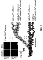
- FIGs. 1A, 1B and 1C show experimental data from optoGs and optoGq, two examples of light-activated inducers of secondary messenger signaling ('optoXRs') that have been developed. These light-activated inducers are a rhodopsin/GPCR chimerism. OptoGq provides light-responsive control of Gq signaling, whereas, OptoGs, provides light-responsive control of Gs signaling.
- FIG. 1A shows a schematic of optoGs and optoGq, consistent with example embodiments of the present disclosure.
- the intracellular loops of rhodopsin are replaced with those of adrenergic proteins normally coupled to either Gs (beta2) or Gq (alpha1).
- the genetic coding sequences are optimized for expression in human and murine cells. Examples of the resulting sequences include optoGs: Seq. Id. No. 1 and Seq. Id. No. 2; and optoGq: Seq. Id No. 3 and Seq. Id. No 4.
- amino acid sequences of the proteins are presented as non-limiting examples in support of embodiments which extend to variations (e.g., point mutations) in the genetic sequence that otherwise provide consistent, interchangeable or equivalent results.
- FIG. 1B shows Enzyme-Linked Immunosorbent Assay (ELISA) of cAMP (top), cGMP (middle), and IP 1 (bottom; a degradation product of IP 3 ) of cells transfected with either nothing, optoGs, or optoGq, consistent with an example embodiment of the present disclosure.
- the results of FIG. 1B were obtained from cells that were stimulated with 504 nm light (20 nm bandwidth) for one minute per spot or kept in the dark, as indicated.
- Stimulation was implemented using an environment-controlled inverted culture microscope (Leica DMI6000B).
- cAMP assay some cells were treated with 10uM forskolin for 30 minutes as a saturating, positive control of the assay.
- OptoGs significantly increased cAMP levels in response to light. No significant baseline increase of cAMP, or deviations of cGMP or IP 3 levels with optoGs were found.
- OptoGq significantly increased IP3 levels in response to light without significantly altering cGMP levels.
- An increase in cAMP levels with IP 3 production is believed to be a consequence of intracellular Ca 2+ release.
- FIG. 1C shows Ca-imaging of cells transfected with mCherry fusion proteins of optoGs and optoGq, consistent with example embodiments of the present disclosure.
- a cAMP-selective mutant of the cyclic nucleotide gated Ca 2+ channel CNGA2 was transfected in excess of optoGs.
- IP 3 activates release of intracellular Ca 2+ stores, thereby providing a reliable signal of Gq activation.
- a control population was also transfected with mCherry alone with the mutant CNGA2 in excess.
- Cells were loaded with fura-2 (20-25 minute incubation) and 2 ms exposures of 340 nm and 380 nm were acquired every two seconds. In each of optoGs and optoGq the acquisitions alone were sufficient to yield a Ca signal, while no significant signal was detected in the control population.
- FIG. 1 shows data obtained from a specific experimental setup, however, the disclosure is not so limited.
- various deliver techniques other than transfecting are contemplated including, but not limited to, viral transduction, ballistic gene delivery (gene gun), and spontaneous nucleic acid uptake.
- the base-rhodopsin can be modified for use with any suitable heterologous receptor subunits, such as Gi- coupled receptors like the alpha2-adrenergic receptor or the dopamine D2 receptor or the serotonin 5HT2A receptor; or other Gs- or Gq-coupled receptors like the dopamine D1A receptor or the metabotropic glutamate receptors.
- Gi- coupled receptors like the alpha2-adrenergic receptor or the dopamine D2 receptor or the serotonin 5HT2A receptor
- Gs- or Gq-coupled receptors like the dopamine D1A receptor or the metabotropic glutamate receptors.
- the base-rhodopsin is a protein derived from the bovine Bos taurus.
- the base-protein other than the base-rhodopsin mentioned above can also be used and includes various 7-transmembrane proteins, such as the cone opsins (red, green, or blue), rhodopsins of other species, and ligand-gated receptors like the dopamine or serotonin receptors.
- various 7-transmembrane proteins such as the cone opsins (red, green, or blue), rhodopsins of other species, and ligand-gated receptors like the dopamine or serotonin receptors.
- implementations relate to in vivo applications in mammals. These implementations include, but are not limited to, testing and confirming neural circuit and disease models.
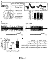
- FIGs. 3A and 3B show experimental data from an in vivo application of optoGs (opto- ⁇ 2AR) and optoGq (opto- ⁇ 1 AR), which are two examples of light-activated inducers of secondary messenger signaling.
- optoGs opto- ⁇ 2AR
- optoGq opto- ⁇ 1 AR
- Aspects of the present disclosure relate to the use and development of a versatile family of genetically encoded optical tools ('optoXRs') that leverage common structure-function relationships among G-protein-coupled receptors (GPCRs) to recruit and control, with high spatiotemporal precision, receptor-initiated biochemical signaling pathways.
- GPCRs G-protein-coupled receptors
- the results shown in FIGs. 3A and 3B relate to two specific optoXRs that selectively recruit distinct, targeted signaling pathways in response to light.
- the two optoXRs exerted opposing effects on spike firing in nucleus accumbens in vivo, and precisely timed optoXR photostimulation in nucleus accumbens by itself sufficed to drive conditioned place preference in freely moving mice.
- the optoXR approach allows testing of hypotheses regarding the causal impact of biochemical signaling in behaving mammals, in a targetable and temporally precise manner.
- Optical control over intracellular signaling was implemented in mammals, using shared structure-function relationships among GPCRs to develop and express in vivo multiple distinct opsin/GPCR2 chimeras with novel transduction logic that couples signal to effector.
- one or more chimeric opsin-receptor proteins are engineered to be functional within mammals in vivo, targetable to specific cells, and responsive to precisely timed light pulses.
- Such approaches allow for the use of high-speed optical stimulus (and protein response) to test for and characterize intracellular biochemical events at precisely-defined and behaviorally-relevant times.
- a few non-limiting example implementations include, pulsatile versus tonic modulation, synchrony between different modulatory systems, and other fundamental physiological and pathological processes in defined cell types over a range of timescales.
- the intracellular loops of rhodopsin were replaced with those of specific adrenergic receptors by first aligning conserved residues of the Gq-coupled human ⁇ 1a adrenergic receptor ( ⁇ 1 AR) and the Gs-coupled hamster ⁇ 2 -adrenergic receptor ( ⁇ 2 AR) with the Gt-coupled bovine rhodopsin ( FIG. 1A ).
- Exchanges of intracellular regions were engineered for each receptor based on structural models to transfer G-protein coupling from Gt, and optimized each receptor for in vivo expression in mammals.
- the native receptors Upon activation by varied ligands, the native receptors can explore multiple ensemble states to recruit canonical and non-canonical pathways in a ligand-biased signaling phenomenon.
- the optoXRs are likely to select a single active ensemble state upon sensing light in a manner dependent on biological context.
- Ratiometric [Ca 2+ ] i imaging demonstrated that 60 s of green light stimulation (504 +/- 6 nm, 7 mW mm -2 ) was sufficient to drive prominent [Ca 2+ ] i signals downstream of either optoXR but not in control conditions ( FIG. 2 ), revealing functional expression.
- transduced HEK cells were illuminated with 3 mW mm -2 504 +/- 6 nm light for 60 s and then lysed and analyzed for levels of cGMP, cAMP and IP 1 (a degradation product of IP 3 ) via immunoassays.
- the canonical pattern was as expected for opto- ⁇ 2 AR corresponding to its molecular design, as optical stimulation yielded significant production of cAMP in opto- ⁇ 2 AR-expressing cells ( FIG. 3A , top), comparable to that achieved with pharmacological stimulation of the wild-type ⁇ 2 AR and without recruitment of IP 3 ( FIG. 3A , middle), [Ca 2+ ] i ( FIG. 2 ), or substantial dark activity.
- optical stimulation yielded significant upregulation of IP 3 signaling in opto- ⁇ 1 AR-expressing cells ( FIG. 3A , middle), comparable to levels induced by pharmacological stimulation of the wild-type ⁇ 1 AR. Together with the [Ca 2+ ] i elevations ( FIG.
- OptoXR performance in intact neural tissue has been tested, including whether or not supplementation of retinal cofactors was necessary.
- lentiviral vectors carrying the optoXR fusion genes under control of the synapsin-I promoter to target biochemical modulation to local neurons rather than other potentially Gs/Gq-responsive cellular tissue elements such as glia and endothelial cells; FIG. 3B , top left
- This strategy targets biochemical modulation to neurons with somatodendritic compartments in accumbens ( ⁇ 95%GABAergic medium spiny neurons, without further subtype specificity; FIG.
- optogenetics were used to assess the ability of precisely timed optoXR stimulation to modulate behavior in freely moving mice.
- Portable solid-state light delivery was combined with transgenic expression of optoXRs to optically control intracellular signaling within accumbens neurons in the temporally precise manner used for operant behavior ( FIG. 5A ).
- Confocal analysis revealed expression to be limited to local accumbens neurons; in particular no labeling was observed in afferent fibers, in distant regions projecting to accumbens, in glia, or in surrounding regions.
- Optical stimulation was targeted to transduced accumbens as part of a three-day operant conditioned place preference assay ( FIG. 5A ).
- Behavioral analysis was performed using optical stimulation that was applied through an optical fiber (200 mm diameter, Thor Labs) coupled to a 473 nm blue diode laser (CrystaLaser) and registered with a cannula targeting accumbens (0-100 mm from tip).
- Light was delivered with 50 ms pulse width for optoXRs via a function generator (Agilent 33220A). Place preference was conducted in a standard apparatus (SD Instruments) with walls between chambers removed to permit free exploration. Data were analyzed from video for amount of time spent in each chamber by two independent, blinded observers using a custom tallying script run in MATLAB (Mathworks).
- Mammalian codon optimized sequences of opto- ⁇ 1 AR and opto- ⁇ 2 AR (amino acid sequences in FIG. 1A ) were synthesized and cloned into pcDNA3.1, and fused to the N-terminus of mCherry or YFP (with its start codon deleted) using the NotI site.
- the linker between the optoXR and mCherry/YFP is 5' GCGGCCGCC 3'.
- Lentiviral vectors containing Synapsin I optoXR mCherry were constructed by cloning the transgene for each optoXR mCherry into the AgeI and EcoRI sites of the pLenti SynapsinI hChR2 mCherry WPRE vector.
- High titer lentivirus was produced. Briefly, HEK 293FT cells were plated to 90% confluence in a 4-layer cell factory (Nunc) cultured with DMEM containing 10% FBS. Cells were co-transfected with 690 ⁇ g of the lentiviral vector described above and two helper plasmids (690 ⁇ g of p ⁇ CMVR8.74 and 460 ⁇ g of pMD2.G). Media was changed at 15 h post transfection. At 24 h post transfection, media was changed with 200-220 mL of serum free UltraCULTURE (Cambrex) containing 5 mM sodium butyrate.
- the culture supernatant now containing viruses, was spun at 1000 rpm for 5 min to remove cellular debris and then filtered using a 0.45 ⁇ m low-protein-binding filter flask.
- the clarified supernatant was then ultra centrifuged for 2 h at 55,000g using an SW 28 rotor (Beckman) to precipitate the virus. After centrifugation, supernatant was discarded and the resultant viral pellet was dissolved in a total of 100 ⁇ L of cold (4°C) PBS.
- the resuspended virus was centrifuged for 5 min at 7000 rpm to remove remaining cellular and viral debris. Aliquots were frozen at -80°C until further use.
- mice Female C57BL/6 mice, 10-12 weeks old, were housed and handled according to the Laboratory Vertebrate Animals protocol of Stanford University. Virus solution was delivered to the right nucleus accumbens as follows. Animals were anaesthetized under isoflurane and fur was sheared from the top of the head. While under isoflurane anesthesia, the head of the animal was placed in a stereotactic frame (David Kopf Instruments). A midline scalp incision was made and a ⁇ 1 mm diameter craniotomy was drilled 1.10mm anterior, and 1.45 mm lateral to bregma.
- a beveled 33 gauge needle (NanoFil, World Precision Instruments) pre-loaded with virus was then lowered into the accumbens (needle tip at 4.70-4.80 mm ventral to bregma) and 1.0 ⁇ L of virus was injected at 100 nL/min using an automated syringe pump (NanoFil, World Precision Instruments). Following injection, 3-5 min was allowed for tissue relaxation and fluid diffusion before retraction of the needle. For animals targeted for acute slice or in vivo recording experiments, the craniotomy was filled with dental cement (Lang Dental) and the incision was closed using VetBond (3M).
- Coronal, 275 ⁇ m-thick slices containing accumbens were cut and stored in a cutting solution containing 64mM NaCl, 2.5mM KCl, 1.25mM NaH 2 PO 4 , 25mM NaHCO 3 , 10mM glucose, 120mM sucrose, 0.5mM CaCl 2 and 7mM MgCl 2 (equilibrated with 95% O2/5% CO 2 ). Following slicing, slices were incubated in the cutting solution at 32-35°C for 30 min and then at room temperature until experimentation.
- slices were loaded on the stage of an upright microscope (BX51 W, Olympus) and perfused with an artificial cerebrospinal fluid containing 124mM NaCl, 3mM KCl, 1.25mM NaH 2 PO 4 , 26mM NaHCO 3 , 10mM glucose, 2.4mM CaCl 2 , and 1.3mM MgCl 2 (equilibrated with 95% O 2 /5% CO 2 ).
- HEK293FT cells were transfected using Lipofectamine 2000 (Invitrogen) in 24 well plates and changed to serum-free medium 4-6 hrs post-transfection.
- cells plated on matrigel-coated coverslips were loaded with 5 ⁇ g/ml fura-2 AM in F-127 Pluronic/DMSO (Probes) in Tyrode containing 1 ⁇ M ATR, at 37°C and 5% atmospheric CO 2 for 20-25 min.
- coverslips were imaged at 340nm/380nm on an Olympus BX51W using Metafluor (Axon Instruments) controlling a 300W Lambda DG-4 (Sutter).
- mice were transcardially perfused with ice-cold 4% paraformaldehyde (PFA) in PBS (pH 7.4) 90 min after termination of stimulation. Brains were removed and fixed overnight in 4% PFA and then equilibrated in 30% sucrose in PBS. Coronal, 40 ⁇ m-thick sections were cut on a freezing microtome and stored in cryoprotectant at 4°C until processed for immunohistochemistry. Free-floating sections were washed in PBS and then incubated for 30 min in 0.3% Tx100 and 3% normal donkey serum (NDS).
- PFA paraformaldehyde
- Sections were washed and incubated with secondary antibodies (1:1000) conjugated to either FITC or Cy5 (Jackson Laboratories, West Grove, PA) for 3 hrs at room temperature. Following 20 min incubation with DAPI (1:50,000) sections were washed and mounted on microscope slides with PVD-DABCO.
- embodiments of the present disclosure relate to optogenetic control of intracellular signaling and are useful for temporally precision while operating in vivo within behaving mammals, while displaying extremely low dark activity, and recruiting the complex fabric of multiple signaling molecules downstream of native receptors, thereby unifying in a single technology many of the individual positive aspects of other approaches.
- Similar embodiments directly probe the causal significance of seven-transmembrane-dependent signaling pathways triggered by other modulators, including myriad neurotransmitters and endocrine hormones.
- Other embodiments use an optoXR approach in ways that extend beyond excitable cells to capitalize upon the versatile integration of fiber-optic depth targeting with optogenetically targeted photosensitivity.
- One such embodiment relates to probing causal significance of temporally precise biochemical signaling in diverse non-excitable tissues.
- Embodiments of the present disclosure relate to considerations of the phenomenon of ligand-biased signaling, wherein varied ligands can stabilize ensemble receptor conformational states and thereby bias the intracellular action of the receptor in coupling to alternative transduction cascades.
- the optoXRs are used to induce these alternative cascades to similar levels as with pharmacological manipulation (for example, opto- ⁇ 2 AR can induce similar changes in MAPK activation compared with native ligand acting on the wild-type ⁇ 2 AR); however, individual optoXRs may not always be found to permit control of all of the conformational states that contribute to ligand biased signaling.
- Retinal-based tools can be particularly useful due to the presence of the endogenous chromophore in mammalian tissues, and the extremely low activity in the dark.
- Optogenetics can take the form of diverse effectors linked to fast, single-component retinal-binding modules, capitalizing on the temporal precision of optics.
- Embodiments of the present disclosure use optoXR methods to complement microbial opsin strategies, providing another dimension of fast, targetable cellular control operative in behaving mammals.
- wavelength-shifted versions of the optoXRs are used.
- Such optoXRs can be particularly useful for providing separable channels of biochemical and electrical control.
- compositions of the present disclosure include the protein and nucleic acid sequences provided herein including variants which are more than about 50% homologous to the provided sequence up to and including 100% homologous.
Description
- The present disclosure relates generally to systems and approaches for generating secondary messengers in response to optical stimulus and more particularly to a cell lines, nucleotide sequences, chimeric proteins, and uses thereof, each relating to the production of secondary messengers in response to light.
- Guanine nucleotide-binding proteins (G proteins) are believed to alternate between an inactive guanosine diphosphate (GDP) state and an active guanosine triphosphate (GTP) bound state. These two states have been linked to the release of a secondary messenger within a cell. The released secondary messenger can function to regulate downstream cell processes.
- Secondary messengers include signaling molecules that are rapidly generated/released. These molecules produce cellular responses by activating effector proteins within the cell. Example cellular signaling systems include the phosphoinositol system, the cyclic adenosine monophosphate (cAMP) system, and the arachidonic acid system.
- Changes between the different states of the G proteins can be triggered as a result of proteins called G protein-coupled receptors (GPCRs), G protein-linked receptors (GPLR), seven transmembrane domain receptors (7TM receptors) or heptahelical receptors. This protein family includes a variety of transmembrane receptors. These receptors respond to external stimuli (e.g., light, neurotransmitters, odors or hormones) by activating signal transduction pathways internal to the cell. Specifically, ligands bind and activate the transduction pathways thereby causing the G proteins to alternate states. GPCR-related activity is associated with many diseases, and thus, GPCRs are the target of many pharmaceuticals and treatments.
- It is believed that over 30% of all drugs on the market target G-protein coupled receptors (GPCRs) and that many of those drugs relate to the production or inhibition of the secondary messenger cAMP. There is an abundance of pathological processes that directly involve cAMP, including neurophysiological, endocrinological, cardiac, metabolic, and immune diseases. In the study of complex mammalian behaviors, technological limitations have prevented spatiotemporally precise control over intracellular signaling processes. Current chemical-based methods for modulating secondary messenger levels, such as cAMP levels, operate relatively slowly and present problems to study activity on the fast timescales that the body uses in connection with certain tissue, such as in nervous or cardiac tissue. These chemical-methods often lack the speed to probe these fast timescales (e.g., while screening for novel therapeutics). Li et al (Proc. Natl. Acad. Sci., USA, (2005), 102; 17816-17821 describes fast non-invasive activation and inhibition of neural and network activity by vertebrate rhodopsin and green algae channelrhodopsin.
- The present disclosure is directed to overcoming the above-mentioned challenges and others related to generation of secondary messengers and related imaging devices and their implementations. The present invention is defined in the accompanying claims and is exemplified in a number of implementations and applications, some of which are summarized below.
- Consistent with an embodiment of the present disclosure a method is implemented for generating secondary messengers in a cell. A nucleotide sequence for expressing a chimeric light responsive membrane protein (e.g., rhodopsin) is modified with one or more heterologous receptor subunits {e.g., an adrenergic receptor (alpha!, Beta2)}. The light responsive membrane protein is expressed in a cell for producing a secondary messenger in response to light.
- Consistent with an embodiment of the present disclosure a method is implemented for assessing the efficacy of a putative treatment regimen (e.g., a drug or electrical stimulus or anything that works via these secondary messengers) relating to intracellular messengers. A nucleotide sequence for expressing a chimeric light responsive membrane protein (rhodopsin) is modified with one or more heterologous receptor subunits {e.g., an adrenergic receptor (alpha1, Beta2)}. The light responsive membrane protein is expressed in a cell for producing a secondary messenger in response to light. The protein is exposed to light. The effects of the treatment are assessed.
- An embodiment of the present disclosure is directed toward, a cell expressing a chimeric light responsive membrane protein (rhodopsin) with one or more heterologous receptor subunits {e.g., an adrenergic receptor (alpha1, Beta2)}.
- An embodiment of the present disclosure is directed toward, a nucleotide sequence for expressing a chimeric light responsive membrane protein (rhodopsin) with one or more heterologous receptor subunits {e.g., an adrenergic receptor (alpha1, Beta2)}.
- The above summary of the present disclosure is not intended to describe each illustrated embodiment or every implementation of the present disclosure. The figures and detailed description that follow more particularly exemplify these embodiments.
- The disclosure may be more completely understood in consideration of the detailed description of various embodiments of the disclosure that follows in connection with the accompanying drawings, in which:
-
FIG. 1A shows a schematic showing optoGs and optoGq, consistent with example embodiments of the present disclosure; -
FIG. 1B shows Enzyme-Linked Immunosorbent Assay (ELISA) of cAMP, cGMP, and IP1 of cells transfected with either nothing, optoGs, or optoGq, consistent with example embodiments of the present disclosure; -
FIG. 1C shows Ca-imaging of cells transfected with mCherry fusion proteins of optoGs and optoGq, consistent with example embodiments of the present disclosure; -
FIG. 2 shows Ca-imaging of cells transfected with mCherry fusion proteins of optoGs and optoGq, consistent with example embodiments of the present disclosure; -
FIG. 3A shows cAMP, IP1 and IP3 levels for HEK cells expressing various constructs, consistent with example embodiments of the present disclosure; -
FIG. 3B shows a lentiviral express vector, GAD immunostaining of opto-α1AR-expressing cells and observed pCREB activation in optoXR-expressing cells (mCherry+) following 10 min optical stimulation, consistent with example embodiments of the present disclosure; -
FIG. 4A shows optrode targeting of transduced accumbens, spike waveforms and baseline firing rates for indicated constructs, consistent with example embodiments of the present disclosure; -
FIG. 4B shows in vivo optrode recordings with light stimulation, consistent with example embodiments of the present disclosure; -
FIG. 4C shows change in spiking frequency with light versus baseline, consistent with example embodiments of the present disclosure; -
FIG. 4D shows firing rate change kinetics, consistent with example embodiments of the present disclosure; -
FIG. 5A shows stereotactic targeting of a transduced region, a freely moving mouse with implanted fiber optics, a schematic of place preference apparatus and test and a trace of a freely exploring mouse, consistent with example embodiments of the present disclosure; -
FIG. 5B shows preferences for control and opto-α1AR, consistent with example embodiments of the present disclosure; and -
FIG. 5C shows results of total distance for various open field tests; consistent with example embodiments of the present disclosure. - While the disclosure is amenable to various modifications and alternative forms, specifics thereof have been shown by way of example in the drawings and will be described in detail. It should be understood, however, that the intention is not to limit the disclosure to the particular embodiments described. On the contrary, the intention is to cover all modifications, equivalents, and alternatives falling within the spirit and scope of the disclosure.
- The present disclosure is believed to be useful for enabling practical applications of a variety of optical-based systems and methods, and the disclosure has been found to be particularly suited for use in systems and methods dealing with optical control of secondary messenger levels within a cell. While the present disclosure is not necessarily limited to such applications, various aspects of the disclosure may be appreciated through a discussion of various examples using this context.
- Embodiments of the present disclosure involve a chimeric membrane protein that responds to optical stimulus by causing the release of a secondary messenger within the cell. In a specific instance, the chimeric protein is a combination of a heterologous receptor subunit and a protein that undergoes conformation in reaction to light via photoisomerization and thus is activated by light. Rhodopsins or retinylidene proteins provide an example group of light-responsive proteins that can be modified to include a heterologous receptor subunit.
- According to an embodiment of the present disclosure a protein believed to contain a seven transmembrane α-helical domain is modified to include a heterologous receptor subunit associated with a secondary messenger. When expressed in a cell membrane, the protein reacts to light by undergoing a conformal change. The conformal change triggers the release/production of the secondary messenger.
- Embodiments of the present disclosure involve a nucleotide sequence for coding a chimeric membrane protein that responds to optical stimulus by causing the release of a secondary messenger within the cell.
- Embodiments of the present disclosure involve a cell that expresses a heterologous and chimeric membrane protein. The chimeric membrane protein responds to optical stimulus by triggering the release of a secondary messenger within the cell. In certain embodiments the expression of the chimeric membrane protein occurs in vivo. In other embodiments expression of the chimeric membrane protein occurs in vitro.
- Embodiments of the present disclosure can implemented for production of any suitable secondary messenger by modifying a Guanine nucleotide-binding protein coupled receptor protein (GPCR) to include the appropriate receptor subunit.
- Embodiments of the present disclosure allow for the use of proteins that respond to a variety of wavelengths and intensities of light.
- An embodiment of the present disclosure involves the use of a chimeric GPCR protein, as disclosed herein, to determine any downstream effect of the secondary messenger activity of interest.
- Embodiments of the present disclosure are directed to expression of a chimeric GPCR protein in a variety of cell types including, but not limited to, mammalian cells, stems cells, plant cells, and unicellular organisms like yeast and E. coli.
- A specific embodiment of the present disclosure is related to an optimized expression of a chimeric protein with attached fluorescent proteins for ease of visualization, and optimized use of the modality for studying downstream effects of the secondary messenger activity induced by light.
- An embodiment of the present disclosure is directed to genetically targeting a chimeric GPCR protein, as disclosed herein, to specific cell populations for expression therein. Cell-type specific promoters exist that are selectively expressed in a target cell type (e.g., Synapsin-1 for targeting neurons; Troponin variants for cardiac tissue). Placing these promoters upstream of the chimeric GPCR protein in an expression vector can be used to target expression of the protein to a cell type of interest. This includes inducible, reversible, or otherwise controllable promoter systems such as Tet-response, ER-response, and Cre/Lox systems.
- According to an example embodiment of the present disclosure a genetically encodeable protein is developed such that, when these are expressed in cell types of interest, cyclic adenosine monophosphate (cAMP) is produced in response to light. This can be useful, for example, to visualize downstream effects on cell physiology including, but not limited to, screening for pharmaceuticals. Other embodiments use a chimeric and heterologous GPCR that results in the release of secondary messengers in response to light. Example secondary messengers include cAMP, cyclic guanosine monophosphate (cGMP), inositol trisphosphate/
inositol - Consistent with an embodiment of the present disclosure a method is implemented for assessing the efficacy of a putative treatment regimen (e.g., a drug or electrical stimulus or anything that works via these secondary messengers) relating to intracellular messengers. A nucleotide sequence for expressing a chimeric light responsive membrane protein (e.g., rhodopsin) is modified with one or more heterologous receptor subunits {e.g., an adrenergic receptor (alpha1, Beta2)}. The light responsive membrane protein is expressed in a cell for producing a secondary messenger in response to light. The protein is exposed to light. The effects of the treatment are assessed.
- The light can be applied according to a desired stimulus profile. In one embodiment the expressed membrane protein responds to light within tens of milliseconds. Thus, the stimulus profile can include a series of light pulses in rapid succession and the resulting effects can be monitored using, for example, Ca2+ sensitive dyes.
- In one instance, the cell can first be stimulated without the treatment. Once the treatment is administered, the cell can then be stimulated again. The results of each test can be compared to assess the effectiveness of the treatment.
- The treatment can include a wide variety of different implementations including, but not limited to, pharmaceuticals, modifications to the cell (genetic or otherwise), physical parameters of the cell (e.g., temperature changes or electrical stimulus) or a treatment regimen applied to an organism.
- In one embodiment, the treatment is the optical stimulus of the expressed membrane protein. In such an instance the effectiveness can be measured, for example, by monitoring the symptoms associated with a disorder to be treated.
- In another embodiment, the treatment regimen is implemented as part of modeling a disease or disorder. For example, a disease model can be used (cells or animals) and the background/baseline state can be assessed before the protein is expressed and the treatment regimen evaluated.
- Experimental results show that optically-evoked cAMP regulation of targeted ion channels can be visualized by transfecting cells with both the cAMP-inducer and a cAMP-targeted cation channel and visualizing resultant activity using Ca2+-sensitive dyes. This suite of genetically-encodable, optically-activated modulators of secondary messenger activity can be useful in screening novel therapeutics as well as being a therapeutic modality itself, given the implication of cAMP in numerous diseases states, like ADHD and cardiac channelopathies. The protein can be engineered for use with various other secondary messengers (e.g., IP3), other colors for light activation by engineering the retinal binding site or choosing for the chimera a rhodopsin or cone opsin with a different absorbance/action spectrum, and other downstream effects of the secondary messenger, such as calcium signaling and/or kinase activity.
-
FIGs. 1A, 1B and 1C show experimental data from optoGs and optoGq, two examples of light-activated inducers of secondary messenger signaling ('optoXRs') that have been developed. These light-activated inducers are a rhodopsin/GPCR chimerism. OptoGq provides light-responsive control of Gq signaling, whereas, OptoGs, provides light-responsive control of Gs signaling. - In both optoGs and optoGq it has been shown that there is negligible difference in baseline cAMP and IP3 levels in darkness and that there is no crossover to other secondary messenger pathways such as cGMP. The increased cAMP levels seen with light stimulation of optoGq is an expected downstream effect of IP3 production.
-
FIG. 1A shows a schematic of optoGs and optoGq, consistent with example embodiments of the present disclosure. For each protein, the intracellular loops of rhodopsin are replaced with those of adrenergic proteins normally coupled to either Gs (beta2) or Gq (alpha1). The genetic coding sequences are optimized for expression in human and murine cells. Examples of the resulting sequences include optoGs: Seq. Id. No. 1 and Seq. Id. No. 2; and optoGq: Seq. Id No. 3 and Seq. Id.No 4. - As is appreciated by the skilled artisan, the amino acid sequences of the proteins are presented as non-limiting examples in support of embodiments which extend to variations (e.g., point mutations) in the genetic sequence that otherwise provide consistent, interchangeable or equivalent results.
-
FIG. 1B shows Enzyme-Linked Immunosorbent Assay (ELISA) of cAMP (top), cGMP (middle), and IP1 (bottom; a degradation product of IP3) of cells transfected with either nothing, optoGs, or optoGq, consistent with an example embodiment of the present disclosure. The results ofFIG. 1B were obtained from cells that were stimulated with 504 nm light (20 nm bandwidth) for one minute per spot or kept in the dark, as indicated. - Stimulation was implemented using an environment-controlled inverted culture microscope (Leica DMI6000B). In the cAMP assay, some cells were treated with 10uM forskolin for 30 minutes as a saturating, positive control of the assay. OptoGs significantly increased cAMP levels in response to light. No significant baseline increase of cAMP, or deviations of cGMP or IP3 levels with optoGs were found. OptoGq significantly increased IP3 levels in response to light without significantly altering cGMP levels. An increase in cAMP levels with IP3 production is believed to be a consequence of intracellular Ca2+ release.
-
FIG. 1C shows Ca-imaging of cells transfected with mCherry fusion proteins of optoGs and optoGq, consistent with example embodiments of the present disclosure. To detect cAMP, a cAMP-selective mutant of the cyclic nucleotide gated Ca2+ channel CNGA2 was transfected in excess of optoGs. IP3 activates release of intracellular Ca2+ stores, thereby providing a reliable signal of Gq activation. A control population was also transfected with mCherry alone with the mutant CNGA2 in excess. Cells were loaded with fura-2 (20-25 minute incubation) and 2 ms exposures of 340 nm and 380 nm were acquired every two seconds. In each of optoGs and optoGq the acquisitions alone were sufficient to yield a Ca signal, while no significant signal was detected in the control population. -
FIG. 1 shows data obtained from a specific experimental setup, however, the disclosure is not so limited. For example, various deliver techniques other than transfecting are contemplated including, but not limited to, viral transduction, ballistic gene delivery (gene gun), and spontaneous nucleic acid uptake. - The base-rhodopsin can be modified for use with any suitable heterologous receptor subunits, such as Gi- coupled receptors like the alpha2-adrenergic receptor or the dopamine D2 receptor or the serotonin 5HT2A receptor; or other Gs- or Gq-coupled receptors like the dopamine D1A receptor or the metabotropic glutamate receptors.
- According to one example embodiment, the base-rhodopsin is a protein derived from the bovine Bos taurus.
- According to one embodiment the base-protein other than the base-rhodopsin mentioned above can also be used and includes various 7-transmembrane proteins, such as the cone opsins (red, green, or blue), rhodopsins of other species, and ligand-gated receptors like the dopamine or serotonin receptors.
- Various implementations relate to in vivo applications in mammals. These implementations include, but are not limited to, testing and confirming neural circuit and disease models.
-
FIGs. 3A and 3B show experimental data from an in vivo application of optoGs (opto-β2AR) and optoGq (opto-α1AR), which are two examples of light-activated inducers of secondary messenger signaling. Aspects of the present disclosure relate to the use and development of a versatile family of genetically encoded optical tools ('optoXRs') that leverage common structure-function relationships among G-protein-coupled receptors (GPCRs) to recruit and control, with high spatiotemporal precision, receptor-initiated biochemical signaling pathways. - The results shown in
FIGs. 3A and 3B relate to two specific optoXRs that selectively recruit distinct, targeted signaling pathways in response to light. The two optoXRs exerted opposing effects on spike firing in nucleus accumbens in vivo, and precisely timed optoXR photostimulation in nucleus accumbens by itself sufficed to drive conditioned place preference in freely moving mice. The optoXR approach allows testing of hypotheses regarding the causal impact of biochemical signaling in behaving mammals, in a targetable and temporally precise manner. - Optical control over intracellular signaling was implemented in mammals, using shared structure-function relationships among GPCRs to develop and express in vivo multiple distinct opsin/GPCR2 chimeras with novel transduction logic that couples signal to effector. Consistent with various implementations, one or more chimeric opsin-receptor proteins are engineered to be functional within mammals in vivo, targetable to specific cells, and responsive to precisely timed light pulses. Such approaches allow for the use of high-speed optical stimulus (and protein response) to test for and characterize intracellular biochemical events at precisely-defined and behaviorally-relevant times. A few non-limiting example implementations include, pulsatile versus tonic modulation, synchrony between different modulatory systems, and other fundamental physiological and pathological processes in defined cell types over a range of timescales.
- Mammalian implementations have been successfully implemented. In one example implementation, the intracellular loops of rhodopsin were replaced with those of specific adrenergic receptors by first aligning conserved residues of the Gq-coupled human α1a adrenergic receptor (α1AR) and the Gs-coupled hamster β2-adrenergic receptor (β2AR) with the Gt-coupled bovine rhodopsin (
FIG. 1A ). Exchanges of intracellular regions (including carboxy-terminal domains) were engineered for each receptor based on structural models to transfer G-protein coupling from Gt, and optimized each receptor for in vivo expression in mammals. Upon activation by varied ligands, the native receptors can explore multiple ensemble states to recruit canonical and non-canonical pathways in a ligand-biased signaling phenomenon. The optoXRs are likely to select a single active ensemble state upon sensing light in a manner dependent on biological context. - Genes encoding chimeras (opto-α1AR and optoβ2AR) were fused to a fluorescent protein. Validation of functional optoXR expression, was accomplished through imaged [Ca2+]i (intracellular calcium concentration) in HEK cells transfected with opto-α1AR alone (expected to recruit[Ca2+]i via Gq), or with both opto-β2AR (expected to recruit cyclic AMP via Gs) and the cAMP-gated Ca2+ channel CNGA2-C460W/E583M. Ratiometric [Ca2+]i imaging demonstrated that 60 s of green light stimulation (504 +/- 6 nm, 7 mW mm-2) was sufficient to drive prominent [Ca2+]i signals downstream of either optoXR but not in control conditions (
FIG. 2 ), revealing functional expression. To test specificity of the signaling controlled by each optoXR, transduced HEK cells were illuminated with 3 mW mm-2 504 +/- 6 nm light for 60 s and then lysed and analyzed for levels of cGMP, cAMP and IP1 (a degradation product of IP3) via immunoassays. The canonical pattern was as expected for opto-β2AR corresponding to its molecular design, as optical stimulation yielded significant production of cAMP in opto-β2AR-expressing cells (FIG. 3A , top), comparable to that achieved with pharmacological stimulation of the wild-type β2AR and without recruitment of IP3 (FIG. 3A , middle), [Ca2+]i (FIG. 2 ), or substantial dark activity. In contrast, optical stimulation yielded significant upregulation of IP3 signaling in opto-α1AR-expressing cells (FIG. 3A , middle), comparable to levels induced by pharmacological stimulation of the wild-type α1AR. Together with the [Ca2+]i elevations (FIG. 2 ), these data reveal the pattern expected for Gq recruitment, a pattern not seen in opto-β2AR-expressing cells (FIG. 3A , top). Optical stimulation of cells expressing either construct was unable to modulate cGMP levels (FIG. 3A , bottom), further indicating the signaling specificity of the chimeric proteins. Similar assays revealed that the optoXRs retain an action spectrum close to that of native rhodopsin, are able to integrate signals over a range of biologically suitable light fluxes, and can activate non-canonical pathways to a similar extent as wild-type receptors, as for p42/p44-MAPK signaling. - OptoXR performance in intact neural tissue has been tested, including whether or not supplementation of retinal cofactors was necessary. In one such test, lentiviral vectors carrying the optoXR fusion genes under control of the synapsin-I promoter (to target biochemical modulation to local neurons rather than other potentially Gs/Gq-responsive cellular tissue elements such as glia and endothelial cells;
FIG. 3B , top left) were stereotactically injected into the nucleus accumbens of adult mice. This strategy targets biochemical modulation to neurons with somatodendritic compartments in accumbens (∼95%GABAergic medium spiny neurons, without further subtype specificity;FIG. 3B , left) and excludes fibers of passage or afferent presynaptic terminals as these lentiviruses do not transduce cells via axons. Two weeks after transduction, acute coronal slices of accumbens were prepared in artificial cerebrospinal fluid, optically stimulated for 10 min, and immediately fixed and stained for Ser 133-phosphorylated CREB (pCREB), a biochemical integrator of both cAMP and Ca2+-coupled signaling cascades. Without supplementation of exogenous retinoids, significantly elevated pCREB was observed in the optoXR-expressing populations (FIG. 3B , right) and not in non-illuminated tissue. - The functional consequences of optoXR activation on accumbens local electrical activity was determined by recording multi-unit in vivo neuronal firing with an optrode targeted to transduced accumbens (
FIG. 4A ). No significant differences in baseline firing rates were observed in the dark with either construct (FIG. 4A , bottom right). Optical stimulation resulted in decreased network firing in opto-β2AR-expressing accumbens (left trace inFIG. 4B illustrates effect kinetics; summary data shown inFIG. 4C and 4D respectively), in agreement with previous pharmacological studies targeting Gs. Optical stimulation increased firing in opto-α1AR-expressing accumbens (FIG. 4B right;FIG. 4C, 4D ). Spike frequency histograms showed that the kinetics of optoXR effects on firing rates was consistent with biochemical rather than electrical initiation of the signal (FIG. 4D ). These electrophysiological data, in combination with the earlier biochemical validations, support that optoXRs can be functionally expressed in vivo, to permit differential photoactivatable control of intracellular cascades and to modulate network physiology. - In one implementation, optogenetics were used to assess the ability of precisely timed optoXR stimulation to modulate behavior in freely moving mice. Portable solid-state light delivery was combined with transgenic expression of optoXRs to optically control intracellular signaling within accumbens neurons in the temporally precise manner used for operant behavior (
FIG. 5A ). Confocal analysis revealed expression to be limited to local accumbens neurons; in particular no labeling was observed in afferent fibers, in distant regions projecting to accumbens, in glia, or in surrounding regions. Optical stimulation was targeted to transduced accumbens as part of a three-day operant conditioned place preference assay (FIG. 5A ). On each day of the test, animals were allowed to freely explore the place preference apparatus (FIG. 5A , bottom). Onday 1, animals freely explored the apparatus without optical stimulation. Onday 2, whenever the animal freely entered the designated conditioned chamber, a laser-diode-coupled optical fiber registered to the transduced region delivered light pulses at 10 Hz to approximate the likely intensity of monoaminergic input during strong reward. Path tracing revealed that the flexible optical fiber approach allowed full and unimpeded exploration of all chambers (FIG. 5A , bottom). Onday 3, animals again freely explored the apparatus without optical stimulation, and the time spent in the conditioned chamber was quantified by two independent, blinded scorers. Notably, animals expressing opto-α1AR showed a robust increase in preference for the conditioned side of the apparatus following optical stimulation (FIG. 5B ). This effect of temporally precise biochemical modulation was reproducible across two separate cohorts of opto-α1AR animals (n=5-6, P<0.05, Student's t-test for each cohort for time in conditioned chamber; n=11, P<0.01 for the total population), whereas the other opsin genes, opto-β2AR and ChR2, appeared less effective in driving preference. The effect of opto-α1AR stimulation in accumbens neurons was specific to reward-related behavior and did not extend to direct modulation of anxiety-related behaviors or locomotor activity, as identical optical stimulation delivered to a cohort of the same animals in an open field test revealed no significant effect on distance travelled or preference for wall proximity (FIG. 5C ). - A specific and non-limiting implementation that is consistent with the above experiments is now described. In vivo recording and analysis was performed using optrodes consisting of a multi-mode optical fiber 200 mm in diameter (Thorlabs) coupled to a recording electrode (1MV tungsten, A-M Systems) with an electrode/fiber tip-to-tip distance of 200-400 mm were lowered into the transduced accumbens (electrode tip 4.8-5.2mm below bregma) of mice placed in a stereotactic frame (David Kopf Instruments) and anaesthetized under isoflurane. Light from a 473nm diode laser (CrystaLaser) was delivered through the fiber. Electrical signals were bandpass filtered and amplified (0.3-1 kHz, 1800 Microelectrode AC Amplifier, A-M Systems) and analyzed withpClamp 10.0 (Molecular Devices). Spikes were detected by threshold and individually confirmed by inspection.
- Behavioral analysis was performed using optical stimulation that was applied through an optical fiber (200 mm diameter, Thor Labs) coupled to a 473 nm blue diode laser (CrystaLaser) and registered with a cannula targeting accumbens (0-100 mm from tip). Light was delivered with 50 ms pulse width for optoXRs via a function generator (Agilent 33220A). Place preference was conducted in a standard apparatus (SD Instruments) with walls between chambers removed to permit free exploration. Data were analyzed from video for amount of time spent in each chamber by two independent, blinded observers using a custom tallying script run in MATLAB (Mathworks). For open field tests, animals were placed in a square open field measuring 40340 cm; light stimulation was delivered with the same parameters as for place preference experiments. Videos were analyzed using automated software (Viewpoint), for total time and distance in the central 15315 cm square versus the outer annulus (remainder of the field).
- Statistical analysis, where indicated, was performed using two-tailed Student's t-tests (calculated in Microsoft Excel) or one-way ANOVA with Tukey post-hoc tests (GraphPad Prism) were used. All summary bar graphs are presented as mean +/- s.e.m., with significance denoted as follows: *P<0.05, **P<0.01, ***P<0.001.
- Further details supporting the surprising results and effectiveness of various embodiments of the present disclosure can be found in Temporally precise in vivo control of intracellular signalling, Raag D. Airan, et al., Nature 458, 1025-1029 (23 April 2009), which is fully incorporated herein by reference.
- The following description provides details for specific and non-limiting method that is consistent with an embodiment of the present disclosure. Numerous variations of this methodology are envisioned and within the scope of the present disclosure.
- Mammalian codon optimized sequences of opto-α1AR and opto-β2AR (amino acid sequences in
FIG. 1A ) were synthesized and cloned into pcDNA3.1, and fused to the N-terminus of mCherry or YFP (with its start codon deleted) using the NotI site. The linker between the optoXR and mCherry/YFP is 5' GCGGCCGCC 3'. Lentiviral vectors containing Synapsin I optoXR mCherry were constructed by cloning the transgene for each optoXR mCherry into the AgeI and EcoRI sites of the pLenti SynapsinI hChR2 mCherry WPRE vector. - High titer lentivirus was produced. Briefly, HEK 293FT cells were plated to 90% confluence in a 4-layer cell factory (Nunc) cultured with DMEM containing 10% FBS. Cells were co-transfected with 690 µg of the lentiviral vector described above and two helper plasmids (690 µg of pΔCMVR8.74 and 460 µg of pMD2.G). Media was changed at 15 h post transfection. At 24 h post transfection, media was changed with 200-220 mL of serum free UltraCULTURE (Cambrex) containing 5 mM sodium butyrate. At 40 h post transfection, the culture supernatant, now containing viruses, was spun at 1000 rpm for 5 min to remove cellular debris and then filtered using a 0.45 µm low-protein-binding filter flask. The clarified supernatant was then ultra centrifuged for 2 h at 55,000g using an SW 28 rotor (Beckman) to precipitate the virus. After centrifugation, supernatant was discarded and the resultant viral pellet was dissolved in a total of 100 µL of cold (4°C) PBS. The resuspended virus was centrifuged for 5 min at 7000 rpm to remove remaining cellular and viral debris. Aliquots were frozen at -80°C until further use.
- Female C57BL/6 mice, 10-12 weeks old, were housed and handled according to the Laboratory Vertebrate Animals protocol of Stanford University. Virus solution was delivered to the right nucleus accumbens as follows. Animals were anaesthetized under isoflurane and fur was sheared from the top of the head. While under isoflurane anesthesia, the head of the animal was placed in a stereotactic frame (David Kopf Instruments). A midline scalp incision was made and a ∼1 mm diameter craniotomy was drilled 1.10mm anterior, and 1.45 mm lateral to bregma. A beveled 33 gauge needle (NanoFil, World Precision Instruments) pre-loaded with virus was then lowered into the accumbens (needle tip at 4.70-4.80 mm ventral to bregma) and 1.0 µL of virus was injected at 100 nL/min using an automated syringe pump (NanoFil, World Precision Instruments). Following injection, 3-5 min was allowed for tissue relaxation and fluid diffusion before retraction of the needle. For animals targeted for acute slice or in vivo recording experiments, the craniotomy was filled with dental cement (Lang Dental) and the incision was closed using VetBond (3M). For animals targeted for behavioral analysis, cannulas (C316G, cut 4.5 mm below the pedestal; PlasticsOne) were placed with the pedestal flush to the skull. Cannulae were secured using Metabond (Parkell) and dental cement (Lang Dental). Following drying of VetBond or cement, animals were removed from the frame and allowed to recover for at least one week before further manipulation. Control animals for behavioral experiments underwent the same manipulations (surgery, cannula implantation, light stimulation) as experimental animals, and were injected with vehicle (PBS) alone instead of virus. For place preference experiments, animals that did not show a baseline preference for either side chamber (>70% or <10%) or for the central chamber (>40%) were admitted into the study; >90% of all animals met these criteria for an unbiased, balanced place preference design.
- Animals were anaesthetized under isoflurane and decapitated using surgical shears (Fine Science Tools). Coronal, 275 µm-thick slices containing accumbens were cut and stored in a cutting solution containing 64mM NaCl, 2.5mM KCl, 1.25mM NaH2PO4, 25mM NaHCO3, 10mM glucose, 120mM sucrose, 0.5mM CaCl2 and 7mM MgCl2 (equilibrated with 95% O2/5% CO2). Following slicing, slices were incubated in the cutting solution at 32-35°C for 30 min and then at room temperature until experimentation. For ex vivo optoXR stimulation, slices were loaded on the stage of an upright microscope (BX51 W, Olympus) and perfused with an artificial cerebrospinal fluid containing 124mM NaCl, 3mM KCl, 1.25mM NaH2PO4, 26mM NaHCO3, 10mM glucose, 2.4mM CaCl2, and 1.3mM MgCl2 (equilibrated with 95% O2/5% CO2). Light from a 300W Lambda DG-4 (Sutter) was passed through a 473 nm ± 20 nm bandpass filter (Semrock) and applied to the slices using a 4X objective (0.28 NA) for 10 min followed immediately by fixation for later analysis.
- HEK293FT cells (Invitrogen) were transfected using Lipofectamine 2000 (Invitrogen) in 24 well plates and changed to serum-free medium 4-6 hrs post-transfection. For Ca2+ imaging, cells plated on matrigel-coated coverslips were loaded with 5µg/ml fura-2 AM in F-127 Pluronic/DMSO (Probes) in Tyrode containing 1µM ATR, at 37°C and 5% atmospheric CO2 for 20-25 min. Following loading, coverslips were imaged at 340nm/380nm on an Olympus BX51W using Metafluor (Axon Instruments) controlling a 300W Lambda DG-4 (Sutter). For immunoassays, 18-24 hrs after transfection, 1µM ATR and 50mM LiCl (to prevent IP1 degradation) were added and plates transferred to an environmentally-controlled microscope (Leica DMI6000; 37°C, 5% atmospheric CO2). 5 regions/well were optically stimulated for 1 min each (Sutter 300W Lambda DG-4; Semrock 504/12nm bandpass filter; 10X 0.30 NA objective); 3 wells/condition. Following incubation (cAMP/cGMP: 20 min; EP1: 1 hr), cells were lysed and analyze by HTRF (CisBio) and a Biotek Synergy4 reader.
- Following in vivo stimulation, mice were transcardially perfused with ice-cold 4% paraformaldehyde (PFA) in PBS (pH 7.4) 90 min after termination of stimulation. Brains were removed and fixed overnight in 4% PFA and then equilibrated in 30% sucrose in PBS. Coronal, 40 µm-thick sections were cut on a freezing microtome and stored in cryoprotectant at 4°C until processed for immunohistochemistry. Free-floating sections were washed in PBS and then incubated for 30 min in 0.3% Tx100 and 3% normal donkey serum (NDS). For acute slice experiments, immediately following stimulation the 275 µm-thick slices were fixed for 1 hr in ice-cold 4% PFA and incubated with 0.5% Tx100 and 3% NDS. For MAPK assays, immediately following HEK293 cell stimulation, coverslips were fixed for 15 min, incubated with 0.6% H2O2 and then permeabilized with 0.1% Tx100 in 3% NDS. Primary antibody incubations were conducted overnight in 0.01% Tx100 and 3% NDS for mouse anti-GAD67 1:500, Millipore, Billerica, MA; rabbit anti-cfos 1:500, Calbiochem, San Diego, CA; rabbit anti-phospho-CREB Ser133 1:500, Millipore. Sections were washed and incubated with secondary antibodies (1:1000) conjugated to either FITC or Cy5 (Jackson Laboratories, West Grove, PA) for 3 hrs at room temperature. Following 20 min incubation with DAPI (1:50,000) sections were washed and mounted on microscope slides with PVD-DABCO. The remaining overnight primary antibody incubations (rabbit anti-phosphoErk1/2; anti-phospho-MAPK p38 1:500, Promega, Madison, WI; mouse monoclonal anti-dopamine D1 receptor 1:50, Chemicon; rabbit polyclonal anti-dopamine D2 receptor 1:50, Millipore; goat polyclonal anti-choline acetyltransferase 1:200, Millipore) were followed by incubation with biotinylated secondary antibody (1:500, Jackson Laboratories), avidin-biotin-horseradish peroxidase treatment (ABC kit, Vector Labs, Burlingame, CA), and TSA detection (Perkin Elmer, Shelton, CT) according to manufacturer's instructions.
- Confocal fluorescence images were acquired on a Leica TCS SP5 scanning laser microscope using a 20X/0.70NA or a 40X/1.25NA oil immersion objective. Four serial stack images per condition were acquired within a 500 µm region beneath the cannula tract. DAPI staining was used to delineate nuclei for determination of the mean pixel intensity of cfos or pCREB immunoreactivity using Velocity (Improvision) software. Positive or pCREB-active cells were identified by intensity threshold, and image acquisition and analysis were performed blind to the experimental conditions.
Table S1 Raw numerical pCREB intensities (au) for data represented in FIG. 3B . Mean and SEM in bold for each subgroup; p-values for two-tailed t-test of subgroup versus control in italics.opto-α1AR opto-β2AR mCherry - + - + Mean 65.326 97.95309 63.6385 82.83284 SEM 3.758281 7.199024 3.847409 6.907057 p-value vs. mCherry- 0.000272 0. 019559 Table S2 Raw numerical baseline firing rates (Hz) for data presented in FIG. 4A . Mean and SEM in bold for each subgroup; p-values for t-test of subgroup versus control in italics.XFP oα1AR oβ2AR Mean 2.596154 2.439357 2.687798 SEM 0.436406 0.603845 0.346556 p-value vs XFP 0.834496 0.869791 Table S3 Raw numerical changes in firing rate (Hz) for data presented in FIG. 4C calculated within the baseline itself ('Base') and between the baseline and the light stimulation periods ('Light').opto-β2AR Opto-α1AR Base Light Base Light Mean 0.061788 -0.68113 -0.01287 3.816198 SEM 0.134665 0.162402 0.336387 0.812251 p-value vs Base 0.000861 0.000239 - Accordingly, embodiments of the present disclosure relate to optogenetic control of intracellular signaling and are useful for temporally precision while operating in vivo within behaving mammals, while displaying extremely low dark activity, and recruiting the complex fabric of multiple signaling molecules downstream of native receptors, thereby unifying in a single technology many of the individual positive aspects of other approaches. Similar embodiments directly probe the causal significance of seven-transmembrane-dependent signaling pathways triggered by other modulators, including myriad neurotransmitters and endocrine hormones. Other embodiments use an optoXR approach in ways that extend beyond excitable cells to capitalize upon the versatile integration of fiber-optic depth targeting with optogenetically targeted photosensitivity. One such embodiment relates to probing causal significance of temporally precise biochemical signaling in diverse non-excitable tissues.
- Embodiments of the present disclosure relate to considerations of the phenomenon of ligand-biased signaling, wherein varied ligands can stabilize ensemble receptor conformational states and thereby bias the intracellular action of the receptor in coupling to alternative transduction cascades. The optoXRs are used to induce these alternative cascades to similar levels as with pharmacological manipulation (for example, opto-β2AR can induce similar changes in MAPK activation compared with native ligand acting on the wild-type β2AR); however, individual optoXRs may not always be found to permit control of all of the conformational states that contribute to ligand biased signaling. Retinal-based tools can be particularly useful due to the presence of the endogenous chromophore in mammalian tissues, and the extremely low activity in the dark. Optogenetics can take the form of diverse effectors linked to fast, single-component retinal-binding modules, capitalizing on the temporal precision of optics.
- Embodiments of the present disclosure use optoXR methods to complement microbial opsin strategies, providing another dimension of fast, targetable cellular control operative in behaving mammals.
- Consistent with another embodiment of the present disclosure wavelength-shifted versions of the optoXRs, based on known opsin genes with different action spectra, are used. Such optoXRs can be particularly useful for providing separable channels of biochemical and electrical control.
- Variants of the specific protein sequences discussed herein are consistent with embodiments of the present disclosure Some variants are greater than about 75% homologous to these protein sequences, while others are greater than about 80%, 85% or 90%. In some embodiments the homology will be as high as about 93 to about 95 or about 98%. The compositions of the present disclosure include the protein and nucleic acid sequences provided herein including variants which are more than about 50% homologous to the provided sequence up to and including 100% homologous.
- The various embodiments discussed herein could be integrated with fast circuit readout technologies for increasingly sophisticated interrogation and reverse engineering of neural circuitry, both in normal operation and in disease states.
- The various embodiments described above are provided by way of illustration only and should not be construed to limit the invention. Based on the above discussion and illustrations, those skilled in the art will readily recognize that various modifications and changes may be made to the present invention without strictly following the exemplary embodiments and applications illustrated and described herein. For instance, such changes may include variations of the secondary messenger produced.
-
- <110> The Board of Trustees of the Leland Stanford Junior University
- <120> CELL LINE, SYSTEM AND METHOD FOR OPTICAL CONTROL OF SECONDARY MESSENGERS
- <130> AHB/FP7071178
- <140> EP
<141> 2009-05-29 - <150>
EP13160448.0
<151> 2009-05-29 - <150>
EP09759107.7
<151> 2009-05-29 - <150>
PCT/US2009/045611
<151> 2009-05-29 - <150>
US 61/057,108
<151> 2008-05-29 - <160> 4
- <170> PatentIn version 3.5
- <210> 1
<211> 1302
<212> DNA
<213> Artificial Sequence - <220>
<223> rhodopsin/GPCR chimerism - <400> 1


- <210> 2
<211> 434
<212> PRT
<213> Artificial Sequence - <220>
<223> rhodopsin/GPCR chimerism - <400> 2


- <210> 3
<211> 1485
<212> DNA
<213> Artificial Sequence - <220>
<223> rhodopsin/GPCR chimerism - <400> 3


- <210> 4
<211> 495
<212> PRT
<213> Artificial Sequence - <220>
<223> rhodopsin/GPCR chimerism - <400> 4


Claims (4)
- A chimeric light-responsive fusion protein comprising a light-responsive rhodopsin-based membrane protein and a heterologous alpha-1 adrenergic receptor, wherein the chimeric light-responsive fusion protein has an amino acid sequence having at least 85% homology to the amino acid sequence set forth in SEQ ID NO:4, and wherein expression of the fusion protein in a mammalian cell provides for production of a secondary messenger in the cell in response to light.
- The chimeric light-responsive fusion protein of claim 1, wherein the amino acid sequence has at least 90% amino acid sequence identity to the amino acid sequence set forth in SEQ ID NO:4.
- The chimeric light-responsive fusion protein of claim 1, wherein the amino acid sequence has at least 95% amino acid sequence identity to the amino acid sequence set forth in SEQ ID NO:4.
- The chimeric light-responsive fusion protein of any one of claims 1-3, wherein the secondary messenger is inositol trisphosphate/inositol 1,4,5-trisphosphate/triphosphoinositol (IP3).
Applications Claiming Priority (3)
| Application Number | Priority Date | Filing Date | Title |
|---|---|---|---|
| US5710808P | 2008-05-29 | 2008-05-29 | |
| EP13160448.0A EP2610350B1 (en) | 2008-05-29 | 2009-05-29 | Cell line, system and method for optical control of secondary messengers |
| EP09759107.7A EP2294208B1 (en) | 2008-05-29 | 2009-05-29 | Cell line, system and method for optical control of secondary messengers |
Related Parent Applications (3)
| Application Number | Title | Priority Date | Filing Date |
|---|---|---|---|
| EP09759107.7A Division EP2294208B1 (en) | 2008-05-29 | 2009-05-29 | Cell line, system and method for optical control of secondary messengers |
| EP13160448.0A Division-Into EP2610350B1 (en) | 2008-05-29 | 2009-05-29 | Cell line, system and method for optical control of secondary messengers |
| EP13160448.0A Division EP2610350B1 (en) | 2008-05-29 | 2009-05-29 | Cell line, system and method for optical control of secondary messengers |
Publications (2)
| Publication Number | Publication Date |
|---|---|
| EP2857519A1 EP2857519A1 (en) | 2015-04-08 |
| EP2857519B1 true EP2857519B1 (en) | 2016-09-14 |
Family
ID=41398792
Family Applications (3)
| Application Number | Title | Priority Date | Filing Date |
|---|---|---|---|
| EP14191235.2A Not-in-force EP2857519B1 (en) | 2008-05-29 | 2009-05-29 | Cell line, system and method for optical control of secondary messengers |
| EP09759107.7A Not-in-force EP2294208B1 (en) | 2008-05-29 | 2009-05-29 | Cell line, system and method for optical control of secondary messengers |
| EP13160448.0A Not-in-force EP2610350B1 (en) | 2008-05-29 | 2009-05-29 | Cell line, system and method for optical control of secondary messengers |
Family Applications After (2)
| Application Number | Title | Priority Date | Filing Date |
|---|---|---|---|
| EP09759107.7A Not-in-force EP2294208B1 (en) | 2008-05-29 | 2009-05-29 | Cell line, system and method for optical control of secondary messengers |
| EP13160448.0A Not-in-force EP2610350B1 (en) | 2008-05-29 | 2009-05-29 | Cell line, system and method for optical control of secondary messengers |
Country Status (13)
| Country | Link |
|---|---|
| US (4) | US8729040B2 (en) |
| EP (3) | EP2857519B1 (en) |
| JP (3) | JP5890176B2 (en) |
| KR (1) | KR20110018924A (en) |
| CN (1) | CN102076866A (en) |
| AU (1) | AU2009256457B2 (en) |
| BR (1) | BRPI0913285A2 (en) |
| CA (1) | CA2726128C (en) |
| ES (3) | ES2532235T3 (en) |
| IL (1) | IL209575A0 (en) |
| MX (1) | MX2010012986A (en) |
| MY (1) | MY152003A (en) |
| WO (1) | WO2009148946A2 (en) |
Families Citing this family (46)
| Publication number | Priority date | Publication date | Assignee | Title |
|---|---|---|---|---|
| US9238150B2 (en) | 2005-07-22 | 2016-01-19 | The Board Of Trustees Of The Leland Stanford Junior University | Optical tissue interface method and apparatus for stimulating cells |
| US10052497B2 (en) * | 2005-07-22 | 2018-08-21 | The Board Of Trustees Of The Leland Stanford Junior University | System for optical stimulation of target cells |
| US8926959B2 (en) * | 2005-07-22 | 2015-01-06 | The Board Of Trustees Of The Leland Stanford Junior University | System for optical stimulation of target cells |
| US20090093403A1 (en) | 2007-03-01 | 2009-04-09 | Feng Zhang | Systems, methods and compositions for optical stimulation of target cells |
| US9278159B2 (en) | 2005-07-22 | 2016-03-08 | The Board Of Trustees Of The Leland Stanford Junior University | Light-activated cation channel and uses thereof |
| US9274099B2 (en) | 2005-07-22 | 2016-03-01 | The Board Of Trustees Of The Leland Stanford Junior University | Screening test drugs to identify their effects on cell membrane voltage-gated ion channel |
| WO2008086470A1 (en) | 2007-01-10 | 2008-07-17 | The Board Of Trustees Of The Leland Stanford Junior University | System for optical stimulation of target cells |
| US8401609B2 (en) * | 2007-02-14 | 2013-03-19 | The Board Of Trustees Of The Leland Stanford Junior University | System, method and applications involving identification of biological circuits such as neurological characteristics |
| US10434327B2 (en) | 2007-10-31 | 2019-10-08 | The Board Of Trustees Of The Leland Stanford Junior University | Implantable optical stimulators |
| US10035027B2 (en) | 2007-10-31 | 2018-07-31 | The Board Of Trustees Of The Leland Stanford Junior University | Device and method for ultrasonic neuromodulation via stereotactic frame based technique |
| CA2722278C (en) | 2008-04-23 | 2020-03-31 | Feng Zhang | Systems, methods and compositions for optical stimulation of target cells |
| MX2010012986A (en) | 2008-05-29 | 2011-05-25 | Univ Leland Stanford Junior | Cell line, system and method for optical control of secondary messengers. |
| BRPI0915583A2 (en) | 2008-06-17 | 2016-01-26 | Univ Leland Stanford Junior | apparatus and methods for controlling cell development |
| CA2728238C (en) | 2008-06-17 | 2019-03-12 | M. Bret Schneider | Methods, systems and devices for optical stimulation of target cells using an optical transmission element |
| WO2010006049A1 (en) | 2008-07-08 | 2010-01-14 | The Board Of Trustees Of The Leland Stanford Junior University | Materials and approaches for optical stimulation of the peripheral nervous system |
| NZ602416A (en) | 2008-11-14 | 2014-08-29 | Univ Leland Stanford Junior | Optically-based stimulation of target cells and modifications thereto |
| CN102686147B (en) | 2009-11-05 | 2016-01-20 | 格雷特巴奇有限公司 | waveguide neural interface device |
| SG183899A1 (en) | 2010-03-17 | 2012-10-30 | Univ Leland Stanford Junior | Light-sensitive ion-passing molecules |
| EP2575894B1 (en) * | 2010-05-28 | 2015-02-25 | Oxford Biomedica (UK) Ltd | Delivery of lentiviral vectors to the brain |
| EP3486253A1 (en) | 2010-11-05 | 2019-05-22 | The Board of Trustees of The Leland Stanford Junior University | Light-activated chimeric opsins and methods of using the same |
| CN106106368A (en) | 2010-11-05 | 2016-11-16 | 斯坦福大学托管董事会 | Optically-controlledCNS CNS dysfunction |
| EP3225108A1 (en) | 2010-11-05 | 2017-10-04 | The Board of Trustees of The Leland Stanford Junior University | Optogenetic control of reward-related behaviors |
| US10086012B2 (en) | 2010-11-05 | 2018-10-02 | The Board Of Trustees Of The Leland Stanford Junior University | Control and characterization of memory function |
| CN106422081B (en) | 2010-11-05 | 2019-06-21 | 斯坦福大学托管董事会 | The upper conversion of light for light genetic method |
| ES2684307T3 (en) | 2010-11-05 | 2018-10-02 | The Board Of Trustees Of The Leland Stanford Junior University | Stabilized opsin-type proteins of staggered function and methods of use thereof |
| US8696722B2 (en) | 2010-11-22 | 2014-04-15 | The Board Of Trustees Of The Leland Stanford Junior University | Optogenetic magnetic resonance imaging |
| EP2726498A4 (en) * | 2011-06-28 | 2015-01-14 | Univ Rochester | Photoactivatable receptors and their uses |
| WO2013056037A1 (en) | 2011-10-13 | 2013-04-18 | The Cleveland Clinic Foundation | Estimation of neural response for optical stimulation |
| CA2859364C (en) | 2011-12-16 | 2019-05-07 | The Board Of Trustees Of The Leland Stanford Junior University | Opsin polypeptides and methods of use thereof |
| CA2865296A1 (en) | 2012-02-21 | 2013-08-29 | Karl A. DEISSEROTH | Compositions and methods for treating neurogenic disorders of the pelvic floor |
| PT3401402T (en) * | 2012-06-08 | 2020-11-11 | Alkermes Pharma Ireland Ltd | Ligands modified by circular permutation as agonists and antagonists |
| US9636380B2 (en) | 2013-03-15 | 2017-05-02 | The Board Of Trustees Of The Leland Stanford Junior University | Optogenetic control of inputs to the ventral tegmental area |
| EP2968997B1 (en) | 2013-03-15 | 2019-06-26 | The Board of Trustees of the Leland Stanford Junior University | Optogenetic control of behavioral state |
| WO2014179331A2 (en) * | 2013-04-29 | 2014-11-06 | The Board Of Trustees Of The Leland Stanford Junior University | Devices, systems and methods for optogenetic modulation of action potentials in target cells |
| WO2015023782A1 (en) | 2013-08-14 | 2015-02-19 | The Board Of Trustees Of The Leland Stanford Junior University | Compositions and methods for controlling pain |
| AU2015237265A1 (en) | 2014-03-28 | 2016-10-13 | The Board Of Trustees Of The Leland Stanford Junior University | Engineered light-activated anion channel proteins and methods of use thereof |
| US10568516B2 (en) | 2015-06-22 | 2020-02-25 | The Board Of Trustees Of The Leland Stanford Junior University | Methods and devices for imaging and/or optogenetic control of light-responsive neurons |
| AU2016335848B2 (en) * | 2015-10-09 | 2020-12-17 | Miltenyi Biotec Technology, Inc. | Chimeric antigen receptors and methods of use |
| JP6875815B2 (en) * | 2016-09-29 | 2021-05-26 | 住友化学株式会社 | Olfactory receptor co-receptor |
| US11294165B2 (en) | 2017-03-30 | 2022-04-05 | The Board Of Trustees Of The Leland Stanford Junior University | Modular, electro-optical device for increasing the imaging field of view using time-sequential capture |
| WO2019060298A1 (en) | 2017-09-19 | 2019-03-28 | Neuroenhancement Lab, LLC | Method and apparatus for neuroenhancement |
| US11717686B2 (en) | 2017-12-04 | 2023-08-08 | Neuroenhancement Lab, LLC | Method and apparatus for neuroenhancement to facilitate learning and performance |
| US11478603B2 (en) | 2017-12-31 | 2022-10-25 | Neuroenhancement Lab, LLC | Method and apparatus for neuroenhancement to enhance emotional response |
| US11364361B2 (en) | 2018-04-20 | 2022-06-21 | Neuroenhancement Lab, LLC | System and method for inducing sleep by transplanting mental states |
| WO2020056418A1 (en) | 2018-09-14 | 2020-03-19 | Neuroenhancement Lab, LLC | System and method of improving sleep |
| US11795208B2 (en) * | 2019-10-01 | 2023-10-24 | The Regents Of The University Of California | Modulators of Cas9 polypeptide activity and methods of use thereof |
Family Cites Families (248)
| Publication number | Priority date | Publication date | Assignee | Title |
|---|---|---|---|---|
| US2968302A (en) * | 1956-07-20 | 1961-01-17 | Univ Illinois | Multibeam focusing irradiator |
| US3131690A (en) * | 1962-10-22 | 1964-05-05 | American Optical Corp | Fiber optics devices |
| US3499437A (en) * | 1967-03-10 | 1970-03-10 | Ultrasonic Systems | Method and apparatus for treatment of organic structures and systems thereof with ultrasonic energy |
| US3567847A (en) * | 1969-01-06 | 1971-03-02 | Edgar E Price | Electro-optical display system |
| US4343301A (en) * | 1979-10-04 | 1982-08-10 | Robert Indech | Subcutaneous neural stimulation or local tissue destruction |
| US4559951A (en) | 1982-11-29 | 1985-12-24 | Cardiac Pacemakers, Inc. | Catheter assembly |
| US4616231A (en) | 1984-03-26 | 1986-10-07 | Hughes Aircraft Company | Narrow-band beam steering system |
| FR2580277B1 (en) | 1985-04-15 | 1988-06-10 | Oreal | NOVEL NAPHTHALENIC DERIVATIVES WITH RETINOIC ACTION, THEIR PREPARATION METHOD AND MEDICINAL AND COSMETIC COMPOSITIONS CONTAINING THEM |
| US4865042A (en) | 1985-08-16 | 1989-09-12 | Hitachi, Ltd. | Ultrasonic irradiation system |
| US5041224A (en) * | 1988-03-28 | 1991-08-20 | Canon Kabushiki Kaisha | Ion permeable membrane and ion transport method by utilizing said membrane |
| US5082670A (en) * | 1988-12-15 | 1992-01-21 | The Regents Of The University Of California | Method of grafting genetically modified cells to treat defects, disease or damage or the central nervous system |
| JP2882818B2 (en) * | 1989-09-08 | 1999-04-12 | 株式会社エス・エル・ティ・ジャパン | Laser irradiation equipment |
| CA2028261C (en) | 1989-10-28 | 1995-01-17 | Won Suck Yang | Non-invasive method and apparatus for measuring blood glucose concentration |
| US5032123A (en) * | 1989-12-28 | 1991-07-16 | Cordis Corporation | Laser catheter with radially divergent treatment beam |
| WO1992009698A1 (en) | 1990-11-26 | 1992-06-11 | Genetics Institute, Inc. | Expression of pace in host cells and methods of use thereof |
| US5550316A (en) * | 1991-01-02 | 1996-08-27 | Fox Chase Cancer Center | Transgenic animal model system for human cutaneous melanoma |
| US6497872B1 (en) | 1991-07-08 | 2002-12-24 | Neurospheres Holdings Ltd. | Neural transplantation using proliferated multipotent neural stem cells and their progeny |
| US5249575A (en) | 1991-10-21 | 1993-10-05 | Adm Tronics Unlimited, Inc. | Corona discharge beam thermotherapy system |
| SE9103752D0 (en) | 1991-12-18 | 1991-12-18 | Astra Ab | NEW COMPOUNDS |
| US5670113A (en) | 1991-12-20 | 1997-09-23 | Sibia Neurosciences, Inc. | Automated analysis equipment and assay method for detecting cell surface protein and/or cytoplasmic receptor function using same |
| US5739273A (en) * | 1992-02-12 | 1998-04-14 | Yale University | Transmembrane polypeptide and methods of use |
| US5460954A (en) | 1992-04-01 | 1995-10-24 | Cheil Foods & Chemicals, Inc. | Production of human proinsulin using a novel vector system |
| US5330515A (en) | 1992-06-17 | 1994-07-19 | Cyberonics, Inc. | Treatment of pain by vagal afferent stimulation |
| US5382516A (en) | 1992-09-15 | 1995-01-17 | Schleicher & Schuell, Inc. | Method and devices for delivery of substrate for the detection of enzyme-linked, membrane-based binding assays |
| US5527695A (en) * | 1993-01-29 | 1996-06-18 | Purdue Research Foundation | Controlled modification of eukaryotic genomes |
| WO1994021789A1 (en) * | 1993-03-25 | 1994-09-29 | The Regents Of The University Of California | Expression of heterologous polypeptides in halobacteria |
| US5411540A (en) | 1993-06-03 | 1995-05-02 | Massachusetts Institute Of Technology | Method and apparatus for preferential neuron stimulation |
| GB2278783A (en) * | 1993-06-11 | 1994-12-14 | Daniel Shellon Gluck | Method of magnetically stimulating neural cells |
| US5445608A (en) | 1993-08-16 | 1995-08-29 | James C. Chen | Method and apparatus for providing light-activated therapy |
| JPH07171162A (en) | 1993-09-07 | 1995-07-11 | Olympus Optical Co Ltd | Laser probe |
| US5470307A (en) | 1994-03-16 | 1995-11-28 | Lindall; Arnold W. | Catheter system for controllably releasing a therapeutic agent at a remote tissue site |
| CA2187626C (en) | 1994-04-13 | 2009-11-03 | Michael G. Kaplitt | Aav-mediated delivery of dna to cells of the nervous system |
| US6436908B1 (en) * | 1995-05-30 | 2002-08-20 | Duke University | Use of exogenous β-adrenergic receptor and β-adrenergic receptor kinase gene constructs to enhance myocardial function |
| US5495541A (en) * | 1994-04-19 | 1996-02-27 | Murray; Steven C. | Optical delivery device with high numerical aperture curved waveguide |
| US5807285A (en) | 1994-08-18 | 1998-09-15 | Ethicon-Endo Surgery, Inc. | Medical applications of ultrasonic energy |
| US5520188A (en) * | 1994-11-02 | 1996-05-28 | Focus Surgery Inc. | Annular array transducer |
| US6334846B1 (en) * | 1995-03-31 | 2002-01-01 | Kabushiki Kaisha Toshiba | Ultrasound therapeutic apparatus |
| US5795581A (en) * | 1995-03-31 | 1998-08-18 | Sandia Corporation | Controlled release of molecular components of dendrimer/bioactive complexes |
| WO1996032076A1 (en) | 1995-04-11 | 1996-10-17 | Baxter Internatonal Inc. | Tissue implant systems |
| US6342379B1 (en) | 1995-06-07 | 2002-01-29 | The Regents Of The University Of California | Detection of transmembrane potentials by optical methods |
| US6480743B1 (en) | 2000-04-05 | 2002-11-12 | Neuropace, Inc. | System and method for adaptive brain stimulation |
| US5755750A (en) | 1995-11-13 | 1998-05-26 | University Of Florida | Method and apparatus for selectively inhibiting activity in nerve fibers |
| US5722426A (en) * | 1996-02-26 | 1998-03-03 | Kolff; Jack | Coronary light probe and method of use |
| US5703985A (en) | 1996-04-29 | 1997-12-30 | Eclipse Surgical Technologies, Inc. | Optical fiber device and method for laser surgery procedures |
| US5898058A (en) | 1996-05-20 | 1999-04-27 | Wellman, Inc. | Method of post-polymerization stabilization of high activity catalysts in continuous polyethylene terephthalate production |
| US5939320A (en) * | 1996-05-20 | 1999-08-17 | New York University | G-coupled receptors associated with macrophage-trophic HIV, and diagnostic and therapeutic uses thereof |
| US20040076613A1 (en) | 2000-11-03 | 2004-04-22 | Nicholas Mazarakis | Vector system |
| US7732129B1 (en) * | 1998-12-01 | 2010-06-08 | Crucell Holland B.V. | Method for the production and purification of adenoviral vectors |
| US5741316A (en) | 1996-12-02 | 1998-04-21 | Light Sciences Limited Partnership | Electromagnetic coil configurations for power transmission through tissue |
| US5756351A (en) * | 1997-01-13 | 1998-05-26 | The Regents Of The University Of California | Biomolecular optical sensors |
| US5782896A (en) * | 1997-01-29 | 1998-07-21 | Light Sciences Limited Partnership | Use of a shape memory alloy to modify the disposition of a device within an implantable medical probe |
| US5904659A (en) * | 1997-02-14 | 1999-05-18 | Exogen, Inc. | Ultrasonic treatment for wounds |
| NZ333334A (en) * | 1997-04-17 | 2001-06-29 | Frank L Sorgi | Delivery system for gene therapy to the brain |
| US5816256A (en) | 1997-04-17 | 1998-10-06 | Bioanalytical Systems, Inc. | Movement--responsive system for conducting tests on freely-moving animals |
| US7276488B2 (en) * | 1997-06-04 | 2007-10-02 | Oxford Biomedica (Uk) Limited | Vector system |
| US5984861A (en) * | 1997-09-29 | 1999-11-16 | Boston Scientific Corporation | Endofluorescence imaging module for an endoscope |
| US6597954B1 (en) * | 1997-10-27 | 2003-07-22 | Neuropace, Inc. | System and method for controlling epileptic seizures with spatially separated detection and stimulation electrodes |
| US6647296B2 (en) | 1997-10-27 | 2003-11-11 | Neuropace, Inc. | Implantable apparatus for treating neurological disorders |
| US6016449A (en) | 1997-10-27 | 2000-01-18 | Neuropace, Inc. | System for treatment of neurological disorders |
| US6790652B1 (en) | 1998-01-08 | 2004-09-14 | Bioimage A/S | Method and apparatus for high density format screening for bioactive molecules |
| ATE397887T1 (en) * | 1998-04-07 | 2008-07-15 | Cytyc Corp | DEVICES FOR LOCALIZING LESIONS IN SOLID TISSUE |
| US6319241B1 (en) * | 1998-04-30 | 2001-11-20 | Medtronic, Inc. | Techniques for positioning therapy delivery elements within a spinal cord or a brain |
| AU5898599A (en) | 1998-08-19 | 2000-03-14 | Board Of Trustees Of The Leland Stanford Junior University | Methods and compositions for genomic modification |
| US6377842B1 (en) * | 1998-09-22 | 2002-04-23 | Aurora Optics, Inc. | Method for quantitative measurement of fluorescent and phosphorescent drugs within tissue utilizing a fiber optic probe |
| US6253109B1 (en) * | 1998-11-05 | 2001-06-26 | Medtronic Inc. | System for optimized brain stimulation |
| EP1128769A4 (en) | 1998-11-06 | 2007-08-01 | Univ Rochester | A method to improve circulation to ischemic tissue |
| US6303362B1 (en) | 1998-11-19 | 2001-10-16 | The Board Of Trustees Of The Leland Stanford Junior University | Adenoviral vector and methods for making and using the same |
| US6790657B1 (en) | 1999-01-07 | 2004-09-14 | The United States Of America As Represented By The Department Of Health And Human Services | Lentivirus vector system |
| US7507545B2 (en) * | 1999-03-31 | 2009-03-24 | Cardiome Pharma Corp. | Ion channel modulating activity method |
| US6161045A (en) | 1999-06-01 | 2000-12-12 | Neuropace, Inc. | Method for determining stimulation parameters for the treatment of epileptic seizures |
| US7655423B2 (en) | 1999-06-14 | 2010-02-02 | Henry Ford Health System | Nitric oxide donors for inducing neurogenesis |
| US6662039B2 (en) | 1999-06-18 | 2003-12-09 | The Trustees Of Columbia University In The City Of New York | Optical probing of neuronal connections with fluorescent indicators |
| US20040034882A1 (en) | 1999-07-15 | 2004-02-19 | Vale Wylie W. | Corticotropin releasing factor receptor 2 deficient mice and uses thereof |
| EP1207788A4 (en) | 1999-07-19 | 2009-12-09 | St Jude Medical Atrial Fibrill | Apparatus and method for ablating tissue |
| KR100630292B1 (en) | 1999-08-06 | 2006-09-29 | 알프스 덴키 가부시키가이샤 | Tray for conveying magnetic head for magnetic disk |
| GB9923558D0 (en) | 1999-10-05 | 1999-12-08 | Oxford Biomedica Ltd | Producer cell |
| GB9928248D0 (en) | 1999-12-01 | 2000-01-26 | Gill Steven S | An implantable guide tube for neurosurgery |
| US6808873B2 (en) | 2000-01-14 | 2004-10-26 | Mitokor, Inc. | Screening assays using intramitochondrial calcium |
| US7706882B2 (en) * | 2000-01-19 | 2010-04-27 | Medtronic, Inc. | Methods of using high intensity focused ultrasound to form an ablated tissue area |
| US6595934B1 (en) | 2000-01-19 | 2003-07-22 | Medtronic Xomed, Inc. | Methods of skin rejuvenation using high intensity focused ultrasound to form an ablated tissue area containing a plurality of lesions |
| CA2400087A1 (en) * | 2000-02-18 | 2001-08-23 | The Board Of Trustees Of The Leland Stanford Junior University | Altered recombinases for genome modification |
| US6473639B1 (en) | 2000-03-02 | 2002-10-29 | Neuropace, Inc. | Neurological event detection procedure using processed display channel based algorithms and devices incorporating these procedures |
| WO2001083729A2 (en) | 2000-05-01 | 2001-11-08 | Novartis Ag | Vectors for ocular transduction and use thereof for genetic therapy |
| US6599281B1 (en) | 2000-05-03 | 2003-07-29 | Aspect Medical Systems, Inc. | System and method for adaptive drug delivery |
| US6551346B2 (en) * | 2000-05-17 | 2003-04-22 | Kent Crossley | Method and apparatus to prevent infections |
| AU2001268149B2 (en) | 2000-06-01 | 2005-08-18 | University Of North Carolina At Chapel Hill | Methods and compounds for controlled release of recombinant parvovirus vectors |
| EP1307551A2 (en) | 2000-07-05 | 2003-05-07 | PHARMACIA & UPJOHN COMPANY | Human ion channels |
| US6686193B2 (en) * | 2000-07-10 | 2004-02-03 | Vertex Pharmaceuticals, Inc. | High throughput method and system for screening candidate compounds for activity against target ion channels |
| US6437639B1 (en) * | 2000-07-18 | 2002-08-20 | Lucent Technologies Inc. | Programmable RC filter |
| AU2001288251A1 (en) * | 2000-08-16 | 2002-02-25 | Vanderbilt University | Methods and devices for optical stimulation of neural tissues |
| US6567690B2 (en) * | 2000-10-16 | 2003-05-20 | Cole Giller | Method and apparatus for probe localization in brain matter |
| US6584357B1 (en) * | 2000-10-17 | 2003-06-24 | Sony Corporation | Method and system for forming an acoustic signal from neural timing difference data |
| US6536440B1 (en) * | 2000-10-17 | 2003-03-25 | Sony Corporation | Method and system for generating sensory data onto the human neural cortex |
| US6631283B2 (en) | 2000-11-15 | 2003-10-07 | Virginia Tech Intellectual Properties, Inc. | B/B-like fragment targeting for the purposes of photodynamic therapy and medical imaging |
| US6506154B1 (en) * | 2000-11-28 | 2003-01-14 | Insightec-Txsonics, Ltd. | Systems and methods for controlling a phased array focused ultrasound system |
| SE525540C2 (en) | 2000-11-30 | 2005-03-08 | Datainnovation I Lund Ab | System and procedure for automatic sampling from a sample object |
| US20070196838A1 (en) | 2000-12-08 | 2007-08-23 | Invitrogen Corporation | Methods and compositions for synthesis of nucleic acid molecules using multiple recognition sites |
| US6489115B2 (en) | 2000-12-21 | 2002-12-03 | The Board Of Regents Of The University Of Nebraska | Genetic assays for trinucleotide repeat mutations in eukaryotic cells |
| US6615080B1 (en) | 2001-03-29 | 2003-09-02 | John Duncan Unsworth | Neuromuscular electrical stimulation of the foot muscles for prevention of deep vein thrombosis and pulmonary embolism |
| US7047078B2 (en) | 2001-03-30 | 2006-05-16 | Case Western Reserve University | Methods for stimulating components in, on, or near the pudendal nerve or its branches to achieve selective physiologic responses |
| WO2002080758A2 (en) | 2001-04-04 | 2002-10-17 | Irm Llc | Methods for treating drug addiction |
| US7107996B2 (en) | 2001-04-10 | 2006-09-19 | Ganz Robert A | Apparatus and method for treating atherosclerotic vascular disease through light sterilization |
| US6810285B2 (en) | 2001-06-28 | 2004-10-26 | Neuropace, Inc. | Seizure sensing and detection using an implantable device |
| AU2002323210A1 (en) * | 2001-08-16 | 2003-03-03 | Sloan Kettering Institute For Cancer Research | Bio-synthetic photostimulators and methods of use |
| US6974448B2 (en) | 2001-08-30 | 2005-12-13 | Medtronic, Inc. | Method for convection enhanced delivery catheter to treat brain and other tumors |
| US7904176B2 (en) | 2006-09-07 | 2011-03-08 | Bio Control Medical (B.C.M.) Ltd. | Techniques for reducing pain associated with nerve stimulation |
| WO2003020103A2 (en) | 2001-09-04 | 2003-03-13 | Amit Technology Science & Medicine Ltd. | Method of and device for therapeutic illumination of internal organs and tissues |
| EP1496860A1 (en) * | 2001-09-28 | 2005-01-19 | Saoirse Corporation | Localized non-invasive biological modulation system |
| US7175596B2 (en) * | 2001-10-29 | 2007-02-13 | Insightec-Txsonics Ltd | System and method for sensing and locating disturbances in an energy path of a focused ultrasound system |
| US8308784B2 (en) * | 2006-08-24 | 2012-11-13 | Jackson Streeter | Low level light therapy for enhancement of neurologic function of a patient affected by Parkinson's disease |
| US7303578B2 (en) | 2001-11-01 | 2007-12-04 | Photothera, Inc. | Device and method for providing phototherapy to the brain |
| WO2003040323A2 (en) | 2001-11-08 | 2003-05-15 | Children's Medical Center Corporation | Bacterial ion channel and a method for screening ion channel modulators |
| US20030104512A1 (en) * | 2001-11-30 | 2003-06-05 | Freeman Alex R. | Biosensors for single cell and multi cell analysis |
| US6873868B2 (en) * | 2001-12-31 | 2005-03-29 | Infraredx, Inc. | Multi-fiber catheter probe arrangement for tissue analysis or treatment |
| US6721603B2 (en) | 2002-01-25 | 2004-04-13 | Cyberonics, Inc. | Nerve stimulation as a treatment for pain |
| AU2003208901A1 (en) | 2002-02-01 | 2003-09-02 | The Cleveland Clinic Foundation | Microinfusion device |
| WO2003070105A1 (en) * | 2002-02-20 | 2003-08-28 | Liposonix, Inc. | Ultrasonic treatment and imaging of adipose tissue |
| US20030186249A1 (en) | 2002-04-01 | 2003-10-02 | Zairen Sun | Human TARPP genes and polypeptides |
| US20070135875A1 (en) * | 2002-04-08 | 2007-06-14 | Ardian, Inc. | Methods and apparatus for thermally-induced renal neuromodulation |
| DE10216005A1 (en) | 2002-04-11 | 2003-10-30 | Max Planck Gesellschaft | Use of biological photoreceptors as direct light-controlled ion channels |
| US7283861B2 (en) | 2002-04-30 | 2007-10-16 | Alexander Bystritsky | Methods for modifying electrical currents in neuronal circuits |
| US9592409B2 (en) | 2002-04-30 | 2017-03-14 | The Regents Of The University Of California | Methods for modifying electrical currents in neuronal circuits |
| US7298143B2 (en) | 2002-05-13 | 2007-11-20 | Koninklijke Philips Electronics N.V. | Reduction of susceptibility artifacts in subencoded single-shot magnetic resonance imaging |
| JP2006511197A (en) | 2002-05-31 | 2006-04-06 | スローン − ケッタリング インスティチュート フォー キャンサー リサーチ | Heterogeneous stimulus-gated ion channel and method of use thereof |
| EP1513584A2 (en) | 2002-06-04 | 2005-03-16 | Cyberkinetics, Inc. | Optically-connected implants and related systems and methods of use |
| CA2489291A1 (en) | 2002-06-12 | 2003-12-24 | Fraunhofer-Gesellschaft Zur Foerderung Der Angewandten Forschung E.V. | Vegetable protein preparations and use thereof |
| US7292890B2 (en) | 2002-06-20 | 2007-11-06 | Advanced Bionics Corporation | Vagus nerve stimulation via unidirectional propagation of action potentials |
| EP1527407A2 (en) | 2002-08-09 | 2005-05-04 | Siemens Aktiengesellschaft | Method and computer program comprising program code means, and computer program product for analysing the activity of a pharmaceutical preparation |
| US20060014206A1 (en) | 2002-10-10 | 2006-01-19 | Menghang Xia | Assay methods for state-dependent calcium channel agonists/antagonists |
| US7355033B2 (en) * | 2002-11-18 | 2008-04-08 | Health Research, Inc. | Screening for West Nile Virus antiviral therapy |
| US20040122475A1 (en) * | 2002-12-18 | 2004-06-24 | Myrick Andrew J. | Electrochemical neuron systems |
| US7377900B2 (en) | 2003-06-02 | 2008-05-27 | Insightec - Image Guided Treatment Ltd. | Endo-cavity focused ultrasound transducer |
| US7091500B2 (en) | 2003-06-20 | 2006-08-15 | Lucent Technologies Inc. | Multi-photon endoscopic imaging system |
| US20050153885A1 (en) * | 2003-10-08 | 2005-07-14 | Yun Anthony J. | Treatment of conditions through modulation of the autonomic nervous system |
| WO2005039696A1 (en) | 2003-10-21 | 2005-05-06 | The Regents Of The University Of Michigan | Intracranial neural interface system |
| US6952097B2 (en) | 2003-10-22 | 2005-10-04 | Siemens Aktiengesellschaft | Method for slice position planning of tomographic measurements, using statistical images |
| US20080119421A1 (en) * | 2003-10-31 | 2008-05-22 | Jack Tuszynski | Process for treating a biological organism |
| US20060034943A1 (en) * | 2003-10-31 | 2006-02-16 | Technology Innovations Llc | Process for treating a biological organism |
| CN1997749A (en) | 2003-11-21 | 2007-07-11 | 约翰·霍普金斯大学 | Biomolecule partition motifs and uses thereof |
| US20050124897A1 (en) * | 2003-12-03 | 2005-06-09 | Scimed Life Systems, Inc. | Apparatus and methods for delivering acoustic energy to body tissue |
| US7783349B2 (en) | 2006-04-10 | 2010-08-24 | Cardiac Pacemakers, Inc. | System and method for closed-loop neural stimulation |
| US7662114B2 (en) | 2004-03-02 | 2010-02-16 | Focus Surgery, Inc. | Ultrasound phased arrays |
| US20050215764A1 (en) | 2004-03-24 | 2005-09-29 | Tuszynski Jack A | Biological polymer with differently charged portions |
| ITMI20040598A1 (en) * | 2004-03-26 | 2004-06-26 | Carlotta Giorgi | METHOD FOR DETECTION OF INTRACELLULAR PARAMETERS WITH LUMINESCENT PROTEIN PROBES FOR THE SCREENING OF MOLECULES ABLE TO ALTER THE SAID PARAMETERS |
| US9801527B2 (en) * | 2004-04-19 | 2017-10-31 | Gearbox, Llc | Lumen-traveling biological interface device |
| US7313442B2 (en) | 2004-04-30 | 2007-12-25 | Advanced Neuromodulation Systems, Inc. | Method of treating mood disorders and/or anxiety disorders by brain stimulation |
| US7670838B2 (en) | 2004-05-24 | 2010-03-02 | The Board Of Trustees Of The Leland Stanford Junior University | Coupling of excitation and neurogenesis in neural stem/progenitor cells |
| US20050279354A1 (en) | 2004-06-21 | 2005-12-22 | Harvey Deutsch | Structures and Methods for the Joint Delivery of Fluids and Light |
| US20060057614A1 (en) | 2004-08-04 | 2006-03-16 | Nathaniel Heintz | Tethering neuropeptides and toxins for modulation of ion channels and receptors |
| US7699780B2 (en) * | 2004-08-11 | 2010-04-20 | Insightec—Image-Guided Treatment Ltd. | Focused ultrasound system with adaptive anatomical aperture shaping |
| US8409099B2 (en) * | 2004-08-26 | 2013-04-02 | Insightec Ltd. | Focused ultrasound system for surrounding a body tissue mass and treatment method |
| WO2006024038A2 (en) * | 2004-08-27 | 2006-03-02 | Codman & Shurtleff | Light-based implant for treating alzheimer’s disease |
| US7734340B2 (en) | 2004-10-21 | 2010-06-08 | Advanced Neuromodulation Systems, Inc. | Stimulation design for neuromodulation |
| US7544171B2 (en) | 2004-10-22 | 2009-06-09 | General Patent Llc | Methods for promoting nerve regeneration and neuronal growth and elongation |
| US7833257B2 (en) * | 2004-11-12 | 2010-11-16 | Northwestern University | Apparatus and methods for optical stimulation of the auditory nerve |
| WO2006055582A2 (en) * | 2004-11-15 | 2006-05-26 | Christopher Decharms | Stimulation of neural tissue with light |
| US8109981B2 (en) | 2005-01-25 | 2012-02-07 | Valam Corporation | Optical therapies and devices |
| US7686839B2 (en) * | 2005-01-26 | 2010-03-30 | Lumitex, Inc. | Phototherapy treatment devices for applying area lighting to a wound |
| US7553284B2 (en) | 2005-02-02 | 2009-06-30 | Vaitekunas Jeffrey J | Focused ultrasound for pain reduction |
| US9034650B2 (en) * | 2005-02-02 | 2015-05-19 | Intrexon Corporation | Site-specific serine recombinases and methods of their use |
| US7548780B2 (en) | 2005-02-22 | 2009-06-16 | Cardiac Pacemakers, Inc. | Cell therapy and neural stimulation for cardiac repair |
| US7288108B2 (en) | 2005-03-14 | 2007-10-30 | Codman & Shurtleff, Inc. | Red light implant for treating Parkinson's disease |
| ES2668959T3 (en) | 2005-03-31 | 2018-05-23 | Esther Mayer | Probe device for tissue photobiomodulation that covers a body cavity |
| JP2006295350A (en) | 2005-04-07 | 2006-10-26 | Sony Corp | Imaging apparatus and method of processing imaging result |
| US9445211B2 (en) | 2005-04-11 | 2016-09-13 | St. Jude Medical, Atrial Fibrillation Division, Inc. | Methods for manufacturing high intensity ultrasound transducers |
| GB0508254D0 (en) | 2005-04-23 | 2005-06-01 | Smith & Nephew | Ultrasound device |
| US7640057B2 (en) | 2005-04-25 | 2009-12-29 | Cardiac Pacemakers, Inc. | Methods of providing neural markers for sensed autonomic nervous system activity |
| US8066908B2 (en) | 2005-04-26 | 2011-11-29 | Uvic Industry Partnerships Inc. | Production of light from sol-gel derived thin films made with lanthanide doped nanoparticles, and preparation thereof |
| JP5766389B2 (en) * | 2005-05-02 | 2015-08-19 | ジェンザイム・コーポレーション | Gene therapy for spinal cord disease |
| CN1879906A (en) | 2005-06-15 | 2006-12-20 | 郑云峰 | Magnetic stimulating device for nervous centralis system and its usage method |
| US8926959B2 (en) * | 2005-07-22 | 2015-01-06 | The Board Of Trustees Of The Leland Stanford Junior University | System for optical stimulation of target cells |
| US9274099B2 (en) * | 2005-07-22 | 2016-03-01 | The Board Of Trustees Of The Leland Stanford Junior University | Screening test drugs to identify their effects on cell membrane voltage-gated ion channel |
| US9278159B2 (en) | 2005-07-22 | 2016-03-08 | The Board Of Trustees Of The Leland Stanford Junior University | Light-activated cation channel and uses thereof |
| US9238150B2 (en) | 2005-07-22 | 2016-01-19 | The Board Of Trustees Of The Leland Stanford Junior University | Optical tissue interface method and apparatus for stimulating cells |
| US20090093403A1 (en) | 2007-03-01 | 2009-04-09 | Feng Zhang | Systems, methods and compositions for optical stimulation of target cells |
| US10052497B2 (en) * | 2005-07-22 | 2018-08-21 | The Board Of Trustees Of The Leland Stanford Junior University | System for optical stimulation of target cells |
| US20070034882A1 (en) * | 2005-08-15 | 2007-02-15 | Takayoshi Fujii | Semiconductor light-emitting device |
| US8852184B2 (en) * | 2005-09-15 | 2014-10-07 | Cannuflow, Inc. | Arthroscopic surgical temperature control system |
| US7988688B2 (en) | 2006-09-21 | 2011-08-02 | Lockheed Martin Corporation | Miniature apparatus and method for optical stimulation of nerves and other animal tissue |
| US8058509B2 (en) | 2005-12-21 | 2011-11-15 | Pioneer Hi-Bred International, Inc. | Methods and compositions for in planta production of inverted repeats |
| US7610100B2 (en) | 2005-12-30 | 2009-10-27 | Boston Scientific Neuromodulation Corporation | Methods and systems for treating osteoarthritis |
| US20070191906A1 (en) | 2006-02-13 | 2007-08-16 | Anand Iyer | Method and apparatus for selective nerve stimulation |
| US20070219600A1 (en) | 2006-03-17 | 2007-09-20 | Michael Gertner | Devices and methods for targeted nasal phototherapy |
| US20070282404A1 (en) | 2006-04-10 | 2007-12-06 | University Of Rochester | Side-firing linear optic array for interstitial optical therapy and monitoring using compact helical geometry |
| US20070253995A1 (en) | 2006-04-28 | 2007-11-01 | Medtronic, Inc. | Drug Delivery Methods and Devices for Treating Stress Urinary Incontinence |
| US8057464B2 (en) | 2006-05-03 | 2011-11-15 | Light Sciences Oncology, Inc. | Light transmission system for photoreactive therapy |
| US8470790B2 (en) | 2006-05-04 | 2013-06-25 | Wayne State University | Restoration of visual responses by in vivo delivery of rhodopsin nucleic acids |
| US20080176076A1 (en) | 2006-05-11 | 2008-07-24 | University Of Victoria Innovation And Development Corporation | Functionalized lanthanide rich nanoparticles and use thereof |
| US20080262411A1 (en) | 2006-06-02 | 2008-10-23 | Dobak John D | Dynamic nerve stimulation in combination with other eating disorder treatment modalities |
| WO2007149811A2 (en) * | 2006-06-19 | 2007-12-27 | Highland Instruments, Inc. | Apparatus and method for stimulation of biological tissue |
| US7795632B2 (en) | 2006-06-26 | 2010-09-14 | Osram Sylvania Inc. | Light emitting diode with direct view optic |
| US9284363B2 (en) * | 2006-07-26 | 2016-03-15 | Case Western Reserve University | System and method for controlling G-protein coupled receptor pathways |
| US20080027505A1 (en) * | 2006-07-26 | 2008-01-31 | G&L Consulting, Llc | System and method for treatment of headaches |
| US7848797B2 (en) * | 2006-08-17 | 2010-12-07 | Neurometrix, Inc. | Motor unit number estimation (MUNE) for the assessment of neuromuscular function |
| US7521590B2 (en) | 2006-09-01 | 2009-04-21 | Korea Institute Of Science And Technology | Phospholipase C β1 (PLCβ1) knockout mice as a model system for testing schizophrenia drugs |
| US20080215360A1 (en) * | 2006-10-24 | 2008-09-04 | Kent Dicks | Systems and methods for medical data interchange interface |
| US10420948B2 (en) | 2006-10-30 | 2019-09-24 | Medtronic, Inc. | Implantable medical device with variable data retransmission characteristics based upon data type |
| EE200600039A (en) | 2006-12-12 | 2008-10-15 | Tartu Ülikool | Animal Transgenic Model for Modeling Pathological Anxiety, Method for Identifying Compounds Suitable for the Treatment of Pathological Anxiety Diseases or Conditions, and Method for Wfs1 Protein Targeting Against Pathological Anxiety |
| WO2008086470A1 (en) | 2007-01-10 | 2008-07-17 | The Board Of Trustees Of The Leland Stanford Junior University | System for optical stimulation of target cells |
| US8401609B2 (en) | 2007-02-14 | 2013-03-19 | The Board Of Trustees Of The Leland Stanford Junior University | System, method and applications involving identification of biological circuits such as neurological characteristics |
| US8282559B2 (en) | 2007-03-09 | 2012-10-09 | Philip Chidi Njemanze | Method for inducing and monitoring long-term potentiation and long-term depression using transcranial doppler ultrasound device in head-down bed rest |
| US8139339B2 (en) | 2007-03-16 | 2012-03-20 | Old Dominion University Research Foundation | Modulation of neuromuscular functions with ultrashort electrical pulses |
| US20080287821A1 (en) | 2007-03-30 | 2008-11-20 | Searete Llc, A Limited Liability Corporation Of The State Of Delaware | Computational user-health testing |
| EP2142082A4 (en) | 2007-05-01 | 2015-10-28 | Neurofocus Inc | Neuro-informatics repository system |
| US20110165681A1 (en) | 2009-02-26 | 2011-07-07 | Massachusetts Institute Of Technology | Light-Activated Proton Pumps and Applications Thereof |
| US8097422B2 (en) | 2007-06-20 | 2012-01-17 | Salk Institute For Biological Studies | Kir channel modulators |
| US9138596B2 (en) | 2007-08-22 | 2015-09-22 | Cardiac Pacemakers, Inc. | Optical depolarization of cardiac tissue |
| US10035027B2 (en) * | 2007-10-31 | 2018-07-31 | The Board Of Trustees Of The Leland Stanford Junior University | Device and method for ultrasonic neuromodulation via stereotactic frame based technique |
| US10434327B2 (en) | 2007-10-31 | 2019-10-08 | The Board Of Trustees Of The Leland Stanford Junior University | Implantable optical stimulators |
| AU2008329724B2 (en) | 2007-11-26 | 2011-10-13 | Microtransponder Inc. | Transfer coil architecture |
| US8562658B2 (en) | 2007-12-06 | 2013-10-22 | Technion Research & Development Foundation Limited | Method and system for optical stimulation of neurons |
| US8883719B2 (en) | 2008-01-16 | 2014-11-11 | University Of Connecticut | Bacteriorhodopsin protein variants and methods of use for long term data storage |
| US20090254134A1 (en) | 2008-02-04 | 2009-10-08 | Medtrode Inc. | Hybrid ultrasound/electrode device for neural stimulation and recording |
| WO2009119782A1 (en) | 2008-03-24 | 2009-10-01 | 国立大学法人東北大学 | Modified photoreceptor channel type rhodopsin protein |
| WO2009124189A1 (en) | 2008-04-04 | 2009-10-08 | Immunolight, Llc | Non-invasive systems and methods for in-situ photobiomodulation |
| CA2722278C (en) | 2008-04-23 | 2020-03-31 | Feng Zhang | Systems, methods and compositions for optical stimulation of target cells |
| EP2315833B8 (en) * | 2008-05-20 | 2015-05-27 | Eos Neuroscience, Inc. | Vectors for delivery of light-sensitive proteins and methods of use |
| MX2010012986A (en) | 2008-05-29 | 2011-05-25 | Univ Leland Stanford Junior | Cell line, system and method for optical control of secondary messengers. |
| BRPI0915583A2 (en) | 2008-06-17 | 2016-01-26 | Univ Leland Stanford Junior | apparatus and methods for controlling cell development |
| CA2728238C (en) | 2008-06-17 | 2019-03-12 | M. Bret Schneider | Methods, systems and devices for optical stimulation of target cells using an optical transmission element |
| WO2010006049A1 (en) | 2008-07-08 | 2010-01-14 | The Board Of Trustees Of The Leland Stanford Junior University | Materials and approaches for optical stimulation of the peripheral nervous system |
| NZ602416A (en) | 2008-11-14 | 2014-08-29 | Univ Leland Stanford Junior | Optically-based stimulation of target cells and modifications thereto |
| CN102438698B (en) | 2009-03-24 | 2014-09-10 | 脊髓调制公司 | Pain management with stimulation subthreshold to parasthesia |
| KR101081360B1 (en) | 2009-03-25 | 2011-11-08 | 한국과학기술연구원 | Photostimulation array apparatus |
| EP2421376A4 (en) | 2009-04-21 | 2016-04-27 | Immunolight Llc | Non-invasive energy upconversion methods and systems for in-situ photobiomodulation |
| WO2011005978A2 (en) | 2009-07-08 | 2011-01-13 | Duke University | Methods of manipulating cell signaling |
| US20110112463A1 (en) | 2009-11-12 | 2011-05-12 | Jerry Silver | Compositions and methods for treating a neuronal injury or neuronal disorders |
| US8936630B2 (en) | 2009-11-25 | 2015-01-20 | Medtronic, Inc. | Optical stimulation therapy |
| SG183899A1 (en) | 2010-03-17 | 2012-10-30 | Univ Leland Stanford Junior | Light-sensitive ion-passing molecules |
| GB2492719A (en) | 2010-04-05 | 2013-01-09 | Eos Neuroscience Inc | Methods and compositions for decreasing chronic pain |
| CN103261219B (en) | 2010-09-08 | 2017-05-17 | 马克斯-普朗克科学促进协会 | Mutant channelrhodopsin 2 |
| ES2684307T3 (en) | 2010-11-05 | 2018-10-02 | The Board Of Trustees Of The Leland Stanford Junior University | Stabilized opsin-type proteins of staggered function and methods of use thereof |
| EP2635110B1 (en) | 2010-11-05 | 2018-05-09 | The Board of Trustees of the Leland Stanford Junior University | Control and characterization of psychotic states |
| EP3486253A1 (en) | 2010-11-05 | 2019-05-22 | The Board of Trustees of The Leland Stanford Junior University | Light-activated chimeric opsins and methods of using the same |
| CN106422081B (en) | 2010-11-05 | 2019-06-21 | 斯坦福大学托管董事会 | The upper conversion of light for light genetic method |
| US10086012B2 (en) | 2010-11-05 | 2018-10-02 | The Board Of Trustees Of The Leland Stanford Junior University | Control and characterization of memory function |
| CN106106368A (en) | 2010-11-05 | 2016-11-16 | 斯坦福大学托管董事会 | Optically-controlledCNS CNS dysfunction |
| EP3225108A1 (en) | 2010-11-05 | 2017-10-04 | The Board of Trustees of The Leland Stanford Junior University | Optogenetic control of reward-related behaviors |
| US8696722B2 (en) | 2010-11-22 | 2014-04-15 | The Board Of Trustees Of The Leland Stanford Junior University | Optogenetic magnetic resonance imaging |
| WO2012106407A2 (en) | 2011-02-01 | 2012-08-09 | The University Of Vermont And State Agricultural College | Diagnostic and therapeutic methods and products related to anxiety disorders |
| US20120253261A1 (en) | 2011-03-29 | 2012-10-04 | Medtronic, Inc. | Systems and methods for optogenetic modulation of cells within a patient |
| US9782091B2 (en) | 2011-07-25 | 2017-10-10 | Neuronexus Technologies, Inc. | Opto-electrical device for artifact reduction |
| EP2737536B1 (en) | 2011-07-27 | 2018-05-09 | The Board of Trustees of the University of Illionis | Nanopore sensors for biomolecular characterization |
| CA2859364C (en) | 2011-12-16 | 2019-05-07 | The Board Of Trustees Of The Leland Stanford Junior University | Opsin polypeptides and methods of use thereof |
| CA2865296A1 (en) | 2012-02-21 | 2013-08-29 | Karl A. DEISSEROTH | Compositions and methods for treating neurogenic disorders of the pelvic floor |
| EP2827707A4 (en) | 2012-03-20 | 2015-10-21 | Univ Leland Stanford Junior | Non-human animal models of depression and methods of use thereof |
| US9636380B2 (en) | 2013-03-15 | 2017-05-02 | The Board Of Trustees Of The Leland Stanford Junior University | Optogenetic control of inputs to the ventral tegmental area |
| US20150112411A1 (en) | 2013-10-18 | 2015-04-23 | Varaya Photoceuticals, Llc | High powered light emitting diode photobiology compositions, methods and systems |
-
2009
- 2009-05-29 MX MX2010012986A patent/MX2010012986A/en not_active Application Discontinuation
- 2009-05-29 KR KR1020107029438A patent/KR20110018924A/en not_active Application Discontinuation
- 2009-05-29 EP EP14191235.2A patent/EP2857519B1/en not_active Not-in-force
- 2009-05-29 WO PCT/US2009/045611 patent/WO2009148946A2/en active Application Filing
- 2009-05-29 ES ES13160448.0T patent/ES2532235T3/en active Active
- 2009-05-29 CA CA2726128A patent/CA2726128C/en not_active Expired - Fee Related
- 2009-05-29 JP JP2011511844A patent/JP5890176B2/en not_active Expired - Fee Related
- 2009-05-29 EP EP09759107.7A patent/EP2294208B1/en not_active Not-in-force
- 2009-05-29 CN CN2009801259133A patent/CN102076866A/en active Pending
- 2009-05-29 US US12/993,605 patent/US8729040B2/en active Active
- 2009-05-29 MY MYPI20105597 patent/MY152003A/en unknown
- 2009-05-29 ES ES14191235.2T patent/ES2606458T3/en active Active
- 2009-05-29 EP EP13160448.0A patent/EP2610350B1/en not_active Not-in-force
- 2009-05-29 AU AU2009256457A patent/AU2009256457B2/en not_active Ceased
- 2009-05-29 BR BRPI0913285A patent/BRPI0913285A2/en not_active Application Discontinuation
- 2009-05-29 ES ES09759107T patent/ES2426095T3/en active Active
-
2010
- 2010-11-25 IL IL209575A patent/IL209575A0/en unknown
-
2013
- 2013-03-26 US US13/850,426 patent/US8962589B2/en active Active
-
2015
- 2015-01-05 US US14/589,756 patent/US9453215B2/en active Active
-
2016
- 2016-02-18 JP JP2016028870A patent/JP6513588B2/en not_active Expired - Fee Related
- 2016-09-07 US US15/258,876 patent/US20170081388A1/en not_active Abandoned
-
2018
- 2018-12-06 JP JP2018228644A patent/JP6637581B2/en not_active Expired - Fee Related
Also Published As
Similar Documents
| Publication | Publication Date | Title |
|---|---|---|
| EP2857519B1 (en) | Cell line, system and method for optical control of secondary messengers | |
| JP6590786B2 (en) | Photo-activated chimeric opsin and method of use thereof | |
| Nguyen et al. | Nicotine inhibits the VTA-to-amygdala dopamine pathway to promote anxiety | |
| Stachniak et al. | Chemogenetic synaptic silencing of neural circuits localizes a hypothalamus→ midbrain pathway for feeding behavior | |
| KR102194185B1 (en) | Channelrhodopsins for optical control of cells | |
| Sundvik et al. | The histaminergic system regulates wakefulness and orexin/hypocretin neuron development via histamine receptor H1 in zebrafish | |
| US20030040080A1 (en) | Bio-synthetic photostimulators and methods of use | |
| Serrat et al. | Astroglial ER-mitochondria calcium transfer mediates endocannabinoid-dependent synaptic integration | |
| Eickelbeck et al. | CaMello-XR enables visualization and optogenetic control of Gq/11 signals and receptor trafficking in GPCR-specific domains | |
| US10882892B2 (en) | Channelrhodopsin variants and uses thereof | |
| Polit et al. | The Gαi protein subclass selectivity to the dopamine D 2 receptor is also decided by their location at the cell membrane | |
| Lao-Peregrin et al. | Synaptic plasticity via receptor tyrosine kinase/G-protein-coupled receptor crosstalk | |
| Shields et al. | Thousandfold cell-specific pharmacology of neurotransmission | |
| AU2014233591A1 (en) | Cell line, system and method for optical control of secondary messengers | |
| Ke et al. | Formation of Kiss1R/GPER heterocomplexes negatively regulates Kiss1R-mediated signalling through limiting receptor cell surface expression | |
| Polit et al. | The Gαi protein subclass selectivity to the dopamine D | |
| Serrat et al. | ER-mitochondria contacts underline cannabinoid regulation of calcium signaling in astrocytes | |
| US20210246185A1 (en) | Inverted transporter polypeptides and methods of using | |
| Laçin | Development of Cell-Based Neurotransmitter Fluorescent Reporters (CNiFERs) for in vivo Detection of Somatostatin | |
| Yano | Deconstructing G Protein-Coupled Receptor Dimer Pharmacology: Case Studies in Dopamine D1 and D2 Receptors | |
| Marsicano | Astroglial calcium transfer from endoplasmic reticulum to mitochondria determines synaptic integration |
Legal Events
| Date | Code | Title | Description |
|---|---|---|---|
| PUAI | Public reference made under article 153(3) epc to a published international application that has entered the european phase |
Free format text: ORIGINAL CODE: 0009012 |
|
| 17P | Request for examination filed |
Effective date: 20141031 |
|
| AC | Divisional application: reference to earlier application |
Ref document number: 2294208 Country of ref document: EP Kind code of ref document: P Ref document number: 2610350 Country of ref document: EP Kind code of ref document: P |
|
| AK | Designated contracting states |
Kind code of ref document: A1 Designated state(s): AT BE BG CH CY CZ DE DK EE ES FI FR GB GR HR HU IE IS IT LI LT LU LV MC MK MT NL NO PL PT RO SE SI SK TR |
|
| RAP1 | Party data changed (applicant data changed or rights of an application transferred) |
Owner name: THE BOARD OF TRUSTEES OF THE LELAND STANFORD JUNIO |
|
| RAP1 | Party data changed (applicant data changed or rights of an application transferred) |
Owner name: THE BOARD OF TRUSTEES OF THE LELAND STANFORD JUNIO |
|
| R17P | Request for examination filed (corrected) |
Effective date: 20150930 |
|
| RBV | Designated contracting states (corrected) |
Designated state(s): AT BE BG CH CY CZ DE DK EE ES FI FR GB GR HR HU IE IS IT LI LT LU LV MC MK MT NL NO PL PT RO SE SI SK TR |
|
| GRAP | Despatch of communication of intention to grant a patent |
Free format text: ORIGINAL CODE: EPIDOSNIGR1 |
|
| INTG | Intention to grant announced |
Effective date: 20160322 |
|
| RIN1 | Information on inventor provided before grant (corrected) |
Inventor name: DISSEROTH, KARL Inventor name: AIRAN, RAAG D. |
|
| GRAS | Grant fee paid |
Free format text: ORIGINAL CODE: EPIDOSNIGR3 |
|
| GRAA | (expected) grant |
Free format text: ORIGINAL CODE: 0009210 |
|
| AC | Divisional application: reference to earlier application |
Ref document number: 2294208 Country of ref document: EP Kind code of ref document: P Ref document number: 2610350 Country of ref document: EP Kind code of ref document: P |
|
| AK | Designated contracting states |
Kind code of ref document: B1 Designated state(s): AT BE BG CH CY CZ DE DK EE ES FI FR GB GR HR HU IE IS IT LI LT LU LV MC MK MT NL NO PL PT RO SE SI SK TR |
|
| REG | Reference to a national code |
Ref country code: GB Ref legal event code: FG4D |
|
| REG | Reference to a national code |
Ref country code: CH Ref legal event code: EP |
|
| REG | Reference to a national code |
Ref country code: IE Ref legal event code: FG4D |
|
| REG | Reference to a national code |
Ref country code: AT Ref legal event code: REF Ref document number: 829031 Country of ref document: AT Kind code of ref document: T Effective date: 20161015 |
|
| REG | Reference to a national code |
Ref country code: DE Ref legal event code: R096 Ref document number: 602009041226 Country of ref document: DE |
|
| REG | Reference to a national code |
Ref country code: SE Ref legal event code: TRGR |
|
| REG | Reference to a national code |
Ref country code: NL Ref legal event code: FP |
|
| REG | Reference to a national code |
Ref country code: LT Ref legal event code: MG4D |
|
| PG25 | Lapsed in a contracting state [announced via postgrant information from national office to epo] |
Ref country code: HR Free format text: LAPSE BECAUSE OF FAILURE TO SUBMIT A TRANSLATION OF THE DESCRIPTION OR TO PAY THE FEE WITHIN THE PRESCRIBED TIME-LIMIT Effective date: 20160914 Ref country code: LT Free format text: LAPSE BECAUSE OF FAILURE TO SUBMIT A TRANSLATION OF THE DESCRIPTION OR TO PAY THE FEE WITHIN THE PRESCRIBED TIME-LIMIT Effective date: 20160914 Ref country code: NO Free format text: LAPSE BECAUSE OF FAILURE TO SUBMIT A TRANSLATION OF THE DESCRIPTION OR TO PAY THE FEE WITHIN THE PRESCRIBED TIME-LIMIT Effective date: 20161214 Ref country code: FI Free format text: LAPSE BECAUSE OF FAILURE TO SUBMIT A TRANSLATION OF THE DESCRIPTION OR TO PAY THE FEE WITHIN THE PRESCRIBED TIME-LIMIT Effective date: 20160914 |
|
| REG | Reference to a national code |
Ref country code: AT Ref legal event code: MK05 Ref document number: 829031 Country of ref document: AT Kind code of ref document: T Effective date: 20160914 |
|
| PG25 | Lapsed in a contracting state [announced via postgrant information from national office to epo] |
Ref country code: GR Free format text: LAPSE BECAUSE OF FAILURE TO SUBMIT A TRANSLATION OF THE DESCRIPTION OR TO PAY THE FEE WITHIN THE PRESCRIBED TIME-LIMIT Effective date: 20161215 Ref country code: LV Free format text: LAPSE BECAUSE OF FAILURE TO SUBMIT A TRANSLATION OF THE DESCRIPTION OR TO PAY THE FEE WITHIN THE PRESCRIBED TIME-LIMIT Effective date: 20160914 |
|
| PG25 | Lapsed in a contracting state [announced via postgrant information from national office to epo] |
Ref country code: RO Free format text: LAPSE BECAUSE OF FAILURE TO SUBMIT A TRANSLATION OF THE DESCRIPTION OR TO PAY THE FEE WITHIN THE PRESCRIBED TIME-LIMIT Effective date: 20160914 Ref country code: EE Free format text: LAPSE BECAUSE OF FAILURE TO SUBMIT A TRANSLATION OF THE DESCRIPTION OR TO PAY THE FEE WITHIN THE PRESCRIBED TIME-LIMIT Effective date: 20160914 |
|
| REG | Reference to a national code |
Ref country code: FR Ref legal event code: PLFP Year of fee payment: 9 |
|
| PG25 | Lapsed in a contracting state [announced via postgrant information from national office to epo] |
Ref country code: CZ Free format text: LAPSE BECAUSE OF FAILURE TO SUBMIT A TRANSLATION OF THE DESCRIPTION OR TO PAY THE FEE WITHIN THE PRESCRIBED TIME-LIMIT Effective date: 20160914 Ref country code: PT Free format text: LAPSE BECAUSE OF FAILURE TO SUBMIT A TRANSLATION OF THE DESCRIPTION OR TO PAY THE FEE WITHIN THE PRESCRIBED TIME-LIMIT Effective date: 20170116 Ref country code: AT Free format text: LAPSE BECAUSE OF FAILURE TO SUBMIT A TRANSLATION OF THE DESCRIPTION OR TO PAY THE FEE WITHIN THE PRESCRIBED TIME-LIMIT Effective date: 20160914 Ref country code: BG Free format text: LAPSE BECAUSE OF FAILURE TO SUBMIT A TRANSLATION OF THE DESCRIPTION OR TO PAY THE FEE WITHIN THE PRESCRIBED TIME-LIMIT Effective date: 20161214 Ref country code: SK Free format text: LAPSE BECAUSE OF FAILURE TO SUBMIT A TRANSLATION OF THE DESCRIPTION OR TO PAY THE FEE WITHIN THE PRESCRIBED TIME-LIMIT Effective date: 20160914 Ref country code: BE Free format text: LAPSE BECAUSE OF FAILURE TO SUBMIT A TRANSLATION OF THE DESCRIPTION OR TO PAY THE FEE WITHIN THE PRESCRIBED TIME-LIMIT Effective date: 20160914 Ref country code: PL Free format text: LAPSE BECAUSE OF FAILURE TO SUBMIT A TRANSLATION OF THE DESCRIPTION OR TO PAY THE FEE WITHIN THE PRESCRIBED TIME-LIMIT Effective date: 20160914 Ref country code: IS Free format text: LAPSE BECAUSE OF FAILURE TO SUBMIT A TRANSLATION OF THE DESCRIPTION OR TO PAY THE FEE WITHIN THE PRESCRIBED TIME-LIMIT Effective date: 20170114 |
|
| REG | Reference to a national code |
Ref country code: DE Ref legal event code: R097 Ref document number: 602009041226 Country of ref document: DE |
|
| PLBE | No opposition filed within time limit |
Free format text: ORIGINAL CODE: 0009261 |
|
| STAA | Information on the status of an ep patent application or granted ep patent |
Free format text: STATUS: NO OPPOSITION FILED WITHIN TIME LIMIT |
|
| PG25 | Lapsed in a contracting state [announced via postgrant information from national office to epo] |
Ref country code: DK Free format text: LAPSE BECAUSE OF FAILURE TO SUBMIT A TRANSLATION OF THE DESCRIPTION OR TO PAY THE FEE WITHIN THE PRESCRIBED TIME-LIMIT Effective date: 20160914 |
|
| 26N | No opposition filed |
Effective date: 20170615 |
|
| PG25 | Lapsed in a contracting state [announced via postgrant information from national office to epo] |
Ref country code: LU Free format text: LAPSE BECAUSE OF NON-PAYMENT OF DUE FEES Effective date: 20170531 |
|
| PG25 | Lapsed in a contracting state [announced via postgrant information from national office to epo] |
Ref country code: SI Free format text: LAPSE BECAUSE OF FAILURE TO SUBMIT A TRANSLATION OF THE DESCRIPTION OR TO PAY THE FEE WITHIN THE PRESCRIBED TIME-LIMIT Effective date: 20160914 |
|
| PG25 | Lapsed in a contracting state [announced via postgrant information from national office to epo] |
Ref country code: MC Free format text: LAPSE BECAUSE OF FAILURE TO SUBMIT A TRANSLATION OF THE DESCRIPTION OR TO PAY THE FEE WITHIN THE PRESCRIBED TIME-LIMIT Effective date: 20160914 |
|
| PG25 | Lapsed in a contracting state [announced via postgrant information from national office to epo] |
Ref country code: LU Free format text: LAPSE BECAUSE OF NON-PAYMENT OF DUE FEES Effective date: 20170529 |
|
| REG | Reference to a national code |
Ref country code: FR Ref legal event code: PLFP Year of fee payment: 10 |
|
| PG25 | Lapsed in a contracting state [announced via postgrant information from national office to epo] |
Ref country code: MT Free format text: LAPSE BECAUSE OF NON-PAYMENT OF DUE FEES Effective date: 20170529 |
|
| PG25 | Lapsed in a contracting state [announced via postgrant information from national office to epo] |
Ref country code: HU Free format text: LAPSE BECAUSE OF FAILURE TO SUBMIT A TRANSLATION OF THE DESCRIPTION OR TO PAY THE FEE WITHIN THE PRESCRIBED TIME-LIMIT; INVALID AB INITIO Effective date: 20090529 |
|
| PGFP | Annual fee paid to national office [announced via postgrant information from national office to epo] |
Ref country code: NL Payment date: 20190515 Year of fee payment: 11 |
|
| PGFP | Annual fee paid to national office [announced via postgrant information from national office to epo] |
Ref country code: IT Payment date: 20190527 Year of fee payment: 11 Ref country code: ES Payment date: 20190603 Year of fee payment: 11 Ref country code: IE Payment date: 20190509 Year of fee payment: 11 Ref country code: DE Payment date: 20190514 Year of fee payment: 11 |
|
| PGFP | Annual fee paid to national office [announced via postgrant information from national office to epo] |
Ref country code: FR Payment date: 20190510 Year of fee payment: 11 Ref country code: SE Payment date: 20190513 Year of fee payment: 11 |
|
| PGFP | Annual fee paid to national office [announced via postgrant information from national office to epo] |
Ref country code: CH Payment date: 20190516 Year of fee payment: 11 |
|
| PG25 | Lapsed in a contracting state [announced via postgrant information from national office to epo] |
Ref country code: CY Free format text: LAPSE BECAUSE OF FAILURE TO SUBMIT A TRANSLATION OF THE DESCRIPTION OR TO PAY THE FEE WITHIN THE PRESCRIBED TIME-LIMIT Effective date: 20160914 |
|
| PGFP | Annual fee paid to national office [announced via postgrant information from national office to epo] |
Ref country code: GB Payment date: 20190529 Year of fee payment: 11 |
|
| PG25 | Lapsed in a contracting state [announced via postgrant information from national office to epo] |
Ref country code: MK Free format text: LAPSE BECAUSE OF FAILURE TO SUBMIT A TRANSLATION OF THE DESCRIPTION OR TO PAY THE FEE WITHIN THE PRESCRIBED TIME-LIMIT Effective date: 20160914 |
|
| PG25 | Lapsed in a contracting state [announced via postgrant information from national office to epo] |
Ref country code: TR Free format text: LAPSE BECAUSE OF FAILURE TO SUBMIT A TRANSLATION OF THE DESCRIPTION OR TO PAY THE FEE WITHIN THE PRESCRIBED TIME-LIMIT Effective date: 20160914 |
|
| REG | Reference to a national code |
Ref country code: DE Ref legal event code: R119 Ref document number: 602009041226 Country of ref document: DE |
|
| REG | Reference to a national code |
Ref country code: NL Ref legal event code: MM Effective date: 20200601 |
|
| PG25 | Lapsed in a contracting state [announced via postgrant information from national office to epo] |
Ref country code: LI Free format text: LAPSE BECAUSE OF NON-PAYMENT OF DUE FEES Effective date: 20200531 Ref country code: CH Free format text: LAPSE BECAUSE OF NON-PAYMENT OF DUE FEES Effective date: 20200531 Ref country code: SE Free format text: LAPSE BECAUSE OF NON-PAYMENT OF DUE FEES Effective date: 20200530 |
|
| PG25 | Lapsed in a contracting state [announced via postgrant information from national office to epo] |
Ref country code: NL Free format text: LAPSE BECAUSE OF NON-PAYMENT OF DUE FEES Effective date: 20200601 |
|
| GBPC | Gb: european patent ceased through non-payment of renewal fee |
Effective date: 20200529 |
|
| PG25 | Lapsed in a contracting state [announced via postgrant information from national office to epo] |
Ref country code: IE Free format text: LAPSE BECAUSE OF NON-PAYMENT OF DUE FEES Effective date: 20200529 Ref country code: GB Free format text: LAPSE BECAUSE OF NON-PAYMENT OF DUE FEES Effective date: 20200529 Ref country code: FR Free format text: LAPSE BECAUSE OF NON-PAYMENT OF DUE FEES Effective date: 20200531 |
|
| PG25 | Lapsed in a contracting state [announced via postgrant information from national office to epo] |
Ref country code: DE Free format text: LAPSE BECAUSE OF NON-PAYMENT OF DUE FEES Effective date: 20201201 |
|
| REG | Reference to a national code |
Ref country code: ES Ref legal event code: FD2A Effective date: 20211006 |
|
| PG25 | Lapsed in a contracting state [announced via postgrant information from national office to epo] |
Ref country code: IT Free format text: LAPSE BECAUSE OF NON-PAYMENT OF DUE FEES Effective date: 20200529 |
|
| PG25 | Lapsed in a contracting state [announced via postgrant information from national office to epo] |
Ref country code: ES Free format text: LAPSE BECAUSE OF NON-PAYMENT OF DUE FEES Effective date: 20200530 |