EP2361932A1 - Immunoglobulin chimeric monomer-dimer hybrids - Google Patents
Immunoglobulin chimeric monomer-dimer hybrids Download PDFInfo
- Publication number
- EP2361932A1 EP2361932A1 EP20100182890 EP10182890A EP2361932A1 EP 2361932 A1 EP2361932 A1 EP 2361932A1 EP 20100182890 EP20100182890 EP 20100182890 EP 10182890 A EP10182890 A EP 10182890A EP 2361932 A1 EP2361932 A1 EP 2361932A1
- Authority
- EP
- European Patent Office
- Prior art keywords
- chimeric protein
- constant region
- factor
- biologically active
- protein
- Prior art date
- Legal status (The legal status is an assumption and is not a legal conclusion. Google has not performed a legal analysis and makes no representation as to the accuracy of the status listed.)
- Granted
Links
- 239000000539 dimer Substances 0.000 title claims abstract description 217
- 108060003951 Immunoglobulin Proteins 0.000 title description 67
- 102000018358 immunoglobulin Human genes 0.000 title description 67
- 108090000765 processed proteins & peptides Proteins 0.000 claims abstract description 196
- 102000004196 processed proteins & peptides Human genes 0.000 claims abstract description 175
- 229920001184 polypeptide Polymers 0.000 claims abstract description 170
- 102000009786 Immunoglobulin Constant Regions Human genes 0.000 claims abstract description 165
- 108010009817 Immunoglobulin Constant Regions Proteins 0.000 claims abstract description 165
- 238000000034 method Methods 0.000 claims abstract description 162
- 108090000623 proteins and genes Proteins 0.000 claims abstract description 132
- 102000004169 proteins and genes Human genes 0.000 claims abstract description 104
- 108020001507 fusion proteins Proteins 0.000 claims description 394
- 102000037865 fusion proteins Human genes 0.000 claims description 394
- 108020004414 DNA Proteins 0.000 claims description 117
- 230000027455 binding Effects 0.000 claims description 72
- 102100026120 IgG receptor FcRn large subunit p51 Human genes 0.000 claims description 68
- 108010091135 Immunoglobulin Fc Fragments Proteins 0.000 claims description 64
- 102000018071 Immunoglobulin Fc Fragments Human genes 0.000 claims description 64
- 102000015081 Blood Coagulation Factors Human genes 0.000 claims description 55
- 108010039209 Blood Coagulation Factors Proteins 0.000 claims description 55
- 239000003114 blood coagulation factor Substances 0.000 claims description 55
- 239000000203 mixture Substances 0.000 claims description 52
- 239000003446 ligand Substances 0.000 claims description 41
- 102100022641 Coagulation factor IX Human genes 0.000 claims description 28
- 208000037265 diseases, disorders, signs and symptoms Diseases 0.000 claims description 28
- 201000010099 disease Diseases 0.000 claims description 27
- 102100023804 Coagulation factor VII Human genes 0.000 claims description 25
- 108010023321 Factor VII Proteins 0.000 claims description 25
- 229940012413 factor vii Drugs 0.000 claims description 25
- 229960004222 factor ix Drugs 0.000 claims description 24
- 108010076282 Factor IX Proteins 0.000 claims description 23
- 230000009870 specific binding Effects 0.000 claims description 20
- 238000004519 manufacturing process Methods 0.000 claims description 19
- 230000002685 pulmonary effect Effects 0.000 claims description 18
- 239000007787 solid Substances 0.000 claims description 17
- 239000013604 expression vector Substances 0.000 claims description 16
- 102000053602 DNA Human genes 0.000 claims description 15
- 108010054265 Factor VIIa Proteins 0.000 claims description 15
- 229940012414 factor viia Drugs 0.000 claims description 15
- 239000003102 growth factor Substances 0.000 claims description 15
- 238000012258 culturing Methods 0.000 claims description 13
- 102000017727 Immunoglobulin Variable Region Human genes 0.000 claims description 12
- 108010067060 Immunoglobulin Variable Region Proteins 0.000 claims description 12
- 239000003443 antiviral agent Substances 0.000 claims description 11
- 102000040430 polynucleotide Human genes 0.000 claims description 9
- 108091033319 polynucleotide Proteins 0.000 claims description 9
- 239000002157 polynucleotide Substances 0.000 claims description 9
- 108010054218 Factor VIII Proteins 0.000 claims description 8
- 102000001690 Factor VIII Human genes 0.000 claims description 8
- 229960000301 factor viii Drugs 0.000 claims description 8
- 108010014173 Factor X Proteins 0.000 claims description 7
- 239000000443 aerosol Substances 0.000 claims description 7
- 229940030225 antihemorrhagics Drugs 0.000 claims description 7
- 239000002874 hemostatic agent Substances 0.000 claims description 7
- 102000004127 Cytokines Human genes 0.000 claims description 6
- 108090000695 Cytokines Proteins 0.000 claims description 6
- 229940088597 hormone Drugs 0.000 claims description 6
- 239000008194 pharmaceutical composition Substances 0.000 claims description 6
- 108010014172 Factor V Proteins 0.000 claims description 5
- 108010071289 Factor XIII Proteins 0.000 claims description 5
- 108010049003 Fibrinogen Proteins 0.000 claims description 5
- 102000008946 Fibrinogen Human genes 0.000 claims description 5
- 108010094028 Prothrombin Proteins 0.000 claims description 5
- 102100027378 Prothrombin Human genes 0.000 claims description 5
- 229940012444 factor xiii Drugs 0.000 claims description 5
- 229940012952 fibrinogen Drugs 0.000 claims description 5
- 108010068617 neonatal Fc receptor Proteins 0.000 claims description 5
- 229940039716 prothrombin Drugs 0.000 claims description 5
- 108010074864 Factor XI Proteins 0.000 claims description 4
- 108010080865 Factor XII Proteins 0.000 claims description 4
- 102000000429 Factor XII Human genes 0.000 claims description 4
- 239000000546 pharmaceutical excipient Substances 0.000 claims description 4
- 229960001134 von willebrand factor Drugs 0.000 claims description 4
- 108010047303 von Willebrand Factor Proteins 0.000 claims description 3
- 102100036537 von Willebrand factor Human genes 0.000 claims description 3
- 108070000030 Viral receptors Proteins 0.000 claims description 2
- 101710177940 IgG receptor FcRn large subunit p51 Proteins 0.000 claims 4
- 239000005556 hormone Substances 0.000 claims 1
- 210000004027 cell Anatomy 0.000 description 166
- 235000001014 amino acid Nutrition 0.000 description 88
- 229940024606 amino acid Drugs 0.000 description 87
- 150000007523 nucleic acids Chemical group 0.000 description 87
- 235000018102 proteins Nutrition 0.000 description 87
- 125000005647 linker group Chemical group 0.000 description 72
- 150000001413 amino acids Chemical class 0.000 description 64
- 239000000710 homodimer Substances 0.000 description 63
- 108091028043 Nucleic acid sequence Proteins 0.000 description 59
- 239000000499 gel Substances 0.000 description 56
- 125000003275 alpha amino acid group Chemical group 0.000 description 55
- 108010076504 Protein Sorting Signals Proteins 0.000 description 50
- 102000039446 nucleic acids Human genes 0.000 description 50
- 108020004707 nucleic acids Proteins 0.000 description 50
- 238000006243 chemical reaction Methods 0.000 description 49
- 239000012634 fragment Substances 0.000 description 43
- 238000003752 polymerase chain reaction Methods 0.000 description 41
- 230000003612 virological effect Effects 0.000 description 41
- 239000013612 plasmid Substances 0.000 description 39
- FAPWRFPIFSIZLT-UHFFFAOYSA-M Sodium chloride Chemical compound [Na+].[Cl-] FAPWRFPIFSIZLT-UHFFFAOYSA-M 0.000 description 37
- 230000014509 gene expression Effects 0.000 description 34
- 239000013598 vector Substances 0.000 description 34
- 241000700605 Viruses Species 0.000 description 32
- 108091026890 Coding region Proteins 0.000 description 30
- 229940125777 fusion inhibitor Drugs 0.000 description 30
- 108010047761 Interferon-alpha Proteins 0.000 description 28
- 102000006992 Interferon-alpha Human genes 0.000 description 28
- 238000010367 cloning Methods 0.000 description 28
- 239000002953 phosphate buffered saline Substances 0.000 description 25
- 239000001963 growth medium Substances 0.000 description 24
- 238000001890 transfection Methods 0.000 description 24
- 108091032973 (ribonucleotides)n+m Proteins 0.000 description 22
- DHMQDGOQFOQNFH-UHFFFAOYSA-N Glycine Chemical compound NCC(O)=O DHMQDGOQFOQNFH-UHFFFAOYSA-N 0.000 description 22
- 210000002966 serum Anatomy 0.000 description 22
- 150000007970 thio esters Chemical class 0.000 description 21
- 235000018417 cysteine Nutrition 0.000 description 20
- 230000035772 mutation Effects 0.000 description 20
- 241000282693 Cercopithecidae Species 0.000 description 19
- 208000024659 Hemostatic disease Diseases 0.000 description 19
- 230000000694 effects Effects 0.000 description 19
- 230000014616 translation Effects 0.000 description 19
- 108010019673 Darbepoetin alfa Proteins 0.000 description 18
- XUJNEKJLAYXESH-UHFFFAOYSA-N cysteine Natural products SCC(N)C(O)=O XUJNEKJLAYXESH-UHFFFAOYSA-N 0.000 description 18
- 238000000605 extraction Methods 0.000 description 18
- 239000013587 production medium Substances 0.000 description 18
- 238000000746 purification Methods 0.000 description 18
- 102000005962 receptors Human genes 0.000 description 18
- 108020003175 receptors Proteins 0.000 description 18
- 239000011780 sodium chloride Substances 0.000 description 18
- 238000013519 translation Methods 0.000 description 18
- 208000012908 vascular hemostatic disease Diseases 0.000 description 18
- CURLTUGMZLYLDI-UHFFFAOYSA-N Carbon dioxide Chemical compound O=C=O CURLTUGMZLYLDI-UHFFFAOYSA-N 0.000 description 17
- 102000003951 Erythropoietin Human genes 0.000 description 17
- 108090000394 Erythropoietin Proteins 0.000 description 17
- 241000725303 Human immunodeficiency virus Species 0.000 description 17
- 229940105423 erythropoietin Drugs 0.000 description 17
- OXCMYAYHXIHQOA-UHFFFAOYSA-N potassium;[2-butyl-5-chloro-3-[[4-[2-(1,2,4-triaza-3-azanidacyclopenta-1,4-dien-5-yl)phenyl]phenyl]methyl]imidazol-4-yl]methanol Chemical group [K+].CCCCC1=NC(Cl)=C(CO)N1CC1=CC=C(C=2C(=CC=CC=2)C2=N[N-]N=N2)C=C1 OXCMYAYHXIHQOA-UHFFFAOYSA-N 0.000 description 17
- 229920002684 Sepharose Polymers 0.000 description 16
- 229910002092 carbon dioxide Inorganic materials 0.000 description 16
- 108020004999 messenger RNA Proteins 0.000 description 16
- 108091034117 Oligonucleotide Proteins 0.000 description 15
- 239000004231 Riboflavin-5-Sodium Phosphate Substances 0.000 description 15
- 239000003795 chemical substances by application Substances 0.000 description 15
- 125000000151 cysteine group Chemical group N[C@@H](CS)C(=O)* 0.000 description 15
- -1 for example Chemical class 0.000 description 15
- 235000019234 riboflavin-5-sodium phosphate Nutrition 0.000 description 15
- 150000003839 salts Chemical class 0.000 description 15
- 230000001225 therapeutic effect Effects 0.000 description 15
- 108091003079 Bovine Serum Albumin Proteins 0.000 description 14
- 102000014150 Interferons Human genes 0.000 description 14
- 108010050904 Interferons Proteins 0.000 description 14
- 241000282567 Macaca fascicularis Species 0.000 description 14
- 239000000872 buffer Substances 0.000 description 14
- 230000001965 increasing effect Effects 0.000 description 14
- 239000002609 medium Substances 0.000 description 14
- 102000053642 Catalytic RNA Human genes 0.000 description 13
- 108090000994 Catalytic RNA Proteins 0.000 description 13
- 108090001126 Furin Proteins 0.000 description 13
- 102000004961 Furin Human genes 0.000 description 13
- 125000000539 amino acid group Chemical group 0.000 description 13
- 239000002835 hiv fusion inhibitor Substances 0.000 description 13
- 229940079322 interferon Drugs 0.000 description 13
- 239000002773 nucleotide Substances 0.000 description 13
- 125000003729 nucleotide group Chemical group 0.000 description 13
- 108091092562 ribozyme Proteins 0.000 description 13
- 239000000243 solution Substances 0.000 description 13
- 239000000126 substance Substances 0.000 description 13
- 210000001519 tissue Anatomy 0.000 description 13
- 239000007983 Tris buffer Substances 0.000 description 12
- 238000007792 addition Methods 0.000 description 12
- 125000003295 alanine group Chemical group N[C@@H](C)C(=O)* 0.000 description 12
- 230000000692 anti-sense effect Effects 0.000 description 12
- 239000012091 fetal bovine serum Substances 0.000 description 12
- 230000004927 fusion Effects 0.000 description 12
- 230000003993 interaction Effects 0.000 description 12
- 239000000047 product Substances 0.000 description 12
- 238000002415 sodium dodecyl sulfate polyacrylamide gel electrophoresis Methods 0.000 description 12
- 239000004471 Glycine Substances 0.000 description 11
- 208000032843 Hemorrhage Diseases 0.000 description 11
- 241000700159 Rattus Species 0.000 description 11
- 238000003556 assay Methods 0.000 description 11
- 238000009396 hybridization Methods 0.000 description 11
- 150000003384 small molecules Chemical class 0.000 description 11
- LENZDBCJOHFCAS-UHFFFAOYSA-N tris Chemical compound OCC(N)(CO)CO LENZDBCJOHFCAS-UHFFFAOYSA-N 0.000 description 11
- 241000196324 Embryophyta Species 0.000 description 10
- DCXYFEDJOCDNAF-REOHCLBHSA-N L-asparagine Chemical compound OC(=O)[C@@H](N)CC(N)=O DCXYFEDJOCDNAF-REOHCLBHSA-N 0.000 description 10
- 235000004279 alanine Nutrition 0.000 description 10
- 229940115115 aranesp Drugs 0.000 description 10
- 210000004899 c-terminal region Anatomy 0.000 description 10
- 230000035602 clotting Effects 0.000 description 10
- 239000002299 complementary DNA Substances 0.000 description 10
- 230000017730 intein-mediated protein splicing Effects 0.000 description 10
- 210000004962 mammalian cell Anatomy 0.000 description 10
- 241000894007 species Species 0.000 description 10
- 108010074604 Epoetin Alfa Proteins 0.000 description 9
- PEDCQBHIVMGVHV-UHFFFAOYSA-N Glycerine Chemical compound OCC(O)CO PEDCQBHIVMGVHV-UHFFFAOYSA-N 0.000 description 9
- 210000004369 blood Anatomy 0.000 description 9
- 239000008280 blood Substances 0.000 description 9
- 238000003776 cleavage reaction Methods 0.000 description 9
- 238000010828 elution Methods 0.000 description 9
- 239000002777 nucleoside Substances 0.000 description 9
- 238000003757 reverse transcription PCR Methods 0.000 description 9
- 230000007017 scission Effects 0.000 description 9
- 229940006193 2-mercaptoethanesulfonic acid Drugs 0.000 description 8
- 238000002965 ELISA Methods 0.000 description 8
- LFQSCWFLJHTTHZ-UHFFFAOYSA-N Ethanol Chemical compound CCO LFQSCWFLJHTTHZ-UHFFFAOYSA-N 0.000 description 8
- ZHNUHDYFZUAESO-UHFFFAOYSA-N Formamide Chemical compound NC=O ZHNUHDYFZUAESO-UHFFFAOYSA-N 0.000 description 8
- 102000003996 Interferon-beta Human genes 0.000 description 8
- 108090000467 Interferon-beta Proteins 0.000 description 8
- FBOZXECLQNJBKD-ZDUSSCGKSA-N L-methotrexate Chemical compound C=1N=C2N=C(N)N=C(N)C2=NC=1CN(C)C1=CC=C(C(=O)N[C@@H](CCC(O)=O)C(O)=O)C=C1 FBOZXECLQNJBKD-ZDUSSCGKSA-N 0.000 description 8
- 208000036142 Viral infection Diseases 0.000 description 8
- 238000004458 analytical method Methods 0.000 description 8
- 230000015572 biosynthetic process Effects 0.000 description 8
- 208000034158 bleeding Diseases 0.000 description 8
- 230000000740 bleeding effect Effects 0.000 description 8
- ZNEWHQLOPFWXOF-UHFFFAOYSA-N coenzyme M Chemical compound OS(=O)(=O)CCS ZNEWHQLOPFWXOF-UHFFFAOYSA-N 0.000 description 8
- 230000000295 complement effect Effects 0.000 description 8
- 229960005029 darbepoetin alfa Drugs 0.000 description 8
- 230000003247 decreasing effect Effects 0.000 description 8
- BRZYSWJRSDMWLG-CAXSIQPQSA-N geneticin Chemical compound O1C[C@@](O)(C)[C@H](NC)[C@@H](O)[C@H]1O[C@@H]1[C@@H](O)[C@H](O[C@@H]2[C@@H]([C@@H](O)[C@H](O)[C@@H](C(C)O)O2)N)[C@@H](N)C[C@H]1N BRZYSWJRSDMWLG-CAXSIQPQSA-N 0.000 description 8
- 229960001388 interferon-beta Drugs 0.000 description 8
- 239000003550 marker Substances 0.000 description 8
- 239000000463 material Substances 0.000 description 8
- 229960000485 methotrexate Drugs 0.000 description 8
- 125000002496 methyl group Chemical group [H]C([H])([H])* 0.000 description 8
- 239000004177 patent blue V Substances 0.000 description 8
- 235000012736 patent blue V Nutrition 0.000 description 8
- 239000000523 sample Substances 0.000 description 8
- 241000701161 unidentified adenovirus Species 0.000 description 8
- 230000009385 viral infection Effects 0.000 description 8
- 239000006144 Dulbecco’s modified Eagle's medium Substances 0.000 description 7
- 241000588724 Escherichia coli Species 0.000 description 7
- 239000012505 Superdex™ Substances 0.000 description 7
- 230000000840 anti-viral effect Effects 0.000 description 7
- 210000004978 chinese hamster ovary cell Anatomy 0.000 description 7
- 210000003743 erythrocyte Anatomy 0.000 description 7
- 210000003527 eukaryotic cell Anatomy 0.000 description 7
- 230000006870 function Effects 0.000 description 7
- 239000000122 growth hormone Substances 0.000 description 7
- 230000023597 hemostasis Effects 0.000 description 7
- 229940072221 immunoglobulins Drugs 0.000 description 7
- 230000002401 inhibitory effect Effects 0.000 description 7
- 238000010369 molecular cloning Methods 0.000 description 7
- 125000003835 nucleoside group Chemical group 0.000 description 7
- 210000001236 prokaryotic cell Anatomy 0.000 description 7
- 238000010561 standard procedure Methods 0.000 description 7
- IOOMXAQUNPWDLL-UHFFFAOYSA-N 2-[6-(diethylamino)-3-(diethyliminiumyl)-3h-xanthen-9-yl]-5-sulfobenzene-1-sulfonate Chemical compound C=12C=CC(=[N+](CC)CC)C=C2OC2=CC(N(CC)CC)=CC=C2C=1C1=CC=C(S(O)(=O)=O)C=C1S([O-])(=O)=O IOOMXAQUNPWDLL-UHFFFAOYSA-N 0.000 description 6
- 108020004705 Codon Proteins 0.000 description 6
- KCXVZYZYPLLWCC-UHFFFAOYSA-N EDTA Chemical compound OC(=O)CN(CC(O)=O)CCN(CC(O)=O)CC(O)=O KCXVZYZYPLLWCC-UHFFFAOYSA-N 0.000 description 6
- ROHFNLRQFUQHCH-YFKPBYRVSA-N L-leucine Chemical compound CC(C)C[C@H](N)C(O)=O ROHFNLRQFUQHCH-YFKPBYRVSA-N 0.000 description 6
- 230000001580 bacterial effect Effects 0.000 description 6
- 239000011324 bead Substances 0.000 description 6
- 229960000074 biopharmaceutical Drugs 0.000 description 6
- 238000005119 centrifugation Methods 0.000 description 6
- 238000004587 chromatography analysis Methods 0.000 description 6
- 239000003636 conditioned culture medium Substances 0.000 description 6
- 230000007423 decrease Effects 0.000 description 6
- 210000000981 epithelium Anatomy 0.000 description 6
- 230000003203 everyday effect Effects 0.000 description 6
- 229940012426 factor x Drugs 0.000 description 6
- 230000002068 genetic effect Effects 0.000 description 6
- RAXXELZNTBOGNW-UHFFFAOYSA-N imidazole Natural products C1=CNC=N1 RAXXELZNTBOGNW-UHFFFAOYSA-N 0.000 description 6
- 210000004185 liver Anatomy 0.000 description 6
- 238000011068 loading method Methods 0.000 description 6
- 230000004048 modification Effects 0.000 description 6
- 238000012986 modification Methods 0.000 description 6
- 238000013518 transcription Methods 0.000 description 6
- 230000035897 transcription Effects 0.000 description 6
- 239000012096 transfection reagent Substances 0.000 description 6
- QKNYBSVHEMOAJP-UHFFFAOYSA-N 2-amino-2-(hydroxymethyl)propane-1,3-diol;hydron;chloride Chemical compound Cl.OCC(N)(CO)CO QKNYBSVHEMOAJP-UHFFFAOYSA-N 0.000 description 5
- 101001011741 Bos taurus Insulin Proteins 0.000 description 5
- 102100026735 Coagulation factor VIII Human genes 0.000 description 5
- 241000710188 Encephalomyocarditis virus Species 0.000 description 5
- 201000003542 Factor VIII deficiency Diseases 0.000 description 5
- 229930182566 Gentamicin Natural products 0.000 description 5
- CEAZRRDELHUEMR-URQXQFDESA-N Gentamicin Chemical compound O1[C@H](C(C)NC)CC[C@@H](N)[C@H]1O[C@H]1[C@H](O)[C@@H](O[C@@H]2[C@@H]([C@@H](NC)[C@@](C)(O)CO2)O)[C@H](N)C[C@@H]1N CEAZRRDELHUEMR-URQXQFDESA-N 0.000 description 5
- 208000009292 Hemophilia A Diseases 0.000 description 5
- 101000911390 Homo sapiens Coagulation factor VIII Proteins 0.000 description 5
- 241000713772 Human immunodeficiency virus 1 Species 0.000 description 5
- ZDXPYRJPNDTMRX-VKHMYHEASA-N L-glutamine Chemical compound OC(=O)[C@@H](N)CCC(N)=O ZDXPYRJPNDTMRX-VKHMYHEASA-N 0.000 description 5
- 206010028980 Neoplasm Diseases 0.000 description 5
- 238000012408 PCR amplification Methods 0.000 description 5
- 229910019142 PO4 Inorganic materials 0.000 description 5
- BMQYVXCPAOLZOK-UHFFFAOYSA-N Trihydroxypropylpterisin Natural products OCC(O)C(O)C1=CN=C2NC(N)=NC(=O)C2=N1 BMQYVXCPAOLZOK-UHFFFAOYSA-N 0.000 description 5
- 108090000631 Trypsin Proteins 0.000 description 5
- 102000004142 Trypsin Human genes 0.000 description 5
- JLCPHMBAVCMARE-UHFFFAOYSA-N [3-[[3-[[3-[[3-[[3-[[3-[[3-[[3-[[3-[[3-[[3-[[5-(2-amino-6-oxo-1H-purin-9-yl)-3-[[3-[[3-[[3-[[3-[[3-[[5-(2-amino-6-oxo-1H-purin-9-yl)-3-[[5-(2-amino-6-oxo-1H-purin-9-yl)-3-hydroxyoxolan-2-yl]methoxy-hydroxyphosphoryl]oxyoxolan-2-yl]methoxy-hydroxyphosphoryl]oxy-5-(5-methyl-2,4-dioxopyrimidin-1-yl)oxolan-2-yl]methoxy-hydroxyphosphoryl]oxy-5-(6-aminopurin-9-yl)oxolan-2-yl]methoxy-hydroxyphosphoryl]oxy-5-(6-aminopurin-9-yl)oxolan-2-yl]methoxy-hydroxyphosphoryl]oxy-5-(6-aminopurin-9-yl)oxolan-2-yl]methoxy-hydroxyphosphoryl]oxy-5-(6-aminopurin-9-yl)oxolan-2-yl]methoxy-hydroxyphosphoryl]oxyoxolan-2-yl]methoxy-hydroxyphosphoryl]oxy-5-(5-methyl-2,4-dioxopyrimidin-1-yl)oxolan-2-yl]methoxy-hydroxyphosphoryl]oxy-5-(4-amino-2-oxopyrimidin-1-yl)oxolan-2-yl]methoxy-hydroxyphosphoryl]oxy-5-(5-methyl-2,4-dioxopyrimidin-1-yl)oxolan-2-yl]methoxy-hydroxyphosphoryl]oxy-5-(5-methyl-2,4-dioxopyrimidin-1-yl)oxolan-2-yl]methoxy-hydroxyphosphoryl]oxy-5-(6-aminopurin-9-yl)oxolan-2-yl]methoxy-hydroxyphosphoryl]oxy-5-(6-aminopurin-9-yl)oxolan-2-yl]methoxy-hydroxyphosphoryl]oxy-5-(4-amino-2-oxopyrimidin-1-yl)oxolan-2-yl]methoxy-hydroxyphosphoryl]oxy-5-(4-amino-2-oxopyrimidin-1-yl)oxolan-2-yl]methoxy-hydroxyphosphoryl]oxy-5-(4-amino-2-oxopyrimidin-1-yl)oxolan-2-yl]methoxy-hydroxyphosphoryl]oxy-5-(6-aminopurin-9-yl)oxolan-2-yl]methoxy-hydroxyphosphoryl]oxy-5-(4-amino-2-oxopyrimidin-1-yl)oxolan-2-yl]methyl [5-(6-aminopurin-9-yl)-2-(hydroxymethyl)oxolan-3-yl] hydrogen phosphate Polymers Cc1cn(C2CC(OP(O)(=O)OCC3OC(CC3OP(O)(=O)OCC3OC(CC3O)n3cnc4c3nc(N)[nH]c4=O)n3cnc4c3nc(N)[nH]c4=O)C(COP(O)(=O)OC3CC(OC3COP(O)(=O)OC3CC(OC3COP(O)(=O)OC3CC(OC3COP(O)(=O)OC3CC(OC3COP(O)(=O)OC3CC(OC3COP(O)(=O)OC3CC(OC3COP(O)(=O)OC3CC(OC3COP(O)(=O)OC3CC(OC3COP(O)(=O)OC3CC(OC3COP(O)(=O)OC3CC(OC3COP(O)(=O)OC3CC(OC3COP(O)(=O)OC3CC(OC3COP(O)(=O)OC3CC(OC3COP(O)(=O)OC3CC(OC3COP(O)(=O)OC3CC(OC3COP(O)(=O)OC3CC(OC3COP(O)(=O)OC3CC(OC3CO)n3cnc4c(N)ncnc34)n3ccc(N)nc3=O)n3cnc4c(N)ncnc34)n3ccc(N)nc3=O)n3ccc(N)nc3=O)n3ccc(N)nc3=O)n3cnc4c(N)ncnc34)n3cnc4c(N)ncnc34)n3cc(C)c(=O)[nH]c3=O)n3cc(C)c(=O)[nH]c3=O)n3ccc(N)nc3=O)n3cc(C)c(=O)[nH]c3=O)n3cnc4c3nc(N)[nH]c4=O)n3cnc4c(N)ncnc34)n3cnc4c(N)ncnc34)n3cnc4c(N)ncnc34)n3cnc4c(N)ncnc34)O2)c(=O)[nH]c1=O JLCPHMBAVCMARE-UHFFFAOYSA-N 0.000 description 5
- 239000002253 acid Substances 0.000 description 5
- 229960000723 ampicillin Drugs 0.000 description 5
- AVKUERGKIZMTKX-NJBDSQKTSA-N ampicillin Chemical compound C1([C@@H](N)C(=O)N[C@H]2[C@H]3SC([C@@H](N3C2=O)C(O)=O)(C)C)=CC=CC=C1 AVKUERGKIZMTKX-NJBDSQKTSA-N 0.000 description 5
- 230000004071 biological effect Effects 0.000 description 5
- IXIBAKNTJSCKJM-BUBXBXGNSA-N bovine insulin Chemical compound C([C@@H](C(=O)N[C@@H](CC(C)C)C(=O)N[C@H]1CSSC[C@H]2C(=O)N[C@@H](C)C(=O)N[C@@H](CO)C(=O)N[C@H](C(=O)N[C@H](C(N[C@@H](CO)C(=O)N[C@@H](CC(C)C)C(=O)N[C@@H](CC=3C=CC(O)=CC=3)C(=O)N[C@@H](CCC(N)=O)C(=O)N[C@@H](CC(C)C)C(=O)N[C@@H](CCC(O)=O)C(=O)N[C@@H](CC(N)=O)C(=O)N[C@@H](CC=3C=CC(O)=CC=3)C(=O)N[C@@H](CSSC[C@H](NC(=O)[C@H](C(C)C)NC(=O)[C@H](CC(C)C)NC(=O)[C@H](CC=3C=CC(O)=CC=3)NC(=O)[C@H](CC(C)C)NC(=O)[C@H](C)NC(=O)[C@H](CCC(O)=O)NC(=O)[C@H](C(C)C)NC(=O)[C@H](CC(C)C)NC(=O)[C@H](CC=3NC=NC=3)NC(=O)[C@H](CO)NC(=O)CNC1=O)C(=O)NCC(=O)N[C@@H](CCC(O)=O)C(=O)N[C@@H](CCCNC(N)=N)C(=O)NCC(=O)N[C@@H](CC=1C=CC=CC=1)C(=O)N[C@@H](CC=1C=CC=CC=1)C(=O)N[C@@H](CC=1C=CC(O)=CC=1)C(=O)N[C@@H]([C@@H](C)O)C(=O)N1[C@@H](CCC1)C(=O)N[C@@H](CCCCN)C(=O)N[C@@H](C)C(O)=O)C(=O)N[C@@H](CC(N)=O)C(O)=O)=O)CSSC[C@@H](C(N2)=O)NC(=O)[C@H](CCC(N)=O)NC(=O)[C@H](CCC(O)=O)NC(=O)[C@H](C(C)C)NC(=O)[C@@H](NC(=O)CN)[C@@H](C)CC)C(C)C)NC(=O)[C@H](CCC(N)=O)NC(=O)[C@H](CC(N)=O)NC(=O)[C@@H](NC(=O)[C@@H](N)CC=1C=CC=CC=1)C(C)C)C1=CN=CN1 IXIBAKNTJSCKJM-BUBXBXGNSA-N 0.000 description 5
- 235000012745 brilliant blue FCF Nutrition 0.000 description 5
- 238000004422 calculation algorithm Methods 0.000 description 5
- 201000011510 cancer Diseases 0.000 description 5
- 230000007812 deficiency Effects 0.000 description 5
- UQLDLKMNUJERMK-UHFFFAOYSA-L di(octadecanoyloxy)lead Chemical compound [Pb+2].CCCCCCCCCCCCCCCCCC([O-])=O.CCCCCCCCCCCCCCCCCC([O-])=O UQLDLKMNUJERMK-UHFFFAOYSA-L 0.000 description 5
- 235000021186 dishes Nutrition 0.000 description 5
- 239000003814 drug Substances 0.000 description 5
- 229940079593 drug Drugs 0.000 description 5
- 238000005516 engineering process Methods 0.000 description 5
- 229940089118 epogen Drugs 0.000 description 5
- 229960002518 gentamicin Drugs 0.000 description 5
- 208000009429 hemophilia B Diseases 0.000 description 5
- 238000003018 immunoassay Methods 0.000 description 5
- 238000001727 in vivo Methods 0.000 description 5
- 230000001939 inductive effect Effects 0.000 description 5
- 238000001990 intravenous administration Methods 0.000 description 5
- BMQYVXCPAOLZOK-XINAWCOVSA-N neopterin Chemical compound OC[C@@H](O)[C@@H](O)C1=CN=C2NC(N)=NC(=O)C2=N1 BMQYVXCPAOLZOK-XINAWCOVSA-N 0.000 description 5
- NBIIXXVUZAFLBC-UHFFFAOYSA-K phosphate Chemical compound [O-]P([O-])([O-])=O NBIIXXVUZAFLBC-UHFFFAOYSA-K 0.000 description 5
- 239000010452 phosphate Substances 0.000 description 5
- 238000010188 recombinant method Methods 0.000 description 5
- 238000001542 size-exclusion chromatography Methods 0.000 description 5
- 239000001488 sodium phosphate Substances 0.000 description 5
- 229910000162 sodium phosphate Inorganic materials 0.000 description 5
- 238000001356 surgical procedure Methods 0.000 description 5
- RYFMWSXOAZQYPI-UHFFFAOYSA-K trisodium phosphate Chemical compound [Na+].[Na+].[Na+].[O-]P([O-])([O-])=O RYFMWSXOAZQYPI-UHFFFAOYSA-K 0.000 description 5
- 239000012588 trypsin Substances 0.000 description 5
- GANZODCWZFAEGN-UHFFFAOYSA-N 5-mercapto-2-nitro-benzoic acid Chemical compound OC(=O)C1=CC(S)=CC=C1[N+]([O-])=O GANZODCWZFAEGN-UHFFFAOYSA-N 0.000 description 4
- 102100028169 BET1-like protein Human genes 0.000 description 4
- 101710138653 BET1-like protein Proteins 0.000 description 4
- 101150074155 DHFR gene Proteins 0.000 description 4
- XZWYTXMRWQJBGX-VXBMVYAYSA-N FLAG peptide Chemical compound NCCCC[C@@H](C(O)=O)NC(=O)[C@H](CC(O)=O)NC(=O)[C@H](CC(O)=O)NC(=O)[C@H](CC(O)=O)NC(=O)[C@H](CC(O)=O)NC(=O)[C@H](CCCCN)NC(=O)[C@@H](NC(=O)[C@@H](N)CC(O)=O)CC1=CC=C(O)C=C1 XZWYTXMRWQJBGX-VXBMVYAYSA-N 0.000 description 4
- 102000009123 Fibrin Human genes 0.000 description 4
- 108010073385 Fibrin Proteins 0.000 description 4
- BWGVNKXGVNDBDI-UHFFFAOYSA-N Fibrin monomer Chemical compound CNC(=O)CNC(=O)CN BWGVNKXGVNDBDI-UHFFFAOYSA-N 0.000 description 4
- 208000031886 HIV Infections Diseases 0.000 description 4
- 101001054334 Homo sapiens Interferon beta Proteins 0.000 description 4
- COLNVLDHVKWLRT-QMMMGPOBSA-N L-phenylalanine Chemical compound OC(=O)[C@@H](N)CC1=CC=CC=C1 COLNVLDHVKWLRT-QMMMGPOBSA-N 0.000 description 4
- ROHFNLRQFUQHCH-UHFFFAOYSA-N Leucine Natural products CC(C)CC(N)C(O)=O ROHFNLRQFUQHCH-UHFFFAOYSA-N 0.000 description 4
- 108010000817 Leuprolide Proteins 0.000 description 4
- KDXKERNSBIXSRK-UHFFFAOYSA-N Lysine Natural products NCCCCC(N)C(O)=O KDXKERNSBIXSRK-UHFFFAOYSA-N 0.000 description 4
- MJVAVZPDRWSRRC-UHFFFAOYSA-N Menadione Chemical compound C1=CC=C2C(=O)C(C)=CC(=O)C2=C1 MJVAVZPDRWSRRC-UHFFFAOYSA-N 0.000 description 4
- 241001465754 Metazoa Species 0.000 description 4
- 102100036011 T-cell surface glycoprotein CD4 Human genes 0.000 description 4
- KZSNJWFQEVHDMF-UHFFFAOYSA-N Valine Natural products CC(C)C(N)C(O)=O KZSNJWFQEVHDMF-UHFFFAOYSA-N 0.000 description 4
- 101710133062 Vitamin K-dependent gamma-carboxylase Proteins 0.000 description 4
- 102100038182 Vitamin K-dependent gamma-carboxylase Human genes 0.000 description 4
- 150000007513 acids Chemical class 0.000 description 4
- 230000001154 acute effect Effects 0.000 description 4
- 208000007502 anemia Diseases 0.000 description 4
- 238000002832 anti-viral assay Methods 0.000 description 4
- 230000008901 benefit Effects 0.000 description 4
- 239000004161 brilliant blue FCF Substances 0.000 description 4
- 239000003153 chemical reaction reagent Substances 0.000 description 4
- 239000013599 cloning vector Substances 0.000 description 4
- 230000000120 cytopathologic effect Effects 0.000 description 4
- 238000012217 deletion Methods 0.000 description 4
- 230000037430 deletion Effects 0.000 description 4
- 229960003388 epoetin alfa Drugs 0.000 description 4
- 229950003499 fibrin Drugs 0.000 description 4
- 208000015181 infectious disease Diseases 0.000 description 4
- 239000003112 inhibitor Substances 0.000 description 4
- GFIJNRVAKGFPGQ-LIJARHBVSA-N leuprolide Chemical compound CCNC(=O)[C@@H]1CCCN1C(=O)[C@H](CCCNC(N)=N)NC(=O)[C@H](CC(C)C)NC(=O)[C@@H](CC(C)C)NC(=O)[C@@H](NC(=O)[C@H](CO)NC(=O)[C@H](CC=1C2=CC=CC=C2NC=1)NC(=O)[C@H](CC=1N=CNC=1)NC(=O)[C@H]1NC(=O)CC1)CC1=CC=C(O)C=C1 GFIJNRVAKGFPGQ-LIJARHBVSA-N 0.000 description 4
- 229960004338 leuprorelin Drugs 0.000 description 4
- BASFCYQUMIYNBI-UHFFFAOYSA-N platinum Chemical compound [Pt] BASFCYQUMIYNBI-UHFFFAOYSA-N 0.000 description 4
- 238000002360 preparation method Methods 0.000 description 4
- 108091008146 restriction endonucleases Proteins 0.000 description 4
- 230000002441 reversible effect Effects 0.000 description 4
- 238000006467 substitution reaction Methods 0.000 description 4
- 230000002194 synthesizing effect Effects 0.000 description 4
- 235000010215 titanium dioxide Nutrition 0.000 description 4
- 230000009261 transgenic effect Effects 0.000 description 4
- YNJBWRMUSHSURL-UHFFFAOYSA-N trichloroacetic acid Chemical compound OC(=O)C(Cl)(Cl)Cl YNJBWRMUSHSURL-UHFFFAOYSA-N 0.000 description 4
- 235000014393 valine Nutrition 0.000 description 4
- 125000002987 valine group Chemical group [H]N([H])C([H])(C(*)=O)C([H])(C([H])([H])[H])C([H])([H])[H] 0.000 description 4
- 239000003981 vehicle Substances 0.000 description 4
- 230000029812 viral genome replication Effects 0.000 description 4
- 239000013603 viral vector Substances 0.000 description 4
- 239000011652 vitamin K3 Substances 0.000 description 4
- 229920000936 Agarose Polymers 0.000 description 3
- 102100035875 C-C chemokine receptor type 5 Human genes 0.000 description 3
- 101710149870 C-C chemokine receptor type 5 Proteins 0.000 description 3
- 102100031650 C-X-C chemokine receptor type 4 Human genes 0.000 description 3
- 241000283707 Capra Species 0.000 description 3
- 229920002101 Chitin Polymers 0.000 description 3
- 102000004190 Enzymes Human genes 0.000 description 3
- 108090000790 Enzymes Proteins 0.000 description 3
- 108010020195 FLAG peptide Proteins 0.000 description 3
- 108010048049 Factor IXa Proteins 0.000 description 3
- 108010061932 Factor VIIIa Proteins 0.000 description 3
- 102000005720 Glutathione transferase Human genes 0.000 description 3
- 108010070675 Glutathione transferase Proteins 0.000 description 3
- 244000068988 Glycine max Species 0.000 description 3
- 235000010469 Glycine max Nutrition 0.000 description 3
- 108010051696 Growth Hormone Proteins 0.000 description 3
- 241000238631 Hexapoda Species 0.000 description 3
- 101000922348 Homo sapiens C-X-C chemokine receptor type 4 Proteins 0.000 description 3
- 241000713340 Human immunodeficiency virus 2 Species 0.000 description 3
- 102100034349 Integrase Human genes 0.000 description 3
- LRQKBLKVPFOOQJ-YFKPBYRVSA-N L-norleucine Chemical compound CCCC[C@H]([NH3+])C([O-])=O LRQKBLKVPFOOQJ-YFKPBYRVSA-N 0.000 description 3
- 229910020788 La—F Inorganic materials 0.000 description 3
- CPLXHLVBOLITMK-UHFFFAOYSA-N Magnesium oxide Chemical compound [Mg]=O CPLXHLVBOLITMK-UHFFFAOYSA-N 0.000 description 3
- 239000002202 Polyethylene glycol Substances 0.000 description 3
- 240000004808 Saccharomyces cerevisiae Species 0.000 description 3
- VYPSYNLAJGMNEJ-UHFFFAOYSA-N Silicium dioxide Chemical compound O=[Si]=O VYPSYNLAJGMNEJ-UHFFFAOYSA-N 0.000 description 3
- HEMHJVSKTPXQMS-UHFFFAOYSA-M Sodium hydroxide Chemical compound [OH-].[Na+] HEMHJVSKTPXQMS-UHFFFAOYSA-M 0.000 description 3
- 102100038803 Somatotropin Human genes 0.000 description 3
- 241000251131 Sphyrna Species 0.000 description 3
- GWEVSGVZZGPLCZ-UHFFFAOYSA-N Titan oxide Chemical compound O=[Ti]=O GWEVSGVZZGPLCZ-UHFFFAOYSA-N 0.000 description 3
- 241000723873 Tobacco mosaic virus Species 0.000 description 3
- 108060008682 Tumor Necrosis Factor Proteins 0.000 description 3
- 102000000852 Tumor Necrosis Factor-alpha Human genes 0.000 description 3
- MCMNRKCIXSYSNV-UHFFFAOYSA-N Zirconium dioxide Chemical compound O=[Zr]=O MCMNRKCIXSYSNV-UHFFFAOYSA-N 0.000 description 3
- 208000037919 acquired disease Diseases 0.000 description 3
- 230000009471 action Effects 0.000 description 3
- 238000001042 affinity chromatography Methods 0.000 description 3
- 238000000137 annealing Methods 0.000 description 3
- 239000005557 antagonist Substances 0.000 description 3
- 239000004410 anthocyanin Substances 0.000 description 3
- 235000010208 anthocyanin Nutrition 0.000 description 3
- 239000002259 anti human immunodeficiency virus agent Substances 0.000 description 3
- 229940124411 anti-hiv antiviral agent Drugs 0.000 description 3
- 238000013459 approach Methods 0.000 description 3
- 230000004888 barrier function Effects 0.000 description 3
- 108010005774 beta-Galactosidase Proteins 0.000 description 3
- 230000003592 biomimetic effect Effects 0.000 description 3
- 229930189065 blasticidin Natural products 0.000 description 3
- 230000010261 cell growth Effects 0.000 description 3
- 239000004121 copper complexes of chlorophylls and chlorophyllins Substances 0.000 description 3
- 235000012700 copper complexes of chlorophylls and chlorophyllins Nutrition 0.000 description 3
- 230000008021 deposition Effects 0.000 description 3
- 230000029087 digestion Effects 0.000 description 3
- 238000006471 dimerization reaction Methods 0.000 description 3
- 239000012636 effector Substances 0.000 description 3
- 230000002255 enzymatic effect Effects 0.000 description 3
- 229940088598 enzyme Drugs 0.000 description 3
- 235000019441 ethanol Nutrition 0.000 description 3
- 239000013613 expression plasmid Substances 0.000 description 3
- 230000006624 extrinsic pathway Effects 0.000 description 3
- 208000031169 hemorrhagic disease Diseases 0.000 description 3
- 230000002209 hydrophobic effect Effects 0.000 description 3
- 238000000338 in vitro Methods 0.000 description 3
- 238000002347 injection Methods 0.000 description 3
- 239000007924 injection Substances 0.000 description 3
- 238000003780 insertion Methods 0.000 description 3
- 230000037431 insertion Effects 0.000 description 3
- 230000006623 intrinsic pathway Effects 0.000 description 3
- 239000004407 iron oxides and hydroxides Substances 0.000 description 3
- 235000010213 iron oxides and hydroxides Nutrition 0.000 description 3
- 210000000265 leukocyte Anatomy 0.000 description 3
- 239000011159 matrix material Substances 0.000 description 3
- 239000012528 membrane Substances 0.000 description 3
- 210000004379 membrane Anatomy 0.000 description 3
- 239000006199 nebulizer Substances 0.000 description 3
- 229920001223 polyethylene glycol Polymers 0.000 description 3
- 239000004175 ponceau 4R Substances 0.000 description 3
- 235000012731 ponceau 4R Nutrition 0.000 description 3
- 239000000843 powder Substances 0.000 description 3
- 238000005215 recombination Methods 0.000 description 3
- 230000006798 recombination Effects 0.000 description 3
- 230000009467 reduction Effects 0.000 description 3
- 208000037921 secondary disease Diseases 0.000 description 3
- 238000010532 solid phase synthesis reaction Methods 0.000 description 3
- 238000007920 subcutaneous administration Methods 0.000 description 3
- 239000000725 suspension Substances 0.000 description 3
- 238000003786 synthesis reaction Methods 0.000 description 3
- 238000010189 synthetic method Methods 0.000 description 3
- 239000004408 titanium dioxide Substances 0.000 description 3
- 230000031998 transcytosis Effects 0.000 description 3
- 238000002054 transplantation Methods 0.000 description 3
- 230000032258 transport Effects 0.000 description 3
- 230000001960 triggered effect Effects 0.000 description 3
- 238000011144 upstream manufacturing Methods 0.000 description 3
- 239000004474 valine Substances 0.000 description 3
- 239000011534 wash buffer Substances 0.000 description 3
- XLYOFNOQVPJJNP-UHFFFAOYSA-N water Substances O XLYOFNOQVPJJNP-UHFFFAOYSA-N 0.000 description 3
- VBICKXHEKHSIBG-UHFFFAOYSA-N 1-monostearoylglycerol Chemical compound CCCCCCCCCCCCCCCCCC(=O)OCC(O)CO VBICKXHEKHSIBG-UHFFFAOYSA-N 0.000 description 2
- JKMHFZQWWAIEOD-UHFFFAOYSA-N 2-[4-(2-hydroxyethyl)piperazin-1-yl]ethanesulfonic acid Chemical compound OCC[NH+]1CCN(CCS([O-])(=O)=O)CC1 JKMHFZQWWAIEOD-UHFFFAOYSA-N 0.000 description 2
- 229920001817 Agar Polymers 0.000 description 2
- IJGRMHOSHXDMSA-UHFFFAOYSA-N Atomic nitrogen Chemical compound N#N IJGRMHOSHXDMSA-UHFFFAOYSA-N 0.000 description 2
- 208000023275 Autoimmune disease Diseases 0.000 description 2
- 208000035143 Bacterial infection Diseases 0.000 description 2
- 241000283690 Bos taurus Species 0.000 description 2
- VTYYLEPIZMXCLO-UHFFFAOYSA-L Calcium carbonate Chemical compound [Ca+2].[O-]C([O-])=O VTYYLEPIZMXCLO-UHFFFAOYSA-L 0.000 description 2
- 101710132601 Capsid protein Proteins 0.000 description 2
- 239000004215 Carbon black (E152) Substances 0.000 description 2
- HEDRZPFGACZZDS-UHFFFAOYSA-N Chloroform Chemical compound ClC(Cl)Cl HEDRZPFGACZZDS-UHFFFAOYSA-N 0.000 description 2
- 101710094648 Coat protein Proteins 0.000 description 2
- 108010014303 DNA-directed DNA polymerase Proteins 0.000 description 2
- 102000016928 DNA-directed DNA polymerase Human genes 0.000 description 2
- 241000702421 Dependoparvovirus Species 0.000 description 2
- 102100036509 Erythropoietin receptor Human genes 0.000 description 2
- LYCAIKOWRPUZTN-UHFFFAOYSA-N Ethylene glycol Chemical compound OCCO LYCAIKOWRPUZTN-UHFFFAOYSA-N 0.000 description 2
- 108010087819 Fc receptors Proteins 0.000 description 2
- 102000009109 Fc receptors Human genes 0.000 description 2
- 102000012673 Follicle Stimulating Hormone Human genes 0.000 description 2
- 108010079345 Follicle Stimulating Hormone Proteins 0.000 description 2
- 241000233866 Fungi Species 0.000 description 2
- 108010010803 Gelatin Proteins 0.000 description 2
- WQZGKKKJIJFFOK-GASJEMHNSA-N Glucose Natural products OC[C@H]1OC(O)[C@H](O)[C@@H](O)[C@@H]1O WQZGKKKJIJFFOK-GASJEMHNSA-N 0.000 description 2
- 102100021181 Golgi phosphoprotein 3 Human genes 0.000 description 2
- 239000000579 Gonadotropin-Releasing Hormone Substances 0.000 description 2
- 239000007995 HEPES buffer Substances 0.000 description 2
- 208000009889 Herpes Simplex Diseases 0.000 description 2
- 101000959820 Homo sapiens Interferon alpha-1/13 Proteins 0.000 description 2
- 241000701044 Human gammaherpesvirus 4 Species 0.000 description 2
- 108010073807 IgG Receptors Proteins 0.000 description 2
- 102000008394 Immunoglobulin Fragments Human genes 0.000 description 2
- 108010021625 Immunoglobulin Fragments Proteins 0.000 description 2
- 102100040019 Interferon alpha-1/13 Human genes 0.000 description 2
- 102100040018 Interferon alpha-2 Human genes 0.000 description 2
- 108010079944 Interferon-alpha2b Proteins 0.000 description 2
- 108091092195 Intron Proteins 0.000 description 2
- KFZMGEQAYNKOFK-UHFFFAOYSA-N Isopropanol Chemical compound CC(C)O KFZMGEQAYNKOFK-UHFFFAOYSA-N 0.000 description 2
- QNAYBMKLOCPYGJ-REOHCLBHSA-N L-alanine Chemical compound C[C@H](N)C(O)=O QNAYBMKLOCPYGJ-REOHCLBHSA-N 0.000 description 2
- 229930182816 L-glutamine Natural products 0.000 description 2
- AGPKZVBTJJNPAG-WHFBIAKZSA-N L-isoleucine Chemical compound CC[C@H](C)[C@H](N)C(O)=O AGPKZVBTJJNPAG-WHFBIAKZSA-N 0.000 description 2
- KDXKERNSBIXSRK-YFKPBYRVSA-N L-lysine Chemical compound NCCCC[C@H](N)C(O)=O KDXKERNSBIXSRK-YFKPBYRVSA-N 0.000 description 2
- AYFVYJQAPQTCCC-GBXIJSLDSA-N L-threonine Chemical compound C[C@@H](O)[C@H](N)C(O)=O AYFVYJQAPQTCCC-GBXIJSLDSA-N 0.000 description 2
- KZSNJWFQEVHDMF-BYPYZUCNSA-N L-valine Chemical compound CC(C)[C@H](N)C(O)=O KZSNJWFQEVHDMF-BYPYZUCNSA-N 0.000 description 2
- GUBGYTABKSRVRQ-QKKXKWKRSA-N Lactose Natural products OC[C@H]1O[C@@H](O[C@H]2[C@H](O)[C@@H](O)C(O)O[C@@H]2CO)[C@H](O)[C@@H](O)[C@H]1O GUBGYTABKSRVRQ-QKKXKWKRSA-N 0.000 description 2
- 239000000232 Lipid Bilayer Substances 0.000 description 2
- 102100029193 Low affinity immunoglobulin gamma Fc region receptor III-A Human genes 0.000 description 2
- 101710125418 Major capsid protein Proteins 0.000 description 2
- 241000124008 Mammalia Species 0.000 description 2
- 230000004988 N-glycosylation Effects 0.000 description 2
- PXHVJJICTQNCMI-UHFFFAOYSA-N Nickel Chemical compound [Ni] PXHVJJICTQNCMI-UHFFFAOYSA-N 0.000 description 2
- 239000000020 Nitrocellulose Substances 0.000 description 2
- 101710141454 Nucleoprotein Proteins 0.000 description 2
- 208000002606 Paramyxoviridae Infections Diseases 0.000 description 2
- ISWSIDIOOBJBQZ-UHFFFAOYSA-N Phenol Chemical compound OC1=CC=CC=C1 ISWSIDIOOBJBQZ-UHFFFAOYSA-N 0.000 description 2
- 101710083689 Probable capsid protein Proteins 0.000 description 2
- RJKFOVLPORLFTN-LEKSSAKUSA-N Progesterone Chemical compound C1CC2=CC(=O)CC[C@]2(C)[C@@H]2[C@@H]1[C@@H]1CC[C@H](C(=O)C)[C@@]1(C)CC2 RJKFOVLPORLFTN-LEKSSAKUSA-N 0.000 description 2
- ONIBWKKTOPOVIA-UHFFFAOYSA-N Proline Natural products OC(=O)C1CCCN1 ONIBWKKTOPOVIA-UHFFFAOYSA-N 0.000 description 2
- 238000012228 RNA interference-mediated gene silencing Methods 0.000 description 2
- 108010092799 RNA-directed DNA polymerase Proteins 0.000 description 2
- 239000012980 RPMI-1640 medium Substances 0.000 description 2
- 108010003581 Ribulose-bisphosphate carboxylase Proteins 0.000 description 2
- 101000857870 Squalus acanthias Gonadoliberin Proteins 0.000 description 2
- 229920002472 Starch Polymers 0.000 description 2
- 108091081024 Start codon Proteins 0.000 description 2
- 229930006000 Sucrose Natural products 0.000 description 2
- CZMRCDWAGMRECN-UGDNZRGBSA-N Sucrose Chemical compound O[C@H]1[C@H](O)[C@@H](CO)O[C@@]1(CO)O[C@@H]1[C@H](O)[C@@H](O)[C@H](O)[C@@H](CO)O1 CZMRCDWAGMRECN-UGDNZRGBSA-N 0.000 description 2
- QAOWNCQODCNURD-UHFFFAOYSA-N Sulfuric acid Chemical compound OS(O)(=O)=O QAOWNCQODCNURD-UHFFFAOYSA-N 0.000 description 2
- MUMGGOZAMZWBJJ-DYKIIFRCSA-N Testostosterone Chemical compound O=C1CC[C@]2(C)[C@H]3CC[C@](C)([C@H](CC4)O)[C@@H]4[C@@H]3CCC2=C1 MUMGGOZAMZWBJJ-DYKIIFRCSA-N 0.000 description 2
- 108090000190 Thrombin Proteins 0.000 description 2
- 101800001690 Transmembrane protein gp41 Proteins 0.000 description 2
- 229940122618 Trypsin inhibitor Drugs 0.000 description 2
- 101710162629 Trypsin inhibitor Proteins 0.000 description 2
- XSQUKJJJFZCRTK-UHFFFAOYSA-N Urea Chemical compound NC(N)=O XSQUKJJJFZCRTK-UHFFFAOYSA-N 0.000 description 2
- 206010046865 Vaccinia virus infection Diseases 0.000 description 2
- 238000002835 absorbance Methods 0.000 description 2
- 230000002378 acidificating effect Effects 0.000 description 2
- 239000008272 agar Substances 0.000 description 2
- 239000004191 allura red AC Substances 0.000 description 2
- 235000012741 allura red AC Nutrition 0.000 description 2
- 239000003242 anti bacterial agent Substances 0.000 description 2
- 239000000427 antigen Substances 0.000 description 2
- 239000004176 azorubin Substances 0.000 description 2
- 235000012733 azorubine Nutrition 0.000 description 2
- 208000022362 bacterial infectious disease Diseases 0.000 description 2
- 230000009286 beneficial effect Effects 0.000 description 2
- WQZGKKKJIJFFOK-FPRJBGLDSA-N beta-D-galactose Chemical group OC[C@H]1O[C@@H](O)[C@H](O)[C@@H](O)[C@H]1O WQZGKKKJIJFFOK-FPRJBGLDSA-N 0.000 description 2
- WQZGKKKJIJFFOK-VFUOTHLCSA-N beta-D-glucose Chemical compound OC[C@H]1O[C@@H](O)[C@H](O)[C@@H](O)[C@@H]1O WQZGKKKJIJFFOK-VFUOTHLCSA-N 0.000 description 2
- 230000023555 blood coagulation Effects 0.000 description 2
- 239000001678 brown HT Substances 0.000 description 2
- 235000012670 brown HT Nutrition 0.000 description 2
- 238000004364 calculation method Methods 0.000 description 2
- 239000002775 capsule Substances 0.000 description 2
- 229910052799 carbon Inorganic materials 0.000 description 2
- 235000019241 carbon black Nutrition 0.000 description 2
- 238000012754 cardiac puncture Methods 0.000 description 2
- 230000004663 cell proliferation Effects 0.000 description 2
- 239000001913 cellulose Substances 0.000 description 2
- 229920002678 cellulose Polymers 0.000 description 2
- HVYWMOMLDIMFJA-DPAQBDIFSA-N cholesterol Chemical compound C1C=C2C[C@@H](O)CC[C@]2(C)[C@@H]2[C@@H]1[C@@H]1CC[C@H]([C@H](C)CCCC(C)C)[C@@]1(C)CC2 HVYWMOMLDIMFJA-DPAQBDIFSA-N 0.000 description 2
- 239000001679 citrus red 2 Substances 0.000 description 2
- 235000013986 citrus red 2 Nutrition 0.000 description 2
- 150000001875 compounds Chemical class 0.000 description 2
- 238000010168 coupling process Methods 0.000 description 2
- 238000005859 coupling reaction Methods 0.000 description 2
- 230000001351 cycling effect Effects 0.000 description 2
- 150000001945 cysteines Chemical group 0.000 description 2
- 230000002950 deficient Effects 0.000 description 2
- 230000000447 dimerizing effect Effects 0.000 description 2
- 150000004662 dithiols Chemical class 0.000 description 2
- 239000003937 drug carrier Substances 0.000 description 2
- 238000004520 electroporation Methods 0.000 description 2
- 239000003995 emulsifying agent Substances 0.000 description 2
- 239000003623 enhancer Substances 0.000 description 2
- 239000004174 erythrosine Substances 0.000 description 2
- 235000012732 erythrosine Nutrition 0.000 description 2
- 230000020764 fibrinolysis Effects 0.000 description 2
- 229940028334 follicle stimulating hormone Drugs 0.000 description 2
- 239000008273 gelatin Substances 0.000 description 2
- 229920000159 gelatin Polymers 0.000 description 2
- 235000019322 gelatine Nutrition 0.000 description 2
- 235000011852 gelatine desserts Nutrition 0.000 description 2
- 230000009368 gene silencing by RNA Effects 0.000 description 2
- 239000008103 glucose Substances 0.000 description 2
- RWSXRVCMGQZWBV-WDSKDSINSA-N glutathione Chemical compound OC(=O)[C@@H](N)CCC(=O)N[C@@H](CS)C(=O)NCC(O)=O RWSXRVCMGQZWBV-WDSKDSINSA-N 0.000 description 2
- XLXSAKCOAKORKW-AQJXLSMYSA-N gonadorelin Chemical compound C([C@@H](C(=O)NCC(=O)N[C@@H](CC(C)C)C(=O)N[C@@H](CCCNC(N)=N)C(=O)N1[C@@H](CCC1)C(=O)NCC(N)=O)NC(=O)[C@H](CO)NC(=O)[C@H](CC=1C2=CC=CC=C2NC=1)NC(=O)[C@H](CC=1N=CNC=1)NC(=O)[C@H]1NC(=O)CC1)C1=CC=C(O)C=C1 XLXSAKCOAKORKW-AQJXLSMYSA-N 0.000 description 2
- 229940035638 gonadotropin-releasing hormone Drugs 0.000 description 2
- 230000036541 health Effects 0.000 description 2
- 208000006454 hepatitis Diseases 0.000 description 2
- 231100000283 hepatitis Toxicity 0.000 description 2
- 230000001900 immune effect Effects 0.000 description 2
- 230000005847 immunogenicity Effects 0.000 description 2
- 238000000099 in vitro assay Methods 0.000 description 2
- 238000010348 incorporation Methods 0.000 description 2
- 238000011534 incubation Methods 0.000 description 2
- 239000004179 indigotine Substances 0.000 description 2
- 235000012738 indigotine Nutrition 0.000 description 2
- 206010022000 influenza Diseases 0.000 description 2
- 230000005764 inhibitory process Effects 0.000 description 2
- 208000014674 injury Diseases 0.000 description 2
- 210000003734 kidney Anatomy 0.000 description 2
- 239000008101 lactose Substances 0.000 description 2
- 230000000670 limiting effect Effects 0.000 description 2
- 150000002632 lipids Chemical group 0.000 description 2
- 239000007788 liquid Substances 0.000 description 2
- 239000000395 magnesium oxide Substances 0.000 description 2
- 230000001404 mediated effect Effects 0.000 description 2
- 229960004051 menadione sodium bisulfite Drugs 0.000 description 2
- XDPFHGWVCTXHDX-UHFFFAOYSA-M menadione sodium sulfonate Chemical compound [Na+].C1=CC=C2C(=O)C(C)(S([O-])(=O)=O)CC(=O)C2=C1 XDPFHGWVCTXHDX-UHFFFAOYSA-M 0.000 description 2
- 229960004635 mesna Drugs 0.000 description 2
- 229920001220 nitrocellulos Polymers 0.000 description 2
- 230000009871 nonspecific binding Effects 0.000 description 2
- 150000003833 nucleoside derivatives Chemical class 0.000 description 2
- 235000015097 nutrients Nutrition 0.000 description 2
- 230000008520 organization Effects 0.000 description 2
- YBYRMVIVWMBXKQ-UHFFFAOYSA-N phenylmethanesulfonyl fluoride Chemical compound FS(=O)(=O)CC1=CC=CC=C1 YBYRMVIVWMBXKQ-UHFFFAOYSA-N 0.000 description 2
- 229910052697 platinum Inorganic materials 0.000 description 2
- 229920000642 polymer Polymers 0.000 description 2
- 239000003755 preservative agent Substances 0.000 description 2
- 230000005180 public health Effects 0.000 description 2
- WPPDXAHGCGPUPK-UHFFFAOYSA-N red 2 Chemical compound C1=CC=CC=C1C(C1=CC=CC=C11)=C(C=2C=3C4=CC=C5C6=CC=C7C8=C(C=9C=CC=CC=9)C9=CC=CC=C9C(C=9C=CC=CC=9)=C8C8=CC=C(C6=C87)C(C=35)=CC=2)C4=C1C1=CC=CC=C1 WPPDXAHGCGPUPK-UHFFFAOYSA-N 0.000 description 2
- 230000002829 reductive effect Effects 0.000 description 2
- 238000011160 research Methods 0.000 description 2
- 230000000241 respiratory effect Effects 0.000 description 2
- 230000029058 respiratory gaseous exchange Effects 0.000 description 2
- 230000004044 response Effects 0.000 description 2
- 238000002976 reverse transcriptase assay Methods 0.000 description 2
- 238000012216 screening Methods 0.000 description 2
- 230000028327 secretion Effects 0.000 description 2
- 230000035939 shock Effects 0.000 description 2
- 238000004513 sizing Methods 0.000 description 2
- 239000001509 sodium citrate Substances 0.000 description 2
- NLJMYIDDQXHKNR-UHFFFAOYSA-K sodium citrate Chemical compound O.O.[Na+].[Na+].[Na+].[O-]C(=O)CC(O)(CC([O-])=O)C([O-])=O NLJMYIDDQXHKNR-UHFFFAOYSA-K 0.000 description 2
- 238000012453 sprague-dawley rat model Methods 0.000 description 2
- 239000008107 starch Substances 0.000 description 2
- 235000019698 starch Nutrition 0.000 description 2
- 230000000638 stimulation Effects 0.000 description 2
- UCSJYZPVAKXKNQ-HZYVHMACSA-N streptomycin Chemical compound CN[C@H]1[C@H](O)[C@@H](O)[C@H](CO)O[C@H]1O[C@@H]1[C@](C=O)(O)[C@H](C)O[C@H]1O[C@@H]1[C@@H](NC(N)=N)[C@H](O)[C@@H](NC(N)=N)[C@H](O)[C@H]1O UCSJYZPVAKXKNQ-HZYVHMACSA-N 0.000 description 2
- 239000000758 substrate Substances 0.000 description 2
- 239000005720 sucrose Substances 0.000 description 2
- 235000000346 sugar Nutrition 0.000 description 2
- 239000000375 suspending agent Substances 0.000 description 2
- 208000024891 symptom Diseases 0.000 description 2
- 239000003826 tablet Substances 0.000 description 2
- 230000008685 targeting Effects 0.000 description 2
- 238000012360 testing method Methods 0.000 description 2
- 229960004072 thrombin Drugs 0.000 description 2
- 230000009466 transformation Effects 0.000 description 2
- 230000008733 trauma Effects 0.000 description 2
- 238000011282 treatment Methods 0.000 description 2
- 239000002753 trypsin inhibitor Substances 0.000 description 2
- 108010087967 type I signal peptidase Proteins 0.000 description 2
- 241000701447 unidentified baculovirus Species 0.000 description 2
- 241001515965 unidentified phage Species 0.000 description 2
- 208000007089 vaccinia Diseases 0.000 description 2
- 230000007502 viral entry Effects 0.000 description 2
- 238000005406 washing Methods 0.000 description 2
- 238000001262 western blot Methods 0.000 description 2
- OZFAFGSSMRRTDW-UHFFFAOYSA-N (2,4-dichlorophenyl) benzenesulfonate Chemical compound ClC1=CC(Cl)=CC=C1OS(=O)(=O)C1=CC=CC=C1 OZFAFGSSMRRTDW-UHFFFAOYSA-N 0.000 description 1
- DIGQNXIGRZPYDK-WKSCXVIASA-N (2R)-6-amino-2-[[2-[[(2S)-2-[[2-[[(2R)-2-[[(2S)-2-[[(2R,3S)-2-[[2-[[(2S)-2-[[2-[[(2S)-2-[[(2S)-2-[[(2R)-2-[[(2S,3S)-2-[[(2R)-2-[[(2S)-2-[[(2S)-2-[[(2S)-2-[[2-[[(2S)-2-[[(2R)-2-[[2-[[2-[[2-[(2-amino-1-hydroxyethylidene)amino]-3-carboxy-1-hydroxypropylidene]amino]-1-hydroxy-3-sulfanylpropylidene]amino]-1-hydroxyethylidene]amino]-1-hydroxy-3-sulfanylpropylidene]amino]-1,3-dihydroxypropylidene]amino]-1-hydroxyethylidene]amino]-1-hydroxypropylidene]amino]-1,3-dihydroxypropylidene]amino]-1,3-dihydroxypropylidene]amino]-1-hydroxy-3-sulfanylpropylidene]amino]-1,3-dihydroxybutylidene]amino]-1-hydroxy-3-sulfanylpropylidene]amino]-1-hydroxypropylidene]amino]-1,3-dihydroxypropylidene]amino]-1-hydroxyethylidene]amino]-1,5-dihydroxy-5-iminopentylidene]amino]-1-hydroxy-3-sulfanylpropylidene]amino]-1,3-dihydroxybutylidene]amino]-1-hydroxy-3-sulfanylpropylidene]amino]-1,3-dihydroxypropylidene]amino]-1-hydroxyethylidene]amino]-1-hydroxy-3-sulfanylpropylidene]amino]-1-hydroxyethylidene]amino]hexanoic acid Chemical compound C[C@@H]([C@@H](C(=N[C@@H](CS)C(=N[C@@H](C)C(=N[C@@H](CO)C(=NCC(=N[C@@H](CCC(=N)O)C(=NC(CS)C(=N[C@H]([C@H](C)O)C(=N[C@H](CS)C(=N[C@H](CO)C(=NCC(=N[C@H](CS)C(=NCC(=N[C@H](CCCCN)C(=O)O)O)O)O)O)O)O)O)O)O)O)O)O)O)N=C([C@H](CS)N=C([C@H](CO)N=C([C@H](CO)N=C([C@H](C)N=C(CN=C([C@H](CO)N=C([C@H](CS)N=C(CN=C(C(CS)N=C(C(CC(=O)O)N=C(CN)O)O)O)O)O)O)O)O)O)O)O)O DIGQNXIGRZPYDK-WKSCXVIASA-N 0.000 description 1
- IIZPXYDJLKNOIY-JXPKJXOSSA-N 1-palmitoyl-2-arachidonoyl-sn-glycero-3-phosphocholine Chemical compound CCCCCCCCCCCCCCCC(=O)OC[C@H](COP([O-])(=O)OCC[N+](C)(C)C)OC(=O)CCC\C=C/C\C=C/C\C=C/C\C=C/CCCCC IIZPXYDJLKNOIY-JXPKJXOSSA-N 0.000 description 1
- IHPYMWDTONKSCO-UHFFFAOYSA-N 2,2'-piperazine-1,4-diylbisethanesulfonic acid Chemical compound OS(=O)(=O)CCN1CCN(CCS(O)(=O)=O)CC1 IHPYMWDTONKSCO-UHFFFAOYSA-N 0.000 description 1
- GOJUJUVQIVIZAV-UHFFFAOYSA-N 2-amino-4,6-dichloropyrimidine-5-carbaldehyde Chemical group NC1=NC(Cl)=C(C=O)C(Cl)=N1 GOJUJUVQIVIZAV-UHFFFAOYSA-N 0.000 description 1
- DVLFYONBTKHTER-UHFFFAOYSA-N 3-(N-morpholino)propanesulfonic acid Chemical compound OS(=O)(=O)CCCN1CCOCC1 DVLFYONBTKHTER-UHFFFAOYSA-N 0.000 description 1
- OWSRLHPWDZOHCR-UHFFFAOYSA-N 4,4-diaminobutanoic acid Chemical compound NC(N)CCC(O)=O OWSRLHPWDZOHCR-UHFFFAOYSA-N 0.000 description 1
- YRNWIFYIFSBPAU-UHFFFAOYSA-N 4-[4-(dimethylamino)phenyl]-n,n-dimethylaniline Chemical compound C1=CC(N(C)C)=CC=C1C1=CC=C(N(C)C)C=C1 YRNWIFYIFSBPAU-UHFFFAOYSA-N 0.000 description 1
- SLXKOJJOQWFEFD-UHFFFAOYSA-N 6-aminohexanoic acid Chemical compound NCCCCCC(O)=O SLXKOJJOQWFEFD-UHFFFAOYSA-N 0.000 description 1
- 101800000112 Acidic peptide Proteins 0.000 description 1
- NIXOWILDQLNWCW-UHFFFAOYSA-M Acrylate Chemical compound [O-]C(=O)C=C NIXOWILDQLNWCW-UHFFFAOYSA-M 0.000 description 1
- WQVFQXXBNHHPLX-ZKWXMUAHSA-N Ala-Ala-His Chemical compound C[C@H](N)C(=O)N[C@@H](C)C(=O)N[C@@H](Cc1cnc[nH]1)C(O)=O WQVFQXXBNHHPLX-ZKWXMUAHSA-N 0.000 description 1
- NBTGEURICRTMGL-WHFBIAKZSA-N Ala-Gly-Ser Chemical compound C[C@H](N)C(=O)NCC(=O)N[C@@H](CO)C(O)=O NBTGEURICRTMGL-WHFBIAKZSA-N 0.000 description 1
- YYAVDNKUWLAFCV-ACZMJKKPSA-N Ala-Ser-Gln Chemical compound [H]N[C@@H](C)C(=O)N[C@@H](CO)C(=O)N[C@@H](CCC(N)=O)C(O)=O YYAVDNKUWLAFCV-ACZMJKKPSA-N 0.000 description 1
- 102000002260 Alkaline Phosphatase Human genes 0.000 description 1
- 108020004774 Alkaline Phosphatase Proteins 0.000 description 1
- 239000004229 Alkannin Substances 0.000 description 1
- 235000019489 Almond oil Nutrition 0.000 description 1
- GUBGYTABKSRVRQ-XLOQQCSPSA-N Alpha-Lactose Chemical compound O[C@@H]1[C@@H](O)[C@@H](O)[C@@H](CO)O[C@H]1O[C@@H]1[C@@H](CO)O[C@H](O)[C@H](O)[C@H]1O GUBGYTABKSRVRQ-XLOQQCSPSA-N 0.000 description 1
- 108020004491 Antisense DNA Proteins 0.000 description 1
- 108020000948 Antisense Oligonucleotides Proteins 0.000 description 1
- 108020005544 Antisense RNA Proteins 0.000 description 1
- 108091023037 Aptamer Proteins 0.000 description 1
- 239000004475 Arginine Substances 0.000 description 1
- PTNFNTOBUDWHNZ-GUBZILKMSA-N Asn-Arg-Met Chemical compound [H]N[C@@H](CC(N)=O)C(=O)N[C@@H](CCCNC(N)=N)C(=O)N[C@@H](CCSC)C(O)=O PTNFNTOBUDWHNZ-GUBZILKMSA-N 0.000 description 1
- PCKRJVZAQZWNKM-WHFBIAKZSA-N Asn-Asn-Gly Chemical compound NC(=O)C[C@H](N)C(=O)N[C@@H](CC(N)=O)C(=O)NCC(O)=O PCKRJVZAQZWNKM-WHFBIAKZSA-N 0.000 description 1
- KHCNTVRVAYCPQE-CIUDSAMLSA-N Asn-Lys-Asn Chemical compound [H]N[C@@H](CC(N)=O)C(=O)N[C@@H](CCCCN)C(=O)N[C@@H](CC(N)=O)C(O)=O KHCNTVRVAYCPQE-CIUDSAMLSA-N 0.000 description 1
- DCXYFEDJOCDNAF-UHFFFAOYSA-N Asparagine Natural products OC(=O)C(N)CC(N)=O DCXYFEDJOCDNAF-UHFFFAOYSA-N 0.000 description 1
- 206010003571 Astrocytoma Diseases 0.000 description 1
- 241001367049 Autographa Species 0.000 description 1
- 241000894006 Bacteria Species 0.000 description 1
- 208000034309 Bacterial disease carrier Diseases 0.000 description 1
- 102100026189 Beta-galactosidase Human genes 0.000 description 1
- 102000004506 Blood Proteins Human genes 0.000 description 1
- 108010017384 Blood Proteins Proteins 0.000 description 1
- 108010036239 CD4-IgG(2) Proteins 0.000 description 1
- UXVMQQNJUSDDNG-UHFFFAOYSA-L Calcium chloride Chemical compound [Cl-].[Cl-].[Ca+2] UXVMQQNJUSDDNG-UHFFFAOYSA-L 0.000 description 1
- OKTJSMMVPCPJKN-UHFFFAOYSA-N Carbon Chemical compound [C] OKTJSMMVPCPJKN-UHFFFAOYSA-N 0.000 description 1
- BVKZGUZCCUSVTD-UHFFFAOYSA-L Carbonate Chemical compound [O-]C([O-])=O BVKZGUZCCUSVTD-UHFFFAOYSA-L 0.000 description 1
- 102000014914 Carrier Proteins Human genes 0.000 description 1
- 241000701489 Cauliflower mosaic virus Species 0.000 description 1
- 102000001327 Chemokine CCL5 Human genes 0.000 description 1
- 108010055166 Chemokine CCL5 Proteins 0.000 description 1
- 102100032768 Complement receptor type 2 Human genes 0.000 description 1
- 108060003955 Contactin Proteins 0.000 description 1
- 102000018361 Contactin Human genes 0.000 description 1
- NAPULYCVEVVFRB-HEIBUPTGSA-N Cys-Thr-Thr Chemical compound C[C@@H](O)[C@@H](C(O)=O)NC(=O)[C@H]([C@@H](C)O)NC(=O)[C@@H](N)CS NAPULYCVEVVFRB-HEIBUPTGSA-N 0.000 description 1
- 102100026234 Cytokine receptor common subunit gamma Human genes 0.000 description 1
- WQZGKKKJIJFFOK-QTVWNMPRSA-N D-mannopyranose Chemical compound OC[C@H]1OC(O)[C@@H](O)[C@@H](O)[C@@H]1O WQZGKKKJIJFFOK-QTVWNMPRSA-N 0.000 description 1
- 241000238557 Decapoda Species 0.000 description 1
- 229920002307 Dextran Polymers 0.000 description 1
- 239000004338 Dichlorodifluoromethane Substances 0.000 description 1
- 102100024746 Dihydrofolate reductase Human genes 0.000 description 1
- BWGNESOTFCXPMA-UHFFFAOYSA-N Dihydrogen disulfide Chemical compound SS BWGNESOTFCXPMA-UHFFFAOYSA-N 0.000 description 1
- 239000012591 Dulbecco’s Phosphate Buffered Saline Substances 0.000 description 1
- ZGTMUACCHSMWAC-UHFFFAOYSA-L EDTA disodium salt (anhydrous) Chemical compound [Na+].[Na+].OC(=O)CN(CC([O-])=O)CCN(CC(O)=O)CC([O-])=O ZGTMUACCHSMWAC-UHFFFAOYSA-L 0.000 description 1
- 108010067770 Endopeptidase K Proteins 0.000 description 1
- 102000005593 Endopeptidases Human genes 0.000 description 1
- 108010059378 Endopeptidases Proteins 0.000 description 1
- 241000792859 Enema Species 0.000 description 1
- 108010013369 Enteropeptidase Proteins 0.000 description 1
- 102100029727 Enteropeptidase Human genes 0.000 description 1
- 241000709661 Enterovirus Species 0.000 description 1
- 241000991587 Enterovirus C Species 0.000 description 1
- 101710091045 Envelope protein Proteins 0.000 description 1
- 108010062466 Enzyme Precursors Proteins 0.000 description 1
- 102000010911 Enzyme Precursors Human genes 0.000 description 1
- 102400001368 Epidermal growth factor Human genes 0.000 description 1
- 101800003838 Epidermal growth factor Proteins 0.000 description 1
- YQYJSBFKSSDGFO-UHFFFAOYSA-N Epihygromycin Natural products OC1C(O)C(C(=O)C)OC1OC(C(=C1)O)=CC=C1C=C(C)C(=O)NC1C(O)C(O)C2OCOC2C1O YQYJSBFKSSDGFO-UHFFFAOYSA-N 0.000 description 1
- 108010075944 Erythropoietin Receptors Proteins 0.000 description 1
- 101710102442 Erythropoietin receptor Proteins 0.000 description 1
- 101000867232 Escherichia coli Heat-stable enterotoxin II Proteins 0.000 description 1
- 108010037362 Extracellular Matrix Proteins Proteins 0.000 description 1
- 102000010834 Extracellular Matrix Proteins Human genes 0.000 description 1
- 108010074105 Factor Va Proteins 0.000 description 1
- 201000007176 Factor XII Deficiency Diseases 0.000 description 1
- 108010074860 Factor Xa Proteins 0.000 description 1
- 239000004214 Fast Green FCF Substances 0.000 description 1
- 239000004230 Fast Yellow AB Substances 0.000 description 1
- 108010021468 Fc gamma receptor IIA Proteins 0.000 description 1
- 108010021472 Fc gamma receptor IIB Proteins 0.000 description 1
- QYKBTDOAMKORGL-FXQIFTODSA-N Gln-Gln-Asp Chemical compound C(CC(=O)N)[C@@H](C(=O)N[C@@H](CCC(=O)N)C(=O)N[C@@H](CC(=O)O)C(=O)O)N QYKBTDOAMKORGL-FXQIFTODSA-N 0.000 description 1
- NUSWUSKZRCGFEX-FXQIFTODSA-N Glu-Glu-Cys Chemical compound OC(=O)CC[C@H](N)C(=O)N[C@@H](CCC(O)=O)C(=O)N[C@@H](CS)C(O)=O NUSWUSKZRCGFEX-FXQIFTODSA-N 0.000 description 1
- WHUUTDBJXJRKMK-UHFFFAOYSA-N Glutamic acid Natural products OC(=O)C(N)CCC(O)=O WHUUTDBJXJRKMK-UHFFFAOYSA-N 0.000 description 1
- 108010024636 Glutathione Proteins 0.000 description 1
- 108010017213 Granulocyte-Macrophage Colony-Stimulating Factor Proteins 0.000 description 1
- 102000004457 Granulocyte-Macrophage Colony-Stimulating Factor Human genes 0.000 description 1
- 108010009202 Growth Factor Receptors Proteins 0.000 description 1
- 239000001922 Gum ghatti Substances 0.000 description 1
- 208000037357 HIV infectious disease Diseases 0.000 description 1
- 241000711549 Hepacivirus C Species 0.000 description 1
- 241000282412 Homo Species 0.000 description 1
- 101100005713 Homo sapiens CD4 gene Proteins 0.000 description 1
- 101000941929 Homo sapiens Complement receptor type 2 Proteins 0.000 description 1
- 101001055227 Homo sapiens Cytokine receptor common subunit gamma Proteins 0.000 description 1
- 101001022148 Homo sapiens Furin Proteins 0.000 description 1
- 101000976075 Homo sapiens Insulin Proteins 0.000 description 1
- 101001057504 Homo sapiens Interferon-stimulated gene 20 kDa protein Proteins 0.000 description 1
- 101001055144 Homo sapiens Interleukin-2 receptor subunit alpha Proteins 0.000 description 1
- 101001055145 Homo sapiens Interleukin-2 receptor subunit beta Proteins 0.000 description 1
- 101000961414 Homo sapiens Membrane cofactor protein Proteins 0.000 description 1
- 101000716102 Homo sapiens T-cell surface glycoprotein CD4 Proteins 0.000 description 1
- 102000009490 IgG Receptors Human genes 0.000 description 1
- IOVUXUSIGXCREV-DKIMLUQUSA-N Ile-Leu-Phe Chemical compound CC[C@H](C)[C@H](N)C(=O)N[C@@H](CC(C)C)C(=O)N[C@H](C(O)=O)CC1=CC=CC=C1 IOVUXUSIGXCREV-DKIMLUQUSA-N 0.000 description 1
- 239000004233 Indanthrene blue RS Substances 0.000 description 1
- 102100027268 Interferon-stimulated gene 20 kDa protein Human genes 0.000 description 1
- 108010002350 Interleukin-2 Proteins 0.000 description 1
- 102000000588 Interleukin-2 Human genes 0.000 description 1
- 102100026879 Interleukin-2 receptor subunit beta Human genes 0.000 description 1
- 108010002386 Interleukin-3 Proteins 0.000 description 1
- 102000000646 Interleukin-3 Human genes 0.000 description 1
- 108020004684 Internal Ribosome Entry Sites Proteins 0.000 description 1
- ONIBWKKTOPOVIA-BYPYZUCNSA-N L-Proline Chemical compound OC(=O)[C@@H]1CCCN1 ONIBWKKTOPOVIA-BYPYZUCNSA-N 0.000 description 1
- ODKSFYDXXFIFQN-BYPYZUCNSA-P L-argininium(2+) Chemical compound NC(=[NH2+])NCCC[C@H]([NH3+])C(O)=O ODKSFYDXXFIFQN-BYPYZUCNSA-P 0.000 description 1
- CKLJMWTZIZZHCS-REOHCLBHSA-N L-aspartic acid Chemical compound OC(=O)[C@@H](N)CC(O)=O CKLJMWTZIZZHCS-REOHCLBHSA-N 0.000 description 1
- WHUUTDBJXJRKMK-VKHMYHEASA-N L-glutamic acid Chemical compound OC(=O)[C@@H](N)CCC(O)=O WHUUTDBJXJRKMK-VKHMYHEASA-N 0.000 description 1
- FFEARJCKVFRZRR-BYPYZUCNSA-N L-methionine Chemical compound CSCC[C@H](N)C(O)=O FFEARJCKVFRZRR-BYPYZUCNSA-N 0.000 description 1
- 125000000174 L-prolyl group Chemical group [H]N1C([H])([H])C([H])([H])C([H])([H])[C@@]1([H])C(*)=O 0.000 description 1
- QIVBCDIJIAJPQS-VIFPVBQESA-N L-tryptophane Chemical compound C1=CC=C2C(C[C@H](N)C(O)=O)=CNC2=C1 QIVBCDIJIAJPQS-VIFPVBQESA-N 0.000 description 1
- 125000000510 L-tryptophano group Chemical group [H]C1=C([H])C([H])=C2N([H])C([H])=C(C([H])([H])[C@@]([H])(C(O[H])=O)N([H])[*])C2=C1[H] 0.000 description 1
- 208000034693 Laceration Diseases 0.000 description 1
- 240000007472 Leucaena leucocephala Species 0.000 description 1
- 235000010643 Leucaena leucocephala Nutrition 0.000 description 1
- 102100029204 Low affinity immunoglobulin gamma Fc region receptor II-a Human genes 0.000 description 1
- 102100029205 Low affinity immunoglobulin gamma Fc region receptor II-b Human genes 0.000 description 1
- 101710099301 Low affinity immunoglobulin gamma Fc region receptor III-A Proteins 0.000 description 1
- 239000004472 Lysine Substances 0.000 description 1
- 102000043129 MHC class I family Human genes 0.000 description 1
- 108091054437 MHC class I family Proteins 0.000 description 1
- 239000007993 MOPS buffer Substances 0.000 description 1
- 101710175625 Maltose/maltodextrin-binding periplasmic protein Proteins 0.000 description 1
- 201000005505 Measles Diseases 0.000 description 1
- 102000018697 Membrane Proteins Human genes 0.000 description 1
- 108010052285 Membrane Proteins Proteins 0.000 description 1
- 102100039373 Membrane cofactor protein Human genes 0.000 description 1
- 102000003792 Metallothionein Human genes 0.000 description 1
- 108090000157 Metallothionein Proteins 0.000 description 1
- 241001529936 Murinae Species 0.000 description 1
- 241000714177 Murine leukemia virus Species 0.000 description 1
- 101000966481 Mus musculus Dihydrofolate reductase Proteins 0.000 description 1
- GXCLVBGFBYZDAG-UHFFFAOYSA-N N-[2-(1H-indol-3-yl)ethyl]-N-methylprop-2-en-1-amine Chemical compound CN(CCC1=CNC2=C1C=CC=C2)CC=C GXCLVBGFBYZDAG-UHFFFAOYSA-N 0.000 description 1
- 229930193140 Neomycin Natural products 0.000 description 1
- 108010079246 OMPA outer membrane proteins Proteins 0.000 description 1
- 102000015636 Oligopeptides Human genes 0.000 description 1
- 108010038807 Oligopeptides Proteins 0.000 description 1
- 239000004235 Orange GGN Substances 0.000 description 1
- 239000004218 Orcein Substances 0.000 description 1
- 240000007594 Oryza sativa Species 0.000 description 1
- 235000007164 Oryza sativa Nutrition 0.000 description 1
- 238000002944 PCR assay Methods 0.000 description 1
- 239000007990 PIPES buffer Substances 0.000 description 1
- 241001494479 Pecora Species 0.000 description 1
- 229930182555 Penicillin Natural products 0.000 description 1
- JGSARLDLIJGVTE-MBNYWOFBSA-N Penicillin G Chemical compound N([C@H]1[C@H]2SC([C@@H](N2C1=O)C(O)=O)(C)C)C(=O)CC1=CC=CC=C1 JGSARLDLIJGVTE-MBNYWOFBSA-N 0.000 description 1
- 102000035195 Peptidases Human genes 0.000 description 1
- 108091005804 Peptidases Proteins 0.000 description 1
- 102000003992 Peroxidases Human genes 0.000 description 1
- WEMYTDDMDBLPMI-DKIMLUQUSA-N Phe-Ile-Lys Chemical compound CC[C@H](C)[C@@H](C(=O)N[C@@H](CCCCN)C(=O)O)NC(=O)[C@H](CC1=CC=CC=C1)N WEMYTDDMDBLPMI-DKIMLUQUSA-N 0.000 description 1
- KIQUCMUULDXTAZ-HJOGWXRNSA-N Phe-Tyr-Tyr Chemical compound N[C@@H](Cc1ccccc1)C(=O)N[C@@H](Cc1ccc(O)cc1)C(=O)N[C@@H](Cc1ccc(O)cc1)C(O)=O KIQUCMUULDXTAZ-HJOGWXRNSA-N 0.000 description 1
- 240000006711 Pistacia vera Species 0.000 description 1
- 108010076039 Polyproteins Proteins 0.000 description 1
- 229920001213 Polysorbate 20 Polymers 0.000 description 1
- 239000004793 Polystyrene Substances 0.000 description 1
- 239000004237 Ponceau 6R Substances 0.000 description 1
- 239000004236 Ponceau SX Substances 0.000 description 1
- XBDQKXXYIPTUBI-UHFFFAOYSA-N Propionic acid Chemical compound CCC(O)=O XBDQKXXYIPTUBI-UHFFFAOYSA-N 0.000 description 1
- 101710118538 Protease Proteins 0.000 description 1
- 239000004365 Protease Substances 0.000 description 1
- 229940124158 Protease/peptidase inhibitor Drugs 0.000 description 1
- 101710188315 Protein X Proteins 0.000 description 1
- 201000004681 Psoriasis Diseases 0.000 description 1
- 101000913100 Rattus norvegicus IgG receptor FcRn large subunit p51 Proteins 0.000 description 1
- 108010008281 Recombinant Fusion Proteins Proteins 0.000 description 1
- 102000007056 Recombinant Fusion Proteins Human genes 0.000 description 1
- 206010039203 Road traffic accident Diseases 0.000 description 1
- 241000283984 Rodentia Species 0.000 description 1
- 229920005654 Sephadex Polymers 0.000 description 1
- 239000012507 Sephadex™ Substances 0.000 description 1
- QMCDMHWAKMUGJE-IHRRRGAJSA-N Ser-Phe-Val Chemical compound [H]N[C@@H](CO)C(=O)N[C@@H](CC1=CC=CC=C1)C(=O)N[C@@H](C(C)C)C(O)=O QMCDMHWAKMUGJE-IHRRRGAJSA-N 0.000 description 1
- DKGRNFUXVTYRAS-UBHSHLNASA-N Ser-Ser-Trp Chemical compound [H]N[C@@H](CO)C(=O)N[C@@H](CO)C(=O)N[C@@H](CC1=CNC2=C1C=CC=C2)C(O)=O DKGRNFUXVTYRAS-UBHSHLNASA-N 0.000 description 1
- MTCFGRXMJLQNBG-UHFFFAOYSA-N Serine Natural products OCC(N)C(O)=O MTCFGRXMJLQNBG-UHFFFAOYSA-N 0.000 description 1
- 108010034546 Serratia marcescens nuclease Proteins 0.000 description 1
- 108010071390 Serum Albumin Proteins 0.000 description 1
- 102000007562 Serum Albumin Human genes 0.000 description 1
- VMHLLURERBWHNL-UHFFFAOYSA-M Sodium acetate Chemical compound [Na+].CC([O-])=O VMHLLURERBWHNL-UHFFFAOYSA-M 0.000 description 1
- 241000256251 Spodoptera frugiperda Species 0.000 description 1
- 241000282898 Sus scrofa Species 0.000 description 1
- 210000001744 T-lymphocyte Anatomy 0.000 description 1
- 108010001244 Tli polymerase Proteins 0.000 description 1
- QIVBCDIJIAJPQS-UHFFFAOYSA-N Tryptophan Natural products C1=CC=C2C(CC(N)C(O)=O)=CNC2=C1 QIVBCDIJIAJPQS-UHFFFAOYSA-N 0.000 description 1
- ARJASMXQBRNAGI-YESZJQIVSA-N Tyr-Leu-Pro Chemical compound CC(C)C[C@@H](C(=O)N1CCC[C@@H]1C(=O)O)NC(=O)[C@H](CC2=CC=C(C=C2)O)N ARJASMXQBRNAGI-YESZJQIVSA-N 0.000 description 1
- 241000700618 Vaccinia virus Species 0.000 description 1
- 208000027276 Von Willebrand disease Diseases 0.000 description 1
- 206010052428 Wound Diseases 0.000 description 1
- 208000027418 Wounds and injury Diseases 0.000 description 1
- 239000004234 Yellow 2G Substances 0.000 description 1
- WMHSRBZIJNQHKT-FFKFEZPRSA-N abacavir sulfate Chemical compound OS(O)(=O)=O.C=12N=CN([C@H]3C=C[C@@H](CO)C3)C2=NC(N)=NC=1NC1CC1.C=12N=CN([C@H]3C=C[C@@H](CO)C3)C2=NC(N)=NC=1NC1CC1 WMHSRBZIJNQHKT-FFKFEZPRSA-N 0.000 description 1
- 230000005856 abnormality Effects 0.000 description 1
- 230000021736 acetylation Effects 0.000 description 1
- 238000006640 acetylation reaction Methods 0.000 description 1
- 230000004913 activation Effects 0.000 description 1
- 239000004480 active ingredient Substances 0.000 description 1
- 230000009056 active transport Effects 0.000 description 1
- 230000002411 adverse Effects 0.000 description 1
- 230000002776 aggregation Effects 0.000 description 1
- 238000004220 aggregation Methods 0.000 description 1
- 108010024078 alanyl-glycyl-serine Proteins 0.000 description 1
- 235000019232 alkannin Nutrition 0.000 description 1
- 239000008168 almond oil Substances 0.000 description 1
- PNEYBMLMFCGWSK-UHFFFAOYSA-N aluminium oxide Inorganic materials [O-2].[O-2].[O-2].[Al+3].[Al+3] PNEYBMLMFCGWSK-UHFFFAOYSA-N 0.000 description 1
- 239000004178 amaranth Substances 0.000 description 1
- 235000012735 amaranth Nutrition 0.000 description 1
- 229960002684 aminocaproic acid Drugs 0.000 description 1
- 230000003321 amplification Effects 0.000 description 1
- 229960001830 amprenavir Drugs 0.000 description 1
- YMARZQAQMVYCKC-OEMFJLHTSA-N amprenavir Chemical compound C([C@@H]([C@H](O)CN(CC(C)C)S(=O)(=O)C=1C=CC(N)=CC=1)NC(=O)O[C@@H]1COCC1)C1=CC=CC=C1 YMARZQAQMVYCKC-OEMFJLHTSA-N 0.000 description 1
- 210000004102 animal cell Anatomy 0.000 description 1
- 238000010171 animal model Methods 0.000 description 1
- 230000001567 anti-fibrinolytic effect Effects 0.000 description 1
- 230000036436 anti-hiv Effects 0.000 description 1
- 229940088710 antibiotic agent Drugs 0.000 description 1
- 229940082620 antifibrinolytics Drugs 0.000 description 1
- 108091007433 antigens Proteins 0.000 description 1
- 102000036639 antigens Human genes 0.000 description 1
- 239000003816 antisense DNA Substances 0.000 description 1
- 239000000074 antisense oligonucleotide Substances 0.000 description 1
- 238000012230 antisense oligonucleotides Methods 0.000 description 1
- 239000008135 aqueous vehicle Substances 0.000 description 1
- ODKSFYDXXFIFQN-UHFFFAOYSA-N arginine Natural products OC(=O)C(N)CCCNC(N)=N ODKSFYDXXFIFQN-UHFFFAOYSA-N 0.000 description 1
- 235000009697 arginine Nutrition 0.000 description 1
- 206010003246 arthritis Diseases 0.000 description 1
- 235000009582 asparagine Nutrition 0.000 description 1
- 229960001230 asparagine Drugs 0.000 description 1
- 235000003704 aspartic acid Nutrition 0.000 description 1
- 238000003149 assay kit Methods 0.000 description 1
- 210000004082 barrier epithelial cell Anatomy 0.000 description 1
- 239000001654 beetroot red Substances 0.000 description 1
- 235000012677 beetroot red Nutrition 0.000 description 1
- OQFSQFPPLPISGP-UHFFFAOYSA-N beta-carboxyaspartic acid Natural products OC(=O)C(N)C(C(O)=O)C(O)=O OQFSQFPPLPISGP-UHFFFAOYSA-N 0.000 description 1
- 210000003445 biliary tract Anatomy 0.000 description 1
- 108091008324 binding proteins Proteins 0.000 description 1
- 238000005842 biochemical reaction Methods 0.000 description 1
- 230000003115 biocidal effect Effects 0.000 description 1
- 230000008827 biological function Effects 0.000 description 1
- 239000000090 biomarker Substances 0.000 description 1
- 229920001222 biopolymer Polymers 0.000 description 1
- 239000002981 blocking agent Substances 0.000 description 1
- 230000008499 blood brain barrier function Effects 0.000 description 1
- 230000017531 blood circulation Effects 0.000 description 1
- 210000004204 blood vessel Anatomy 0.000 description 1
- 210000001218 blood-brain barrier Anatomy 0.000 description 1
- 230000037396 body weight Effects 0.000 description 1
- 229940098773 bovine serum albumin Drugs 0.000 description 1
- 239000004126 brilliant black BN Substances 0.000 description 1
- 235000012709 brilliant black BN Nutrition 0.000 description 1
- 239000004109 brown FK Substances 0.000 description 1
- 235000012713 brown FK Nutrition 0.000 description 1
- 239000006172 buffering agent Substances 0.000 description 1
- 230000003139 buffering effect Effects 0.000 description 1
- 235000010216 calcium carbonate Nutrition 0.000 description 1
- 239000001110 calcium chloride Substances 0.000 description 1
- 229910001628 calcium chloride Inorganic materials 0.000 description 1
- BRPQOXSCLDDYGP-UHFFFAOYSA-N calcium oxide Chemical compound [O-2].[Ca+2] BRPQOXSCLDDYGP-UHFFFAOYSA-N 0.000 description 1
- 239000000292 calcium oxide Substances 0.000 description 1
- ODINCKMPIJJUCX-UHFFFAOYSA-N calcium oxide Inorganic materials [Ca]=O ODINCKMPIJJUCX-UHFFFAOYSA-N 0.000 description 1
- 239000001506 calcium phosphate Substances 0.000 description 1
- 229910000389 calcium phosphate Inorganic materials 0.000 description 1
- 235000011010 calcium phosphates Nutrition 0.000 description 1
- 244000309466 calf Species 0.000 description 1
- 239000004202 carbamide Substances 0.000 description 1
- 150000001721 carbon Chemical group 0.000 description 1
- 125000004432 carbon atom Chemical group C* 0.000 description 1
- 239000001569 carbon dioxide Substances 0.000 description 1
- 239000004106 carminic acid Substances 0.000 description 1
- 235000012730 carminic acid Nutrition 0.000 description 1
- 239000005018 casein Substances 0.000 description 1
- BECPQYXYKAMYBN-UHFFFAOYSA-N casein, tech. Chemical compound NCCCCC(C(O)=O)N=C(O)C(CC(O)=O)N=C(O)C(CCC(O)=N)N=C(O)C(CC(C)C)N=C(O)C(CCC(O)=O)N=C(O)C(CC(O)=O)N=C(O)C(CCC(O)=O)N=C(O)C(C(C)O)N=C(O)C(CCC(O)=N)N=C(O)C(CCC(O)=N)N=C(O)C(CCC(O)=N)N=C(O)C(CCC(O)=O)N=C(O)C(CCC(O)=O)N=C(O)C(COP(O)(O)=O)N=C(O)C(CCC(O)=N)N=C(O)C(N)CC1=CC=CC=C1 BECPQYXYKAMYBN-UHFFFAOYSA-N 0.000 description 1
- 235000021240 caseins Nutrition 0.000 description 1
- 230000015556 catabolic process Effects 0.000 description 1
- 230000003197 catalytic effect Effects 0.000 description 1
- 238000005341 cation exchange Methods 0.000 description 1
- 210000000170 cell membrane Anatomy 0.000 description 1
- 230000001413 cellular effect Effects 0.000 description 1
- CETPSERCERDGAM-UHFFFAOYSA-N ceric oxide Chemical compound O=[Ce]=O CETPSERCERDGAM-UHFFFAOYSA-N 0.000 description 1
- 229910000422 cerium(IV) oxide Inorganic materials 0.000 description 1
- 230000008859 change Effects 0.000 description 1
- 238000001311 chemical methods and process Methods 0.000 description 1
- 238000007385 chemical modification Methods 0.000 description 1
- 238000002512 chemotherapy Methods 0.000 description 1
- 229930002868 chlorophyll a Natural products 0.000 description 1
- ATNHDLDRLWWWCB-AENOIHSZSA-M chlorophyll a Chemical compound C1([C@@H](C(=O)OC)C(=O)C2=C3C)=C2N2C3=CC(C(CC)=C3C)=[N+]4C3=CC3=C(C=C)C(C)=C5N3[Mg-2]42[N+]2=C1[C@@H](CCC(=O)OC\C=C(/C)CCC[C@H](C)CCC[C@H](C)CCCC(C)C)[C@H](C)C2=C5 ATNHDLDRLWWWCB-AENOIHSZSA-M 0.000 description 1
- 229930002869 chlorophyll b Natural products 0.000 description 1
- NSMUHPMZFPKNMZ-VBYMZDBQSA-M chlorophyll b Chemical compound C1([C@@H](C(=O)OC)C(=O)C2=C3C)=C2N2C3=CC(C(CC)=C3C=O)=[N+]4C3=CC3=C(C=C)C(C)=C5N3[Mg-2]42[N+]2=C1[C@@H](CCC(=O)OC\C=C(/C)CCC[C@H](C)CCC[C@H](C)CCCC(C)C)[C@H](C)C2=C5 NSMUHPMZFPKNMZ-VBYMZDBQSA-M 0.000 description 1
- 235000012698 chlorophylls and chlorophyllins Nutrition 0.000 description 1
- 239000001752 chlorophylls and chlorophyllins Substances 0.000 description 1
- 235000012000 cholesterol Nutrition 0.000 description 1
- 230000004087 circulation Effects 0.000 description 1
- 230000015271 coagulation Effects 0.000 description 1
- 238000005345 coagulation Methods 0.000 description 1
- 229940110456 cocoa butter Drugs 0.000 description 1
- 235000019868 cocoa butter Nutrition 0.000 description 1
- 210000001072 colon Anatomy 0.000 description 1
- 239000003086 colorant Substances 0.000 description 1
- 238000004040 coloring Methods 0.000 description 1
- 238000002648 combination therapy Methods 0.000 description 1
- 239000003184 complementary RNA Substances 0.000 description 1
- 239000008139 complexing agent Substances 0.000 description 1
- 238000004590 computer program Methods 0.000 description 1
- 208000011664 congenital factor XI deficiency Diseases 0.000 description 1
- 230000021615 conjugation Effects 0.000 description 1
- 238000010276 construction Methods 0.000 description 1
- 230000008094 contradictory effect Effects 0.000 description 1
- 239000005289 controlled pore glass Substances 0.000 description 1
- 238000007796 conventional method Methods 0.000 description 1
- 238000001816 cooling Methods 0.000 description 1
- 230000008878 coupling Effects 0.000 description 1
- 229940088900 crixivan Drugs 0.000 description 1
- 239000003431 cross linking reagent Substances 0.000 description 1
- 238000009295 crossflow filtration Methods 0.000 description 1
- 238000002447 crystallographic data Methods 0.000 description 1
- 239000004148 curcumin Substances 0.000 description 1
- 235000012754 curcumin Nutrition 0.000 description 1
- UFULAYFCSOUIOV-UHFFFAOYSA-N cysteamine Chemical compound NCCS UFULAYFCSOUIOV-UHFFFAOYSA-N 0.000 description 1
- 102000003675 cytokine receptors Human genes 0.000 description 1
- 108010057085 cytokine receptors Proteins 0.000 description 1
- 238000006731 degradation reaction Methods 0.000 description 1
- 238000013461 design Methods 0.000 description 1
- 238000010586 diagram Methods 0.000 description 1
- PXBRQCKWGAHEHS-UHFFFAOYSA-N dichlorodifluoromethane Chemical compound FC(F)(Cl)Cl PXBRQCKWGAHEHS-UHFFFAOYSA-N 0.000 description 1
- 235000019404 dichlorodifluoromethane Nutrition 0.000 description 1
- 235000014113 dietary fatty acids Nutrition 0.000 description 1
- 108020001096 dihydrofolate reductase Proteins 0.000 description 1
- 238000010790 dilution Methods 0.000 description 1
- 239000012895 dilution Substances 0.000 description 1
- 230000003467 diminishing effect Effects 0.000 description 1
- LOKCTEFSRHRXRJ-UHFFFAOYSA-I dipotassium trisodium dihydrogen phosphate hydrogen phosphate dichloride Chemical compound P(=O)(O)(O)[O-].[K+].P(=O)(O)([O-])[O-].[Na+].[Na+].[Cl-].[K+].[Cl-].[Na+] LOKCTEFSRHRXRJ-UHFFFAOYSA-I 0.000 description 1
- 208000035475 disorder Diseases 0.000 description 1
- 239000002270 dispersing agent Substances 0.000 description 1
- VHJLVAABSRFDPM-QWWZWVQMSA-N dithiothreitol Chemical compound SC[C@@H](O)[C@H](O)CS VHJLVAABSRFDPM-QWWZWVQMSA-N 0.000 description 1
- 239000002552 dosage form Substances 0.000 description 1
- 231100000673 dose–response relationship Toxicity 0.000 description 1
- XPOQHMRABVBWPR-ZDUSSCGKSA-N efavirenz Chemical compound C([C@]1(C2=CC(Cl)=CC=C2NC(=O)O1)C(F)(F)F)#CC1CC1 XPOQHMRABVBWPR-ZDUSSCGKSA-N 0.000 description 1
- 230000029117 egress of virus within host cell Effects 0.000 description 1
- 239000012149 elution buffer Substances 0.000 description 1
- 210000002257 embryonic structure Anatomy 0.000 description 1
- 239000000839 emulsion Substances 0.000 description 1
- 230000002616 endonucleolytic effect Effects 0.000 description 1
- 239000007920 enema Substances 0.000 description 1
- 229940095399 enema Drugs 0.000 description 1
- 230000002708 enhancing effect Effects 0.000 description 1
- 229940116977 epidermal growth factor Drugs 0.000 description 1
- 230000004890 epithelial barrier function Effects 0.000 description 1
- 235000020776 essential amino acid Nutrition 0.000 description 1
- 239000003797 essential amino acid Substances 0.000 description 1
- 235000020774 essential nutrients Nutrition 0.000 description 1
- 150000002148 esters Chemical class 0.000 description 1
- 229940011871 estrogen Drugs 0.000 description 1
- 239000000262 estrogen Substances 0.000 description 1
- BEFDCLMNVWHSGT-UHFFFAOYSA-N ethenylcyclopentane Chemical compound C=CC1CCCC1 BEFDCLMNVWHSGT-UHFFFAOYSA-N 0.000 description 1
- 230000001747 exhibiting effect Effects 0.000 description 1
- 238000002474 experimental method Methods 0.000 description 1
- 210000003722 extracellular fluid Anatomy 0.000 description 1
- 201000007219 factor XI deficiency Diseases 0.000 description 1
- 235000019240 fast green FCF Nutrition 0.000 description 1
- 235000019233 fast yellow AB Nutrition 0.000 description 1
- 239000003925 fat Substances 0.000 description 1
- 235000019197 fats Nutrition 0.000 description 1
- 239000000194 fatty acid Substances 0.000 description 1
- 229930195729 fatty acid Natural products 0.000 description 1
- 150000004665 fatty acids Chemical class 0.000 description 1
- 239000012894 fetal calf serum Substances 0.000 description 1
- 230000001605 fetal effect Effects 0.000 description 1
- 239000003527 fibrinolytic agent Substances 0.000 description 1
- 239000000796 flavoring agent Substances 0.000 description 1
- 235000013312 flour Nutrition 0.000 description 1
- 235000003599 food sweetener Nutrition 0.000 description 1
- 238000009472 formulation Methods 0.000 description 1
- 125000000524 functional group Chemical group 0.000 description 1
- 230000000799 fusogenic effect Effects 0.000 description 1
- 239000007789 gas Substances 0.000 description 1
- 210000004602 germ cell Anatomy 0.000 description 1
- 239000003365 glass fiber Substances 0.000 description 1
- 235000013922 glutamic acid Nutrition 0.000 description 1
- 239000004220 glutamic acid Substances 0.000 description 1
- 102000005396 glutamine synthetase Human genes 0.000 description 1
- 108020002326 glutamine synthetase Proteins 0.000 description 1
- 229960003180 glutathione Drugs 0.000 description 1
- 125000005456 glyceride group Chemical group 0.000 description 1
- YQEMORVAKMFKLG-UHFFFAOYSA-N glycerine monostearate Natural products CCCCCCCCCCCCCCCCCC(=O)OC(CO)CO YQEMORVAKMFKLG-UHFFFAOYSA-N 0.000 description 1
- SVUQHVRAGMNPLW-UHFFFAOYSA-N glycerol monostearate Natural products CCCCCCCCCCCCCCCCC(=O)OCC(O)CO SVUQHVRAGMNPLW-UHFFFAOYSA-N 0.000 description 1
- 150000002327 glycerophospholipids Chemical class 0.000 description 1
- 230000013595 glycosylation Effects 0.000 description 1
- 238000006206 glycosylation reaction Methods 0.000 description 1
- 239000004120 green S Substances 0.000 description 1
- 235000012701 green S Nutrition 0.000 description 1
- HNDVDQJCIGZPNO-UHFFFAOYSA-N histidine Natural products OC(=O)C(N)CC1=CN=CN1 HNDVDQJCIGZPNO-UHFFFAOYSA-N 0.000 description 1
- 108091008039 hormone receptors Proteins 0.000 description 1
- 102000050253 human FURIN Human genes 0.000 description 1
- 229960000027 human factor ix Drugs 0.000 description 1
- 208000033519 human immunodeficiency virus infectious disease Diseases 0.000 description 1
- 239000001257 hydrogen Substances 0.000 description 1
- 229910052739 hydrogen Inorganic materials 0.000 description 1
- 230000007062 hydrolysis Effects 0.000 description 1
- 238000006460 hydrolysis reaction Methods 0.000 description 1
- WGCNASOHLSPBMP-UHFFFAOYSA-N hydroxyacetaldehyde Natural products OCC=O WGCNASOHLSPBMP-UHFFFAOYSA-N 0.000 description 1
- 230000001771 impaired effect Effects 0.000 description 1
- 230000001976 improved effect Effects 0.000 description 1
- 230000006872 improvement Effects 0.000 description 1
- 238000010874 in vitro model Methods 0.000 description 1
- 230000002779 inactivation Effects 0.000 description 1
- 235000019239 indanthrene blue RS Nutrition 0.000 description 1
- CBVCZFGXHXORBI-PXQQMZJSSA-N indinavir Chemical compound C([C@H](N(CC1)C[C@@H](O)C[C@@H](CC=2C=CC=CC=2)C(=O)N[C@H]2C3=CC=CC=C3C[C@H]2O)C(=O)NC(C)(C)C)N1CC1=CC=CN=C1 CBVCZFGXHXORBI-PXQQMZJSSA-N 0.000 description 1
- 229940090438 infergen Drugs 0.000 description 1
- 230000004968 inflammatory condition Effects 0.000 description 1
- 239000004615 ingredient Substances 0.000 description 1
- 229910052500 inorganic mineral Inorganic materials 0.000 description 1
- 229910052809 inorganic oxide Inorganic materials 0.000 description 1
- PBGKTOXHQIOBKM-FHFVDXKLSA-N insulin (human) Chemical compound C([C@@H](C(=O)N[C@@H](CC(C)C)C(=O)N[C@H]1CSSC[C@H]2C(=O)N[C@H](C(=O)N[C@@H](CO)C(=O)N[C@H](C(=O)N[C@H](C(N[C@@H](CO)C(=O)N[C@@H](CC(C)C)C(=O)N[C@@H](CC=3C=CC(O)=CC=3)C(=O)N[C@@H](CCC(N)=O)C(=O)N[C@@H](CC(C)C)C(=O)N[C@@H](CCC(O)=O)C(=O)N[C@@H](CC(N)=O)C(=O)N[C@@H](CC=3C=CC(O)=CC=3)C(=O)N[C@@H](CSSC[C@H](NC(=O)[C@H](C(C)C)NC(=O)[C@H](CC(C)C)NC(=O)[C@H](CC=3C=CC(O)=CC=3)NC(=O)[C@H](CC(C)C)NC(=O)[C@H](C)NC(=O)[C@H](CCC(O)=O)NC(=O)[C@H](C(C)C)NC(=O)[C@H](CC(C)C)NC(=O)[C@H](CC=3NC=NC=3)NC(=O)[C@H](CO)NC(=O)CNC1=O)C(=O)NCC(=O)N[C@@H](CCC(O)=O)C(=O)N[C@@H](CCCNC(N)=N)C(=O)NCC(=O)N[C@@H](CC=1C=CC=CC=1)C(=O)N[C@@H](CC=1C=CC=CC=1)C(=O)N[C@@H](CC=1C=CC(O)=CC=1)C(=O)N[C@@H]([C@@H](C)O)C(=O)N1[C@@H](CCC1)C(=O)N[C@@H](CCCCN)C(=O)N[C@@H]([C@@H](C)O)C(O)=O)C(=O)N[C@@H](CC(N)=O)C(O)=O)=O)CSSC[C@@H](C(N2)=O)NC(=O)[C@H](CCC(N)=O)NC(=O)[C@H](CCC(O)=O)NC(=O)[C@H](C(C)C)NC(=O)[C@@H](NC(=O)CN)[C@@H](C)CC)[C@@H](C)CC)[C@@H](C)O)NC(=O)[C@H](CCC(N)=O)NC(=O)[C@H](CC(N)=O)NC(=O)[C@@H](NC(=O)[C@@H](N)CC=1C=CC=CC=1)C(C)C)C1=CN=CN1 PBGKTOXHQIOBKM-FHFVDXKLSA-N 0.000 description 1
- 239000000138 intercalating agent Substances 0.000 description 1
- 108010010648 interferon alfacon-1 Proteins 0.000 description 1
- 229940047124 interferons Drugs 0.000 description 1
- 210000004347 intestinal mucosa Anatomy 0.000 description 1
- 210000000936 intestine Anatomy 0.000 description 1
- 238000007918 intramuscular administration Methods 0.000 description 1
- 238000010253 intravenous injection Methods 0.000 description 1
- 238000011835 investigation Methods 0.000 description 1
- 125000002346 iodo group Chemical class I* 0.000 description 1
- 238000004255 ion exchange chromatography Methods 0.000 description 1
- 238000002955 isolation Methods 0.000 description 1
- AGPKZVBTJJNPAG-UHFFFAOYSA-N isoleucine Natural products CCC(C)C(N)C(O)=O AGPKZVBTJJNPAG-UHFFFAOYSA-N 0.000 description 1
- 229960000310 isoleucine Drugs 0.000 description 1
- BPHPUYQFMNQIOC-NXRLNHOXSA-N isopropyl beta-D-thiogalactopyranoside Chemical compound CC(C)S[C@@H]1O[C@H](CO)[C@H](O)[C@H](O)[C@H]1O BPHPUYQFMNQIOC-NXRLNHOXSA-N 0.000 description 1
- 238000005304 joining Methods 0.000 description 1
- 229960000318 kanamycin Drugs 0.000 description 1
- 229930027917 kanamycin Natural products 0.000 description 1
- SBUJHOSQTJFQJX-NOAMYHISSA-N kanamycin Chemical compound O[C@@H]1[C@@H](O)[C@H](O)[C@@H](CN)O[C@@H]1O[C@H]1[C@H](O)[C@@H](O[C@@H]2[C@@H]([C@@H](N)[C@H](O)[C@@H](CO)O2)O)[C@H](N)C[C@@H]1N SBUJHOSQTJFQJX-NOAMYHISSA-N 0.000 description 1
- 229930182823 kanamycin A Natural products 0.000 description 1
- 239000000787 lecithin Substances 0.000 description 1
- 229940067606 lecithin Drugs 0.000 description 1
- 235000010445 lecithin Nutrition 0.000 description 1
- 125000001909 leucine group Chemical group [H]N(*)C(C(*)=O)C([H])([H])C(C([H])([H])[H])C([H])([H])[H] 0.000 description 1
- 208000032839 leukemia Diseases 0.000 description 1
- 239000002502 liposome Substances 0.000 description 1
- 239000004335 litholrubine BK Substances 0.000 description 1
- 235000010187 litholrubine BK Nutrition 0.000 description 1
- 239000007937 lozenge Substances 0.000 description 1
- 210000004072 lung Anatomy 0.000 description 1
- 206010025135 lupus erythematosus Diseases 0.000 description 1
- 235000018977 lysine Nutrition 0.000 description 1
- 239000012139 lysis buffer Substances 0.000 description 1
- 210000002540 macrophage Anatomy 0.000 description 1
- AXZKOIWUVFPNLO-UHFFFAOYSA-N magnesium;oxygen(2-) Chemical compound [O-2].[Mg+2] AXZKOIWUVFPNLO-UHFFFAOYSA-N 0.000 description 1
- 230000014759 maintenance of location Effects 0.000 description 1
- 230000007246 mechanism Effects 0.000 description 1
- 238000002844 melting Methods 0.000 description 1
- 230000008018 melting Effects 0.000 description 1
- 229960003151 mercaptamine Drugs 0.000 description 1
- 229930182817 methionine Natural products 0.000 description 1
- 235000010270 methyl p-hydroxybenzoate Nutrition 0.000 description 1
- YACKEPLHDIMKIO-UHFFFAOYSA-N methylphosphonic acid Chemical compound CP(O)(O)=O YACKEPLHDIMKIO-UHFFFAOYSA-N 0.000 description 1
- 244000005700 microbiome Species 0.000 description 1
- 238000000520 microinjection Methods 0.000 description 1
- 239000004005 microsphere Substances 0.000 description 1
- 235000013336 milk Nutrition 0.000 description 1
- 239000008267 milk Substances 0.000 description 1
- 210000004080 milk Anatomy 0.000 description 1
- 230000003278 mimic effect Effects 0.000 description 1
- 239000011707 mineral Substances 0.000 description 1
- 235000010755 mineral Nutrition 0.000 description 1
- 238000002156 mixing Methods 0.000 description 1
- 238000001823 molecular biology technique Methods 0.000 description 1
- 239000000178 monomer Substances 0.000 description 1
- 201000006417 multiple sclerosis Diseases 0.000 description 1
- 238000002703 mutagenesis Methods 0.000 description 1
- 231100000350 mutagenesis Toxicity 0.000 description 1
- 239000002105 nanoparticle Substances 0.000 description 1
- 210000002850 nasal mucosa Anatomy 0.000 description 1
- 229960004927 neomycin Drugs 0.000 description 1
- 230000007935 neutral effect Effects 0.000 description 1
- 230000003472 neutralizing effect Effects 0.000 description 1
- 229910052759 nickel Inorganic materials 0.000 description 1
- 229910052757 nitrogen Inorganic materials 0.000 description 1
- 244000309711 non-enveloped viruses Species 0.000 description 1
- 239000002687 nonaqueous vehicle Substances 0.000 description 1
- 238000003199 nucleic acid amplification method Methods 0.000 description 1
- 238000001668 nucleic acid synthesis Methods 0.000 description 1
- 230000000269 nucleophilic effect Effects 0.000 description 1
- 235000019236 orange GGN Nutrition 0.000 description 1
- 235000019248 orcein Nutrition 0.000 description 1
- 239000006174 pH buffer Substances 0.000 description 1
- 238000007911 parenteral administration Methods 0.000 description 1
- 230000006320 pegylation Effects 0.000 description 1
- 239000008188 pellet Substances 0.000 description 1
- 229940049954 penicillin Drugs 0.000 description 1
- 239000000137 peptide hydrolase inhibitor Substances 0.000 description 1
- 108040007629 peroxidase activity proteins Proteins 0.000 description 1
- 238000002823 phage display Methods 0.000 description 1
- COLNVLDHVKWLRT-UHFFFAOYSA-N phenylalanine Natural products OC(=O)C(N)CC1=CC=CC=C1 COLNVLDHVKWLRT-UHFFFAOYSA-N 0.000 description 1
- 125000002467 phosphate group Chemical group [H]OP(=O)(O[H])O[*] 0.000 description 1
- 230000026731 phosphorylation Effects 0.000 description 1
- 238000006366 phosphorylation reaction Methods 0.000 description 1
- 230000001766 physiological effect Effects 0.000 description 1
- 239000013600 plasmid vector Substances 0.000 description 1
- 239000002985 plastic film Substances 0.000 description 1
- 229920006255 plastic film Polymers 0.000 description 1
- 229920002401 polyacrylamide Polymers 0.000 description 1
- 229920000193 polymethacrylate Polymers 0.000 description 1
- 239000000256 polyoxyethylene sorbitan monolaurate Substances 0.000 description 1
- 235000010486 polyoxyethylene sorbitan monolaurate Nutrition 0.000 description 1
- 229920001282 polysaccharide Polymers 0.000 description 1
- 229920002223 polystyrene Polymers 0.000 description 1
- 235000019238 ponceau 6R Nutrition 0.000 description 1
- 235000019237 ponceau SX Nutrition 0.000 description 1
- 238000001556 precipitation Methods 0.000 description 1
- 230000035935 pregnancy Effects 0.000 description 1
- 230000002335 preservative effect Effects 0.000 description 1
- 125000002924 primary amino group Chemical group [H]N([H])* 0.000 description 1
- 230000008569 process Effects 0.000 description 1
- 238000012545 processing Methods 0.000 description 1
- 239000000186 progesterone Substances 0.000 description 1
- 229960003387 progesterone Drugs 0.000 description 1
- 230000035755 proliferation Effects 0.000 description 1
- 125000001500 prolyl group Chemical group [H]N1C([H])(C(=O)[*])C([H])([H])C([H])([H])C1([H])[H] 0.000 description 1
- 230000001737 promoting effect Effects 0.000 description 1
- 239000003380 propellant Substances 0.000 description 1
- 230000000069 prophylactic effect Effects 0.000 description 1
- 238000011321 prophylaxis Methods 0.000 description 1
- 235000010232 propyl p-hydroxybenzoate Nutrition 0.000 description 1
- QQONPFPTGQHPMA-UHFFFAOYSA-N propylene Natural products CC=C QQONPFPTGQHPMA-UHFFFAOYSA-N 0.000 description 1
- 125000004805 propylene group Chemical group [H]C([H])([H])C([H])([*:1])C([H])([H])[*:2] 0.000 description 1
- QELSKZZBTMNZEB-UHFFFAOYSA-N propylparaben Chemical class CCCOC(=O)C1=CC=C(O)C=C1 QELSKZZBTMNZEB-UHFFFAOYSA-N 0.000 description 1
- 235000019419 proteases Nutrition 0.000 description 1
- 239000013636 protein dimer Substances 0.000 description 1
- 239000002510 pyrogen Substances 0.000 description 1
- 239000004172 quinoline yellow Substances 0.000 description 1
- 235000012752 quinoline yellow Nutrition 0.000 description 1
- 239000000376 reactant Substances 0.000 description 1
- 230000008707 rearrangement Effects 0.000 description 1
- 239000004180 red 2G Substances 0.000 description 1
- 235000012739 red 2G Nutrition 0.000 description 1
- 230000010076 replication Effects 0.000 description 1
- 238000002271 resection Methods 0.000 description 1
- 238000012552 review Methods 0.000 description 1
- 239000002151 riboflavin Substances 0.000 description 1
- 239000002342 ribonucleoside Substances 0.000 description 1
- 235000009566 rice Nutrition 0.000 description 1
- 239000003419 rna directed dna polymerase inhibitor Substances 0.000 description 1
- 239000004248 saffron Substances 0.000 description 1
- 235000013974 saffron Nutrition 0.000 description 1
- 239000012723 sample buffer Substances 0.000 description 1
- 230000003248 secreting effect Effects 0.000 description 1
- 230000018448 secretion by cell Effects 0.000 description 1
- 238000012163 sequencing technique Methods 0.000 description 1
- 239000004017 serum-free culture medium Substances 0.000 description 1
- 230000037432 silent mutation Effects 0.000 description 1
- 239000000741 silica gel Substances 0.000 description 1
- 229910002027 silica gel Inorganic materials 0.000 description 1
- 239000000377 silicon dioxide Substances 0.000 description 1
- 239000004332 silver Substances 0.000 description 1
- 235000010191 silver Nutrition 0.000 description 1
- 238000002741 site-directed mutagenesis Methods 0.000 description 1
- 235000020183 skimmed milk Nutrition 0.000 description 1
- 239000001632 sodium acetate Substances 0.000 description 1
- 235000017281 sodium acetate Nutrition 0.000 description 1
- PTLRDCMBXHILCL-UHFFFAOYSA-M sodium arsenite Chemical compound [Na+].[O-][As]=O PTLRDCMBXHILCL-UHFFFAOYSA-M 0.000 description 1
- RYYKJJJTJZKILX-UHFFFAOYSA-M sodium octadecanoate Chemical compound [Na+].CCCCCCCCCCCCCCCCCC([O-])=O RYYKJJJTJZKILX-UHFFFAOYSA-M 0.000 description 1
- 239000012064 sodium phosphate buffer Substances 0.000 description 1
- STZCRXQWRGQSJD-UHFFFAOYSA-N sodium;4-[[4-(dimethylamino)phenyl]diazenyl]benzenesulfonic acid Chemical compound [Na+].C1=CC(N(C)C)=CC=C1N=NC1=CC=C(S(O)(=O)=O)C=C1 STZCRXQWRGQSJD-UHFFFAOYSA-N 0.000 description 1
- 235000010199 sorbic acid Nutrition 0.000 description 1
- 239000004334 sorbic acid Substances 0.000 description 1
- 229940075582 sorbic acid Drugs 0.000 description 1
- 239000000600 sorbitol Substances 0.000 description 1
- 235000010356 sorbitol Nutrition 0.000 description 1
- 238000001179 sorption measurement Methods 0.000 description 1
- 150000003408 sphingolipids Chemical class 0.000 description 1
- 239000007921 spray Substances 0.000 description 1
- 239000003381 stabilizer Substances 0.000 description 1
- 230000000087 stabilizing effect Effects 0.000 description 1
- 238000011476 stem cell transplantation Methods 0.000 description 1
- 150000003431 steroids Chemical class 0.000 description 1
- 230000004936 stimulating effect Effects 0.000 description 1
- 238000003860 storage Methods 0.000 description 1
- 229960005322 streptomycin Drugs 0.000 description 1
- 239000004173 sunset yellow FCF Substances 0.000 description 1
- 235000012751 sunset yellow FCF Nutrition 0.000 description 1
- 239000006228 supernatant Substances 0.000 description 1
- 239000000829 suppository Substances 0.000 description 1
- 239000002511 suppository base Substances 0.000 description 1
- 229940054565 sustiva Drugs 0.000 description 1
- 239000003765 sweetening agent Substances 0.000 description 1
- 229920005613 synthetic organic polymer Polymers 0.000 description 1
- 239000006188 syrup Substances 0.000 description 1
- 235000020357 syrup Nutrition 0.000 description 1
- 239000000454 talc Substances 0.000 description 1
- 229910052623 talc Inorganic materials 0.000 description 1
- 239000001648 tannin Substances 0.000 description 1
- 235000018553 tannin Nutrition 0.000 description 1
- 239000004149 tartrazine Substances 0.000 description 1
- 235000012756 tartrazine Nutrition 0.000 description 1
- 229960003604 testosterone Drugs 0.000 description 1
- 150000003573 thiols Chemical class 0.000 description 1
- RYYWUUFWQRZTIU-UHFFFAOYSA-K thiophosphate Chemical compound [O-]P([O-])([O-])=S RYYWUUFWQRZTIU-UHFFFAOYSA-K 0.000 description 1
- 239000003104 tissue culture media Substances 0.000 description 1
- GYDJEQRTZSCIOI-LJGSYFOKSA-N tranexamic acid Chemical compound NC[C@H]1CC[C@H](C(O)=O)CC1 GYDJEQRTZSCIOI-LJGSYFOKSA-N 0.000 description 1
- 229960000401 tranexamic acid Drugs 0.000 description 1
- 230000000472 traumatic effect Effects 0.000 description 1
- 238000011277 treatment modality Methods 0.000 description 1
- QORWJWZARLRLPR-UHFFFAOYSA-H tricalcium bis(phosphate) Chemical compound [Ca+2].[Ca+2].[Ca+2].[O-]P([O-])([O-])=O.[O-]P([O-])([O-])=O QORWJWZARLRLPR-UHFFFAOYSA-H 0.000 description 1
- CYRMSUTZVYGINF-UHFFFAOYSA-N trichlorofluoromethane Chemical compound FC(Cl)(Cl)Cl CYRMSUTZVYGINF-UHFFFAOYSA-N 0.000 description 1
- 229940029284 trichlorofluoromethane Drugs 0.000 description 1
- DCXXMTOCNZCJGO-UHFFFAOYSA-N tristearoylglycerol Chemical compound CCCCCCCCCCCCCCCCCC(=O)OCC(OC(=O)CCCCCCCCCCCCCCCCC)COC(=O)CCCCCCCCCCCCCCCCC DCXXMTOCNZCJGO-UHFFFAOYSA-N 0.000 description 1
- VBEQCZHXXJYVRD-GACYYNSASA-N uroanthelone Chemical compound C([C@@H](C(=O)N[C@H](C(=O)N[C@@H](CS)C(=O)N[C@@H](CC(N)=O)C(=O)N[C@@H](CS)C(=O)N[C@H](C(=O)N[C@@H]([C@@H](C)CC)C(=O)NCC(=O)N[C@@H](CC=1C=CC(O)=CC=1)C(=O)N[C@@H](CO)C(=O)NCC(=O)N[C@@H](CC(O)=O)C(=O)N[C@@H](CCCNC(N)=N)C(=O)N[C@@H](CS)C(=O)N[C@@H](CCC(N)=O)C(=O)N[C@@H]([C@@H](C)O)C(=O)N[C@@H](CCCNC(N)=N)C(=O)N[C@@H](CC(O)=O)C(=O)N[C@@H](CC(C)C)C(=O)N[C@@H](CCCNC(N)=N)C(=O)N[C@@H](CC=1C2=CC=CC=C2NC=1)C(=O)N[C@@H](CC=1C2=CC=CC=C2NC=1)C(=O)N[C@@H](CCC(O)=O)C(=O)N[C@@H](CC(C)C)C(=O)N[C@@H](CCCNC(N)=N)C(O)=O)C(C)C)[C@@H](C)O)NC(=O)[C@H](CO)NC(=O)[C@H](CC(O)=O)NC(=O)[C@H](CC(C)C)NC(=O)[C@H](CO)NC(=O)[C@H](CCC(O)=O)NC(=O)[C@@H](NC(=O)[C@H](CC=1NC=NC=1)NC(=O)[C@H](CCSC)NC(=O)[C@H](CS)NC(=O)[C@@H](NC(=O)CNC(=O)CNC(=O)[C@H](CC(N)=O)NC(=O)[C@H](CC(C)C)NC(=O)[C@H](CS)NC(=O)[C@H](CC=1C=CC(O)=CC=1)NC(=O)CNC(=O)[C@H](CC(O)=O)NC(=O)[C@H](CC=1C=CC(O)=CC=1)NC(=O)[C@H](CO)NC(=O)[C@H](CO)NC(=O)[C@H]1N(CCC1)C(=O)[C@H](CS)NC(=O)CNC(=O)[C@H]1N(CCC1)C(=O)[C@H](CC=1C=CC(O)=CC=1)NC(=O)[C@H](CO)NC(=O)[C@@H](N)CC(N)=O)C(C)C)[C@@H](C)CC)C1=CC=C(O)C=C1 VBEQCZHXXJYVRD-GACYYNSASA-N 0.000 description 1
- 150000003680 valines Chemical group 0.000 description 1
- 235000015112 vegetable and seed oil Nutrition 0.000 description 1
- 239000004108 vegetable carbon Substances 0.000 description 1
- 235000012712 vegetable carbon Nutrition 0.000 description 1
- 239000008158 vegetable oil Substances 0.000 description 1
- 229920002554 vinyl polymer Polymers 0.000 description 1
- 230000008478 viral entry into host cell Effects 0.000 description 1
- 210000002845 virion Anatomy 0.000 description 1
- 238000011179 visual inspection Methods 0.000 description 1
- 229940088594 vitamin Drugs 0.000 description 1
- 239000011782 vitamin Substances 0.000 description 1
- 235000013343 vitamin Nutrition 0.000 description 1
- 229930003231 vitamin Natural products 0.000 description 1
- 208000012137 von Willebrand disease (hereditary or acquired) Diseases 0.000 description 1
- 238000009736 wetting Methods 0.000 description 1
- 239000000080 wetting agent Substances 0.000 description 1
- 238000002424 x-ray crystallography Methods 0.000 description 1
- 238000002689 xenotransplantation Methods 0.000 description 1
- 235000019235 yellow 2G Nutrition 0.000 description 1
- 229940052255 ziagen Drugs 0.000 description 1
- DGVVWUTYPXICAM-UHFFFAOYSA-N β‐Mercaptoethanol Chemical compound OCCS DGVVWUTYPXICAM-UHFFFAOYSA-N 0.000 description 1
Images
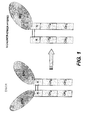


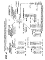


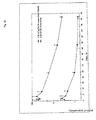

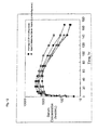

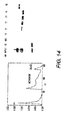

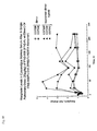
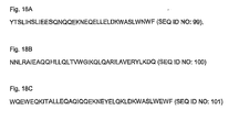
Classifications
-
- A—HUMAN NECESSITIES
- A61—MEDICAL OR VETERINARY SCIENCE; HYGIENE
- A61K—PREPARATIONS FOR MEDICAL, DENTAL OR TOILETRY PURPOSES
- A61K39/00—Medicinal preparations containing antigens or antibodies
- A61K39/395—Antibodies; Immunoglobulins; Immune serum, e.g. antilymphocytic serum
-
- C—CHEMISTRY; METALLURGY
- C07—ORGANIC CHEMISTRY
- C07K—PEPTIDES
- C07K14/00—Peptides having more than 20 amino acids; Gastrins; Somatostatins; Melanotropins; Derivatives thereof
- C07K14/435—Peptides having more than 20 amino acids; Gastrins; Somatostatins; Melanotropins; Derivatives thereof from animals; from humans
- C07K14/745—Blood coagulation or fibrinolysis factors
- C07K14/755—Factors VIII, e.g. factor VIII C (AHF), factor VIII Ag (VWF)
-
- A—HUMAN NECESSITIES
- A61—MEDICAL OR VETERINARY SCIENCE; HYGIENE
- A61K—PREPARATIONS FOR MEDICAL, DENTAL OR TOILETRY PURPOSES
- A61K47/00—Medicinal preparations characterised by the non-active ingredients used, e.g. carriers or inert additives; Targeting or modifying agents chemically bound to the active ingredient
- A61K47/50—Medicinal preparations characterised by the non-active ingredients used, e.g. carriers or inert additives; Targeting or modifying agents chemically bound to the active ingredient the non-active ingredient being chemically bound to the active ingredient, e.g. polymer-drug conjugates
- A61K47/51—Medicinal preparations characterised by the non-active ingredients used, e.g. carriers or inert additives; Targeting or modifying agents chemically bound to the active ingredient the non-active ingredient being chemically bound to the active ingredient, e.g. polymer-drug conjugates the non-active ingredient being a modifying agent
- A61K47/56—Medicinal preparations characterised by the non-active ingredients used, e.g. carriers or inert additives; Targeting or modifying agents chemically bound to the active ingredient the non-active ingredient being chemically bound to the active ingredient, e.g. polymer-drug conjugates the non-active ingredient being a modifying agent the modifying agent being an organic macromolecular compound, e.g. an oligomeric, polymeric or dendrimeric molecule
- A61K47/59—Medicinal preparations characterised by the non-active ingredients used, e.g. carriers or inert additives; Targeting or modifying agents chemically bound to the active ingredient the non-active ingredient being chemically bound to the active ingredient, e.g. polymer-drug conjugates the non-active ingredient being a modifying agent the modifying agent being an organic macromolecular compound, e.g. an oligomeric, polymeric or dendrimeric molecule obtained otherwise than by reactions only involving carbon-to-carbon unsaturated bonds, e.g. polyureas or polyurethanes
- A61K47/60—Medicinal preparations characterised by the non-active ingredients used, e.g. carriers or inert additives; Targeting or modifying agents chemically bound to the active ingredient the non-active ingredient being chemically bound to the active ingredient, e.g. polymer-drug conjugates the non-active ingredient being a modifying agent the modifying agent being an organic macromolecular compound, e.g. an oligomeric, polymeric or dendrimeric molecule obtained otherwise than by reactions only involving carbon-to-carbon unsaturated bonds, e.g. polyureas or polyurethanes the organic macromolecular compound being a polyoxyalkylene oligomer, polymer or dendrimer, e.g. PEG, PPG, PEO or polyglycerol
-
- A—HUMAN NECESSITIES
- A61—MEDICAL OR VETERINARY SCIENCE; HYGIENE
- A61K—PREPARATIONS FOR MEDICAL, DENTAL OR TOILETRY PURPOSES
- A61K47/00—Medicinal preparations characterised by the non-active ingredients used, e.g. carriers or inert additives; Targeting or modifying agents chemically bound to the active ingredient
- A61K47/50—Medicinal preparations characterised by the non-active ingredients used, e.g. carriers or inert additives; Targeting or modifying agents chemically bound to the active ingredient the non-active ingredient being chemically bound to the active ingredient, e.g. polymer-drug conjugates
- A61K47/51—Medicinal preparations characterised by the non-active ingredients used, e.g. carriers or inert additives; Targeting or modifying agents chemically bound to the active ingredient the non-active ingredient being chemically bound to the active ingredient, e.g. polymer-drug conjugates the non-active ingredient being a modifying agent
- A61K47/62—Medicinal preparations characterised by the non-active ingredients used, e.g. carriers or inert additives; Targeting or modifying agents chemically bound to the active ingredient the non-active ingredient being chemically bound to the active ingredient, e.g. polymer-drug conjugates the non-active ingredient being a modifying agent the modifying agent being a protein, peptide or polyamino acid
- A61K47/64—Drug-peptide, drug-protein or drug-polyamino acid conjugates, i.e. the modifying agent being a peptide, protein or polyamino acid which is covalently bonded or complexed to a therapeutically active agent
- A61K47/642—Drug-peptide, drug-protein or drug-polyamino acid conjugates, i.e. the modifying agent being a peptide, protein or polyamino acid which is covalently bonded or complexed to a therapeutically active agent the peptide or protein in the drug conjugate being a cytokine, e.g. IL2, chemokine, growth factors or interferons being the inactive part of the conjugate
-
- A—HUMAN NECESSITIES
- A61—MEDICAL OR VETERINARY SCIENCE; HYGIENE
- A61K—PREPARATIONS FOR MEDICAL, DENTAL OR TOILETRY PURPOSES
- A61K47/00—Medicinal preparations characterised by the non-active ingredients used, e.g. carriers or inert additives; Targeting or modifying agents chemically bound to the active ingredient
- A61K47/50—Medicinal preparations characterised by the non-active ingredients used, e.g. carriers or inert additives; Targeting or modifying agents chemically bound to the active ingredient the non-active ingredient being chemically bound to the active ingredient, e.g. polymer-drug conjugates
- A61K47/51—Medicinal preparations characterised by the non-active ingredients used, e.g. carriers or inert additives; Targeting or modifying agents chemically bound to the active ingredient the non-active ingredient being chemically bound to the active ingredient, e.g. polymer-drug conjugates the non-active ingredient being a modifying agent
- A61K47/68—Medicinal preparations characterised by the non-active ingredients used, e.g. carriers or inert additives; Targeting or modifying agents chemically bound to the active ingredient the non-active ingredient being chemically bound to the active ingredient, e.g. polymer-drug conjugates the non-active ingredient being a modifying agent the modifying agent being an antibody, an immunoglobulin or a fragment thereof, e.g. an Fc-fragment
- A61K47/6801—Drug-antibody or immunoglobulin conjugates defined by the pharmacologically or therapeutically active agent
- A61K47/6803—Drugs conjugated to an antibody or immunoglobulin, e.g. cisplatin-antibody conjugates
- A61K47/6811—Drugs conjugated to an antibody or immunoglobulin, e.g. cisplatin-antibody conjugates the drug being a protein or peptide, e.g. transferrin or bleomycin
- A61K47/6813—Drugs conjugated to an antibody or immunoglobulin, e.g. cisplatin-antibody conjugates the drug being a protein or peptide, e.g. transferrin or bleomycin the drug being a peptidic cytokine, e.g. an interleukin or interferon
-
- A—HUMAN NECESSITIES
- A61—MEDICAL OR VETERINARY SCIENCE; HYGIENE
- A61K—PREPARATIONS FOR MEDICAL, DENTAL OR TOILETRY PURPOSES
- A61K47/00—Medicinal preparations characterised by the non-active ingredients used, e.g. carriers or inert additives; Targeting or modifying agents chemically bound to the active ingredient
- A61K47/50—Medicinal preparations characterised by the non-active ingredients used, e.g. carriers or inert additives; Targeting or modifying agents chemically bound to the active ingredient the non-active ingredient being chemically bound to the active ingredient, e.g. polymer-drug conjugates
- A61K47/51—Medicinal preparations characterised by the non-active ingredients used, e.g. carriers or inert additives; Targeting or modifying agents chemically bound to the active ingredient the non-active ingredient being chemically bound to the active ingredient, e.g. polymer-drug conjugates the non-active ingredient being a modifying agent
- A61K47/68—Medicinal preparations characterised by the non-active ingredients used, e.g. carriers or inert additives; Targeting or modifying agents chemically bound to the active ingredient the non-active ingredient being chemically bound to the active ingredient, e.g. polymer-drug conjugates the non-active ingredient being a modifying agent the modifying agent being an antibody, an immunoglobulin or a fragment thereof, e.g. an Fc-fragment
- A61K47/6835—Medicinal preparations characterised by the non-active ingredients used, e.g. carriers or inert additives; Targeting or modifying agents chemically bound to the active ingredient the non-active ingredient being chemically bound to the active ingredient, e.g. polymer-drug conjugates the non-active ingredient being a modifying agent the modifying agent being an antibody, an immunoglobulin or a fragment thereof, e.g. an Fc-fragment the modifying agent being an antibody or an immunoglobulin bearing at least one antigen-binding site
-
- A—HUMAN NECESSITIES
- A61—MEDICAL OR VETERINARY SCIENCE; HYGIENE
- A61P—SPECIFIC THERAPEUTIC ACTIVITY OF CHEMICAL COMPOUNDS OR MEDICINAL PREPARATIONS
- A61P31/00—Antiinfectives, i.e. antibiotics, antiseptics, chemotherapeutics
-
- A—HUMAN NECESSITIES
- A61—MEDICAL OR VETERINARY SCIENCE; HYGIENE
- A61P—SPECIFIC THERAPEUTIC ACTIVITY OF CHEMICAL COMPOUNDS OR MEDICINAL PREPARATIONS
- A61P31/00—Antiinfectives, i.e. antibiotics, antiseptics, chemotherapeutics
- A61P31/12—Antivirals
-
- A—HUMAN NECESSITIES
- A61—MEDICAL OR VETERINARY SCIENCE; HYGIENE
- A61P—SPECIFIC THERAPEUTIC ACTIVITY OF CHEMICAL COMPOUNDS OR MEDICINAL PREPARATIONS
- A61P31/00—Antiinfectives, i.e. antibiotics, antiseptics, chemotherapeutics
- A61P31/12—Antivirals
- A61P31/14—Antivirals for RNA viruses
- A61P31/18—Antivirals for RNA viruses for HIV
-
- A—HUMAN NECESSITIES
- A61—MEDICAL OR VETERINARY SCIENCE; HYGIENE
- A61P—SPECIFIC THERAPEUTIC ACTIVITY OF CHEMICAL COMPOUNDS OR MEDICINAL PREPARATIONS
- A61P43/00—Drugs for specific purposes, not provided for in groups A61P1/00-A61P41/00
-
- A—HUMAN NECESSITIES
- A61—MEDICAL OR VETERINARY SCIENCE; HYGIENE
- A61P—SPECIFIC THERAPEUTIC ACTIVITY OF CHEMICAL COMPOUNDS OR MEDICINAL PREPARATIONS
- A61P7/00—Drugs for disorders of the blood or the extracellular fluid
-
- A—HUMAN NECESSITIES
- A61—MEDICAL OR VETERINARY SCIENCE; HYGIENE
- A61P—SPECIFIC THERAPEUTIC ACTIVITY OF CHEMICAL COMPOUNDS OR MEDICINAL PREPARATIONS
- A61P7/00—Drugs for disorders of the blood or the extracellular fluid
- A61P7/04—Antihaemorrhagics; Procoagulants; Haemostatic agents; Antifibrinolytic agents
-
- A—HUMAN NECESSITIES
- A61—MEDICAL OR VETERINARY SCIENCE; HYGIENE
- A61P—SPECIFIC THERAPEUTIC ACTIVITY OF CHEMICAL COMPOUNDS OR MEDICINAL PREPARATIONS
- A61P7/00—Drugs for disorders of the blood or the extracellular fluid
- A61P7/06—Antianaemics
-
- A—HUMAN NECESSITIES
- A61—MEDICAL OR VETERINARY SCIENCE; HYGIENE
- A61P—SPECIFIC THERAPEUTIC ACTIVITY OF CHEMICAL COMPOUNDS OR MEDICINAL PREPARATIONS
- A61P9/00—Drugs for disorders of the cardiovascular system
-
- C—CHEMISTRY; METALLURGY
- C07—ORGANIC CHEMISTRY
- C07K—PEPTIDES
- C07K14/00—Peptides having more than 20 amino acids; Gastrins; Somatostatins; Melanotropins; Derivatives thereof
- C07K14/435—Peptides having more than 20 amino acids; Gastrins; Somatostatins; Melanotropins; Derivatives thereof from animals; from humans
- C07K14/475—Growth factors; Growth regulators
- C07K14/505—Erythropoietin [EPO]
-
- C—CHEMISTRY; METALLURGY
- C07—ORGANIC CHEMISTRY
- C07K—PEPTIDES
- C07K14/00—Peptides having more than 20 amino acids; Gastrins; Somatostatins; Melanotropins; Derivatives thereof
- C07K14/435—Peptides having more than 20 amino acids; Gastrins; Somatostatins; Melanotropins; Derivatives thereof from animals; from humans
- C07K14/52—Cytokines; Lymphokines; Interferons
- C07K14/555—Interferons [IFN]
- C07K14/56—IFN-alpha
-
- C—CHEMISTRY; METALLURGY
- C07—ORGANIC CHEMISTRY
- C07K—PEPTIDES
- C07K14/00—Peptides having more than 20 amino acids; Gastrins; Somatostatins; Melanotropins; Derivatives thereof
- C07K14/435—Peptides having more than 20 amino acids; Gastrins; Somatostatins; Melanotropins; Derivatives thereof from animals; from humans
- C07K14/52—Cytokines; Lymphokines; Interferons
- C07K14/555—Interferons [IFN]
- C07K14/565—IFN-beta
-
- C—CHEMISTRY; METALLURGY
- C07—ORGANIC CHEMISTRY
- C07K—PEPTIDES
- C07K14/00—Peptides having more than 20 amino acids; Gastrins; Somatostatins; Melanotropins; Derivatives thereof
- C07K14/435—Peptides having more than 20 amino acids; Gastrins; Somatostatins; Melanotropins; Derivatives thereof from animals; from humans
- C07K14/575—Hormones
- C07K14/59—Follicle-stimulating hormone [FSH]; Chorionic gonadotropins, e.g. HCG; Luteinising hormone [LH]; Thyroid-stimulating hormone [TSH]
-
- C—CHEMISTRY; METALLURGY
- C07—ORGANIC CHEMISTRY
- C07K—PEPTIDES
- C07K14/00—Peptides having more than 20 amino acids; Gastrins; Somatostatins; Melanotropins; Derivatives thereof
- C07K14/435—Peptides having more than 20 amino acids; Gastrins; Somatostatins; Melanotropins; Derivatives thereof from animals; from humans
- C07K14/575—Hormones
- C07K14/61—Growth hormones [GH] (Somatotropin)
-
- C—CHEMISTRY; METALLURGY
- C07—ORGANIC CHEMISTRY
- C07K—PEPTIDES
- C07K14/00—Peptides having more than 20 amino acids; Gastrins; Somatostatins; Melanotropins; Derivatives thereof
- C07K14/435—Peptides having more than 20 amino acids; Gastrins; Somatostatins; Melanotropins; Derivatives thereof from animals; from humans
- C07K14/705—Receptors; Cell surface antigens; Cell surface determinants
- C07K14/70503—Immunoglobulin superfamily
-
- C—CHEMISTRY; METALLURGY
- C07—ORGANIC CHEMISTRY
- C07K—PEPTIDES
- C07K14/00—Peptides having more than 20 amino acids; Gastrins; Somatostatins; Melanotropins; Derivatives thereof
- C07K14/435—Peptides having more than 20 amino acids; Gastrins; Somatostatins; Melanotropins; Derivatives thereof from animals; from humans
- C07K14/745—Blood coagulation or fibrinolysis factors
-
- C—CHEMISTRY; METALLURGY
- C07—ORGANIC CHEMISTRY
- C07K—PEPTIDES
- C07K16/00—Immunoglobulins [IGs], e.g. monoclonal or polyclonal antibodies
-
- C—CHEMISTRY; METALLURGY
- C12—BIOCHEMISTRY; BEER; SPIRITS; WINE; VINEGAR; MICROBIOLOGY; ENZYMOLOGY; MUTATION OR GENETIC ENGINEERING
- C12N—MICROORGANISMS OR ENZYMES; COMPOSITIONS THEREOF; PROPAGATING, PRESERVING, OR MAINTAINING MICROORGANISMS; MUTATION OR GENETIC ENGINEERING; CULTURE MEDIA
- C12N9/00—Enzymes; Proenzymes; Compositions thereof; Processes for preparing, activating, inhibiting, separating or purifying enzymes
- C12N9/14—Hydrolases (3)
- C12N9/48—Hydrolases (3) acting on peptide bonds (3.4)
- C12N9/50—Proteinases, e.g. Endopeptidases (3.4.21-3.4.25)
- C12N9/64—Proteinases, e.g. Endopeptidases (3.4.21-3.4.25) derived from animal tissue
- C12N9/6421—Proteinases, e.g. Endopeptidases (3.4.21-3.4.25) derived from animal tissue from mammals
- C12N9/6424—Serine endopeptidases (3.4.21)
- C12N9/6437—Coagulation factor VIIa (3.4.21.21)
-
- C—CHEMISTRY; METALLURGY
- C12—BIOCHEMISTRY; BEER; SPIRITS; WINE; VINEGAR; MICROBIOLOGY; ENZYMOLOGY; MUTATION OR GENETIC ENGINEERING
- C12N—MICROORGANISMS OR ENZYMES; COMPOSITIONS THEREOF; PROPAGATING, PRESERVING, OR MAINTAINING MICROORGANISMS; MUTATION OR GENETIC ENGINEERING; CULTURE MEDIA
- C12N9/00—Enzymes; Proenzymes; Compositions thereof; Processes for preparing, activating, inhibiting, separating or purifying enzymes
- C12N9/14—Hydrolases (3)
- C12N9/48—Hydrolases (3) acting on peptide bonds (3.4)
- C12N9/50—Proteinases, e.g. Endopeptidases (3.4.21-3.4.25)
- C12N9/64—Proteinases, e.g. Endopeptidases (3.4.21-3.4.25) derived from animal tissue
- C12N9/6421—Proteinases, e.g. Endopeptidases (3.4.21-3.4.25) derived from animal tissue from mammals
- C12N9/6424—Serine endopeptidases (3.4.21)
- C12N9/644—Coagulation factor IXa (3.4.21.22)
-
- C—CHEMISTRY; METALLURGY
- C12—BIOCHEMISTRY; BEER; SPIRITS; WINE; VINEGAR; MICROBIOLOGY; ENZYMOLOGY; MUTATION OR GENETIC ENGINEERING
- C12N—MICROORGANISMS OR ENZYMES; COMPOSITIONS THEREOF; PROPAGATING, PRESERVING, OR MAINTAINING MICROORGANISMS; MUTATION OR GENETIC ENGINEERING; CULTURE MEDIA
- C12N9/00—Enzymes; Proenzymes; Compositions thereof; Processes for preparing, activating, inhibiting, separating or purifying enzymes
- C12N9/14—Hydrolases (3)
- C12N9/48—Hydrolases (3) acting on peptide bonds (3.4)
- C12N9/50—Proteinases, e.g. Endopeptidases (3.4.21-3.4.25)
- C12N9/64—Proteinases, e.g. Endopeptidases (3.4.21-3.4.25) derived from animal tissue
- C12N9/6421—Proteinases, e.g. Endopeptidases (3.4.21-3.4.25) derived from animal tissue from mammals
- C12N9/6424—Serine endopeptidases (3.4.21)
- C12N9/647—Blood coagulation factors not provided for in a preceding group or according to more than one of the proceeding groups
-
- C—CHEMISTRY; METALLURGY
- C12—BIOCHEMISTRY; BEER; SPIRITS; WINE; VINEGAR; MICROBIOLOGY; ENZYMOLOGY; MUTATION OR GENETIC ENGINEERING
- C12N—MICROORGANISMS OR ENZYMES; COMPOSITIONS THEREOF; PROPAGATING, PRESERVING, OR MAINTAINING MICROORGANISMS; MUTATION OR GENETIC ENGINEERING; CULTURE MEDIA
- C12N9/00—Enzymes; Proenzymes; Compositions thereof; Processes for preparing, activating, inhibiting, separating or purifying enzymes
- C12N9/96—Stabilising an enzyme by forming an adduct or a composition; Forming enzyme conjugates
-
- C—CHEMISTRY; METALLURGY
- C12—BIOCHEMISTRY; BEER; SPIRITS; WINE; VINEGAR; MICROBIOLOGY; ENZYMOLOGY; MUTATION OR GENETIC ENGINEERING
- C12Y—ENZYMES
- C12Y304/00—Hydrolases acting on peptide bonds, i.e. peptidases (3.4)
- C12Y304/21—Serine endopeptidases (3.4.21)
- C12Y304/21021—Coagulation factor VIIa (3.4.21.21)
-
- C—CHEMISTRY; METALLURGY
- C12—BIOCHEMISTRY; BEER; SPIRITS; WINE; VINEGAR; MICROBIOLOGY; ENZYMOLOGY; MUTATION OR GENETIC ENGINEERING
- C12Y—ENZYMES
- C12Y304/00—Hydrolases acting on peptide bonds, i.e. peptidases (3.4)
- C12Y304/21—Serine endopeptidases (3.4.21)
- C12Y304/21022—Coagulation factor IXa (3.4.21.22)
-
- A—HUMAN NECESSITIES
- A61—MEDICAL OR VETERINARY SCIENCE; HYGIENE
- A61K—PREPARATIONS FOR MEDICAL, DENTAL OR TOILETRY PURPOSES
- A61K38/00—Medicinal preparations containing peptides
-
- C—CHEMISTRY; METALLURGY
- C07—ORGANIC CHEMISTRY
- C07K—PEPTIDES
- C07K2317/00—Immunoglobulins specific features
- C07K2317/50—Immunoglobulins specific features characterized by immunoglobulin fragments
- C07K2317/52—Constant or Fc region; Isotype
-
- C—CHEMISTRY; METALLURGY
- C07—ORGANIC CHEMISTRY
- C07K—PEPTIDES
- C07K2319/00—Fusion polypeptide
-
- C—CHEMISTRY; METALLURGY
- C07—ORGANIC CHEMISTRY
- C07K—PEPTIDES
- C07K2319/00—Fusion polypeptide
- C07K2319/30—Non-immunoglobulin-derived peptide or protein having an immunoglobulin constant or Fc region, or a fragment thereof, attached thereto
Definitions
- the invention relates generally to therapeutic chimeric proteins, comprised of two polypeptide chains, wherein the first chain is comprised of a therapeutic biologically active molecule and the second chain is not comprised of the therapeutic biologically active molecule of the first chain. More specifically, the invention relates to chimeric proteins, comprised of two polypeptide chains, wherein both chains are comprised of at least a portion of an immunoglobulin constant region wherein the first chain is modified to further comprise a biologically active molecule, and the second chain is not so modified.
- the invention thus relates to a chimeric protein that is a monomer-dimer hybrid, i.e., a chimeric protein having a dimeric aspect and a monomeric aspect, wherein the dimeric aspect relates to the fact that it is comprised of two polypeptide chains each comprised of a portion of an immunoglobulin constant region, and wherein the monomeric aspect relates to the fact that only one of the two chains is comprised of a therapeutic biologically active molecule.
- Figure 1 illustrates one example of a monomer-dimer hybrid wherein the biologically active molecule is erythropoietin (EPO) and the portion of an immunoglobulin constant region is an IgG Fc region.
- EPO erythropoietin
- lmmunoglobulins are comprised of four polypeptide chains, two heavy chains and two light chains, which associate via disulfide bonds to form tetramers. Each chain is further comprised of one variable region and one constant region.
- the variable regions mediate antigen recognition and binding, while the constant regions, particularly the heavy chain constant regions, mediate a variety of effector functions, e.g., complement binding and Fc receptor binding (see, e.g., U.S. Patent Nos.: 6,086,875 ; 5,624,821 ; 5,116,964 ).
- the constant region is further comprised of domains denoted CH (constant heavy) domains (CH1, CH2, etc.).
- CH constant heavy domains
- the constant region can be comprised of three or four CH domains.
- Some isotypes (e.g . IgG) constant regions also contain a hinge region Janeway et al. 2001, Immunobiology, Garland Publishing, N.Y., N.Y .
- chimeric proteins comprised of immunoglobulin constant regions linked to a protein of interest, or fragment thereof, has been described (see , e.g ., U.S. Patent Nos. 5,480,981 and 5,808,029 ; Gascoigne et al. 1987, Proc. Natl. Acad. Sci. USA 84:2936 ; Capon et al. 1989, Nature 337:525 ; Traunecker et al. 1989, Nature 339:68 ; Zettmeissl et al. 1990, DNA Cell Biol. USA 9:347 ; Bym et al. 1990, Nature 344:667 ; Watson et al. 1990, J. Cell. Biol.
- the Fc portion of an immunoglobulin constant region can include the CH2, CH3, and CH4 domains, as well as the hinge region.
- Chimeric proteins comprising an Fc portion of an immunoglobulin bestow several desirable properties on a chimeric protein including increased stability, increased serum half life (see Capon et al. 1989, Nature 337:525 ) as well as binding to Fc receptors such as the neonatal Fc receptor (FcRn) ( U.S. Patent Nos. 6,086,875 , 6,485,726 , 6,030,613 ; WO 03/077834 ; US2003-0235536A1 ).
- FcRn is active in adult epithelial tissue and expressed in the lumen of the intestines, pulmonary airways, nasal surfaces, vaginal surfaces, colon and rectal surfaces ( U.S. Patent No. 6,485,726 ).
- Chimeric proteins comprised of FcRn binding partners e.g . IgG, Fc fragments
- FcRn binding partners e.g . IgG, Fc fragments
- FcRn binding partners e.g . IgG, Fc fragments
- FcRn binding partners e.g . IgG, Fc fragments
- FcRn binding partners e.g . IgG, Fc fragments
- chimeric proteins comprising an FcRn binding partner are endocytosed by cells expressing the FcRn. But instead of being marked for degradation, these chimeric proteins are recycled out into circulation again, thus increasing the in vivo half life of these proteins.
- Portions of immunoglobulin constant regions e.g ., FcRn binding partners typically associate, via disulfide bonds and other non-specific interactions, with one another to form dimers and higher order multimers.
- the instant invention is based in part upon the surprising discovery that transcytosis of chimeric proteins comprised of FcRn binding partners appears to be limited by the molecular weight of the chimeric protein, with higher molecular weight species being transported less efficiently.
- Chimeric proteins comprised of biologically active molecules once administered, typically will interact with a target molecule or cell.
- the instant invention is further based in part upon the surprising discovery that monomer-dimer hybrids, with one biologically active molecule, but two portions of an immunoglobulin constant region, e.g ., two FcRn binding partners, function and can be transported more effectively than homodimers, also referred to herein simply as "dimers" or higher order multimers with two or more copies of the biologically active molecule.
- chimeric proteins comprised of two or more biologically active molecules, which exist as dimers and higher order multimers, can be sterically hindered from interacting with their target molecule or cell, due to the presence of the two or more biologically active molecules in close proximity to one another and that the biologically active molecule can have a high affinity for itself.
- one aspect of the invention provides chimeric proteins comprised of a biologically active molecule that is transported across the epithelium barrier.
- An additional aspect of the invention provides chimeric proteins comprised of at least one biologically active molecule that is able to interact with its target molecule or cell with little or no steric hindrance or self aggregation.
- the aspects of the invention provide for chimeric proteins comprising a first and second polypeptide chain, the first chain comprising at least a portion of immunoglobulin constant region, wherein the portion of an immunoglobulin constant region has been modified to include a biologically active molecule and the second chain comprising at least a portion of immunoglobulin constant region, wherein the portion of an immunoglobulin constant region has not been so modified to include the biologically active molecule of the first chain.
- the invention relates to a chimeric protein comprising one biologically active molecule and two molecules of at least a portion of an immunoglobulin constant region.
- the chimeric protein is capable of interacting with a target molecule or cell with less steric hindrance compared to a chimeric protein comprised of at least two biologically active molecules and at least a portion of two immunoglobulin constant regions.
- the invention also relates to a chimeric protein comprising at least one biologically active molecule and two molecules of at least a portion of an immunoglobulin constant region that is transported across an epithelium barrier more efficiently than a corresponding homodimer, i.e., wherein both chains are linked to the same biologically active molecule.
- the invention thus relates to a chimeric protein comprising a first and a second polypeptide chain linked together, wherein said first chain comprises a biologically active molecule and at least a portion of an immunoglobulin constant region, and said second chain comprises at least a portion of an immunoglobulin constant region, but no immunoglobulin variable region and without any biologically active molecule attached.
- the invention relates to a chimeric protein comprising a first and a second polypeptide chain linked together, wherein said first chain comprises a biologically active molecule and at least a portion of an immunoglobulin constant region, and said second chain comprises at least a portion of an immunoglobulin constant region without an immunoglobulin variable region or any biologically active molecule and wherein said second chain is not covalently bonded to any molecule having a molecular weight greater than 1 kD, 2 kD, 5 kD, 10 kD, or 20 kD. In one embodiment, the second chain is not covalently bonded to any molecule having a molecular weight greater than 0-2 kD.
- the second chain is not covalently bonded to any molecule having a molecular weight greater than 5-10 kD. In one embodiment, the second chain is not covalently bonded to any molecule having a molecular weight greater than 15-20 kD.
- the invention relates to a chimeric protein comprising a first and a second polypeptide chain linked together, wherein said first chain comprises a biologically active molecule and at least a portion of an immunoglobulin constant region, and said second chain comprises at least a portion of an immunoglobulin constant region not covalently linked to any other molecule except the portion of an immunoglobulin of said first polypeptide chain.
- the invention relates to a chimeric protein comprising a first and a second polypeptide chain linked together, wherein said first chain comprises a biologically active molecule and at least a portion of an immunoglobulin constant region, and said second chain consists of at least a portion of an immunoglobulin constant region and optionally an affinity tag.
- the invention relates to a chimeric protein comprising a first and a second polypeptide chain linked together, wherein said first chain comprises a biologically active molecule and at least a portion of an immunoglobulin constant region, and said second chain consists essentially of at least a portion of an immunoglobulin constant region and optionally an affinity tag.
- the invention relates to a chimeric protein comprising a first and a second polypeptide chain linked together, wherein said first chain comprises a biologically active molecule and at least a portion of an immunoglobulin constant region, and said second chain comprises at least a portion of an immunoglobulin constant region without an immunoglobulin variable region or any biologically active molecule and optionally a molecule with a molecular weight less than 10 kD, 5 kD, 2 kD or 1 kD.
- the second chain comprises a molecule less than 15-20 kD.
- the second chain comprises a molecule less than 5-10 kD.
- the second chain comprises a molecule less than 1-2 kD.
- the invention relates to a chimeric protein comprising a first and second polypeptide chain, wherein said first chain comprises a biologically active molecule, at least a portion of an immunoglobulin constant region, and at least a first domain, said first domain having at least one specific binding partner, and wherein said second chain comprises at least a portion of an immunoglobulin constant region, and at least a second domain, wherein said second domain is a specific binding partner of said first domain, without any immunoglobulin variable region or a biologically active molecule.
- the invention relates to a method of making a chimeric protein comprising a first and second polypeptide chain, wherein the first polypeptide chain and the second polypeptide chain are not the same, said method comprising transfecting a cell with a first DNA construct comprising a DNA molecule encoding a first polypeptide chain comprising a biologically active molecule and at least a portion of an immunoglobulin constant region and optionally a linker, and a second DNA construct comprising a DNA molecule encoding a second polypeptide chain comprising at least a portion of an immunoglobulin constant region without any biologically active molecule or an immunoglobulin variable region, and optionally a linker, culturing the cells under conditions such that the polypeptide chain encoded by the first DNA construct is expressed and the polypeptide chain encoded by the second DNA construct is expressed and isolating monomer-dimer hybrids comprised of the polypeptide chain encoded by the first DNA construct and the polypeptide chain encoded by the second DNA construct
- the invention relates to a method of making a chimeric protein comprising a first and second polypeptide chain, wherein the first polypeptide chain and the second polypeptide chain are not the same, and wherein said first polypeptide chain comprises a biologically active molecule, at least a portion of an immunoglobulin constant region, and at least a first domain, said first domain, having at least one specific binding partner, and wherein said second polypeptide chain comprises at least a portion of an immunoglobulin constant region and a second domain, wherein said second domain, is a specific binding partner of said first domain, without any biologically active molecule or an immunoglobulin variable region, said method comprising transfecting a cell with a first DNA construct comprising a DNA molecule encoding said first polypeptide chain and a second DNA construct comprising a DNA molecule encoding, said second polypeptide chain, culturing the cells under conditions such that the polypeptide chain encoded by the first DNA construct is expressed and the polypeptide chain encoded by the second DNA
- the invention relates to a method of making a chimeric protein of the invention said method comprising transfecting a cell with a first DNA construct comprising a DNA molecule encoding a first polypeptide chain comprising a biologically active molecule and at least a portion of an immunoglobulin constant region and optionally a linker, culturing the cell under conditions such that the polypeptide chain encoded by the first DNA construct is expressed, isolating the polypeptide chain encoded by the first DNA construct and transfecting a cell with a second DNA construct comprising a DNA molecule encoding a second polypeptide chain comprising at least a portion of an immunoglobulin constant region without any biologically active molecule or immunoglobulin variable region, culturing the cell under conditions such that the polypeptide chain encoded by the second DNA construct is expressed, isolating the polypeptide chain, encoded by the second DNA construct, combining the polypeptide chain, encoded by the first DNA construct and the polypeptide chain encoded by the second DNA
- the invention relates to a method of making a chimeric protein comprising a first and second polypeptide chain, wherein the first polypeptide chain and the second polypeptide chain are not the same, said method comprising transfecting a cell with a DNA construct comprising a DNA molecule encoding a polypeptide chain comprising at least a portion of an immunoglobulin constant region, culturing the cells under conditions such that the polypeptide chain encoded by the DNA construct is expressed with an N terminal cysteine such that dimers of the polypeptide chain form and isolating dimers comprised of two copies of the polypeptide chain encoded by the DNA construct and chemically reacting the isolated dimers with a biologically active molecule, wherein said biologically active molecule has a C terminus thioester, under conditions such that the biologically active molecule reacts predominantly with only one polypeptide chain of the dimer thereby forming a monomer-dimer hybrid.
- the invention relates to a method of making a chimeric protein comprising a first and second polypeptide chain, wherein the first polypeptide chain and the second polypeptide chain are not the same, said method comprising transfecting a cell with a DNA construct comprising a DNA molecule encoding a polypeptide chain comprising at least a portion of an immunoglobulin constant region, culturing the cells under conditions such that the polypeptide chain encoded by the DNA construct is expressed with an N terminal cysteine such that dimers of the polypeptide chains form, and isolating dimers comprised of two copies of the polypeptide chain encoded by the DNA construct, and chemically reacting the isolated dimers with a biologically active molecule, wherein said biologically active molecule has a C terminus thioester, such that the biologically active molecule is linked to each chain of the dimer, denaturing the dimer comprised of the portion of the immunoglobulin linked to the biologically active molecule such that monomeric chains form, combining the mono
- the invention relates to a method of making a chimeric protein comprising a first and second polypeptide chain, wherein the first polypeptide chain and the second polypeptide chain are not the same, said method comprising transfecting a cell with a DNA construct comprising a DNA molecule encoding a polypeptide chain comprising at least a portion of an immunoglobulin constant region, culturing the cells under conditions such that the polypeptide chain encoded by the DNA construct is expressed as a mixture of two polypeptide chains, wherein the mixture comprises a polypeptide with an N terminal cysteine, and a polypeptide with a cysteine in close proximity to the N terminus, isolating dimers comprised of the mixture of polypeptide chains encoded by the DNA construct and chemically reacting the isolated dimers with a biologically active molecule, wherein said biologically active molecule has an active thioester, such that at least some monomer-dimer hybrid forms and isolating the monomer-dimer hybrid from said mixture
- the invention relates to a method of treating a disease or condition comprising administering a chimeric protein of the invention thereby treating the disease or condition.
- Figure 1 is a schematic diagram comparing the structure of an EPO-Fc homodimer, or dimer, and the structure of an Epo-FC monomer-dimer hybrid.
- Figure 2a is the amino acid sequence of the chimeric protein Factor VII-Fc. Included in the sequence is the signal peptide (underlined), which is cleaved by the cell and the propeptide (bold), which is recognized by the vitamin K-dependent ⁇ carboxylase which modifies the Factor VII to achieve full activity. The sequence is subsequently cleaved by PACE to yield Factor VII-Fc.
- Figure 2b is the amino acid sequence of the chimeric protein Factor IX-Fc. Included in the sequence is the signal peptide (underlined) which is cleaved by the cell and the propeptide (bold) which is recognized by the vitamin K-dependent ⁇ carboxylase which modifies the Factor IX to achieve full activity. The sequence is subsequently cleaved by PACE to yield Factor IX-Fc.
- Figure 2c is the amino acid sequence of the chimeric protein IFN ⁇ -Fc. Included in the sequence is the signal peptide (underlined), which is cleaved by the cell resulting in the mature IFN ⁇ -Fc.
- Figure 2d is the amino acid sequence of the chimeric protein IFN ⁇ -Fc ⁇ linker. Included in the sequence is the signal peptide (underlined) which is cleaved by the cell resulting in the mature IFN ⁇ - Fc ⁇ linker.
- Figure 2e is the amino acid sequence of the chimeric protein Flag-Fc. Included in the sequence is the signal peptide (underlined), which is cleaved by the cell resulting in the mature Flag-Fc.
- Figure 2f is the amino acid sequence of the chimeric protein Epo-CCA-Fc. Included in the sequence is the signal peptide (underlined), which is cleaved by the cell resulting in the mature Epo-CCA-Fc. Also shown in bold is the acidic coiled coil domain.
- Figure 2g is the amino acid sequence of the chimeric protein CCB-Fc. Included in the sequence is the signal peptide (undefined), which is cleaved by the cell resulting in the mature CCB-Fc. Also shown in bold is the basic coiled coil domain.
- Figure 2h is the amino acid sequence of the chimeric protein Cys-Fc. Included in the sequence is the signal peptide (underlined), which is cleaved by the cell resulting in the mature Cys-Fc. When this sequence is produced in CHO cells a percentage of the molecules are incorrectly cleaved by the signal peptidase such that two extra amino acids are left on the N terminus, thus preventing the linkage of a biologically active molecule with a C terminal thioester ( e.g ., via native ligation). When these improperly cleaved species dimerize with the properly cleaved Cys-Fc and are subsequently reacted with biologically active molecules with C terminal thioesters, monomer-dimer hybrids form.
- the signal peptide underlined
- Figure 2i is the amino acid sequence of the chimeric protein IFN ⁇ -GS15-Fc. Included in the sequence is the signal peptide (underlined) which is cleaved by the cell resulting in the mature IFN ⁇ - GS15-Fc.
- Figure 2j is the amino acid sequence of the chimeric protein Epo-Fc. Included in the sequence is the signal peptide (underlined), which is cleaved by the cell resulting in the mature Epo-Fc. Also shown in bold is the 8 amino acid linker.
- Figure 3a is the nucleic acid sequence of the chimeric protein Factor VII-Fc. Included in the sequence is the signal peptide (undefined) and the propeptide (bold) which is recognized by the vitamin K-dependent ⁇ carboxylase which modifies the Factor VII to achieve full activity. The translated sequence is subsequently cleaved by PACE to yield mature Factor VII-Fc.
- Figure 3b is the nucleic acid sequence of the chimeric protein Factor IX-Fc. Included in the sequence is the signal peptide (underlined) and the propeptide (bold) which is recognized by the vitamin K-dependent ⁇ carboxylase which modifies the Factor IX to achieve full activity. The translated sequence is subsequently cleaved by PACE to yield mature Factor IX-Fc.
- Figure 3c is the nucleic acid sequence of the chimeric protein IFN ⁇ -Fc. Included in the sequence is the signal peptide (underlined), which is cleaved by the cell after translation resulting in the mature IFNa-Fc.
- Figure 3d is the nucleic acid sequence of the chimeric protein IFN ⁇ -Fc ⁇ linker. Included in the sequence is the signal peptide (underlined) which is cleaved by the cell after translation resulting in the mature IFN ⁇ - Fc ⁇ linker.
- Figure 3e is the amino acid sequence of the chimeric protein Flag-Fc. Included in the sequence is the signal peptide (underlined), which is cleaved by the cell after translation resulting in the mature Flag-Fc.
- Figure 3f is the nucleic acid sequence of the chimeric protein Epo-CCA-Fc. Included in the sequence is the signal peptide (underlined), which is cleaved by the cell after translation resulting in the mature Epo-CCA-Fc. Also shown in bold is the acidic coiled coil domain.
- Figure 3g is the nucleic acid sequence of the chimeric protein CCB-Fc. Included in the sequence is the signal peptide (underlined), which is cleaved by the cell after translation resulting in the mature CCB-Fc. Also shown in bold is the basic coiled coil domain.
- Figure 3h is the nucleic acid sequence of the chimeric protein Cys-Fc. Included in the sequence is the signal peptide (underlined), which is cleaved by the cell after translation resulting in the mature Cys-Fc.
- Figure 3i is the nucleic acid sequence of the chimeric protein IFN ⁇ -GS15-Fc. Included in the sequence is the signal peptide (underlined) which is cleaved by the cell after translation resulting in the mature IFNa-GS15-Fc.
- Figure 3j is the nucleic acid sequence of the chimeric protein Epo-Fc. Included in the sequence is the signal peptide (underlined), which is cleaved by the cell after translation resulting in the mature Epo-Fc. Also shown in bold is a nucleic acid sequence encoding the 8 amino acid linker.
- Figure 4 demonstrates ways to form monomer-dimer hybrids through native ligation.
- Figure 5a shows the amino acid sequence of Fc MESNA (SEQ ID NO:4).
- Figure 5b shows the DNA sequence of Fc MESNA (SEQ ID NO:5).
- Figure 6 compares antiviral activity of IFN ⁇ homo-dimer (i.e. comprised of 2 IFN ⁇ molecules) with an IFN ⁇ monomer-dimer hybrid (i.e. comprised of 1 IFN ⁇ molecule).
- Figure 7 is a comparison of clotting activity of a chimeric monomer-dimer hybrid Factor VIIa-Fc (one Factor VII molecule) and a chimeric homodimer Factor VIIa-Fc (two Factor VII molecules).
- Figure 8 compares oral dosing in neonatal rats of a chimeric monomer-dimer hybrid Factor VIIa-Fc (one Factor VII molecule) and a chimeric homodimer Factor VIIa-Fc (two Factor VII molecules).
- Figure 9 compares oral dosing in neonatal rats of a chimeric monomer-dimer hybrid Factor IX-Fc (one Factor IX molecule) with a chimeric homodimer.
- Figure 10 is a time course study comparing a chimeric monomer-dimer hybrid Factor IX-Fc (one Factor IX molecule) administered orally to neonatal rats with an orally administered chimeric homodimer.
- Figure 11 demonstrates pharmokinetics of Epo-Fc dimer compared to Epo-Fc monomer-dimer hybrid in cynomolgus monkeys after a single pulmonary dose.
- Figure 12 compares serum concentration in monkeys of subcutaneously administered Epo-Fc monomer-dimer hybrid with subcutaneously administered Aranesp ® (darbepoetin alfa).
- Figure 13 compares serum concentration in monkeys of intravenously administered Epo-Fc monomer-dimer hybrid with intravenously administered Aranesp ® (darbepoetin alfa) and Epogen ® (epoetin alfa).
- Figure 14 shows a trace from a Mimetic Red 2TM column (ProMetic LifeSciences, Inc., Wayne, NJ) and an SDS-PAGE of fractions from the column containing EpoFc monomer-dimer hybrid, EpoFc dimer, and Fc.
- EpoFc monomer-dimer hybrid is found in fractions 11,12,13, and 14.
- EpoFc dimer is found in fraction 18.
- Fc is found in fractions 1/2.
- Figure 15 shows the pharmacokinetics of IFN ⁇ Fc with an 8 amino acid linker in cynomolgus monkeys after a single pulmonary dose.
- Figure 16 shows neopterin stimulation in response to the IFN ⁇ -Fc homodimer and the IFN ⁇ -Fc N297A monomer-dimer hybrid in cynomolgus monkeys.
- Figure 17a shows the nucleotide sequence of interferon ⁇ -Fc
- Figure 17b shows the amino acid sequence of interferon ⁇ -Fc.
- FIG. 18 shows the amino acid sequence of T20(a); T21(b) and T1249(c).
- Affinity tag means a molecule attached to a second molecule of interest, capable of interacting with a specific binding partner for the purpose of isolating or identifying said second molecule of interest.
- Analogs of chimeric proteins of the invention, or proteins or peptides substantially identical to the chimeric proteins of the invention, as used herein, means that a relevant amino acid sequence of a protein or a peptide is at least 70%, 75%, 80%, 85%, 90%, 95%, 97%, 98%, 99%, or 100% identical to a given sequence.
- sequences may be variants derived from various species, or they may be derived from the given sequence by truncation, deletion, amino acid substitution or addition. Percent identity between two amino acid sequences is determined by standard alignment algorithms such as, for example, Basic Local Alignment Tool (BLAST) described in Altschul et al. 1990, J. Mol.
- BLAST Basic Local Alignment Tool
- BLAST 2 Sequences program BLASTN, reward for match 2, penalty for mismatch -2, open gap and extension gap penalties 5 and 2 respectively, gap x_dropoff 50, expect 10, word size 11, filter ON.
- program BLASTP program BLASTP, matrix BLOSUM62, open gap and extension gap penalties 11 and 1 respectively, gap x_dropoff 50, expect 10, word size 3, filter ON.
- Bioavailability means the extent and rate at which a substance is absorbed into a living system or is made available at the site of physiological activity.
- Biologically active molecule means a non-immunoglobulin molecule or fragment thereof, capable of treating a disease or condition or localizing or targeting a molecule to a site of a disease or condition in the body by performing a function or an action, or stimulating or responding to a function, an action or a reaction, in a biological context ( e.g . in an organism, a cell, or an in vitro model thereof).
- Biologically active molecules may comprise at least one of polypeptides, nucleic acids, small molecules such as small organic or inorganic molecules.
- a chimeric protein refers to any protein comprised of a first amino acid sequence derived from a first source, bonded, covalently or non-covalently, to a second amino acid sequence derived from a second source, wherein the first and second source are not the same.
- a first source and a second source that are not the same can include two different biological entities, or two different proteins from the same biological entity, or a biological entity and a non-biological entity.
- a chimeric protein can include for example, a protein derived from at least 2 different biological sources.
- a biological source can include any non-synthetically produced nucleic acid or amino acid sequence ( e.g .
- a synthetic source can include a protein or nucleic acid sequence produced chemically and not by a biological system (e.g . solid phase synthesis of amino acid sequences).
- a chimeric protein can also include a protein derived from at least 2 different synthetic sources or a protein derived from at least one biological source and at least one synthetic source.
- a chimeric protein may also comprise a first amino acid sequence derived from a first source, covalently or non-covalently linked to a nucleic acid, derived from any source or a small organic or inorganic molecule derived from any source.
- the chimeric protein may comprise a linker molecule between the first and second amino acid sequence or between the first amino acid sequence and the nucleic acid, or between the first amino acid sequence and the small organic or inorganic molecule.
- Clotting factor means any molecule, or analog thereof, naturally occurring or recombinantly produced which prevents or decreases the duration of a bleeding episode in a subject with a hemostatic disorder. In other words, it means any molecule having clotting activity.
- Clotting activity means the ability to participate in a cascade of biochemical reactions that culminates in the formation of a fibrin clot and/or reduces the severity, duration or frequency of hemorrhage or bleeding episode.
- Dimer as used herein refers to a chimeric protein comprising a first and second polypeptide chain, wherein the first and second chains both comprise a biologically active molecule, and at least a portion of an immunoglobulin constant region.
- a homodimer refers to a dimer where both biologically active molecules are the same.
- Dimerically linked monomer-dimer hybrid refers to a chimeric protein comprised of at least a portion of an immunloglobulin constant region, e.g. an Fc fragment of an immunoglobulin, a biologically active molecule and a linker which links the two together such that one biologically active molecule is bound to 2 polypeptide chains, each comprising a portion of an immunoglobulin constant region.
- Figure 4 shows an example of a dimerically linked monomer-dimer hybrid.
- DNA construct means a DNA molecule, or a clone of such a molecule, either single- or double-stranded that has been modified through human intervention to contain segments of DNA combined in a manner that as a whole would not otherwise exist in nature.
- DNA constructs contain the information necessary to direct the expression of polypeptides of interest.
- DNA constructs can include promoters, enhancers and transcription terminators.
- DNA constructs containing the information necessary to direct the secretion of a polypeptide will also contain at least one secretory signal sequence.
- Domain means a region of a polypeptide (including proteins as that term is defined) having some distinctive physical feature or role including for example an independently folded structure composed of one section of a polypeptide chain.
- a domain may contain the sequence of the distinctive physical feature of the polypeptide or it may contain a fragment of the physical feature which retains its binding characteristics (i.e., it can bind to a second domain).
- a domain may be associated with another domain. In other words, a first domain may naturally bind to a second domain.
- a fragment refers to a peptide or polypeptide comprising an amino acid sequence of at least 2 contiguous amino acid residues, of at least 5 contiguous amino acid residues, of at least 10 contiguous amino acid residues, of at least 15 contiguous amino acid residues, of at least 20 contiguous amino acid residues, of at least 25 contiguous amino acid residues, of at least 40 contiguous amino acid residues, of at least 50 contiguous amino acid residues, of at least 100 contiguous amino acid residues, or of at least 200 contiguous amino acid residues or any deletion or truncation of a protein, peptide, or polypeptide.
- Hemostasis means the stoppage of bleeding or hemorrhage; or the stoppage of blood flow through a blood vessel or body part.
- Hemostatic disorder means a genetically inherited or acquired condition characterized by a tendency to hemorrhage, either spontaneously or as a result of trauma, due to an impaired ability or inability to form a fibrin clot.
- Linked refers to a first nucleic acid sequence covalently joined to a second nucleic acid sequence.
- the first nucleic acid sequence can be directly joined or juxtaposed to the second nucleic acid sequence or alternatively an intervening sequence can covalently join the first sequence to the second sequence.
- Linked as used herein can also refer to a first amino acid sequence covalently, or non-covalently, joined to a second amino acid sequence.
- the first amino acid sequence can be directly joined or juxtaposed to the second amino acid sequence or alternatively an intervening sequence can covalently join the first amino acid sequence to the second amino acid sequence.
- Operatively linked means a first nucleic acid sequence linked to a second nucleic acid sequence such that both sequences are capable of being expressed as a biologically active protein or peptide.
- Polypeptide refers to a polymer of amino acids and does not refer to a specific length of the product; thus, peptides, oligopeptides, and proteins are included within the definition of polypeptide. This term does not exclude post-expression modifications of the polypeptide, for example, glycosylation, acetylation, phosphorylation, pegylation, addition of a lipid moiety, or the addition of any organic or inorganic molecule. Included within the definition, are for example, polypeptides containing one or more analogs of an amino acid (including, for example, unnatural amino acids) and polypeptides with substituted linkages, as well as other modifications known in the art, both naturally occurring and non-naturally occurring.
- High stringency includes conditions readily determined by the skilled artisan based on, for example, the length of the DNA. Generally, such conditions are defined in Sambrook et al. Molecular Cloning: A Laboratory Manual, 2 ed. Vol. 1, pp. 1.101-104, Cold Spring Harbor Laboratory Press (1989 ), and include use of a prewashing solution for the nitrocellulose filters 5X SSC, 0.5% SDS, 1.0 mM EDTA (PH 8.0), hybridization conditions of 50% formamide, 6X SSC at 42°C (or other similar hybridization solution, such as Stark's solution, in 50% formamide at 42°C, and with washing at approximately 68°C, 0.2X SSC, 0.1 % SDS. The skilled artisan will recognize that the temperature and wash solution salt concentration can be adjusted as necessary according to factors such as the length of the probe.
- Moderate stringency include conditions that can be readily determined by those having ordinary skill in the art based on, for example, the length of the DNA.
- the basic conditions are set forth by Sambrook et al. Molecular Cloning. A Laboratory Manual, 2d ed. Vol. 1, pp. 1.101-104, Cold Spring Harbor Laboratory Press (1989 ), and include use of a prewashing solution for the nitrocellulose filters 5X SSC, 0.5% SDS, 1.0 mM EDTA (pH 8.0), hybridization conditions of 50% formamide, 6X SSC at 42°C (or other similar hybridization solution, such as Stark's solution, in 50% formamide at 42°C), and washing conditions of 60°C, 0.5X SSC, 0.1 % SDS.
- a small inorganic molecule as used herein means a molecule containing no carbon atoms and being no larger than 50 kD.
- a small organic molecule as used herein means a molecule containing at least one carbon atom and being no larger than 50 kD.
- Treat, treatment, treating means, any of the following: the reduction in severity of a disease or condition; the reduction in the duration of a disease course; the amelioration of one or more symptoms associated with a disease or condition; the provision of beneficial effects to a subject with a disease or condition, without necessarily curing the disease or condition, the prophylaxis of one or more symptoms associated with a disease or condition.
- the invention provides for chimeric proteins (monomer-dimer hybrids) comprising a first and a second polypeptide chain, wherein said first chain comprises a biologically active molecule and at least a portion of an immunoglobulin constant region, and said second chain comprises at least a portion of an immunoglobulin constant region without any biologically active molecule or variable region of an immunoglobulin.
- Figure 1 contrasts traditional fusion protein dimers with one example of the monomer-dimer hybrid of the invention.
- the biologically active molecule is EPO and the portion of an immunoglobulin is IgG Fc region.
- the invention provides for chimeric proteins which afford enhanced stability and increased bioavailability of the chimeric protein compared to the biologically active molecule alone. Additionally, however, because only one of the two chains comprises the biologically active molecule, the chimeric protein has a lower molecular weight than a chimeric protein wherein all chains comprise a biologically active molecule and while not wishing to be bound by any theory, this may result in the chimeric protein being more readily transcytosed across the epithelium barrier, e.g ., by binding to the FcRn receptor thereby increasing the half-life of the chimeric protein.
- the invention thus provides for an improved non-invasive method (e.g . via any mucosal surface, such as, orally, buccally, sublingually, nasally, rectally, vaginally, or via pulmonary or occular route) of administering a therapeutic chimeric protein of the invention.
- the invention thus provides methods of attaining therapeutic levels of the chimeric proteins of the invention using less frequent and lower doses compared to previously described chimeric proteins (e.g. chimeric proteins comprised of at least a portion of an immunoglobulin constant region and a biologically active molecule, wherein all chains of the chimeric protein comprise a biologically active molecule).
- the invention provides an invasive method, e.g ., subcutaneously, intravenously, of administering a therapeutic chimeric protein of the invention.
- Invasive administration of the therapeutic chimeric protein of the invention provides for an increased half life of the therapeutic chimeric protein which results in using less frequent and lower doses compared to previously described chimeric proteins (e.g . chimeric proteins comprised of at least a portion of an immunoglobulin constant region and a biologically active molecule, wherein all chains of the chimeric protein comprise a biologically active molecule).
- Yet another advantage of a chimeric protein wherein only one of the chains comprises a biologically active molecule is the enhanced accessibility of the biologically active molecule for its target cell or molecule resulting from decreased steric hindrance, decreased hydrophobic interactions, decreased ionic interactions, or decreased molecular weight compared to a chimeric protein wherein all chains are comprised of a biologically active molecule.
- the invention relates to chimeric proteins comprising one biologically active molecule, at least a portion of an immunoglobulin constant region, and optionally at least one linker.
- the portion of an immunoglobulin will have both an N, or an amino terminus, and a C, or carboxy terminus.
- the chimeric protein may have the biologically active molecule linked to the N terminus of the portion of an immunoglobulin.
- the biologically active molecule may be linked to the C terminus of the portion of an immunoglobulin.
- the linkage is a covalent bond. In another embodiment, the linkage is a non-covalent bond.
- the chimeric protein can optionally comprise at least one linker; thus, the biologically active molecule does not have to be directly linked to the portion of an immunoglobulin constant region.
- the linker can intervene in between the biologically active molecule and the portion of an immunoglobulin constant region.
- the linker can be linked to the N terminus of the portion of an immunoglobulin constant region, or the C terminus of the portion of an immunoglobulin constant region. If the biologically active molecule is comprised of at least one amino acid the biologically active molecule will have an N terminus and a C terminus and the linker can be linked to the N terminus of the biologically active molecule, or the C terminus the biologically active molecule.
- the invention relates to a chimeric protein of the formula X-L a -F:F or F:F-L a -X , wherein X is a biologically active molecule, L is an optional linker, F is at least a portion of an immunoglobulin constant region and, a is any integer or zero.
- the invention also relates to a chimeric protein of the formula T a -X-L a -F:F or T a -F:F-L a -X, wherein X is a biologically active molecule, L is an optional linker, F is at least a portion of an immunoglobulin constant region, a is any integer or zero, T is a second linker or alternatively a tag that can be used to facilitate purification of the chimeric protein, e.g ., a FLAG tag, a histidine tag, a GST tag, a maltose binding protein tag and (:) represents a chemical association, e.g. at least one non-peptide bond.
- the chemical association i.e., (:) is a covalent bond.
- the chemical association i.e., (:) is a non-covalent interaction, e.g., an ionic interaction, a hydrophobic interaction, a hydrophilic interaction, a Van dar Waals interaction, a hydrogen bond. It will be understood by the skilled artisan that when a equals zero X will be directly linked to F. Thus, for example, a may be 0, 1, 2, 3, 4, 5, or more than 5.
- the chimeric protein of the invention comprises the amino acid sequence of figure 2a (SEQ ID NO:6). In one embodiment, the chimeric protein of the invention comprises the amino acid sequence of figure 2b (SEQ ID NO:8). In one embodiment, the chimeric protein of the invention comprises the amino acid sequence of figure 2c (SEQ ID NO:10). In one embodiment, the chimeric protein of the invention comprises the amino acid sequence of figure 2d (SEQ ID NO:12). In one embodiment, the chimeric protein of the invention comprises the amino acid sequence of figure 2e (SEQ ID NO:14). In one embodiment, the chimeric protein of the invention comprises the amino acid sequence of figure 2f (SEQ ID NO:16).
- the chimeric protein of the invention comprises the amino acid sequence of figure 2g (SEQ ID NO:18). In one embodiment, the chimeric protein of the invention comprises the amino acid sequence of figure 2h (SEQ ID NO:20). In one embodiment, the chimeric protein of the invention comprises the amino acid sequence of figure 2i (SEQ ID NO:22). In one embodiment, the chimeric protein of the invention comprises the amino acid sequence of figure 2j (SEQ ID NO:24). In one embodiment, the chimeric protein of the invention comprises the amino acid sequence of figure 17b (SEQ ID NO:27).
- chimeric proteins of the invention are all contemplated, and can be made by altering their amino acids sequences by substitutions, additions, and/or deletions/truncations or by introducing chemical modification that result in functionally equivalent molecules. It will be understood by one of ordinary skill in the art that certain amino acids in a sequence of any protein may be substituted for other amino acids without adversely affecting the activity of the protein.
- the derivative is functionally active, i.e., capable of exhibiting one or more activities associated with the chimeric proteins of the invention, e.g ., FcRn binding, viral inhibition, hemostasis, production of red blood cells.
- FcRn binding a functionally active molecule
- Many assays capable of testing the activity of a chimeric protein comprising a biologically active molecule are known in the art.
- activity can be tested by measuring reverse transcriptase activity using known methods (see, e.g., Barre-Sinoussi et al. 1983, Science 220:868 ; Gallo et al. 1984, Science 224:500 ). Alternatively, activity can be measured by measuring fusogenic activity (see, e.g., Nussbaum et al. 1994, J. Virol. 68(9):5411 ). Where the biological activity is hemostasis, a StaCLot FVIIa-rTF assay can be performed to assess activity of Factor VIIa derivatives ( Johannessen et al. 2000, Blood Coagulation and Fibrinolysis 11:S159 ).
- Substitutes for an amino acid within the sequence may be selected from other members of the class to which the amino acid belongs (see Table 1). Furthermore, various amino acids are commonly substituted with neutral amino acids, e.g ., alanine, leucine, isoleucine, valine, proline, phenylalanine, tryptophan, and methionine (see, e.g., MacLennan et al. 1998, Acta Physiol. Scand. Suppl. 643:55-67 ; Sasaki et al. 1998, Adv. Biophys. 35:1-24 ).
- neutral amino acids e.g alanine, leucine, isoleucine, valine, proline, phenylalanine, tryptophan, and methionine
- the invention contemplates the use of any biologically active molecule as the therapeutic molecule of the invention.
- the biologically active molecule can be a polypeptide.
- the biologically active molecule can be a single amino acid.
- the biologically active molecule can include a modified polypeptide.
- the biologically active molecule can include a lipid molecule (e.g. a steroid or cholesterol, a fatty acid, a triacylglycerol, glycerophospholipid, or sphingolipid).
- the biologically active molecule can include a sugar molecule (e.g. glucose, sucrose, mannose).
- the biologically active molecule can include a nucleic acid molecule (e.g. DNA, RNA).
- the biologically active molecule can include a small organic molecule or a small inorganic molecule.
- the biologically active molecule is a growth factor, hormone or cytokine or analog or fragment thereof.
- the biologically active molecule can be any agent capable of inducing cell growth and proliferation.
- the biologically active molecule is any agent which can induce erythrocytes to proliferate.
- a biologically active molecule contemplated by the invention is EPO.
- the biologically active molecule can also include, but is not limited to, RANTES, MlP1 ⁇ , MlP1 ⁇ , IL-2, IL-3, GM-CSF, growth hormone, tumor necrosis factor ( e.g . TNF ⁇ or ⁇ ).
- the biologically active molecule can include interferon ⁇ , whether synthetically or recombinantly produced, including but not limited to, any one of the about twenty-five structurally related subtypes, as for example interferon- ⁇ 2a, now commercially available for clinical use (ROFERON®, Roche) and interferon- ⁇ 2b also approved for clinical use (INTRON®, Schering) as well as genetically engineered versions of various subtypes, including, but not limited to, commercially available consensus interferon ⁇ (INFERGEN®, Intermune, developed by Amgen) and consensus human leukocyte interferon see, e.g ., U.S.
- interferon ⁇ whether synthetically or recombinantly produced, including but not limited to, any one of the about twenty-five structurally related subtypes, as for example interferon- ⁇ 2a, now commercially available for clinical use (ROFERON®, Roche) and interferon- ⁇ 2b also approved for clinical use (INTRON®, Schering) as well as genetically engine
- GnRH gonadotropin releasing hormone
- leuprolide leuprolide
- follicle stimulating hormone progesterone
- estrogen or testosterone.
- cytokines and growth factors which may be used in the chimeric protein of the invention has been previously described (see , e.g ., U.S. Patent Nos. 6,086,875 , 6,485,726 , 6,030,613 ; WO 03/077834 ; US2003-0235536A1 ).
- the biologically active molecule is an antiviral agent, including fragments and analogs thereof.
- An antiviral agent can include any molecule that inhibits or prevents viral replication, or inhibits or prevents viral entry into a cell, or inhibits or prevents viral egress from a cell.
- the antiviral agent is a fusion inhibitor.
- the antiviral agent is a cytokine which inhibits viral replication.
- the antiviral agent is interferon ⁇ .
- the viral fusion inhibitor for use in the chimeric protein can be any molecule which decreases or prevents viral penetration of a cellular membrane of a target cell.
- the viral fusion inhibitor can be any molecule that decreases or prevents the formation of syncytia between at least two susceptible cells.
- the viral fusion inhibitor can be any molecule that decreases or prevents the joining of a lipid bilayer membrane of a eukaryotic cell and a lipid bilayer of an enveloped virus.
- enveloped virus include, but are not limited to HIV-1, HIV-2, SIV, influenza, parainfluenza, Epstein-Barr virus, CMV, herpes simplex 1, herpes simplex 2 and respiratory syncytia virus.
- the viral fusion inhibitor can be any molecule that decreases or prevents viral fusion including, but not limited to, a polypeptide, a small organic molecule or a small inorganic molecule.
- the fusion inhibitor is a polypeptide.
- the viral fusion inhibitor is a polypeptide of 3-36 amino acids.
- the viral fusion inhibitor is a polypeptide of 3-50 amino acids, 10-65 amino acids, 10-75 amino acids.
- the polypeptide can be comprised of a naturally occurring amino acid sequence (e.g . a fragment of gp41) including analogs and mutants thereof or the polypeptide can be comprised of an amino acid sequence not found in nature, so long as the polypeptide exhibits viral fusion inhibitory activity.
- the viral fusion inhibitor is a polypeptide, identified as being a viral fusion inhibitor using at least one computer algorithm, e.g ., ALLMOTI5, 107x178x4 and PLZIP ( see , e.g ., U.S. Patent Nos.: 6,013,263 ; 6,015,881 ; 6,017,536 ; 6,020,459 ; 6,060,065 ; 6,068,973 ; 6,093,799 ; and 6,228,983 ).
- ALLMOTI5, 107x178x4 and PLZIP see , e.g ., U.S. Patent Nos.: 6,013,263 ; 6,015,881 ; 6,017,536 ; 6,020,459 ; 6,060,065 ; 6,068,973 ; 6,093,799 ; and 6,228,983 ).
- the viral fusion inhibitor is an HIV fusion inhibitor.
- HIV is HIV-1.
- HIV is HIV-2.
- the HIV fusion inhibitor is a polypeptide comprised of a fragment of the gp41 envelope protein of HIV-1.
- the HIV fusion inhibitor can comprise, e.g ., T20 (SEQ ID NO:1) or an analog thereof, T21 (SEQ ID NO:2) or an analog thereof, T1249 (SEQ ID NO:3) or an analog thereof, N CCG gp41 ( Louis et al. 2001, J. Biol. Chem. 276:(31)29485 ) or an analog thereof, or 5 helix ( Root et al. 2001, Science 291:884 ) or an analog thereof.
- Assays known in the art can be used to test for viral fusion inhibiting activity of a polypeptide, a small organic molecule, or a small inorganic molecule. These assays include a reverse transcriptase assay, a p24 assay, or syncytia formation assay (see , e.g ., U.S. Patent No. 5,464,933 ).
- the biologically active molecule is a clotting factor or other agent that promotes hemostasis, including fragments and analogs thereof.
- the clotting factor can include any molecule that has clotting activity or activates a molecule with clotting activity.
- the clotting factor can be comprised of a polypeptide.
- the clotting factor can be, as an example, but not limited to Factor VIII, Factor IX, Factor XI, Factor XII, fibrinogen, prothrombin, Factor V, Factor VII, Factor X, Factor XIII or von Willebrand Factor.
- the clotting factor is Factor VII or Factor VIIa.
- the clotting factor can be a factor that participates in the extrinsic pathway.
- the clotting factor can be a factor that participates in the intrinsic pathway.
- the clotting factor can be a factor that participates in both the extrinsic and intrinsic pathway.
- the clotting factor can be a human clotting factor or a non-human clotting factor, e.g., derived from a non-human primate, a pig or any mammal.
- the clotting factor can be chimeric clotting factor, e.g., the clotting factor can comprise a portion of a human clotting factor and a portion of a porcine clotting factor or a portion of a first non-human clotting factor and a portion of a second non-human clotting factor.
- the clotting factor can be an activated clotting factor.
- the clotting factor can be an inactive form of a clotting factor, e.g ., a zymogen.
- the inactive clotting factor can undergo activation subsequent to being linked to at least a portion of an immunoglobulin constant region.
- the inactive clotting factor can be activated subsequent to administration to a subject.
- the inactive clotting factor can be activated prior to administration.
- an endopeptidase e.g ., paired basic amino acid cleaving enzyme (PACE), or any PACE family member, such as PCSK1-9, including truncated versions thereof, or its yeast equivalent Kex2 from S. cerevisiae and truncated versions of Kex2 (Kex2 1-675) (see , e.g., U.S. Patent Nos. 5,077,204 ; 5,162,220 ; 5,234,830 ; 5,885,821 ; 6,329,176 ) may be used to cleave a propetide to form the mature chimeric protein of the invention (e.g . factor VII, factor IX).
- PACE paired basic amino acid cleaving enzyme
- the biologically active molecule is a receptor or a fragment or analog thereof.
- the receptor can be expressed on a cell surface, or alternatively the receptor can be expressed on the interior of the cell.
- the receptor can be a viral receptor, e.g ., CD4, CCR5, CXCR4, CD21, CD46.
- the biologically active molecule can be a bacterial receptor.
- the biologically active molecule can be an extra-cellular matrix protein or fragment or analog thereof, important in bacterial colonization and infection ( see, e.g ., U.S. Patent Nos.: 5,648,240 ; 5,189,015 ; 5,175,096 ) or a bacterial surface protein important in adhesion and infection (see, e.g., U.S. Patent No.
- the biologically active molecule can be a growth factor, hormone or cytokine receptor, or a fragment or analog thereof, e.g., TNF ⁇ receptor, the erythropoietin receptor, CD25, CD122, or CD132.
- the biologically active molecule is a nucleic acid, e.g., DNA, RNA.
- the biologically active molecule is a nucleic acid that can be used in RNA interference (RNAi).
- RNAi RNA interference
- the nucleic acid molecule can be as an example, but not as a limitation, an anti-sense molecule or a ribozyme or an aptamer.
- Antisense RNA and DNA molecules act to directly block the translation of mRNA by hybridizing to targeted mRNA and preventing protein translation.
- Antisense approaches involve the design of oligonucleotides that are complementary to a target gene mRNA. The antisense oligonucleotides will bind to the complementary target gene mRNA transcripts and prevent translation. Absolute complementarily, is not required.
- a sequence "complementary" to a portion of an RNA means a sequence having sufficient complementarity to be able to hybridize with the RNA, forming a stable duplex; in the case of double-stranded antisense nucleic acids, a single strand of the duplex DNA may thus be tested, or triplex formation may be assayed.
- the ability to hybridize will depend on both the degree of complementarity and the length of the antisense nucleic acid. Generally, the longer the hybridizing nucleic acid, the more base mismatches with an RNA it may contain and still form a stable duplex (or triplex, as the case may be).
- One skilled in the art can ascertain a tolerable degree of mismatch by use of standard procedures to determine the melting point of the hybridized complex.
- Antisense nucleic acids should be at least six nucleotides in length, and are preferably oligonucleotides ranging from 6 to about 50 nucleotides in length. In specific aspects, the oligonucleotide is at least 10 nucleotides, at least 17 nucleotides, at least 25 nucleotides or at least 50 nucleotides.
- the oligonucleotides can be DNA or RNA or chimeric mixtures or derivatives or modified versions thereof, single-stranded or double-stranded.
- the oligonucleotide can be modified at the base moiety, sugar moiety, or phosphate backbone, for example, to improve stability of the molecule, hybridization, etc.
- the oligonucleotide may include other appended groups such as polypeptides (e.g. for targeting host cell receptors in vivo), or agents facilitating transport across the cell membrane (see, e.g., Letsinger et al. 1989, Proc. Natl. Acad. Sci. USA 86:6553 ; Lemaitre et al. 1987, Proc. Natl. Acad.
- the oligonucleotide may be conjugated to another molecule, e.g., a polypeptide, hybridization triggered crosslinking agent, transport agent, or hybridization-triggered cleavage agent.
- Ribozyme molecules designed to catalytically cleave target gene mRNA transcripts can also be used to prevent translation of target gene mRNA and, therefore, expression of target gene product. (See, e.g., WO 90/11364 ; Sarver et al. 1990, Science 247,1222-1225 ).
- Ribozymes are enzymatic RNA molecules capable of catalyzing the specific cleavage of RNA. (See Rossi 1994, Current Biology 4:469 ). The mechanism of ribozyme action involves sequence specific hybridization of the ribozyme molecule to complementary target RNA, followed by an endonucleolytic cleavage event. The composition of ribozyme molecules must include one or more sequences complementary to the target gene mRNA, and must include the well known catalytic sequence responsible for mRNA cleavage. For this sequence, see, e.g., U.S. Pat. No. 5,093,246 .
- ribozymes that cleave mRNA at site specific recognition sequences can be used to destroy target gene mRNAs.
- the use of hammerhead ribozymes is contemplated. Hammerhead ribozymes cleave mRNAs at locations dictated by flanking regions that form complementary base pairs with the target mRNA. The sole requirement is that the target mRNA have the following sequence of two bases: 5'-UG-3'.
- the construction and production of hammerhead ribozymes is well known in the art and is described more fully in Myers 1995, Molecular Biology and Biotechnology: A Comprehensive Desk Reference, VCH Publishers, New York , and in Haseloff and Gerlach 1988, Nature, 334:585 .
- the invention also contemplates the use of any therapeutic small molecule or drug as the biologically active molecule in the chimeric protein of the invention.
- a list of small molecules and drugs which may be used in the chimeric protein of the invention has been previously described (see, e.g., U.S. Patent Nos. 6,086,875 ; 6,485,726 ; 6,030,613 ; WO 03/077834 ; US2003-0235536A1 ).
- the chimeric proteins of the invention comprise at least a portion of an immunoglobulin constant region.
- Immunoglobulins are comprised of four protein chains that associate covalently—two heavy chains and two light chains. Each chain is further comprised of one variable region and one constant region.
- the heavy chain constant region is comprised of 3 or 4 constant region domains (e.g. CH1, CH2, CH3, CH4). Some isotypes are further comprised of a hinge region.
- the portion of an immunoglobulin constant region can be obtained from any mammal.
- the portion of an immunoglobulin constant region can include a portion of a human immunoglobulin constant region, a non-human primate immunoglobulin constant region, a bovine immunoglobulin constant region, a porcine immunoglobulin constant region, a murine immunoglobulin constant region, an ovine immunoglobulin constant region or a rat immunoglobulin constant region.
- the portion of an immunoglobulin constant region can be produced recombinantly or synthetically.
- the immunoglobulin can be isolated from a cDNA library.
- the portion of an immunoglobulin constant region can be isolated from a phage library (See, e.g., McCafferty et al, 1990, Nature 348:552 , Kang et al. 1991, Proc. Natl. Acad. Sci. USA 88:4363 ; EP 0 589 877 B1 ).
- the portion of an immunoglobulin constant region can be obtained by gene shuffling of known sequences ( Mark et a/. 1992, Bio/Technol. 10:779 ).
- the portion of an immunoglobulin constant region can be isolated by in vivo recombination ( Waterhouse et al. 1993, Nucl. Acid Res. 21:2265 ).
- the immunoglobulin can be a humanized immunoglobulin ( U.S. Patent No. 5,585,089 , Jones et al. 1986, Nature 332:323 ).
- the portion of an immunoglobulin constant region can include a portion of an IgG, an IgA, an IgM, an IgD, or an IgE.
- the immunoglobulin is an IgG.
- the immunoglobulin is IgG1.
- the immunoglobulin is IgG2.
- the portion of an immunoglobulin constant region can include the entire heavy chain constant region, or a fragment or analog thereof.
- a heavy chain constant region can comprise a CH1 domain, a CH2 domain, a CH3 domain, and/or a hinge region.
- a heavy chain constant region can comprise a GH1 domain, a CH2 domain, a CH3 domain, and/or a CH4 domain.
- the portion of an immunoglobulin constant region can include an Fc fragment.
- An Fc fragment can be comprised of the CH2 and CH3 domains of an immunoglobulin and the hinge region of the immunoglobulin.
- the Fc fragment can be the Fc fragment of an IgG1, an IgG2, an IgG3 or an IgG4.
- the portion of an immunoglobulin constant region is an Fc fragment of an IgG1.
- the portion of an immunoglobulin constant region is an Fc fragment of an IgG2.
- an immunoglobulin constant region is an Fc neonatal receptor (FcRn) binding partner.
- FcRn binding partner is any molecule that can be specifically bound by the FcRn receptor with consequent active transport by the FcRn receptor of the FcRn binding partner. Specifically bound refers to two molecules forming a complex that is relatively stable under physiologic conditions. Specific binding is characterized by a high affinity and a low to moderate capacity as distinguished from nonspecific binding which usually has a low affinity with a moderate to high capacity. Typically, binding is considered specific when the affinity constant K A is higher than 10 6 M -1 , or more preferably higher than 10 8 M -1 .
- non-specific binding can be reduced without substantially affecting specific binding by varying the binding conditions.
- the appropriate binding conditions such as concentration of the molecules, ionic strength of the solution, temperature, time allowed for binding, concentration of a blocking agent (e.g. serum albumin, milk casein), etc., may be optimized by a skilled artisan using routine techniques.
- the FcRn receptor has been isolated from several mammalian species including humans. The sequences of the human FcRn, monkey FcRn rat FcRn, and mouse FcRn are known ( Story et al. 1994, J. Exp. Med. 180:2377 ).
- the FcRn receptor binds IgG (but not other immunoglobulin classes such as IgA, IgM, IgD, and IgE) at relatively low pH, actively transports the IgG transcellularly in a luminal to serosal direction, and then releases the IgG at relatively higher pH found in the interstitial fluids. It is expressed in adult epithelial tissue ( U.S. Patent Nos.
- FcRn binding partners of the present invention encompass any molecule that can be specifically bound by the FcRn receptor including whole IgG, the Fc fragment of IgG, and other fragments that include the complete binding region of the FcRn receptor.
- the region of the Fc portion of IgG that binds to the FcRn receptor has been described based on X-ray crystallography ( Burffle et al. 1994, Nature 372:379 ).
- the major contact area of the Fc with the FcRn is near the junction of the CH2 and CH3 domains. Fc-FcRn contacts are all within a single lg heavy chain.
- the FcRn binding partners include whole IgG, the Fc fragment of IgG, and other fragments of IgG that include the complete binding region of FcRn.
- the major contact sites include amino acid residues 248, 250-257, 272, 285, 288, 290-291, 308-311, and 314 of the CH2 domain and amino acid residues 385-387, 428, and 433-436 of the CH3 domain.
- References made to amino acid numbering of immunoglobulins or immunoglobulin fragments, or regions, are all based on Kabat et al. 1991, Sequences of Proteins of Immunological Interest, U.S. Department of Public Health, Bethesda, MD .
- the Fc region of IgG can be modified according to well recognized procedures such as site directed mutagenesis and the like to yield modified IgG or Fc fragments or portions thereof that will be bound by FcRn.
- modifications include modifications remote from the FcRn contact sites as well as modifications within the contact sites that preserve or even enhance binding to the FcRn.
- the following single amino acid residues in human IgG1 Fc can be substituted without significant loss of Fc binding affinity for FcRn: P238A, S239A, K246A, K248A, D249A, M252A, T266A, E258A, T260A, D266A, S267A, H268A, E269A, D270A, E272A, L274A, N276A, Y278A, D280A, V282A, E283A, H285A, N286A, T289A, K290A, R292A, E293A, E294A, Q295A, Y296F, N297A, S298A, Y300F, R301A, V303A, V305A, T307A, L309A, Q311A, D312A, N315A, K317A, E318A, K320A,
- one specifc embodiment incorporates the N297A mutation, removing a highly conserved N-glycosylation site.
- N297A mutation may be used to modify Fc, regardless of the biologically active molecule (e.g., EPO, IFN, Factor IX, T20).
- one embodiment incorporates N297A, removing a highly conserved N-glycosylation site.
- the effect of this mutation is to reduce immunogenicity, thereby enhancing circulating half life of the FcRn binding partner, and to render the FcRn binding partner incapable of binding to Fc ⁇ Rl, Fc ⁇ RIIA, Fc ⁇ RIIB, and Fc ⁇ RIIIA, without compromising affinity for FcRn ( Routiedge et al. 1995, Transplantation 60:847 ; Friend et al. 1999, Transplantation 68:1632 ; Shields et al. 1995, J. Biol. Chem. 276:6591 ).
- affinity for FcRn may be increased beyond that of wild type in some instances. This increased affinity may reflect an increased "on” rate, a decreased “off' rate or both an increased “on” rate and a decreased “off' rate. Mutations believed to impart an increased affinity for FcRn include T256A, T307A, E380A, and N434A ( Shields et al. 2001, J. Biol. Chem. 276:6591 ).
- At least three human Fc gamma receptors appear to recognize a binding site on IgG within the lower hinge region, generally amino acids 234-237. Therefore, another example of new functionality and potential decreased immunogenicity may arise from mutations of this region, as for example by replacing amino acids 233-236 of human IgG1 "ELLG” to the corresponding sequence from IgG2 "PVA" (with one amino acid deletion). It has been shown that Fc ⁇ RI Fc ⁇ RII, and Fc ⁇ RIII, which mediate various effector functions will not bind to IgG1 when such mutations have been introduced. Ward and Ghetie 1995, Therapeutic Immunology 2:77 and Armour et al. 1999, Eur. J. Immunol. 29:2613 .
- the FcRn binding partner is a polypeptide including the sequence PKNSSMISNTP (SEQ ID NO:26) and optionally further including a sequence selected from HQSLGTQ (SEQ ID NO:27), HQNLSDGK (SEQ ID NO:28), HQNISDGK (SEQ ID NO:29), or VISSHLGQ (SEQ ID NO:30) ( U.S. Patent No. 5,739,277 ).
- linking the FcRn binding partner, e.g., an Fc fragment of an IgG, to a biologically active molecule provides a means of delivering the biologically active molecule orally, buccally, sublingually, rectally, vaginally, as an aerosol administered nasally or via a pulmonary route, or via an ocular route.
- the chimeric protein can be administered invasively, e.g., subcutaneously, intravenously.
- portions of an immunoglobulin constant region for use in the chimeric protein of the invention can include mutants or analogs thereof, or can include chemically modified immunoglobulin constant regions (e.g. pegylated), or fragments thereof (see, e.g. , Aslam and Dent 1998, Bioconjugation: Protein Coupling Techniques For the Biomedical Sciences Macmilan Reference, Lond on).
- a mutant can provide for enhanced binding of an FcRn binding partner for the FcRn.
- peptide mimetics of at least a portion of an immunoglobulin constant region e.g., a peptide mimetic of an Fc fragment or a peptide mimetic of an FcRn binding partner.
- the peptide mimetic is identified using phage display or via chemical library screening (see, e.g., McCafferty et al. 1990, Nature 348:552 , Kang et al. 1991, Proc. Natl. Acad. Sci. USA 88:4363 ; EP 0 589 877 B1 ).
- the chimeric protein of the invention can optionally comprise at least one linker molecule.
- the linker can be comprised of any organic molecule.
- the linker is polyethylene glycol (PEG).
- the linker is comprised of amino acids.
- the linker can comprise 1-5 amino acids, 1-10 amino acids, 1-20 amino acids, 10-50 amino acids, 50-100 amino acids, 100-200 amino acids.
- the linker is the eight amino acid linker EFAGAAAV (SEQ ID NO:31). Any of the linkers described herein may be used in the chimeric protein of the invention, e.g., a monomer-dimer hybrid, including EFAGAAAV, regardless of the biologically active molecule (e.g. EPO, IFN, Factor IX).
- the linker can comprise the sequence G n .
- the linker can comprise the sequence (GA) n (SEQ ID NO:32).
- the linker can comprise the sequence (GGS) n (SEQ ID NO:33).
- the linker can comprise the sequence (GGS) n (GGGGS) n (SEQ ID NO:34).
- n may be an integer from 1-10, i.e., 1, 2, 3,4, 5, 6, 7,8, 9,10.
- linkers include, but are not limited to, GGG (SEQ ID NO:35), SGGSGGS (SEQ ID NO:36), GGSGGSGGSGGSGGG (SEQ ID NO:37), GGSGGSGGGGSGGGGS (SEQ ID NO:38), GGSGGSGGSGGSGGSGGS (SEQ ID NO:39).
- the linker does not eliminate or diminish the biological activity of the chimeric protein.
- the linker enhances the biological activity of the chimeric protein, e.g., by further diminishing the effects of steric hindrance and making the biologically active molecule more accessible to its target binding site.
- the linker for interferon ⁇ is 15-25 amino acids long. In another specific embodiment, the linker for interferon ⁇ is 15-20 amino acids long. In another specific embodiment, the linker for interferon ⁇ is 10-25 amino acids long. I n another specific embodiment, the linker for interferon ⁇ is 15 amino acids long. In one embodiment, the linker for interferon ⁇ is (GGGGS) n (SEQ ID NO:40) where G represents glycine, S represents serine and n is an integer from 1-10. In a specific embodiment, n is 3.
- the linker may also incorporate a moiety capable of being cleaved either chemically (e.g. hydrolysis of an ester bond), enzymatically (i.e. incorporation of a protease cleavage sequence) or photolytically (e.g., a chromophore such as 3-amino-3-(2-nitrophenyl) proprionic acid (ANP)) in order to release the biologically active molecule from the Fc protein.
- a moiety capable of being cleaved either chemically (e.g. hydrolysis of an ester bond), enzymatically (i.e. incorporation of a protease cleavage sequence) or photolytically (e.g., a chromophore such as 3-amino-3-(2-nitrophenyl) proprionic acid (ANP)
- the chimeric protein of the invention comprises a first polypeptide chain comprising at least a first domain, said first domain having at least one specific binding partner, and a second polypeptide chain comprising at least a second domain, wherein said second domain, is a specific binding partner of said first domain.
- the chimeric protein thus comprises a polypeptide capable of dimerizing with another polypeptide due to the interaction of the first domain and the second domain.
- Methods of dimerizing antibodies using heterologous domains are known in the art ( U.S. Patent Nos.: 5,807,706 and 5,910,573 ; Kostelny et al. 1992, J. Immunol. 148(5):1547 ).
- Dimerization can occur by formation of a covalent bond, or alternatively a non-covalent bond, e.g., hydrophobic interaction, Van der Waal's forces, interdigitation of amphiphilic peptides such as, but not limited to, alpha helices, charge-charge interactions of amino acids bearing opposite charges, such as, but not limited to, lysine and aspartic acid, arginine and glutamic acid.
- the domain is a helix bundle comprising a helix, a turn and another helix.
- the domain is a leucine zipper comprising a peptide having several repeating amino acids in which every seventh amino acid is a leucine residue.
- the specific binding partners are fos/jun. (see Branden et al. 1991, Introduction To Protein Structure, Garland Publishing, New York ).
- binding is mediated by a chemical linkage (see, e.g., Brennan et al. 1985, Science 229:81 ).
- intact immunoglobulins, or chimeric proteins comprised of at least a portion of an immunoglobulin constant region are cleaved to generate heavy chain fragments. These fragments are reduced in the presence of the dithiol complexing agent sodium arsenite to stabilize vicinal dithiols and prevent intermolecular disulfide formation.
- the fragments generated are then converted to thionitrobenzoate (TNB) derivatives.
- TNB thionitrobenzoate
- One of the TNB derivatives is then reconverted to the heavy chain fragment thiol by reduction with mercaptoethylamine and is then mixed with an equimolar amount of the other TNB derivative to form a chimeric dimer.
- the invention relates to a first nucleic acid construct and a second nucleic acid construct each comprising a nucleic acid sequence encoding at least a portion of the chimeric protein of the invention.
- the first nucleic acid construct comprises a nucleic acid sequence encoding a portion of an immunoglobulin constant region operatively linked to a second DNA sequence encoding a biologically active molecule
- said second DNA construct comprises a DNA sequence encoding an immunoglobulin constant region without the second DNA sequence encoding a biologically active molecule.
- the biologically active molecule can include, for example, but not as a limitation, a viral fusion inhibitor, a clotting factor, a growth factor or hormone, or a receptor, or analog, or fragment of any of the preceding.
- the nucleic acid sequences can also include additional sequences or elements known in the art (e.g., promoters, enhancers, poly A sequences, affinity tags).
- the nucleic acid sequence of the second construct can optionally include a nucleic acid sequence encoding a linker placed between the nucleic acid sequence encoding the biologically active molecule and the portion of the immunoglobulin constant region.
- the nucleic acid sequence of the second DNA construct can optionally include a linker sequence placed before or after the nucleic acid sequence encoding the biologically active molecule and/or the portion of the immunoglobulin constant region.
- the nucleic acid construct is comprised of DNA. In another embodiment, the nucleic acid construct is comprised of RNA.
- the nucleic acid construct can be a vector, e.g., a viral vector or a plasmid.
- viral vectors include, but are not limited to adeno virus vector, an adeno associated virus vector or a murine leukemia virus vector.
- plasmids include but are not limited to pUC, pGEM and pGEX.
- the nucleic acid construct comprises the nucleic acid sequence of figure 3a (SEQ ID NO:7). In one embodiment, the nucleic acid construct comprises the nucleic acid sequence of figure 3b (SEQ ID NO:9). In one embodiment, the nucleic acid construct comprises the nucleic acid sequence of figure 3c (SEQ ID NO:11). In one embodiment, the nucleic acid construct comprises the nucleic acid sequence of figure 3d (SEQ ID NO:13). In one embodiment, the nucleic acid construct comprises the nucleic acid sequence of figure 3e (SEQ ID NO:15). In one embodiment, the nucleic acid construct comprises the nucleic acid sequence of figure 3f (SEQ ID NO:17).
- the nucleic acid construct comprises the nucleic acid sequence of figure 3g (SEQ ID NO:19). In one embodiment, the nucleic acid construct comprises the nucleic acid sequence of figure 3h (SEQ ID NO:21). In one embodiment, the nucleic acid construct comprises the nucleic acid sequence of figure 3i (SEQ ID NO:23). In one embodiment, the nucleic acid construct comprises the nucleic acid sequence of figure 3j (SEQ ID NO:25). In one embodiment, the nucleic acid construct comprises the nucleic acid sequence of figure 17a (SEQ ID NO:27).
- a DNA sequence can vary from that shown in SEQ ID NOS:7, 9, 11, 13, 15, 17, 19, 21, 23, 25 or 27 and still encode a polypeptide having the corresponding amino acid sequence of SEQ ID NOS:6, 8, 10, 12,14,16,18,20, 22, 24 or 26 respectively.
- Such variant DNA sequences can result from silent mutations ( e.g. occurring during PCR amplification), or can be the product of deliberate mutagenesis of a native sequence.
- the invention thus provides isolated DNA sequences encoding polypeptides of the invention, chosen from: (a) DNA comprising the nucleotide sequence of SEQ ID NOS:7, 9,11,13,15, 17,19, 21, 23, 25 or 27; (b) DNA encoding the polypeptides of SEQ ID NOS:6, 8, 10,12,14,16,18, 20, 22, 24 or 26; (c) DNA capable of hybridization to a DNA of (a) or (b) under conditions of moderate stringency and which encodes polypeptides of the invention; (d) DNA capable of hybridization to a DNA of (a) or (b) under conditions of high stringency and which encodes polypeptides of the invention, and (e) DNA which is degenerate as a result of the genetic code to a DNA defined in (a), (b), (c), or (d) and which encode polypeptides of the invention.
- polypeptides encoded by such DNA sequences are encompassed by the invention.
- nucleic acid molecules comprising a sequence encoding the chimeric protein of the invention can also comprise nucleotide sequences that are at least 80% identical to a native sequence. Also contemplated are embodiments in which a nucleic acid molecules comprising a sequence encoding the chimeric protein of the invention comprises a sequence that is at least 90% identical, at least 95% identical, at least 98% identical, at least 99% identical, or at least 99.9% identical to a native sequence.
- a native sequence can include any DNA sequence not altered by the human hand. The percent identity may be determined by visual inspection and mathematical calculation.
- the percent identity of two nucleic acid sequences can be determined by comparing sequence information using the GAP computer program, version 6.0 described by Devereux et al. 1984, Nucl. Acids Res. 12:387 , and available from the University of Wisconsin Genetics Computer Group (UWGCG).
- the preferred default parameters for the GAP program include: (1) a unary comparison matrix (containing a value of 1 for identities and 0 for non identities) for nucleotides, and the weighted comparison matrix of Gribskov and Burgess 1986, Nucl. Acids Res. 14:6745 , as described by Schwartz and Dayhoff, eds. 1979, Atlas of Protein Sequence and Structure, National Biomedical Research Foundation, pp. 353-358 ; (2) a penalty of 3.0 for each gap and an additional 0.10 penalty for each symbol in each gap; and (3) no penalty for end gaps.
- Other programs used by one skilled in the art of sequence comparison may also be used.
- Chimeric proteins comprising at least a portion of an immunoglobulin constant region and a biologically active molecule can be synthesized using techniques well known in the art.
- the chimeric proteins of the invention can be synthesized recombinantly in cells (see, e.g., Sambrook et al. 1989, Molecular Cloning A Laboratory Manual, Cold Spring Harbor Laboratory, N.Y . and Ausubel et al. 1989, Current Protocols in Molecular Biology, Greene Publishing Associates and Wiley Interscience, N.Y .).
- the chimeric proteins of the invention can be synthesized using known synthetic methods such as solid phase synthesis.
- chimeric proteins of the invention can be synthesized using a combination of recombinant and synthetic methods. In certain applications, it may be beneficial to use either a recombinant method or a combination of recombinant and synthetic methods.
- Nucleic acids encoding a biologically active molecule can be readily synthesized using recombinant techniques well known in the art. Alternatively, the peptides themselves can be chemically synthesized. Nucleic acids of the invention may be synthesized by standard methods known in the art, e.g., by use of an automated DNA synthesizer (such as are commercially available from Biosearch, Applied Biosystems, etc.). As examples, phosphorothioate oligonucleotides may be synthesized by the method of Stein et al. 1988, Nucl. Acids Res. 16:3209 , methylphosphonate oligonucleotides can be prepared by use of controlled pore glass polymer supports as described in Sarin et al.
- DNA sequences encoding immunoglobulin constant regions, or fragments thereof may be cloned from a variety of genomic or cDNA libraries known in the art.
- the techniques for isolating such DNA sequences using probe-based methods are conventional techniques and are well known to those skilled in the art.
- Probes for isolating such DNA sequences may be based on published DNA sequences (see, for example, Hieter et al. 1980, Cell 22:197-207 ).
- the polymerase chain reaction (PCR) method disclosed by Mullis et al. (U.S. Patent No. 4,683,195 ) and Mullis (U.S. Patent No. 4,683,202 ) may be used.
- DNA sequences encoding immunoglobulins or fragments thereof can be obtained from vectors known in the art to contain immunoglobulins or fragments thereof.
- a first polynucleotide sequence encoding a portion of the chimeric protein of the invention e.g. a portion of an immunoglobulin constant region
- a second polynucleotide sequence encoding a portion of the chimeric protein of the invention e.g. a portion of an immunoglobulin constant region and a biologically active molecule
- appropriate expression vehicles i.e. vectors which contains the necessary elements for the transcription and translation of the inserted coding sequence, or in the case of an RNA viral vector, the necessary elements for replication and translation.
- the nucleic acids encoding the chimeric protein are inserted into the vector in proper reading frame.
- the expression vehicles are then transfected or co-transfected into a suitable target cell, which will express the polypeptides.
- Transfection techniques known in the art include, but are not limited to, calcium phosphate precipitation ( Wigler et al. 1978, Cell 14:725 ) and electroporation ( Neumann et al. 1982, EMBO, J. 1:841 ), and liposome based reagents.
- a variety of host-expression vector systems may be utilized to express the chimeric proteins described herein including both prokaryotic or eukaryotic cells. These include, but are not limited to, microorganisms such as bacteria ( e.g. E.
- yeast or filamentous fungi transformed with recombinant yeast or fungi expression vectors containing an appropriate coding sequence insect cell systems infected with recombinant virus expression vectors (e.g. baculovirus) containing an appropriate coding sequence
- plant cell systems infected with recombinant virus expression vectors e.g. cauliflower mosaic virus or tobacco mosaic virus
- recombinant plasmid expression vectors e.g. Ti plasmid
- animal cell systems including mammalian cells (e.g. CHO, Cos, HeLa cells).
- the chimeric protein of the invention When the chimeric protein of the invention is recombinantly synthesized in a prokaryotic cell it may be desirable to refold the chimeric protein.
- the chimeric protein produced by this method can be refolded to a biologically active conformation using conditions known in the art, e.g., denaturing under reducing conditions and then dialyzed slowly into PBS.
- the expressed chimeric protein is then isolated by procedures well-established in the art (e.g. affinity chromatography, size exclusion chromatography, ion exchange chromatography).
- the expression vectors can encode for tags that permit for easy purification of the recombinantly produced chimeric protein.
- tags include, but are not limited to vector pUR278 ( Ruther et al. 1983, EMBO J. 2:1791 ) in which the chimeric protein described herein coding sequences may be ligated into the vector in frame with the lac z coding region so that a hybrid protein is produced;
- pGEX vectors may be used to express chimeric proteins of the invention with a glutathione S-transferase (GST) tag.
- GST glutathione S-transferase
- These proteins are usually soluble and can easily be purified from cells by adsorption to glutathione-agarose beads followed by elution in the presence of free glutathione.
- the vectors include cleavage sites (thrombin or Factor Xa protease or PreScission ProteaseTM (Pharmacia, Peapack, N.J
- the polynucleotides can be designed to encode multiple units of the chimeric protein of the invention separated by enzymatic cleavage sites.
- the resulting polypeptide can be cleaved (e.g. by treatment with the appropriate enzyme) in order to recover the polypeptide units.
- This can increase the yield of polypeptides driven by a single promoter.
- the translation of each polypeptide encoded by the mRNA is directed internally in the transcript; e.g., by an internal ribosome entry site, IRES.
- the polycistronic construct directs the transcription of a single, large polycistronic mRNA which, in turn, directs the translation of multiple, individual polypeptides. This approach eliminates the production and enzymatic processing of polyproteins and may significantly increase yield of polypeptide driven by a single promoter.
- Vectors used in transformation will usually contain a selectable marker used to identify transformants. In bacterial systems, this can include an antibiotic resistance gene such as ampicillin or kanamycin. Selectable markers for use in cultured mammalian cells include genes that confer resistance to drugs, such as neomycin, hygromycin, and methotrexate.
- the selectable marker may be an amplifiable selectable marker.
- One amplifiable selectable marker is the DHFR gene.
- Another amplifiable marker is the DHFR cDNA ( Simonsen and Levinson 1983, Proc. Natl. Acad. Sci. USA 80:2495 ). Selectable markers are reviewed by Thilly (mammalian Cel/ Technology, Butterworth Publishers, Stoneham, MA ) and the choice of selectable markers is well within the level of ordinary skill in the art.
- Selectable markers may be introduced into the cell on a separate plasmid at the same time as the gene of interest, or they may be introduced on the same plasmid. If on the same plasmid, the selectable marker and the gene of interest may be under the control of different promoters or the same promoter, the latter arrangement producing a dicistronic message. Constructs of this type are known in the art (for example, U.S. Pat. No. 4,713,339 ).
- the expression elements of the expression systems vary in their strength and specificities.
- any of a number of suitable transcription and translation elements may be used in the expression vector.
- inducible promoters such as pL of bacteriophage A, plac, ptrp, ptac (ptrp-lac hybrid promoter) and the like may be used;
- promoters such as the baculovirus polyhedron promoter may be used;
- promoters derived from the genome of plant cells e.g.
- heat shock promoters the promoter for the small subunit of RUBISCO; the promoter for the chlorophyll a/b binding protein) or from plant viruses (e.g. the 35S RNA promoter of CaMV; the coat protein promoter of TMV) may be used; when cloning in mammalian cell systems, promoters derived from the genome of mammalian cells (e.g. metallothionein promoter) or from mammalian viruses (e.g. the adenovirus late promoter; the vaccinia virus 7.5 K promoter) may be used; when generating cell lines that contain multiple copies of expression product, SV40-, BPV-and EBV-based vectors may be used with an appropriate selectable marker.
- plant viruses e.g. the 35S RNA promoter of CaMV; the coat protein promoter of TMV
- promoters derived from the genome of mammalian cells e.g. metallothionein promoter
- mammalian viruses e.g
- the expression of sequences encoding linear or non-cyclized forms of the chimeric proteins of the invention may be driven by any of a number of promoters.
- viral promoters such as the 35S RNA and 19S RNA promoters of CaMV ( Brisson et al. 1984, Nature 310:511-514 ), or the coat protein promoter of TMV ( Takamatsu et al. 1987, EMBO J. 3:1671-1680 ; Broglie et al.
- Autographa califomica nuclear polyhidrosis virus (AcNPV) is used as a vector to express the foreign genes.
- the virus grows in Spodoptera frugiperda cells.
- a coding sequence may be cloned into non-essential regions (for example, the polyhedron gene) of the virus and placed under control of an AcNPV promoter (for example, the polyhedron promoter). Successful insertion of a coding sequence will result in inactivation of the polyhedron gene and production of non-occluded recombinant virus (i.e. virus lacking the proteinaceous coat coded for by the polyhedron gene).
- glutamine synthetase gene expression system also referred to as the "GS expression system” (Lonza Biologics PLC, Berkshire UK). This expression system is described in detail in U.S. Patent No. 5,981,216 .
- a number of viral based expression systems may be utilized.
- a coding sequence may be ligated to an adenovirus transcription/translation control complex, e.g., the late promoter and tripartite leader sequence.
- This chimeric gene may then be inserted in the adenovirus genome by in vitro or in vivo recombination. Insertion in a non-essential region of the viral genome (e.g. region E1 or E3) will result in a recombinant virus that is viable and capable of expressing peptide in infected hosts (see, e.g., Logan & Shenk 1984, Proc. Natl. Acad.
- the vaccinia 7.5 K promoter may be used (see, e.g., Mackett et al. 1982, Proc. Natl. Cad Sci. USA 79:7415 ; Mackett et al. 1984, J. Virol. 49:857 ; Panicali et al. 1982, Proc. Natl. Acad. Sci. USA 79:4927 ).
- a coding sequence may be ligated to an adenovirus transcription/translation control complex, e.g., the late promoter and tripartite leader sequence.
- This chimeric gene may then be inserted in the adenovirus genome by in vitro or in vivo recombination. Insertion in a non-essential region of the viral genome (e.g. region E1 or E3) will result in a recombinant virus that is viable and capable of expressing peptide in infected hosts (see, e.g., Logan & Shenk 1984, Proc. Natl. Acad. Sci. USA 81:3655 ).
- the vaccinia 7.5 K promoter may be used (see, e.g., Mackett et al. 1982, Proc. Natl. Acad. Sci. USA 79:7415 ; Mackett et al. 1984, J. Virol. 49:857 ; Panicali et al. 1982, Proc. Natl. Acad. Sci. USA 79:4927 ).
- the term "appropriate growth medium” means a medium containing nutrients required for the growth of cells. Nutrients required for cell growth may include a carbon source, a nitrogen source, essential amino acids, vitamins, minerals and growth factors. Optionally the media can contain bovine calf serum or fetal calf serum. In one embodiment, the media contains substantially no IgG.
- the growth medium will generally select for cells containing the DNA construct by, for example, drug selection or deficiency in an essential nutrient which is complemented by the selectable marker on the DNA construct or co-transfected with the DNA construct.
- Cultured mammalian cells are generally grown in commercially available serum-containing or serum-free media (e.g. MEM, DMEM). Selection of a medium appropriate for the particular cell line used is within the level of ordinary skill in the art.
- the recombinantly produced chimeric protein of the invention can be isolated from the culture media.
- the culture medium from appropriately grown transformed or transfected host cells is separated from the cell material, and the presence of chimeric proteins is demonstrated.
- One method of detecting the chimeric proteins is by the binding of the chimeric proteins or portions of the chimeric proteins to a specific antibody recognizing the chimeric protein of the invention.
- An anti-chimeric protein antibody may be a monoclonal or polyclonal antibody raised against the chimeric protein in question.
- the chimeric protein contains at least a portion of an immunoglobulin constant region.
- Antibodies recognizing the constant region of many immunoglobulins are known in the art and are commercially available.
- An antibody can be used to perform an ELISA or a western blot to detect the presence of the chimeric protein of the invention.
- transgenic animals refers to non-human animals that have incorporated a foreign gene into their genome. Because this gene is present in germline tissues, it is passed from parent to offspring. Exogenous genes are introduced into single-celled embryos ( Brinster et al. 1985, Proc. Natl. Acad. Sci. USA 82:4438 ). Methods of producing transgenic animals are known in the art, including transgenics that produce immunoglobulin molecules ( Wagner et al. 1981, Proc. Natl. Acad. Sci. USA 78:6376 ; McKnight et al.
- the chimeric protein of the invention can also be produced by a combination of synthetic chemistry and recombinant techniques.
- the portion of an immunoglobulin constant region can be expressed recombinantly as described above.
- the biologically active molecule can be produced using known chemical synthesis techniques (e.g . solid phase synthesis).
- the portion of an immunoglobulin constant region can be ligated to the biologically active molecule using appropriate ligation chemistry and then combined with a portion of an immunoglobulin constant region that has not been ligated to a biologically active molecule to form the chimeric protein of the invention.
- the portion of an immunoglobulin constant region is an Fc fragment.
- the Fc fragment can be recombinantly produced to form Cys-Fc and reacted with a biologically active molecule expressing a thioester to make a monomer-dimer hybrid.
- an Fc-thioester is made and reacted with a biologically active molecule expressing an N terminus Cysteine ( Figure 4 ).
- the portion of an immunoglobulin constant region ligated to the biologically active molecule will form homodimers.
- the homodimers can be disrupted by exposing the homodimers to denaturing and reducing conditions (e.g . beta-mercaptoethanol and 8M urea) and then subsequently combined with a portion of an immunoglobulin constant region not linked to a biologically active molecule to form monomer-dimer hybrids.
- the monomer-dimer hybrids are then renatured and refolded by dialyzing into PBS and isolated, e.g ., by size exclusion or affinity chromatography.
- the portion of an immunoglobulin constant region will form homodimers before being linked to a biologically active molecule.
- reaction conditions for linking the biologically active molecule to the homodimer can be adjusted such that linkage of the biologically active molecule to only one chain of the homodimer is favored ( e.g . by adjusting the molar equivalents of each reactant).
- the biologically active molecule can be chemically synthesized with an N terminal cysteine.
- the sequence encoding a portion of an immunoglobulin constant region can be sub-cloned into a vector encoding intein linked to a chitin binding domain (New England Biolabs, Beverly, MA).
- the intein can be linked to the C terminus of the portion of an immunoglobulin constant region.
- the portion of the immunoglobulin with the intein linked to its C terminus can be expressed in a prokaryotic cell.
- the portion of the immunoglobulin with the intein linked to its C terminus can be expressed in a eukaryotic cell.
- the portion of immunoglobulin constant region linked to intein can be reacted with MESNA.
- the portion of an immunoglobulin constant region linked to intein is bound to a column, e.g ., a chitin column and then eluted with MESNA.
- the biologically active molecule and portion of an immunoglobulin can be reacted together such that nucleophilic rearrangement occurs and the biologically active molecule is covalently linked to the portion of an immunoglobulin via an amide bond.
- the chimeric protein synthesized this way can optionally include a linker peptide between the portion of an immunoglobulin and the biologically active molecule.
- the linker can for example be synthesized on the N terminus of the biologically active molecule.
- Linkers can include peptides and/or organic molecules (e.g . polyethylene glycol and/or short amino acid sequences). This combined recombinant and chemical synthesis allows for the rapid screening of biologically active molecules and linkers to optimize desired properties of the chimeric protein of the invention, e.g ., viral inhibition, hemostasis, production of red blood cells, biological half-life, stability, binding to serum proteins or some other property of the chimeric protein.
- the method also allows for the incorporation of non-natural amino acids into the chimeric protein of the invention which may be useful for optimizing a desired property of the chimeric protein of the invention.
- the chimeric protein produced by this method can be refolded to a biologically active conformation using conditions known in the art, e.g ., reducing conditions and then dialyzed slowly into PBS.
- the N-terminal cysteine can be on the portion of an immunoglobulin constant region, e.g ., an Fc fragment.
- An Fc fragment can be generated with an N- terminal cysteine by taking advantage of the fact that a native Fc has a cysteine at position 226 (see Kabat et al. 1991, Sequences of Proteins of Immunological Interest, U.S. Department of Public Health, Bethesda, MD ).
- an Fc fragment can be recombinantly expressed.
- the Fc fragment is expressed in a prokaryotic cell, e.g ., E.coli.
- the sequence encoding the Fc portion beginning with Cys 226 (EU numbering) can be placed immediately following a sequence endcoding a signal peptide, e.g ., OmpA, PhoA, STII.
- the prokaryotic cell can be osmotically shocked to release the recombinant Fc fragment.
- the Fc fragment is produced in a eukaryotic cell, e.g ., a CHO cell, a BHK cell.
- the sequence encoding the Fc portion fragment can be placed directly following a sequence encoding a signal peptide, e.g ., mouse Ig ⁇ light chain or MHC class I Kb signal sequence, such that when the recombinant chimeric protein is synthesized by a eukaryotic cell, the signal sequence will be cleaved, leaving an N terminal cysteine which can than be isolated and chemically reacted with a molecule bearing a thioester (e.g. a C terminal thioester if the molecule is comprised of amino acids).
- a signal peptide e.g ., mouse Ig ⁇ light chain or MHC class I Kb signal sequence
- the N terminal cysteine on an Fc fragment can also be generated using an enzyme that cleaves its substrate at its N terminus, e.g ., Factor X a , enterokinase, and the product isolated and reacted with a molecule with a thioester.
- an enzyme that cleaves its substrate at its N terminus e.g ., Factor X a , enterokinase, and the product isolated and reacted with a molecule with a thioester.
- the recombinantly expressed Fc fragment can be used to make homodimers or monomer-dimer hybrids.
- an Fc fragment is expressed with the human ⁇ interferon signal peptide adjacent to the Cys at position 226.
- the CHO cells cleave the signal peptide at two distinct positions (at Cys 226 and at Val within the signal peptide 2 amino acids upstream in the N terminus direction). This generates a mixture of two species of Fc fragments (one with an N-terminal Val and one with an N-terminal Cys). This in turn results in a mixture of dimeric species (homodimers with terminal Val, homodimers with terminal Cys and heterodimers where one chain has a terminal Cys and the other chain has a terminal Val).
- the Fc fragments can be reacted with a biologically active molecule having a C terminal thioester and the resulting monomer-dimer hybrid can be isolated from the mixture ( e.g . by size exclusion chromatography). It is contemplated that when other signal peptide sequences are used for expression of Fc fragments in CHO cells a mixture of species of Fc fragments with at least two different N termini will be generated.
- a recombinantly produced Cys-Fc can form a homodimer.
- the homodimer can be reacted with peptide that has a branched linker on the C terminus, wherein the branched linker has two C terminal thioesters that can be reacted with the Cys-Fc.
- the biologically active molecule has a single non-terminal thioester that can be reacted with Cys-Fc.
- the branched linker can have two C terminal cysteines that can be reacted with an Fc thioester.
- the branched linker has two functional groups that can be reacted with the Fc thioester, e.g ., 2-mercaptoamine
- the biologically active molecule may be comprised of amino acids.
- the biologically active molecule may include a small organic molecule or a small inorganic molecule.
- the chimeric proteins of the invention have many uses as will be recognized by one skilled in the art, including, but not limited to methods of treating a subject with a disease or condition.
- the disease or condition can include, but is not limited to, a viral infection, a hemostatic disorder, anemia, cancer, leukemia, an inflammatory condition or an autoimmune disease (e.g . arthritis, psoriasis, lupus erythematosus, multiple sclerosis), or a bacterial infection (see, e.g ., U.S. Patent Nos. 6,086,875 , 6,030,613 , 6,485,726 ; WO 03/077834 ; US2003-0235536A1 ).
- the invention relates to a method of treating a subject having a deficiency of red blood cells, e.g ., anemia, comprising administering a therapeutically effective amount of at least one chimeric protein, wherein the chimeric protein comprises a first and a second polypeptide chain, wherein the first chain comprises at least a portion of an immunoglobulin constant region and at least one agent capable of inducing proliferation of red blood cells, e.g ., EPO, and the second polypeptide chain comprises at least a portion of an immunoglobulin without the agent capable of inducing red blood cell proliferation of the first chain.
- the invention relates to a method of treating a subject having a viral infection or exposed to a virus comprising administering a therapeutically effective amount of at least one chimeric protein, wherein the chimeric protein comprises a first and a second polypeptide chain, wherein the first chain comprises at least a portion of an immunoglobulin constant region and at least one antiviral agent, e.g ., a fusion inhibitor or interferon ⁇ and the second polypeptide chain comprises at least a portion of an immunoglobulin without the antiviral agent of the first chain.
- the subject is infected with a virus which can be treated with IFN ⁇ , e.g ., hepatitis C virus.
- the subject is infected with HIV, such as HIV-1 or HIV-2.
- the chimeric protein of the invention inhibits viral replication. In one embodiment, the chimeric protein of the invention prevents or inhibits viral entry into target cells, thereby stopping, preventing, or limiting the spread of a viral infection in a subject and decreasing the viral burden in an infected subject.
- the invention provides a chimeric protein with viral fusion inhibitory activity with greater stability and greater bioavailability compared to viral fusion inhibitors alone, e.g ., T20, T21, T1249.
- the viral fusion inhibitor decreases or prevents HIV infection of a target cell, e.g ., HIV-1.
- the chimeric protein of the invention can be used to inhibit or prevent the infection of a target cell by a hepatitis virus, e.g ., hepatitis virus C.
- the chimeric protein may comprise an anti-viral agent which inhibits viral replication.
- the chimeric protein of the invention comprises a fusion inhibitor.
- the chimeric protein of the invention can be used to inhibit or prevent the infection of any target cell by any virus (see, e.g., U.S. Patent Nos. 6,086,875 , 6,030,613 , 6,485,726 ; WO 03/077834 ; US2003-0235536A1 ).
- the virus is an enveloped virus such as, but not limited to HIV, SIV, measles, influenza, Epstein-Barr virus, respiratory syncytia virus, or parainfluenza virus.
- the virus is a non-enveloped virus such as rhino virus or polio virus
- the chimeric protein of the invention can be used to treat a subject already infected with a virus.
- the subject can be acutely infected with a virus.
- the subject can be chronically infected with a virus.
- the chimeric protein of the invention can also be used to prophylactically treat a subject at risk for contracting a viral infection, e.g., a subject known or believed to in close contact with a virus or subject believed to be infected or carrying a virus.
- the chimeric protein of the invention can be used to treat a subject who may have been exposed to a virus, but who has not yet been positively diagnosed.
- the invention relates to a method of treating a subject infected with HCV comprising administering to the subject a therapeutically effective amount of a chimeric protein, wherein the chimeric protein comprises an Fc fragment of an IgG and a cytokine, e.g ., IFN ⁇ .
- the invention relates to a method of treating a subject infected with HIV comprising administering to the subject a therapeutically effective amount of a chimeric protein wherein the chimeric protein comprises an Fc fragment of an IgG and the viral fusion inhibitor comprises T20.
- the invention relates to a method of treating a subject having a hemostatic disorder comprising administering a therapeutically effective amount of at least one chimeric protein, wherein the chimeric protein comprises a first and a second chain, wherein the first chain comprises at least one clotting factor and at least a portion of an immunoglobulin constant region, and the second chain comprises at least a portion of an immunoglobulin constant region.
- the chimeric protein of the invention treats or prevents a hemostatic disorder by promoting the formation of a fibrin clot.
- the chimeric protein of the invention can activate any member of a coagulation cascade.
- the clotting factor can be a participant in the extrinsic pathway, the intrinsic pathway or both.
- the clotting factor is Factor VII or Factor VIIa.
- Factor VIIa can activate Factor X which interacts with Factor Va to cleave prothrombin to thrombin, which in turn cleaves fibrinogen to fibrin.
- the clotting factor is Factor IX or Factor IXa.
- the clotting factor is Factor VIII or Factor VIIIa.
- the clotting factor is von Willebrand Factor, Factor XI, Factor XII, Factor V, Factor X or Factor XIII.
- the chimeric protein of the invention can be used to treat any hemostatic disorder.
- hemostatic disorders that may be treated by administration of the chimeric protein of the invention include, but are not limited to, hemophilia A, hemophilia B, von Willebrand's disease, Factor XI deficiency (PTA deficiency), Factor XII deficiency, as well as deficiencies or structural abnormalities in fibrinogen, prothrombin, Factor V, Factor VII, Factor X, or Factor XIII.
- the hemostatic disorder is an inherited disorder.
- the subject has hemophilia A, and the chimeric protein comprises Factor VIII or Factor VIIIa.
- the subject has hemophilia A and the chimeric protein comprises Factor VII or Factor VIIa.
- the subject has hemophilia B and the chimeric protein comprises Factor IX or Factor IXa.
- the subject has hemophilia B and the chimeric protein comprises Factor VII or Factor VIIa.
- the subject has inhibitory antibodies to Factor VIII or Factor VIIIa and the chimeric protein comprises Factor VII or Factor VIIa.
- the subject has inhibitory antibodies against Factor IX or Factor IXa and the chimeric protein comprises Factor VII or Factor VIIa.
- the chimeric protein of the invention can be used to prophylactically treat a subject with a hemostatic disorder.
- the chimeric protein of the invention can be used to treat an acute bleeding episode in a subject with a hemostatic disorder
- the hemostatic disorder is the result of a deficiency in a clotting factor, e.g., Factor IX, Factor VIII.
- a clotting factor e.g., Factor IX, Factor VIII.
- the hemostatic disorder can be the result of a defective clotting factor, e.g ., von Willebrand's Factor.
- the hemostatic disorder can be an acquired disorder.
- the acquired disorder can result from an underlying secondary disease or condition.
- the unrelated condition can be, as an example, but not as a limitation, cancer, an autoimmune disease, or pregnancy.
- the acquired disorder can result from old age or from medication to treat an underlying secondary disorder (e.g . cancer chemotherapy).
- the invention also relates to methods of treating a subject that does not have a hemostatic disorder or a secondary disease or condition resulting in acquisition of a hemostatic disorder.
- the invention thus relates to a method of treating a subject in need of a general hemostatic agent comprising administering a therapeutically effective amount of at least one chimeric protein, wherein the chimeric protein comprises a first and a second polypeptide chain wherein the first polypeptide chain comprises at least a portion of an immunoglobulin constant region and at least one clotting factor and the second chain comprises at least a portion of an immunoglobulin constant region without the clotting factor of the first polypeptide chain.
- the subject in need of a general hemostatic agent is undergoing, or is about to undergo, surgery.
- the chimeric protein of the invention can be administered prior to or after surgery as a prophylactic.
- the chimeric protein of the invention can be administered during or after surgery to control an acute bleeding episode.
- the surgery can include, but is not limited to, liver transplantation, liver resection, or stem cell transplantation.
- the chimeric protein of the invention can be used to treat a subject having an acute bleeding episode who does not have a hemostatic disorder.
- the acute bleeding episode can result from severe trauma, e.g., surgery, an automobile accident, wound, laceration gun shot, or any other traumatic event resulting in uncontrolled bleeding.
- the chimeric protein of the invention can be administered intravenously, subcutaneously, intra-muscularly, or via any mucosal surface, e.g ., orally, sublingually, buccally, sublingually, nasally, rectally, vaginally or via pulmonary route.
- the chimeric protein can be implanted within or linked to a biopolymer solid support that allows for the slow release of the chimeric protein to the desired site.
- the dose of the chimeric protein of the invention will vary depending on the subject and upon the particular route of administration used. Dosages can range from 0.1 to 100,000 ⁇ g/kg body weight. In one embodiment, the dosing range is 0.1-1,000 ⁇ g/kg.
- the protein can be administered continuously or at specific timed intervals.
- In vitro assays may be employed to determine optimal dose ranges and/or schedules for administration. Many in vitro assays that measure viral infectivity are known in the art. For example, a reverse transcriptase assay, or an rt PCR assay or branched DNA assay can be used to measure HIV concentrations. A StaClot assay can be used to measure clotting activity. Additionally, effective doses may be extrapolated from dose-response curves obtained from animal models.
- the invention also relates to a pharmaceutical composition
- a pharmaceutical composition comprising a viral fusion inhibitor, at least a portion of an immunoglobulin and a pharmaceutically acceptable carrier or excipient.
- suitable pharmaceutical carriers are described in Remington's Pharmaceutical Sciences by E.W. Martin.
- excipients can include starch, glucose, lactose, sucrose, gelatin, malt, rice, flour, chalk, silica gel, sodium stearate, glycerol monostearate, talc, sodium chloride, dried skim milk, glycerol, propylene, glycol, water, ethanol, and the like.
- the composition can also contain pH buffering reagents, and wetting or emulsifying agents.
- the pharmaceutical composition can take the form of tablets or capsules prepared by conventional means.
- the composition can also be prepared as a liquid for example a syrup or a suspension.
- the liquid can include suspending agents (e.g . sorbitol syrup, cellulose derivatives or hydrogenated edible fats), emulsifying agents (lecithin or acacia), non-aqueous vehicles (e.g . almond oil, oily esters, ethyl alcohol, or fractionated vegetable oils), and preservatives ( e.g . methyl or propyl-p-hydroxybenzoates or sorbic acid).
- the preparations can also include flavoring, coloring and sweetening agents.
- the composition can be presented as a dry product for constitution with water or another suitable vehicle.
- composition may take the form of tablets, lozenges or fast dissolving films according to conventional protocols.
- the compounds for use according to the present invention are conveniently delivered in the form of an aerosol spray from a pressurized pack or nebulizer (e.g . in PBS), with a suitable propellant, e.g ., dichlorodifluoromethane, trichlorofluoromethane, dichlorotetrafluoromethane, carbon dioxide or other suitable gas.
- a suitable propellant e.g ., dichlorodifluoromethane, trichlorofluoromethane, dichlorotetrafluoromethane, carbon dioxide or other suitable gas.
- a pressurized aerosol the dosage unit can be determined by providing a valve to deliver a metered amount.
- Capsules and cartridges of, e.g ., gelatin for use in an inhaler or insufflator can be formulated containing a powder mix of the compound and a suitable powder base such as lactose or starch.
- the pharmaceutical composition can be formulated for parenteral administration (i.e. intravenous or intramuscular) by bolus injection.
- parenteral administration i.e. intravenous or intramuscular
- Formulations for injection can be presented in unit dosage form, e.g ., in ampoules or in multidose containers with an added preservative.
- the compositions can take such forms as suspensions, solutions, or emulsions in oily or aqueous vehicles, and contain formulatory agents such as suspending, stabilizing and/or dispersing agents.
- the active ingredient can be in powder form for constitution with a suitable vehicle, e.g ., pyrogen free water.
- the pharmaceutical composition can also be formulated for rectal administration as a suppository or retention enema, e.g ., containing conventional suppository bases such as cocoa butter or other glycerides.
- the chimeric protein of the invention can be used to treat a subject with a disease or condition in combination with at least one other known agent to treat said disease or condition.
- the invention relates to a method of treating a subject infected with HIV comprising administering a therapeutically effective amount of at least one chimeric protein comprising a first and a second chain, wherein the first chain comprises an HIV fusion inhibitor and at least a portion of an immunoglobulin constant region and the second chain comprises at least a portion of an immunoglobulin without an HIV fusion inhibitor of the first chain, in combination with at least one other anti-HIV agent.
- Said other anti-HIV agent can be any therapeutic with demonstrated anti-HIV activity.
- Said other anti-HIV agent can include, as an example, but not as a limitation, a protease inhibitor (e.g.
- Amprenavir ® Crixivan ® , Ritonivir ®
- a reverse transcriptase nucleoside analog e.g. AZT, DDI, D4T, 3TC, Ziagen ®
- a nonnucleoside analog reverse transcriptase inhibitor e.g. Sustiva ®
- another HIV fusion inhibitor e.g., a neutralizing antibody specific to HIV, an antibody specific to CD4, a CD4 mimic, e.g., CD4-IgG2 fusion protein ( U.S. Patent Application 09/912,824 ) or an antibody specific to CCR5, or CXCR4, or a specific binding partner of CCR5, or CXCR4.
- the invention in another embodiment, relates to a method of treating a subject with a hemostatic disorder comprising administering a therapeutically effective amount of at least one chimeric protein comprising a first and a second chain, wherein the first chain comprises at least one clotting factor and at least a portion of an immunoglobulin constant region and the second chain comprises at least a portion of an immunoglobulin constant region without the clotting factor of the first chain, in combination with at least one other clotting factor or agent that promotes hemostasis.
- Said other clotting factor or agent that promotes hemostasis can be any therapeutic with demonstrated clotting activity.
- the clotting factor or hemostatic agent can include Factor V, Factor VII, Factor VIII, Factor IX, Factor X, Factor XI, Factor XII, Factor XIII, prothrombin, or fibrinogen or activated forms of any of the preceding.
- the clotting factor of hemostatic agent can also include anti-fibrinolytic drugs, e.g ., epsilon-amino-caproic acid, tranexamic acid.
- the invention also relates to an in vitro method of inhibiting HIV fusion with a mammalian cell comprising combining the mammalian cell with at least one chimeric protein, wherein the chimeric protein comprises a first and a second chain, wherein the first chain comprises at least a portion of an immunoglobulin constant region and an HIV inhibitor and the second chain comprises at least a portion of an immunoglobulin constant region without the HIV inhibitor of the first chain.
- the mammalian cell can include any cell or cell line susceptible to infection by HIV including but not limited to primary human CD4 + T cells or macrophages, MOLT-4 cells, CEM cells, AA5 cells or HeLa cells which express CD4 on the cell surface.
- chimeric proteins of the invention are contained in a mixture of other molecules such as other proteins or protein fragments.
- the invention thus provides for methods of isolating any of the chimeric proteins described supra from a mixture containing the chimeric proteins. It has been determined that the chimeric proteins of the invention bind to dye ligands under suitable conditions and that altering those conditions subsequent to binding can disrupt the bond between the dye ligand and the chimeric protein, thereby providing a method of isolating the chimeric protein.
- the mixture may comprise a monomer-dimer hybrid, a dimer and at least a portion of an immunoglobulin constant region, e.g., an Fc.
- the invention provides a method of isolating a monomer-dimer hybrid.
- the invention provides a method of isolating a dimer.
- the invention provides a method of isolating a monomer-dimer hybrid from a mixture, where the mixture comprises
- Suitable conditions for contacting the mixture with the dye ligand may include a buffer to maintain the mixture at an appropriate pH.
- An appropriate pH may include a pH of from, 3-10, 4-9, 5-8. In one embodiment, the appropriate pH is 8.0. Any buffering agent known in the art may be used so long as it maintains the pH in the appropriate range, e.g., tris, HEPES, PIPES, MOPS.
- Suitable conditions may also include a wash buffer to elute unbound species from the dye ligand.
- the wash buffer may be any buffer which does not disrupt binding of a bound species.
- the wash buffer can be the same buffer used in the contacting step.
- the chimeric protein is isolated by altering the suitable conditions.
- Altering the suitable conditions may include the addition of a salt to the buffer. Any salt may be used, e.g., NaCI, KCI.
- the salt should be added at a concentration that is high enough to disrupt the binding between the dye ligand and the desired species, e.g., a monomer-dimmer hybrid.
- the mixture is comprised of an Fc, a monomer-dimer hybrid, and a dimer
- the Fc does not bind to the dye ligand and thus elutes with the flow through.
- the dimer binds more tightly to the dye ligand than the monomer-dimer hybrid.
- a higher concentration of salt is required to disrupt the bond (e.g. elute) between the dimer and the dye ligand compared to the salt concentration required to disrupt the bond between the dye ligand and the monomer-dimer hybrid.
- NaCI may be used to isolate the monomer-dimer hybrid from the mixture.
- the appropriate concentration of salt which disrupts the bond between the dye ligand and the monomer-dimer hybrid is from 200-700 mM, 300-600 mM, 400-500 mM.
- the concentration of NaCI required to disrupt the binding between the dye ligand the monomer-dimer hybrid is 400 mM.
- NaCI may also be used to isolate the dimer from the mixture.
- the monomer-dimer hybrid is isolated from the mixture before the dimer.
- the dimer is isolated by adding an appropriate concentration of salt to the buffer, thereby disrupting the binding between the dye ligand and the dimer.
- the appropriate concentration of salt which disrupts the bond between the dye ligand and the dimer is from 800 mM to 2 M, 900 mM to1.5 M, 950 mM to 1.2 M.
- 1 M NaCl is used to disrupt the binding between the dye ligand and the dimer.
- the dye ligand may be a bio-mimetic.
- a bio-mimetic is a human-made substance, device, or system that imitates nature. Thus in some embodiments the dye ligand imitates a molecule's naturally occurring ligand.
- the dye ligand may be chosen from Mimetic Red 1TM, Mimetic Red 2TM, Mimetic Orange 1 TM, Mimetic Orange 2TM, Mimetic Orange 3TM, Mimetic Yellow 1TM, Mimetic Yellow 2TM, Mimetic Green 1TM, Mimetic Blue 1TM , and Mimetic Blue 2TM (Prometic Biosciences (USA) Inc., Wayne, NJ).
- the dye ligand is Mimetic Red 2TM (Prometic Biosciences (USA) Inc., Wayne, NJ).
- the dye ligand is linked to a solid support, e.g., from Mimetic Red 1A6XLTM, Mimetic Red 2 A6XLTM, Mimetic Orange 1 A6XLTM, Mimetic Orange 2 A6XLTM, Mimetic Orange 3 A6XLTM, Mimetic Yellow 1 A6XLTM, Mimetic Yellow 2 A6XLTM, Mimetic Green 1 A6XLTM, Mimetic Blue 1 A6XLTM, and Mimetic Blue 2 A6XLTM (Prometic Biosciences (USA) Inc., Wayne, NJ).
- the dye ligand may be linked to a solid support.
- the solid support may be any solid support known in the art (see, e.g ., www.seperatbnsNOW.com). Examples of solid supports may include a bead, a gel, a membrane, a nanoparticle, or a microsphere.
- the solid support may comprise any material which can be linked to a dye ligand ( e.g . agarose, polystyrene, sepharose, sephadex).
- Solid supports may comprise any synthetic organic polymer such as polyacrylic, vinyl polymers, acrylate, polymethacrylate, and polyacrylamide. Solid supports may also comprise a carbohydrate polymer, e.g ., agarose, cellulose, or dextran.
- Solid supports may comprise inorganic oxides, such as silica, zirconia, titania, ceria, alumina, magnesia (i.e., magnesium oxide), or calcium oxide. Solid supports may also comprise combinations of some of the above-mentioned supports including, but not limited to, dextran-acrylamide.
- Chimeric proteins comprised of various proteins of interest and IgG Fc were recombinantly produced ( Sambrook et al. Molecular Cloning: A Laboratory Manual, 2 ed., Cold Spring Harbor Laboratory Press, (1989 )) or in the case of contactin-Fc, MAB- ⁇ -gal, (a complex of a monoclonal antibody bound to ⁇ -gal) (Biodesign International, Saco, ME) and MAB-GH (a complex of monoclonal antibody and growth hormone)(Research Diagnostics, Inc. Flanders, NJ) were purchased commercially. Briefly, the genes encoding the protein of interest were cloned by PCR, and then sub-cloned into an Fc fusion expression plasmid.
- the plasmids were transfected into DG44 CHO cells and stable transfectants were selected and amplified with methotrexate.
- the chimeric protein homodimers were purified over a protein A column.
- the proteins tested included interferon ⁇ , growth hormone, erythropoietin, follicle stimulating hormone, Factor IX, beta-galactosidase, contactin, and Factor VIII.
- Linking the proteins to immunoglobulin portions, including the FcRn receptor binding partner, or using commercially available whole antibody (including the FcRn binding region)-antigen complexes permitted the investigation of transcytosis as a function of molecular weight (see U.S. Patent No. 6,030,613 ).
- the chimeric proteins were administered to rats orally and serum levels were measured 2-4 hours post administration using an ELISA for recombinantly produced chimeric proteins and both a western blot and ELISA for commercially obtained antibody complexes and chimeric proteins. Additionally, all of the commercially obtained proteins or complexes as well as Factor VIII-Fc, Factor IX-Fc and Epo-Fc controls were iodinated using IODO beads (Pierce, Pittsburgh, PA). The results indicated serum levels of Fc and monoclonal antibody chimeric proteins orally administered to rats are directly related to the size of the protein. The apparent cutoff point for orally administered Fc chimeric proteins is between 200-285 kD. (Table 2).
- the sequence for the FLAG peptide (Asp-Tyr-Lys-Asp-Asp-Asp-Asp-Lys), a common affinity tag used to identify or purify proteins, was cloned into the pcDNA 3.1-Fc plasmid, which contains the mouse IgK signal sequence followed by the Fc fragment of human IgG1 (amino acids 221-447, EU numbering).
- the construct was created by overlapping PCR using the following primers:
- the pcDNA 3.1-Fc template was then added to two separate PCR reactions containing 50 pmol each of the primer pairs FlagFc-F1/R1 or FlagFc-F2/R2 in a 50 ⁇ l reaction using Pfu Ultra DNA polymerase (Stratagene, CA) according to manufacturer's standard protocol in a MJ Thermocycler using the following cycles: 95°C 2 minutes; 30 cycles of (95°C 30 seconds, 52°C 30 seconds, 72°C 45 seconds), followed by 72°C for 10 minutes.
- the products of these two reactions were then mixed in another PCR reaction (2 ⁇ l each) with 50 pmol of FlagFc-F1 and FlagFc-R2 primers in a 50 ⁇ l reaction using Pfu Ultra DNA polymerase (Stratagene, CA) according to manufacturer's standard protocol in a MJ Thermocycler using the following cycles: 95°C 2 minutes; 30 cycles of (95°C 30 seconds, 52°C 30 seconds, 72°C 45 seconds), followed by 72°C for 10 minutes.
- the resulting fragment was gel purified, digested and inserted into the pcDNA 3.1-Fc plasmid Nhel-Bam HI.
- the resulting plasmid contains contains the mouse IgK signal sequence producing the FlagFc protein.
- the coding sequence for Factor VII was obtained by RT-PCR from human fetal liver RNA (Clontech, Palo Alto, CA).
- the cloned region is comprised of the cDNA sequence from bp 36 to bp 1430 terminating just before the stop codon.
- a Sbfl site was introduced on the N-terminus.
- a BspEI site was introduced on the C-terminus.
- the construct was cloned by PCR using the primers: and the following conditions: 95°C for 5 minutes followed by 30 cycles of 95°C for 30 seconds, 55°C for 30 seconds, 72°C for 1 minute and 45 seconds, and a final extension cycle of 72°C for 10 minutes.
- the fragment was digested Sbfl - BspE I and inserted into pED.dC-Fc a plasmid encoding for the Fc fragment of an IgG1.
- the human Factor IX coding sequence including the prepropeptide sequence, was obtained by RT-PCR amplification from adult human liver RNA using the following primers:
- the fragment was gel purified using Qiagen Gel Extraction Kit (Qiagen, Valencia, CA), and digested with PstI-EcoRI, gel purified, and cloned into the corresponding digest of the pED.dC.XFc plasmid.
- the coding sequence for human PACE (paired basic amino acid cleaving enzyme), an endoprotease, was obtained by RT-PCR.
- the following primers were used:
- the PACE-F1 primer adds a HindIII site to the 5' end of the PACE sequence beginning with 3 nucleotides before the start codon, while the PACE-R2 primer adds a stop codon after amino acid 715, which occurs at the end of the extracellular domain of PACE, as well as adding an EcoRI site to the 3' end of the stop codon.
- the PACE-R1 and -F2 primers anneal on the 3' and 5' sides of an internal BamHI site, respectively.
- RT-PCR reactions Two RT-PCR reactions were then set up using 25 pmol each of the primer pairs of PACE-F1/R1 or PACE-F2/R2 with 20 ng of adult human liver RNA (Clontech; Palo Alto, CA) in a 50 ⁇ l RT-PCR reaction using the SuperScript.TM One-Step RT-PCR with PLATINUM® Taq system (Invitrogen, Carlsbad, CA) according to manufacturers protocol.
- the reaction was carried out in a MJ Thermocycler using the following cycles: 50°C 30 minutes; 94°C 2 minutes; 30 cycles of (94°C 30 seconds, 58°C 30 seconds, 72°C 2 minutes), followed by 72°C 10 minutes.
- the human interferon ⁇ 2b (hIFN ⁇ ) coding sequence was obtained by PCR from human genomic DNA using the following primers:
- Genomic DNA was prepared from 373MG human astrocytoma cell line, according to standard methods ( Sambrook et al. 1989, Molecular Cloning: A Laboratory Manual, 2d ed., Cold Spring Harbor Laboratory Press ). Briefly, approximately 2 x 10 5 cells were pelleted by centrifugation, resuspended in 100 ⁇ l phosphate buffered saline pH 7.4, then mixed with an equal volume of lysis buffer (100 mM Tris pH 8.0/ 200 mM NaCI/2% SDS / 5 mM EDTA). Proteinase K was added to a final concentration of 100 ⁇ g/ml, and the sample was digested at 37°C for 4 hours with occasional gentle mixing.
- lysis buffer 100 mM Tris pH 8.0/ 200 mM NaCI/2% SDS / 5 mM EDTA
- the sample was then extracted twice with phenol:chloroform, the DNA precipitated by adding sodium acetate pH 7.0 to 100 mM and an equal volume of isopropanol, and pelleted by centrifugation for 10 min at room temperature. The supernatant was removed and the pellet was washed once with cold 70% ethanol and allowed to air dry before resuspending in TE (10 mM Tris pH 8.0 /1 mM EDTA).
- the PCR reaction was carried out using Expand High Fidelity System (Boehringer Mannheim, Indianapolis, IN) according to the manufacturer's standard protocol in a RapidCycler thermocycler (Idaho Technology, Salt Lake City, UT), denaturing at 94°C for 2 minutes followed by 18 cycles of 95°C for 15 seconds, 55°C for 0 seconds, and 72°C for 1 minute with a slope of 6, followed by 72°C extension for 10 minutes.
- Expand High Fidelity System Boehringer Mannheim, Indianapolis, IN
- RapidCycler thermocycler Idaho Technology, Salt Lake City, UT
- a PCR product of the correct size ( ⁇ 525 bp) was gel purified using a Gel Extraction kit (Qiagen; Valencia, CA), digested with the PstI and BspEI restriction enzymes, gel purified, and subcloned into the corresponding sites of a modified pED.dC.XFc, where amino acids 231-233 of the Fc region were altered using the degeneracy of the genetic code to incorporate a BspEI site while maintaining the wild type amino acid sequence.
- a new backbone vector was created using the Fc found in the ⁇ linker construct (containing BspEI and RsrII sites in the 5' end using the degeneracy of the genetic code to maintain the amino acid sequence), using this DNA as a template for a PCR reaction with the following primers: 3' GGGGS: 5' tgacgcggccgctcatttacccggagacaggg 3'(SEQ ID NO:57)
- a PCR reaction was carried out with 25 pmol of each primer using Pfu Turbo enzyme (Stratagene, La Jolla, CA) according to manufacturer's standard protocol in a MJ Thermocycler using the following method: 95°C 2 minutes; 30 cycles of (95°C 30 seconds, 54°C 30 seconds, 72°C 2 minutes), 72°C 10 minutes.
- Pfu Turbo enzyme (Stratagene, La Jolla, CA) according to manufacturer's standard protocol in a MJ Thermocycler using the following method: 95°C 2 minutes; 30 cycles of (95°C 30 seconds, 54°C 30 seconds, 72°C 2 minutes), 72°C 10 minutes.
- the expected sized band ( ⁇ 730 bp) was gel purified with a Gel Extraction kit (Qiagen, Valencia CA), digested BamHI/Notl; gel purified again, and cloned into the BamHI/NotIdigested vector of pcDNA6 ID, a version of pcDNA6 with the IRES sequence and dhfr gene inserted into NotI/XbaI site.
- a PCR reaction was carried out with 25 pmol of each primer using Expand High Fidelity System (Boehringer Mannheim, Indianapolis, IN) according to manufacturer's standard protocol in a MJ Thermocycler using the following cycles: 95°C 2 minutes; 14 cycles of (94°C 30 seconds, 48°C 30 seconds, 72°C 1 minute), 72°C 10 minutes.
- Expand High Fidelity System Boehringer Mannheim, Indianapolis, IN
- the expected sized band ( ⁇ 600 bp) was gel purified with a Gel Extraction kit (Qiagen, Valencia CA), digested NheI/BamHI, gel purified again, and cloned into the NheI/BamHI site of the pcDNA6 ID/Fc vector, above, to create an IFN ⁇ Fc fusion with a 10 amino acid Gly/Ser linker (2xGGGGS), pcDNA6 ID/IFN ⁇ -GS10-Fc.
- a Gel Extraction kit Qiagen, Valencia CA
- digested NheI/BamHI gel purified again, and cloned into the NheI/BamHI site of the pcDNA6 ID/Fc vector, above, to create an IFN ⁇ Fc fusion with a 10 amino acid Gly/Ser linker (2xGGGGS), pcDNA6 ID/IFN ⁇ -GS10-Fc.
- a PCR reaction was then performed using 500 ng of this pcDNA6 ID/IFN ⁇ -GS10-Fc with the following primers 5' B3XGGGGS:5'(SEQ ID NO:60) gtcaggatccggtggaggcgggtccggcggtggagggagcgacaaactcacacgtgccc 3'(SEQ ID NO:61) fcclv-R: 5' atagaagcctttgaccaggc 3'(SEQ ID NO:62)
- a PCR reaction was carried out with 25 pmol of each primer using Expand High Fidelity System (Boehringer Mannheim, Indianapolis, IN) according to manufacturer's standard protocol in a MJ Thermocycler using the following cycles: 95°C 2 minutes; 14 cycles of (94°C 30 seconds, 48°C 30 seconds, 72°C 1 minute), 72°C 10 minutes.
- Expand High Fidelity System Boehringer Mannheim, Indianapolis, IN
- the expected sized band (504 bp) was gel purified with a Gel Extraction kit (Qiagen, Valencia CA), digested BamHI/BspEI, the 68 bp band was gel purified, and cloned into the BamHI/BspEI site of the pcDNA6 ID/IFN ⁇ -GS10-Fc vector, above, to create an IFN ⁇ Fc fusion with a 15 amino acid Gly/Ser linker (3xGGGGS), pcDNA6 ID/IFN ⁇ -GS15-Fc.
- the hinge region of the human IgG1 Fc fragment from amino acid 221-229 was replaced with a basic peptide (CCB).
- the oligos were reconstituted to a concentration of 50 ⁇ M with dH 2 0. 5 ⁇ l of each oligo were annealed to each other by combining in a thin walled PCR tube with 2.2 ⁇ l of restriction buffer #2 (i.e. final concentration of 10 mM Tris HCI pH 7.9, 10 mM MgCI 2 , 50 mM Na Cl, 1 mM dithiothreitol) (New England Biolabs, Beverly, MA) and heated to 95°C for 30 seconds and then allowed to anneal by cooling slowly for 2 hours to 25°C.
- restriction buffer #2 i.e. final concentration of 10 mM Tris HCI pH 7.9, 10 mM MgCI 2 , 50 mM Na Cl, 1 mM dithiothreitol
- 5 pmol of the now annealed oligos were ligated into a pGEM T-Easy vector as directed in the kit manual. (Promega, Madison WI).
- the ligation mixture was added to 50 ⁇ l of DH5 ⁇ competent E. coli cells (Invitrogen, Carlsbad, CA) on ice for 2 minutes, incubated at 37°C for 5 minutes, incubated on ice for 2 minutes, and then plated on LB+100 ⁇ g/L ampicillin agar plates and placed at 37°C for 14 hours. Individual bacterial colonies were picked and placed in 5 ml of LB+100 ⁇ g/L ampicillin and allowed to grow for 14 hours.
- the tubes were spun down at 2000xg, 4°C for 15 minutes and the vector DNA was isolated using Qiagen miniprep kit (Qiagen, Valencia, CA) as indicated in the kit manual. 2 ⁇ g of DNA was digested with NgoM IV-Rsr-II. The fragment was gel purified by the Qiaquick method as instructed in the kit manual (Qiagen, Valencia, CA) and ligated to pED.dcEpoFc with NgoM IV/Rsr II. The ligation was transformed into DH5 ⁇ competent E. coli cells and the DNA prepared as described for the pGEM T-Easy vector.
- CCA acidic peptide
- the oligos were reconstituted to a concentration of 50 ⁇ M with dH 2 0. 5 ⁇ l of each oligo were annealed to each other by combining in a thin walled PCR tube with 2.2 ⁇ l of restriction buffer No. 2 (New England Biolabs, Beverly, MA) and heated to 95°C for 30 seconds and then allowed to cool slowly for 2 hours to 25°C. 5 pmol of the now annealed oligos were ligated into a pGEM T-Easy vector as directed in the kit manual. (Promega, Madison, WI). The ligation mixture was added to 50 ⁇ l of DH5 ⁇ competent E.
- coli cells (Invitrogen, Carlsbad, CA) on ice for 2 minutes, incubated at 37°C 5 minutes, incubated on ice for 2 minutes, and then plated on LB+100 ⁇ g/L ampicillin agar plates and placed at 37°C for 14 hours. Individual bacterial colonies were picked and placed in 5 ml of LB+100 ⁇ g/L ampicillin and allowed to grow for 14 hours. The tubes were spun down at 2000xg, 4°C for 15 minutes and the vector DNA was prepared using Qiagen miniprep kit (Qiagen, Valencia, CA) as indicated in the kit manual. 2 ⁇ g of DNA was digested with Age I-Rsr-II.
- the fragment was gel purified by the Qiaquick method as instructed in the kit manual (Qiagen, Valencia, CA) and ligated into pED.Epo Fc.1 Age I-Rsr II.
- the ligation was transformed into DH5 ⁇ competent E. coli cells and DNA prepped as described above.
- the primers IFNa-Sig-F (IFNa-Sig-F: 5'-GCTACTGCAGCCACCATGGCCTTGACCTT TGCTTTAC-3')(SEQ ID NO:71) and Cys-Fc-R (5'-CAGTTCCGGAGCTGGGCACGGCGGA GAGCCCACAGAGCAGCTTG-3') (SEQ ID NO:72) were used in a PCR reaction to create a fragment linking the IFN ⁇ signal sequence with the N terminus of Fc, beginning with Cys 226.
- the expected sized band ( ⁇ 112 bp) was gel purified with a Gel Extraction kit (Qiagen, Valencia CA), digested with the Pstl and BspEI restriction enzymes, gel purified, and subcloned into the corresponding sites pED.dC.native hiFN ⁇ ⁇ linker to generate pED.dC.Cys-Fc ( Figure 5 ).
- the coding sequence for Fc (the constant region of human IgG1) was obtained by PCR amplification from an Fc-containing plasmid using standard conditions and reagents, following the manufacturer's recommended procedure to subclone the Fc coding sequence Nd eI lSap I .
- the primers 5'- GTGGTCATA TGGGCATTGAAGGCAGAGGCGCCGCTGCGGTCG - 3'(SEQ ID NO:73) and 5' - GGTGGTTGC TGTTCGGC CCCGGAGAGAGGGAGAGACTCTTCTGCG - 3' (SEQ ID NO:74) were used to amplify the Fc sequence from 500 ng of the plasmid pED.dC.Epo-Fc using Expand High Fidelity System (Boehringer Mannheim, Basel Switzerland) in a RapidCylcler thermocycler (Idaho Technology Salt Lake City, Utah), denaturing at 95°C for 2 minutes followed by 18 cycles of 95°C for 0 sec, 55°C for 0 sec, and 72°C for 1 minute with a slope of 4, followed by 72°C extension for 10 minutes.
- Expand High Fidelity System Boehringer Mannheim, Basel Switzerland
- the PCR product was subcloned into an intermediate cloning vector and sequenced fully, and then subcloned using the Nde I and Sap I sites in the pTWIN1 vector following standard procedures. Sambrook, J., Fritsch, E.F. and Maniatis, T. 1989, Molecular Cloning: A Laboratory Manual, 2nd ed.; Cold Spring Harbor, New York: Cold Spring Harbor Laboratory Press .
- This plasmid was then transformed into BL21(DE3) pLysS cells using standard methods. Id.
- a 1 liter culture of cells was grown to an absorbance reading of 0.8 AU at 37°C, induced with 1 mM isopropyl beta-D-1-thiogalactopyranoside, and grown overnight at 25°C.
- Cells were pelleted by centrifugation, lysed in 20 mM Tris 8.8/1% NP40/0.1 mM phenylmethanesulfonyl fluoride/ 1 ⁇ g/ml Benzonase (Novagen Madison, WI), and bound to chitin beads (New England Biolabs; Beverly, MA) overnight at 4°C. Beads were then washed with several column volumes of 20 mM Tris 8.5/ 500 mM NaCl/ 1 mM EDTA, and then stored at -80°C.
- Purified Fc-MESNA was generated by eluting the protein from the beads in 20 mM Tris 8.5/ 500 mM NaCl / 1 mM EDTA / 500 mM 2-mercapto ethane sulfonic acid (MESNA), and the eluate was used directly in the coupling reaction, below.
- MESNA 2-mercapto ethane sulfonic acid
- CHO DG-44 cells expressing Factor VII-Fc were established. CHO DG-44 cells were grown at 37°C, 5% CO 2 , in MEM Alpha plus nucleoside and ribonucleosides and supplemented with 5% heat-inactivated fetal bovine serum until transfection.
- DG44 cells were plated in 100 mm tissue culture petri dishes and grown to a confluency of 50%- 60%. A total of 10 ⁇ g of DNA was used to transfect one 100 mm dish: 7.5 ⁇ g of pED.dC.FVII-Fc + 1.5 ⁇ g pcDNA3/Flag-Fc + 1 ⁇ g of pcDNA6-PACE. The cells were transfected as described in the Superfect transfection reagent manual (Qiagen, Valencia, CA).
- the media was removed from transfection after 48 hours and replaced with MEM Alpha without nucleosides plus 5% dialyzed fetal bovine serum and 10 ⁇ g/ml of Blasticidin (Invitrogen, Carlsbad, CA) and 0.2 mg/ml geneticin (Invitrogen, Carlsbad, CA). After 10 days, the cells were released from the plate with 0.25% trypsin and transferred into T25 tissue culture flasks, and the selection was continued for 10-14 days until the cells began to grow well as stable cell lines were established. Protein expression was subsequently amplified by the addition 25 nM methotrexate.
- Approximately 2 x 10 7 cells were used to inoculate 300 ml of growth medium in a 170a cm 2 roller bottle (Corning, Corning, NY) supplemented with 5 ⁇ g/ml of vitamin K 3 (menadione sodium bisulfite) (Sigma, St Louis, MO).
- the roller bottles were incubated in a 5% CO 2 at 37°C for 72 hours.
- the growth medium was exchanged with 300 ml serum-free production medium (DMEM/F12 with 5 ⁇ g/ml bovine insulin and 10 ⁇ g/ml Gentamicin) supplemented with 5 ⁇ g/L of vitamin K 3 .
- the production medium (conditioned medium) was collected every day for 10 days and stored at 4°C.
- Fresh production medium was added to the roller bottles after each collection and the bottles were returned to the incubator. Pooled media was first clarified using a Sartoclean glass fiber filter (3.0 ⁇ m + 0.2 ⁇ m) (Sartorious Corp. Gottingen, Germany) followed by an Acropack 500 filter (0.8 ⁇ m + 0.2 ⁇ m) (Pall Corp., East Hills, NY). The clarified media was then concentrated approximately 20-fold using Pellicon Biomax tangential flow filtration cassettes (10 kDa MWCO) (Millipore Corp., Billerica, MA).
- Fc chimeras were then captured from the concentrated media by passage over a Protein A Sepharose 4 Fast Flow Column (AP Biotech, Piscataway, NJ).
- a 5 x 5 cm (100 ml) column was loaded with ⁇ 5 mg Fc protein per ml column volume at a linear flow rate of 100 cm/hour to achieve a residence time of ⁇ 3 minutes.
- the column was then washed with >5 column volumes of 1X DPBS to remove non-specifically bound proteins.
- the bound proteins were eluted with 100 mM Glycine pH 3.0. Elution fractions containing the protein peak were then neutralized by adding 1 part 1 M Tris-HCL, pH 8 to 10 parts elute fraction.
- the Protein A Sepharose 4 Fast Flow pool was first dialyzed into 20 mM MES, 20 mM NaCl, pH 6.1. The dialyzed material was then loaded onto a 1.1 x 11 cm (9.9 ml) column at 150 cm/hour. During the wash and elution, the flow rate was increased to 500 cm/hour. The column was washed sequentially with 8 column volumes of 20 mM MES, 20 mM NaCI, pH 6.1 and 8 column volumes of 20 mM MES, 40 mM NaCl, pH 6.1. The bound protein was eluted with 20 mM MES, 750 mM NaCI, pH 6.1. Elution fractions containing the protein peak were pooled and sterile filtered through a 0.2 ⁇ m filter disc prior to storage at -80°C.
- An anti-FLAG MAB affinity column was used to separate chimeric Fc dimers with hFVII fused to both Fc molecules from those with one FLAG peptide and one hFVII fusion.
- the Unosphere S Eluate pool was diluted 1:1 with 20 mM Tris, 50 mM NaCI, 5 mM CaCI 2 . pH 8 and loaded onto a 1.6 x 5 cm M2 anti-FLAG sepharose column (Sigma Corp., St. Louis, MO) at a linear flow rate of 60 cm/hour. Loading was targeted to ⁇ 2.5 mg monomer-dimer hybrid /ml column volume.
- Example 14 Factor IX-FC homodimer and monomer-dimer hybrid expression and purification
- CHO DG-44 cells expressing Factor IX-Fc were established. DG44 cells were plated in 100 mm tissue culture petri dishes and grown to a confluency of 50%- 60%. A total of 10 ⁇ g of DNA was used to transfect one 100 mm dish: for the homodimer transfection, 8 ⁇ g of pED.dC.Factor IX-Fc + 2 ⁇ g of pcDNA6-PACE was used; for the monomer-dimer hybrid transfection, 8 ⁇ g of pED.dC.Factor IX-Fc + 1 ⁇ g of pcDNA3-FlagFc +1 ⁇ g pcDNA6-PACE was used.
- the cells were transfected as described in the Superfect transfection reagent manual (Qiagen, Valencia, CA). The media was removed from transfection after 48 hours and replaced with MEM Alpha without nucleosides plus 5% dialyzed fetal bovine serum and 10 ⁇ g/ml of Blasticidin (Invitrogen, Carlsbad, CA) for both transfections, while the monomer-dimer hybrid transfection was also supplemented with 0.2 mg/ml geneticin (Invitrogen, Carlsbad, CA). After 3 days, the cells were released from the plate with 0.25% trypsin and transferred into T25 tissue culture flasks, and the selection was continued for 10-14 days until the cells began to grow well as stable cell lines were established. Protein expression was subsequently amplified by the addition 10 nM or 100 nM methotrexate for the homodimer or monomer-dimer hybrid, respectively.
- approximately 2 x10 7 cells were used to inoculate 300 ml of growth medium in a 1700 cm 2 roller bottle (Corning, Corning, NY), supplemented with 5 ⁇ g/L of vitamin K 3 (menadione sodium bisulfite) (Sigma, St. Louis, MO).
- the roller bottles were incubated in a 5% CO 2 at 37°C for approximately 72 hours.
- the growth medium was exchanged with 300 ml serum-free production medium (DMEM/F12 with 5 ⁇ g/ml bovine insulin and 10 ⁇ g/ml Gentamicin), supplemented with 5 ⁇ g/L of vitamin K 3 .
- the production medium (conditioned medium) was collected everyday for 10 days and stored at 4°C.
- Fresh production medium was added to the roller bottles after each collection and the bottles were returned to the incubator. Prior to chromatography, the medium was clarified using a SuporCap-100 (0.8/0.2 ⁇ m) filter (Pall Gelman Sciences, Ann Arbor, Ml). All of the following steps were performed at 4°C. The clarified medium was applied to Protein A Sepharose, washed with 5 column volumes of 1X PBS (10 mM phosphate, pH 7.4,2.7 mM KCI, and 137 mM NaCI), eluted with 0.1 M glycine, pH 2.7 , and then neutralized with 1/10 volume of 1 M Tris-HCI, pH 9.0. The protein was then dialyzed into PBS.
- 1X PBS 10 mM phosphate, pH 7.4,2.7 mM KCI, and 137 mM NaCI
- the monomer-dimer hybrid transfection protein sample was subject to further purification, as it contained a mixture of FIX-Fc:FIX-Fc homodimer, FIX-Fc:Flag-Fc monomer-dimer hybrid, and Flag-Fc:Flag-Fc homodimer. Material was concentrated and applied to a 2.6 cm x 60 cm (318 ml) Superdex 200 Prep Grade column at a flow rate of 4 ml/minute (36 cm/hour) and then eluted with 3 column volumes of 1X PBS. Fractions corresponding to two peaks on the UV detector were collected and analyzed by SDS-PAGE.
- Fractions from the first peak contained either FIX-Fc:FIX-Fc homodimer or FIX-Fc:FlagFc monomer-dimer hybrid, while the second peak contained FlagFc:FlagFc homodimer. All fractions containing the monomer-dimer hybrid but no FlagFc homodimer were pooled and applied directly to a 1.6 x 5 cm M2 anti-FLAG sepharose column (Sigma Corp., St. Louis, MO) at a linear flow rate of 60 cm/hour. After loading, the column was washed with 5 column volumes PBS. Monomer-dimer hybrids were then eluted with 100 mM Glycine, pH 3.0.
- Elution fractions containing the protein peak were then neutralized by adding 1/10 volume of 1 M Tris-HCl, and analyzed by reducing and nonreducing SDS-PAGE. Fractions were dialyzed into PBS, concentrated to 1-5 mg/ml, and stored at -80°C.
- CHO DG-44 cells expressing hlFN ⁇ were established. DG44 cells were plated in 100 mm tissue culture petri dishes and grown to a confluency of 50%-60%. A total of 10 ⁇ g of DNA was used to transfect one 100 mm dish: for the homodimer transfection, 10 ⁇ g of the hlFN ⁇ Fc constructs; for the monomer-dimer hybrid transfection, 8 ⁇ g of the hlFN ⁇ Fc constructs + 2 ⁇ g of pcDNA3-FlagFc. The cells were transfected as described in the Superfect transfection reagent manual (Qiagen, Valencia, CA).
- the media was removed from transfection after 48 hours and replaced with MEM Alpha without nucleosides plus 5% dialyzed fetal bovine serum, while the monomer-dimer hybrid transfection was also supplemented with 0.2 mg/ml geneticin (Invitrogen, Carlsbad, CA). After 3 days, the cells were released from the plate with 0.25% trypsin and transferred into T25 tissue culture flasks, and the selection was continued for 10-14 days until the cells began to grow well and stable cell lines were established. Protein expression was subsequently amplified by the addition methotrexate: ranging from 10 to 50 nM.
- the medium Prior to chromatography, the medium was clarified using a SuporCap-100 (0.8/0.2 ⁇ m) filter from Pall Gelman Sciences (Ann Arbor, MI). All of the following steps were performed at 4°C.
- the clarified medium was applied to Protein A Sepharose, washed with 5 column volumes of 1X PBS (10 mM phosphate, pH 7.4, 2.7 mM KCI, and 137 mM NaCl), eluted with 0.1 M glycine, pH 2.7, and then neutralized with 1/10 volume of 1 M Tris-HCI, pH 9.0.
- the protein was then dialyzed into PBS.
- the monomer-dimer hybrid transfection protein samples were then subject to further purification, as it contained a mixture of IFN ⁇ Fc:IFN ⁇ Fc homodimer, IFN ⁇ Fc:FlagFc monomer-dimer hybrid, and FlagFc:FlagFc homodimer (or ⁇ linker or GS15 linker).
- Material was concentrated and applied to a 2.6 cm x 60 cm (318 ml) Superdex 200 Prep Grade column at a flow rate of 4 ml/min (36 cm/hr) and then eluted with 3 column volumes of 1X PBS. Fractions corresponding to two peaks on the UV detector were collected and analyzed by SDS-PAGE.
- Fractions from the first peak contained either IFN ⁇ Fc:IFN ⁇ Fc homodimer or IFN ⁇ Fc:FlagFc monomer-dimer hybrid, while the second peak contained FlagFc:FlagFc homodimer. All fractions containing the monomer-dimer hybrid, but no FlagFc homodimer, were pooled and applied directly to a 1.6 x 5 cm M2 anti-FLAG sepharose column (Sigma Corp., St. Louis, MO) at a linear flow rate of 60 cm/hour. After loading the column was washed with 5 column volumes PBS monomer-dimer hybrids were then eluted with 100 mM Glycine, pH 3.0.
- Elution fractions containing the protein peak were then neutralized by adding 1/10 volume of 1 M Tris-HCI, and analyzed by reducing and nonreducing SDS-PAGE. Fractions were dialyzed into PBS, concentrated to 1-5 mg/ml, and stored at -80°C.
- the plasmids, pED.dC Epo-CCA-Fc and pED.dC CCB-Fc will be transfected either alone or together at a 1:1 ratio into CHO DG44 cells.
- the cells will be transfected as described in the Superfect transfection reagent manual (Qiagen, Valencia, CA).
- the media will be removed after 48 hours and replaced with MEM Alpha w/o nucleosides plus 5% dialyzed fetal bovine serum. Purification will be done by affinity chromatography over a protein A column according to methods known in the art. Alternatively, purification can be achieved using size exclusion chromatography.
- CHO DG-44 cells expressing Cys-Fc were established.
- the pED.dC.Cys-Fc expression plasmid which contains the mouse dihydrofolate reductase (dhfr) gene, was transfected into CHO DG44 (dhfr deficient) cells using Superfect reagent (Qiagen; Valencia, CA) according to manufacturer's protocol, followed by selection for stable transfectants in ⁇ MEM (without nucleosides) tissue culture media supplemented with 5% dialyzed FBS and penicillin/streptomycin antibiotics (Invitrogen; Carlsbad, CA) for 10 days.
- the resulting pool of stably transfected cells were then amplified with 50 nM methotrexate to increase expression.
- Approximately 2 x 10 7 cells were used to inoculate 300 ml of growth medium in a 1700 cm 2 roller bottle (Corning, Corning, NY).
- the roller bottles were incubated in a 5% CO 2 at 37°C for approximately 72 hours.
- the growth medium was exchanged with 300 ml serum-free production medium (DMEM/F12 with 5 ⁇ g/ml bovine insulin and 10 ⁇ g/ml Gentamicin).
- the production medium (conditioned medium) was collected every day for 10 days and stored at 4°C. Fresh production medium was added to the roller bottles after each collection and the bottles were returned to the incubator.
- the medium Prior to chromatography, the medium was clarified using a SuporCap-100 (0.8/0.2 ⁇ m) filter from Pall Gelman Sciences (Ann Arbor, MI). All of the following steps were performed at 4°C.
- the clarified medium was applied to Protein A Sepharose, washed with 5 column volumes of 1 X PBS (10 mM phosphate, pH 7.4, 2.7 mM KCI, and 137 mM NaCl), eluted with 0.1 M glycine, pH 2.7, and then neutralized with 1/10 volume of 1 M Tris-HCI, pH 9.0. Protein was dialyzed into PBS and used directly in conjugation reactions.
- Antiviral activity (IU/ml) of IFN ⁇ fusion proteins was determined using a CPE (cytopathic effect) assay.
- A549 cells were plated in a 96 well tissue culture plate in growth media (RPMI 1640 supplemented with 10% fetal bovine serum (FBS) and 2 mM L-glutamine) for 2 hours at 37°C, 5% CO 2 .
- IFN ⁇ standards and IFN ⁇ fusion proteins were diluted in growth media and added to cells in triplicate for 20 hours at 37°C, 5% CO 2 . Following incubation, all media was removed from wells, encephalomyocarditis virus (EMC) virus was diluted in growth media and added (3000 pfu/well) to each well with the exception of control wells.
- EMC encephalomyocarditis virus
- the StaClot FVIIa-rTF assay kit was purchased from Diagnostica Stago (Parsippany, NJ) and modified as described in Johannessen et al. 2000, Blood Coagulation and Fibrinolysis 11:S159 . A standard curve was preformed with the FVIIa World Health Organization standard 89/688. The assay was used to compare clotting activity of monomer-dimer hybrids compared to homodimers. The results showed the monomer-dimer hybrid had four times the clotting activity compared to the homodimer ( Figure 7 ).
- 25 gram day 9 newborn Sprague Dawley rats were purchased from Charles River (Wilmington, MA) and allowed to acclimate for 24 hours.
- the rats were dosed orally with FVIIaFc homodimer, monomer-dimer hybrid or a 50:50 mix of the two.
- a volume of 200 ⁇ l of a FVIIaFc solution for a dose of 1 mg/kg was administered.
- the solution was composed of a Tris-HCI buffer pH 7.4 with 5 mg/ml soybean trypsin inhibitor.
- the rats were euthanized with CO 2 at several time points, and 200 ⁇ l of blood was drawn by cardiac puncture.
- Plasma was obtained by the addition of a 3.8% sodium citrate solution and centrifugation at room temperature at a speed of 1268xg.
- the plasma samples were either assayed fresh or frozen at 20°C.
- Orally dosed monomer-dimer hybrid resulted in significantly higher maximum (C max ) serum concentrations compared to homodimeric Factor VII ( Figure 8 ).
- FIX-Fc FlagFc monomer-dimer hybrid at approximately equimolar doses of 10 nmol/kg in 0.1 M sodium phosphate buffer, pH 6.5 containing 5 mg/ml soybean trypsin inhibitor and 0.9% NaCl.
- animals were euthanized with CO 2, blood was drawn via cardiac puncture and plasma was obtained by the addition of a 3.8% sodium citrate solution and centrifugation at room temperature at a speed of 1268xg. Samples were then sedimented by centrifugation, serum collected and frozen at -20°C until analysis of the fusion proteins by ELISA.
- a 96-well Immulon 4HBX ELISA plate (Thermo LabSystems, Vantaa, Finland) was coated with 100 ⁇ l/well of goat anti-Factor IX IgG (Affinity Biologicals, Ancaster, Canada) diluted 1:100 in 50 mM carbonate buffer, pH 9.6. The plates were incubated at ambient temperature for 2 hours or overnight at 4°C sealed with plastic film. The wells were washed 4 times with PBST, 300 ⁇ l/well using the TECAN plate washer. The wells were blocked with PBST + 6% BSA, 200 ⁇ l/well, and incubated 90 minutes at ambient temperature.
- HBET buffer 5.95 g HEPES, 1.46 g NaCI, 0.93 g Na 2 EDTA, 2.5 g Bovine Serum Albumin, 0.25 ml Tween-20, bring up to 250 ml with dH 2 0, adjust pH to 7.2).
- Standard curve range was from 200 ng/ml to 0.78 ng/ml with 2 fold dilutions in between.
- the mature Epo coding region was obtained by PCR amplification from a plasmid encoding the mature erythropoietin coding sequence, originally obtained by RT-PCR from Hep G2 mRNA, and primers hepoxba-F and hepoeco-R, indicated below.
- Primer hepoxba-F contains an Xbal site
- primer hepoeco-R contains an EcoRI site.
- PCR was carried out in the Idaho Technology RapidCycler using Vent polymerase, denaturing at 95°C for 15 seconds, followed by 28 cycles with a slope of 6.0 of 95°C for 0 seconds, 55°C for 0 seconds, and 72°C for 1 minute 20 seconds, followed by 3 minute extension at 72°C.
- An approximately 514 bp product was gel purified, digested with XbaI and Eco RI, gel purified again and directionally subcloned into an XbaI/ Eco RI-digested, gel purified pED.dC.XFc vector, mentioned above. This construct was named pED.dC.EpoFc.
- Epo sequence containing both the endogenous signal peptide and the mature sequence, was obtained by PCR amplification using an adult kidney QUICK-clone cDNA preparation as the template and primers Epo+Pep-Sbf-F and Epo+Pep-Sbf-R, described below.
- the primer Epo+Pep-Sbf-F contains an Sbf I site upstream of the start codon, while the primer Epo+Pep-Sbf-R anneals downstream of the endogenous Sbfl site in the Epo sequence.
- the PCR reaction was carried out in the PTC-200 MJ Thermocycler using Expand polymerase, denaturing at 94°C for 2 minutes, followed by 32 cycles of 94°C for 30 seconds, 57°C for 30 seconds, and 72°C for 45 seconds, followed by a 10 minute extension at 72°C.
- An approximately 603 bp product was gel isolated and subcloned into the pGEM-T Easy vector.
- the correct coding sequence was excised by Sbf I digestion, gel purified, and cloned into the Pst I-digested, shrimp alkaline phosphatase (SAP)-treated, gel purified pED.dC.EpoFc plasmid.
- the plasmid with the insert in the correct orientation was initially determined by Kpn I digestion. A Xmn I and Pvu II digestion of this construct was compared with pED.dC.EpoFc and confirmed to be in the correct orientation. The sequence was determined and the construct was named pED.dC.natEpoFc.
- the forward primer incorporates an Sbfl and HindIII site upstream of a Kozak sequence, while the reverse primer removes the internal Sbfl site, and adds an 8 amino acid linker to the 3' end of the coding sequence (EFAGAAAV) (SEQ ID NO:81) as well as Sall and EcoRI restriction sites.
- Epo coding sequence was then amplified from a kidney cDNA library (BD Biosciences Clontech, Palo Alto, CA) using 25 pmol of these primers in a 25 ⁇ l PCR reaction using Expand High Fidelity System (Boehringer Mannheim, Indianapolis, IN) according to manufacturer's standard protocol in a MJ Thermocycler using the following cycles: 94°C 2 minutes; 30 cycles of (94°C 30 seconds, 58°C 30 seconds, 72°C 45 seconds), followed by 72°C for 10 minutes.
- Expand High Fidelity System Boehringer Mannheim, Indianapolis, IN
- the expected sized band (641 bp) was gel purified with a Gel Extraction kit (Qiagen, Valencia, CA) and ligated into the intermediate cloning vector pGEM T-Easy (Promega, Madison, WI). DNA was transformed into DH5 ⁇ cells (Invitrogen, Carlsbad, CA) and miniprep cultures grown and purified with a Plasmid Miniprep Kit (Qiagen, Valencia, CA) both according to manufacturer's standard protocols. Once the sequence was confirmed, this insert was digested out with Sbfl/EcoRl restriction enzymes, gel purified, and cloned into the Pstl/EcoRl sites of the mammalian expression vector pED.dC in a similar manner.
- Primers were designed to amplify the coding sequence for the constant region of human IgG1 (the Fc region, EU numbering 221-447) as follows:
- the forward primer incorporates a Sall site at the linker-Fc junction, as well as introducing BspEI and Rsrll sites into the Fc region without affecting the coding sequence, while the reverse primer adds an EcoRI site after the stop codon.
- the Fc coding sequence was then amplified from a leukocyte cDNA library (BD Biosciences Clontech, Palo Alto, CA) using 25 pmol of these primers in a 25 ⁇ l PCR reaction using Expand High Fidelity System (Boehringer Mannheim, Indianapolis, IN) according to manufacturer's standard protocol in a MJ Thermocycler using the following cycles: 94°C 2 minutes; 30 cycles of (94°C 30 seconds, 58°C 30 seconds, 72°C 45 seconds), followed by 72°C for 10 minutes.
- Expand High Fidelity System Boehringer Mannheim, Indianapolis, IN
- the expected sized band (696 bp) was gel purified with a Gel Extraction kit (Qiagen, Valencia, CA) and ligated into the intermediate cloning vector pGEM T-Easy (Promega, Madison, WI). DNA was transformed into DH5 ⁇ cells (Invitrogen, Carlsbad, CA) and miniprep cultures grown and purified with a Plasmid Miniprep Kit (Qiagen, Valencia, CA), both according to manufacturer's standard protocols.
- this insert was digested out with Sal/EcoRl restriction enzymes, gel purified, and cloned into the SaII/EcoRI sites of the plasmid pED.dC.Epo (above) in a similar manner, to generate the mammalian expression plasmid pED.dC.EpoFc.
- this plasmid was also digested with RsrII/XmaI, and the corresponding fragment from pSYN-Fc-002, which contains the Asn 297 Ala mutation (EU numbering) was cloned in to create pED.dC.EPO-Fc N297A (pSYN-EPO-004).
- G6A G at nucleotide 6 changed to A
- G567A Removes endogenous Sbfl site from Epo
- A582G Removes EcoRI site from linker
- A636T and T639G adds unique BspEI site to Fc
- G651C adds unique RsrII site to Fc.
- the nucleotide sequence in figure 3J is from the construct made in Example 25, which incorporates these differences from the sequence of the construct from Example 24.
- DG44 cells were plated in 100 mm tissue culture petri dishes and grown to a confluency of 50%-60%. A total of 10 ⁇ g of DNA was used to transfect one 100 mm dish: for the homodimer transfection,10 ⁇ g of pED.dC.EPO-Fc; for the monomer-dimer hybrid transfection, 8 ⁇ g of pED.dC.EPO-Fc + 2 ⁇ g of pcDNA3-FlagFc.
- the constructs used were cloned as described in Example 24.
- the cloning method described in Example 25 could also be used to obtain constructs for use in this example.
- the cells were transfected as described in the Superfect transfection reagent manual (Qiagen, Valencia, CA).
- pED.dC.EPO-Fc was cotransfected with pSYN-Fc-016 to make an untagged monomer.
- the media was removed from transfection after 48 hours and replaced with MEM Alpha without nucleosides plus 5% dialyzed fetal bovine serum for both transfections, while the monomer-dimer hybrid transfection was also supplemented with 0.2 mg/ml geneticin (Invitrogen, Carlsbad, CA). After 3 days, the cells were released from the plate with 0.25% trypsin and transferred into T25 tissue culture flasks, and the selection was continued for 10-14 days until the cells began to grow well as stable cell lines were established. Protein expression was subsequently amplified by the addition methotrexate.
- approximately 2 x 10 7 cells were used to inoculate 300 ml of growth medium in a 1700 cm 2 roller bottle (Corning, Corning, NY).
- the roller bottles were incubated in a 5% CO 2 at 37°C for approximately 72 hours.
- the growth medium was exchanged with 300 ml serum-free production medium (DMEM/F12 with 5 ⁇ g/ml bovine insulin and 10 ⁇ g/ml Gentamicin).
- the production medium (conditioned medium) was collected every day for 10 days and stored at 4°C. Fresh production medium was added to the roller bottles after each collection and the bottles were returned to the incubator.
- the medium Prior to chromatography, the medium was clarified using a SuporCap-100 (0.8/0.2 ⁇ m) filter from Pall Gelman Sciences (Ann Arbor, MI). All of the following steps were performed at 4°C.
- the clarified medium was applied to Protein A Sepharose, washed with 5 column volumes of 1X PBS (10 mM phosphate, pH 7.4, 2.7 mM KCI, and 137 mM NaCI), eluted with 0.1 M glycine, pH 2.7, and then neutralized with 1/10 volume of 1 M Tris-HCl, pH 9.0. Protein was then dialyzed into PBS.
- the monomer-dimer hybrid transfection protein sample was subject to further purification, as it contained a mixture of EPO-Fc:EPO-Fc homodimer, EPO-Fc:Flag-Fc monomer-dimer hybrid, and Flag-Fc:Flag-Fc homodimer. Material was concentrated and applied to a 2.6 cm x 60 cm (318 ml) Superdex 200 Prep Grade column at a flow rate of 4 ml/min (36 cm/hour) and then eluted with 3 column volumes of 1X PBS. Fractions corresponding to two peaks on the UV detector were collected and analyzed by SDS-PAGE.
- Fractions from the first peak contained either EPO-Fc:EPO-Fc homodimer or EPO-Fc:FlagFc monomer-dimer hybrid, while the second peak contained FlagFc:FlagFc homodimer. All fractions containing the monomer-dimer hybrid but no FlagFc homodimer were pooled and applied directly to a 1.6 x 5 cm M2 anti-FLAG sepharose column (Sigma Corp.) at a linear flow rate of 60 cm/hour. After loading the column was washed with 5 column volumes PBS. Monomer-dimer hybrids were then eluted with 100 mM Glycine, pH 3.0.
- Elution fractions containing the protein peak were then neutralized by adding 1/10 volume of 1 M Tris-HCI, and analyzed by reducing and nonreducing SDS-PAGE. Fractions were dialyzed into PBS, concentrated to 1-5 mg/ml, and stored at -80°C.
- fractions from first peak of the Superdex 200 were analyzed by SDS-PAGE, and only fractions containing a majority of EpoFc monomer-dimer hybrid, with a minority of EpoFc homodimer, were pooled. This pool, enriched for the monomer-dimer hybrid, was then reapplied to a Superdex 200 column, and fractions containing only EpoFc monomer-dimer hybrid were then pooled, dialyzed and stored as purified protein. Note that this alternate purification method could be used to purify non-tagged monomer-dimer hybrids as well.
- aerosols of either EpoFc dimer or EpoFc monomer-dimer hybrid proteins (both with the 8 amino acid linker) in PBS, pH 7.4 were created with the Aeroneb ProTM (AeroGen, Mountain View, CA) nebulizer, in-line with a Bird Mark 7A respirator, and administered to anesthetized na ⁇ ve cynomolgus monkeys through endotracheal tubes (approximating normal tidal breathing). Both proteins were also administered to naive cynomolgus monkeys by intravenous injection.
- the pharmacokinetics of EpoFc with an 8 amino acid linker administered to cynomolgus monkeys is presented in figure 11 .
- the figure compares the EpoFc dimer with the EpoFc monomer-dimer hybrid in monkeys after administration of a single pulmonary dose. Based on a molar comparison significantly higher serum levels were obtained in monkeys treated with the monomer-dimer hybrid compared to the dimer.
- both EPOFc monomer-dimer hybrid and Aranesp ® (darbepoetin alfa), which is not a chimeric fusion protein, were administered subcutaneously to different monkeys and the serum concentration of both was measured over time.
- EPOFc monomer-dimer hybrid EPOFc monomer-dimer hybrid, Aranesp ® (darbepoetin alfa), and Epogen ® (epoetin alfa), neither of which is a chimeric fusion protein, were administered intravenously to different monkeys and the serum concentration of both was measured over time.
- EPO-Fc Yet another alternative for purifying EPO-Fc is described herein.
- a mixture containing Fc, EpoFc monomer-dimer hybrid, and EpoFc dimer was applied to a Protein A Sepharose column (Amersham, Uppsala, Sweden). The mixture was eluted according to the manufacturer's instructions. The Protein A Sepharose eluate, containing the mixture was buffer exchanged into 50 mM Tris-Cl (pH 8.0). The protein mixture was loaded onto an 8 mL Mimetic Red 2 XL column (ProMetic Life Sciences, Inc., Wayne, NJ) that had been equilibrated in 50 mM Tris-Cl (pH 8.0).
- EpoFc monomer-dimer hybrid was specifically eluted from the column with 50 mM Tris-Cl (pH 8.0); 400 mM NaCl. EpoFc dimer can be eluted and the column regenerated with 5 column volumes of 1 M NaOH. Eluted fractions from the column were analyzed by SDS-PAGE ( Figure 14 ).
- Example 31 Cloning of Ig ⁇ signal sequence - Fc construct for making untagged Fc alone.
- the coding sequence for the constant region of IgG1 (EU # 221-447; the Fc region) was obtained by PCR amplification from a leukocyte cDNA library (Clontech, CA) using the following primers: rcFc-R 5'- ATTGGAATTCTCATTTACCCGGAGACAGGGAGAGGC -3' (SEQ ID NO:85)
- the forward primer adds three amino acids (AAV) and a Sall cloning site before the beginning of the Fc region, and also incorporates a BspEl restriction site at amino acids 231-233 and an Rsrll restriction site at amino acids 236-238 using the degeneracy of the genetic code to preserve the correct amino acid sequence (EU numbering).
- the reverse primer adds an EcoRl cloning site after the stop codon of the Fc.
- a 25 ⁇ l PCR reaction was carried out with 25 pmol of each primer using Expand High Fidelity System (Boehringer Mannheim, Indianapolis, IN) according to the manufacturer's standard protocol in a MJ Thermocycler using the following cycles: 94°C 2 minutes; 30 cycles of (94°C 30 seconds, 58°C 30 seconds, 72°C 45 seconds), 72°C 10 minutes.
- the expected sized band ( ⁇ 696 bp) was gel purified with a Gel Extraction kit (Qiagen, Valencia CA), and cloned into pGEM T-Easy (Promega, Madison, WI) to produce an intermediate plasmid pSYN-Fc-001 (pGEM T-Easy/Fc).
- the mouse Ig ⁇ signal sequence was added to the Fc CDS using the following primers: Fc-noXma-GS-R: 5'- GGTCAGCTCATCGCGGGATGGG -3' (SEQ ID NO: 87) Fc-noXma-GS-F: 5'- CCCATCCCGCGATGAGCTGACC -3' (SEQ ID NO: 88)
- the rc-Ig ⁇ signal sequence-F primer adds a Hindlll restriction site to the 5'end of the molecule, followed by a Kozak sequence (GCCGCCACC) (SEQ ID NO: 89) followed by the signal sequence from the mouse Ig ⁇ light chain, directly abutted to the beginning of the Fc sequence (EU# 221).
- the Fc-noXma-GS-F and - R primers remove the internal Xmal site from the Fc coding sequence, using the degeneracy of the genetic code to preserve the correct amino acid sequence.
- Two 25 ⁇ l PCR reactions were carried out with 25 pmol of either rc-Ig ⁇ signal sequence-F and Fc-noXma-GS-R or Fc-noXma-GS-F and rcFc-R using Expand High Fidelity System (Boehringer Mannheim, Indianapolis, IN) according to the manufacturer's standard protocol in a MJ Thermocycler.
- the first reaction was carried out with 500 ng of leukocyte cDNA library (BD Biosciences Clontech, Palo Alto, CA) as a template using the following cycles: 94°C 2 minutes; 30 cycles of (94°C 30 seconds, 55°C 30 seconds, 72°C 45 seconds), 72°C 10 minutes.
- the second reaction was carried out with 500 ng of pSYN-Fc-001 as a template (above) using the following cycles: 94°C 2 minutes; 16 cycles of (94°C 30 seconds, 58°C 30 seconds, 72°C 45 seconds), 72°C 10 minutes.
- the expected sized bands ( ⁇ 495 and 299 bp, respectively) were gel purified with a Gel Extraction kit (Qiagen, Valencia CA), then combined in a PCR reaction with 25 pmol of rc-Ig ⁇ signal sequence-F and rcFc-R primers and run as before, annealing at 58°C and continuing for 16 cycles.
- the expected sized band ( ⁇ 772 bp) was gel purified with a Gel Extraction kit (Qiagen, Valencia CA) and cloned into pGEM T-Easy (Promega, Madison, WI) to produce an intermediate plasmid pSYN-Fc-007 (pGEM T-Easy/Ig ⁇ sig seq-Fc).
- the entire Ig ⁇ signal sequence-Fc cassette was then subcloned using the Hindlll and EcoRl sites into either the pEE6.4 (Lonza, Slough, UK) or pcDNA3.1 (invitrogen, Carlsbad, CA) mammalian expression vector, depending on the system to be used, to generate pSYN-Fc-009 (pEE6.4/Ig ⁇ sig seq-Fc) and pSYN-Fc-015 (pcDNA3/Ig ⁇ sig seq-Fc).
- Example 32 Cloning of Ig ⁇ signal sequence - Fc N297A construct for making untagged Fc N297A alone.
- the expected sized bands ( ⁇ 319 and 475 bp, respectively) were gel purified with a Gel Extraction kit (Qiagen, Valencia CA), then combined in a PCR reaction with 25 pmol of rc-Ig ⁇ signal sequence-F and rcFc-R primers and run as before, annealing at 58°C and continuing for 16 cycles.
- the expected sized band ( ⁇ 772 bp) was gel purified with a Gel Extraction kit (Qiagen, Valencia CA) and cloned into pGEM T-Easy (Promega, Madison, WI) to produce an intermediate plasmid pSYN-Fc-008 (pGEM T-Easy/Ig ⁇ sig seq-Fc N297A).
- the entire Ig ⁇ signal sequence-Fc alone cassette was then subcloned using the Hindlll and EcoRl sites into either the pEE6.4 (Lonza, Slough, UK) or pcDNA3.1 (invitrogen, Carlsbad, CA) mammalian expression vector, depending on the system to be used, to generate pSYN-Fc-010 (pEE6.4/Ig ⁇ sig seq-Fc N297A) and pSYN-Fc-016 (pcDNA3/Ig ⁇ sig seq-Fc N297A).
- N297A primers were also used with rcFc-F and rcFc-R primers and pSYN-Fc-001 as a template in a PCR reaction followed by subcloning as indicated above to generate pSYN-Fc-002 (pGEM T Easy/Fc N297A).
- Example 33 Cloning of EpoFc and Fc into single plasmids for double gene vectors for making EpoFc wildlype or N297A monomer-dimer hybrids, and expression.
- EpoFc and Fc constructs on separate plasmids are to clone them into a single plasmid, also called a double gene vector, such as used in the Lonza Biologics (Slough, UK) system.
- the Rsrll/EcoRl fragment from pSYN-Fc-002 was subcloned into the corresponding sites in pEE12.4 (Lonza Biologics, Slough, UK) according to standard procedures to generate pSYN-Fc-006 (pEE12.4/Fc N297A fragment).
- the pSYN-EPO-004 plasmid was used as a template for a PCR reaction using Epo-F primer from Example 25 and the following primer:
- a PCR reaction was carried out using Expand High Fidelity System (Boehringer Mannheim, Indianapolis, IN) according to the manufacturer's standard protocol in a MJ Thermocycler as indicated above, for 16 cycles with 55°C annealing temperature.
- the expected sized band ( ⁇ 689 bp) was gel purified with a Gel Extraction kit (Qiagen, Valencia CA) and cloned into pSYN-Fc-006 using the Hindlll/Rsrll restriction sites, to generate pSYN-EPO-005 (pEE12.4/EpoFc N297A).
- the double gene vector for the EpoFc N297A monomer-dimer hybrid was then constructed by cloning the Notl/BamHl fragment from pSYN-Fc-010 into the corresponding sites in pSYN-EPO-005 to generate pSYN-EPO-008 (pEE12.4-6.4/EpoFc N297A/Fc N297A).
- the wild type construct was also made by subcloning the wild type Fc sequence from pSYN-Fc-001 into pSYN-EPO-005 using the Rsrll and EcoRl sites, to generate pSYN-EPO-006 (pEE12.4/EpaFc).
- the double gene vector for the EpoFc monomer-dimer hybrid was then constructed by cloning the Notl/BamHl fragment from pSYN-Fc-009 into the corresponding sites in pSYN-EPO-006 to generate pSYN-EPO-007 (pEE12.4-6.4/EpoFc /Fc).
- Each plasmid was transfected into CHOK1SV cells and positive clones identified and adpated to serum-free suspension, as indicated in the Lonza Biologics Manual for Standard Operating procedures (Lonza Biologics, Slough, UK), and purified as indicated for the other monomer-dimer constructs.
- Example 34 Cloning of human IFN ⁇ Fc, IFN ⁇ -Fc N297A with eight amino acid linkers and Ig ⁇ -Fc-6His constructs
- the reverse primer was also used to create an eight amino acid linker sequence (EFAGAAAV) (SEQ ID NO: 94) on the 3' end of the human IFN ⁇ sequence.
- the PCR reaction was carried out using the Expand High Fidelity System (Boehringer Mannheim, Indianapolis, IN) according to the manufacturer's standard protocol in a Rapid Cycler thermocycler (Idaho Technology, Salt Lake City, UT).
- a PCR product of the correct size ( ⁇ 607 bp) was gel purified using a Gel Extraction kit (Qiagen; Valencia, CA), cloned into TA cloning vector (Promega, Madison, WI) and sequenced. This construct was named pSYN-IFNP-002.
- pSYN-IFN ⁇ -002 was digested with Sbfl and Sall and cloned into pSP72 (Promega) at Pstl and Sall sites to give pSYN-IFN ⁇ -005.
- Purified pSYN-Fc-001 (0.6 ⁇ g) was digested with Sall and EcoRl and cloned into the corresponding sites of pSYN-IFN ⁇ -005 to create the plasmid pSYN-IFN ⁇ -006 which contains human IFN ⁇ linked to human Fc through an eight amino acid linker sequence pSYN-IFN ⁇ -006 was then digested with Sbfl and EcoRl and the full-length IFN ⁇ -Fc sequence cloned into the Pstl and EcoRl sites of pEDdC.sig to create plasmid pSYN-IFN ⁇ -008.
- pSYN-Fc-002 containing the human Fc DNA with a single amino acid change from asparagine to alanine at position 297 was digested with BspEl and Xmal to isolate a DNA fragment of ⁇ 365 bp containing the N297A mutation.
- This DNA fragment was cloned into the corresponding sites in pSYN-IFN ⁇ -008 to create plasmid pSYN-IFN ⁇ -009 that contains the IFN ⁇ -Fc sequence with an eight amino acid linker and an N297A mutation in Fc in the expression vector, pED.dC.
- the expected sized bands ( ⁇ 780 and 138 bp, respectively) were gel purified with a Gel Extraction kit (Qiagen, Valencia CA), then combined in a 50 ⁇ l PCR reaction with 50 pmol of Fc GS-F and Sp6+T-R primers and run as before, using standard cycling conditions.
- the expected sized band ( ⁇ 891 bp) was gel purified with a Gel Extraction kit (Qiagen, Valencia CA) and cloned into pcDNA6 V5-His B using the Hindlll and EcoRl sites to generate pSYN-Fc-014 (pcDNA6/Ig ⁇ sig seq-Fc N297A-6 His).
- Example 35 Expression and purification of IFN ⁇ Fc. IFN ⁇ -Fc N297A homodimer and IFN ⁇ -Fc N297A monomer-dimer hybrid
- CHO DG44 cells were plated in 100 mm tissue culture dishes and grown to a confluency of 50%-60%. A total of 10 ⁇ g of DNA was used to transfect a single 100 mm dish.
- 10 ⁇ g of the pSYN-FN ⁇ -008 or pSYN-IFN ⁇ -009 construct was used;
- 8 ⁇ g of the pSYN-IFN ⁇ -009 + 2 ⁇ g of pSYN-Fc-014 construct was used.
- the cells were transfected using Superfect transfection reagents (Qiagen, Valencia, CA) according to the manufacturer's instructions.
- the clarified medium was applied to Protein A Sepharose, washed with 5 column volumes of 1X PBS (10 mM phosphate, pH 7.4, 2.7 mM KCI, and 137 mM NaCl), eluted with 0.1 M glycine, pH 2.7, and then neutralized with 1/10 volume of 1 M Tris-HCl pH 8.0, 5 M NaCl.
- the homodimer proteins were further purified over a Superdex 200 Prep Grade sizing column run and eluted in 50 mM sodium phosphate pH 7.5, 500 mM NaCl, 10% glycerol.
- the monomer-dimer hybrid protein was subject to further purification since it contained a mixture of IFN ⁇ Fc N297A:IFN ⁇ Fc N297A homodimer, IFN ⁇ Fc N297A: Fc N297A His monomer-dimer hybrid, and Fc N29TA His: Fc N297A His homodimer.
- Material was applied to a Nickel chelating column in 50 mM sodium phosphate pH 7.5, 500 mM NaCl.
- Example 36 Antiviral assay for IFN ⁇ activity
- Antiviral activity (IU/ml) of IFN ⁇ fusion proteins was determined using a CPE (cytopathic effect) assay.
- A549 cells were plated in a 96 well tissue culture plate in growth media (RPMI 1640 supplemented with 10% fetal bovine serum (FBS) and 2 mM L-glutamine) for 2 hours at 37°C, 5% CO 2 .
- IFN ⁇ standards and IFN ⁇ fusion proteins were diluted in growth media and added to cells in triplicate for 20 hours at 37°C, 5% CO 2 Following incubation, all media was removed from wells, encephalomyocarditis virus (EMCV) was diluted in growth media and added (3000 pfu/well) to each well with the exception of control wells.
- EMCV encephalomyocarditis virus
- aerosols of either IFN ⁇ Fc homodimer or IFN ⁇ Fc N297A monomer-dimer hybrid proteins (both with the 8 amino acid linker) in PBS, pH 7.4, 0.25% HSA were created with the Aeroneb ProTM (AeroGen, Mountain View, CA) nebulizer, in-line with a Bird Mark 7A respirator, and administered to anesthetized na ⁇ ve cynomolgus monkeys through endotracheal tubes (approximating normal tidal breathing).
- the pharmacokinetics of IFN ⁇ Fc with an 8 amino acid linker administered to cynomolgus monkeys is presented in figure 15 .
- the figure compares the IFN ⁇ Fc homodimer with the IFN ⁇ Fc N297A monomer-dimer hybrid in monkeys after administration of a single pulmonary dose. Significantly higher serum levels were obtained in monkeys treated with the monomer-dimer hybrid compared to the homodimer.
- Serum samples were also analyzed for neopterin levels (a biomarker of IFN ⁇ activity) using a neopterin immunoassay (MP Biomedicals, Orangeburg, NY). The results for this analysis are shown in figure 16 .
- the figure compares neopterin stimulation in response to the IFN ⁇ -Fc homodimer and the IFN ⁇ -Fc N297A monomer-dimer hybrid. It can be seen that significantly higher neopterin levels were detected in monkeys treated with IFN ⁇ -Fc N297A monomer-dimer hybrid as compared to the IFN ⁇ -Fc homodimer.
Abstract
Description
- This application claims priority to United States Provisional Appln. No.:
60/469,600 filed May 6, 2003 60/487,964 filed July 17, 2003 60/539,207 filed January 26, 2004 - The invention relates generally to therapeutic chimeric proteins, comprised of two polypeptide chains, wherein the first chain is comprised of a therapeutic biologically active molecule and the second chain is not comprised of the therapeutic biologically active molecule of the first chain. More specifically, the invention relates to chimeric proteins, comprised of two polypeptide chains, wherein both chains are comprised of at least a portion of an immunoglobulin constant region wherein the first chain is modified to further comprise a biologically active molecule, and the second chain is not so modified. The invention, thus relates to a chimeric protein that is a monomer-dimer hybrid, i.e., a chimeric protein having a dimeric aspect and a monomeric aspect, wherein the dimeric aspect relates to the fact that it is comprised of two polypeptide chains each comprised of a portion of an immunoglobulin constant region, and wherein the monomeric aspect relates to the fact that only one of the two chains is comprised of a therapeutic biologically active molecule.
Figure 1 illustrates one example of a monomer-dimer hybrid wherein the biologically active molecule is erythropoietin (EPO) and the portion of an immunoglobulin constant region is an IgG Fc region. - lmmunoglobulins are comprised of four polypeptide chains, two heavy chains and two light chains, which associate via disulfide bonds to form tetramers. Each chain is further comprised of one variable region and one constant region. The variable regions mediate antigen recognition and binding, while the constant regions, particularly the heavy chain constant regions, mediate a variety of effector functions, e.g., complement binding and Fc receptor binding (see, e.g.,
U.S. Patent Nos.: 6,086,875 ;5,624,821 ;5,116,964 ). - The constant region is further comprised of domains denoted CH (constant heavy) domains (CH1, CH2, etc.). Depending on the isotype, (i.e. IgG, IgM, IgA IgD, IgE) the constant region can be comprised of three or four CH domains. Some isotypes (e.g. IgG) constant regions also contain a hinge region Janeway et al. 2001, Immunobiology, Garland Publishing, N.Y., N.Y.
- The creation of chimeric proteins comprised of immunoglobulin constant regions linked to a protein of interest, or fragment thereof, has been described (see, e.g.,
U.S. Patent Nos. 5,480,981 and5,808,029 ; Gascoigne et al. 1987, Proc. Natl. Acad. Sci. USA 84:2936; Capon et al. 1989, Nature 337:525; Traunecker et al. 1989, Nature 339:68; Zettmeissl et al. 1990, DNA Cell Biol. USA 9:347; Bym et al. 1990, Nature 344:667; Watson et al. 1990, J. Cell. Biol. 110:2221 ; Watson et al. 1991, Nature 349:164; Aruffo et al. 1990, Cell 61:1303; Linsley et al. 1991, J. Exp. Med. 173:721; Linsley et al. 1991, J. Exp. Mend. 174:561; Stamenkovic et al., 1991, Cell 66:1133; Ashkenazi et al. 1991, Proc. Natl. Acad. Sci. USA 88:10535; Lesslauer et al. 1991, Eur. J. Immunol 27:2883; Peppel et al. 1991, J. Exp. Med. 174:1483; Bennett et al. 1991, J. Biol. Chem. 266:23060; Kurschner et al. 1992, J. Biol. Chem. 267:9354; Chalupny et al. 1992, Proc. Natl. Acad. Sci. USA 89:10360; Ridgway and Gorman, 1991, J. Cell. Biol. 115, Abstract No. 1448; Zheng et al. 1995, J. Immun. 154:5590). These molecules usually possess both the biological activity associated with the linked molecule of interest as well as the effector function, or some other desired characteristic associated with the immunoglobulin constant region (e.g. biological stability, cellular secretion). - The Fc portion of an immunoglobulin constant region, depending on the immunoglobulin isotype can include the CH2, CH3, and CH4 domains, as well as the hinge region. Chimeric proteins comprising an Fc portion of an immunoglobulin bestow several desirable properties on a chimeric protein including increased stability, increased serum half life (see Capon et al. 1989, Nature 337:525) as well as binding to Fc receptors such as the neonatal Fc receptor (FcRn) (
U.S. Patent Nos. 6,086,875 ,6,485,726 ,6,030,613 ;WO 03/077834 US2003-0235536A1 ). - FcRn is active in adult epithelial tissue and expressed in the lumen of the intestines, pulmonary airways, nasal surfaces, vaginal surfaces, colon and rectal surfaces (
U.S. Patent No. 6,485,726 ). Chimeric proteins comprised of FcRn binding partners (e.g. IgG, Fc fragments) can be effectively shuttled across epithelial barriers by FcRn, thus providing a non-invasive means to systemically administer a desired therapeutic molecule. Additionally, chimeric proteins comprising an FcRn binding partner are endocytosed by cells expressing the FcRn. But instead of being marked for degradation, these chimeric proteins are recycled out into circulation again, thus increasing the in vivo half life of these proteins. - Portions of immunoglobulin constant regions, e.g., FcRn binding partners typically associate, via disulfide bonds and other non-specific interactions, with one another to form dimers and higher order multimers. The instant invention is based in part upon the surprising discovery that transcytosis of chimeric proteins comprised of FcRn binding partners appears to be limited by the molecular weight of the chimeric protein, with higher molecular weight species being transported less efficiently.
- Chimeric proteins comprised of biologically active molecules, once administered, typically will interact with a target molecule or cell. The instant invention is further based in part upon the surprising discovery that monomer-dimer hybrids, with one biologically active molecule, but two portions of an immunoglobulin constant region, e.g., two FcRn binding partners, function and can be transported more effectively than homodimers, also referred to herein simply as "dimers" or higher order multimers with two or more copies of the biologically active molecule. This is due in part to the fact that chimeric proteins, comprised of two or more biologically active molecules, which exist as dimers and higher order multimers, can be sterically hindered from interacting with their target molecule or cell, due to the presence of the two or more biologically active molecules in close proximity to one another and that the biologically active molecule can have a high affinity for itself.
- Accordingly one aspect of the invention provides chimeric proteins comprised of a biologically active molecule that is transported across the epithelium barrier. An additional aspect of the invention provides chimeric proteins comprised of at least one biologically active molecule that is able to interact with its target molecule or cell with little or no steric hindrance or self aggregation.
- The aspects of the invention provide for chimeric proteins comprising a first and second polypeptide chain, the first chain comprising at least a portion of immunoglobulin constant region, wherein the portion of an immunoglobulin constant region has been modified to include a biologically active molecule and the second chain comprising at least a portion of immunoglobulin constant region, wherein the portion of an immunoglobulin constant region has not been so modified to include the biologically active molecule of the first chain.
- The invention relates to a chimeric protein comprising one biologically active molecule and two molecules of at least a portion of an immunoglobulin constant region. The chimeric protein is capable of interacting with a target molecule or cell with less steric hindrance compared to a chimeric protein comprised of at least two biologically active molecules and at least a portion of two immunoglobulin constant regions. The invention also relates to a chimeric protein comprising at least one biologically active molecule and two molecules of at least a portion of an immunoglobulin constant region that is transported across an epithelium barrier more efficiently than a corresponding homodimer, i.e., wherein both chains are linked to the same biologically active molecule. The invention, thus relates to a chimeric protein comprising a first and a second polypeptide chain linked together, wherein said first chain comprises a biologically active molecule and at least a portion of an immunoglobulin constant region, and said second chain comprises at least a portion of an immunoglobulin constant region, but no immunoglobulin variable region and without any biologically active molecule attached.
- The invention relates to a chimeric protein comprising a first and a second polypeptide chain linked together, wherein said first chain comprises a biologically active molecule and at least a portion of an immunoglobulin constant region, and said second chain comprises at least a portion of an immunoglobulin constant region without an immunoglobulin variable region or any biologically active molecule and wherein said second chain is not covalently bonded to any molecule having a molecular weight greater than 1 kD, 2 kD, 5 kD, 10 kD, or 20 kD. In one embodiment, the second chain is not covalently bonded to any molecule having a molecular weight greater than 0-2 kD. In one embodiment, the second chain is not covalently bonded to any molecule having a molecular weight greater than 5-10 kD. In one embodiment, the second chain is not covalently bonded to any molecule having a molecular weight greater than 15-20 kD.
- The invention relates to a chimeric protein comprising a first and a second polypeptide chain linked together, wherein said first chain comprises a biologically active molecule and at least a portion of an immunoglobulin constant region, and said second chain comprises at least a portion of an immunoglobulin constant region not covalently linked to any other molecule except the portion of an immunoglobulin of said first polypeptide chain.
- The invention relates to a chimeric protein comprising a first and a second polypeptide chain linked together, wherein said first chain comprises a biologically active molecule and at least a portion of an immunoglobulin constant region, and said second chain consists of at least a portion of an immunoglobulin constant region and optionally an affinity tag.
- The invention relates to a chimeric protein comprising a first and a second polypeptide chain linked together, wherein said first chain comprises a biologically active molecule and at least a portion of an immunoglobulin constant region, and said second chain consists essentially of at least a portion of an immunoglobulin constant region and optionally an affinity tag.
- The invention relates to a chimeric protein comprising a first and a second polypeptide chain linked together, wherein said first chain comprises a biologically active molecule and at least a portion of an immunoglobulin constant region, and said second chain comprises at least a portion of an immunoglobulin constant region without an immunoglobulin variable region or any biologically active molecule and optionally a molecule with a molecular weight less than 10 kD, 5 kD, 2 kD or 1 kD. In one embodiment, the second chain comprises a molecule less than 15-20 kD. In one embodiment, the second chain comprises a molecule less than 5-10 kD. In one embodiment, the second chain comprises a molecule less than 1-2 kD.
- The invention relates to a chimeric protein comprising a first and second polypeptide chain, wherein said first chain comprises a biologically active molecule, at least a portion of an immunoglobulin constant region, and at least a first domain, said first domain having at least one specific binding partner, and wherein said second chain comprises at least a portion of an immunoglobulin constant region, and at least a second domain, wherein said second domain is a specific binding partner of said first domain, without any immunoglobulin variable region or a biologically active molecule.
- The invention relates to a method of making a chimeric protein comprising a first and second polypeptide chain, wherein the first polypeptide chain and the second polypeptide chain are not the same, said method comprising transfecting a cell with a first DNA construct comprising a DNA molecule encoding a first polypeptide chain comprising a biologically active molecule and at least a portion of an immunoglobulin constant region and optionally a linker, and a second DNA construct comprising a DNA molecule encoding a second polypeptide chain comprising at least a portion of an immunoglobulin constant region without any biologically active molecule or an immunoglobulin variable region, and optionally a linker, culturing the cells under conditions such that the polypeptide chain encoded by the first DNA construct is expressed and the polypeptide chain encoded by the second DNA construct is expressed and isolating monomer-dimer hybrids comprised of the polypeptide chain encoded by the first DNA construct and the polypeptide chain encoded by the second DNA construct.
- The invention relates to a method of making a chimeric protein comprising a first and second polypeptide chain, wherein the first polypeptide chain and the second polypeptide chain are not the same, and wherein said first polypeptide chain comprises a biologically active molecule, at least a portion of an immunoglobulin constant region, and at least a first domain, said first domain, having at least one specific binding partner, and wherein said second polypeptide chain comprises at least a portion of an immunoglobulin constant region and a second domain, wherein said second domain, is a specific binding partner of said first domain, without any biologically active molecule or an immunoglobulin variable region, said method comprising transfecting a cell with a first DNA construct comprising a DNA molecule encoding said first polypeptide chain and a second DNA construct comprising a DNA molecule encoding, said second polypeptide chain, culturing the cells under conditions such that the polypeptide chain encoded by the first DNA construct is expressed and the polypeptide chain encoded by the second DNA construct is expressed and isolating monomer-dimer hybrids comprised of the polypeptide chain encoded by the first DNA construct and polypeptide chain encoded by the second DNA construct.
- The invention relates to a method of making a chimeric protein of the invention said method comprising transfecting a cell with a first DNA construct comprising a DNA molecule encoding a first polypeptide chain comprising a biologically active molecule and at least a portion of an immunoglobulin constant region and optionally a linker, culturing the cell under conditions such that the polypeptide chain encoded by the first DNA construct is expressed, isolating the polypeptide chain encoded by the first DNA construct and transfecting a cell with a second DNA construct comprising a DNA molecule encoding a second polypeptide chain comprising at least a portion of an immunoglobulin constant region without any biologically active molecule or immunoglobulin variable region, culturing the cell under conditions such that the polypeptide chain encoded by the second DNA construct is expressed, isolating the polypeptide chain, encoded by the second DNA construct, combining the polypeptide chain, encoded by the first DNA construct and the polypeptide chain encoded by the second DNA construct under conditions such that monomer-dimer hybrids comprising the polypeptide chain encoded by the first DNA construct and the polypeptide chain encoded by the second DNA construct form, and isolating said monomer-dimer hybrids.
- The invention relates to a method of making a chimeric protein comprising a first and second polypeptide chain, wherein the first polypeptide chain and the second polypeptide chain are not the same, said method comprising transfecting a cell with a DNA construct comprising a DNA molecule encoding a polypeptide chain comprising at least a portion of an immunoglobulin constant region, culturing the cells under conditions such that the polypeptide chain encoded by the DNA construct is expressed with an N terminal cysteine such that dimers of the polypeptide chain form and isolating dimers comprised of two copies of the polypeptide chain encoded by the DNA construct and chemically reacting the isolated dimers with a biologically active molecule, wherein said biologically active molecule has a C terminus thioester, under conditions such that the biologically active molecule reacts predominantly with only one polypeptide chain of the dimer thereby forming a monomer-dimer hybrid.
- The invention relates to a method of making a chimeric protein comprising a first and second polypeptide chain, wherein the first polypeptide chain and the second polypeptide chain are not the same, said method comprising transfecting a cell with a DNA construct comprising a DNA molecule encoding a polypeptide chain comprising at least a portion of an immunoglobulin constant region, culturing the cells under conditions such that the polypeptide chain encoded by the DNA construct is expressed with an N terminal cysteine such that dimers of the polypeptide chains form, and isolating dimers comprised of two copies of the polypeptide chain encoded by the DNA construct, and chemically reacting the isolated dimers with a biologically active molecule, wherein said biologically active molecule has a C terminus thioester, such that the biologically active molecule is linked to each chain of the dimer, denaturing the dimer comprised of the portion of the immunoglobulin linked to the biologically active molecule such that monomeric chains form, combining the monomeric chains with a polypeptide chain comprising at least a portion of an immunoglobulin constant region without a biologically active molecule linked to it, such that monomer-dimer hybrids form, and isolating the monomer-dimer hybrids.
- The invention relates to a method of making a chimeric protein comprising a first and second polypeptide chain, wherein the first polypeptide chain and the second polypeptide chain are not the same, said method comprising transfecting a cell with a DNA construct comprising a DNA molecule encoding a polypeptide chain comprising at least a portion of an immunoglobulin constant region, culturing the cells under conditions such that the polypeptide chain encoded by the DNA construct is expressed as a mixture of two polypeptide chains, wherein the mixture comprises a polypeptide with an N terminal cysteine, and a polypeptide with a cysteine in close proximity to the N terminus, isolating dimers comprised of the mixture of polypeptide chains encoded by the DNA construct and chemically reacting the isolated dimers with a biologically active molecule, wherein said biologically active molecule has an active thioester, such that at least some monomer-dimer hybrid forms and isolating the monomer-dimer hybrid from said mixture.
- The invention relates to a method of treating a disease or condition comprising administering a chimeric protein of the invention thereby treating the disease or condition.
- Additional objects and advantages of the invention will be set forth in part in the description which follows, and in part will be obvious from the description, or may be learned by practice of the invention. The objects and advantages of the invention will be realized and attained by means of the elements and combinations particularly pointed out in the appended claims.
- It is to be understood that both the foregoing general description and the following detailed description are exemplary and explanatory only and are not restrictive of the invention, as claimed.
-
Figure 1 is a schematic diagram comparing the structure of an EPO-Fc homodimer, or dimer, and the structure of an Epo-FC monomer-dimer hybrid. -
Figure 2a is the amino acid sequence of the chimeric protein Factor VII-Fc. Included in the sequence is the signal peptide (underlined), which is cleaved by the cell and the propeptide (bold), which is recognized by the vitamin K-dependent γ carboxylase which modifies the Factor VII to achieve full activity. The sequence is subsequently cleaved by PACE to yield Factor VII-Fc. -
Figure 2b is the amino acid sequence of the chimeric protein Factor IX-Fc. Included in the sequence is the signal peptide (underlined) which is cleaved by the cell and the propeptide (bold) which is recognized by the vitamin K-dependent γ carboxylase which modifies the Factor IX to achieve full activity. The sequence is subsequently cleaved by PACE to yield Factor IX-Fc. -
Figure 2c is the amino acid sequence of the chimeric protein IFNα-Fc. Included in the sequence is the signal peptide (underlined), which is cleaved by the cell resulting in the mature IFNα-Fc. -
Figure 2d is the amino acid sequence of the chimeric protein IFNα-Fc Δ linker. Included in the sequence is the signal peptide (underlined) which is cleaved by the cell resulting in the mature IFNα- Fc Δ linker. -
Figure 2e is the amino acid sequence of the chimeric protein Flag-Fc. Included in the sequence is the signal peptide (underlined), which is cleaved by the cell resulting in the mature Flag-Fc. -
Figure 2f is the amino acid sequence of the chimeric protein Epo-CCA-Fc. Included in the sequence is the signal peptide (underlined), which is cleaved by the cell resulting in the mature Epo-CCA-Fc. Also shown in bold is the acidic coiled coil domain. -
Figure 2g is the amino acid sequence of the chimeric protein CCB-Fc. Included in the sequence is the signal peptide (undefined), which is cleaved by the cell resulting in the mature CCB-Fc. Also shown in bold is the basic coiled coil domain. -
Figure 2h is the amino acid sequence of the chimeric protein Cys-Fc. Included in the sequence is the signal peptide (underlined), which is cleaved by the cell resulting in the mature Cys-Fc. When this sequence is produced in CHO cells a percentage of the molecules are incorrectly cleaved by the signal peptidase such that two extra amino acids are left on the N terminus, thus preventing the linkage of a biologically active molecule with a C terminal thioester (e.g., via native ligation). When these improperly cleaved species dimerize with the properly cleaved Cys-Fc and are subsequently reacted with biologically active molecules with C terminal thioesters, monomer-dimer hybrids form. -
Figure 2i is the amino acid sequence of the chimeric protein IFNα-GS15-Fc. Included in the sequence is the signal peptide (underlined) which is cleaved by the cell resulting in the mature IFNα- GS15-Fc. -
Figure 2j is the amino acid sequence of the chimeric protein Epo-Fc. Included in the sequence is the signal peptide (underlined), which is cleaved by the cell resulting in the mature Epo-Fc. Also shown in bold is the 8 amino acid linker. -
Figure 3a is the nucleic acid sequence of the chimeric protein Factor VII-Fc. Included in the sequence is the signal peptide (undefined) and the propeptide (bold) which is recognized by the vitamin K-dependent γ carboxylase which modifies the Factor VII to achieve full activity. The translated sequence is subsequently cleaved by PACE to yield mature Factor VII-Fc. -
Figure 3b is the nucleic acid sequence of the chimeric protein Factor IX-Fc. Included in the sequence is the signal peptide (underlined) and the propeptide (bold) which is recognized by the vitamin K-dependent γ carboxylase which modifies the Factor IX to achieve full activity. The translated sequence is subsequently cleaved by PACE to yield mature Factor IX-Fc. -
Figure 3c is the nucleic acid sequence of the chimeric protein IFNα-Fc. Included in the sequence is the signal peptide (underlined), which is cleaved by the cell after translation resulting in the mature IFNa-Fc. -
Figure 3d is the nucleic acid sequence of the chimeric protein IFNα-Fc Δ linker. Included in the sequence is the signal peptide (underlined) which is cleaved by the cell after translation resulting in the mature IFNα- Fc Δ linker. -
Figure 3e is the amino acid sequence of the chimeric protein Flag-Fc. Included in the sequence is the signal peptide (underlined), which is cleaved by the cell after translation resulting in the mature Flag-Fc. -
Figure 3f is the nucleic acid sequence of the chimeric protein Epo-CCA-Fc. Included in the sequence is the signal peptide (underlined), which is cleaved by the cell after translation resulting in the mature Epo-CCA-Fc. Also shown in bold is the acidic coiled coil domain. -
Figure 3g is the nucleic acid sequence of the chimeric protein CCB-Fc. Included in the sequence is the signal peptide (underlined), which is cleaved by the cell after translation resulting in the mature CCB-Fc. Also shown in bold is the basic coiled coil domain. -
Figure 3h is the nucleic acid sequence of the chimeric protein Cys-Fc. Included in the sequence is the signal peptide (underlined), which is cleaved by the cell after translation resulting in the mature Cys-Fc. -
Figure 3i is the nucleic acid sequence of the chimeric protein IFNα-GS15-Fc. Included in the sequence is the signal peptide (underlined) which is cleaved by the cell after translation resulting in the mature IFNa-GS15-Fc. -
Figure 3j is the nucleic acid sequence of the chimeric protein Epo-Fc. Included in the sequence is the signal peptide (underlined), which is cleaved by the cell after translation resulting in the mature Epo-Fc. Also shown in bold is a nucleic acid sequence encoding the 8 amino acid linker. -
Figure 4 demonstrates ways to form monomer-dimer hybrids through native ligation. -
Figure 5a shows the amino acid sequence of Fc MESNA (SEQ ID NO:4). -
Figure 5b shows the DNA sequence of Fc MESNA (SEQ ID NO:5). -
Figure 6 compares antiviral activity of IFNα homo-dimer (i.e. comprised of 2 IFNα molecules) with an IFNα monomer-dimer hybrid (i.e. comprised of 1 IFNα molecule). -
Figure 7 is a comparison of clotting activity of a chimeric monomer-dimer hybrid Factor VIIa-Fc (one Factor VII molecule) and a chimeric homodimer Factor VIIa-Fc (two Factor VII molecules). -
Figure 8 compares oral dosing in neonatal rats of a chimeric monomer-dimer hybrid Factor VIIa-Fc (one Factor VII molecule) and a chimeric homodimer Factor VIIa-Fc (two Factor VII molecules). -
Figure 9 compares oral dosing in neonatal rats of a chimeric monomer-dimer hybrid Factor IX-Fc (one Factor IX molecule) with a chimeric homodimer. -
Figure 10 is a time course study comparing a chimeric monomer-dimer hybrid Factor IX-Fc (one Factor IX molecule) administered orally to neonatal rats with an orally administered chimeric homodimer. -
Figure 11 demonstrates pharmokinetics of Epo-Fc dimer compared to Epo-Fc monomer-dimer hybrid in cynomolgus monkeys after a single pulmonary dose. -
Figure 12 compares serum concentration in monkeys of subcutaneously administered Epo-Fc monomer-dimer hybrid with subcutaneously administered Aranesp® (darbepoetin alfa). -
Figure 13 compares serum concentration in monkeys of intravenously administered Epo-Fc monomer-dimer hybrid with intravenously administered Aranesp® (darbepoetin alfa) and Epogen® (epoetin alfa). -
Figure 14 shows a trace from aMimetic Red 2™ column (ProMetic LifeSciences, Inc., Wayne, NJ) and an SDS-PAGE of fractions from the column containing EpoFc monomer-dimer hybrid, EpoFc dimer, and Fc. EpoFc monomer-dimer hybrid is found infractions fraction 18. Fc is found infractions 1/2. -
Figure 15 shows the pharmacokinetics of IFNβFc with an 8 amino acid linker in cynomolgus monkeys after a single pulmonary dose. -
Figure 16 shows neopterin stimulation in response to the IFNβ-Fc homodimer and the IFNβ-Fc N297A monomer-dimer hybrid in cynomolgus monkeys. -
Figure 17a shows the nucleotide sequence of interferon β-Fc;Figure 17b shows the amino acid sequence of interferon β-Fc. -
Figure 18 shows the amino acid sequence of T20(a); T21(b) and T1249(c). - Affinity tag, as used herein, means a molecule attached to a second molecule of interest, capable of interacting with a specific binding partner for the purpose of isolating or identifying said second molecule of interest.
- Analogs of chimeric proteins of the invention, or proteins or peptides substantially identical to the chimeric proteins of the invention, as used herein, means that a relevant amino acid sequence of a protein or a peptide is at least 70%, 75%, 80%, 85%, 90%, 95%, 97%, 98%, 99%, or 100% identical to a given sequence. By way of example, such sequences may be variants derived from various species, or they may be derived from the given sequence by truncation, deletion, amino acid substitution or addition. Percent identity between two amino acid sequences is determined by standard alignment algorithms such as, for example, Basic Local Alignment Tool (BLAST) described in Altschul et al. 1990, J. Mol. Biol., 215:403-410, the algorithm of Needleman et al. 1970, J. Mol. Biol., 48:444-453; the algorithm of Meyers et al. 1988, Comput. Appl. Biosci., 4:11-17; or Tatusova et al. 1999, FEMS Microbiol. Lett., 174:247-250, etc. Such algorithms are incorporated into the BLASTN, BLASTP and "
BLAST 2 Sequences" programs (see www.ncbi.nlm.nih.gov/BLAST). When utilizing such programs, the default parameters can be used. For example, for nucleotide sequences the following settings can be used for "BLAST 2 Sequences": program BLASTN, reward formatch 2, penalty for mismatch -2, open gap andextension gap penalties gap x_dropoff 50, expect 10,word size 11, filter ON. For amino acid sequences the following settings can be used for "BLAST 2 Sequences": program BLASTP, matrix BLOSUM62, open gap andextension gap penalties gap x_dropoff 50, expect 10,word size 3, filter ON. - Bioavailability, as used herein, means the extent and rate at which a substance is absorbed into a living system or is made available at the site of physiological activity.
- Biologically active molecule, as used herein, means a non-immunoglobulin molecule or fragment thereof, capable of treating a disease or condition or localizing or targeting a molecule to a site of a disease or condition in the body by performing a function or an action, or stimulating or responding to a function, an action or a reaction, in a biological context (e.g. in an organism, a cell, or an in vitro model thereof). Biologically active molecules may comprise at least one of polypeptides, nucleic acids, small molecules such as small organic or inorganic molecules.
- A chimeric protein, as used herein, refers to any protein comprised of a first amino acid sequence derived from a first source, bonded, covalently or non-covalently, to a second amino acid sequence derived from a second source, wherein the first and second source are not the same. A first source and a second source that are not the same can include two different biological entities, or two different proteins from the same biological entity, or a biological entity and a non-biological entity. A chimeric protein can include for example, a protein derived from at least 2 different biological sources. A biological source can include any non-synthetically produced nucleic acid or amino acid sequence (e.g. a genomic or cDNA sequence, a plasmid or viral vector, a native virion or a mutant or analog, as further described herein, of any of the above). A synthetic source can include a protein or nucleic acid sequence produced chemically and not by a biological system (e.g. solid phase synthesis of amino acid sequences). A chimeric protein can also include a protein derived from at least 2 different synthetic sources or a protein derived from at least one biological source and at least one synthetic source. A chimeric protein may also comprise a first amino acid sequence derived from a first source, covalently or non-covalently linked to a nucleic acid, derived from any source or a small organic or inorganic molecule derived from any source. The chimeric protein may comprise a linker molecule between the first and second amino acid sequence or between the first amino acid sequence and the nucleic acid, or between the first amino acid sequence and the small organic or inorganic molecule.
- Clotting factor, as used herein, means any molecule, or analog thereof, naturally occurring or recombinantly produced which prevents or decreases the duration of a bleeding episode in a subject with a hemostatic disorder. In other words, it means any molecule having clotting activity.
- Clotting activity, as used herein, means the ability to participate in a cascade of biochemical reactions that culminates in the formation of a fibrin clot and/or reduces the severity, duration or frequency of hemorrhage or bleeding episode.
- Dimer as used herein refers to a chimeric protein comprising a first and second polypeptide chain, wherein the first and second chains both comprise a biologically active molecule, and at least a portion of an immunoglobulin constant region. A homodimer refers to a dimer where both biologically active molecules are the same.
- Dimerically linked monomer-dimer hybrid refers to a chimeric protein comprised of at least a portion of an immunloglobulin constant region, e.g. an Fc fragment of an immunoglobulin, a biologically active molecule and a linker which links the two together such that one biologically active molecule is bound to 2 polypeptide chains, each comprising a portion of an immunoglobulin constant region.
Figure 4 shows an example of a dimerically linked monomer-dimer hybrid. - DNA construct, as used herein, means a DNA molecule, or a clone of such a molecule, either single- or double-stranded that has been modified through human intervention to contain segments of DNA combined in a manner that as a whole would not otherwise exist in nature. DNA constructs contain the information necessary to direct the expression of polypeptides of interest. DNA constructs can include promoters, enhancers and transcription terminators. DNA constructs containing the information necessary to direct the secretion of a polypeptide will also contain at least one secretory signal sequence.
- Domain, as used herein, means a region of a polypeptide (including proteins as that term is defined) having some distinctive physical feature or role including for example an independently folded structure composed of one section of a polypeptide chain. A domain may contain the sequence of the distinctive physical feature of the polypeptide or it may contain a fragment of the physical feature which retains its binding characteristics (i.e., it can bind to a second domain). A domain may be associated with another domain. In other words, a first domain may naturally bind to a second domain.
- A fragment, as used herein, refers to a peptide or polypeptide comprising an amino acid sequence of at least 2 contiguous amino acid residues, of at least 5 contiguous amino acid residues, of at least 10 contiguous amino acid residues, of at least 15 contiguous amino acid residues, of at least 20 contiguous amino acid residues, of at least 25 contiguous amino acid residues, of at least 40 contiguous amino acid residues, of at least 50 contiguous amino acid residues, of at least 100 contiguous amino acid residues, or of at least 200 contiguous amino acid residues or any deletion or truncation of a protein, peptide, or polypeptide.
- Hemostasis, as used herein, means the stoppage of bleeding or hemorrhage; or the stoppage of blood flow through a blood vessel or body part.
- Hemostatic disorder, as used herein, means a genetically inherited or acquired condition characterized by a tendency to hemorrhage, either spontaneously or as a result of trauma, due to an impaired ability or inability to form a fibrin clot.
- Linked, as used herein, refers to a first nucleic acid sequence covalently joined to a second nucleic acid sequence. The first nucleic acid sequence can be directly joined or juxtaposed to the second nucleic acid sequence or alternatively an intervening sequence can covalently join the first sequence to the second sequence. Linked as used herein can also refer to a first amino acid sequence covalently, or non-covalently, joined to a second amino acid sequence. The first amino acid sequence can be directly joined or juxtaposed to the second amino acid sequence or alternatively an intervening sequence can covalently join the first amino acid sequence to the second amino acid sequence.
- Operatively linked, as used herein, means a first nucleic acid sequence linked to a second nucleic acid sequence such that both sequences are capable of being expressed as a biologically active protein or peptide.
- Polypeptide, as used herein, refers to a polymer of amino acids and does not refer to a specific length of the product; thus, peptides, oligopeptides, and proteins are included within the definition of polypeptide. This term does not exclude post-expression modifications of the polypeptide, for example, glycosylation, acetylation, phosphorylation, pegylation, addition of a lipid moiety, or the addition of any organic or inorganic molecule. Included within the definition, are for example, polypeptides containing one or more analogs of an amino acid (including, for example, unnatural amino acids) and polypeptides with substituted linkages, as well as other modifications known in the art, both naturally occurring and non-naturally occurring.
- High stringency, as used herein, includes conditions readily determined by the skilled artisan based on, for example, the length of the DNA. Generally, such conditions are defined in Sambrook et al. Molecular Cloning: A Laboratory Manual, 2 ed. Vol. 1, pp. 1.101-104, Cold Spring Harbor Laboratory Press (1989), and include use of a prewashing solution for the nitrocellulose filters 5X SSC, 0.5% SDS, 1.0 mM EDTA (PH 8.0), hybridization conditions of 50% formamide, 6X SSC at 42°C (or other similar hybridization solution, such as Stark's solution, in 50% formamide at 42°C, and with washing at approximately 68°C, 0.2X SSC, 0.1 % SDS. The skilled artisan will recognize that the temperature and wash solution salt concentration can be adjusted as necessary according to factors such as the length of the probe.
- Moderate stringency, as used herein, include conditions that can be readily determined by those having ordinary skill in the art based on, for example, the length of the DNA. The basic conditions are set forth by Sambrook et al. Molecular Cloning. A Laboratory Manual, 2d ed. Vol. 1, pp. 1.101-104, Cold Spring Harbor Laboratory Press (1989), and include use of a prewashing solution for the nitrocellulose filters 5X SSC, 0.5% SDS, 1.0 mM EDTA (pH 8.0), hybridization conditions of 50% formamide, 6X SSC at 42°C (or other similar hybridization solution, such as Stark's solution, in 50% formamide at 42°C), and washing conditions of 60°C, 0.5X SSC, 0.1 % SDS.
- A small inorganic molecule, as used herein means a molecule containing no carbon atoms and being no larger than 50 kD.
- A small organic molecule, as used herein means a molecule containing at least one carbon atom and being no larger than 50 kD.
- Treat, treatment, treating, as used herein means, any of the following: the reduction in severity of a disease or condition; the reduction in the duration of a disease course; the amelioration of one or more symptoms associated with a disease or condition; the provision of beneficial effects to a subject with a disease or condition, without necessarily curing the disease or condition, the prophylaxis of one or more symptoms associated with a disease or condition.
- The invention provides for chimeric proteins (monomer-dimer hybrids) comprising a first and a second polypeptide chain, wherein said first chain comprises a biologically active molecule and at least a portion of an immunoglobulin constant region, and said second chain comprises at least a portion of an immunoglobulin constant region without any biologically active molecule or variable region of an immunoglobulin.
Figure 1 contrasts traditional fusion protein dimers with one example of the monomer-dimer hybrid of the invention. In this example, the biologically active molecule is EPO and the portion of an immunoglobulin is IgG Fc region. - Like other chimeric proteins comprised of at least a portion of an immunoglobulin constant region, the invention provides for chimeric proteins which afford enhanced stability and increased bioavailability of the chimeric protein compared to the biologically active molecule alone. Additionally, however, because only one of the two chains comprises the biologically active molecule, the chimeric protein has a lower molecular weight than a chimeric protein wherein all chains comprise a biologically active molecule and while not wishing to be bound by any theory, this may result in the chimeric protein being more readily transcytosed across the epithelium barrier, e.g., by binding to the FcRn receptor thereby increasing the half-life of the chimeric protein. In one embodiment, the invention thus provides for an improved non-invasive method (e.g. via any mucosal surface, such as, orally, buccally, sublingually, nasally, rectally, vaginally, or via pulmonary or occular route) of administering a therapeutic chimeric protein of the invention. The invention thus provides methods of attaining therapeutic levels of the chimeric proteins of the invention using less frequent and lower doses compared to previously described chimeric proteins (e.g. chimeric proteins comprised of at least a portion of an immunoglobulin constant region and a biologically active molecule, wherein all chains of the chimeric protein comprise a biologically active molecule).
- In another embodiment, the invention provides an invasive method, e.g., subcutaneously, intravenously, of administering a therapeutic chimeric protein of the invention. Invasive administration of the therapeutic chimeric protein of the invention provides for an increased half life of the therapeutic chimeric protein which results in using less frequent and lower doses compared to previously described chimeric proteins (e.g. chimeric proteins comprised of at least a portion of an immunoglobulin constant region and a biologically active molecule, wherein all chains of the chimeric protein comprise a biologically active molecule).
- Yet another advantage of a chimeric protein wherein only one of the chains comprises a biologically active molecule is the enhanced accessibility of the biologically active molecule for its target cell or molecule resulting from decreased steric hindrance, decreased hydrophobic interactions, decreased ionic interactions, or decreased molecular weight compared to a chimeric protein wherein all chains are comprised of a biologically active molecule.
- The invention relates to chimeric proteins comprising one biologically active molecule, at least a portion of an immunoglobulin constant region, and optionally at least one linker. The portion of an immunoglobulin will have both an N, or an amino terminus, and a C, or carboxy terminus. The chimeric protein may have the biologically active molecule linked to the N terminus of the portion of an immunoglobulin. Alternatively, the biologically active molecule may be linked to the C terminus of the portion of an immunoglobulin. In one embodiment, the linkage is a covalent bond. In another embodiment, the linkage is a non-covalent bond.
- The chimeric protein can optionally comprise at least one linker; thus, the biologically active molecule does not have to be directly linked to the portion of an immunoglobulin constant region. The linker can intervene in between the biologically active molecule and the portion of an immunoglobulin constant region. The linker can be linked to the N terminus of the portion of an immunoglobulin constant region, or the C terminus of the portion of an immunoglobulin constant region. If the biologically active molecule is comprised of at least one amino acid the biologically active molecule will have an N terminus and a C terminus and the linker can be linked to the N terminus of the biologically active molecule, or the C terminus the biologically active molecule.
- The invention relates to a chimeric protein of the formula X-La-F:F or F:F-La-X , wherein X is a biologically active molecule, L is an optional linker, F is at least a portion of an immunoglobulin constant region and, a is any integer or zero. The invention also relates to a chimeric protein of the formula Ta-X-La-F:F or Ta-F:F-La-X, wherein X is a biologically active molecule, L is an optional linker, F is at least a portion of an immunoglobulin constant region, a is any integer or zero, T is a second linker or alternatively a tag that can be used to facilitate purification of the chimeric protein, e.g., a FLAG tag, a histidine tag, a GST tag, a maltose binding protein tag and (:) represents a chemical association, e.g. at least one non-peptide bond. In certain embodiments, the chemical association, i.e., (:) is a covalent bond. In other embodiments, the chemical association, i.e., (:) is a non-covalent interaction, e.g., an ionic interaction, a hydrophobic interaction, a hydrophilic interaction, a Van dar Waals interaction, a hydrogen bond. It will be understood by the skilled artisan that when a equals zero X will be directly linked to F. Thus, for example, a may be 0, 1, 2, 3, 4, 5, or more than 5.
- In one embodiment, the chimeric protein of the invention comprises the amino acid sequence of
figure 2a (SEQ ID NO:6). In one embodiment, the chimeric protein of the invention comprises the amino acid sequence offigure 2b (SEQ ID NO:8). In one embodiment, the chimeric protein of the invention comprises the amino acid sequence offigure 2c (SEQ ID NO:10). In one embodiment, the chimeric protein of the invention comprises the amino acid sequence offigure 2d (SEQ ID NO:12). In one embodiment, the chimeric protein of the invention comprises the amino acid sequence offigure 2e (SEQ ID NO:14). In one embodiment, the chimeric protein of the invention comprises the amino acid sequence offigure 2f (SEQ ID NO:16). In one embodiment, the chimeric protein of the invention comprises the amino acid sequence offigure 2g (SEQ ID NO:18). In one embodiment, the chimeric protein of the invention comprises the amino acid sequence offigure 2h (SEQ ID NO:20). In one embodiment, the chimeric protein of the invention comprises the amino acid sequence offigure 2i (SEQ ID NO:22). In one embodiment, the chimeric protein of the invention comprises the amino acid sequence offigure 2j (SEQ ID NO:24). In one embodiment, the chimeric protein of the invention comprises the amino acid sequence offigure 17b (SEQ ID NO:27). - Derivatives of the chimeric proteins of the invention, antibodies against the chimeric proteins of the invention and antibodies against binding partners of the chimeric proteins of the invention are all contemplated, and can be made by altering their amino acids sequences by substitutions, additions, and/or deletions/truncations or by introducing chemical modification that result in functionally equivalent molecules. It will be understood by one of ordinary skill in the art that certain amino acids in a sequence of any protein may be substituted for other amino acids without adversely affecting the activity of the protein.
- Various changes may be made in the amino acid sequences of the chimeric proteins of the invention or DNA sequences encoding therefore without appreciable loss of their biological activity, function, or utility. Derivatives, analogs, or mutants resulting from such changes and the use of such derivatives is within the scope of the present invention. In a specific embodiment, the derivative is functionally active, i.e., capable of exhibiting one or more activities associated with the chimeric proteins of the invention, e.g., FcRn binding, viral inhibition, hemostasis, production of red blood cells. Many assays capable of testing the activity of a chimeric protein comprising a biologically active molecule are known in the art. Where the biologically active molecule is an HIV inhibitor, activity can be tested by measuring reverse transcriptase activity using known methods (see, e.g., Barre-Sinoussi et al. 1983, Science 220:868; Gallo et al. 1984, Science 224:500). Alternatively, activity can be measured by measuring fusogenic activity (see, e.g., Nussbaum et al. 1994, J. Virol. 68(9):5411). Where the biological activity is hemostasis, a StaCLot FVIIa-rTF assay can be performed to assess activity of Factor VIIa derivatives (Johannessen et al. 2000, Blood Coagulation and Fibrinolysis 11:S159).
- Substitutes for an amino acid within the sequence may be selected from other members of the class to which the amino acid belongs (see Table 1). Furthermore, various amino acids are commonly substituted with neutral amino acids, e.g., alanine, leucine, isoleucine, valine, proline, phenylalanine, tryptophan, and methionine (see, e.g., MacLennan et al. 1998, Acta Physiol. Scand. Suppl. 643:55-67; Sasaki et al. 1998, Adv. Biophys. 35:1-24).
TABLE 1 Original Residues Exemplary Substitutions Typical Substitutions Ala (A) Val, Leu, Ile Val Arg (R) Lys, Gln, Asn Lys Asn (N) Gln Gln Asp (D) Glu Glu Cys (C) Ser, Ala Ser Gln (Q) Asn Asn Gly (G) Pro, Ala Ala His (H) Asn, Gln, Lys, Arg Arg Ile (I) Leu, Val, Met, Ala, Phe, Norleucine Leu Leu (L) Norleucine, Ile, Val, Met, Ala, Phe Ile Lys (K) Arg, 1,4-Diamino-butyric Acid, Gln, Asn Arg Met (M) Leu, Phe, Ile Leu Phe (F) Leu, Val, Ile, Ala, Tyr Leu Pro (P) Ala Gly Ser (S) Thr, Ala, Cys Thr Thr (T) Ser Ser Trp (W) Tyr, Phe Tyr Tyr (Y) Trp, Phe, Thr, Ser Phe Val (V) Ile, Met, Leu, Phe, Ala, Norleucine Leu - The invention contemplates the use of any biologically active molecule as the therapeutic molecule of the invention. The biologically active molecule can be a polypeptide. The biologically active molecule can be a single amino acid. The biologically active molecule can include a modified polypeptide.
- The biologically active molecule can include a lipid molecule (e.g. a steroid or cholesterol, a fatty acid, a triacylglycerol, glycerophospholipid, or sphingolipid). The biologically active molecule can include a sugar molecule (e.g. glucose, sucrose, mannose). The biologically active molecule can include a nucleic acid molecule (e.g. DNA, RNA). The biologically active molecule can include a small organic molecule or a small inorganic molecule.
- In one embodiment, the biologically active molecule is a growth factor, hormone or cytokine or analog or fragment thereof. The biologically active molecule can be any agent capable of inducing cell growth and proliferation. In a specific embodiment, the biologically active molecule is any agent which can induce erythrocytes to proliferate. Thus, one example of a biologically active molecule contemplated by the invention is EPO. The biologically active molecule can also include, but is not limited to, RANTES, MlP1α, MlP1β, IL-2, IL-3, GM-CSF, growth hormone, tumor necrosis factor (e.g. TNFα or β).
- The biologically active molecule can include interferon α, whether synthetically or recombinantly produced, including but not limited to, any one of the about twenty-five structurally related subtypes, as for example interferon-α2a, now commercially available for clinical use (ROFERON®, Roche) and interferon-α2b also approved for clinical use (INTRON®, Schering) as well as genetically engineered versions of various subtypes, including, but not limited to, commercially available consensus interferon α (INFERGEN®, Intermune, developed by Amgen) and consensus human leukocyte interferon see, e.g.,
U.S. Patent Nos.: 4,695,623 ;4,897,471 , interferon β, epidermal growth factor, gonadotropin releasing hormone (GnRH), leuprolide, follicle stimulating hormone, progesterone, estrogen, or testosterone. - A list of cytokines and growth factors which may be used in the chimeric protein of the invention has been previously described (see, e.g.,
U.S. Patent Nos. 6,086,875 ,6,485,726 ,6,030,613 ;WO 03/077834 US2003-0235536A1 ). - In one embodiment, the biologically active molecule is an antiviral agent, including fragments and analogs thereof. An antiviral agent can include any molecule that inhibits or prevents viral replication, or inhibits or prevents viral entry into a cell, or inhibits or prevents viral egress from a cell. In one embodiment, the antiviral agent is a fusion inhibitor. In one embodiment, the antiviral agent is a cytokine which inhibits viral replication. In another embodiment, the antiviral agent is interferon α.
- The viral fusion inhibitor for use in the chimeric protein can be any molecule which decreases or prevents viral penetration of a cellular membrane of a target cell. The viral fusion inhibitor can be any molecule that decreases or prevents the formation of syncytia between at least two susceptible cells. The viral fusion inhibitor can be any molecule that decreases or prevents the joining of a lipid bilayer membrane of a eukaryotic cell and a lipid bilayer of an enveloped virus. Examples of enveloped virus include, but are not limited to HIV-1, HIV-2, SIV, influenza, parainfluenza, Epstein-Barr virus, CMV,
herpes simplex 1,herpes simplex 2 and respiratory syncytia virus. - The viral fusion inhibitor can be any molecule that decreases or prevents viral fusion including, but not limited to, a polypeptide, a small organic molecule or a small inorganic molecule. In one embodiment, the fusion inhibitor is a polypeptide. In one embodiment, the viral fusion inhibitor is a polypeptide of 3-36 amino acids. In another embodiment, the viral fusion inhibitor is a polypeptide of 3-50 amino acids, 10-65 amino acids, 10-75 amino acids. The polypeptide can be comprised of a naturally occurring amino acid sequence (e.g. a fragment of gp41) including analogs and mutants thereof or the polypeptide can be comprised of an amino acid sequence not found in nature, so long as the polypeptide exhibits viral fusion inhibitory activity.
- In one embodiment, the viral fusion inhibitor is a polypeptide, identified as being a viral fusion inhibitor using at least one computer algorithm, e.g., ALLMOTI5, 107x178x4 and PLZIP (see, e.g.,
U.S. Patent Nos.: 6,013,263 ;6,015,881 ;6,017,536 ;6,020,459 ;6,060,065 ;6,068,973 ;6,093,799 ; and6,228,983 ). - In one embodiment, the viral fusion inhibitor is an HIV fusion inhibitor. In one embodiment, HIV is HIV-1. In another embodiment, HIV is HIV-2. In one embodiment, the HIV fusion inhibitor is a polypeptide comprised of a fragment of the gp41 envelope protein of HIV-1. The HIV fusion inhibitor can comprise, e.g., T20 (SEQ ID NO:1) or an analog thereof, T21 (SEQ ID NO:2) or an analog thereof, T1249 (SEQ ID NO:3) or an analog thereof, NCCGgp41 (Louis et al. 2001, J. Biol. Chem. 276:(31)29485) or an analog thereof, or 5 helix (Root et al. 2001, Science 291:884) or an analog thereof.
- Assays known in the art can be used to test for viral fusion inhibiting activity of a polypeptide, a small organic molecule, or a small inorganic molecule. These assays include a reverse transcriptase assay, a p24 assay, or syncytia formation assay (see, e.g.,
U.S. Patent No. 5,464,933 ). - A list of antiviral agents which may be used in the chimeric protein of the invention has been previously described (see, e.g.,
U.S. Patent Nos. 6,086,875 ,6,485,726 ,6,030,613 ;WO 03/077834 US2003-0235536A1 ). - In one embodiment, the biologically active molecule is a clotting factor or other agent that promotes hemostasis, including fragments and analogs thereof. The clotting factor can include any molecule that has clotting activity or activates a molecule with clotting activity. The clotting factor can be comprised of a polypeptide. The clotting factor can be, as an example, but not limited to Factor VIII, Factor IX, Factor XI, Factor XII, fibrinogen, prothrombin, Factor V, Factor VII, Factor X, Factor XIII or von Willebrand Factor. In one embodiment, the clotting factor is Factor VII or Factor VIIa. The clotting factor can be a factor that participates in the extrinsic pathway. The clotting factor can be a factor that participates in the intrinsic pathway. Alternatively, the clotting factor can be a factor that participates in both the extrinsic and intrinsic pathway.
- The clotting factor can be a human clotting factor or a non-human clotting factor, e.g., derived from a non-human primate, a pig or any mammal. The clotting factor can be chimeric clotting factor, e.g., the clotting factor can comprise a portion of a human clotting factor and a portion of a porcine clotting factor or a portion of a first non-human clotting factor and a portion of a second non-human clotting factor.
- The clotting factor can be an activated clotting factor. Alternatively, the clotting factor can be an inactive form of a clotting factor, e.g., a zymogen. The inactive clotting factor can undergo activation subsequent to being linked to at least a portion of an immunoglobulin constant region. The inactive clotting factor can be activated subsequent to administration to a subject. Alternatively, the inactive clotting factor can be activated prior to administration.
- In certain embodiments an endopeptidase, e.g., paired basic amino acid cleaving enzyme (PACE), or any PACE family member, such as PCSK1-9, including truncated versions thereof, or its yeast equivalent Kex2 from S. cerevisiae and truncated versions of Kex2 (Kex2 1-675) (see, e.g.,
U.S. Patent Nos. 5,077,204 ;5,162,220 ;5,234,830 ;5,885,821 ;6,329,176 ) may be used to cleave a propetide to form the mature chimeric protein of the invention (e.g. factor VII, factor IX). - In one embodiment, the biologically active molecule is a receptor or a fragment or analog thereof. The receptor can be expressed on a cell surface, or alternatively the receptor can be expressed on the interior of the cell. The receptor can be a viral receptor, e.g., CD4, CCR5, CXCR4, CD21, CD46. The biologically active molecule can be a bacterial receptor. The biologically active molecule can be an extra-cellular matrix protein or fragment or analog thereof, important in bacterial colonization and infection (see, e.g.,
U.S. Patent Nos.: 5,648,240 ;5,189,015 ;5,175,096 ) or a bacterial surface protein important in adhesion and infection (see, e.g.,U.S. Patent No. 5,648,240 ). The biologically active molecule can be a growth factor, hormone or cytokine receptor, or a fragment or analog thereof, e.g., TNFα receptor, the erythropoietin receptor, CD25, CD122, or CD132. - A list of other proteinaceous molecules which may be used in the chimeric protein of the invention has been previously described (see, e.g.,
U.S. Patent Nos. 6,086,875 ;6,485,726 ;6,030,613 ;WO 03/077834 US2003-0235536A1 ). - In one embodiment, the biologically active molecule is a nucleic acid, e.g., DNA, RNA. In one specific embodiment, the biologically active molecule is a nucleic acid that can be used in RNA interference (RNAi). The nucleic acid molecule can be as an example, but not as a limitation, an anti-sense molecule or a ribozyme or an aptamer.
- Antisense RNA and DNA molecules act to directly block the translation of mRNA by hybridizing to targeted mRNA and preventing protein translation. Antisense approaches involve the design of oligonucleotides that are complementary to a target gene mRNA. The antisense oligonucleotides will bind to the complementary target gene mRNA transcripts and prevent translation. Absolute complementarily, is not required.
- A sequence "complementary" to a portion of an RNA, as referred to herein, means a sequence having sufficient complementarity to be able to hybridize with the RNA, forming a stable duplex; in the case of double-stranded antisense nucleic acids, a single strand of the duplex DNA may thus be tested, or triplex formation may be assayed. The ability to hybridize will depend on both the degree of complementarity and the length of the antisense nucleic acid. Generally, the longer the hybridizing nucleic acid, the more base mismatches with an RNA it may contain and still form a stable duplex (or triplex, as the case may be). One skilled in the art can ascertain a tolerable degree of mismatch by use of standard procedures to determine the melting point of the hybridized complex.
- Antisense nucleic acids should be at least six nucleotides in length, and are preferably oligonucleotides ranging from 6 to about 50 nucleotides in length. In specific aspects, the oligonucleotide is at least 10 nucleotides, at least 17 nucleotides, at least 25 nucleotides or at least 50 nucleotides.
- The oligonucleotides can be DNA or RNA or chimeric mixtures or derivatives or modified versions thereof, single-stranded or double-stranded. The oligonucleotide can be modified at the base moiety, sugar moiety, or phosphate backbone, for example, to improve stability of the molecule, hybridization, etc. The oligonucleotide may include other appended groups such as polypeptides (e.g. for targeting host cell receptors in vivo), or agents facilitating transport across the cell membrane (see, e.g., Letsinger et al. 1989, Proc. Natl. Acad. Sci. USA 86:6553; Lemaitre et al. 1987, Proc. Natl. Acad. Sci. USA 84:648;
WO 88/09810 WO 89/10134 - Ribozyme molecules designed to catalytically cleave target gene mRNA transcripts can also be used to prevent translation of target gene mRNA and, therefore, expression of target gene product. (See, e.g.,
WO 90/11364 - Ribozymes are enzymatic RNA molecules capable of catalyzing the specific cleavage of RNA. (See Rossi 1994, Current Biology 4:469). The mechanism of ribozyme action involves sequence specific hybridization of the ribozyme molecule to complementary target RNA, followed by an endonucleolytic cleavage event. The composition of ribozyme molecules must include one or more sequences complementary to the target gene mRNA, and must include the well known catalytic sequence responsible for mRNA cleavage. For this sequence, see, e.g.,
U.S. Pat. No. 5,093,246 . - In one embodiment, ribozymes that cleave mRNA at site specific recognition sequences can be used to destroy target gene mRNAs. In another embodiment, the use of hammerhead ribozymes is contemplated. Hammerhead ribozymes cleave mRNAs at locations dictated by flanking regions that form complementary base pairs with the target mRNA. The sole requirement is that the target mRNA have the following sequence of two bases: 5'-UG-3'. The construction and production of hammerhead ribozymes is well known in the art and is described more fully in Myers 1995, Molecular Biology and Biotechnology: A Comprehensive Desk Reference, VCH Publishers, New York, and in Haseloff and Gerlach 1988, Nature, 334:585.
- The invention also contemplates the use of any therapeutic small molecule or drug as the biologically active molecule in the chimeric protein of the invention. A list of small molecules and drugs which may be used in the chimeric protein of the invention has been previously described (see, e.g.,
U.S. Patent Nos. 6,086,875 ;6,485,726 ;6,030,613 ;WO 03/077834 US2003-0235536A1 ). - The chimeric proteins of the invention comprise at least a portion of an immunoglobulin constant region. Immunoglobulins are comprised of four protein chains that associate covalently—two heavy chains and two light chains. Each chain is further comprised of one variable region and one constant region. Depending upon the immunoglobulin isotype, the heavy chain constant region is comprised of 3 or 4 constant region domains (e.g. CH1, CH2, CH3, CH4). Some isotypes are further comprised of a hinge region.
- The portion of an immunoglobulin constant region can be obtained from any mammal. The portion of an immunoglobulin constant region can include a portion of a human immunoglobulin constant region, a non-human primate immunoglobulin constant region, a bovine immunoglobulin constant region, a porcine immunoglobulin constant region, a murine immunoglobulin constant region, an ovine immunoglobulin constant region or a rat immunoglobulin constant region.
- The portion of an immunoglobulin constant region can be produced recombinantly or synthetically. The immunoglobulin can be isolated from a cDNA library. The portion of an immunoglobulin constant region can be isolated from a phage library (See, e.g., McCafferty et al, 1990, Nature 348:552, Kang et al. 1991, Proc. Natl. Acad. Sci. USA 88:4363;
EP 0 589 877 B1U.S. Patent No. 5,585,089 , Jones et al. 1986, Nature 332:323). - The portion of an immunoglobulin constant region can include a portion of an IgG, an IgA, an IgM, an IgD, or an IgE. In one embodiment, the immunoglobulin is an IgG. In another embodiment, the immunoglobulin is IgG1. In another embodiment, the immunoglobulin is IgG2.
- The portion of an immunoglobulin constant region can include the entire heavy chain constant region, or a fragment or analog thereof. In one embodiment, a heavy chain constant region can comprise a CH1 domain, a CH2 domain, a CH3 domain, and/or a hinge region. In another embodiment, a heavy chain constant region can comprise a GH1 domain, a CH2 domain, a CH3 domain, and/or a CH4 domain.
- The portion of an immunoglobulin constant region can include an Fc fragment. An Fc fragment can be comprised of the CH2 and CH3 domains of an immunoglobulin and the hinge region of the immunoglobulin. The Fc fragment can be the Fc fragment of an IgG1, an IgG2, an IgG3 or an IgG4. In one specific embodiment, the portion of an immunoglobulin constant region is an Fc fragment of an IgG1. In another embodiment, the portion of an immunoglobulin constant region is an Fc fragment of an IgG2.
- In another embodiment, the portion of an immunoglobulin constant region is an Fc neonatal receptor (FcRn) binding partner. An FcRn binding partner is any molecule that can be specifically bound by the FcRn receptor with consequent active transport by the FcRn receptor of the FcRn binding partner. Specifically bound refers to two molecules forming a complex that is relatively stable under physiologic conditions. Specific binding is characterized by a high affinity and a low to moderate capacity as distinguished from nonspecific binding which usually has a low affinity with a moderate to high capacity. Typically, binding is considered specific when the affinity constant KA is higher than 106 M-1, or more preferably higher than 108 M-1. If necessary, non-specific binding can be reduced without substantially affecting specific binding by varying the binding conditions. The appropriate binding conditions such as concentration of the molecules, ionic strength of the solution, temperature, time allowed for binding, concentration of a blocking agent (e.g. serum albumin, milk casein), etc., may be optimized by a skilled artisan using routine techniques.
- The FcRn receptor has been isolated from several mammalian species including humans. The sequences of the human FcRn, monkey FcRn rat FcRn, and mouse FcRn are known (Story et al. 1994, J. Exp. Med. 180:2377). The FcRn receptor binds IgG (but not other immunoglobulin classes such as IgA, IgM, IgD, and IgE) at relatively low pH, actively transports the IgG transcellularly in a luminal to serosal direction, and then releases the IgG at relatively higher pH found in the interstitial fluids. It is expressed in adult epithelial tissue (
U.S. Patent Nos. 6,485,726 ,6,030,613 ,6,086,875 ;WO 03/077834 US2003-0235536A1 ) including lung and intestinal epithelium (Israel et al. 1997, Immunology 92:69) renal proximal tubular epithelium (Kobayashi et al. 2002, Am. J. Physiol. Renal Physiol. 282:F358) as well as nasal epithelium, vaginal surfaces, and biliary tree surfaces. - FcRn binding partners of the present invention encompass any molecule that can be specifically bound by the FcRn receptor including whole IgG, the Fc fragment of IgG, and other fragments that include the complete binding region of the FcRn receptor. The region of the Fc portion of IgG that binds to the FcRn receptor has been described based on X-ray crystallography (Burmeister et al. 1994, Nature 372:379). The major contact area of the Fc with the FcRn is near the junction of the CH2 and CH3 domains. Fc-FcRn contacts are all within a single lg heavy chain. The FcRn binding partners include whole IgG, the Fc fragment of IgG, and other fragments of IgG that include the complete binding region of FcRn. The major contact sites include amino acid residues 248, 250-257, 272, 285, 288, 290-291, 308-311, and 314 of the CH2 domain and amino acid residues 385-387, 428, and 433-436 of the CH3 domain. References made to amino acid numbering of immunoglobulins or immunoglobulin fragments, or regions, are all based on Kabat et al. 1991, Sequences of Proteins of Immunological Interest, U.S. Department of Public Health, Bethesda, MD.
- The Fc region of IgG can be modified according to well recognized procedures such as site directed mutagenesis and the like to yield modified IgG or Fc fragments or portions thereof that will be bound by FcRn. Such modifications include modifications remote from the FcRn contact sites as well as modifications within the contact sites that preserve or even enhance binding to the FcRn. For example, the following single amino acid residues in human IgG1 Fc (Fcγ1) can be substituted without significant loss of Fc binding affinity for FcRn: P238A, S239A, K246A, K248A, D249A, M252A, T266A, E258A, T260A, D266A, S267A, H268A, E269A, D270A, E272A, L274A, N276A, Y278A, D280A, V282A, E283A, H285A, N286A, T289A, K290A, R292A, E293A, E294A, Q295A, Y296F, N297A, S298A, Y300F, R301A, V303A, V305A, T307A, L309A, Q311A, D312A, N315A, K317A, E318A, K320A, K322A, S324A, K326A, A327Q, P329A, A330Q, P331A, E333A, K334A, T335A, S337A, K338A, K340A, Q342A, R344A, E345A, Q347A, R355A, E356A, M358A, T359A, K360A, N361 A, Q362A, Y373A, S375A, D376A, A378Q, E380A, E382A, S383A ,N384A, Q386A, E388A, N389A, N390A, Y391 F, K392A, L398A, S400A, D401A, D413A, K414A, R416A, Q418A, Q419A, N421A, V422A, S424A, E430A, N434A, T437A, Q438A, K439A, S440A, S444A, and K447A, where for example P238A represents wildtype proline substituted by alanine at position number 238. As an example, one specifc embodiment, incorporates the N297A mutation, removing a highly conserved N-glycosylation site. In addition to alanine other amino acids may be substituted for the wildtype amino acids at the positions specified above. Mutations may be introduced singly into Fc giving rise to more than one hundred FcRn binding partners distinct from native Fc. Additionally, combinations of two, three, or more of these individual mutations may be introduced together, giving rise to hundreds more FcRn binding partners. Moreover, one of the FcRn binding partners of the monomer-dimer hybrid may be mutated and the other FcRn binding partner not mutated at all, or they both may be mutated but with different mutations. Any of the mutations described herein, including N297A, may be used to modify Fc, regardless of the biologically active molecule (e.g., EPO, IFN, Factor IX, T20).
- Certain of the above mutations may confer new functionality upon the FcRn binding partner. For example, one embodiment incorporates N297A, removing a highly conserved N-glycosylation site. The effect of this mutation is to reduce immunogenicity, thereby enhancing circulating half life of the FcRn binding partner, and to render the FcRn binding partner incapable of binding to FcγRl, FcγRIIA, FcγRIIB, and FcγRIIIA, without compromising affinity for FcRn (Routiedge et al. 1995, Transplantation 60:847; Friend et al. 1999, Transplantation 68:1632; Shields et al. 1995, J. Biol. Chem. 276:6591). As a further example of new functionality arising from mutations described above affinity for FcRn may be increased beyond that of wild type in some instances. This increased affinity may reflect an increased "on" rate, a decreased "off' rate or both an increased "on" rate and a decreased "off' rate. Mutations believed to impart an increased affinity for FcRn include T256A, T307A, E380A, and N434A (Shields et al. 2001, J. Biol. Chem. 276:6591).
- Additionally, at least three human Fc gamma receptors appear to recognize a binding site on IgG within the lower hinge region, generally amino acids 234-237. Therefore, another example of new functionality and potential decreased immunogenicity may arise from mutations of this region, as for example by replacing amino acids 233-236 of human IgG1 "ELLG" to the corresponding sequence from IgG2 "PVA" (with one amino acid deletion). It has been shown that FcγRI FcγRII, and FcγRIII, which mediate various effector functions will not bind to IgG1 when such mutations have been introduced. Ward and Ghetie 1995, Therapeutic Immunology 2:77 and Armour et al. 1999, Eur. J. Immunol. 29:2613.
- In one embodiment, the FcRn binding partner is a polypeptide including the sequence PKNSSMISNTP (SEQ ID NO:26) and optionally further including a sequence selected from HQSLGTQ (SEQ ID NO:27), HQNLSDGK (SEQ ID NO:28), HQNISDGK (SEQ ID NO:29), or VISSHLGQ (SEQ ID NO:30) (
U.S. Patent No. 5,739,277 ). - Two FcRn receptors can bind a single Fc molecule. Crystallographic data suggest that each FcRn molecule binds a single polypeptide of the Fc homodimer. In one embodiment, linking the FcRn binding partner, e.g., an Fc fragment of an IgG, to a biologically active molecule provides a means of delivering the biologically active molecule orally, buccally, sublingually, rectally, vaginally, as an aerosol administered nasally or via a pulmonary route, or via an ocular route. In another embodiment, the chimeric protein can be administered invasively, e.g., subcutaneously, intravenously.
- The skilled artisan will understand that portions of an immunoglobulin constant region for use in the chimeric protein of the invention can include mutants or analogs thereof, or can include chemically modified immunoglobulin constant regions (e.g. pegylated), or fragments thereof (see, e.g., Aslam and Dent 1998, Bioconjugation: Protein Coupling Techniques For the Biomedical Sciences Macmilan Reference, London). In one instance, a mutant can provide for enhanced binding of an FcRn binding partner for the FcRn. Also contemplated for use in the chimeric protein of the invention are peptide mimetics of at least a portion of an immunoglobulin constant region, e.g., a peptide mimetic of an Fc fragment or a peptide mimetic of an FcRn binding partner. In one embodiment, the peptide mimetic is identified using phage display or via chemical library screening (see, e.g., McCafferty et al. 1990, Nature 348:552, Kang et al. 1991, Proc. Natl. Acad. Sci. USA 88:4363;
EP 0 589 877 B1 - The chimeric protein of the invention can optionally comprise at least one linker molecule. The linker can be comprised of any organic molecule. In one embodiment, the linker is polyethylene glycol (PEG). In another embodiment, the linker is comprised of amino acids. The linker can comprise 1-5 amino acids, 1-10 amino acids, 1-20 amino acids, 10-50 amino acids, 50-100 amino acids, 100-200 amino acids. In one embodiment, the linker is the eight amino acid linker EFAGAAAV (SEQ ID NO:31). Any of the linkers described herein may be used in the chimeric protein of the invention, e.g., a monomer-dimer hybrid, including EFAGAAAV, regardless of the biologically active molecule (e.g. EPO, IFN, Factor IX).
- The linker can comprise the sequence Gn. The linker can comprise the sequence (GA)n (SEQ ID NO:32). The linker can comprise the sequence (GGS)n (SEQ ID NO:33). The linker can comprise the sequence (GGS)n(GGGGS)n (SEQ ID NO:34). In these instances, n may be an integer from 1-10, i.e., 1, 2, 3,4, 5, 6, 7,8, 9,10. Examples of linkers include, but are not limited to, GGG (SEQ ID NO:35), SGGSGGS (SEQ ID NO:36), GGSGGSGGSGGSGGG (SEQ ID NO:37), GGSGGSGGGGSGGGGS (SEQ ID NO:38), GGSGGSGGSGGSGGSGGS (SEQ ID NO:39). The linker does not eliminate or diminish the biological activity of the chimeric protein. Optionally, the linker enhances the biological activity of the chimeric protein, e.g., by further diminishing the effects of steric hindrance and making the biologically active molecule more accessible to its target binding site.
- In one specific embodiment, the linker for interferon α is 15-25 amino acids long. In another specific embodiment, the linker for interferon α is 15-20 amino acids long. In another specific embodiment, the linker for interferon α is 10-25 amino acids long. I n another specific embodiment, the linker for interferon α is 15 amino acids long. In one embodiment, the linker for interferon α is (GGGGS)n (SEQ ID NO:40) where G represents glycine, S represents serine and n is an integer from 1-10. In a specific embodiment, n is 3.
- The linker may also incorporate a moiety capable of being cleaved either chemically (e.g. hydrolysis of an ester bond), enzymatically (i.e. incorporation of a protease cleavage sequence) or photolytically (e.g.,a chromophore such as 3-amino-3-(2-nitrophenyl) proprionic acid (ANP)) in order to release the biologically active molecule from the Fc protein.
- In one embodiment, the chimeric protein of the invention comprises a first polypeptide chain comprising at least a first domain, said first domain having at least one specific binding partner, and a second polypeptide chain comprising at least a second domain, wherein said second domain, is a specific binding partner of said first domain. The chimeric protein thus comprises a polypeptide capable of dimerizing with another polypeptide due to the interaction of the first domain and the second domain. Methods of dimerizing antibodies using heterologous domains are known in the art (
U.S. Patent Nos.: 5,807,706 and5,910,573 ; Kostelny et al. 1992, J. Immunol. 148(5):1547). - Dimerization can occur by formation of a covalent bond, or alternatively a non-covalent bond, e.g., hydrophobic interaction, Van der Waal's forces, interdigitation of amphiphilic peptides such as, but not limited to, alpha helices, charge-charge interactions of amino acids bearing opposite charges, such as, but not limited to, lysine and aspartic acid, arginine and glutamic acid. In one embodiment, the domain is a helix bundle comprising a helix, a turn and another helix. In another embodiment, the domain is a leucine zipper comprising a peptide having several repeating amino acids in which every seventh amino acid is a leucine residue. In one embodiment, the specific binding partners are fos/jun. (see Branden et al. 1991, Introduction To Protein Structure, Garland Publishing, New York).
- In another embodiment, binding is mediated by a chemical linkage (see, e.g., Brennan et al. 1985, Science 229:81). In this embodiment, intact immunoglobulins, or chimeric proteins comprised of at least a portion of an immunoglobulin constant region are cleaved to generate heavy chain fragments. These fragments are reduced in the presence of the dithiol complexing agent sodium arsenite to stabilize vicinal dithiols and prevent intermolecular disulfide formation. The fragments generated are then converted to thionitrobenzoate (TNB) derivatives. One of the TNB derivatives is then reconverted to the heavy chain fragment thiol by reduction with mercaptoethylamine and is then mixed with an equimolar amount of the other TNB derivative to form a chimeric dimer.
- The invention relates to a first nucleic acid construct and a second nucleic acid construct each comprising a nucleic acid sequence encoding at least a portion of the chimeric protein of the invention. In one embodiment, the first nucleic acid construct comprises a nucleic acid sequence encoding a portion of an immunoglobulin constant region operatively linked to a second DNA sequence encoding a biologically active molecule, and said second DNA construct comprises a DNA sequence encoding an immunoglobulin constant region without the second DNA sequence encoding a biologically active molecule.
- The biologically active molecule can include, for example, but not as a limitation, a viral fusion inhibitor, a clotting factor, a growth factor or hormone, or a receptor, or analog, or fragment of any of the preceding. The nucleic acid sequences can also include additional sequences or elements known in the art (e.g., promoters, enhancers, poly A sequences, affinity tags). In one embodiment, the nucleic acid sequence of the second construct can optionally include a nucleic acid sequence encoding a linker placed between the nucleic acid sequence encoding the biologically active molecule and the portion of the immunoglobulin constant region. The nucleic acid sequence of the second DNA construct can optionally include a linker sequence placed before or after the nucleic acid sequence encoding the biologically active molecule and/or the portion of the immunoglobulin constant region.
- In one embodiment, the nucleic acid construct is comprised of DNA. In another embodiment, the nucleic acid construct is comprised of RNA. The nucleic acid construct can be a vector, e.g., a viral vector or a plasmid. Examples of viral vectors include, but are not limited to adeno virus vector, an adeno associated virus vector or a murine leukemia virus vector. Examples of plasmids include but are not limited to pUC, pGEM and pGEX.
- In one embodiment, the nucleic acid construct comprises the nucleic acid sequence of
figure 3a (SEQ ID NO:7). In one embodiment, the nucleic acid construct comprises the nucleic acid sequence offigure 3b (SEQ ID NO:9). In one embodiment, the nucleic acid construct comprises the nucleic acid sequence offigure 3c (SEQ ID NO:11). In one embodiment, the nucleic acid construct comprises the nucleic acid sequence offigure 3d (SEQ ID NO:13). In one embodiment, the nucleic acid construct comprises the nucleic acid sequence offigure 3e (SEQ ID NO:15). In one embodiment, the nucleic acid construct comprises the nucleic acid sequence offigure 3f (SEQ ID NO:17). In one embodiment, the nucleic acid construct comprises the nucleic acid sequence offigure 3g (SEQ ID NO:19). In one embodiment, the nucleic acid construct comprises the nucleic acid sequence offigure 3h (SEQ ID NO:21). In one embodiment, the nucleic acid construct comprises the nucleic acid sequence offigure 3i (SEQ ID NO:23). In one embodiment, the nucleic acid construct comprises the nucleic acid sequence offigure 3j (SEQ ID NO:25). In one embodiment, the nucleic acid construct comprises the nucleic acid sequence offigure 17a (SEQ ID NO:27). - Due to the known degeneracy of the genetic code, wherein more than one codon can encode the same amino acid, a DNA sequence can vary from that shown in SEQ ID NOS:7, 9, 11, 13, 15, 17, 19, 21, 23, 25 or 27 and still encode a polypeptide having the corresponding amino acid sequence of SEQ ID NOS:6, 8, 10, 12,14,16,18,20, 22, 24 or 26 respectively. Such variant DNA sequences can result from silent mutations (e.g. occurring during PCR amplification), or can be the product of deliberate mutagenesis of a native sequence. The invention thus provides isolated DNA sequences encoding polypeptides of the invention, chosen from: (a) DNA comprising the nucleotide sequence of SEQ ID NOS:7, 9,11,13,15, 17,19, 21, 23, 25 or 27; (b) DNA encoding the polypeptides of SEQ ID NOS:6, 8, 10,12,14,16,18, 20, 22, 24 or 26; (c) DNA capable of hybridization to a DNA of (a) or (b) under conditions of moderate stringency and which encodes polypeptides of the invention; (d) DNA capable of hybridization to a DNA of (a) or (b) under conditions of high stringency and which encodes polypeptides of the invention, and (e) DNA which is degenerate as a result of the genetic code to a DNA defined in (a), (b), (c), or (d) and which encode polypeptides of the invention. Of course, polypeptides encoded by such DNA sequences are encompassed by the invention.
- In another embodiment, the nucleic acid molecules comprising a sequence encoding the chimeric protein of the invention can also comprise nucleotide sequences that are at least 80% identical to a native sequence. Also contemplated are embodiments in which a nucleic acid molecules comprising a sequence encoding the chimeric protein of the invention comprises a sequence that is at least 90% identical, at least 95% identical, at least 98% identical, at least 99% identical, or at least 99.9% identical to a native sequence. A native sequence can include any DNA sequence not altered by the human hand. The percent identity may be determined by visual inspection and mathematical calculation. Alternatively, the percent identity of two nucleic acid sequences can be determined by comparing sequence information using the GAP computer program, version 6.0 described by Devereux et al. 1984, Nucl. Acids Res. 12:387, and available from the University of Wisconsin Genetics Computer Group (UWGCG). The preferred default parameters for the GAP program include: (1) a unary comparison matrix (containing a value of 1 for identities and 0 for non identities) for nucleotides, and the weighted comparison matrix of Gribskov and Burgess 1986, Nucl. Acids Res. 14:6745, as described by Schwartz and Dayhoff, eds. 1979, Atlas of Protein Sequence and Structure, National Biomedical Research Foundation, pp. 353-358; (2) a penalty of 3.0 for each gap and an additional 0.10 penalty for each symbol in each gap; and (3) no penalty for end gaps. Other programs used by one skilled in the art of sequence comparison may also be used.
- Chimeric proteins comprising at least a portion of an immunoglobulin constant region and a biologically active molecule can be synthesized using techniques well known in the art. For example, the chimeric proteins of the invention can be synthesized recombinantly in cells (see, e.g., Sambrook et al. 1989, Molecular Cloning A Laboratory Manual, Cold Spring Harbor Laboratory, N.Y. and Ausubel et al. 1989, Current Protocols in Molecular Biology, Greene Publishing Associates and Wiley Interscience, N.Y.). Alternatively, the chimeric proteins of the invention can be synthesized using known synthetic methods such as solid phase synthesis. Synthetic techniques are well known in the art (see, e.g., Merrifield, 1973, Chemical Polypeptides, (Katsoyannis and Panayotis eds.) pp. 335-61; Merrifield 1963, J. Am. Chem. Soc. 85:2149; Davis et al. 1985, Biochem. Intl. 10:394; Finn et al. 1976, The Proteins (3d ed.) 2:105; Erikson et al. 1976, The Proteins (3d ed.) 2:257;
U.S. Patent No. 3,941,763 . Alternatively, the chimeric proteins of the invention can be synthesized using a combination of recombinant and synthetic methods. In certain applications, it may be beneficial to use either a recombinant method or a combination of recombinant and synthetic methods. - Nucleic acids encoding a biologically active molecule can be readily synthesized using recombinant techniques well known in the art. Alternatively, the peptides themselves can be chemically synthesized. Nucleic acids of the invention may be synthesized by standard methods known in the art, e.g., by use of an automated DNA synthesizer (such as are commercially available from Biosearch, Applied Biosystems, etc.). As examples, phosphorothioate oligonucleotides may be synthesized by the method of Stein et al. 1988, Nucl. Acids Res. 16:3209, methylphosphonate oligonucleotides can be prepared by use of controlled pore glass polymer supports as described in Sarin et al. 1988, Proc. Natl. Acad. Sci. USA 85:7448. Additional methods of nucleic acid synthesis are known in the art. (see, e.g.,
U.S. Patent Nos. 6,015,881 ;6,281,331 ;6,469,136 ). - DNA sequences encoding immunoglobulin constant regions, or fragments thereof, may be cloned from a variety of genomic or cDNA libraries known in the art. The techniques for isolating such DNA sequences using probe-based methods are conventional techniques and are well known to those skilled in the art. Probes for isolating such DNA sequences may be based on published DNA sequences (see, for example, Hieter et al. 1980, Cell 22:197-207). The polymerase chain reaction (PCR) method disclosed by
Mullis et al. (U.S. Patent No. 4,683,195 ) andMullis (U.S. Patent No. 4,683,202 ) may be used. The choice of library and selection of probes for the isolation of such DNA sequences is within the level of ordinary skill in the art. Alternatively, DNA sequences encoding immunoglobulins or fragments thereof can be obtained from vectors known in the art to contain immunoglobulins or fragments thereof. - For recombinant production, a first polynucleotide sequence encoding a portion of the chimeric protein of the invention (e.g. a portion of an immunoglobulin constant region) and a second polynucleotide sequence encoding a portion of the chimeric protein of the invention (e.g. a portion of an immunoglobulin constant region and a biologically active molecule) are inserted into appropriate expression vehicles, i.e. vectors which contains the necessary elements for the transcription and translation of the inserted coding sequence, or in the case of an RNA viral vector, the necessary elements for replication and translation. The nucleic acids encoding the chimeric protein are inserted into the vector in proper reading frame.
- The expression vehicles are then transfected or co-transfected into a suitable target cell, which will express the polypeptides. Transfection techniques known in the art include, but are not limited to, calcium phosphate precipitation (Wigler et al. 1978, Cell 14:725) and electroporation (Neumann et al. 1982, EMBO, J. 1:841), and liposome based reagents. A variety of host-expression vector systems may be utilized to express the chimeric proteins described herein including both prokaryotic or eukaryotic cells. These include, but are not limited to, microorganisms such as bacteria (e.g. E. coli) transformed with recombinant bacteriophage DNA or plasmid DNA expression vectors containing an appropriate coding sequence; yeast or filamentous fungi transformed with recombinant yeast or fungi expression vectors containing an appropriate coding sequence; insect cell systems infected with recombinant virus expression vectors (e.g. baculovirus) containing an appropriate coding sequence; plant cell systems infected with recombinant virus expression vectors (e.g. cauliflower mosaic virus or tobacco mosaic virus) or transformed with recombinant plasmid expression vectors (e.g. Ti plasmid) containing an appropriate coding sequence; or animal cell systems, including mammalian cells (e.g. CHO, Cos, HeLa cells).
- When the chimeric protein of the invention is recombinantly synthesized in a prokaryotic cell it may be desirable to refold the chimeric protein. The chimeric protein produced by this method can be refolded to a biologically active conformation using conditions known in the art, e.g., denaturing under reducing conditions and then dialyzed slowly into PBS.
- Depending on the expression system used, the expressed chimeric protein is then isolated by procedures well-established in the art (e.g. affinity chromatography, size exclusion chromatography, ion exchange chromatography).
- The expression vectors can encode for tags that permit for easy purification of the recombinantly produced chimeric protein. Examples include, but are not limited to vector pUR278 (Ruther et al. 1983, EMBO J. 2:1791) in which the chimeric protein described herein coding sequences may be ligated into the vector in frame with the lac z coding region so that a hybrid protein is produced; pGEX vectors may be used to express chimeric proteins of the invention with a glutathione S-transferase (GST) tag. These proteins are usually soluble and can easily be purified from cells by adsorption to glutathione-agarose beads followed by elution in the presence of free glutathione. The vectors include cleavage sites (thrombin or Factor Xa protease or PreScission Protease™ (Pharmacia, Peapack, N.J.)) for easy removal of the tag after purification.
- To increase efficiency of production, the polynucleotides can be designed to encode multiple units of the chimeric protein of the invention separated by enzymatic cleavage sites. The resulting polypeptide can be cleaved (e.g. by treatment with the appropriate enzyme) in order to recover the polypeptide units. This can increase the yield of polypeptides driven by a single promoter. When used in appropriate viral expression systems, the translation of each polypeptide encoded by the mRNA is directed internally in the transcript; e.g., by an internal ribosome entry site, IRES. Thus, the polycistronic construct directs the transcription of a single, large polycistronic mRNA which, in turn, directs the translation of multiple, individual polypeptides. This approach eliminates the production and enzymatic processing of polyproteins and may significantly increase yield of polypeptide driven by a single promoter.
- Vectors used in transformation will usually contain a selectable marker used to identify transformants. In bacterial systems, this can include an antibiotic resistance gene such as ampicillin or kanamycin. Selectable markers for use in cultured mammalian cells include genes that confer resistance to drugs, such as neomycin, hygromycin, and methotrexate. The selectable marker may be an amplifiable selectable marker. One amplifiable selectable marker is the DHFR gene. Another amplifiable marker is the DHFR cDNA (Simonsen and Levinson 1983, Proc. Natl. Acad. Sci. USA 80:2495). Selectable markers are reviewed by Thilly (mammalian Cel/ Technology, Butterworth Publishers, Stoneham, MA) and the choice of selectable markers is well within the level of ordinary skill in the art.
- Selectable markers may be introduced into the cell on a separate plasmid at the same time as the gene of interest, or they may be introduced on the same plasmid. If on the same plasmid, the selectable marker and the gene of interest may be under the control of different promoters or the same promoter, the latter arrangement producing a dicistronic message. Constructs of this type are known in the art (for example,
U.S. Pat. No. 4,713,339 ). - The expression elements of the expression systems vary in their strength and specificities. Depending on the host/vector system utilized, any of a number of suitable transcription and translation elements, including constitutive and inducible promoters, may be used in the expression vector. For example, when cloning in bacterial systems, inducible promoters such as pL of bacteriophage A, plac, ptrp, ptac (ptrp-lac hybrid promoter) and the like may be used; when cloning in insect cell systems, promoters such as the baculovirus polyhedron promoter may be used; when cloning in plant cell systems, promoters derived from the genome of plant cells (e.g. heat shock promoters; the promoter for the small subunit of RUBISCO; the promoter for the chlorophyll a/b binding protein) or from plant viruses (e.g. the 35S RNA promoter of CaMV; the coat protein promoter of TMV) may be used; when cloning in mammalian cell systems, promoters derived from the genome of mammalian cells (e.g. metallothionein promoter) or from mammalian viruses (e.g. the adenovirus late promoter; the vaccinia virus 7.5 K promoter) may be used; when generating cell lines that contain multiple copies of expression product, SV40-, BPV-and EBV-based vectors may be used with an appropriate selectable marker.
- In cases where plant expression vectors are used, the expression of sequences encoding linear or non-cyclized forms of the chimeric proteins of the invention may be driven by any of a number of promoters. For example, viral promoters such as the 35S RNA and 19S RNA promoters of CaMV (Brisson et al. 1984, Nature 310:511-514), or the coat protein promoter of TMV (Takamatsu et al. 1987, EMBO J. 6:307-311) may be used; alternatively, plant promoters such as the small subunit of RUBISCO (Coruzzi et al. 1984, EMBO J. 3:1671-1680; Broglie et al. 1984, Science 224:838-843) or heat shock promoters, e.g., soybean hsp17.5-E or hsp17.3-B (Gurley et al. 1986, Mol. Call. Biol. 6:559-565) may be used. These constructs can be introduced into plant cells using Ti plasmids, Ri plasmids, plant virus vectors, direct DNA transformation, microinjection, electroporation, etc. For reviews of such techniques see, e.g., Weissbach & Weissbach 1988, Methods for Plant Molecular Biology, Academic Press, NY, Section VIII, pp. 421-463; and Grierson & Corey 1988, Plant Molecular Biology, 2d Ed., Blackie, London, Ch. 7-9.
- In one insect expression system that may be used to produce the chimeric proteins of the invention, Autographa califomica nuclear polyhidrosis virus (AcNPV) is used as a vector to express the foreign genes. The virus grows in Spodoptera frugiperda cells. A coding sequence may be cloned into non-essential regions (for example, the polyhedron gene) of the virus and placed under control of an AcNPV promoter (for example, the polyhedron promoter). Successful insertion of a coding sequence will result in inactivation of the polyhedron gene and production of non-occluded recombinant virus (i.e. virus lacking the proteinaceous coat coded for by the polyhedron gene). These recombinant viruses are then used to infect Spadaptera frugiperda cells in which the inserted gene is expressed. (see, e.g., Smith et al. 1983, J. Virol. 46:584;
U.S. Patent No. 4,215,051 ). Further examples of this expression system may be found in Ausubel et al., eds. 1989, Current Protocols in Molecular Biology, Vol. 2, Greene Publish. Assoc. & Wiley Interscience. - Another system which can be used to express the chimeric proteins of the invention is the glutamine synthetase gene expression system, also referred to as the "GS expression system" (Lonza Biologics PLC, Berkshire UK). This expression system is described in detail in
U.S. Patent No. 5,981,216 . - In mammalian host cells, a number of viral based expression systems may be utilized. In cases where an adenovirus is used as an expression vector, a coding sequence may be ligated to an adenovirus transcription/translation control complex, e.g., the late promoter and tripartite leader sequence. This chimeric gene may then be inserted in the adenovirus genome by in vitro or in vivo recombination. Insertion in a non-essential region of the viral genome (e.g. region E1 or E3) will result in a recombinant virus that is viable and capable of expressing peptide in infected hosts (see, e.g., Logan & Shenk 1984, Proc. Natl. Acad. Sci. USA 81:3655). Alternatively, the vaccinia 7.5 K promoter may be used (see, e.g., Mackett et al. 1982, Proc. Natl. Cad Sci. USA 79:7415; Mackett et al. 1984, J. Virol. 49:857; Panicali et al. 1982, Proc. Natl. Acad. Sci. USA 79:4927).
- In cases where an adenovirus is used as an expression vector, a coding sequence may be ligated to an adenovirus transcription/translation control complex, e.g., the late promoter and tripartite leader sequence. This chimeric gene may then be inserted in the adenovirus genome by in vitro or in vivo recombination. Insertion in a non-essential region of the viral genome (e.g. region E1 or E3) will result in a recombinant virus that is viable and capable of expressing peptide in infected hosts (see, e.g., Logan & Shenk 1984, Proc. Natl. Acad. Sci. USA 81:3655). Alternatively, the vaccinia 7.5 K promoter may be used (see, e.g., Mackett et al. 1982, Proc. Natl. Acad. Sci. USA 79:7415; Mackett et al. 1984, J. Virol. 49:857; Panicali et al. 1982, Proc. Natl. Acad. Sci. USA 79:4927).
- Host cells containing DNA constructs of the chimeric protein are grown in an appropriate growth medium. As used herein, the term "appropriate growth medium" means a medium containing nutrients required for the growth of cells. Nutrients required for cell growth may include a carbon source, a nitrogen source, essential amino acids, vitamins, minerals and growth factors. Optionally the media can contain bovine calf serum or fetal calf serum. In one embodiment, the media contains substantially no IgG. The growth medium will generally select for cells containing the DNA construct by, for example, drug selection or deficiency in an essential nutrient which is complemented by the selectable marker on the DNA construct or co-transfected with the DNA construct. Cultured mammalian cells are generally grown in commercially available serum-containing or serum-free media (e.g. MEM, DMEM). Selection of a medium appropriate for the particular cell line used is within the level of ordinary skill in the art.
- The recombinantly produced chimeric protein of the invention can be isolated from the culture media. The culture medium from appropriately grown transformed or transfected host cells is separated from the cell material, and the presence of chimeric proteins is demonstrated. One method of detecting the chimeric proteins, for example, is by the binding of the chimeric proteins or portions of the chimeric proteins to a specific antibody recognizing the chimeric protein of the invention. An anti-chimeric protein antibody may be a monoclonal or polyclonal antibody raised against the chimeric protein in question. For example, the chimeric protein contains at least a portion of an immunoglobulin constant region. Antibodies recognizing the constant region of many immunoglobulins are known in the art and are commercially available. An antibody can be used to perform an ELISA or a western blot to detect the presence of the chimeric protein of the invention.
- The chimeric protein of the invention can be synthesized in a transgenic animal, such as a rodent, cow, pig, sheep, or goat. The term "transgenic animals" refers to non-human animals that have incorporated a foreign gene into their genome. Because this gene is present in germline tissues, it is passed from parent to offspring. Exogenous genes are introduced into single-celled embryos (Brinster et al. 1985, Proc. Natl. Acad. Sci. USA 82:4438). Methods of producing transgenic animals are known in the art, including transgenics that produce immunoglobulin molecules (Wagner et al. 1981, Proc. Natl. Acad. Sci. USA 78:6376; McKnight et al. 1983, Cell 34:335; Brinster et al. 1983, Nature 306:332; Ritchie et al. 1984, Nature 312:517; Baldassarre et al. 2003, Theriogenology 59:831; Robl et al. 2003, Theriogenology 59:107; Malassagne et al. 2003, Xenotransplantation 10(3):267).
- The chimeric protein of the invention can also be produced by a combination of synthetic chemistry and recombinant techniques. For example, the portion of an immunoglobulin constant region can be expressed recombinantly as described above. The biologically active molecule, can be produced using known chemical synthesis techniques (e.g. solid phase synthesis).
- The portion of an immunoglobulin constant region can be ligated to the biologically active molecule using appropriate ligation chemistry and then combined with a portion of an immunoglobulin constant region that has not been ligated to a biologically active molecule to form the chimeric protein of the invention. In one embodiment, the portion of an immunoglobulin constant region is an Fc fragment. The Fc fragment can be recombinantly produced to form Cys-Fc and reacted with a biologically active molecule expressing a thioester to make a monomer-dimer hybrid. In another embodiment, an Fc-thioester is made and reacted with a biologically active molecule expressing an N terminus Cysteine (
Figure 4 ). - In one embodiment, the portion of an immunoglobulin constant region ligated to the biologically active molecule will form homodimers. The homodimers can be disrupted by exposing the homodimers to denaturing and reducing conditions (e.g. beta-mercaptoethanol and 8M urea) and then subsequently combined with a portion of an immunoglobulin constant region not linked to a biologically active molecule to form monomer-dimer hybrids. The monomer-dimer hybrids are then renatured and refolded by dialyzing into PBS and isolated, e.g., by size exclusion or affinity chromatography.
- In another embodiment, the portion of an immunoglobulin constant region will form homodimers before being linked to a biologically active molecule. In this embodiment, reaction conditions for linking the biologically active molecule to the homodimer can be adjusted such that linkage of the biologically active molecule to only one chain of the homodimer is favored (e.g. by adjusting the molar equivalents of each reactant).
- The biologically active molecule can be chemically synthesized with an N terminal cysteine. The sequence encoding a portion of an immunoglobulin constant region can be sub-cloned into a vector encoding intein linked to a chitin binding domain (New England Biolabs, Beverly, MA). The intein can be linked to the C terminus of the portion of an immunoglobulin constant region. In one embodiment, the portion of the immunoglobulin with the intein linked to its C terminus can be expressed in a prokaryotic cell. In another embodiment, the portion of the immunoglobulin with the intein linked to its C terminus can be expressed in a eukaryotic cell. The portion of immunoglobulin constant region linked to intein can be reacted with MESNA. In one embodiment, the portion of an immunoglobulin constant region linked to intein is bound to a column, e.g., a chitin column and then eluted with MESNA. The biologically active molecule and portion of an immunoglobulin can be reacted together such that nucleophilic rearrangement occurs and the biologically active molecule is covalently linked to the portion of an immunoglobulin via an amide bond. (Dawsen et al. 2000, Annu. Rev. Biochem. 69:923). The chimeric protein synthesized this way can optionally include a linker peptide between the portion of an immunoglobulin and the biologically active molecule. The linker can for example be synthesized on the N terminus of the biologically active molecule. Linkers can include peptides and/or organic molecules (e.g. polyethylene glycol and/or short amino acid sequences). This combined recombinant and chemical synthesis allows for the rapid screening of biologically active molecules and linkers to optimize desired properties of the chimeric protein of the invention, e.g., viral inhibition, hemostasis, production of red blood cells, biological half-life, stability, binding to serum proteins or some other property of the chimeric protein. The method also allows for the incorporation of non-natural amino acids into the chimeric protein of the invention which may be useful for optimizing a desired property of the chimeric protein of the invention. If desired, the chimeric protein produced by this method can be refolded to a biologically active conformation using conditions known in the art, e.g., reducing conditions and then dialyzed slowly into PBS.
- Alternatively, the N-terminal cysteine can be on the portion of an immunoglobulin constant region, e.g., an Fc fragment. An Fc fragment can be generated with an N- terminal cysteine by taking advantage of the fact that a native Fc has a cysteine at position 226 (see Kabat et al. 1991, Sequences of Proteins of Immunological Interest, U.S. Department of Public Health, Bethesda, MD).
- To expose a terminal cysteine, an Fc fragment can be recombinantly expressed. In one embodiment, the Fc fragment is expressed in a prokaryotic cell, e.g., E.coli. The sequence encoding the Fc portion beginning with Cys 226 (EU numbering) can be placed immediately following a sequence endcoding a signal peptide, e.g., OmpA, PhoA, STII. The prokaryotic cell can be osmotically shocked to release the recombinant Fc fragment. In another embodiment, the Fc fragment is produced in a eukaryotic cell, e.g., a CHO cell, a BHK cell. The sequence encoding the Fc portion fragment can be placed directly following a sequence encoding a signal peptide, e.g., mouse Igκ light chain or MHC class I Kb signal sequence, such that when the recombinant chimeric protein is synthesized by a eukaryotic cell, the signal sequence will be cleaved, leaving an N terminal cysteine which can than be isolated and chemically reacted with a molecule bearing a thioester (e.g. a C terminal thioester if the molecule is comprised of amino acids).
- The N terminal cysteine on an Fc fragment can also be generated using an enzyme that cleaves its substrate at its N terminus, e.g., Factor Xa, enterokinase, and the product isolated and reacted with a molecule with a thioester.
- The recombinantly expressed Fc fragment can be used to make homodimers or monomer-dimer hybrids.
- In a specific embodiment, an Fc fragment is expressed with the human α interferon signal peptide adjacent to the Cys at position 226. When a construct encoding this polypeptide is expressed in CHO cells, the CHO cells cleave the signal peptide at two distinct positions (at Cys 226 and at Val within the
signal peptide 2 amino acids upstream in the N terminus direction). This generates a mixture of two species of Fc fragments (one with an N-terminal Val and one with an N-terminal Cys). This in turn results in a mixture of dimeric species (homodimers with terminal Val, homodimers with terminal Cys and heterodimers where one chain has a terminal Cys and the other chain has a terminal Val). The Fc fragments can be reacted with a biologically active molecule having a C terminal thioester and the resulting monomer-dimer hybrid can be isolated from the mixture (e.g. by size exclusion chromatography). It is contemplated that when other signal peptide sequences are used for expression of Fc fragments in CHO cells a mixture of species of Fc fragments with at least two different N termini will be generated. - In another embodiment, a recombinantly produced Cys-Fc can form a homodimer. The homodimer can be reacted with peptide that has a branched linker on the C terminus, wherein the branched linker has two C terminal thioesters that can be reacted with the Cys-Fc. In another embodiment, the biologically active molecule has a single non-terminal thioester that can be reacted with Cys-Fc. Alternatively, the branched linker can have two C terminal cysteines that can be reacted with an Fc thioester. In another embodiment, the branched linker has two functional groups that can be reacted with the Fc thioester, e.g., 2-mercaptoamine The biologically active molecule may be comprised of amino acids. The biologically active molecule may include a small organic molecule or a small inorganic molecule.
- The chimeric proteins of the invention have many uses as will be recognized by one skilled in the art, including, but not limited to methods of treating a subject with a disease or condition. The disease or condition can include, but is not limited to, a viral infection, a hemostatic disorder, anemia, cancer, leukemia, an inflammatory condition or an autoimmune disease (e.g. arthritis, psoriasis, lupus erythematosus, multiple sclerosis), or a bacterial infection (see, e.g.,
U.S. Patent Nos. 6,086,875 ,6,030,613 ,6,485,726 ;WO 03/077834 US2003-0235536A1 ). - The invention relates to a method of treating a subject having a deficiency of red blood cells, e.g., anemia, comprising administering a therapeutically effective amount of at least one chimeric protein, wherein the chimeric protein comprises a first and a second polypeptide chain, wherein the first chain comprises at least a portion of an immunoglobulin constant region and at least one agent capable of inducing proliferation of red blood cells, e.g., EPO, and the second polypeptide chain comprises at least a portion of an immunoglobulin without the agent capable of inducing red blood cell proliferation of the first chain.
- The invention relates to a method of treating a subject having a viral infection or exposed to a virus comprising administering a therapeutically effective amount of at least one chimeric protein, wherein the chimeric protein comprises a first and a second polypeptide chain, wherein the first chain comprises at least a portion of an immunoglobulin constant region and at least one antiviral agent, e.g., a fusion inhibitor or interferon α and the second polypeptide chain comprises at least a portion of an immunoglobulin without the antiviral agent of the first chain. In one embodiment, the subject is infected with a virus which can be treated with IFNα, e.g., hepatitis C virus. In one embodiment, the subject is infected with HIV, such as HIV-1 or HIV-2.
- In one embodiment, the chimeric protein of the invention inhibits viral replication. In one embodiment, the chimeric protein of the invention prevents or inhibits viral entry into target cells, thereby stopping, preventing, or limiting the spread of a viral infection in a subject and decreasing the viral burden in an infected subject. By linking a portion of an immunoglobulin to a viral fusion inhibitor the invention provides a chimeric protein with viral fusion inhibitory activity with greater stability and greater bioavailability compared to viral fusion inhibitors alone, e.g., T20, T21, T1249. Thus, in one embodiment, the viral fusion inhibitor decreases or prevents HIV infection of a target cell, e.g., HIV-1.
- The chimeric protein of the invention can be used to inhibit or prevent the infection of a target cell by a hepatitis virus, e.g., hepatitis virus C. The chimeric protein may comprise an anti-viral agent which inhibits viral replication.
- In one embodiment, the chimeric protein of the invention comprises a fusion inhibitor. The chimeric protein of the invention can be used to inhibit or prevent the infection of any target cell by any virus (see, e.g.,
U.S. Patent Nos. 6,086,875 ,6,030,613 ,6,485,726 ;WO 03/077834 US2003-0235536A1 ). In one embodiment, the virus is an enveloped virus such as, but not limited to HIV, SIV, measles, influenza, Epstein-Barr virus, respiratory syncytia virus, or parainfluenza virus. In another embodiment, the virus is a non-enveloped virus such as rhino virus or polio virus - The chimeric protein of the invention can be used to treat a subject already infected with a virus. The subject can be acutely infected with a virus. Alternatively, the subject can be chronically infected with a virus. The chimeric protein of the invention can also be used to prophylactically treat a subject at risk for contracting a viral infection, e.g., a subject known or believed to in close contact with a virus or subject believed to be infected or carrying a virus. The chimeric protein of the invention can be used to treat a subject who may have been exposed to a virus, but who has not yet been positively diagnosed.
- In one embodiment, the invention relates to a method of treating a subject infected with HCV comprising administering to the subject a therapeutically effective amount of a chimeric protein, wherein the chimeric protein comprises an Fc fragment of an IgG and a cytokine, e.g., IFNα.
- In one embodiment, the invention relates to a method of treating a subject infected with HIV comprising administering to the subject a therapeutically effective amount of a chimeric protein wherein the chimeric protein comprises an Fc fragment of an IgG and the viral fusion inhibitor comprises T20.
- The invention relates to a method of treating a subject having a hemostatic disorder comprising administering a therapeutically effective amount of at least one chimeric protein, wherein the chimeric protein comprises a first and a second chain, wherein the first chain comprises at least one clotting factor and at least a portion of an immunoglobulin constant region, and the second chain comprises at least a portion of an immunoglobulin constant region.
- The chimeric protein of the invention treats or prevents a hemostatic disorder by promoting the formation of a fibrin clot. The chimeric protein of the invention can activate any member of a coagulation cascade. The clotting factor can be a participant in the extrinsic pathway, the intrinsic pathway or both. In one embodiment, the clotting factor is Factor VII or Factor VIIa. Factor VIIa can activate Factor X which interacts with Factor Va to cleave prothrombin to thrombin, which in turn cleaves fibrinogen to fibrin. In another embodiment, the clotting factor is Factor IX or Factor IXa. In yet another embodiment, the clotting factor is Factor VIII or Factor VIIIa. In yet another embodiment, the clotting factor is von Willebrand Factor, Factor XI, Factor XII, Factor V, Factor X or Factor XIII.
- The chimeric protein of the invention can be used to treat any hemostatic disorder. The hemostatic disorders that may be treated by administration of the chimeric protein of the invention include, but are not limited to, hemophilia A, hemophilia B, von Willebrand's disease, Factor XI deficiency (PTA deficiency), Factor XII deficiency, as well as deficiencies or structural abnormalities in fibrinogen, prothrombin, Factor V, Factor VII, Factor X, or Factor XIII.
- In one embodiment, the hemostatic disorder is an inherited disorder. In one embodiment, the subject has hemophilia A, and the chimeric protein comprises Factor VIII or Factor VIIIa. In another embodiment, the subject has hemophilia A and the chimeric protein comprises Factor VII or Factor VIIa. In another embodiment, the subject has hemophilia B and the chimeric protein comprises Factor IX or Factor IXa. In another embodiment, the subject has hemophilia B and the chimeric protein comprises Factor VII or Factor VIIa. In another embodiment, the subject has inhibitory antibodies to Factor VIII or Factor VIIIa and the chimeric protein comprises Factor VII or Factor VIIa. In yet another embodiment, the subject has inhibitory antibodies against Factor IX or Factor IXa and the chimeric protein comprises Factor VII or Factor VIIa.
- The chimeric protein of the invention can be used to prophylactically treat a subject with a hemostatic disorder. The chimeric protein of the invention can be used to treat an acute bleeding episode in a subject with a hemostatic disorder
- In one embodiment, the hemostatic disorder is the result of a deficiency in a clotting factor, e.g., Factor IX, Factor VIII. In another embodiment, the hemostatic disorder can be the result of a defective clotting factor, e.g., von Willebrand's Factor.
- In another embodiment, the hemostatic disorder can be an acquired disorder. The acquired disorder can result from an underlying secondary disease or condition. The unrelated condition can be, as an example, but not as a limitation, cancer, an autoimmune disease, or pregnancy. The acquired disorder can result from old age or from medication to treat an underlying secondary disorder (e.g. cancer chemotherapy).
- The invention also relates to methods of treating a subject that does not have a hemostatic disorder or a secondary disease or condition resulting in acquisition of a hemostatic disorder. The invention thus relates to a method of treating a subject in need of a general hemostatic agent comprising administering a therapeutically effective amount of at least one chimeric protein, wherein the chimeric protein comprises a first and a second polypeptide chain wherein the first polypeptide chain comprises at least a portion of an immunoglobulin constant region and at least one clotting factor and the second chain comprises at least a portion of an immunoglobulin constant region without the clotting factor of the first polypeptide chain.
- In one embodiment, the subject in need of a general hemostatic agent is undergoing, or is about to undergo, surgery. The chimeric protein of the invention can be administered prior to or after surgery as a prophylactic. The chimeric protein of the invention can be administered during or after surgery to control an acute bleeding episode. The surgery can include, but is not limited to, liver transplantation, liver resection, or stem cell transplantation.
- The chimeric protein of the invention can be used to treat a subject having an acute bleeding episode who does not have a hemostatic disorder. The acute bleeding episode can result from severe trauma, e.g., surgery, an automobile accident, wound, laceration gun shot, or any other traumatic event resulting in uncontrolled bleeding.
- The chimeric protein of the invention can be administered intravenously, subcutaneously, intra-muscularly, or via any mucosal surface, e.g., orally, sublingually, buccally, sublingually, nasally, rectally, vaginally or via pulmonary route. The chimeric protein can be implanted within or linked to a biopolymer solid support that allows for the slow release of the chimeric protein to the desired site.
- The dose of the chimeric protein of the invention will vary depending on the subject and upon the particular route of administration used. Dosages can range from 0.1 to 100,000 µg/kg body weight. In one embodiment, the dosing range is 0.1-1,000 µg/kg. The protein can be administered continuously or at specific timed intervals. In vitro assays may be employed to determine optimal dose ranges and/or schedules for administration. Many in vitro assays that measure viral infectivity are known in the art. For example, a reverse transcriptase assay, or an rt PCR assay or branched DNA assay can be used to measure HIV concentrations. A StaClot assay can be used to measure clotting activity. Additionally, effective doses may be extrapolated from dose-response curves obtained from animal models.
- The invention also relates to a pharmaceutical composition comprising a viral fusion inhibitor, at least a portion of an immunoglobulin and a pharmaceutically acceptable carrier or excipient. Examples of suitable pharmaceutical carriers are described in Remington's Pharmaceutical Sciences by E.W. Martin. Examples of excipients can include starch, glucose, lactose, sucrose, gelatin, malt, rice, flour, chalk, silica gel, sodium stearate, glycerol monostearate, talc, sodium chloride, dried skim milk, glycerol, propylene, glycol, water, ethanol, and the like. The composition can also contain pH buffering reagents, and wetting or emulsifying agents.
- For oral administration, the pharmaceutical composition can take the form of tablets or capsules prepared by conventional means. The composition can also be prepared as a liquid for example a syrup or a suspension. The liquid can include suspending agents (e.g. sorbitol syrup, cellulose derivatives or hydrogenated edible fats), emulsifying agents (lecithin or acacia), non-aqueous vehicles (e.g. almond oil, oily esters, ethyl alcohol, or fractionated vegetable oils), and preservatives (e.g. methyl or propyl-p-hydroxybenzoates or sorbic acid). The preparations can also include flavoring, coloring and sweetening agents. Alternatively, the composition can be presented as a dry product for constitution with water or another suitable vehicle.
- For buccal and sublingual administration the composition may take the form of tablets, lozenges or fast dissolving films according to conventional protocols.
- For administration by inhalation, the compounds for use according to the present invention are conveniently delivered in the form of an aerosol spray from a pressurized pack or nebulizer (e.g. in PBS), with a suitable propellant, e.g., dichlorodifluoromethane, trichlorofluoromethane, dichlorotetrafluoromethane, carbon dioxide or other suitable gas. In the case of a pressurized aerosol the dosage unit can be determined by providing a valve to deliver a metered amount. Capsules and cartridges of, e.g., gelatin for use in an inhaler or insufflator can be formulated containing a powder mix of the compound and a suitable powder base such as lactose or starch.
- The pharmaceutical composition can be formulated for parenteral administration (i.e. intravenous or intramuscular) by bolus injection. Formulations for injection can be presented in unit dosage form, e.g., in ampoules or in multidose containers with an added preservative. The compositions can take such forms as suspensions, solutions, or emulsions in oily or aqueous vehicles, and contain formulatory agents such as suspending, stabilizing and/or dispersing agents. Alternatively, the active ingredient can be in powder form for constitution with a suitable vehicle, e.g., pyrogen free water.
- The pharmaceutical composition can also be formulated for rectal administration as a suppository or retention enema, e.g., containing conventional suppository bases such as cocoa butter or other glycerides.
- The chimeric protein of the invention can be used to treat a subject with a disease or condition in combination with at least one other known agent to treat said disease or condition.
- In one embodiment, the invention relates to a method of treating a subject infected with HIV comprising administering a therapeutically effective amount of at least one chimeric protein comprising a first and a second chain, wherein the first chain comprises an HIV fusion inhibitor and at least a portion of an immunoglobulin constant region and the second chain comprises at least a portion of an immunoglobulin without an HIV fusion inhibitor of the first chain, in combination with at least one other anti-HIV agent. Said other anti-HIV agent can be any therapeutic with demonstrated anti-HIV activity. Said other anti-HIV agent can include, as an example, but not as a limitation, a protease inhibitor (e.g. Amprenavir®, Crixivan®, Ritonivir®), a reverse transcriptase nucleoside analog (e.g. AZT, DDI, D4T, 3TC, Ziagen®), a nonnucleoside analog reverse transcriptase inhibitor (e.g. Sustiva®), another HIV fusion inhibitor, a neutralizing antibody specific to HIV, an antibody specific to CD4, a CD4 mimic, e.g., CD4-IgG2 fusion protein (
U.S. Patent Application 09/912,824 - In another embodiment, the invention relates to a method of treating a subject with a hemostatic disorder comprising administering a therapeutically effective amount of at least one chimeric protein comprising a first and a second chain, wherein the first chain comprises at least one clotting factor and at least a portion of an immunoglobulin constant region and the second chain comprises at least a portion of an immunoglobulin constant region without the clotting factor of the first chain, in combination with at least one other clotting factor or agent that promotes hemostasis. Said other clotting factor or agent that promotes hemostasis can be any therapeutic with demonstrated clotting activity. As an example, but not as a limitation, the clotting factor or hemostatic agent can include Factor V, Factor VII, Factor VIII, Factor IX, Factor X, Factor XI, Factor XII, Factor XIII, prothrombin, or fibrinogen or activated forms of any of the preceding. The clotting factor of hemostatic agent can also include anti-fibrinolytic drugs, e.g., epsilon-amino-caproic acid, tranexamic acid.
- The invention also relates to an in vitro method of inhibiting HIV fusion with a mammalian cell comprising combining the mammalian cell with at least one chimeric protein, wherein the chimeric protein comprises a first and a second chain, wherein the first chain comprises at least a portion of an immunoglobulin constant region and an HIV inhibitor and the second chain comprises at least a portion of an immunoglobulin constant region without the HIV inhibitor of the first chain. The mammalian cell can include any cell or cell line susceptible to infection by HIV including but not limited to primary human CD4+ T cells or macrophages, MOLT-4 cells, CEM cells, AA5 cells or HeLa cells which express CD4 on the cell surface.
- Typically, when chimeric proteins of the invention are produced they are contained in a mixture of other molecules such as other proteins or protein fragments. The invention thus provides for methods of isolating any of the chimeric proteins described supra from a mixture containing the chimeric proteins. It has been determined that the chimeric proteins of the invention bind to dye ligands under suitable conditions and that altering those conditions subsequent to binding can disrupt the bond between the dye ligand and the chimeric protein, thereby providing a method of isolating the chimeric protein. In some embodiments the mixture may comprise a monomer-dimer hybrid, a dimer and at least a portion of an immunoglobulin constant region, e.g., an Fc. Thus, in one embodiment, the invention provides a method of isolating a monomer-dimer hybrid. In another embodiment, the invention provides a method of isolating a dimer.
- Accordingly, in one embodiment, the invention provides a method of isolating a monomer-dimer hybrid from a mixture, where the mixture comprises
- a) the monomer-dimer hybrid comprising a first and second polypeptide chain, wherein the first chain comprises a biologically active molecule, and at least a portion of an immunoglobulin constant region and wherein the second chain comprises at least a portion of an immunoglobulin constant region without a biologically active molecule or immunoglobulin variable region;
- b) a dimer comprising a first and second polypeptide chain, wherein the first and second chains both comprise a biologically active molecule, and at least a portion of an immunoglobulin constant region; and
- c) a portion of an immunoglobulin constant region; said method comprising
- 1) contacting the mixture with a dye ligand linked to a solid support under suitable conditions such that both the monomer-dimer hybrid and the dimer bind to the dye ligand;
- 2) removing the unbound portion of an immunoglobulin constant region;
- 3) altering the suitable conditions of 1) such that the binding between the monomer-dimer hybrid and the dye ligand linked to the solid support is disrupted;
- 4) isolating the monomer-dimer hybrid.
- Suitable conditions for contacting the mixture with the dye ligand may include a buffer to maintain the mixture at an appropriate pH. An appropriate pH may include a pH of from, 3-10, 4-9, 5-8. In one embodiment, the appropriate pH is 8.0. Any buffering agent known in the art may be used so long as it maintains the pH in the appropriate range, e.g., tris, HEPES, PIPES, MOPS. Suitable conditions may also include a wash buffer to elute unbound species from the dye ligand. The wash buffer may be any buffer which does not disrupt binding of a bound species. For example, the wash buffer can be the same buffer used in the contacting step.
- Once the chimeric protein is bound to the dye ligand, the chimeric protein is isolated by altering the suitable conditions. Altering the suitable conditions may include the addition of a salt to the buffer. Any salt may be used, e.g., NaCI, KCI. The salt should be added at a concentration that is high enough to disrupt the binding between the dye ligand and the desired species, e.g., a monomer-dimmer hybrid.
- In some embodiments where the mixture is comprised of an Fc, a monomer-dimer hybrid, and a dimer, it has been found that the Fc does not bind to the dye ligand and thus elutes with the flow through. The dimer binds more tightly to the dye ligand than the monomer-dimer hybrid. Thus a higher concentration of salt is required to disrupt the bond (e.g. elute) between the dimer and the dye ligand compared to the salt concentration required to disrupt the bond between the dye ligand and the monomer-dimer hybrid.
- In some embodiments NaCI may be used to isolate the monomer-dimer hybrid from the mixture. In some embodiments the appropriate concentration of salt which disrupts the bond between the dye ligand and the monomer-dimer hybrid is from 200-700 mM, 300-600 mM, 400-500 mM. In one embodiment, the concentration of NaCI required to disrupt the binding between the dye ligand the monomer-dimer hybrid is 400 mM.
- NaCI may also be used to isolate the dimer from the mixture. Typically, the monomer-dimer hybrid is isolated from the mixture before the dimer. The dimer is isolated by adding an appropriate concentration of salt to the buffer, thereby disrupting the binding between the dye ligand and the dimer. In some embodiments the appropriate concentration of salt which disrupts the bond between the dye ligand and the dimer is from 800 mM to 2 M, 900 mM to1.5 M, 950 mM to 1.2 M. In one specific embodiment, 1 M NaCl is used to disrupt the binding between the dye ligand and the dimer.
- The dye ligand may be a bio-mimetic. A bio-mimetic is a human-made substance, device, or system that imitates nature. Thus in some embodiments the dye ligand imitates a molecule's naturally occurring ligand. The dye ligand may be chosen from
Mimetic Red 1™,Mimetic Red 2™,Mimetic Orange 1 ™,Mimetic Orange 2™,Mimetic Orange 3™,Mimetic Yellow 1™,Mimetic Yellow 2™,Mimetic Green 1™,Mimetic Blue 1™ , andMimetic Blue 2™ (Prometic Biosciences (USA) Inc., Wayne, NJ). In one specific embodiment, the dye ligand isMimetic Red 2™ (Prometic Biosciences (USA) Inc., Wayne, NJ). In certain embodiments the dye ligand is linked to a solid support, e.g., from Mimetic Red 1A6XL™,Mimetic Red 2 A6XL™,Mimetic Orange 1 A6XL™,Mimetic Orange 2 A6XL™,Mimetic Orange 3 A6XL™,Mimetic Yellow 1 A6XL™,Mimetic Yellow 2 A6XL™,Mimetic Green 1 A6XL™,Mimetic Blue 1 A6XL™, andMimetic Blue 2 A6XL™ (Prometic Biosciences (USA) Inc., Wayne, NJ). - The dye ligand may be linked to a solid support. The solid support may be any solid support known in the art (see, e.g., www.seperatbnsNOW.com). Examples of solid supports may include a bead, a gel, a membrane, a nanoparticle, or a microsphere. The solid support may comprise any material which can be linked to a dye ligand (e.g. agarose, polystyrene, sepharose, sephadex). Solid supports may comprise any synthetic organic polymer such as polyacrylic, vinyl polymers, acrylate, polymethacrylate, and polyacrylamide. Solid supports may also comprise a carbohydrate polymer, e.g., agarose, cellulose, or dextran. Solid supports may comprise inorganic oxides, such as silica, zirconia, titania, ceria, alumina, magnesia (i.e., magnesium oxide), or calcium oxide. Solid supports may also comprise combinations of some of the above-mentioned supports including, but not limited to, dextran-acrylamide.
- Chimeric proteins comprised of various proteins of interest and IgG Fc were recombinantly produced (Sambrook et al. Molecular Cloning: A Laboratory Manual, 2 ed., Cold Spring Harbor Laboratory Press, (1989)) or in the case of contactin-Fc, MAB-β-gal, (a complex of a monoclonal antibody bound to β-gal) (Biodesign International, Saco, ME) and MAB-GH (a complex of monoclonal antibody and growth hormone)(Research Diagnostics, Inc. Flanders, NJ) were purchased commercially. Briefly, the genes encoding the protein of interest were cloned by PCR, and then sub-cloned into an Fc fusion expression plasmid. The plasmids were transfected into DG44 CHO cells and stable transfectants were selected and amplified with methotrexate. The chimeric protein homodimers were purified over a protein A column. The proteins tested included interferon α, growth hormone, erythropoietin, follicle stimulating hormone, Factor IX, beta-galactosidase, contactin, and Factor VIII. Linking the proteins to immunoglobulin portions, including the FcRn receptor binding partner, or using commercially available whole antibody (including the FcRn binding region)-antigen complexes permitted the investigation of transcytosis as a function of molecular weight (see
U.S. Patent No. 6,030,613 ). The chimeric proteins were administered to rats orally and serum levels were measured 2-4 hours post administration using an ELISA for recombinantly produced chimeric proteins and both a western blot and ELISA for commercially obtained antibody complexes and chimeric proteins. Additionally, all of the commercially obtained proteins or complexes as well as Factor VIII-Fc, Factor IX-Fc and Epo-Fc controls were iodinated using IODO beads (Pierce, Pittsburgh, PA). The results indicated serum levels of Fc and monoclonal antibody chimeric proteins orally administered to rats are directly related to the size of the protein. The apparent cutoff point for orally administered Fc chimeric proteins is between 200-285 kD. (Table 2).TABLE 2 Protein Size (kD) Transcytosis IFNα-Fe 92 ++++ GH-Fc 96 +++ Epo- Fc 120 +++ FSH-Fc 170 +++ MAB:GH 172-194 +++ FIX- Fc 200 + MAB:βGal 285-420 - Contactin-Fc 300 - FVIIIΔ-Fc 380 - - The sequence for the FLAG peptide (Asp-Tyr-Lys-Asp-Asp-Asp-Asp-Lys), a common affinity tag used to identify or purify proteins, was cloned into the pcDNA 3.1-Fc plasmid, which contains the mouse IgK signal sequence followed by the Fc fragment of human IgG1 (amino acids 221-447, EU numbering). The construct was created by overlapping PCR using the following primers:
- FlagFc-F1: 5'- GCTGGCTAGCCACCATGGA -3'(SEQ ID NO:41)


- FlagFc-R2: 5'- TAGTGGATCCTCATTTACCCG -3' (SEQ ID NO:44)
- The pcDNA 3.1-Fc template was then added to two separate PCR reactions containing 50 pmol each of the primer pairs FlagFc-F1/R1 or FlagFc-F2/R2 in a 50 µl reaction using Pfu Ultra DNA polymerase (Stratagene, CA) according to manufacturer's standard protocol in a MJ Thermocycler using the following cycles: 95°
C 2 minutes; 30 cycles of (95°C 30 seconds, 52°C 30 seconds, 72°C 45 seconds), followed by 72°C for 10 minutes. The products of these two reactions were then mixed in another PCR reaction (2 µl each) with 50 pmol of FlagFc-F1 and FlagFc-R2 primers in a 50 µl reaction using Pfu Ultra DNA polymerase (Stratagene, CA) according to manufacturer's standard protocol in a MJ Thermocycler using the following cycles: 95°C 2 minutes; 30 cycles of (95°C 30 seconds, 52°C 30 seconds, 72°C 45 seconds), followed by 72°C for 10 minutes. The resulting fragment was gel purified, digested and inserted into the pcDNA 3.1-Fc plasmid Nhel-Bam HI. The resulting plasmid contains contains the mouse IgK signal sequence producing the FlagFc protein. - The coding sequence for Factor VII, was obtained by RT-PCR from human fetal liver RNA (Clontech, Palo Alto, CA). The cloned region is comprised of the cDNA sequence from bp 36 to bp 1430 terminating just before the stop codon. A Sbfl site was introduced on the N-terminus. A BspEI site was introduced on the C-terminus. The construct was cloned by PCR using the primers:
 and the following conditions: 95°C for 5 minutes followed by 30 cycles of 95°C for 30 seconds, 55°C for 30 seconds, 72°C for 1 minute and 45 seconds, and a final extension cycle of 72°C for 10 minutes.
- The fragment was digested Sbfl - BspE I and inserted into pED.dC-Fc a plasmid encoding for the Fc fragment of an IgG1.
- The human Factor IX coding sequence, including the prepropeptide sequence, was obtained by RT-PCR amplification from adult human liver RNA using the following primers:
- natFIX-F: 5'-TTACTGCAGAAGGTTATGCAGCGCGTGAACATG- 3'(SEQ ID NO:47)
- F9-R: 5'-TTTTTCGAATTCAGTGAGCTTTGTTTTTTCCTTAATCC- 3'(SEQ ID NO:48)
- 20 ng of adult human liver RNA (Clontech, Palo Alto, CA) and 25 pmol each primer were added to a RT-PCR reaction using the SuperScript.™ One-Step RT-PCR with PLATINUM® Taq system (Invitrogen, Carlsbad, CA) according to manufacturers protocol. Reaction was carried out in a MJ Thermocycler using the following cycles: 50°
C 30 minutes; 94°C 2 minutes; 35 cycles of (94°C 30 seconds, 58°C 30 seconds, 72°C 1 minute), and a final 72°C 10 minutes. The fragment was gel purified using Qiagen Gel Extraction Kit (Qiagen, Valencia, CA), and digested with PstI-EcoRI, gel purified, and cloned into the corresponding digest of the pED.dC.XFc plasmid. - The coding sequence for human PACE (paired basic amino acid cleaving enzyme), an endoprotease, was obtained by RT-PCR. The following primers were used:
- PACE-F1: 5'-GGTAAGCTTGCCATGGAGCTGAGGCCCTGGTTGC -3'(SEQ ID NO:49)
- PACE-R1: 5'- GTTTTCAATCTCTAGGACCCACTCGCC -3'(SEQ ID NO:50)
- PACE-F2: 5'-GCCAGGCCACATGACTACTCCGC -3'(SEQ ID NO:51)
- PACE-R2: 5'- GGTGAATTCTCACTCAGGCAGGTGTGAGGGCAGC -3'(SEQ ID NO:52)
- The PACE-F1 primer adds a HindIII site to the 5' end of the PACE sequence beginning with 3 nucleotides before the start codon, while the PACE-R2 primer adds a stop codon after amino acid 715, which occurs at the end of the extracellular domain of PACE, as well as adding an EcoRI site to the 3' end of the stop codon. The PACE-R1 and -F2 primers anneal on the 3' and 5' sides of an internal BamHI site, respectively. Two RT-PCR reactions were then set up using 25 pmol each of the primer pairs of PACE-F1/R1 or PACE-F2/R2 with 20 ng of adult human liver RNA (Clontech; Palo Alto, CA) in a 50 µl RT-PCR reaction using the SuperScript.™ One-Step RT-PCR with PLATINUM® Taq system (Invitrogen, Carlsbad, CA) according to manufacturers protocol. The reaction was carried out in a MJ Thermocycler using the following cycles: 50°
C 30 minutes; 94°C 2 minutes; 30 cycles of (94°C 30 seconds, 58°C 30 seconds, 72°C 2 minutes), followed by 72°C 10 minutes. These fragments were each ligated into the vector pGEM T-easy (Promega, Madison, WI) and sequenced fully. The F2-R2 fragment was then subcloned into pcDNA6 V5/His (Invitrogen, Carlsbad, CA) using the BamHI/EcoRI sites, and then the F1-R1 fragment was cloned into this construct using the HindIII/BamHI sites. The final plasmid, pcDNA6-PACE, produces a soluble form of PACE (amino acids 1-715), as the transmembrane region has been deleted. The sequence of PACE in pcDNA6-PACE is essentially as described in Harrison et al. 1998, Seminars in Hematology 35:4. - The human interferon α 2b (hIFNα) coding sequence, including the signal sequence, was obtained by PCR from human genomic DNA using the following primers:


- Genomic DNA was prepared from 373MG human astrocytoma cell line, according to standard methods (Sambrook et al. 1989, Molecular Cloning: A Laboratory Manual, 2d ed., Cold Spring Harbor Laboratory Press). Briefly, approximately 2 x 10⁵ cells were pelleted by centrifugation, resuspended in 100 µl phosphate buffered saline pH 7.4, then mixed with an equal volume of lysis buffer (100 mM Tris pH 8.0/ 200 mM NaCI/2% SDS / 5 mM EDTA). Proteinase K was added to a final concentration of 100 µg/ml, and the sample was digested at 37°C for 4 hours with occasional gentle mixing. The sample was then extracted twice with phenol:chloroform, the DNA precipitated by adding sodium acetate pH 7.0 to 100 mM and an equal volume of isopropanol, and pelleted by centrifugation for 10 min at room temperature. The supernatant was removed and the pellet was washed once with cold 70% ethanol and allowed to air dry before resuspending in TE (10 mM Tris pH 8.0 /1 mM EDTA).
- 100 ng of this genomic DNA was then used in a 25 µl PCR reaction with 25 pmol of each primer using Expand High Fidelity System (Boehringer Mannheim, Indianapolis, IN) according to manufacturer's standard protocol in a MJ Thermocycler using the following cycles: 94°
C 2 minutes; 30 cycles of (94°C 30 seconds, 50°C 30 seconds, 72°C 45 seconds), and finally 72°C 10 minutes, The expected sized band (∼550 bp) was gel purified with a Gel Extraction kit (Qiagen, Valencia, CA), digested with PstI/EcoRI, gel purified again, and cloned into the PstI/EcoRI site of pED.dC.XFc, which contains an 8 amino acid linker (EFAGAAAV) followed by the Fc region of human IgG1, - 1 µg of purified pED.dC.native human IFNαFc DNA, from Example 6, was then used as a template in a 25 µl PCR reaction with 25 pmol of each primer IFNa-Sig-F and the following primer:

- The PCR reaction was carried out using Expand High Fidelity System (Boehringer Mannheim, Indianapolis, IN) according to the manufacturer's standard protocol in a RapidCycler thermocycler (Idaho Technology, Salt Lake City, UT), denaturing at 94°C for 2 minutes followed by 18 cycles of 95°C for 15 seconds, 55°C for 0 seconds, and 72°C for 1 minute with a slope of 6, followed by 72°C extension for 10 minutes. A PCR product of the correct size (∼525 bp) was gel purified using a Gel Extraction kit (Qiagen; Valencia, CA), digested with the PstI and BspEI restriction enzymes, gel purified, and subcloned into the corresponding sites of a modified pED.dC.XFc, where amino acids 231-233 of the Fc region were altered using the degeneracy of the genetic code to incorporate a BspEI site while maintaining the wild type amino acid sequence.
- A new backbone vector was created using the Fc found in the Δlinker construct (containing BspEI and RsrII sites in the 5' end using the degeneracy of the genetic code to maintain the amino acid sequence), using this DNA as a template for a PCR reaction with the following primers:3' GGGGS: 5' tgacgcggccgctcatttacccggagacaggg 3'(SEQ ID NO:57)

- A PCR reaction was carried out with 25 pmol of each primer using Pfu Turbo enzyme (Stratagene, La Jolla, CA) according to manufacturer's standard protocol in a MJ Thermocycler using the following method: 95°
C 2 minutes; 30 cycles of (95°C 30 seconds, 54°C 30 seconds, 72°C 2 minutes), 72°C 10 minutes. The expected sized band (∼730 bp) was gel purified with a Gel Extraction kit (Qiagen, Valencia CA), digested BamHI/Notl; gel purified again, and cloned into the BamHI/NotIdigested vector of pcDNA6 ID, a version of pcDNA6 with the IRES sequence and dhfr gene inserted into NotI/XbaI site. - 500 ng of purified pED.dC.native human IFNαFc DNA was then used as a template in a 25 µl PCR reaction with the following primers:
- 5' IFNa for GGGGS: 5' ccgctagcctgcaggccaccatggccttgacc 3'(SEQ ID NO:58)
- 3' IFNa for GGGGS: 5' ccggatccgccgccaccttccttactacgtaaac 3'(SEQ ID NO:59)
- A PCR reaction was carried out with 25 pmol of each primer using Expand High Fidelity System (Boehringer Mannheim, Indianapolis, IN) according to manufacturer's standard protocol in a MJ Thermocycler using the following cycles: 95°
C 2 minutes; 14 cycles of (94°C 30 seconds, 48°C 30 seconds, 72°C 1 minute), 72°C 10 minutes. The expected sized band (∼600 bp) was gel purified with a Gel Extraction kit (Qiagen, Valencia CA), digested NheI/BamHI, gel purified again, and cloned into the NheI/BamHI site of the pcDNA6 ID/Fc vector, above, to create an IFNα Fc fusion with a 10 amino acid Gly/Ser linker (2xGGGGS), pcDNA6 ID/IFNα-GS10-Fc. - A PCR reaction was then performed using 500 ng of this pcDNA6 ID/IFNα-GS10-Fc with the following primers
5' B3XGGGGS:5'(SEQ ID NO:60)
gtcaggatccggtggaggcgggtccggcggtggagggagcgacaaaactcacacgtgccc 3'(SEQ ID NO:61)
fcclv-R: 5' atagaagcctttgaccaggc 3'(SEQ ID NO:62) - A PCR reaction was carried out with 25 pmol of each primer using Expand High Fidelity System (Boehringer Mannheim, Indianapolis, IN) according to manufacturer's standard protocol in a MJ Thermocycler using the following cycles: 95°
C 2 minutes; 14 cycles of (94°C 30 seconds, 48°C 30 seconds, 72°C 1 minute), 72°C 10 minutes. The expected sized band (504 bp) was gel purified with a Gel Extraction kit (Qiagen, Valencia CA), digested BamHI/BspEI, the 68 bp band was gel purified, and cloned into the BamHI/BspEI site of the pcDNA6 ID/IFNα-GS10-Fc vector, above, to create an IFNα Fc fusion with a 15 amino acid Gly/Ser linker (3xGGGGS), pcDNA6 ID/IFNα-GS15-Fc. - The hinge region of the human IgG1 Fc fragment from amino acid 221-229 (EU numbering) was replaced with a basic peptide (CCB).
- Four overlapping oligos were used (IDT, Coralville, IA):
- 1. CCB-Fc Sense 1:

- 2. CCB-Fc Sense 2:

- 3. CCB-Fc Anti-Sense 1:

- 4. CCB-Fc Anti-Sense 2:

- The oligos were reconstituted to a concentration of 50 µM with
dH 20. 5 µl of each oligo were annealed to each other by combining in a thin walled PCR tube with 2.2 µl of restriction buffer #2 (i.e. final concentration of 10 mM Tris HCI pH 7.9, 10 mM MgCI2, 50 mM Na Cl, 1 mM dithiothreitol) (New England Biolabs, Beverly, MA) and heated to 95°C for 30 seconds and then allowed to anneal by cooling slowly for 2 hours to 25°C. 5 pmol of the now annealed oligos were ligated into a pGEM T-Easy vector as directed in the kit manual. (Promega, Madison WI). The ligation mixture was added to 50 µl of DH5α competent E. coli cells (Invitrogen, Carlsbad, CA) on ice for 2 minutes, incubated at 37°C for 5 minutes, incubated on ice for 2 minutes, and then plated on LB+100 µg/L ampicillin agar plates and placed at 37°C for 14 hours. Individual bacterial colonies were picked and placed in 5 ml of LB+100 µg/L ampicillin and allowed to grow for 14 hours. The tubes were spun down at 2000xg, 4°C for 15 minutes and the vector DNA was isolated using Qiagen miniprep kit (Qiagen, Valencia, CA) as indicated in the kit manual. 2 µg of DNA was digested with NgoM IV-Rsr-II. The fragment was gel purified by the Qiaquick method as instructed in the kit manual (Qiagen, Valencia, CA) and ligated to pED.dcEpoFc with NgoM IV/Rsr II. The ligation was transformed into DH5α competent E. coli cells and the DNA prepared as described for the pGEM T-Easy vector. - The hinge region of the human IgG1 Fc, fragment in EPO-Fc from amino acid 221-229 (EU numbering) was replaced with an acidic peptide (CCA). Four overlapping oligos were used (IDT, Coralville, IA):
- 1. Epo-CCA-Fc Sense 1:

- 2. Epo-CCA-Fc Sense 2:

- 3. Epo-CCA-Fc Anti-Sense 1:

- 4. Epo-CCA-Fc Anti-Sense 2:

- The oligos were reconstituted to a concentration of 50 µM with
dH 20. 5 µl of each oligo were annealed to each other by combining in a thin walled PCR tube with 2.2 µl of restriction buffer No. 2 (New England Biolabs, Beverly, MA) and heated to 95°C for 30 seconds and then allowed to cool slowly for 2 hours to 25°C. 5 pmol of the now annealed oligos were ligated into a pGEM T-Easy vector as directed in the kit manual. (Promega, Madison, WI). The ligation mixture was added to 50 µl of DH5α competent E. coli cells (Invitrogen, Carlsbad, CA) on ice for 2 minutes, incubated at 37°C 5 minutes, incubated on ice for 2 minutes, and then plated on LB+100 µg/L ampicillin agar plates and placed at 37°C for 14 hours. Individual bacterial colonies were picked and placed in 5 ml of LB+100 µg/L ampicillin and allowed to grow for 14 hours. The tubes were spun down at 2000xg, 4°C for 15 minutes and the vector DNA was prepared using Qiagen miniprep kit (Qiagen, Valencia, CA) as indicated in the kit manual. 2 µg of DNA was digested with Age I-Rsr-II. The fragment was gel purified by the Qiaquick method as instructed in the kit manual (Qiagen, Valencia, CA) and ligated into pED.Epo Fc.1 Age I-Rsr II. The ligation was transformed into DH5α competent E. coli cells and DNA prepped as described above. - Using PCR and standard molecular biology techniques (Sambrook et al. 1989, Molecular Cloning: A Laboratory Manual, 2nd ed., Cold Spring Harbor Laboratory Press), a mammalian expression construct was generated such that the coding sequence for the human IFNα signal peptide was directly abutted against the coding sequence of Fc beginning at the first cysteine residue (Cys 226, EU Numbering). Upon signal peptidase cleavage and secretion from mammalian cells, an Fc protein with an N-terminal cysteine residue was thus generated. Briefly, the primers IFNa-Sig-F (IFNa-Sig-F: 5'-GCTACTGCAGCCACCATGGCCTTGACCTT TGCTTTAC-3')(SEQ ID NO:71) and Cys-Fc-R (5'-CAGTTCCGGAGCTGGGCACGGCGGA GAGCCCACAGAGCAGCTTG-3') (SEQ ID NO:72) were used in a PCR reaction to create a fragment linking the IFNα signal sequence with the N terminus of Fc, beginning with Cys 226. 500 ng of pED.dC.native hlFNα Δlinker was added to 25 pmol of each primer in a PCR reaction with Expand High Fidelity System (Boehringer Mannheim, Indianapolis, IN) according to manufacturer's standard protocol. The reaction was carried out in a MJ Thermocycler using the following cycles: 94°
C 2 minutes; 30 cycles of (94°C 30 seconds, 50°C 30 seconds, 72°C 45 seconds), and finally 72°C 10 minutes. The expected sized band (∼112 bp) was gel purified with a Gel Extraction kit (Qiagen, Valencia CA), digested with the Pstl and BspEI restriction enzymes, gel purified, and subcloned into the corresponding sites pED.dC.native hiFNα Δlinker to generate pED.dC.Cys-Fc (Figure 5 ). - The coding sequence for Fc (the constant region of human IgG1) was obtained by PCR amplification from an Fc-containing plasmid using standard conditions and reagents, following the manufacturer's recommended procedure to subclone the Fc coding sequence NdeIlSapI. Briefly, the primers 5'- GTGGTCATA TGGGCATTGAAGGCAGAGGCGCCGCTGCGGTCG - 3'(SEQ ID NO:73) and 5' - GGTGGTTGC TGTTCGGC CCCGGAGAGAGGGAGAGACTCTTCTGCG - 3' (SEQ ID NO:74)were used to amplify the Fc sequence from 500 ng of the plasmid pED.dC.Epo-Fc using Expand High Fidelity System (Boehringer Mannheim, Basel Switzerland) in a RapidCylcler thermocycler (Idaho Technology Salt Lake City, Utah), denaturing at 95°C for 2 minutes followed by 18 cycles of 95°C for 0 sec, 55°C for 0 sec, and 72°C for 1 minute with a slope of 4, followed by 72°C extension for 10 minutes. The PCR product was subcloned into an intermediate cloning vector and sequenced fully, and then subcloned using the NdeI and SapI sites in the pTWIN1 vector following standard procedures. Sambrook, J., Fritsch, E.F. and Maniatis, T. 1989, Molecular Cloning: A Laboratory Manual, 2nd ed.; Cold Spring Harbor, New York: Cold Spring Harbor Laboratory Press. This plasmid was then transformed into BL21(DE3) pLysS cells using standard methods. Id. A 1 liter culture of cells was grown to an absorbance reading of 0.8 AU at 37°C, induced with 1 mM isopropyl beta-D-1-thiogalactopyranoside, and grown overnight at 25°C. Cells were pelleted by centrifugation, lysed in 20 mM Tris 8.8/1% NP40/0.1 mM phenylmethanesulfonyl fluoride/ 1 µg/ml Benzonase (Novagen Madison, WI), and bound to chitin beads (New England Biolabs; Beverly, MA) overnight at 4°C. Beads were then washed with several column volumes of 20 mM Tris 8.5/ 500 mM NaCl/ 1 mM EDTA, and then stored at -80°C. Purified Fc-MESNA was generated by eluting the protein from the beads in 20 mM Tris 8.5/ 500 mM NaCl / 1 mM EDTA / 500 mM 2-mercapto ethane sulfonic acid (MESNA), and the eluate was used directly in the coupling reaction, below.
- CHO DG-44 cells expressing Factor VII-Fc were established. CHO DG-44 cells were grown at 37°C, 5% CO2, in MEM Alpha plus nucleoside and ribonucleosides and supplemented with 5% heat-inactivated fetal bovine serum until transfection.
- DG44 cells were plated in 100 mm tissue culture petri dishes and grown to a confluency of 50%- 60%. A total of 10 µg of DNA was used to transfect one 100 mm dish: 7.5 µg of pED.dC.FVII-Fc + 1.5 µg pcDNA3/Flag-Fc + 1 µg of pcDNA6-PACE. The cells were transfected as described in the Superfect transfection reagent manual (Qiagen, Valencia, CA). The media was removed from transfection after 48 hours and replaced with MEM Alpha without nucleosides plus 5% dialyzed fetal bovine serum and 10 µg/ml of Blasticidin (Invitrogen, Carlsbad, CA) and 0.2 mg/ml geneticin (Invitrogen, Carlsbad, CA). After 10 days, the cells were released from the plate with 0.25% trypsin and transferred into T25 tissue culture flasks, and the selection was continued for 10-14 days until the cells began to grow well as stable cell lines were established. Protein expression was subsequently amplified by the
addition 25 nM methotrexate. - Approximately 2 x 10⁷ cells were used to inoculate 300 ml of growth medium in a 170a cm2 roller bottle (Corning, Corning, NY) supplemented with 5 µg/ml of vitamin K3 (menadione sodium bisulfite) (Sigma, St Louis, MO). The roller bottles were incubated in a 5% CO2 at 37°C for 72 hours. Then the growth medium was exchanged with 300 ml serum-free production medium (DMEM/F12 with 5 µg/ml bovine insulin and 10 µg/ml Gentamicin) supplemented with 5 µg/L of vitamin K3. The production medium (conditioned medium) was collected every day for 10 days and stored at 4°C. Fresh production medium was added to the roller bottles after each collection and the bottles were returned to the incubator. Pooled media was first clarified using a Sartoclean glass fiber filter (3.0 µm + 0.2 µm) (Sartorious Corp. Gottingen, Germany) followed by an Acropack 500 filter (0.8 µm + 0.2 µm) (Pall Corp., East Hills, NY). The clarified media was then concentrated approximately 20-fold using Pellicon Biomax tangential flow filtration cassettes (10 kDa MWCO) (Millipore Corp., Billerica, MA).
- Fc chimeras were then captured from the concentrated media by passage over a
Protein A Sepharose 4 Fast Flow Column (AP Biotech, Piscataway, NJ). A 5 x 5 cm (100 ml) column was loaded with ≤5 mg Fc protein per ml column volume at a linear flow rate of 100 cm/hour to achieve a residence time of ≥ 3 minutes. The column was then washed with >5 column volumes of 1X DPBS to remove non-specifically bound proteins. The bound proteins were eluted with 100 mM Glycine pH 3.0. Elution fractions containing the protein peak were then neutralized by adding 1 part 1 M Tris-HCL,pH 8 to 10 parts elute fraction. - To remove FLAG-Fc homodimers (that is, chimeric Fc dimers with FLAG peptide expressed as fusions with both Fc molecules) from the preparation, the
Protein A Sepharose 4 Fast Flow pool was passed over a Unosphere S cation-exchange column (BioRad Corp., Richmond, CA). Under the operating conditions for the column, the FLAG-Fc monomer-dimer hybrid is uncharged (FLAG-Fc theoretical pl=6.19) and flows through the column while the hFVII-Fc constructs are positively charged, and thus bind to the column and elute at higher ionic strength. TheProtein A Sepharose 4 Fast Flow pool was first dialyzed into 20 mM MES, 20 mM NaCl, pH 6.1. The dialyzed material was then loaded onto a 1.1 x 11 cm (9.9 ml) column at 150 cm/hour. During the wash and elution, the flow rate was increased to 500 cm/hour. The column was washed sequentially with 8 column volumes of 20 mM MES, 20 mM NaCI, pH 6.1 and 8 column volumes of 20 mM MES, 40 mM NaCl, pH 6.1. The bound protein was eluted with 20 mM MES, 750 mM NaCI, pH 6.1. Elution fractions containing the protein peak were pooled and sterile filtered through a 0.2 µm filter disc prior to storage at -80°C. - An anti-FLAG MAB affinity column was used to separate chimeric Fc dimers with hFVII fused to both Fc molecules from those with one FLAG peptide and one hFVII fusion. The Unosphere S Eluate pool was diluted 1:1 with 20 mM Tris, 50 mM NaCI, 5 mM CaCI2.
pH 8 and loaded onto a 1.6 x 5 cm M2 anti-FLAG sepharose column (Sigma Corp., St. Louis, MO) at a linear flow rate of 60 cm/hour. Loading was targeted to < 2.5 mg monomer-dimer hybrid /ml column volume. After loading the column was washed with 5column volumes 20 mM Tris, 50 mM NaCI, 5 mM CaCl2, pH 8.0, monomer-dimer hybrids were then eluted with 100 mM Glycine, pH 3.0. Elution fractions containing the protein peak were then neutralized by adding 1 part 1 M Tris-HCl,pH 8 to 10 parts eluate fraction. Pools were stored at -80°C. - CHO DG-44 cells expressing Factor IX-Fc were established. DG44 cells were plated in 100 mm tissue culture petri dishes and grown to a confluency of 50%- 60%. A total of 10 µg of DNA was used to transfect one 100 mm dish: for the homodimer transfection, 8 µg of pED.dC.Factor IX-Fc + 2 µg of pcDNA6-PACE was used; for the monomer-dimer hybrid transfection, 8 µg of pED.dC.Factor IX-Fc + 1 µg of pcDNA3-
FlagFc + 1 µg pcDNA6-PACE was used. The cells were transfected as described in the Superfect transfection reagent manual (Qiagen, Valencia, CA). The media was removed from transfection after 48 hours and replaced with MEM Alpha without nucleosides plus 5% dialyzed fetal bovine serum and 10 µg/ml of Blasticidin (Invitrogen, Carlsbad, CA) for both transfections, while the monomer-dimer hybrid transfection was also supplemented with 0.2 mg/ml geneticin (Invitrogen, Carlsbad, CA). After 3 days, the cells were released from the plate with 0.25% trypsin and transferred into T25 tissue culture flasks, and the selection was continued for 10-14 days until the cells began to grow well as stable cell lines were established. Protein expression was subsequently amplified by theaddition 10 nM or 100 nM methotrexate for the homodimer or monomer-dimer hybrid, respectively. - For both cell lines, approximately 2 x10⁷ cells were used to inoculate 300 ml of growth medium in a 1700 cm2 roller bottle (Corning, Corning, NY), supplemented with 5 µg/L of vitamin K3 (menadione sodium bisulfite) (Sigma, St. Louis, MO). The roller bottles were incubated in a 5% CO2 at 37°C for approximately 72 hours. The growth medium was exchanged with 300 ml serum-free production medium (DMEM/F12 with 5 µg/ml bovine insulin and 10 µg/ml Gentamicin), supplemented with 5 µg/L of vitamin K3. The production medium (conditioned medium) was collected everyday for 10 days and stored at 4°C. Fresh production medium was added to the roller bottles after each collection and the bottles were returned to the incubator. Prior to chromatography, the medium was clarified using a SuporCap-100 (0.8/0.2 µm) filter (Pall Gelman Sciences, Ann Arbor, Ml). All of the following steps were performed at 4°C. The clarified medium was applied to Protein A Sepharose, washed with 5 column volumes of 1X PBS (10 mM phosphate, pH 7.4,2.7 mM KCI, and 137 mM NaCI), eluted with 0.1 M glycine, pH 2.7 , and then neutralized with 1/10 volume of 1 M Tris-HCI, pH 9.0. The protein was then dialyzed into PBS.
- The monomer-dimer hybrid transfection protein sample was subject to further purification, as it contained a mixture of FIX-Fc:FIX-Fc homodimer, FIX-Fc:Flag-Fc monomer-dimer hybrid, and Flag-Fc:Flag-Fc homodimer. Material was concentrated and applied to a 2.6 cm x 60 cm (318 ml)
Superdex 200 Prep Grade column at a flow rate of 4 ml/minute (36 cm/hour) and then eluted with 3 column volumes of 1X PBS. Fractions corresponding to two peaks on the UV detector were collected and analyzed by SDS-PAGE. Fractions from the first peak contained either FIX-Fc:FIX-Fc homodimer or FIX-Fc:FlagFc monomer-dimer hybrid, while the second peak contained FlagFc:FlagFc homodimer. All fractions containing the monomer-dimer hybrid but no FlagFc homodimer were pooled and applied directly to a 1.6 x 5 cm M2 anti-FLAG sepharose column (Sigma Corp., St. Louis, MO) at a linear flow rate of 60 cm/hour. After loading, the column was washed with 5 column volumes PBS. Monomer-dimer hybrids were then eluted with 100 mM Glycine, pH 3.0. Elution fractions containing the protein peak were then neutralized by adding 1/10 volume of 1 M Tris-HCl, and analyzed by reducing and nonreducing SDS-PAGE. Fractions were dialyzed into PBS, concentrated to 1-5 mg/ml, and stored at -80°C. - CHO DG-44 cells expressing hlFNα were established. DG44 cells were plated in 100 mm tissue culture petri dishes and grown to a confluency of 50%-60%. A total of 10 µg of DNA was used to transfect one 100 mm dish: for the homodimer transfection, 10 µg of the hlFNαFc constructs; for the monomer-dimer hybrid transfection, 8 µg of the hlFNαFc constructs + 2 µg of pcDNA3-FlagFc. The cells were transfected as described in the Superfect transfection reagent manual (Qiagen, Valencia, CA). The media was removed from transfection after 48 hours and replaced with MEM Alpha without nucleosides plus 5% dialyzed fetal bovine serum, while the monomer-dimer hybrid transfection was also supplemented with 0.2 mg/ml geneticin (Invitrogen, Carlsbad, CA). After 3 days, the cells were released from the plate with 0.25% trypsin and transferred into T25 tissue culture flasks, and the selection was continued for 10-14 days until the cells began to grow well and stable cell lines were established. Protein expression was subsequently amplified by the addition methotrexate: ranging from 10 to 50 nM.
- For all cell lines, approximately 2 x10⁷ cells were used to inoculate 300 ml of growth medium in a 1700 cm2 roller bottle (Corning, Corning, NY). The roller bottles were incubated in a 5% CO2 at 37°C for approximately 72 hours. Then the growth medium was exchanged with 300 ml serum-free production medium (DMEM/F12 with 5 µg/ml bovine insulin and 10 µg/ml Gentamicin). The production medium (conditioned medium) was collected every day for 10 days and stored at 4°C. Fresh production medium was added to the roller bottles after each collection and the bottles were returned to the incubator. Prior to chromatography, the medium was clarified using a SuporCap-100 (0.8/0.2 µm) filter from Pall Gelman Sciences (Ann Arbor, MI). All of the following steps were performed at 4°C. The clarified medium was applied to Protein A Sepharose, washed with 5 column volumes of 1X PBS (10 mM phosphate, pH 7.4, 2.7 mM KCI, and 137 mM NaCl), eluted with 0.1 M glycine, pH 2.7, and then neutralized with 1/10 volume of 1 M Tris-HCI, pH 9.0. The protein was then dialyzed into PBS.
- The monomer-dimer hybrid transfection protein samples were then subject to further purification, as it contained a mixture of IFNαFc:IFNαFc homodimer, IFNαFc:FlagFc monomer-dimer hybrid, and FlagFc:FlagFc homodimer (or Δlinker or GS15 linker). Material was concentrated and applied to a 2.6 cm x 60 cm (318 ml)
Superdex 200 Prep Grade column at a flow rate of 4 ml/min (36 cm/hr) and then eluted with 3 column volumes of 1X PBS. Fractions corresponding to two peaks on the UV detector were collected and analyzed by SDS-PAGE. Fractions from the first peak contained either IFNαFc:IFNαFc homodimer or IFNαFc:FlagFc monomer-dimer hybrid, while the second peak contained FlagFc:FlagFc homodimer. All fractions containing the monomer-dimer hybrid, but no FlagFc homodimer, were pooled and applied directly to a 1.6 x 5 cm M2 anti-FLAG sepharose column (Sigma Corp., St. Louis, MO) at a linear flow rate of 60 cm/hour. After loading the column was washed with 5 column volumes PBS monomer-dimer hybrids were then eluted with 100 mM Glycine, pH 3.0. Elution fractions containing the protein peak were then neutralized by adding 1/10 volume of 1 M Tris-HCI, and analyzed by reducing and nonreducing SDS-PAGE. Fractions were dialyzed into PBS, concentrated to 1-5 mg/ml, and stored at -80°C. - The plasmids, pED.dC Epo-CCA-Fc and pED.dC CCB-Fc will be transfected either alone or together at a 1:1 ratio into CHO DG44 cells. The cells will be transfected as described in the Superfect transfection reagent manual (Qiagen, Valencia, CA). The media will be removed after 48 hours and replaced with MEM Alpha w/o nucleosides plus 5% dialyzed fetal bovine serum. Purification will be done by affinity chromatography over a protein A column according to methods known in the art. Alternatively, purification can be achieved using size exclusion chromatography.
- CHO DG-44 cells expressing Cys-Fc were established. The pED.dC.Cys-Fc expression plasmid, which contains the mouse dihydrofolate reductase (dhfr) gene, was transfected into CHO DG44 (dhfr deficient) cells using Superfect reagent (Qiagen; Valencia, CA) according to manufacturer's protocol, followed by selection for stable transfectants in αMEM (without nucleosides) tissue culture media supplemented with 5% dialyzed FBS and penicillin/streptomycin antibiotics (Invitrogen; Carlsbad, CA) for 10 days. The resulting pool of stably transfected cells were then amplified with 50 nM methotrexate to increase expression. Approximately 2 x 10⁷ cells were used to inoculate 300 ml of growth medium in a 1700 cm2 roller bottle (Corning, Corning, NY). The roller bottles were incubated in a 5% CO2 at 37°C for approximately 72 hours. The growth medium was exchanged with 300 ml serum-free production medium (DMEM/F12 with 5 µg/ml bovine insulin and 10 µg/ml Gentamicin). The production medium (conditioned medium) was collected every day for 10 days and stored at 4°C. Fresh production medium was added to the roller bottles after each collection and the bottles were returned to the incubator. Prior to chromatography, the medium was clarified using a SuporCap-100 (0.8/0.2 µm) filter from Pall Gelman Sciences (Ann Arbor, MI). All of the following steps were performed at 4°C. The clarified medium was applied to Protein A Sepharose, washed with 5 column volumes of 1 X PBS (10 mM phosphate, pH 7.4, 2.7 mM KCI, and 137 mM NaCl), eluted with 0.1 M glycine, pH 2.7, and then neutralized with 1/10 volume of 1 M Tris-HCI, pH 9.0. Protein was dialyzed into PBS and used directly in conjugation reactions.
- Cys-Fc (4 mg, 3.2 mg/ml final concentration) and either T20-thioester or T20-PEG-thioester (2 mg, approximately 5 molar equivalents) were incubated for 16 hours at room temperature in 0.1
M Tris 8/10 mM MESNA. Analysis by SDS-PAGE (Tris-Gly gel) using reducing sample buffer indicated the presence of a new band approximately 5 kDa larger than the Fc control (>40-50% conversion to the conjugate). Previous N-terminal sequencing of Cys-Fc and unreacted Cys-Fc indicated that the signal peptide is incorrectly processed in a fraction of the molecules, leaving a mixture of (Cys)-Fc, which will react through native ligation with peptide-thioesters, and (Val)-(Gly)-(Cys)-Fc, which will not. As the reaction conditions are insufficient to disrupt the dimerization of the Cys-Fc molecules, this reaction generated a mixture of T20-Cys-Fc:T20-Cys-Fc homodimers, T20-Cys-Fc: Fc monomer-dimer hybrids, and Cys-Fc:Cys-Fc Fc-dimers. This protein was purified using size exclusion chromatography as indicated above to separate the three species. The result was confirmed by SDS-PAGE analysis under nonreducing conditions. - Antiviral activity (IU/ml) of IFNα fusion proteins was determined using a CPE (cytopathic effect) assay. A549 cells were plated in a 96 well tissue culture plate in growth media (RPMI 1640 supplemented with 10% fetal bovine serum (FBS) and 2 mM L-glutamine) for 2 hours at 37°C, 5% CO2. IFNα standards and IFNα fusion proteins were diluted in growth media and added to cells in triplicate for 20 hours at 37°C, 5% CO2. Following incubation, all media was removed from wells, encephalomyocarditis virus (EMC) virus was diluted in growth media and added (3000 pfu/well) to each well with the exception of control wells. Plates were incubated at 37°C, 5% CO2 for 28 hours. Living cells were fixed with 10% cold trichloroacetic acid (TCA) and then stained with Sulforhodamine B (SRB) according to published protocols (Rubinstein et al. 1990, J. Natl. Cancer Inst. 82, 1113). The SRB dye was solubilized with 10 mM Tris pH 10.5 and read on a spectrophotometer at 490 nm. Samples were analyzed by comparing activities to a known standard curve World Health Organization IFNα 2b International Standard ranging from 5 to 0.011 IU/ml. The results are presented below in Table 3 and
Figure 6 and demonstrate increased antiviral activity of monomer-dimer hybrids.TABLE 3: INTERFERON ANTIVIRAL ASSAY HOMODIMER V. MONOMER-DIMER HYBRID Protein Antiviral Activity (IU/nmol) Std dev FNαFc 8aa linker homodimer 0.45 x 10⁵ 0.45x10⁵ IFNαFc 8aa linker:FlagFc monomer-dimer hybrid 4.5 x 10⁵ 1.2 x 10⁵ IFNαFc Δ linker homodimer 0.22 x 10⁵ 0.07 x 10⁵ IFNαFc Δ delta linker: FlagFc monomer-dimer hybrid 2.4 x 10⁵ 0.0005 x 10⁵ IFNαFc GS15 linker homodimer 2.3x10⁵ 1.0x10⁵ IFNαFc GS15 linker monomer-dimer hybrid 5.3x10⁵ 0.15x10⁵ - The StaClot FVIIa-rTF assay kit was purchased from Diagnostica Stago (Parsippany, NJ) and modified as described in Johannessen et al. 2000, Blood Coagulation and Fibrinolysis 11:S159. A standard curve was preformed with the FVIIa World Health Organization standard 89/688. The assay was used to compare clotting activity of monomer-dimer hybrids compared to homodimers. The results showed the monomer-dimer hybrid had four times the clotting activity compared to the homodimer (
Figure 7 ). - 25 gram day 9 newborn Sprague Dawley rats were purchased from Charles River (Wilmington, MA) and allowed to acclimate for 24 hours. The rats were dosed orally with FVIIaFc homodimer, monomer-dimer hybrid or a 50:50 mix of the two. A volume of 200 µl of a FVIIaFc solution for a dose of 1 mg/kg was administered. The solution was composed of a Tris-HCI buffer pH 7.4 with 5 mg/ml soybean trypsin inhibitor. The rats were euthanized with CO2 at several time points, and 200 µl of blood was drawn by cardiac puncture. Plasma was obtained by the addition of a 3.8% sodium citrate solution and centrifugation at room temperature at a speed of 1268xg. The plasma samples were either assayed fresh or frozen at 20°C. Orally dosed monomer-dimer hybrid resulted in significantly higher maximum (Cmax) serum concentrations compared to homodimeric Factor VII (
Figure 8 ). - Ten-day old neonatal Sprague-Dawley rats were dosed p.o. with 200 µl of FIX-Fc homodimer or FIX-Fc: FlagFc monomer-dimer hybrid at approximately equimolar doses of 10 nmol/kg in 0.1 M sodium phosphate buffer, pH 6.5 containing 5 mg/ml soybean trypsin inhibitor and 0.9% NaCl. At 1, 2, 4, 8, 24, 48, and 72 hours post injection, animals were euthanized with CO2, blood was drawn via cardiac puncture and plasma was obtained by the addition of a 3.8% sodium citrate solution and centrifugation at room temperature at a speed of 1268xg. Samples were then sedimented by centrifugation, serum collected and frozen at -20°C until analysis of the fusion proteins by ELISA.
- A 96-well Immulon 4HBX ELISA plate (Thermo LabSystems, Vantaa, Finland) was coated with 100 µl/well of goat anti-Factor IX IgG (Affinity Biologicals, Ancaster, Canada) diluted 1:100 in 50 mM carbonate buffer, pH 9.6. The plates were incubated at ambient temperature for 2 hours or overnight at 4°C sealed with plastic film. The wells were washed 4 times with PBST, 300 µl/well using the TECAN plate washer. The wells were blocked with PBST + 6% BSA, 200 µl/well, and incubated 90 minutes at ambient temperature. The wells were washed 4 times with PBST, 300 µl/well using the TECAN plate washer. Standards and blood samples from rats described in Example 18 were added to the wells, (100 µl/well), and incubated 90 minutes at ambient temperature. Samples and standards were diluted in HBET buffer (HBET: 5.95 g HEPES, 1.46 g NaCI, 0.93 g Na2EDTA, 2.5 g Bovine Serum Albumin, 0.25 ml Tween-20, bring up to 250 ml with
dH 20, adjust pH to 7.2). Standard curve range was from 200 ng/ml to 0.78 ng/ml with 2 fold dilutions in between. Wells were washed 4 times with PBST, 300 µl/well using the TECAN plate washer. 100 µl/well of conjugated goat anti-human IgG-Fc-HARP antibody (Pierce, Rockford, IL) diluted in HBET 1:25,000 was added to each well. The plates were incubated 90 minutes at ambient temperature. The wells were washed 4 times with PBST, 300 µl/well using the TECAN plate washer. The plates were developed with 100 µl/well of tetramethylbenzidine peroxidase substrate (TMB) (Pierce, Rockford, IL) was added according to the manufacturer's instructions. The plates were incubated 5 minutes at ambient temperature in the dark or until color developed. The reaction was stopped with 100 µl/well of 2 M sulfuric acid. Absorbance was read at 450 nm on SpectraMax plusplate reader (Molecular Devices, Sunnyvale, CA). Analysis of blood drawn at 4 hours indicated more than a 10 fold difference in serum concentration between Factor IX-Fc monomer-dimer hybrids compared to Factor IX Fc homodimers (Figure 9 ). The results indicated Factor IX-Fc monomer-dimer hybrid levels were consistently higher than Factor IX-Fc homodimers (Figure 10 ). - The mature Epo coding region was obtained by PCR amplification from a plasmid encoding the mature erythropoietin coding sequence, originally obtained by RT-PCR from Hep G2 mRNA, and primers hepoxba-F and hepoeco-R, indicated below. Primer hepoxba-F contains an Xbal site, while primer hepoeco-R contains an EcoRI site. PCR was carried out in the Idaho Technology RapidCycler using Vent polymerase, denaturing at 95°C for 15 seconds, followed by 28 cycles with a slope of 6.0 of 95°C for 0 seconds, 55°C for 0 seconds, and 72°C for 1
minute 20 seconds, followed by 3 minute extension at 72°C. An approximately 514 bp product was gel purified, digested with XbaI and EcoRI, gel purified again and directionally subcloned into an XbaI/EcoRI-digested, gel purified pED.dC.XFc vector, mentioned above. This construct was named pED.dC.EpoFc. - The Epo sequence, containing both the endogenous signal peptide and the mature sequence, was obtained by PCR amplification using an adult kidney QUICK-clone cDNA preparation as the template and primers Epo+Pep-Sbf-F and Epo+Pep-Sbf-R, described below. The primer Epo+Pep-Sbf-F contains an SbfI site upstream of the start codon, while the primer Epo+Pep-Sbf-R anneals downstream of the endogenous Sbfl site in the Epo sequence. The PCR reaction was carried out in the PTC-200 MJ Thermocycler using Expand polymerase, denaturing at 94°C for 2 minutes, followed by 32 cycles of 94°C for 30 seconds, 57°C for 30 seconds, and 72°C for 45 seconds, followed by a 10 minute extension at 72°C. An approximately 603 bp product was gel isolated and subcloned into the pGEM-T Easy vector. The correct coding sequence was excised by SbfI digestion, gel purified, and cloned into the PstI-digested, shrimp alkaline phosphatase (SAP)-treated, gel purified pED.dC.EpoFc plasmid. The plasmid with the insert in the correct orientation was initially determined by KpnI digestion. A XmnI and PvuII digestion of this construct was compared with pED.dC.EpoFc and confirmed to be in the correct orientation. The sequence was determined and the construct was named pED.dC.natEpoFc. PCR Primers:
- hepoxba-F (EPO-F): 5'-AATCTAGAGCCCCACCACGCCTCATCTGTGAC-3'(SEQ ID NO:75)
- hepoeco-R (EPO-R) 5'-TTGAATTCTCTGTCCCCTGTCCTGCAGGCC-3'(SEQ ID NO:76)
- Epo+Pep-Sbf-F: 5'-GTACCTGCAGGCGGAGATGGGGGTGCA-3'(SEQ ID NO:77)
- Epo+Pep-Sbf-R: 5'-CCTGGTCATCTGTCCCCTGTCC-3'(SEQ ID NO:78)
- An alternative method of cloning EPO-Fc is described herein. Primers were first designed to amplify the full length Epo coding sequence, including the native signal sequence, as follows:


- The forward primer incorporates an Sbfl and HindIII site upstream of a Kozak sequence, while the reverse primer removes the internal Sbfl site, and adds an 8 amino acid linker to the 3' end of the coding sequence (EFAGAAAV) (SEQ ID NO:81) as well as Sall and EcoRI restriction sites. The Epo coding sequence was then amplified from a kidney cDNA library (BD Biosciences Clontech, Palo Alto, CA) using 25 pmol of these primers in a 25 µl PCR reaction using Expand High Fidelity System (Boehringer Mannheim, Indianapolis, IN) according to manufacturer's standard protocol in a MJ Thermocycler using the following cycles: 94°
C 2 minutes; 30 cycles of (94°C 30 seconds, 58°C 30 seconds, 72°C 45 seconds), followed by 72°C for 10 minutes. The expected sized band (641 bp) was gel purified with a Gel Extraction kit (Qiagen, Valencia, CA) and ligated into the intermediate cloning vector pGEM T-Easy (Promega, Madison, WI). DNA was transformed into DH5α cells (Invitrogen, Carlsbad, CA) and miniprep cultures grown and purified with a Plasmid Miniprep Kit (Qiagen, Valencia, CA) both according to manufacturer's standard protocols. Once the sequence was confirmed, this insert was digested out with Sbfl/EcoRl restriction enzymes, gel purified, and cloned into the Pstl/EcoRl sites of the mammalian expression vector pED.dC in a similar manner. - Primers were designed to amplify the coding sequence for the constant region of human IgG1 (the Fc region, EU numbering 221-447) as follows:
 The forward primer incorporates a Sall site at the linker-Fc junction, as well as introducing BspEI and Rsrll sites into the Fc region without affecting the coding sequence, while the reverse primer adds an EcoRI site after the stop codon. The Fc coding sequence was then amplified from a leukocyte cDNA library (BD Biosciences Clontech, Palo Alto, CA) using 25 pmol of these primers in a 25 µl PCR reaction using Expand High Fidelity System (Boehringer Mannheim, Indianapolis, IN) according to manufacturer's standard protocol in a MJ Thermocycler using the following cycles: 94°
C 2 minutes; 30 cycles of (94°C 30 seconds, 58°C 30 seconds, 72°C 45 seconds), followed by 72°C for 10 minutes. The expected sized band (696 bp) was gel purified with a Gel Extraction kit (Qiagen, Valencia, CA) and ligated into the intermediate cloning vector pGEM T-Easy (Promega, Madison, WI). DNA was transformed into DH5α cells (Invitrogen, Carlsbad, CA) and miniprep cultures grown and purified with a Plasmid Miniprep Kit (Qiagen, Valencia, CA), both according to manufacturer's standard protocols. Once the sequence was confirmed, this insert was digested out with Sal/EcoRl restriction enzymes, gel purified, and cloned into the SaII/EcoRI sites of the plasmid pED.dC.Epo (above) in a similar manner, to generate the mammalian expression plasmid pED.dC.EpoFc. In another experiment this plasmid was also digested with RsrII/XmaI, and the corresponding fragment from pSYN-Fc-002, which contains the Asn 297 Ala mutation (EU numbering) was cloned in to create pED.dC.EPO-Fc N297A (pSYN-EPO-004). Expression in mammalian cells was as described in Example 26. The amino acid sequence of EpoFc with an eight amino acid linker is provided infigure 2j . During the process of this alternative cloning method, although the exact EpoFc amino acid sequence was preserved (figure 2J ), a number of non-coding changes were made at the nucleotide level (figure 3J ). These are G6A (G atnucleotide 6 changed to A) (eliminate possible secondary structure in primer), G567A (removes endogenous Sbfl site from Epo), A582G (removes EcoRI site from linker), A636T and T639G (adds unique BspEI site to Fc), and G651C (adds unique RsrII site to Fc). The nucleotide sequence infigure 3J is from the construct made in Example 25, which incorporates these differences from the sequence of the construct from Example 24. - DG44 cells were plated in 100 mm tissue culture petri dishes and grown to a confluency of 50%-60%. A total of 10 µg of DNA was used to transfect one 100 mm dish: for the homodimer transfection,10 µg of pED.dC.EPO-Fc; for the monomer-dimer hybrid transfection, 8 µg of pED.dC.EPO-Fc + 2 µg of pcDNA3-FlagFc. The constructs used were cloned as described in Example 24. The cloning method described in Example 25 could also be used to obtain constructs for use in this example. The cells were transfected as described in the Superfect transfection reagent manual (Qiagen, Valencia, CA). Alternatively, pED.dC.EPO-Fc was cotransfected with pSYN-Fc-016 to make an untagged monomer. The media was removed from transfection after 48 hours and replaced with MEM Alpha without nucleosides plus 5% dialyzed fetal bovine serum for both transfections, while the monomer-dimer hybrid transfection was also supplemented with 0.2 mg/ml geneticin (Invitrogen, Carlsbad, CA). After 3 days, the cells were released from the plate with 0.25% trypsin and transferred into T25 tissue culture flasks, and the selection was continued for 10-14 days until the cells began to grow well as stable cell lines were established. Protein expression was subsequently amplified by the addition methotrexate.
- For both cell lines, approximately 2 x 10⁷ cells were used to inoculate 300 ml of growth medium in a 1700 cm2 roller bottle (Corning, Corning, NY). The roller bottles were incubated in a 5% CO2 at 37°C for approximately 72 hours. The growth medium was exchanged with 300 ml serum-free production medium (DMEM/F12 with 5 µg/ml bovine insulin and 10 µg/ml Gentamicin). The production medium (conditioned medium) was collected every day for 10 days and stored at 4°C. Fresh production medium was added to the roller bottles after each collection and the bottles were returned to the incubator. Prior to chromatography, the medium was clarified using a SuporCap-100 (0.8/0.2 µm) filter from Pall Gelman Sciences (Ann Arbor, MI). All of the following steps were performed at 4°C. The clarified medium was applied to Protein A Sepharose, washed with 5 column volumes of 1X PBS (10 mM phosphate, pH 7.4, 2.7 mM KCI, and 137 mM NaCI), eluted with 0.1 M glycine, pH 2.7, and then neutralized with 1/10 volume of 1 M Tris-HCl, pH 9.0. Protein was then dialyzed into PBS.
- The monomer-dimer hybrid transfection protein sample was subject to further purification, as it contained a mixture of EPO-Fc:EPO-Fc homodimer, EPO-Fc:Flag-Fc monomer-dimer hybrid, and Flag-Fc:Flag-Fc homodimer. Material was concentrated and applied to a 2.6 cm x 60 cm (318 ml)
Superdex 200 Prep Grade column at a flow rate of 4 ml/min (36 cm/hour) and then eluted with 3 column volumes of 1X PBS. Fractions corresponding to two peaks on the UV detector were collected and analyzed by SDS-PAGE. Fractions from the first peak contained either EPO-Fc:EPO-Fc homodimer or EPO-Fc:FlagFc monomer-dimer hybrid, while the second peak contained FlagFc:FlagFc homodimer. All fractions containing the monomer-dimer hybrid but no FlagFc homodimer were pooled and applied directly to a 1.6 x 5 cm M2 anti-FLAG sepharose column (Sigma Corp.) at a linear flow rate of 60 cm/hour. After loading the column was washed with 5 column volumes PBS. Monomer-dimer hybrids were then eluted with 100 mM Glycine, pH 3.0. Elution fractions containing the protein peak were then neutralized by adding 1/10 volume of 1 M Tris-HCI, and analyzed by reducing and nonreducing SDS-PAGE. Fractions were dialyzed into PBS, concentrated to 1-5 mg/ml, and stored at -80°C. - Alternatively, fractions from first peak of the
Superdex 200 were analyzed by SDS-PAGE, and only fractions containing a majority of EpoFc monomer-dimer hybrid, with a minority of EpoFc homodimer, were pooled. This pool, enriched for the monomer-dimer hybrid, was then reapplied to aSuperdex 200 column, and fractions containing only EpoFc monomer-dimer hybrid were then pooled, dialyzed and stored as purified protein. Note that this alternate purification method could be used to purify non-tagged monomer-dimer hybrids as well. - For pulmonary administration, aerosols of either EpoFc dimer or EpoFc monomer-dimer hybrid proteins (both with the 8 amino acid linker) in PBS, pH 7.4 were created with the Aeroneb Pro™ (AeroGen, Mountain View, CA) nebulizer, in-line with a Bird Mark 7A respirator, and administered to anesthetized naïve cynomolgus monkeys through endotracheal tubes (approximating normal tidal breathing). Both proteins were also administered to naive cynomolgus monkeys by intravenous injection. Samples were taken at various time points, and the amount of Epo-containing protein in the resulting plasma was quantitated using the Quantikine IVD Human Epo Immunoassay (R&D Systems, Minneapolis, MN). Pharmacokinetic parameters were calculated using the software WinNonLin. Table 4 presents the bioavailability results of cynomolgus monkeys treated with EpoFc monomer-dimer hybrid or EpoFc dimer.
TABLE 4: ADMINISTRATION OF EPOFC MONOMER-DIMER HYBRID AND EPOFC DIMER TO MONKEYS Protein Monkey # Route Approx. Deposited Dose 1
(µg/kg)Cmax
(ng/ml)Cmax
(fmol/ml)t1/2
(hr)t1/2 avg
(hr)EpoFc monomer-dimer hybrid CO6181 pulm 20 72.3 1014 23.6 25.2 CO6214 pulm 20 50.1 703 23.5 CO7300 pulm 20 120 1684 36.2 CO7332 pulm 20 100 1403 17.5 CO7285 IV 25 749 10508 21.3 22.6 CO7288 IV 25 566 7941 23 CO7343 IV 25 551 1014 23.5 EpoFc dimer DD026 pulm 15 10.7 120 11.5 22.1 DD062 pulm 15 21.8 244 27.3 DD046 pulm 15 6.4 72 21.8 DD015 pulm 15 12.8 143 20.9 DD038 pulm 35 27 302 29 F4921 IV 150 3701 41454 15.1 14.6 96Z002 IV 150 3680 41219 15.3 1261CQ IV 150 2726 30533 23.6 127-107 IV 150 4230 47379 15.0 118-22 IV 150 4500 50403 8.7 126-60 IV 150 3531 39550 9.8 1 Based on 15% deposition fraction of nebulized dose as determined by gamma scintigraphy - The percent bioavailability (F) was calculated for the pulmonary doses using the following equation:

TABLE 5: CALCULATION OF PERCENT BIOAVAILABILITY FOR EPOFC MONOMER-DIMER HYBRID V. DIMER AFTER PULMONARY ADMINISTRATION TO NAÏVE CYMOMOLGUS MONKEYS Protein Monkey # Approx. Dose1
(deposited)AUC ng•hr/mL Bioavailability2
(F)Average Bioavailabiity EpoFc monomer- dimer hybrid CO6181 20 µg/kg 3810 25.2% 34.9 % CO6214 20 µg/kg 3072 20.3 % CO7300 20 µg/kg 9525 63.0 % CO7332 20 µg/kg 4708 31.1% EpoFc dimer DD026 15 µg/kg 361 5.1% 10.0 % DD062 15 µg/kg 1392 19.6 % DD046 15 µg/kg 267 3.8 % DD015 15 µg/kg 647 9.1 % DD038 35 µg/kg 2062 12.4% 1 Based on 15% deposition fraction of nebulized dose as determined by gamma scintigraphy
2 Mean AUC for IV EpoFc monomer-dimer hybrid = 18,913 ng-hr/mL (n=3 monkeys), dosed at 25 µg/kg. Mean AUC for IV EpoFc dimmer = 70,967 ng-hr/mL (n=6 monkeys), dosed at 150 µg/kg - The pharmacokinetics of EpoFc with an 8 amino acid linker administered to cynomolgus monkeys is presented in
figure 11 . The figure compares the EpoFc dimer with the EpoFc monomer-dimer hybrid in monkeys after administration of a single pulmonary dose. Based on a molar comparison significantly higher serum levels were obtained in monkeys treated with the monomer-dimer hybrid compared to the dimer. - To compare serum concentrations of known erythropoietin agents with EPOFc monomer-dimer hybrids, both EPOFc monomer-dimer hybrid and Aranesp® (darbepoetin alfa), which is not a chimeric fusion protein, were administered subcutaneously to different monkeys and the serum concentration of both was measured over time.
- Cynomolgus monkeys (n = 3 per group) were injected subcutaneously with 0.025 mg/kg EpoFc monomer-dimer hybrid. Blood samples were collected predose and at times up to 144 hours post dose. Serum was prepared from the blood and stored frozen until analysis by ELISA (Human Epo Quantikine Immunoassay) (R & D Systems, Minneapolis, MN). Pharmacokinetic parameters were determined using WinNonLinå® software (Pharsight, Mountainview, CA).
- The results indicated the serum concentrations of both EPOFc monomer-dimer hybrid and Aranesp® (darbepoetin alfa) were equivalent over time, even though the administered molar dose of Aranesp® (darbepoetin alfa) was slightly larger (Table 6) (
figure 12 ).TABLE 6 Route Dose
(µg/kg)Dose
(nmol/kg)Cmax
(ng/mL)AUC
(ng•hr•mL-1)T1/2
(hr)% Bioavailability
(F)EpoFc Monomer- dimer hybrid Subcutaneous 25 0.3 133 ± 34 10,745 ± 3,144 26 ±5 57 ± 17 Aranesp® Subcutaneous 20 0.54 83 ± 11 5390 ± 747 22 ± 2 53 ± 8 - To compare serum concentrations of known erythropoietin agents with EPOFc monomer-dimer hybrids, EPOFc monomer-dimer hybrid, Aranesp® (darbepoetin alfa), and Epogen® (epoetin alfa), neither of which is a chimeric fusion protein, were administered intravenously to different monkeys and the serum concentration of both was measured over time.
- Cynomolgus monkeys (n = 3 per group) were injected intravenously with 0.025 mg/kg EpoFc monomer-dimer hybrid. Blood samples were collected predose and at times up to 144 hours post dose. Serum was prepared from the blood and stored frozen until analysis by ELISA (Human Epo Quantikine Immunoassay) (R & D Systems, Minneapolis, MN). Pharmacokinetic parameters were determined using WinNonLinâ software (Pharsight, Mountainview, CA).
- The results indicated the serum concentration versus time (AUC) of EPOFc monomer-dimer hybrid was greater than the concentrations of either Epogen® (epoetin alfa) or Aranesp® (darbepoetin alfa), even though the monkeys received larger molar doses of both Epogen® (epoetin alfa) and Aranesp® (darbepoetin alfa) (Table 7) (
Figure 13 ).TABLE 7 Route Dose
(µg/kg)Dose
(nmol/kg)Cmax
(ng/mL)AUC
(ng•hr•mL-1)T1/2
(hr)EpoFc Monomer-dimer hybrid Intravenous 25 0.3 622 ± 110 18,913 ± 3,022 23 ± 1 Aranesp® Intravenous 20 0.54 521 ± 8 10,219 ± 298 20 ± 1 Epogen Intravenous 20 0.66 514 ± 172 3936 ± 636 6.3 ± 0.6 - Yet another alternative for purifying EPO-Fc is described herein. A mixture containing Fc, EpoFc monomer-dimer hybrid, and EpoFc dimer was applied to a Protein A Sepharose column (Amersham, Uppsala, Sweden). The mixture was eluted according to the manufacturer's instructions. The Protein A Sepharose eluate, containing the mixture was buffer exchanged into 50 mM Tris-Cl (pH 8.0). The protein mixture was loaded onto an 8
mL Mimetic Red 2 XL column (ProMetic Life Sciences, Inc., Wayne, NJ) that had been equilibrated in 50 mM Tris-Cl (pH 8.0). The column was then washed with 50 mM Tris-Cl (pH 8.0); 50 mM NaCl. This step removed the majority of the Fc. EpoFc monomer-dimer hybrid was specifically eluted from the column with 50 mM Tris-Cl (pH 8.0); 400 mM NaCl. EpoFc dimer can be eluted and the column regenerated with 5 column volumes of 1 M NaOH. Eluted fractions from the column were analyzed by SDS-PAGE (Figure 14 ). - The coding sequence for the constant region of IgG1 (EU # 221-447; the Fc region) was obtained by PCR amplification from a leukocyte cDNA library (Clontech, CA) using the following primers:rcFc-R 5'- ATTGGAATTCTCATTTACCCGGAGACAGGGAGAGGC -3' (SEQ ID NO:85)

- The forward primer adds three amino acids (AAV) and a Sall cloning site before the beginning of the Fc region, and also incorporates a BspEl restriction site at amino acids 231-233 and an Rsrll restriction site at amino acids 236-238 using the degeneracy of the genetic code to preserve the correct amino acid sequence (EU numbering). The reverse primer adds an EcoRl cloning site after the stop codon of the Fc. A 25 µl PCR reaction was carried out with 25 pmol of each primer using Expand High Fidelity System (Boehringer Mannheim, Indianapolis, IN) according to the manufacturer's standard protocol in a MJ Thermocycler using the following cycles: 94°
C 2 minutes; 30 cycles of (94°C 30 seconds, 58°C 30 seconds, 72°C 45 seconds), 72°C 10 minutes. The expected sized band (∼696 bp) was gel purified with a Gel Extraction kit (Qiagen, Valencia CA), and cloned into pGEM T-Easy (Promega, Madison, WI) to produce an intermediate plasmid pSYN-Fc-001 (pGEM T-Easy/Fc). - The mouse Igκ signal sequence was added to the Fc CDS using the following primers:Fc-noXma-GS-R: 5'- GGTCAGCTCATCGCGGGATGGG -3' (SEQ ID NO: 87)

Fc-noXma-GS-F: 5'- CCCATCCCGCGATGAGCTGACC -3' (SEQ ID NO: 88) - The rc-Igκ signal sequence-F primer adds a Hindlll restriction site to the 5'end of the molecule, followed by a Kozak sequence (GCCGCCACC) (SEQ ID NO: 89) followed by the signal sequence from the mouse Igκ light chain, directly abutted to the beginning of the Fc sequence (EU# 221). The Fc-noXma-GS-F and - R primers remove the internal Xmal site from the Fc coding sequence, using the degeneracy of the genetic code to preserve the correct amino acid sequence. Two 25 µl PCR reactions were carried out with 25 pmol of either rc-Igκ signal sequence-F and Fc-noXma-GS-R or Fc-noXma-GS-F and rcFc-R using Expand High Fidelity System (Boehringer Mannheim, Indianapolis, IN) according to the manufacturer's standard protocol in a MJ Thermocycler. The first reaction was carried out with 500 ng of leukocyte cDNA library (BD Biosciences Clontech, Palo Alto, CA) as a template using the following cycles: 94°
C 2 minutes; 30 cycles of (94°C 30 seconds, 55°C 30 seconds, 72°C 45 seconds), 72°C 10 minutes. The second reaction was carried out with 500 ng of pSYN-Fc-001 as a template (above) using the following cycles: 94°C 2 minutes; 16 cycles of (94°C 30 seconds, 58°C 30 seconds, 72°C 45 seconds), 72°C 10 minutes. The expected sized bands (∼495 and 299 bp, respectively) were gel purified with a Gel Extraction kit (Qiagen, Valencia CA), then combined in a PCR reaction with 25 pmol of rc-Igκ signal sequence-F and rcFc-R primers and run as before, annealing at 58°C and continuing for 16 cycles. The expected sized band (∼772 bp) was gel purified with a Gel Extraction kit (Qiagen, Valencia CA) and cloned into pGEM T-Easy (Promega, Madison, WI) to produce an intermediate plasmid pSYN-Fc-007 (pGEM T-Easy/Igκ sig seq-Fc). The entire Igκ signal sequence-Fc cassette was then subcloned using the Hindlll and EcoRl sites into either the pEE6.4 (Lonza, Slough, UK) or pcDNA3.1 (invitrogen, Carlsbad, CA) mammalian expression vector, depending on the system to be used, to generate pSYN-Fc-009 (pEE6.4/Igκ sig seq-Fc) and pSYN-Fc-015 (pcDNA3/Igκ sig seq-Fc). - In order to mutate Asn 297 (EU numbering) of the Fc to an Ala residue, the following primers were used:
- N297A-F 5'- GAGCAGTACGCTAGCACGTACCG -3' (SEQ ID NO: 90)
- N297A-R 5'- GGTACGTGCTAGCGTACTGCTCC -3' (SEQ ID NO: 91)
- Two PCR reactions were carried out with 25 pmol of either rc-Igκ signal sequence-F and N297A-R or N297A-F and rcFc-R using Expand High Fidelity System (Boehringer Mannheim, Indianapolis, IN) according to the manufacturer's standard protocol in a MJ Thermocycler. Both reactions were carried out using 500 ng of pSYN-Fc-007 as a template using the following cycles: 94°
C 2 minutes; 16 cycles of (94°C 30 seconds, 48°C 30 seconds, 72°C 45 seconds), 72°C 10 minutes. The expected sized bands (∼319 and 475 bp, respectively) were gel purified with a Gel Extraction kit (Qiagen, Valencia CA), then combined in a PCR reaction with 25 pmol of rc-Igκ signal sequence-F and rcFc-R primers and run as before, annealing at 58°C and continuing for 16 cycles. The expected sized band (∼772 bp) was gel purified with a Gel Extraction kit (Qiagen, Valencia CA) and cloned into pGEM T-Easy (Promega, Madison, WI) to produce an intermediate plasmid pSYN-Fc-008 (pGEM T-Easy/Igκ sig seq-Fc N297A). The entire Igκ signal sequence-Fc alone cassette was then subcloned using the Hindlll and EcoRl sites into either the pEE6.4 (Lonza, Slough, UK) or pcDNA3.1 (invitrogen, Carlsbad, CA) mammalian expression vector, depending on the system to be used, to generate pSYN-Fc-010 (pEE6.4/Igκ sig seq-Fc N297A) and pSYN-Fc-016 (pcDNA3/Igκ sig seq-Fc N297A). - These same N297A primers were also used with rcFc-F and rcFc-R primers and pSYN-Fc-001 as a template in a PCR reaction followed by subcloning as indicated above to generate pSYN-Fc-002 (pGEM T Easy/Fc N297A).
- An alternative to transfecting the EpoFc and Fc constructs on separate plasmids is to clone them into a single plasmid, also called a double gene vector, such as used in the Lonza Biologics (Slough, UK) system. The Rsrll/EcoRl fragment from pSYN-Fc-002 was subcloned into the corresponding sites in pEE12.4 (Lonza Biologics, Slough, UK) according to standard procedures to generate pSYN-Fc-006 (pEE12.4/Fc N297A fragment). The pSYN-EPO-004 plasmid was used as a template for a PCR reaction using Epo-F primer from Example 25 and the following primer:

- A PCR reaction was carried out using Expand High Fidelity System (Boehringer Mannheim, Indianapolis, IN) according to the manufacturer's standard protocol in a MJ Thermocycler as indicated above, for 16 cycles with 55°C annealing temperature. The expected sized band (∼689 bp) was gel purified with a Gel Extraction kit (Qiagen, Valencia CA) and cloned into pSYN-Fc-006 using the Hindlll/Rsrll restriction sites, to generate pSYN-EPO-005 (pEE12.4/EpoFc N297A). The double gene vector for the EpoFc N297A monomer-dimer hybrid was then constructed by cloning the Notl/BamHl fragment from pSYN-Fc-010 into the corresponding sites in pSYN-EPO-005 to generate pSYN-EPO-008 (pEE12.4-6.4/EpoFc N297A/Fc N297A).
- The wild type construct was also made by subcloning the wild type Fc sequence from pSYN-Fc-001 into pSYN-EPO-005 using the Rsrll and EcoRl sites, to generate pSYN-EPO-006 (pEE12.4/EpaFc). The double gene vector for the EpoFc monomer-dimer hybrid was then constructed by cloning the Notl/BamHl fragment from pSYN-Fc-009 into the corresponding sites in pSYN-EPO-006 to generate pSYN-EPO-007 (pEE12.4-6.4/EpoFc /Fc).
- Each plasmid was transfected into CHOK1SV cells and positive clones identified and adpated to serum-free suspension, as indicated in the Lonza Biologics Manual for Standard Operating procedures (Lonza Biologics, Slough, UK), and purified as indicated for the other monomer-dimer constructs.
- 10 ng of a human genomic DNA library from Clontech (BD Biosciences Clontech, Palo Alto, CA) was used as a template to isolate human IFNβ with its native signal sequence using the following primers:


- The reverse primer was also used to create an eight amino acid linker sequence (EFAGAAAV) (SEQ ID NO: 94) on the 3' end of the human IFNβ sequence. The PCR reaction was carried out using the Expand High Fidelity System (Boehringer Mannheim, Indianapolis, IN) according to the manufacturer's standard protocol in a Rapid Cycler thermocycler (Idaho Technology, Salt Lake City, UT). A PCR product of the correct size (∼607 bp) was gel purified using a Gel Extraction kit (Qiagen; Valencia, CA), cloned into TA cloning vector (Promega, Madison, WI) and sequenced. This construct was named pSYN-IFNP-002. pSYN-IFNβ-002 was digested with Sbfl and Sall and cloned into pSP72 (Promega) at Pstl and Sall sites to give pSYN-IFNβ-005.
- Purified pSYN-Fc-001 (0.6 µg) was digested with Sall and EcoRl and cloned into the corresponding sites of pSYN-IFNβ-005 to create the plasmid pSYN-IFNβ-006 which contains human IFNβ linked to human Fc through an eight amino acid linker sequence pSYN-IFNβ-006 was then digested with Sbfl and EcoRl and the full-length IFNβ-Fc sequence cloned into the Pstl and EcoRl sites of pEDdC.sig to create plasmid pSYN-IFNβ-008.
- pSYN-Fc-002 containing the human Fc DNA with a single amino acid change from asparagine to alanine at position 297 (N297A; EU numbering) was digested with BspEl and Xmal to isolate a DNA fragment of∼365 bp containing the N297A mutation. This DNA fragment was cloned into the corresponding sites in pSYN-IFNβ-008 to create plasmid pSYN-IFNβ-009 that contains the IFNβ-Fc sequence with an eight amino acid linker and an N297A mutation in Fc in the expression vector, pED.dC.
- Cloning of Igκ signal sequence-Fc N297A - 6His. The following primers were used to add a 6xHis tag to the C terminus of the Fc N297A coding sequence:
- Fc GS-F: 5'- GGCAAGCTTGCCGCCACCATGGAGACAGACACACTCC -3' (SEQ ID NO: 95)
- Fc.6His-R: 5'- TCAGTGGTGATGGTGATGATGTTTACCCGGAGACAGGGAG -3' (SEQ ID NO: 96)

- Sp6+T-R: 5'- GCTATTTAGGTGACACTATAGAATACTCAAGC -3' (SEQ ID NO: 98)
- Two PCR reactions were carried out with 50 pmol of either Fc GS-F and Fc.6His-R or Fc.6His-F and Sp6+T-R using the Expand High Fidelity System (Boehringer Mannheim, Indianapolis, IN) according to the manufacturer's standard protocol in a MJ Thermocycler. Both reactions were carried out using 500 ng of pSYN-Fc-008 as a template in a 50 µl reaction, using standard cycling conditions. The expected sized bands (∼780 and 138 bp, respectively) were gel purified with a Gel Extraction kit (Qiagen, Valencia CA), then combined in a 50 µl PCR reaction with 50 pmol of Fc GS-F and Sp6+T-R primers and run as before, using standard cycling conditions. The expected sized band (∼891 bp) was gel purified with a Gel Extraction kit (Qiagen, Valencia CA) and cloned into pcDNA6 V5-His B using the Hindlll and EcoRl sites to generate pSYN-Fc-014 (pcDNA6/Igκ sig seq-Fc N297A-6 His).
- CHO DG44 cells were plated in 100 mm tissue culture dishes and grown to a confluency of 50%-60%. A total of 10 µg of DNA was used to transfect a single 100 mm dish. For the homodimer transfection, 10 µg of the pSYN-FNβ-008 or pSYN-IFNβ-009 construct was used; for the monomer-dimer hybrid transfection, 8 µg of the pSYN-IFNβ-009 + 2 µg of pSYN-Fc-014 construct was used. The cells were transfected using Superfect transfection reagents (Qiagen, Valencia, CA) according to the manufacturer's instructions. 48 to 72 hours post-transfection, growth medium was removed and cells were released from the plates with 0.25% trypsin and transferred to T75 tissue culture flasks in selection medium (MEM Alpha without nucleosides plus 5% dialyzed fetal bovine serum). The selection medium for the monomer-dimer hybrid transfection was supplemented with 5 µg/ml Blasticidin (Invitrogen, Carlsbad, CA). Selection was continued for 10-14 days until the cells began to grow well and stable cell lines were established. Protein expression was subsequently amplified by the addition methotrexate: ranging from 10 to 50 nM.
- For all cell lines, approximately 2 x 10⁷ cells were used to inoculate 300 ml of growth medium in a 1700 cm2 roller bottle (Corning, Corning, NY). The roller bottles were incubated in a 5% CO2 incubator at 37°C for approximately 72 hours. The growth medium was then exchanged with 300 ml serum-free production medium (DMEM/F12 with 5 µg/ml human insulin). The production medium (conditioned medium) was collected every day for 10 days and stored at 4°C. Fresh production medium was added to the roller bottles after each collection and the bottles were returned to the incubator. Prior to chromatography, the medium was clarified using a SuporCap-100 (0.810.2 µm) filter from Pall Gelman Sciences (Ann Arbor, MI). All of the following steps were performed at 4°C. The clarified medium was applied to Protein A Sepharose, washed with 5 column volumes of 1X PBS (10 mM phosphate, pH 7.4, 2.7 mM KCI, and 137 mM NaCl), eluted with 0.1 M glycine, pH 2.7, and then neutralized with 1/10 volume of 1 M Tris-HCl pH 8.0, 5 M NaCl. The homodimer proteins were further purified over a
Superdex 200 Prep Grade sizing column run and eluted in 50 mM sodium phosphate pH 7.5, 500 mM NaCl, 10% glycerol. - The monomer-dimer hybrid protein was subject to further purification since it contained a mixture of IFNβFc N297A:IFNβFc N297A homodimer, IFNβFc N297A: Fc N297A His monomer-dimer hybrid, and Fc N29TA His: Fc N297A His homodimer. Material was applied to a Nickel chelating column in 50 mM sodium phosphate pH 7.5, 500 mM NaCl. After loading, the column was washed with 50 mM imidazole in 50 mM sodium phosphate pH 7.5, 500 mM NaCl and protein was eluted with a gradient of 50 ― 500 mM imidazole in 50 mM sodium phosphate pH 7.5, 500 mM NaCl. Fractions corresponding to elution peaks on a UV detector were collected and analyzed by SDS-PAGE. Fractions from the first peak contained IFNβFc N297A: Fc N297A His monomer-dimer hybrid, while the second peak contained Fc N297A His: Fc N297A His homodimer. All fractions containing the monomer-dimer hybrid, but no Fc homodimer, were pooled and applied directly to a
Superdex 200 Prep Grade sizing column, run and eluted in 50 mM sodium phosphate pH 7.5, 500 mM NaCl, 10% glycerol. Fractions containing IFNβ-Fc N297A:Fc N297A His monomer-dimer hybrids were pooled and stored at -80°C. - Antiviral activity (IU/ml) of IFNβ fusion proteins was determined using a CPE (cytopathic effect) assay. A549 cells were plated in a 96 well tissue culture plate in growth media (RPMI 1640 supplemented with 10% fetal bovine serum (FBS) and 2 mM L-glutamine) for 2 hours at 37°C, 5% CO2. IFNβ standards and IFNβ fusion proteins were diluted in growth media and added to cells in triplicate for 20 hours at 37°C, 5% CO2 Following incubation, all media was removed from wells, encephalomyocarditis virus (EMCV) was diluted in growth media and added (3000 pfu/well) to each well with the exception of control wells. Plates were incubated at 37°C, 5% CO2 for 28 hours. Living cells were fixed with 10% cold trichloroacetic acid (TCA) and then stained with Sulforhodamine B (SRB) according to published protocols (Rubinstein et al. 1990, J. Natl. Cancer Inst. 82, 1113). The SRB dye was solubilized with 10 mM Tris pH 10.5 and read on a spectrophotometer at 490 nm. Samples were analyzed by comparing activities to a known standard curve ranging from 10 to 0.199 IU/ml. The results are presented below in Table 8 and demonstrate increased antiviral activity of monomer-dimer hybrids.
TABLE 8: INTERFERON BETA ANTIVIRAL ASSAY HOMODIMER V. MONOMER-DIMER HYBRID Protein Antiviral Activity (IU/nmol) Std dev IFNβ-Fc 8aa linker homodimer 4.5 x 10⁵ 0.72 x 10⁵ IFNβFc N297A 8aa linker homodimer 3.21 x 10⁵ 0.48 x 10⁵ IFNβFc N297A 8aa linker:Fc His monomer-dimer hybrid 12.2 x 10⁵ 2 x 10⁵ - For pulmonary administration, aerosols of either IFNβFc homodimer or IFNβFc N297A monomer-dimer hybrid proteins (both with the 8 amino acid linker) in PBS, pH 7.4, 0.25% HSA were created with the Aeroneb Pro™ (AeroGen, Mountain View, CA) nebulizer, in-line with a Bird Mark 7A respirator, and administered to anesthetized naïve cynomolgus monkeys through endotracheal tubes (approximating normal tidal breathing). Blood samples were taken at various time points, and the amount of IFNβ-containing protein in the resulting serum was quantitated using a human IFNβ Immunoassay (Biosource International, Camarillo, CA). Pharmacokinetic parameters were calculated using the software WinNonLin. Table 9 presents the results of cynomolgus monkeys treated with IFNβFc N297A monomer-dimer hybrid or IFNβFc homodimer.
TABLE 9: ADMINISTRATION OF IFNβFC N297A MONOMER-DIMER HYBRID AND IFNβFC HOMODIMER TO MONKEYS Protein Monkey # Route Approx. Deposited Dose1
(µg/kg)Cmax
(ng/ml)AUC
(hr*ng/ml)t1/2
(hr)t1/2 avg
(hr)IFNβFc N297A monomer-dimer hybrid CO7308 pulm 20 23.3 987.9 27.6 27.1 CO7336 pulm 20 22.4 970.6 25.6 CO7312 pulm 20 21.2 1002.7 28.0 IFNβFc homodimer CO7326 pulm 20 2.6 94.6 11.1 11.4 CO7338 pulm 20 5.0 150.6 11.7 1 Based on 15% deposition fraction of nebulized dose as determined by gamma scintigraphy - The pharmacokinetics of IFNβFc with an 8 amino acid linker administered to cynomolgus monkeys is presented in
figure 15 . The figure compares the IFNβFc homodimer with the IFNβFc N297A monomer-dimer hybrid in monkeys after administration of a single pulmonary dose. Significantly higher serum levels were obtained in monkeys treated with the monomer-dimer hybrid compared to the homodimer. - Serum samples were also analyzed for neopterin levels (a biomarker of IFNβ activity) using a neopterin immunoassay (MP Biomedicals, Orangeburg, NY). The results for this analysis are shown in
figure 16 . The figure compares neopterin stimulation in response to the IFNβ-Fc homodimer and the IFNβ-Fc N297A monomer-dimer hybrid. It can be seen that significantly higher neopterin levels were detected in monkeys treated with IFNβ-Fc N297A monomer-dimer hybrid as compared to the IFNβ-Fc homodimer. - All numbers expressing quantities of ingredients, reaction conditions, and so forth used in the specification and claims are to be understood as being modified in all instances by the term "about." Accordingly, unless indicated to the contrary, the numerical parameters set forth in the specification and attached claims are approximations that may vary depending upon the desired properties sought to be obtained by the present invention. At the very least, and not as an attempt to limit the application of the doctrine of equivalents to the scope of the claims, each numerical parameter should be construed in light of the number of significant digits and ordinary rounding approaches.
- All references cited herein are incorporated herein by reference in their entirety and for all purposes to the same extent as if each individual publication or patent or patent application was specifically and individually indicated to be incorporated by reference in its entirety for all purposes. To the extent publications and patents or patent applications incorporated by reference contradict the disclosure contained in the specification, the specification is intended to supercede and/or take precedence over any such contradictory material.
- Many modifications and variations of this invention can be made without departing from its spirit and scope, as will be apparent to those skilled in the art. The specific embodiments described herein are offered by way of example only and are not meant to be limiting in any way. It is intended that the specification and examples be considered as exemplary only, with a true scope and spirit of the invention being indicated by the following claims.
- Preferred embodiments of the present invention are described below and are referred to as embodiments E1 - E194
-
E 1. A chimeric protein comprising a first and second polypeptide chain, wherein said first chain comprises a biologically active molecule, and at least a portion of an immunoglobulin constant region and
wherein said second chain comprises at least a portion of an immunoglobulin constant region without a biologically active molecule or immunoglobulin variable region. -
E 2. The chimeric protein ofE 1, wherein said second chain further comprises an affinity tag. -
E 3. The chimeric protein ofE 2, wherein the affinity tag is a FLAG tag. -
E 4. The chimeric protein ofE 1, wherein the portion of an immunoglobulin is an Fc fragment. -
E 5. The chimeric protein ofE 4, wherein the portion of an immunoglobulin is an FcRn binding partner. -
E 6. The chimeric protein ofE 5, wherein the FcRn binding partner is a peptide mimetic of an Fc fragment of an immunoglobulin. - E 7. The chimeric protein of
E -
E 8. The chimeric protein ofE - E 9. The chimeric protein of E 7, wherein the IgG is an IgG1 or an IgG2.
-
E 10. The chimeric protein ofE -
E 11. The chimeric protein of E10, wherein the viral fusion inhibitor is an HIV fusion inhibitor. -
E 12. The chimeric protein of E11, wherein the HIV fusion inhibitor is T20 (SEQ ID NO:1), T21 (SEQ ID NO:2), or T1249 (SEQ ID NO:3). -
E 13. The chimeric protein of E1 or 5, wherein the biologically active molecule is a clotting factor. -
E 14. The chimeric protein of E13, wherein the clothing factor is Factor VII or VIIa. -
E 15. The chimeric protein of E13, wherein the clotting factor is Factor IX. - E 16. The chimeric protein of E1 or 5, wherein the biologically active molecule is a small molecule.
- E 17. The chimeric protein of E 16, wherein the biologically active molecule is leuprolide.
-
E 18. The chimeric protein ofE - E 19. The chimeric protein of
E 18, wherein the interferon is interferon α and has a linker of 15-25 amino acids. -
E 20. The chimeric protein of E 19, wherein the interferon α has a linker of 15-20 amino acids. - E 21. The chimeric protein of
E - E 22. The chimeric protein of E 21, wherein the nucleic acid is DNA or RNA.
-
E 23. The chimeric protein of E 21, wherein the nucleic acid is an antisense molecule. - E 24. The chimeric protein of E 29, wherein the nucleic acid is a ribozyme.
-
E 25. The chimeric protein ofE - E 26. The chimeric protein of
E 25, wherein the growth factor is erythropoietin. - E 27. The chimeric protein of E 16, wherein the small molecule is a VLA4 antagonist.
- E 28. A chimeric protein comprising a first and second polypeptide chain, wherein said first chain comprises a biologically active molecule, and at least a portion of an immunoglobulin constant region and
wherein said second chain consists of at least a portion of an immunoglobulin constant region and optionally an affinity tag. - E 29. The chimeric protein of E 28, wherein the affinity tag is a FLAG tag.
-
E 30. A chimeric protein comprising a first and second polypeptide chain, wherein said first chain comprises a biologically active molecule, and at least a portion of an immunoglobulin constant region and
wherein said second chain consists essentially of at least a portion of an immunoglobulin constant region and optionally an affinity tag. - E 31. The chimeric protein of
E 30, wherein the affinity tag is a FLAG tag. - E 32. A chimeric protein comprising a first and second polypeptide chain
- a) wherein said first chain comprises a biologically active molecule, at least a portion of an immunoglobulin constant region, and a first domain having at least one specific binding partner, and
- b) wherein said second chain comprises at least a portion of an immunoglobulin without a biologically active molecule or immunoglobulin variable region and further comprising a second domain said second domain being a specific binding partner of said first domain.
- E 33. The chimeric protein of E 32, wherein said second chain further comprises an affinity tag.
- E 34. The chimeric protein of E 33, wherein the affinity tag is a FLAG tag.
-
E 35. The chimeric protein of E 32, wherein the portion of an immunoglobulin is an Fc fragment. - E 36. The chimeric protein of
E 32 or 35, wherein the immunoglobulin is IgG. - E 37. The chimeric protein of
E 35, wherein the portion of an immunoglobulin is an FcRn binding partner. - E 38. The chimeric protein of E 37, wherein the FcRn binding partner is a peptide mimetic of an Fc fragment of an immunoglobulin.
- E 39. The chimeric protein of E 32 or 37, wherein the first domain binds with the second domain non-covalently.
-
E 40. The chimeric protein of E 32 or 37, wherein the first domain is one half of a leucine zipper coiled coil and said second domain is the complementary binding partner of said leucine zipper coiled coil. - E 41. The chimeric protein of E 32 or 37, wherein the biologically active molecule is a peptide.
- E 42. The chimeric protein of E 32 or 37, wherein the biologically active molecule is interferon.
- E 43. The chimeric protein of E 42, wherein the interferon is interferon α and has a linker of 15-25 amino acids.
- E 44. The chimeric protein of E 43, wherein the interferon α as a linker of 15-20 amino acids.
-
E 45. The chimeric protein of E 41, wherein the biologically active molecule is leuprolide. - E 46. The chimeric protein of E 32 or 37, wherein the biologically active molecule is a viral fusion inhibitor.
- E 47. The chimeric protein of E 46, wherein the viral fusion inhibitor is an HIV fusion inhibitor.
- E 48. The chimeric protein of E 47, wherein the HIV fusion inhibitor is T20 (SEQ ID NO:1), or T21 (SEQ ID NO:2), or T1249 (SEQ ID NO:3).
- E 49. The chimeric protein of E 32 or 37, wherein the biologically active molecule is a clotting factor.
-
E 50. The chimeric protein of E 49, wherein the clotting factor is Factor VII or Factor VIIa. -
E 51. The chimeric protein of E 49, wherein the clotting factor is Factor IX. - E 52. The chimeric protein of E 32 or 37, wherein the biologically active molecule is a small molecule.
- E 53. The chimeric protein of E 52, wherein the small molecule is a VLA4 antagonist.
- E 54. The chimeric protein of E 32 or 37, wherein the biologically active molecule comprises a nucleic acid.
-
E 55. The chimeric protein of E 54, wherein the nucleic acid is DNA or RNA. - E 56. The chimeric protein of E 54, wherein the nucleic acid is an antisense nucleic acid.
- E 57. The chimeric protein of E 54, wherein the nucleic acid is a ribozyme.
- E 58. The chimeric protein of E 32 or 37, wherein the biologically active molecule is a growth factor or hormone.
- E 59. The chimeric protein of E 58, wherein the growth factor is erythropoietin.
-
E 60. A pharmaceutical composition comprising the chimeric protein ofclaim -
E 61. A chimeric protein comprising a first and second polypeptide chain- a) wherein said first chain comprises a biologically active molecule, at least a portion of an immunoglobulin constant region, and a first domain having at least one specific binding partner; and
- b) wherein said second chain consists of at least a portion of an immunoglobulin, a second domain said second domain being a specific binding partner of said first domain and optionally an affinity tag.
- E 62. The chimeric protein of
E 61, wherein the affinity tag is a FLAG tag. - E 63. A chimeric protein comprising a first and second polypeptide chain
- a) wherein said first chain comprises a biologically active molecule, at least a portion of an immunoglobulin constant region, and a first domain having at least one specific binding partner; and
- b) wherein said second chain consists essentially of at least a portion of an immunoglobulin, and a second domain said second domain being a specific binding partner of said first domain and optionally an affinity tag.
- E 64. The chimeric protein of 63, wherein the affinity tag is a FLAG tag.
-
E 65. A method of making a biologically active chimeric protein comprising:- a) transfecting a first cell with a first DNA construct comprising a DNA molecule encoding a polypeptide comprising a biologically active molecule operatively linked to a second DNA molecule encoding at least a portion of an immunoglobulin constant region;
- b) transfecting a second cell with a second DNA construct comprising a DNA molecule encoding a polypeptide comprising at least a portion of an immunoglobulin constant region without a biologically active molecule or variable region of an immunoglobulin);
- c) culturing the cell of a) and b) under conditions such that the polypeptide encoded by said first DNA construct and said second DNA construct is expressed; and
- d) isolating dimers of a) and b) from said transfected cell.
- E 66. The method of
E 65, wherein said portion of an immunoglobulin constant region is an FcRn binding partner. - E 67. The method of
E 65 or 66, wherein the biologically active molecule is a polypeptide. - E 68. The method of
E 65 or 66, wherein the biologically active molecule is interferon. - E 69. The method of E 68, wherein the interferon is interferon α and has a linker of 15-25 amino acids.
-
E 70. The method of E 69, wherein the interferon α has a linker of 15-20 amino acids. - E 71. The method of
E 65 or 66, wherein the biologically active molecule is a peptide. - E 72. The method of
E 65 or 66, wherein the biologically active molecule is a viral fusion inhibitor. - E 73. The method of E 72, wherein the viral fusion inhibitor is an HIV viral fusion inhibitor.
- E 74. The method of E 73, wherein the HIV viral fusion inhibitor is T20 (SEQ ID NO:1), T21 (SEQ ID NO:2), T1249 (SEQ ID NO:3).
-
E 75. The method ofE 65 or 66, wherein the biologically active molecule comprises a clotting factor. - E 76. The method of
E 75, wherein the clotting factor is Factor VII or Factor VIIa. - E 77. The method of
E 75, wherein the clotting factor is a Factor IX. - E 78. The method of
E 65 or 66 wherein the biologically active molecule is a small molecule. - E 79. The method of
E 65 or 66, wherein the biologically active molecule comprises a nucleic acid. -
E 80. The method of E 79, wherein the nucleic acid is DNA or RNA. - E 81. The method of E 79, wherein the nucleic acid is an antisense molecule.
- E 82. The method of E 79, wherein the nucleic acid is a ribozyme.
- E 83. The method of
E 65 or 66, wherein the biologically active molecule comprises a growth factor or hormone. - E 84. The method of E 83, wherein the growth factor is erythropoietin.
- E 85. The method of
E 65 or 66, wherein the dimers are isolated by chromatography. - E 86. The method of
E 65 or 66, wherein the cell is a eukaryotic cell. - E 87. The method of E 86, wherein the eukaryotic cell is a CHO cell.
- E 88. The method of
E 65 or 66, wherein the cell is a prokaryotic cell. - E 89. The method of E 88, wherein the prokaryotic cell is E. coli.
- E 90. A method of treating a subject with a disease or condition comprising administering a chimeric protein to the subject, such that said disease or condition is treated, wherein said chimeric protein comprises a first and second polypeptide chain,
- a) said first chain comprising an FcRn binding partner, and a biologically active molecule and
- b) said second chain comprising an FcRn binding partner without a biologically active molecule or a variable region of an immunoglobulin.
- E 91. The method of E 90, wherein said chimeric protein is administered intravenously, subcutaneously, orally, buccally, sublingually, nasally, parenterally, rectally, vaginally or via a pulmonary route.
- E 92. The method of E 90, wherein said disease or condition is a viral infection.
- E 93. The method of E 90, wherein the biologically active molecule is interferon.
- E 94. The method of E 93, wherein the interferon is interferon α and has a linker of 15-25 amino acids.
- E 95. The method of E 94, wherein the interferon α has a linker of 15-20 amino acids.
- E 96. The method of E 90, wherein said disease or condition is HIV.
- E 97. The method of E 90, wherein said biologically active molecule is a viral fusion inhibitor.
- E 98. The method of E 97, wherein said viral fusion inhibitor is T20, T21, or T1249.
-
E 99. The method of E 90, wherein said disease or condition is a hemostatic disorder. -
E 100. The method of E 90, wherein said disease or condition is hemophilia A. -
E 101. The method of E 90, wherein said disease or condition is hemophilia B. - E 102. The method of E 90, wherein said biologically active molecule is Factor VII or Factor VIIa.
- E 103. The method of E 90, wherein said biologically active molecule is Factor IX.
- E 104. The method of E 90, wherein said disease or condition is anemia.
- E 105. The method of E 90, wherein said biologically active molecule is erythropoietin.
- E 106. A chimeric protein of the formula
X-La-F:F or F:F-La-X
wherein X is a biologically active molecule, L is a linker, F is at least a portion of an immunoglobulin constant region and, a is any integer or zero. - E 107. The chimeric protein of E 106, wherein F is an FcRn binding partner.
- E 108. The chimeric protein of E 106, wherein the FcRn is a peptide mimetic of an Fc fragment of an immunoglobulin.
- E 109. The chimeric protein of E 106 or 107, wherein each F is chemically associated with the other F.
- E 110. The method of E 109, wherein the chemical association is a non-covalent interaction.
- E 111. The method of E 109, wherein the chemical bond is a covalent bond.
- E 112. The method of E 109, wherein the chemical bond is a disulfide bond.
- E 113. The chimeric protein of E 106 or 107, wherein F is linked to F by a bond that is not a disulfide bond.
- E 114. The chimeric protein of E 106, wherein F is an IgG immunoglobulin constant region.
- E 115. The chimeric protein of E 106, wherein F is an IgG1.
- E 116. The chimeric protein of E 106, wherein F is an Fc fragment.
- E 117. The chimeric protein of E 106, wherein X is a polypeptide.
- E 118. The chimeric protein of E 106, wherein X is leuprolide.
- E 119. The chimeric protein of E 106, wherein X is a small molecule.
-
E 120. The chimeric protein of E 119, wherein the small molecule is a VLA4 antagonist. - E 121. The chimeric protein of E 106, wherein X is a viral fusion inhibitor.
- E 122. The chimeric protein of E 121, wherein the viral fusion inhibitor is an HIV fusion inhibitor.
- E 123. The chimeric protein of E 122, wherein the HIV fusion inhibitor is T20 (SEQ ID NO:1), or T21 (SEQ ID NO:2), or T1249 (SEQ ID NO:3).
- E 124. The chimeric protein of E 106 or 107, wherein X is a clotting Factor.
- E 125. The chimeric protein of E 124, wherein the clotting factor is Factor VII or VIIa.
- E 126. The chimeric protein of E 124, wherein the clotting factor is Factor IX.
- E 127. The chimeric protein of E 106 or 107, wherein X is a nucleic acid.
- E 128. The chimeric protein of E 127, wherein the nucleic acid is a DNA or an RNA molecule.
- E 129. The chimeric protein of E 106 or 107, wherein X is a growth factor.
- E 130. The chimeric protein of E 129, wherein the growth factor is erythropoietin.
- E 131. A method of treating a disease or condition in a subject comprising administering the chimeric protein of
E - E 132. The method of E 131, wherein the disease or condition is a viral infection.
- E 133. The method of E 131, wherein the biologically active molecule is interferon.
- E 134. The method of E 133, wherein the interferon is interferon α and has a linker of 15-25 amino acids.
- E 135. The method of E 134, wherein the interferon α as a linker of 15-20 amino acids.
- E 136. The method of E 132, wherein the viral infection is HIV.
- E 137. The method of E 131, wherein the disease or condition is a bleeding disorder.
- E 138. The method of E 137, wherein the bleeding disorder is hemophilia A.
- E 139. The method of E 137, wherein the bleeding disorder is hemophilia B.
-
E 140. The method of E 131, wherein the disease or condition is anemia. - E 141. The method of E 131, wherein the chimeric protein is administered intravenously, intramuscularly, subcutaneously, orally, buccally, sublingually, nasally, rectally, vaginally, via an aerosol, or via a pulmonary route.
- E 142. The method of E 141, wherein the chimeric protein is administered via a pulmonary route.
- E 143. The method of E 141, wherein the chimeric protein is administered orally.
- E 144. The method of E 131, wherein the immunoglobulin is IgG.
- E 145. The method of E 131, wherein the portion of an immunoglobulin is an Fc fragment.
- E 146. A chimeric protein comprising a first and a second polypeptide chain linked together, wherein said first chain comprises a biologically active molecule and at least a portion of an immunoglobulin constant region, and said second chain comprises at least a portion of an immunoglobulin constant region without the biologically active molecule of the first chain and wherein said second chain is not covalently bonded to any molecule having a molecular weight greater than 2 kD.
- E 147. The chimeric protein of E 146, wherein the portion of an immunoglobulin constant region is an FcRn binding partner.
- E 148. A chimeric protein comprising a first and a second polypeptide chain linked together, wherein said first chain comprises a biologically active molecule and at least a portion of an immunoglobulin constant region, and said second chain comprises at least a portion of an immunoglobulin constant region not covalently linked to any other molecule except the portion of an immunoglobulin of said first polypeptide chain.
- E 149. The chimeric protein of E 148, wherein the portion of an immunoglobulin constant region is an FcRn binding partner.
-
E 150. A chimeric protein comprising a first and a second polypeptide chain linked together, wherein said first chain comprises a biologically active molecule and at least a portion of an immunoglobulin constant region, and said second chain consists of at least a portion of an immunoglobulin constant region. -
E 151. The chimeric protein ofE 150, wherein the portion of an immunoglobulin constant region is an FcRn binding partner. - E 152. A chimeric protein comprising a first and a second polypeptide chain linked together, wherein said first chain comprises a biologically active molecule and at least a portion of an immunoglobulin constant region, and said second chain comprises at least a portion of an immunoglobulin constant region without the biologically active molecule of the first chain and a molecule with a molecular weight less than 2 kD covalently attached.
- E 153. The chimeric protein of E 152, wherein the portion of an immunoglobulin constant region is an FcRn binding partner.
- E 154. A method of making a chimeric protein comprising an Fc fragment of an immunoglobulin linked to a biologically active molecule, said method comprising
- a) transfecting a cell with a DNA construct comprising a DNA sequence encoding an Fc fragment of an immunoglobulin and a second DNA sequence encoding intein;
- b) culturing said cell under conditions such that the Fc fragment and intein is expressed;
- c) isolating said Fc fragment and intein from said cell;
- d) chemically synthesizing a biologically active molecule having an N terminal Cys;
- e) reacting the isolated intein Fc of c) with MESNA to generate a C terminal thio-ester;
- f) reacting the biologically active molecule of d) with the Fc of e) to make a chimeric protein comprising an Fc linked to a biologically active molecule.
- E 155. A method of making a chimeric protein comprising an Fc fragment of an immunoglobulin linked to a biologically active molecule, said method comprising
- a) transfecting a cell with a DNA construct comprising a DNA sequence encoding an Fc fragment of an immunoglobulin and a second DNA sequence encoding a signal peptide wherein said signal peptide is adjacent to an Fc fragment cysteine;
- b) culturing said cell under conditions such that the Fc fragment and signal peptide is expressed and the Fc fragment is secreted from the cell without the signal peptide and with a N terminal cysteine;
- c) isolating dimers of said Fc fragment with an N terminal cysteine from said cell;
- d) chemically synthesizing a biologically active molecule having a thioester;
- e) reacting the biologically active molecule of d) with the Fc of c) under conditions such that the biologically active molecule can link to one chain of the dimer of c) to make a chimeric protein comprising an Fc linked to a biologically active molecule.
- E 156. The method of E 155, wherein the thioester is a C terminal thioester.
- E 157. A method of making a chimeric protein comprising an Fc fragment of an immunoglobulin linked to a biologically active molecule, said method comprising
- a) transfecting a cell with a DNA construct comprising a DNA sequence encoding an Fc fragment of an immunoglobulin and a second DNA sequence encoding a signal peptide wherein said signal peptide is adjacent to an Fc fragment cysteine;
- b) culturing said cell under conditions such that the Fc fragment and signal peptide are expressed linked together and said signal peptide is cleaved from the Fc fragment by the cell at a first position adjacent to a cysteine or a second position adjacent to a valine;
- c) isolating dimers of said Fc fragments with two N terminal cysteines or two N terminal valines or an N terminal cysteine and an N terminal valine from said cell;
- d) chemically synthesizing a biologically active molecule having a thioester;
- e) reacting the biologically active molecule of d) with the dimers of c) to make a chimeric protein comprising a first chain comprising an Fc linked to a biologically active molecule and a second chain comprising an Fc not linked to any biologically active molecule or a variable region of an immunoglobulin.
- E 158. The method of E 157, wherein the thioester is a C terminal thioester.
- E 159. The chimeric protein of
E 20, wherein the linker is (GGGGS)3. -
E 160. A method of isolating a monomer-dimer hybrid from a mixture, where the mixture comprises,- a) the monomer-dimer hybrid comprising a first and second polypeptide chain, wherein the first chain comprises a biologically active molecule, and at least a portion of an immunoglobulin constant region and wherein the second chain comprises at least a portion of an immunoglobulin constant region without a biologically active molecule or immunoglobulin variable region;
- b) a dimer comprising a first and second polypeptide chain, wherein the first and second chains both comprise a biologically active molecule, and at least a portion of an immunoglobulin constant region;
- c) a portion of an immunoglobulin constant region; said method comprising
- 1) contacting the mixture with a dye ligand linked to a solid support under suitable conditions such that both the monomer-dimer hybrid and the dimer bind to the dye ligand;
- 2) removing the unbound portion of an immunoglobulin constant region;
- 3) altering the suitable conditions of 1) such that the binding between the monomer-dimer hybrid and the dye ligand linked to the solid support is disrupted;
- 4) isolating the monomer-dimer hybrid.
- E 161. The method of 160, wherein the portion of an immunoglobulin is an Fc fragment
- E 162. The method of
E 160, wherein the dye ligand is a bio-mimetic molecule. - E 163. The method of
E 160, wherein the dye ligand is chosen fromMimetic Red 1™,Mimetic Red 2™,Mimetic Orange 1™,Mimetic Orange 2™,Mimetic Orange 3™,Mimetic Yellow 1™,Mimetic Yellow 2™,Mimetic Green 1™,Mimetic Blue 1™, andMimetic Blue 2™. - E 164. The method of
E 160, wherein the chimeric protein comprises Epo. - E 165. The method of E 163 or 164, wherein the dye ligand is
Mimetic Red 2™. - E 166. The method of
E 160, wherein the chimeric protein comprises Factor VII or VIIa. - E 167. The method of
E 160, wherein the chimeric protein comprises Factor IX. -
E 168. The method ofE 160, wherein the chimeric protein comprises interferon. - E 169. The method of
E 160, wherein the chimeric protein comprises an HIV fusion inhibitor. - E 170. The method of E163 or 167, wherein the dye ligand is
Mimetic Green 1™. - E 171. The method of E160, wherein the suitable conditions comprises a buffer having a pH in the range of 4-9 inclusive.
- E 172. The method of E171, wherein altering the suitable conditions comprises adding at least one salt to the buffer at a concentration sufficient to disrupt the binding of the monomer-dimer hybrid to the dye ligand thereby isolating the monomer-dimer hybrid.
- 173. The method of E172, wherein the at least one salt is NaCl. 174. The method of E171, wherein the buffer has a pH of 8.
- 175. The method of E174, wherein the salt concentration is 400 mM and the chimeric protein comprises Epo.
- E 176. The method of E172, further comprising adding a higher concentration of salt compared to the concentration of salt which disrupts the binding of the monomer-dimer hybrid to the dye ligand such that the higher concentration of salt disrupts the binding of the dimer to the dye ligand thereby isolating the dimer.
- E 177. The chimeric protein of
E 18, wherein the biologically active molecule is interferon α. - E 178. The chimeric protein of
E 18, wherein the biologically active molecule is interferon β. - E 179. The chimeric protein of E 42, wherein the biologically active molecule is interferon α.
-
E 180. The chimeric protein of E 42, wherein the biologically active molecule is interferon β. - E 181. The method of E 68, wherein the biologically active molecule is interferon α.
- E 182. The method of E 68, wherein the biologically active molecules is interferon β.
- E 183. The method of E 93, wherein the biologically active molecule is interferon α.
- E 184. The method of E 93, wherein the biologically active molecule is interferon β.
- E 185. The method of E 133, wherein the biologically active molecule is interferon α.
- E 186. The method of E 133, wherein the biologically active molecule is interferon β.
- E 187. The method of
E 168, wherein the chimeric protein comprises interferon α. - E 188. The method of
E 168, wherein the chimeric protein comprises interferon β. - E 189. A chimeric protein comprising a first and second polypeptide chain, wherein said first chain comprises EPO, an eight amino acid linker having the amino acid sequence EFAGAAAV, and an Fc fragment of an immunoglobulin constant region comprising a mutation of aspargine to alanine at position 297; and
wherein said second chain comprises an Fc fragment of an immunoglobulin constant region comprising a mutation of aspargine to alanine at position 297. - E 190. The chimeric protein of E 189, further comprising an affinity tag.
- E 191. A chimeric protein comprising a first and second polypeptide chain, wherein said first chain comprises IFNβ, an eight amino acid linker having the amino acid sequence EFAGAAAV, and an Fc fragment of an immunoglobulin constant region comprising a mutation of aspargine to alanine at position 297; and
wherein said second chain comprises an Fc fragment of an immunoglobulin constant region comprising a mutation of aspargine to alanine at position 297. - E 192. The chimeric protein of E 191, further comprising an affinity tag.
- 193. A chimeric protein comprising a first and second polypeptide chain, wherein said first chain comprises factor IX, an eight amino acid linker having the amino acid sequence EFAGAAAV, and an Fc fragment of an immunoglobulin constant region comprising a mutation of aspargine to alanine at position 297; and wherein said second chain comprises an Fc fragment of an immunoglobulin constant region comprising a mutation of aspargine to alanine at position 297.
- E 194. The chimeric protein of E 193, further comprising an affinity tag.


Claims (17)
- A chimeric protein comprising a first polypeptide chain and a second polypeptide chain,
wherein said first chain comprises a protein and at least a portion of an immunoglobulin constant region which binds to Fc neonatal receptor (FcRn), and
wherein said second chain: (a) comprises at least a portion of an immunoglobulin constant region which binds to FcRn; but (b) does not comprise said protein or an immunoglobulin variable region. - The chimeric protein of claim 1, wherein said second chain consists essentially of at least a portion of an immunoglobulin constant region which binds to FcRn, and, optionally, an affinity tag.
- The chimeric protein of claim 1, wherein said first chain further comprises a first domain having at least one specific binding partner, and wherein said second chain consists essentially of at least a portion of an immunoglobulin constant region which binds to FcRn, a second domain which is a specific binding partner of said first domain, and, optionally, an affinity tag.
- The chimeric protein according to any one of claims 1 to 3, wherein the first chain further comprises a linker between the protein and the portion of the immunoglobulin constant region.
- The chimeric protein according to any one of claims 1 to 4, wherein the portion of the immunoglobulin constant region is an Fc fragment.
- The chimeric protein according to any one of claims 1 to 5, wherein the immunoglobulin constant region is an IgG1 or IgG2 constant region.
- The chimeric protein of any one of claims 1 to 6, wherein the protein in the first polypeptide chain is selected from the group consisting of a cytokine, a hormone, a growth factor, a hemostatic agent, a viral receptor, and an antiviral agent.
- The chimeric protein of claim 7, wherein the protein is a clotting factor selected from the group consisting of Factor VII, Factor VIIa, Factor VIII, Factor V, Factor IX, Factor X, Factor XI, Factor XII, Factor XIII, fibrinogen, prothrombin, and von Willebrand Factor.
- A pharmaceutical composition comprising the chimeric protein according to any one of claims 1 to 8 and a pharmaceutically acceptable excipient.
- The chimeric protein according to any one of claims 1 to 8, for use in treating or preventing a disease or condition.
- The chimeric protein according to claim 10, wherein the chimeric protein is to be administered intravenously, intramuscularly, subcutaneously, orally, buccally, sublingually, nasally, parenterally, rectally, vaginally, via a mucosal surface, via an aerosol, or via a pulmonary route.
- A first polynucleotide encoding the first polypeptide chain of the chimeric protein of any one of claims 1 to 8, and a second polynucleotide encoding the second polypeptide chain of said chimeric protein.
- The first and second polynucleotides according to claim 12, wherein each of said polynucleotides is present in an expression vector.
- A method of making the chimeric protein according to any one of claims 1 to 8, the method comprising:a) transfecting a host cell with a first polynucleotide encoding the first polypeptide chain and a second polynucleotide encoding the second polypeptide chain;b) culturing the cell under conditions such that the first and second polypeptide chains are expressed; andc) isolating the chimeric protein from said host cell.
- A method of making the chimeric protein of any one of claims 1 to 8, the method comprising:a) transfecting a first cell with a first DNA construct comprising a DNA molecule encoding the first polypeptide chain;b) transfecting a second cell with a second DNA construct comprising a DNA molecule encoding the second polypeptide chain;c) culturing the cells of a) and b) under conditions such that the polypeptides encoded by said first DNA construct and said second DNA construct are expressed; andd) isolating dimers of a) and b) from said transfected cells.
- A method of isolating the chimeric protein of any one of claims 1 to 8 from a mixture, wherein the mixture comprises:a) said chimeric protein;b) a dimer comprising a first polypeptide chain and a second polypeptide chain, wherein the first and second polypeptide chains both comprise said protein, and at least a portion of an immunoglobulin constant region;c) a portion of an immunoglobulin constant region;
said method comprising:1) contacting the mixture with a ligand linked to a solid support under suitable conditions such that both the chimeric protein and the dimer bind to the ligand;2) removing the unbound portion of an immunoglobulin constant region;3) altering the suitable conditions of 1) such that the binding between the chimeric protein and the ligand linked to the solid support is disrupted; and4) isolating the chimeric protein. - A chimeric protein obtainable by the method of any one of claims 14 to 16.
Priority Applications (3)
| Application Number | Priority Date | Filing Date | Title |
|---|---|---|---|
| SI200432336A SI2361932T1 (en) | 2003-05-06 | 2004-05-06 | Immunoglobulin chimeric monomer-dimer hybrids |
| EP16167345.4A EP3103809B1 (en) | 2003-05-06 | 2004-05-06 | Immunoglobulin chimeric monomer-dimer hybrids |
| EP21191566.5A EP3978508A1 (en) | 2003-05-06 | 2004-05-06 | Immunoglobulin chimeric monomer-dimer hybrids |
Applications Claiming Priority (5)
| Application Number | Priority Date | Filing Date | Title |
|---|---|---|---|
| US46960003P | 2003-05-06 | 2003-05-06 | |
| US48796403P | 2003-07-17 | 2003-07-17 | |
| US53920704P | 2004-01-26 | 2004-01-26 | |
| PCT/US2004/014064 WO2005001025A2 (en) | 2003-05-06 | 2004-05-06 | Immunoglobulin chimeric monomer-dimer hybrids |
| EP04775946.9A EP1625209B1 (en) | 2003-05-06 | 2004-05-06 | Immunoglobulin chimeric monomer-dimer hybrids |
Related Parent Applications (2)
| Application Number | Title | Priority Date | Filing Date |
|---|---|---|---|
| EP04775946.9A Division EP1625209B1 (en) | 2003-05-06 | 2004-05-06 | Immunoglobulin chimeric monomer-dimer hybrids |
| EP04775946.9 Division | 2004-05-06 |
Related Child Applications (4)
| Application Number | Title | Priority Date | Filing Date |
|---|---|---|---|
| EP21191566.5A Division-Into EP3978508A1 (en) | 2003-05-06 | 2004-05-06 | Immunoglobulin chimeric monomer-dimer hybrids |
| EP21191566.5A Division EP3978508A1 (en) | 2003-05-06 | 2004-05-06 | Immunoglobulin chimeric monomer-dimer hybrids |
| EP16167345.4A Division-Into EP3103809B1 (en) | 2003-05-06 | 2004-05-06 | Immunoglobulin chimeric monomer-dimer hybrids |
| EP16167345.4A Division EP3103809B1 (en) | 2003-05-06 | 2004-05-06 | Immunoglobulin chimeric monomer-dimer hybrids |
Publications (3)
| Publication Number | Publication Date |
|---|---|
| EP2361932A1 true EP2361932A1 (en) | 2011-08-31 |
| EP2361932B1 EP2361932B1 (en) | 2016-05-04 |
| EP2361932B2 EP2361932B2 (en) | 2022-06-22 |
Family
ID=33458764
Family Applications (7)
| Application Number | Title | Priority Date | Filing Date |
|---|---|---|---|
| EP04775946.9A Active EP1625209B1 (en) | 2003-05-06 | 2004-05-06 | Immunoglobulin chimeric monomer-dimer hybrids |
| EP10182890.3A Active EP2361932B2 (en) | 2003-05-06 | 2004-05-06 | Immunoglobulin chimeric monomer-dimer hybrids |
| EP16167345.4A Active EP3103809B1 (en) | 2003-05-06 | 2004-05-06 | Immunoglobulin chimeric monomer-dimer hybrids |
| EP21191566.5A Pending EP3978508A1 (en) | 2003-05-06 | 2004-05-06 | Immunoglobulin chimeric monomer-dimer hybrids |
| EP09013214.3A Active EP2174946B1 (en) | 2003-05-06 | 2004-05-06 | Methods for chemically synthesizing immunoglobulin chimeric proteins |
| EP10182902.6A Active EP2357196B9 (en) | 2003-05-06 | 2004-05-06 | Immunoglobulin chimeric monomer-dimer hybrids |
| EP04751356A Active EP1654378B1 (en) | 2003-05-06 | 2004-05-06 | Methods for chemically synthesizing immunoglobulin chimeric proteins |
Family Applications Before (1)
| Application Number | Title | Priority Date | Filing Date |
|---|---|---|---|
| EP04775946.9A Active EP1625209B1 (en) | 2003-05-06 | 2004-05-06 | Immunoglobulin chimeric monomer-dimer hybrids |
Family Applications After (5)
| Application Number | Title | Priority Date | Filing Date |
|---|---|---|---|
| EP16167345.4A Active EP3103809B1 (en) | 2003-05-06 | 2004-05-06 | Immunoglobulin chimeric monomer-dimer hybrids |
| EP21191566.5A Pending EP3978508A1 (en) | 2003-05-06 | 2004-05-06 | Immunoglobulin chimeric monomer-dimer hybrids |
| EP09013214.3A Active EP2174946B1 (en) | 2003-05-06 | 2004-05-06 | Methods for chemically synthesizing immunoglobulin chimeric proteins |
| EP10182902.6A Active EP2357196B9 (en) | 2003-05-06 | 2004-05-06 | Immunoglobulin chimeric monomer-dimer hybrids |
| EP04751356A Active EP1654378B1 (en) | 2003-05-06 | 2004-05-06 | Methods for chemically synthesizing immunoglobulin chimeric proteins |
Country Status (28)
| Country | Link |
|---|---|
| US (13) | US7404956B2 (en) |
| EP (7) | EP1625209B1 (en) |
| JP (6) | JP5091480B2 (en) |
| CN (3) | CN104448003B (en) |
| AT (1) | ATE446307T1 (en) |
| AU (1) | AU2004252422B2 (en) |
| BE (1) | BE2016C015I2 (en) |
| BR (2) | BRPI0410345A8 (en) |
| CA (1) | CA2522590C (en) |
| CY (4) | CY1114639T1 (en) |
| DE (1) | DE602004023724D1 (en) |
| DK (4) | DK1625209T3 (en) |
| EA (1) | EA012566B1 (en) |
| ES (3) | ES2579938T3 (en) |
| FR (1) | FR16C0011I2 (en) |
| HK (2) | HK1159649A1 (en) |
| HU (4) | HUE031663T2 (en) |
| IL (2) | IL171779A (en) |
| LT (2) | LTC1625209I2 (en) |
| LU (1) | LU92991I2 (en) |
| NL (1) | NL300799I2 (en) |
| NO (5) | NO339539B1 (en) |
| PL (3) | PL1625209T3 (en) |
| PT (3) | PT2361932T (en) |
| SI (4) | SI1625209T1 (en) |
| TW (1) | TWI353991B (en) |
| UA (1) | UA95436C2 (en) |
| WO (2) | WO2005001025A2 (en) |
Cited By (1)
| Publication number | Priority date | Publication date | Assignee | Title |
|---|---|---|---|---|
| US9597378B2 (en) | 2011-10-06 | 2017-03-21 | Hanmi Science Co., Ltd. | Blood coagulation factor VII and VIIa derivatives, conjugates and complexes comprising the same, and use thereof |
Families Citing this family (375)
| Publication number | Priority date | Publication date | Assignee | Title |
|---|---|---|---|---|
| JPS5992710A (en) * | 1982-11-16 | 1984-05-29 | 関西電力株式会社 | Method of laying shaft of direct water cooling line |
| SK782002A3 (en) | 1999-07-21 | 2003-08-05 | Lexigen Pharm Corp | FC fusion proteins for enhancing the immunogenicity of protein and peptide antigens |
| US7173003B2 (en) | 2001-10-10 | 2007-02-06 | Neose Technologies, Inc. | Granulocyte colony stimulating factor: remodeling and glycoconjugation of G-CSF |
| US7214660B2 (en) | 2001-10-10 | 2007-05-08 | Neose Technologies, Inc. | Erythropoietin: remodeling and glycoconjugation of erythropoietin |
| US8093357B2 (en) | 2002-03-01 | 2012-01-10 | Xencor, Inc. | Optimized Fc variants and methods for their generation |
| US7317091B2 (en) | 2002-03-01 | 2008-01-08 | Xencor, Inc. | Optimized Fc variants |
| US8188231B2 (en) | 2002-09-27 | 2012-05-29 | Xencor, Inc. | Optimized FC variants |
| US20040132101A1 (en) | 2002-09-27 | 2004-07-08 | Xencor | Optimized Fc variants and methods for their generation |
| US20090010920A1 (en) | 2003-03-03 | 2009-01-08 | Xencor, Inc. | Fc Variants Having Decreased Affinity for FcyRIIb |
| US8388955B2 (en) | 2003-03-03 | 2013-03-05 | Xencor, Inc. | Fc variants |
| US8084582B2 (en) | 2003-03-03 | 2011-12-27 | Xencor, Inc. | Optimized anti-CD20 monoclonal antibodies having Fc variants |
| SG155777A1 (en) | 2003-04-09 | 2009-10-29 | Neose Technologies Inc | Glycopegylation methods and proteins/peptides produced by the methods |
| US9051373B2 (en) | 2003-05-02 | 2015-06-09 | Xencor, Inc. | Optimized Fc variants |
| TWI353991B (en) | 2003-05-06 | 2011-12-11 | Syntonix Pharmaceuticals Inc | Immunoglobulin chimeric monomer-dimer hybrids |
| AU2004238263A1 (en) * | 2003-05-06 | 2004-11-25 | Syntonix Pharmaceuticals, Inc. | Inhibition of drug binding to serum albumin |
| US7348004B2 (en) | 2003-05-06 | 2008-03-25 | Syntonix Pharmaceuticals, Inc. | Immunoglobulin chimeric monomer-dimer hybrids |
| PT2298347E (en) * | 2003-05-06 | 2016-01-29 | Biogen Hemophilia Inc | Clotting factor chimeric proteins for treatment of a hemostatic disorder |
| US9005625B2 (en) | 2003-07-25 | 2015-04-14 | Novo Nordisk A/S | Antibody toxin conjugates |
| US8007805B2 (en) | 2003-08-08 | 2011-08-30 | Paladin Labs, Inc. | Chimeric antigens for breaking host tolerance to foreign antigens |
| US9714282B2 (en) | 2003-09-26 | 2017-07-25 | Xencor, Inc. | Optimized Fc variants and methods for their generation |
| US8101720B2 (en) | 2004-10-21 | 2012-01-24 | Xencor, Inc. | Immunoglobulin insertions, deletions and substitutions |
| US8110665B2 (en) | 2003-11-13 | 2012-02-07 | Hanmi Holdings Co., Ltd. | Pharmaceutical composition comprising an immunoglobulin FC region as a carrier |
| PL2256134T3 (en) | 2003-11-13 | 2014-06-30 | Hanmi Science Co Ltd | IgG Fc fragment for a drug carrier and method for the preparation thereof |
| US20080305992A1 (en) | 2003-11-24 | 2008-12-11 | Neose Technologies, Inc. | Glycopegylated erythropoietin |
| EP1771066A2 (en) | 2004-07-13 | 2007-04-11 | Neose Technologies, Inc. | Branched peg remodeling and glycosylation of glucagon-like peptide-1 glp-1 |
| US20150010550A1 (en) | 2004-07-15 | 2015-01-08 | Xencor, Inc. | OPTIMIZED Fc VARIANTS |
| CA2585758C (en) | 2004-10-29 | 2017-08-01 | Neose Technologies, Inc. | Remodeling and glycopegylation of fibroblast growth factor (fgf) |
| US8367805B2 (en) | 2004-11-12 | 2013-02-05 | Xencor, Inc. | Fc variants with altered binding to FcRn |
| US8546543B2 (en) | 2004-11-12 | 2013-10-01 | Xencor, Inc. | Fc variants that extend antibody half-life |
| KR101027427B1 (en) | 2004-11-12 | 2011-04-11 | 젠코어 인코포레이티드 | Fc VARIANTS WITH INCREASED BINDING TO FcRn |
| US8802820B2 (en) | 2004-11-12 | 2014-08-12 | Xencor, Inc. | Fc variants with altered binding to FcRn |
| ES2449195T3 (en) | 2005-01-10 | 2014-03-18 | Ratiopharm Gmbh | Glycopegylated granulocyte colony stimulating factor |
| US20070154992A1 (en) | 2005-04-08 | 2007-07-05 | Neose Technologies, Inc. | Compositions and methods for the preparation of protease resistant human growth hormone glycosylation mutants |
| US7833979B2 (en) * | 2005-04-22 | 2010-11-16 | Amgen Inc. | Toxin peptide therapeutic agents |
| KR20080021614A (en) * | 2005-05-26 | 2008-03-07 | 쉐링 코포레이션 | Interferon-igg fusion |
| WO2006134173A2 (en) | 2005-06-17 | 2006-12-21 | Novo Nordisk Health Care Ag | Selective reduction and derivatization of engineered proteins comprising at least one non-native cysteine |
| US20070105755A1 (en) | 2005-10-26 | 2007-05-10 | Neose Technologies, Inc. | One pot desialylation and glycopegylation of therapeutic peptides |
| CN101282990B (en) | 2005-08-26 | 2013-04-03 | 阿雷斯贸易股份有限公司 | Method for preparing glycosylated interferon-beta |
| DK1931709T3 (en) | 2005-10-03 | 2017-03-13 | Xencor Inc | FC VARIETIES WITH OPTIMIZED FC RECEPTOR BINDING PROPERTIES |
| JP4860703B2 (en) | 2005-10-06 | 2012-01-25 | ゼンコー・インコーポレイテッド | Optimized anti-CD30 antibody |
| US8153598B2 (en) * | 2005-10-19 | 2012-04-10 | Intrexon Corporation | PKD ligands and polynucleotides encoding PKD ligands |
| US7723477B2 (en) | 2005-10-31 | 2010-05-25 | Oncomed Pharmaceuticals, Inc. | Compositions and methods for inhibiting Wnt-dependent solid tumor cell growth |
| PL1978993T3 (en) | 2005-10-31 | 2017-07-31 | Oncomed Pharmaceuticals, Inc. | Compositions and methods for treating cancer based on human fzd receptors |
| US20090048440A1 (en) | 2005-11-03 | 2009-02-19 | Neose Technologies, Inc. | Nucleotide Sugar Purification Using Membranes |
| AU2006323925B2 (en) | 2005-12-09 | 2012-08-02 | Ares Trading S.A. | Method for purifying FSH or a FSH mutant |
| US7625564B2 (en) | 2006-01-27 | 2009-12-01 | Novagen Holding Corporation | Recombinant human EPO-Fc fusion proteins with prolonged half-life and enhanced erythropoietic activity in vivo |
| EP1816201A1 (en) | 2006-02-06 | 2007-08-08 | CSL Behring GmbH | Modified coagulation factor VIIa with extended half-life |
| TW200745163A (en) | 2006-02-17 | 2007-12-16 | Syntonix Pharmaceuticals Inc | Peptides that block the binding of IgG to FcRn |
| EP2650305A1 (en) | 2006-03-24 | 2013-10-16 | Biogen Idec Hemophilia Inc. | PC5 as a factor IX propeptide processing enzyme |
| EP2007885B1 (en) * | 2006-04-11 | 2010-07-21 | CSL Behring GmbH | Method of increasing the in vivo recovery of therapeutic polypeptides |
| KR20090013816A (en) | 2006-05-24 | 2009-02-05 | 노보 노르디스크 헬스 케어 악티엔게젤샤프트 | Factor ix analogues having prolonged in vivo half life |
| US7939632B2 (en) | 2006-06-14 | 2011-05-10 | Csl Behring Gmbh | Proteolytically cleavable fusion proteins with high molar specific activity |
| EP1867660A1 (en) * | 2006-06-14 | 2007-12-19 | CSL Behring GmbH | Proteolytically cleavable fusion protein comprising a blood coagulation factor |
| EP2032607B2 (en) * | 2006-06-14 | 2017-02-22 | CSL Behring GmbH | Proteolytically cleavable fusion protein comprising a blood coagulation factor |
| AU2013202566C1 (en) * | 2006-06-14 | 2018-07-12 | Csl Behring Gmbh | Proteolytically cleavable fusion protein comprising a blood coagulation factor |
| WO2008036449A2 (en) * | 2006-06-29 | 2008-03-27 | The Regents Of The University Of California | Chemical antibodies for immunotherapy and imaging |
| WO2008011633A2 (en) | 2006-07-21 | 2008-01-24 | Neose Technologies, Inc. | Glycosylation of peptides via o-linked glycosylation sequences |
| GB0614780D0 (en) * | 2006-07-25 | 2006-09-06 | Ucb Sa | Biological products |
| AU2007285976B2 (en) | 2006-08-14 | 2011-08-18 | Xencor, Inc | Optimized antibodies that target CD19 |
| BRPI0715794A2 (en) * | 2006-08-17 | 2013-07-23 | Hoffmann La Roche | ccr5 antibody conjugate and antifusogenic peptide |
| TW200817438A (en) | 2006-08-17 | 2008-04-16 | Hoffmann La Roche | A conjugate of an antibody against CCR5 and an antifusogenic peptide |
| WO2008051326A2 (en) * | 2006-08-21 | 2008-05-02 | President And Fellows Of Harvard College | Identification of contactins and l1- cams as ligands for the amyloid precursor protein |
| US20100267932A1 (en) * | 2006-08-28 | 2010-10-21 | Alex Eon-Duval | Process for the purification of fc-fusion proteins |
| HUE045898T2 (en) * | 2006-08-28 | 2020-01-28 | Ares Trading Sa | Process for the purification of fc-containing proteins |
| WO2008036688A2 (en) | 2006-09-18 | 2008-03-27 | Xencor, Inc. | Optimized antibodies that target hm1.24 |
| WO2008057683A2 (en) | 2006-10-03 | 2008-05-15 | Novo Nordisk A/S | Methods for the purification of polypeptide conjugates |
| US7820623B2 (en) * | 2006-10-25 | 2010-10-26 | Amgen Inc. | Conjugated toxin peptide therapeutic agents |
| US20080254512A1 (en) * | 2006-11-02 | 2008-10-16 | Capon Daniel J | Hybrid immunoglobulins with moving parts |
| WO2008077616A1 (en) | 2006-12-22 | 2008-07-03 | Csl Behring Gmbh | Modified coagulation factors with prolonged in vivo half-life |
| ES2566385T3 (en) * | 2007-02-12 | 2016-04-12 | Csl Behring Gmbh | Therapeutic application of serine protease inhibitors of the Kazal type |
| PL2144923T3 (en) | 2007-04-03 | 2013-12-31 | Biogenerix Ag | Methods of treatment using glycopegylated g-csf |
| EP2162540A2 (en) * | 2007-05-22 | 2010-03-17 | Amgen Inc. | Compositions and methods for producing bioactive fusion proteins |
| ES2551123T3 (en) | 2007-06-12 | 2015-11-16 | Ratiopharm Gmbh | Improved process for the production of nucleotide sugars |
| TWI554520B (en) * | 2007-07-20 | 2016-10-21 | 諾華健控股公司 | Recombinant human epo-fc fusion proteins with prolonged half-life and enhanced erythropoietic activity in vivo |
| AR067584A1 (en) * | 2007-07-20 | 2009-10-14 | Hoffmann La Roche | A CONJUGATE OF AN ANTIBODY AGAINST CD4 AND ANTIFUSOGENIC PEPTIDES |
| CA2720628A1 (en) | 2007-07-26 | 2009-01-29 | Novagen Holding Corporation | Fusion proteins having mutated immunoglobulin hinge region |
| EA201070231A1 (en) | 2007-08-09 | 2010-10-29 | Синтоникс Фармасьютикалз, Инк. | IMMUNOMODULATING PEPTIDES |
| AU2008304111B2 (en) | 2007-09-27 | 2014-04-24 | Amgen Inc. | Pharmaceutical formulations |
| EP2205258B1 (en) * | 2007-09-28 | 2017-05-24 | Janssen Biotech, Inc. | Methods and structural conformations of antibody preparations with increased resistance to proteases |
| EP2203465A1 (en) * | 2007-10-22 | 2010-07-07 | Merck Serono S.A. | Method for purifying an fc-containing protein |
| ES2400107T3 (en) * | 2007-10-22 | 2013-04-05 | Merck Serono S.A. | Single IFN-beta fused to an Fc fragment of mutated lgG |
| CA2702448A1 (en) * | 2007-10-22 | 2009-04-30 | Merck Serono S.A. | Method for purifying fc-fusion proteins |
| MX2010004298A (en) | 2007-10-30 | 2010-05-03 | Univ Indiana Res & Tech Corp | Compounds exhibiting glucagon antagonist and glp-1 agonist activity. |
| EP3381445B1 (en) | 2007-11-15 | 2023-10-25 | Amgen Inc. | Aqueous formulation of antibody stablised by antioxidants for parenteral administration |
| US20090162359A1 (en) | 2007-12-21 | 2009-06-25 | Christian Klein | Bivalent, bispecific antibodies |
| US9266967B2 (en) | 2007-12-21 | 2016-02-23 | Hoffmann-La Roche, Inc. | Bivalent, bispecific antibodies |
| EP2808343B8 (en) | 2007-12-26 | 2020-01-15 | Xencor, Inc. | Fc variants with altered binding to FcRn |
| CA2713126C (en) * | 2008-01-24 | 2017-08-15 | Esperance Pharmaceuticals | Lytic domain fusion constructs and methods of making and using same |
| EP2574628B1 (en) | 2008-01-25 | 2015-05-20 | Amgen Inc. | Ferroportin antibodies and methods of use |
| AU2009219232B2 (en) | 2008-02-27 | 2014-02-27 | Novo Nordisk A/S | Conjugated Factor VIII molecules |
| EP2294087B1 (en) | 2008-05-01 | 2014-05-14 | Amgen, Inc. | Anti-hepcidin antibodies and methods of use |
| TWI474832B (en) | 2008-06-17 | 2015-03-01 | Univ Indiana Res & Tech Corp | Glucagon/glp-1 receptor co-agonists |
| MX2010012695A (en) | 2008-06-17 | 2011-03-15 | Univ Indiana Res & Tech Corp | Glucagon analogs exhibiting enhanced solubility and stability in physiological ph buffers. |
| MX2010013453A (en) | 2008-06-17 | 2011-01-21 | Univ Indiana Res & Tech Corp | Gip-based mixed agonists for treatment of metabolic disorders and obesity. |
| DK2865760T3 (en) | 2008-06-24 | 2018-01-15 | Csl Behring Gmbh | Factor VIII, Von Willebrand Factor or complexes thereof with extended in vivo half-life |
| WO2010014909A1 (en) * | 2008-08-01 | 2010-02-04 | Syntonix Pharmaceuticals, Inc. | Immunomodulatory peptides |
| MX2011003183A (en) | 2008-09-26 | 2011-04-21 | Oncomed Pharm Inc | Frizzled-binding agents and uses thereof. |
| EP2365979A2 (en) * | 2008-10-22 | 2011-09-21 | Biogen Idec MA Inc. | Recombinant fcrn and variants thereof for purification of fc-containing fusion proteins |
| AU2009313902B9 (en) | 2008-11-13 | 2014-03-27 | The General Hospital Corporation | Methods and compositions for regulating iron homeostasis by modulation of BMP-6 |
| EP2350271B1 (en) | 2008-11-20 | 2016-01-27 | Biogen MA Inc. | Arginine inactivation of enveloped viruses |
| US8697632B2 (en) | 2008-12-19 | 2014-04-15 | Indiana University Research And Technology Corporation | Amide based insulin prodrugs |
| WO2010096394A2 (en) | 2009-02-17 | 2010-08-26 | Redwood Biosciences, Inc. | Aldehyde-tagged protein-based drug carriers and methods of use |
| JP5739865B2 (en) * | 2009-03-24 | 2015-06-24 | バイエル・ヘルスケア・エルエルシー | Factor VIII variants and methods of use |
| WO2010111617A2 (en) | 2009-03-27 | 2010-09-30 | Van Andel Research Institute | Parathyroid hormone peptides and parathyroid hormone-related protein peptides and methods of use |
| BRPI1014089A2 (en) | 2009-04-02 | 2016-04-19 | Roche Glycart Ag | multispecific antibodies comprising full length antibodies and single chain fab fragments |
| JP5616428B2 (en) | 2009-04-07 | 2014-10-29 | ロシュ グリクアート アクチェンゲゼルシャフト | Trivalent bispecific antibody |
| KR20120030383A (en) * | 2009-04-22 | 2012-03-28 | 메르크 파텐트 게엠베하 | Antibody fusion proteins with modified fcrn binding sites |
| US9676845B2 (en) | 2009-06-16 | 2017-06-13 | Hoffmann-La Roche, Inc. | Bispecific antigen binding proteins |
| RU2012101274A (en) | 2009-06-16 | 2013-07-27 | Индиана Юниверсити Рисерч Энд Текнолоджи Корпорейшн | Glucagon Compounds Active Against the GIP Receptor |
| EP3222287A1 (en) | 2009-08-24 | 2017-09-27 | Amunix Operating Inc. | Coagulation factor ix compositions and methods of making and using same |
| US9493578B2 (en) | 2009-09-02 | 2016-11-15 | Xencor, Inc. | Compositions and methods for simultaneous bivalent and monovalent co-engagement of antigens |
| SG10201408401RA (en) | 2009-09-16 | 2015-01-29 | Genentech Inc | Coiled coil and/or tether containing protein complexes and uses thereof |
| CA2778105C (en) | 2009-10-23 | 2019-04-02 | Amgen Inc. | Vial adapter and system |
| EP2499165B1 (en) | 2009-11-13 | 2016-09-14 | Grifols Therapeutics Inc. | Von willebrand factor (vwf)-containing preparations, and methods, kits, and uses related thereto |
| EP2506868B1 (en) * | 2009-12-06 | 2017-11-15 | Bioverativ Therapeutics Inc. | Factor viii-fc chimeric and hybrid polypeptides, and methods of use thereof |
| EP2512503A4 (en) | 2009-12-18 | 2013-08-21 | Univ Indiana Res & Tech Corp | Glucagon/glp-1 receptor co-agonists |
| TWI535445B (en) | 2010-01-12 | 2016-06-01 | 安可美德藥物股份有限公司 | Wnt antagonists and methods of treatment and screening |
| US8362210B2 (en) | 2010-01-19 | 2013-01-29 | Xencor, Inc. | Antibody variants with enhanced complement activity |
| EP2528618A4 (en) | 2010-01-27 | 2015-05-27 | Univ Indiana Res & Tech Corp | Glucagon antagonist - gip agonist conjugates and compositions for the treatment of metabolic disorders and obesity |
| DK2547359T3 (en) | 2010-03-15 | 2016-06-06 | The Board Of Trustees Of The Univ Of Illionis | Inhibitors of beta integrin G-protein alpha subunit-BINDING INTERACTIONS |
| TW201138821A (en) | 2010-03-26 | 2011-11-16 | Roche Glycart Ag | Bispecific antibodies |
| EP2371857A1 (en) | 2010-04-01 | 2011-10-05 | CSL Behring GmbH | Factor XII inhibitors for treating interstitial lung disease |
| NZ602700A (en) | 2010-04-01 | 2014-10-31 | Oncomed Pharm Inc | Frizzled-binding agents and uses thereof |
| MX2012013001A (en) | 2010-05-13 | 2013-02-26 | Univ Indiana Res & Tech Corp | Glucagon superfamily peptides exhibiting nuclear hormone receptor activity. |
| KR102513760B1 (en) | 2010-06-07 | 2023-03-27 | 암젠 인크 | Drug delivery device |
| CA2802485C (en) | 2010-06-16 | 2019-09-17 | Indiana University Research And Technology Corporation | Single chain insulin agonists exhibiting high activity at the insulin receptor |
| EP2585102B1 (en) | 2010-06-24 | 2015-05-06 | Indiana University Research and Technology Corporation | Amide-based insulin prodrugs |
| US8778872B2 (en) | 2010-06-24 | 2014-07-15 | Indiana University Research And Technology Corporation | Amide based glucagon superfamily peptide prodrugs |
| UA115120C2 (en) | 2010-07-09 | 2017-09-25 | Байоджен Хемофіліа Інк. | Factor ix polypeptides and methods fo use thereof |
| NZ605400A (en) | 2010-07-09 | 2015-05-29 | Biogen Idec Hemophilia Inc | Chimeric clotting factors |
| EP3508573A1 (en) | 2010-07-09 | 2019-07-10 | Bioverativ Therapeutics Inc. | Systems for factor viii processing and methods thereof |
| WO2012019168A2 (en) | 2010-08-06 | 2012-02-09 | Moderna Therapeutics, Inc. | Engineered nucleic acids and methods of use thereof |
| WO2012025530A1 (en) | 2010-08-24 | 2012-03-01 | F. Hoffmann-La Roche Ag | Bispecific antibodies comprising a disulfide stabilized - fv fragment |
| PL3590949T3 (en) | 2010-10-01 | 2022-08-29 | Modernatx, Inc. | Ribonucleic acids containing n1-methyl-pseudouracils and uses thereof |
| US9029502B2 (en) | 2010-12-20 | 2015-05-12 | The Regents Of The University Of Michigan | Inhibitors of the epidermal growth factor receptor-heat shock protein 90 binding interaction |
| CN103458920B (en) | 2010-12-22 | 2016-07-06 | 印第安那大学科技研究公司 | Show the glucagon analogs of GIP receptor active |
| WO2012085111A1 (en) | 2010-12-23 | 2012-06-28 | F. Hoffmann-La Roche Ag | Polypeptide-polynucleotide-complex and its use in targeted effector moiety delivery |
| CN103415621A (en) | 2011-01-14 | 2013-11-27 | 雷德伍德生物科技股份有限公司 | Aldehyde-tagged immunoglobulin polypeptides and method of use thereof |
| UA117294C2 (en) * | 2011-02-10 | 2018-07-10 | Рош Глікарт Аг | Immunoconjugate |
| RU2607038C2 (en) | 2011-02-28 | 2017-01-10 | Ф. Хоффманн-Ля Рош Аг | Antigen-binding proteins |
| AR085403A1 (en) | 2011-02-28 | 2013-09-25 | Hoffmann La Roche | MONOVALENT PROTEINS THAT JOIN ANTIGENS |
| EP2497489A1 (en) | 2011-03-09 | 2012-09-12 | CSL Behring GmbH | Factor XII inhibitors for the treatment of silent brain ischemia and ischemia of other organs |
| WO2012120128A1 (en) | 2011-03-09 | 2012-09-13 | Csl Behring Gmbh | Factor xii inhibitors for the administration with medical procedures comprising contact with artificial surfaces |
| AU2012236573B2 (en) | 2011-03-31 | 2016-06-02 | Amgen Inc. | Vial adapter and system |
| AU2012236099A1 (en) | 2011-03-31 | 2013-10-03 | Moderna Therapeutics, Inc. | Delivery and formulation of engineered nucleic acids |
| CA3021845C (en) | 2011-04-20 | 2022-03-29 | Amgen Inc. | Autoinjector apparatus |
| EA201892619A1 (en) * | 2011-04-29 | 2019-04-30 | Роше Гликарт Аг | IMMUNOCONJUGATES CONTAINING INTERLEUKIN-2 MUTANT POLYPETIPS |
| ES2641877T3 (en) | 2011-05-06 | 2017-11-14 | The Government Of The United States Of America, As Represented By The Secretary Of The Department Of Health And Human Services | Mesothelin-directed recombinant immunotoxin |
| EP3527218A1 (en) | 2011-06-10 | 2019-08-21 | Bioverativ Therapeutics Inc. | Pro-coagulant compounds and methods of use thereof |
| LT2723367T (en) | 2011-06-22 | 2017-08-25 | Indiana University Research And Technology Corporation | Glucagon/glp-1 receptor co-agonists |
| PT3513804T (en) | 2011-07-08 | 2022-06-02 | Bioverativ Therapeutics Inc | Factor viii chimeric and hybrid polypeptides, and methods of use thereof |
| WO2013014092A1 (en) | 2011-07-22 | 2013-01-31 | Csl Behring Gmbh | Inhibitory anti -factor xii/xiia monoclonal antibodies and their uses |
| WO2013016454A1 (en) * | 2011-07-25 | 2013-01-31 | Biogen Idec Hemophilia Inc. | Assays to monitor bleeding disorders |
| US9464124B2 (en) | 2011-09-12 | 2016-10-11 | Moderna Therapeutics, Inc. | Engineered nucleic acids and methods of use thereof |
| RS62993B1 (en) | 2011-10-03 | 2022-03-31 | Modernatx Inc | Modified nucleosides, nucleotides, and nucleic acids, and uses thereof |
| ES2675035T3 (en) | 2011-10-14 | 2018-07-05 | Amgen, Inc | Injector and assembly method |
| WO2013065343A1 (en) * | 2011-10-31 | 2013-05-10 | 株式会社 島津製作所 | Peptide-hinge-free flexible antibody-like molecule |
| CA2847246A1 (en) | 2011-11-17 | 2013-05-23 | Indiana University Research And Technology Corporation | Glucagon superfamily peptides exhibiting glucocorticoid receptor activity |
| EP4144378A1 (en) | 2011-12-16 | 2023-03-08 | ModernaTX, Inc. | Modified nucleoside, nucleotide, and nucleic acid compositions |
| CN104114183A (en) | 2011-12-20 | 2014-10-22 | 印第安纳大学研究及科技有限公司 | CTP-based insulin analogs for treatment of diabetes |
| LT2804623T (en) | 2012-01-12 | 2019-12-10 | Bioverativ Therapeutics Inc | Chimeric factor viii polypeptides and uses thereof |
| JP6234378B2 (en) | 2012-01-12 | 2017-11-22 | バイオジェン・エムエイ・インコーポレイテッドBiogen MA Inc. | Method for reducing immunogenicity against factor VIII in an individual undergoing factor VIII therapy |
| EP2623110A1 (en) | 2012-01-31 | 2013-08-07 | CSL Behring GmbH | Factor XII inhibitors for the treatment of neurological inflammatory disorders |
| CA2861124A1 (en) | 2012-02-10 | 2013-08-15 | Genentech, Inc. | Single-chain antibodies and other heteromultimers |
| ES2771208T3 (en) | 2012-02-15 | 2020-07-06 | Bioverativ Therapeutics Inc | Factor VIII compositions and methods of preparation and use thereof |
| DK2822577T3 (en) | 2012-02-15 | 2019-04-01 | Bioverativ Therapeutics Inc | RECOMBINANT FACTOR VIII PROTEINS |
| DK2814502T3 (en) | 2012-02-15 | 2017-12-18 | Csl Behring Gmbh | Von Willebrand Factor variants with improved Factor VIII binding affinity |
| JP2015513914A (en) | 2012-04-02 | 2015-05-18 | モデルナ セラピューティクス インコーポレイテッドModerna Therapeutics,Inc. | Modified polynucleotides for the production of secreted proteins |
| US9283287B2 (en) | 2012-04-02 | 2016-03-15 | Moderna Therapeutics, Inc. | Modified polynucleotides for the production of nuclear proteins |
| US9572897B2 (en) | 2012-04-02 | 2017-02-21 | Modernatx, Inc. | Modified polynucleotides for the production of cytoplasmic and cytoskeletal proteins |
| US9878056B2 (en) | 2012-04-02 | 2018-01-30 | Modernatx, Inc. | Modified polynucleotides for the production of cosmetic proteins and peptides |
| CA2870149A1 (en) * | 2012-05-03 | 2013-11-07 | Medimmune, Llc | Method for analyzing sample components |
| AU2013270682A1 (en) | 2012-06-08 | 2014-12-11 | Biogen Ma Inc. | Procoagulant compounds |
| EP2863940A4 (en) | 2012-06-08 | 2016-08-10 | Biogen Ma Inc | Chimeric clotting factors |
| DK2864350T3 (en) | 2012-06-21 | 2018-05-28 | Univ Indiana Res & Tech Corp | ANALOGUE OF GLUCAGON EXPRESSING GIP RECEPTOR ACTIVITY |
| KR20150023826A (en) | 2012-06-21 | 2015-03-05 | 인디애나 유니버시티 리서치 앤드 테크놀로지 코퍼레이션 | Incretin receptor ligand polypeptide fc-region fusion polypeptides and conjugates with altered fc-effector function |
| CN104583232B (en) | 2012-06-21 | 2018-04-13 | 印第安纳大学研究及科技有限公司 | Show the glucagon analogs of GIP receptor actives |
| CN104395339A (en) | 2012-06-27 | 2015-03-04 | 弗·哈夫曼-拉罗切有限公司 | Method for selection and production of tailor-made highly selective and multi-specific targeting entities containing at least two different binding entities and uses thereof |
| RU2015100656A (en) | 2012-06-27 | 2016-08-20 | Ф. Хоффманн-Ля Рош Аг | METHOD FOR PRODUCING ANTIBODY FC-FRAGMENT CONNECTING, INCLUDING AT LEAST ONE CONNECTING GROUP, WHICH SPECIALLY RELATED TO THE TARGET, AND THEIR APPLICATION |
| US10023628B2 (en) | 2012-07-06 | 2018-07-17 | Bioverativ Therapeutics Inc. | Cell line expressing single chain factor VIII polypeptides and uses thereof |
| EA201792485A3 (en) | 2012-07-11 | 2019-03-29 | Амуникс Оперэйтинг Инк. | COMPLEX OF FACTOR VIII WITH XTEN AND PROTEIN FACTOR VILLEBRAND VON AND ITS APPLICATION (OPTIONS) |
| RU2650868C2 (en) * | 2012-07-13 | 2018-04-17 | Займворкс Инк. | Bispecific asymmetric heterodimers comprising anti-cd3 constructs |
| US10001495B2 (en) | 2012-07-25 | 2018-06-19 | Bioverativ Therapeutics Inc. | Blood factor monitoring assay and uses thereof |
| SI2880170T1 (en) | 2012-08-02 | 2016-12-30 | F. Hoffmann-La Roche Ag | METHOD FOR PRODUCING SOLUBLE FcR AS Fc-FUSION WITH INERT IMMUNOGLOBULIN Fc-REGION AND USES THEREOF |
| TWI810729B (en) | 2012-09-25 | 2023-08-01 | 美商百歐維拉提夫治療公司 | Methods of using fix polypeptides |
| AU2013331000B2 (en) | 2012-10-18 | 2018-04-19 | Bioverativ Therapeutics Inc. | Methods of using a fixed dose of a clotting factor |
| CA2887711A1 (en) | 2012-10-23 | 2014-05-01 | Oncomed Pharmaceuticals, Inc. | Methods of treating neuroendocrine tumors using wnt pathway-binding agents |
| EP2914293A4 (en) | 2012-10-30 | 2016-04-20 | Biogen Ma Inc | Methods of using fviii polypeptide |
| JP2015535464A (en) | 2012-11-21 | 2015-12-14 | アムジエン・インコーポレーテツド | Drug delivery device |
| SI2922554T1 (en) | 2012-11-26 | 2022-06-30 | Modernatx, Inc. | Terminally modified rna |
| US10717965B2 (en) | 2013-01-10 | 2020-07-21 | Gloriana Therapeutics, Inc. | Mammalian cell culture-produced neublastin antibodies |
| AU2014212081A1 (en) | 2013-02-04 | 2015-08-13 | Oncomed Pharmaceuticals, Inc. | Methods and monitoring of treatment with a Wnt pathway inhibitor |
| LT3889173T (en) | 2013-02-15 | 2023-10-10 | Bioverativ Therapeutics Inc. | Optimized factor viii gene |
| JP6636334B2 (en) | 2013-03-08 | 2020-01-29 | ツェー・エス・エル・ベーリング・ゲー・エム・ベー・ハー | Treatment and prevention of remote ischemia-reperfusion injury |
| US20160017017A1 (en) | 2013-03-11 | 2016-01-21 | Novo Nordisk Healthcare Ag | Growth Hormone Compounds |
| US9168300B2 (en) | 2013-03-14 | 2015-10-27 | Oncomed Pharmaceuticals, Inc. | MET-binding agents and uses thereof |
| JP6387392B2 (en) | 2013-03-15 | 2018-09-05 | バイオベラティブ セラピューティクス インコーポレイテッド | Factor IX polypeptide preparation |
| WO2014144911A2 (en) * | 2013-03-15 | 2014-09-18 | Capon Daniel J | Hybrid immunoglobulin containing non-peptidyl linkage |
| EP2968477B1 (en) | 2013-03-15 | 2019-12-04 | Bioverativ Therapeutics Inc. | Factor viii polypeptide formulations |
| CA2904661C (en) | 2013-03-15 | 2022-03-15 | Amgen Inc. | Drug cassette, autoinjector, and autoinjector system |
| US8980864B2 (en) | 2013-03-15 | 2015-03-17 | Moderna Therapeutics, Inc. | Compositions and methods of altering cholesterol levels |
| US10092703B2 (en) | 2013-03-15 | 2018-10-09 | Amgen Inc. | Drug cassette, autoinjector, and autoinjector system |
| RU2668391C2 (en) | 2013-03-15 | 2018-09-28 | ИНТРИНСИК ЛАЙФСАЙЕНСИС ЭлЭлСи | Anti-hepcidin antibodies and uses thereof |
| JP6670740B2 (en) | 2013-03-22 | 2020-03-25 | アムジエン・インコーポレーテツド | Injector and assembly method |
| EP2796145B1 (en) | 2013-04-22 | 2017-11-01 | CSL Ltd. | A covalent complex of von willebrand factor and faktor viii linked by a disulphide bridge |
| KR20160026905A (en) | 2013-06-28 | 2016-03-09 | 체에스엘 베링 게엠베하 | Combination therapy using a factor xii inhibitor and a c1-inhibitor |
| AR096891A1 (en) * | 2013-07-12 | 2016-02-03 | Hanmi Pharm Ind Co Ltd | CONJUGATE OF BIOMOLOGICALLY ACTIVE POLYPEPTIDE MONOMER AND CONJUGATE OF FRAGMENTO FC OF IMMUNOGLOBULINA, THAT SHOWS CLEARING THROUGH REDUCED RECEPTOR, AND THE METHOD FOR PREPARING THE SAME |
| EP3875106A1 (en) | 2013-08-08 | 2021-09-08 | Bioverativ Therapeutics Inc. | Purification of chimeric fviii molecules |
| US10548953B2 (en) | 2013-08-14 | 2020-02-04 | Bioverativ Therapeutics Inc. | Factor VIII-XTEN fusions and uses thereof |
| CN103599527A (en) * | 2013-08-16 | 2014-02-26 | 安源生物科技(上海)有限公司 | Pharmaceutical composition containing modified type human coagulation factor FVII-Fc fusion protein |
| CN103397009B (en) * | 2013-08-16 | 2015-06-03 | 安源生物科技(上海)有限公司 | Improved-type human coagulation factor FVII-Fc fusion protein as well as preparation method and application thereof |
| US9322037B2 (en) * | 2013-09-06 | 2016-04-26 | President And Fellows Of Harvard College | Cas9-FokI fusion proteins and uses thereof |
| EP3048899B1 (en) | 2013-09-25 | 2021-09-08 | Bioverativ Therapeutics Inc. | On-column viral inactivation methods |
| WO2015048744A2 (en) | 2013-09-30 | 2015-04-02 | Moderna Therapeutics, Inc. | Polynucleotides encoding immune modulating polypeptides |
| EP3052521A1 (en) | 2013-10-03 | 2016-08-10 | Moderna Therapeutics, Inc. | Polynucleotides encoding low density lipoprotein receptor |
| JP6422956B2 (en) | 2013-10-11 | 2018-11-14 | エフ.ホフマン−ラ ロシュ アーゲーF. Hoffmann−La Roche Aktiengesellschaft | Multispecific domain exchange common variable light chain antibody |
| AU2014340174B2 (en) | 2013-10-24 | 2019-09-12 | Amgen Inc. | Drug delivery system with temperature-sensitive control |
| KR102458637B1 (en) | 2013-10-24 | 2022-10-24 | 암겐 인코포레이티드 | Injector and method of assembly |
| EP3065769A4 (en) | 2013-11-08 | 2017-05-31 | Biogen MA Inc. | Procoagulant fusion compound |
| WO2015070210A1 (en) | 2013-11-11 | 2015-05-14 | Wake Forest University Health Sciences | Epha3 and multi-valent targeting of tumors |
| CA2931356A1 (en) | 2013-11-27 | 2015-06-04 | Zymeworks Inc. | Bispecific antigen-binding constructs targeting her2 |
| EP4332839A2 (en) | 2013-12-06 | 2024-03-06 | Bioverativ Therapeutics Inc. | Population pharmacokinetics tools and uses thereof |
| US10137170B2 (en) | 2013-12-20 | 2018-11-27 | Indiana University Research And Technology Corporation | Lipidated incretin receptor ligand human immunoglobulin Fc-region fusion polypeptides |
| JP2017500017A (en) | 2013-12-20 | 2017-01-05 | バイオジェン・エムエイ・インコーポレイテッドBiogen MA Inc. | Use of perfusion seed cultures to improve biopharmaceutical fed-batch production capacity and product quality |
| IL246476B (en) | 2014-01-10 | 2022-06-01 | Biogen Ma Inc | Factor viii chimeric proteins and uses thereof |
| AU2015214245B2 (en) | 2014-02-04 | 2020-09-10 | Biogen Ma Inc. | Use of cation-exchange chromatography in the flow-through mode to enrich post-translational modifications |
| WO2015119906A1 (en) | 2014-02-05 | 2015-08-13 | Amgen Inc. | Drug delivery system with electromagnetic field generator |
| ES2788973T3 (en) * | 2014-03-14 | 2020-10-23 | Biomolecular Holdings Llc | Hybrid immunoglobulin containing a non-peptidyl bond |
| DK3129406T3 (en) * | 2014-04-11 | 2019-04-01 | Medimmune Llc | CONJUGATED COMPOUNDS INCLUDING CYSTEIN MODIFIED ANTIBODIES |
| JP6640113B2 (en) | 2014-05-07 | 2020-02-05 | アムジエン・インコーポレーテツド | Auto-injector with impact reducing element |
| JP6822843B2 (en) | 2014-06-03 | 2021-01-27 | アムジエン・インコーポレーテツド | Systems and methods for remotely processing data collected by drug delivery devices |
| PL3157548T3 (en) | 2014-06-18 | 2022-01-17 | Csl Behring Gmbh | Therapy using a factor xii inhibitor in a neurotraumatic disorder |
| WO2016004113A1 (en) | 2014-06-30 | 2016-01-07 | Biogen Ma Inc. | Optimized factor ix gene |
| BR112016030950A2 (en) | 2014-07-02 | 2018-03-27 | Csl Ltd | modified factor viii binding polypeptide, complex, pharmaceutical composition, methods for treating a coagulopathy, to produce a polypeptide comprising a modified vwf, and to increase vwf factor viii binding affinity and factor viii half-life, use of a modified polypeptide or complex, polynucleotide, plasmid or vector, and host cell. |
| EP3197915A4 (en) | 2014-09-22 | 2018-12-19 | Intrinsic Lifesciences LLC | Humanized anti-hepcidin antibodies and uses thereof |
| CR20170110A (en) | 2014-09-26 | 2017-05-08 | Bayer Pharma AG | STABILIZED DERIVATIVES OF ADRENOMEDULINE AND USE OF THE SAME |
| CA2957960C (en) | 2014-10-14 | 2023-08-22 | Amgen, Inc. | Drug injection device with visual and audible indicators |
| ES2753391T3 (en) | 2014-10-14 | 2020-04-08 | Halozyme Inc | Adenosine deaminase 2 (ADA2) compositions, variants thereof and methods of use thereof |
| MA40864A (en) | 2014-10-31 | 2017-09-05 | Biogen Ma Inc | HYPOTAURINE, GABA, BETA-ALANINE AND CHOLINE FOR THE REGULATION OF THE ACCUMULATION OF RESIDUAL BY-PRODUCTS IN MAMMAL CELL CULTURE PROCESSES |
| JP6730278B2 (en) * | 2014-11-27 | 2020-07-29 | ノヴォ ノルディスク アー/エス | GLP-1 derivative and use thereof |
| CN107001482B (en) | 2014-12-03 | 2021-06-15 | 豪夫迈·罗氏有限公司 | Multispecific antibodies |
| JP6716566B2 (en) | 2014-12-19 | 2020-07-01 | アムジエン・インコーポレーテツド | Drug delivery device with proximity sensor |
| WO2016100055A1 (en) | 2014-12-19 | 2016-06-23 | Amgen Inc. | Drug delivery device with live button or user interface field |
| EP3250609A4 (en) | 2015-01-26 | 2018-07-11 | The University of Chicago | Il13ra alpha 2 binding agents and use thereof in cancer treatment |
| EP3250611B1 (en) | 2015-01-26 | 2021-04-21 | The University of Chicago | Car t-cells recognizing cancer-specific il 13r-alpha2 |
| CN114773469A (en) | 2015-02-05 | 2022-07-22 | 中外制药株式会社 | Antibodies comprising an ion concentration-dependent antigen-binding domain, FC region variants, IL-8-binding antibodies and uses thereof |
| EP3258988B1 (en) | 2015-02-17 | 2019-08-28 | Amgen Inc. | Drug delivery device with vacuum assisted securement and/or feedback |
| ES2905870T3 (en) | 2015-02-27 | 2022-04-12 | Amgen Inc | Drug delivery device having a needle guard mechanism with an adjustable threshold resistance to movement of the needle guard |
| CA2980757A1 (en) | 2015-03-26 | 2016-09-29 | Acceleron Pharma Inc. | Follistatin-related fusion proteins and uses thereof |
| EP4089109A3 (en) | 2015-05-22 | 2023-03-01 | CSL Behring Lengnau AG | Methods for preparing modified von willebrand factor |
| MX2017014503A (en) | 2015-05-22 | 2018-08-01 | CSL Behring Lengnau AG | Truncated von willebrand factor polypeptides for treating hemophilia. |
| WO2016193380A1 (en) | 2015-06-02 | 2016-12-08 | Novo Nordisk A/S | Insulins with polar recombinant extensions |
| KR20180029262A (en) | 2015-08-03 | 2018-03-20 | 바이오버라티브 테라퓨틱스 인크. | Factor IX fusion proteins and methods for their manufacture and use |
| WO2017039786A1 (en) | 2015-09-02 | 2017-03-09 | Amgen Inc. | Syringe assembly adapter for a syringe |
| WO2017046994A1 (en) | 2015-09-18 | 2017-03-23 | Chugai Seiyaku Kabushiki Kaisha | Il-8-binding antibodies and uses thereof |
| MA43348A (en) | 2015-10-01 | 2018-08-08 | Novo Nordisk As | PROTEIN CONJUGATES |
| WO2017070167A1 (en) | 2015-10-20 | 2017-04-27 | The University Of North Carolina At Chapel Hill | Methods and compositions for modified factor ix fusion proteins |
| WO2017100501A1 (en) | 2015-12-09 | 2017-06-15 | Amgen Inc. | Auto-injector with signaling cap |
| CN105384828B (en) * | 2015-12-18 | 2019-06-21 | 中国科学技术大学 | Long-acting interferon-α and its remodeling method |
| EP3184149A1 (en) | 2015-12-23 | 2017-06-28 | Julius-Maximilians-Universität Würzburg | Soluble glycoprotein v for treating thrombotic diseases |
| RU2647769C2 (en) * | 2015-12-30 | 2018-03-19 | Закрытое Акционерное Общество "Биокад" | Chemeric improved recombinant factor viii |
| US11154661B2 (en) | 2016-01-06 | 2021-10-26 | Amgen Inc. | Auto-injector with signaling electronics |
| US10808023B2 (en) | 2016-01-07 | 2020-10-20 | CSL Behring Lengnau AG | Mutated von Willebrand factor |
| CA3010720A1 (en) | 2016-01-07 | 2017-07-13 | Csl Behring Recombinant Facility Ag | Mutated truncated von willebrand factor |
| KR20180096789A (en) * | 2016-01-11 | 2018-08-29 | 인히브릭스, 인크. | Multivalent and multispecific 41BB-binding fusion proteins |
| AU2017214378B2 (en) | 2016-02-01 | 2023-05-04 | Bioverativ Therapeutics Inc. | Optimized Factor VIII genes |
| CN106397598B (en) * | 2016-02-23 | 2020-07-14 | 上海交通大学 | Expression and preparation method of multivalent multispecific antibody and immune hybrid protein |
| CN106397599B (en) | 2016-02-23 | 2020-08-07 | 上海交通大学 | Expression and preparation method of bivalent and bispecific antibody hybrid protein |
| WO2017160799A1 (en) | 2016-03-15 | 2017-09-21 | Amgen Inc. | Reducing probability of glass breakage in drug delivery devices |
| CA3019851A1 (en) | 2016-04-06 | 2017-10-12 | Csl Limited | Method of treating atherosclerosis |
| JP2019515904A (en) * | 2016-04-14 | 2019-06-13 | アイコニック セラピューティクス,インコーポレイテッド | Compositions and methods for treating disorders associated with neovascularization |
| US11208632B2 (en) | 2016-04-26 | 2021-12-28 | R.P. Scherer Technologies, Llc | Antibody conjugates and methods of making and using the same |
| US11541168B2 (en) | 2016-04-29 | 2023-01-03 | Amgen Inc. | Drug delivery device with messaging label |
| JP7020403B2 (en) | 2016-05-02 | 2022-02-16 | 味の素株式会社 | Azide group-containing Fc protein |
| WO2017192287A1 (en) | 2016-05-02 | 2017-11-09 | Amgen Inc. | Syringe adapter and guide for filling an on-body injector |
| AU2017263558B2 (en) | 2016-05-13 | 2022-12-22 | Amgen Inc. | Vial sleeve assembly |
| WO2017200989A1 (en) | 2016-05-16 | 2017-11-23 | Amgen Inc. | Data encryption in medical devices with limited computational capability |
| WO2017200981A1 (en) | 2016-05-16 | 2017-11-23 | Baxalta Incorporated | Anti-factor ix padua antibodies |
| US11541176B2 (en) | 2016-06-03 | 2023-01-03 | Amgen Inc. | Impact testing apparatuses and methods for drug delivery devices |
| EP3478342A1 (en) | 2016-07-01 | 2019-05-08 | Amgen Inc. | Drug delivery device having minimized risk of component fracture upon impact events |
| WO2018034784A1 (en) | 2016-08-17 | 2018-02-22 | Amgen Inc. | Drug delivery device with placement detection |
| JP7035033B2 (en) | 2016-09-28 | 2022-03-14 | コーバー、インコーポレイテッド | Therapeutic MOTS-c related peptides |
| EP3532127A1 (en) | 2016-10-25 | 2019-09-04 | Amgen Inc. | On-body injector |
| US11249082B2 (en) | 2016-10-29 | 2022-02-15 | University Of Miami | Zika virus assay systems |
| ES2908008T3 (en) | 2016-11-11 | 2022-04-27 | CSL Behring Lengnau AG | Truncated von Willebrand Factor polypeptides for extravascular administration in the treatment or prophylaxis of a blood coagulation disorder |
| SG11201903950UA (en) | 2016-11-11 | 2019-05-30 | CSL Behring Lengnau AG | Truncated von willebrand factor polypeptides for treating hemophilia |
| EP3548063A1 (en) | 2016-12-02 | 2019-10-09 | Bioverativ Therapeutics Inc. | Methods of inducing immune tolerance to clotting factors |
| WO2018102743A1 (en) | 2016-12-02 | 2018-06-07 | Bioverativ Therapeutics Inc. | Methods of treating hemophilic arthropathy using chimeric clotting factors |
| MX2019008432A (en) | 2017-01-17 | 2019-11-18 | Amgen Inc | Injection devices and related methods of use and assembly. |
| EP3573641A4 (en) * | 2017-01-25 | 2020-11-11 | Iconic Therapeutics, Inc. | Methods for treating disorders associated with angiogenesis and neovascularization |
| US11649288B2 (en) | 2017-02-07 | 2023-05-16 | Seattle Children's Hospital | Phospholipid ether (PLE) CAR T cell tumor targeting (CTCT) agents |
| AU2018221351B2 (en) | 2017-02-17 | 2023-02-23 | Amgen Inc. | Insertion mechanism for drug delivery device |
| CA3048520A1 (en) | 2017-02-17 | 2018-08-23 | Amgen Inc. | Drug delivery device with sterile fluid flowpath and related method of assembly |
| WO2018156892A1 (en) | 2017-02-23 | 2018-08-30 | Adrx, Inc. | Peptide inhibitors of transcription factor aggregation |
| CN110582288A (en) | 2017-02-28 | 2019-12-17 | 恩多塞特公司 | Compositions and methods for CAR T cell therapy |
| EP3592403A1 (en) | 2017-03-06 | 2020-01-15 | Amgen Inc. | Drug delivery device with activation prevention feature |
| JP2020509839A (en) | 2017-03-07 | 2020-04-02 | アムジエン・インコーポレーテツド | Needle insertion due to overpressure |
| KR20240005194A (en) | 2017-03-09 | 2024-01-11 | 암겐 인코포레이티드 | Insertion mechanism for drug delivery device |
| MA47786A (en) * | 2017-03-14 | 2020-01-22 | Ohio State Innovation Foundation | METHODS AND COMPOSITIONS RELATING TO IGG3 IMMUNOCONJUGATES TARGETING TISSUE FACTOR |
| CN110446499A (en) | 2017-03-20 | 2019-11-12 | 豪夫迈·罗氏有限公司 | A kind of method of external glycosyl engineering erythropoiesis stimulating protein |
| KR102517342B1 (en) | 2017-03-28 | 2023-03-31 | 암겐 인코포레이티드 | Plunger rod and syringe assembly systems and methods |
| JP2020513019A (en) | 2017-04-05 | 2020-04-30 | ノヴォ ノルディスク アー/エス | Oligomer-extended insulin-Fc conjugate |
| MA49131A (en) * | 2017-04-21 | 2020-03-25 | Hoffmann La Roche | USE OF KLK5 ANTAGONISTS FOR THE TREATMENT OF DISEASE |
| CN111405910A (en) * | 2017-05-10 | 2020-07-10 | 阿尔巴朱纳治疗有限公司 | Fc fusion protein derivative with high HIV antiviral and immunoregulatory dual activity |
| US20230137562A1 (en) | 2017-06-07 | 2023-05-04 | Adrx, Inc. | Tau aggregation inhibitors |
| EP3634539A1 (en) | 2017-06-08 | 2020-04-15 | Amgen Inc. | Syringe assembly for a drug delivery device and method of assembly |
| AU2018282077B2 (en) | 2017-06-08 | 2023-11-23 | Amgen Inc. | Torque driven drug delivery device |
| MX2019015472A (en) | 2017-06-22 | 2020-02-19 | Amgen Inc | Device activation impact/shock reduction. |
| EP3641800B1 (en) | 2017-06-22 | 2023-10-04 | CSL Behring Lengnau AG | Modulation of fviii immunogenicity by truncated vwf |
| MA49461A (en) | 2017-06-23 | 2020-04-29 | Amgen Inc | ELECTRONIC DRUG DELIVERY DEVICE INCLUDING A CAP ACTIVATED BY A SWITCH ASSEMBLY |
| IL270784B2 (en) | 2017-07-14 | 2023-11-01 | Amgen Inc | Needle insertion-retraction system having dual torsion spring system |
| IL271173B1 (en) | 2017-07-21 | 2023-12-01 | Amgen Inc | Gas permeable sealing member for drug container and methods of assembly |
| WO2019022950A1 (en) | 2017-07-25 | 2019-01-31 | Amgen Inc. | Drug delivery device with container access system and related method of assembly |
| EP3658203B1 (en) | 2017-07-25 | 2022-08-31 | Amgen Inc. | Drug delivery device with gear module and related method of assembly |
| TW201920255A (en) | 2017-08-09 | 2019-06-01 | 美商生物化學醫療公司 | Nucleic acid molecules and uses thereof |
| EP3664863A2 (en) | 2017-08-09 | 2020-06-17 | Amgen Inc. | Hydraulic-pneumatic pressurized chamber drug delivery system |
| US11077246B2 (en) | 2017-08-18 | 2021-08-03 | Amgen Inc. | Wearable injector with sterile adhesive patch |
| EP3668886A2 (en) | 2017-08-18 | 2020-06-24 | Adrx, Inc. | Tau aggregation peptide inhibitors |
| US11103636B2 (en) | 2017-08-22 | 2021-08-31 | Amgen Inc. | Needle insertion mechanism for drug delivery device |
| EP3691717B1 (en) | 2017-10-04 | 2023-02-08 | Amgen Inc. | Flow adapter for drug delivery device |
| WO2019070552A1 (en) | 2017-10-06 | 2019-04-11 | Amgen Inc. | Drug delivery device with interlock assembly and related method of assembly |
| EP3694578A1 (en) | 2017-10-09 | 2020-08-19 | Amgen Inc. | Drug delivery device with drive assembly and related method of assembly |
| MA50528A (en) | 2017-11-03 | 2020-09-09 | Amgen Inc | SYSTEMS AND APPROACHES TO STERILIZE A DRUG DELIVERY DEVICE |
| EP3707075A1 (en) | 2017-11-06 | 2020-09-16 | Amgen Inc. | Fill-finish assemblies and related methods |
| JP2021501616A (en) | 2017-11-06 | 2021-01-21 | アムジエン・インコーポレーテツド | Drug delivery device with placement and flow detection |
| EP3706826A1 (en) | 2017-11-10 | 2020-09-16 | Amgen Inc. | Plungers for drug delivery devices |
| MX2020004996A (en) | 2017-11-16 | 2020-08-27 | Amgen Inc | Door latch mechanism for drug delivery device. |
| WO2019099322A1 (en) | 2017-11-16 | 2019-05-23 | Amgen Inc. | Autoinjector with stall and end point detection |
| JP7002927B2 (en) | 2017-11-22 | 2022-01-20 | 本田技研工業株式会社 | Drive shaft arrangement structure |
| JP7417542B2 (en) | 2018-01-22 | 2024-01-18 | シアトル チルドレンズ ホスピタル (ディービーエイ シアトル チルドレンズ リサーチ インスティテュート) | How to use CAR T cells |
| BR112020015228A2 (en) | 2018-02-01 | 2020-12-29 | Bioverativ Therapeutics Inc. | USE OF LENTIVIRAL VECTORS THAT EXPRESS FACTOR VIII |
| EP3774861A1 (en) | 2018-03-28 | 2021-02-17 | Bristol-Myers Squibb Company | Interleukin-2/interleukin-2 receptor alpha fusion proteins and methods of use |
| CU24554B1 (en) * | 2018-05-07 | 2021-11-04 | Ct Inmunologia Molecular | FUSION PROTEINS COMPOSED OF INTERLEUKIN 2 MUTEIN AND TYPE 1 INTERFERON |
| KR20210020030A (en) | 2018-05-18 | 2021-02-23 | 바이오버라티브 테라퓨틱스 인크. | How to treat hemophilia A |
| US10835685B2 (en) | 2018-05-30 | 2020-11-17 | Amgen Inc. | Thermal spring release mechanism for a drug delivery device |
| US11083840B2 (en) | 2018-06-01 | 2021-08-10 | Amgen Inc. | Modular fluid path assemblies for drug delivery devices |
| US11830582B2 (en) | 2018-06-14 | 2023-11-28 | University Of Miami | Methods of designing novel antibody mimetics for use in detecting antigens and as therapeutic agents |
| BR112020026512A2 (en) | 2018-07-03 | 2021-04-06 | Bristol-Myers Squibb Company | FGF-21 FORMULATIONS |
| WO2020023444A1 (en) | 2018-07-24 | 2020-01-30 | Amgen Inc. | Delivery devices for administering drugs |
| CA3103682A1 (en) | 2018-07-24 | 2020-01-30 | Amgen Inc. | Delivery devices for administering drugs |
| WO2020023336A1 (en) | 2018-07-24 | 2020-01-30 | Amgen Inc. | Hybrid drug delivery devices with grip portion |
| US20210260279A1 (en) | 2018-07-24 | 2021-08-26 | Amgen Inc. | Hybrid drug delivery devices with optional grip portion and related method of preparation |
| MA53320A (en) | 2018-07-31 | 2021-11-03 | Amgen Inc | FLUID PATH ASSEMBLY FOR DRUG DELIVERY DEVICE |
| MX2021001599A (en) | 2018-08-09 | 2021-07-02 | Bioverativ Therapeutics Inc | Nucleic acid molecules and uses thereof for non-viral gene therapy. |
| JP2022500095A (en) | 2018-09-24 | 2022-01-04 | アムジエン・インコーポレーテツド | Intervention dosing system and method |
| AU2019350660A1 (en) | 2018-09-28 | 2021-03-18 | Amgen Inc. | Muscle wire escapement activation assembly for a drug delivery device |
| JP2022503983A (en) | 2018-10-02 | 2022-01-12 | アムジエン・インコーポレーテツド | Injection system for drug delivery with internal force transmission |
| CA3112214A1 (en) | 2018-10-05 | 2020-04-09 | Amgen Inc. | Drug delivery device having dose indicator |
| CN109439679A (en) * | 2018-10-09 | 2019-03-08 | 上海市儿童医院 | A kind of high activity recombinant human plasma thromboplastin component fusion protein and preparation method thereof |
| EA202191038A1 (en) | 2018-10-15 | 2021-07-06 | Эмджен Инк. | METHOD OF PLATFORM ASSEMBLY FOR MEDICINE DELIVERY DEVICE |
| WO2020081479A1 (en) | 2018-10-15 | 2020-04-23 | Amgen Inc. | Drug delivery device having damping mechanism |
| WO2020086408A1 (en) | 2018-10-26 | 2020-04-30 | The United States Of America, As Represented By The Secretary, Department Of Health And Human Services | A high-yield perfusion-based transient gene expression bioprocess |
| US11213620B2 (en) | 2018-11-01 | 2022-01-04 | Amgen Inc. | Drug delivery devices with partial drug delivery member retraction |
| US20220031953A1 (en) | 2018-11-01 | 2022-02-03 | Amgen Inc. | Drug delivery devices with partial needle retraction |
| EP3873563A1 (en) | 2018-11-01 | 2021-09-08 | Amgen Inc. | Drug delivery devices with partial drug delivery member retraction |
| WO2020160003A1 (en) | 2019-01-28 | 2020-08-06 | Cohbar, Inc. | Therapeutic peptides |
| CN117603949A (en) | 2019-03-19 | 2024-02-27 | 杰特创新股份有限公司 | Factor IX variants and their use in therapy |
| CN114008069A (en) | 2019-04-17 | 2022-02-01 | 科迪亚克生物科学公司 | Compositions of exosomes and AAV |
| MX2021012557A (en) | 2019-04-24 | 2021-11-12 | Amgen Inc | Syringe sterilization verification assemblies and methods. |
| KR20220009984A (en) | 2019-05-17 | 2022-01-25 | 유니베르시태트 취리히 | Haptoglobin for use in the treatment of secondary neurological adverse events after hemorrhagic stroke |
| CN114174326A (en) | 2019-06-18 | 2022-03-11 | 拜耳公司 | Long-term stable adrenomedullin analogues and uses thereof |
| KR20220029733A (en) | 2019-07-04 | 2022-03-08 | 체에스엘 베링 렝나우 아게 | truncated von Willebrand factor (VWF) to increase the in vitro stability of coagulation factor VIII |
| US20220273887A1 (en) | 2019-08-23 | 2022-09-01 | Amgen Inc. | Drug delivery device with configurable needle shield engagement components and related methods |
| TW202126284A (en) | 2019-09-30 | 2021-07-16 | 美商百歐維拉提夫治療公司 | Lentiviral vector formulations |
| JP2023500953A (en) | 2019-11-11 | 2023-01-11 | ツェー・エス・エル・ベーリング・レングナウ・アクチエンゲゼルシャフト | Polypeptides for inducing tolerance to factor VIII |
| EP4058465A1 (en) | 2019-11-14 | 2022-09-21 | Cohbar Inc. | Cxcr4 antagonist peptides |
| MX2022007754A (en) | 2019-12-20 | 2022-07-19 | Regeneron Pharma | Novel il2 agonists and methods of use thereof. |
| KR20220130678A (en) | 2019-12-23 | 2022-09-27 | 데날리 테라퓨틱스 인크. | progranulin variants |
| JP2023514152A (en) | 2020-02-06 | 2023-04-05 | ブリストル-マイヤーズ スクイブ カンパニー | IL-10 and its uses |
| TWI815194B (en) | 2020-10-22 | 2023-09-11 | 美商基利科學股份有限公司 | INTERLEUKIN-2-Fc FUSION PROTEINS AND METHODS OF USE |
| AU2021381360A1 (en) | 2020-11-20 | 2023-06-22 | Csl Behring Gmbh | Method for treating antibody-mediated rejection |
| CA3206884A1 (en) | 2021-02-01 | 2022-08-04 | Kevin AKERET | Method of treating or preventing an adverse secondary neurological outcome following a haemorrhagic stroke |
| CN117321074A (en) | 2021-05-07 | 2023-12-29 | 瑞士杰特贝林生物制品有限公司 | Expression system for producing recombinant haptoglobin (Hp) beta chain |
| CA3217207A1 (en) | 2021-05-21 | 2022-11-24 | Amgen Inc. | Method of optimizing a filling recipe for a drug container |
| WO2024047219A1 (en) | 2022-09-02 | 2024-03-07 | Csl Behring Ag | Haptoglobin for use in treating or preventing exaggerated erectile response or erectile dysfunction |
Citations (49)
| Publication number | Priority date | Publication date | Assignee | Title |
|---|---|---|---|---|
| US3941763A (en) | 1975-03-28 | 1976-03-02 | American Home Products Corporation | PGlu-D-Met-Trp-Ser-Tyr-D-Ala-Leu-Arg-Pro-Gly-NH2 and intermediates |
| US4215051A (en) | 1979-08-29 | 1980-07-29 | Standard Oil Company (Indiana) | Formation, purification and recovery of phthalic anhydride |
| US4683195A (en) | 1986-01-30 | 1987-07-28 | Cetus Corporation | Process for amplifying, detecting, and/or-cloning nucleic acid sequences |
| US4683202A (en) | 1985-03-28 | 1987-07-28 | Cetus Corporation | Process for amplifying nucleic acid sequences |
| US4695623A (en) | 1982-05-06 | 1987-09-22 | Amgen | Consensus human leukocyte interferon |
| US4713339A (en) | 1983-01-19 | 1987-12-15 | Genentech, Inc. | Polycistronic expression vector construction |
| WO1988009810A1 (en) | 1987-06-11 | 1988-12-15 | Synthetic Genetics | Novel amphiphilic nucleic acid conjugates |
| EP0325262A2 (en) * | 1988-01-22 | 1989-07-26 | The General Hospital Corporation | Cloned genes encoding IG-CD4 fusion proteins and the use thereof |
| WO1989010134A1 (en) | 1988-04-25 | 1989-11-02 | The Regents Of The University Of California | Chimeric peptides for neuropeptide delivery through the blood-brain barrier |
| FR2641468A1 (en) * | 1989-01-11 | 1990-07-13 | Merieux Inst | Chimeric antibodies to human interleukin-2 receptor, their preparation and pharmaceutical compositions containing them |
| WO1990011364A1 (en) | 1989-03-24 | 1990-10-04 | University Patents, Inc. | Rna ribozyme restriction endoribonucleases and methods |
| US5077204A (en) | 1984-06-21 | 1991-12-31 | Chiron Corporation | Yeast endopeptidase for basic amino-acid site cleavage, preparation and use |
| US5093246A (en) | 1986-12-03 | 1992-03-03 | University Patents, Inc. | Rna ribozyme polymerases, dephosphorylases, restriction endoribo-nucleases and methods |
| US5116964A (en) | 1989-02-23 | 1992-05-26 | Genentech, Inc. | Hybrid immunoglobulins |
| WO1992010209A1 (en) * | 1990-12-04 | 1992-06-25 | The Wistar Institute Of Anatomy And Biology | Bifunctional antibodies and method of preparing same |
| US5162220A (en) | 1988-02-03 | 1992-11-10 | Suntory Limited | Kex2 endoprotease without a c-terminal hydrophobic region |
| US5175096A (en) | 1989-05-11 | 1992-12-29 | Alfa-Laval Agri International Aktiebolag | Dna encoding a fibronectin binding protein as well as its preparation |
| US5189015A (en) | 1984-05-30 | 1993-02-23 | Alfa-Laval Agri International Ab | Method for prophylactic treatment of the colonization of a Staphylococcus aureus bacterial strain by bacterial cell surface protein with fibronectin and fibrinogen binding ability |
| WO1993011162A1 (en) * | 1991-11-29 | 1993-06-10 | Protein Design Labs, Inc. | Bispecific antibody heterodimers |
| US5234830A (en) | 1988-02-03 | 1993-08-10 | Suntory Limited | DNA encoding a KEX2 endoprotease without a C-terminal hydrophobic region |
| US5464933A (en) | 1993-06-07 | 1995-11-07 | Duke University | Synthetic peptide inhibitors of HIV transmission |
| US5480981A (en) | 1992-05-26 | 1996-01-02 | Immunex Corporation | CD30 ligand |
| WO1996022024A1 (en) * | 1995-01-17 | 1996-07-25 | Brigham And Women's Hospital, Inc. | Receptor specific transepithelial transport of immunogens |
| EP0589877B1 (en) | 1990-07-10 | 1996-11-13 | Cambridge Antibody Technology Limited | METHODS FOR PRODUCING FUNCTIONAL, SINGLE-CHAIN Fv ANTIBODY FRAGMENTS ON THE SURFACE OF BACTERIOPHAGE PARTICLES |
| US5585089A (en) | 1988-12-28 | 1996-12-17 | Protein Design Labs, Inc. | Humanized immunoglobulins |
| US5624821A (en) | 1987-03-18 | 1997-04-29 | Scotgen Biopharmaceuticals Incorporated | Antibodies with altered effector functions |
| US5648240A (en) | 1994-05-24 | 1997-07-15 | Texas A&M University | MHC II analog from Staphylococcus aureus |
| US5739277A (en) | 1995-04-14 | 1998-04-14 | Genentech Inc. | Altered polypeptides with increased half-life |
| US5807706A (en) | 1995-03-01 | 1998-09-15 | Genentech, Inc. | Method for making heteromultimeric polypeptides |
| US5808029A (en) | 1989-09-12 | 1998-09-15 | Hoffmann-La Roche Inc. | DNA encoding a human TNF binding protein |
| US5885821A (en) | 1996-03-04 | 1999-03-23 | Suntory Limited | Process for production of secretory Kex2 derivatives |
| US5910573A (en) | 1992-01-23 | 1999-06-08 | Merck Patent Gesellschaft Mit Beschrankter Haftung | Monomeric and dimeric antibody-fragment fusion proteins |
| US5981216A (en) | 1985-04-01 | 1999-11-09 | Alusuisse Holdings A.G. | Transformed myeloma cell-line and a process for the expression of a gene coding for a eukaryotic polypeptide employing same |
| US6013263A (en) | 1993-06-07 | 2000-01-11 | Trimeris, Inc. | Measles virus peptides with antifusogenic and antiviral activities |
| US6015881A (en) | 1998-03-23 | 2000-01-18 | Trimeris, Inc. | Methods and compositions for peptide synthesis |
| US6017536A (en) | 1993-06-07 | 2000-01-25 | Trimeris, Inc. | Simian immunodeficiency virus peptides with antifusogenic and antiviral activities |
| US6030613A (en) | 1995-01-17 | 2000-02-29 | The Brigham And Women's Hospital, Inc. | Receptor specific transepithelial transport of therapeutics |
| US6086875A (en) | 1995-01-17 | 2000-07-11 | The Brigham And Women's Hospital, Inc. | Receptor specific transepithelial transport of immunogens |
| US6093799A (en) | 1998-03-16 | 2000-07-25 | Pharmacopeia, Inc. | Universal linker for combinatorial synthesis |
| WO2001002439A1 (en) * | 1999-07-01 | 2001-01-11 | Yale University | Neovascular-targeted immunoconjugates |
| WO2001036637A1 (en) * | 1999-11-17 | 2001-05-25 | Immunex Corporation | Receptor activator of nf-kappa b |
| US6310180B1 (en) * | 1993-06-21 | 2001-10-30 | Vanderbilt University | Method for synthesis of proteins |
| WO2001083526A2 (en) * | 2000-05-03 | 2001-11-08 | Amgen Inc. | Calcitonin-related molecules |
| US6329176B1 (en) | 1998-11-06 | 2001-12-11 | Novo Nordisk A/S | Method for the production of factor VII |
| WO2002046208A2 (en) * | 2000-11-01 | 2002-06-13 | Elusys Therapeutics, Inc. | Method of producing biospecific molecules by protein trans-splicing |
| US6469136B1 (en) | 1999-07-07 | 2002-10-22 | Trimeris, Inc. | Methods and composition for peptide synthesis |
| US6485726B1 (en) | 1995-01-17 | 2002-11-26 | The Brigham And Women's Hospital, Inc. | Receptor specific transepithelial transport of therapeutics |
| WO2003077834A2 (en) | 2002-03-15 | 2003-09-25 | The Brigham And Women's Hospital, Inc. | Central airway administration for systemic delivery of therapeutics |
| EP2077121A1 (en) * | 2003-05-06 | 2009-07-08 | Syntonix Pharmaceuticals, Inc. | Clotting factor VII-Fc chimeric proteins for treatment of a hemostatic disorder |
Family Cites Families (263)
| Publication number | Priority date | Publication date | Assignee | Title |
|---|---|---|---|---|
| US660843A (en) * | 1900-05-16 | 1900-10-30 | Rebecca Coyle | Nursing-gown. |
| US786376A (en) | 1902-05-15 | 1905-04-04 | Fischer Motor Vehicle Company | Electrogasolene-vehicle. |
| US4704362A (en) | 1977-11-08 | 1987-11-03 | Genentech, Inc. | Recombinant cloning vehicle microbial polypeptide expression |
| US4456591A (en) | 1981-06-25 | 1984-06-26 | Baxter Travenol Laboratories, Inc. | Therapeutic method for activating factor VII |
| US4474893A (en) | 1981-07-01 | 1984-10-02 | The University of Texas System Cancer Center | Recombinant monoclonal antibodies |
| AU561343B2 (en) | 1981-10-19 | 1987-05-07 | Genentech Inc. | Human immune interferon by recombinant dna |
| US4444878A (en) | 1981-12-21 | 1984-04-24 | Boston Biomedical Research Institute, Inc. | Bispecific antibody determinants |
| GB8308235D0 (en) | 1983-03-25 | 1983-05-05 | Celltech Ltd | Polypeptides |
| US4816567A (en) | 1983-04-08 | 1989-03-28 | Genentech, Inc. | Recombinant immunoglobin preparations |
| US4694778A (en) | 1984-05-04 | 1987-09-22 | Anicon, Inc. | Chemical vapor deposition wafer boat |
| US4889919A (en) | 1986-08-13 | 1989-12-26 | Zymogenetics, Inc. | Biologically active PDGF derived A-chain homodimers |
| US4766073A (en) | 1985-02-25 | 1988-08-23 | Zymogenetics Inc. | Expression of biologically active PDGF analogs in eucaryotic cells |
| DK525384D0 (en) * | 1984-11-05 | 1984-11-05 | Nordisk Insulinlab | FACTOR PREPARATION VIII FOR THE TREATMENT OF HAEMOFILI A INHIBITOR PATIENTS AND PROCEDURE FOR THE PREPARATION OF SUCH A PREPARATION |
| US5157021A (en) | 1985-03-15 | 1992-10-20 | Novo Nordisk A/S | Insulin derivatives and pharmaceutical preparations containing these derivatives |
| US4965188A (en) | 1986-08-22 | 1990-10-23 | Cetus Corporation | Process for amplifying, detecting, and/or cloning nucleic acid sequences using a thermostable enzyme |
| CA1341174C (en) | 1985-04-12 | 2001-01-30 | John J. Toole Jr. | Procoagulant proteins derived from factor viii: c |
| GR860984B (en) | 1985-04-17 | 1986-08-18 | Zymogenetics Inc | Expression of factor vii and ix activities in mammalian cells |
| US4745055A (en) | 1985-05-07 | 1988-05-17 | California Biotechnology Inc. | Fused protein for enzyme immunoassay system |
| KR910006424B1 (en) | 1985-08-21 | 1991-08-24 | 인코텍스 비.브이 | Method of manufacturing knitted briefs |
| US5180583A (en) * | 1985-11-26 | 1993-01-19 | Hedner Ulla K E | Method for the treatment of bleeding disorders |
| DE3785102T2 (en) | 1986-01-03 | 1993-07-22 | Genetics Inst | METHOD FOR PRODUCING FACTOR VIII: C TYPE PROTEINS. |
| US5595886A (en) | 1986-01-27 | 1997-01-21 | Chiron Corporation | Protein complexes having Factor VIII:C activity and production thereof |
| US4800159A (en) | 1986-02-07 | 1989-01-24 | Cetus Corporation | Process for amplifying, detecting, and/or cloning nucleic acid sequences |
| IL78444A (en) | 1986-04-08 | 1992-05-25 | Yeda Res & Dev | Human gamma interferon-specific receptor protein,antibody against said protein,and compositions containing said protein and antibody |
| US5543502A (en) | 1986-06-24 | 1996-08-06 | Novo Nordisk A/S | Process for producing a coagulation active complex of factor VIII fragments |
| ATE143052T1 (en) | 1986-07-07 | 1996-10-15 | Centocor Inc | CHIMERIC MUINE-HUMAN IMMUNOGLOBULIN SPECIFIC FOR TUMOR-ASSOCIATED 17-1A ANTIGEN |
| NO873164L (en) | 1986-07-30 | 1988-02-01 | Teijin Ltd | MUSEUM-HUMAN CHEMICAL ANTIBODIES. |
| IL84285A (en) | 1986-10-27 | 1993-03-15 | Int Genetic Engineering | Chimeric antibody with specificity to human tumor antigen |
| GB8626412D0 (en) | 1986-11-05 | 1986-12-03 | Clark M R | Antibodies |
| CA1339445C (en) | 1986-11-12 | 1997-09-09 | The General Hospital Corporation | Recombinant hybrid immunoglobulin molecules and method of use |
| EP0279582A3 (en) | 1987-02-17 | 1989-10-18 | Pharming B.V. | Dna sequences to target proteins to the mammary gland for efficient secretion |
| US6060447A (en) | 1987-05-19 | 2000-05-09 | Chiron Corporation | Protein complexes having Factor VIII:C activity and production thereof |
| US5258498A (en) * | 1987-05-21 | 1993-11-02 | Creative Biomolecules, Inc. | Polypeptide linkers for production of biosynthetic proteins |
| US6346513B1 (en) | 1987-06-12 | 2002-02-12 | Baxter Trading Gmbh | Proteins with factor VIII activity: process for their preparation using genetically-engineered cells and pharmaceutical compositions containing them |
| US5171844A (en) | 1987-06-12 | 1992-12-15 | Gist-Brocades N.W. | Proteins with factor viii activity: process for their preparation using genetically-engineered cells and pharmaceutical compositions containing them |
| DE3720246A1 (en) | 1987-06-19 | 1988-12-29 | Behringwerke Ag | FACTOR VIII: C-LIKE MOLECULE WITH COAGULATION ACTIVITY |
| US5892019A (en) | 1987-07-15 | 1999-04-06 | The United States Of America, As Represented By The Department Of Health And Human Services | Production of a single-gene-encoded immunoglobulin |
| FR2619314B1 (en) | 1987-08-11 | 1990-06-15 | Transgene Sa | FACTOR VIII ANALOG, PREPARATION METHOD AND PHARMACEUTICAL COMPOSITION CONTAINING THE SAME |
| US6018026A (en) * | 1988-01-22 | 2000-01-25 | Zymogenetics, Inc. | Biologically active dimerized and multimerized polypeptide fusions |
| US6780613B1 (en) | 1988-10-28 | 2004-08-24 | Genentech, Inc. | Growth hormone variants |
| AU643427B2 (en) | 1988-10-31 | 1993-11-18 | Immunex Corporation | Interleukin-4 receptors |
| AU634186B2 (en) | 1988-11-11 | 1993-02-18 | Medical Research Council | Single domain ligands, receptors comprising said ligands, methods for their production, and use of said ligands and receptors |
| US5175384A (en) | 1988-12-05 | 1992-12-29 | Genpharm International | Transgenic mice depleted in mature t-cells and methods for making transgenic mice |
| US5225538A (en) | 1989-02-23 | 1993-07-06 | Genentech, Inc. | Lymphocyte homing receptor/immunoglobulin fusion proteins |
| JPH04505260A (en) | 1989-05-22 | 1992-09-17 | ザイモジェネティクス,インコーポレイティド | PDGFα-receptor |
| US5061786A (en) | 1989-05-25 | 1991-10-29 | Genentech, Inc. | Biologically active polypeptides based on transforming growth factor-β |
| US5194596A (en) | 1989-07-27 | 1993-03-16 | California Biotechnology Inc. | Production of vascular endothelial cell growth factor |
| EP0415622B1 (en) | 1989-08-21 | 1994-01-05 | Ngk Insulators, Ltd. | Recording head including electrode supporting substrate having thin-walled contact end portion, and substrate reinforcing layer |
| US5395760A (en) | 1989-09-05 | 1995-03-07 | Immunex Corporation | DNA encoding tumor necrosis factor-α and -β receptors |
| JPH0396383A (en) | 1989-09-08 | 1991-04-22 | Riso Kagaku Corp | Image forming device |
| US5935815A (en) | 1989-10-25 | 1999-08-10 | Katholieke Universiteit Leuven | Process for micro biological production of proteins |
| US5633076A (en) | 1989-12-01 | 1997-05-27 | Pharming Bv | Method of producing a transgenic bovine or transgenic bovine embryo |
| SE465222C5 (en) | 1989-12-15 | 1998-02-10 | Pharmacia & Upjohn Ab | A recombinant human factor VIII derivative and process for its preparation |
| US5648273A (en) | 1989-12-27 | 1997-07-15 | The United States Of America, As Represented By The Department Of Health And Human Services | Hepatic growth factor receptor is the MET proto-oncogene |
| US6075181A (en) | 1990-01-12 | 2000-06-13 | Abgenix, Inc. | Human antibodies derived from immunized xenomice |
| DE69133566T2 (en) | 1990-01-12 | 2007-12-06 | Amgen Fremont Inc. | Formation of xenogenic antibodies |
| GB9009106D0 (en) | 1990-04-23 | 1990-06-20 | 3I Res Expl Ltd | Processes and intermediates for synthetic antibody derivatives |
| CA2040099A1 (en) | 1990-05-01 | 1991-11-02 | Mariano Barbacid | Tyrosine kinase negative trkb |
| DK0835939T3 (en) | 1990-06-28 | 2006-03-13 | Sanofi Aventis Deutschland | Fusion proteins with proportions of immunoglobulin, their preparation and use |
| US20030064480A1 (en) | 1990-06-28 | 2003-04-03 | Leander Lauffer | Fusion proteins with immunoglobulin portions, the preparation and use thereof |
| US5453491A (en) | 1990-09-11 | 1995-09-26 | Kiyoshi Takatsu | Murine interleukin-5 receptor |
| EP0785273A1 (en) | 1990-11-26 | 1997-07-23 | Genetics Institute, Inc. | Paired basic amino acid converting enzyme and DNA sequence encoding it |
| WO1992009690A2 (en) | 1990-12-03 | 1992-06-11 | Genentech, Inc. | Enrichment method for variant proteins with altered binding properties |
| US5374617A (en) | 1992-05-13 | 1994-12-20 | Oklahoma Medical Research Foundation | Treatment of bleeding with modified tissue factor in combination with FVIIa |
| JPH06506600A (en) | 1991-04-19 | 1994-07-28 | シェリング・コーポレーション | Human interleukin 3 receptor subunit |
| EP0636173B1 (en) * | 1991-05-31 | 1998-09-02 | Genentech, Inc. | Treatment of hiv-associated immune thrombocytopenic purpura |
| US5227158A (en) | 1991-06-10 | 1993-07-13 | Genentech, Inc. | Method of stimulating hepatocyte proliferation by administration of hepatocyte growth factor and gamma-interferon |
| US5346991A (en) | 1991-06-13 | 1994-09-13 | Genentech, Inc. | Tissue factor mutants useful for the treatment of myocardial infarction and coagulopathic disorders |
| US5348856A (en) | 1991-07-08 | 1994-09-20 | E. R. Squibb & Sons, Inc. | DNA encoding TRKC protein |
| IE922437A1 (en) | 1991-07-25 | 1993-01-27 | Idec Pharma Corp | Recombinant antibodies for human therapy |
| US5116753A (en) * | 1991-07-30 | 1992-05-26 | The Salk Institute For Biological Studies | Maintenance of pancreatic islets |
| US5454933A (en) * | 1991-12-16 | 1995-10-03 | Exxon Research And Engineering Company | Deep desulfurization of distillate fuels |
| FR2686899B1 (en) | 1992-01-31 | 1995-09-01 | Rhone Poulenc Rorer Sa | NOVEL BIOLOGICALLY ACTIVE POLYPEPTIDES, THEIR PREPARATION AND PHARMACEUTICAL COMPOSITIONS CONTAINING THEM. |
| ATE244763T1 (en) | 1992-02-11 | 2003-07-15 | Cell Genesys Inc | ACHIEVEMENT OF HOMOZYGOTE THROUGH TARGETED GENETIC EVENTS |
| US6765087B1 (en) | 1992-08-21 | 2004-07-20 | Vrije Universiteit Brussel | Immunoglobulins devoid of light chains |
| US6005079A (en) | 1992-08-21 | 1999-12-21 | Vrije Universiteit Brussels | Immunoglobulins devoid of light chains |
| AU5093593A (en) | 1992-08-28 | 1994-03-29 | Scripps Research Institute, The | Inhibition of tumor metastasis via neutralization of tissue factor function |
| GB2270845B (en) | 1992-09-24 | 1996-07-10 | Smiths Ind Med Syst Inc | Suction catheter assemblies |
| US5736137A (en) | 1992-11-13 | 1998-04-07 | Idec Pharmaceuticals Corporation | Therapeutic application of chimeric and radiolabeled antibodies to human B lymphocyte restricted differentiation antigen for treatment of B cell lymphoma |
| ES2088838T3 (en) | 1992-11-13 | 2004-01-01 | Idec Pharmaceuticals Corporation | FULLY ALTERED KOZAK CONSENSUS SEQUENCES INTENDED FOR EXPRESSION IN THE MAMMALS. |
| US5648446A (en) * | 1993-02-24 | 1997-07-15 | Mitsui Toatsu Chemicals, Inc. | Diguanamines and preparation process, derivatives and use thereof |
| WO1994028017A1 (en) | 1993-06-01 | 1994-12-08 | The Scripps Research Institute | Human mutant tissue factor compositions useful as tissue factor antagonists |
| US6518013B1 (en) | 1993-06-07 | 2003-02-11 | Trimeris, Inc. | Methods for the inhibition of epstein-barr virus transmission employing anti-viral peptides capable of abrogating viral fusion and transmission |
| US5621039A (en) * | 1993-06-08 | 1997-04-15 | Hallahan; Terrence W. | Factor IX- polymeric conjugates |
| SE504074C2 (en) | 1993-07-05 | 1996-11-04 | Pharmacia Ab | Protein preparation for subcutaneous, intramuscular or intradermal administration |
| KR950003279A (en) | 1993-07-29 | 1995-02-16 | 고사이 아끼오 | Method for preparing fluoroalkane carboxamide derivatives |
| US5827690A (en) | 1993-12-20 | 1998-10-27 | Genzyme Transgenics Corporatiion | Transgenic production of antibodies in milk |
| US6740734B1 (en) | 1994-01-14 | 2004-05-25 | Biovitrum Ab | Bacterial receptor structures |
| US5877016A (en) | 1994-03-18 | 1999-03-02 | Genentech, Inc. | Human trk receptors and neurotrophic factor inhibitors |
| EP0771216B9 (en) | 1994-07-11 | 2003-01-02 | The Board Of Regents, The University Of Texas System | Methods and compositions for the specific coagulation of tumoral vasculature |
| US6027726A (en) * | 1994-09-30 | 2000-02-22 | Inex Phamaceuticals Corp. | Glycosylated protein-liposome conjugates and methods for their preparation |
| US5596080A (en) * | 1994-10-03 | 1997-01-21 | E. I. Du Pont De Nemours And Company | Crosslinking processes/agents for zein |
| GB9422383D0 (en) | 1994-11-05 | 1995-01-04 | Wellcome Found | Antibodies |
| US5591573A (en) * | 1995-04-10 | 1997-01-07 | Alpha Therapeutic Corporation | Method and system for testing blood samples |
| US5869046A (en) * | 1995-04-14 | 1999-02-09 | Genentech, Inc. | Altered polypeptides with increased half-life |
| US6096871A (en) | 1995-04-14 | 2000-08-01 | Genentech, Inc. | Polypeptides altered to contain an epitope from the Fc region of an IgG molecule for increased half-life |
| US6121022A (en) | 1995-04-14 | 2000-09-19 | Genentech, Inc. | Altered polypeptides with increased half-life |
| US5579277A (en) * | 1995-05-01 | 1996-11-26 | Apple Computer, Inc. | System and method for interleaving memory banks |
| US6184344B1 (en) | 1995-05-04 | 2001-02-06 | The Scripps Research Institute | Synthesis of proteins by native chemical ligation |
| US5686292A (en) | 1995-06-02 | 1997-11-11 | Genentech, Inc. | Hepatocyte growth factor receptor antagonist antibodies and uses thereof |
| US5811524A (en) | 1995-06-07 | 1998-09-22 | Idec Pharmaceuticals Corporation | Neutralizing high affinity human monoclonal antibodies specific to RSV F-protein and methods for their manufacture and therapeutic use thereof |
| US6475491B1 (en) | 1995-06-07 | 2002-11-05 | Trimeris, Inc. | Treatment of HIV and other viral infections using combinatorial therapy |
| US5840529A (en) | 1995-08-02 | 1998-11-24 | Clinical Research Institute Of Montreal | Mammalian pro-hormone convertase |
| EP0848755B2 (en) | 1995-09-08 | 2011-02-09 | Genentech, Inc. | Vegf-related protein |
| SE9503380D0 (en) | 1995-09-29 | 1995-09-29 | Pharmacia Ab | Protein derivatives |
| GB9524973D0 (en) | 1995-12-06 | 1996-02-07 | Lynxvale Ltd | Viral vectors |
| US20030026779A1 (en) | 1999-10-15 | 2003-02-06 | Liming Yu | Treatment of tumors and viral infections with a hybrid conjugate of interferon and an immunoglobulin Fc |
| US5723125A (en) * | 1995-12-28 | 1998-03-03 | Tanox Biosystems, Inc. | Hybrid with interferon-alpha and an immunoglobulin Fc linked through a non-immunogenic peptide |
| US6750334B1 (en) * | 1996-02-02 | 2004-06-15 | Repligen Corporation | CTLA4-immunoglobulin fusion proteins having modified effector functions and uses therefor |
| US6458563B1 (en) | 1996-06-26 | 2002-10-01 | Emory University | Modified factor VIII |
| DK0922102T3 (en) | 1996-07-03 | 2010-08-16 | Genentech Inc | Hepatocyte growth factor receptor agonists and applications thereof |
| CA2203745A1 (en) | 1996-07-26 | 1998-01-26 | Robert Day | Pro-protein converting enzyme |
| DE69738522T2 (en) | 1996-08-02 | 2009-04-02 | Bristol-Myers Squibb Co. | A METHOD FOR INHIBITING IMMUNOGLOBINENE IN IMMUNOGLOBINS IN THERAPY AND IN VIVO DIAGNOSTIC IMMUNOGLOBININE-INDUCED TOXICITY |
| US6159462A (en) | 1996-08-16 | 2000-12-12 | Genentech, Inc. | Uses of Wnt polypeptides |
| WO1998023289A1 (en) | 1996-11-27 | 1998-06-04 | The General Hospital Corporation | MODULATION OF IgG BINDING TO FcRn |
| EP2003203A1 (en) | 1996-12-23 | 2008-12-17 | Immunex Corporation | Ligand for receptor activator of nf-kappa b, ligand is member of TNF superfamily |
| US6271349B1 (en) | 1996-12-23 | 2001-08-07 | Immunex Corporation | Receptor activator of NF-κB |
| WO1998031394A2 (en) | 1997-01-22 | 1998-07-23 | Board Of Regents, The University Of Texas System | Tissue factor methods and compositions for coagulation and tumor treatment |
| TW502011B (en) * | 1997-02-05 | 2002-09-11 | Ajinomoto Kk | Process for producing n-long-chain acyl acidic amino acids or salts thereof |
| US6277375B1 (en) | 1997-03-03 | 2001-08-21 | Board Of Regents, The University Of Texas System | Immunoglobulin-like domains with increased half-lives |
| ES2242997T3 (en) | 1997-03-14 | 2005-11-16 | Biogen Idec Inc. | METHOD FOR INTEGRATING GENES IN SPECIFIC SITES IN MAMMER CELLS BY HOMOLOGICAL RECOMBINATION AND VECTORS TO MAKE THE SAME. |
| AU729035B2 (en) | 1997-06-12 | 2001-01-25 | Novartis Ag | Artificial antibody polypeptides |
| ATE285415T1 (en) | 1997-06-13 | 2005-01-15 | Gryphon Therapeutics Inc | SOLID PHASE NATIVE CHEMICAL LIGATION OF UNPROTECTED OR N-CYSTEINE PROTECTED PEPTIDES IN AQUEOUS SOLUTIONS |
| DE59809378D1 (en) * | 1997-06-21 | 2003-10-02 | Tesa Ag | Adhesive film strip composite and its use |
| US6310183B1 (en) | 1997-09-10 | 2001-10-30 | Novo Nordisk A/S | Coagulation factor VIIa composition |
| DE19742706B4 (en) | 1997-09-26 | 2013-07-25 | Pieris Proteolab Ag | lipocalin muteins |
| AU737592B2 (en) | 1998-01-23 | 2001-08-23 | Immunex Corporation | Il-18 receptors |
| EP1060194A1 (en) | 1998-02-25 | 2000-12-20 | Lexigen Pharmaceuticals Corp. | Enhancing the circulating half-life of antibody-based fusion proteins |
| AUPP221098A0 (en) | 1998-03-06 | 1998-04-02 | Diatech Pty Ltd | V-like domain binding molecules |
| US6194551B1 (en) | 1998-04-02 | 2001-02-27 | Genentech, Inc. | Polypeptide variants |
| CA2323757C (en) | 1998-04-02 | 2011-08-02 | Genentech, Inc. | Antibody variants and fragments thereof |
| US6242195B1 (en) | 1998-04-02 | 2001-06-05 | Genentech, Inc. | Methods for determining binding of an analyte to a receptor |
| US6528624B1 (en) | 1998-04-02 | 2003-03-04 | Genentech, Inc. | Polypeptide variants |
| EP1082137A4 (en) | 1998-05-06 | 2004-05-19 | Univ Temple | REVERSAL OF PROINFLAMMATORY RESPONSE BY LIGATING THE MACROPHAGE Fc$g(g)RI RECEPTOR |
| GB9809951D0 (en) | 1998-05-08 | 1998-07-08 | Univ Cambridge Tech | Binding molecules |
| WO1999059643A2 (en) * | 1998-05-20 | 1999-11-25 | Sdg, Inc. | Liposomal delivery complex |
| US6355782B1 (en) | 1998-07-09 | 2002-03-12 | Baylor College Of Medicine | Hypohidrotic ectodermal dyplasia genes and proteins |
| CA2335274A1 (en) | 1998-07-15 | 2000-01-27 | Genentech, Inc. | Tissue factor protein variants with increased affinity for coagulation factor fvii/fviia |
| JP2002521053A (en) * | 1998-07-28 | 2002-07-16 | マイクロメット アーゲー | Heteromini body |
| US20020142374A1 (en) * | 1998-08-17 | 2002-10-03 | Michael Gallo | Generation of modified molecules with increased serum half-lives |
| US6927044B2 (en) | 1998-09-25 | 2005-08-09 | Regeneron Pharmaceuticals, Inc. | IL-1 receptor based cytokine traps |
| WO2000018881A2 (en) * | 1998-09-30 | 2000-04-06 | New England Biolabs, Inc. | Intein mediated peptide ligation |
| US6660843B1 (en) * | 1998-10-23 | 2003-12-09 | Amgen Inc. | Modified peptides as therapeutic agents |
| MY126795A (en) | 1998-10-23 | 2006-10-31 | Amgen K A Inc | Dimeric thrombopoietic peptide mimetics binding to mp1 receptor and having thrombopoietic activity. |
| US7488590B2 (en) | 1998-10-23 | 2009-02-10 | Amgen Inc. | Modified peptides as therapeutic agents |
| US6280994B1 (en) * | 1998-11-25 | 2001-08-28 | Zymogenetics, Inc. | Zace 1: a human metalloenzyme |
| EP1006183A1 (en) | 1998-12-03 | 2000-06-07 | Max-Planck-Gesellschaft zur Förderung der Wissenschaften e.V. | Recombinant soluble Fc receptors |
| US6818418B1 (en) | 1998-12-10 | 2004-11-16 | Compound Therapeutics, Inc. | Protein scaffolds for antibody mimics and other binding proteins |
| AU762047B2 (en) | 1999-01-06 | 2003-06-19 | Genentech Inc. | Insulin-like growth factor (IGF) I mutant variants |
| JP3971108B2 (en) | 1999-01-06 | 2007-09-05 | ジェネンテック・インコーポレーテッド | Insulin-like growth factor (IGF) I mutant |
| WO2000042072A2 (en) * | 1999-01-15 | 2000-07-20 | Genentech, Inc. | Polypeptide variants with altered effector function |
| US6737056B1 (en) | 1999-01-15 | 2004-05-18 | Genentech, Inc. | Polypeptide variants with altered effector function |
| US20020102208A1 (en) | 1999-03-01 | 2002-08-01 | Paul Chinn | Radiolabeling kit and binding assay |
| US6602977B1 (en) | 1999-04-19 | 2003-08-05 | Biovitrum Ab | Receptor structures |
| SE9901379D0 (en) | 1999-04-19 | 1999-04-19 | Pharmacia & Upjohn Ab | Receptor structures |
| CA2270600A1 (en) * | 1999-05-03 | 2000-11-03 | Infectio Recherche Inc. | Method and formulations for the treatment of diseases, particularly those caused by human immunodeficiency virus and leishmania |
| AU777963B2 (en) * | 1999-05-19 | 2004-11-04 | Merck Patent Gmbh | Expression and export of interferon-alpha proteins as Fc fusion proteins |
| CA2377731A1 (en) | 1999-07-02 | 2001-01-11 | Genentech, Inc. | Fusion peptides comprising a peptide ligand domain and a multimerization domain |
| JP2003503426A (en) | 1999-07-02 | 2003-01-28 | ジェネンテック・インコーポレーテッド | FVIIa antagonist |
| CA2379388C (en) * | 1999-07-13 | 2012-05-29 | George N. Cox, Iii | Immunoglobulin fusion proteins |
| SK782002A3 (en) | 1999-07-21 | 2003-08-05 | Lexigen Pharm Corp | FC fusion proteins for enhancing the immunogenicity of protein and peptide antigens |
| EP1200479B1 (en) * | 1999-08-09 | 2006-02-01 | Lexigen Pharmaceuticals Corp. | Multiple cytokine-antibody complexes |
| CA2387545A1 (en) * | 1999-10-14 | 2001-04-19 | Human Genome Sciences, Inc. | Methods of treating or preventing cell, tissue, and organ damage using human myeloid progenitor inhibitory factor-1 (mpif-1) |
| CA2390166C (en) | 1999-11-05 | 2011-03-22 | Biogen, Inc. | Hedgehog fusion proteins and uses |
| US6808902B1 (en) * | 1999-11-12 | 2004-10-26 | Amgen Inc. | Process for correction of a disulfide misfold in IL-1Ra Fc fusion molecules |
| AU2154401A (en) | 1999-11-12 | 2001-05-30 | Merck Patent Gmbh | Erythropoietin forms with improved properties |
| DE19963859A1 (en) * | 1999-12-30 | 2001-07-12 | Apotech Res & Dev Ltd | Bi- or oligomer of a di-, tri-, quattro- or pentamer of recombinant fusion proteins |
| JP5179689B2 (en) | 2000-02-11 | 2013-04-10 | メルク パテント ゲゼルシャフト ミット ベシュレンクテル ハフツング | Enhancing the half-life of antibody-based fusion proteins in the circulation |
| RU2278123C2 (en) | 2000-02-11 | 2006-06-20 | Максиджен Холдингз Лтд. | Molecules similar to factor vii or viia |
| WO2001070763A1 (en) | 2000-03-22 | 2001-09-27 | The Children's Hospital Of Philadelphia | Modified blood clotting factors and methods of use |
| MXPA02010324A (en) * | 2000-04-21 | 2003-04-25 | Amgen Inc | Apo ai aii peptide derivatives. |
| DE10020332A1 (en) | 2000-04-26 | 2001-11-15 | Henkel Kgaa | Detergents and cleaning agents |
| US6756480B2 (en) | 2000-04-27 | 2004-06-29 | Amgen Inc. | Modulators of receptors for parathyroid hormone and parathyroid hormone-related protein |
| AU2001259432B2 (en) | 2000-05-03 | 2005-04-21 | Amgen Inc. | Modified peptides, comprising an FC domain, as therapeutic agents |
| JP2003534384A (en) | 2000-05-26 | 2003-11-18 | オーソ−マクニール・フアーマシユーチカル・インコーポレーテツド | Neuroprotective peptide |
| CA2410551A1 (en) | 2000-06-30 | 2002-01-10 | Vlaams Interuniversitair Instituut Voor Biotechnologie Vzw (Vib) | Heterodimeric fusion proteins |
| AU2001293231A1 (en) | 2000-09-01 | 2002-03-13 | Gryphon Therapeutics, Inc. | Nucleophile-stable thioester generating compounds, methods of production and use |
| DE60140474D1 (en) | 2000-09-08 | 2009-12-24 | Univ Zuerich | COLLECTION OF PROTEINS WITH REPEATED SEQUENCES (REPEAT PROTEINS) CONTAINING REPETITIVE SEQUENCE MODULES |
| US7208151B2 (en) | 2001-09-12 | 2007-04-24 | Biogen Idec Ma Inc. | Tweak receptor agonists as anti-angiogenic agents |
| US7138119B2 (en) * | 2000-09-15 | 2006-11-21 | Progenics Pharmaceuticals, Inc. | Compositions and methods for inhibition of HIV-1 infection |
| WO2002055110A2 (en) | 2000-10-25 | 2002-07-18 | Genzyme Corp | Methods for treating blood coagulation disorders |
| GB0029407D0 (en) | 2000-12-01 | 2001-01-17 | Affitech As | Product |
| EP2341060B1 (en) | 2000-12-12 | 2019-02-20 | MedImmune, LLC | Molecules with extended half-lives, compositions and uses thereof |
| CN1494553A (en) | 2001-01-29 | 2004-05-05 | IDECҩ�﹫˾ | Modified antibodies and method of use |
| AU2002327164A1 (en) | 2001-01-29 | 2002-12-09 | Idec Pharmaceuticals Corporation | Engineered tetravalent antibodies and methods of use |
| US20040132971A1 (en) * | 2001-02-09 | 2004-07-08 | Haaning Jesper Mortensen | Rank ligand-binding polypeptides |
| PL362324A1 (en) * | 2001-02-19 | 2004-10-18 | Merck Patent Gmbh | Artificial fusion proteins with reduced immunogenicity |
| US7235638B2 (en) | 2001-03-22 | 2007-06-26 | Novo Nordisk Healthcare A/G | Coagulation factor VII derivatives |
| JP4369662B2 (en) | 2001-04-26 | 2009-11-25 | アビディア インコーポレイテッド | Combinatorial library of monomer domains |
| US20030157561A1 (en) | 2001-11-19 | 2003-08-21 | Kolkman Joost A. | Combinatorial libraries of monomer domains |
| WO2002089828A2 (en) | 2001-05-04 | 2002-11-14 | Institute Of Molecular And Cell Biology | Interferons in the treatment of ischemia |
| US7419949B2 (en) | 2001-07-16 | 2008-09-02 | Novo Noridsk Healthcare A/G | Single-dose administration of factor VIIa |
| US7144380B2 (en) * | 2001-07-23 | 2006-12-05 | Gilliam Larry A | Traction method and device |
| DK1288433T3 (en) | 2001-08-31 | 2006-05-01 | Lille Zeger De | Drilling rig Interior |
| ATE364632T1 (en) | 2001-10-05 | 2007-07-15 | Affimed Therapeutics Ag | ANTIBODIES OF HUMAN ORIGIN FOR INHIBITING THROMBOCYTE AGGREGATION |
| FR2831170B1 (en) | 2001-10-19 | 2004-03-19 | Inst Nat Sante Rech Med | MODIFIED PROTEINS C DIRECTLY ACTIVABLE WITH THROMBIN |
| US20030078180A1 (en) | 2001-10-24 | 2003-04-24 | Benchmark Research & Technology, Inc. | Contaminant-tolerant foaming additive |
| EP1997829A1 (en) | 2001-12-21 | 2008-12-03 | Human Genome Sciences, Inc. | Albumin fusion proteins |
| US20040102440A1 (en) | 2002-07-01 | 2004-05-27 | Wong Erik Ho Fong | Method of promoting smoking cessation |
| US20040002587A1 (en) | 2002-02-20 | 2004-01-01 | Watkins Jeffry D. | Fc region variants |
| DK1946776T3 (en) | 2002-02-27 | 2017-03-13 | Immunex Corp | STABILIZED TNFR-FC COMPOSITION COMPREHENSIVE ARGININE |
| US20040132101A1 (en) | 2002-09-27 | 2004-07-08 | Xencor | Optimized Fc variants and methods for their generation |
| US7317091B2 (en) | 2002-03-01 | 2008-01-08 | Xencor, Inc. | Optimized Fc variants |
| EP1487879B1 (en) | 2002-03-01 | 2012-12-26 | Immunomedics, Inc. | Bispecific antibody point mutations for enhancing rate of clearance |
| CA2491567A1 (en) | 2002-07-01 | 2004-01-08 | The Kenneth S. Warren Institute, Inc. | Recombinant tissue protective cytokines and encoding nucleic acids thereof for protection, restoration, and enhancement of responsive cells, tissues, and organs |
| CA2491129A1 (en) | 2002-07-03 | 2004-01-15 | Brigham And Women's Hospital, Inc. | Central airway administration for systemic delivery of therapeutics |
| EP1523504A2 (en) | 2002-07-12 | 2005-04-20 | Novo Nordisk A/S | A tissue factor binding immunoconjugate comprising factor viia |
| US20040110929A1 (en) * | 2002-07-12 | 2004-06-10 | Bjorn Soren E. | TF binding compound |
| AU2003262650B2 (en) | 2002-08-14 | 2009-10-29 | Macrogenics, Inc. | FcgammaRIIB-specific antibodies and methods of use thereof |
| WO2004029207A2 (en) | 2002-09-27 | 2004-04-08 | Xencor Inc. | Optimized fc variants and methods for their generation |
| DK1562972T3 (en) | 2002-10-15 | 2010-12-06 | Facet Biotech Corp | Modification of FcRn binding affinities or serum half-lives for antibodies by mutagenesis |
| EP1587540B1 (en) | 2003-01-09 | 2021-09-15 | MacroGenics, Inc. | IDENTIFICATION AND ENGINEERING OF ANTIBODIES WITH VARIANT Fc REGIONS AND METHODS OF USING SAME |
| US7041635B2 (en) | 2003-01-28 | 2006-05-09 | In2Gen Co., Ltd. | Factor VIII polypeptide |
| US20090010920A1 (en) | 2003-03-03 | 2009-01-08 | Xencor, Inc. | Fc Variants Having Decreased Affinity for FcyRIIb |
| US8388955B2 (en) | 2003-03-03 | 2013-03-05 | Xencor, Inc. | Fc variants |
| US7348004B2 (en) * | 2003-05-06 | 2008-03-25 | Syntonix Pharmaceuticals, Inc. | Immunoglobulin chimeric monomer-dimer hybrids |
| WO2004108885A2 (en) | 2003-05-06 | 2004-12-16 | Syntonix Pharmaceuticals, Inc. | Fc chimeric proteins with anti-hiv drugs |
| TWI353991B (en) | 2003-05-06 | 2011-12-11 | Syntonix Pharmaceuticals Inc | Immunoglobulin chimeric monomer-dimer hybrids |
| AU2004238263A1 (en) | 2003-05-06 | 2004-11-25 | Syntonix Pharmaceuticals, Inc. | Inhibition of drug binding to serum albumin |
| AU2004247042A1 (en) | 2003-06-12 | 2004-12-23 | Eli Lilly And Company | Fusion proteins |
| WO2005019254A1 (en) | 2003-08-25 | 2005-03-03 | Pieris Proteolab Ag | Muteins of a bilin-binding protein with affinity for a given target |
| GB0324368D0 (en) | 2003-10-17 | 2003-11-19 | Univ Cambridge Tech | Polypeptides including modified constant regions |
| AU2004287643C1 (en) | 2003-11-05 | 2012-05-31 | Roche Glycart Ag | CD20 antibodies with increased FC receptor binding affinity and effector function |
| WO2005047327A2 (en) | 2003-11-12 | 2005-05-26 | Biogen Idec Ma Inc. | NEONATAL Fc RECEPTOR (FcRn)-BINDING POLYPEPTIDE VARIANTS, DIMERIC Fc BINDING PROTEINS AND METHODS RELATED THERETO |
| PL2256134T3 (en) | 2003-11-13 | 2014-06-30 | Hanmi Science Co Ltd | IgG Fc fragment for a drug carrier and method for the preparation thereof |
| US8263084B2 (en) | 2003-11-13 | 2012-09-11 | Hanmi Science Co., Ltd | Pharmaceutical composition for treating obesity-related disease comprising insulinotropic peptide conjugate |
| US8110665B2 (en) | 2003-11-13 | 2012-02-07 | Hanmi Holdings Co., Ltd. | Pharmaceutical composition comprising an immunoglobulin FC region as a carrier |
| JP2007511604A (en) | 2003-11-18 | 2007-05-10 | アイコニック セラピューティクス インコーポレイティッド | Homogeneous preparation of chimeric protein |
| US20050249723A1 (en) | 2003-12-22 | 2005-11-10 | Xencor, Inc. | Fc polypeptides with novel Fc ligand binding sites |
| AU2005206473B2 (en) | 2004-01-12 | 2011-11-10 | Mentrik Biotech, Llc | Fc region variants |
| US7276585B2 (en) | 2004-03-24 | 2007-10-02 | Xencor, Inc. | Immunoglobulin variants outside the Fc region |
| WO2005123780A2 (en) | 2004-04-09 | 2005-12-29 | Protein Design Labs, Inc. | Alteration of fcrn binding affinities or serum half-lives of antibodies by mutagenesis |
| DK1773872T3 (en) | 2004-05-21 | 2017-05-08 | Uab Res Found | VARIABLE Lymphocyte Receptors, Associated Polypeptides and Nucleic Acids, and Uses thereof |
| WO2006085967A2 (en) | 2004-07-09 | 2006-08-17 | Xencor, Inc. | OPTIMIZED ANTI-CD20 MONOCONAL ANTIBODIES HAVING Fc VARIANTS |
| EP3342782B1 (en) | 2004-07-15 | 2022-08-17 | Xencor, Inc. | Optimized fc variants |
| WO2006047350A2 (en) | 2004-10-21 | 2006-05-04 | Xencor, Inc. | IgG IMMUNOGLOBULIN VARIANTS WITH OPTIMIZED EFFECTOR FUNCTION |
| EP1824796A4 (en) | 2004-11-16 | 2010-02-17 | Avidia Res Inst | Protein scaffolds and uses thereof |
| KR100754667B1 (en) | 2005-04-08 | 2007-09-03 | 한미약품 주식회사 | Immunoglobulin Fc fragment modified by non-peptide polymer and pharmaceutical composition comprising the same |
| AU2006236439B2 (en) | 2005-04-15 | 2012-05-03 | Macrogenics, Inc. | Covalent diabodies and uses thereof |
| US7427787B2 (en) | 2005-07-08 | 2008-09-23 | Texas Instruments Incorporated | Guardringed SCR ESD protection |
| US7666622B2 (en) | 2005-10-19 | 2010-02-23 | Regeneron Pharmaceuticals, Inc. | Monomeric self-associating fusion polypeptides and therapeutic uses thereof |
| EP1816201A1 (en) | 2006-02-06 | 2007-08-08 | CSL Behring GmbH | Modified coagulation factor VIIa with extended half-life |
| EP1820508A1 (en) | 2006-02-21 | 2007-08-22 | CSL Behring GmbH | Coagulation factor X polypeptides with modified activation properties |
| SG170750A1 (en) | 2006-03-17 | 2011-05-30 | Biogen Idec Inc | Stabilized polypeptide compositions |
| EP2650305A1 (en) | 2006-03-24 | 2013-10-16 | Biogen Idec Hemophilia Inc. | PC5 as a factor IX propeptide processing enzyme |
| US7939632B2 (en) | 2006-06-14 | 2011-05-10 | Csl Behring Gmbh | Proteolytically cleavable fusion proteins with high molar specific activity |
| EP2032607B2 (en) | 2006-06-14 | 2017-02-22 | CSL Behring GmbH | Proteolytically cleavable fusion protein comprising a blood coagulation factor |
| GB0614780D0 (en) | 2006-07-25 | 2006-09-06 | Ucb Sa | Biological products |
| EP1952822A1 (en) | 2007-01-26 | 2008-08-06 | Novo Nordisk A/S | Factor VII polypeptides with increased affinity to platelets |
| EP2144930A1 (en) | 2007-04-18 | 2010-01-20 | ZymoGenetics, Inc. | Single chain fc, methods of making and methods of treatment |
| US20090252729A1 (en) | 2007-05-14 | 2009-10-08 | Farrington Graham K | Single-chain Fc (scFc) regions, binding polypeptides comprising same, and methods related thereto |
| ES2400107T3 (en) | 2007-10-22 | 2013-04-05 | Merck Serono S.A. | Single IFN-beta fused to an Fc fragment of mutated lgG |
| TWI465247B (en) | 2008-04-11 | 2014-12-21 | Catalyst Biosciences Inc | Factor vii polypeptides that are modified and uses thereof |
| WO2009140598A1 (en) | 2008-05-16 | 2009-11-19 | Bayer Healthcare Llc | Targeted coagulation factors and method of using the same |
| US20120093840A1 (en) | 2009-04-06 | 2012-04-19 | Novo Nordisk A/S | Targeted delivery of factor viii proteins to platelets |
| JP5909180B2 (en) | 2009-06-25 | 2016-04-26 | ザ・ユニヴァーシティ・オヴ・ノース・キャロライナ・アト・チャペル・ヒル | Chimeric factor VII molecule |
| CN102574908B (en) | 2009-08-27 | 2018-08-03 | 诺沃—诺迪斯克有限公司 | Targeting of the tissue factor to the blood platelet of activation |
| EP2506868B1 (en) | 2009-12-06 | 2017-11-15 | Bioverativ Therapeutics Inc. | Factor viii-fc chimeric and hybrid polypeptides, and methods of use thereof |
| AR081066A1 (en) | 2010-04-02 | 2012-06-06 | Hanmi Holdings Co Ltd | INSULIN CONJUGATE WHERE AN IMMUNOGLOBULIN FRAGMENT IS USED |
| EP3508573A1 (en) * | 2010-07-09 | 2019-07-10 | Bioverativ Therapeutics Inc. | Systems for factor viii processing and methods thereof |
| UA115120C2 (en) | 2010-07-09 | 2017-09-25 | Байоджен Хемофіліа Інк. | Factor ix polypeptides and methods fo use thereof |
| NZ605400A (en) | 2010-07-09 | 2015-05-29 | Biogen Idec Hemophilia Inc | Chimeric clotting factors |
| CN103429256B (en) | 2011-03-02 | 2022-09-13 | 诺和诺德保健股份有限公司 | Targeting of coagulation factors to TLT-1 on activated platelets |
-
2004
- 2004-05-05 TW TW093112725A patent/TWI353991B/en active
- 2004-05-06 PT PT101828903T patent/PT2361932T/en unknown
- 2004-05-06 EA EA200501756A patent/EA012566B1/en not_active IP Right Cessation
- 2004-05-06 DE DE602004023724T patent/DE602004023724D1/en active Active
- 2004-05-06 CN CN201410443460.1A patent/CN104448003B/en active Active
- 2004-05-06 EP EP04775946.9A patent/EP1625209B1/en active Active
- 2004-05-06 PL PL04775946T patent/PL1625209T3/en unknown
- 2004-05-06 EP EP10182890.3A patent/EP2361932B2/en active Active
- 2004-05-06 BR BRPI0410345A patent/BRPI0410345A8/en not_active Application Discontinuation
- 2004-05-06 ES ES10182902.6T patent/ES2579938T3/en active Active
- 2004-05-06 CN CN2004800191889A patent/CN1852736B/en active Active
- 2004-05-06 CN CN201110082150.8A patent/CN102234334B/en active Active
- 2004-05-06 HU HUE09013214A patent/HUE031663T2/en unknown
- 2004-05-06 AT AT04751356T patent/ATE446307T1/en not_active IP Right Cessation
- 2004-05-06 DK DK04775946.9T patent/DK1625209T3/en active
- 2004-05-06 CA CA2522590A patent/CA2522590C/en active Active
- 2004-05-06 PL PL10182890.3T patent/PL2361932T3/en unknown
- 2004-05-06 US US10/841,250 patent/US7404956B2/en active Active
- 2004-05-06 SI SI200432097T patent/SI1625209T1/en unknown
- 2004-05-06 PT PT47759469T patent/PT1625209E/en unknown
- 2004-05-06 WO PCT/US2004/014064 patent/WO2005001025A2/en active Application Filing
- 2004-05-06 HU HUE10182902A patent/HUE030065T2/en unknown
- 2004-05-06 SI SI200432336A patent/SI2361932T1/en unknown
- 2004-05-06 DK DK10182902.6T patent/DK2357196T3/en active
- 2004-05-06 EP EP16167345.4A patent/EP3103809B1/en active Active
- 2004-05-06 WO PCT/US2004/013939 patent/WO2004101739A2/en active Search and Examination
- 2004-05-06 AU AU2004252422A patent/AU2004252422B2/en active Active
- 2004-05-06 EP EP21191566.5A patent/EP3978508A1/en active Pending
- 2004-05-06 PT PT101829026T patent/PT2357196T/en unknown
- 2004-05-06 DK DK09013214.3T patent/DK2174946T3/en active
- 2004-05-06 JP JP2006532807A patent/JP5091480B2/en active Active
- 2004-05-06 BR BR122019011535-4A patent/BR122019011535B1/en active IP Right Grant
- 2004-05-06 PL PL10182902.6T patent/PL2357196T3/en unknown
- 2004-05-06 US US10/842,054 patent/US7381408B2/en active Active
- 2004-05-06 SI SI200432348A patent/SI2174946T1/en unknown
- 2004-05-06 EP EP09013214.3A patent/EP2174946B1/en active Active
- 2004-05-06 UA UAA200511543A patent/UA95436C2/en unknown
- 2004-05-06 HU HUE10182890A patent/HUE030293T2/en unknown
- 2004-05-06 SI SI200432335A patent/SI2357196T1/en unknown
- 2004-05-06 ES ES04775946T patent/ES2431116T3/en active Active
- 2004-05-06 DK DK10182890.3T patent/DK2361932T3/en active
- 2004-05-06 EP EP10182902.6A patent/EP2357196B9/en active Active
- 2004-05-06 EP EP04751356A patent/EP1654378B1/en active Active
- 2004-05-06 ES ES10182890.3T patent/ES2582947T3/en active Active
-
2005
- 2005-11-03 IL IL171779A patent/IL171779A/en active Protection Beyond IP Right Term
- 2005-12-06 NO NO20055773A patent/NO339539B1/en active Protection Beyond IP Right Term
-
2006
- 2006-08-11 HK HK11113913.4A patent/HK1159649A1/en unknown
- 2006-08-11 HK HK12101713.0A patent/HK1161604A1/en unknown
- 2006-10-27 US US11/588,431 patent/US7862820B2/en active Active
-
2008
- 2008-04-09 US US12/100,183 patent/US7820162B2/en active Active
-
2010
- 2010-05-16 IL IL205802A patent/IL205802A/en active Protection Beyond IP Right Term
- 2010-10-22 US US12/910,265 patent/US20110159540A1/en not_active Abandoned
- 2010-11-23 US US12/952,551 patent/US8329182B2/en active Active
-
2012
- 2012-01-10 JP JP2012002429A patent/JP2012067142A/en active Pending
- 2012-11-02 US US13/667,951 patent/US8932830B2/en active Active
-
2013
- 2013-10-16 CY CY20131100911T patent/CY1114639T1/en unknown
-
2014
- 2014-04-28 JP JP2014092357A patent/JP2014139244A/en not_active Withdrawn
- 2014-10-31 US US14/530,256 patent/US9636416B2/en active Active
-
2015
- 2015-12-02 JP JP2015235339A patent/JP2016034977A/en active Pending
-
2016
- 2016-03-08 HU HUS1600011C patent/HUS1600011I1/en unknown
- 2016-03-09 NL NL300799C patent/NL300799I2/nl unknown
- 2016-03-09 LU LU92991C patent/LU92991I2/en unknown
- 2016-03-15 FR FR16C0011C patent/FR16C0011I2/en active Active
- 2016-03-17 LT LTPA2016007C patent/LTC1625209I2/en unknown
- 2016-03-18 CY CY2016007C patent/CY2016007I1/en unknown
- 2016-03-22 NO NO20160483A patent/NO342384B1/en unknown
- 2016-04-06 BE BE2016C015C patent/BE2016C015I2/fr unknown
- 2016-07-11 CY CY20161100653T patent/CY1117764T1/en unknown
- 2016-07-11 CY CY20161100651T patent/CY1117756T1/en unknown
- 2016-10-18 LT LTPA2016028C patent/LTPA2016028I1/en unknown
-
2017
- 2017-03-21 US US15/465,319 patent/US11401322B2/en active Active
- 2017-04-19 US US15/491,742 patent/US9725496B1/en active Active
- 2017-06-23 NO NO2017027C patent/NO2017027I2/en unknown
- 2017-06-23 NO NO2017028C patent/NO2017028I1/en unknown
- 2017-10-06 JP JP2017195992A patent/JP6441432B2/en active Active
-
2018
- 2018-04-10 JP JP2018075260A patent/JP6526281B2/en active Active
-
2019
- 2019-07-22 US US16/518,642 patent/US11168125B2/en active Active
-
2020
- 2020-06-12 NO NO2020016C patent/NO2020016I1/en unknown
-
2021
- 2021-09-21 US US17/480,984 patent/US20220010000A1/en not_active Abandoned
-
2022
- 2022-06-02 US US17/830,745 patent/US20230023927A1/en active Pending
Patent Citations (57)
| Publication number | Priority date | Publication date | Assignee | Title |
|---|---|---|---|---|
| US3941763A (en) | 1975-03-28 | 1976-03-02 | American Home Products Corporation | PGlu-D-Met-Trp-Ser-Tyr-D-Ala-Leu-Arg-Pro-Gly-NH2 and intermediates |
| US4215051A (en) | 1979-08-29 | 1980-07-29 | Standard Oil Company (Indiana) | Formation, purification and recovery of phthalic anhydride |
| US4695623A (en) | 1982-05-06 | 1987-09-22 | Amgen | Consensus human leukocyte interferon |
| US4897471A (en) | 1982-05-06 | 1990-01-30 | Amgen | Consensus human leukocyte interferon |
| US4713339A (en) | 1983-01-19 | 1987-12-15 | Genentech, Inc. | Polycistronic expression vector construction |
| US5189015A (en) | 1984-05-30 | 1993-02-23 | Alfa-Laval Agri International Ab | Method for prophylactic treatment of the colonization of a Staphylococcus aureus bacterial strain by bacterial cell surface protein with fibronectin and fibrinogen binding ability |
| US5077204A (en) | 1984-06-21 | 1991-12-31 | Chiron Corporation | Yeast endopeptidase for basic amino-acid site cleavage, preparation and use |
| US4683202A (en) | 1985-03-28 | 1987-07-28 | Cetus Corporation | Process for amplifying nucleic acid sequences |
| US4683202B1 (en) | 1985-03-28 | 1990-11-27 | Cetus Corp | |
| US5981216A (en) | 1985-04-01 | 1999-11-09 | Alusuisse Holdings A.G. | Transformed myeloma cell-line and a process for the expression of a gene coding for a eukaryotic polypeptide employing same |
| US4683195B1 (en) | 1986-01-30 | 1990-11-27 | Cetus Corp | |
| US4683195A (en) | 1986-01-30 | 1987-07-28 | Cetus Corporation | Process for amplifying, detecting, and/or-cloning nucleic acid sequences |
| US5093246A (en) | 1986-12-03 | 1992-03-03 | University Patents, Inc. | Rna ribozyme polymerases, dephosphorylases, restriction endoribo-nucleases and methods |
| US5624821A (en) | 1987-03-18 | 1997-04-29 | Scotgen Biopharmaceuticals Incorporated | Antibodies with altered effector functions |
| WO1988009810A1 (en) | 1987-06-11 | 1988-12-15 | Synthetic Genetics | Novel amphiphilic nucleic acid conjugates |
| EP0325262A2 (en) * | 1988-01-22 | 1989-07-26 | The General Hospital Corporation | Cloned genes encoding IG-CD4 fusion proteins and the use thereof |
| US5234830A (en) | 1988-02-03 | 1993-08-10 | Suntory Limited | DNA encoding a KEX2 endoprotease without a C-terminal hydrophobic region |
| US5162220A (en) | 1988-02-03 | 1992-11-10 | Suntory Limited | Kex2 endoprotease without a c-terminal hydrophobic region |
| WO1989010134A1 (en) | 1988-04-25 | 1989-11-02 | The Regents Of The University Of California | Chimeric peptides for neuropeptide delivery through the blood-brain barrier |
| US5585089A (en) | 1988-12-28 | 1996-12-17 | Protein Design Labs, Inc. | Humanized immunoglobulins |
| FR2641468A1 (en) * | 1989-01-11 | 1990-07-13 | Merieux Inst | Chimeric antibodies to human interleukin-2 receptor, their preparation and pharmaceutical compositions containing them |
| US5116964A (en) | 1989-02-23 | 1992-05-26 | Genentech, Inc. | Hybrid immunoglobulins |
| WO1990011364A1 (en) | 1989-03-24 | 1990-10-04 | University Patents, Inc. | Rna ribozyme restriction endoribonucleases and methods |
| US5175096A (en) | 1989-05-11 | 1992-12-29 | Alfa-Laval Agri International Aktiebolag | Dna encoding a fibronectin binding protein as well as its preparation |
| US5808029A (en) | 1989-09-12 | 1998-09-15 | Hoffmann-La Roche Inc. | DNA encoding a human TNF binding protein |
| EP0589877B1 (en) | 1990-07-10 | 1996-11-13 | Cambridge Antibody Technology Limited | METHODS FOR PRODUCING FUNCTIONAL, SINGLE-CHAIN Fv ANTIBODY FRAGMENTS ON THE SURFACE OF BACTERIOPHAGE PARTICLES |
| WO1992010209A1 (en) * | 1990-12-04 | 1992-06-25 | The Wistar Institute Of Anatomy And Biology | Bifunctional antibodies and method of preparing same |
| WO1993011162A1 (en) * | 1991-11-29 | 1993-06-10 | Protein Design Labs, Inc. | Bispecific antibody heterodimers |
| US5910573A (en) | 1992-01-23 | 1999-06-08 | Merck Patent Gesellschaft Mit Beschrankter Haftung | Monomeric and dimeric antibody-fragment fusion proteins |
| US5480981A (en) | 1992-05-26 | 1996-01-02 | Immunex Corporation | CD30 ligand |
| US5464933A (en) | 1993-06-07 | 1995-11-07 | Duke University | Synthetic peptide inhibitors of HIV transmission |
| US6228983B1 (en) | 1993-06-07 | 2001-05-08 | Trimeris, Inc. | Human respiratory syncytial virus peptides with antifusogenic and antiviral activities |
| US6068973A (en) | 1993-06-07 | 2000-05-30 | Trimeris, Inc. | Methods for inhibition of membrane fusion-associated events, including influenza virus |
| US6017536A (en) | 1993-06-07 | 2000-01-25 | Trimeris, Inc. | Simian immunodeficiency virus peptides with antifusogenic and antiviral activities |
| US6060065A (en) | 1993-06-07 | 2000-05-09 | Trimeris, Inc. | Compositions for inhibition of membrane fusion-associated events, including influenza virus transmission |
| US6013263A (en) | 1993-06-07 | 2000-01-11 | Trimeris, Inc. | Measles virus peptides with antifusogenic and antiviral activities |
| US6310180B1 (en) * | 1993-06-21 | 2001-10-30 | Vanderbilt University | Method for synthesis of proteins |
| US5648240A (en) | 1994-05-24 | 1997-07-15 | Texas A&M University | MHC II analog from Staphylococcus aureus |
| WO1996022024A1 (en) * | 1995-01-17 | 1996-07-25 | Brigham And Women's Hospital, Inc. | Receptor specific transepithelial transport of immunogens |
| US6030613A (en) | 1995-01-17 | 2000-02-29 | The Brigham And Women's Hospital, Inc. | Receptor specific transepithelial transport of therapeutics |
| US6086875A (en) | 1995-01-17 | 2000-07-11 | The Brigham And Women's Hospital, Inc. | Receptor specific transepithelial transport of immunogens |
| US6485726B1 (en) | 1995-01-17 | 2002-11-26 | The Brigham And Women's Hospital, Inc. | Receptor specific transepithelial transport of therapeutics |
| US5807706A (en) | 1995-03-01 | 1998-09-15 | Genentech, Inc. | Method for making heteromultimeric polypeptides |
| US5739277A (en) | 1995-04-14 | 1998-04-14 | Genentech Inc. | Altered polypeptides with increased half-life |
| US5885821A (en) | 1996-03-04 | 1999-03-23 | Suntory Limited | Process for production of secretory Kex2 derivatives |
| US6093799A (en) | 1998-03-16 | 2000-07-25 | Pharmacopeia, Inc. | Universal linker for combinatorial synthesis |
| US6015881A (en) | 1998-03-23 | 2000-01-18 | Trimeris, Inc. | Methods and compositions for peptide synthesis |
| US6281331B1 (en) | 1998-03-23 | 2001-08-28 | Trimeris, Inc. | Methods and compositions for peptide synthesis |
| US6329176B1 (en) | 1998-11-06 | 2001-12-11 | Novo Nordisk A/S | Method for the production of factor VII |
| WO2001002439A1 (en) * | 1999-07-01 | 2001-01-11 | Yale University | Neovascular-targeted immunoconjugates |
| US6469136B1 (en) | 1999-07-07 | 2002-10-22 | Trimeris, Inc. | Methods and composition for peptide synthesis |
| WO2001036637A1 (en) * | 1999-11-17 | 2001-05-25 | Immunex Corporation | Receptor activator of nf-kappa b |
| WO2001083526A2 (en) * | 2000-05-03 | 2001-11-08 | Amgen Inc. | Calcitonin-related molecules |
| WO2002046208A2 (en) * | 2000-11-01 | 2002-06-13 | Elusys Therapeutics, Inc. | Method of producing biospecific molecules by protein trans-splicing |
| WO2003077834A2 (en) | 2002-03-15 | 2003-09-25 | The Brigham And Women's Hospital, Inc. | Central airway administration for systemic delivery of therapeutics |
| US20030235536A1 (en) | 2002-03-15 | 2003-12-25 | The Brigham And Women's Hospital, Inc. | Central airway administration for systemic delivery of therapeutics |
| EP2077121A1 (en) * | 2003-05-06 | 2009-07-08 | Syntonix Pharmaceuticals, Inc. | Clotting factor VII-Fc chimeric proteins for treatment of a hemostatic disorder |
Non-Patent Citations (106)
| Title |
|---|
| "Atlas of Protein Sequence and Structure", 1979, NATIONAL BIOMEDICAL RESEARCH FOUNDATION, pages: 353 - 358 |
| "Current Protocols in Molecular Biology", vol. 2, 1989, GREENE PUBLISH. ASSOC. & WILEY INTERSCIENCE |
| ALTSCHUL ET AL., J. MOL. BIOL., vol. 215, 1990, pages 403 - 410 |
| ARMOUR ET AL., EUR. J. IMMUNOL., vol. 29, 1999, pages 2613 |
| ARUFFO ET AL., CELL, vol. 61, 1990, pages 1303 |
| ASHKENAZI ET AL., PROC. NATL. ACAD. SCI. USA, vol. 88, 1991, pages 10535 |
| ASLAMDENT: "Bioconjugation: Protein Coupling Techniques For the Biomedical Sciences", 1998, MACMILAN |
| AUSUBEL ET AL.: "Current Protocols in Molecular Biology", 1989, GREENE PUBLISHING ASSOCIATES AND WILEY INTERSCIENCE |
| BALDASSARRE ET AL., THERIOGENOLOGY, vol. 59, 2003, pages 831 |
| BARRE-SINOUSSI ET AL., SCIENCE, vol. 220, 1983, pages 868 |
| BENNETT ET AL., J. BIOL. CHEM., vol. 266, 1991, pages 23060 |
| BRANDEN ET AL.: "Introduction To Protein Structure", 1991, GARLAND PUBLISHING |
| BRENNAN ET AL., SCIENCE, vol. 229, 1985, pages 81 |
| BRINSTER ET AL., NATURE, vol. 306, 1983, pages 332 |
| BRINSTER ET AL., PROC. NATL. ACAD. SCI. USA, vol. 82, 1985, pages 4438 |
| BRISSON ET AL., NATURE, vol. 310, 1984, pages 511 - 514 |
| BROGLIE ET AL., SCIENCE, vol. 224, 1984, pages 838 - 843 |
| BURMEISTER ET AL., NATURE, 1994, pages 372 - 379 |
| BYM ET AL., NATURE, vol. 344, 1990, pages 667 |
| CAPON ET AL., NATURE, vol. 337, 1989, pages 525 |
| CHALUPNY ET AL., PROC. NATL. ACAD. SCI. USA, vol. 89, 1992, pages 10360 |
| CORUZZI ET AL., EMBO J., vol. 3, 1984, pages 1671 - 1680 |
| DAVIS ET AL., BIOCHEM. INTL., vol. 10, 1985, pages 394 |
| DAWSEN ET AL., ANNU. REV. BIOCHEM., vol. 69, 2000, pages 923 |
| DEVEREUX ET AL., NUCL. ACIDS RES., vol. 12, 1984, pages 387 |
| ERIKSON ET AL.: "The Proteins", vol. 2, 1976, pages: 257 |
| FINN ET AL.: "The Proteins", vol. 2, 1976, pages: 105 |
| FRIEND ET AL., TRANSPLANTATION, vol. 68, 1999, pages 1632 |
| GALLO ET AL., SCIENCE, vol. 224, 1984, pages 500 |
| GASCOIGNE ET AL., PROC. NATL. ACAD. SCI. USA, vol. 84, 1987, pages 2936 |
| GERLACH, NATURE, vol. 334, 1988, pages 585 |
| GLENNIE M J ET AL: "UNIVALENT ANTIBODIES KILL TUMOR CELLS IN-VITRO AND IN-VIVO", NATURE, NATURE PUBLISHING GROUP, LONDON, GB, vol. 295, no. 5851, 25 February 1982 (1982-02-25), pages 712 - 714, XP001247133, ISSN: 0028-0836 * |
| GRIBSKOVBURGESS, NUCL. ACIDS RES., vol. 14, 1986, pages 6745 |
| GRIERSONCOREY: "Plant Molecular Biology", 1988, BLACKIE |
| GURLEY ET AL., MOL. CELL. BIOL., vol. 6, 1986, pages 559 - 565 |
| HIETER ET AL., CELL, vol. 22, 1980, pages 197 - 207 |
| ISRAEL ET AL., IMMUNOLOGY, vol. 92, 1997, pages 69 |
| JOHANNESSEN ET AL., BLOOD COAGULATION AND FIBRINOLYSIS, vol. 11, 2000, pages 159 |
| KABAT ET AL.: "Sequences of Proteins of Immunological Interest", 1991, U.S. DEPARTMENT OF PUBLIC HEALTH |
| KANG ET AL., PROC. NATL. ACAD. SCI. USA, vol. 88, 1991, pages 4363 |
| KETAS THOMAS J ET AL: "Human immunodeficiency virus type 1 attachment, coreceptor, and fusion inhibitors are active against both direct and trans infection of primary cells.", JOURNAL OF VIROLOGY, vol. 77, no. 4, February 2003 (2003-02-01), pages 2762 - 2767, XP002404182, ISSN: 0022-538X * |
| KOBAYASHI ET AL., AM. J. PHYSIOL. RENAL PHYSIOL., vol. 282, 2002, pages F358 |
| KOSTELNY ET AL., J. IMMUNOL., vol. 148, no. 5, 1992, pages 1547 |
| KROL ET AL., BIOTECHNIQUES, vol. 6, 1988, pages 958 |
| KURSCHNER ET AL., J. BIOL. CHEM., vol. 267, 1992, pages 9354 |
| LEMAITRE ET AL., PROC. NATL. ACAD. SCI. USA, vol. 84, 1987, pages 648 |
| LESSLAUER ET AL., EUR. J. IMMUNOL., vol. 27, 1991, pages 2883 |
| LETSINGER ET AL., PROC. NATL. ACAD. SCI. USA, vol. 86, 1989, pages 6553 |
| LINSLEY ET AL., J. EXP. MED., vol. 173, 1991, pages 721 |
| LINSLEY ET AL., J. EXP. MED., vol. 174, 1991, pages 561 |
| LOGANSHENK, PROC. NATL. ACAD. SCI. USA, vol. 81, 1984, pages 3655 |
| LOUIS ET AL., J. BIOL. CHEM., vol. 276, no. 31, 2001, pages 29485 |
| MACKETT ET AL., J. VIROL., vol. 49, 1984, pages 857 |
| MACKETT ET AL., PROC. NATL. ACAD. SCI. USA, vol. 79, 1982, pages 7415 |
| MACLENNAN ET AL., ACTA PHYSIOL. SCAND SUPPL., vol. 643, 1998, pages 55 - 67 |
| MALASSAGNE ET AL., XENOTRANSPLANTATION, vol. 10, no. 3, 2003, pages 267 |
| MARK ET AL., BIO/TECHNOL., vol. 10, 1992, pages 779 |
| MCCAFFERTY ET AL., NATURE, vol. 348, 1990, pages 552 |
| MCKNIGHT ET AL., CELL, vol. 34, 1983, pages 335 |
| MERRIFIELD, CHEMICAL POLYPEPTIDES, 1973, pages 335 - 61 |
| MERRIFIELD, J. AM. CHEM. SOC., vol. 85, 1963, pages 2149 |
| MEYERS ET AL., COMPUT. APPL. BIOSCI., vol. 4, 1988, pages 11 - 17 |
| MYERS: "Molecular Biology and Biotechnology: A Comprehensive Desk Reference", 1995, VCH PUBLISHERS |
| NATURE, vol. 332, no. 323, 1986 |
| NEEDLEMAN ET AL., J. MOL. BIOL., vol. 48, 1970, pages 444 - 453 |
| NEUMANN ET AL., EMBO, J., vol. 1, 1982, pages 841 |
| NUSSBAUM ET AL., J. VIROL., vol. 68, no. 9, 1994, pages 5411 |
| PANICALI ET AL., PROC. NATL. ACAD. SCI. USA, vol. 79, 1982, pages 4927 |
| PEPPEL ET AL., J. EXP. MED., vol. 174, 1991, pages 1483 |
| RIDGWAYGERMAN, J. CELL. BIOL., vol. 115, 1991 |
| RITCHIE ET AL., NATURE, vol. 312, 1984, pages 517 |
| ROB ET AL., THERIOGENOLOGY, vol. 59, 2003, pages 107 |
| ROOT ET AL., SCIENCE, vol. 291, 2001, pages 884 |
| ROSSI, CURRENT BIOLOGY, vol. 4, 1994, pages 469 |
| ROUTLEDGE ET AL., TRANSPLANTATION, vol. 60, 1995, pages 847 |
| RUBINSTEIN ET AL., J. NATL. CANCER INST., vol. 82, 1990, pages 1113 |
| RUTHER ET AL., EMBO J., vol. 2, 1983, pages 1791 |
| SAMBROOK ET AL.: "Molecular Cloning A Laboratory Manual", 1989, COLD SPRING HARBOR LABORATORY |
| SAMBROOK ET AL.: "Molecular Cloning: A Laboratory Manual", 1989, COLD SPRING HARBOR LABORATORY PRESS |
| SAMBROOK ET AL.: "Molecular Cloning: A Laboratory Manual", vol. 1, 1989, COLD SPRING HARBOR LABORATORY PRESS, pages: 1.101 - 104 |
| SAMBROOK, J.FRITSCH, E.F.MANIATIS, T.: "Molecular Cloning: A Laboratory Manual", 1989, COLD SPRING HARBOR LABORATORY PRESS |
| SARIN ET AL., PROC. NATL. ACAD. SCI. USA, vol. 85, 1988, pages 7448 |
| SARVER ET AL., SCIENCE, vol. 247, 1990, pages 1222 - 1225 |
| SASAKI ET AL., ADV. BIOPHYS., vol. 35, 1998, pages 1 - 24 |
| SHIELDS ET AL., J. BIOL. CHEM., vol. 276, 1995, pages 6591 |
| SHIELDS ET AL., J. BIOL. CHEM., vol. 276, 2001, pages 6591 |
| SIMONSENLEVINSON, PROC. NATL. ACAD. SCI. USA, vol. 80, 1983, pages 2495 |
| SMITH ET AL., J. VIROL., vol. 46, 1983, pages 584 |
| STAMENKOVIC ET AL., CELL, vol. 66, 1991, pages 1133 |
| STEIN ET AL., NUCL. ACIDS RES., vol. 16, 1988, pages 3209 |
| STEVENSON G T ET AL: "Chimeric univalent antibodies for treating lymphoid malignancies.", MEDICAL ONCOLOGY AND TUMOR PHARMACOTHERAPY. 1984, vol. 1, no. 4, 1984, pages 275 - 278, XP002404181, ISSN: 0736-0118 * |
| STORY ET AL., J. EXP. MED., vol. 180, 1994, pages 2377 |
| TAKAMATSU ET AL., EMBO J., vol. 6, 1987, pages 307 - 311 |
| TATUSOVA ET AL., FEMS MICROBIOL. LETT., vol. 174, 1999, pages 247 - 250 |
| TRAUNECKER ET AL., NATURE, vol. 339, no. 68, 1989 |
| WAGNER ET AL., PROC. NATL. ACAD. SCI. USA, vol. 78, 1981, pages 6376 |
| WARDGHETIE, THERAPEUTIC IMMUNOLOGY, vol. 2, 1995, pages 77 |
| WATERHOUSE ET AL., NUCL. ACID RES., vol. 21, 1993, pages 2265 |
| WATSON ET AL., J. CELL. BIOL., vol. 110, 1990, pages 2221 |
| WATSON ET AL., NATURE, vol. 349, 1991, pages 164 |
| WEISSBACHWEISSBACH: "Methods for Plant Molecular Biology", 1988, ACADEMIC PRESS, pages: 421 - 463 |
| WIGLER ET AL., CELL, vol. 14, 1978, pages 725 |
| XU DANZHU ET AL: "Mimetic ligand-based affinity purification of immune complexes and immunoconjugates", JOURNAL OF CHROMATOGRAPHY B, vol. 706, no. 2, 20 March 1998 (1998-03-20), pages 217 - 229, XP002404183, ISSN: 0378-4347 * |
| ZETTMEISSL ET AL., DNA CELL BIOL. USA, vol. 9, 1990, pages 347 |
| ZHENG ET AL., J. IMMUN., vol. 154, 1995, pages 5590 |
| ZON, PHARM. RES., vol. 5, 1988, pages 539 |
Cited By (1)
| Publication number | Priority date | Publication date | Assignee | Title |
|---|---|---|---|---|
| US9597378B2 (en) | 2011-10-06 | 2017-03-21 | Hanmi Science Co., Ltd. | Blood coagulation factor VII and VIIa derivatives, conjugates and complexes comprising the same, and use thereof |
Also Published As
Similar Documents
| Publication | Publication Date | Title |
|---|---|---|
| US11168125B2 (en) | Immunoglobulin chimeric monomer-dimer hybrids | |
| US7348004B2 (en) | Immunoglobulin chimeric monomer-dimer hybrids | |
| AU2012200470B2 (en) | Immunoglobulin Chimeric Monomer-Dimer Hybrids | |
| AU2016244273A1 (en) | Immunoglobulin Chimeric Monomer-Dimer Hybrids | |
| AU2013245463A1 (en) | Immunoglobulin Chimeric Monomer-Dimer Hybrids |
Legal Events
| Date | Code | Title | Description |
|---|---|---|---|
| PUAI | Public reference made under article 153(3) epc to a published international application that has entered the european phase |
Free format text: ORIGINAL CODE: 0009012 |
|
| AC | Divisional application: reference to earlier application |
Ref document number: 1625209 Country of ref document: EP Kind code of ref document: P |
|
| AK | Designated contracting states |
Kind code of ref document: A1 Designated state(s): AT BE BG CH CY CZ DE DK EE ES FI FR GB GR HU IE IT LI LU MC NL PL PT RO SE SI SK TR |
|
| AX | Request for extension of the european patent |
Extension state: AL LT LV MK |
|
| RAP1 | Party data changed (applicant data changed or rights of an application transferred) |
Owner name: BIOGEN IDEC HEMOPHILIA INC. |
|
| 17P | Request for examination filed |
Effective date: 20120221 |
|
| REG | Reference to a national code |
Ref country code: HK Ref legal event code: DE Ref document number: 1161604 Country of ref document: HK |
|
| 17Q | First examination report despatched |
Effective date: 20120731 |
|
| RAP1 | Party data changed (applicant data changed or rights of an application transferred) |
Owner name: BIOGEN HEMOPHILIA INC. |
|
| REG | Reference to a national code |
Ref country code: DE Ref legal event code: R079 Ref document number: 602004049256 Country of ref document: DE Free format text: PREVIOUS MAIN CLASS: C07K0016000000 Ipc: C07K0014505000 |
|
| GRAP | Despatch of communication of intention to grant a patent |
Free format text: ORIGINAL CODE: EPIDOSNIGR1 |
|
| RIC1 | Information provided on ipc code assigned before grant |
Ipc: C07K 16/00 20060101ALI20151015BHEP Ipc: C07K 14/56 20060101ALI20151015BHEP Ipc: C12N 9/96 20060101ALI20151015BHEP Ipc: C07K 14/505 20060101AFI20151015BHEP Ipc: A61K 47/48 20060101ALI20151015BHEP Ipc: C12N 9/64 20060101ALI20151015BHEP Ipc: C07K 14/475 20060101ALI20151015BHEP Ipc: C07K 14/565 20060101ALI20151015BHEP Ipc: C07K 14/745 20060101ALI20151015BHEP Ipc: C07K 14/555 20060101ALI20151015BHEP |
|
| INTG | Intention to grant announced |
Effective date: 20151110 |
|
| RIN1 | Information on inventor provided before grant (corrected) |
Inventor name: BITONTI, ALAN J Inventor name: STATTEL, JAMES M Inventor name: MEZO, ADAM R Inventor name: LOW, SUSAN C Inventor name: RIVERA, DANIEL S Inventor name: PETERS, ROBERT T. |
|
| GRAS | Grant fee paid |
Free format text: ORIGINAL CODE: EPIDOSNIGR3 |
|
| GRAA | (expected) grant |
Free format text: ORIGINAL CODE: 0009210 |
|
| STAA | Information on the status of an ep patent application or granted ep patent |
Free format text: STATUS: THE PATENT HAS BEEN GRANTED |
|
| AC | Divisional application: reference to earlier application |
Ref document number: 1625209 Country of ref document: EP Kind code of ref document: P |
|
| AK | Designated contracting states |
Kind code of ref document: B1 Designated state(s): AT BE BG CH CY CZ DE DK EE ES FI FR GB GR HU IE IT LI LU MC NL PL PT RO SE SI SK TR |
|
| AX | Request for extension of the european patent |
Extension state: AL LT LV MK |
|
| REG | Reference to a national code |
Ref country code: GB Ref legal event code: FG4D |
|
| REG | Reference to a national code |
Ref country code: CH Ref legal event code: EP |
|
| REG | Reference to a national code |
Ref country code: AT Ref legal event code: REF Ref document number: 796840 Country of ref document: AT Kind code of ref document: T Effective date: 20160515 |
|
| REG | Reference to a national code |
Ref country code: FR Ref legal event code: PLFP Year of fee payment: 13 |
|
| REG | Reference to a national code |
Ref country code: IE Ref legal event code: FG4D |
|
| REG | Reference to a national code |
Ref country code: DE Ref legal event code: R096 Ref document number: 602004049256 Country of ref document: DE |
|
| REG | Reference to a national code |
Ref country code: CH Ref legal event code: NV Representative=s name: SERVOPATENT GMBH, CH |
|
| REG | Reference to a national code |
Ref country code: RO Ref legal event code: EPE |
|
| REG | Reference to a national code |
Ref country code: PT Ref legal event code: SC4A Ref document number: 2361932 Country of ref document: PT Date of ref document: 20160802 Kind code of ref document: T Free format text: AVAILABILITY OF NATIONAL TRANSLATION Effective date: 20160722 |
|
| REG | Reference to a national code |
Ref country code: NL Ref legal event code: FP |
|
| REG | Reference to a national code |
Ref country code: DK Ref legal event code: T3 Effective date: 20160808 |
|
| REG | Reference to a national code |
Ref country code: SE Ref legal event code: TRGR |
|
| REG | Reference to a national code |
Ref country code: EE Ref legal event code: FG4A Ref document number: E012203 Country of ref document: EE Effective date: 20160720 |
|
| REG | Reference to a national code |
Ref country code: ES Ref legal event code: FG2A Ref document number: 2582947 Country of ref document: ES Kind code of ref document: T3 Effective date: 20160916 |
|
| REG | Reference to a national code |
Ref country code: GR Ref legal event code: EP Ref document number: 20160401844 Country of ref document: GR Effective date: 20161118 |
|
| REG | Reference to a national code |
Ref country code: DE Ref legal event code: R026 Ref document number: 602004049256 Country of ref document: DE |
|
| PLBI | Opposition filed |
Free format text: ORIGINAL CODE: 0009260 |
|
| PLAX | Notice of opposition and request to file observation + time limit sent |
Free format text: ORIGINAL CODE: EPIDOSNOBS2 |
|
| 26 | Opposition filed |
Opponent name: RICKER, MATHIAS Effective date: 20170203 Opponent name: NEUEFEIND, REGINA Effective date: 20170206 |
|
| REG | Reference to a national code |
Ref country code: DE Ref legal event code: R082 Ref document number: 602004049256 Country of ref document: DE Representative=s name: BOSCH JEHLE PATENTANWALTSGESELLSCHAFT MBH, DE Ref country code: DE Ref legal event code: R081 Ref document number: 602004049256 Country of ref document: DE Owner name: BIOVERATIV THERAPEUTICS INC., WALTHAM, US Free format text: FORMER OWNER: BIOGEN HEMOPHILIA INC., CAMBRIDGE, MASS., US |
|
| REG | Reference to a national code |
Ref country code: HK Ref legal event code: GR Ref document number: 1161604 Country of ref document: HK Ref country code: HU Ref legal event code: AG4A Ref document number: E030293 Country of ref document: HU |
|
| REG | Reference to a national code |
Ref country code: FR Ref legal event code: PLFP Year of fee payment: 14 |
|
| PLAF | Information modified related to communication of a notice of opposition and request to file observations + time limit |
Free format text: ORIGINAL CODE: EPIDOSCOBS2 |
|
| REG | Reference to a national code |
Ref country code: NL Ref legal event code: HC Owner name: BIOVERATIV THERAPEUTICS INC.; US Free format text: DETAILS ASSIGNMENT: CHANGE OF OWNER(S), CHANGE OF OWNER(S) NAME; FORMER OWNER NAME: BIOGEN HEMOPHILIA INC. Effective date: 20170411 |
|
| RAP2 | Party data changed (patent owner data changed or rights of a patent transferred) |
Owner name: BIOVERATIV THERAPEUTICS INC. |
|
| REG | Reference to a national code |
Ref country code: LU Ref legal event code: HC Owner name: BIOVERATIV THERAPEUTICS INC.; US Free format text: FORMER OWNER: BIOGEN HEMOPHILIA INC. Effective date: 20170628 |
|
| PLBB | Reply of patent proprietor to notice(s) of opposition received |
Free format text: ORIGINAL CODE: EPIDOSNOBS3 |
|
| REG | Reference to a national code |
Ref country code: FR Ref legal event code: CA Effective date: 20170928 Ref country code: FR Ref legal event code: CD Owner name: BIOVERATIV THERAPEUTICS INC., US Effective date: 20170928 |
|
| REG | Reference to a national code |
Ref country code: CH Ref legal event code: PFA Owner name: BIOVERATIV THERAPEUTICS INC., US Free format text: FORMER OWNER: BIOGEN HEMOPHILIA INC., US |
|
| REG | Reference to a national code |
Ref country code: EE Ref legal event code: HC1A Ref document number: E012203 Country of ref document: EE |
|
| REG | Reference to a national code |
Ref country code: BE Ref legal event code: FP Effective date: 20160706 Ref country code: BE Ref legal event code: HC Owner name: BIOVERATIV THERAPEUTICS INC.; US Free format text: DETAILS ASSIGNMENT: CHANGE OF OWNER(S), CHANGEMENT DE NOM DU PROPRIETAIRE; FORMER OWNER NAME: BIOGEN HEMOPHILIA INC. Effective date: 20180104 |
|
| REG | Reference to a national code |
Ref country code: SK Ref legal event code: TC4A Ref document number: E 21708 Country of ref document: SK Owner name: BIOVERATIV THERAPEUTICS INC., WALTHAM, MA, US Effective date: 20180214 |
|
| REG | Reference to a national code |
Ref country code: AT Ref legal event code: HC Ref document number: 796840 Country of ref document: AT Kind code of ref document: T Owner name: BIOVERATIV THERAPEUTICS INC., US Effective date: 20180216 |
|
| REG | Reference to a national code |
Ref country code: FR Ref legal event code: PLFP Year of fee payment: 15 |
|
| PLBP | Opposition withdrawn |
Free format text: ORIGINAL CODE: 0009264 |
|
| REG | Reference to a national code |
Ref country code: SI Ref legal event code: SP73 Owner name: BIOVERATIV THERAPEUTICS INC.; US Effective date: 20180524 |
|
| REG | Reference to a national code |
Ref country code: HU Ref legal event code: HC9C Owner name: BIOVERATIV THERAPEUTICS INC., US Free format text: FORMER OWNER(S): BIOGEN HEMOPHILIA INC., US |
|
| REG | Reference to a national code |
Ref country code: AT Ref legal event code: UEP Ref document number: 796840 Country of ref document: AT Kind code of ref document: T Effective date: 20160504 |
|
| APAH | Appeal reference modified |
Free format text: ORIGINAL CODE: EPIDOSCREFNO |
|
| APBM | Appeal reference recorded |
Free format text: ORIGINAL CODE: EPIDOSNREFNO |
|
| APBP | Date of receipt of notice of appeal recorded |
Free format text: ORIGINAL CODE: EPIDOSNNOA2O |
|
| PLAB | Opposition data, opponent's data or that of the opponent's representative modified |
Free format text: ORIGINAL CODE: 0009299OPPO |
|
| R26 | Opposition filed (corrected) |
Opponent name: NEUEFEIND, REGINA Effective date: 20170206 |
|
| APBQ | Date of receipt of statement of grounds of appeal recorded |
Free format text: ORIGINAL CODE: EPIDOSNNOA3O |
|
| PLAB | Opposition data, opponent's data or that of the opponent's representative modified |
Free format text: ORIGINAL CODE: 0009299OPPO |
|
| R26 | Opposition filed (corrected) |
Opponent name: NEUEFEIND, REGINA Effective date: 20170206 |
|
| PLAB | Opposition data, opponent's data or that of the opponent's representative modified |
Free format text: ORIGINAL CODE: 0009299OPPO |
|
| R26 | Opposition filed (corrected) |
Opponent name: NEUEFEIND, REGINA Effective date: 20170206 |
|
| REG | Reference to a national code |
Ref country code: CH Ref legal event code: PCAR Free format text: NEW ADDRESS: WANNERSTRASSE 9/1, 8045 ZUERICH (CH) |
|
| PGFP | Annual fee paid to national office [announced via postgrant information from national office to epo] |
Ref country code: CY Payment date: 20200505 Year of fee payment: 17 |
|
| PG25 | Lapsed in a contracting state [announced via postgrant information from national office to epo] |
Ref country code: MC Free format text: LAPSE BECAUSE OF NON-PAYMENT OF DUE FEES Effective date: 20200602 |
|
| PG25 | Lapsed in a contracting state [announced via postgrant information from national office to epo] |
Ref country code: CY Free format text: LAPSE BECAUSE OF NON-PAYMENT OF DUE FEES Effective date: 20210506 |
|
| APBU | Appeal procedure closed |
Free format text: ORIGINAL CODE: EPIDOSNNOA9O |
|
| PGFP | Annual fee paid to national office [announced via postgrant information from national office to epo] |
Ref country code: IE Payment date: 20220309 Year of fee payment: 19 Ref country code: BG Payment date: 20220309 Year of fee payment: 19 |
|
| PUAH | Patent maintained in amended form |
Free format text: ORIGINAL CODE: 0009272 |
|
| STAA | Information on the status of an ep patent application or granted ep patent |
Free format text: STATUS: PATENT MAINTAINED AS AMENDED |
|
| PGFP | Annual fee paid to national office [announced via postgrant information from national office to epo] |
Ref country code: SK Payment date: 20220324 Year of fee payment: 19 Ref country code: SE Payment date: 20220310 Year of fee payment: 19 Ref country code: PL Payment date: 20220304 Year of fee payment: 19 Ref country code: EE Payment date: 20220322 Year of fee payment: 19 |
|
| 27A | Patent maintained in amended form |
Effective date: 20220622 |
|
| AK | Designated contracting states |
Kind code of ref document: B2 Designated state(s): AT BE BG CH CY CZ DE DK EE ES FI FR GB GR HU IE IT LI LU MC NL PL PT RO SE SI SK TR |
|
| REG | Reference to a national code |
Ref country code: DE Ref legal event code: R102 Ref document number: 602004049256 Country of ref document: DE |
|
| PGFP | Annual fee paid to national office [announced via postgrant information from national office to epo] |
Ref country code: RO Payment date: 20220413 Year of fee payment: 19 Ref country code: PT Payment date: 20220504 Year of fee payment: 19 Ref country code: LU Payment date: 20220429 Year of fee payment: 19 Ref country code: IT Payment date: 20220412 Year of fee payment: 19 Ref country code: HU Payment date: 20220420 Year of fee payment: 19 Ref country code: ES Payment date: 20220603 Year of fee payment: 19 Ref country code: DK Payment date: 20220510 Year of fee payment: 19 Ref country code: CZ Payment date: 20220419 Year of fee payment: 19 |
|
| PGFP | Annual fee paid to national office [announced via postgrant information from national office to epo] |
Ref country code: TR Payment date: 20220428 Year of fee payment: 19 Ref country code: SI Payment date: 20220329 Year of fee payment: 19 Ref country code: GR Payment date: 20220420 Year of fee payment: 19 Ref country code: FI Payment date: 20220509 Year of fee payment: 19 Ref country code: BE Payment date: 20220420 Year of fee payment: 19 Ref country code: AT Payment date: 20220425 Year of fee payment: 19 |
|
| REG | Reference to a national code |
Ref country code: DE Ref legal event code: R082 Ref document number: 602004049256 Country of ref document: DE |
|
| REG | Reference to a national code |
Ref country code: NL Ref legal event code: FP |
|
| PG25 | Lapsed in a contracting state [announced via postgrant information from national office to epo] |
Ref country code: SK Free format text: LAPSE BECAUSE OF FAILURE TO SUBMIT A TRANSLATION OF THE DESCRIPTION OR TO PAY THE FEE WITHIN THE PRESCRIBED TIME-LIMIT Effective date: 20220622 Ref country code: GR Free format text: LAPSE BECAUSE OF FAILURE TO SUBMIT A TRANSLATION OF THE DESCRIPTION OR TO PAY THE FEE WITHIN THE PRESCRIBED TIME-LIMIT Effective date: 20220923 Ref country code: FI Free format text: LAPSE BECAUSE OF FAILURE TO SUBMIT A TRANSLATION OF THE DESCRIPTION OR TO PAY THE FEE WITHIN THE PRESCRIBED TIME-LIMIT Effective date: 20220622 Ref country code: CZ Free format text: LAPSE BECAUSE OF FAILURE TO SUBMIT A TRANSLATION OF THE DESCRIPTION OR TO PAY THE FEE WITHIN THE PRESCRIBED TIME-LIMIT Effective date: 20160504 |
|
| REG | Reference to a national code |
Ref country code: AT Ref legal event code: MK05 Ref document number: 796840 Country of ref document: AT Kind code of ref document: T Effective date: 20160504 |
|
| REG | Reference to a national code |
Ref country code: EE Ref legal event code: HC1A Ref document number: E012203 Country of ref document: EE |
|
| REG | Reference to a national code |
Ref country code: SE Ref legal event code: NAV |
|
| PG25 | Lapsed in a contracting state [announced via postgrant information from national office to epo] |
Ref country code: PT Free format text: LAPSE BECAUSE OF FAILURE TO SUBMIT A TRANSLATION OF THE DESCRIPTION OR TO PAY THE FEE WITHIN THE PRESCRIBED TIME-LIMIT Effective date: 20221103 Ref country code: ES Free format text: LAPSE BECAUSE OF FAILURE TO SUBMIT A TRANSLATION OF THE DESCRIPTION OR TO PAY THE FEE WITHIN THE PRESCRIBED TIME-LIMIT Effective date: 20220622 Ref country code: AT Free format text: LAPSE BECAUSE OF FAILURE TO SUBMIT A TRANSLATION OF THE DESCRIPTION OR TO PAY THE FEE WITHIN THE PRESCRIBED TIME-LIMIT Effective date: 20160504 |
|
| REG | Reference to a national code |
Ref country code: FR Ref legal event code: PLFP Year of fee payment: 20 |
|
| PG25 | Lapsed in a contracting state [announced via postgrant information from national office to epo] |
Ref country code: DK Free format text: LAPSE BECAUSE OF FAILURE TO SUBMIT A TRANSLATION OF THE DESCRIPTION OR TO PAY THE FEE WITHIN THE PRESCRIBED TIME-LIMIT Effective date: 20220622 |
|
| PGFP | Annual fee paid to national office [announced via postgrant information from national office to epo] |
Ref country code: FR Payment date: 20230309 Year of fee payment: 20 |
|
| PGFP | Annual fee paid to national office [announced via postgrant information from national office to epo] |
Ref country code: GB Payment date: 20230316 Year of fee payment: 20 |
|
| P01 | Opt-out of the competence of the unified patent court (upc) registered |
Effective date: 20230424 |
|
| PGFP | Annual fee paid to national office [announced via postgrant information from national office to epo] |
Ref country code: NL Payment date: 20230314 Year of fee payment: 20 |
|
| PGFP | Annual fee paid to national office [announced via postgrant information from national office to epo] |
Ref country code: DE Payment date: 20230314 Year of fee payment: 20 Ref country code: CH Payment date: 20230602 Year of fee payment: 20 |
|
| REG | Reference to a national code |
Ref country code: EE Ref legal event code: MM4A Ref document number: E012203 Country of ref document: EE Effective date: 20230531 |
|
| REG | Reference to a national code |
Ref country code: BE Ref legal event code: MM Effective date: 20230531 |
|
| PG25 | Lapsed in a contracting state [announced via postgrant information from national office to epo] |
Ref country code: SI Free format text: LAPSE BECAUSE OF NON-PAYMENT OF DUE FEES Effective date: 20230507 Ref country code: RO Free format text: LAPSE BECAUSE OF NON-PAYMENT OF DUE FEES Effective date: 20230506 Ref country code: LU Free format text: LAPSE BECAUSE OF NON-PAYMENT OF DUE FEES Effective date: 20230506 Ref country code: HU Free format text: LAPSE BECAUSE OF NON-PAYMENT OF DUE FEES Effective date: 20230507 Ref country code: EE Free format text: LAPSE BECAUSE OF NON-PAYMENT OF DUE FEES Effective date: 20230531 |
|
| REG | Reference to a national code |
Ref country code: IE Ref legal event code: MM4A |
|
| REG | Reference to a national code |
Ref country code: SI Ref legal event code: KO00 Effective date: 20240222 |