EP2196154B1 - Transport system for single-insertion, multiple sample biopsy devices - Google Patents
Transport system for single-insertion, multiple sample biopsy devices Download PDFInfo
- Publication number
- EP2196154B1 EP2196154B1 EP10158390A EP10158390A EP2196154B1 EP 2196154 B1 EP2196154 B1 EP 2196154B1 EP 10158390 A EP10158390 A EP 10158390A EP 10158390 A EP10158390 A EP 10158390A EP 2196154 B1 EP2196154 B1 EP 2196154B1
- Authority
- EP
- European Patent Office
- Prior art keywords
- pump
- tissue
- sample
- stylet
- biopsy
- Prior art date
- Legal status (The legal status is an assumption and is not a legal conclusion. Google has not performed a legal analysis and makes no representation as to the accuracy of the status listed.)
- Active
Links
- 0 *N1CCCC1 Chemical compound *N1CCCC1 0.000 description 1
- XDTMQSROBMDMFD-UHFFFAOYSA-N C1CCCCC1 Chemical compound C1CCCCC1 XDTMQSROBMDMFD-UHFFFAOYSA-N 0.000 description 1
Images


Classifications
-
- A—HUMAN NECESSITIES
- A61—MEDICAL OR VETERINARY SCIENCE; HYGIENE
- A61B—DIAGNOSIS; SURGERY; IDENTIFICATION
- A61B10/00—Other methods or instruments for diagnosis, e.g. instruments for taking a cell sample, for biopsy, for vaccination diagnosis; Sex determination; Ovulation-period determination; Throat striking implements
- A61B10/02—Instruments for taking cell samples or for biopsy
- A61B10/0233—Pointed or sharp biopsy instruments
- A61B10/0283—Pointed or sharp biopsy instruments with vacuum aspiration, e.g. caused by retractable plunger or by connected syringe
-
- A—HUMAN NECESSITIES
- A61—MEDICAL OR VETERINARY SCIENCE; HYGIENE
- A61B—DIAGNOSIS; SURGERY; IDENTIFICATION
- A61B10/00—Other methods or instruments for diagnosis, e.g. instruments for taking a cell sample, for biopsy, for vaccination diagnosis; Sex determination; Ovulation-period determination; Throat striking implements
- A61B10/02—Instruments for taking cell samples or for biopsy
- A61B10/0233—Pointed or sharp biopsy instruments
- A61B10/0266—Pointed or sharp biopsy instruments means for severing sample
- A61B10/0275—Pointed or sharp biopsy instruments means for severing sample with sample notch, e.g. on the side of inner stylet
-
- A—HUMAN NECESSITIES
- A61—MEDICAL OR VETERINARY SCIENCE; HYGIENE
- A61B—DIAGNOSIS; SURGERY; IDENTIFICATION
- A61B10/00—Other methods or instruments for diagnosis, e.g. instruments for taking a cell sample, for biopsy, for vaccination diagnosis; Sex determination; Ovulation-period determination; Throat striking implements
- A61B10/02—Instruments for taking cell samples or for biopsy
- A61B2010/0208—Biopsy devices with actuators, e.g. with triggered spring mechanisms
-
- A—HUMAN NECESSITIES
- A61—MEDICAL OR VETERINARY SCIENCE; HYGIENE
- A61B—DIAGNOSIS; SURGERY; IDENTIFICATION
- A61B10/00—Other methods or instruments for diagnosis, e.g. instruments for taking a cell sample, for biopsy, for vaccination diagnosis; Sex determination; Ovulation-period determination; Throat striking implements
- A61B10/02—Instruments for taking cell samples or for biopsy
- A61B2010/0225—Instruments for taking cell samples or for biopsy for taking multiple samples
-
- A—HUMAN NECESSITIES
- A61—MEDICAL OR VETERINARY SCIENCE; HYGIENE
- A61B—DIAGNOSIS; SURGERY; IDENTIFICATION
- A61B90/00—Instruments, implements or accessories specially adapted for surgery or diagnosis and not covered by any of the groups A61B1/00 - A61B50/00, e.g. for luxation treatment or for protecting wound edges
- A61B90/39—Markers, e.g. radio-opaque or breast lesions markers
- A61B2090/3904—Markers, e.g. radio-opaque or breast lesions markers specially adapted for marking specified tissue
-
- A—HUMAN NECESSITIES
- A61—MEDICAL OR VETERINARY SCIENCE; HYGIENE
- A61B—DIAGNOSIS; SURGERY; IDENTIFICATION
- A61B90/00—Instruments, implements or accessories specially adapted for surgery or diagnosis and not covered by any of the groups A61B1/00 - A61B50/00, e.g. for luxation treatment or for protecting wound edges
- A61B90/39—Markers, e.g. radio-opaque or breast lesions markers
- A61B2090/3987—Applicators for implanting markers
-
- A—HUMAN NECESSITIES
- A61—MEDICAL OR VETERINARY SCIENCE; HYGIENE
- A61B—DIAGNOSIS; SURGERY; IDENTIFICATION
- A61B90/00—Instruments, implements or accessories specially adapted for surgery or diagnosis and not covered by any of the groups A61B1/00 - A61B50/00, e.g. for luxation treatment or for protecting wound edges
- A61B90/39—Markers, e.g. radio-opaque or breast lesions markers
- A61B2090/3991—Markers, e.g. radio-opaque or breast lesions markers having specific anchoring means to fixate the marker to the tissue, e.g. hooks
Definitions
- This invention relates to a tissue biopsy sampling device.
- a biopsy can be done either by an open or percutaneous technique.
- Open biopsy is an invasive procedure using a scalpel, by either a portion (incisional biopsy) being removed or the entire mass (excisional biopsy) is removed.
- Percutaneous biopsy is usually done with a needle-like instrument through a relatively small incision, and can be performed by fine needle aspiration (FNA) or through the taking of a core biopsy sample.
- FNA biopsy individual cells or clusters of cells are obtained for cytologic examination and can be prepared such as in a Papanicolaou smear.
- a core or fragment of the tissue is obtained for histological examination.
- Uncontaminated and intact tissue from the organ, lesion, or tumor is preferred by medical personnel in order to arrive at a definitive diagnosis regarding the patient's condition. In most cases only part of the tissue in question needs to be sampled. The portions of tissue extracted must be indicative of the organ, lesion, or tumor as a whole. Often, multiple tissue samples from various locations of the mass being sampled may be taken.
- the percutaneous biopsy procedure can be performed utilizing various techniques and devices.
- One such biopsy device can include an inner stylet positioned inside an outer cannula, where the stylet is able to slide into and out of the cannula.
- the stylet can be a solid, pointed needle having a tissue sampling recess, and the cannula can be a hollow, open-ended needle having a sharp tip.
- the stylet and cannula can be manipulated cooperatively to capture a tissue sample in the sample recess.
- Such existing devices can be manually operated, semi-automated, and automated.
- US Patent No. 6,485,436 shows a multiple sample biopsy needle with a hydraulic mechanism that circulates fluid from the tip of the needle back to a receiving basket or baskets.
- a revolver-type array of receiving chambers is disclosed.
- US Patent No. 5,827,305 shows a tissue sampling needle that pushes a sample proximally using a saline wash. Samples remain spaced apart within the needle such that the sequence of their collection is preserved. Samples can also be removed from a port while the needle remains in place. No mechanical transport mechanisms or drives are disclosed.
- US Patent No. 5,526,822 shows a transport system that uses a cannula and knock-out pin combined with a vacuum source to shuttle a tissue sample to a multiple-chamber cassette where it is knocked out. The cannula is then repositioned for another sample. The vacuum source is external. A revolving sample cassette is also shown. A vent opening in each sample cylinder of the cassette is provided to eject the fluid used to transport the tissue sample.
- a removable disposable needle-bearing cassette interfaces with rotary and linear drives by means of long gears and shuttles that cradle the gears. Cutters operate in rotary and linear fashion (a counter-rotating cutters embodiment is included) and the cannula can be rotated to orient the sample opening.
- US Patent No. 6,017,316 shows a transport system similar to US Patent No. 5,827,822 in which a cutter transports with vacuum assist. Multiple sampling with single insertion is described but not automated multiple sample-handling. The details of a drive system are not disclosed
- US Patent No. 6,193,673 shows a needle with a durable part and a disposable part.
- An external cutting cannula rotates and advances axially to cut a sample.
- the tissue cutter is driven axially by a rack and pinion drive which are part of a durable component.
- a cradle connects the rack to the cutting cannula.
- US Patent No. 5,944,673 describes a tissue extractor that rotates within a piercing needle to align with any one of multiple receiving ports while obstructing the remaining ports.
- the tissue sample is cut by advancing the cutter and removing by withdrawing the extractor.
- a vacuum holds the tissue sample in place during the removal of the tissue extractor from the cutter.
- the cutter rotates as it advances.
- WO 98/35615 describes, in one embodiment, a tissue sampling device which includes a lumen for supplying fluid to a distal end of the device so as to move a sample in a proximal direction.
- US 2005/0085838 A1 describes a method of operating a biopsy device including the application of a vacuum to a lumen and a control of a cutter.
- WO 2006005342 A1 is a document published after the priority date describing a biopsy device with a movable sample receptacle and a sample container with the samples being transferred from the sample receptacle to the sample retainer by the application of a flushing liquid.
- the present invention provides a biopsy device according to claim 1. Preferred aspects of the invention are provided according to the dependent claims.
- Figures 1A-1D illustrate a transport subassembly for a biopsy device according to one exemplary embodiment of the present invention.
- FIGS 2A-2E illustrate another transport assembly.
- FIGS 3A and 3B illustrate yet another biopsy transport system.
- Figures 4A-4C illustrate yet another tissue transport system for a biopsy device.
- FIGS 5A-5J illustrate a tissue transport system utilizing saline for deposit into a bandolier type collection chamber.
- Figure 6A1-6A3 illustrate a tissue transport using a threaded type inner cannula.
- Figures 6B1-6B3 illustrate a tissue transport using a telescoping drive.
- Figures 7A-7D illustrate an integrated biopsy marker system for each of the transport assembly of Figures 1-6 .
- Figures 8A and 8B illustrate another integrated biopsy marker system for the transport assembly of Figures 1-6 .
- Figures 9A-9C illustrate a further integrated biopsy marker system for each of the transport assembly of Figures 1-6 .
- Figures 10A and 10B illustrate yet another integrated biopsy marker system for each of the transport assembly of Figures 1-6 .
- Figure 11 shows a controller
- Figures 12A and 12B show an embodiment of a paddle transport mechanism
- Figures 1-10 illustrate the preferred exemplary embodiments which utilize the same reference numeral to indicate generally similar components.
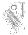
- Figure 1A shows a perspective view of a stylet 10 coupled to the single-insertion, multiple samples biopsy transport subassembly 100 having distal end 100A and proximal end 100B that can be implemented in a multiple sampling biopsy device (not shown).
- the transport subassembly 100 includes the stylet 10, which has a tip 11 at the distal end 100A and an outer cutting cannula 20 covering a substantial portion of the stylet 10 and a first port 10A.
- Extending through a hollow portion of the stylet 10 are a plurality of nested paddles 12, 14, 16, and 18 coupled to a drive unit at the proximal end 100B, and other ancillary components of the device 100 such as respective saline or vacuum reservoirs, motor drive, reduction gears, switches and sensors (not shown).
- the transport subassembly 100 operates by retracting the outer cannula 20 proximally to expose the first port 10A. Vacuum can be provided to the lumen 10B with orifices 10C to allow the lumen 10B to siphon biological tissue into the port 10A ( Fig. 1A ).
- the outer cannula 20 is extended distally to sever the tissue BSM from its main mass. Alternatively, a cannula disposed internally of the stylet 10 can also be used. Once the tissue BSM has separated from the main mass, two of the paddles 12 and 14 are retracted proximally. The longitudinal distance between the two paddles and the port 10A partly define the size of tissue sample per retraction of the two paddles. As shown in Fig.
- each paddle and its corresponding connector can be mounted in an arcuate offset configuration.
- the paddles 412 ,413, 414 are linked together as a chain so that only the most proximal one 414 of the paddle elements needs to be moved by a drive 450.
- moving the most proximal paddle 414 first a particular distance in a proximal direction, which distance is less than or equal to a link 404 length, will not cause the next paddle 413 to move. But a further movement will cause the most proximal paddle 414 to engage the next paddle 413 causing it to move.
- Paddle 413 would then engage the next paddle 412 after it is moved beyond the length of its link 403.
- the drive 450 only needs to moved the paddles 412, 413, and 414 in a single direction for multiple samples.
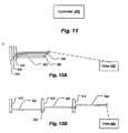
- the final result after multiple samples is shown in Fig. 12B .
- the links 402, 403 may be guided by openings or slots in the proximal adjacent paddle, for example as indicated at 432.
- the paddles themselves may be frictionally engaged within a surrounding cannula. This friction would be overcome by the drive 450.
- the transport trough 22A can be a similar material as extension 22B.
- the trough 22A can be an arcuate sectioned polymer tube 22A coupled to a flexible extender 22B, which is winds onto a roller 24.
- an outer cannula 20 (not shown for clarity) is used to sever the tissue from its main mass.
- a cannula disposed internally of the stylet 10 can also be used. Thereafter, the extender 22B is rolled counterclockwise to move the section 22A proximally.
- the stylet 10 can be provided with tracks 23A and 23B to allow the section 22A to be flattened due to the plastic material of the section 22A as the extender 22B is moved distally.
- the edges of the section 22A can disengage from the rails 23A and 23B, thereby allowing the flexible arcuate section 22A to fold inward forming a folded-in configuration 22D.
- This folding in of the polymer section 22A allows the section 22A to clamp over the biopsy sample ( Fig. 2C ) for transport proximally. As the sample is transported proximally, the sample enters an area of stylet 10 proximate port 20A.
- a keyed boss portion 26 can be provided inside the stylet 10 so that as the section 22A reaches the port the boss 26 spreads the polymer section 22A apart, from the closed configuration 22D, back to the open configuration of 22A, thereby releasing the grip on the tissue sample. At the same time, the boss 26 forces the tissue into a collection chamber ( Fig. 2D ).
- the extender 22B can be unrolled to move the polymer section 22A for engagement against tracks 23A and 23B for a subsequent tissue sampling.
- the extender 22B can be any suitable materials that allow for application of axial force distally to move the section 22A while permitting the extender 22B to be rolled in a circular configuration.
- a boss 26 is illustrated as a means for spreading the closed section 22D to open it into the open configuration 22A to release the sample
- other means for opening the section 22D are possible.
- guides similar to rails 23A can be provided at the proximal end which catch the edges of the rolled section 22D and gradually unwrap it.
- Such guides could be provided in the form of an insert in the stylet 10.
- the transport includes an inner cannula 28 surrounded by a nylon mesh tube (or "sock") 30.
- Nylon braid or weave having similar weight and elasticity similar to a woman's hose is suitable. This would allow the tube 30 to be stretched over the inner cannula 28 and to evert easily.
- the tube 30 can be of hydrophobic material or have a hydrophobic surface to help prevent tissue samples adhering to it.
- PTFE PolyTetraFluoroEthylene
- a passageway 10B is provided to permit fluid communication between the mesh tube 30 and the passage 10B.
- saline is provided via passage 10B while vacuum is provided in the mesh tube 30, which causes tissue BSM to be moved into the tube 30.
- a support tube 49 allows the mesh tube 30 to be everted over the inner cannula 28 as samples BSM are forced into it.
- the mesh tube 30 has a surface that helps to ensure positive engagement with samples, such as a surface covered with spines or hooks as illustrated. As each sample is drawn into the mesh tube 30, the mesh tube becomes ready to accept another sample.
- the mesh tube 30 itself may serve as a removable carrier that holds the samples BSM and separates them for delivery to a biopsy laboratory.
- a saline flush may be provided to help ensure samples are moved into the mesh tube 30. This may provide lubrication as well as positive transfer into the mesh tube 30.
- the proximal end 32 of the mesh tube 30 may be pulled by a line 47.
- the drive mechanism for pulling the line 47 may include a pulley, for example. Extraction of the tissue BSM can be achieved by back flushing the tube 30 with saline, causing the sample to be ejected from the tube 30 as the tube 30 is counter-everted at a recovery position.
- the support tube 49 and the mesh tube 30 may be transported through the stylet 10 to recover position and the mesh tube 30 counter-everted by pulling at the leading edge 51 by a tow line (not shown).
- the mesh tube 30 can be removed from the biopsy device.
- the samples can remain in a row in the tube thereby keeping the samples organized according to the order in which they were taken.
- an outer cannula 20 (not shown for clarity) is used to sever the tissue from its main mass.
- a cannula disposed internally of the stylet 10 can also be used.
- a shuttle transport system utilizing pulleys is provided.
- a shuttle 34 (which defines a trough to receive tissue samples) is connected by a system of pulleys 36A, 36B, and belt or endless connector 36C.
- Orifices 34A can be formed on the underside of the shuttle 34 so that vacuum provided from a passage 10B can be used to siphon a tissue sample BSM from a main tissue mass.
- an outer cannula 20 (not shown for clarity) can be used to sever the sample from its main mass ( Fig. 4B ).
- the shuttle 34 is moved proximally towards port 20A via the system of pulleys and belt.
- Ejection of the sample BSM out of the port 20A can be accomplished by a series of plungers 34B that are sized for insertion through orifices 34A.
- a saline transport with a bandolier type collection cartridge is provided.
- the stylet 10 is provided with a fluid passage 10B and main passage 10F.
- Fluid passage 10B can be connected via a suitable switching valve to allow saline to be pumped through the passage 10B in a distal direction while main passage 10F can be connected to a vacuum source to allow for saline and any other object entrained by the saline flow from passage 10B to flow through main passage 10F ( Fig. 5B ) and delivering the object (e.g., tissue sample BSM) into a bandolier type collection cartridge 39.
- the bandolier cartridge 39 has design details that are believed to be advantageous.
- the bandolier cartridges 39 are designed to be indexed through a double sided port 20A so that each cartridge is indexed once through the stylet 10.
- the cartridge has an open distal end 39A and a mesh material 39B formed over a proximal end. This allows the tissue to be pushed through the open end 39A but to be retained by the mesh 39B with fluid maintaining its flow through the stylet 10.
- the cartridges can be linked to each other via a flexible connector; chain link connection; or via a rigid connection.
- FIGS 5D-5J describe a saline pumping mechanism that may be used with the above and other embodiments.
- a dual-action pump 40 e.g., a syringe actuatable by a drive motor
- a four-way valve 44 with a vent 42 at one branch, is configured to empty the chamber 45 to the ambient through the four-way valve and out the air vent 42 as air is sucked into the chamber 40A.
- the vent 42 may be fitted with a filter to prevent contamination leaking into the biopsy device.
- the vacuuming action draws in a tissue sample 53.
- sensors may be used to detect the movement of the tissue sample 53 into the lumen 10B, or the passage of an elapsed time interval or user action may be used to determine that a sample 53 has been drawn into the passage 10B.
- the outer cannula 20 can be used to sever the tissue sample from the host. Alternatively, a cannula disposed internally of the stylet 10 can also be used.
- the four-way valve 44 is configured to allow the dual-action pump 40 to draw saline into port 40B.
- the dual-action pump 40 via the four-way valve 44, forces saline to flow through passage 10B, causing the tissue sample to be transported proximally towards through-port 20A ( Fig. 5F ).
- the sample encounters the mesh material 39B in a collection vial or cartridge, it remains in place while residual saline falls into the sump 55.
- any remaining saline in the lumens can be drawn back into the reservoir 48 by first drawing from the lumens into the chamber 45 ( Fig. 5G ) and then pumping into the reservoir 48 ( Fig. 5H ) for subsequent use by the dual-action pump 40.
- the passage 10F is provided with a flexible tube segment 10R that can be pinch-clamped by means of a valve actuator 10S.
- a pair of inline connectors 10V and 10W provides a smooth transition from a lead in part 10P to a lead out part 10Q to allow fluid and samples to pass through as in the earlier embodiment of passage 10F.
- the reason for adding this capability to close the valve is to allow a stronger vacuum to be developed in the sample area 10A by improving the volumetric efficiency of the dual action pump 40.
- the piston valve is configured as illustrated in Fig. 5F .
- the situation in Fig. 5F unlike the situation in Fig.
- Figures 6A1-6A3 illustrate a rotary to-linear type tissue transport assembly 57 utilizing a shuttle 10.
- the shuttle 34 is coupled to a helically threaded member via a suitable joint coupling.
- the joint coupling allows the shuttle to remain in a generally fixed orientation (e.g., upwardly oriented) while an inner cannula 21 with external threads are rotated against the stylet 10 (provided with internal threads), which allows the inner cannula 21 to convert the rotary motion of the cannula 21 into a linear motion while the stylet 10 remains stationary.
- the number and nature of the internal threads can be designed to achieve a sufficient transport speed with little or no back drive or backlash in the system.
- a fixed elongate slide 34B passing through and engaged in a slot 34A in the shuttle 34 may be used to prevent the shuttle 34 from rotating while permitting it to travel along the cannula 20.
- An outer cannula 20 can be used to sever the tissue sample from its main mass.
- a cannula disposed internally of the stylet 10 can also be used. Thereafter, the internal cannula 21 is rotated against the internal threads of the stylet 10 to transport the shuttle 34 to a tissue ejection port 20A.
- Figures 6B1-6B3 illustrate a linear motion by longitudinal expansion of a plurality of nested elongated members.
- the shuttle 34 is connected to a first elongated member 21A that is nestable to second elongated member 21B, that is nestable to a third elongated member 21C and so on.
- the shuttle 34 and nested elongated members are disposed inside the stylet 10 (not shown for clarity).
- any of a variety of linear actuator devices may be employed. As in the embodiment of Figs.
- a fixed elongate slide 34B passing through and engaged in a slot 34A in the shuttle 34 may be used to prevent the shuttle 34 from rotating while permitting it to travel along the cannula 20.
- Each of the nested members may be provided with a stop so that when it reaches the end of a permitted range of travel relative to the member in which it is inserted, it is prevented from rotating further. In this way, only the most proximal member (e.g., 21C) needs to be rotated to extend and retract the shuttle 34.
- the outer cannula 20 can be used to sever the tissue sample from its main mass.
- a cannula disposed internally of the stylet 10 can also be used.
- a suitable mechanism can be used to translate the shuttle in a linear motion between the first port 10A and second port 20A.
- a Bowden type cable can be connected to the first elongated member through the interior of the second and third elongated members so that one to one movement of the cable would force the first elongated member 2 1 A to telescope out of the interior of the second elongated member 21B.
- a hydraulic mechanism can be used to telescopically expand these members by pressurizing the interiors of the elongated members 21B and 21C with a suitable bio-compatible liquid. Retraction of the members 21A, 21B, and 21C into a nested configuration can be achieved by providing a vacuum that extracts the liquid out of the interiors of the elongated members.
- Orifices 34A can be formed in the underside of shuttle 34 (e.g., Figs. 4A and 4C ) so that vacuum can be provided for siphoning of tissue at port 10A and ejection of the tissue by pressurized fluid at port 20A into a tissue vial or cartridge.
- a mechanical ejector 34B can also be used.
- each of the above embodiments can be utilized with a suitably sized stylet.
- the internal volume is sufficient to capture a mass of at least 150 milligrams of biological tissues, e.g., turkey breast tissues.
- the internal volume is sufficient to capture a mass of at least 50 milligrams or more of biological tissues, e.g., turkey breast tissues.
- the length of the stylet 10 can be of any suitable lengths, such as, for example, about 250 to about 300 millimeters.
- the volume V of the housing containing all of the components of the device 100 is preferably 500 cubic centimeters or less and preferably about 320 cubic centimeters with particularly preferable dimensions of about 40 millimeters by about 40 millimeters and about 200 millimeters.
- the term "about” or “approximately” for any numerical values indicates a suitable dimensional tolerance that allows the part or collection of components to function for its intended purpose as a biopsy cutter, biopsy system or the combination of both the system and cutter.
- the cutting action by the cannula 20 can be by translation, rotation, translation and rotation or a combination of these movements along with back and forth axial movements of the cannula 20 as part of the cutting strategy.
- the drive unit can be a suitable drive unit such as the one shown and described, by way of example, in Figures 2 , 9A , and 10A of U.S. Patent Application Publication No. 2005/0165328 published on July 28, 2005
- each of four marking systems can be integrated with each of the examples described above to provide for at least 32 different integrated biopsy cutter and marker systems.
- each marking system can be integrated with each of the examples described above to provide for at least 32 different integrated biopsy cutter and marker systems.
- the four marking systems will be described and shown below.
- those skilled in the art can combine each marker system with each of the biopsy cutter systems as appropriate to arrive at a suitable permutation of biopsy sampling device and integrated marker.
- a marker system utilizing a hook type marker 50 i.e., a "harpoon" to prevent migration of the marker 50 once it has been deployed, is shown.
- the hook type marker 50 with hook 52 can be deployed in sequence or simultaneously with the sampling of biopsy tissues with the various technologies described in relation to Figures 1-6 above.
- a member e.g., an internal D-Rod 14A, 14B, or the outer cannula 20
- a second tracer 14B is provided with a cut-out portion 14B1 having a ramp 14B2 formed on a distal end of the rod 14B.
- the ramp 14B2 can be used (depending on whether the cannula 20 or rod 14B is axially translated only, rotated only or a combination of axial translation and rotation) to ensure that the marker 50 is deposited sufficiently near the tissue sampling site.
- Various marker configurations can be utilized. For example, as shown in Fig. 7D , a marker with wire like hooks 50A, square sectioned hook 50B, or marker with serrated edges 50C can be used in this system.
- a marker system utilizing a split ring marker 60 can be utilized with various biopsy techniques described above in relation to Figures 1-5 .
- the split-ring marker 60 can be mounted to the stylet 10 via a suitable technique such as, for example, crimping, swaging or semi-permanent bonding.
- an intermediate member 38 that forms a seal with the cannula or cutter 20 can be provided to maintain a generally constant outer diameter of the cannula 20 without an abrupt transition to the tip 11.
- the split-ring marker 60 can be deployed by itself, simultaneously with the sampling of the tissue, prior to sampling or subsequent to the sampling.
- the stylet tip 11 can be actuated proximally towards the user to force the split-ring marker 60 to detach from the tip 11.
- the outer cannula 20 can be actuated distally away from the user to force the split-ring marker 60 to separate from the stylet tip 11.
- a marker system using a blossom-type marker 70 can be utilized with various biopsy techniques described above in relation to Figures 1 and 2 .
- the blossom marker 70 is mounted on a specially configured stylet tip 110 ( Fig. 9C ), which has grooves 112 and ramps 114 disposed about a longitudinal axis of the tip 110.
- the blossom marker 70 can be mounted by a suitable technique, such as, for example, crimping, swaging, or casting onto the specially configured stylet tip 110.
- the outer cannula 20 can be moved distally away from the user to force the blossom marker to be separated from the stylet tip 110.
- the ramps 114 on the tip 110 force the sectioned tips 62A-62E to blossom radially, thereby forming hooks 64A-64E.
- the stylet tip 110 can be actuated proximally towards the user so that the marker is deployed via contact against the outer cannula 20.
- FIG. 10A and 10B another marker system is shown which uses a spiral-type marker 80 in conjunction with various biopsy systems described above in relation to Figures 1-6 .
- a coiled marker wire 80 can be disposed in a hollowed out section 110 of the stylet tip 11.
- a suitable deployment mechanism can be used to eject the coiled marker wire out of its storage space in the stylet tip 11.
- the deployment mechanism can be a suitable mechanism, such as, for example, a linear-to-rotary motion converter that converts a linear motion into a rotary motion to rotatably expel the marker.
- each marker can be, for example, stainless steel, gold, titanium, platinum, tantalum, barium sulfate, biodegradable iron or shape memory polymer or metal alloy such as Nitinol. It is noted that Nitinol is radio-opaque, ultrasonically opaque and MRI compatible and therefore would be preferred by itself or in combination with other materials described herein and as known to those skilled in the art. Further, the markers can be of any suitable size so that it can be fitted onto a 7, 8, 9, 10, 11, 12, 14, or 16 gauge needle.
- the tip 11 can be configured to store a plurality of harpoon markers 50; the stylet 10 can be mounted with a longitudinal series of split-ring markers 60; the tip 11 can be configured with a cutter so that multiple helical markers 80 can be deployed.
- the paddle transport of Figs. 1A-1D can be utilized with the threaded transport of Figs. 6A1-6A3 by forming threads on the paddle connectors 18A, 16A, 14A, and 12A.
- the roller transport of Figs. 2A-2E can be utilized for the paddle connectors of Figs. 1A-1D .
- the bandolier type cartridges 39 of Fig. 5C can be utilized for any of the transport subassemblies described herein.
- the hydraulic and vacuum transport system of Figs. 5D-5G can be utilized in any one of the embodiments described herein.
- a device may employ a controller 350 such as a programmable microprocessor controller, to provide the described functionality.
- controller 350 such as a programmable microprocessor controller
Abstract
Description
- This invention relates to a tissue biopsy sampling device.
- It is sometimes desirable or necessary to obtain specimens of tissue from humans and other animals, particularly in the diagnosis and treatment of patients with cancerous tumors, premalignant conditions, and other diseases or disorders. For example, when it is discovered that suspicious conditions exist, either by means of x-ray or ultrasound imaging in various tissues of the body, a physician usually performs a biopsy to determine if the cells at the suspected site are cancerous or benign.
- A biopsy can be done either by an open or percutaneous technique. Open biopsy is an invasive procedure using a scalpel, by either a portion (incisional biopsy) being removed or the entire mass (excisional biopsy) is removed. Percutaneous biopsy is usually done with a needle-like instrument through a relatively small incision, and can be performed by fine needle aspiration (FNA) or through the taking of a core biopsy sample. In FNA biopsy, individual cells or clusters of cells are obtained for cytologic examination and can be prepared such as in a Papanicolaou smear. In a core biopsy, a core or fragment of the tissue is obtained for histological examination.
- Uncontaminated and intact tissue from the organ, lesion, or tumor is preferred by medical personnel in order to arrive at a definitive diagnosis regarding the patient's condition. In most cases only part of the tissue in question needs to be sampled. The portions of tissue extracted must be indicative of the organ, lesion, or tumor as a whole. Often, multiple tissue samples from various locations of the mass being sampled may be taken.
- The percutaneous biopsy procedure can be performed utilizing various techniques and devices. One such biopsy device can include an inner stylet positioned inside an outer cannula, where the stylet is able to slide into and out of the cannula. The stylet can be a solid, pointed needle having a tissue sampling recess, and the cannula can be a hollow, open-ended needle having a sharp tip. The stylet and cannula can be manipulated cooperatively to capture a tissue sample in the sample recess. Such existing devices can be manually operated, semi-automated, and automated.
-
US Patent No. 6,485,436 shows a multiple sample biopsy needle with a hydraulic mechanism that circulates fluid from the tip of the needle back to a receiving basket or baskets. A revolver-type array of receiving chambers is disclosed. -
US Patent No. 5,827,305 shows a tissue sampling needle that pushes a sample proximally using a saline wash. Samples remain spaced apart within the needle such that the sequence of their collection is preserved. Samples can also be removed from a port while the needle remains in place. No mechanical transport mechanisms or drives are disclosed. -
US Patent No. 5,526,822 shows a transport system that uses a cannula and knock-out pin combined with a vacuum source to shuttle a tissue sample to a multiple-chamber cassette where it is knocked out. The cannula is then repositioned for another sample. The vacuum source is external. A revolving sample cassette is also shown. A vent opening in each sample cylinder of the cassette is provided to eject the fluid used to transport the tissue sample. A removable disposable needle-bearing cassette interfaces with rotary and linear drives by means of long gears and shuttles that cradle the gears. Cutters operate in rotary and linear fashion (a counter-rotating cutters embodiment is included) and the cannula can be rotated to orient the sample opening. -
US Patent No. 6,017,316 shows a transport system similar toUS Patent No. 5,827,822 in which a cutter transports with vacuum assist. Multiple sampling with single insertion is described but not automated multiple sample-handling. The details of a drive system are not disclosed -
US Patent No. 6,193,673 shows a needle with a durable part and a disposable part. An external cutting cannula rotates and advances axially to cut a sample. The tissue cutter is driven axially by a rack and pinion drive which are part of a durable component. A cradle connects the rack to the cutting cannula. -
US Patent No. 5,944,673 describes a tissue extractor that rotates within a piercing needle to align with any one of multiple receiving ports while obstructing the remaining ports. The tissue sample is cut by advancing the cutter and removing by withdrawing the extractor. A vacuum holds the tissue sample in place during the removal of the tissue extractor from the cutter. The cutter rotates as it advances. - It is known to obtain a single sample with a single insertion. However, there are circumstances where there may be a need to obtain more than one samples. While the known biopsy needle can be re-inserted multiple times, such technique can cause pain and scarring of the body site.
- It is known to leave a marker at the biopsied site. To do so, however, a physician or healthcare provider would typically need to withdraw the biopsy needle and insert a different device to leave a marker at the biopsied site. The additional step with the marker device concurrent with the tissue sampling may not allow the marker to be deposited at the actual biopsied site, which can lead to inaccurate post-biopsy diagnosis.
-
WO 98/35615 US 2005/0085838 A1 describes a method of operating a biopsy device including the application of a vacuum to a lumen and a control of a cutter.WO 2006005342 A1 is a document published after the priority date describing a biopsy device with a movable sample receptacle and a sample container with the samples being transferred from the sample receptacle to the sample retainer by the application of a flushing liquid. - The present invention provides a biopsy device according to claim 1. Preferred aspects of the invention are provided according to the dependent claims.
- The accompanying drawings, which are incorporated herein and constitute part of this specification, illustrate presently preferred exemplary embodiments of the invention, and, together with the general description given above and the detailed description given below, serve to explain features of the invention.
-
Figures 1A-1D illustrate a transport subassembly for a biopsy device according to one exemplary embodiment of the present invention. -
Figures 2A-2E illustrate another transport assembly. -
Figures 3A and 3B illustrate yet another biopsy transport system. -
Figures 4A-4C illustrate yet another tissue transport system for a biopsy device. -
Figures 5A-5J illustrate a tissue transport system utilizing saline for deposit into a bandolier type collection chamber. -
Figure 6A1-6A3 illustrate a tissue transport using a threaded type inner cannula. -
Figures 6B1-6B3 illustrate a tissue transport using a telescoping drive. -
Figures 7A-7D illustrate an integrated biopsy marker system for each of the transport assembly ofFigures 1-6 . -
Figures 8A and 8B illustrate another integrated biopsy marker system for the transport assembly ofFigures 1-6 . -
Figures 9A-9C illustrate a further integrated biopsy marker system for each of the transport assembly ofFigures 1-6 . -
Figures 10A and 10B illustrate yet another integrated biopsy marker system for each of the transport assembly ofFigures 1-6 . -
Figure 11 shows a controller. -
Figures 12A and 12B show an embodiment of a paddle transport mechanism -
Figures 1-10 illustrate the preferred exemplary embodiments which utilize the same reference numeral to indicate generally similar components. In particular,Figure 1A shows a perspective view of astylet 10 coupled to the single-insertion, multiple samplesbiopsy transport subassembly 100 havingdistal end 100A andproximal end 100B that can be implemented in a multiple sampling biopsy device (not shown). Thetransport subassembly 100 includes thestylet 10, which has atip 11 at thedistal end 100A and anouter cutting cannula 20 covering a substantial portion of thestylet 10 and afirst port 10A. Extending through a hollow portion of thestylet 10 are a plurality of nestedpaddles proximal end 100B, and other ancillary components of thedevice 100 such as respective saline or vacuum reservoirs, motor drive, reduction gears, switches and sensors (not shown). - The
transport subassembly 100 operates by retracting theouter cannula 20 proximally to expose thefirst port 10A. Vacuum can be provided to thelumen 10B withorifices 10C to allow thelumen 10B to siphon biological tissue into theport 10A (Fig. 1A ). Theouter cannula 20 is extended distally to sever the tissue BSM from its main mass. Alternatively, a cannula disposed internally of thestylet 10 can also be used. Once the tissue BSM has separated from the main mass, two of thepaddles 12 and 14 are retracted proximally. The longitudinal distance between the two paddles and theport 10A partly define the size of tissue sample per retraction of the two paddles. As shown inFig. 1B , the device is now ready for a subsequent sample withpaddles 14 and 16. As shown inFig. 1C , to ensure that the plurality of paddles can be retained in thestylet 10 without reducing the internal volume that would be needed to transport the tissue BSM through the internal passage ofstylet 10, each paddle and its corresponding connector can be mounted in an arcuate offset configuration. - Referring to
Figures 12A and 12B , in an embodiment, thepaddles drive 450. Thus, moving the mostproximal paddle 414 first a particular distance in a proximal direction, which distance is less than or equal to alink 404 length, will not cause thenext paddle 413 to move. But a further movement will cause the mostproximal paddle 414 to engage thenext paddle 413 causing it to move.Paddle 413 would then engage thenext paddle 412 after it is moved beyond the length of itslink 403. If a sample is received and moved by thepaddle 414 while leaving the other paddles in place, then thedrive 450 only needs to moved thepaddles Fig. 12B . Thelinks drive 450. - Referring to
Figure 2A , a flexible transport mechanism is shown and described. In this embodiment, thetransport trough 22A can be a similar material asextension 22B. Alternatively, thetrough 22A can be an arcuate sectionedpolymer tube 22A coupled to aflexible extender 22B, which is winds onto aroller 24. As is the case above, an outer cannula 20 (not shown for clarity) is used to sever the tissue from its main mass. Alternatively, a cannula disposed internally of thestylet 10 can also be used. Thereafter, theextender 22B is rolled counterclockwise to move thesection 22A proximally. To ensure that thearcuate section 22A can retain the tissue sample on thesurface 22C, thestylet 10 can be provided withtracks 23A and 23B to allow thesection 22A to be flattened due to the plastic material of thesection 22A as theextender 22B is moved distally. When theextender 22B is moved proximally, the edges of thesection 22A can disengage from therails 23A and 23B, thereby allowing the flexiblearcuate section 22A to fold inward forming a folded-in configuration 22D. This folding in of thepolymer section 22A allows thesection 22A to clamp over the biopsy sample (Fig. 2C ) for transport proximally. As the sample is transported proximally, the sample enters an area ofstylet 10proximate port 20A. Akeyed boss portion 26 can be provided inside thestylet 10 so that as thesection 22A reaches the port theboss 26 spreads thepolymer section 22A apart, from the closed configuration 22D, back to the open configuration of 22A, thereby releasing the grip on the tissue sample. At the same time, theboss 26 forces the tissue into a collection chamber (Fig. 2D ). Theextender 22B can be unrolled to move thepolymer section 22A for engagement againsttracks 23A and 23B for a subsequent tissue sampling. Theextender 22B can be any suitable materials that allow for application of axial force distally to move thesection 22A while permitting theextender 22B to be rolled in a circular configuration. - While in the foregoing embodiment, a
boss 26 is illustrated as a means for spreading the closed section 22D to open it into theopen configuration 22A to release the sample, other means for opening the section 22D are possible. For example, guides similar torails 23A can be provided at the proximal end which catch the edges of the rolled section 22D and gradually unwrap it. Such guides could be provided in the form of an insert in thestylet 10. - Referring to
Figure 3A , another transport subassembly is provided. In this embodiment, the transport includes aninner cannula 28 surrounded by a nylon mesh tube (or "sock") 30. Nylon braid or weave having similar weight and elasticity similar to a woman's hose is suitable. This would allow thetube 30 to be stretched over theinner cannula 28 and to evert easily. Also, preferably, thetube 30 can be of hydrophobic material or have a hydrophobic surface to help prevent tissue samples adhering to it. For example a mesh coated with PolyTetraFluoroEthylene (PTFE) may be used. - A
passageway 10B is provided to permit fluid communication between themesh tube 30 and thepassage 10B. In one embodiment, saline is provided viapassage 10B while vacuum is provided in themesh tube 30, which causes tissue BSM to be moved into thetube 30. A support tube 49 allows themesh tube 30 to be everted over theinner cannula 28 as samples BSM are forced into it. Preferably themesh tube 30 has a surface that helps to ensure positive engagement with samples, such as a surface covered with spines or hooks as illustrated. As each sample is drawn into themesh tube 30, the mesh tube becomes ready to accept another sample. Themesh tube 30 itself may serve as a removable carrier that holds the samples BSM and separates them for delivery to a biopsy laboratory. - A saline flush may be provided to help ensure samples are moved into the
mesh tube 30. This may provide lubrication as well as positive transfer into themesh tube 30. Theproximal end 32 of themesh tube 30 may be pulled by a line 47. The drive mechanism for pulling the line 47 may include a pulley, for example. Extraction of the tissue BSM can be achieved by back flushing thetube 30 with saline, causing the sample to be ejected from thetube 30 as thetube 30 is counter-everted at a recovery position. In this case, the support tube 49 and themesh tube 30 may be transported through thestylet 10 to recover position and themesh tube 30 counter-everted by pulling at the leading edge 51 by a tow line (not shown). - After the samples are harvested, the
mesh tube 30 can be removed from the biopsy device. The samples can remain in a row in the tube thereby keeping the samples organized according to the order in which they were taken. - As is the case above, an outer cannula 20 (not shown for clarity) is used to sever the tissue from its main mass. Alternatively, a cannula disposed internally of the
stylet 10 can also be used. positioned insecond port 20A. - Referring to
Figure 4A , a shuttle transport system utilizing pulleys is provided. In this system, a shuttle 34 (which defines a trough to receive tissue samples) is connected by a system of pulleys 36A, 36B, and belt orendless connector 36C.Orifices 34A can be formed on the underside of theshuttle 34 so that vacuum provided from apassage 10B can be used to siphon a tissue sample BSM from a main tissue mass. As is the case above, an outer cannula 20 (not shown for clarity) can be used to sever the sample from its main mass (Fig. 4B ). Thereafter, theshuttle 34 is moved proximally towardsport 20A via the system of pulleys and belt. Ejection of the sample BSM out of theport 20A can be accomplished by a series ofplungers 34B that are sized for insertion throughorifices 34A. Once the tissue BSM has been ejected into a collection vial or chamber (not shown), theshuttle 34 is translated towardsport 10A for another collection of tissue (Fig. 4A ). - Referring to
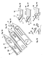
Figure 5A , a saline transport with a bandolier type collection cartridge is provided. In this embodiment, thestylet 10 is provided with afluid passage 10B andmain passage 10F.Fluid passage 10B can be connected via a suitable switching valve to allow saline to be pumped through thepassage 10B in a distal direction whilemain passage 10F can be connected to a vacuum source to allow for saline and any other object entrained by the saline flow frompassage 10B to flow throughmain passage 10F (Fig. 5B ) and delivering the object (e.g., tissue sample BSM) into a bandoliertype collection cartridge 39. Thebandolier cartridge 39 has design details that are believed to be advantageous. First, thebandolier cartridges 39 are designed to be indexed through a doublesided port 20A so that each cartridge is indexed once through thestylet 10. Second, the cartridge has an opendistal end 39A and amesh material 39B formed over a proximal end. This allows the tissue to be pushed through theopen end 39A but to be retained by themesh 39B with fluid maintaining its flow through thestylet 10. Third, the cartridges can be linked to each other via a flexible connector; chain link connection; or via a rigid connection. -
Figures 5D-5J describe a saline pumping mechanism that may be used with the above and other embodiments. InFig. 5D , a dual-action pump 40 (e.g., a syringe actuatable by a drive motor) can be used to generate negative pressure by forcing apiston 46 to expand the volume of achamber 40A, which is in communication with themain passage 10F of thestylet 10. A four-way valve 44, with avent 42 at one branch, is configured to empty thechamber 45 to the ambient through the four-way valve and out theair vent 42 as air is sucked into thechamber 40A. Note that thevent 42 may be fitted with a filter to prevent contamination leaking into the biopsy device. - The vacuuming action draws in a tissue sample 53. To trigger the cutting of the sample, sensors (not shown) may be used to detect the movement of the tissue sample 53 into the
lumen 10B, or the passage of an elapsed time interval or user action may be used to determine that a sample 53 has been drawn into thepassage 10B. Theouter cannula 20 can be used to sever the tissue sample from the host. Alternatively, a cannula disposed internally of thestylet 10 can also be used. - At this point, shown here in
Fig. 5E , the four-way valve 44, with avent 42 at one branch, is configured to allow the dual-action pump 40 to draw saline intoport 40B. With theouter cannula 20 covering theport 10A (not shown for clarity), the dual-action pump 40, via the four-way valve 44, forces saline to flow throughpassage 10B, causing the tissue sample to be transported proximally towards through-port 20A (Fig. 5F ). As the sample encounters themesh material 39B in a collection vial or cartridge, it remains in place while residual saline falls into thesump 55. Any remaining saline in the lumens can be drawn back into the reservoir 48 by first drawing from the lumens into the chamber 45 (Fig. 5G ) and then pumping into the reservoir 48 (Fig. 5H ) for subsequent use by the dual-action pump 40. - Referring to
Fig. 5J , in an alternative embodiment, thepassage 10F is provided with aflexible tube segment 10R that can be pinch-clamped by means of avalve actuator 10S. In this configuration, a pair ofinline connectors part 10P to a lead outpart 10Q to allow fluid and samples to pass through as in the earlier embodiment ofpassage 10F. The reason for adding this capability to close the valve is to allow a stronger vacuum to be developed in thesample area 10A by improving the volumetric efficiency of thedual action pump 40. To apply a vacuum to sampleport 10A, the piston valve is configured as illustrated inFig. 5F . However, unlike the situation inFig. 5E , in this case, there is fluid only in the sump 48 as depicted inFig. 5D . Theclamp 10S is closed. Thepiston 46 is moved to the right to generate the vacuum by expanding the volume ofchamber 45. Because thepassage 10P is closed, the total volume evacuated, relative to thechamber volume 45, is markedly decreased. This configuration ofpassage 10P also has the advantage of avoiding the need for vacuum-competent sealing of thecollection chamber 56 andsump 55. -
Figures 6A1-6A3 illustrate a rotary to-linear type tissue transport assembly 57 utilizing ashuttle 10. In this embodiment, theshuttle 34 is coupled to a helically threaded member via a suitable joint coupling. The joint coupling allows the shuttle to remain in a generally fixed orientation (e.g., upwardly oriented) while an inner cannula 21 with external threads are rotated against the stylet 10 (provided with internal threads), which allows the inner cannula 21 to convert the rotary motion of the cannula 21 into a linear motion while thestylet 10 remains stationary. The number and nature of the internal threads can be designed to achieve a sufficient transport speed with little or no back drive or backlash in the system. A fixedelongate slide 34B passing through and engaged in aslot 34A in theshuttle 34 may be used to prevent theshuttle 34 from rotating while permitting it to travel along thecannula 20. Anouter cannula 20 can be used to sever the tissue sample from its main mass. Alternatively, a cannula disposed internally of thestylet 10 can also be used. Thereafter, the internal cannula 21 is rotated against the internal threads of thestylet 10 to transport theshuttle 34 to atissue ejection port 20A. -
Figures 6B1-6B3 illustrate a linear motion by longitudinal expansion of a plurality of nested elongated members. Theshuttle 34 is connected to a firstelongated member 21A that is nestable to secondelongated member 21B, that is nestable to a thirdelongated member 21C and so on. Theshuttle 34 and nested elongated members are disposed inside the stylet 10 (not shown for clarity). There may be any desired number of nested members such as 21A through 21 C. Further, any of a variety of linear actuator devices may be employed. As in the embodiment ofFigs. 6A1-6A4 , a fixedelongate slide 34B passing through and engaged in aslot 34A in theshuttle 34 may be used to prevent theshuttle 34 from rotating while permitting it to travel along thecannula 20. Each of the nested members may be provided with a stop so that when it reaches the end of a permitted range of travel relative to the member in which it is inserted, it is prevented from rotating further. In this way, only the most proximal member (e.g., 21C) needs to be rotated to extend and retract theshuttle 34. - As is the case above, the
outer cannula 20 can be used to sever the tissue sample from its main mass. Alternatively, a cannula disposed internally of thestylet 10 can also be used. With the tissue contained in theshuttle 34, a suitable mechanism can be used to translate the shuttle in a linear motion between thefirst port 10A andsecond port 20A. For example, a Bowden type cable can be connected to the first elongated member through the interior of the second and third elongated members so that one to one movement of the cable would force the firstelongated member 2 1 A to telescope out of the interior of the secondelongated member 21B. Further expansion of the cable would force the secondelongated member 21B to telescope out of the interior of the thirdelongated member 21C (Fig. 6B2 ). Retraction of the cable would force the member to be nested inside each other in proximal direction (Fig. 6B3 ). Alternatively, a hydraulic mechanism can be used to telescopically expand these members by pressurizing the interiors of theelongated members members Orifices 34A can be formed in the underside of shuttle 34 (e.g.,Figs. 4A and 4C ) so that vacuum can be provided for siphoning of tissue atport 10A and ejection of the tissue by pressurized fluid atport 20A into a tissue vial or cartridge. Alternatively, amechanical ejector 34B can also be used. - Each of the above embodiments can be utilized with a suitably sized stylet. For a 14 gauge stylet or needle, the internal volume is sufficient to capture a mass of at least 150 milligrams of biological tissues, e.g., turkey breast tissues. For a 10
gauge stylet 10, the internal volume is sufficient to capture a mass of at least 50 milligrams or more of biological tissues, e.g., turkey breast tissues. The length of thestylet 10 can be of any suitable lengths, such as, for example, about 250 to about 300 millimeters. The volume V of the housing containing all of the components of thedevice 100 is preferably 500 cubic centimeters or less and preferably about 320 cubic centimeters with particularly preferable dimensions of about 40 millimeters by about 40 millimeters and about 200 millimeters. As used herein, the term "about" or "approximately" for any numerical values indicates a suitable dimensional tolerance that allows the part or collection of components to function for its intended purpose as a biopsy cutter, biopsy system or the combination of both the system and cutter. - The cutting action by the
cannula 20 can be by translation, rotation, translation and rotation or a combination of these movements along with back and forth axial movements of thecannula 20 as part of the cutting strategy. In the preferred embodiments, the drive unit can be a suitable drive unit such as the one shown and described, by way of example, inFigures 2 ,9A , and10A ofU.S. Patent Application Publication No. 2005/0165328 published on July 28, 2005 - The examples shown in the illustrations and described in detail above can be integrated with one or more of four exemplary marking systems. In particular, each of four marking systems can be integrated with each of the examples described above to provide for at least 32 different integrated biopsy cutter and marker systems. For clarity, only the four marking systems will be described and shown below. However, those skilled in the art can combine each marker system with each of the biopsy cutter systems as appropriate to arrive at a suitable permutation of biopsy sampling device and integrated marker.
- Referring to
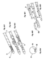
Figures 7A-7G , a marker system utilizing a hook type marker 50 (i.e., a "harpoon") to prevent migration of themarker 50 once it has been deployed, is shown. Thehook type marker 50 withhook 52 can be deployed in sequence or simultaneously with the sampling of biopsy tissues with the various technologies described in relation toFigures 1-6 above. As shown inFigures 7A and 7E , a member (e.g., an internal D-Rod marker 50 stored in thestylet tip 11. In the exemplary embodiment ofFigures 7A-7G , asecond tracer 14B is provided with a cut-out portion 14B1 having a ramp 14B2 formed on a distal end of therod 14B. The ramp 14B2 can be used (depending on whether thecannula 20 orrod 14B is axially translated only, rotated only or a combination of axial translation and rotation) to ensure that themarker 50 is deposited sufficiently near the tissue sampling site. Various marker configurations can be utilized. For example, as shown inFig. 7D , a marker with wire likehooks 50A, square sectionedhook 50B, or marker withserrated edges 50C can be used in this system. - Referring
Figures 8A and 8B , a marker system utilizing asplit ring marker 60 can be utilized with various biopsy techniques described above in relation toFigures 1-5 . InFigure 8A , the split-ring marker 60 can be mounted to thestylet 10 via a suitable technique such as, for example, crimping, swaging or semi-permanent bonding. Optionally, anintermediate member 38 that forms a seal with the cannula orcutter 20 can be provided to maintain a generally constant outer diameter of thecannula 20 without an abrupt transition to thetip 11. The split-ring marker 60 can be deployed by itself, simultaneously with the sampling of the tissue, prior to sampling or subsequent to the sampling. As shown inFigure 8B , thestylet tip 11 can be actuated proximally towards the user to force the split-ring marker 60 to detach from thetip 11. Alternatively, theouter cannula 20 can be actuated distally away from the user to force the split-ring marker 60 to separate from thestylet tip 11. - Referring to
Figures 9A-9C , a marker system using a blossom-type marker 70 can be utilized with various biopsy techniques described above in relation toFigures 1 and2 . As shown inFigure 9A , theblossom marker 70 is mounted on a specially configured stylet tip 110 (Fig. 9C ), which hasgrooves 112 andramps 114 disposed about a longitudinal axis of thetip 110. Theblossom marker 70 can be mounted by a suitable technique, such as, for example, crimping, swaging, or casting onto the specially configuredstylet tip 110. As shown inFig. 9B , theouter cannula 20 can be moved distally away from the user to force the blossom marker to be separated from thestylet tip 110. As themarker 70 is separated from thetip 110, theramps 114 on thetip 110 force the sectioned tips 62A-62E to blossom radially, thereby forming hooks 64A-64E. Alternatively, thestylet tip 110 can be actuated proximally towards the user so that the marker is deployed via contact against theouter cannula 20. - Referring to
Figures 10A and 10B , another marker system is shown which uses a spiral-type marker 80 in conjunction with various biopsy systems described above in relation toFigures 1-6 . As shown inFigure 10A , a coiled marker wire 80 can be disposed in a hollowed outsection 110 of thestylet tip 11. A suitable deployment mechanism can be used to eject the coiled marker wire out of its storage space in thestylet tip 11. The deployment mechanism can be a suitable mechanism, such as, for example, a linear-to-rotary motion converter that converts a linear motion into a rotary motion to rotatably expel the marker. - The materials suitable for use as part of each marker can be, for example, stainless steel, gold, titanium, platinum, tantalum, barium sulfate, biodegradable iron or shape memory polymer or metal alloy such as Nitinol. It is noted that Nitinol is radio-opaque, ultrasonically opaque and MRI compatible and therefore would be preferred by itself or in combination with other materials described herein and as known to those skilled in the art. Further, the markers can be of any suitable size so that it can be fitted onto a 7, 8, 9, 10, 11, 12, 14, or 16 gauge needle.
- Although the markers have been shown as a single deployment marker, some of the embodiments disclosed herein can be utilized in a multiple deployment aspect. For example, the
tip 11 can be configured to store a plurality ofharpoon markers 50; thestylet 10 can be mounted with a longitudinal series of split-ring markers 60; thetip 11 can be configured with a cutter so that multiple helical markers 80 can be deployed. - Moreover, while specific embodiments have been described, various combinations of components and features can be obtained. For example, the paddle transport of
Figs. 1A-1D can be utilized with the threaded transport ofFigs. 6A1-6A3 by forming threads on thepaddle connectors Figs. 2A-2E can be utilized for the paddle connectors ofFigs. 1A-1D . Thebandolier type cartridges 39 ofFig. 5C can be utilized for any of the transport subassemblies described herein. The hydraulic and vacuum transport system ofFigs. 5D-5G can be utilized in any one of the embodiments described herein. Thus, it is clear to one skilled in the art that various permutations of components, sub-components and features can be utilized with the embodiments described herein and each seven transport devices is not limited only to the specific embodiment described herein. - Referring to
Fig. 11 , in all of the above embodiments, various motors, drives, valves, and other actuators are variously described along with their respective operations and operational sequences. It is clear from the particulars of each embodiment that a device may employ acontroller 350 such as a programmable microprocessor controller, to provide the described functionality. - While the present invention has been disclosed with reference to certain preferred embodiments, numerous modifications, alterations, and changes to the described embodiments are possible without departing from the sphere and scope of the present invention, which is described, by way of example, above. Accordingly, it is intended that the present invention not be limited to the described embodiments, but that it have the full scope and equivalents thereof.
Claims (7)
- A biopsy device, comprising:a biopsy sample extraction needle (10) with a sample extraction end, recovery end, and a transport channel linking the extraction and recovery ends;a pump (40); anda fluid reservoir (48), characterized in that the device further comprises a multi-way valve (44) connected to the pump and in that the fluid reservoir is linked to the transport channel and pump such that:in a first setting of the multi-way valve, the pump is arranged to draw a vacuum at at least the recovery end;in a second setting of the multi-way valve, the pump is arranged to draw fluid from the reservoir, andin a third setting of the multi-way valve, the pump is arranged to flush the transport channel from the extraction end to the recovery end to transport a sample through the transport channel.
- The device of claim 1, where the first and third multi-way valve settings are identical.
- The device of claim 1, where the pump is a syringe.
- The device of claim 1, where the pump can recover residual saline from the transport channel and deliver it to the reservoir.
- The device of claim 1, further comprising a volume-reducing valve (10S) that selectively reduces a total sealed volume in fluid flow connection with the recovery end such that a vacuum drawn by the pump is greater when the volume-reducing valve is engaged.
- The device of any one of claims 1-5, further comprising a cassette (39) with multiple recesses each having an access and a fluid-permeable blind end, the cassette positioned at the recovery position to align a selected one of the cassette recesses with the recovery position such that the selected cassette recess is in fluid communication with the interior volume of the stylet.
- The device of claim 6, wherein the cassette recesses are linked together by flexible connections to form a bandolier.
Applications Claiming Priority (2)
| Application Number | Priority Date | Filing Date | Title |
|---|---|---|---|
| US70722805P | 2005-08-10 | 2005-08-10 | |
| EP06801223.6A EP1921999B1 (en) | 2005-08-10 | 2006-08-10 | Single-insertion, multiple sampling biopsy device usable with various transport systems |
Related Parent Applications (2)
| Application Number | Title | Priority Date | Filing Date |
|---|---|---|---|
| EP06801223.6 Division | 2006-08-10 | ||
| EP06801223.6A Division-Into EP1921999B1 (en) | 2005-08-10 | 2006-08-10 | Single-insertion, multiple sampling biopsy device usable with various transport systems |
Publications (3)
| Publication Number | Publication Date |
|---|---|
| EP2196154A2 EP2196154A2 (en) | 2010-06-16 |
| EP2196154A3 EP2196154A3 (en) | 2011-02-16 |
| EP2196154B1 true EP2196154B1 (en) | 2012-01-18 |
Family
ID=37758185
Family Applications (3)
| Application Number | Title | Priority Date | Filing Date |
|---|---|---|---|
| EP06801223.6A Not-in-force EP1921999B1 (en) | 2005-08-10 | 2006-08-10 | Single-insertion, multiple sampling biopsy device usable with various transport systems |
| EP10158392.0A Active EP2196155B1 (en) | 2005-08-10 | 2006-08-10 | Single-insertion, multiple sample biopsy device with various transport system |
| EP10158390A Active EP2196154B1 (en) | 2005-08-10 | 2006-08-10 | Transport system for single-insertion, multiple sample biopsy devices |
Family Applications Before (2)
| Application Number | Title | Priority Date | Filing Date |
|---|---|---|---|
| EP06801223.6A Not-in-force EP1921999B1 (en) | 2005-08-10 | 2006-08-10 | Single-insertion, multiple sampling biopsy device usable with various transport systems |
| EP10158392.0A Active EP2196155B1 (en) | 2005-08-10 | 2006-08-10 | Single-insertion, multiple sample biopsy device with various transport system |
Country Status (7)
| Country | Link |
|---|---|
| US (4) | US8282574B2 (en) |
| EP (3) | EP1921999B1 (en) |
| JP (1) | JP5102207B2 (en) |
| AT (1) | ATE541517T1 (en) |
| CA (1) | CA2616823C (en) |
| ES (3) | ES2547725T3 (en) |
| WO (1) | WO2007021904A2 (en) |
Families Citing this family (124)
| Publication number | Priority date | Publication date | Assignee | Title |
|---|---|---|---|---|
| WO2003077768A1 (en) | 2002-03-19 | 2003-09-25 | Bard Dublin Itc Limited | Biopsy device and biopsy needle module that can be inserted into the biopsy device |
| ATE303099T1 (en) | 2002-03-19 | 2005-09-15 | Bard Dublin Itc Ltd | VACUUM BIOPSY DEVICE |
| DE10314240A1 (en) | 2003-03-29 | 2004-10-07 | Bard Dublin Itc Ltd., Crawley | Pressure generating unit |
| EP1776047B1 (en) | 2004-07-09 | 2012-12-05 | Bard Peripheral Vascular, Inc. | Transport system for biopsy device |
| US20060074345A1 (en) | 2004-09-29 | 2006-04-06 | Hibner John A | Biopsy apparatus and method |
| US7517321B2 (en) | 2005-01-31 | 2009-04-14 | C. R. Bard, Inc. | Quick cycle biopsy system |
| US7896817B2 (en) | 2005-08-05 | 2011-03-01 | Devicor Medical Products, Inc. | Biopsy device with manually rotated sample barrel |
| US7854707B2 (en) | 2005-08-05 | 2010-12-21 | Devicor Medical Products, Inc. | Tissue sample revolver drum biopsy device |
| US7828748B2 (en) | 2005-08-05 | 2010-11-09 | Devicor Medical Products, Inc. | Vacuum syringe assisted biopsy device |
| US7662109B2 (en) | 2006-02-01 | 2010-02-16 | Ethicon Endo-Surgery, Inc. | Biopsy device with replaceable probe incorporating static vacuum source dual valve sample stacking retrieval and saline flush |
| US7867173B2 (en) | 2005-08-05 | 2011-01-11 | Devicor Medical Products, Inc. | Biopsy device with replaceable probe and incorporating vibration insertion assist and static vacuum source sample stacking retrieval |
| US20080004545A1 (en) * | 2005-08-05 | 2008-01-03 | Garrison William A | Trigger Fired Radial Plate Specimen Retrieval Biopsy Instrument |
| USRE46135E1 (en) | 2005-08-05 | 2016-09-06 | Devicor Medical Products, Inc. | Vacuum syringe assisted biopsy device |
| US8262585B2 (en) | 2005-08-10 | 2012-09-11 | C. R. Bard, Inc. | Single-insertion, multiple sampling biopsy device with linear drive |
| WO2007021905A2 (en) | 2005-08-10 | 2007-02-22 | C.R. Bard Inc. | Single-insertion, multiple sample biopsy device with integrated markers |
| EP1921999B1 (en) | 2005-08-10 | 2015-08-05 | C.R.Bard, Inc. | Single-insertion, multiple sampling biopsy device usable with various transport systems |
| WO2007112751A2 (en) * | 2006-03-31 | 2007-10-11 | Sonion Roskilde A/S | Tissue sample collection system with visual sample inspection |
| WO2008024684A2 (en) | 2006-08-21 | 2008-02-28 | C.R. Bard, Inc. | Self-contained handheld biopsy needle |
| US8485987B2 (en) | 2006-10-06 | 2013-07-16 | Bard Peripheral Vascular, Inc. | Tissue handling system with reduced operator exposure |
| WO2008051987A2 (en) | 2006-10-24 | 2008-05-02 | C.R. Bard Inc. | Large sample low aspect ratio biopsy needle |
| US8251916B2 (en) * | 2006-12-13 | 2012-08-28 | Devicor Medical Products, Inc. | Revolving tissue sample holder for biopsy device |
| CN102217954B (en) * | 2006-12-13 | 2013-11-06 | 伊西康内外科公司 | Biopsy device and biopsy sample storing assembly |
| US20140039343A1 (en) | 2006-12-13 | 2014-02-06 | Devicor Medical Products, Inc. | Biopsy system |
| US9345457B2 (en) | 2006-12-13 | 2016-05-24 | Devicor Medical Products, Inc. | Presentation of biopsy sample by biopsy device |
| ES2342621T3 (en) * | 2006-12-13 | 2010-07-09 | Ethicon Endo-Surgery, Inc. | STORAGE OF BIOPSY SAMPLES. |
| US8066717B2 (en) | 2007-03-19 | 2011-11-29 | Restoration Robotics, Inc. | Device and method for harvesting and implanting follicular units |
| CA2737608A1 (en) * | 2007-09-19 | 2009-03-26 | Wayne State University | Device for collection and preservation of tissue or stool samples |
| US8211134B2 (en) | 2007-09-29 | 2012-07-03 | Restoration Robotics, Inc. | Systems and methods for harvesting, storing, and implanting hair grafts |
| WO2009055640A1 (en) * | 2007-10-25 | 2009-04-30 | Epitome Pharmaceuticals Limited | Tissue splitting biopsy needle |
| US8454531B2 (en) | 2007-11-20 | 2013-06-04 | Devicor Medical Products, Inc. | Icon-based user interface on biopsy system control module |
| US7575556B2 (en) * | 2007-11-20 | 2009-08-18 | Ethicon Endo-Surgery, Inc. | Deployment device interface for biopsy device |
| US8241225B2 (en) | 2007-12-20 | 2012-08-14 | C. R. Bard, Inc. | Biopsy device |
| US7854706B2 (en) | 2007-12-27 | 2010-12-21 | Devicor Medical Products, Inc. | Clutch and valving system for tetherless biopsy device |
| US8152827B2 (en) | 2008-01-11 | 2012-04-10 | Restoration Robotics, Inc. | Systems and methods for harvesting, storing, and implanting hair grafts |
| DE102008004977A1 (en) * | 2008-01-17 | 2009-07-23 | Miltenyi Biotec Gmbh | Device for the removal of biological material |
| US9032806B2 (en) * | 2008-02-25 | 2015-05-19 | Atrial Systems, Llc | Force assessment device and method for lead extraction |
| WO2009117324A1 (en) | 2008-03-18 | 2009-09-24 | Restoration Robotics, Inc. | Biological unit removal tools with movable retention member |
| WO2009120206A1 (en) * | 2008-03-28 | 2009-10-01 | Ethicon Endo-Surgery, Inc | Tissue sample to related drum biopsy device |
| US9782565B2 (en) | 2008-10-01 | 2017-10-10 | Covidien Lp | Endoscopic ultrasound-guided biliary access system |
| US8968210B2 (en) | 2008-10-01 | 2015-03-03 | Covidien LLP | Device for needle biopsy with integrated needle protection |
| US9186128B2 (en) | 2008-10-01 | 2015-11-17 | Covidien Lp | Needle biopsy device |
| US11298113B2 (en) | 2008-10-01 | 2022-04-12 | Covidien Lp | Device for needle biopsy with integrated needle protection |
| US8070691B2 (en) * | 2008-12-09 | 2011-12-06 | Cook Medical Technologies Llc | Cytology device |
| US8690793B2 (en) * | 2009-03-16 | 2014-04-08 | C. R. Bard, Inc. | Biopsy device having rotational cutting |
| AU2009344276B2 (en) | 2009-04-15 | 2014-06-05 | C.R. Bard, Inc. | Biopsy apparatus having integrated fluid management |
| US8206316B2 (en) | 2009-06-12 | 2012-06-26 | Devicor Medical Products, Inc. | Tetherless biopsy device with reusable portion |
| EP3572002A1 (en) | 2009-08-12 | 2019-11-27 | C.R. Bard Inc. | Biopsy apparatus having integrated thumbwheel mechanism for manual rotation of biopsy cannula |
| US8485989B2 (en) | 2009-09-01 | 2013-07-16 | Bard Peripheral Vascular, Inc. | Biopsy apparatus having a tissue sample retrieval mechanism |
| USD640977S1 (en) | 2009-09-25 | 2011-07-05 | C. R. Bard, Inc. | Charging station for a battery operated biopsy device |
| US8430824B2 (en) | 2009-10-29 | 2013-04-30 | Bard Peripheral Vascular, Inc. | Biopsy driver assembly having a control circuit for conserving battery power |
| US8597206B2 (en) | 2009-10-12 | 2013-12-03 | Bard Peripheral Vascular, Inc. | Biopsy probe assembly having a mechanism to prevent misalignment of components prior to installation |
| US20110105946A1 (en) * | 2009-10-31 | 2011-05-05 | Sorensen Peter L | Biopsy system with infrared communications |
| WO2011066470A1 (en) | 2009-11-25 | 2011-06-03 | Clements Robert M | Device and system for multiple core biopsy |
| US8376957B2 (en) * | 2010-02-22 | 2013-02-19 | Devicor Medical Products, Inc. | Biopsy device with auxiliary vacuum source |
| US9241692B2 (en) * | 2010-04-28 | 2016-01-26 | Sanovas, Inc. | Pressure/vacuum actuated catheter forceps |
| DE102010017185A1 (en) * | 2010-06-01 | 2011-12-01 | Hipp Medical Ag | Biopsy needle for taking tissue samples and a method therefor |
| WO2012068315A1 (en) | 2010-11-21 | 2012-05-24 | Robert Peliks | Tissue removal device and method of use |
| US9968337B2 (en) * | 2010-12-20 | 2018-05-15 | Cook Medical Technologies Llc | Coring tissue biopsy needle and method of use |
| US9125640B2 (en) | 2010-12-30 | 2015-09-08 | C. R. Bard, Inc. | Biopsy device having a ratchet drive mechanism for driving a biopsy probe assembly |
| US9668718B2 (en) * | 2011-06-03 | 2017-06-06 | Theragenics Corporation | Methods and apparatus for tissue removal |
| US8838208B2 (en) * | 2011-06-28 | 2014-09-16 | Cook Medical Technologies Llc | Fiducial deployment needle system |
| US9326755B2 (en) | 2011-08-26 | 2016-05-03 | Devicor Medical Products, Inc. | Biopsy device tissue sample holder with bulk chamber and pathology chamber |
| US9999407B2 (en) | 2012-01-21 | 2018-06-19 | Choon Kee Lee | Tissue sampling device |
| WO2013142393A1 (en) * | 2012-03-19 | 2013-09-26 | Hudson River Biolabs | Method and apparatus for collecting, transporting and maintaining live tumor specimens ex vivo |
| WO2013158072A1 (en) | 2012-04-16 | 2013-10-24 | Hathaway Jeff M | Biopsy device |
| US9901328B2 (en) * | 2012-06-06 | 2018-02-27 | Carefusion 2200, Inc. | Vacuum assisted biopsy device |
| WO2014081812A1 (en) | 2012-11-21 | 2014-05-30 | C.R. Bard, Inc. | Core needle biopsy device |
| EP2967642B1 (en) | 2013-02-26 | 2017-02-01 | Cook Medical Technologies LLC | Ratchet-slide handle and system for fiducial deployment |
| EP3498176B1 (en) | 2013-03-20 | 2021-04-28 | Bard Peripheral Vascular, Inc. | Biopsy device |
| US10149700B2 (en) * | 2013-08-12 | 2018-12-11 | Jan R. Lau | 3 dimensional simultaneous multiple core biopsy or fiducial marker placement device and methods |
| US9204867B2 (en) | 2013-08-31 | 2015-12-08 | Robert Bilgor Peliks | Tissue removal device and method of use |
| WO2015031217A1 (en) * | 2013-08-31 | 2015-03-05 | Robert Peliks | Tissue removal device and method of use |
| EP3808281B1 (en) | 2013-11-05 | 2024-01-10 | C. R. Bard, Inc. | Biopsy device having integrated vacuum |
| US20150223908A1 (en) * | 2014-02-13 | 2015-08-13 | Jody Westerfeld | System and Method For Simultaneous Deployment of a Plurality of Fiducials |
| US9820721B2 (en) * | 2014-04-23 | 2017-11-21 | Beta Pharma, Inc. | Aspiration and biopsy needle apparatus and devices and applications thereof |
| FR3020566B1 (en) * | 2014-04-30 | 2019-07-26 | Commissariat A L'energie Atomique Et Aux Energies Alternatives | DEVICE FOR IN VIVO COLLECTION OF BIOLOGICAL SPECIES |
| US9993232B2 (en) | 2014-05-22 | 2018-06-12 | Andrew N. Ellingson | Biopsy with marker device and method |
| CN106456213B (en) | 2014-06-09 | 2019-04-12 | 库克医药技术有限责任公司 | Screw drive-type handle and system for primary standard substance deployment |
| JP6302573B2 (en) | 2014-06-16 | 2018-03-28 | クック・メディカル・テクノロジーズ・リミテッド・ライアビリティ・カンパニーCook Medical Technologies Llc | Plunger-driven collet handle and fiducial deployment system |
| CN107072646A (en) * | 2014-07-31 | 2017-08-18 | 文森佐·森纳莫 | It is used for the device of cytology/histology test for gathering the sample of tissue |
| JP6668339B2 (en) * | 2014-10-08 | 2020-03-18 | デヴィコア メディカル プロダクツ, インク.Devicor Medical Products, Inc. | Biopsy marker and method for producing biopsy marker |
| EP3217887B1 (en) * | 2014-11-12 | 2019-01-09 | Koninklijke Philips N.V. | Device for obtaining 3d biopsy |
| WO2016089667A1 (en) | 2014-12-03 | 2016-06-09 | Cook Medical Technologies Llc | Endoscopic ultrasound fiducial needle stylet handle assembly |
| WO2016176195A1 (en) * | 2015-04-27 | 2016-11-03 | Cook Medical Technologies Llc | Biopsy tissue handling apparatus |
| PL3288467T3 (en) | 2015-05-01 | 2022-03-07 | C. R. Bard, Inc. | Biopsy device |
| EP4327841A2 (en) * | 2015-05-07 | 2024-02-28 | Avitus Orthopaedics, Inc. | Systems and methods for bone and tissue harvesting |
| US9974565B2 (en) | 2015-05-21 | 2018-05-22 | Restoration Robotics, Inc. | Instruments, systems and methods for improving hair transplantation |
| US10076352B2 (en) | 2015-05-29 | 2018-09-18 | Restoration Robotics, Inc. | Implantation needle |
| WO2016196597A1 (en) * | 2015-06-04 | 2016-12-08 | The University Of Florida Research Foundation, Inc. | Coaxial biopsy needles |
| JP6121085B1 (en) * | 2015-06-10 | 2017-04-26 | オリンパス株式会社 | Biopsy needle |
| US20170055968A1 (en) * | 2015-08-31 | 2017-03-02 | Cook Medical Technologies Llc | Needle and stylet management device with rotary handle |
| KR101733322B1 (en) * | 2015-12-30 | 2017-05-24 | 인하대학교 산학협력단 | Device for sampling biopsy |
| WO2017146343A1 (en) * | 2016-02-25 | 2017-08-31 | 백운 | Needle assembly for biopsy |
| US20210321940A1 (en) * | 2016-04-08 | 2021-10-21 | Parakrama Chandrasoma | Pathologic assessment of lower esophageal sphincter damage |
| EP3454760B1 (en) | 2016-05-10 | 2020-04-22 | Koninklijke Philips N.V. | Biopsy container |
| EP4190335A1 (en) | 2016-10-13 | 2023-06-07 | Juno Therapeutics, Inc. | Immunotherapy methods and compositions involving tryptophan metabolic pathway modulators |
| US11160538B2 (en) * | 2016-10-31 | 2021-11-02 | Devicor Medical Products, Inc. | Biopsy device with linear actuator |
| KR102562024B1 (en) * | 2016-12-02 | 2023-08-02 | 데비코어 메디컬 프로덕츠, 인코포레이티드 | Apparatus to allow biopsy sample visualization during tissue removal |
| US10709429B2 (en) | 2016-12-05 | 2020-07-14 | Argon Medical Devices Inc. | Biopsy device handle |
| US11589845B2 (en) * | 2017-01-06 | 2023-02-28 | Sorek Medical Systems Ltd. | Core biopsy system for storage and preservation of multiple tissue samples |
| EP3576635B1 (en) | 2017-02-06 | 2022-01-26 | Device and Design, LLC | System, method and apparatus for integrated tissue sampling and tissue marker placement |
| US10907626B2 (en) | 2017-02-16 | 2021-02-02 | Biosense Webster (Israel) Ltd. | Peristaltic pump with reduced triboelectric effects |
| EP3585291A1 (en) * | 2017-02-27 | 2020-01-01 | Avent, Inc. | Method and system for improving location accuracy of a radiofrequency ablation procedure via fiducial marking |
| EP3624698A4 (en) | 2017-05-19 | 2021-06-09 | Merit Medical Systems, Inc. | Semi-automatic biopsy needle device and methods of use |
| EP3624697B1 (en) | 2017-05-19 | 2024-02-14 | Merit Medical Systems, Inc. | Biopsy needle devices and methods of use |
| US11116483B2 (en) | 2017-05-19 | 2021-09-14 | Merit Medical Systems, Inc. | Rotating biopsy needle |
| US11116890B2 (en) * | 2017-08-28 | 2021-09-14 | Surgical Design Corporation | Ocular work tip sleeve adapter |
| US11419971B2 (en) * | 2017-08-28 | 2022-08-23 | Surgical Design Corporation | Ocular surgical work tip adapter |
| US10722258B2 (en) | 2017-12-21 | 2020-07-28 | Gyrus Acmi, Inc. | Surgical device having atraumatic tissue control |
| EP3578111A1 (en) * | 2018-06-04 | 2019-12-11 | TeesuVac ApS | Tissue collection device for collection of tissue samples from a biopsy needle and biopsy device including tissue collection device |
| US10576248B2 (en) * | 2018-07-23 | 2020-03-03 | Crossbay Medical, Inc. | Apparatus and method for everting catheter for uterine access for biopsy and cytology |
| CN117045289A (en) * | 2018-07-31 | 2023-11-14 | Devicor医疗产业收购公司 | Core needle biopsy device for collecting multiple samples in a single insertion |
| NL2021655B1 (en) * | 2018-09-18 | 2020-05-07 | Univ Delft Tech | Tissue transfer device |
| GB2579343A (en) * | 2018-11-02 | 2020-06-24 | Holmin Staffan | Device |
| US11698059B2 (en) | 2018-12-29 | 2023-07-11 | Biosense Webster (Israel) Ltd. | Disposable dual-action reciprocating pump assembly |
| US11795941B2 (en) * | 2018-12-29 | 2023-10-24 | Biosense Webster (Israel) Ltd. | Using silicone o-rings in dual action irrigation pump |
| EP4027896A2 (en) * | 2019-10-17 | 2022-07-20 | Devicor Medical Products, Inc. | Sample management for core needle biopsy device |
| US11821820B2 (en) * | 2020-07-22 | 2023-11-21 | EnTellect Medical Holdings | Specimen collector including multiple specimen wells |
| WO2022272258A1 (en) * | 2021-06-23 | 2022-12-29 | Gyrus Acmi, Inc. D/B/A Olympus Surgical Technologies America | Tissue collector with shuttle and expandable shaft |
| CN113815992B (en) * | 2021-08-11 | 2023-09-08 | 南京善若网络科技有限公司 | Intelligent storage device for biopsy samples for hepatobiliary surgery |
| KR102464482B1 (en) * | 2021-12-08 | 2022-11-14 | 주식회사 엠케이바이오텍 | Biopsy syringe for tissue colelction |
| US20230277164A1 (en) * | 2022-03-03 | 2023-09-07 | Devicor Medical Products, Inc. | Sample management for core needle biopsy device |
| CN116407168B (en) * | 2023-05-16 | 2023-10-03 | 杭州丽康医学科技有限公司 | Biopsy device with focus marker is put into function |
| CN116429961B (en) * | 2023-06-13 | 2023-11-07 | 国网浙江省电力有限公司宁波供电公司 | Oil chromatographic analysis instrument and detection method thereof |
Family Cites Families (468)
| Publication number | Priority date | Publication date | Assignee | Title |
|---|---|---|---|---|
| US33258A (en) * | 1861-09-10 | Improvement in gas-burners | ||
| US737293A (en) * | 1900-11-01 | 1903-08-25 | George H Summerfeldt | Veterinary surgical instrument. |
| US1585934A (en) * | 1923-12-29 | 1926-05-25 | Radium Emanation Corp | Diagnostic needle |
| US1663761A (en) * | 1927-02-07 | 1928-03-27 | George A Johnson | Surgical instrument |
| US3019733A (en) | 1957-05-21 | 1962-02-06 | Harvey Machine Co Inc | Projectile construction |
| US3016844A (en) | 1958-02-10 | 1962-01-16 | Pan American Petroleum Corp | Gas lift apparatus |
| US2953934A (en) * | 1958-04-28 | 1960-09-27 | Sundt Edward Victor | Mechanism for operating telescopic antennas or the like |
| US3224434A (en) * | 1962-11-06 | 1965-12-21 | Waldemar Medical Res Foundatio | Cell collector |
| FR1345429A (en) | 1963-01-22 | 1963-12-06 | Hypodermic needle | |
| US3289669A (en) * | 1964-02-25 | 1966-12-06 | Donald J Dwyer | Biopsy capsule arrangement |
| US3477423A (en) * | 1967-01-09 | 1969-11-11 | Baxter Laboratories Inc | Biopsy instrument |
| US3512519A (en) * | 1967-10-26 | 1970-05-19 | Robert M Hall | Anatomical biopsy sampler |
| US3561429A (en) * | 1968-05-23 | 1971-02-09 | Eversharp Inc | Instrument for obtaining a biopsy specimen |
| US3732858A (en) * | 1968-09-16 | 1973-05-15 | Surgical Design Corp | Apparatus for removing blood clots, cataracts and other objects from the eye |
| US3606878A (en) * | 1968-10-04 | 1971-09-21 | Howard B Kellogg Jr | Needle instrument for extracting biopsy sections |
| US3844272A (en) * | 1969-02-14 | 1974-10-29 | A Banko | Surgical instruments |
| US3565074A (en) * | 1969-04-24 | 1971-02-23 | Becton Dickinson Co | Indwelling arterial cannula assembly |
| SE353016B (en) * | 1970-06-15 | 1973-01-22 | Hyden V | |
| US3785380A (en) | 1972-02-22 | 1974-01-15 | R Brumfield | Filtering blood sucker |
| US3800783A (en) * | 1972-06-22 | 1974-04-02 | K Jamshidi | Muscle biopsy device |
| US3916948A (en) * | 1973-07-27 | 1975-11-04 | Medical Environment Devices In | Control valve for medicinal fluids |
| US3889682A (en) | 1973-08-17 | 1975-06-17 | Said Roger Denis By Said Jewel | Vacuum curettage device |
| US3882849A (en) * | 1974-03-25 | 1975-05-13 | Khosrow Jamshidi | Soft Tissue Biopsy Device |
| GB2018601A (en) | 1978-03-28 | 1979-10-24 | Microsurgical Administrative S | Surgical cutting apparatus |
| GB2022421B (en) * | 1978-06-08 | 1982-09-15 | Wolf Gmbh Richard | Devices for obtaining tissure samples |
| JPS5824124B2 (en) * | 1978-10-05 | 1983-05-19 | 松下電器産業株式会社 | hair adjustment tool |
| US4275730A (en) * | 1979-11-05 | 1981-06-30 | Becton, Dickinson And Company | Syringe with pressure-limited delivery |
| US4393879A (en) | 1980-04-11 | 1983-07-19 | Milex Products, Inc. | Tissue-collecting apparatus |
| US4306570A (en) * | 1980-08-20 | 1981-12-22 | Matthews Larry S | Counter rotating biopsy needle |
| US4445509A (en) * | 1982-02-04 | 1984-05-01 | Auth David C | Method and apparatus for removal of enclosed abnormal deposits |
| US4490137A (en) * | 1982-09-30 | 1984-12-25 | Moukheibir Nabil W | Surgically implantable peritoneal dialysis apparatus |
| US4603694A (en) * | 1983-03-08 | 1986-08-05 | Richards Medical Company | Arthroscopic shaver |
| SE434332B (en) * | 1983-03-23 | 1984-07-23 | Jan Ingemar Neslund | CELL SAMPLING DEVICE |
| JPS59200644A (en) * | 1983-04-27 | 1984-11-14 | オリンパス光学工業株式会社 | Surgical incision instrument |
| US4620539A (en) * | 1983-07-11 | 1986-11-04 | Andrews E Trent | Pistol grip, bone drill |
| US4577629A (en) * | 1983-10-28 | 1986-03-25 | Coopervision, Inc. | Surgical cutting instrument for ophthalmic surgery |
| US4549554A (en) * | 1984-01-03 | 1985-10-29 | Markham Charles W | Aspiration biopsy device |
| US4776346A (en) * | 1984-02-10 | 1988-10-11 | Dan Beraha | Biopsy instrument |
| US4617430A (en) * | 1984-04-19 | 1986-10-14 | General Electric Company | Swivel mount |
| USRE33258E (en) * | 1984-07-23 | 1990-07-10 | Surgical Dynamics Inc. | Irrigating, cutting and aspirating system for percutaneous surgery |
| US4678459A (en) * | 1984-07-23 | 1987-07-07 | E-Z-Em, Inc. | Irrigating, cutting and aspirating system for percutaneous surgery |
| US4616215A (en) | 1984-07-31 | 1986-10-07 | Maddalena's, Inc. | Vacuum monitoring and signaling apparatus |
| US4706687A (en) | 1985-02-28 | 1987-11-17 | Alcon Instrumentation, Inc. | Linear suction control system |
| US4702260A (en) * | 1985-04-16 | 1987-10-27 | Ko Pen Wang | Flexible bronchoscopic needle assembly |
| US4643197A (en) * | 1985-05-10 | 1987-02-17 | E-Z-Em, Inc. | Suction collection and drainage apparatus |
| US4645153A (en) * | 1985-05-23 | 1987-02-24 | Ncr Corporation | Tilt and swivel support |
| US4750488A (en) * | 1986-05-19 | 1988-06-14 | Sonomed Technology, Inc. | Vibration apparatus preferably for endoscopic ultrasonic aspirator |
| US4696298A (en) * | 1985-11-19 | 1987-09-29 | Storz Instrument Company | Vitrectomy cutting mechanism |
| US4792327A (en) | 1986-09-15 | 1988-12-20 | Barry Swartz | Lipectomy cannula |
| US4893635A (en) * | 1986-10-15 | 1990-01-16 | Groot William J De | Apparatus for performing a biopsy |
| US4735215A (en) | 1987-01-05 | 1988-04-05 | Goto David S | Soft tissue biopsy instrument |
| IT210260Z2 (en) * | 1987-05-05 | 1988-12-06 | Bauer Alberto | GUILLOTINE BIOPSY NEEDLE WITH FLEXIBLE STYLE AND CANNULA. |
| US4850354A (en) * | 1987-08-13 | 1989-07-25 | Baxter Travenol Laboratories, Inc. | Surgical cutting instrument |
| US4832044A (en) | 1987-09-16 | 1989-05-23 | Garg Rakesh K | Cannula including a valve structure and associated instrument elements |
| US4844087A (en) * | 1987-09-16 | 1989-07-04 | Garg Rakesh K | First method for using cannula including a valve structure and associated instrument element |
| US4844064A (en) | 1987-09-30 | 1989-07-04 | Baxter Travenol Laboratories, Inc. | Surgical cutting instrument with end and side openings |
| US5146921A (en) * | 1987-11-27 | 1992-09-15 | Vance Products Inc. | Biopsy instrument stylet and cannula assembly |
| US5048538A (en) | 1989-11-27 | 1991-09-17 | Vance Products Incorporated | Biopsy instrument |
| US4989614A (en) * | 1988-02-23 | 1991-02-05 | Vance Products Incorporated | Fine-needle aspiration cell sampling methods |
| DE3825120A1 (en) * | 1988-05-11 | 1989-11-23 | Wella Ag | DEVICE FOR AN ELECTRICAL DEVICE |
| US5301685A (en) | 1989-01-10 | 1994-04-12 | Guirguis Raouf A | Method and apparatus for obtaining a cytology monolayer |
| IL88947A (en) * | 1989-01-13 | 1993-02-21 | Mordechai Ravid Tel Aviv Uzi K | Biopsy syringe device and method of using same |
| US4986807A (en) * | 1989-01-23 | 1991-01-22 | Interventional Technologies, Inc. | Atherectomy cutter with radially projecting blade |
| US5669394A (en) * | 1989-02-06 | 1997-09-23 | The Board Of Regents Of The Univ. Of Oklahoma | Biosample aspirator |
| US4986279A (en) * | 1989-03-01 | 1991-01-22 | National-Standard Company | Localization needle assembly with reinforced needle assembly |
| US5025797A (en) * | 1989-03-29 | 1991-06-25 | Baran Gregory W | Automated biopsy instrument |
| US5400798A (en) * | 1989-03-29 | 1995-03-28 | Baran; Gregory W. | Automated biopsy instrument |
| US5617874A (en) * | 1989-03-29 | 1997-04-08 | Baran; Gregory W. | Automated biopsy instrument |
| US4967762A (en) * | 1989-05-05 | 1990-11-06 | Dlp, Inc. | Biopsy syringe with suction vent |
| US4952817A (en) * | 1989-05-31 | 1990-08-28 | Dallas Semiconductor Corporation | Self-starting test station |
| US5290253A (en) | 1989-06-09 | 1994-03-01 | Terumo Kabushiki Kaisha | Cap for medical tool connection and medical tool |
| US4958625A (en) * | 1989-07-18 | 1990-09-25 | Boston Scientific Corporation | Biopsy needle instrument |
| US5535755A (en) * | 1989-07-22 | 1996-07-16 | Heske; Norbert | Tissue sampler |
| DE3924291C2 (en) | 1989-07-22 | 2000-07-13 | Bip Acquisition Company Inc | Biopsy channels for taking tissue samples |
| US5078603A (en) | 1989-09-22 | 1992-01-07 | Howard Cohen | Filtering suction nozzle |
| US5176628A (en) * | 1989-10-27 | 1993-01-05 | Alcon Surgical, Inc. | Vitreous cutter |
| US5335671A (en) | 1989-11-06 | 1994-08-09 | Mectra Labs, Inc. | Tissue removal assembly with provision for an electro-cautery device |
| US5415169A (en) | 1989-11-21 | 1995-05-16 | Fischer Imaging Corporation | Motorized mammographic biopsy apparatus |
| US5172702A (en) | 1989-11-24 | 1992-12-22 | Medical Device Technologies, Inc. | Disposable spring-loaded soft tissue biopsy apparatus |
| US4940061A (en) * | 1989-11-27 | 1990-07-10 | Ingress Technologies, Inc. | Biopsy instrument |
| DE8914941U1 (en) | 1989-12-19 | 1990-09-27 | B. Braun Melsungen Ag, 3508 Melsungen, De | |
| DE4017071A1 (en) | 1990-05-26 | 1991-11-28 | Heimbach Gmbh Thomas Josef | FILTER CANDLE |
| US5158528A (en) * | 1990-06-15 | 1992-10-27 | Sherwood Medical Company | Peristaltic infusion device and charger unit |
| US5156160A (en) | 1990-06-22 | 1992-10-20 | Bennett Lavon L | Adjustable biopsy device capable of controlling tissue sample size collected thereby |
| US5057822A (en) | 1990-09-07 | 1991-10-15 | Puritan-Bennett Corporation | Medical gas alarm system |
| US5111828A (en) | 1990-09-18 | 1992-05-12 | Peb Biopsy Corporation | Device for percutaneous excisional breast biopsy |
| US5282476A (en) * | 1990-11-07 | 1994-02-01 | Terwilliger Richard A | Biopsy apparatus with tapered vacuum chamber |
| US5183052A (en) | 1990-11-07 | 1993-02-02 | Terwilliger Richard A | Automatic biopsy instrument with cutting cannula |
| DE4041614C1 (en) | 1990-12-22 | 1992-10-15 | Ronald Dr.Med. O-3101 Gerwisch De Luther | Tissue sample taking suction biopsy appts. - has rotary cannula fastener and vacuum cylinder at opposite ends in rotary drive handpiece |
| US5249583A (en) * | 1991-02-01 | 1993-10-05 | Vance Products Incorporated | Electronic biopsy instrument with wiperless position sensors |
| US5211627A (en) | 1991-02-12 | 1993-05-18 | C. R. Bard, Inc. | Catheter and method for infusion of aerated liquid |
| US5225763A (en) * | 1991-03-20 | 1993-07-06 | Sherwood Medical Company | Battery charging circuit and method for an ambulatory feeding pump |
| GB2256369B (en) * | 1991-06-04 | 1995-10-25 | Chiou Rei Kwen | Improved biopsy device |
| US5290310A (en) * | 1991-10-30 | 1994-03-01 | Howmedica, Inc. | Hemostatic implant introducer |
| US5383874A (en) | 1991-11-08 | 1995-01-24 | Ep Technologies, Inc. | Systems for identifying catheters and monitoring their use |
| US5236334A (en) * | 1991-12-16 | 1993-08-17 | Bennett Lavon L | Core biopsy needle units for use with automated biopsy guns |
| IT1252234B (en) * | 1991-12-18 | 1995-06-05 | Bauer Di Bauer Albeto | DEVICE FOR THE SAFE PERFORMANCE OF A BIOPSY, IN PARTICULAR OSTEO-BONE MARROW |
| US5242404A (en) | 1992-02-12 | 1993-09-07 | American Cyanamid Company | Aspiration control system |
| US5254117A (en) | 1992-03-17 | 1993-10-19 | Alton Dean Medical | Multi-functional endoscopic probe apparatus |
| US5602449A (en) * | 1992-04-13 | 1997-02-11 | Smith & Nephew Endoscopy, Inc. | Motor controlled surgical system and method having positional control |
| US5368029A (en) * | 1992-04-16 | 1994-11-29 | Holcombe; David A. | Integral catheter and blood tester |
| US5305762A (en) * | 1992-09-21 | 1994-04-26 | Medical Graphics Corporation | Patient valve incorporating one-way check valves for infection control |
| US5234000A (en) * | 1992-09-25 | 1993-08-10 | Hakky Said I | Automatic biopsy device housing a plurality of stylets |
| US5496860A (en) * | 1992-12-28 | 1996-03-05 | Suntory Limited | Antibacterial fiber, textile and water-treating element using the fiber and method of producing the same |
| JP2849300B2 (en) * | 1993-03-15 | 1999-01-20 | ローム株式会社 | Cordless telephone |
| US5509918A (en) | 1993-05-11 | 1996-04-23 | David Romano | Method and apparatus for drilling a curved bore in an object |
| US5397462A (en) | 1993-08-24 | 1995-03-14 | Matsushita Electric Industrial Co., Ltd. | Filter with laterally removable element and valve means |
| US5546957A (en) * | 1993-09-09 | 1996-08-20 | Norbert Heske | Biopsy needle |
| WO1995008945A2 (en) * | 1993-09-20 | 1995-04-06 | Boston Scientific Corporation | Multiple biopsy sampling device |
| US5601585A (en) * | 1994-02-08 | 1997-02-11 | Boston Scientific Corporation | Multi-motion side-cutting biopsy sampling device |
| US5439474A (en) * | 1993-10-08 | 1995-08-08 | Li Medical Technologies, Inc. | Morcellator system |
| US5485917A (en) * | 1993-12-06 | 1996-01-23 | Ethicon-Endo-Surgery | Quick release package for surgical instrument |
| DE59502334D1 (en) | 1994-01-07 | 1998-07-02 | Qiagen Gmbh | METHOD FOR CRUSHING HIGH MOLECULAR STRUCTURES |
| US5526822A (en) * | 1994-03-24 | 1996-06-18 | Biopsys Medical, Inc. | Method and apparatus for automated biopsy and collection of soft tissue |
| US5649547A (en) | 1994-03-24 | 1997-07-22 | Biopsys Medical, Inc. | Methods and devices for automated biopsy and collection of soft tissue |
| US5817033A (en) | 1994-04-11 | 1998-10-06 | Desantis; Stephen A. | Needle core biopsy device |
| US5511556A (en) * | 1994-04-11 | 1996-04-30 | Desantis; Stephen A. | Needle core biopsy instrument |
| US5560373A (en) * | 1994-04-11 | 1996-10-01 | De Santis; Stephen A. | Needle core biopsy instrument with durable or disposable cannula assembly |
| KR0150056B1 (en) | 1994-08-12 | 1998-12-01 | 이대원 | Camera having ability of color correcting |
| US5458112A (en) * | 1994-08-15 | 1995-10-17 | Arrow Precision Products, Inc. | Biliary biopsy device |
| EP1304085A3 (en) | 1994-09-16 | 2004-01-21 | Ethicon Endo-Surgery, Inc. | Biodegradable tissue marking device |
| US5569284A (en) * | 1994-09-23 | 1996-10-29 | United States Surgical Corporation | Morcellator |
| US5554151A (en) * | 1994-09-27 | 1996-09-10 | United States Surgical Corporation | Specimen retrieval container |
| US6032673A (en) * | 1994-10-13 | 2000-03-07 | Femrx, Inc. | Methods and devices for tissue removal |
| US5591170A (en) * | 1994-10-14 | 1997-01-07 | Genesis Orthopedics | Intramedullary bone cutting saw |
| IT1272870B (en) | 1995-01-10 | 1997-07-01 | Ing Ruggeri Guido Dr | MULTIPLE COLLECTION OF LIQUID SAMPLES AND PROCEDURE FOR ITS USE |
| US5665062A (en) | 1995-01-23 | 1997-09-09 | Houser; Russell A. | Atherectomy catheter and RF cutting method |
| US6126617A (en) | 1995-01-26 | 2000-10-03 | Ascendia Ab | Impact-damped biopsy instrument |
| US5655542A (en) | 1995-01-26 | 1997-08-12 | Weilandt; Anders | Instrument and apparatus for biopsy and a method thereof |
| US5575293A (en) * | 1995-02-06 | 1996-11-19 | Promex, Inc. | Apparatus for collecting and staging tissue |
| US5601583A (en) | 1995-02-15 | 1997-02-11 | Smith & Nephew Endoscopy Inc. | Surgical instrument |
| US5766135A (en) | 1995-03-08 | 1998-06-16 | Terwilliger; Richard A. | Echogenic needle tip |
| US5769795A (en) | 1995-03-08 | 1998-06-23 | Terwilliger; Richard A. | Echogenic needle |
| NL9500524A (en) | 1995-03-16 | 1996-11-01 | Metracon C V | Aspiration tool for cell biopsy purposes. |
| US5879365A (en) | 1995-04-04 | 1999-03-09 | United States Surgical Corporation | Surgical cutting apparatus |
| US5827822A (en) | 1996-03-25 | 1998-10-27 | Sangstat Medical Corporation | Cyclosporin a formulations as nanoparticles |
| US5817034A (en) | 1995-09-08 | 1998-10-06 | United States Surgical Corporation | Apparatus and method for removing tissue |
| US5857982A (en) | 1995-09-08 | 1999-01-12 | United States Surgical Corporation | Apparatus and method for removing tissue |
| US5564436A (en) * | 1995-09-21 | 1996-10-15 | Hakky; Said I. | Automatic rotating cassette multiple biopsy device |
| US5655657A (en) * | 1995-09-25 | 1997-08-12 | Ethicon, Inc. | Package for specimen retrieval bag |
| CA2187975C (en) * | 1995-10-20 | 2001-05-01 | Lisa W. Heaton | Surgical apparatus and method for marking tissue location |
| FR2739293A1 (en) | 1995-11-15 | 1997-04-04 | Nogitek Sa | Suction device for removal of fatty tissue |
| US5709697A (en) | 1995-11-22 | 1998-01-20 | United States Surgical Corporation | Apparatus and method for removing tissue |
| US5769086A (en) | 1995-12-06 | 1998-06-23 | Biopsys Medical, Inc. | Control system and method for automated biopsy device |
| US5807282A (en) | 1995-12-28 | 1998-09-15 | Mayo Foundation For Medical Education And Research | Endometrial tissue curette and method |
| US5827305A (en) | 1996-01-24 | 1998-10-27 | Gordon; Mark G. | Tissue sampling device |
| US5916229A (en) | 1996-02-07 | 1999-06-29 | Evans; Donald | Rotating needle biopsy device and method |
| US5951575A (en) * | 1996-03-01 | 1999-09-14 | Heartport, Inc. | Apparatus and methods for rotationally deploying needles |
| US5823970A (en) | 1996-03-22 | 1998-10-20 | Medical Device Technologies, Inc. | Biopsy needle set |
| US5665101A (en) | 1996-04-01 | 1997-09-09 | Linvatec Corporation | Endoscopic or open lipectomy instrument |
| US5980545A (en) | 1996-05-13 | 1999-11-09 | United States Surgical Corporation | Coring device and method |
| US5817032A (en) | 1996-05-14 | 1998-10-06 | Biopath Automation Llc. | Means and method for harvesting and handling tissue samples for biopsy analysis |
| US20020010406A1 (en) | 1996-05-17 | 2002-01-24 | Douglas Joel S. | Methods and apparatus for expressing body fluid from an incision |
| USD403405S (en) | 1996-06-24 | 1998-12-29 | Medical Device Technologies, Inc. | Biopsy needle set |
| US5752923A (en) | 1996-06-24 | 1998-05-19 | Medical Device Technologies, Inc. | Biopsy instrument with handle and needle set |
| US6071247A (en) | 1996-07-21 | 2000-06-06 | Kennedy; William R. | Skin blister biopsy apparatus and method |
| US5699909A (en) * | 1996-08-07 | 1997-12-23 | United States Surgical Corporation | Surgical instrument package |
| US5913857A (en) | 1996-08-29 | 1999-06-22 | Ethicon End0-Surgery, Inc. | Methods and devices for collection of soft tissue |
| US5976164A (en) | 1996-09-13 | 1999-11-02 | Eclipse Surgical Technologies, Inc. | Method and apparatus for myocardial revascularization and/or biopsy of the heart |
| US5792167A (en) | 1996-09-13 | 1998-08-11 | Stryker Corporation | Surgical irrigation pump and tool system |
| US5755714A (en) | 1996-09-17 | 1998-05-26 | Eclipse Surgical Technologies, Inc. | Shaped catheter for transmyocardial revascularization |
| US6142956A (en) | 1996-11-25 | 2000-11-07 | Symbiosis Corporation | Proximal actuation handle for a biopsy forceps instrument having irrigation and aspiration capabilities |
| US6331165B1 (en) | 1996-11-25 | 2001-12-18 | Scimed Life Systems, Inc. | Biopsy instrument having irrigation and aspiration capabilities |
| US7347828B2 (en) | 1996-11-25 | 2008-03-25 | Boston Scientific Miami Corporation | Suction adapter for medical instrument |
| IT1287512B1 (en) | 1996-12-11 | 1998-08-06 | Angela Martone | NEEDLE FOR BIOPSY |
| US5779649A (en) | 1996-12-17 | 1998-07-14 | Pabban Development, Inc. | Surgical suction wand with filter |
| US6027458A (en) | 1996-12-23 | 2000-02-22 | Janssens; Jacques Phillibert | Device for taking a tissue sample |
| US6053871A (en) | 1997-01-21 | 2000-04-25 | William Cook Australia Pty. Ltd | Calibrated hollow probe for use with ultrasound imaging |
| WO1998033435A1 (en) | 1997-01-30 | 1998-08-06 | Boston Scientific Corporation | Pneumatically actuated tissue sampling device |
| US5830219A (en) | 1997-02-24 | 1998-11-03 | Trex Medical Corporation | Apparatus for holding and driving a surgical cutting device using stereotactic mammography guidance |
| KR100213463B1 (en) | 1997-03-31 | 1999-08-02 | 신명철 | Needle for sampling of tissue of living body and method for making of the same and operating device of the same |
| GB2323288B (en) | 1997-04-11 | 1999-02-24 | Vacsax Limited | Apparatus for separating tissue from aspirates |
| US6017316A (en) | 1997-06-18 | 2000-01-25 | Biopsys Medical | Vacuum control system and method for automated biopsy device |
| US6123957A (en) | 1997-07-16 | 2000-09-26 | Jernberg; Gary R. | Delivery of agents and method for regeneration of periodontal tissues |
| US5916198A (en) | 1997-08-05 | 1999-06-29 | Femrx, Inc. | Non-binding surgical valve |
| US6434507B1 (en) | 1997-09-05 | 2002-08-13 | Surgical Navigation Technologies, Inc. | Medical instrument and method for use with computer-assisted image guided surgery |
| DE19758617B4 (en) | 1997-09-11 | 2006-06-14 | Biopsytec Gmbh | earmark |
| US6142955A (en) | 1997-09-19 | 2000-11-07 | United States Surgical Corporation | Biopsy apparatus and method |
| US6019733A (en) * | 1997-09-19 | 2000-02-01 | United States Surgical Corporation | Biopsy apparatus and method |
| US6050955A (en) | 1997-09-19 | 2000-04-18 | United States Surgical Corporation | Biopsy apparatus and method |
| US5908233A (en) | 1997-11-26 | 1999-06-01 | Heskett Bryon Kenneth | Auto rechargeable flashlight |
| US20030163142A1 (en) | 1997-11-27 | 2003-08-28 | Yoav Paltieli | System and method for guiding the movements of a device to a target particularly for medical applications |
| IL122792A0 (en) | 1997-12-29 | 1998-08-16 | T C T Products Ltd | Suction tissue collecting device |
| US6022324A (en) | 1998-01-02 | 2000-02-08 | Skinner; Bruce A. J. | Biopsy instrument |
| US6007495A (en) | 1998-01-22 | 1999-12-28 | United States Surgical Corporation | Biopsy apparatus and method |
| US6193673B1 (en) | 1998-02-20 | 2001-02-27 | United States Surgical Corporation | Biopsy instrument driver apparatus |
| US6331166B1 (en) | 1998-03-03 | 2001-12-18 | Senorx, Inc. | Breast biopsy system and method |
| US6659105B2 (en) | 1998-02-26 | 2003-12-09 | Senorx, Inc. | Tissue specimen isolating and damaging device and method |
| US6261241B1 (en) | 1998-03-03 | 2001-07-17 | Senorx, Inc. | Electrosurgical biopsy device and method |
| US6758848B2 (en) | 1998-03-03 | 2004-07-06 | Senorx, Inc. | Apparatus and method for accessing a body site |
| US6083176A (en) | 1998-08-11 | 2000-07-04 | Medical Device Technologies, Inc. | Automated biopsy needle handle |
| US6283925B1 (en) | 1998-05-12 | 2001-09-04 | Medical Device Technologies, Inc. | Biopsy needle handle |
| US6106484A (en) | 1998-05-12 | 2000-08-22 | Medical Device Technologies, Inc. | Reusable automated biopsy needle handle |
| US5964716A (en) * | 1998-05-14 | 1999-10-12 | Ethicon Endo-Surgery, Inc. | Method of use for a multi-port biopsy instrument |
| US5944673A (en) | 1998-05-14 | 1999-08-31 | Ethicon Endo-Surgery, Inc. | Biopsy instrument with multi-port needle |
| US6077230A (en) | 1998-05-14 | 2000-06-20 | Ethicon Endo-Surgery, Inc. | Biopsy instrument with removable extractor |
| US6018227A (en) | 1998-06-22 | 2000-01-25 | Stryker Corporation | Battery charger especially useful with sterilizable, rechargeable battery packs |
| US6007497A (en) | 1998-06-30 | 1999-12-28 | Ethicon Endo-Surgery, Inc. | Surgical biopsy device |
| US6110129A (en) | 1998-07-13 | 2000-08-29 | Medical Device Technologies, Inc. | Biopsy needle and surgical instrument |
| DE69937738D1 (en) | 1998-07-21 | 2008-01-24 | Altea Therapeutics Corp | METHOD AND DEVICE FOR THE CONTINUOUS MONITORING OF AN ANALYTE |
| US6022362A (en) | 1998-09-03 | 2000-02-08 | Rubicor Medical, Inc. | Excisional biopsy devices and methods |
| US6220248B1 (en) | 1998-10-21 | 2001-04-24 | Ethicon Endo-Surgery, Inc. | Method for implanting a biopsy marker |
| CA2287087C (en) | 1998-10-23 | 2007-12-04 | Ethicon Endo-Surgery, Inc. | Surgical device for the collection of soft tissue |
| US6083237A (en) | 1998-10-23 | 2000-07-04 | Ethico Endo-Surgery, Inc. | Biopsy instrument with tissue penetrating spiral |
| US6632182B1 (en) | 1998-10-23 | 2003-10-14 | The Trustees Of Columbia University In The City Of New York | Multiple bit, multiple specimen endoscopic biopsy forceps |
| US20010047183A1 (en) | 2000-04-05 | 2001-11-29 | Salvatore Privitera | Surgical device for the collection of soft tissue |
| US20080146965A1 (en) | 2003-08-11 | 2008-06-19 | Salvatore Privitera | Surgical Device for The Collection of Soft Tissue |
| AU760879B2 (en) | 1998-11-25 | 2003-05-22 | United States Surgical Corporation | Biopsy system |
| US6165136A (en) | 1998-12-23 | 2000-12-26 | Scimed Life Systems, Inc. | Semi-automatic biopsy device and related method of use |
| US6371904B1 (en) * | 1998-12-24 | 2002-04-16 | Vivant Medical, Inc. | Subcutaneous cavity marking device and method |
| US7651505B2 (en) * | 2002-06-17 | 2010-01-26 | Senorx, Inc. | Plugged tip delivery for marker placement |
| US8282573B2 (en) | 2003-02-24 | 2012-10-09 | Senorx, Inc. | Biopsy device with selectable tissue receiving aperture orientation and site illumination |
| US7189206B2 (en) | 2003-02-24 | 2007-03-13 | Senorx, Inc. | Biopsy device with inner cutter |
| US7983734B2 (en) | 2003-05-23 | 2011-07-19 | Senorx, Inc. | Fibrous marker and intracorporeal delivery thereof |
| US6488766B2 (en) | 1999-03-15 | 2002-12-03 | Earl T. Balkum | Aggregate using recycled plastics |
| WO2000056220A1 (en) | 1999-03-19 | 2000-09-28 | Paul Cervi | Biopsy needle |
| US6402701B1 (en) | 1999-03-23 | 2002-06-11 | Fna Concepts, Llc | Biopsy needle instrument |
| US6086544A (en) | 1999-03-31 | 2000-07-11 | Ethicon Endo-Surgery, Inc. | Control apparatus for an automated surgical biopsy device |
| US6120462A (en) * | 1999-03-31 | 2000-09-19 | Ethicon Endo-Surgery, Inc. | Control method for an automated surgical biopsy device |
| US6267759B1 (en) | 1999-06-22 | 2001-07-31 | Senorx, Inc. | Shaped scalpel |
| US20040015079A1 (en) | 1999-06-22 | 2004-01-22 | Teratech Corporation | Ultrasound probe with integrated electronics |
| US6055870A (en) | 1999-06-22 | 2000-05-02 | Jaeger; Ben E. | Sampler for fluidized product |
| US6702832B2 (en) | 1999-07-08 | 2004-03-09 | Med Logics, Inc. | Medical device for cutting a cornea that has a vacuum ring with a slitted vacuum opening |
| US6162187A (en) | 1999-08-02 | 2000-12-19 | Ethicon Endo-Surgery, Inc. | Fluid collection apparatus for a surgical device |
| JP2001104313A (en) | 1999-10-06 | 2001-04-17 | Asahi Optical Co Ltd | Organization sampling device for endoscope |
| US6280398B1 (en) | 1999-10-18 | 2001-08-28 | Ethicon Endo-Surgery | Methods and devices for collection of soft tissue |
| US6471659B2 (en) | 1999-12-27 | 2002-10-29 | Neothermia Corporation | Minimally invasive intact recovery of tissue |
| US6432065B1 (en) | 1999-12-17 | 2002-08-13 | Ethicon Endo-Surgery, Inc. | Method for using a surgical biopsy system with remote control for selecting and operational mode |
| US6428487B1 (en) | 1999-12-17 | 2002-08-06 | Ethicon Endo-Surgery, Inc. | Surgical biopsy system with remote control for selecting an operational mode |
| US6461302B1 (en) | 1999-12-17 | 2002-10-08 | Medworks Corp. | Device for retrieval of ovum |
| US7464040B2 (en) | 1999-12-18 | 2008-12-09 | Raymond Anthony Joao | Apparatus and method for processing and/or for providing healthcare information and/or healthcare-related information |
| US7490048B2 (en) | 1999-12-18 | 2009-02-10 | Raymond Anthony Joao | Apparatus and method for processing and/or for providing healthcare information and/or healthcare-related information |
| US20010034530A1 (en) | 2000-01-27 | 2001-10-25 | Malackowski Donald W. | Surgery system |
| US6358217B1 (en) | 2000-01-31 | 2002-03-19 | Hugh Bourassa | Automatic and semi-automatic disposable biopsy needle device |
| US6808505B2 (en) | 2000-02-01 | 2004-10-26 | Kadan Jeffrey S | Diagnostic needle arthroscopy and lavage system |
| US6231522B1 (en) | 2000-02-18 | 2001-05-15 | Ethicon Endo-Surgery, Inc. | Biopsy instrument with breakable sample segments |
| US6241687B1 (en) | 2000-02-18 | 2001-06-05 | Ethicon Endo-Surgery, Inc. | Method of use for a biopsy instrument with breakable sample segments |
| US7010332B1 (en) | 2000-02-21 | 2006-03-07 | Telefonaktiebolaget Lm Ericsson(Publ) | Wireless headset with automatic power control |
| US8016855B2 (en) | 2002-01-08 | 2011-09-13 | Tyco Healthcare Group Lp | Surgical device |
| WO2001072230A1 (en) | 2000-03-29 | 2001-10-04 | Apple Medical Corporation | Uterine sampler |
| JP4939717B2 (en) * | 2000-05-02 | 2012-05-30 | ウィルソン−クック メディカル インコーポレイテッド | Catheter with reversible sleeve O.D. T.A. Introducing device for L |
| JP4404445B2 (en) | 2000-05-17 | 2010-01-27 | テルモ株式会社 | Blood filter and blood filter manufacturing method |
| US6482158B2 (en) | 2000-05-19 | 2002-11-19 | Healthetech, Inc. | System and method of ultrasonic mammography |
| DE10026303A1 (en) | 2000-05-26 | 2002-02-07 | Pajunk Gmbh | Biopsy needle has triangular cross section needle improves suction of tissue samples |
| US6494844B1 (en) | 2000-06-21 | 2002-12-17 | Sanarus Medical, Inc. | Device for biopsy and treatment of breast tumors |
| US6585664B2 (en) | 2000-08-02 | 2003-07-01 | Ethicon Endo-Surgery, Inc. | Calibration method for an automated surgical biopsy device |
| US6485436B1 (en) | 2000-08-10 | 2002-11-26 | Csaba Truckai | Pressure-assisted biopsy needle apparatus and technique |
| DE10042519C1 (en) | 2000-08-30 | 2002-04-04 | Karlsruhe Forschzent | Biopsy device for MRI use |
| US6585694B1 (en) | 2000-09-07 | 2003-07-01 | Syntheon, Llc | Knob-controlled endoscopic needle device |
| US6712773B1 (en) | 2000-09-11 | 2004-03-30 | Tyco Healthcare Group Lp | Biopsy system |
| AUPR044000A0 (en) | 2000-09-28 | 2000-10-26 | Norwood Abbey Ltd | Diagnostic device |
| US6656133B2 (en) | 2000-10-13 | 2003-12-02 | Ethicon Endo-Surgery, Inc. | Transmission assembly for a surgical biopsy device |
| US6602203B2 (en) | 2000-10-13 | 2003-08-05 | Ethicon Endo-Surgery, Inc. | Remote thumbwheel for a surgical biopsy device |
| IT1319207B1 (en) | 2000-10-13 | 2003-09-26 | Istituto Giannina Gaslini | IMPROVED SURGICAL INSTRUMENT, IN PARTICULAR FOR BIOPSIES OF THE RECTAL MUCOSA. |
| US6712774B2 (en) | 2000-10-13 | 2004-03-30 | James W. Voegele | Lockout for a surgical biopsy device |
| JP4108473B2 (en) | 2000-10-16 | 2008-06-25 | サナルス・メディカル・インコーポレイテッド | Tumor biopsy device |
| US6540694B1 (en) | 2000-10-16 | 2003-04-01 | Sanarus Medical, Inc. | Device for biopsy tumors |
| US6527736B1 (en) | 2000-10-23 | 2003-03-04 | Grieshaber & Co. Ag Schaffhausen | Device for use in ophthalmologic procedures |
| US7458940B2 (en) | 2000-11-06 | 2008-12-02 | Suros Surgical Systems, Inc. | Biopsy apparatus |
| WO2002069808A2 (en) | 2000-11-06 | 2002-09-12 | Suros Surgical Systems, Inc. | Biopsy apparatus |
| US6758824B1 (en) | 2000-11-06 | 2004-07-06 | Suros Surgical Systems, Inc. | Biopsy apparatus |
| WO2002038032A2 (en) | 2000-11-13 | 2002-05-16 | Atossa Healthcare, Inc. | Methods and devices for collecting and processing mammary fluid |
| US6592530B1 (en) | 2000-11-20 | 2003-07-15 | Ashkan Farhadi | Automated hot biopsy needle and device |
| JP3996057B2 (en) | 2000-11-27 | 2007-10-24 | タイコ ヘルスケア グループ リミテッド パートナーシップ | Tissue extractor |
| US6419641B1 (en) | 2000-11-28 | 2002-07-16 | Promex, Llc | Flexible tip medical instrument |
| JP3077968U (en) | 2000-11-28 | 2001-06-12 | 谷下工業株式会社 | Mobile phone charger |
| IL140494A0 (en) | 2000-12-22 | 2002-02-10 | Pneumatic control system for a biopsy device | |
| US20050004559A1 (en) | 2003-06-03 | 2005-01-06 | Senorx, Inc. | Universal medical device control console |
| US20020107043A1 (en) | 2001-01-19 | 2002-08-08 | Adamson Alan D. | Cordless phone apparatus |
| US6840950B2 (en) | 2001-02-20 | 2005-01-11 | Scimed Life Systems, Inc. | Low profile emboli capture device |
| US6673023B2 (en) | 2001-03-23 | 2004-01-06 | Stryker Puerto Rico Limited | Micro-invasive breast biopsy device |
| US6984213B2 (en) | 2001-03-15 | 2006-01-10 | Specialized Health Products, Inc. | Biopsy needle device |
| US6695786B2 (en) | 2001-03-16 | 2004-02-24 | U-Systems, Inc. | Guide and position monitor for invasive medical instrument |
| US20020138021A1 (en) | 2001-03-23 | 2002-09-26 | Devonrex, Inc. | Micro-invasive tissue removal device |
| US6626848B2 (en) | 2001-03-30 | 2003-09-30 | Eric M. Neuenfeldt | Method and device to reduce needle insertion force |
| US6432064B1 (en) | 2001-04-09 | 2002-08-13 | Ethicon Endo-Surgery, Inc. | Biopsy instrument with tissue marking element |
| US6753671B1 (en) | 2001-04-17 | 2004-06-22 | Thomas Patrick Harvey | Recharger for use with a portable electronic device and which includes a proximally located light emitting device |
| US6620111B2 (en) | 2001-04-20 | 2003-09-16 | Ethicon Endo-Surgery, Inc. | Surgical biopsy device having automatic rotation of the probe for taking multiple samples |
| US6850159B1 (en) | 2001-05-15 | 2005-02-01 | Brian P. Platner | Self-powered long-life occupancy sensors and sensor circuits |
| JP3783576B2 (en) | 2001-05-25 | 2006-06-07 | 日立工機株式会社 | DC power supply with charging function |
| US6866994B2 (en) | 2001-05-30 | 2005-03-15 | Neomatrix, Llc | Noninvasive intraductal fluid diagnostic screen |
| US7510534B2 (en) | 2001-07-20 | 2009-03-31 | Ethicon Endo-Surgery, Inc. | Method for operating biopsy device |
| US6719691B2 (en) | 2001-07-26 | 2004-04-13 | Common Sense Ltd. | Method, device and kit for obtaining biological samples |
| US7077842B1 (en) | 2001-08-03 | 2006-07-18 | Cosman Jr Eric R | Over-the-wire high frequency electrode |
| JP4215162B2 (en) | 2001-08-08 | 2009-01-28 | ストライカー・コーポレーション | Surgical cutting accessory with internal memory |
| US6709408B2 (en) | 2001-08-09 | 2004-03-23 | Biopsy Sciences, Llc | Dual action aspiration biopsy needle |
| EP1425577A4 (en) | 2001-08-10 | 2004-12-29 | Symyx Technologies Inc | Apparatuses and methods for creating and testing pre-formulations and systems for same |
| US10595710B2 (en) | 2001-10-19 | 2020-03-24 | Visionscope Technologies Llc | Portable imaging system employing a miniature endoscope |
| US7510563B2 (en) | 2001-10-26 | 2009-03-31 | Smith & Nephew, Inc. | Reciprocating rotary arthroscopic surgical instrument |
| US6626849B2 (en) | 2001-11-01 | 2003-09-30 | Ethicon Endo-Surgery, Inc. | MRI compatible surgical biopsy device |
| US6695791B2 (en) | 2002-01-04 | 2004-02-24 | Spiration, Inc. | System and method for capturing body tissue samples |
| US20030225420A1 (en) * | 2002-03-11 | 2003-12-04 | Wardle John L. | Surgical coils and methods of deploying |
| DE20209525U1 (en) | 2002-06-19 | 2002-11-07 | Heske Norbert F | Plastic coaxial cannula |
| DE10235480A1 (en) | 2002-08-02 | 2004-02-19 | Bard Dublin Itc Ltd., Crawley | Handheld biopsy unit for the removal of tissue, comprises at least one tensioning and launching unit, and a needle unit with an outer hollow needle and a hollow biopsy needle |
| ATE303099T1 (en) | 2002-03-19 | 2005-09-15 | Bard Dublin Itc Ltd | VACUUM BIOPSY DEVICE |
| WO2003077768A1 (en) | 2002-03-19 | 2003-09-25 | Bard Dublin Itc Limited | Biopsy device and biopsy needle module that can be inserted into the biopsy device |
| DE20204363U1 (en) | 2002-03-19 | 2002-05-29 | Heske Norbert F | biopsy device |
| JP2005520617A (en) | 2002-03-20 | 2005-07-14 | ボード・オブ・リージエンツ,ザ・ユニバーシテイ・オブ・テキサス・システム | Biopsy needle |
| GB0208627D0 (en) | 2002-04-16 | 2002-05-22 | Imprint Pharm Ltd | Needle |
| US7374544B2 (en) | 2002-04-19 | 2008-05-20 | Pelikan Technologies, Inc. | Method and apparatus for penetrating tissue |
| US7316726B2 (en) | 2002-04-23 | 2008-01-08 | Tissue Extraction Devices, Llc | Evaporation valve |
| US6755802B2 (en) | 2002-05-06 | 2004-06-29 | Beckman Coulter, Inc. | Whole blood sampling device |
| US7004174B2 (en) | 2002-05-31 | 2006-02-28 | Neothermia Corporation | Electrosurgery with infiltration anesthesia |
| US7658718B2 (en) | 2002-05-31 | 2010-02-09 | Promex Technologies, Llc | Biopsy needle with integrated guide pin |
| US7066893B2 (en) * | 2002-06-06 | 2006-06-27 | Ethicon Endo-Surgery, Inc. | Biopsy method |
| US7182754B2 (en) | 2002-06-19 | 2007-02-27 | N.M. Beale Company | Containerless tissue sample collection trap |
| US20040030367A1 (en) | 2002-08-09 | 2004-02-12 | Olympus Optical Co., Ltd. | Medical control device, control method for medical control device, medical system device and control system |
| US7347829B2 (en) | 2002-10-07 | 2008-03-25 | Suros Surgical Systems, Inc. | Introduction system for minimally invasive surgical instruments |
| US20080161720A1 (en) | 2002-10-07 | 2008-07-03 | Nicoson Zachary R | Registration system |
| US20040092992A1 (en) | 2002-10-23 | 2004-05-13 | Kenneth Adams | Disposable battery powered rotary tissue cutting instruments and methods therefor |
| US6981948B2 (en) | 2002-11-18 | 2006-01-03 | Depuy Spine, Inc. | Bone marrow aspiration system |
| US7351210B2 (en) | 2002-12-11 | 2008-04-01 | Ethicon-Endo-Surgery, Inc. | Biopsy device with piston advance |
| US7740597B2 (en) | 2002-12-11 | 2010-06-22 | Ethicon Endo-Surgery, Inc. | Biopsy device with sample tube |
| US6889833B2 (en) | 2002-12-30 | 2005-05-10 | Calypso Medical Technologies, Inc. | Packaged systems for implanting markers in a patient and methods for manufacturing and using such systems |
| US7573212B2 (en) | 2003-01-22 | 2009-08-11 | Avis Deborah K | Automatic power control module for battery powered devices |
| CN100453052C (en) | 2003-02-25 | 2009-01-21 | 伊西康内外科公司 | Biopsy device with variable speed cutter advance |
| US7025732B2 (en) | 2003-02-25 | 2006-04-11 | Ethicon Endo-Surgery, Inc. | Biopsy device with variable speed cutter advance |
| US7156815B2 (en) | 2003-03-19 | 2007-01-02 | Biomedical Resources, Inc. | Soft tissue biopsy instrument |
| DE10314240A1 (en) | 2003-03-29 | 2004-10-07 | Bard Dublin Itc Ltd., Crawley | Pressure generating unit |
| US7311673B2 (en) * | 2003-04-24 | 2007-12-25 | Acueity, Inc. | Biopsy device |
| US20040230188A1 (en) | 2003-05-12 | 2004-11-18 | Iulian Cioanta | Treatment catheters with thermally insulated regions |
| US7244236B2 (en) | 2003-05-16 | 2007-07-17 | M D Technologies Inc. | Specimen trap with strainer |
| US7877133B2 (en) * | 2003-05-23 | 2011-01-25 | Senorx, Inc. | Marker or filler forming fluid |
| US7169114B2 (en) | 2003-06-04 | 2007-01-30 | Krause William R | Biopsy and delivery device |
| US20050020909A1 (en) | 2003-07-10 | 2005-01-27 | Moctezuma De La Barrera Jose Luis | Display device for surgery and method for using the same |
| US7494473B2 (en) | 2003-07-30 | 2009-02-24 | Intact Medical Corp. | Electrical apparatus and system with improved tissue capture component |
| AT413790B (en) | 2003-08-07 | 2006-06-15 | Frass Michael Dr | DEVICE FOR NEEDLE BIOPSIA |
| US7452367B2 (en) | 2003-08-12 | 2008-11-18 | William R. Rassman | Method and apparatus for transplanting a hair graft |
| US7001341B2 (en) * | 2003-08-13 | 2006-02-21 | Scimed Life Systems, Inc. | Marking biopsy sites |
| US7608048B2 (en) | 2003-08-28 | 2009-10-27 | Goldenberg Alec S | Rotating soft tissue biopsy needle |
| US8034003B2 (en) | 2003-09-11 | 2011-10-11 | Depuy Mitek, Inc. | Tissue extraction and collection device |
| US7419472B2 (en) | 2003-09-30 | 2008-09-02 | Ethicon Endo-Surgery, Inc. | Biopsy instrument with internal specimen collection mechanism |
| US7405536B2 (en) | 2003-10-08 | 2008-07-29 | Black & Decker Inc. | Battery pack-detecting charger |
| US20070213590A1 (en) | 2003-10-09 | 2007-09-13 | Gyntec Medical, Inc. | Apparatus and methods for examining, visualizing, diagnosing, manipulating, treating and recording of abnormalities within interior regions of body cavities |
| US7988642B2 (en) | 2003-10-14 | 2011-08-02 | Suros Surgical Systems, Inc. | Vacuum assisted biopsy device |
| EP1673015B1 (en) | 2003-10-14 | 2014-03-19 | Suros Surgical Systems, Inc. | Vacuum assisted biopsy needle set |
| US8048003B2 (en) | 2003-10-14 | 2011-11-01 | Suros Surgical Systems, Inc. | Vacuum assisted biopsy device |
| US8357103B2 (en) | 2003-10-14 | 2013-01-22 | Suros Surgical Systems, Inc. | Vacuum assisted biopsy needle set |
| US20050101879A1 (en) | 2003-11-06 | 2005-05-12 | Shidham Vinod B. | Needle aspiration biopsy device and method |
| US7304573B2 (en) | 2003-11-26 | 2007-12-04 | Ge Medical Systems, Inc | Method and system for determining hardware configuration of medical equipment using RF tags |
| US20050124914A1 (en) | 2003-12-04 | 2005-06-09 | Dicarlo Paul | Medical instrument |
| US20050153309A1 (en) | 2003-12-22 | 2005-07-14 | David Hoon | Method and apparatus for in vivo surveillance of circulating biological components |
| US20050193451A1 (en) | 2003-12-30 | 2005-09-01 | Liposonix, Inc. | Articulating arm for medical procedures |
| US20050165329A1 (en) | 2004-01-22 | 2005-07-28 | Reflux Corporation | Multiple biopsy collection device |
| US7402140B2 (en) | 2004-02-12 | 2008-07-22 | Sanarus Medical, Inc. | Rotational core biopsy device with liquid cryogen adhesion probe |
| US7974681B2 (en) | 2004-03-05 | 2011-07-05 | Hansen Medical, Inc. | Robotic catheter system |
| US7328794B2 (en) | 2004-03-05 | 2008-02-12 | Boston Scientific Scimed, Inc. | Packaging for elongate medical devices and methods of manufacture and use thereof |
| WO2005086874A2 (en) | 2004-03-11 | 2005-09-22 | Medrad, Inc. | Energy assisted medical devices, systems and methods |
| US7445739B2 (en) | 2004-03-24 | 2008-11-04 | Ethicon Endo-Surgery, Inc. | Method of forming a biopsy device |
| CN100428912C (en) | 2004-03-25 | 2008-10-29 | 奥林巴斯株式会社 | In-vivo information acquisition apparatus and in-vivo information acquisition apparatus system |
| US7708721B2 (en) | 2004-04-05 | 2010-05-04 | University Of Medicine & Dentistry Of New Jersey | Vascular access needle |
| GB0424046D0 (en) | 2004-10-29 | 2004-12-01 | Smith & Nephew | Apparatus |
| US8932233B2 (en) | 2004-05-21 | 2015-01-13 | Devicor Medical Products, Inc. | MRI biopsy device |
| EP1761174B1 (en) | 2004-05-21 | 2009-10-07 | Ethicon Endo-Surgery, Inc. | Mri biopsy apparatus incorporating an imagable penetrating portion |
| US9638770B2 (en) | 2004-05-21 | 2017-05-02 | Devicor Medical Products, Inc. | MRI biopsy apparatus incorporating an imageable penetrating portion |
| US7708751B2 (en) | 2004-05-21 | 2010-05-04 | Ethicon Endo-Surgery, Inc. | MRI biopsy device |
| CA2569601C (en) | 2004-06-07 | 2014-10-21 | Iquum, Inc. | Sample multiprocessing |
| US8075568B2 (en) | 2004-06-11 | 2011-12-13 | Selis James E | Biopsy devices and methods |
| US20050275378A1 (en) | 2004-06-14 | 2005-12-15 | Serafino Canino | Apparatus and method for illuminated battery charging device |
| US20050283148A1 (en) | 2004-06-17 | 2005-12-22 | Janssen William M | Ablation apparatus and system to limit nerve conduction |
| USD508458S1 (en) | 2004-06-25 | 2005-08-16 | Harman International Industries, Incorporated | Audio and charging station for a handheld electronic device |
| EP1776047B1 (en) | 2004-07-09 | 2012-12-05 | Bard Peripheral Vascular, Inc. | Transport system for biopsy device |
| JP2008508058A (en) * | 2004-07-29 | 2008-03-21 | ヴァートス メディカル インコーポレーテッド | Spinal ligament correction device |
| DE102004037270B4 (en) | 2004-07-31 | 2008-01-31 | Roche Diagnostics Gmbh | Blood collection system for taking blood for diagnostic purposes |
| US20060074345A1 (en) | 2004-09-29 | 2006-04-06 | Hibner John A | Biopsy apparatus and method |
| US7740596B2 (en) | 2004-09-29 | 2010-06-22 | Ethicon Endo-Surgery, Inc. | Biopsy device with sample storage |
| US7276032B2 (en) * | 2004-09-29 | 2007-10-02 | Ethicon Endo-Surgery, Inc. | Biopsy apparatus and method |
| US7740594B2 (en) | 2004-09-29 | 2010-06-22 | Ethicon Endo-Surgery, Inc. | Cutter for biopsy device |
| US20060074344A1 (en) | 2004-09-29 | 2006-04-06 | Hibner John A | Fluid control for biopsy device |
| US8083671B2 (en) * | 2004-09-30 | 2011-12-27 | Boston Scientific Scimed, Inc. | Fluid delivery system for use with an endoscope |
| US7445604B2 (en) * | 2004-10-04 | 2008-11-04 | Biosensors International Usa | Blood sampling kit and method of using same |
| KR100865834B1 (en) | 2004-10-05 | 2008-10-28 | 올림푸스 가부시키가이샤 | Endoscope system and bio-specimen storage container |
| US7557536B2 (en) | 2004-11-07 | 2009-07-07 | Milwaukee Electric Tool Corporation | Light |
| JP2006141441A (en) | 2004-11-16 | 2006-06-08 | Olympus Corp | Biopsy device and container for biopsy device |
| US8795195B2 (en) | 2004-11-29 | 2014-08-05 | Senorx, Inc. | Graphical user interface for tissue biopsy system |
| US20060122535A1 (en) | 2004-12-08 | 2006-06-08 | Wolfgang Daum | Method and device to obtain percutaneous tissue samples |
| USD524730S1 (en) | 2005-01-20 | 2006-07-11 | Braun Gmbh | Charger unit for a toothbrush |
| US7517321B2 (en) | 2005-01-31 | 2009-04-14 | C. R. Bard, Inc. | Quick cycle biopsy system |
| US7860556B2 (en) | 2005-02-02 | 2010-12-28 | Voyage Medical, Inc. | Tissue imaging and extraction systems |
| US20060184063A1 (en) | 2005-02-15 | 2006-08-17 | Miller Michael E | Single motor handheld biopsy device |
| US7517322B2 (en) | 2005-03-04 | 2009-04-14 | Ethicon Endo-Surgery, Inc. | Biopsy device with variable side aperture |
| US20060200041A1 (en) | 2005-03-04 | 2006-09-07 | Ethicon Endo-Surgery, Inc. | Biopsy device incorporating an adjustable probe sleeve |
| WO2007040621A2 (en) | 2005-04-21 | 2007-04-12 | Brigham Young University | Single-hand operated syringe-like device that provides electronic chain of custody when securing a sample for analysis |
| US7556622B2 (en) | 2005-05-18 | 2009-07-07 | Suros Surgical Systems, Inc. | Selectively openable tissue filter |
| US7397654B2 (en) | 2005-06-07 | 2008-07-08 | Belkin International Inc. | Uninterruptible power supply and method of manufacturing same |
| US20070016101A1 (en) | 2005-07-13 | 2007-01-18 | Feldman Dennis D | Core Biopsy Device |
| US7219867B2 (en) | 2005-07-14 | 2007-05-22 | Garmin Ltd. | Mount assembly for electronic devices |
| US8317725B2 (en) | 2005-08-05 | 2012-11-27 | Senorx, Inc. | Biopsy device with fluid delivery to tissue specimens |
| US7854707B2 (en) | 2005-08-05 | 2010-12-21 | Devicor Medical Products, Inc. | Tissue sample revolver drum biopsy device |
| US7867173B2 (en) | 2005-08-05 | 2011-01-11 | Devicor Medical Products, Inc. | Biopsy device with replaceable probe and incorporating vibration insertion assist and static vacuum source sample stacking retrieval |
| US20080004545A1 (en) | 2005-08-05 | 2008-01-03 | Garrison William A | Trigger Fired Radial Plate Specimen Retrieval Biopsy Instrument |
| US7662109B2 (en) | 2006-02-01 | 2010-02-16 | Ethicon Endo-Surgery, Inc. | Biopsy device with replaceable probe incorporating static vacuum source dual valve sample stacking retrieval and saline flush |
| US7828748B2 (en) | 2005-08-05 | 2010-11-09 | Devicor Medical Products, Inc. | Vacuum syringe assisted biopsy device |
| WO2007021905A2 (en) | 2005-08-10 | 2007-02-22 | C.R. Bard Inc. | Single-insertion, multiple sample biopsy device with integrated markers |
| US8262585B2 (en) | 2005-08-10 | 2012-09-11 | C. R. Bard, Inc. | Single-insertion, multiple sampling biopsy device with linear drive |
| EP1921999B1 (en) | 2005-08-10 | 2015-08-05 | C.R.Bard, Inc. | Single-insertion, multiple sampling biopsy device usable with various transport systems |
| US20070055173A1 (en) | 2005-08-23 | 2007-03-08 | Sanarus Medical, Inc. | Rotational core biopsy device with liquid cryogen adhesion probe |
| US8187294B2 (en) | 2005-09-26 | 2012-05-29 | Suros Surgical Systems, Inc. | Rotating surgical cutter |
| US8380126B1 (en) | 2005-10-13 | 2013-02-19 | Abbott Medical Optics Inc. | Reliable communications for wireless devices |
| AU2006306465B8 (en) | 2005-10-21 | 2012-03-08 | Stryker Corporation | Rechargeable battery and method of sterilizing such a battery |
| US8764664B2 (en) | 2005-11-28 | 2014-07-01 | Vizyontech Imaging, Inc. | Methods and apparatus for conformable medical data acquisition pad and configurable imaging system |
| US8190238B2 (en) | 2005-12-09 | 2012-05-29 | Hansen Medical, Inc. | Robotic catheter system and methods |
| EP1968455B1 (en) | 2005-12-13 | 2015-12-09 | William Krause | Automated biopsy and delivery device |
| US7491177B2 (en) | 2006-02-03 | 2009-02-17 | Ethicon Endo-Surgery, Inc. | Biopsy needle and method |
| JP5554927B2 (en) | 2006-02-15 | 2014-07-23 | ホロジック, インコーポレイテッド | Breast biopsy and needle localization using tomosynthesis system |
| US7473232B2 (en) | 2006-02-24 | 2009-01-06 | Boston Scientific Scimed, Inc. | Obtaining a tissue sample |
| US7766843B2 (en) | 2006-03-03 | 2010-08-03 | Ethicon Endo-Surgery, Inc. | Biopsy method |
| US20070208272A1 (en) | 2006-03-03 | 2007-09-06 | Voegele James W | Biopsy device |
| US7806834B2 (en) | 2006-03-07 | 2010-10-05 | Devicor Medical Products, Inc. | Device for minimally invasive internal tissue removal |
| US7670299B2 (en) | 2006-03-07 | 2010-03-02 | Ethincon Endo-Surgery, Inc. | Device for minimally invasive internal tissue removal |
| EP2021455A4 (en) | 2006-03-20 | 2014-01-01 | Rongshan Li | Cytoblock preparation system and methods of use |
| WO2007112751A2 (en) | 2006-03-31 | 2007-10-11 | Sonion Roskilde A/S | Tissue sample collection system with visual sample inspection |
| US8013572B2 (en) | 2006-04-11 | 2011-09-06 | Andrew Rodgers | Recharging device for use with portable electronic devices |
| US8073008B2 (en) | 2006-04-28 | 2011-12-06 | Medtronic Minimed, Inc. | Subnetwork synchronization and variable transmit synchronization techniques for a wireless medical device network |
| US20070287933A1 (en) | 2006-06-08 | 2007-12-13 | Chris Phan | Tissue debulking device and method of using the same |
| WO2007147058A2 (en) | 2006-06-14 | 2007-12-21 | Cornova, Inc. | Method and apparatus for identifying and treating myocardial infarction |
| US20070293788A1 (en) | 2006-06-19 | 2007-12-20 | Vita Special Purpose Corporation | Bone harvest system |
| US20080007217A1 (en) | 2006-07-06 | 2008-01-10 | Riley Louis F | Method and apparatus for recharging a hearing device |
| US7666200B2 (en) | 2006-07-19 | 2010-02-23 | Target Medical Innovations Llc | Endoscopic cutting instrument with axial and rotary motion |
| DE102006034756A1 (en) | 2006-07-24 | 2008-01-31 | Karl Storz Gmbh & Co. Kg | Medical instrument for cutting tissue |
| US20080030170A1 (en) | 2006-08-03 | 2008-02-07 | Bruno Dacquay | Safety charging system for surgical hand piece |
| CA2660629C (en) | 2006-08-15 | 2015-12-29 | Cytyc Corporation | Cell block embedding system and methods |
| US20080042861A1 (en) | 2006-08-16 | 2008-02-21 | Bruno Dacquay | Safety battery meter system for surgical hand piece |
| WO2008024684A2 (en) | 2006-08-21 | 2008-02-28 | C.R. Bard, Inc. | Self-contained handheld biopsy needle |
| EP1906506B1 (en) | 2006-09-29 | 2009-04-22 | W & H Dentalwerk Bürmoos GmbH | Device for charging accumulators |
| US8485987B2 (en) | 2006-10-06 | 2013-07-16 | Bard Peripheral Vascular, Inc. | Tissue handling system with reduced operator exposure |
| US20100030020A1 (en) | 2006-10-20 | 2010-02-04 | Femsuite Llc | Optical surgical device and method of use |
| WO2008051987A2 (en) | 2006-10-24 | 2008-05-02 | C.R. Bard Inc. | Large sample low aspect ratio biopsy needle |
| US8600299B2 (en) | 2006-11-10 | 2013-12-03 | Siemens Medical Solutions Usa, Inc. | Transducer array imaging system |
| US8042689B2 (en) | 2006-11-22 | 2011-10-25 | Becton, Dickinson And Company | Extravascular system packaging systems |
| US8480595B2 (en) | 2006-12-13 | 2013-07-09 | Devicor Medical Products, Inc. | Biopsy device with motorized needle cocking |
| US8251916B2 (en) | 2006-12-13 | 2012-08-28 | Devicor Medical Products, Inc. | Revolving tissue sample holder for biopsy device |
| US9345457B2 (en) | 2006-12-13 | 2016-05-24 | Devicor Medical Products, Inc. | Presentation of biopsy sample by biopsy device |
| US9220573B2 (en) | 2007-01-02 | 2015-12-29 | Medtronic Navigation, Inc. | System and method for tracking positions of uniform marker geometries |
| EP1958576A3 (en) | 2007-02-13 | 2009-08-05 | Arthrex Inc | Double cut shaver |
| US20080221444A1 (en) | 2007-03-07 | 2008-09-11 | Ritchie Paul G | Integrated Imaging and Biopsy System with Integrated Surgical, Therapy, and Diagnostic Devices |
| BRPI0808215A2 (en) | 2007-03-23 | 2014-07-01 | 3M Innovative Properties Co | "ELECTRONIC BIOSSENSOR AND ENERGY MANAGEMENT METHOD IN AN ELECTRONIC BIOSSENSOR" |
| WO2008124463A2 (en) | 2007-04-04 | 2008-10-16 | Vidacare Corporation | Powered drivers, intraosseous devices and methods to access bone marrow |
| WO2008131362A2 (en) | 2007-04-20 | 2008-10-30 | Doheny Eye Institute | Personal surgical center |
| US20090062624A1 (en) | 2007-04-26 | 2009-03-05 | Thomas Neville | Methods and systems of delivering a probability of a medical condition |
| US7510107B2 (en) | 2007-06-18 | 2009-03-31 | Ethicon Endo-Surgery, Inc. | Cable driven surgical stapling and cutting instrument with apparatus for preventing inadvertent cable disengagement |
| WO2009000078A1 (en) | 2007-06-25 | 2008-12-31 | Led Medical Diagnostics, Inc. | Methods, systems and apparatus relating to colposcopic-type viewing extension devices |
| US7906076B2 (en) | 2007-07-02 | 2011-03-15 | University Of Massachusetts | Method and apparatus for biopsy sample processing |
| EP2015421B1 (en) | 2007-07-13 | 2016-03-09 | Black & Decker, Inc. | Reset mechanism for a battery pack |
| US20090204022A1 (en) | 2007-09-13 | 2009-08-13 | Tissue Extraction Devices, Llc | Pneumatic Circuit and Biopsy Device |
| US8202229B2 (en) | 2007-10-01 | 2012-06-19 | Suros Surgical Systems, Inc. | Surgical device |
| FR2923151B1 (en) | 2007-11-02 | 2010-09-03 | Commissariat Energie Atomique | BLOOD SAMPLING DEVICE COMPRISING AT LEAST ONE FILTER. |
| US8241331B2 (en) | 2007-11-08 | 2012-08-14 | Spine21 Ltd. | Spinal implant having a post-operative adjustable dimension |
| US20090146609A1 (en) | 2007-12-10 | 2009-06-11 | Cesario Dos Santos | Charging Base with Testing Capability for Medical Device |
| US8241225B2 (en) | 2007-12-20 | 2012-08-14 | C. R. Bard, Inc. | Biopsy device |
| US8057402B2 (en) | 2007-12-27 | 2011-11-15 | Devicor Medical Products, Inc. | Vacuum sensor and pressure pump for tetherless biopsy device |
| US7854706B2 (en) | 2007-12-27 | 2010-12-21 | Devicor Medical Products, Inc. | Clutch and valving system for tetherless biopsy device |
| US8172771B2 (en) | 2008-02-13 | 2012-05-08 | Suros Surgical Systems, Inc. | Tissue collection system |
| US8622924B2 (en) | 2008-02-27 | 2014-01-07 | Devicor Medical Products, Inc. | Needle tip for biopsy device |
| JP2009247434A (en) | 2008-04-02 | 2009-10-29 | Olympus Medical Systems Corp | Operation system |
| US8197419B2 (en) | 2008-05-30 | 2012-06-12 | Inrad, Inc. | Biopsy device having specimen length adjustment |
| US8075495B2 (en) | 2008-06-18 | 2011-12-13 | Devicor Medical Products, Inc. | Biopsy devices with universal probe |
| US8574167B2 (en) | 2008-12-16 | 2013-11-05 | Devicor Medical Products, Inc. | Needle for biopsy device |
| US8430825B2 (en) | 2008-12-16 | 2013-04-30 | Nico Corporation | Tissue removal device for neurosurgical and spinal surgery applications |
| US8162850B2 (en) | 2008-12-16 | 2012-04-24 | Devicor Medical Products, Inc. | Hand actuated tetherless biopsy device with scissors grip |
| US8672860B2 (en) | 2009-05-18 | 2014-03-18 | Devicor Medical Products, Inc. | Tetherless biopsy device with self-reversing cutter drive mechanism |
| US8206316B2 (en) | 2009-06-12 | 2012-06-26 | Devicor Medical Products, Inc. | Tetherless biopsy device with reusable portion |
| USD640977S1 (en) | 2009-09-25 | 2011-07-05 | C. R. Bard, Inc. | Charging station for a battery operated biopsy device |
| US8430824B2 (en) | 2009-10-29 | 2013-04-30 | Bard Peripheral Vascular, Inc. | Biopsy driver assembly having a control circuit for conserving battery power |
| US8485989B2 (en) | 2009-09-01 | 2013-07-16 | Bard Peripheral Vascular, Inc. | Biopsy apparatus having a tissue sample retrieval mechanism |
| US8597206B2 (en) | 2009-10-12 | 2013-12-03 | Bard Peripheral Vascular, Inc. | Biopsy probe assembly having a mechanism to prevent misalignment of components prior to installation |
| US20110105946A1 (en) | 2009-10-31 | 2011-05-05 | Sorensen Peter L | Biopsy system with infrared communications |
| US20110152715A1 (en) | 2009-12-22 | 2011-06-23 | Cook Incorporated | Biopsy needle with vacuum assist |
-
2006
- 2006-08-10 EP EP06801223.6A patent/EP1921999B1/en not_active Not-in-force
- 2006-08-10 WO PCT/US2006/031326 patent/WO2007021904A2/en active Application Filing
- 2006-08-10 ES ES06801223.6T patent/ES2547725T3/en active Active
- 2006-08-10 ES ES10158390T patent/ES2380208T3/en active Active
- 2006-08-10 EP EP10158392.0A patent/EP2196155B1/en active Active
- 2006-08-10 CA CA2616823A patent/CA2616823C/en active Active
- 2006-08-10 ES ES10158392.0T patent/ES2539578T3/en active Active
- 2006-08-10 US US11/997,404 patent/US8282574B2/en active Active
- 2006-08-10 JP JP2008526222A patent/JP5102207B2/en active Active
- 2006-08-10 AT AT10158390T patent/ATE541517T1/en active
- 2006-08-10 EP EP10158390A patent/EP2196154B1/en active Active
-
2012
- 2012-09-10 US US13/608,609 patent/US8961430B2/en active Active
-
2015
- 2015-02-19 US US14/625,996 patent/US10368849B2/en active Active
-
2019
- 2019-06-19 US US16/445,637 patent/US11849928B2/en active Active
Also Published As
| Publication number | Publication date |
|---|---|
| US20120330185A1 (en) | 2012-12-27 |
| EP1921999B1 (en) | 2015-08-05 |
| CA2616823A1 (en) | 2007-02-22 |
| ES2539578T3 (en) | 2015-07-02 |
| ATE541517T1 (en) | 2012-02-15 |
| ES2547725T3 (en) | 2015-10-08 |
| WO2007021904A2 (en) | 2007-02-22 |
| US8961430B2 (en) | 2015-02-24 |
| US10368849B2 (en) | 2019-08-06 |
| US20090227893A1 (en) | 2009-09-10 |
| EP1921999A4 (en) | 2010-08-18 |
| US8282574B2 (en) | 2012-10-09 |
| US20190321014A1 (en) | 2019-10-24 |
| EP2196155A2 (en) | 2010-06-16 |
| WO2007021904A3 (en) | 2009-04-23 |
| EP2196155B1 (en) | 2015-03-18 |
| CA2616823C (en) | 2014-06-03 |
| EP2196154A2 (en) | 2010-06-16 |
| EP2196154A3 (en) | 2011-02-16 |
| US20150223787A1 (en) | 2015-08-13 |
| JP2009505697A (en) | 2009-02-12 |
| US11849928B2 (en) | 2023-12-26 |
| EP2196155A3 (en) | 2011-02-16 |
| ES2380208T3 (en) | 2012-05-09 |
| EP1921999A2 (en) | 2008-05-21 |
| JP5102207B2 (en) | 2012-12-19 |
Similar Documents
| Publication | Publication Date | Title |
|---|---|---|
| US11849928B2 (en) | Single-insertion, multiple sampling biopsy device usable with various transport systems and integrated markers | |
| US20220125418A1 (en) | Single-insertion, multiple sampling biopsy device with linear drive | |
| CA2952566C (en) | Single-insertion, multiple sample biopsy device with integrated markers |
Legal Events
| Date | Code | Title | Description |
|---|---|---|---|
| PUAI | Public reference made under article 153(3) epc to a published international application that has entered the european phase |
Free format text: ORIGINAL CODE: 0009012 |
|
| AC | Divisional application: reference to earlier application |
Ref document number: 1921999 Country of ref document: EP Kind code of ref document: P |
|
| AK | Designated contracting states |
Kind code of ref document: A2 Designated state(s): AT BE BG CH CY CZ DE DK EE ES FI FR GB GR HU IE IS IT LI LT LU LV MC NL PL PT RO SE SI SK TR |
|
| PUAL | Search report despatched |
Free format text: ORIGINAL CODE: 0009013 |
|
| AK | Designated contracting states |
Kind code of ref document: A3 Designated state(s): AT BE BG CH CY CZ DE DK EE ES FI FR GB GR HU IE IS IT LI LT LU LV MC NL PL PT RO SE SI SK TR |
|
| 17P | Request for examination filed |
Effective date: 20110802 |
|
| GRAP | Despatch of communication of intention to grant a patent |
Free format text: ORIGINAL CODE: EPIDOSNIGR1 |
|
| RIC1 | Information provided on ipc code assigned before grant |
Ipc: A61B 10/02 20060101AFI20110829BHEP Ipc: A61B 19/00 20060101ALN20110829BHEP |
|
| RTI1 | Title (correction) |
Free format text: TRANSPORT SYSTEM FOR SINGLE-INSERTION, MULTIPLE SAMPLE BIOPSY DEVICES |
|
| GRAS | Grant fee paid |
Free format text: ORIGINAL CODE: EPIDOSNIGR3 |
|
| GRAA | (expected) grant |
Free format text: ORIGINAL CODE: 0009210 |
|
| AC | Divisional application: reference to earlier application |
Ref document number: 1921999 Country of ref document: EP Kind code of ref document: P |
|
| AK | Designated contracting states |
Kind code of ref document: B1 Designated state(s): AT BE BG CH CY CZ DE DK EE ES FI FR GB GR HU IE IS IT LI LT LU LV MC NL PL PT RO SE SI SK TR |
|
| REG | Reference to a national code |
Ref country code: GB Ref legal event code: FG4D |
|
| REG | Reference to a national code |
Ref country code: CH Ref legal event code: EP |
|
| REG | Reference to a national code |
Ref country code: IE Ref legal event code: FG4D Ref country code: AT Ref legal event code: REF Ref document number: 541517 Country of ref document: AT Kind code of ref document: T Effective date: 20120215 |
|
| REG | Reference to a national code |
Ref country code: DE Ref legal event code: R096 Ref document number: 602006027249 Country of ref document: DE Effective date: 20120315 |
|
| REG | Reference to a national code |
Ref country code: NL Ref legal event code: T3 |
|
| REG | Reference to a national code |
Ref country code: ES Ref legal event code: FG2A Ref document number: 2380208 Country of ref document: ES Kind code of ref document: T3 Effective date: 20120509 |
|
| LTIE | Lt: invalidation of european patent or patent extension |
Effective date: 20120118 |
|
| PG25 | Lapsed in a contracting state [announced via postgrant information from national office to epo] |
Ref country code: BG Free format text: LAPSE BECAUSE OF FAILURE TO SUBMIT A TRANSLATION OF THE DESCRIPTION OR TO PAY THE FEE WITHIN THE PRESCRIBED TIME-LIMIT Effective date: 20120418 Ref country code: LT Free format text: LAPSE BECAUSE OF FAILURE TO SUBMIT A TRANSLATION OF THE DESCRIPTION OR TO PAY THE FEE WITHIN THE PRESCRIBED TIME-LIMIT Effective date: 20120118 Ref country code: IS Free format text: LAPSE BECAUSE OF FAILURE TO SUBMIT A TRANSLATION OF THE DESCRIPTION OR TO PAY THE FEE WITHIN THE PRESCRIBED TIME-LIMIT Effective date: 20120518 |
|
| PG25 | Lapsed in a contracting state [announced via postgrant information from national office to epo] |
Ref country code: GR Free format text: LAPSE BECAUSE OF FAILURE TO SUBMIT A TRANSLATION OF THE DESCRIPTION OR TO PAY THE FEE WITHIN THE PRESCRIBED TIME-LIMIT Effective date: 20120419 Ref country code: FI Free format text: LAPSE BECAUSE OF FAILURE TO SUBMIT A TRANSLATION OF THE DESCRIPTION OR TO PAY THE FEE WITHIN THE PRESCRIBED TIME-LIMIT Effective date: 20120118 Ref country code: LV Free format text: LAPSE BECAUSE OF FAILURE TO SUBMIT A TRANSLATION OF THE DESCRIPTION OR TO PAY THE FEE WITHIN THE PRESCRIBED TIME-LIMIT Effective date: 20120118 Ref country code: PL Free format text: LAPSE BECAUSE OF FAILURE TO SUBMIT A TRANSLATION OF THE DESCRIPTION OR TO PAY THE FEE WITHIN THE PRESCRIBED TIME-LIMIT Effective date: 20120118 Ref country code: PT Free format text: LAPSE BECAUSE OF FAILURE TO SUBMIT A TRANSLATION OF THE DESCRIPTION OR TO PAY THE FEE WITHIN THE PRESCRIBED TIME-LIMIT Effective date: 20120518 |
|
| REG | Reference to a national code |
Ref country code: AT Ref legal event code: MK05 Ref document number: 541517 Country of ref document: AT Kind code of ref document: T Effective date: 20120118 |
|
| PG25 | Lapsed in a contracting state [announced via postgrant information from national office to epo] |
Ref country code: CY Free format text: LAPSE BECAUSE OF FAILURE TO SUBMIT A TRANSLATION OF THE DESCRIPTION OR TO PAY THE FEE WITHIN THE PRESCRIBED TIME-LIMIT Effective date: 20120118 |
|
| PG25 | Lapsed in a contracting state [announced via postgrant information from national office to epo] |
Ref country code: EE Free format text: LAPSE BECAUSE OF FAILURE TO SUBMIT A TRANSLATION OF THE DESCRIPTION OR TO PAY THE FEE WITHIN THE PRESCRIBED TIME-LIMIT Effective date: 20120118 Ref country code: SI Free format text: LAPSE BECAUSE OF FAILURE TO SUBMIT A TRANSLATION OF THE DESCRIPTION OR TO PAY THE FEE WITHIN THE PRESCRIBED TIME-LIMIT Effective date: 20120118 Ref country code: DK Free format text: LAPSE BECAUSE OF FAILURE TO SUBMIT A TRANSLATION OF THE DESCRIPTION OR TO PAY THE FEE WITHIN THE PRESCRIBED TIME-LIMIT Effective date: 20120118 Ref country code: SE Free format text: LAPSE BECAUSE OF FAILURE TO SUBMIT A TRANSLATION OF THE DESCRIPTION OR TO PAY THE FEE WITHIN THE PRESCRIBED TIME-LIMIT Effective date: 20120118 Ref country code: CZ Free format text: LAPSE BECAUSE OF FAILURE TO SUBMIT A TRANSLATION OF THE DESCRIPTION OR TO PAY THE FEE WITHIN THE PRESCRIBED TIME-LIMIT Effective date: 20120118 Ref country code: RO Free format text: LAPSE BECAUSE OF FAILURE TO SUBMIT A TRANSLATION OF THE DESCRIPTION OR TO PAY THE FEE WITHIN THE PRESCRIBED TIME-LIMIT Effective date: 20120118 |
|
| PLBE | No opposition filed within time limit |
Free format text: ORIGINAL CODE: 0009261 |
|
| STAA | Information on the status of an ep patent application or granted ep patent |
Free format text: STATUS: NO OPPOSITION FILED WITHIN TIME LIMIT |
|
| PG25 | Lapsed in a contracting state [announced via postgrant information from national office to epo] |
Ref country code: SK Free format text: LAPSE BECAUSE OF FAILURE TO SUBMIT A TRANSLATION OF THE DESCRIPTION OR TO PAY THE FEE WITHIN THE PRESCRIBED TIME-LIMIT Effective date: 20120118 |
|
| 26N | No opposition filed |
Effective date: 20121019 |
|
| PG25 | Lapsed in a contracting state [announced via postgrant information from national office to epo] |
Ref country code: AT Free format text: LAPSE BECAUSE OF FAILURE TO SUBMIT A TRANSLATION OF THE DESCRIPTION OR TO PAY THE FEE WITHIN THE PRESCRIBED TIME-LIMIT Effective date: 20120118 |
|
| REG | Reference to a national code |
Ref country code: DE Ref legal event code: R097 Ref document number: 602006027249 Country of ref document: DE Effective date: 20121019 |
|
| REG | Reference to a national code |
Ref country code: CH Ref legal event code: PL |
|
| PG25 | Lapsed in a contracting state [announced via postgrant information from national office to epo] |
Ref country code: MC Free format text: LAPSE BECAUSE OF NON-PAYMENT OF DUE FEES Effective date: 20120831 |
|
| PG25 | Lapsed in a contracting state [announced via postgrant information from national office to epo] |
Ref country code: CH Free format text: LAPSE BECAUSE OF NON-PAYMENT OF DUE FEES Effective date: 20120831 Ref country code: LI Free format text: LAPSE BECAUSE OF NON-PAYMENT OF DUE FEES Effective date: 20120831 |
|
| REG | Reference to a national code |
Ref country code: IE Ref legal event code: MM4A |
|
| PG25 | Lapsed in a contracting state [announced via postgrant information from national office to epo] |
Ref country code: IE Free format text: LAPSE BECAUSE OF NON-PAYMENT OF DUE FEES Effective date: 20120810 |
|
| PG25 | Lapsed in a contracting state [announced via postgrant information from national office to epo] |
Ref country code: TR Free format text: LAPSE BECAUSE OF FAILURE TO SUBMIT A TRANSLATION OF THE DESCRIPTION OR TO PAY THE FEE WITHIN THE PRESCRIBED TIME-LIMIT Effective date: 20120118 |
|
| PG25 | Lapsed in a contracting state [announced via postgrant information from national office to epo] |
Ref country code: LU Free format text: LAPSE BECAUSE OF NON-PAYMENT OF DUE FEES Effective date: 20120810 |
|
| PG25 | Lapsed in a contracting state [announced via postgrant information from national office to epo] |
Ref country code: HU Free format text: LAPSE BECAUSE OF FAILURE TO SUBMIT A TRANSLATION OF THE DESCRIPTION OR TO PAY THE FEE WITHIN THE PRESCRIBED TIME-LIMIT Effective date: 20060810 |
|
| REG | Reference to a national code |
Ref country code: FR Ref legal event code: PLFP Year of fee payment: 11 |
|
| REG | Reference to a national code |
Ref country code: FR Ref legal event code: PLFP Year of fee payment: 12 |
|
| REG | Reference to a national code |
Ref country code: DE Ref legal event code: R082 Ref document number: 602006027249 Country of ref document: DE Representative=s name: HOFFMANN - EITLE PATENT- UND RECHTSANWAELTE PA, DE |
|
| REG | Reference to a national code |
Ref country code: FR Ref legal event code: PLFP Year of fee payment: 13 |
|
| PGFP | Annual fee paid to national office [announced via postgrant information from national office to epo] |
Ref country code: NL Payment date: 20190726 Year of fee payment: 14 |
|
| PGFP | Annual fee paid to national office [announced via postgrant information from national office to epo] |
Ref country code: IT Payment date: 20190722 Year of fee payment: 14 Ref country code: ES Payment date: 20190902 Year of fee payment: 14 |
|
| PGFP | Annual fee paid to national office [announced via postgrant information from national office to epo] |
Ref country code: BE Payment date: 20190725 Year of fee payment: 14 |
|
| REG | Reference to a national code |
Ref country code: NL Ref legal event code: MM Effective date: 20200901 |
|
| REG | Reference to a national code |
Ref country code: BE Ref legal event code: MM Effective date: 20200831 |
|
| PG25 | Lapsed in a contracting state [announced via postgrant information from national office to epo] |
Ref country code: IT Free format text: LAPSE BECAUSE OF NON-PAYMENT OF DUE FEES Effective date: 20200810 |
|
| PG25 | Lapsed in a contracting state [announced via postgrant information from national office to epo] |
Ref country code: BE Free format text: LAPSE BECAUSE OF NON-PAYMENT OF DUE FEES Effective date: 20200831 |
|
| PG25 | Lapsed in a contracting state [announced via postgrant information from national office to epo] |
Ref country code: NL Free format text: LAPSE BECAUSE OF NON-PAYMENT OF DUE FEES Effective date: 20200901 |
|
| REG | Reference to a national code |
Ref country code: ES Ref legal event code: FD2A Effective date: 20220110 |
|
| PG25 | Lapsed in a contracting state [announced via postgrant information from national office to epo] |
Ref country code: ES Free format text: LAPSE BECAUSE OF NON-PAYMENT OF DUE FEES Effective date: 20200811 |
|
| PGFP | Annual fee paid to national office [announced via postgrant information from national office to epo] |
Ref country code: GB Payment date: 20220721 Year of fee payment: 17 Ref country code: DE Payment date: 20220616 Year of fee payment: 17 |
|
| PGFP | Annual fee paid to national office [announced via postgrant information from national office to epo] |
Ref country code: FR Payment date: 20220721 Year of fee payment: 17 |