EP2186530A1 - Promoters exhibiting endothelial cell specificity and methods of using same - Google Patents
Promoters exhibiting endothelial cell specificity and methods of using same Download PDFInfo
- Publication number
- EP2186530A1 EP2186530A1 EP09174998A EP09174998A EP2186530A1 EP 2186530 A1 EP2186530 A1 EP 2186530A1 EP 09174998 A EP09174998 A EP 09174998A EP 09174998 A EP09174998 A EP 09174998A EP 2186530 A1 EP2186530 A1 EP 2186530A1
- Authority
- EP
- European Patent Office
- Prior art keywords
- promoter
- ppe
- expression
- ad5ppe
- cells
- Prior art date
- Legal status (The legal status is an assumption and is not a legal conclusion. Google has not performed a legal analysis and makes no representation as to the accuracy of the status listed.)
- Granted
Links
- 210000002889 endothelial cell Anatomy 0.000 title claims abstract description 85
- 238000000034 method Methods 0.000 title claims description 47
- 230000001747 exhibiting effect Effects 0.000 title description 2
- 108091033319 polynucleotide Proteins 0.000 claims abstract description 44
- 102000040430 polynucleotide Human genes 0.000 claims abstract description 44
- 239000002157 polynucleotide Substances 0.000 claims abstract description 44
- 150000007523 nucleic acids Chemical group 0.000 claims abstract description 41
- 230000001105 regulatory effect Effects 0.000 claims abstract description 28
- 108091028043 Nucleic acid sequence Proteins 0.000 claims abstract description 22
- 239000002619 cytotoxin Substances 0.000 claims abstract description 6
- 210000004027 cell Anatomy 0.000 claims description 125
- 206010021143 Hypoxia Diseases 0.000 claims description 52
- 230000007954 hypoxia Effects 0.000 claims description 39
- 238000001727 in vivo Methods 0.000 claims description 29
- 230000033115 angiogenesis Effects 0.000 claims description 27
- 102000039446 nucleic acids Human genes 0.000 claims description 19
- 108020004707 nucleic acids Proteins 0.000 claims description 19
- 108091027981 Response element Proteins 0.000 claims description 12
- 230000002950 deficient Effects 0.000 claims description 11
- 230000009885 systemic effect Effects 0.000 claims description 9
- 241000701161 unidentified adenovirus Species 0.000 claims description 7
- 239000003814 drug Substances 0.000 claims description 5
- 230000010076 replication Effects 0.000 claims description 5
- 208000009889 Herpes Simplex Diseases 0.000 claims description 4
- 108020004440 Thymidine kinase Proteins 0.000 claims description 4
- 239000013604 expression vector Substances 0.000 claims description 4
- 210000004962 mammalian cell Anatomy 0.000 claims description 4
- 102400000068 Angiostatin Human genes 0.000 claims description 3
- 108010079709 Angiostatins Proteins 0.000 claims description 3
- 108010079505 Endostatins Proteins 0.000 claims description 3
- FZCSTZYAHCUGEM-UHFFFAOYSA-N aspergillomarasmine B Natural products OC(=O)CNC(C(O)=O)CNC(C(O)=O)CC(O)=O FZCSTZYAHCUGEM-UHFFFAOYSA-N 0.000 claims description 3
- 102400001047 Endostatin Human genes 0.000 claims description 2
- 230000005747 tumor angiogenesis Effects 0.000 claims description 2
- 230000002401 inhibitory effect Effects 0.000 claims 4
- 102000006601 Thymidine Kinase Human genes 0.000 claims 3
- 238000004519 manufacturing process Methods 0.000 claims 2
- 230000014509 gene expression Effects 0.000 description 188
- 108060001084 Luciferase Proteins 0.000 description 129
- 230000000694 effects Effects 0.000 description 127
- 239000005089 Luciferase Substances 0.000 description 126
- 101800004490 Endothelin-1 Proteins 0.000 description 118
- 102100033902 Endothelin-1 Human genes 0.000 description 112
- 241000699670 Mus sp. Species 0.000 description 79
- 239000005090 green fluorescent protein Substances 0.000 description 77
- 108010043121 Green Fluorescent Proteins Proteins 0.000 description 70
- 102000004144 Green Fluorescent Proteins Human genes 0.000 description 70
- 210000001519 tissue Anatomy 0.000 description 69
- 206010028980 Neoplasm Diseases 0.000 description 49
- 108700028146 Genetic Enhancer Elements Proteins 0.000 description 45
- 108090000623 proteins and genes Proteins 0.000 description 43
- 230000003511 endothelial effect Effects 0.000 description 42
- 210000004072 lung Anatomy 0.000 description 37
- 239000013612 plasmid Substances 0.000 description 35
- 210000004204 blood vessel Anatomy 0.000 description 31
- 238000002347 injection Methods 0.000 description 30
- 239000007924 injection Substances 0.000 description 30
- 241000701022 Cytomegalovirus Species 0.000 description 29
- 206010061289 metastatic neoplasm Diseases 0.000 description 27
- 210000004185 liver Anatomy 0.000 description 26
- 230000001394 metastastic effect Effects 0.000 description 24
- 102000004169 proteins and genes Human genes 0.000 description 24
- 208000006552 Lewis Lung Carcinoma Diseases 0.000 description 23
- 238000010361 transduction Methods 0.000 description 21
- 230000026683 transduction Effects 0.000 description 21
- 230000002491 angiogenic effect Effects 0.000 description 19
- 238000002474 experimental method Methods 0.000 description 19
- 241001529936 Murinae Species 0.000 description 17
- 108700008625 Reporter Genes Proteins 0.000 description 17
- 230000002062 proliferating effect Effects 0.000 description 16
- 108020004414 DNA Proteins 0.000 description 15
- 210000000709 aorta Anatomy 0.000 description 15
- 238000003556 assay Methods 0.000 description 15
- 238000000338 in vitro Methods 0.000 description 15
- 230000003143 atherosclerotic effect Effects 0.000 description 14
- 230000000302 ischemic effect Effects 0.000 description 14
- 230000001146 hypoxic effect Effects 0.000 description 13
- 239000013598 vector Substances 0.000 description 13
- 241001465754 Metazoa Species 0.000 description 12
- 230000001965 increasing effect Effects 0.000 description 12
- 210000003205 muscle Anatomy 0.000 description 12
- 230000004044 response Effects 0.000 description 11
- 230000006907 apoptotic process Effects 0.000 description 10
- 230000003902 lesion Effects 0.000 description 10
- 239000000463 material Substances 0.000 description 10
- 238000001890 transfection Methods 0.000 description 10
- 241000283690 Bos taurus Species 0.000 description 9
- 108091003079 Bovine Serum Albumin Proteins 0.000 description 9
- 238000012360 testing method Methods 0.000 description 9
- 108010073929 Vascular Endothelial Growth Factor A Proteins 0.000 description 8
- 102000005789 Vascular Endothelial Growth Factors Human genes 0.000 description 8
- 108010019530 Vascular Endothelial Growth Factors Proteins 0.000 description 8
- 239000003623 enhancer Substances 0.000 description 8
- 238000002360 preparation method Methods 0.000 description 8
- 208000027418 Wounds and injury Diseases 0.000 description 7
- 210000002403 aortic endothelial cell Anatomy 0.000 description 7
- 238000004113 cell culture Methods 0.000 description 7
- 230000001413 cellular effect Effects 0.000 description 7
- 210000003527 eukaryotic cell Anatomy 0.000 description 7
- 239000012894 fetal calf serum Substances 0.000 description 7
- 239000012634 fragment Substances 0.000 description 7
- 238000001415 gene therapy Methods 0.000 description 7
- 230000035876 healing Effects 0.000 description 7
- 210000000056 organ Anatomy 0.000 description 7
- 239000008194 pharmaceutical composition Substances 0.000 description 7
- 102000013918 Apolipoproteins E Human genes 0.000 description 6
- 108010025628 Apolipoproteins E Proteins 0.000 description 6
- 238000011740 C57BL/6 mouse Methods 0.000 description 6
- 241000699666 Mus <mouse, genus> Species 0.000 description 6
- 206010029113 Neovascularisation Diseases 0.000 description 6
- 101100076795 Pasteurella multocida (strain Pm70) metQ gene Proteins 0.000 description 6
- 102100024616 Platelet endothelial cell adhesion molecule Human genes 0.000 description 6
- 101100269618 Streptococcus pneumoniae serotype 4 (strain ATCC BAA-334 / TIGR4) aliA gene Proteins 0.000 description 6
- 206010052428 Wound Diseases 0.000 description 6
- 210000003494 hepatocyte Anatomy 0.000 description 6
- 238000012744 immunostaining Methods 0.000 description 6
- 230000006698 induction Effects 0.000 description 6
- 208000028867 ischemia Diseases 0.000 description 6
- 101150063537 plpA gene Proteins 0.000 description 6
- 108091008146 restriction endonucleases Proteins 0.000 description 6
- 210000000952 spleen Anatomy 0.000 description 6
- 238000013518 transcription Methods 0.000 description 6
- 230000035897 transcription Effects 0.000 description 6
- 238000011830 transgenic mouse model Methods 0.000 description 6
- NHBKXEKEPDILRR-UHFFFAOYSA-N 2,3-bis(butanoylsulfanyl)propyl butanoate Chemical compound CCCC(=O)OCC(SC(=O)CCC)CSC(=O)CCC NHBKXEKEPDILRR-UHFFFAOYSA-N 0.000 description 5
- 206010027476 Metastases Diseases 0.000 description 5
- 241000699660 Mus musculus Species 0.000 description 5
- 239000004480 active ingredient Substances 0.000 description 5
- 208000037265 diseases, disorders, signs and symptoms Diseases 0.000 description 5
- 230000007959 normoxia Effects 0.000 description 5
- 239000000546 pharmaceutical excipient Substances 0.000 description 5
- 230000008569 process Effects 0.000 description 5
- 230000035755 proliferation Effects 0.000 description 5
- 108020003519 protein disulfide isomerase Proteins 0.000 description 5
- 239000011780 sodium chloride Substances 0.000 description 5
- 230000003612 virological effect Effects 0.000 description 5
- 102000012410 DNA Ligases Human genes 0.000 description 4
- 108010061982 DNA Ligases Proteins 0.000 description 4
- 206010027458 Metastases to lung Diseases 0.000 description 4
- 102100033732 Tumor necrosis factor receptor superfamily member 1A Human genes 0.000 description 4
- 238000004458 analytical method Methods 0.000 description 4
- 210000002376 aorta thoracic Anatomy 0.000 description 4
- 230000004663 cell proliferation Effects 0.000 description 4
- 201000010099 disease Diseases 0.000 description 4
- 238000003384 imaging method Methods 0.000 description 4
- 230000001939 inductive effect Effects 0.000 description 4
- 208000015181 infectious disease Diseases 0.000 description 4
- 210000003734 kidney Anatomy 0.000 description 4
- 230000004048 modification Effects 0.000 description 4
- 238000012986 modification Methods 0.000 description 4
- 230000037311 normal skin Effects 0.000 description 4
- 239000002504 physiological saline solution Substances 0.000 description 4
- 238000000746 purification Methods 0.000 description 4
- 230000020874 response to hypoxia Effects 0.000 description 4
- 210000001626 skin fibroblast Anatomy 0.000 description 4
- 210000003462 vein Anatomy 0.000 description 4
- 230000029663 wound healing Effects 0.000 description 4
- 108091032973 (ribonucleotides)n+m Proteins 0.000 description 3
- 201000001320 Atherosclerosis Diseases 0.000 description 3
- 102000053602 DNA Human genes 0.000 description 3
- 102000004190 Enzymes Human genes 0.000 description 3
- 108090000790 Enzymes Proteins 0.000 description 3
- 101150066002 GFP gene Proteins 0.000 description 3
- 102000016878 Hypoxia-Inducible Factor 1 Human genes 0.000 description 3
- 108010028501 Hypoxia-Inducible Factor 1 Proteins 0.000 description 3
- 229930040373 Paraformaldehyde Natural products 0.000 description 3
- 229930182555 Penicillin Natural products 0.000 description 3
- JGSARLDLIJGVTE-MBNYWOFBSA-N Penicillin G Chemical compound N([C@H]1[C@H]2SC([C@@H](N2C1=O)C(O)=O)(C)C)C(=O)CC1=CC=CC=C1 JGSARLDLIJGVTE-MBNYWOFBSA-N 0.000 description 3
- FAPWRFPIFSIZLT-UHFFFAOYSA-M Sodium chloride Chemical compound [Na+].[Cl-] FAPWRFPIFSIZLT-UHFFFAOYSA-M 0.000 description 3
- 241000700605 Viruses Species 0.000 description 3
- 230000003187 abdominal effect Effects 0.000 description 3
- 210000000702 aorta abdominal Anatomy 0.000 description 3
- 230000004071 biological effect Effects 0.000 description 3
- 230000036770 blood supply Effects 0.000 description 3
- 201000011510 cancer Diseases 0.000 description 3
- 239000000969 carrier Substances 0.000 description 3
- 230000030570 cellular localization Effects 0.000 description 3
- 230000008859 change Effects 0.000 description 3
- 238000010367 cloning Methods 0.000 description 3
- 230000008045 co-localization Effects 0.000 description 3
- 238000012761 co-transfection Methods 0.000 description 3
- 238000009826 distribution Methods 0.000 description 3
- 210000001105 femoral artery Anatomy 0.000 description 3
- 238000009472 formulation Methods 0.000 description 3
- ZDXPYRJPNDTMRX-UHFFFAOYSA-N glutamine Natural products OC(=O)C(N)CCC(N)=O ZDXPYRJPNDTMRX-UHFFFAOYSA-N 0.000 description 3
- 210000002216 heart Anatomy 0.000 description 3
- 230000006872 improvement Effects 0.000 description 3
- 206010022498 insulinoma Diseases 0.000 description 3
- 230000002601 intratumoral effect Effects 0.000 description 3
- 238000010253 intravenous injection Methods 0.000 description 3
- 238000000386 microscopy Methods 0.000 description 3
- 239000000203 mixture Substances 0.000 description 3
- 238000010369 molecular cloning Methods 0.000 description 3
- 239000013642 negative control Substances 0.000 description 3
- 208000021255 pancreatic insulinoma Diseases 0.000 description 3
- 229920002866 paraformaldehyde Polymers 0.000 description 3
- 229940049954 penicillin Drugs 0.000 description 3
- 230000000861 pro-apoptotic effect Effects 0.000 description 3
- UCSJYZPVAKXKNQ-HZYVHMACSA-N streptomycin Chemical compound CN[C@H]1[C@H](O)[C@@H](O)[C@H](CO)O[C@H]1O[C@@H]1[C@](C=O)(O)[C@H](C)O[C@H]1O[C@@H]1[C@@H](NC(N)=N)[C@H](O)[C@@H](NC(N)=N)[C@H](O)[C@H]1O UCSJYZPVAKXKNQ-HZYVHMACSA-N 0.000 description 3
- 230000001225 therapeutic effect Effects 0.000 description 3
- 238000011282 treatment Methods 0.000 description 3
- 210000003606 umbilical vein Anatomy 0.000 description 3
- 238000011144 upstream manufacturing Methods 0.000 description 3
- 230000002792 vascular Effects 0.000 description 3
- 239000013603 viral vector Substances 0.000 description 3
- 108010081589 Becaplermin Proteins 0.000 description 2
- VTYYLEPIZMXCLO-UHFFFAOYSA-L Calcium carbonate Chemical compound [Ca+2].[O-]C([O-])=O VTYYLEPIZMXCLO-UHFFFAOYSA-L 0.000 description 2
- 208000024172 Cardiovascular disease Diseases 0.000 description 2
- 102000004594 DNA Polymerase I Human genes 0.000 description 2
- 108010017826 DNA Polymerase I Proteins 0.000 description 2
- 239000006144 Dulbecco’s modified Eagle's medium Substances 0.000 description 2
- 108050009340 Endothelin Proteins 0.000 description 2
- 102400000686 Endothelin-1 Human genes 0.000 description 2
- 241001135569 Human adenovirus 5 Species 0.000 description 2
- 238000012404 In vitro experiment Methods 0.000 description 2
- 241000124008 Mammalia Species 0.000 description 2
- MWUXSHHQAYIFBG-UHFFFAOYSA-N Nitric oxide Chemical compound O=[N] MWUXSHHQAYIFBG-UHFFFAOYSA-N 0.000 description 2
- -1 OCT compound Chemical class 0.000 description 2
- QGMRQYFBGABWDR-UHFFFAOYSA-M Pentobarbital sodium Chemical compound [Na+].CCCC(C)C1(CC)C(=O)NC(=O)[N-]C1=O QGMRQYFBGABWDR-UHFFFAOYSA-M 0.000 description 2
- 108020004511 Recombinant DNA Proteins 0.000 description 2
- 108020004682 Single-Stranded DNA Proteins 0.000 description 2
- 229930006000 Sucrose Natural products 0.000 description 2
- CZMRCDWAGMRECN-UGDNZRGBSA-N Sucrose Chemical compound O[C@H]1[C@H](O)[C@@H](CO)O[C@@]1(CO)O[C@@H]1[C@H](O)[C@@H](O)[C@H](O)[C@@H](CO)O1 CZMRCDWAGMRECN-UGDNZRGBSA-N 0.000 description 2
- 239000011543 agarose gel Substances 0.000 description 2
- 230000003466 anti-cipated effect Effects 0.000 description 2
- 108010005774 beta-Galactosidase Proteins 0.000 description 2
- 230000033228 biological regulation Effects 0.000 description 2
- 229940098773 bovine serum albumin Drugs 0.000 description 2
- 210000000621 bronchi Anatomy 0.000 description 2
- 239000000872 buffer Substances 0.000 description 2
- HVYWMOMLDIMFJA-DPAQBDIFSA-N cholesterol Chemical compound C1C=C2C[C@@H](O)CC[C@]2(C)[C@@H]2[C@@H]1[C@@H]1CC[C@H]([C@H](C)CCCC(C)C)[C@@]1(C)CC2 HVYWMOMLDIMFJA-DPAQBDIFSA-N 0.000 description 2
- 230000000295 complement effect Effects 0.000 description 2
- 150000001875 compounds Chemical class 0.000 description 2
- 238000010276 construction Methods 0.000 description 2
- 230000007423 decrease Effects 0.000 description 2
- 229940079593 drug Drugs 0.000 description 2
- ZUBDGKVDJUIMQQ-UBFCDGJISA-N endothelin-1 Chemical compound C([C@@H](C(=O)N[C@@H](CC(C)C)C(=O)N[C@@H](CC(O)=O)C(=O)N[C@@H]([C@@H](C)CC)C(=O)N[C@@H]([C@@H](C)CC)C(=O)N[C@@H](CC=1C2=CC=CC=C2NC=1)C(O)=O)NC(=O)[C@H]1NC(=O)[C@H](CC=2C=CC=CC=2)NC(=O)[C@@H](CC=2C=CC(O)=CC=2)NC(=O)[C@H](C(C)C)NC(=O)[C@H]2CSSC[C@@H](C(N[C@H](CO)C(=O)N[C@@H](CO)C(=O)N[C@H](CC(C)C)C(=O)N[C@@H](CCSC)C(=O)N[C@H](CC(O)=O)C(=O)N[C@@H](CCCCN)C(=O)N[C@@H](CCC(O)=O)C(=O)N2)=O)NC(=O)[C@@H](CO)NC(=O)[C@H](N)CSSC1)C1=CNC=N1 ZUBDGKVDJUIMQQ-UBFCDGJISA-N 0.000 description 2
- 238000011156 evaluation Methods 0.000 description 2
- 238000000605 extraction Methods 0.000 description 2
- 210000003414 extremity Anatomy 0.000 description 2
- MHMNJMPURVTYEJ-UHFFFAOYSA-N fluorescein-5-isothiocyanate Chemical compound O1C(=O)C2=CC(N=C=S)=CC=C2C21C1=CC=C(O)C=C1OC1=CC(O)=CC=C21 MHMNJMPURVTYEJ-UHFFFAOYSA-N 0.000 description 2
- 238000000799 fluorescence microscopy Methods 0.000 description 2
- 239000003102 growth factor Substances 0.000 description 2
- 150000002632 lipids Chemical class 0.000 description 2
- 210000003141 lower extremity Anatomy 0.000 description 2
- 239000012139 lysis buffer Substances 0.000 description 2
- 230000009401 metastasis Effects 0.000 description 2
- 210000000496 pancreas Anatomy 0.000 description 2
- 239000008363 phosphate buffer Substances 0.000 description 2
- 229920001184 polypeptide Polymers 0.000 description 2
- 102000004196 processed proteins & peptides Human genes 0.000 description 2
- 108090000765 processed proteins & peptides Proteins 0.000 description 2
- 210000005084 renal tissue Anatomy 0.000 description 2
- 150000003839 salts Chemical class 0.000 description 2
- RCTGMCJBQGBLKT-PAMTUDGESA-N scarlet red Chemical compound CC1=CC=CC=C1\N=N\C(C=C1C)=CC=C1\N=N\C1=C(O)C=CC2=CC=CC=C12 RCTGMCJBQGBLKT-PAMTUDGESA-N 0.000 description 2
- 210000002966 serum Anatomy 0.000 description 2
- 239000000243 solution Substances 0.000 description 2
- 239000000126 substance Substances 0.000 description 2
- 239000005720 sucrose Substances 0.000 description 2
- 239000006228 supernatant Substances 0.000 description 2
- 210000000115 thoracic cavity Anatomy 0.000 description 2
- 230000002103 transcriptional effect Effects 0.000 description 2
- 238000013519 translation Methods 0.000 description 2
- 210000004881 tumor cell Anatomy 0.000 description 2
- 230000004614 tumor growth Effects 0.000 description 2
- 238000002604 ultrasonography Methods 0.000 description 2
- WZUVPPKBWHMQCE-XJKSGUPXSA-N (+)-haematoxylin Chemical compound C12=CC(O)=C(O)C=C2C[C@]2(O)[C@H]1C1=CC=C(O)C(O)=C1OC2 WZUVPPKBWHMQCE-XJKSGUPXSA-N 0.000 description 1
- FWMNVWWHGCHHJJ-SKKKGAJSSA-N 4-amino-1-[(2r)-6-amino-2-[[(2r)-2-[[(2r)-2-[[(2r)-2-amino-3-phenylpropanoyl]amino]-3-phenylpropanoyl]amino]-4-methylpentanoyl]amino]hexanoyl]piperidine-4-carboxylic acid Chemical compound C([C@H](C(=O)N[C@H](CC(C)C)C(=O)N[C@H](CCCCN)C(=O)N1CCC(N)(CC1)C(O)=O)NC(=O)[C@H](N)CC=1C=CC=CC=1)C1=CC=CC=C1 FWMNVWWHGCHHJJ-SKKKGAJSSA-N 0.000 description 1
- 101150037123 APOE gene Proteins 0.000 description 1
- 108020005544 Antisense RNA Proteins 0.000 description 1
- 102000007272 Apoptosis Inducing Factor Human genes 0.000 description 1
- 108010033604 Apoptosis Inducing Factor Proteins 0.000 description 1
- 241000894006 Bacteria Species 0.000 description 1
- 102100026189 Beta-galactosidase Human genes 0.000 description 1
- 238000009010 Bradford assay Methods 0.000 description 1
- 206010006187 Breast cancer Diseases 0.000 description 1
- 208000026310 Breast neoplasm Diseases 0.000 description 1
- 201000009030 Carcinoma Diseases 0.000 description 1
- 108090000994 Catalytic RNA Proteins 0.000 description 1
- 102000053642 Catalytic RNA Human genes 0.000 description 1
- 241000699802 Cricetulus griseus Species 0.000 description 1
- 102000004127 Cytokines Human genes 0.000 description 1
- 108090000695 Cytokines Proteins 0.000 description 1
- 102000004163 DNA-directed RNA polymerases Human genes 0.000 description 1
- 108090000626 DNA-directed RNA polymerases Proteins 0.000 description 1
- 206010061818 Disease progression Diseases 0.000 description 1
- KCXVZYZYPLLWCC-UHFFFAOYSA-N EDTA Chemical compound OC(=O)CN(CC(O)=O)CCN(CC(O)=O)CC(O)=O KCXVZYZYPLLWCC-UHFFFAOYSA-N 0.000 description 1
- 108010051542 Early Growth Response Protein 1 Proteins 0.000 description 1
- 102100023226 Early growth response protein 1 Human genes 0.000 description 1
- 102000002045 Endothelin Human genes 0.000 description 1
- 108090000394 Erythropoietin Proteins 0.000 description 1
- 102000003951 Erythropoietin Human genes 0.000 description 1
- 101150021185 FGF gene Proteins 0.000 description 1
- 108050007372 Fibroblast Growth Factor Proteins 0.000 description 1
- 102000018233 Fibroblast Growth Factor Human genes 0.000 description 1
- 101710082961 GATA-binding factor 2 Proteins 0.000 description 1
- 108010010803 Gelatin Proteins 0.000 description 1
- WQZGKKKJIJFFOK-GASJEMHNSA-N Glucose Natural products OC[C@H]1OC(O)[C@H](O)[C@@H](O)[C@@H]1O WQZGKKKJIJFFOK-GASJEMHNSA-N 0.000 description 1
- WZUVPPKBWHMQCE-UHFFFAOYSA-N Haematoxylin Natural products C12=CC(O)=C(O)C=C2CC2(O)C1C1=CC=C(O)C(O)=C1OC2 WZUVPPKBWHMQCE-UHFFFAOYSA-N 0.000 description 1
- 239000012981 Hank's balanced salt solution Substances 0.000 description 1
- 241000282412 Homo Species 0.000 description 1
- 101000925493 Homo sapiens Endothelin-1 Proteins 0.000 description 1
- 206010061218 Inflammation Diseases 0.000 description 1
- 241000581650 Ivesia Species 0.000 description 1
- 102000003960 Ligases Human genes 0.000 description 1
- 108090000364 Ligases Proteins 0.000 description 1
- 208000007542 Paresis Diseases 0.000 description 1
- 208000018262 Peripheral vascular disease Diseases 0.000 description 1
- 108091036333 Rapid DNA Proteins 0.000 description 1
- 229920002472 Starch Polymers 0.000 description 1
- 238000000692 Student's t-test Methods 0.000 description 1
- 108700009124 Transcription Initiation Site Proteins 0.000 description 1
- GLNADSQYFUSGOU-GPTZEZBUSA-J Trypan blue Chemical compound [Na+].[Na+].[Na+].[Na+].C1=C(S([O-])(=O)=O)C=C2C=C(S([O-])(=O)=O)C(/N=N/C3=CC=C(C=C3C)C=3C=C(C(=CC=3)\N=N\C=3C(=CC4=CC(=CC(N)=C4C=3O)S([O-])(=O)=O)S([O-])(=O)=O)C)=C(O)C2=C1N GLNADSQYFUSGOU-GPTZEZBUSA-J 0.000 description 1
- 102000004142 Trypsin Human genes 0.000 description 1
- 108090000631 Trypsin Proteins 0.000 description 1
- 101710187743 Tumor necrosis factor receptor superfamily member 1A Proteins 0.000 description 1
- 239000002671 adjuvant Substances 0.000 description 1
- 230000003444 anaesthetic effect Effects 0.000 description 1
- 238000000540 analysis of variance Methods 0.000 description 1
- 210000004102 animal cell Anatomy 0.000 description 1
- 238000010171 animal model Methods 0.000 description 1
- 230000001772 anti-angiogenic effect Effects 0.000 description 1
- 230000001640 apoptogenic effect Effects 0.000 description 1
- 238000013459 approach Methods 0.000 description 1
- 239000007864 aqueous solution Substances 0.000 description 1
- 210000001367 artery Anatomy 0.000 description 1
- 239000012131 assay buffer Substances 0.000 description 1
- 230000000923 atherogenic effect Effects 0.000 description 1
- 230000003190 augmentative effect Effects 0.000 description 1
- 230000004888 barrier function Effects 0.000 description 1
- 230000008901 benefit Effects 0.000 description 1
- WQZGKKKJIJFFOK-FPRJBGLDSA-N beta-D-galactose Chemical compound OC[C@H]1O[C@@H](O)[C@H](O)[C@@H](O)[C@H]1O WQZGKKKJIJFFOK-FPRJBGLDSA-N 0.000 description 1
- 210000000424 bronchial epithelial cell Anatomy 0.000 description 1
- 229910000019 calcium carbonate Inorganic materials 0.000 description 1
- 239000001506 calcium phosphate Substances 0.000 description 1
- 229910000389 calcium phosphate Inorganic materials 0.000 description 1
- 235000011010 calcium phosphates Nutrition 0.000 description 1
- 210000004413 cardiac myocyte Anatomy 0.000 description 1
- 201000011529 cardiovascular cancer Diseases 0.000 description 1
- 239000001913 cellulose Substances 0.000 description 1
- 229920002678 cellulose Polymers 0.000 description 1
- 238000012512 characterization method Methods 0.000 description 1
- 235000012000 cholesterol Nutrition 0.000 description 1
- 239000003184 complementary RNA Substances 0.000 description 1
- 108091036078 conserved sequence Proteins 0.000 description 1
- 230000006378 damage Effects 0.000 description 1
- 230000006735 deficit Effects 0.000 description 1
- 230000001419 dependent effect Effects 0.000 description 1
- 238000013461 design Methods 0.000 description 1
- 238000001514 detection method Methods 0.000 description 1
- 238000011161 development Methods 0.000 description 1
- 206010012601 diabetes mellitus Diseases 0.000 description 1
- 235000005911 diet Nutrition 0.000 description 1
- 230000037213 diet Effects 0.000 description 1
- 239000003085 diluting agent Substances 0.000 description 1
- 230000005750 disease progression Effects 0.000 description 1
- 208000035475 disorder Diseases 0.000 description 1
- 239000003937 drug carrier Substances 0.000 description 1
- 230000002526 effect on cardiovascular system Effects 0.000 description 1
- 238000001493 electron microscopy Methods 0.000 description 1
- 230000001804 emulsifying effect Effects 0.000 description 1
- 210000003038 endothelium Anatomy 0.000 description 1
- 210000003989 endothelium vascular Anatomy 0.000 description 1
- YQGOJNYOYNNSMM-UHFFFAOYSA-N eosin Chemical compound [Na+].OC(=O)C1=CC=CC=C1C1=C2C=C(Br)C(=O)C(Br)=C2OC2=C(Br)C(O)=C(Br)C=C21 YQGOJNYOYNNSMM-UHFFFAOYSA-N 0.000 description 1
- 210000002919 epithelial cell Anatomy 0.000 description 1
- 210000000981 epithelium Anatomy 0.000 description 1
- 229940105423 erythropoietin Drugs 0.000 description 1
- 229940126864 fibroblast growth factor Drugs 0.000 description 1
- 239000008273 gelatin Substances 0.000 description 1
- 229920000159 gelatin Polymers 0.000 description 1
- 235000019322 gelatine Nutrition 0.000 description 1
- 235000011852 gelatine desserts Nutrition 0.000 description 1
- 238000001476 gene delivery Methods 0.000 description 1
- 239000008103 glucose Substances 0.000 description 1
- 230000002414 glycolytic effect Effects 0.000 description 1
- 238000003306 harvesting Methods 0.000 description 1
- 201000010235 heart cancer Diseases 0.000 description 1
- 208000024348 heart neoplasm Diseases 0.000 description 1
- 238000010562 histological examination Methods 0.000 description 1
- 238000000265 homogenisation Methods 0.000 description 1
- 230000006702 hypoxic induction Effects 0.000 description 1
- 210000001822 immobilized cell Anatomy 0.000 description 1
- 238000003018 immunoassay Methods 0.000 description 1
- 238000003364 immunohistochemistry Methods 0.000 description 1
- 238000002513 implantation Methods 0.000 description 1
- 238000000099 in vitro assay Methods 0.000 description 1
- 238000010348 incorporation Methods 0.000 description 1
- 238000011534 incubation Methods 0.000 description 1
- 230000002458 infectious effect Effects 0.000 description 1
- 230000004054 inflammatory process Effects 0.000 description 1
- 208000014674 injury Diseases 0.000 description 1
- 230000007794 irritation Effects 0.000 description 1
- 210000003292 kidney cell Anatomy 0.000 description 1
- 238000013493 large scale plasmid preparation Methods 0.000 description 1
- 238000011031 large-scale manufacturing process Methods 0.000 description 1
- 201000002818 limb ischemia Diseases 0.000 description 1
- 210000005228 liver tissue Anatomy 0.000 description 1
- 230000007762 localization of cell Effects 0.000 description 1
- 238000003670 luciferase enzyme activity assay Methods 0.000 description 1
- 239000006166 lysate Substances 0.000 description 1
- 239000002609 medium Substances 0.000 description 1
- 230000002906 microbiologic effect Effects 0.000 description 1
- 238000002156 mixing Methods 0.000 description 1
- 208000031225 myocardial ischemia Diseases 0.000 description 1
- 230000001338 necrotic effect Effects 0.000 description 1
- 230000000683 nonmetastatic effect Effects 0.000 description 1
- 231100000956 nontoxicity Toxicity 0.000 description 1
- 238000007899 nucleic acid hybridization Methods 0.000 description 1
- 239000002773 nucleotide Substances 0.000 description 1
- 125000003729 nucleotide group Chemical group 0.000 description 1
- 238000002515 oligonucleotide synthesis Methods 0.000 description 1
- 210000001672 ovary Anatomy 0.000 description 1
- 208000012318 pareses Diseases 0.000 description 1
- 230000037361 pathway Effects 0.000 description 1
- 229960001412 pentobarbital Drugs 0.000 description 1
- 229960002275 pentobarbital sodium Drugs 0.000 description 1
- 230000010412 perfusion Effects 0.000 description 1
- 230000003836 peripheral circulation Effects 0.000 description 1
- 230000002093 peripheral effect Effects 0.000 description 1
- 230000004962 physiological condition Effects 0.000 description 1
- 230000008488 polyadenylation Effects 0.000 description 1
- 229920001223 polyethylene glycol Polymers 0.000 description 1
- 210000003137 popliteal artery Anatomy 0.000 description 1
- OXCMYAYHXIHQOA-UHFFFAOYSA-N potassium;[2-butyl-5-chloro-3-[[4-[2-(1,2,4-triaza-3-azanidacyclopenta-1,4-dien-5-yl)phenyl]phenyl]methyl]imidazol-4-yl]methanol Chemical compound [K+].CCCCC1=NC(Cl)=C(CO)N1CC1=CC=C(C=2C(=CC=CC=2)C2=N[N-]N=N2)C=C1 OXCMYAYHXIHQOA-UHFFFAOYSA-N 0.000 description 1
- 230000003389 potentiating effect Effects 0.000 description 1
- 230000002265 prevention Effects 0.000 description 1
- 238000012545 processing Methods 0.000 description 1
- 230000000644 propagated effect Effects 0.000 description 1
- 238000012514 protein characterization Methods 0.000 description 1
- 238000001742 protein purification Methods 0.000 description 1
- 230000009467 reduction Effects 0.000 description 1
- 230000022532 regulation of transcription, DNA-dependent Effects 0.000 description 1
- 238000011160 research Methods 0.000 description 1
- 230000001177 retroviral effect Effects 0.000 description 1
- 230000000250 revascularization Effects 0.000 description 1
- 108091092562 ribozyme Proteins 0.000 description 1
- 231100000241 scar Toxicity 0.000 description 1
- 239000004017 serum-free culture medium Substances 0.000 description 1
- 239000007787 solid Substances 0.000 description 1
- 210000004988 splenocyte Anatomy 0.000 description 1
- 238000010186 staining Methods 0.000 description 1
- 239000008107 starch Substances 0.000 description 1
- 235000019698 starch Nutrition 0.000 description 1
- 238000007619 statistical method Methods 0.000 description 1
- 210000000130 stem cell Anatomy 0.000 description 1
- 238000007920 subcutaneous administration Methods 0.000 description 1
- 238000010254 subcutaneous injection Methods 0.000 description 1
- 239000007929 subcutaneous injection Substances 0.000 description 1
- 235000000346 sugar Nutrition 0.000 description 1
- 150000008163 sugars Chemical class 0.000 description 1
- 238000001356 surgical procedure Methods 0.000 description 1
- 238000012353 t test Methods 0.000 description 1
- 238000002626 targeted therapy Methods 0.000 description 1
- 230000008685 targeting Effects 0.000 description 1
- 231100000331 toxic Toxicity 0.000 description 1
- 230000002588 toxic effect Effects 0.000 description 1
- 239000003053 toxin Substances 0.000 description 1
- 231100000765 toxin Toxicity 0.000 description 1
- 108700012359 toxins Proteins 0.000 description 1
- 230000009466 transformation Effects 0.000 description 1
- 230000001131 transforming effect Effects 0.000 description 1
- 238000011820 transgenic animal model Methods 0.000 description 1
- QORWJWZARLRLPR-UHFFFAOYSA-H tricalcium bis(phosphate) Chemical compound [Ca+2].[Ca+2].[Ca+2].[O-]P([O-])([O-])=O.[O-]P([O-])([O-])=O QORWJWZARLRLPR-UHFFFAOYSA-H 0.000 description 1
- 239000012588 trypsin Substances 0.000 description 1
- 230000001173 tumoral effect Effects 0.000 description 1
- 208000019553 vascular disease Diseases 0.000 description 1
- 210000003556 vascular endothelial cell Anatomy 0.000 description 1
- 210000005166 vasculature Anatomy 0.000 description 1
- 235000015112 vegetable and seed oil Nutrition 0.000 description 1
- 239000008158 vegetable oil Substances 0.000 description 1
- 230000035899 viability Effects 0.000 description 1
- 210000002845 virion Anatomy 0.000 description 1
- 230000000007 visual effect Effects 0.000 description 1
- 238000011816 wild-type C57Bl6 mouse Methods 0.000 description 1
Images


Classifications
-
- C—CHEMISTRY; METALLURGY
- C12—BIOCHEMISTRY; BEER; SPIRITS; WINE; VINEGAR; MICROBIOLOGY; ENZYMOLOGY; MUTATION OR GENETIC ENGINEERING
- C12N—MICROORGANISMS OR ENZYMES; COMPOSITIONS THEREOF; PROPAGATING, PRESERVING, OR MAINTAINING MICROORGANISMS; MUTATION OR GENETIC ENGINEERING; CULTURE MEDIA
- C12N15/00—Mutation or genetic engineering; DNA or RNA concerning genetic engineering, vectors, e.g. plasmids, or their isolation, preparation or purification; Use of hosts therefor
- C12N15/09—Recombinant DNA-technology
- C12N15/11—DNA or RNA fragments; Modified forms thereof; Non-coding nucleic acids having a biological activity
-
- C—CHEMISTRY; METALLURGY
- C07—ORGANIC CHEMISTRY
- C07K—PEPTIDES
- C07K14/00—Peptides having more than 20 amino acids; Gastrins; Somatostatins; Melanotropins; Derivatives thereof
- C07K14/435—Peptides having more than 20 amino acids; Gastrins; Somatostatins; Melanotropins; Derivatives thereof from animals; from humans
- C07K14/475—Growth factors; Growth regulators
-
- A—HUMAN NECESSITIES
- A61—MEDICAL OR VETERINARY SCIENCE; HYGIENE
- A61K—PREPARATIONS FOR MEDICAL, DENTAL OR TOILETRY PURPOSES
- A61K48/00—Medicinal preparations containing genetic material which is inserted into cells of the living body to treat genetic diseases; Gene therapy
- A61K48/005—Medicinal preparations containing genetic material which is inserted into cells of the living body to treat genetic diseases; Gene therapy characterised by an aspect of the 'active' part of the composition delivered, i.e. the nucleic acid delivered
- A61K48/0058—Nucleic acids adapted for tissue specific expression, e.g. having tissue specific promoters as part of a contruct
-
- A—HUMAN NECESSITIES
- A61—MEDICAL OR VETERINARY SCIENCE; HYGIENE
- A61P—SPECIFIC THERAPEUTIC ACTIVITY OF CHEMICAL COMPOUNDS OR MEDICINAL PREPARATIONS
- A61P17/00—Drugs for dermatological disorders
-
- A—HUMAN NECESSITIES
- A61—MEDICAL OR VETERINARY SCIENCE; HYGIENE
- A61P—SPECIFIC THERAPEUTIC ACTIVITY OF CHEMICAL COMPOUNDS OR MEDICINAL PREPARATIONS
- A61P35/00—Antineoplastic agents
-
- A—HUMAN NECESSITIES
- A61—MEDICAL OR VETERINARY SCIENCE; HYGIENE
- A61P—SPECIFIC THERAPEUTIC ACTIVITY OF CHEMICAL COMPOUNDS OR MEDICINAL PREPARATIONS
- A61P43/00—Drugs for specific purposes, not provided for in groups A61P1/00-A61P41/00
-
- A—HUMAN NECESSITIES
- A61—MEDICAL OR VETERINARY SCIENCE; HYGIENE
- A61P—SPECIFIC THERAPEUTIC ACTIVITY OF CHEMICAL COMPOUNDS OR MEDICINAL PREPARATIONS
- A61P9/00—Drugs for disorders of the cardiovascular system
-
- A—HUMAN NECESSITIES
- A61—MEDICAL OR VETERINARY SCIENCE; HYGIENE
- A61P—SPECIFIC THERAPEUTIC ACTIVITY OF CHEMICAL COMPOUNDS OR MEDICINAL PREPARATIONS
- A61P9/00—Drugs for disorders of the cardiovascular system
- A61P9/10—Drugs for disorders of the cardiovascular system for treating ischaemic or atherosclerotic diseases, e.g. antianginal drugs, coronary vasodilators, drugs for myocardial infarction, retinopathy, cerebrovascula insufficiency, renal arteriosclerosis
-
- C—CHEMISTRY; METALLURGY
- C07—ORGANIC CHEMISTRY
- C07K—PEPTIDES
- C07K14/00—Peptides having more than 20 amino acids; Gastrins; Somatostatins; Melanotropins; Derivatives thereof
- C07K14/435—Peptides having more than 20 amino acids; Gastrins; Somatostatins; Melanotropins; Derivatives thereof from animals; from humans
- C07K14/475—Growth factors; Growth regulators
- C07K14/515—Angiogenesic factors; Angiogenin
-
- C—CHEMISTRY; METALLURGY
- C07—ORGANIC CHEMISTRY
- C07K—PEPTIDES
- C07K14/00—Peptides having more than 20 amino acids; Gastrins; Somatostatins; Melanotropins; Derivatives thereof
- C07K14/435—Peptides having more than 20 amino acids; Gastrins; Somatostatins; Melanotropins; Derivatives thereof from animals; from humans
- C07K14/575—Hormones
- C07K14/57536—Endothelin, vasoactive intestinal contractor [VIC]
-
- C—CHEMISTRY; METALLURGY
- C12—BIOCHEMISTRY; BEER; SPIRITS; WINE; VINEGAR; MICROBIOLOGY; ENZYMOLOGY; MUTATION OR GENETIC ENGINEERING
- C12N—MICROORGANISMS OR ENZYMES; COMPOSITIONS THEREOF; PROPAGATING, PRESERVING, OR MAINTAINING MICROORGANISMS; MUTATION OR GENETIC ENGINEERING; CULTURE MEDIA
- C12N15/00—Mutation or genetic engineering; DNA or RNA concerning genetic engineering, vectors, e.g. plasmids, or their isolation, preparation or purification; Use of hosts therefor
- C12N15/09—Recombinant DNA-technology
- C12N15/63—Introduction of foreign genetic material using vectors; Vectors; Use of hosts therefor; Regulation of expression
- C12N15/79—Vectors or expression systems specially adapted for eukaryotic hosts
- C12N15/85—Vectors or expression systems specially adapted for eukaryotic hosts for animal cells
-
- A—HUMAN NECESSITIES
- A61—MEDICAL OR VETERINARY SCIENCE; HYGIENE
- A61K—PREPARATIONS FOR MEDICAL, DENTAL OR TOILETRY PURPOSES
- A61K48/00—Medicinal preparations containing genetic material which is inserted into cells of the living body to treat genetic diseases; Gene therapy
-
- C—CHEMISTRY; METALLURGY
- C12—BIOCHEMISTRY; BEER; SPIRITS; WINE; VINEGAR; MICROBIOLOGY; ENZYMOLOGY; MUTATION OR GENETIC ENGINEERING
- C12N—MICROORGANISMS OR ENZYMES; COMPOSITIONS THEREOF; PROPAGATING, PRESERVING, OR MAINTAINING MICROORGANISMS; MUTATION OR GENETIC ENGINEERING; CULTURE MEDIA
- C12N2830/00—Vector systems having a special element relevant for transcription
- C12N2830/001—Vector systems having a special element relevant for transcription controllable enhancer/promoter combination
- C12N2830/002—Vector systems having a special element relevant for transcription controllable enhancer/promoter combination inducible enhancer/promoter combination, e.g. hypoxia, iron, transcription factor
-
- C—CHEMISTRY; METALLURGY
- C12—BIOCHEMISTRY; BEER; SPIRITS; WINE; VINEGAR; MICROBIOLOGY; ENZYMOLOGY; MUTATION OR GENETIC ENGINEERING
- C12N—MICROORGANISMS OR ENZYMES; COMPOSITIONS THEREOF; PROPAGATING, PRESERVING, OR MAINTAINING MICROORGANISMS; MUTATION OR GENETIC ENGINEERING; CULTURE MEDIA
- C12N2830/00—Vector systems having a special element relevant for transcription
- C12N2830/008—Vector systems having a special element relevant for transcription cell type or tissue specific enhancer/promoter combination
-
- C—CHEMISTRY; METALLURGY
- C12—BIOCHEMISTRY; BEER; SPIRITS; WINE; VINEGAR; MICROBIOLOGY; ENZYMOLOGY; MUTATION OR GENETIC ENGINEERING
- C12N—MICROORGANISMS OR ENZYMES; COMPOSITIONS THEREOF; PROPAGATING, PRESERVING, OR MAINTAINING MICROORGANISMS; MUTATION OR GENETIC ENGINEERING; CULTURE MEDIA
- C12N2830/00—Vector systems having a special element relevant for transcription
- C12N2830/15—Vector systems having a special element relevant for transcription chimeric enhancer/promoter combination
-
- C—CHEMISTRY; METALLURGY
- C12—BIOCHEMISTRY; BEER; SPIRITS; WINE; VINEGAR; MICROBIOLOGY; ENZYMOLOGY; MUTATION OR GENETIC ENGINEERING
- C12N—MICROORGANISMS OR ENZYMES; COMPOSITIONS THEREOF; PROPAGATING, PRESERVING, OR MAINTAINING MICROORGANISMS; MUTATION OR GENETIC ENGINEERING; CULTURE MEDIA
- C12N2830/00—Vector systems having a special element relevant for transcription
- C12N2830/42—Vector systems having a special element relevant for transcription being an intron or intervening sequence for splicing and/or stability of RNA
-
- C—CHEMISTRY; METALLURGY
- C12—BIOCHEMISTRY; BEER; SPIRITS; WINE; VINEGAR; MICROBIOLOGY; ENZYMOLOGY; MUTATION OR GENETIC ENGINEERING
- C12N—MICROORGANISMS OR ENZYMES; COMPOSITIONS THEREOF; PROPAGATING, PRESERVING, OR MAINTAINING MICROORGANISMS; MUTATION OR GENETIC ENGINEERING; CULTURE MEDIA
- C12N2830/00—Vector systems having a special element relevant for transcription
- C12N2830/80—Vector systems having a special element relevant for transcription from vertebrates
- C12N2830/85—Vector systems having a special element relevant for transcription from vertebrates mammalian
-
- C—CHEMISTRY; METALLURGY
- C12—BIOCHEMISTRY; BEER; SPIRITS; WINE; VINEGAR; MICROBIOLOGY; ENZYMOLOGY; MUTATION OR GENETIC ENGINEERING
- C12N—MICROORGANISMS OR ENZYMES; COMPOSITIONS THEREOF; PROPAGATING, PRESERVING, OR MAINTAINING MICROORGANISMS; MUTATION OR GENETIC ENGINEERING; CULTURE MEDIA
- C12N2840/00—Vectors comprising a special translation-regulating system
- C12N2840/44—Vectors comprising a special translation-regulating system being a specific part of the splice mechanism, e.g. donor, acceptor
- C12N2840/445—Vectors comprising a special translation-regulating system being a specific part of the splice mechanism, e.g. donor, acceptor for trans-splicing, e.g. polypyrimidine tract, branch point splicing
Definitions
- the present invention relates to isolated polynucleotide sequences exhibiting endothelial cell specific promoter activity, and methods of use thereof and, more particularly, to a modified-preproendothelin-1 (PPE-1) promoter which exhibits increased activity and specificity in endothelial cells.
- PPE-1 modified-preproendothelin-1
- the invention further relates to modifications of the PPE promoter which enhance its expression in response to physiological conditions including hypoxia and angiogenesis.
- Gene therapy is an emerging modality for treating inherited and acquired human diseases.
- Great efforts are directed towards developing methods for gene therapy of cancer, cardiovascular and peripheral vascular diseases, but there is still a major obstacle in effective and specific gene delivery.
- the main limiting factor of gene therapy with a gene of interest using a recombinant viral vector as a shuttle is the ability to specifically direct the gene of interest to the target tissue.
- Adenoviruses that are used for this purpose are capable of infecting a large variety of cells with different affinities. Indeed, using a non-tissue specific promoter induces up to 95% of the expression of the gene of interest in the liver; hence regulation of expression is highly required. Further, it is currently infeasible to differentiate between normal vascular endothelia and developing vascular endothelia in a growing tumor when targeting a gene.
- angiogenesis the creation of new vessels, plays a central role.
- regulation of this process by gene therapy to the vascular endothelium can be tremendously important in inducing targeted therapy for these diseases.
- PPE-1 preproendothelin-1
- EC endothelial cells
- transgenic animal models high efficiency of the human preproendothelin-1 (PPE-1), delivered by retroviral vector, was obtained in endothelial cells (EC) in-vitro, and in transgenic animal models.
- EC endothelial cells
- transgenic animal models high efficiency of the human preproendothelin-1
- the human PPE-1 lacks regulatory elements found in PPE-1 genes of other animals, most notably the mouse.
- United States patent 5,747,340 teaches use of the murine PPE-1 promoter and portions thereof.
- this patent contains no hint or suggestion that an endothelial-specific enhancer can be employed to increase the level of expression achieved with the PPE promoter while preserving endothelial specificity. Further, this patent does not teach that the PPE-1 promoter is induced to higher levels of transcription under hypoxic conditions.
- an isolated polynucleotide functional as a promoter in eukaryotic cells.
- the isolated polynucleotide includes an enhancer element including at least two copies of the sequence set forth in SEQ ID NO:6.
- a method of expressing a nucleic acid sequence of interest, encoding an active RNA molecule or a protein such as an enzyme, reporter molecule and the like in endothelial cells includes administering to a subject a construct which includes the nucleic acid sequence of interest positioned under the regulatory control of a promoter functional in eukaryotic cells.
- the construct further includes an enhancer element including at least one copy of the sequence set forth in SEQ ID NO:6.
- a method of regulating angiogenesis in a tissue includes administering a nucleic acid construct including: (a) an endothelial cell specific promoter; (b) at least one copy of a hypoxia response element set forth in SEQ ID NO:5; and (c) a nucleic acid sequence encoding an angiogenesis regulator, the nucleic acid sequence being under regulatory control of the promoter and the hypoxia response element.
- an isolated polynucleotide functional as a promoter in eukaryotic cells.
- the isolated polynucleotide includes an enhancer element including the sequence set forth in SEQ ID NO: 7.
- a method of regulating angiogenesis in a tissue includes administering a nucleic acid construct including: (a) an endothelial cell specific promoter; (b) an enhancer element including the sequence set forth in SEQ ID NO: 7; (c) at least one copy of a hypoxia response element set forth in SEQ ID NO:5; and (d) a nucleic acid sequence encoding an angiogenesis regulator, the nucleic acid sequence being under regulatory control of the promoter, the enhancer element and the hypoxia response element.
- isolated polynucleotide functional as a promoter in eukaryotic cells
- the isolated polynucleotide includes an enhancer element including at least one copy of the sequence set forth in SEQ ID NO:8.
- a method of expressing a nucleic acid sequence of interest in endothelial cells includes administering to a subject a construct, the construct includes the nucleic acid sequence of interest positioned under the regulatory control of a promoter functional in eukaryotic cells, and an enhancer element including at least one copy of the sequence set forth in SEQ ID NO:8.
- isolated polynucleotide functional as a promoter in eukaryotic cells
- the isolated polynucleotide includes an enhancer element including the sequence set forth in SEQ ID NO: 8.
- the enhancer element includes three copies of the sequence set forth in SEQ ID NO:6.
- the at least two copies of the sequence set forth in SEQ ID NO:6 are contiguous.
- the isolated polynucleotide further includes an endothelial specific promoter element.
- the endothelial specific promoter element includes at least one copy of the PPE-1 promoter.
- the isolated polynucleotide further includes a hypoxia response element.
- hypoxia response element includes at least one copy of the sequence set forth in SEQ ID NO: 5.
- the enhancer element is as set forth in SEQ ID NO: 7.
- nucleic acid construct including a claimed isolated polynucleotide and a nucleic acid sequence of interest, the nucleic acid sequence of interest being under regulatory control of the isolated polynucleotide.
- nucleic acid sequence of interest is selected from the group consisting of VEGF, p55 and PDGF-BB.
- the promoter exhibits endothelial cell specificity.
- the promoter is the PPE-1 promoter as set forth in SEQ ID NO: 1.
- administering is effected by a method selected from the group consisting of: (i) systemic in-vivo administration; (ii) ex-vivo administration to cells removed from a body of a subject and subsequent reintroduction of the cells into the body of the subject; and (iii) local in-vivo administration.
- nucleic acid construct further includes an enhancer element including at least two copies of the sequence set forth in SEQ ID NO:6.
- the endothelial cell specific promoter includes at least one copy of the PPE-1 promoter.
- nucleic acid construct including a claimed isolated polynucleotide and a nucleic acid sequence of interest, the nucleic acid sequence of interest being under regulatory control of the isolated polynucleotide.
- the enhancer element further includes at least one copy of the sequence set forth in SEQ ID NO:6.
- the enhancer element includes one copy of the sequence set forth in SEQ ID NO:8 and at least two copies of the sequence set forth in SEQ ID NO:6.
- the enhancer element further includes at least one copy of the sequence set forth in SEQ ID NO:6.
- the at least one copy includes two copies.
- nucleic acid construct further includes an enhancer element including at least one copy of the sequence set forth in SEQ ID NO:8.
- a nucleic acid construct including: (a) an endothelial cell specific promoter; (b) an enhancer element including at least one copy of the sequence set forth in SEQ ID NO:8; and (c) a nucleic acid sequence encoding an angiogenesis regulator, the nucleic acid sequence being under regulatory control of the promoter and the enhancer element.
- the enhancer element further includes at least one copy of the sequence set forth in SEQ ID NO:6.
- the enhancer element includes one copy of the sequence set forth in SEQ ID NO:8 and at least two copies of the sequence set forth in SEQ ID NO:6.
- the present invention successfully addresses the shortcomings of the presently known configurations by providing improved isolated polynucleotide sequences with endothelial cell specificity, and methods of use thereof.
- the improvements in the sequence make feasible methods of treating a variety of diseases, disorders and conditions which were previously considered infeasible.
- the improvements relate to increased specificity to endothelial cells, increased levels of expression of a sequence of interest and enhanced induction by conditions including ischemia and angiogenesis.
- the present invention is of an improved endothelial cell-specific promoter which can be employed to reliably direct high-level expression of a sequence of interest to endothelial cells and in particular endothelial cells participating in angiogenesis.
- endothelial specific promoters have been previously described (e.g. United States patent 5,747,340 ) these promoters have typically been inefficient at directing expression to endothelial cells or have not been demonstrated as being specific to endothelia cells in-vivo.
- Enhancer elements specific to endothelial cells have also been described. Bu et al. (J.Biol Chem. (1997) 272(19): 32613-32622 ) have demonstrated that three copies (3X) of the 1X enhancer element of PPE-1 (containing elements ETE-C, ETE-D, and ETE-E) endows promoter sequences with endothelial cell specificity in-vitro, however such activity has not been demonstrated in-vivo.
- angiogenesis regulator e.g., cell toxin
- the present inventors through laborious experimentation, have provided, for the first time, conclusive evidence as to the in-vivo activity of the 3X enhancer element.
- Such evidence identifies the 3X element and its sequence derivatives (e.g., SEQ ID NO:7) as highly suitable for use in therapeutic applications.
- an isolated polynucleotide functional as an endothelial cell specific promoter in a mammal such as a human being.
- the isolated polynucleotide includes an enhancer element including one or more copies of the sequence set forth in SEQ ID NO:6 and preferably one or more copies of the sequence set forth in SEQ ID NO:8, which as is illustrated in the Examples section which follows, plays an important role in regulating expression in endothelial cells participating in angiogenesis.
- SEQ ID NO:7 One specific and novel sequence configuration of an enhancer element utilizable by the present invention is illustrated in SEQ ID NO:7.
- the term “enhancer” refers to any polynucleotide sequence which increases the transcriptional efficiency of a promoter.
- the isolated polynucleotide includes contiguous copies of SEQ ID NOs:6 and/or 8. Such sequences are preferably positioned in a head-to-tail orientation, although, the enhancer element of the present invention can also include one or more copies of a specific portion of the sequence of SEQ ID NO:6 or 8, in an inverted orientation, e.g., by using sequences complementary to SEQ ID NO:6 or 8 in construction of the enhancer element.
- the isolated polynucleotide further includes an endothelial cell-specific promoter sequence element.
- promoter refers to any polynucleotide sequence capable of mediating RNA transcription of a downstream sequence of interest.
- the endothelial specific promoter element may include, for example, at least one copy of the PPE-1 promoter.
- the isolated polynucleotide further includes a hypoxia response element, for example at least one copy of the sequence set forth in SEQ ID NO: 5.
- an endothelial cell specific promoter which includes various enhancer element configurations is provided.
- enhancer element sequences can be positioned within the promoter sequence used, upstream of the promoter, between the promoter and a downstream sequence of interest or within the sequence of interest (e.g., intron).
- the isolated nucleic acid sequence of the present invention can be used to regulate gene expression in eukaryotic tissue, and in particular, in proliferating endothelial cells, for example endothelial cells involved in angiogenesis.
- the isolated polynucleotide sequence of the present invention may be provided, in some cases, as part of a nucleic acid construct further including a nucleic acid sequence of interest which is positioned under the regulatory control of the isolated polynucleotide of the present invention.
- a nucleic acid construct can further include any additional polynucleotide sequences such as for example, sequences encoding selection markers, origin of replication in bacteria, or sequences encoding reporter polypeptides.
- Such a nucleic acid construct is preferably configured for mammalian cell expression and can be of viral origin. Numerous examples of nucleic acid constructs suitable for mammalian expression are known in the art; the Examples section which follows provides further detail of several such constructs.
- sequence of interest refers to any polynucleotide sequence which has the capacity to be transcribed by an RNA polymerase.
- This definition includes coding sequences translatable into polypeptides, as well as sequence for antisense RNA, RNA which binds DNA, ribozymes and other molecular moieties which are not destined to undergo translation. Examples of nucleic acid sequence of interest which may be used by the construct according to the present invention are provided hereinbelow and in the Examples section which follows.
- Examples presented hereinbelow illustrate that the improved endothelial cell specific promoters of the present invention can reliably direct expression of a reporter gene to endothelial tissue after systemic in-vivo administration. These examples further show, for the first time, that the isolated polynucleotide of the present invention can be used to preferentially express a reporter protein (GFP) in atherosclerotic and/or angiogenic tissue, thus providing for the first time direct evidence as to the importance of the PPE-1 enhancer element and its derivative in therapeutic applications.
- GFP reporter protein
- AdPPE-1GFP can be used in a combination with AdPPEltk, AdPPE-1p55 and/or other anti-angiogenic treatments, in order to follow and treat angiogenesis by a relatively non-invasive method.
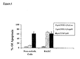
- apoptosis inducing factor e.g. p55; GenBank accession M75866
- apoptosis inducing factor e.g. p55; GenBank accession M75866
- a construct of, for example AdPPE-1-3X-p55 is predicted to reliably target apoptosis to rapidly proliferating endothelial cells in angiogenic blood vessels of a growing tumor.
- AdPPE-1-3X-p55 e.g. p55; GenBank accession M75866
- AdPPE-1-3X-GF where GF is a growth factor (e.g., cytokine) or modificants thereof (e.g., AdPPE-1-SEQ ID NO:7-GF), can be employed.
- GF is a growth factor (e.g., cytokine) or modificants thereof (e.g., AdPPE-1-SEQ ID NO:7-GF)
- Suitable growth factors for use in this context include, but are not limited to, VEGF (GenBank accession M95200) and rat PDGF- BB (GenBank accession; 99% identity to mus-AF162784) and EGR-1 (GenBank accession M22326) FGFs (including, but not limited to, GenBank accession XM 003306) and combinations thereof.
- hypoxia response element e.g. SEQ ID NO: 5
- SEQ ID NO: 5 a hypoxia response element
- the promoter sequences generated according to the teachings of the present invention are particularly useful in regulating angiogenesis in a tissue.
- the modified 3X (SEQ. ID. NO:7) containing promoter sequence of the present invention and the unmodified PPE-1 promoter are both expressed in metastatic foci of the LLC model.
- example 22 clearly illustrates that the modified 3X sequence is specifically responsible for both a decrease in expression levels of the reporter gene in normal lung and a dramatic increase in expression of the reporter gene in metastatic foci. There is neither a hint nor a suggestion in the prior art that such a result could be achieved.
- use of a construct including the 3X element in a gene therapy context can be expected to maximize delivery to tumors while minimizing toxic effects on surrounding normal tissue.
- the p55 gene might be used in conjunction with a promoter of the present invention containing a hypoxia response element in order to specifically induce apoptosis in growing tumors.
- a promoter of the present invention containing a hypoxia response element in order to specifically induce apoptosis in growing tumors.
- HSV-tk Herpes simplex thymidine kinase gene
- angiostatin Genebank accession number X05199
- endostatin Genebank accession number M33272
- angiostatin-endostatin chimera included in the pORF-HSV1tk expression vector available from InvivoGen, San Diego, CA.
- angiostatin or endostatin genes might be used in conjunction with a promoter of the present invention in order to specifically block angiogenesis without inducing apoptosis.
- angiogenesis may be stimulated or blocked.
- This flexibility will allow varied uses of the invention including, but not limited to reduction of tumor mass and revascularization of atherosclerotic regions of the heart or neo-vascularization of peripheral tissues with an inadequate blood supply.
- One relevant clinical scenario is use of a promoter according to the present invention to generate new blood vessels to increase the blood supply in limbs of diabetic patients.
- the nucleic acid construct according to the present invention can be administered to a subject (mammals, preferably humans) per se , or in a pharmaceutical composition where it is mixed with suitable carriers or excipients.
- a "pharmaceutical composition” refers to a preparation of one or more of the active ingredients described herein with other chemical components such as physiologically suitable carriers and excipients.
- the purpose of a pharmaceutical composition is to facilitate administration of a compound to an organism.
- active ingredient refers to the nucleic acid construct accountable for the biological effect.
- physiologically acceptable carrier and “pharmaceutically acceptable carrier” which may be interchangeably used refer to a carrier or a diluent that does not cause significant irritation to an organism and does not abrogate the biological activity and properties of the administered compound.
- An adjuvant is included under these phrases.
- excipient refers to an inert substance added to a pharmaceutical composition to further facilitate administration of an active ingredient.
- excipients include calcium carbonate, calcium phosphate, various sugars and types of starch, cellulose derivatives, gelatin, vegetable oils and polyethylene glycols.
- compositions of the present invention may be manufactured by processes well known in the art, e.g., by means of conventional mixing, dissolving, granulating, dragee-making, levigating, emulsifying, encapsulating, entrapping or lyophilizing processes.
- compositions for use in accordance with the present invention thus may be formulated in conventional manner using one or more physiologically acceptable carriers comprising excipients and auxiliaries, which facilitate processing of the active ingredients into preparations which, can be used pharmaceutically. Proper formulation is dependent upon the route of administration chosen.
- the active ingredients of the pharmaceutical composition may be formulated in aqueous solutions, preferably in physiologically compatible buffers such as Hank's solution, Ringer's solution, or physiological salt buffer.
- physiologically compatible buffers such as Hank's solution, Ringer's solution, or physiological salt buffer.
- penetrants appropriate to the barrier to be permeated are used in the formulation. Such penetrants are generally known in the art.
- the isolated polynucleotide of the present invention has been isolated based on its capacity to promote or enhance transcription in eukaryotic cells of an endothelial lineage. Therefore a mammalian cell transformed with a claimed isolated polynucleotide is an additional embodiment of the invention. Numerous examples of such transformed cells are provided in examples recited herein below.
- enhancer elements are often portable, i.e., they can be transferred from one promoter sequence to another, unrelated, promoter sequence and still maintain activity.
- enhancer elements are often portable, i.e., they can be transferred from one promoter sequence to another, unrelated, promoter sequence and still maintain activity.
- D. Jones et al. (Dev. Biol. (1995) 171(1):60-72 ); N. S. Yew et al, (Mol. Ther. (2001) 4:75-820 ) and L. Wu. et al. (Gene Ther. (2001) 8;1416-26 ).
- the earlier work of Bu et al. J.Biol Chem.
- enhancer elements related to those of the present invention for example enhancers including SEQ ID NO: 6 may be used with constitutive promoters, for example the SV-40 promoter.
- constructs containing, methods employing and isolated polynucleotides including a eukaryotic promoter modified to include the enhancer sequence of the present invention are well within the scope of the claimed invention.
- an enhancer element is an isolated polynucleotide as set forth in SEQ ID NO:8.
- This enhancer is anticipated to function with a wide variety of promoters, including but not limited to endothelial specific promoters (e.g. PPE-1; SEQ ID NO.: 1) and constitutive promoters, for example viral promoters such as those derived from CMV and SV-40.
- This enhancer should be capable of imparting endothelial specificity to a wide variety of promoters.
- the enhancer element may be augmented, for example by addition of one or more copies of the sequence set forth in SEQ ID NO:6. These additional sequences may be added contiguously or non-contiguously to the sequence of SEQ ID NO.: 8.
- the present invention further includes a method of expressing a nucleic acid sequence of interest in endothelial cells employing a construct which relies upon an enhancer element including at least one copy of the sequence set forth in SEQ ID NO:8 and a promoter to direct high level expression of the sequence of interest specifically to endothelial cells.
- ex-vivo administration to cells removed from a body of a subject and subsequent reintroduction of the cells into the body of the subject specifically includes use of stem cells as described in ( Lyden et al. (2001) Nature Medicine 7:1194-1201 ).
- Lewis Lung Carcinoma - (D122-96) (kindly provided by Prof. L. Eisenbach, The Weizmann Institute of Science Rehovot, Israel), Human Embryonic Kidney (293) and HeLa cells were grown in 4.5gr/l DMEM, supplemented with 10% fetal calf serum (FCS), 50 U/ml penicillin, 50 ⁇ g/ml streptomycine and 2 mM glutamine (Biological industries, Beit-Haemek, Israel).
- FCS fetal calf serum
- penicillin 50 ⁇ g/ml streptomycine
- 2 mM glutamine Biological industries, Beit-Haemek, Israel.
- Bovine Aortic Endothelial Cells - BAEC (kindly provided by Prof. N.
- RINr1046-38 (RIN-38) were grown in 199 Earle's salts (5.5mM glucose) medium supplemented with 5% FCS (Biological Industries, Beit-Haemek, Israel), 50U penicillin/ml, 50 ⁇ g streptomycine/ml and 2 mM glutamine.
- HepG2 refers to ATCC-HB-8065.
- HeLa refers to ATCC-CCL-2 .
- Human Bronchial Epithelial cells and “B2B” as used herein refers to ATCC-CRL-9609.
- VEC Human Umbilical Vein Endothelial Cells
- Luciferase gene expression system kit was employed (Promega Corp., Madison, WI). Forty eight hours post transfection or transduction the cells were washed and 200 ⁇ l lysis buffer was added for 15 minutes. Cells lysates were collected and centrifuged for 15 minutes (14,000rpm) at 4 0 C. Subsequently, 10 ⁇ l of the supernatant was added to 50 ⁇ l Luciferase assay buffer. The activity was measured in Luminometer over a 20 second period.
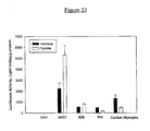
- Luciferase activity in solid tissue a 20mg sample was excised and homogenized in 1ml of the homogenization solution and centrifuged for 15 minutes (14,000rpm) at 4 0 C, and 10 ml of the supernatant were assayed for Luciferase activity, as described above. Results were expressed as Luciferase light units per 1 ⁇ g protein. Protein was measured using the Bradford assay with bovine serum albumin (BSA) as a standard.
- BSA bovine serum albumin
- tissues were fixed in freshly made 4% paraformaldehyde in 0.1 M phosphate buffer for 6 hours at 4 0 C, soaked overnight in 30% sucrose at 4 0 C and frozen in OCT compound (Sakura, USA).
- OCT compound Sakura, USA.
- the tissue blocks were sliced by a cryostat at 10 ⁇ m thickness and observed directly under fluorescence microscopy (FITC filter).
- proliferating cells growing and infecting in 10% FCS media.
- quiescent cells growing and infected in serum free media started in 72 hours prior to the transduction. All cells were grown in humidified incubator, 5% CO 2 , 37 0 C.
- Several recombinant replication deficient adenoviruses (type 5) were constructed.
- the GFP gene (originated from pEGFP, GenBank accession number AAB02572) was ligated to the PPE-1 promoter at the NotI restriction site.
- Ad5PPE-1Luc or Ad5PPE-1GFP The replication deficient recombinant adenoviruses termed Ad5PPE-1Luc or Ad5PPE-1GFP were prepared by co-transfection of pPACPPE-lLuc or Ad5PPE-1GFP with adenovirus plasmid pJM17 as described by Becker, T.C. et al. (Methods Cell biol. 43, Roth M. (ed). New York. Academic Press, 1994, pp. 161-189 ) followed by harvest of recombinant virions.
- Viruses were prepared for large-scale production. The viral stocks were stored at 4 0 C at concentration of 10 9 -10 12 plaque-forming units/ml (pfu/ml).
- the viruses Ad5CMV-Luc (kindly provided by R. Gerard from UTSw Dallas, Texas) and Ad5CMV-GFP (Quantum biotechnologies, Carlsbad, Canada) containing the cytomegalovirus (CMV) immediate early promoter (GenBank Accession number U47119) were prepared for large scale preparation as described for the PPE-1 viral vectors and were used as a non-tissue specific control.
- the modified murine PPE-1 promoter was developed by inserting three copies of the positive transcription element discovered by Bu et al (J.Biol Chem. (1997) 272(19): 32613-32622 ) into the NheI restriction enzyme site located downstream (-286bp) to the 43 base pairs endogenous positive element (-364 to -320 bp).
- the enhancer fragment termed herein "3X” is a triplicate copy of an endogenous sequence element (nucleotide coordinates 407-452 of SEQ ID NO:1) present in the murine PPE-1 promoter. It has been previously shown that induction of PPE-1 promoter activity in vascular endothelial cells depends on the presence of this element Bu et al (J.Biol Chem. (1997) 272(19): 32613-32622 ).
- the 3X fragment was synthesized by using two complementary single stranded DNA strands 96 base pares in length (BioTechnology industries; Nes Tziona, Israel), (SEQ ID NO: 2 and 3). The two single stranded DNA fragment were annealed and filled using Klenow fragment (NEB); the resulting double stranded DNA was 145 base pairs long and included Nhe-1 restriction sites (SEQ ID NO: 4).
- the 3X fragment was ligated into the murine PPE-1 promoter down stream of endogenous Nhe-1 site using T4 Ligase.
- the resulting construct was propagated in DH5 ⁇ compatent cells and a large-scale plasmid preparation was produced using the maxi-prep Qiagene kit.
- the PPE-1-Luciferase cassette (5249bp) containing 1.4kb of the murine preproendothelin-1 (PPE-1) promoter, the Luciferase gene with an SV40 polyA signal (GenBank Accession number X 65323) site and the first intron of the murine ET-1 gene is originated from the pEL8 plasmid (8848bp) used by Harats et al (J. Clin. Inv. (1995) 95: 1335-1344 ).
- the PPE-1-Luciferase cassette was extracted from the pEL8 plasmid by using the BamHI restriction enzyme, following by extraction of the DNA fragment from a 1% agarose gel using an extraction kit (Qiagen, Hilden, Germany).
- the promoter-less pPAC.plpA plasmid (7594bp) containing sequences of the adenovirus type 5 was originated from the pPACCMV.pLpA (8800bp).
- the CMV promoter, the multiple cloning site and the SV40 polyadenylation site (1206 bp) were eliminated by NotI restriction enzyme,
- the fragmented DNA was extracted from 1% agarose gel.
- the linear plasmid (7594 bp) was filled-in by Klenow fragment and BamHI linker was ligated by rapid DNA ligation kit to both cohesive ends.
- the linear plasmid was re-ligated by T4 DNA ligase and transformed into DH5 ⁇ competent cells, in order to amplify the pPAC.plpA with the BamH1 restriction sites.
- the plasmid was prepared for large-scale preparation and purified by maxi prep DNA purification kit.
- the pPACPPE-lLuciferase plasmid was constructed by inserting the PPE-1-Luciferase cassette into the BamHI restriction site of the pPAC.plpA plasmid, by using T4 DNA ligase. The plasmid was subsequently used to transform DH5 ⁇ competent cells.
- the plasmid (12843bp) was prepared for large-scale preparation and purified by maxi prep DNA purification kit.
- the pPACPPE-1GFP plasmid was constructed by sub-cloning the GFP gene (originated from pEGFP, GenBank accession number AAB02572) downstream to the PPE-1 promoter into the NotI restriction site, by T4 DNA ligase.
- the plasmid was subsequently used to transform DH5 ⁇ competent cells.
- the plasmid (11,801 bp) was prepared for large-scale preparation and purified by maxi prep DNA purification kit.
- the pPACPPE-1-3XLuciferase and pPACPPE-1-3XGFP were constructed by inserting the PPE-1-3XLuc or PPE-1-3XGFP cassette digested by BamHI restriction enzyme from pEL8-3X ( Figure 26B ) containing Luc or GFP into the BamHI restriction site of the pPAC.plpA plasmid.
- pEL8-3X contains the modified murine PPE-1 promoter (1.55kb) (red) - located between BamHI and NotI that contains the triplicate endothelial specific enhancer 3X (as set forth in SEQ ID NO.: 7) located between two NheI site.
- the promoter, the Luciferase or GFP gene, the SV40 poly A sites and the first intron of the endothelin-1 gene, all termed the PPE-1 modified promoter cassette was digested and extracted by BamHI restriction enzyme as described in materials and methods.
- the plasmids (12843bp) were prepared for large-scale preparation and purified by maxi prep DNA purification kit.
- BAEC Bovine Aortic Endothelial Cells
- HUVEC Human Umbilical Vein Endothelial Cells
- LLC Lewis Lung Carcinoma
- RIN Ragon insulinoma
- HepG2 HeLa and Normal skin fibroblasts (NSF) cells
- moi multiplicity of infection
- Ad5CMVLuc was used as a non-tissue specific control.
- mice All mice were grown in the Lipids and Atherosclerosis Research Institute.
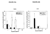
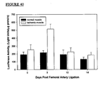
- Ad5PPElLuc or Ad5CMVLuc were suspended in 100 ⁇ l of physiological saline and injected into the tail vein of mice as described hereinabove. Luciferase activity was assayed 1, 5, 14, 30 and 90 days post-injection.
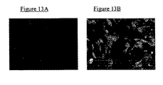
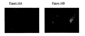
- Ad5PPE-1GFP or Ad5CMVGFP (10 10 pfu/ml in 100 ⁇ l physiological saline) were injected into the tail vein of normal 3 month old, male C57BL/6 mice. GFP expression was detected five days post-injection. All mice appeared healthy and no toxicity or inflammation was noted in the liver or other tissue.
- tissue samples from injected mice were fixed in freshly made 4% paraformaldehyde in 0.1 M phosphate buffer for 6 hours at 4 0 C, soaked overnight in 30% sucrose at 4 0 C and frozen in OCT compound (Sakura, California, USA).
- OCT compound Sakura, California, USA.
- the tissue blocks were sliced at 10 ⁇ m thickness and observed directly under fluorescence microscopy (FITC filter).
- LLC Lewis Lung Carcinoma cells
- the tumor tissue reached a size of 0.7 mm in diameter, the foot pad (with the primary tumor) was resected under anaesthetic and sterile conditions.
- the viruses (Ad5PPE-1, Ad5PPE-IGFP, Ad5CMVLuc or Ad5CMVGFP) were injected to the mouse tail vein.
- mice were sacrificed 5 days post viral injection, their tissues were excised and tested for Luciferase or GFP activities.
- mice Male 3 month old C57BL/6 mice were anaesthetized by subcutaneous injection of sodium pentobarbital (6mg/kg). Their backs were shaved and 5 cm of straight incisions was made. The incisions were immediately sutured by 4/0 sterile silk thread. The angiogenic process in the healing wound was examined every two days by H&E and anti von-Willibrand antibody immuno-histochemistry staining.
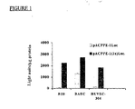
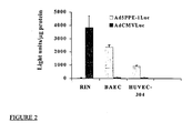
- reporter gene expression in the PPE-1-3X promoter plasmid and the unmodified PPE-1 promoter plasmid was undertaken.
- Reporter gene plasmids containing either the PPE-1-3X fragment or the unmodified PPE-1 fragment and the reporter gene Luciferase were transfected into endothelial and non-endothelial cell lines as well as to a bronchial epithelium cell line (B2B) which express the PPE-1 promoter (see materials and methods above).
- B2B cell line was chosen to provide an indication of the 3X element's capacity to reduce expression in non-endothelial cell lines relative to the PPE-1 promoter.
- Transfection was accomplished using lipofectamine (Promega Corp., Madison, WI).
- a ⁇ gal-neo plasmid was employed as an indicator of the transfection efficiency in each case according to accepted molecular biology practice.
- the PPE-1/Luciferase, PPE-1-3X/Luciferase, PPE-1/GFP and PPE-1-3X/GFP were also ligated into the Ad5 plasmid to produce Ad5PPE-1/Luc and Ad5PPE-1-3X/luc, Ad5PPE-1/GFP and Ad5PPE-1-3X/GFP ( Varda-Bloom et al., (2001) Gene therapy 8:819-827 ). These constructs were assayed separately as detailed hereinbelow.
- B2B Human bronchial epithelial
- BAEC Bovine Aortic Endothelial Cells
- HUVEC Human Umbilical Vein Endothelial Cells
- FIG 2 clearly illustrates that higher Luciferase expression was achieved in endothelial BAEC and HUVEC cell lines with the PPE-1 promoter than with the CMV promoter.
- the CMV promoter produced more Luciferase activity than the PPE-1 promoter.
- Ad5PPE-3X/Luciferase and Ad5PPE-3X/GFP constructs were used to transfect the cell lines described hereinabove in Example 2 in order to ascertain the impact of the 3X element on specificity and expression levels.
- Ad5CMVLuc was used as a non-endothelial-specific control.
- Higher Luciferase expression in BAEC and HUVEC cell lines was detected under the control of the PPE-3X promoter as compared to the CMV promoter.
- Figure 3a is a photomicrograph illustrating GFP expression under the control of Ad5PPE-1-3X in the BAEC cell line.
- Figure 3b is a photomicrograph illustrating GFP expression of Ad5CMV in the BAEC line.
- the PPE-1-3X promoter is more active in endothelial cells. These results clearly indicate that the 3X element does not detract from the endothelial specificity of the PPE-1 promoter. Relative activities of the PPE-1 and PPE-1-3X promoters in cell culture are presented in example 6 hereinbelow.
- Hypoxia responsive element can enhance target gene expression in hypoxic sensitive endothelial cells
- hypoxia is an important regulator of blood vessels' tone and structure. It has also been shown to be a potent stimulus of angiogenesis (in both ischemic heart diseases and cancer ( Semenza, G.L. et al. (2000) Adv Exp Med Biol.; 475:123-30 ; Williams, K.J. (2001) Breast Cancer Res. 2001: 3;328-31 and Shimo, T. (2001) Cancer Lett. 174;57-64 ). Further, hypoxia has been reported to regulate the expression of many genes including erythropoietin, VEGF, glycolytic enzymes and ET-1. These genes are controlled by a common oxygen-sensing pathway, an inducible transcription complex termed hypoxia inducible factor-1 (HIF-1).
- HIF-1 hypoxia inducible factor-1
- the HIF-1 complex mediates transcriptional responses to hypoxia by binding the cis acting hypoxia responsive element (HRE) of target genes.
- HRE hypoxia responsive element
- the HRE is a conserved sequence located in the promoters of few genes that respond to hypoxia including: VEGF, Nitric Oxide Syntase-2, erytropoietin and others including endothelin-1, ET-1.
- the ET-1 promoter contains an inverted hypoxia response element at position -118 bp upstream of the transcription start site, the element contain 7 base pairs and is located between the GATA-2 and AP1 sites 5' GCACGTT 3' - 50 base-pairs. (SEQ ID NO: 5.)
- the preproendothelin-1 (PPE-1) promoter contains an hypoxia responsive element (HRE) that has the potential to increase its expression in the hypoxic microenviroment of tumor or ischemic tissues, thus making it "tumoral tissue specific” and/or "ischemic tissue specific”.
- HRE hypoxia responsive element
- assays of the PPE-1 promoter and PPE-1-3X promoter in conjunction with a Luciferase or GFP reporter gene and delivered by an adenoviral vector were undertaken.
- Luciferase activity under the control of the PPE-1 promoter or the PPE-1-3X promoter was compared in BAEC cells under normoxic and hypoxic conditions (0.5% O 2 for 16h).
- the Luciferase activity under the control of PPE-1 promoter was 5 times higher when exposed to hypoxia ( Figures 5 and 6 ).
- the Luciferase activity under the control of PPE-1-3X promoter was 2.5 times higher under hypoxic conditions.
- introduction of the 3X element into the PPE 1 promoter is till capable of increasing expression levels of a downstream gene in response to hypoxia, even though the normoxic levels of expression with the PPE-1-3X gene are higher than those observed with the unmodified PPE-1 promoter.
- Figure 7 summarizes the results from B2B, HUVEC and BAEC transfection experiments using pPPE-1/Luciferase and pPPE-1-3X/Luciferase.
- Higher Luciferase expression (30, 8.5 and 1.5 times more) was observed under the control of the PPE-1-3X promoter than under the PPE-1 promoter in B2B, HUVEC and BAEC, respectively.
- These results confirm those presented hereinabove and serve to establish that PPE-1-3X is well suited to directing high level expression specifically to endothelial cells.
- the higher levels of expression achieved with the PPE-1-3X construct translate into administration of smaller amounts of DNA. This, in turn, will serve to increase specificity even further.
- Ad5PPE-1/Luciferase construct was injected into C57BL/6 mice as described hereinabove in "Tissue gene expression in normal mice".
- Ad5CMV/Luciferase was employed as a negative control.
- Luciferase activity was found in the liver. Luciferase activity controlled by the PPE-1 promoter was lower in the liver (37-54% of the total body expression).
- the PPE-1 derived expression was much higher in the aorta (23-33% of the total body expression 5 and 14 days post injection, respectively), compared to Ad5CMV/Luciferase. treated mice (up to 1.8% of total body expression; Table 2). These results confirm the endothelial specificity observed in cell culture. It should be remembered that the liver is a highly vascularized organ. Therefore examination of cellular expression within organs was undertaken, as detailed hereinbelow.
- Table 2 Luciferase expression in organs 5 and 14 days post injection of PPE-1 and CMV based constructs Day post injection 5 14 Light units/ ⁇ g protein Light units/ ⁇ g protein Organ PPE-1 CMV PPE-1 CMV Aorta 13.0 ⁇ 2.9 (32.7%) 1.4 ⁇ 0.5 (0.56%) 10.6 ⁇ 2.4 (12.6%) 1.3 ⁇ 0.3 (1.1%) Heart 0.2 ⁇ 0.1 (0.5%) 1 ⁇ 0.6 (0.4%) 1.5 ⁇ 0.3 (1.7%) 1.8 ⁇ 0.6 (1.6%) liver 22.7 ⁇ 4.5 (57%) 219 ⁇ 111.5 (88.6%) 34.9 ⁇ 7.8 (41.6%) 52.8 ⁇ 10.6 (46.8%) lung 0.2 ⁇ 0.1 (0.5%) 2.3 ⁇ 1.0 (0.9%) 3.6 ⁇ 0.8 (4.3%) 2.0 ⁇ 0.9 (1.8%) muscle 0.3 ⁇ 0.1 (0.7%) 0.8 ⁇ 0.2 (0.3%) 1.2 ⁇ 0.3 (1.4%) 1.5 ⁇ 0.5 (1.3%) spleen 1.3 ⁇ 0.8 (3.2%) 1.6 ⁇ 0.9 (0.6%) 2.0 ⁇ 0.4 (2.4%) 2.3 ⁇ 0.9 (2.0%) pancreas 2 ⁇ 0.6 (5.0%) 20.1
- the results in the aorta represent the promoters (PPE-1 or CMV) activity mostly in endothelial cells, while the results in the livers represent their activity mostly in hepatocytes.
- tissue specificity of the PPE-1 promoter is sufficiently strong to effectively eliminate hepatocyte expression, despite preferential uptake of injected DNA by hepatocytes.
- GFP Green Fluorescent Protein
- Ad5PPE-1 and Ad5PPE-1-3X In order to determine the relative efficacy of Ad5PPE-1 and Ad5PPE-1-3X in driving expression of the reporter genes Luciferase and green fluorescent protein (GFP) in cells, specific activity in endothelial cells was tested in-vitro using cell lines described hereinabove. Ad5CMVLuc and Ad5CMVGFP were employed as non-tissue specific controls. Ad5PPE-1Luc and Ad5PPE-1GFP were employed to ascertain the relative change in expression level caused by addition of the 3X sequence.
- FIG 10 shows Luciferase activity as light units/ ⁇ g protein in B2B, BAEC and RIN cells transduced by Ad5PPE-1Luc, Ad5PPE-1-3XLuc, and Ad5CMVLuc Highest Luciferase expression was observed in RIN cells transduced by Ad5CMVLuc, however this construct was poorly expressed in BAEC and B2B cells. The next highest level of Luciferase expression was observed in BAEC cells transduced by Ad5PPE-1-3XLuc. Ad5PPE-1Luc was expressed at lower levels in BAEC cells. In the B2B cell line Ad5PPE-1Luc and Ad5PPE-1-3XLuc were expressed at nearly identical levels.
- FIG. 11 shows Luciferase activity as light units/ ⁇ g protein in HeLA, HepG2, NSF and BAEC cells transduced by Ad5PPE-1Luc, Ad5PPE-1-3YLuc and Ad5CMVLuc. Transduction with Ad5CMVLuc caused high levels of Luciferase expression in HeLA, HepG2 and NSF cells. These cell lines failed to express Luciferase under the control of PPE-1 and expressed Luciferase at low levels with the PPE-1-3X promoter. As expected, BAEC cells transduced with Ad5PPE-1Luc or Ad5PPE-1-3YLuc exhibited high Luciferase expression.
- panel A indicates Ad5PPE-1-3XGFP transduced cells
- panel B indicates Ad5PPE-1GFP transduced cells
- panel C indicates Ad5CMVGFP.
- Ad5PPE-1-3X-GFP and Ad5PPE-1GFP transduction resulted in no GFP expression in non-endothelial cells SMC, HelA, HePG2 and normal skin fibroblasts (NSF) compared to the high expression under the CMV promoter as summarized in Figures 13-16 .
- Figure 14 shows results of a similar experiment conducted in HeLa cells.
- panel A indicates cells transduced with Ad5PPE-1-3XGFP and panel B indicates cells transduced with Ad5CMVGFP.
- panel B indicates cells transduced with Ad5CMVGFP.
- Figure 15 shows results of a similar experiment conducted in HepG2 cells.
- panel A indicates cells transduced with Ad5PPE-1(3X)GFP and panel B indicates cells transduced with Ad5CMVGFP.
- panel B indicates cells transduced with Ad5CMVGFP.
- Figure 16 shows results of a similar experiment conducted in NSF cells.
- panel A indicates cells transduced with Ad5PPE-1-3XGFP and panel B indicates cells transduced with Ad5CMVGFP.
- panel B indicates cells transduced with Ad5CMVGFP.
- Ad5PPE-1-3XGFP and Ad5PPE-1GFP were injected into mice as described hereinabove. Five days post-intravenous injection, the mice were sacrificed and their tissues were analyzed by a fluorescent microscopy.
- Figure 17A shows low level GFP expression in endothelial cells lining a blood vessel of a mouse injected with the Ad5PPE-1GFP.
- Figure 17B shows the much higher level of GFP expression resulting from addition of the 3X sequence to the construct.
- FIG 18 shows representative results from kidney tissue of injected mice.
- Ad5CMVGFP injected mice ( Figure 18A ), Ad5PPE-1GFP ( Figure 18b ) and Ad5PPE-1-3XGFP ( Figure 18C ) injected mice all exhibited low GFP activity in kidney cells.
- Figure 18B slightly higher GFP expression is visible in the blood vessel wall (indicated by arrow).
- FIG 19 shows representative results from spleen tissue of injected mice.
- Ad5CMVGFP injected mice ( Figure 19A ), Ad5PPE-1GFP injected mice ( Figure 19B ) and Ad5PPE-1-3XGFP injected mice ( Figure 19 C) all exhibited low level GFP activity in cells of the spleen. Higher GFP activity is visible in the blood vessels of Ad5PPE-1-3XGFP injected mice (indicated by arrow).
- both the PPE-1 and the PPE-1-3X promoter are endothelial cell specific in-vivo . They further suggest that activity of both promoters was limited in non-proliferating endothelial tissue (i.e. blood vessels of healthy organs. Therefore, assays in a tumor angiogenic model were undertaken.
- Luciferase expression in tumor neovascularization was tested five days post systemic injections of Ad5PPE-1Luc or Ad5CMVLuc (10 10 pfu/ml each).
- the Luciferase expression in non-metastatic tissues such as the liver, kidney, heart and pancreas was minimal.
- the expression level in the aorta was about 30% of the levels in the metastatic lungs.
- Ad5PPE-1GFP and Ad5CMVGFP constructs were employed to localize reporter gene expression in the primary tumor and metastatic lungs.
- Ad5PPE-1GFP injected mice showed high levels of GFP specific expression in the blood vessels of the primary tumor ( Figure 36C ), although no expression was detected in the tumor cells themselves. This observation is consistent with the results of the LLC cell culture model presented in example 20.
- high levels of GFP expression were detected in both big arteries and small angiogenic vessels of the metastatic foci ( Figure 36A ). No expression was detected in the normal lung tissue.
- the endothelial cell localization was demonstrated by co-localization of the GFP expression ( Figure 16A ) and the CD31 antibody immuno-staining ( Figure 16B ).
- Ad5CMVGFP injected mice no GFP activity was detectable in both the primary tumor and lung metastasis.
- Figure 36C illustrates GFP expression in blood vessels of a primary tumor after intra tumoral injection of Ad5PPE-1GFP.
- Figure 36D is a phase contrast image of the same filed as panel C illustrating the tumor and its blood vessels.
- LLC Lewis Lung Carcinoma
- FIGs 20 A-D summarize the GFP expression in metastatic lungs of control mice injected with Saline ( Figure 20A ), mice injected with Ad5CMVGFP ( Figure 20 B) , mice injected with Ad5PPE-1GFP ( Figure 20 C) and mice injected with Ad5PPE-1-3XGFP ( Figure 20D ).
- Anti-CD31 immunostaining ( Figures 20C' to 20D' ) confirm the location of the GFP expression in each metastatic tissue. The results show that while no GFP expression was detected in control - saline injected mice ( Figure 20A ), there was a slight expression around the epithelial bronchi of the CMV injected mice, but not in the angiogenic blood vessels of the metastatic lung of these mice ( Figure 20B ).
- the LLC metastases model was employed. Five days post i.v. injection of 10 10 pfu/ml of Ad5PPE-1Luc, Ad5PPE-1-3XLuc, Ad5CMVLuc, Ad5PPE-1GFP, Ad5PPE-1-3X-GFP or Ad5CMVGFP, the mice were sacrificed and their tissues were analyzed for Luciferase or GFP expression as described hereinabove.
- Luciferase expression under the control of the PPE-1-3X promoter was 35 fold greater in the metastatic lungs relative to its activity in normal lungs and 3.5 fold higher than expression driven by the PPE-1 promoter without the 3X element (p ⁇ 0.001). Very low Luciferase activity was detected in other tissues of mice injected with Ad5PPE-1-3XLuc. Calculating the Luciferase expression in the lungs as percentage from the liver of each injected animal revealed that the activity increased 10 fold in the metastatic lung compared to the activity in normal lung ( Figure 38 ).
- Figure 39 shows the GFP expression (Figure 39A) in metastatic lungs of Ad5PPE-1-3XGFP injected mice. Immuno-staining by CD31 antibody ( Figure 39B) confirm the location of the GFP expression in the new blood vessels. No GFP expression was detected in control - saline injected mice. Low level expression around the epithelial bronchi of the CMV injected mice, but not in the angiogenic blood vessels of the metastatic lung. In summary, these results indicate that large increases in expression level resulted from introduction of a 3X element into Ad5PPE-1 constructs and that this increased expression was specific to the angiogenic blood vessels of tumors. Potentially, the observed effect may be coupled with the hypoxia response described hereinabove to further boost expression levels of a sequence of interest.
- bovine aortic endothelial cells were transfected by a DNA plasmid (pEL8; Figure 26A ).
- the pEL8 plasmid contains the murine PPE-1 promoter (1.4kb) (red), the luciferase gene (1842 bp), the SV40 poly A sites and the first intron of the endothelin-1 gene, all termed the PPE-1 promoter cassette was digested and extracted by BamHI restriction enzyme as described in material and methods. After transfection, cells were subjected to hypoxic conditions.
- Luciferase expression in transfected BAEC subjected to 18 hours of hypoxia (0.5% 02) was eight times higher than Luciferase expression in cells grown in a normoxic environment ( Figure 21).
- Figure 21 shows that Luciferase activity (light units/ ⁇ g protein) in BAEC transfected by a plasmid containing the murine PPE-1 promoter was significantly higher when transfected cells were incubated in a hypoxic environment. Equivalent transfection efficiencies were confirmed by co-transfection with a ⁇ -galactosidase reporter vector and assays of LacZ activity.
- mice In order to examine the murine PPE-1 promoter activity in tissues subjected to regional hypoxia/ischemia, mPPE-1-Luc transgenic mice, described hereinabove in materials and methods, were employed. The mice were induced to regional hind limb ischemia as previously described ( Couffinhal T. et al. (1998) Am. J. Pathol. 152;1667-1679 ). In brief, animals were anesthetized with pentobarbital sodium (40 mg/kg, IP). Unilateral ischemia of the hind limb was induced by ligation of the right femoral artery, approx. 2 mm proximal to the bifurcation of the saphenous and popliteal arteries.
- Luciferase expression was assayed 2, 5, 10 and 18 days post ligation in the ischemic muscle, in the normal non-ligated muscle, in the liver, lung, and aorta.
- Luciferase in other non-ischemic tissues including liver, lungs and aorta of the transgenic mice subjected to regional ischemia revealed no significant changes within 18 days post ischemic induction in the Luciferase expression in these tissues ( Figure 41 ).
- BAEC endothelial cells
- Luciferase expression under the control of PPE-1 promoter (open bars; Figure 28 ) was 4 times higher in normal proliferating BAEC than in quiescent cells, and 25 times higher in normal proliferating BAEC than Luciferase expression under control of the CMV promoter (Black bars; Figure 28 ). Further, in proliferating cells, the activity under the control of PPE-1 promoter was 10 times higher than that under the CMV promoter control.
- Ad5PPE-1Luc activity was tested in BAEC induced to rapid proliferation by addition of 40 ng/ml vascular endothelial growth factor (VEGF). Activity under these conditions was compared activity in normal proliferating cells and quiescent cells as described hereinabove. Luciferase expression in BAEC induced to cell proliferation with VEGF was 44 times higher than in normal proliferating cells, and 83 times higher than in quiescent cells ( Figure 29 ).
- VEGF vascular endothelial growth factor
- Figure 32 is a picture of an aorta dissected from an ApoE deficient mouse colored by Sudan - IV. Note that the thoracic aorta contains less red stained atherosclerotic lesions while the abdominal region is highly atherosclerotic. ( Figure 32 adapted from Imaging of Aortic atherosclerotic lesions by 125I-HDL and 125I-BSA. A. Shaish et al, Pathobiology - submitted for publication).
- Luciferase expression controlled by the PPE-1 promoter was 6 fold higher in the highly atherosclerotic abdominal, and 1.6 fold higher in the slightly atherosclerotic thoracic aorta as compared to expression under the control CMV promoter.
- CMV constitutive promoter
- Ad5CMVLuc was used as a non-tissue specific control. Luciferase activity under the PPE-1 promoter ( Figure 34 ; open bars) control was higher both in the normal (6.8 ⁇ 3.2) and in healing wound region (5 ⁇ 1.6) compared to the activity observed under the CMV control ( Figure 34 ; black bars).
Abstract
(a) a promoter functional in endothelial cells;
(b) a regulatory element comprising at least one copy of the sequence set forth in SEQ ID NO:8 operably linked to said promoter, and
(c) a nucleic acid sequence encoding a cell toxin under regulatory control of said regulatory element.
Description
- The present invention relates to isolated polynucleotide sequences exhibiting endothelial cell specific promoter activity, and methods of use thereof and, more particularly, to a modified-preproendothelin-1 (PPE-1) promoter which exhibits increased activity and specificity in endothelial cells. The invention further relates to modifications of the PPE promoter which enhance its expression in response to physiological conditions including hypoxia and angiogenesis.
- Gene therapy is an emerging modality for treating inherited and acquired human diseases. Great efforts are directed towards developing methods for gene therapy of cancer, cardiovascular and peripheral vascular diseases, but there is still a major obstacle in effective and specific gene delivery. In general, the main limiting factor of gene therapy with a gene of interest using a recombinant viral vector as a shuttle is the ability to specifically direct the gene of interest to the target tissue. Adenoviruses that are used for this purpose are capable of infecting a large variety of cells with different affinities. Indeed, using a non-tissue specific promoter induces up to 95% of the expression of the gene of interest in the liver; hence regulation of expression is highly required. Further, it is currently infeasible to differentiate between normal vascular endothelia and developing vascular endothelia in a growing tumor when targeting a gene.
- In both cancer development and vascular diseases, angiogenesis, the creation of new vessels, plays a central role. Hence, regulation of this process by gene therapy to the vascular endothelium can be tremendously important in inducing targeted therapy for these diseases.
- High efficiency of the human preproendothelin-1 (PPE-1), delivered by retroviral vector, was obtained in endothelial cells (EC) in-vitro, and in transgenic animal models. However, the prior art does not teach use of this promoter for in-vivo gene therapy. The human PPE-1 lacks regulatory elements found in PPE-1 genes of other animals, most notably the mouse.
- United States patent
5,747,340 teaches use of the murine PPE-1 promoter and portions thereof. However, this patent contains no hint or suggestion that an endothelial-specific enhancer can be employed to increase the level of expression achieved with the PPE promoter while preserving endothelial specificity. Further, this patent does not teach that the PPE-1 promoter is induced to higher levels of transcription under hypoxic conditions. - There is thus a widely recognized need for, and it would be highly advantageous to have, an improved endothelial cell specific promoter, methods of use thereof and cells transformed therewith devoid of the above limitations. The disclosed invention is expected to demonstrate added utility in treatment of cardiovascular disease, cancer and wound healing relative to previously known configurations.
- According to one aspect of the present invention there is provided an isolated polynucleotide functional as a promoter in eukaryotic cells. The isolated polynucleotide includes an enhancer element including at least two copies of the sequence set forth in SEQ ID NO:6.
- According to another aspect of the present invention there is provided a method of expressing a nucleic acid sequence of interest, encoding an active RNA molecule or a protein such as an enzyme, reporter molecule and the like in endothelial cells. The method includes administering to a subject a construct which includes the nucleic acid sequence of interest positioned under the regulatory control of a promoter functional in eukaryotic cells. The construct further includes an enhancer element including at least one copy of the sequence set forth in SEQ ID NO:6.
- According to yet another aspect of the present invention there is provided a method of regulating angiogenesis in a tissue. The method includes administering a nucleic acid construct including: (a) an endothelial cell specific promoter; (b) at least one copy of a hypoxia response element set forth in SEQ ID NO:5; and (c) a nucleic acid sequence encoding an angiogenesis regulator, the nucleic acid sequence being under regulatory control of the promoter and the hypoxia response element.
- According to still another aspect of the present invention there is provided an isolated polynucleotide functional as a promoter in eukaryotic cells. The isolated polynucleotide includes an enhancer element including the sequence set forth in SEQ ID NO: 7.
- According to an additional aspect of the present invention there is provided a method of regulating angiogenesis in a tissue. The method includes administering a nucleic acid construct including: (a) an endothelial cell specific promoter; (b) an enhancer element including the sequence set forth in SEQ ID NO: 7; (c) at least one copy of a hypoxia response element set forth in SEQ ID NO:5; and (d) a nucleic acid sequence encoding an angiogenesis regulator, the nucleic acid sequence being under regulatory control of the promoter, the enhancer element and the hypoxia response element.
- According to still a further aspect of the present invention there is provided isolated polynucleotide functional as a promoter in eukaryotic cells, the isolated polynucleotide includes an enhancer element including at least one copy of the sequence set forth in SEQ ID NO:8.
- According to still a further aspect of the present invention there is provided a method of expressing a nucleic acid sequence of interest in endothelial cells, the method includes administering to a subject a construct, the construct includes the nucleic acid sequence of interest positioned under the regulatory control of a promoter functional in eukaryotic cells, and an enhancer element including at least one copy of the sequence set forth in SEQ ID NO:8.
- According to still another further aspect of the present invention there is provided isolated polynucleotide functional as a promoter in eukaryotic cells, the isolated polynucleotide includes an enhancer element including the sequence set forth in SEQ ID NO: 8.
- According to further features in preferred embodiments of the invention described below, the enhancer element includes three copies of the sequence set forth in SEQ ID NO:6.
- According to still further features in preferred embodiments of the invention the at least two copies of the sequence set forth in SEQ ID NO:6 are contiguous.
- According to still further features in preferred embodiments of the invention the isolated polynucleotide further includes an endothelial specific promoter element.
- According to still further features in preferred embodiments of the invention the endothelial specific promoter element includes at least one copy of the PPE-1 promoter.
- According to still further features in preferred embodiments of the invention the isolated polynucleotide further includes a hypoxia response element.
- According to still further features in preferred embodiments of the invention the hypoxia response element includes at least one copy of the sequence set forth in SEQ ID NO: 5.
- According to still further features in preferred embodiments of the invention the enhancer element is as set forth in SEQ ID NO: 7.
- According to still further features in preferred embodiments of the invention there is provided a nucleic acid construct including a claimed isolated polynucleotide and a nucleic acid sequence of interest, the nucleic acid sequence of interest being under regulatory control of the isolated polynucleotide.
- According to still further features in preferred embodiments of the invention the nucleic acid sequence of interest is selected from the group consisting of VEGF, p55 and PDGF-BB.
- According to still further features in preferred embodiments of the invention there is provided a mammalian cell transformed with a claimed isolated polynucleotide.
- According to still further features in preferred embodiments of the invention the promoter exhibits endothelial cell specificity.
- According to still further features in preferred embodiments of the invention the promoter is the PPE-1 promoter as set forth in SEQ ID NO: 1.
- According to still further features in preferred embodiments of the invention administering is effected by a method selected from the group consisting of: (i) systemic in-vivo administration; (ii) ex-vivo administration to cells removed from a body of a subject and subsequent reintroduction of the cells into the body of the subject; and (iii) local in-vivo administration.
- According to still further features in preferred embodiments of the invention the nucleic acid construct further includes an enhancer element including at least two copies of the sequence set forth in SEQ ID NO:6.
- According to still further features in preferred embodiments of the invention the endothelial cell specific promoter includes at least one copy of the PPE-1 promoter.
- According to still further features in preferred embodiments of the invention there is provided a nucleic acid construct including a claimed isolated polynucleotide and a nucleic acid sequence of interest, the nucleic acid sequence of interest being under regulatory control of the isolated polynucleotide.
- According to still further features in the described preferred embodiments the enhancer element further includes at least one copy of the sequence set forth in SEQ ID NO:6.
- According to still further features in the described preferred embodiments the enhancer element includes one copy of the sequence set forth in SEQ ID NO:8 and at least two copies of the sequence set forth in SEQ ID NO:6.
- According to still further features in the described preferred embodiments the enhancer element further includes at least one copy of the sequence set forth in SEQ ID NO:6.
- According to still further features in the described preferred embodiments the at least one copy includes two copies.
- According to still further features in the described preferred embodiments the nucleic acid construct further includes an enhancer element including at least one copy of the sequence set forth in SEQ ID NO:8.
- According to yet another further aspect of the present invention there is provided method of regulating angiogenesis in a tissue, the method comprising administering a nucleic acid construct including: (a) an endothelial cell specific promoter; (b) an enhancer element including at least one copy of the sequence set forth in SEQ ID NO:8; and (c) a nucleic acid sequence encoding an angiogenesis regulator, the nucleic acid sequence being under regulatory control of the promoter and the enhancer element.
- According to still further features in the described preferred embodiments the enhancer element further includes at least one copy of the sequence set forth in SEQ ID NO:6.
- According to still further features in the described preferred embodiments the enhancer element includes one copy of the sequence set forth in SEQ ID NO:8 and at least two copies of the sequence set forth in SEQ ID NO:6.
- The present invention successfully addresses the shortcomings of the presently known configurations by providing improved isolated polynucleotide sequences with endothelial cell specificity, and methods of use thereof. The improvements in the sequence make feasible methods of treating a variety of diseases, disorders and conditions which were previously considered infeasible. Specifically, the improvements relate to increased specificity to endothelial cells, increased levels of expression of a sequence of interest and enhanced induction by conditions including ischemia and angiogenesis.
- The invention is herein described, by way of example only, with reference to the accompanying drawings. With specific reference now to the drawings in detail, it is stressed that the particulars shown are by way of example and for purposes of illustrative discussion of the preferred embodiments of the present invention only, and are presented in the cause of providing what is believed to be the most useful and readily understood description of the principles and conceptual aspects of the invention. In this regard, no attempt is made to show structural details of the invention in more detail than is necessary for a fundamental understanding of the invention, the description taken with the drawings making apparent to those skilled in the art how the several forms of the invention may be embodied in practice.
- In the drawings:
-
FIG. 1 is a histogram illustrating the effect of the enhancer element of the present invention on Luciferase expression in both bovine and human endothelial cell lines using the B2B cell line as a non-endothelial control. -
FIG. 2 is a histogram illustrating endothelial specificity of a promoter of the present invention in an adenoviral vector on Luciferase expression in various cell lines. -
FIGs. 3A and 3B are photomicrographs illustrating GFP expression under the control of Ad5PPE-1-3X of the present invention and an Ad5CMV control construct in the BAEC cell line. -
FIG. 4 is histogram of % apoptosis induced by pACPPE-1-3Xp55, pACPPE-1-3XLuciferase and pCCMVp55 in endothelial and non-endothelial cells. -
FIG. 5 is a histogram illustrating the effect of introducing an enhancer element according to the present invention into a promoter construct on hypoxia response. -
FIG. 6 is a histogram illustrating the effect of introducing an enhancer element according to the present invention into a promoter of an adenovector construct on hypoxia response. -
FIG. 7 is a histogram illustrating the effect of introducing an enhancer element according to the present invention into a promoter on levels of expression in bovine and human endothelial cell lines. -
FIG. 8 is a histogram illustrating levels of expression of a reporter gene observed in various organs after injection of an adenoviral construct containing either an endothelial promoter (PPE-1) or a control (CMV) promoter; -
FIGs. 9A-B are two photomicrographs illustrating cellular expression of an Ad5CMVGFP construct (Figure 9a ) and an Ad5PPE-1-GFP construct (Figure 9b ) in liver tissue of mice injected with the constructs. -
FIG. 10 is a histogram illustrating the effect of introducing an enhancer element according to the present invention into a promoter on levels of expression in endothelial and non-endothelial cell lines. -
FIG. 11 is a histogram illustrating the effect of introducing an enhancer element according to the present invention into a promoter on levels of expression in endothelial and non-endothelial cell lines.FIGs. 12A-C are photomicrographs illustrating GFP expression in Ad5PPE-1-3XGFP transduced cells, Ad5PPE-1GFP transduced cells and Ad5CMVGFP transduced cells respectively. -
FIGs. 13A-B illustrate GFP expression in SMC transduced by moi-1 of Ad5PPE-1-3XGFP and Ad5CMVGFP respectively. -
FIGs. 14A-B show results of an experiment similar to that ofFigures 13A-B conducted in HeLa cells. -
FIGs. 15A-B show results of an experiment similar to that ofFigures 13A-B conducted in HepG2 cells. -
FIGs. 16A-B show results of an experiment similar to that ofFigures 13a-b conducted in NSF cells. -
FIGs. 17A-B are photomicrographs illustrating GFP expression in endothelial cells lining a blood vessel of mice injected with the Ad5PPE-1GFP and the Ad5PPE-1-3XGFP constructs respectively. -
FIGs. 18A-C are photomicrographs illustrating results from kidney tissue of injected mice. Ad5CMVGFP injected mice (Figure 18A ) , Ad5PPE-1GFP (Figure 18B ; slightly higher GFP expression is visible in the blood vessel wall; indicated by arrow) and Ad5PPE-1-3XGFP (Figure 18C ). -
FIGs. 19A-C illustrate experiments similar to those depicted inFigures 18A-C , conducted on sections of spleen tissue. -
FIGs. 20A-D and 20 C'-D' illustrate GFP expression in metastatic lungs of control mice injected with Saline (Figure 20A ), mice injected with Ad5CMVGFP (Figure 20 B) , mice injected with Ad5PPE-1GFP (Figure 20 C) and mice injected with Ad5PPE-1-3XGFP (Figure 20D ). Anti Cd31 immunostaining (Figures 20C' to 20D' ) confirm the co-localization of the GFP expression and CD31 expression in each metastatic tissue. -
FIG. 21 is a histogram illustrating that Luciferase activity (light units/µg protein) in BAEC transfected by a plasmid containing the murine PPE-1 promoter is significantly higher when transfected cells were incubated under hypoxic conditions. -
FIG. 22 is a histogram as inFigure 21 , except that Ad5PPE-1Luc and Ad5CMVLuc were employed. -
FIG. 23 is a series of histograms as inFigure 22 showing the effects of hypoxia in different cell lines. -
FIG. 24 is a histogram illustrating the effect of the 3X sequence of the present invention on the PPE-1 hypoxia response in BAEC cells. Cells were transduced by Ad5PPE-1Luc and Ad5PPE-1-3XLuc. -
FIG. 25 is a histogram showing levels of Luciferase expression in PPE-1-Luc transgenic mice following femoral artery ligation. -
FIGs. 26A-B are plasmid maps of constructs employed in conjunction with the present invention. -
FIGs. 27A-D are a series of ultrasound images of ligated limbs of representative animals from the different treatment groups, 21 days following ligation.Figure 27A Control, Ad5CMVLuc. Treated;Figure 27B Control, saline treated;Figure 27C Ad5PPE-3X-VEGF treated;Figure 27D Ad5CMV-VEGF treated. -
FIG. 28 is a histogram illustrating Luciferase activity in proliferating and quiescent Bovine Aortic Endothelial Cells (BAEC) transduced with Ad5PPE-1Luc (open bars) and Ad5CMVLuc (black bars). -
FIG. 29 is a histogram illustrating Luciferase activity in BAEC transduced with Ad5PPE-1Luc. during normal proliferation, a quiescent state and rapid proliferation following addition of VEGF. -
FIGs. 30A-B are histograms illustrating Luciferase activity (light units/µg protein) in the (Figure 30A ) aortas and livers (Figure 30B ) of Ad5PPE-1Luc and Ad5CMVLuc normal injected C57BL/6 mice. Activities were determined 1 (n=13), 5 (n=34), 14 (n=32), 30 (n=20) and 90 (n=11) days post injection. -
FIGs. 31A-B are histograms illustrating relative Luciferase activity (light units/µg protein) detected five (Figure 31A ) and fourteen (Figure 31B ) (n=10 for each time point) days post injection of Ad5PPE-1Luc (open bars) or Ad5CMVLuc (black bars) in normal injected BALB/C mice. Activity is expressed as percentage of total body Luciferase expression of each animal. -
FIG. 32 is a prior art image depicting an Aorta dissected from ApoE deficient mice colored by Sudan - IV. The thoracic aorta contains less red stained atherosclerotic lesion while the abdominal region includes many red stained atherosclerotic lesions. (Adapted from Imaging of Aortic atherosclerotic lesions by 125I-HDL and 125I-BSA. A. Shaish et al, Pathobiology - submitted for publication). -
FIG. 33 is a histogram illustrating absolute Luciferase activity (light units/µg protein) detected 5 days post systemic injections of Ad5PPE-1Luc (open bars; n=12) or Ad5CMVLuc (black bars; n=12) to ApoE deficient mice. Luciferase activity observed from the abdominal aorta contain high lesion levels and from the thoracic area (low lesion levels). -
FIG. 34 is a histogram illustrating absolute Luciferase activity (light units/µg protein) 5 days post systemic injections of Ad5PPE-1Luc (black bars) or Ad5CMVLuc (open bars) to healing wound C57BL/6 induced mice. -
FIG. 35 is a histogram illustrating Luciferase activity in normal lung, metastatic lung and primary tumor of Lewis lung carcinoma-induced mice. Lewis lung carcinoma was induced by D122-96 cells injection to the backs for primary tumor model and to the footpad for the metastatic model. Luciferase activity was measured five days post-systemic injection ofAd5PPE-1Luc (n=9; open bars) or Ad5CMVLuc (n=12; black bars). Activity is expressed as light units/µg protein. -
FIGs. 36A-D are photomicrographs illustrating GFP expression and tissue morphology in lungs and tumors of LLC bearing mice after intra-tumoral injection of Ad5PPE-1GFP. Tissue was frozen in OCT and sectioned to 10µm by cryostat. All pictures were taken in magnification of 25x.Figure 36A - GFP in angiogenic blood vessels of lung metastases;Figure 36B - CD31 antibody immuno-staining of the section pictured inFigure 36aA ;Figure 36C - GFP expression in blood vessels of primary tumor;Figure 36D - phase contrast of the section of C illustrating blood vessels. -
FIG. 37 is a histogram illustrating Luciferase expression in normal lung and metastatic lung of Lewis lung carcinoma-induced mice, injected with Ad5CMVLuc, Ad5PPE-1Luc and Ad5PPE-1-3X-Luc Lewis lung carcinoma was induced by D122-96 cells injected to the foot pad for the metastatic model. Luciferase activity was measured five days post-systemic injection of Ad5CMVLuc (n=7;black bars), Ad5PPE-1Luc (n=6;gray bars), or Ad5PPE-1-3XLuc (n=13;brown bars). Activity is expressed as light units/µg protein. -
FIG. 38 is a histogram illustrating Luciferase activity as percentage of liver activity (where the liver is 100%), in normal lung and lung metastasis of Lewis lung carcinoma-induced mice injected with Ad5CMV, Ad5PPE-1Luc and Ad5PPE-1(3X). - FIGs. 39A-B are photomicrographs illustrating co-localization of GFP expression (Figure 39A) and Cd31 immuno-staining (Figure 39B) in mice with LLC lung metastases injected with Ad5PPE-1-3X-GFP.
-
FIG. 40 is a histogram illustrating Luciferase activity (light units/µg protein) in muscles (ischemic and normal) of PPE-1Luciferase transgenic mice at two, five, ten and 18 days post femoral ligation and in control (non-ligated animals -day 0; n=8 for each group). -
FIG. 41 is a histogram illustrating Luciferase activity (light units/µg protein) in the liver, lung and aorta in muscles (ischemic and normal) of PPE-1Luciferase transgenic mice at five (n=6), ten (n=6) and 18 (n=8) days post femoral ligation and in control (non ligated animals - day 0). -
FIG. 42 is a histogram illustrating Luciferase activity, (light units/µg protein detected in the livers, lungs and primary tumors of LLC mice injected in primary tumors with Ad5CMVLuc (black bars) or Ad5PPE-1Luc (open bars). - The present invention is of an improved endothelial cell-specific promoter which can be employed to reliably direct high-level expression of a sequence of interest to endothelial cells and in particular endothelial cells participating in angiogenesis.
- The principles and use of the present invention may be better understood with reference to the drawings and accompanying descriptions.
- Before explaining at least one embodiment of the invention in detail, it is to be understood that the invention is not limited in its application to the details of construction and the arrangement of the components set forth in the following description or illustrated in the examples and drawings. The invention is capable of other embodiments or of being practiced or carried out in various ways. Also, it is to be understood that the phraseology and terminology employed herein is for the purpose of description and should not be regarded as limiting.
- Although endothelial specific promoters have been previously described (e.g. United States patent
5,747,340 ) these promoters have typically been inefficient at directing expression to endothelial cells or have not been demonstrated as being specific to endothelia cells in-vivo. - Enhancer elements specific to endothelial cells have also been described. Bu et al. (J.Biol Chem. (1997) 272(19): 32613-32622) have demonstrated that three copies (3X) of the 1X enhancer element of PPE-1 (containing elements ETE-C, ETE-D, and ETE-E) endows promoter sequences with endothelial cell specificity in-vitro, however such activity has not been demonstrated in-vivo.
- As is well known in the art, in-vitro experiments cannot reliably predict in-vivo results. As such, the results presented by Bu et al., although suggestive of endothelial cell specificity, do not provide sufficient evidence as to the utility of 3X enhancer element in-vivo.
- The lack of in-vivo studies also brings into question the endothelial cell specificity of the 3X enhancer element in whole organisms. Lack of this data implies that therapeutic application of this element is questionable, because when employed in-vivo, and in particular when employed for regulating angiogenesis, it is imperative that expression of an angiogenesis regulator (e.g., cell toxin) be directed specifically to endothelial cells, preferably in a specific subset of endothelial cells which are involved in angiogenesis.
- As is illustrated in the examples section which follows, the present inventors, through laborious experimentation, have provided, for the first time, conclusive evidence as to the in-vivo activity of the 3X enhancer element. Such evidence identifies the 3X element and its sequence derivatives (e.g., SEQ ID NO:7) as highly suitable for use in therapeutic applications.
- In addition, in reducing the present invention to practice, it was discovered that a novel configuration of the PPE-1 enhancer sequence of the present invention endows promoter sequences with an unexpected and highly specific activity in endothelial cells participating in angiogenesis.
- Thus, according to one aspect of the present invention there is provided an isolated polynucleotide functional as an endothelial cell specific promoter in a mammal such as a human being.
- The isolated polynucleotide includes an enhancer element including one or more copies of the sequence set forth in SEQ ID NO:6 and preferably one or more copies of the sequence set forth in SEQ ID NO:8, which as is illustrated in the Examples section which follows, plays an important role in regulating expression in endothelial cells participating in angiogenesis.
- One specific and novel sequence configuration of an enhancer element utilizable by the present invention is illustrated in SEQ ID NO:7.
- For purposes of this specification and the accompanying claims, the term "enhancer" refers to any polynucleotide sequence which increases the transcriptional efficiency of a promoter.
- According to some embodiments of the invention, the isolated polynucleotide includes contiguous copies of SEQ ID NOs:6 and/or 8. Such sequences are preferably positioned in a head-to-tail orientation, although, the enhancer element of the present invention can also include one or more copies of a specific portion of the sequence of SEQ ID NO:6 or 8, in an inverted orientation, e.g., by using sequences complementary to SEQ ID NO:6 or 8 in construction of the enhancer element.
- Preferably the isolated polynucleotide further includes an endothelial cell-specific promoter sequence element. For purposes of this specification and the accompanying claims, the term "promoter" refers to any polynucleotide sequence capable of mediating RNA transcription of a downstream sequence of interest. The endothelial specific promoter element may include, for example, at least one copy of the PPE-1 promoter.
- Preferably, the isolated polynucleotide further includes a hypoxia response element, for example at least one copy of the sequence set forth in SEQ ID NO: 5.
- Thus, according to this aspect of the present invention, an endothelial cell specific promoter which includes various enhancer element configurations is provided.
- It will be appreciated that the enhancer element sequences can be positioned within the promoter sequence used, upstream of the promoter, between the promoter and a downstream sequence of interest or within the sequence of interest (e.g., intron).
- The isolated nucleic acid sequence of the present invention can be used to regulate gene expression in eukaryotic tissue, and in particular, in proliferating endothelial cells, for example endothelial cells involved in angiogenesis.
- Thus, the isolated polynucleotide sequence of the present invention may be provided, in some cases, as part of a nucleic acid construct further including a nucleic acid sequence of interest which is positioned under the regulatory control of the isolated polynucleotide of the present invention. It will be appreciated that such a nucleic acid construct can further include any additional polynucleotide sequences such as for example, sequences encoding selection markers, origin of replication in bacteria, or sequences encoding reporter polypeptides. Such a nucleic acid construct is preferably configured for mammalian cell expression and can be of viral origin. Numerous examples of nucleic acid constructs suitable for mammalian expression are known in the art; the Examples section which follows provides further detail of several such constructs.
- For purposes of this specification and the accompanying claims, the phrase "sequence of interest" refers to any polynucleotide sequence which has the capacity to be transcribed by an RNA polymerase. This definition includes coding sequences translatable into polypeptides, as well as sequence for antisense RNA, RNA which binds DNA, ribozymes and other molecular moieties which are not destined to undergo translation. Examples of nucleic acid sequence of interest which may be used by the construct according to the present invention are provided hereinbelow and in the Examples section which follows.
- Examples presented hereinbelow illustrate that the improved endothelial cell specific promoters of the present invention can reliably direct expression of a reporter gene to endothelial tissue after systemic in-vivo administration. These examples further show, for the first time, that the isolated polynucleotide of the present invention can be used to preferentially express a reporter protein (GFP) in atherosclerotic and/or angiogenic tissue, thus providing for the first time direct evidence as to the importance of the PPE-1 enhancer element and its derivative in therapeutic applications.
- While use of a reporter protein, such as GFP, may have utility in detection of early stages of metastatic tumor growth, especially in animal models, or for non-invasive imaging of metastases (Yang, M. et al., Proc. Nat. Acad. of Sci. (2001) 27:2616-2621) such a use is only a small portion of the projected utility of the claimed invention. It is believed, for example, that AdPPE-1GFP can be used in a combination with AdPPEltk, AdPPE-1p55 and/or other anti-angiogenic treatments, in order to follow and treat angiogenesis by a relatively non-invasive method.
- Replacement of the GFP reporter gene with an apoptosis inducing factor (e.g. p55; GenBank accession M75866) in a construct of, for example AdPPE-1-3X-p55 is predicted to reliably target apoptosis to rapidly proliferating endothelial cells in angiogenic blood vessels of a growing tumor. Because such a vector may be administered systemically, it can be employed to effectively induce apoptosis in developing metastatic foci, without discovering the location of those foci. Such a use represents a significant improvement in comparison to prior art practice. By inducing apoptosis specifically in developing vasculature, it is feasible to eliminate angiogenesis.
- An opposite approach may be used to re-vascularize tissue, for example in atherosclerotic patients or in patients that have suffered significant impairment of peripheral circulation as a result of disease or injury. In this case, a construct of the type AdPPE-1-3X-GF, where GF is a growth factor (e.g., cytokine) or modificants thereof (e.g., AdPPE-1-SEQ ID NO:7-GF), can be employed. Suitable growth factors for use in this context include, but are not limited to, VEGF (GenBank accession M95200) and rat PDGF- BB (GenBank accession; 99% identity to mus-AF162784) and EGR-1 (GenBank accession M22326) FGFs (including, but not limited to, GenBank accession XM 003306) and combinations thereof.
- It will be appreciated that incorporation of a hypoxia response element (e.g. SEQ ID NO: 5) within the promoter sequence of the present invention can be used to further enhance expression selectivity to Ischemic tissues, thus leading to neo-vascularization of selected tissues. As the blood supply improves, Ischemia is relieved, the hypoxia response element ceases to be induced, GF levels decline and the neo-vascularization process is halted.
- The promoter sequences generated according to the teachings of the present invention are particularly useful in regulating angiogenesis in a tissue. As illustrated in the Examples section which follows, the modified 3X (SEQ. ID. NO:7) containing promoter sequence of the present invention and the unmodified PPE-1 promoter are both expressed in metastatic foci of the LLC model. However example 22 clearly illustrates that the modified 3X sequence is specifically responsible for both a decrease in expression levels of the reporter gene in normal lung and a dramatic increase in expression of the reporter gene in metastatic foci. There is neither a hint nor a suggestion in the prior art that such a result could be achieved. Thus, use of a construct including the 3X element in a gene therapy context can be expected to maximize delivery to tumors while minimizing toxic effects on surrounding normal tissue. Significantly, this is true even if the surrounding tissue contains an endothelial component, as illustrated in
Figure 37 . This is because, as demonstrated in example 11, the 3X sequence greatly increases the level of expression in rapidly proliferating endothelial tissue, even in the context of the PPE-1 promoter. - For example, the p55 gene might be used in conjunction with a promoter of the present invention containing a hypoxia response element in order to specifically induce apoptosis in growing tumors. Such a strategy is deemed feasible because a growing tumor mass tends toward ischemia as tumor growth often exceeds the angiogenic capacity of the surrounding tissue. Other expressible cell toxins which can be used along with the promoter sequence of the present invention in order to specifically reduce a tumor mass include but are not limited to, other pro-apoptotic genes, the Herpes simplex thymidine kinase gene (HSV-tk; included in the pORF-HSV1tk expression vector available from InvivoGen, San Diego, CA) angiostatin (Genbank accession number X05199), endostatin (Genbank accession number M33272) and angiostatin-endostatin chimera (included in the pORF-HSV1tk expression vector available from InvivoGen, San Diego, CA).
- Alternately, or additionally, angiostatin or endostatin genes might be used in conjunction with a promoter of the present invention in order to specifically block angiogenesis without inducing apoptosis.
- Thus, according to alternate preferred embodiments, angiogenesis may be stimulated or blocked. This flexibility will allow varied uses of the invention including, but not limited to reduction of tumor mass and revascularization of atherosclerotic regions of the heart or neo-vascularization of peripheral tissues with an inadequate blood supply. One relevant clinical scenario is use of a promoter according to the present invention to generate new blood vessels to increase the blood supply in limbs of diabetic patients.
- The nucleic acid construct according to the present invention can be administered to a subject (mammals, preferably humans) per se, or in a pharmaceutical composition where it is mixed with suitable carriers or excipients.
- As used herein a "pharmaceutical composition" refers to a preparation of one or more of the active ingredients described herein with other chemical components such as physiologically suitable carriers and excipients. The purpose of a pharmaceutical composition is to facilitate administration of a compound to an organism.
- Herein the term "active ingredient" refers to the nucleic acid construct accountable for the biological effect.
- Hereinafter, the phrases "physiologically acceptable carrier" and "pharmaceutically acceptable carrier" which may be interchangeably used refer to a carrier or a diluent that does not cause significant irritation to an organism and does not abrogate the biological activity and properties of the administered compound. An adjuvant is included under these phrases.
- Herein the term "excipient" refers to an inert substance added to a pharmaceutical composition to further facilitate administration of an active ingredient. Examples, without limitation, of excipients include calcium carbonate, calcium phosphate, various sugars and types of starch, cellulose derivatives, gelatin, vegetable oils and polyethylene glycols.
- Techniques for formulation and administration of drugs may be found in "Remington's Pharmaceutical Sciences," Mack Publishing Co., Easton, PA, latest edition, which is incorporated herein by reference.
- Pharmaceutical compositions of the present invention may be manufactured by processes well known in the art, e.g., by means of conventional mixing, dissolving, granulating, dragee-making, levigating, emulsifying, encapsulating, entrapping or lyophilizing processes.
- Pharmaceutical compositions for use in accordance with the present invention thus may be formulated in conventional manner using one or more physiologically acceptable carriers comprising excipients and auxiliaries, which facilitate processing of the active ingredients into preparations which, can be used pharmaceutically. Proper formulation is dependent upon the route of administration chosen.
- For injection, the active ingredients of the pharmaceutical composition may be formulated in aqueous solutions, preferably in physiologically compatible buffers such as Hank's solution, Ringer's solution, or physiological salt buffer. For transmucosal administration, penetrants appropriate to the barrier to be permeated are used in the formulation. Such penetrants are generally known in the art.
- It will be appreciated that the isolated polynucleotide of the present invention has been isolated based on its capacity to promote or enhance transcription in eukaryotic cells of an endothelial lineage. Therefore a mammalian cell transformed with a claimed isolated polynucleotide is an additional embodiment of the invention. Numerous examples of such transformed cells are provided in examples recited herein below.
- While the examples provided hereinbelow deal specifically with the use of the 3X sequence in conjunction with the PPE-1 promoter, it is anticipated that the enhancer sequence of the present invention will also exert its cell specific effect when used with other eukaryotic promoter sequences.
- Such anticipation is based on prior art findings which show that enhancer elements are often portable, i.e., they can be transferred from one promoter sequence to another, unrelated, promoter sequence and still maintain activity. For examples, see D. Jones et al. (Dev. Biol. (1995) 171(1):60-72); N. S. Yew et al, (Mol. Ther. (2001) 4:75-820) and L. Wu. et al. (Gene Ther. (2001) 8;1416-26). Indeed, the earlier work of Bu et al. (J.Biol Chem. (1997) 272(19): 32613-32622) strongly suggests that enhancer elements related to those of the present invention, for example enhancers including SEQ ID NO: 6 may be used with constitutive promoters, for example the SV-40 promoter. As such, constructs containing, methods employing and isolated polynucleotides including a eukaryotic promoter modified to include the enhancer sequence of the present invention are well within the scope of the claimed invention.
- Thus, it is postulated that a minimal configuration of an enhancer element according to the present invention is an isolated polynucleotide as set forth in SEQ ID NO:8. This enhancer is anticipated to function with a wide variety of promoters, including but not limited to endothelial specific promoters (e.g. PPE-1; SEQ ID NO.: 1) and constitutive promoters, for example viral promoters such as those derived from CMV and SV-40. This enhancer should be capable of imparting endothelial specificity to a wide variety of promoters. The enhancer element may be augmented, for example by addition of one or more copies of the sequence set forth in SEQ ID NO:6. These additional sequences may be added contiguously or non-contiguously to the sequence of SEQ ID NO.: 8.
- The present invention further includes a method of expressing a nucleic acid sequence of interest in endothelial cells employing a construct which relies upon an enhancer element including at least one copy of the sequence set forth in SEQ ID NO:8 and a promoter to direct high level expression of the sequence of interest specifically to endothelial cells.
- As used herein "ex-vivo administration to cells removed from a body of a subject and subsequent reintroduction of the cells into the body of the subject" specifically includes use of stem cells as described in (Lyden et al. (2001) Nature Medicine 7:1194-1201).
- While adenoviruses are employed in the experiments described in examples presented hereinbelow, the constructs of the present invention could be easily adapted by those of ordinary skill in the art to other viral delivery systems.
- Additional objects, advantages, and novel features of the present invention will become apparent to one ordinarily skilled in the art upon examination of the following examples, which are not intended to be limiting. Additionally, each of the various embodiments and aspects of the present invention as delineated hereinabove and as claimed in the claims section below finds experimental support in the following examples.
- Reference is now made to the following examples, which together with the above descriptions, illustrate the invention in a non-limiting fashion.
- Generally, the nomenclature used herein and the laboratory procedures utilized in the present invention include molecular, biochemical, microbiological and recombinant DNA techniques. Such techniques are thoroughly explained in the literature. See, for example, "Molecular Cloning: A laboratory Manual" Sambrook et al., (1989); "Current Protocols in Molecular Biology" Volumes I-III Ausubel, R. M., ed. (1994); Ausubel et al., "Current Protocols in Molecular Biology", John Wiley and Sons, Baltimore, Maryland (1989); Perbal, "A Practical Guide to Molecular Cloning", John Wiley & Sons, New York (1988); Watson et al., "Recombinant DNA", Scientific American Books, New York; Birren et al. (eds) "Genome Analysis: A Laboratory Manual Series", Vols. 1-4, Cold Spring Harbor Laboratory Press, New York (1998); methodologies set forth in
U.S. Pat. Nos. 4,666,828 ;4,683,202 ;4,801,531 ;5,192,659 and5,272,057 ; "Cell Biology: A Laboratory Handbook", Volumes I-III Cellis, J. E., ed. (1994); "Current Protocols in Immunology" Volumes I-III Coligan J. E., ed. (1994); Stites et al. (eds), "Basic and Clinical Immunology" (8th Edition), Appleton & Lange, Norwalk, CT (1994); Mishell and Shiigi (eds), "Selected Methods in Cellular Immunology", W. H. Freeman and Co., New York (1980); available immunoassays are extensively described in the patent and scientific literature, see, for example,U.S. Pat. Nos. 3,791,932 ;3,839,153 ;3,850,752 ;3,850,578 ;3,853,987 ;3,867,517 ;3,879,262 ;3,901,654 ;3,935,074 ;3,984,533 ;3,996,345 ;4,034,074 ;4,098,876 ;4,879,219 ;5,011,771 and5,281,521 ; "Oligonucleotide Synthesis" Gait, M. J., ed. (1984); "Nucleic Acid Hybridization" Hames, B. D., and Higgins S. J., eds. (1985); "Transcription and Translation" Hames, B. D., and Higgins S. J., eds. (1984); "Animal Cell Culture" Freshney, R. I., ed. (1986); "Immobilized Cells and Enzymes" IRL Press, (1986); "A Practical Guide to Molecular Cloning" Perbal, B., (1984) and "Methods in Enzymology" Vol. 1-317, Academic Press; "PCR Protocols: A Guide To Methods And Applications", Academic Press, San Diego, CA (1990); Marshak et al., "Strategies for Protein Purification and Characterization - A Laboratory Course Manual" CSHL Press (1996); all of which are incorporated by reference as if fully set forth herein. Other general references are provided throughout this document. The procedures therein are believed to be well known in the art and are provided for the convenience of the reader. All the information contained therein is incorporated herein by reference. - Specifically, experiments conducted in conjunction with the examples recited hereinbelow employed the following methods and materials:
- Lewis Lung Carcinoma - (D122-96) (kindly provided by Prof. L. Eisenbach, The Weizmann Institute of Science Rehovot, Israel), Human Embryonic Kidney (293) and HeLa cells were grown in 4.5gr/l DMEM, supplemented with 10% fetal calf serum (FCS), 50 U/ml penicillin, 50 µg/ml streptomycine and 2 mM glutamine (Biological industries, Beit-Haemek, Israel). Bovine Aortic Endothelial Cells - BAEC (kindly provided by Prof. N. Savion, Goldshlager Institute, Sheba Medical Center, Tel-Hashomer, Israel), Normal Skin Fibroblasts - NSF, HepG2 and Human Umbilical Endothelial Cells - HUVEC-304 (ATCC, USA) were grown in 1.0gr/l DMEM (Biological industries, Beit-Haemek, Israel), supplemented with 5% FCS, 50 U/ml penicillin, 50 µg/ml streptomycine and 2 mM glutamine. The BAEC cells were supplemented with complete fibroblast growth factor (Sigma, St. Louis. MO.). RINr1046-38 (RIN-38) were grown in 199 Earle's salts (5.5mM glucose) medium supplemented with 5% FCS (Biological Industries, Beit-Haemek, Israel), 50U penicillin/ml, 50 µg streptomycine/ml and 2 mM glutamine.
- "HepG2" as used herein refers to ATCC-HB-8065.
- "HeLa" as used herein refers to ATCC-CCL-2 .
- "Human Bronchial Epithelial cells" and "B2B" as used herein refers to ATCC-CRL-9609.
- "HUVEC" and "Human Umbilical Vein Endothelial Cells" as used herein refers to ATCC-CRL-1730.
- "CHO" and "Chinese Hamster Ovary" as used herein refers to ATCC-61.
- Twenty six hours post transfection or transduction cells were incubated in an isolated chamber which was washed for 30 minutes by a gas flow containing 0.5%O2, 5%CO2, balance by N2. The isolated chamber was placed in humidified 5% CO2, 370C incubator.
- To assay the PPE-1 promoter activity quantitatively in-vitro and in-vivo, a Luciferase gene expression system kit was employed (Promega Corp., Madison, WI). Forty eight hours post transfection or transduction the cells were washed and 200µl lysis buffer was added for 15 minutes. Cells lysates were collected and centrifuged for 15 minutes (14,000rpm) at 40C. Subsequently, 10µl of the supernatant was added to 50µl Luciferase assay buffer. The activity was measured in Luminometer over a 20 second period.
- To assay Luciferase activity in solid tissue a 20mg sample was excised and homogenized in 1ml of the homogenization solution and centrifuged for 15 minutes (14,000rpm) at 40C, and 10 ml of the supernatant were assayed for Luciferase activity, as described above. Results were expressed as Luciferase light units per 1µg protein. Protein was measured using the Bradford assay with bovine serum albumin (BSA) as a standard.
- To test GFP expression in-vitro, cells were washed twice with PBS and were fixed for 30 minutes with freshly made 4% paraformaldehyde in PBS. After fixation, examination by fluorescent microscopy was conducted.
- In order to test the cellular distribution of the delivered gene in-vivo, tissues were fixed in freshly made 4% paraformaldehyde in 0.1 M phosphate buffer for 6 hours at 40C, soaked overnight in 30% sucrose at 40C and frozen in OCT compound (Sakura, USA). The tissue blocks were sliced by a cryostat at 10 µm thickness and observed directly under fluorescence microscopy (FITC filter).
- In order to compare the PPE-1 promoter activity in proliferating and quiescent BAEC, the cells were divided into two groups: 1. proliferating cells - growing and infecting in 10% FCS media. 2. quiescent cells - growing and infected in serum free media started in 72 hours prior to the transduction.
All cells were grown in humidified incubator, 5% CO2, 370C. - Several recombinant replication deficient adenoviruses (type 5) were constructed. An expression cassette including the murine preproendothelin-1 (PPE-1) promoter (SEQ ID NO:1) located upstream to the Luciferase gene (originated from pGL2-basic GenBank Accession number X65323) and the SV40 polyA site (originated from pGL2-basic GenBank Accession number X65323) was ligated into the BamHI restriction site of pPAC.plpA (promoterless construct). The GFP gene (originated from pEGFP, GenBank accession number AAB02572) was ligated to the PPE-1 promoter at the NotI restriction site. The replication deficient recombinant adenoviruses termed Ad5PPE-1Luc or Ad5PPE-1GFP were prepared by co-transfection of pPACPPE-lLuc or Ad5PPE-1GFP with adenovirus plasmid pJM17 as described by Becker, T.C. et al. (Methods Cell biol. 43, Roth M. (ed). New York. Academic Press, 1994, pp. 161-189) followed by harvest of recombinant virions.
- Viruses were prepared for large-scale production. The viral stocks were stored at 40C at concentration of 109-1012 plaque-forming units/ml (pfu/ml). The viruses Ad5CMV-Luc (kindly provided by R. Gerard from UTSw Dallas, Texas) and Ad5CMV-GFP (Quantum biotechnologies, Carlsbad, Canada) containing the cytomegalovirus (CMV) immediate early promoter (GenBank Accession number U47119) were prepared for large scale preparation as described for the PPE-1 viral vectors and were used as a non-tissue specific control.
- The modified murine PPE-1 promoter was developed by inserting three copies of the positive transcription element discovered by Bu et al (J.Biol Chem. (1997) 272(19): 32613-32622) into the NheI restriction enzyme site located downstream (-286bp) to the 43 base pairs endogenous positive element (-364 to -320 bp).
- The enhancer fragment termed herein "3X" is a triplicate copy of an endogenous sequence element (nucleotide coordinates 407-452 of SEQ ID NO:1) present in the murine PPE-1 promoter. It has been previously shown that induction of PPE-1 promoter activity in vascular endothelial cells depends on the presence of this element Bu et al (J.Biol Chem. (1997) 272(19): 32613-32622). The 3X fragment was synthesized by using two complementary single stranded DNA strands 96 base pares in length (BioTechnology industries; Nes Tziona, Israel), (SEQ ID NO: 2 and 3). The two single stranded DNA fragment were annealed and filled using Klenow fragment (NEB); the resulting double stranded DNA was 145 base pairs long and included Nhe-1 restriction sites (SEQ ID NO: 4).
- The 3X fragment was ligated into the murine PPE-1 promoter down stream of endogenous Nhe-1 site using T4 Ligase. The resulting construct was propagated in DH5α compatent cells and a large-scale plasmid preparation was produced using the maxi-prep Qiagene kit.
- The PPE-1-Luciferase cassette (5249bp) containing 1.4kb of the murine preproendothelin-1 (PPE-1) promoter, the Luciferase gene with an SV40 polyA signal (GenBank Accession number X 65323) site and the first intron of the murine ET-1 gene is originated from the pEL8 plasmid (8848bp) used by Harats et al (J. Clin. Inv. (1995) 95: 1335-1344). The PPE-1-Luciferase cassette was extracted from the pEL8 plasmid by using the BamHI restriction enzyme, following by extraction of the DNA fragment from a 1% agarose gel using an extraction kit (Qiagen, Hilden, Germany).
- The promoter-less pPAC.plpA plasmid (7594bp) containing sequences of the
adenovirus type 5 was originated from the pPACCMV.pLpA (8800bp). The CMV promoter, the multiple cloning site and the SV40 polyadenylation site (1206 bp) were eliminated by NotI restriction enzyme, The fragmented DNA was extracted from 1% agarose gel. The linear plasmid (7594 bp) was filled-in by Klenow fragment and BamHI linker was ligated by rapid DNA ligation kit to both cohesive ends. The linear plasmid was re-ligated by T4 DNA ligase and transformed into DH5α competent cells, in order to amplify the pPAC.plpA with the BamH1 restriction sites. The plasmid was prepared for large-scale preparation and purified by maxi prep DNA purification kit. - The pPACPPE-lLuciferase plasmid was constructed by inserting the PPE-1-Luciferase cassette into the BamHI restriction site of the pPAC.plpA plasmid, by using T4 DNA ligase. The plasmid was subsequently used to transform DH5α competent cells. The plasmid (12843bp) was prepared for large-scale preparation and purified by maxi prep DNA purification kit.
- The pPACPPE-1GFP plasmid was constructed by sub-cloning the GFP gene (originated from pEGFP, GenBank accession number AAB02572) downstream to the PPE-1 promoter into the NotI restriction site, by T4 DNA ligase.
- The plasmid was subsequently used to transform DH5α competent cells.
The plasmid (11,801 bp) was prepared for large-scale preparation and purified by maxi prep DNA purification kit. - The pPACPPE-1-3XLuciferase and pPACPPE-1-3XGFP were constructed by inserting the PPE-1-3XLuc or PPE-1-3XGFP cassette digested by BamHI restriction enzyme from pEL8-3X (
Figure 26B ) containing Luc or GFP into the BamHI restriction site of the pPAC.plpA plasmid. pEL8-3X contains the modified murine PPE-1 promoter (1.55kb) (red) - located between BamHI and NotI that contains the triplicate endothelialspecific enhancer 3X (as set forth in SEQ ID NO.: 7) located between two NheI site. The promoter, the Luciferase or GFP gene, the SV40 poly A sites and the first intron of the endothelin-1 gene, all termed the PPE-1 modified promoter cassette was digested and extracted by BamHI restriction enzyme as described in materials and methods. The plasmids (12843bp) were prepared for large-scale preparation and purified by maxi prep DNA purification kit. - Cells were plated in 16 mm dishes 24 hours before transduction. DNA transduction of BAEC (Bovine Aortic Endothelial Cells), HUVEC (Human Umbilical Vein Endothelial Cells), LLC (Lewis Lung Carcinoma) and RIN (Rat insulinoma), HepG2, HeLa and Normal skin fibroblasts (NSF) cells was performed by incubating each cell line with 1, 5 and 10 multiplicity of infection (moi) of Ad5PPE-1Luc for 4 h in a total volume of 500 µl growing media, following by incubation with the growing media in a total volume of 2 ml for 48 hours. Ad5CMVLuc was used as a non-tissue specific control.
- All animal procedures were approved by the "Animal Care and Use Committee" of Sheba Medical Center, Tel-Hashomer.
- Different mouse strains were used:
- (i) Male, 3 months old, wild type C57BL/6 mice (Harlan farms, Jerusalem, Israel).
- (ii) Male 3 month old BALB/C mice (Harlan farms, Jerusalem, Israel).
- (iii) Male and female 6 month old ApoE gene deficient mice hybrids of C57BL/6xSJ129 mice (Plump AS. et al. Cell (1991) 71:343-353).
- (iv) Male and female 3 month old over-expressing the Luciferase gene under the control of murine PPE-1 promoter (5.9Kb), generated by Harats et al. (J. Clin. Inv. (1995) 95: 1335-1344).
- All mice were grown in the Lipids and Atherosclerosis Research Institute.
- To assay the efficiency and tissue specificity, 1010 pfu/ml of Ad5PPElLuc or Ad5CMVLuc (as non-tissue-specific control), were suspended in 100 µl of physiological saline and injected into the tail vein of mice as described hereinabove. Luciferase activity was assayed 1, 5, 14, 30 and 90 days post-injection. To localize cellular distribution of the expressed reporter genes, Ad5PPE-1GFP or Ad5CMVGFP (1010 pfu/ml in 100 µl physiological saline) were injected into the tail vein of normal 3 month old, male C57BL/6 mice. GFP expression was detected five days post-injection. All mice appeared healthy and no toxicity or inflammation was noted in the liver or other tissue.
- To test the cellular distribution of the delivered gene in-vivo, tissue samples from injected mice were fixed in freshly made 4% paraformaldehyde in 0.1 M phosphate buffer for 6 hours at 40C, soaked overnight in 30% sucrose at 40C and frozen in OCT compound (Sakura, California, USA). The tissue blocks were sliced at 10 µm thickness and observed directly under fluorescence microscopy (FITC filter).
- Lewis Lung Carcinoma cells (LLC) were harvested with trypsin/EDTA, washed 3 times with PBS and counted with 0.1% trypan blue (Biological industries, Beit-Haemek, Israel) to assess their viability. In order to test the level of activity of the PPE-1 promoter activity in tumor angiogenesis in mice, two different tumor models were used.
- In the primary tumor model, the cells (1x10⁶ cells/ml in 100 µl physiological saline) were subcutaneously injected to the mice backs (n=17). Twenty-one days post injection Ad5PPE-1, Ad5PPE-1GFP, Ad5CMV, or Ad5CMVGFP (1010pfu/ml) were injected into the tumor tissue (IT) or intravenously and their activity was detected as described above.
- In the metastatic tumor model, the cells (5x10⁵ cells/ml in 50 µl physiological saline) were injected to the mice foot-pad (n=12). When the tumor tissue reached a size of 0.7 mm in diameter, the foot pad (with the primary tumor) was resected under anaesthetic and sterile conditions. Fourteen days post surgery the viruses (Ad5PPE-1, Ad5PPE-IGFP, Ad5CMVLuc or Ad5CMVGFP) were injected to the mouse tail vein.
- In both tumor experimental models mice were sacrificed 5 days post viral injection, their tissues were excised and tested for Luciferase or GFP activities.
- Male 3 month old C57BL/6 mice were anaesthetized by subcutaneous injection of sodium pentobarbital (6mg/kg). Their backs were shaved and 5 cm of straight incisions was made. The incisions were immediately sutured by 4/0 sterile silk thread. The angiogenic process in the healing wound was examined every two days by H&E and anti von-Willibrand antibody immuno-histochemistry staining.
- Ten days post
incisions 1010pfu/ml of Ad5PPE-1Luc or Ad5CMVLuc were systemically injected to the tail vein. Five days post injections the mice were sacrificed and Luciferase activity was assayed as described above in the skin of the incision site and in the normal contra lateral site as a control. - In order to evaluate the extent of angiogenesis in tumor and metastasized tissue, the tissues were sliced into 5 µm sections and stained with Haematoxylin and Eosin (H&E). Anti CD31 (rat anti mouse CD31 monoclonal Ab. Pharminogen, NJ, USA) antibodies were used for analyses of neo-vascularization in the tumor models.
- Analysis between groups for statistically significant differences was performed with the use of t-test ANOVA, or the Mann-Whitney Rank test. Data are shown as mean+SE.
- In order to analyze the activity of the PPE-1-3X, a comparison of reporter gene expression in the PPE-1-3X promoter plasmid and the unmodified PPE-1 promoter plasmid was undertaken. Reporter gene plasmids containing either the PPE-1-3X fragment or the unmodified PPE-1 fragment and the reporter gene Luciferase were transfected into endothelial and non-endothelial cell lines as well as to a bronchial epithelium cell line (B2B) which express the PPE-1 promoter (see materials and methods above). The B2B cell line was chosen to provide an indication of the 3X element's capacity to reduce expression in non-endothelial cell lines relative to the PPE-1 promoter. Transfection was accomplished using lipofectamine (Promega Corp., Madison, WI). A βgal-neo plasmid was employed as an indicator of the transfection efficiency in each case according to accepted molecular biology practice.
- Forty-eight hours post transfection, the cells were harvested using lysis buffer (Promega Corp., Madison, WI) and Luciferase activity was analyzed by a luminometer (TD-20e - Turner Designs, Sunnyvale, California). In parallel, βgal activity was analyzed in order to standardize for different transformation efficiencies. The results are summarized in
Figure 1 and Table 1. Luciferase activity under the control of PPE-3X is 15-20 times higher than Luciferase activity under the control of the unmodified PPE-1. In non-endothelial cell lines minimal expression was detected using both the PPE-1 and PPE-1-3X. This demonstrates that PPE-3X is a promising candidate for delivery of a gene specifically into endothelial cells in-vivo.Table 1 - Luciferase activity in cells transfected with PPE-1 and PPE-1-3X Luciferase constructs Plasmid Luciferase activity in: endothelial cell lines non endothelial cell lines HUVAC BAEC RIN PPE-1 135.12 1121.3 0.73 PPE-1-3X 768 18331.7 0.32 - The PPE-1/Luciferase, PPE-1-3X/Luciferase, PPE-1/GFP and PPE-1-3X/GFP were also ligated into the Ad5 plasmid to produce Ad5PPE-1/Luc and Ad5PPE-1-3X/luc, Ad5PPE-1/GFP and Ad5PPE-1-3X/GFP (Varda-Bloom et al., (2001) Gene therapy 8:819-827). These constructs were assayed separately as detailed hereinbelow.
- In order to test the activity of the Ad5PPE-1/luc, transfections of B2B (Human bronchial epithelial), BAEC (Bovine Aortic Endothelial Cells) and HUVEC (Human Umbilical Vein Endothelial Cells) were undertaken. These three cell lines express the endothelin gene and were chosen to indicate levels of expression of the tested construct in an endothelial cell. The RIN (Rat Insulinoma) cell line , which does not express endothelin, was employed as a negative control and transfected with the same construct. Ad5CMVLuc (Luciferase under the control of CMV promoter) was used as non-endothelial-specific control in all cell lines.
-
Figure 2 clearly illustrates that higher Luciferase expression was achieved in endothelial BAEC and HUVEC cell lines with the PPE-1 promoter than with the CMV promoter. In the RIN cells, which are not of endothelial origin, the CMV promoter produced more Luciferase activity than the PPE-1 promoter. These results demonstrate the endothelial specificity of the unmodified PPE-1 promoter. - The Ad5PPE-3X/Luciferase and Ad5PPE-3X/GFP constructs were used to transfect the cell lines described hereinabove in Example 2 in order to ascertain the impact of the 3X element on specificity and expression levels. As in example 2, Ad5CMVLuc was used as a non-endothelial-specific control. Higher Luciferase expression in BAEC and HUVEC cell lines was detected under the control of the PPE-3X promoter as compared to the CMV promoter.
-
Figure 3a is a photomicrograph illustrating GFP expression under the control of Ad5PPE-1-3X in the BAEC cell line.Figure 3b is a photomicrograph illustrating GFP expression of Ad5CMV in the BAEC line. As is clearly shown by these Figures, the PPE-1-3X promoter is more active in endothelial cells. These results clearly indicate that the 3X element does not detract from the endothelial specificity of the PPE-1 promoter. Relative activities of the PPE-1 and PPE-1-3X promoters in cell culture are presented in example 6 hereinbelow. - Following sub cloning of P55 (TNFR1, GenBank accession number M75866) into PACPPE3X (containing the PPE-1-3X promoter), and into PACCMV, co-transfection of these plasmids and GFP (pEGFP-C1 vector; CLONTECH, Palo Alto, CA). was performed as described hereinabove. Briefly, the gene was subcloned downstream to the PPE-1 promoter (instead of the luciferase gene) into the NotI restriction site, by T4 DNA ligase, following by transforming it into DH5α competent cells. Twenty four hours post-transfection, small and rounded apoptotic cells were visually discernible from normal cells. Electron microscopy of cells transfected with the pro-apoptotic plasmids showed typical appearance of apoptosis, confirming the visual evaluation.
- Under the control of the PPE-1-3X promoter, apoptosis was induced by p55 only in endothelial cells (
Figure 4 ), whereas the CMV promoter did not show any cell specific activity. Luciferase under the control of PPE-1-3X did not induce apoptosis in any tested cell lines. These results indicate that by employing the PPE-1-3X promoter, it is feasible to induce apoptosis specifically in endothelial cells. - Hypoxia is an important regulator of blood vessels' tone and structure. It has also been shown to be a potent stimulus of angiogenesis (in both ischemic heart diseases and cancer (Semenza, G.L. et al. (2000) Adv Exp Med Biol.; 475:123-30; Williams, K.J. (2001) Breast Cancer Res. 2001: 3;328-31 and Shimo, T. (2001) Cancer Lett. 174;57-64). Further, hypoxia has been reported to regulate the expression of many genes including erythropoietin, VEGF, glycolytic enzymes and ET-1. These genes are controlled by a common oxygen-sensing pathway, an inducible transcription complex termed hypoxia inducible factor-1 (HIF-1). The HIF-1 complex mediates transcriptional responses to hypoxia by binding the cis acting hypoxia responsive element (HRE) of target genes. The HRE is a conserved sequence located in the promoters of few genes that respond to hypoxia including: VEGF, Nitric Oxide Syntase-2, erytropoietin and others including endothelin-1, ET-1. The ET-1 promoter contains an inverted hypoxia response element at position -118 bp upstream of the transcription start site, the element contain 7 base pairs and is located between the GATA-2 and AP1 sites 5' GCACGTT 3' - 50 base-pairs. (SEQ ID NO: 5.)
- The preproendothelin-1 (PPE-1) promoter contains an hypoxia responsive element (HRE) that has the potential to increase its expression in the hypoxic microenviroment of tumor or ischemic tissues, thus making it "tumoral tissue specific" and/or "ischemic tissue specific". In order evaluate the actual function of this HRE, assays of the PPE-1 promoter and PPE-1-3X promoter in conjunction with a Luciferase or GFP reporter gene and delivered by an adenoviral vector were undertaken.
- Luciferase activity under the control of the PPE-1 promoter or the PPE-1-3X promoter was compared in BAEC cells under normoxic and hypoxic conditions (0.5% O2 for 16h). The Luciferase activity under the control of PPE-1 promoter was 5 times higher when exposed to hypoxia (
Figures 5 and6 ). Further, the Luciferase activity under the control of PPE-1-3X promoter was 2.5 times higher under hypoxic conditions. In summary, introduction of the 3X element into thePPE 1 promoter is till capable of increasing expression levels of a downstream gene in response to hypoxia, even though the normoxic levels of expression with the PPE-1-3X gene are higher than those observed with the unmodified PPE-1 promoter. -
Figure 7 summarizes the results from B2B, HUVEC and BAEC transfection experiments using pPPE-1/Luciferase and pPPE-1-3X/Luciferase. Higher Luciferase expression (30, 8.5 and 1.5 times more) was observed under the control of the PPE-1-3X promoter than under the PPE-1 promoter in B2B, HUVEC and BAEC, respectively. These results confirm those presented hereinabove and serve to establish that PPE-1-3X is well suited to directing high level expression specifically to endothelial cells. In the context of future in-vivo delivery, the higher levels of expression achieved with the PPE-1-3X construct translate into administration of smaller amounts of DNA. This, in turn, will serve to increase specificity even further. - In order to confirm that the endothelial specificity of expression observed in examples 2 through 6 was not an artifact of cell culture, the Ad5PPE-1/Luciferase construct was injected into C57BL/6 mice as described hereinabove in "Tissue gene expression in normal mice". As in the in-vitro studies, Ad5CMV/Luciferase was employed as a negative control.
- Following injection of adenoviral vectors, the specific activity and stability of Luciferase in vascularized and non-vascularized tissues was assayed. Results are summarized in
Figure 8 (Luciferase expression relative to expression in liver) and Table 2 (Luciferase expression as a percentage of total expression in the body). As expected, in Ad5CMV/Luciferase treated mice most of the Luciferase activity (>80% of the total body expression) was found in the liver. Luciferase activity controlled by the PPE-1 promoter was lower in the liver (37-54% of the total body expression). The PPE-1 derived expression was much higher in the aorta (23-33% of thetotal body expression Table 2 - Luciferase expression in organs 5 and 14 days post injection of PPE-1 and CMV based constructs Day post injection 5 14 Light units/µg protein Light units/µg protein Organ PPE-1 CMV PPE-1 CMV Aorta 13.0±2.9 (32.7%) 1.4±0.5 (0.56%) 10.6±2.4 (12.6%) 1.3±0.3 (1.1%) Heart 0.2±0.1 (0.5%) 1±0.6 (0.4%) 1.5±0.3 (1.7%) 1.8±0.6 (1.6%) liver 22.7±4.5 (57%) 219±111.5 (88.6%) 34.9±7.8 (41.6%) 52.8±10.6 (46.8%) lung 0.2±0.1 (0.5%) 2.3±1.0 (0.9%) 3.6±0.8 (4.3%) 2.0±0.9 (1.8%) muscle 0.3±0.1 (0.7%) 0.8±0.2 (0.3%) 1.2±0.3 (1.4%) 1.5±0.5 (1.3%) spleen 1.3±0.8 (3.2%) 1.6±0.9 (0.6%) 2.0±0.4 (2.4%) 2.3±0.9 (2.0%) pancreas 2±0.6 (5.0%) 20.1±6.8 (8.1%) 26.4±5.9 (31.5%) 45.2±24.5 (40.1%) kidney 0.1±0 (0.25%) 0.9±0.6 (0.4%) 0.6±0.1 (0.71 %) 0.8±0.3 (0.7%) -
Figures 30A and 30 B demonstrate the absolute Luciferase activity (light units/µg protein) in the aortas (A) and livers (B) of the 110 injected mice. Luciferase activity was measured 1 (n=13), 5 (n=34), 14 (n=32), 30 (n=20) and 90 (n=11)days post injection. The results in the aorta represent the promoters (PPE-1 or CMV) activity mostly in endothelial cells, while the results in the livers represent their activity mostly in hepatocytes. - The experiments of example 7 were repeated in 12 week old BALB/C mice (n=10 for each group) in order to demonstrate that the observed results were not an artifact of a particular strain of animals.
- Because Absolute results with the adenoviral vectors were lower in BALB/C mice than in C57BL/6 mice, the Luciferase expression is expressed as percentage of the total Luciferase activity in all tissues.
- The highest relative Luciferase
expression 5 days post injection was observed in the spleens of Ad5PPE-1 (90.9%), and in the livers of Ad5CMV (86.2%) injected mice. A significant increase in the relative Luciferase activity in the aortas of Ad5PPE-1 injectedmice 14 days post injection (32.9%), compared to its activity five days post injection (1.75%) was also observed (Figures 31A and 31B ; Ad5PPE-1Luc -open bars; Ad5CMVLuc-black bars). - These results confirm that regardless of mouse strain, the tissue specificity of the PPE-1 promoter is sufficiently strong to effectively eliminate hepatocyte expression, despite preferential uptake of injected DNA by hepatocytes.
- In order to ascertain cellular expression sites of the gene expressed by PPE-1 in-vivo, Green Fluorescent Protein (GFP) delivered by the adenoviral vector Ad5PPE-1-GFP was used. Ad5CMVGFP (Quantum, Canada) was used as non-endothelial-cell-specific negative control. Five days post-intravenous injection the mice were sacrificed and their tissues were analyzed by fluorescent microscopy.
- In the mice injected with Ad%CMVGFP vector, most of the expression was detected in the hepatocytes, and no expression was detected in endothelial cell in the liver (
Figure 9A ). In sharp contrast, Ad5PPE-1-GFP injected mice (Figure 9B ), showed no expression in hepatocytes, but significant expression in endothelial cells in the blood vessels of the liver. Similar results were obtained in other tissues where practically all the PPE-1 derived expression was detected in the endothelium, while none of the CMV derived expression was endothelial.
These results indicate endothelial specificity is preserved even within an organ containing endothelial and non-endothelial cells. This finding has important implications for prevention of angiogenesis in growing tumors. - In order to determine the relative efficacy of Ad5PPE-1 and Ad5PPE-1-3X in driving expression of the reporter genes Luciferase and green fluorescent protein (GFP) in cells, specific activity in endothelial cells was tested in-vitro using cell lines described hereinabove. Ad5CMVLuc and Ad5CMVGFP were employed as non-tissue specific controls. Ad5PPE-1Luc and Ad5PPE-1GFP were employed to ascertain the relative change in expression level caused by addition of the 3X sequence.
- Results, summarized in
Figures 10 and11 , indicate that Luciferase activities under the control of the PPE-1-3X promoter were 5-10 times higher in EC lines (Bovine Aortic Endothelial Cells - BAEC) compared to activity in non-endothelial cells - Rat Insulinoma - RIN, HeLA, HePG2 and normal skin fibroblasts (NSF) (figures 10 and11 ). -
Figure 10 shows Luciferase activity as light units/µg protein in B2B, BAEC and RIN cells transduced by Ad5PPE-1Luc, Ad5PPE-1-3XLuc, and Ad5CMVLuc Highest Luciferase expression was observed in RIN cells transduced by Ad5CMVLuc, however this construct was poorly expressed in BAEC and B2B cells. The next highest level of Luciferase expression was observed in BAEC cells transduced by Ad5PPE-1-3XLuc. Ad5PPE-1Luc was expressed at lower levels in BAEC cells. In the B2B cell line Ad5PPE-1Luc and Ad5PPE-1-3XLuc were expressed at nearly identical levels. - Overall, Luciferase activity in the endothelial cell lines under the control of PPE-1-3X promoter was 23 times higher than under the control of PPE-1 promoter and 23-47 times higher than under the control of the CMV promoter at the same infection conditions (moi=10). This is despite the fact that Luciferase expression in non-endothelial RIN cells was 3000 times higher under the control of the CMV promoter (
figure 10 ). - In order to establish that PPE-1 and PPE-1-3X are inactive in other non-endothelial cell lineages HeLA, HepG2, NSF cell lines were transduced. BAEC was employed as an endothelial control.
Figure 11 shows Luciferase activity as light units/µg protein in HeLA, HepG2, NSF and BAEC cells transduced by Ad5PPE-1Luc, Ad5PPE-1-3YLuc and Ad5CMVLuc. Transduction with Ad5CMVLuc caused high levels of Luciferase expression in HeLA, HepG2 and NSF cells. These cell lines failed to express Luciferase under the control of PPE-1 and expressed Luciferase at low levels with the PPE-1-3X promoter. As expected, BAEC cells transduced with Ad5PPE-1Luc or Ad5PPE-1-3YLuc exhibited high Luciferase expression. - Taken together these results indicate that introduction of the 3X sequence into the PPE-1 promoter caused higher levels of expression in endothelial cell lines while preventing unwanted expression in non-endothelial cells.
- Addition of the 3X sequence to the PPE-1 promoter also increased levels of Green fluorescent protein expression in EC lines (Bovine Aortic Endothelial Cells - BAEC) as indicated in
Figures 12 A-C which depicts GFP expression in BAEC transduced by moi=1. No expression of GFP was observed using a CMV promoter in this experiment. - In
Figure 12 , panel A indicates Ad5PPE-1-3XGFP transduced cells, panel B indicates Ad5PPE-1GFP transduced cells and panel C indicates Ad5CMVGFP. Again, introduction of the 3X sequence into the PPE-1 promoter significantly increased expression of the reporter gene. This result indicates that the ability of the 3X sequence to function as an endothelial specific enhancer is not a function of the downstream gene being transcribed. - Moreover, Ad5PPE-1-3X-GFP and Ad5PPE-1GFP transduction resulted in no GFP expression in non-endothelial cells SMC, HelA, HePG2 and normal skin fibroblasts (NSF) compared to the high expression under the CMV promoter as summarized in
Figures 13-16 . -
Figure 13 shows GFP expression in SMC transduced by moi=1 of either Ad5PPE-1-3XGFP (panel A) or Ad5CMVGFP (panel B). While high level GFP expression resulted from Ad5CMVGFP transduction, no GFP expression resulted from transduction with Ad5PPE-1-3XGFP transduction. -
Figure 14 shows results of a similar experiment conducted in HeLa cells.
As in the previous figure, panel A indicates cells transduced with Ad5PPE-1-3XGFP and panel B indicates cells transduced with Ad5CMVGFP. Again, while high level GFP expression resulted from Ad5CMVGFP transduction, no GFP expression resulted from transduction with Ad5PPE-1-3XGFP transduction. -
Figure 15 shows results of a similar experiment conducted in HepG2 cells. As in the previous figure, panel A indicates cells transduced with Ad5PPE-1(3X)GFP and panel B indicates cells transduced with Ad5CMVGFP. Again, while high level GFP expression resulted from Ad5CMVGFP transduction, no GFP expression resulted from transduction with Ad5PPE-1-3XGFP. -
Figure 16 shows results of a similar experiment conducted in NSF cells.
As in the previous figure, panel A indicates cells transduced with Ad5PPE-1-3XGFP and panel B indicates cells transduced with Ad5CMVGFP. Again, while high level GFP expression resulted from Ad5CMVGFP transduction, very low GFP expression resulted from transduction with Ad5PPE-1-3XGFP. - These results, taken together, indicate a high level of endothelial specificity and a high level of endothelial expression is obtained by using a modified PPE-1 promoter containing the 3X sequence of SEQ ID NO.: 7.
- In order to determine the cellular localization pattern of a reporter gene expressed under the control of the PPE-1-3X promoter in-vivo, Ad5PPE-1-3XGFP and Ad5PPE-1GFP were injected into mice as described hereinabove. Five days post-intravenous injection, the mice were sacrificed and their tissues were analyzed by a fluorescent microscopy.
- Significantly higher GFP activity was observed in the endothelial cells of the liver, kidney and spleen blood vessels of Ad5PPE-1-3XGFP injected mice compared to the Ad5PPE-1GFP injected mice.
Figures 17A and B show representative results. -
Figure 17A shows low level GFP expression in endothelial cells lining a blood vessel of a mouse injected with the Ad5PPE-1GFP.Figure 17B shows the much higher level of GFP expression resulting from addition of the 3X sequence to the construct. - Despite the high expression in the lining of the blood vessels, no expression was detected in the hepatocytes, glomeruli, epithelial cells and splenocytes (
Figures 18 and19 ). -
Figure 18 shows representative results from kidney tissue of injected mice. Ad5CMVGFP injected mice (Figure 18A ), Ad5PPE-1GFP (Figure 18b ) and Ad5PPE-1-3XGFP (Figure 18C ) injected mice all exhibited low GFP activity in kidney cells. Infigure 18B , slightly higher GFP expression is visible in the blood vessel wall (indicated by arrow). -
Figure 19 shows representative results from spleen tissue of injected mice. Ad5CMVGFP injected mice(Figure 19A ), Ad5PPE-1GFP injected mice (Figure 19B ) and Ad5PPE-1-3XGFP injected mice (Figure 19 C) all exhibited low level GFP activity in cells of the spleen. Higher GFP activity is visible in the blood vessels of Ad5PPE-1-3XGFP injected mice (indicated by arrow). - These results confirmed that both the PPE-1 and the PPE-1-3X promoter are endothelial cell specific in-vivo. They further suggest that activity of both promoters was limited in non-proliferating endothelial tissue (i.e. blood vessels of healthy organs. Therefore, assays in a tumor angiogenic model were undertaken.
- In order to ascertain the ability of AD5PPE to specifically direct expression of a reporter gene to angiogenic blood vessels in a tumor, the murine LLC model (described hereinabove in materials and methods) was employed.
- In a one experiment, Luciferase expression in tumor neovascularization was tested five days post systemic injections of Ad5PPE-1Luc or Ad5CMVLuc (1010pfu/ml each).
- In this experiment, systemic injection of Ad5CMVLuc to both primary and metastatic tumor models resulted in minimal expression in the primary tumor or in the metastatic lung. This level of expression was similar to the minimal expression of Luciferase directed by CMV in naive normal lungs (
Figure 35 ; black bars; n=12). In sharp contrast, under the control of PPE-1 promoter (Figure 35 ; open bars; n=9), the highly angiogenic lung metastases were associated Luciferase activity which was about 200 times higher than the Luciferase activity in the poorly-vascularized primary tumor and the naive lungs. - The Luciferase expression in non-metastatic tissues such as the liver, kidney, heart and pancreas was minimal. The expression level in the aorta was about 30% of the levels in the metastatic lungs.
- In an additional experiment in the LLC model Ad5PPE-1GFP and Ad5CMVGFP constructs were employed to localize reporter gene expression in the primary tumor and metastatic lungs.
- Ad5PPE-1GFP injected mice, showed high levels of GFP specific expression in the blood vessels of the primary tumor (
Figure 36C ), although no expression was detected in the tumor cells themselves. This observation is consistent with the results of the LLC cell culture model presented in example 20. In lung metastases, high levels of GFP expression were detected in both big arteries and small angiogenic vessels of the metastatic foci (Figure 36A ). No expression was detected in the normal lung tissue. The endothelial cell localization was demonstrated by co-localization of the GFP expression (Figure 16A ) and the CD31 antibody immuno-staining (Figure 16B ). In striking contrast, in Ad5CMVGFP injected mice, no GFP activity was detectable in both the primary tumor and lung metastasis. -
Figure 36C illustrates GFP expression in blood vessels of a primary tumor after intra tumoral injection of Ad5PPE-1GFP.Figure 36D is a phase contrast image of the same filed as panel C illustrating the tumor and its blood vessels. - These results indicate that while PPE-1 does not drive high level expression in tumor cells per se, the promoter does drive high level expression in vascular endothelia within the tumor, especially in rapidly proliferating angiogenic vessels.
- Intra-tumor injection of Ad5CMV into primary subcutaneous tumor model resulted in high Luciferase expression in the tumor tissue and moderately levels of expression liver (10% of the amount expressed in the tumor;
Figure 42 ). No expression was detected in the metastatic lungs. On the other hand, when injected intra-tumoral, Luciferase expression under the control PPE-1 promoter resulted in similar Luciferase levels of expression in the primary tumor and the metastatic lungs and no expression was detected in the liver. - In order to assay the efficiency of Ad5PPE-1 and Ad5CMV to drive Luciferase expression in cancerous cells, the D122-96 Lewis Lung Carcinoma cell line was employed.
- In-vitro transduction at varying multiplicities of infection (moi) was performed. The results indicate that both adenoviral vectors are able to transduce the Luciferase gene to these cells (Table 3). Nevertheless, Luciferase activity directed by the PPE-1 promoter was much lower in the LLC cells than the activity detected in endothelial cells, 50 vs. 1000-2500 light units/µg protein, respectively.
Table 3 - In-vitro transduction of Lewis lung carcinoma cell line (D122-96) with Ad5PPE-1Luc andAd5CMVLuc. MOI=1 MOI=5 MOI=10 Ad5PPE-1 8.1±0.06 33.95±7.0 50.7±5.0 Ad5CMV 9.3±1.1 47.3±4.0 88.13±10.1 - In order to ascertain the effect of the 3X sequence on the PPE-1 promoter in angiogenic blood vessels, the Lewis Lung Carcinoma (LLC) metastases model (described hereinabove in material and methods) was employed. Five days post IV injection of 1010 infectious units of Ad5PPE-1GFP, Ad5PPE-1-3XGFP or Ad5CMVGFP, the mice were sacrificed and their tissues were analyzed as described in material and methods.
-
Figures 20 A-D summarize the GFP expression in metastatic lungs of control mice injected with Saline (Figure 20A ), mice injected with Ad5CMVGFP (Figure 20 B) , mice injected with Ad5PPE-1GFP (Figure 20 C) and mice injected with Ad5PPE-1-3XGFP (Figure 20D ). Anti-CD31 immunostaining (Figures 20C' to 20D' ) confirm the location of the GFP expression in each metastatic tissue. The results show that while no GFP expression was detected in control - saline injected mice (Figure 20A ), there was a slight expression around the epithelial bronchi of the CMV injected mice, but not in the angiogenic blood vessels of the metastatic lung of these mice (Figure 20B ). Low GFP expression was observed in metastatic lungs of Ad5PPE-1GFP injected mice (Figures 20C and 20C' ), while high and specific expression was observed in the new blood vessels of Ad5PPE-1-3XGFP injected mice (Figure 20D and 20 D'). - These results explain the apparent disparity between the in-vivo results of example 10 and the in-vitro results of examples 2, 3 and 6. Both the PPE-1 and the PPE-1-3X promoter are endothelial specific. However, the 3X sequence greatly increases the level of expression in rapidly proliferating endothelial tissue, such as newly forming blood vessels in a growing tumor.
- In order to study the effect of the 3X element of the present invention on efficacy and specific activity of the PPE-1 promoter in tumor angiogenic blood vessels, the LLC metastases model was employed. Five days post i.v. injection of 1010 pfu/ml of Ad5PPE-1Luc, Ad5PPE-1-3XLuc, Ad5CMVLuc, Ad5PPE-1GFP, Ad5PPE-1-3X-GFP or Ad5CMVGFP, the mice were sacrificed and their tissues were analyzed for Luciferase or GFP expression as described hereinabove.
-
Figure 37 is a histogram comparing Luciferase expression in normal lungs versus that in metastatic lungs after systemic injection of Ad5PPE-1-3Xluc, Ad5PPE-1Luc or Ad5CMVLuc. Experimental groups were Ad5CMVLuc (n=7; black bars), Ad5PPE-1Luc (n=6;gray bars) and Ad5PPE-1-3XLuc (n=13; brown bars). Activity is expressed as light units/µg protein. - Luciferase expression under the control of the PPE-1-3X promoter was 35 fold greater in the metastatic lungs relative to its activity in normal lungs and 3.5 fold higher than expression driven by the PPE-1 promoter without the 3X element (p<0.001). Very low Luciferase activity was detected in other tissues of mice injected with Ad5PPE-1-3XLuc. Calculating the Luciferase expression in the lungs as percentage from the liver of each injected animal revealed that the activity increased 10 fold in the metastatic lung compared to the activity in normal lung (
Figure 38 ). - In order to localize reporter gene expression to specific cell types, GFP constructs were employed. Figure 39 shows the GFP expression (Figure 39A) in metastatic lungs of Ad5PPE-1-3XGFP injected mice. Immuno-staining by CD31 antibody (Figure 39B) confirm the location of the GFP expression in the new blood vessels. No GFP expression was detected in control - saline injected mice. Low level expression around the epithelial bronchi of the CMV injected mice, but not in the angiogenic blood vessels of the metastatic lung. In summary, these results indicate that large increases in expression level resulted from introduction of a 3X element into Ad5PPE-1 constructs and that this increased expression was specific to the angiogenic blood vessels of tumors. Potentially, the observed effect may be coupled with the hypoxia response described hereinabove to further boost expression levels of a sequence of interest.
- In order to further characterize the effect of hypoxia on the murine PPE-1 promoter activity, bovine aortic endothelial cells (BAEC) were transfected by a DNA plasmid (pEL8;
Figure 26A ). The pEL8 plasmid contains the murine PPE-1 promoter (1.4kb) (red), the luciferase gene (1842 bp), the SV40 poly A sites and the first intron of the endothelin-1 gene, all termed the PPE-1 promoter cassette was digested and extracted by BamHI restriction enzyme as described in material and methods. After transfection, cells were subjected to hypoxic conditions. - Luciferase expression in transfected BAEC subjected to 18 hours of hypoxia (0.5% 02) was eight times higher than Luciferase expression in cells grown in a normoxic environment (
Figure 21). Figure 21 shows that Luciferase activity (light units/µg protein) in BAEC transfected by a plasmid containing the murine PPE-1 promoter was significantly higher when transfected cells were incubated in a hypoxic environment. Equivalent transfection efficiencies were confirmed by co-transfection with a β-galactosidase reporter vector and assays of LacZ activity. - In order to determine whether murine PPE-1 promoter delivered by adenoviral vector is also up-regulated by hypoxia, BAEC were transduced by Ad5PPE-1Luc. Ad5CMVLuc was used a non specific control in this experiment. Results are summarized in
Figure 22 . Hypoxia Luciferase activity in BAEC transduced by Ad5PPE-1Luc. In stark contrast, no significant difference between normoxia and hypoxia was detected in the Ad5CMV transduced cells (Figure 22 ). - To understand whether the enhancement of the PPE-1 promoter activity is specific to endothelial cells, different cell lines (BAEC, B2B, CHO, RIN and Cardiac Myocytes) were transduced by Ad5PPE-1 (moi=10) and were subjected to hypoxia (0.5% O2) or normoxia environment. Results are summarized in
figure 23 . Luciferase expression was slightly increased in B2B cells and significantly increased in BAEC cells cultured in a hypoxic environment. Luciferase expression in other cell lines was reduced by the hypoxic environment, compared to normoxia. These results confirm that hypoxic induction of the PPE-1 promoter occurs primarily in endothelial cell lineages. - In order to ascertain the effect of the 3X sequence on the PPE-1 hypoxia response, BAEC were transduced by Ad5PPE-1Luc and Ad5PPE-1(3X)Luc. Following transduction, the BAEC cells were incubated either in a hypoxic or a normoxic environment as detailed hereinabove. Results are summarized in
Figure 24 . Luciferase expression using the Ad5PPE-1Luc construct significantly increased (seven folds) in response to hypoxia (2578 in hypoxia and 322.1 in normoxia). In contrast, the Ad5PPE-1(3X)Luc construct exhibited only 1.5 fold increase in response to hypoxia (from 2874.5 in normoxia to 4315 in hypoxia conditions). These results indicate that the high normoxic level of expression observed when the 3X sequence is added to the PPE-1 promoter serves to mask the hypoxic response to some extent. - In order to examine the murine PPE-1 promoter activity in tissues subjected to regional hypoxia/ischemia, mPPE-1-Luc transgenic mice, described hereinabove in materials and methods, were employed. The mice were induced to regional hind limb ischemia as previously described (Couffinhal T. et al. (1998) Am. J. Pathol. 152;1667-1679). In brief, animals were anesthetized with pentobarbital sodium (40 mg/kg, IP). Unilateral ischemia of the hind limb was induced by ligation of the right femoral artery, approx. 2 mm proximal to the bifurcation of the saphenous and popliteal arteries. To verify the induction of functional change in perfusion, ultrasonic imaging was performed on
days - Luciferase expression was assayed 2, 5, 10 and 18 days post ligation in the ischemic muscle, in the normal non-ligated muscle, in the liver, lung, and aorta.
- Results, summarized in
Figure 25 , show that while no significant difference was detected in the liver, lung and aorta during the days post ligation, Luciferase gene expression increased after the femoral ligation in both in the normal non-ligated and in the ischemic muscle. While peak Luciferase expression in the ischemic muscle was detected five days post ligation, peak Luciferase expression in the non-ligated muscle was detected ten days post femoral artery ligation. This indicates that the hypoxic response of the PPE-1 promoter is functional in an in-vivo system. Luciferase expression in the non-ischemic muscle did not change during the days tested, compared to its expression in the control non-operated tissue (day=0). In contrast, Luciferase expression in the ischemic muscle was significantly higher onday 5 than at other time points. - On
day 5, PPE-1 driven expression of Luciferase was 2.5 times higher than in control non-operated mice and compared to the ischemic muscle indays 10 and 18 (Figure 40 ). - Expression of Luciferase in other non-ischemic tissues including liver, lungs and aorta of the transgenic mice subjected to regional ischemia revealed no significant changes within 18 days post ischemic induction in the Luciferase expression in these tissues (
Figure 41 ). - Further, these results confirm that Luciferase expression was higher in tissues containing a high percentage of endothelial tissue (lung and aorta) than in those tissues containing a low percentage of endothelial tissue (liver and non-ischemic muscle).
- In order to ascertain the effect of level of cellular proliferation on efficiency and specific activity of Ad5PPE-1Luc, an angiogenic model of endothelial cells (BAEC), was tested in-vitro. Transduced BAEC were either induced to quiescence by serum deprivation or grown in 10% FCS for normal proliferation. Briefly, cells were transduced for 48 hours either as quiescent cells - 72 hours post serum deprivation or as proliferating cells - in normal media (10% FCS). Luciferase activity is expressed as light unit/µg protein, to normalize for the difference in cell amount. The results presented are an average of triplicate test from four representative independent experiments.
- Luciferase expression under the control of PPE-1 promoter (open bars;
Figure 28 ) was 4 times higher in normal proliferating BAEC than in quiescent cells, and 25 times higher in normal proliferating BAEC than Luciferase expression under control of the CMV promoter (Black bars;Figure 28 ). Further, in proliferating cells, the activity under the control of PPE-1 promoter was 10 times higher than that under the CMV promoter control. - In order to simulate angiogenic conditions in-vitro, Ad5PPE-1Luc activity was tested in BAEC induced to rapid proliferation by addition of 40 ng/ml vascular endothelial growth factor (VEGF). Activity under these conditions was compared activity in normal proliferating cells and quiescent cells as described hereinabove. Luciferase expression in BAEC induced to cell proliferation with VEGF was 44 times higher than in normal proliferating cells, and 83 times higher than in quiescent cells (
Figure 29 ). - Together, these experiments indicate that the level of activity of a sequence of interest under transcriptional control of the PPE-1 Promoter is a function of the level of cellular proliferation, with rapid proliferation causing higher levels of expression.
- In order to test the efficiency and specificity of the Ad5PPE-1 vector in atherosclerotic blood vessels, 1010pfu/ml of the viral vectors were systemically injected to 6 month old ApoE deficient mice (Plump, A.S. et al. Cell; 1991; 71:343-353).
- As ApoE deficient mice age, they develop high cholesterol values and extensive atherogenic plaques with no induction of lipid reach diet.
Figure 32 is a picture of an aorta dissected from an ApoE deficient mouse colored by Sudan - IV. Note that the thoracic aorta contains less red stained atherosclerotic lesions while the abdominal region is highly atherosclerotic. (Figure 32 adapted from Imaging of Aortic atherosclerotic lesions by 125I-HDL and 125I-BSA. A. Shaish et al, Pathobiology - submitted for publication). -
Figure 33 summarizes Luciferase expression observed 5 days post systemic injections of Ad5PPE-1Luc (open bars; n=12) and Ad5CMVLuc (black bars; n=12) to ApoE deficient mice. Results are presented as absolute Luciferase expression in the thoracic area that contains less atherosclerotic lesion, and the abdominal aorta that is rich atherosclerotic lesion. - Luciferase expression controlled by the PPE-1 promoter was 6 fold higher in the highly atherosclerotic abdominal, and 1.6 fold higher in the slightly atherosclerotic thoracic aorta as compared to expression under the control CMV promoter.
- No significant difference was observed between the two aorta regions in the Ad5PPE-1Luc injected mice, while higher Luciferase expression was observed in thoracic aorta of the Ad5CMVLuc injected group compared to low expression in the abdominal aorta that contain lesion.
- These results indicate that while a constitutive promoter (CMV) has a tendency to shut down in areas where atherosclerosis is most severe, the PPE-1 promoter is relatively unaffected by disease progression.
- In order to test the Ad5PPE-1 constructs efficiency and specific activity in directing Luciferase expression to healing wound blood vessels, a murine wound healing as described hereinabove in Material and Methods was employed.
- As in other experiments, Ad5CMVLuc was used as a non-tissue specific control. Luciferase activity under the PPE-1 promoter (
Figure 34 ; open bars) control was higher both in the normal (6.8±3.2) and in healing wound region (5±1.6) compared to the activity observed under the CMV control (Figure 34 ; black bars). - Because both the CMV and PPE-1 promoter exhibited reduced expression levels in the healing wound, these results are difficult to interpret. Despite this unexpected observation, it is clear that the PPE-1 promoter drives higher levels of expression than the CMV promoter in both normal and healing tissue. The presence of necrotic scar tissue may account for the reduced expression levels observed with both promoters in the healing wound.
- Although the invention has been described in conjunction with specific embodiments thereof, it is evident that many alternatives, modifications and variations will be apparent to those skilled in the art. Accordingly, it is intended to embrace all such alternatives, modifications and variations that fall within the spirit and broad scope of the appended claims. All publications, patents, patent applications and sequences identified by their accession numbers mentioned in this specification are herein incorporated in their entirety by reference into the specification, to the same extent as if each individual publication, patent, patent application or sequence identified by their accession number was specifically and individually indicated to be incorporated herein by reference. In addition, citation or identification of any reference in this application shall not be construed as an admission that such reference is available as prior art to the present invention.


Claims (17)
- An isolated polynucleotide comprising:(a) a promoter functional in endothelial cells;(b) a regulatory element comprising at least one copy of the sequence set forth in SEQ ID NO:8 operably linked to said promoter, and(c) a nucleic acid sequence encoding a cell toxin under regulatory control of said regulatory element.
- The isolated polynucleotide of claim 1, wherein said regulatory element further includes at least one copy of the sequence set forth in SEQ ID NO:6.
- The isolated polynucleotide of claim 1, wherein said regulatory element includes one copy of the sequence set forth in SEQ ID NO:8 and at least two copies of the sequence set forth in SEQ ID NO:6.
- The isolated polynucleotide of claim 1, wherein said promoter functional in endothelial cells comprises at least one copy of the PPE-1 promoter.
- The isolated polynucleotide of claim 1, further comprising a hypoxia response element includes at least one copy of the sequence set forth in SEQ ID NO: 5.
- The isolated polynucleotide of claim 1, wherein said regulatory element is as set forth in SEQ ID NO: 7.
- The isolated polynucleotide of claim 1, which comprises at least one copy of the PPE-1-3X promoter.
- The isolated polynucleotide of any of claims 1-7, wherein said cell toxin is selected from the group consisting of a Herpes simplex thymidine kinase, an angiostatin, an endostatin and an angiostatin-endostatin chimera.
- A nucleic acid construct comprising the isolated polynucleotide of any of claims 1-8.
- The nucleic acid construct of claim 9, further including the pORF-HSV1tk expression vector.
- The nucleic acid construct of claim 9 or 10, comprising a replication deficient adenovirus (type 5) expression vector.
- A mammalian cell transformed with the nucleic acid construct of any of claims 9-11.
- Use of the nucleic acid construct of any of claims 9-11 for the manufacture of a medicament for inhibiting angiogenesis in a subject in need thereof.
- The use of claim 13, wherein said medicament is designed for administration by a method selected from the group consisting of:(i) systemic in-vivo administration;(ii) ex-vivo administration to cells removed from a body of a subject and subsequent reintroduction of said cells into said body of said subject;
and(iii) local in-vivo administration. - The use of claim 13 or 14, wherein said cell toxin is the Herpes simplex thymidine kinase.
- The use of any of claims 13-15, wherein said inhibiting angiogenesis is inhibiting tumor angiogenesis.
- Use of a nucleic acid construct comprising an isolated polynucleotide comprising:(a) a PPE-1 promoter;(b) a regulatory element comprising the sequence set forth in SEQ ID NO:7 operably linked to said promoter, and(c) a nucleic acid sequence encoding a Herpes simplex thymidine kinase cell toxin under regulatory control of said regulatory element;
for the manufacture of a medicament for inhibiting angiogenesis in a subject in need thereof.
Priority Applications (3)
| Application Number | Priority Date | Filing Date | Title |
|---|---|---|---|
| DK10185193.9T DK2386319T3 (en) | 2000-11-17 | 2001-11-15 | Promoters exhibiting endothelial cell specificity and methods for their use |
| EP10185193.9A EP2386319B1 (en) | 2000-11-17 | 2001-11-15 | Promoters exhibiting endothelial cell specificity and methods of using same |
| CY20131100681T CY1114239T1 (en) | 2000-11-17 | 2013-08-08 | PROTECTORS INDICATORS OF ADMINISTRATIVE CELLS AND METHODS OF USING THESE |
Applications Claiming Priority (2)
| Application Number | Priority Date | Filing Date | Title |
|---|---|---|---|
| US24858200P | 2000-11-17 | 2000-11-17 | |
| EP01996590A EP1443970B1 (en) | 2000-11-17 | 2001-11-15 | Promoters exhibiting endothelial cell specificity and methods of using same |
Related Parent Applications (3)
| Application Number | Title | Priority Date | Filing Date |
|---|---|---|---|
| EP01996590A Division EP1443970B1 (en) | 2000-11-17 | 2001-11-15 | Promoters exhibiting endothelial cell specificity and methods of using same |
| WOPCT/IL01/01059 Previously-Filed-Application | 2001-11-15 | ||
| EP01996590.4 Division | 2001-11-15 |
Related Child Applications (1)
| Application Number | Title | Priority Date | Filing Date |
|---|---|---|---|
| EP10185193.9 Division-Into | 2010-10-01 |
Publications (2)
| Publication Number | Publication Date |
|---|---|
| EP2186530A1 true EP2186530A1 (en) | 2010-05-19 |
| EP2186530B1 EP2186530B1 (en) | 2013-05-22 |
Family
ID=22939729
Family Applications (3)
| Application Number | Title | Priority Date | Filing Date |
|---|---|---|---|
| EP01996590A Expired - Lifetime EP1443970B1 (en) | 2000-11-17 | 2001-11-15 | Promoters exhibiting endothelial cell specificity and methods of using same |
| EP09174998.6A Expired - Lifetime EP2186530B1 (en) | 2000-11-17 | 2001-11-15 | Promoters exhibiting endothelial cell specificity and methods of using same |
| EP10185193.9A Expired - Lifetime EP2386319B1 (en) | 2000-11-17 | 2001-11-15 | Promoters exhibiting endothelial cell specificity and methods of using same |
Family Applications Before (1)
| Application Number | Title | Priority Date | Filing Date |
|---|---|---|---|
| EP01996590A Expired - Lifetime EP1443970B1 (en) | 2000-11-17 | 2001-11-15 | Promoters exhibiting endothelial cell specificity and methods of using same |
Family Applications After (1)
| Application Number | Title | Priority Date | Filing Date |
|---|---|---|---|
| EP10185193.9A Expired - Lifetime EP2386319B1 (en) | 2000-11-17 | 2001-11-15 | Promoters exhibiting endothelial cell specificity and methods of using same |
Country Status (16)
| Country | Link |
|---|---|
| US (1) | US20040048280A1 (en) |
| EP (3) | EP1443970B1 (en) |
| JP (1) | JP4243653B2 (en) |
| KR (2) | KR100869814B1 (en) |
| CN (1) | CN100457190C (en) |
| AT (1) | ATE455562T1 (en) |
| AU (2) | AU2400202A (en) |
| CA (1) | CA2429342C (en) |
| DE (1) | DE60141175D1 (en) |
| DK (2) | DK2386319T3 (en) |
| ES (2) | ES2338529T3 (en) |
| HK (3) | HK1068057A1 (en) |
| IL (1) | IL155940A0 (en) |
| MX (1) | MXPA03004325A (en) |
| PT (1) | PT2186530E (en) |
| WO (1) | WO2002040629A2 (en) |
Families Citing this family (12)
| Publication number | Priority date | Publication date | Assignee | Title |
|---|---|---|---|---|
| US8071740B2 (en) * | 2000-11-17 | 2011-12-06 | Vascular Biogenics Ltd. | Promoters exhibiting endothelial cell specificity and methods of using same for regulation of angiogenesis |
| US20100282634A1 (en) * | 2000-11-17 | 2010-11-11 | Dror Harats | Promoters Exhibiting Endothelial Cell Specificity and Methods of Using Same for Regulation of Angiogenesis |
| US20070286845A1 (en) * | 2000-11-17 | 2007-12-13 | Vascular Biogenics Ltd. | Promoters exhibiting endothelial cell specificity and methods of using same for regulation of angiogenesis |
| AU2003222427B8 (en) | 2000-11-17 | 2010-04-29 | Vascular Biogenics Ltd. | Promoters exhibiting endothelial cell specificity and methods of using same |
| US8039261B2 (en) * | 2000-11-17 | 2011-10-18 | Vascular Biogenics Ltd. | Promoters exhibiting endothelial cell specificity and methods of using same for regulation of angiogenesis |
| US7067649B2 (en) * | 2000-11-17 | 2006-06-27 | Vascular Biogenics Ltd. | Promoters exhibiting endothelial cell specificity and methods of using same |
| US6838452B2 (en) * | 2000-11-24 | 2005-01-04 | Vascular Biogenics Ltd. | Methods employing and compositions containing defined oxidized phospholipids for prevention and treatment of atherosclerosis |
| CA2463816C (en) | 2001-10-19 | 2014-07-08 | Vascular Biogenics Ltd. | Polynucleotide constructs, pharmaceutical compositions and methods for targeted downregulation of angiogenesis and anticancer therapy |
| US20100298226A1 (en) * | 2006-07-31 | 2010-11-25 | Vascular Biogenics Ltd. | Polypeptides and polynucleotides encoding same and use thereof in the treatment of medical conditions associated with ischemia |
| SG182366A1 (en) | 2010-01-05 | 2012-08-30 | Vascular Biogenics Ltd | Compositions and methods for treating glioblastoma gbm |
| AU2011204405B2 (en) | 2010-01-05 | 2015-06-18 | Vascular Biogenics Ltd. | Methods for use of a specific anti-angiogenic adenoviral agent |
| WO2012052878A1 (en) * | 2010-10-19 | 2012-04-26 | Vascular Biogenics Ltd. | Isolated polynucleotides and nucleic acid constructs for directing expression of a gene-of-interest in cells |
Citations (22)
| Publication number | Priority date | Publication date | Assignee | Title |
|---|---|---|---|---|
| US3791932A (en) | 1971-02-10 | 1974-02-12 | Akzona Inc | Process for the demonstration and determination of reaction components having specific binding affinity for each other |
| US3839153A (en) | 1970-12-28 | 1974-10-01 | Akzona Inc | Process for the detection and determination of specific binding proteins and their corresponding bindable substances |
| US3850752A (en) | 1970-11-10 | 1974-11-26 | Akzona Inc | Process for the demonstration and determination of low molecular compounds and of proteins capable of binding these compounds specifically |
| US3850578A (en) | 1973-03-12 | 1974-11-26 | H Mcconnell | Process for assaying for biologically active molecules |
| US3853987A (en) | 1971-09-01 | 1974-12-10 | W Dreyer | Immunological reagent and radioimmuno assay |
| US3867517A (en) | 1971-12-21 | 1975-02-18 | Abbott Lab | Direct radioimmunoassay for antigens and their antibodies |
| US3879262A (en) | 1972-05-11 | 1975-04-22 | Akzona Inc | Detection and determination of haptens |
| US3901654A (en) | 1971-06-21 | 1975-08-26 | Biological Developments | Receptor assays of biologically active compounds employing biologically specific receptors |
| US3935074A (en) | 1973-12-17 | 1976-01-27 | Syva Company | Antibody steric hindrance immunoassay with two antibodies |
| US3984533A (en) | 1975-11-13 | 1976-10-05 | General Electric Company | Electrophoretic method of detecting antigen-antibody reaction |
| US3996345A (en) | 1974-08-12 | 1976-12-07 | Syva Company | Fluorescence quenching with immunological pairs in immunoassays |
| US4034074A (en) | 1974-09-19 | 1977-07-05 | The Board Of Trustees Of Leland Stanford Junior University | Universal reagent 2-site immunoradiometric assay using labelled anti (IgG) |
| US4098876A (en) | 1976-10-26 | 1978-07-04 | Corning Glass Works | Reverse sandwich immunoassay |
| US4666828A (en) | 1984-08-15 | 1987-05-19 | The General Hospital Corporation | Test for Huntington's disease |
| US4683202A (en) | 1985-03-28 | 1987-07-28 | Cetus Corporation | Process for amplifying nucleic acid sequences |
| US4801531A (en) | 1985-04-17 | 1989-01-31 | Biotechnology Research Partners, Ltd. | Apo AI/CIII genomic polymorphisms predictive of atherosclerosis |
| US4879219A (en) | 1980-09-19 | 1989-11-07 | General Hospital Corporation | Immunoassay utilizing monoclonal high affinity IgM antibodies |
| US5011771A (en) | 1984-04-12 | 1991-04-30 | The General Hospital Corporation | Multiepitopic immunometric assay |
| US5192659A (en) | 1989-08-25 | 1993-03-09 | Genetype Ag | Intron sequence analysis method for detection of adjacent and remote locus alleles as haplotypes |
| US5272057A (en) | 1988-10-14 | 1993-12-21 | Georgetown University | Method of detecting a predisposition to cancer by the use of restriction fragment length polymorphism of the gene for human poly (ADP-ribose) polymerase |
| US5281521A (en) | 1992-07-20 | 1994-01-25 | The Trustees Of The University Of Pennsylvania | Modified avidin-biotin technique |
| US5747340A (en) | 1994-06-03 | 1998-05-05 | Syntex (U.S.A.) Inc. | Targeted gene expression using preproendothelin-1 promoters |
Family Cites Families (1)
| Publication number | Priority date | Publication date | Assignee | Title |
|---|---|---|---|---|
| US5792453A (en) * | 1995-02-28 | 1998-08-11 | The Regents Of The University Of California | Gene transfer-mediated angiogenesis therapy |
-
2001
- 2001-11-15 WO PCT/IL2001/001059 patent/WO2002040629A2/en active IP Right Grant
- 2001-11-15 ES ES01996590T patent/ES2338529T3/en not_active Expired - Lifetime
- 2001-11-15 EP EP01996590A patent/EP1443970B1/en not_active Expired - Lifetime
- 2001-11-15 KR KR1020037006728A patent/KR100869814B1/en active IP Right Grant
- 2001-11-15 PT PT91749986T patent/PT2186530E/en unknown
- 2001-11-15 MX MXPA03004325A patent/MXPA03004325A/en active IP Right Grant
- 2001-11-15 IL IL15594001A patent/IL155940A0/en unknown
- 2001-11-15 AU AU2400202A patent/AU2400202A/en active Pending
- 2001-11-15 AT AT01996590T patent/ATE455562T1/en not_active IP Right Cessation
- 2001-11-15 DK DK10185193.9T patent/DK2386319T3/en active
- 2001-11-15 KR KR1020087018598A patent/KR100917854B1/en active IP Right Grant
- 2001-11-15 CN CNB018220754A patent/CN100457190C/en not_active Expired - Fee Related
- 2001-11-15 DK DK09174998.6T patent/DK2186530T3/en active
- 2001-11-15 EP EP09174998.6A patent/EP2186530B1/en not_active Expired - Lifetime
- 2001-11-15 ES ES10185193T patent/ES2425321T3/en not_active Expired - Lifetime
- 2001-11-15 US US10/416,917 patent/US20040048280A1/en not_active Abandoned
- 2001-11-15 DE DE60141175T patent/DE60141175D1/en not_active Expired - Lifetime
- 2001-11-15 JP JP2002543626A patent/JP4243653B2/en not_active Expired - Lifetime
- 2001-11-15 CA CA2429342A patent/CA2429342C/en not_active Expired - Lifetime
- 2001-11-15 EP EP10185193.9A patent/EP2386319B1/en not_active Expired - Lifetime
-
2005
- 2005-01-12 HK HK05100240.3A patent/HK1068057A1/en not_active IP Right Cessation
-
2010
- 2010-06-25 AU AU2010202660A patent/AU2010202660B2/en not_active Ceased
- 2010-10-13 HK HK10109698.4A patent/HK1143082A1/en not_active IP Right Cessation
-
2011
- 2011-12-02 HK HK11113094.5A patent/HK1158529A1/en not_active IP Right Cessation
Patent Citations (23)
| Publication number | Priority date | Publication date | Assignee | Title |
|---|---|---|---|---|
| US3850752A (en) | 1970-11-10 | 1974-11-26 | Akzona Inc | Process for the demonstration and determination of low molecular compounds and of proteins capable of binding these compounds specifically |
| US3839153A (en) | 1970-12-28 | 1974-10-01 | Akzona Inc | Process for the detection and determination of specific binding proteins and their corresponding bindable substances |
| US3791932A (en) | 1971-02-10 | 1974-02-12 | Akzona Inc | Process for the demonstration and determination of reaction components having specific binding affinity for each other |
| US3901654A (en) | 1971-06-21 | 1975-08-26 | Biological Developments | Receptor assays of biologically active compounds employing biologically specific receptors |
| US3853987A (en) | 1971-09-01 | 1974-12-10 | W Dreyer | Immunological reagent and radioimmuno assay |
| US3867517A (en) | 1971-12-21 | 1975-02-18 | Abbott Lab | Direct radioimmunoassay for antigens and their antibodies |
| US3879262A (en) | 1972-05-11 | 1975-04-22 | Akzona Inc | Detection and determination of haptens |
| US3850578A (en) | 1973-03-12 | 1974-11-26 | H Mcconnell | Process for assaying for biologically active molecules |
| US3935074A (en) | 1973-12-17 | 1976-01-27 | Syva Company | Antibody steric hindrance immunoassay with two antibodies |
| US3996345A (en) | 1974-08-12 | 1976-12-07 | Syva Company | Fluorescence quenching with immunological pairs in immunoassays |
| US4034074A (en) | 1974-09-19 | 1977-07-05 | The Board Of Trustees Of Leland Stanford Junior University | Universal reagent 2-site immunoradiometric assay using labelled anti (IgG) |
| US3984533A (en) | 1975-11-13 | 1976-10-05 | General Electric Company | Electrophoretic method of detecting antigen-antibody reaction |
| US4098876A (en) | 1976-10-26 | 1978-07-04 | Corning Glass Works | Reverse sandwich immunoassay |
| US4879219A (en) | 1980-09-19 | 1989-11-07 | General Hospital Corporation | Immunoassay utilizing monoclonal high affinity IgM antibodies |
| US5011771A (en) | 1984-04-12 | 1991-04-30 | The General Hospital Corporation | Multiepitopic immunometric assay |
| US4666828A (en) | 1984-08-15 | 1987-05-19 | The General Hospital Corporation | Test for Huntington's disease |
| US4683202B1 (en) | 1985-03-28 | 1990-11-27 | Cetus Corp | |
| US4683202A (en) | 1985-03-28 | 1987-07-28 | Cetus Corporation | Process for amplifying nucleic acid sequences |
| US4801531A (en) | 1985-04-17 | 1989-01-31 | Biotechnology Research Partners, Ltd. | Apo AI/CIII genomic polymorphisms predictive of atherosclerosis |
| US5272057A (en) | 1988-10-14 | 1993-12-21 | Georgetown University | Method of detecting a predisposition to cancer by the use of restriction fragment length polymorphism of the gene for human poly (ADP-ribose) polymerase |
| US5192659A (en) | 1989-08-25 | 1993-03-09 | Genetype Ag | Intron sequence analysis method for detection of adjacent and remote locus alleles as haplotypes |
| US5281521A (en) | 1992-07-20 | 1994-01-25 | The Trustees Of The University Of Pennsylvania | Modified avidin-biotin technique |
| US5747340A (en) | 1994-06-03 | 1998-05-05 | Syntex (U.S.A.) Inc. | Targeted gene expression using preproendothelin-1 promoters |
Non-Patent Citations (36)
| Title |
|---|
| "Animal Cell Culture", 1986 |
| "Basic and Clinical Immunology", 1994, APPLETON & LANGE |
| "Cell Biology: A Laboratory Handbook", vol. I-III, 1994 |
| "Current Protocols in Immunology", vol. I-III, 1994 |
| "Current Protocols in Molecular Biology", vol. I-III, 1994 |
| "Genome Analysis: A Laboratory Manual Series", vol. 1-4, 1998, COLD SPRING HARBOR LABORATORY PRESS |
| "Immobilized Cells and Enzymes", 1986, IRL PRESS |
| "Methods in Enzymology", vol. 1-317, ACADEMIC PRESS |
| "Nucleic Acid Hybridization", 1985 |
| "Oligonucleotide Synthesis", 1984 |
| "PCR Protocols: A Guide To Methods And Applications", 1990, ACADEMIC PRESS |
| "Selected Methods in Cellular Immunology", 1980, W. H. FREEMAN AND CO. |
| "Transcription and Translation", 1984 |
| AUSUBEL ET AL.: "Current Protocols in Molecular Biology", 1989, JOHN WILEY AND SONS |
| BECKER, T.C. ET AL.: "Methods Cell biol.", vol. 43, 1994, ACADEMIC PRESS, pages: 161 - 189 |
| BU ET AL., J.BIOL CHEM., vol. 272, no. 19, 1997, pages 32613 - 32622 |
| COUFFINHAL T. ET AL., AM. J. PATHOL., vol. 152, 1998, pages 1667 - 1679 |
| D. JONES ET AL., DEV. BIOL., vol. 171, no. 1, 1995, pages 60 - 72 |
| HARATS ET AL., J. CLIN. INV., vol. 95, 1995, pages 1335 - 1344 |
| L. WU. ET AL., GENE THER., vol. 8, 2001, pages 1416 - 26 |
| LYDEN ET AL., NATURE MEDICINE, vol. 7, 2001, pages 1194 - 1201 |
| MARSHAK ET AL.: "Strategies for Protein Purification and Characterization - A Laboratory Course Manual", 1996, CSHL PRESS |
| MINCHENKO ALEXANDER ET AL: "Regulation of endothelin-1 gene expression in human microvascular endothelial cells by hypoxia and cobalt: Role of hypoxia responsive element", MOLECULAR AND CELLULAR BIOCHEMISTRY, vol. 208, no. 1-2, May 2000 (2000-05-01), pages 53 - 62, XP002305520, ISSN: 0300-8177 * |
| N. S. YEW ET AL., MOL. THER., vol. 4, 2001, pages 75 - 820 |
| PERBAL, B., A PRACTICAL GUIDE TO MOLECULAR CLONING, 1984 |
| PERBAL: "A Practical Guide to Molecular Cloning", 1988, JOHN WILEY & SONS |
| PLUMP AS. ET AL., CELL, vol. 71, 1991, pages 343 - 353 |
| PLUMP, A.S. ET AL., CELL, vol. 71, 1991, pages 343 - 353 |
| SAMBROOK ET AL.: "Molecular Cloning: A laboratory Manual", 1989 |
| SEMENZA, G.L. ET AL., ADV EXP MED BIOL., vol. 475, 2000, pages 123 - 30 |
| SHIMO, T., CANCER LETT., vol. 174, 2001, pages 57 - 64 |
| SUN GUO ET AL: "Functional analysis of the preproendothelin-1 gene promoter in pulmonary epithelial cells and monocytes", BIOCHEMICAL AND BIOPHYSICAL RESEARCH COMMUNICATIONS, vol. 221, no. 3, 1996, pages 647 - 652, XP002305521, ISSN: 0006-291X * |
| VARDA-BLOOM ET AL., GENE THERAPY, vol. 8, 2001, pages 819 - 827 |
| WATSON ET AL.: "Recombinant DNA", SCIENTIFIC AMERICAN BOOKS |
| WILLIAMS, K.J., BREAST CANCER RES. 2001, vol. 3, 2001, pages 328 - 31 |
| YANG, M. ET AL., PROC. NAT. ACAD. OF SCI., vol. 27, 2001, pages 2616 - 2621 |
Also Published As
Similar Documents
| Publication | Publication Date | Title |
|---|---|---|
| US8859747B2 (en) | Promoters exhibiting endothelial cell specificity and methods of using same | |
| AU2010202660B2 (en) | Promoters Exhibiting Endothelial Cell Specificity and Methods of Using Same | |
| US7067649B2 (en) | Promoters exhibiting endothelial cell specificity and methods of using same | |
| CN101808669A (en) | Promoters exhibiting endothelial cell specificity and methods of using same for regulation of angiogenesis | |
| AU2002224002C1 (en) | Promoters exhibiting endothelial cell specificity and methods of using same | |
| AU2002224002A1 (en) | Promoters exhibiting endothelial cell specificity and methods of using same | |
| IL155940A (en) | Promoters exhibiting endothelial cell specificity and methods of using same |
Legal Events
| Date | Code | Title | Description |
|---|---|---|---|
| PUAI | Public reference made under article 153(3) epc to a published international application that has entered the european phase |
Free format text: ORIGINAL CODE: 0009012 |
|
| 17P | Request for examination filed |
Effective date: 20091104 |
|
| AC | Divisional application: reference to earlier application |
Ref document number: 1443970 Country of ref document: EP Kind code of ref document: P |
|
| AK | Designated contracting states |
Kind code of ref document: A1 Designated state(s): AT BE CH CY DE DK ES FI FR GB GR IE IT LI LU MC NL PT SE TR |
|
| AX | Request for extension of the european patent |
Extension state: AL LT LV MK RO SI |
|
| REG | Reference to a national code |
Ref country code: HK Ref legal event code: DE Ref document number: 1143082 Country of ref document: HK |
|
| 17Q | First examination report despatched |
Effective date: 20110902 |
|
| GRAP | Despatch of communication of intention to grant a patent |
Free format text: ORIGINAL CODE: EPIDOSNIGR1 |
|
| GRAS | Grant fee paid |
Free format text: ORIGINAL CODE: EPIDOSNIGR3 |
|
| GRAS | Grant fee paid |
Free format text: ORIGINAL CODE: EPIDOSNIGR3 |
|
| GRAP | Despatch of communication of intention to grant a patent |
Free format text: ORIGINAL CODE: EPIDOSNIGR1 |
|
| GRAA | (expected) grant |
Free format text: ORIGINAL CODE: 0009210 |
|
| AC | Divisional application: reference to earlier application |
Ref document number: 1443970 Country of ref document: EP Kind code of ref document: P |
|
| AK | Designated contracting states |
Kind code of ref document: B1 Designated state(s): AT BE CH CY DE DK ES FI FR GB GR IE IT LI LU MC NL PT SE TR |
|
| REG | Reference to a national code |
Ref country code: GB Ref legal event code: FG4D |
|
| REG | Reference to a national code |
Ref country code: CH Ref legal event code: EP |
|
| REG | Reference to a national code |
Ref country code: AT Ref legal event code: REF Ref document number: 612850 Country of ref document: AT Kind code of ref document: T Effective date: 20130615 |
|
| REG | Reference to a national code |
Ref country code: IE Ref legal event code: FG4D |
|
| REG | Reference to a national code |
Ref country code: DE Ref legal event code: R096 Ref document number: 60148032 Country of ref document: DE Effective date: 20130718 |
|
| REG | Reference to a national code |
Ref country code: DK Ref legal event code: T3 |
|
| REG | Reference to a national code |
Ref country code: PT Ref legal event code: SC4A Free format text: AVAILABILITY OF NATIONAL TRANSLATION Effective date: 20130729 |
|
| REG | Reference to a national code |
Ref country code: SE Ref legal event code: TRGR |
|
| REG | Reference to a national code |
Ref country code: CH Ref legal event code: NV Representative=s name: VOSSIUS AND PARTNER, CH |
|
| REG | Reference to a national code |
Ref country code: NL Ref legal event code: T3 |
|
| REG | Reference to a national code |
Ref country code: AT Ref legal event code: MK05 Ref document number: 612850 Country of ref document: AT Kind code of ref document: T Effective date: 20130522 |
|
| REG | Reference to a national code |
Ref country code: GR Ref legal event code: EP Ref document number: 20130401620 Country of ref document: GR Effective date: 20130920 |
|
| REG | Reference to a national code |
Ref country code: HK Ref legal event code: GR Ref document number: 1143082 Country of ref document: HK |
|
| PG25 | Lapsed in a contracting state [announced via postgrant information from national office to epo] |
Ref country code: ES Free format text: LAPSE BECAUSE OF FAILURE TO SUBMIT A TRANSLATION OF THE DESCRIPTION OR TO PAY THE FEE WITHIN THE PRESCRIBED TIME-LIMIT Effective date: 20130902 Ref country code: AT Free format text: LAPSE BECAUSE OF FAILURE TO SUBMIT A TRANSLATION OF THE DESCRIPTION OR TO PAY THE FEE WITHIN THE PRESCRIBED TIME-LIMIT Effective date: 20130522 |
|
| PG25 | Lapsed in a contracting state [announced via postgrant information from national office to epo] |
Ref country code: BE Free format text: LAPSE BECAUSE OF FAILURE TO SUBMIT A TRANSLATION OF THE DESCRIPTION OR TO PAY THE FEE WITHIN THE PRESCRIBED TIME-LIMIT Effective date: 20130522 |
|
| PG25 | Lapsed in a contracting state [announced via postgrant information from national office to epo] |
Ref country code: IT Free format text: LAPSE BECAUSE OF FAILURE TO SUBMIT A TRANSLATION OF THE DESCRIPTION OR TO PAY THE FEE WITHIN THE PRESCRIBED TIME-LIMIT Effective date: 20130522 |
|
| PLBE | No opposition filed within time limit |
Free format text: ORIGINAL CODE: 0009261 |
|
| STAA | Information on the status of an ep patent application or granted ep patent |
Free format text: STATUS: NO OPPOSITION FILED WITHIN TIME LIMIT |
|
| 26N | No opposition filed |
Effective date: 20140225 |
|
| REG | Reference to a national code |
Ref country code: DE Ref legal event code: R097 Ref document number: 60148032 Country of ref document: DE Effective date: 20140225 |
|
| REG | Reference to a national code |
Ref country code: FR Ref legal event code: PLFP Year of fee payment: 15 |
|
| PGFP | Annual fee paid to national office [announced via postgrant information from national office to epo] |
Ref country code: TR Payment date: 20151027 Year of fee payment: 15 |
|
| REG | Reference to a national code |
Ref country code: CH Ref legal event code: PFA Owner name: VASCULAR BIOGENICS LTD., IL Free format text: FORMER OWNER: VASCULAR BIOGENICS LTD., IL |
|
| REG | Reference to a national code |
Ref country code: FR Ref legal event code: PLFP Year of fee payment: 16 |
|
| REG | Reference to a national code |
Ref country code: FR Ref legal event code: PLFP Year of fee payment: 17 |
|
| PGFP | Annual fee paid to national office [announced via postgrant information from national office to epo] |
Ref country code: LU Payment date: 20191120 Year of fee payment: 19 |
|
| PGFP | Annual fee paid to national office [announced via postgrant information from national office to epo] |
Ref country code: PT Payment date: 20191115 Year of fee payment: 19 Ref country code: CY Payment date: 20191112 Year of fee payment: 19 Ref country code: SE Payment date: 20191121 Year of fee payment: 19 Ref country code: MC Payment date: 20191114 Year of fee payment: 19 Ref country code: NL Payment date: 20191120 Year of fee payment: 19 Ref country code: FI Payment date: 20191121 Year of fee payment: 19 Ref country code: IE Payment date: 20191121 Year of fee payment: 19 Ref country code: DE Payment date: 20191121 Year of fee payment: 19 |
|
| PGFP | Annual fee paid to national office [announced via postgrant information from national office to epo] |
Ref country code: DK Payment date: 20191122 Year of fee payment: 19 Ref country code: GR Payment date: 20191114 Year of fee payment: 19 Ref country code: FR Payment date: 20191120 Year of fee payment: 19 |
|
| PGFP | Annual fee paid to national office [announced via postgrant information from national office to epo] |
Ref country code: CH Payment date: 20191121 Year of fee payment: 19 |
|
| PGFP | Annual fee paid to national office [announced via postgrant information from national office to epo] |
Ref country code: GB Payment date: 20191120 Year of fee payment: 19 |
|
| REG | Reference to a national code |
Ref country code: DE Ref legal event code: R119 Ref document number: 60148032 Country of ref document: DE |
|
| REG | Reference to a national code |
Ref country code: FI Ref legal event code: MAE |
|
| REG | Reference to a national code |
Ref country code: DK Ref legal event code: EBP Effective date: 20201130 |
|
| REG | Reference to a national code |
Ref country code: SE Ref legal event code: EUG |
|
| PG25 | Lapsed in a contracting state [announced via postgrant information from national office to epo] |
Ref country code: MC Free format text: LAPSE BECAUSE OF NON-PAYMENT OF DUE FEES Effective date: 20201130 |
|
| REG | Reference to a national code |
Ref country code: CH Ref legal event code: PL |
|
| REG | Reference to a national code |
Ref country code: NL Ref legal event code: MM Effective date: 20201201 |
|
| GBPC | Gb: european patent ceased through non-payment of renewal fee |
Effective date: 20201115 |
|
| PG25 | Lapsed in a contracting state [announced via postgrant information from national office to epo] |
Ref country code: LU Free format text: LAPSE BECAUSE OF NON-PAYMENT OF DUE FEES Effective date: 20201115 Ref country code: FI Free format text: LAPSE BECAUSE OF NON-PAYMENT OF DUE FEES Effective date: 20201115 Ref country code: GR Free format text: LAPSE BECAUSE OF NON-PAYMENT OF DUE FEES Effective date: 20210608 Ref country code: PT Free format text: LAPSE BECAUSE OF NON-PAYMENT OF DUE FEES Effective date: 20210517 Ref country code: CY Free format text: LAPSE BECAUSE OF NON-PAYMENT OF DUE FEES Effective date: 20201115 |
|
| PG25 | Lapsed in a contracting state [announced via postgrant information from national office to epo] |
Ref country code: SE Free format text: LAPSE BECAUSE OF NON-PAYMENT OF DUE FEES Effective date: 20201116 Ref country code: LI Free format text: LAPSE BECAUSE OF NON-PAYMENT OF DUE FEES Effective date: 20201130 Ref country code: CH Free format text: LAPSE BECAUSE OF NON-PAYMENT OF DUE FEES Effective date: 20201130 Ref country code: NL Free format text: LAPSE BECAUSE OF NON-PAYMENT OF DUE FEES Effective date: 20201201 |
|
| PG25 | Lapsed in a contracting state [announced via postgrant information from national office to epo] |
Ref country code: FR Free format text: LAPSE BECAUSE OF NON-PAYMENT OF DUE FEES Effective date: 20201130 Ref country code: IE Free format text: LAPSE BECAUSE OF NON-PAYMENT OF DUE FEES Effective date: 20201115 |
|
| PG25 | Lapsed in a contracting state [announced via postgrant information from national office to epo] |
Ref country code: GB Free format text: LAPSE BECAUSE OF NON-PAYMENT OF DUE FEES Effective date: 20201115 Ref country code: DE Free format text: LAPSE BECAUSE OF NON-PAYMENT OF DUE FEES Effective date: 20210601 Ref country code: DK Free format text: LAPSE BECAUSE OF NON-PAYMENT OF DUE FEES Effective date: 20201130 |
|
| PG25 | Lapsed in a contracting state [announced via postgrant information from national office to epo] |
Ref country code: PT Free format text: LAPSE BECAUSE OF EXPIRATION OF PROTECTION Effective date: 20211122 |
|
| PG25 | Lapsed in a contracting state [announced via postgrant information from national office to epo] |
Ref country code: TR Free format text: LAPSE BECAUSE OF NON-PAYMENT OF DUE FEES Effective date: 20161115 |