EP2045601A1 - Use of microcarrier beads for detection and/or isolation of cells by flow cytometry and/or dielectrophoresis - Google Patents
Use of microcarrier beads for detection and/or isolation of cells by flow cytometry and/or dielectrophoresis Download PDFInfo
- Publication number
- EP2045601A1 EP2045601A1 EP07104924A EP07104924A EP2045601A1 EP 2045601 A1 EP2045601 A1 EP 2045601A1 EP 07104924 A EP07104924 A EP 07104924A EP 07104924 A EP07104924 A EP 07104924A EP 2045601 A1 EP2045601 A1 EP 2045601A1
- Authority
- EP
- European Patent Office
- Prior art keywords
- cell
- microcarrier
- cells
- beads
- bead
- Prior art date
- Legal status (The legal status is an assumption and is not a legal conclusion. Google has not performed a legal analysis and makes no representation as to the accuracy of the status listed.)
- Ceased
Links
- 239000011324 bead Substances 0.000 title claims abstract description 207
- 238000004720 dielectrophoresis Methods 0.000 title claims abstract description 72
- 238000000684 flow cytometry Methods 0.000 title claims abstract description 34
- 238000001514 detection method Methods 0.000 title claims description 35
- 238000002955 isolation Methods 0.000 title description 5
- 239000000523 sample Substances 0.000 claims abstract description 45
- 238000000034 method Methods 0.000 claims abstract description 41
- 210000004027 cell Anatomy 0.000 claims description 187
- 108010000134 Vascular Cell Adhesion Molecule-1 Proteins 0.000 claims description 19
- 102100023543 Vascular cell adhesion protein 1 Human genes 0.000 claims description 18
- 238000004113 cell culture Methods 0.000 claims description 14
- 210000000265 leukocyte Anatomy 0.000 claims description 13
- 230000003287 optical effect Effects 0.000 claims description 10
- -1 antibodies Substances 0.000 claims description 8
- 108010044426 integrins Proteins 0.000 claims description 8
- 102000006495 integrins Human genes 0.000 claims description 8
- 102000004169 proteins and genes Human genes 0.000 claims description 8
- 108090000623 proteins and genes Proteins 0.000 claims description 8
- 102000005962 receptors Human genes 0.000 claims description 8
- 108020003175 receptors Proteins 0.000 claims description 8
- 102000016289 Cell Adhesion Molecules Human genes 0.000 claims description 7
- 108010067225 Cell Adhesion Molecules Proteins 0.000 claims description 7
- 239000000463 material Substances 0.000 claims description 7
- 239000004793 Polystyrene Substances 0.000 claims description 5
- 239000003102 growth factor Substances 0.000 claims description 5
- 229920002223 polystyrene Polymers 0.000 claims description 5
- 102000004196 processed proteins & peptides Human genes 0.000 claims description 5
- 108090000765 processed proteins & peptides Proteins 0.000 claims description 5
- 239000002356 single layer Substances 0.000 claims description 5
- 102000004127 Cytokines Human genes 0.000 claims description 4
- 108090000695 Cytokines Proteins 0.000 claims description 4
- 102000039446 nucleic acids Human genes 0.000 claims description 4
- 108020004707 nucleic acids Proteins 0.000 claims description 4
- 150000007523 nucleic acids Chemical class 0.000 claims description 4
- 102000019034 Chemokines Human genes 0.000 claims description 3
- 108010012236 Chemokines Proteins 0.000 claims description 3
- 102000002068 Glycopeptides Human genes 0.000 claims description 3
- 108010015899 Glycopeptides Proteins 0.000 claims description 3
- 102000003886 Glycoproteins Human genes 0.000 claims description 3
- 108090000288 Glycoproteins Proteins 0.000 claims description 3
- 229940088597 hormone Drugs 0.000 claims description 3
- 239000005556 hormone Substances 0.000 claims description 3
- 239000003446 ligand Substances 0.000 claims description 3
- 150000002632 lipids Chemical class 0.000 claims description 3
- 150000003384 small molecules Chemical class 0.000 claims description 3
- 239000011782 vitamin Substances 0.000 claims description 3
- 229940088594 vitamin Drugs 0.000 claims description 3
- 229930003231 vitamin Natural products 0.000 claims description 3
- 235000013343 vitamin Nutrition 0.000 claims description 3
- 229920002988 biodegradable polymer Polymers 0.000 claims description 2
- 239000004621 biodegradable polymer Substances 0.000 claims description 2
- 229920001577 copolymer Polymers 0.000 claims description 2
- 230000003100 immobilizing effect Effects 0.000 claims description 2
- 229920003229 poly(methyl methacrylate) Polymers 0.000 claims description 2
- 229920001610 polycaprolactone Polymers 0.000 claims description 2
- 239000004926 polymethyl methacrylate Substances 0.000 claims description 2
- 230000027455 binding Effects 0.000 abstract description 3
- 230000005684 electric field Effects 0.000 description 16
- 238000013459 approach Methods 0.000 description 14
- 230000001413 cellular effect Effects 0.000 description 14
- 229920000642 polymer Polymers 0.000 description 10
- 239000000725 suspension Substances 0.000 description 10
- 239000002245 particle Substances 0.000 description 9
- 210000004369 blood Anatomy 0.000 description 8
- 239000008280 blood Substances 0.000 description 8
- 230000003993 interaction Effects 0.000 description 7
- 241000894007 species Species 0.000 description 7
- 102000008186 Collagen Human genes 0.000 description 4
- 108010035532 Collagen Proteins 0.000 description 4
- 239000008346 aqueous phase Substances 0.000 description 4
- 238000000576 coating method Methods 0.000 description 4
- 229920001436 collagen Polymers 0.000 description 4
- 230000000694 effects Effects 0.000 description 4
- 210000002950 fibroblast Anatomy 0.000 description 4
- 230000001965 increasing effect Effects 0.000 description 4
- 239000000203 mixture Substances 0.000 description 4
- 210000000130 stem cell Anatomy 0.000 description 4
- YMWUJEATGCHHMB-UHFFFAOYSA-N Dichloromethane Chemical compound ClCCl YMWUJEATGCHHMB-UHFFFAOYSA-N 0.000 description 3
- 108010008212 Integrin alpha4beta1 Proteins 0.000 description 3
- 206010028980 Neoplasm Diseases 0.000 description 3
- 238000004458 analytical method Methods 0.000 description 3
- 230000000712 assembly Effects 0.000 description 3
- 238000000429 assembly Methods 0.000 description 3
- 230000008859 change Effects 0.000 description 3
- 239000011248 coating agent Substances 0.000 description 3
- 239000000839 emulsion Substances 0.000 description 3
- 238000005516 engineering process Methods 0.000 description 3
- 239000007850 fluorescent dye Substances 0.000 description 3
- 210000003494 hepatocyte Anatomy 0.000 description 3
- 210000004698 lymphocyte Anatomy 0.000 description 3
- 230000004048 modification Effects 0.000 description 3
- 238000012986 modification Methods 0.000 description 3
- 239000002904 solvent Substances 0.000 description 3
- 239000000126 substance Substances 0.000 description 3
- 102000016359 Fibronectins Human genes 0.000 description 2
- 108010067306 Fibronectins Proteins 0.000 description 2
- 230000004075 alteration Effects 0.000 description 2
- 230000003247 decreasing effect Effects 0.000 description 2
- 210000003743 erythrocyte Anatomy 0.000 description 2
- 230000001605 fetal effect Effects 0.000 description 2
- 238000001943 fluorescence-activated cell sorting Methods 0.000 description 2
- 239000012634 fragment Substances 0.000 description 2
- 230000033001 locomotion Effects 0.000 description 2
- 238000004519 manufacturing process Methods 0.000 description 2
- 210000002569 neuron Anatomy 0.000 description 2
- 238000012634 optical imaging Methods 0.000 description 2
- 210000002381 plasma Anatomy 0.000 description 2
- 230000008569 process Effects 0.000 description 2
- 238000012545 processing Methods 0.000 description 2
- 238000000926 separation method Methods 0.000 description 2
- 239000003381 stabilizer Substances 0.000 description 2
- KZNICNPSHKQLFF-UHFFFAOYSA-N succinimide Chemical class O=C1CCC(=O)N1 KZNICNPSHKQLFF-UHFFFAOYSA-N 0.000 description 2
- 210000004881 tumor cell Anatomy 0.000 description 2
- CXCHEKCRJQRVNG-UHFFFAOYSA-N 2,2,2-trifluoroethanesulfonyl chloride Chemical compound FC(F)(F)CS(Cl)(=O)=O CXCHEKCRJQRVNG-UHFFFAOYSA-N 0.000 description 1
- 150000003923 2,5-pyrrolediones Chemical class 0.000 description 1
- HRPVXLWXLXDGHG-UHFFFAOYSA-N Acrylamide Chemical compound NC(=O)C=C HRPVXLWXLXDGHG-UHFFFAOYSA-N 0.000 description 1
- 102000004506 Blood Proteins Human genes 0.000 description 1
- 108010017384 Blood Proteins Proteins 0.000 description 1
- 102000000905 Cadherin Human genes 0.000 description 1
- 108050007957 Cadherin Proteins 0.000 description 1
- 102100022133 Complement C3 Human genes 0.000 description 1
- 102000009024 Epidermal Growth Factor Human genes 0.000 description 1
- 102000010834 Extracellular Matrix Proteins Human genes 0.000 description 1
- 108010037362 Extracellular Matrix Proteins Proteins 0.000 description 1
- 101150021185 FGF gene Proteins 0.000 description 1
- 239000001828 Gelatine Substances 0.000 description 1
- 238000010867 Hoechst staining Methods 0.000 description 1
- 101000901154 Homo sapiens Complement C3 Proteins 0.000 description 1
- 108090000723 Insulin-Like Growth Factor I Proteins 0.000 description 1
- 102000004218 Insulin-Like Growth Factor I Human genes 0.000 description 1
- 102000048143 Insulin-Like Growth Factor II Human genes 0.000 description 1
- 108090001117 Insulin-Like Growth Factor II Proteins 0.000 description 1
- 102100025323 Integrin alpha-1 Human genes 0.000 description 1
- 102100032816 Integrin alpha-6 Human genes 0.000 description 1
- 108010041341 Integrin alpha1 Proteins 0.000 description 1
- 108010055795 Integrin alpha1beta1 Proteins 0.000 description 1
- 108010041100 Integrin alpha6 Proteins 0.000 description 1
- 108010030465 Integrin alpha6beta1 Proteins 0.000 description 1
- 108010064593 Intercellular Adhesion Molecule-1 Proteins 0.000 description 1
- 102000015271 Intercellular Adhesion Molecule-1 Human genes 0.000 description 1
- 108010064600 Intercellular Adhesion Molecule-3 Proteins 0.000 description 1
- 102100037872 Intercellular adhesion molecule 2 Human genes 0.000 description 1
- 101710148794 Intercellular adhesion molecule 2 Proteins 0.000 description 1
- 102100037871 Intercellular adhesion molecule 3 Human genes 0.000 description 1
- 108010002352 Interleukin-1 Proteins 0.000 description 1
- 102000000589 Interleukin-1 Human genes 0.000 description 1
- 108010002350 Interleukin-2 Proteins 0.000 description 1
- 102000000588 Interleukin-2 Human genes 0.000 description 1
- 102000004889 Interleukin-6 Human genes 0.000 description 1
- 108090001005 Interleukin-6 Proteins 0.000 description 1
- 108090001007 Interleukin-8 Proteins 0.000 description 1
- 102000004890 Interleukin-8 Human genes 0.000 description 1
- 108010092694 L-Selectin Proteins 0.000 description 1
- 102000016551 L-selectin Human genes 0.000 description 1
- 102000007547 Laminin Human genes 0.000 description 1
- 108010085895 Laminin Proteins 0.000 description 1
- 102100028793 Mucosal addressin cell adhesion molecule 1 Human genes 0.000 description 1
- 101710139349 Mucosal addressin cell adhesion molecule 1 Proteins 0.000 description 1
- 101100006976 Mus musculus C4b gene Proteins 0.000 description 1
- 108010025020 Nerve Growth Factor Proteins 0.000 description 1
- 102000015336 Nerve Growth Factor Human genes 0.000 description 1
- 101100309040 Neurospora crassa (strain ATCC 24698 / 74-OR23-1A / CBS 708.71 / DSM 1257 / FGSC 987) lea-1 gene Proteins 0.000 description 1
- 108010038512 Platelet-Derived Growth Factor Proteins 0.000 description 1
- 102000010780 Platelet-Derived Growth Factor Human genes 0.000 description 1
- 239000004372 Polyvinyl alcohol Substances 0.000 description 1
- 101710098940 Pro-epidermal growth factor Proteins 0.000 description 1
- 102000007056 Recombinant Fusion Proteins Human genes 0.000 description 1
- 108010008281 Recombinant Fusion Proteins Proteins 0.000 description 1
- 108090001012 Transforming Growth Factor beta Proteins 0.000 description 1
- 102000004887 Transforming Growth Factor beta Human genes 0.000 description 1
- 108060008682 Tumor Necrosis Factor Proteins 0.000 description 1
- 102100040247 Tumor necrosis factor Human genes 0.000 description 1
- 206010054094 Tumour necrosis Diseases 0.000 description 1
- 108010019530 Vascular Endothelial Growth Factors Proteins 0.000 description 1
- 102000005789 Vascular Endothelial Growth Factors Human genes 0.000 description 1
- 230000001133 acceleration Effects 0.000 description 1
- 150000001265 acyl fluorides Chemical group 0.000 description 1
- 239000011543 agarose gel Substances 0.000 description 1
- 150000001299 aldehydes Chemical class 0.000 description 1
- 125000003277 amino group Chemical group 0.000 description 1
- 150000008064 anhydrides Chemical class 0.000 description 1
- 238000003556 assay Methods 0.000 description 1
- 230000008901 benefit Effects 0.000 description 1
- 239000012965 benzophenone Substances 0.000 description 1
- 150000008366 benzophenones Chemical class 0.000 description 1
- 239000012620 biological material Substances 0.000 description 1
- 239000012472 biological sample Substances 0.000 description 1
- 210000001772 blood platelet Anatomy 0.000 description 1
- 210000001124 body fluid Anatomy 0.000 description 1
- 150000001718 carbodiimides Chemical class 0.000 description 1
- PFKFTWBEEFSNDU-UHFFFAOYSA-N carbonyldiimidazole Chemical compound C1=CN=CN1C(=O)N1C=CN=C1 PFKFTWBEEFSNDU-UHFFFAOYSA-N 0.000 description 1
- 125000003178 carboxy group Chemical group [H]OC(*)=O 0.000 description 1
- 125000002091 cationic group Chemical group 0.000 description 1
- 230000021164 cell adhesion Effects 0.000 description 1
- 230000005779 cell damage Effects 0.000 description 1
- 238000005119 centrifugation Methods 0.000 description 1
- 210000001175 cerebrospinal fluid Anatomy 0.000 description 1
- 230000008878 coupling Effects 0.000 description 1
- 238000010168 coupling process Methods 0.000 description 1
- 238000005859 coupling reaction Methods 0.000 description 1
- ATDGTVJJHBUTRL-UHFFFAOYSA-N cyanogen bromide Chemical compound BrC#N ATDGTVJJHBUTRL-UHFFFAOYSA-N 0.000 description 1
- 230000006378 damage Effects 0.000 description 1
- 230000001419 dependent effect Effects 0.000 description 1
- 238000013461 design Methods 0.000 description 1
- 150000008049 diazo compounds Chemical class 0.000 description 1
- 238000009826 distribution Methods 0.000 description 1
- 239000000975 dye Substances 0.000 description 1
- 238000001962 electrophoresis Methods 0.000 description 1
- 238000004945 emulsification Methods 0.000 description 1
- 210000002889 endothelial cell Anatomy 0.000 description 1
- 210000003038 endothelium Anatomy 0.000 description 1
- 230000007613 environmental effect Effects 0.000 description 1
- 150000002118 epoxides Chemical class 0.000 description 1
- 150000002148 esters Chemical class 0.000 description 1
- 210000002744 extracellular matrix Anatomy 0.000 description 1
- 238000001914 filtration Methods 0.000 description 1
- 239000012530 fluid Substances 0.000 description 1
- 238000001215 fluorescent labelling Methods 0.000 description 1
- 125000000524 functional group Chemical group 0.000 description 1
- 229920000159 gelatin Polymers 0.000 description 1
- 235000019322 gelatine Nutrition 0.000 description 1
- 239000011521 glass Substances 0.000 description 1
- 230000035876 healing Effects 0.000 description 1
- 229940042795 hydrazides for tuberculosis treatment Drugs 0.000 description 1
- 239000000017 hydrogel Substances 0.000 description 1
- 125000002887 hydroxy group Chemical group [H]O* 0.000 description 1
- 150000002463 imidates Chemical class 0.000 description 1
- 210000002865 immune cell Anatomy 0.000 description 1
- 230000001939 inductive effect Effects 0.000 description 1
- 230000002757 inflammatory effect Effects 0.000 description 1
- 238000007641 inkjet printing Methods 0.000 description 1
- 230000005732 intercellular adhesion Effects 0.000 description 1
- 150000002540 isothiocyanates Chemical class 0.000 description 1
- JJTUDXZGHPGLLC-UHFFFAOYSA-N lactide Chemical compound CC1OC(=O)C(C)OC1=O JJTUDXZGHPGLLC-UHFFFAOYSA-N 0.000 description 1
- 239000010410 layer Substances 0.000 description 1
- FPYJFEHAWHCUMM-UHFFFAOYSA-N maleic anhydride Chemical class O=C1OC(=O)C=C1 FPYJFEHAWHCUMM-UHFFFAOYSA-N 0.000 description 1
- 210000004962 mammalian cell Anatomy 0.000 description 1
- 239000003550 marker Substances 0.000 description 1
- 239000011159 matrix material Substances 0.000 description 1
- 210000001616 monocyte Anatomy 0.000 description 1
- 210000002433 mononuclear leukocyte Anatomy 0.000 description 1
- 229940053128 nerve growth factor Drugs 0.000 description 1
- 229920001983 poloxamer Polymers 0.000 description 1
- 229920002451 polyvinyl alcohol Polymers 0.000 description 1
- 125000002924 primary amino group Chemical class [H]N([H])* 0.000 description 1
- 238000000746 purification Methods 0.000 description 1
- 230000004044 response Effects 0.000 description 1
- 238000005096 rolling process Methods 0.000 description 1
- 210000003296 saliva Anatomy 0.000 description 1
- 239000002689 soil Substances 0.000 description 1
- 230000009870 specific binding Effects 0.000 description 1
- 230000006641 stabilisation Effects 0.000 description 1
- 238000011105 stabilization Methods 0.000 description 1
- 238000004381 surface treatment Methods 0.000 description 1
- 125000003396 thiol group Chemical group [H]S* 0.000 description 1
- 210000001519 tissue Anatomy 0.000 description 1
- 125000005490 tosylate group Chemical group 0.000 description 1
- 210000002700 urine Anatomy 0.000 description 1
- 210000005166 vasculature Anatomy 0.000 description 1
- 238000005406 washing Methods 0.000 description 1
- XLYOFNOQVPJJNP-UHFFFAOYSA-N water Substances O XLYOFNOQVPJJNP-UHFFFAOYSA-N 0.000 description 1
Images


Classifications
-
- G—PHYSICS
- G01—MEASURING; TESTING
- G01N—INVESTIGATING OR ANALYSING MATERIALS BY DETERMINING THEIR CHEMICAL OR PHYSICAL PROPERTIES
- G01N33/00—Investigating or analysing materials by specific methods not covered by groups G01N1/00 - G01N31/00
- G01N33/48—Biological material, e.g. blood, urine; Haemocytometers
- G01N33/50—Chemical analysis of biological material, e.g. blood, urine; Testing involving biospecific ligand binding methods; Immunological testing
- G01N33/53—Immunoassay; Biospecific binding assay; Materials therefor
- G01N33/569—Immunoassay; Biospecific binding assay; Materials therefor for microorganisms, e.g. protozoa, bacteria, viruses
- G01N33/56966—Animal cells
- G01N33/56972—White blood cells
-
- G—PHYSICS
- G01—MEASURING; TESTING
- G01N—INVESTIGATING OR ANALYSING MATERIALS BY DETERMINING THEIR CHEMICAL OR PHYSICAL PROPERTIES
- G01N15/00—Investigating characteristics of particles; Investigating permeability, pore-volume, or surface-area of porous materials
- G01N15/10—Investigating individual particles
- G01N15/14—Electro-optical investigation, e.g. flow cytometers
- G01N15/1456—Electro-optical investigation, e.g. flow cytometers without spatial resolution of the texture or inner structure of the particle, e.g. processing of pulse signals
- G01N15/1459—Electro-optical investigation, e.g. flow cytometers without spatial resolution of the texture or inner structure of the particle, e.g. processing of pulse signals the analysis being performed on a sample stream
-
- G—PHYSICS
- G01—MEASURING; TESTING
- G01N—INVESTIGATING OR ANALYSING MATERIALS BY DETERMINING THEIR CHEMICAL OR PHYSICAL PROPERTIES
- G01N33/00—Investigating or analysing materials by specific methods not covered by groups G01N1/00 - G01N31/00
- G01N33/48—Biological material, e.g. blood, urine; Haemocytometers
- G01N33/50—Chemical analysis of biological material, e.g. blood, urine; Testing involving biospecific ligand binding methods; Immunological testing
- G01N33/53—Immunoassay; Biospecific binding assay; Materials therefor
- G01N33/543—Immunoassay; Biospecific binding assay; Materials therefor with an insoluble carrier for immobilising immunochemicals
- G01N33/54313—Immunoassay; Biospecific binding assay; Materials therefor with an insoluble carrier for immobilising immunochemicals the carrier being characterised by its particulate form
-
- G01N2015/016—
-
- G—PHYSICS
- G01—MEASURING; TESTING
- G01N—INVESTIGATING OR ANALYSING MATERIALS BY DETERMINING THEIR CHEMICAL OR PHYSICAL PROPERTIES
- G01N15/00—Investigating characteristics of particles; Investigating permeability, pore-volume, or surface-area of porous materials
- G01N15/10—Investigating individual particles
- G01N15/14—Electro-optical investigation, e.g. flow cytometers
- G01N2015/1486—Counting the particles
-
- G—PHYSICS
- G01—MEASURING; TESTING
- G01N—INVESTIGATING OR ANALYSING MATERIALS BY DETERMINING THEIR CHEMICAL OR PHYSICAL PROPERTIES
- G01N2333/00—Assays involving biological materials from specific organisms or of a specific nature
- G01N2333/435—Assays involving biological materials from specific organisms or of a specific nature from animals; from humans
- G01N2333/705—Assays involving receptors, cell surface antigens or cell surface determinants
- G01N2333/70503—Immunoglobulin superfamily, e.g. VCAMs, PECAM, LFA-3
- G01N2333/70542—CD106
Definitions
- the present invention pertains to methods of and devices for detecting and/or isolating cells by optical means including flow cytometry and/or dielectrophoresis. To this end, the methods and devices in accordance with the invention make use of microcarrier beads.
- Rapid cell counting is typically performed by relying on flow cytometry, with fluorescence activated cell sorting (FACS) being representative for this type of methodology.
- FACS fluorescence activated cell sorting
- modem flow cytometry techniques have in common that they count cells on an individual basis.
- a suspension of cells is hydrodynamically focussed in a narrow flow stream that passes through a detection chamber at high speed.
- a typical nozzle size for the focusing stream in a flow cytometer is in the order or 70 to 100 ⁇ m. If such a nozzle is coupled to high-speed fluidic flow, it is ensured that cells pass through the detection volume in a single file and are counted one by one.
- Modem flow cytometers typically reach detection speeds of 10,000 to 40,000 cells/sec.
- flow cytometry suffers from the drawback that it usually cannot be used to analyse multi-cell aggregates such as multi-spheroids.
- multi-cellular aggregates are typically of too large size to efficiently pass the nozzle of a flow cytometer and frequently lead to problems such as instrument clogging.
- there is an interest in analysing multi-cellular assemblies as they more correctly represent the events that occur, e.g. within a cellular tumour.
- Dielectrophoresis relies on the application of a non-uniform electrical field to samples that are polarisable.
- a polarisable electrically uncharged particle such as a cell
- This force can either be in the direction of increasing field strength (so-called positive DEP) or decreasing field strength (negative DEP).
- positive DEP positive to negative DEP
- negative DEP negative field strength
- the point at which the electrical field changes the force on a cell from positive to negative DEP (and visa versa) is called the cross-over frequency, and this cross-over frequency can be used to discriminate various cell types.
- use of DEP for isolating and detecting specific cells from complex samples can be hampered by the fact that various cell types may not differ with respect to their polarisation characteristics sufficiently enough to allow separation based on DEP.
- the present invention in one embodiment relates to a method of detecting and/or isolating at least one cell comprising the steps of
- Said cell associated-microcarrier beads may be detected by optical detection methods, magnetic detection methods and/or dielectrophoresis.
- optical detection methods include optical imaging, image processing approaches and preferably flow cytometry.
- microcarrier beads typically have a diameter of about 20 to about 1000 ⁇ m.
- a preferred diameter can be in the range of about 75 ⁇ m to about 350 ⁇ m.
- a size of about 100 ⁇ m to about 200 ⁇ m and about 150 ⁇ m can also be preferred.
- microcarrier beads comprise bead materials that render them suitable for application in optical detection devices such as flow cytometers and/or dielectrophoresis devices.
- Said microcarrier beads may thus be made from a material that is polarizable and can experience a force in an inhomogeneous electrical field. In this case, the beads can be moved by dielectrophoresis.
- microcarrier beads will comprise bead materials selected from the group comprising polystyrene and poly-lactide.
- the method in accordance with the invention may be performed by contacting the microcarrier beads with cells in cell culture such that the cells associate with the microcarrier beads during growth.
- the microcarrier beads may be further modified to display at least one probe.
- probe molecules may be specific for certain target molecules or structures such as cells.
- the probe molecule may thus be selected from the group comprising small molecules, peptides, glycopeptides, proteins, glycoproteins, receptors, receptor ligands, antibodies, growth factors, hormones, cytokines, chemokines, cell adhesion molecules (CAMs), integrins, vitamins, nucleic acids and derivatives thereof, lipids and glycans.
- a preferred probe molecule can be VCAM-1.
- the method in accordance with the invention can be used to detect the target molecules and/or structures for which the probe molecules are specific.
- methods in accordance with the invention can be adapted to allow isolation and/or detection of specific cell types such as e.g. leukocytes, stem cells, progenitor cells, tumor cells and fetal cells.
- specific cell types such as e.g. leukocytes, stem cells, progenitor cells, tumor cells and fetal cells.
- the present invention further relates to a flow cytometry device for detecting and/or isolating at least one cell comprising a noozle of a diameter to allow for passing and detecting of cell-associated microcarries.
- Such noozles may have a diameter of at least about 200 ⁇ m. Other diameters such as at least about 300 ⁇ m, at least about 500 ⁇ m or at least about 1mm may also be feasible.
- the present invention further relates to a device for detecting and/or isolating at least one cell comprising an electrode being capable of immobilizing at least one polarizable microcarrier bead thereto by way of dielectrophoresis.
- the present invention also relates to the use of such devices to detect and/or isolate at least one cell.
- the present invention further relates to the use of microcarrier beads described hereinafter for detecting and/or isolating at least one cell.
- the present invention is based on the finding that microcarrier beads to which cells associate can be used to detect and/or isolate cells by optical detection methods such as flow cytometry and/or dielectrophoresis.
- a probe can include more than one probe, namely two, three, four, five etc. probes.
- the invention relates in one embodiment to a method of detecting and/or isolating at least one cell comprising the steps of
- the present invention in one aspect relies on the use of microcarrier beads for isolating and/or detecting cells by optical detection methods including flow cytometry, magnetic detection methods and/or dielectrophoresis (DEP).
- optical detection methods including flow cytometry, magnetic detection methods and/or dielectrophoresis (DEP).
- microcarrier bead denotes a structure which has a diameter in the range of approximately 20 ⁇ m to about 1000 ⁇ m. Diameters in the range of approximately 75 ⁇ m to approximately 300 ⁇ m may be preferred as may be diameters in the range of approximately 100 ⁇ m to approximately 200 ⁇ m.
- the beads will display a diameter in the range of about 120 ⁇ m to about 180 ⁇ m or about 150 ⁇ m.
- the beads may have any form that is suitable for the purposes of the present invention. They thus may have a rectangular shape, a triangular shape etc. Beads with a round or a substantially round form can be preferred according to the present invention. Thus, the beads will typically have a round, elliptical or spheroid form.
- the structure of the microcarrier beads or as to the materials from which the microcarrier beads are formed except that the beads must be capable of allowing cells to associate with them, and that the structure and the materials must be compatible with the detection technology that is going to be applied, such as flow cytometry, magnetic detection methods and/or dielectrophoresis.
- cell associated-microcarrier beads are to be analysed by dielectrophoresis, a further requirement is thus that they are polarizable in an inhomogeneous electrical field to experience a force if dielectrophoresis is performed on the beads.
- the microcarrier beads may be produced from materials such as polystyrene, polydivnylbenzene and polymethylmethacrylate and biodegradable polymers such as poly-lactides, polyclycolides, polycaprolacton and copolymers thereof.
- Such beads are in principle commercially available from e.g. SoloHill Engineering, Inc (Ann Arbor, Michigan, USA).
- the beads may be further modified, e.g. coated in order to ensure adherence of probe molecules and/or to allow for association of a certain type of cell (see below).
- coatings include glass, collagen (gelatine), a cationic surface treatment, recombinant proteins such as cell adhesion molecules including vascular cell adhesion molecule 1 (VCAM-1) etc.
- a preferred embodiment of the present invention relates to the use of dielectrophoresis for detecting cell-covered microcarrier beads.
- the present invention can make use of the different polar characteristics of a microcarrier bead versus e.g. a certain cell type. Given that one can influence the microcarrier beads' polarisable characteristics by selecting the appropriate architecture and/or composition, the person skilled in the art will choose the microcarrier depending on the detection method.
- microcarrier bead whose polarisable properties change depending on whether the microcarrier bead is covered with cells or not.
- microcarrier beads The person skilled in the art is familiar with the production of microcarrier beads. Thus, one may obtain these beads from aforementioned commercial sources or produce them according to the protocols being described in e.g. Gu et al (J. Control Release (2004), 96.3:463-472 ), Li et al (Acta Pharmacol. Sin. (2006) 27.6:754-759 ), Norrgren et al (Dev. Biol. Stand. (1983), 55:43-51 ) or Tatard et al (Biomaterials (2005), 26.17:3727-3737 ). Further information as to the production of microcarrier beads that can be used for detection in e.g. DEP approaches can be found in US 2003/0119057 A1 and US 2003/0015428 A1 . Thus, the bead technology to create both e.g. microcarrier beads as well as probe covered-beads is known to the skilled person.
- Microcarrier beads can e.g. be synthesized e.g. by controlled emulsification techniques such as submerged ink-jet printing.
- a microcarrier bead is to be produced from a polymer such as polystyrene
- a polymer solution in a solvent such as dichloromethane can be ink-jetted into an aqueous phase containing a stabilizer for the monodisperse emulsion that is formed.
- a solvent such as dichloromethane
- polymer beads with a narrow size distribution are formed upon removal of the solvent.
- a double emulsion technique can be used.
- the water-soluble peptides and/or proteins may be first dissolved in an aqueous phase and this aqueous phase is emulsified in the polymer solution.
- additional stabilizers such as e.g. Pluronic can be added.
- This first emulsion is then ink-jetted into a second aqueous phase and beads are again formed upon removal of the solvent.
- the present invention relies partly on the effect that cells are allowed to adhere to the afore-described microcarrier beads and that these cell-covered microcarrier beads are then used to detect the said cells by flow cytometry and/or dielectrophoresis.
- Contact between cells and microcarrier beads may be established e.g. already during cell culture.
- microcarrier beads may be added to a suspension cell culture and the cells will attach to the surface of the microcarrier beads and subsequently continue to grow on the surface of the microcarrier beads.
- microcarrier beads with collagen or polymeric substances that allow adherence of cells to the surface of a microcarrier bead.
- collagen or polymeric substances include collagen, fibronectin, hydrogels (acrylamide and agarose gels), polymers linked to CAMs etc.
- microcarriers such as those available from SoloHill ( vide supra ) to a cell culture of e.g. mammalian cells. If e.g. a suspension cell culture is gently stirred under appropriate conditions, the cells will start adhering to the microcarriers and will form layers of cells on microcarrier beads. The cell-coated microcarrier beads may then be used in the methods in accordance with the invention.
- cell denotes all types of cells which can associate with microcarrier beads as described above.
- the term may relate to any type of cell that can be isolated from a biological sample such as bodily fluids including blood, plasma, urine, saliva etc.
- samples may also comprise environmental samples taking from soils, lakes, rivers, plants etc.
- Typical cell types which can be analysed by the methods in accordance with the invention include e.g. stem cells, progenitor cells, leukocytes, fibroblasts, tumour cells, sperm cells, fibroblasts, neuronal cells, hepatic cells and fetal cells.
- microcarrier beads may be isolated by simple purification protocols such as centrifugation, magnetic assisted sorting, fluorescence assisted sorting and filtering as well as by washing steps and may be subsequently analysed by optical detection methods including flow cytometry, magnetic detection methods and/or dielectrophoresis.
- microcarrier beads may be modified to display probe molecules that have a specific affinity for certain target molecules and structures such as certain cell types.
- microcarrier bead it is thus possible to functionalise the surface of a microcarrier bead and to allow adherence of e.g. only a certain cell type to a microcarrier bead. This allows selective isolation as well as detection of certain cell types by the methods in accordance with the present invention.
- modifications of the microcarrier bead, as well as the initial cell input composition one can theoretically distinguish different various "cell-bead" complexes.
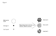
- Fig. 1 depicts two cellular species in light grey and dark grey that are mixed with a microcarrier bead. If e.g. the surface of a microcarrier is modified with a specific antibody or adhesion molecule, then only a singular cellular species will bind to the specific microcarrier bead. If the adhesion of the cells is non-specific, then different bead-cell complexes containing both cellular species are generated.
- the present invention in one aspect also relates to the use of microcarrier beads in methods in accordance with the invention that display at least one probe molecule.
- a probe molecule may be any type of molecule that is capable of specifically interacting with another target molecule or structure.
- probe molecules may be selected from the group comprising small molecules, peptides, glycopeptides, proteins, glycoproteins, receptors, receptor ligands, antibodies, growth factors, hormones, cytokines, chemokines, cell adhesion molecules, integrins, vitamins, nucleic acids and derivatives thereof, lipids and glycans.
- Typical growth factors include e.g.
- human growth factor human nerve growth factor, platelet derived growth factor, TGF ⁇ , EGF, VEGF, TNF-a,TNF-b, FGFs, TGF-a, Epo, IGF-I, IGF-II, IL-1, IL-2, IL-6, IL-8, INF-g and CSFs as well as integrins and CAMs including VLA-1 to VLA-6, LAM-1, Lea-1, CR3, CR4, leukointegrin, collagen, laminin, fibronectin, VCAM-1, MAdCAM-1, E-cadherin, ICAM-1, ICAM-2, ICAM-3, C3b, C4b.
- fragments of the aforementioned molecules can be used as probe molecules as long as such fragments provide the necessary binding specificity.
- a person skilled in the art is also familiar as to how to functionalise e.g. the surface of a microcarrier bead with the aforementioned probe molecules.
- Modification of a microcarrier bead with a probe molecule may be achieved by covalent or non-covalent interaction between the microcarrier bead and the probe molecule.
- one may e.g. coat the microcarrier bead with a self-assembling polymer.
- a self-assembling polymer may e.g. be polyethylenglycol.
- the polymers may be further functionalised to introduce chemical functionalities that allow e.g. covalent coupling of an antibody to the surface of the microcarrier bead.
- beads may be used that provide functional groups such as carboxyl groups, amine groups, hydroxyl groups, sulfhydryl groups etc. These groups may then be cross-linked either directly to the antibodies or using a linker that may be homo-or hetero-bifunctional.
- micorcarrier beads may be activated for providing a chemical linkage to the probe molecules by e.g. coating the beads with a polymer having e.g. acyl fluoride functionalities.
- a polymer having e.g. acyl fluoride functionalities e.g. acyl fluoride functionalities.
- Other covalent attachment chemistries are also applicable but not limited to anhydrides, epoxides, aldehydes, hydrazides, acyl azides, aryl azides, diazo compounds, benzophenones, carbodiimides, imidoesters, isothiocyanates, NHS esters, CNBr, maleimides, tosylates, tresyl chloride, maleic acid anhydrides and carbonyldiimidazole.
- a particular preferred type of probe molecule that can be used to modify a microcarrier bead are molecules that mediates adhesion of a specific cell type.
- the present invention makes use of microcarrier beads, which are modified to display the vascular cell adhesion molecule-1 (VCAM-1).
- VCAM-1 vascular cell adhesion molecule-1
- the cell adhesion molecule VCAM-1 is known to specifically bind leukocytes, thus allowing for separation of white blood cells from plasma, platelets and red blood cells in whole blood samples.
- microcarrier beads of the present invention may be modified with probe molecules such as antibodies that are specific e.g. for fibroblasts, neuronal cells, hepatic cells, etc.
- probe molecules that are known to detect e.g. specifically a certain cell type.
- a person skilled in the art will furthermore be capable of identifying suitable attachment chemistry for either covalently or non-covalently associating such a probe molecule with microcarrier beads.
- microcarrier beads that have been functionalised additionally with at least one probe molecule are cultivated in the presence of cells, preferably the cell type for which the probe molecule is specific, will adhere to the surface of the microcarrier beads.
- Flow cytometry and/or dielectrophoresis may then be used to isolate the beads as well as to quantify the number of cells adhered to the beads.
- microcarrier beads can be perceived.
- probe molecules may be used to select for adherence of three different cell types.
- microcarrier beads which e.g. are then capable of e.g. functionally mimicking the interactions in a tumour or normal tissue.
- the methods in accordance with the present invention allow to detect and isolate multi-cellular arrangements as they occur e.g. in tumours by cultivating the cells on functionalised microcarrier beads and subsequent detection of the cells by flow cytometry and/or dielectrophoresis
- microcarriers allow significant acceleration of isolation and detection processes.
- typical microcarrier beads as has been set out above are on the order of about 100 to about 200 ⁇ m in diameter.
- a typical white blood cell displays a diameter of about 20 ⁇ m.
- the total surface area of a microcarrier bead is thus in the order of about 30,000 to about 130,000 ⁇ m 2 . If one assumes a cell to adhere to a surface on one circular surface that is approximately equal to the diameter of the cell, a typical white blood cell would occupy about 300 ⁇ m 2 of area.
- one bead could potentially contain approximately 100 to 400 cells if one assumes that the cells have grown to monolayer coverage of the microcarrier beads.
- microcarrier beads are counted at a similar speed as in modem flow cytometers (up to 100,000/sec) and if one knows the amount of cells per microcarrier, then the potential throughput of such a method would be on the order of 10 to 40 million cells/sec.
- the cell associated-microcarrier beads may be detected by optical detection methods including flow cytometry, magnetic detection methods and/or dielectrophoresis. While flow cytometry and dielectrophoresis will be discussed in more detail below, the person skilled in the art is aware that optical imaging and image processing approaches may be used as well as microscopic set-ups etc.
- Flow cytometry is a technology that is well known to the person skilled in the art in the context of determining the number and type of cells. The person skilled in the art will understand that typical flow cytometers have to be adapted in order to allow determination of the cell associated-microcarrier beads as described above.
- this instrument should usually have a nozzle larger than at least about 200 ⁇ m, assuming a maximum bead diameter of about 150 ⁇ m coated with cells of approximately 20 ⁇ m diameter.
- Other feasible values for the nozzle of such a flow cytometer-type device may be larger than at least about 300 ⁇ m, at least about 500 ⁇ m or at least about 1000 ⁇ m.
- FIG. 2 A schematic comparison between traditional flow cytometry and the flow cytometry approaches of the present invention are depicted in Fig. 2 . While traditional flow cytometry involves fluidic focussing of single cells through an observation volume, the present invention contemplates detection of cell-adhered microcarrier beads as an alternative to single cell counting. By passing these multi-cell-adhered microcarrier beads through the observation volume, one can simultaneously count multiple cells per bead and in effect thereby multiply the speed at which the overall cell numbers are obtained.
- Detection of the cells on the multi-cell adhered microcarrier beads may be performed as in traditional flow cytometry.
- one may e.g. use a fluorescent marker that specifically interacts with the cell type to be detected.
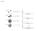
- the methods detect the cell species by fluorescent labelling. This can be achieved by addition of a simple fluorescent and cell-permeable dye, such as Hoechst stain. If multiple cell species are present and require selection, a specific antibody, which target specific surface receptors on the respective cell surfaces and which carry a fluorescent label can be used. By carefully selecting the fluorescent dyes used to label the cells, one can then quantify the cells based on fluorescence intensity.
- Fig. 3 schematically depicts this approach.
- the relative number of cells per bead can be obtained.
- the fluorescent emission yields a characteristic intensity which is proportional to the number of cells.
- multi-colour detection allows for quantisation as well as determination of multiple cellular species on a single microcarrier bead (see Fig. 3D ).
- dielectrophoresis can also be used to isolate and detect cells that are grown on microcarrier beads.
- Dielectrophoresis is the motion of particles caused by the effects of dielectric polarisation in non-uniform electric fields. Unlike classical electrophoresis where the force acting on a particle is determined by its net charge, dielectrophoresis depends on the volume and dielectric properties of the particle. Thus, when a dielectric particle is exposed to an electric field it polarises. The size and direction of the induced electrical dipole depend on the frequency of the applied field and dielectric properties of the particle and medium such as conductivity, permittivity, morphology and shape of the dielectric particle. Typically, in an inhomogeneous field this causes a force due to the interaction of the induced dipoles and the electric fields.
- the microcarrier beads may be added to cell culture suspensions in order to allow monolayer cell coverage thereof.
- monolayer cell coverage of the microcarrier beads By maintaining monolayer cell coverage of the microcarrier beads, one can select an upper limit as to how many cells can be bound to a microcarrier bead.
- the number of cells depends on the diameter of the microcarrier bead as well as the diameter of the cells to be grown. In a typical scenario, 100 to 400 cells will grow per bead assuming monolayer coverage.
- the coverage of the beads can be controlled, the need for fluorescent-base quantisation of the cells by e.g. flow cytometry can be absolved.
- DEP dielectrophoresis
- DEP can move cells by inducing dipole changes within cells upon application of a non-uniform electrical field. Due to the fact that individual cells and microcarrier beads have different size and charge characteristics, they may also possess different DEP characteristics. This means that the electrical field, which may be required to manipulate a bead by DEP may be significantly different from the type of electrical field that one can use to manipulate a single cell. More specifically, the frequency at which negative DEP changes to positive DEP which is also called the "cross-over frequency" can be specific for the type of cell and microcarrier bead depending on the properties chosen for these two components.
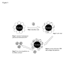
- the present invention therefore allows a method for detecting and/or isolating cells in which the electrical field applied ensures that microcarrier beads are trapped at a positive DEP electrode. If such an electrode is immersed into cell culture, the cells may adhere to the microcarrier beads and depending on the increasing intensity by which the microcarrier beads are inhabited by cells, the dielectric properties of the microcarrier beads may change such that a significant alteration of the bead surface properties by e.g. the cell binding, will induce a change in its crossover frequency. This will allow beads, which have been covered with cells to a certain density, to be released from the positive DEP electrode.
- This aspect of the present invention is schematically depicted in Fig. 4 .
- the cell associated-microcarrier beads can then be analysed e.g. by flow cytometry.
- the present invention relates to a method of detecting and/or isolating cells from a cellular sample in which a microcarrier particle is localised at a DEP electrode and in which coverage of the surface of the microcarrier bead by cells induces release of the cell-associated microcarrier beads from the DEP electrode.
- the frequency for trapping the microcarrier beads at the DEP electrode may be manipulated. It may thus be possible to develop an easy-to-perform assay for counting cells in a cellular sample such as a cell culture suspension.
- the devices and methods in accordance with the invention may use more than two electrodes such as for example at least two pairs of electrodes as this will allow to achieve the different field strengths more easily.
- electrode this may mean an electrode pair to create the different field strengths at the desired parts of the device.
- the device may also comprise quadrupole electrodes etc.
- microcarrier beads are added to a cell culture suspension and the cells are allowed to adhere to the surface of microcarrier beads.
- the cell-adhered microcarrier beads are then transferred to a DEP device, for which the DEP frequency of the electrodes has been set to a value to ensure that naked microcarrier beads do not attach to the electrodes.
- the device can be furthermore tuned in such a way that only microcarrier beads are allowed to attach to the DEP electrode, which are covered by a certain number of cells.
- the cells and "naked" microcarrier beads are first stirred in a suspension in order to allow adherence of the cells to the surface of the microcarrier beads. Once the critical point of cell coverage is achieved, the cell-adhered microcarrier beads become trapped in the DEP field at the DEP electrode and then can be further analysed.
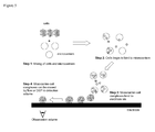
- a schematic outline of this process is depicted in Fig. 5 .
- cells are mixed with microcarrier beads.
- step 2 cells begin to bind to microcarrier beads.
- step 3 a critical point of cell density on the surface of the microcarrier bead is reached which allows the cell-associated microcarrier beads to become attracted to an electrode via DEP.
- the DEP electrode fixed cell-associated microcarrier beads are further analysed.
- Cells can also be separated or categorised based on their dielectrophoretic response.
- dielectrophoresis to isolate microcarrier beads on which cells have been grown. If e.g. the microcarrier beads have been selected to display a movement in an inhomogeneous electric field, one can simply put the cell-associated microcarrier beads in a chamber comprising electrodes for dielectrophoresis and then isolate the beads on which the cells have grown.
- Such an approach may be particularly interesting if beads are used that are functionalised with probe molecules as one may then separate the cell-associated microcarrier beads from other components of the cellular sample.
- microcarrier beads of the invention can be functionalised with e.g. probe molecules such as cell adhesion proteins.
- probe molecules such as cell adhesion proteins.
- VCAM-1 VCAM-1
- integrin super family of receptors is in integral component in organising and directing immune cells within the lymphoid systems.
- integrins are implicated in intercellular adhesion and adhesion to extra cellular matrix components.
- a broad range of different integrins exists, most of which are expressed on cell surfaces and mediate specific interactions.
- VCAM-1 The vascular cell adhesion molecule-1 (VCAM-1) is a protein that is induced by inflammatory cytokines, interleucine-1 and tumour necrosis factors on human endothelial cells. VCAM-1 mediates interaction with mononuclear leukocytes via the integrin receptor VLA-4. This interaction ensures adhesion and tethering of rolling lymphocytes to infected parts of the endothelium and thus is of utmost importance to ensure efficient healing of injured and infected sites of the vasculature.
- VCAM-1 and VLA-4 expressing leukocytes can also use the specific interaction between VCAM-1 and VLA-4 expressing leukocytes in order to isolate such white blood cells from cellular samples such as whole blood.
- cell types include monocytes and lymphocytes.
- VCAM-1 For functionalising of microcarrier beads with VCAM-1, one can rely on typical polystyrene beads of approximately 150 ⁇ m diameter. VCAM-1 can then be coupled to the surfaces of such beads, which have been functionalised with a reactive group such as e.g. succinimide esters.
- the succinimide esters will preferably react with primary amines within the VCAM-1 to form a covalent linkage.
- microcarrier beads are suspended in a mixture with whole blood or other lymphocyte-containing mixtures, this will result in the specific binding of typically only leukocytes to the surface of the functionalised microcarrier surfaces.
- the present invention in essence, relies on the use of microcarrier beads on to which cells have adhered and the detection of such cell-associated microcarrier beads by either flow cytometry and/or dielectrophoresis.
- flow cytometry the use of microcarrier beads has the advantage that the number of cells that can be detected compared to classical approaches is significantly increased. At the same time, the cells are protected against potential harmful factors such as shear forces as they occur during the typical flow cytometric approaches.
- dielectrophoresis also offers interesting options. Thus, it is for example possible to add microcarrier beads to a cell culture suspension and pass that cell culture suspension continuously through a DEP chamber.
- cells will have grown on the microcarrier beads to a certain density, the cells will become polarised in the non-uniform electrical field that is applied to the DEP chamber and depending on the specific settings, only cell-associated microcarrier beads of a certain cell density will e.g. be attracted to a specific electrode.
- This approach allows cells to be grown on a microcarrier bead to a certain density and to then subsequently isolate such microcarrier beads which may then be further analysed e.g. in flow cytometry.
Abstract
The present invention pertains to methods of and devices for detecting and/or isolating cells. Cells are allowed to associate to microcarrier beads directly or through binding to probes displayed on the microcarrier beads and subsequently subjected to flow cytometry and/or dielectrophoresis.
Description
- The present invention pertains to methods of and devices for detecting and/or isolating cells by optical means including flow cytometry and/or dielectrophoresis. To this end, the methods and devices in accordance with the invention make use of microcarrier beads.
- Rapid cell counting is typically performed by relying on flow cytometry, with fluorescence activated cell sorting (FACS) being representative for this type of methodology.
- These modem flow cytometry techniques have in common that they count cells on an individual basis. Typically, a suspension of cells is hydrodynamically focussed in a narrow flow stream that passes through a detection chamber at high speed. A typical nozzle size for the focusing stream in a flow cytometer is in the order or 70 to 100 µm. If such a nozzle is coupled to high-speed fluidic flow, it is ensured that cells pass through the detection volume in a single file and are counted one by one. Modem flow cytometers typically reach detection speeds of 10,000 to 40,000 cells/sec.
- However, flow cytometry suffers from the drawback that it usually cannot be used to analyse multi-cell aggregates such as multi-spheroids. The reason is that multi-cellular aggregates are typically of too large size to efficiently pass the nozzle of a flow cytometer and frequently lead to problems such as instrument clogging. On the other side, there is an interest in analysing multi-cellular assemblies as they more correctly represent the events that occur, e.g. within a cellular tumour.
- Another method for detecting cells is dielectrophoresis. Dielectrophoresis relies on the application of a non-uniform electrical field to samples that are polarisable. When a polarisable electrically uncharged particle such as a cell is placed in such an inhomogeneous electrical field, it will become polarised and experience a net driving form due to the non-uniformity of the electrical field. This force can either be in the direction of increasing field strength (so-called positive DEP) or decreasing field strength (negative DEP). The point at which the electrical field changes the force on a cell from positive to negative DEP (and visa versa) is called the cross-over frequency, and this cross-over frequency can be used to discriminate various cell types. However, use of DEP for isolating and detecting specific cells from complex samples can be hampered by the fact that various cell types may not differ with respect to their polarisation characteristics sufficiently enough to allow separation based on DEP.
- In view of this situation there is a continuing need in the art for further methods of and devices for isolating and detecting cells and particularly multi-cellular samples in an efficient and straightforward way.
- It is one objective of the present invention to provide methods for isolating and/or detecting single cells as well as multi-cellular assemblies from different cellular samples such as e.g. blood.
- It is a further objective of the present invention to provide devices that allow efficient detection and/or isolation of single cells and/or multi-cellular assemblies in cellular samples such as e.g. blood.
- These objectives as well as others which will become apparent from the ensuing description are attained by the subject matter of the independent claims. Some of the more specific embodiments of the present invention are defined by the dependent claims.
- The present invention in one embodiment relates to a method of detecting and/or isolating at least one cell comprising the steps of
- providing at least one microcarrier bead;
- contacting said at least one microcarrier bead with at least one cell such that said at least one cell associates with said at least one microcarrier bead;
- detecting and/or isolating said cell-associated microcarrier bead.
- Said cell associated-microcarrier beads may be detected by optical detection methods, magnetic detection methods and/or dielectrophoresis.
- The optical detection methods include optical imaging, image processing approaches and preferably flow cytometry.
- Typically said microcarrier beads have a diameter of about 20 to about 1000 µm. A preferred diameter can be in the range of about 75 µm to about 350 µm. A size of about 100 µm to about 200 µm and about 150 µm can also be preferred.
- The microcarrier beads comprise bead materials that render them suitable for application in optical detection devices such as flow cytometers and/or dielectrophoresis devices. Said microcarrier beads may thus be made from a material that is polarizable and can experience a force in an inhomogeneous electrical field. In this case, the beads can be moved by dielectrophoresis.
- In general, said microcarrier beads will comprise bead materials selected from the group comprising polystyrene and poly-lactide.
- The method in accordance with the invention may be performed by contacting the microcarrier beads with cells in cell culture such that the cells associate with the microcarrier beads during growth.
- In one of the preferred embodiments of the invention, the microcarrier beads may be further modified to display at least one probe.
- Such probe molecules may be specific for certain target molecules or structures such as cells. The probe molecule may thus be selected from the group comprising small molecules, peptides, glycopeptides, proteins, glycoproteins, receptors, receptor ligands, antibodies, growth factors, hormones, cytokines, chemokines, cell adhesion molecules (CAMs), integrins, vitamins, nucleic acids and derivatives thereof, lipids and glycans.
- A preferred probe molecule can be VCAM-1.
- If the microcarrier beads are modified to display probe molecules, the method in accordance with the invention can be used to detect the target molecules and/or structures for which the probe molecules are specific.
- Thus, methods in accordance with the invention can be adapted to allow isolation and/or detection of specific cell types such as e.g. leukocytes, stem cells, progenitor cells, tumor cells and fetal cells.
- The present invention further relates to a flow cytometry device for detecting and/or isolating at least one cell comprising a noozle of a diameter to allow for passing and detecting of cell-associated microcarries.
- Such noozles may have a diameter of at least about 200 µm. Other diameters such as at least about 300 µm, at least about 500 µm or at least about 1mm may also be feasible.
- The present invention further relates to a device for detecting and/or isolating at least one cell comprising an electrode being capable of immobilizing at least one polarizable microcarrier bead thereto by way of dielectrophoresis.
- The present invention also relates to the use of such devices to detect and/or isolate at least one cell.
- The present invention further relates to the use of microcarrier beads described hereinafter for detecting and/or isolating at least one cell.
-
- Fig. 1
- schematically depicts how a microcarrier bead may be covered by different cell types "A" and "B".
- Fig. 2
- schematically depicts a comparison between traditional flow cytometry of single individual cells in a flow stream and detection in accordance with the present invention by detecting cell-associated microcarrier beads.
- Fig. 3
- schematically depicts the influence on the degree of association of cells to the microcarrier beads on the detection signal.
- Fig. 4
- schematically depicts one approach for using dielectrophoresis for detecting cells by coating microcarrier beads therewith.
- Fig. 5
- schematically depicts another embodiment of use of dielectrophoresis for detecting cells by making the cells adhere to microcarrier beads.
- Fig. 6
- schematically depicts how leukocytes can be isolated from whole blood samples by using "VCAM-1 coated microcarrier beads".
- The present invention is based on the finding that microcarrier beads to which cells associate can be used to detect and/or isolate cells by optical detection methods such as flow cytometry and/or dielectrophoresis.
- Before some of the embodiments of the present inventions are described in more detail, the following definitions are introduced.
- As used in this specification and in the appended claims, the singular forms of "a" and "an" also include the respective plurals unless the context clearly dictates otherwise. Thus, the term "a probe" can include more than one probe, namely two, three, four, five etc. probes.
- The terms "about" or "approximately" in the context of the present invention denotes an interval of accuracy that the person skilled in the art will understand to still ensure the technical effect of the feature in question. The term typically indicates deviation from the indicated numerical value of +/- 10%, and preferably +/- 5%.
- As has been mentioned above, the invention relates in one embodiment to a method of detecting and/or isolating at least one cell comprising the steps of
- providing at least one microcarrier bead;
- contacting said at least one microcarrier bead with at least one cell such that said at least one cell associates with said at least one microcarrier bead;
- detecting and/or isolating said cell-associated microcarrier bead.
- Thus, the present invention in one aspect relies on the use of microcarrier beads for isolating and/or detecting cells by optical detection methods including flow cytometry, magnetic detection methods and/or dielectrophoresis (DEP).
- The term "microcarrier bead" denotes a structure which has a diameter in the range of approximately 20 µm to about 1000µm. Diameters in the range of approximately 75 µm to approximately 300 µm may be preferred as may be diameters in the range of approximately 100 µm to approximately 200 µm.
- In some of the preferred embodiments, the beads will display a diameter in the range of about 120 µm to about 180 µm or about 150 µm.
- The beads may have any form that is suitable for the purposes of the present invention. They thus may have a rectangular shape, a triangular shape etc. Beads with a round or a substantially round form can be preferred according to the present invention. Thus, the beads will typically have a round, elliptical or spheroid form.
- There are no specific requirements as to the structure of the microcarrier beads or as to the materials from which the microcarrier beads are formed except that the beads must be capable of allowing cells to associate with them, and that the structure and the materials must be compatible with the detection technology that is going to be applied, such as flow cytometry, magnetic detection methods and/or dielectrophoresis.
- If the cell associated-microcarrier beads are to be analysed by dielectrophoresis, a further requirement is thus that they are polarizable in an inhomogeneous electrical field to experience a force if dielectrophoresis is performed on the beads.
- With respect to the aforementioned requirements, the microcarrier beads may be produced from materials such as polystyrene, polydivnylbenzene and polymethylmethacrylate and biodegradable polymers such as poly-lactides, polyclycolides, polycaprolacton and copolymers thereof.
- Such beads are in principle commercially available from e.g. SoloHill Engineering, Inc (Ann Arbor, Michigan, USA).
- The beads may be further modified, e.g. coated in order to ensure adherence of probe molecules and/or to allow for association of a certain type of cell (see below). Such coatings include glass, collagen (gelatine), a cationic surface treatment, recombinant proteins such as cell adhesion molecules including vascular cell adhesion molecule 1 (VCAM-1) etc.
- As will be set out below, a preferred embodiment of the present invention relates to the use of dielectrophoresis for detecting cell-covered microcarrier beads. In this aspect, the present invention can make use of the different polar characteristics of a microcarrier bead versus e.g. a certain cell type. Given that one can influence the microcarrier beads' polarisable characteristics by selecting the appropriate architecture and/or composition, the person skilled in the art will choose the microcarrier depending on the detection method.
- If the cell-covered microcarrier beads are used in a dielectrophoresis approach, it may be preferred to use a microcarrier bead whose polarisable properties change depending on whether the microcarrier bead is covered with cells or not.
- The person skilled in the art is familiar with the production of microcarrier beads. Thus, one may obtain these beads from aforementioned commercial sources or produce them according to the protocols being described in e.g. Gu et al (J. Control Release (2004), 96.3:463-472), Li et al (Acta Pharmacol. Sin. (2006) 27.6:754-759), Norrgren et al (Dev. Biol. Stand. (1983), 55:43-51) or Tatard et al (Biomaterials (2005), 26.17:3727-3737). Further information as to the production of microcarrier beads that can be used for detection in e.g. DEP approaches can be found in
US 2003/0119057 A1 andUS 2003/0015428 A1 . Thus, the bead technology to create both e.g. microcarrier beads as well as probe covered-beads is known to the skilled person. - Microcarrier beads can e.g. be synthesized e.g. by controlled emulsification techniques such as submerged ink-jet printing.
- If a microcarrier bead is to be produced from a polymer such as polystyrene, a polymer solution in a solvent such as dichloromethane can be ink-jetted into an aqueous phase containing a stabilizer for the monodisperse emulsion that is formed. To this end, one can use e.g. polyvinyl alcohol but also proteins could be used to achieve efficient stabilization. Subsequently polymer beads with a narrow size distribution are formed upon removal of the solvent.
- If one intends to incorporate e.g. peptides/proteins or nucleic acids as probes which can be water soluble, a double emulsion technique can be used. To this end, the water-soluble peptides and/or proteins may be first dissolved in an aqueous phase and this aqueous phase is emulsified in the polymer solution. If necessary or desired, additional stabilizers such as e.g. Pluronic can be added. This first emulsion is then ink-jetted into a second aqueous phase and beads are again formed upon removal of the solvent.
- As has been set out above, the present invention relies partly on the effect that cells are allowed to adhere to the afore-described microcarrier beads and that these cell-covered microcarrier beads are then used to detect the said cells by flow cytometry and/or dielectrophoresis. Contact between cells and microcarrier beads may be established e.g. already during cell culture. Thus, microcarrier beads may be added to a suspension cell culture and the cells will attach to the surface of the microcarrier beads and subsequently continue to grow on the surface of the microcarrier beads.
- The person skilled in the art is well-aware that it may be necessary to provide an attachment matrix for certain cell types to allow their growth on the surface of a microcarrier bead. This may be achieved by e.g. coating the microcarrier beads with collagen or polymeric substances that allow adherence of cells to the surface of a microcarrier bead. Such polymers include collagen, fibronectin, hydrogels (acrylamide and agarose gels), polymers linked to CAMs etc.
- By way of example, one may add microcarriers such as those available from SoloHill (vide supra) to a cell culture of e.g. mammalian cells. If e.g. a suspension cell culture is gently stirred under appropriate conditions, the cells will start adhering to the microcarriers and will form layers of cells on microcarrier beads. The cell-coated microcarrier beads may then be used in the methods in accordance with the invention.
- In the context of the present invention the term "cell" denotes all types of cells which can associate with microcarrier beads as described above. Thus, the term may relate to any type of cell that can be isolated from a biological sample such as bodily fluids including blood, plasma, urine, saliva etc. However, samples may also comprise environmental samples taking from soils, lakes, rivers, plants etc.
- Typical cell types which can be analysed by the methods in accordance with the invention include e.g. stem cells, progenitor cells, leukocytes, fibroblasts, tumour cells, sperm cells, fibroblasts, neuronal cells, hepatic cells and fetal cells.
- Once cells have started to grow on the surface of microcarrier beads, the microcarrier beads may be isolated by simple purification protocols such as centrifugation, magnetic assisted sorting, fluorescence assisted sorting and filtering as well as by washing steps and may be subsequently analysed by optical detection methods including flow cytometry, magnetic detection methods and/or dielectrophoresis.
- As will be set out below, the microcarrier beads may be modified to display probe molecules that have a specific affinity for certain target molecules and structures such as certain cell types.
- Depending on these modifications, it is thus possible to functionalise the surface of a microcarrier bead and to allow adherence of e.g. only a certain cell type to a microcarrier bead. This allows selective isolation as well as detection of certain cell types by the methods in accordance with the present invention. Depending on the modifications of the microcarrier bead, as well as the initial cell input composition, one can theoretically distinguish different various "cell-bead" complexes.
-
Fig. 1 for example depicts two cellular species in light grey and dark grey that are mixed with a microcarrier bead. If e.g. the surface of a microcarrier is modified with a specific antibody or adhesion molecule, then only a singular cellular species will bind to the specific microcarrier bead. If the adhesion of the cells is non-specific, then different bead-cell complexes containing both cellular species are generated. - Thus, the present invention in one aspect also relates to the use of microcarrier beads in methods in accordance with the invention that display at least one probe molecule. Such a probe molecule may be any type of molecule that is capable of specifically interacting with another target molecule or structure. Thus, probe molecules may be selected from the group comprising small molecules, peptides, glycopeptides, proteins, glycoproteins, receptors, receptor ligands, antibodies, growth factors, hormones, cytokines, chemokines, cell adhesion molecules, integrins, vitamins, nucleic acids and derivatives thereof, lipids and glycans. Typical growth factors include e.g. human growth factor, human nerve growth factor, platelet derived growth factor, TGFβ, EGF, VEGF, TNF-a,TNF-b, FGFs, TGF-a, Epo, IGF-I, IGF-II, IL-1, IL-2, IL-6, IL-8, INF-g and CSFs as well as integrins and CAMs including VLA-1 to VLA-6, LAM-1, Lea-1, CR3, CR4, leukointegrin, collagen, laminin, fibronectin, VCAM-1, MAdCAM-1, E-cadherin, ICAM-1, ICAM-2, ICAM-3, C3b, C4b.
- The person skilled in the art will also be aware that fragments of the aforementioned molecules can be used as probe molecules as long as such fragments provide the necessary binding specificity.
- A person skilled in the art is also familiar as to how to functionalise e.g. the surface of a microcarrier bead with the aforementioned probe molecules.
- Modification of a microcarrier bead with a probe molecule may be achieved by covalent or non-covalent interaction between the microcarrier bead and the probe molecule. To this end, one may e.g. coat the microcarrier bead with a self-assembling polymer. Such a self-assembling polymer may e.g. be polyethylenglycol. The polymers may be further functionalised to introduce chemical functionalities that allow e.g. covalent coupling of an antibody to the surface of the microcarrier bead.
- If e.g. antibodies are used as a probe and should be covalently coupled to the microcarrier beads, beads may be used that provide functional groups such as carboxyl groups, amine groups, hydroxyl groups, sulfhydryl groups etc. These groups may then be cross-linked either directly to the antibodies or using a linker that may be homo-or hetero-bifunctional.
- Thus, micorcarrier beads may be activated for providing a chemical linkage to the probe molecules by e.g. coating the beads with a polymer having e.g. acyl fluoride functionalities. Other covalent attachment chemistries are also applicable but not limited to anhydrides, epoxides, aldehydes, hydrazides, acyl azides, aryl azides, diazo compounds, benzophenones, carbodiimides, imidoesters, isothiocyanates, NHS esters, CNBr, maleimides, tosylates, tresyl chloride, maleic acid anhydrides and carbonyldiimidazole.
- A particular preferred type of probe molecule that can be used to modify a microcarrier bead are molecules that mediates adhesion of a specific cell type. Thus, in a particularly preferred embodiment, the present invention makes use of microcarrier beads, which are modified to display the vascular cell adhesion molecule-1 (VCAM-1). The cell adhesion molecule VCAM-1 is known to specifically bind leukocytes, thus allowing for separation of white blood cells from plasma, platelets and red blood cells in whole blood samples.
- Similarly, the microcarrier beads of the present invention may be modified with probe molecules such as antibodies that are specific e.g. for fibroblasts, neuronal cells, hepatic cells, etc.
- The person skilled in the art will be able to identify probe molecules that are known to detect e.g. specifically a certain cell type. A person skilled in the art will furthermore be capable of identifying suitable attachment chemistry for either covalently or non-covalently associating such a probe molecule with microcarrier beads.
- If such microcarrier beads that have been functionalised additionally with at least one probe molecule are cultivated in the presence of cells, preferably the cell type for which the probe molecule is specific, will adhere to the surface of the microcarrier beads. Flow cytometry and/or dielectrophoresis may then be used to isolate the beads as well as to quantify the number of cells adhered to the beads.
- The person skilled in the art is well aware that further elaborations of microcarrier beads can be perceived. Thus, one may e.g. modify the microcarrier beads' surface with e.g. two types of probe molecules and thus select for the presence of two cell types. Similarly, one may use three different types of probe molecules and select for adherence of three different cell types.
- Using such an approach it is possible to selectively adhere combinations of cell types to the surface of the microcarrier beads, which e.g. are then capable of e.g. functionally mimicking the interactions in a tumour or normal tissue.
- Thus, the methods in accordance with the present invention allow to detect and isolate multi-cellular arrangements as they occur e.g. in tumours by cultivating the cells on functionalised microcarrier beads and subsequent detection of the cells by flow cytometry and/or dielectrophoresis
- Before analysis of cell-associated microcarrier beads by optical methods such as flow cytometry and/or dielectrophoresis is discussed in further detail, it is emphasised that the use of microcarriers allows significant acceleration of isolation and detection processes.
- As has been explained in the background section, classical flow cytometry relies on the detection of cellular samples comprising single cells. If thus a cellular sample being obtainable from an adherently growing cell culture is to be analysed, the cells must be released from the surface of the e.g. cell culture dishes in which they are grown by e.g. proteologic treatment. This is not necessary for the cell-adhered microcarrier beads that are used in accordance with the present invention.
- In addition, typical microcarrier beads as has been set out above are on the order of about 100 to about 200 µm in diameter. A typical white blood cell displays a diameter of about 20 µm. The total surface area of a microcarrier bead is thus in the order of about 30,000 to about 130,000 µm2. If one assumes a cell to adhere to a surface on one circular surface that is approximately equal to the diameter of the cell, a typical white blood cell would occupy about 300 µm2 of area. Hence, one bead could potentially contain approximately 100 to 400 cells if one assumes that the cells have grown to monolayer coverage of the microcarrier beads. Assuming that the microcarrier beads are counted at a similar speed as in modem flow cytometers (up to 100,000/sec) and if one knows the amount of cells per microcarrier, then the potential throughput of such a method would be on the order of 10 to 40 million cells/sec.
- Studies with flow cytometers that operate at very high rates (e.g. about 40,000/per sec) often result in cellular damage and clogging of the instrument. By adhering cells to the microcarrier beads as described above and subsequent analysis by e.g. flow cytometry, it is thus possible to increase the throughput of the system and at the same time to minimise the damage to the cells given that the shear forces for the cell associated-microcarrier beads is reduced compared to the situation where the cells are not grown on the microcarrier beads. Thus, cells which are associated with microcarrier beads are likely more stable and robust for counting in a high flow environment than single cells in suspension.
- As has been mentioned above, the cell associated-microcarrier beads may be detected by optical detection methods including flow cytometry, magnetic detection methods and/or dielectrophoresis. While flow cytometry and dielectrophoresis will be discussed in more detail below, the person skilled in the art is aware that optical imaging and image processing approaches may be used as well as microscopic set-ups etc.
- Flow cytometry is a technology that is well known to the person skilled in the art in the context of determining the number and type of cells. The person skilled in the art will understand that typical flow cytometers have to be adapted in order to allow determination of the cell associated-microcarrier beads as described above.
- Thus, if the cell-adhered microcarrier beads are put in a flow cytometer-type device in accordance with the invention, this instrument should usually have a nozzle larger than at least about 200 µm, assuming a maximum bead diameter of about 150 µm coated with cells of approximately 20 µm diameter. Other feasible values for the nozzle of such a flow cytometer-type device may be larger than at least about 300 µm, at least about 500 µm or at least about 1000 µm.
- A schematic comparison between traditional flow cytometry and the flow cytometry approaches of the present invention are depicted in
Fig. 2 . While traditional flow cytometry involves fluidic focussing of single cells through an observation volume, the present invention contemplates detection of cell-adhered microcarrier beads as an alternative to single cell counting. By passing these multi-cell-adhered microcarrier beads through the observation volume, one can simultaneously count multiple cells per bead and in effect thereby multiply the speed at which the overall cell numbers are obtained. - Detection of the cells on the multi-cell adhered microcarrier beads may be performed as in traditional flow cytometry. Thus, one may e.g. use a fluorescent marker that specifically interacts with the cell type to be detected. In one of its more simple embodiments, the methods detect the cell species by fluorescent labelling. This can be achieved by addition of a simple fluorescent and cell-permeable dye, such as Hoechst stain. If multiple cell species are present and require selection, a specific antibody, which target specific surface receptors on the respective cell surfaces and which carry a fluorescent label can be used. By carefully selecting the fluorescent dyes used to label the cells, one can then quantify the cells based on fluorescence intensity.
-
Fig. 3 schematically depicts this approach. Thus, based on fluorescent intensity, the relative number of cells per bead can be obtained. In a scenario, such asFig. 3 A to C , where only one type of cell is grown on a microcarrier bead at different densities, the fluorescent emission yields a characteristic intensity which is proportional to the number of cells. Furthermore, by use of different fluorescent labels that specifically recognise different types of cells, multi-colour detection allows for quantisation as well as determination of multiple cellular species on a single microcarrier bead (seeFig. 3D ). - As has been mentioned above, dielectrophoresis can also be used to isolate and detect cells that are grown on microcarrier beads.
- Dielectrophoresis is the motion of particles caused by the effects of dielectric polarisation in non-uniform electric fields. Unlike classical electrophoresis where the force acting on a particle is determined by its net charge, dielectrophoresis depends on the volume and dielectric properties of the particle. Thus, when a dielectric particle is exposed to an electric field it polarises. The size and direction of the induced electrical dipole depend on the frequency of the applied field and dielectric properties of the particle and medium such as conductivity, permittivity, morphology and shape of the dielectric particle. Typically, in an inhomogeneous field this causes a force due to the interaction of the induced dipoles and the electric fields.
- As mentioned above, the microcarrier beads may be added to cell culture suspensions in order to allow monolayer cell coverage thereof. By maintaining monolayer cell coverage of the microcarrier beads, one can select an upper limit as to how many cells can be bound to a microcarrier bead. As mentioned above, the number of cells depends on the diameter of the microcarrier bead as well as the diameter of the cells to be grown. In a typical scenario, 100 to 400 cells will grow per bead assuming monolayer coverage.
- If the coverage of the beads can be controlled, the need for fluorescent-base quantisation of the cells by e.g. flow cytometry can be absolved.
- If only a certain type of cellular species is adhered to the microcarrier beads, all beads will display a comparable coverage by this type of cell. Then, each bead would contain an approximately equivalent number of cells and hence only the single beads would require detection in order to produce an overall cell count ((number of beads detected) X (average number of cells per bead) = total cell number). To achieve this, dielectrophoresis (DEP) can be employed in one embodiment of the present invention.
- As described above, DEP can move cells by inducing dipole changes within cells upon application of a non-uniform electrical field. Due to the fact that individual cells and microcarrier beads have different size and charge characteristics, they may also possess different DEP characteristics. This means that the electrical field, which may be required to manipulate a bead by DEP may be significantly different from the type of electrical field that one can use to manipulate a single cell. More specifically, the frequency at which negative DEP changes to positive DEP which is also called the "cross-over frequency" can be specific for the type of cell and microcarrier bead depending on the properties chosen for these two components.
- The present invention therefore allows a method for detecting and/or isolating cells in which the electrical field applied ensures that microcarrier beads are trapped at a positive DEP electrode. If such an electrode is immersed into cell culture, the cells may adhere to the microcarrier beads and depending on the increasing intensity by which the microcarrier beads are inhabited by cells, the dielectric properties of the microcarrier beads may change such that a significant alteration of the bead surface properties by e.g. the cell binding, will induce a change in its crossover frequency. This will allow beads, which have been covered with cells to a certain density, to be released from the positive DEP electrode. This aspect of the present invention is schematically depicted in
Fig. 4 . The cell associated-microcarrier beads can then be analysed e.g. by flow cytometry. - Thus, in one embodiment the present invention relates to a method of detecting and/or isolating cells from a cellular sample in which a microcarrier particle is localised at a DEP electrode and in which coverage of the surface of the microcarrier bead by cells induces release of the cell-associated microcarrier beads from the DEP electrode.
- The person skilled in the art will understand that depending on the design of the device that is used for this type of DEP detection, the frequency for trapping the microcarrier beads at the DEP electrode may be manipulated. It may thus be possible to develop an easy-to-perform assay for counting cells in a cellular sample such as a cell culture suspension.
- The person skilled in the art is aware that in such an embodiment it is preferred to have microcarrier beads of uniform dimensions given that such a uniform dimension will translate into a uniform frequency requirement for trapping by DEP
- This in turn would ensure a uniform coverage of the beads by cells and thus would result in relatively constant "point of release" of the beads from the electrode. The person skilled in the art is clearly aware of further embodiments of this approach by e.g. engineering electrode configurations in multiphase fields within the device that is used for DEP to e.g. levitate microcarrier particles, trapped in a field cage and rotate them or transport them over comparatively long distances.
- The person skilled in the art will further be aware that the devices and methods in accordance with the invention may use more than two electrodes such as for example at least two pairs of electrodes as this will allow to achieve the different field strengths more easily. Thus where the figures denote an "electrode", this may mean an electrode pair to create the different field strengths at the desired parts of the device. However, the device may also comprise quadrupole electrodes etc.
- Another embodiment of the present invention in which DEP is used to detect and/or isolate cells, relies on the principle described above in reverse order. Thus, microcarrier beads are added to a cell culture suspension and the cells are allowed to adhere to the surface of microcarrier beads. The cell-adhered microcarrier beads are then transferred to a DEP device, for which the DEP frequency of the electrodes has been set to a value to ensure that naked microcarrier beads do not attach to the electrodes. The device can be furthermore tuned in such a way that only microcarrier beads are allowed to attach to the DEP electrode, which are covered by a certain number of cells.
- Thus, the cells and "naked" microcarrier beads are first stirred in a suspension in order to allow adherence of the cells to the surface of the microcarrier beads. Once the critical point of cell coverage is achieved, the cell-adhered microcarrier beads become trapped in the DEP field at the DEP electrode and then can be further analysed. A schematic outline of this process is depicted in
Fig. 5 . Instep 1 ofFig. 5 , cells are mixed with microcarrier beads. Instep 2, cells begin to bind to microcarrier beads. Instep 3, a critical point of cell density on the surface of the microcarrier bead is reached which allows the cell-associated microcarrier beads to become attracted to an electrode via DEP. Finally, the DEP electrode fixed cell-associated microcarrier beads are further analysed. - Cells can also be separated or categorised based on their dielectrophoretic response. Thus, one can use dielectrophoresis to isolate microcarrier beads on which cells have been grown. If e.g. the microcarrier beads have been selected to display a movement in an inhomogeneous electric field, one can simply put the cell-associated microcarrier beads in a chamber comprising electrodes for dielectrophoresis and then isolate the beads on which the cells have grown. Such an approach may be particularly interesting if beads are used that are functionalised with probe molecules as one may then separate the cell-associated microcarrier beads from other components of the cellular sample.
- It has been set out above that the microcarrier beads of the invention can be functionalised with e.g. probe molecules such as cell adhesion proteins. In the following, one embodiment of the present invention is discussed in which microcarrier beads are functionalised with VCAM-1.
- The integrin super family of receptors is in integral component in organising and directing immune cells within the lymphoid systems. In general, integrins are implicated in intercellular adhesion and adhesion to extra cellular matrix components. A broad range of different integrins exists, most of which are expressed on cell surfaces and mediate specific interactions.
- The vascular cell adhesion molecule-1 (VCAM-1) is a protein that is induced by inflammatory cytokines, interleucine-1 and tumour necrosis factors on human endothelial cells. VCAM-1 mediates interaction with mononuclear leukocytes via the integrin receptor VLA-4. This interaction ensures adhesion and tethering of rolling lymphocytes to infected parts of the endothelium and thus is of utmost importance to ensure efficient healing of injured and infected sites of the vasculature.
- However, one can also use the specific interaction between VCAM-1 and VLA-4 expressing leukocytes in order to isolate such white blood cells from cellular samples such as whole blood.
- To this end, one may modify a microcarrier bead with VCAM-1 to create a surface, which is specific for adherence of any type of cells that expresses VLA-4. Typically, such cell types include monocytes and lymphocytes.
- For functionalising of microcarrier beads with VCAM-1, one can rely on typical polystyrene beads of approximately 150 µm diameter. VCAM-1 can then be coupled to the surfaces of such beads, which have been functionalised with a reactive group such as e.g. succinimide esters. The succinimide esters will preferably react with primary amines within the VCAM-1 to form a covalent linkage.
- If such microcarrier beads are suspended in a mixture with whole blood or other lymphocyte-containing mixtures, this will result in the specific binding of typically only leukocytes to the surface of the functionalised microcarrier surfaces.
- If one now places the microcarrier beads to which the white blood cells have adhered into a non-uniform electrical field, the cells will become polarised and will experience a net driving force due to the non-uniformity of the field. This force can be either in the direction of increasing field strength (so-called "positive DEP") or decreasing field strength (negative DEP). Thus, if one e.g. introduces the VCAM-1 coated-microcarrier beads which have been contacted with e.g. blood, into a DEP quadropole device and applies a fluid to this chamber, one can retain the microcarrier beads on which white blood cells have adhered, within the DEP quadropole while other components, such as red blood cells or plasma proteins are washed away (see
Figure 6 ). - The person skilled in the art is of course also aware that such an approach can be used to isolate other types of cells depending on the nature and the specificity of the probe molecule. If one, for example, coats a microcarrier bead with a probe molecule that is specific for fibroblasts, it may be possible to use DEP to isolate microcarrier beads onto which this cell type has adhered. The same applies for hepatic cells if a corresponding probe molecule is used etc.
- The present invention, in essence, relies on the use of microcarrier beads on to which cells have adhered and the detection of such cell-associated microcarrier beads by either flow cytometry and/or dielectrophoresis. In the case of flow cytometry, the use of microcarrier beads has the advantage that the number of cells that can be detected compared to classical approaches is significantly increased. At the same time, the cells are protected against potential harmful factors such as shear forces as they occur during the typical flow cytometric approaches. The use of dielectrophoresis also offers interesting options. Thus, it is for example possible to add microcarrier beads to a cell culture suspension and pass that cell culture suspension continuously through a DEP chamber. Once, cells will have grown on the microcarrier beads to a certain density, the cells will become polarised in the non-uniform electrical field that is applied to the DEP chamber and depending on the specific settings, only cell-associated microcarrier beads of a certain cell density will e.g. be attracted to a specific electrode.
- This approach allows cells to be grown on a microcarrier bead to a certain density and to then subsequently isolate such microcarrier beads which may then be further analysed e.g. in flow cytometry.
- While the above invention has been described with respect to some of its preferred embodiments, this is in no way to limit the scope of the invention. The person skilled in the art is clearly aware of further embodiments and alterations to the previously described embodiments that are still within the scope of the present invention.
Claims (15)
- Method of detecting and/or isolating at least one cell comprising the steps of- providing at least one microcarrier bead;- contacting said at least one microcarrier bead with at least one cell such that said at least one cell associates with said at least one microcarrier bead;- detecting and/or isolating said cell-associated microcarrier bead.
- Method according to claim 1,
wherein said cell associated-microcarrier bead is detected by optical detection methods including flow cytometry, magnetic detection methods and/or dielectrophoresis. - Method according to claim 1 or claim2,
wherein said microcarrier bead has a diameter of about 20 µm to about 1000 µm. - Method according to any of claims 1 to 3,
wherein said microcarrier bead comprises bead materials selected from the group comprising polystyrene, polydivnylbenzene and polymethylmethacrylate and biodegradable polymers such as poly-lactides, polyclycolides, polycaprolacton and copolymers thereof. - Method according to claim 1,
wherein said contacting step b) occurs in cell culture. - Method according to claim 5,
wherein cells form a substantially continuous monolayer on said microcarrier. - Method according to any of claims 1 to 6,
wherein said method is used to determine the number of cells in a sample. - Method according to any of claims 1 to 7,
wherein said microcarrier is modified to display at least one probe. - Method according to claim 9,
wherein said probe is specific for at least one certain cell type. - Method according to claim 8 or claim 9,
wherein said probe is selected from the group comprising small molecules, peptides, glycopeptides, proteins, glycoproteins, receptors, receptor ligands, antibodies, growth factors, hormones, cytokines, chemokines, cell adhesion molecules, integrins, vitamins, nucleic acids and derivatives thereof, lipids and glycans. - Method according to claim 10,
wherein said probe is VCAM-1 and wherein said method is used to detect leukocytes. - Device for detecting and/or isolating at least one cell by flow cytometry comprising a noozle of a diameter to allow for passing and detecting of cell-associated microcarries.
- Device for detecting and/or isolating at least one cell comprising an electrode setup being capable of immobilizing at least one polarizable microcarrier bead thereto by way of dielectrophoresis.
- Use of a device according to claim 12 or 13 to detect and/or isolate at least one cell.
- Use of microcarrier beads which preferably are polarizable for detecting and/or isolating at least one cell.
Priority Applications (2)
| Application Number | Priority Date | Filing Date | Title |
|---|---|---|---|
| EP07104924A EP2045601A1 (en) | 2007-03-26 | 2007-03-26 | Use of microcarrier beads for detection and/or isolation of cells by flow cytometry and/or dielectrophoresis |
| PCT/IB2008/050994 WO2008117195A2 (en) | 2007-03-26 | 2008-03-17 | Use of microcarrier beads for detection and/or isolation of cells by flow cytometry |
Applications Claiming Priority (1)
| Application Number | Priority Date | Filing Date | Title |
|---|---|---|---|
| EP07104924A EP2045601A1 (en) | 2007-03-26 | 2007-03-26 | Use of microcarrier beads for detection and/or isolation of cells by flow cytometry and/or dielectrophoresis |
Publications (1)
| Publication Number | Publication Date |
|---|---|
| EP2045601A1 true EP2045601A1 (en) | 2009-04-08 |
Family
ID=38377333
Family Applications (1)
| Application Number | Title | Priority Date | Filing Date |
|---|---|---|---|
| EP07104924A Ceased EP2045601A1 (en) | 2007-03-26 | 2007-03-26 | Use of microcarrier beads for detection and/or isolation of cells by flow cytometry and/or dielectrophoresis |
Country Status (2)
| Country | Link |
|---|---|
| EP (1) | EP2045601A1 (en) |
| WO (1) | WO2008117195A2 (en) |
Cited By (4)
| Publication number | Priority date | Publication date | Assignee | Title |
|---|---|---|---|---|
| US8541246B2 (en) | 2009-07-29 | 2013-09-24 | Dynex Technologies, Inc. | Sample plate systems and methods |
| CN103645308A (en) * | 2013-12-09 | 2014-03-19 | 东南大学 | Two-dimensional coding method of micro-carrier |
| US9523701B2 (en) | 2009-07-29 | 2016-12-20 | Dynex Technologies, Inc. | Sample plate systems and methods |
| US11366109B2 (en) | 2018-12-06 | 2022-06-21 | Winmems Technologies Co., Ltd. | Encoded microflakes |
Families Citing this family (6)
| Publication number | Priority date | Publication date | Assignee | Title |
|---|---|---|---|---|
| EP2058662A1 (en) * | 2007-10-17 | 2009-05-13 | Koninklijke Philips Electronics N.V. | Diagnosis of diseases by measuring cell mediated extracellular matrix degradation |
| EP2187211A1 (en) * | 2008-11-18 | 2010-05-19 | Koninklijke Philips Electronics N.V. | Diagnosis of diseases by measuring chemokinesis |
| JP5583532B2 (en) * | 2010-09-14 | 2014-09-03 | 学校法人慶應義塾 | Cell accumulation method |
| WO2012079749A1 (en) | 2010-12-16 | 2012-06-21 | Stichting Dutch Polymer Institute | Process for making cell suspension |
| US9709556B2 (en) | 2013-12-11 | 2017-07-18 | Analiza, Inc. | Devices and methods for determining and/or isolating circulating cancer cells |
| US20150160246A1 (en) | 2013-12-11 | 2015-06-11 | Analiza, Inc. | Devices and methods for determining and/or isolating cells such as circulating cancer or fetal cells |
Citations (8)
| Publication number | Priority date | Publication date | Assignee | Title |
|---|---|---|---|---|
| US3970518A (en) * | 1975-07-01 | 1976-07-20 | General Electric Company | Magnetic separation of biological particles |
| DE3806558A1 (en) * | 1987-03-03 | 1988-09-15 | Hitachi Ltd | Method for cell measurement |
| WO1994016821A1 (en) * | 1993-01-21 | 1994-08-04 | Scientific Generics Limited | Methods of analysis/separation |
| WO1997049731A1 (en) * | 1996-06-21 | 1997-12-31 | Zeneca Limited | Cell adhesion inhibiting compounds |
| US6387707B1 (en) * | 1996-04-25 | 2002-05-14 | Bioarray Solutions | Array Cytometry |
| WO2002044338A2 (en) * | 2000-11-30 | 2002-06-06 | The Trustees Of Columbia University In The City Of New York | Growth of human dendritic cells for cancer immunotherapy in closed system using microcarrier beads |
| US20030015428A1 (en) * | 2000-06-14 | 2003-01-23 | Becker Frederick F. | Dielectrically-engineered microparticles |
| US6858439B1 (en) * | 1999-03-15 | 2005-02-22 | Aviva Biosciences | Compositions and methods for separation of moieties on chips |
Family Cites Families (4)
| Publication number | Priority date | Publication date | Assignee | Title |
|---|---|---|---|---|
| US4994388A (en) * | 1988-04-15 | 1991-02-19 | Solohill Engineering, Inc. | Collagen-coated polystyrene microcarrier beads |
| WO1991007485A1 (en) * | 1989-11-09 | 1991-05-30 | Bio-Metric Systems, Inc. | Improved bioreactor surfaces and methods of making same |
| JP4723862B2 (en) * | 2002-12-23 | 2011-07-13 | フジフィルム マニュファクチャリング ユーロプ ビー.ブイ. | Method for coating a cell culture support |
| JP2006325467A (en) * | 2005-05-25 | 2006-12-07 | Pentax Corp | Collagen-coated carrier and method for producing the collagen-coated carrier |
-
2007
- 2007-03-26 EP EP07104924A patent/EP2045601A1/en not_active Ceased
-
2008
- 2008-03-17 WO PCT/IB2008/050994 patent/WO2008117195A2/en active Application Filing
Patent Citations (8)
| Publication number | Priority date | Publication date | Assignee | Title |
|---|---|---|---|---|
| US3970518A (en) * | 1975-07-01 | 1976-07-20 | General Electric Company | Magnetic separation of biological particles |
| DE3806558A1 (en) * | 1987-03-03 | 1988-09-15 | Hitachi Ltd | Method for cell measurement |
| WO1994016821A1 (en) * | 1993-01-21 | 1994-08-04 | Scientific Generics Limited | Methods of analysis/separation |
| US6387707B1 (en) * | 1996-04-25 | 2002-05-14 | Bioarray Solutions | Array Cytometry |
| WO1997049731A1 (en) * | 1996-06-21 | 1997-12-31 | Zeneca Limited | Cell adhesion inhibiting compounds |
| US6858439B1 (en) * | 1999-03-15 | 2005-02-22 | Aviva Biosciences | Compositions and methods for separation of moieties on chips |
| US20030015428A1 (en) * | 2000-06-14 | 2003-01-23 | Becker Frederick F. | Dielectrically-engineered microparticles |
| WO2002044338A2 (en) * | 2000-11-30 | 2002-06-06 | The Trustees Of Columbia University In The City Of New York | Growth of human dendritic cells for cancer immunotherapy in closed system using microcarrier beads |
Non-Patent Citations (1)
| Title |
|---|
| TATARD V M ET AL: "Pharmacologically active microcarriers: a tool for cell therapy", BIOMATERIALS, ELSEVIER SCIENCE PUBLISHERS BV., BARKING, GB, vol. 26, no. 17, June 2005 (2005-06-01), pages 3727 - 3737, XP004696122, ISSN: 0142-9612 * |
Cited By (8)
| Publication number | Priority date | Publication date | Assignee | Title |
|---|---|---|---|---|
| US8541246B2 (en) | 2009-07-29 | 2013-09-24 | Dynex Technologies, Inc. | Sample plate systems and methods |
| US9244069B2 (en) | 2009-07-29 | 2016-01-26 | Dynex Technologies | Sample plate systems and methods |
| US9523701B2 (en) | 2009-07-29 | 2016-12-20 | Dynex Technologies, Inc. | Sample plate systems and methods |
| US9857367B2 (en) | 2009-07-29 | 2018-01-02 | Dynex Technologies, Inc. | Sample plate systems and methods |
| US10207268B2 (en) | 2009-07-29 | 2019-02-19 | Dynex Technologies, Inc. | Sample plate systems and methods |
| US10969386B2 (en) | 2009-07-29 | 2021-04-06 | Dynex Technologies, Inc. | Sample plate systems and methods |
| CN103645308A (en) * | 2013-12-09 | 2014-03-19 | 东南大学 | Two-dimensional coding method of micro-carrier |
| US11366109B2 (en) | 2018-12-06 | 2022-06-21 | Winmems Technologies Co., Ltd. | Encoded microflakes |
Also Published As
| Publication number | Publication date |
|---|---|
| WO2008117195A2 (en) | 2008-10-02 |
| WO2008117195A3 (en) | 2008-12-11 |
Similar Documents
| Publication | Publication Date | Title |
|---|---|---|
| EP2045601A1 (en) | Use of microcarrier beads for detection and/or isolation of cells by flow cytometry and/or dielectrophoresis | |
| US6890426B2 (en) | Magnetic separation apparatus and methods | |
| JP3863373B2 (en) | Method of using an apparatus for separation of biological fluids | |
| JP6775498B2 (en) | Methods and related equipment for analyzing the contents of droplets | |
| US6790366B2 (en) | Magnetic separation apparatus and methods | |
| EP2270126A1 (en) | Cell selection apparatus, and cell selection method using the same | |
| US20100055758A1 (en) | Magnetic Device for Isolation of Cells and Biomolecules in a Microfluidic Environment | |
| EP2579988A2 (en) | Non-linear magnetophoretic separation device, system and method | |
| JP5937586B2 (en) | Method for detecting intermolecular interactions | |
| EP2781906B1 (en) | Method for identifying cells | |
| JP2004532968A (en) | Systems and methods for cell subpopulation analysis | |
| CN109863398B (en) | Method for detecting and/or characterizing tumor cells and related device | |
| US20120132593A1 (en) | Systems and methods for magnetic separation of biological materials | |
| KR101279918B1 (en) | Device for detection of tumor cells and detecting method tumor cells | |
| JP7158671B2 (en) | Specific cell capture method | |
| US20120135494A1 (en) | Systems and methods for magnetic separation of biological materials | |
| CA2352064C (en) | Magnetic separation apparatus and methods | |
| JP4911280B2 (en) | Organic polymer particles and method for producing the same | |
| WO2007105784A1 (en) | Dielectrophoresis device | |
| US20080113333A1 (en) | Method for preventing the adhesion of particles | |
| Shah et al. | Using magnetic microparticles in molecular and cellular isolations | |
| JP7109719B2 (en) | Specific cell capture method | |
| JP2018157812A (en) | Detection method of cells included in sample | |
| Mukherjee et al. | Isolation and Purification of Various Mammalian Cells: Single Cell Isolation | |
| KR20230012541A (en) | Improving devices and methods for microdroplet manipulation |
Legal Events
| Date | Code | Title | Description |
|---|---|---|---|
| PUAI | Public reference made under article 153(3) epc to a published international application that has entered the european phase |
Free format text: ORIGINAL CODE: 0009012 |
|
| PUAI | Public reference made under article 153(3) epc to a published international application that has entered the european phase |
Free format text: ORIGINAL CODE: 0009012 |
|
| AK | Designated contracting states |
Kind code of ref document: A1 Designated state(s): AT BE BG CH CY CZ DE DK EE ES FI FR GB GR HU IE IS IT LI LT LU LV MC MT NL PL PT RO SE SI SK TR |
|
| AX | Request for extension of the european patent |
Extension state: AL BA HR MK RS |
|
| STAA | Information on the status of an ep patent application or granted ep patent |
Free format text: STATUS: THE APPLICATION HAS BEEN REFUSED |
|
| 18R | Application refused |
Effective date: 20090508 |