EP1768060B1 - Image processing method and apparatus for modification of images of skin - Google Patents
Image processing method and apparatus for modification of images of skin Download PDFInfo
- Publication number
- EP1768060B1 EP1768060B1 EP06254907A EP06254907A EP1768060B1 EP 1768060 B1 EP1768060 B1 EP 1768060B1 EP 06254907 A EP06254907 A EP 06254907A EP 06254907 A EP06254907 A EP 06254907A EP 1768060 B1 EP1768060 B1 EP 1768060B1
- Authority
- EP
- European Patent Office
- Prior art keywords
- individual
- image
- chromophore
- distribution
- skin
- Prior art date
- Legal status (The legal status is an assumption and is not a legal conclusion. Google has not performed a legal analysis and makes no representation as to the accuracy of the status listed.)
- Not-in-force
Links
- 0 *C1*2=CC=C2C=C1 Chemical compound *C1*2=CC=C2C=C1 0.000 description 1
Images


Classifications
-
- G—PHYSICS
- G01—MEASURING; TESTING
- G01N—INVESTIGATING OR ANALYSING MATERIALS BY DETERMINING THEIR CHEMICAL OR PHYSICAL PROPERTIES
- G01N21/00—Investigating or analysing materials by the use of optical means, i.e. using sub-millimetre waves, infrared, visible or ultraviolet light
- G01N21/17—Systems in which incident light is modified in accordance with the properties of the material investigated
- G01N21/21—Polarisation-affecting properties
-
- A—HUMAN NECESSITIES
- A61—MEDICAL OR VETERINARY SCIENCE; HYGIENE
- A61B—DIAGNOSIS; SURGERY; IDENTIFICATION
- A61B5/00—Measuring for diagnostic purposes; Identification of persons
- A61B5/0059—Measuring for diagnostic purposes; Identification of persons using light, e.g. diagnosis by transillumination, diascopy, fluorescence
-
- A—HUMAN NECESSITIES
- A61—MEDICAL OR VETERINARY SCIENCE; HYGIENE
- A61B—DIAGNOSIS; SURGERY; IDENTIFICATION
- A61B5/00—Measuring for diagnostic purposes; Identification of persons
- A61B5/44—Detecting, measuring or recording for evaluating the integumentary system, e.g. skin, hair or nails
- A61B5/441—Skin evaluation, e.g. for skin disorder diagnosis
- A61B5/442—Evaluating skin mechanical properties, e.g. elasticity, hardness, texture, wrinkle assessment
-
- A—HUMAN NECESSITIES
- A61—MEDICAL OR VETERINARY SCIENCE; HYGIENE
- A61B—DIAGNOSIS; SURGERY; IDENTIFICATION
- A61B5/00—Measuring for diagnostic purposes; Identification of persons
- A61B5/44—Detecting, measuring or recording for evaluating the integumentary system, e.g. skin, hair or nails
- A61B5/441—Skin evaluation, e.g. for skin disorder diagnosis
- A61B5/443—Evaluating skin constituents, e.g. elastin, melanin, water
-
- G—PHYSICS
- G01—MEASURING; TESTING
- G01N—INVESTIGATING OR ANALYSING MATERIALS BY DETERMINING THEIR CHEMICAL OR PHYSICAL PROPERTIES
- G01N21/00—Investigating or analysing materials by the use of optical means, i.e. using sub-millimetre waves, infrared, visible or ultraviolet light
- G01N21/17—Systems in which incident light is modified in accordance with the properties of the material investigated
- G01N21/25—Colour; Spectral properties, i.e. comparison of effect of material on the light at two or more different wavelengths or wavelength bands
-
- G—PHYSICS
- G06—COMPUTING; CALCULATING OR COUNTING
- G06T—IMAGE DATA PROCESSING OR GENERATION, IN GENERAL
- G06T11/00—2D [Two Dimensional] image generation
- G06T11/001—Texturing; Colouring; Generation of texture or colour
-
- G—PHYSICS
- G06—COMPUTING; CALCULATING OR COUNTING
- G06T—IMAGE DATA PROCESSING OR GENERATION, IN GENERAL
- G06T3/00—Geometric image transformation in the plane of the image
-
- G—PHYSICS
- G06—COMPUTING; CALCULATING OR COUNTING
- G06T—IMAGE DATA PROCESSING OR GENERATION, IN GENERAL
- G06T7/00—Image analysis
- G06T7/0002—Inspection of images, e.g. flaw detection
- G06T7/0012—Biomedical image inspection
-
- G—PHYSICS
- G06—COMPUTING; CALCULATING OR COUNTING
- G06T—IMAGE DATA PROCESSING OR GENERATION, IN GENERAL
- G06T2207/00—Indexing scheme for image analysis or image enhancement
- G06T2207/10—Image acquisition modality
- G06T2207/10024—Color image
-
- G—PHYSICS
- G06—COMPUTING; CALCULATING OR COUNTING
- G06T—IMAGE DATA PROCESSING OR GENERATION, IN GENERAL
- G06T2207/00—Indexing scheme for image analysis or image enhancement
- G06T2207/20—Special algorithmic details
- G06T2207/20212—Image combination
-
- G—PHYSICS
- G06—COMPUTING; CALCULATING OR COUNTING
- G06T—IMAGE DATA PROCESSING OR GENERATION, IN GENERAL
- G06T2207/00—Indexing scheme for image analysis or image enhancement
- G06T2207/30—Subject of image; Context of image processing
- G06T2207/30004—Biomedical image processing
- G06T2207/30024—Cell structures in vitro; Tissue sections in vitro
Definitions
- the present application relates to image processing.
- embodiments of the present application concern methods and apparatus for processing images of individuals to generate images representative of the results of cosmetic or surgical interventions or the progression of medical conditions.
- thread veins and age spots can adversely affect the appearance of individuals.
- Various cosmetic and surgical techniques have therefore been developed to address these conditions.
- the appearance of thread veins can be minimised through laser cauterisation of the affected blood vessels.
- the appearance of age spots can be addressed through the application of an acid peel.
- Image-based skin colour and texture analysis/synthesis by extracting haemoglobin and melanin information in the skin discloses an E-cosmetic function for digital images based on physics and physiologically-based image processing. Shading on the face is removed by a simple colour vector analysis in the optical density domain as an inverse lighting technique. The image without shading is analyzed by a technique that extracts haemoglobin and melanin components by independent component analysis. Experimental results using UV-B irradiation and the application of methyl nicotinate on the arms support the physiological validity of the analysis and effectiveness of the proposed shading removal.
- a digital imaging system in which colour information of an entire port wine stain or other skin condition with a single image in CIE L*a*b* colour space (L*, a*) is derived from RGB pixel data (R, G, B).
- Cross-polarization optics produce marked reduction in specularly reflected light in the images.
- a patient positioning device allows for repeatable positioning of the patient's head or body portion.
- the digital nature of the system provides a near real-time mapping of melanin or erythema or other skin chromophore metrics.
- the cross-polarized diffuse reflectance colour digital imagine system obtains subsurface skin colour information and acquisition of facial images in a reproducible fashion at a fixed distance from an illumination source at optimized angles of view depending on the region of interest being imaged.
- an apparatus and method for providing a process that can be implemented by a human operator and a computing device to analyze and display human skin images. They system acquires a digital image from a camera or scanner. Subsequently, the system determines which area(s) of the image to analyze using landmarks such as the corner of the eye. The determined areas are then analyzed to locate skin defects such as red spots, and the defects are visually identified on a display. A severity is calculated for the defects and the severity associated with a population of people. In addition, a simulation is generated and displayed showing an improvement to the defect areas.
- US2004/085324 discloses an image-adjusting system and method, and more particularly, a system and method for adjusting a facial image.
- a set of adjusting parameters that by using a face-adjusting template stored in a database of face-adjusting template to adjust the image data of a facial image, wherein the database of face-adjusting template comprises plural data of face adjustment parameter generated by computer software or manual adjustment, such as skin texture, proportion of facial features, and variations of expression, etc., for improving the quality of facial image
- the arrangement and combination of the plural face adjustment parameters can further constitute different face-adjusting templates, so that the facial expression may be appropriately modified to generate the effect of different facial images.
- “Pyramid-based texture analysis/synthesis” discloses a method for synthesizing images that match the texture appearance of a given digitized sample. This synthesis is completely automatic and requires only the "target” texture as input. It allows generation of as much texture as desired so that any object can be covered. The approach is based on a model of human texture perception, and has potential to be a practically useful tool for image processing and graphics applications.
- an image processing apparatus comprising:
- an image processing method comprising
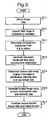
- FIG. 1 is a schematic block diagram of a first embodiment of the present invention.
- a digital camera 1 comprising a conventional digital camera is provided which is arranged to obtain an image of an individual 2 illuminated by a light source 3.
- the images obtained by the digital camera 1 are then transmitted to a computer 4 which is configured by software either provided on a disk 5 or by receiving an electrical signal 6 by via a communications network to be configured into a number of functional modules 16-24 which cause the computer 4 to process the image data received from the digital camera 1 to generate an output image 30 which is shown on a display 31.
- the output image 30 comprises a first image portion 33 being a representation of the original image data generated by the digital camera 1 and a second image portion 34 being a calculated representation of the individual 2 illustrating the expected results from a treatment affecting the individual 2.
- skin has a layered structure comprising an outer cornified layer 50, the epidermis 52, and the dermis which itself can be divided into the papillary dermis 54 which contains the blood supply 55 for the skin and the reticular dermis 56.
- the effect of reflection of light directly by the cornified layer 50 is required to be removed so that a measurement of the remitted light which has interacted with the chromophores present in the epidermis 52 and papillary dermis 54 can be made.
- a first polarising filter 36 is provided in front of the lens of the digital camera 1 and a second polarising filter 38 cross polarised with the first is provided in front of the light source 3.
- a second polarising filter 38 cross polarised with the first is provided in front of the light source 3.
- the functional modules illustrated in Figure 1 are purely notional in order to assist with the understanding of the working of the claimed invention and may not in certain embodiments directly correspond with blocks of code in the source code for the software. In other embodiments the function performed by the illustrated functional modules may be divided between different modules or may be performed by the re use of the same modules for different functions.
- the functional modules comprise a spherical conversion unit 10 for converting RGB image data into corresponding spherical co-ordinates, an image conversion module 12 and a conversion table 14 for processing spherical angular co-ordinates to generate data indicative of concentrations of blood and melanin; a treatment simulation module 16 arranged to determine a revised chromophore distribution representative of a treatment by processing chromophore distributions generated by the conversion module 12; an image generation module 18 and an inverse conversion table 20 operable to generate image data utilising chromophores distribution data; a texture determination module 22 for identifying variations in appearance in an image of an individual which do not arise due to variations in chromophore concentrations; and a combination module 24 for combining texture data generated by the texture determination module 22 and image data generated by the image generation module 18 and outputting a simulated treated image 34 for display on a display screen 31.
- FIG. 3 is a flow diagram of the processing performed by the computer 4 of Figure 1 , initially (S3-1) an image is obtained by the digital camera 1 of the individual 2 illuminated by the light source 3.
- the digital camera 1 comprises a conventional digital camera.
- the image data generated by the digital camera 1 therefore comprises RGB values ranging from 0 to 255 for a large array of pixels where the RGB values are indicative of the extent light received by a photo receptor within the camera 1 for each pixel in an image appears to be red, green and blue where a completely black pixel has RGB values of 0, 0, 0 and a completely bright white pixel has RGB values of 255, 255, 255.
- the image is initially passed to the spherical conversion module 10 which converts (S3-2) the conventional RGB data for each pixel in an image into a corresponding set of spherical co-ordinates ⁇ ⁇ r where the spherical angles of ⁇ ⁇ are substantially indicative of the hue and chromaticity represented by an individual pixel in an image captured by the digital camera 1 and the radial co-ordinate r is substantially indicative of the brightness of the pixel.
- the conversion is performed for each pixel in the original pixel array for the image generated by the digital camera.
- the result of the conversion is a set of spherical ⁇ ⁇ r co-ordinates for each pixel in the original image.
- the array of radial elements r is then passed directly to the image generation module 18 whereas arrays of the calculated angular spherical co-ordinates ⁇ and ⁇ are in this embodiment passed to the image conversion module 12.
- the image conversion module 12 After the spherical conversion module 10 has converted the RGB values for an image into spherical co-ordinates, the image conversion module 12 then processes the generated array of ⁇ and ⁇ values to obtain values indicative of the concentration of blood and melanin at individual points on the surface of the skin of the individual.
- this is achieved by processing each pair of ⁇ and ⁇ values for each pixel in an array in turn by scaling the and ⁇ values so that instead of comprising values between n and - n, and 0 and n/2, the scaled ⁇ and ⁇ values comprise integer values ranging between 0 and 255.
- These scaled and ⁇ values are then utilised to access the conversion table 14 which in this embodiment is a 255 by 255 a lookup table associating pairs of scaled ⁇ and ⁇ co-ordinates with pairs of concentrations of blood and melanin liable to give rise to such scaled ⁇ and ⁇ values.
- the conversion table 14 comprises a table associating blood and melanin concentrations with various ⁇ and ⁇ values, where the ⁇ and ⁇ values fall within the expected range of the colour space for skin.
- the conversion module 12 returns a null value for the concentration of blood and melanin for the pixel with ⁇ and ⁇ values for the pixel.
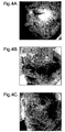
- Figure 4A is an illustrative example of an image of an individual's face captured by a digital camera 1.
- the pixels in the image will each comprise a red, green and blue pixel value.
- Figures 4B and 4C are illustrative representations of determined concentrations of blood and melanin respectively derived by processing the exemplary image of Figure 4A where greater concentrations of blood and melanin are shown by darker points within the images.
- the applicants have appreciated through analysis of the remittance of light from the skin that under controlled lighting using polarised white light, light remitted by the skin lies within a well defined colour space. In this colour space apparent hue or colour of a portion of skin is predominantly accounted for through variations in blood and melanin concentration. Conversely, the brightness of a particular portion of skin is primarily determined through a combination of: the brightness of light incident on the skin, the angle at which light illuminates a particular portion of skin, the distance between a subject and a light source, and the concentration of collagen at a particular point since the concentration of collagen makes skin more or less reflective.
- this chromophore distribution data is then passed by the conversion module 12 to the treatment simulation module 16 and the image generation module 18.
- the treatment simulation module 16 processes the received chromophore distribution data to generate revised chromophore distribution data indicative of the chromophore distribution after a treatment.
- the treatment simulation module 16 processes the received chromophore distribution data representing the concentration of blood to generate revised blood distribution data which is the expected distribution of blood after treatment.
- the revised blood distribution data is determined through applying a conventional blurring algorithm to the portions of the image where treatment would occur.
- Figure 4D The result of processing the image of Figure 4B using such a blurring algorithm is shown as Figure 4D .
- the result of processing these specific areas of image highlighted in Figure 5B is shown as Figure 5C .
- the result of processing by the treatment simulation module 16 in this embodiment is to generate blood distribution data illustrating a distribution of blood where the blood is more smoothly distributed than exists in the original calculated distribution for the individual.
- the conversion model 12 in addition to passing chromophore distribution data to the treatment simulation model 16, the conversion model 12 also passes the chromophore distribution data to the image generation module 18 which together with the inverse conversion table 20 and the texture determination module 22 proceeds to determine (S3-5) texture data indicative of the variation in appearance of an individual which does not arise due to different distributions of chromophores.
- the image generation module 18 processes the unamended chromophore distribution data for each pixel in an image to generate a corresponding expected pair of ⁇ and ⁇ colour angles.
- this conversion is achieved by the image generation module 18 accessing the inverse conversion table 20 which is a lookup table which associates each possible pair of determined blood and melanin concentrations for a pixel with a corresponding expected ⁇ and ⁇ values.
- the inverse conversion table 20 is therefore data representative of an inverse function corresponding to the function for converting ⁇ and ⁇ values to measurements of blood and melanin concentration as is stored in the conversion table 14. In the case of pixels which are associated with null values of within the chromophore distribution data no ⁇ and ⁇ values are determined.
- the image generation module 18 is able to generate a derived image where each pixel image for which the conversion module 12 is able to determine chromophore distribution values is represented by a pair of calculated colour angles ⁇ and ⁇ and a radial value r corresponding to the radial value for that particular pixel as determined by the spherical conversion module 10.
- This derived image data is then passed to the texture determination module 22 which proceeds to convert the array of received ⁇ ⁇ r data into an image of equivalent RGB values.
- the texture determination module 22 then performs a difference operation comparing for each pixel the calculated RGB values for that pixel with the corresponding RGB values in the original image data obtained by the digital camera 1.
- this difference data will correspond to the RGB values for the corresponding pixels in the original image.
- the conversion module 12 is able to derive chromophore distribution data for a particular pixel, if the derived RGB values for a particular pixel do not exactly correspond to the RGB values for that pixel in the original image, this indicates that some of the apparent colouring of the area of skin represented by that pixel arises due to factors other than the estimated concentrations of blood and melanin for that area of skin.
- the array of differences in RGB values is then output as texture data which is passed to the combination module 24.
- the image generation module 18 is also arranged to process (S3-6) the revised chromophore distribution data generated by the treatment simulation module 16 in a similar way to generate ⁇ and ⁇ values which together with the radial value r for individual pixels are indicative for a particular portion of skin of the appearance of that portion after treatment which results in the change in chromophore distribution determined by the treatment simulation modules 16.
- the image generation module 18 accessing the inverse conversion table 20 to convert determined blood and melanin concentration values for individual pixels into ⁇ and ⁇ values.
- the obtained ⁇ and ⁇ values together with the original radial r element of the spherical co-ordinate determined for a pixel are then processed in a conventional way described above to convert the obtained ⁇ ⁇ r values into conventional RGB values for that pixel representing the intensities in the red, green and blue channels for the portion of the individual represented by that pixel.
- This processing is repeated for all the pixels for which a chromophore distribution has been determined by the conversion module 12.
- the generated array of red, green and blue pixel values is then output by the image generation module 18 as a treated image and passed to the combination module 24.
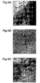
- the result of the processing is to generate an image similar to that of the original image but where the disfigurement arising due to the thread veins has been removed. This difference may most clearly be seen by comparing the image portion corresponding to the original image portion highlighted in Figure 5A a corresponding highlighted image portion from Figure 4E which is shown in Figure 5D .
- the treated image generated by the image generation module 18 will lack features which arise due to other factors.
- Figures 4A and Figures 4E and also Figures 5A and 5D the generated images of Figures 4E and 5D appear to lack texture when compared with the corresponding original images of Figures 4A and 5A . This can be seen most clearly in the portion of the images corresponding to the individual's eye appearing in the image.
- Figure 6A the portion in the original image of Figure 4A corresponding to the individual's eye is shown in Figure 6A and the corresponding portion from the generated image of Figure 4E is shown as Figure 6B .
- Figure 6B the corresponding portion from the generated image of Figure 4E is shown as Figure 6B .
- this missing texture information in this embodiment is exactly the texture data for an image determined by the texture determination module 22.
- this missing information can therefore be reintroduced to the image by the combination module 24 varying the red, green and blue values for each pixel in the treated image by the corresponding red, green and blue values determined for those pixels determined by the texture determination module 22.
- this processing in addition to reintroducing the missing texture, also causes pixels which the image conversion module 12 is unable to convert ⁇ and ⁇ spherical co-ordinates into chromophore concentrations to be represented by the original pixel values for those pixel from the original image obtained by the digital camera 1.
- FIG. 4F A final image generated after reintroducing this texture to the treated image of Figure 4E is shown in Figure 4F . Highlighted portions of the final images corresponding to the portions shown in Figure 5D and Figure 6B are also illustrated as Figures 5E and 6C respectively.
- the original image 33 generated by the camera and the final generated image 34 can then be output and displayed simultaneously as an output image 30 on a display 31 thereby illustrating to the individual the expected result of the treatment.
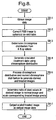
- FIGS. 7 and 8 are a schematic block diagram of an image processing system in accordance with a second embodiment of the present invention and a flow diagram of the processing performed by the image processing system respectively.
- an image processing system was described in which an image generation module 18 generated a derived and treated image utilising chromophore distribution data and r co-ordinate data derived by a spherical conversion module 10.
- the derived image data was then processed by a texture determination module 22 to obtain texture data which was then combined with the generated treated image data by a combination module 24 in order to generate an output image.
- the image processing system in accordance with this embodiment of the present invention is identical to the image processing system of the previous embodiment with the exception that the texture determination and combination modules 22, 24 are replaced by an output module 100 and the image generation module 18 and the inverse conversion table 20 are replaced with a modified image generation module 101 and a modified inverse conversion table 102.
- the remaining elements of the system and are identical to the elements previously described in relation to the first embodiment and are identified by the same reference numerals as were previously used in Figure 1 .
- the processing performed by the image processing system of Figure 7 is illustrated by the flow diagram of Figure 8 .
- the processing by the system to derive chromophore distribution data from input image data and to derive simulated treatment data using that chromophore distribution data (s8-1 - s8-4) is identical to the corresponding steps (s3-1 - s3-4) undertaken in the first embodiment and description of these steps will not be repeated.
- the image generation module 101 processes the derived chromophore distribution data and simulated treatment data by accessing a modified inverse conversion table 102 which in this embodiment comprises a look up table associating pairs of blood and melanin concentrations with RGB values representative of the apparent colour of skin having such blood and melanin concentrations as viewed under fixed lighting conditions.
- a modified inverse conversion table 102 which in this embodiment comprises a look up table associating pairs of blood and melanin concentrations with RGB values representative of the apparent colour of skin having such blood and melanin concentrations as viewed under fixed lighting conditions.
- the RGB values stored in the modified inverse conversion table 102 could be generated by storing empirical data of the appearance of skin containing specific blood and melanin concentrations as viewed under fixed lighting conditions.
- the RGB values comprise calculated RGB values determined by applying the inverse function relating blood and melanin concentrations to 9 and ⁇ spherical co-ordinates as defined by data in the conversion table 14 and converting the determined ⁇ and ⁇ spherical co-ordinates to RGB values using a fixed value for r.
- the image generation module 101 processes each pair of chromophore concentration values in the chromophore distribution and the revised chromophore distribution in turn. As a result of the processing of the arrays defining the chromophore distributions performed by the image generation module 101, the image generation module 101 generates a pair of RGB images. These images are then passed to the output module 100.
- RGB image data is converted to spherical co-ordinate data
- the angular ⁇ co-ordinates are substantially indicative of the hue and chromaticity represented by an individual pixel which arises due to the presence and concentrations of chromophores such as blood and melanin in the skin.
- the radial co-ordinate r is then substantially indicative of the brightness of the pixel which arises due to a combination of lighting factors and the concentrations of collagen in the skin.
- the RGB values for pixels in the derived and treated images are generated utilising a constant r value, the images will be representative of areas of skin under fixed lighting conditions and where the concentration of collagen is constant.
- output RGB data for each pixel in an image is derived (s8-6) utilising the following equations:
- R out x y R original x y R derived x y ⁇ R treated x y
- G out x y G original x y G derived x y ⁇ G treated x y
- B out x y B original x y B derived x y ⁇ B treated x y
- R out (x, y), G out (x, y) and B out (x, y) are the output RGB values for a pixel at position x, y and R original (x, y), R derived (x, y) R treated (x, y), G original (x, y), G derived (x, y) G treated (x, y), B original (x, y), B derived (x,y) B treated (x, y) are the red green and
- a means is provided to vary the brightness of each pixel so as to reflect the variations in apparent brightness due to variations in lighting and collagen concentration present in the original image and thereby generate a realistic final output image.
- a generated output image is output (s8-7) and displayed on a display screen 31.
- the present invention could be utilised to illustrate the effect of the progression of an aliment or of aging.
- the treatment simulation module 16 of the first and second embodiments would be modified so as to process obtained chromophore distributions generated by the image conversion module 12 to generate a revised chromophore distribution where the revised chromophore distribution was representative of a distribution arising due to an aliment or through aging.
- Figure 9A is an illustrative example of an original image of an individual.
- Figure 9B is an illustrative example image generated by a third embodiment of the present invention in which the appearance of acne on the face of the individual is simulated by processing a determined chromophore distribution for the image of Figure 9A .
- a further image could be obtained when the absence of the cross polarising filter 36.
- the image obtained in the absence of the cross polarising filter 36 could then be processed by the texture determination module 22 so as to generate texture data which identifies the difference between a derived image and an image obtained in the absence of the polarising filter 36.
- this texture data is combined with a treated image, a final output image would then be generated where the output image included not only the texture missing from a derived image but also the difference in appearance resulting from specular reflections from the surface of the skin.
- the advantage of such a system would be that the images generated by the computer would be more realistic as the direct reflections would also be present in the generated images.
- modified chromophore distributions for more than one chromophore might be generated and simulated images created based on the revised chromophore distributions for more than one chromophore could be created.
- the system could be modified to obtain measurements of other chromophores such as bilirubin, tattoo pigments or dye stuffs, keratin and hair.
- the wavelengths of emitted light detected by a digital camera could be selected so as to measure wavelengths which are substantially unaffected by the presence of for example melanin. Processing such measurements in the way described would enable measurements of other chromophores to be obtained.
- a modified camera obtaining intensity measurements for more wavebands could be provided and the additional measurements could be utilised to determine measurements of for example blood, melanin and collagen concentrations.
- a digital camera arranged to obtain measurements in an infrared region might be utilised.
- means could be provided to process the chromophore distribution data generated by the image conversion module to identify abnormal distributions of chromophores. The processing performed by a treatment simulation module could then be selected to normalise the abnormal distributions.
- abnormally high concentrations of blood might be automatically detected and replaced with an average expected concentration for a particular body part.
- abnormally high or low concentrations of other chromophores might be detected and corrected.
- generated image data has been described as being output as a screen image, it will be appreciated that if a 3D computer model of an individual appearing in an image is available the generated image data could be used as texture render data for texture rendering a model thereby generating a 3D model of an individual's appearance utilising a revised chromophore distribution.
- the embodiments of the invention described with reference to the drawings comprise computer apparatus and processes performed in computer apparatus, the invention also extends to computer programs, particularly computer programs on or in a carrier, adapted for putting the invention into practice.
- the program may be in the form of source or object code or in any other form suitable for use in the implementation of the processes according to the invention.
- the carrier can be any entity or device capable of carrying the program.
- the carrier may comprise a storage medium, such as a ROM, for example a CD ROM or a semiconductor ROM, or a magnetic recording medium, for example a floppy disc or hard disk.
- a storage medium such as a ROM, for example a CD ROM or a semiconductor ROM, or a magnetic recording medium, for example a floppy disc or hard disk.
- the carrier may be a transmissible carrier such as an electrical or optical signal which may be conveyed via electrical or optical cable or by radio or other means.
- the carrier When a program is embodied in a signal which may be conveyed directly by a cable or other device or means, the carrier may be constituted by such cable or other device or means.
- the carrier may be an integrated circuit in which the program is embedded, the integrated circuit being adapted for performing, or for use in the performance of, the relevant processes.
Description
- The present application relates to image processing. In particular embodiments of the present application concern methods and apparatus for processing images of individuals to generate images representative of the results of cosmetic or surgical interventions or the progression of medical conditions.
- Many skin conditions for example thread veins and age spots can adversely affect the appearance of individuals. Various cosmetic and surgical techniques have therefore been developed to address these conditions. Thus for example the appearance of thread veins can be minimised through laser cauterisation of the affected blood vessels. Similarly the appearance of age spots can be addressed through the application of an acid peel.
- In order to determine whether a particular intervention is worth pursuing, it is useful for an individual to be given an indication of the likely results of the intervention prior to undertaking a course of treatment. It is desirable that such representations are as accurate as possible. There is therefore a need for image processing methods and apparatus which enable such images to be generated in such a way that they can be more accurate representations of the likely results of an intervention than exists in the prior art.
- "Image-based skin colour and texture analysis/synthesis by extracting haemoglobin and melanin information in the skin" discloses an E-cosmetic function for digital images based on physics and physiologically-based image processing. Shading on the face is removed by a simple colour vector analysis in the optical density domain as an inverse lighting technique. The image without shading is analyzed by a technique that extracts haemoglobin and melanin components by independent component analysis. Experimental results using UV-B irradiation and the application of methyl nicotinate on the arms support the physiological validity of the analysis and effectiveness of the proposed shading removal.
- In
US2005/030372 , a digital imaging system is disclosed in which colour information of an entire port wine stain or other skin condition with a single image in CIE L*a*b* colour space (L*, a*) is derived from RGB pixel data (R, G, B). Cross-polarization optics produce marked reduction in specularly reflected light in the images. A patient positioning device allows for repeatable positioning of the patient's head or body portion. The digital nature of the system provides a near real-time mapping of melanin or erythema or other skin chromophore metrics. The cross-polarized diffuse reflectance colour digital imagine system obtains subsurface skin colour information and acquisition of facial images in a reproducible fashion at a fixed distance from an illumination source at optimized angles of view depending on the region of interest being imaged. - In
WOOO/76398 -
US2004/085324 discloses an image-adjusting system and method, and more particularly, a system and method for adjusting a facial image. By a set of adjusting parameters that by using a face-adjusting template stored in a database of face-adjusting template to adjust the image data of a facial image, wherein the database of face-adjusting template comprises plural data of face adjustment parameter generated by computer software or manual adjustment, such as skin texture, proportion of facial features, and variations of expression, etc., for improving the quality of facial image, moreover, the arrangement and combination of the plural face adjustment parameters can further constitute different face-adjusting templates, so that the facial expression may be appropriately modified to generate the effect of different facial images. - "Pyramid-based texture analysis/synthesis" discloses a method for synthesizing images that match the texture appearance of a given digitized sample. This synthesis is completely automatic and requires only the "target" texture as input. It allows generation of as much texture as desired so that any object can be covered. The approach is based on a model of human texture perception, and has potential to be a practically useful tool for image processing and graphics applications.
- In accordance with one aspect of the present invention there is provided an image processing apparatus comprising:
- a data store operable to store model data defining a relationship between data indicative of the apparent colour of skin derived from image data and concentrations of one or more chromophoresin the skin;
- a distribution determination module operable to process received image data representative of an image of an individual obtained via a polarizing filter and use model data stored in said data store to determine the distribution of at least one chromophore in the skin of the represented individual;
- a processing module operable to process a determined distribution of at least one chromophore determined by said distribution determination module to generate data representative of a revised chromophore distribution;
- a variation determination module operable to utilise a determined distribution of at least one chromophore determined by said distribution determination module and received image data representative of an image of an individual obtained in the absence of said polarizing filter to determine variations in appearance of an individual's skin due to factors other than the distribution of said at least one chromophore in the skin of the represented individual; and
- an image data generation module operable to generate data representative of an individual utilising data representative of a revised chromophore distribution determined by said processing module and said model data stored in said data store and variations in appearance of an individual's skin due to factors other than the distribution of said at least one chromophore in the skin of the represented individual determined by said variation determination module.
- In accordance with another aspect of the present invention there is provided an image processing method comprising
- storing model data defining a relationship between data indicative of the apparent colour of skin derived from image data and concentrations of one or more chromophores in the skin;
- processing received image data representative of an image of an individual obtained via a polarizing filter utilising said stored model data to determine the distribution of at least one chromophore in the skin of the represented individual;
- processing a determined distribution of at least one chromophore to generate data representative of a revised chromophore distribution;
- utilising a determined distribution of at least one chromophore and received image data representative of an image of an individual obtained in the absence of said polarizing filter to determine variations in appearance of an individual's skin due to factors other than the distribution of said at least one chromophore in the skin of the represented individual and
- generating data representative of an individual utilising said data representative of said revised chromophore distribution, said stored model data and said determined variations in appearance of an individual's skin due to factors other than the distribution of said at least one chromophore in the skin of the represented individual.
- Further aspects and embodiments of the present invention will become apparent with reference to the accompanying drawings in which:
-
Figure 1 is a schematic block diagram of an image processing system in accordance with a first embodiment of the present invention; -
Figure 2 is a schematic cross sectional view through a layer of skin illustrating the structure of the skin and the interaction of that structure with incident light; -
Figure 3 is a flow diagram of the processing performed by the image processing system ofFigure 1 ; -
Figure 4A is an exemplary original image of an individual; -
Figure 4B is an image representative of a determined distribution of blood derived for the image ofFigure 4A ; -
Figure 4C is an image illustrating a determined distribution of melanin of the image ofFigure 4A ; -
Figure 4D is an image illustrating a revised distribution of blood determined by processing the image ofFigure 4B ; -
Figure 4E is an image generated utilising the blood and melanin distributions illustrated inFigures 4D and C; -
Figure 4F is a final image derived by processing the image ofFigure 4E to add additional variations in appearance due to factors other than chromophore distributions ofFigures 4C and4D ; -
Figure 5A is an enlarged portion of the image of the cheek of the individual appearing inFigure 4A illustrating thread veins appearing in that image; -
Figure 5B is an illustration of the distribution of blood determined for the enlarged image portion ofFigure 5A ; -
Figure 5C is an illustration of a corrected distribution of blood illustrative of the expected distribution of blood after the treatment of the thread veins appearing inFigure 5A ; -
Figure 5D is an enlarged portion of the image ofFigure 4E corresponding to the enlarged area ofFigure 4A shown inFigure 5A ; -
Figure 5E is an enlarged portion of the image ofFigure 4F corresponding to the enlarged area ofFigure 4A shown inFigure 5A ; -
Figure 6A is an enlarged portion illustrating the eye of the individual inFigure 4A ; -
Figure 6B is an enlarged image portion illustrating the eye of the individual inFigure 4E ; -
Figure 6C is an enlarged image portion of illustrating the eye of the individual inFigure 4F ; accompanying drawings in which: -
Figure 7 is a schematic block diagram of an image processing system in accordance with a second embodiment of the present invention; -
Figure 8 is a flow diagram of the processing performed by the image processing system ofFigure 7 ; and -
Figures 9A and 9B are a pair of illustrative images generated by a third embodiment of the present invention. -
Figure 1 is a schematic block diagram of a first embodiment of the present invention. In accordance with this embodiment, a digital camera 1 comprising a conventional digital camera is provided which is arranged to obtain an image of an individual 2 illuminated by a light source 3. The images obtained by the digital camera 1 are then transmitted to a computer 4 which is configured by software either provided on a disk 5 or by receiving anelectrical signal 6 by via a communications network to be configured into a number of functional modules 16-24 which cause the computer 4 to process the image data received from the digital camera 1 to generate anoutput image 30 which is shown on adisplay 31. - In this embodiment where the individual is shown with a
cosmetic disfigurement 32 on their cheek theoutput image 30 comprises afirst image portion 33 being a representation of the original image data generated by the digital camera 1 and asecond image portion 34 being a calculated representation of the individual 2 illustrating the expected results from a treatment affecting theindividual 2. - Prior to describing the detailed processing of the various functional modules 10-24 of the computer 4, the physical structure of skin and the interaction of skin with light will be briefly explained with reference to
Figure 2 . - As shown in
Figure 2 , skin has a layered structure comprising an outercornified layer 50, theepidermis 52, and the dermis which itself can be divided into thepapillary dermis 54 which contains theblood supply 55 for the skin and thereticular dermis 56. - When light is incident on the skin, much of the light is immediately reflected when coming into contact with the outer
cornified layer 50. A proportion of incident light does, however, pass through thecornified layer 50 and proceeds to interact with the constituents of theepidermis 52 and thepapillary dermis 54. As light passes through theepidermis 52 and thepapillary dermis 54 the light is absorbed by various chromophores present in the skin, most notably chromophores such as haemoglobin present in the blood inblood vessels 55 in the papillary dermis, melanin, a pigment produced bymelanocytes 57 in theepidermis 52 and collagen a fibrous material present throughout the skin. By the time the incident light reaches thereticular dermis 56 the scattering of light is highly forward and therefore for that reason thereticular dermis 56 can for all intents and purposes be considered returning no light. - In addition to chromophores present in the
epidermis 52 andpapillary dermis 54 absorbing various wavelengths, certain structures in the skin most notably collagen cause incident light to be reflected. The outward appearance of the skin can therefore be considered to be a mixture of the light immediately reflected by thecornified layer 50 and the remitted light which has interacted with the chromophores present in theepidermis 52 and thepapillary dermis 54. As has been demonstrated in the applicant's priorUS patent US6324417 and co-pending US patent applicationsUS09/760387 US10/240071 US10/521639 US10/532158 - In order to obtain measurements of the concentrations and distribution of chromophores in the
papillary dermis 54 andepidermis 52, the effect of reflection of light directly by thecornified layer 50 is required to be removed so that a measurement of the remitted light which has interacted with the chromophores present in theepidermis 52 andpapillary dermis 54 can be made. - Returning to
Figure 1 , in this embodiment afirst polarising filter 36 is provided in front of the lens of the digital camera 1 and asecond polarising filter 38 cross polarised with the first is provided in front of the light source 3. As the interaction of light with collagen in the skin is such to cause the light to lose its polarisation, by providing these filters. Light from the light source 3 passing through thesecond polarising filter 38 which is reflected directly by thecornified layer 50 without interacting with the other layers of the skin is caused to be filtered by thefirst polarising filter 36. The image data obtained by the digital camera 1 is thereby caused to be solely representative of the light remitted which has interacted with the structures of theepidermis 52 andpapillary dermis 54 of an individual's skin. - As stated previously, software provided on a disk 5 or as an
electrical signal 6 via a communications network causes the memory and processors of the computer 4 become configured as a number of functional modules. - The functional modules illustrated in
Figure 1 are purely notional in order to assist with the understanding of the working of the claimed invention and may not in certain embodiments directly correspond with blocks of code in the source code for the software. In other embodiments the function performed by the illustrated functional modules may be divided between different modules or may be performed by the re use of the same modules for different functions. - In the present embodiment the functional modules comprise a
spherical conversion unit 10 for converting RGB image data into corresponding spherical co-ordinates, animage conversion module 12 and a conversion table 14 for processing spherical angular co-ordinates to generate data indicative of concentrations of blood and melanin; atreatment simulation module 16 arranged to determine a revised chromophore distribution representative of a treatment by processing chromophore distributions generated by theconversion module 12; animage generation module 18 and an inverse conversion table 20 operable to generate image data utilising chromophores distribution data; atexture determination module 22 for identifying variations in appearance in an image of an individual which do not arise due to variations in chromophore concentrations; and acombination module 24 for combining texture data generated by thetexture determination module 22 and image data generated by theimage generation module 18 and outputting a simulated treatedimage 34 for display on adisplay screen 31. - Referring to
Figure 3 which is a flow diagram of the processing performed by the computer 4 ofFigure 1 , initially (S3-1) an image is obtained by the digital camera 1 of the individual 2 illuminated by the light source 3. In this embodiment the digital camera 1 comprises a conventional digital camera. The image data generated by the digital camera 1 therefore comprises RGB values ranging from 0 to 255 for a large array of pixels where the RGB values are indicative of the extent light received by a photo receptor within the camera 1 for each pixel in an image appears to be red, green and blue where a completely black pixel has RGB values of 0, 0, 0 and a completely bright white pixel has RGB values of 255, 255, 255. - When an image of an individual 2 has been obtained by the camera 1, the image is initially passed to the
spherical conversion module 10 which converts (S3-2) the conventional RGB data for each pixel in an image into a corresponding set of spherical co-ordinates θ ψ r where the spherical angles of θ ψ are substantially indicative of the hue and chromaticity represented by an individual pixel in an image captured by the digital camera 1 and the radial co-ordinate r is substantially indicative of the brightness of the pixel. - This conversion is achieved in a conventional manner with


and
- The conversion is performed for each pixel in the original pixel array for the image generated by the digital camera. The result of the conversion is a set of spherical θ ψ r co-ordinates for each pixel in the original image.
- The array of radial elements r is then passed directly to the
image generation module 18 whereas arrays of the calculated angular spherical co-ordinates θ and ψ are in this embodiment passed to theimage conversion module 12. - After the
spherical conversion module 10 has converted the RGB values for an image into spherical co-ordinates, theimage conversion module 12 then processes the generated array of θ and ψ values to obtain values indicative of the concentration of blood and melanin at individual points on the surface of the skin of the individual. - In this embodiment this is achieved by processing each pair of θ and ψ values for each pixel in an array in turn by scaling the and ψ values so that instead of comprising values between n and - n, and 0 and n/2, the scaled θ and ψ values comprise integer values ranging between 0 and 255. These scaled and ψ values are then utilised to access the conversion table 14 which in this embodiment is a 255 by 255 a lookup table associating pairs of scaled θ and ψ co-ordinates with pairs of concentrations of blood and melanin liable to give rise to such scaled θ and ψ values. In this embodiment, the conversion table 14 comprises a table associating blood and melanin concentrations with various θ and ψ values, where the θ and ψ values fall within the expected range of the colour space for skin. In the event that the combination of θ and ψ values for a particular pixel falls outside the range of values for which chromophores concentration data is stored within the conversion table 14, in this embodiment the
conversion module 12 returns a null value for the concentration of blood and melanin for the pixel with θ and ψ values for the pixel. - By way of example referring to
Figures 4A , B and C,Figure 4A is an illustrative example of an image of an individual's face captured by a digital camera 1. The pixels in the image will each comprise a red, green and blue pixel value.Figures 4B and 4C are illustrative representations of determined concentrations of blood and melanin respectively derived by processing the exemplary image ofFigure 4A where greater concentrations of blood and melanin are shown by darker points within the images. - The applicants have appreciated through analysis of the remittance of light from the skin that under controlled lighting using polarised white light, light remitted by the skin lies within a well defined colour space. In this colour space apparent hue or colour of a portion of skin is predominantly accounted for through variations in blood and melanin concentration. Conversely, the brightness of a particular portion of skin is primarily determined through a combination of: the brightness of light incident on the skin, the angle at which light illuminates a particular portion of skin, the distance between a subject and a light source, and the concentration of collagen at a particular point since the concentration of collagen makes skin more or less reflective.
- Through this analysis, the applicants have further appreciated that under uncontrolled illumination conditions where the strength, distance and angle of illumination of light is not controlled, variations in strength, distance and angle of illumination cause significant variations in the apparent brightness of the skin but have only a limited effect on the apparent colour or hue of the skin. Thus when the RGB values for a pixel in a digital image obtained by a digital camera 1 are converted to spherical co-ordinates, the variation in brightness due to variation in illumination and collagen concentration is primarily accounted for through variations in the radial r value for a particular image pixel. In contrast, the angular values θ ψ obtained through converting an RGB image to spherical co-ordinates are primarily determined by variations in concentration of blood and melanin and such values are substantially independent of lighting geometry. The applicants have therefore appreciated that measurements of the concentrations of blood and melanin can be determined by processing solely the angular values alone thereby reducing the amount of data required to derive measurements of such concentrations from an image captured by the camera 1.
- Returning to
Figure 3 , after chromophore distribution values for blood and melanin for each of the pixels in an image have been calculated by theconversion module 12, this chromophore distribution data is then passed by theconversion module 12 to thetreatment simulation module 16 and theimage generation module 18. When the chromophore distribution values are received by thetreatment simulation modules 16, thetreatment simulation module 16 then (S3-4) processes the received chromophore distribution data to generate revised chromophore distribution data indicative of the chromophore distribution after a treatment. - By way of example in the case of the individual appearing in the image of
Figure 4A , the individual has a number of thread veins on their cheek shown in the left hand side of the image ofFigure 4A . These thread veins may more clearly be observed in the enlarged image ofFigure 5A and in corresponding enlarged image portion ofFigure 4B shown asFigure 5B . - Such thread veins arise due to excessively large blood vessels being present near the surface of the skin. These thread veins can be treated through cauterisation of the unwanted blood vessels. In order to illustrate to an individual the result of such intervention, in this embodiment the
treatment simulation module 16 processes the received chromophore distribution data representing the concentration of blood to generate revised blood distribution data which is the expected distribution of blood after treatment. - In this embodiment which is an illustrative example of processing an image to remove the appearance of thread veins, the revised blood distribution data is determined through applying a conventional blurring algorithm to the portions of the image where treatment would occur.
- The result of processing the image of
Figure 4B using such a blurring algorithm is shown asFigure 4D . The result of processing these specific areas of image highlighted inFigure 5B is shown asFigure 5C . As can be seen by comparingFigures 4B and4D andFigures 5B and 5C , the result of processing by thetreatment simulation module 16 in this embodiment is to generate blood distribution data illustrating a distribution of blood where the blood is more smoothly distributed than exists in the original calculated distribution for the individual. - Returning to
Figure 3 , in addition to passing chromophore distribution data to thetreatment simulation model 16, theconversion model 12 also passes the chromophore distribution data to theimage generation module 18 which together with the inverse conversion table 20 and thetexture determination module 22 proceeds to determine (S3-5) texture data indicative of the variation in appearance of an individual which does not arise due to different distributions of chromophores. - More specifically as has previously been explained, the majority of the variation in apparent brightness of portions of an individual's skin arises due to variations in collagen concentration and in illumination whereas the apparent hue of portions of an individual's skin largely arises due to variations in the distribution and concentration of chromophores principally blood and melanin. Other factors do however affect the appearance of an individual. Typically such factors include variations arising due to the small scale texture of the skin. It is, however, possible to identify these variations in appearance which arise due to other factors as will now be explained.
- In this embodiment initially the
image generation module 18 processes the unamended chromophore distribution data for each pixel in an image to generate a corresponding expected pair of θ and ψ colour angles. In this embodiment this conversion is achieved by theimage generation module 18 accessing the inverse conversion table 20 which is a lookup table which associates each possible pair of determined blood and melanin concentrations for a pixel with a corresponding expected θ and ψ values. The inverse conversion table 20 is therefore data representative of an inverse function corresponding to the function for converting θ and ψ values to measurements of blood and melanin concentration as is stored in the conversion table 14. In the case of pixels which are associated with null values of within the chromophore distribution data no θ and ψ values are determined. - By processing the chromophore distribution data in this way, and accessing the radial co-ordinates r for pixels generated by the
spherical conversion module 10, theimage generation module 18 is able to generate a derived image where each pixel image for which theconversion module 12 is able to determine chromophore distribution values is represented by a pair of calculated colour angles θ and ψ and a radial value r corresponding to the radial value for that particular pixel as determined by thespherical conversion module 10. - This derived image data is then passed to the
texture determination module 22 which proceeds to convert the array of received θ ψ r data into an image of equivalent RGB values. - This is achieved by applying the following equations to the θ ψ r data for each pixel:


- The
texture determination module 22 then performs a difference operation comparing for each pixel the calculated RGB values for that pixel with the corresponding RGB values in the original image data obtained by the digital camera 1. - In the case of pixel for which no chromophore data is generated by the
conversion module 12, this difference data will correspond to the RGB values for the corresponding pixels in the original image. Where theconversion module 12 is able to derive chromophore distribution data for a particular pixel, if the derived RGB values for a particular pixel do not exactly correspond to the RGB values for that pixel in the original image, this indicates that some of the apparent colouring of the area of skin represented by that pixel arises due to factors other than the estimated concentrations of blood and melanin for that area of skin. The array of differences in RGB values is then output as texture data which is passed to thecombination module 24. - In addition to processing the original chromophore distribution data output by the
conversion module 12 and the radial spherical co-ordinate data output by thespherical conversion module 10, in this embodiment theimage generation module 18 is also arranged to process (S3-6) the revised chromophore distribution data generated by thetreatment simulation module 16 in a similar way to generate θ and ψ values which together with the radial value r for individual pixels are indicative for a particular portion of skin of the appearance of that portion after treatment which results in the change in chromophore distribution determined by thetreatment simulation modules 16. - As in the case of the processing of the original chromophore distribution data, this is achieved by the
image generation module 18 accessing the inverse conversion table 20 to convert determined blood and melanin concentration values for individual pixels into θ and ψ values. The obtained θ and ψ values together with the original radial r element of the spherical co-ordinate determined for a pixel are then processed in a conventional way described above to convert the obtained θ ψ r values into conventional RGB values for that pixel representing the intensities in the red, green and blue channels for the portion of the individual represented by that pixel. This processing is repeated for all the pixels for which a chromophore distribution has been determined by theconversion module 12. The generated array of red, green and blue pixel values is then output by theimage generation module 18 as a treated image and passed to thecombination module 24. - Thus by way of example in the case of the modified blood image of
Figure 4D , the melanin image ofFigure 4C and the radial co-ordinate values determined by processing the image ofFigure 4A , a treated image indicative of the appearance of the individual appearing in the image ofFigure 4A where the blood vessels responsible for the thread veins has been treated so as to result in a distribution of blood as shown inFigure 4D would be generated and is illustrated asFigure 4E . - As can be seen the result of the processing is to generate an image similar to that of the original image but where the disfigurement arising due to the thread veins has been removed. This difference may most clearly be seen by comparing the image portion corresponding to the original image portion highlighted in
Figure 5A a corresponding highlighted image portion fromFigure 4E which is shown inFigure 5D . - As stated above since the conversion of angular spherical co-ordinates to a chromophore distribution and a reconversion of that a chromophore distribution to angular co-ordinates is based on the assumption that all of the variation in hue of a particular image portion rises due to variations in concentration of chromophore, the treated image generated by the
image generation module 18 will lack features which arise due to other factors. - Thus for example comparing
Figures 4A andFigures 4E and alsoFigures 5A and5D , the generated images ofFigures 4E and5D appear to lack texture when compared with the corresponding original images ofFigures 4A and5A . This can be seen most clearly in the portion of the images corresponding to the individual's eye appearing in the image. - To illustrate these differences, the portion in the original image of
Figure 4A corresponding to the individual's eye is shown inFigure 6A and the corresponding portion from the generated image ofFigure 4E is shown asFigure 6B . As can be seen most clearly in these two images around the eye portion as a result of the processing much of the detailed texture of lines about the eye is lost. - As the detail which is lost through the conversion and reconversion of spherical co-ordinates is independent of any underlying manipulation of the chromophore distributions used to generate a treated image, this missing texture information in this embodiment is exactly the texture data for an image determined by the
texture determination module 22. - Thus in this embodiment this missing information can therefore be reintroduced to the image by the
combination module 24 varying the red, green and blue values for each pixel in the treated image by the corresponding red, green and blue values determined for those pixels determined by thetexture determination module 22. In this embodiment this processing, in addition to reintroducing the missing texture, also causes pixels which theimage conversion module 12 is unable to convert θ and ψ spherical co-ordinates into chromophore concentrations to be represented by the original pixel values for those pixel from the original image obtained by the digital camera 1. - A final image generated after reintroducing this texture to the treated image of
Figure 4E is shown inFigure 4F . Highlighted portions of the final images corresponding to the portions shown inFigure 5D andFigure 6B are also illustrated asFigures 5E and6C respectively. - As can be seen in particular in
Figure 6C , the addition of the texture data derived by the texture determination module reintroduces to the image the detailed texture which is missing from the generated image ofFigure 6B when compared with the original image ofFigure 6A . - The
original image 33 generated by the camera and the final generatedimage 34 can then be output and displayed simultaneously as anoutput image 30 on adisplay 31 thereby illustrating to the individual the expected result of the treatment. - A second embodiment of the present invention will now be described with reference to
Figures 7 and8 which are a schematic block diagram of an image processing system in accordance with a second embodiment of the present invention and a flow diagram of the processing performed by the image processing system respectively. - In the first embodiment an image processing system was described in which an
image generation module 18 generated a derived and treated image utilising chromophore distribution data and r co-ordinate data derived by aspherical conversion module 10. The derived image data was then processed by atexture determination module 22 to obtain texture data which was then combined with the generated treated image data by acombination module 24 in order to generate an output image. - In this embodiment and alternative image processing system will be described which generates output image data directly from derived and treated image data without utilising the r co-ordinates determined for an image by a
spherical conversion module 10. - Referring to
Figure 7 , the image processing system in accordance with this embodiment of the present invention is identical to the image processing system of the previous embodiment with the exception that the texture determination andcombination modules output module 100 and theimage generation module 18 and the inverse conversion table 20 are replaced with a modifiedimage generation module 101 and a modified inverse conversion table 102. The remaining elements of the system and are identical to the elements previously described in relation to the first embodiment and are identified by the same reference numerals as were previously used inFigure 1 . - The processing performed by the image processing system of
Figure 7 is illustrated by the flow diagram ofFigure 8 . The processing by the system to derive chromophore distribution data from input image data and to derive simulated treatment data using that chromophore distribution data (s8-1 - s8-4) is identical to the corresponding steps (s3-1 - s3-4) undertaken in the first embodiment and description of these steps will not be repeated. - After the image processing system in
Figure 7 has derived chromophore distribution data and simulated treatment data, this data is then processed (s8-5) by theimage generation module 101. - More specifically, in this embodiment the
image generation module 101 processes the derived chromophore distribution data and simulated treatment data by accessing a modified inverse conversion table 102 which in this embodiment comprises a look up table associating pairs of blood and melanin concentrations with RGB values representative of the apparent colour of skin having such blood and melanin concentrations as viewed under fixed lighting conditions. - The RGB values stored in the modified inverse conversion table 102 could be generated by storing empirical data of the appearance of skin containing specific blood and melanin concentrations as viewed under fixed lighting conditions. However, in this embodiment, the RGB values comprise calculated RGB values determined by applying the inverse function relating blood and melanin concentrations to 9 and ψ spherical co-ordinates as defined by data in the conversion table 14 and converting the determined θ and ψ spherical co-ordinates to RGB values using a fixed value for r.
- As in the previous embodiment, the
image generation module 101 processes each pair of chromophore concentration values in the chromophore distribution and the revised chromophore distribution in turn. As a result of the processing of the arrays defining the chromophore distributions performed by theimage generation module 101, theimage generation module 101 generates a pair of RGB images. These images are then passed to theoutput module 100. - As has previously been stated when RGB image data is converted to spherical co-ordinate data the angular ψ co-ordinates are substantially indicative of the hue and chromaticity represented by an individual pixel which arises due to the presence and concentrations of chromophores such as blood and melanin in the skin. The radial co-ordinate r is then substantially indicative of the brightness of the pixel which arises due to a combination of lighting factors and the concentrations of collagen in the skin. As in this embodiment, the RGB values for pixels in the derived and treated images are generated utilising a constant r value, the images will be representative of areas of skin under fixed lighting conditions and where the concentration of collagen is constant.
- In order to generate an image representative of an individual under true lighting conditions and representative of varying amounts of collagen, output RGB data for each pixel in an image is derived (s8-6) utilising the following equations:


where Rout(x, y), Gout(x, y) and Bout(x, y) are the output RGB values for a pixel at position x, y and Roriginal (x, y), Rderived (x, y) Rtreated (x, y), Goriginal (x, y), Gderived(x, y) Gtreated(x, y), Boriginal (x, y), Bderived(x,y) Btreated(x, y) are the red green and blue values for corresponding pixels at position x,y in an original image obtained by the digital camera 1 and in the derived and treated images generated by theimage generation module 101 respectively. - By determining for each colour channel in each pixel the ratio of a colour value in the original image output by the camera 1 to the corresponding colour value in a generated derived image and then scaling the corresponding colour value in a treated image by that ratio, a means is provided to vary the brightness of each pixel so as to reflect the variations in apparent brightness due to variations in lighting and collagen concentration present in the original image and thereby generate a realistic final output image. Finally, a generated output image is output (s8-7) and displayed on a
display screen 31. - A third embodiment of the present invention will now be briefly described with reference to
Figures 9A and B . - In the first and second embodiments a system for generating images simulating the effect of the treatment for thread veins has been described, it will be appreciated that the described system could be modified to simulate any cosmetic or surgical treatment which varies the distribution of choromophores within the skin of an individual. Thus for example in other embodiments, images generated on the basis of a revised melanin distribution might be created. Such a system could for example simulate the effect of an acid peel on the appearance of age spots.
- Alternatively the present invention could be utilised to illustrate the effect of the progression of an aliment or of aging. In such an embodiment, the
treatment simulation module 16 of the first and second embodiments would be modified so as to process obtained chromophore distributions generated by theimage conversion module 12 to generate a revised chromophore distribution where the revised chromophore distribution was representative of a distribution arising due to an aliment or through aging. - Thus for example referring to
Figures 9A and B, Figure 9A is an illustrative example of an original image of an individual.Figure 9B is an illustrative example image generated by a third embodiment of the present invention in which the appearance of acne on the face of the individual is simulated by processing a determined chromophore distribution for the image ofFigure 9A . - Other alternative embodiments could for example generate modified chromophore distributions and generate representations of other conditions and ailments such as for example sunburn.
- In the first above described embodiment, a system has been described in which an image illuminated by polarised light is obtained. By obtaining an image through a
cross polarising filter 36, specular reflections directly from the surface of an individual's skin are eliminated. - It will be appreciated that in alternative embodiments, in addition to obtaining an image of an individual in which specular reflections have been eliminated, a further image could be obtained when the absence of the
cross polarising filter 36. The image obtained in the absence of thecross polarising filter 36 could then be processed by thetexture determination module 22 so as to generate texture data which identifies the difference between a derived image and an image obtained in the absence of thepolarising filter 36. When this texture data is combined with a treated image, a final output image would then be generated where the output image included not only the texture missing from a derived image but also the difference in appearance resulting from specular reflections from the surface of the skin. The advantage of such a system would be that the images generated by the computer would be more realistic as the direct reflections would also be present in the generated images. - It will be appreciated that in a system where images are obtained both in the presence and the absence of a
polarising filter 36, either two images could be obtained successively with the polarisingfilter 36 being removed or alternatively an additional digital camera could be provided solely for the purpose of obtaining image data in the absence of thecross polarising filter 36. - In the above embodiments, systems involving the manipulation of a single chromophore distribution have been described. It will be appreciated that in other embodiments, modified chromophore distributions for more than one chromophore might be generated and simulated images created based on the revised chromophore distributions for more than one chromophore could be created.
- Although in the above described embodiments, a system has been described in which measurements of the concentration of blood and melanin are obtained, it will be appreciated that the system could be modified to obtain measurements of other chromophores such as bilirubin, tattoo pigments or dye stuffs, keratin and hair. In such alternative embodiments the wavelengths of emitted light detected by a digital camera could be selected so as to measure wavelengths which are substantially unaffected by the presence of for example melanin. Processing such measurements in the way described would enable measurements of other chromophores to be obtained.
- Alternatively instead of obtaining only three measurements corresponding to the red, green and blue channels in an image obtained by a digital camera, a modified camera obtaining intensity measurements for more wavebands could be provided and the additional measurements could be utilised to determine measurements of for example blood, melanin and collagen concentrations. In such a system in addition to the usual red, green and blue channels a digital camera arranged to obtain measurements in an infrared region might be utilised.
- In other embodiments means could be provided to process the chromophore distribution data generated by the image conversion module to identify abnormal distributions of chromophores. The processing performed by a treatment simulation module could then be selected to normalise the abnormal distributions.
- Thus for example in the case of thread veins abnormally high concentrations of blood might be automatically detected and replaced with an average expected concentration for a particular body part. In other embodiments abnormally high or low concentrations of other chromophores might be detected and corrected.
- Although in the above embodiments reference has been made to obtaining measurements of melanin concentrations, it will be appreciated that embodiments could be provided where measurements of the distribution and concentration of different types of melanin could be provided. Thus for example in some embodiments a system could be provided so that measurements of melanin comprised measurements of the distribution of only eumelanin or alternatively measurements of only pheomelanin rather than total melanin measurements.
- Although in the above described embodiments generated image data has been described as being output as a screen image, it will be appreciated that if a 3D computer model of an individual appearing in an image is available the generated image data could be used as texture render data for texture rendering a model thereby generating a 3D model of an individual's appearance utilising a revised chromophore distribution.
- Although the embodiments of the invention described with reference to the drawings comprise computer apparatus and processes performed in computer apparatus, the invention also extends to computer programs, particularly computer programs on or in a carrier, adapted for putting the invention into practice. The program may be in the form of source or object code or in any other form suitable for use in the implementation of the processes according to the invention. The carrier can be any entity or device capable of carrying the program.
- For example, the carrier may comprise a storage medium, such as a ROM, for example a CD ROM or a semiconductor ROM, or a magnetic recording medium, for example a floppy disc or hard disk. Further, the carrier may be a transmissible carrier such as an electrical or optical signal which may be conveyed via electrical or optical cable or by radio or other means.
- When a program is embodied in a signal which may be conveyed directly by a cable or other device or means, the carrier may be constituted by such cable or other device or means.
- Alternatively, the carrier may be an integrated circuit in which the program is embedded, the integrated circuit being adapted for performing, or for use in the performance of, the relevant processes.
Claims (21)
- An image processing apparatus comprising:a data store (14,20) operable to store model data defining a relationship between data indicative of the apparent colour of skin derived from image data and concentrations of one or more chromophores in the skin;a distribution determination module (12) operable to process received image data representative of an image of an individual obtained via a polarizing filter (36) and use model data stored in said data store (14, 20) to determine the distribution of at least one chromophore in the skin of the represented individual;a processing module (16) operable to process a determined distribution of at least one chromophore determined by said distribution determination module (12) to generate data representative of a revised chromophore distribution;a variation determination module (22) operable to utilise a determined distribution of at least one chromophore determined by said distribution determination module (12) and received image data representative of an image of an individual obtained in the absence of said polarizing filter (36) to determine variations in appearance of an individual's skin due to factors other than the distribution of said at least one chromophore in the skin of the represented individual; andan image data generation module (18,24) operable to generate data representative of an individual utilising data representative of a revised chromophore distribution determined by said processing module (16) and said model data stored in said data store (14,20) and variations in appearance of an individual's skin due to factors other than the distribution of said at least one chromophore in the skin of the represented individual determined by said variation determination module (22).
- An image processing apparatus in accordance with claim 1 wherein said distribution determination module (12) is operable to process received image data representative of an image of an individual to determine the distribution of at least one chromophore in the skin of the represented individual wherein said at least one chromophore comprises at least one chromophore selected from the list of blood, melanin, collagen, bilirubin, tattoo pigments or dye stuffs, keratin and hair.
- An image processing apparatus in accordance with claim 2 wherein said distribution determination module (12) is operable to process received image data representative of an image of an individual to determine the distribution of blood and melanin in the skin of the represented individual.
- An image processing apparatus in accordance with any preceding claim wherein said variation determination module (22) operable to determine variations in appearance of an individual's skin due to factors other than the distribution of said at least one chromophore in the skin of the represented individual by utilising a determined distribution of one or more chromophores determined by said distribution determination module (12) to generate image data representative of the appearance of an individual's skin due to the distribution of said one or more chromophores and comparing said generated image with received image data representative of an image of the actual appearance of said individual.
- An image processing apparatus in accordance with any preceding claim further comprising:a camera (1) operable to obtain image data representative of an individual;a light source (3,38) operable to illuminate an individual with polarized light; anda polarizing filter (36) positioned relative to said camera (1) so as to prevent light having the same polarity as the polarized light generated by said light source (3,38) from being received by the camera (1).
- An image processing apparatus in accordance with claim 5 wherein said camera (1) is operable to obtain image data of an individual illuminated by said light source (3,38) via said polarizing filter (36) and image data representative of an individual illuminated by said light source (3,38) in the absence of said polarizing filter (36),
- An image processing apparatus in accordance with claim 5 further comprising an additional camera (1) operable to obtain image data representative of an individual in the absence of said polarizing filter (36).
- An image processing apparatus in accordance with any preceding claim wherein said processing module (16) is operable to process a determined distribution of at least one chromophore to generate data representative of a revised chromophore distribution by identifying excessive concentrations of one or more chromophores and processing said identified excessive chromophore concentrations to reduce the detected excessive concentrations.
- An image processing apparatus in accordance with claim 8 wherein said processing module (16) is operable to process said identified excessive chromophore concentrations to reduce the detected excessive concentrations by performing a blurring function.
- An image processing method comprising:storing model data defining a relationship between data indicative of the apparent colour of skin derived from image data and concentrations of one or more chromophores in the skin;processing received image data representative of an image of an individual obtained via a polarizing filter (36) utilising said stored model data to determine the distribution of at least one chromophore in the skin of the represented individual;processing a determined distribution of at least one chromophore to generate data representative of a revised chromophore distribution;utilizing a determined distribution of at least one chromophore and received image data representative of an image of an individual obtained in the absence of said polarizing filter (36) to determine variations in appearance of an individual's skin due to factors other than the distribution of said at least one chromophore in the skin of the represented individual; andgenerating data representative of an individual utilising said data representative of said revised chromophore distribution, said stored model data and said determined variations in appearance of an individual's skin due to factors other than the distribution of said at least one chromophore in the skin of the represented individual.
- An image processing method in accordance with claim 10 wherein said at least one chromophore comprises at least one chromophore selected from the list of blood, melanin, collagen, bilirubin, tattoo pigments or dye stuffs, keratin and hair.
- An image processing method in accordance with claim 11 wherein processing received image data to determine the distribution of at least one chromophore in the skin of the represented individual comprises processing received image data to determine the distribution of blood and melanin in the skin of the represented individual.
- An image processing method in accordance with any of claims 10-12 wherein determining variations in appearance of an individual's skin due to factors other than the distribution of said at least one chromophore in the skin of the represented individual comprises:utilising a determined distribution of at least one chromophore to generate image data representative of the appearance of an individual's skin due to the distribution of said at least one chromophore; andcomparing said generated image with received image data representative of an image of the actual appearance of said individual.
- An image processing method in accordance with any of claim 10-13 further comprising:illuminating an individual with polarized light from a light source (3,38) operable to generate polarized light; andobtaining image data representative of said individual illuminated with polarized light as viewed via a polarizing filter (36) positioned relative to said camera (1) so as to prevent light having the same polarity as the polarized light generated by said light source (3, 38) from being received by the camera (1).
- An image processing method in accordance with claim 14 further comprising obtaining image data representative of said individual illuminated with polarized light in the absence of said polarizing filter (36), positioned relative to said camera (1) so as to prevent light having the same polarity as the polarized light generated by said light source (3, 38) from being received by the camera (1).
- An image processing method in accordance with any of claim 10-15 wherein processing a determined distribution of at least one chromophore ore to generate data representative of a revised chromophore distribution comprises identifying excessive concentrations of one more chromophores and processing said identified excessive chromophore concentrations to reduce the detected excessive concentrations.
- An image processing method in accordance with claim 16 wherein said processing comprises processing said identified excessive chromophore concentrations to reduce the detected excessive concentrations, by performing a blurring function.
- A computer readable medium comprising computer interpretable instructions for causing a programmable computer (4) to become configured as an apparatus in accordance with any of claims 1-4 or to cause a programmable apparatus (4) to perform a method in accordance with any of claims 10-13.
- A computer readable medium (5,6) in accordance with claim 18 comprising a computer disc.
- A computer disc (5) in accordance with claim 19 comprising a magnetic, optical or magneto-optical disc.
- A computer readable medium in accordance with claim 18 comprising a signal (6) for transmission in a communications network.
Priority Applications (1)
| Application Number | Priority Date | Filing Date | Title |
|---|---|---|---|
| EP11169373A EP2393063A1 (en) | 2005-09-23 | 2006-09-22 | Image processing method and apparatus for modification of images of skin |
Applications Claiming Priority (1)
| Application Number | Priority Date | Filing Date | Title |
|---|---|---|---|
| GB0519497A GB2429385C (en) | 2005-09-23 | 2005-09-23 | Image processing method and apparatus. |
Related Child Applications (1)
| Application Number | Title | Priority Date | Filing Date |
|---|---|---|---|
| EP11169373.5 Division-Into | 2011-06-09 |
Publications (3)
| Publication Number | Publication Date |
|---|---|
| EP1768060A2 EP1768060A2 (en) | 2007-03-28 |
| EP1768060A3 EP1768060A3 (en) | 2008-10-29 |
| EP1768060B1 true EP1768060B1 (en) | 2012-06-13 |
Family
ID=35335394
Family Applications (2)
| Application Number | Title | Priority Date | Filing Date |
|---|---|---|---|
| EP11169373A Withdrawn EP2393063A1 (en) | 2005-09-23 | 2006-09-22 | Image processing method and apparatus for modification of images of skin |
| EP06254907A Not-in-force EP1768060B1 (en) | 2005-09-23 | 2006-09-22 | Image processing method and apparatus for modification of images of skin |
Family Applications Before (1)
| Application Number | Title | Priority Date | Filing Date |
|---|---|---|---|
| EP11169373A Withdrawn EP2393063A1 (en) | 2005-09-23 | 2006-09-22 | Image processing method and apparatus for modification of images of skin |
Country Status (6)
| Country | Link |
|---|---|
| US (1) | US7916910B2 (en) |
| EP (2) | EP2393063A1 (en) |
| JP (1) | JP2007190364A (en) |
| AU (1) | AU2006220441B2 (en) |
| CA (1) | CA2560364A1 (en) |
| GB (2) | GB2429385C (en) |
Families Citing this family (38)
| Publication number | Priority date | Publication date | Assignee | Title |
|---|---|---|---|---|
| US10533998B2 (en) | 2008-07-18 | 2020-01-14 | Bio-Rad Laboratories, Inc. | Enzyme quantification |
| US20080014589A1 (en) | 2006-05-11 | 2008-01-17 | Link Darren R | Microfluidic devices and methods of use thereof |
| US10085643B2 (en) | 2007-01-05 | 2018-10-02 | Jadran Bandic | Analytic methods of tissue evaluation |
| BRPI0806109A2 (en) * | 2007-01-05 | 2011-08-30 | Myskin Inc | dermal imaging system, device and method |
| US8772046B2 (en) | 2007-02-06 | 2014-07-08 | Brandeis University | Manipulation of fluids and reactions in microfluidic systems |
| US8592221B2 (en) | 2007-04-19 | 2013-11-26 | Brandeis University | Manipulation of fluids, fluid components and reactions in microfluidic systems |
| ATE524108T1 (en) * | 2007-06-19 | 2011-09-15 | Biocompatibles Uk Ltd | METHOD AND DEVICE FOR MEASURING SKIN TEXTURE |
| EP2005885A1 (en) | 2007-06-19 | 2008-12-24 | Astron Clinica Limited | Method and apparatus for measuring skin texture |
| EP2042095A1 (en) | 2007-09-26 | 2009-04-01 | Astron Clinica Limited | Methods and apparatus for quantifying photo-damage |
| EP2340763A1 (en) * | 2007-09-20 | 2011-07-06 | Biocompatibles UK Limited | Method and apparatus for measuring collagen thickness |
| US7995816B2 (en) * | 2007-09-24 | 2011-08-09 | Baxter International Inc. | Detecting access disconnect by pattern recognition |
| CA2712868C (en) | 2008-01-24 | 2018-11-06 | Balter, Inc. | Method for discriminating between malignant and benign tissue lesions |
| DK2255173T3 (en) * | 2008-03-18 | 2018-03-26 | Balter Inc | OPTICAL PROCEDURE FOR DETERMINING MORPHOLOGICAL PARAMETERS AND PHYSIOLOGICAL PROPERTIES OF SKIN TISSUE |
| JP5534288B2 (en) | 2008-04-02 | 2014-06-25 | 富士フイルム株式会社 | Signal processing apparatus, method of operating signal processing apparatus, and program |
| US9171353B2 (en) * | 2008-07-16 | 2015-10-27 | Siemens Medical Solutions Usa, Inc. | Multimodal image reconstruction with zonal smoothing |
| EP2315629B1 (en) | 2008-07-18 | 2021-12-15 | Bio-Rad Laboratories, Inc. | Droplet libraries |
| KR101475684B1 (en) | 2008-10-17 | 2014-12-23 | 삼성전자주식회사 | Apparatus and method for improving face image in digital image processing device |
| US9676696B2 (en) * | 2009-01-29 | 2017-06-13 | The Procter & Gamble Company | Regulation of mammalian keratinous tissue using skin and/or hair care actives |
| US9399797B2 (en) | 2010-02-12 | 2016-07-26 | Raindance Technologies, Inc. | Digital analyte analysis |
| CA2789425C (en) | 2010-02-12 | 2020-04-28 | Raindance Technologies, Inc. | Digital analyte analysis with polymerase error correction |
| CA2804830A1 (en) | 2010-07-22 | 2012-01-26 | The Procter & Gamble Company | Methods for improving the appearance of hyperpigmented spot(s) with multiple actives |
| EP2442788B1 (en) | 2010-07-22 | 2016-08-31 | The Procter and Gamble Company | Methods for improving the appearance of hyperpigmented spot(s) using an extract of laminaria saccharina |
| WO2012011907A1 (en) | 2010-07-22 | 2012-01-26 | The Procter & Gamble Company | Laminaria saccharina extract and vitamin b3 as whitening agents |
| EP3736281A1 (en) | 2011-02-18 | 2020-11-11 | Bio-Rad Laboratories, Inc. | Compositions and methods for molecular labeling |
| JP5797921B2 (en) * | 2011-03-30 | 2015-10-21 | 花王株式会社 | Body surface evaluation method and body surface evaluation apparatus |
| US8658430B2 (en) | 2011-07-20 | 2014-02-25 | Raindance Technologies, Inc. | Manipulating droplet size |
| MY166969A (en) * | 2011-11-30 | 2018-07-26 | Institute Of Tech Petronas Sdn Bhd | Methodology for determining concentration of the types of melanin pigment in the skin |
| US9001326B2 (en) | 2011-12-13 | 2015-04-07 | Welch Allyn, Inc. | Method and apparatus for observing subsurfaces of a target material |
| US20140213909A1 (en) * | 2013-01-31 | 2014-07-31 | Xerox Corporation | Control-based inversion for estimating a biological parameter vector for a biophysics model from diffused reflectance data |
| US9256963B2 (en) | 2013-04-09 | 2016-02-09 | Elc Management Llc | Skin diagnostic and image processing systems, apparatus and articles |
| US9101320B2 (en) | 2013-04-09 | 2015-08-11 | Elc Management Llc | Skin diagnostic and image processing methods |
| US11901041B2 (en) | 2013-10-04 | 2024-02-13 | Bio-Rad Laboratories, Inc. | Digital analysis of nucleic acid modification |
| US9944977B2 (en) | 2013-12-12 | 2018-04-17 | Raindance Technologies, Inc. | Distinguishing rare variations in a nucleic acid sequence from a sample |
| GB2538260B (en) * | 2015-05-12 | 2017-07-05 | Vision Rt Ltd | A method of calibrating a sterescopic camera system |
| US9949714B2 (en) * | 2015-07-29 | 2018-04-24 | Htc Corporation | Method, electronic apparatus, and computer readable medium of constructing classifier for disease detection |
| JP6985826B2 (en) * | 2017-06-27 | 2021-12-22 | 花王株式会社 | Application condition evaluation method |
| LT6670B (en) | 2018-02-09 | 2019-10-10 | Kauno technologijos universitetas | Complex analysis system of spectrophotometry and ultrasound images and data for automatic early - stage diagnostic of malignant skin tumors |
| US20220051399A1 (en) * | 2018-09-14 | 2022-02-17 | Health Partners Pte Ltd | Method and system for determining well-being indicators |
Family Cites Families (16)
| Publication number | Priority date | Publication date | Assignee | Title |
|---|---|---|---|---|
| GB236007A (en) | 1924-05-09 | 1925-07-02 | Wilfred Jackson | Improvements in and relating to devices for raising and lowering windows |
| US5701902A (en) * | 1994-09-14 | 1997-12-30 | Cedars-Sinai Medical Center | Spectroscopic burn injury evaluation apparatus and method |
| GB9624003D0 (en) * | 1996-11-19 | 1997-01-08 | Univ Birmingham | Method and apparatus for measurement of skin histology |
| US7054674B2 (en) * | 1996-11-19 | 2006-05-30 | Astron Clinica Limited | Method of and apparatus for investigating tissue histology |
| US6418238B1 (en) * | 1997-09-22 | 2002-07-09 | Olympus Optical Co., Ltd. | Image detection apparatus and image detection method capable of detecting roundish shape |
| GB2396007A (en) * | 1999-06-04 | 2004-06-09 | Astron Clinica Ltd | Method of and apparatus for investigating tissue histology |
| US6571003B1 (en) * | 1999-06-14 | 2003-05-27 | The Procter & Gamble Company | Skin imaging and analysis systems and methods |
| GB2361994B (en) * | 2000-05-06 | 2004-12-08 | Astron Clinica Ltd | Apparatus and methods for analysing skin histology |
| EP1296593A2 (en) * | 2000-07-05 | 2003-04-02 | Astron Clinica Limited | Ephitelial diagnostic aid |
| GB0017039D0 (en) * | 2000-07-11 | 2000-08-30 | King S College London | Image enhancement |
| US6959119B2 (en) * | 2000-11-03 | 2005-10-25 | Unilever Home & Personal Care Usa | Method of evaluating cosmetic products on a consumer with future predictive transformation |
| JP3877959B2 (en) * | 2000-12-28 | 2007-02-07 | 花王株式会社 | Skin color measuring device and skin image processing device |
| GB2413180B (en) * | 2002-07-19 | 2005-12-28 | Astron Clinica Ltd | Method and apparatus for investigating histology of epithelial tissue |
| JP2004147288A (en) * | 2002-10-25 | 2004-05-20 | Reallusion Inc | Facial image correction method |
| AU2003294237A1 (en) * | 2002-11-07 | 2004-06-03 | The Johns Hopkins University | Moving belt sensor |
| US7400754B2 (en) * | 2003-04-08 | 2008-07-15 | The Regents Of The University Of California | Method and apparatus for characterization of chromophore content and distribution in skin using cross-polarized diffuse reflectance imaging |
-
2005
- 2005-09-23 GB GB0519497A patent/GB2429385C/en not_active Expired - Fee Related
-
2006
- 2006-09-19 AU AU2006220441A patent/AU2006220441B2/en not_active Ceased
- 2006-09-20 CA CA002560364A patent/CA2560364A1/en not_active Abandoned
- 2006-09-21 US US11/524,803 patent/US7916910B2/en not_active Expired - Fee Related
- 2006-09-22 EP EP11169373A patent/EP2393063A1/en not_active Withdrawn
- 2006-09-22 JP JP2006257839A patent/JP2007190364A/en active Pending
- 2006-09-22 EP EP06254907A patent/EP1768060B1/en not_active Not-in-force
-
2007
- 2007-07-17 GB GB0713892A patent/GB2439469A/en not_active Withdrawn
Also Published As
| Publication number | Publication date |
|---|---|
| EP1768060A2 (en) | 2007-03-28 |
| GB2429385C (en) | 2008-04-24 |
| EP1768060A3 (en) | 2008-10-29 |
| JP2007190364A (en) | 2007-08-02 |
| GB2429385A (en) | 2007-02-21 |
| GB0519497D0 (en) | 2005-11-02 |
| AU2006220441A1 (en) | 2007-04-19 |
| GB2439469A (en) | 2007-12-27 |
| GB2429385B (en) | 2008-04-16 |
| AU2006220441B2 (en) | 2011-11-24 |
| US7916910B2 (en) | 2011-03-29 |
| EP2393063A1 (en) | 2011-12-07 |
| GB0713892D0 (en) | 2007-08-29 |
| US20080075340A1 (en) | 2008-03-27 |
| CA2560364A1 (en) | 2007-03-23 |
Similar Documents
| Publication | Publication Date | Title |
|---|---|---|
| EP1768060B1 (en) | Image processing method and apparatus for modification of images of skin | |
| USRE47921E1 (en) | Reflectance imaging and analysis for evaluating tissue pigmentation | |
| US9706929B2 (en) | Method and apparatus for imaging tissue topography | |
| JP5885344B2 (en) | Method for characterizing skin or skin tone | |
| RU2657377C2 (en) | Intellectual head for smartphone for determination of cleanliness, moisture and photo-age of skin | |
| EP2039287B1 (en) | Method and apparatus for measuring collagen thickness | |
| US20080212894A1 (en) | Method and apparatus for simulation of facial skin aging and de-aging | |
| EP2005885A1 (en) | Method and apparatus for measuring skin texture | |
| Kikuchi et al. | Development of an image evaluation method for skin color distribution in facial images and its application: Aging effects and seasonal changes of facial color distribution | |
| Gevaux et al. | Three-dimensional hyperspectral imaging: a new method for human face acquisition | |
| JP2003275179A (en) | Device and method for measuring flesh color | |
| JP2004283357A (en) | Method of forming simulation picture of skin | |
| JP4385925B2 (en) | Image forming method | |
| Alotaibi et al. | Decomposing multispectral face images into diffuse and specular shading and biophysical parameters | |
| Kang et al. | Polarization color imaging system for on‐line quantitative evaluation of facial skin lesions | |
| Kikuchi et al. | Age‐related changes in surface reflection, diffuse reflection, and subsurface scattering light of facial skin: luminance value measured by the system for the optical properties of facial skin | |
| Jung et al. | Generation of skin tone and pigmented region‐modified images using a pigment discrimination model trained with an optical approach | |
| Cengizler | Diagnostic Methods and Instrumentation for Aesthetic Skin Conditions | |
| Dahlan | Facial Age Analysis using an Absorptive Rough Laminar Light Scattering Model | |
| Matts | 72 The Measurement and Perception of Uneven Coloration in Aging Skin | |
| IES85695Y1 (en) | Method and apparatus for imaging tissue topography | |
| IE20100104U1 (en) | Method and apparatus for imaging tissue topography |
Legal Events
| Date | Code | Title | Description |
|---|---|---|---|
| PUAI | Public reference made under article 153(3) epc to a published international application that has entered the european phase |
Free format text: ORIGINAL CODE: 0009012 |
|
| AK | Designated contracting states |
Kind code of ref document: A2 Designated state(s): AT BE BG CH CY CZ DE DK EE ES FI FR GB GR HU IE IS IT LI LT LU LV MC NL PL PT RO SE SI SK TR |
|
| AX | Request for extension of the european patent |
Extension state: AL BA HR MK YU |
|
| PUAL | Search report despatched |
Free format text: ORIGINAL CODE: 0009013 |
|
| AK | Designated contracting states |
Kind code of ref document: A3 Designated state(s): AT BE BG CH CY CZ DE DK EE ES FI FR GB GR HU IE IS IT LI LT LU LV MC NL PL PT RO SE SI SK TR |
|
| AX | Request for extension of the european patent |
Extension state: AL BA HR MK RS |
|
| RIC1 | Information provided on ipc code assigned before grant |
Ipc: G06T 5/00 20060101ALI20080924BHEP Ipc: G06T 11/00 20060101AFI20080924BHEP Ipc: H04N 1/62 20060101ALI20080924BHEP Ipc: G06T 7/00 20060101ALI20080924BHEP |
|
| 17P | Request for examination filed |
Effective date: 20090203 |
|
| AKX | Designation fees paid |
Designated state(s): AT BE BG CH CY CZ DE DK EE ES FI FR GB GR HU IE IS IT LI LT LU LV MC NL PL PT RO SE SI SK TR |
|
| 17Q | First examination report despatched |
Effective date: 20090610 |
|
| 19U | Interruption of proceedings before grant |
Effective date: 20090601 |
|
| RAP1 | Party data changed (applicant data changed or rights of an application transferred) |
Owner name: ASTRON CLINICA LIMITED |
|
| 19W | Proceedings resumed before grant after interruption of proceedings |
Effective date: 20101102 |
|
| RAP1 | Party data changed (applicant data changed or rights of an application transferred) |
Owner name: BIOCOMPATIBLES UK LIMITED |
|
| R17C | First examination report despatched (corrected) |
Effective date: 20101123 |
|
| GRAP | Despatch of communication of intention to grant a patent |
Free format text: ORIGINAL CODE: EPIDOSNIGR1 |
|
| RAP1 | Party data changed (applicant data changed or rights of an application transferred) |
Owner name: MEDX HEALTH CORPORATION |
|
| GRAS | Grant fee paid |
Free format text: ORIGINAL CODE: EPIDOSNIGR3 |
|
| GRAA | (expected) grant |
Free format text: ORIGINAL CODE: 0009210 |
|
| AK | Designated contracting states |
Kind code of ref document: B1 Designated state(s): AT BE BG CH CY CZ DE DK EE ES FI FR GB GR HU IE IS IT LI LT LU LV MC NL PL PT RO SE SI SK TR |
|
| REG | Reference to a national code |
Ref country code: GB Ref legal event code: FG4D |
|
| REG | Reference to a national code |
Ref country code: CH Ref legal event code: EP Ref country code: AT Ref legal event code: REF Ref document number: 562277 Country of ref document: AT Kind code of ref document: T Effective date: 20120615 |
|
| REG | Reference to a national code |
Ref country code: IE Ref legal event code: FG4D |
|
| REG | Reference to a national code |
Ref country code: DE Ref legal event code: R096 Ref document number: 602006030085 Country of ref document: DE Effective date: 20120809 |
|
| REG | Reference to a national code |
Ref country code: NL Ref legal event code: VDEP Effective date: 20120613 |
|
| PG25 | Lapsed in a contracting state [announced via postgrant information from national office to epo] |
Ref country code: LT Free format text: LAPSE BECAUSE OF FAILURE TO SUBMIT A TRANSLATION OF THE DESCRIPTION OR TO PAY THE FEE WITHIN THE PRESCRIBED TIME-LIMIT Effective date: 20120613 Ref country code: CY Free format text: LAPSE BECAUSE OF FAILURE TO SUBMIT A TRANSLATION OF THE DESCRIPTION OR TO PAY THE FEE WITHIN THE PRESCRIBED TIME-LIMIT Effective date: 20120613 Ref country code: FI Free format text: LAPSE BECAUSE OF FAILURE TO SUBMIT A TRANSLATION OF THE DESCRIPTION OR TO PAY THE FEE WITHIN THE PRESCRIBED TIME-LIMIT Effective date: 20120613 Ref country code: SE Free format text: LAPSE BECAUSE OF FAILURE TO SUBMIT A TRANSLATION OF THE DESCRIPTION OR TO PAY THE FEE WITHIN THE PRESCRIBED TIME-LIMIT Effective date: 20120613 |
|
| REG | Reference to a national code |
Ref country code: AT Ref legal event code: MK05 Ref document number: 562277 Country of ref document: AT Kind code of ref document: T Effective date: 20120613 |
|
| REG | Reference to a national code |
Ref country code: LT Ref legal event code: MG4D Effective date: 20120613 |
|
| PG25 | Lapsed in a contracting state [announced via postgrant information from national office to epo] |
Ref country code: GR Free format text: LAPSE BECAUSE OF FAILURE TO SUBMIT A TRANSLATION OF THE DESCRIPTION OR TO PAY THE FEE WITHIN THE PRESCRIBED TIME-LIMIT Effective date: 20120914 Ref country code: LV Free format text: LAPSE BECAUSE OF FAILURE TO SUBMIT A TRANSLATION OF THE DESCRIPTION OR TO PAY THE FEE WITHIN THE PRESCRIBED TIME-LIMIT Effective date: 20120613 Ref country code: SI Free format text: LAPSE BECAUSE OF FAILURE TO SUBMIT A TRANSLATION OF THE DESCRIPTION OR TO PAY THE FEE WITHIN THE PRESCRIBED TIME-LIMIT Effective date: 20120613 |
|
| PG25 | Lapsed in a contracting state [announced via postgrant information from national office to epo] |
Ref country code: RO Free format text: LAPSE BECAUSE OF FAILURE TO SUBMIT A TRANSLATION OF THE DESCRIPTION OR TO PAY THE FEE WITHIN THE PRESCRIBED TIME-LIMIT Effective date: 20120613 Ref country code: BE Free format text: LAPSE BECAUSE OF FAILURE TO SUBMIT A TRANSLATION OF THE DESCRIPTION OR TO PAY THE FEE WITHIN THE PRESCRIBED TIME-LIMIT Effective date: 20120613 Ref country code: CZ Free format text: LAPSE BECAUSE OF FAILURE TO SUBMIT A TRANSLATION OF THE DESCRIPTION OR TO PAY THE FEE WITHIN THE PRESCRIBED TIME-LIMIT Effective date: 20120613 Ref country code: EE Free format text: LAPSE BECAUSE OF FAILURE TO SUBMIT A TRANSLATION OF THE DESCRIPTION OR TO PAY THE FEE WITHIN THE PRESCRIBED TIME-LIMIT Effective date: 20120613 Ref country code: SK Free format text: LAPSE BECAUSE OF FAILURE TO SUBMIT A TRANSLATION OF THE DESCRIPTION OR TO PAY THE FEE WITHIN THE PRESCRIBED TIME-LIMIT Effective date: 20120613 Ref country code: IS Free format text: LAPSE BECAUSE OF FAILURE TO SUBMIT A TRANSLATION OF THE DESCRIPTION OR TO PAY THE FEE WITHIN THE PRESCRIBED TIME-LIMIT Effective date: 20121013 Ref country code: AT Free format text: LAPSE BECAUSE OF FAILURE TO SUBMIT A TRANSLATION OF THE DESCRIPTION OR TO PAY THE FEE WITHIN THE PRESCRIBED TIME-LIMIT Effective date: 20120613 |
|
| PG25 | Lapsed in a contracting state [announced via postgrant information from national office to epo] |
Ref country code: IT Free format text: LAPSE BECAUSE OF FAILURE TO SUBMIT A TRANSLATION OF THE DESCRIPTION OR TO PAY THE FEE WITHIN THE PRESCRIBED TIME-LIMIT Effective date: 20120613 Ref country code: PL Free format text: LAPSE BECAUSE OF FAILURE TO SUBMIT A TRANSLATION OF THE DESCRIPTION OR TO PAY THE FEE WITHIN THE PRESCRIBED TIME-LIMIT Effective date: 20120613 Ref country code: PT Free format text: LAPSE BECAUSE OF FAILURE TO SUBMIT A TRANSLATION OF THE DESCRIPTION OR TO PAY THE FEE WITHIN THE PRESCRIBED TIME-LIMIT Effective date: 20121015 |
|
| PG25 | Lapsed in a contracting state [announced via postgrant information from national office to epo] |
Ref country code: NL Free format text: LAPSE BECAUSE OF FAILURE TO SUBMIT A TRANSLATION OF THE DESCRIPTION OR TO PAY THE FEE WITHIN THE PRESCRIBED TIME-LIMIT Effective date: 20120613 |
|
| PLBE | No opposition filed within time limit |
Free format text: ORIGINAL CODE: 0009261 |
|
| STAA | Information on the status of an ep patent application or granted ep patent |
Free format text: STATUS: NO OPPOSITION FILED WITHIN TIME LIMIT |
|
| PG25 | Lapsed in a contracting state [announced via postgrant information from national office to epo] |
Ref country code: DK Free format text: LAPSE BECAUSE OF FAILURE TO SUBMIT A TRANSLATION OF THE DESCRIPTION OR TO PAY THE FEE WITHIN THE PRESCRIBED TIME-LIMIT Effective date: 20120613 Ref country code: ES Free format text: LAPSE BECAUSE OF FAILURE TO SUBMIT A TRANSLATION OF THE DESCRIPTION OR TO PAY THE FEE WITHIN THE PRESCRIBED TIME-LIMIT Effective date: 20120924 Ref country code: MC Free format text: LAPSE BECAUSE OF NON-PAYMENT OF DUE FEES Effective date: 20120930 |
|
| REG | Reference to a national code |
Ref country code: CH Ref legal event code: PL |
|
| 26N | No opposition filed |
Effective date: 20130314 |
|
| GBPC | Gb: european patent ceased through non-payment of renewal fee |
Effective date: 20120922 |
|
| REG | Reference to a national code |
Ref country code: IE Ref legal event code: MM4A |
|
| REG | Reference to a national code |
Ref country code: FR Ref legal event code: ST Effective date: 20130531 |
|
| REG | Reference to a national code |
Ref country code: DE Ref legal event code: R097 Ref document number: 602006030085 Country of ref document: DE Effective date: 20130314 |
|
| PG25 | Lapsed in a contracting state [announced via postgrant information from national office to epo] |
Ref country code: BG Free format text: LAPSE BECAUSE OF FAILURE TO SUBMIT A TRANSLATION OF THE DESCRIPTION OR TO PAY THE FEE WITHIN THE PRESCRIBED TIME-LIMIT Effective date: 20120913 Ref country code: IE Free format text: LAPSE BECAUSE OF NON-PAYMENT OF DUE FEES Effective date: 20120922 Ref country code: GB Free format text: LAPSE BECAUSE OF NON-PAYMENT OF DUE FEES Effective date: 20120922 Ref country code: LI Free format text: LAPSE BECAUSE OF NON-PAYMENT OF DUE FEES Effective date: 20120930 Ref country code: DE Free format text: LAPSE BECAUSE OF NON-PAYMENT OF DUE FEES Effective date: 20130403 Ref country code: CH Free format text: LAPSE BECAUSE OF NON-PAYMENT OF DUE FEES Effective date: 20120930 |
|
| REG | Reference to a national code |
Ref country code: DE Ref legal event code: R119 Ref document number: 602006030085 Country of ref document: DE Effective date: 20130403 |
|
| PG25 | Lapsed in a contracting state [announced via postgrant information from national office to epo] |
Ref country code: FR Free format text: LAPSE BECAUSE OF NON-PAYMENT OF DUE FEES Effective date: 20121001 |
|
| PG25 | Lapsed in a contracting state [announced via postgrant information from national office to epo] |
Ref country code: TR Free format text: LAPSE BECAUSE OF FAILURE TO SUBMIT A TRANSLATION OF THE DESCRIPTION OR TO PAY THE FEE WITHIN THE PRESCRIBED TIME-LIMIT Effective date: 20120613 |
|
| PG25 | Lapsed in a contracting state [announced via postgrant information from national office to epo] |
Ref country code: LU Free format text: LAPSE BECAUSE OF NON-PAYMENT OF DUE FEES Effective date: 20120922 |
|
| PG25 | Lapsed in a contracting state [announced via postgrant information from national office to epo] |
Ref country code: HU Free format text: LAPSE BECAUSE OF FAILURE TO SUBMIT A TRANSLATION OF THE DESCRIPTION OR TO PAY THE FEE WITHIN THE PRESCRIBED TIME-LIMIT Effective date: 20060922 |