EP1575484B1 - Humanized immunomodulatory monoclonal antibodies for the treatment of neoplastic disease or immunodeficiency - Google Patents
Humanized immunomodulatory monoclonal antibodies for the treatment of neoplastic disease or immunodeficiency Download PDFInfo
- Publication number
- EP1575484B1 EP1575484B1 EP03723038.0A EP03723038A EP1575484B1 EP 1575484 B1 EP1575484 B1 EP 1575484B1 EP 03723038 A EP03723038 A EP 03723038A EP 1575484 B1 EP1575484 B1 EP 1575484B1
- Authority
- EP
- European Patent Office
- Prior art keywords
- antibody
- seq
- cdr
- bat
- humanized
- Prior art date
- Legal status (The legal status is an assumption and is not a legal conclusion. Google has not performed a legal analysis and makes no representation as to the accuracy of the status listed.)
- Expired - Lifetime
Links
Images


Classifications
-
- C—CHEMISTRY; METALLURGY
- C07—ORGANIC CHEMISTRY
- C07K—PEPTIDES
- C07K16/00—Immunoglobulins [IGs], e.g. monoclonal or polyclonal antibodies
- C07K16/42—Immunoglobulins [IGs], e.g. monoclonal or polyclonal antibodies against immunoglobulins
-
- C—CHEMISTRY; METALLURGY
- C07—ORGANIC CHEMISTRY
- C07K—PEPTIDES
- C07K16/00—Immunoglobulins [IGs], e.g. monoclonal or polyclonal antibodies
- C07K16/18—Immunoglobulins [IGs], e.g. monoclonal or polyclonal antibodies against material from animals or humans
- C07K16/28—Immunoglobulins [IGs], e.g. monoclonal or polyclonal antibodies against material from animals or humans against receptors, cell surface antigens or cell surface determinants
-
- A—HUMAN NECESSITIES
- A61—MEDICAL OR VETERINARY SCIENCE; HYGIENE
- A61P—SPECIFIC THERAPEUTIC ACTIVITY OF CHEMICAL COMPOUNDS OR MEDICINAL PREPARATIONS
- A61P31/00—Antiinfectives, i.e. antibiotics, antiseptics, chemotherapeutics
- A61P31/12—Antivirals
- A61P31/14—Antivirals for RNA viruses
- A61P31/18—Antivirals for RNA viruses for HIV
-
- A—HUMAN NECESSITIES
- A61—MEDICAL OR VETERINARY SCIENCE; HYGIENE
- A61P—SPECIFIC THERAPEUTIC ACTIVITY OF CHEMICAL COMPOUNDS OR MEDICINAL PREPARATIONS
- A61P35/00—Antineoplastic agents
-
- A—HUMAN NECESSITIES
- A61—MEDICAL OR VETERINARY SCIENCE; HYGIENE
- A61P—SPECIFIC THERAPEUTIC ACTIVITY OF CHEMICAL COMPOUNDS OR MEDICINAL PREPARATIONS
- A61P37/00—Drugs for immunological or allergic disorders
- A61P37/02—Immunomodulators
-
- A—HUMAN NECESSITIES
- A61—MEDICAL OR VETERINARY SCIENCE; HYGIENE
- A61P—SPECIFIC THERAPEUTIC ACTIVITY OF CHEMICAL COMPOUNDS OR MEDICINAL PREPARATIONS
- A61P37/00—Drugs for immunological or allergic disorders
- A61P37/02—Immunomodulators
- A61P37/04—Immunostimulants
-
- A—HUMAN NECESSITIES
- A61—MEDICAL OR VETERINARY SCIENCE; HYGIENE
- A61P—SPECIFIC THERAPEUTIC ACTIVITY OF CHEMICAL COMPOUNDS OR MEDICINAL PREPARATIONS
- A61P43/00—Drugs for specific purposes, not provided for in groups A61P1/00-A61P41/00
-
- C—CHEMISTRY; METALLURGY
- C07—ORGANIC CHEMISTRY
- C07K—PEPTIDES
- C07K16/00—Immunoglobulins [IGs], e.g. monoclonal or polyclonal antibodies
- C07K16/18—Immunoglobulins [IGs], e.g. monoclonal or polyclonal antibodies against material from animals or humans
-
- C—CHEMISTRY; METALLURGY
- C07—ORGANIC CHEMISTRY
- C07K—PEPTIDES
- C07K16/00—Immunoglobulins [IGs], e.g. monoclonal or polyclonal antibodies
- C07K16/46—Hybrid immunoglobulins
-
- A—HUMAN NECESSITIES
- A61—MEDICAL OR VETERINARY SCIENCE; HYGIENE
- A61K—PREPARATIONS FOR MEDICAL, DENTAL OR TOILETRY PURPOSES
- A61K39/00—Medicinal preparations containing antigens or antibodies
- A61K2039/505—Medicinal preparations containing antigens or antibodies comprising antibodies
-
- C—CHEMISTRY; METALLURGY
- C07—ORGANIC CHEMISTRY
- C07K—PEPTIDES
- C07K2317/00—Immunoglobulins specific features
- C07K2317/20—Immunoglobulins specific features characterized by taxonomic origin
- C07K2317/24—Immunoglobulins specific features characterized by taxonomic origin containing regions, domains or residues from different species, e.g. chimeric, humanized or veneered
-
- C—CHEMISTRY; METALLURGY
- C07—ORGANIC CHEMISTRY
- C07K—PEPTIDES
- C07K2317/00—Immunoglobulins specific features
- C07K2317/50—Immunoglobulins specific features characterized by immunoglobulin fragments
- C07K2317/56—Immunoglobulins specific features characterized by immunoglobulin fragments variable (Fv) region, i.e. VH and/or VL
-
- C—CHEMISTRY; METALLURGY
- C07—ORGANIC CHEMISTRY
- C07K—PEPTIDES
- C07K2317/00—Immunoglobulins specific features
- C07K2317/70—Immunoglobulins specific features characterized by effect upon binding to a cell or to an antigen
- C07K2317/73—Inducing cell death, e.g. apoptosis, necrosis or inhibition of cell proliferation
Definitions
- the present invention relates to the field of immunotherapy and more specifically concerns humanized monoclonal antibodies useful for therapy of a variety of indications, particularly in the treatment of cancer.
- mAbs monoclonal antibodies
- Monoclonal antibodies derived from mouse hybridomas contain substantial stretches of amino acid sequences that are immunogenic when injected into a human patient, often eliminating the antibody's therapeutic efficacy after an initial treatment. While the production of so called “chimeric antibodies” (i.e., mouse variable regions joined to human constant regions) has proven somewhat successful, a significant immunogenicity impediment remains.
- Recombinant DNA technology has been utilized to produce immunoglobulins containing human framework regions (FRs) combined with complementarity determining regions (CDRs) from a donor mouse or rat immunoglobulin.
- FRs human framework regions
- CDRs complementarity determining regions
- Humanized antibodies are important because they bind to the same antigen as the original antibodies, but are less immunogenic when injected into humans.
- US Patent No. 6,294,654 discloses a modified immunoglobulin molecule or functional fragment or part thereof (Ig), having an antigenic peptide foreign to the Ig incorporated in one or more non-CDR loops, and wherein the main outline of the constant domain framework is maintained. Further disclosed is the use of the modified antibody for therapeutic or prophylactic use.
- Ig immunoglobulin molecule or functional fragment or part thereof
- US Patent No. 6,074,635 discloses a method for antigen independent activation of T cells in vitro comprising contacting T cells in the absence of antigen with a combination of at least two cytokines selected from the group consisting of interleukin-2, interleukin-6, and tumor necrosis factor alpha, or functionally equivalent fragments thereof.
- US Patent No. 5,658,741 discloses a method of inducing the activation and proliferation of T-cells, said method comprising: (a) conjugating a plurality of T-cell specific monoclonal antibodies to an aminodextran molecule having 7-20% by weight amine groups and a molecular weight of at least 100,000 daltons, wherein the molar ratio of said antibodies to said aminodextran is greater than or equal to two; and (b) reacting said conjugate with a sample containing said T-cells to effect the binding of said conjugated antibodies to said T-cells to induce activation and proliferation of said T-cells.
- US Patent 5,585,089 of Queen et al. discloses a humanized immunoglobulin having complementarity determining regions (CDRs) from a donor immunoglobulin and heavy and light chain variable region frameworks from human acceptor immunoglobulin heavy and light chains, which humanized immunoglobulin specifically binds to an antigen with an affinity constant of at least 10 7 M -1 and no greater than about four-fold that of the donor immunoglobulin, wherein said humanized immunoglobulin comprises amino acids from the donor immunoglobulin framework outside the Kabat and Chothia CDRs, wherein the donor amino acids replace corresponding amino acids in the acceptor immunoglobulin heavy or light chain frameworks, and each of said donor amino acids: (I) is adjacent to a CDR in the donor immunoglobulin sequence, or (II) contains an atom within a distance of 4 ⁇ of a CDR in said humanized immunoglobulin.
- CDRs complementarity determining regions
- US Patent 5,225,539, of Winter discloses an altered antibody or antigen-binding fragment thereof, wherein a variable domain of the antibody or antigen-binding fragment has the framework regions of a first immunoglobulin heavy or light chain variable domain and the complementarity determining regions of a second immunoglobulin heavy or light chain variable domain, wherein said second immunoglobulin heavy or light chain variable domain is different from said first immunoglobulin heavy or light chain variable domain in antigen binding specificity, antigen binding affinity, species, class or subclass.
- US Patent No. 5,897,862 of one of the inventors of the present invention which is incorporated herein by reference, discloses a monoclonal antibody or an antigen binding fragment thereof, wherein the monoclonal antibody: (i) is secreted by the hybridoma cell line deposited at the Collection Nationale de Cultures de Microorganismes (CNCM), under Accession No. I-1397, or (ii) recognizes the same antigenic epitope as the antibody under (i).
- the monoclonal antibody disclosed in US5,897,862 is directed against "Daudi" cells, a human B lymphoblastoid cell line, and was shown to stimulate murine lymphocytes and human peripheral blood T cells ( Hardy et al, Cell Immunol.
- mBAT-1 This murine antibody is also termed mBAT-1 hereinafter.
- mBAT-1 also exhibits anti-tumor and immunostimulatory effects in various types of tumors ( Hardy et al., Int. J. Oncol. 19:897, 2001 ) including tumors of human origin ( Hardy et al., Proc. Natl. Acad. Sci. USA 94:5756, 1997 ).
- hBAT-1 humanized monoclonal immunomodulatory antibody
- mBAT-1 murine monoclonal immunomodulatory antibody
- the present disclosure provides a comprehensive description of the humanization process of mBAT-1 along with the rationale for each synthesis step.
- the description of the humanization process provided in the present invention is suitable for humanization of BAT antibodies other than mBAT-1, by a person skilled in the art.
- humanized BAT-1 antibody offers a method for therapeutic prevention, detection or treatment of cancer.
- the present invention is based in part on the unexpected finding that the humanized BAT-1 antibody appears to induce a greater anti-tumor effect than that induced by the parent murine BAT-1 antibody.
- KKKKKKKKKKKKK According to a first aspect, provides a humanized monoclonal antibody having at least one complementarity determining region (CDR) of murine monoclonal antibody BAT-1 (mBAT-1) and a framework region (FR) derived from an acceptor human immunoglobulin wherein the humanized antibody retains the anti-tumor activity of mBAT-1 monoclonal antibody and is less immunogenic in a human subject than said murine antibody.
- CDR complementarity determining region
- mBAT-1 murine monoclonal antibody BAT-1
- FR framework region
- the humanized antibody induces a greater anti-tumor effect than that induced by the parent murine BAT-1 antibody.
- humanized antibody comprises the complementarity-determining regions (CDRs) of murine monoclonal antibody BAT-1 (mBAT-1), wherein the humanized antibody comprises:
- the present invention provides a humanized monoclonal antibody has a genetically modified Fab region comprising the complementarity-determining regions (CDRs) of murine monoclonal antibody BAT-1 (mBAT-1), wherein the genetically modified antibody retains the biological activity of said mBAT-1 and wherein said genetically modified monoclonal antibody comprises an amino acid sequence selected from:
- the framework regions (FRs) are derived from a human antibody.
- humanized monoclonal antibody comprises complementarity-determining regions (CDRs) of murine monoclonal antibody BAT-1 (mBAT-1), wherein said humanized antibody comprises:
- said humanized antibody induces a greater antitumor effect than murine BAT-1 monoclonal antibody; or said humanized antibody induces a greater anti-metastatic effect than murine BAT-1 monoclonal antibody; or the FR of the heavy chain variable region are derived from the FRs of the heavy chain variable region of the human hsighv1295 antibody; or the FRs of the kappa light chain variable region are based on the FRs of the kappa light chain variable region of the human TEL9 antibody; or it has a human kappa constant region; or the antibody is further labeled with a detectable label, immobilized on a solid phase, or conjugated to a heterologous compound; or the humanized monoclonal antibody light chain variable regions are selected from the group consisting of: BATR ⁇ A (SEQ ID NO: 15), BATR ⁇ B (SEQ ID NO: 16), BATR ⁇ C (SEQ ID NO: 17), BATR ⁇ D (SEQ ID NO: 18), and
- the antibody is a full length antibody.
- the antibody is of isotype IgG.
- said isotype subclass is selected from IgG 1 or IgG 4 .
- the present invention provides an antibody fragment derived from the humanized antibody as indicated above, wherein the antibody fragment is selected from the group consisting of: Fv, F(ab'), F(ab') 2 , a single chain antibody.
- the humanized antibody is generated by recombinant DNA technology, utilizing CDR grafting.
- the present invention provides an isolated polynucleotide construct encoding any of the monoclonal antibodies as mentioned above or fragments thereof.
- the isolated polynucleotide construct encodes a kappa light chain variable region selected from the group consisting of: SEQ ID NO: 15, SEQ ID NO: 16, SEQ ID NO: 17, SEQ ID NO: 18.
- the isolated polynucleotide construct is selected from the group consisting of: SEQ ID NO: 87, SEQ ID NO: 88, SEQ ID NO: 89.
- the isolated polynucleotide construct encodes a heavy chain variable region selected from the group consisting of: SEQ ID NO: 20, SEQ ID NO: 21, SEQ ID NO: 22, SEQ ID NO: 23, SEQ ID NO: 24.
- the isolated polynucleotide construct is selected from the group consisting of: SEQ ID NO: 90, SEQ ID NO: 91, SEQ ID NO: 92.
- the present invention provides a vector comprising any of the above polynucleotides.
- the vector further comprises at least one polynucleotide sequence encoding a component selected from the group consisting of: a promoter operatively linked to the polynucleotide encoding the antibody, one or more resistance gene, a Kozak sequence, an origin of replication, one or more selection marker genes, an enhancer element, transcription terminator, a signal peptide, genomic human kappa constant region, genomic human IgG constant region.
- the vector is a plasmid or a virus.
- the vector is selected from the group comprising: pKN110, pG1D200, pG1KD210, pUC or pBR322.
- the vector comprises the polynucleotide sequence of SEQ ID NO: 93.
- the present invention provides a host cell comprising the above vector.
- the host cell is capable of expressing an antibody or fragments thereof; or is selected from eukaryotic and prokaryotic; or is selected from the group consisting of: CHO, CHO dhfr , NSO, COS or COS7 cells.
- the present invention provides a pharmaceutical composition comprising as an active ingredient the above antibody or antibody fragments thereof.
- the pharmaceutical composition further comprises a physiologically acceptable carrier, diluent, or stabilizer.
- the present invention provides the use of the above antibody or antibody fragments for the preparation of a medicament for the treatment of cancer.
- the cancer is selected from melanoma, lung tumors, colorectal cancer or hepatic metastasis.
- the present invention provides the use of the above humanized monoclonal antibody or antibody fragments for the preparation of a pharmaceutical composition comprising an effective amount of said antibody or antibody fragments for inducing proliferative, cytolytic or stimulatory activity of CD4+ T cells.
- the present invention provides the use of the above humanized monoclonal antibody or antibody fragments for the preparation of a pharmaceutical composition comprising an effective amount of said antibody or antibody fragments for increasing the survival of activated CD4+ T cells.
- the pharmaceutical composition is directed to the treatment of a genetic or an acquired immune deficiency.
- the pharmaceutical composition is directed to the treatment of the early stages of HIV infection.
- the pharmaceutical composition is directed to the treatment of AIDS (Acquired Immune Deficiency Syndrome).
- the pharmaceutical composition is directed to the treatment of patients having a blood count that shows a decrease in CD4+ T cells.
- the present invention provides a method for producing the above antibody.
- the method comprises the steps of (i) transfecting a host cell with a vector comprising a polynucleotide sequence encoding said antibody, or co-transfecting the host cell with 2 vectors each comprising a polynucleotide sequence encoding the heavy or light chain regions of said antibody; (ii) culturing the host cell of (i) so that said antibody is expressed; and (iii) recovering the antibody from the host.
- antibody is used in the broadest sense and specifically covers monoclonal antibodies (including full length monoclonal antibodies) and antibody fragments so long as they exhibit the desired biological activity.
- Antibody fragments comprise a portion of a full length antibody, generally the antigen binding or variable region thereof. Examples of antibody fragments include Fab, Fab', F(ab') 2 , and Fv fragments; diabodies; linear antibodies; single-chain antibody molecules; and multispecific antibodies formed from antibody fragments.
- monoclonal antibody refers to antibodies that are highly specific, being directed against a single antigenic site.
- the monoclonal antibodies to be used in accordance with the present invention may be made by recombinant DNA methods (see, e.g., U.S. Patent 4,816,567 of Cabilly et al. ).
- frame region or "FR” residues are those variable domain residues other than the hypervariable region residues as herein defined.
- hypervariable region when used herein refers to the amino acid residues of an antibody which are responsible for antigen binding.
- the hypervariable region comprises amino acid residues from a "complementarity determining region” or "CDR".
- CDRs are primarily responsible for binding to an epitope of an antigen.
- the extent of FRs and CDRs has been precisely defined (see, Kabat et al., ibid).
- humanized antibody refers to an antibody comprising a framework region from a human antibody and one or more CDRs from a non-human (usually a mouse or rat) immunoglobulin. Parts of a humanized immunoglobulin, except possibly the CDRs, are substantially identical to corresponding parts of natural human immunoglobulin sequences. Importantly, the humanized antibody is expected to bind to the same antigen as the donor antibody that provides the CDRs. For further details, see e.g. U.S. Pat. No. 5,225,539 assigned to Medical Research Council, UK.
- human antibody is intended to mean an antibody encoded by a gene actually occurring in a human, or an allele, variant or mutant thereof.
- the term "donor" or “parental” immunoglobulin refers to the non-human immunoglobulin providing the CDRs.
- acceptor immunoglobulin refers to the human immunoglobulin providing the framework.
- expression vector refers to a recombinant DNA molecule containing a desired coding sequence and appropriate nucleic acid sequences necessary for the expression of the operably linked coding sequence in a particular host cell. It is contemplated that the present invention encompasses expression vectors that are integrated into host cell genomes, as well as vectors that remain unintegrated into the host genome.
- genetically modified cells as referred to herein relates to cells being transfected or infected by a vector, as exemplified by a virus encoding a polypeptide of interest, said cells capable of expressing said polypeptide.
- the genetically modified cells are capable of expressing and secreting the antibody of the invention.
- transfection refers to the introduction of DNA into a host cell. It is contemplated that coding sequences may be expressed in transfected cells. Numerous methods of transfection are known to the ordinary skilled artisan, for example, CaPO 4 and electroporation.
- anti-tumor effect refers to a biological effect which can be manifested by a decrease in tumor volume, a decrease in the number of tumor cells, a decrease in the number of metastases, an increase in life expectancy, or amelioration of various physiological symptoms associated with the cancerous condition.
- An "anti-tumor effect” can also be manifested by the ability of the antibody of the invention in prevention of the occurrence of tumor in the first place. Given its properties, the antibody of the invention can be used both in the treatment of acute cancer as well as in cancer prophylaxis.
- excipient refers to an inert substance added to a pharmaceutical composition to further facilitate administration of a compound.
- excipients include calcium carbonate, calcium phosphate, various sugars and types of starch, cellulose derivatives, gelatin, vegetable oils and polyethylene glycols.
- Pharmaceutical compositions may also include one or more additional active ingredients.
- PCR Polymerase Chain Reaction
- the non-human antibody starting material namely mBAT-1 is prepared, following the design and preparation of the humanized variants.
- the murine BAT-1 monoclonal antibody was described previously in US Patent 5,897,862 . Accordingly, a representative hybridoma cell line that produces monoclonal murine BAT-1 antibodies, was deposited at the Collection Nationale de Cultures de Microorganismes (CNCM), Institute Pasteur, 25, Rue du Dondel Roux, 75724, Paris, Cedex 15, under Deposit Accession No. I-1397, on Jan. 28, 1994.
- CNCM Collection Nationale de Cultures de Microorganismes
- the chimeric ⁇ 1/ ⁇ BAT-1 antibody as produced from the murine BAT-1 may be used for the preparation of a humanized BAT-1.
- the chimeric BAT-1 antibody and its production, have been described in PCT application No. WO 00/58363 .
- the present invention discloses procedures for humanization of BAT-1 antibody via a process in which the donor antibody, preferably mouse antibody, is converted into a human-like antibody by combining the CDRs of the donor antibody with a human framework.
- the donor antibody preferably mouse antibody
- the methods applied to select sites for substitution, insertion or deletion, from both the donor BAT-1 antibody and the selected human acceptor antibody, including the selection of acceptor human antibodies are described in detail.
- the extensive analysis and guidelines for antibody humanization which is provided hereinbelow, is not disclosed in the background art and is crucial for the preparation of an active altered antibody.
- the design of a humanized antibody is preferably initiated by sequence analysis of the heavy and light chains of the non-human antibody variable region, also termed hereinafter V H and V L , respectively.
- V H and V L are sequence analysis of the heavy and light chains of the non-human antibody variable region.
- Such analysis includes a comparison between the amino acid sequence of V L and V H of the non-humanized antibody and other mouse variable regions.
- the comparison can be further conducted with consensus sequences of the subgroups into which the variable regions were subdivided in the Kabat database (Kabat et al., ibid).
- the classification of the different elements of the variable region facilitates selection of immunoglobulin variable regions which are similar to the V L and V H of the non-humanized antibody of the present invention and are structurally solved.
- V ⁇ human kappa light chain variable region
- V H human kappa light chain variable region
- the selection of the acceptor human V ⁇ and V H is preferably proceeded by conducting a comparison between V L and V H of the parental non-human antibody of the invention and all the recorded examples of individual sequences of human variable regions publicly available.
- An appropriate human V ⁇ and V H are selected on the basis of closest match to the parental non-human antibody.
- variable regions of both the non-human and humanized antibodies are thus prepared to assist the design of the humanized antibody.
- the modeling of these structures are based on the classifications of the variable region elements that were determined in the analysis procedure and can be obtained, for example, by using homology and ab initio techniques.
- the corresponding X-ray crystallographic structures can be obtained from the Brookhaven database.
- variable region of the non-human antibody of the invention such as FRs, CDRs, and loop structures
- FRs, CDRs, and loop structures are modeled on elements from similar, structurally solved, immunoglobulin variable regions. Steric clashes are identified in the models and consequently mismatched side-chains are selected for substitution.
- a .particularly preferred approach for structure conformation includes categorization of the structural elements according to canonical classes based on those described by Chothia and his colleagues (Chothia et al., 1987, 1989, 1992 ibid; Tramontano et al., ibid).
- a preferred approach for structure prediction includes a database search or CONGEN search ( Bruccoleri, R.E. et al., Biopolymers 26:137, 1987 ).
- CONGEN search Bruccoleri, R.E. et al., Biopolymers 26:137, 1987 ).
- the selected human V ⁇ and V H that would serve as the basis of the humanized antibody are similarly
- Energy minimization is preferably applied after adjusting the models for obvious steric clashes. Energy minimization is implemented here both to relieve unfavorable atomic contacts and to optimize van der Waals and electrostatic interaction.
- the humanized antibody variants of BAT-1 may comprise additional, or substituted conservative amino acid residues which are not found in the recipient antibody or in the donor antibody. Deletion of amino acid residues included in the original acceptor or donor antibodies may also be applied. These modifications are made to refine antibody performance and have substantially no effect on antigen binding or other immunoglobulin functions.
- the sites of greatest interest for modifications include the hypervariable loops, but FR alterations are also contemplated. Hypervariable region residues or FR residues involved in antigen binding are generally substituted in a relatively conservative manner.
- the conservative substitutions that may be applied in the present invention comprise the following options: Val, Ile; Ser, Thr; Lys, Arg; Phe, Tyr; Trp, Leu; Asp, Ser; Cys, Thr; Gln, Lys; Val, Ala; Asn, Ser; Thr, Asn.
- the BAT-1 antibody variants are conventionally prepared in recombinant cell culture, as described in more detail below. Recombinant synthesis is preferred here but it is known to prepare peptides by chemical synthesis or to purify them from natural sources.
- CDR L1 SEQ. ID NO. 9 and SEQ L1 in FIG.5
- CDR L2 SEQ. ID NO. 10 and SEQ L2 in FIG.5
- RTSNL AS SARSS VSYMH
- CDR L3 SEQ. ID NO. 11 and SEQ L3 in FIG.5
- QQRSS FPLT CDR H1 (SEQ. ID NO. 12 and SEQ H1 in FIG.6 ): NYGMN
- CDR H2 SEQ. ID NO. 13 and SEQ H2 in FIG.6
- WINTD SGEST YAEEF KG CDR H3 (SEQ. ID NO. 14 and SEQ H3 in FIG.6 ): VGYDA LDY.
- oligonucleotides encoding these CDRs can be synthesized for use in the present invention.
- the oligonucleotides may contain nucleotides in addition to those of BAT-1 CDRs, to facilitate cloning or to introduce restriction sites, for instance.
- Oligonucleotide synthesis techniques suitable to this aspect of the invention are well known to the skilled artisan and may be carried out using any of several commercially available automated synthesizers.
- DNAs encoding the CDRs set forth herein can be obtained through the services of commercial DNA synthesis vendors. It is thus not necessary to reclone BAT-1 CDRs from a natural source.
- mBAT-1 CDRs are grafted into a human antibody to produce the humanized BAT-1 variants.
- human antibody in this context refers to any antibody that occurs in a human or an engineered antibody that has been designed, in some respect, to be compatible with the human immune system. Particularly preferred for this purpose are antibodies that, broadly, do not engender an adverse immune response in a patient.
- oligonucleotides encoding the BAT-1 CDRs can be integrated into other DNAs encoding antibody heavy and light chains and fragments thereof, using well-known recombinant techniques such as those described in the above references.
- BAT-1 CDRs can be introduced into practically any set of FRs in accordance with the present invention.
- a variety of human antibody genes are available in the form of publicly accessible deposits and suitable antibody genes can be synthesized from these sequences much as described above. Preferred techniques employed in this regard, for cloning and manipulating polynucleotides are illustrated by the methods and examples set forth.
- FR L1 EIVLT QSPSS LSASV GDRVT ITC
- FR L2 SEQ. ID NO. 2
- WXaaQQK PGKAP KLXbbI Y W, L
- FR L3 SEQ. ID NO.
- the oligonucleotides encoding the BAT-1 CDRs and/or specific FR residues originated from human antibodies may be used to introduce codons into the DNA encoding V ⁇ or V H of the humanized BAT-1 variants.
- the additional codons may include those not derived from BAT-1 CDR as well as those that make up the CDR.
- These additional bases may be included to facilitate joining the CDR to the FRs from a heterologous source. They may comprise restriction sites or overlapping complementary regions for this purpose.
- the template DNAs are typically single-stranded DNAs (ssDNAs) vectors.
- the CDRs of the BAT-1 heavy and light chains may also be modified particularly after incorporation into a humanized antibody using well-known recombinant DNA techniques for deleting, inserting and altering bases in a cloned or synthetic DNA or RNA.
- Site-specific mutagenesis techniques suitable to this end are well known to those of skill in the art, and are illustrated in the foregoing references on recombinant DNA techniques. These methods can be used to introduce practically any desired alteration into polynucleotides that encode the BAT-1 CDRs or into other regions of a closed heavy or light chain gene.
- the construction of all versions of the human BAT-1 variable region is preferably carried out as described by Stemmer ( Stemmer et al., GENE 164:49, 1995 ). Essentially, this method is favored for the synthesis of long DNA sequences from large numbers of oligodeoxyribonucleotides (oligos). The method relies on DNA polymerase using conventional PCR technique, to build increasingly longer DNA fragments during assembly process. Once the new variable region gene is synthesized it is preferentially subcloned into a vector which is transformed into competent cells as described in the above references. Putative positive clones can be identified by PCR-screening using appropriate primers and/or by restriction digest.
- Individual clones selected from the confirmed positive clones may be sequenced to double-stranded-DNA (ds-DNA).
- ds-DNA double-stranded-DNA
- the resultant ds-DNAs can be rechecked for PCR-induced errors, by sequencing, and corrected by subcloning correct fragments from other clones.
- DNA of selected clones, from the confirmed positive clone, containing the humanized V ⁇ or V H of the BAT-1 variant may be directly inserted into expression vectors which comprise human light and heavy constant regions, respectively.
- DNA encoding the humanized BAT-1 CDR-grafted complete antibody variant, or the light or the heavy chain regions of the humanized BAT-1 CDR-grafted antibody, has been assembled, it may be inserted into a vector for propagation and expression by conventional techniques. In this manner desired amounts of the antibody may be obtained.
- the invention also provides isolated polynucleotide sequences encoding the complete humanized BAT-1 antibody, the light chain complete or variable region, heavy chain complete or variable region sequence, as well as vectors and host cells comprising the coding nucleic acid.
- the polynucleotide sequence encoding said antibody or its fragments is isolated and inserted into a replicable vector for further cloning, amplification or for expression.
- DNA encoding the antibody is readily isolated and sequenced using conventional procedures (e.g., by using oligonucleotide probes that are capable of binding specifically to genes encoding the heavy and light chains of the antibody).
- Many vectors are available which generally include, but are not limited to, one or more of the following: a signal sequence, an origin of replication, one or more marker genes, an enhancer element, a promoter, and a transcription termination sequence.
- the polynucleotide encoding the humanized BAT-1 antibody or fragments thereof may be cloned into an expression vector.
- An expression control sequence such as an immunoglobulin or viral promoter, is introduced upstream of the polynucleotide.
- Selection markers such as the dhfr gene, or other suitable selectable marker well known to those skilled in the art, are included in the vector to allow selection of host cells which are expressing the said polynucleotide included on the vector.
- the host cell may endogenously produce antibodies, while in an alternative, the cell is genetically modified to produce antibodies.
- Examples of cells that endogenously produce antibodies include, but are not limited to hybridomas, lymphomas, plasmacytomas and EBV transformed cells.
- a cell can be genetically modified to produce antibodies by conventional methods, such as by transfection with a vector encoding an antibody molecule.
- the expression vector comprising the polynucleotide encoding the humanized BAT-1 antibody or fragments thereof, is transfected into cells. Transfection methods are well known in the art and such methods are suitable for employment in the present invention.
- the cells expressing the expression vector are selected using the selectable marker incorporated into the expression vector or a vector used for co-transfection.
- Cells expressing the antibody can be screened by enzyme-linked immunoabsorbent assay (ELISA) assays or other suitable methods well known to those skilled in the art.
- ELISA enzyme-linked immunoabsorbent assay
- the humanized BAT-1 antibody variants are introduced into a host cell by transfection of a vector comprising polynucleotide encoding the complete or Fv fragment of the antibody. Humanized BAT-1 antibody variants is also introduced into a host cells by co-transfection of: (i) a vector comprising polynucleotide encoding the variable or complete light chain region of the antibody and (ii) a vector comprising polynucleotide encoding the variable or complete heavy chain region of the antibody.
- the antibody of the invention may be produced by a transfection of a single vector comprising polynucleotide sequences encoding the light and heavy variable regions of the antibody.
- This vector may further comprise two promoters, each operatively linked to the polynucleotide sequence encoding the light chain and the heavy chain regions of reshaped BAT-1.
- the resulting expression of the BAT-1 antibody is higher than its expression following co-transfection with two vectors, each encoding the light chain or heavy chain regions, of the antibody, whereas the transfection and co-transfection being conducted in a similar host cell.
- the humanized BAT-1 antibody variants can be expressed in any suitable cell type, including but not limited to mammalian, avian, insect, bacterial or yeast cells.
- mammalian cells include, but are not limited to, human, rabbit, rodent (e.g., mouse, rat) and bovine cells.
- the cell may be a myeloma cell, a Chinese hamster ovary (CHO) cell, COS cell, COS7 cell or fibroblast.
- Antibody-producing cell lines may be cultured using techniques well known to the skilled artisan. Such techniques are described in a variety of laboratory manuals and primary publications. For instance, techniques suitable for use in the invention as described below are described in current protocols in immunology, Coligan et al., (Green Publishing Associates and Wiley-Interscience, John Wiley & Sons, N.Y. 1991 ).
- the humanized monoclonal antibodies of the invention can be frozen or lyophilized for storage and reconstituted in a suitable carrier prior to use. This technique has been shown to be effective with conventional immune globulins and art-known lyophilization and reconstitution techniques can be employed. It will be appreciated by those skilled in the art that lyophilization and reconstitution can lead to varying degrees of antibody activity loss and that use levels may have to be adjusted to compensate.
- the antibody can be produced intracellularly, in the periplasmic space, or directly secreted into the medium. If the antibody is produced intracellularly, as a first step the particulate debris, either host cells or lysed fragments, is removed, for example, by centrifugation or ultrafiltration. Carter et al., (Biotechnology 10:163, 1992 ) describe a procedure for isolating antibodies which are secreted to the periplasmic space of E . coli. Briefly, cell paste is thawed in the presence of sodium acetate (pH 3.5), EDTA, and phenylmethylsulfonylfluoride (PMSF) over about 30 min. Cell debris can be removed by centrifugation.
- sodium acetate pH 3.5
- EDTA EDTA
- PMSF phenylmethylsulfonylfluoride
- the antibody of the invention may be secreted into the medium, supernatants from such expression systems are generally first concentrated using a commercially available protein concentration filter, for example, an Amicon or Millipore ultrafiltration unit.
- a protease inhibitor may be included in any of the foregoing steps to inhibit proteolysis and antibiotics may be included to prevent the growth of adventitious contaminants.
- the antibody composition prepared from the cells can be purified using methods well known in the art, for example, hydroxyapatite chromatography, gel electrophoresis, dialysis, and affinity chromatography, with affinity chromatography particularly with protein A, being a preferred purification technique.
- the matrix to which the affinity ligand is attached is most often agarose, but other matrices are available. Mechanically stable matrices, such as controlled pore glass or poly(styrenedivinyl)benzene, allow for faster flow rates and shorter processing times than can be achieved with agarose.
- the antibody comprises a C H 3 domain
- the Bakerbond ABXTM resin J. T. Baker, Phillipsburg, N.J.
- the humanized BAT monoclonal antibodies may be identical in their function or activity to those produced by cells deposited under ATCC # (PTA-5189), on May 9, 2003.
- the invention also provides a composition comprising the antibody of the invention.
- the present invention provides a pharmaceutical composition
- a pharmaceutical composition comprising as an active ingredient the antibody of the invention.
- Said compositions may be in any pharmaceutical form suitable for administration to a patient, including but not limited to solutions, suspensions, lyophilized powders for reconstitution with a suitable vehicle or dilution prior to usage, capsules and tablets.
- the pharmaceutical compositions disclosed in this invention may further comprise any pharmaceutically acceptable diluent or carrier to provide a physiologically acceptable conjugates comprising the antibodies with therapeutic agents for diagnosis, prognosis and therapy, among others.
- compositions of the present invention may be manufactured by processes well known in the art, e.g., by means of conventional mixing, dissolving, granulating, grinding, pulverizing, dragee-making, levigating, emulsifying, encapsulating, entrapping or lyophilizing processes.
- compositions for use in accordance with the present invention thus may be formulated in conventional manner using one or more physiologically acceptable carriers comprising excipients and auxiliaries, which facilitate processing of the active compounds into preparations which, can be used pharmaceutically. Proper formulation is dependent upon the route of administration chosen.
- the compounds of the invention may be formulated in aqueous solutions, preferably in physiologically compatible buffers such as Hank's solution, Ringer's solution, or physiological saline buffer.
- physiologically compatible buffers such as Hank's solution, Ringer's solution, or physiological saline buffer.
- penetrants appropriate to the barrier to be permeated are used in the formulation.
- penetrants for example polyethylene glycol, are generally known in the art.
- Pharmaceutical compositions which can be used orally, include push-fit capsules.
- the molecules for use according to the present invention are conveniently delivered in the form of an aerosol spray presentation from a pressurized pack or a nebulizer with the use of a suitable propellant, e.g., dichlorodifluoromethane, trichlorofluoromethane, dichloro-tetrafluoroethane or carbon dioxide.
- a suitable propellant e.g., dichlorodifluoromethane, trichlorofluoromethane, dichloro-tetrafluoroethane or carbon dioxide.
- the dosage unit may be determined by providing a valve to deliver a metered amount.
- Capsules and cartridges of, e.g., gelatin for use in an inhaler or insufflator may be formulated containing a powder mix of the polypeptide and a suitable powder base such as lactose or starch.
- compositions for parenteral administration include aqueous solutions of the active ingredients in water-soluble form.
- suspensions of the active compounds may be prepared as appropriate oily injection suspensions. Suitable natural or synthetic carriers are well known in the art.
- the suspension may also contain suitable stabilizers or agents, which increase the solubility of the compounds, to allow for the preparation of highly concentrated solutions.
- the active ingredient may be in powder form for reconstitution with a suitable vehicle, e.g., sterile pyrogen-free water, before use.
- compositions suitable for use in context of the present invention include compositions wherein the active ingredients are contained in an amount effective to achieve the intended purpose. All formulations for administration should be in dosages suitable for the chosen route of administration. More specifically, a "therapeutically effective" dose means an amount of a compound effective to prevent, alleviate or ameliorate symptoms of a disease of the subject being treated. Determination of a therapeutically effective amount is well within the capability of those skilled in the art, especially in light of the detailed disclosure provided herein.
- Toxicity and therapeutic efficacy of the compositions described herein can be determined by standard pharmaceutical procedures in cell cultures or experimental animals, e.g., by determining the IC 50 (the concentration which provides 50% inhibition) and the maximal tolerated dose for a subject compound.
- the data obtained from these cell culture assays and animal studies can be used in formulating a range of dosage for use in human.
- the dosage may vary depending upon the dosage form employed and the route of administration utilized. The exact formulation, route of administration and dosage can be chosen by the individual physician in view of the patient's condition.
- dosing can also be a single administration of a slow release composition, with course of treatment lasting from several days to several weeks or until cure is effected or diminution of the disease state is achieved.
- the amount of a composition to be administered will, of course, be dependent on the subject being treated, the severity of the affliction, the manner of administration, the judgment of the prescribing physician, and all other relevant factors.
- the novel hBAT-1 may be used in the treatment of tumor.
- the term "effective amount” should be understood as meaning an amount of an antibody required to achieve a therapeutic effect.
- the effective amount required to achieve the therapeutic end result may depend on a number of factors including, for example, the tumor type and the severity of the patient's condition (i.e. the cancerous state), and whether the antibody is co-administered together with another agent which acts together with the antibody in an additive or synergistic manner.
- the antibody may be administered either following detection of primary or secondary tumors in the subject or, as preventive therapy of a subject having a high risk of developing cancers, such as an individual exposed to radiation or such having a genetic pre-disposition.
- compositions can be typically achieved by means of parenteral administration, e.g., intravenously (i.v.) intraperitoneally (i.p.) or intramuscularly (i.m.).
- parenteral administration e.g., intravenously (i.v.) intraperitoneally (i.p.) or intramuscularly (i.m.).
- Methods of treatment may comprise pharmaceutical compositions of the antibodies according to the invention.
- methods of treatment may include cell therapy, ex-vivo or in-vivo wherein cells are autologous or allogeneic.
- cytokines including but not limited to IL-1 (Interleuken-1), IL-2, IL-6 and IFN- ⁇ (Interferon-a), as well as cell vaccines or additional antibodies, including but not limited to T-cell stimulatory antibodies, or anti-tumor therapeutic antibodies.
- the antibody of the invention may be useful in the therapy of a variety of diseases other than cancer where activation or other effects of the antibody on the immune system's proliferative, cytolytic or stimulatory activity may have a therapeutic effect, such as, for example, in early stages of HIV infection or in patients whose blood count shows a decrease in CD4+ T cells (the causative virus of AIDS, Acquired Immune Deficiency Syndrome), in various autoimmune disorders, or in some cases of genetic or acquired immune deficiencies.
- the antibody may be administered to infected individuals, which have not yet developed any symptoms of the disease, or in individuals at early stages of the HIV infection process.
- the dose of the antibody or composition to be administrated to a subject should be sufficient to effect a beneficial therapeutic response in the subject over time, or to inhibit tumor growth.
- the antibody or composition may be administered to a subject in an amount sufficient to alleviate, reduce, cure or at least partially arrest the disease.
- the dose will be determined by the activity of the therapeutic composition produced and the condition of the subject, as well as the body weight or surface area of the subject to be treated.
- the size of the dose and the dosing regiment also will be determined by the existence, nature, and extent of any adverse side effects that accompany the administration of a particular therapeutic composition in a particular subject.
- the physician In determining the effective amount of the therapeutic composition to be administered, the physician needs to evaluate circulating plasma levels, toxicity, and progression of the disease.
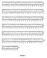
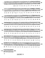
- the DNA and amino acid sequences of the BAT-1 V ⁇ region is shown in FIG. 1 .
- the FRs of the BAT-1 kappa light chain variable region i.e.
- mice subgroup VI The reason for the selection of mouse subgroup VI was related to the canonical classes of the hypervariable loops of the BAT-1 V ⁇ region, as defined by Chothia and his co-workers ( Chothia et al., J. Mol. Biol. 196:901, 1987 ; Nature 34:877, 1989 ; J. Mol. Biol. 227:799, 1992 ; Tramontano et al., ibid ). According to Chothia, each of the CDRs: CDR1 (L1), CDR2 (L2) and CDR3 (L3), were canonical class 1 ( FIG. 2 ). Crucially, the 10 amino acid canonical class 1 L1 hypervariable loop was only seen in mouse V ⁇ regions which fitted Kabat subgroup VI.
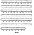
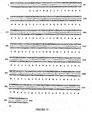
- the DNA and amino acid sequences of the BAT-1 V H region is shown in FIG. 3 .
- An analysis similar to that given in Example 1 was conducted for the BAT-1 V H region which determined that it exhibited the closest match to the consensus sequence of the mouse heavy chain miscellaneous subgroup in the Kabat database (Kabat et al., ibid).
- the first step in the design of the humanized variable regions of the BAT-1 antibody was the selection of the human kappa light chain variable region that would serve as the basis of the humanized BAT-1 V K region.
- the BAT-1 V K region was initially compared to the consensus sequences of the four human kappa light chain variable region subgroups as defined by Kabat and his coworkers (Kabat et al., ibid).
- the mouse BAT-1 light chain variable region was most similar to the consensus sequences of human kappa light chain subgroup I and human kappa light chain subgroup III.
- human kappa light chain subgroup I the mouse BAT-1 V K region displayed a 63.21% identity over the whole variable region and a 70.00% identity within the FRs alone. When measured with respect to similarity, these values increased to 71.70% overall and 80.00% within the FRs alone.
- human kappa light chain subgroup III the mouse BAT-1 V K region displayed a 65.09% identity over the whole variable region and a 68.75% identity within the FRs alone. When measured with respect to similarity, these values increased to 74.53% overall and 80.00% within the FRs alone. Consequently, it generally appeared to match well a broad range of human kappa light chain variable region sequences, however, with respect to FRs in particular, it was marginally more identical to those found within human kappa light chain subgroup I.
- the mouse BAT-1 V ⁇ region was then compared to all the recorded examples of individual sequences of human variable regions publicly available. Table 3 shows the best fifteen matches to the mouse BAT-1 V K region which were identified through this analysis. Overall, the search algorithm selected the human V ⁇ region from antibody TEL9 ( Marks et al., J. Mol. Biol. 222:581, 1991 ) as the closest match to the mouse BAT-1 V ⁇ region (Table 4). This human sequence had an overall identity to the BAT-1 V ⁇ region of 67.93% overall and 72.50% within the FRs alone. When measured with respect to similarity, these values increased to 77.36% overall and 82.50% within the FRs alone.
- the TEL9 kappa light chain variable region FR was selected as the human acceptor sequence for the humanization of the BAT-1 antibody kappa light chain variable region. This then became the basis of the first humanized version of the BAT-1 kappa light chain (BATR ⁇ A ), which essentially comprised the CDRs of the BAT-1 V ⁇ region and the FRs of the TEL9 V ⁇ region.
- the next step in the design process was to study the amino acid sequences of the human acceptor TEL9 V K region FRs to determine if any of these amino acid residues were likely to adversely influence binding to antigen, either directly through interactions with antigen, or indirectly by altering the conformation or orientation of the CDR loops. This was a difficult process which was only made possible through the availability of a model of the BAT-1 variable regions i.e. both the V ⁇ and V H regions. The modeling procedure will be given in detail in Example 5. Nevertheless, any amino acid in the mouse BAT-1 FRs which did appear to affect antigen binding were then considered for conservation in the humanized BAT-1 antibody.
- V ⁇ region amino acids located at the V ⁇ /V H interface as defined by Chothia and colleagues ( Chothia et al., J. Mol. Biol. 186:651, 1985 ), were checked for unusual or rare residues. From this analysis, the only residue position that raised any level of concern was the Phe at position 36 (Phe36) in FR2. Tyr (as found in TEL9) was normally seen at this position, however, in mBAT-1 Phe was present. In addition, position 36 was a recognized position for a Vernier amino acid (Foote et al., ibid). Vernier residues were thought to be important for maintaining CDR loop conformation.
- the third FR change introduced into BATR ⁇ B was located at position 71, which as well as being identified as a Vernier residue position (Foote et al., ibid), was also recognized as being one of the important canonical residue positions for the L1 loop structure.
- These canonical residues were defined by Chothia and his co-workers (Chothia et al., 1987, 1989, 1992 ibid; Tramontano et al., ibid) as being vital for the conservation of the CDR loop structure.
- Many of the canonical amino acids were located within the CDRs, however, a number (such as 71Tyr) were also positioned within the FRs. Although the amino acid change was conservative, the Phe71Tyr change was considered critical for the successful humanization of the BAT-1 kappa light chain.
- the first step in the design of the humanized V H region of the mouse BAT-1 antibody was the selection of the acceptor human heavy chain variable region that would serve as the basis of the humanized BAT-1 V H region.
- the mBAT-1 V H region was initially compared to the consensus sequences of the three human heavy chain variable region subgroups it was found to be most similar to the consensus sequence for human heavy chain subgroup I with a 61.54% identity overall and a 67.82% identity between the FRs alone. When measured with respect to similarity, these values also increased to 70.09% overall and 77.01% within the FRs alone.
- mice BAT-1 V H region was then compared to all the recorded examples of individual sequences of human variable regions publicly available. Tables 6 and 7 show the best fifteen matches to the mouse BAT-1 V H region which were identified through this analysis. Overall, the search algorithm selected the human V H region from antibody hsighv1295 ( Fang et al., J. Exp. Med. 179:1445, 1994 ) as the closest match to the mouse BAT-1 V H region. This human V H region had an overall identity to the BAT-1 V H region of 69.23% (Table 7), a value which increased to 74.71% when the FRs alone were compared. When measured with respect to similarity, these values increased to 75.21% overall and 79.31 % within the FRs alone.
- the next step in the design process was to study the amino acid sequences of the human acceptor hsighv1295 V H region FRs to determine if any of these amino acid residues were likely to adversely influence binding to antigen.
- the molecular models built by OML were crucial to this design process, from which a number of amino acids in the murine BAT-1 V H region FRs were identified for conservation in the first (BATRH A ) and subsequent versions of the humanized BAT-1 antibody (Table 8). There were 22 amino acid differences between the FRs of the donor mouse BAT-1 and the acceptor human hsighv1295 V H regions and up to nine murine residues were considered for conservation in the humanized FRs.
- BATRH A therefore consisted of the CDRs of the mouse BAT-1 antibody V H region genetically inserted into the FRs of the human hsighv1295 antibody V H region. This was the CDR-grafted version of the V H region of the humanized BAT-1 antibody and contained no FR amino acid changes whatsoever.
- residue positions 27-30 were considered part of the H1 loop itself and so were even more critical to the correct conformation and orientation of this loop - justifying their conservation even more strongly.
- residue positions 27-30 represented the sum of the changes made to the FRs of the human hsighv1295 sequence in BATRH B .
- the next step in the design process was to study the amino acid sequences of the human acceptor hsighv1295 V H region FRs to determine if any of these amino acid residues were likely to adversely influence binding to antigen.
- the molecular models built by OML were crucial to this design process, from which a number of amino acids in the murine BAT-1 V H region FRs were identified for conservation in the first (BATRH A ) and subsequent versions of the humanized BAT-1 antibody (Table 8). There were 22 amino acid differences between the FRs of the donor mouse BAT-1 and the acceptor human hsighv1295 V H regions and up to nine murine residues were considered for conservation in the humanized FRs.
- BATRH A therefore consisted of the CDRs of the mouse BAT-1 antibody V H region genetically inserted into the FRs of the human hsighv1295 antibody V H region. This was the CDR-grafted version of the V H region of the humanized BAT-1 antibody and contained no FR amino acid changes whatsoever.
- residue positions 27-30 were considered part of the H1 loop itself and so were even more critical to the correct conformation and orientation of this loop - justifying their conservation even more strongly.
- these two residue positions represented the sum of the changes made to the FRs of the human hsighv1295 sequence in BATRH B .
- the third version of the humanized BAT-1 V H region (BATRH C ) incorporated all the substitutions made in BATRH B and, in addition, contained a further three murine amino acids, which were inserted into the human FRs in place of the corresponding human residues.
- the first of these was the Asn amino acid located at position 76 in FR3.
- the Asn residue was close to CDR H1 and may have been supporting the loop structure.
- the Asn was surface exposed and larger than the Ser in the human hsighv1295 FRs. Consequently, a Ser76Asn substitution was made to the FR.
- This residue was considered a packing residue, as defined by Chothia (Chothia et al., 1985 ibid), important for the correct packing of the V ⁇ and V H regions.
- this was identified as a Vernier residue position, and therefore important for maintaining CDR loop conformation, a classification confirmed by an analysis of the molecular model. Taken together, all the data and molecular analysis suggested that it was appropriate to conserve these three murine residues in the humanized V H region of BATRH C , i.e. Ser76Asn, Ala93Val and Lys94Arg.
- Version D of the humanized BAT-1 V H region (BATRH D ) incorporated all the substitutions made in BATRH C and, in addition, contained one further mouse amino acid located at position 2 in FR1. This location was defined as both a canonical (Martin et al, ibid) and Vernier (Foote et al., ibid) residue position.
- the murine Ile amino acid was close to Tyr27 in FR1, which is itself part of the H1 loop structure.
- the murine Ile and human Val amino acids, at this location in the mouse and human FRs were similar in character and only slightly different in size, i.e. Ile has an extra methyl group. Therefore, it was decided to make the Val2Ile change only at this stage of the humanization procedure and incorporate the mutation into version BATRH D .
- BATRH E humanized BAT-1 heavy chain variable region
- the Arg38Lys modification was made because the model suggested that the Arg, deeply buried in the core of the V H region, was close to Phe63 in CDR H2. However, this was not a previously identified canonical or Vernier residue position. In addition, Arg and Lys are relatively similar in structure, although Arg is bulkier, and so the significance of any amino acid change was hard to judge. Consequently, this was considered as only a tentative possibility and the substitution was only going to be made if the binding affinity of the humanized BAT-1 antibody was found to be poor. The same rationale was also behind the selection of the Gln46Lys modification. The Lys amino acid was half-buried, according to the molecular model, but close to Glu62 and Phe63 in CDR H2.
- the FRs of the BAT-1 variable regions were modeled on FRs from similar, structurally solved immunoglobulin variable regions. While identical amino acid side-chains were kept in their original orientation, mismatched side-chains were substituted as in the original BAT-1 Fv region.
- the backbone atoms of the FAB17-IA V ⁇ region were used for the model of the BAT-1 V ⁇ region, while the FRs of the 409.5.3 V H region were used to model the BAT-1 V H region (Brookhaven PDB codes 1for and 1iai, respectively).
- FR backbone homology is an important factor in the quality of any model, since the use of FR structures that poorly match a sequence being modeled can significantly and adversely affect the position and orientation of the CDR loop structure.
- the loop conformations of the murine BAT-1 V ⁇ region and the humanized BATR ⁇ B sequence were taken from canonical classes used by AbM. These canonical classes are based on those described by Chothia and his colleagues, but they have been modified to take into consideration structures that have become available since the original articles were published (Chothia et al., 1987, 1989, 1992 ibid; Tramontano et al., ibid). Testing the performance of AbM predictions for known loop structures has shown that CDR loops which are created in this way are usually modeled very accurately, i.e. to within 1-1.5 ⁇ RMS deviation.
- the H3 loop in the BAT-1 V H region was eight residues long, so two methods were used for predicting the H3 loop structure.
- a database search for the backbone conformations was used for both methods, but in addition, the conformation of the central five residues in the model were searched more thoroughly using a CONGEN search (Bruccoleri, ibid). Although this took longer to compute, it reassuringly produced a conformation which was very similar to those identified from the database search.
- Putative positive transformants were identified using the PCR-screening assay, restriction digest and then ds-DNA sequenced.
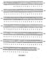
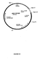
- the humanized V ⁇ genes ( FIGS. 7-9 ; SEQ ID NOS. 15, 16 and 18) were then subcloned into expression plasmids.
- the light chain pKN110 construct included Ampicillin and Neomycin resistance genes.
- the humanized V ⁇ gene variants of BAT-1 i.e. BATR ⁇ A , BATR ⁇ B and BATR ⁇ D
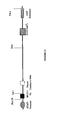
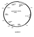
- BATR ⁇ A , BATR ⁇ B and BATR ⁇ D were inserted between the HCMV Immediate Early Promoter and the genomic human kappa constant region resulting in the following expression vectors: pKN110-BATR ⁇ A , pKN110-BATR ⁇ B and pKN110-BATR ⁇ D , respectively (see FIG. 10 for a representative pKN110-BATR ⁇ D vector).
- the BAT-1 light chain expression cassette inserted into an expression vector included a DNA fragment encoding a mouse immunoglobulin signal peptide sequence, Kozak sequence and a signal sequence intron which was added to both sides of the humanized V ⁇ gene variants of BAT-1 ( FIG. 11 ). This cassette was inserted between the HCMV Immediate Early Promoter and the genomic human kappa constant region.
- the complete light chain expression vector also included a BGH polyA transcription terminator and a Neo/G418 selection marker. All constructs were restriction enzyme digested and ds-DNA sequenced to confirm the presence of the correct insert.
- BATRH A The construction of the various versions of the reshaped human BAT-1 heavy chain variable region genes (i.e. BATRH A , BATRH B , BATRH C ) produced an approximately 450 bp product which was then subcloned into pCR2.1TM.
- the PCR reactions were set up using the primers described in Tables 11 and 12.
- Putative positive transformants were again identified in a PCR screen and then ds-DNA sequenced.
- the humanized V H genes (SEQ ID NOS. 20-22) were then subcloned into expression vectors.
- the heavy chain pG1D110 construct included Ampicillin resistance gene and the hamster dhfr as the selectable marker.
- the humanized V H gene variants of BAT-1 were inserted between the HCMV Immediate Early Promoter and the genomic human IgG1 constant region resulting in the following expression vectors: pG1D110-BATRH A , pG1D110-BATRH B , pG1D110-BATRH C (see FIG. 15 for a representative pG1D110.BAT-1.RH C vector).
- the BAT-1 heavy chain expression cassette inserted into an expression vector which included a DNA fragment encoding a mouse immunoglobulin signal peptide sequence, Kozak sequence and a signal sequence intron which was added to both sides of the humanized V ⁇ gene variants of BAT-1 ( FIG. 16 ). This cassette was inserted between the HCMV Immediate Early Promoter and the genomic human IgG1 constant region.
- the complete light chain expression vector also included a BGH polyA transcription terminator and a dhfr selection marker.
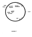
- pG1D200 is another ⁇ 1 immunoglobulin heavy chain mammalian expression vector (AERES Biomedical ; FIG. 17 ). This vector is a V H :C H ⁇ 1 intron minus version of the pG1D110 vector (i.e. it does not have the 71bp intron at the V H :C H junction).
- a Bst EII fragment (219bp) was excised from the pG1D200 vector and gel purified using a Qiagen gel extraction/purification kit. This fragment contained the intron minus V H :C H junction.
- the pG1D110.BAT-1.RH C construct ( FIG. 15 ) was also restriction digested with Bst EII, releasing a 290bp fragment which contained the intron plus VH:CH junction.
- the remaining vector fragment ( ⁇ 7207bp) was gel purified using a Qiagen gel extraction/ purification kit.
- the intron minus Bst EII fragment (219bp) from the pG1D200 vector digest was then ligated into the ⁇ 7207bp Bst EII digested pG1D110.BAT-1.RH C vector. 2 ⁇ l of ligated DNA was transformed into DH5 ⁇ cells (Stratagene) according to the manufacturers instructions. Plasmid DNA was prepared from 10 colonies and each plasmid DNA was analyzed for the presence of the correct Bst EII fragment by DNA sequence analysis.
- the new intron minus construct (pG1D210.BAT-1.RH C ) and the light chain construct pKN110.BAT.R ⁇ D (see FIG. 10 ) were used to construct the pG1KD210.BAT-1.RH C /R ⁇ D single expression vector (SEQ ID NO. 93).
- the BATRH C heavy chain variable region was transferred to the combined (single) expression vector as an Xho 1 to Hind 111 fragment.
- the BATR ⁇ D light chain variable region was transferred to the combined (single) expression vector as an Xba 1 to Bam H1 fragment.
- the internal Xba 1 site in the light chain gene was removed without changing the amino acid sequence.
- the sequences of the BAT-1.R ⁇ D /BAT-1.RH C heavy and light chain variable regions in this vector were confirmed.
- the vector includes genomic human IgG1 and Kappa constant regions. Both heavy and light chain genes were placed under the control of the HCMV Immediate Early promoter.
- the vector includes a mouse dhfr gene as the selectable marker (see FIG. 19 ).
- the same Kozak sequence, signal peptide sequence and intron were added as for the two vector expression system (see Examples 6 and 7).
- the first step in the construction of the BAT-1 ⁇ 4 single expression vector construct was the cloning of the modified BAT-1.RH C gene out of the pG1D110.BAT-1.RH C construct ( FIG. 14 ) by Bam HI and Hind III restriction digest, and ligation of this 430bp fragment into the gamma-4 immunoglobulin heavy chain expression vector pG4D110, again via Bam HI and Hind III restriction sites.
- Plasmid DNA was prepared from 10 colonies and each plasmid DNA was analyzed for the presence of the correct BAT-1.RH C Bam HI/ Hind III fragment by DNA sequence analysis.
- the new gamma-4 construct (pG4D110.BAT-1.RH C ) and the light chain construct pKN110.BAT-1.RK D ( FIG. 10 ) were used to construct the pG4KD110.BAT-1.HR C /R ⁇ D single expression vector in the following way.
- the BAT-1 kappa light chain expression cassette which contained the HCMVi promoter, the BAT-1 kappa light chain variable region gene, and the kappa light chain constant region gene, was restriction enzyme digested ( Eco RI/ Spe I) out of the pKN110.BAT-1.R ⁇ D construct and subsequently ligated into pG4D110.BAT-1.RH C construct via the unique EcoRI and Spe I restriction sites. This ligation resulted in the construction of a single expression vector construct pG4KD110.BAT-1.RH C /R ⁇ D , containing both the heavy and kappa light chains of the BAT-1 humanized antibody RH C /R ⁇ D variant.
- the humanized BAT-1 heavy (pG1D110) and light (pKN110; Example 7) chain expression vectors were co-transfected, at various combinations, into COS7 cells and after 72 hr incubation, the medium was collected, spun to remove cell debris, filtered and analyzed by ELISA for humanized antibody production.
- the concentration of humanized antibody in the COS7 cell supernatants varied with each combination of reshaped human BAT-1 antibody constructs that were tested (Table 13). For example, version BATRH B /BATR ⁇ A expressed the highest antibody levels (4800 ng/ml) whilst the BATRH B BATR ⁇ D version was the poorest expresser (357 ng/ml).
- the Immunopure ⁇ (A) IgG purification kit essentially comprised of a 2 ml column of immobilized Protein A Sepharose column. The antibody was eluted from the column with 5 ml of elution buffer, the eluate of which was collected in 1 ml fractions. The concentration of humanized BAT-1 antibody in each fraction was then assayed using ELISA methods. Table 13 describes the final concentrations of the Protein A purified antibody constructs collected. On average the purification step increased the antibody concentration by approximately 150-fold.
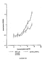
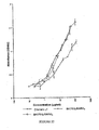
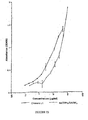
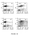
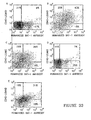
- FIGS. 20-23 show typical examples for these binding experiments. Sigmoidal dose-response curves of Daudi cell binding by the recombinant antibodies were also plotted and the hill slopes of these binding curves were calculated. The combination of the hill slope data and the positions of the dose-response curves relative to the chimeric antibody dose-response curves suggested a qualitative hierarchy with respect to Daudi cell binding among the various humanized BAT-1 antibody constructs tested (Table 14). At the top of this hierarchy was clearly construct BATRH C /BATR ⁇ D , which exhibited a hill slope (i.e.
- construct BATRH A /BATR ⁇ A clearly has the poorest binding characteristics of all the humanized BAT-1 antibody constructs tested (Table 14) and so was ranked sixth in the binding hierarchy.
- the calculated hill slope for this version i.e. 1.2730 ⁇ 0.2688
- BATRH B /BATR ⁇ A i.e. 1.7710 ⁇ 0.6461
- this difference is again not statistically significant.
- FIG. 21 it is clear from FIG. 21 that the CDR-grafted BATRH A /BATR ⁇ A BAT-1 antibody is reaching its maximum binding response at much lower level than the humanized construct BATRH B /BATR ⁇ A - which was ranked fifth in the binding hierarchy.
- BATRH B /BATR ⁇ B ( FIG. 20 ; ranked fourth) and BATRH B /BATR ⁇ D ( FIG. 23 ; ranked third) display intermediate levels of binding between these two sets of extremes. Again these rankings were mainly based upon a subjective interpretation of the binding data available and previous experience.
- Kettleborough Kettleborough et al., Eur. J. Immunol. 23:206, 1993
- DNA 10 ⁇ g each of the kappa light chain expression construct pKN110.BAT-1.R ⁇ D and the heavy chain expression construct pG1D210.BAT-1.RH C , or 13 ⁇ g of the single vector construct pG1KD210.BAT-1.RH C /R ⁇ D
- the electroporated cells were transferred to petri-dishes containing 8 ml of DMEM containing 10% FCS and incubated for 72 hrs in 5%CO 2 at 37°C. After 72 hrs incubation, the medium was collected, spun to remove cell debris, and analyzed by capture ELISA for antibody production.
- the co-transfections, with light chain expression vector and heavy chain expression vector, and transfections with a single-vector expressing both light and heavy chains, were carried out in triplicate. The results are presented in Table 15. The results indicate that expression levels from the single vector are -6 fold higher than the expression levels observed for the co-transfections. TABLE 15 Transfection no.
- CHOdhfr- cells were propagated in a non-selective media consisting of ⁇ -MEM with ribonucleosides and deoxyribonucleosides, supplemented with 10% Fetal Clone II and 50 ⁇ g/ml Gentamicin. Aliquot, 0.7 ml, of 10 7 cells/ml in PBS was transfected with 13 ⁇ g of pG1.KD210.BAT-1.RH C /R ⁇ D at 1900 V, 25 ⁇ F capacitance using a Bio-Rad Gene Pulser. The cells were allowed to recover for 10 minutes at RT before being transferred to 10 cm petri-dishes in 8 ml of non-selective media and then incubated in 5% CO 2 at 37°C for 48 hours.
- the cells were trypsinized, spun down and resuspended in 150 ml of prewarmed selective media ( ⁇ -MEM without ribonucleosides and deoxyribonucleosides, supplemented with 10% dialyzed FBS and 50 ⁇ g/ml Gentamicin, and containing either 10nM, 50nM, 100nM or 500nM Methotrxate) before being divided equally between fifteen 10cm petri-dishes. These were then incubated in 5% CO 2 at 37°C for 20-30 days, the selective media being changed every 3-4 days until foci were clearly visible. After 2 weeks from the initial transfection, foci began to develop on the 10nM plates. Eight days later, one focus developed on the 50nM plates. No other foci developed after 35 days, on the 50nM plates and no foci developed on the 100nM or 500nM plates.
- the method of Kettleborough et al. was followed to transfect the mammalian expression constructs into COS cells. Briefly, the DNA (10 ⁇ g each of the kappa light chain expression construct pKN110.BAT-1.R ⁇ D and the heavy chain expression construct pG4D110.BAT-1.RH C , or 13 ⁇ g of the supervector construct pG4D110.BAT-1.RH C /R ⁇ D ) was added to a 0.7 ml aliquot of 10 7 cells/ml in PBS and pulsed at 1900 V, 25 ⁇ F capacitance using a Bio-Rad Gene Pulser apparatus.
- the electroporated cells were transferred to petri-dishes containing 8 ml of DMEM containing 10% FCS and incubated for 72 hrs in 5%CO 2 at 37°C. After 72 hrs incubation, the medium was collected, spun to remove cell debris, and analyzed by capture ELISA for antibody production.
- CHOdhfr- cells were propagated in a non-selective media consisting of ⁇ -MEM with ribonucleosides and deoxyribonucleosides, supplemented with 10% Fetal Clone II and 50 ⁇ g/ml Gentamicin. Aliquot, 0.7 ml, of 10 7 cells/ml in PBS was transfected with 13 ⁇ g of pG4KD110.BAT-1.RH C /R ⁇ D at 1900 V, 25 ⁇ F capacitance using a Bio-Rad Gene Pulser.
- the cells were allowed to recover for 10 minutes at room temperature before being transferred to 10cm petri-dishes in 8 ml of non-selective media and then incubated in 5% CO 2 at 37°C for 48 hours. Two days following this incubation, the cells were trypsinized, spun down and resuspended in 150 ml of prewarmed selective media ( ⁇ -MEM without ribonucleosides and deoxyribonucleosides, supplemented with 10% dialyzed FBS and 50 ⁇ g/ml Gentamicin, and containing either 10nM 50nM, 100nM or 500nM Methotrexate) before being divided equally between fifteen 10cm petri-dishes. These were then incubated in 5% CO 2 at 37°C for 20-30 days, the selective media being changed every 3-4 days until foci were clearly visible.
- Transfected cells were distributed into 10 96-well plates in Dulbecco's Modified Eagles medium (DMEM) supplemented with 10% Foetal Bovine Serum (FBS) and 1 mg/ml G418 (Gentamicin) medium. After 10 to 14 days when colonies of transfected cells have developed, samples of conditioned medium from the wells were assayed for humanized BAT-1 antibody. Cells from the highest producing wells were picked, and expanded in medium including G418.
- DMEM Dulbecco's Modified Eagles medium
- FBS Foetal Bovine Serum
- G418 Genetamicin
- the transfection was repeated after one week as a back up and to provide more transfected cell clones for selection. After 10 days visible colonies of transfected cells had developed, and conditioned medium from the wells was screened for antibody production.
- ELISA plates were coated with sheep anti-human ⁇ antibody. 25 ⁇ l samples of medium from the wells were transferred to the ELISA plate and diluted to 100 ⁇ l in PBS Tween (PBST).
- the secondary antibody was HRP-conjugated sheep anti-human IgG ( ⁇ chain specific) and color was developed with o-Phenylene Diamine (OPD).
- the combined (single) antibody expression vector described in Example 9 was transfected into the NS0 host cell line by electroporation.
- Transfected cells were distributed into 10 96-well plates in DMEM with 10% FBS. After 2 days an equal amount of medium with 0.1 ⁇ M Methotrexate was added. Half the medium was changed with the same volume of 0.1 ⁇ M MTX-containing medium every 2 days until the 8 th day post transfection. The transfection was repeated after one week as a back up and to provide more transfected cell clones for selection. After 14-21 days visible colonies of transfected cells had developed, and conditioned medium from the wells was screened for antibody production as described in the above Example. Positive wells were examined microscopically and the cells from the highest producing wells were picked into 1.5 ml of DMEM supplemented with 10% FBS and 0.1 ⁇ M Methotrexate in 24 well plates.
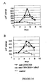
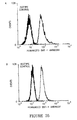
- Daudi cells were incubated with increasing amounts of the humanized BAT-1 or the mouse BAT-1 as control (0-80 ⁇ g/ml). Unbound antibody was discarded and biotinylated murine-BAT-1 (20 ⁇ g/ml) added to the cells and stained with streptavidin-FITC.
- Figure 24 depicts a decreased binding of murine BAT-1 in the presence of increasing concentrations of both the humanized and original mouse mAb, supporting the recognition of the same epitope as expected. Both antibodies show a similar dose dependency, with an IC 50 of approximately 10 ⁇ g/ml, suggesting a similar affinity of antigen binding.
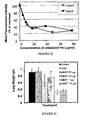
- C57BL mice were inoculated with B16 melanoma cells to induce lung metastases.
- Increasing amounts (1,10 and 20 ⁇ g) of humanized mAb were injected on day 12 post tumor-inoculation and compared to an optimal dose of 10 ⁇ g murine-BAT-1.
- Lung weight measured on Day 24 post tumor inoculation is depicted in FIG. 25 and corresponds to the establishment of a tumor.
- Both non-treated mice and mice treated with an isotype-matched irrelevant human IgG1 had an average lung weight of 0.9 gr.
- the humanized BAT-1 exhibited a dose dependent inhibition of metastases growth with the highest inhibition occurring at a low dose of 1 ⁇ g/mouse.
- Mouse-BAT-1 mAb has been shown to inhibit the formation of human-tumor metastases in the presence of human peripheral blood lymphocytes (hPBL).
- hPBL peripheral blood lymphocytes
- the humanized antibody was studied in a model combining both tumors and lymphocytes of human origin. Severe combined immune-deficient mice (SCID) were engrafted with hPBL to restore immune-competence. Mice were challenged with human melanoma cells (SK-28) and treated with increasing concentrations of the humanized antibody, administered in a single i.v. dose on day 11 post tumor inoculation.
- Fig 26 depicts lung weight that correlates with the number of metastases observed, as measured on day 23. Both concentrations of the humanized antibody induced tumor inhibition in the presence of hPBL. As observed in the mouse tumor model described above, the humanized antibody could more efficiently inhibit tumor growth in vivo, in comparison to mouse BAT-1. A single dose of 1 ⁇ g of this humanized antibody inhibited tumor growth by 68% showing a higher efficacy than 10 ⁇ g of the mouse BAT-1 antibody (30%).
- LIM6 and HM7 are two sub-clones of the human CRC cell line LS174T that were selected for their high mucin synthesis and metastatic potential.
- the tumor cells were injected into the exposed spleen of anesthetized nude mice. After 1 minute, the spleens were removed and the excisions closed. Low doses of murine and humanized BAT-1 antibody were administered 12 days later and mice were sacrificed 35 days post tumor inoculation. The livers were weighed, the number of metastatic nodules was counted, and liver tissue was processed for histology and Immunohistochemistry study.
- Mouse BAT-1 has been shown to bind human lymphocytes, recognizing both CD4+ and CD8+ subsets.
- human Peripheral Blood Lymphocytes PBL were isolated from the blood of normal donors, as described hereinbelow, and analyzed for co-localization of hBAT with known lymphocyte markers.
- PBMC Peripheral blood mononuclear cells
- Figure 28 depicts an example of binding to lymphocytes of a selected donor in which 50% of the isolated PBL were positive for hBAT, including both CD4+ cells (25%) and CD8+ cells (15%). Within these subpopulations, the majority of CD4+ as well as CD8+ cells bound the hBAT mAb (58% and 71% respectively).
- the humanized BATRH C /BATR ⁇ D ⁇ 1 mAb was raised against the membranes of Daudi cells, a human B lymphoma cell-line.
- PBL from normal donors were isolated by ficoll, as described above, followed by adherence to tissue culture plates.
- Non-adherent cells were examined for the co-localization of hBAT with B-cell markers including CD19 and CD20. Binding was performed at 4°C for 1 hr, and determined by flow cytometry on gated lymphocytes.
- Figure 29 depicts the evaluation of binding to the cells of a representative normal donor.
- lymphocytes in the sample were positive for the humanized BAT mAb. These cells included the majority of B cells (70-75%) as demonstrated by both independent markers. 70% of CD20+ were positive for the humanized BAT mAb (Gated on R1 and PI negative; Fig. 29A ) and 75% of CDR19+ were positive for the humanized BAT mAb (Gated on R1 and PI negative). The results suggest that the BAT-binding moiety on the cell surface could be common to peripheral B cells.
- Binding of the mouse BAT antibody has been formerly correlated with lymphocyte activation. This binding activity was further studied for the human mAb and the binding level of the human BAT mAB to human CD4+ T cells, subjected to activation, was examined. Cells were isolated from a normal donor by negative selection and stimulated with beads conjugated to anti-CD3 and anti-CD28 (5 ⁇ l/ml). This treatment was selected in order to exert polyclonal activation through the T-cell receptor and co-stimulatory molecules.
- T cell activation is characterized by cell-surface expression of various molecules, some of which have been shown to be involved in the activation process.
- hBAT was studied for its co-expression with different markers including both early and late activation molecules.
- CD69 an early activation marker, is up-regulated on T cells upon activation.
- Four days following activation cells were examined for binding of hBAT and anti-CD69 (4°C, 1 hr). Analysis was performed by flow cytometry on cells negative for PI staining. Quadrants were determined by isotype controls.
- FIG. 31 A dose-dependent activation of CD4+ T cells from a normal donor is demonstrated in figure 31 .
- Increased time of activation also resulted in increase binding to hBAT beginning at day one of activation.
- Time dependency of activation could also be demonstrated and resulted in an increase in hBAT binding beginning at day one of activation.
- hBAT binding to both CD4+ and CD8+ cells remained high even after CD69 decrease (day 5) suggesting a correlation of binding with multiple stages of lymphocyte activation.
- hBAT binding to CD69+ cells suggests that the expression of hBAT binding protein is correlated with early activation.
- CD25 the high-affinity receptor for IL2
- CD25 is vital for T-cell expansion and is typically increased on the surface of activated cells. Chronologically it follows the appearance of CD69 and its expression is extended several days after the down-regulation of CD69.
- CD4+ T cells were isolated from a normal donor by negative selection and stimulated for several days with beads conjugated to anti-CD3 and anti-CD28 (5 ⁇ l/ml). Cells were examined for binding of hBAT and anti-CD25 (4°C, 1 hr) on day 0 ( Fig. 32A ), day 1 ( Fig. 32B ), and day 5 ( Fig. 32D ) of activation with respect to controls (day 0, Fig. 32A and day 5 of no activation, Fig. 32C ). Analysis was performed by flow cytometry on cells negative for PI staining. Quadrants were determined by isotype controls.
- CD4+ and CD8+ T cells showed a time dependent increase in CD25 expression upon anti-CD3 and anti-CD28 stimulation, beginning at day 1 of stimulation.
- hBAT co-localized with CD25 on these activated cells ( Fig. 32 ).
- CD25 expression increased from 55% of the cells on day 1 ( Fig. 32B ) to 93% on day 5 ( Fig. 32D ) following activation. At both time points the majority of hBAT binding cells were CD25+ (85% and 98% respectively).
- hBAT increases survival of activated CD4+ cells
- hBAT human CD4+ cells were isolated from normal donors by negative selection and activated with a suboptimal concentration (0.25 ⁇ /ml) of anti-CD3/CD28 beads ( FIG. 34 ).
- hBAT 0.5 ⁇ g/ml was added 2 days following activation and its effect was evaluated by determining the number of viable cells.
- the results indicate that hBAT induced a significant increase in the number of viable CD4+ cells isolated from the two separate donors ( Fig. 34A and B ).
- Control nonstimulated cells died within eight days of isolation whereas activated cells expanded in a manner that is typical of lymphocytes, commencing with cell proliferation followed by a stage of stable cell number leading to a stage dominated by cell death.
- the addition of hBAT enhanced the expansion of CD4+ cell and increased the number of cells by 1.5 folds with respect to cells in the absence of the mAb.
- Mouse BAT-1 was raised against membranes of the Daudi B-cell line and has been shown to bind human T cells.
- hBAT was examined for its binding to two human cell lines of myeloid origin: Daudi cells - a human B cell lymphoma line and Jurkat cells - a human T cell leukemia line.
- hBAT conjugated to FITC was incubated with Daudi and Jurkat cells at a concentration of 150ug/ml (4°C for 1 hr). Binding was determined by flow cytometry.
- hBAT is capable of binding human T cells from normal donors.
- PBL were isolated from the blood of a prostate cancer patient by ficoll followed by adherence to tissue culture plates. Non-adherent cells were examined for binding of hBAT and lymphocyte markers. Binding was performed at 4°C for 1 hr, and determined by flow cytometry on gated lymphocytes. Isotype controls were used to determine the quadrants. These patients have formerly undergone therapy that often affects the presence and phenotype of lymphocytes. hBAT binding to these cells is a pre-requisite for its activity and as depicted in figure 36 , resembles the binding to lymphocytes of normal donors. Although total lymphocyte numbers were low, hBAT could still bind a large proportion of the lymphocyte subpopulations which we examined including 39% of CD4+ cells, 60% of CD8+ cells and 68% of B cells.
- the purpose of this study was to examine the cross reactivity of hBAT-1 monoclonal antibody with a range of normal human tissue. This study involved immunohistochemical testing of the monoclonal antibody against-a range of human tissues. A comparison of in vitro cross-reactivity in tissues from cynomologus monkey and CD-1 mice was also undertaken.
- the tissues used in this study were each obtained from three unrelated donors to minimize the chances of donor specific factors affecting antibody binding.
- the human tissue was provided by an ethical source.
- the primate and murine tissues used in this study were obtained from two animals of each species, by an ethical source.
- the murine and primate are potential test systems that may be evaluated in pre-clinical toxicology Studies.
- the tissues selected were those specified in the FDA Points to Consider in the Manufacture and Testing of Monoclonal Antibody Products for Human Use (Office of Biologies Research and Review. Center for Biologies Evaluation and Research FDA. 1997 ) and the Rules Governing Medicinal Products in the European Community Vol. 3a (Production and Quality Control of Monoclonal Antibodies Dec. 1994, 3AB4a).
- the humanized monoclonal hBAT-1 antibody was conjugated to FITC by the Custom Antibody Services Division of Serotec Ltd (ISO 9001, Certification) before the study was started.
- the final concentration of the conjugated antibody was 1.99 mg/ml.
- 3 refers to strong positive-staining
- 2 refers to positive staining
- 1 refers to weak positive staining
- 0 refers to no staining/signal.
- +++ refers to strong visual signal
- ++ refers to good visual signal
- + refers to weak visual signal.
- Negative control reactions in which the antibody was substituted with a buffer, were carried out for each tissue.
- Each detection reaction included positive control cells, Jurkat E6, reacted at the three predetermined dilutions of the antibody. This allowed the consistency of the reaction to be monitored. Sections of thyroid, incubated with anti actin antibody, were included in each assay run as controls for the detection system.
- Sections of each tissue were stained with Haematoxylin and Eosin (H&E) to confirm their identity and suitability for the study. Sections were also incubated with anti smooth muscle actin (SMA; Table 22) or rabbit anti human transferrin control sera, which showed the tissues were suitable for immunohistochemistry. Three sections of each of the tissues were prepared and incubated with the antibody, which had been conjugated to FITC, at concentrations of 1:100, 1:250 and 1:500 as determined during the validation phase. After washing in buffer and blocking with normal serum, the sections were incubated with the appropriate secondary and tertiary antibodies for alkaline-phosphatase detection, and counter-stained with haematoxylin before microscopical examination to determine sites of binding.
- H&E Haematoxylin and Eosin
- the FITC -conjugated staining method with Alkaline Phosphatase detection contained the following steps:
- Endogenous alkaline phosphatase was minimized by using Levamisole incorporated into the chromogen.
- tissues where endogenous alkaline phosphatase activity could not be suppressed human colon, ileum, placenta and endothelium, murine colon and pancreas, primate stomach, ileum and prostate
- horseradish peroxidase conjugated antibody at 1:200 for 2 hours was used, followed by Diaminobenzidene (DAB) reagent for 20 minutes.
- DAB Diaminobenzidene
Description
- The present invention relates to the field of immunotherapy and more specifically concerns humanized monoclonal antibodies useful for therapy of a variety of indications, particularly in the treatment of cancer.
- Cancer in its different forms is a major cause of death in humans. The most widely used therapeutic treatments of cancer are surgery, radiation and chemotherapy. The rapid increase of knowledge in recent years about the molecular and cellular bases of immune regulation, particularly at the level of T-cell responses, provides a new arsenal of immunotherapeutic approaches including the development of tumor vaccines. Certain monoclonal antibodies (mAbs) were shown to have immunomodulatory activity including the ability to bind determinants on the surface of T cells and to induce proliferation, activation or differentiation of these cells.
- Monoclonal antibodies derived from mouse hybridomas contain substantial stretches of amino acid sequences that are immunogenic when injected into a human patient, often eliminating the antibody's therapeutic efficacy after an initial treatment. While the production of so called "chimeric antibodies" (i.e., mouse variable regions joined to human constant regions) has proven somewhat successful, a significant immunogenicity impediment remains.
- Recombinant DNA technology has been utilized to produce immunoglobulins containing human framework regions (FRs) combined with complementarity determining regions (CDRs) from a donor mouse or rat immunoglobulin. These new proteins are called "reshaped" or "humanized" immunoglobulins and the process by which the donor immunoglobulin is converted into a human-like immunoglobulin by combining its CDRs with a human framework is called "humanization". Humanized antibodies are important because they bind to the same antigen as the original antibodies, but are less immunogenic when injected into humans.
-
US Patent No. 6,294,654 discloses a modified immunoglobulin molecule or functional fragment or part thereof (Ig), having an antigenic peptide foreign to the Ig incorporated in one or more non-CDR loops, and wherein the main outline of the constant domain framework is maintained. Further disclosed is the use of the modified antibody for therapeutic or prophylactic use. -
US Patent No. 6,074,635 discloses a method for antigen independent activation of T cells in vitro comprising contacting T cells in the absence of antigen with a combination of at least two cytokines selected from the group consisting of interleukin-2, interleukin-6, and tumor necrosis factor alpha, or functionally equivalent fragments thereof. -
US Patent No. 5,658,741 discloses a method of inducing the activation and proliferation of T-cells, said method comprising: (a) conjugating a plurality of T-cell specific monoclonal antibodies to an aminodextran molecule having 7-20% by weight amine groups and a molecular weight of at least 100,000 daltons, wherein the molar ratio of said antibodies to said aminodextran is greater than or equal to two; and (b) reacting said conjugate with a sample containing said T-cells to effect the binding of said conjugated antibodies to said T-cells to induce activation and proliferation of said T-cells. -
US Patent 5,585,089 of Queen et al. discloses a humanized immunoglobulin having complementarity determining regions (CDRs) from a donor immunoglobulin and heavy and light chain variable region frameworks from human acceptor immunoglobulin heavy and light chains, which humanized immunoglobulin specifically binds to an antigen with an affinity constant of at least 107 M-1 and no greater than about four-fold that of the donor immunoglobulin, wherein said humanized immunoglobulin comprises amino acids from the donor immunoglobulin framework outside the Kabat and Chothia CDRs, wherein the donor amino acids replace corresponding amino acids in the acceptor immunoglobulin heavy or light chain frameworks, and each of said donor amino acids: (I) is adjacent to a CDR in the donor immunoglobulin sequence, or (II) contains an atom within a distance of 4Å of a CDR in said humanized immunoglobulin. -
US Patent 5,225,539, of Winter , discloses an altered antibody or antigen-binding fragment thereof, wherein a variable domain of the antibody or antigen-binding fragment has the framework regions of a first immunoglobulin heavy or light chain variable domain and the complementarity determining regions of a second immunoglobulin heavy or light chain variable domain, wherein said second immunoglobulin heavy or light chain variable domain is different from said first immunoglobulin heavy or light chain variable domain in antigen binding specificity, antigen binding affinity, species, class or subclass. -
US 5,225,539 andUS Patent 5,585,089 do not provide sufficient tools and comprehensive description for carrying out the synthesis of an altered antibody, particularly a humanized antibody, by a person skilled in the art. -
US Patent No. 5,897,862 of one of the inventors of the present invention which is incorporated herein by reference, discloses a monoclonal antibody or an antigen binding fragment thereof, wherein the monoclonal antibody: (i) is secreted by the hybridoma cell line deposited at the Collection Nationale de Cultures de Microorganismes (CNCM), under Accession No. I-1397, or (ii) recognizes the same antigenic epitope as the antibody under (i). The monoclonal antibody disclosed inUS5,897,862 is directed against "Daudi" cells, a human B lymphoblastoid cell line, and was shown to stimulate murine lymphocytes and human peripheral blood T cells (Hardy et al, Cell Immunol. 118:22, 1989). This murine antibody is also termed mBAT-1 hereinafter. mBAT-1 also exhibits anti-tumor and immunostimulatory effects in various types of tumors (Hardy et al., Int. J. Oncol. 19:897, 2001) including tumors of human origin (Hardy et al., Proc. Natl. Acad. Sci. USA 94:5756, 1997). - International Patent Application
WO 00/58363 - Nowhere in the background art is it taught or suggested that a humanized monoclonal antibody comprising CDRs of a murine origin and FRs of a human origin may elicit an immune response and may further exhibit anti-cancer activity. Moreover, there is an unmet need for reliable methods for designing functional humanized antibodies, as it is well known in the art that the synthesis of the humanized antibody of the present invention cannot be predictably or routinely based on the background art.
- It is an object of the present invention to provide a humanized monoclonal immunomodulatory antibody, also termed hereinafter hBAT-1, which binds to B lymphoblastoid cells and induces proliferation and activation of peripheral blood lymphocytes. Said hBAT-1 is based on the previously known murine monoclonal immunomodulatory antibody, also termed herein mBAT-1, which binds to B lymphoblastoid cells and induces proliferation and activation of peripheral blood lymphocytes and further elicits an anti-tumor effect when injected into a tumor-bearing subject.
- The present disclosure provides a comprehensive description of the humanization process of mBAT-1 along with the rationale for each synthesis step. Thus, the description of the humanization process provided in the present invention is suitable for humanization of BAT antibodies other than mBAT-1, by a person skilled in the art.
- The administration of humanized BAT-1 antibody offers a method for therapeutic prevention, detection or treatment of cancer. Treatment of a subject in need thereof with the humanized form of the BAT-1 antibody, as provided by the present invention, is considerably more efficient than treatment with a chimeric BAT-1 antibody, and avoids adverse immunogenic responses.
- The present invention is based in part on the unexpected finding that the humanized BAT-1 antibody appears to induce a greater anti-tumor effect than that induced by the parent murine BAT-1 antibody.
KKKKKKKKKKKKK According to a first aspect, the present invention provides a humanized monoclonal antibody having at least one complementarity determining region (CDR) of murine monoclonal antibody BAT-1 (mBAT-1) and a framework region (FR) derived from an acceptor human immunoglobulin wherein the humanized antibody retains the anti-tumor activity of mBAT-1 monoclonal antibody and is less immunogenic in a human subject than said murine antibody. - According to one embodiment the humanized antibody induces a greater anti-tumor effect than that induced by the parent murine BAT-1 antibody.
- According to another embodiment the humanized antibody comprises the complementarity-determining regions (CDRs) of murine monoclonal antibody BAT-1 (mBAT-1), wherein the humanized antibody comprises:
- a. light chain variable regions of the formula: FRL1-CDRL1-FRL2-CORL2-FRL3-CDRL3-FRL4 wherein each FR is independently a framework region of a human antibody, and each CDR is a complementarity determining region, derived from BAT-1;
- b. the heavy chain variable regions of the formula: FRH1-CDRH1-FRH2-CDRH2-FRH3-CDRH3-FRH4 wherein each FR is separately a framework region of a human antibody, and each CDR is a complementarity determining region, form mBAT-1.
- According to a second aspect the present invention provides a humanized monoclonal antibody has a genetically modified Fab region comprising the complementarity-determining regions (CDRs) of murine monoclonal antibody BAT-1 (mBAT-1), wherein the genetically modified antibody retains the biological activity of said mBAT-1 and wherein said genetically modified monoclonal antibody comprises an amino acid sequence selected from:
- (i) a heavy chain variable region comprising the amino acid sequences of: CDRH1 (SEQ ID NO: 12); CDRH2 (SEQ ID NO: 13); CDRH3 (SEQ ID NO: 14); and a light chain variable region comprising the amino acid sequences of CDRL1 (SEQ ID NO: 9); CDRL2 (SEQ ID NO: 10); CDRL3 (SEQ ID NO: 11);
- (ii) heavy chain and light chain variable regions comprising the amino acid sequences having greater than about 80 percent similarity to all or part of the sequences of: CDRH1 (SEQ ID NO: 12); CDRH2 (SEQ ID NO: 13); CDRH3 (SEQ ID NO: 14); CDRL1 (SEQ ID NO: 9); CDRL2 (SEQ ID NO: 10); CDRL3 (SEQ ID NO: 11);
- (iii) an antibody of (i) or (ii) in which one or more amino acid residues have been added, deleted, replaced or chemically modified without substantially affecting the biological activity or binding specificity of the antibody.
- According to one embodiment the framework regions (FRs) are derived from a human antibody.
- According to another embodiment the humanized monoclonal antibody comprises complementarity-determining regions (CDRs) of murine monoclonal antibody BAT-1 (mBAT-1), wherein said humanized antibody comprises:
- a. light chain variable regions of the formula: FRL1-CDRL1-FRL2-CDRL2-FRL3-CDRL3-FRL4 wherein each FR is independently a framework region of a human antibody, and each CDR is a complementarity determining region, wherein the amino acid sequence of CDRL1 is SARSSVSYMH (SEQ ID NO: 9); CDRL2 is RTSNLAS (SEQ ID NO: 10); CDRL3 is QQRSSFPLT (SEQ ID NO: 11);
- b. the heavy chain variable regions of the formula: FRH1-CDRH1-FRH2-CDRH2-FRH3-CDRH3-FRH4 wherein each FR is separately a framework region of a human antibody, and each CDR is a complementarity determining region, wherein the amino acid sequence of: CDRH1 is NYGMN (SEQ ID NO: 12); CDRH2 is WINTDSGESTYAEEFKG (SEQ ID NO: 13); CDRH3 is VGYDALDY (SEQ ID NO: 14).
- According to yet another embodiment said humanized antibody induces a greater antitumor effect than murine BAT-1 monoclonal antibody; or said humanized antibody induces a greater anti-metastatic effect than murine BAT-1 monoclonal antibody; or the FR of the heavy chain variable region are derived from the FRs of the heavy chain variable region of the human hsighv1295 antibody; or the FRs of the kappa light chain variable region are based on the FRs of the kappa light chain variable region of the human TEL9 antibody; or it has a human kappa constant region; or the antibody is further labeled with a detectable label, immobilized on a solid phase, or conjugated to a heterologous compound; or the humanized monoclonal antibody light chain variable regions are selected from the group consisting of: BATRκA (SEQ ID NO: 15), BATRκB (SEQ ID NO: 16), BATRκC (SEQ ID NO: 17), BATRκD (SEQ ID NO: 18), and the heavy chain variable regions are selected from the group consisting of: BATRHA (SEQ ID NO: 20), BATRHB (SEQ ID NO: 21), BATRHC (SEQ ID NO: 22), BATRHD (SEQ ID NO: 23) or BATRHE (SEQ ID NO: 24); or the humanized monoclonal antibody variable regions are selected from the group consisting of: BATRHA/BATRκA (SEQ ID NO: 20/SEQ ID NO: 15), BATRHB/ BATRκA (SEQ ID NO: 21/SEQ ID NO: 15), BATRHB/ BATRκB (SEQ ID NO: 21/SEQ ID NO: 16), BATRHC/ BATRκB (SEQ ID NO: 22/SEQ ID NO: 16), BATRHB/ BATRκD (SEQ ID NO: 21/SEQ ID NO: 18), or BATRHC/ BATRκD (SEQ ID NO: 22/SEQ ID NO: 18).
- According to yet another embodiment the antibody is a full length antibody.
- According to yet another embodiment the antibody is of isotype IgG.
- According to yet another embodiment said isotype subclass is selected from IgG1 or IgG4.
- According to a third aspect, the present invention provides an antibody fragment derived from the humanized antibody as indicated above, wherein the antibody fragment is selected from the group consisting of: Fv, F(ab'), F(ab')2, a single chain antibody.
- According to one embodiment the humanized antibody is generated by recombinant DNA technology, utilizing CDR grafting.
- According to a fourth aspect the present invention provides an isolated polynucleotide construct encoding any of the monoclonal antibodies as mentioned above or fragments thereof.
- According to one embodiment the isolated polynucleotide construct encodes a kappa light chain variable region selected from the group consisting of: SEQ ID NO: 15, SEQ ID NO: 16, SEQ ID NO: 17, SEQ ID NO: 18.
- According to another embodiment the isolated polynucleotide construct is selected from the group consisting of: SEQ ID NO: 87, SEQ ID NO: 88, SEQ ID NO: 89.
- According to yet another embodiment the isolated polynucleotide construct encodes a heavy chain variable region selected from the group consisting of: SEQ ID NO: 20, SEQ ID NO: 21, SEQ ID NO: 22, SEQ ID NO: 23, SEQ ID NO: 24.
- According to yet another embodiment the isolated polynucleotide construct is selected from the group consisting of: SEQ ID NO: 90, SEQ ID NO: 91, SEQ ID NO: 92.
- According to a fifth aspect, the present invention provides a vector comprising any of the above polynucleotides.
- According to one embodiment the vector further comprises at least one polynucleotide sequence encoding a component selected from the group consisting of: a promoter operatively linked to the polynucleotide encoding the antibody, one or more resistance gene, a Kozak sequence, an origin of replication, one or more selection marker genes, an enhancer element, transcription terminator, a signal peptide, genomic human kappa constant region, genomic human IgG constant region.
- According to another embodiment the vector is a plasmid or a virus. Preferably, the vector is selected from the group comprising: pKN110, pG1D200, pG1KD210, pUC or pBR322.
- According to yet another embodiment the vector comprises the polynucleotide sequence of SEQ ID NO: 93.
- According to a sixth aspect, the present invention provides a host cell comprising the above vector.
- According to one embodiment the host cell is capable of expressing an antibody or fragments thereof; or is selected from eukaryotic and prokaryotic; or is selected from the group consisting of: CHO, CHOdhfr, NSO, COS or COS7 cells.
- According to a seventh aspect, the present invention provides a pharmaceutical composition comprising as an active ingredient the above antibody or antibody fragments thereof.
- According to one embodiment the pharmaceutical composition further comprises a physiologically acceptable carrier, diluent, or stabilizer.
- According to an eighth aspect, the present invention provides the use of the above antibody or antibody fragments for the preparation of a medicament for the treatment of cancer.
- According to one embodiment the cancer is selected from melanoma, lung tumors, colorectal cancer or hepatic metastasis.
- According to a ninth aspect, the present invention provides the use of the above humanized monoclonal antibody or antibody fragments for the preparation of a pharmaceutical composition comprising an effective amount of said antibody or antibody fragments for inducing proliferative, cytolytic or stimulatory activity of CD4+ T cells.
- According to a tenth aspect, the present invention provides the use of the above humanized monoclonal antibody or antibody fragments for the preparation of a pharmaceutical composition comprising an effective amount of said antibody or antibody fragments for increasing the survival of activated CD4+ T cells.
- According to one embodiment the pharmaceutical composition is directed to the treatment of a genetic or an acquired immune deficiency.
- According to another embodiment the pharmaceutical composition is directed to the treatment of the early stages of HIV infection.
- According to yet another embodiment the pharmaceutical composition is directed to the treatment of AIDS (Acquired Immune Deficiency Syndrome).
- According to yet another embodiment the pharmaceutical composition is directed to the treatment of patients having a blood count that shows a decrease in CD4+ T cells.
- According to an eleventh aspect, the present invention provides a method for producing the above antibody. The method comprises the steps of (i) transfecting a host cell with a vector comprising a polynucleotide sequence encoding said antibody, or co-transfecting the host cell with 2 vectors each comprising a polynucleotide sequence encoding the heavy or light chain regions of said antibody; (ii) culturing the host cell of (i) so that said antibody is expressed; and (iii) recovering the antibody from the host.Other objects, features and advantages of the present invention will become apparent from the following detailed description and appended claims.
-
-
FIGURE1 shows the DNA and peptide sequences of the kappa light chain variable region (Vκ) of the murine BAT-1 antibody. -
FIGURE 2 depicts the canonical classes of CDRs in the murine BAT-1 Vκ region. "Chothia Canonical Classes" indicates where the canonical classes as defined by Chothia and his colleagues (Chothia et al., 1987, 1989, 1992 ibid; Tramontano et al., J. Mol. Biol. 215:175, 1990) were used while "Martin Canonical Classes" signifies where the canonical classes defined by Martin and Thornton (Martin et al., J. Mol. Biol. 263:800, 1996) were used. FR residues are highlighted in bold. -
FIGURE 3 presents the DNA and peptide sequences of the heavy chain variable region (VH) of the murine BAT-1 antibody. -
FIGURE 4 depicts the canonical classes of CDRs in the murine BAT-1 VH region. "Chothia Canonical Classes" indicates where the canonical classes as defined by Chothia and his colleagues (Chothia et al., 1987, 1989, 1992 ibid; Tramontano et al., ibid) were used while "Martin Canonical Classes" signifies where the canonical classes defined by Martin and Thornton (Martin et al., ibid) were used. FR residues are highlighted in bold. -
FIGURE 5 shows the amino acid sequences of the various versions of the humanized BAT-1 VK region that are proposed (SEQ ID NOS. 15-18). Where the BAT-1 Vκ region residues and the human TEL9 Vκ region sequence match a dot [.] is shown. Where no amino acid is present at a specific residue position a dash [-] is shown. Where an amino acid in the TEL9 FRs is changed in the humanized BAT-1 Vκ region, it is highlighted in bold. The CDRs are described by the use of the nomenclature [==L1==]. The numbering used is as according to Kabat (Kabat et al., Sequences of proteins of immunological interest, Fifth Edition, U.S. Department of Health and Human Services, U.S. Government Printing Office, 1991). -
FIGURE 6 presents the amino acid sequences of the various versions of the humanized BAT-1 VH region that are proposed (SEQ ID NOS. 20-24). Where the BAT-1 VH region residues and the human hsighv1295 VH region sequence match a dot [.] is shown. Where no amino acid is present at a specific residue position a dash [-] is shown. Where an amino acid in the hsighv1295 FRs is changed in the humanized BAT-1 VH region, it is highlighted in bold. The CDRs are described by the use of the nomenclature [==H1==], while [-----] denotes part of the H1 structural loop. The numbering used is as according to Kabat (Kabat et al., ibid). -
FIGURE 7 shows the DNA (SEQ ID NO. 87) and peptide (SEQ ID NO. 15) sequences of version A (BATRκA) of the reshaped human kappa light chain variable region of the humanized BAT-1 antibody. -
FIGURE 8 shows the DNA (SEQ ID NO. 88) and peptide (SEQ ID NO. 16) of version B (BATRκB) of the reshaped human kappa light chain variable region of the humanized BAT-1 antibody. -
FIGURE 9 presents the DNA (SEQ ID NO. 89) and peptide (SEQ ID NO. 18) sequences of version D (BATRκD) of the reshaped human kappa light chain variable region of the humanized BAT-1 antibody. -
FIGURE 10 is a diagrammatic representation of the pKN110-BATRκD vector construct. -
FIGURE 11 is a diagrammatic representation of the BAT-1 light chain cassette inserted into BAT-1 light chain expression vectors. -
FIGURES 12-14 do not form part of the present invention. -
FIGURE 15 is a diagrammatic representation of the pG1D110.BAT-1.RHC vector construct. -
FIGURE 16 is a diagrammatic representation of the BAT-1 heavy chain cassette inserted into BAT-1 heavy chain expression vectors. -
FIGURE 17 is a diagrammatic representation of the pG1D200 gamma-1 immunoglobulin heavy chain mammalian expression vector. -
FIGURE 18 is a diagrammatic representation of the pG1KD210.BAT-1.RHC/RKD single expression vector (SEQ ID NO. 93). -
FIGURE 19 is a diagrammatic representation of the BATRκD/BATRHC heavy and light chains cassette inserted into a single expression vector for the expression of the complete BAT-1 antibody. -
FIGURE 20 shows a Daudi cell ELISA of humanized BATRHB/BATRκB variant against BAT-1 chimeric antibody. -
FIGURE 21 shows a Daudi cell ELISA of humanized BATRHB/BATRκA and BATRHA/BATRκA variants against BAT-1 chimeric antibody. -
FIGURE 22 shows a Daudi cell ELISA of humanized BATRHC/BATRκB and BATRHC/BATRκD variants against BAT-1 chimeric antibody. -
FIGURE 23 shows a Daudi cell ELISA of humanized BATRHB/BATRκD variant against BAT-1 chimeric antibody. -
FIGURE 24 presents dose dependence binding curves to Daudi cells of the murine BAT-1 mAb and the humanized BATRHC/BATRκD γ1 mAb. -
FIGURE 25 illustrates the dose-dependent anti-metastatic activity of the humanized BATRHC/BATRκD γ1 mAb (hBAT) in murine B16 lung tumors, with respect to control (no treatment) and to treatment with the original murine BAT-1 mAb. All treatments were administered intravenously 14 days post tumor inoculation and lungs were examined 10 days post treatment. -
FIGURE 26 represents the inhibitory effect of the humanized BATRHC/BATRκD γ1 mAb on human melanoma (SK-28) in SCID mice engrafted with human lymphocytes. The effect of the humanized BAT-1 on tumor growth is compared with control (no treatment) or treatment with the murine BAT-1 mAb (mBAT-1). -
FIGURE 27 demonstrates the anti-metastatic activity of the humanized BATRHC/BATRκD γ1 mAb in a Murine Tumor Model (HM7) implanted in BALB/c nude mice. -
FIGURE 28 shows co-localization of the humanized BATRHC/BATRκD γ1 mAb (hBAT) with CD4 (A) and CD8 (B) determined by flow cytometry on gated lymphocytes. -
FIGURE 29 presents binding of the humanized BATRHC/BATRκD γ1 mAb to cellular markers CD19 (A) and CD20 (B) of B lymphocytes isolated from a normal donor. -
FIGURE 30 represents the binding of the humanized BAT mAb to non-activated (day 0, A; -
day 5, C) and activated (2 days, B; 5 days, D) CD4+ T cells. -
FIGURE 31 shows the binding of the humanized BAT mAb to CD69+ T cells activated with beads conjugated to anti-CD3 and anti-CD28 in a dose-dependent manner (no activation, A; 0.25 µl, B; 0.5 µl, C). -
FIGURE 32 presents co-localization of the humanized BATRHC/BATRκD γ1 mAb with CD25 marker of T cells in a time dependent manner:day 0, A;day 2 andday 5 of activation, B and D respectively;day 5 of no activation, C. -
FIGURE 33 shows co-localization of the humanized BATRHC/BATRκD γ1 mAb with CD40-Ligand marker of T cells in a time dependent manner:day 0, A;day 1,day 2 andday 5 of activation, B - C and E, respectively;day 5 of no activation, D. -
FIGURE 34 describes hBAT induced increase in the number of viable CD4+ cells, isolated from two separate donors (A and B). -
FIGURE 35 presents hBAT binding to Daudi (A) and Jurkat (B) cell lines. -
FIGURE 36 demonstrates hBAT binding to PBL of cancer patients. - For convenience certain terms employed in the specifications, examples and claims are set forth.
- The term "antibody" is used in the broadest sense and specifically covers monoclonal antibodies (including full length monoclonal antibodies) and antibody fragments so long as they exhibit the desired biological activity. "Antibody fragments" comprise a portion of a full length antibody, generally the antigen binding or variable region thereof. Examples of antibody fragments include Fab, Fab', F(ab')2, and Fv fragments; diabodies; linear antibodies; single-chain antibody molecules; and multispecific antibodies formed from antibody fragments.
- The term "monoclonal antibody" as used herein refers to antibodies that are highly specific, being directed against a single antigenic site. The monoclonal antibodies to be used in accordance with the present invention may be made by recombinant DNA methods (see, e.g.,
U.S. Patent 4,816,567 of Cabilly et al. ). - The term "framework region" or "FR" residues are those variable domain residues other than the hypervariable region residues as herein defined. The term "hypervariable region" when used herein refers to the amino acid residues of an antibody which are responsible for antigen binding. The hypervariable region comprises amino acid residues from a "complementarity determining region" or "CDR". The CDRs are primarily responsible for binding to an epitope of an antigen. The extent of FRs and CDRs has been precisely defined (see, Kabat et al., ibid).
- As used herein, the term "humanized antibody" refers to an antibody comprising a framework region from a human antibody and one or more CDRs from a non-human (usually a mouse or rat) immunoglobulin. Parts of a humanized immunoglobulin, except possibly the CDRs, are substantially identical to corresponding parts of natural human immunoglobulin sequences. Importantly, the humanized antibody is expected to bind to the same antigen as the donor antibody that provides the CDRs. For further details, see e.g.
U.S. Pat. No. 5,225,539 assigned to Medical Research Council, UK. - The expression "human antibody" is intended to mean an antibody encoded by a gene actually occurring in a human, or an allele, variant or mutant thereof.
- As used herein, the term "donor" or "parental" immunoglobulin refers to the non-human immunoglobulin providing the CDRs.
- As used herein, the term "acceptor" immunoglobulin refers to the human immunoglobulin providing the framework.
- The term "expression vector" as used herein refers to a recombinant DNA molecule containing a desired coding sequence and appropriate nucleic acid sequences necessary for the expression of the operably linked coding sequence in a particular host cell. It is contemplated that the present invention encompasses expression vectors that are integrated into host cell genomes, as well as vectors that remain unintegrated into the host genome.
- The term "genetically modified cells" as referred to herein relates to cells being transfected or infected by a vector, as exemplified by a virus encoding a polypeptide of interest, said cells capable of expressing said polypeptide. Particularly in the context of this invention, the genetically modified cells are capable of expressing and secreting the antibody of the invention.
- The term "transfection" refers to the introduction of DNA into a host cell. It is contemplated that coding sequences may be expressed in transfected cells. Numerous methods of transfection are known to the ordinary skilled artisan, for example, CaPO4 and electroporation.
- The term "anti-tumor effect" as used herein, refers to a biological effect which can be manifested by a decrease in tumor volume, a decrease in the number of tumor cells, a decrease in the number of metastases, an increase in life expectancy, or amelioration of various physiological symptoms associated with the cancerous condition. An "anti-tumor effect" can also be manifested by the ability of the antibody of the invention in prevention of the occurrence of tumor in the first place. Given its properties, the antibody of the invention can be used both in the treatment of acute cancer as well as in cancer prophylaxis.
- Herein the term "excipient" refers to an inert substance added to a pharmaceutical composition to further facilitate administration of a compound. Examples, without limitation, of excipients include calcium carbonate, calcium phosphate, various sugars and types of starch, cellulose derivatives, gelatin, vegetable oils and polyethylene glycols. Pharmaceutical compositions may also include one or more additional active ingredients.
- The term "Polymerase Chain Reaction" ("PCR") refers to the methods disclosed in
U.S. Pat. Nos. 4,683,195 ;4,683,202 , and4,965,188 . - In order to humanize the BAT-1 antibody, the non-human antibody starting material, namely mBAT-1 is prepared, following the design and preparation of the humanized variants. Some aspects of this invention, including the selection of a donor non-human antibody variable domain, humanizing an antibody gene sequence and producing a desired humanized antibody, are described in the following sections.
- The murine BAT-1 monoclonal antibody was described previously in
US Patent 5,897,862 . Accordingly, a representative hybridoma cell line that produces monoclonal murine BAT-1 antibodies, was deposited at the Collection Nationale de Cultures de Microorganismes (CNCM), Institute Pasteur, 25, Rue du Docteur Roux, 75724, Paris,Cedex 15, under Deposit Accession No. I-1397, on Jan. 28, 1994. - Alternatively, the chimeric γ1/κ BAT-1 antibody as produced from the murine BAT-1 may be used for the preparation of a humanized BAT-1. The chimeric BAT-1 antibody and its production, have been described in
PCT application No. WO 00/58363 - The present invention discloses procedures for humanization of BAT-1 antibody via a process in which the donor antibody, preferably mouse antibody, is converted into a human-like antibody by combining the CDRs of the donor antibody with a human framework. In certain embodiments, it may be desirable to generate amino acid sequence variants of the humanized antibody, particularly where these improve the binding affinity or other properties of the humanized antibody. The methods applied to select sites for substitution, insertion or deletion, from both the donor BAT-1 antibody and the selected human acceptor antibody, including the selection of acceptor human antibodies are described in detail. The extensive analysis and guidelines for antibody humanization which is provided hereinbelow, is not disclosed in the background art and is crucial for the preparation of an active altered antibody.
- The design of a humanized antibody is preferably initiated by sequence analysis of the heavy and light chains of the non-human antibody variable region, also termed hereinafter VH and VL, respectively. Such analysis includes a comparison between the amino acid sequence of VL and VH of the non-humanized antibody and other mouse variable regions. In a preferred embodiment, the comparison can be further conducted with consensus sequences of the subgroups into which the variable regions were subdivided in the Kabat database (Kabat et al., ibid). The classification of the different elements of the variable region facilitates selection of immunoglobulin variable regions which are similar to the VL and VH of the non-humanized antibody of the present invention and are structurally solved.
- Selection of the human kappa light chain variable region, also termed hereinafter Vκ, and of VH that would serve as the basis of the variable region of the humanized antibody, also termed an acceptor antibody, is preferably initiated by classifying the VL and VH of the non human antibody according to consensus sequences of human immunoglobulins. Particularly, VL of the non-humanized antibody is compared with and consequently categorized according to the consensus sequences of the four human kappa light chain variable region subgroups as defined by Kabat (Kabat et al., ibid). Similarly, VH of the non-humanized antibody is compared and categorized according to the consensus sequences of the three human heavy chain variable region subgroups.
- The selection of the acceptor human Vκ and VH is preferably proceeded by conducting a comparison between VL and VH of the parental non-human antibody of the invention and all the recorded examples of individual sequences of human variable regions publicly available. An appropriate human Vκ and VH are selected on the basis of closest match to the parental non-human antibody.
- Analysis of the sequences of the donor and humanized antibodies and reference to appropriate molecular models can help to discern which residues might be involved in antigen binding or maintenance of proper antibody structure and which residues should be removed or substituted in order to improve the structure of the humanized antibody.
- Molecular models of the variable regions of both the non-human and humanized antibodies are thus prepared to assist the design of the humanized antibody. The modeling of these structures are based on the classifications of the variable region elements that were determined in the analysis procedure and can be obtained, for example, by using homology and ab initio techniques. The corresponding X-ray crystallographic structures can be obtained from the Brookhaven database.
- Elements within the variable region of the non-human antibody of the invention, such as FRs, CDRs, and loop structures, are modeled on elements from similar, structurally solved, immunoglobulin variable regions. Steric clashes are identified in the models and consequently mismatched side-chains are selected for substitution. A .particularly preferred approach for structure conformation includes categorization of the structural elements according to canonical classes based on those described by Chothia and his colleagues (Chothia et al., 1987, 1989, 1992 ibid; Tramontano et al., ibid). A preferred approach for structure prediction includes a database search or CONGEN search (Bruccoleri, R.E. et al., Biopolymers 26:137, 1987). The selected human Vκ and VH that would serve as the basis of the humanized antibody are similarly modeled and their amino acid sequences are studied to determine if any of their residues are likely to adversely influence binding specificity.
- Energy minimization is preferably applied after adjusting the models for obvious steric clashes. Energy minimization is implemented here both to relieve unfavorable atomic contacts and to optimize van der Waals and electrostatic interaction.
- As a result of the above design procedure the humanized antibody variants of BAT-1 may comprise additional, or substituted conservative amino acid residues which are not found in the recipient antibody or in the donor antibody. Deletion of amino acid residues included in the original acceptor or donor antibodies may also be applied. These modifications are made to refine antibody performance and have substantially no effect on antigen binding or other immunoglobulin functions. The sites of greatest interest for modifications include the hypervariable loops, but FR alterations are also contemplated. Hypervariable region residues or FR residues involved in antigen binding are generally substituted in a relatively conservative manner. The conservative substitutions that may be applied in the present invention comprise the following options: Val, Ile; Ser, Thr; Lys, Arg; Phe, Tyr; Trp, Leu; Asp, Ser; Cys, Thr; Gln, Lys; Val, Ala; Asn, Ser; Thr, Asn.
- Generally, the BAT-1 antibody variants are conventionally prepared in recombinant cell culture, as described in more detail below. Recombinant synthesis is preferred here but it is known to prepare peptides by chemical synthesis or to purify them from natural sources.
- Molecular biology techniques and CDR grafting protocols suitable to carrying out the invention as herein described are known to those skilled in the art. Suitable teachings are described in numerous manuals and primary publications, including inter alia, Sambrook et al., (Molecular Cloning: A Laboratory Manual, 2nd Ed., Cold Spring Harbor Laboratory Press, Cold Spring Harbor, New York, 1989); Ausubel et al., (Protocols In Molecular Biology, Green Publishing Associates and Wiley-Interscience, John Wiley and Sons, New York 1987, 1988, 1989);
US Patent Nos. 5,225,539 and5,585,089 . - The amino acid sequences of BAT-1 light and heavy chain CDRs are herein identified and illustrated in
FIG. 5 and6 : CDRL1 (SEQ. ID NO. 9 and SEQ L1 inFIG.5 ): SARSS VSYMH; CDRL2 (SEQ. ID NO. 10 and SEQ L2 inFIG.5 ): RTSNL AS; CDRL3 (SEQ. ID NO. 11 and SEQ L3 inFIG.5 ): QQRSS FPLT; CDRH1 (SEQ. ID NO. 12 and SEQ H1 inFIG.6 ): NYGMN; CDRH2 (SEQ. ID NO. 13 and SEQ H2 inFIG.6 ): WINTD SGEST YAEEF KG; CDRH3 (SEQ. ID NO. 14 and SEQ H3 inFIG.6 ): VGYDA LDY. - Using these amino acid sequences, oligonucleotides encoding these CDRs can be synthesized for use in the present invention. Also, the oligonucleotides may contain nucleotides in addition to those of BAT-1 CDRs, to facilitate cloning or to introduce restriction sites, for instance. Oligonucleotide synthesis techniques suitable to this aspect of the invention are well known to the skilled artisan and may be carried out using any of several commercially available automated synthesizers. In addition, DNAs encoding the CDRs set forth herein can be obtained through the services of commercial DNA synthesis vendors. It is thus not necessary to reclone BAT-1 CDRs from a natural source.
- mBAT-1 CDRs are grafted into a human antibody to produce the humanized BAT-1 variants. It will be understood that human antibody in this context refers to any antibody that occurs in a human or an engineered antibody that has been designed, in some respect, to be compatible with the human immune system. Particularly preferred for this purpose are antibodies that, broadly, do not engender an adverse immune response in a patient.
- To construct CDR-grafted humanized BAT-1 antibodies, oligonucleotides encoding the BAT-1 CDRs can be integrated into other DNAs encoding antibody heavy and light chains and fragments thereof, using well-known recombinant techniques such as those described in the above references. Particularly, BAT-1 CDRs can be introduced into practically any set of FRs in accordance with the present invention. A variety of human antibody genes are available in the form of publicly accessible deposits and suitable antibody genes can be synthesized from these sequences much as described above. Preferred techniques employed in this regard, for cloning and manipulating polynucleotides are illustrated by the methods and examples set forth.
- The amino acid sequences of mBAT-1 and reshaped BAT-1 light (
FIG. 5 ) and heavy (FIG.6 ) chain FRs and modified FRs are herein identified: FRL1 (SEQ. ID NO. 1): EIVLT QSPSS LSASV GDRVT ITC; FRL2 (SEQ. ID NO. 2): WXaaQQK PGKAP KLXbbI Y, wherein Xaa = F, Y and Xbb = W, L; FRL3 (SEQ. ID NO. 3): GVPSR FSGSG SGTXaaXbb XccLTIN SLQPE DFATY YC, wherein Xaa = D, S; Xbb = Y, F and Xcc= C, T; FRL4 (SEQ. ID NO. 4): FGGGT KLEIK; FRH1 (SEQ. ID NO. 5): QXaaQLV QSGSE LKKPG ASVKI SCKAS GYXbbFXcc, wherein Xaa = I, V; Xbb = T, S; Xcc = T, S; FRH2 (SEQ. ID NO. 6): WVXaaQA PGQGL XbbWMG, wherein Xaa=R,K; Xbb = Q, K; FRH3 (SEQ. ID NO. 7): RFXaaFS LDTSV XbbTAYL QITSL XccAEDT GMYFC XddXee, wherein Xaa = V, A; Xbb = N, S; Xcc = T, N; Xdd = V, A; Xee = R, K; FRH4 (SEQ. ID NO. 8): WGQGT LVTVS S. - The oligonucleotides encoding the BAT-1 CDRs and/or specific FR residues originated from human antibodies may be used to introduce codons into the DNA encoding Vκ or VH of the humanized BAT-1 variants. In accordance with this aspect of the invention the additional codons may include those not derived from BAT-1 CDR as well as those that make up the CDR. These additional bases may be included to facilitate joining the CDR to the FRs from a heterologous source. They may comprise restriction sites or overlapping complementary regions for this purpose. The template DNAs are typically single-stranded DNAs (ssDNAs) vectors.
- The CDRs of the BAT-1 heavy and light chains may also be modified particularly after incorporation into a humanized antibody using well-known recombinant DNA techniques for deleting, inserting and altering bases in a cloned or synthetic DNA or RNA. Site-specific mutagenesis techniques suitable to this end are well known to those of skill in the art, and are illustrated in the foregoing references on recombinant DNA techniques. These methods can be used to introduce practically any desired alteration into polynucleotides that encode the BAT-1 CDRs or into other regions of a closed heavy or light chain gene.
- The synthesis of longer, double-stranded DNAs from shorter, overlapping, single-stranded DNAs is well known to those of skill in the art. Likewise, well known is the end-to-end joining of DNAs, including blunt-ended DNAs and those with at least partially overlapping complementary termini. These techniques are illustrated in the foregoing references on recombinant DNA techniques, for instance.
- The construction of all versions of the human BAT-1 variable region is preferably carried out as described by Stemmer (Stemmer et al., GENE 164:49, 1995). Essentially, this method is favored for the synthesis of long DNA sequences from large numbers of oligodeoxyribonucleotides (oligos). The method relies on DNA polymerase using conventional PCR technique, to build increasingly longer DNA fragments during assembly process. Once the new variable region gene is synthesized it is preferentially subcloned into a vector which is transformed into competent cells as described in the above references. Putative positive clones can be identified by PCR-screening using appropriate primers and/or by restriction digest. Individual clones selected from the confirmed positive clones may be sequenced to double-stranded-DNA (ds-DNA). Preferably, the resultant ds-DNAs can be rechecked for PCR-induced errors, by sequencing, and corrected by subcloning correct fragments from other clones.
- DNA of selected clones, from the confirmed positive clone, containing the humanized Vκ or VH of the BAT-1 variant may be directly inserted into expression vectors which comprise human light and heavy constant regions, respectively. Once DNA encoding the humanized BAT-1 CDR-grafted complete antibody variant, or the light or the heavy chain regions of the humanized BAT-1 CDR-grafted antibody, has been assembled, it may be inserted into a vector for propagation and expression by conventional techniques. In this manner desired amounts of the antibody may be obtained.
- The invention also provides isolated polynucleotide sequences encoding the complete humanized BAT-1 antibody, the light chain complete or variable region, heavy chain complete or variable region sequence, as well as vectors and host cells comprising the coding nucleic acid.
- For recombinant production of the BAT-1 antibody, the polynucleotide sequence encoding said antibody or its fragments, is isolated and inserted into a replicable vector for further cloning, amplification or for expression. DNA encoding the antibody is readily isolated and sequenced using conventional procedures (e.g., by using oligonucleotide probes that are capable of binding specifically to genes encoding the heavy and light chains of the antibody). Many vectors are available which generally include, but are not limited to, one or more of the following: a signal sequence, an origin of replication, one or more marker genes, an enhancer element, a promoter, and a transcription termination sequence.
- For expression, the polynucleotide encoding the humanized BAT-1 antibody or fragments thereof, may be cloned into an expression vector. Such vectors are well known to those skilled in the art. An expression control sequence, such as an immunoglobulin or viral promoter, is introduced upstream of the polynucleotide. Selection markers such as the dhfr gene, or other suitable selectable marker well known to those skilled in the art, are included in the vector to allow selection of host cells which are expressing the said polynucleotide included on the vector.
- The host cell may endogenously produce antibodies, while in an alternative, the cell is genetically modified to produce antibodies. Examples of cells that endogenously produce antibodies include, but are not limited to hybridomas, lymphomas, plasmacytomas and EBV transformed cells. A cell can be genetically modified to produce antibodies by conventional methods, such as by transfection with a vector encoding an antibody molecule.
- In use, the expression vector comprising the polynucleotide encoding the humanized BAT-1 antibody or fragments thereof, is transfected into cells. Transfection methods are well known in the art and such methods are suitable for employment in the present invention. The cells expressing the expression vector are selected using the selectable marker incorporated into the expression vector or a vector used for co-transfection. Cells expressing the antibody can be screened by enzyme-linked immunoabsorbent assay (ELISA) assays or other suitable methods well known to those skilled in the art.
- The humanized BAT-1 antibody variants are introduced into a host cell by transfection of a vector comprising polynucleotide encoding the complete or Fv fragment of the antibody. Humanized BAT-1 antibody variants is also introduced into a host cells by co-transfection of: (i) a vector comprising polynucleotide encoding the variable or complete light chain region of the antibody and (ii) a vector comprising polynucleotide encoding the variable or complete heavy chain region of the antibody.
- The antibody of the invention may be produced by a transfection of a single vector comprising polynucleotide sequences encoding the light and heavy variable regions of the antibody. This vector may further comprise two promoters, each operatively linked to the polynucleotide sequence encoding the light chain and the heavy chain regions of reshaped BAT-1. The resulting expression of the BAT-1 antibody is higher than its expression following co-transfection with two vectors, each encoding the light chain or heavy chain regions, of the antibody, whereas the transfection and co-transfection being conducted in a similar host cell.
- The humanized BAT-1 antibody variants can be expressed in any suitable cell type, including but not limited to mammalian, avian, insect, bacterial or yeast cells. Examples of mammalian cells include, but are not limited to, human, rabbit, rodent (e.g., mouse, rat) and bovine cells. The cell may be a myeloma cell, a Chinese hamster ovary (CHO) cell, COS cell, COS7 cell or fibroblast.
- Antibody-producing cell lines may be cultured using techniques well known to the skilled artisan. Such techniques are described in a variety of laboratory manuals and primary publications. For instance, techniques suitable for use in the invention as described below are described in current protocols in immunology, Coligan et al., (Green Publishing Associates and Wiley-Interscience, John Wiley & Sons, N.Y. 1991).
- The humanized monoclonal antibodies of the invention can be frozen or lyophilized for storage and reconstituted in a suitable carrier prior to use. This technique has been shown to be effective with conventional immune globulins and art-known lyophilization and reconstitution techniques can be employed. It will be appreciated by those skilled in the art that lyophilization and reconstitution can lead to varying degrees of antibody activity loss and that use levels may have to be adjusted to compensate.
- Using recombinant techniques, the antibody can be produced intracellularly, in the periplasmic space, or directly secreted into the medium. If the antibody is produced intracellularly, as a first step the particulate debris, either host cells or lysed fragments, is removed, for example, by centrifugation or ultrafiltration. Carter et al., (Biotechnology 10:163, 1992) describe a procedure for isolating antibodies which are secreted to the periplasmic space of E. coli. Briefly, cell paste is thawed in the presence of sodium acetate (pH 3.5), EDTA, and phenylmethylsulfonylfluoride (PMSF) over about 30 min. Cell debris can be removed by centrifugation.
- The antibody of the invention may be secreted into the medium, supernatants from such expression systems are generally first concentrated using a commercially available protein concentration filter, for example, an Amicon or Millipore ultrafiltration unit. A protease inhibitor may be included in any of the foregoing steps to inhibit proteolysis and antibiotics may be included to prevent the growth of adventitious contaminants.
- The antibody composition prepared from the cells can be purified using methods well known in the art, for example, hydroxyapatite chromatography, gel electrophoresis, dialysis, and affinity chromatography, with affinity chromatography particularly with protein A, being a preferred purification technique. The matrix to which the affinity ligand is attached is most often agarose, but other matrices are available. Mechanically stable matrices, such as controlled pore glass or poly(styrenedivinyl)benzene, allow for faster flow rates and shorter processing times than can be achieved with agarose. Where the antibody comprises a
C H3 domain, the Bakerbond ABX™ resin (J. T. Baker, Phillipsburg, N.J.) is useful for purification. Other techniques for protein purification such as fractionation on an ionexchange column, ethanol precipitation, reverse phase HPLC, chromatography on silica, chromatography on heparin SEPHAROSE™, chromatography on an anion or cation exchange resin (such as a polyaspartic acid column), chromatofocusing, SDS-page, and ammonium sulfate precipitation are also available depending on the antibody to be recovered. - The humanized BAT monoclonal antibodies may be identical in their function or activity to those produced by cells deposited under ATCC # (PTA-5189), on May 9, 2003.
- The invention also provides a composition comprising the antibody of the invention.
- According to another embodiment, the present invention provides a pharmaceutical composition comprising as an active ingredient the antibody of the invention. Said compositions may be in any pharmaceutical form suitable for administration to a patient, including but not limited to solutions, suspensions, lyophilized powders for reconstitution with a suitable vehicle or dilution prior to usage, capsules and tablets. The pharmaceutical compositions disclosed in this invention may further comprise any pharmaceutically acceptable diluent or carrier to provide a physiologically acceptable conjugates comprising the antibodies with therapeutic agents for diagnosis, prognosis and therapy, among others.
- Pharmaceutical compositions of the present invention may be manufactured by processes well known in the art, e.g., by means of conventional mixing, dissolving, granulating, grinding, pulverizing, dragee-making, levigating, emulsifying, encapsulating, entrapping or lyophilizing processes.
- Pharmaceutical compositions for use in accordance with the present invention thus may be formulated in conventional manner using one or more physiologically acceptable carriers comprising excipients and auxiliaries, which facilitate processing of the active compounds into preparations which, can be used pharmaceutically. Proper formulation is dependent upon the route of administration chosen.
- For injection, the compounds of the invention may be formulated in aqueous solutions, preferably in physiologically compatible buffers such as Hank's solution, Ringer's solution, or physiological saline buffer. For transmucosal administration, penetrants appropriate to the barrier to be permeated are used in the formulation. Such penetrants, for example polyethylene glycol, are generally known in the art. Pharmaceutical compositions which can be used orally, include push-fit capsules.
- For administration by inhalation, the molecules for use according to the present invention are conveniently delivered in the form of an aerosol spray presentation from a pressurized pack or a nebulizer with the use of a suitable propellant, e.g., dichlorodifluoromethane, trichlorofluoromethane, dichloro-tetrafluoroethane or carbon dioxide. In the case of a pressurized aerosol, the dosage unit may be determined by providing a valve to deliver a metered amount. Capsules and cartridges of, e.g., gelatin for use in an inhaler or insufflator, may be formulated containing a powder mix of the polypeptide and a suitable powder base such as lactose or starch.
- Pharmaceutical compositions for parenteral administration include aqueous solutions of the active ingredients in water-soluble form. Additionally, suspensions of the active compounds may be prepared as appropriate oily injection suspensions. Suitable natural or synthetic carriers are well known in the art. Optionally, the suspension may also contain suitable stabilizers or agents, which increase the solubility of the compounds, to allow for the preparation of highly concentrated solutions. Alternatively, the active ingredient may be in powder form for reconstitution with a suitable vehicle, e.g., sterile pyrogen-free water, before use.
- Pharmaceutical compositions suitable for use in context of the present invention include compositions wherein the active ingredients are contained in an amount effective to achieve the intended purpose. All formulations for administration should be in dosages suitable for the chosen route of administration. More specifically, a "therapeutically effective" dose means an amount of a compound effective to prevent, alleviate or ameliorate symptoms of a disease of the subject being treated. Determination of a therapeutically effective amount is well within the capability of those skilled in the art, especially in light of the detailed disclosure provided herein.
- Toxicity and therapeutic efficacy of the compositions described herein can be determined by standard pharmaceutical procedures in cell cultures or experimental animals, e.g., by determining the IC50 (the concentration which provides 50% inhibition) and the maximal tolerated dose for a subject compound. The data obtained from these cell culture assays and animal studies can be used in formulating a range of dosage for use in human. The dosage may vary depending upon the dosage form employed and the route of administration utilized. The exact formulation, route of administration and dosage can be chosen by the individual physician in view of the patient's condition. Depending on the severity and responsiveness of the condition to be treated, dosing can also be a single administration of a slow release composition, with course of treatment lasting from several days to several weeks or until cure is effected or diminution of the disease state is achieved. The amount of a composition to be administered will, of course, be dependent on the subject being treated, the severity of the affliction, the manner of administration, the judgment of the prescribing physician, and all other relevant factors.
- It has been found that a monoclonal antibody in accordance with the invention elicits anti-tumor effects in a variety of tumors. Within the scope of the present invention, the novel hBAT-1 may be used in the treatment of tumor. The term "effective amount" should be understood as meaning an amount of an antibody required to achieve a therapeutic effect. The effective amount required to achieve the therapeutic end result may depend on a number of factors including, for example, the tumor type and the severity of the patient's condition (i.e. the cancerous state), and whether the antibody is co-administered together with another agent which acts together with the antibody in an additive or synergistic manner. The antibody may be administered either following detection of primary or secondary tumors in the subject or, as preventive therapy of a subject having a high risk of developing cancers, such as an individual exposed to radiation or such having a genetic pre-disposition.
- The administration of said compositions can be typically achieved by means of parenteral administration, e.g., intravenously (i.v.) intraperitoneally (i.p.) or intramuscularly (i.m.). Methods of treatment may comprise pharmaceutical compositions of the antibodies according to the invention. Alternatively or additionally, methods of treatment may include cell therapy, ex-vivo or in-vivo wherein cells are autologous or allogeneic.
- In order to boost the anti-tumor activity of the antibody, it is at times advantageous to administer the antibody of the invention together with, prior to, or following, the administration of other agents, which can act in an additive or synergistic manner with it. Examples comprise various cytokines, including but not limited to IL-1 (Interleuken-1), IL-2, IL-6 and IFN-α (Interferon-a), as well as cell vaccines or additional antibodies, including but not limited to T-cell stimulatory antibodies, or anti-tumor therapeutic antibodies.
- The antibody of the invention may be useful in the therapy of a variety of diseases other than cancer where activation or other effects of the antibody on the immune system's proliferative, cytolytic or stimulatory activity may have a therapeutic effect, such as, for example, in early stages of HIV infection or in patients whose blood count shows a decrease in CD4+ T cells (the causative virus of AIDS, Acquired Immune Deficiency Syndrome), in various autoimmune disorders, or in some cases of genetic or acquired immune deficiencies. In AIDS patients, the antibody may be administered to infected individuals, which have not yet developed any symptoms of the disease, or in individuals at early stages of the HIV infection process.
- The dose of the antibody or composition to be administrated to a subject should be sufficient to effect a beneficial therapeutic response in the subject over time, or to inhibit tumor growth. Thus, the antibody or composition may be administered to a subject in an amount sufficient to alleviate, reduce, cure or at least partially arrest the disease.
- The dose will be determined by the activity of the therapeutic composition produced and the condition of the subject, as well as the body weight or surface area of the subject to be treated. The size of the dose and the dosing regiment also will be determined by the existence, nature, and extent of any adverse side effects that accompany the administration of a particular therapeutic composition in a particular subject. In determining the effective amount of the therapeutic composition to be administered, the physician needs to evaluate circulating plasma levels, toxicity, and progression of the disease.
- Having now generally described the invention, the same will be more readily understood through reference to the following examples, which are provided by way of illustration and are not intended to be limiting of the present invention.
- The DNA and amino acid sequences of the BAT-1 Vκ region is shown in
FIG. 1 . The amino acid sequences were compared with other mouse variable regions and also with the consensus sequences of the subgroups that the variable regions were subdivided into in the Kabat database (Kabat et al., ibid). From this analysis the BAT-1 Vκ region was found to most closely match the consensus sequences of both mouse kappa subgroup IV (Identity = 88.38%; Similarity = 92.45) and mouse kappa subgroup VI (Identity = 87.74%; Similarity = 89.62). When only the FRs of the BAT-1 kappa light chain variable region (i.e. without the amino acids in the CDRs) were compared to mouse subgroups IV and VI, percentage identity increased to exactly 90.00% for both, while percentage similarity rose to 92.50%, again for both consensus sequences. However, despite the close similarities to both Kabat subgroups, it was decided that the murine BAT-1 Vκ region should be classed as mouse subgroup VI. - The reason for the selection of mouse subgroup VI was related to the canonical classes of the hypervariable loops of the BAT-1 Vκ region, as defined by Chothia and his co-workers (Chothia et al., J. Mol. Biol. 196:901, 1987; Nature 34:877, 1989; J. Mol. Biol. 227:799, 1992; Tramontano et al., ibid). According to Chothia, each of the CDRs: CDR1 (L1), CDR2 (L2) and CDR3 (L3), were canonical class 1 (
FIG. 2 ). Crucially, the 10 amino acidcanonical class 1 L1 hypervariable loop was only seen in mouse Vκ regions which fitted Kabat subgroup VI. - Most restrictive canonical classes for the CDR related loops structures have more recently been defined by Martin and Thornton (Martin et al, ibid) and these too are described in
FIG. 2 . The utility of these new canonical class definitions lies in their stringency, which in turn is related to the presence of a greater number of so-called framework canonical residues in each class. The importance of these "extra", potentially key, residues was later considered when designing the humanized BAT-1 antibody. Loops L1 and L2 were easily assigned to Martinscanonical classes 1/10A and 1/7A, respectively, however, the L3 loop did not perfectly match any of the classes available to it. The class that it most closely matched wasclass 1/9A, however, to fit this class there had to be residue atposition 28 in the Vκ region of BAT-1, which is not actually present. The closest mouse kappa light chain variable region germline gene to BAT-1 Vκ was H4, which also contained a 10 amino acid L1 loop (Table 1). Only 12 mismatches were found between the H4 germline sequence and the BAT-1 Vκ region. The majority of these mismatches were positioned in the CDRs with only four differences located in the FRs. Most of these mismatches were highly conservative changes, except for the cysteine at position 72 (Kabat numbering) in FR3. Its location immediately adjacent to an important canonical residue (position 71) suggested that the cysteine may have a key role in antigen binding. Nevertheless, taken together, the above example clearly suggested that the BAT-1 sequence was typical of a mouse Vκ variable region.

- The DNA and amino acid sequences of the BAT-1 VH region is shown in
FIG. 3 . An analysis similar to that given in Example 1 was conducted for the BAT-1 VH region which determined that it exhibited the closest match to the consensus sequence of the mouse heavy chain miscellaneous subgroup in the Kabat database (Kabat et al., ibid). Identity between the mouse heavy chain variable region amino acid sequence of mBAT-1 and the consensus sequences of the miscellaneous subgroup was measured at 60.64% while the similarity was calculated to be 69.23%, with the next closest Kabat subgroup consensus sequences being subgroup IIa (Identity = 59.83%; Similarity = 66.67%). However, when only the FRs of the BAT-1 VH region was compared to mouse subgroup IIa, percentage identity decreased to 54.02% while the similarity dropped to 62.06%. Conversely, the same comparisons carried out against the mouse miscellaneous subgroup found the FRs of the BAT-1 VH region exhibited a 65.52% identity and a 74.71% similarity. - When the canonical classes of the hypervariable loops of the BAT-1 VH region, as defined by Chothia and his co-workers, were analyzed (
FIG. 4 ) the CDR1 and CDR2 loops (H1) matched Chothiacanonical class 1 loops. However, no class was assigned to the CDR3 loop structure (H3) due to the wide range of size and amino acid make-up that H3 loops can display. - Using the more stringent canonical classes for CDR loop structures defined by Martin and Thornton (Martin et al., ibid) it was a straight forward matter to determine that the H1 loop matched Martin
canonical class 1/10A. However, for the H2 loop it was more difficult to assign class, although the closest Martin canonical class wasClass 2/10A. Unfortunately, since the amino acid Asp53 in the H2 loop did not match the expected residues for this position (i.e. Ala, Gly, Tyr, Ser, Lys, Thr or Asn), the match was also not perfect.
The closest mouse heavy chain variable region germline gene to mBAT-1 VH identified was VMS2/VGK4 (Table 2). Thus the above example clearly suggested that the mBAT-1 sequence was typical of a mouse VH variable region.

- The first step in the design of the humanized variable regions of the BAT-1 antibody was the selection of the human kappa light chain variable region that would serve as the basis of the humanized BAT-1 VK region. As an aid to this process the BAT-1 VK region was initially compared to the consensus sequences of the four human kappa light chain variable region subgroups as defined by Kabat and his coworkers (Kabat et al., ibid).
- The mouse BAT-1 light chain variable region was most similar to the consensus sequences of human kappa light chain subgroup I and human kappa light chain subgroup III. In the case of human kappa light chain subgroup I the mouse BAT-1 VK region displayed a 63.21% identity over the whole variable region and a 70.00% identity within the FRs alone. When measured with respect to similarity, these values increased to 71.70% overall and 80.00% within the FRs alone. In the case of human kappa light chain subgroup III the mouse BAT-1 VK region displayed a 65.09% identity over the whole variable region and a 68.75% identity within the FRs alone. When measured with respect to similarity, these values increased to 74.53% overall and 80.00% within the FRs alone. Consequently, it generally appeared to match well a broad range of human kappa light chain variable region sequences, however, with respect to FRs in particular, it was marginally more identical to those found within human kappa light chain subgroup I.
- The mouse BAT-1 Vκ region was then compared to all the recorded examples of individual sequences of human variable regions publicly available. Table 3 shows the best fifteen matches to the mouse BAT-1 VK region which were identified through this analysis. Overall, the search algorithm selected the human Vκ region from antibody TEL9 (Marks et al., J. Mol. Biol. 222:581, 1991) as the closest match to the mouse BAT-1 Vκ region (Table 4). This human sequence had an overall identity to the BAT-1 Vκ region of 67.93% overall and 72.50% within the FRs alone. When measured with respect to similarity, these values increased to 77.36% overall and 82.50% within the FRs alone. Consequently, the TEL9 kappa light chain variable region FR was selected as the human acceptor sequence for the humanization of the BAT-1 antibody kappa light chain variable region. This then became the basis of the first humanized version of the BAT-1 kappa light chain (BATRκA), which essentially comprised the CDRs of the BAT-1 Vκ region and the FRs of the TEL9 Vκ region.
- The next step in the design process was to study the amino acid sequences of the human acceptor TEL9 VK region FRs to determine if any of these amino acid residues were likely to adversely influence binding to antigen, either directly through interactions with antigen, or indirectly by altering the conformation or orientation of the CDR loops. This was a difficult process which was only made possible through the availability of a model of the BAT-1 variable regions i.e. both the Vκ and VH regions. The modeling procedure will be given in detail in Example 5. Nevertheless, any amino acid in the mouse BAT-1 FRs which did appear to affect antigen binding were then considered for conservation in the humanized BAT-1 antibody. In deciding which murine residues to conserve the following points were addressed:


TABLE 4 NAME 1ID 2All 3Surface Core 4Kabat CDR 5FR 3FR Surface 4FR Core 6FR near CDR BATVκ 100.0 106 23 81 26 80 17 63 32 TEL9 64.8 70 11 59 13 58 10 50 26 V1 clone47 63.3 69 11 59 13 57 10 50 26 SiP055 63.3 69 11 59 13 57 10 50 26 039741 63.9 69 11 59 13 57 10 50 26 AC32 63.9 69 11 59 13 57 10 50 26 AC21B 64.5 69 11 59 13 57 10 50 26 B9601 (Vg-Jk2) 62.7 69 11 59 13 57 10 50 26 LS1 62.4 68 11 59 13 57 10 50 26 TR1.21 63.0 68 11 58 13 57 10 50 26 AC18 63.0 68 11 58 13 57 10 50 26 19.E7 63.6 68 11 58 12 57 10 50 26 STRAb SA-1A 63.0 68 11 58 12 57 10 49 26 VLclone49 62.4 68 11 58 12 57 10 49 26 MP6 62.4 68 11 58 12 57 10 49 25 AC33 63.6 68 11 58 12 57 10 49 25 NAME 7Vernier 8Vκ 8J Close Human Germline 9L1 Size L2 Size L3 Size 10L1 Class L2 Class L3 Class BATVκ 14 94 12 10 7 9 ? 1/7A ? TEL9 12 60 10 DPK8- Vd+ 11 Same Same 2/11A Same ? V1 clone47 12 60 10 V3b+ 11 Same Same 2/11A Same 1/ 9A SiP055 12 60 10 3A7 11 Same Same 2/11A Same 1/9A 039741 12 59 10 3A7 AC32 12 59 10 3A7 11 Same 10 2/11A Same ? AC21B 12 59 10 3A7 11 Same Same 2/11A Same 1/9A B9601 (Vg-Jk2) 12 59 10 3A7 11 Same 10 2/11A Same ? LS1 12 59 10 3A7 11 Same Same 2/11A Same 1/9A TR1.21 12 59 10 V3b+ 11 Same Same ? Same 1/ 9A AC18 12 59 10 3A7 11 Same 10 2/11A Same ? 19.E7 12 59 10 3A7 11 Same Same 2/11A Same 1/9A STRAb SA- 1A 12 59 10 V3b+ 11 Same Same 2/11A Same 1/ 9A Vlclone49 12 59 10 V3b+ 11 Same Same 2/11A Same 1/ 9A MP6 12 59 10 V3b+ 11 Same 2/11A Same ? AC33 12 59 10 3A7 11 Same Same 2/11A Same 1/9A 1ID - percentage identity of the human Vκ sequences to the murine BAT Vκ region 2All - number of identical residues in the whole of the human Vκ region when compared to the whole of the murine BAT Vκ region
3Surface (FR Surface) - number of identical (FR) residues on the surface
4Core (FR Core) - number of identical residues within the (FR) core of the Fv domain
5CDR/FR - number of identical residues within the CDRs or the FRs;
6FR Near CDR - represents the number of identical residues amongst the FR amino acids within 5Å of a CDR;
7Vernier - number of identical residues amongst the 14 Vernier amino acids (Foote et al., ibid);
8VK (J Chain) - number of identical residues within the Vκ (J Chain) gene
9L1 to L3 Size - number of residues in each CDR
10L1 to L3 Class - Canonical class of the CDR according to Martin & Thornton (Martin et al., ibid) - a. It was of great importance that the canonical structures for the hypervariable loops (Chothia et al., 1987, 1989, 1992 ibid; Tramontano et al., ibid) were conserved. Consequently, it was crucial to conserve in the humanized BAT-1 variable regions all the mouse FR residues that were part of these canonical structures.
- b. The sequences of the mBAT-1 antibody variable regions were compared to similar sequences from other mouse antibodies to identify unusual or rare residues - which may have indicated an important role in antigen binding. This was then investigated using the model of the BAT-1 variable region genes.
- c. A direct analysis of the model was also made to try and predict whether any of the other mouse FR residues not present in the humanized FRs could influence antigen binding in some way.
- d. Comparisons of the individual human acceptor sequences for the kappa light and heavy chain variable regions to the consensus sequence of human variable regions subgroups to which the acceptor sequences belonged were also made. The identification of any idiosyncratic amino acids in the human donor sequences was important as these could have adversely affected antigen binding.
- e. Since the human light and heavy chain variable regions selected would be derived from two different human antibodies (see Example 4 for the selection of the human VH acceptor sequence), a careful analysis of the interdomain packing residues of both the donor and acceptor kappa light variable regions should be carried out. This was because any miss-packing in this region could have had a dramatic affect upon antigen binding, irrespective of the conformation of the CDR loop structures of the humanized BAT-1 antibody.
- f. By following this design process, a number of amino acids in the murine BAT-1 Vκ FRs were identified for conservation in the second version (BATRκB) of the humanized BAT-1 antibody (Table 5). Table 5 provides alignment of amino acid sequences leading to the design of the first (BATRκA) and second (BATRκB) reshaped human versions of the BAT-1 antibody kappa light chain variable region. There were 21 amino acid differences between the FRs of the donor mouse BAT-1 Vκ region and the acceptor human TEL9 Vκ region. However, there were only five residues in the humanized FRs where it was considered necessary to change the amino acid present in the human FRs to the amino acid present in the original mouse FRs.
- The Vκ region amino acids, located at the Vκ/VH interface as defined by Chothia and colleagues (Chothia et al., J. Mol. Biol. 186:651, 1985), were checked for unusual or rare residues. From this analysis, the only residue position that raised any level of concern was the Phe at position 36 (Phe36) in FR2. Tyr (as found in TEL9) was normally seen at this position, however, in mBAT-1 Phe was present. In addition,
position 36 was a recognized position for a Vernier amino acid (Foote et al., ibid). Vernier residues were thought to be important for maintaining CDR loop conformation. Moreover, Phe was not commonly seen in Kabat mouse subgroup VI (21/153) while Tyr was very commonly seen in both mouse subgroup VI (131/153) and human subgroup I (66/74) (Kabat et al., ibid). Consequently, a Tyr36Phe change was thought to be appropriate, both to mimic the interdomain packing found in BAT-1, between the two heterologous human acceptor variable regions, and also to maintain CDR loop conformation.TABLES Kabat # FR or CDR Mouse BAT Vκ Mouse κ-VI Human κ-I Human acceptor TEL9 Surface or Core BAT RκA BAT RκB Comment 1 1 FR1 Q Q D E S E E 2 2 I I I* . C I I Chothia Canonical (L1); Martin Canonical (L1/L3); Vernier 3 3 V V* Q . S V V Martin Canonical (L3); 4 4 L L* M . C L L Chothia Canonical (L1/L3); Martin Canonical (L1/L3); Vernier 5 5 T T T* . C T T 6 6 Q Q* Q . c Q Q 7 7 S S* S* . c S S 8 8 P P* P* . c P P 9 9 A A* S* S s S S 10 10 I I* S S C S S 11 11 M M L L c L L 12 12 S S S* . c S S 13 13 A A* A . c A A 14 14 S S* S . c S S 15 15 P P V* V s V V 16 16 G G* G* . c G G 17 17 E E D D c D D 18 18 K K* R R s R R 19 19 V V* V . c V V 20 20 T T* T* . c T T 21 21 I M I* . c I I 22 22 T T* T . C T T 23 23 FR1 C C* C* . C C C Martin Canonical (L1/L2) 24 24 CD R1 S s R R s S S 25 25 | A A* A . c A A Chothia Canonical (L1); Martin Canonical (L1) 26 26 | R S* S S s R R 27 27 | S S* Q Q s S S 27A | - - S - s - - 27B | - - L - c - - 27C | - - V - s - - 27D | - - x - c - - 27E | - - x - s - - 27F | - - s - - 28 | - - S S s - - Martin Canonical (L3) ; There is no amino acid here in BAT Vκ 29 28 | S S* I I s S S Martin Canonical (L3) 30 29 | V V S S c V V Chothia Canonical (L1); Martin Canonical (L1) 31 30 | S S N N c S S Martin Canonical (L3) 32 31 | Y Y* Y · c Y Y Martin Canonical (L3) 33 32 | M M L* L c M M Chothia Canonical (L1); Martin Canonical (L1/L3) 34 33 CD R1 H H A N c H H Packing AA 35 34 FR2 W W* W* · C W W Martin Canonical (L1); Vernier 36 35 F Y Y Y C Y F Packing AA ; Mouse germline =Tyr; (Δ1) 37 36 Q Q* Q · c Q Q 38 37 Q Q* Q · c Q Q Packing AA 39 38 K K* K · c K K 40 39 P S P* · s P P 41 40 G G G · s G G 42 41 T T K K c K K Mouse germline = Ser 43 42 S S* A A c A A 44 43 P P* P* · C P P Core packing AA 45 44 K K* K · s K K 46 45 L R L · C L L Vernier; Packing AA; Mouse germline = Pro 47 46 W W L* L C L W Vernier; (Δ2) 48 47 I I* I* · C I I Chothia Canonical (L2); Vernier 49 48 FR2 Y Y* Y · C Vernier 50 49 CD R2 R D A A c R R 51 50 | T T A A c T T Chothia Canonical (L2) 52 51 | S S* S · c S S Chothia Canonical (L2) 53 52 | N K S T s N N 54 53 | L L* L* · c L L 55 54 | A A E Q c A A 56 55 CD R2 S S* S · s S S 57 56 FR3 G G* G* · S G G 58 57 V V* V · C V V 59 58 P P* P* · C P P 60 59 A A S* S S S S 61 60 R R* R* · c R R 62 61 F F* F* · C F F 63 62 S S* S · C S S 64 63 G G* G* · C G G Chothia Canonical (L2); Vernier 65 64 S S* S · C S S 66 65 G G* G* · C G G Vernier 67 66 S S* S · S S S 68 67 G G* G* · C G G Vernier 69 68 T T T · C T T Vernier 70 69 S S* D D S D D 71 70 Y Y F F C F Y Chothia Canonical (L1); Martin Canonical (L2); Vernier; (Δ3) 72 71 C S* T T C T T Mouse germline = Ser 73 72 L L* L · c L L 74 73 T T* T · c T T 75 74 I I* I · c I I 76 75 S S S N c N N 77 76 R S S S c S S Mouse germline = Ser 78 77 M M L* L c L L 79 78 E E* Q Q c Q Q 80 79 A A* P P s P P 81 80 E E* E · s E E 82 81 D D* D · c D D 83 82 A A F F c F F 84 83 A A* A* · c A A 85 84 T T T · c T T 86 85 Y Y* Y* · c Y Y 87 86 Y Y* Y* · C Y Y Packing AA 88 87 FR3 C C* C* · C C C Martin Canonical (L3) 89 88 CD R3 Q Q Q · c Q Q Martin Canonical (L3); Packing AA 90 89 | Q Q* Q · c Q Q Chothia Canonical (L3); Martin Canonical (L3) 91 90 | R W Y T c R R Martin Canonical (L3); Packing AA 92 91 | S S N N c S S Martin Canonical (L3) 93 92 | S S S · c S S Martin Canonical (L3) 94 93 | F N L · c F F Martin Canonical (L3) 95 94 | P P P · c P P Chothia Canonical (L3); Martin Canonical (L3) 95A | - P E - - - 95B | - M - - - - 95C | - P - - - - 95D | - - - - - - 95E | - - - - - - 95F | - - E - - - 96 95 | L L W · c L L Martin Canonical (L3); Core packing AA 97 96 CD R3 T T* T · c T T Martin Canonical (L3) 98 97 FR4 F F* F* · C F F Martin Canonical (L3); Vernier; Core packing AA. 99 98 G G* G* . c G G 100 99 S A Q G s G G 101 100 G G* G* · c 102 101 T T* T* · c 103 102 K K* K · c K K 104 103 L L* V . c L L 105 104 E E* E · c E E 106 105 I L I · c I I 106 A - - - - - - - 107 106 FR4 K K* K · s K K - A second change was also decided upon at
position 47 in FR2. The highly conserved Leu found in the human TEL9 kappa light chain variable region was changed to a Trp, as found in the mouse BAT-1 kappa light chain variable region.Position 47 was another recognized Vernier residue position and was also located near the VH interface according to the molecular model. In particular, it was close to Ala55 in H2 and may have been interacting with it. Therefore, although Trp was never seen at this core residue position in human VH sequences, it was felt prudent to conserve it in BATRκB by making the Leu47Trp modification. - The third FR change introduced into BATRκB was located at
position 71, which as well as being identified as a Vernier residue position (Foote et al., ibid), was also recognized as being one of the important canonical residue positions for the L1 loop structure. These canonical residues were defined by Chothia and his co-workers (Chothia et al., 1987, 1989, 1992 ibid; Tramontano et al., ibid) as being vital for the conservation of the CDR loop structure. Many of the canonical amino acids were located within the CDRs, however, a number (such as 71Tyr) were also positioned within the FRs. Although the amino acid change was conservative, the Phe71Tyr change was considered critical for the successful humanization of the BAT-1 kappa light chain. - Other versions of the humanized Vκ region are:
- BATRκC: Cys and Ser are similar in size and character, and from the model both amino acids at position 72 in FR3 appeared to be reasonably buried and pointing away from the L1 loop. However, in the case of the Cys amino acid the sulphur side-chain is exposed, according to the model, whereas according to the Kabat database (Kabat et al., ibid) the presence of Cys at this position is a unique event and Ser is commonly seen at this position (421/1234).
- Consequently, BATRκC contained the changes at Tyr36Phe, Leu47Trp and Phe71Tyr (as in BATRκB) plus the Ser72Cys modification to the Vκ FRs residues of the acceptor TEL9 antibody.
- BATRκD: Evidence from the murine BAT-1 Fv model suggests that the surface exposed 70Ser is a residue which may interact with the L1 loop. In the human TEL9 kappa light chain the amino acid at this position is Asp, which is larger than Ser and positively charged. Ser is never seen at this position in human Vκ regions (Asp being by far the most common amino acid). The proximity to the L1 loop and the surface exposed nature of 70Ser tentatively suggested that it may be either interacting with L1 or even the antigen directly. Consequently, it was decided to make the Asp70Ser change in BATRκD, which was otherwise identical to BATRκC.
- A description of the amino acid sequences of all the humanized BAT-1 antibody Vκ region variants proposed above is given in
FIG. 5 . - Although potential N-linked glycosylation sites i.e. Asn-Xaa-(Ser/Thr)-Xaa (Gavel et al., Protein Eng. 3:43, 1990) were searched for in both the donor mouse and acceptor human Vκ regions, as well as the humanized constructs themselves, none were identified.
- Again, the first step in the design of the humanized VH region of the mouse BAT-1 antibody was the selection of the acceptor human heavy chain variable region that would serve as the basis of the humanized BAT-1 VH region. When the mBAT-1 VH region was initially compared to the consensus sequences of the three human heavy chain variable region subgroups it was found to be most similar to the consensus sequence for human heavy chain subgroup I with a 61.54% identity overall and a 67.82% identity between the FRs alone. When measured with respect to similarity, these values also increased to 70.09% overall and 77.01% within the FRs alone.


- The mouse BAT-1 VH region was then compared to all the recorded examples of individual sequences of human variable regions publicly available. Tables 6 and 7 show the best fifteen matches to the mouse BAT-1 VH region which were identified through this analysis. Overall, the search algorithm selected the human VH region from antibody hsighv1295 (Fang et al., J. Exp. Med. 179:1445, 1994) as the closest match to the mouse BAT-1 VH region. This human VH region had an overall identity to the BAT-1 VH region of 69.23% (Table 7), a value which increased to 74.71% when the FRs alone were compared. When measured with respect to similarity, these values increased to 75.21% overall and 79.31 % within the FRs alone. This human FR thus became the basis of the humanized version of the BAT-1 heavy chain.
TABLE 7 NAME ID All Surface Core Kabat CDR FR FR Surface FR Core FR Near CDR BAT V H 100 117 26 86 30 87 18 68 27 Hsighvl2 9565.0 78 17 63 17 65 14 51 22 R2C5H 60.3 76 16 59 17 64 14 49 20 030805 56.8 71 16 56 14 59 13 47 19 WIL2 57.7 71 15 56 13 59 13 46 19 21/28 59.7 71 15 55 13 59 13 46 19 UC 57.7 71 15 55 13 59 13 46 19 030802 58.2 71 15 55 13 59 13 46 19 039734 57.7 71 15 55 13 57 13 46 19 030812 56.3 71 15 55 13 58 13 46 18 030810 57.9 70 15 55 13 58 13 46 17 4d275a 71.4 70 15 54 13 58 13 46 18 030811 56.0 70 15 54 13 58 13 46 18 IF10 59.3 70 15 54 13 58 13 46 18 GD9 71.4 70 15 54 11 58 13 46 18 039232 59.3 70 15 54 13 58 13 46 18 NAME Vernier VH J Close Human Germline H1 Size H2 Size H3 Size H1 Class H2 Class BAT V H 16 98 19 5 17 8 ? ? Hsighvl2 9513 70 14 VI-4.1b+ Same Same 11 1/ 10A 2/ 10A R2C5H 13 70 14 VI-4.1b+ Same Same 17 Same Same 030805 11 66 14 DP-15- V18+ WIL2 11 65 14 DP-8+ Same Same 14 1/10A Same 21/28 11 64 14 DP-25-VI3b+ Same Same 10 1/10A Same UC 11 62 14 DP-8+ Same Same 14 1/10A Same 030802 11 62 14 DP-25-VI3b+ 039734 11 62 14 DP-8+ 030812 11 60 14 hv1f10t 030810 11 59 14 hv1f10t 4d275a 11 59 14 DP-21-4d275a+ Same Same 0 Same Same 030811 11 59 14 hv1f10t IF10 11 59 14 DP-15-V18+ Same Same 9 Same Same GD9 11 58 14 VI-4.1b+ Same Same 0 Same 2/10A 039232 11 58 14 VI-4.1b+ ID - percentage identity of the human VH sequences to the murine BAT VH region 2All - number of identical residues in the whole of the human VH region when compared to the whole of the murine BAT VH region
3Surface (FR Surface) - number of identical (FR) residues on the surface
4Core (FR Core) - number of identical residues within the (FR) core of the Fv domain
5CDR/FR - number of identical residues within the CDRs or the FRs;
6FR Near CDR - represents the number of identical residues amongst the FR amino acids within 5Å of a CDR;
7Vernier - number of identical residues amongst the 14 Vernier amino acids (Foote et al., ibid);
8VH (J Chain) - number of identical residues within the VH (J Chain) gene
9L1 to L3 Size - number of residues in each CDR
10L1 to L3 Class - Canonical class of the CDR according to Martin & Thornton (Martin et al., ibid) - The next step in the design process was to study the amino acid sequences of the human acceptor hsighv1295 VH region FRs to determine if any of these amino acid residues were likely to adversely influence binding to antigen. Once again, the molecular models built by OML (see Example 5) were crucial to this design process, from which a number of amino acids in the murine BAT-1 VH region FRs were identified for conservation in the first (BATRHA) and subsequent versions of the humanized BAT-1 antibody (Table 8). There were 22 amino acid differences between the FRs of the donor mouse BAT-1 and the acceptor human hsighv1295 VH regions and up to nine murine residues were considered for conservation in the humanized FRs.
- BATRHA therefore consisted of the CDRs of the mouse BAT-1 antibody VH region genetically inserted into the FRs of the human hsighv1295 antibody VH region. This was the CDR-grafted version of the VH region of the humanized BAT-1 antibody and contained no FR amino acid changes whatsoever.
- In BATRHB, the amino acids at
positions 28 and 30 in FR1 of the mouse BAT-1 sequence (i.e. Thr and Thr, respectively) replaced the corresponding human hsighv1295 amino acids (i.e. Ser, and Ser, respectively) in the humanized BAT-1 heavy chain variable region. This was done because they represented some of the canonical residues important for the H1 hypervariable loop conformation (Chothia et al., 1992 ibid). Canonical residues were considered critical for the correct orientation and structure of hypervariable loops and were generally always conserved in a humanized variable region. Moreover, residue positions 27-30 were considered part of the H1 loop itself and so were even more critical to the correct conformation and orientation of this loop - justifying their conservation even more strongly. Thus, these two residue positions represented the sum of the changes made to the FRs of the human hsighv1295 sequence in BATRHB. - The next step in the design process was to study the amino acid sequences of the human acceptor hsighv1295 VH region FRs to determine if any of these amino acid residues were likely to adversely influence binding to antigen. Once again, the molecular models built by OML (see Example 5) were crucial to this design process, from which a number of amino acids in the murine BAT-1 VH region FRs were identified for conservation in the first (BATRHA) and subsequent versions of the humanized BAT-1 antibody (Table 8). There were 22 amino acid differences between the FRs of the donor mouse BAT-1 and the acceptor human hsighv1295 VH regions and up to nine murine residues were considered for conservation in the humanized FRs.
- BATRHA therefore consisted of the CDRs of the mouse BAT-1 antibody VH region genetically inserted into the FRs of the human hsighv1295 antibody VH region. This was the CDR-grafted version of the VH region of the humanized BAT-1 antibody and contained no FR amino acid changes whatsoever.
- In BATRHB, the amino acids at
positions 28 and 30 in FR1 of the mouse BAT-1 sequence (i.e. Thr and Thr, respectively) replaced the corresponding human hsighv1295 amino acids (i.e. Ser, and Ser, respectively) in the humanized BAT-1 heavy chain variable region. This was done because they represented some of the canonical residues important for the H1 hypervariable loop conformation (Chothia et al., 1992 ibid). Canonical residues were considered critical for the correct orientation and structure of hypervariable loops and were generally always conserved in a humanized variable region. Moreover, residue positions 27-30 were considered part of the H1 loop itself and so were even more critical to the correct conformation and orientation of this loop - justifying their conservation even more strongly. Thus, these two residue positions represented the sum of the changes made to the FRs of the human hsighv1295 sequence in BATRHB.TABLE 8 Kabat # FR or CDR Mouse BAT VH Mouse Misc. Human I Human Accep. hsighv 1295 Surf. or Core BAT RHA BAT RHB BAT RHC Comment 1 1 FR 1 Q E Q · s Q Q Q 2 2 | I V* V V c V V V Martin Canonical (H1); Vernier 3 3 | Q Q Q · s Q Q Q 4 4 | L L L* · C L L L Martin Canonical (H1) 5 5 | V Q* V · c V V V 6 6 | Q Q* Q · c Q Q Q 7 7 | S S S* . c S S S 8 8 | G G G* . c G G G 9 9 | P A* A S S S S S 10 10 | E E E · c E E E 11 11 | L L* V · S L L L 12 12 | K V* K . c K K K 13 13 | K K K* . s K K K 14 14 | P P* P* · s P P P 15 15 | G G G* · c G G G 16 16 | E A A A c A A A 17 17 | T S* S* S c S S S 18 18 | V V* V · c V V V 19 19 | K K* K · s K K K 20 20 | I L V . c I I I Martin Canonical (H1) 21 21 | S S s · c S S S 22 22 | C C C* . C C C C Martin Canonical (H1) 23 23 | K T K · c K K K 24 24 | A A* A . c A A A Chothia Canonical (H1); Martin Canonical (H1) 25 25 | S S S* · c S S S 26 26 | G G G* . c G G G Chothia Canonical (H1); Martin Canonical (H1) 27 27 | Y P* Y . C Y Y Y Chothia Canonical (H1); Vernier 28 28 | T N T S S S T T Vernier; (Δ1) 29 29 | F I* F* . C F F F Chothia Canonical (H1); Martin Canonical (H1); Vernier 30 30 FR 1 T K T S C S T T Vernier; (Δ2) 31 31 CDR 1 N D S S s N N N 32 32 | Y T Y H c Y Y Y Martin Canonical (H1) 33 33 | G Y* A A c G G G Martin Canonical (H1/H2) 34 34 | M M I I c M M M Chothia Canonical (H1) Martin Canonical (H1) 35 35 | N H S - c N N N Martin Canonical (H1); Packing AA. 35a | - - - - - - 35b CDR 1 - - - - - - 36 36 FR 2 W W W* . C W W W Martin Canonical (H1) 37 37 | V V* V · C V V V Packing AA. 38 38 | K K R* R C R R R 39 39 | Q Q Q* · c Q Q Q Packing AA. 40 40 | A R A . c A A A 41 41 | P P P . s P P P 42 42 | G E* G* . s G G G 43 43 | K Q* Q Q s Q Q Q 44 44 | G G* G . c G G G 45 45 | L L L* . C L L L Core packing AA. 46 46 | K E E* Q c Q Q Q 47 47 | W W W* . C W W W Martin Canonical (H2); Packing AA. 48 48 | M I* M . C M M M Martin Canonical (H1) 49 49 FR 2 G G* G* . C G G G 50 50 CDR 2 W R W · c W W W Martin Canonical (H2) 51 51 | I I I · c I I I Martin Canonical (H1/H2) 52 52 | N D* N · c N N N Martin Canonical (H2) 52a 53 | T P* P . c T T T Chothia Canonical (H2) Martin Canonical (H2) 52b | - - Y - - - - Martin Canonical (H2) 52c | - - - - - - Martin Canonical (H2) 53 54 | D A G N s D D D Martin Canonical (H2) 54 55 | S N N T c S S s Martin Canonical (H2) 55 56 | G G G · c G G G Chothia Canonical (H2); Martin Canonical (H2) 56 57 | E N D S s E E E Martin Canonical (H2) 57 58 | S T T P c S S S 58 59 | T K N . c T T T Martin Canonical (H2) 59 60 | Y Y Y . c Y Y Y Martin Canonical (H2) 60 61 | A D A · c A A A 61 62 | E P* Q Q s E E E 62 63 | E K* K G s E E E 63 64 | F F* F . c F F F 64 65 | K Q* Q T s K K K 65 66 CDR 2 G G G · s G G G 66 67 FR 3 R K R · C R R R 67 68 | F A V . C F F F 68 69 | A T* T V C V V V 69 70 | F I I · C F F F Martin Canonical (H1/H2) 70 71 | S T* T . C S S S 71 72 | L A* A · C L L L Chothia Canonical (H2); Martin Canonical (H2) 72 73 | E D D D c D D D 73 74 | T T* T . S T T T 74 75 | S S* S* · s S S S 75 76 | A S* T V c V V V 76 77 | N N* S S c S S N (Δ3) 77 78 | T T* T · c T T T 78 79 | A A* A . c A A A Martin Canonical (H1/H2) 79 80 | Y Y* Y . c Y Y Y 80 81 | L L M . L L L Martin Canonical (H1) 81 82 | Q Q* E . c Q Q Q 82 83 | I L* L . c I I I 82a 84 | N S* S T c T T T 82b 85 | N S* S S s S S s 82c 86 | L L L* . c L L L 83 87 | N T* R T c T T T 84 88 | N S* S A s A A A 85 89 | E E* E . E E E 86 90 | D D* D* . c D D D 87 91 | T T* T . c T T T 88 92 | A A A G c G G G 89 93 | T V V M c M M M 90 94 | Y Y Y* . · c Y Y Y Martin Canonical (H1) 91 95 | F Y* Y C* · C F F F Packing AA. 92 96 | C C . C C C C Martin Canonical (H1) 93 97 | V A A* A C V V A Packing AA; (Δ4) 94 98 FR 3 R R R K C R R K Chothia Canonical (H1); Martin Canonical (H1); (Δ5) 95 99 CDR 3 V G A V V V Packing AA. 96 100 | G Y P G G G 97 101 | Y L G H s Y Y Y 98 102 | D R Y S s D D D 99 103 | A R G S c A A A 100 | - D S A - - - 100 a | - - G L - - - 100 b | - - G D - - - 100 c | - - G - - - - 100 d | - - C - - - - 100 e | - - Y - - - - 100 f | - - R - - - - 100 g | . - G - · · · 100 h | · - D - · · · 100 i | · - Y - · · · 100 j | · - x - · · · 100 k 104 | L - F · c L L L Core packing AA. 101 105 | D D D · c D D D 102 106 CDR 3 Y Y Y · c Y Y Y Martin Canonical (H1) 103 107 FR 4 W W W* · C W W W Core packing AA. 104 108 | G G G · c G G G 105 109 | Q Q . s Q Q Q 106 110 | G G G* . c G G G 107 111 | T T* T · c T T T 108 112 | S S L L c L L L 109 113 | V V V* . c V V V 110 114 | T T T · c T T T 111 115 | V V* V* . V V V 112 116 | S S S* . S S S 113 117 FR 4 S S S* . S S S - The third version of the humanized BAT-1 VH region (BATRHC) incorporated all the substitutions made in BATRHB and, in addition, contained a further three murine amino acids, which were inserted into the human FRs in place of the corresponding human residues. The first of these was the Asn amino acid located at position 76 in FR3. According to the molecular model of the BAT-1 Fv region, the Asn residue was close to CDR H1 and may have been supporting the loop structure. In addition, in the mouse BAT-1 VH region, the Asn was surface exposed and larger than the Ser in the human hsighv1295 FRs. Consequently, a Ser76Asn substitution was made to the FR.
- A further change was made to the amino acid at
position 94 in FR3 of the VH region, a residue position which had been previously identified by Chothia et al. (Chothia et al., 1992 ibid) as well as by Martin and Thornton (Martin et al., ibid), as important for H3 loop conformation. Moreover, the molecular model indicated that the Arg94 could form a salt bridge with Asp101 in CDR H3, stabilizing the loop structure. Consequently, the Arg in the mouse replaced the Lys in the human at this residue position. A final modification was also made atposition 93 in FR3 where the human Ala was replaced by the murine Val amino acid. This residue was considered a packing residue, as defined by Chothia (Chothia et al., 1985 ibid), important for the correct packing of the Vκ and VH regions. In addition, this was identified as a Vernier residue position, and therefore important for maintaining CDR loop conformation, a classification confirmed by an analysis of the molecular model. Taken together, all the data and molecular analysis suggested that it was appropriate to conserve these three murine residues in the humanized VH region of BATRHC, i.e. Ser76Asn, Ala93Val and Lys94Arg. - The construction of the next two humanized variants of the BAT-1 VH region depended upon the binding affinity of these first three humanized versions i.e. BATRHA, BATRHA and BATRHC. If all three failed to display an adequate level of binding, then versions BATRHD and BATRHE would be synthesized and tested.
- Version D of the humanized BAT-1 VH region (BATRHD) incorporated all the substitutions made in BATRHC and, in addition, contained one further mouse amino acid located at
position 2 in FR1. This location was defined as both a canonical (Martin et al, ibid) and Vernier (Foote et al., ibid) residue position. In addition, from the model of the BAT-1 variable region, the murine Ile amino acid was close to Tyr27 in FR1, which is itself part of the H1 loop structure. Conversely, the murine Ile and human Val amino acids, at this location in the mouse and human FRs, were similar in character and only slightly different in size, i.e. Ile has an extra methyl group. Therefore, it was decided to make the Val2Ile change only at this stage of the humanization procedure and incorporate the mutation into version BATRHD. - The final version of the humanized BAT-1 heavy chain variable region (BATRHE) incorporated all the mouse FR substitutions made in BATRHD along with three additional amino acid changes at positions 38 (FR2), 46 (FR2) and 68 (FR3).
- The Arg38Lys modification was made because the model suggested that the Arg, deeply buried in the core of the VH region, was close to Phe63 in CDR H2. However, this was not a previously identified canonical or Vernier residue position. In addition, Arg and Lys are relatively similar in structure, although Arg is bulkier, and so the significance of any amino acid change was hard to judge. Consequently, this was considered as only a tentative possibility and the substitution was only going to be made if the binding affinity of the humanized BAT-1 antibody was found to be poor. The same rationale was also behind the selection of the Gln46Lys modification. The Lys amino acid was half-buried, according to the molecular model, but close to Glu62 and Phe63 in CDR H2. There was a faint possibility that the larger, charges Lys46 residue could interact with the antigen directly, therefore it was conserved in BATRHE. The case for preserving the murine 68Ala amino acid was related to its proximity to CDR H2, particularly to residue Tyr59 in the H2 loop, and to the chance of it therefore influencing loop structure. The Ala was unlikely to be important due to its small size, however the larger Val, found in the human hsighv1295 FRs could have adversely affected H2 loop structure, and so was replaced with the murine Ala residue.
- A description of the amino acid sequences of all the humanized VH region variants proposed above is given in
FIG. 6 . - Although potential N-linked glycosylation sites i.e. Asn-Xaa-(Ser/Thr)-Xaa (Gavel et al., ibid) were searched for in both the donor mouse and acceptor human VH regions, as well as the humanized constructs themselves, none were identified.
- To assist the design of the humanized variable regions of the BAT-1 antibody, a molecular model of the variable regions of both the murine and the humanized antibodies were built. The modeling of these structures was achieved using both the established methods of modeling by homology and ab initio techniques. This was done using AbM molecular modeling package, which was supplied and utilized by Oxfored Molecular Limited (OML). Antibody X-ray crystallographic structures from the Brookhaven database available were formatted to allow them to be used for modeling with AbM.
- The FRs of the BAT-1 variable regions were modeled on FRs from similar, structurally solved immunoglobulin variable regions. While identical amino acid side-chains were kept in their original orientation, mismatched side-chains were substituted as in the original BAT-1 Fv region. The backbone atoms of the FAB17-IA Vκ region were used for the model of the BAT-1 Vκ region, while the FRs of the 409.5.3 VH region were used to model the BAT-1 VH region (Brookhaven PDB codes 1for and 1iai, respectively). These sequences both represented good matches for the variable region sequences of murine BAT-1 antibody, and their humanized variants. The identities for the mBAT-1 and humanized sequences ranged from 73% to 92% for Vκ region sequences and between 65% and 79% for VH region sequences. Testing of AbM with known structures has shown that FR backbone homology is an important factor in the quality of any model, since the use of FR structures that poorly match a sequence being modeled can significantly and adversely affect the position and orientation of the CDR loop structure.
- For the backbone structure of the L1 loop, the loop conformations of the murine BAT-1 Vκ region and the humanized BATRκB sequence (
FIG. 5 ) were taken from canonical classes used by AbM. These canonical classes are based on those described by Chothia and his colleagues, but they have been modified to take into consideration structures that have become available since the original articles were published (Chothia et al., 1987, 1989, 1992 ibid; Tramontano et al., ibid). Testing the performance of AbM predictions for known loop structures has shown that CDR loops which are created in this way are usually modeled very accurately, i.e. to within 1-1.5Å RMS deviation. For the Vκ region sequence BATRκA, the substitution of Phe for Tyr at position 71 (in FR3) meant that it no longer fitted the canonical class (Class 1) seen in the murine Vκ region and the humanized BATRκB Vκ region. Tyr71 had an important role in the conformation of the L1 loop, however, analysis of the modeled structures suggested that it was the packing of the L1 loop against the aromatic ring of Tyr which was the key feature of the residue. Thus, there was reason to believe that Phe could also perform this function. In addition, from the models there did not seem to be any strong interactions with the hydroxyl group of Tyr71. Consequently, there was a possibility that the substitution of Tyr with Phe could well have had no affect the actual conformation of the L1 loop. - For the backbone structures of CDRs L2, L3, H1 and H2, conformations for all the models were taken from canonical classes defined by AbM without modification.
- The H3 loop in the BAT-1 VH region was eight residues long, so two methods were used for predicting the H3 loop structure. A database search for the backbone conformations was used for both methods, but in addition, the conformation of the central five residues in the model were searched more thoroughly using a CONGEN search (Bruccoleri, ibid). Although this took longer to compute, it reassuringly produced a conformation which was very similar to those identified from the database search.
- After adjusting the whole of the model for obvious steric clashes it was finally subjected to energy minimization, as implemented in MACROMODEL, both to relieve unfavorable atomic contacts and to optimize van der Waals and electrostatic interactions.
- As with all examples, a strict PCR-cloning and sequencing protocol was followed. This was done to minimize the possibility of introducing errors into the humanized versions. The construction of the humanized BAT-1 kappa light chain variable region genes (i.e. BATRκA, BATRκB, and BATRκD) produced an approximately 425 bp product which was then subcloned in pCR2.1™. The PCR reactions were set up using the primers described in Tables 9 and 10.
TABLE 9 Primer Name SEQ ID NO Oligonucleotide used in the construction of the various humanized versions of the BAT-1 antibody kappa light chain variable region gene (5' → 3') BATRκ.1 30 CCCAAGCTTGCCGCCACCATG GACATGAGGGTCCCCGCTCAG C BARκ.2 31 TCCTGGGGCTCCTGCTGCTCT GGCTCCCAGGTGCCAAATG BATRκ.3 32 TGAAATTGTGTTGACGCAGTC TCCATCCTCCCTGTCTGCA BATRκ.4 33 TCTGTAGGAGACAGAGTCACC ATCACTTGCAGTGCCAGGT BATRκ.5 34 CAAGTGTAAGTTACATGCACT GGTATCAGCAGAAACCAGG BATRκ.6 35 GAAAGCCCCTAAGCTCCTGAT CTATAGGACATCCAACCTG BATRκ.7 36 GCTTCTGGGGTCCCATCTAGA TTCAGCGGCAGTGGATCTG BATRκ.8 37 GGACAGATTTCACTCTCACCA TCAACAGCCTGCAGCCTGA BATRκ.9 38 AGATTTTGCAACTTACTATTG CCAGCAAAGGAGTAGTTTC BATRκ.10 39 
BATRκ.11 40 GAGCAGCAGGAGCCCCAGGAG CTGAGCGGGGACCCTCATG BATRκ.12 41 ACTGCGTCAACACAATTTCAC ATTTGGCACCTGGGAGCCA BATRκ.13 42 GTGACTCTGTCTCCTACAGAT GCAGACAGGGAGGATGGAG BATRκ.14 43 GTGCATGTAACTTACACTTGACCTGGCACTGCAAGTGATG BATRκ.15 44 TCAGGAGCTTAGGGGCTTTCCCTGGTTTCTGCTGATACCA BATRκ.16 45 CTAGATGGGACCCCAGAAGCCAGGTTGGATGTCCTATAGA BATRκ.17 46 GGTGAGAGTGAAATCTGTCCCAGATCCACTGCCGCTGAAT BATRκ.18 47 AATAGTAAGTTGCAAAATCTTCAGGCTGCAGGCTGTTGAT BATRκ.19 48 CCTCCGCCGAACGTGAGTGGGAAACTACTCCTTTGCTGGC BATRκ.20 49 CGCGGATCCACTCACGTTTGATCTCCAGCTTGGTC BATRκ.5B 50 CAAGTGTAAGTTACATGCACTGGTTCCAGCAGAAACCAGG BATRκ.6B 51 GAAAGCCCCTAAGCTCTGGATCTATAGGACATCCAACCTG BATRκ.8B 52 GGACAGATTACACTCTCACCATCAACAGCCTGCAGCCTGA BATRκ.15B 53 TCCAGAGCTTAGGGGCTTTCCCTGGTTTCTGCTGGAACCA BATRκ.17B 54 GGTGAGAGTGTAATCTGTCCCAGATCCACTGCCGCTGAAC BATRκ.17D 55 GGTGAGACAGTAAGATGTCCCAGATCCACTGCCGCTGAAC BATRκ.8D 56 GGACATCTTACTGTCTCACCATCAACAGCCTGCAGCCTGA TABLE 10 Humanized BAT-1 variant Oligonucleotide primer1 combinations used for the construction of the kappa light chain of each variant2 BATRκA BATRκ.1 BATRκ.2 BATRκ.3 BATRκ.4 BATRκ.5 BATRκ.6 BATRκ.7 BATRκ.8 BATRκ.9 BATRκ.10 BATRκ.11 BATRκ.12 BATRκ.13 BATRκ.14 BATRκ.15 BATRκ.16 BATRκ.17 BATRκ.18 BATRκ.19 BATRκ.20 BATRκB BATRκ.1 BATRκ.2 BATRκ.3 BATRκ.4 BATRκ.5B BATRκ.6B BATRκ.7 BATRκ.8B BATRκ.9 BATRκ.10 BATRκ.11 BATRκ.12 BATRκ.13 BATRκ.14 BATRκ.15B BATRκ.16 BATRκ.17B BATRκ.18 BATRκ.19 BATRκ.20 BATRκD BATRκ.1 BATRκ.2 BATRκ.3 BATRκ.4 BATRκ.5B BATRκ.6B BATRκ.7 BATRκ.8D BATRκ.9 BATRκ.10 BATRκ.11 BATRκ.12 BATRκ.13 BATRκ.14 BATRκ.15B BATRκ.16 BATRκ.17D BATRκ.18 BATRκ.19 BATRκ.20 1Oligonucleotide sequences are given in Table 9.
2Oligonuclotide primers BATRκ.1 and BATRκ.20 were also used as the outer amplification primers. - Putative positive transformants were identified using the PCR-screening assay, restriction digest and then ds-DNA sequenced. The humanized Vκ genes (
FIGS. 7-9 ; SEQ ID NOS. 15, 16 and 18) were then subcloned into expression plasmids. - The light chain pKN110 construct included Ampicillin and Neomycin resistance genes. The humanized Vκ gene variants of BAT-1 (i.e. BATRκA, BATRκB and BATRκD) were inserted between the HCMV Immediate Early Promoter and the genomic human kappa constant region resulting in the following expression vectors: pKN110-BATRκA, pKN110-BATRκB and pKN110-BATRκD, respectively (see
FIG. 10 for a representative pKN110-BATRκD vector). - The BAT-1 light chain expression cassette inserted into an expression vector included a DNA fragment encoding a mouse immunoglobulin signal peptide sequence, Kozak sequence and a signal sequence intron which was added to both sides of the humanized Vκ gene variants of BAT-1 (
FIG. 11 ). This cassette was inserted between the HCMV Immediate Early Promoter and the genomic human kappa constant region. The complete light chain expression vector also included a BGH polyA transcription terminator and a Neo/G418 selection marker. All constructs were restriction enzyme digested and ds-DNA sequenced to confirm the presence of the correct insert. - The construction of the various versions of the reshaped human BAT-1 heavy chain variable region genes (i.e. BATRHA, BATRHB, BATRHC) produced an approximately 450 bp product which was then subcloned into pCR2.1™. The PCR reactions were set up using the primers described in Tables 11 and 12.
- Putative positive transformants were again identified in a PCR screen and then ds-DNA sequenced. The humanized VH genes (SEQ ID NOS. 20-22) were then subcloned into expression vectors.
- The heavy chain pG1D110 construct included Ampicillin resistance gene and the hamster dhfr as the selectable marker. The humanized VH gene variants of BAT-1 were inserted between the HCMV Immediate Early Promoter and the genomic human IgG1 constant region resulting in the following expression vectors: pG1D110-BATRHA, pG1D110-BATRHB, pG1D110-BATRHC (see
FIG. 15 for a representative pG1D110.BAT-1.RHC vector). - The BAT-1 heavy chain expression cassette inserted into an expression vector which included a DNA fragment encoding a mouse immunoglobulin signal peptide sequence, Kozak sequence and a signal sequence intron which was added to both sides of the humanized Vκ gene variants of BAT-1 (
FIG. 16 ). This cassette was inserted between the HCMV Immediate Early Promoter and the genomic human IgG1 constant region. The complete light chain expression vector also included a BGH polyA transcription terminator and a dhfr selection marker. - The resulting expression vectors were restriction enzyme digested to confirm the presence of the correct insert.
TABLE 11 Primer Name SEQ ID NO Oligonucleotide used in the construction of the various humanized BAT-1 antibody heavy chain variable region gene (5' → 3') BATRH.1 57 CCCAAGCTTGCCGCCACCATG GACTGGACCTGGAGGATCC BATRH.2 58 TCTTCTTGGTGGCAGCAGCAA CAGGTGCCCACT BATRH.3 59 CCCAGGTGCAGCTGGTGCAAT CTGGGTCTGAGCTTAAGAA BATRH.4 60 GCCTGGGGCCTCAGTGAAGAT CTCCTGCAAGGCTTCTGGA BATRB.5 61 TATAGCTTCAGTAACTATGGA ATGAACTGGGTGCGACAGG BATRH.6 62 CCCCTGGACAAGGGCTTCAGT GGATGGGATGGATAAACAC BATRH.7 63 CGACAGTGGAGAGTCAACATA TGCTGAAGAGTTCAAGGGA BATRH.8 64 CGGTTTGTCTTCTCCTTGGAC ACCTCTGTCAGCACGGCAT BATRH.9 65 ATCTGCAGATCACCAGCCTCA CGGCTGAGGACACTGGCAT BATRH.10 66 GTATTTCTGTGCGAAAGTCGG CTACGATGCTTTGG BATRH.11 67 
BATRH.12 68 
BATRH.13 69 TTGCACCAGCTGCACCTGGGA GTGGGCACCTGTTGC BATRH.14 70 T CTTCACTGAGGCCCCAGGCT TCTTAAGCTCAGACCCAGA BATRH.15 71 CCATAGTTACTGAAGCTATAT CCAGAAGCTTGCAGGAGA BATRH.16 72 CTGAAGCCCTTGTCCAGGGGC CTGTCGCACCCAGTTCATT BATRH.17 73 ATGTTGACTCTCCACTGTCGG TGTTTATCCATCCCATCCA BATRH.18 74 TCCAAGGAGAAGACAAACCGT CCCTTGAACTCTTCAGCAT BATRH.19 75 GAGGCTGGTGATCTGCAGATA TGCCGTGCTGACAGAGGTG BATRH.20 76 CGACTTTCGCACAGAAATACA TGCCAGTGTCCTCAGCCGT BATRH.21 77 TTCCCTGGCCCCAGTAGTCCA AAGCATCGTAGC BATRH.22 78 CGCGGATCCACTCACCTGAGG AGACGGTGACCAGGG BATRH.5B 79 TATACTTTCACAAACTATGGA ATGAACTGGGTGCGACAGG BATRH.15B 80 CCATAGTTTGTGAAAGTATAT CCAGAAGCCTTGCAGGAGA BATRH.8C 81 CGGTTTGTCTTCTCCTTGGAC ACCTCTGTCAACACGGCAT BATRH.10C 82 GTATTTCTGTGTGAGAGTCGG CTACGATGCTTTGG BATRH.20C 83 CGACTCTCACACAGAAATACATG CCAGTGTCCTCAGCCGT BATRH.9C 84 ATCTGCAGATCACCAGCC TC AACGCTGAGGACACTGGCAT BATRH.19C 85 GAGGCTGGTGATCTGCAGAT ATGCCGTGTTGACAGAGGTG BATRH.5C 86 TATACTTTCACAAACTATGG AATGAACTGGGTGAAGCAGG TABLE 12 Humanized BAT-1 variant Oligonucleotide primer1 combinations used for the construction of the heavy chain of each variant2 BATRHA BATRH.1 BATRH.2 BATRH.3 BATRH.4 BATRH.5 BATRH.6 BATRH.7 BATRH.8 BATRH.9 BATRH.10 BATRH.11 BATRH.12 BATRH.13 BATRH.14 BATRH.15 BATRH.16 BATRH.17 BATRH.18 BATRH.19 BATRH.20 BATRH.21 BATRH.22 BATRHB BATRH.1 BATRH.2 BATRH.3 BATRH.4 BATRH.5B BATRH.6 BATRH.7 BATRH.8 BATRH.9 BATRH.10 BATRH.11 BATRH.12 BATRH.13 BATRH.14 BATRH.15B BATRH.16 BATRH.17 BATRH.18 BATRH.19 BATRH.20 BATRH.21 BATRH.22 BATRHC BATRH.1 BATRH.2 BATRH.3 BATRH.4 BATRH.5C BATRH.6 BATRH.7 BATRH.8C BATRH.9 BATRH.10 BATRH.11 BATRH.12 BATRH.13 BATRH.14 BATRH.15C BATRH.16 BATRH.17 BATRH.18 BATRH.19 BATRH.20 BATRH.21 BATRH.22 1Oligonucleotide sequences are given in Table 11.
2Oligonuclotide primers BATRH.1 and BATRH.22 were also used as the outer amplification primers. - In order to maximize the achievable expression levels for the BAT-1 γ1 antibody it was decided to remove an intron from the pG1D110.BAT-1.RHC construct (described in Example 7, see
FIG. 15 ) before making the BAT-1 γ1 single vector construct. This procedure was carried out as follows. - pG1D200 is another γ1 immunoglobulin heavy chain mammalian expression vector (AERES Biomedical ;
FIG. 17 ). This vector is a VH:CH γ1 intron minus version of the pG1D110 vector (i.e. it does not have the 71bp intron at the VH:CH junction). - In order to convert the pG1D110.BAT-1.RHC construct into a construct, a BstEII fragment (219bp) was excised from the pG1D200 vector and gel purified using a Qiagen gel extraction/purification kit. This fragment contained the intron minus VH:CH junction.
- The pG1D110.BAT-1.RHC construct (
FIG. 15 ) was also restriction digested with BstEII, releasing a 290bp fragment which contained the intron plus VH:CH junction. The remaining vector fragment (~7207bp) was gel purified using a Qiagen gel extraction/ purification kit. - The intron minus BstEII fragment (219bp) from the pG1D200 vector digest was then ligated into the ~7207bp BstEII digested pG1D110.BAT-1.RHC vector. 2µl of ligated DNA was transformed into DH5α cells (Stratagene) according to the manufacturers instructions. Plasmid DNA was prepared from 10 colonies and each plasmid DNA was analyzed for the presence of the correct BstEII fragment by DNA sequence analysis.
- Following identification of a perfect clone, the new intron minus construct (pG1D210.BAT-1.RHC) and the light chain construct pKN110.BAT.RκD (see
FIG. 10 ) were used to construct the pG1KD210.BAT-1.RHC/RκD single expression vector (SEQ ID NO. 93). - The component of this pG1KD210.BAT-1.RHC/RκD single expression vector within SEQ ID NO 93 are localized as follows:
- 1. Nucleotide range: 1 to 2502 - pBR322 (pBR322 based sequence including the Amp-resistance gene and ColEI origin plus the SV40 origin and crippled SV40 early promoter)
- 2. Nucleotide range: 206 to 1067 - Amp (Ampicillin resistance gene)
- 3. Position: 1824 - ColE1
- 4. Nucleotide range: 2502 to 3227 - DHFR (Dihydrofolate reductase gene)
- 5. Nucleotide range: 3233 to 4074 - SV40 polyA (SV40 poly A sequence etc)
- 6. Nucleotide range: 4109 to 5649 - HCMVi (HCMVi promoter)
- 7. Nucleotide range: 5662 to 6067- BAT rKd
-
- 8. Nucleotide range: 6073 to 6720 - HuK (cDNA copy of human kappa constant region (Km(3)) gene)
- 9. Nucleotide range: 6726 to 6943 - spaC2 Artificial spaC2 termination sequence
- 10. Nucleotide range: 6949 to 8489- HCMVi (HCMVi promoter)
- 11. 12. Nucleotide range: 8502 to 8923 - BAT rHc
-
- 13. Nucleotide range : 8924 to 10297 - HG1 (Human gamma-1 constant regions preceded by a 60bp intron and followed by the 'Arnie' termination sequence)
- The BATRHC heavy chain variable region was transferred to the combined (single) expression vector as an Xho1 to Hind111 fragment. The BATRκD light chain variable region was transferred to the combined (single) expression vector as an Xba1 to BamH1 fragment. The internal Xba1 site in the light chain gene was removed without changing the amino acid sequence. The sequences of the BAT-1.RκD/BAT-1.RHC heavy and light chain variable regions in this vector were confirmed. The vector includes genomic human IgG1 and Kappa constant regions. Both heavy and light chain genes were placed under the control of the HCMV Immediate Early promoter. The vector includes a mouse dhfr gene as the selectable marker (see
FIG. 19 ). The same Kozak sequence, signal peptide sequence and intron were added as for the two vector expression system (see Examples 6 and 7). - The first step in the construction of the BAT-1 γ4 single expression vector construct was the cloning of the modified BAT-1.RHC gene out of the pG1D110.BAT-1.RHC construct (
FIG. 14 ) by BamHI and HindIII restriction digest, and ligation of this 430bp fragment into the gamma-4 immunoglobulin heavy chain expression vector pG4D110, again via BamHI and HindIII restriction sites. - 2µl of ligated DNA was transformed into DH5α cells (Stratagene) according to the manufacturers instructions. Plasmid DNA was prepared from 10 colonies and each plasmid DNA was analyzed for the presence of the correct BAT-1.RHC BamHI/HindIII fragment by DNA sequence analysis.
- Following identification of a perfect clone, the new gamma-4 construct (pG4D110.BAT-1.RHC) and the light chain construct pKN110.BAT-1.RKD (
FIG. 10 ) were used to construct the pG4KD110.BAT-1.HRC/RκD single expression vector in the following way. - The BAT-1 kappa light chain expression cassette which contained the HCMVi promoter, the BAT-1 kappa light chain variable region gene, and the kappa light chain constant region gene, was restriction enzyme digested (EcoRI/SpeI) out of the pKN110.BAT-1.RκD construct and subsequently ligated into pG4D110.BAT-1.RHC construct via the unique EcoRI and SpeI restriction sites. This ligation resulted in the construction of a single expression vector construct pG4KD110.BAT-1.RHC/RκD, containing both the heavy and kappa light chains of the BAT-1 humanized antibody RHC/RκD variant. 2µl of ligated DNA was transformed into DH5α cells (Stratagene) according to the manufacturers instructions. Mini prep DNA was prepared from ten colonies and each plasmid DNA was analyzed for the presence of the correct single expression vector construct by restriction digest analysis. The correct single expression vector construct digested with BamHI and with HindIII released a 2864bp fragment and the HindIII digest released a 2840bp fragment. One clone was chosen for the transient expression of the BAT-1 gamma-4 antibody in COS cells.
- The humanized BAT-1 heavy (pG1D110) and light (pKN110; Example 7) chain expression vectors were co-transfected, at various combinations, into COS7 cells and after 72 hr incubation, the medium was collected, spun to remove cell debris, filtered and analyzed by ELISA for humanized antibody production. The concentration of humanized antibody in the COS7 cell supernatants varied with each combination of reshaped human BAT-1 antibody constructs that were tested (Table 13). For example, version BATRHB/BATRκA expressed the highest antibody levels (4800 ng/ml) whilst the BATRHBBATRκD version was the poorest expresser (357 ng/ml).
- Harvesting approximately 8 ml per co-transfection (see Example 11), a series of transfections were carried out until in excess of 200 ml of COS7 supernatant had been collected. The volume of this supernatant was reduced to 10 ml by passing the supernatant through a stirred ultra-filtration cell with a PM30 filter membrane - which had a molecular weight cut-off of 30 kDa.
- The Immunopure©(A) IgG purification kit essentially comprised of a 2 ml column of immobilized Protein A Sepharose column. The antibody was eluted from the column with 5 ml of elution buffer, the eluate of which was collected in 1 ml fractions. The concentration of humanized BAT-1 antibody in each fraction was then assayed using ELISA methods. Table 13 describes the final concentrations of the Protein A purified antibody constructs collected. On average the purification step increased the antibody concentration by approximately 150-fold.
TABLE 13 Antibody chain Chimeric and hBAT-1 antibody concentrations in COS7 cells supernatants (transient expression experiments) Heavy Kappa Light Raw supernatants (µg/ml) After Protein A Purification (µg/ml) BATCH BATRκ 0.358 50 BATRHA BATRκA 2.350 110 BATRHB BATRκA 4.800 211 BATRHB BATRκB 0.757 149 BATRHC BATRκB 1.250 137 BATRHB BATRκD 0.357 112 BATRHC BATRκD 0.718 122 - Using the Daudi cell ELISA it was clear that the different versions of the Protein A purified humanized BAT-1 antibody bound to Daudi cells to various degrees.
FIGS. 20-23 show typical examples for these binding experiments. Sigmoidal dose-response curves of Daudi cell binding by the recombinant antibodies were also plotted and the hill slopes of these binding curves were calculated. The combination of the hill slope data and the positions of the dose-response curves relative to the chimeric antibody dose-response curves suggested a qualitative hierarchy with respect to Daudi cell binding among the various humanized BAT-1 antibody constructs tested (Table 14). At the top of this hierarchy was clearly construct BATRHC/BATRκD, which exhibited a hill slope (i.e. 0.8818 ± 0.1107) very similar to its chimeric BAT-1 antibody control (i.e. 0.8248 ± 0.1210) and closely tracked the dose-response curve of the chimeric control. Although construct BATRHC/BATRκB displayed a steeper hill slope (i.e. 0.6408 ± 0.1622) than the same chimeric BAT-1 antibody control (i.e. 0.8248 ± 0.1210), as calculated from the available binding data, the difference was no statistically significant. In addition, it is clear fromFIG. 22 that the dose-response curve for this construct is not as good as for the BATRHC/BATRκD construct and was therefore ranked second in the binding hierarchy. - Conversely, construct BATRHA/BATRκA clearly has the poorest binding characteristics of all the humanized BAT-1 antibody constructs tested (Table 14) and so was ranked sixth in the binding hierarchy. Although the calculated hill slope for this version (i.e. 1.2730 ± 0.2688) is apparently better than the very similar humanized construct BATRHB/BATRκA (i.e. 1.7710 ± 0.6461) this difference is again not statistically significant. In addition, it is clear from
FIG. 21 that the CDR-grafted BATRHA/BATRκA BAT-1 antibody is reaching its maximum binding response at much lower level than the humanized construct BATRHB/BATRκA - which was ranked fifth in the binding hierarchy. - Constructs BATRHB/BATRκB (
FIG. 20 ; ranked fourth) and BATRHB/BATRκD (FIG. 23 ; ranked third) display intermediate levels of binding between these two sets of extremes. Again these rankings were mainly based upon a subjective interpretation of the binding data available and previous experience.TABLE 14 Relative binding affinities of Protein A purified humanized versus chimeric BAT-1 antibodies constructs harvested following transient expression in COS cells Experiment Number Heavy Chain Kappa Light Chain Hill slope ±SEMa Binding hierarchy from hill slope analysis 1 BATRH BATRκ 0.5422 ± 0.2911 - BATRHA BATRκA 1.273 ± 0.2688 6 BATRHB BATRκA 1.771 ± 0.6461 5 2 BATRH BATRκ 0.8057 ± 0.0849 - BATRHB BATRκD 0.6555 ± 0.1252 3 3 BATRH BATRκ 0.8248 ± 0.1210 - BATRHC BATRκB 0.6408 ± 0.1622 2 BATRHC BATRκD 0.8818 ± 0.1107 1 4 BATRH BATRκ 0.7090 ± 0.2768 - BATRHB BATRκB 0.7796 ± 0.3420 4 a Standard error mean of 3 duplicate Daudi cell ELISA calculated after fitting ELISA data onto a sigmoidal dose- response curve. - The method of Kettleborough (Kettleborough et al., Eur. J. Immunol. 23:206, 1993) was followed to transfect the mammalian expression constructs into COS cells. Briefly, the DNA (10µg each of the kappa light chain expression construct pKN110.BAT-1.RκD and the heavy chain expression construct pG1D210.BAT-1.RHC, or 13µg of the single vector construct pG1KD210.BAT-1.RHC/RκD) was added to a 0.7 ml aliquot of 107 cells/ml in PBS and pulsed at 1900 V, 25 µF capacitance using a Bio-Rad Gene Pulser apparatus. Following a 10 minute recovery at room temperature, the electroporated cells were transferred to petri-dishes containing 8 ml of DMEM containing 10% FCS and incubated for 72 hrs in 5%CO2 at 37°C. After 72 hrs incubation, the medium was collected, spun to remove cell debris, and analyzed by capture ELISA for antibody production. The co-transfections, with light chain expression vector and heavy chain expression vector, and transfections with a single-vector expressing both light and heavy chains, were carried out in triplicate. The results are presented in Table 15. The results indicate that expression levels from the single vector are -6 fold higher than the expression levels observed for the co-transfections.
TABLE 15 Transfection no. Construct transfected Transient COS cell expression levels for the BAT-1 γ1 antibody (µg/ml)* 1 Single vector 55.451 2 " 49.009 3 " 66.018 1 Light and heavy chain vectors 9.06 2 " 10.232 3 " 9.536 *Trasfection levels of humanized RHC/RκD BAT-1 variant, from co-trasfection using pG1D110 and pKN110 vectors, were 0.718 µg/ml - CHOdhfr- cells were propagated in a non-selective media consisting of α-MEM with ribonucleosides and deoxyribonucleosides, supplemented with 10% Fetal Clone II and 50µg/ml Gentamicin. Aliquot, 0.7 ml, of 107 cells/ml in PBS was transfected with 13µg of pG1.KD210.BAT-1.RHC/RκD at 1900 V, 25 µF capacitance using a Bio-Rad Gene Pulser. The cells were allowed to recover for 10 minutes at RT before being transferred to 10 cm petri-dishes in 8 ml of non-selective media and then incubated in 5% CO2 at 37°C for 48 hours.
- Two days after transfection, the cells were trypsinized, spun down and resuspended in 150 ml of prewarmed selective media (α-MEM without ribonucleosides and deoxyribonucleosides, supplemented with 10% dialyzed FBS and 50µg/ml Gentamicin, and containing either 10nM, 50nM, 100nM or 500nM Methotrxate) before being divided equally between fifteen 10cm petri-dishes. These were then incubated in 5% CO2 at 37°C for 20-30 days, the selective media being changed every 3-4 days until foci were clearly visible. After 2 weeks from the initial transfection, foci began to develop on the 10nM plates. Eight days later, one focus developed on the 50nM plates. No other foci developed after 35 days, on the 50nM plates and no foci developed on the 100nM or 500nM plates.
- To "pick" foci, 1mm squares of Whatman 1MM filter paper were first immersed in 0.05% trypsin, 0.02% EDTA solution. The selective media was carefully removed from the culture dishes, which were then washed carefully with 5 ml of PBS. The PBS was then removed and, using sterile forceps, the squares of pre-soaked filter paper were carefully placed onto individual focus of cells. The squares were left on the foci for 15 seconds before being transferred into individual wells of a 24-well tissue culture plate containing 1 ml of the appropriate selective media.
- A total of 31 gamma-1 foci were picked, 30 were from the 10nM MTX plates and one was from the 50nM plates. These cells were allowed to grow in selective media until almost confluent and the media from individual wells was tested for antibody production. Those clones producing human antibody were then selected for expansion and specific production analysis. The results of the specific production assays are presented in Table 16.
TABLE 16 Clone Specific CHO cell production levels for the RHC/RKD BAT-1 γ1 whole antibody variant (ng/106 cells/day) Assay No.1 Assay No. 2 Assay No. 3 γ1 B9 3284.7 2921.5 1227.1 γ1 B10 297 1288 268.3 γ1 B13 12443 5425.2 7731.53 γ1 B18 6.5 10.4 4.9 γ1 B19 199.7 26.9 43 γ1 B15 5978.6 1657.1 3015.43 γ1 D6 2539.2 1605.5 2072.40 - The three cell lines (B9, B13 and B15) which showed the best specific productivity levels were further analyzed and monitored for accurate doubling times. (see Table 17).
TABLE 17 Cell line Production levels of the best γ1 CHO cell lines (µg/106 cells/day) Doubling time of the best γ1 CHO cell lines (hours) B9 3.5 22.5 B13 7.7 31.5 B15 3 21 - Based on specific productivity levels and doubling times it was decided to begin production of the 500µg quantity of the BAT-1 γ1 antibody using the
B 15 cell line. - The method of Kettleborough et al. was followed to transfect the mammalian expression constructs into COS cells. Briefly, the DNA (10µg each of the kappa light chain expression construct pKN110.BAT-1.RκD and the heavy chain expression construct pG4D110.BAT-1.RHC, or 13µg of the supervector construct pG4D110.BAT-1.RHC/RκD) was added to a 0.7 ml aliquot of 107 cells/ml in PBS and pulsed at 1900 V, 25 µF capacitance using a Bio-Rad Gene Pulser apparatus. Following a 10 minute recovery at RT, the electroporated cells were transferred to petri-dishes containing 8 ml of DMEM containing 10% FCS and incubated for 72 hrs in 5%CO2 at 37°C. After 72 hrs incubation, the medium was collected, spun to remove cell debris, and analyzed by capture ELISA for antibody production.
- Both the co-transfections and single transfections were carried out in triplicate. The results are presented in Table 18. The results indicate that expression levels from this single expression vector are ~4 fold higher than the expression levels observed for the co-transfections.
TABLE 18 Transfection No. Construct transfected Transient COS cell expression levels for the BAT-1 γ4 antibody (ng/ml) 1 Single vector 519.3 2 " 522 3 " 567.2 1 Light and heavy chain vectors 65.6 2 " 152.3 3 " 129.9 - CHOdhfr- cells were propagated in a non-selective media consisting of α-MEM with ribonucleosides and deoxyribonucleosides, supplemented with 10% Fetal Clone II and 50µg/ml Gentamicin. Aliquot, 0.7 ml, of 107 cells/ml in PBS was transfected with 13µg of pG4KD110.BAT-1.RHC/RκD at 1900 V, 25 µF capacitance using a Bio-Rad Gene Pulser. The cells were allowed to recover for 10 minutes at room temperature before being transferred to 10cm petri-dishes in 8 ml of non-selective media and then incubated in 5% CO2 at 37°C for 48 hours. Two days following this incubation, the cells were trypsinized, spun down and resuspended in 150 ml of prewarmed selective media (α-MEM without ribonucleosides and deoxyribonucleosides, supplemented with 10% dialyzed FBS and 50µg/ml Gentamicin, and containing either 10nM 50nM, 100nM or 500nM Methotrexate) before being divided equally between fifteen 10cm petri-dishes. These were then incubated in 5% CO2 at 37°C for 20-30 days, the selective media being changed every 3-4 days until foci were clearly visible.
- After 2 weeks, foci began to develop on the 10nM plates. No foci developed after 35 days on the 50nM plates and on the 100nM or 500nM plates. Foci were picked as described earlier (Example 15) and those selected clones producing human antibody were then selected for expansion and specific production analysis. The results of the specific production assays are presented in Table 19.
TABLE 19 Clone CHO cell production levels for the BAT-1 γ4 whole antibody (ng/106cells/day) Assay No. 1 Assay No. 2 Assay No. 3 γ4 A9 4.8 6.08 5.7 γ4 A13 48.5 14.8 68.8 γ4 A12 60.7 77.0 52.7 γ4 C4 66.0 141.7 104.4 γ4 C8 41.7 52.4 77.6 γ4 C9 30.7 30.6 32.2 γ4 F2 40.7 17.9 29.30 - Expression vectors containing the BATRHC heavy chain cassette (
FIG. 16 ) and the BATRκD light chain cassettes (FIG. 11 ) were mixed and transfected into the NS0 host cell line by electroporation. - Transfected cells were distributed into 10 96-well plates in Dulbecco's Modified Eagles medium (DMEM) supplemented with 10% Foetal Bovine Serum (FBS) and 1 mg/ml G418 (Gentamicin) medium. After 10 to 14 days when colonies of transfected cells have developed, samples of conditioned medium from the wells were assayed for humanized BAT-1 antibody. Cells from the highest producing wells were picked, and expanded in medium including G418.
- The transfection was repeated after one week as a back up and to provide more transfected cell clones for selection. After 10 days visible colonies of transfected cells had developed, and conditioned medium from the wells was screened for antibody production. ELISA plates were coated with sheep anti-human κ antibody. 25 µl samples of medium from the wells were transferred to the ELISA plate and diluted to 100µl in PBS Tween (PBST). The secondary antibody was HRP-conjugated sheep anti-human IgG (γ chain specific) and color was developed with o-Phenylene Diamine (OPD). Positive wells were examined microscopically and the cells from the highest producing wells were picked into 1.5 ml of DMEM supplemented with 10% FBS and 1 mg/ml G418 in 24 well plates. A total of 15 high producing colonies were picked from the two transfections (Table 20). Two independent cell lines gave antibody production levels around 40µg/ml or greater.
- For amplification using the dhfr gene in the heavy chain vector, an initial two high producing cell lines have been transferred to medium (DMEM with 10% FCS and 1mg/ml G418) with 0.02 µM Methotrexate added.
- The combined (single) antibody expression vector described in Example 9, was transfected into the NS0 host cell line by electroporation.
- Transfected cells were distributed into 10 96-well plates in DMEM with 10% FBS. After 2 days an equal amount of medium with 0.1µM Methotrexate was added. Half the medium was changed with the same volume of 0.1µM MTX-containing medium every 2 days until the 8th day post transfection. The transfection was repeated after one week as a back up and to provide more transfected cell clones for selection. After 14-21 days visible colonies of transfected cells had developed, and conditioned medium from the wells was screened for antibody production as described in the above Example. Positive wells were examined microscopically and the cells from the highest producing wells were picked into 1.5 ml of DMEM supplemented with 10% FBS and 0.1µM Methotrexate in 24 well plates. A total of 13 high producing colonies were picked from the two transfections and kept frozen in liquid nitrogen (Table 20). Six independent cell lines gave antibody production levels above 40µg/ml. Due to the different selection, the cell lines containing the single vector were slower to develop than those containing the antibody genes on 2 different vectors.
TABLE 20 Two Vector System Single Vector System Cell line Production level (µg/ml) Cell line Production level (µg/ml) 31E1 43 1B7 48 33E5 15 3E3 45 33B10 40 3H5 35 34F1 8 8H7 26 35C12 12.5 9D7 41 36G4 4 24B7 26 37H5 20 26A6 24 38E8 15 26D6 33 39A3 38 26E3 43 42G7 12 27B2 23 44F4 7 27C4 45 45C2 10 23E10 45 45H12 13 29E3 22 46A10 7 49H2 15 - A representative example of the humanized BAT producing cells after transfection of an NSO host cell line with a single amplification vector containing BAT-1.RHC/RκD γ1 gene and selection of antibody producing cell lines, i.e., cloned cell line 1B7, was deposited at the ATCC Cell Bank using the Budapest Treaty Deposit Form on May 9, 2003 under accession number ATCC# (PTA-5189).
- To assure that the humanized BAT-1.RHC/RκD γ1 variant can recognize the same epitope as the original murine BAT-1, a competition assay of binding to Daudi cells that express the BAT-1-binding epitope was conducted.
- Daudi cells were incubated with increasing amounts of the humanized BAT-1 or the mouse BAT-1 as control (0-80 µg/ml). Unbound antibody was discarded and biotinylated murine-BAT-1 (20 µg/ml) added to the cells and stained with streptavidin-FITC.
Figure 24 depicts a decreased binding of murine BAT-1 in the presence of increasing concentrations of both the humanized and original mouse mAb, supporting the recognition of the same epitope as expected. Both antibodies show a similar dose dependency, with an IC50 of approximately 10 □g/ml, suggesting a similar affinity of antigen binding. - As shown in Example 20, CDR grafting resulting in the formation of the humanized BAT-1.RHC/RκD γ1 mAb retained recognition of BAT-1 antigen. To examine whether this binding can transmit the biological effects characteristic of murine BAT-1, the efficacy of the humanized BAT-1 was studied in vivo. This is of particular importance in view of the isotype difference between the mouse and human mAbs.
- C57BL mice were inoculated with B16 melanoma cells to induce lung metastases. Increasing amounts (1,10 and 20 µg) of humanized mAb were injected on
day 12 post tumor-inoculation and compared to an optimal dose of 10µg murine-BAT-1. Lung weight measured onDay 24 post tumor inoculation is depicted inFIG. 25 and corresponds to the establishment of a tumor. Both non-treated mice and mice treated with an isotype-matched irrelevant human IgG1, had an average lung weight of 0.9 gr. The humanized BAT-1 exhibited a dose dependent inhibition of metastases growth with the highest inhibition occurring at a low dose of 1µg/mouse. This resulted in a decrease of 67% in tumor mass and was similar to that achieved by an optimal dose of murine BAT-1 (62%). Importantly, this maximal effect was achieved by a ten-fold lower dose of the humanized mAb, suggesting a higher therapeutic efficacy of this antibody in comparison to the original murine BAT-1 mAb. - Mouse-BAT-1 mAb has been shown to inhibit the formation of human-tumor metastases in the presence of human peripheral blood lymphocytes (hPBL). To estimate the efficacy of humanized BAT-1.RHC/RκD γ1 mAb in inhibition of human cancer, the humanized antibody was studied in a model combining both tumors and lymphocytes of human origin. Severe combined immune-deficient mice (SCID) were engrafted with hPBL to restore immune-competence. Mice were challenged with human melanoma cells (SK-28) and treated with increasing concentrations of the humanized antibody, administered in a single i.v. dose on
day 11 post tumor inoculation.Fig 26 depicts lung weight that correlates with the number of metastases observed, as measured onday 23. Both concentrations of the humanized antibody induced tumor inhibition in the presence of hPBL. As observed in the mouse tumor model described above, the humanized antibody could more efficiently inhibit tumor growth in vivo, in comparison to mouse BAT-1. A single dose of 1µg of this humanized antibody inhibited tumor growth by 68% showing a higher efficacy than 10µg of the mouse BAT-1 antibody (30%). - LIM6 and HM7 are two sub-clones of the human CRC cell line LS174T that were selected for their high mucin synthesis and metastatic potential. The tumor cells were injected into the exposed spleen of anesthetized nude mice. After 1 minute, the spleens were removed and the excisions closed. Low doses of murine and humanized BAT-1 antibody were administered 12 days later and mice were sacrificed 35 days post tumor inoculation. The livers were weighed, the number of metastatic nodules was counted, and liver tissue was processed for histology and Immunohistochemistry study.
- Treatment with BAT-1, murine and humanized antibodies, was found efficient in inhibition of liver metastases establishment in the murine model. Mouse BAT-1 antibody treatment prevented LIM-6 xenografts development. The average weight of xenografts from BAT-1 treated mice and controls were of 0.14±0.17gr and 0.98±1.12gr, respectively (P=0.004). HM7 cells injected to the nude mice resulted in large number of bulky metastatic lesions in the liver that were prevented by the single administration of murine BAT-1 and humanized BAT-1 (
Fig. 27 ). A major (over 40%) decrease was observed in the number of metastatic nodules, namely from 134.5±34 in the control mice to 8.36±3 and 4.88±2 in mice treated with murine BAT-1 humanized BAT-1, respectively. Treatment with BAT-1 prevented the accumulation of lymphocytes in the tumor edge. The role of lymphocyte infiltration around the metastatic nodule may be related to outcome of the cancer and may suggest a mechanism for BAT-1 therapy. - Mouse BAT-1 has been shown to bind human lymphocytes, recognizing both CD4+ and CD8+ subsets. To establish the binding specificity of the humanized BATRHC/BATRκD γ1 mAb (hBAT), human Peripheral Blood Lymphocytes (PBL) were isolated from the blood of normal donors, as described hereinbelow, and analyzed for co-localization of hBAT with known lymphocyte markers.
- Peripheral blood mononuclear cells (PBMC) were isolated by ficoll and incubated in tissue culture plates to remove adherent cells. Isolated PBL were gated on lymphocytes by size and granularity and on live cells by propidium iodine (PI) exclusion. Binding was performed at 4°C for 1 hr, and determined by flow cytometry on gated lymphocytes.
- In all samples examined at least 20% of PBL exhibited binding to hBAT.
Figure 28 depicts an example of binding to lymphocytes of a selected donor in which 50% of the isolated PBL were positive for hBAT, including both CD4+ cells (25%) and CD8+ cells (15%). Within these subpopulations, the majority of CD4+ as well as CD8+ cells bound the hBAT mAb (58% and 71% respectively). - The humanized BATRHC/BATRκD γ1 mAb (hBAT) was raised against the membranes of Daudi cells, a human B lymphoma cell-line. PBL from normal donors were isolated by ficoll, as described above, followed by adherence to tissue culture plates. Non-adherent cells were examined for the co-localization of hBAT with B-cell markers including CD19 and CD20. Binding was performed at 4°C for 1 hr, and determined by flow cytometry on gated lymphocytes.
Figure 29 depicts the evaluation of binding to the cells of a representative normal donor. - 25-29% of lymphocytes in the sample were positive for the humanized BAT mAb. These cells included the majority of B cells (70-75%) as demonstrated by both independent markers. 70% of CD20+ were positive for the humanized BAT mAb (Gated on R1 and PI negative;
Fig. 29A ) and 75% of CDR19+ were positive for the humanized BAT mAb (Gated on R1 and PI negative). The results suggest that the BAT-binding moiety on the cell surface could be common to peripheral B cells. - Binding of the mouse BAT antibody has been formerly correlated with lymphocyte activation. This binding activity was further studied for the human mAb and the binding level of the human BAT mAB to human CD4+ T cells, subjected to activation, was examined. Cells were isolated from a normal donor by negative selection and stimulated with beads conjugated to anti-CD3 and anti-CD28 (5µl/ml). This treatment was selected in order to exert polyclonal activation through the T-cell receptor and co-stimulatory molecules.
- Cells were examined for binding of the humanized BATRHC/BATRκD γ1 mAb (hBAT) and anti-CD4 (4°C, 1 hr) on
day Fig. 30A , B and D). Analysis was performed by flow cytometry on cells negative for PI staining. Quadrants were determined by isotype controls. - The binding of the humanized BATRHC/BATRκD γ1 mAb to CD4+ cells increased dramatically following activation (
Fig. 30 ). Whereas non-activated cells, at day 0 (Fig. 30A ) and at day 5 (Fig. 30C ), exhibited 17-20% positive binding to hBAT, 52% and 77% of CD4+ cells bound hBAT on day 2 (Fig. 30B ) and day 5 (Fig. 30D ) of activation, respectively. Similar results were obtained with multiple samples and could also be demonstrated for CD8+ cells. This demonstrates that hBAT binding to T cells is increased upon TCR activation. - The dose dependency of this activation was demonstrated by co-localization of hBAT with CD69. T cell activation is characterized by cell-surface expression of various molecules, some of which have been shown to be involved in the activation process. hBAT was studied for its co-expression with different markers including both early and late activation molecules. CD69, an early activation marker, is up-regulated on T cells upon activation. Four days following activation, cells were examined for binding of hBAT and anti-CD69 (4°C, 1 hr). Analysis was performed by flow cytometry on cells negative for PI staining. Quadrants were determined by isotype controls.
- A dose-dependent activation of CD4+ T cells from a normal donor is demonstrated in
figure 31 . Upon strong activation (5µl/ml of beads conjugated to anti-CD3 and anti-CD28;Fig. 31B ) most of the cells, which were capable of binding to hBAT (93%), were activated cells and were identified by CD69 expression. Increased time of activation also resulted in increase binding to hBAT beginning at day one of activation. Time dependency of activation could also be demonstrated and resulted in an increase in hBAT binding beginning at day one of activation. Interestingly, hBAT binding to both CD4+ and CD8+ cells remained high even after CD69 decrease (day 5) suggesting a correlation of binding with multiple stages of lymphocyte activation. hBAT binding to CD69+ cells suggests that the expression of hBAT binding protein is correlated with early activation. - CD25, the high-affinity receptor for IL2, is vital for T-cell expansion and is typically increased on the surface of activated cells. Chronologically it follows the appearance of CD69 and its expression is extended several days after the down-regulation of CD69.
- CD4+ T cells were isolated from a normal donor by negative selection and stimulated for several days with beads conjugated to anti-CD3 and anti-CD28 (5µl/ml). Cells were examined for binding of hBAT and anti-CD25 (4°C, 1 hr) on day 0 (
Fig. 32A ), day 1 (Fig. 32B ), and day 5 (Fig. 32D ) of activation with respect to controls (day 0,Fig. 32A andday 5 of no activation,Fig. 32C ). Analysis was performed by flow cytometry on cells negative for PI staining. Quadrants were determined by isotype controls. - Both CD4+ and CD8+ T cells showed a time dependent increase in CD25 expression upon anti-CD3 and anti-CD28 stimulation, beginning at
day 1 of stimulation. hBAT co-localized with CD25 on these activated cells (Fig. 32 ). - CD25 expression increased from 55% of the cells on day 1 (
Fig. 32B ) to 93% on day 5 (Fig. 32D ) following activation. At both time points the majority of hBAT binding cells were CD25+ (85% and 98% respectively). - Correlation with activation markers was further extended to the late activation marker CD40-Ligand (
Fig. 33 ). hBAT binding positively correlated with the expression of CD40-Ligand in CD4+ (Fig. 33 ) and CD8+ T cells in a time dependent manner. The results culminate to suggest that activation of T cells induces the expression of the hBAT binding protein in a manner that correlates with different activation stages. - To examine whether activated T cells can be further stimulated by the hBAT, human CD4+ cells were isolated from normal donors by negative selection and activated with a suboptimal concentration (0.25□/ml) of anti-CD3/CD28 beads (
Fig. 34 ). hBAT (0.5µg/ml) was added 2 days following activation and its effect was evaluated by determining the number of viable cells. The results indicate that hBAT induced a significant increase in the number of viable CD4+ cells isolated from the two separate donors (Fig. 34A and B ). Control nonstimulated cells died within eight days of isolation whereas activated cells expanded in a manner that is typical of lymphocytes, commencing with cell proliferation followed by a stage of stable cell number leading to a stage dominated by cell death. The addition of hBAT enhanced the expansion of CD4+ cell and increased the number of cells by 1.5 folds with respect to cells in the absence of the mAb. - The fact that the efficacy of BAT antibody in vivo is increased in the presence of tumor together with the results herein, suggests that the increased efficacy may depend on the presence of activated BAT target cells. Lymphocytes directed against tumor antigens have been observed in cancer patients, albeit inefficient in the inhibition of tumor growth, and may serve as target cells for BAT activity. Thus, in view of the results it may be implied that hBAT activates CD4+ cells by stimulating cell proliferation and/or by inhibiting cell death.
- Mouse BAT-1 was raised against membranes of the Daudi B-cell line and has been shown to bind human T cells. To verify the specificity of the humanized antibody, hBAT was examined for its binding to two human cell lines of myeloid origin: Daudi cells - a human B cell lymphoma line and Jurkat cells - a human T cell leukemia line. hBAT conjugated to FITC was incubated with Daudi and Jurkat cells at a concentration of 150ug/ml (4°C for 1 hr). Binding was determined by flow cytometry.
- Both cell lines, Daudi (
Fig. 35A ) and Jurkat (Fig. 35B ) bound the humanized antibody. Moreover, most of the cells in culture of both lines were capable of binding the antibody. An isotype matched human-IgGI served as a negative control (Fig. 35 ; isotype control) and established the reading threshold. Both cell lines demonstrated a similar intensity of antibody staining suggesting that they express a similar number of hBAT binding molecules. - Following the observation that hBAT is capable of binding human T cells from normal donors, we compared its ability to bind lymphocytes collected from cancer patients. PBL were isolated from the blood of a prostate cancer patient by ficoll followed by adherence to tissue culture plates. Non-adherent cells were examined for binding of hBAT and lymphocyte markers. Binding was performed at 4°C for 1 hr, and determined by flow cytometry on gated lymphocytes. Isotype controls were used to determine the quadrants. These patients have formerly undergone therapy that often affects the presence and phenotype of lymphocytes. hBAT binding to these cells is a pre-requisite for its activity and as depicted in
figure 36 , resembles the binding to lymphocytes of normal donors. Although total lymphocyte numbers were low, hBAT could still bind a large proportion of the lymphocyte subpopulations which we examined including 39% of CD4+ cells, 60% of CD8+ cells and 68% of B cells. - The purpose of this study was to examine the cross reactivity of hBAT-1 monoclonal antibody with a range of normal human tissue. This study involved immunohistochemical testing of the monoclonal antibody against-a range of human tissues. A comparison of in vitro cross-reactivity in tissues from cynomologus monkey and CD-1 mice was also undertaken.
- The tissues used in this study were each obtained from three unrelated donors to minimize the chances of donor specific factors affecting antibody binding. The human tissue was provided by an ethical source. The primate and murine tissues used in this study were obtained from two animals of each species, by an ethical source. The murine and primate are potential test systems that may be evaluated in pre-clinical toxicology Studies. The tissues selected were those specified in the FDA Points to Consider in the Manufacture and Testing of Monoclonal Antibody Products for Human Use (Office of Biologies Research and Review. Center for Biologies Evaluation and Research FDA. 1997) and the Rules Governing Medicinal Products in the European Community Vol. 3a (Production and Quality Control of Monoclonal Antibodies Dec. 1994, 3AB4a). All tissues used in this investigation were snap frozen in liquid nitrogen and stored at or below -70°C until required. Cryostat sections were prepared at a nominal thickness of 5 µm to 8 µm. The positive control was Jurkat E6 cells. Samples of fresh blood were collected from 3 donors and smears prepared on the day of use.
- The humanized monoclonal hBAT-1 antibody was conjugated to FITC by the Custom Antibody Services Division of Serotec Ltd (ISO 9001, Certification) before the study was started. The final concentration of the conjugated antibody was 1.99 mg/ml.
- Initial validation of the methodology was performed on control tissue (Jurkat E6 cell line) to determine the titer concentration for antibody-tissue binding with frozen sections and other conditions relevant to the proper performance of the antibody-tissue binding. Slides were microscopically examined and scored subjectively against the antibody specificity (Table 21). Based on these data, the concentrations of hBAT-1 that were used throughout the study were 1:100, 1:250 and 1:500.
TABLE 21 hBAT-1 dilution 1Specific ity Signal Strength Background 1 :25 3 +++ 3 1:50 3 +++ 2 1:100 3 +++ 1 1:250 3 +++ 0 1:500 2 ++ 0 1:1000 1 + 0 1:2000 0 0 0 1:4000 0 0 0 1:8000 0 0 0 Negative 0 0 0 - 1Key: 3 refers to strong positive-staining, 2 refers to positive staining, 1 refers to weak positive staining and 0 refers to no staining/signal. +++ refers to strong visual signal, ++ refers to good visual signal and + refers to weak visual signal.
- Negative control reactions, in which the antibody was substituted with a buffer, were carried out for each tissue. Each detection reaction included positive control cells, Jurkat E6, reacted at the three predetermined dilutions of the antibody. This allowed the consistency of the reaction to be monitored. Sections of thyroid, incubated with anti actin antibody, were included in each assay run as controls for the detection system.
- Sections of each tissue were stained with Haematoxylin and Eosin (H&E) to confirm their identity and suitability for the study. Sections were also incubated with anti smooth muscle actin (SMA; Table 22) or rabbit anti human transferrin control sera, which showed the tissues were suitable for immunohistochemistry. Three sections of each of the tissues were prepared and incubated with the antibody, which had been conjugated to FITC, at concentrations of 1:100, 1:250 and 1:500 as determined during the validation phase. After washing in buffer and blocking with normal serum, the sections were incubated with the appropriate secondary and tertiary antibodies for alkaline-phosphatase detection, and counter-stained with haematoxylin before microscopical examination to determine sites of binding.
- The FITC -conjugated staining method with Alkaline Phosphatase detection contained the following steps:
- 1. Air dry cryostat sections.
- 2. Fix by immersion in acetone, 10 minutes at room temperature
- 3. Air dry.
- 4. Buffer wash.
- 5. Normal serum, 1:5, at least 20 minutes.
- 6. Buffer wash
- 7. 1022292 test FITC conjugated antibody at 1: 100, 1 :250 and 1 :500, overnight at 2-8°C.
- 8. Buffer wash.
- 9. Monoclonal anti FITC antibody, 1:50,30 minutes.
- 10. Buffer wash.
- 11. Alkaline phosphatase conjugated antibody, 1 :200, 2 hours. 12. Buffer wash.
- 13. Vector red and levamisole, 20 minutes.
- 14. Buffer wash.
- 15. Counterstain and mount.
- Endogenous alkaline phosphatase was minimized by using Levamisole incorporated into the chromogen. In tissues where endogenous alkaline phosphatase activity could not be suppressed (human colon, ileum, placenta and endothelium, murine colon and pancreas, primate stomach, ileum and prostate), horseradish peroxidase conjugated antibody at 1:200 for 2 hours was used, followed by Diaminobenzidene (DAB) reagent for 20 minutes.
- Samples of individual tissues stained with H&E were examined for quality of tissue, presence of normal histological features and adequacy of preservation. All samples that were tested were considered to be suitable for the purposes of this study. Positive staining was achieved in the Jurkat E6 cell line for hBAT-1 and in the thyroid sections treated with smooth muscle actin. As the controls gave the expected results, the test was considered valid.
- Individual cross reactivity results for hBAT-1 and human tissues are shown in Tables 22. Positive staining was detected in blood vessels human endothelium at a dilution of 1:100 and was probably a result of hBAT-1 binding to lymphocytes. Positive staining indicates probable tissue binding of the humanized monoclonal hBAT-1 antibody. No staining, i.e. cross reactivity with hBAT-1, was observed in spleen sections, blood smears or other human tissues (except human endothelium - blood vessels). None of the murine and primate tissues showed evidence of cross reactivity with hBAT-1.
TABLE 22 Tissue SMA hBAT-1 Antibody 1:100 1:250 1:500 1:100 Adrenal + - - - - Bladder + - - - - Blood Cells 1N/A - - - - Blood Vessel (endothelium) + + - - - Bone Marrow N/A - - - - Breast + - - - - Cerebellum + - - Cerebral Cortex + - - - - Colon + - - - - Lye (Retina) + - - - - Fallopian tube + - - - - Heart + - - - - Ileum (GI tract) + - - - - Kidney + - - - - Liver + - - - - Lung + - - - - Lymph node + - - - - Ovary + - - - - Pancreas + - - - - Parathyroid + - - - - Parotid + - - - - Pituitary + - - - - Placenta + - - - - Prostate + - - - - Skin + - - - - Spinal cord + - - - - Spleen + - - - - Stomach + - - - - Striated muscle + - - - - Testes + - - - - Thymus + - - - - Thyroid + - - - - Tonsil + - - - - Ureter + - - - - Uterus-cervix + - - - - Uterus-endometrium + - - - - 1N/A - result not applicable - The foregoing description of the specific embodiments will so fully reveal the general nature of the invention that others can, by applying current knowledge, readily modify and/or adapt for various applications such specific embodiments without undue experimentation and without departing from the generic concept, and, therefore, such adaptations and modifications should and are intended to be comprehended within the meaning and range of equivalents of the disclosed embodiments. It is to be understood that the phraseology or terminology employed herein is for the purpose of description and not of limitation. The means, materials, and steps for carrying out various disclosed functions may take a variety of alternative forms without departing from the invention. Thus the expressions "means to..." and "means for...", or any method step language, as may be found in the specification above and/or in the claims below, followed by a functional statement, are intended to define and cover whatever structural, physical, chemical or electrical element or structure, or whatever method step, which may now or in the future exist which carries out the recited function, whether or not precisely equivalent to the embodiment or embodiments disclosed in the specification above, i.e., other means or steps for carrying out the same functions can be used; and it is intended that such expressions be given their broadest interpretation.
-
- <110> CURETECH LTD. MOR - RESEARCH APPLICATIONS LTD.
- <120> Humanized Immunomodulatory Monoclonal Antibodies For The Treatment Of Neoplastic Disease Or Immunodeficiency
- <130> CT/004 PCT
- <140>
PCT/IL03/00425
<141> 2003-05-22 - <150>
IL 149820
<151> 2002-05-23 - <160> 93
- <170> PatentIn version 3.3
- <210> 1
<211> 23
<212> PRT
<213> Artificial sequence - <220>
<223> Synthetic peptide - <400> 1

- <210> 2
<211> 15
<212> PRT
<213> Artificial Sequence - <220>
<223> Synthetic peptide - <220>
<221> MISC_FEATURE
<222> (2)..(2)
<223> Xaa = Phe or Tyr - <220>
<221> MISC_FEATURE
<222> (13)..(13)
<223> Xaa = Trp or Leu - <400> 2

- <210> 3
<211> 32
<212> PRT
<213> Artificial Sequence - <220>
<223> synthetic peptide - <220>
<221> MISC_FEATURE
<222> (14)..(14)
<223> Xaa = Asp or Ser - <220>
<221> MISC_FEATURE
<222> (15)..(15)
<223> Xaa = Tyr or Phe - <220>
<221> MISC_FEATURE
<222> (16)..(16)
<223> Xaa = Cys or Thr - <400> 3

- <210> 4
<211> 10
<212> PRT
<213> Artificial sequence - <220>
<223> synthetic peptide - <400> 4

- <210> 5
<211> 30
<212> PRT
<213> Artificial sequence - <220>
<223> Synthetic peptide - <220>
<221> MISC_FEATURE
<222> (2)..(2)
<223> Xaa = Ile or Val - <220>
<221> MISC_FEATURE
<222> (28)..(28)
<223> Xaa = Thr or Ser - <220>
<221> MISC_FEATURE
<222> (30)..(30)
<223> Xaa = Thr or Ser - <400> 5

- <210> 6
<211> 14
<212> PRT
<213> Artificial Sequence - <220>
<223> Synthetic peptide - <220>
<221> MISC_FEATURE
<222> (3)..(3)
<223> Xaa = Arg or Lys - <220>
<221> MIST_FEATURE
<222> (11)..(11)
<223> Xaa = Gln or Lys - <400> 6

- <210> 7
<211> 32
<212> PRT
<213> Artificial Sequence - <220>
<223> Synthetic peptide - <220>
<221> MIST_FEATURE
<222> (3)..(3)
<223> Xaa = Val or Ala - <220>
<221> MISC_FEATURE
<222> (11)..(11)
<223> Xaa = Asn or Ser - <220>
<221> MISC_FEATURE
<222> (21)..(21)
<223> Xaa = Thr or Asn - <220>
<221> MISC_FEATURE
<222> (31)..(31)
<223> Xaa = Val or Ala - <220>
<221> MISC_FEATURE
<222> (32)..(32)
<223> Xaa = Arg or Lys - <400> 7

- <210> 8
<211> 11
<212> PRT
<213> Artificial Sequence - <220>
<223> Synthetic peptide - <400> 8

- <210> 9
<211> 10
<212> PRT
<213> Artificial Sequence - <220>
<223> Synthetic peptide - <400> 9

- <210> 10
<211> 7
<212> PRT
<213> Artificial Sequence - <220>
<223> Synthetic peptide - <400> 10

- <210> 11
<211> 9
<212> PRT
<213> Artificial Sequence - <220>
<223> Synthetic peptide - <400> 11

- <210> 12
<211> 5
<212> PRT
<213> Artificial Sequence - <220>
<223> Synthetic peptide - <400> 12

- <210> 13
<211> 17
<212> PRT
<213> Artificial Sequence - <220>
<223> Synthetic peptide - <400> 13

- <210> 14
<211> 8
<212> PRT
<213> Artificial Sequence - <220>
<223> synthetic peptide - <400> 14

- <210> 15
<211> 106
<212> PRT
<213> Artificial sequence - <220>
<223> Synthetic peptide - <400> 15


- <210> 16
<211> 106
<212> PRT
<213> Artificial sequence - <220>
<223> Synthetic peptide - <400> 16

- <210> 17
<211> 106
<212> PRT
<213> Artificial Sequence - <220>
<223> Synthetic peptide - <400> 17


- <210> 18
<211> 106
<212> PRT
<213> Artificial Sequence - <220>
<223> Synthetic peptide - <400> 18

- <210> 19
<211> 129
<212> PRT
<213> Artificial sequence - <220>
<223> Synthetic peptide - <400> 19

- <210> 20
<211> 117
<212> PRT
<213> Artificial Sequence - <220>
<223> Synthetic peptide - <400> 20


- <210> 21
<211> 117
<212> PRT
<213> Artificial Sequence - <220>
<223> Synthetic peptide - <400> 21

- <210> 22
<211> 117
<212> PRT
<213> Artificial Sequence - <220>
<223> Synthetic peptide - <400> 22


- <210> 23
<211> 117
<212> PRT
<213> Artificial Sequence - <220>
<223> Synthetic peptide - <400> 23

- <210> 24
<211> 117
<212> PRT
<213> Artificial Sequence - <220>
<223> Synthetic peptide - <400> 24

- <210> 25
<211> 384
<212> DNA
<213> Artificial sequence - <220>
<223> Synthetic polynucleotide - <400> 25

- <210> 26
<211> 136
<212> PRT
<213> Artificial Sequence - <220>
<223> Synthetic peptide - <400> 26

- <210> 27
<211> 408
<212> DNA
<213> Artificial sequence - <220>
<223> Synthetic polynucleotide - <400> 27

- <210> 28
<211> 107
<212> PRT
<213> Artificial Sequence - <220>
<223> Synthetic peptide - <400> 28

- <210> 29
<211> 120
<212> PRT
<213> Artificial sequence - <220>
<223> Synthetic peptide - <400> 29


- <210> 30
<211> 43
<212> DNA
<213> Artificial Sequence - <220>
<223> Synthetic polynucleotide - <400> 30
cccaagcttg ccgccaccat ggacatgagg gtccccgctc agc 43 - <210> 31
<211> 40
<212> DNA
<213> Artificial sequence - <220>
<223> Synthetic peptide - <400> 31
tcctggggct cctgctgctc tggctcccag gtgccaaatg 40 - <210> 32
<211> 40
<212> DNA
<213> Artificial Sequence - <220>
<223> Synthetic polynucleotide - <400> 32
tgaaattgtg ttgacgcagt ctccatcctc cctgtctgca 40 - <210> 33
<211> 40
<212> DNA
<213> Artificial Sequence - <220>
<223> Synthetic polynucleotide - <400> 33
tctgtaggag acagagtcac catcacttgc agtgccaggt 40 - <210> 34
<211> 40
<212> DNA
<213> Artificial Sequence - <220>
<223> Synthetic polynucleotide - <400> 34
caagtgtaag ttacatgcac tggtatcagc agaaaccagg 40 - <210> 35
<211> 40
<212> DNA
<213> Artificial Sequence - <220>
<223> Synthetic polynucleotide - <400> 35
gaaagcccct aagctcctga tctataggac atccaacctg 40 - <210> 36
<211> 40
<212> DNA
<213> Artificial Sequence - <220>
<223> Synthetic polynucleotide - <400> 36
gcttctgggg tcccatctag attcagcggc agtggatctg 40 - <210> 37
<211> 40
<212> DNA
<213> Artificial sequence - <220>
<223> synthetic polynucleotide - <400> 37
ggacagattt cactctcacc atcaacagcc tgcagcctga 40 - <210> 38
<211> 40
<212> DNA
<213> Artificial sequence - <220>
<223> Synthetic polynucleotide - <400> 38
agattttgca acttactatt gccagcaaag gagtagtttc 40 - <210> 39
<211> 55
<212> DNA
<213> Artificial Sequence - <220>
<223> Synthetic polynucleotide - <400> 39
ccactcacgt tcggcggagg gaccaagctg gagatcaaac gtgagtggat ccgcg 55 - <210> 40
<211> 40
<212> DNA
<213> Artificial sequence - <220>
<223> synthetic polynucleotide - <400> 40
gagcagcagg agccccagga gctgagcggg gaccctcatg 40 - <210> 41
<211> 40
<212> DNA
<213> Artificial Sequence - <220>
<223> synthetic polynucleotide - <400> 41
actgcgtcaa cacaatttca catttggcac ctgggagcca 40 - <210> 42
<211> 40
<212> DNA
<213> Artificial Sequence - <220>
<223> Synthetic polynucleotide - <400> 42
gtgactctgt ctcctacaga tgcagacagg gaggatggag 40 - <210> 43
<211> 40
<212> DNA
<213> Artificial Sequence - <220>
<223> Synthetic polynucleotide - <400> 43
gtgcatgtaa cttacacttg acctggcact gcaagtgatg 40 - <210> 44
<211> 40
<212> DNA
<213> Artificial Sequence - <220>
<223> Synthetic polynucleotide - <400> 44
tcaggagctt aggggctttc cctggtttct gctgatacca 40 - <210> 45
<211> 40
<212> DNA
<213> Artificial sequence - <220>
<223> synthetic polynucleotide - <400> 45
ctagatggga ccccagaagc caggttggat gtcctataga 40 - <210> 46
<211> 40
<212> DNA
<213> Artificial Sequence - <220>
<223> Synthetic polynucleotide - <400> 46
ggtgagagtg aaatctgtcc cagatccact gccgctgaat 40 - <210> 47
<211> 40
<212> DNA
<213> Artificial Sequence - <220>
<223> Synthetic polynucleotide - <400> 47
aatagtaagt tgcaaaatct tcaggctgca ggctgttgat 40 - <210> 48
<211> 40
<212> DNA
<213> Artificial sequence - <220>
<223> Synthetic polynucleotide - <400> 48
cctccgccga acgtgagtgg gaaactactc ctttgctggc 40 - <210> 49
<211> 35
<212> DNA
<213> Artificial Sequence - <220>
<223> synthetic polynucleotide - <400> 49
cgcggatcca ctcacgtttg atctccagct tggtc 35 - <210> 50
<211> 40
<212> DNA
<213> Artificial Sequence - <220>
<223> Synthetic polynucleotide - <400> 50
caagtgtaag ttacatgcac tggttccagc agaaaccagg 40 - <210> 51
<211> 40
<212> DNA
<213> Artificial Sequence - <220>
<223> synthetic polynucleotide - <400> 51
gaaagcccct aagctctgga tctataggac atccaacctg 40 - <210> 52
<211> 40
<212> DNA
<213> Artificial Sequence - <220>
<223> Synthetic polynucleotide - <400> 52
ggacagatta cactctcacc atcaacagcc tgcagcctga 40 - <210> 53
<211> 40
<212> DNA
<213> Artificial Sequence - <220>
<223> Synthetic polynucleotide - <400> 53
tccagagctt aggggctttc cctggtttct gctggaacca 40 - <210> 54
<211> 40
<212> DNA
<213> Artificial Sequence - <220>
<223> Synthetic polynucleotide - <400> 54
ggtgagagtg taatctgtcc cagatccact gccgctgaac 40 - <210> 55
<211> 40
<212> DNA
<213> Artificial sequence - <220>
<223> Synthetic polynucleotide - <400> 55
ggtgagacag taagatgtcc cagatccact gccgctgaac 40 - <210> 56
<211> 40
<212> DNA
<213> Artificial Sequence - <220>
<223> Synthetic polynucleotide - <400> 56
ggacatctta ctgtctcacc atcaacagcc tgcagcctga 40 - <210> 57
<211> 40
<212> DNA
<213> Artificial Sequence - <220>
<223> Synthetic polynucleotide - <400> 57
cccaagcttg ccgccaccat ggactggacc tggaggatcc 40 - <210> 58
<211> 33
<212> DNA
<213> Artificial Sequence - <220>
<223> Synthetic polynucleotide - <400> 58
tcttcttggt ggcagcagca acaggtgcccact 33 - <210> 59
<211> 40
<212> DNA
<213> Artificial sequence - <220>
<223> Synthetic polynucleotide - <400> 59
cccaggtgca gctggtgcaa tctgggtctg agcttaagaa 40 - <210> 60
<211> 40
<212> DNA
<213> Artificial sequence - <220>
<223> Synthetic polynucleotide - <400> 60
gcctggggcc tcagtgaaga tctcctgcaa ggcttctgga 40 - <210> 61
<211> 40
<212> DNA
<213> Artificial Sequence - <220>
<223> Synthetic polynucleotide - <400> 61
tatagcttca gtaactatgg aatgaactgg gtgcgacagg 40 - <210> 62
<211> 40
<212> DNA
<213> Artificial sequence - <220>
<223> Synthetic polynucleotide - <400> 62
cccctggaca agggcttcag tggatgggat ggataaacac 40 - <210> 63
<211> 40
<212> DNA
<213> Artificial Sequence - <220>
<223> Synthetic polynucleotide - <400> 63
cgacagtgga gagtcaacat atgctgaaga gttcaaggga 40 - <210> 64
<211> 40
<212> DNA
<213> Artificial Sequence - <220>
<223> Synthetic polynucleotide - <400> 64
cggtttgtct tctccttgga cacctctgtc agcacggcat 40 - <210> 65
<211> 40
<212> DNA
<213> Artificial Sequence - <220>
<223> synthetic polynucleotide - <400> 65
atctgcagat caccagcctc acggctgagg acactggcat 40 - <210> 66
<211> 35
<212> DNA
<213> Artificial sequence - <220>
<223> Synthetic polynucleotide - <400> 66
gtatttctgt gcgaaagtcg gctacgatgc tttgg 35 - <210> 67
<211> 54
<212> DNA
<213> Artificial Sequence - <220>
<223> Synthetic polynucleotide - <400> 67
actactgggg ccagggaacc ctggtcaccg tctcctcagg tgagtggatc cgcg 54 - <210> 68
<211> 45
<212> DNA
<213> Artificial sequence - <220>
<223> Synthetic polynucleotide - <400> 68
tgctgccacc aagaagagga tccttccagg tggagtccat ggtgg 45 - <210> 69
<211> 36
<212> DNA
<213> Artificial Sequence - <220>
<223> Synthetic polynucleotide - <400> 69
ttgcaccagc tgcacctggg agtgggcacc tgttgc 36 - <210> 70
<211> 40
<212> DNA
<213> Artificial Sequence - <220>
<223> Synthetic polynucleotide - <400> 70
tcttcactga ggccccaggc ttcttaagct cagacccaga 40 - <210> 71
<211> 39
<212> DNA
<213> Artificial sequence - <220>
<223> Synthetic polynucleotide - <400> 71
ccatagttac tgaagctata tccagaagct tgcaggaga 39 - <210> 72
<211> 40
<212> DNA
<213> Artificial Sequence - <220>
<223> Synthetic polynucleotide - <400> 72
ctgaagccct tgtccagggg cctgtcgcac ccagttcatt 40 - <210> 73
<211> 40
<212> DNA
<213> Artificial Sequence - <220>
<223> Synthetic polynucleotide - <400> 73
atgttgactc tccactgtcg gtgtttatcc atcccatcca 40 - <210> 74
<211> 40
<212> DNA
<213> Artificial sequence - <220>
<223> Synthetic polynucleotide - <400> 74
tccaaggaga agacaaaccg tcccttgaac tcttcagcat 40 - <210> 75
<211> 40
<212> DNA
<213> Artificial Sequence - <220>
<223> Synthetic polynucleotide - <400> 75
gaggctggtg atctgcagat atgccgtgct gacagaggtg 40 - <210> 76
<211> 40
<212> DNA
<213> Artificial Sequence - <220>
<223> Synthetic polynucleotide - <400> 76
cgactttcgc acagaaatac atgccagtgt cctcagccgt 40 - <210> 77
<211> 33
<212> DNA
<213> Artificial Sequence - <220>
<223> Synthetic polynucleotide - <400> 77
ttccctggcc ccagtagtcc aaagcatcgt agc 33 - <210> 78
<211> 36
<212> DNA
<213> Artificial Sequence - <220>
<223> Synthetic polynucleotide - <400> 78
cgcggatcca ctcacctgag gagacggtga ccaggg 36 - <210> 79
<211> 40
<212> DNA
<213> Artificial Sequence - <220>
<223> Synthetic polynucleotide - <400> 79
tatactttca caaactatgg aatgaactgg gtgcgacagg 40 - <210> 80
<211> 40
<212> DNA
<213> Artificial sequence - <220>
<223> Synthetic polynucleotide - <400> 80
ccatagtttg tgaaagtata tccagaagcc ttgcaggaga 40 - <210> 81
<211> 40
<212> DNA
<213> Artificial sequence - <220>
<223> Synthetic polynucleotide - <400> 81
cggtttgtct tctccttgga cacctctgtc aacacggcat 40 - <210> 82
<211> 35
<212> DNA
<213> Artificial Sequence - <220>
<223> Synthetic polynucleotide - <400> 82
gtatttctgt gtgagagtcg gctacgatgc tttgg 35 - <210> 83
<211> 40
<212> DNA
<213> Artificial sequence - <220>
<223> Synthetic polynucleotide - <400> 83
cgactctcac acagaaatac atgccagtgt cctcagccgt 40 - <210> 84
<211> 40
<212> DNA
<213> Artificial sequence - <220>
<223> Synthetic polynucleotide - <400> 84
atctgcagat caccagcctc aacgctgagg acactggcat 40 - <210> 85
<211> 40
<212> DNA
<213> Artificial Sequence - <220>
<223> Synthetic polynucleotide - <400> 85
gaggctggtg atctgcagat atgccgtgtt gacagaggtg 40 - <210> 86
<211> 40
<212> DNA
<213> Artificial sequence - <220>
<223> Synthetic polynucleotide - <400> 86
tatactttca caaactatgg aatgaactgg gtgaagcagg 40 - <210> 87
<211> 412
<212> DNA
<213> Artificial Sequence - <220>
<223> Synthetic polynucleotide - <400> 87

- <210> 88
<211> 412
<212> DNA
<213> Artificial Sequence - <220>
<223> Synthetic polynucleotide - <400> 88

- <210> 89
<211> 412
<212> DNA
<213> Artificial Sequence - <220>
<223> Synthetic polynucleotide - <400> 89

- <210> 90
<211> 436
<212> DNA
<213> Artificial Sequence - <220>
<223> Synthetic polynucleotide - <400> 90

- <210> 91
<211> 436
<212> DNA
<213> Artificial Sequence - <220>
<223> Synthetic polynucleotide - <400> 91


- <210> 92
<211> 436
<212> DNA
<213> Artificial Sequence - <220>
<223> Synthetic polynucleotide - <400> 92

- <210> 93
<211> 10259
<212> DNA
<213> Artificial Sequence - <220>
<223> pG1KD210.BAT-1.RHC/RkD single expression vector - <400> 93


Claims (27)
- A humanized monoclonal antibody having at least one complementarity determining region (CDR) of murine monoclonal antibody BAT-1 (mBAT-1) and a framework region (FR) derived from an acceptor human immunoglobulin wherein the humanized antibody retains the anti-tumor activity of mBAT-1 monoclonal antibody and is less immunogenic in a human subject than said murine antibody.
- The humanized monoclonal antibody of claim 1, wherein the humanized antibody induces a greater anti-tumor effect than that induced by the parent murine BAT-1 antibody.
- The humanized antibody of claim 1, comprising the complementarity-determining regions (CDRs) of murine monoclonal antibody BAT-1 (mBAT-1), wherein said humanized antibody comprises:a. light chain variable regions of the formula: FRL1-CDRL1-FRL2-CDRL2-FRL3-CDRL3-FRL4 wherein each FR is independently a framework region of a human antibody, and each CDR is a complementarity determining region, derived from BAT-1;b. the heavy chain variable regions of the formula: FRH1-CDRH1-FRH2-CDRH2-FRH3-CDRH3-FRH4 wherein each FR is separately a framework region of a human antibody, and each CDR is a complementarity determining region, form mBAT-1.
- A humanized monoclonal antibody having a genetically modified Fab region comprising the complementarity-determining regions (CDRs) of murine monoclonal antibody BAT-1 (mBAT-1), wherein the genetically modified antibody retains the biological activity of said mBAT-1 and wherein said genetically modified monoclonal antibody comprises an amino acid sequence selected from:(i) a heavy chain variable region comprising the amino acid sequences of: CDRH1 (SEQ ID NO: 12); CDRH2 (SEQ ID NO: 13); CDRH3 (SEQ ID NO: 14); and a light chain variable region comprising the amino acid sequences of CDRL1 (SEQ ID NO: 9); CDRL2 (SEQ ID NO: 10); CDRL3 (SEQ ID NO: 11);(ii) heavy chain and light chain variable regions comprising the amino acid sequences having greater than about 80 percent similarity to all or part of the sequences of: CDRH1 (SEQ ID NO: 12); CDRH2 (SEQ ID NO: 13); CDRH3 (SEQ ID NO: 14); CDRL1 (SEQ ID NO: 9); CDRL2 (SEQ ID NO: 10); CDRL3 (SEQ ID NO: 11);(iii) an antibody of (i) or (ii) in which one or more amino acid residues have been added, deleted, replaced or chemically modified without substantially affecting the biological activity or binding specificity of the antibody.
- The humanized monoclonal antibody of claim 4, wherein the framework regions (FRs) are derived from a human antibody.
- The humanized monoclonal antibody of claim 3 comprising complementarity-determining regions (CDRs) of murine monoclonal antibody BAT-1 (mBAT-1), wherein said humanized antibody comprises:a. light chain variable regions of the formula: FRL1-CDRL1-FRL2-CDRL2-FRL3-CDRL3-FRL4 wherein each FR is independently a framework region of a human antibody, and each CDR is a complementarity determining region, wherein the amino acid sequence of CDRL1 is SARSSVSYMH (SEQ ID NO: 9); CDRL2 is RTSNLAS (SEQ ID NO: 10); CDRL3 is QQRSSFPLT (SEQ ID NO: 11);b. the heavy chain variable regions of the formula: FRH1-CDRH1-FRH2-CDRH2-FRH3-CDRH3-FRH4 wherein each FR is separately a framework region of a human antibody, and each CDR is a complementarity determining region, wherein the amino acid sequence of: CDRH1 is NYGMN (SEQ ID NO: 12); CDRH2 is WINTDSGESTYAEEFKG (SEQ ID NO: 13); CDRH3 is VGYDALDY (SEQ ID NO: 14).
- The humanized antibody of claim 6,
wherein said humanized antibody induces a greater antitumor effect than murine BAT-1 monoclonal antibody; or
wherein said humanized antibody induces a greater anti-metastatic effect than murine BAT-1 monoclonal antibody; or
wherein the FR of the heavy chain variable region are derived from the FRs of the heavy chain variable region of the human hsighv1295 antibody; or
wherein the FRs of the kappa light chain variable region are based on the FRs of the kappa light chain variable region of the human TEL9 antibody; or
having a human kappa constant region; or
wherein the antibody is further labeled with a detectable label, immobilized on a solid phase, or conjugated to a heterologous compound; or
wherein said humanized monoclonal antibody light chain variable regions are selected from the group consisting of: BATRκA (SEQ ID NO: 15), BATRκB (SEQ ID NO: 16), BATRκC (SEQ ID NO: 17), BATRκD (SEQ ID NO: 18), and the heavy chain variable regions are selected from the group consisting of: BATRHA (SEQ ID NO: 20), BATRHB (SEQ ID NO: 21), BATRHC (SEQ ID NO: 22), BATRHD (SEQ ID NO: 23) or BATRHE (SEQ ID NO: 24); or
wherein said humanized monoclonal antibody variable regions are selected from the group consisting of: BATRHA/BATRκA (SEQ ID NO: 20/SEQ ID NO: 15), BATRHB/ BATRκA (SEQ ID NO: 21/SEQ ID NO: 15), BATRHB/ BATRκB (SEQ ID NO: 21/SEQ ID NO: 16), BATRHC/ BATRκB (SEQ ID NO: 22/SEQ ID NO: 16), BATRHB/ BATRκD (SEQ ID NO: 21/SEQ ID NO: 18), or BATRHC/ BATRκD (SEQ ID NO: 22/SEQ ID NO: 18). - The humanized antibody of claim 6, wherein said light chain variable region is BATRκD (SEQ ID NO: 18), and wherein said heavy chain variable region is BATRHC (SEQ ID NO: 22).
- The humanized antibody of claim 6, wherein the antibody is a full length antibody.
- The humanized antibody of Claim 9,
wherein the antibody is of isotype IgG; preferably
wherein said isotype subclass is selected from IgG1 or IgG4. - An antibody fragment derived from humanized antibody of claim 6, wherein the antibody fragment is selected from the group consisting of: Fv, F(ab'), F(ab')2, a single chain antibody.
- The humanized antibody of claim 6 generated by recombinant DNA technology, utilizing CDR grafting.
- An isolated polynucleotide construct encoding any of the monoclonal antibodies of claims 1-12 or fragments thereof.
- The isolated polynucleotide construct according to claim 13 encoding a kappa light chain variable region selected from the group consisting of: SEQ ID NO: 15, SEQ ID NO: 16, SEQ ID NO: 17, SEQ ID NO: 18; preferably wherein the isolated polynucleotide construct is selected from the group consisting of: SEQ ID NO: 87, SEQ ID NO: 88, SEQ ID NO: 89.
- The isolated polynucleotide construct according to claim 13 encoding a heavy chain variable region selected from the group consisting of: SEQ ID NO: 20, SEQ ID NO: 21, SEQ ID NO: 22, SEQ ID NO: 23, SEQ ID NO: 24; preferably wherein the isolated polynucleotide construct is selected from the group consisting of: SEQ ID NO: 90, SEQ ID NO: 91, SEQ ID NO: 92.
- A vector comprising any of the polynucleotides of claim 13 to 15.
- The vector of claim 16, further comprising at least one polynucleotide sequence encoding a component selected from the group consisting of: a promoter operatively linked to the polynucleotide encoding the antibody, one or more resistance gene, a Kozak sequence, an origin of replication, one or more selection marker genes, an enhancer element, transcription terminator, a signal peptide, genomic human kappa constant region, genomic human IgG constant region.
- The vector of claim 17, wherein the vector is a plasmid or a virus; preferably wherein the vector is selected from the group comprising: pKN110, pG1D200, pG1KD210, pUC or pBR322.
- The vector of claim 16, comprising the polynucleotide sequence of SEQ ID NO: 93.
- A host cell comprising the vector of any of claims 16-19.
- The host cell of claim 20,
capable of expressing an antibody or fragments thereof; or
wherein the cell is selected from eukaryotic and prokaryotic; or
selected from the group consisting of: CHO, CHOdhfr, NSO, COS or COS7 cells. - A pharmaceutical composition comprising as an active ingredient an antibody or antibody fragments of any of claims 1-12; preferably further comprising a physiologically acceptable carrier, diluent, or stabilizer.
- Use of an antibody or antibody fragments of any of claims 1-12 for the preparation of a medicament for the treatment of cancer; preferably wherein the cancer is selected from melanoma, lung tumors, colorectal cancer or hepatic metastasis.
- Use of a humanized monoclonal antibody or antibody fragments of any of claims 1-12 for the preparation of a pharmaceutical composition comprising an effective amount of said antibody or antibody fragments for inducing proliferative, cytolytic or stimulatory activity of CD4+ T cells.
- Use of a humanized monoclonal antibody or antibody fragments of any of claims 1-12 for the preparation of a pharmaceutical composition comprising an effective amount of said antibody or antibody fragments for increasing the survival of activated CD4+ T cells.
- Use according to claim 24 or 25,
wherein the pharmaceutical composition is directed to the treatment of a genetic or an acquired immune deficiency; or
wherein the pharmaceutical composition is directed to the treatment of the early stages of HIV infection; or
wherein the pharmaceutical composition is directed to the treatment of AIDS (Acquired Immune Deficiency Syndrome); or
wherein the pharmaceutical composition is directed to the treatment of patients having a blood count that shows a decrease in CD4+ T cells. - A method for producing the antibody of any of claims 1-12, comprising:(i) transfecting a host cell with a vector comprising a polynucleotide sequence encoding said antibody, or co-transfecting the host cell with 2 vectors each comprising a polynucleotide sequence encoding the heavy or light chain regions of said antibody;(ii) culturing the host cell of (i) so that said antibody is expressed; and(iii) recovering the antibody from the host.
Priority Applications (2)
| Application Number | Priority Date | Filing Date | Title |
|---|---|---|---|
| SI200332448T SI1575484T1 (en) | 2002-05-23 | 2003-05-22 | Humanized immunomodulatory monoclonal antibodies for the treatment of neoplastic disease or immunodeficiency |
| CY20151100903T CY1117114T1 (en) | 2002-05-23 | 2015-10-07 | HUMANIZED IMMUNOLABLIZED MONOCLONIC ANTIBODIES FOR THE TREATMENT OF TREATMENT IN NEOPLASMAL DISEASE OR IMMUNITY |
Applications Claiming Priority (3)
| Application Number | Priority Date | Filing Date | Title |
|---|---|---|---|
| IL14982002 | 2002-05-23 | ||
| IL14982002A IL149820A0 (en) | 2002-05-23 | 2002-05-23 | Humanized immunomodulatory monoclonal antibodies for the treatment of neoplastic disease or immunodeficiency |
| PCT/IL2003/000425 WO2003099196A2 (en) | 2002-05-23 | 2003-05-22 | Humanized immunomodulatory monoclonal antibodies for the treatment of neoplastic disease or immunodeficiency |
Publications (3)
| Publication Number | Publication Date |
|---|---|
| EP1575484A2 EP1575484A2 (en) | 2005-09-21 |
| EP1575484A4 EP1575484A4 (en) | 2006-11-22 |
| EP1575484B1 true EP1575484B1 (en) | 2015-07-08 |
Family
ID=28460428
Family Applications (1)
| Application Number | Title | Priority Date | Filing Date |
|---|---|---|---|
| EP03723038.0A Expired - Lifetime EP1575484B1 (en) | 2002-05-23 | 2003-05-22 | Humanized immunomodulatory monoclonal antibodies for the treatment of neoplastic disease or immunodeficiency |
Country Status (14)
| Country | Link |
|---|---|
| US (3) | US7332582B2 (en) |
| EP (1) | EP1575484B1 (en) |
| JP (2) | JP4764627B2 (en) |
| KR (1) | KR101169254B1 (en) |
| AU (1) | AU2003230183B2 (en) |
| CA (1) | CA2487060C (en) |
| CY (1) | CY1117114T1 (en) |
| DK (1) | DK1575484T3 (en) |
| ES (1) | ES2549303T3 (en) |
| HU (1) | HUE025710T2 (en) |
| IL (3) | IL149820A0 (en) |
| PT (1) | PT1575484E (en) |
| SI (1) | SI1575484T1 (en) |
| WO (1) | WO2003099196A2 (en) |
Cited By (5)
| Publication number | Priority date | Publication date | Assignee | Title |
|---|---|---|---|---|
| US9394365B1 (en) | 2014-03-12 | 2016-07-19 | Yeda Research And Development Co., Ltd | Reducing systemic regulatory T cell levels or activity for treatment of alzheimer's disease |
| US9512225B2 (en) | 2014-03-12 | 2016-12-06 | Yeda Research And Development Co. Ltd | Reducing systemic regulatory T cell levels or activity for treatment of Alzheimer's disease |
| US10519237B2 (en) | 2014-03-12 | 2019-12-31 | Yeda Research And Development Co. Ltd | Reducing systemic regulatory T cell levels or activity for treatment of disease and injury of the CNS |
| US10618963B2 (en) | 2014-03-12 | 2020-04-14 | Yeda Research And Development Co. Ltd | Reducing systemic regulatory T cell levels or activity for treatment of disease and injury of the CNS |
| US10995141B2 (en) | 2019-04-19 | 2021-05-04 | ImmunoBrain Checkpoint, Inc. | Modified anti-PD-L1 antibody and methods and uses for treating a neurodegenerative disease |
Families Citing this family (235)
| Publication number | Priority date | Publication date | Assignee | Title |
|---|---|---|---|---|
| IL129299A0 (en) | 1999-03-31 | 2000-02-17 | Mor Research Applic Ltd | Monoclonal antibodies antigens and diagnosis of malignant diseases |
| US7981420B2 (en) | 2000-12-22 | 2011-07-19 | Max-Planck-Gesellschaft Zur Foederung Der Wissenschaften E.V. | Therapeutic use of antibodies directed against repulsive guidance molecule (RGM) |
| DE10161767T1 (en) | 2002-07-03 | 2018-06-07 | Honjo Tasuku | Immunopotentiating compositions containing an anti-PD-L1 antibody |
| US7521051B2 (en) * | 2002-12-23 | 2009-04-21 | Medimmune Limited | Methods of upmodulating adaptive immune response using anti-PD-1 antibodies |
| US20090123413A1 (en) * | 2004-08-23 | 2009-05-14 | Britta Hardy | Use of bat monoclonal antibody for immunotherapy |
| AU2006244885B2 (en) | 2005-05-09 | 2011-03-31 | E. R. Squibb & Sons, L.L.C. | Human monoclonal antibodies to programmed death 1(PD-1) and methods for treating cancer using anti-PD-1 antibodies alone or in combination with other immunotherapeutics |
| HUE026039T2 (en) | 2005-07-01 | 2016-05-30 | Squibb & Sons Llc | Human monoclonal antibodies to programmed death ligand 1 (pd-l1) |
| CA2624562A1 (en) | 2005-09-30 | 2007-04-12 | Abbott Gmbh & Co. Kg | Binding domains of proteins of the repulsive guidance molecule (rgm) protein family and functional fragments thereof, and their use |
| SI2170959T1 (en) | 2007-06-18 | 2014-04-30 | Merck Sharp & Dohme B.V. | Antibodies to human programmed death receptor pd-1 |
| EP2769991B1 (en) * | 2008-01-03 | 2018-08-22 | The Scripps Research Institute | Antibody targeting through a modular recognition domain |
| US8747847B2 (en) * | 2008-02-11 | 2014-06-10 | Curetech Ltd. | Monoclonal antibodies for tumor treatment |
| US8962803B2 (en) | 2008-02-29 | 2015-02-24 | AbbVie Deutschland GmbH & Co. KG | Antibodies against the RGM A protein and uses thereof |
| UA112050C2 (en) * | 2008-08-04 | 2016-07-25 | БАЄР ХЕЛСКЕР ЛЛСі | THERAPEUTIC COMPOSITION CONTAINING MONOCLONAL ANTIBODY AGAINST TISSUE FACTOR INHIBITOR (TFPI) |
| PE20110435A1 (en) | 2008-08-25 | 2011-07-20 | Amplimmune Inc | ANTAGONIST COMPOSITIONS OF PD-1 |
| NZ595792A (en) | 2009-04-20 | 2014-01-31 | Oxford Biotherapeutics Ltd | Antibodies specific to cadherin-17 |
| CA2780069C (en) | 2009-12-08 | 2018-07-17 | Abbott Gmbh & Co. Kg | Monoclonal antibodies against the rgm a protein for use in the treatment of retinal nerve fiber layer degeneration |
| SI3345615T1 (en) | 2010-03-01 | 2020-03-31 | Bayer Healthcare Llc | Optimized monoclonal antibodies against tissue factor pathway inhibitor (tfpi) |
| RU2012153241A (en) | 2010-05-11 | 2014-06-20 | Авео Фармасьютикалз, Инк. | ANTIBODIES TO FGFR2 |
| KR20130138802A (en) * | 2010-10-20 | 2013-12-19 | 옥스포드 바이오테라퓨틱스 리미티드 | Antibodies |
| WO2013013188A1 (en) | 2011-07-21 | 2013-01-24 | Tolero Pharmaceuticals, Inc. | Heterocyclic protein kinase inhibitors |
| LT2734551T (en) | 2011-07-24 | 2018-04-10 | Cure Tech Ltd. | Variants of humanized immunomodulatory monoclonal antibodies |
| NZ625403A (en) | 2012-01-27 | 2016-03-31 | Abbvie Inc | Composition and method for diagnosis and treatment of diseases associated with neurite degeneration |
| WO2013155487A1 (en) | 2012-04-12 | 2013-10-17 | Yale University | Vehicles for controlled delivery of different pharmaceutical agents |
| JP6279466B2 (en) * | 2012-04-27 | 2018-02-14 | Jcrファーマ株式会社 | New expression vector |
| WO2014062245A2 (en) | 2012-05-31 | 2014-04-24 | Sorrento Therapeutics Inc. | Antigen binding proteins that bind dll-4 |
| CN112587658A (en) | 2012-07-18 | 2021-04-02 | 博笛生物科技有限公司 | Targeted immunotherapy for cancer |
| KR102134088B1 (en) | 2012-08-24 | 2020-07-14 | 더 리젠츠 오브 더 유니버시티 오브 캘리포니아 | Antibodies and vaccines for use in treating ror1 cancers and inhibiting metastasis |
| WO2014122271A1 (en) | 2013-02-07 | 2014-08-14 | INSERM (Institut National de la Santé et de la Recherche Médicale) | Methods for predicting the survival time of patients suffering from diffuse large b-cell lymphomas |
| SG11201601763SA (en) | 2013-09-20 | 2016-04-28 | Bristol Myers Squibb Co | Combination of anti-lag-3 antibodies and anti-pd-1 antibodies to treat tumors |
| EP3757130A1 (en) | 2013-09-26 | 2020-12-30 | Costim Pharmaceuticals Inc. | Methods for treating hematologic cancers |
| CA2929277C (en) | 2013-11-01 | 2018-01-16 | Yale University | Delivery vehicles comprising il-2 and losartan |
| US9580504B1 (en) | 2013-11-07 | 2017-02-28 | Curetech Ltd. | Pidilizumab monoclonal antibody therapy following stem cell transplantation |
| DE202014010499U1 (en) | 2013-12-17 | 2015-10-20 | Kymab Limited | Targeting of human PCSK9 for cholesterol treatment |
| CN105899537A (en) | 2014-01-10 | 2016-08-24 | 博笛生物科技(北京)有限公司 | Compounds and compositions for treating EGFR expressing tumors |
| JOP20200094A1 (en) | 2014-01-24 | 2017-06-16 | Dana Farber Cancer Inst Inc | Antibody molecules to pd-1 and uses thereof |
| JOP20200096A1 (en) | 2014-01-31 | 2017-06-16 | Children’S Medical Center Corp | Antibody molecules to tim-3 and uses thereof |
| WO2015187835A2 (en) | 2014-06-06 | 2015-12-10 | Bristol-Myers Squibb Company | Antibodies against glucocorticoid-induced tumor necrosis factor receptor (gitr) and uses thereof |
| TWI693232B (en) | 2014-06-26 | 2020-05-11 | 美商宏觀基因股份有限公司 | Covalently bonded diabodies having immunoreactivity with pd-1 and lag-3, and methods of use thereof |
| CN105440135A (en) | 2014-09-01 | 2016-03-30 | 博笛生物科技有限公司 | Anti-PD-L1 conjugate for treating tumors |
| CA2954446A1 (en) | 2014-07-09 | 2016-01-14 | Shanghai Birdie Biotech, Inc. | Anti-pd-l1 combinations for treating tumors |
| WO2016033555A1 (en) | 2014-08-28 | 2016-03-03 | Halozyme, Inc. | Combination therapy with a hyaluronan-degrading enzyme and an immune checkpoint inhibitor |
| US11344620B2 (en) | 2014-09-13 | 2022-05-31 | Novartis Ag | Combination therapies |
| JP6625627B2 (en) | 2014-10-14 | 2019-12-25 | ハロザイム インコーポレイテッド | Compositions of adenosine deaminase-2 (ADA2), variants thereof and methods of using the same |
| MY189836A (en) | 2014-11-21 | 2022-03-11 | Bristol Myers Squibb Co | Antibodies against cd73 and uses thereof |
| CA2968382A1 (en) | 2014-11-21 | 2016-05-26 | Bristol-Myers Squibb Company | Antibodies comprising modified heavy constant regions |
| KR102644115B1 (en) | 2014-12-23 | 2024-03-05 | 브리스톨-마이어스 스큅 컴퍼니 | Antibodies to tigit |
| US10983128B2 (en) | 2015-02-05 | 2021-04-20 | Bristol-Myers Squibb Company | CXCL11 and SMICA as predictive biomarkers for efficacy of anti-CTLA4 immunotherapy |
| PL3303394T3 (en) | 2015-05-29 | 2020-11-16 | Agenus Inc. | Anti-ctla-4 antibodies and methods of use thereof |
| JP6797137B2 (en) | 2015-05-29 | 2020-12-09 | ブリストル−マイヤーズ スクイブ カンパニーBristol−Myers Squibb Company | Antibodies to OX40 and its use |
| CA2985818A1 (en) | 2015-05-31 | 2016-12-08 | Curegenix Corporation | Combination compositions comprising an antagonist of porcupine and a pd-l/pd-1 axis antagonist for immunotherapy |
| TWI773646B (en) | 2015-06-08 | 2022-08-11 | 美商宏觀基因股份有限公司 | Lag-3-binding molecules and methods of use thereof |
| TW201709929A (en) | 2015-06-12 | 2017-03-16 | 宏觀基因股份有限公司 | Combination therapy for the treatment of cancer |
| US20160376373A1 (en) | 2015-06-24 | 2016-12-29 | Janssen Biotech, Inc. | Immune Modulation and Treatment of Solid Tumors with Antibodies that Specifically Bind CD38 |
| BR112017028353A2 (en) | 2015-06-29 | 2018-09-04 | The Rockfeller University | cd40 antibodies with enhanced agonist activity |
| WO2017011580A2 (en) | 2015-07-13 | 2017-01-19 | Cytomx Therapeutics, Inc. | Anti-pd-1 antibodies, activatable anti-pd-1 antibodies, and methods of use thereof |
| LT3328419T (en) | 2015-07-30 | 2021-11-10 | Macrogenics, Inc. | Pd-1-binding molecules and methods of use thereof |
| AU2016306597A1 (en) | 2015-08-07 | 2018-02-22 | Pieris Pharmaceuticals Gmbh | Novel fusion polypeptide specific for LAG-3 and PD-1 |
| BR112018003186A2 (en) | 2015-09-01 | 2018-09-25 | Agenus Inc. | anti-pd-1 antibodies and their methods of use |
| CA2997963A1 (en) | 2015-09-29 | 2017-04-06 | Celgene Corporation | Pd-1 binding proteins and methods of use thereof |
| US20190048089A1 (en) | 2015-09-30 | 2019-02-14 | Janssen Biotech, Inc. | Antagonistic Antibodies Specifically Binding Human CD40 and Methods of Use |
| MX2018004177A (en) | 2015-10-08 | 2018-09-11 | Macrogenics Inc | Combination therapy for the treatment of cancer. |
| CA3003969A1 (en) | 2015-11-06 | 2017-05-11 | Orionis Biosciences Nv | Bi-functional chimeric proteins and uses thereof |
| EA201891121A1 (en) | 2015-11-19 | 2018-12-28 | Бристол-Майерс Сквибб Компани | ANTIBODIES TO THE GLUKORTIKOID-INDUCED RECEPTOR OF THE TUMOR NECROSIS FACTOR (GITR) AND THEIR APPLICATIONS |
| EP3389714A4 (en) | 2015-12-14 | 2019-11-13 | MacroGenics, Inc. | Bispecific molecules having immunoreactivity with pd-1 and ctla-4, and methods of use thereof |
| CN106943598A (en) | 2016-01-07 | 2017-07-14 | 博笛生物科技(北京)有限公司 | Anti- HER2 for treating tumour is combined |
| CN106943597A (en) | 2016-01-07 | 2017-07-14 | 博笛生物科技(北京)有限公司 | Anti-EGFR for treating tumour is combined |
| CN106943596A (en) | 2016-01-07 | 2017-07-14 | 博笛生物科技(北京)有限公司 | Anti-CD 20 for treating tumour is combined |
| JP6991979B2 (en) | 2016-02-05 | 2022-03-04 | オリオニス バイオサイエンシズ ビーブイ | CD8 binding substance |
| AU2017219254B2 (en) | 2016-02-17 | 2019-12-12 | Novartis Ag | TGFbeta 2 antibodies |
| IL295230A (en) | 2016-03-04 | 2022-10-01 | Bristol Myers Squibb Co | Combination therapy with anti-cd73 antibodies |
| US20170252417A1 (en) | 2016-03-07 | 2017-09-07 | Massachusetts Institute Of Technology | Protein-chaperoned t-cell vaccines |
| TW201735949A (en) | 2016-03-24 | 2017-10-16 | 千禧製藥公司 | Methods of treating gastrointestinal immune-related adverse events in anti-CTLA4 anti-PD-1 combination treatments |
| EP3433275A1 (en) | 2016-03-24 | 2019-01-30 | Millennium Pharmaceuticals, Inc. | Methods of treating gastrointestinal immune-related adverse events in immune oncology treatments |
| MA44723A (en) | 2016-04-18 | 2019-02-27 | Celldex Therapeutics Inc | HUMAN CD40 BINDING AGONIST ANTIBODIES AND THEIR USES |
| EP3454887B1 (en) | 2016-05-13 | 2021-01-20 | Orionis Biosciences BV | Targeted mutant interferon-beta and uses thereof |
| CN109563141A (en) | 2016-05-13 | 2019-04-02 | 奥里尼斯生物科学公司 | To the therapeutic targeting of cellular structures |
| CA3024508A1 (en) | 2016-05-27 | 2017-11-30 | Agenus Inc. | Anti-tim-3 antibodies and methods of use thereof |
| WO2017210335A1 (en) | 2016-06-01 | 2017-12-07 | Bristol-Myers Squibb Company | Imaging methods using 18f-radiolabeled biologics |
| US11472856B2 (en) | 2016-06-13 | 2022-10-18 | Torque Therapeutics, Inc. | Methods and compositions for promoting immune cell function |
| EP3474854A4 (en) | 2016-06-27 | 2020-02-19 | The Regents of The University of California | Cancer treatment combinations |
| SG11201900026TA (en) | 2016-07-14 | 2019-01-30 | Bristol Myers Squibb Co | Antibodies against tim3 and uses thereof |
| US11090391B2 (en) | 2016-09-16 | 2021-08-17 | The Johns Hopkins University | Protein nanocages with enhanced mucus penetration for targeted tissue and intracellular delivery |
| US10751414B2 (en) | 2016-09-19 | 2020-08-25 | Celgene Corporation | Methods of treating psoriasis using PD-1 binding antibodies |
| JP2019534859A (en) | 2016-09-19 | 2019-12-05 | セルジーン コーポレイション | Method for treating vitiligo using PD-1 binding protein |
| KR20240035625A (en) | 2016-09-21 | 2024-03-15 | 넥스트큐어 인코포레이티드 | Antibodies for siglec-15 and methods of use thereof |
| MA46529A (en) | 2016-10-11 | 2019-08-21 | Agenus Inc | ANTI-LAG-3 ANTIBODIES AND PROCESSES FOR USE |
| CA3040802A1 (en) | 2016-10-24 | 2018-05-03 | Orionis Biosciences Nv | Targeted mutant interferon-gamma and uses thereof |
| CN110167964B (en) | 2016-11-02 | 2023-12-01 | 百时美施贵宝公司 | Combination of bispecific antibodies and immunopharmaceuticals against BCMA and CD3 for the treatment of multiple myeloma |
| WO2018094275A1 (en) | 2016-11-18 | 2018-05-24 | Tolero Pharmaceuticals, Inc. | Alvocidib prodrugs and their use as protein kinase inhibitors |
| WO2018098352A2 (en) | 2016-11-22 | 2018-05-31 | Jun Oishi | Targeting kras induced immune checkpoint expression |
| JP6992068B2 (en) | 2016-12-07 | 2022-02-03 | アジェナス インコーポレイテッド | Anti-CTLA-4 antibodies and how to use them |
| CA3046082A1 (en) | 2016-12-07 | 2018-06-14 | Agenus Inc. | Antibodies and methods of use thereof |
| BR112019014510A2 (en) | 2017-01-13 | 2020-02-18 | Agenus Inc. | T-CELL RECEPTORS THAT CONNECT TO NY-ESO-1 AND METHODS OF USING THEM |
| WO2018134279A1 (en) | 2017-01-18 | 2018-07-26 | Pieris Pharmaceuticals Gmbh | Novel fusion polypeptides specific for lag-3 and pd-1 |
| WO2018134784A1 (en) | 2017-01-20 | 2018-07-26 | Novartis Ag | Combination therapy for the treatment of cancer |
| WO2018144999A1 (en) | 2017-02-06 | 2018-08-09 | Orionis Biosciences, Inc. | Targeted engineered interferon and uses thereof |
| MX2019009255A (en) | 2017-02-06 | 2019-11-05 | Orionis Biosciences Nv | Targeted chimeric proteins and uses thereof. |
| ES2893006T3 (en) | 2017-02-10 | 2022-02-07 | Novartis Ag | 1-(4-amino-5-bromo-6-(1 H-pyrazol-1-yl)pyrimidin-2-yl)-1 H-pyrazol-4-ol and its use in cancer treatment |
| KR102629972B1 (en) | 2017-04-13 | 2024-01-29 | 아게누스 인코포레이티드 | Anti-CD137 antibody and methods of using the same |
| CN108728444A (en) | 2017-04-18 | 2018-11-02 | 长春华普生物技术股份有限公司 | Immunoregulation polynucleotide and its application |
| EP3615070A1 (en) | 2017-04-26 | 2020-03-04 | Bristol-Myers Squibb Company | Methods of antibody production that minimize disulfide bond reduction |
| CN108794467A (en) | 2017-04-27 | 2018-11-13 | 博笛生物科技有限公司 | 2- amino-quinoline derivatives |
| PL3618863T3 (en) | 2017-05-01 | 2023-11-06 | Agenus Inc. | Anti-tigit antibodies and methods of use thereof |
| EP3625260A1 (en) | 2017-05-16 | 2020-03-25 | Bristol-Myers Squibb Company | Treatment of cancer with anti-gitr agonist antibodies |
| AR111760A1 (en) | 2017-05-19 | 2019-08-14 | Novartis Ag | COMPOUNDS AND COMPOSITIONS FOR THE TREATMENT OF SOLID TUMORS THROUGH INTRATUMORAL ADMINISTRATION |
| CN116333129A (en) | 2017-05-25 | 2023-06-27 | 百时美施贵宝公司 | Antibodies comprising modified heavy chain constant regions |
| AU2018275209A1 (en) | 2017-05-30 | 2019-10-17 | Bristol-Myers Squibb Company | Compositions comprising an anti-LAG-3 antibody or an anti-LAG-3 antibody and an anti-PD-1 or anti-PD-L1 antibody |
| LT3631454T (en) | 2017-05-30 | 2023-11-27 | Bristol-Myers Squibb Company | Treatment of lag-3 positive tumors |
| WO2018222711A2 (en) | 2017-05-30 | 2018-12-06 | Bristol-Myers Squibb Company | Compositions comprising a combination of an anti-lag-3 antibody, a pd-1 pathway inhibitor, and an immunotherapeutic agent |
| JOP20190279A1 (en) | 2017-05-31 | 2019-11-28 | Novartis Ag | Crystalline forms of 5-bromo-2,6-di(1 h-pyrazol-1-yl)pyrimidin-4-amine and new salts |
| WO2018229715A1 (en) | 2017-06-16 | 2018-12-20 | Novartis Ag | Compositions comprising anti-cd32b antibodies and methods of use thereof |
| WO2018237157A1 (en) | 2017-06-22 | 2018-12-27 | Novartis Ag | Antibody molecules to cd73 and uses thereof |
| EP3642240A1 (en) | 2017-06-22 | 2020-04-29 | Novartis AG | Antibody molecules to cd73 and uses thereof |
| JP7080501B2 (en) | 2017-06-23 | 2022-06-06 | バーディー バイオファーマシューティカルズ インコーポレイテッド | Pharmaceutical composition |
| CN111093700A (en) | 2017-06-27 | 2020-05-01 | 纽洛可科学有限公司 | Use of anti-FAM 19a5 antibodies for treating cancer |
| WO2019006007A1 (en) | 2017-06-27 | 2019-01-03 | Novartis Ag | Dosage regimens for anti-tim-3 antibodies and uses thereof |
| CA3070095A1 (en) | 2017-07-20 | 2019-01-24 | Novartis Ag | Dosage regimens of anti-lag-3 antibodies and uses thereof |
| SG11202001319QA (en) | 2017-09-04 | 2020-03-30 | Agenus Inc | T cell receptors that bind to mixed lineage leukemia (mll)-specific phosphopeptides and methods of use thereof |
| CN111133005A (en) | 2017-09-07 | 2020-05-08 | 奥古斯塔大学研究所公司 | Programmed cell death protein 1 antibodies |
| US11497756B2 (en) | 2017-09-12 | 2022-11-15 | Sumitomo Pharma Oncology, Inc. | Treatment regimen for cancers that are insensitive to BCL-2 inhibitors using the MCL-1 inhibitor alvocidib |
| WO2019075090A1 (en) | 2017-10-10 | 2019-04-18 | Tilos Therapeutics, Inc. | Anti-lap antibodies and uses thereof |
| US20210040205A1 (en) | 2017-10-25 | 2021-02-11 | Novartis Ag | Antibodies targeting cd32b and methods of use thereof |
| EP3704159A1 (en) | 2017-11-01 | 2020-09-09 | Bristol-Myers Squibb Company | Immunostimulatory agonistic antibodies for use in treating cancer |
| AU2018368731A1 (en) | 2017-11-16 | 2020-05-14 | Novartis Ag | Combination therapies |
| US20200371091A1 (en) | 2017-11-30 | 2020-11-26 | Novartis Ag | Bcma-targeting chimeric antigen receptor, and uses thereof |
| US11946094B2 (en) | 2017-12-10 | 2024-04-02 | Augusta University Research Institute, Inc. | Combination therapies and methods of use thereof |
| KR20200103761A (en) | 2017-12-27 | 2020-09-02 | 브리스톨-마이어스 스큅 컴퍼니 | Anti-CD40 antibody and uses thereof |
| US11324774B2 (en) | 2018-01-05 | 2022-05-10 | Augusta University Research Institute, Inc. | Compositions of oral alkaline salts and metabolic acid inducers and uses thereof |
| WO2019136432A1 (en) | 2018-01-08 | 2019-07-11 | Novartis Ag | Immune-enhancing rnas for combination with chimeric antigen receptor therapy |
| WO2019140229A1 (en) | 2018-01-12 | 2019-07-18 | Bristol-Myers Squibb Company | Antibodies against tim3 and uses thereof |
| US11572405B2 (en) | 2018-01-12 | 2023-02-07 | Bristol-Myers Squibb Company | Combination therapy with anti-IL-8 antibodies and anti-PD-1 antibodies for treating cancer |
| WO2019144126A1 (en) | 2018-01-22 | 2019-07-25 | Pascal Biosciences Inc. | Cannabinoids and derivatives for promoting immunogenicity of tumor and infected cells |
| AU2019211326A1 (en) | 2018-01-23 | 2020-09-10 | Nextcure, Inc. | B7-H4 antibodies and methods of use thereof |
| AU2019215031A1 (en) | 2018-01-31 | 2020-08-20 | Novartis Ag | Combination therapy using a chimeric antigen receptor |
| EP3749295A4 (en) | 2018-02-05 | 2022-04-27 | Orionis Biosciences, Inc. | Fibroblast binding agents and use thereof |
| WO2019157124A1 (en) | 2018-02-08 | 2019-08-15 | Bristol-Myers Squibb Company | Combination of a tetanus toxoid, anti-ox40 antibody and/or anti-pd-1 antibody to treat tumors |
| EP3752203A1 (en) | 2018-02-13 | 2020-12-23 | Novartis AG | Chimeric antigen receptor therapy in combination with il-15r and il15 |
| US20210002373A1 (en) | 2018-03-01 | 2021-01-07 | Nextcure, Inc. | KLRG1 Binding Compositions and Methods of Use Thereof |
| CA3092589A1 (en) | 2018-03-21 | 2019-09-26 | Five Prime Therapeutics, Inc. | Antibodies binding to vista at acidic ph |
| JP2021521182A (en) | 2018-04-12 | 2021-08-26 | ブリストル−マイヤーズ スクイブ カンパニーBristol−Myers Squibb Company | Combination therapy of CD73 antagonist and PD-1 / PD-L1 axis antagonist |
| US20210147547A1 (en) | 2018-04-13 | 2021-05-20 | Novartis Ag | Dosage Regimens For Anti-Pd-L1 Antibodies And Uses Thereof |
| US11065317B2 (en) | 2018-04-26 | 2021-07-20 | Agenus Inc. | Heat shock protein-binding peptide compositions and methods of use thereof |
| UY38247A (en) | 2018-05-30 | 2019-12-31 | Novartis Ag | ANTIBODIES AGAINST ENTPD2, COMBINATION THERAPIES AND METHODS OF USE OF ANTIBODIES AND COMBINATION THERAPIES |
| US20210214459A1 (en) | 2018-05-31 | 2021-07-15 | Novartis Ag | Antibody molecules to cd73 and uses thereof |
| PE20210320A1 (en) | 2018-06-01 | 2021-02-16 | Novartis Ag | BINDING MOLECULES AGAINST BCMA AND THE USES OF THEM |
| CN112566938A (en) | 2018-06-03 | 2021-03-26 | 拉姆卡普生物测试有限公司 | Bispecific antibodies against CEACAM5 and CD47 |
| EP3820904A2 (en) | 2018-07-09 | 2021-05-19 | Five Prime Therapeutics, Inc. | Antibodies binding to ilt4 |
| CA3106143A1 (en) | 2018-07-11 | 2020-01-16 | Actym Therapeutics, Inc. | Engineered immunostimulatory bacterial strains and uses thereof |
| KR20210031722A (en) | 2018-07-11 | 2021-03-22 | 파이브 프라임 테라퓨틱스, 인크. | Antibodies that bind to VISTA at acidic pH |
| US20210277135A1 (en) | 2018-07-13 | 2021-09-09 | Bristol-Myers Squibb Company | Ox-40 agonist, pd-1 pathway inhibitor and ctla-4 inhibitor combination for use in a method of treating a cancer or a solid tumor |
| WO2020018879A1 (en) | 2018-07-20 | 2020-01-23 | Surface Oncology, Inc. | Anti-cd112r compositions and methods |
| WO2020021465A1 (en) | 2018-07-25 | 2020-01-30 | Advanced Accelerator Applications (Italy) S.R.L. | Method of treatment of neuroendocrine tumors |
| WO2020021061A1 (en) | 2018-07-26 | 2020-01-30 | Pieris Pharmaceuticals Gmbh | Humanized anti-pd-1 antibodies and uses thereof |
| JP2021532143A (en) | 2018-07-26 | 2021-11-25 | ブリストル−マイヤーズ スクイブ カンパニーBristol−Myers Squibb Company | LAG-3 combination therapy for the treatment of cancer |
| KR20210052468A (en) | 2018-08-27 | 2021-05-10 | 피어이스 파마슈티컬즈 게엠베하 | Combination therapy comprising a CD137/HER2 bispecific agent and a PD-1 axis inhibitor and uses thereof |
| WO2020044252A1 (en) | 2018-08-31 | 2020-03-05 | Novartis Ag | Dosage regimes for anti-m-csf antibodies and uses thereof |
| US20210340279A1 (en) | 2018-08-31 | 2021-11-04 | Yale University | Compositions and methods of using cell-penetrating antibodies in combination with immune checkpoint modulators |
| WO2020049534A1 (en) | 2018-09-07 | 2020-03-12 | Novartis Ag | Sting agonist and combination therapy thereof for the treatment of cancer |
| WO2020061376A2 (en) | 2018-09-19 | 2020-03-26 | Alpine Immune Sciences, Inc. | Methods and uses of variant cd80 fusion proteins and related constructs |
| CN113164780A (en) | 2018-10-10 | 2021-07-23 | 泰洛斯治疗公司 | anti-LAP antibody variants and uses thereof |
| CN112867803A (en) | 2018-10-16 | 2021-05-28 | 诺华股份有限公司 | Tumor mutational burden alone or in combination with immune markers as biomarkers for predicting response to targeted therapy |
| JP2022512750A (en) | 2018-10-19 | 2022-02-07 | ブリストル-マイヤーズ スクイブ カンパニー | Combination therapy for melanoma |
| KR20210093950A (en) | 2018-11-16 | 2021-07-28 | 네오이뮨텍, 인코퍼레이티드 | Methods of Treating Tumors with a Combination of IL-7 Protein and Immune Checkpoint Inhibitors |
| WO2020112781A1 (en) | 2018-11-28 | 2020-06-04 | Bristol-Myers Squibb Company | Antibodies comprising modified heavy constant regions |
| US11034710B2 (en) | 2018-12-04 | 2021-06-15 | Sumitomo Dainippon Pharma Oncology, Inc. | CDK9 inhibitors and polymorphs thereof for use as agents for treatment of cancer |
| CN109887035A (en) * | 2018-12-27 | 2019-06-14 | 哈尔滨理工大学 | Based on bat algorithm optimization BP neural network binocular vision calibration |
| WO2020150320A1 (en) | 2019-01-17 | 2020-07-23 | Georgia Tech Research Corporation | Drug delivery systems containing oxidized cholesterols |
| AU2020221247A1 (en) | 2019-02-12 | 2021-08-05 | Sumitomo Pharma Oncology, Inc. | Formulations comprising heterocyclic protein kinase inhibitors |
| BR112021015487A2 (en) | 2019-02-12 | 2021-10-05 | Novartis Ag | PHARMACEUTICAL COMBINATION COMPRISING TNO155 AND A PD-1 INHIBITOR |
| US11793802B2 (en) | 2019-03-20 | 2023-10-24 | Sumitomo Pharma Oncology, Inc. | Treatment of acute myeloid leukemia (AML) with venetoclax failure |
| EP3941463A1 (en) | 2019-03-22 | 2022-01-26 | Sumitomo Dainippon Pharma Oncology, Inc. | Compositions comprising pkm2 modulators and methods of treatment using the same |
| CA3143634A1 (en) | 2019-06-18 | 2020-12-24 | Janssen Sciences Ireland Unlimited Company | Combination of hepatitis b virus (hbv) vaccines and anti-pd-1 antibody |
| CA3143680A1 (en) | 2019-06-18 | 2020-12-24 | Janssen Sciences Ireland Unlimited Company | Combination of hepatitis b virus (hbv) vaccines and anti-pd-1 or anti-pd-l1 antibody |
| WO2021003417A1 (en) | 2019-07-03 | 2021-01-07 | Sumitomo Dainippon Pharma Oncology, Inc. | Tyrosine kinase non-receptor 1 (tnk1) inhibitors and uses thereof |
| WO2021024020A1 (en) | 2019-08-06 | 2021-02-11 | Astellas Pharma Inc. | Combination therapy involving antibodies against claudin 18.2 and immune checkpoint inhibitors for treatment of cancer |
| CR20220076A (en) | 2019-08-30 | 2022-06-24 | Agenus Inc | Anti-cd96 antibodies and methods of use thereof |
| JP2022548947A (en) | 2019-09-18 | 2022-11-22 | ランカプ バイオ アルファ アーゲー | Bispecific antibody against CEACAM5 and CD3 |
| TW202124446A (en) | 2019-09-18 | 2021-07-01 | 瑞士商諾華公司 | Combination therapies with entpd2 antibodies |
| JP2022548881A (en) | 2019-09-18 | 2022-11-22 | ノバルティス アーゲー | ENTPD2 Antibodies, Combination Therapy and Methods of Using Antibodies and Combination Therapy |
| US20230192860A1 (en) | 2019-09-19 | 2023-06-22 | Bristol-Myers Squibb Company | Antibodies Binding to Vista at Acidic pH |
| EP4048285A1 (en) | 2019-10-21 | 2022-08-31 | Novartis AG | Tim-3 inhibitors and uses thereof |
| TW202128166A (en) | 2019-10-21 | 2021-08-01 | 瑞士商諾華公司 | Combination therapies |
| AU2020374883A1 (en) | 2019-10-29 | 2022-05-26 | Eisai R&D Management Co., Ltd. | Combination of a PD-1 antagonist, a VEGFR/FGFR/RET tyrosine kinase inhibitor and a CBP/beta-catenin inhibitor for treating cancer |
| EP4058465A1 (en) | 2019-11-14 | 2022-09-21 | Cohbar Inc. | Cxcr4 antagonist peptides |
| US20230000864A1 (en) | 2019-11-22 | 2023-01-05 | Sumitomo Pharma Oncology, Inc. | Solid dose pharmaceutical composition |
| US20210154281A1 (en) | 2019-11-26 | 2021-05-27 | Massachusetts Institute Of Technology | Cell-based cancer vaccines and cancer therapies |
| EP3831849A1 (en) | 2019-12-02 | 2021-06-09 | LamKap Bio beta AG | Bispecific antibodies against ceacam5 and cd47 |
| US11897950B2 (en) | 2019-12-06 | 2024-02-13 | Augusta University Research Institute, Inc. | Osteopontin monoclonal antibodies |
| CA3165399A1 (en) | 2019-12-20 | 2021-06-24 | Novartis Ag | Uses of anti-tgf-beta antibodies and checkpoint inhibitors for the treatment of proliferative diseases |
| BR112022012310A2 (en) | 2020-01-17 | 2022-09-06 | Novartis Ag | A COMBINATION COMPRISING A TIM-3 INHIBITOR AND A HYPOMETYLING AGENT FOR USE IN THE TREATMENT OF MYELODYSPLASTIC SYNDROME OR CHRONIC MYELOMONOCYTIC LEUKEMIA |
| CN115397859A (en) | 2020-01-30 | 2022-11-25 | Ona疗法有限公司 | Combination therapy for the treatment of cancer and cancer metastasis |
| KR20220148846A (en) | 2020-02-28 | 2022-11-07 | 노파르티스 아게 | Triple Pharmaceutical Combination Comprising Dabrafenib, ERK Inhibitor, and RAF Inhibitor |
| US20230235073A1 (en) | 2020-03-06 | 2023-07-27 | Ona Therapeutics, S.L. | Anti-cd36 antibodies and their use to treat cancer |
| US20230140384A1 (en) | 2020-03-09 | 2023-05-04 | Bristol-Myers Squibb Company | Antibodies to cd40 with enhanced agonist activity |
| WO2021207449A1 (en) | 2020-04-09 | 2021-10-14 | Merck Sharp & Dohme Corp. | Affinity matured anti-lap antibodies and uses thereof |
| CA3180635A1 (en) | 2020-04-21 | 2021-10-28 | Novartis Ag | Dosing regimen for treating a disease modulated by csf-1r |
| EP4149424A1 (en) | 2020-05-13 | 2023-03-22 | Massachusetts Institute Of Technology | Compositions of polymeric microdevices and their use in cancer immunotherapy |
| WO2021231732A1 (en) | 2020-05-15 | 2021-11-18 | Bristol-Myers Squibb Company | Antibodies to garp |
| US11767353B2 (en) | 2020-06-05 | 2023-09-26 | Theraly Fibrosis, Inc. | Trail compositions with reduced immunogenicity |
| JP2023532768A (en) | 2020-07-07 | 2023-07-31 | バイオエヌテック エスエー | RNA for treatment of HPV-positive cancer |
| WO2022009157A1 (en) | 2020-07-10 | 2022-01-13 | Novartis Ag | Lhc165 and spartalizumab combinations for treating solid tumors |
| JP2023537412A (en) | 2020-08-13 | 2023-08-31 | ブリストル-マイヤーズ スクイブ カンパニー | Methods of Redirecting IL-2 to Target Cells of Interest |
| US20230321285A1 (en) | 2020-08-31 | 2023-10-12 | Advanced Accelerator Applications International Sa | Method of treating psma-expressing cancers |
| US20230338587A1 (en) | 2020-08-31 | 2023-10-26 | Advanced Accelerator Applications International Sa | Method of treating psma-expressing cancers |
| WO2022097060A1 (en) | 2020-11-06 | 2022-05-12 | Novartis Ag | Cd19 binding molecules and uses thereof |
| TW202237119A (en) | 2020-12-10 | 2022-10-01 | 美商住友製藥腫瘤公司 | Alk-5 inhibitors and uses thereof |
| JP2024501809A (en) | 2020-12-18 | 2024-01-16 | ランカプ バイオ ベータ リミテッド | Bispecific antibody against CEACAM5 and CD47 |
| TW202245808A (en) | 2020-12-21 | 2022-12-01 | 德商拜恩迪克公司 | Therapeutic rna for treating cancer |
| WO2022135666A1 (en) | 2020-12-21 | 2022-06-30 | BioNTech SE | Treatment schedule for cytokine proteins |
| WO2022135667A1 (en) | 2020-12-21 | 2022-06-30 | BioNTech SE | Therapeutic rna for treating cancer |
| EP4284510A1 (en) | 2021-01-29 | 2023-12-06 | Novartis AG | Dosage regimes for anti-cd73 and anti-entpd2 antibodies and uses thereof |
| CA3206549A1 (en) | 2021-01-29 | 2022-08-04 | Frederick G. Vogt | Methods of making modified tumor infiltrating lymphocytes and their use in adoptive cell therapy |
| WO2022165403A1 (en) | 2021-02-01 | 2022-08-04 | Yale University | Chemotherapeutic bioadhesive particles with immunostimulatory molecules for cancer treatment |
| TW202304506A (en) | 2021-03-25 | 2023-02-01 | 日商安斯泰來製藥公司 | Combination therapy involving antibodies against claudin 18.2 for treatment of cancer |
| TW202304979A (en) | 2021-04-07 | 2023-02-01 | 瑞士商諾華公司 | USES OF ANTI-TGFβ ANTIBODIES AND OTHER THERAPEUTIC AGENTS FOR THE TREATMENT OF PROLIFERATIVE DISEASES |
| CA3213079A1 (en) | 2021-04-13 | 2022-10-20 | Kristin Lynne ANDREWS | Amino-substituted heterocycles for treating cancers with egfr mutations |
| TW202313117A (en) | 2021-06-03 | 2023-04-01 | 美商欣爍克斯公司 | Head and neck cancer combination therapy comprising an il-2 conjugate and cetuximab |
| IL309831A (en) | 2021-07-13 | 2024-02-01 | BioNTech SE | Multispecific binding agents against cd40 and cd137 in combination therapy for cancer |
| WO2023007472A1 (en) | 2021-07-30 | 2023-02-02 | ONA Therapeutics S.L. | Anti-cd36 antibodies and their use to treat cancer |
| WO2023051926A1 (en) | 2021-09-30 | 2023-04-06 | BioNTech SE | Treatment involving non-immunogenic rna for antigen vaccination and pd-1 axis binding antagonists |
| WO2023057534A1 (en) | 2021-10-06 | 2023-04-13 | Genmab A/S | Multispecific binding agents against pd-l1 and cd137 in combination |
| TW202333802A (en) | 2021-10-11 | 2023-09-01 | 德商拜恩迪克公司 | Therapeutic rna for lung cancer |
| WO2023077034A1 (en) | 2021-10-28 | 2023-05-04 | Lyell Immunopharma, Inc. | Methods for culturing immune cells |
| WO2023083439A1 (en) | 2021-11-09 | 2023-05-19 | BioNTech SE | Tlr7 agonist and combinations for cancer treatment |
| WO2023122573A1 (en) | 2021-12-20 | 2023-06-29 | Synthorx, Inc. | Head and neck cancer combination therapy comprising an il-2 conjugate and pembrolizumab |
| WO2023147488A1 (en) | 2022-01-28 | 2023-08-03 | Iovance Biotherapeutics, Inc. | Cytokine associated tumor infiltrating lymphocytes compositions and methods |
| WO2023170606A1 (en) | 2022-03-08 | 2023-09-14 | Alentis Therapeutics Ag | Use of anti-claudin-1 antibodies to increase t cell availability |
| EP4249498A1 (en) * | 2022-03-23 | 2023-09-27 | Lipotrue, S.L. | Peptides and compositions for use in cosmetics, food and medicine |
| WO2023192478A1 (en) | 2022-04-01 | 2023-10-05 | Bristol-Myers Squibb Company | Combination therapy with anti-il-8 antibodies and anti-pd-1 antibodies for treating cancer |
| WO2023218046A1 (en) | 2022-05-12 | 2023-11-16 | Genmab A/S | Binding agents capable of binding to cd27 in combination therapy |
| WO2023230554A1 (en) | 2022-05-25 | 2023-11-30 | Pfizer Inc. | Combination of a braf inhibitor, an egfr inhibitor, and a pd-1 antagonist for the treatment of braf v600e-mutant, msi-h/dmmr colorectal cancer |
| WO2023242351A1 (en) | 2022-06-16 | 2023-12-21 | Lamkap Bio Beta Ag | Combination therapy of bispecific antibodies against ceacam5 and cd47 and bispecific antibodies against ceacam5 and cd3 |
| WO2024040264A1 (en) | 2022-08-19 | 2024-02-22 | Massachusetts Institute Of Technology | Compositions and methods for targeting dendritic cell lectins |
| WO2024069009A1 (en) | 2022-09-30 | 2024-04-04 | Alentis Therapeutics Ag | Treatment of drug-resistant hepatocellular carcinoma |
| WO2024081736A2 (en) | 2022-10-11 | 2024-04-18 | Yale University | Compositions and methods of using cell-penetrating antibodies |
Family Cites Families (14)
| Publication number | Priority date | Publication date | Assignee | Title |
|---|---|---|---|---|
| US4816567A (en) | 1983-04-08 | 1989-03-28 | Genentech, Inc. | Recombinant immunoglobin preparations |
| US4683195A (en) | 1986-01-30 | 1987-07-28 | Cetus Corporation | Process for amplifying, detecting, and/or-cloning nucleic acid sequences |
| US4683202A (en) | 1985-03-28 | 1987-07-28 | Cetus Corporation | Process for amplifying nucleic acid sequences |
| US4965188A (en) | 1986-08-22 | 1990-10-23 | Cetus Corporation | Process for amplifying, detecting, and/or cloning nucleic acid sequences using a thermostable enzyme |
| US5618920A (en) * | 1985-11-01 | 1997-04-08 | Xoma Corporation | Modular assembly of antibody genes, antibodies prepared thereby and use |
| US5225539A (en) | 1986-03-27 | 1993-07-06 | Medical Research Council | Recombinant altered antibodies and methods of making altered antibodies |
| US5530101A (en) * | 1988-12-28 | 1996-06-25 | Protein Design Labs, Inc. | Humanized immunoglobulins |
| GB8928874D0 (en) * | 1989-12-21 | 1990-02-28 | Celltech Ltd | Humanised antibodies |
| CA2164646A1 (en) | 1993-06-11 | 1994-12-22 | Wade E. Bolton | Anti-cd3 antibody-aminodextran conjugates for induction of t-cell activation and proliferation |
| IL108501A (en) * | 1994-01-31 | 1998-10-30 | Mor Research Applic Ltd | Antibodies and pharmaceutical compositions containing them |
| GB9416657D0 (en) | 1994-08-17 | 1994-10-12 | Biocine Spa | T cell activation |
| GB9501079D0 (en) | 1995-01-19 | 1995-03-08 | Bioinvent Int Ab | Activation of T-cells |
| IL129299A0 (en) * | 1999-03-31 | 2000-02-17 | Mor Research Applic Ltd | Monoclonal antibodies antigens and diagnosis of malignant diseases |
| IL145926A0 (en) * | 2001-10-15 | 2002-07-25 | Mor Research Applic Ltd | Peptide epitopes of mimotopes useful in immunomodulation |
-
2002
- 2002-05-23 IL IL14982002A patent/IL149820A0/en unknown
-
2003
- 2003-05-22 HU HUE03723038A patent/HUE025710T2/en unknown
- 2003-05-22 PT PT37230380T patent/PT1575484E/en unknown
- 2003-05-22 ES ES03723038.0T patent/ES2549303T3/en not_active Expired - Lifetime
- 2003-05-22 KR KR1020047018962A patent/KR101169254B1/en active IP Right Grant
- 2003-05-22 WO PCT/IL2003/000425 patent/WO2003099196A2/en active Application Filing
- 2003-05-22 SI SI200332448T patent/SI1575484T1/en unknown
- 2003-05-22 JP JP2004506723A patent/JP4764627B2/en not_active Expired - Fee Related
- 2003-05-22 EP EP03723038.0A patent/EP1575484B1/en not_active Expired - Lifetime
- 2003-05-22 CA CA2487060A patent/CA2487060C/en not_active Expired - Fee Related
- 2003-05-22 AU AU2003230183A patent/AU2003230183B2/en not_active Ceased
- 2003-05-22 DK DK03723038.0T patent/DK1575484T3/en active
-
2004
- 2004-11-14 IL IL165193A patent/IL165193A/en active IP Right Grant
- 2004-11-19 US US10/994,091 patent/US7332582B2/en not_active Expired - Lifetime
-
2007
- 2007-09-12 US US11/854,160 patent/US7524498B2/en not_active Expired - Lifetime
-
2009
- 2009-03-18 US US12/406,492 patent/US7981416B2/en not_active Expired - Lifetime
-
2010
- 2010-09-05 IL IL207985A patent/IL207985A/en active IP Right Grant
-
2011
- 2011-02-25 JP JP2011039339A patent/JP2011139708A/en not_active Abandoned
-
2015
- 2015-10-07 CY CY20151100903T patent/CY1117114T1/en unknown
Cited By (22)
| Publication number | Priority date | Publication date | Assignee | Title |
|---|---|---|---|---|
| US10144778B2 (en) | 2014-03-12 | 2018-12-04 | Yeda Research And Development Co. Ltd | Reducing systemic regulatory T cell levels or activity for treatment of disease and injury of the CNS |
| US9512227B2 (en) | 2014-03-12 | 2016-12-06 | Yeda Research And Development Co. Ltd | Reducing systemic regulatory T cell levels or activity for treatment of Alzheimer's disease |
| US10214585B2 (en) | 2014-03-12 | 2019-02-26 | Yeda Research And Development Co., Ltd. | Reducing systemic regulatory T cell levels or activity for treatment of disease and injury of the CNS |
| US9534052B2 (en) | 2014-03-12 | 2017-01-03 | Yeda Research And Development Co. Ltd | Reducing systemic regulatory T cell levels or activity for treatment of Alzheimer's disease |
| US9856318B2 (en) | 2014-03-12 | 2018-01-02 | Yeda Research And Development Co., Ltd. | Reducing systemic regulatory T cell levels or activity for treatment of disease and injury of the CNS |
| US9982047B2 (en) | 2014-03-12 | 2018-05-29 | Yeda Research And Development Co. Ltd. | Reducing systemic regulatory T cell levels or activity for treatment of Alzheimer's disease |
| US9982049B2 (en) | 2014-03-12 | 2018-05-29 | Yeda Research And Development Co. Ltd. | Reducing systemic regulatory T cell levels or activity for treatment of alzheimer's disease |
| US9982051B2 (en) | 2014-03-12 | 2018-05-29 | Yeda Research And Development Co. Ltd. | Reducing systemic regulatory T cell levels or activity for treatment of disease and injury of the CNS |
| US10519237B2 (en) | 2014-03-12 | 2019-12-31 | Yeda Research And Development Co. Ltd | Reducing systemic regulatory T cell levels or activity for treatment of disease and injury of the CNS |
| US9982048B2 (en) | 2014-03-12 | 2018-05-29 | Yeda Research And Development Co. Ltd. | Reducing systemic regulatory T cell levels or activity for treatment of Alzheimer's disease |
| US11884727B2 (en) | 2014-03-12 | 2024-01-30 | Yeda Research And Development Co. Ltd. | Reducing systemic regulatory T cell levels or activity for treatment of amyotrophic lateral sclerosis |
| US9512225B2 (en) | 2014-03-12 | 2016-12-06 | Yeda Research And Development Co. Ltd | Reducing systemic regulatory T cell levels or activity for treatment of Alzheimer's disease |
| US9982050B2 (en) | 2014-03-12 | 2018-05-29 | Yeda Research And Development Co. Ltd. | Reducing systemic regulatory T cell levels or activity for treatment of alzheimer's disease |
| US10618963B2 (en) | 2014-03-12 | 2020-04-14 | Yeda Research And Development Co. Ltd | Reducing systemic regulatory T cell levels or activity for treatment of disease and injury of the CNS |
| US10961309B2 (en) | 2014-03-12 | 2021-03-30 | Yeda Research And Development Co. Ltd | Reducing systemic regulatory T cell levels or activity for treatment of disease and injury of the CNS |
| US10981989B2 (en) | 2014-03-12 | 2021-04-20 | Yeda Research And Development Co. Ltd. | Reducing systemic regulatory T cell levels or activity for treatment of disease and injury of the CNS |
| US9394365B1 (en) | 2014-03-12 | 2016-07-19 | Yeda Research And Development Co., Ltd | Reducing systemic regulatory T cell levels or activity for treatment of alzheimer's disease |
| US11492405B2 (en) | 2014-03-12 | 2022-11-08 | Yeda Research And Development Co. Ltd | Reducing systemic regulatory t cell levels or activity for treatment of disease and injury of the CNS |
| US11643464B2 (en) | 2014-03-12 | 2023-05-09 | Yeda Research and Develpment & Co. Ltd | Reducing systemic regulatory T cell levels or activity for treatment of a retinal degeneration disorder |
| US11884728B2 (en) | 2014-03-12 | 2024-01-30 | Yeda Research And Development Co. Ltd. | Reducing systemic regulatory T cell levels or activity for treatment of amyotrophic lateral sclerosis |
| US11732046B2 (en) | 2019-04-19 | 2023-08-22 | ImmunoBrain Checkpoint, Inc. | Modified anti-PD-L1 antibody and methods and uses for treating a neurodegenerative disease |
| US10995141B2 (en) | 2019-04-19 | 2021-05-04 | ImmunoBrain Checkpoint, Inc. | Modified anti-PD-L1 antibody and methods and uses for treating a neurodegenerative disease |
Also Published As
| Publication number | Publication date |
|---|---|
| IL207985A0 (en) | 2010-12-30 |
| US7524498B2 (en) | 2009-04-28 |
| CA2487060A1 (en) | 2003-12-04 |
| IL165193A0 (en) | 2005-12-18 |
| IL165193A (en) | 2010-12-30 |
| US7332582B2 (en) | 2008-02-19 |
| WO2003099196A3 (en) | 2005-07-28 |
| WO2003099196A2 (en) | 2003-12-04 |
| CA2487060C (en) | 2011-01-04 |
| US20050180969A1 (en) | 2005-08-18 |
| DK1575484T3 (en) | 2015-10-12 |
| KR101169254B1 (en) | 2012-08-03 |
| EP1575484A2 (en) | 2005-09-21 |
| US20080025980A1 (en) | 2008-01-31 |
| US20090263386A1 (en) | 2009-10-22 |
| IL149820A0 (en) | 2002-11-10 |
| CY1117114T1 (en) | 2017-04-05 |
| HUE025710T2 (en) | 2016-04-28 |
| SI1575484T1 (en) | 2015-11-30 |
| KR20050008736A (en) | 2005-01-21 |
| JP4764627B2 (en) | 2011-09-07 |
| IL207985A (en) | 2013-10-31 |
| JP2006505244A (en) | 2006-02-16 |
| AU2003230183B2 (en) | 2009-01-22 |
| JP2011139708A (en) | 2011-07-21 |
| PT1575484E (en) | 2015-11-02 |
| AU2003230183A1 (en) | 2003-12-12 |
| EP1575484A4 (en) | 2006-11-22 |
| US7981416B2 (en) | 2011-07-19 |
| ES2549303T3 (en) | 2015-10-26 |
Similar Documents
| Publication | Publication Date | Title |
|---|---|---|
| EP1575484B1 (en) | Humanized immunomodulatory monoclonal antibodies for the treatment of neoplastic disease or immunodeficiency | |
| EP3476399B1 (en) | Lag-3 antibody, antigen-binding fragment thereof, and pharmaceutical application thereof | |
| EP1383785B1 (en) | Recombinant tumor specific antibody and use thereof | |
| JP3653093B2 (en) | Humanized antibody | |
| CN101134779B (en) | Antibodies specific for human CD22 and their therapeutic and diagnostic uses | |
| EP3851455B1 (en) | Monoclonal antibody specifically binding human and monkey cd38 antigens, preparation method and use thereof | |
| WO2001023432A1 (en) | Human type complementarity determining region transplantation antibody against ganglioside gd3 and derivatives of antibody against ganglioside gd3 | |
| KR20000034847A (en) | Humanized Antibody Specific for Human 4-1BB Molecule and Pharmaceutical Composition Comprising Same | |
| JPH05336989A (en) | Antibody derivative | |
| AU9283098A (en) | Natural human antibody | |
| AU2002308562A1 (en) | Recombinant tumor specific antibody and use thereof | |
| KR20000048884A (en) | RSeconstituted Human Anti-HM 1.24 Antibody | |
| AU2004245038A1 (en) | De-immunized anti-CD3 antibody | |
| JP2002506420A (en) | Mutated inactivated IgG2 domain and anti-CD3 antibody incorporating the same | |
| EP3741777A1 (en) | Pd-l1 antibody, antigen-binding fragment thereof, and pharmaceutical use thereof | |
| EP4039702A1 (en) | Anti-b7-h3 antibody and application thereof | |
| EP4108684A1 (en) | Antibody binding to tigit antigen, preparation method therefor and use thereof | |
| WO2022033535A1 (en) | Antibody binding to human cd38, preparation method thereof, and use thereof | |
| WO2002078739A1 (en) | Drugs containing genetically modified antibody against ganglioside gd3 | |
| WO2001023431A1 (en) | Derivatives of antibody against ganglioside gm2 | |
| WO2022247826A1 (en) | Specific binding protein targeting pd-l1 and cd73 | |
| KR20030070716A (en) | Manufacturing Method of Enhancing binding affinity of Humanized antibody specific for human 4-1BB molecule through modifying amino acid substitutions in variable region |
Legal Events
| Date | Code | Title | Description |
|---|---|---|---|
| PUAI | Public reference made under article 153(3) epc to a published international application that has entered the european phase |
Free format text: ORIGINAL CODE: 0009012 |
|
| 17P | Request for examination filed |
Effective date: 20041122 |
|
| AK | Designated contracting states |
Kind code of ref document: A2 Designated state(s): AT BE BG CH CY CZ DE DK EE ES FI FR GB GR HU IE IT LI LU MC NL PT RO SE SI SK TR |
|
| RIN1 | Information on inventor provided before grant (corrected) |
Inventor name: JONES, STEVEN TARRAN Inventor name: HARDY, BRITTA Inventor name: KLAPPER, LEAH |
|
| RIC1 | Information provided on ipc code assigned before grant |
Ipc: A61K 6/00 20060101AFI20031205BHEP Ipc: C07K 16/28 20060101ALI20061011BHEP |
|
| A4 | Supplementary search report drawn up and despatched |
Effective date: 20061024 |
|
| 17Q | First examination report despatched |
Effective date: 20070419 |
|
| GRAP | Despatch of communication of intention to grant a patent |
Free format text: ORIGINAL CODE: EPIDOSNIGR1 |
|
| INTG | Intention to grant announced |
Effective date: 20131031 |
|
| GRAP | Despatch of communication of intention to grant a patent |
Free format text: ORIGINAL CODE: EPIDOSNIGR1 |
|
| INTG | Intention to grant announced |
Effective date: 20140516 |
|
| GRAP | Despatch of communication of intention to grant a patent |
Free format text: ORIGINAL CODE: EPIDOSNIGR1 |
|
| INTG | Intention to grant announced |
Effective date: 20150128 |
|
| GRAS | Grant fee paid |
Free format text: ORIGINAL CODE: EPIDOSNIGR3 |
|
| GRAA | (expected) grant |
Free format text: ORIGINAL CODE: 0009210 |
|
| AK | Designated contracting states |
Kind code of ref document: B1 Designated state(s): AT BE BG CH CY CZ DE DK EE ES FI FR GB GR HU IE IT LI LU MC NL PT RO SE SI SK TR |
|
| REG | Reference to a national code |
Ref country code: GB Ref legal event code: FG4D |
|
| REG | Reference to a national code |
Ref country code: AT Ref legal event code: REF Ref document number: 734809 Country of ref document: AT Kind code of ref document: T Effective date: 20150715 Ref country code: CH Ref legal event code: EP |
|
| REG | Reference to a national code |
Ref country code: IE Ref legal event code: FG4D |
|
| REG | Reference to a national code |
Ref country code: DE Ref legal event code: R096 Ref document number: 60347788 Country of ref document: DE |
|
| REG | Reference to a national code |
Ref country code: RO Ref legal event code: EPE |
|
| REG | Reference to a national code |
Ref country code: DK Ref legal event code: T3 Effective date: 20151008 |
|
| REG | Reference to a national code |
Ref country code: CH Ref legal event code: NV Representative=s name: ROMAN VUILLE, CH |
|
| REG | Reference to a national code |
Ref country code: ES Ref legal event code: FG2A Ref document number: 2549303 Country of ref document: ES Kind code of ref document: T3 Effective date: 20151026 |
|
| REG | Reference to a national code |
Ref country code: PT Ref legal event code: SC4A Free format text: AVAILABILITY OF NATIONAL TRANSLATION Effective date: 20151007 |
|
| REG | Reference to a national code |
Ref country code: SE Ref legal event code: TRGR |
|
| REG | Reference to a national code |
Ref country code: EE Ref legal event code: FG4A Ref document number: E011078 Country of ref document: EE Effective date: 20151008 |
|
| REG | Reference to a national code |
Ref country code: NL Ref legal event code: FP |
|
| REG | Reference to a national code |
Ref country code: SK Ref legal event code: T3 Ref document number: E 19478 Country of ref document: SK |
|
| REG | Reference to a national code |
Ref country code: GR Ref legal event code: EP Ref document number: 20150402075 Country of ref document: GR Effective date: 20151209 |
|
| REG | Reference to a national code |
Ref country code: DE Ref legal event code: R097 Ref document number: 60347788 Country of ref document: DE |
|
| REG | Reference to a national code |
Ref country code: HU Ref legal event code: AG4A Ref document number: E025710 Country of ref document: HU |
|
| PLBE | No opposition filed within time limit |
Free format text: ORIGINAL CODE: 0009261 |
|
| STAA | Information on the status of an ep patent application or granted ep patent |
Free format text: STATUS: NO OPPOSITION FILED WITHIN TIME LIMIT |
|
| REG | Reference to a national code |
Ref country code: FR Ref legal event code: PLFP Year of fee payment: 14 |
|
| 26N | No opposition filed |
Effective date: 20160411 |
|
| REG | Reference to a national code |
Ref country code: FR Ref legal event code: PLFP Year of fee payment: 15 |
|
| REG | Reference to a national code |
Ref country code: AT Ref legal event code: UEP Ref document number: 734809 Country of ref document: AT Kind code of ref document: T Effective date: 20150708 |
|
| PGFP | Annual fee paid to national office [announced via postgrant information from national office to epo] |
Ref country code: LU Payment date: 20181122 Year of fee payment: 16 Ref country code: NL Payment date: 20181122 Year of fee payment: 16 |
|
| PG25 | Lapsed in a contracting state [announced via postgrant information from national office to epo] |
Ref country code: CY Free format text: LAPSE BECAUSE OF NON-PAYMENT OF DUE FEES Effective date: 20180522 |
|
| PGFP | Annual fee paid to national office [announced via postgrant information from national office to epo] |
Ref country code: IE Payment date: 20181129 Year of fee payment: 16 Ref country code: PT Payment date: 20181121 Year of fee payment: 16 Ref country code: SK Payment date: 20181121 Year of fee payment: 16 Ref country code: CY Payment date: 20181126 Year of fee payment: 16 Ref country code: MC Payment date: 20181127 Year of fee payment: 16 Ref country code: SE Payment date: 20181126 Year of fee payment: 16 Ref country code: FI Payment date: 20181120 Year of fee payment: 16 Ref country code: GR Payment date: 20181130 Year of fee payment: 16 Ref country code: AT Payment date: 20181123 Year of fee payment: 16 Ref country code: EE Payment date: 20181128 Year of fee payment: 16 Ref country code: HU Payment date: 20181119 Year of fee payment: 16 Ref country code: RO Payment date: 20181122 Year of fee payment: 16 Ref country code: DK Payment date: 20181126 Year of fee payment: 16 |
|
| PGFP | Annual fee paid to national office [announced via postgrant information from national office to epo] |
Ref country code: CH Payment date: 20181126 Year of fee payment: 16 Ref country code: TR Payment date: 20181121 Year of fee payment: 16 Ref country code: SI Payment date: 20181121 Year of fee payment: 16 |
|
| REG | Reference to a national code |
Ref country code: EE Ref legal event code: MM4A Ref document number: E011078 Country of ref document: EE Effective date: 20190531 |
|
| REG | Reference to a national code |
Ref country code: CH Ref legal event code: PL |
|
| REG | Reference to a national code |
Ref country code: DK Ref legal event code: EBP Effective date: 20190531 |
|
| REG | Reference to a national code |
Ref country code: NL Ref legal event code: MM Effective date: 20190601 |
|
| REG | Reference to a national code |
Ref country code: DE Ref legal event code: R082 Ref document number: 60347788 Country of ref document: DE Representative=s name: 2K PATENT- UND RECHTSANWAELTE PARTNERSCHAFT MB, DE |
|
| REG | Reference to a national code |
Ref country code: AT Ref legal event code: MM01 Ref document number: 734809 Country of ref document: AT Kind code of ref document: T Effective date: 20190522 |
|
| PG25 | Lapsed in a contracting state [announced via postgrant information from national office to epo] |
Ref country code: CH Free format text: LAPSE BECAUSE OF NON-PAYMENT OF DUE FEES Effective date: 20190531 Ref country code: CY Free format text: LAPSE BECAUSE OF NON-PAYMENT OF DUE FEES Effective date: 20190522 Ref country code: SK Free format text: LAPSE BECAUSE OF NON-PAYMENT OF DUE FEES Effective date: 20190522 Ref country code: AT Free format text: LAPSE BECAUSE OF NON-PAYMENT OF DUE FEES Effective date: 20190522 Ref country code: PT Free format text: LAPSE BECAUSE OF NON-PAYMENT OF DUE FEES Effective date: 20191122 Ref country code: BG Free format text: LAPSE BECAUSE OF NON-PAYMENT OF DUE FEES Effective date: 20191130 Ref country code: CZ Free format text: LAPSE BECAUSE OF NON-PAYMENT OF DUE FEES Effective date: 20190522 Ref country code: SE Free format text: LAPSE BECAUSE OF NON-PAYMENT OF DUE FEES Effective date: 20190523 Ref country code: LI Free format text: LAPSE BECAUSE OF NON-PAYMENT OF DUE FEES Effective date: 20190531 Ref country code: EE Free format text: LAPSE BECAUSE OF NON-PAYMENT OF DUE FEES Effective date: 20190531 Ref country code: MC Free format text: LAPSE BECAUSE OF NON-PAYMENT OF DUE FEES Effective date: 20190531 Ref country code: RO Free format text: LAPSE BECAUSE OF NON-PAYMENT OF DUE FEES Effective date: 20190522 Ref country code: FI Free format text: LAPSE BECAUSE OF NON-PAYMENT OF DUE FEES Effective date: 20190522 Ref country code: HU Free format text: LAPSE BECAUSE OF NON-PAYMENT OF DUE FEES Effective date: 20190523 |
|
| REG | Reference to a national code |
Ref country code: SK Ref legal event code: MM4A Ref document number: E 19478 Country of ref document: SK Effective date: 20190522 |
|
| REG | Reference to a national code |
Ref country code: BE Ref legal event code: MM Effective date: 20190531 |
|
| PG25 | Lapsed in a contracting state [announced via postgrant information from national office to epo] |
Ref country code: LU Free format text: LAPSE BECAUSE OF NON-PAYMENT OF DUE FEES Effective date: 20190522 Ref country code: SI Free format text: LAPSE BECAUSE OF NON-PAYMENT OF DUE FEES Effective date: 20190523 Ref country code: GR Free format text: LAPSE BECAUSE OF NON-PAYMENT OF DUE FEES Effective date: 20191205 |
|
| PG25 | Lapsed in a contracting state [announced via postgrant information from national office to epo] |
Ref country code: NL Free format text: LAPSE BECAUSE OF NON-PAYMENT OF DUE FEES Effective date: 20190601 Ref country code: IE Free format text: LAPSE BECAUSE OF NON-PAYMENT OF DUE FEES Effective date: 20190522 Ref country code: DK Free format text: LAPSE BECAUSE OF NON-PAYMENT OF DUE FEES Effective date: 20190531 |
|
| REG | Reference to a national code |
Ref country code: SE Ref legal event code: EUG |
|
| PG25 | Lapsed in a contracting state [announced via postgrant information from national office to epo] |
Ref country code: BE Free format text: LAPSE BECAUSE OF NON-PAYMENT OF DUE FEES Effective date: 20190531 |
|
| PGFP | Annual fee paid to national office [announced via postgrant information from national office to epo] |
Ref country code: GB Payment date: 20200921 Year of fee payment: 18 Ref country code: DE Payment date: 20200925 Year of fee payment: 18 Ref country code: FR Payment date: 20200918 Year of fee payment: 18 Ref country code: ES Payment date: 20200922 Year of fee payment: 18 |
|
| PGFP | Annual fee paid to national office [announced via postgrant information from national office to epo] |
Ref country code: IT Payment date: 20200922 Year of fee payment: 18 |
|
| REG | Reference to a national code |
Ref country code: DE Ref legal event code: R119 Ref document number: 60347788 Country of ref document: DE |
|
| GBPC | Gb: european patent ceased through non-payment of renewal fee |
Effective date: 20210522 |
|
| PG25 | Lapsed in a contracting state [announced via postgrant information from national office to epo] |
Ref country code: GB Free format text: LAPSE BECAUSE OF NON-PAYMENT OF DUE FEES Effective date: 20210522 Ref country code: DE Free format text: LAPSE BECAUSE OF NON-PAYMENT OF DUE FEES Effective date: 20211201 |
|
| PG25 | Lapsed in a contracting state [announced via postgrant information from national office to epo] |
Ref country code: FR Free format text: LAPSE BECAUSE OF NON-PAYMENT OF DUE FEES Effective date: 20210531 |
|
| PG25 | Lapsed in a contracting state [announced via postgrant information from national office to epo] |
Ref country code: TR Free format text: LAPSE BECAUSE OF NON-PAYMENT OF DUE FEES Effective date: 20190522 |
|
| REG | Reference to a national code |
Ref country code: ES Ref legal event code: FD2A Effective date: 20220801 |
|
| PG25 | Lapsed in a contracting state [announced via postgrant information from national office to epo] |
Ref country code: ES Free format text: LAPSE BECAUSE OF NON-PAYMENT OF DUE FEES Effective date: 20210523 |
|
| PG25 | Lapsed in a contracting state [announced via postgrant information from national office to epo] |
Ref country code: IT Free format text: LAPSE BECAUSE OF NON-PAYMENT OF DUE FEES Effective date: 20200522 |