EP1557717A1 - Sensors comprising barriers of photoresist material - Google Patents
Sensors comprising barriers of photoresist material Download PDFInfo
- Publication number
- EP1557717A1 EP1557717A1 EP03258136A EP03258136A EP1557717A1 EP 1557717 A1 EP1557717 A1 EP 1557717A1 EP 03258136 A EP03258136 A EP 03258136A EP 03258136 A EP03258136 A EP 03258136A EP 1557717 A1 EP1557717 A1 EP 1557717A1
- Authority
- EP
- European Patent Office
- Prior art keywords
- sensor
- photoresist material
- barrier
- bio
- reagent
- Prior art date
- Legal status (The legal status is an assumption and is not a legal conclusion. Google has not performed a legal analysis and makes no representation as to the accuracy of the status listed.)
- Granted
Links
- 239000000463 material Substances 0.000 title claims abstract description 24
- 229920002120 photoresistant polymer Polymers 0.000 title claims abstract description 15
- 230000004888 barrier function Effects 0.000 title claims abstract description 12
- 238000000034 method Methods 0.000 claims abstract description 20
- 239000003153 chemical reaction reagent Substances 0.000 claims abstract description 16
- 239000012491 analyte Substances 0.000 claims abstract description 10
- XUIMIQQOPSSXEZ-UHFFFAOYSA-N Silicon Chemical compound [Si] XUIMIQQOPSSXEZ-UHFFFAOYSA-N 0.000 claims description 7
- 229910052710 silicon Inorganic materials 0.000 claims description 7
- 239000010703 silicon Substances 0.000 claims description 7
- 238000000151 deposition Methods 0.000 claims description 6
- 238000010438 heat treatment Methods 0.000 claims description 5
- 238000000059 patterning Methods 0.000 claims description 3
- 238000005530 etching Methods 0.000 claims 2
- 230000008569 process Effects 0.000 abstract description 4
- 238000004519 manufacturing process Methods 0.000 description 12
- 239000011800 void material Substances 0.000 description 6
- 230000015572 biosynthetic process Effects 0.000 description 3
- 238000005755 formation reaction Methods 0.000 description 3
- 230000008021 deposition Effects 0.000 description 2
- 238000005516 engineering process Methods 0.000 description 2
- 239000000155 melt Substances 0.000 description 2
- 230000004048 modification Effects 0.000 description 2
- 238000012986 modification Methods 0.000 description 2
- 239000000126 substance Substances 0.000 description 2
- 239000000758 substrate Substances 0.000 description 2
- 239000004642 Polyimide Substances 0.000 description 1
- 229910052581 Si3N4 Inorganic materials 0.000 description 1
- 230000008859 change Effects 0.000 description 1
- 238000006243 chemical reaction Methods 0.000 description 1
- 238000010276 construction Methods 0.000 description 1
- 238000001514 detection method Methods 0.000 description 1
- 230000005484 gravity Effects 0.000 description 1
- 238000000206 photolithography Methods 0.000 description 1
- 229920001721 polyimide Polymers 0.000 description 1
- 230000035945 sensitivity Effects 0.000 description 1
- HQVNEWCFYHHQES-UHFFFAOYSA-N silicon nitride Chemical compound N12[Si]34N5[Si]62N3[Si]51N64 HQVNEWCFYHHQES-UHFFFAOYSA-N 0.000 description 1
Images


Classifications
-
- G—PHYSICS
- G02—OPTICS
- G02B—OPTICAL ELEMENTS, SYSTEMS OR APPARATUS
- G02B3/00—Simple or compound lenses
- G02B3/0006—Arrays
- G02B3/0012—Arrays characterised by the manufacturing method
- G02B3/0018—Reflow, i.e. characterized by the step of melting microstructures to form curved surfaces, e.g. manufacturing of moulds and surfaces for transfer etching
-
- B—PERFORMING OPERATIONS; TRANSPORTING
- B01—PHYSICAL OR CHEMICAL PROCESSES OR APPARATUS IN GENERAL
- B01L—CHEMICAL OR PHYSICAL LABORATORY APPARATUS FOR GENERAL USE
- B01L3/00—Containers or dishes for laboratory use, e.g. laboratory glassware; Droppers
- B01L3/50—Containers for the purpose of retaining a material to be analysed, e.g. test tubes
- B01L3/508—Containers for the purpose of retaining a material to be analysed, e.g. test tubes rigid containers not provided for above
- B01L3/5085—Containers for the purpose of retaining a material to be analysed, e.g. test tubes rigid containers not provided for above for multiple samples, e.g. microtitration plates
-
- G—PHYSICS
- G01—MEASURING; TESTING
- G01N—INVESTIGATING OR ANALYSING MATERIALS BY DETERMINING THEIR CHEMICAL OR PHYSICAL PROPERTIES
- G01N33/00—Investigating or analysing materials by specific methods not covered by groups G01N1/00 - G01N31/00
- G01N33/48—Biological material, e.g. blood, urine; Haemocytometers
- G01N33/50—Chemical analysis of biological material, e.g. blood, urine; Testing involving biospecific ligand binding methods; Immunological testing
- G01N33/53—Immunoassay; Biospecific binding assay; Materials therefor
- G01N33/543—Immunoassay; Biospecific binding assay; Materials therefor with an insoluble carrier for immobilising immunochemicals
-
- G—PHYSICS
- G03—PHOTOGRAPHY; CINEMATOGRAPHY; ANALOGOUS TECHNIQUES USING WAVES OTHER THAN OPTICAL WAVES; ELECTROGRAPHY; HOLOGRAPHY
- G03F—PHOTOMECHANICAL PRODUCTION OF TEXTURED OR PATTERNED SURFACES, e.g. FOR PRINTING, FOR PROCESSING OF SEMICONDUCTOR DEVICES; MATERIALS THEREFOR; ORIGINALS THEREFOR; APPARATUS SPECIALLY ADAPTED THEREFOR
- G03F7/00—Photomechanical, e.g. photolithographic, production of textured or patterned surfaces, e.g. printing surfaces; Materials therefor, e.g. comprising photoresists; Apparatus specially adapted therefor
- G03F7/26—Processing photosensitive materials; Apparatus therefor
- G03F7/40—Treatment after imagewise removal, e.g. baking
-
- B—PERFORMING OPERATIONS; TRANSPORTING
- B01—PHYSICAL OR CHEMICAL PROCESSES OR APPARATUS IN GENERAL
- B01L—CHEMICAL OR PHYSICAL LABORATORY APPARATUS FOR GENERAL USE
- B01L2200/00—Solutions for specific problems relating to chemical or physical laboratory apparatus
- B01L2200/12—Specific details about manufacturing devices
-
- B—PERFORMING OPERATIONS; TRANSPORTING
- B01—PHYSICAL OR CHEMICAL PROCESSES OR APPARATUS IN GENERAL
- B01L—CHEMICAL OR PHYSICAL LABORATORY APPARATUS FOR GENERAL USE
- B01L2300/00—Additional constructional details
- B01L2300/08—Geometry, shape and general structure
- B01L2300/0809—Geometry, shape and general structure rectangular shaped
- B01L2300/0819—Microarrays; Biochips
-
- G—PHYSICS
- G02—OPTICS
- G02B—OPTICAL ELEMENTS, SYSTEMS OR APPARATUS
- G02B3/00—Simple or compound lenses
- G02B3/0006—Arrays
- G02B3/0037—Arrays characterized by the distribution or form of lenses
- G02B3/0056—Arrays characterized by the distribution or form of lenses arranged along two different directions in a plane, e.g. honeycomb arrangement of lenses
Landscapes
- Health & Medical Sciences (AREA)
- Engineering & Computer Science (AREA)
- Chemical & Material Sciences (AREA)
- Immunology (AREA)
- Physics & Mathematics (AREA)
- Life Sciences & Earth Sciences (AREA)
- Hematology (AREA)
- General Physics & Mathematics (AREA)
- Molecular Biology (AREA)
- General Health & Medical Sciences (AREA)
- Analytical Chemistry (AREA)
- Biomedical Technology (AREA)
- Manufacturing & Machinery (AREA)
- Urology & Nephrology (AREA)
- Chemical Kinetics & Catalysis (AREA)
- Clinical Laboratory Science (AREA)
- Biotechnology (AREA)
- Cell Biology (AREA)
- Microbiology (AREA)
- Optics & Photonics (AREA)
- Crystallography & Structural Chemistry (AREA)
- Food Science & Technology (AREA)
- Medicinal Chemistry (AREA)
- Biochemistry (AREA)
- Pathology (AREA)
- Solid State Image Pick-Up Elements (AREA)
Abstract
Description
Claims (12)
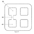
- A method of forming a sensor, comprising the step of forming a barrier of photoresist material on a surface of the sensor, the barrier defining at least one region which is suitable for constraining a bio-optical reagent or analyte therein.
- The method of claim 1, further comprising the steps of;
depositing photoresist material on a surface of the sensor;
patterning and etching the photoresist material so that a plurality of discrete photoresist material volumes are formed; and
deforming the photoresist material such that adjacent volumes form the barrier. - The method of claim 2, wherein the step of deforming the photoresist material comprises the step of heating a plurality of the discrete material volumes.
- The method of claim 3, wherein the plurality of photoresist material volumes formed by patterning and etching are, prior to being deformed, spaced apart by a distance, the value of which ensures that adjacent volumes coalesce upon heating.
- The method of claim 4, wherein the distance is not more than 2µm.
- The method of any of claims 2-5, further comprising the step of depositing a bio-optical reagent in the region defined by the barrier.
- The method of claim 6, further comprising the step of depositing different types of reagents in different regions.
- A sensor comprising a barrier of photoresist material on a surface thereof, the barrier defining at least one region which is suitable for constraining a bio-optical reagent or analyte therein.
- The sensor of claim 8, wherein the sensor is a bio-optical sensor.
- The sensor of claim 9, further comprising a reagent constrained in the region defined by the barrier.
- The sensor of claim 10, comprising different types of reagents constrained within different regions.
- The sensor of any of claims 8-11, wherein the barrier of photoresist material is formed on a silicon surface of the sensor.
Priority Applications (3)
| Application Number | Priority Date | Filing Date | Title |
|---|---|---|---|
| DE60315691T DE60315691D1 (en) | 2003-12-22 | 2003-12-22 | Method of producing sensors with barriers of photoresist material |
| EP03258136A EP1557717B1 (en) | 2003-12-22 | 2003-12-22 | Method of forming sensors comprising barriers of photoresist material |
| US11/018,979 US7273633B2 (en) | 2003-12-22 | 2004-12-21 | Sensors |
Applications Claiming Priority (1)
| Application Number | Priority Date | Filing Date | Title |
|---|---|---|---|
| EP03258136A EP1557717B1 (en) | 2003-12-22 | 2003-12-22 | Method of forming sensors comprising barriers of photoresist material |
Publications (2)
| Publication Number | Publication Date |
|---|---|
| EP1557717A1 true EP1557717A1 (en) | 2005-07-27 |
| EP1557717B1 EP1557717B1 (en) | 2007-08-15 |
Family
ID=34626441
Family Applications (1)
| Application Number | Title | Priority Date | Filing Date |
|---|---|---|---|
| EP03258136A Expired - Fee Related EP1557717B1 (en) | 2003-12-22 | 2003-12-22 | Method of forming sensors comprising barriers of photoresist material |
Country Status (3)
| Country | Link |
|---|---|
| US (1) | US7273633B2 (en) |
| EP (1) | EP1557717B1 (en) |
| DE (1) | DE60315691D1 (en) |
Cited By (1)
| Publication number | Priority date | Publication date | Assignee | Title |
|---|---|---|---|---|
| EP2179267A2 (en) * | 2007-07-18 | 2010-04-28 | SiliconFile Technologies Inc. | Diagnosis device and method of manufacturing the diagnosis device |
Families Citing this family (7)
| Publication number | Priority date | Publication date | Assignee | Title |
|---|---|---|---|---|
| US20190357827A1 (en) | 2003-08-01 | 2019-11-28 | Dexcom, Inc. | Analyte sensor |
| US8774886B2 (en) | 2006-10-04 | 2014-07-08 | Dexcom, Inc. | Analyte sensor |
| EP1544602B1 (en) * | 2003-12-19 | 2008-05-07 | STMicroelectronics Limited | Bio-optical sensors |
| US7557338B2 (en) * | 2006-03-14 | 2009-07-07 | Avago Technologies General Ip (Singapore) Pte. Ltd. | Electronic device with integrated optical navigation module and microlens array therefore |
| KR20080024770A (en) * | 2006-09-14 | 2008-03-19 | 삼성전자주식회사 | Micro-lens and method of forming the same |
| US9801575B2 (en) | 2011-04-15 | 2017-10-31 | Dexcom, Inc. | Advanced analyte sensor calibration and error detection |
| US9131885B2 (en) | 2009-07-02 | 2015-09-15 | Dexcom, Inc. | Analyte sensors and methods of manufacturing same |
Citations (5)
| Publication number | Priority date | Publication date | Assignee | Title |
|---|---|---|---|---|
| EP0368482A1 (en) * | 1988-10-14 | 1990-05-16 | Secretary Of State For Trade And Industry In Her Britannic Majesty's Gov. Of The U.K. Of Great Britain And Northern Ireland | Method of making a product with a feature having a multiplicity of fine lines |
| WO2000033084A2 (en) * | 1998-12-01 | 2000-06-08 | Syntrix Biochip, Inc. | Arrays of organic compounds attached to surfaces |
| US6159681A (en) * | 1997-05-28 | 2000-12-12 | Syntrix Biochip, Inc. | Light-mediated method and apparatus for the regional analysis of biologic material |
| DE10139742A1 (en) * | 2001-08-13 | 2003-03-06 | Univ Freiburg | Process for producing a "lab on chip" from photoresist material for medical diagnostic applications |
| US20030064316A1 (en) * | 1998-12-01 | 2003-04-03 | Zebala John A. | Solvent resistant photosensitive compositions |
Family Cites Families (1)
| Publication number | Priority date | Publication date | Assignee | Title |
|---|---|---|---|---|
| AU4218499A (en) * | 1998-05-27 | 1999-12-13 | Syntrix Biochip | Light-mediated method and apparatus for the regional analysis of biologic material |
-
2003
- 2003-12-22 EP EP03258136A patent/EP1557717B1/en not_active Expired - Fee Related
- 2003-12-22 DE DE60315691T patent/DE60315691D1/en not_active Expired - Lifetime
-
2004
- 2004-12-21 US US11/018,979 patent/US7273633B2/en active Active
Patent Citations (5)
| Publication number | Priority date | Publication date | Assignee | Title |
|---|---|---|---|---|
| EP0368482A1 (en) * | 1988-10-14 | 1990-05-16 | Secretary Of State For Trade And Industry In Her Britannic Majesty's Gov. Of The U.K. Of Great Britain And Northern Ireland | Method of making a product with a feature having a multiplicity of fine lines |
| US6159681A (en) * | 1997-05-28 | 2000-12-12 | Syntrix Biochip, Inc. | Light-mediated method and apparatus for the regional analysis of biologic material |
| WO2000033084A2 (en) * | 1998-12-01 | 2000-06-08 | Syntrix Biochip, Inc. | Arrays of organic compounds attached to surfaces |
| US20030064316A1 (en) * | 1998-12-01 | 2003-04-03 | Zebala John A. | Solvent resistant photosensitive compositions |
| DE10139742A1 (en) * | 2001-08-13 | 2003-03-06 | Univ Freiburg | Process for producing a "lab on chip" from photoresist material for medical diagnostic applications |
Non-Patent Citations (2)
| Title |
|---|
| HANAZATO Y ET AL: "INTEGRATED MULTI-BIOSENSORS BASED ON AN ION-SENSITIVE FIELD-EFFECT TRANSISTOR USING PHOTOLITHOGRAPHIC TECHNIQUES", IEEE TRANSACTIONS ON ELECTRON DEVICES, IEEE INC. NEW YORK, US, vol. 36, no. 7, 1 July 1989 (1989-07-01), pages 1303 - 1310, XP000034484, ISSN: 0018-9383 * |
| POPOVIC Z D ET AL: "TECHNIQUE FOR MONOLITHIC FABRICATION OF MICROLENS ARRAYS", APPLIED OPTICS, OPTICAL SOCIETY OF AMERICA,WASHINGTON, US, vol. 27, no. 7, 1 April 1988 (1988-04-01), pages 1281 - 1284, XP000118684, ISSN: 0003-6935 * |
Cited By (3)
| Publication number | Priority date | Publication date | Assignee | Title |
|---|---|---|---|---|
| EP2179267A2 (en) * | 2007-07-18 | 2010-04-28 | SiliconFile Technologies Inc. | Diagnosis device and method of manufacturing the diagnosis device |
| EP2179267A4 (en) * | 2007-07-18 | 2010-09-29 | Siliconfile Technologies Inc | Diagnosis device and method of manufacturing the diagnosis device |
| JP2010533860A (en) * | 2007-07-18 | 2010-10-28 | シリコンファイル・テクノロジーズ・インコーポレイテッド | Diagnostic device and manufacturing method thereof |
Also Published As
| Publication number | Publication date |
|---|---|
| US7273633B2 (en) | 2007-09-25 |
| DE60315691D1 (en) | 2007-09-27 |
| US20050151148A1 (en) | 2005-07-14 |
| EP1557717B1 (en) | 2007-08-15 |
Similar Documents
| Publication | Publication Date | Title |
|---|---|---|
| US11467089B2 (en) | Arrays of integrated analytical devices | |
| US10310178B2 (en) | Arrays of integrated analytical devices and methods for production | |
| EP2320462B1 (en) | Image sensor having waveguides formed in color filters | |
| US6906797B1 (en) | Side light activated microfluid channels | |
| US8335029B2 (en) | Micromirror arrays having self aligned features | |
| CN100461003C (en) | Hemi-spherical structure and method for fabricating the same | |
| US7078779B2 (en) | Enhanced color image sensor device and method of making the same | |
| CN1574381A (en) | Image sensor having micro-lens array separated with trench structures and method of making | |
| EP1557717B1 (en) | Method of forming sensors comprising barriers of photoresist material | |
| US9095852B2 (en) | Multilayer high density microwells | |
| RU2016122443A (en) | BIOSENSORS FOR BIOLOGICAL OR CHEMICAL ANALYSIS AND METHODS FOR THEIR MANUFACTURE | |
| CA2510996A1 (en) | Waveguiding structures with embedded microchannels and method for fabrication thereof | |
| KR20180004480A (en) | Image Sensors Having a Plurality of Photodiodes Sharing One Color Filter and One Micro-lens | |
| US8026559B2 (en) | Biosensor devices and method for fabricating the same | |
| JP2007227474A (en) | Solid-state imaging apparatus | |
| EP4095516B1 (en) | Biosensor with grating array | |
| US8416283B2 (en) | 3D imaging device and method for manufacturing same | |
| JP2006253464A (en) | Microlens manufacturing method, and solid imaging element manufactured by using same method | |
| WO2001020309A1 (en) | Side light activated microfluid channels | |
| US20080044941A1 (en) | Fabricating cmos image sensor | |
| KR100358195B1 (en) | Solid-state image sensing device and method of manufacturing the same | |
| JP2008085325A (en) | Image sensor and manufacturing method thereof | |
| JPH07161794A (en) | Condensing lens check method of solid-state image sensing device | |
| US11125939B1 (en) | Waveguide filtering biochemical sensor | |
| US20230131829A1 (en) | Waveguide structure with aperture array |
Legal Events
| Date | Code | Title | Description |
|---|---|---|---|
| PUAI | Public reference made under article 153(3) epc to a published international application that has entered the european phase |
Free format text: ORIGINAL CODE: 0009012 |
|
| AK | Designated contracting states |
Kind code of ref document: A1 Designated state(s): AT BE BG CH CY CZ DE DK EE ES FI FR GB GR HU IE IT LI LU MC NL PT RO SE SI SK TR |
|
| AX | Request for extension of the european patent |
Extension state: AL LT LV MK |
|
| 17P | Request for examination filed |
Effective date: 20051230 |
|
| AKX | Designation fees paid |
Designated state(s): DE FR GB IT |
|
| GRAP | Despatch of communication of intention to grant a patent |
Free format text: ORIGINAL CODE: EPIDOSNIGR1 |
|
| RTI1 | Title (correction) |
Free format text: METHOD OF FORMING SENSORS COMPRISING BARRIERS OF PHOTORESIST MATERIAL |
|
| GRAS | Grant fee paid |
Free format text: ORIGINAL CODE: EPIDOSNIGR3 |
|
| GRAA | (expected) grant |
Free format text: ORIGINAL CODE: 0009210 |
|
| AK | Designated contracting states |
Kind code of ref document: B1 Designated state(s): DE FR GB IT |
|
| REG | Reference to a national code |
Ref country code: GB Ref legal event code: FG4D |
|
| REF | Corresponds to: |
Ref document number: 60315691 Country of ref document: DE Date of ref document: 20070927 Kind code of ref document: P |
|
| ET | Fr: translation filed | ||
| PLBE | No opposition filed within time limit |
Free format text: ORIGINAL CODE: 0009261 |
|
| STAA | Information on the status of an ep patent application or granted ep patent |
Free format text: STATUS: NO OPPOSITION FILED WITHIN TIME LIMIT |
|
| 26N | No opposition filed |
Effective date: 20080516 |
|
| PG25 | Lapsed in a contracting state [announced via postgrant information from national office to epo] |
Ref country code: DE Free format text: LAPSE BECAUSE OF FAILURE TO SUBMIT A TRANSLATION OF THE DESCRIPTION OR TO PAY THE FEE WITHIN THE PRESCRIBED TIME-LIMIT Effective date: 20071116 |
|
| PG25 | Lapsed in a contracting state [announced via postgrant information from national office to epo] |
Ref country code: IT Free format text: LAPSE BECAUSE OF NON-PAYMENT OF DUE FEES Effective date: 20071231 |
|
| PGFP | Annual fee paid to national office [announced via postgrant information from national office to epo] |
Ref country code: GB Payment date: 20101201 Year of fee payment: 8 |
|
| PGFP | Annual fee paid to national office [announced via postgrant information from national office to epo] |
Ref country code: FR Payment date: 20110120 Year of fee payment: 8 |
|
| GBPC | Gb: european patent ceased through non-payment of renewal fee |
Effective date: 20111222 |
|
| REG | Reference to a national code |
Ref country code: FR Ref legal event code: ST Effective date: 20120831 |
|
| PG25 | Lapsed in a contracting state [announced via postgrant information from national office to epo] |
Ref country code: GB Free format text: LAPSE BECAUSE OF NON-PAYMENT OF DUE FEES Effective date: 20111222 |
|
| PG25 | Lapsed in a contracting state [announced via postgrant information from national office to epo] |
Ref country code: FR Free format text: LAPSE BECAUSE OF NON-PAYMENT OF DUE FEES Effective date: 20120102 |