EP0991421B1 - Detection and modulation of the iaps and naip for the diagnosis and treatment of proliferative disease - Google Patents
Detection and modulation of the iaps and naip for the diagnosis and treatment of proliferative disease Download PDFInfo
- Publication number
- EP0991421B1 EP0991421B1 EP98917563A EP98917563A EP0991421B1 EP 0991421 B1 EP0991421 B1 EP 0991421B1 EP 98917563 A EP98917563 A EP 98917563A EP 98917563 A EP98917563 A EP 98917563A EP 0991421 B1 EP0991421 B1 EP 0991421B1
- Authority
- EP
- European Patent Office
- Prior art keywords
- polypeptide
- iap
- cell
- naip
- nucleic acid
- Prior art date
- Legal status (The legal status is an assumption and is not a legal conclusion. Google has not performed a legal analysis and makes no representation as to the accuracy of the status listed.)
- Expired - Lifetime
Links
Images


Classifications
-
- C—CHEMISTRY; METALLURGY
- C12—BIOCHEMISTRY; BEER; SPIRITS; WINE; VINEGAR; MICROBIOLOGY; ENZYMOLOGY; MUTATION OR GENETIC ENGINEERING
- C12N—MICROORGANISMS OR ENZYMES; COMPOSITIONS THEREOF; PROPAGATING, PRESERVING, OR MAINTAINING MICROORGANISMS; MUTATION OR GENETIC ENGINEERING; CULTURE MEDIA
- C12N15/00—Mutation or genetic engineering; DNA or RNA concerning genetic engineering, vectors, e.g. plasmids, or their isolation, preparation or purification; Use of hosts therefor
- C12N15/09—Recombinant DNA-technology
- C12N15/11—DNA or RNA fragments; Modified forms thereof; Non-coding nucleic acids having a biological activity
- C12N15/113—Non-coding nucleic acids modulating the expression of genes, e.g. antisense oligonucleotides; Antisense DNA or RNA; Triplex- forming oligonucleotides; Catalytic nucleic acids, e.g. ribozymes; Nucleic acids used in co-suppression or gene silencing
- C12N15/1135—Non-coding nucleic acids modulating the expression of genes, e.g. antisense oligonucleotides; Antisense DNA or RNA; Triplex- forming oligonucleotides; Catalytic nucleic acids, e.g. ribozymes; Nucleic acids used in co-suppression or gene silencing against oncogenes or tumor suppressor genes
-
- A—HUMAN NECESSITIES
- A61—MEDICAL OR VETERINARY SCIENCE; HYGIENE
- A61K—PREPARATIONS FOR MEDICAL, DENTAL OR TOILETRY PURPOSES
- A61K38/00—Medicinal preparations containing peptides
- A61K38/16—Peptides having more than 20 amino acids; Gastrins; Somatostatins; Melanotropins; Derivatives thereof
- A61K38/17—Peptides having more than 20 amino acids; Gastrins; Somatostatins; Melanotropins; Derivatives thereof from animals; from humans
- A61K38/1703—Peptides having more than 20 amino acids; Gastrins; Somatostatins; Melanotropins; Derivatives thereof from animals; from humans from vertebrates
- A61K38/1709—Peptides having more than 20 amino acids; Gastrins; Somatostatins; Melanotropins; Derivatives thereof from animals; from humans from vertebrates from mammals
-
- A—HUMAN NECESSITIES
- A61—MEDICAL OR VETERINARY SCIENCE; HYGIENE
- A61P—SPECIFIC THERAPEUTIC ACTIVITY OF CHEMICAL COMPOUNDS OR MEDICINAL PREPARATIONS
- A61P35/00—Antineoplastic agents
-
- C—CHEMISTRY; METALLURGY
- C07—ORGANIC CHEMISTRY
- C07K—PEPTIDES
- C07K14/00—Peptides having more than 20 amino acids; Gastrins; Somatostatins; Melanotropins; Derivatives thereof
- C07K14/435—Peptides having more than 20 amino acids; Gastrins; Somatostatins; Melanotropins; Derivatives thereof from animals; from humans
- C07K14/46—Peptides having more than 20 amino acids; Gastrins; Somatostatins; Melanotropins; Derivatives thereof from animals; from humans from vertebrates
- C07K14/47—Peptides having more than 20 amino acids; Gastrins; Somatostatins; Melanotropins; Derivatives thereof from animals; from humans from vertebrates from mammals
- C07K14/4701—Peptides having more than 20 amino acids; Gastrins; Somatostatins; Melanotropins; Derivatives thereof from animals; from humans from vertebrates from mammals not used
- C07K14/4702—Regulators; Modulating activity
- C07K14/4703—Inhibitors; Suppressors
-
- C—CHEMISTRY; METALLURGY
- C12—BIOCHEMISTRY; BEER; SPIRITS; WINE; VINEGAR; MICROBIOLOGY; ENZYMOLOGY; MUTATION OR GENETIC ENGINEERING
- C12N—MICROORGANISMS OR ENZYMES; COMPOSITIONS THEREOF; PROPAGATING, PRESERVING, OR MAINTAINING MICROORGANISMS; MUTATION OR GENETIC ENGINEERING; CULTURE MEDIA
- C12N15/00—Mutation or genetic engineering; DNA or RNA concerning genetic engineering, vectors, e.g. plasmids, or their isolation, preparation or purification; Use of hosts therefor
- C12N15/09—Recombinant DNA-technology
- C12N15/11—DNA or RNA fragments; Modified forms thereof; Non-coding nucleic acids having a biological activity
- C12N15/113—Non-coding nucleic acids modulating the expression of genes, e.g. antisense oligonucleotides; Antisense DNA or RNA; Triplex- forming oligonucleotides; Catalytic nucleic acids, e.g. ribozymes; Nucleic acids used in co-suppression or gene silencing
-
- C—CHEMISTRY; METALLURGY
- C12—BIOCHEMISTRY; BEER; SPIRITS; WINE; VINEGAR; MICROBIOLOGY; ENZYMOLOGY; MUTATION OR GENETIC ENGINEERING
- C12Q—MEASURING OR TESTING PROCESSES INVOLVING ENZYMES, NUCLEIC ACIDS OR MICROORGANISMS; COMPOSITIONS OR TEST PAPERS THEREFOR; PROCESSES OF PREPARING SUCH COMPOSITIONS; CONDITION-RESPONSIVE CONTROL IN MICROBIOLOGICAL OR ENZYMOLOGICAL PROCESSES
- C12Q1/00—Measuring or testing processes involving enzymes, nucleic acids or microorganisms; Compositions therefor; Processes of preparing such compositions
- C12Q1/68—Measuring or testing processes involving enzymes, nucleic acids or microorganisms; Compositions therefor; Processes of preparing such compositions involving nucleic acids
- C12Q1/6876—Nucleic acid products used in the analysis of nucleic acids, e.g. primers or probes
- C12Q1/6883—Nucleic acid products used in the analysis of nucleic acids, e.g. primers or probes for diseases caused by alterations of genetic material
- C12Q1/6886—Nucleic acid products used in the analysis of nucleic acids, e.g. primers or probes for diseases caused by alterations of genetic material for cancer
-
- G—PHYSICS
- G01—MEASURING; TESTING
- G01N—INVESTIGATING OR ANALYSING MATERIALS BY DETERMINING THEIR CHEMICAL OR PHYSICAL PROPERTIES
- G01N33/00—Investigating or analysing materials by specific methods not covered by groups G01N1/00 - G01N31/00
- G01N33/48—Biological material, e.g. blood, urine; Haemocytometers
- G01N33/50—Chemical analysis of biological material, e.g. blood, urine; Testing involving biospecific ligand binding methods; Immunological testing
- G01N33/5005—Chemical analysis of biological material, e.g. blood, urine; Testing involving biospecific ligand binding methods; Immunological testing involving human or animal cells
- G01N33/5008—Chemical analysis of biological material, e.g. blood, urine; Testing involving biospecific ligand binding methods; Immunological testing involving human or animal cells for testing or evaluating the effect of chemical or biological compounds, e.g. drugs, cosmetics
- G01N33/5011—Chemical analysis of biological material, e.g. blood, urine; Testing involving biospecific ligand binding methods; Immunological testing involving human or animal cells for testing or evaluating the effect of chemical or biological compounds, e.g. drugs, cosmetics for testing antineoplastic activity
-
- G—PHYSICS
- G01—MEASURING; TESTING
- G01N—INVESTIGATING OR ANALYSING MATERIALS BY DETERMINING THEIR CHEMICAL OR PHYSICAL PROPERTIES
- G01N33/00—Investigating or analysing materials by specific methods not covered by groups G01N1/00 - G01N31/00
- G01N33/48—Biological material, e.g. blood, urine; Haemocytometers
- G01N33/50—Chemical analysis of biological material, e.g. blood, urine; Testing involving biospecific ligand binding methods; Immunological testing
- G01N33/53—Immunoassay; Biospecific binding assay; Materials therefor
- G01N33/574—Immunoassay; Biospecific binding assay; Materials therefor for cancer
- G01N33/57484—Immunoassay; Biospecific binding assay; Materials therefor for cancer involving compounds serving as markers for tumor, cancer, neoplasia, e.g. cellular determinants, receptors, heat shock/stress proteins, A-protein, oligosaccharides, metabolites
- G01N33/57496—Immunoassay; Biospecific binding assay; Materials therefor for cancer involving compounds serving as markers for tumor, cancer, neoplasia, e.g. cellular determinants, receptors, heat shock/stress proteins, A-protein, oligosaccharides, metabolites involving intracellular compounds
-
- A—HUMAN NECESSITIES
- A01—AGRICULTURE; FORESTRY; ANIMAL HUSBANDRY; HUNTING; TRAPPING; FISHING
- A01K—ANIMAL HUSBANDRY; CARE OF BIRDS, FISHES, INSECTS; FISHING; REARING OR BREEDING ANIMALS, NOT OTHERWISE PROVIDED FOR; NEW BREEDS OF ANIMALS
- A01K2217/00—Genetically modified animals
- A01K2217/05—Animals comprising random inserted nucleic acids (transgenic)
-
- A—HUMAN NECESSITIES
- A61—MEDICAL OR VETERINARY SCIENCE; HYGIENE
- A61K—PREPARATIONS FOR MEDICAL, DENTAL OR TOILETRY PURPOSES
- A61K48/00—Medicinal preparations containing genetic material which is inserted into cells of the living body to treat genetic diseases; Gene therapy
-
- C—CHEMISTRY; METALLURGY
- C12—BIOCHEMISTRY; BEER; SPIRITS; WINE; VINEGAR; MICROBIOLOGY; ENZYMOLOGY; MUTATION OR GENETIC ENGINEERING
- C12N—MICROORGANISMS OR ENZYMES; COMPOSITIONS THEREOF; PROPAGATING, PRESERVING, OR MAINTAINING MICROORGANISMS; MUTATION OR GENETIC ENGINEERING; CULTURE MEDIA
- C12N2310/00—Structure or type of the nucleic acid
- C12N2310/10—Type of nucleic acid
- C12N2310/11—Antisense
-
- C—CHEMISTRY; METALLURGY
- C12—BIOCHEMISTRY; BEER; SPIRITS; WINE; VINEGAR; MICROBIOLOGY; ENZYMOLOGY; MUTATION OR GENETIC ENGINEERING
- C12N—MICROORGANISMS OR ENZYMES; COMPOSITIONS THEREOF; PROPAGATING, PRESERVING, OR MAINTAINING MICROORGANISMS; MUTATION OR GENETIC ENGINEERING; CULTURE MEDIA
- C12N2799/00—Uses of viruses
- C12N2799/02—Uses of viruses as vector
- C12N2799/021—Uses of viruses as vector for the expression of a heterologous nucleic acid
- C12N2799/022—Uses of viruses as vector for the expression of a heterologous nucleic acid where the vector is derived from an adenovirus
-
- C—CHEMISTRY; METALLURGY
- C12—BIOCHEMISTRY; BEER; SPIRITS; WINE; VINEGAR; MICROBIOLOGY; ENZYMOLOGY; MUTATION OR GENETIC ENGINEERING
- C12Q—MEASURING OR TESTING PROCESSES INVOLVING ENZYMES, NUCLEIC ACIDS OR MICROORGANISMS; COMPOSITIONS OR TEST PAPERS THEREFOR; PROCESSES OF PREPARING SUCH COMPOSITIONS; CONDITION-RESPONSIVE CONTROL IN MICROBIOLOGICAL OR ENZYMOLOGICAL PROCESSES
- C12Q2600/00—Oligonucleotides characterized by their use
- C12Q2600/106—Pharmacogenomics, i.e. genetic variability in individual responses to drugs and drug metabolism
-
- C—CHEMISTRY; METALLURGY
- C12—BIOCHEMISTRY; BEER; SPIRITS; WINE; VINEGAR; MICROBIOLOGY; ENZYMOLOGY; MUTATION OR GENETIC ENGINEERING
- C12Q—MEASURING OR TESTING PROCESSES INVOLVING ENZYMES, NUCLEIC ACIDS OR MICROORGANISMS; COMPOSITIONS OR TEST PAPERS THEREFOR; PROCESSES OF PREPARING SUCH COMPOSITIONS; CONDITION-RESPONSIVE CONTROL IN MICROBIOLOGICAL OR ENZYMOLOGICAL PROCESSES
- C12Q2600/00—Oligonucleotides characterized by their use
- C12Q2600/136—Screening for pharmacological compounds
Definitions
- the invention relates to the diagnosis and treatment of proliferative disease, in particular, cancer.
- Apoptosis One mechanism by which cells die is referred to as apoptosis, or programmed cell death.
- Apoptosis often occurs as a normal part of the development and maintenance of healthy tissues, and is now known to play a critical role in embryonic development. The failure of a normal apoptotic response has been implicated in the development of cancer; autoimmune disorders, such as lupus erythematosis and multiple sclerosis; and in viral infections, including those associated with herpes virus, poxvirus, and adenovirus.
- Baculoviruses encode proteins termed inhibitors of apoptosis proteins (IAPs) which inhibit the apoptosis that would otherwise occur when insect cells are infected by the baculovirus.
- the baculovirus IAP genes include sequences encoding a ring zinc finger-like motif (RZF), which is presumed to be directly involved in DNA binding, and two N-terminal domains that consist of a 70 amino acid repeat motif termed a BIR domain (Baculovirus IAP Repeat).
- RZF ring zinc finger-like motif
- BIR domain Baculovirus IAP Repeat
- WO97/06255 describes genomic, cDNA or synthetic DNA molecules, which encode a mammalian IAP polypeptide. Nucleic acid sequences encoding a NAIP polypeptide and methods of inhibiting apoptosis via administration of a NAIP polypeptide are already disclosed in WO97/26331.
- LAP and NAIP overexpression are specifically associated with a wide range of cancer types including ovarian cancer, adenocarcinoma, lymphoma, and pancreatic cancer.
- the presence of a fragmented IAP polypeptide in the nucleus, and an overexpression of an IAP polypeptide in the presence of a p53 mutation correlates with a cancer diagnosis, a poor prognosis, and a resistance to numerous chemotherapeutic cancer drugs.
- an therapeutic agent that reduces the biological activity of an lAP polypeptide will induce apoptosis in a cell expressing the polypeptide (e.g ., a cell that is proliferating in a proliferative disease).
- the invention features a method for enhancing apoptosis in a cell from a mammal with a proliferative disease, the method including administering to the cell a compound that inhibits the biological activity of an IAP polypeptide or a NAIP polypeptide, the compound being administered to the cell in an amount sufficient to enhance apoptosis in the cell, wherein said method is performed in vitro, and wherein said cell is proliferating in said proliferative disease.
- the biological activity is the level of expression of the polypeptide (measured, for example, by assaying the amount of polypeptide present in the cell); the level of expression of an mRNA molecule encoding the polypeptide; or an apoptosis-inhibiting activity.
- the polypeptide is selected from the group consisting of HIAP-1, m-HIAP-1, HIAP-2, m-HIAP-2, XIAP, and m-XIAP. In other embodiment, the polypeptide is NAIP, XIAP, HIAP-1, or HIAP-2.
- the mammal is a human or a mouse
- the proliferative disease is cancer, for example, a cancer in a tissue selected from the group consisting of ovary, breast, pancreas, lymph node, skin, blood, lung, brain, kidney, liver, nasopharyngeal cavity, thyroid, central nervous system, prostate, colon, rectum, cervix, endometrium, and lung.
- the compound is a negative regulator of an IAP or an NAIP-dependent anti-apoptotic pathway; a fragment of the IAP polypeptide, the fragment including a ring zinc finger and having no more than two BIR domains; a nucleic acid molecule encoding a ring zinc finger domain of the LAP polypeptide; a compound that prevents cleavage of the IAP polypeptide or the NAIP polypeptide; a purified antibody or a fragment thereof that specifically binds to the LAP polypeptide or the NAIP polypeptide; a ribozyme; or an antisense nucleic acid molecule have a nucleic acid sequence that is complementary to the coding strand of a nucleic acid sequence encoding the IAP polypeptide or the NAIP polypeptide.
- the cleavage is decreased by at least 20% in the cell; the antibody binds to a BIR domain of the IAP polypeptide or the NAIP polypeptide; the nucleic acid sequence encoding the IAP polypeptide or the NAIP polypeptide has about 50% or greater homology with the nucleotide sequence of SEQ ID NO: 3. SEQ ID NO: 5, SEQ ID NO: 7, SEQ ID NO: 9.
- the antisense nucleic acid molecule decreases the level of the nucleic acid sequence encoding the IAP polypeptide or the NAIP polypeptide by at least 20%, the level being measured in the cytoplasm of the ccll; the antisense nucleic acid molecule is encoded by a virus vector; or the antisense nucleic acid molecule is encoded by transgene.
- the invention features a method for detecting a proliferative disease or an increased likelihood of the proliferative disease in a mammal that includes: (a) contacting an IAP or a NAIP nucleic acid molecule that is greater than about 18 nucleotides in length with a preparation of nucleic acid from a cell of the mammal, the cell proliferating in the disease, the cell from a tissue; and (b) measuring the amount of nucleic acid from the cell of the mammal that hybridizes to the molecule, an increase in the amount from the cell of the mammal relative to a control indicating a an increased likelihood of the mammal having or developing a proliferative disease.
- the method further includes the steps of: (a) contacting the molecule with a preparation of nucleic acid from the control, wherein the control is a cell from the tissue of a second mammal, the second mammal lacking a proliferative disease; and (b) measuring the amount of nucleic acid from the control, an increase in the amount of the nucleic acid from the cell of the mammal that hybridizes to the molecule relative to the amount of the nucleic acid from the control indicating an increased likelihood of the mammal having or developing a proliferative disease.
- the method further includes the steps of: (a) providing a pair of oligonucleotides having sequence identity to or being complementary to a region of the IAP or the NAIP nucleic acid molecule; (b) combining the pair of oligonucleotides with the nucleic acid under conditions suitable for polymerase chain reaction-mediated nucleic acid amplification; and (c) isolating the amplified nucleic acid or fragment thereof.
- the amplification is carried out using a reverse-transcription polymerase chain reaction (e.g . rapid amplification of cDNA ends, RACE).
- the method provides measuring the nucleic acid having a nucleotide sequence that has about 50% or greater homology with the nucleotide sequence of SEQ ID NO: 3. SEQ ID NO: 5, SEQ ID NO: 7, SEQ ID NO: 9, SEQ ID NO: 11. SEQ ID NO: 13. or the nucleic acid sequence of NAIP. In other embodiments, the method provides measuring the nucleic acid having a nucleotide sequence that has about 50% or greater homology with the nucleotide sequence of SEQ ID NO: 3. SEQ ID NO: 5, SEQ ID NO: 7, or NAIP.
- the invention features a method for detecting a proliferative disease or an increased likelihood of developing the disease in a mammal, the method including measuring the level of biological activity of an IAP polypeptide or a NAIP polypeptide in a sample of the mammal, an increase in the level of the IAP polypeptide or the NAIP polypeptide relative to a sample from a control mammal being an indication that the mammal has the disease or increased likelihood of developing the disease, wherein said sample comprises a all from a tissue of said mammal, and wherein said all is proliferating in said proliferative disease; and said sample from a control mammal is from said tissue, said sample consisting of healthy cells.
- the mammal and the control mammal are the same.
- the biological activity is the level of expression of the polypeptide (measured, for example, by assaying the amount of the polypeptide present in the cell); wherein the biological activity is the level of expression of an mRNA molecule encoding the polypeptide; or wherein the biological activity is an apoptosis-inhibiting activity.
- the polypeptide is selected from the group consisting of HIAP-1, m-HIAP-1, HIAP-2, m-HIAP-2, XIAP, and m-XIAP.
- the polypeptide is NAIP, XIAP, HIAP-1, or HIAP-2.
- the invention features a method for identifying a compound enhances apoptosis in an affected cell that is proliferating in a proliferative disease that includes exposing a cell that overexpresses an IAP polypeptide or a NAIP polypeptide to a candidate compound, a decrease the level of biological activity of the polypeptide indicating the presence of a compound that enhances apoptosis in the affected cell that is proliferating in the proliferative disease.
- the invention features a method for identifying a compound that enhances apoptosis in an affected cell that is proliferating in a proliferative disease that includes the steps of: (a) providing a cell including a nucleic acid molecule encoding a IAP polypeptide or a nucleic acid molecule encoding a NAIP polypeptide, the nucleic acid molecule being expressed in the cell; and (b) contacting the cell with a candidate compound and monitoring level of biological activity of the IAP polypeptide or the NAIP polypeptide in the cell, a decrease in the level of biological activity of the IAP polypeptide or the NAIP polypeptide in the cell in response to the candidate compound relative to a cell not contacted with the candidate compound indicating the presence of a compound that enhances apoptosis in the affected cell that is proliferating in the proliferative disease.
- the cell further expresses a p53 polypeptide associated with the proliferative disease.
- the biological activity is the level of expression of the polypeptide (measured, for example, by assaying the amount of the polypeptide present in the cell); wherein the biological activity is the level of expression of an mRNA molecule encoding the polypeptide; or wherein the biological activity is an apoptosis-inhibiting activity.
- the polypeptide is selected from the group consisting of HIAP-1, m-HIAP-1, HIAP-2, m-HIAP-2, XIAP, and m-XIAP.
- the polypeptide is NAIP, XIAP, HIAP-1, or HIAP-2.
- the invention features a method for determining the prognosis of a mammal diagnosed with a proliferative disease that includes the steps of: (a) isolating a sample from a tissue from the mammal; and (b) determining whether the sample has an increased level of biological activity of an IAP polypeptide or an NAIP polypeptide relative to a control sample, an increase in the level in the sample being an indication that the mammal has a poor prognosis, wherein said sample comprises a cell that is proliferating in said proliferative disease; and the control sample is from said tissue, said control sample consisting of healthy cells; and the sample and the control sample are from the mammal.
- the sample further includes a cell expressing a p53 polypeptide associated with the proliferative disease.
- the biological activity is the level of expression of the polypeptide (measured, for example, by assaying the amount of the polypeptide present in the cell); wherein the biological activity is the level of expression of an mRNA molecule encoding the polypeptide; or wherein the biological activity is an apoptosis-inhibiting activity.
- the polypeptide is selected from the group consisting of HIAP-1, m-HIAP-1, HIAP-2, m-HIAP-2. XIAP, and m-XIAP.
- the polypeptide is NAIP, XIAP, HIAP-1, or HIAP-2.
- the level is assayed by measuring the amount of lAP peptide of less than 64 kDa present in the sample.
- the invention features a method for determining the prognosis of a mammal diagnosed with a proliferative disease that includes the steps of: (a) isolating a sample from the mammal, the sample having a nuclear fraction; and (b) measuring the amount of a polypeptide that is recognized by an antibody that specifically binds an IAP polypeptide or an antibody that specifically binds an NAIP polypeptide in the nuclear fraction of the sample relative to an amount from a control sample, an increase in the amount from the sample being an indication that the mammal has a poor prognosis, wherein said sample is from a tissue of the mammal, said sample comprising a cell that is proliferating in said proliferative disease; and the control sample is from said tissue, said control sample consisting of healthy cells.
- the sample and the control sample are from the mammal.
- the biological activity is the level of expression of the polypeptide (measured, for example, by assaying the amount of the polypeptide present in the cell); wherein the biological activity is the level of expression of an mRNA molecule encoding the polypeptide; or wherein the biological activity is an apoptosis-inhibiting activity.
- the polypeptide is selected from the group consisting of HIAP-1, m-HIAP-1, HIAP-2, m-HIAP-2, XIAP, and m-XIAP.
- the polypeptide is NAIP, XIAP, HIAP-1, or HIAP-2.
- the amount is measured by immunological methods.
- the biological activity is the level of expression of the polypeptide (measured, for example, by assaying the amount of the polypeptide present in the cell); wherein the biological activity is the level of expression of an mRNA molecule encoding the polypeptide; or wherein the biological activity is an apoptosis-inhibiting activity.
- the-polypeptide is-selected from the group consisting of HIAP-1, m-HIAP-1, HIAP-2, m-HIAP-2, XIAP, and m-XIAP.
- the polypeptide is NAIP, XIAP, HIAP-1, or HIAP-2.
- the invention features the use of a compound that decreases the biological activity an IAP polypeptide or a NAIP polypeptidc for the manufacture of a medicament for the enhancement of apoptosis.
- the biological activity is the level of expression of the polypeptide (measured, for example, by assaying the amount of the polypeptide present in the cell); wherein the biological activity is the level of expression of an mRNA molecule encoding the polypeptide; or wherein the biological activity is an apoptosis-inhibiting activity.
- the polypeptide is selected from the group consisting of HIAP-1, m-HIAP-1, HIAP-2, m-HIAP-2, XIAP, and m-XIAP.
- the polypeptide is NAIP, XIAP, HIAP-1, or HIAP-2.
- the invention features a kit for diagnosing a mammal for the presence of a proliferative disease or an increased likelihood of developing a proliferative disease, the kit compromising an oligonucleotide that hybridizes to a nucleic acid'sequence that encodes an IAP polypeptide or a NAIP polypeptide.
- the biological activity is the level of expression of the polypeptide (measured, for example, by assaying the amount of the polypeptide present in the cell); wherein the biological activity is the level of expression of an mRNA molecule encoding the polypeptide; or wherein the biological activity is an apoptosis-inhibiting activity.
- the polypeptide is selected from the group consisting of HIAP-1, m-HIAP-1, HIAP-2, m-HIAP-2, XIAP, and m-XIAP.
- the polypeptide is NAIP, XIAP, HIAP-1, or HIAP-2.
- the invention features a non-human transgenic mammal, the mammal having an elevated level of biological activity of an IAP polypeptide or a NAIP polypeptide, wherein said transgenic mammal has a predisposition to cancer.
- the biological activity is the level of expression of the polypeptide (measured, for example, by assaying the amount of the polypeptide present in the cell); wherein the biological activity is the level of expression of an mRNA molecule encoding the polypeptide; or wherein the biological activity is an apoptosis-inhibiting activity.
- the polypeptide is selected from the group consisting of HIAP-1, m-HIAP-1, HIAP-2, m-HIAP-2, XIAP, and m-XIAP.
- the polypeptide is NAIP, XIAP, HIAP-1, or HIAP-2.
- IAP gene is meant a gene encoding a polypeptide having at least one BIR domain and is capable of modulating (inhibiting or enhancing) apoptosis in a cell or tissue when provided by other intracellular or extracellular delivery methods (see, e.g ., the U.S. Patent No. 6,156,535 and WO 97/06255.
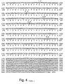
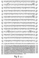
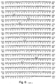
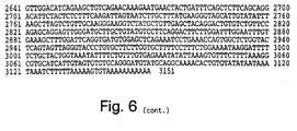
- the IAP gene is a gene having about 50% or greater nucleotide sequence homology to at least one of the IAP amino acid encoding sequences of Figs.
- Mammalian LAP genes include nucleotide sequences isolated from any mammalian source.
- the mammal is a human.
- the term "IAP gene" is meant to encompass any member of the family of genes that encode inhibitors of apoptosis.
- An IAP gene may encode a polypeptide that has at least 20%, preferably at least 30%, and most preferably at least 50% amino acid sequence homology with at least one of the conserved regions of one of the IAP members described herein (i.e ., either the BIR or ring zinc finger domains from the human or murine XIAP, HIAP-1, or HIAP-2).
- Representative members of the IAP gene family include, without limitation, the human and murine XIAP, HIAP-1, or HIAP-2 genes.
- a virus vector is meant a functional or attenuated virus that is capable of delivering to a virus-infected cell a nucleic acid molecule.
- the virus vector has been genetically engineered according to standard molecular biology techniques to bear a heterologous nucleic acid molecule.
- Virus vectors include, without limitation, adenoviruses, retroviruses, baculoviruses, cytomegaloviruses (CMV), and vaccinia viruses.
- IAP protein or "IAP polypeptide” is meant a polypeptide, or fragment thereof, encoded by an IAP gene.
- NAIP gene and "NAIP polypeptide” is meant the NAIP genes, fragments thereof, and polypeptides encoded by the same described in UK9601108.5 filed January 19, 1996 and PCT Application No. PCT/IB97/00142 (claiming priority from UK9601108.5) filed January 17, 1997.
- BIR domain is meant a domain having the amino acid sequence of the consensus sequence: Xaa1-Xaa1-Xaa1-Arg-Leu-Xaa1-Thr-Phe-Xaa1-Xaa1-Trp-Pro-Xaa2-Xaa1-Xaa1-Xaa2-Xaa2-Xaa1-Xaa1-Xaa1-Xaa1-Leu-Ala-Xaa1-Ala-Gly-Phe-Tyr-Tyr-Xaa1-Gly-Xaal-Xaa1-Asp-Xaa1-Val-Xaa1-Cys-Phe-Xaa1-Cys-Xaa1-Xaa1- Xaa1- Xaa1-Xaa1-Xaa1-Trp-Xaa1-Xaa1-Xaa1-Xaa1-Xaa1-Xaa1-Trp-
- ring zinc finger or “RZF” is meant a domain having the amino acid sequence of the consensus sequence: Glu-Xaa1-Xaa1-Xaa1-Xaa1-Xaa1-Xaa1-Xaa1-Xaa2-Xaa1-Xaa1-Xaa1-Cys-Lys-Xaa3-Cys-Met-Xaa1-Xaa1-Xaa1-Xaa1-Xaa1-Xaa1-Xaa3-Xaa1-Phe-Xaa1-Pro-Cys-Gly-His-Xaa1-Xaa1-Xaa1-Cys-Xaa1-Xaa1-Cys-Ala- Xaa1-Xaa1-Xaa1-Xaa1-Xaa1-Cys-Pro-Xaa1-Cys, wherein Xaa1 is any amino acid, Xaa
- the sequence is substantially identical to the RZF domains provided herein for the human or murine XIAP, HIAP-1, or HIAP-2.
- enhancing apoptosis is meant increasing the number of cells which apoptose in a given cell population.
- the cell population is selected from a group including ovarian cancer.cells, breast cancer cells, pancreatic cancer cells, T cells, neuronal cells, fibroblasts, or any other cell line known to proliferate in a laboratory setting.
- the degree of apoptosis enhancement provided by an apoptosis enhancing compound in a given assay will vary, but that one skilled in the art can determine the statistically significant change in the level of apoptosis which identifies a compound which enhances apoptosis otherwise limited by an IAP.
- enhancing apoptosis means that the increase in the number of cells undergoing apoptosis is at least 25%, more preferably the increase is 50%, and most preferably the increase is at least one-fold.
- the sample monitored is a sample of cells which normally undergo insufficient apoptosis ( i.e ., cancer cells).
- proliferative disease is meant a disease which is caused by or results in inappropriately high levels of cell division, inappropriately low levels of apoptosis. or both.
- cancers such as lymphoma, leukemia, melanoma, ovarian cancer, breast cancer, pancreatic cancer, and lung cancer are all examples of proliferative disease.
- a neoplasm i.e., any abnormal proliferation of cells, malignant or benign, is also a proliferative disease of the invention.
- a “cell proliferating in a proliferative disease” is meant a cell whose abnormal proliferation contributes to the disease.
- the cell expresses the antigen PCNA.
- polypeptide any chain of more than two amino acids, regardless of post-translational modification such as glycosylation or phosphorylation.
- IAP or NAIP biological activity is meant any activity known to be caused in vivo or in vitro by a NAIP or an IAP polypeptide.
- Preferred biological activities of IAP and NAIP polypeptides are those described herein, and include, without limitation, a level of expression of the polypeptide that is normal for that cell type, a level of expression of the mRNA that is normal for that cell type, an ability to block apoptosis, and an ability to be cleaved.
- a “compound that decreases the biological activity” is meant a compound that decreases any activity known to be caused in vivo or in vitro by a NAIP polypeptide or an IAP polypeptide.
- Preferred compounds include, without limitation, an antisense nucleic acid molecule that is complementary to the coding strand of nucleic acid molecule that encodes an IAP or a NAIP polypeptide; an antibody, such as a neutralizing antibody, that specifically binds to an IAP or a NAIP polypeptide; and a negative regulator of an IAP or a NAIP polypeptide, such as a polypeptide fragment that includes the ring zing finger of an IAP polypeptide, a polypeptide fragment that has no more than two BIR domains, or nucleic acid molecules encoding these polypeptide fragments.
- substantially identical is meant a polypeptide or nucleic acid exhibiting at least 50%, preferably 85%, more preferably 90%, and most preferably 95% homology to a reference ammo acid or nucleic acid sequence.
- the length of comparison sequences will generally be at least 16 amino acids, preferably at least 20 amino acids, more preferably at least 25 amino acids, and most preferably 35 amino acids.
- the length of comparison sequences will generally be at least 50 nucleotides, preferably at least 60 nucleotides, more preferably at least 75 nucleotides, and most preferably 110 nucleotides.
- Sequence identity is typically measured using sequence analysis software with the default parameters specified therein (e.g ., Sequence Analysis Software Package of the Genetics Computer Group, University of Wisconsin Biotechnology Center, 1710 University Avenue, Madison, WI 53705). This software program matches similar sequences by assigning degrees of homology to various substitutions, deletions, and other modifications. Conservative substitutions typically include substitutions within the following groups: glycine, alanine, valine, isoleucine, leucine; aspartic acid, glutamic acid, asparagine, glutamine; serine, threonine; lysine, arginine; and phenylalanine, tyrosine.
- substantially pure polypeptide is meant a polypeptide that has been separated from the components that naturally accompany it.
- the polypeptide is substantially pure when it is at least 60%, by weight, free from the proteins and naturally-occurring organic molecules with which it is naturally associated.
- the polypeptide is an IAP polypeptide that is at least 75%, more preferably at least 90%, and most preferably at least 99%, by weight, pure.
- a substantially pure IAP polypeptide may be obtained, for example, by extraction from a natural source (e.g . a fibroblast, neuronal cell, or lymphocyte) by expression of a recombinant nucleic acid encoding an IAP polypeptide, or by chemically synthesizing the protein. Purity can be measured by any appropriate method, e.g. , by column chromatography, polyacrylamide gel electrophoresis, or HPLC analysis.
- a protein is substantially free of naturally associated components when it is separated from those contaminants which accompany it in its natural state.
- a protein which is chemically synthesized or produced in a cellular system different from the cell from which it naturally originates will be substantially free from its naturally associated components.
- substantially pure polypeptides include those derived from eukaryotic organisms but synthesized in E . coli or other prokaryotes.
- substantially pure DNA DNA that is free of the genes which, in the naturally-occurring genome of the organism from which the DNA of the invention is derived, flank the gene.
- the term therefore includes, for example, a recombinant DNA which is incorporated into a vector; into an autonomously replicating plasmid or virus; or into the genomic DNA of a prokaryote or eukaryote; or which exists as a separate molecule ( e.g. , a cDNA or a genomic or cDNA fragment produced by PCR or restriction endonuclease digestion) independent of other sequences. It also includes a recombinant DNA which is part of a hybrid gene encoding additional polypeptide sequence.
- transformed cell is meant a cell into which (or into an ancestor of which) has been introduced, by means of recombinant DNA techniques, a DNA molecule encoding (as used herein) an IAP polypeptide.
- transgene any piece of DNA which is inserted by artifice into a cell, and becomes part of the genome of the organism which develops from that cell.
- a transgene may include a gene which is partly or entirely heterologous (i.e., foreign) to the transgenic organism, or may represent a gene homologous to an endogenous gene of the organism.
- transgenic any cell which includes a DNA sequence which is inserted by artifice into a cell and becomes-part of the genome of the organism which develops from that cell.
- the transgenic organisms are generally transgenic mammalian (e.g. , rodents such as rats or mice) and the DNA (transgene) is inserted by artifice into the nuclear genome.
- transformation is meant any method for introducing foreign molecules into a cell. Lipofection, calcium phosphate precipitation, retroviral delivery, electroporation, and biolistic transformation are just a few of the teachings which may be used.
- biolistic transformation is a method for introducing foreign molecules into a cell using velocity driven microprojectiles such as tungsten or gold particles. Such velocity-driven methods originate from pressure bursts which include, but are not limited to, helium-driven, air-driven, and gunpowder-driven techniques.
- Biolistic transformation may be applied to the transformation or transfection of a wide variety of cell types and intact tissues including, without limitation, intracellular organelles (e.g. , and mitochondria and chloroplasts), bacteria, yeast, fungi, algae, animal tissue, and cultured cells.
- positioned for expression is meant that the DNA molecule is positioned adjacent to a DNA sequence which directs transcription and translation of the sequence (i.e ., facilitates the production of, e.g ., an IAP polypeptide. a recombinant protein or a RNA molecule).
- reporter gene is meant a gene whose expression may be assayed; such genes include, without limitation, glucuronidase (GUS), luciferase, chloramphenicol transacetylase (CAT), and ⁇ -galactosidase.
- GUS glucuronidase
- CAT chloramphenicol transacetylase
- ⁇ -galactosidase glucuronidase
- promoter minimal sequence sufficient to direct transcription. Also included in the invention are those promoter elements which are sufficient to render promoter-dependent gene expression controllable for cell type-specific, tissue-specific or inducible by external signals or agents; such elements may be located in the 5' or 3' regions of the native gene.
- operably linked is meant that a gene and one or more regulatory sequences are connected in such a way as to permit gene expression when the appropriate molecules (e.g., transcriptional activator proteins are bound to the regulatory sequences).
- conserved region any stretch of six or more contiguous amino acids exhibiting at least 30%, preferably 50%, and most preferably 70% amino acid sequence homology between two or more of the IAP family members, ( e.g ., between human HIAP-1, HIAP-2, and XIAP). Examples of preferred conserved regions are shown (as boxed or designated sequences) in Figures 5-7 and Tables 1 and 2, and include, without limitation, BIR domains and ring zinc finger domains.
- detectably-labelled any means for marking and identifying the presence of a molecule, e.g ., an oligonucleotide probe or primer, a gene or fragment thereof, or a cDNA molecule.
- Methods for detectably-labelling a molecule are well known in the art and include, without limitation, radioactive labelling (e.g ., with an isotope such as 32 P or 35 S) and nonradioactive labelling (e.g ., chemiluminescent labelling, e.g., fluorescein labelling).
- antisense as used herein in reference to nucleic acids, is meant a nucleic acid sequence, regardless of length, that is complementary to a region on the coding strand of nucleic acid molecule (e.g. , genomic DNA, cDNA, or mRNA) that encodes an IAP or a NAIP polypeptide.
- the region of the nucleic acid molecule encoding an IAP or a NAIP polypeptide that the antisense molecule is complementary to may be a region within the coding region, a region upstream of the coding region, a region downstream of the coding region, or a region within an intron, where the nucleic acid molecule is genomic DNA.
- the antisense nucleic acid is capable of enhancing apoptosis when present in a cell which normally does not undergo sufficient apoptosis and/or is between 8 and 25 nucleotides in length.
- the increase is at least 10%, relative to a control, more preferably 25%, and most preferably 1-fold or more. It will be understood that antisense nucleic acid molecules may have chemical modifications known in the art of antisense design to enhance antisense compound efficiency.
- purified antibody is meant antibody which is at least 60%, by weight, free from proteins and naturally occurring organic molecules with which it is naturally associated. Preferably, the preparation is at least 75%, more preferably 90%, and most preferably at least 99%, by weight, antibody, e.g ., an IAP specific antibody.
- a purified antibody may be obtained, for example, by affinity chromatography using recombinantly-produced protein or conserved motif peptides and standard techniques.
- telomere binding By “specifically binds” is meant an antibody that recognizes and binds a protein but that does not substantially recognize and bind other molecules in a sample, e.g., a biological sample, that naturally includes protein.
- HIAP-2 and XIAP mRNA in situ analysis suggest a role for NAIP in the developmental biology of the ovary.
- Overexpression of HIAP-2 and XIAP mRNA has also been documented in some ovarian cancer cell lines.
- pancreatic cancer cell lines tested to date demonstrate HIAP-1 and HIAP-2 mRNA elevation.
- wild-type p53 also transcriptionally suppresses HIAP-1 and HIAP-2.
- DNA damage that includes the increase in wild-type levels p53 levels would therefore result in decreased HIAP-1 and HIAP-2 in normal cells, resulting in apoptosis. Mutations in the p53 gene would therefore result in a loss of transcriptional control of these IAP genes.
- p53 mutant cancer cells would display constitutively high levels of HIAP-1 and HIAP-2, rendering the cells resistant to anti-cancer therapies.
- the p53/HIAP-1 and HIAP-2 correlations may be extended to the other cancer cell line panels. One may directly demonstrate p53 regulation of the IAPs using transfection assays and northern blot analysis.
- mice have constructed a number of IAP and NAIP transgenic mouse expression vectors, including T-cell, B-cell, and neuronal specific promoter constructs. Founder mice have been identified and are viable, and, for most of these constructs, we have developed breeding colonies. These mice will likely be prone to cancers of the tissue types in which the promoter is active. Thus the mice provide an excellent resource for testing the efficacy of anti-sense oligonucleotides and for screening for apoptosis-enhancing cancer therapeutics. Standard mouse drug screening models and gene delivery protocols may be employed to utilize the mice for this purpose.
- Mutation of the p53 gene remains one of the best prognostic indicators in cancer biology. However, the number of different mutations identified to date is great and the mutations are scattered throughout the gene. In addition, many mutations in p53 result in an inappropriate stabilization of the protein, which allows detection at the protein level rather than at the mRNA level. Mutations which alter the transactivation/repression activities of the protein are not necessarily apparent at either the mRNA or protein levels.
- IAP and NAIP expression levels correlate with p53 mutation they may provide more valuable prognostic information and assist in the determination of which patients require more aggressive treatment or which patients are, perhaps, not treatable with currently approved therapies.
- This latter class of patients may be identified as ideal candidates for clinical testing of new cancer therapeutics, particularly those which decrease IAP levels or act in a manner independent of the anti-apoptotic pathway.
- the invention provides at least two assays for prognosis an diagnosis.
- Semiquantitative RT-PCR based assays may be used to assay for IAP and/or NAIP gene or protein expression levels.
- monoclonal antibodies may be incorporated into an ELISA (enzyme-linked immunosorbent assay) -type assay for direct determination of protein levels.
- antisense constructs which enhance apoptosis at least 10%, preferably by enhancing degradation of the RNA in the nucleus.
- the invention features small molecule screening assays which may be used to identify lead compounds that negatively regulate the IAPs or NAIP. For example, compounds which enhance apoptosis in the presence of IAP overexpression or which decrease the level of IAP biological activity may be detected and are useful cancer therapeutics.
- Molecules that are found, by the methods described herein, to effectively modulate IAP gene expression or polypeptide activity may be tested further in standard animal cancer models. If they continue to function successfully in an in vivo setting, they may be used as therapeutics to either inhibit or enhance apoptosis, as appropriate.
- Retroviral vectors may be used as an oligonucleotide transfer delivery system for a therapeutic constructs.
- Standard non-viral delivery methods may be used.
- Numerous vectors useful for viral delivery are generally known (Miller, A.D., Human Gene Therapy 1: 5-14, 1990; Friedman, T., Science 244: 1275-1281, 1989; Eglitis and Anderson, BioTechniques 6: 608-614, 1988; Tolstoshev and Anderson, Curr. Opin. Biotech. 1: 55-61, 1990; Cornetta et al., Prog. Nucl. Acid Res. and Mol. Biol. 36: 311-322, 1987; Anderson, W. F., Science 226: 401-409, 1984; Moen, R.
- Retroviral vectors are particularly well developed and have been used in clinical settings (Rosenberg et al ., New Engl. J. Med. 323: 570-578, 1990; Anderson et al., U.S. Patent No. 5,399,346).
- Non-viral approaches may also be employed for the introduction of therapeutic nucleic acid molecules (e.g., oligonucleotides) into cells otherwise predicted to undergo apoptosis.
- IAP may be introduced into a neuron or a T cell by lipofection (Felgner et al ., Proc. Natl. Acad. Sci. USA 84: 7413-7417, 1987; Ono et al., Neurosci. Lett. 117: 259-263, 1990; Brigham et al., Am. J. Med. Sci. 298: 278-281, 1989; Staubinger et al ., Meth. Enz.
- the therapeutic nucleic acid construct is preferably applied to the site of the needed apoptosis event (for example, by injection). However, it may also be applied to tissue in the vicinity of the predicted apoptosis event, to a blood vessel supplying the cells predicted to require enhanced apoptosis, or orally.
- nucleic acid expression can be directed from any suitable promoter (e.g. , the human cytomegalovirus (CMV), simian virus 40 (SV40), or metallothionein promoters), and regulated by any appropriate mammalian regulatory element.
- CMV human cytomegalovirus
- SV40 simian virus 40
- metallothionein promoters e.g., metallothionein promoters
- enhancers known to preferentially direct gene expression in ovarian cells, breast tissue, neural cells, T cells, or B cells may be used to direct expression.
- the enhancers used could include, without limitation, those that are characterized as tissue- or cell-specific in their expression.
- regulation may be mediated by the cognate regulatory sequences or, if desired, by regulatory sequences derived from a heterologous source, including any of the promoters or regulatory elements described above.
- Anti-cancer therapy is also accomplished by direct administration of the therapeutic sense IAP nucleic acid or antisense IAP nucleic acid (e.g ., oligonucleotides) to a cell that is expected to require enhanced apoptosis.
- the nucleic acid molecule may be produced and isolated by any standard technique, but is most readily produced by in vitro transcription using an IAP related nucleic acid under the control of a high efficiency promoter (e.g., the T7 promoter), or, by organic synthesis techniques (for, e.g. , oligonucleotides).
- Administration of IAP antisense nucleic acid to malignant cells can be carried out by any of the methods for direct nucleic acid administration described above, or any method otherwise known in the art.
- Another therapeutic approach within the invention involves administration of recombinant IAP protein fragments or IAP antibodies, either directly to the site where enhanced apoptosis is desirable (for example, by injection) or systemically (for example, by any conventional recombinant protein administration technique).
- NAIP or an IAP protein a polypeptide fragment thereof, a mutant thereof, or antibodies that specifically bind NAIP or an IAP polypeptide depends on a number of factors, including the size and health of the individual patient, but, generally, between 0.1 mg and 500 mg inclusive are administered per day to an adult in any pharmaceutically acceptable formulation.
- An IAP or NAIP mutant protein or protein fragment, a nucleic acid molecule encoding the same, a nucleic acid molecule encoding an IAP or NAIP antisense nucleic acid, or a inhibitor of an IAPs or NAIP may be administered within a pharmaceutically-acceptable diluent, carrier, or excipient, in unit dosage form.
- Conventional pharmaceutical practice may be employed to provide suitable formulations or compositions to administer the compounds to patients suffering from a disease that is caused by excessive cell proliferation. Administration may begin before the patient is symptomatic.
- administration may be parenteral, intravenous, intraarterial, subcutaneous, intramuscular, intracranial, intraorbital, ophthalmic, intraventricular, intrathecal, intracapsular, intracistemal, intraperitoneal, intranasal, aerosol, suppository, or oral administration.
- therapeutic formulations may be in the form of liquid solutions or suspensions; for oral administration, formulations may be in the form of tablets or capsules; and for intranasal formulations, in the form of powders, nasal drops, or aerosols.
- Formulations for parenteral administration may, for example, contain excipients, sterile water, or saline, polyalkylene glycols such as polyethylene glycol, oils of vegetable origin, or hydrogenated napthalenes.
- Biocompatible, biodegradable lactide polymer, lactide/glycolide copolymer, or polyoxyethylene-polyoxypropylene copolymers may be used to control the release of the compounds.
- parenteral delivery systems for IAP or NAIP modulatory compounds include ethylene-vinyl acetate copolymer particles, osmotic pumps, implantable infusion systems, and liposomes.
- Formulations for inhalation may contain excipients. for example, lactose, or may be aqueous solutions containing, for example, polyoxyethylene-9-lauryl ether, glycocholate and deoxycholate, or may be oily solutions for administration in the form of nasal drops, or as a gel.
- treatment with an IAP or NAIP mutant proteins or IAP or NAIP fragments, related genes, or other modulatory compounds may be combined with more traditional therapies for the proliferative disease such as surgery or chemotherapy.
- IAP and NAIP polypeptides and nucleic acid sequences find diagnostic use in the detection or monitoring of conditions involving insufficient levels of apoptosis, i.e., proliferative disease.
- proliferative disease i.e., proliferative disease.
- increased expression of TAPs or NAIP, alterations in localization, and IAP or NAIP cleavage correlate with inhibition of apoptosis and cancer in humans.
- an increase in the level of IAP or NAIP production may provide an indication of a proliferative condition or a predisposition to such a condition.
- Levels of IAP or NAIP expression may be assayed by any standard technique.
- IAP or NAIP expression in a biological sample may be monitored by standard Northern blot analysis or may be aided by PCR (see, e.g., Ausubel et al ., Current Protocols in Molecular Biology , John Wiley & Sons, New York, 1994; PCR Technology: Principles and Applications for DNA Amplification , H.A. Ehrlich, Ed., Stockton Press, NY; Yap et al ., Nucl. Acids. Res. 19: 4294, 1991).
- a biological sample obtained from a patient may be analyzed for one or more mutations in the IAP or NAIP sequences or p53 sequences using a mismatch detection approach.
- these techniques involve PCR amplification of nucleic acid from the patient sample, followed by identification of the mutation (i.e ., mismatch) by either altered hybridization, aberrant electrophoretic gel migration, binding or cleavage mediated by mismatch binding proteins, or direct nucleic acid sequencing.
- Any of these techniques may be used to facilitate mutant IAP or NAIP detection, and each is well known in the art; examples of particular techniques are described, without limitation. in Orita et al ., Proc. Natl. Acad. Sci. USA 86: 2766-2770, 1989; Sheffield et al ., Proc. Natl. Acad. Sci. USA 86: 232-236, 1989).
- immunoassays are used to detect or monitor IAP or NAIP protein in a biological sample.
- IAP or NAIP-specific polyclonal or monoclonal antibodies produced as described above may be used in any standard immunoassay format (e.g ., ELISA, Western blot, or RIA) to measure IAP or NAIP polypeptide levels from cancerous control cells. These levels would be compared to wild-type IAP or NAIP levels, with a decrease in IAP production relative to a wild-type cell indicating a condition involving increased apoptosis and a decrease relative to a known cancer cell indicating a decreased likelihood of an IAP or NAIP-related cancer.
- Immunohistochemical techniques may also be utilized for IAP or NAIP detection.
- a tissue sample may be obtained from a patient, sectioned, and stained for the presence of IAP or NAIP using an anti-IAP or anti-NAIP antibodies and any standard detection system (e.g., one which includes a secondary antibody conjugated to horseradish peroxidase).
- any standard detection system e.g., one which includes a secondary antibody conjugated to horseradish peroxidase.
- a combined diagnostic method may be employed that begins with an evaluation of IAP or NAIP protein production (for example, by immunological techniques or the protein truncation test (Hogerrorst et al ., Nature Genetics 10:208-212, 1995)) and also includes a nucleic acid-based detection technique designed to identify more subtle IAP or NAIP alterations, e.g ., mutations. As described above, a number of mismatch detection assays are available to those skilled in the art, and any preferred technique may be used. Mutations in IAP or NAIP may be detected that either result in enhanced IAP or NAIP expression or alterations in IAP or NAIP biological activity. In a variation of this combined diagnostic method, IAP or NAIP biological activity is measured as anti-apoptotic activity using any appropriate apoptosis assay system (for example, those described above).
- Mismatch detection assays also provide an opportunity to diagnose an IAP-mediated or an NAIP-mediated predisposition to diseases caused by insufficient apoptosis.
- a patient heterozygous for an IAP or a NAIP mutation may show no clinical symptoms and yet possess a higher than normal probability of developing one or more types of proliferative diseases.
- a patient may take precautions to minimize their exposure to adverse environmental factors (for example, UV exposure or chemical mutagens) and to carefully monitor their medical condition (for example, through frequent physical examinations).
- This type of IAP or NAIP diagnostic approach may also be used to detect IAP or NAIP mutations in prenatal screens.
- the IAP or NAIP diagnostic assays described above may be carried out using any biological sample (for example, any biopsy sample or bodily fluid or tissue) in which IAP or NAIP is normally expressed. Identification of a mutant IAP or NAIP gene may also be assayed using these sources for test samples.
- an alteration in IAP or NAIP activity may be tested using a nucleic acid sample from any cell, for example, by mismatch detection techniques.
- the DNA sample is subjected to PCR amplification prior to analysis.
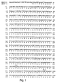
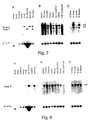
- a Human Cancer Cell Line Multiple Tissue Northern Blot (Clontech, Palo Alto, CA; #7757-1) was probed. This Northern blot contained approximately 2 ⁇ g of poly A + RNA per lane from eight different human cell lines: (1) promyelocytic leukemia HL-60, (2) HeLa cell S3, (3) chronic myelogenous leukemia K-562, (4) lymphoblastic leukemia MOLT-4, (5) Burkitt's lymphoma Raji, (6) colorectal adenocarcinoma SW480, (7) lung carcinoma A549, and (8) melanoma G361.
- a Human Multiple Tissue Northern Blot (Clontech, Palo Alto, CA; #7759-1) was probed. This Northern blot contained approximately 2 ⁇ g of poly A + RNA from eight different human tissues: (I) spleen, (2) thymus, (3) prostate, (4) testis, (5) ovary, (6) small intestine, (7) colon, and (8) peripheral blood leukocytes.
- the Northern blots were hybridized sequentially with: (1) a 1.6 kb probe to the XIAP coding region, (2) a 375 bp HIAP-2 specific probe corresponding to the 3' untranslated region, (3) a 1.3 kb probe to the coding region of HIAP-1, which cross-reacts with HIAP-2, (4) a 1.0 kb probe derived from the coding region of BCL-2, and (5) a probe to ⁇ -actin, which was provided by the manufacturer. Hybridization was carried out at 50°C overnight, according to the manufacturer's suggestion. The blot was washed twice with 2X SSC, 0.1% SDS at room temperature for 15 minutes and then with 2X SSC, 0.1% SDS at 50°C.
- upregulation of the anti-apoptotic IAP genes may be a widespread phenomenon in proliferative diseases, perhaps occurring much more frequently than upregulation of BCL-2. Furthermore, upregulation may be necessary for the establishment or maintenance of the transformed state of cancerous cells.
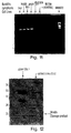
- RNA was reverse transcribed, and amplified by PCR with the following set of oligonucleotide primers: 5'-AGTGCGGGTTTTTATTATGTG-3' (SEQ ID NO: 15) and 5'-AGATGACCACAAGGAATAAACACTA-3' (SEQ ID NO: 16), which selectively amplify a hiap-1 cDNA fragment.
- RT-PCR was conducted using a Perkin Elmer 480 Thermocycler to carry out 35 cycles of the following program: 94°C for 1 minute, 50°C for 1.5 minutes, and 72°C for 1 minute.
- the PCR reaction product was electrophoresed on an agarose gel and stained with ethidium bromide.
- Amplified cDNA fragments of the appropriate size were clearly visible in all lanes containing Burkitt's lymphoma samples, but absent in the lanes containing the normal placental tissue sample, and absent in lanes containing negative control samples, where template DNA was omitted from the reaction (Fig. 11).
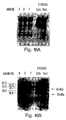
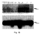
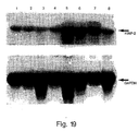
- Figs. 18 and 19 show data demonstrating that HIAP-1 and HIAP-2 are both upregulated in breast cancer cell lines that contain mutant p53.
- the lanes contain 20 ⁇ g of total RNA from the following lines: 1. MCF-7 (clone 1, wt p53); 2. MCF-7 (clone 2, wt p53); 3. MCF-7 (American Type Culture Collection, wt p53); 4. MCF-7 (parental line, California, wt p53); 5. MCF-7 (California, adriamycin resistant variant, mutant p53); 6. MDA MB 231 (ATCC, mutant p53, codon 280); 7.
- T47-D (ATCC, mutant p53, codon 194); 8. ZR-75 (ATCC, wt p53).
- GPDH glycerol phosphate dehydrogenase
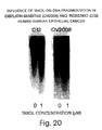
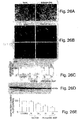
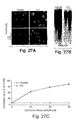
- Epithelial ovarian cancer is the leading cause of death from gynecologic malignancy. Although clinical and histologic prognostic factors such as tumor grade and surgical stage are well understood, the biologic process that leads to uncontrolled cellular growth is less clear. The control of cell numbers during tissue growth is thought to be the results of a balance of cell proliferation and cell death. An aberration in this natural homeostasis likely contributes to malignant cellular transformation.
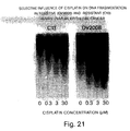
- Cisplatin-sensitive (OV2008) and cisplatin-resistant (C13) human ovarian epithelial cells were cultured in a chemically-defined medium at 37°C for up to 48 hours in the presence or absence of TGF ⁇ (20 ng/ml), taxol (0 - 1.0 ⁇ M) or cisplatin (0 - 30 ⁇ M).
- TGF ⁇ 20 ng/ml
- taxol 0. - 1.0 ⁇ M
- cisplatin 0. - 30 ⁇ M
- DNA ladders For quantitation of DNA ladders, cellular DNA was extracted using the Qiagen Blood kit (Qiagen Inc., Chatsworth, CA). DNA was quantified by ethidium bromide fluorescence. DNA (0.5 ⁇ g) was then end labelled by incubating (20 min., room temp.) with Klenow enzyme (2 U in 10 mM Tris plus 5 mM MgCl 2 ) and 0.1 ⁇ Ci [ ⁇ 32 P]dCTP. Unincorporated nucleotides were removed with the Qiagen nucleotide removal kit and samples were resolved by Tris-acetate-EDTA agarose (1.8%) gel electrophoresis.
- the gel was then dried (2 hours, no heat) and exposed to a Bio-Rad phosphoimager screen to densitometrically quantify low molecular weight DNA ( ⁇ 15 kilo base-pairs), and subsequently to X-ray film at -80°C.
- Protein extracts were prepared from human surface epithelial cancer cells sonicated (8 sec/cycle, 3 cycles) on ice in sucrose buffer (0.25 M sucrose, 0.025 M NaCl, 1 mM EGTA and 15 mM Tris-HCl pH 6.8, supplemented with 1 mM PMSF, 2 ⁇ g/ml of leupeptin and 5 ⁇ g/ml of aprotinin.
- sucrose buffer 0.25 M sucrose, 0.025 M NaCl, 1 mM EGTA and 15 mM Tris-HCl pH 6.8, supplemented with 1 mM PMSF, 2 ⁇ g/ml of leupeptin and 5 ⁇ g/ml of aprotinin.
- the sonicates were centrifuged at 13,000xg for 10 min., the supernatants were collected and stored at -20°C until electrophoretic analyses were performed. Protein concentration was determined by Bio-Rad Protein Assay.
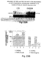
- Proteins (10-30 ⁇ g) were resolved by one-dimensional SDS-PAGE, and electrophoretically transferred to nitrocellulose membrane. Membranes were blocked with 5% non-fat milk, and subsequently incubated with rabbit polyclonal antibody for IAP [anti-human HIAP-2 ⁇ E (960529; 1:1000 dilution), anti-human NAIP E1.0 (951015; 1:1000 dilution) or anti-human XIAP (1:1000 dilution)] diluted in TBST (10 mM Tris-buffered saline, 0.1% Tween-20, pH7.5) containing 5% milk. An ECL kit was used to visualize immunopositive protein (Amersham Intl., Arlington Heights, IL).
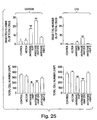
- RNA from ovarian surface epithelial cancer cells by using RNeasy Kit (Qiagen).
- the RNA samples (10-15 ⁇ g) were quantified spectrophotometrically and size-fractioned by electrophoresis on formaldehyde-agarose gels (1.1%) containing 1 ⁇ g/ml ethidium bromide to confirm even loading of RNA samples and adequate separation of 28S and 18S ribosomal bands.
- the RNAs bands were blotted onto a nylon membrane and crosslinked by UV light.
- Membranes were prehybridized in 50% formamide, saline sodium citrate (SSC; 750 mM NaCl, 75 mM sodium citrate), 1X Denhardt's solution, 1% SDS, 4 mM EDTA and 100 ⁇ g/ml sheared salmon sperm DNA for 4 hours at 42°C. Hybridization was performed overnight at 42 °C with 20 million cpm of 32 P-labelled IAP cDNA probes (rat NAIP, rat XIAP or human HIAP-2) added to the prehybridization buffer.
- SSC saline sodium citrate
- 1X Denhardt's solution 1% SDS
- 4 mM EDTA 100 ⁇ g/ml sheared salmon sperm DNA
- the membranes were then washed twice with SSC (300 mM NaCl, 30 mM sodium citrate) in 0.1% SDS for 20 min at room temperature and twice with SSC (30 mM NaCl, 3 mM sodium citrate) in 0.1% SDS for 20 min at 55°C and exposed to X-ray film at -80°C for visualization. Densitometric analysis of various IAPs and 28S rRNA band was performed with the Image Analysis Systems from Bio-Rad Laboratories. Data were normalized by the respective 28S and expressed as a percentage of the control (defined as 100%).
- Induction of apoptosis in human ovarian epithelial cancer cell by Taxol was accompanied by suppressed IAP gene expression.
- Eventual loss of sensitivity of the cells to the chemotherapeutic agent may be associated with the decreased ability of the cell to express IAP genes.
- the decreased HIAP-2 protein content in the face of an absence of noticeable change in HIAP-2 mRNA abundance
- Taxol was accompanied an increase in the intensity of a 45 kDa immunoreactive HIAP-2 protein band.
- EXAMPLE 4 Accumulation of a 26 kDa Cleavage Protein in Astrocytoma Cells
- a total protein extract was prepared from Jurkat and astrocytoma cells by sonicating them (X3 for 15 seconds at 4°C) in 50 mM Tris-HCI (pH 8.0), 150 mM NaCl, 1 mM PMSF, 1 ⁇ g/ml aprotinin, and 5 mM benzamidine. Following sonication, the samples were centrifuged (14,000 RPM in a micro centrifuge) for five minutes. 20 ⁇ g of protein was loaded per well on a 10% SDS-polyacrylamide gel, electrophoresed, and electroblotted by standard methods to PVDF membranes.
- a 26 kDa XIAP-reactive band was also observed under the following experimental conditions.
- Jurkat cells a transformed human T cell line
- an anti-Fas antibody (1 ⁇ g/ml).
- Identical cultures of Jurkat cells were exposed either to: (1) anti-Fas antibody and cycloheximide (20 ⁇ g/ml), (2) tumor necrosis factor alpha (TNF- ⁇ , at 1,000 U/ml), or (3) TNF- ⁇ and cycloheximide (20 ⁇ g/ml). All cells were harvested 6 hours after treatment began.
- anti-Fas antibody was added to an extract after the cells were harvested.
- the cells were harvested in SDS sample buffer, electrophoresed on a 12.5% SDS polyacrylamide gel, and electroblotted onto PVDF membranes using standard methods.
- the membranes were immunostained with a rabbit polyclonal anti-XIAP antibody at 1:1000 for 1 hour at room temperature. Following four 15 minute washes, a goat anti-rabbit antibody conjugated to horse-radish peroxidase was applied at room temperature for 1 hour. Unbound secondary antibody was washed away, and chemiluminescent detection of XIAP protein was performed.
- the Western blot revealed the presence of the full-length, 55 kDa XIAP protein, both in untreated and treated cells. In addition, a novel, approximately 26 kDa XIAP-reactive band was also observed in apoptotic cell extracts, but not in the control, untreated cell extracts (Fig. 13).
- Cleavage of XIAP occurs in a variety of cell types, including other cancer cell lines such as HeLa.
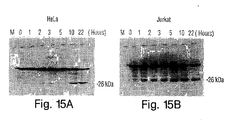
- the expression of the 26 kDa XIAP cleavage product was demonstrated in HeLa cells as follows. HeLa cells were treated with either: (1) cyclohexamide (20 ⁇ g/ml), (2) anti-Fas antibody (1 ⁇ g/ml), (3) anti-Fas antibody (1 ⁇ g/ml) and cyclohexamide (20 ⁇ g/ml), (4) TNF ⁇ (1,000 U/ml), or (5) TNF ⁇ (1,000 U/ml) and cyclohexamide (20 ⁇ g/ml). All cells were harvested 18 hours after treatment began.
- the time course over which the 26 kDa cleavage product accumulates was examined by treating HeLa and Jurkat cells with anti-Fas antibody ( 1 ⁇ g/ml) and harvesting them either immediately, or 1, 2, 3, 5, 10, or 22 hours after treatment. Protein extracts were prepared and Western blot analysis was performed as described above. Both types of cells accumulated increasing quantities of the 26 kDa cleavage product over the time course examined (Figs. 15A and 15B).
- Jurkat cells were induced to undergo apoptosis by exposure to anti-Fas antibody (1 ⁇ g/ml) and were then harvested either immediately, 3 hours, or 7 hours later.
- Total protein extracts were prepared, as described above, from cells harvested at each time point.
- apoptotic Jurkat cells were washed with isotonic Tris buffered saline (pH 7.0) and lysed by freezing and thawing five times in cell extraction buffer (50 mM PIPES, 50 mM KCI, 5 mM EGTA, 2 mM MgCl 2 , 1 mM DTT, and 20 ⁇ M cytochalasin B). Nuclei were pelleted by centrifugation and resuspended in isotonic Tris (pH 7.0) and frozen at -80°C.
- the cytoplasmic fraction of the extract was processed further by centrifugation at 60,000 RPM in a TA 100.3 rotor for 30 minutes. Supernatants were removed and frozen at -80°C. Samples of both nuclear and cytoplasmic fractions were loaded on a 12.5% SDS-polyacrylamide gel, and electroblotted onto PVDF membranes. Western blot analysis was then performed using either an anti-CPP32 antibody (Transduction Laboratories Lexington, KY; Fig. 16A) or the rabbit anti-XIAP antibody described above (Fig. 16B).
- the anti-CPP32 antibody which recognizes the CPP32 protease (also known as YAMA or Apopain) partitioned almost exclusively in the cytoplasmic fraction.
- the 55 kDa XIAP protein localized exclusively in the cytoplasm of apoptotic cells, in agreement with the studies presented above, where XIAP protein in normal, healthy COS cells was seen to localize, by immunofluoresence microscopy, to the cytoplasm.
- the 26 kDa cleavage product localized exclusively to the nuclear fraction of apoptotic Jurkat cells.
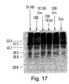
- XIAP protein was labeled with 35 S using the plasmid pcDNA3-6myc-XIAP, T7 RNA polymerase, and a coupled transcription/translation kit (Promega, Madison, WI) according to the manufacturer's instructions. Radioactively labeled XIAP protein was separated from unincorporated methionine by column chromatography using Sephadex G-50TM. In addition, extracts of apoptotic Jurkat cells were prepared following treatment with anti-Fas antibody (1 ⁇ g/ml) for three hours.
- the cells were lysed in Triton X-100 buffer (1% Triton X-100, 25 mM Tris HCl) on ice for two hours and then microcentrifuged for 5 minutes. The soluble extract was retained (and was labeled TX100). Cells were lysed in cell extraction buffer with freeze/thawing. The soluble cytoplasmic fraction was set aside (and labeled CEB). Nuclear pellets from the preparation of the CEB cytoplasmic fraction were solubilized with Triton X-100 buffer, microcentrifuged, and the soluble fractions, which contains primarily nuclear DNA, was retained (and labeled CEB-TX100).
- Triton X-100 buffer 1% Triton X-100, 25 mM Tris HCl
- Soluble cell extract was prepared by lysing cells with NP-40 buffer, followed by microcentrifugation for 5 minutes (and was labeled NP-40). In vitro cleavage was performed by incubating 16 ⁇ l of each extract (CEB, TX-100, CEB-TX100, and NP-40) with 4 ⁇ l of in vitro translated XIAP protein at 37°C for 7 hours. Negative controls, containing only TX100 buffer or CEB buffer were also included. The proteins were separated on a 10% SDS-polyacrylamide gel, which was dried and exposed to X-ray film overnight.
- IAPs to modulate apoptosis
- Mammalian expression constructs carrying IAP cDNAs which are either full-length truncated, or antisense constructs can be introduced into cell lines such as CHO, NIH 3T3, HL60, Rat-1, or Jurkat cells.
- SF21 insect cells may be used. in which case the IAP gene is preferentially expressed using an insect heat shock promoter.
- apoptosis can be induced by standard methods, which include serum withdrawal, or application of staurosporine, menadione (which induces apoptosis via free radial formation), or anti-Fas antibodies.
- cells are cultured under the same conditions as those induced to undergo apoptosis, but either not transfected, or transfected with a vector that lacks an IAP insert.
- the ability of each IAP related construct to inhibit or enhance apoptosis upon expression can be quantified by calculating the survival index of the cells, i.e., the ratio of surviving transfected cells to surviving control cells.
- Figs. 10A to 10D Specific examples of the results obtained by performing various apoptosis suppression assays are shown in Figs. 10A to 10D.
- CHO cell survival following transfection with one of six constructs and subsequent serum withdrawal is shown in Fig. 10A.
- the cells were transfected using LipofectaceTM with 2 ⁇ g of one of the following recombinant plasmids: pCDNA36myc-xiap (XIAP), pCDNA3-6myc-hiap-1 (HIAP-1), pCDNA3-6myc-hiap-2 (HIAP-2), pCDNA3-bcl-2 (BCL-2), pCDNA3-HA-smn (SMN), and pCDNA3-6myc (6-myc).
- XIAP pCDNA36myc-xiap
- HIAP-1 pCDNA3-6myc-hiap-1
- HIAP-2 pCDNA3-6myc-hiap-2
- Oligonucleotide primers were synthesized to allow PCR amplification and cloning of the XIAP, HIAP-1, and HIAP-2 ORFs in pCDNA3 (Invitrogen). Each construct was modified to incorporate a synthetic myc tag encoding six repeats of the peptide sequence MEQKLISEEDL (SEQ ID NO: 17), thus allowing detection of myc-IAP fusion proteins via monoclonal anti-myc antiserum (Egan et al ., Nature 363: 45-51, 1993). Triplicate samples of cell lines in 24-well dishes were washed 5 times with serum free media and maintained in serum free conditions during the course of the experiment.
- Fig. 10B The survival of CHO cells following transfection (with each one of the six constructs described above) and exposure to menadione is shown in Fig. 10B.
- the cells were plated in 24-well dishes, allowed to grow overnight, and then exposed to 20 ⁇ M menadione for 1.5 hours (Sigma Chemical Co., St. Louis, MO). Triplicate samples were harvested at the time of exposure to menadione and 24 hours afterward, and survival was assessed by trypan blue exclusion.
- Rat-1 cells were transfected and then selected in medium containing 800 ⁇ g/ml G418 for two weeks. The cell line was assessed for resistance to staurosporine-induced apoptosis (1 ⁇ M) for 5 hours. Viable cells were counted 24 hours after exposure to staurosporine by trypan blue exclusion. The percentage of viable cells shown represents the average of two experiments, +/- average deviation.
- Rat-1 cell line was also used to test the resistance of these cells to menadione (Fig. 10D) following transfection with each of the six constructs described above.
- the cells were exposed to 10 ⁇ M menadione for 1.5 hours, and the NUMBER of viable cells was counted 18 hours later.
- EXAMPLE 7 COMPARISON OF CELL SURVIVAL FOLLOWING TRANSFECTION WITH FULL-LENGTH VS. PARTIAL IAP CONSTRUCTS
- expression vectors were constructed that contained either: (1) full-length IAP cDNA (as described above), (2) a portion of an IAP gene that encodes the BIR domains, but not the RZF, or (3) a portion of an IAP gene that encodes the RZF, but not the BIR domains.
- Human and murine XIAP cDNAs were tested by transient or stable expression in HeLa, Jurkat, and CHO cell lines. Following transfection, apoptosis was induced by serum withdrawal, application of menadione, or application of an anti-Fas antibody. Cell death was then assessed, as described above, by trypan blue exclusion. As a control for transfection efficiency, the cells were co-transfected with a ⁇ -gal expression construct. Typically, approximately 20% of the cells were successfully transfected.
- Stable pools of transfected CHO cells which were maintained for several months under G418 selection, were induced to undergo apoptosis by exposure to 10 ⁇ M menadione for 2 hours.
- CHO cells tested were those that were stably transfected with: (1) full-length murine XIAP cDNA (MIAP), (2) full-length XIAP cDNA (XIAP), (3) full-length BCL-2 cDNA (BCL-2), (4) cDNA encoding the three BIR domains (but not the RZF) of murine XIAP (BIR), and (5) cDNA encoding the RZF (but not BIR domains) of M-XIAP (RZF).
- MIAP full-length murine XIAP cDNA
- XIAP full-length XIAP cDNA
- BCL-2 full-length BCL-2 cDNA
- RZF cDNA encoding the three BIR domains (but not the RZF) of murine
- the percentage of viable CHO cells that expressed the BIR domain cDNA construct was higher than the percentage of viable cells that expressed either full-length murine XIAP or BCL-2.
- EXAMPLE 8 ANALYSIS OF THE SUBCELLULAR LOCATION OF EXPRESSED RZF AND BIR DOMAINS
- the assays of cell death described above indicate that the RZF acts as a negative regulator of the anti-apoptotic function of IAPs.
- One way in which the RZF, and possibly other IAP domains, may exert their regulatory influence is by altering the expression of genes, whose products function in the apoptotic pathway.
- COS cells were transiently transfected with the following four constructs, and the expressed polypeptide was localized by immunofluorescent microscopy: (1) pcDNA3-6myc-XIAP, which encodes all 497 amino acids of SEQ ID NO: 4, (2) pcDNA3-6myc-m-XIAP, which encodes all 496 amino acids of mouse XIAP (SEQ ID NO: 10), (3) pcDNA3-6myc-mxiap-BIR, which encodes amino acids 1 to 341 of m-XIAP, and (4) pcDNA3-6myc-mxiap-RZF, which encodes amino acids 342-496 of murine XIAP.
- pcDNA3-6myc-XIAP which encodes all 497 amino acids of SEQ ID NO: 4
- pcDNA3-6myc-m-XIAP which encodes all 496 amino acids of mouse XIAP (SEQ ID NO: 10)
- pcDNA3-6myc-mxiap-BIR which encodes amino acids
- the cells were grown on multi-well tissue culture slides for 12 hours, and then fixed and permeabilized with methanol.
- the constructs used (here and in the cell death assays) were tagged with a human Myc epitope tag at the N-terminus. Therefore, a monoclonal anti-Myc antibody and a secondary goat anti-mouse antibody, which was conjugated to FITC, could be used to localize the expressed products in transiently transfected COS cells.
- Full-length XIAP and MIAP were located in the cytoplasm, with accentuated expression in the peri-nuclear zone.
- the same pattern of localization was observed when the cells expressed a construct encoding the RZF domain (but not the BIR domains). However, cells expressing the BIR domains (without the RZF) exhibited, primarily, nuclear staining.
- the protein expressed by the BIR domain construct appeared to be in various stages of transfer to the nucleus.
- XIAP is cleaved within T cells that are treated with anti-Fas antibodies (which are potent inducers of apoptosis), and its N-terminal domain is translocated to the nucleus.
- anti-Fas antibodies which are potent inducers of apoptosis
- HIAP-2 appears to undergo a similar cleavage event.
- apoptosis assays are also provided in the following references. Assays for apoptosis in lymphocytes are disclosed by: Li et al ., Science 268: 429-431, 1995; Gibellini et al ., Br. J. Haematol. 89: 24-33, 1995; Martin et al ., J. Immunol. 152: 330-342, 1994; Terai et al ., J. Clin Invest. 87: 1710-1715, 1991; Dhein et al ., Nature 373: 438-441, 1995; Katsikis et al ., J. Exp. Med. 1815: 2029-2036, 1995; Westendorp et al ., Nature 375: 497-500, 1995; DeRossi et al ., Virology 198: 234-244, 1994.
- IAP and NAIP genes provided information that necessary for generation IAP and NAIP transgenic animal models to be developed by homologous recombination (for knockouts) or transfection (for expression of IAP or NAIP fragments, antisense nucleic acids, or increased expression of wild-type or mutant IAPs or NAIP).
- Such a model may be a mammalian animal, e.g ., a mouse, and is useful for the identification of cancer therapeutics alone or in combination with cancer inducing cells or agents, or when such mice are crossed with mice genetically predisposed to cancers.
- the preferred transgenic animal overexpression in IAP or NAIP and has a predisposition to cancer has a predisposition to cancer. This mouse is particularly useful for the screening of potential cancer therapeutics.
- IAP and NAIP genes and fragments thereof may be expressed in both prokaryotic and eukaryotic cell types. If an IAP or NAIP fragment enhances apoptosis, it may be desirable to express that protein under control of an inducible promoter.
- LAPs and NAIP may be produced by transforming a suitable host cell with all or part of the IAP-encoding or NAIP-encoding cDNA fragment that has been placed into a suitable expression vector.
- the IAP protein may be produced in a prokaryotic host (e.g ., E. coli ) or in a eukaryotic host (e.g., S. cerevisiae , insect cells such as Sf21 cells, or mammalian cells such as COS-1, NIH 3T3, or HeLa cells, or other highly proliferative cell types). These cells are publically available, for example, from the American Type Culture Collection, Rockville, MD; see also Ausubel et al., supra ).
- transduction and the choice of expression vehicle will depend on the host system selected. Transformation and transfection methods are described, e.g ., in Ausubel et al. ( supra ), and expression vehicles may be chosen from those provided, e.g ., in Cloning Vectors: A Laboratory Manual (P.H. Pouwels et al ., 1985, Supp. 1987).
- Polypeptides of the invention can also be produced by chemical synthesis ( e.g ., by the methods described in Solid Phase Peptide Synthesis , 2nd ed., 1984 The Pierce Chemical Co., Rockford, IL). These general techniques of polypeptide expression and purification can also be used to produce and isolate useful IAP fragments or analogs, as described herein.
- an IAP or NAIP coding sequence (e.g ., amino acids 180-276) can be expressed as a C-terminal fusion with glutathione S-transferase (GST: Smith et al ., Gene 67: 31-40, 1988).
- the fusion protein can be purified on glutathione-Sepharose beads, eluted with glutathione, and cleaved with thrombin (at the engineered cleavage site), and purified to the degree required to successfully immunize rabbits.
- Primary immunizations can be carried out with Freund's complete adjuvant and subsequent immunizations performed with Freund's incomplete adjuvant.
- Antibody titres are monitored by Western blot and immunoprecipitation analyses using the thrombin-cleaved IAP fragment of the GST-IAP and GST-NAIP fusion proteins. Immune sera are affinity purified using CNBr-Sepharose-coupled IAP protein. Antiserum specificity is determined using a panel of unrelated GST proteins (including GSTp53, Rb, HPV-16 E6, and E6-AP) and GST-trypsin (which was generated by PCR using known sequences).
- peptides corresponding to relatively unique hydrophilic regions of IAP or NAIP may be generated and coupled to keyhole limpet hemocyanin (KLH) through an introduced C-terminal lysine.
- KLH keyhole limpet hemocyanin
- Antiserum to each of these peptides is similarly affinity purified on peptides conjugated to BSA, and specificity is tested by ELISA and Western blotting using peptide conjugates, and by Western blotting and immunoprecipitation using IAP or NAIP expressed as a GST fusion protein.
- monoclonal antibodies may be prepared using the IAP or NAIP proteins described above and standard hybridoma technology (see, e.g., Kohler et al ., Nature 256: 495, 1975; Kohler et al ., Eur. J. Immunol. 6: 511, 1976; Kohler et al., Eur. J. Immunol. 6:292, 1976; Hammerling et al ., In Monoclonal Antibodies and T Cell Hybridomas , Elsevier, New York, NY, 1981; Ausubel et al ., supra ). Once produced, monoclonal antibodies are also tested for specific IAP or NAIP recognition by Western blot or immunoprecipitation analysis (by the methods described in Ausubel et al ., supra ).
- Antibodies that specifically recognize IAPs or NAIP or fragments thereof, such as those described herein containing one or more BIR domains (but not a ring zinc finger domain), or that contain a ring zinc finger domain (but not a BIR domain) are considered useful in the invention. They may, for example, be used in an immunoassay to monitor IAP or NAIP expression levels or to determine the subcellular location of an IAP or NAIP (or fragment thereof) produced by a mammal. Antibodies that inhibit the 26 kDa IAP cleavage product described herein (which contains at least one BIR domain) may be especially useful in inducing apoptosis in cells undergoing undesirable proliferation.
- antibodies of the invention are produced using IAP or NAIP sequence that does not reside within highly conserved regions, and that appears likely to be antigenic, as analyzed by criteria such as those provided by the Peptide structure program (Genetics Computer Group Sequence Analysis Package, Program Manual for the GCG Package, Version 7, 1991) using the algorithm of Jameson and Wolf (CABIOS 4: 181, 1988).
- these regions which are found between BIR1 and BIR2 of all IAPs, are: from amino acid 99 to amino acid 170 of HIAP-1, from amino acid 123 to amino acid 184 of HIAP-2, and from amino acid 116 to amino acid 133 of either XIAP or m-XIAP.
- fragments can be generated by standard techniques, e.g ., by the PCR, and cloned into the pGEX expression vector (Ausubel et al., supra ). Fusion proteins are expressed in E. coli and purified using a glutathione agarose affinity matrix as described in Ausubel et al. ( supra ). In order to minimize the potential for obtaining antisera that is non-specific, or exhibits low-affinity binding to IAP, two or three fusions are generated for each protein, and each fusion is injected into at least two rabbits. Antisera are raised by injections in series, preferably including at least three booster injections.
- EXAMPLE 14 IDENTIFICATION OF MOLECULES THAT MODULATE THE EXPRESSION OR BIOLOGICAL ACTIVITY OF AN IAP OR NAIP GENE
- IAP and NAIP cDNAs facilitate the identification of molecules that decrease IAP or NAIP expression or otherwise enhance apoptosis normally blocked by these polypeptides.
- Such compounds are highly useful as, for example, chemotherapeutic agents to destroy a cancer cell, or to reduce the growth of a cancer cell, where the cancer cell is one, as is described herein, with an elevated level of an IAP or NAIP polypeptide.
- candidate molecules are added, in varying concentration, to the culture medium of cells expressing IAP or NAIP mRNA.
- IAP or NAIP expression is then measured, for example, by Northern blot analysis (Ausubel et al ., supra ) using an IAP or NAIP cDNA, or cDNA fragment, as a hybridization probe.
- the level of IAP or NAIP expression in the presence of the candidate molecule is compared to the level of IAP or NAIP expression in the absence of the candidate molecule, all other factors (e.g., cell type and culture conditions) being equal.
- the effect of candidate molecules on IAP- or NAIP-mediated apoptosis may, instead, be measured at the level of protein or the level of polypeptide fragments of IAP or NAIP polypeptides using the general approach described above with standard polypeptide detection techniques, such as Western blotting or immunoprecipitation with an IAP or NAIP-specific antibodies (for example, the antibodies described herein).
- Compounds that modulate the level of a IAP or NAIP polypeptide may be purified, or substantially purified, or may be one component of a mixture of compounds such as an extract or supernatant obtained from cells (Ausubel et al. , supra ). In an assay of a mixture of compounds, IAP or NAIP polypeptide expression is tested against progressively smaller subsets of the compound pool (e.g. , produced by standard purification techniques such as HPLC or FPLC) until a single compound or minimal number of effective compounds is demonstrated to modulate IAP or NAIP expression.
- Compounds may also be screened for their ability to modulate the biological activity of an IAP or NAIP polypeptide by, for example, an ability to enhance IAP- or NAIP-mediated apoptosis.
- the degree of apoptosis in the presence of a candidate compound is compared to the degree of apoptosis in its absence, under equivalent conditions.
- the screen may begin with a pool of candidate compounds, from which one or more useful modulator compounds are isolated in a step-wise fashion.
- Apoptosis activity may be measured by any standard assay, for example, those described herein.
- Another method for detecting compounds that modulate the expression or biological activity of an IAP or a NAIP polypeptide is to screen for compounds that interact physically with a given IAP polypeptide.
- These compounds may be detected by adapting two hybrid systems known in the art. These systems detect protein interactions using a transcriptional activation assay and are generally described by Gyuris et al. (Cell 75: 791-803, 1993) and Field et al. (Nature 340: 245-246, 1989), and are commercially available from Clontech (Palo Alto, CA).
- PCT Publication WO 95/28497 describes a two hybrid system in which proteins involved in apoptosis, by virtue of their interaction with BCL-2, are detected.
- a similar method may be used to identify proteins and other compounds that interact with IAP or NAIP polypeptides.
- Compounds or molecules that function as modulators of IAP-mediated cell death may include peptide and non-peptide molecules such as those present in cell extracts, mammalian serum, or growth medium in which mammalian cells have been cultured.
- compounds previously known for their abilities to modulate apoptosis in cancer cells may be tested for an ability to modulate expression of an IAP molecule.
- EXAMPLE 15 ROLE OF IAPs IN HUMAN OVARIAN CANCER RESISTANCE TO CISPLATIN
- Ovarian epithelial cancer cell apoptosis has been demonstrated to be involved in cisplatin-induced cell death (Hovicesky et al ., Obstet. Gynecol. 85: 1007-1010, 1995; Anthoney et al ., Cancer Res. 56: 1374-1381, 1996).
- the action of cisplatin is thought to involve the formation of inter and intra-strand DNA crosslinks (Sherman et al ., Science 230: 412-417, 1985) although the events leading to cell death after cisplatin treatment is unclear.
- cisplatin-sensitive human ovarian surface epithelial cells (OV2008) were infected with either adenoviral XIAP antisense, adenoviral HIAP-2 antisense, or the empty vector with LacZ (as control) for up to 60 hours, at which time changes in cell morphology, apoptotic cell number, cell viability, and total cell number were determined.
- adenoviral XIAP antisense adenoviral HIAP-2 antisense
- LacZ as control
- the open reading frame for XIAP and HIAP-2 were PCR amplified with primers corresponding to the amino and carboxy terminus. These PCR products were cloned in the pCR2.1 vector (InvitroGen, Carlsbad. CA), and sequenced. The ORFs were then excised with EcoRI digestion, blunt ended with Klenow fragment, and ligated into Swal digested pAdex1CAwt cosmid DNA. Packaging was performed with Promega (Madison, WI) cosmid packaging extracts and used to infect E. coli .
- Colonies were picked and screened for the presence of the insert in both the sense and antisense orientation relative to the chicken B-actin (CA) promoter.
- CsCI purified cosmid DNA was co-transfected with wild-type adenovirus DNA, which contains the terminal protein complexed to the ends of the DNA. Wild type adenovirus DNA was cut with NsiI such that only homologous recombinant with the cosmid DNA generated infectious adenovirus DNA.
- the final recombinant adenovirus contains a linear, double stranded genome of 44,820 bp plus the insert size (approximately 1,500 for XIAP, approximately 1,800 for HIAP-2).
- MOI adenoviral anti-sense
- OV2008 cels were cultured in the absence and presence of cisplatin (10-30 ⁇ M) for 24 hours, apoptosis and XIAP and HIAP-2 expression were assessed morphologically and by Western analysis, respectively.
- apoptosis and XIAP and HIAP-2 expression were assessed morphologically and by Western analysis, respectively.
- adenoviral XIAP antisense infection the presence of cisplatin induced morphologic feature of apoptosis in OV2008 cells, including decreased cell volume, chromatin condensation and nuclear fragmentation (Fig. 27A, left two photographs), and apoptotic low molecular weight DNA fragmentation (Fig. 27B), and was accompanied by decreased IAP expression (Figs. 28A and 28B).
- the increase of apoptotic cell number in response to cisplatin was also concentration-dependent and was significant (50% vs. 2%; p ⁇ 0.05) even at a concentration of 10 ⁇ M cisplatin (Fig
- XIAP and HIAP-2 are present in the cisplatin-sensitive human ovarian surface epithelial cancer cell line OV2008 (protein sizes 55kDa and 68 kDa, respectively), their expression were down-regulated by cisplatin in a concentration-dependent manner.
- XIAP appearing more responsive to the anti-cancer agent. While XIAP protein content was decreased by almost 80% (p ⁇ 0.01) in the presence 20 ⁇ M cisplatin, the decrease of HIAP-2 protein content was not suppressed by cisplatin (Figs. 28A and 28B).
- XIAP protein content was decreased in A2780s (as in OV2008 cells) and not significantly altered in A2780cp (as in C13 cells) in the presence of the chemotherapeutic agent.
- XIAP expression is indeed the an important determinant in chemoresistance in human ovarian surface epithelial cancer
- the influence of cisplatin on XIAP protein content and apoptosis in OV2008 cells following adenoviral XIAP sense infection was investigated. While cisplatin reduced XIAP protein content in OV2008 cells infected with the empty vector (Figs. 31C and 31D, vector plus cisplatin), overexpression of the protein with adenoviral sense XIAP cDNA 48 hrs prior to treatment with the chemotherapeutic agent in vitro attenuated the cisplatin effects not only on XIAP protein expression (Figs.
- XIAP may be an important element in human ovarian epithelial cancer chemoresistance.
- XIAP and HIAP-2 were immunlocalized in human ovarian surface epithelial tumors obtained as pathological samples from patients during surgical debulking, using polyclonal antibodies (rabbit polyclonal anti-XIAP and HIAP-2 antibodies were prepared by immunization with human XIAP and HIAP-2 GST fusion protein) against human XIAP and HIAP-2, respectively (Figs. 32C and 32D, respectively).
- XIAP and HIAP-2 immunoreactivity specifically localized in the cytoplasm or the perinuclear region was highest in proliferatively active cells (PCNA positive) and was low or absent in apoptotic cells (TUNEL positive) occasionally found in the tumor specimens.
- an anti-cancer agent that will successfully inhibit the growth of the particular cancer of interest.
- One method to detect such an agent is to excise proliferative cells from the cancer of interest, and determine the level of expression and/or level of biological activity of each individual IAP or NAIP polypeptide, and compare these levels to the levels of these polypeptide in a similar cell type from an unaffected individual. For example, if an human female individual has breast cancer (or a neoplasm suspected of being cancerous), cells from the cancer collected, for example, during a biopsy of the cancer, can be isolated and, if necessary, propagated in culture.
- the cells can then be analyzed for level of expression and/or level of biological activity of all of the IAP and NAIP polypeptides in the cell.
- the expression levels and/or biological activity levels of these polypeptides from the proliferating cells can be compared to the levels of expression and/or biological activity of these polypeptides from normal, healthy cells from a human female individual.
- the comparison is made between on affected ( i.e., abnormally proliferating) and healthy cells of the same individual (e.g. , cells taken from healthy breast tissue from the individual being tested.
- the level of expression and/or biological activity of each polypeptide in the affected cells is compared to its counterpart in the healthy cells. Any increase in any (or all) of the IAP or NAIP polypeptides is detected.
- the cancer is then treated with a compound that decreases expression level or biological activity level of each particular elevated IAP or NAIP polypeptide. Methods for identifying such compounds are described above (see, e.g ., Example 14).
- the individual undergoing such analysis and treatment may have already received treatment with an anti-cancer therapeutic agent. It will also be understood that, in addition to targeting the levels of expression and/or biological activities of IAP and NAIP polypeptides, the anti-cancer compounds may also target these levels for other apoptosis-inhibiting polypeptides, such as BCL-2.
- an individual with breast cancer whose proliferating cells have an increased level of XIAP compared to the level of XIAP in healthy breast cells may be treated with a compound (e.g ., cisplatin) plus a compound that targets another IAP polypeptide, or that targets an NAIP polypeptide or a non-related apoptosis-inhibiting polypeptide, such as BCL-2).
- a compound e.g ., cisplatin
- a compound that targets another IAP polypeptide or that targets an NAIP polypeptide or a non-related apoptosis-inhibiting polypeptide, such as BCL-2).
- IAP and NAIP polypeptides One rapid method to determine expression levels of IAP and NAIP polypeptides is an ELISA assay using antibodies that specifically binds each of these polypeptides. Other methods include quantitative PCR and the various apoptosis assays described herein.
- EXAMPLE 17 ASSIGNMENT OF XIAP, HIAP-1, AND HIAP-2 TO CHROMOSOMES XQ25 AND 11Q22-23 BY FLUORESCENCE IN SITU HYBRIDIZATION (FISH)
- Fluorescence in situ hybridization was used to identify the chromosomal location of XIAP, HIAP-1 and HIAP-2.
- a total of 101 mctaphasc spreads were examined with the XIAP probe. as described above. Symmetrical fluorescent signals on either one or both homologs of chromosome Xq25 were observed in 74% of the cells analyzed. Following staining with HIAP-1 and HIAP-2 probes, 56 cells were analyzed and doublet signals in the region 11q22-23 were observed in 83% of cells examined. The XIAP gene was mapped to Xq25 while the HIAP-1 and HIAP-2 genes were mapped at the border of 11q22 and 11q23 bands.
- the invention includes use of any protein which is substantially identical to a mammalian IAP polypeptides (Figs. 1-6: SEQ ID Nos: 3-14); such homologs include other substantially pure naturally-occurring mammalian IAP proteins as well as allelic variants; natural mutants; induced mutants; DNA sequences which encode proteins and also hybridize to the IAP DNA sequences of Figs. 1-6 (SEQ ID NOS: 3-14) under high stringency conditions or, less preferably, under low stringency conditions ( e.g ., washing at 2X SSC at 40°C with a probe length of at least 40 nucleotides); and proteins specifically bound by antisera directed to a IAP polypeptide.
- the term also includes chimeric polypeptides that include a IAP portion.
- the invention further includes use of analogs of any naturally-occurring IAP polypeptide.
- Analogs can differ from the naturally-occurring IAP protein by amino acid sequence differences, by post-translational modifications, or by both.
- Analogs of the invention will generally exhibit at least 85%, more preferably 90%, and most preferably 95% or even 99% homology with all or part of a naturally occurring IAP amino acid sequence.
- the length of sequence comparison is at least 15 amino acid residues, preferably at least 25 amino acid residues, and more preferably more than 35 amino acid residues.
- Modifications include in vivo and in vitro chemical derivatization of polypeptides, e.g., acetylation, carboxylation, phosphorylation, or glycosylation; such modifications may occur during polypeptide synthesis or processing or following treatment with isolated modifying enzymes.
- Analogs can also differ from the naturally-occurring IAP polypeptide by alterations in primary sequence.
- cyclized peptides, molecules, and analogs which contain residues other than L-amino acids, e.g., D-amino acids or nonnaturally occurring or synthetic amino acids, e.g., B or y amino acids.
- the invention also includes IAP polypeptide fragments.
- fragment means at least 20 contiguous amino acids, preferably at least 30 contiguous amino acids, more preferably at least 50 contiguous amino acids, and most preferably at least 60 to 80 or more contiguous amino acids. Fragments of IAP polypeptides can be generated by methods known to those skilled in the art or may result from normal protein processing (e.g. , removal of amino acids from the nascent polypeptide that are not required for biological activity or removal of amino acids by alternative mRNA splicing or alternative protein processing events).
- Preferable fragments or analogs used according to the methods of the invention are those which facilitate specific detection of an IAP nucleic acid or amino acid sequence in a sample to be diagnosed.
- Particularly useful IAP fragments for this purpose include, without limitation, the amino acid fragments shown in Table 2.
- the methods of the invention may use antibodies prepared by a variety of methods.
- the IAP or NAIP polypeptide, or antigenic fragments thereof can be administered to an animal in order to induce the production of polyclonal antibodies.
- antibodies used as described herein may be monoclonal antibodies, which are prepared using hybridoma technology (see, e.g., Kohler et al ., Nature 256: 495-497, 1975; Kohler et al ., Eur. J. Immunol. 6: 511-519, 1976; Kohler et al ., Eur. J. Immunol.
- the invention features use of antibodies that specifically bind human or murine IAP or NAIP polypeptides, or fragments thereof.
- the invention features "neutralizing” antibodies.
- neutralizing antibodies is meant antibodies that interfere with any of the biological activities of IAP or NAIP polypeptides, particularly the ability of IAPs to inhibit apoptosis.
- the neutralizing antibody may reduce the ability of IAP polypeptides to inhibit polypeptides by, preferably 50%, more preferably by 70%, and most preferably by 90% or more. Any standard assay of apoptosis, including those described herein, by those incorporated by reference and those in the art, may be used to assess neutralizing antibodies.
- the invention features use of various genetically engineered antibodies, humanized antibodies, and antibody fragments, including F(ab')2, Fab', Fab, Fv and sFv fragments.
- Antibodies can be humanized by methods known in the art, e.g. , monoclonal antibodies with a desired binding specificity can be commercially humanized (Scotgene, Scotland; Oxford Molecular, Palo Alto, CA). Fully human antibodies, such as those expressed in transgenic animals, are also features of the invention (Green et al ., Nature Genetics 7:13-21, 1994).
- Ladner (U.S. Patent Nos. 4,946,778 and 4,704,692) describes methods for preparing single polypeptide chain antibodies.
- Ward et al . (Nature 341: 544-546, 1989) describe the preparation of heavy chain variable domains, which they term "single domain antibodies," which have high antigen-binding affinities.
- McCafferty et al . (Nature 348: 552-554, 1990) show that complete antibody V domains can be displayed on the surface of fd bacteriophage, that the phage bind specifically to antigen, and that rare phage (one in a million) can be isolated after affinity chromatography.
- Boss et al. (U.S. Patent No.
Abstract
Description
- The invention relates to the diagnosis and treatment of proliferative disease, in particular, cancer.
- One mechanism by which cells die is referred to as apoptosis, or programmed cell death. Apoptosis often occurs as a normal part of the development and maintenance of healthy tissues, and is now known to play a critical role in embryonic development. The failure of a normal apoptotic response has been implicated in the development of cancer; autoimmune disorders, such as lupus erythematosis and multiple sclerosis; and in viral infections, including those associated with herpes virus, poxvirus, and adenovirus.
- Compared to the numerous growth promoting genes identified to date (>100) relatively few genes have been isolated that regulate apoptosis. Baculoviruses encode proteins termed inhibitors of apoptosis proteins (IAPs) which inhibit the apoptosis that would otherwise occur when insect cells are infected by the baculovirus. The baculovirus IAP genes include sequences encoding a ring zinc finger-like motif (RZF), which is presumed to be directly involved in DNA binding, and two N-terminal domains that consist of a 70 amino acid repeat motif termed a BIR domain (Baculovirus IAP Repeat). Mammalian IAP family members, and a related anti-apoptotic polypeptide, NAIP, have recently been identified.
- WO97/06255 describes genomic, cDNA or synthetic DNA molecules, which encode a mammalian IAP polypeptide. Nucleic acid sequences encoding a NAIP polypeptide and methods of inhibiting apoptosis via administration of a NAIP polypeptide are already disclosed in WO97/26331.
- Both normal cell types and cancer cell types display a wide range of susceptibility to apoptotic triggers. Many normal cell types undergo temporary growth arrest in response to a sub-lethal dose of radiation or cytotoxic chemical, while cancer cells in the vicinity undergo apoptosis. This provides the crucial treatment "window" of appropriate toxicity that allows successful anti-cancer therapy. It is therefore not surprising that resistance of tumor cells to apoptosis is emerging as a major category of cancer treatment failure. Finding compounds which overcome or prevent this resistance would greatly improve cancer therapies.
- We have discovered that LAP and NAIP overexpression are specifically associated with a wide range of cancer types including ovarian cancer, adenocarcinoma, lymphoma, and pancreatic cancer. The presence of a fragmented IAP polypeptide in the nucleus, and an overexpression of an IAP polypeptide in the presence of a p53 mutation correlates with a cancer diagnosis, a poor prognosis, and a resistance to numerous chemotherapeutic cancer drugs. In addition, we have found that an therapeutic agent that reduces the biological activity of an lAP polypeptide will induce apoptosis in a cell expressing the polypeptide (e.g., a cell that is proliferating in a proliferative disease). These discoveries provide diagnostic and prognostic methods for the detection and treatment of proliferative diseases, and provide therapeutic compounds useful for the treatment of proliferative diseases, particularly cancer.
- In a first aspect, the invention features a method for enhancing apoptosis in a cell from a mammal with a proliferative disease, the method including administering to the cell a compound that inhibits the biological activity of an IAP polypeptide or a NAIP polypeptide, the compound being administered to the cell in an amount sufficient to enhance apoptosis in the cell, wherein said method is performed in vitro, and wherein said cell is proliferating in said proliferative disease. In another embodiment, the biological activity is the level of expression of the polypeptide (measured, for example, by assaying the amount of polypeptide present in the cell); the level of expression of an mRNA molecule encoding the polypeptide; or an apoptosis-inhibiting activity.
- In various embodiments of the first aspect of the invention, the polypeptide is selected from the group consisting of HIAP-1, m-HIAP-1, HIAP-2, m-HIAP-2, XIAP, and m-XIAP. In other embodiment, the polypeptide is NAIP, XIAP, HIAP-1, or HIAP-2. In other preferred embodiments, the mammal is a human or a mouse, and the proliferative disease is cancer, for example, a cancer in a tissue selected from the group consisting of ovary, breast, pancreas, lymph node, skin, blood, lung, brain, kidney, liver, nasopharyngeal cavity, thyroid, central nervous system, prostate, colon, rectum, cervix, endometrium, and lung.
- In various preferred embodiments of the first aspect of the invention, the compound is a negative regulator of an IAP or an NAIP-dependent anti-apoptotic pathway; a fragment of the IAP polypeptide, the fragment including a ring zinc finger and having no more than two BIR domains; a nucleic acid molecule encoding a ring zinc finger domain of the LAP polypeptide; a compound that prevents cleavage of the IAP polypeptide or the NAIP polypeptide; a purified antibody or a fragment thereof that specifically binds to the LAP polypeptide or the NAIP polypeptide; a ribozyme; or an antisense nucleic acid molecule have a nucleic acid sequence that is complementary to the coding strand of a nucleic acid sequence encoding the IAP polypeptide or the NAIP polypeptide. Preferably, the cleavage is decreased by at least 20% in the cell; the antibody binds to a BIR domain of the IAP polypeptide or the NAIP polypeptide; the nucleic acid sequence encoding the IAP polypeptide or the NAIP polypeptide has about 50% or greater homology with the nucleotide sequence of SEQ ID NO: 3. SEQ ID NO: 5, SEQ ID NO: 7, SEQ ID NO: 9. SEQ ID NO: 11, SEQ ID NO: 13, or the nucleic acid sequence of NAIP; the antisense nucleic acid molecule decreases the level of the nucleic acid sequence encoding the IAP polypeptide or the NAIP polypeptide by at least 20%, the level being measured in the cytoplasm of the ccll; the antisense nucleic acid molecule is encoded by a virus vector; or the antisense nucleic acid molecule is encoded by transgene.
- In a second aspect, the invention features a method for detecting a proliferative disease or an increased likelihood of the proliferative disease in a mammal that includes: (a) contacting an IAP or a NAIP nucleic acid molecule that is greater than about 18 nucleotides in length with a preparation of nucleic acid from a cell of the mammal, the cell proliferating in the disease, the cell from a tissue; and (b) measuring the amount of nucleic acid from the cell of the mammal that hybridizes to the molecule, an increase in the amount from the cell of the mammal relative to a control indicating a an increased likelihood of the mammal having or developing a proliferative disease. In one embodiment, the method further includes the steps of: (a) contacting the molecule with a preparation of nucleic acid from the control, wherein the control is a cell from the tissue of a second mammal, the second mammal lacking a proliferative disease; and (b) measuring the amount of nucleic acid from the control, an increase in the amount of the nucleic acid from the cell of the mammal that hybridizes to the molecule relative to the amount of the nucleic acid from the control indicating an increased likelihood of the mammal having or developing a proliferative disease.
- In one embodiment of the methods of the second aspect of the invention, the method further includes the steps of: (a) providing a pair of oligonucleotides having sequence identity to or being complementary to a region of the IAP or the NAIP nucleic acid molecule; (b) combining the pair of oligonucleotides with the nucleic acid under conditions suitable for polymerase chain reaction-mediated nucleic acid amplification; and (c) isolating the amplified nucleic acid or fragment thereof. Preferably, the amplification is carried out using a reverse-transcription polymerase chain reaction (e.g. rapid amplification of cDNA ends, RACE).
- In one embodiment of the second aspect of the invention. the method provides measuring the nucleic acid having a nucleotide sequence that has about 50% or greater homology with the nucleotide sequence of SEQ ID NO: 3. SEQ ID NO: 5, SEQ ID NO: 7, SEQ ID NO: 9, SEQ ID NO: 11. SEQ ID NO: 13. or the nucleic acid sequence of NAIP. In other embodiments, the method provides measuring the nucleic acid having a nucleotide sequence that has about 50% or greater homology with the nucleotide sequence of SEQ ID NO: 3. SEQ ID NO: 5, SEQ ID NO: 7, or NAIP.
- In a third aspect, the invention features a method for detecting a proliferative disease or an increased likelihood of developing the disease in a mammal, the method including measuring the level of biological activity of an IAP polypeptide or a NAIP polypeptide in a sample of the mammal, an increase in the level of the IAP polypeptide or the NAIP polypeptide relative to a sample from a control mammal being an indication that the mammal has the disease or increased likelihood of developing the disease, wherein said sample comprises a all from a tissue of said mammal, and wherein said all is proliferating in said proliferative disease; and said sample from a control mammal is from said tissue, said sample consisting of healthy cells. In another embodiment, the mammal and the control mammal are the same.
- In various embodiments of the third aspect of the invention, the biological activity is the level of expression of the polypeptide (measured, for example, by assaying the amount of the polypeptide present in the cell); wherein the biological activity is the level of expression of an mRNA molecule encoding the polypeptide; or wherein the biological activity is an apoptosis-inhibiting activity. In another embodiment, the polypeptide is selected from the group consisting of HIAP-1, m-HIAP-1, HIAP-2, m-HIAP-2, XIAP, and m-XIAP. In other 5 embodiments, the polypeptide is NAIP, XIAP, HIAP-1, or HIAP-2.
- In a fourth aspect, the invention features a method for identifying a compound enhances apoptosis in an affected cell that is proliferating in a proliferative disease that includes exposing a cell that overexpresses an IAP polypeptide or a NAIP polypeptide to a candidate compound, a decrease the level of biological activity of the polypeptide indicating the presence of a compound that enhances apoptosis in the affected cell that is proliferating in the proliferative disease.
- In a fifth aspect, the invention features a method for identifying a compound that enhances apoptosis in an affected cell that is proliferating in a proliferative disease that includes the steps of: (a) providing a cell including a nucleic acid molecule encoding a IAP polypeptide or a nucleic acid molecule encoding a NAIP polypeptide, the nucleic acid molecule being expressed in the cell; and (b) contacting the cell with a candidate compound and monitoring level of biological activity of the IAP polypeptide or the NAIP polypeptide in the cell, a decrease in the level of biological activity of the IAP polypeptide or the NAIP polypeptide in the cell in response to the candidate compound relative to a cell not contacted with the candidate compound indicating the presence of a compound that enhances apoptosis in the affected cell that is proliferating in the proliferative disease. Preferably, the cell further expresses a p53 polypeptide associated with the proliferative disease.
- In various embodiments of the fourth and fifth aspects of the invention, the biological activity is the level of expression of the polypeptide (measured, for example, by assaying the amount of the polypeptide present in the cell); wherein the biological activity is the level of expression of an mRNA molecule encoding the polypeptide; or wherein the biological activity is an apoptosis-inhibiting activity. In another embodiment, the polypeptide is selected from the group consisting of HIAP-1, m-HIAP-1, HIAP-2, m-HIAP-2, XIAP, and m-XIAP. In other embodiments, the polypeptide is NAIP, XIAP, HIAP-1, or HIAP-2.
- In a sixth aspect, the invention features a method for determining the prognosis of a mammal diagnosed with a proliferative disease that includes the steps of: (a) isolating a sample from a tissue from the mammal; and (b) determining whether the sample has an increased level of biological activity of an IAP polypeptide or an NAIP polypeptide relative to a control sample, an increase in the level in the sample being an indication that the mammal has a poor prognosis, wherein said sample comprises a cell that is proliferating in said proliferative disease; and the control sample is from said tissue, said control sample consisting of healthy cells; and the sample and the control sample are from the mammal. Preferably, the sample further includes a cell expressing a p53 polypeptide associated with the proliferative disease.
- In various embodiments of the sixth aspect of the invention, the biological activity is the level of expression of the polypeptide (measured, for example, by assaying the amount of the polypeptide present in the cell); wherein the biological activity is the level of expression of an mRNA molecule encoding the polypeptide; or wherein the biological activity is an apoptosis-inhibiting activity. In another embodiment, the polypeptide is selected from the group consisting of HIAP-1, m-HIAP-1, HIAP-2, m-HIAP-2. XIAP, and m-XIAP. In other embodiments, the polypeptide is NAIP, XIAP, HIAP-1, or HIAP-2. In a preferred embodiment, the level is assayed by measuring the amount of lAP peptide of less than 64 kDa present in the sample.
- In a seventh aspect, the invention features a method for determining the prognosis of a mammal diagnosed with a proliferative disease that includes the steps of: (a) isolating a sample from the mammal, the sample having a nuclear fraction; and (b) measuring the amount of a polypeptide that is recognized by an antibody that specifically binds an IAP polypeptide or an antibody that specifically binds an NAIP polypeptide in the nuclear fraction of the sample relative to an amount from a control sample, an increase in the amount from the sample being an indication that the mammal has a poor prognosis, wherein said sample is from a tissue of the mammal, said sample comprising a cell that is proliferating in said proliferative disease; and the control sample is from said tissue, said control sample consisting of healthy cells. In another embodiment, the sample and the control sample are from the mammal.
- In various embodiments of the seventh aspect of the invention, the biological activity is the level of expression of the polypeptide (measured, for example, by assaying the amount of the polypeptide present in the cell); wherein the biological activity is the level of expression of an mRNA molecule encoding the polypeptide; or wherein the biological activity is an apoptosis-inhibiting activity. In another embodiment, the polypeptide is selected from the group consisting of HIAP-1, m-HIAP-1, HIAP-2, m-HIAP-2, XIAP, and m-XIAP. In other embodiments, the polypeptide is NAIP, XIAP, HIAP-1, or HIAP-2. In another embodiment, the amount is measured by immunological methods.
- In various embodiments of the eighth aspect of the invention, the biological activity is the level of expression of the polypeptide (measured, for example, by assaying the amount of the polypeptide present in the cell); wherein the biological activity is the level of expression of an mRNA molecule encoding the polypeptide; or wherein the biological activity is an apoptosis-inhibiting activity. In another embodiment, the-polypeptide is-selected from the group consisting of HIAP-1, m-HIAP-1, HIAP-2, m-HIAP-2, XIAP, and m-XIAP. In other embodiments, the polypeptide is NAIP, XIAP, HIAP-1, or HIAP-2.
- In a ninth embodiment, the invention features the use of a compound that decreases the biological activity an IAP polypeptide or a NAIP polypeptidc for the manufacture of a medicament for the enhancement of apoptosis.
- In various embodiments of the ninth aspect of the invention, the biological activity is the level of expression of the polypeptide (measured, for example, by assaying the amount of the polypeptide present in the cell); wherein the biological activity is the level of expression of an mRNA molecule encoding the polypeptide; or wherein the biological activity is an apoptosis-inhibiting activity. In another embodiment, the polypeptide is selected from the group consisting of HIAP-1, m-HIAP-1, HIAP-2, m-HIAP-2, XIAP, and m-XIAP. In other embodiments, the polypeptide is NAIP, XIAP, HIAP-1, or HIAP-2.
- In a tenth aspect, the invention features a kit for diagnosing a mammal for the presence of a proliferative disease or an increased likelihood of developing a proliferative disease, the kit compromising an oligonucleotide that hybridizes to a nucleic acid'sequence that encodes an IAP polypeptide or a NAIP polypeptide.
- In various embodiments of the tenth aspect of the invention, the biological activity is the level of expression of the polypeptide (measured, for example, by assaying the amount of the polypeptide present in the cell); wherein the biological activity is the level of expression of an mRNA molecule encoding the polypeptide; or wherein the biological activity is an apoptosis-inhibiting activity. In another embodiment, the polypeptide is selected from the group consisting of HIAP-1, m-HIAP-1, HIAP-2, m-HIAP-2, XIAP, and m-XIAP. In other embodiments, the polypeptide is NAIP, XIAP, HIAP-1, or HIAP-2.
- In an eleventh aspect, the invention features a non-human transgenic mammal, the mammal having an elevated level of biological activity of an IAP polypeptide or a NAIP polypeptide, wherein said transgenic mammal has a predisposition to cancer.
- In various embodiments of the eleventh aspect of the invention, the biological activity is the level of expression of the polypeptide (measured, for example, by assaying the amount of the polypeptide present in the cell); wherein the biological activity is the level of expression of an mRNA molecule encoding the polypeptide; or wherein the biological activity is an apoptosis-inhibiting activity. In another embodiment, the polypeptide is selected from the group consisting of HIAP-1, m-HIAP-1, HIAP-2, m-HIAP-2, XIAP, and m-XIAP. In other embodiments, the polypeptide is NAIP, XIAP, HIAP-1, or HIAP-2.
- By "IAP gene" is meant a gene encoding a polypeptide having at least one BIR domain and is capable of modulating (inhibiting or enhancing) apoptosis in a cell or tissue when provided by other intracellular or extracellular delivery methods (see, e.g., the U.S. Patent No. 6,156,535 and WO 97/06255. In preferred embodiments the IAP gene is a gene having about 50% or greater nucleotide sequence homology to at least one of the IAP amino acid encoding sequences of Figs. 1-6 (SEQ ID NO: 3, SEQ ID NO: 5, SEQ ID NO: 7, SEQ ID NO: 9, SEQ ID NO: 11, and SEQ ID NO: 13) or portions thereof, or has a ring zinc finger domain. Preferably, the region of sequence over which identity is measured is a region encoding at least one BIR domain and a ring zinc finger domain. Mammalian LAP genes include nucleotide sequences isolated from any mammalian source. Preferably, the mammal is a human. The term "IAP gene" is meant to encompass any member of the family of genes that encode inhibitors of apoptosis. An IAP gene may encode a polypeptide that has at least 20%, preferably at least 30%, and most preferably at least 50% amino acid sequence homology with at least one of the conserved regions of one of the IAP members described herein (i.e., either the BIR or ring zinc finger domains from the human or murine XIAP, HIAP-1, or HIAP-2). Representative members of the IAP gene family include, without limitation, the human and murine XIAP, HIAP-1, or HIAP-2 genes.
- By "a virus vector" is meant a functional or attenuated virus that is capable of delivering to a virus-infected cell a nucleic acid molecule. Preferably, the virus vector has been genetically engineered according to standard molecular biology techniques to bear a heterologous nucleic acid molecule. Virus vectors include, without limitation, adenoviruses, retroviruses, baculoviruses, cytomegaloviruses (CMV), and vaccinia viruses.
- By "IAP protein" or "IAP polypeptide" is meant a polypeptide, or fragment thereof, encoded by an IAP gene.
- By "NAIP gene" and "NAIP polypeptide" is meant the NAIP genes, fragments thereof, and polypeptides encoded by the same described in UK9601108.5 filed January 19, 1996 and PCT Application No. PCT/IB97/00142 (claiming priority from UK9601108.5) filed January 17, 1997.
- By "BIR domain" is meant a domain having the amino acid sequence of the consensus sequence: Xaa1-Xaa1-Xaa1-Arg-Leu-Xaa1-Thr-Phe-Xaa1-Xaa1-Trp-Pro-Xaa2-Xaa1-Xaa1-Xaa2-Xaa2-Xaa1-Xaa1-Xaa1-Xaa1-Leu-Ala-Xaa1-Ala-Gly-Phe-Tyr-Tyr-Xaa1-Gly-Xaal-Xaa1-Asp-Xaa1-Val-Xaa1-Cys-Phe-Xaa1-Cys-Xaa1-Xaa1- Xaa1-Xaa1-Xaa1-Xaa1-Trp-Xaa1-Xaa1-Xaa1-Asp-Xaa1-Xaa1-Xaa1- Xaa1-Xaa1-His-Xaa1-Xaa1-Xaa1-Xaa1-Pro-Xaa1-Cys-Xaa1-Phe-Val, wherein Xaa1 is any amino acid and Xaa2 is any amino acid or is absent (SEQ ID NO: 2). Preferably, the sequence is substantially identical to one of the BIR domain sequences provided for XIAP, HIAP-1, or HIAP-2 herein.
- By "ring zinc finger" or "RZF" is meant a domain having the amino acid sequence of the consensus sequence: Glu-Xaa1-Xaa1-Xaa1-Xaa1-Xaa1-Xaa1-Xaa2-Xaa1-Xaa1-Xaa1-Cys-Lys-Xaa3-Cys-Met-Xaa1-Xaa1-Xaa1-Xaa1-Xaa1-Xaa3-Xaa1-Phe-Xaa1-Pro-Cys-Gly-His-Xaa1-Xaa1-Xaa1-Cys-Xaa1-Xaa1-Cys-Ala- Xaa1-Xaa1-Xaa1-Xaa1-Xaa1-Cys-Pro-Xaa1-Cys, wherein Xaa1 is any amino acid, Xaa2 is Glu or Asp, and Xaa3 is Val or IIe (SEQ ID NO:1).
- Preferably, the sequence is substantially identical to the RZF domains provided herein for the human or murine XIAP, HIAP-1, or HIAP-2.
- By "enhancing apoptosis" is meant increasing the number of cells which apoptose in a given cell population. Preferably, the cell population is selected from a group including ovarian cancer.cells, breast cancer cells, pancreatic cancer cells, T cells, neuronal cells, fibroblasts, or any other cell line known to proliferate in a laboratory setting. It will be appreciated that the degree of apoptosis enhancement provided by an apoptosis enhancing compound in a given assay will vary, but that one skilled in the art can determine the statistically significant change in the level of apoptosis which identifies a compound which enhances apoptosis otherwise limited by an IAP. Preferably, "enhancing apoptosis" means that the increase in the number of cells undergoing apoptosis is at least 25%, more preferably the increase is 50%, and most preferably the increase is at least one-fold. Preferably, the sample monitored is a sample of cells which normally undergo insufficient apoptosis (i.e., cancer cells).
- By "proliferative disease" is meant a disease which is caused by or results in inappropriately high levels of cell division, inappropriately low levels of apoptosis. or both. For example, cancers such as lymphoma, leukemia, melanoma, ovarian cancer, breast cancer, pancreatic cancer, and lung cancer are all examples of proliferative disease. A neoplasm (i.e., any abnormal proliferation of cells, malignant or benign), is also a proliferative disease of the invention.
- By a "cell proliferating in a proliferative disease" is meant a cell whose abnormal proliferation contributes to the disease. Preferably, the cell expresses the antigen PCNA.
- By "polypeptide" is meant any chain of more than two amino acids, regardless of post-translational modification such as glycosylation or phosphorylation.
- By "IAP or NAIP biological activity" is meant any activity known to be caused in vivo or in vitro by a NAIP or an IAP polypeptide. Preferred biological activities of IAP and NAIP polypeptides are those described herein, and include, without limitation, a level of expression of the polypeptide that is normal for that cell type, a level of expression of the mRNA that is normal for that cell type, an ability to block apoptosis, and an ability to be cleaved.
- By a "compound that decreases the biological activity" is meant a compound that decreases any activity known to be caused in vivo or in vitro by a NAIP polypeptide or an IAP polypeptide. Preferred compounds include, without limitation, an antisense nucleic acid molecule that is complementary to the coding strand of nucleic acid molecule that encodes an IAP or a NAIP polypeptide; an antibody, such as a neutralizing antibody, that specifically binds to an IAP or a NAIP polypeptide; and a negative regulator of an IAP or a NAIP polypeptide, such as a polypeptide fragment that includes the ring zing finger of an IAP polypeptide, a polypeptide fragment that has no more than two BIR domains, or nucleic acid molecules encoding these polypeptide fragments.
- By "substantially identical" is meant a polypeptide or nucleic acid exhibiting at least 50%, preferably 85%, more preferably 90%, and most preferably 95% homology to a reference ammo acid or nucleic acid sequence. For polypeptides, the length of comparison sequences will generally be at least 16 amino acids, preferably at least 20 amino acids, more preferably at least 25 amino acids, and most preferably 35 amino acids. For nucleic acids, the length of comparison sequences will generally be at least 50 nucleotides, preferably at least 60 nucleotides, more preferably at least 75 nucleotides, and most preferably 110 nucleotides.
- Sequence identity is typically measured using sequence analysis software with the default parameters specified therein (e.g., Sequence Analysis Software Package of the Genetics Computer Group, University of Wisconsin Biotechnology Center, 1710 University Avenue, Madison, WI 53705). This software program matches similar sequences by assigning degrees of homology to various substitutions, deletions, and other modifications. Conservative substitutions typically include substitutions within the following groups:
glycine, alanine, valine, isoleucine, leucine; aspartic acid, glutamic acid, asparagine, glutamine; serine, threonine; lysine, arginine; and phenylalanine, tyrosine. - By "substantially pure polypeptide" is meant a polypeptide that has been separated from the components that naturally accompany it. Typically, the polypeptide is substantially pure when it is at least 60%, by weight, free from the proteins and naturally-occurring organic molecules with which it is naturally associated. Preferably, the polypeptide is an IAP polypeptide that is at least 75%, more preferably at least 90%, and most preferably at least 99%, by weight, pure. A substantially pure IAP polypeptide may be obtained, for example, by extraction from a natural source (e.g. a fibroblast, neuronal cell, or lymphocyte) by expression of a recombinant nucleic acid encoding an IAP polypeptide, or by chemically synthesizing the protein. Purity can be measured by any appropriate method, e.g., by column chromatography, polyacrylamide gel electrophoresis, or HPLC analysis.
- A protein is substantially free of naturally associated components when it is separated from those contaminants which accompany it in its natural state. Thus, a protein which is chemically synthesized or produced in a cellular system different from the cell from which it naturally originates will be substantially free from its naturally associated components. Accordingly, substantially pure polypeptides include those derived from eukaryotic organisms but synthesized in E. coli or other prokaryotes.
- By "substantially pure DNA" is meant DNA that is free of the genes which, in the naturally-occurring genome of the organism from which the DNA of the invention is derived, flank the gene. The term therefore includes, for example, a recombinant DNA which is incorporated into a vector; into an autonomously replicating plasmid or virus; or into the genomic DNA of a prokaryote or eukaryote; or which exists as a separate molecule (e.g., a cDNA or a genomic or cDNA fragment produced by PCR or restriction endonuclease digestion) independent of other sequences. It also includes a recombinant DNA which is part of a hybrid gene encoding additional polypeptide sequence.
- By "transformed cell" is meant a cell into which (or into an ancestor of which) has been introduced, by means of recombinant DNA techniques, a DNA molecule encoding (as used herein) an IAP polypeptide.
- By "transgene" is meant any piece of DNA which is inserted by artifice into a cell, and becomes part of the genome of the organism which develops from that cell. Such a transgene may include a gene which is partly or entirely heterologous (i.e., foreign) to the transgenic organism, or may represent a gene homologous to an endogenous gene of the organism.
- By "transgenic" is meant any cell which includes a DNA sequence which is inserted by artifice into a cell and becomes-part of the genome of the organism which develops from that cell. As used herein, the transgenic organisms are generally transgenic mammalian (e.g., rodents such as rats or mice) and the DNA (transgene) is inserted by artifice into the nuclear genome.
- By "transformation" is meant any method for introducing foreign molecules into a cell. Lipofection, calcium phosphate precipitation, retroviral delivery, electroporation, and biolistic transformation are just a few of the teachings which may be used. For example, biolistic transformation is a method for introducing foreign molecules into a cell using velocity driven microprojectiles such as tungsten or gold particles. Such velocity-driven methods originate from pressure bursts which include, but are not limited to, helium-driven, air-driven, and gunpowder-driven techniques. Biolistic transformation may be applied to the transformation or transfection of a wide variety of cell types and intact tissues including, without limitation, intracellular organelles (e.g., and mitochondria and chloroplasts), bacteria, yeast, fungi, algae, animal tissue, and cultured cells.
- By "positioned for expression" is meant that the DNA molecule is positioned adjacent to a DNA sequence which directs transcription and translation of the sequence (i.e., facilitates the production of, e.g., an IAP polypeptide. a recombinant protein or a RNA molecule).
- By "reporter gene" is meant a gene whose expression may be assayed; such genes include, without limitation, glucuronidase (GUS), luciferase, chloramphenicol transacetylase (CAT), and β-galactosidase.
- By "promoter" is meant minimal sequence sufficient to direct transcription. Also included in the invention are those promoter elements which are sufficient to render promoter-dependent gene expression controllable for cell type-specific, tissue-specific or inducible by external signals or agents; such elements may be located in the 5' or 3' regions of the native gene.
- By "operably linked" is meant that a gene and one or more regulatory sequences are connected in such a way as to permit gene expression when the appropriate molecules (e.g., transcriptional activator proteins are bound to the regulatory sequences).
- By "conserved region" is meant any stretch of six or more contiguous amino acids exhibiting at least 30%, preferably 50%, and most preferably 70% amino acid sequence homology between two or more of the IAP family members, (e.g., between human HIAP-1, HIAP-2, and XIAP). Examples of preferred conserved regions are shown (as boxed or designated sequences) in Figures 5-7 and Tables 1 and 2, and include, without limitation, BIR domains and ring zinc finger domains.
- By "detectably-labelled" is meant any means for marking and identifying the presence of a molecule, e.g., an oligonucleotide probe or primer, a gene or fragment thereof, or a cDNA molecule. Methods for detectably-labelling a molecule are well known in the art and include, without limitation, radioactive labelling (e.g., with an isotope such as 32P or 35S) and nonradioactive labelling (e.g., chemiluminescent labelling, e.g., fluorescein labelling).
- By "antisense," as used herein in reference to nucleic acids, is meant a nucleic acid sequence, regardless of length, that is complementary to a region on the coding strand of nucleic acid molecule (e.g., genomic DNA, cDNA, or mRNA) that encodes an IAP or a NAIP polypeptide. The region of the nucleic acid molecule encoding an IAP or a NAIP polypeptide that the antisense molecule is complementary to may be a region within the coding region, a region upstream of the coding region, a region downstream of the coding region, or a region within an intron, where the nucleic acid molecule is genomic DNA. Preferably, the antisense nucleic acid is capable of enhancing apoptosis when present in a cell which normally does not undergo sufficient apoptosis and/or is between 8 and 25 nucleotides in length. Preferably, the increase is at least 10%, relative to a control, more preferably 25%, and most preferably 1-fold or more. It will be understood that antisense nucleic acid molecules may have chemical modifications known in the art of antisense design to enhance antisense compound efficiency.
- By "purified antibody" is meant antibody which is at least 60%, by weight, free from proteins and naturally occurring organic molecules with which it is naturally associated. Preferably, the preparation is at least 75%, more preferably 90%, and most preferably at least 99%, by weight, antibody, e.g., an IAP specific antibody. A purified antibody may be obtained, for example, by affinity chromatography using recombinantly-produced protein or conserved motif peptides and standard techniques.
- By "specifically binds" is meant an antibody that recognizes and binds a protein but that does not substantially recognize and bind other molecules in a sample, e.g., a biological sample, that naturally includes protein.
- Other features and advantages of the invention will be apparent from the following description of the preferred embodiments thereof, and from the claims.
-
- Fig. 1 is the human XIAP cDNA sequence (SEQ ID NO: 3) and the XIAP polypeptide sequence (SEQ ID NO: 4).
- Fig. 2 is the human HIAP-1 cDNA sequence (SEQ ID NO: 5) and the HIAP-1 polypeptide sequence (SEQ ID NO: 6).
- Fig. 3 is the human HIAP-2 cDNA sequence (SEQ ID NO: 7) and the HIAP-2 polypeptide sequence (SEQ ID NO: 8).
- Fig. 4 is the murine XIAP (also referred to as "MIAP-3" or "m-XIAP") cDNA sequence (SEQ ID NO: 9) and encoded murine XIAP polypeptide sequence (SEQ ID NO: 10).
- Fig. 5 is the murine HIAP-1 (also referred to as "MIAP-1" or "m-HIAP-1") cDNA sequence (SEQ ID NO: 11) and the encoded murine HIAP-1 polypeptide sequence (SEQ ID NO: 12).
- Fig. 6 is the murine HIAP-2 (also referred to as "MIAP-2" or "m-HIAP-2") cDNA sequence (SEQ ID NO: 13) and the encoded murine HIAP-2 polypeptide (SEQ ID NO: 14).
- Fig. 7 is a photograph of a Northern blot illustrating human HIAP-1 and HIAP-2 mRNA expression in human tissues.
- Fig. 8 is a photograph of a Northern blot illustrating human HIAP-2 mRNA expression in human tissues.
- Fig. 9 is a photograph of a Northern blot illustrating human XIAP mRNA expression in human tissues.
- Figs. 10A - 10D are graphs depicting suppression of apoptosis by XIAP, HIAP-1, HIAP-2, BCL-2, SMN, and 6-MYC.
- Fig. 11 is a photograph of an agarose gel containing cDNA fragments that were amplified, with HIAP 1-specific primers, from RNA obtained from Raji, Ramos, EB-3, Burkitt's lymphoma cells, and Jiyoye cells, and cells from normal placenta.
- Fig. 12 is a photograph of a Western blot containing protein extracted from Jurkat and astrocytoma cells stained with an anti-XIAP antibody. The position and size of a series of marker proteins is indicated.
- Fig. 13 is a photograph of a Western blot containing protein extracted from Jurkat
cells following treatment as described in Example XII. The blot was stained with a rabbit
polyclonal anti-XIAP antibody.
Lane 1, negative control;lane 2, anti-Fas antibody;lane 3, anti-Fas antibody and cycloheximide;lane 4, TNF-α;lane 5, TNF-α and cycloheximide. - Fig. 14 is a photograph of a Western blot containing protein extracted from HeLa
cells following exposure to anti-Fas antibodies. The blot was stained with a rabbit polyclonal
anti-XIAP antibody.
Lane 1, negative control;lane 2, cycloheximide;lane 3, anti-Fas antibody;lane 4, anti-Fas antibody and cycloheximide;lane 5, TNF-α;lane 6, TNF-α and cycloheximide. - Figs. 15A and 15B are photographs of Western blots stained with rabbit polyclonal anti-XIAP antibody. Protein was extracted from HeLa cells (Fig. 15A) and Jurkat cells (Fig. 15B) immediately, 1, 2, 3, 5, 10, and 22 hours after exposure to anti-Fas antibody.
- Figs. 16A and 16B are photographs of Western blots stained with an anti-CPP32 antibody (Fig. 16A) or a rabbit polyclonal anti-XIAP antibody (Fig. 16B). Protein was extracted from Jurkat cells immediately, 3 hours, or 7 hours after exposure to an anti-Fas antibody. In addition to total protein, cytoplasmic and nuclear extracts are shown.
- Fig. 17 is a photograph of a polyacrylamide gel following electrophoresis of the products of an in vitro XIAP cleavage assay.
- Figs. 18 and 19 shows the increased level of HIAP-1 and HIAP-2 mRNA, respectively, in breast cancer cell lines having p53 mutations (lanes 5-7). The bottom portion of the figure shows the control.
- Fig. 20 shows the influence of Taxol on DNA fragmentation in Cisplatin-sensitive (right) and resistant (left) human ovarian epithelial cancer cells.
- Fig. 21 shows the influence of Cisplatin on DNA fragmentation in sensitive (right) and resistant (left) human ovarian epithelial cancer cells.
- Fig. 22 shows the effects of Taxol on XIAP and HIAP-2 protein levels in Cisplatin sensitive (right) and resistant (left) human ovarian epithelial cancer cells.
- Figs. 23A and 23B show the influence of Taxol and TGFβ on HIAP-2 mRNA levels in Cisplatin sensitive (right) and resistant (left) human epithelial cancer cells.
- Figs. 24A and 24B show the effect of TGFβ on XIAP protein expression (Fig. 24A) and DNA fragmentation (Fig. 24B) in Cisplatin-sensitive (OV2008) and cisplatin-resistant (C13) cells.
- Fig. 25 is a series of bar graphs showing the effect of XIAP and HIAP-2 down-regulation on ovarian epithelial cancer cell viability and number. The top two panels show dead cells as a percentage of total cell population. The bottom two panels illustrate total cell number at the end of the infection period. Data represents the mean +/- SEM of four experiments. **p<0.01, ***p<0.001 (compared to vector control).
- Fig. 26A is a set of photographs showing the influence of XIAP down-regulation on whole cell morphology (phase contrast; black arrows indicate cell detachment) in OV2008 cells after 60 hours of adenovirus infection with vector only (left) or adenoviral antisense XIAP (right). MOI=5 (1X; "a" and "b"); magnification 400X.
- Fig. 26B is a series of photographs ("a" through "d") showing the influence of XIAP down-regulation on nuclear morphology (Hoechst staining; white arrows show nuclear fragmentation) in OV2008 cells after 60 hours of adenovirus infection with vector only ("a" and "c") or adenoviral antisense XIAP ("b" and "d"). MOI=5 (1X; "a" and "b") and MOI=10 (2X; "c" and "d"); magnification 400X.
- Fig. 26C is a bar graph showing the influence of XIAP down-regulation on the extent of apoptosis in OV2008 cells after 60 hours of no treatment, adenovirus infection with vector only, or adenovirus infection with antisense XIAP. Data represents the mean +/- SEM of three to four experiments. MOI=5 (1X) and MOI=10 (2X); *p<0.05, **p<0.01 (compared to vector control).
- Fig. 26D is a representative Western blotting analysis showing effective XIAP antisense infection in OV2008 cells after 60 hours of no treatment, adenovirus infection with vector only, or adenovirus infection with antisense XIAP. Lanes are, from left to right: control, vector (1X), vector (2X), antisense XIAP (1X), and antisense XIAP (2X). MOI=5 (1X) and MOI=10 (2X).
- Fig. 26E is a bar graph showing changes in XIAP protein content in OV2008 cells after 60 hours of no treatment, adenovirus infection with vector only, or adnovirus infection with antisense XIAP, as analyzed densitometrically, using a Molecular Dynamic Phosphoimager. Data represents the mean +/- SEM of three to four experiments. MOI=5 (1X) and MOI=10 (2X); *p<0.05, **p<0.01 (compared to vector control).
- Fig. 27A is a series of photographs showing effects of cisplatin-induced apoptosis (at 0 and 30 µM cisplatin in a 24 hour culture) the nuclear morphology of cisplatin-sensitive cells (OV2008; left two photographs) and cisplatin-resistant cells (C13; right two photographs), using Hoechst staining, magnification 400X; arrows show fragmented nuclei.
- Fig. 27B is a set of photographs showing agarose gel immobilized electrophoretically resolved apoptotic low molecular weight DNA fragmentation from cisplatin treated OV2008 and C13 cells.
- Fig. 27C is a line graph showing a concentration-response study of apoptosis in OV2008 and C13 cells following 24 hours of culture in 0, 10, 20, and 30 µM cisplatin. Data represents the mean +/- SEM of three experiments. **p<0.01 (compared to control).
- Fig. 28A is a series of representative Western blotting analyses showing concentration-dependent inhibition of XIAP and HIAP-2 protein expression in cisplatin-sensitive (OV2008) and cisplatin-resistant (C13) ovarian epithelial cancer cells following 24 hour culture with 0, 10, 20, and 30 µM cisplatin. Equal amounts of solubilized proteins (20-60 µg/lane, depending on the individual experiment) were analyzed by Western blot using anti-human XIAP or anti-HIAP-2 antibodies.
- Fig. 28B is a panel of bar graphs showing the changes in XIAP (left two graphs) and HIAP-2 (right two graphs) protein content as analyzed densitometrically, using the Molecular Dynamics Phosphoimager, for cisplatin-treated (24 hours at indicated concentration) OV2008 cells (upper two graphs) and C13 cells (lower two graphs). Data represents the mean +/-SEM of three experiments. *p<0.05, **p<0.01 (compared to control).
- Fig. 29A is a series of representative Western blotting analyses showing concentration-dependent inhibition of XIAP and HIAP-2 protein expression in cisplatin-sensitive (OV2008) and cisplatin-resistant (C13) ovarian epithelial cancer cells following 6, 12, or 24 hours of culture with or without 30 µM cisplatin. Equal amounts of solubilized proteins (20-60 µg/lane, depending on the individual experiment) were analyzed by Western blot using anti-human XIAP or anti-HIAP-2 antibodies.
- Fig. 29B is a panel of bar graphs showing the changes in XIAP (left two graphs) and HIAP-2 (right two graphs) protein content as analyzed densitometrically, using the Molecular Dynamics Phosphoimager, for OV2008 cells (white bars) and C13 cells (black bars) cultured with or without 30 µM cisplatin for 6, 12, or 24 hours. Data represents the mean +/- SEM of three experiments. *p<0.05, **p<0.01 (compared to control).
- Fig. 30A is a series of representative Western blotting analyses showing concentration-dependent inhibition of XIAP and HIAP-2 protein expression in cisplatin-sensitive (A2780s) and cisplatin-resistant (A2780cp) ovarian epithelial cancer cells following hours of culture with or without 30 µM cisplatin. Equal amounts of solubilized proteins (40-60 µg/lane, depending on the individual experiment) were analyzed by Western blot using anti-human XIAP or anti-HIAP-2 antibodies.
- Fig. 30B is a panel of bar graphs showing the changes in XIAP (top graph) and HIAP-2 (bottom graph) protein content as analyzed densitometrically, using the Molecular Dynamics Phosphoimager, for A2780s cells (left) and A2780cp cells (right) cultured with (black bars) or without (white bars) 30 µM cisplatin for 24 hours. Data represents the mean +/- SEM of three experiments. **p<0.01 (compared to control).
- Fig. 31A is set of photographs ("a" through "d") showing the effects of XIAP overexpression on the apoptotic action of cisplatin (30µM) on nuclear morphology of cisplatin-sensitive OV2008 cells after 48 hours of infection of these cells with adenoviral sense XIAP cDNA or vector only (control). At a magnification of 400X. "a", vector (no cisplatin); "b", sense XIAP (no cisplatin); "c", vector plus cisplatin-treatment; "d", sense XIAP plus cisplatin treatment.
- Fig. 31B is a graph showing the percentage of total cell population undergoing apoptosis of 30 µM cisplatin-treated OV2008 cells following 48 hours of infection of these cells with adenoviral sense XIAP cDNA or vector only (control). Data represent mean +/-SEM of three experiments. *p<0.05, ***p<0.001 (compared to vector control); ++p<0.01, +++p<0.001 (compared to vector plus cisplatin group).
- Fig. 31C is a representative Western blotting analysis showing changes in XIAP protein content in OV2008 cells following infection with adenoviral sense XIAP cDNA or vector only (control) with or without treatment with 30 µM cisplatin. Lanes are, from left to right: control, vector, vector plus cisplatin, sense XIAP, and sense XIAP plus cisplatin.
- Fig. 31D is a graph showing the changes in XIAP protein content in OV2008 cells following infection with adenoviral sense XIAP cDNA or vector only (control) with or without treatment with 30 µM cisplatin, as analyzed densitometrically, using the Molecular Dynamic Phosphoimager. Data represent mean +/- SEM of three experiments. *p<0.05, ***p<0.001 (compared to vector control); ++p<0.01, +++p<0.001 (compared to vector + cisplatin group).
- Figs. 32A-32D are a series of photographs showing the in situ detection of apoptosis (using TUNEL) and immunolocalization of PCNA, XIAP and HIAP-2 in human ovarian surface epithelial tumour tissue. Fig. 32A indicates the in situ TUNEL localization of apoptotic cells. Figs. 32B, 32C, and 32D represent immuno-reactivates for PCNA, XIAP and HIAP-2, respectively. The regions of tumor shown in the circle and the rectangle in each of Figs. 32A-32D was TUNEL-positive and TUNEL-negative, respectively. Magnification is 400X.
-
- Previously, we have provided a novel family of inhibitors of apoptosis, the IAPs, and an additional related anti-apoptotic protein, NAIP. Here we provide identification of cancer types in which dysregulation of the IAPs and NAIP is apparent. Our results are of paramount importance and provide diagnostics, prognostics, treatments, and drug screens aimed at the detection and effective treatment of cancer.
- We initially studied IAP and NAIP expression levels in a variety of normal tissues and cancer cell lines using commercially available northern blots. Elevated XIAP, HIAP-1 and HIAP-2 mRNA was noted in a surprising number of cancer lines of diverse lineage, including colorectal cancer, lymphoma, leukemia, and melanoma cell lines. In contrast, BCL-2 mRNA was elevated in only a single cell line. Although this result reinforced the importance of the IAPs and NAIP in cancer, the question remained as to whether the individual cancer cell lines on the blot were representative of the cancer type. As a result, we screened panels of cancer cell lines of particular tumor type by northern blot and quantitative 5 RT-PCR analysis in order to ascertain the frequency of IAP and NAIP dysregulation. The results are summarized as follows:
- We studied both the frequency and consequences of IAP upregulation in Burkitt's lymphoma. Elevated levels of HIAP-1 and HIAP-2 have been found in the vast majority of the Burkitt's cell lines examined. Furthermore, those Burkitt's lines expressing low levels of HIAP-1 are transcriptionally activated by Epstein-Barr virus (EBV) infection.
- A key observation was made in this survey, in which a correlation was observed between drug resistance, p53 status, and HIAP-1 and HIAP-2 expression. Four of the cell lines possessed wild-type p53, while three possessed documented p53 mutations that correlated with resistance to the anti-cancer drug adriamycin. Significantly, the three lines which were relatively more drug resistant also displayed elevated HIAP-1 and HIAP-2 mRNA levels. These results indicate that one of the ways that p53 controls apoptosis is through regulation of these genes.
- mRNA in situ analysis suggest a role for NAIP in the developmental biology of the ovary. Overexpression of HIAP-2 and XIAP mRNA has also been documented in some ovarian cancer cell lines.
- Approximately 25% of the pancreatic cancer cell lines tested to date demonstrate HIAP-1 and HIAP-2 mRNA elevation.
- To date, a significant fraction of cancer cell lines of each type examined display elevated IAP levels. Increased NAIP levels are also implicated in cancer. Our results indicate that HIAP-1 and HIAP-2 tend to be the most frequently and dramatically upregulated. The apparent coordinate regulation of both genes was surprising given that the normal tissue distribution of these proteins is very different. Our observations are strengthened by the fact that HIAP-1 and HIAP-2 reside in tandem array on chromosome 11q23, a site frequently rearranged in lymphomas and leukemias.
- Our experiments have established a correlation between p53 status and transcriptional overexpression of HIAP-1 and HIAP-2. This provides an important new way in which to enhance apoptosis, particularly in view of the fact that the mechanism by which p53 controls cell fate remains largely unknown. It has previously been documented that wild-type p53 negatively down-regulates BCL-2, and positively upregulates the BCL-2 antagonist BAX. In some cancer cell types, mutation of p53 causes a two-fold effect; namely, the upregulation of BCL-2, and down regulation of BAX, both of which contribute to the anti-apoptotic phenotype. While not wishing to bind ourselves to a particular theory, we believe that wild-type p53 also transcriptionally suppresses HIAP-1 and HIAP-2. DNA damage that includes the increase in wild-type levels p53 levels would therefore result in decreased HIAP-1 and HIAP-2 in normal cells, resulting in apoptosis. Mutations in the p53 gene would therefore result in a loss of transcriptional control of these IAP genes. As a result, p53 mutant cancer cells would display constitutively high levels of HIAP-1 and HIAP-2, rendering the cells resistant to anti-cancer therapies. The p53/HIAP-1 and HIAP-2 correlations may be extended to the other cancer cell line panels. One may directly demonstrate p53 regulation of the IAPs using transfection assays and northern blot analysis.
- Accordingly, we predict that cancer cells having p53 mutations (p53*) will have increased IAP levels resulting in a poor response to chemotherapeutics. Because IAP levels may be assessed more readily than the presence of a p53* mutation, our discovery also provides an important improvement in cancer diagnosis and prognosis (see below).
- We have constructed a number of IAP and NAIP transgenic mouse expression vectors, including T-cell, B-cell, and neuronal specific promoter constructs. Founder mice have been identified and are viable, and, for most of these constructs, we have developed breeding colonies. These mice will likely be prone to cancers of the tissue types in which the promoter is active. Thus the mice provide an excellent resource for testing the efficacy of anti-sense oligonucleotides and for screening for apoptosis-enhancing cancer therapeutics. Standard mouse drug screening models and gene delivery protocols may be employed to utilize the mice for this purpose.
- There is a relative lack of diagnostic and prognostic tests which clinical oncologists may utilize in determining the appropriate degree of intervention in the treatment of cancer. Mutation of the p53 gene remains one of the best prognostic indicators in cancer biology. However, the number of different mutations identified to date is great and the mutations are scattered throughout the gene. In addition, many mutations in p53 result in an inappropriate stabilization of the protein, which allows detection at the protein level rather than at the mRNA level. Mutations which alter the transactivation/repression activities of the protein are not necessarily apparent at either the mRNA or protein levels. On the other hand, if IAP and NAIP expression levels correlate with p53 mutation they may provide more valuable prognostic information and assist in the determination of which patients require more aggressive treatment or which patients are, perhaps, not treatable with currently approved therapies. This latter class of patients may be identified as ideal candidates for clinical testing of new cancer therapeutics, particularly those which decrease IAP levels or act in a manner independent of the anti-apoptotic pathway.
- Thus, the invention provides at least two assays for prognosis an diagnosis. Semiquantitative RT-PCR based assays may be used to assay for IAP and/or NAIP gene or protein expression levels. Alternatively, monoclonal antibodies may be incorporated into an ELISA (enzyme-linked immunosorbent assay) -type assay for direct determination of protein levels.
- For IAP or NAIP-related therapies, one may employ the paradigms utilized for BCL-2 and RAS antisense development, although, in contrast to RAS antisense, accommodation of mutations is not required. Most useful are antisense constructs which enhance apoptosis at least 10%, preferably by enhancing degradation of the RNA in the nucleus.
- In addition to the antisense approaches described herein, the invention features small molecule screening assays which may be used to identify lead compounds that negatively regulate the IAPs or NAIP. For example, compounds which enhance apoptosis in the presence of IAP overexpression or which decrease the level of IAP biological activity may be detected and are useful cancer therapeutics.
- Molecules that are found, by the methods described herein, to effectively modulate IAP gene expression or polypeptide activity may be tested further in standard animal cancer models. If they continue to function successfully in an in vivo setting, they may be used as therapeutics to either inhibit or enhance apoptosis, as appropriate.
- We have documented that overexpression of the IAPs renders cell lines resistant to serum growth factor withdrawal, tumor necrosis factor alpha (TNF) and menadione exposure, all of which are treatments that normally induce apoptosis. Herein we describe the extension of these studies to cancer cell lines using apoptotic triggers used in clinical situations, such as doxorubicin, adriamycin, and methotrexate. Our findings have led up to design antisense RNA therapeutics. Rapid screening of multiple cell lines for apoptotic response has been made feasible through the generation of a series of sense and antisense adenoviral IAP and NAIP expression vectors, as well as control lacZ viruses. One may now show enhanced drug resistance or enhanced drug sensitivity using these expression constructs. In addition, antisense adenovirus constructs have been developed and used to test reversal of the drug resistant phenotype of appropriate cell lines.
- We have surveyed cancer cell lines with the objective of identifying tumor types in which IAP or NAIP overexpression is apparent or altered and these results are described both above and in the Examples below. Concomitant to this research, we have designed a series of antisense oligonucleotides to various regions of each of the IAPs. After testing in an assay system, i.e., with the adenoviral vectors system, these oligonucleotides, as well as antisense oligonucleotides to various regions of NAIP, may be used to enhance drug sensitivity. Animal modeling of the effectiveness of antisense IAP and NAIP oligonucleotides may also be employed as a step in testing and appropriate transgenic mammals for this are described above and also generally available in the art.
- The following describes some of the testing systems which may be employed.
- Retroviral vectors, adenoviral vectors, adeno-associated viral vectors, or other viral vectors with the appropriate tropism for cells requiring enhanced apoptosis (for example, breast cancer and ovarian cancer cells) may be used as an oligonucleotide transfer delivery system for a therapeutic constructs.
- Alternatively, standard non-viral delivery methods may be used. Numerous vectors useful for viral delivery are generally known (Miller, A.D., Human Gene Therapy 1: 5-14, 1990; Friedman, T., Science 244: 1275-1281, 1989; Eglitis and Anderson, BioTechniques 6: 608-614, 1988; Tolstoshev and Anderson, Curr. Opin. Biotech. 1: 55-61, 1990; Cornetta et al., Prog. Nucl. Acid Res. and Mol. Biol. 36: 311-322, 1987; Anderson, W. F., Science 226: 401-409, 1984; Moen, R. C., Blood Cells 17: 407-416, 1991; Miller et al., BioTechniques 7: 980-990, 1989; Le Gal La Salle et al., Science 259: 988-990, 1993; and Johnson, Chest 107: 77S-83S, 1995).
- Retroviral vectors are particularly well developed and have been used in clinical settings (Rosenberg et al., New Engl. J. Med. 323: 570-578, 1990; Anderson et al., U.S. Patent No. 5,399,346).
- Non-viral approaches may also be employed for the introduction of therapeutic nucleic acid molecules (e.g., oligonucleotides) into cells otherwise predicted to undergo apoptosis. For example, IAP may be introduced into a neuron or a T cell by lipofection (Felgner et al., Proc. Natl. Acad. Sci. USA 84: 7413-7417, 1987; Ono et al., Neurosci. Lett. 117: 259-263, 1990; Brigham et al., Am. J. Med. Sci. 298: 278-281, 1989; Staubinger et al., Meth. Enz. 101: 512-527, 1983), asialorosonucoid-polylysine conjugation (Wu et al., J. Biol. Chem. 263: 14621-14624, 1988; Wu et al., J. Biol. Chem. 264: 16985-16987, 1989); direct deliver in saline; or, less preferably, microinjection under surgical conditions (Wolff et al., Science 247: 1465-1468, 1990).
- For any of the methods of application described above, the therapeutic nucleic acid construct is preferably applied to the site of the needed apoptosis event (for example, by injection). However, it may also be applied to tissue in the vicinity of the predicted apoptosis event, to a blood vessel supplying the cells predicted to require enhanced apoptosis, or orally.
- In the constructs described, nucleic acid expression can be directed from any suitable promoter (e.g., the human cytomegalovirus (CMV), simian virus 40 (SV40), or metallothionein promoters), and regulated by any appropriate mammalian regulatory element. For example, if desired, enhancers known to preferentially direct gene expression in ovarian cells, breast tissue, neural cells, T cells, or B cells may be used to direct expression. The enhancers used could include, without limitation, those that are characterized as tissue- or cell-specific in their expression. Alternatively, if a clone used as a therapeutic construct, regulation may be mediated by the cognate regulatory sequences or, if desired, by regulatory sequences derived from a heterologous source, including any of the promoters or regulatory elements described above.
- Anti-cancer therapy is also accomplished by direct administration of the therapeutic sense IAP nucleic acid or antisense IAP nucleic acid (e.g., oligonucleotides) to a cell that is expected to require enhanced apoptosis. The nucleic acid molecule may be produced and isolated by any standard technique, but is most readily produced by in vitro transcription using an IAP related nucleic acid under the control of a high efficiency promoter (e.g., the T7 promoter), or, by organic synthesis techniques (for, e.g., oligonucleotides).
Administration of IAP antisense nucleic acid to malignant cells can be carried out by any of the methods for direct nucleic acid administration described above, or any method otherwise known in the art. - Another therapeutic approach within the invention involves administration of recombinant IAP protein fragments or IAP antibodies, either directly to the site where enhanced apoptosis is desirable (for example, by injection) or systemically (for example, by any conventional recombinant protein administration technique).
- The dosage of a NAIP or an IAP protein, a polypeptide fragment thereof, a mutant thereof, or antibodies that specifically bind NAIP or an IAP polypeptide depends on a number of factors, including the size and health of the individual patient, but, generally, between 0.1 mg and 500 mg inclusive are administered per day to an adult in any pharmaceutically acceptable formulation.
- An IAP or NAIP mutant protein or protein fragment, a nucleic acid molecule encoding the same, a nucleic acid molecule encoding an IAP or NAIP antisense nucleic acid, or a inhibitor of an IAPs or NAIP may be administered within a pharmaceutically-acceptable diluent, carrier, or excipient, in unit dosage form. Conventional pharmaceutical practice may be employed to provide suitable formulations or compositions to administer the compounds to patients suffering from a disease that is caused by excessive cell proliferation. Administration may begin before the patient is symptomatic.
- Any appropriate route of administration may be employed, for example, administration may be parenteral, intravenous, intraarterial, subcutaneous, intramuscular, intracranial, intraorbital, ophthalmic, intraventricular, intrathecal, intracapsular, intracistemal, intraperitoneal, intranasal, aerosol, suppository, or oral administration. For example, therapeutic formulations may be in the form of liquid solutions or suspensions; for oral administration, formulations may be in the form of tablets or capsules; and for intranasal formulations, in the form of powders, nasal drops, or aerosols.
- Methods well known in the art for making formulations are found, for example, in Remington's Pharmaceutical Sciences, (18th edition), ed. A. Gennaro, 1990, Mack Publishing Company, Easton, PA. Formulations for parenteral administration may, for example, contain excipients, sterile water, or saline, polyalkylene glycols such as polyethylene glycol, oils of vegetable origin, or hydrogenated napthalenes. Biocompatible, biodegradable lactide polymer, lactide/glycolide copolymer, or polyoxyethylene-polyoxypropylene copolymers may be used to control the release of the compounds. Other potentially useful parenteral delivery systems for IAP or NAIP modulatory compounds include ethylene-vinyl acetate copolymer particles, osmotic pumps, implantable infusion systems, and liposomes. Formulations for inhalation may contain excipients. for example, lactose, or may be aqueous solutions containing, for example, polyoxyethylene-9-lauryl ether, glycocholate and deoxycholate, or may be oily solutions for administration in the form of nasal drops, or as a gel.
- If desired, treatment with an IAP or NAIP mutant proteins or IAP or NAIP fragments, related genes, or other modulatory compounds may be combined with more traditional therapies for the proliferative disease such as surgery or chemotherapy.
- IAP and NAIP polypeptides and nucleic acid sequences find diagnostic use in the detection or monitoring of conditions involving insufficient levels of apoptosis, i.e., proliferative disease. For example, increased expression of TAPs or NAIP, alterations in localization, and IAP or NAIP cleavage correlate with inhibition of apoptosis and cancer in humans. Accordingly, an increase in the level of IAP or NAIP production may provide an indication of a proliferative condition or a predisposition to such a condition. Levels of IAP or NAIP expression may be assayed by any standard technique. For example, IAP or NAIP expression in a biological sample (e.g., a biopsy sample) may be monitored by standard Northern blot analysis or may be aided by PCR (see, e.g., Ausubel et al., Current Protocols in Molecular Biology, John Wiley & Sons, New York, 1994; PCR Technology: Principles and Applications for DNA Amplification, H.A. Ehrlich, Ed., Stockton Press, NY; Yap et al., Nucl. Acids. Res. 19: 4294, 1991).
- Alternatively, a biological sample obtained from a patient may be analyzed for one or more mutations in the IAP or NAIP sequences or p53 sequences using a mismatch detection approach. Generally, these techniques involve PCR amplification of nucleic acid from the patient sample, followed by identification of the mutation (i.e., mismatch) by either altered hybridization, aberrant electrophoretic gel migration, binding or cleavage mediated by mismatch binding proteins, or direct nucleic acid sequencing. Any of these techniques may be used to facilitate mutant IAP or NAIP detection, and each is well known in the art; examples of particular techniques are described, without limitation. in Orita et al., Proc. Natl. Acad. Sci. USA 86: 2766-2770, 1989; Sheffield et al., Proc. Natl. Acad. Sci. USA 86: 232-236, 1989).
- In yet another approach, immunoassays are used to detect or monitor IAP or NAIP protein in a biological sample. IAP or NAIP-specific polyclonal or monoclonal antibodies (produced as described above) may be used in any standard immunoassay format (e.g., ELISA, Western blot, or RIA) to measure IAP or NAIP polypeptide levels from cancerous control cells. These levels would be compared to wild-type IAP or NAIP levels, with a decrease in IAP production relative to a wild-type cell indicating a condition involving increased apoptosis and a decrease relative to a known cancer cell indicating a decreased likelihood of an IAP or NAIP-related cancer. Examples of immunoassays are described, e.g., in Ausubel et al., supra. Immunohistochemical techniques may also be utilized for IAP or NAIP detection. For example, a tissue sample may be obtained from a patient, sectioned, and stained for the presence of IAP or NAIP using an anti-IAP or anti-NAIP antibodies and any standard detection system (e.g., one which includes a secondary antibody conjugated to horseradish peroxidase). General guidance regarding such techniques can be found in, e.g., Bancroft and Stevens (Theory and Practice of Histological Technigues, Churchill Livingstone, 1982) and Ausubel et al. (supra).
- In one preferred example, a combined diagnostic method may be employed that begins with an evaluation of IAP or NAIP protein production (for example, by immunological techniques or the protein truncation test (Hogerrorst et al., Nature Genetics 10:208-212, 1995)) and also includes a nucleic acid-based detection technique designed to identify more subtle IAP or NAIP alterations, e.g., mutations. As described above, a number of mismatch detection assays are available to those skilled in the art, and any preferred technique may be used. Mutations in IAP or NAIP may be detected that either result in enhanced IAP or NAIP expression or alterations in IAP or NAIP biological activity. In a variation of this combined diagnostic method, IAP or NAIP biological activity is measured as anti-apoptotic activity using any appropriate apoptosis assay system (for example, those described above).
- Mismatch detection assays also provide an opportunity to diagnose an IAP-mediated or an NAIP-mediated predisposition to diseases caused by insufficient apoptosis. For example, a patient heterozygous for an IAP or a NAIP mutation may show no clinical symptoms and yet possess a higher than normal probability of developing one or more types of proliferative diseases. Given this diagnosis, a patient may take precautions to minimize their exposure to adverse environmental factors (for example, UV exposure or chemical mutagens) and to carefully monitor their medical condition (for example, through frequent physical examinations). This type of IAP or NAIP diagnostic approach may also be used to detect IAP or NAIP mutations in prenatal screens. The IAP or NAIP diagnostic assays described above may be carried out using any biological sample (for example, any biopsy sample or bodily fluid or tissue) in which IAP or NAIP is normally expressed. Identification of a mutant IAP or NAIP gene may also be assayed using these sources for test samples.
- Alternatively, an alteration in IAP or NAIP activity, particularly as part of a diagnosis for predisposition to IAP-associated or NAIP-associated proliferative disease, may be tested using a nucleic acid sample from any cell, for example, by mismatch detection techniques. Preferably, the DNA sample is subjected to PCR amplification prior to analysis.
- The following examples are meant to illustrate, not limit, the invention.
- In order to specifically demonstrate the utility of IAP gene sequences as diagnostics and prognostics for cancer, a Human Cancer Cell Line Multiple Tissue Northern Blot (Clontech, Palo Alto, CA; #7757-1) was probed. This Northern blot contained approximately 2 µg of poly A+ RNA per lane from eight different human cell lines: (1) promyelocytic leukemia HL-60, (2) HeLa cell S3, (3) chronic myelogenous leukemia K-562, (4) lymphoblastic leukemia MOLT-4, (5) Burkitt's lymphoma Raji, (6) colorectal adenocarcinoma SW480, (7) lung carcinoma A549, and (8) melanoma G361. As a control, a Human Multiple Tissue Northern Blot (Clontech, Palo Alto, CA; #7759-1) was probed. This Northern blot contained approximately 2 µg of poly A+ RNA from eight different human tissues: (I) spleen, (2) thymus, (3) prostate, (4) testis, (5) ovary, (6) small intestine, (7) colon, and (8) peripheral blood leukocytes.
- The Northern blots were hybridized sequentially with: (1) a 1.6 kb probe to the XIAP coding region, (2) a 375 bp HIAP-2 specific probe corresponding to the 3' untranslated region, (3) a 1.3 kb probe to the coding region of HIAP-1, which cross-reacts with HIAP-2, (4) a 1.0 kb probe derived from the coding region of BCL-2, and (5) a probe to β-actin, which was provided by the manufacturer. Hybridization was carried out at 50°C overnight, according to the manufacturer's suggestion. The blot was washed twice with 2X SSC, 0.1% SDS at room temperature for 15 minutes and then with 2X SSC, 0.1% SDS at 50°C.
- All cancer lines tested showed increased IAP expression relative to samples from non-cancerous control tissues (Table 1). Expression of XIAP was particularly high in HeLa (S-3), chronic myelogenous leukemia (K-562), colorectal adenocarcinoma (SW-480), and melanoma (G-361) lines. Expression of HIAP-1 was extremely high in Burkitt's lymphoma, and was also elevated in colorectal adenocarcinoma. Expression of HIAP-2 was particularly high in chronic myelogenous leukemia (K-562) and colorectal adenocarcinoma (SW-480). Expression of BCL-2 was upregulated only in HL-60 leukemia cells.
NORTHERN BLOT IAP RNA LEVELS IN CANCER CELLS XIAP HIAP-1 HIAP- 2 Promyelocytic Leukemia HL-60 + + + Hela S-3 + + + Chronic Myelogenous Leukemia K-562 +++ + +++ Lymphoblastic Leukemia MOLT-4 +++ + + Burkitt's Lymphoma Raji + +(x10) + Colorectal Adenocarcinoma SW-480 +++ +++ +++ Lung Carcinoma A-549 + + + Melanoma G-361 +++ + + - These observations indicate that upregulation of the anti-apoptotic IAP genes may be a widespread phenomenon in proliferative diseases, perhaps occurring much more frequently than upregulation of BCL-2. Furthermore, upregulation may be necessary for the establishment or maintenance of the transformed state of cancerous cells.
- In order to pursue the observation described above, i.e., that HIAP-1 is overexpressed in the Raji Burkitt's lymphoma cell line, RT-PCR analysis was performed in multiple Burkitt's lymphoma cell lines. Total RNA was extracted from cells of the Raji, Ramos, EB-3, and Jiyoye cell lines, and as a positive control, from normal placental tissue. The RNA was reverse transcribed, and amplified by PCR with the following set of oligonucleotide primers:
5'-AGTGCGGGTTTTTATTATGTG-3' (SEQ ID NO: 15) and
5'-AGATGACCACAAGGAATAAACACTA-3' (SEQ ID NO: 16), which selectively amplify a hiap-1 cDNA fragment. RT-PCR was conducted using aPerkin Elmer 480 Thermocycler to carry out 35 cycles of the following program: 94°C for 1 minute, 50°C for 1.5 minutes, and 72°C for 1 minute. The PCR reaction product was electrophoresed on an agarose gel and stained with ethidium bromide. Amplified cDNA fragments of the appropriate size were clearly visible in all lanes containing Burkitt's lymphoma samples, but absent in the lanes containing the normal placental tissue sample, and absent in lanes containing negative control samples, where template DNA was omitted from the reaction (Fig. 11). - The following data relate to the regulation and role of HIAPs in cancer cells. Figs. 18 and 19 show data demonstrating that HIAP-1 and HIAP-2 are both upregulated in breast cancer cell lines that contain mutant p53. The lanes contain 20 µg of total RNA from the following lines: 1. MCF-7 (
clone 1, wt p53); 2. MCF-7 (clone 2, wt p53); 3. MCF-7 (American Type Culture Collection, wt p53); 4. MCF-7 (parental line, California, wt p53); 5. MCF-7 (California, adriamycin resistant variant, mutant p53); 6. MDA MB 231 (ATCC, mutant p53, codon 280); 7. T47-D (ATCC, mutant p53, codon 194); 8. ZR-75 (ATCC, wt p53). The amount of RNA loaded on each gel was controlled for by hybridization with glycerol phosphate dehydrogenase (GAPDH). - Epithelial ovarian cancer is the leading cause of death from gynecologic malignancy. Although clinical and histologic prognostic factors such as tumor grade and surgical stage are well understood, the biologic process that leads to uncontrolled cellular growth is less clear. The control of cell numbers during tissue growth is thought to be the results of a balance of cell proliferation and cell death. An aberration in this natural homeostasis likely contributes to malignant cellular transformation.
- Recent studies on ovarian cancer cell biology have suggested that the deregulation of apoptosis may be one of the underlying pathologic mechanism in this disease. However, the molecular mechanisms involved in its regulation is poorly understood and the role and regulation of the IAP genes in ovarian cell transformation have not been examined previously. Ovarian epithelial cancer is in part a result of suppressed apoptosis of ovarian surface epithelial cells. The effectiveness of certain chemotherapeutic agents rests on their ability to induce cell death. The loss of responsiveness of the cells to these agents is due to a desensitization of the apoptotic process to these agents. The regulation of ovarian epithelial cell apoptosis involves changes in the expression of IAP genes and post-translational modification/processing of the IAP gene products.
- We have conducted experiments and now believe that IAPs play a key role in maintaining the normal growth of ovarian surface epithelial cells and that the overexpression of these genes leads to cellular transformation. Furthermore, we have discovered that the effectiveness of chemotherapeutic agents in the treatment of this form of malignancy rests upon their ability to suppress the expression of the IAP genes. By seeking to control the regulation of the IAP genes in human ovarian epithelial cancer cells we have provided a rational approach for the development of new chemotherapeutics for patients both responsive and resistant to current cancer drugs. Similarly, assays designed to detect compounds which decrease IAP biological activity provide a rational method for drug discovery.
- Cisplatin-sensitive (OV2008) and cisplatin-resistant (C13) human ovarian epithelial cells were cultured in a chemically-defined medium at 37°C for up to 48 hours in the presence or absence of TGFβ (20 ng/ml), taxol (0 - 1.0 µM) or cisplatin (0 - 30 µM). At the end of the culture period, cells were either fixed for immunocytochemistry and TUNEL analyses, or snap frozen for subsequent extraction for IAP mRNA and proteins analyses.
- For nuclear staining, human ovarian epithelial cancer cells were fixed (4% formalin in PBS; 10 min., room temp.), washed in PBS, resuspended in Hoescht 33248 stain (0.1 µg/ml PBS, 10 min) washed again and spotted onto slides for microscopy. Nuclear staining was observed and photographed using a Zeiss fluorescent microscope equipped with an FITC filter. Apoptotic cells were identified by typical nuclear morphology, and counted using randomly selected fields and numbered photographic slides to avoid bias during counting.
- For quantitation of DNA ladders, cellular DNA was extracted using the Qiagen Blood kit (Qiagen Inc., Chatsworth, CA). DNA was quantified by ethidium bromide fluorescence. DNA (0.5µg) was then end labelled by incubating (20 min., room temp.) with Klenow enzyme (2 U in 10 mM Tris plus 5 mM MgCl2) and 0.1 µCi [α32P]dCTP. Unincorporated nucleotides were removed with the Qiagen nucleotide removal kit and samples were resolved by Tris-acetate-EDTA agarose (1.8%) gel electrophoresis. The gel was then dried (2 hours, no heat) and exposed to a Bio-Rad phosphoimager screen to densitometrically quantify low molecular weight DNA (<15 kilo base-pairs), and subsequently to X-ray film at -80°C.
- For in situ TUNEL labelling of apoptotic cells to identify cell death, the in situ cell death detection kit (Boehringer-Mannheim, Indianapolis, IN) was used, according to manufacturer's instructions. Slides prepared for histology were treated (20 min. at 37°C) with terminal transferase in the presence of FITC-conjugated dUTP.
- Protein extracts were prepared from human surface epithelial cancer cells sonicated (8 sec/cycle, 3 cycles) on ice in sucrose buffer (0.25 M sucrose, 0.025 M NaCl, 1 mM EGTA and 15 mM Tris-HCl pH 6.8, supplemented with 1 mM PMSF, 2 µg/ml of leupeptin and 5 µg/ml of aprotinin. The sonicates were centrifuged at 13,000xg for 10 min., the supernatants were collected and stored at -20°C until electrophoretic analyses were performed. Protein concentration was determined by Bio-Rad Protein Assay. Proteins (10-30 µg) were resolved by one-dimensional SDS-PAGE, and electrophoretically transferred to nitrocellulose membrane. Membranes were blocked with 5% non-fat milk, and subsequently incubated with rabbit polyclonal antibody for IAP [anti-human HIAP-2ΔE (960529; 1:1000 dilution), anti-human NAIP E1.0 (951015; 1:1000 dilution) or anti-human XIAP (1:1000 dilution)] diluted in TBST (10 mM Tris-buffered saline, 0.1% Tween-20, pH7.5) containing 5% milk. An ECL kit was used to visualize immunopositive protein (Amersham Intl., Arlington Heights, IL).
- Total RNA from ovarian surface epithelial cancer cells by using RNeasy Kit (Qiagen). The RNA samples (10-15 µg) were quantified spectrophotometrically and size-fractioned by electrophoresis on formaldehyde-agarose gels (1.1%) containing 1 µg/ml ethidium bromide to confirm even loading of RNA samples and adequate separation of 28S and 18S ribosomal bands. The RNAs bands were blotted onto a nylon membrane and crosslinked by UV light. Membranes were prehybridized in 50% formamide, saline sodium citrate (SSC; 750 mM NaCl, 75 mM sodium citrate), 1X Denhardt's solution, 1% SDS, 4 mM EDTA and 100 µg/ml sheared salmon sperm DNA for 4 hours at 42°C. Hybridization was performed overnight at 42 °C with 20 million cpm of 32P-labelled IAP cDNA probes (rat NAIP, rat XIAP or human HIAP-2) added to the prehybridization buffer. The membranes were then washed twice with SSC (300 mM NaCl, 30 mM sodium citrate) in 0.1% SDS for 20 min at room temperature and twice with SSC (30 mM NaCl, 3 mM sodium citrate) in 0.1% SDS for 20 min at 55°C and exposed to X-ray film at -80°C for visualization. Densitometric analysis of various IAPs and 28S rRNA band was performed with the Image Analysis Systems from Bio-Rad Laboratories. Data were normalized by the respective 28S and expressed as a percentage of the control (defined as 100%).
- We observed the following.
- 1. Cisplatin induced a concentration-dependent increase in the incidence of apoptosis in cisplatin-sensitive (OV2008) but to a lesser extent in -resistant (C13) human ovarian epithelial cells in vitro (Fig. 20). Similarly, Taxol also induced apoptosis in OV2008 cells, but to a lesser extent in the C13 cells (Fig. 21).
- 2. Basal XIAP and HIAP-2 protein contents were markedly higher in cisplatin-sensitive than -resistant cells. Taxol (0.04-1.0 µM) decreased XIAP and HIAP-2 protein levels in a concentration-dependent manner, the response being more pronounced in sensitive than resistant cells (Fig. 22). A lower molecular weight (approx. 45 kDa) immunoreactive fragment of HIAP-2 was also evident in both the sensitive and resistant cells. The content of this fragment was increased in the C13 cells but decreased in OV2008 cells by Taxol (Fig. 22).
- 3. Whereas Taxol (0.2 µM) marked suppressed HIAP-2 mRNA abundance in cisplatin-sensitive cells (approx. 80%), it was ineffective in the resistant cells (Fig. 23).
- 4. TGFβ (20ng/ml) induced apoptosis in OV2008 but not in C13. Although its influence on XIAP protein content in cisplatin-resistant cells was only marginal, it markedly suppressed the protein level of this IAP in the cisplatin-sensitive cells (Fig. 24A, 24B). TGFβ (20 ng/ml) also decreased HIAP-2 mRNA in OV2008 but not C13 cells (Fig. 23).
-
- Induction of apoptosis in human ovarian epithelial cancer cell by Taxol was accompanied by suppressed IAP gene expression. Eventual loss of sensitivity of the cells to the chemotherapeutic agent may be associated with the decreased ability of the cell to express IAP genes. In drug-resistant cells, the decreased HIAP-2 protein content (in the face of an absence of noticeable change in HIAP-2 mRNA abundance) in the presence of Taxol was accompanied an increase in the intensity of a 45 kDa immunoreactive HIAP-2 protein band. These observations lead us to believe that the 45 kDa protein is a proteolytic product of HIAP-2 and plays a role in the development of drug resistance. In addition, the sensitivity of the IAP family in these ovarian cancer cells to Taxol suggest possible novel sites for gene targeting in the development of new chemotherapeutic agents for the treatment of human ovarian epithelial cell cancer.
- A total protein extract was prepared from Jurkat and astrocytoma cells by sonicating them (X3 for 15 seconds at 4°C) in 50 mM Tris-HCI (pH 8.0), 150 mM NaCl, 1 mM PMSF, 1 µg/ml aprotinin, and 5 mM benzamidine. Following sonication, the samples were centrifuged (14,000 RPM in a micro centrifuge) for five minutes. 20 µg of protein was loaded per well on a 10% SDS-polyacrylamide gel, electrophoresed, and electroblotted by standard methods to PVDF membranes. Western blot analysis, performed as described previously, revealed that the astrocytoma cell line (CCF-STTG1) abundantly expressed an anti-xiap reactive band of approximately 26 kDa, despite the lack of an apoptotic trigger event (Fig. 12). In fact, this cell line has been previously characterized as being particularly resistant to standard apoptotic triggers.
- A 26 kDa XIAP-reactive band was also observed under the following experimental conditions. Jurkat cells (a transformed human T cell line) were induced to undergo apoptosis by exposure to an anti-Fas antibody (1 µg/ml). Identical cultures of Jurkat cells were exposed either to: (1) anti-Fas antibody and cycloheximide (20 µg/ml), (2) tumor necrosis factor alpha (TNF-α, at 1,000 U/ml), or (3) TNF-α and cycloheximide (20 µg/ml). All cells were harvested 6 hours after treatment began. In addition, as a negative control, anti-Fas antibody was added to an extract after the cells were harvested. The cells were harvested in SDS sample buffer, electrophoresed on a 12.5% SDS polyacrylamide gel, and electroblotted onto PVDF membranes using standard methods. The membranes were immunostained with a rabbit polyclonal anti-XIAP antibody at 1:1000 for 1 hour at room temperature. Following four 15 minute washes, a goat anti-rabbit antibody conjugated to horse-radish peroxidase was applied at room temperature for 1 hour. Unbound secondary antibody was washed away, and chemiluminescent detection of XIAP protein was performed. The Western blot revealed the presence of the full-length, 55 kDa XIAP protein, both in untreated and treated cells. In addition, a novel, approximately 26 kDa XIAP-reactive band was also observed in apoptotic cell extracts, but not in the control, untreated cell extracts (Fig. 13).
- Cleavage of XIAP occurs in a variety of cell types, including other cancer cell lines such as HeLa. The expression of the 26 kDa XIAP cleavage product was demonstrated in HeLa cells as follows. HeLa cells were treated with either: (1) cyclohexamide (20 µg/ml), (2) anti-Fas antibody (1 µg/ml), (3) anti-Fas antibody (1 µg/ml) and cyclohexamide (20 µg/ml), (4) TNFα (1,000 U/ml), or (5) TNFα (1,000 U/ml) and cyclohexamide (20 µg/ml). All cells were harvested 18 hours after treatment began. As above, anti-Fas antibody was added to an extract after the cells were harvested. HeLa cells were harvested, and the Western blot was probed under the same conditions as used to visualize XIAP-reactive bands from Jurkat cell samples. A 26 kDa XIAP band was again seen in the apoptotic cell preparations (Fig. 14). Furthermore, the degree of XIAP cleavage correlated positively with cellular exposure to apoptotic triggers. Treatment of HeLa cells with cycloheximide or TNFα alone caused only minor apoptosis, and little cleavage product was observed. If the cells were treated with the anti-Fas antibody, a greater amount of cleavage product was apparent. These data indicate that XIAP is cleaved in more than one cell type and in response to more than one type of apoptotic trigger.
- The time course over which the 26 kDa cleavage product accumulates was examined by treating HeLa and Jurkat cells with anti-Fas antibody ( 1 µg/ml) and harvesting them either immediately, or 1, 2, 3, 5, 10, or 22 hours after treatment. Protein extracts were prepared and Western blot analysis was performed as described above. Both types of cells accumulated increasing quantities of the 26 kDa cleavage product over the time course examined (Figs. 15A and 15B).
- In order to determine the subcellular location of the 26 kDa cleavage product, Jurkat cells were induced to undergo apoptosis by exposure to anti-Fas antibody (1 µg/ml) and were then harvested either immediately, 3 hours, or 7 hours later. Total protein extracts were prepared, as described above, from cells harvested at each time point. In order to prepare nuclear and cytoplasmic cell extracts, apoptotic Jurkat cells were washed with isotonic Tris buffered saline (pH 7.0) and lysed by freezing and thawing five times in cell extraction buffer (50 mM PIPES, 50 mM KCI, 5 mM EGTA, 2 mM MgCl2, 1 mM DTT, and 20 µM cytochalasin B). Nuclei were pelleted by centrifugation and resuspended in isotonic Tris (pH 7.0) and frozen at -80°C. The cytoplasmic fraction of the extract was processed further by centrifugation at 60,000 RPM in a TA 100.3 rotor for 30 minutes. Supernatants were removed and frozen at -80°C. Samples of both nuclear and cytoplasmic fractions were loaded on a 12.5% SDS-polyacrylamide gel, and electroblotted onto PVDF membranes. Western blot analysis was then performed using either an anti-CPP32 antibody (Transduction Laboratories Lexington, KY; Fig. 16A) or the rabbit anti-XIAP antibody described above (Fig. 16B).
- The anti-CPP32 antibody, which recognizes the CPP32 protease (also known as YAMA or Apopain) partitioned almost exclusively in the cytoplasmic fraction. The 55 kDa XIAP protein localized exclusively in the cytoplasm of apoptotic cells, in agreement with the studies presented above, where XIAP protein in normal, healthy COS cells was seen to localize, by immunofluoresence microscopy, to the cytoplasm. In contrast, the 26 kDa cleavage product localized exclusively to the nuclear fraction of apoptotic Jurkat cells. Taken together, these observations suggest that the anti-apoptotic component of XIAP could be the 26 kDa cleavage product, which exerts its influence within the nucleus.
- For this series of experiments, XIAP protein was labeled with 35S using the plasmid pcDNA3-6myc-XIAP, T7 RNA polymerase, and a coupled transcription/translation kit (Promega, Madison, WI) according to the manufacturer's instructions. Radioactively labeled XIAP protein was separated from unincorporated methionine by column chromatography using Sephadex G-50™. In addition, extracts of apoptotic Jurkat cells were prepared following treatment with anti-Fas antibody (1 µg/ml) for three hours. To prepare the extracts, the cells were lysed in Triton X-100 buffer (1% Triton X-100, 25 mM Tris HCl) on ice for two hours and then microcentrifuged for 5 minutes. The soluble extract was retained (and was labeled TX100). Cells were lysed in cell extraction buffer with freeze/thawing. The soluble cytoplasmic fraction was set aside (and labeled CEB). Nuclear pellets from the preparation of the CEB cytoplasmic fraction were solubilized with Triton X-100 buffer, microcentrifuged, and the soluble fractions, which contains primarily nuclear DNA, was retained (and labeled CEB-TX100). Soluble cell extract was prepared by lysing cells with NP-40 buffer, followed by microcentrifugation for 5 minutes (and was labeled NP-40). In vitro cleavage was performed by incubating 16 µl of each extract (CEB, TX-100, CEB-TX100, and NP-40) with 4 µl of in vitro translated XIAP protein at 37°C for 7 hours. Negative controls, containing only TX100 buffer or CEB buffer were also included. The proteins were separated on a 10% SDS-polyacrylamide gel, which was dried and exposed to X-ray film overnight.
- In vitro cleavage of XIAP was apparent in the CEB extract. The observed molecular weight of the cleavage product was approximately 36 kDa (Fig. 17). The 10 kDa shift in the size of the cleavage product indicates that the observed product is derived from the amino-terminus of the recombinant protein, which contains six copies of the myc epitope (10 kDa). It thus appears that the cleavage product possesses at least two of the BIR domains, and that it is localized to the nucleus.
- The ability of IAPs to modulate apoptosis can be defined in vitro systems in which alterations of apoptosis can be detected. Mammalian expression constructs carrying IAP cDNAs, which are either full-length truncated, or antisense constructs can be introduced into cell lines such as CHO, NIH 3T3, HL60, Rat-1, or Jurkat cells. In addition, SF21 insect cells may be used. in which case the IAP gene is preferentially expressed using an insect heat shock promoter. Following transfection, apoptosis can be induced by standard methods, which include serum withdrawal, or application of staurosporine, menadione (which induces apoptosis via free radial formation), or anti-Fas antibodies. As a control, cells are cultured under the same conditions as those induced to undergo apoptosis, but either not transfected, or transfected with a vector that lacks an IAP insert. The ability of each IAP related construct to inhibit or enhance apoptosis upon expression can be quantified by calculating the survival index of the cells, i.e., the ratio of surviving transfected cells to surviving control cells. These experiments can confirm the presence of apoptosis inhibiting activity and. as discussed below, can also be used to determine the functional region(s) of an IAP which may be employed to achieve enhancement of apoptosis. These assays may also be performed in combination with the application of additional compounds in order to identify compounds that enhance apoptosis via IAP expression.
- Specific examples of the results obtained by performing various apoptosis suppression assays are shown in Figs. 10A to 10D. For example, CHO cell survival following transfection with one of six constructs and subsequent serum withdrawal is shown in Fig. 10A. The cells were transfected using Lipofectace™ with 2 µg of one of the following recombinant plasmids: pCDNA36myc-xiap (XIAP), pCDNA3-6myc-hiap-1 (HIAP-1), pCDNA3-6myc-hiap-2 (HIAP-2), pCDNA3-bcl-2 (BCL-2), pCDNA3-HA-smn (SMN), and pCDNA3-6myc (6-myc). Oligonucleotide primers were synthesized to allow PCR amplification and cloning of the XIAP, HIAP-1, and HIAP-2 ORFs in pCDNA3 (Invitrogen). Each construct was modified to incorporate a synthetic myc tag encoding six repeats of the peptide sequence MEQKLISEEDL (SEQ ID NO: 17), thus allowing detection of myc-IAP fusion proteins via monoclonal anti-myc antiserum (Egan et al., Nature 363: 45-51, 1993). Triplicate samples of cell lines in 24-well dishes were washed 5 times with serum free media and maintained in serum free conditions during the course of the experiment. Cells that excluded trypan blue, and that were therefore viable, were counted with a hemocytometer immediately, 24 hours, 48 hours, and 72 hours, after serum withdrawal. Survival was calculated as a percentage of the initial number of viable cells. In this experiment and those presented in Figs. 10B and 10D, the percentage of viable cells shown represents the average of three separate experiments performed in triplicate, +/- average deviation.
- The survival of CHO cells following transfection (with each one of the six constructs described above) and exposure to menadione is shown in Fig. 10B. The cells were plated in 24-well dishes, allowed to grow overnight, and then exposed to 20 µM menadione for 1.5 hours (Sigma Chemical Co., St. Louis, MO). Triplicate samples were harvested at the time of exposure to menadione and 24 hours afterward, and survival was assessed by trypan blue exclusion.
- The survival of Rat-1 cells following transfection (with each one of the six constructs described above) and exposure to staurosporine is shown in Fig. 10C. Rat-1 cells were transfected and then selected in medium containing 800 µg/ml G418 for two weeks. The cell line was assessed for resistance to staurosporine-induced apoptosis (1 µM) for 5 hours. Viable cells were counted 24 hours after exposure to staurosporine by trypan blue exclusion. The percentage of viable cells shown represents the average of two experiments, +/- average deviation.
- The Rat-1 cell line was also used to test the resistance of these cells to menadione (Fig. 10D) following transfection with each of the six constructs described above. The cells were exposed to 10 µM menadione for 1.5 hours, and the NUMBER of viable cells was counted 18 hours later.
- In order to investigate the mechanism whereby human IAPs, including XIAP, HIAP-1, and HIAP-2, afford protection against cell death, expression vectors were constructed that contained either: (1) full-length IAP cDNA (as described above), (2) a portion of an IAP gene that encodes the BIR domains, but not the RZF, or (3) a portion of an IAP gene that encodes the RZF, but not the BIR domains. Human and murine XIAP cDNAs were tested by transient or stable expression in HeLa, Jurkat, and CHO cell lines. Following transfection, apoptosis was induced by serum withdrawal, application of menadione, or application of an anti-Fas antibody. Cell death was then assessed, as described above, by trypan blue exclusion. As a control for transfection efficiency, the cells were co-transfected with a β-gal expression construct. Typically, approximately 20% of the cells were successfully transfected.
- When CHO cells were transiently transfected, constructs containing full-length human or mouse xiap cDNAs conferred modest but definite protection against cell death. In contrast, the survival of CHO cells transfected with constructs encoding only the BIR domains (i.e., lacking the RZF domain) was markedly enhanced 72 hours after serum deprivation. Furthermore, a large percentage of cells expressing the BIR domains were still viable after 96 hours, at which time no viable cells remained in the control, i.e. non-transfected, cell cultures, and less than 5% of the cells transfected with the vector only, i.e., lacking a cDNA insert, remained viable. Deletion of any of the BIR domains results in the complete loss of apoptotic suppression, which is reflected by a decrease in the percentage of surviving CHO cells to control levels within 72 hours of serum withdrawal.
- Stable pools of transfected CHO cells, which were maintained for several months under G418 selection, were induced to undergo apoptosis by exposure to 10 µM menadione for 2 hours. Among the CHO cells tested were those that were stably transfected with: (1) full-length murine XIAP cDNA (MIAP), (2) full-length XIAP cDNA (XIAP), (3) full-length BCL-2 cDNA (BCL-2), (4) cDNA encoding the three BIR domains (but not the RZF) of murine XIAP (BIR), and (5) cDNA encoding the RZF (but not BIR domains) of M-XIAP (RZF). Cells that were non-transfected (CHO) or transfected with the vector only (pcDNA3), served as controls for this experiment. Following exposure to 10 µM menadione, the transfected cells were washed with phosphate buffered saline (PBS) and cultured for an additional 24 hours in menadione-free medium. Cell death was assessed, as described above, by trypan blue exclusion. Less than 10% of the non-transfected or vector-only transfected cells remained viable at the end of the 24 hour survival period. Cells expressing the RZF did not fare significantly better. However, expression of full-length murine XIAP, human XIAP, or BCL-2, and expression of the BIR domains, enhanced cell survival. When the concentration of menadione was increased from 10 µM to 20 µM (with all other conditions of the experiment being the same as when 10 µM menadione was applied), the percentage of viable CHO cells that expressed the BIR domain cDNA construct was higher than the percentage of viable cells that expressed either full-length murine XIAP or BCL-2.
- The assays of cell death described above indicate that the RZF acts as a negative regulator of the anti-apoptotic function of IAPs. One way in which the RZF, and possibly other IAP domains, may exert their regulatory influence is by altering the expression of genes, whose products function in the apoptotic pathway.
- In order to determine whether the subcellular locations of expressed RZF and BIR domains are consistent with roles as nuclear regulatory factors, COS cells were transiently transfected with the following four constructs, and the expressed polypeptide was localized by immunofluorescent microscopy: (1) pcDNA3-6myc-XIAP, which encodes all 497 amino acids of SEQ ID NO: 4, (2) pcDNA3-6myc-m-XIAP, which encodes all 496 amino acids of mouse XIAP (SEQ ID NO: 10), (3) pcDNA3-6myc-mxiap-BIR, which encodes
amino acids 1 to 341 of m-XIAP, and (4) pcDNA3-6myc-mxiap-RZF, which encodes amino acids 342-496 of murine XIAP. The cells were grown on multi-well tissue culture slides for 12 hours, and then fixed and permeabilized with methanol. The constructs used (here and in the cell death assays) were tagged with a human Myc epitope tag at the N-terminus. Therefore, a monoclonal anti-Myc antibody and a secondary goat anti-mouse antibody, which was conjugated to FITC, could be used to localize the expressed products in transiently transfected COS cells. Full-length XIAP and MIAP were located in the cytoplasm, with accentuated expression in the peri-nuclear zone. The same pattern of localization was observed when the cells expressed a construct encoding the RZF domain (but not the BIR domains). However, cells expressing the BIR domains (without the RZF) exhibited, primarily, nuclear staining. The protein expressed by the BIR domain construct appeared to be in various stages of transfer to the nucleus. - These observations are consistent with the fact that, as described below, XIAP is cleaved within T cells that are treated with anti-Fas antibodies (which are potent inducers of apoptosis), and its N-terminal domain is translocated to the nucleus. As noted in Example 2, HIAP-2 appears to undergo a similar cleavage event.
-
- 1. Complete panel of adenovirus constructs. The panel may consist of approximately four types of recombinant virus. A) Sense orientation viruses for each of the IAP or NAIP open reading frames: XIAP, HIAP-1, HIAP-2, and NAIP. These viruses are designed to massively overexpress the recombinant protein in infected cells. B) Antisense orientation viruses in which the viral promoter drives the synthesis of an mRNA of opposite polarity to the IAP mRNA, thereby shutting off host cell synthesis of the targeted protein coding region. XIAP, HIAP-1, HIAP-2, and NAIP "antisense" constructs required. C) Sub-domain expression viruses. These constructs express only a partial IAP protein in infected cells. Our results indicate that deletion of the zinc finger of XIAP renders the protein more potent in protecting cell against apoptotic triggers. This data also indicates that expression of the zinc finger alone will indicate apoptosis by functioning as a dominant-negative repressor of XIAP function. XIAP-ΔZF and XIAP-ΔBIR viruses required. D) Control viruses. Functional analysis of the IAPs requires suitable positive and negative controls for comparison. BCL-2 sense, BCL-2 antisense, p53 sense, and Lac Z (negative control) viruses may be utilized.
- 2. Confirmation of recombinant adenovirus function. Verification of the sense adenovirus function involves infection of tissue culture cells and determination of protein expression levels. We have performed western blot analysis of several of the recombinant adenoviruses, including NAIP, XIAP and XIAP-ΔRZF. The remaining viruses may be ready readily assessed for protein expression using the polyclonal IAP antibodies. Functional analysis of the antisense viruses may be done at the RNA level using either northern blots of total RNA harvested from infected tissue culture cells or ribonuclease protection assays. Western blot analysis of infected cells will be used to determine whether the expressed antisense RNA interferes with IAP expression in the host cell.
- 3. Documentation that IAP overexpression results in increased drug resistance. We have optimized cell death assays to allow high through-put of samples with minimal sample variation. Testing of the sense IAP adenoviruses for their ability to alter drug sensitivity of breast and pancreatic adenocarcinoma cell lines may be accomplished as follows. Cancer cell lines are infected with the recombinant viruses, cultured for 5 days, then subdivided into 24 well plates. Triplicate cell receive increasing concentrations of the anti-cancer drug under investigation. Samples are harvested at 24, 48, and 72 hours post exposure, and assayed for the number of viable cells in the well. The dose response curve is then compared to uninfected and control virus (both positive and negative) infected cells. One may document a dramatic increase in the relative resistance of the cancer cell lines when infected with the sense viruses, confirming our hypothesis that overexpression of the IAP proteins contributes to the anti-apoptotic phenotype of cancer cells. Initial experiments utilize the chemotherapeutic drugs doxorubicin and adriamycin.
- 4. Documentation that antisense IAP overexpression results in increased drug sensitivity. Having confirmed that IAP overexpression renders cancer cell more resistant to chemotherapeutic drugs, one may examine whether the antisense adenoviruses render the same cells more sensitive. The effectiveness of antisense IAP viruses relative to antisense BCL-2 virus will also be assessed as a crucial milestone.
- 5. Identification of antisense oligonucleotides. Concomitant to the adenovirus work, we have designed a series of antisense oligonucleotides to various regions of each of the IAPs. A generally accepted model of how antisense oligonucleotides function proposes that the formation of RNA/DNA duplexes in the nucleus activates cellular RnaseH enzymes which then enzymatically degrade the mRNA component of the hybrid. Virtually any region of the mRNA can be targeted, and therefore choosing an appropriate sequence to target is somewhat empirical. Many factors, including secondary structure of the target mRNA and the binding affinity of the targeted sequence determine whether a particular oligonucleotide will be effective, necessitating several oligonucleotides for each IAP. Five oligonucleotides have been made for each IAP mRNA based on the available computer algorhythms for predicting binding affinities and mRNA secondary structures. These and other oligonucleotides may be tested for their ability to target their respective mRNAs for degradation using northern blot analysis.
- 6. Optimization of oligonucleotides. A secondary round of oligonucleotides may be made when effective target regions have been identified. These oligonucleotides target sequences in the immediate vicinity of the most active antisense oligonucleotides identified using methods such as those provided above. A second round of testing by northern blot analysis may be required.
- 7. Testing antisense oligonucleotides in vitro. Following successful identification and optimization of targeting oligonucleotides, one may test these in the tissue culture model system using the optimal cell lines such as those described in the cancer survey described herein. Experimental procedures may parallel those used in the recombinant antisense adenovirus work. Negative control oligonucleotides with miss-match sequences are used to establish base line or non-specific effects. Assisted transfection of the oligonucleotides using cationic lipid carriers may be compared to unassisted transfection. Confirmation of the effectiveness of specific antisense oligonucleotides prompts synthesis of oligonucleotides with modified phosphodiester linkages, such as phosphorothioate or methylimino substituted oligonucleotides. These may also be tested in vitro.
- 8. Animal modeling of antisense oligonucleotide therapies.
Animal modeling of the effectiveness of the antisense IAP approach is described here. Cell lines are routinely assessed for their tumorigenic potential in "nude" mice, a hairless strain of mouse that is immunocompromised (lacks a functional thymus), and thus extremely susceptible to developing tumors. In the nude mouse assay, cancer cells are grown in tissue culture and then injected under the skin at multiple sites. The frequency with which these cells give rise to palpable tumors within a defined period of time provides an index of the tumorigenic potential of the cell line in the absence of interference by a functional immune system. Preliminary assessment of an antisense IAP therapeutic involves injection of cancer cells infected with the recombinant adenoviruses (sense, antisense, and control viruses) under the skin, and the tumorigenic index compared to that of untreated cells. One may also use this model to assess the effectiveness of systemic administration of antisense oligonucleotides in increasing the efficacy of anti-cancer drugs in the nude mouse model. Phosphorothioate or methylimino substituted oligonucleotides will be assessed at this stage. This type of antisense oligonucleotide has demonstrated enhanced cell permeability and slower clearance rates from the body in experimental animal models. -
- Specific examples of apoptosis assays are also provided in the following references. Assays for apoptosis in lymphocytes are disclosed by: Li et al., Science 268: 429-431, 1995; Gibellini et al., Br. J. Haematol. 89: 24-33, 1995; Martin et al., J. Immunol. 152: 330-342, 1994; Terai et al., J. Clin Invest. 87: 1710-1715, 1991; Dhein et al., Nature 373: 438-441, 1995; Katsikis et al., J. Exp. Med. 1815: 2029-2036, 1995; Westendorp et al., Nature 375: 497-500, 1995; DeRossi et al., Virology 198: 234-244, 1994.
- Assays for apoptosis in fibroblasts are disclosed by: Vossbeck et al., Int. J. Cancer 61: 92-97, 1995; Goruppi et al., Oncogene 9: 1537-1544, 1994; Femandez et al., Oncogene 9: 2009-2017, 1994; Harrington et al., EMBO J., 13: 3286-3295, 1994; Itoh et al., J. Biol. Chem. 268: 10932-10937, 1993.
- Assays for apoptosis in neuronal cells are disclosed by: Melino et al., Mol. Cell. Biol. 14: 6584-6596, 1994; Rosenbaum et al., Ann. Neurol. 36: 864-870, 1994; Sato et al., J. Neurobiol.25: 1227-1234, 1994; Ferrari et al., J. Neurosci. 1516: 2857-2866, 1995; Talley et al., Mol. Cell. Biol. 15: 2359-2366, 1995; Walkinshaw et al., J. Clin. Invest. 95: 2458-2464, 1995.
- Assays for apoptosis in insect cells are disclosed by: Clem et al., Science 254: 1388-1390, 1991; Crook et al., J. Virol. 67: 2168-2174, 1993; Rabizadeh et al., J. Neurochem. 61: 2318-2321, 1993; Birnbaum et al., J. Virol. 68: 2521-2528, 1994; Clem et al., Mol. Cell. Biol. 14: 5212-5222, 1994.
- Characterization of IAP and NAIP genes provided information that necessary for generation IAP and NAIP transgenic animal models to be developed by homologous recombination (for knockouts) or transfection (for expression of IAP or NAIP fragments, antisense nucleic acids, or increased expression of wild-type or mutant IAPs or NAIP). Such a model may be a mammalian animal, e.g., a mouse, and is useful for the identification of cancer therapeutics alone or in combination with cancer inducing cells or agents, or when such mice are crossed with mice genetically predisposed to cancers.
- The preferred transgenic animal overexpression in IAP or NAIP and has a predisposition to cancer. This mouse is particularly useful for the screening of potential cancer therapeutics.
- IAP and NAIP genes and fragments thereof (i.e., RZF fragments) may be expressed in both prokaryotic and eukaryotic cell types. If an IAP or NAIP fragment enhances apoptosis, it may be desirable to express that protein under control of an inducible promoter.
- In general, LAPs and NAIP, and fragments thereof, may be produced by transforming a suitable host cell with all or part of the IAP-encoding or NAIP-encoding cDNA fragment that has been placed into a suitable expression vector.
- Those skilled in the art of molecular biology will understand that a wide variety of expression systems may be used to produce the recombinant protein. The precise host cell used is not critical to the invention, although cancer cells are preferable. The IAP protein may be produced in a prokaryotic host (e.g., E. coli) or in a eukaryotic host (e.g., S. cerevisiae, insect cells such as Sf21 cells, or mammalian cells such as COS-1, NIH 3T3, or HeLa cells, or other highly proliferative cell types). These cells are publically available, for example, from the American Type Culture Collection, Rockville, MD; see also Ausubel et al., supra). The method of transduction and the choice of expression vehicle will depend on the host system selected. Transformation and transfection methods are described, e.g., in Ausubel et al. (supra), and expression vehicles may be chosen from those provided, e.g., in Cloning Vectors: A Laboratory Manual (P.H. Pouwels et al., 1985, Supp. 1987).
- Polypeptides of the invention, particularly short IAP fragments, can also be produced by chemical synthesis (e.g., by the methods described in Solid Phase Peptide Synthesis, 2nd ed., 1984 The Pierce Chemical Co., Rockford, IL). These general techniques of polypeptide expression and purification can also be used to produce and isolate useful IAP fragments or analogs, as described herein.
- In order to generate IAP-specific and NAIP-specific antibodies, an IAP or NAIP coding sequence (e.g., amino acids 180-276) can be expressed as a C-terminal fusion with glutathione S-transferase (GST: Smith et al., Gene 67: 31-40, 1988). The fusion protein can be purified on glutathione-Sepharose beads, eluted with glutathione, and cleaved with thrombin (at the engineered cleavage site), and purified to the degree required to successfully immunize rabbits. Primary immunizations can be carried out with Freund's complete adjuvant and subsequent immunizations performed with Freund's incomplete adjuvant. Antibody titres are monitored by Western blot and immunoprecipitation analyses using the thrombin-cleaved IAP fragment of the GST-IAP and GST-NAIP fusion proteins. Immune sera are affinity purified using CNBr-Sepharose-coupled IAP protein. Antiserum specificity is determined using a panel of unrelated GST proteins (including GSTp53, Rb, HPV-16 E6, and E6-AP) and GST-trypsin (which was generated by PCR using known sequences).
- As an alternate or adjunct immunogen to GST fusion proteins, peptides corresponding to relatively unique hydrophilic regions of IAP or NAIP may be generated and coupled to keyhole limpet hemocyanin (KLH) through an introduced C-terminal lysine. Antiserum to each of these peptides is similarly affinity purified on peptides conjugated to BSA, and specificity is tested by ELISA and Western blotting using peptide conjugates, and by Western blotting and immunoprecipitation using IAP or NAIP expressed as a GST fusion protein.
- Alternatively, monoclonal antibodies may be prepared using the IAP or NAIP proteins described above and standard hybridoma technology (see, e.g., Kohler et al., Nature 256: 495, 1975; Kohler et al., Eur. J. Immunol. 6: 511, 1976; Kohler et al., Eur. J. Immunol. 6:292, 1976; Hammerling et al., In Monoclonal Antibodies and T Cell Hybridomas, Elsevier, New York, NY, 1981; Ausubel et al., supra). Once produced, monoclonal antibodies are also tested for specific IAP or NAIP recognition by Western blot or immunoprecipitation analysis (by the methods described in Ausubel et al., supra).
- Antibodies that specifically recognize IAPs or NAIP or fragments thereof, such as those described herein containing one or more BIR domains (but not a ring zinc finger domain), or that contain a ring zinc finger domain (but not a BIR domain) are considered useful in the invention. They may, for example, be used in an immunoassay to monitor IAP or NAIP expression levels or to determine the subcellular location of an IAP or NAIP (or fragment thereof) produced by a mammal. Antibodies that inhibit the 26 kDa IAP cleavage product described herein (which contains at least one BIR domain) may be especially useful in inducing apoptosis in cells undergoing undesirable proliferation.
- Preferably, antibodies of the invention are produced using IAP or NAIP sequence that does not reside within highly conserved regions, and that appears likely to be antigenic, as analyzed by criteria such as those provided by the Peptide structure program (Genetics Computer Group Sequence Analysis Package, Program Manual for the GCG Package,
Version 7, 1991) using the algorithm of Jameson and Wolf (CABIOS 4: 181, 1988). Specifically, these regions, which are found between BIR1 and BIR2 of all IAPs, are: from amino acid 99 toamino acid 170 of HIAP-1, from amino acid 123 to amino acid 184 of HIAP-2, and from amino acid 116 to amino acid 133 of either XIAP or m-XIAP. These fragments can be generated by standard techniques, e.g., by the PCR, and cloned into the pGEX expression vector (Ausubel et al., supra). Fusion proteins are expressed in E. coli and purified using a glutathione agarose affinity matrix as described in Ausubel et al. (supra). In order to minimize the potential for obtaining antisera that is non-specific, or exhibits low-affinity binding to IAP, two or three fusions are generated for each protein, and each fusion is injected into at least two rabbits. Antisera are raised by injections in series, preferably including at least three booster injections. - IAP and NAIP cDNAs facilitate the identification of molecules that decrease IAP or NAIP expression or otherwise enhance apoptosis normally blocked by these polypeptides. Such compounds are highly useful as, for example, chemotherapeutic agents to destroy a cancer cell, or to reduce the growth of a cancer cell, where the cancer cell is one, as is described herein, with an elevated level of an IAP or NAIP polypeptide.
- In one approach, candidate molecules are added, in varying concentration, to the culture medium of cells expressing IAP or NAIP mRNA. IAP or NAIP expression is then measured, for example, by Northern blot analysis (Ausubel et al., supra) using an IAP or NAIP cDNA, or cDNA fragment, as a hybridization probe. The level of IAP or NAIP expression in the presence of the candidate molecule is compared to the level of IAP or NAIP expression in the absence of the candidate molecule, all other factors (e.g., cell type and culture conditions) being equal.
- The effect of candidate molecules on IAP- or NAIP-mediated apoptosis may, instead, be measured at the level of protein or the level of polypeptide fragments of IAP or NAIP polypeptides using the general approach described above with standard polypeptide detection techniques, such as Western blotting or immunoprecipitation with an IAP or NAIP-specific antibodies (for example, the antibodies described herein).
- Compounds that modulate the level of a IAP or NAIP polypeptide may be purified, or substantially purified, or may be one component of a mixture of compounds such as an extract or supernatant obtained from cells (Ausubel et al., supra). In an assay of a mixture of compounds, IAP or NAIP polypeptide expression is tested against progressively smaller subsets of the compound pool (e.g., produced by standard purification techniques such as HPLC or FPLC) until a single compound or minimal number of effective compounds is demonstrated to modulate IAP or NAIP expression.
- Compounds may also be screened for their ability to modulate the biological activity of an IAP or NAIP polypeptide by, for example, an ability to enhance IAP- or NAIP-mediated apoptosis. In this approach, the degree of apoptosis in the presence of a candidate compound is compared to the degree of apoptosis in its absence, under equivalent conditions. Again, the screen may begin with a pool of candidate compounds, from which one or more useful modulator compounds are isolated in a step-wise fashion. Apoptosis activity may be measured by any standard assay, for example, those described herein.
- Another method for detecting compounds that modulate the expression or biological activity of an IAP or a NAIP polypeptide is to screen for compounds that interact physically with a given IAP polypeptide. These compounds may be detected by adapting two hybrid systems known in the art. These systems detect protein interactions using a transcriptional activation assay and are generally described by Gyuris et al. (Cell 75: 791-803, 1993) and Field et al. (Nature 340: 245-246, 1989), and are commercially available from Clontech (Palo Alto, CA). In addition, PCT Publication WO 95/28497 describes a two hybrid system in which proteins involved in apoptosis, by virtue of their interaction with BCL-2, are detected. A similar method may be used to identify proteins and other compounds that interact with IAP or NAIP polypeptides.
- Compounds or molecules that function as modulators of IAP-mediated cell death may include peptide and non-peptide molecules such as those present in cell extracts, mammalian serum, or growth medium in which mammalian cells have been cultured. In addition, compounds previously known for their abilities to modulate apoptosis in cancer cells may be tested for an ability to modulate expression of an IAP molecule.
OLIGONUCLEOTIDE PRIMERS FOR THE SPECIFIC RT-PCR AMPLIFICATION OF IAP GENES IAP Gene Forward Primer (nucleotide position) Reverse Primer (nucleotide position) Size of Product (bp) h-XIAP p2415 (876-896) p2449 (1291-1311) 435 m-XIAP p2566 (458-478) p2490 (994-1013) 555 h- HIAP 1p2465 (827-847) p2464 (1008-1038) 211 m- HIAP 1p2687 (747-767) p2684 (1177-1197) 450 HIAP2 p2595 (1562-1585) p2578 (2339-2363) 801& 618@ m-HIAP2 p2693 (1751-1772) p2734 (2078-2100) 349 - Ovarian epithelial cancer cell apoptosis has been demonstrated to be involved in cisplatin-induced cell death (Havrilesky et al., Obstet. Gynecol. 85: 1007-1010, 1995; Anthoney et al., Cancer Res. 56: 1374-1381, 1996). The action of cisplatin is thought to involve the formation of inter and intra-strand DNA crosslinks (Sherman et al., Science 230: 412-417, 1985) although the events leading to cell death after cisplatin treatment is unclear. [fIAPs are indeed key elements in the regulation of apoptosis in ovarian cancer cells, one would expect that down-regulation of this anti-apoptotic protein would result in cell death. To test this, cisplatin-sensitive human ovarian surface epithelial cells (OV2008) were infected with either adenoviral XIAP antisense, adenoviral HIAP-2 antisense, or the empty vector with LacZ (as control) for up to 60 hours, at which time changes in cell morphology, apoptotic cell number, cell viability, and total cell number were determined. The full length sense and antisense constructs of XIAP and HIAP-2 were prepared as briefly described hereafter. To construct the adenoviruses, the open reading frame for XIAP and HIAP-2 were PCR amplified with primers corresponding to the amino and carboxy terminus. These PCR products were cloned in the pCR2.1 vector (InvitroGen, Carlsbad. CA), and sequenced. The ORFs were then excised with EcoRI digestion, blunt ended with Klenow fragment, and ligated into Swal digested pAdex1CAwt cosmid DNA. Packaging was performed with Promega (Madison, WI) cosmid packaging extracts and used to infect E. coli. Colonies were picked and screened for the presence of the insert in both the sense and antisense orientation relative to the chicken B-actin (CA) promoter. CsCI purified cosmid DNA was co-transfected with wild-type adenovirus DNA, which contains the terminal protein complexed to the ends of the DNA. Wild type adenovirus DNA was cut with NsiI such that only homologous recombinant with the cosmid DNA generated infectious adenovirus DNA. The final recombinant adenovirus contains a linear, double stranded genome of 44,820 bp plus the insert size (approximately 1,500 for XIAP, approximately 1,800 for HIAP-2).
- Cisplatin-sensitive (OV2008) and cisplatin-resistant (C13) ovarian epithelial cancer cells were infected with adenovirus [multiplicity of infection (MOI) = 5 (1X); MOI = 10 (2X)] containing antisense XIAP or HIAP-2 cDNA, or vector (control) for 60 hours. Cells were then trypsinized and total cell number was determined with haemocytometry while cell viability was determined by the trypan blue dye exclusion test. XIAP antisense infection of OV2008 cells significantly increased the percentage of dead cells compared to control (vector, p<0.001), as determined by trypan blue exclusion tests (Fig. 25, top left panel). Although there appeared also to be a slight increase in percentage of dead cells with HIAP-2 antisense infection of OV2008 cells. it was not statistically significant (Fig. 25, top left panel; p>0.05). Infection of the cisplatin-resistant variant of OV2008 cells (C13) with antisense of XIAP but not of Hisp-2 also significantly, though to a lesser extent, decreased cell viability (Fig. 25, top right panel). The cell death induced in both OV2008 and C13 by XIAP antisense was also accompanied by decreases in total cell number, with the effect of the antisense infection being more pronounced in the cisplatin-sensitive cells (Fig.25, bottom two panels).
- In addition, 60 hours of adenoviral XIAP antisense infection of OV2008 decreased XIAP protein content and induced extensive cell detachment, as is shown in Fig. 26A (black arrows in left "b" photograph). Nuclear fragmentation (Fig. 26B, white arrows in photographs "b" and "d") and increased the number of apoptotic cells as well as the abundance of apoptotic bodies (Fig 26B: photographs "b" and "d" compared to "a" and "c") is also induced in OV2008 cells following 60 hours of infection with adenovirus XIAP antisense. For nuclear staining, cells were fixed in 4% formalin (in PBS, room temp., 10 min.) and washed in PBS. The washed cells were then resuspended in Hoechst staining solution (0.1 µg Hoechst 33248/ml PBS, 10 min.), washed again, and spotted onto slides for microscopy. Nuclear staining was observed and photographed using a Zeiss flourescence microscope. Cells with typical apoptotic nuclear morphology were identified and counted, suing randomly selected fields and numbered photographic slides to avoid bias during counting. Analysis of variance indicated that there was highly significant effects of the antisense on XIAP protein content (p<0.001; Fig. 26D and 26E) and apoptosis (p<0.001; Fig 26C). Indeed, infection of these cells with a higher titre of the adenoviral anti-sense (MOI=10 (2X)) further increased the number of cells undergoing apoptosis (Fig. 26C).
- To study whether IAP expression is the target for the chemotherapeutic action of cisplatin, OV2008 cels were cultured in the absence and presence of cisplatin (10-30 µM) for 24 hours, apoptosis and XIAP and HIAP-2 expression were assessed morphologically and by Western analysis, respectively. Like adenoviral XIAP antisense infection, the presence of cisplatin induced morphologic feature of apoptosis in OV2008 cells, including decreased cell volume, chromatin condensation and nuclear fragmentation (Fig. 27A, left two photographs), and apoptotic low molecular weight DNA fragmentation (Fig. 27B), and was accompanied by decreased IAP expression (Figs. 28A and 28B). The increase of apoptotic cell number in response to cisplatin was also concentration-dependent and was significant (50% vs. 2%; p<0.05) even at a concentration of 10 µM cisplatin (Fig. 27C).
- As shown in Figs. 28A and 28B, although both XIAP and HIAP-2 are present in the cisplatin-sensitive human ovarian surface epithelial cancer cell line OV2008 (protein sizes 55kDa and 68 kDa, respectively), their expression were down-regulated by cisplatin in a concentration-dependent manner. XIAP appearing more responsive to the anti-cancer agent. While XIAP protein content was decreased by almost 80% (p<0.01) in the
presence 20 µM cisplatin, the decrease of HIAP-2 protein content was not suppressed by cisplatin (Figs. 28A and 28B). - The expression of XIAP and HIAP-2 in C13, the cisplatin-resistant variant of OV2008, was not suppressed by cisplatin (Figs. 28A and 28B), and no morphologic and biochemical changes characteristic of apoptosis could be detected (Figs. 27A and 27B). Although XIAP and HIAP-2 contents in C13 appeared to be higher in the presence of the anti-cancer agent, the differences were statistically non-significant (p>0.05). Time course experiments on IAP expression demonstrated that the suppression of XIAP and HIAP-2 protein levels in OV2008 by cisplatin was time-dependent; a significant decrease was observed between 12-24 hours of culture (Figs. 29A and 29B). Expression of XIAP and HIAP-2 in C13 cells was not influenced by cisplatin, irrespective of the duration of treatment.
- To determine if the observed XIAP responses in OV2008 and C13 cells were specific to this pair of cell lines, the influence of cisplatin in vitro on XIAP and HIAP-2 protein content in another cisplatin-sensitive ovarian surface epithelial cancer cell line (A2780s) and its cisplatin-resistant variant (A2780cp) was studied (Figs. 30A and 30B). Interestingly, whereas HIAP-2 expression in both the sensitive and resistant cells was not significantly altered by the presence of the cisplatin (30 µM; Fig. 30B), XIAP protein content was decreased in A2780s (as in OV2008 cells) and not significantly altered in A2780cp (as in C13 cells) in the presence of the chemotherapeutic agent. Taken together, these data suggest that the apoptotic responsiveness of ovarian cancer cells to cisplatin may be related to the ability f the chemotherapeutic agent to down-regulate XIAP expression and that HIAP-2 may play a minor or no role in cisplatin-induced apoptosis.
- To determine if XIAP expression is indeed the an important determinant in chemoresistance in human ovarian surface epithelial cancer, the influence of cisplatin on XIAP protein content and apoptosis in OV2008 cells following adenoviral XIAP sense infection was investigated. While cisplatin reduced XIAP protein content in OV2008 cells infected with the empty vector (Figs. 31C and 31D, vector plus cisplatin), overexpression of the protein with adenoviral
sense XIAP cDNA 48 hrs prior to treatment with the chemotherapeutic agent in vitro attenuated the cisplatin effects not only on XIAP protein expression (Figs. 31C and 31D) but also apoptotic nuclear fragmentation (Fig. 31A, "d" compared to "c") and number of apoptotic cells (Fig. 31B), suggesting that XIAP may be an important element in human ovarian epithelial cancer chemoresistance. - The in vitro studies with ovarian epithelial cancer cell lines strongly suggest an important role of IAPs, particularly of XIAP, in the control of apoptosis and tumor progression in human ovarian cancer. To determine if indeed IAPs are expressed in ovarian carcinoma and thus of clinical relevance, XIAP and HIAP-2 were immunlocalized in human ovarian surface epithelial tumors obtained as pathological samples from patients during surgical debulking, using polyclonal antibodies (rabbit polyclonal anti-XIAP and HIAP-2 antibodies were prepared by immunization with human XIAP and HIAP-2 GST fusion protein) against human XIAP and HIAP-2, respectively (Figs. 32C and 32D, respectively). In addition, in situ TUNEL (described in Gavrieli et al., J. Cell. Biol. 119: 493-501, 1992) and immunohistochemistry for PCNA (proliferating cell nuclear antigen: an auxiliary protein of DNA polymerase α highly expressed as the G1/S interphase) were performed to examine if and how the expression of these IAPs relates to epithelial cell apoptosis and /or proliferation. Ovarian epithelial tumors exhibited considerable cellular heterogeneity (Fig. 32A) and PCNA positive cells were evident throughout the nucleus in the tumor section (Fig. 32B). In general, most of the cells were TUNEL negative (Fig. 32A), and the expression of XIAP and HIAP-2 was highly correlated to the proliferative state of the cells and inversely related to epithelial cell death. XIAP and HIAP-2 immunoreactivity (Figs. 32C and 32D, respectively) specifically localized in the cytoplasm or the perinuclear region was highest in proliferatively active cells (PCNA positive) and was low or absent in apoptotic cells (TUNEL positive) occasionally found in the tumor specimens.
- Given the increased proliferation rate of cancer cells, it is preferable in anti-cancer therapeutic regimes to initiate treatment with an anti-cancer agent that will successfully inhibit the growth of the particular cancer of interest. One method to detect such an agent is to excise proliferative cells from the cancer of interest, and determine the level of expression and/or level of biological activity of each individual IAP or NAIP polypeptide, and compare these levels to the levels of these polypeptide in a similar cell type from an unaffected individual. For example, if an human female individual has breast cancer (or a neoplasm suspected of being cancerous), cells from the cancer collected, for example, during a biopsy of the cancer, can be isolated and, if necessary, propagated in culture. The cells can then be analyzed for level of expression and/or level of biological activity of all of the IAP and NAIP polypeptides in the cell. The expression levels and/or biological activity levels of these polypeptides from the proliferating cells can be compared to the levels of expression and/or biological activity of these polypeptides from normal, healthy cells from a human female individual. Preferably, the comparison is made between on affected (i.e., abnormally proliferating) and healthy cells of the same individual (e.g., cells taken from healthy breast tissue from the individual being tested. The level of expression and/or biological activity of each polypeptide in the affected cells is compared to its counterpart in the healthy cells. Any increase in any (or all) of the IAP or NAIP polypeptides is detected. The cancer is then treated with a compound that decreases expression level or biological activity level of each particular elevated IAP or NAIP polypeptide. Methods for identifying such compounds are described above (see, e.g., Example 14).
- It will be understood that the individual undergoing such analysis and treatment may have already received treatment with an anti-cancer therapeutic agent. It will also be understood that, in addition to targeting the levels of expression and/or biological activities of IAP and NAIP polypeptides, the anti-cancer compounds may also target these levels for other apoptosis-inhibiting polypeptides, such as BCL-2. For example, an individual with breast cancer whose proliferating cells have an increased level of XIAP compared to the level of XIAP in healthy breast cells may be treated with a compound (e.g., cisplatin) plus a compound that targets another IAP polypeptide, or that targets an NAIP polypeptide or a non-related apoptosis-inhibiting polypeptide, such as BCL-2).
- One rapid method to determine expression levels of IAP and NAIP polypeptides is an ELISA assay using antibodies that specifically binds each of these polypeptides. Other methods include quantitative PCR and the various apoptosis assays described herein.
- Fluorescence in situ hybridization (FISH) was used to identify the chromosomal location of XIAP, HIAP-1 and HIAP-2.
- A total of 101 mctaphasc spreads were examined with the XIAP probe. as described above. Symmetrical fluorescent signals on either one or both homologs of chromosome Xq25 were observed in 74% of the cells analyzed. Following staining with HIAP-1 and HIAP-2 probes, 56 cells were analyzed and doublet signals in the region 11q22-23 were observed in 83% of cells examined. The XIAP gene was mapped to Xq25 while the HIAP-1 and HIAP-2 genes were mapped at the border of 11q22 and 11q23 bands.
- These experiments confirmed the location of the XIAP gene on chromosome Xq25. No highly consistent chromosomal abnormalities involving band Xq25 have been reported so far in any malignancies. However, deletions within this region are associated with a number of immune system defects including X-linked lymphoproliferative disease (Wu et al., Genomics 17:163-170, 1993).
- Cytogenetic abnormalities of band 11q23 have been identified in more than 50% of infant leukemias regardless of the phenotype (Martinez-Climet et al., Leukaemia 9: 1299-1304, 1995). Rearrangements of the MLL Gene (mixed lineage leukemia or myeloid lymphoid leukemia; Ziemin-van der Poel et al., Proc. Natl. Acad. Sci. USA 88: 10735-10739, 1991) have been detected in 80% of cases with 11q23 translocation, however patients whose rearrangements clearly involved regions other than the MLL gene were also reported (Kobayashi et al., Blood 82: 547-551, 1993). Thus, the IAP genes may follow the BCL-2 paradigm, and would therefore play an important role in cancer transformation.
- In other embodiments, the invention includes use of any protein which is substantially identical to a mammalian IAP polypeptides (Figs. 1-6: SEQ ID Nos: 3-14); such homologs include other substantially pure naturally-occurring mammalian IAP proteins as well as allelic variants; natural mutants; induced mutants; DNA sequences which encode proteins and also hybridize to the IAP DNA sequences of Figs. 1-6 (SEQ ID NOS: 3-14) under high stringency conditions or, less preferably, under low stringency conditions (e.g., washing at 2X SSC at 40°C with a probe length of at least 40 nucleotides); and proteins specifically bound by antisera directed to a IAP polypeptide. The term also includes chimeric polypeptides that include a IAP portion.
- The invention further includes use of analogs of any naturally-occurring IAP polypeptide. Analogs can differ from the naturally-occurring IAP protein by amino acid sequence differences, by post-translational modifications, or by both. Analogs of the invention will generally exhibit at least 85%, more preferably 90%, and most preferably 95% or even 99% homology with all or part of a naturally occurring IAP amino acid sequence. The length of sequence comparison is at least 15 amino acid residues, preferably at least 25 amino acid residues, and more preferably more than 35 amino acid residues. Modifications include in vivo and in vitro chemical derivatization of polypeptides, e.g., acetylation, carboxylation, phosphorylation, or glycosylation; such modifications may occur during polypeptide synthesis or processing or following treatment with isolated modifying enzymes. Analogs can also differ from the naturally-occurring IAP polypeptide by alterations in primary sequence. These include genetic variants, both natural and induced (for example, resulting from random mutagenesis by irradiation or exposure to ethanemethylsulfate or by site-specific mutagenesis as described in Sambrook, Fritsch and Maniatis, Molecular Cloning: A Laboratory Manual, 2nd ed., CSH Press, 1989, or Ausubel et al., supra). Also included are cyclized peptides, molecules, and analogs which contain residues other than L-amino acids, e.g., D-amino acids or nonnaturally occurring or synthetic amino acids, e.g., B or y amino acids. In addition to full-length polypeptides, the invention also includes IAP polypeptide fragments. As used herein, the term "fragment," means at least 20 contiguous amino acids, preferably at least 30 contiguous amino acids, more preferably at least 50 contiguous amino acids, and most preferably at least 60 to 80 or more contiguous amino acids. Fragments of IAP polypeptides can be generated by methods known to those skilled in the art or may result from normal protein processing (e.g., removal of amino acids from the nascent polypeptide that are not required for biological activity or removal of amino acids by alternative mRNA splicing or alternative protein processing events).
- Preferable fragments or analogs used according to the methods of the invention are those which facilitate specific detection of an IAP nucleic acid or amino acid sequence in a sample to be diagnosed. Particularly useful IAP fragments for this purpose include, without limitation, the amino acid fragments shown in Table 2.
- The methods of the invention may use antibodies prepared by a variety of methods. For example, the IAP or NAIP polypeptide, or antigenic fragments thereof, can be administered to an animal in order to induce the production of polyclonal antibodies. Alternatively, antibodies used as described herein may be monoclonal antibodies, which are prepared using hybridoma technology (see, e.g., Kohler et al., Nature 256: 495-497, 1975; Kohler et al., Eur. J. Immunol. 6: 511-519, 1976; Kohler et al., Eur. J. Immunol. 6: 292-295, 1976; Hammerling et al., In Monoclonal Antibodies and T Cell Hybridomas, Elsevier, NY, 1981; Harlow and Lane, Antibodies: A Laboratory Manual, Cold Spring Harbor Laboratory, Cold Spring Harbor, NY, 1988). The invention features use of antibodies that specifically bind human or murine IAP or NAIP polypeptides, or fragments thereof. In particular the invention features "neutralizing" antibodies. By "neutralizing" antibodies is meant antibodies that interfere with any of the biological activities of IAP or NAIP polypeptides, particularly the ability of IAPs to inhibit apoptosis. The neutralizing antibody may reduce the ability of IAP polypeptides to inhibit polypeptides by, preferably 50%, more preferably by 70%, and most preferably by 90% or more. Any standard assay of apoptosis, including those described herein, by those incorporated by reference and those in the art, may be used to assess neutralizing antibodies.
- In addition to intact monoclonal and polyclonal anti-IAP antibodies, the invention features use of various genetically engineered antibodies, humanized antibodies, and antibody fragments, including F(ab')2, Fab', Fab, Fv and sFv fragments. Antibodies can be humanized by methods known in the art, e.g., monoclonal antibodies with a desired binding specificity can be commercially humanized (Scotgene, Scotland; Oxford Molecular, Palo Alto, CA). Fully human antibodies, such as those expressed in transgenic animals, are also features of the invention (Green et al., Nature Genetics 7:13-21, 1994).
- Ladner (U.S. Patent Nos. 4,946,778 and 4,704,692) describes methods for preparing single polypeptide chain antibodies. Ward et al. (Nature 341: 544-546, 1989) describe the preparation of heavy chain variable domains, which they term "single domain antibodies," which have high antigen-binding affinities. McCafferty et al. (Nature 348: 552-554, 1990) show that complete antibody V domains can be displayed on the surface of fd bacteriophage, that the phage bind specifically to antigen, and that rare phage (one in a million) can be isolated after affinity chromatography. Boss et al. (U.S. Patent No. 4,816,397) describe various methods for producing immunoglobulins, and immunologically functional fragments thereof, which include at least the variable domains of the heavy and light chain in a single host cell. Cabilly et al. (U.S. Patent No. 4,816,567) describe methods for preparing chimeric antibodies.
-
- (1) GENERAL INFORMATION
- (i) APPLICANT: University of Ottawa
- (ii) TITLE OF THE INVENTION: DETECTION AND MODULATION OF IAPS AND NAIP FOR THE DIAGNOSIS AND TREATMENT OF PROLIFERATIVE DISEASE
- (iii) NUMBER OF SEQUENCES: 17
- (iv) CORRESPONDENCE ADDRESS:
- (A) ADDRESSEE: Clark & Elbing LLP
- (B) STREET: 176 Federal Street
- (C) CITY: Boston
- (D) STATE: MA
- (E) COUNTRY: USA
- (F) ZIP: 02110
- (v) COMPUTER READABLE FORM:
- (A) MEDIUM TYPE: Diskette
- (B) COMPUTER: IBM Compatible
- (C) OPERATING SYSTEM: DOS
- (D) SOFTWARE: FastSEQ for Windows Version 2.0
- (vi) CURRENT APPLICATION DATA:
- (A) APPLICATION NUMBER: PCT/---
- (B) FILING DATE: 13-FEB-1998
- (C) CLASSIFICATION:
- (vi) PRIOR APPLICATION DATA:
- (A) APPLICATION NUMBER: 08/800,929
- (B) FILING DATE: 13-FEB-1997
- (C) CLASSIFICATION:
- (viii) ATTORNEY/AGENT INFORMATION:
- (A) NAME: Bieker-Brady, Kristina
- (B) REGISTRATION NUMBER: 39,109
- (C) REFERENCE/DOCKET NUMBER: 07891/009WO2
- (ix) TELECOMMUNICATION INFORMATION:
- (A) TELEPHONE: 617-428-0200
- (B) TELEFAX: 617-428-7045
- (C) TELEX:
- (2) INFORMATION FOR SEQ ID NO:1:
- (i) SEQUENCE CHARACTERISTICS:
- (A) LENGTH: 46 amino acids
- (B) TYPE: amino acid
- (C) STRANDEDNESS: single
- (D) TOPOLOGY: linear
- (ii) MOLECULE TYPE: protein
- (v) FRAGMENT TYPE: internal
- (ix) FEATURE:
- (D) OTHER INFORMATION: Xaa at
positions position 8 is Glu or Asp. Xaa atpositions 14 & 22 is Val or Ile. - (xi) SEQUENCE DESCRIPTION: SEQ ID NO:1:

- (2) INFORMATION FOR SEQ ID NO:2:
- (i) SEQUENCE CHARACTERISTICS:
- (A) LENGTH: 68 amino acids
- (B) TYPE: amino acid
- (C) STRANDEDNESS: single
- (D) TOPOLOGY: linear
- (ii) MOLECULE TYPE: protein
- (v) FRAGMENT TYPE: internal
- (ix) FEATURE:
- (D) OTHER INFORMATION: Xaa at
positions positions - (xi) SEQUENCE DESCRIPTION: SEQ ID NO:2:

- (2) INFORMATION FOR SEQ ID NO:3:
- (i) SEQUENCE CHARACTERISTICS:
- (A) LENGTH: 5232 base pairs
- (B) TYPE: nucleic acid
- (C) STRANDEDNESS: single
- (D) TOPOLOGY: linear
- (ii) MOLECULE TYPE: cDNA
- (xi) SEQUENCE DESCRIPTION: SEQ ID NO:3:


- (2) INFORMATION FOR SEQ ID NO:4:
- (i) SEQUENCE CHARACTERISTICS:
- (A) LENGTH: 497 amino acids
- (B) TYPE: amino acid
- (C) STRANDEDNESS: single
- (D) TOPOLOGY: linear
- (ii) MOLECULE TYPE: protein
- (xi) SEQUENCE DESCRIPTION: SEQ ID NO:4:


- (2) INFORMATION FOR SEQ ID NO:5:
- (i) SEQUENCE CHARACTERISTICS:
- (A) LENGTH: 6669 base pairs
- (B) TYPE: nucleic acid
- (C) STRANDEDNESS: single
- (D) TOPOLOGY: linear
- (ii) MOLECULE TYPE: cDNA
- (xi) SEQUENCE DESCRIPTION: SEQ ID NO:5:


- (2) INFORMATION FOR SEQ ID NO:6:
- (i) SEQUENCE CHARACTERISTICS:
- (A) LENGTH: 604 amino acids
- (B) TYPE: amino acid
- (C) STRANDEDNESS: single
- (D) TOPOLOGY: linear
- (ii) MOLECULE TYPE: protein
- (xi) SEQUENCE DESCRIPTION: SEQ ID NO:6:


- (2) INFORMATION FOR SEQ ID NO:7:
- (i) SEQUENCE CHARACTERISTICS:
- (A) LENGTH: 3732 base pairs
- (B) TYPE: nucleic acid
- (C) STRANDEDNESS: single
- (D) TOPOLOGY: linear
- (ii) MOLECULE TYPE: cDNA
- (xi) SEQUENCE DESCRIPTION: SEQ ID NO:7:


- (2) INFORMATION FOR SEQ ID NO:8:
- (i) SEQUENCE CHARACTERISTICS:
- (A) LENGTH: 618 amino acids
- (B) TYPE: amino acid
- (C) STRANDEDNESS: single
- (D) TOPOLOGY: linear
- (ii) MOLECULE TYPE: protein
- (xi) SEQUENCE DESCRIPTION: SEQ ID NO:8:


- (2) INFORMATION FOR SEQ ID NO:9:
- (i) SEQUENCE CHARACTERISTICS:
- (A) LENGTH: 2691 base pairs
- (B) TYPE: nucleic acid
- (C) STRANDEDNESS: single
- (D) TOPOLOGY: linear
- (ii) MOLECULE TYPE: cDNA
- (xi) SEQUENCE DESCRIPTION: SEQ ID NO:9:


- (2) INFORMATION FOR SEQ ID NO:10:
- (i) SEQUENCE CHARACTERISTICS:
- (A) LENGTH: 496 amino acids
- (B) TYPE: amino acid
- (C) STRANDEDNESS: single
- (D) TOPOLOGY: linear
- (ii) MOLECULE TYPE: protein
- (xi) SEQUENCE DESCRIPTION: SEQ ID NO:10:


- (2) INFORMATION FOR SEQ ID NO:11:
- (i) SEQUENCE CHARACTERISTICS:
- (A) LENGTH: 2676 base pairs
- (B) TYPE: nucleic acid
- (C) STRANDEDNESS: single
- (D) TOPOLOGY: linear
- (ii) MOLECULE TYPE: cDNA
- (xi) SEQUENCE DESCRIPTION: SEQ ID NO:11:


- (2) INFORMATION FOR SEQ ID NO:12:
- (i) SEQUENCE CHARACTERISTICS:
- (A) LENGTH: 600 amino acids
- (B) TYPE: amino acid
- (C) STRANDEDNESS: single
- (D) TOPOLOGY: linear
- (ii) MOLECULE TYPE: protein
- (xi) SEQUENCE DESCRIPTION: SEQ ID NO:12:


- (2) INFORMATION FOR SEQ ID NO:13:
- (i) SEQUENCE CHARACTERISTICS:
- (A) LENGTH: 3151 base pairs
- (B) TYPE: nucleic acid
- (C) STRANDEDNESS: single
- (D) TOPOLOGY: linear
- (ii) MOLECULE TYPE: cDNA
- (xi) SEQUENCE DESCRIPTION: SEQ ID NO:13:


- (2) INFORMATION FOR SEQ ID NO:14:
- (i) SEQUENCE CHARACTERISTICS:
- (A) LENGTH: 612 amino acids
- (B) TYPE: amino acid
- (C) STRANDEDNESS: single
- (D) TOPOLOGY: linear
- (ii) MOLECULE TYPE: protein
- (xi) SEQUENCE DESCRIPTION: SEQ ID NO:14:


- (2) INFORMATION FOR SEQ ID NO:15:
- (i) SEQUENCE CHARACTERISTICS:
- (A) LENGTH: 21 base pairs
- (B) TYPE: nucleic acid
- (C) STRANDEDNESS: single
- (D) TOPOLOGY: linear
- (ii) MOLECULE TYPE: Other
- (xi) SEQUENCE DESCRIPTION: SEQ ID NO:15:

- (2) INFORMATION FOR SEQ ID NO:16:
- (i) SEQUENCE CHARACTERISTICS:
- (A) LENGTH: 25 base pairs
- (B) TYPE: nucleic acid
- (C) STRANDEDNESS: single
- (D) TOPOLOGY: linear
- (ii) MOLECULE TYPE: Other
- (xi) SEQUENCE DESCRIPTION: SEQ ID NO:16:

- (2) INFORMATION FOR SEQ ID NO:17:
- (i) SEQUENCE CHARACTERISTICS:
- (A) LENGTH: 11 amino acids
- (B) TYPE: amino acid
- (C) STRANDEDNESS: single
- (D) TOPOLOGY: linear
- (ii) MOLECULE TYPE: peptide
- (xi) SEQUENCE DESCRIPTION: SEQ ID NO:17:

-
Claims (54)
- A method for enhancing apoptosis in a cell from a mammal with a proliferative disease, said method comprising administering to said cell a compound that inhibits the biological activity of an IAP polypeptide or an NAIP polypeptide. said compound being administered to said cell in an amount sufficient to enhance apoptosis in said cell, wherein said method is performed in vitro, and wherein said cell is proliferating in said proliferative disease.
- The method of claim 1, wherein said compound is a negative regulator of an IAP or an NAIP-dependent anti-apoptotic pathway; wherein said compound is a fragment of said IAP polypeptide, said fragment comprising a ring zinc finger and having no more than two BIR domains; wherein said compound is a nucleic acid molecule encoding a ring zinc finger domain of said LAP polypeptide; wherein said compound is a compound that prevents cleavage of said IAP polypeptide or said NAIP polypeptide; wherein said compound is a purified antibody or a fragment thereof that specifically binds to said IAP polypeptide or said NAIP polypeptide; wherein said compound is a ribozyme; or wherein said compound is an antisense nucleic acid molecule having a nucleic acid sequence that is complementary to the coding strand of a nucleic acid sequence encoding said IAP polypeptide or said NAIP polypeptide.
- The method of claim 2, wherein said cleavage is decreased by at least 20% in said cell.
- The method of claim 2, wherein said antibody binds to a BIR domain of said IAP polypeptide or said NAIP polypeptide.
- The method of claim 2, wherein said nucleic acid sequence encoding said IAP polypeptide or said NAIP polypeptide has about 50% or greater homology with the nucleotide sequence of SEQ ID NO: 3, SEQ ID NO: 5, SEQ ID NO: 7, SEQ ID NO: 9, SEQ ID NO: 11, SEQ ID NO: 13, or the nucleic acid sequence of NAIP.
- The method of claim 2, wherein said antisense nucleic acid molecule decreases the level of said nucleic acid sequence encoding said IAP polypeptide or said NAIP polypeptide by at least 20%, said level being measured in the cytoplasm of said cell.
- The method of claim 2, wherein said antisense nucleic acid molecule is encoded by a virus vector, or by a transgene.
- The method of claim 1, wherein said mammal is a human or a mouse.
- The method of claim 1, wherein said proliferative disease is cancer.
- The method of claim 9, wherein said cancer is in a tissue selected from the group consisting of ovary, breast, pancreas, lymph node, skin, blood, lung, brain, kidney, liver, nasopharyngeal cavity, thyroid, central nervous system, prostate, colon, rectum, cervix, endometrium, and lung.
- A method for detecting a proliferative disease or an increased likelihood of said proliferative disease in a mammal, said method comprising:(a) contacting an IAP or a NAIP nucleic acid molecule that is greater than about 18 nucleotides in length with a preparation of nucleic acid from a cell of said mammal, said cell proliferating in said disease, said cell from a tissue; and(b) measuring the amount of nucleic acid from said cell of said mammal that hybridizes to said molecule, an increase in the amount from said cell of said mammal relative to a control indicating a an increased likelihood of said mammal having or developing a proliferative disease.
- The method of claim 11, wherein said method further comprises the steps of:(a) contacting said molecule with a preparation of nucleic acid from said control, wherein said control is a cell from said tissue of a second mammal, said second mammal lacking a proliferative disease; and(b) measuring the amount of nucleic acid from said control, an increase in the amount of said nucleic acid from said cell of said mammal that hybridizes to said molecule relative to said amount of said nucleic acid from said control indicating an increased likelihood of said mammal having or developing a proliferative disease.
- The method of claim 11 or 12, said method further comprising the steps of:(a) providing a pair of oligonucleotides having sequence identity to or being complementary to a region of said IAP or said NAIP nucleic acid molecule;(b) combining said pair of oligonucleotides with said nucleic acid under conditions suitable for polymerase chain reaction-mediated nucleic acid amplification; and(c) isolating said amplified nucleic acid or fragment thereof.
- The method of claim 13. wherein said amplification is carried out using a reverse-transcription polymerasc chain reaction.
- The method of claim 14, wherein said reverse-transcription polymerase chain reaction is rapid amplification of CDNA ends (RACE).
- The method of claim 11, 12 or 13, wherein said method provides measuring said nucleic acid having a nucleotide sequence that has about 50% or greater homology with the nucleotide sequence of SEQ ID NO: 3. SEQ ID NO: 5. SEQ ID NO: 7, SEQ ID NO: 9, SEQ ID NO: 11, SEQ ID NO: 13, or the nucleic acid sequence of NAIP.
- A method for detecting a proliferative disease or an increased likelihood of developing said disease in a mammal. said method comprising measuring the level of biological activity of an IAP polypeptide or a NAIP polypeptide in a sample of said mammal, an increase in said level of said IAP polypeptide or said NAIP polypeptide relative to a sample from a control mammal being an indication that said mammal has said disease or increased likelihood of developing said disease, wherein said sample comprises a cell from a tissue of said mammal, and wherein said cell is proliferating in said proliferative disease.
- The method of claim 17, wherein said sample from a control mammal is from said tissue, said sample consisting of healthy cells.
- The method of claim 18, wherein said mammal and said control mammal are the same.
- A method for identifying a compound that enhances apoptosis in an affected cell that is proliferating in a proliferative disease, said method comprising exposing a cell that overexpresses an IAP polypeptidc or a NAIP polypeptide to a candidate compound, a decrease in the level of biological activity of said polypeptide indicating the presence of a compound that enhances apoptosis in said affected cell that is proliferating in said proliferative disease.
- A method for identifying a compound that enhances apoptosis in an affected cell that is proliferating in a proliferative disease, said method comprising the steps of:(a) providing a cell comprising a nucleic acid molecule encoding a IAP polypeptide or a nucleic acid molecule encoding a NAIP polypeptide. said nucleic acid molecule being expressed in said cell; and(b) contacting said cell with a candidate compound and monitoring level of biological activity of said IAP polypeptide or said NAIP polypeptide in said cell, a decrease in the level of biological activity of said IAP polypeptide or said NAIP polypeptide in said cell in response to said candidate compound relative to a cell not contacted with said candidate compound indicating the presence of a compound that enhances apoptosis in said affected cell that is proliferating in said proliferative disease.
- The method of claim 21, wherein said cell further expresses a p53 polypeptide associated with said proliferative disease.
- A method for determining the prognosis of a mammal diagnosed with a proliferative disease, said method comprising the steps of:(a) isolating a sample from a tissue from said mammal; and(b) determining whether said sample has an increased level of biological activity of an IAP polypeptide or an NAIP polypeptide relative to a control sample, an increase in said level in said sample being an indication that said mammal has a poor prognosis, wherein said sample comprises a cell that is proliferating in said proliferative disease.
- The method of claim 23, wherein said control sample is from said tissue, said control sample consisting of healthy cells.
- The method of claim 24, wherein said sample and said control sample are from said mammal.
- The method of claim 23, wherein said sample further comprises a cell expressing a p53 polypeptide associated with said proliferative disease.
- The method of claim 23, wherein said level is assayed by measuring the amount of IAP peptide of less than 64 kDa present in said sample.
- A method for determining the prognosis of a mammal diagnosed with a proliferative disease, said method comprising the steps of:(a) isolating a sample from said mammal, said sample having a nuclear fraction; and(b) measuring the amount of a polypeptide that is recognized by an antibody that specifically binds an IAP polypeptide or an antibody that specifically binds an NAIP polypeptide in said nuclear fraction of said sample relative to an amount from a control sample, an increase in said amount from said sample being an indication that said mammal has a poor prognosis, wherein said sample is from a tissue of said mammal, said sample comprising a cell that is proliferating in said proliferative disease.
- The method of claim 28, wherein said control sample is from said tissue. said control sample consisting of healthy cells.
- The method of claim 29, wherein said sample and said control sample are from said mammal.
- The method of claim 28, wherein said amount is measured by immunological methods.
- The method of claim 1, 17, 20, 21, 23 or 28, wherein said biological activity is the level of expression of said polypeptide; wherein said biological activity is the level of expression of an mRNA molecule encoding said polypeptide; or wherein said biological activity is an apoptosis-inhibiting activity.
- The method of claim 32, wherein said level of expression is measured by assaying the amount of said polypeptide present in said cell.
- The method of claim 1, 17, 20, 21, 23 or 28, wherein said polypeptide is selected from the group consisting of HIAP-1, m-HIAP-1, HIAP-2, m-HIAP-2, XIAP, m-XIAP, and NAIP.
- Use of a compound that decreases the biological activity of an IAP polypeptide or a NAIP polypeptide for the manufacture of a medicament for the enhancement of apoptosis, wherein said polypeptide is from a cell that is proliferating is a proliferative disease.
- The use of claim 35, wherein said biological activity is the level of expression of said polypcptidc; wherein said biological activity is the level of expression of an mRNA molecule encoding said polypeptide; or wherein said biological activity is an apoptosis-inhibiting activity.
- The use of claim 36, wherein said level of expression is measured by assaying the amount of said polypeptide present in said cell.
- The use of claim 35, wherein said polypeptide is selected from the group consisting of HIAP-1, m-HIAP-1, HIAP-2, m-HIAP-2, XIAP, m-XIAP, and NAIP
- The use of claim 35, wherein said compound is a negative regulator of an IAP or an NAIP-dependent anti-apoptotic pathway; wherein said compound is a fragment of said IAP polypeptide, said fragment comprising a ring zinc finger and having no more than two BIR domains; wherein said compound is a nucleic acid molecule encoding a ring zinc finger domain of said IAP polypeptide; wherein said compound is a compound that prevents cleavage of said IAP polypeptide or said NAIP polypeptide; wherein said compound is a purified antibody or a fragment thereof that specificxlIy binds to said IAP polypeptide or said NAIP polypeptide; wherein said compound is a ribozyme; or wherein said compound is an antisense nucleic acid molecule having a nucleic acid sequence that is complementary to the coding strand of a nucleic acid sequence encoding said IAP polypeptide or said NAIP polypeptide.
- The use of claim 39, wherein said cleavage is decreased by at least 20 % in said cell.
- The use of claim 39, wherein said antibody binds to a BIR domain of said IAP polypeptide or said NAIP polypeptide.
- The use of claim 39, wherein said nucleic acid sequence encoding said IAP polypeptide or said NAIP polypeptide has about 50 % or greater homology with the nucleotide sequence of SEQ ID NO:3, SEQ ID NO:5, SEQ ID NO:7, SEQ ID NO:9, SEQ ID NO:11, SEQ ID NO:13, or the nucleic acid sequence of NAIP.
- The use of claim 39, wherein said antisense nucleic acid molecule decreases the level of said nucleic acid sequence encoding said LAP polypeptide or said NAIP polypeptide by at least 20 %, said level being measured in the cytoplasm of said cell.
- The use of claim 39, wherein said antisense nucleic acid molecule is encoded by a virus vector.
- The use of claim 39, wherein said antisense nucleic acid molecule is encoded by a transgene.
- The use of claim 35, wherein said proliferative disease is in a mammal.
- The use of claim 46, wherein said mammal is a human or a mouse.
- The use of claim 35, wherein said proliferative disease is cancer.
- A kit for diagnosing a mammal for the presence of a proliferative disease or an increased likelihood of developing a proliferative disease, said kit compromising an oligonucleotide that hybridizes to a nucleic acid sequence that encodes an IAP polypeptide or a NAIP polypeptide.
- The kit of claim 49, wherein said polypeptide is selected from the group consisting of HIAP-1, m-HIAP-1, HIAP-2, m-HIAP-2, XIAP, m-XIAP, and NAIP
- A non-human transgenic mammal, said mammal having an elevated level of biological activity of an IAP polypeptide or a NAIP polypeptide, wherein said transgenic mammal has a predisposition to cancer.
- The transgenic mammal of claim 51, wherein said biological activity is the level of expression of said polypeptide; wherein said biological activity is the level of expression of an mRNA molecule encoding said polypeptide; or wherein said biological activity is an apoptosis-inhibiting activity.
- The transgenic mammal of claim 52, wherein said level of expression is measured by assaying the amount of said polypeptide present in said cell.
- The transgenic mammal of claim 51, wherein said polypeptide is selected from the group consisting of HIAP-1, m-HIAP-1, HIAP-2, m-HIAP-2, XIAP, m-XIAP, and NAIP.
Priority Applications (1)
| Application Number | Priority Date | Filing Date | Title |
|---|---|---|---|
| EP02003348A EP1277836A1 (en) | 1997-02-13 | 1998-02-13 | Modulation of the IAPs and NAIP for the treatment of proliferative diseases |
Applications Claiming Priority (3)
| Application Number | Priority Date | Filing Date | Title |
|---|---|---|---|
| US08/800,929 US6133437A (en) | 1997-02-13 | 1997-02-13 | Modulation of IAPs for the treatment of proliferative diseases |
| US800929 | 1997-02-13 | ||
| PCT/IB1998/000781 WO1998035693A2 (en) | 1997-02-13 | 1998-02-13 | Detection and modulation of the iaps and naip for the diagnosis and treatment of proliferative disease |
Related Child Applications (1)
| Application Number | Title | Priority Date | Filing Date |
|---|---|---|---|
| EP02003348A Division EP1277836A1 (en) | 1997-02-13 | 1998-02-13 | Modulation of the IAPs and NAIP for the treatment of proliferative diseases |
Publications (2)
| Publication Number | Publication Date |
|---|---|
| EP0991421A2 EP0991421A2 (en) | 2000-04-12 |
| EP0991421B1 true EP0991421B1 (en) | 2002-09-11 |
Family
ID=25179735
Family Applications (2)
| Application Number | Title | Priority Date | Filing Date |
|---|---|---|---|
| EP98917563A Expired - Lifetime EP0991421B1 (en) | 1997-02-13 | 1998-02-13 | Detection and modulation of the iaps and naip for the diagnosis and treatment of proliferative disease |
| EP02003348A Ceased EP1277836A1 (en) | 1997-02-13 | 1998-02-13 | Modulation of the IAPs and NAIP for the treatment of proliferative diseases |
Family Applications After (1)
| Application Number | Title | Priority Date | Filing Date |
|---|---|---|---|
| EP02003348A Ceased EP1277836A1 (en) | 1997-02-13 | 1998-02-13 | Modulation of the IAPs and NAIP for the treatment of proliferative diseases |
Country Status (11)
| Country | Link |
|---|---|
| US (7) | US6133437A (en) |
| EP (2) | EP0991421B1 (en) |
| JP (1) | JP2002512602A (en) |
| AT (1) | ATE223727T1 (en) |
| AU (1) | AU7074698A (en) |
| CA (1) | CA2273821C (en) |
| DE (1) | DE69807878T2 (en) |
| DK (1) | DK0991421T3 (en) |
| ES (1) | ES2182297T3 (en) |
| PT (1) | PT991421E (en) |
| WO (1) | WO1998035693A2 (en) |
Families Citing this family (59)
| Publication number | Priority date | Publication date | Assignee | Title |
|---|---|---|---|---|
| US20030073159A1 (en) * | 1992-05-11 | 2003-04-17 | Human Genome Sciences, Inc. | Human inhibitor of apoptosis gene 1 |
| US5958772A (en) * | 1998-12-03 | 1999-09-28 | Isis Pharmaceuticals Inc. | Antisense inhibition of cellular inhibitor of apoptosis-1 expression |
| US5958771A (en) * | 1998-12-03 | 1999-09-28 | Isis Pharmaceuticals, Inc. | Antisense modulation of cellular inhibitor of Apoptosis-2 expression |
| US6087173A (en) * | 1999-09-09 | 2000-07-11 | Isis Pharmaceuticals Inc. | Antisense modulation of X-linked inhibitor of apoptosis expression |
| US20030083300A1 (en) * | 1998-12-03 | 2003-05-01 | Bennett C Frank | Antisense modulation of cellular inhibitor of Apoptosis-2 expression |
| US6156535A (en) * | 1995-08-04 | 2000-12-05 | University Of Ottawa | Mammalian IAP gene family, primers, probes, and detection methods |
| GB9601108D0 (en) * | 1996-01-19 | 1996-03-20 | Univ Ottawa | Neuronal apoptosis inhibitor protein (NAIP) |
| US6133437A (en) * | 1997-02-13 | 2000-10-17 | Apoptogen, Inc. | Modulation of IAPs for the treatment of proliferative diseases |
| US6511828B1 (en) | 1996-05-31 | 2003-01-28 | Arch Development Corporation | Human and drosophila inhibitors of apoptosis proteins (IAPs) |
| KR100645448B1 (en) * | 1996-11-20 | 2006-11-13 | 예일 유니버시티 | Survivin, a protein that inhibit cellular apoptosis, and its modulation |
| US6270980B1 (en) * | 1997-06-05 | 2001-08-07 | Idun Pharmaceuticals, Inc. | Rapid methods for identifying modifiers of cellular apoptosis activity |
| US6171821B1 (en) | 1998-07-24 | 2001-01-09 | Apoptogen, Inc. | XIAP IRES and uses thereof |
| US6472172B1 (en) * | 1998-07-31 | 2002-10-29 | Schering Aktiengesellschaft | DNA encoding a novel human inhibitor-of-apoptosis protein |
| WO2001023568A2 (en) * | 1999-09-30 | 2001-04-05 | The Government Of The United States Of America, As Represented By The Secretary, Department Of Health & Human Services, The National Institutes Of Health | Members of the iap gene family |
| EP1118334A1 (en) * | 2000-01-11 | 2001-07-25 | Aventis Behring Gesellschaft mit beschränkter Haftung | Method for the production of conjugates and uses thereof for the prevention and treatment of allergic reactions and autoimmune diseases |
| US20030106084A1 (en) * | 2000-03-29 | 2003-06-05 | Yang Liu | Methods of blocking tissue destruction by autoreactive T cells |
| DK1295611T3 (en) * | 2000-06-20 | 2010-10-04 | Dainippon Sumitomo Pharma Co | Oligonucleotide Transforming Compounds |
| JP2004504841A (en) * | 2000-07-28 | 2004-02-19 | メルク パテント ゲゼルシャフト ミット ベシュレンクテル ハフトング | Novel proteinaceous inhibitors of apoptotic proteins |
| US6673917B1 (en) * | 2000-09-28 | 2004-01-06 | University Of Ottawa | Antisense IAP nucleic acids and uses thereof |
| US20050214290A1 (en) * | 2001-03-29 | 2005-09-29 | Yang Liu | Methods of blocking tissue destruction by autoreactive T cells |
| WO2002094271A1 (en) * | 2001-05-15 | 2002-11-28 | Faulk Pharmaceuticals, Inc. | Targeted delivery of bioaffecting compounds for the treatment of cancer |
| EP1430140B1 (en) * | 2001-08-01 | 2010-09-15 | University of Utah | N-terminally truncated isoforms of pde3a cyclic phosphodiesterases |
| US7704700B2 (en) * | 2002-02-12 | 2010-04-27 | Burnham Institute For Medical Research | Methods for determining the prognosis for patients with a prostate neoplastic condition using inhibitor of apoptosis polypeptides |
| US7897753B2 (en) * | 2002-02-20 | 2011-03-01 | Sirna Therapeutics, Inc. | RNA interference mediated inhibition of XIAP gene expression using short interfering nucleic acid (siNA) |
| DE60327618D1 (en) * | 2002-03-27 | 2009-06-25 | Aegera Therapeutics Inc | ANTISENSE NUCLEOBASIS AND ITS APPLICATIONS AGAINST IAP |
| US20050215498A1 (en) * | 2002-05-31 | 2005-09-29 | Guenther Eissner | Method for the protection of endothelial and epithclial cells during chemotherapy |
| US20040005565A1 (en) * | 2002-07-02 | 2004-01-08 | Isis Pharmaceuticals Inc. | Antisense modulation of livin expression |
| WO2004017991A1 (en) * | 2002-08-13 | 2004-03-04 | Cell Center Cologne Gmbh | Use of iap for the diagnosis and of iap-inhibitors for the treatment of hodgkin’s lymphomas |
| US20040132554A1 (en) * | 2003-01-02 | 2004-07-08 | Patrick Schulze | Golf tee holder |
| US20080199439A1 (en) * | 2003-02-12 | 2008-08-21 | Mclendon George L | IAP-binding cargo molecules and peptidomimetics for use in diagnostic and therapeutic methods |
| WO2004092407A1 (en) * | 2003-04-17 | 2004-10-28 | Genesis Group Inc. | Pygopus in diagnosis and treatment of cancer |
| US8012944B2 (en) * | 2003-10-30 | 2011-09-06 | Pharmascience Inc. | Method for treating cancer using IAP antisense oligomer and chemotherapeutic agent |
| US20050227302A1 (en) * | 2004-01-07 | 2005-10-13 | Sarit Larisch | Induction of apoptosis via ARTS-IAP complexes |
| TWI350373B (en) * | 2004-01-23 | 2011-10-11 | Oncotherapy Science Inc | Methods of detecting methyl transferase activity and methods of screening formethyl transferase activity modulators |
| AU2005274937B2 (en) | 2004-07-15 | 2011-08-18 | Medivir Ab | IAP binding compounds |
| US7252482B2 (en) * | 2004-08-24 | 2007-08-07 | Beckett Corporation | Motor driven pump with improved motor cooling air flow |
| EP2353651A3 (en) * | 2004-10-22 | 2011-09-07 | Neurologix, Inc. | Use of apoptosis inhibiting compounds in degenerative neurological disorders |
| WO2006060898A1 (en) * | 2004-12-06 | 2006-06-15 | Aegera Therapeutics Inc | Method for treating inflammatory disorders |
| CA2596687A1 (en) * | 2005-02-01 | 2006-08-10 | Banyu Pharmaceutical Co., Ltd. | Method for detecting p53 dysfunction, method for molecular diagnosis of cancer and method for evaluating compound effective in treating cancer |
| EP1851200B1 (en) | 2005-02-25 | 2014-01-15 | Tetralogic Pharmaceuticals Corporation | Dimeric iap inhibitors |
| TW200741009A (en) * | 2005-07-01 | 2007-11-01 | Oncotherapy Science Inc | Methods of modulating SMYD3 for treatment of cancer |
| US20100256046A1 (en) * | 2009-04-03 | 2010-10-07 | Tetralogic Pharmaceuticals Corporation | Treatment of proliferative disorders |
| WO2007021825A2 (en) * | 2005-08-09 | 2007-02-22 | Tetralogic Pharmaceuticals Corporation | Treatment of proliferative disorders |
| US8163792B2 (en) | 2006-05-16 | 2012-04-24 | Pharmascience Inc. | IAP BIR domain binding compounds |
| JP5452223B2 (en) * | 2006-07-24 | 2014-03-26 | テトラロジック ファーマシューティカルズ コーポレーション | IAP inhibitor |
| WO2008014238A2 (en) * | 2006-07-24 | 2008-01-31 | Tetralogic Pharmaceuticals Corporation | Dimeric iap inhibitors |
| WO2008014229A2 (en) * | 2006-07-24 | 2008-01-31 | Tetralogic Pharmaceuticals Corporation | Dimeric iap inhibitors |
| WO2008014240A2 (en) * | 2006-07-24 | 2008-01-31 | Tetralogic Pharmaceuticals Corporation | Dimeric iap inhibitors |
| BRPI0715195A2 (en) * | 2006-07-24 | 2013-06-11 | Tetralogic Pharm Corp | compound, methods for inducing and stimulating apoptosis in a cell, for enhancing apoptosis of pathogenic cells in vivo in an individual, and for treating a disease, and, pharmaceutical composition. |
| WO2008014236A1 (en) * | 2006-07-24 | 2008-01-31 | Tetralogic Pharmaceuticals Corporation | Dimeric iap inhibitors |
| US7725622B2 (en) * | 2006-11-29 | 2010-05-25 | Townsend Analytics, Ltd. | Data distribution system and method |
| WO2008137930A1 (en) * | 2007-05-07 | 2008-11-13 | Tetralogic Pharmaceuticals Corp. | TNFα GENE EXPRESSION AS A BIOMARKER OF SENSITIVITY TO ANTAGONISTS OF INHIBITOR OF APOPTOSIS PROTEINS |
| KR20110022608A (en) | 2008-05-16 | 2011-03-07 | 노파르티스 아게 | Immunomodulation by iap inhibitors |
| US8283372B2 (en) | 2009-07-02 | 2012-10-09 | Tetralogic Pharmaceuticals Corp. | 2-(1H-indol-3-ylmethyl)-pyrrolidine dimer as a SMAC mimetic |
| ES2625637T3 (en) | 2010-02-12 | 2017-07-20 | Pharmascience Inc. | BIR IAP domain binding compounds |
| WO2012052758A1 (en) * | 2010-10-22 | 2012-04-26 | Astrazeneca Ab | Response biomarkers for iap antagonists in human cancers |
| FI20115640A0 (en) * | 2011-06-22 | 2011-06-22 | Turun Yliopisto | combination therapy |
| EP2872631B1 (en) | 2012-07-13 | 2017-05-03 | Turun yliopisto | Combination therapy |
| WO2015109391A1 (en) | 2014-01-24 | 2015-07-30 | Children's Hospital Of Eastern Ontario Research Institute Inc. | Smc combination therapy for the treatment of cancer |
Family Cites Families (43)
| Publication number | Priority date | Publication date | Assignee | Title |
|---|---|---|---|---|
| US5665550A (en) * | 1990-10-19 | 1997-09-09 | Board Of Trustees Of The University Of Illinois-Urbana | Genes and genetic elements associated with sensitivity to chemotherapeutic drugs |
| WO1994008003A1 (en) * | 1991-06-14 | 1994-04-14 | Isis Pharmaceuticals, Inc. | ANTISENSE OLIGONUCLEOTIDE INHIBITION OF THE ras GENE |
| US5594076A (en) * | 1991-09-24 | 1997-01-14 | The Pennsylvania Research Foundation | Hydrodegradable polyesters |
| US6265157B1 (en) * | 1991-12-03 | 2001-07-24 | Allegheny University Of The Health Sciences | Compositions and methods for detecting altered COL1A1 gene sequences |
| US20030073159A1 (en) * | 1992-05-11 | 2003-04-17 | Human Genome Sciences, Inc. | Human inhibitor of apoptosis gene 1 |
| ATE235550T1 (en) * | 1992-09-14 | 2003-04-15 | Gen Hospital Corp | IKAROS: A REGULATORY GENE IN T-CELL DIFFERENTIATION |
| US6087173A (en) * | 1999-09-09 | 2000-07-11 | Isis Pharmaceuticals Inc. | Antisense modulation of X-linked inhibitor of apoptosis expression |
| US5958772A (en) * | 1998-12-03 | 1999-09-28 | Isis Pharmaceuticals Inc. | Antisense inhibition of cellular inhibitor of apoptosis-1 expression |
| US5958771A (en) * | 1998-12-03 | 1999-09-28 | Isis Pharmaceuticals, Inc. | Antisense modulation of cellular inhibitor of Apoptosis-2 expression |
| US5718883A (en) * | 1993-04-14 | 1998-02-17 | The United States Of America As Represented By The Secretary Of The Navy | Transgenic animal model for autoimmune diseases |
| US5691179A (en) * | 1993-08-26 | 1997-11-25 | Washington University | Cell death regulators |
| US5624803A (en) * | 1993-10-14 | 1997-04-29 | The Regents Of The University Of California | In vivo oligonucleotide generator, and methods of testing the binding affinity of triplex forming oligonucleotides derived therefrom |
| US5801154A (en) * | 1993-10-18 | 1998-09-01 | Isis Pharmaceuticals, Inc. | Antisense oligonucleotide modulation of multidrug resistance-associated protein |
| US5510239A (en) * | 1993-10-18 | 1996-04-23 | Isis Pharmaceuticals, Inc. | Oligonucleotide modulation of multidrug resistance-associated protein |
| CA2681922C (en) * | 1994-01-18 | 2012-05-15 | The Scripps Research Institute | Zinc finger protein derivatives and methods therefor |
| AU2290195A (en) * | 1994-04-13 | 1995-11-10 | La Jolla Cancer Research Foundation | Interaction of proteins involved in a cell death pathway |
| ATE314469T1 (en) * | 1994-10-18 | 2006-01-15 | Univ Ottawa | NEURONAL APOTOSIS INHIBITOR PROTEIN, GENE SEQUENCE AND MUTATIONS THAT CAUSE SPINAL MUSCLE ATROPHY |
| EP0830375A4 (en) * | 1995-05-11 | 2001-11-21 | Human Genome Sciences Inc | Human inhibitor of apoptosis gene 1 |
| US5770690A (en) * | 1995-06-27 | 1998-06-23 | Neurex Corporation | Bax omega protein and methods |
| US5877021A (en) * | 1995-07-07 | 1999-03-02 | Ribozyme Pharmaceuticals, Inc. | B7-1 targeted ribozymes |
| US5919912A (en) * | 1995-08-04 | 1999-07-06 | University Of Ottawa | Mammalian IAP antibodies and diagnostic kits |
| US6156535A (en) * | 1995-08-04 | 2000-12-05 | University Of Ottawa | Mammalian IAP gene family, primers, probes, and detection methods |
| US6187557B1 (en) * | 1995-08-08 | 2001-02-13 | Tularik Inc. | c-IAP1 and c-IAP2: inhibitors of apoptosis |
| AU6692996A (en) * | 1995-08-08 | 1997-03-05 | Tularik Inc. | Inhibitors of apoptosis |
| US5834216A (en) * | 1995-09-06 | 1998-11-10 | Arch Development Corporation | Screening methods for the identification of inducers and inhibitors of programmed cell death (apoptosis) |
| US5605022A (en) * | 1995-12-26 | 1997-02-25 | Nci Building Systems, Inc. | Vented closure |
| GB9601108D0 (en) * | 1996-01-19 | 1996-03-20 | Univ Ottawa | Neuronal apoptosis inhibitor protein (NAIP) |
| US6133437A (en) * | 1997-02-13 | 2000-10-17 | Apoptogen, Inc. | Modulation of IAPs for the treatment of proliferative diseases |
| AU5065098A (en) * | 1996-11-15 | 1998-06-10 | University Of Ottawa | Modulators of ovarial apoptosis related to iap |
| US5994076A (en) * | 1997-05-21 | 1999-11-30 | Clontech Laboratories, Inc. | Methods of assaying differential expression |
| US6228603B1 (en) * | 1997-05-22 | 2001-05-08 | The Burnham Institute | Screening assays for agents that alter inhibitor of apoptosis (IAP) protein regulation of caspase activity |
| CA2225187A1 (en) * | 1997-07-14 | 1999-01-14 | Universite D'ottawa/ University Of Ottawa | Xaf genes and polypeptides: methods and reagents for modulating apoptosis |
| US6133246A (en) * | 1997-08-13 | 2000-10-17 | Isis Pharmaceuticals Inc. | Antisense oligonucleotide compositions and methods for the modulation of JNK proteins |
| AU1080899A (en) * | 1997-10-14 | 1999-05-03 | Nadine A. Tatton | Methods for increasing schwann cell survival |
| US6506559B1 (en) * | 1997-12-23 | 2003-01-14 | Carnegie Institute Of Washington | Genetic inhibition by double-stranded RNA |
| US6492427B2 (en) * | 1998-10-09 | 2002-12-10 | L. Sai Latha Shankar | Methods for treating multiple sclerosis |
| US5998148A (en) * | 1999-04-08 | 1999-12-07 | Isis Pharmaceuticals Inc. | Antisense modulation of microtubule-associated protein 4 expression |
| US6395771B1 (en) * | 2000-05-31 | 2002-05-28 | Dabur Research Foundation | Paclitaxel derivatives for the treatment of cancer |
| US6673917B1 (en) * | 2000-09-28 | 2004-01-06 | University Of Ottawa | Antisense IAP nucleic acids and uses thereof |
| US20020119168A1 (en) * | 2001-02-20 | 2002-08-29 | Rudnic Edward M. | Therapeutic agent delivery |
| KR100397275B1 (en) * | 2001-03-08 | 2003-09-17 | 주식회사 웰진 | Novel high-throughput system for functional genomics using unidirectional antisense cDNA library |
| DE60327618D1 (en) * | 2002-03-27 | 2009-06-25 | Aegera Therapeutics Inc | ANTISENSE NUCLEOBASIS AND ITS APPLICATIONS AGAINST IAP |
| CN101579529A (en) * | 2003-09-29 | 2009-11-18 | 托皮根药品公司 | Oligonucleotide compositions and method for treating disease including inflammatory conditions |
-
1997
- 1997-02-13 US US08/800,929 patent/US6133437A/en not_active Expired - Lifetime
-
1998
- 1998-02-13 DK DK98917563T patent/DK0991421T3/en active
- 1998-02-13 WO PCT/IB1998/000781 patent/WO1998035693A2/en active IP Right Grant
- 1998-02-13 EP EP98917563A patent/EP0991421B1/en not_active Expired - Lifetime
- 1998-02-13 JP JP53132598A patent/JP2002512602A/en active Pending
- 1998-02-13 ES ES98917563T patent/ES2182297T3/en not_active Expired - Lifetime
- 1998-02-13 EP EP02003348A patent/EP1277836A1/en not_active Ceased
- 1998-02-13 DE DE69807878T patent/DE69807878T2/en not_active Expired - Lifetime
- 1998-02-13 PT PT98917563T patent/PT991421E/en unknown
- 1998-02-13 AU AU70746/98A patent/AU7074698A/en not_active Abandoned
- 1998-02-13 CA CA2273821A patent/CA2273821C/en not_active Expired - Fee Related
- 1998-02-13 AT AT98917563T patent/ATE223727T1/en active
- 1998-12-16 US US09/212,971 patent/US6107041A/en not_active Expired - Lifetime
-
2000
- 2000-07-14 US US09/617,053 patent/US6300492B1/en not_active Expired - Fee Related
-
2001
- 2001-10-09 US US09/974,592 patent/US7087584B2/en not_active Expired - Fee Related
-
2006
- 2006-03-20 US US11/384,739 patent/US20060189563A1/en not_active Abandoned
- 2006-08-25 US US11/509,784 patent/US20070203088A1/en not_active Abandoned
-
2008
- 2008-04-28 US US12/111,013 patent/US20090142334A1/en not_active Abandoned
Also Published As
| Publication number | Publication date |
|---|---|
| AU7074698A (en) | 1998-09-08 |
| WO1998035693A2 (en) | 1998-08-20 |
| EP0991421A2 (en) | 2000-04-12 |
| US20060189563A1 (en) | 2006-08-24 |
| ES2182297T3 (en) | 2003-03-01 |
| US20070203088A1 (en) | 2007-08-30 |
| WO1998035693A3 (en) | 1998-10-15 |
| DE69807878T2 (en) | 2003-05-28 |
| PT991421E (en) | 2002-12-31 |
| DE69807878D1 (en) | 2002-10-17 |
| DK0991421T3 (en) | 2002-12-23 |
| US20020120121A1 (en) | 2002-08-29 |
| CA2273821C (en) | 2010-08-24 |
| US20090142334A1 (en) | 2009-06-04 |
| US6133437A (en) | 2000-10-17 |
| CA2273821A1 (en) | 1998-08-20 |
| US6300492B1 (en) | 2001-10-09 |
| US6107041A (en) | 2000-08-22 |
| EP1277836A1 (en) | 2003-01-22 |
| JP2002512602A (en) | 2002-04-23 |
| ATE223727T1 (en) | 2002-09-15 |
| US7087584B2 (en) | 2006-08-08 |
Similar Documents
| Publication | Publication Date | Title |
|---|---|---|
| EP0991421B1 (en) | Detection and modulation of the iaps and naip for the diagnosis and treatment of proliferative disease | |
| EP0837939B1 (en) | Mammalian apoptosis inhibitor protein gene family, primers, probes and detection methods | |
| US20070202096A1 (en) | XAF genes and polypeptides: methods and reagents for modulating apoptosis | |
| US20060172348A1 (en) | Use of neuronal apoptosis inhibitor protein (NAIP) | |
| US20070219360A1 (en) | Detection and modulation of IAPs and NAIP for the diagnosis and treatment of proliferative disease | |
| CA2403947C (en) | Bir domains of mammalian iap gene family | |
| CA2512366A1 (en) | Mammalian iap gene family, primers, probes and detection methods |
Legal Events
| Date | Code | Title | Description |
|---|---|---|---|
| PUAI | Public reference made under article 153(3) epc to a published international application that has entered the european phase |
Free format text: ORIGINAL CODE: 0009012 |
|
| 17P | Request for examination filed |
Effective date: 19990913 |
|
| AK | Designated contracting states |
Kind code of ref document: A2 Designated state(s): AT BE CH DE DK ES FI FR GB GR IE IT LI LU MC NL PT SE |
|
| 17Q | First examination report despatched |
Effective date: 20000804 |
|
| GRAG | Despatch of communication of intention to grant |
Free format text: ORIGINAL CODE: EPIDOS AGRA |
|
| GRAG | Despatch of communication of intention to grant |
Free format text: ORIGINAL CODE: EPIDOS AGRA |
|
| GRAG | Despatch of communication of intention to grant |
Free format text: ORIGINAL CODE: EPIDOS AGRA |
|
| GRAH | Despatch of communication of intention to grant a patent |
Free format text: ORIGINAL CODE: EPIDOS IGRA |
|
| GRAA | (expected) grant |
Free format text: ORIGINAL CODE: 0009210 |
|
| AK | Designated contracting states |
Kind code of ref document: B1 Designated state(s): AT BE CH DE DK ES FI FR GB GR IE IT LI LU MC NL PT SE |
|
| REF | Corresponds to: |
Ref document number: 223727 Country of ref document: AT Date of ref document: 20020915 Kind code of ref document: T |
|
| REG | Reference to a national code |
Ref country code: GB Ref legal event code: FG4D |
|
| REG | Reference to a national code |
Ref country code: CH Ref legal event code: EP |
|
| RAP2 | Party data changed (patent owner data changed or rights of a patent transferred) |
Owner name: UNIVERSITY OF OTTAWA |
|
| REG | Reference to a national code |
Ref country code: IE Ref legal event code: FG4D |
|
| REF | Corresponds to: |
Ref document number: 69807878 Country of ref document: DE Date of ref document: 20021017 |
|
| NLT2 | Nl: modifications (of names), taken from the european patent patent bulletin |
Owner name: UNIVERSITY OF OTTAWA |
|
| REG | Reference to a national code |
Ref country code: GR Ref legal event code: EP Ref document number: 20020403777 Country of ref document: GR |
|
| REG | Reference to a national code |
Ref country code: DK Ref legal event code: T3 |
|
| REG | Reference to a national code |
Ref country code: PT Ref legal event code: SC4A Free format text: AVAILABILITY OF NATIONAL TRANSLATION Effective date: 20021108 Ref country code: CH Ref legal event code: NV Representative=s name: E. BLUM & CO. PATENTANWAELTE |
|
| REG | Reference to a national code |
Ref country code: ES Ref legal event code: FG2A Ref document number: 2182297 Country of ref document: ES Kind code of ref document: T3 |
|
| ET | Fr: translation filed | ||
| PLBE | No opposition filed within time limit |
Free format text: ORIGINAL CODE: 0009261 |
|
| STAA | Information on the status of an ep patent application or granted ep patent |
Free format text: STATUS: NO OPPOSITION FILED WITHIN TIME LIMIT |
|
| 26N | No opposition filed |
Effective date: 20030612 |
|
| REG | Reference to a national code |
Ref country code: CH Ref legal event code: PFA Owner name: UNIVERSITY OF OTTAWA Free format text: UNIVERSITY OF OTTAWA#550 CUMBERLAND STREET#OTTAWA, ONTARIO K1N 6N5 (CA) -TRANSFER TO- UNIVERSITY OF OTTAWA#550 CUMBERLAND STREET#OTTAWA, ONTARIO K1N 6N5 (CA) |
|
| PG25 | Lapsed in a contracting state [announced via postgrant information from national office to epo] |
Ref country code: IT Free format text: LAPSE BECAUSE OF NON-PAYMENT OF DUE FEES Effective date: 20080213 |
|
| PGFP | Annual fee paid to national office [announced via postgrant information from national office to epo] |
Ref country code: FR Payment date: 20120306 Year of fee payment: 15 Ref country code: IE Payment date: 20120224 Year of fee payment: 15 Ref country code: LU Payment date: 20120308 Year of fee payment: 15 Ref country code: MC Payment date: 20120201 Year of fee payment: 15 Ref country code: CH Payment date: 20120224 Year of fee payment: 15 |
|
| PGFP | Annual fee paid to national office [announced via postgrant information from national office to epo] |
Ref country code: DE Payment date: 20120228 Year of fee payment: 15 Ref country code: PT Payment date: 20120201 Year of fee payment: 15 |
|
| PGFP | Annual fee paid to national office [announced via postgrant information from national office to epo] |
Ref country code: DK Payment date: 20120224 Year of fee payment: 15 Ref country code: GB Payment date: 20120224 Year of fee payment: 15 Ref country code: SE Payment date: 20120228 Year of fee payment: 15 Ref country code: BE Payment date: 20120227 Year of fee payment: 15 Ref country code: IT Payment date: 20120224 Year of fee payment: 15 Ref country code: GR Payment date: 20120228 Year of fee payment: 15 Ref country code: FI Payment date: 20120228 Year of fee payment: 15 |
|
| PGFP | Annual fee paid to national office [announced via postgrant information from national office to epo] |
Ref country code: NL Payment date: 20120228 Year of fee payment: 15 |
|
| PGFP | Annual fee paid to national office [announced via postgrant information from national office to epo] |
Ref country code: AT Payment date: 20120201 Year of fee payment: 15 |
|
| PGFP | Annual fee paid to national office [announced via postgrant information from national office to epo] |
Ref country code: ES Payment date: 20120227 Year of fee payment: 15 |
|
| REG | Reference to a national code |
Ref country code: PT Ref legal event code: MM4A Free format text: LAPSE DUE TO NON-PAYMENT OF FEES Effective date: 20130813 |
|
| BERE | Be: lapsed |
Owner name: *UNIVERSITY OF OTTAWA Effective date: 20130228 |
|
| REG | Reference to a national code |
Ref country code: NL Ref legal event code: V1 Effective date: 20130901 |
|
| REG | Reference to a national code |
Ref country code: DK Ref legal event code: EBP |
|
| PG25 | Lapsed in a contracting state [announced via postgrant information from national office to epo] |
Ref country code: MC Free format text: LAPSE BECAUSE OF NON-PAYMENT OF DUE FEES Effective date: 20130228 |
|
| REG | Reference to a national code |
Ref country code: CH Ref legal event code: PL |
|
| REG | Reference to a national code |
Ref country code: SE Ref legal event code: EUG |
|
| REG | Reference to a national code |
Ref country code: AT Ref legal event code: MM01 Ref document number: 223727 Country of ref document: AT Kind code of ref document: T Effective date: 20130228 |
|
| REG | Reference to a national code |
Ref country code: GR Ref legal event code: ML Ref document number: 20020403777 Country of ref document: GR Effective date: 20130904 |
|
| GBPC | Gb: european patent ceased through non-payment of renewal fee |
Effective date: 20130213 |
|
| PG25 | Lapsed in a contracting state [announced via postgrant information from national office to epo] |
Ref country code: NL Free format text: LAPSE BECAUSE OF NON-PAYMENT OF DUE FEES Effective date: 20130901 Ref country code: PT Free format text: LAPSE BECAUSE OF NON-PAYMENT OF DUE FEES Effective date: 20130813 Ref country code: LI Free format text: LAPSE BECAUSE OF NON-PAYMENT OF DUE FEES Effective date: 20130228 Ref country code: FI Free format text: LAPSE BECAUSE OF NON-PAYMENT OF DUE FEES Effective date: 20130213 Ref country code: CH Free format text: LAPSE BECAUSE OF NON-PAYMENT OF DUE FEES Effective date: 20130228 Ref country code: AT Free format text: LAPSE BECAUSE OF NON-PAYMENT OF DUE FEES Effective date: 20130228 Ref country code: GR Free format text: LAPSE BECAUSE OF NON-PAYMENT OF DUE FEES Effective date: 20130904 Ref country code: SE Free format text: LAPSE BECAUSE OF NON-PAYMENT OF DUE FEES Effective date: 20130214 |
|
| REG | Reference to a national code |
Ref country code: FR Ref legal event code: ST Effective date: 20131031 |
|
| REG | Reference to a national code |
Ref country code: IE Ref legal event code: MM4A |
|
| REG | Reference to a national code |
Ref country code: DE Ref legal event code: R119 Ref document number: 69807878 Country of ref document: DE Effective date: 20130903 |
|
| PG25 | Lapsed in a contracting state [announced via postgrant information from national office to epo] |
Ref country code: IT Free format text: LAPSE BECAUSE OF NON-PAYMENT OF DUE FEES Effective date: 20130213 |
|
| PG25 | Lapsed in a contracting state [announced via postgrant information from national office to epo] |
Ref country code: DE Free format text: LAPSE BECAUSE OF NON-PAYMENT OF DUE FEES Effective date: 20130903 Ref country code: IE Free format text: LAPSE BECAUSE OF NON-PAYMENT OF DUE FEES Effective date: 20130213 Ref country code: DK Free format text: LAPSE BECAUSE OF NON-PAYMENT OF DUE FEES Effective date: 20130228 Ref country code: GB Free format text: LAPSE BECAUSE OF NON-PAYMENT OF DUE FEES Effective date: 20130213 Ref country code: BE Free format text: LAPSE BECAUSE OF NON-PAYMENT OF DUE FEES Effective date: 20130228 Ref country code: FR Free format text: LAPSE BECAUSE OF NON-PAYMENT OF DUE FEES Effective date: 20130228 |
|
| REG | Reference to a national code |
Ref country code: ES Ref legal event code: FD2A Effective date: 20140408 |
|
| PG25 | Lapsed in a contracting state [announced via postgrant information from national office to epo] |
Ref country code: ES Free format text: LAPSE BECAUSE OF NON-PAYMENT OF DUE FEES Effective date: 20130214 |
|
| PG25 | Lapsed in a contracting state [announced via postgrant information from national office to epo] |
Ref country code: LU Free format text: LAPSE BECAUSE OF NON-PAYMENT OF DUE FEES Effective date: 20130213 |