EP0955010B1 - Biplane ultrasound imaging for the guidance of intracavitary probes - Google Patents
Biplane ultrasound imaging for the guidance of intracavitary probes Download PDFInfo
- Publication number
- EP0955010B1 EP0955010B1 EP99303599A EP99303599A EP0955010B1 EP 0955010 B1 EP0955010 B1 EP 0955010B1 EP 99303599 A EP99303599 A EP 99303599A EP 99303599 A EP99303599 A EP 99303599A EP 0955010 B1 EP0955010 B1 EP 0955010B1
- Authority
- EP
- European Patent Office
- Prior art keywords
- probe
- imaging
- ultrasound imaging
- instrument
- transducer
- Prior art date
- Legal status (The legal status is an assumption and is not a legal conclusion. Google has not performed a legal analysis and makes no representation as to the accuracy of the status listed.)
- Expired - Lifetime
Links
Images


Classifications
-
- A—HUMAN NECESSITIES
- A61—MEDICAL OR VETERINARY SCIENCE; HYGIENE
- A61B—DIAGNOSIS; SURGERY; IDENTIFICATION
- A61B8/00—Diagnosis using ultrasonic, sonic or infrasonic waves
- A61B8/08—Detecting organic movements or changes, e.g. tumours, cysts, swellings
- A61B8/0833—Detecting organic movements or changes, e.g. tumours, cysts, swellings involving detecting or locating foreign bodies or organic structures
- A61B8/0841—Detecting organic movements or changes, e.g. tumours, cysts, swellings involving detecting or locating foreign bodies or organic structures for locating instruments
-
- A—HUMAN NECESSITIES
- A61—MEDICAL OR VETERINARY SCIENCE; HYGIENE
- A61B—DIAGNOSIS; SURGERY; IDENTIFICATION
- A61B8/00—Diagnosis using ultrasonic, sonic or infrasonic waves
- A61B8/12—Diagnosis using ultrasonic, sonic or infrasonic waves in body cavities or body tracts, e.g. by using catheters
-
- A—HUMAN NECESSITIES
- A61—MEDICAL OR VETERINARY SCIENCE; HYGIENE
- A61B—DIAGNOSIS; SURGERY; IDENTIFICATION
- A61B8/00—Diagnosis using ultrasonic, sonic or infrasonic waves
- A61B8/13—Tomography
- A61B8/14—Echo-tomography
- A61B8/145—Echo-tomography characterised by scanning multiple planes
-
- A—HUMAN NECESSITIES
- A61—MEDICAL OR VETERINARY SCIENCE; HYGIENE
- A61B—DIAGNOSIS; SURGERY; IDENTIFICATION
- A61B8/00—Diagnosis using ultrasonic, sonic or infrasonic waves
- A61B8/44—Constructional features of the ultrasonic, sonic or infrasonic diagnostic device
- A61B8/4444—Constructional features of the ultrasonic, sonic or infrasonic diagnostic device related to the probe
- A61B8/445—Details of catheter construction
-
- A—HUMAN NECESSITIES
- A61—MEDICAL OR VETERINARY SCIENCE; HYGIENE
- A61B—DIAGNOSIS; SURGERY; IDENTIFICATION
- A61B8/00—Diagnosis using ultrasonic, sonic or infrasonic waves
- A61B8/44—Constructional features of the ultrasonic, sonic or infrasonic diagnostic device
- A61B8/4483—Constructional features of the ultrasonic, sonic or infrasonic diagnostic device characterised by features of the ultrasound transducer
- A61B8/4488—Constructional features of the ultrasonic, sonic or infrasonic diagnostic device characterised by features of the ultrasound transducer the transducer being a phased array
Definitions
- the invention relates to the field of medical devices. More particularly, the invention relates to ultrasound endocavitary imaging devices.
- body refers to various types of subjects, such as humans, animals, non-living structures, etc.
- ultrasound imaging has been in the medical field, and in particular, in endocavitary probes (e.g., biopsy guidance endocavitary probes).
- endocavitary probes e.g., biopsy guidance endocavitary probes.
- Such probes may be used, for example, for-endovaginal examination (e.g., to examine the uterus, ovaries, etc.), endorectal examination (e.g., to examine the rectal wall, prostate, etc.), and/or other medically-related applications.
- endocavitary probes include a linear array transducer positioned at the distal end of the probe that is to be inserted into a cavity of a body.
- the transducer provides an imaging plane for viewing structures/features of the body and/or another instrument (e.g., a biopsy needle) that, for example, may be guided into the body via the probe.
- the imaging plane may be provided at a side of a probe (corresponding to a "side-fire” transducer) or the front of the probe (corresponding to an "end-fire” transducer).
- linear array transducer endocavitary probes provide one imaging plane at any time for viewing the interior of the body and/or another instrument (e.g., a biopsy needle) that may be guided into the body via the probe, the ultrasound images provided by such probes are generally limited to a single plane.
- 2D (two-dimensional) images typically do not provide desired accuracy of structures/features within a body.
- such linear array transducer probes may not be practical.
- endocavitary probes utilize a mechanical, rather than a linear array, transducer.
- Such mechanical transducer probes provide a single imaging plane at any one time, but the mechanical transducer may be rotated up to 180 degrees or even more to provide multiplane three-dimensional (3D) orientation in a field of view.
- end-fire multiplane single mechanical transducer probes cannot be used for biopsy guidance, since the biopsy needle is only visible in at most one plane of the rotation.
- side-fire multiplane single mechanical transducer probes are unable to provide viewing of a forward-penetrating biopsy need at all.
- biplane probes In addition to the single-plane linear array transducer probes and the multiplane mechanical transducer probes described above, some biplane probes have been proposed.
- One type of a biplane probe utilizes a dual convex oblique end-fire linear array transducer structure, which generates two intersecting orthogonal imaging planes to provide 3D orientation in the field of view.
- this type of biplane probe is limited to only one imaging plane, since the two imaging planes intersect over one transducer, not along the biopsy needle.
- a second type of a biplane probe utilizes a convex and flat side-fire linear array transducer structure, which generates two non-intersecting orthogonal imaging planes to provide an indication of 3D orientation.
- this second type of a biplane endocavitary probe does not allow viewing of a biopsy needle in either imaging plane.
- a probe for use in laparoscopic surgery is provided for ultrasonic forward viewing and side viewing.
- a curved portion is continuous with a linear portion, which has a plurality of ultrasonic transducer elements located substantially in a line that is parallel to the longitudinal axis.
- a surgical tool is monitored by use of the transducers.
- a removable, rigid sheath is provided to fit over the probe.
- US 4,817,616 discloses an ultrasonic probe comprising two transducers which provide two orthogonal imaging planes.
- an ultrasound endocavitary probe that provides simultaneous viewing of an instrument, such as a biopsy needle or other instrument, in two imaging planes.
- an ultrasound imaging apparatus comprising a first transducer to provide a first imaging plane and a second transducer to provide a second imaging plane.
- the first imaging plane and the second imaging plane intersect orthogonally.
- a channel provides an instrument path to provide simultaneous viewing of an instrument in said first and second imaging planes.
- the present invention provides apparatuses for providing simultaneous viewing of an instrument in two ultrasound imaging planes.
- an ultrasound imaging probe is provided which can generate at least two ultrasound imaging planes.
- the two imaging planes intersect orthogonally.
- An instrument path is positioned with respect to the planes such that an instrument may be simultaneously viewed in both imaging planes.
- the instrument path is provided at an intersection that, defines the intersection of the two imaging planes.
- the present invention provides an ultrasound imaging device that provides simultaneous access to an instrument in at least two imaging planes.
- three-dimensional (3D) biplane orientation of the instrument and/or other structures may be generated.
- the instrument may be a biopsy needle that is simultaneously guided through the two imaging planes.
- the probe may be used to provide images in one plane as well.
- an endocavitary biopsy probe embodying the invention may be manipulated within the rectum or other cavity for oblique side-fire scanning.
- FIG. 1 is a side view diagram of a (portion of) an ultrasound imaging probe for providing simultaneous biplane imaging, according to one embodiment of the invention.
- an ultrasound imaging probe 100 is shown which is defined by a shaft 106.
- Joined obliquely to the shaft 106 at a distal end of the probe 100 are two convex transducers, a forward-leaning side-fire transducer 102 and an oblique end-fire transducer 104.
- the invention is not limited to a particular type(s) of transducer.
- the side-fire transducer 102 and the end-fire transducer 104 are placed orthogonally relative to each other at the oblique distal end of the probe 100, as shown in Figure 3C , which depicts a frontal view of one implementation of the probe 100 shown in Figure 1 .
- the side-fire transducer 102 and the end-fire transducer 104 generate an imaging plane 110 and an imaging plane 112, respectively, which two imaging planes intersect orthogonally.
- the shaft 106 also defines a groove 108 for providing an instrument path 114.
- the groove 108 is configured to accept an attachment having a channel to provide the instrument path 114, such as the channel guide attachment 118 shown in Figure 2 and 3A-3C , and described in further detail below.
- the groove 108 may be substituted by a channel within the shaft 106 (and the distal end of the probe 100 that includes the transducer assembly) for providing the instrument path 114.
- the instrument path 114 may be provided, in one embodiment, to a biopsy needle or other instrument for ultrasound image guidance.
- the instrument path 114 substantially lies on a line that defines the intersection of the imaging plane 110 and the imaging plane 112.
- simultaneous viewing in the imaging plane 110 and the imaging plane 112 of an instrument that is guided in association with the probe 100 along the instrument path 114 may be provided.
- a biplane image of the instrument and/or structures in proximity thereto may be generated by processing the image provided by each of the imaging plane 110 and the imaging plane 112.
- a 3D orientation comprising a composite of two images, each provided by one of the imaging plane 110 and the imaging plane 112, may be provided.
- interleaving of images from the two imaging planes may be used to generate the composite of two images.
- the probe is coupled to a display system, which includes circuitry and a monitor/display for viewing single or biplane images generated by the probe 100, and in particular, signals derived from the transducers 102 and 104.
- a display system which includes circuitry and a monitor/display for viewing single or biplane images generated by the probe 100, and in particular, signals derived from the transducers 102 and 104.
- Such images may be generated according to one or a combination of the above-described ultrasound imaging techniques/devices.
- FIG. 2 illustrates a channel guide attachment that may be used with an ultrasound imaging probe, according to one embodiment of the present invention.
- a channel guide attachment 118 is shown, which includes a channel guide clamp 116 and a channel guide clamp 124.
- the channel guide attachment 118 also includes a channel 120 that may be used to facilitate guidance of an instrument (e.g., a biopsy needle).
- an instrument e.g., a biopsy needle
- the channel guide attachment 118 may be attached to the probe 100 shown in Figure 1 and described with reference thereto.
- the channel guide attachment 118 may attach to the probe 100 at the groove 108, described above with reference to Figure 1 .
- the channel guide attachment 118 may provide simultaneous access to an instrument, which may be guided through the channel 120, within the imaging plane 112 and the imaging plane 110.
- the channel guide clamps 116 and 124 shown in Figure 2 allow the channel guide attachment 118 to be attached to a biopsy imaging probe, such as the probe 100 shown in Figure 1 .
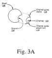
- Figure 3A illustrates a cross-sectional view of an ultrasound probe and channel guide attachment according to the present invention.
- Figure 3A shows the front view of the shaft 106 of the ultrasound imaging probe 100 shown in Figure 1 , along with the channel guide attachment 118, which may be attached thereto.
- the shaft 106 is defined by the groove 108, as described above with reference to Figure 1 .
- the channel guide attachment 118 includes the channel 120 as well as the channel guide clamp 116, and may, but not necessarily include other components (e.g., additional channel guide clamps, such as the channel guide clamp 124).
- Figure 3A illustrates one configuration for the shaft of an ultrasound probe and compatible channel guide attachment, which may be attached thereto for guidance of an instrument through at least two ultrasound imaging planes.
- the channel guide attachment 118, the channel guide clamps 116 and 124, the groove 108, and/or the ultrasound imaging probe 100 may be configured according to several various shapes and sizes to provide simultaneous viewing of an instrument in two or more imaging planes.
- a probe sheath or cover may be accommodated.
- the ultrasound imaging probe may not provide a groove and/or other attachment features.
- a channel for providing an instrument path may be defined by the shaft of the probe itself.
- Figure 3B shows the side view of an ultrasound imaging probe, according to one embodiment of the invention.
- Figure 3B shows the ultrasound imaging probe 100 of Figure 1 attached to the channel guide attachment 118 attached within the groove 108.
- the channel guide attachment 118 may be attached to the probe 100 within the groove 108.
- the channel guide attachment 118, the channel guide clamps 116 and 124, the groove 108, and/or the ultrasound imaging probe 100 may be configured according to several various shapes and sizes to provide simultaneous viewing of an instrument in two or more imaging planes, such as the imaging planes 110 and 112, via the instrument path 114.
- Figure 3C is an end view of an ultrasound imaging probe, according to one embodiment of the invention.
- the ultrasound probe 100 is shown in an end view, which shows the offset positioning of the two transducers, namely the side-fire transducer 102 and the end-fire transducer 104, relative to the channel 120.
- the probe 100 may also include a handle 122.
- Figure 4A shows an alternative configuration of an ultrasound imaging probe that provides simultaneous viewing of an instrument in two imaging planes, according to one embodiment of the invention.
- the probe 200 shown in Figure 4A has the positions of the side-fire and end-fire transducers reversed as shown by a side-fire transducer 204 and an end-fire transducer 202, which generate an imaging plane 208 and an imaging plane 210, respectively.
- the probe 200 also provides an instrument path, namely the instrument path 212, which provides simultaneous viewing in at least two imaging planes, and in particular, the imaging planes 208 and 210.
- the instrument path 212 is defined by the intersection of the imaging plane 208 and the imaging plane 210.
- the two transducers 202 and 204 are placed orthogonally with respect to each other.
- the imaging planes 210 and 208, generated by the transducers 202 and 204, respectively, intersect orthogonally.
- the instrument path 212 lies on a line that substantially defines the intersection of the two imaging planes shown in Figure 4A , the instrument path 212 provides simultaneous viewing of an instrument (e.g. a biopsy needle) in the two imaging planes 208 and 210.
- Figure 4B is an end view an ultrasound imaging probe, according to one embodiment of the invention.
- Figure 4B depicts an end view of the probe 200 shown in Figure 4A .
- the probe 200 may include a handle such as the handle 214, and the end view of the probe 200 shows the orientation of the channel 120 relative to the two transducers 202 and 204.
- a channel guide attachment, groove, etc. are not necessary to provide an instrument path, such as the channel 108, which provides simultaneous viewing of an instrument in two imaging planes.
- Figure 5 depicts an ultrasound imaging probe, according to one embodiment of the invention.
- Figure 5 shows a probe 300 which utilizes a flat phased linear array transducer, according to one embodiment of the invention.
- the probe 300 includes an oblique end-fire transducer 304 placed orthogonally relative to a flat phased linear array transducer 302.
- the end-fire transducer 304 and the flat phased linear array transducer 302 generate an imaging plane 308 and an imaging plane 306, respectively, which also intersect orthogonally.
- An instrument path 310 is shown which provides simultaneous viewing in the imaging planes 306 and 308.
- the flat phased linear array transducer 302 may be operated with electronic beam steering to generate a fan image while reducing the cross sectional profile of the probe head.
- Figure 6 illustrates an ultrasound imaging system which may provide operation of an ultrasound imaging probe embodying the present invention in simultaneous duplex imaging mode and may be used with a suitable ultrasound scanner, according to one embodiment of the invention.
- the system show in Figure 6 provides real-time frame interleaving of images provided by two transducers to provide dual simultaneous real-time biplane imaging. It will be appreciated that in alternative embodiments of the invention, one or more of the operations of the system depicted in Figure 6 are not performed in real-time.
- a system 400 includes a transducer 402 and a transducer 404, both coupled to a transducer transmission/reception (TX/RX) switch 406.
- the TX/RX switch 406 is coupled to a vector sequencer 420 via a sequencer signal path 422.
- the vector sequencer 420 is also coupled, via the sequencer signal path 422, to a scan converter 418, whose output is switched between an image memory 424 and an image memory 426, for the respective transducers 402 and 406.
- the vector sequencer 420 in addition to sequencing vectors within an imaging frame, also sequences vectors between the imaging frames generated by the transducer 402 and the transducer 404, and provided for display to the image memory 424 and the image memory 426, respectively.
- a transmit beamformer pulser 408 provides signal input to the TX/RX switch 406.
- the TX/RX switch 406 is coupled to provide signal input to a receive beamformer 410.
- Signals received by the receive beamformer 410 are provided to a summing unit 412, which in turn provides summed signals to a zone switch/depth gain unit 414.
- the zone switch depth gain unit 414 is coupled to a vector processor 416 which in turn is coupled to the scan converter 418.
- the scan converter 418 provides signals to two image memories, namely an image memory 424 and an image memory 426.
- the image memories 424 and 426 provide signals to a frame processor 428 and a display subsystem 430.
- Figure 6 shows one embodiment of a system in which the present invention may find use, it will be appreciated that the invention may be used with several types configurations and architectures of ultrasound imaging devices and systems, which may include various types of transducers, digital signal processing (DSP) circuits, general purpose processors, storage devices/media, Doppler processing routines/circuitry, etc.
- DSP digital signal processing
Description
- The invention relates to the field of medical devices. More particularly, the invention relates to ultrasound endocavitary imaging devices.
- Various ultrasound techniques and devices have been developed for imaging the interior of a body (hereinafter, "body" refers to various types of subjects, such as humans, animals, non-living structures, etc.). One application of ultrasound imaging has been in the medical field, and in particular, in endocavitary probes (e.g., biopsy guidance endocavitary probes). Such probes may be used, for example, for-endovaginal examination (e.g., to examine the uterus, ovaries, etc.), endorectal examination (e.g., to examine the rectal wall, prostate, etc.), and/or other medically-related applications. Typically, endocavitary probes include a linear array transducer positioned at the distal end of the probe that is to be inserted into a cavity of a body. The transducer provides an imaging plane for viewing structures/features of the body and/or another instrument (e.g., a biopsy needle) that, for example, may be guided into the body via the probe. The imaging plane may be provided at a side of a probe (corresponding to a "side-fire" transducer) or the front of the probe (corresponding to an "end-fire" transducer).
- Since such linear array transducer endocavitary probes provide one imaging plane at any time for viewing the interior of the body and/or another instrument (e.g., a biopsy needle) that may be guided into the body via the probe, the ultrasound images provided by such probes are generally limited to a single plane. Unfortunately, such 2D (two-dimensional) images typically do not provide desired accuracy of structures/features within a body. In applications where a relatively high level of imaging accuracy may be critical, such as biopsy needle guidance through or in proximity to sensitive bodily structures which may need to be avoided by the needle, such linear array transducer probes may not be practical.
- Several techniques for improving the above-mentioned limitation(s) of single linear array transducer endocavitary probes have been proposed, but each is relatively limited as well. For example, some endocavitary probes utilize a mechanical, rather than a linear array, transducer. Such mechanical transducer probes provide a single imaging plane at any one time, but the mechanical transducer may be rotated up to 180 degrees or even more to provide multiplane three-dimensional (3D) orientation in a field of view. However, end-fire multiplane single mechanical transducer probes cannot be used for biopsy guidance, since the biopsy needle is only visible in at most one plane of the rotation. Moreover, side-fire multiplane single mechanical transducer probes are unable to provide viewing of a forward-penetrating biopsy need at all.
- In addition to the single-plane linear array transducer probes and the multiplane mechanical transducer probes described above, some biplane probes have been proposed. One type of a biplane probe utilizes a dual convex oblique end-fire linear array transducer structure, which generates two intersecting orthogonal imaging planes to provide 3D orientation in the field of view. However, this type of biplane probe is limited to only one imaging plane, since the two imaging planes intersect over one transducer, not along the biopsy needle. A second type of a biplane probe utilizes a convex and flat side-fire linear array transducer structure, which generates two non-intersecting orthogonal imaging planes to provide an indication of 3D orientation. However, this second type of a biplane endocavitary probe does not allow viewing of a biopsy needle in either imaging plane.
- In
WO 94/13208A -
US 4,817,616 discloses an ultrasonic probe comprising two transducers which provide two orthogonal imaging planes. - Thus, what is desired is an ultrasound endocavitary probe that provides simultaneous viewing of an instrument, such as a biopsy needle or other instrument, in two imaging planes.
- According to the invention which is defined in claim 1, there is provided an ultrasound imaging apparatus comprising a first transducer to provide a first imaging plane and a second transducer to provide a second imaging plane. The first imaging plane and the second imaging plane intersect orthogonally. A channel provides an instrument path to provide simultaneous viewing of an instrument in said first and second imaging planes.
- The present invention provides apparatuses for providing simultaneous viewing of an instrument in two ultrasound imaging planes. In one embodiment of the invention, an ultrasound imaging probe is provided which can generate at least two ultrasound imaging planes. The two imaging planes intersect orthogonally. An instrument path is positioned with respect to the planes such that an instrument may be simultaneously viewed in both imaging planes. The instrument path is provided at an intersection that, defines the intersection of the two imaging planes.
- The invention will now be described in greater detail, by way of example, with reference to the drawings, in which:-
-
Figure 1 is a side view diagram of a (portion of) an ultrasound imaging probe for providing simultaneous biplane imaging, according to one embodiment of the invention; -
Figure 2 illustrates a channel guide attachment that may be used with an ultrasound imaging probe, according to one embodiment of the present invention; -
Figure 3A illustrates a cross-sectional view of an ultrasound probe and channel guide attachment according to the present invention; -
Figure 3B shows the side view of an ultrasound imaging probe, according to one embodiment of the invention; -
Figure 3C is an end view of an ultrasound imaging probe, according to one embodiment of the invention; -
Figure 4A shows an alternative configuration of an ultrasound imaging probe that provides simultaneous viewing of an instrument in two imaging planes, according to one embodiment of the invention; -
Figure 4B is an end view an ultrasound imaging probe, according to one embodiment of the invention; -
Figure 5 depicts an ultrasound imaging probe, according to one embodiment of the invention; and -
Figure 6 illustrates an ultrasound imaging system which may provide operation of an ultrasound imaging probe embodying the present invention in simultaneous duplex imaging mode and may be used with a suitable ultrasound scanner, according to one embodiment of the invention. - The present invention provides an ultrasound imaging device that provides simultaneous access to an instrument in at least two imaging planes. In one embodiment of the invention, based on simultaneously imaging the instrument in two imaging planes, three-dimensional (3D) biplane orientation of the instrument and/or other structures may be generated. In one embodiment wherein the invention is embodied in an endocavitary biopsy probe used in medical applications, the instrument may be a biopsy needle that is simultaneously guided through the two imaging planes. It should be appreciated that in this embodiment, the probe may be used to provide images in one plane as well. For example, an endocavitary biopsy probe embodying the invention may be manipulated within the rectum or other cavity for oblique side-fire scanning. However, it should be appreciated that while the invention is primarily described herein with reference to endocavitary biopsy, one or a combination of the ultrasound imaging apparatuses and methods disclosed herein may be utilized in various other medical or non-medical applications without departing from the scope of the invention. Thus, the invention should not necessarily be limited to endocavitary biopsy probes or medical purpose imaging devices.
- In the following description, numerous specific details are set forth to provide a thorough understanding of the invention. However, it is understood that the invention may be practiced without these specific details. In other instances, well-known circuits, structures and techniques have not been shown in detail to avoid obscuring the invention.
-
Figure 1 is a side view diagram of a (portion of) an ultrasound imaging probe for providing simultaneous biplane imaging, according to one embodiment of the invention. InFigure 1 , at least a portion of anultrasound imaging probe 100 is shown which is defined by ashaft 106. Joined obliquely to theshaft 106 at a distal end of theprobe 100 are two convex transducers, a forward-leaning side-fire transducer 102 and an oblique end-fire transducer 104. However, it will be appreciated from the following description that the invention is not limited to a particular type(s) of transducer. - The side-
fire transducer 102 and the end-fire transducer 104 are placed orthogonally relative to each other at the oblique distal end of theprobe 100, as shown inFigure 3C , which depicts a frontal view of one implementation of theprobe 100 shown inFigure 1 . As such, the side-fire transducer 102 and the end-fire transducer 104 generate animaging plane 110 and animaging plane 112, respectively, which two imaging planes intersect orthogonally. - The
shaft 106 also defines agroove 108 for providing aninstrument path 114. In one embodiment, thegroove 108 is configured to accept an attachment having a channel to provide theinstrument path 114, such as thechannel guide attachment 118 shown inFigure 2 and3A-3C , and described in further detail below. In another embodiment, thegroove 108 may be substituted by a channel within the shaft 106 (and the distal end of theprobe 100 that includes the transducer assembly) for providing theinstrument path 114. Theinstrument path 114 may be provided, in one embodiment, to a biopsy needle or other instrument for ultrasound image guidance. - In either case, the
instrument path 114 substantially lies on a line that defines the intersection of theimaging plane 110 and theimaging plane 112. As such, simultaneous viewing in theimaging plane 110 and theimaging plane 112 of an instrument that is guided in association with theprobe 100 along theinstrument path 114 may be provided. Thus, a biplane image of the instrument and/or structures in proximity thereto may be generated by processing the image provided by each of theimaging plane 110 and theimaging plane 112. In one embodiment of the invention, a 3D orientation, comprising a composite of two images, each provided by one of theimaging plane 110 and theimaging plane 112, may be provided. In one embodiment, interleaving of images from the two imaging planes may be used to generate the composite of two images. In one embodiment, the probe is coupled to a display system, which includes circuitry and a monitor/display for viewing single or biplane images generated by theprobe 100, and in particular, signals derived from thetransducers -
Figure 2 illustrates a channel guide attachment that may be used with an ultrasound imaging probe, according to one embodiment of the present invention. InFigure 2 , achannel guide attachment 118 is shown, which includes achannel guide clamp 116 and achannel guide clamp 124. Thechannel guide attachment 118 also includes achannel 120 that may be used to facilitate guidance of an instrument (e.g., a biopsy needle). According to one embodiment of the invention, thechannel guide attachment 118 may be attached to theprobe 100 shown inFigure 1 and described with reference thereto. In particular, thechannel guide attachment 118 may attach to theprobe 100 at thegroove 108, described above with reference toFigure 1 . In so doing, thechannel guide attachment 118 may provide simultaneous access to an instrument, which may be guided through thechannel 120, within theimaging plane 112 and theimaging plane 110. The channel guide clamps 116 and 124 shown inFigure 2 allow thechannel guide attachment 118 to be attached to a biopsy imaging probe, such as theprobe 100 shown inFigure 1 . -
Figure 3A illustrates a cross-sectional view of an ultrasound probe and channel guide attachment according to the present invention. In particular,Figure 3A shows the front view of theshaft 106 of theultrasound imaging probe 100 shown inFigure 1 , along with thechannel guide attachment 118, which may be attached thereto. As shown inFigure 3A , theshaft 106 is defined by thegroove 108, as described above with reference toFigure 1 . Thechannel guide attachment 118 includes thechannel 120 as well as thechannel guide clamp 116, and may, but not necessarily include other components (e.g., additional channel guide clamps, such as the channel guide clamp 124). In general,Figure 3A illustrates one configuration for the shaft of an ultrasound probe and compatible channel guide attachment, which may be attached thereto for guidance of an instrument through at least two ultrasound imaging planes. However, it will be appreciated that thechannel guide attachment 118, the channel guide clamps 116 and 124, thegroove 108, and/or theultrasound imaging probe 100 may be configured according to several various shapes and sizes to provide simultaneous viewing of an instrument in two or more imaging planes. By providing a detachable/attachable channel guide attachment, in one embodiment of the invention, a probe sheath or cover may be accommodated. In alternative embodiments, the ultrasound imaging probe may not provide a groove and/or other attachment features. For example, a channel for providing an instrument path may be defined by the shaft of the probe itself. -
Figure 3B shows the side view of an ultrasound imaging probe, according to one embodiment of the invention. In particular,Figure 3B shows theultrasound imaging probe 100 ofFigure 1 attached to thechannel guide attachment 118 attached within thegroove 108. As shown inFigure 3B and also depicted and described with reference toFigure 3A , thechannel guide attachment 118 may be attached to theprobe 100 within thegroove 108. In alternative embodiments, thechannel guide attachment 118, the channel guide clamps 116 and 124, thegroove 108, and/or theultrasound imaging probe 100 may be configured according to several various shapes and sizes to provide simultaneous viewing of an instrument in two or more imaging planes, such as the imaging planes 110 and 112, via theinstrument path 114. -
Figure 3C is an end view of an ultrasound imaging probe, according to one embodiment of the invention. InFigure 3C , theultrasound probe 100 is shown in an end view, which shows the offset positioning of the two transducers, namely the side-fire transducer 102 and the end-fire transducer 104, relative to thechannel 120. As shown inFigure 3C , theprobe 100 may also include ahandle 122. -
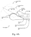
Figure 4A shows an alternative configuration of an ultrasound imaging probe that provides simultaneous viewing of an instrument in two imaging planes, according to one embodiment of the invention. In contrast to theprobe 100 described with reference toFigure 1 , theprobe 200 shown inFigure 4A has the positions of the side-fire and end-fire transducers reversed as shown by a side-fire transducer 204 and an end-fire transducer 202, which generate animaging plane 208 and animaging plane 210, respectively. Nonetheless, theprobe 200 also provides an instrument path, namely theinstrument path 212, which provides simultaneous viewing in at least two imaging planes, and in particular, the imaging planes 208 and 210. In one embodiment, theinstrument path 212 is defined by the intersection of theimaging plane 208 and theimaging plane 210. As shown in the embodiment ofFigure 4A , the twotransducers transducers instrument path 212 lies on a line that substantially defines the intersection of the two imaging planes shown inFigure 4A , theinstrument path 212 provides simultaneous viewing of an instrument (e.g. a biopsy needle) in the twoimaging planes -
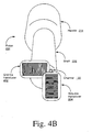
Figure 4B is an end view an ultrasound imaging probe, according to one embodiment of the invention. In particular,Figure 4B depicts an end view of theprobe 200 shown inFigure 4A . As shown inFigure 4B , theprobe 200 may include a handle such as thehandle 214, and the end view of theprobe 200 shows the orientation of thechannel 120 relative to the twotransducers probe 100, a channel guide attachment, groove, etc., are not necessary to provide an instrument path, such as thechannel 108, which provides simultaneous viewing of an instrument in two imaging planes. -
Figure 5 depicts an ultrasound imaging probe, according to one embodiment of the invention. In particular,Figure 5 shows aprobe 300 which utilizes a flat phased linear array transducer, according to one embodiment of the invention. Theprobe 300 includes an oblique end-fire transducer 304 placed orthogonally relative to a flat phasedlinear array transducer 302. The end-fire transducer 304 and the flat phasedlinear array transducer 302 generate animaging plane 308 and animaging plane 306, respectively, which also intersect orthogonally. Aninstrument path 310 is shown which provides simultaneous viewing in the imaging planes 306 and 308. According to one aspect of the invention as embodied in theprobe 300 shown inFigure 5 , the flat phasedlinear array transducer 302 may be operated with electronic beam steering to generate a fan image while reducing the cross sectional profile of the probe head. -
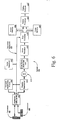
Figure 6 illustrates an ultrasound imaging system which may provide operation of an ultrasound imaging probe embodying the present invention in simultaneous duplex imaging mode and may be used with a suitable ultrasound scanner, according to one embodiment of the invention. In one embodiment, the system show inFigure 6 provides real-time frame interleaving of images provided by two transducers to provide dual simultaneous real-time biplane imaging. It will be appreciated that in alternative embodiments of the invention, one or more of the operations of the system depicted inFigure 6 are not performed in real-time. - As shown in
Figure 6 , asystem 400 includes atransducer 402 and atransducer 404, both coupled to a transducer transmission/reception (TX/RX)switch 406. The TX/RX switch 406 is coupled to avector sequencer 420 via asequencer signal path 422. Thevector sequencer 420 is also coupled, via thesequencer signal path 422, to ascan converter 418, whose output is switched between animage memory 424 and animage memory 426, for therespective transducers vector sequencer 420, in addition to sequencing vectors within an imaging frame, also sequences vectors between the imaging frames generated by thetransducer 402 and thetransducer 404, and provided for display to theimage memory 424 and theimage memory 426, respectively. A transmitbeamformer pulser 408 provides signal input to the TX/RX switch 406. In turn, the TX/RX switch 406 is coupled to provide signal input to a receivebeamformer 410. Signals received by the receivebeamformer 410 are provided to a summingunit 412, which in turn provides summed signals to a zone switch/depth gain unit 414. The zone switch depth gain unit 414 is coupled to avector processor 416 which in turn is coupled to thescan converter 418. Thescan converter 418 provides signals to two image memories, namely animage memory 424 and animage memory 426. Theimage memories frame processor 428 and adisplay subsystem 430. - Although
Figure 6 shows one embodiment of a system in which the present invention may find use, it will be appreciated that the invention may be used with several types configurations and architectures of ultrasound imaging devices and systems, which may include various types of transducers, digital signal processing (DSP) circuits, general purpose processors, storage devices/media, Doppler processing routines/circuitry, etc. - While the invention has been described in terms of several embodiments, those skilled in the art will recognize that the invention is not limited to the embodiments described. For example, while some types of transducers, ultrasound imaging device circuitry, probe/channel guide attachments, etc., have been shown and described, it will be appreciated that the invention is not limited to such. Accordingly, it will be appreciated that the invention may be embodied in various probe configurations and imaging system architectures that provide simultaneous viewing of an instrument (e.g., an endocavitary biopsy needle) in at least two ultrasound imaging planes.
Claims (10)
- An ultrasound imaging apparatus comprising:a first transducer (102) to provide a first imaging plane;a second transducer (104) to provide a second imaging plane;wherein said first imaging plane and said second imaging plane intersect orthogonally; said apparatus being characterised by:a guide (118) which defines a channel (120), said channel providing an instrument path (114) which substantially passes through a line defining the intersection of said first and second imaging planes, to provide simultaneous viewing of an instrument in said first and second imaging planes, the instrument being guidable along the instrument path.
- The ultrasound imaging apparatus of claim 1, further comprising a display (430) to provide an image of said instrument, said image being based on simultaneous imaging provided from said first and second imaging planes.
- The ultrasound imaging apparatus of claim 1, wherein said first and second transducers are positioned at the distal end of an endocavitary probe (100).
- The ultrasound imaging apparatus of claim 3, wherein said guide (118) is detachable from said endocavitary probe.
- The ultrasound imaging apparatus of claim1, wherein said channel (120) provides access to a biopsy needle.
- The ultrasound imaging apparatus of claim 3, wherein said probe comprises a shaft, said shaft having a groove (108) that provides attachment of said guide.
- The ultrasound imaging apparatus of claim 6, wherein said guide may be detached from said groove.
- The ultrasound imaging apparatus of claim 1, wherein an image is generated based one of three modes, wherein a first mode includes an image based on said first image plane, a second mode includes an image based on said second image plane, and a third mode includes an image based on a combination of said first and second image planes.
- The ultrasound imaging apparatus of claim 1, wherein at least one of said first and second transducers comprises a convex linear array transducer.
- The ultrasound imaging apparatus of claim 1, wherein at least one of said first and second transducers comprises a flat phased linear array transducer.
Applications Claiming Priority (2)
| Application Number | Priority Date | Filing Date | Title |
|---|---|---|---|
| US74571 | 1998-05-07 | ||
| US09/074,571 US6261234B1 (en) | 1998-05-07 | 1998-05-07 | Method and apparatus for ultrasound imaging with biplane instrument guidance |
Publications (2)
| Publication Number | Publication Date |
|---|---|
| EP0955010A1 EP0955010A1 (en) | 1999-11-10 |
| EP0955010B1 true EP0955010B1 (en) | 2009-10-14 |
Family
ID=22120283
Family Applications (1)
| Application Number | Title | Priority Date | Filing Date |
|---|---|---|---|
| EP99303599A Expired - Lifetime EP0955010B1 (en) | 1998-05-07 | 1999-05-07 | Biplane ultrasound imaging for the guidance of intracavitary probes |
Country Status (3)
| Country | Link |
|---|---|
| US (1) | US6261234B1 (en) |
| EP (1) | EP0955010B1 (en) |
| JP (1) | JP2000201936A (en) |
Families Citing this family (115)
| Publication number | Priority date | Publication date | Assignee | Title |
|---|---|---|---|---|
| DK176387B1 (en) * | 1998-01-07 | 2007-10-22 | Bk Medical Aps | Apparatus for insertion into the human body |
| US6254601B1 (en) * | 1998-12-08 | 2001-07-03 | Hysterx, Inc. | Methods for occlusion of the uterine arteries |
| US9402601B1 (en) * | 1999-06-22 | 2016-08-02 | Teratech Corporation | Methods for controlling an ultrasound imaging procedure and providing ultrasound images to an external non-ultrasound application via a network |
| US7223279B2 (en) * | 2000-04-21 | 2007-05-29 | Vascular Control Systems, Inc. | Methods for minimally-invasive, non-permanent occlusion of a uterine artery |
| US20030120306A1 (en) * | 2000-04-21 | 2003-06-26 | Vascular Control System | Method and apparatus for the detection and occlusion of blood vessels |
| US6550482B1 (en) * | 2000-04-21 | 2003-04-22 | Vascular Control Systems, Inc. | Methods for non-permanent occlusion of a uterine artery |
| US6669641B2 (en) * | 2000-08-17 | 2003-12-30 | Koninklijke Philips Electronics N.V. | Method of and system for ultrasound imaging |
| US6638286B1 (en) | 2000-11-16 | 2003-10-28 | Vascular Control Systems, Inc. | Doppler directed suture ligation device and method |
| US6635065B2 (en) * | 2000-11-16 | 2003-10-21 | Vascular Control Systems, Inc. | Doppler directed suture ligation device and method |
| EP1337183B1 (en) * | 2000-11-24 | 2005-05-25 | Innovacell Biotechnologie GmbH | Ultrasonic probe comprising a positioning device for examination devices and operation devices |
| ITSV20010008A1 (en) * | 2001-03-05 | 2002-09-05 | Esaote Spa | NEEDLE GUIDE DEVICE IN PARTICULAR FOR ECHOGRAPHIC PROBES AND COMBINATION OF ECHOGRAPHIC PROBE AND SAID NEEDLE GUIDE DEVICE |
| US7354444B2 (en) * | 2001-03-28 | 2008-04-08 | Vascular Control Systems, Inc. | Occlusion device with deployable paddles for detection and occlusion of blood vessels |
| US20030120286A1 (en) * | 2001-03-28 | 2003-06-26 | Vascular Control System | Luminal clip applicator with sensor |
| JP4227415B2 (en) * | 2001-03-28 | 2009-02-18 | ヴァスキュラー・コントロール・システムズ・インコーポレーテッド | Method and apparatus for detecting and ligating uterine arteries |
| US6951542B2 (en) * | 2002-06-26 | 2005-10-04 | Esaote S.P.A. | Method and apparatus for ultrasound imaging of a biopsy needle or the like during an ultrasound imaging examination |
| US20040238732A1 (en) * | 2001-10-19 | 2004-12-02 | Andrei State | Methods and systems for dynamic virtual convergence and head mountable display |
| US7806828B2 (en) * | 2002-02-05 | 2010-10-05 | Inceptio Medical Technologies, Lc | Multiplanar ultrasonic vascular sensor assembly and apparatus for movably affixing a sensor assembly to a body |
| US6755789B2 (en) | 2002-02-05 | 2004-06-29 | Inceptio Medical Technologies, Llc | Ultrasonic vascular imaging system and method of blood vessel cannulation |
| US7207996B2 (en) * | 2002-04-04 | 2007-04-24 | Vascular Control Systems, Inc. | Doppler directed suturing and compression device and method |
| JP4018450B2 (en) * | 2002-05-27 | 2007-12-05 | キヤノン株式会社 | Document management system, document management apparatus, authentication method, computer readable program, and storage medium |
| US20040097961A1 (en) * | 2002-11-19 | 2004-05-20 | Vascular Control System | Tenaculum for use with occlusion devices |
| US7172603B2 (en) * | 2002-11-19 | 2007-02-06 | Vascular Control Systems, Inc. | Deployable constrictor for uterine artery occlusion |
| US7351205B2 (en) * | 2003-01-03 | 2008-04-01 | Civco Medical Instruments Co., Inc. | Shallow angle needle guide apparatus and method |
| US7404821B2 (en) * | 2003-01-30 | 2008-07-29 | Vascular Control Systems, Inc. | Treatment for post partum hemorrhage |
| US7651511B2 (en) * | 2003-02-05 | 2010-01-26 | Vascular Control Systems, Inc. | Vascular clamp for caesarian section |
| US7333844B2 (en) | 2003-03-28 | 2008-02-19 | Vascular Control Systems, Inc. | Uterine tissue monitoring device and method |
| US7909766B2 (en) * | 2003-05-21 | 2011-03-22 | Scimed Life Systems, Inc. | Systems and methods for improving the imaging resolution of an imaging transducer |
| US20040260199A1 (en) * | 2003-06-19 | 2004-12-23 | Wilson-Cook Medical, Inc. | Cytology collection device |
| US20050159676A1 (en) * | 2003-08-13 | 2005-07-21 | Taylor James D. | Targeted biopsy delivery system |
| US7066887B2 (en) * | 2003-10-21 | 2006-06-27 | Vermon | Bi-plane ultrasonic probe |
| US7244234B2 (en) | 2003-11-11 | 2007-07-17 | Soma Development Llc | Ultrasound guided probe device and method of using same |
| US7325546B2 (en) * | 2003-11-20 | 2008-02-05 | Vascular Control Systems, Inc. | Uterine artery occlusion device with cervical receptacle |
| US7686817B2 (en) * | 2003-11-25 | 2010-03-30 | Vascular Control Systems, Inc. | Occlusion device for asymmetrical uterine artery anatomy |
| JP4373400B2 (en) * | 2003-12-16 | 2009-11-25 | 株式会社日立メディコ | Ultrasonic body motion detection device, and image presentation device and ultrasonic therapy device using the same |
| US20060015144A1 (en) * | 2004-07-19 | 2006-01-19 | Vascular Control Systems, Inc. | Uterine artery occlusion staple |
| WO2006043639A1 (en) * | 2004-10-20 | 2006-04-27 | Kabushiki Kaisha Toshiba | Ultrasonic diagnostic equipment and control method therefor |
| US7875036B2 (en) * | 2004-10-27 | 2011-01-25 | Vascular Control Systems, Inc. | Short term treatment for uterine disorder |
| US20060264760A1 (en) * | 2005-02-10 | 2006-11-23 | Board Of Regents, The University Of Texas System | Near infrared transrectal probes for prostate cancer detection and prognosis |
| US20070049973A1 (en) * | 2005-08-29 | 2007-03-01 | Vascular Control Systems, Inc. | Method and device for treating adenomyosis and endometriosis |
| US20070099531A1 (en) * | 2005-10-27 | 2007-05-03 | Efremova Nadezhda V | Foam fastening system that includes a surface modifier |
| US9259208B2 (en) * | 2006-03-24 | 2016-02-16 | B-K Medical Aps | Ultrasound probe |
| US9539025B2 (en) * | 2006-03-24 | 2017-01-10 | B-K Medical Aps | Biopsy system |
| US8425418B2 (en) * | 2006-05-18 | 2013-04-23 | Eigen, Llc | Method of ultrasonic imaging and biopsy of the prostate |
| US20110057930A1 (en) * | 2006-07-26 | 2011-03-10 | Inneroptic Technology Inc. | System and method of using high-speed, high-resolution depth extraction to provide three-dimensional imagery for endoscopy |
| WO2008017051A2 (en) | 2006-08-02 | 2008-02-07 | Inneroptic Technology Inc. | System and method of providing real-time dynamic imagery of a medical procedure site using multiple modalities |
| US8147414B2 (en) * | 2006-10-12 | 2012-04-03 | Innoscion, Llc | Image guided catheter having remotely controlled surfaces-mounted and internal ultrasound transducers |
| US10772600B2 (en) | 2015-09-25 | 2020-09-15 | Perceptive Navigation Llc | Image guided catheters and methods of use |
| US8064664B2 (en) * | 2006-10-18 | 2011-11-22 | Eigen, Inc. | Alignment method for registering medical images |
| US7804989B2 (en) * | 2006-10-30 | 2010-09-28 | Eigen, Inc. | Object recognition system for medical imaging |
| KR100936456B1 (en) * | 2006-12-07 | 2010-01-13 | 주식회사 메디슨 | Ultrasound system |
| US20080161687A1 (en) * | 2006-12-29 | 2008-07-03 | Suri Jasjit S | Repeat biopsy system |
| US8175350B2 (en) * | 2007-01-15 | 2012-05-08 | Eigen, Inc. | Method for tissue culture extraction |
| US20080186378A1 (en) * | 2007-02-06 | 2008-08-07 | Feimo Shen | Method and apparatus for guiding towards targets during motion |
| US7856130B2 (en) * | 2007-03-28 | 2010-12-21 | Eigen, Inc. | Object recognition system for medical imaging |
| US7832114B2 (en) * | 2007-04-04 | 2010-11-16 | Eigen, Llc | Tracker holder assembly |
| JP4627769B2 (en) * | 2007-06-12 | 2011-02-09 | アロカ株式会社 | Ultrasonic probe |
| US20090030317A1 (en) * | 2007-07-25 | 2009-01-29 | Mayo Foundation For Medical Education And Research | Ultrasonic imaging devices, systems, and methods |
| US20090048515A1 (en) * | 2007-08-14 | 2009-02-19 | Suri Jasjit S | Biopsy planning system |
| US8571277B2 (en) * | 2007-10-18 | 2013-10-29 | Eigen, Llc | Image interpolation for medical imaging |
| US7942829B2 (en) * | 2007-11-06 | 2011-05-17 | Eigen, Inc. | Biopsy planning and display apparatus |
| US20090227874A1 (en) * | 2007-11-09 | 2009-09-10 | Eigen, Inc. | Holder assembly for a medical imaging instrument |
| US20090324041A1 (en) * | 2008-01-23 | 2009-12-31 | Eigen, Llc | Apparatus for real-time 3d biopsy |
| WO2009094646A2 (en) * | 2008-01-24 | 2009-07-30 | The University Of North Carolina At Chapel Hill | Methods, systems, and computer readable media for image guided ablation |
| US20100001996A1 (en) * | 2008-02-28 | 2010-01-07 | Eigen, Llc | Apparatus for guiding towards targets during motion using gpu processing |
| US8340379B2 (en) | 2008-03-07 | 2012-12-25 | Inneroptic Technology, Inc. | Systems and methods for displaying guidance data based on updated deformable imaging data |
| CN102056559A (en) * | 2008-06-12 | 2011-05-11 | 皇家飞利浦电子股份有限公司 | Biopsy device with acoustic element |
| US20090312629A1 (en) * | 2008-06-13 | 2009-12-17 | Inneroptic Technology Inc. | Correction of relative tracking errors based on a fiducial |
| JP2009297384A (en) | 2008-06-17 | 2009-12-24 | Fujifilm Corp | Ultrasonic diagnostic apparatus and ultrasonic probe |
| US20090326372A1 (en) * | 2008-06-30 | 2009-12-31 | Darlington Gregory | Compound Imaging with HIFU Transducer and Use of Pseudo 3D Imaging |
| US11464578B2 (en) | 2009-02-17 | 2022-10-11 | Inneroptic Technology, Inc. | Systems, methods, apparatuses, and computer-readable media for image management in image-guided medical procedures |
| US8641621B2 (en) | 2009-02-17 | 2014-02-04 | Inneroptic Technology, Inc. | Systems, methods, apparatuses, and computer-readable media for image management in image-guided medical procedures |
| US8690776B2 (en) | 2009-02-17 | 2014-04-08 | Inneroptic Technology, Inc. | Systems, methods, apparatuses, and computer-readable media for image guided surgery |
| US8554307B2 (en) | 2010-04-12 | 2013-10-08 | Inneroptic Technology, Inc. | Image annotation in image-guided medical procedures |
| JP5671008B2 (en) | 2009-04-28 | 2015-02-18 | コーニンクレッカ フィリップス エヌ ヴェ | Biopsy guide system with ultrasonic transducer and method of use thereof |
| US20100286527A1 (en) * | 2009-05-08 | 2010-11-11 | Penrith Corporation | Ultrasound system with multi-head wireless probe |
| WO2011014687A2 (en) * | 2009-07-31 | 2011-02-03 | Inneroptic Technology, Inc. | Dual-tube stereoscope |
| US20110082351A1 (en) * | 2009-10-07 | 2011-04-07 | Inneroptic Technology, Inc. | Representing measurement information during a medical procedure |
| US8761862B2 (en) * | 2009-10-09 | 2014-06-24 | Stephen F. Ridley | Ultrasound guided probe device and sterilizable shield for same |
| US9282947B2 (en) | 2009-12-01 | 2016-03-15 | Inneroptic Technology, Inc. | Imager focusing based on intraoperative data |
| US8758256B2 (en) | 2010-07-12 | 2014-06-24 | Best Medical International, Inc. | Apparatus for brachytherapy that uses a scanning probe for treatment of malignant tissue |
| US9044216B2 (en) | 2010-07-12 | 2015-06-02 | Best Medical International, Inc. | Biopsy needle assembly |
| WO2012014120A1 (en) * | 2010-07-30 | 2012-02-02 | Koninklijke Philips Electronics N.V. | Display and export of individual biplane images |
| WO2012103650A1 (en) * | 2011-01-31 | 2012-08-09 | Sunnybrook Health Sciences Centre | Ultrasonic probe with ultrasonic transducers addressable on common electrical channel |
| BE1019976A3 (en) | 2011-05-16 | 2013-03-05 | Mepy Benelux | TRANSDUCER COMPOSITION FOR AN ECHOSCOPE. |
| JP5976441B2 (en) * | 2011-08-03 | 2016-08-23 | 東芝メディカルシステムズ株式会社 | Ultrasonic probe and ultrasonic diagnostic apparatus |
| GB201119005D0 (en) * | 2011-11-03 | 2011-12-14 | Univ Dundee | Ultrasound probe |
| US8670816B2 (en) | 2012-01-30 | 2014-03-11 | Inneroptic Technology, Inc. | Multiple medical device guidance |
| US10667790B2 (en) * | 2012-03-26 | 2020-06-02 | Teratech Corporation | Tablet ultrasound system |
| US9877699B2 (en) * | 2012-03-26 | 2018-01-30 | Teratech Corporation | Tablet ultrasound system |
| US9113825B2 (en) * | 2012-07-10 | 2015-08-25 | Fujifilm Sonosite, Inc. | Ultrasonic probe and aligned needle guide system |
| US8992427B2 (en) | 2012-09-07 | 2015-03-31 | Gynesonics, Inc. | Methods and systems for controlled deployment of needle structures in tissue |
| US9131922B2 (en) | 2013-01-29 | 2015-09-15 | Eigen, Inc. | Calibration for 3D reconstruction of medical images from a sequence of 2D images |
| US10314559B2 (en) | 2013-03-14 | 2019-06-11 | Inneroptic Technology, Inc. | Medical device guidance |
| US9901406B2 (en) | 2014-10-02 | 2018-02-27 | Inneroptic Technology, Inc. | Affected region display associated with a medical device |
| WO2016083985A1 (en) * | 2014-11-25 | 2016-06-02 | Koninklijke Philips N.V. | A multi-sensor ultrasound probe and related methods |
| US10188467B2 (en) | 2014-12-12 | 2019-01-29 | Inneroptic Technology, Inc. | Surgical guidance intersection display |
| US11246566B2 (en) * | 2015-06-26 | 2022-02-15 | B-K Medical Aps | US imaging probe with an instrument channel |
| US10849650B2 (en) | 2015-07-07 | 2020-12-01 | Eigen Health Services, Llc | Transperineal needle guidance |
| US9949700B2 (en) | 2015-07-22 | 2018-04-24 | Inneroptic Technology, Inc. | Medical device approaches |
| US10716544B2 (en) | 2015-10-08 | 2020-07-21 | Zmk Medical Technologies Inc. | System for 3D multi-parametric ultrasound imaging |
| JP2019505302A (en) | 2016-01-27 | 2019-02-28 | ガイネソニックス, インコーポレイテッド | A disposable sheath for an ultrasound probe mounted on a reusable needle structure |
| KR20170093632A (en) * | 2016-02-05 | 2017-08-16 | 삼성전자주식회사 | Electronic device and operating method thereof |
| KR101893640B1 (en) * | 2016-02-15 | 2018-10-04 | 사회복지법인 삼성생명공익재단 | Cross-shaped or t-shaped ultrasonic probe and ultrasound diagnosis device thereof |
| US9675319B1 (en) | 2016-02-17 | 2017-06-13 | Inneroptic Technology, Inc. | Loupe display |
| US20180064415A1 (en) * | 2016-09-07 | 2018-03-08 | Siemens Medical Solutions Usa, Inc. | Acoustic ablation assisted intra-cardiac echocardiography catheter |
| US10278778B2 (en) | 2016-10-27 | 2019-05-07 | Inneroptic Technology, Inc. | Medical device navigation using a virtual 3D space |
| EP3537982B1 (en) | 2016-11-11 | 2022-09-07 | Gynesonics, Inc. | Controlled treatment of tissue and dynamic interaction with tissue and/or treatment data and comparison of tissue and/or treatment data |
| EP3360486A1 (en) | 2017-02-13 | 2018-08-15 | Koninklijke Philips N.V. | Ultrasound evaluation of anatomical features |
| US20190000558A1 (en) | 2017-06-28 | 2019-01-03 | Theodore P. Abraham | Devices and methods for image-guided percutaneous cardiac valve implantation and repair |
| WO2019016343A1 (en) | 2017-07-21 | 2019-01-24 | Khonsari Sassan | Cross-plane ultrasound imaging system for combined in-plane and out-of-plane instrument guidance |
| US11259879B2 (en) | 2017-08-01 | 2022-03-01 | Inneroptic Technology, Inc. | Selective transparency to assist medical device navigation |
| JP6379363B1 (en) * | 2017-12-11 | 2018-08-29 | 本多電子株式会社 | Ultrasonic image display device |
| US11484365B2 (en) | 2018-01-23 | 2022-11-01 | Inneroptic Technology, Inc. | Medical image guidance |
| CN117796848A (en) * | 2018-08-29 | 2024-04-02 | 深圳迈瑞生物医疗电子股份有限公司 | Device for detecting liver based on ultrasound, ultrasound equipment and ultrasound imaging method |
| CN113768538A (en) * | 2021-09-15 | 2021-12-10 | 上海益超医疗器械有限公司 | Detection control method, device and system for biplane ultrasonic probe device |
Family Cites Families (21)
| Publication number | Priority date | Publication date | Assignee | Title |
|---|---|---|---|---|
| FR2552652B3 (en) | 1983-09-29 | 1985-12-13 | Fornage Bruno | IMPROVEMENT IN ENDOCAVITY MEDICAL ECHOGRAPHY PROBES |
| US4817616A (en) | 1987-10-30 | 1989-04-04 | Wayne State University | Auto switch biplane prostate probe |
| JPH0271732A (en) * | 1988-09-08 | 1990-03-12 | Hitachi Medical Corp | Intra-coelomic probe |
| JPH0627129Y2 (en) * | 1988-12-02 | 1994-07-27 | アロカ株式会社 | Puncture needle adapter for probe |
| US4870867A (en) * | 1988-12-27 | 1989-10-03 | North American Philips Corp. | Crossed linear arrays for ultrasonic medical imaging |
| JP2774139B2 (en) * | 1989-04-13 | 1998-07-09 | 株式会社日立メディコ | Ultrasonic tomograph |
| US4991565A (en) | 1989-06-26 | 1991-02-12 | Asahi Kogaku Kogyo Kabushiki Kaisha | Sheath device for endoscope and fluid conduit connecting structure therefor |
| JPH03182238A (en) * | 1989-12-13 | 1991-08-08 | Toshiba Corp | Ultrasonic probe and ultrasonic diagnosing device for body cavity |
| JPH04183455A (en) * | 1990-11-16 | 1992-06-30 | Shimadzu Corp | Vibration piece array for ultrasonic body cavity probe |
| US5316000A (en) * | 1991-03-05 | 1994-05-31 | Technomed International (Societe Anonyme) | Use of at least one composite piezoelectric transducer in the manufacture of an ultrasonic therapy apparatus for applying therapy, in a body zone, in particular to concretions, to tissue, or to bones, of a living being and method of ultrasonic therapy |
| US5704361A (en) | 1991-11-08 | 1998-01-06 | Mayo Foundation For Medical Education And Research | Volumetric image ultrasound transducer underfluid catheter system |
| US5713363A (en) * | 1991-11-08 | 1998-02-03 | Mayo Foundation For Medical Education And Research | Ultrasound catheter and method for imaging and hemodynamic monitoring |
| JPH05317309A (en) * | 1992-05-21 | 1993-12-03 | Hitachi Medical Corp | Ultrasonic probe for drill-piercing |
| US5335663A (en) | 1992-12-11 | 1994-08-09 | Tetrad Corporation | Laparoscopic probes and probe sheaths useful in ultrasonic imaging applications |
| US5433198A (en) * | 1993-03-11 | 1995-07-18 | Desai; Jawahar M. | Apparatus and method for cardiac ablation |
| US5873828A (en) * | 1994-02-18 | 1999-02-23 | Olympus Optical Co., Ltd. | Ultrasonic diagnosis and treatment system |
| US5577502A (en) * | 1995-04-03 | 1996-11-26 | General Electric Company | Imaging of interventional devices during medical procedures |
| JP3691895B2 (en) * | 1996-01-19 | 2005-09-07 | 株式会社日立メディコ | Ultrasonic diagnostic equipment |
| US6045508A (en) * | 1997-02-27 | 2000-04-04 | Acuson Corporation | Ultrasonic probe, system and method for two-dimensional imaging or three-dimensional reconstruction |
| US5876345A (en) * | 1997-02-27 | 1999-03-02 | Acuson Corporation | Ultrasonic catheter, system and method for two dimensional imaging or three-dimensional reconstruction |
| US6066096A (en) * | 1998-05-08 | 2000-05-23 | Duke University | Imaging probes and catheters for volumetric intraluminal ultrasound imaging and related systems |
-
1998
- 1998-05-07 US US09/074,571 patent/US6261234B1/en not_active Expired - Lifetime
-
1999
- 1999-05-07 EP EP99303599A patent/EP0955010B1/en not_active Expired - Lifetime
- 1999-05-07 JP JP11126686A patent/JP2000201936A/en active Pending
Also Published As
| Publication number | Publication date |
|---|---|
| JP2000201936A (en) | 2000-07-25 |
| US6261234B1 (en) | 2001-07-17 |
| EP0955010A1 (en) | 1999-11-10 |
Similar Documents
| Publication | Publication Date | Title |
|---|---|---|
| EP0955010B1 (en) | Biplane ultrasound imaging for the guidance of intracavitary probes | |
| EP1543349B1 (en) | Ultrasonic diagnostic imaging with tilted image plane | |
| EP1594404B1 (en) | Ultrasonic imaging device and system | |
| US7037264B2 (en) | Ultrasonic diagnostic imaging with steered image plane | |
| US9259208B2 (en) | Ultrasound probe | |
| US6645148B2 (en) | Ultrasonic probe including pointing devices for remotely controlling functions of an associated imaging system | |
| CA2501647C (en) | High frequency high frame-rate ultrasound imaging system | |
| JP3967950B2 (en) | Puncture needle guide, ultrasonic probe, and ultrasonic imaging apparatus | |
| US20130096430A1 (en) | Ultrasonic diagnostic apparatus and ultrasonic scanning method | |
| US20060184034A1 (en) | Ultrasonic probe with an integrated display, tracking and pointing devices | |
| US20080025145A1 (en) | Ultrasound Imaging Probe Featuring Wide Field of View | |
| US20080021322A1 (en) | Ultrasonic imaging apparatus and method | |
| US20070161905A1 (en) | Intrauterine ultrasound and method for use | |
| US20100298713A1 (en) | Systems and methods for ultrasound assembly including multiple imaging transducer arrays | |
| JPH0197440A (en) | Ultrasonic probe apparatus | |
| WO2007145926A2 (en) | Biopsy system with integrated ultrasonic imaging | |
| EP3381373A1 (en) | Ultrasonic diagnostic apparatus and method for controlling the same | |
| US5213102A (en) | Shock wave generating apparatus capable of setting moving direction of shock wave generating source to ultrasonic tomographic image plane | |
| US10321847B2 (en) | Integrated tracking system for endocavity imaging | |
| JP2000116655A (en) | Diagnostic device | |
| JPH03182238A (en) | Ultrasonic probe and ultrasonic diagnosing device for body cavity | |
| EP0397056A1 (en) | Shock wave generating apparatus | |
| JP2009061076A (en) | Ultrasonic diagnostic apparatus | |
| JP2007215921A (en) | Ultrasonic diagnostic apparatus and ultrasonic probe | |
| KR20200112154A (en) | Ultrasonic probe |
Legal Events
| Date | Code | Title | Description |
|---|---|---|---|
| PUAI | Public reference made under article 153(3) epc to a published international application that has entered the european phase |
Free format text: ORIGINAL CODE: 0009012 |
|
| AK | Designated contracting states |
Kind code of ref document: A1 Designated state(s): FR NL |
|
| AX | Request for extension of the european patent |
Free format text: AL;LT;LV;MK;RO;SI |
|
| 17P | Request for examination filed |
Effective date: 20000510 |
|
| AKX | Designation fees paid |
Free format text: FR NL |
|
| REG | Reference to a national code |
Ref country code: DE Ref legal event code: 8566 |
|
| 17Q | First examination report despatched |
Effective date: 20061207 |
|
| GRAP | Despatch of communication of intention to grant a patent |
Free format text: ORIGINAL CODE: EPIDOSNIGR1 |
|
| GRAS | Grant fee paid |
Free format text: ORIGINAL CODE: EPIDOSNIGR3 |
|
| GRAA | (expected) grant |
Free format text: ORIGINAL CODE: 0009210 |
|
| AK | Designated contracting states |
Kind code of ref document: B1 Designated state(s): FR NL |
|
| PLBE | No opposition filed within time limit |
Free format text: ORIGINAL CODE: 0009261 |
|
| STAA | Information on the status of an ep patent application or granted ep patent |
Free format text: STATUS: NO OPPOSITION FILED WITHIN TIME LIMIT |
|
| 26N | No opposition filed |
Effective date: 20100715 |
|
| PGFP | Annual fee paid to national office [announced via postgrant information from national office to epo] |
Ref country code: NL Payment date: 20130526 Year of fee payment: 15 Ref country code: FR Payment date: 20130606 Year of fee payment: 15 |
|
| REG | Reference to a national code |
Ref country code: NL Ref legal event code: V1 Effective date: 20141201 |
|
| PG25 | Lapsed in a contracting state [announced via postgrant information from national office to epo] |
Ref country code: NL Free format text: LAPSE BECAUSE OF NON-PAYMENT OF DUE FEES Effective date: 20141201 |
|
| REG | Reference to a national code |
Ref country code: FR Ref legal event code: ST Effective date: 20150130 |
|
| PG25 | Lapsed in a contracting state [announced via postgrant information from national office to epo] |
Ref country code: FR Free format text: LAPSE BECAUSE OF NON-PAYMENT OF DUE FEES Effective date: 20140602 |