EP0775466A2 - Catheter mapping system and method - Google Patents
Catheter mapping system and method Download PDFInfo
- Publication number
- EP0775466A2 EP0775466A2 EP96308472A EP96308472A EP0775466A2 EP 0775466 A2 EP0775466 A2 EP 0775466A2 EP 96308472 A EP96308472 A EP 96308472A EP 96308472 A EP96308472 A EP 96308472A EP 0775466 A2 EP0775466 A2 EP 0775466A2
- Authority
- EP
- European Patent Office
- Prior art keywords
- signals
- catheter
- patient
- electrode
- location
- Prior art date
- Legal status (The legal status is an assumption and is not a legal conclusion. Google has not performed a legal analysis and makes no representation as to the accuracy of the status listed.)
- Granted
Links
Images

Classifications
-
- A—HUMAN NECESSITIES
- A61—MEDICAL OR VETERINARY SCIENCE; HYGIENE
- A61B—DIAGNOSIS; SURGERY; IDENTIFICATION
- A61B5/00—Measuring for diagnostic purposes; Identification of persons
- A61B5/24—Detecting, measuring or recording bioelectric or biomagnetic signals of the body or parts thereof
- A61B5/25—Bioelectric electrodes therefor
- A61B5/279—Bioelectric electrodes therefor specially adapted for particular uses
- A61B5/28—Bioelectric electrodes therefor specially adapted for particular uses for electrocardiography [ECG]
- A61B5/283—Invasive
-
- A—HUMAN NECESSITIES
- A61—MEDICAL OR VETERINARY SCIENCE; HYGIENE
- A61B—DIAGNOSIS; SURGERY; IDENTIFICATION
- A61B5/00—Measuring for diagnostic purposes; Identification of persons
- A61B5/06—Devices, other than using radiation, for detecting or locating foreign bodies ; determining position of probes within or on the body of the patient
- A61B5/065—Determining position of the probe employing exclusively positioning means located on or in the probe, e.g. using position sensors arranged on the probe
- A61B5/068—Determining position of the probe employing exclusively positioning means located on or in the probe, e.g. using position sensors arranged on the probe using impedance sensors
-
- A—HUMAN NECESSITIES
- A61—MEDICAL OR VETERINARY SCIENCE; HYGIENE
- A61B—DIAGNOSIS; SURGERY; IDENTIFICATION
- A61B34/00—Computer-aided surgery; Manipulators or robots specially adapted for use in surgery
- A61B34/20—Surgical navigation systems; Devices for tracking or guiding surgical instruments, e.g. for frameless stereotaxis
- A61B2034/2046—Tracking techniques
- A61B2034/2051—Electromagnetic tracking systems
- A61B2034/2053—Tracking an applied voltage gradient
-
- A—HUMAN NECESSITIES
- A61—MEDICAL OR VETERINARY SCIENCE; HYGIENE
- A61B—DIAGNOSIS; SURGERY; IDENTIFICATION
- A61B5/00—Measuring for diagnostic purposes; Identification of persons
- A61B5/72—Signal processing specially adapted for physiological signals or for diagnostic purposes
- A61B5/7203—Signal processing specially adapted for physiological signals or for diagnostic purposes for noise prevention, reduction or removal
- A61B5/7207—Signal processing specially adapted for physiological signals or for diagnostic purposes for noise prevention, reduction or removal of noise induced by motion artifacts
Definitions
- This invention relates to systems and methods for mapping catheter electrode position within a patient's body and, more particularly, a system and method which enables automatic real time three-dimensional measurement of catheter electrode position with an accuracy well less than 1 cm.
- the need for accurate positioning information is further illustrated by the standard method of cardiac mapping and subsequent ablation of the site of a ventricular tachycardia in a patient.
- the catheter is introduced into the atrium or ventricle, and the tip is positioned at an endocardial site.
- a tachycardia is induced, and the tip is moved to different positions, where the timing of sensed intracardiac signals is compared with ECG signals.
- Each position and the local activation moment must be accurately determined and recorded, so that an accurate map can be made from which the tachycardia focus can be determined.
- the ablation tip must be accurately repositioned with respect to the focus.
- cardiologists generally employ a monoplane and sometimes biplane fluoroscopic imaging to estimate the position of the catheter within the heart.
- fluoroscopy it is not yet possible to obtain automatic and objective three-dimensional information about the catheter position without laborious three-dimensional reconstruction from fluoroscopic images.
- automatic measurement of the catheter position would be extremely useful during many interventional catheterization procedures.
- a magnetic system employs a special element in the catheter tip, the size and configuration of which make it useful for only certain catheter types. Given the many different types of catheters in use for different applications, a system and technique that would be able to accurately locate the position of any type catheter would constitute a significant advance.
- the patent art contains a great many devices and systems directed toward catheter location. These systems embrace a number of different approaches, such as securing an inductor coil adjacent to the catheter tip with leads extending from the coil along the catheter for connection to external indicating equipment; positioning a varying magnetic field responsive component on the catheter or implement to be positioned, and using a movable external magnetic field source; use of a probe which generates a small magnetic field which is disturbed by a magnetically permeable metal in the device to be positioned; and the construction of various types of cardiac mapping probes and electrode configurations.
- these approaches have not proven commercially successful for one reason or another, and there remains a substantial need in the art for an improved technique of catheter mapping, particularly as applicable to cardiac catheterization and ablation procedures.
- a method of three-dimensional measurement of positions within a patient comprising:
- a catheter mapping method for mapping locations within a patient's heart comprising:
- a system for use in carrying out catheter mapping of a body location within a patient comprising:
- the invention thus provides a catheter mapping system and method for mapping locations within a patient, which provides an improved accuracy of location, the accuracy being of the order of a few mm.
- the preferred embodiment involves applying three orthogonal (x, y, z) current signals through the patient, directed substantially toward the area to be explored, such as the patient's heart, each of the signals having a respective characteristic which renders it distinguishable from the other two orthogonal signals.
- the catheter which has been introduced into the body area to be explored, has a tip electrode or another mapping electrode positioned in the vicinity of the distal tip, which is connected through to three sensing channels.
- the sensing channels sense the signals induced at the electrode location by the three respective applied signals, which sensed signals are used to calculate the location of the electrode.
- a simple calibration procedure uses two electrodes at a known interelectrode distance on the catheter, and three quick measurements for determining the correlation of the respective sensed x, y and z signals with the tip position.
- the external signals are orthogonal, but can be slightly off orthogonal.
- the externally applied signals are suitably constant current pulse currents at frequencies in the range of about 25-50 kHz, with the constant current pulses having a current in a range centered around about 0.1 mA. While these parameters have been found to be useful for avoiding interference with ECG pickups, other parameters can be used.
- the sensed x, y and z signals are separated by digital filters, or other suitable narrow pass filters, with the resulting signals passed through a low pass filter which has a cutoff designed to eliminate variations due to cardiac contraction and patient respiration.
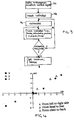
- Fig. 1 is a block diagram illustrating the major components of a system according to this invention as used for catheter mapping and related procedures.
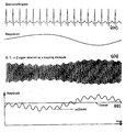
- Fig. 2(a) shows a normal electrocardiogram above and a signal representative of respiration below, illustrating the relative frequencies
- Fig. 2(b) illustrates a sensed location signal, and shows variations due to cardiac contraction and patient respiration
- Fig. 2(c) illustrates the sensed location signal with the high frequency filtered out, but still containing variations due to contraction and respiration (Vc), and the signal after the low frequencies have also been filtered out (V).
- Fig. 3 is a flow diagram setting forth the primary steps of catheter mapping and ablation in accordance with this invention.
- Fig. 4 is a diagram representing a plot of three-dimensional location data obtained in accordance with this invention.
- This invention is based upon using the patient, and in a specific embodiment example, the patient's heart, as a potentiometer in three orthogonal directions.
- Orthogonal high-frequency current sources are utilized to transmit a relatively low current in each of three respective orthogonal directions through the patient, directed toward or near the body area of interest.
- respective current sources 40, 41 and 42 are used to generate signals applied to electrodes which are shown diagrammatically as electrode pairs x, x_; y, y_; and z, z_.
- a catheter 46 is introduced into the patient, and for purposes of the ongoing discussion it will be assumed that the distal end of the catheter is introduced into the patient's heart.
- the catheter has at least two electrodes, illustrated at 47, 48.
- Electrode 47 is at about the tip end of the catheter, and can be positioned at or adjacent to the heart wall area of interest. As used herein, the tip electrode may be actually at the tip, or displaced somewhat proximally from the tip but in the distal tip region of the catheter.
- the second electrode 48 is positioned a predetermined distance D from the electrode 47.
- the catheter may contain three, four or more electrodes, so long as it contains at least a position-detecting electrode, preferably at or near the tip, and provides a pair of electrodes separated by a predetermined distance D, for calibration purposes as set forth below.
- a PTCA catheter can have two electrodes near its tip, or on opposite sides of the balloon, with suitable connections for use in the practice of this invention.
- the three electrical signals applied to the patient are high-frequency constant current pulse signals, of the form illustrated in Fig. 2a, each at a slightly different frequency.
- the current source which drives the x, x_ electrodes, designated CS x may operate at 30 kHz, with a current of about 0.1 mA; CS y operates at 31 kHz; and CS z operates at 32 kHz.
- all three sources can operate at about the same frequency, but are time multiplexed so that they can be distinguished upon subsequent pick-up of sensed signals.
- some characteristic such as frequency, phase or time is adjusted for each of the three applied signals, so that three signals can be sensed in the patient and then separated out as respective x, y and z signals.
- the range of 25-50 kHz is advantageous for practice of this invention, because it is well above the usual upper cut off frequency of bandpass endocardial electrogram amplifiers. Lower frequencies may also be used, but in such case specially trimmed filters are required for all electrogram amplifiers to eliminate the external signals. If, however, the invention is practiced with procedures where no endocardial electrograms are recorded, e.g. PTCA, then the external source frequencies may be much lower.
- the orthogonal signals may have any current level suitable for avoiding noise pickup in other signals. And while current pulses are preferred because they eliminate the influence of varying skin contact impedance, the signals may be voltage pulses in some applications. Thus, the optimum frequency, as well as the signal level, will depend upon the application.
- the tip or mapping electrode 47 is connected through to the three detection filters 50, 54, 57, each of which is adjusted to be sensitive to a respective one of the three current source frequencies.
- a voltage is sensed between electrode 47 and reference electrode R, suitably a surface electrode on the patient's skin.
- the constants, or sensitivities, a, b and c are unknowns which must be determined, and are expressed in mV/mm.
- a catheter is employed that has two electrodes with a known interelectrode distance (D, in mm).
- One of the two electrodes may be the tip electrode, or the two electrodes may be two separate electrodes, such as two electrodes of a quadripolar catheter.
- This calibration arrangement requires two sets of equally sensitive detection amplifiers and signal processing paths for each direction as indicated diagrammatically in Fig. 1.
- V x1 ax 1
- V y1 by 1
- V z1 cz 1
- V x2 ax 2
- V y2 by 2
- V z2 cz 2
- the system may not be ideally homogeneous, meaning that any given set of obtained measurements is not absolutely correct. This is not a basic problem to obtaining accurate measurements, since the calculations can be continuously performed automatically during catheterization, and the results can be averaged. Thus, as long as the catheter is being manipulated, the calibration measurements and calculations can be repeated any number of times, and a resulting average obtained that provides a very real and accurate position determination. Note also that it is easy with this invention to calculate the calibration constants, or sensitivities, for different areas of the heart chamber. This could be useful since the measurements may not be precisely linear. By recalculating the calibration constants for different areas of the heart chamber, calculated relative positions can be reliably obtained for clinical use in mapping and ablation purposes.
- a catheterization can be performed by assuming a "ballpark" sensitivity based, for example, on the weight or thorax dimensions of the patient. Note also that it is not usually necessary to map the whole heart chamber. Mapping and subsequent ablation is usually only performed in a certain part or area of the chamber where the arrhythmia originates. Linearity is much better when the mapping is confined to a limited area of that heart chamber.
- calibration can be achieved without using two electrodes in the heart, by assuming certain cardiac dimensions while only measuring V x , V y , and V z on the mapping electrode.
- the catheter before entering the left ventricle, the catheter has to be manipulated through the aorta descendens, the aortic arc, and the aortic valve. With the patient on his or her back, the depth from the aorta descendens to the aortic valve is approximately 5 cm. The distance from the aortic valve to the left ventricular apex is known to be close to 10 cm.
- catheter 46 is shown having a tip electrode 47, which is manipulated into some position within the heart chamber.
- a reference electrode R on the surface of the patient's body, is connected to a lead to provide a reference potential.
- the sensed voltage between tip electrode 47 and electrode R is connected through a switch matrix 49 to each of the three filters 50, 54 and 57, which are digital filters or other suitable narrow bandpass filters designed to pick up the respective signals generated at 40, 41 and 42 respectively.
- the three current sources are driven by respective clocks indicated at 44, which generate the basic timing signals at f x , f y and f z .
- each of the filters 50, 54 and 57 is coupled through a corresponding amplifier 51, 55, 58, and then through a low pass filter 52, 56, 59.
- the low pass filters have a cutoff of about 0.1 Hz, to filter out any more quickly moving variations in the signal from each amplifier. The purpose of this is to avoid problems arising from heart contraction and patient respiration. Accordingly, the low pass filters suitably have a long time constant in the range of 5-10 seconds, so as to filter out the cardiac and respiratory movements. However, it is to be noted that in some applications the respiration and cardiac movement information may be useful, such that use of the low pass filters is an option.
- Figs. 2(a), 2(b) and 2(c) illustrate the effect of contraction and respiration on the sensed x, y and z signals, and how these effects can be filtered out.
- Fig. 2(a) shows an electrocardiogram and a respiration signal
- Fig. 2(b) shows the sensed signal which has been sampled at the plus and minus peaks, to develop a signal corresponding to the difference between the plus and minus portions of the sensed 30 kHz pulses.
- the amplitude of the sensed signal is illustrated as varying due to respiration and contraction, giving rise to a signal V c , as illustrated in Fig. 2(c).
- Such signal changes are removed by the low pass filter, resulting in an accurate position signal of voltage V, illustrated in Fig. 2(c).
- the x, y and z outputs from the three channels shown in Fig. 1 are connected to a computer 65, or equivalent apparatus, for calculation of each three-dimensional location.
- the outputs are connected to a suitable output, or display 66, for vertical real time display.
- position data can be stored for re-display later.
- each of the electrodes of the electrode pair on catheter 46 that is used for calibration is connected to a pair of z processing channels, a pair of y processing channels, and a pair of x processing channels.
- the two signals are inputted to z filters 50, 50_; y filters 54, 54_; and x filters 57, 57_.
- These filters are accurately matched, in order to provide the DV x , DV y and DV z signals.
- the clock signals from block 44 are connected through to each of the six filters, to provide digital filtering of the respective pairs of x, y and z signals.
- the orthogonal constant current signals are applied across the patient, as represented in Fig. 1. As indicated previously, these respective signals are suitably about 30, 31 and 32 kHz, each with a current of about 0.1 mA. While somewhat lower frequencies can be used, it is noted that lower frequencies, as well as higher currents, have the disadvantage that they are more likely to be picked up in ECG tracings. While higher frequencies are suitable and clearly within the scope of the invention, they require more exacting electronics.
- the step of inserting the catheter so that the tip is in the region to be mapped is indicated. This step may, of course, be performed before applying the orthogonal signals.
- the step of calibrating the system for determining the a, b and c constants is indicated. As set forth above, this step can be done simultaneously with taking location data.
- the catheter tip is moved into a position of interest. For a catheterization procedure which is to lead to ablation, sensing is performed to gather data relating to the heart, such as the location of an arrhythmia focus. Such data gathering techniques are well known in the art.
- the location information is determined, by the calculations set forth above, and the sensed information and location are stored and/or mapped.
- the flow diagram indicates that the steps of block 76 may be repeated any number of times, at the discretion of the physician.
- the catheter tip may be moved to any number of locations, all of which can be identified and automatically mapped in accordance with this procedure.
- mapping information is utilized to position the catheter tip, i.e., the catheter tip is moved and located, and when it is at the desired mapped position, ablation is performed for removing a source of the arrhythmia.
- ablation is suitably performed by applying a pulse of radio frequency energy to the heart tissue for a period of time, e.g., several minutes.
- the typical ablation procedure makes a lesion of about 1 cm in diameter.
- the ablation can be repeated at different locations in the vicinity of arrhythmia focus, using the previous mapping data, and determining the exact location, or position of the catheter tip in accordance with this invention.
- FIG. 4 there is shown an example of electrical localization, or position measurements, taken during a cardiac catheterization.
- a 10 kHz current at .1 mA pulse height was delivered in three orthogonal directions through the patient chest.
- X was left to right; Y was head to legs; and Z was frontal chest to dorsal.
- Actual catheter tip positions were measured by means of calibrated Roentgen images (centimeters, horizontal axis) and plotted versus measured electrical potentials amplified five times (mV, vertical axis), for each of the X, Y and Z directions.
- the catheter tip was positioned at four different places, namely in the high right atrial appendage; on the bundle of HIS in the left ventricle near the mitral annulus; and inside the coronary sinus.
- One of the four positions is represented as the reference, at the intersection of the horizontal and vertical axes.
- linearity for each of the x, y and z directions is very good.
- the accuracy is within mm, depending upon the degree of filtering of variations induced by respiration and heart movement. This accuracy leads to a significant improvement in ablation procedures, since previously after the initial mapping, the physician essentially had to again do fine tuning or remapping when returning to ablate.
- the physician can return very quickly to the primary ablation position, and reposition the ablation electrode for producing lesions at precisely defined positions so as to effectively cure and control the arrhythmia.
- the reference electrode is placed somewhere on the skin, which has the advantage that it is unlikely to be displaced during the procedure.
- a disadvantage of this arrangement is that the cardiac contraction and respiration induced signal amplitude variations are relatively high.
- Another option is to use one electrode of a stable catheter within the body area, eg, heart, as the reference electrode. Positioning the reference electrode in another chamber of the heart has the advantage that displacement is relatively unlikely; and respiration and contraction influences are reduced because for a given position of the mapping electrode, both electrodes move simultaneously with respiration and contraction.
- the reference catheter electrode may occasionally displace, which renders previous measurements substantially useless.
- the source electrodes are used also as reference electrodes, and the source signals are time multiplexed, as disclosed above.
- a 90 kHz signal source is used, with respective successive pulses being connected across first the x-x_ electrodes, next the y-y_ electrodes, and then the z-z_ electrodes, so that each pair of electrodes transmits a respective 30 kHz signal.
- separation of the sensed voltages is achieved by timing as contrasted to frequency, in a known manner.
- the two other pairs of electrodes are available, and can be used as reference electrodes.
- both the z and z_ electrodes are connected together as the reference electrode; when the z measurement is being made, the y and y_ electrodes are connected as the reference electrode.
- the advantage of this arrangement is that the effective electrode is located roughly in the middle of the patient, close to the heart, and is not likely to dislocate because it involves skin electrodes.
- mapping locations which are rather close to a pair of skin electrodes. Due to the curvature of the equipotential lines of the resulting electric field, a slight error could be introduced when the mapping position is not roughly midway between the electrode pair. However, such an error can be compensated for by estimating the position of the mapping electrode between each such electrode pair, and making an appropriate mathematical adjustment.
- the approximate position of the mapping electrode can be checked by comparing the y voltage on the z-z_ electrodes with the y voltage on the mapping electrode; comparing the z voltage on the y-y_ electrodes with the z voltage on the mapping electrode; and comparing the x voltage on the y-y_ and/or the z-z_ electrodes with the x voltage on the mapping electrode.
- a primary application involves identification of the focus, or exit-site of tachycardia, e.g., ventricular tachycardia (VT).
- VT ventricular tachycardia
- surface ECGs are obtained during ventricular VT and compared with intracardiac ECGs obtained through the tip electrode at various places within the ventricle.
- intracardiac ECGs obtained through the tip electrode at various places within the ventricle.
- the exit-site of the VT can be identified.
- the pacing sites and the corresponding correlations between paced and VT ECGs can be used to determine the optimal site at which the best correlation between both can be expected.
- the system and method of this invention are also applicable for three-dimensional imaging of coronary stenosis.
- This can be done by combining echo tip data with three-dimensional data.
- a single catheter can be equipped with an echo tip at the end, as well as two distally located electrodes for obtaining dimensional information.
- an accurate three-dimensional map can be made for identifying coronary stenosis.
- stent placement By obtaining three-dimensional information in accordance with this invention, previously explored catheter positions can be accurately re-located so that a stent can be placed at exactly the same site where, for example, a PCTA had been applied, or where an intraluminal echo-image has been obtained.
- the technique can likewise be used for obtaining two-dimensional, or even one-dimensional position data, in applications not involving three-dimensional positioning.
Abstract
Description
- This invention relates to systems and methods for mapping catheter electrode position within a patient's body and, more particularly, a system and method which enables automatic real time three-dimensional measurement of catheter electrode position with an accuracy well less than 1 cm.
- As is known, accurate position information is necessary for mapping, or localizing an accessory atrioventricular pathway. In this case, one can localize the ventricular or atrial insertion site of the pathway during antegrade or retrograde conduction through the pathway.
- The need for accurate positioning information is further illustrated by the standard method of cardiac mapping and subsequent ablation of the site of a ventricular tachycardia in a patient. The catheter is introduced into the atrium or ventricle, and the tip is positioned at an endocardial site. A tachycardia is induced, and the tip is moved to different positions, where the timing of sensed intracardiac signals is compared with ECG signals. Each position and the local activation moment must be accurately determined and recorded, so that an accurate map can be made from which the tachycardia focus can be determined. Following the mapping, the ablation tip must be accurately repositioned with respect to the focus. This repositioning places a great importance on being able to obtain accurate tip position information when and as the tip is moved to a position. Further, it is well known that frequently multiple ablations are often necessary in a relatively small area within the heart, in order to eliminate arrhythmogenic foci. Accordingly, the catheter is sequentially positioned at slightly different positions close to the focus, for producing lesions in the heart wall. These lesions are produced at different locations in order to ensure elimination of the foci. At present, it is difficult to obtain accurate and reliable information concerning the distances between successive ablation sites.
- During hemodynamic and electrophysiologic cardiac catheterization procedures, cardiologists generally employ a monoplane and sometimes biplane fluoroscopic imaging to estimate the position of the catheter within the heart. However, with fluoroscopy, it is not yet possible to obtain automatic and objective three-dimensional information about the catheter position without laborious three-dimensional reconstruction from fluoroscopic images. As is readily understood, automatic measurement of the catheter position would be extremely useful during many interventional catheterization procedures.
- Systems for obtaining three-dimensional catheter position data are known, but have serious limitations. For example, a magnetic system employs a special element in the catheter tip, the size and configuration of which make it useful for only certain catheter types. Given the many different types of catheters in use for different applications, a system and technique that would be able to accurately locate the position of any type catheter would constitute a significant advance.
- The patent art contains a great many devices and systems directed toward catheter location. These systems embrace a number of different approaches, such as securing an inductor coil adjacent to the catheter tip with leads extending from the coil along the catheter for connection to external indicating equipment; positioning a varying magnetic field responsive component on the catheter or implement to be positioned, and using a movable external magnetic field source; use of a probe which generates a small magnetic field which is disturbed by a magnetically permeable metal in the device to be positioned; and the construction of various types of cardiac mapping probes and electrode configurations. However, these approaches have not proven commercially successful for one reason or another, and there remains a substantial need in the art for an improved technique of catheter mapping, particularly as applicable to cardiac catheterization and ablation procedures.
- In accordance with the above need in the art, there is provided a method of three-dimensional measurement of positions within a patient, comprising:
- (a) applying respective orthogonal alternating current signals, corresponding substantially to x, y and z directions through said patient;
- (b) inserting a catheter into said patient, said catheter having a mapping electrode and at least one other electrode;
- (c) obtaining correlation factors correlating measurements at said mapping electrode with x, y and z positions within said patient;
- (d) measuring, at a plurality of different positions within said patient's body, signals sensed at said catheter mapping electrode and corresponding to each of said three dimensional signals;
- (e) using said correlation factors to obtain three-dimensional x, y and z positions for each of said catheter mapping electrode positions; and
- (f) outputting position data representative of such three-dimensional positions.
- In accordance with another aspect, there is provided a catheter mapping method for mapping locations within a patient's heart, comprising:
- applying three substantially orthogonal current signals through said patient, each of said current signals having a respective characteristic distinguishable from the other two;
- positioning a catheter having a sensing electrode, so that said sensing electrode is at a given location within said patient's heart;
- sensing at said sensing electrode each of said three signals, and separating said three sensed signals on the basis of said respective characteristics;
- determining the location of said sensing electrode from said three sensed signals;
- repeating said positioning, sensing and determining steps at a plurality of locations within the patient's heart; and
- outputting data corresponding to said determined locations.
- In accordance with another aspect, there is provided a system for use in carrying out catheter mapping of a body location within a patient, comprising:
- external signal means for applying at least one electrical alternating current signal to said patient;
- a catheter adapted to be inserted into said patient's body and manipulated to a plurality of locations, said catheter having at least a mapping electrode;
- location means connected to said mapping electrode for sensing said at least one alternating current signal and processing same to obtain location signals indicative of the location of said mapping electrode when positioned at respective different body locations;
- means for calculating from said location signals positions for each location where said alternating current signals were sensed; and
- output means for outputting data corresponding to said positions.
- The invention thus provides a catheter mapping system and method for mapping locations within a patient, which provides an improved accuracy of location, the accuracy being of the order of a few mm. The preferred embodiment involves applying three orthogonal (x, y, z) current signals through the patient, directed substantially toward the area to be explored, such as the patient's heart, each of the signals having a respective characteristic which renders it distinguishable from the other two orthogonal signals. The catheter, which has been introduced into the body area to be explored, has a tip electrode or another mapping electrode positioned in the vicinity of the distal tip, which is connected through to three sensing channels. The sensing channels sense the signals induced at the electrode location by the three respective applied signals, which sensed signals are used to calculate the location of the electrode. A simple calibration procedure uses two electrodes at a known interelectrode distance on the catheter, and three quick measurements for determining the correlation of the respective sensed x, y and z signals with the tip position.
- In the preferred embodiment, the external signals are orthogonal, but can be slightly off orthogonal. The externally applied signals are suitably constant current pulse currents at frequencies in the range of about 25-50 kHz, with the constant current pulses having a current in a range centered around about 0.1 mA. While these parameters have been found to be useful for avoiding interference with ECG pickups, other parameters can be used. The sensed x, y and z signals are separated by digital filters, or other suitable narrow pass filters, with the resulting signals passed through a low pass filter which has a cutoff designed to eliminate variations due to cardiac contraction and patient respiration.
- The ready availability of accurate three dimensional position data will allow for numerous improvements in visualization of catheter position. While the X-ray state of the art only presents two separate images in two usually perpendicular directions, three dimensional information allows for a three dimensional presentation of catheter tip position to cardiologists. This will make catheterization a lot easier and quicker, and meet a substantial long-existing need.
- Preferred embodiments will now be described by way of example only, with reference to the accompanying drawings.
- Fig. 1 is a block diagram illustrating the major components of a system according to this invention as used for catheter mapping and related procedures.
- Fig. 2(a) shows a normal electrocardiogram above and a signal representative of respiration below, illustrating the relative frequencies; Fig. 2(b) illustrates a sensed location signal, and shows variations due to cardiac contraction and patient respiration; Fig. 2(c) illustrates the sensed location signal with the high frequency filtered out, but still containing variations due to contraction and respiration (Vc), and the signal after the low frequencies have also been filtered out (V).
- Fig. 3 is a flow diagram setting forth the primary steps of catheter mapping and ablation in accordance with this invention.
- Fig. 4 is a diagram representing a plot of three-dimensional location data obtained in accordance with this invention.
- This invention is based upon using the patient, and in a specific embodiment example, the patient's heart, as a potentiometer in three orthogonal directions. Orthogonal high-frequency current sources are utilized to transmit a relatively low current in each of three respective orthogonal directions through the patient, directed toward or near the body area of interest. As seen in Fig. 1, respective
current sources catheter 46 is introduced into the patient, and for purposes of the ongoing discussion it will be assumed that the distal end of the catheter is introduced into the patient's heart. The catheter has at least two electrodes, illustrated at 47, 48. Electrode 47 is at about the tip end of the catheter, and can be positioned at or adjacent to the heart wall area of interest. As used herein, the tip electrode may be actually at the tip, or displaced somewhat proximally from the tip but in the distal tip region of the catheter. The second electrode 48 is positioned a predetermined distance D from theelectrode 47. Although just two such electrodes are shown, the catheter may contain three, four or more electrodes, so long as it contains at least a position-detecting electrode, preferably at or near the tip, and provides a pair of electrodes separated by a predetermined distance D, for calibration purposes as set forth below. Note that a PTCA catheter can have two electrodes near its tip, or on opposite sides of the balloon, with suitable connections for use in the practice of this invention. - In a first embodiment, the three electrical signals applied to the patient are high-frequency constant current pulse signals, of the form illustrated in Fig. 2a, each at a slightly different frequency. For example, the current source which drives the x, x_ electrodes, designated CSx, may operate at 30 kHz, with a current of about 0.1 mA; CSy operates at 31 kHz; and CSz operates at 32 kHz. In the alternative, all three sources can operate at about the same frequency, but are time multiplexed so that they can be distinguished upon subsequent pick-up of sensed signals. The important distinction is that some characteristic such as frequency, phase or time is adjusted for each of the three applied signals, so that three signals can be sensed in the patient and then separated out as respective x, y and z signals.
- It is to be noted that the range of 25-50 kHz is advantageous for practice of this invention, because it is well above the usual upper cut off frequency of bandpass endocardial electrogram amplifiers. Lower frequencies may also be used, but in such case specially trimmed filters are required for all electrogram amplifiers to eliminate the external signals. If, however, the invention is practiced with procedures where no endocardial electrograms are recorded, e.g. PTCA, then the external source frequencies may be much lower. Likewise, the orthogonal signals may have any current level suitable for avoiding noise pickup in other signals. And while current pulses are preferred because they eliminate the influence of varying skin contact impedance, the signals may be voltage pulses in some applications. Thus, the optimum frequency, as well as the signal level, will depend upon the application.
- Continuing with the illustration of the invention, and assuming three different frequency external signals, the mathematical basis for determining a position of the catheter tip is now explained. Still referring to Fig. 1, in the method of intracardiac mapping of this invention, the tip or
mapping electrode 47 is connected through to the three detection filters 50, 54, 57, each of which is adjusted to be sensitive to a respective one of the three current source frequencies. At any given location, for each orthogonal current a voltage is sensed betweenelectrode 47 and reference electrode R, suitably a surface electrode on the patient's skin. Presuming that the body behaves linearly, the three different measured voltages give unique x,y and z values for the location of thetip electrode 47 within the patient's body, as follows:


- The constants, or sensitivities, a, b and c, are unknowns which must be determined, and are expressed in mV/mm. In order to automatically calibrate, i.e., determine the three constants, in the preferred embodiment of this invention a catheter is employed that has two electrodes with a known interelectrode distance (D, in mm). One of the two electrodes may be the tip electrode, or the two electrodes may be two separate electrodes, such as two electrodes of a quadripolar catheter. This calibration arrangement requires two sets of equally sensitive detection amplifiers and signal processing paths for each direction as indicated diagrammatically in Fig. 1. Since each of the two electrodes picks up a voltage for each of the x, y and z currents, the following equations are applicable:


- To calculate the unknowns a, b and c, it is necessary to use the measured value DVx = Vx2 - Vx1 along with the unknown Dx = x2 - x1; DVy = Vy2 - Vy1 together with Dy = y2 - y1 and DVz = Vz2 - Vz1 together with Dz = z2 - z1. Then, knowing that DVx = aDx, DVy = bDy, and DVz = cDz, and Dx2 + Dy2 + Dz2 = D2, one obtains:
- With DVx, DVy, and DVz as measured, and D known, a2, b2, and c2 can be calculated. To simplify, let 1/a2 = A, 1/b2 = B, and 1/c2 = C, and DVx 2 = X, DVy 2 = Y and DVz 2 = Z. This produces the following simplified equation:

- In practice, the system may not be ideally homogeneous, meaning that any given set of obtained measurements is not absolutely correct. This is not a basic problem to obtaining accurate measurements, since the calculations can be continuously performed automatically during catheterization, and the results can be averaged. Thus, as long as the catheter is being manipulated, the calibration measurements and calculations can be repeated any number of times, and a resulting average obtained that provides a very real and accurate position determination. Note also that it is easy with this invention to calculate the calibration constants, or sensitivities, for different areas of the heart chamber. This could be useful since the measurements may not be precisely linear. By recalculating the calibration constants for different areas of the heart chamber, calculated relative positions can be reliably obtained for clinical use in mapping and ablation purposes.
- Even without any calibration, a catheterization can be performed by assuming a "ballpark" sensitivity based, for example, on the weight or thorax dimensions of the patient. Note also that it is not usually necessary to map the whole heart chamber. Mapping and subsequent ablation is usually only performed in a certain part or area of the chamber where the arrhythmia originates. Linearity is much better when the mapping is confined to a limited area of that heart chamber.
- In another embodiment of the invention, calibration can be achieved without using two electrodes in the heart, by assuming certain cardiac dimensions while only measuring Vx, Vy, and Vz on the mapping electrode. For example, before entering the left ventricle, the catheter has to be manipulated through the aorta descendens, the aortic arc, and the aortic valve. With the patient on his or her back, the depth from the aorta descendens to the aortic valve is approximately 5 cm. The distance from the aortic valve to the left ventricular apex is known to be close to 10 cm. Using such approximate distance figures, together with the measured voltages at those sites, it is possible to generate a sensitivity calibration when the system is "told" where, in an anatomical sense, the catheter electrodes are positioned. This results in catheter positions inside a normalized left ventricle. The same technique can be used in other heart chambers for obtaining reliable position data.
- Referring again to Fig. 1,
catheter 46 is shown having atip electrode 47, which is manipulated into some position within the heart chamber. A reference electrode R, on the surface of the patient's body, is connected to a lead to provide a reference potential. For making position measurements, the sensed voltage betweentip electrode 47 and electrode R is connected through aswitch matrix 49 to each of the three filters 50, 54 and 57, which are digital filters or other suitable narrow bandpass filters designed to pick up the respective signals generated at 40, 41 and 42 respectively. The three current sources are driven by respective clocks indicated at 44, which generate the basic timing signals at fx, fy and fz. These clock signals drive the current source generators, and also are connected to the respective x, y and z filters, for time sampling of the received signals as illustrated in Fig. 2(a) at points V1 and V2. The output of each of the filters 50, 54 and 57 is coupled through a correspondingamplifier low pass filter - Figs. 2(a), 2(b) and 2(c) illustrate the effect of contraction and respiration on the sensed x, y and z signals, and how these effects can be filtered out. Fig. 2(a) shows an electrocardiogram and a respiration signal; Fig. 2(b) shows the sensed signal which has been sampled at the plus and minus peaks, to develop a signal corresponding to the difference between the plus and minus portions of the sensed 30 kHz pulses. The amplitude of the sensed signal is illustrated as varying due to respiration and contraction, giving rise to a signal Vc, as illustrated in Fig. 2(c). Such signal changes are removed by the low pass filter, resulting in an accurate position signal of voltage V, illustrated in Fig. 2(c).
- The x, y and z outputs from the three channels shown in Fig. 1 are connected to a
computer 65, or equivalent apparatus, for calculation of each three-dimensional location. The outputs are connected to a suitable output, ordisplay 66, for vertical real time display. As discussed further below, position data can be stored for re-display later. - Referring back to Fig. 1, during the calibration steps each of the electrodes of the electrode pair on
catheter 46 that is used for calibration is connected to a pair of z processing channels, a pair of y processing channels, and a pair of x processing channels. Thus, the two signals are inputted to z filters 50, 50_; y filters 54, 54_; and x filters 57, 57_. These filters are accurately matched, in order to provide the DVx, DVy and DVz signals. As with the position measurements, the clock signals fromblock 44 are connected through to each of the six filters, to provide digital filtering of the respective pairs of x, y and z signals. These six signals are amplified through amplifiers 51, 51_; 55, 55_; and 58, 58_; and then filtered through low pass filters 52, 52_; 56, 56_; and 59, 59_. The three pairs of x, y and z signals are then passed tocomputer 65 for carrying out the calculations set forth above, and determination of the a, b and c constants. Instead of using 2 sets of channel amplifiers and filters, only one set can be used, with each electrode alternatingly connected to the same channel input. Using the same channel for processing the signals ensures identical amplification, and thus greater accuracy. - Referring now to Fig. 3, some of the salient steps taken in practicing this invention are set forth. At
block 70, the orthogonal constant current signals are applied across the patient, as represented in Fig. 1. As indicated previously, these respective signals are suitably about 30, 31 and 32 kHz, each with a current of about 0.1 mA. While somewhat lower frequencies can be used, it is noted that lower frequencies, as well as higher currents, have the disadvantage that they are more likely to be picked up in ECG tracings. While higher frequencies are suitable and clearly within the scope of the invention, they require more exacting electronics. At 72, the step of inserting the catheter so that the tip is in the region to be mapped is indicated. This step may, of course, be performed before applying the orthogonal signals. At 74, the step of calibrating the system for determining the a, b and c constants is indicated. As set forth above, this step can be done simultaneously with taking location data. At 76, the catheter tip is moved into a position of interest. For a catheterization procedure which is to lead to ablation, sensing is performed to gather data relating to the heart, such as the location of an arrhythmia focus. Such data gathering techniques are well known in the art. The location information is determined, by the calculations set forth above, and the sensed information and location are stored and/or mapped. The flow diagram indicates that the steps ofblock 76 may be repeated any number of times, at the discretion of the physician. Thus, the catheter tip may be moved to any number of locations, all of which can be identified and automatically mapped in accordance with this procedure. Next, at block 78, it is determined whether there is a need or a reason to calibrate again, because of locating in a new area. As discussed above, it may be desirable to recalibrate if the catheter tip has been moved substantially, and, if so, the procedure goes back to 74. Again, note that the calibration can be undertaken together with the steps of moving the catheter tip, sensing, getting location, and mapping. When the mapping has been finished, the procedure goes to 80, for ablation. Here, the previously obtained mapping information is utilized to position the catheter tip, i.e., the catheter tip is moved and located, and when it is at the desired mapped position, ablation is performed for removing a source of the arrhythmia. As is known, ablation is suitably performed by applying a pulse of radio frequency energy to the heart tissue for a period of time, e.g., several minutes. The typical ablation procedure makes a lesion of about 1 cm in diameter. The ablation can be repeated at different locations in the vicinity of arrhythmia focus, using the previous mapping data, and determining the exact location, or position of the catheter tip in accordance with this invention. - Referring now to Fig. 4, there is shown an example of electrical localization, or position measurements, taken during a cardiac catheterization. A 10 kHz current at .1 mA pulse height, was delivered in three orthogonal directions through the patient chest. Referring to the legend of Fig. 4, X was left to right; Y was head to legs; and Z was frontal chest to dorsal. Actual catheter tip positions were measured by means of calibrated Roentgen images (centimeters, horizontal axis) and plotted versus measured electrical potentials amplified five times (mV, vertical axis), for each of the X, Y and Z directions. In this patient, the catheter tip was positioned at four different places, namely in the high right atrial appendage; on the bundle of HIS in the left ventricle near the mitral annulus; and inside the coronary sinus. One of the four positions is represented as the reference, at the intersection of the horizontal and vertical axes. Note that linearity for each of the x, y and z directions is very good. Heretofore, it has been very difficult to obtain a locational accuracy within several cm. With this invention, the accuracy is within mm, depending upon the degree of filtering of variations induced by respiration and heart movement. This accuracy leads to a significant improvement in ablation procedures, since previously after the initial mapping, the physician essentially had to again do fine tuning or remapping when returning to ablate. Using the technique of this invention, the physician can return very quickly to the primary ablation position, and reposition the ablation electrode for producing lesions at precisely defined positions so as to effectively cure and control the arrhythmia.
- As discussed above, in the preferred embodiment, the reference electrode is placed somewhere on the skin, which has the advantage that it is unlikely to be displaced during the procedure. However, a disadvantage of this arrangement is that the cardiac contraction and respiration induced signal amplitude variations are relatively high. Another option is to use one electrode of a stable catheter within the body area, eg, heart, as the reference electrode. Positioning the reference electrode in another chamber of the heart has the advantage that displacement is relatively unlikely; and respiration and contraction influences are reduced because for a given position of the mapping electrode, both electrodes move simultaneously with respiration and contraction. However, even here the reference catheter electrode may occasionally displace, which renders previous measurements substantially useless.
- In another embodiment of the invention, and which addresses the need of a reliable reference electrode which has minimal sensitivity to contractions and respiration, the source electrodes are used also as reference electrodes, and the source signals are time multiplexed, as disclosed above. For example, a 90 kHz signal source is used, with respective successive pulses being connected across first the x-x_ electrodes, next the y-y_ electrodes, and then the z-z_ electrodes, so that each pair of electrodes transmits a respective 30 kHz signal. In this case, separation of the sensed voltages is achieved by timing as contrasted to frequency, in a known manner. When one signal is being detected, the two other pairs of electrodes are available, and can be used as reference electrodes. For example, when either the x or y measurement is being made, both the z and z_ electrodes are connected together as the reference electrode; when the z measurement is being made, the y and y_ electrodes are connected as the reference electrode. The advantage of this arrangement is that the effective electrode is located roughly in the middle of the patient, close to the heart, and is not likely to dislocate because it involves skin electrodes.
- It is to be noted that, with some of the source electrodes positioned around the heart, there may be some mapping locations which are rather close to a pair of skin electrodes. Due to the curvature of the equipotential lines of the resulting electric field, a slight error could be introduced when the mapping position is not roughly midway between the electrode pair. However, such an error can be compensated for by estimating the position of the mapping electrode between each such electrode pair, and making an appropriate mathematical adjustment. The approximate position of the mapping electrode can be checked by comparing the y voltage on the z-z_ electrodes with the y voltage on the mapping electrode; comparing the z voltage on the y-y_ electrodes with the z voltage on the mapping electrode; and comparing the x voltage on the y-y_ and/or the z-z_ electrodes with the x voltage on the mapping electrode.
- The system and method of this invention are applicable to a number of important medical techniques. A primary application, as indicated above, involves identification of the focus, or exit-site of tachycardia, e.g., ventricular tachycardia (VT). As is known, in the catheterization process, surface ECGs are obtained during ventricular VT and compared with intracardiac ECGs obtained through the tip electrode at various places within the ventricle. By known techniques, the exit-site of the VT can be identified. By using the three-dimensional localization of this invention, the pacing sites and the corresponding correlations between paced and VT ECGs can be used to determine the optimal site at which the best correlation between both can be expected.
By way of further illustration of the scope of the invention, the system and method of this invention are also applicable for three-dimensional imaging of coronary stenosis. This can be done by combining echo tip data with three-dimensional data. A single catheter can be equipped with an echo tip at the end, as well as two distally located electrodes for obtaining dimensional information. By combining echo tip and three-dimensional data, an accurate three-dimensional map can be made for identifying coronary stenosis. Yet another application is stent placement. By obtaining three-dimensional information in accordance with this invention, previously explored catheter positions can be accurately re-located so that a stent can be placed at exactly the same site where, for example, a PCTA had been applied, or where an intraluminal echo-image has been obtained. It is also to be noted that the technique can likewise be used for obtaining two-dimensional, or even one-dimensional position data, in applications not involving three-dimensional positioning. - While the invention has been illustrated by the use of orthogonal signals, they need not be absolutely orthogonal, although preferably substantially orthogonal. The angles may vary from strictly orthogonal and still be three-dimensional for purposes of practicing this invention; as long as the actual angles are known, a mathematical correction can be applied to compensate for any difference from truly orthogonal.
Claims (28)
- A method of three-dimensional measurement of positions within a patient, comprising:(a) applying respective orthogonal alternating current signals, corresponding substantially to x, y and z directions through said patient;(b) inserting a catheter (46) into said patient, said catheter having a mapping electrode (47) and at least one other electrode;(c) obtaining correlation factors correlating measurements at said mapping electrode with x, y and z positions within said patient;(d) measuring, at a plurality of different positions within said patient's body, signals sensed at said catheter mapping electrode and corresponding to each of said three dimensional signals;(e) using said correlation factors to obtain three-dimensional x, y and z positions for each of said catheter mapping electrode positions; and(f) outputting position data representative of such three-dimensional positions.
- The method as described in claim 1, comprising generating said orthogonal signals at respective different frequencies.
- The method as described in claim 1, comprising time multiplexing said respective three dimensional signals.
- The method as described in claim 2, wherein said respective frequencies are in a range of about 25-50 kHz.
- The method as described in any preceding claim, wherein said applying comprises applying constant current pulses, said pulses having a current of no more than about 0.1 mA.
- The method as described in any preceding claim, comprising filtering said sensed signals to remove components attributable to cardiac contraction and patient respiration.
- The method as described in any preceding claim, comprising using said outputted information to map three dimensional position data.
- The method as described in any preceding claim, comprising obtaining said correlation factors at positions where said position signals are also sensed.
- The method as described in any preceding claim, comprising providing said catheter with two electrodes separated by a distance D, and wherein said obtaining step comprises obtaining sensed signals from said two electrodes corresponding to each of said x, y and z signals.
- The method as described in any preceding claim, comprising using a processing unit in real time to obtain said three-dimensional x, y and z positions and visually displaying data representative of said obtained positions.
- The method as described in any preceding claim, comprising manipulating said catheter to different positions within said patient's body, and outputting a visual representation showing the three dimensional location of each said different position.
- The method as described in any of claims 1 to 10, comprising inserting said catheter into said patient's heart, and further comprising obtaining local activation data corresponding to each said obtained three-dimensional position, and storing said local activation data and said position data.
- The method as described in claim 12, comprising selecting at least one position in said heart corresponding to said stored positions, manipulating a catheter tip to said selected position, and ablating the heart at said position.
- A catheter mapping method for mapping locations within a patient's heart, comprising:applying three substantially orthogonal current signals through said patient, each of said current signals having a respective characteristic distinguishable from the other two;positioning a catheter (46) having a sensing electrode (47), so that said sensing electrode is at a given location within said patient's heart;sensing at said sensing electrode (47) each of said three signals, and separating said three sensed signals on the basis of said respective characteristics;determining the location of said sensing electrode (47) from said three sensed signals;repeating said positioning, sensing and determining steps at a plurality of locations within the patient's heart; andoutputting data corresponding to said determined locations.
- The method as described in claim 14, comprising calibrating said sensed signals with respect to locations within said patient's heart, thereby obtaining a respective relationship between each of said three sensed signals and a respective x, y or z location.
- The method as described in claim 15, wherein said determining uses said obtained respective relationships.
- The method as described in claim 14, 15 or 16, wherein said catheter comprises a pair of electrodes separated by a predetermined distance, and said calibrating comprises measuring signals induced from said three orthogonal signals at each of said pair of electrodes at at least three different positions or orientations.
- The method as described in any of claims 14 to 17, wherein said three substantially orthogonal signals are constant current pulse signals.
- The method as described in claim 18, comprising providing said orthogonal signals at a frequency of at least about 10 kHz.
- The method as described in any of claims 14 to 19, comprising generating said current signals with different frequencies, whereby said characteristic is frequency.
- The method as described in any of claims 14 to 19, comprising generating said current signals as time multiplexed signals, whereby said characteristic is time.
- A system for use in carrying out catheter mapping of a body location within a patient, comprising:external signal means (40,41,42) for applying at least one electrical alternating current signal to said patient;a catheter (46) adapted to be inserted into said patient's body and manipulated to a plurality of locations, said catheter having at least a mapping electrode (46);location means connected to said mapping electrode (46) for sensing said at least one alternating current signal and processing same to obtain location signals indicative of the location of said mapping electrode when positioned at respective different body locations;means (66) for calculating from said location signals positions for each location where said alternating current signals were sensed; andoutput means (66) for outputting data corresponding to said positions.
- The system as described in claim 22, wherein said catheter (46) comprises a pair of electrodes (47,48) separated by a distance D, and comprising means for sensing calibration signals from said pair of electrodes, and calibration means for determining calibration factors from said calibration signals, said means for calculating comprising means for calculating said positions from said location signals and said calibration signals.
- The system as described in claim 22 or 23, wherein said output means comprises video means for providing a three-dimensional representation of catheter location at each location to which it is manipulated.
- The system as described in claim 22, 23 or 24 wherein said location means comprises a reference electrode (R).
- The system as described in claim 25, wherein said reference electrode is a skin electrode (R).
- The system as described in claim 25, wherein said external means comprises means (40,41,42) for applying three substantially orthogonal alternating current signals, three pairs of skin electrodes, and time means for time multiplexing said signals, and said reference electrode comprises selected pairs of said skin electrodes.
- The system as described in claim 27, wherein said mapping electrode is a tip electrode (47).
Applications Claiming Priority (2)
| Application Number | Priority Date | Filing Date | Title |
|---|---|---|---|
| US08/561,773 US5697377A (en) | 1995-11-22 | 1995-11-22 | Catheter mapping system and method |
| US561773 | 1995-11-22 |
Publications (3)
| Publication Number | Publication Date |
|---|---|
| EP0775466A2 true EP0775466A2 (en) | 1997-05-28 |
| EP0775466A3 EP0775466A3 (en) | 1999-03-31 |
| EP0775466B1 EP0775466B1 (en) | 2003-10-22 |
Family
ID=24243405
Family Applications (1)
| Application Number | Title | Priority Date | Filing Date |
|---|---|---|---|
| EP96308472A Expired - Lifetime EP0775466B1 (en) | 1995-11-22 | 1996-11-22 | Catheter mapping system |
Country Status (6)
| Country | Link |
|---|---|
| US (2) | US5697377A (en) |
| EP (1) | EP0775466B1 (en) |
| JP (1) | JP3955346B2 (en) |
| AU (1) | AU714372B2 (en) |
| CA (1) | CA2189399C (en) |
| DE (1) | DE69630432T2 (en) |
Cited By (29)
| Publication number | Priority date | Publication date | Assignee | Title |
|---|---|---|---|---|
| US6216027B1 (en) | 1997-08-01 | 2001-04-10 | Cardiac Pathways Corporation | System for electrode localization using ultrasound |
| WO2004103174A1 (en) * | 2003-05-21 | 2004-12-02 | Philips Intellectual Property & Standards Gmbh | Apparatus and method for navigating a catheter |
| US6950689B1 (en) | 1998-08-03 | 2005-09-27 | Boston Scientific Scimed, Inc. | Dynamically alterable three-dimensional graphical model of a body region |
| WO2007111542A1 (en) * | 2006-03-27 | 2007-10-04 | St. Jude Medical Ab | Medical system for monitoring and localisation of electrode leads in the heart |
| EP1867279A3 (en) * | 2006-06-14 | 2008-02-06 | Biosense Webster, Inc. | Reference catheter for impedance calibration |
| US7869865B2 (en) | 2005-01-07 | 2011-01-11 | Biosense Webster, Inc. | Current-based position sensing |
| WO2011072507A1 (en) * | 2009-12-15 | 2011-06-23 | 四川锦江电子科技有限公司 | Method and apparatus for location determination of cardiac catheter |
| US8715195B2 (en) | 2007-04-11 | 2014-05-06 | Elcam Medical Agricultural Cooperative | System and method for accurate placement of a catheter tip in a patient |
| EP2482735A4 (en) * | 2009-09-30 | 2017-03-29 | Mayo Foundation For Medical Education And Research | Enhanced signal navigation and capture systems and methods |
| EP3730084A1 (en) * | 2012-10-23 | 2020-10-28 | Valtech Cardio, Ltd. | Controlled steering functionality for implant-delivery tool |
| EP4018925A1 (en) | 2020-12-22 | 2022-06-29 | Koninklijke Philips N.V. | Determining the shape of an interventional device |
| EP4023182A1 (en) | 2020-12-31 | 2022-07-06 | Koninklijke Philips N.V. | Generating a mapping function for tracking a position of an electrode |
| EP4023153A1 (en) | 2020-12-31 | 2022-07-06 | Koninklijke Philips N.V. | Quality measure for a mapping function |
| EP4026494A1 (en) | 2021-01-06 | 2022-07-13 | Koninklijke Philips N.V. | Tracking the position of an interventional device |
| EP4036930A1 (en) | 2021-01-29 | 2022-08-03 | Koninklijke Philips N.V. | Image reconstruction method for dielectric anatomical mapping |
| EP4036864A1 (en) | 2021-01-29 | 2022-08-03 | Koninklijke Philips N.V. | Generating an anatomical model of an anatomical cavity |
| WO2022161851A1 (en) | 2021-01-29 | 2022-08-04 | Koninklijke Philips N.V. | Image reconstruction method for dielectric anatomical mapping |
| EP4193908A1 (en) | 2021-12-08 | 2023-06-14 | Koninklijke Philips N.V. | Improving mapping of an anatomical cavity and/or location tracking in the anatomical cavity |
| EP4198998A1 (en) | 2021-12-17 | 2023-06-21 | Koninklijke Philips N.V. | Aiding an analysis of an anatomical element of a subject |
| EP4201350A1 (en) | 2021-12-24 | 2023-06-28 | Koninklijke Philips N.V. | Controlling and monitoring signals provided to electrodes |
| EP4201320A1 (en) | 2021-12-22 | 2023-06-28 | Koninklijke Philips N.V. | Interventional devices |
| EP4202846A1 (en) | 2021-12-23 | 2023-06-28 | Koninklijke Philips N.V. | Adaptive 3d modelling of an anatomical cavity |
| EP4248891A1 (en) | 2022-03-24 | 2023-09-27 | Koninklijke Philips N.V. | An ablation sysytem for performing ablation |
| EP4252640A1 (en) | 2022-03-31 | 2023-10-04 | Koninklijke Philips N.V. | Determining the distance between device electrodes |
| EP4278963A1 (en) | 2022-05-19 | 2023-11-22 | Koninklijke Philips N.V. | Controlling electrical signals between a catheter and a recording system |
| EP4295763A1 (en) | 2022-06-23 | 2023-12-27 | Koninklijke Philips N.V. | Generating an anatomical model of an anatomical cavity |
| WO2023247159A1 (en) | 2022-06-23 | 2023-12-28 | Koninklijke Philips N.V. | Generating an anatomical model of an anatomical cavity |
| EP4306051A1 (en) | 2022-07-14 | 2024-01-17 | Koninklijke Philips N.V. | Determining a distance between pairs of field-sensitive sensors of an interventional device |
| EP4335358A1 (en) | 2022-09-09 | 2024-03-13 | Koninklijke Philips N.V. | Registering an anatomical model to a reference anatomical model |
Families Citing this family (758)
| Publication number | Priority date | Publication date | Assignee | Title |
|---|---|---|---|---|
| FR2652928B1 (en) | 1989-10-05 | 1994-07-29 | Diadix Sa | INTERACTIVE LOCAL INTERVENTION SYSTEM WITHIN A AREA OF A NON-HOMOGENEOUS STRUCTURE. |
| US5325860A (en) | 1991-11-08 | 1994-07-05 | Mayo Foundation For Medical Education And Research | Ultrasonic and interventional catheter and method |
| US5704361A (en) | 1991-11-08 | 1998-01-06 | Mayo Foundation For Medical Education And Research | Volumetric image ultrasound transducer underfluid catheter system |
| US5603318A (en) | 1992-04-21 | 1997-02-18 | University Of Utah Research Foundation | Apparatus and method for photogrammetric surgical localization |
| ES2115776T3 (en) | 1992-08-14 | 1998-07-01 | British Telecomm | POSITION LOCATION SYSTEM. |
| US6240307B1 (en) | 1993-09-23 | 2001-05-29 | Endocardial Solutions, Inc. | Endocardial mapping system |
| US7189208B1 (en) | 1992-09-23 | 2007-03-13 | Endocardial Solutions, Inc. | Method for measuring heart electrophysiology |
| US5662108A (en) * | 1992-09-23 | 1997-09-02 | Endocardial Solutions, Inc. | Electrophysiology mapping system |
| WO1994006349A1 (en) * | 1992-09-23 | 1994-03-31 | Endocardial Therapeutics, Inc. | Endocardial mapping system |
| US7930012B2 (en) * | 1992-09-23 | 2011-04-19 | St. Jude Medical, Atrial Fibrillation Division, Inc. | Chamber location method |
| IL116699A (en) | 1996-01-08 | 2001-09-13 | Biosense Ltd | Method of constructing cardiac map |
| US6947785B1 (en) * | 1993-09-23 | 2005-09-20 | Endocardial Solutions, Inc. | Interface system for endocardial mapping catheter |
| US6129685A (en) * | 1994-02-09 | 2000-10-10 | The University Of Iowa Research Foundation | Stereotactic hypothalamic obesity probe |
| US7077822B1 (en) | 1994-02-09 | 2006-07-18 | The University Of Iowa Research Foundation | Stereotactic hypothalamic obesity probe |
| US5803089A (en) | 1994-09-15 | 1998-09-08 | Visualization Technology, Inc. | Position tracking and imaging system for use in medical applications |
| US5941251A (en) * | 1994-10-11 | 1999-08-24 | Ep Technologies, Inc. | Systems for locating and guiding operative elements within interior body regions |
| US5876336A (en) | 1994-10-11 | 1999-03-02 | Ep Technologies, Inc. | Systems and methods for guiding movable electrode elements within multiple-electrode structure |
| US5592939A (en) | 1995-06-14 | 1997-01-14 | Martinelli; Michael A. | Method and system for navigating a catheter probe |
| US6292689B1 (en) * | 1996-04-17 | 2001-09-18 | Imagyn Medical Technologies California, Inc. | Apparatus and methods of bioelectrical impedance analysis of blood flow |
| US6050267A (en) * | 1997-04-28 | 2000-04-18 | American Cardiac Ablation Co. Inc. | Catheter positioning system |
| US5944022A (en) * | 1997-04-28 | 1999-08-31 | American Cardiac Ablation Co. Inc. | Catheter positioning system |
| US6171247B1 (en) | 1997-06-13 | 2001-01-09 | Mayo Foundation For Medical Education And Research | Underfluid catheter system and method having a rotatable multiplane transducer |
| US6226548B1 (en) | 1997-09-24 | 2001-05-01 | Surgical Navigation Technologies, Inc. | Percutaneous registration apparatus and method for use in computer-assisted surgical navigation |
| US5954649A (en) * | 1997-10-20 | 1999-09-21 | Irvine Biomedical, Inc. | Catheter system having ultrasound locating capabilities |
| US6021343A (en) | 1997-11-20 | 2000-02-01 | Surgical Navigation Technologies | Image guided awl/tap/screwdriver |
| US6348058B1 (en) | 1997-12-12 | 2002-02-19 | Surgical Navigation Technologies, Inc. | Image guided spinal surgery guide, system, and method for use thereof |
| US6223066B1 (en) | 1998-01-21 | 2001-04-24 | Biosense, Inc. | Optical position sensors |
| SE9801006D0 (en) * | 1998-03-25 | 1998-03-25 | Siemens Elema Ab | Method and arrangement for determining the location of a catheter within an animal body |
| US6091980A (en) * | 1998-05-12 | 2000-07-18 | Massachusetts Institute Of Technology | Stent slip sensing system and method |
| US6129669A (en) * | 1998-05-22 | 2000-10-10 | Scimed Life Systems, Inc. | Systems and methods for assessing stability of an ablation electrode in contact with heart tissue |
| US6035226A (en) * | 1998-05-22 | 2000-03-07 | Scimed Life Systems, Inc. | Systems and methods for assessing stability of an operative instrument inside a body region |
| US7263397B2 (en) | 1998-06-30 | 2007-08-28 | St. Jude Medical, Atrial Fibrillation Division, Inc. | Method and apparatus for catheter navigation and location and mapping in the heart |
| US7806829B2 (en) | 1998-06-30 | 2010-10-05 | St. Jude Medical, Atrial Fibrillation Division, Inc. | System and method for navigating an ultrasound catheter to image a beating heart |
| US7670297B1 (en) | 1998-06-30 | 2010-03-02 | St. Jude Medical, Atrial Fibrillation Division, Inc. | Chamber mapping system |
| US6226542B1 (en) | 1998-07-24 | 2001-05-01 | Biosense, Inc. | Three-dimensional reconstruction of intrabody organs |
| US6301496B1 (en) | 1998-07-24 | 2001-10-09 | Biosense, Inc. | Vector mapping of three-dimensionally reconstructed intrabody organs and method of display |
| US6059731A (en) | 1998-08-19 | 2000-05-09 | Mayo Foundation For Medical Education And Research | Simultaneous side-and-end viewing underfluid catheter |
| US6477400B1 (en) | 1998-08-20 | 2002-11-05 | Sofamor Danek Holdings, Inc. | Fluoroscopic image guided orthopaedic surgery system with intraoperative registration |
| US6185448B1 (en) | 1998-09-29 | 2001-02-06 | Simcha Borovsky | Apparatus and method for locating and mapping a catheter in intracardiac operations |
| US20050004642A1 (en) * | 1998-11-09 | 2005-01-06 | Medtronic, Inc. | Implantable medical lead including overlay |
| US7575550B1 (en) | 1999-03-11 | 2009-08-18 | Biosense, Inc. | Position sensing based on ultrasound emission |
| US7558616B2 (en) | 1999-03-11 | 2009-07-07 | Biosense, Inc. | Guidance of invasive medical procedures using implantable tags |
| US7549960B2 (en) * | 1999-03-11 | 2009-06-23 | Biosense, Inc. | Implantable and insertable passive tags |
| US7174201B2 (en) * | 1999-03-11 | 2007-02-06 | Biosense, Inc. | Position sensing system with integral location pad and position display |
| US7590441B2 (en) | 1999-03-11 | 2009-09-15 | Biosense, Inc. | Invasive medical device with position sensing and display |
| US6470207B1 (en) | 1999-03-23 | 2002-10-22 | Surgical Navigation Technologies, Inc. | Navigational guidance via computer-assisted fluoroscopic imaging |
| US6398736B1 (en) | 1999-03-31 | 2002-06-04 | Mayo Foundation For Medical Education And Research | Parametric imaging ultrasound catheter |
| US6179865B1 (en) | 1999-04-01 | 2001-01-30 | Cardiac Pacemakers, Inc. | Cross chamber interval correlation |
| US6491699B1 (en) | 1999-04-20 | 2002-12-10 | Surgical Navigation Technologies, Inc. | Instrument guidance method and system for image guided surgery |
| US6626899B2 (en) | 1999-06-25 | 2003-09-30 | Nidus Medical, Llc | Apparatus and methods for treating tissue |
| AU1607600A (en) | 1999-07-26 | 2001-02-13 | Super Dimension Ltd. | Linking of an intra-body tracking system to external reference coordinates |
| WO2001013060A1 (en) | 1999-08-13 | 2001-02-22 | Advanced Sensor Technologies Llc | Probe position sensing system for use in a coordinate measuring machine |
| US6385476B1 (en) | 1999-09-21 | 2002-05-07 | Biosense, Inc. | Method and apparatus for intracardially surveying a condition of a chamber of a heart |
| US6368285B1 (en) | 1999-09-21 | 2002-04-09 | Biosense, Inc. | Method and apparatus for mapping a chamber of a heart |
| US6546271B1 (en) | 1999-10-01 | 2003-04-08 | Bioscience, Inc. | Vascular reconstruction |
| US6235038B1 (en) | 1999-10-28 | 2001-05-22 | Medtronic Surgical Navigation Technologies | System for translation of electromagnetic and optical localization systems |
| US8239001B2 (en) | 2003-10-17 | 2012-08-07 | Medtronic Navigation, Inc. | Method and apparatus for surgical navigation |
| US6474341B1 (en) | 1999-10-28 | 2002-11-05 | Surgical Navigation Technologies, Inc. | Surgical communication and power system |
| US6499488B1 (en) | 1999-10-28 | 2002-12-31 | Winchester Development Associates | Surgical sensor |
| US8644907B2 (en) | 1999-10-28 | 2014-02-04 | Medtronic Navigaton, Inc. | Method and apparatus for surgical navigation |
| US6493573B1 (en) | 1999-10-28 | 2002-12-10 | Winchester Development Associates | Method and system for navigating a catheter probe in the presence of field-influencing objects |
| US6381485B1 (en) | 1999-10-28 | 2002-04-30 | Surgical Navigation Technologies, Inc. | Registration of human anatomy integrated for electromagnetic localization |
| US7366562B2 (en) | 2003-10-17 | 2008-04-29 | Medtronic Navigation, Inc. | Method and apparatus for surgical navigation |
| US11331150B2 (en) | 1999-10-28 | 2022-05-17 | Medtronic Navigation, Inc. | Method and apparatus for surgical navigation |
| US6379302B1 (en) | 1999-10-28 | 2002-04-30 | Surgical Navigation Technologies Inc. | Navigation information overlay onto ultrasound imagery |
| WO2001064124A1 (en) | 2000-03-01 | 2001-09-07 | Surgical Navigation Technologies, Inc. | Multiple cannula image guided tool for image guided procedures |
| US6535756B1 (en) | 2000-04-07 | 2003-03-18 | Surgical Navigation Technologies, Inc. | Trajectory storage apparatus and method for surgical navigation system |
| US7085400B1 (en) | 2000-06-14 | 2006-08-01 | Surgical Navigation Technologies, Inc. | System and method for image based sensor calibration |
| EP1174076A3 (en) * | 2000-07-18 | 2002-10-16 | BIOTRONIK Mess- und Therapiegeräte GmbH & Co Ingenieurbüro Berlin | Device for automatically performing diagnostic and/or therapeutic actions in body cavities |
| US6650927B1 (en) | 2000-08-18 | 2003-11-18 | Biosense, Inc. | Rendering of diagnostic imaging data on a three-dimensional map |
| US6633773B1 (en) | 2000-09-29 | 2003-10-14 | Biosene, Inc. | Area of interest reconstruction for surface of an organ using location data |
| US6891551B2 (en) * | 2000-11-10 | 2005-05-10 | Microsoft Corporation | Selection handles in editing electronic documents |
| US6941166B2 (en) | 2000-11-10 | 2005-09-06 | C.R. Bard, Inc. | Software controlled electrophysiology data management |
| US20020103430A1 (en) * | 2001-01-29 | 2002-08-01 | Hastings Roger N. | Catheter navigation within an MR imaging device |
| US6807439B2 (en) | 2001-04-03 | 2004-10-19 | Medtronic, Inc. | System and method for detecting dislodgement of an implantable medical device |
| US20030040676A1 (en) * | 2001-05-03 | 2003-02-27 | Prentice John A. | Method and apparatus for determining spatial relation of multiple implantable electrodes |
| IL143260A (en) * | 2001-05-20 | 2006-09-05 | Given Imaging Ltd | Array system and method for locating an in vivo signal source |
| US6636757B1 (en) | 2001-06-04 | 2003-10-21 | Surgical Navigation Technologies, Inc. | Method and apparatus for electromagnetic navigation of a surgical probe near a metal object |
| DE60228266D1 (en) | 2001-06-18 | 2008-09-25 | Given Imaging Ltd | SWITCHABLE IN VIVO CAPSULE WITH A RIGID AND FLEXIBLE SECTION CIRCUIT BOARD |
| US8428685B2 (en) * | 2001-09-05 | 2013-04-23 | Given Imaging Ltd. | System and method for magnetically maneuvering an in vivo device |
| JP2005501630A (en) | 2001-09-05 | 2005-01-20 | ギブン・イメージング・リミテッド | System and method for three-dimensional display of body lumen |
| WO2003028224A2 (en) | 2001-09-24 | 2003-04-03 | Given Imaging Ltd. | System and method for controlling a device in vivo |
| US20030195415A1 (en) * | 2002-02-14 | 2003-10-16 | Iddan Gavriel J. | Device, system and method for accoustic in-vivo measuring |
| US6947786B2 (en) | 2002-02-28 | 2005-09-20 | Surgical Navigation Technologies, Inc. | Method and apparatus for perspective inversion |
| US7457658B2 (en) * | 2002-03-07 | 2008-11-25 | Medtronic, Inc. | Algorithm for accurate three-dimensional reconstruction of non-linear implanted medical devices in VIVO |
| US6990368B2 (en) | 2002-04-04 | 2006-01-24 | Surgical Navigation Technologies, Inc. | Method and apparatus for virtual digital subtraction angiography |
| US6955674B2 (en) * | 2002-04-11 | 2005-10-18 | Medtronic, Inc. | Medical ablation catheter control |
| US7998062B2 (en) | 2004-03-29 | 2011-08-16 | Superdimension, Ltd. | Endoscope structures and techniques for navigating to a target in branched structure |
| US7477763B2 (en) | 2002-06-18 | 2009-01-13 | Boston Scientific Scimed, Inc. | Computer generated representation of the imaging pattern of an imaging device |
| JP4511935B2 (en) * | 2002-08-24 | 2010-07-28 | サブラマニアム シー. クリシュナン | Transseptal puncture device |
| US6944490B1 (en) | 2002-09-25 | 2005-09-13 | Advanced Cardiovascular Systems, Inc. | Apparatus and method for positioning and delivering a therapeutic tool to the inside of a heart |
| JP4746876B2 (en) | 2002-10-15 | 2011-08-10 | ギブン イメージング リミテッド | Apparatus, system and method for transferring a signal to a mobile device |
| EP1565118B1 (en) | 2002-10-31 | 2016-03-09 | Boston Scientific Scimed, Inc. | Electrophysiology catheter with biased tip |
| US7599730B2 (en) | 2002-11-19 | 2009-10-06 | Medtronic Navigation, Inc. | Navigation system for cardiac therapies |
| US7697972B2 (en) | 2002-11-19 | 2010-04-13 | Medtronic Navigation, Inc. | Navigation system for cardiac therapies |
| US7130700B2 (en) | 2002-11-19 | 2006-10-31 | Medtronic, Inc. | Multilumen body for an implantable medical device |
| US20040097803A1 (en) * | 2002-11-20 | 2004-05-20 | Dorin Panescu | 3-D catheter localization using permanent magnets with asymmetrical properties about their longitudinal axis |
| US8388540B2 (en) * | 2002-12-13 | 2013-03-05 | Boston Scientific Scimed, Inc. | Method and apparatus for orienting a medical image |
| US8043281B2 (en) * | 2002-12-23 | 2011-10-25 | Medtronic, Inc. | Catheters incorporating valves and permeable membranes |
| AU2003303387A1 (en) * | 2002-12-23 | 2004-07-22 | Medtronic, Inc. | Multiple infusion section catheters, systems, and methods |
| US8246602B2 (en) * | 2002-12-23 | 2012-08-21 | Medtronic, Inc. | Catheters with tracking elements and permeable membranes |
| US7833151B2 (en) | 2002-12-26 | 2010-11-16 | Given Imaging Ltd. | In vivo imaging device with two imagers |
| US7542791B2 (en) | 2003-01-30 | 2009-06-02 | Medtronic Navigation, Inc. | Method and apparatus for preplanning a surgical procedure |
| US7660623B2 (en) | 2003-01-30 | 2010-02-09 | Medtronic Navigation, Inc. | Six degree of freedom alignment display for medical procedures |
| US20070055142A1 (en) * | 2003-03-14 | 2007-03-08 | Webler William E | Method and apparatus for image guided position tracking during percutaneous procedures |
| JP5033418B2 (en) * | 2003-07-02 | 2012-09-26 | ギブン イメージング リミテッド | Imaging sensor array and apparatus and method using the same |
| US7313430B2 (en) | 2003-08-28 | 2007-12-25 | Medtronic Navigation, Inc. | Method and apparatus for performing stereotactic surgery |
| US20050065441A1 (en) * | 2003-08-29 | 2005-03-24 | Arkady Glukhovsky | System, apparatus and method for measurement of motion parameters of an in-vivo device |
| EP2113189B1 (en) | 2003-09-15 | 2013-09-04 | Covidien LP | System of accessories for use with bronchoscopes |
| EP2316328B1 (en) | 2003-09-15 | 2012-05-09 | Super Dimension Ltd. | Wrap-around holding device for use with bronchoscopes |
| US7604589B2 (en) * | 2003-10-01 | 2009-10-20 | Given Imaging, Ltd. | Device, system and method for determining orientation of in-vivo devices |
| US7835778B2 (en) | 2003-10-16 | 2010-11-16 | Medtronic Navigation, Inc. | Method and apparatus for surgical navigation of a multiple piece construct for implantation |
| US7840253B2 (en) | 2003-10-17 | 2010-11-23 | Medtronic Navigation, Inc. | Method and apparatus for surgical navigation |
| US20050137579A1 (en) * | 2003-12-23 | 2005-06-23 | Medtronic, Inc. | Permeable membrane catheters, systems, and methods |
| US20050154282A1 (en) * | 2003-12-31 | 2005-07-14 | Wenguang Li | System and method for registering an image with a representation of a probe |
| US7966058B2 (en) * | 2003-12-31 | 2011-06-21 | General Electric Company | System and method for registering an image with a representation of a probe |
| US20050154279A1 (en) * | 2003-12-31 | 2005-07-14 | Wenguang Li | System and method for registering an image with a representation of a probe |
| US20050154285A1 (en) * | 2004-01-02 | 2005-07-14 | Neason Curtis G. | System and method for receiving and displaying information pertaining to a patient |
| US8764725B2 (en) | 2004-02-09 | 2014-07-01 | Covidien Lp | Directional anchoring mechanism, method and applications thereof |
| US7976539B2 (en) | 2004-03-05 | 2011-07-12 | Hansen Medical, Inc. | System and method for denaturing and fixing collagenous tissue |
| EP1720480A1 (en) | 2004-03-05 | 2006-11-15 | Hansen Medical, Inc. | Robotic catheter system |
| US20050209524A1 (en) * | 2004-03-10 | 2005-09-22 | General Electric Company | System and method for receiving and storing information pertaining to a patient |
| US20050228251A1 (en) * | 2004-03-30 | 2005-10-13 | General Electric Company | System and method for displaying a three-dimensional image of an organ or structure inside the body |
| US20050222509A1 (en) * | 2004-04-02 | 2005-10-06 | General Electric Company | Electrophysiology system and method |
| US20050228252A1 (en) * | 2004-04-02 | 2005-10-13 | General Electric Company | Electrophysiology system and method |
| US7567834B2 (en) | 2004-05-03 | 2009-07-28 | Medtronic Navigation, Inc. | Method and apparatus for implantation between two vertebral bodies |
| US8010175B2 (en) * | 2004-05-05 | 2011-08-30 | Siemens Medical Solutions Usa, Inc. | Patient-specific coronary territory mapping |
| US7515956B2 (en) | 2004-05-12 | 2009-04-07 | Cardiac Pacemakers, Inc. | Template based AV/VA interval comparison for the discrimination of cardiac arrhythmias |
| US8945116B2 (en) * | 2004-05-17 | 2015-02-03 | Boston Scientific Scimed, Inc. | Mapping and ablation method for the treatment of ventricular tachycardia |
| US7633502B2 (en) * | 2004-05-19 | 2009-12-15 | Boston Scientific Scimed, Inc. | System and method for graphically representing anatomical orifices and vessels |
| US9782130B2 (en) | 2004-05-28 | 2017-10-10 | St. Jude Medical, Atrial Fibrillation Division, Inc. | Robotic surgical system |
| US8755864B2 (en) | 2004-05-28 | 2014-06-17 | St. Jude Medical, Atrial Fibrillation Division, Inc. | Robotic surgical system and method for diagnostic data mapping |
| US10863945B2 (en) | 2004-05-28 | 2020-12-15 | St. Jude Medical, Atrial Fibrillation Division, Inc. | Robotic surgical system with contact sensing feature |
| US8528565B2 (en) | 2004-05-28 | 2013-09-10 | St. Jude Medical, Atrial Fibrillation Division, Inc. | Robotic surgical system and method for automated therapy delivery |
| US10258285B2 (en) | 2004-05-28 | 2019-04-16 | St. Jude Medical, Atrial Fibrillation Division, Inc. | Robotic surgical system and method for automated creation of ablation lesions |
| US7850610B2 (en) * | 2004-06-28 | 2010-12-14 | Medtronic, Inc. | Electrode location mapping system and method |
| US7877149B2 (en) * | 2004-09-02 | 2011-01-25 | Proteus Biomedical Inc. | Electrical angle gauge |
| US20080058656A1 (en) * | 2004-10-08 | 2008-03-06 | Costello Benedict J | Electric tomography |
| US7925329B2 (en) * | 2004-10-08 | 2011-04-12 | Proteus Biomedical, Inc. | Implantable doppler tomography system |
| WO2006042039A2 (en) * | 2004-10-08 | 2006-04-20 | Proteus Biomedical, Inc. | Continuous field tomography |
| US7636595B2 (en) | 2004-10-28 | 2009-12-22 | Medtronic Navigation, Inc. | Method and apparatus for calibrating non-linear instruments |
| US8409191B2 (en) | 2004-11-04 | 2013-04-02 | Boston Scientific Scimed, Inc. | Preshaped ablation catheter for ablating pulmonary vein ostia within the heart |
| US7328063B2 (en) * | 2004-11-30 | 2008-02-05 | Cardiac Pacemakers, Inc. | Method and apparatus for arrhythmia classification using atrial signal mapping |
| US20060116576A1 (en) * | 2004-12-01 | 2006-06-01 | Scimed Life Systems, Inc. | System and use thereof to provide indication of proximity between catheter and location of interest in 3-D space |
| WO2006069323A1 (en) * | 2004-12-22 | 2006-06-29 | Proteus Biomedical, Inc. | Implantable hermetically sealed structures |
| JP5112879B2 (en) * | 2004-12-22 | 2013-01-09 | プロテウス デジタル ヘルス, インコーポレイテッド | Segmented electrodes that are implantable and addressable |
| US20080077186A1 (en) * | 2006-04-18 | 2008-03-27 | Proteus Biomedical, Inc. | High phrenic, low capture threshold pacing devices and methods |
| DE602006019063D1 (en) * | 2005-03-03 | 2011-02-03 | Atrial Fibrillation Division Inc | DEVICE FOR CONSTRUCTING THE FOSSA OVALIS BY PRODUCTION OF A VIRTUAL FOSSA OVALIS AND PERFORMANCE OF A TRANSSEPTAL POINT |
| US20060224118A1 (en) * | 2005-03-31 | 2006-10-05 | Morris Mary M | Medical fluid delivery system |
| US20060235289A1 (en) * | 2005-04-19 | 2006-10-19 | Willem Wesselink | Pacemaker lead with motion sensor |
| JP5188389B2 (en) * | 2005-05-05 | 2013-04-24 | ボストン サイエンティフィック リミテッド | Pre-shaped localization catheter and system for reconstructing the pulmonary vein port as an image |
| US20090118612A1 (en) | 2005-05-06 | 2009-05-07 | Sorin Grunwald | Apparatus and Method for Vascular Access |
| EP2474268B1 (en) | 2005-05-06 | 2013-07-10 | Vasonova, Inc. | Apparatus for endovascular device guiding and positioning |
| US8155910B2 (en) | 2005-05-27 | 2012-04-10 | St. Jude Medical, Atrial Fibrillation Divison, Inc. | Robotically controlled catheter and method of its calibration |
| IL176231A (en) * | 2005-06-14 | 2010-12-30 | Given Imaging Ltd | Modulator and method for producing a modulated signal |
| US20070010781A1 (en) * | 2005-06-27 | 2007-01-11 | Venkataramana Vijay | Implantable aorto-coronary sinus shunt for myocardial revascularization |
| US20070010780A1 (en) * | 2005-06-27 | 2007-01-11 | Venkataramana Vijay | Methods of implanting an aorto-coronary sinus shunt for myocardial revascularization |
| EP1906858B1 (en) | 2005-07-01 | 2016-11-16 | Hansen Medical, Inc. | Robotic catheter system |
| US7848787B2 (en) | 2005-07-08 | 2010-12-07 | Biosense Webster, Inc. | Relative impedance measurement |
| US7536218B2 (en) | 2005-07-15 | 2009-05-19 | Biosense Webster, Inc. | Hybrid magnetic-based and impedance-based position sensing |
| US8784336B2 (en) | 2005-08-24 | 2014-07-22 | C. R. Bard, Inc. | Stylet apparatuses and methods of manufacture |
| US7610078B2 (en) * | 2005-08-26 | 2009-10-27 | Boston Scientific Scimed, Inc. | System and method of graphically generating anatomical structures using ultrasound echo information |
| US7517315B2 (en) | 2005-08-26 | 2009-04-14 | Boston Scientific Scimed, Inc. | System and method for determining the proximity between a medical probe and a tissue surface |
| US7756576B2 (en) * | 2005-08-26 | 2010-07-13 | Biosense Webster, Inc. | Position sensing and detection of skin impedance |
| US7885707B2 (en) | 2005-09-15 | 2011-02-08 | St. Jude Medical, Atrial Fibrillation Division, Inc. | Method of scaling navigation signals to account for impedance drift in tissue |
| US8038625B2 (en) | 2005-09-15 | 2011-10-18 | St. Jude Medical, Atrial Fibrillation Division, Inc. | System and method for three-dimensional mapping of electrophysiology information |
| US7365745B2 (en) * | 2005-09-15 | 2008-04-29 | St. Jude Medical, Atrial Fibrillation Division, Inc. | Method of rendering a surface from a solid graphical image |
| US8229545B2 (en) * | 2005-09-15 | 2012-07-24 | St. Jude Medical, Atrial Fibrillation Division, Inc. | System and method for mapping complex fractionated electrogram information |
| DE102005045093A1 (en) * | 2005-09-21 | 2007-04-05 | Siemens Ag | Method for localizing a medical instrument inserted into the body of a test object |
| US7835784B2 (en) | 2005-09-21 | 2010-11-16 | Medtronic Navigation, Inc. | Method and apparatus for positioning a reference frame |
| US20090131955A1 (en) * | 2005-09-29 | 2009-05-21 | Corindus Ltd. | Methods and apparatuses for treatment of hollow organs |
| US20110066057A1 (en) * | 2005-10-31 | 2011-03-17 | Zdeblick Mark J | Electrical Angle Gauge |
| US20070106147A1 (en) * | 2005-11-01 | 2007-05-10 | Altmann Andres C | Controlling direction of ultrasound imaging catheter |
| WO2007120290A2 (en) * | 2005-11-22 | 2007-10-25 | Proteus Biomedical, Inc. | External continuous field tomography |
| US20070167758A1 (en) * | 2005-11-23 | 2007-07-19 | Costello Benedict J | Automated detection of cardiac motion using contrast markers |
| US8303505B2 (en) | 2005-12-02 | 2012-11-06 | Abbott Cardiovascular Systems Inc. | Methods and apparatuses for image guided medical procedures |
| US10362959B2 (en) * | 2005-12-06 | 2019-07-30 | St. Jude Medical, Atrial Fibrillation Division, Inc. | System and method for assessing the proximity of an electrode to tissue in a body |
| AU2006321918B2 (en) | 2005-12-06 | 2011-08-25 | St. Jude Medical, Atrial Fibrillation Division Inc. | Assessment of electrode coupling for tissue ablation |
| US8603084B2 (en) | 2005-12-06 | 2013-12-10 | St. Jude Medical, Atrial Fibrillation Division, Inc. | System and method for assessing the formation of a lesion in tissue |
| US8449535B2 (en) * | 2005-12-06 | 2013-05-28 | St. Jude Medical, Atrial Fibrillation Division, Inc. | System and method for assessing coupling between an electrode and tissue |
| US20090177111A1 (en) * | 2006-12-06 | 2009-07-09 | Miller Stephan P | System and method for displaying contact between a catheter and tissue |
| US8406866B2 (en) | 2005-12-06 | 2013-03-26 | St. Jude Medical, Atrial Fibrillation Division, Inc. | System and method for assessing coupling between an electrode and tissue |
| US9254163B2 (en) | 2005-12-06 | 2016-02-09 | St. Jude Medical, Atrial Fibrillation Division, Inc. | Assessment of electrode coupling for tissue ablation |
| US8998890B2 (en) | 2005-12-06 | 2015-04-07 | St. Jude Medical, Atrial Fibrillation Division, Inc. | Assessment of electrode coupling for tissue ablation |
| US9492226B2 (en) | 2005-12-06 | 2016-11-15 | St. Jude Medical, Atrial Fibrillation Division, Inc. | Graphical user interface for real-time RF lesion depth display |
| US8403925B2 (en) | 2006-12-06 | 2013-03-26 | St. Jude Medical, Atrial Fibrillation Division, Inc. | System and method for assessing lesions in tissue |
| US20070161894A1 (en) * | 2005-12-23 | 2007-07-12 | Mark Zdeblick | Ultrasound synchrony measurement |
| US8862200B2 (en) | 2005-12-30 | 2014-10-14 | DePuy Synthes Products, LLC | Method for determining a position of a magnetic source |
| US7525309B2 (en) | 2005-12-30 | 2009-04-28 | Depuy Products, Inc. | Magnetic sensor array |
| US9168102B2 (en) | 2006-01-18 | 2015-10-27 | Medtronic Navigation, Inc. | Method and apparatus for providing a container to a sterile environment |
| US8798711B2 (en) * | 2006-02-09 | 2014-08-05 | Biosense Webster, Inc. | Shielding of catheter handle |
| US7860553B2 (en) * | 2006-02-09 | 2010-12-28 | Biosense Webster, Inc. | Two-stage calibration of medical probes |
| US7996059B2 (en) * | 2006-03-08 | 2011-08-09 | Biosense Webster, Inc. | Esophagus imaging enhancement device |
| US7855723B2 (en) * | 2006-03-21 | 2010-12-21 | Biosense Webster, Inc. | Image registration using locally-weighted fitting |
| EP2520331A3 (en) * | 2006-04-12 | 2013-02-20 | Proteus Digital Health, Inc. | Void-free implantable hermetically sealed structures |
| US8112292B2 (en) | 2006-04-21 | 2012-02-07 | Medtronic Navigation, Inc. | Method and apparatus for optimizing a therapy |
| US8196580B2 (en) * | 2006-05-11 | 2012-06-12 | Yossi Gross | Implantable respiration therapy device |
| US7774051B2 (en) | 2006-05-17 | 2010-08-10 | St. Jude Medical, Atrial Fibrillation Division, Inc. | System and method for mapping electrophysiology information onto complex geometry |
| US7988639B2 (en) * | 2006-05-17 | 2011-08-02 | St. Jude Medical, Atrial Fibrillation Division, Inc. | System and method for complex geometry modeling of anatomy using multiple surface models |
| KR20090010995A (en) * | 2006-05-26 | 2009-01-30 | 코닌클리케 필립스 일렉트로닉스 엔.브이. | Improved calibration method for catheter tracking system using medical imaging data |
| US7962195B2 (en) * | 2006-06-01 | 2011-06-14 | Biosense Webster, Inc. | Model-based correction of position measurements |
| US7505810B2 (en) | 2006-06-13 | 2009-03-17 | Rhythmia Medical, Inc. | Non-contact cardiac mapping, including preprocessing |
| US7729752B2 (en) * | 2006-06-13 | 2010-06-01 | Rhythmia Medical, Inc. | Non-contact cardiac mapping, including resolution map |
| US7515954B2 (en) * | 2006-06-13 | 2009-04-07 | Rhythmia Medical, Inc. | Non-contact cardiac mapping, including moving catheter and multi-beat integration |
| US20080097566A1 (en) * | 2006-07-13 | 2008-04-24 | Olivier Colliou | Focused segmented electrode |
| US20080039916A1 (en) * | 2006-08-08 | 2008-02-14 | Olivier Colliou | Distally distributed multi-electrode lead |
| US9370312B2 (en) * | 2006-09-06 | 2016-06-21 | Biosense Webster, Inc. | Correlation of cardiac electrical maps with body surface measurements |
| US8532741B2 (en) * | 2006-09-08 | 2013-09-10 | Medtronic, Inc. | Method and apparatus to optimize electrode placement for neurological stimulation |
| US8160676B2 (en) * | 2006-09-08 | 2012-04-17 | Medtronic, Inc. | Method for planning a surgical procedure |
| US8150498B2 (en) * | 2006-09-08 | 2012-04-03 | Medtronic, Inc. | System for identification of anatomical landmarks |
| EP2063801A1 (en) | 2006-09-08 | 2009-06-03 | Medtronic, Inc. | System for identification of anatomical landmarks |
| US8150497B2 (en) | 2006-09-08 | 2012-04-03 | Medtronic, Inc. | System for navigating a planned procedure within a body |
| US8160677B2 (en) * | 2006-09-08 | 2012-04-17 | Medtronic, Inc. | Method for identification of anatomical landmarks |
| US8660635B2 (en) | 2006-09-29 | 2014-02-25 | Medtronic, Inc. | Method and apparatus for optimizing a computer assisted surgical procedure |
| US20100283484A1 (en) * | 2006-10-16 | 2010-11-11 | Cohen Richard J | Method and Apparatus for Localizing an Object in the Body |
| US7761134B2 (en) * | 2006-10-20 | 2010-07-20 | Given Imaging Ltd. | System and method for modeling a tracking curve of an in vivo device |
| US8388546B2 (en) | 2006-10-23 | 2013-03-05 | Bard Access Systems, Inc. | Method of locating the tip of a central venous catheter |
| US7794407B2 (en) | 2006-10-23 | 2010-09-14 | Bard Access Systems, Inc. | Method of locating the tip of a central venous catheter |
| US20080114230A1 (en) * | 2006-11-14 | 2008-05-15 | Bruce Addis | Electrode support |
| US8285399B2 (en) * | 2006-11-16 | 2012-10-09 | Koninklijke Philips Electronics N.V. | Present invention is directed to a feeding tube in particular for total parental nutrition and/or medicine dosing |
| IL187667A (en) * | 2006-11-27 | 2011-12-29 | Mediguide Ltd | System and method for navigating a surgical needle toward an organ of the body of a patient |
| US7835557B2 (en) * | 2006-11-28 | 2010-11-16 | Medtronic Navigation, Inc. | System and method for detecting status of imaging device |
| US20080140180A1 (en) * | 2006-12-07 | 2008-06-12 | Medtronic Vascular, Inc. | Vascular Position Locating Apparatus and Method |
| US20080139915A1 (en) * | 2006-12-07 | 2008-06-12 | Medtronic Vascular, Inc. | Vascular Position Locating and/or Mapping Apparatus and Methods |
| US20080147173A1 (en) * | 2006-12-18 | 2008-06-19 | Medtronic Vascular, Inc. | Prosthesis Deployment Apparatus and Methods |
| US8068648B2 (en) | 2006-12-21 | 2011-11-29 | Depuy Products, Inc. | Method and system for registering a bone of a patient with a computer assisted orthopaedic surgery system |
| US7941213B2 (en) | 2006-12-28 | 2011-05-10 | Medtronic, Inc. | System and method to evaluate electrode position and spacing |
| WO2008108901A1 (en) | 2006-12-28 | 2008-09-12 | Medtronic, Inc | Chronically-implantable active fixation medical electrical leads and related methods for non-fluoroscopic implantation |
| US7957784B2 (en) * | 2006-12-29 | 2011-06-07 | St. Jude Medical, Atrial Fibrillation Division, Inc. | Body surface mapping system |
| US7996055B2 (en) * | 2006-12-29 | 2011-08-09 | St. Jude Medical, Atrial Fibrillation Division, Inc. | Cardiac navigation system including electrode array for use therewith |
| US8265745B2 (en) | 2006-12-29 | 2012-09-11 | St. Jude Medical, Atrial Fibillation Division, Inc. | Contact sensor and sheath exit sensor |
| US9220439B2 (en) * | 2006-12-29 | 2015-12-29 | St. Jude Medical, Atrial Fibrillation Division, Inc. | Navigational reference dislodgement detection method and system |
| US9585586B2 (en) | 2006-12-29 | 2017-03-07 | St. Jude Medical, Atrial Fibrillation Division, Inc. | Navigational reference dislodgement detection method and system |
| US7985254B2 (en) | 2007-01-08 | 2011-07-26 | David Tolkowsky | Endobronchial fluid exhaler devices and methods for use thereof |
| US20080172119A1 (en) * | 2007-01-12 | 2008-07-17 | Medtronic Vascular, Inc. | Prosthesis Deployment Apparatus and Methods |
| US8473030B2 (en) * | 2007-01-12 | 2013-06-25 | Medtronic Vascular, Inc. | Vessel position and configuration imaging apparatus and methods |
| WO2008092246A1 (en) | 2007-01-29 | 2008-08-07 | Simon Fraser University | Transvascular nerve stimulation apparatus and methods |
| US20080188921A1 (en) * | 2007-02-02 | 2008-08-07 | Medtronic Vascular, Inc. | Prosthesis Deployment Apparatus and Methods |
| US20080190438A1 (en) | 2007-02-08 | 2008-08-14 | Doron Harlev | Impedance registration and catheter tracking |
| US20080208068A1 (en) * | 2007-02-26 | 2008-08-28 | Timothy Robertson | Dynamic positional information constrained heart model |
| US8399871B2 (en) * | 2007-03-09 | 2013-03-19 | Corindus Inc. | Protected control console apparatuses |
| US7825925B2 (en) | 2007-03-09 | 2010-11-02 | St. Jude Medical, Atrial Fibrillation Division, Inc. | Method and system for repairing triangulated surface meshes |
| US9549689B2 (en) * | 2007-03-09 | 2017-01-24 | St. Jude Medical, Atrial Fibrillation Division, Inc. | System and method for correction of inhomogeneous fields |
| US10433929B2 (en) | 2007-03-09 | 2019-10-08 | St. Jude Medical, Atrial Fibrillation Division, Inc. | System and method for local deformable registration of a catheter navigation system to image data or a model |
| WO2008111070A2 (en) * | 2007-03-12 | 2008-09-18 | David Tolkowsky | Devices and methods for performing medical procedures in tree-like luminal structures |
| US8150494B2 (en) | 2007-03-29 | 2012-04-03 | Medtronic Navigation, Inc. | Apparatus for registering a physical space to image space |
| US20080242976A1 (en) * | 2007-03-30 | 2008-10-02 | Proteus Biomedical, Inc. | Electric field tomography |
| EP2136706A1 (en) | 2007-04-18 | 2009-12-30 | Medtronic, Inc. | Chronically-implantable active fixation medical electrical leads and related methods for non-fluoroscopic implantation |
| US20080275367A1 (en) * | 2007-04-23 | 2008-11-06 | Hansen Medical, Inc | Systems and methods for mapping intra-body tissue compliance |
| US9289270B2 (en) | 2007-04-24 | 2016-03-22 | Medtronic, Inc. | Method and apparatus for performing a navigated procedure |
| US20090012509A1 (en) * | 2007-04-24 | 2009-01-08 | Medtronic, Inc. | Navigated Soft Tissue Penetrating Laser System |
| US8010177B2 (en) * | 2007-04-24 | 2011-08-30 | Medtronic, Inc. | Intraoperative image registration |
| US8301226B2 (en) | 2007-04-24 | 2012-10-30 | Medtronic, Inc. | Method and apparatus for performing a navigated procedure |
| EP2142130B1 (en) | 2007-04-24 | 2010-08-11 | Medtronic, Inc. | Flexible array for use in navigated surgery |
| US8311611B2 (en) * | 2007-04-24 | 2012-11-13 | Medtronic, Inc. | Method for performing multiple registrations in a navigated procedure |
| US8108025B2 (en) * | 2007-04-24 | 2012-01-31 | Medtronic, Inc. | Flexible array for use in navigated surgery |
| US8734466B2 (en) * | 2007-04-25 | 2014-05-27 | Medtronic, Inc. | Method and apparatus for controlled insertion and withdrawal of electrodes |
| US8311613B2 (en) | 2007-06-20 | 2012-11-13 | Siemens Aktiengesellschaft | Electrode catheter positioning system |
| CN101854853B (en) | 2007-06-26 | 2013-11-06 | 威索诺瓦公司 | Apparatus and method for endovascular device guiding and positioning using physiological parameters |
| EP2083683A4 (en) * | 2007-07-11 | 2011-11-16 | Proteus Biomedical Inc | Spread spectrum electric tomography |
| US20090076476A1 (en) * | 2007-08-15 | 2009-03-19 | Hansen Medical, Inc. | Systems and methods employing force sensing for mapping intra-body tissue |
| EP2195078B1 (en) | 2007-08-20 | 2013-10-09 | Medtronic, Inc. | Implantable medical lead with biased electrode |
| EP2190528B1 (en) | 2007-08-20 | 2014-10-08 | Medtronic, Inc. | Evaluating therapeutic stimulation electrode configurations based on physiological responses |
| WO2009025816A1 (en) * | 2007-08-20 | 2009-02-26 | Medtronic, Inc. | Electrode configurations for directional leads |
| US8825134B2 (en) * | 2007-09-14 | 2014-09-02 | Siemens Aktiengesellschaft | Catheter localization system |
| US8905920B2 (en) | 2007-09-27 | 2014-12-09 | Covidien Lp | Bronchoscope adapter and method |
| US8180428B2 (en) * | 2007-10-03 | 2012-05-15 | Medtronic, Inc. | Methods and systems for use in selecting cardiac pacing sites |
| US8357152B2 (en) | 2007-10-08 | 2013-01-22 | Biosense Webster (Israel), Ltd. | Catheter with pressure sensing |
| US8535308B2 (en) | 2007-10-08 | 2013-09-17 | Biosense Webster (Israel), Ltd. | High-sensitivity pressure-sensing probe |
| JP5011060B2 (en) | 2007-10-22 | 2012-08-29 | オリンパスメディカルシステムズ株式会社 | Medical equipment |
| US20090105532A1 (en) * | 2007-10-22 | 2009-04-23 | Zvika Gilad | In vivo imaging device and method of manufacturing thereof |
| US9265589B2 (en) | 2007-11-06 | 2016-02-23 | Medtronic Navigation, Inc. | System and method for navigated drill guide |
| WO2009065140A1 (en) | 2007-11-16 | 2009-05-22 | St. Jude Medical, Atrial Fibrillation Division, Inc. | Device and method for real-time lesion estimation during ablation |
| US9717501B2 (en) | 2007-11-21 | 2017-08-01 | St. Jude Medical, Atrial Fibrillation Division, Inc. | Methods and systems for occluding vessels during cardiac ablation including optional electroanatomical guidance |
| US9521961B2 (en) | 2007-11-26 | 2016-12-20 | C. R. Bard, Inc. | Systems and methods for guiding a medical instrument |
| US8388541B2 (en) | 2007-11-26 | 2013-03-05 | C. R. Bard, Inc. | Integrated system for intravascular placement of a catheter |
| US10449330B2 (en) | 2007-11-26 | 2019-10-22 | C. R. Bard, Inc. | Magnetic element-equipped needle assemblies |
| US8849382B2 (en) | 2007-11-26 | 2014-09-30 | C. R. Bard, Inc. | Apparatus and display methods relating to intravascular placement of a catheter |
| US10751509B2 (en) | 2007-11-26 | 2020-08-25 | C. R. Bard, Inc. | Iconic representations for guidance of an indwelling medical device |
| US8781555B2 (en) | 2007-11-26 | 2014-07-15 | C. R. Bard, Inc. | System for placement of a catheter including a signal-generating stylet |
| US10524691B2 (en) | 2007-11-26 | 2020-01-07 | C. R. Bard, Inc. | Needle assembly including an aligned magnetic element |
| US9649048B2 (en) | 2007-11-26 | 2017-05-16 | C. R. Bard, Inc. | Systems and methods for breaching a sterile field for intravascular placement of a catheter |
| WO2009079602A1 (en) | 2007-12-17 | 2009-06-25 | St. Jude Medical, Atrial Fibrillation Division, Inc. | Systems and methods for modeling both unobstructed and obstructed portions of a catheter |
| US8290578B2 (en) | 2007-12-28 | 2012-10-16 | St. Jude Medical, Atrial Fibrillation Division, Inc. | Method and apparatus for complex impedance compensation |
| US10660690B2 (en) | 2007-12-28 | 2020-05-26 | St. Jude Medical, Atrial Fibrillation Division, Inc. | System and method for measurement of an impedance using a catheter such as an ablation catheter |
| US9204927B2 (en) | 2009-05-13 | 2015-12-08 | St. Jude Medical, Atrial Fibrillation Division, Inc. | System and method for presenting information representative of lesion formation in tissue during an ablation procedure |
| US9320570B2 (en) | 2007-12-28 | 2016-04-26 | St. Jude Medical, Atrial Fibrillation Division, Inc. | System and method for preventing collateral damage with interventional medical procedures |
| US8103327B2 (en) | 2007-12-28 | 2012-01-24 | Rhythmia Medical, Inc. | Cardiac mapping catheter |
| US8000941B2 (en) * | 2007-12-30 | 2011-08-16 | St. Jude Medical, Atrial Fibrillation Division, Inc. | System and method for surface reconstruction from an unstructured point set |
| US9445772B2 (en) | 2007-12-31 | 2016-09-20 | St. Jude Medical, Atrial Fibrillatin Division, Inc. | Reduced radiation fluoroscopic system |
| US8478382B2 (en) | 2008-02-11 | 2013-07-02 | C. R. Bard, Inc. | Systems and methods for positioning a catheter |
| WO2009105720A2 (en) | 2008-02-20 | 2009-08-27 | Guided Delivery Systems, Inc. | Electrophysiology catheter system |
| US8538509B2 (en) | 2008-04-02 | 2013-09-17 | Rhythmia Medical, Inc. | Intracardiac tracking system |
| WO2009122273A2 (en) | 2008-04-03 | 2009-10-08 | Superdimension, Ltd. | Magnetic interference detection system and method |
| US20090259296A1 (en) * | 2008-04-10 | 2009-10-15 | Medtronic Vascular, Inc. | Gate Cannulation Apparatus and Methods |
| US20090259284A1 (en) * | 2008-04-10 | 2009-10-15 | Medtronic Vascular, Inc. | Resonating Stent or Stent Element |
| US9002076B2 (en) * | 2008-04-15 | 2015-04-07 | Medtronic, Inc. | Method and apparatus for optimal trajectory planning |
| US8494608B2 (en) | 2008-04-18 | 2013-07-23 | Medtronic, Inc. | Method and apparatus for mapping a structure |
| US8260395B2 (en) | 2008-04-18 | 2012-09-04 | Medtronic, Inc. | Method and apparatus for mapping a structure |
| US8663120B2 (en) | 2008-04-18 | 2014-03-04 | Regents Of The University Of Minnesota | Method and apparatus for mapping a structure |
| US8532734B2 (en) | 2008-04-18 | 2013-09-10 | Regents Of The University Of Minnesota | Method and apparatus for mapping a structure |
| US8839798B2 (en) | 2008-04-18 | 2014-09-23 | Medtronic, Inc. | System and method for determining sheath location |
| US8340751B2 (en) * | 2008-04-18 | 2012-12-25 | Medtronic, Inc. | Method and apparatus for determining tracking a virtual point defined relative to a tracked member |
| WO2009129477A1 (en) | 2008-04-18 | 2009-10-22 | Medtronic, Inc. | Method and apparatus for mapping a structure |
| US8406490B2 (en) | 2008-04-30 | 2013-03-26 | Given Imaging Ltd. | System and methods for determination of procedure termination |
| EP2821094B1 (en) | 2008-05-06 | 2018-07-04 | Corindus Inc. | Catheter system |
| US20100280366A1 (en) * | 2008-05-13 | 2010-11-04 | Lawrence Arne | Continuous field tomography systems and methods of using the same |
| EP2297673B1 (en) | 2008-06-03 | 2020-04-22 | Covidien LP | Feature-based registration method |
| US8437832B2 (en) | 2008-06-06 | 2013-05-07 | Biosense Webster, Inc. | Catheter with bendable tip |
| US8218847B2 (en) | 2008-06-06 | 2012-07-10 | Superdimension, Ltd. | Hybrid registration method |
| US8206385B2 (en) * | 2008-06-09 | 2012-06-26 | St. Jude Medical, Atrial Fibrillation Division, Inc. | Catheter assembly with front-loaded tip and multi-contact connector |
| EP2303135A2 (en) * | 2008-06-27 | 2011-04-06 | Proteus Biomedical, Inc. | Clinical applications for electrical tomography derived metrics |
| US8932207B2 (en) | 2008-07-10 | 2015-01-13 | Covidien Lp | Integrated multi-functional endoscopic tool |
| US20100030061A1 (en) * | 2008-07-31 | 2010-02-04 | Canfield Monte R | Navigation system for cardiac therapies using gating |
| US8290571B2 (en) * | 2008-08-01 | 2012-10-16 | Koninklijke Philips Electronics N.V. | Auxiliary cavity localization |
| US9901714B2 (en) | 2008-08-22 | 2018-02-27 | C. R. Bard, Inc. | Catheter assembly including ECG sensor and magnetic assemblies |
| US20100057157A1 (en) | 2008-08-28 | 2010-03-04 | Assaf Govari | Pacemaker with position sensing |
| WO2010025338A1 (en) | 2008-08-29 | 2010-03-04 | Corindus Ltd. | Catheter control system and graphical user interface |
| US9101734B2 (en) | 2008-09-09 | 2015-08-11 | Biosense Webster, Inc. | Force-sensing catheter with bonded center strut |
| US8165658B2 (en) | 2008-09-26 | 2012-04-24 | Medtronic, Inc. | Method and apparatus for positioning a guide relative to a base |
| US8456182B2 (en) * | 2008-09-30 | 2013-06-04 | Biosense Webster, Inc. | Current localization tracker |
| US9023027B2 (en) | 2008-09-30 | 2015-05-05 | Biosense Webster (Israel), Ltd. | Current localization tracker |
| US20100114184A1 (en) * | 2008-10-07 | 2010-05-06 | Brainsgate Ltd. | Flexible tools for preparing bony canals |
| US8437833B2 (en) | 2008-10-07 | 2013-05-07 | Bard Access Systems, Inc. | Percutaneous magnetic gastrostomy |
| US8167876B2 (en) | 2008-10-27 | 2012-05-01 | Rhythmia Medical, Inc. | Tracking system using field mapping |
| WO2010054320A1 (en) * | 2008-11-07 | 2010-05-14 | Cardioinsight Technologies, Inc. | Visualization of physiological data for virtual electrodes |
| WO2010054409A1 (en) | 2008-11-10 | 2010-05-14 | Cardioinsight Technologies, Inc. | Visualization of electrophysiology data |
| US8478379B2 (en) | 2008-11-12 | 2013-07-02 | Biosense Webster, Inc. | Probe visualization based on mechanical properties |
| US8400164B2 (en) * | 2008-11-12 | 2013-03-19 | Biosense Webster, Inc. | Calibration and compensation for errors in position measurement |
| US7996085B2 (en) * | 2008-11-12 | 2011-08-09 | Biosense Webster, Inc. | Isolation of sensing circuit from pace generator |
| US9642555B2 (en) * | 2008-11-20 | 2017-05-09 | Medtronic, Inc. | Subcutaneous lead guidance |
| EP2358275A2 (en) * | 2008-12-02 | 2011-08-24 | Proteus Biomedical, Inc. | Optimial drive frequency selection in electrical tomography |
| US8175681B2 (en) | 2008-12-16 | 2012-05-08 | Medtronic Navigation Inc. | Combination of electromagnetic and electropotential localization |
| US9326700B2 (en) | 2008-12-23 | 2016-05-03 | Biosense Webster (Israel) Ltd. | Catheter display showing tip angle and pressure |
| US8900150B2 (en) | 2008-12-30 | 2014-12-02 | St. Jude Medical, Atrial Fibrillation Division, Inc. | Intracardiac imaging system utilizing a multipurpose catheter |
| US8600472B2 (en) | 2008-12-30 | 2013-12-03 | Biosense Webster (Israel), Ltd. | Dual-purpose lasso catheter with irrigation using circumferentially arranged ring bump electrodes |
| US8475450B2 (en) | 2008-12-30 | 2013-07-02 | Biosense Webster, Inc. | Dual-purpose lasso catheter with irrigation |
| US8700129B2 (en) | 2008-12-31 | 2014-04-15 | St. Jude Medical, Atrial Fibrillation Division, Inc. | Devices and methods for catheter localization |
| US8620978B2 (en) * | 2008-12-31 | 2013-12-31 | St. Jude Medical, Atrial Fibrillation Division, Inc. | System and method for filtering electrophysiological signals |
| DE102009007216A1 (en) | 2009-02-03 | 2010-08-12 | Siemens Aktiengesellschaft | Blood pump e.g. right ventricular impella blood pump, for insertion into heart of patient, has position sensor for determining position and/or location of pump in patient's body, where pump is connected to catheter at proximal end |
| DE102009011726A1 (en) | 2009-03-04 | 2010-09-09 | Siemens Aktiengesellschaft | Medical device for controlling location of e.g. left-ventricular, minimally invasive catheter-based cardiac assist device-blood pump in heart of patient, has reference position sensor arranged at surface of heart of patient |
| US8784800B2 (en) * | 2009-03-09 | 2014-07-22 | Medtronic, Inc. | Method of delivering cell therapy to a target site |
| US9226688B2 (en) | 2009-03-10 | 2016-01-05 | Medtronic Xomed, Inc. | Flexible circuit assemblies |
| US8504139B2 (en) | 2009-03-10 | 2013-08-06 | Medtronic Xomed, Inc. | Navigating a surgical instrument |
| US9226689B2 (en) | 2009-03-10 | 2016-01-05 | Medtronic Xomed, Inc. | Flexible circuit sheet |
| WO2010107916A1 (en) | 2009-03-18 | 2010-09-23 | Corindus Inc. | Remote catheter system with steerable catheter |
| US8335552B2 (en) * | 2009-03-20 | 2012-12-18 | Medtronic, Inc. | Method and apparatus for instrument placement |
| AU2010235197B2 (en) * | 2009-03-31 | 2014-10-16 | Covidien Lp | Method of determining body exit of an ingested capsule |
| US8611984B2 (en) | 2009-04-08 | 2013-12-17 | Covidien Lp | Locatable catheter |
| US9398862B2 (en) | 2009-04-23 | 2016-07-26 | Rhythmia Medical, Inc. | Multi-electrode mapping system |
| US9254123B2 (en) | 2009-04-29 | 2016-02-09 | Hansen Medical, Inc. | Flexible and steerable elongate instruments with shape control and support elements |
| US8103338B2 (en) | 2009-05-08 | 2012-01-24 | Rhythmia Medical, Inc. | Impedance based anatomy generation |
| US8571647B2 (en) | 2009-05-08 | 2013-10-29 | Rhythmia Medical, Inc. | Impedance based anatomy generation |
| US8737708B2 (en) | 2009-05-13 | 2014-05-27 | Medtronic Navigation, Inc. | System and method for automatic registration between an image and a subject |
| US8503745B2 (en) | 2009-05-13 | 2013-08-06 | Medtronic Navigation, Inc. | System and method for automatic registration between an image and a subject |
| US8238631B2 (en) | 2009-05-13 | 2012-08-07 | Medtronic Navigation, Inc. | System and method for automatic registration between an image and a subject |
| GB0908506D0 (en) * | 2009-05-18 | 2009-06-24 | Imagination Tech Ltd | Method and apparatus for drawing polygons |
| WO2010140069A1 (en) | 2009-06-04 | 2010-12-09 | Koninklijke Philips Electronics N.V. | Visualization apparatus |
| EP3542713A1 (en) | 2009-06-12 | 2019-09-25 | Bard Access Systems, Inc. | Adapter for a catheter tip positioning device |
| US9532724B2 (en) | 2009-06-12 | 2017-01-03 | Bard Access Systems, Inc. | Apparatus and method for catheter navigation using endovascular energy mapping |
| JP5730872B2 (en) | 2009-07-23 | 2015-06-10 | プロテウス デジタル ヘルス, インコーポレイテッド | Solid thin film capacitor |
| WO2011019760A2 (en) | 2009-08-10 | 2011-02-17 | Romedex International Srl | Devices and methods for endovascular electrography |
| US8494614B2 (en) | 2009-08-31 | 2013-07-23 | Regents Of The University Of Minnesota | Combination localization system |
| US8494613B2 (en) | 2009-08-31 | 2013-07-23 | Medtronic, Inc. | Combination localization system |
| US20110066029A1 (en) * | 2009-09-11 | 2011-03-17 | Medtronic, Inc. | Electromagnetic Medical Device |
| EP2517622A3 (en) | 2009-09-29 | 2013-04-24 | C. R. Bard, Inc. | Stylets for use with apparatus for intravascular placement of a catheter |
| US11103213B2 (en) | 2009-10-08 | 2021-08-31 | C. R. Bard, Inc. | Spacers for use with an ultrasound probe |
| US9962229B2 (en) | 2009-10-12 | 2018-05-08 | Corindus, Inc. | System and method for navigating a guide wire |
| WO2011046874A1 (en) | 2009-10-12 | 2011-04-21 | Corindus Inc. | Catheter system with percutaneous device movement algorithm |
| US8355774B2 (en) | 2009-10-30 | 2013-01-15 | Medtronic, Inc. | System and method to evaluate electrode position and spacing |
| US8583215B2 (en) | 2009-11-05 | 2013-11-12 | Biosense Webster (Israel) Ltd. | Reduction of catheter electrode loading |
| US10688278B2 (en) * | 2009-11-30 | 2020-06-23 | Biosense Webster (Israel), Ltd. | Catheter with pressure measuring tip |
| US8920415B2 (en) | 2009-12-16 | 2014-12-30 | Biosense Webster (Israel) Ltd. | Catheter with helical electrode |
| US8521462B2 (en) | 2009-12-23 | 2013-08-27 | Biosense Webster (Israel), Ltd. | Calibration system for a pressure-sensitive catheter |
| US8529476B2 (en) | 2009-12-28 | 2013-09-10 | Biosense Webster (Israel), Ltd. | Catheter with strain gauge sensor |
| US9320571B2 (en) * | 2009-12-30 | 2016-04-26 | Medtronic, Inc. | Lead tracking and positioning system and method |
| US8608735B2 (en) * | 2009-12-30 | 2013-12-17 | Biosense Webster (Israel) Ltd. | Catheter with arcuate end section |
| US8942818B2 (en) * | 2009-12-30 | 2015-01-27 | Medtronic, Inc. | Communication with an implantable medical device during implantation |
| US8374670B2 (en) | 2010-01-22 | 2013-02-12 | Biosense Webster, Inc. | Catheter having a force sensing distal tip |
| CN102821679B (en) | 2010-02-02 | 2016-04-27 | C·R·巴德股份有限公司 | For the apparatus and method that catheter navigation and end are located |
| US20110213260A1 (en) * | 2010-02-26 | 2011-09-01 | Pacesetter, Inc. | Crt lead placement based on optimal branch selection and optimal site selection |
| US8845631B2 (en) | 2010-04-28 | 2014-09-30 | Medtronic Ablation Frontiers Llc | Systems and methods of performing medical procedures |
| US8842893B2 (en) | 2010-04-30 | 2014-09-23 | Medtronic Navigation, Inc. | Method and apparatus for image-based navigation |
| EP3563788A1 (en) | 2010-04-30 | 2019-11-06 | Medtronic Xomed, Inc. | Navigated malleable surgical instrument |
| US9131869B2 (en) | 2010-05-11 | 2015-09-15 | Rhythmia Medical, Inc. | Tracking using field mapping |
| JP5980201B2 (en) | 2010-05-28 | 2016-08-31 | シー・アール・バード・インコーポレーテッドC R Bard Incorporated | Insertion guidance system for needles and medical components |
| WO2011150376A1 (en) | 2010-05-28 | 2011-12-01 | C.R. Bard, Inc. | Apparatus for use with needle insertion guidance system |
| US9763587B2 (en) | 2010-06-10 | 2017-09-19 | Biosense Webster (Israel), Ltd. | Operator-controlled map point density |
| US8798952B2 (en) | 2010-06-10 | 2014-08-05 | Biosense Webster (Israel) Ltd. | Weight-based calibration system for a pressure sensitive catheter |
| WO2011159834A1 (en) | 2010-06-15 | 2011-12-22 | Superdimension, Ltd. | Locatable expandable working channel and method |
| US20110313280A1 (en) | 2010-06-16 | 2011-12-22 | Assaf Govari | Optical contact sensing in medical probes |
| US11490957B2 (en) | 2010-06-16 | 2022-11-08 | Biosense Webster (Israel) Ltd. | Spectral sensing of ablation |
| US10314650B2 (en) | 2010-06-16 | 2019-06-11 | Biosense Webster (Israel) Ltd. | Spectral sensing of ablation |
| US8226580B2 (en) | 2010-06-30 | 2012-07-24 | Biosense Webster (Israel), Ltd. | Pressure sensing for a multi-arm catheter |
| US8603004B2 (en) | 2010-07-13 | 2013-12-10 | St. Jude Medical, Atrial Fibrillation Division, Inc. | Methods and systems for filtering respiration noise from localization data |
| JP2013535301A (en) | 2010-08-09 | 2013-09-12 | シー・アール・バード・インコーポレーテッド | Ultrasonic probe head support / cover structure |
| US8380276B2 (en) | 2010-08-16 | 2013-02-19 | Biosense Webster, Inc. | Catheter with thin film pressure sensing distal tip |
| BR112013002431B1 (en) | 2010-08-20 | 2021-06-29 | C.R. Bard, Inc | SYSTEM FOR RECONFIRMING THE POSITION OF A CATHETER INSIDE A PATIENT |
| US9833293B2 (en) | 2010-09-17 | 2017-12-05 | Corindus, Inc. | Robotic catheter system |
| US8805519B2 (en) | 2010-09-30 | 2014-08-12 | Nevro Corporation | Systems and methods for detecting intrathecal penetration |
| US8965482B2 (en) * | 2010-09-30 | 2015-02-24 | Nevro Corporation | Systems and methods for positioning implanted devices in a patient |
| US8753292B2 (en) | 2010-10-01 | 2014-06-17 | Angiodynamics, Inc. | Method for locating a catheter tip using audio detection |
| US8731859B2 (en) | 2010-10-07 | 2014-05-20 | Biosense Webster (Israel) Ltd. | Calibration system for a force-sensing catheter |
| EP2632360A4 (en) | 2010-10-29 | 2014-05-21 | Bard Inc C R | Bioimpedance-assisted placement of a medical device |
| US9867990B2 (en) | 2010-10-29 | 2018-01-16 | Medtronic, Inc. | Determination of dipole for tissue conductance communication |
| US8979772B2 (en) | 2010-11-03 | 2015-03-17 | Biosense Webster (Israel), Ltd. | Zero-drift detection and correction in contact force measurements |
| US8532738B2 (en) | 2010-11-04 | 2013-09-10 | Biosense Webster (Israel), Ltd. | Visualization of catheter-tissue contact by map distortion |
| WO2012064769A2 (en) | 2010-11-08 | 2012-05-18 | Vasonova, Inc. | Endovascular navigation system |
| US20120123276A1 (en) | 2010-11-16 | 2012-05-17 | Assaf Govari | Catheter with optical contact sensing |
| US8971993B2 (en) | 2010-11-19 | 2015-03-03 | Mediguide Ltd. | Systems and methods for navigating a surgical device |
| US8478383B2 (en) | 2010-12-14 | 2013-07-02 | Biosense Webster (Israel), Ltd. | Probe tracking using multiple tracking methods |
| JP5795080B2 (en) | 2010-12-17 | 2015-10-14 | セント・ジュード・メディカル・エイトリアル・フィブリレーション・ディヴィジョン・インコーポレーテッド | Navigation standard deviation detection method and system |
| US8517031B2 (en) | 2010-12-29 | 2013-08-27 | St. Jude Medical, Atrial Fibrillation Division, Inc. | System for determining the position of a medical device within a body |
| US9113807B2 (en) * | 2010-12-29 | 2015-08-25 | St. Jude Medical, Atrial Fibrillation Division, Inc. | Dynamic adaptive respiration compensation with automatic gain control |
| US9002442B2 (en) | 2011-01-13 | 2015-04-07 | Rhythmia Medical, Inc. | Beat alignment and selection for cardiac mapping |
| US9277872B2 (en) | 2011-01-13 | 2016-03-08 | Rhythmia Medical, Inc. | Electroanatomical mapping |
| US20120191079A1 (en) | 2011-01-20 | 2012-07-26 | Hansen Medical, Inc. | System and method for endoluminal and translumenal therapy |
| US10492868B2 (en) | 2011-01-28 | 2019-12-03 | Medtronic Navigation, Inc. | Method and apparatus for image-based navigation |
| US10617374B2 (en) | 2011-01-28 | 2020-04-14 | Medtronic Navigation, Inc. | Method and apparatus for image-based navigation |
| US9974501B2 (en) | 2011-01-28 | 2018-05-22 | Medtronic Navigation, Inc. | Method and apparatus for image-based navigation |
| US8768019B2 (en) | 2011-02-03 | 2014-07-01 | Medtronic, Inc. | Display of an acquired cine loop for procedure navigation |
| US9901303B2 (en) | 2011-04-14 | 2018-02-27 | St. Jude Medical, Atrial Fibrillation Division, Inc. | System and method for registration of multiple navigation systems to a common coordinate frame |
| US10918307B2 (en) | 2011-09-13 | 2021-02-16 | St. Jude Medical, Atrial Fibrillation Division, Inc. | Catheter navigation using impedance and magnetic field measurements |
| US10362963B2 (en) | 2011-04-14 | 2019-07-30 | St. Jude Medical, Atrial Fibrillation Division, Inc. | Correction of shift and drift in impedance-based medical device navigation using magnetic field information |
| US8811662B2 (en) | 2011-04-29 | 2014-08-19 | Medtronic Navigation, Inc. | Method and apparatus for calibrating and re-aligning an ultrasound image plane to a navigation tracker |
| US9138204B2 (en) | 2011-04-29 | 2015-09-22 | Medtronic Navigation, Inc. | Method and apparatus for calibrating and re-aligning an ultrasound image plane to a navigation tracker |
| US8971495B2 (en) | 2011-06-02 | 2015-03-03 | Medtronic Navigation, Inc. | Method and apparatus for power control in an image-based navigation system |
| US8523787B2 (en) | 2011-06-03 | 2013-09-03 | Biosense Webster (Israel), Ltd. | Detection of tenting |
| US10849574B2 (en) | 2011-06-22 | 2020-12-01 | Medtronic Navigation, Inc. | Interventional imaging |
| US9220433B2 (en) | 2011-06-30 | 2015-12-29 | Biosense Webster (Israel), Ltd. | Catheter with variable arcuate distal section |
| EP2729064A4 (en) | 2011-07-05 | 2015-03-25 | Cardioinsight Technologies Inc | Localization for electrocardiographic mapping |
| KR20140051284A (en) | 2011-07-06 | 2014-04-30 | 씨. 알. 바드, 인크. | Needle length determination and calibration for insertion guidance system |
| US9713435B2 (en) | 2011-07-27 | 2017-07-25 | Biosense Webster (Israel) Ltd. | Cardiac mapping using non-gated MRI |
| US20130030363A1 (en) | 2011-07-29 | 2013-01-31 | Hansen Medical, Inc. | Systems and methods utilizing shape sensing fibers |
| US9662169B2 (en) | 2011-07-30 | 2017-05-30 | Biosense Webster (Israel) Ltd. | Catheter with flow balancing valve |
| USD699359S1 (en) | 2011-08-09 | 2014-02-11 | C. R. Bard, Inc. | Ultrasound probe head |
| USD724745S1 (en) | 2011-08-09 | 2015-03-17 | C. R. Bard, Inc. | Cap for an ultrasound probe |
| EP2564771A1 (en) | 2011-09-05 | 2013-03-06 | ECP Entwicklungsgesellschaft mbH | Medicinal product with a functional element for invasive use in the body of a patient |
| DE202011110389U1 (en) | 2011-09-05 | 2013-09-26 | Ecp Entwicklungsgesellschaft Mbh | Medical device having a functional element for invasive use in the body of a patient |
| US9008414B2 (en) | 2011-10-04 | 2015-04-14 | Medtronic Navigation, Inc. | Method and apparatus for assisted trajectory planning |
| US9750486B2 (en) | 2011-10-25 | 2017-09-05 | Medtronic Navigation, Inc. | Trackable biopsy needle |
| WO2013070775A1 (en) | 2011-11-07 | 2013-05-16 | C.R. Bard, Inc | Ruggedized ultrasound hydrogel insert |
| US9687289B2 (en) | 2012-01-04 | 2017-06-27 | Biosense Webster (Israel) Ltd. | Contact assessment based on phase measurement |
| US8663116B2 (en) | 2012-01-11 | 2014-03-04 | Angiodynamics, Inc. | Methods, assemblies, and devices for positioning a catheter tip using an ultrasonic imaging system |
| EP3228351B1 (en) | 2012-03-05 | 2019-06-05 | Lungpacer Medical Inc. | Transvascular nerve stimulation apparatus |
| US8676331B2 (en) | 2012-04-02 | 2014-03-18 | Nevro Corporation | Devices for controlling spinal cord modulation for inhibiting pain, and associated systems and methods, including controllers for automated parameter selection |
| US9510772B2 (en) | 2012-04-10 | 2016-12-06 | Cardionxt, Inc. | System and method for localizing medical instruments during cardiovascular medical procedures |
| EP2676627B1 (en) | 2012-04-18 | 2021-07-28 | Medtronic Navigation, Inc. | System and method for automatic registration between an image and a subject |
| US9020224B2 (en) | 2012-04-27 | 2015-04-28 | Medtronic, Inc. | Volume of efficacy model capture |
| JP6088046B2 (en) | 2012-05-07 | 2017-03-01 | バソノバ・インコーポレイテッドVasonova, Inc. | Right atrial indicator |
| US20130310673A1 (en) * | 2012-05-17 | 2013-11-21 | Assaf Govari | Guide wire with position sensing electrodes |
| WO2013188833A2 (en) | 2012-06-15 | 2013-12-19 | C.R. Bard, Inc. | Apparatus and methods for detection of a removable cap on an ultrasound probe |
| EP4233953A3 (en) | 2012-06-21 | 2023-11-01 | Lungpacer Medical Inc. | Transvascular diaphragm pacing systems |
| US9226710B2 (en) | 2012-06-25 | 2016-01-05 | Biosense Webster (Israel) Ltd. | Wireless catheter with base wireless transceiver |
| US20140005526A1 (en) | 2012-07-02 | 2014-01-02 | Assaf Govari | Catheter with synthetic aperture mri sensor |
| US20140094684A1 (en) | 2012-10-03 | 2014-04-03 | Biosense Webster (Israel), Ltd. | Mri catheter with resonant filter |
| US20140121657A1 (en) | 2012-10-26 | 2014-05-01 | Biosense Webster (Israel) Ltd. | Irrrigated ablation catheter with deformable head |
| US9770593B2 (en) | 2012-11-05 | 2017-09-26 | Pythagoras Medical Ltd. | Patient selection using a transluminally-applied electric current |
| CN104902836B (en) | 2012-11-05 | 2017-08-08 | 毕达哥拉斯医疗有限公司 | Controlled tissue melts |
| US20140148673A1 (en) | 2012-11-28 | 2014-05-29 | Hansen Medical, Inc. | Method of anchoring pullwire directly articulatable region in catheter |
| US9308022B2 (en) | 2012-12-10 | 2016-04-12 | Nevro Corporation | Lead insertion devices and associated systems and methods |
| US9008752B2 (en) | 2012-12-14 | 2015-04-14 | Medtronic, Inc. | Method to determine distribution of a material by an infused magnetic resonance image contrast agent |
| US9393361B2 (en) | 2012-12-14 | 2016-07-19 | Medtronic, Inc. | Method to determine a material distribution |
| US9295430B2 (en) | 2013-02-07 | 2016-03-29 | Biosense Webster (Israel), Ltd. | Operator controlled mixed modality feedback |
| US9026196B2 (en) | 2013-03-05 | 2015-05-05 | St. Jude Medical, Atrial Fibrillation Division, Inc. | System and method for detecting sheathing and unsheathing of localization elements |
| US10188314B2 (en) | 2013-03-05 | 2019-01-29 | St. Jude Medical, Cardiology Division, Inc. | System and method for detecting sheathing and unsheathing of localization elements |
| US10448860B2 (en) * | 2013-03-13 | 2019-10-22 | The Johns Hopkins University | System and method for bioelectric localization and navigation of interventional medical devices |
| US10188831B2 (en) | 2013-03-14 | 2019-01-29 | Angiodynamics, Inc. | Systems and methods for catheter tip placement using ECG |
| US9326822B2 (en) | 2013-03-14 | 2016-05-03 | Hansen Medical, Inc. | Active drives for robotic catheter manipulators |
| US20140277334A1 (en) | 2013-03-14 | 2014-09-18 | Hansen Medical, Inc. | Active drives for robotic catheter manipulators |
| US20140276936A1 (en) | 2013-03-15 | 2014-09-18 | Hansen Medical, Inc. | Active drive mechanism for simultaneous rotation and translation |
| US9408669B2 (en) | 2013-03-15 | 2016-08-09 | Hansen Medical, Inc. | Active drive mechanism with finite range of motion |
| US10278729B2 (en) | 2013-04-26 | 2019-05-07 | Medtronic Xomed, Inc. | Medical device and its construction |
| JP6240751B2 (en) | 2013-05-06 | 2017-11-29 | ボストン サイエンティフィック サイムド,インコーポレイテッドBoston Scientific Scimed,Inc. | Anatomic mapping system for continuous display of recent heart rate characteristics during real-time or playback electrophysiological data visualization |
| CN105228510B (en) | 2013-05-14 | 2018-12-14 | 波士顿科学医学有限公司 | The expression and identification of the activity pattern of vector field are used during electrophysiology mapping |
| US9775578B2 (en) | 2013-08-12 | 2017-10-03 | Biosense Webster (Israel) Ltd. | Unmapped region visualization |
| EP2986206B1 (en) | 2013-08-20 | 2018-12-05 | St. Jude Medical Atrial Fibrillation Division Inc. | System for generating electrophysiology maps |
| EP3035843B1 (en) | 2013-08-22 | 2021-11-03 | AFTx, Inc. | Methods, systems, and apparatus for identification and characterization of rotors associated with atrial fibrillation |
| US10684986B2 (en) | 2013-08-28 | 2020-06-16 | Biosense Webster (Israel) Ltd. | Double buffering with atomic transactions for the persistent storage of real-time data flows |
| WO2015029033A1 (en) | 2013-08-29 | 2015-03-05 | Given Imaging Ltd. | System and method for maneuvering coils power optimization |
| US9220435B2 (en) | 2013-10-09 | 2015-12-29 | St. Jude Medical, Cardiology Division, Inc. | System and method for generating electrophysiology maps |
| US10687889B2 (en) | 2013-10-11 | 2020-06-23 | Biosense Webster (Israel) Ltd. | Patient-specific pre-shaped cardiac catheter |
| WO2015057521A1 (en) | 2013-10-14 | 2015-04-23 | Boston Scientific Scimed, Inc. | High resolution cardiac mapping electrode array catheter |
| US9743991B2 (en) | 2013-10-21 | 2017-08-29 | Biosense Webster (Israel) Ltd. | Real-time estimation of tissue perforation risk during minimally invasive medical procedure |
| US9717429B2 (en) | 2013-10-31 | 2017-08-01 | St. Jude Medical, Cardiology Division, Inc. | System and method for analyzing biological signals and generating electrophyisology maps |
| US9675796B2 (en) | 2013-11-10 | 2017-06-13 | Brainsgate Ltd. | Implant and delivery system for neural stimulator |
| EP3071285B1 (en) | 2013-11-22 | 2020-06-03 | Lungpacer Medical Inc. | Apparatus for assisted breathing by transvascular nerve stimulation |
| US9572535B2 (en) | 2013-12-05 | 2017-02-21 | Biosense Webster (Israel) Ltd. | Dynamic mapping point filtering using a pre-acquired image |
| WO2015095577A1 (en) | 2013-12-20 | 2015-06-25 | St. Jude Medical, Cardiology Division, Inc. | Coaxial electrode catheters for extracting electrophysiologic parameters |
| US9339333B2 (en) | 2013-12-24 | 2016-05-17 | Biosense Webster (Israel) Ltd. | Torsion reduction system |
| CA2935454A1 (en) | 2014-01-21 | 2015-07-30 | Simon Fraser University | Systems and related methods for optimization of multi-electrode nerve pacing |
| US9498147B2 (en) | 2014-01-30 | 2016-11-22 | Biosense Webster (Israel) Ltd. | Enhanced ECG chart presentation |
| CN105979868B (en) | 2014-02-06 | 2020-03-10 | C·R·巴德股份有限公司 | Systems and methods for guidance and placement of intravascular devices |
| US9320447B2 (en) | 2014-02-07 | 2016-04-26 | Biosense Webster (Israel) Ltd. | Dynamic cancellation of MRI sequencing noise appearing in an ECG signal |
| US20150223712A1 (en) | 2014-02-07 | 2015-08-13 | Biosense Webster (Israel) Ltd. | Analog cancellation of mri sequencing noise appearing in an ecg signal |
| WO2015143136A1 (en) * | 2014-03-19 | 2015-09-24 | Cardionxt, Inc. | System and methods for using body surface cardiac electrogram information combined with internal information to deliver therapy |
| US9364285B2 (en) | 2014-04-01 | 2016-06-14 | Biosense Webster (Israel) Ltd. | Sealed two-way magnetic manifold |
| US20150282734A1 (en) | 2014-04-08 | 2015-10-08 | Timothy Schweikert | Medical device placement system and a method for its use |
| US10046140B2 (en) | 2014-04-21 | 2018-08-14 | Hansen Medical, Inc. | Devices, systems, and methods for controlling active drive systems |
| US10478249B2 (en) | 2014-05-07 | 2019-11-19 | Pythagoras Medical Ltd. | Controlled tissue ablation techniques |
| CN106413540A (en) | 2014-06-03 | 2017-02-15 | 波士顿科学医学有限公司 | Electrode assembly having an atraumatic distal tip |
| EP3151773B1 (en) | 2014-06-04 | 2018-04-04 | Boston Scientific Scimed, Inc. | Electrode assembly |
| US10952593B2 (en) | 2014-06-10 | 2021-03-23 | Covidien Lp | Bronchoscope adapter |
| US9603668B2 (en) | 2014-07-02 | 2017-03-28 | Covidien Lp | Dynamic 3D lung map view for tool navigation inside the lung |
| CN111449669B (en) | 2014-07-02 | 2023-04-18 | 柯惠有限合伙公司 | System and method for detecting trachea |
| US9836848B2 (en) | 2014-07-02 | 2017-12-05 | Covidien Lp | System and method for segmentation of lung |
| US9770216B2 (en) | 2014-07-02 | 2017-09-26 | Covidien Lp | System and method for navigating within the lung |
| JP6534193B2 (en) | 2014-07-02 | 2019-06-26 | コヴィディエン リミテッド パートナーシップ | Real-time automatic registration feedback |
| US9754367B2 (en) | 2014-07-02 | 2017-09-05 | Covidien Lp | Trachea marking |
| US20160000414A1 (en) | 2014-07-02 | 2016-01-07 | Covidien Lp | Methods for marking biopsy location |
| US9784922B2 (en) | 2014-07-18 | 2017-10-10 | Biosense Webster (Israel) Ltd | Electro-optical connector with hot electrical contact protection |
| CN107106077B (en) * | 2014-08-29 | 2021-03-09 | 科迪影技术股份有限公司 | Object location and tracking |
| JP6531170B2 (en) | 2014-10-15 | 2019-06-12 | セント・ジュード・メディカル,カーディオロジー・ディヴィジョン,インコーポレイテッド | Method and system for generating an integrated substrate map for cardiac arrhythmias |
| CN107072574B (en) | 2014-10-15 | 2020-06-12 | 圣犹达医疗用品心脏病学部门有限公司 | Method and system for mapping local conduction velocity |
| CN111494008A (en) | 2014-12-05 | 2020-08-07 | 科林达斯公司 | System and method for guiding a wire |
| US9662033B2 (en) | 2014-12-31 | 2017-05-30 | Biosense Webster (Israel) Ltd. | System and method for visualizing electrophysiology data |
| JP6633082B2 (en) | 2015-01-07 | 2020-01-22 | セント・ジュード・メディカル,カーディオロジー・ディヴィジョン,インコーポレイテッド | System, method, and apparatus for visualizing cardiac timing information using animation |
| US20170354395A1 (en) | 2015-01-07 | 2017-12-14 | St. Jude Medical, Cardiology Division, Inc. | Imaging Device |
| US10973584B2 (en) | 2015-01-19 | 2021-04-13 | Bard Access Systems, Inc. | Device and method for vascular access |
| US10383685B2 (en) | 2015-05-07 | 2019-08-20 | Pythagoras Medical Ltd. | Techniques for use with nerve tissue |
| JP6738349B2 (en) | 2015-05-07 | 2020-08-12 | セント・ジュード・メディカル,カーディオロジー・ディヴィジョン,インコーポレイテッド | Position identification system and method of operating the same |
| JP2018514279A (en) | 2015-05-08 | 2018-06-07 | セント・ジュード・メディカル,カーディオロジー・ディヴィジョン,インコーポレイテッド | System and method for real-time electrophysiological mapping |
| WO2016181318A1 (en) | 2015-05-12 | 2016-11-17 | Navix International Limited | Lesion assessment by dielectric property analysis |
| US11039888B2 (en) | 2015-05-12 | 2021-06-22 | Navix International Limited | Calculation of an ablation plan |
| JP2018520718A (en) | 2015-05-12 | 2018-08-02 | ナヴィックス インターナショナル リミテッドNavix International Limited | Contact quality evaluation by dielectric property analysis |
| US10278616B2 (en) | 2015-05-12 | 2019-05-07 | Navix International Limited | Systems and methods for tracking an intrabody catheter |
| US10271907B2 (en) | 2015-05-13 | 2019-04-30 | Brainsgate Ltd. | Implant and delivery system for neural stimulator |
| US10426555B2 (en) | 2015-06-03 | 2019-10-01 | Covidien Lp | Medical instrument with sensor for use in a system and method for electromagnetic navigation |
| US10349890B2 (en) | 2015-06-26 | 2019-07-16 | C. R. Bard, Inc. | Connector interface for ECG-based catheter positioning system |
| US10758144B2 (en) | 2015-08-20 | 2020-09-01 | Boston Scientific Scimed Inc. | Flexible electrode for cardiac sensing and method for making |
| CN108348155B (en) | 2015-09-02 | 2019-02-01 | 圣犹达医疗用品心脏病学部门有限公司 | For identification with the method and system of mapping heart excitement wavefront |
| US11033201B2 (en) | 2015-09-04 | 2021-06-15 | Biosense Webster (Israel) Ltd. | Inconsistent field-based patch location coordinate correction |
| US20170065353A1 (en) | 2015-09-04 | 2017-03-09 | Biosense Webster (Israel) Ltd. | Identifying and presenting suspected map shifts |
| US11006853B2 (en) | 2015-09-04 | 2021-05-18 | Biosense Webster (Israel) Ltd. | Field-based location coordinate correction |
| US10986990B2 (en) | 2015-09-24 | 2021-04-27 | Covidien Lp | Marker placement |
| JP6691209B2 (en) | 2015-09-26 | 2020-04-28 | ボストン サイエンティフィック サイムド,インコーポレイテッドBoston Scientific Scimed,Inc. | Methods for editing anatomical shells |
| US10271757B2 (en) | 2015-09-26 | 2019-04-30 | Boston Scientific Scimed Inc. | Multiple rhythm template monitoring |
| CN108024747B (en) | 2015-09-26 | 2020-12-04 | 波士顿科学医学有限公司 | Intracardiac EGM signal for beat matching and acceptance |
| US10405766B2 (en) | 2015-09-26 | 2019-09-10 | Boston Scientific Scimed, Inc. | Method of exploring or mapping internal cardiac structures |
| JP6620229B2 (en) | 2015-10-07 | 2019-12-11 | セント・ジュード・メディカル,カーディオロジー・ディヴィジョン,インコーポレイテッド | Method and system for mapping cardiac recovery |
| JP2018534035A (en) | 2015-10-07 | 2018-11-22 | セント・ジュード・メディカル,カーディオロジー・ディヴィジョン,インコーポレイテッド | Method and system for mapping cardiac repolarization |
| US10709352B2 (en) | 2015-10-27 | 2020-07-14 | Covidien Lp | Method of using lung airway carina locations to improve ENB registration |
| US9962134B2 (en) | 2015-10-28 | 2018-05-08 | Medtronic Navigation, Inc. | Apparatus and method for maintaining image quality while minimizing X-ray dosage of a patient |
| CN106618749B (en) * | 2015-10-30 | 2019-07-19 | 四川锦江电子科技有限公司 | A kind of positioning conduit and the puncture arrangement for guiding using the positioning conduit |
| CN106618685B (en) * | 2015-10-30 | 2019-10-11 | 四川锦江电子科技有限公司 | A kind of sheath for atrial septal puncture |
| JP6741776B2 (en) | 2015-12-04 | 2020-08-19 | セント・ジュード・メディカル,カーディオロジー・ディヴィジョン,インコーポレイテッド | Method and system for statistically analyzing and mapping electrograms for regional abnormal ventricular activity |
| US10244963B2 (en) | 2015-12-22 | 2019-04-02 | Biosense Webster (Israel) Ltd. | Ascertaining a position and orientation for visualizing a tool |
| US10524695B2 (en) | 2015-12-22 | 2020-01-07 | Biosense Webster (Israel) Ltd. | Registration between coordinate systems for visualizing a tool |
| US11000207B2 (en) | 2016-01-29 | 2021-05-11 | C. R. Bard, Inc. | Multiple coil system for tracking a medical device |
| EP3383259A1 (en) | 2016-02-16 | 2018-10-10 | St. Jude Medical, Cardiology Division, Inc. | Methods and systems for electrophysiology mapping using medical images |
| JP6646755B2 (en) | 2016-03-01 | 2020-02-14 | セント・ジュード・メディカル,カーディオロジー・ディヴィジョン,インコーポレイテッド | Method and system for operating a system for mapping cardiac activity |
| US10191615B2 (en) | 2016-04-28 | 2019-01-29 | Medtronic Navigation, Inc. | Method and apparatus for image-based navigation |
| US11172821B2 (en) | 2016-04-28 | 2021-11-16 | Medtronic Navigation, Inc. | Navigation and local thermometry |
| US10478254B2 (en) | 2016-05-16 | 2019-11-19 | Covidien Lp | System and method to access lung tissue |
| US11678932B2 (en) | 2016-05-18 | 2023-06-20 | Symap Medical (Suzhou) Limited | Electrode catheter with incremental advancement |
| EP3484362A1 (en) | 2016-07-14 | 2019-05-22 | Navix International Limited | Characteristic track catheter navigation |
| US10709508B2 (en) | 2016-07-28 | 2020-07-14 | Medtronics Ps Medical, Inc. | Tracked powered drill assembly |
| US10463439B2 (en) | 2016-08-26 | 2019-11-05 | Auris Health, Inc. | Steerable catheter with shaft load distributions |
| US11241559B2 (en) | 2016-08-29 | 2022-02-08 | Auris Health, Inc. | Active drive for guidewire manipulation |
| US11266467B2 (en) | 2016-10-25 | 2022-03-08 | Navix International Limited | Systems and methods for registration of intra-body electrical readings with a pre-acquired three dimensional image |
| US10418705B2 (en) | 2016-10-28 | 2019-09-17 | Covidien Lp | Electromagnetic navigation antenna assembly and electromagnetic navigation system including the same |
| US10751126B2 (en) | 2016-10-28 | 2020-08-25 | Covidien Lp | System and method for generating a map for electromagnetic navigation |
| US10638952B2 (en) | 2016-10-28 | 2020-05-05 | Covidien Lp | Methods, systems, and computer-readable media for calibrating an electromagnetic navigation system |
| US10722311B2 (en) | 2016-10-28 | 2020-07-28 | Covidien Lp | System and method for identifying a location and/or an orientation of an electromagnetic sensor based on a map |
| US10615500B2 (en) | 2016-10-28 | 2020-04-07 | Covidien Lp | System and method for designing electromagnetic navigation antenna assemblies |
| US10517505B2 (en) | 2016-10-28 | 2019-12-31 | Covidien Lp | Systems, methods, and computer-readable media for optimizing an electromagnetic navigation system |
| US10446931B2 (en) | 2016-10-28 | 2019-10-15 | Covidien Lp | Electromagnetic navigation antenna assembly and electromagnetic navigation system including the same |
| US10792106B2 (en) | 2016-10-28 | 2020-10-06 | Covidien Lp | System for calibrating an electromagnetic navigation system |
| US20180125576A1 (en) | 2016-11-09 | 2018-05-10 | Biosense Webster (Israel) Ltd. | Multi-electrode catheter for preventing physiological fluid flow restriction |
| US10638932B2 (en) | 2016-11-09 | 2020-05-05 | Biosense Webster (Israel) Ltd. | MRI thermography for cardiac lesion assessment |
| WO2018089172A1 (en) | 2016-11-11 | 2018-05-17 | St. Jude Medical, Cardiology Division, Inc. | System and method for generating electrophysiology maps |
| WO2018092071A1 (en) | 2016-11-16 | 2018-05-24 | Navix International Limited | Estimators for ablation effectiveness |
| WO2018092063A1 (en) | 2016-11-16 | 2018-05-24 | Navix International Limited | Real-time display of treatment-related tissue changes using virtual material |
| US11010983B2 (en) | 2016-11-16 | 2021-05-18 | Navix International Limited | Tissue model dynamic visual rendering |
| WO2018092062A1 (en) | 2016-11-16 | 2018-05-24 | Navix International Limited | Real-time display of tissue deformation by interactions with an intra-body probe |
| WO2018092070A1 (en) | 2016-11-16 | 2018-05-24 | Navix International Limited | Esophagus position detection by electrical mapping |
| WO2018094063A1 (en) | 2016-11-21 | 2018-05-24 | St. Jude Medical, Cardiology Division, Inc. | System and method for generating electrophysiology maps |
| US10821272B2 (en) | 2016-11-23 | 2020-11-03 | Biosense Webster (Israel) Ltd. | Double balloon catheter having a lobed inner balloon |
| US11400205B2 (en) | 2016-11-23 | 2022-08-02 | Biosense Webster (Israel) Ltd. | Balloon-in-balloon irrigation balloon catheter |
| US11534078B2 (en) | 2016-12-07 | 2022-12-27 | Biosense Webster (Israel) Ltd. | Steerable guiding sheath with ring electrodes and related method of construction |
| US20180193052A1 (en) | 2017-01-10 | 2018-07-12 | Biosense Webster (Israel) Ltd. | Combined Debrider and Coagulator |
| US11311204B2 (en) | 2017-01-12 | 2022-04-26 | Navix International Limited | Systems and methods for reconstruction of intrabody electrical readings to anatomical structure |
| US11730395B2 (en) | 2017-01-12 | 2023-08-22 | Navix International Limited | Reconstruction of an anatomical structure from intrabody measurements |
| US11471067B2 (en) | 2017-01-12 | 2022-10-18 | Navix International Limited | Intrabody probe navigation by electrical self-sensing |
| US10610120B2 (en) | 2017-01-13 | 2020-04-07 | St. Jude Medical, Cardiology Division, Inc. | System and method for generating premature ventricular contraction electrophysiology maps |
| US10893819B2 (en) | 2017-01-25 | 2021-01-19 | Biosense Webster (Israel) Ltd. | Analyzing and mapping ECG signals and determining ablation points to eliminate Brugada syndrome |
| US10952793B2 (en) | 2017-01-25 | 2021-03-23 | Biosense Webster (Israel) Ltd. | Method and system for eliminating a broad range of cardiac conditions by analyzing intracardiac signals providing a detailed map and determining potential ablation points |
| US10888379B2 (en) | 2017-01-25 | 2021-01-12 | Biosense Webster (Israel) Ltd. | Analyzing and mapping ECG signals and determining ablation points to eliminate brugada syndrome |
| US11842030B2 (en) | 2017-01-31 | 2023-12-12 | Medtronic Navigation, Inc. | Method and apparatus for image-based navigation |
| CN110381813B (en) | 2017-03-02 | 2022-10-21 | 圣犹达医疗用品心脏病学部门有限公司 | System and method for distinguishing adipose tissue and scar tissue during electrophysiology mapping |
| AU2018231031B2 (en) | 2017-03-09 | 2023-11-02 | Nevro Corp. | Paddle leads and delivery tools, and associated systems and methods |
| US10631928B2 (en) | 2017-03-24 | 2020-04-28 | Biosense Webster (Israel) Ltd. | Catheter with deformable distal electrode |
| US20180280080A1 (en) | 2017-03-31 | 2018-10-04 | Biosense Webster (Israel) Ltd. | Balloon catheter with large area electrodes |
| US20180280658A1 (en) | 2017-04-03 | 2018-10-04 | Biosense Webster (Israel) Ltd. | Balloon catheter with ultrasonic transducers |
| US11598328B2 (en) | 2017-04-06 | 2023-03-07 | Biosense Webster (Israel) Ltd. | Disposable pump chamber for an infusion pump |
| US11497560B2 (en) | 2017-04-28 | 2022-11-15 | Biosense Webster (Israel) Ltd. | Wireless tool with accelerometer for selective power saving |
| US20180318013A1 (en) | 2017-05-04 | 2018-11-08 | St. Jude Medical, Cardiology Division, Inc. | System and Method for Determining Ablation Parameters |
| EP3580763A1 (en) | 2017-05-17 | 2019-12-18 | St. Jude Medical, Cardiology Division, Inc. | System and method for mapping local activation times |
| US10293164B2 (en) | 2017-05-26 | 2019-05-21 | Lungpacer Medical Inc. | Apparatus and methods for assisted breathing by transvascular nerve stimulation |
| US20180360342A1 (en) | 2017-06-16 | 2018-12-20 | Biosense Webster (Israel) Ltd. | Renal ablation and visualization system and method with composite anatomical display image |
| CN111163834A (en) | 2017-06-30 | 2020-05-15 | 隆佩瑟尔医疗公司 | Device for preventing, reducing and/or treating cognitive impairment |
| US10874462B2 (en) | 2017-06-30 | 2020-12-29 | Biosense Webster (Israel) Ltd. | Network sniffer for system watchdog and diagnostic |
| US11298066B2 (en) | 2017-07-07 | 2022-04-12 | St. Jude Medical, Cardiology Division, Inc. | System and method for electrophysiological mapping |
| US20190015003A1 (en) | 2017-07-11 | 2019-01-17 | Biosense Webster (Israel) Ltd. | Embedding visual information into ecg signal in real time |
| US11564606B2 (en) | 2017-07-19 | 2023-01-31 | St. Jude Medical, Cardiology Division, Inc. | System and method for electrophysiological mapping |
| US10143847B1 (en) | 2017-07-20 | 2018-12-04 | Medtronic, Inc. | Determining a position for an implantable medical device |
| US10195429B1 (en) | 2017-08-02 | 2019-02-05 | Lungpacer Medical Inc. | Systems and methods for intravascular catheter positioning and/or nerve stimulation |
| US10940308B2 (en) | 2017-08-04 | 2021-03-09 | Lungpacer Medical Inc. | Systems and methods for trans-esophageal sympathetic ganglion recruitment |
| US10842399B2 (en) | 2017-08-17 | 2020-11-24 | Biosense Webster (Israel) Ltd. | System and method of managing ECG data for user defined map |
| CN111050641B (en) | 2017-08-17 | 2023-06-09 | 纳维斯国际有限公司 | Remote imaging based on field gradients |
| US11471219B2 (en) | 2017-08-18 | 2022-10-18 | Biosense Webster (Israel) Ltd. | Catheter probe navigation method and device employing opposing transducers |
| EP3675729A1 (en) | 2017-09-01 | 2020-07-08 | St. Jude Medical, Cardiology Division, Inc. | System and method for visualizing a proximity of a catheter electrode to a 3d geometry of biological tissue |
| US20190069953A1 (en) | 2017-09-06 | 2019-03-07 | Biosense Webster (Israel) Ltd. | Method and apparatus for enhancement of bronchial airways representations using vascular morphology |
| US11583249B2 (en) | 2017-09-08 | 2023-02-21 | Biosense Webster (Israel) Ltd. | Method and apparatus for performing non-fluoroscopic transseptal procedure |
| US10359464B2 (en) | 2017-09-18 | 2019-07-23 | Biosense Webster (Israel) Ltd. | Cable and associated continuity monitoring system and method |
| EP3651636B1 (en) | 2017-09-18 | 2022-04-13 | St. Jude Medical, Cardiology Division, Inc. | System and method for sorting electrophysiological signals from multi-dimensional catheters |
| US10792087B2 (en) | 2017-09-29 | 2020-10-06 | Biosense Webster (Israel) Ltd. | Highlighting region for re-ablation |
| US11344268B2 (en) | 2017-09-29 | 2022-05-31 | Medtronic Navigation, Inc. | System and method for mobile imaging |
| US10856771B2 (en) | 2017-09-29 | 2020-12-08 | Biosense Webster (Israel) Ltd. | Ablation size estimation and visual representation |
| US10542888B2 (en) | 2017-10-02 | 2020-01-28 | Biosense Webster (Israel) Ltd. | Interactive display of selected ECG channels |
| US10321876B2 (en) | 2017-10-03 | 2019-06-18 | Biosense Webster (Israel) Ltd. | Middle point zero reference |
| US11484359B2 (en) | 2017-10-31 | 2022-11-01 | Biosense Webster (Israel) Ltd. | Method and system for gap detection in ablation lines |
| US11219489B2 (en) | 2017-10-31 | 2022-01-11 | Covidien Lp | Devices and systems for providing sensors in parallel with medical tools |
| US10881376B2 (en) | 2017-11-08 | 2021-01-05 | Biosense Webster (Israel) Ltd. | System and method for providing auditory guidance in medical systems |
| US10463445B2 (en) | 2017-11-27 | 2019-11-05 | Biosense Webster (Israel) Ltd. | Point density illustration |
| US11700695B2 (en) | 2017-12-07 | 2023-07-11 | Biosense Webster (Israel) Ltd. | Using encapsulated flexible substrate to link sensors and electronics in a catheter assembly |
| US10806365B2 (en) * | 2017-12-26 | 2020-10-20 | Biosense Webster (Israel) Ltd. | Impedance-based position tracking performance using principal component analysis |
| US10932876B2 (en) | 2017-12-28 | 2021-03-02 | Biosense Webster (Israel) Ltd. | Haptic feedback device, system and method for operation of a medical tool |
| US11291398B2 (en) | 2018-01-09 | 2022-04-05 | St Jude Medical, Cardiology Division, Inc. | System and method for sorting electrophysiological signals on virtual catheters |
| US11224392B2 (en) | 2018-02-01 | 2022-01-18 | Covidien Lp | Mapping disease spread |
| US10773082B2 (en) | 2018-02-07 | 2020-09-15 | Medtronic Xomed, Inc. | Cochlear implant localization system |
| CA3090802A1 (en) | 2018-02-07 | 2019-08-15 | Medtronic Xomed, Inc. | Cochlear implant localization system |
| US10751132B2 (en) | 2018-02-07 | 2020-08-25 | Medtronic Xomed, Inc. | Cochlear implant localization system |
| US11185693B2 (en) | 2018-02-07 | 2021-11-30 | Medtronic Xomed, Inc. | Cochlear implant localization system |
| CN111655141B (en) | 2018-02-12 | 2024-03-19 | 圣犹达医疗用品心脏病学部门有限公司 | System and method for mapping myocardial fiber orientation |
| AU2019242906A1 (en) | 2018-03-29 | 2020-10-15 | Nevro Corp. | Leads having sidewall openings, and associated systems and methods |
| US11138768B2 (en) | 2018-04-06 | 2021-10-05 | Medtronic Navigation, Inc. | System and method for artifact reduction in an image |
| US11103177B2 (en) | 2018-04-18 | 2021-08-31 | St, Jude Medical, Cardiology Division, Inc. | System and method for mapping cardiac activity |
| EP3761859B1 (en) | 2018-04-26 | 2022-06-15 | St. Jude Medical, Cardiology Division, Inc. | System for mapping arrhythmic driver sites |
| US10722141B2 (en) | 2018-04-30 | 2020-07-28 | Biosense Webster (Israel) Ltd. | Active voltage location (AVL) resolution |
| US11071486B2 (en) | 2018-06-01 | 2021-07-27 | St. Jude Medical, Cardiology Division, Inc. | System and method for generating activation timing maps |
| WO2019241079A1 (en) | 2018-06-14 | 2019-12-19 | St. Jude Medical, Cardiology Division, Inc. | System and method for mapping cardiac activity |
| US11185274B2 (en) | 2018-06-18 | 2021-11-30 | Biosense Webster (Israel) Ltd. | Identifying orthogonal sets of active current location (ACL) patches |
| US11819229B2 (en) | 2019-06-19 | 2023-11-21 | Boston Scientific Scimed, Inc. | Balloon surface photoacoustic pressure wave generation to disrupt vascular lesions |
| US11399735B2 (en) | 2018-08-09 | 2022-08-02 | Biosense Webster (Israel) Ltd. | Nonlinear electric field location system |
| JP7175333B2 (en) | 2018-09-10 | 2022-11-18 | セント・ジュード・メディカル,カーディオロジー・ディヴィジョン,インコーポレイテッド | Systems and methods for displaying electrophysiological signals from multidimensional catheters |
| WO2020055506A1 (en) | 2018-09-12 | 2020-03-19 | St. Jude Medical, Cardiology Division, Inc. | System and method for generating three dimensional geometric models of anatomical regions |
| US10992079B2 (en) | 2018-10-16 | 2021-04-27 | Bard Access Systems, Inc. | Safety-equipped connection systems and methods thereof for establishing electrical connections |
| US11452484B2 (en) | 2018-10-25 | 2022-09-27 | Biosense Webster (Israel) Ltd. | Electrodes on double-sided printed circuit board (PCB) to cancel far-held signal |
| US11596324B2 (en) | 2018-10-25 | 2023-03-07 | Biosense Webster (Israel) Ltd. | Combined active current location (ACL) and tissue proximity indication (TPI) system |
| JP7282883B2 (en) | 2018-11-07 | 2023-05-29 | バイオセンス・ウエブスター・(イスラエル)・リミテッド | Cryoballoon with gas direction control |
| EP3877043A4 (en) | 2018-11-08 | 2022-08-24 | Lungpacer Medical Inc. | Stimulation systems and related user interfaces |
| US20200163583A1 (en) | 2018-11-23 | 2020-05-28 | Biosense Webster (Israel) Ltd. | Compensating for artifacts while tracking an intrabody probe |
| US11443501B2 (en) * | 2018-12-05 | 2022-09-13 | Verily Life Sciences Llc | Robotic surgical safety via video processing |
| US11166645B2 (en) | 2018-12-18 | 2021-11-09 | Biosense Webster (Israel) Ltd. | Visualizing lesions formed by thermal ablation in a magnetic resonance imaging (MRI) scan |
| US11116942B2 (en) | 2018-12-28 | 2021-09-14 | Biosense Webster (Israel) Ltd. | Medical device shaft with reduced whipping |
| US11173287B2 (en) | 2018-12-31 | 2021-11-16 | Biosense Webster (Israel) Ltd. | Adjustable balloon fixation for a sheath |
| JP2022517465A (en) | 2019-01-03 | 2022-03-09 | セント・ジュード・メディカル,カーディオロジー・ディヴィジョン,インコーポレイテッド | Systems and methods for mapping cardiac activation wavefronts |
| US11135025B2 (en) | 2019-01-10 | 2021-10-05 | Medtronic Navigation, Inc. | System and method for registration between coordinate systems and navigation |
| US11918297B2 (en) | 2019-01-10 | 2024-03-05 | Mazor Robotics Ltd. | System and method for registration between coordinate systems and navigation |
| US11911110B2 (en) | 2019-01-30 | 2024-02-27 | Medtronic Navigation, Inc. | System and method for registration between coordinate systems and navigation of selected members |
| US11426242B2 (en) | 2019-01-30 | 2022-08-30 | Medtronic Navigation, Inc. | System and method for registration between coordinate systems and navigation of selected members |
| WO2020163940A1 (en) | 2019-02-14 | 2020-08-20 | Kardium Inc. | Catheter navigation systems and methods |
| US20220142545A1 (en) | 2019-03-08 | 2022-05-12 | St. Jude Medical, Cardiology Division, Inc. | High density electrode catheters |
| US20220142553A1 (en) | 2019-03-12 | 2022-05-12 | St. Jude Medical, Cardiology Division, Inc. | System and method for cardiac mapping |
| US11806094B2 (en) | 2019-04-03 | 2023-11-07 | Biosense Webster (Israel) Ltd. | Catheter motion trace visualization |
| US20220167899A1 (en) | 2019-04-04 | 2022-06-02 | St. Jude Medical Cardiology Division, Inc. | System and method for cardiac mapping |
| CN113710157A (en) | 2019-04-18 | 2021-11-26 | 圣犹达医疗用品心脏病学部门有限公司 | Systems and methods for cardiac mapping |
| WO2020219513A1 (en) | 2019-04-24 | 2020-10-29 | St. Jude Medical, Cardiology Division, Inc. | System, method, and apparatus for visualizing cardiac activation |
| US11850051B2 (en) | 2019-04-30 | 2023-12-26 | Biosense Webster (Israel) Ltd. | Mapping grid with high density electrode array |
| US11446094B2 (en) | 2019-05-02 | 2022-09-20 | Medtronic Navigation, Inc. | Nasal patient tracking device and method of using the same |
| US11547491B2 (en) | 2019-05-02 | 2023-01-10 | Medtronic Navigation, Inc. | Oral patient tracking device and method of using the same |
| US11564738B2 (en) | 2019-05-06 | 2023-01-31 | Biosense Webster (Israel) Ltd. | Using pulmonary vein isolation for patients with atrial fibrillation |
| WO2020227469A1 (en) | 2019-05-09 | 2020-11-12 | St. Jude Medical, Cardiology Division, Inc. | System and method for detection and mapping of near field conduction in scar tissue |
| JP2022532375A (en) | 2019-05-16 | 2022-07-14 | ラングペーサー メディカル インコーポレイテッド | Systems and methods for detection and stimulation |
| US20220183610A1 (en) | 2019-05-24 | 2022-06-16 | St. Jude Medical, Cardiology Division, Inc. | System and method for cardiac mapping |
| JP2022536478A (en) | 2019-06-12 | 2022-08-17 | ラングペーサー メディカル インコーポレイテッド | Circuits for medical stimulation systems |
| US11717139B2 (en) | 2019-06-19 | 2023-08-08 | Bolt Medical, Inc. | Plasma creation via nonaqueous optical breakdown of laser pulse energy for breakup of vascular calcium |
| US11660427B2 (en) | 2019-06-24 | 2023-05-30 | Boston Scientific Scimed, Inc. | Superheating system for inertial impulse generation to disrupt vascular lesions |
| US20200406009A1 (en) | 2019-06-26 | 2020-12-31 | Boston Scientific Scimed, Inc. | Focusing element for plasma system to disrupt vascular lesions |
| CN114340531A (en) | 2019-07-03 | 2022-04-12 | 皇家飞利浦有限公司 | Devices, systems, and methods for assisting balloon ablation |
| US11712172B2 (en) | 2019-07-18 | 2023-08-01 | Biosense Webster (Israel) Ltd. | Visual guidance for positioning a distal end of a medical probe |
| US20210022802A1 (en) | 2019-07-26 | 2021-01-28 | Biosense Webster (Israel) Ltd. | Myocardial tissue ablation with narrow temperature variation |
| US11672607B2 (en) | 2019-08-14 | 2023-06-13 | Warsaw Orthopedic, Inc. | Systems, devices, and methods for surgical navigation with anatomical tracking |
| US11576685B2 (en) | 2019-10-25 | 2023-02-14 | Medtronic Ps Medical, Inc. | Drill guide assembly and method |
| US11583339B2 (en) | 2019-10-31 | 2023-02-21 | Bolt Medical, Inc. | Asymmetrical balloon for intravascular lithotripsy device and method |
| EP3815641A1 (en) | 2019-11-04 | 2021-05-05 | Koninklijke Philips N.V. | Electroyphysiological guidance and visualization for balloon, and methods therapy and associated devices, systems |
| US20210137588A1 (en) | 2019-11-12 | 2021-05-13 | Biosense Webster (Israel) Ltd. | Accurate Positioning and Shape Visualization of Balloon Catheter Ablation Tags |
| US11147497B2 (en) | 2019-11-28 | 2021-10-19 | Biosense Webster (Israel) Ltd. | Mapping local activation times for sinus and non-sinus cardiac cycles |
| EP3831277A1 (en) | 2019-12-03 | 2021-06-09 | Koninklijke Philips N.V. | Reference location visualization for electroyphysiological mapping, and associated devices, systems, and methods |
| US20210186603A1 (en) | 2019-12-23 | 2021-06-24 | Biosense Webster (Israel) Ltd. | Lasso Catheter with Balloon |
| WO2021150421A1 (en) | 2020-01-24 | 2021-07-29 | St. Jude Medical, Cardiology Division, Inc. | System and method for generating three dimensional geometric models of anatomical regions |
| US11672599B2 (en) | 2020-03-09 | 2023-06-13 | Bolt Medical, Inc. | Acoustic performance monitoring system and method within intravascular lithotripsy device |
| WO2021188182A1 (en) | 2020-03-16 | 2021-09-23 | St. Jude Medical, Cardiology Division, Inc. | System, method, and apparatus for mapping local activation times |
| US20210290286A1 (en) | 2020-03-18 | 2021-09-23 | Bolt Medical, Inc. | Optical analyzer assembly and method for intravascular lithotripsy device |
| US11707323B2 (en) | 2020-04-03 | 2023-07-25 | Bolt Medical, Inc. | Electrical analyzer assembly for intravascular lithotripsy device |
| CN115379798A (en) | 2020-04-21 | 2022-11-22 | 圣犹达医疗用品心脏病学部门有限公司 | System and method for mapping cardiac activity |
| US11571261B2 (en) | 2020-04-22 | 2023-02-07 | Medtronic Navigation, Inc. | System and method for navigation |
| US20210330390A1 (en) | 2020-04-22 | 2021-10-28 | Medtronic Navigation, Inc. | System and method for navigation |
| US11857267B2 (en) | 2020-04-22 | 2024-01-02 | Medtronic Navigation, Inc. | System and method for navigation |
| US20210330372A1 (en) | 2020-04-22 | 2021-10-28 | Biosense Webster (Israel) Ltd. | Ablation of a hard-to-access region |
| US20210345902A1 (en) | 2020-05-05 | 2021-11-11 | Biosense Webster (Israel) Ltd. | Catheter shape and position detection using flexible magnetic sensor |
| WO2021236310A1 (en) | 2020-05-19 | 2021-11-25 | St. Jude Medical, Cardiology Division, Inc. | System and method for mapping electrophysiological activation |
| EP3915501B1 (en) | 2020-05-29 | 2022-09-14 | Biosense Webster (Israel) Ltd. | Intraluminal reference electrode for cardiovascular treatment apparatus |
| EP3925559A1 (en) | 2020-06-19 | 2021-12-22 | Koninklijke Philips N.V. | Guiding balloon therapy in an anatomical cavity |
| EP3922169A1 (en) | 2020-06-12 | 2021-12-15 | Koninklijke Philips N.V. | Guiding balloon therapy in an anatomical cavity |
| EP4164532A1 (en) | 2020-06-12 | 2023-04-19 | Koninklijke Philips N.V. | Guiding balloon therapy in an anatomical cavity |
| EP3932351A1 (en) | 2020-06-30 | 2022-01-05 | Koninklijke Philips N.V. | Contact sensing for an ablation catheter |
| US20220016419A1 (en) | 2020-07-16 | 2022-01-20 | Biosense Webster (Israel) Ltd. | Apparatus for testing a cardiac catheter utilizing live cardiac cells |
| US20220047325A1 (en) | 2020-08-13 | 2022-02-17 | Biosense Webster (Israel) Ltd. | Balloon probe for irreversible electroporation |
| EP4210579A1 (en) | 2020-09-14 | 2023-07-19 | Medtronic Navigation, Inc. | System and method for imaging |
| US11813094B2 (en) | 2020-09-14 | 2023-11-14 | Medtronic Navigation, Inc. | System and method for imaging |
| US20220125329A1 (en) | 2020-10-23 | 2022-04-28 | Biosense Webster (Israel) Ltd. | Coated Electrodes for Intrabody Location Sensing |
| EP3991668A1 (en) | 2020-10-29 | 2022-05-04 | Koninklijke Philips N.V. | Determining parameters for a penetrating device |
| US11771373B2 (en) | 2020-11-09 | 2023-10-03 | Biosense Webster (Israel) Ltd. | Staggered electrode arrangements for electrophysiological sensing |
| US20220183669A1 (en) | 2020-12-16 | 2022-06-16 | Biosense Webster (Israel) Ltd. | Probe-cavity motion modeling |
| US20220192742A1 (en) | 2020-12-22 | 2022-06-23 | Biosense Webster (Israel) Ltd. | Bipolar ablation graphical user interface |
| US20220192576A1 (en) | 2020-12-22 | 2022-06-23 | Biosense Webster (Israel) Ltd. | Cardiac mapping via near-field identification from physiological signals |
| WO2022140509A1 (en) | 2020-12-23 | 2022-06-30 | Medtronic Navigation, Inc. | Powered drill assembly |
| US20220202370A1 (en) | 2020-12-29 | 2022-06-30 | Biosense Webster (Israel) Ltd. | Systems, methods, and processes for detecting electrode wire noise |
| US11672585B2 (en) | 2021-01-12 | 2023-06-13 | Bolt Medical, Inc. | Balloon assembly for valvuloplasty catheter system |
| US11769261B2 (en) | 2021-03-18 | 2023-09-26 | Medtronic Navigation, Inc. | Imaging system |
| US11922645B2 (en) | 2021-03-18 | 2024-03-05 | Medtronic Navigation, Inc. | Imaging system |
| US20220313374A1 (en) | 2021-04-06 | 2022-10-06 | Medtronic Navigation, Inc. | Powered Drill Assembly |
| US20220313340A1 (en) | 2021-04-06 | 2022-10-06 | Medtronic Navigation, Inc. | Energizable instrument assembly |
| EP4079243A1 (en) | 2021-04-23 | 2022-10-26 | Koninklijke Philips N.V. | Sensing for a catheter |
| US11648057B2 (en) | 2021-05-10 | 2023-05-16 | Bolt Medical, Inc. | Optical analyzer assembly with safety shutdown system for intravascular lithotripsy device |
| EP4092685A1 (en) | 2021-05-18 | 2022-11-23 | Koninklijke Philips N.V. | System and method for generating a visualization of oxygen levels |
| US20220370016A1 (en) | 2021-05-24 | 2022-11-24 | Biosense Webster (Israel) Ltd. | Computing local propagation velocities for cardiac maps |
| US20220370794A1 (en) | 2021-05-24 | 2022-11-24 | Biosense Webster (Israel) Ltd. | Computing local propagation velocities in real-time |
| US11806075B2 (en) | 2021-06-07 | 2023-11-07 | Bolt Medical, Inc. | Active alignment system and method for laser optical coupling |
| EP4115834A1 (en) | 2021-07-08 | 2023-01-11 | Biosense Webster (Israel) Ltd | Biased electrodes for improved tissue contact and current delivery |
| US20230033654A1 (en) | 2021-07-27 | 2023-02-02 | Biosense Webster (Israel) Ltd. | Identifying the state of a balloon catheter |
| IL310283A (en) | 2021-07-30 | 2024-03-01 | Biosense Webster Israel Ltd | Planar end effector with irrigation |
| US20230057137A1 (en) | 2021-08-18 | 2023-02-23 | Kardium Inc. | Systems and methods for treating tissue based on navigation information |
| US11742959B2 (en) | 2021-08-25 | 2023-08-29 | Medtronic, Inc. | System and method for wireless communications |
| WO2023028133A1 (en) | 2021-08-26 | 2023-03-02 | St. Jude Medical, Cardiology Division, Inc. | Method and system for generating respiration signals for use in electrophysiology procedures |
| US20230075838A1 (en) | 2021-09-09 | 2023-03-09 | Biosense Webster (Israel) Ltd. | Basket Catheter with Mushroom Shape Distal Tip |
| US20230084207A1 (en) | 2021-09-10 | 2023-03-16 | Biosense Webster (Israel) Ltd. | Staggered Pairs of Biased Ablation Electrodes on Basket Catheter |
| EP4151167A1 (en) | 2021-09-17 | 2023-03-22 | Koninklijke Philips N.V. | Assistance of balloon ablation |
| US20230109856A1 (en) | 2021-10-11 | 2023-04-13 | Biosense Webster (Israel) Ltd. | Forecasted electroanatomical maps for ablation planning |
| US20230149069A1 (en) | 2021-11-16 | 2023-05-18 | Biosense Webster (Israel) Ltd. | Planar catheter with overlapping electrode pairs |
| US11839391B2 (en) | 2021-12-14 | 2023-12-12 | Bolt Medical, Inc. | Optical emitter housing assembly for intravascular lithotripsy device |
| WO2023114588A1 (en) | 2021-12-17 | 2023-06-22 | St. Jude Medical, Cardiology Division, Inc. | Method and system for visualizing ablation procedure data |
| US20230190363A1 (en) | 2021-12-22 | 2023-06-22 | Biosense Webster (Israel) Ltd. | Irreversible Electroporation with Shorted Electrodes |
| US20230190357A1 (en) | 2021-12-22 | 2023-06-22 | Biosense Webster (Israel) Ltd. | Compact Basket Probe |
| FR3131218B1 (en) | 2021-12-28 | 2024-02-02 | Carthera | METHOD AND SYSTEM FOR DETECTING A CONNECTION TERMINAL |
| US20230210433A1 (en) | 2021-12-31 | 2023-07-06 | Biosense Webster (Israel) Ltd. | Reconfigurable electrode apparatus for diagnosis of arrhythmias |
| US20230240632A1 (en) | 2022-02-01 | 2023-08-03 | Medtronic Navigation, Inc. | Long axis imaging system and method |
| JP2023122622A (en) | 2022-02-23 | 2023-09-04 | セント・ジュード・メディカル,カーディオロジー・ディヴィジョン,インコーポレイテッド | Method and system for tracking and visualizing medical devices |
| US20230337956A1 (en) | 2022-04-21 | 2023-10-26 | Biosense Webster (Israel) Ltd. | Collapsible electrode apparatus for diagnosis of arrhythmias |
| US20230346360A1 (en) | 2022-04-29 | 2023-11-02 | Coherex Medical, Inc. | Sensor enabled left atrial appendage occlusion system for three-dimensional imaging and method thereof |
| US20230363626A1 (en) | 2022-05-16 | 2023-11-16 | Medtronic Navigation, Inc. | Manual Hexapod Locking Mechanism |
| US20230380746A1 (en) | 2022-05-31 | 2023-11-30 | Biosense Webster (Israel) Ltd. | Catheter end effector providing mapping grid with high density electrode array and strain reduction |
| US20240000497A1 (en) | 2022-06-29 | 2024-01-04 | Biosense Webster (Israel) Ltd. | Apparatus and method for transseptal puncture based on impedance |
| US20240023865A1 (en) | 2022-07-22 | 2024-01-25 | Biosense Webster (Israel) Ltd. | Loop configuration for cardiac catheter end effector |
| US20240071025A1 (en) | 2022-08-31 | 2024-02-29 | Mazor Robotics Ltd. | System and method for imaging |
| US20240065661A1 (en) | 2022-08-31 | 2024-02-29 | Mazor Robotics Ltd. | System and method for imaging |
Family Cites Families (22)
| Publication number | Priority date | Publication date | Assignee | Title |
|---|---|---|---|---|
| US4317078A (en) * | 1979-10-15 | 1982-02-23 | Ohio State University Research Foundation | Remote position and orientation detection employing magnetic flux linkage |
| US4304239A (en) * | 1980-03-07 | 1981-12-08 | The Kendall Company | Esophageal probe with balloon electrode |
| US4522212A (en) * | 1983-11-14 | 1985-06-11 | Mansfield Scientific, Inc. | Endocardial electrode |
| JPS6162444A (en) * | 1984-08-14 | 1986-03-31 | コンシ−リオ・ナツイオナ−レ・デツレ・リチエルケ | Method and apparatus for detecting frequent pulse generatingposition |
| US4660571A (en) * | 1985-07-18 | 1987-04-28 | Cordis Corporation | Percutaneous lead having radially adjustable electrode |
| US4699147A (en) * | 1985-09-25 | 1987-10-13 | Cordis Corporation | Intraventricular multielectrode cardial mapping probe and method for using same |
| US5231995A (en) * | 1986-11-14 | 1993-08-03 | Desai Jawahar M | Method for catheter mapping and ablation |
| FR2622098B1 (en) * | 1987-10-27 | 1990-03-16 | Glace Christian | METHOD AND AZIMUTAL PROBE FOR LOCATING THE EMERGENCY POINT OF VENTRICULAR TACHYCARDIES |
| CN1049287A (en) * | 1989-05-24 | 1991-02-20 | 住友电气工业株式会社 | The treatment conduit |
| EP0419729A1 (en) * | 1989-09-29 | 1991-04-03 | Siemens Aktiengesellschaft | Position finding of a catheter by means of non-ionising fields |
| GB9018660D0 (en) * | 1990-08-24 | 1990-10-10 | Imperial College | Probe system |
| ATE196234T1 (en) * | 1990-10-19 | 2000-09-15 | Univ St Louis | LOCALIZATION SYSTEM FOR A SURGICAL PROBE FOR USE ON THE HEAD |
| JP2735747B2 (en) * | 1991-09-03 | 1998-04-02 | ゼネラル・エレクトリック・カンパニイ | Tracking and imaging system |
| US5445150A (en) * | 1991-11-18 | 1995-08-29 | General Electric Company | Invasive system employing a radiofrequency tracking system |
| ES2115776T3 (en) * | 1992-08-14 | 1998-07-01 | British Telecomm | POSITION LOCATION SYSTEM. |
| US5297549A (en) * | 1992-09-23 | 1994-03-29 | Endocardial Therapeutics, Inc. | Endocardial mapping system |
| US5311866A (en) * | 1992-09-23 | 1994-05-17 | Endocardial Therapeutics, Inc. | Heart mapping catheter |
| US5375596A (en) * | 1992-09-29 | 1994-12-27 | Hdc Corporation | Method and apparatus for determining the position of catheters, tubes, placement guidewires and implantable ports within biological tissue |
| US5433198A (en) * | 1993-03-11 | 1995-07-18 | Desai; Jawahar M. | Apparatus and method for cardiac ablation |
| US5391199A (en) * | 1993-07-20 | 1995-02-21 | Biosense, Inc. | Apparatus and method for treating cardiac arrhythmias |
| US5398691A (en) * | 1993-09-03 | 1995-03-21 | University Of Washington | Method and apparatus for three-dimensional translumenal ultrasonic imaging |
| US5558091A (en) * | 1993-10-06 | 1996-09-24 | Biosense, Inc. | Magnetic determination of position and orientation |
-
1995
- 1995-11-22 US US08/561,773 patent/US5697377A/en not_active Expired - Lifetime
-
1996
- 1996-11-01 CA CA002189399A patent/CA2189399C/en not_active Expired - Lifetime
- 1996-11-18 AU AU71842/96A patent/AU714372B2/en not_active Ceased
- 1996-11-22 JP JP31207796A patent/JP3955346B2/en not_active Expired - Lifetime
- 1996-11-22 DE DE69630432T patent/DE69630432T2/en not_active Expired - Lifetime
- 1996-11-22 EP EP96308472A patent/EP0775466B1/en not_active Expired - Lifetime
-
1997
- 1997-08-01 US US08/904,588 patent/US5983126A/en not_active Expired - Lifetime
Non-Patent Citations (1)
| Title |
|---|
| None |
Cited By (57)
| Publication number | Priority date | Publication date | Assignee | Title |
|---|---|---|---|---|
| US6216027B1 (en) | 1997-08-01 | 2001-04-10 | Cardiac Pathways Corporation | System for electrode localization using ultrasound |
| US6490474B1 (en) | 1997-08-01 | 2002-12-03 | Cardiac Pathways Corporation | System and method for electrode localization using ultrasound |
| US6970733B2 (en) | 1997-08-01 | 2005-11-29 | Scimed Life Systems, Inc. | System and method for electrode localization using ultrasound |
| US6950689B1 (en) | 1998-08-03 | 2005-09-27 | Boston Scientific Scimed, Inc. | Dynamically alterable three-dimensional graphical model of a body region |
| WO2004103174A1 (en) * | 2003-05-21 | 2004-12-02 | Philips Intellectual Property & Standards Gmbh | Apparatus and method for navigating a catheter |
| EP2386246A1 (en) * | 2003-05-21 | 2011-11-16 | Koninklijke Philips Electronics N.V. | Apparatus and method for navigating a catheter |
| US7684850B2 (en) | 2005-01-07 | 2010-03-23 | Biosense Webster, Inc. | Reference catheter for impedance calibration |
| US7869865B2 (en) | 2005-01-07 | 2011-01-11 | Biosense Webster, Inc. | Current-based position sensing |
| WO2007111542A1 (en) * | 2006-03-27 | 2007-10-04 | St. Jude Medical Ab | Medical system for monitoring and localisation of electrode leads in the heart |
| US7908005B2 (en) | 2006-03-27 | 2011-03-15 | St. Jude Medical Ab | Medical system for monitoring and localization of electrode leads in the heart |
| EP1867279A3 (en) * | 2006-06-14 | 2008-02-06 | Biosense Webster, Inc. | Reference catheter for impedance calibration |
| AU2007202686B2 (en) * | 2006-06-14 | 2013-01-31 | Biosense Webster, Inc. | Reference catheter for impedance calibration |
| US8715195B2 (en) | 2007-04-11 | 2014-05-06 | Elcam Medical Agricultural Cooperative | System and method for accurate placement of a catheter tip in a patient |
| US9668671B2 (en) | 2009-09-30 | 2017-06-06 | Mayo Foundation For Medical Education And Research | Enhanced signal navigation and capture systems and methods |
| EP2482735A4 (en) * | 2009-09-30 | 2017-03-29 | Mayo Foundation For Medical Education And Research | Enhanced signal navigation and capture systems and methods |
| US10154801B2 (en) | 2009-09-30 | 2018-12-18 | Mayo Foundation For Medical Education And Research | Enhanced signal navigation and capture systems and methods |
| WO2011072507A1 (en) * | 2009-12-15 | 2011-06-23 | 四川锦江电子科技有限公司 | Method and apparatus for location determination of cardiac catheter |
| EP3730084A1 (en) * | 2012-10-23 | 2020-10-28 | Valtech Cardio, Ltd. | Controlled steering functionality for implant-delivery tool |
| EP4018925A1 (en) | 2020-12-22 | 2022-06-29 | Koninklijke Philips N.V. | Determining the shape of an interventional device |
| WO2022136075A1 (en) | 2020-12-22 | 2022-06-30 | Koninklijke Philips N.V. | Determining the shape of an interventional device |
| DE112021006681T5 (en) | 2020-12-22 | 2023-10-12 | Koninklijke Philips N.V. | DETERMINING THE SHAPE OF AN INTERVENTION DEVICE |
| WO2022144198A1 (en) | 2020-12-31 | 2022-07-07 | Koninklijke Philips N.V. | Generating a mapping function for tracking a position of an electrode |
| DE112021006710T5 (en) | 2020-12-31 | 2023-10-05 | Koninklijke Philips N.V. | QUALITY MEASURE FOR A MAPPING FUNCTION |
| WO2022144263A1 (en) | 2020-12-31 | 2022-07-07 | Koninklijke Philips N.V. | Quality measure for a mapping function |
| DE112021006734T5 (en) | 2020-12-31 | 2023-10-12 | Koninklijke Philips N.V. | GENERATING A MAGING FUNCTION TO TRACK A POSITION OF AN ELECTRODE |
| EP4023153A1 (en) | 2020-12-31 | 2022-07-06 | Koninklijke Philips N.V. | Quality measure for a mapping function |
| EP4023182A1 (en) | 2020-12-31 | 2022-07-06 | Koninklijke Philips N.V. | Generating a mapping function for tracking a position of an electrode |
| EP4026494A1 (en) | 2021-01-06 | 2022-07-13 | Koninklijke Philips N.V. | Tracking the position of an interventional device |
| WO2022148712A1 (en) | 2021-01-06 | 2022-07-14 | Koninklijke Philips N.V. | Tracking the position of an interventional device |
| DE112021006766T5 (en) | 2021-01-06 | 2023-10-19 | Koninklijke Philips N.V. | TRACKING THE POSITION OF AN INTERVENTION DEVICE |
| WO2022161851A1 (en) | 2021-01-29 | 2022-08-04 | Koninklijke Philips N.V. | Image reconstruction method for dielectric anatomical mapping |
| DE112022000826T5 (en) | 2021-01-29 | 2023-11-16 | Koninklijke Philips N.V. | CREATING AN ANATOMIC MODEL OF AN ANATOMIC CAVITY |
| EP4036864A1 (en) | 2021-01-29 | 2022-08-03 | Koninklijke Philips N.V. | Generating an anatomical model of an anatomical cavity |
| EP4036930A1 (en) | 2021-01-29 | 2022-08-03 | Koninklijke Philips N.V. | Image reconstruction method for dielectric anatomical mapping |
| WO2022161919A1 (en) | 2021-01-29 | 2022-08-04 | Koninklijke Philips N.V. | Generating an anatomical model of an anatomical cavity |
| EP4193908A1 (en) | 2021-12-08 | 2023-06-14 | Koninklijke Philips N.V. | Improving mapping of an anatomical cavity and/or location tracking in the anatomical cavity |
| WO2023104541A1 (en) | 2021-12-08 | 2023-06-15 | Koninklijke Philips N.V. | Improving mapping of an anatomical cavity and/or location tracking in the anatomical cavity |
| EP4198998A1 (en) | 2021-12-17 | 2023-06-21 | Koninklijke Philips N.V. | Aiding an analysis of an anatomical element of a subject |
| WO2023110624A1 (en) | 2021-12-17 | 2023-06-22 | Koninklijke Philips N.V. | Aiding an analysis of an anatomical element of a subject |
| EP4201320A1 (en) | 2021-12-22 | 2023-06-28 | Koninklijke Philips N.V. | Interventional devices |
| WO2023117720A1 (en) | 2021-12-22 | 2023-06-29 | Koninklijke Philips N.V. | Interventional devices |
| WO2023117744A1 (en) | 2021-12-23 | 2023-06-29 | Koninklijke Philips N.V. | Adaptive 3d modelling of an anatomical cavity |
| EP4202846A1 (en) | 2021-12-23 | 2023-06-28 | Koninklijke Philips N.V. | Adaptive 3d modelling of an anatomical cavity |
| WO2023117826A1 (en) | 2021-12-24 | 2023-06-29 | Koninklijke Philips N.V. | Controlling and monitoring signals provided to electrodes |
| EP4201350A1 (en) | 2021-12-24 | 2023-06-28 | Koninklijke Philips N.V. | Controlling and monitoring signals provided to electrodes |
| EP4248891A1 (en) | 2022-03-24 | 2023-09-27 | Koninklijke Philips N.V. | An ablation sysytem for performing ablation |
| WO2023180066A1 (en) | 2022-03-24 | 2023-09-28 | Koninklijke Philips N.V. | An ablation sysytem for performing ablation |
| WO2023186534A1 (en) | 2022-03-31 | 2023-10-05 | Koninklijke Philips N.V. | Determining the distance between device electrodes |
| EP4252640A1 (en) | 2022-03-31 | 2023-10-04 | Koninklijke Philips N.V. | Determining the distance between device electrodes |
| EP4278963A1 (en) | 2022-05-19 | 2023-11-22 | Koninklijke Philips N.V. | Controlling electrical signals between a catheter and a recording system |
| WO2023222492A1 (en) | 2022-05-19 | 2023-11-23 | Koninklijke Philips N.V. | Controlling electrical signals between a catheter and a recording system |
| EP4295763A1 (en) | 2022-06-23 | 2023-12-27 | Koninklijke Philips N.V. | Generating an anatomical model of an anatomical cavity |
| WO2023247159A1 (en) | 2022-06-23 | 2023-12-28 | Koninklijke Philips N.V. | Generating an anatomical model of an anatomical cavity |
| EP4306051A1 (en) | 2022-07-14 | 2024-01-17 | Koninklijke Philips N.V. | Determining a distance between pairs of field-sensitive sensors of an interventional device |
| WO2024012882A1 (en) | 2022-07-14 | 2024-01-18 | Koninklijke Philips N.V. | Determining a distance between pairs of field-sensitive sensors of an interventional device |
| EP4335358A1 (en) | 2022-09-09 | 2024-03-13 | Koninklijke Philips N.V. | Registering an anatomical model to a reference anatomical model |
| WO2024052113A1 (en) | 2022-09-09 | 2024-03-14 | Koninklijke Philips N.V. | Registering an anatomical model to a reference anatomical model |
Also Published As
| Publication number | Publication date |
|---|---|
| US5697377A (en) | 1997-12-16 |
| AU7184296A (en) | 1997-05-29 |
| AU714372B2 (en) | 1999-12-23 |
| CA2189399C (en) | 2008-10-07 |
| JPH09168519A (en) | 1997-06-30 |
| US5983126A (en) | 1999-11-09 |
| DE69630432T2 (en) | 2004-05-06 |
| CA2189399A1 (en) | 1997-05-23 |
| EP0775466B1 (en) | 2003-10-22 |
| EP0775466A3 (en) | 1999-03-31 |
| DE69630432D1 (en) | 2003-11-27 |
| JP3955346B2 (en) | 2007-08-08 |
Similar Documents
| Publication | Publication Date | Title |
|---|---|---|
| US5697377A (en) | Catheter mapping system and method | |
| EP1125549B1 (en) | Catheter for generating an electrical map of a chamber of the heart | |
| KR100786547B1 (en) | Rapid mapping of electrical activity in the heart | |
| US8706195B2 (en) | Method for producing an electrophysiological map of the heart | |
| US6584345B2 (en) | Apparatus and method for measuring a plurality of electrical signals from the body of a patient | |
| US6957101B2 (en) | Transient event mapping in the heart | |
| JP4394733B2 (en) | Diagnosis and handling of medical equipment and video system | |
| US5840025A (en) | Apparatus and method for treating cardiac arrhythmias | |
| US20020055674A1 (en) | Mapping catheter | |
| EP1377340B1 (en) | System and method for detecting dislodgement of an implantable medical device | |
| US20140317910A1 (en) | Single-axis sensors on flexible backbone | |
| RU2422084C2 (en) | Device for watching electrodes inside patient's body and method of its realisation |
Legal Events
| Date | Code | Title | Description |
|---|---|---|---|
| PUAI | Public reference made under article 153(3) epc to a published international application that has entered the european phase |
Free format text: ORIGINAL CODE: 0009012 |
|
| AK | Designated contracting states |
Kind code of ref document: A2 Designated state(s): CH DE FR GB LI NL |
|
| PUAL | Search report despatched |
Free format text: ORIGINAL CODE: 0009013 |
|
| AK | Designated contracting states |
Kind code of ref document: A3 Designated state(s): CH DE FR GB LI NL |
|
| 17P | Request for examination filed |
Effective date: 19990602 |
|
| 17Q | First examination report despatched |
Effective date: 20020314 |
|
| GRAH | Despatch of communication of intention to grant a patent |
Free format text: ORIGINAL CODE: EPIDOS IGRA |
|
| RTI1 | Title (correction) |
Free format text: CATHETER MAPPING SYSTEM |
|
| GRAS | Grant fee paid |
Free format text: ORIGINAL CODE: EPIDOSNIGR3 |
|
| GRAA | (expected) grant |
Free format text: ORIGINAL CODE: 0009210 |
|
| AK | Designated contracting states |
Kind code of ref document: B1 Designated state(s): CH DE FR GB LI NL |
|
| REG | Reference to a national code |
Ref country code: GB Ref legal event code: FG4D |
|
| REG | Reference to a national code |
Ref country code: CH Ref legal event code: EP |
|
| PGFP | Annual fee paid to national office [announced via postgrant information from national office to epo] |
Ref country code: CH Payment date: 20031124 Year of fee payment: 8 |
|
| REF | Corresponds to: |
Ref document number: 69630432 Country of ref document: DE Date of ref document: 20031127 Kind code of ref document: P |
|
| ET | Fr: translation filed | ||
| RAP2 | Party data changed (patent owner data changed or rights of a patent transferred) |
Owner name: MEDTRONIC, INC. |
|
| PLBE | No opposition filed within time limit |
Free format text: ORIGINAL CODE: 0009261 |
|
| STAA | Information on the status of an ep patent application or granted ep patent |
Free format text: STATUS: NO OPPOSITION FILED WITHIN TIME LIMIT |
|
| NLT2 | Nl: modifications (of names), taken from the european patent patent bulletin |
Owner name: MEDTRONIC, INC. |
|
| 26N | No opposition filed |
Effective date: 20040723 |
|
| PG25 | Lapsed in a contracting state [announced via postgrant information from national office to epo] |
Ref country code: LI Free format text: LAPSE BECAUSE OF NON-PAYMENT OF DUE FEES Effective date: 20041130 Ref country code: CH Free format text: LAPSE BECAUSE OF NON-PAYMENT OF DUE FEES Effective date: 20041130 |
|
| REG | Reference to a national code |
Ref country code: CH Ref legal event code: PL |
|
| PGFP | Annual fee paid to national office [announced via postgrant information from national office to epo] |
Ref country code: GB Payment date: 20051004 Year of fee payment: 10 |
|
| GBPC | Gb: european patent ceased through non-payment of renewal fee |
Effective date: 20061122 |
|
| PG25 | Lapsed in a contracting state [announced via postgrant information from national office to epo] |
Ref country code: GB Free format text: LAPSE BECAUSE OF NON-PAYMENT OF DUE FEES Effective date: 20061122 |
|
| PGFP | Annual fee paid to national office [announced via postgrant information from national office to epo] |
Ref country code: NL Payment date: 20071010 Year of fee payment: 12 |
|
| PG25 | Lapsed in a contracting state [announced via postgrant information from national office to epo] |
Ref country code: NL Free format text: LAPSE BECAUSE OF NON-PAYMENT OF DUE FEES Effective date: 20090601 |
|
| NLV4 | Nl: lapsed or anulled due to non-payment of the annual fee |
Effective date: 20090601 |
|
| REG | Reference to a national code |
Ref country code: FR Ref legal event code: PLFP Year of fee payment: 20 |
|
| PGFP | Annual fee paid to national office [announced via postgrant information from national office to epo] |
Ref country code: DE Payment date: 20151127 Year of fee payment: 20 |
|
| PGFP | Annual fee paid to national office [announced via postgrant information from national office to epo] |
Ref country code: FR Payment date: 20151117 Year of fee payment: 20 |
|
| REG | Reference to a national code |
Ref country code: DE Ref legal event code: R071 Ref document number: 69630432 Country of ref document: DE |