EP0303824A2 - A device for locating the epidural space - Google Patents
A device for locating the epidural space Download PDFInfo
- Publication number
- EP0303824A2 EP0303824A2 EP88111169A EP88111169A EP0303824A2 EP 0303824 A2 EP0303824 A2 EP 0303824A2 EP 88111169 A EP88111169 A EP 88111169A EP 88111169 A EP88111169 A EP 88111169A EP 0303824 A2 EP0303824 A2 EP 0303824A2
- Authority
- EP
- European Patent Office
- Prior art keywords
- epidural space
- needle
- sleeve
- locating
- circuit
- Prior art date
- Legal status (The legal status is an assumption and is not a legal conclusion. Google has not performed a legal analysis and makes no representation as to the accuracy of the status listed.)
- Withdrawn
Links
Images


Classifications
-
- A—HUMAN NECESSITIES
- A61—MEDICAL OR VETERINARY SCIENCE; HYGIENE
- A61B—DIAGNOSIS; SURGERY; IDENTIFICATION
- A61B17/00—Surgical instruments, devices or methods, e.g. tourniquets
- A61B17/34—Trocars; Puncturing needles
- A61B17/3403—Needle locating or guiding means
-
- A—HUMAN NECESSITIES
- A61—MEDICAL OR VETERINARY SCIENCE; HYGIENE
- A61B—DIAGNOSIS; SURGERY; IDENTIFICATION
- A61B17/00—Surgical instruments, devices or methods, e.g. tourniquets
- A61B17/34—Trocars; Puncturing needles
- A61B17/3401—Puncturing needles for the peridural or subarachnoid space or the plexus, e.g. for anaesthesia
-
- A—HUMAN NECESSITIES
- A61—MEDICAL OR VETERINARY SCIENCE; HYGIENE
- A61B—DIAGNOSIS; SURGERY; IDENTIFICATION
- A61B5/00—Measuring for diagnostic purposes; Identification of persons
- A61B5/48—Other medical applications
- A61B5/4887—Locating particular structures in or on the body
- A61B5/4896—Epidural space
Definitions
- the present invention refers to a device for locating the epidural space, so as to introduce into it anaesthetics, analgesics, narcotics, etc., by injection with a needle or a catheter.
- the present invention proposes a device based on the characteristic that the epidural space possesses a depression.
- Said device consists of a canula and a needle which inserted into the former, said needle having a vacuum inlet in contact with a pressure sensor that transforms the pressure signal into an electrical signal which is received by an integrated circuit (comparator), wherein it is compared with the atmospheric pressure.
- Said needle must be made of a ferromagnetic material (for instance, chromium-vanadium steel) and the part opposing the point must have a corrugated outer finish that enables it to be easily blocked against the sleeve.
- a ferromagnetic material for instance, chromium-vanadium steel
- the needle 22 has a conical point 20 without sharp edges, so that when it is introduced it separates the tissue fibres apart without cutting them, thereby producing as less traumatism as possible.
- the outer diameter of the needle will be slightly smaller than the inner one of the tube 4 of the canula and of the sleeve 2.
- a portion 21 of the outer surface of the needle has a corrugated surface complementary to the inner surface 19 of the sleeve.
- the circuit is connected by the hand switch 6.
- a puncture is performed with the canula up to the surroundings of the yellow ligament, being fixed at this position by the fixation element, as it is displaced until it contacts the patient's back.
- the needle is introduced until the alarms and the electromagnet are activated, the latter attracting the needle due to the ferromagnetic condition of the material of which it is constituted, inserting it into the sleeve due to the rubbing of the corrugated surfaces in contact.
- the simultaneous displacement of sleeve and needle one reaches the epidural space centre when arriving to the anterior sleeve displacement stop. At this position, the concerned substances can be injected or the catheter can be introduced.
- the alarms and the electromagnet become activated due to the fact that the pressure sensor detects the depression existing at the epidural space, which signal is converted into an electric signal that produces a voltage difference between the sensor and the potentiometer, this difference being annuled with observation of the millivoltmeter.
- a lamp (28) can be kept on meanwhile the epidural space is not reached, the luminous 13 and acoustic 14 alarms being then activated.
- the circuit may comprise some system, such as the relay (29) and the switches (30) and (31), in order to allow the alarms to keep on working, although the membrane (25) contact is closed again.
- the device for locating the epidural space which has been described constitutes a revolution in anaesthesia field, as the guarantees of success have a considerable increase, since it is not required for the technicians to be so much specialized.
Abstract
The device for locating the epidural space being proposed is based on the depression existing at that space. It consists of a circuit and a canula (1) with its corresponding needle.
Once the circuit is connected, a puncture is carried out with the canula (1) up to the surroundings of the yellow ligament, being fixed at this position by means of a fixation element (5) which is displaced up to the patient's back. The needle (3) is inserted into the canula (1) and it continues punching until reaching the epidural space, what happens when the circuit is triggered due to the existing depression, a/some alarm/s and an electromagnet (16) being thereby activated, the latter attracting the needle (3) and inserting it into a sleeve (2). With the simultaneous displacement of the latter (2) and of the needle (3), one reaches the centre of the epidural space, situated 2 mm away from the circuit triggering position, this being the maximum sleeve displacement distance.
Description
- The present invention, as expressed by the title of this specification, refers to a device for locating the epidural space, so as to introduce into it anaesthetics, analgesics, narcotics, etc., by injection with a needle or a catheter.
- This space corresponds to that between both leaves of the dura mater, inside the rachitic duct and extends itself along it from the occipital hole up to the sacrococcygeal hiatus.
- The current method for locating the epidural space is based on the sanitary technician's skill. The reduced size of this space, varying depending on multiple factors, such as:age, sex, weight, body size, etc., as well as its differences with respect to that portion of the spinal column which is intended to be blocked, make the current methods be rather rudimentary and dangerous, since the punching of the dura mater may produce a continuous loss of cephalorachitic fluid with the subsequent occurrence of hypotension in cavities containing said fluid and, as a consequence thereof, headaches and even paralysis when pressure fall is very sharp.
- In order to improve the guarantees of success of anaesthesias of this type, avoiding to the maximum extent the above mentioned complications, the present invention proposes a device based on the characteristic that the epidural space possesses a depression. Said device consists of a canula and a needle which inserted into the former, said needle having a vacuum inlet in contact with a pressure sensor that transforms the pressure signal into an electrical signal which is received by an integrated circuit (comparator), wherein it is compared with the atmospheric pressure. When the end of the needle reaches the epidural space, the circuit is closed by a transistorized switch, so that an electromagnet situated at the canula handle is activated, said electromagnet diametrically attracting the needle (of ferromagnetic material), and introducing it into a rear sleeve the inner surface of which is corrugated, just as the outside of the needle is in a portion of length. Just at the same moment as the electromagnet is activated, a visual and/or sound alarm is activated so as to advise the user on the moment when the epidural space is contacted. At this moment, the user makes the needle pierce through by means of the sleeve, which has a maximum displacement of 2 mm, this being the distance at which the centre of the epidural space is away. Afterwards, any substance can be injected or a catheter can be introduced.
- The device comprises:
- A pressure sensor that transforms the pressure signal into an electric signal.
- An adjustable voltage divider in order to be able to equalize the voltage with that of the pressure sensor.
- A millivoltmeter in order to graphically see the difference between both above voltages.
- An integrated circuit (comparator) that emits a signal when there is a difference between the aforementioned voltages.
- A transistorized switch that closes the circuit when receiving the signal from the comparator.
- All necessary circuit protection elements against voltage picks and induced currents.
- A buzzer and a lamp or led diode which become activated when the circuit is closed by the transistorized switch.
- A support canula of whichever external shape and provided with a handle inside which there are an electromagnet, a stop and a sleeve which can axially move in the distance determined by said stop, being equivalent to 2 mm. ; the sleeve is of plastic material or of any other non-ferromagnetic material, with a very smooth outer finish and a cor rugated inner finish so that the needle being displaced inside it is easily blocked against it. This sleeve is limited by a stop that enables it to have a 2 mm maximum displacement.
- A fixation element constituted by a flat body of resilient material, provided with two unidirectional fixation flanges.
- A conically pointed needle without sharp edges so that when it penetrates it separates tissue fibres without cutting, the latter returning to their position by elasticity as the needle is taken out, thereby producing the least traumatism as possible. - Said needle must be made of a ferromagnetic material (for instance, chromium-vanadium steel) and the part opposing the point must have a corrugated outer finish that enables it to be easily blocked against the sleeve.
- The rear part of the needle is welded to a conical body, generally of plastic material, provided with a cut whereto a wing nut can be coupled, the latter serving as support for safer and easier puncture, and with a cut whereto the connector indicating the situation of the needle opening is coupled.The connector for vacuum inlet is situated at the rear part, with some grooves in its outside for easier handling.
- Once the circuit has been connected by a hand switch, a puncture is carried out with the canula up to the surroundings of the yellow ligament, being fixed at this position by means of the fixation element that is displaced up to the patient's back. The needle is introduced into the canula and it goes on with the puncture, up to reaching the epidural space, when the circuit is triggered due to the existing depression, the alarms and the electromagnet being thereby activated, the latter attracting the needle and inserting it into the sleeve. With the simultaneous displacement of the sleeve and the needle, the centre of the epidural space, situated 2 mm away, is reached, this being the distance to which the sleeve can displace itself.
- The device can also be used without the mechanical blocking of the support canula, allowing oneself to be guided by the acoustic and/or optical alarms and, moreover, if the existing vacuum is not intended to be measured, the circuit can be substituted by a membrane which, when being displaced due to the vacuum, closes the alarm circuit.
- For a better comprehension of this specification and as an integral part thereof, some drawing sheets are attached thereto, wherein their different figures represent, with an illustrative but not-limiting character, the following:
- Figure 1.- It is an elevational view, with a one-fourth section, of the device according to the invention.
- Figure 2.- It is a schematic view of the circuit of the device according to Figure 1.
- Figure 3.- It is an elevational view of the canula according to Figure 1.
- Figure 4.- It is a ground view of the canula according to the previous Figure.
- Figure 5.- It is an elevational view of the needle of the device according to the invention.
- Figure 6.- It is a ground view of the needle of the previous Figure.
- Figure 7.- It is an elevational and lateral view of the unidirectional fixation element of the canula according to Figures 3 and 4.
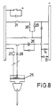
- Figure 8.- It is a schematic view of a second type of circuit which the device according to the invention may comprise.
- Reference being made to the above mentioned figures, it can be seen that the device for locating the epidural space proposed by the invention consists of a canula 1, at the rear part of which there is a
sleeve 2, theneedle 3 be ing introduced through them. Along thetube 4 of the canula aunidirectional fixation element 5 can be displaced. - The electric circuit as represented in figure 2 is constituted by a
hand switch 6, avoltage divider 7, a millivoltmeter 8, a pressure sensor 9, a comparator 10, a switch 11 at said comparator outlet, arelay 12 and the luminous 13 and acoustic 14 alarms. - The canula 1 is constituted by the
support body 15 and thetube 4. Inside said support body, there is anelectromagnet 16 surrounding theaxial sleeve 2, situated at the rear part of thesupport body 15. This sleeve may gently displace itself up to a maximum of 2 mm., this distance being regulated by thestop 18. Theinner surface 19 of the sleeve is considerably corrugated. - The
needle 22 has aconical point 20 without sharp edges, so that when it is introduced it separates the tissue fibres apart without cutting them, thereby producing as less traumatism as possible. The outer diameter of the needle will be slightly smaller than the inner one of thetube 4 of the canula and of the sleeve 2.A portion 21 of the outer surface of the needle has a corrugated surface complementary to theinner surface 19 of the sleeve. - The rear part of the needle is welded to a
conical body 22 provided with a cut 23 whereto a wing nut can be coupled serving as support to punch easily and safely. The vacuum inlet 24, besides somegrooves 25 for an easier handling, is situated at the rear part. - The
fixation element 5 is constituted by a flat body of elastic material, provided with twounidirectional fixation flanges 26. - The device as described in the preceding paragraphs works as follows:
- The circuit is connected by the
hand switch 6. A puncture is performed with the canula up to the surroundings of the yellow ligament, being fixed at this position by the fixation element, as it is displaced until it contacts the patient's back. Once the canula has been fixed, the axial sleeve being at its backmost position, the needle is introduced until the alarms and the electromagnet are activated, the latter attracting the needle due to the ferromagnetic condition of the material of which it is constituted, inserting it into the sleeve due to the rubbing of the corrugated surfaces in contact. With the simultaneous displacement of sleeve and needle, one reaches the epidural space centre when arriving to the anterior sleeve displacement stop. At this position, the concerned substances can be injected or the catheter can be introduced. - The alarms and the electromagnet become activated due to the fact that the pressure sensor detects the depression existing at the epidural space, which signal is converted into an electric signal that produces a voltage difference between the sensor and the potentiometer, this difference being annuled with observation of the millivoltmeter.
- Figure 8 represents a second example of embodiment of the alarm circuit of the epidural device. In this case, the pressure sensor consists of a small membrane (25) provided with an electric contact, which is closed in the membrane resting position and open when said membrane is sucked on reaching the epidural space. The passage of current keeps the circuit open by means of a relay (26). Current intensity is low thanks to the fact that the circuit comprises a resistor (27).
- A lamp (28) can be kept on meanwhile the epidural space is not reached, the luminous 13 and acoustic 14 alarms being then activated. The circuit may comprise some system, such as the relay (29) and the switches (30) and (31), in order to allow the alarms to keep on working, although the membrane (25) contact is closed again.
- The device for locating the epidural space which has been described constitutes a revolution in anaesthesia field, as the guarantees of success have a considerable increase, since it is not required for the technicians to be so much specialized.
Claims (6)
1. A DEVICE FOR LOCATING THE EPIDURAL SPACE, for introducing into it anaesthetics, analgesics, narcotics, etc., by injection with a needle or a catheter, essentially characterized by comprising a canula provided with a handle inside which an electromagnet is situated and at the rear part of which there is a retaining element that also works as a stop of a sleeve axially situated in the handle and the inner surface of which is considerably corrugated; with the peculiarity that the canula comprises a fixation element in order to prevent the former from penetrating beyond the yellow ligament, thereby allowing the axial comfortable introduction through the sleeve and the canula duct, of a conically pointed needle with lateral outlet, which has a corrugated outer surface portion capable of being inserted into the sleeve by the electromagnet action, which is activated by a circuit capable of detecting the depression existing in the epidural space by the use of a pressure sensor, which transforms the pressure signal into an electric signal, which is passed on to an electric circuit, so that, if there is a difference between the atmospheric pressure and the detected one, a transistorized switch will close the circuit and the above mentioned electromagnet and a sound and/or visual alarm will be activated.
2. A DEVICE FOR LOCATING THE EPIDURAL SPACE, according to claim 1, characterized in that the needle is made of ferromagnetic material, such as, for instance, chromium-vanadium steel.
3. A DEVICE FOR LOCATING THE EPIDURAL SPACE, according to claims 1 or 2, characterized in that the needle support element, generally of plastic, has a cut for coupling a wing nut and some outer grooves for a safer and easier puncture, besides a connector for the vacuum inlet.
4. A DEVICE FOR LOCATING THE EPIDURAL SPACE, accord ing to claim 1, characterized in that the circuit includes an adjustable voltage divider in order to be able to equalize the voltage with that of the pressure sensor, a millivoltmeter to control the difference between both voltages and a hand switch for operating the device.
5. A DEVICE FOR LOCATING THE EPIDURAL SPACE, according to claim 1, characterized in that the pressure sensor consists of a small membrane which opens or closes the circuit that activates the alarms, as it displaces itself.
6. A DEVICE FOR LOCATING THE EPIDURAL SPACE, according to claim 1, characterized in that the displacement of the needle once the epidural space has been reached, is carried out through the sleeve wherein it is inserted, said sleeve having a maximum displacement of 2 mm., which is suitable for reaching the centre of said epidural space.
Applications Claiming Priority (2)
| Application Number | Priority Date | Filing Date | Title |
|---|---|---|---|
| ES8702216 | 1987-07-28 | ||
| ES8702216A ES2007667A6 (en) | 1987-07-28 | 1987-07-28 | A device for locating the epidural space. |
Publications (2)
| Publication Number | Publication Date |
|---|---|
| EP0303824A2 true EP0303824A2 (en) | 1989-02-22 |
| EP0303824A3 EP0303824A3 (en) | 1990-12-19 |
Family
ID=8251952
Family Applications (1)
| Application Number | Title | Priority Date | Filing Date |
|---|---|---|---|
| EP19880111169 Withdrawn EP0303824A3 (en) | 1987-07-28 | 1988-07-12 | A device for locating the epidural space |
Country Status (6)
| Country | Link |
|---|---|
| US (1) | US4919653A (en) |
| EP (1) | EP0303824A3 (en) |
| JP (1) | JPH01119242A (en) |
| AU (1) | AU1972888A (en) |
| ES (1) | ES2007667A6 (en) |
| PT (1) | PT88122A (en) |
Cited By (22)
| Publication number | Priority date | Publication date | Assignee | Title |
|---|---|---|---|---|
| WO1992015256A1 (en) * | 1991-02-28 | 1992-09-17 | Industrias Palex, S.A. | Method and apparatus for locating anatomical cavities |
| EP0608659A1 (en) * | 1993-01-28 | 1994-08-03 | Du Kedem Technologies Ltd. | Epidural needle location indicator assembly |
| US5342311A (en) * | 1993-06-03 | 1994-08-30 | Dirina John G | Skin shield for protection against accidental needle puncture |
| ES2059279A1 (en) * | 1993-04-16 | 1994-11-01 | Olive Jose Javier Rodiera | Needle such as epidural needle, etc. |
| WO1997034536A2 (en) * | 1996-03-22 | 1997-09-25 | Sdgi Holdings, Inc. | Devices and methods for percutaneous surgery |
| US5792044A (en) * | 1996-03-22 | 1998-08-11 | Danek Medical, Inc. | Devices and methods for percutaneous surgery |
| DE19828794C1 (en) * | 1998-06-27 | 2000-02-24 | Heinrich Pajunk | Unipolar cannula for continuous line anesthesia |
| US6152871A (en) * | 1996-03-22 | 2000-11-28 | Sdgi Holdings, Inc. | Apparatus for percutaneous surgery |
| NL1018334C2 (en) * | 2001-06-20 | 2002-12-30 | Timotheus Joan Marie Lechner | Device for locating a cavity in the interior of a body. |
| US6679833B2 (en) | 1996-03-22 | 2004-01-20 | Sdgi Holdings, Inc. | Devices and methods for percutaneous surgery |
| US7022115B1 (en) | 1999-11-11 | 2006-04-04 | Gisela Meier | Continuously conductive unipolar cannula for anesthesia |
| US7198598B2 (en) | 1996-03-22 | 2007-04-03 | Warsaw Orthopedic, Inc. | Devices and methods for percutaneous surgery |
| US7427264B2 (en) | 2005-04-22 | 2008-09-23 | Warsaw Orthopedic, Inc. | Instruments and methods for selective tissue retraction through a retractor sleeve |
| US7699877B2 (en) | 2000-08-01 | 2010-04-20 | Zimmer Spine, Inc. | Method of securing vertebrae |
| US7985247B2 (en) | 2000-08-01 | 2011-07-26 | Zimmer Spine, Inc. | Methods and apparatuses for treating the spine through an access device |
| US10004450B2 (en) | 2016-05-03 | 2018-06-26 | Texas Medical Center | Tactile sensing device for lumbar punctures |
| US10383610B2 (en) | 2017-10-27 | 2019-08-20 | Intuitap Medical, Inc. | Tactile sensing and needle guidance device |
| US10632255B2 (en) | 2017-02-15 | 2020-04-28 | Milestone Scientific, Inc. | Drug infusion device |
| US10646660B1 (en) | 2019-05-16 | 2020-05-12 | Milestone Scientific, Inc. | Device and method for identification of a target region |
| US10842966B2 (en) | 2015-10-16 | 2020-11-24 | Milestone Scientific, Inc. | Apparatus for assisting a user in advancing a needle into a subject at a selected rate |
| US10946139B2 (en) | 2012-07-03 | 2021-03-16 | Milestone Scientific, Inc. | Disposable assembly for drug infusion with pressure sensing for identification of and injection into fluid-filled anatomic spaces |
| US11471595B2 (en) | 2017-05-04 | 2022-10-18 | Milestone Scientific, Inc. | Method and apparatus for performing a peripheral nerve block |
Families Citing this family (77)
| Publication number | Priority date | Publication date | Assignee | Title |
|---|---|---|---|---|
| US5004457A (en) * | 1988-12-02 | 1991-04-02 | The United States Of Americas As Represented By The Secretary Of The Department Of Health And Human Services | Tissue transplantation system |
| US5061244A (en) * | 1990-03-09 | 1991-10-29 | Becton, Dickinson And Company | Pudendal/paracervical block needle assembly |
| US5429609A (en) * | 1990-07-26 | 1995-07-04 | Yoon; Inbae | Endoscopic portal for use in endoscopic procedures and methods therefor |
| US5395342A (en) * | 1990-07-26 | 1995-03-07 | Yoon; Inbae | Endoscopic portal |
| US5389080A (en) * | 1990-07-26 | 1995-02-14 | Yoon; Inbae | Endoscopic portal for use in endoscopic procedures and methods therefor |
| DE69105900T2 (en) * | 1990-10-05 | 1995-05-24 | United States Surgical Corp | Security trocar. |
| US5104382A (en) * | 1991-01-15 | 1992-04-14 | Ethicon, Inc. | Trocar |
| US5261891A (en) * | 1991-01-15 | 1993-11-16 | Ethicon, Inc. | Trocar |
| US5295993A (en) | 1991-04-30 | 1994-03-22 | United States Surgical Corporation | Safety trocar |
| US5255691A (en) * | 1991-11-13 | 1993-10-26 | Medtronic, Inc. | Percutaneous epidural lead introducing system and method |
| AU651745B2 (en) * | 1991-12-13 | 1994-07-28 | Covidien Ag | Locking pneumoneedle |
| US5441513A (en) * | 1992-03-12 | 1995-08-15 | United States Surgical Corporation | Retracting tip trocar assembly |
| US5857996A (en) | 1992-07-06 | 1999-01-12 | Catheter Imaging Systems | Method of epidermal surgery |
| US5354266A (en) * | 1992-07-06 | 1994-10-11 | Catheter Imaging Systems | Method of epidural surgery |
| US5322514A (en) * | 1992-08-19 | 1994-06-21 | Sherwood Medical Company | Needle assembly with detachable wing |
| US5387197A (en) * | 1993-02-25 | 1995-02-07 | Ethicon, Inc. | Trocar safety shield locking mechanism |
| US5425718A (en) * | 1993-10-22 | 1995-06-20 | Tay; Sew-Wah | Self-sticking needle assembly and method for insertion into an artery |
| US5449355A (en) * | 1993-11-24 | 1995-09-12 | Valleylab Inc. | Retrograde tissue splitter and method |
| US5517846A (en) * | 1994-02-18 | 1996-05-21 | Caggiani; Carlos A. | Electronic vacuum sensor |
| US5480389A (en) * | 1994-08-09 | 1996-01-02 | Becton, Dickinson And Company | Method and apparatus for adjusting the length of a combined spinal-epidural needle |
| US5836914A (en) * | 1995-09-15 | 1998-11-17 | Becton Dickinson And Company | Method and apparatus for variably regulating the length of a combined spinal-epidural needle |
| USD405881S (en) | 1996-01-16 | 1999-02-16 | Catheter Imaging Systems, Inc. | Handle for steerable catheter |
| US6007531A (en) * | 1995-11-21 | 1999-12-28 | Catheter Imaging Systems, Inc. | Steerable catheter having disposable module and sterilizable handle and method of connecting same |
| USD398986S (en) | 1996-01-16 | 1998-09-29 | Catheter Imaging Systems, Inc. | Handle interface for steerable catheter |
| US5860953A (en) * | 1995-11-21 | 1999-01-19 | Catheter Imaging Systems, Inc. | Steerable catheter having disposable module and sterilizable handle and method of connecting same |
| US6213974B1 (en) | 1996-12-30 | 2001-04-10 | Visionary Biomedical, Inc. | Steerable catheter having segmented tip and one-piece inlet housing, and method of fabricating same |
| US6146355A (en) * | 1996-12-30 | 2000-11-14 | Myelotec, Inc. | Steerable catheter |
| US5964732A (en) | 1997-02-07 | 1999-10-12 | Abbeymoor Medical, Inc. | Urethral apparatus with position indicator and methods of use thereof |
| US6190370B1 (en) * | 1997-07-25 | 2001-02-20 | Arrow International, Inc. | Devices, systems and methods for determining proper placement of epidural catheters |
| US5971967A (en) | 1997-08-19 | 1999-10-26 | Abbeymoor Medical, Inc. | Urethral device with anchoring system |
| US5997509A (en) | 1998-03-06 | 1999-12-07 | Cornell Research Foundation, Inc. | Minimally invasive gene therapy delivery device and method |
| US6187000B1 (en) | 1998-08-20 | 2001-02-13 | Endius Incorporated | Cannula for receiving surgical instruments |
| US6546787B1 (en) | 1999-03-25 | 2003-04-15 | Regents Of The University Of Minnesota | Means and method for modeling and treating specific tissue structures |
| US6508802B1 (en) | 2000-05-23 | 2003-01-21 | Cornell Research Foundation, Inc. | Remote sensing gene therapy delivery device and method of administering a therapeutic solution to a heart |
| KR100411200B1 (en) * | 2000-10-11 | 2003-12-18 | 서현배 | Method for confirmation epidural space of the spinal cord and apparatus thereof |
| KR100430149B1 (en) * | 2001-03-16 | 2004-05-03 | 서현배 | Method for confirmation epidural space of the spinal cord |
| KR100400838B1 (en) * | 2001-05-07 | 2003-10-08 | 대한민국 | Automated apparatus identifying the epidural space to assist epidural anesthesia |
| US20070049945A1 (en) | 2002-05-31 | 2007-03-01 | Miller Larry J | Apparatus and methods to install, support and/or monitor performance of intraosseous devices |
| US11337728B2 (en) | 2002-05-31 | 2022-05-24 | Teleflex Life Sciences Limited | Powered drivers, intraosseous devices and methods to access bone marrow |
| US8668698B2 (en) | 2002-05-31 | 2014-03-11 | Vidacare Corporation | Assembly for coupling powered driver with intraosseous device |
| US8641715B2 (en) | 2002-05-31 | 2014-02-04 | Vidacare Corporation | Manual intraosseous device |
| US10973545B2 (en) | 2002-05-31 | 2021-04-13 | Teleflex Life Sciences Limited | Powered drivers, intraosseous devices and methods to access bone marrow |
| CA2485904C (en) | 2002-05-31 | 2013-05-21 | Vidacare Corporation | Apparatus and method to access the bone marrow |
| GB0301934D0 (en) * | 2003-01-28 | 2003-02-26 | Sundar Satish | Delivery apparatus and location method |
| US9504477B2 (en) | 2003-05-30 | 2016-11-29 | Vidacare LLC | Powered driver |
| US8340779B2 (en) | 2003-08-29 | 2012-12-25 | Medtronic, Inc. | Percutaneous flat lead introducer |
| US20050049663A1 (en) * | 2003-08-29 | 2005-03-03 | Harris Charmaine K. | Percutaneous flat lead introducer |
| US7803142B2 (en) | 2005-02-02 | 2010-09-28 | Summit Access Llc | Microtaper needle and method of use |
| US7792591B2 (en) * | 2005-06-09 | 2010-09-07 | Medtronic, Inc. | Introducer for therapy delivery elements |
| WO2007022450A1 (en) * | 2005-08-18 | 2007-02-22 | Clean Filtration Technologies, Inc. | Hydroclone based fluid filtration system |
| WO2007022599A1 (en) * | 2005-08-26 | 2007-03-01 | Novodural Pty Ltd | Improvements relating to epidural administration systems |
| US8944069B2 (en) | 2006-09-12 | 2015-02-03 | Vidacare Corporation | Assemblies for coupling intraosseous (IO) devices to powered drivers |
| EP2223662A1 (en) * | 2007-11-20 | 2010-09-01 | Innovamédica S.A.P.I. DE C.V. | Electronic syringe with safety system for spinal injections |
| WO2009153807A2 (en) * | 2008-06-19 | 2009-12-23 | Ravinder Bethi | A device for locating epidural space while safeguarding against dural puncture through differential friction technique |
| US20100069851A1 (en) * | 2008-09-17 | 2010-03-18 | Mobitech Regenerative Medicine | Method And Apparatus For Pressure Detection |
| US8377034B2 (en) | 2009-12-04 | 2013-02-19 | Std Med, Inc. | Vascular access port |
| US8805519B2 (en) | 2010-09-30 | 2014-08-12 | Nevro Corporation | Systems and methods for detecting intrathecal penetration |
| US8965482B2 (en) | 2010-09-30 | 2015-02-24 | Nevro Corporation | Systems and methods for positioning implanted devices in a patient |
| JP5921827B2 (en) * | 2011-06-30 | 2016-05-24 | 株式会社トップ | Epidural needle puncture system |
| AU2013211937B2 (en) | 2012-01-25 | 2016-07-28 | Nevro Corporation | Lead anchors and associated systems and methods |
| DE202012100925U1 (en) | 2012-03-14 | 2012-04-24 | B. Braun Melsungen Ag | Bellows syringe for finding the epidural space by means of differential resistance technology to protect against a dural puncture |
| US9308022B2 (en) | 2012-12-10 | 2016-04-12 | Nevro Corporation | Lead insertion devices and associated systems and methods |
| US9265935B2 (en) | 2013-06-28 | 2016-02-23 | Nevro Corporation | Neurological stimulation lead anchors and associated systems and methods |
| US10369345B2 (en) | 2014-03-31 | 2019-08-06 | Versago Vascular Access, Inc. | Medical access port, systems and methods of use thereof |
| US9764124B2 (en) | 2014-03-31 | 2017-09-19 | Versago Vascular Access, Inc. | Vascular access port |
| JP6568200B2 (en) | 2014-04-03 | 2019-08-28 | ヴェルサゴ ヴァスキュラー アクセス インコーポレイテッド | Device for attaching and removing the needle tip of a needle |
| TWI572387B (en) | 2014-11-21 | 2017-03-01 | 羅文甫 | A positioning device for needle syringe |
| JP6837971B2 (en) | 2014-12-18 | 2021-03-03 | ヴェルサゴ ヴァスキュラー アクセス インコーポレイテッド | Catheter patency system and method |
| AU2015364276B2 (en) | 2014-12-18 | 2020-04-30 | Versago Vascular Access, Inc. | Devices, systems and methods for removal and replacement of a catheter for an implanted access port |
| CN104586479B (en) * | 2015-01-07 | 2016-11-09 | 东南大学 | A kind of laparoscopic surgery sting device capable of automatic alarm |
| JP6879946B2 (en) | 2015-07-14 | 2021-06-02 | ヴェルサゴ ヴァスキュラー アクセス インコーポレイテッド | Medical access ports, transport devices and how to use them |
| CN105419818B (en) * | 2015-10-29 | 2018-02-13 | 中国矿业大学 | A kind of preparation method and application method of the retardant for preventing drying brown coal spontaneous combustion |
| AU2018231031B2 (en) | 2017-03-09 | 2023-11-02 | Nevro Corp. | Paddle leads and delivery tools, and associated systems and methods |
| CA3086211A1 (en) | 2017-12-21 | 2019-06-27 | Versago Vascular Access, Inc. | Medical access ports, transfer devices and methods of use thereof |
| AU2019242906A1 (en) | 2018-03-29 | 2020-10-15 | Nevro Corp. | Leads having sidewall openings, and associated systems and methods |
| CN110575236B (en) * | 2019-09-04 | 2022-09-20 | 贵州医科大学 | Puncture integrated device with multiple indexes and accurate positioning for spinal anesthesia |
| CN112690878A (en) * | 2020-12-31 | 2021-04-23 | 江苏省华星医疗器械实业有限公司 | Epidural puncture detector |
Citations (3)
| Publication number | Priority date | Publication date | Assignee | Title |
|---|---|---|---|---|
| DE836392C (en) * | 1949-01-06 | 1952-04-10 | Dr Med Fritz Hachfeld | Medical puncture device |
| US4175567A (en) * | 1976-04-28 | 1979-11-27 | The Kendall Company | Method of locating the epidural space |
| US4623335A (en) * | 1985-10-09 | 1986-11-18 | Anthony Jackson | Apparatus and methods for detecting probe penetration of human internal target tissue having predetermined internal pressure |
Family Cites Families (3)
| Publication number | Priority date | Publication date | Assignee | Title |
|---|---|---|---|---|
| US4356826A (en) * | 1979-05-09 | 1982-11-02 | Olympus Optical Co., Ltd. | Stabbing apparatus for diagnosis of living body |
| US4535773A (en) * | 1982-03-26 | 1985-08-20 | Inbae Yoon | Safety puncturing instrument and method |
| US4801293A (en) * | 1985-10-09 | 1989-01-31 | Anthony Jackson | Apparatus and method for detecting probe penetration of human epidural space and injecting a therapeutic substance thereinto |
-
1987
- 1987-07-28 ES ES8702216A patent/ES2007667A6/en not_active Expired
-
1988
- 1988-07-12 EP EP19880111169 patent/EP0303824A3/en not_active Withdrawn
- 1988-07-13 US US07/219,219 patent/US4919653A/en not_active Expired - Fee Related
- 1988-07-22 AU AU19728/88A patent/AU1972888A/en not_active Abandoned
- 1988-07-27 PT PT88122A patent/PT88122A/en not_active Application Discontinuation
- 1988-07-27 JP JP63192209A patent/JPH01119242A/en active Pending
Patent Citations (3)
| Publication number | Priority date | Publication date | Assignee | Title |
|---|---|---|---|---|
| DE836392C (en) * | 1949-01-06 | 1952-04-10 | Dr Med Fritz Hachfeld | Medical puncture device |
| US4175567A (en) * | 1976-04-28 | 1979-11-27 | The Kendall Company | Method of locating the epidural space |
| US4623335A (en) * | 1985-10-09 | 1986-11-18 | Anthony Jackson | Apparatus and methods for detecting probe penetration of human internal target tissue having predetermined internal pressure |
Cited By (44)
| Publication number | Priority date | Publication date | Assignee | Title |
|---|---|---|---|---|
| WO1992015256A1 (en) * | 1991-02-28 | 1992-09-17 | Industrias Palex, S.A. | Method and apparatus for locating anatomical cavities |
| EP0608659A1 (en) * | 1993-01-28 | 1994-08-03 | Du Kedem Technologies Ltd. | Epidural needle location indicator assembly |
| ES2059279A1 (en) * | 1993-04-16 | 1994-11-01 | Olive Jose Javier Rodiera | Needle such as epidural needle, etc. |
| US5342311A (en) * | 1993-06-03 | 1994-08-30 | Dirina John G | Skin shield for protection against accidental needle puncture |
| US7198598B2 (en) | 1996-03-22 | 2007-04-03 | Warsaw Orthopedic, Inc. | Devices and methods for percutaneous surgery |
| US6217509B1 (en) | 1996-03-22 | 2001-04-17 | Sdgi Holdings, Inc. | Devices and methods for percutaneous surgery |
| US5792044A (en) * | 1996-03-22 | 1998-08-11 | Danek Medical, Inc. | Devices and methods for percutaneous surgery |
| US5902231A (en) * | 1996-03-22 | 1999-05-11 | Sdgi Holdings, Inc. | Devices and methods for percutaneous surgery |
| US5954635A (en) * | 1996-03-22 | 1999-09-21 | Sdgi Holdings Inc. | Devices and methods for percutaneous surgery |
| US6007487A (en) * | 1996-03-22 | 1999-12-28 | Sdgi Holdings, Inc. | Tissue retractor for use through a cannula |
| WO1997034536A2 (en) * | 1996-03-22 | 1997-09-25 | Sdgi Holdings, Inc. | Devices and methods for percutaneous surgery |
| US6152871A (en) * | 1996-03-22 | 2000-11-28 | Sdgi Holdings, Inc. | Apparatus for percutaneous surgery |
| US6162170A (en) * | 1996-03-22 | 2000-12-19 | Sdgi Holdings, Inc. | Devices and methods for percutaneous surgery |
| US6176823B1 (en) | 1996-03-22 | 2001-01-23 | Sdgi Holdings, Inc. | Fixture for supporting a viewing element within a cannula |
| US6206822B1 (en) | 1996-03-22 | 2001-03-27 | Sdgi Holdings, Inc. | Devices and methods for percutaneous surgery |
| US7993378B2 (en) | 1996-03-22 | 2011-08-09 | Warsaw Orthopedic, IN. | Methods for percutaneous spinal surgery |
| US6425859B1 (en) | 1996-03-22 | 2002-07-30 | Sdgi Holdings, Inc. | Cannula and a retractor for percutaneous surgery |
| WO1997034536A3 (en) * | 1996-03-22 | 1997-11-13 | Devices and methods for percutaneous surgery | |
| US6679833B2 (en) | 1996-03-22 | 2004-01-20 | Sdgi Holdings, Inc. | Devices and methods for percutaneous surgery |
| DE19828794C1 (en) * | 1998-06-27 | 2000-02-24 | Heinrich Pajunk | Unipolar cannula for continuous line anesthesia |
| US7022115B1 (en) | 1999-11-11 | 2006-04-04 | Gisela Meier | Continuously conductive unipolar cannula for anesthesia |
| US9622735B2 (en) | 2000-08-01 | 2017-04-18 | Zimmer Spine, Inc. | Method for securing vertebrae |
| US9101353B2 (en) | 2000-08-01 | 2015-08-11 | Zimmer Spine, Inc. | Method of securing vertebrae |
| US8277486B2 (en) | 2000-08-01 | 2012-10-02 | Zimmer Spine, Inc. | System for performing a procedure at a spinal location |
| US7985247B2 (en) | 2000-08-01 | 2011-07-26 | Zimmer Spine, Inc. | Methods and apparatuses for treating the spine through an access device |
| US7699877B2 (en) | 2000-08-01 | 2010-04-20 | Zimmer Spine, Inc. | Method of securing vertebrae |
| NO341943B1 (en) * | 2001-06-20 | 2018-02-26 | Milestone Scientific Inc | Device for locating an anatomical cavity in a body |
| KR100910783B1 (en) * | 2001-06-20 | 2009-08-04 | 반 니이케르크 얀 | Device and method for locating an anatomical cavity in a body |
| NO20035628L (en) * | 2001-06-20 | 2004-01-26 | Milestone Scientific Inc | Method and apparatus for locating an anatomical cavity in a body |
| WO2003000146A1 (en) * | 2001-06-20 | 2003-01-03 | Van Niekerk, Jan | Device and method for locating an anatomical cavity in a body |
| NL1018334C2 (en) * | 2001-06-20 | 2002-12-30 | Timotheus Joan Marie Lechner | Device for locating a cavity in the interior of a body. |
| US7922689B2 (en) | 2001-06-20 | 2011-04-12 | Timotheus Joan Marie Lechner | Device and method for locating anatomical cavity in a body |
| US7427264B2 (en) | 2005-04-22 | 2008-09-23 | Warsaw Orthopedic, Inc. | Instruments and methods for selective tissue retraction through a retractor sleeve |
| US10946139B2 (en) | 2012-07-03 | 2021-03-16 | Milestone Scientific, Inc. | Disposable assembly for drug infusion with pressure sensing for identification of and injection into fluid-filled anatomic spaces |
| US10842966B2 (en) | 2015-10-16 | 2020-11-24 | Milestone Scientific, Inc. | Apparatus for assisting a user in advancing a needle into a subject at a selected rate |
| US10004450B2 (en) | 2016-05-03 | 2018-06-26 | Texas Medical Center | Tactile sensing device for lumbar punctures |
| US11179097B2 (en) | 2016-05-03 | 2021-11-23 | Texas Medical Center | Tactile sensing device for lumbar punctures |
| US10632255B2 (en) | 2017-02-15 | 2020-04-28 | Milestone Scientific, Inc. | Drug infusion device |
| US11471595B2 (en) | 2017-05-04 | 2022-10-18 | Milestone Scientific, Inc. | Method and apparatus for performing a peripheral nerve block |
| US11000311B2 (en) | 2017-10-27 | 2021-05-11 | Intuitap Medical, Inc. | Tactile sensing and needle guidance device |
| US10383610B2 (en) | 2017-10-27 | 2019-08-20 | Intuitap Medical, Inc. | Tactile sensing and needle guidance device |
| US10646660B1 (en) | 2019-05-16 | 2020-05-12 | Milestone Scientific, Inc. | Device and method for identification of a target region |
| US10960141B1 (en) | 2019-05-16 | 2021-03-30 | Milestone Scientific, Inc. | Device and method for identification of a target region |
| US11147927B2 (en) | 2019-05-16 | 2021-10-19 | Milestone Scientific, Inc. | Device and method for identification of a target region |
Also Published As
| Publication number | Publication date |
|---|---|
| ES2007667A6 (en) | 1989-07-01 |
| EP0303824A3 (en) | 1990-12-19 |
| AU1972888A (en) | 1989-02-02 |
| PT88122A (en) | 1989-06-30 |
| JPH01119242A (en) | 1989-05-11 |
| US4919653A (en) | 1990-04-24 |
Similar Documents
| Publication | Publication Date | Title |
|---|---|---|
| EP0303824A2 (en) | A device for locating the epidural space | |
| US5445617A (en) | Automatic retractable safety penetrating instrument for portal sleeve introduction and method of use | |
| US5902273A (en) | Pressurizable epidural space identification syringe | |
| US6304785B1 (en) | Electrode insertion tool | |
| US5601533A (en) | Endoscopic puncture needle device | |
| US5916175A (en) | Biopsy needle appliance and inserting guide with adjustable sample length and/or needle cutting stroke | |
| US8801680B2 (en) | Angled retracting sheath for safety needle | |
| US10258428B2 (en) | Marker delivery device for tissue marker placement | |
| EP0546769A2 (en) | Locking insufflation needle | |
| US6093154A (en) | Biopsy needle | |
| EP1841370A2 (en) | Multi-lancet unit, method and lancet device using the multi-lancet unit, and method of assembling and/or making the multi-lancet unit | |
| EP0858778A3 (en) | Retractable safety penetrating instrument | |
| EP0588470A1 (en) | Needle stop and safety sheath | |
| CA2686277A1 (en) | Instrument and apparatus for biopsy and a method thereof | |
| US5478350A (en) | Rack and pinion actuator handle for endoscopic instruments | |
| WO1993020866A1 (en) | Automatic retractable trocar with safety shield | |
| WO2001066020A3 (en) | Apparatus and method for performing a bypass procedure in a digestive system | |
| EP1501417B1 (en) | Stoma measuring device | |
| WO2009068661A1 (en) | Device for thoracostomy | |
| GB2240044A (en) | Surgical needle shield | |
| CN107072688B (en) | Medical device | |
| JP2726725B2 (en) | Safety syringe for blood sample collection and infusion | |
| US10485577B2 (en) | Surgical device with triggered propulsion system for inserting a trocar-cannula assembly | |
| US4414983A (en) | Medico-surgical instruments | |
| JPH02177952A (en) | Surgical instrument and assembly thereof |
Legal Events
| Date | Code | Title | Description |
|---|---|---|---|
| PUAI | Public reference made under article 153(3) epc to a published international application that has entered the european phase |
Free format text: ORIGINAL CODE: 0009012 |
|
| AK | Designated contracting states |
Kind code of ref document: A2 Designated state(s): AT BE CH DE FR GB GR IT LI LU NL SE |
|
| PUAL | Search report despatched |
Free format text: ORIGINAL CODE: 0009013 |
|
| AK | Designated contracting states |
Kind code of ref document: A3 Designated state(s): AT BE CH DE FR GB GR IT LI LU NL SE |
|
| STAA | Information on the status of an ep patent application or granted ep patent |
Free format text: STATUS: THE APPLICATION IS DEEMED TO BE WITHDRAWN |
|
| 18D | Application deemed to be withdrawn |
Effective date: 19910620 |