EP0150834A2 - Method of adjusting radiation image read-out conditions - Google Patents
Method of adjusting radiation image read-out conditions Download PDFInfo
- Publication number
- EP0150834A2 EP0150834A2 EP85100788A EP85100788A EP0150834A2 EP 0150834 A2 EP0150834 A2 EP 0150834A2 EP 85100788 A EP85100788 A EP 85100788A EP 85100788 A EP85100788 A EP 85100788A EP 0150834 A2 EP0150834 A2 EP 0150834A2
- Authority
- EP
- European Patent Office
- Prior art keywords
- read
- image
- light emission
- stimulable phosphor
- emission amount
- Prior art date
- Legal status (The legal status is an assumption and is not a legal conclusion. Google has not performed a legal analysis and makes no representation as to the accuracy of the status listed.)
- Withdrawn
Links
Images

Classifications
-
- H—ELECTRICITY
- H04—ELECTRIC COMMUNICATION TECHNIQUE
- H04N—PICTORIAL COMMUNICATION, e.g. TELEVISION
- H04N1/00—Scanning, transmission or reproduction of documents or the like, e.g. facsimile transmission; Details thereof
- H04N1/40—Picture signal circuits
- H04N1/407—Control or modification of tonal gradation or of extreme levels, e.g. background level
- H04N1/4072—Control or modification of tonal gradation or of extreme levels, e.g. background level dependent on the contents of the original
- H04N1/4074—Control or modification of tonal gradation or of extreme levels, e.g. background level dependent on the contents of the original using histograms
-
- G—PHYSICS
- G01—MEASURING; TESTING
- G01T—MEASUREMENT OF NUCLEAR OR X-RADIATION
- G01T1/00—Measuring X-radiation, gamma radiation, corpuscular radiation, or cosmic radiation
- G01T1/16—Measuring radiation intensity
- G01T1/20—Measuring radiation intensity with scintillation detectors
- G01T1/2012—Measuring radiation intensity with scintillation detectors using stimulable phosphors, e.g. stimulable phosphor sheets
- G01T1/2014—Reading out of stimulable sheets, e.g. latent image
Definitions
- This invention relates to a method of adjusting radiation image read-out conditions used in a radiation image recording and reproducing system.
- This invention particularly relates to a method of adjusting the final read-out conditions by conducting preliminary read-out in a radiation image recording and reproducing system wherein preliminary read-out and final read-out are carried out.
- phosphors When certain kinds of phosphors are exposed to a radiation such as X-rays, a-rays, S-rays, y-rays, cathode- rays or ultra-violet rays, they store a part of the energy of the radiation. Then, when the phosphor which has been exposed to the radiation is exposed to stimulating rays such as visible light, light is emitted by the phosphor in proportion to the stored energy of the radiation. A phosphor exhibiting such properties is referred to as a stimulable phosphor.
- a stimulable phosphor sheet As disclosed in U.S. Patent No. 4,258,264 and Japanese Unexamined Patent Publication No. 56(1981)-11395, it has been proposed to use a stimulable phosphor in a radiation image recording and reproducing system. Specifically, a sheet provided with a layer of the stimulable phosphor (hereinafter referred to as a stimulable phosphor sheet) is first exposed to a radiation passing through an object to have a radiation image stored therein, and is then scanned with stimulating rays such as a laser beam which cause it to emit light in the pattern of the stored image.
- stimulating rays such as a laser beam
- the light emitted by the stimulable phosphor sheet upon stimulation thereof is photoelectrically detected and converted to an electric image signal, which is processed as desired to reproduce a visible image on a recording medium such as a photographic light-sensitive material or on a display device such as a cathode ray tube (CRT).
- a recording medium such as a photographic light-sensitive material
- a display device such as a cathode ray tube (CRT).
- One embodiment of the aforesaid radiation image recording and reproducing system is disclosed, for example, in Japanese Unexamined Patent Publication No. 58(1983)-67240.
- preliminary read-out for approximately detecting the image information stored in the stimulable phosphor sheet is conducted by use of stimulating rays of a level lower than the level of the stimulating rays used in the final read-out.
- Read-out conditions for the final read-out are adjusted on the basis of the information obtained by the preliminary read-out, and the final read-out is conducted by use of the read-out conditions.
- the electric image signal obtained by the final read-out is sent to an image processing means and is processed in accordance with the image recording portion and/or image recording conditions, for example, radiation energy, radiation dose, type of grid or collimator, type of stimulable phosphor sheet, to obtain a visible image suitable for viewing, particularly for diagnostic purposes.
- the processed image signal is used to reproduce the visible image on a photographic film or the like.
- the level of the stimulating rays used in the preliminary read-out should be lower than the level of the stimulating rays used in the final read-out. That is, the effective energy of the stimulating rays which the stimulable phosphor sheet receives per unit area in the preliminary read-out should be lower than the effective energy of the stimulating rays used in the final read-out.
- the output of the stimulating ray source such as a laser beam source may be decreased in the preliminary read-out, or the stimulating rays emitted by the stimulating ray source may be attenuated by an ND filter, an AOM, or the like positioned on the optical path.
- a stimulating ray source for the preliminary read-out may be positioned independently of the stimulating ray source for the final read-out, and the output of the former may be made lower than the output of the latter.
- the beam diameter of the stimulating rays may be increased,-the scanning speed of the stimulating rays may be increased, or the moving speed of the stimulable phosphor sheet may be increased in the preliminary read-out.
- the image information stored in the stimulable phosphor sheet is approximately grasped prior to the final read-out and the final read-out is conducted by use of the read-out conditions adjusted on the basis of the image information, it becomes possible to eliminate adverse effects of a fluctuation in the level of the radiation energy stored in the stimulable phosphor sheet, which is caused by a change in the object or the image recording portion thereof, by a fluctuation in the radiation dose, or the like, and to conduct the final read-out by use of desirable read-out conditions.
- the primary object of the present invention is to provide a method of adjusting radiation image read-out conditions in a simple manner in accordance with various apparatus conditions so that the limitation imposed on the image reproducing apparatus is minimized.
- Another object of the present invention is to provide a method of adjusting radiation image read-out conditions, which improves the operating efficiency and reduces the cost of the whole system.
- the present invention provides a method of adjusting radiation image read-out conditions used in a radiation image recording and reproducing system wherein preliminary read-out conducted by scanning a stimulable phosphor sheet carrying a radiation image of an object stored therein by stimulating rays of a level lower than the level of the stimulating rays used in final read-out and approximately detecting the image information stored in the stimulable phosphor sheet is carried out prior to the final read-out for scanning the stimulable phosphor sheet by the stimulating rays which cause the stimulable phosphor sheet to emit light in proportion to the radiation energy stored and detecting the emitted light by a photoelectric read-out means to obtain an image signal, read-out conditions for the final read-out are adjusted on the basis of the information obtained by the preliminary read-out, the final read-out is conducted by use of the read-out conditions, the image signal obtained by the final read-out is sent to an image processing means, and a visible image is reproduced by use of the image signal processed by the image processing means,
- the method of adjusting radiation image read-out conditions comprising the steps of:
- read-out conditions various conditions affecting the relationship between the amount of the light emitted by the stimulable phosphor sheet at the read-out step and the output of the read-out apparatus, for example, the read-out gain determining the relationship between the input and the output of the read-out apparatus, the scale factor, and the power of the stimulating rays used for read-out.
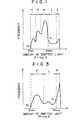
- a histogram of the amount of the light emitted by the stimulable phosphor sheet is obtained by the preliminary read-out, and a desired image information range (range of the amount of the emitted light) is determined on the basis of the histogram. Since the pattern of the histogram is approximately fixed by the image recording portion and/or image recording conditions, the desired image information range is determined from the histogram with reference to the image recording portion and/or image recording conditions.
- the pattern of the histogram becomes as shown in Figure 1, and it is possible to know that F in the histogram designates the mediastinum, G designates the heart, H denotes the lungs, I denotes the skin and the soft tissue, and J denotes region outside of the object. Therefore, from the histogram, it is possible to calculate the maximum light emission amount Smax and the minimum light emission amount Smin defining the desired image information range. For example, when information on the skin and the soft tissue (I) and information on the region outside of the object (J) in Figure 1 are unnecessary, the desired image information range becomes the range from Smax to Smin including F, G and H as shown. Smax and Smin may be calculated by various methods from the histogram, for example, by determining threshold values T1 and T2 in accordance with the desired image information range and calculating Smax and Smin on the basis of T1 and T2.
- an electric image signal is obtained from the light emitted by the stimulable phosphor sheet by use of the read-out conditions with the final read-out means, and is subjected to various signal processings in the image processing means, particularly the gradation processing which is conducted by use of the gradation processing conditions determined in accordance with the image recording portion and/or image recording conditions.
- the processed signal is used to reproduce a visible image on a photographic film or the like.
- the reproduced visible image should have a correct density range suitable for viewing, particularly for diagnostic purposes. In general, the correct density range (Dmax to Dmin) is determined in advance. Desired gradation processing conditions are also determined in advance.
- the range of signal level (Qmax to Qmin) which should be sent to the image processing means as suitable for obtaining the correct density range (Dmax to Dmin) is determined by the gradation processing conditions.
- Figure 2 shows the relationship between the amount of light emitted by the stimulable phosphor sheet and the density of the reproduced visible image in the radiation image recording and reproducing system wherein the method of the present invention is used.
- the read-out conditions are adjusted so that the maximum light emission amount Smax and the minimum light emission amount Smin calculated as described above correspond to the maximum signal level Qmax and the minimum signal level Qmin determined as mentioned above.
- the method of the present invention is constituted so that the outputs Smax and Smin of the read-out means correspond to the maximum signal level Qmax and the minimum signal level Qmin determined by the image reproducing apparatus, it is possible to conduct the image processing by the image processing means such that no limitation to nor modification of the image reproducing apparatus is required.
- the range of the level of the image signal has already been adjusted in the final read-out means to correspond to the correct density range. Therefore, in the image processing means, it is sufficient that only the gradation processing be conducted to suit the image recording portion and/or image recording conditions. As a result, it becomes possible to simplify the gradation processing means or the image reproducing means, to improve the operating efficiency of the whole system, and to reduce cost.
- the image signal corresponding to the correct density range can be generated by the final read-out means, the signal resolution at the time of signal storing may be high. Also for this reason, it is possible to reduce the costs of the A/D converter, image processing device, memory and the like used in the system, and to improve the operating efficiency of the whole system.
- the image information is approximately grasped by the preliminary read-out using the stimulating rays of a low level, it is possible to obtain a histogram accurately representing the image information obtained by the final read-out. Since the read-out conditions are adjusted on the basis of the accurate histogram, it is possible to obtain a visible image having an improved image quality, particularly a high diagnostic efficiency and accuracy.
- a histogram of the amount of light emitted by a stimulable phosphor sheet carrying a radiation image stored therein is first obtained by preliminary read-out, and the maximum light emission amount Smax and the minimum light emission amount Smin of a desired image information range are calculated from the histogram.
- a histogram as shown in Figure 1 is obtained as described above.
- the maximum value at the lung portion H and the minimum value at the mediastinum portion F are calculated by use of threshold values T1 and T2.
- the maximum value and the minimum value are taken as Smax and Smin.
- a histogram as shown in Figure 3 is obtained.
- the maximum value at the skin and soft tissue portion L and the minimum value at the brain portion K are calculated by use of threshold values Tl and T2 by omitting the portion M outside of the object.
- the maximum value and the minimum value are taken as Smax and Smin of the desired image information range.
- the image read-out conditions are adjusted so that the maximum light emission amount Smax and the minimum light emission amount Smin correspond respectively to the maximum signal level Qmax and the minimum signal level Qmin of the desired input signal range in the image processing means which are determined by a desired gradation processing curve on the basis of the maximum density Dmax and the minimum density Dmin of the correct density range in the reproduced visible image.
- the read-out conditions are represented by the input-to-output conversion curve in the second quadrant. Specifically, the read-out conditions are the read-out gain in the final read-out and the scale factor. When the power of the stimulating rays is variable, the read-out conditions are changed in accordance with the power.
- the maximum signal level Qmax and the minimum signal level Qmin defining the desired range of the input signals to the image processing means and corresponding to the Dmax and the Dmin are fixed on the basis of the gradation processing conditions determined in advance to suit the image recording portion and/or image recording conditions.
- the soft tissue portion I is to be diagnosed in the case of the aforesaid chest image recording, it is possible to use the maximum value at the soft tissue portion I as the aforesaid Smax and to use a value suitable for the diagnosis of the soft tissue portion as the Dmax.
- Qmax is determined in accordance with Dmax.
- the scale factor represents the inclination of the line indicating the read-out conditions in Figure 2 (arrow A in the second quadrant in Figure 2).
- the scale factor in accordance with the range (Smax - Smin) of the amount of the light emitted by the stimulable phosphor sheet, it is possible to make the range of the level of the input signal to the image processing means always coincide with the range (Qmax - Qmin) of the desired input signal level.
- kl is a constant for making the range of the unit amount of the emitted light coincide with the range of the unit desired input signal level.
- the read-out gain represents the transverse position of the line indicating the read-out conditions in Figure 2 (arrow B in the second quadrant in Figure 2).
- the histogram is at the position indicated by the two-dotted chain line in Figure 2 and the image read-out is conducted by use of the read-out conditions shown in Figure 2, the maximum input signal level and the minimum input signal level become Q'max and Q'min, and the position of the range of the input signal level deviates to the right from the position of the range of the desired input signal level from Qmax to Qmin. Therefore, the line representing the read-out conditions is moved to the left to make the position of the range of the input signal level coincide with the position of the range of the desired input signal level from Qmax to Qmin.
- the density of the lung portion should preferably be within the range of approximately 1.6 to approximately 1.8 in terms of the optical density
- the density of the mediastinum portion should preferably be within the range of approximately 0.2 to approximately 0.3 in terms of the optical density. Accordingly, Dmax should preferably be approximately 1.8, and Dmin should preferably be approximately 0.2.
- the input signals to the image processing means which correspond to the output densities of 1.8 and 0.2 respectively correspond to approximately 70% and approximately 10% of the signal latitude of the image processing means. Therefore, the read-out conditions should be adjusted so that Smax and Smin obtained by the preliminary read-out are 70% and 10% of the signal latitude of the image processing means.
- the threshold values T1 and T2 for calculating Smax and Smin should preferably be approximately 0.1% to 2.0% and approximately 0.05% to 1.0% of the number of picture elements.
Landscapes
- General Physics & Mathematics (AREA)
- Engineering & Computer Science (AREA)
- Signal Processing (AREA)
- Physics & Mathematics (AREA)
- Health & Medical Sciences (AREA)
- Life Sciences & Earth Sciences (AREA)
- Multimedia (AREA)
- High Energy & Nuclear Physics (AREA)
- Molecular Biology (AREA)
- Spectroscopy & Molecular Physics (AREA)
- Apparatus For Radiation Diagnosis (AREA)
- Radiography Using Non-Light Waves (AREA)
- Facsimile Scanning Arrangements (AREA)
- Transforming Light Signals Into Electric Signals (AREA)
Abstract
Description
- This invention relates to a method of adjusting radiation image read-out conditions used in a radiation image recording and reproducing system. This invention particularly relates to a method of adjusting the final read-out conditions by conducting preliminary read-out in a radiation image recording and reproducing system wherein preliminary read-out and final read-out are carried out.
- When certain kinds of phosphors are exposed to a radiation such as X-rays, a-rays, S-rays, y-rays, cathode- rays or ultra-violet rays, they store a part of the energy of the radiation. Then, when the phosphor which has been exposed to the radiation is exposed to stimulating rays such as visible light, light is emitted by the phosphor in proportion to the stored energy of the radiation. A phosphor exhibiting such properties is referred to as a stimulable phosphor.
- As disclosed in U.S. Patent No. 4,258,264 and Japanese Unexamined Patent Publication No. 56(1981)-11395, it has been proposed to use a stimulable phosphor in a radiation image recording and reproducing system. Specifically, a sheet provided with a layer of the stimulable phosphor (hereinafter referred to as a stimulable phosphor sheet) is first exposed to a radiation passing through an object to have a radiation image stored therein, and is then scanned with stimulating rays such as a laser beam which cause it to emit light in the pattern of the stored image. The light emitted by the stimulable phosphor sheet upon stimulation thereof is photoelectrically detected and converted to an electric image signal, which is processed as desired to reproduce a visible image on a recording medium such as a photographic light-sensitive material or on a display device such as a cathode ray tube (CRT).
- One embodiment of the aforesaid radiation image recording and reproducing system is disclosed, for example, in Japanese Unexamined Patent Publication No. 58(1983)-67240. In the embodiment, before final read-out is conducted by scanning the stimulable phosphor sheet carrying a radiation image of an object stored therein by stimulating rays which cause the stimulable phosphor sheet to emit light in proportion to the radiation energy stored, detecting the emitted light by a photoelectric read-out means and converting it into an electric image signal, preliminary read-out for approximately detecting the image information stored in the stimulable phosphor sheet is conducted by use of stimulating rays of a level lower than the level of the stimulating rays used in the final read-out. Read-out conditions for the final read-out are adjusted on the basis of the information obtained by the preliminary read-out, and the final read-out is conducted by use of the read-out conditions. The electric image signal obtained by the final read-out is sent to an image processing means and is processed in accordance with the image recording portion and/or image recording conditions, for example, radiation energy, radiation dose, type of grid or collimator, type of stimulable phosphor sheet, to obtain a visible image suitable for viewing, particularly for diagnostic purposes. The processed image signal is used to reproduce the visible image on a photographic film or the like.
- As described above, the level of the stimulating rays used in the preliminary read-out should be lower than the level of the stimulating rays used in the final read-out. That is, the effective energy of the stimulating rays which the stimulable phosphor sheet receives per unit area in the preliminary read-out should be lower than the effective energy of the stimulating rays used in the final read-out. In order to make the level of the stimulating rays used in the preliminary read-out lower than the level of the stimulating rays in the final read-out, the output of the stimulating ray source such as a laser beam source may be decreased in the preliminary read-out, or the stimulating rays emitted by the stimulating ray source may be attenuated by an ND filter, an AOM, or the like positioned on the optical path. Alternatively, a stimulating ray source for the preliminary read-out may be positioned independently of the stimulating ray source for the final read-out, and the output of the former may be made lower than the output of the latter. Or, the beam diameter of the stimulating rays may be increased,-the scanning speed of the stimulating rays may be increased, or the moving speed of the stimulable phosphor sheet may be increased in the preliminary read-out.
- When the image information stored in the stimulable phosphor sheet is approximately grasped prior to the final read-out and the final read-out is conducted by use of the read-out conditions adjusted on the basis of the image information, it becomes possible to eliminate adverse effects of a fluctuation in the level of the radiation energy stored in the stimulable phosphor sheet, which is caused by a change in the object or the image recording portion thereof, by a fluctuation in the radiation dose, or the like, and to conduct the final read-out by use of desirable read-out conditions.
- Various methods may be used for approximately grasping the image information prior to the final read-out and adjusting the read-out conditions. However, a practical method will be one that enables the read-out conditions to be adjusted as simply as possible and that puts minimum limitation on the design and operation of the image reproducing apparatus. Specifically, since the density range of the visible image ultimately reproduced is determined in advance and the level of the signal which should be supplied to the image reproducing apparatus for realizing the predetermined density range is determined in advance by gradation processing conditions or the like, the read-out conditions should be adjusted on the basis of these conditions so that the read-out apparatus generates a signal of a level equal to the level of the signal which should be supplied to the image reproducing apparatus.
- The primary object of the present invention is to provide a method of adjusting radiation image read-out conditions in a simple manner in accordance with various apparatus conditions so that the limitation imposed on the image reproducing apparatus is minimized.
- Another object of the present invention is to provide a method of adjusting radiation image read-out conditions, which improves the operating efficiency and reduces the cost of the whole system.
- The present invention provides a method of adjusting radiation image read-out conditions used in a radiation image recording and reproducing system wherein preliminary read-out conducted by scanning a stimulable phosphor sheet carrying a radiation image of an object stored therein by stimulating rays of a level lower than the level of the stimulating rays used in final read-out and approximately detecting the image information stored in the stimulable phosphor sheet is carried out prior to the final read-out for scanning the stimulable phosphor sheet by the stimulating rays which cause the stimulable phosphor sheet to emit light in proportion to the radiation energy stored and detecting the emitted light by a photoelectric read-out means to obtain an image signal, read-out conditions for the final read-out are adjusted on the basis of the information obtained by the preliminary read-out, the final read-out is conducted by use of the read-out conditions, the image signal obtained by the final read-out is sent to an image processing means, and a visible image is reproduced by use of the image signal processed by the image processing means,
- the method of adjusting radiation image read-out conditions comprising the steps of:
- i) determining a histogram of the amount of the light emitted by said stimulable phosphor sheet during said preliminary read-out,
- ii) from said histogram, calculating the maximum light emission amount Smax and the minimum light emission amount Smin of a desired image information range in said histogram, and
- iii) adjusting said read-out conditions so that said maximum light emission amount Smax and said minimum light emission amount Smin correspond respectively to the maximum signal level Qmax and the minimum signal level Qmin of a desired input signal range in said image processing means which are determined by the maximum density Dmax and the minimum density Dmin of a correct density range in said reproduced visible image.
- By the term "read-out conditions" as used herein are meant various conditions affecting the relationship between the amount of the light emitted by the stimulable phosphor sheet at the read-out step and the output of the read-out apparatus, for example, the read-out gain determining the relationship between the input and the output of the read-out apparatus, the scale factor, and the power of the stimulating rays used for read-out.
- In the present invention, a histogram of the amount of the light emitted by the stimulable phosphor sheet is obtained by the preliminary read-out, and a desired image information range (range of the amount of the emitted light) is determined on the basis of the histogram. Since the pattern of the histogram is approximately fixed by the image recording portion and/or image recording conditions, the desired image information range is determined from the histogram with reference to the image recording portion and/or image recording conditions. For example, in the case of chest image recording, the pattern of the histogram becomes as shown in Figure 1, and it is possible to know that F in the histogram designates the mediastinum, G designates the heart, H denotes the lungs, I denotes the skin and the soft tissue, and J denotes region outside of the object. Therefore, from the histogram, it is possible to calculate the maximum light emission amount Smax and the minimum light emission amount Smin defining the desired image information range. For example, when information on the skin and the soft tissue (I) and information on the region outside of the object (J) in Figure 1 are unnecessary, the desired image information range becomes the range from Smax to Smin including F, G and H as shown. Smax and Smin may be calculated by various methods from the histogram, for example, by determining threshold values T1 and T2 in accordance with the desired image information range and calculating Smax and Smin on the basis of T1 and T2.
- As described above, in the radiation image recording and reproducing system wherein the method of the present invention may be used, an electric image signal is obtained from the light emitted by the stimulable phosphor sheet by use of the read-out conditions with the final read-out means, and is subjected to various signal processings in the image processing means, particularly the gradation processing which is conducted by use of the gradation processing conditions determined in accordance with the image recording portion and/or image recording conditions. The processed signal is used to reproduce a visible image on a photographic film or the like. The reproduced visible image should have a correct density range suitable for viewing, particularly for diagnostic purposes. In general, the correct density range (Dmax to Dmin) is determined in advance. Desired gradation processing conditions are also determined in advance. Therefore, the range of signal level (Qmax to Qmin) which should be sent to the image processing means as suitable for obtaining the correct density range (Dmax to Dmin) is determined by the gradation processing conditions. Figure 2 shows the relationship between the amount of light emitted by the stimulable phosphor sheet and the density of the reproduced visible image in the radiation image recording and reproducing system wherein the method of the present invention is used.
- In the method of the present invention, the read-out conditions are adjusted so that the maximum light emission amount Smax and the minimum light emission amount Smin calculated as described above correspond to the maximum signal level Qmax and the minimum signal level Qmin determined as mentioned above.
- Since the method of the present invention is constituted so that the outputs Smax and Smin of the read-out means correspond to the maximum signal level Qmax and the minimum signal level Qmin determined by the image reproducing apparatus, it is possible to conduct the image processing by the image processing means such that no limitation to nor modification of the image reproducing apparatus is required.
- In order to obtain a visible image having an improved image quality, particularly a high diagnostic efficiency and accuracy, it is necessary to subject the electric image signal generated by the final read-out means to the gradation processing by use of gradation processing conditions suitable for the image recording portion and/or image recording conditions, i.e. suitable for the viewing purposes, and to process the signal so that the density of the visible image is within the correct density range. However, since the gradation processing conditions are generally nonlinear as shown in Figure 2, the image processing by the image processing means becomes very complicated and expensive and requires a complicated image processing means or a complicated image reproducing means when the gradation processing and the signal processing for making the image density within the correct density range are conducted at the same time. On the other hand, in the method of the present invention, the range of the level of the image signal has already been adjusted in the final read-out means to correspond to the correct density range. Therefore, in the image processing means, it is sufficient that only the gradation processing be conducted to suit the image recording portion and/or image recording conditions. As a result, it becomes possible to simplify the gradation processing means or the image reproducing means, to improve the operating efficiency of the whole system, and to reduce cost.
- Also, in the present invention, since the image signal corresponding to the correct density range can be generated by the final read-out means, the signal resolution at the time of signal storing may be high. Also for this reason, it is possible to reduce the costs of the A/D converter, image processing device, memory and the like used in the system, and to improve the operating efficiency of the whole system.
- Further, in the present invention, since the image information is approximately grasped by the preliminary read-out using the stimulating rays of a low level, it is possible to obtain a histogram accurately representing the image information obtained by the final read-out. Since the read-out conditions are adjusted on the basis of the accurate histogram, it is possible to obtain a visible image having an improved image quality, particularly a high diagnostic efficiency and accuracy.
-
- Figure 1 is a graph showing the histogram obtained by preliminary read-out in the case of chest image recording,
- Figure 2 is a graph showing the relationship between the histogram and the reproduced visible image in the radiation image recording and reproducing system wherein an embodiment of the method of adjusting radiation image read-out conditions in accordance with the present invention is used, and
- Figure 3 is a graph showing the histogram obtained by preliminary read-out in the case of head image recording.
- The present invention will hereinbelow be described in further detail with reference to the accompanying drawings.
- In the method of adjusting radiation image read-out conditions in accordance with the present invention, a histogram of the amount of light emitted by a stimulable phosphor sheet carrying a radiation image stored therein is first obtained by preliminary read-out, and the maximum light emission amount Smax and the minimum light emission amount Smin of a desired image information range are calculated from the histogram.
- For example, in the case of chest image recording, a histogram as shown in Figure 1 is obtained as described above. On the basis of the histogram, the maximum value at the lung portion H and the minimum value at the mediastinum portion F are calculated by use of threshold values T1 and T2. The maximum value and the minimum value are taken as Smax and Smin. In the case of head image recording,- a histogram as shown in Figure 3 is obtained. On the basis of the histogram, the maximum value at the skin and soft tissue portion L and the minimum value at the brain portion K are calculated by use of threshold values Tl and T2 by omitting the portion M outside of the object. The maximum value and the minimum value are taken as Smax and Smin of the desired image information range.
- Thereafter, as shown in Figure 2, the image read-out conditions are adjusted so that the maximum light emission amount Smax and the minimum light emission amount Smin correspond respectively to the maximum signal level Qmax and the minimum signal level Qmin of the desired input signal range in the image processing means which are determined by a desired gradation processing curve on the basis of the maximum density Dmax and the minimum density Dmin of the correct density range in the reproduced visible image.
- As the maximum density Dmax and the minimum density Dmin, it is possible to use optimal values which are fixed by the image recording portion and/or image recording conditions and which are known and can be determined in advance as described above.
- In Figure 2, the read-out conditions are represented by the input-to-output conversion curve in the second quadrant. Specifically, the read-out conditions are the read-out gain in the final read-out and the scale factor. When the power of the stimulating rays is variable, the read-out conditions are changed in accordance with the power.
- When the maximum density Dmax and the minimum density Dmin are determined, the maximum signal level Qmax and the minimum signal level Qmin defining the desired range of the input signals to the image processing means and corresponding to the Dmax and the Dmin are fixed on the basis of the gradation processing conditions determined in advance to suit the image recording portion and/or image recording conditions.
- For example, when the soft tissue portion I is to be diagnosed in the case of the aforesaid chest image recording, it is possible to use the maximum value at the soft tissue portion I as the aforesaid Smax and to use a value suitable for the diagnosis of the soft tissue portion as the Dmax. In this case, Qmax is determined in accordance with Dmax.
- The scale factor and the read-out gain as the read-out conditions are expressed by the formulae:

- Read-out gain = f(Sk)
- Namely, the scale factor represents the inclination of the line indicating the read-out conditions in Figure 2 (arrow A in the second quadrant in Figure 2). By changing the scale factor in accordance with the range (Smax - Smin) of the amount of the light emitted by the stimulable phosphor sheet, it is possible to make the range of the level of the input signal to the image processing means always coincide with the range (Qmax - Qmin) of the desired input signal level. In the above-described formula, kl is a constant for making the range of the unit amount of the emitted light coincide with the range of the unit desired input signal level.
- The read-out gain represents the transverse position of the line indicating the read-out conditions in Figure 2 (arrow B in the second quadrant in Figure 2). By changing the read-out gain in accordance with the position of the range of the amount of the emitted light, it is possible to make the position of the range of the input signal level always coincide with the position of the range of the desired input signal level. In the above-described formula, the predetermined amount Sk of the emitted light on the histogram is used as a factor for indicating the position of the range of the amount of the emitted light. For example, when the histogram is at the position indicated by the two-dotted chain line in Figure 2 and the image read-out is conducted by use of the read-out conditions shown in Figure 2, the maximum input signal level and the minimum input signal level become Q'max and Q'min, and the position of the range of the input signal level deviates to the right from the position of the range of the desired input signal level from Qmax to Qmin. Therefore, the line representing the read-out conditions is moved to the left to make the position of the range of the input signal level coincide with the position of the range of the desired input signal level from Qmax to Qmin.
- More specifically, in the case of the chest image recording, the density of the lung portion should preferably be within the range of approximately 1.6 to approximately 1.8 in terms of the optical density, and the density of the mediastinum portion should preferably be within the range of approximately 0.2 to approximately 0.3 in terms of the optical density. Accordingly, Dmax should preferably be approximately 1.8, and Dmin should preferably be approximately 0.2.
- In the case where the gradation processing conditions suitable for the diagnosis of the chest are adjusted as indicated by the curve in Figure 2, the input signals to the image processing means which correspond to the output densities of 1.8 and 0.2 respectively correspond to approximately 70% and approximately 10% of the signal latitude of the image processing means. Therefore, the read-out conditions should be adjusted so that Smax and Smin obtained by the preliminary read-out are 70% and 10% of the signal latitude of the image processing means.
- The threshold values T1 and T2 for calculating Smax and Smin should preferably be approximately 0.1% to 2.0% and approximately 0.05% to 1.0% of the number of picture elements. As the predetermined amount Sk of the emitted light, the amount of the emitted light corresponding to the middle density Do = 1.2 of the system should preferably be used.
Claims (6)
the method of adjusting radiation image read-out conditions comprising the steps of:
Applications Claiming Priority (2)
| Application Number | Priority Date | Filing Date | Title |
|---|---|---|---|
| JP59012658A JPS60156055A (en) | 1984-01-26 | 1984-01-26 | Determining method of read condition of radiation picture information |
| JP12658/84 | 1984-01-26 |
Publications (2)
| Publication Number | Publication Date |
|---|---|
| EP0150834A2 true EP0150834A2 (en) | 1985-08-07 |
| EP0150834A3 EP0150834A3 (en) | 1989-01-25 |
Family
ID=11811455
Family Applications (1)
| Application Number | Title | Priority Date | Filing Date |
|---|---|---|---|
| EP85100788A Withdrawn EP0150834A3 (en) | 1984-01-26 | 1985-01-25 | Method of adjusting radiation image read-out conditions |
Country Status (4)
| Country | Link |
|---|---|
| US (1) | US4682028A (en) |
| EP (1) | EP0150834A3 (en) |
| JP (1) | JPS60156055A (en) |
| CA (1) | CA1223980A (en) |
Cited By (7)
| Publication number | Priority date | Publication date | Assignee | Title |
|---|---|---|---|---|
| EP0189209A2 (en) * | 1985-01-25 | 1986-07-30 | Fuji Photo Film Co., Ltd. | Radiation image read-out method |
| EP0288042A2 (en) * | 1987-04-20 | 1988-10-26 | Fuji Photo Film Co., Ltd. | Method of determining desired image signal range |
| WO1988008990A1 (en) * | 1987-05-12 | 1988-11-17 | Eastman Kodak Company | Method for reading out an image signal stored in a transparent photostimulable phosphor |
| EP0352491A1 (en) * | 1988-06-30 | 1990-01-31 | Dainippon Screen Mfg. Co., Ltd. | Method of generating gradation correction curve for correcting gradation character of image |
| EP0490532A2 (en) * | 1990-11-29 | 1992-06-17 | Konica Corporation | Radiographic image processing apparatus and method |
| EP0544644A2 (en) * | 1988-04-20 | 1993-06-02 | Fuji Photo Film Co., Ltd. | Method for judging the correctness or incorrectness of prospective contour points of an irradiation field |
| EP0654761A1 (en) * | 1993-11-23 | 1995-05-24 | Agfa-Gevaert N.V. | Method of locating saturated pixels in the display of a radiographic image |
Families Citing this family (29)
| Publication number | Priority date | Publication date | Assignee | Title |
|---|---|---|---|---|
| JPH0638150B2 (en) * | 1984-03-05 | 1994-05-18 | 富士写真フイルム株式会社 | Radiation image information reading condition determination method |
| US4904867A (en) * | 1986-06-13 | 1990-02-27 | Fuji Photo Film Co., Ltd. | Radiation image read-out method and apparatus |
| US4970393A (en) * | 1986-11-25 | 1990-11-13 | Fuji Photo Film Co., Ltd. | Irradiation field recognizing method, and method of adjusting image processing conditions using the same |
| JPS63133760A (en) * | 1986-11-25 | 1988-06-06 | Fuji Photo Film Co Ltd | Irradiation field recognizing method and image processing condition deciding method |
| US4859850A (en) * | 1987-01-12 | 1989-08-22 | Fuji Photo Film Co., Ltd. | Irradiation field recognizing method, and method of adjusting image processing conditions using the same |
| JP2596734B2 (en) * | 1987-01-27 | 1997-04-02 | 富士写真フイルム 株式会社 | Image processing condition determination method |
| JPH0671300B2 (en) * | 1987-03-20 | 1994-09-07 | 富士写真フイルム株式会社 | Radiation image information reading processing condition determination device |
| JP2843912B2 (en) * | 1987-06-09 | 1999-01-06 | キヤノン株式会社 | Image processing method |
| US4952805A (en) * | 1987-08-20 | 1990-08-28 | Fuji Photo Film Co., Ltd. | Method of judging the presence or absence of a limited irradiation field, method of selecting a correct irradiation field, and method of judging correctness or incorrectness of an irradiation field |
| JP2631663B2 (en) * | 1987-08-20 | 1997-07-16 | 富士写真フイルム株式会社 | Desired image signal range determination method |
| JPH01128167A (en) * | 1987-11-12 | 1989-05-19 | Fujitsu Ltd | Card image reader |
| JPH0792830B2 (en) * | 1988-03-19 | 1995-10-09 | 富士写真フイルム株式会社 | Radiation image information reading method |
| JPH0789371B2 (en) * | 1988-09-28 | 1995-09-27 | 富士写真フイルム株式会社 | Desired image signal range determination method |
| JPH083839B2 (en) * | 1988-10-04 | 1996-01-17 | 富士写真フイルム株式会社 | Radiation field recognition method |
| JP2952416B2 (en) * | 1988-10-17 | 1999-09-27 | 富士写真フイルム株式会社 | Radiation image information processing method |
| JP3030556B2 (en) * | 1988-10-17 | 2000-04-10 | 富士写真フイルム株式会社 | Radiation image information processing method |
| US5651362A (en) * | 1989-03-29 | 1997-07-29 | Fuji Photo Film Co., Ltd. | Support apparatus for use with radiation image information processing system |
| JP2631742B2 (en) * | 1989-04-14 | 1997-07-16 | 富士写真フイルム株式会社 | Method for determining image points in subject image |
| US5198669A (en) * | 1989-09-20 | 1993-03-30 | Fujitsu Limited | Digital X-ray image processing apparatus |
| US5124913A (en) * | 1989-12-18 | 1992-06-23 | Eastman Kodak Co. | Rule-based technique to automatically determine the final scan gain in storage phosphor radiography |
| US5046118A (en) * | 1990-02-06 | 1991-09-03 | Eastman Kodak Company | Tone-scale generation method and apparatus for digital x-ray images |
| JP2694580B2 (en) * | 1991-03-07 | 1997-12-24 | 富士写真フイルム株式会社 | Method for determining image points in subject image |
| JP2676009B2 (en) * | 1991-12-26 | 1997-11-12 | 富士写真フイルム株式会社 | Radiation image reading condition and / or image processing condition determination method and device, and radiation image analysis method and device |
| US5764791A (en) * | 1992-03-05 | 1998-06-09 | Fuji Photo Film Co., Ltd. | Method for determining the shape and location of an irradiation field |
| JP2932020B2 (en) * | 1992-10-15 | 1999-08-09 | 富士写真フイルム株式会社 | Method for determining magen image reading conditions and / or image processing conditions |
| JPH08115418A (en) * | 1994-10-17 | 1996-05-07 | Nec Corp | Picture processing method |
| US5761333A (en) * | 1995-01-31 | 1998-06-02 | General Electric Company | Contrast enhancement for CT systems |
| US5541028A (en) * | 1995-02-02 | 1996-07-30 | Eastman Kodak Company | Constructing tone scale curves |
| JP4794319B2 (en) * | 2006-03-03 | 2011-10-19 | 富士フイルム株式会社 | Radiation imaging equipment |
Citations (6)
| Publication number | Priority date | Publication date | Assignee | Title |
|---|---|---|---|---|
| FR2450472A1 (en) * | 1979-02-28 | 1980-09-26 | Fuji Photo Film Co Ltd | GRADATION IMAGE PROCESSING METHOD AND APPARATUS FOR RADIATION IMAGE RECORDING SYSTEM |
| FR2450471A1 (en) * | 1979-02-28 | 1980-09-26 | Fuji Photo Film Co Ltd | GRADATION PROCESSING METHOD AND APPARATUS FOR RADIATION IMAGE RECORDING SYSTEM |
| US4310886A (en) * | 1978-12-26 | 1982-01-12 | Fuji Photo Film, Co. Ltd. | Image gradation processing method and apparatus for radiation image recording system |
| US4346406A (en) * | 1979-07-11 | 1982-08-24 | Fuji Photo Film Co., Ltd. | Gradation processing method for a radiation image recording system |
| GB2109191A (en) * | 1981-10-19 | 1983-05-25 | Konan Camera Res Inst | Device for measuring amount of tone correction to be applied to a visual image |
| GB2132780A (en) * | 1982-11-26 | 1984-07-11 | Loge Dunn Instr Inc | Apparatus for maintaining a cathode ray tube image in relation to the light acceptance range of a photographic film |
Family Cites Families (4)
| Publication number | Priority date | Publication date | Assignee | Title |
|---|---|---|---|---|
| FR2284306A1 (en) * | 1974-09-13 | 1976-04-09 | Thomson Csf | IMPROVEMENTS TO VISUALIZATION DEVICES OF A SECTION OF A BODY SUBJECT TO PENETRANT RADIATION, AND IN PARTICULAR TO X OR G RAYS |
| JPS5867244A (en) * | 1981-10-16 | 1983-04-21 | 富士写真フイルム株式会社 | Apparatus for reading out radioactive image information |
| JPS5867243A (en) * | 1981-10-16 | 1983-04-21 | 富士写真フイルム株式会社 | Apparatus for reading out radioactive image information |
| JPS5889244A (en) * | 1981-11-25 | 1983-05-27 | 富士写真フイルム株式会社 | Reading out of radioactive image information |
-
1984
- 1984-01-26 JP JP59012658A patent/JPS60156055A/en active Granted
-
1985
- 1985-01-25 CA CA000472852A patent/CA1223980A/en not_active Expired
- 1985-01-25 EP EP85100788A patent/EP0150834A3/en not_active Withdrawn
- 1985-01-28 US US06/695,332 patent/US4682028A/en not_active Expired - Lifetime
Patent Citations (6)
| Publication number | Priority date | Publication date | Assignee | Title |
|---|---|---|---|---|
| US4310886A (en) * | 1978-12-26 | 1982-01-12 | Fuji Photo Film, Co. Ltd. | Image gradation processing method and apparatus for radiation image recording system |
| FR2450472A1 (en) * | 1979-02-28 | 1980-09-26 | Fuji Photo Film Co Ltd | GRADATION IMAGE PROCESSING METHOD AND APPARATUS FOR RADIATION IMAGE RECORDING SYSTEM |
| FR2450471A1 (en) * | 1979-02-28 | 1980-09-26 | Fuji Photo Film Co Ltd | GRADATION PROCESSING METHOD AND APPARATUS FOR RADIATION IMAGE RECORDING SYSTEM |
| US4346406A (en) * | 1979-07-11 | 1982-08-24 | Fuji Photo Film Co., Ltd. | Gradation processing method for a radiation image recording system |
| GB2109191A (en) * | 1981-10-19 | 1983-05-25 | Konan Camera Res Inst | Device for measuring amount of tone correction to be applied to a visual image |
| GB2132780A (en) * | 1982-11-26 | 1984-07-11 | Loge Dunn Instr Inc | Apparatus for maintaining a cathode ray tube image in relation to the light acceptance range of a photographic film |
Cited By (13)
| Publication number | Priority date | Publication date | Assignee | Title |
|---|---|---|---|---|
| EP0189209A2 (en) * | 1985-01-25 | 1986-07-30 | Fuji Photo Film Co., Ltd. | Radiation image read-out method |
| EP0189209B1 (en) * | 1985-01-25 | 1993-06-16 | Fuji Photo Film Co., Ltd. | Radiation image read-out method |
| US5596654A (en) * | 1987-04-20 | 1997-01-21 | Fuji Photo Film Co., Ltd. | Method of determining desired image signal range based on histogram data |
| EP0288042A2 (en) * | 1987-04-20 | 1988-10-26 | Fuji Photo Film Co., Ltd. | Method of determining desired image signal range |
| EP0288042A3 (en) * | 1987-04-20 | 1990-01-24 | Fuji Photo Film Co., Ltd. | Method of determining desired image signal range |
| WO1988008990A1 (en) * | 1987-05-12 | 1988-11-17 | Eastman Kodak Company | Method for reading out an image signal stored in a transparent photostimulable phosphor |
| EP0544644A3 (en) * | 1988-04-20 | 1997-12-17 | Fuji Photo Film Co., Ltd. | Method for judging the correctness or incorrectness of prospective contour points of an irradiation field |
| EP0544644A2 (en) * | 1988-04-20 | 1993-06-02 | Fuji Photo Film Co., Ltd. | Method for judging the correctness or incorrectness of prospective contour points of an irradiation field |
| EP0352491A1 (en) * | 1988-06-30 | 1990-01-31 | Dainippon Screen Mfg. Co., Ltd. | Method of generating gradation correction curve for correcting gradation character of image |
| EP0490532A3 (en) * | 1990-11-29 | 1993-04-14 | Konica Corporation | Radiographic image processing apparatus |
| US5283736A (en) * | 1990-11-29 | 1994-02-01 | Konica Corporation | Radiographic image processing apparatus |
| EP0490532A2 (en) * | 1990-11-29 | 1992-06-17 | Konica Corporation | Radiographic image processing apparatus and method |
| EP0654761A1 (en) * | 1993-11-23 | 1995-05-24 | Agfa-Gevaert N.V. | Method of locating saturated pixels in the display of a radiographic image |
Also Published As
| Publication number | Publication date |
|---|---|
| JPH0322968B2 (en) | 1991-03-28 |
| JPS60156055A (en) | 1985-08-16 |
| CA1223980A (en) | 1987-07-07 |
| EP0150834A3 (en) | 1989-01-25 |
| US4682028A (en) | 1987-07-21 |
Similar Documents
| Publication | Publication Date | Title |
|---|---|---|
| US4682028A (en) | Method of adjusting radiation image read-out conditions | |
| US4887305A (en) | Method of adjusting read-out conditions for radiation image | |
| EP0156393B1 (en) | Method and apparatus for automatically correcting subtraction image desity | |
| US4950894A (en) | Radiation image read-out method | |
| US4638162A (en) | Method of adjusting radiation image read-out condition | |
| EP0154131B1 (en) | Radiation image read-out and gradation processing method and apparatus | |
| US4761739A (en) | Density correcting method and apparatus for energy substraction image | |
| US7483556B2 (en) | Energy subtraction processing method and apparatus | |
| US4952807A (en) | Method of adjusting radiation image read-out conditions and image processing conditions | |
| EP0145982B1 (en) | Method of adjusting scale factor for radiation image | |
| US4994662A (en) | Radiation image read-out apparatus and method for operating the same | |
| EP0163903B1 (en) | Density correcting method and apparatus for subtraction image | |
| US4804841A (en) | Radiation image read-out method | |
| EP0154880B1 (en) | Method of adjusting radiation image read-out conditions and/or image processing conditions | |
| US5015853A (en) | Method of recognizing irradiation field, and method of adjusting image processing conditions | |
| EP0189206B1 (en) | Method of adjusting radiation image read-out conditions | |
| US4870277A (en) | Radiation image read-out method and apparatus, and radiation image read-out and reproducing method and apparatus | |
| US4806756A (en) | Method of adjusting radiation image read-out conditions | |
| US4873437A (en) | Radiation image read-out method and apparatus | |
| US4810887A (en) | Radiation image read-out method and apparatus | |
| EP0254301B1 (en) | Method of adjusting radiation image processing conditions | |
| US4904867A (en) | Radiation image read-out method and apparatus | |
| EP0178675A1 (en) | Radiation image read-out apparatus | |
| US4847498A (en) | Radiation image read-out method | |
| EP0189209B1 (en) | Radiation image read-out method |
Legal Events
| Date | Code | Title | Description |
|---|---|---|---|
| PUAI | Public reference made under article 153(3) epc to a published international application that has entered the european phase |
Free format text: ORIGINAL CODE: 0009012 |
|
| AK | Designated contracting states |
Designated state(s): BE DE FR GB NL |
|
| PUAL | Search report despatched |
Free format text: ORIGINAL CODE: 0009013 |
|
| AK | Designated contracting states |
Kind code of ref document: A3 Designated state(s): BE DE FR GB NL |
|
| 17P | Request for examination filed |
Effective date: 19890316 |
|
| 17Q | First examination report despatched |
Effective date: 19900814 |
|
| STAA | Information on the status of an ep patent application or granted ep patent |
Free format text: STATUS: THE APPLICATION IS DEEMED TO BE WITHDRAWN |
|
| 18D | Application deemed to be withdrawn |
Effective date: 19920617 |
|
| RIN1 | Information on inventor provided before grant (corrected) |
Inventor name: NAKAJIMA, NOBUYOSHIFUJI PHOTO FILM CO., LTD Inventor name: TANAKA, HIROSHIFUJI PHOTO FILM CO., LTD |