EP0128733A1 - Human insulin-like growth factor (IGF) produced from a recombinant host, process, expression vector and recombinant host therefor, and IGF-containing pharmaceutical composition - Google Patents
Human insulin-like growth factor (IGF) produced from a recombinant host, process, expression vector and recombinant host therefor, and IGF-containing pharmaceutical composition Download PDFInfo
- Publication number
- EP0128733A1 EP0128733A1 EP84303783A EP84303783A EP0128733A1 EP 0128733 A1 EP0128733 A1 EP 0128733A1 EP 84303783 A EP84303783 A EP 84303783A EP 84303783 A EP84303783 A EP 84303783A EP 0128733 A1 EP0128733 A1 EP 0128733A1
- Authority
- EP
- European Patent Office
- Prior art keywords
- igf
- ecori
- dna
- human
- recombinant host
- Prior art date
- Legal status (The legal status is an assumption and is not a legal conclusion. Google has not performed a legal analysis and makes no representation as to the accuracy of the status listed.)
- Granted
Links
Images


Classifications
-
- C—CHEMISTRY; METALLURGY
- C12—BIOCHEMISTRY; BEER; SPIRITS; WINE; VINEGAR; MICROBIOLOGY; ENZYMOLOGY; MUTATION OR GENETIC ENGINEERING
- C12N—MICROORGANISMS OR ENZYMES; COMPOSITIONS THEREOF; PROPAGATING, PRESERVING, OR MAINTAINING MICROORGANISMS; MUTATION OR GENETIC ENGINEERING; CULTURE MEDIA
- C12N15/00—Mutation or genetic engineering; DNA or RNA concerning genetic engineering, vectors, e.g. plasmids, or their isolation, preparation or purification; Use of hosts therefor
-
- C—CHEMISTRY; METALLURGY
- C12—BIOCHEMISTRY; BEER; SPIRITS; WINE; VINEGAR; MICROBIOLOGY; ENZYMOLOGY; MUTATION OR GENETIC ENGINEERING
- C12N—MICROORGANISMS OR ENZYMES; COMPOSITIONS THEREOF; PROPAGATING, PRESERVING, OR MAINTAINING MICROORGANISMS; MUTATION OR GENETIC ENGINEERING; CULTURE MEDIA
- C12N9/00—Enzymes; Proenzymes; Compositions thereof; Processes for preparing, activating, inhibiting, separating or purifying enzymes
- C12N9/14—Hydrolases (3)
- C12N9/24—Hydrolases (3) acting on glycosyl compounds (3.2)
- C12N9/2402—Hydrolases (3) acting on glycosyl compounds (3.2) hydrolysing O- and S- glycosyl compounds (3.2.1)
- C12N9/2405—Glucanases
- C12N9/2408—Glucanases acting on alpha -1,4-glucosidic bonds
-
- C—CHEMISTRY; METALLURGY
- C07—ORGANIC CHEMISTRY
- C07K—PEPTIDES
- C07K14/00—Peptides having more than 20 amino acids; Gastrins; Somatostatins; Melanotropins; Derivatives thereof
- C07K14/435—Peptides having more than 20 amino acids; Gastrins; Somatostatins; Melanotropins; Derivatives thereof from animals; from humans
- C07K14/475—Growth factors; Growth regulators
- C07K14/485—Epidermal growth factor [EGF] (urogastrone)
-
- C—CHEMISTRY; METALLURGY
- C07—ORGANIC CHEMISTRY
- C07K—PEPTIDES
- C07K14/00—Peptides having more than 20 amino acids; Gastrins; Somatostatins; Melanotropins; Derivatives thereof
- C07K14/435—Peptides having more than 20 amino acids; Gastrins; Somatostatins; Melanotropins; Derivatives thereof from animals; from humans
- C07K14/575—Hormones
- C07K14/65—Insulin-like growth factors (Somatomedins), e.g. IGF-1, IGF-2
-
- C—CHEMISTRY; METALLURGY
- C12—BIOCHEMISTRY; BEER; SPIRITS; WINE; VINEGAR; MICROBIOLOGY; ENZYMOLOGY; MUTATION OR GENETIC ENGINEERING
- C12N—MICROORGANISMS OR ENZYMES; COMPOSITIONS THEREOF; PROPAGATING, PRESERVING, OR MAINTAINING MICROORGANISMS; MUTATION OR GENETIC ENGINEERING; CULTURE MEDIA
- C12N15/00—Mutation or genetic engineering; DNA or RNA concerning genetic engineering, vectors, e.g. plasmids, or their isolation, preparation or purification; Use of hosts therefor
- C12N15/09—Recombinant DNA-technology
- C12N15/63—Introduction of foreign genetic material using vectors; Vectors; Use of hosts therefor; Regulation of expression
- C12N15/79—Vectors or expression systems specially adapted for eukaryotic hosts
- C12N15/80—Vectors or expression systems specially adapted for eukaryotic hosts for fungi
- C12N15/81—Vectors or expression systems specially adapted for eukaryotic hosts for fungi for yeasts
-
- A—HUMAN NECESSITIES
- A61—MEDICAL OR VETERINARY SCIENCE; HYGIENE
- A61K—PREPARATIONS FOR MEDICAL, DENTAL OR TOILETRY PURPOSES
- A61K38/00—Medicinal preparations containing peptides
-
- C—CHEMISTRY; METALLURGY
- C07—ORGANIC CHEMISTRY
- C07K—PEPTIDES
- C07K2319/00—Fusion polypeptide
-
- C—CHEMISTRY; METALLURGY
- C07—ORGANIC CHEMISTRY
- C07K—PEPTIDES
- C07K2319/00—Fusion polypeptide
- C07K2319/01—Fusion polypeptide containing a localisation/targetting motif
- C07K2319/02—Fusion polypeptide containing a localisation/targetting motif containing a signal sequence
-
- C—CHEMISTRY; METALLURGY
- C07—ORGANIC CHEMISTRY
- C07K—PEPTIDES
- C07K2319/00—Fusion polypeptide
- C07K2319/01—Fusion polypeptide containing a localisation/targetting motif
- C07K2319/036—Fusion polypeptide containing a localisation/targetting motif targeting to the medium outside of the cell, e.g. type III secretion
-
- C—CHEMISTRY; METALLURGY
- C07—ORGANIC CHEMISTRY
- C07K—PEPTIDES
- C07K2319/00—Fusion polypeptide
- C07K2319/70—Fusion polypeptide containing domain for protein-protein interaction
- C07K2319/74—Fusion polypeptide containing domain for protein-protein interaction containing a fusion for binding to a cell surface receptor
- C07K2319/75—Fusion polypeptide containing domain for protein-protein interaction containing a fusion for binding to a cell surface receptor containing a fusion for activation of a cell surface receptor, e.g. thrombopoeitin, NPY and other peptide hormones
Definitions
- This invention relates to the preparation of human IGF (insulin-like growth factor), in various forms, via recombinant DNA technology.
- the present invention provides for the preparation of human IGF as a mature protein product of expression, processing, and secretion in a recombinant DNA modified host organism.
- This invention thus provides for the production, isolation, and use of human IGF, in its various forms, as well as to the associated recombinant DNA technology by which it is prepared.
- the present invention relates to the similar preparation of a related protein, human EGF (Epidermal Growth Factor).
- the present invention arises in part from the discovery of a novel system by which human IGF can be prepared by a recombinant host organism in the form of a discrete, mature protein.
- This is accomplished according to one aspect of the present invention by an expression system which permits the expression of the amino acid sequence of human IGF fused with at least a portion of the yeast alpha factor signal sequence, followed by processing of said signal sequence, and secretion of mature human IGF protein into the medium supporting the host organism.
- this novel aspect of the present invention it is believed for the first time, permits the preparation, isolation, and utilization of human IGF as a discrete, mature protein.
- the present invention in its broad compass, however, covers the preparation of the amino acid sequence of human IGF in other recombinant systems including bacteria and cell culture and includes, therefore, the expression of human IGF DNA sequences providing not only mature human IGF but also fusion product derivatives containing the amino acid sequence of IGF as the essential component. All such products have been found to be biologically active, hence useful as intended.
- Insulin-like growth factors I and II have been isolated from human serum (A).
- the designation "insulin-like growth factor” or IGF was chosen to express the insulin-like effects and the insulin-like structure of these polypeptides which act as mitogens on a number of cells.
- the complete amino acid sequences of IGF-I and IGF-II have been determined (D,E). They are both single-chain polypeptides with three disulphide bridges and a sequence identity of 49 and 47 percent respectively, to human insulin A and B chains.
- the connecting peptide or C region is considerably shorter than the one of proinsulin and does not show any significant homology to it.-(For a summary of earlier studies on the biological efforts of IGF, see Reference F).
- IFG-I and IGF-II are growth promoting polypeptides occuring in human serum and human cerebral spinal fluid. Their structure is homologous to proinsulin. IGF-I seems to be produced by the liver along with a specific IGF-binding protein both of which are under control of growth hormone. Thus, human IGF is considered to be an active growth promoting molecule that mediates the effect of human growth hormone.
- DNA recombination of the essential elements i.e., an origin of replication, one or more phenotypic selection characteristics, an expression promoter, heterologous gene insert and remainder vector, generally is performed outside the host cell.
- the resulting recombinant replicable expression vehicle, or plasmid is introduced into cells by transformation and large quantities of the recombinant vehicle obtained by growing the transformant.
- the resulting expression vehicle is useful to actually produce the polypeptide sequence for which the inserted gene codes, a process referred to as expression.
- the resulting product may be obtained by lysing, if necessary, the host cell, in microbial systems, and recovering the product by appropriate purification from other proteins.
- heterologous polypeptides in practice, can express entirely heterologous polypeptides--so-called direct expression--or alternatively may express a heterologous polypeptide fused to a portion of the amino acid sequence of a homologous polypeptide.
- the intended bioactive product is sometimes rendered bioinactive within the fused, homologous/heterologous polypeptide until it is cleaved in an extracellular environment. See references (M) and (N).
- protein biochemistry is a useful, indeed necessary, adjunct in biotechnology.
- Cells producing the desired protein also produce hundreds of other proteins, endogenous products of the cell's metabolism. These contaminating proteins, as well as otner compounds, if not removed from the desired protein, could prove toxic if administered to an animal or human in the course of therapeutic treatment with desired protein.
- the techniques of protein biochemistry come to bear, allowing the design of separation procedures suitable for the particular system under consideration and providing a homogeneous product safe for intended use.
- Protein biochemistry also proves the identity of the desired product, characterizing it and ensuring that the cells have produced it faithfully with no alterations or mutations. This branch of science is also involved in the design of bioassays, stability studies and other procedures necessary to apply before successful clinical studies and marketing can take place.
- the present invention is based upon the discovery that recombinant DNA technology can be used successfully to produce human IGF and related protein, human EGF, preferably in direct form and in amounts sufficient to initiate and conduct animal and clinical testing as prerequisites to market approval.
- human IGF and EGF are suitable for use in all of their forms as produced according to the present invention, viz. in the prophylactic or therapeutic treatment of human beings for various growth associated conditions or diseases.
- the present invention in one important aspect, is directed to methods of treating growth conditions in human subjects using human IGF or human EGF, and suitable pharmaceutical compositions thereof, prepared in accordance with the methods and means of the present invention.
- the present invention further comprises essentially pure, mature human IGF, as a product of expression, processing, and secretion in a recombinant host organism.
- human IGF is free from association with N-terminus amino acid sequence derivable from the expression systems that can be employed to prepare the material.
- the present invention is directed to the preparation of polypeptides comprising the amino acid sequence of IGF, a notable aspect of the present invention involves the production of the mature human IGF directly into the medium of the recombinant host organism employed.
- the present invention is also directed to replicable DNA expression vehicles harboring gene sequences encoding human IGF and human EGF in expressible form, to microorganism strains or cell cultures transformed with such vehicles and to microbial or cell cultures of such transformants capable of producing amino acid sequences of human IGF and human EGF.
- the present invention is directed to various processes useful for preparing said genes sequences, DNA expression vehicles, microorganisms and cell cultures and specific embodiments thereof. Still further, this invention is directed to the preparation of fermentation cultures of said microorganisms and cell cultures.
- human IGF and human EGF denotes human insulin-like growth factor and human epidermal growth factor, produced by microbial or cell cultures systems and bioactive forms comprising the amino acid sequence corresponding to human IGF and human EGF etherwise native to human tissue.
- the human IGF and EGF proteins produced herein have been defined by means of DNA, gene, and deductive amino acid sequencing. It will be understood that inasmuch as natural allelic variations exist and occur from individual to individual, as demonstrated by (an) amino acid difference(s) in the overall sequence or by deletions, substitutions, insertions, inversions, or additions of one or more amino acids of said sequences, the present invention is intended to embrace of all such allelic variations of the two molecules involved.
- Essentially pure form when used to describe the state of human IGF or human EGF produced by this invention means that the proteins are free of proteins or other materials normally associated with human IGF or human EGF when produced by non-recombinant cells, i.e. in their "native" environments.
- “Expression vector” includes vectors whicn are capable of expressing DNA sequences contained therein, where such sequences are operably linked to other sequences capable of effecting their expression, i.e., promotor/operator sequences.
- expression vector is given a functional definition: any DNA sequence which is capable of effecting expression of a specified DNA code disposed therein.
- expression vectors of utility in recombinant DNA techniques are often in the form of "plasmids” which refer to circular double stranded DNA loops which in their vector form, are not bound to the chromosone.
- plasmid and “vector” are used interchangably as the plasmid is the most commonly used form of vector.
- the invention is intended to include such other forms of expression vectors which function equivalently and which become known in the art subsequently.
- Recombinant host cells refers to cells which have been transformed with such vectors.
- human IGF and human EGF molecules produced by such cells can be referred to as “recombinant human IGF” and “recombinant human EGF”.
- vectors and methods disclosed herein are suitable for use in host cells over a wide range of prokaryotic and eukaryotic organisms.
- E. coli K12 strain 294 (ATCC No. 31446) is particularly useful.
- Other microbial strains which may be used include E. coli strains such as E. coli B, and E. coli X1776 (ATTC No. 31537).
- the aforementioned strains, as well as E. coli W3110 (F - , ⁇ - , prototrophic, ATTC No. 27325), bacilli such as Bacillus subtilus, and other enterobacteriaceae such as Salmonella typhimurium or Serratia marcesans, and various pseudomonas species may be used. These examples are, of course, intended to be illustrative rather than limiting.
- plasmid vectors containing replicon and control sequences which are derived from species compatible with the host cell are used in connection with these hosts.
- the vector ordinarily carries a replication site, as well as marking sequences which are capable of providing phenotypic selection in transformed cells.
- E. coli is typically transformed using pBR 322, a plasmid derived from an E. coli species (Bolivar, et al., Gene 2: 95 (1977)).
- pBR322 contains genes for ampicillin and tetracycline resistance and thus provides easy means for identifying transformed cells.
- the pBR322 plasmid, or other microbial plasmid must also contain, or be modified to contain, promoters which can be used by the microbial organism for expression of its own proteins.
- promoters most commonly used in recombinant DNA construction include the ⁇ -lactamase (penicillinase) and lactose promoter systems (Chang et al, Nature, 275: 615 (1978), Itakura, et al, Science, 198: 1056 (1977); (Goeddel, et al Nature 281: 544 (1979)) and a tryptophan (trp) promoter system (Goeddel, et al, Nucleic Acids Res., 8: 4057 (1980); EPO Appl Publ No.
- eukaryotic microbes such as yeast cultures may also be used.
- Saccharomyces cerevisiae, or common baker's yeast is the most commonly used among eukaryotic microorganisms, although a number of other strains are commonly available.
- the plasmid YRp7 for example, (Stinchcomb, et al, Nature, 282: 39 (1979); Kingsman et al, Gene, 7: 141 (1979); Tschemper, et al, Gene, 10: 157 (1980)) is commonly used.
- This plasmid already contains the trpl gene which provides a selection marker for a mutant strain of yeast lacking the ability to grow in tryptophan, for example ATCC No. 44076 or PEP4-1 (Jones, Genetics, 85: 12 (1977)).
- the presence of the trpl lesion as a characteristic of the yeast host cell genome then provides an effective environment for detecting transformation by growth in the absence of tryptophan.
- Suitable promoting sequences in yeast vectors include the promoters for 3-phosphoglycerate kinase (Hitzeman, et al., J. Biol. Chem., 255: 2073 (1980)) or other glycolytic enzymes (Hess, et al, J. Adv.
- the tennination sequences associated with these genes are also ligated into the expression vector 3' of the sequence desired to be expressed to provide polyadenylation of the mRNA and termination.
- Other promoters which have the additional advantage of transcription controlled by growth conditions are the promoter regions for alcohol dehydrogenase 2, isocytochrome C, acid phosphatase, degradative enzymes associated with nitrogen metabolism, and the aforementioned glyceraldehyde-3-phosphate dehydrogenase, and enzymes responsible for maltose and galactose utilization (Holland, ibid.). Any plasmid vector containing yeast-compatible promoter, origin of replication and termination sequences is suitable.
- cultures of cells derived from multicellular organisms may also be used as hosts.
- any such cell culture is workable, whether from vertebrate or invertebrate culture.
- interest has been greatest in vertebrate cells, and propogation of vertebrate cells in culture (tissue culture) has become a routine procedure in recent years [Tissue Culture, Academic Press, Kruse and Patterson, editors (1973)].
- useful host cell lines are VERO and HeLa cells, Chinese hamster ovary (CHO) cell lines, and W138, BHK, COS-7 and MDCK cell lines.
- Expression vectors for such cells ordinarily include (if necessary) an origin of replication, a promoter located in front of the gene to be expressed, along with any necessary ribosome binding sites, RNA splice sites, polyadenylation site, and transcriptional terminator sequences.
- control functions on the expression vectors are often provided by viral material.
- promoters are derived from polyoma, Adenovirus 2, and most frequently Simian Virus 40 (SV40).
- the early and late promoters of SV40 virus are particularly useful because both are obtained easily from the virus as a fragment which also contains the SV40 viral origin of replication (Fiers, et al, Nature, 273: 113 (1978) incorporated herein by reference. Smaller or larger SV40 fragments may also be used, provided there is included the approximately 250 bp sequence extending from the Hind III site toward the Bgl I site located in the viral origin of replication. Further, it is also possible, and often desirable, to utilize promoter or control sequences normally associated with the desired gene sequence, provide such control sequences are compatible with the host cell systems.
- An origin of replication may be provided either by construction of the vector to include an exogenous origin, such as may be derived from SV40 or other viral (e.g. Polyoma, Adeno, VSV, BPV, etc.) source, or may be provided by the host cell chromosomal replication mechanism. If the vector is integrated into the host cell chromosome, the latter is often sufficient.
- an exogenous origin such as may be derived from SV40 or other viral (e.g. Polyoma, Adeno, VSV, BPV, etc.) source, or may be provided by the host cell chromosomal replication mechanism. If the vector is integrated into the host cell chromosome, the latter is often sufficient.
- transfection is carried out by the calcium phosphate precipitation method as described by Graham and Van der Eb, Virology, 52: 546 (1978).
- other methods for introducing DNA into cells such as by nuclear injection or by protoplast fusion may also be used.
- the preferred method of transfection is calcium treatment using calcium chloride as described by Cohen, F.N. et al Proc. Natl. Acad. Sci. (USA), 69: 2110 (1972).
- Plasmids containing the desired coding and control sequences employ standard ligation techniques. Isolated plasmids or DNA fragments are cleaved, tailored, and religated in the form desired to form the plasmids required.
- Cleavage is performed by treating with restriction enzyme (or enyzmes) in suitable buffer.
- restriction enzyme or enyzmes
- about 1 ⁇ g plasmid or DNA fragments is used with about 1 unit of enzyme in about 20 ⁇ l of buffer solution.
- Incubation times of about 1 hour at 37°C are workable. After incubations, protein is removed by extraction with phenol and chloroform, and the nucleic acid is recovered from the aqueous fraction by precipitation with ethanol.
- the preparation is treated for 15 minutes at 15° with 10 units of Polymerase I (Klenow), phenol-chlorofonn extracted, and ethanol precipitated.
- Size separation of the cleaved fragments is perfonned using 6 percent polyacrylamide gel described by Goeddel, D., et al, Nucleic Acids Res., 8: 4057 (1980) incorporated herein by reference.
- ligation For ligation approximately equimolar amounts of the desired components, suitably end tailored to provide correct matching are treated with about 10 units T4 DNA ligase per 0.5 ⁇ g DNA. (When cleaved vectors are used as components, it may be useful to prevent religation of the cleaved vector by pretreatment with bacterial alkaline phosphatase.)
- the ligation mixtures are used to transform E. coli K12 strain 294 (ATCC 31446), and successful transformants selected by ampicillin resistance where appropriate. Plasmids from the transformants are prepared, analyzed by restriction and/or sequenced by the method of Messing, et al, Nucleic Acids Res., 9:309 (1981) or by the method of Maxam, et al, Methods in Enzymology, 65:499 (1980).
- the 1° protein structure of the human IGF-1 molecule has been determined (1). Based upon this protein sequence and the genetic code, a DNA sequence coding for mature human IGF-1 protein, including all possible base substitutions at any one base position, was determined by computer analysis (Genentech Untrans Program). Using a restriction site analysis program (Genentech Asearch Program), all potential restriction sites located in all possiole DNA sequences consistently coding for the same protein were found. Three sites internal to the coding sequence were selected: Pstl, BamHI, and AvaII. Two additional sites were placed at the ends, just outside of the coding sequence of the mature protein: one EcoRI site before the initiation codon, AUG, and the Sall site following the termination codon, TAG of the coding sequence.
- Each of these four double-stranded DNAs were synthesized to include 9-12 additional bp of non-IGF-1 coding DNA at each end (see Fig. 2). This additional DNA was included to allow generation of sticky ends by restriction enzyme digestion. The sticky ends thus formed facilitated the ligation of the double-stranded pieces to contiguous coding sections of the synthetic gene or into a cloning vehicle.
- the method used successfully here was similar to that described by Rossi et al. (28); however, attempts at the construction and cloning of the IGF-1 coding sequence using the Rossi et al. method (28) with only two base pairs of extra DNA beyond the restriction enzyme recognition sites repeatedly failed.
- the method employed here also differs from the Rossi et al. procedure (28) in that restriction sites placed at both ends of a double stranded DNA allow for the convenience of cloning each double stranded DNA fragment, individually, by (dC)-tailing and annealling into a (dG)-tailed vector, a method which in practice requires less of the double stranded DNA than three-part ligations.
- the syntheses of the fragments were accomplished from the appropriate solid support (cellulose) by sequential addition of the appropriate fully-protected dimer- or trimer-blocks. The cycles were carried out under the same conditions as described in the synthesis of oligothymidilic acid (see Crea et al., supra).
- the final polymers were treated with base (aqueous conc. NH 3 ) and acid (80 percent HoAc), the polymer pelleted off, and the supernatant evaporated to dryness. The residue, dissolved in 4 percent aq. NH 3 , was washed with ethyl ester (3X) and used for the isolation of the fully deprotected fragment.
- each chemically synthesized fragment was mixed with an equivalent amount of the complementary single-stranded DNA fragment (i.e. 1L+3L; 2L+4L; 1R+3R, 2R+4R) in the presence of deoxyribonucleoside triphosphates at a final concentration of 200 ⁇ M with the exception of dCTP.
- dCTP was added to a concentration of 5 ⁇ M as a ⁇ 32 P-labeled isotope with a specific activity of 1000-2000 Ci/mmol) to allow easy monitoring of the repair-synthesis reaction product.
- the reactions were carried out in a buffer containing a final concentration of 50mM Tris HCl pH 7.5; 20 mM MgCl; 20 mM DTT and 154 DNA Polymerase I (Klenow) in a reaction volume of 200 ⁇ l. Reactions were allowed to proceed at 4" for 12-18 hrs.
- EDTA was added to a concentration of 25 mM.
- Sample buffer containing the mixes were phenol extracted, CHCl 3 extracted 2X, and products were etOH precipitated. Pellets were taken up in .3 M NaOAc and the DNA reprecipitated with etOH. After- dissolving the pellets in H20, the 1L+3L and 2L+4L products were then digested separately with PstI in 100 ⁇ l reaction mixes containing 1X PstI buffer (50mM (NH 4 ) 2 SO 4 , 20 mM Tris HCl pH 7.5, 10 mM MgCl 2 ), and 70 U PstI.
- 1X PstI buffer 50mM (NH 4 ) 2 SO 4 , 20 mM Tris HCl pH 7.5, 10 mM MgCl 2
- the PstI digested 2L+4L product was digested at 37" with BamHI in a 100 ⁇ l reaction mix in IX BamHI Buffer (150 mM NaCl, 6 mM Tris HCl pH 7.9, 6 mM MgCl 2 ) and 70 U BamHI. After 4 hrs, EDTA was added to both mixtures, and sample buffer was added. They were electrophoresed on a 6 percent polyacrylamide slab gel. Six percent slab gels were cast with a mixture containing 6 percent (w/v) acrylamide (20 to 1 ratio of acrylamide to Bis acrylamide) 1X TBE, 1 percent APS and 0.1 percent TEMED.
- Reaction products were located on the gel by autoradiography and the oand corresponding to the 45 bp EcoRI-PstI digested 1L+3L product (Part 1) (see Fig. 3) and the band corresponding to the 50 bp PstI-BamHI digested 2L+4L product (Part 2) (see Fig. 3) were excised from the gel, the material electroeluted in 0.2X TBE, phenol extracted, CHCl 3 extracted, and ethanol precipitated. Parts 1 and 2 were dissolved in H 2 O.
- Cloning vector was prepared by digesting 20 ⁇ g pBR322 (15) with 50 U EcoRI and 50 U BamHI, in IX RI Buffer at 37° for 6 hr. After addition of EDTA to a concentration of 10 mM, sample buffer was added, and the mixture was run on a 5 percent polyacrylamide gel. The gel was developed by staining 10' in H 2 0 containing 5 ⁇ g/ml Et. Bromide, rinsing 2X in H 2 0 and placing upon a UV transilluminator (302 nM). the band corresponding to the ca. 3712 bp EcoRI-BamHI digested pBR322 molecules was cut from the gel. The-DNA was electroeluted from the gel slice, phenol extracted, CHCl 3 extracted 2X, and ethanol precipitated. The pellet was dissolved in H 2 0 and was ready for ligation.
- E. col strain 294 was used as the transformation host, using the procedure of M. Dagert and S.D. Ehrlich (3).
- the transformed cells were plated on LB-agar plates containing ampicillin (20 ⁇ g/ml ; LB-Amp-plates) and transfonnants were screened and grown in LB medium containing ampicillin at 20 ⁇ g/ml ampicillin.
- Transformants were screened using a modification of the rapid miniscreen method of Birnboim and Doly (4).
- Miniprep DNA prepared as such was digested with EcoRI and BamHI and run on polyacrylamide slab gels. Several transformants which illustrated a ca.
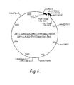
- IGF-1 LH 322 The pBR322 vector containing the complete correct left half sequence of IGF-1 was called IGF-1 LH 322 (see Fig. 5).
- Reactions were carried out in 50 ⁇ l vols. of IX tailing mix (cont. .2M Pot. Cacodylate, 25 mM Tris HCl pH 6.9, 2 mM DTT, .5 mM CoCl 2 ) and 22 ⁇ m dCTP. After prewarming at 37° for 10', the 150 second reaction was begun by the addition of 10-20 units of terminal nucleotidyl transferase and terminated by addition of EDTA followed by phenol extraction, CHC1 3 extraction 2X, and ethanol precipitation.
- oligo (dC) tailed Parts III and IV were then separately mixed with equimolar amounts of oligo (dG)-tailed PstI cut pBR322 vector in 50 ⁇ l of IX annealling buffer (.1M NaCl; 10 mM Tris HCl pH 7.8, 1 mM EDTA) at a final DNA concentration of 1-2 ⁇ g/ml. After heating to 75°C, the mixes were gradually cooled to 4° over a period of 16 hr and the mix transformed into competent E. coli 294 cells prepared according to the procedure of Dagert and Ehrlich (3).
- Transformed cells were plated on LB-Tetracycline-Agar plates and grown in LB-Tetracycline medium at tetracycline concentrations of 5 ⁇ g/ml. Tetracycline resistant transformants were picked and plated onto LB-Ampicillin-Agar plates to check for insertions at the PstI site. Several tetracycline resistant, Ampicillin-sensitive colonies for each Part 3 and 4 were miniscreened and those exhibiting insertions at the PstI locus were grown in large scale and sequenced by the Maxam and Gilbert technique (5) to confirm the correct DNA sequences of Parts 3 and 4.
- Parts 3 and 4 were separately removed from their vectors by digestions of 20 ⁇ g of each vector with AvaIl in IX AvaIl buffer (60 mM NaCl, 6 mM Tris-HCl (pH 8.0); 10 mM MgCl 2 ; 6 mM 2-mercaptoethanol) and 30 U of AvaII. After 6 hr., at 37°, EDTA was added to the 150 ⁇ l reactions to a concentration of 15 mM and the material phenol extracted, CHC1 3 extracted 2X and ethanol precipitated. The Part 3 pellet was then taken up in 1X BamHI buffer and digested in a volume of 150 ⁇ l with 30 U BamHI at 37° for 4 hr. The pellet containing Part 4 was digested with 30 U SalI in 150 ⁇ l of 1X SalI buffer at 37° for 4 hr.
- IX AvaIl buffer 60 mM NaCl, 6 mM Tris-HCl (pH 8.0); 10
- the DNA pellet was taken up in H 2 0 and was ready for ligation with Parts 3 and 4 in a three-part ligation.
- the ligation was performed under conditions described above for a three-part ligation (see Fig. 7). Parts 3 and 4 were present in the ligation mix at a 10-fold molar excess of inserts to vector.
- the mix was transformed into competent E. coli 294 cells prepared according to the Dagert and Ehrlich procedure (3) and plated onto LB-Ampicillin plates. Several transformants were miniscreened and two clones exhibiting a ca. 115 bp BamHI-Sall fragment were grown in large scale and their plasmids prepared.
- the pNCVsLE expression vector is a derivative of the pNCV vector and was prepared as follows: pNCV was treated with BgII, which cleaves at the 13 codon of the LE fusion. The site was converted to an ECoRl cleavage site using synthetic DNA, to give the expression vector pNCVsLE.
- the synthetic DNA introduced into the plasmid has the sequence: into the plasmid: and this sequence was introduced
- a linker was designed such that an enzymatic proteolysis method reported by Wunsch et al. (8) could be applied to this expression system.
- a DNA linker was chemically synthesized by standard methods (2) which when linked to the trp fusion protein and the IGF-1 gene, coded for the amino acid residues Proline and Alanine followed by Glycine and Proline which are the first two amino acid residues of IGF-1 and preceded by Proline and Alanine together comprise a recognition site for a collagenase isolated from Clostridium histolyticum (11,12). This enzyme reportedly acts at such a site to cleave the alanine-glycine peptide bond.
- the smaller PvuI-BamHI fragment ( - 725 bp) was isolated and digested with 40 U AvaIl in 150 ⁇ l 1X Sau961 buffer (60 mM NaCl, 6 mM Tris-HCl pH 7.4, 15 mM MgCl 2 , 6 mM 2-mercaptoethanol). After addition of EDTA to a concentration of 15 mM, the resulting mix chromatographed on a 6 percent polyacrylamide slab gel. The smaller Sau96I-BamHI fragment ( ⁇ 86 bp) was extracted from the gel, phenol extracted, chloroform extracted 2X, and ethanol precipitated. This fragment was ready for ligation.
- linker fragments were kinased with 100 U polynucleotide kinase in 20 ⁇ l of 1X polynucleotide kinase buffer (70 mM Tris-HCl (pH 7.6); 10 mM MgCl 2 ; 5 mM DTT; 1 mM rATP) at 37° for 1 hour. The reaction was terminated by heating to 65°C for 5 minutes. 100 pmols of the kinased linker fragments were ligated to the 86 bp Sau96I-BamHI fragment with 400 U of T4 DNA ligase in 30 ⁇ l of 1X T4 DNA ligase buffer at 14° for 12-16 hours.
- 1X polynucleotide kinase buffer 70 mM Tris-HCl (pH 7.6); 10 mM MgCl 2 ; 5 mM DTT; 1 mM rATP
- the ligation reaction was terminated by addition of EDTA to a concentration of 15 mM followed by phenol extraction, chloroform extraction 2X, and ethanol precipitation.
- the pellet was then taken up in 1X BamHI buffer and digested in a 100 ⁇ l reaction with 50 U of EcoRI and 50 U of BamHI at 37° for 6 hrs. After terminating the digestion with EDTA, the mixture was chromatographed on a 6 percent polyacrylamide slab gel and the newly created ( ⁇ 97 bp) EcoRI-BamHI fragment was extracted from the gel, and prepared for ligation.
- the vector to receive this new fragment was prepared by digesting 30 ⁇ g pBR322 HuIGF-1 with 100 U of each EcoRI and BamHI in 200 ⁇ l of 1X BamHI buffer at 37° for 8 hr. The reaction was terminated, chromatographed on a 6 percent polyacrylamide slab gel and the larger band ( ⁇ 3830 bp) representing the EcoRI-BamHI digested plasmid was isolated and the plasmid DNA extracted and prepared for ligation as above.
- a Sall-EcoRI linker was synthesized and 400 pmols kinased, as above. 200 pmols of the kinased linker was ligated to the SalI digested pBR322 HuSynIGF-1-M (prepared above) with 800 U T4 DNA ligase in 30 ⁇ l of IX ligation buffer for 12-16 hours at 14°C.
- the mixture was phenol extracted, chloroform extracted 2X, and ethanol precipitated.
- the pellet was then taken up in IX EcoRI buffer and digested with 100 U EcoRI in a volume of 200 ⁇ l for 8 hours at 37°.
- the mixture was chromatographed on a 6 percent polyacrylamide slab gel. The gel was stained and the ⁇ 230 bp band corresponding to the EcoRI-EcoRI HuIGF-1 fragment was extracted from the gel, phenol extracted, chloroform extracted 2X, and ethanol precipitated. This fragment was ready for ligation into pNCV and pNCVsLE.
- pNCV and pNCVsLE were prepared for ligation by digestion of 20 ⁇ g of each with 100 U EcoRI in 200 ⁇ l 1X EcoRI buffer at 37° for 8 hours. After digestion, 200 U of bacterial alkaline phosphatase was added to each reaction and the mixtures warmed to 65°C for 2 hours. EDTA was added to a concentration of 15 mM and the mixes were phenol extracted 3X, chloroform extracted 2X and then ethanol precipitated. These expression vectors were prepared for ligation.
- Competent E. coli 294 were prepared (3) (ATCC 31446) and used as transformation hosts for the ligations. Transformed cells were plated onto LB-agar plates containing tetracycline (5 ⁇ g/ml; LB - Tet-plates) and transformants were miniscreened (4). Miniscreen plasmid DNA from transformants of the pMCV-IGF-1 construction were digested with both PstI and BgIII to determine the orientation of the EcoRI fragment insertions. Two clones whose plasmids contained a ⁇ 570 bp BglII-PstI fragment (as opposed to a ⁇ 690 bp fragment) were grown in large scale and their plasmids prepared.
- the construction was sequenced using the Maxam-Gilbert procedure (5) to confirm the correct insertion at the junction of the trp fusion and IGF-1 protein coding sequences as well as retention of the desired reading frame. Plasmids with the correctly inserted IGF-1 fragment were called pNCVLE-IGF-1. Transformants of the pNCV-sLE-IGF-1 construction were also miniscreened by the same procedure (5), and the plasmid DNAs were digested with HincII and PstI. Two clones exhibiting a ⁇ 150 bp HincII-PstI fragment (as opposed to a ⁇ 105 bp HincII-HincII fragment) were grown in large scale and their plasmids prepared.
- the refractile bodies were purified from the pelleted cells by: a) suspending the host cells in a buffered solution of ionic strength suitable to solubilize most of the host protein, b) subjecting the suspension to cell wall/membrane disruption, c) centrifuging the disrupted suspension at low speed to form a pellet, optionally repeating the foregoing steps, and d) recovering the heterologous protein as refractile bodies in the pellet (Reference 13).
- a small quantity of refractile particles of each of the three preparations was boiled in SDS and 2-mercaptoethanol containing sample buffer and run on SDS-polyacrylamide slab gels according to the Laemmli method (14).
- the size of the protein expressed by pNCV-IGF-1 was ⁇ 28,670 .Daltons (see Figure 7), and ⁇ 9770 Daltons for the pNCV-sLE-IGF-1 protein (sLE-IGF-1) (see Figure 8). These two expressed proteins were subjected to solubilization in 6M Guanidine-HCI followed by 50-fold dilution with dilute buffers.
- the final buffer for pNCY-IGF-1 after dilution was 0.12 M Guanidine-HCl; .05 M Tris-HCI pH 8, 20 percent glycerol; 0.1 mg/ml BSA; .15 M NaCl; 0.1 mM EDTA and the final buffer after.dilution of the pNCV-sLE-IGF-1 refractile bodies was 0.14 M Guanidine-HCl; 25 mM Tris-HCl pH 7.6; 10 mM CaCl 2 .
- the two solutions containing solubilized trp-IGF-1 fusion proteins were assayed by a radioimmune assay procedure of Furlanetto et al. (23), as modified by Hintz et al. (24). Both fusion proteins demonstrated activity in this assay.
- a negative control prep was also included in the assay and the control exhibited no measurable activity.
- yeast expression-secretion systems were sought as an alternative. Aside from the advantage of avoiding protein purification from cell lysates, coupled expression-secretion systems might obviate a subsequent in vitro processing step to remove a fused protein.
- Plasmid P65 (Fig. 9) possesses sequences of the a-factor promoter, a-factor preprosequence, yeast 2 micron terminator, the yeast Trp 1 gene, as well .as portions of the p3R322 plasmid. Due to the dearth of convenient restriction sites in the a-factor preprosequence, to insert the IGF-1 coding sequence, the identical ⁇ 230 bp EcoRI-EcoRI HuSynIGF-1-M fragment that was ligated into pNCV and pNCVsLE (as mentioned previously in bacterial construction) was used.
- This EcoRI-EcoRI fragment contained the collagenase recognition site Proline-Alanine-Glycine-Proline, and allowed for collagenase digestion should IGF-1 be secreted as a fusion protein.
- the protein expressed in this construction (see Fig. 10) consists of the prepro a-factor protein fused to IGF-1.
- the plasmid P65 was partially digested in 1X EcoRI buffer with EcoRI, and then sized upon a 0.7 percent horizontal agarose gel. The band corresponding to the linearized singularly restricted plasmid was excised, eluted from the gel, and phenol extracted, chloroform extracted 2X, and then ethanol precipitated. This DNA pellet was then taken up in 50 mM Tris-HCl (pH 8) and treated with bacterial alkaline phosphatase under conditions to ensure 100 percent dephosphorylation of the 5' protruding ends.
- the phosphatase activity was removed by first adding EDTA to a concentration of 15 mM, then extracting the DNA with phenol 3X, chloroform extracting 2X, and ethanol precipitating the vector.
- This material then contained linearized P65 vector, digested with EcoRI in either of two locations: one, either at the EcoRI site upstream of the a-factor promoter and preprosequence, or at another, at the EcoRI site just downstream of the a-factor promoter and preprosequence.
- the ⁇ 230 bp EcoRI-EcoRI IGF-1 fragment was ligated into the vector. The desired location of insertion was at the EcoRI site just downstream from the a-factor promoter and preprosequence.
- the ligation was carried out under standard ligation conditions and the transformation hosts were competent E. coli 294 prepared according to Dagert and Ehrlich (3).
- the transformed cells were plated onto LB-Amp-Agar plates.
- Several transformants were miniscreened according to the method of Birnboim and Doly (4), and plasmid DNA prepared as such was digested both SalI and HindIII in the appropriate buffers.
- One of several clones which contained a plasmid with an ⁇ 110 bp EcoRI-HindIII fragment was grown in large scale and its plasmid was purified.
- This plasmid, YEp9T ⁇ -factor EcoRI-EcoRI IGF-1 was used to transform competent yeast strain 20B-12 (atrp pep4) cells according to the Hitzeman modification (19) of Hinnen et al. (I7) and Beggs et al. (18) procedures.
- yeast invertase expression-secretion system became of interest. Attempted first was expression of the yeast invertase signal protein fused to IGF-1 (Fig. 12), coupled with the processing and secretion of IGF-1, using the invertase promoter as a starting point for transcription.
- the yeast invertase signal coding sequence was attached to the IGF-1 gene by the use of a NcoI-HindIII ( - 400 bp) fragment containing the initiation ATG codon and 5' end of the signal DNA sequence, and 4 DNA fragments synthesized by standard procedures (2):
- the construction began with the isolation of the 90 bp AvaII-BamHI IGF-1 left half fragment by AvaII digestion of a ⁇ 730 bp PvuI-BamHI fragment isolated from PvuI-BamHI digested pBR322-HuSynIGF-1.
- This material was then ligated into HindIII-BamHI digested pBR322 vector, which had been first digested with HindIII, then BamHI in the appropriate buffers, followed by purification of the 4014 bp vector fragment from a 6 percent gel.
- the transformation host was competent E. coli 294 prepared by standard procedures (3) and the transformed cells were plated onto LB-Ampicillin agar plates.
- Several transfonnants were miniscreened by the Birnboim-Doly procedure (4) and their plasmid DNAs digested with EcoRI and BamHI.
- Two plasmids containing a ⁇ 167 bp EcoRI-BamHI fragment (illustrating the insertion of a 140 bp fragment into the HindIII and BamHI sites) were grown in large scale and their plasmids prepared.
- Maxam-Gilbert sequencing techniques (5), the entire 43 bp HindIII-AvaII section of DNA was sequenced to confirm the correct chemical synthesis and construction.
- the correctly constructed plasmid was called pBR322-P-I-HuSynIGF HindIII-BamHI ( ⁇ 4154 bp).
- this newly created plasmid was digested with BamHI-SalI in the appropriate buffers and the larger fragment ( ⁇ 3879 bp) was purified by gel fractionation.
- pBR322 HuSynIGF was digested with BamHI-SalI in the appropriate buffers and the 115 bp BamHI-SalI fragment corresponding to the right half of the IGF-1 gene was isolated by gel fractionation.
- This 115 bp BamHI-SalI IGF-1 right half fragment was then ligated into the BamHI-SalI digested pBR322-P-I-IGF-1 LH HindIII-BamHI vector using standard ligation conditions. Competent E.
- coli strain 294 prepared .according to Dagert and Ehrlich (3) were used as transformation hosts and transformed cells were plated onto LB-Amp-Agar plates. Several transformants were miniscreened using standard techniques (4) and plasmid DNA prepared as such was digested with EcoRI and Sall in the appropriate buffers and those plasmids illustrating an insertion of the BamHI-SalI fragment corresponding to the right half of IGF-1 were called pBR322 P-I-HuSynIGF-1 HindIII-SalI. One of the clones containing the pBR322 P-I-IGF-1 HindIII-SalI plasmid was grown in large scale and the plasmid was isolated.
- This plasmid was then digested with HindIII and SaIl in the appropriate buffer to prepare a 255 bp HindIII-SalI fragment containing all of the IGF-1 gene and the 3' portion of the yeast invertase signal coding sequence. This fragment of DNA was isolated by polyacrylamide gel fractionation and prepared for ligation by standard techniques.
- the ( - 400 bp) NcoI-HindIII fragment containing the 5' end of the DNA sequence coding for the invertase signal as well as the yeast invertase promoter was created by NcoI and HindIII digestion of plasmid YIpsp-LeIFA (16) in the appropriate buffers.
- the YIpsp-LeIFA plasmid was first digested with NcoI to completion in the appropriate buffer, then phenol extracted, chloroform extracted 2X and ethanol precipitated. The linearized molecules were then taken up in 1X HindIII buffer and partially digested to generate the needed NcoI-HindIII ("400 bp) fragment which contains an internal HindIII restriction site. This NcoI-HindIII fragment was then isolated by gel fractionation and prepared for ligation using standard techniques.
- plasmid pUC12-YI (EcoRI-BamHI) (16) was digested with NcoI and SalI in the appropriate buffers. After .purification by gel fractionation, the ⁇ 2.6 kbp vector was eluted from the gel and prepared for ligation by standard techniques. To perform the final construction, a three-part ligation was arranged using standard ligation techniques. The DNA used in the ligation included the NcoI-SaII-digested pUC12-YI (EcoRI-BamHI) (16), the ⁇ 400 bp NcoI-HindIII and the ⁇ 255 bp HindIII-SalI fragments. After ligation, the material was transformed into competent E.
- Plasmid DNA prepared as such was digested with NcoI and SalI in the appropriate buffers and one of several clones containing plasmids exhibiting the insertion of a ⁇ 625 bp NcoI-SalI DNA fragment was grown in large scale and its plasmid was purified.
- this plasmid was linearized by digestion with SalI in the appropriate buffer.
- SalI-EcoRI linker prepared as mentioned above, and kinased under standard kination conditions, was ligated to the linearized vector to convert the SalI ends to EcoRI ends using standard ligation conditions.
- the DNA pellet was dissolved in 1X EcoRI buffer, and digested with EcoRI.
- the EcoRI digestion released a ⁇ 1150 bp EcoRI fragment which contained the yeast invertase promoter, yeast invertase signal coding sequence and the IGF-1 coding sequence in one contiguous sequence. This material was isolated as a - 1150 bp band from a 6 percent polyacrylamide slab gel after fractionation and prepared for ligation using standard procedures.
- the yeast-E. coli shuttle vector to receive this EcoRI fragment was prepared by EcoRI digestion of plasmid YEp9T (16) to linearize the vector, followed by treatment of the EcoRI termini with bacterial alkaline phosphatase using conditions recommended by the manufacturer to produce 100 percent dephosphorylation of the 5' protruding ends.
- the phosphatase reaction was terminated by addition of EDTA to 15 mM and the mixture phenol extracted 3X, chloroform extracted 2X, and then the DNA .was ethanol precipitated. After redissolving the DNA pellet in 1X ligation buffer, the vector was mixed with the EcoRI ⁇ 1150 bp fragment and ligated under standard ligation conditions. Competent E.
- coli 294 cells prepared according to Dagert et al. (3) were used as transformation hosts and the transformants were plated onto LB-Amp-Agar plates.
- several transformants were miniscreened using the method Birnboim and Doly (4) and plasmid DNAs purified as such were digested with BamHI in the appropriate buffer.
- One of several transformants possessing plasmids which produced a 1.3 kb BamHI-BamHI fragment upon BamHI digestion (as opposed to a ⁇ 475 bp fragment) was grown in large scale and its plasmid was purified. This plasmid, called P.I.IGF-1 EcoRI-EcoRI P.I.
- Promoter was used to transform competent yeast cells prepared essentially according to the methods of Hinnen, A., et al. (17), and Beggs, J.D. (18), but with the modifications of Hitzeman (19).
- the yeast strain 20B-12 (atrpl pep4) was used and was obtained from the Yeast Genetics Stock Center.
- the fusion protein expressed by this construction consisted of the yeast invertase signal fused to the IGF-1 protein, the combined molecular weight of which was 9964 Daltons.
- Another plasmid with the EcoRI fragment inserted in the reverse orientation was also used to transform competent yeast cells. In this construction, the IGF-1 was not provided with the yeast terminator.
- the supernates of the three transformants demonstrated activities of 1.7 to 3.3 ng/ml of IGF-1 activity and the negative control showed no -activity.
- the pellets from 1 ml of culture were washed 1X in 25 mM Tris-HCl (pH 7.6), 1 mM EDTA and then lysed by 3-4 minutes of vigorous vortexing in 0.5 ml of the above Tris-EDTA solution with 0.4 ml of glass beads.
- the invertase promoter was subject to repression in the presence of glucose. Due to the incompatibility of glucose with high levels of transcription initiation at the invertase promoter, the PGK promoter was sought as an alternative promoter, glucose, being the mainstay carbon source of fermentation processes.
- plasmid pLeIF-A-Invertase Signal (16) was digested with BglII and then 3amHI in the appropriate buffers. This digestion released several fragments, one of which was a ⁇ 625 bp BgIII-BamHI fragment which was isolated from a 6 percent polyacrylamide slab gel and prepared for ligation using standard techniques. To clone this fragment, the pUC8 vector (20) was chosen as a cloning vehicle.
- pUC8 plasmid was digested with BamHI in IX BamHI buffer, treated with bacterial alkaline phosphatase to dephosphorylate the 5' termini, and then run onto and purified from a 5 percent polyacrylamide slab gel.
- the BamHI digested vector was mixed with the above ⁇ 625 bp BgIII-BamHI fragment, and ligated under typical ligation conditions.
- the mixture was then transformed into competent E. coli 294 prepared by the Dagert et al. method (3) and the transformed culture plated onto LB-Amp-Agar plates.
- Several transformants were picked and miniscreened using the Birnboim and Doly (4) technique.
- Miniscreen plasmid DNA was digested with EcoRI and an analytical gel of the digests illustrated two types of plasmids having EcoRI fragments either ⁇ 260 bp or ⁇ 385 bp in length.
- One clone containing a ⁇ 260 bp EcoRI fragment was grown in large scale and its plasmid purified. This plasmid was called pUC8 P.I. Promotor-Signal BgIII-BamHI.
- a clone of this type was chosen because of the desired orientation of the inserted BglII-BamHI fragment. What was needed from this plasmid was an ⁇ 20 bp EcoRI-HindIII fragment containing the ATG initiation codon and 5' end of the invertase signal coding sequence.
- the plasmid pUC8 P.I. Promotor-Signal-BgIII-BamHI was digested with EcoRI in 1X EcoRI buffer. This digestion released the ⁇ 260 bp EcoRI-EcoRI fragment which was isolated from a 6 percent polyacrylamide slab gel after fractionation of the digestion mixture. This ⁇ 260 bp fragment was then digested with HindIII in the appropriate buffer, causing the creation of two HindIII-EcoRI fragments, one ⁇ 20 bp and the other ⁇ 240 bp in length. After complete digestion, the digestion was terminated by addition of EDTA to 15 mM and the entire mix phenol extracted, chloroform extracted 2X, and then ethanol precipitated.
- a vector was prepared by EcoRI-BamHI digestion of p3R322 (15) in the appropriate buffers followed by purification of the EcoRI-BamHI digested vector from a 5 percent polyacrylamide slab gel. After preparation for ligation using standard techniques, the vector was mixed with the ⁇ 150 bp HindIII-BamHI fragment (3' end of invertase signal + Left Half IGF-1), and the two HindIII-EcoRI fragments (the ⁇ 20 bp fragment containing the 5' end of the invertase signal coding sequence), and the entire mixture was ligated under standard ligation conditions. Competent E.
- coli 294 prepared according to Dagert and Ehrlich (3) were used as transformation hosts for the ligation, and the transformed cells plated onto LB-Amp-Agar plates.
- transformants were miniscreened according to Birnboim and Doly (4) and the purified miniscreen DNAs were digested with EcoRI and BamHI.
- One of several clones possessing an - 170 bp EcoRI-BamHI fragment was grown in large volume and its plasmid purified. This plasmid contained the complete yeast invertase signal coding sequence fused to the left half of IGF-1 and was called P.I. IGF-1 L.H. RI-BamHI.
- the desired ⁇ 170 bp EcoRI-BamHI fragment was isolated from this plasmid by digestion of the plasmid with EcoRI and BamHI in the appropriate buffers followed by slab gel fractionation of the reaction mix. Using standard techniques, the ⁇ 170 bp band of DNA was prepared for ligation. To complete the construction, the right half of IGF-1 was isolated as an ⁇ 120 bp BamHI-EcoRI fragment from the plasmid P.I. IGF-1 EcoRI-EcoRI-P.I. Promoter by digestion with EcoRI and BamHI in the appropriate buffers followed by elution from a gel slice after polyacrylamide slab gel fractionation of the digestion mixture.
- YEplPT Small was constructed as a derivative of YEp1PT (21) by ClaI and PvuII digestion of YEp1PT in the appropriate buffers.
- the ClaI 5' protruding end was converted to a blunt end by use of DNA polymerase I (Klenow) under conditions recommended by the vendor. After blunting the ClaI protruding ends, the blunt ends ClaI and PvuII) of the linearized vector were fused using T4 DNA ligase under standard ligation conditions.
- YEp1PT small vector was ⁇ 5.9 kbp in size (or ⁇ 2.7 kbp smaller than YEplPT).
- YEpIPT YEp1PT small possesses the 2 micron origin and tenninator, the PGK promoter, the TRP1 gene, and sequences from pBR322, including the s-lactamase gene.
- YEp1PT Small was employed as a vector by insertion of the ⁇ 290 bp EcoRI fragment into the unique EcoRI site of the plasmid.
- EcoRI linearized YEpIPT Small vector was prepared by EcoRI digestion of YEp1PT small followed by bacterial alkaline phosphatase (BAP) treatment (to prevent religation of the complementary termini). The BAP was removed by phenol extraction 3X, chloroform extraction 2X, and ethanol precipitation. Under standard ligation conditions, the ⁇ 290 bp EcoRI fragment was ligated into the vector.
- Competent E. coli 294 prepared according to Dagert and Ehrlich (3) were used as transformation hosts and the transformed culture was plated onto LB-Amp-Agar plates.
- Several transformants were miniscreened by the Birnboim and Doly procedure (4) and miniscreen plasmid DNAs were digested with HindIII in the appropriate buffer to determine the orientation of the insert.
- One of several transformants possessing a plasmid with a ⁇ 400 bp HindIII fragment was grown in large scale and its plasmid was purified. This plasmid was called YEpIPT Small P.I. IGF-1 PGK promoter (see Fig. 14) and was used to transform competent yeast strain 208-12 (ATCC 20626) (atrp pep4) cells employing the Hitzeman modification (19) of Hinnen et al. (17), and Beggs et al. (18) procedures.
- the plasmid YEp9T ⁇ -factor ECoRI-ECoRI IGF-I ( Figure 16) was digested with Bgl 11 and SalI and the ca. 1.5 Kbp fragment containing the a-factor promotor-signal fused to IGF-I was isolated by polyacrylamide gel electrophoresis. This fragment was then ligated under standard ligation conditions to an MP-8 (BRL) vector digested with BamHI and Sal I, and treated with bacterial alkaline phosphatase. This ligation mix was then transformed into competent JM101 cells prepared according to the method of Dagert and Ehrlich (3).
- a single strand of DNA of the sequence was chemically synthesized by standard methods (2) and used to delete the DNA sequence just preceding the IGF-1 coding sequence of the a-factor promotor/signal IGF-I fusion sequence.
- This construction was then isolated as a replicative form, using a large scale plasmid preparation procedure from a JM101 cell culture inoculated with this plasmid containing the deletion
- the isolated replicative form (10 mg) was then digested with SalI.
- Yeast vector was prepared by digestion of 10 mg YEP9T plasmid with 50 units of ECoRI followed by treatment with bacterial alkaline phosphatase. The digestion was then repeatedly phenol-chloroform extracted and then ethanol precipitated and prepared for ligation.
- the ca. 1.5 kbp ECoRI-ECoRI fragment containing the deletion was then ligated to the ECoRI-ECORI YEP9T vector and the ligation mix was then transferred into competent 294 cells prepared according to the method of Dagert and Erhlich (3) and miniscreened using the method of 3irnboin and Doly (4).
- DNA prepared was screened by degestion with ECoRI and those DNAs illustrating an insertion of the ca. 1.5 kbp fragment were used to transform competent yeast strain 203-12 (ATCC 20626) according to the modification of Hitzennan (19) of the Hinner, et al., (17), and Beggs, et al., (18) procedures.
- Transformants were then grown in shaker flasks and supernates assayed and shown to have IGF-I activity by the radioimmune assay procedure of Furlanetto, et al., (23) as modified by Hintz, et al., (24).
- Human EGF is prepared in accordance with invention following analogous procedures as those described above.
- Fig. 15 double stranded DNA (Fig. 15) synthesized either by chemical means or through polymerization reactions was assembled to form a mature EGF coding sequence, with a codon coding for methionine (ATG) just preceding the amino-terminal asparagine found in the mature protein, and a codon (GTC) substituting valine for methionine at residue number 21 from the amino-tenninal asparagine.
- ATG codon coding for methionine
- GTC codon substituting valine for methionine at residue number 21 from the amino-tenninal asparagine.
- This construction was then attached at the 5' end to an additional coding sequence, which when expressed in yeast or bacteria produced a fusion protein. This fusion protein was then susceptible to CNBr cleavage at the methionine to release the valine substituted human EGF molecule.
- the above sequence coding for the mature protein was attached to the a-factor promoter/prepro sequence, the codon coding for valine at residue number 21 was replaced by ATG, and the appropriate deletion was made to bring the coding sequence for mature EGF adjacent to the a-factor signal coding sequence ( Figure 16).
- This construction was then inserted into the yeast vector Yep9T and transformed into yeast. Transformants produced as such expressed and secreted mature human EGF.
- the sequence coding for mature EGF was attached to the preinvertase signal sequence ( Figure 17) and this construction, when inserted into the yeast vector YeplPT small containing the PGK promoter, and transformed into yeast, resulted in the expression and secretion of human EGF.
- a double stranded DNA sequence coding for mature IGF-II was constructed from a combination of synthetic and natural DNA sequences ( Figure 18). This coding sequence, which did not contain an internal methionine, was attached to the TrpE leader protein coding sequence and was expressed as a fusion protein. Mature IGF-II was chemically cleaved from the purified fusion product by the action of CNBr upon a methionine residue preceding the first residue (alanine) of the mature protein.
- the IGF-II coding sequence was also attached to the a-factor promoter/prepro sequence and after the appropriate deletion was made to bring the 3' end of the a-factor signal coding sequence adjacent to the 5' end of mature IGF-II coding sequence, the construction was inserted into the Yep9T vector and transfonned into yeast. Resultant transfonnants expressed and secreted mature human IGF-II. In the same manner, the sequence coding for mature IGF-II was attached to the preinvertase coding sequence. The resultant construction was inserted into YeplPT small and transformed into yeast. Transformants produced as such expressed and secreted mature human IGF-II.
- the compounds of the present invention can be formulated according to known methods to prepare pharmaceutically useful compositions, whereby the human IGF and human EGF or products hereof are combined in admixture with a pharmaceutically acceptable carrier vehicle.
- a pharmaceutically acceptable carrier vehicle e.g., a pharmaceutically acceptable carrier vehicle.
- suitable vehicles and their formulation, inclusive of other human proteins, e.g. human serum albumin are described, for example, in Remington's Pharmaceutical Sciences by E. W. Martin, which is hereby incorporated by reference.
- Such compositions will contain an effective amount of the protein hereof together with a suitable amount of vehicle in order to prepare pharmaceutically acceptable compositions suitable for effective administration.
Abstract
Description
- This invention relates to the preparation of human IGF (insulin-like growth factor), in various forms, via recombinant DNA technology. Notably, the present invention provides for the preparation of human IGF as a mature protein product of expression, processing, and secretion in a recombinant DNA modified host organism. This invention thus provides for the production, isolation, and use of human IGF, in its various forms, as well as to the associated recombinant DNA technology by which it is prepared. In addition, the present invention relates to the similar preparation of a related protein, human EGF (Epidermal Growth Factor).
- The present invention arises in part from the discovery of a novel system by which human IGF can be prepared by a recombinant host organism in the form of a discrete, mature protein. This is accomplished according to one aspect of the present invention by an expression system which permits the expression of the amino acid sequence of human IGF fused with at least a portion of the yeast alpha factor signal sequence, followed by processing of said signal sequence, and secretion of mature human IGF protein into the medium supporting the host organism. Thus, this novel aspect of the present invention, it is believed for the first time, permits the preparation, isolation, and utilization of human IGF as a discrete, mature protein. The present invention, in its broad compass, however, covers the preparation of the amino acid sequence of human IGF in other recombinant systems including bacteria and cell culture and includes, therefore, the expression of human IGF DNA sequences providing not only mature human IGF but also fusion product derivatives containing the amino acid sequence of IGF as the essential component. All such products have been found to be biologically active, hence useful as intended.
- The publications and other materials hereof used to illuminate the background of the invention, and in particular cases, to provide additional details concerning its practice are incorporated herein by this reference and for convenience, are alphabetically and numerically referenced in the following text and respectively grouped in the appended bibliography.
- Human IGF has been the subject of a fair amount of intensive study by past workers. A body of literature has been developed related to various aspects of this protein or series of proteins (see references A through L).
- Insulin-like growth factors I and II have been isolated from human serum (A). The designation "insulin-like growth factor" or IGF was chosen to express the insulin-like effects and the insulin-like structure of these polypeptides which act as mitogens on a number of cells. The complete amino acid sequences of IGF-I and IGF-II have been determined (D,E). They are both single-chain polypeptides with three disulphide bridges and a sequence identity of 49 and 47 percent respectively, to human insulin A and B chains. The connecting peptide or C region is considerably shorter than the one of proinsulin and does not show any significant homology to it.-(For a summary of earlier studies on the biological efforts of IGF, see Reference F).
- IFG-I and IGF-II are growth promoting polypeptides occuring in human serum and human cerebral spinal fluid. Their structure is homologous to proinsulin. IGF-I seems to be produced by the liver along with a specific IGF-binding protein both of which are under control of growth hormone. Thus, human IGF is considered to be an active growth promoting molecule that mediates the effect of human growth hormone.
- It was perceived that the application of recombinant DNA and associated technologies would be a most effective way of providing the requisite large quantities of high quality human IGF for applied use to human beings as a growth factor. The goal was to produce human IGF either as biologically active fusion protein, or more importantly, as a mature protein, as products of recombinant DNA technology from a host organism. Such materials would exhibit bioactivity admitting of their use clinically in the treatment of various growth affected conditions.
- Recombinant DNA technology has reached the age of some sophistication. Molecular biologists are able to recombine various DNA sequences with some facility, creating new DNA entities capable of producing copious amounts of exogenous protein product in transformed microbes and cell cultures. The general means and methods are in hand for the in vitro ligation of various blunt ended or "sticky" ended fragments of DNA, producing potent expression vehicles useful in transforming particular organisms, thus directing their efficient synthesis of desired exogenous product. However, on an individual product basis, the pathway remains somewhat tortuous and the science has not advanced to a stage where regular predictions of success can be made. Indeed, those who portend successful results without the underlying experimental basis, do so with considerable risk of inoperability.
- DNA recombination of the essential elements, i.e., an origin of replication, one or more phenotypic selection characteristics, an expression promoter, heterologous gene insert and remainder vector, generally is performed outside the host cell. The resulting recombinant replicable expression vehicle, or plasmid, is introduced into cells by transformation and large quantities of the recombinant vehicle obtained by growing the transformant. Where the gene is properly inserted with reference to portions which govern the transcription and translation of the encoded DNA message, the resulting expression vehicle is useful to actually produce the polypeptide sequence for which the inserted gene codes, a process referred to as expression. The resulting product may be obtained by lysing, if necessary, the host cell, in microbial systems, and recovering the product by appropriate purification from other proteins.
- In practice, the use of recombinant DNA technology can express entirely heterologous polypeptides--so-called direct expression--or alternatively may express a heterologous polypeptide fused to a portion of the amino acid sequence of a homologous polypeptide. In the latter cases, the intended bioactive product is sometimes rendered bioinactive within the fused, homologous/heterologous polypeptide until it is cleaved in an extracellular environment. See references (M) and (N).
- Similarly, the art of cell or tissue cultures for studying genetics and cell physiology is well established. Means and methods are in hand for maintaining permanent cell lines, prepared by successive serial transfers from isolate normal cells. For use in research, such cell lines are maintained on a solid support in liquid medium, or by growth in suspension containing support nutriments. Scale-up for large preparations seems to pose only mechanical problems. For further background, attention is directed to references (0) and (P).
- Likewise, protein biochemistry is a useful, indeed necessary, adjunct in biotechnology. Cells producing the desired protein also produce hundreds of other proteins, endogenous products of the cell's metabolism. These contaminating proteins, as well as otner compounds, if not removed from the desired protein, could prove toxic if administered to an animal or human in the course of therapeutic treatment with desired protein. Hence, the techniques of protein biochemistry come to bear, allowing the design of separation procedures suitable for the particular system under consideration and providing a homogeneous product safe for intended use. Protein biochemistry also proves the identity of the desired product, characterizing it and ensuring that the cells have produced it faithfully with no alterations or mutations. This branch of science is also involved in the design of bioassays, stability studies and other procedures necessary to apply before successful clinical studies and marketing can take place.
- The present invention is based upon the discovery that recombinant DNA technology can be used successfully to produce human IGF and related protein, human EGF, preferably in direct form and in amounts sufficient to initiate and conduct animal and clinical testing as prerequisites to market approval. The products human IGF and EGF are suitable for use in all of their forms as produced according to the present invention, viz. in the prophylactic or therapeutic treatment of human beings for various growth associated conditions or diseases. Accordingly, the present invention, in one important aspect, is directed to methods of treating growth conditions in human subjects using human IGF or human EGF, and suitable pharmaceutical compositions thereof, prepared in accordance with the methods and means of the present invention.
- The present invention further comprises essentially pure, mature human IGF, as a product of expression, processing, and secretion in a recombinant host organism. Such human IGF is free from association with N-terminus amino acid sequence derivable from the expression systems that can be employed to prepare the material. Thus, while the present invention is directed to the preparation of polypeptides comprising the amino acid sequence of IGF, a notable aspect of the present invention involves the production of the mature human IGF directly into the medium of the recombinant host organism employed. The present invention is also directed to replicable DNA expression vehicles harboring gene sequences encoding human IGF and human EGF in expressible form, to microorganism strains or cell cultures transformed with such vehicles and to microbial or cell cultures of such transformants capable of producing amino acid sequences of human IGF and human EGF. In still further aspects, the present invention is directed to various processes useful for preparing said genes sequences, DNA expression vehicles, microorganisms and cell cultures and specific embodiments thereof. Still further, this invention is directed to the preparation of fermentation cultures of said microorganisms and cell cultures.
-
- Figure 1 represents the chemically synthesized DNA strands used in the construction of expression vectors for human IGF.
- Figure 2 shows the completed double stranded DNA of Figure 1.
- Figure 3 show the fragments of DNA of Figure 2 after restriction by ECoRI and PstI and Bam HI.
- Figure 4 depicts the ligation of
parts - Figure 5
show parts - Figure 6 depicts the ligation of
parts - Figure 7 shows a sequence of DNA and deduced fusion protein containing IGF-I.
- Figure 8 shows a sequence of DNA and deduced short fusion protein containing IGF-I.
- Figure 9 depicts a plasmid used in the present construction.
- Figure 10 shows the DNA and protein sequence of IGF-I fused with alpha factor pre-pro sequence.
- Figure 11 is a vector containing alpha factor promotor and pre-pro sequence fused to IGF-I.
- Figure 12 shows the yeast invertase signal fused to IGF-I.
- Figure 13 shows the parental plasmid containing the yeast PGK promotor.
- Figure 14 depicts a yeast expression vector containing PGK promotor, invertase signal and human IGF-I gene.
- Figure 15 is the synthetic DNA used to construct the coding sequence of mature human EGF.
- Figure 16 depicts the yeast alpha factor "pre-pro" sequence fused to the human EGF coding sequence.
- Figure 17 depicts the yeast invertase signal sequence fused to the human EGF coding sequence.
- Figure 18 shows the coding sequence for human IGF-II.
- As used herein, "human IGF" and "human EGF" denotes human insulin-like growth factor and human epidermal growth factor, produced by microbial or cell cultures systems and bioactive forms comprising the amino acid sequence corresponding to human IGF and human EGF etherwise native to human tissue. The human IGF and EGF proteins produced herein have been defined by means of DNA, gene, and deductive amino acid sequencing. It will be understood that inasmuch as natural allelic variations exist and occur from individual to individual, as demonstrated by (an) amino acid difference(s) in the overall sequence or by deletions, substitutions, insertions, inversions, or additions of one or more amino acids of said sequences, the present invention is intended to embrace of all such allelic variations of the two molecules involved. In addition, the location of and the degree of glycosylation depends upon the nature of the recombinant host organism employed and such variations as may occur are included within the ambit of this invention. Finally, the potential exists in the use of DNA technology for the preparation of various derivatives of human IGF and human EGF by simple modification of the underlying gene sequence for sucn molecules. Such modifications could be accomplished by means of site directed mutagenesis of the underlying DNA, as an example. All such modifications resulting in derivatives of human IGF and human EGF are included within the scope of the present invention so long as the essential characteristic human IGF and human EGF activities remain unaffected in kind.
- "Essentially pure form" when used to describe the state of human IGF or human EGF produced by this invention means that the proteins are free of proteins or other materials normally associated with human IGF or human EGF when produced by non-recombinant cells, i.e. in their "native" environments.
- "Expression vector" includes vectors whicn are capable of expressing DNA sequences contained therein, where such sequences are operably linked to other sequences capable of effecting their expression, i.e., promotor/operator sequences. In sum, "expression vector" is given a functional definition: any DNA sequence which is capable of effecting expression of a specified DNA code disposed therein. In general, expression vectors of utility in recombinant DNA techniques are often in the form of "plasmids" which refer to circular double stranded DNA loops which in their vector form, are not bound to the chromosone. In the present specification, "plasmid" and "vector" are used interchangably as the plasmid is the most commonly used form of vector. However, the invention is intended to include such other forms of expression vectors which function equivalently and which become known in the art subsequently.
- "Recombinant host cells" refers to cells which have been transformed with such vectors. Thus, the human IGF and human EGF molecules produced by such cells can be referred to as "recombinant human IGF" and "recombinant human EGF".
- The vectors and methods disclosed herein are suitable for use in host cells over a wide range of prokaryotic and eukaryotic organisms.
- In general, of course, prokaryotes are preferred for cloning of DNA sequences in constructing the vectors useful in the invention. For example, E. coli K12 strain 294 (ATCC No. 31446) is particularly useful. Other microbial strains which may be used include E. coli strains such as E. coli B, and E. coli X1776 (ATTC No. 31537). The aforementioned strains, as well as E. coli W3110 (F-, λ-, prototrophic, ATTC No. 27325), bacilli such as Bacillus subtilus, and other enterobacteriaceae such as Salmonella typhimurium or Serratia marcesans, and various pseudomonas species may be used. These examples are, of course, intended to be illustrative rather than limiting.
- In general, plasmid vectors containing replicon and control sequences which are derived from species compatible with the host cell are used in connection with these hosts. The vector ordinarily carries a replication site, as well as marking sequences which are capable of providing phenotypic selection in transformed cells. For example, E. coli is typically transformed using
pBR 322, a plasmid derived from an E. coli species (Bolivar, et al., Gene 2: 95 (1977)). pBR322 contains genes for ampicillin and tetracycline resistance and thus provides easy means for identifying transformed cells. The pBR322 plasmid, or other microbial plasmid must also contain, or be modified to contain, promoters which can be used by the microbial organism for expression of its own proteins. Those promoters most commonly used in recombinant DNA construction include the β-lactamase (penicillinase) and lactose promoter systems (Chang et al, Nature, 275: 615 (1978), Itakura, et al, Science, 198: 1056 (1977); (Goeddel, et al Nature 281: 544 (1979)) and a tryptophan (trp) promoter system (Goeddel, et al, Nucleic Acids Res., 8: 4057 (1980); EPO Appl Publ No. 0036776). While these are the most commonly used, other microbial promoters have been discovered and utilized, and details concerning their nucleotide sequences have been published, enabling a skilled worker to ligate them functionally with plasmid vectors (Siebenlist, et al, Cell 20: 269 (1980)). - In addition to prokaryates, eukaryotic microbes, such as yeast cultures may also be used. Saccharomyces cerevisiae, or common baker's yeast is the most commonly used among eukaryotic microorganisms, although a number of other strains are commonly available. For expression in Saccharomyces, the plasmid YRp7, for example, (Stinchcomb, et al, Nature, 282: 39 (1979); Kingsman et al, Gene, 7: 141 (1979); Tschemper, et al, Gene, 10: 157 (1980)) is commonly used. This plasmid already contains the trpl gene which provides a selection marker for a mutant strain of yeast lacking the ability to grow in tryptophan, for example ATCC No. 44076 or PEP4-1 (Jones, Genetics, 85: 12 (1977)). The presence of the trpl lesion as a characteristic of the yeast host cell genome then provides an effective environment for detecting transformation by growth in the absence of tryptophan.
- Suitable promoting sequences in yeast vectors include the promoters for 3-phosphoglycerate kinase (Hitzeman, et al., J. Biol. Chem., 255: 2073 (1980)) or other glycolytic enzymes (Hess, et al, J. Adv. Enzyme Reg., 7: 149 (1963); Holland, et al, Biochemistry, 17: 4900 (1978)), such as enolase, glyceraldehyde-3-phosphate dehydrogenase, hexokinase, pyruvate decarboxylase, phosphofructokinase, glucose-6-phosphate isomerase, 3-phosphoglycerate mutase, pyruvate kinase, triosephosphate isomerase, phosphoglucose isomerase, and glucokinase. In constructing suitable expression plasmids, the tennination sequences associated with these genes are also ligated into the expression vector 3' of the sequence desired to be expressed to provide polyadenylation of the mRNA and termination. Other promoters, which have the additional advantage of transcription controlled by growth conditions are the promoter regions for
alcohol dehydrogenase 2, isocytochrome C, acid phosphatase, degradative enzymes associated with nitrogen metabolism, and the aforementioned glyceraldehyde-3-phosphate dehydrogenase, and enzymes responsible for maltose and galactose utilization (Holland, ibid.). Any plasmid vector containing yeast-compatible promoter, origin of replication and termination sequences is suitable. - In addition to microorganisms, cultures of cells derived from multicellular organisms may also be used as hosts. In principle, any such cell culture is workable, whether from vertebrate or invertebrate culture. However interest has been greatest in vertebrate cells, and propogation of vertebrate cells in culture (tissue culture) has become a routine procedure in recent years [Tissue Culture, Academic Press, Kruse and Patterson, editors (1973)]. Examples of such useful host cell lines are VERO and HeLa cells, Chinese hamster ovary (CHO) cell lines, and W138, BHK, COS-7 and MDCK cell lines. Expression vectors for such cells ordinarily include (if necessary) an origin of replication, a promoter located in front of the gene to be expressed, along with any necessary ribosome binding sites, RNA splice sites, polyadenylation site, and transcriptional terminator sequences.
- For use in mammalian cells, the control functions on the expression vectors are often provided by viral material. For example, commonly used promoters are derived from polyoma,
Adenovirus 2, and most frequently Simian Virus 40 (SV40). The early and late promoters of SV40 virus are particularly useful because both are obtained easily from the virus as a fragment which also contains the SV40 viral origin of replication (Fiers, et al, Nature, 273: 113 (1978) incorporated herein by reference. Smaller or larger SV40 fragments may also be used, provided there is included the approximately 250 bp sequence extending from the Hind III site toward the Bgl I site located in the viral origin of replication. Further, it is also possible, and often desirable, to utilize promoter or control sequences normally associated with the desired gene sequence, provide such control sequences are compatible with the host cell systems. - An origin of replication may be provided either by construction of the vector to include an exogenous origin, such as may be derived from SV40 or other viral (e.g. Polyoma, Adeno, VSV, BPV, etc.) source, or may be provided by the host cell chromosomal replication mechanism. If the vector is integrated into the host cell chromosome, the latter is often sufficient.
- If cells without formidable cell wall barriers are used as host cells, transfection is carried out by the calcium phosphate precipitation method as described by Graham and Van der Eb, Virology, 52: 546 (1978). However, other methods for introducing DNA into cells such as by nuclear injection or by protoplast fusion may also be used.
- If prokaryotic cells or cells which contain substantial cell wall constructions are used, the preferred method of transfection is calcium treatment using calcium chloride as described by Cohen, F.N. et al Proc. Natl. Acad. Sci. (USA), 69: 2110 (1972).
- Construction of suitable vectors containing the desired coding and control sequences employ standard ligation techniques. Isolated plasmids or DNA fragments are cleaved, tailored, and religated in the form desired to form the plasmids required.
- Cleavage is performed by treating with restriction enzyme (or enyzmes) in suitable buffer. In general, about 1 µg plasmid or DNA fragments is used with about 1 unit of enzyme in about 20 µl of buffer solution. (Appropriate buffers and substrate amounts for particular restriction enzymes are specified by the manufacturer.) Incubation times of about 1 hour at 37°C are workable. After incubations, protein is removed by extraction with phenol and chloroform, and the nucleic acid is recovered from the aqueous fraction by precipitation with ethanol.
- If blunt ends are required, the preparation is treated for 15 minutes at 15° with 10 units of Polymerase I (Klenow), phenol-chlorofonn extracted, and ethanol precipitated.
- Size separation of the cleaved fragments is perfonned using 6 percent polyacrylamide gel described by Goeddel, D., et al, Nucleic Acids Res., 8: 4057 (1980) incorporated herein by reference.
- For ligation approximately equimolar amounts of the desired components, suitably end tailored to provide correct matching are treated with about 10 units T4 DNA ligase per 0.5 µg DNA. (When cleaved vectors are used as components, it may be useful to prevent religation of the cleaved vector by pretreatment with bacterial alkaline phosphatase.)
- For analysis to confirm correct sequences in plasmids constructed, the ligation mixtures are used to transform E. coli K12 strain 294 (ATCC 31446), and successful transformants selected by ampicillin resistance where appropriate. Plasmids from the transformants are prepared, analyzed by restriction and/or sequenced by the method of Messing, et al, Nucleic Acids Res., 9:309 (1981) or by the method of Maxam, et al, Methods in Enzymology, 65:499 (1980).
- The following examples are intended to illustrate but not to limit the present invention.
- Enzymes were obtained from the following suppliers:
- New England Biolabs: restriction enzymes, T4 DNA ligase Bethesda Research Labs: restriction enzymes, Bact. Alkaline Phos. Boehringer-Mannheim: E. coli DNA Polymerase I (Klenow) P+L Biochemicals: Polynucleotide kinase, Terminal Nucleotidyl Transferase
- New England Nuclear:
- pBR322 [oligo(dG)-tailed] DNA
- Reagents:
- BioRad: Bis Acrylamide, Acrylamide, TEMED
- Sigma: Ammonium Persulfate
- Amersham: 10218 γ32P ATP >5000 Ci/mmol; 10165 α32P dCTP >400 Ci/mmol.
- Solutions and Media: 1X TBE: .54M Tris Base, 0.54 M Boric Acid, .017 M Na2 EDTA.
- Difco: Yeast Nitrogenous 3ase (YNB); Tryptone, Yeast Extract; Bacto-Agar; Casamino Acids.
- Autoradiography:
- Kodak X-0 mat AR XAR-2 Film
- Glass Beads:
- 0.45-0.50 mM B. Braun Melsungen AG
- LB medium (per liter):
- 10g NaCl; 5g yeast extract; lOg tryptone; .17ml NaOH (50 percent)
- LB Agar (per liter):
- 10g tryptone; 5g yeast extract; .5g NaCl; 15g Bacto-Agar; adjusted to pH 7.5 with NaOH.
- Antibiotics:
- Tetracycline (5 µg/ml) in all mediums; Ampicillin (20 µg/ml) in all mediums (plates or liquid)
- 6g Na2HPO4 (anhydrous); 1g NH4Cl; 3g KH2PO4; .5g
- NaCl; 1 mM MgSO4; 0.5 percent (w/v) glucose; 0.5 percent (w/v) Casamino Acids; .0001 percent Thiamine-HCI.
- YNB-CAA (per liter):
- 6.7g Yeast Nitrogenous 3ase (witnout Amino Acids); 10 mg adenine; 10 mg uracil; 5g Casamino Acids; 20g Glucose.
- YNB-CAA agar plates (per liter):
- Same as YNB-CAA + 30g agar.
- Standard Ligation Conditions:
- 10-fold molar excess of insert (or linker) to vector. IX T4 DNA ligase buffer and 400-800 U T4 DNA ligase; 14° - 12-16 hours.
- Standard Kination Conditions:
- IX Polynucleotide kinase buffer; 15 U polynucleotide kinase;
- 37° 60 minutes; followed by reaction termination by heating to 65° for 10 minutes.
- 1X Kinase Buffer:
- 70 mM Tris-HCl (pH 7.6); 10 mM MgCl2; 5 mM DTT
- 1X T4 DNA Ligase Buffer:
- 50 mM Tris-HCl (pH 7.8); 10 mM MgCl2; 20 mM DTT; 1 mM rATP.
- The 1° protein structure of the human IGF-1 molecule has been determined (1). Based upon this protein sequence and the genetic code, a DNA sequence coding for mature human IGF-1 protein, including all possible base substitutions at any one base position, was determined by computer analysis (Genentech Untrans Program). Using a restriction site analysis program (Genentech Asearch Program), all potential restriction sites located in all possiole DNA sequences consistently coding for the same protein were found. Three sites internal to the coding sequence were selected: Pstl, BamHI, and AvaII. Two additional sites were placed at the ends, just outside of the coding sequence of the mature protein: one EcoRI site before the initiation codon, AUG, and the Sall site following the termination codon, TAG of the coding sequence. The choice of these sites facilitated the cloning of the coding sequence in separate parts, each of which subsequently could be excised and then assembled to form an intact synthetic IGF-1 gene. This construction involved the assembly of 4 parts, 2 parts forming the left half, 2 parts foraing the right half. Each part consisted of two single strands of chemically synthesized DNA (see Fig. 1). Proposed synthetic fragments were also analyzed for internal complementarity.
- The constructions used to generate these four parts employed the use of DNA Polymerase I repair synthesis of synthetic oligonucleotide substrates having 9-10 bp stretches of complementary sequence at their 3' termini. In the presence of DNA Polymerase I (Klenow) and the four deoxynucleoside triphosphates, these primer-templates were extended to become full-length double-stranded DNAs. To prevent priming at locations other than the desired portions as well as self-hybridizations, each set of single-stranded DNAs were analyzed by a computer program (Genentech Homology Program), and wherever possible, sequences which would have potentially led to hairpin loops, self-priming, or mis-priming, were eliminated by alternate codon usage. Each of these four double-stranded DNAs were synthesized to include 9-12 additional bp of non-IGF-1 coding DNA at each end (see Fig. 2). This additional DNA was included to allow generation of sticky ends by restriction enzyme digestion. The sticky ends thus formed facilitated the ligation of the double-stranded pieces to contiguous coding sections of the synthetic gene or into a cloning vehicle.
- The 9-12 extra bp of double stranded DNA beyond the restriction site at the end of each part (see Fig. 2) allowed for the TdT-mediated formation of single-stranded oligodeoxycytidine strands at the 3' ends of each double-stranded DNA section. These oligodeoxycytidine tailed double-stranded DNAs could then be annealled into a complementary oligodeoxyguanosine tailed PstI site of a cloning vehicle. Once cloned, and sequenced to ensure the correct base sequences, the parts could be easily isolated and ligated following restriction enzyme cleavage at the restriction sites selected at the ends of each of the four parts, to form the intact synthetic IGF-1 gene.
- The method used successfully here was similar to that described by Rossi et al. (28); however, attempts at the construction and cloning of the IGF-1 coding sequence using the Rossi et al. method (28) with only two base pairs of extra DNA beyond the restriction enzyme recognition sites repeatedly failed. The method employed here also differs from the Rossi et al. procedure (28) in that restriction sites placed at both ends of a double stranded DNA allow for the convenience of cloning each double stranded DNA fragment, individually, by (dC)-tailing and annealling into a (dG)-tailed vector, a method which in practice requires less of the double stranded DNA than three-part ligations.
- Eight fragments, 43, 43, 46, 46, 46, 46, 54, and 46 bases in length (see Fig. 1), were chemically synthesized according to the method of Crea and Horn (2), the only change being the use of mesitylene nitrotriazole as the condensing agent rather than 2,4,6 Triisopropyl benzenesulfonylchloride tetrazole.
- The syntheses of the fragments were accomplished from the appropriate solid support (cellulose) by sequential addition of the appropriate fully-protected dimer- or trimer-blocks. The cycles were carried out under the same conditions as described in the synthesis of oligothymidilic acid (see Crea et al., supra). The final polymers were treated with base (aqueous conc. NH3) and acid (80 percent HoAc), the polymer pelleted off, and the supernatant evaporated to dryness. The residue, dissolved in 4 percent aq. NH3, was washed with ethyl ester (3X) and used for the isolation of the fully deprotected fragment.
- Purification was accomplished by electrophoresis using 20 percent polyacrylamide gels. The pure oligonucleotide was ethanol precipitated following gel elution.
- 225-285 pmoles of each chemically synthesized fragment was mixed with an equivalent amount of the complementary single-stranded DNA fragment (i.e. 1L+3L; 2L+4L; 1R+3R, 2R+4R) in the presence of deoxyribonucleoside triphosphates at a final concentration of 200 µM with the exception of dCTP. dCTP was added to a concentration of 5 µM as a α32P-labeled isotope with a specific activity of 1000-2000 Ci/mmol) to allow easy monitoring of the repair-synthesis reaction product. The reactions were carried out in a buffer containing a final concentration of 50mM Tris HCl pH 7.5; 20 mM MgCl; 20 mM DTT and 154 DNA Polymerase I (Klenow) in a reaction volume of 200 µl. Reactions were allowed to proceed at 4" for 12-18 hrs.
- Upon completion, EDTA was added to a concentration of 25 mM. Sample buffer containing the mixes were phenol extracted, CHCl3 extracted 2X, and products were etOH precipitated. Pellets were taken up in .3 M NaOAc and the DNA reprecipitated with etOH. After- dissolving the pellets in H20, the 1L+3L and 2L+4L products were then digested separately with PstI in 100 µl reaction mixes containing 1X PstI buffer (50mM (NH4)2SO4, 20 mM Tris HCl pH 7.5, 10 mM MgCl2), and 70 U PstI. After 4 hrs, EDTA was added to a concentration of 10 mM, and the material was ethanol precipitated. Pellets were then taken up in .3 M NaOAc and reprecipitated, then taken up in H20. The Pstl-digested 1L+3L product was digested with EcoRI at 37° in a 100 µl reaction mix 1X EcoRI buffer (150 mM NaCl, 6 mM Tris HCl pH 7.5, 6 mM MgCl2) and 70 U EcoRI. The PstI digested 2L+4L product was digested at 37" with BamHI in a 100 µl reaction mix in IX BamHI Buffer (150 mM NaCl, 6 mM Tris HCl pH 7.9, 6 mM MgCl2) and 70 U BamHI. After 4 hrs, EDTA was added to both mixtures, and sample buffer was added. They were electrophoresed on a 6 percent polyacrylamide slab gel. Six percent slab gels were cast with a mixture containing 6 percent (w/v) acrylamide (20 to 1 ratio of acrylamide to Bis acrylamide) 1X TBE, 1 percent APS and 0.1 percent TEMED. Reaction products were located on the gel by autoradiography and the oand corresponding to the 45 bp EcoRI-PstI digested 1L+3L product (Part 1) (see Fig. 3) and the band corresponding to the 50 bp PstI-BamHI digested 2L+4L product (Part 2) (see Fig. 3) were excised from the gel, the material electroeluted in 0.2X TBE, phenol extracted, CHCl3 extracted, and ethanol precipitated.
Parts - Cloning vector was prepared by digesting 20 µg pBR322 (15) with 50 U EcoRI and 50 U BamHI, in IX RI Buffer at 37° for 6 hr. After addition of EDTA to a concentration of 10 mM, sample buffer was added, and the mixture was run on a 5 percent polyacrylamide gel. The gel was developed by staining 10' in H20 containing 5 µg/ml Et. Bromide, rinsing 2X in H20 and placing upon a UV transilluminator (302 nM). the band corresponding to the ca. 3712 bp EcoRI-BamHI digested pBR322 molecules was cut from the gel. The-DNA was electroeluted from the gel slice, phenol extracted, CHCl3 extracted 2X, and ethanol precipitated. The pellet was dissolved in H20 and was ready for ligation.
- In a three-part ligation (see Fig. 4), in which the molar ratio of inserts to vector in the ligation reaction was approximately 10 to 1,
parts - E. col strain 294 was used as the transformation host, using the procedure of M. Dagert and S.D. Ehrlich (3). The transformed cells were plated on LB-agar plates containing ampicillin (20 µg/ml ; LB-Amp-plates) and transfonnants were screened and grown in LB medium containing ampicillin at 20 µg/ml ampicillin. Transformants were screened using a modification of the rapid miniscreen method of Birnboim and Doly (4). Miniprep DNA prepared as such was digested with EcoRI and BamHI and run on polyacrylamide slab gels. Several transformants which illustrated a ca. 218 bp EcoRI-BamHI insert were grown in large scale and plasmids from each were isolated and sequenced according to the procedure of Maxam and Gilbert (5) to confirm the correct chemical synthesis and construction. The pBR322 vector containing the complete correct left half sequence of IGF-1 was called IGF-1 LH 322 (see Fig. 5).
- Using the identical conditions of DNA Polymerase I-mediated repair-synthesis, the two pairs of fragments comprising the right half of the synthetic IGF-1 were converted into double-stranded DNAs. After the DNA Polymerase I reactions, and without enzymatic digestion, the 1R+3R (Part III) and 2R+4R (Part IV) reactions were run on a 6 percent polyacrylamide slab gels. The 83 bp (Part III) and 91 bp (Part IV) bands were located by autoradiography and cut from the gel. After electroelution the ethanol precipitated double-stranded DNAs were dC-tailed (see Fig. 6) using the procedures of Villa-Komaroff et al. (6) and Rowenkamp and Firtel (7). Reactions were carried out in 50 µl vols. of IX tailing mix (cont. .2M Pot. Cacodylate, 25 mM Tris HCl pH 6.9, 2 mM DTT, .5 mM CoCl2) and 22 µm dCTP. After prewarming at 37° for 10', the 150 second reaction was begun by the addition of 10-20 units of terminal nucleotidyl transferase and terminated by addition of EDTA followed by phenol extraction, CHC13 extraction 2X, and ethanol precipitation.
- These oligo (dC) tailed Parts III and IV were then separately mixed with equimolar amounts of oligo (dG)-tailed PstI cut pBR322 vector in 50 µl of IX annealling buffer (.1M NaCl; 10 mM Tris HCl pH 7.8, 1 mM EDTA) at a final DNA concentration of 1-2 µg/ml. After heating to 75°C, the mixes were gradually cooled to 4° over a period of 16 hr and the mix transformed into competent E. coli 294 cells prepared according to the procedure of Dagert and Ehrlich (3). Transformed cells were plated on LB-Tetracycline-Agar plates and grown in LB-Tetracycline medium at tetracycline concentrations of 5 µg/ml. Tetracycline resistant transformants were picked and plated onto LB-Ampicillin-Agar plates to check for insertions at the PstI site. Several tetracycline resistant, Ampicillin-sensitive colonies for each
Part Parts - Preparation:
Parts -
Parts Part 3 pellet was then taken up in 1X BamHI buffer and digested in a volume of 150 µl with 30 U BamHI at 37° for 4 hr. Thepellet containing Part 4 was digested with 30 U SalI in 150 µl of 1X SalI buffer at 37° for 4 hr. - Both digests were then run on 6 percent polyacrylamide slab gels and stained. The 51 bp
band representing Part 3 and the 62 bpband representing Part 4 were removed from the gels and the DNA electroeluted, phenol extracted, CHC13 extracted 2X and ethanol precipitated. Pellets were then taken up in H20 and were ready for ligation. - 20 µg of the IGF-1
LH 322 vector was digested with 50 U of BamHI and 50 U of SalI in a 200 µl reaction containing 1X BamHI buffer at 37° for 6 hr. After addition of EDTA to a concentration of 15 mM, the digestion mix was run on a 6 percent polyacrylamide slab gel, ethidium bromide stained and the 3814 bp band excised from the gel. - After electroelution, phenol extraction, chloroform extraction and ethanol precipitation, the DNA pellet was taken up in H20 and was ready for ligation with
Parts Parts - Initial attempts were to obtain expression of IGF-1 as a fusion protein. To accomplish this, both the pNCV (9) and tile pNCVsLE (10) expression vectors were used. (The pNCVsLE expression vector is a derivative of the pNCV vector and was prepared as follows: pNCV was treated with BgII, which cleaves at the 13 codon of the LE fusion. The site was converted to an ECoRl cleavage site using synthetic DNA, to give the expression vector pNCVsLE. The synthetic DNA introduced into the plasmid has the sequence:into the plasmid:
 and this sequence was introduced
- As a strategy to release the fused human IGF-1 protein from the trp fusion protein, a linker was designed such that an enzymatic proteolysis method reported by Wunsch et al. (8) could be applied to this expression system. To accomplish this, a DNA linker:was chemically synthesized by standard methods (2) which when linked to the trp fusion protein and the IGF-1 gene, coded for the amino acid residues Proline and Alanine followed by Glycine and Proline which are the first two amino acid residues of IGF-1 and preceded by Proline and Alanine together comprise a recognition site for a collagenase isolated from Clostridium histolyticum (11,12). This enzyme reportedly acts at such a site to cleave the alanine-glycine peptide bond.

- To construct a DNA sequence coding for a fusion protein with a collagenase cleavage site, 30 µg pBR322 HuIGF-1 plasmid was cleaved with 50 U BamHI and 50 U Pvul enzyme in 200 µl 1X BamHI buffer at 37° for 6 hours. After addition of EDTA to a concentration of 15 mM, the reaction mix was chromatographed on a 6 percent polyacrylamide slab gel. The smaller PvuI-BamHI fragment (-725 bp) was isolated and digested with 40 U AvaIl in 150 µl 1X Sau961 buffer (60 mM NaCl, 6 mM Tris-HCl pH 7.4, 15 mM MgCl2, 6 mM 2-mercaptoethanol). After addition of EDTA to a concentration of 15 mM, the resulting mix chromatographed on a 6 percent polyacrylamide slab gel. The smaller Sau96I-BamHI fragment (~86 bp) was extracted from the gel, phenol extracted, chloroform extracted 2X, and ethanol precipitated. This fragment was ready for ligation.
- 200 pmols of linker fragments were kinased with 100 U polynucleotide kinase in 20 µl of 1X polynucleotide kinase buffer (70 mM Tris-HCl (pH 7.6); 10 mM MgCl2; 5 mM DTT; 1 mM rATP) at 37° for 1 hour. The reaction was terminated by heating to 65°C for 5 minutes. 100 pmols of the kinased linker fragments were ligated to the 86 bp Sau96I-BamHI fragment with 400 U of T4 DNA ligase in 30 µl of 1X T4 DNA ligase buffer at 14° for 12-16 hours. The ligation reaction was terminated by addition of EDTA to a concentration of 15 mM followed by phenol extraction, chloroform extraction 2X, and ethanol precipitation. The pellet was then taken up in 1X BamHI buffer and digested in a 100 µl reaction with 50 U of EcoRI and 50 U of BamHI at 37° for 6 hrs. After terminating the digestion with EDTA, the mixture was chromatographed on a 6 percent polyacrylamide slab gel and the newly created (~97 bp) EcoRI-BamHI fragment was extracted from the gel, and prepared for ligation. The vector to receive this new fragment was prepared by digesting 30 µg pBR322 HuIGF-1 with 100 U of each EcoRI and BamHI in 200 µl of 1X BamHI buffer at 37° for 8 hr. The reaction was terminated, chromatographed on a 6 percent polyacrylamide slab gel and the larger band (~3830 bp) representing the EcoRI-BamHI digested plasmid was isolated and the plasmid DNA extracted and prepared for ligation as above. In a 30 µl ligation reaction containing a 10-fold molar excess of insert fragment to vector, the EcoRI-BamHI fragment was ligated into the EcoRI-BamHI digested plasmid pBR322 HuIGF-1 under standard ligation conditions mentioned above. Competent E. coli 294, prepared as above (3), were used as transformation hosts and the transformed cell were plated onto LB-Ampicillin agar plates. Several transformants were picked,
- miniscreened as above (4), and two exhibiting an EcoRI-BamHI insertion were grown in large scale and their plasmids purified. Using the Maxam-Gilbert procedure (5) the construction was sequenced to verify the correct synthesis and insertion of the EcoRI-Sau96I collagenase linker. This plasmid was called pBR322 HuSynIGF-1-M.
- To prepare this EcoRI-SalI IGF-1 coding sequence for insertion into pNCV and pNCVsLE, 30 µg of pBR322 HuSynIGF-1-M was digested with 70 U of SalI in 200 µl of 1X SalI buffer (150 mM NaCl, 6 mM Tris-HCI (pH 7.9); 6 mM MgCl2; 6 mM 2-mercaptoethanol) at 37° for 6 hours. After addition of EDTA to 15 mM, the mixture was phenol extracted, chloroform extracted 2X, and ethanol precipitated.
- Using standard chemical synthesis procedures (2) a Sall-EcoRI linkerwas synthesized and 400 pmols kinased, as above. 200 pmols of the kinased linker was ligated to the SalI digested pBR322 HuSynIGF-1-M (prepared above) with 800 U T4 DNA ligase in 30 µl of IX ligation buffer for 12-16 hours at 14°C.

- After termination of the reaction with EDTA, the mixture was phenol extracted, chloroform extracted 2X, and ethanol precipitated. The pellet was then taken up in IX EcoRI buffer and digested with 100 U EcoRI in a volume of 200 µl for 8 hours at 37°. After addition of EDTA to a concentration of 15 mM, the mixture was chromatographed on a 6 percent polyacrylamide slab gel. The gel was stained and the ~230 bp band corresponding to the EcoRI-EcoRI HuIGF-1 fragment was extracted from the gel, phenol extracted, chloroform extracted 2X, and ethanol precipitated. This fragment was ready for ligation into pNCV and pNCVsLE. pNCV and pNCVsLE were prepared for ligation by digestion of 20 µg of each with 100 U EcoRI in 200 µl 1X EcoRI buffer at 37° for 8 hours. After digestion, 200 U of bacterial alkaline phosphatase was added to each reaction and the mixtures warmed to 65°C for 2 hours. EDTA was added to a concentration of 15 mM and the mixes were phenol extracted 3X, chloroform extracted 2X and then ethanol precipitated. These expression vectors were prepared for ligation.
- Ligations of the EcoRI-EcoRI Human IGF-1 fragment into the two expression vectors were performed in 30 µl reaction volumes in 1X T4 DNA ligase buffer with 800 U T4 DNA ligase at 14° for 12-16 hours. The EcoRI-EcoRI fragment was present at a 10-fold molar excess to vector.
- Competent E. coli 294 were prepared (3) (ATCC 31446) and used as transformation hosts for the ligations. Transformed cells were plated onto LB-agar plates containing tetracycline (5 µg/ml; LB-Tet-plates) and transformants were miniscreened (4). Miniscreen plasmid DNA from transformants of the pMCV-IGF-1 construction were digested with both PstI and BgIII to determine the orientation of the EcoRI fragment insertions. Two clones whose plasmids contained a ~570 bp BglII-PstI fragment (as opposed to a ~690 bp fragment) were grown in large scale and their plasmids prepared. The construction was sequenced using the Maxam-Gilbert procedure (5) to confirm the correct insertion at the junction of the trp fusion and IGF-1 protein coding sequences as well as retention of the desired reading frame. Plasmids with the correctly inserted IGF-1 fragment were called pNCVLE-IGF-1. Transformants of the pNCV-sLE-IGF-1 construction were also miniscreened by the same procedure (5), and the plasmid DNAs were digested with HincII and PstI. Two clones exhibiting a ~150 bp HincII-PstI fragment (as opposed to a ~105 bp HincII-HincII fragment) were grown in large scale and their plasmids prepared. Using the Maxam-Gilbert techniques (5), the functions of the trp fusion and IGF-1 protein coding sequences were sequenced to ascertain proper orientation and retention of the proper reading frame. Those plasmids possessing the correct insertion and proper reading frame were called pNCV-sLE-IGF-1.
- To attempt expression of each of these constructions, two clones, one possessing pNCV-IGF-1 and the other possessing pNCV-sLE-IGF-1, were inoculated into 10 ml M9-Tetracycline culture medium supplemented with 0.5 mg/ml Tryptophan. A clone containing pNCV-LE with no IGF-1 gene insert was also inoculated into culture medium to provide as a negative control in assays.
- After 12-16 hours growth at 37° with agitation, 0.5 ml of these cultures were used to inoculate 250 milliliters of M9-Tetracycline culture medium. After growing for 12-16 hours at 37° with agitation, the cells were harvested by centrifugation at 5000 rpm for 10 minutes in a Sorvall GSA rotor. The refractile bodies were purified from the pelleted cells by: a) suspending the host cells in a buffered solution of ionic strength suitable to solubilize most of the host protein, b) subjecting the suspension to cell wall/membrane disruption, c) centrifuging the disrupted suspension at low speed to form a pellet, optionally repeating the foregoing steps, and d) recovering the heterologous protein as refractile bodies in the pellet (Reference 13). A small quantity of refractile particles of each of the three preparations was boiled in SDS and 2-mercaptoethanol containing sample buffer and run on SDS-polyacrylamide slab gels according to the Laemmli method (14). The size of the protein expressed by pNCV-IGF-1 (LE-IGF-1) was ~28,670 .Daltons (see Figure 7), and ~9770 Daltons for the pNCV-sLE-IGF-1 protein (sLE-IGF-1) (see Figure 8). These two expressed proteins were subjected to solubilization in 6M Guanidine-HCI followed by 50-fold dilution with dilute buffers. The final buffer for pNCY-IGF-1 after dilution was 0.12 M Guanidine-HCl; .05 M Tris-
HCI pH - To avoid the necessity of refractile body purification and solubilization, from bacterial cell lysates, yeast expression-secretion systems were sought as an alternative. Aside from the advantage of avoiding protein purification from cell lysates, coupled expression-secretion systems might obviate a subsequent in vitro processing step to remove a fused protein. Available were three yeast expression-secretion systems. These were: 1) yeast a factor (22), employing yeast a-factor promoter and preprosequence; 2) yeast invertase (16) consisting of the invertase promoter and signal sequence; and 3) a hybrid, composed of the PGK promoter (25) and invertase signal (16).
- To obtain expression of IGF-1 using the a factor promoter and preprosequence, a plasmid constructed by Singh (22) was used. Plasmid P65 (Fig. 9) possesses sequences of the a-factor promoter, a-factor preprosequence,
yeast 2 micron terminator, theyeast Trp 1 gene, as well .as portions of the p3R322 plasmid. Due to the dearth of convenient restriction sites in the a-factor preprosequence, to insert the IGF-1 coding sequence, the identical ~230 bp EcoRI-EcoRI HuSynIGF-1-M fragment that was ligated into pNCV and pNCVsLE (as mentioned previously in bacterial construction) was used. This EcoRI-EcoRI fragment contained the collagenase recognition site Proline-Alanine-Glycine-Proline, and allowed for collagenase digestion should IGF-1 be secreted as a fusion protein. The protein expressed in this construction (see Fig. 10) consists of the prepro a-factor protein fused to IGF-1. - To insert the ~230 bp EcoRI-EcoRI fragment, the plasmid P65 was partially digested in 1X EcoRI buffer with EcoRI, and then sized upon a 0.7 percent horizontal agarose gel. The band corresponding to the linearized singularly restricted plasmid was excised, eluted from the gel, and phenol extracted, chloroform extracted 2X, and then ethanol precipitated. This DNA pellet was then taken up in 50 mM Tris-HCl (pH 8) and treated with bacterial alkaline phosphatase under conditions to ensure 100 percent dephosphorylation of the 5' protruding ends. Following this treatment, the phosphatase activity was removed by first adding EDTA to a concentration of 15 mM, then extracting the DNA with phenol 3X, chloroform extracting 2X, and ethanol precipitating the vector. This material then contained linearized P65 vector, digested with EcoRI in either of two locations: one, either at the EcoRI site upstream of the a-factor promoter and preprosequence, or at another, at the EcoRI site just downstream of the a-factor promoter and preprosequence. The ~230 bp EcoRI-EcoRI IGF-1 fragment was ligated into the vector. The desired location of insertion was at the EcoRI site just downstream from the a-factor promoter and preprosequence.
- The ligation was carried out under standard ligation conditions and the transformation hosts were competent E. coli 294 prepared according to Dagert and Ehrlich (3). The transformed cells were plated onto LB-Amp-Agar plates. Several transformants were miniscreened according to the method of Birnboim and Doly (4), and plasmid DNA prepared as such was digested both SalI and HindIII in the appropriate buffers. One of several clones which contained a plasmid with an ~110 bp EcoRI-HindIII fragment was grown in large scale and its plasmid was purified. This plasmid, YEp9T α-factor EcoRI-EcoRI IGF-1 (see Fig. 11), was used to transform competent yeast strain 20B-12 (atrp pep4) cells according to the Hitzeman modification (19) of Hinnen et al. (I7) and Beggs et al. (18) procedures.
- Two such transformants, as well as a negative control transfonnant (with no IGF-I insertion in the plasmid), were grown in suspension as were those of the yeast pre-invertase-IGF-1 plasmid transformations. Supernates were tested for secreted IGF-1 activity, as measured by the radioimmune assay procedure of Furlanetto et al. (23) as modified by Hintz et al. (24). Both supernates of transformants having plasmids with IGF-I inserts contained IGF-1 activity and the negative control supernate did not. One of these transformants was grown in large scale in a 10 liter fennenter and the supernate contained secreted IGF-I activity at a peak level of ~3µg/ml. The IGF-1 activity of the fermentation supernate was also demonstrated by a placental membrane radioreceptor assay developed by Horner et al. (26).
- Based upon evidence of correct processing and secretion in yeast of proteins with heterologous signal sequences (16), the yeast invertase expression-secretion system became of interest. Attempted first was expression of the yeast invertase signal protein fused to IGF-1 (Fig. 12), coupled with the processing and secretion of IGF-1, using the invertase promoter as a starting point for transcription.
- The yeast invertase signal coding sequence was attached to the IGF-1 gene by the use of a NcoI-HindIII (-400 bp) fragment containing the initiation ATG codon and 5' end of the signal DNA sequence, and 4 DNA fragments synthesized by standard procedures (2):

- The construction began with the isolation of the 90 bp AvaII-BamHI IGF-1 left half fragment by AvaII digestion of a ~730 bp PvuI-BamHI fragment isolated from PvuI-BamHI digested pBR322-HuSynIGF-1.
- After phosphorylation of all four synthetic DNA fragments using standard kination conditions, the four synthetic fragments were mixed with the AvaII-BamHI IGF-1 left half fragment and ligated using standard ligation conditions. Following inactivation of the ligase by phenol and chloroform extraction 2X, the ethanol precipitated DNA pellet was dissolved and digested with HindIII and BamHI in the appropriate buffers. Newly constructed HindIII-SamHI (ca. 140 bp) fragment was isolated and extracted from a 6 percent polyacrylamide gel. This material was then ligated into HindIII-BamHI digested pBR322 vector, which had been first digested with HindIII, then BamHI in the appropriate buffers, followed by purification of the 4014 bp vector fragment from a 6 percent gel.
- The transformation host was competent E. coli 294 prepared by standard procedures (3) and the transformed cells were plated onto LB-Ampicillin agar plates. Several transfonnants were miniscreened by the Birnboim-Doly procedure (4) and their plasmid DNAs digested with EcoRI and BamHI. Two plasmids containing a ~167 bp EcoRI-BamHI fragment (illustrating the insertion of a 140 bp fragment into the HindIII and BamHI sites) were grown in large scale and their plasmids prepared. Using Maxam-Gilbert sequencing techniques (5), the entire 43 bp HindIII-AvaII section of DNA was sequenced to confirm the correct chemical synthesis and construction. The correctly constructed plasmid was called pBR322-P-I-HuSynIGF HindIII-BamHI (~4154 bp).
- To insert the right half of the IGF-1 gene, this newly created plasmid was digested with BamHI-SalI in the appropriate buffers and the larger fragment (~3879 bp) was purified by gel fractionation. pBR322 HuSynIGF was digested with BamHI-SalI in the appropriate buffers and the 115 bp BamHI-SalI fragment corresponding to the right half of the IGF-1 gene was isolated by gel fractionation. This 115 bp BamHI-SalI IGF-1 right half fragment was then ligated into the BamHI-SalI digested pBR322-P-I-IGF-1 LH HindIII-BamHI vector using standard ligation conditions. Competent E. coli strain 294 prepared .according to Dagert and Ehrlich (3) were used as transformation hosts and transformed cells were plated onto LB-Amp-Agar plates. Several transformants were miniscreened using standard techniques (4) and plasmid DNA prepared as such was digested with EcoRI and Sall in the appropriate buffers and those plasmids illustrating an insertion of the BamHI-SalI fragment corresponding to the right half of IGF-1 were called pBR322 P-I-HuSynIGF-1 HindIII-SalI. One of the clones containing the pBR322 P-I-IGF-1 HindIII-SalI plasmid was grown in large scale and the plasmid was isolated. This plasmid was then digested with HindIII and SaIl in the appropriate buffer to prepare a 255 bp HindIII-SalI fragment containing all of the IGF-1 gene and the 3' portion of the yeast invertase signal coding sequence. This fragment of DNA was isolated by polyacrylamide gel fractionation and prepared for ligation by standard techniques.
- The (-400 bp) NcoI-HindIII fragment containing the 5' end of the DNA sequence coding for the invertase signal as well as the yeast invertase promoter was created by NcoI and HindIII digestion of plasmid YIpsp-LeIFA (16) in the appropriate buffers. The YIpsp-LeIFA plasmid was first digested with NcoI to completion in the appropriate buffer, then phenol extracted, chloroform extracted 2X and ethanol precipitated. The linearized molecules were then taken up in 1X HindIII buffer and partially digested to generate the needed NcoI-HindIII ("400 bp) fragment which contains an internal HindIII restriction site. This NcoI-HindIII fragment was then isolated by gel fractionation and prepared for ligation using standard techniques.
- To provide for a vector, plasmid pUC12-YI (EcoRI-BamHI) (16) was digested with NcoI and SalI in the appropriate buffers. After .purification by gel fractionation, the ~2.6 kbp vector was eluted from the gel and prepared for ligation by standard techniques. To perform the final construction, a three-part ligation was arranged using standard ligation techniques. The DNA used in the ligation included the NcoI-SaII-digested pUC12-YI (EcoRI-BamHI) (16), the ~400 bp NcoI-HindIII and the ~255 bp HindIII-SalI fragments. After ligation, the material was transformed into competent E. coli 294 cells prepared according to Dagert et al. (3). Transformed cells were plated onto LB-Amp-Agar plates and several transformants were miniscreened using the procedure of Birnboim and Doly (4). Plasmid DNA prepared as such was digested with NcoI and SalI in the appropriate buffers and one of several clones containing plasmids exhibiting the insertion of a ~625 bp NcoI-SalI DNA fragment was grown in large scale and its plasmid was purified.
- As a final step, this plasmid was linearized by digestion with SalI in the appropriate buffer. SalI-EcoRI linker, prepared as mentioned above, and kinased under standard kination conditions, was ligated to the linearized vector to convert the SalI ends to EcoRI ends using standard ligation conditions. After termination of the ligation reaction by addition of EDTA to 15 mM, phenol extraction, chloroform extraction 2X and ethanol precipitation, the DNA pellet was dissolved in 1X EcoRI buffer, and digested with EcoRI. The EcoRI digestion released a ~1150 bp EcoRI fragment which contained the yeast invertase promoter, yeast invertase signal coding sequence and the IGF-1 coding sequence in one contiguous sequence. This material was isolated as a -1150 bp band from a 6 percent polyacrylamide slab gel after fractionation and prepared for ligation using standard procedures.
- The yeast-E. coli shuttle vector to receive this EcoRI fragment was prepared by EcoRI digestion of plasmid YEp9T (16) to linearize the vector, followed by treatment of the EcoRI termini with bacterial alkaline phosphatase using conditions recommended by the manufacturer to produce 100 percent dephosphorylation of the 5' protruding ends. The phosphatase reaction was terminated by addition of EDTA to 15 mM and the mixture phenol extracted 3X, chloroform extracted 2X, and then the DNA .was ethanol precipitated. After redissolving the DNA pellet in 1X ligation buffer, the vector was mixed with the EcoRI ~1150 bp fragment and ligated under standard ligation conditions. Competent E. coli 294 cells prepared according to Dagert et al. (3) were used as transformation hosts and the transformants were plated onto LB-Amp-Agar plates. To determine the orientation of the insertion, several transformants were miniscreened using the method Birnboim and Doly (4) and plasmid DNAs purified as such were digested with BamHI in the appropriate buffer. One of several transformants possessing plasmids which produced a 1.3 kb BamHI-BamHI fragment upon BamHI digestion (as opposed to a ~475 bp fragment) was grown in large scale and its plasmid was purified. This plasmid, called P.I.IGF-1 EcoRI-EcoRI P.I. Promoter was used to transform competent yeast cells prepared essentially according to the methods of Hinnen, A., et al. (17), and Beggs, J.D. (18), but with the modifications of Hitzeman (19). The yeast strain 20B-12 (atrpl pep4) was used and was obtained from the Yeast Genetics Stock Center. In this construction, the expression of IGF-1 begins with transcription at the invertase promoter and terminates in the
yeast 2 micron sequence. The fusion protein expressed by this construction consisted of the yeast invertase signal fused to the IGF-1 protein, the combined molecular weight of which was 9964 Daltons. Another plasmid with the EcoRI fragment inserted in the reverse orientation was also used to transform competent yeast cells. In this construction, the IGF-1 was not provided with the yeast terminator. - Several transformants were picked and streaked on YNB-CAA agar plates. Among these, three transformants were picked and inoculated into 10 ml of YN3-CAA grow-up medium, in shake flasks. A fourth culture was also started using a colony transformed with the same vector, but with the EcoRI fragment inserted into the vector in the reverse orientation. After 16-20 hours growth at 30°, the cultures were sampled (1 ml) and cleared of cells by spinning 5' in an eppendorf microfuge. Supernatants were taken off and assayed for secreted activity using the radioimmune assay procedure of Furlanetto et al. (23) as modified by Hintz et al. (24). The supernates of the three transformants demonstrated activities of 1.7 to 3.3 ng/ml of IGF-1 activity and the negative control showed no -activity. To determine intracellular activity, the pellets from 1 ml of culture were washed 1X in 25 mM Tris-HCl (pH 7.6), 1 mM EDTA and then lysed by 3-4 minutes of vigorous vortexing in 0.5 ml of the above Tris-EDTA solution with 0.4 ml of glass beads.
- Assay of the cell lysates demonstrated IGF-1 activities of 1.5-2.8 ng/ml in the three IGF-1 secreting transformants and no activity in the negative control transformant. The highest secretor of the three transformants was grown in a 5 liter fermentation and the secreted IGF-1 activity reached a peak of 74 ng/ml of supernate.
- One difficulty in the use of the invertase promoter was that it was subject to repression in the presence of glucose. Due to the incompatibility of glucose with high levels of transcription initiation at the invertase promoter, the PGK promoter was sought as an alternative promoter, glucose, being the mainstay carbon source of fermentation processes.
- To begin construction of the PGK promoter P.I.IGF-1 construction, it was necessary to clone a fragment containing the entire invertase signal coding sequence. To do this, plasmid pLeIF-A-Invertase Signal (16) was digested with BglII and then 3amHI in the appropriate buffers. This digestion released several fragments, one of which was a ~625 bp BgIII-BamHI fragment which was isolated from a 6 percent polyacrylamide slab gel and prepared for ligation using standard techniques. To clone this fragment, the pUC8 vector (20) was chosen as a cloning vehicle. pUC8 plasmid was digested with BamHI in IX BamHI buffer, treated with bacterial alkaline phosphatase to dephosphorylate the 5' termini, and then run onto and purified from a 5 percent polyacrylamide slab gel.
- After standard preparation for ligation the BamHI digested vector was mixed with the above ~625 bp BgIII-BamHI fragment, and ligated under typical ligation conditions. The mixture was then transformed into competent E. coli 294 prepared by the Dagert et al. method (3) and the transformed culture plated onto LB-Amp-Agar plates. Several transformants were picked and miniscreened using the Birnboim and Doly (4) technique. Miniscreen plasmid DNA was digested with EcoRI and an analytical gel of the digests illustrated two types of plasmids having EcoRI fragments either ~260 bp or ~385 bp in length. One clone containing a ~260 bp EcoRI fragment was grown in large scale and its plasmid purified. This plasmid was called pUC8 P.I. Promotor-Signal BgIII-BamHI.
- A clone of this type was chosen because of the desired orientation of the inserted BglII-BamHI fragment. What was needed from this plasmid was an ~20 bp EcoRI-HindIII fragment containing the ATG initiation codon and 5' end of the invertase signal coding sequence.
- To construct the intact invertase signal coding DNA sequence, -150 bp HindIII-BamHI fragment containing the 3' end of the signal sequence fused to the left half of the IGF-1 gene was isolated from HindIII-BamHI digestion of plasmid pBR322 P.I. IGF-LH HindIII-BamHI (~4154 bp). Isolation was by polyacrylamide slab gel fractionation, and the DNA band corresponding to the -150 bp fragment was excised and prepared for ligation using standard techniques.
- To obtain the short (-20 bp) EcoRI-HindIII fragment, the plasmid pUC8 P.I. Promotor-Signal-BgIII-BamHI was digested with EcoRI in 1X EcoRI buffer. This digestion released the ~260 bp EcoRI-EcoRI fragment which was isolated from a 6 percent polyacrylamide slab gel after fractionation of the digestion mixture. This ~260 bp fragment was then digested with HindIII in the appropriate buffer, causing the creation of two HindIII-EcoRI fragments, one ~20 bp and the other ~240 bp in length. After complete digestion, the digestion was terminated by addition of EDTA to 15 mM and the entire mix phenol extracted, chloroform extracted 2X, and then ethanol precipitated.
- A vector was prepared by EcoRI-BamHI digestion of p3R322 (15) in the appropriate buffers followed by purification of the EcoRI-BamHI digested vector from a 5 percent polyacrylamide slab gel. After preparation for ligation using standard techniques, the vector was mixed with the ~150 bp HindIII-BamHI fragment (3' end of invertase signal + Left Half IGF-1), and the two HindIII-EcoRI fragments (the ~20 bp fragment containing the 5' end of the invertase signal coding sequence), and the entire mixture was ligated under standard ligation conditions. Competent E. coli 294 prepared according to Dagert and Ehrlich (3) were used as transformation hosts for the ligation, and the transformed cells plated onto LB-Amp-Agar plates. Several transformants were miniscreened according to Birnboim and Doly (4) and the purified miniscreen DNAs were digested with EcoRI and BamHI. One of several clones possessing an -170 bp EcoRI-BamHI fragment was grown in large volume and its plasmid purified. This plasmid contained the complete yeast invertase signal coding sequence fused to the left half of IGF-1 and was called P.I. IGF-1 L.H. RI-BamHI.
- The desired ~170 bp EcoRI-BamHI fragment was isolated from this plasmid by digestion of the plasmid with EcoRI and BamHI in the appropriate buffers followed by slab gel fractionation of the reaction mix. Using standard techniques, the ~170 bp band of DNA was prepared for ligation. To complete the construction, the right half of IGF-1 was isolated as an ~120 bp BamHI-EcoRI fragment from the plasmid P.I. IGF-1 EcoRI-EcoRI-P.I. Promoter by digestion with EcoRI and BamHI in the appropriate buffers followed by elution from a gel slice after polyacrylamide slab gel fractionation of the digestion mixture. These two fragments, the ~170 bp EcoRI-BamHI and the ~120 bp BamHI-EcoRI, were ligated together in vitro under standard ligation conditions, with both fragments present in roughly equimolar concentrations. This ligation mixture was then terminated by the addition of EDTA to ~15 mM followed by phenol extraction, chloroform extraction 2X, and ethanol precipitation. The DNA pellet was then taken up in 1X EcoRI buffer and digested with EcoRI. The digest was then run on a 6 percent polyacrylamide slab gel and the DNA band staining at ~290 bp (as opposed to ~340 bp and 240 bp) was excised and prepared for ligation using standard techniques. This ~290 bp EcoRI-EcoRI fragment contained the entire yeast invertase signal coding sequence fused to the complete IGF-1 coding sequence.
- To express this protein, it was necessary to select a yeast vector with a promoter. The PGK promoter of the plasmid YEp1PT Small (see Fig. 13) was used. YEplPT Small was constructed as a derivative of YEp1PT (21) by ClaI and PvuII digestion of YEp1PT in the appropriate buffers. The ClaI 5' protruding end was converted to a blunt end by use of DNA polymerase I (Klenow) under conditions recommended by the vendor. After blunting the ClaI protruding ends, the blunt ends ClaI and PvuII) of the linearized vector were fused using T4 DNA ligase under standard ligation conditions. The resultant YEp1PT small vector was ~5.9 kbp in size (or ~2.7 kbp smaller than YEplPT). Just as YEpIPT, YEp1PT small possesses the 2 micron origin and tenninator, the PGK promoter, the TRP1 gene, and sequences from pBR322, including the s-lactamase gene.
- YEp1PT Small was employed as a vector by insertion of the ~290 bp EcoRI fragment into the unique EcoRI site of the plasmid. EcoRI linearized YEpIPT Small vector was prepared by EcoRI digestion of YEp1PT small followed by bacterial alkaline phosphatase (BAP) treatment (to prevent religation of the complementary termini). The BAP was removed by phenol extraction 3X, chloroform extraction 2X, and ethanol precipitation. Under standard ligation conditions, the ~290 bp EcoRI fragment was ligated into the vector.
- Competent E. coli 294 prepared according to Dagert and Ehrlich (3) were used as transformation hosts and the transformed culture was plated onto LB-Amp-Agar plates. Several transformants were miniscreened by the Birnboim and Doly procedure (4) and miniscreen plasmid DNAs were digested with HindIII in the appropriate buffer to determine the orientation of the insert. One of several transformants possessing a plasmid with a ~400 bp HindIII fragment was grown in large scale and its plasmid was purified. This plasmid was called YEpIPT Small P.I. IGF-1 PGK promoter (see Fig. 14) and was used to transform competent yeast strain 208-12 (ATCC 20626) (atrp pep4) cells employing the Hitzeman modification (19) of Hinnen et al. (17), and Beggs et al. (18) procedures.
- Several yeast transfonnants were grown in suspension in identical fashion as were those of the P.I. IGF-1 EcoRI-EcoRI P.I. promoter plasmid transformation and supernates were measured for activity determined by a radioimmune assay method of Furlanetto et al. (23) as modified by Hintz et al. (24). Shake flask supernates of three transformants contained activities ranging from 38 to 53 ng/ml of supernate. Similarly, one of these transformants was selected and grown in larger scale, utilizing a 10 liter fermenter and the secreted IGF-1 activity in the supernate reached a peak of ~780 ng/ml. This fermentation supernate was also- subjected to a radioreceptor assay (26) and was demonstrated to contain IGF-1 activity.
- To construct a DNA sequence coding for the a-factor pre-pro protein fused to the DNA sequence coding for mature IGF-I, an M-13 in vitro mutagenesis technique was employed. (See Regin et al., Proc. Acad. Science (USA) 75, 4268; Hutchinson, et al., Journal Biological Chem. 253, 6551; Gilliam, et al.,
Gene 8, 81 and 99; Gillam, et al.,Nucleic Acids Research 6, 2973; Adelman, et al., DNA (June, 1983).) - To construct the M-13 plasmid, the plasmid YEp9T α-factor ECoRI-ECoRI IGF-I (Figure 16) was digested with
Bgl 11 and SalI and the ca. 1.5 Kbp fragment containing the a-factor promotor-signal fused to IGF-I was isolated by polyacrylamide gel electrophoresis. This fragment was then ligated under standard ligation conditions to an MP-8 (BRL) vector digested with BamHI and Sal I, and treated with bacterial alkaline phosphatase. This ligation mix was then transformed into competent JM101 cells prepared according to the method of Dagert and Ehrlich (3). These transformants were then mixed with non-competent JM101 cells grown to log phase, mixed with top agar and plated onto LB agar plates. Several clear plaques were picked and sequenced using the M-13 dideoxy sequencing technigue to confirm the presence of an insertion into the SalI-BamHI sites of the vector. - To perform the deletion according to the method above, a single strand of DNA of the sequencewas chemically synthesized by standard methods (2) and used to delete the DNA sequence
 just preceding the IGF-1 coding sequence of the a-factor promotor/signal IGF-I fusion sequence. This construction was then isolated as a replicative form, using a large scale plasmid preparation procedure from a JM101 cell culture inoculated with this plasmid containing the deletion
- The isolated replicative form (10 mg) was then digested with SalI.
- Then phenol-chlorogform extracted and then ethanol precipitated and prepared for ligation. To this replicative form was ligated Sal I-ECoRI linkers. After ligation and inactivation of the ligase by phenol, chloroform extraction followed by ethanol precipitation, the material was digested with "50 U ECoRI enzyme under standard conditions and then run onto a percent polyacrylamide gel. The ca. 1.5 kbp RI-ECoRI fragment released was isolated from the gel and prepared for ligation using standard conditions.
- Yeast vector was prepared by digestion of 10 mg YEP9T plasmid with 50 units of ECoRI followed by treatment with bacterial alkaline phosphatase. The digestion was then repeatedly phenol-chloroform extracted and then ethanol precipitated and prepared for ligation.
- The ca. 1.5 kbp ECoRI-ECoRI fragment containing the deletion was then ligated to the ECoRI-ECORI YEP9T vector and the ligation mix was then transferred into competent 294 cells prepared according to the method of Dagert and Erhlich (3) and miniscreened using the method of 3irnboin and Doly (4). DNA prepared was screened by degestion with ECoRI and those DNAs illustrating an insertion of the ca. 1.5 kbp fragment were used to transform competent yeast strain 203-12 (ATCC 20626) according to the modification of Hitzennan (19) of the Hinner, et al., (17), and Beggs, et al., (18) procedures.
- Transformants were then grown in shaker flasks and supernates assayed and shown to have IGF-I activity by the radioimmune assay procedure of Furlanetto, et al., (23) as modified by Hintz, et al., (24).
- One of these clones were grown in large scale in a 10-liter fermentor and IGF-I purified from the supernatant of this fermentation. This material was then subjected to amino terminal protein sequencing and shown to be mature IGF-I protein.
- Human EGF is prepared in accordance with invention following analogous procedures as those described above.
- In a fashion simildr to IGF-1, double stranded DNA (Fig. 15) synthesized either by chemical means or through polymerization reactions was assembled to form a mature EGF coding sequence, with a codon coding for methionine (ATG) just preceding the amino-terminal asparagine found in the mature protein, and a codon (GTC) substituting valine for methionine at
residue number 21 from the amino-tenninal asparagine. This construction was then attached at the 5' end to an additional coding sequence, which when expressed in yeast or bacteria produced a fusion protein. This fusion protein was then susceptible to CNBr cleavage at the methionine to release the valine substituted human EGF molecule. - To secrete the mature form of EGF from yeast, the above sequence coding for the mature protein was attached to the a-factor promoter/prepro sequence, the codon coding for valine at
residue number 21 was replaced by ATG, and the appropriate deletion was made to bring the coding sequence for mature EGF adjacent to the a-factor signal coding sequence (Figure 16). This construction was then inserted into the yeast vector Yep9T and transformed into yeast. Transformants produced as such expressed and secreted mature human EGF. In addition, the sequence coding for mature EGF was attached to the preinvertase signal sequence (Figure 17) and this construction, when inserted into the yeast vector YeplPT small containing the PGK promoter, and transformed into yeast, resulted in the expression and secretion of human EGF. - A double stranded DNA sequence coding for mature IGF-II was constructed from a combination of synthetic and natural DNA sequences (Figure 18). This coding sequence, which did not contain an internal methionine, was attached to the TrpE leader protein coding sequence and was expressed as a fusion protein. Mature IGF-II was chemically cleaved from the purified fusion product by the action of CNBr upon a methionine residue preceding the first residue (alanine) of the mature protein.
- The IGF-II coding sequence was also attached to the a-factor promoter/prepro sequence and after the appropriate deletion was made to bring the 3' end of the a-factor signal coding sequence adjacent to the 5' end of mature IGF-II coding sequence, the construction was inserted into the Yep9T vector and transfonned into yeast. Resultant transfonnants expressed and secreted mature human IGF-II. In the same manner, the sequence coding for mature IGF-II was attached to the preinvertase coding sequence. The resultant construction was inserted into YeplPT small and transformed into yeast. Transformants produced as such expressed and secreted mature human IGF-II.
- The compounds of the present invention can be formulated according to known methods to prepare pharmaceutically useful compositions, whereby the human IGF and human EGF or products hereof are combined in admixture with a pharmaceutically acceptable carrier vehicle. Suitable vehicles and their formulation, inclusive of other human proteins, e.g. human serum albumin are described, for example, in Remington's Pharmaceutical Sciences by E. W. Martin, which is hereby incorporated by reference. Such compositions will contain an effective amount of the protein hereof together with a suitable amount of vehicle in order to prepare pharmaceutically acceptable compositions suitable for effective administration.
- Notwithstanding that reference has been made to particular preferred embodiments of the present invention, it will be understood that the present invention is not to be construed as limited to such rather to the lawful scope of the appended claims.
-
- A. Rinderknecht, E. W. et al., Proceedings National Academy of Sciences (USA) 73, 2365 (1976).
- B. Rinderknecht, et al., Proceedings National Academy of Sciences (USA) 73, 4379 (1975).
- C. Blundell, et al., Proceedings National Academy of Sciences (USA) 75, 1980 (1978).
- D. Rinderknecht, et al., Journal of Biological Chemistry 253, 2769 (1978), 2365 (1976).
- E. Rinderknecht, et al.,
FEBS Letters 89, 283 (1978). - F. Zaph, et al.,
Metabolism 27, 1803 (1978). - G. Hintz, et al., Journal of Clinical Endocrinology and
Metabolism 50, 405 (1980). - H. Blundell, et al., Nature 287, 781 (1980).
- I. Hintz, et al., Journal of Clinical Endo. and
Metabolism 51, 672 (1980). - J. Baxter, et al., Journal of Clinical Endo. and
Metabolism 54, 474 (1980). - K. Hintz, et al., Journal of Clinical Endo. and
Metabolism 54, 442 (1982). - L. Schoenle, et al., Nature 296, 252 (1982).
- M. British Patent Application Publication No. 2007676A.
- N. Wetzel,
American Scientist 68, 664 (1980). - 0. Microbiology, Second Edition, Harper and Row Publications Inc., Hagerstown, Maryland (1973), especially pages 1122 et sequence.
-
- 1. Rinderknecht, E. and Humbel, R.E., J. Biol. Chem. 253, 8, 2769-2776 (1978).
- 2. Crea, R. and Horn, T.,
Nucleic Acids Research 8, 2331-2348 (1980). - 3. Dagert, M. and Ehrlich, S.D.,
Gene 6, 23-28 (1979). - 4. Birnboim, H.C. and Doly, J.,
Nucleic Acids Research 7, 1513-1523 (1979). - 5. Maxam, A. and Gilbert, W., Methods in
Enzymology 65, 499 (1980). - 6. Villa-Komaroff et al., Proc. Natl. Acad. Sci. USA 75, 3727 (1979).
- 7. Rowenkamp and Firtel, Dictyostelium Dev. Biol. 79, 409 (1980).
- 8. Wunsch, E. et al., Hoppe-Seyler's Z. Physiol. Chem. Bd. 362, S1285-1237 (Sept. 1981).
- 9. Maniatis, T. et al., Molecular Cloning, 426 (1982).
- 10. Kleid, D.G., Annals of New York Acad.Sci., Biochem.Eng., II,413,23-30 (1983).
- 11. Seifter, S. and Gallop, P.M., The Proteins, 2nd Ed. (H. Neurath, ed.) Vol. V, p. 659 (1966).
- 12. Nordwig, A.,
Leder 13, 10 (1962). - 13. Lin, N. (U.S.S.N. 06/452,363, filed 22.12.82.) EPA 83307840.5.
- 14. Laemmli, U.K. Nature (London) 227, 680-685 (1970).
- 15. Bolivar, F. et al.,
Gene 2, 95 (1977). - 16. Chang, C.N., (U.S.S.N. 06/488337 filed 25.4.83) EPA 84302722.8.
- 17. Hinnen, A. et al., Proc. Natl. Acad. Sci. USA 75, 1929-1933 (1978).
- 18. Beggs, J.D.,
Nature 275, 104-109 (1978). - 19. EP 88632.
- 20. EP 88622.
- 21. Hitzeman, R.A. et al., Science 219, 620-625 (1983).
- 22. Singh, A. (U.S.S.N. 06/488323 filed 25.4.83) EPA 84302723.6.
- 23. Furlanetto, R.W., Underwood, L.E., Van Wyk, J.J., D'Ercole, A.J., J. Clin. Invest. 60, 648 (1977).
- 24. Hintz, R.L., Liu, F., Marshall, L.B., Chung, D., J. Clin. Endocrinol. Metab. 50, 405 (1980).
- 25. Hitzeman, R.A. et al., Proceedings of the Berkeley Workshop on Recent Advances in Yeast Molecular Biology: Recombinant DNA. U.C. Press, Berkeley, p. 173 (1982).
- 26. Horner, J.M., Liu, F., Hintz, R.A., J. Clin. Endocrinol. Metab. 47, 6, p. 1287 (1978).
- 27. Rossi, J.J., Zierzek, R., Huang, T., Walker, P.A., Itakura, K., J. Biol. Chem. 257, 16, 9226 (1982).
Claims (10)
Priority Applications (1)
| Application Number | Priority Date | Filing Date | Title |
|---|---|---|---|
| AT84303783T ATE75777T1 (en) | 1983-06-06 | 1984-06-05 | HUMAN INSULIN-LIKE GROWTH FACTOR (IGF)PRODUCED FROM RECOMBINANT HOST, METHOD, EXPRESSION VECTOR AND RECOMBINANT HOST THEREOF AND PHARMACEUTICAL COMPOSITION CONTAINING IGF. |
Applications Claiming Priority (4)
| Application Number | Priority Date | Filing Date | Title |
|---|---|---|---|
| US50135383A | 1983-06-06 | 1983-06-06 | |
| US501353 | 1983-06-06 | ||
| US50607883A | 1983-06-20 | 1983-06-20 | |
| US506078 | 1983-06-20 |
Publications (2)
| Publication Number | Publication Date |
|---|---|
| EP0128733A1 true EP0128733A1 (en) | 1984-12-19 |
| EP0128733B1 EP0128733B1 (en) | 1992-05-06 |
Family
ID=27053789
Family Applications (1)
| Application Number | Title | Priority Date | Filing Date |
|---|---|---|---|
| EP84303783A Expired - Lifetime EP0128733B1 (en) | 1983-06-06 | 1984-06-05 | Human insulin-like growth factor (igf) produced from a recombinant host, process, expression vector and recombinant host therefor, and igf-containing pharmaceutical composition |
Country Status (10)
| Country | Link |
|---|---|
| US (3) | US6331609B1 (en) |
| EP (1) | EP0128733B1 (en) |
| JP (2) | JP2608870B2 (en) |
| KR (1) | KR850000525A (en) |
| AU (1) | AU594301B2 (en) |
| DE (1) | DE3485693D1 (en) |
| DK (1) | DK172480B1 (en) |
| ES (1) | ES8603955A1 (en) |
| IE (1) | IE58317B1 (en) |
| IL (1) | IL71991A (en) |
Cited By (82)
| Publication number | Priority date | Publication date | Assignee | Title |
|---|---|---|---|---|
| EP0020251A2 (en) * | 1979-05-24 | 1980-12-10 | The Regents Of The University Of California | DNA transfer vector, host transformed with it, vaccine, and their production |
| EP0123228A2 (en) * | 1983-04-25 | 1984-10-31 | Chiron Corporation | Hybrid DNA synthesis of mature insulin-like growth factors |
| EP0123544A2 (en) * | 1983-04-25 | 1984-10-31 | Genentech, Inc. | Process for expressing heterologous protein in yeast, expression vehicles and yeast organisms therefor |
| EP0154434A1 (en) * | 1984-02-17 | 1985-09-11 | Genentech, Inc. | Human transforming growth factor and precursor or fragment thereof, cells, DNA, vectors and methods for their production, compositions and products containing them, and related antibodies and diagnostic methods |
| EP0155655A2 (en) * | 1984-03-19 | 1985-09-25 | Fujisawa Pharmaceutical Co., Ltd. | Process for production of insulin-like growth factor I |
| EP0174367A1 (en) * | 1984-03-06 | 1986-03-19 | Harvard College | Hosts and methods for producing recombinant products in high yields. |
| EP0189481A1 (en) * | 1984-07-13 | 1986-08-06 | Chiron Corp | Insulin-like Growth factor II. |
| EP0193112A2 (en) * | 1985-02-22 | 1986-09-03 | The Trustees Of Columbia University In The City Of New York | cDNA encoding mammalian insulin-like growth factor II |
| FR2579222A1 (en) * | 1985-03-25 | 1986-09-26 | Wellcome Found | DNA SEQUENCE, VECTOR, TRANSFORMED HOST AND METHOD FOR PREPARING EPIDERM GROWTH FACTOR, AND METHOD FOR DEPILING ANIMAL USING THE SAME |
| EP0215110A1 (en) * | 1985-03-26 | 1987-03-25 | Biogen Nv | Production of human somatomedin c. |
| EP0219814A1 (en) * | 1985-10-21 | 1987-04-29 | Fujisawa Pharmaceutical Co., Ltd. | Process for production of isulin-like growth factor I and plasmid for production thereof |
| EP0228018A2 (en) * | 1985-12-21 | 1987-07-08 | Hoechst Aktiengesellschaft | GM-CSF protein, its derivatives, preparation of such proteins and their use |
| US4738921A (en) * | 1984-09-27 | 1988-04-19 | Eli Lilly And Company | Derivative of the tryptophan operon for expression of fused gene products |
| EP0266057A2 (en) * | 1986-10-01 | 1988-05-04 | Merck & Co. Inc. | Plasmid containing an eucaryotic secretory signal sequence and process for secreting encoded proteins |
| US4743679A (en) * | 1986-02-24 | 1988-05-10 | Creative Biomolecules, Inc. | Process for producing human epidermal growth factor and analogs thereof |
| EP0289314A2 (en) * | 1987-04-28 | 1988-11-02 | Roche Diagnostics GmbH | Use of IGF-II in the treatment of bone disorders |
| EP0281418A3 (en) * | 1987-03-04 | 1989-04-26 | Suntory Limited | Process for production of physiologically active peptide containing cysteine residue |
| US4870008A (en) * | 1983-08-12 | 1989-09-26 | Chiron Corporation | Secretory expression in eukaryotes |
| EP0406913A1 (en) * | 1983-08-10 | 1991-01-09 | Amgen Inc. | Microbial expression of insulin-like growth factor |
| GB2241703A (en) * | 1990-03-05 | 1991-09-11 | Korea Inst Sci & Tech | Preparation of IGF-1 and plasmids for use therein |
| US5068224A (en) * | 1987-09-18 | 1991-11-26 | Kabivitrum Ab | Method of improving regeneration of transfected peripheral nerves using igf-1 |
| US5082783A (en) * | 1985-11-25 | 1992-01-21 | Biogen, Inc. | Enhanced secretion of heterologous proteins by hosts using substituted promoters |
| US5087616A (en) * | 1986-08-07 | 1992-02-11 | Battelle Memorial Institute | Cytotoxic drug conjugates and their delivery to tumor cells |
| US5102789A (en) * | 1989-03-15 | 1992-04-07 | The Salk Institute Biotechnology/Industrial Associates, Inc. | Production of epideramal growth factor in pichia pastoris yeast cells |
| US5242811A (en) * | 1985-03-26 | 1993-09-07 | Biogen, Inc. | Production of human somatomedin C |
| US5288931A (en) * | 1991-12-06 | 1994-02-22 | Genentech, Inc. | Method for refolding insoluble, misfolded insulin-like growth factor-I into an active conformation |
| US5298603A (en) * | 1985-12-21 | 1994-03-29 | Hoechst Aktiengesellschaft | GM-CSF protein, its derivatives, the preparation of proteins of this type, and their use |
| US5306289A (en) * | 1987-08-26 | 1994-04-26 | United States Surgical Corporation | Braided suture of improved characteristics |
| US5324639A (en) * | 1990-09-04 | 1994-06-28 | The Salk Institute Biotechnology/Industrial Assoc, Inc. | Production of insulin-like growth factor-1 in methylotrophic yeast cells |
| US5374620A (en) * | 1990-06-07 | 1994-12-20 | Genentech, Inc. | Growth-promoting composition and its use |
| US5447100A (en) * | 1991-02-08 | 1995-09-05 | United States Surgical Corporation | Apparatus for calendering sutures in orthogonal directions |
| US5472702A (en) * | 1987-08-26 | 1995-12-05 | United States Surgical Corporation | Sterilization of growth factors |
| US5527776A (en) * | 1993-09-20 | 1996-06-18 | Celtrix Pharmaceuticals | Treatment of immunologic and hematologic disorders with IGFBP alone or complexed with IGF |
| US5541112A (en) * | 1991-04-01 | 1996-07-30 | Salk Institute Biotechnology/Industrial Associates | Genes which influence pichia proteolytic activity, and uses therefor |
| US5597802A (en) * | 1990-06-07 | 1997-01-28 | Genentech, Inc. | Method of formulating IGF-I with growth hormone |
| US5612198A (en) * | 1990-09-04 | 1997-03-18 | The Salk Institute | Production of insulin-like growth factor-1 in methylotrophic yeast cells |
| US5643867A (en) * | 1992-08-26 | 1997-07-01 | Celtrix Pharmaceuticals, Inc. | Method for treating catabolic conditions |
| US5663304A (en) * | 1993-08-20 | 1997-09-02 | Genentech, Inc. | Refolding of misfolded insulin-like growth factor-I |
| US5688763A (en) * | 1987-06-12 | 1997-11-18 | Hammonds, Jr.; R. Glenn | Compositions and methods for the synthesis of growth hormone receptor and growth hormone binding protein |
| US5723441A (en) * | 1993-11-15 | 1998-03-03 | Celtrix Pharmaceuticals, Inc. | Method of treating renal disease by administering IGF-I and IGFBP-3 |
| US5728676A (en) * | 1994-09-08 | 1998-03-17 | Ciba-Geigy Corporation | Use of insulin-like growth factors I and II for inhibition of inflammatory response |
| US5736363A (en) * | 1991-07-29 | 1998-04-07 | British Bio-Technology Limited | IGF-II analogues |
| US5783556A (en) * | 1996-08-13 | 1998-07-21 | Genentech, Inc. | Formulated insulin-containing composition |
| US5789547A (en) * | 1995-06-07 | 1998-08-04 | Celtrix Pharmaceuticals, Inc. | Method of producing insulin-like growth factor-I (IGF-I) and insulin-like growth factor binding protein-3 (IGFBP-3) with correct folding and disulfide bonding |
| US5904716A (en) * | 1995-04-26 | 1999-05-18 | Gendler; El | Method for reconstituting cartilage tissue using demineralized bone and product thereof |
| US5948757A (en) * | 1996-03-01 | 1999-09-07 | Celtrix Pharmaceuticals, Inc. | High dose IGF-1 therapy |
| US6015786A (en) * | 1997-02-25 | 2000-01-18 | Celtrix Pharmaceuticals, Inc. | Method for increasing sex steroid levels using IGF or IGF/IGFBP-3 |
| US6022953A (en) * | 1989-04-19 | 2000-02-08 | Chiron Corporation | Multifunctional M-CSF proteins and genes encoding therefor |
| US6025368A (en) * | 1997-02-25 | 2000-02-15 | Celtrix Pharmaceuticals, Inc. | Method for treating the symptoms of chronic stress-related disorders using IGF |
| US6033875A (en) * | 1994-09-08 | 2000-03-07 | Chiron Corporation | Method of improved production of insulin-like growth factor |
| US6071880A (en) * | 1996-09-16 | 2000-06-06 | Dalhousie University | Use of IGF-I for the treatment of renal insufficiencies, steriod toxicity and related indications |
| US6121416A (en) * | 1997-04-04 | 2000-09-19 | Genentech, Inc. | Insulin-like growth factor agonist molecules |
| US6156728A (en) * | 1996-11-01 | 2000-12-05 | Genentech, Inc. | Treatment of inner ear hair cells |
| US6420518B1 (en) | 1997-04-04 | 2002-07-16 | Genetech, Inc. | Insulin-like growth factor agonist molecules |
| US6506874B1 (en) | 1999-01-06 | 2003-01-14 | Genentech, Inc. | IGF-I variants |
| US6514937B1 (en) | 1997-02-25 | 2003-02-04 | Celtrix Pharmaceuticals, Inc. | Method of treating psychological and metabolic disorders using IGF or IGF/IGFBP-3 |
| US6559122B1 (en) | 1999-04-08 | 2003-05-06 | Genentech, Inc. | Formulated composition |
| US6593290B1 (en) | 1996-11-01 | 2003-07-15 | Genentech, Inc. | Treatment of inner ear hair cells |
| US6861406B2 (en) | 2001-09-18 | 2005-03-01 | Bioexpertise, Llc | IGF-binding protein-derived peptide |
| US6887851B2 (en) | 2001-09-18 | 2005-05-03 | Bioexpertise, Llc | IGF-binding protein-derived peptide |
| US6914049B2 (en) | 2001-09-18 | 2005-07-05 | Bioexpertise, Llc | IGF-binding protein-derived peptide or small molecule |
| US7087580B2 (en) * | 1998-04-23 | 2006-08-08 | Genesense Technologies, Inc. | Neuropilin antisense oligonucleotide sequences and methods of using same to modulate cell growth |
| US7105167B2 (en) | 1999-01-06 | 2006-09-12 | Genentech, Inc. | Methods for treating clinical manifestations of GH/GF axis dysregulation by administration of an IGF-I variant |
| US7198919B1 (en) | 1983-04-25 | 2007-04-03 | Genentech, Inc. | Use of alpha factor sequences in yeast expression systems |
| US7371813B2 (en) | 2000-09-19 | 2008-05-13 | Bioexpertise Llc | Method for use of IGF-binding protein for selective sensitization of target cells in vivo |
| US7423017B2 (en) | 1997-04-04 | 2008-09-09 | Genentech, Inc. | Method for treating cartilage disorders |
| WO2008130315A1 (en) | 2007-04-18 | 2008-10-30 | Premacure Ab | Method and product for treatment and/or prevention of complications of prematurity |
| EP2093241A2 (en) | 2008-01-29 | 2009-08-26 | Confluent Surgical Inc. | Bioabsorbable block copolymer |
| US7625996B2 (en) | 2006-08-31 | 2009-12-01 | Hoffmann-La Roche Inc. | Method for the production of conjugates of insulin-like growth factor-1 and poly(ethylene glycol) |
| EP2274978A1 (en) | 2003-09-12 | 2011-01-19 | Tercica, Inc. | Methods for treatment of insulin-like growth factor-I(IGF-I) deficiency |
| WO2011008904A1 (en) | 2009-07-17 | 2011-01-20 | Tabor Aaron T | Compositions and methods for genetic modification of cells having cosmetic function to enhance cosmetic appearance |
| WO2011076702A1 (en) | 2009-12-23 | 2011-06-30 | F. Hoffmann-La Roche Ag | Pharmaceutical compositions comprising igf-1 proteins, a buffering and a tonicity agent |
| US8552158B2 (en) | 2006-08-31 | 2013-10-08 | Hoffmann-La Roche Inc. | Method for the production of insulin-like growth factor-1 |
| US8580935B2 (en) | 2007-02-26 | 2013-11-12 | Alltech Associates, Inc. | Ultra-fast chromatography |
| US8679490B2 (en) | 2005-11-07 | 2014-03-25 | Genentech, Inc. | Binding polypeptides with diversified and consensus VH/VL hypervariable sequences |
| US8957187B2 (en) | 2005-12-02 | 2015-02-17 | Genentech, Inc. | Binding polypeptides and uses thereof |
| US9724425B2 (en) | 2004-12-22 | 2017-08-08 | Hoffmann-La Roche Inc. | Conjugates of insulin-like growth factor-1 and poly(ethylene glycol) |
| WO2019051474A1 (en) | 2017-09-11 | 2019-03-14 | Shire Human Genetic Therapies, Inc. | Methods and compositions for treating chronic lung diseases |
| WO2021005604A1 (en) | 2019-07-11 | 2021-01-14 | Opko Biologics Ltd. | Long-acting igf-1 or igf-1 variants and methods of producing same |
| EP3773423A4 (en) * | 2018-05-04 | 2022-04-27 | Medytox Inc. | Extracellular vesicles derived from recombinant microorganism including polynucleotide encoding target protein and use thereof |
| WO2022086953A1 (en) | 2020-10-19 | 2022-04-28 | Oak Hill Bio Limited | Compositions suitable for use in neonates |
| WO2023139115A1 (en) | 2022-01-19 | 2023-07-27 | Oak Hill Bio Limited | Compositions and methods for reducing oxidation of igf‐1/igfbp |
Families Citing this family (14)
| Publication number | Priority date | Publication date | Assignee | Title |
|---|---|---|---|---|
| JPS60501989A (en) * | 1983-08-10 | 1985-11-21 | アムジエン | Microbial expression of insulin-like growth factors |
| US4783524A (en) * | 1985-09-17 | 1988-11-08 | Monsanto Company | Growth factors |
| US5366081A (en) | 1987-08-26 | 1994-11-22 | United States Surgical Corporation | Packaged synthetic absorbable surgical elements |
| US5222978A (en) | 1987-08-26 | 1993-06-29 | United States Surgical Corporation | Packaged synthetic absorbable surgical elements |
| US5226912A (en) | 1987-08-26 | 1993-07-13 | United States Surgical Corporation | Combined surgical needle-braided suture device |
| US5359831A (en) | 1989-08-01 | 1994-11-01 | United States Surgical Corporation | Molded suture retainer |
| US20030100505A1 (en) * | 1999-11-01 | 2003-05-29 | Chiron Corporation | Compositions and methods of therapy for IGF-I-responsive conditions |
| US7595160B2 (en) * | 2004-01-13 | 2009-09-29 | U.S. Genomics, Inc. | Analyte detection using barcoded polymers |
| WO2005067692A2 (en) | 2004-01-13 | 2005-07-28 | U.S. Genomics, Inc. | Detection and quantification of analytes in solution using polymers |
| EP1988154B1 (en) | 2007-04-30 | 2013-08-21 | Ajinomoto Co., Inc. | Method for producing human insulin-like growth factor I |
| BRPI0812768A2 (en) * | 2007-06-08 | 2014-12-02 | Massachusetts Inst Technology | IGF FOR TREATMENT OF RETT SYNDROME AND SYNAPTIC DISORDERS. |
| US20100316639A1 (en) | 2009-06-16 | 2010-12-16 | Genentech, Inc. | Biomarkers for igf-1r inhibitor therapy |
| KR101614983B1 (en) | 2009-11-17 | 2016-04-22 | 입센 파마 에스.에이.에스 | Formulation for hgh and rhigf-1 combination |
| KR101687815B1 (en) | 2015-06-19 | 2016-12-20 | 이영근 | Generator using ocean wave power and Power generation system made by connecting a plurality of the generator |
Citations (2)
| Publication number | Priority date | Publication date | Assignee | Title |
|---|---|---|---|---|
| EP0022242A2 (en) * | 1979-07-05 | 1981-01-14 | Genentech, Inc. | Cloning vehicle, method of constructing it, method of producing human growth hormone and bacteria therefor |
| EP0026598A1 (en) * | 1979-09-12 | 1981-04-08 | The Regents Of The University Of California | DNA transfer vectors for human proinsulin and human pre-proinsulin, microorganisms transformed thereby, and processes involving such substances |
Family Cites Families (38)
| Publication number | Priority date | Publication date | Assignee | Title |
|---|---|---|---|---|
| JPS5210872B2 (en) | 1973-07-14 | 1977-03-26 | ||
| NL7607683A (en) | 1976-07-12 | 1978-01-16 | Akzo Nv | PROCESS FOR PREPARING NEW PEPTIDES AND PEPTIDE DERIVATIVES AND THEIR USE |
| US4440859A (en) | 1977-05-27 | 1984-04-03 | The Regents Of The University Of California | Method for producing recombinant bacterial plasmids containing the coding sequences of higher organisms |
| US4366246A (en) | 1977-11-08 | 1982-12-28 | Genentech, Inc. | Method for microbial polypeptide expression |
| US4652525A (en) | 1978-04-19 | 1987-03-24 | The Regents Of The University Of California | Recombinant bacterial plasmids containing the coding sequences of insulin genes |
| US4565785A (en) | 1978-06-08 | 1986-01-21 | The President And Fellows Of Harvard College | Recombinant DNA molecule |
| US4411994A (en) | 1978-06-08 | 1983-10-25 | The President And Fellows Of Harvard College | Protein synthesis |
| FR2441659A1 (en) | 1978-11-14 | 1980-06-13 | Anvar | NOVEL HYBRID PLASMIDS AND MICROORGANISMS CONTAINING THEM |
| IL60184A (en) | 1979-05-31 | 1984-05-31 | Schering Ag | Process for the specific cleavage of protein sequences from proteins |
| US4431740A (en) | 1979-09-12 | 1984-02-14 | The Regents Of The University Of California | DNA Transfer vector and transformed microorganism containing human proinsulin and pre-proinsulin genes |
| US4425437A (en) | 1979-11-05 | 1984-01-10 | Genentech, Inc. | Microbial polypeptide expression vehicle |
| US4350764A (en) | 1980-03-10 | 1982-09-21 | The Regents Of The University Of California | Microbiological synthesis of beta endorphin |
| US4338397A (en) | 1980-04-11 | 1982-07-06 | President And Fellows Of Harvard College | Mature protein synthesis |
| IE53166B1 (en) | 1980-08-05 | 1988-08-03 | Searle & Co | Synthetic urogastrone gene,corresponding plasmid recombinants,transformed cells,production thereof and urogastrone expression |
| NZ199391A (en) * | 1981-01-02 | 1985-12-13 | Genentech Inc | Chimeric polypeptides comprising a proinsulin sequence,and preparation by recombinant dna technique;production of human insulin |
| JPS57200343A (en) | 1981-06-02 | 1982-12-08 | Wakunaga Yakuhin Kk | 27-desamidosecretin and its preparation |
| IE53517B1 (en) | 1981-06-17 | 1988-12-07 | Celltech Ltd | Process for the production of a polypeptide |
| NZ201918A (en) | 1981-09-18 | 1987-04-30 | Genentech Inc | N-terminal methionyl analogues of bovine growth hormone |
| EP0108132A1 (en) | 1982-05-06 | 1984-05-16 | Applied Molecular Genetics Inc. | The manufacture and expression of genes for urogastrone and polypeptide analogs thereof |
| US4546082A (en) | 1982-06-17 | 1985-10-08 | Regents Of The Univ. Of California | E. coli/Saccharomyces cerevisiae plasmid cloning vector containing the alpha-factor gene for secretion and processing of hybrid proteins |
| US4530904A (en) | 1982-09-03 | 1985-07-23 | Eli Lilly And Company | Method for conferring bacteriophage resistance to bacteria |
| DE3382547D1 (en) | 1983-01-12 | 1992-05-27 | Chiron Corp | SECRETORIC EXPRESSION IN EUKARYOTS. |
| SE8300693L (en) * | 1983-02-09 | 1984-08-10 | Sven Lofdahl | SET TO MAKE AND ISOLATE PROTEINS AND POLYPEPTIDES, AND A HYBRID VECTOR FOR THIS |
| GB8308483D0 (en) | 1983-03-28 | 1983-05-05 | Health Lab Service Board | Secretion of gene products |
| US5015575A (en) | 1983-04-07 | 1991-05-14 | Chiron Corporation | Hybrid DNA synthesis of insulin |
| WO1984004330A1 (en) | 1983-04-22 | 1984-11-08 | Amgen | Secretion of exogenous polypeptides from yeast |
| NZ207926A (en) | 1983-04-25 | 1988-04-29 | Genentech Inc | Use of yeast #a#-factor to assist in expression of proteins heterologus to yeast |
| ATE95236T1 (en) * | 1983-04-25 | 1993-10-15 | Chiron Corp | HYBRID DNA SYNTHESIS OF MATURE INSULIN-LIKE GROWTH FACTORS. |
| US4755465A (en) | 1983-04-25 | 1988-07-05 | Genentech, Inc. | Secretion of correctly processed human growth hormone in E. coli and Pseudomonas |
| US4588684A (en) | 1983-04-26 | 1986-05-13 | Chiron Corporation | a-Factor and its processing signals |
| SE8303626D0 (en) | 1983-06-23 | 1983-06-23 | Kabigen Ab | A RECOMBINANT PLASMID AND A TRANSFORMANT MICROORGANISM, A POLYDOXYREBONUCLEOTIDE SEGMENT, A PROCESS FOR PRODUCING A BIOLOGICALLY ACTIVE PROTEIN, AND THE PROTEIN THUS PRODUCED |
| NL8302324A (en) | 1983-06-30 | 1985-01-16 | Rijksuniversiteit Utrecht De | New escherichia coli strain contg. hybrid plasmid - coding for insulin-like growth factor in form of new precursor |
| WO1985000831A1 (en) | 1983-08-10 | 1985-02-28 | Amgen | Microbial expression of insulin-like growth factor |
| DK108685A (en) | 1984-03-19 | 1985-09-20 | Fujisawa Pharmaceutical Co | GROWTH FACTOR I |
| US4745179A (en) * | 1984-04-02 | 1988-05-17 | Fujisawa Pharmaceutical Co., Ltd. | 59 Valine insulin-like growth factor I and process for production thereof |
| WO1986000619A1 (en) | 1984-07-13 | 1986-01-30 | Chiron Corporation | Prepro insulin-like growth factors i and ii |
| US5084384A (en) * | 1987-04-23 | 1992-01-28 | Monsanto Company | Secretion of insulin-like growth factor-I |
| GB8927008D0 (en) * | 1989-11-29 | 1990-01-17 | Ciba Geigy | Novel process for the production of unfused protein in e.coli |
-
1984
- 1984-06-01 IL IL7199184A patent/IL71991A/en unknown
- 1984-06-04 DK DK198402761A patent/DK172480B1/en not_active IP Right Cessation
- 1984-06-05 EP EP84303783A patent/EP0128733B1/en not_active Expired - Lifetime
- 1984-06-05 DE DE8484303783T patent/DE3485693D1/en not_active Revoked
- 1984-06-05 IE IE140084A patent/IE58317B1/en not_active IP Right Cessation
- 1984-06-05 KR KR1019840003134A patent/KR850000525A/en not_active Application Discontinuation
- 1984-06-06 JP JP59117422A patent/JP2608870B2/en not_active Expired - Lifetime
- 1984-06-06 AU AU29133/84A patent/AU594301B2/en not_active Expired
- 1984-06-06 ES ES533170A patent/ES8603955A1/en not_active Expired
-
1994
- 1994-06-08 JP JP6126496A patent/JPH0759578A/en active Pending
-
1995
- 1995-06-05 US US08/464,361 patent/US6331609B1/en not_active Ceased
- 1995-06-05 US US08/464,255 patent/US6331414B1/en not_active Expired - Lifetime
-
2003
- 2003-06-13 US US10/461,712 patent/USRE39355E1/en not_active Expired - Lifetime
Patent Citations (2)
| Publication number | Priority date | Publication date | Assignee | Title |
|---|---|---|---|---|
| EP0022242A2 (en) * | 1979-07-05 | 1981-01-14 | Genentech, Inc. | Cloning vehicle, method of constructing it, method of producing human growth hormone and bacteria therefor |
| EP0026598A1 (en) * | 1979-09-12 | 1981-04-08 | The Regents Of The University Of California | DNA transfer vectors for human proinsulin and human pre-proinsulin, microorganisms transformed thereby, and processes involving such substances |
Cited By (139)
| Publication number | Priority date | Publication date | Assignee | Title |
|---|---|---|---|---|
| EP0020251B1 (en) * | 1979-05-24 | 1993-01-13 | The Regents Of The University Of California | Dna transfer vector, host transformed with it, vaccine, and their production |
| EP0020251A2 (en) * | 1979-05-24 | 1980-12-10 | The Regents Of The University Of California | DNA transfer vector, host transformed with it, vaccine, and their production |
| EP0561137B1 (en) * | 1983-04-25 | 2002-07-03 | Chiron Corporation | Hybrid DNA Synthesis of Mature Insulin-like Growth Factors |
| EP0123544A3 (en) * | 1983-04-25 | 1986-07-16 | Genentech, Inc. | Process for expressing heterologous protein in yeast, expression vehicles and yeast organisms therefor |
| US6642029B1 (en) | 1983-04-25 | 2003-11-04 | Chiron Corporation | Hybrid DNA synthesis of mature insulin-like growth factors |
| US7198919B1 (en) | 1983-04-25 | 2007-04-03 | Genentech, Inc. | Use of alpha factor sequences in yeast expression systems |
| EP0123544A2 (en) * | 1983-04-25 | 1984-10-31 | Genentech, Inc. | Process for expressing heterologous protein in yeast, expression vehicles and yeast organisms therefor |
| EP0123228A2 (en) * | 1983-04-25 | 1984-10-31 | Chiron Corporation | Hybrid DNA synthesis of mature insulin-like growth factors |
| EP0123228A3 (en) * | 1983-04-25 | 1987-03-25 | Chiron Corporation | Hybrid dna synthesis of mature insulin-like growth factors |
| EP0406913A1 (en) * | 1983-08-10 | 1991-01-09 | Amgen Inc. | Microbial expression of insulin-like growth factor |
| US4870008A (en) * | 1983-08-12 | 1989-09-26 | Chiron Corporation | Secretory expression in eukaryotes |
| EP0154434A1 (en) * | 1984-02-17 | 1985-09-11 | Genentech, Inc. | Human transforming growth factor and precursor or fragment thereof, cells, DNA, vectors and methods for their production, compositions and products containing them, and related antibodies and diagnostic methods |
| EP0174367A4 (en) * | 1984-03-06 | 1988-08-29 | Harvard College | Hosts and methods for producing recombinant products in high yields. |
| EP0174367A1 (en) * | 1984-03-06 | 1986-03-19 | Harvard College | Hosts and methods for producing recombinant products in high yields. |
| EP0155655A3 (en) * | 1984-03-19 | 1987-09-16 | Fujisawa Pharmaceutical Co., Ltd. | Process for production of insulin-like growth factor i |
| EP0155655A2 (en) * | 1984-03-19 | 1985-09-25 | Fujisawa Pharmaceutical Co., Ltd. | Process for production of insulin-like growth factor I |
| EP0189481A4 (en) * | 1984-07-13 | 1987-01-10 | Chiron Corp | Insulin-like Growth factor II. |
| US5405942A (en) * | 1984-07-13 | 1995-04-11 | Chiron Corporation | Prepro insulin-like growth factors I and II |
| EP0189481A1 (en) * | 1984-07-13 | 1986-08-06 | Chiron Corp | Insulin-like Growth factor II. |
| US4738921A (en) * | 1984-09-27 | 1988-04-19 | Eli Lilly And Company | Derivative of the tryptophan operon for expression of fused gene products |
| EP0193112A3 (en) * | 1985-02-22 | 1987-02-25 | The Trustees Of Columbia University In The City Of New York | Cdna encoding mammalian insulin-like growth factor ii |
| EP0193112A2 (en) * | 1985-02-22 | 1986-09-03 | The Trustees Of Columbia University In The City Of New York | cDNA encoding mammalian insulin-like growth factor II |
| GB2172890A (en) * | 1985-03-25 | 1986-10-01 | Wellcome Found | Epidermal growth factor production |
| FR2579222A1 (en) * | 1985-03-25 | 1986-09-26 | Wellcome Found | DNA SEQUENCE, VECTOR, TRANSFORMED HOST AND METHOD FOR PREPARING EPIDERM GROWTH FACTOR, AND METHOD FOR DEPILING ANIMAL USING THE SAME |
| EP0215110A1 (en) * | 1985-03-26 | 1987-03-25 | Biogen Nv | Production of human somatomedin c. |
| US5470721A (en) * | 1985-03-26 | 1995-11-28 | Biogen, Inc. | Production of human somatomedin C |
| EP0215110A4 (en) * | 1985-03-26 | 1987-07-09 | Biogen Nv | Production of human somatomedin c. |
| US5242811A (en) * | 1985-03-26 | 1993-09-07 | Biogen, Inc. | Production of human somatomedin C |
| US5654177A (en) * | 1985-03-26 | 1997-08-05 | Biogen, Inc. | Production of human somatomedin C |
| EP0219814A1 (en) * | 1985-10-21 | 1987-04-29 | Fujisawa Pharmaceutical Co., Ltd. | Process for production of isulin-like growth factor I and plasmid for production thereof |
| US5082783A (en) * | 1985-11-25 | 1992-01-21 | Biogen, Inc. | Enhanced secretion of heterologous proteins by hosts using substituted promoters |
| EP0228018A2 (en) * | 1985-12-21 | 1987-07-08 | Hoechst Aktiengesellschaft | GM-CSF protein, its derivatives, preparation of such proteins and their use |
| EP0228018A3 (en) * | 1985-12-21 | 1987-12-02 | Hoechst Aktiengesellschaft | Gm-csf protein, its derivatives, preparation of such proteins and their use |
| US5298603A (en) * | 1985-12-21 | 1994-03-29 | Hoechst Aktiengesellschaft | GM-CSF protein, its derivatives, the preparation of proteins of this type, and their use |
| US5004686A (en) * | 1986-02-24 | 1991-04-02 | Creative Biomolecules, Inc. | Process for producing human epidermal growth factor and analogs thereof |
| US4743679A (en) * | 1986-02-24 | 1988-05-10 | Creative Biomolecules, Inc. | Process for producing human epidermal growth factor and analogs thereof |
| US5087616A (en) * | 1986-08-07 | 1992-02-11 | Battelle Memorial Institute | Cytotoxic drug conjugates and their delivery to tumor cells |
| EP0266057A2 (en) * | 1986-10-01 | 1988-05-04 | Merck & Co. Inc. | Plasmid containing an eucaryotic secretory signal sequence and process for secreting encoded proteins |
| EP0266057A3 (en) * | 1986-10-01 | 1989-11-08 | Merck & Co. Inc. | Plasmid containing an eucaryotic secretory signal sequence and process for secreting encoded proteins |
| US4987070A (en) * | 1987-03-04 | 1991-01-22 | Suntory Limited | Use of a 97 amino acid leader sequence from the E. coli B-galactosidase gene for the production of hanp and hptc as fusion proteins |
| EP0281418A3 (en) * | 1987-03-04 | 1989-04-26 | Suntory Limited | Process for production of physiologically active peptide containing cysteine residue |
| EP0289314A2 (en) * | 1987-04-28 | 1988-11-02 | Roche Diagnostics GmbH | Use of IGF-II in the treatment of bone disorders |
| EP0289314A3 (en) * | 1987-04-28 | 1990-11-22 | British Bio-Technology Limited | Use of igf-ii in the treatment of bone disorders |
| US5688763A (en) * | 1987-06-12 | 1997-11-18 | Hammonds, Jr.; R. Glenn | Compositions and methods for the synthesis of growth hormone receptor and growth hormone binding protein |
| US5472702A (en) * | 1987-08-26 | 1995-12-05 | United States Surgical Corporation | Sterilization of growth factors |
| US5306289A (en) * | 1987-08-26 | 1994-04-26 | United States Surgical Corporation | Braided suture of improved characteristics |
| US5068224A (en) * | 1987-09-18 | 1991-11-26 | Kabivitrum Ab | Method of improving regeneration of transfected peripheral nerves using igf-1 |
| US5102789A (en) * | 1989-03-15 | 1992-04-07 | The Salk Institute Biotechnology/Industrial Associates, Inc. | Production of epideramal growth factor in pichia pastoris yeast cells |
| US6022953A (en) * | 1989-04-19 | 2000-02-08 | Chiron Corporation | Multifunctional M-CSF proteins and genes encoding therefor |
| GB2241703A (en) * | 1990-03-05 | 1991-09-11 | Korea Inst Sci & Tech | Preparation of IGF-1 and plasmids for use therein |
| US5374620A (en) * | 1990-06-07 | 1994-12-20 | Genentech, Inc. | Growth-promoting composition and its use |
| US5681814A (en) * | 1990-06-07 | 1997-10-28 | Genentech, Inc. | Formulated IGF-I Composition |
| US5597802A (en) * | 1990-06-07 | 1997-01-28 | Genentech, Inc. | Method of formulating IGF-I with growth hormone |
| US5324639A (en) * | 1990-09-04 | 1994-06-28 | The Salk Institute Biotechnology/Industrial Assoc, Inc. | Production of insulin-like growth factor-1 in methylotrophic yeast cells |
| US5612198A (en) * | 1990-09-04 | 1997-03-18 | The Salk Institute | Production of insulin-like growth factor-1 in methylotrophic yeast cells |
| US5540773A (en) * | 1991-02-08 | 1996-07-30 | United States Surgical Corporation | Apparatus for calendering and coating/filling sutures |
| US5447100A (en) * | 1991-02-08 | 1995-09-05 | United States Surgical Corporation | Apparatus for calendering sutures in orthogonal directions |
| US6051419A (en) * | 1991-04-01 | 2000-04-18 | Sibia Neurosciences, Inc. | Genes which influence pichia proteolytic activity, and uses therefor |
| US5541112A (en) * | 1991-04-01 | 1996-07-30 | Salk Institute Biotechnology/Industrial Associates | Genes which influence pichia proteolytic activity, and uses therefor |
| US5691166A (en) * | 1991-04-01 | 1997-11-25 | Sibia Neurosciences, Inc. | Genes which influence pichia proteolytic activity, and uses therefor |
| US5831053A (en) * | 1991-04-01 | 1998-11-03 | Sibia Neurosciences, Inc. | Genes which influence pichia proteolytic activity, and uses therefor |
| US5854025A (en) * | 1991-07-29 | 1998-12-29 | British Biotech Pharmaceutical Limited | IGF-II analogues |
| US5736363A (en) * | 1991-07-29 | 1998-04-07 | British Bio-Technology Limited | IGF-II analogues |
| US5288931A (en) * | 1991-12-06 | 1994-02-22 | Genentech, Inc. | Method for refolding insoluble, misfolded insulin-like growth factor-I into an active conformation |
| US5410026A (en) * | 1991-12-06 | 1995-04-25 | Genentech, Inc. | Method for refolding insoluble, misfolded insulin-like growth factor-1 into an active conformation |
| US5643867A (en) * | 1992-08-26 | 1997-07-01 | Celtrix Pharmaceuticals, Inc. | Method for treating catabolic conditions |
| US5808006A (en) * | 1993-08-20 | 1998-09-15 | Genentech, Inc. | Refolding of polypeptides like recombinant insulin-like growth factor IGF-I |
| US5663304A (en) * | 1993-08-20 | 1997-09-02 | Genentech, Inc. | Refolding of misfolded insulin-like growth factor-I |
| US5756672A (en) * | 1993-08-20 | 1998-05-26 | Genentech, Inc. | Refolding of polypeptides |
| US5527776A (en) * | 1993-09-20 | 1996-06-18 | Celtrix Pharmaceuticals | Treatment of immunologic and hematologic disorders with IGFBP alone or complexed with IGF |
| US5723441A (en) * | 1993-11-15 | 1998-03-03 | Celtrix Pharmaceuticals, Inc. | Method of treating renal disease by administering IGF-I and IGFBP-3 |
| US5728676A (en) * | 1994-09-08 | 1998-03-17 | Ciba-Geigy Corporation | Use of insulin-like growth factors I and II for inhibition of inflammatory response |
| US5843899A (en) * | 1994-09-08 | 1998-12-01 | Ciba-Geigy Corporation | Use of insulin-like growth factors I and II for inhibition of inflammatory response |
| US6033875A (en) * | 1994-09-08 | 2000-03-07 | Chiron Corporation | Method of improved production of insulin-like growth factor |
| US5904716A (en) * | 1995-04-26 | 1999-05-18 | Gendler; El | Method for reconstituting cartilage tissue using demineralized bone and product thereof |
| US5789547A (en) * | 1995-06-07 | 1998-08-04 | Celtrix Pharmaceuticals, Inc. | Method of producing insulin-like growth factor-I (IGF-I) and insulin-like growth factor binding protein-3 (IGFBP-3) with correct folding and disulfide bonding |
| US5948757A (en) * | 1996-03-01 | 1999-09-07 | Celtrix Pharmaceuticals, Inc. | High dose IGF-1 therapy |
| US5783556A (en) * | 1996-08-13 | 1998-07-21 | Genentech, Inc. | Formulated insulin-containing composition |
| US6071880A (en) * | 1996-09-16 | 2000-06-06 | Dalhousie University | Use of IGF-I for the treatment of renal insufficiencies, steriod toxicity and related indications |
| US6156728A (en) * | 1996-11-01 | 2000-12-05 | Genentech, Inc. | Treatment of inner ear hair cells |
| US6593290B1 (en) | 1996-11-01 | 2003-07-15 | Genentech, Inc. | Treatment of inner ear hair cells |
| US6927204B2 (en) | 1996-11-01 | 2005-08-09 | Genentech, Inc. | Treatment of inner ear hair cells |
| US6653279B1 (en) | 1996-11-01 | 2003-11-25 | Genentech, Inc. | Treatment of inner ear hair cells |
| US6025368A (en) * | 1997-02-25 | 2000-02-15 | Celtrix Pharmaceuticals, Inc. | Method for treating the symptoms of chronic stress-related disorders using IGF |
| US6025332A (en) * | 1997-02-25 | 2000-02-15 | Celtrix Pharmaceuticals, Inc. | Method for treating low circulating levels of sex hormone steroids associated with aging using IGF or IGF/IGFBP-3 |
| US6015786A (en) * | 1997-02-25 | 2000-01-18 | Celtrix Pharmaceuticals, Inc. | Method for increasing sex steroid levels using IGF or IGF/IGFBP-3 |
| US6514937B1 (en) | 1997-02-25 | 2003-02-04 | Celtrix Pharmaceuticals, Inc. | Method of treating psychological and metabolic disorders using IGF or IGF/IGFBP-3 |
| US6518238B1 (en) | 1997-02-25 | 2003-02-11 | Celtrix Pharmaceuticals, Inc. | Method of treating psychological and metabolic disorders using IGF or IGF/IGFBP-3 |
| US6683053B1 (en) | 1997-04-04 | 2004-01-27 | Genentech, Inc. | Insulin-like growth factor agonist molecules |
| US6680298B1 (en) | 1997-04-04 | 2004-01-20 | Genentech, Inc. | Insulin-like growth factor agonist molecules |
| US6608031B1 (en) | 1997-04-04 | 2003-08-19 | Genentech, Inc. | Insulin-like growth factor agonist molecules |
| US6608028B1 (en) | 1997-04-04 | 2003-08-19 | Genentech, Inc. | Insulin-like growth factor agonist molecules |
| US6620789B1 (en) | 1997-04-04 | 2003-09-16 | Genentech, Inc. | Insulin-like growth factor agonist molecules |
| US6632794B1 (en) | 1997-04-04 | 2003-10-14 | Genentech, Inc. | Insulin-like growth factor agonist molecules |
| US6635619B1 (en) | 1997-04-04 | 2003-10-21 | Genentech, Inc. | Insulin-like growth factor agonist molecules |
| US8110548B2 (en) | 1997-04-04 | 2012-02-07 | Genentech, Inc. | Method for treating cartilage disorders |
| US6645775B1 (en) | 1997-04-04 | 2003-11-11 | Genentech, Inc. | Insulin-like growth factor agonist molecules |
| US7947650B2 (en) | 1997-04-04 | 2011-05-24 | Genentech, Inc. | Article of manufacture |
| US6677305B1 (en) | 1997-04-04 | 2004-01-13 | Genentech, Inc. | Insulin-like growth factor agonist molecules |
| US6949349B1 (en) | 1997-04-04 | 2005-09-27 | Genentech, Inc. | Insulin-like growth factor agonist molecules |
| US6420518B1 (en) | 1997-04-04 | 2002-07-16 | Genetech, Inc. | Insulin-like growth factor agonist molecules |
| US6689751B1 (en) | 1997-04-04 | 2004-02-10 | Genentech, Inc. | Insulin-like growth factor agonist molecules |
| US6693079B1 (en) | 1997-04-04 | 2004-02-17 | Genentech, Inc. | Insulin-like growth factor agonist molecules |
| US6713451B1 (en) | 1997-04-04 | 2004-03-30 | Genentech, Inc. | Insulin-like growth factor agonist molecules |
| US6716586B1 (en) | 1997-04-04 | 2004-04-06 | Genentech, Inc. | Insulin-like growth factor agonist molecules |
| US6743894B1 (en) | 1997-04-04 | 2004-06-01 | Genentech, Inc. | Insulin-like growth factor agonist molecules |
| US6750321B1 (en) | 1997-04-04 | 2004-06-15 | Genentech, Inc. | Insulin-like growth factor agonist molecules |
| US7423017B2 (en) | 1997-04-04 | 2008-09-09 | Genentech, Inc. | Method for treating cartilage disorders |
| US6121416A (en) * | 1997-04-04 | 2000-09-19 | Genentech, Inc. | Insulin-like growth factor agonist molecules |
| US6251865B1 (en) | 1997-04-04 | 2001-06-26 | Genentech, Inc. | Insulin-like growth factor agonist molecules |
| US7087580B2 (en) * | 1998-04-23 | 2006-08-08 | Genesense Technologies, Inc. | Neuropilin antisense oligonucleotide sequences and methods of using same to modulate cell growth |
| US7105167B2 (en) | 1999-01-06 | 2006-09-12 | Genentech, Inc. | Methods for treating clinical manifestations of GH/GF axis dysregulation by administration of an IGF-I variant |
| US6506874B1 (en) | 1999-01-06 | 2003-01-14 | Genentech, Inc. | IGF-I variants |
| US6509443B1 (en) | 1999-01-06 | 2003-01-21 | Genentech, Inc. | IGF-I point variants |
| US8097587B2 (en) | 1999-01-06 | 2012-01-17 | Genentech, Inc. | IGF-I protein variants for treating IGFBP-1-related disorders |
| US6559122B1 (en) | 1999-04-08 | 2003-05-06 | Genentech, Inc. | Formulated composition |
| US7186686B2 (en) | 1999-04-08 | 2007-03-06 | Genentech, Inc. | Formulated composition |
| US7371813B2 (en) | 2000-09-19 | 2008-05-13 | Bioexpertise Llc | Method for use of IGF-binding protein for selective sensitization of target cells in vivo |
| US6887851B2 (en) | 2001-09-18 | 2005-05-03 | Bioexpertise, Llc | IGF-binding protein-derived peptide |
| US6861406B2 (en) | 2001-09-18 | 2005-03-01 | Bioexpertise, Llc | IGF-binding protein-derived peptide |
| US6914049B2 (en) | 2001-09-18 | 2005-07-05 | Bioexpertise, Llc | IGF-binding protein-derived peptide or small molecule |
| EP2274978A1 (en) | 2003-09-12 | 2011-01-19 | Tercica, Inc. | Methods for treatment of insulin-like growth factor-I(IGF-I) deficiency |
| US9724425B2 (en) | 2004-12-22 | 2017-08-08 | Hoffmann-La Roche Inc. | Conjugates of insulin-like growth factor-1 and poly(ethylene glycol) |
| US8679490B2 (en) | 2005-11-07 | 2014-03-25 | Genentech, Inc. | Binding polypeptides with diversified and consensus VH/VL hypervariable sequences |
| US8957187B2 (en) | 2005-12-02 | 2015-02-17 | Genentech, Inc. | Binding polypeptides and uses thereof |
| US7625996B2 (en) | 2006-08-31 | 2009-12-01 | Hoffmann-La Roche Inc. | Method for the production of conjugates of insulin-like growth factor-1 and poly(ethylene glycol) |
| US8476232B2 (en) | 2006-08-31 | 2013-07-02 | Hoffman-La Roche Inc. | Method for the production of conjugates of insulin-like growth factor-1 and poly(ethylene glycol) |
| US8552158B2 (en) | 2006-08-31 | 2013-10-08 | Hoffmann-La Roche Inc. | Method for the production of insulin-like growth factor-1 |
| US8580935B2 (en) | 2007-02-26 | 2013-11-12 | Alltech Associates, Inc. | Ultra-fast chromatography |
| WO2008130315A1 (en) | 2007-04-18 | 2008-10-30 | Premacure Ab | Method and product for treatment and/or prevention of complications of prematurity |
| EP2093241A2 (en) | 2008-01-29 | 2009-08-26 | Confluent Surgical Inc. | Bioabsorbable block copolymer |
| WO2011008904A1 (en) | 2009-07-17 | 2011-01-20 | Tabor Aaron T | Compositions and methods for genetic modification of cells having cosmetic function to enhance cosmetic appearance |
| EP4219708A2 (en) | 2009-07-17 | 2023-08-02 | Aaron Thomas Tabor | Compositions and methods for genetic modification of cells having cosmetic function to enhance cosmetic appearance |
| WO2011076702A1 (en) | 2009-12-23 | 2011-06-30 | F. Hoffmann-La Roche Ag | Pharmaceutical compositions comprising igf-1 proteins, a buffering and a tonicity agent |
| WO2019051474A1 (en) | 2017-09-11 | 2019-03-14 | Shire Human Genetic Therapies, Inc. | Methods and compositions for treating chronic lung diseases |
| EP3773423A4 (en) * | 2018-05-04 | 2022-04-27 | Medytox Inc. | Extracellular vesicles derived from recombinant microorganism including polynucleotide encoding target protein and use thereof |
| WO2021005604A1 (en) | 2019-07-11 | 2021-01-14 | Opko Biologics Ltd. | Long-acting igf-1 or igf-1 variants and methods of producing same |
| WO2022086953A1 (en) | 2020-10-19 | 2022-04-28 | Oak Hill Bio Limited | Compositions suitable for use in neonates |
| WO2023139115A1 (en) | 2022-01-19 | 2023-07-27 | Oak Hill Bio Limited | Compositions and methods for reducing oxidation of igf‐1/igfbp |
Also Published As
| Publication number | Publication date |
|---|---|
| AU594301B2 (en) | 1990-03-08 |
| IE841400L (en) | 1984-12-06 |
| US6331609B1 (en) | 2001-12-18 |
| USRE39355E1 (en) | 2006-10-17 |
| US6331414B1 (en) | 2001-12-18 |
| IE58317B1 (en) | 1993-09-08 |
| DK276184D0 (en) | 1984-06-04 |
| ES533170A0 (en) | 1986-01-16 |
| DK276184A (en) | 1985-01-21 |
| IL71991A (en) | 1994-05-30 |
| DE3485693D1 (en) | 1992-06-11 |
| JPS6069029A (en) | 1985-04-19 |
| ES8603955A1 (en) | 1986-01-16 |
| DK172480B1 (en) | 1998-10-05 |
| JPH0759578A (en) | 1995-03-07 |
| AU2913384A (en) | 1984-12-13 |
| IL71991A0 (en) | 1984-10-31 |
| JP2608870B2 (en) | 1997-05-14 |
| EP0128733B1 (en) | 1992-05-06 |
| KR850000525A (en) | 1985-02-27 |
Similar Documents
| Publication | Publication Date | Title |
|---|---|---|
| EP0128733A1 (en) | Human insulin-like growth factor (IGF) produced from a recombinant host, process, expression vector and recombinant host therefor, and IGF-containing pharmaceutical composition | |
| JP3092811B2 (en) | Peptide and DNA sequences | |
| JP2572952B2 (en) | Method for producing a heterologous protein by a host using a substituted promoter | |
| EP0123228B1 (en) | Hybrid dna synthesis of mature insulin-like growth factors | |
| EP0206783A2 (en) | Expression and secretion of polypeptides from saccharomyces cerevisiae | |
| JPH07503368A (en) | Novel biologically active polypeptides and their production and pharmaceutical compositions containing them | |
| SK279041B6 (en) | Dna construct, a transformed cell and the method of a foreign protein in the transformed cell formation | |
| US5100784A (en) | Process for the preparation of mature human serum albumin | |
| JPH09103295A (en) | Expression of hirudine by transformed enzyme and anticoagulation composition | |
| HU211207B (en) | Method for producing recombinant dna molecules containing yeart ph05 gene upstream activating sequences | |
| KR940005583B1 (en) | Method of producing human-lysozym | |
| JP3305781B2 (en) | Novel DNA molecules and hosts | |
| US6962796B1 (en) | Production of human parathyroid hormone from microorganisms | |
| NZ207925A (en) | Yeast expression vehicle consisting of a yeast promoter and signal peptide encoding region linked to a heterologus peptide coding region; expression and culture | |
| NO179252B (en) | Method of purifying recombinant protein | |
| EP0147178B1 (en) | Expression plasmids for improved production of heterologous protein in bacteria | |
| CA2021406A1 (en) | Expression and secretion of mature human beta interleukin-1 in bacillus subtilis and means and methods for its achievement | |
| EP0399455B1 (en) | Stably transformed yeast host cells and their use for the production of albumin | |
| EP0220689A2 (en) | Method of using bar1 for secreting foreign proteins | |
| CA1341491C (en) | Preparation of human igf and egf via recombinant dna technology | |
| JP2840441B2 (en) | Expression and processing of authentic FGF in yeast | |
| NZ208339A (en) | Preparation of insulin-like growth factor (igf) by recombinant dna technology | |
| EP0245479B1 (en) | Shortened phosphoglycerate kinase promoter | |
| WO1988001647A1 (en) | Stabilised polypeptides and their expression and use in fermentation | |
| WO1986000637A1 (en) | Yeast cloning vehicle |
Legal Events
| Date | Code | Title | Description |
|---|---|---|---|
| PUAI | Public reference made under article 153(3) epc to a published international application that has entered the european phase |
Free format text: ORIGINAL CODE: 0009012 |
|
| AK | Designated contracting states |
Designated state(s): AT BE CH DE FR GB IT LI LU NL SE |
|
| 17P | Request for examination filed |
Effective date: 19850320 |
|
| 17Q | First examination report despatched |
Effective date: 19870527 |
|
| GRAA | (expected) grant |
Free format text: ORIGINAL CODE: 0009210 |
|
| AK | Designated contracting states |
Kind code of ref document: B1 Designated state(s): AT BE CH DE FR GB IT LI LU NL SE |
|
| REF | Corresponds to: |
Ref document number: 75777 Country of ref document: AT Date of ref document: 19920515 Kind code of ref document: T |
|
| REF | Corresponds to: |
Ref document number: 3485693 Country of ref document: DE Date of ref document: 19920611 |
|
| ET | Fr: translation filed | ||
| ITF | It: translation for a ep patent filed |
Owner name: MODIANO & ASSOCIATI S.R.L. |
|
| PLBI | Opposition filed |
Free format text: ORIGINAL CODE: 0009260 |
|
| PLBI | Opposition filed |
Free format text: ORIGINAL CODE: 0009260 |
|
| 26 | Opposition filed |
Opponent name: KABI PHARMACIA AB Effective date: 19930204 |
|
| 26 | Opposition filed |
Opponent name: BOEHRINGER MANNHEIM GMBH Effective date: 19930208 Opponent name: KABI PHARMACIA AB Effective date: 19930204 |
|
| NLR1 | Nl: opposition has been filed with the epo |
Opponent name: KABI PHARMACIA AB. |
|
| NLR1 | Nl: opposition has been filed with the epo |
Opponent name: BOEHRINGER MANNHEIM GMBH. |
|
| EPTA | Lu: last paid annual fee | ||
| PLAB | Opposition data, opponent's data or that of the opponent's representative modified |
Free format text: ORIGINAL CODE: 0009299OPPO |
|
| R26 | Opposition filed (corrected) |
Opponent name: PHARMACIA AB (REG. NO. 556131-9608) * 930208 BOEHR Effective date: 19930204 |
|
| EAL | Se: european patent in force in sweden |
Ref document number: 84303783.9 |
|
| NLXE | Nl: other communications concerning ep-patents (part 3 heading xe) |
Free format text: PAT.BUL.10/93 CORR.:PHARMACIA AKTIEBOLAG |
|
| PGFP | Annual fee paid to national office [announced via postgrant information from national office to epo] |
Ref country code: FR Payment date: 19960611 Year of fee payment: 13 |
|
| PGFP | Annual fee paid to national office [announced via postgrant information from national office to epo] |
Ref country code: DE Payment date: 19960612 Year of fee payment: 13 |
|
| PGFP | Annual fee paid to national office [announced via postgrant information from national office to epo] |
Ref country code: BE Payment date: 19960731 Year of fee payment: 13 |
|
| PGFP | Annual fee paid to national office [announced via postgrant information from national office to epo] |
Ref country code: GB Payment date: 19970527 Year of fee payment: 14 |
|
| PGFP | Annual fee paid to national office [announced via postgrant information from national office to epo] |
Ref country code: AT Payment date: 19970611 Year of fee payment: 14 |
|
| APAC | Appeal dossier modified |
Free format text: ORIGINAL CODE: EPIDOS NOAPO |
|
| PGFP | Annual fee paid to national office [announced via postgrant information from national office to epo] |
Ref country code: LU Payment date: 19970616 Year of fee payment: 14 |
|
| PGFP | Annual fee paid to national office [announced via postgrant information from national office to epo] |
Ref country code: SE Payment date: 19970618 Year of fee payment: 14 Ref country code: CH Payment date: 19970618 Year of fee payment: 14 |
|
| RDAG | Patent revoked |
Free format text: ORIGINAL CODE: 0009271 |
|
| STAA | Information on the status of an ep patent application or granted ep patent |
Free format text: STATUS: PATENT REVOKED |
|
| PGFP | Annual fee paid to national office [announced via postgrant information from national office to epo] |
Ref country code: NL Payment date: 19970630 Year of fee payment: 14 |
|
| REG | Reference to a national code |
Ref country code: CH Ref legal event code: PL |
|
| GBPR | Gb: patent revoked under art. 102 of the ep convention designating the uk as contracting state |
Free format text: 970612 |
|
| 27W | Patent revoked |
Effective date: 19970612 |
|
| NLR2 | Nl: decision of opposition | ||
| APAH | Appeal reference modified |
Free format text: ORIGINAL CODE: EPIDOSCREFNO |