EP0085166A1 - A filter and a process for the preparation thereof - Google Patents
A filter and a process for the preparation thereof Download PDFInfo
- Publication number
- EP0085166A1 EP0085166A1 EP82111629A EP82111629A EP0085166A1 EP 0085166 A1 EP0085166 A1 EP 0085166A1 EP 82111629 A EP82111629 A EP 82111629A EP 82111629 A EP82111629 A EP 82111629A EP 0085166 A1 EP0085166 A1 EP 0085166A1
- Authority
- EP
- European Patent Office
- Prior art keywords
- gel
- filter according
- filter
- fibrin
- shape
- Prior art date
- Legal status (The legal status is an assumption and is not a legal conclusion. Google has not performed a legal analysis and makes no representation as to the accuracy of the status listed.)
- Granted
Links
- 238000000034 method Methods 0.000 title claims description 23
- 230000008569 process Effects 0.000 title claims description 18
- 238000002360 preparation method Methods 0.000 title description 19
- 239000011148 porous material Substances 0.000 claims abstract description 73
- BWGVNKXGVNDBDI-UHFFFAOYSA-N Fibrin monomer Chemical compound CNC(=O)CNC(=O)CN BWGVNKXGVNDBDI-UHFFFAOYSA-N 0.000 claims abstract description 62
- 102000009123 Fibrin Human genes 0.000 claims abstract description 60
- 108010073385 Fibrin Proteins 0.000 claims abstract description 60
- 229950003499 fibrin Drugs 0.000 claims abstract description 60
- 229940012952 fibrinogen Drugs 0.000 claims description 49
- 102000008946 Fibrinogen Human genes 0.000 claims description 44
- 108010049003 Fibrinogen Proteins 0.000 claims description 44
- 108090000790 Enzymes Proteins 0.000 claims description 43
- 102000004190 Enzymes Human genes 0.000 claims description 43
- 229940088598 enzyme Drugs 0.000 claims description 43
- 108090000190 Thrombin Proteins 0.000 claims description 33
- 229960004072 thrombin Drugs 0.000 claims description 33
- 239000002245 particle Substances 0.000 claims description 28
- 108010027612 Batroxobin Proteins 0.000 claims description 26
- 229960002210 batroxobin Drugs 0.000 claims description 26
- 230000015572 biosynthetic process Effects 0.000 claims description 26
- 239000000203 mixture Substances 0.000 claims description 21
- 239000000126 substance Substances 0.000 claims description 18
- BHPQYMZQTOCNFJ-UHFFFAOYSA-N Calcium cation Chemical compound [Ca+2] BHPQYMZQTOCNFJ-UHFFFAOYSA-N 0.000 claims description 16
- 229910001424 calcium ion Inorganic materials 0.000 claims description 16
- 230000015271 coagulation Effects 0.000 claims description 15
- 238000005345 coagulation Methods 0.000 claims description 15
- SXRSQZLOMIGNAQ-UHFFFAOYSA-N Glutaraldehyde Chemical group O=CCCCC=O SXRSQZLOMIGNAQ-UHFFFAOYSA-N 0.000 claims description 13
- 239000000835 fiber Substances 0.000 claims description 11
- 241000700605 Viruses Species 0.000 claims description 10
- 239000001913 cellulose Substances 0.000 claims description 10
- 229920002678 cellulose Polymers 0.000 claims description 10
- 241000894006 Bacteria Species 0.000 claims description 9
- 210000001772 blood platelet Anatomy 0.000 claims description 8
- 239000003431 cross linking reagent Substances 0.000 claims description 8
- 241000588724 Escherichia coli Species 0.000 claims description 6
- 241000711408 Murine respirovirus Species 0.000 claims description 6
- 150000001540 azides Chemical class 0.000 claims description 6
- 210000004369 blood Anatomy 0.000 claims description 6
- 239000008280 blood Substances 0.000 claims description 6
- 210000002381 plasma Anatomy 0.000 claims description 6
- 239000004677 Nylon Substances 0.000 claims description 5
- 239000004744 fabric Substances 0.000 claims description 5
- 229920001778 nylon Polymers 0.000 claims description 5
- 108010071289 Factor XIII Proteins 0.000 claims description 4
- 229940012444 factor xiii Drugs 0.000 claims description 4
- 239000012634 fragment Substances 0.000 claims description 4
- 108010001779 Ancrod Proteins 0.000 claims description 3
- 229920000742 Cotton Polymers 0.000 claims description 3
- -1 Eccarin Proteins 0.000 claims description 3
- 210000005229 liver cell Anatomy 0.000 claims description 3
- 229910052751 metal Inorganic materials 0.000 claims description 3
- 239000002184 metal Substances 0.000 claims description 3
- 108010065152 Coagulase Proteins 0.000 claims description 2
- 108090000526 Papain Proteins 0.000 claims description 2
- 239000004365 Protease Substances 0.000 claims description 2
- 108090000631 Trypsin Proteins 0.000 claims description 2
- 102000004142 Trypsin Human genes 0.000 claims description 2
- 229940055729 papain Drugs 0.000 claims description 2
- 235000019834 papain Nutrition 0.000 claims description 2
- 229920002994 synthetic fiber Polymers 0.000 claims description 2
- 239000012209 synthetic fiber Substances 0.000 claims description 2
- 239000012588 trypsin Substances 0.000 claims description 2
- 239000002435 venom Substances 0.000 claims description 2
- 210000001048 venom Anatomy 0.000 claims description 2
- 231100000611 venom Toxicity 0.000 claims description 2
- 102000034240 fibrous proteins Human genes 0.000 claims 1
- 108091005899 fibrous proteins Proteins 0.000 claims 1
- 210000004185 liver Anatomy 0.000 claims 1
- 239000004745 nonwoven fabric Substances 0.000 claims 1
- 239000002759 woven fabric Substances 0.000 claims 1
- 239000000499 gel Substances 0.000 description 269
- 235000018102 proteins Nutrition 0.000 description 29
- 102000004169 proteins and genes Human genes 0.000 description 29
- 108090000623 proteins and genes Proteins 0.000 description 29
- 239000000243 solution Substances 0.000 description 29
- 238000002474 experimental method Methods 0.000 description 28
- XLYOFNOQVPJJNP-UHFFFAOYSA-N water Substances O XLYOFNOQVPJJNP-UHFFFAOYSA-N 0.000 description 23
- 239000007788 liquid Substances 0.000 description 22
- 239000004033 plastic Substances 0.000 description 20
- 229920003023 plastic Polymers 0.000 description 20
- 230000000694 effects Effects 0.000 description 19
- 238000001914 filtration Methods 0.000 description 18
- 206010053567 Coagulopathies Diseases 0.000 description 17
- 230000035602 clotting Effects 0.000 description 17
- 238000012360 testing method Methods 0.000 description 17
- 239000000872 buffer Substances 0.000 description 15
- FAPWRFPIFSIZLT-UHFFFAOYSA-M Sodium chloride Chemical compound [Na+].[Cl-] FAPWRFPIFSIZLT-UHFFFAOYSA-M 0.000 description 14
- 239000000463 material Substances 0.000 description 13
- 238000005259 measurement Methods 0.000 description 13
- 239000000306 component Substances 0.000 description 11
- 239000011159 matrix material Substances 0.000 description 11
- 238000005406 washing Methods 0.000 description 10
- 239000007853 buffer solution Substances 0.000 description 9
- 230000035931 haemagglutination Effects 0.000 description 9
- RAXXELZNTBOGNW-UHFFFAOYSA-N imidazole Substances C1=CNC=N1 RAXXELZNTBOGNW-UHFFFAOYSA-N 0.000 description 9
- 238000004132 cross linking Methods 0.000 description 8
- 238000000926 separation method Methods 0.000 description 8
- 239000000725 suspension Substances 0.000 description 8
- 238000006243 chemical reaction Methods 0.000 description 7
- 150000001875 compounds Chemical class 0.000 description 7
- 238000009792 diffusion process Methods 0.000 description 7
- 238000001879 gelation Methods 0.000 description 7
- 239000011780 sodium chloride Substances 0.000 description 7
- 238000004364 calculation method Methods 0.000 description 6
- 230000036961 partial effect Effects 0.000 description 6
- 230000035699 permeability Effects 0.000 description 6
- 230000002829 reductive effect Effects 0.000 description 6
- OYPRJOBELJOOCE-UHFFFAOYSA-N Calcium Chemical compound [Ca] OYPRJOBELJOOCE-UHFFFAOYSA-N 0.000 description 5
- 239000011575 calcium Substances 0.000 description 5
- 229910052791 calcium Inorganic materials 0.000 description 5
- 239000003054 catalyst Substances 0.000 description 5
- 239000003795 chemical substances by application Substances 0.000 description 5
- 230000000875 corresponding effect Effects 0.000 description 5
- 239000002953 phosphate buffered saline Substances 0.000 description 5
- ZNZYKNKBJPZETN-WELNAUFTSA-N Dialdehyde 11678 Chemical compound N1C2=CC=CC=C2C2=C1[C@H](C[C@H](/C(=C/O)C(=O)OC)[C@@H](C=C)C=O)NCC2 ZNZYKNKBJPZETN-WELNAUFTSA-N 0.000 description 4
- 210000004027 cell Anatomy 0.000 description 4
- 230000003247 decreasing effect Effects 0.000 description 4
- 230000001419 dependent effect Effects 0.000 description 4
- 229920001971 elastomer Polymers 0.000 description 4
- 239000004816 latex Substances 0.000 description 4
- 229920000126 latex Polymers 0.000 description 4
- 239000011541 reaction mixture Substances 0.000 description 4
- VYPSYNLAJGMNEJ-UHFFFAOYSA-N Silicium dioxide Chemical compound O=[Si]=O VYPSYNLAJGMNEJ-UHFFFAOYSA-N 0.000 description 3
- XSQUKJJJFZCRTK-UHFFFAOYSA-N Urea Chemical compound NC(N)=O XSQUKJJJFZCRTK-UHFFFAOYSA-N 0.000 description 3
- 239000013543 active substance Substances 0.000 description 3
- 239000004202 carbamide Substances 0.000 description 3
- 230000008859 change Effects 0.000 description 3
- 210000003743 erythrocyte Anatomy 0.000 description 3
- 239000008055 phosphate buffer solution Substances 0.000 description 3
- 230000000717 retained effect Effects 0.000 description 3
- 239000011800 void material Substances 0.000 description 3
- 229920002972 Acrylic fiber Polymers 0.000 description 2
- IJGRMHOSHXDMSA-UHFFFAOYSA-N Atomic nitrogen Chemical compound N#N IJGRMHOSHXDMSA-UHFFFAOYSA-N 0.000 description 2
- RYGMFSIKBFXOCR-UHFFFAOYSA-N Copper Chemical compound [Cu] RYGMFSIKBFXOCR-UHFFFAOYSA-N 0.000 description 2
- 229920002307 Dextran Polymers 0.000 description 2
- KCXVZYZYPLLWCC-UHFFFAOYSA-N EDTA Chemical compound OC(=O)CN(CC(O)=O)CCN(CC(O)=O)CC(O)=O KCXVZYZYPLLWCC-UHFFFAOYSA-N 0.000 description 2
- LFQSCWFLJHTTHZ-UHFFFAOYSA-N Ethanol Chemical compound CCO LFQSCWFLJHTTHZ-UHFFFAOYSA-N 0.000 description 2
- 102000007625 Hirudins Human genes 0.000 description 2
- 108010007267 Hirudins Proteins 0.000 description 2
- 108090000604 Hydrolases Proteins 0.000 description 2
- 102000004157 Hydrolases Human genes 0.000 description 2
- 102000014150 Interferons Human genes 0.000 description 2
- 108010050904 Interferons Proteins 0.000 description 2
- XEEYBQQBJWHFJM-UHFFFAOYSA-N Iron Chemical compound [Fe] XEEYBQQBJWHFJM-UHFFFAOYSA-N 0.000 description 2
- 239000007983 Tris buffer Substances 0.000 description 2
- 125000003118 aryl group Chemical group 0.000 description 2
- 230000008901 benefit Effects 0.000 description 2
- 159000000007 calcium salts Chemical class 0.000 description 2
- 229910052799 carbon Inorganic materials 0.000 description 2
- 238000005119 centrifugation Methods 0.000 description 2
- 239000003153 chemical reaction reagent Substances 0.000 description 2
- 229910052802 copper Inorganic materials 0.000 description 2
- 239000010949 copper Substances 0.000 description 2
- 230000002596 correlated effect Effects 0.000 description 2
- 230000002255 enzymatic effect Effects 0.000 description 2
- 239000012530 fluid Substances 0.000 description 2
- 125000000524 functional group Chemical group 0.000 description 2
- WQPDUTSPKFMPDP-OUMQNGNKSA-N hirudin Chemical compound C([C@@H](C(=O)N[C@@H](CCC(O)=O)C(=O)N[C@@H](CCC(O)=O)C(=O)N[C@@H]([C@@H](C)CC)C(=O)N1[C@@H](CCC1)C(=O)N[C@@H](CCC(O)=O)C(=O)N[C@@H](CCC(O)=O)C(=O)N[C@@H](CC=1C=CC(OS(O)(=O)=O)=CC=1)C(=O)N[C@@H](CC(C)C)C(=O)N[C@@H](CCC(N)=O)C(O)=O)NC(=O)[C@H](CC(O)=O)NC(=O)CNC(=O)[C@H](CC(O)=O)NC(=O)[C@H](CC(N)=O)NC(=O)[C@H](CC=1NC=NC=1)NC(=O)[C@H](CO)NC(=O)[C@H](CCC(N)=O)NC(=O)[C@H]1N(CCC1)C(=O)[C@H](CCCCN)NC(=O)[C@H]1N(CCC1)C(=O)[C@@H](NC(=O)CNC(=O)[C@H](CCC(O)=O)NC(=O)CNC(=O)[C@@H](NC(=O)[C@@H](NC(=O)[C@H]1NC(=O)[C@H](CCC(N)=O)NC(=O)[C@H](CC(N)=O)NC(=O)[C@H](CCCCN)NC(=O)[C@H](CCC(O)=O)NC(=O)CNC(=O)[C@H](CC(O)=O)NC(=O)[C@H](CO)NC(=O)CNC(=O)[C@H](CC(C)C)NC(=O)[C@H]([C@@H](C)CC)NC(=O)[C@@H]2CSSC[C@@H](C(=O)N[C@@H](CCC(O)=O)C(=O)NCC(=O)N[C@@H](CO)C(=O)N[C@@H](CC(N)=O)C(=O)N[C@H](C(=O)N[C@H](C(NCC(=O)N[C@@H](CCC(N)=O)C(=O)NCC(=O)N[C@@H](CC(N)=O)C(=O)N[C@@H](CCCCN)C(=O)N2)=O)CSSC1)C(C)C)NC(=O)[C@H](CC(C)C)NC(=O)[C@H]1NC(=O)[C@H](CC(C)C)NC(=O)[C@H](CC(N)=O)NC(=O)[C@H](CCC(N)=O)NC(=O)CNC(=O)[C@H](CO)NC(=O)[C@H](CCC(O)=O)NC(=O)[C@H]([C@@H](C)O)NC(=O)[C@@H](NC(=O)[C@H](CC(O)=O)NC(=O)[C@@H](NC(=O)[C@H](CC=2C=CC(O)=CC=2)NC(=O)[C@@H](NC(=O)[C@@H](N)C(C)C)C(C)C)[C@@H](C)O)CSSC1)C(C)C)[C@@H](C)O)[C@@H](C)O)C1=CC=CC=C1 WQPDUTSPKFMPDP-OUMQNGNKSA-N 0.000 description 2
- 229940006607 hirudin Drugs 0.000 description 2
- 230000002706 hydrostatic effect Effects 0.000 description 2
- 125000002887 hydroxy group Chemical group [H]O* 0.000 description 2
- 229940079322 interferon Drugs 0.000 description 2
- 210000003470 mitochondria Anatomy 0.000 description 2
- 125000000449 nitro group Chemical group [O-][N+](*)=O 0.000 description 2
- 229910052757 nitrogen Inorganic materials 0.000 description 2
- 230000003287 optical effect Effects 0.000 description 2
- 229920002401 polyacrylamide Polymers 0.000 description 2
- 229920000728 polyester Polymers 0.000 description 2
- 229920002635 polyurethane Polymers 0.000 description 2
- 239000004814 polyurethane Substances 0.000 description 2
- 238000003127 radioimmunoassay Methods 0.000 description 2
- 230000009467 reduction Effects 0.000 description 2
- 230000002787 reinforcement Effects 0.000 description 2
- 239000012744 reinforcing agent Substances 0.000 description 2
- 230000003014 reinforcing effect Effects 0.000 description 2
- 230000006641 stabilisation Effects 0.000 description 2
- 238000011105 stabilization Methods 0.000 description 2
- LENZDBCJOHFCAS-UHFFFAOYSA-N tris Chemical compound OCC(N)(CO)CO LENZDBCJOHFCAS-UHFFFAOYSA-N 0.000 description 2
- FXYPGCIGRDZWNR-UHFFFAOYSA-N (2,5-dioxopyrrolidin-1-yl) 3-[[3-(2,5-dioxopyrrolidin-1-yl)oxy-3-oxopropyl]disulfanyl]propanoate Chemical compound O=C1CCC(=O)N1OC(=O)CCSSCCC(=O)ON1C(=O)CCC1=O FXYPGCIGRDZWNR-UHFFFAOYSA-N 0.000 description 1
- MTCFGRXMJLQNBG-REOHCLBHSA-N (2S)-2-Amino-3-hydroxypropansäure Chemical compound OC[C@H](N)C(O)=O MTCFGRXMJLQNBG-REOHCLBHSA-N 0.000 description 1
- VHJLVAABSRFDPM-UHFFFAOYSA-N 1,4-dithiothreitol Chemical compound SCC(O)C(O)CS VHJLVAABSRFDPM-UHFFFAOYSA-N 0.000 description 1
- UFFVWIGGYXLXPC-UHFFFAOYSA-N 1-[2-(2,5-dioxopyrrol-1-yl)phenyl]pyrrole-2,5-dione Chemical compound O=C1C=CC(=O)N1C1=CC=CC=C1N1C(=O)C=CC1=O UFFVWIGGYXLXPC-UHFFFAOYSA-N 0.000 description 1
- 229920001817 Agar Polymers 0.000 description 1
- 108010039627 Aprotinin Proteins 0.000 description 1
- ZOXJGFHDIHLPTG-UHFFFAOYSA-N Boron Chemical compound [B] ZOXJGFHDIHLPTG-UHFFFAOYSA-N 0.000 description 1
- 241000283690 Bos taurus Species 0.000 description 1
- 241000028954 Bothrops marajoensis Species 0.000 description 1
- WKBOTKDWSSQWDR-UHFFFAOYSA-N Bromine atom Chemical compound [Br] WKBOTKDWSSQWDR-UHFFFAOYSA-N 0.000 description 1
- 101100327917 Caenorhabditis elegans chup-1 gene Proteins 0.000 description 1
- UXVMQQNJUSDDNG-UHFFFAOYSA-L Calcium chloride Chemical compound [Cl-].[Cl-].[Ca+2] UXVMQQNJUSDDNG-UHFFFAOYSA-L 0.000 description 1
- 102000005701 Calcium-Binding Proteins Human genes 0.000 description 1
- 108010045403 Calcium-Binding Proteins Proteins 0.000 description 1
- 244000025254 Cannabis sativa Species 0.000 description 1
- 235000012766 Cannabis sativa ssp. sativa var. sativa Nutrition 0.000 description 1
- 235000012765 Cannabis sativa ssp. sativa var. spontanea Nutrition 0.000 description 1
- OKTJSMMVPCPJKN-UHFFFAOYSA-N Carbon Chemical compound [C] OKTJSMMVPCPJKN-UHFFFAOYSA-N 0.000 description 1
- ZAMOUSCENKQFHK-UHFFFAOYSA-N Chlorine atom Chemical compound [Cl] ZAMOUSCENKQFHK-UHFFFAOYSA-N 0.000 description 1
- KRKNYBCHXYNGOX-UHFFFAOYSA-K Citrate Chemical compound [O-]C(=O)CC(O)(CC([O-])=O)C([O-])=O KRKNYBCHXYNGOX-UHFFFAOYSA-K 0.000 description 1
- 240000000491 Corchorus aestuans Species 0.000 description 1
- 235000011777 Corchorus aestuans Nutrition 0.000 description 1
- 235000010862 Corchorus capsularis Nutrition 0.000 description 1
- BWGNESOTFCXPMA-UHFFFAOYSA-N Dihydrogen disulfide Chemical group SS BWGNESOTFCXPMA-UHFFFAOYSA-N 0.000 description 1
- 108010054218 Factor VIII Proteins 0.000 description 1
- 102000001690 Factor VIII Human genes 0.000 description 1
- 108010067306 Fibronectins Proteins 0.000 description 1
- 102000016359 Fibronectins Human genes 0.000 description 1
- PXGOKWXKJXAPGV-UHFFFAOYSA-N Fluorine Chemical compound FF PXGOKWXKJXAPGV-UHFFFAOYSA-N 0.000 description 1
- 108010010803 Gelatin Proteins 0.000 description 1
- DGAQECJNVWCQMB-PUAWFVPOSA-M Ilexoside XXIX Chemical compound C[C@@H]1CC[C@@]2(CC[C@@]3(C(=CC[C@H]4[C@]3(CC[C@@H]5[C@@]4(CC[C@@H](C5(C)C)OS(=O)(=O)[O-])C)C)[C@@H]2[C@]1(C)O)C)C(=O)O[C@H]6[C@@H]([C@H]([C@@H]([C@H](O6)CO)O)O)O.[Na+] DGAQECJNVWCQMB-PUAWFVPOSA-M 0.000 description 1
- 102000004195 Isomerases Human genes 0.000 description 1
- 108090000769 Isomerases Proteins 0.000 description 1
- AYFVYJQAPQTCCC-GBXIJSLDSA-N L-threonine Chemical compound C[C@@H](O)[C@H](N)C(O)=O AYFVYJQAPQTCCC-GBXIJSLDSA-N 0.000 description 1
- 102000003960 Ligases Human genes 0.000 description 1
- 108090000364 Ligases Proteins 0.000 description 1
- 102000004317 Lyases Human genes 0.000 description 1
- 108090000856 Lyases Proteins 0.000 description 1
- 239000004472 Lysine Substances 0.000 description 1
- KDXKERNSBIXSRK-UHFFFAOYSA-N Lysine Natural products NCCCCC(N)C(O)=O KDXKERNSBIXSRK-UHFFFAOYSA-N 0.000 description 1
- 241000699670 Mus sp. Species 0.000 description 1
- 102000004316 Oxidoreductases Human genes 0.000 description 1
- 108090000854 Oxidoreductases Proteins 0.000 description 1
- VRDIULHPQTYCLN-UHFFFAOYSA-N Prothionamide Chemical compound CCCC1=CC(C(N)=S)=CC=N1 VRDIULHPQTYCLN-UHFFFAOYSA-N 0.000 description 1
- 229920000297 Rayon Polymers 0.000 description 1
- MTCFGRXMJLQNBG-UHFFFAOYSA-N Serine Natural products OCC(N)C(O)=O MTCFGRXMJLQNBG-UHFFFAOYSA-N 0.000 description 1
- 102000007562 Serum Albumin Human genes 0.000 description 1
- 108010071390 Serum Albumin Proteins 0.000 description 1
- DBMJMQXJHONAFJ-UHFFFAOYSA-M Sodium laurylsulphate Chemical compound [Na+].CCCCCCCCCCCCOS([O-])(=O)=O DBMJMQXJHONAFJ-UHFFFAOYSA-M 0.000 description 1
- 229910000831 Steel Inorganic materials 0.000 description 1
- AYFVYJQAPQTCCC-UHFFFAOYSA-N Threonine Natural products CC(O)C(N)C(O)=O AYFVYJQAPQTCCC-UHFFFAOYSA-N 0.000 description 1
- 239000004473 Threonine Substances 0.000 description 1
- ATJFFYVFTNAWJD-UHFFFAOYSA-N Tin Chemical compound [Sn] ATJFFYVFTNAWJD-UHFFFAOYSA-N 0.000 description 1
- RTAQQCXQSZGOHL-UHFFFAOYSA-N Titanium Chemical compound [Ti] RTAQQCXQSZGOHL-UHFFFAOYSA-N 0.000 description 1
- 102000004357 Transferases Human genes 0.000 description 1
- 108090000992 Transferases Proteins 0.000 description 1
- HCHKCACWOHOZIP-UHFFFAOYSA-N Zinc Chemical compound [Zn] HCHKCACWOHOZIP-UHFFFAOYSA-N 0.000 description 1
- 238000010521 absorption reaction Methods 0.000 description 1
- 229920006397 acrylic thermoplastic Polymers 0.000 description 1
- 230000004913 activation Effects 0.000 description 1
- 239000000853 adhesive Substances 0.000 description 1
- 230000001070 adhesive effect Effects 0.000 description 1
- 239000008272 agar Substances 0.000 description 1
- 230000002776 aggregation Effects 0.000 description 1
- 238000004220 aggregation Methods 0.000 description 1
- 125000002947 alkylene group Chemical group 0.000 description 1
- 229910052782 aluminium Inorganic materials 0.000 description 1
- XAGFODPZIPBFFR-UHFFFAOYSA-N aluminium Chemical compound [Al] XAGFODPZIPBFFR-UHFFFAOYSA-N 0.000 description 1
- 235000001014 amino acid Nutrition 0.000 description 1
- 150000001413 amino acids Chemical class 0.000 description 1
- 125000003277 amino group Chemical group 0.000 description 1
- 238000004458 analytical method Methods 0.000 description 1
- 235000003704 aspartic acid Nutrition 0.000 description 1
- 150000001510 aspartic acids Chemical class 0.000 description 1
- 230000001588 bifunctional effect Effects 0.000 description 1
- 230000008033 biological extinction Effects 0.000 description 1
- 230000031018 biological processes and functions Effects 0.000 description 1
- 210000000601 blood cell Anatomy 0.000 description 1
- 239000012503 blood component Substances 0.000 description 1
- 229910000085 borane Inorganic materials 0.000 description 1
- 229910052796 boron Inorganic materials 0.000 description 1
- GDTBXPJZTBHREO-UHFFFAOYSA-N bromine Substances BrBr GDTBXPJZTBHREO-UHFFFAOYSA-N 0.000 description 1
- 229910052794 bromium Inorganic materials 0.000 description 1
- 239000000337 buffer salt Substances 0.000 description 1
- 239000001110 calcium chloride Substances 0.000 description 1
- 229910001628 calcium chloride Inorganic materials 0.000 description 1
- 235000009120 camo Nutrition 0.000 description 1
- 125000004432 carbon atom Chemical group C* 0.000 description 1
- 125000003178 carboxy group Chemical group [H]OC(*)=O 0.000 description 1
- 238000005266 casting Methods 0.000 description 1
- 230000003197 catalytic effect Effects 0.000 description 1
- 238000006555 catalytic reaction Methods 0.000 description 1
- 235000010980 cellulose Nutrition 0.000 description 1
- 235000005607 chanvre indien Nutrition 0.000 description 1
- 229910052801 chlorine Inorganic materials 0.000 description 1
- 239000000460 chlorine Substances 0.000 description 1
- 230000006835 compression Effects 0.000 description 1
- 238000007906 compression Methods 0.000 description 1
- YXGZTNUNHBXFAX-UHFFFAOYSA-N copper;1,10-phenanthroline Chemical compound [Cu+2].C1=CN=C2C3=NC=CC=C3C=CC2=C1.C1=CN=C2C3=NC=CC=C3C=CC2=C1 YXGZTNUNHBXFAX-UHFFFAOYSA-N 0.000 description 1
- 239000007799 cork Substances 0.000 description 1
- 229940099500 cystamine Drugs 0.000 description 1
- 230000000593 degrading effect Effects 0.000 description 1
- 238000000502 dialysis Methods 0.000 description 1
- 238000001085 differential centrifugation Methods 0.000 description 1
- 238000010790 dilution Methods 0.000 description 1
- 239000012895 dilution Substances 0.000 description 1
- 230000003292 diminished effect Effects 0.000 description 1
- 230000003467 diminishing effect Effects 0.000 description 1
- LOKCTEFSRHRXRJ-UHFFFAOYSA-I dipotassium trisodium dihydrogen phosphate hydrogen phosphate dichloride Chemical compound P(=O)(O)(O)[O-].[K+].P(=O)(O)([O-])[O-].[Na+].[Na+].[Cl-].[K+].[Cl-].[Na+] LOKCTEFSRHRXRJ-UHFFFAOYSA-I 0.000 description 1
- MOTZDAYCYVMXPC-UHFFFAOYSA-N dodecyl hydrogen sulfate Chemical compound CCCCCCCCCCCCOS(O)(=O)=O MOTZDAYCYVMXPC-UHFFFAOYSA-N 0.000 description 1
- 239000003792 electrolyte Substances 0.000 description 1
- 238000001962 electrophoresis Methods 0.000 description 1
- 229960000301 factor viii Drugs 0.000 description 1
- 239000000706 filtrate Substances 0.000 description 1
- 229910052731 fluorine Inorganic materials 0.000 description 1
- 239000011737 fluorine Substances 0.000 description 1
- 239000006260 foam Substances 0.000 description 1
- 239000006261 foam material Substances 0.000 description 1
- 238000005194 fractionation Methods 0.000 description 1
- 239000007789 gas Substances 0.000 description 1
- 238000001502 gel electrophoresis Methods 0.000 description 1
- 239000008273 gelatin Substances 0.000 description 1
- 229920000159 gelatin Polymers 0.000 description 1
- 235000019322 gelatine Nutrition 0.000 description 1
- 235000011852 gelatine desserts Nutrition 0.000 description 1
- 239000011521 glass Substances 0.000 description 1
- 235000013922 glutamic acid Nutrition 0.000 description 1
- 239000004220 glutamic acid Substances 0.000 description 1
- 150000002307 glutamic acids Chemical class 0.000 description 1
- 150000004676 glycans Chemical class 0.000 description 1
- 150000004820 halides Chemical class 0.000 description 1
- 229910052736 halogen Inorganic materials 0.000 description 1
- 150000002367 halogens Chemical class 0.000 description 1
- 238000010438 heat treatment Methods 0.000 description 1
- 239000011487 hemp Substances 0.000 description 1
- 125000005842 heteroatom Chemical group 0.000 description 1
- 229940106780 human fibrinogen Drugs 0.000 description 1
- 230000036571 hydration Effects 0.000 description 1
- 238000006703 hydration reaction Methods 0.000 description 1
- 150000002460 imidazoles Chemical class 0.000 description 1
- 230000003100 immobilizing effect Effects 0.000 description 1
- 238000010348 incorporation Methods 0.000 description 1
- 239000000411 inducer Substances 0.000 description 1
- 230000003993 interaction Effects 0.000 description 1
- 238000011835 investigation Methods 0.000 description 1
- 150000002500 ions Chemical class 0.000 description 1
- 229910052742 iron Inorganic materials 0.000 description 1
- 210000000265 leukocyte Anatomy 0.000 description 1
- 210000004698 lymphocyte Anatomy 0.000 description 1
- 230000014759 maintenance of location Effects 0.000 description 1
- 238000004519 manufacturing process Methods 0.000 description 1
- 239000012528 membrane Substances 0.000 description 1
- 239000013528 metallic particle Substances 0.000 description 1
- 150000002739 metals Chemical class 0.000 description 1
- 238000000465 moulding Methods 0.000 description 1
- 229940099990 ogen Drugs 0.000 description 1
- 239000012466 permeate Substances 0.000 description 1
- 229940012957 plasmin Drugs 0.000 description 1
- 239000002984 plastic foam Substances 0.000 description 1
- 229920003229 poly(methyl methacrylate) Polymers 0.000 description 1
- 238000002264 polyacrylamide gel electrophoresis Methods 0.000 description 1
- 229920002239 polyacrylonitrile Polymers 0.000 description 1
- 229920000098 polyolefin Polymers 0.000 description 1
- 229920001282 polysaccharide Polymers 0.000 description 1
- 239000005017 polysaccharide Substances 0.000 description 1
- 229920002223 polystyrene Polymers 0.000 description 1
- 239000000843 powder Substances 0.000 description 1
- 239000002244 precipitate Substances 0.000 description 1
- 230000002035 prolonged effect Effects 0.000 description 1
- 239000012460 protein solution Substances 0.000 description 1
- 239000002964 rayon Substances 0.000 description 1
- 230000001105 regulatory effect Effects 0.000 description 1
- 230000002441 reversible effect Effects 0.000 description 1
- 239000000741 silica gel Substances 0.000 description 1
- 229910002027 silica gel Inorganic materials 0.000 description 1
- 239000000377 silicon dioxide Substances 0.000 description 1
- 239000011734 sodium Substances 0.000 description 1
- 229910052708 sodium Inorganic materials 0.000 description 1
- 235000019333 sodium laurylsulphate Nutrition 0.000 description 1
- 239000007787 solid Substances 0.000 description 1
- 230000000087 stabilizing effect Effects 0.000 description 1
- 229910001220 stainless steel Inorganic materials 0.000 description 1
- 239000010935 stainless steel Substances 0.000 description 1
- 239000010959 steel Substances 0.000 description 1
- 230000001954 sterilising effect Effects 0.000 description 1
- 238000004659 sterilization and disinfection Methods 0.000 description 1
- 239000006228 supernatant Substances 0.000 description 1
- 230000008961 swelling Effects 0.000 description 1
- ISXSCDLOGDJUNJ-UHFFFAOYSA-N tert-butyl prop-2-enoate Chemical compound CC(C)(C)OC(=O)C=C ISXSCDLOGDJUNJ-UHFFFAOYSA-N 0.000 description 1
- 229960003766 thrombin (human) Drugs 0.000 description 1
- 229910052718 tin Inorganic materials 0.000 description 1
- 239000011135 tin Substances 0.000 description 1
- 239000010936 titanium Substances 0.000 description 1
- 229910052719 titanium Inorganic materials 0.000 description 1
- 238000007056 transamidation reaction Methods 0.000 description 1
- 229940108519 trasylol Drugs 0.000 description 1
- UORVGPXVDQYIDP-UHFFFAOYSA-N trihydridoboron Substances B UORVGPXVDQYIDP-UHFFFAOYSA-N 0.000 description 1
- 229920002554 vinyl polymer Polymers 0.000 description 1
- 210000002268 wool Anatomy 0.000 description 1
- 239000011701 zinc Substances 0.000 description 1
- 229910052725 zinc Inorganic materials 0.000 description 1
Images


Classifications
-
- B—PERFORMING OPERATIONS; TRANSPORTING
- B01—PHYSICAL OR CHEMICAL PROCESSES OR APPARATUS IN GENERAL
- B01D—SEPARATION
- B01D69/00—Semi-permeable membranes for separation processes or apparatus characterised by their form, structure or properties; Manufacturing processes specially adapted therefor
- B01D69/10—Supported membranes; Membrane supports
- B01D69/107—Organic support material
- B01D69/1071—Woven, non-woven or net mesh
-
- B—PERFORMING OPERATIONS; TRANSPORTING
- B01—PHYSICAL OR CHEMICAL PROCESSES OR APPARATUS IN GENERAL
- B01J—CHEMICAL OR PHYSICAL PROCESSES, e.g. CATALYSIS OR COLLOID CHEMISTRY; THEIR RELEVANT APPARATUS
- B01J31/00—Catalysts comprising hydrides, coordination complexes or organic compounds
- B01J31/003—Catalysts comprising hydrides, coordination complexes or organic compounds containing enzymes
-
- A—HUMAN NECESSITIES
- A61—MEDICAL OR VETERINARY SCIENCE; HYGIENE
- A61K—PREPARATIONS FOR MEDICAL, DENTAL OR TOILETRY PURPOSES
- A61K35/00—Medicinal preparations containing materials or reaction products thereof with undetermined constitution
- A61K35/12—Materials from mammals; Compositions comprising non-specified tissues or cells; Compositions comprising non-embryonic stem cells; Genetically modified cells
- A61K35/14—Blood; Artificial blood
- A61K35/15—Cells of the myeloid line, e.g. granulocytes, basophils, eosinophils, neutrophils, leucocytes, monocytes, macrophages or mast cells; Myeloid precursor cells; Antigen-presenting cells, e.g. dendritic cells
-
- B—PERFORMING OPERATIONS; TRANSPORTING
- B01—PHYSICAL OR CHEMICAL PROCESSES OR APPARATUS IN GENERAL
- B01D—SEPARATION
- B01D39/00—Filtering material for liquid or gaseous fluids
-
- B—PERFORMING OPERATIONS; TRANSPORTING
- B01—PHYSICAL OR CHEMICAL PROCESSES OR APPARATUS IN GENERAL
- B01D—SEPARATION
- B01D69/00—Semi-permeable membranes for separation processes or apparatus characterised by their form, structure or properties; Manufacturing processes specially adapted therefor
- B01D69/10—Supported membranes; Membrane supports
-
- B—PERFORMING OPERATIONS; TRANSPORTING
- B01—PHYSICAL OR CHEMICAL PROCESSES OR APPARATUS IN GENERAL
- B01D—SEPARATION
- B01D69/00—Semi-permeable membranes for separation processes or apparatus characterised by their form, structure or properties; Manufacturing processes specially adapted therefor
- B01D69/14—Dynamic membranes
- B01D69/141—Heterogeneous membranes, e.g. containing dispersed material; Mixed matrix membranes
-
- B—PERFORMING OPERATIONS; TRANSPORTING
- B01—PHYSICAL OR CHEMICAL PROCESSES OR APPARATUS IN GENERAL
- B01D—SEPARATION
- B01D71/00—Semi-permeable membranes for separation processes or apparatus characterised by the material; Manufacturing processes specially adapted therefor
- B01D71/06—Organic material
- B01D71/74—Natural macromolecular material or derivatives thereof
-
- B—PERFORMING OPERATIONS; TRANSPORTING
- B01—PHYSICAL OR CHEMICAL PROCESSES OR APPARATUS IN GENERAL
- B01J—CHEMICAL OR PHYSICAL PROCESSES, e.g. CATALYSIS OR COLLOID CHEMISTRY; THEIR RELEVANT APPARATUS
- B01J31/00—Catalysts comprising hydrides, coordination complexes or organic compounds
- B01J31/02—Catalysts comprising hydrides, coordination complexes or organic compounds containing organic compounds or metal hydrides
- B01J31/06—Catalysts comprising hydrides, coordination complexes or organic compounds containing organic compounds or metal hydrides containing polymers
-
- B—PERFORMING OPERATIONS; TRANSPORTING
- B01—PHYSICAL OR CHEMICAL PROCESSES OR APPARATUS IN GENERAL
- B01J—CHEMICAL OR PHYSICAL PROCESSES, e.g. CATALYSIS OR COLLOID CHEMISTRY; THEIR RELEVANT APPARATUS
- B01J35/00—Catalysts, in general, characterised by their form or physical properties
-
- B01J35/56—
-
- C—CHEMISTRY; METALLURGY
- C07—ORGANIC CHEMISTRY
- C07K—PEPTIDES
- C07K14/00—Peptides having more than 20 amino acids; Gastrins; Somatostatins; Melanotropins; Derivatives thereof
- C07K14/435—Peptides having more than 20 amino acids; Gastrins; Somatostatins; Melanotropins; Derivatives thereof from animals; from humans
- C07K14/745—Blood coagulation or fibrinolysis factors
- C07K14/755—Factors VIII, e.g. factor VIII C (AHF), factor VIII Ag (VWF)
-
- C—CHEMISTRY; METALLURGY
- C12—BIOCHEMISTRY; BEER; SPIRITS; WINE; VINEGAR; MICROBIOLOGY; ENZYMOLOGY; MUTATION OR GENETIC ENGINEERING
- C12N—MICROORGANISMS OR ENZYMES; COMPOSITIONS THEREOF; PROPAGATING, PRESERVING, OR MAINTAINING MICROORGANISMS; MUTATION OR GENETIC ENGINEERING; CULTURE MEDIA
- C12N1/00—Microorganisms, e.g. protozoa; Compositions thereof; Processes of propagating, maintaining or preserving microorganisms or compositions thereof; Processes of preparing or isolating a composition containing a microorganism; Culture media therefor
- C12N1/02—Separating microorganisms from their culture media
-
- C—CHEMISTRY; METALLURGY
- C12—BIOCHEMISTRY; BEER; SPIRITS; WINE; VINEGAR; MICROBIOLOGY; ENZYMOLOGY; MUTATION OR GENETIC ENGINEERING
- C12N—MICROORGANISMS OR ENZYMES; COMPOSITIONS THEREOF; PROPAGATING, PRESERVING, OR MAINTAINING MICROORGANISMS; MUTATION OR GENETIC ENGINEERING; CULTURE MEDIA
- C12N11/00—Carrier-bound or immobilised enzymes; Carrier-bound or immobilised microbial cells; Preparation thereof
- C12N11/02—Enzymes or microbial cells immobilised on or in an organic carrier
- C12N11/04—Enzymes or microbial cells immobilised on or in an organic carrier entrapped within the carrier, e.g. gel or hollow fibres
-
- C—CHEMISTRY; METALLURGY
- C12—BIOCHEMISTRY; BEER; SPIRITS; WINE; VINEGAR; MICROBIOLOGY; ENZYMOLOGY; MUTATION OR GENETIC ENGINEERING
- C12N—MICROORGANISMS OR ENZYMES; COMPOSITIONS THEREOF; PROPAGATING, PRESERVING, OR MAINTAINING MICROORGANISMS; MUTATION OR GENETIC ENGINEERING; CULTURE MEDIA
- C12N7/00—Viruses; Bacteriophages; Compositions thereof; Preparation or purification thereof
-
- A—HUMAN NECESSITIES
- A61—MEDICAL OR VETERINARY SCIENCE; HYGIENE
- A61K—PREPARATIONS FOR MEDICAL, DENTAL OR TOILETRY PURPOSES
- A61K38/00—Medicinal preparations containing peptides
-
- C—CHEMISTRY; METALLURGY
- C12—BIOCHEMISTRY; BEER; SPIRITS; WINE; VINEGAR; MICROBIOLOGY; ENZYMOLOGY; MUTATION OR GENETIC ENGINEERING
- C12N—MICROORGANISMS OR ENZYMES; COMPOSITIONS THEREOF; PROPAGATING, PRESERVING, OR MAINTAINING MICROORGANISMS; MUTATION OR GENETIC ENGINEERING; CULTURE MEDIA
- C12N2760/00—MICROORGANISMS OR ENZYMES; COMPOSITIONS THEREOF; PROPAGATING, PRESERVING, OR MAINTAINING MICROORGANISMS; MUTATION OR GENETIC ENGINEERING; CULTURE MEDIA ssRNA viruses negative-sense
- C12N2760/00011—Details
- C12N2760/18011—Paramyxoviridae
- C12N2760/18811—Sendai virus
- C12N2760/18851—Methods of production or purification of viral material
-
- Y—GENERAL TAGGING OF NEW TECHNOLOGICAL DEVELOPMENTS; GENERAL TAGGING OF CROSS-SECTIONAL TECHNOLOGIES SPANNING OVER SEVERAL SECTIONS OF THE IPC; TECHNICAL SUBJECTS COVERED BY FORMER USPC CROSS-REFERENCE ART COLLECTIONS [XRACs] AND DIGESTS
- Y02—TECHNOLOGIES OR APPLICATIONS FOR MITIGATION OR ADAPTATION AGAINST CLIMATE CHANGE
- Y02A—TECHNOLOGIES FOR ADAPTATION TO CLIMATE CHANGE
- Y02A50/00—TECHNOLOGIES FOR ADAPTATION TO CLIMATE CHANGE in human health protection, e.g. against extreme weather
- Y02A50/30—Against vector-borne diseases, e.g. mosquito-borne, fly-borne, tick-borne or waterborne diseases whose impact is exacerbated by climate change
Definitions
- This invention relates to a filter with a definite pore size comprising fibrin and a process for the preparation thereof from fibrinogen. This invention also relates to a size selective process employing the. filters of the invention.
- the interest when investigating the fibrin formation from fibrinogen the interest was directed to the flow properties of fibrin gels. It has e.g. been shown before that the flow properties through silica gel as well as through agar and gelatin gels are such as for a viscous flow. It has also been shown earlier that the flow through a fibrin gel is dependent upon the ionic strength and fibrinogen concentratia in the preparation.
- the permeability coefficient ( K ) of the fibrin gels was determined by Poiseulle's law as follows ; wherein Q is the flow through the gel in cm 3 , A is the gel surface in cm 2 , ⁇ p is.
- ⁇ can be calculated by means of the protein concentration and with a knowledge of the partial specific volume of the fibrinogen which is 0.72.
- K o and Cos ⁇ cannot be calculated.
- the theoretical pore size is therefore 2r.
- effective pore size we mean: the size at which particles of smaller size pass through the pores and particles of larger size are retained. It has appeared from the tests that the clotting time (time of gel formation) of the thrombin-fibrinogen mixture, here called Ct, is directly proportional to the flow (Q) through the gel.
- the flow (Q) has further been found to be inversely proportional to the fibrinogen concentration (C).
- C fibrinogen concentration
- the equation (1) will have the following form: wherein k is a constant which is dependent on pH, ionic strength and calcium concentration and, moreover, is characteristic of the enzyme used in the gel formation, and Ct is the clotting time in seconds. The other symbols are the same as in equation (1).
- The'term t is omitted when the flow is expressed in cm 3 /s. According to this equation the permeability coefficient Ksis thus directly proportional to the clotting time Ct and inversely proportional to the fibrinogen concentration.
- the calcium ion concentration between 0 and 20 mM and/or the concentration of enzyme e.g. thrombin, "Batroxobin” or "Arvin
- enzyme e.g. thrombin, "Batroxobin” or "Arvin
- the fibrinogen concentration from 0.1 and up to 40 g/l, preferably between 1 and 10 g/l
- gels with K s -values [calculated according to the equation (1)] between 10 -7 and 10 -12 , preferably between 10 -8 and 10 -11 can be prepared.
- the corresponding average radii will be 0.03 - 9 ⁇ m, preferably 0.09 - 2.8 ⁇ m. If FXIII (a transamida- tion enzyme) and calcium ions are present in the gel formation the stability of the gels will be increased as covalent cross-linkings will arise between the chains in the subunits of the gel matrix.
- the filter according to the invention is characterized in that it is built of fibrin and the fibrin gel is in association with a shape-retaining means which retains the shape of at least one surface of said gel against deformation when contacted by a flowing liquid.
- the filter of the invention has substantially uniform pores. By that is meant that the standard deviation of pore size is less than 15 percent, preferably less than 10 percent and in some instances less than 5 percent.
- the pore size of the gel has, moreover, been found to be a function of the clotting parameters used in the gels' preparation, i.e., the pore size is varied by changing said parameters.
- the pore size is then proportional to the clotting time.
- fibrin in gel form can be used as a filter if means are provided to retain the shape of at least one surface of the gel against deformation when the gel is contacted by a flowing-medium such as a flowing liquid medium containing components to be separated. It has also been discovered, quite surprisingly, that the gel has substantially uniform pore sizes and that these pore sizes can be regulated simply by altering the process parameters employed for the formation of the gel.
- the gel is in some way stabilized by a shape-retaining means, that the gel structure is preserved and the uniform pores therein function ideally as a filter medium.
- the gel is brought in contact with a shape-retaining means.
- the shape-retaining means can be a foraminous member such as a foraminous sheet member and is preferably disposed on or in association with an upper surface of.the gel, preferably in contact with the gel either directly or through an adhesive or a graft. Since the foraminous member serves to preserve the shape and structure of the upper surface of the gel when the medium to be filtered contacts the same, the gel does not collapse, thereby allowing the uniform pores thereof to function ideally as a filter medium.
- Foraminous members functioning as shape-retaining means can have virtually any size and shape, although they are preferably in the form of'a sheet and preferably are substantially co-extensive with the upper surface of the gel.
- the foraminous sheet members can be in the-form of a fibrous network such as in the form of a woven or non-woven or knitted fabric, the fibers of which can be natural or synthetic.
- the fibers of a foraminous sheet member are natural, they can be, for example, made of silk, wool, cotton, cellulose, hemp, jute or the like.
- synthetic fibers there are contemplated in particular fibers made of nylon, polyester, polyolefin, fibers 0085166 made of vinyl polymers, acrylics such as polyacrylonitrile, rayon, to name a few.
- the fibers generally have a thickness between 1 ⁇ m and 1000 pm, preferably between 10 and 20 ⁇ m, and are disposed in relationship to one another to define openings therebetween of between 0.01 and 5 mm, preferably between 0.05 and 1 mm, it being understood that.
- the size of the openings between the fibers of the foraminous sheet is not especially critical, provided it allows passage therethrough of the medium to be filtere l It is preferred that as much fiber be in contact with or adhere to the gel as possible so as to insure maximum structural integrity of the surface of the gel initially to come in contact with the medium to be filtered.
- wires such as wires made of copper, tin, zinc, aluminum, glass, boron, titanium, steel, stainless steel, etc.
- the wires function analogously to the function performed by the fibers in providing structural integrity to at least one surface of the gel, preferably the upper surface or surface which is to be initially brought in contact with a mixture to be fltered.
- the interstices between the wires are of the same magnitude as the interstices between the fibers of a woven, non-woven or knitted fabric serving as a foraminous sheet member.
- the wires can be in the form of a screen, wire mesh or an expanded wire sheet and are preferably co-extensive with at least one side of the gel, preferably the upper surface.
- the gel has unitorm pores but owing to the manner by which the gel can be formed, can have uniform pores over a wide range.
- the substantially uniform pores of the fibrin gel have a theoretical pore size or diameter in the range of about 0.03 to 9 ⁇ m, more preferably 0.09 to 2.8 ⁇ m.
- the gel is formed by contacting fibrinogen with an enzyme, especially a coagulation enzyme.
- an enzyme especially a coagulation enzyme.
- enzymes for use in forming a fibrin gel include thrombin, Batroxobin, Arvin, Eccarin, Staphylocoagulase, Papain, Trypsin, caterpillar venom enzyme, etc.
- the gel formation is effected at room temperature, although temperatures from -3°C up to 58°C can be employed. Preferably, the temperature is in'the range of 0 to 40°C.
- the gel be formed by contacting the fibrinogen with an enzyme in the presence of calcium ions.
- the calcium ion concentration can be up to 20 mM.
- the presence of calcium ions is not required in all instances.
- thrombin is employed as the coagulation enzyme, the gel can be formed in the absence of a calcium ion.
- the gel In forming the gel, there is generally employed 0.1 to 10 enzyme units per gram fibrinogen, preferably 10 to 10-3 enzyme units per unit weight fibrinogen. Following formation of the gel whose coagulation time is a function of the relative amount of enzyme to fibrinogen as well as the concentration of calcium ion, the gel is preferably hardened or set by crosslinking the components thereof by contacting the gel with a crosslinking agent.
- Crosslinking agents contemplated include bis-imidates such as suberimidate, azides like tartryl di( ⁇ -amino carproylazide), aryl dihalides like 4,4-difluoro-3, 3'-dinitrophenyl sulfone, glutardialdehyde, nitrenes, N,N'(4- azido-2-nitrophenyl)-cystamine dioxide, cupric di(1,10-phenanthroline), dithio bis-(succinimidyl propionate); N,N'-phenylene dimaleimide as well as polyethyleneimides and other bifunctional compounds, especially those known to crosslink with epsilon lysine, alpha amino groups, carboxy groups of aspartic and glutamic acids, and hydroxyl groups of amino acids in the protein chain (e.g. threonine and serine).
- bis-imidates such as suberimidate, azides like tartryl di( ⁇ -amino carpro
- Azides which can be used include substituted and unsubstituted azides of the formula wherein n - 1 to 20 especially 1 to 15. Azides contemplated include those having a hetero atom in the chain,especially nitrogen. Also contemplated are hydroxy substituted azides.
- Aryl dihalides which can be used include those having mono, poly and fused rings as well as rings joined by a direct bond or through a methylene bridge or a sulfo bridge.
- the halogen of the halide can be fluorine, chlorine, or bromine
- the compounds can be substituted by inert or functional groups such as nitro, or disulfide.
- Contemplated compounds include those where a functional group has replaced one of the halo substituents, e.g. nitro.
- Compounds contemplated include Especially contemplated is glutardialdehyde ;
- the crosslinking agent is employed in an amount of between 0.001% and 8 % by weight, preferably between 1 and 2 % by weight of the gel for 1-120 minutes.
- Crosslinking is effected at temperatures of between 10 and 40°C, preferably 20 to 25°C. After the hardened or crosslinked structure is obtained, the gel is usually washed free of extraneous material..
- the gel in such hardened form is useful as a filter, i.e., without any foraminous sheet material.
- the gel is formed on or in association with a shape-retaining means such as a net, wire mesh or other sheet material and while in contact with such shape-retaining means is hardened by the use of a hardening or crosslinking agent.
- the gel is supported on its upper and lower surfaces by a shape-retaining means such as a foraminous sheet or the like, whereby to insure that the gel retains its shape during use as a filter.
- a shape-retaining means such as a foraminous sheet or the like
- This invention further contemplates a process for separating a first substance having a theoretical size of 0.03 to 9 ⁇ m from a second substance having a larger size which comprises passing a mixture of said first and second substances over a filter comprising fibrin in gel form and having pores of substantially uniform size, said filter having means for retaining the shape of at least one surface of said gel against deformation when contacted by a flowing medium, wherein the effective pore size of said fibrin gel is larger than the particle size of said first substance and sma ' ller than the particle size of said second substance.
- the pores of the gel have a theoretical size of 0.09 to 2.8 mm.
- the filters of the invention are important, as they permit the separation of bacteria and viruses from mixtures containing the same.
- the ability to regulate the pore size and to achieve a gel of uniform pore size is an important and critical characteristic of the filters of the invention.
- These filters permit the separation of blood components, the separation-of components of blood plasma, the removal of platelets from blood, the fractionation of cells and cell fragments and the separation of high molecular weight protein aggregates.
- a variety of particles such as latex, silica, carbon and metallic particles may be separated over these filters. Components which can be separated include those shown in the table below:
- Fibrin gel filters have above all the advantages over other gel filters that the pore size can be simply varied as desired. Moreover, the present filters have high flow rates at such pore sizes as can be used to remove very small particles, such as virus particles. In this respect, the filters of the invention are more suitable than known membrane filters and filters of polyacrylamide gels. The absence of absorption of protein on the filters is also an advantage as compared with certain other filters.
- the process according to the invention for the preparation of gel filters is as described above,characterized in that a fibrinogen solution with preadjusting clotting parameters is mixed with a coagulation enzyme and the resulting mixture is made to clot in a form intended for the filter. It may be convenient to strengthen the fibrin gel formed during or after clotting by a shape-retaining means of greater strength than the gel which is preferably applied to the upper surface of the gel to be prepared and preferably to both the upper and lower surfaces thereof.
- the shape-retaining means are preferably in- the form of a net which is applied at the lower and preferably also at the upper surface in the mold in which the fibrinogen mixture is poured (cast).
- This mixture can preferably penetrate the net (foraminous sheet material) e.g. have its surface 10 um-5mm, preferably 0.5-2 mm, from the net.
- the net can have a mesh width, for example, 10 pm to 5 mm, preferably 50 ⁇ m-1mm and the wire diameter can be, for example,0.01-1.0 mm, preferably 0.1-0.5 mm, where wires are employed as a shape-retaining means.
- wires of natural fibers and plastics can also be employed.
- the filter can also be reinforced in other places than at the surfaces. It can, for example, be built on a foam material, I such as a plastic foam, which can support part of the entire fil- ter.
- the filters of the invention should not be subjected to temperatures in excess of 100°C, as such heat sterilization tends to destroy the gel structure. It is, therefore, necessary in utilizing the filters for biological processes to prepare them sterilely from the beginning.
- Factor FXIII. is to be present., it is preferably present in an amount of at least 5 units per gram fibrinogen, preferably at least 50 units per gram. Calcium is present in a concentration of at least 20 mM.
- a still stronger filter is obtained by effecting crosslinking with one of the above-mentioned crosslinking agents especially a dialdehyde and particularly one of the formula OCH-R-CHO, wherein R is an alkylene group of 1 to 8 carbon atoms, such as glutardialdehyde.
- the filter obtained in this way can be heat treated in an autoclave and consequently sterilized.
- the clotting parameters are above all the enzyme concentration, e.g. between 100 and 3000 NIH units/L for thrombin and for the fibrinogen concentration between 0.1 and 70 g/I, preferably between 1 and 10 g/l, increased concentration giving a tighter gel.
- a tighter gel has a smaller pore size.
- Increase ionic strength also provides a tighter gel as well as a higher pH. It is preferred to carry out the gel preparation using a gel mixture having a pH of between 5.5 and 11, preferably between 6 and 9, and an ionic strength between 0.05 and 0.5. Gels formed at calcium ion concentrations between 0 and 20 mM are tighter.
- Pore size is also affected by,the temperature at which the clotting (gelation) is effected.
- a lower temperature of gelation means an increased clotting time, which in turn means that the resultant gel has a larger pore size. As a result of its larger pore size, it provides a greater rate of'flow.
- the gel of the invention can be used other than as a filter.
- One can dispose catalytically active substances such as catalytically active enzymes or catalytically active metals within the pores and thus use the pores' structure as a catalyst.
- the filter therefore, can act more or less as a catalyst support for the catalytically active agent disposed therein.
- the resultant structure can be employed as a size selective catalyst converting only those components whose size is such as to. freely pass through the pores of the catalyst support. Those materials retained in the surface of the gel are not catalytically converted.
- the filter of the invention By such a filter, one can conveniently effect enzymatic conversions, especially when the enzyme is immobilized within the filter covalently, ionically or otherwise. Since the gel structure is formed by the use of an enzyme, the filter of the invention's chemical components is compatible with the enzyme being employed as an enzyme catalyst. Thus, one can use the filter of the invention for any of the following enzyme conversions when the same contains the appropriate enzyme to effect that enzymatic catalysis: for reactions involving various oxido-reductases, transferases, hydrolases, lyases, isomerases and ligasis (synthetases). Hydrolases which have capacity of degrading the protein strands in the gels cannot be used.
- the method by which the enzyme or other catalytic component is disposed within the filter depends upon the nature of the enzyme. Preferably it is disposed by the use of a known enzyme immobilizing agent followed by washing of the filter to remove extraneous materials.
- An example of such a type of reaction is production of interferon by leukocytes after their reaction with Sendai virus. As shown in this invention, both of these components can be disposed within the pores of the gels.
- Thrombin In most experiments a bovine preparation prepared as previously described (9) was used. Specific activity: 100-200 NIH units per mg. Control experiments with highly purified (specific activity: about 2000 NIH units per mg human thrombin (10) was performed in some instances.
- Batroxobin (from Bothrops marajoensis) was obtained from Pentapharm AG, Basel, Switzerland. Specific activity: 505 BU per mg.
- Hirudin was also obtained from Pentapharm AG. Specific activity: 1000 ATU per mg.
- a solution of thrombin is added to a solution of fibrinogen in a tris-imidazole buffer containing calcium salts with a pH of between 6.5 and 8.2 and an ionic strength between 0.1 and 0.3 so that the final concentration is between 0.05 and 2.5 NIH-units per ml.
- Batroxobin is used to obtain gel formation in a concentration between 0.27 and 3.6 BU per ml.
- the concentration of "Tris" and imidazole salts is each 0.02 M and the concentration of.calcium salt is also 0.02 M.
- the variation in ionic strength is obtained by addition of NaCl.
- Gels are also prepared at calcium.ion concentration between 0 and 20 mM. With reduced calcium ion concentration the opacity of the gels is increased.
- the clotting time (Ct) also in the absence of calcium ions is directly proportional to the flow rate and thus also to K s .
- “Batroxobin” is used for the gel formation, the stability of the gels in the absence of calcium ions is unsatisfactory, which makes flow measurements more difficult.
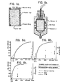
- a coup e.g., such a one shown in Fig. la. It is made of acrylic plastic and has an inside diameter of about 14 mm and a height of about 27 mm.
- the plastic cup is shown in Fig. la and the lower part of the cup is provided with a nylon filter having a mesh size of 80 x 80 pm. This filter is fastened by a plastic guard ring,
- a film layer e,g., "Parafilm ® ", is preferably applied at the lower portion so that liquid is prevented from leaking out of the cup.
- a silk net with the mesh size 150 x 180 ⁇ m is adapted at the upper end and is fastened with a guard ring.
- the liquid in the gel cup can e.g., be about 1 mm over the net surface.
- the cup is left at room temperature for at least 2 hours for complete gel formation, preferably in a place free of vibrations.
- a holder B is applied over the upper end of the cup 1.
- the holder B is filled with liquid (buffer or water) and a rubber cork provided with a tube, which is connected with a rubber hose is inserted into the opening.
- the rubber hose is connected with a container for permeation solution which is allowed to fill the rubber hose without air bubbles.
- the container (not shown in the drawing) is placed at such a height that a suitable flow is
- the fibrinogen used for the preparation of the gels contains trace amounts of factor XIII, which is a transamidase. In the presence of this enzyme and calcium ions,covalent intermolecular cross linkages between chains in the molecule units of the fibrin gel are formed. This is especially the case when thrombin is present as thrombin activates factor XIII.
- The.silk net applied to the top of the gel cup and which is in intimate contact with the gel matrix is of great importance for the mechanical stability of the gels. Without this net or some other means for preventing collapse of the gel, the gel compound is destroyed in the flow tests, the gel collapsing in the central protion and a conical inward bend arising.
- the silk net can, of course, be replaced with other nets, e.g., of cotton, nylon, iron or copper, which also stabilize the gel structure at pressures up to 40 x103 dynes/cm 2 .
- the turbidity profile of the system was determined under identical conditions.
- the Reaction Mixture (see under Fibrinogen) was poured into a cuvette (5ml) of a recording spectrophotometer (Beckman Acta III) and the turbidity (optical density) recorded at 450 nm. After a lag-phase there was a rapid increase in turbidity (cf. Fig. 8) which was accompanied by gelation. A tangent was drawn to the steepest part of the sigmoidal curve. Its intersection with the time axis is defined as the gelation or clotting time (Ct).
- Reaction Mixtures were prepared in several identical tubes. One of them was used for turbidity measurement as described above.
- the other tubes contained each 1 ml of Reaction Mixture.
- the reaction in the latter tubes was quenched at different times by addition of hirudin (2 ATU/ml) and an equal volume of 8 M urea. Thereafter the fibrin (ogen) was precipitated by addition of an equal volume of chilled ethanol.
- the mixtures were kept on ice-bath for 2 hrs. and thereafter the precipitates were secured by centrifugation, dissolved in urea and used for SDS-gel electrophoresis.
- the supernatants were used for radioimmunoassay (RIA) of FPA, FPB and B ⁇ 15-42 was assayed using the recently developed method of Kudryk et al.
- RIA radioimmunoassay
- Viscosity was determined with a viscometer type Ubbelohde, having a flow time for water of about 290 sec at 25°C. It was calibrated against a standard (CNI. Cannon Instrument Company, Pa. USA). Density was determined with a 5 ml pyknometer.
- Pore size The equation for calculation of average pore size of membrances (18) and acrylamide polymer gels (4)'was applied: where r is the average pore radius (in cm), and ⁇ is the fractional void volume of the gel, i.e., the fractional volume of liquid in the gel. ⁇ is calculated on the basis of protein concentration assuming a partial specific volume for fibrinogen of 0.72 (19), ⁇ is in this case the fractional void volume for gels in which no water is bound to the gel matrix. However,.the degree of hydration of fibrinogen in solution has been reported as high as 6 g per g protein.(20). Assuming that this water is retained by the gel matrix we also calculated ⁇ for such hydrated gels.
- Diffusion coefficient The apparent diffusion coefficient of water in the gel was calculated from Ks according to Ticknor (21) and White (4).
- D is the diffusion coefficient (in cm 2 /sec.).
- R is the gas constant (in ergs/mole-degree), T it the absolute temperature (in K) and V is the molar volume of the permeant (in cm /mole).
- Ionic strengths were calculated on basis of the molarity of the electrolytes. Activity coefficients and degree of calcium binding to protein were not taken into account.
- the silk net at the upper end of the gels stabilizes the gel structures. Without support of the silk net, the gels will yield to flow at the pressure applied (about 7 x 10 3 dynes /cm 2 ). The yielding is only noted at the center of the gel, since the gel matrix adhers firmly to the walls of the plastic cup.
- the extent of incorporation of fibrinogen into the gel matrix was determined. This was done by determining the protein content in the void volumne of the column (about 4 ml). The amount of protein, as measured spectrophotometrically using the extinction coefficient of fibrinogen (22), ranged between land 3% of the. total protein used for gelation. When deemed necessary the non-clottable portion was taken into account in calculations of the fibrin content of gels.
- Ionic strength In another series of experiments the ionic strength of the gel forming system was, at constant protein concentration, varied between 0.21 and 0.31. At all pH's (6.5,7.4, 8.2) an increase in ionic strength from 0.2 to 0.3 resulted in a decrease in flow-rate by roughly one order of magnitude. This applied to both thrombin and Batroxobin gels.
- Table III shows Ct at different protein and enzyme con- cnetrations in one series of experiments.
- Batroxobin increasing protein concentrations did not markedly influence Ct.
- thrombin there is a small prolongation of Ct with increasing fibrinogen concentrations.
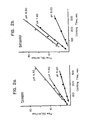
- Fig. 5 shows the result of one series of experiments. It is evident that there exists, at different enzyme concentrations, a linear relationshi l between flow-rate and inverse protein concentration.
- the curves fro thrombin and Batroxobin gels converge to a more or less common intercept near the origin with increasing protein con- centiation.
- the l'vaiues lor 10 dillerent 1/0 versus Ilow-rate curves (4-8 experimental points in each) were calculated.
- Mean r-values and SD were as follows: for thrombin, 0.9738 ⁇ 0.0308 and for Batroxobin, 0.9711 t 0.0356.
- the flow rate is inversely proportional to the fibrin concentration (fibrinogen concentration) in the gel.
- Ks will also be inversely proportional to the fibrinogen concentration.
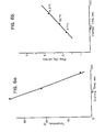
- the flow is dependent on the temperature, as according to equation 1, the flow is inversely proportional to the viscosity of the permeation solution.
- the temperature in gel formation is also of importance as a constant enzyme concentration, Ct is reduced at a higher temperature. This is apparent from Fig 6a. however, the flow rate in gels formed at different temperatures is directly proportional at Ct at the relative temperature, as is evident from Fig. 6b.
- Spherical latex particels of diameters between 0.085 ⁇ 0.0055 (SD) ⁇ m (SD standard deviation) and 0.198 ⁇ 0.0036 (SD) ⁇ m from Dow Chemicals, USA, were used in the tests. A number of gels formed at pH 7.4 and at two different ionic strengths, were used.. In the tests, Ct:varied from 23 to 314 seconds. The theoretical radius was calculated for each gel according to equation 3 assuming that a cylindrical vertical capillary system was present.
- Fig. 7 shows two series of tests.
- the ionic strength was 0.23 during the gel formation and in series II 0.21.
- the gel columns were equilibriated with water. After this suspensions of particles were .applied to the gels.
- the particle size was 0.085 ⁇ m and in series II 0.198 ⁇ m.
- the particles were slurried in water to a concentration of 0.1% (weight/volume). The turbidity at(450nm) of the effluent was determined. It was then possible to establish by means of the turbidity values if the latex particles had passed the filter.
- turbidity has been expressed in % of maximum turbidity of the effluent.
- the turbidity of the effluent increases above a certain theoretical pore size of the gel.
- the difference in theoretical pore size between no and complete permeation is a measure of the sum of pores and particle variation.
- the pore size at 50% permeation is an expression of the average size of pores and particles. If the total variation in pore size is within the range of the average particle size 3 SD it can be assumed that the pore size in the gel is uniform.
- Fig. 7 the variation in particle size (average size ⁇ 3 SD) has been shown with a horizontal line 50% permeation. It is apparent that the total variation can, to a large extent, be explained by the particle variation. It can be concluded from this that the pores in the gels.are rather uniform. It is also apparent from Fig. 7 that the theoretical average pore size is about of an order (one ten power) greater than the real effective particle size. Thus, calculation of the pore size according to equation 3 only gives relative values for the pore size.
- Fibrin gels were prepared in the way described in Example I and the conditions of gel formation is shown in Example 3.
- a small amount (0.2 ml) of human blood was applied to a gel column
- Continued filtration was carried out at room temperature (22 - 25°C) under the conditions shown in Example 3.
- the blood corpuscles did not pass through the fibrin gel. This was ex- peceted as the diameter of the red blood corpuscles (7-8 pm) is much larger than the effective pore diameter of the fibrin gel.
- Plasma rich in platelets was prepared from blood by centrifugation for 4 minutes at 120g, the blood being drawn in citrate solution to prevent coagulation. It was cetrifugated at 2000 g for 5 minutes to remove the remaining red blood corpuscles and EDTA at a concentration of 10 mM was added to the PRP, 0.5 ml of the PRP was applied to a fibrin gel column prepared in the way described in Example I. The conditions of gel formation- is shown in Example 3 and filtration was continued under the conditions shown in Example 3. To prevent aggregation of the platelets and their adhesion to the gel matrix, EDTA (10 mM) was added not only to PRP but also to the solution which was filtered. No platelets could be demonstrated in the eluate from the fibrin gel column. This was expected as the diameter of the platelet lies between 2 and 4 ⁇ m, which is considerably more than the effective pore size of the gel,
- Liver cells of a rat were homogenized in a homogenizator according to Potter-Elvehjem. Separation of cell fragments was achieved by differential centrifugation in known manner.
- the mitochondria were slurried in a buffer solution containing Na-EDTA (10 m M) and succrose (0.25M). 0.3 ml of the resulting suspension was applied to a fibrin gel column prepared in the way described in Example 1. The conditions of gel formation is shown in Table I and filtration was continued under the conditions indicated in Table 1. No mitochondria could be demonstrated in the eluate, which was as expected, since their diameter is about 0.5 ⁇ m; thus considerably bigger than the effective pore size of the gel.
- Sendai virus is a virus specific to mice which is used for preparation of interferon in human lymphocyte cultures.
- a partially purified virus preparation (640 hemagglutination units/ml) was used in the tests.
- 0.2 ml of the virus suspension was :applied to each of three fibrin gel columns prepared in the way described in Example 1.
- the conditions of the gel formation appear from Table 1 and filtration was continued under the conditions shown in Table 1. No hemagglutination activity could be demonstrated in the eluate from column 1; .50% of hemagglutination activity were demonstratec in the eluate from column II and 95% of the hemagglutination activity of the virus particles was demonstrated in the eluate from column III (see Table 1).
- the particle diameter of Sendai virus is stated to be about 0.15 pm.
- the tests show that when the effective pore radius is more than 0.15 ⁇ m the virus particles pass through the gel. When the effective pore radius of the gel is less than 0.15 ⁇ m a retention of the particles will, on the other hand, occur.
- E. coli is an elongatedintestinal bacterium of the approximate dimensions 0.8 x 1-2 um.
- the suspension contained between 10 7 and 10 8 bacteria/ml.
- 45 ml of the suspension were supplied to a fibrin gel column of the dimensions 5 cm 2 x 11 cm prepared in the way described in Example 1.
- the conditions of the gel formation and the filtration are shown in Table 1.
- the flow rate was 31 ml/h. No bacteria passed through the gel, determined by turbidity measurements of the eluate from the column. The flow rate at constant pressure was less at the end of the test than at its beginning.
- the test shows that E. coli cannot pass through gels having a pore diameter which is considerably less than the smallest diameter of the bacteria.
- nets of silk, plastic or metal adapted to the upper and lower portions of the gel have served as stabilizing structure of the fibrin gels.
- a corresponding stability can also be obtained in such a way that the fibrin gel is cast into a porous plastic, e.g. polyurethane, polyester or some similar porous plastic material, preferably one which is wettable by water.
- a foam plastic of polyurethane ("Regilen 40 AG") of a pore size 0.4 mm has been used.
- the gels were casb in a special apparatus. This consisted of a cylindrical plastic chamber in which the porous plastic had been introduced; the plastic was accomodated in a ring of acrylic plastic (height 2 cm and diameter 9 cm).
- the apparatus (chamber) had an opening at the upper and lower end, respectively. One opening was connected to a vaccum pump and the other opening was kept closed. The chamger was evacuated by means of the vacuum pump. After this the valve connecting the chamber with the vacuum pump was shut off.
- a fibrinogen-trombin solution was subsequently-allowed to fill the chamber rapidly through the valve in the opposite opening
- the valve was'thereafter closed and the chamber was left for 2 hours, so that the fibrinogen solution in the porous plastic material should be completely converted to a fibrin gel.
- the clotting parameters of the thrombin-fibrinogen mixture was shown in Table 2.
- a gel was also prepared in the way described in Example 1. In Table 2 the Ks -value of this latter gel is also shown. After complete gel formation the chamber was opened and the plastic cake with fibrin gel( including its plasti frame) was taken out. It was transferred to a special filter chamber.
- the framed ring in which the plastic material and the gel were accomodated,fitted tightly to the edges of the filter chamber through two 0-rings.
- the upper lid of the chamber was provided with an inlet for the liquid to be filterd and a ventilating valve to let our the air above the gel surface.
- In the lower portion of the chamber there was an outlet for collecting the filtered liquid.
- a buffer solution with the composition shown in Table 2 was filtered through the gel cake.
- the Ks value was calculated according to equation 1 (Table 2).
- Table 2 The Ks value was calculated according to equation 1 (Table 2).
- the Ks-value of the gel cast in plastic is of the same order as the gel prepared according to Example 1.
- the partial specific volume of the plastic material in the gel cake is 0.03 which means that the plastic matrix reduces the surface available for flow only to a small extent.
- Cellulose materials can also be used as reinforcing agent (supporting substance).
- a porous cellulose compound (“Wehex cloth”) is used as reinforcing agent of the fibrin gel. It had a thickness of 0.2 - 0.3 cm. Circular pieces of a radius of about 3 cm were wetted with a thrombin-fibrinogen solution. The cellulose pieces then swelled to about double thickness. The partial specific volume of the swollen cellulose compound was 0.04. Immediately after swelling which lasted for about 2 - 4 seconds the pieces were placed on the filter disc of a Büchner funnel. Measures were taken so that the pieces fitted tightly to the edges of the funnel.
- the openings of the funnel were covered with "Parafilm” and the funnel was left at room temperature for 2 hours in order to obtain a complete fibrin fomation in the pores of the cellulose.
- Buffer solutions the composition of which is shown in Table 1, were filtered through the gels.
- the Ks-value of the gels which are cast in cellulose is of the same order of magnitude as control gels prepared without reinforcing substance.
- fibrin gels in thin layers with reinforcement only on the lower surface can be used for filtration.
- About 10 ml of fibrinogen solution in tris-imidazole buffer withpH 7.4 were mixed with a thrombin solution.
- the mixture was thereafter poured into a Petri cup the bottom of which was covered by a damp silk cloth.
- the cup was covered witt a lid and was left for 2 hours for a complete gel formation.
- the thickness of the gel layer was 2 mm.
- the clotting parameters of the gel is shown in Table 2.
- the filter was thereafter attached to a "Millipore" filter support provided with a funnel. The funnel was filled with buffer solution and the flowrate was determined.
- the Ks-value is of the same order or magnitude for a corresponding fibrin gel prepared according to Example 1.
- the filter showed in course of time gradually diminishing Ks-values, which presumably is due to compression of the gel matrix during the flow.
- gels prepared according to Examples 1,8 and 10 can be stabilized by treatment with dialdehyde.
- a gel prepared according to Example 1 was first equilibrated with water and then brought into equilibrium with 0.01.4 M phosphate buffer solution with pH 7.2 in 0.15 M NaCl (phosphate buffered saline solution PBA). 2 - 4 column volumes of a 1% glutaraldehyde solution were then allowed to filter through the gel in the course of 10 minutes - 2 hours.After this the gel was washed with several column volumes of PBS and then with water. The column was finally equilibrated with tris-imidazole buffer . and flow measurements were carried out. The Ks-value is almost unchanged after treatment with glutar dialdehyde.
- the gel was taken out and treated for 72 hours with 8 M urea containing 1% of sodium'dodecyl sulphate (SDS).
- SDS sodium'dodecyl sulphate
- the gel was then reduced with 1% dithiotreitol in a way known per se, Polyacrylamide gel electrophoresis in the presence of SDS showed in comparison with non-stabilized gels the absence of free fibrin chains (fibrinogen chains), which can be interpreted as a proof that glutar dialdehyde had cross-linked the. chain units of the fibrin structure.
- a gel prepared in porous plastic according to Example was first washed with a tris-imidazole buffer solution free of calcium and was then brough into equilibrium with a 0.014 M phosphate buffer solution with pH 7.2 in 0.15 M NaCl (PBS). Two column volumes of a 1% glutar dialdehyde solution were then passed through the gel cake (column) in the course of 10 minutes. The gel cake was then washed with several column volumes of.PBS and then with water. Finally the column was brought into equilibrium with tris-imidazole buffer and flow measurements were carried out. These are shown in Table 2.
- the K s -value is only slightly changed after the treatment with glutar dialdehyde and is of the same order of magnitude as a gel prepared according to Example 1.
- the gel stabilized with glutar dialdehyde was then autoclaved at 120°C for 20 minutes at a pressure of 1.4 atm. After autoclaving the flow of buffer solution was again tested through the gel cake.
- autoclaving has influenced the flow properties of the gel only to a small extent. Cracks in the gel. would have caused drastic increase, of the flow through the gel.
- a fibrin gel prepared according to Example 10 was transferred to a cup with 500 ml water to remove buffer salts by diffusion. After ' 2 hours the gel was transferred to a cup with a new portion of water. After 2 hours the gel was transferred to a cup with 500 ml phosphate buffer solution with pH 7.2 in 0.15 M NaCl (PBS) and was left over night. The gel was then transferred to a Petri cup containing 50 ml of 1% glutar dialdehyde. After 2 hours the glutardialdehyde solution was exchanged for a new portion of the same liquid. After additional 2 hours the gel was transferred to a cup with water and washed in the way described above.
- PBS 0.15 M NaCl
- the filters can be stabilized with a dialdehyde such as glutar dialdehyde.
Abstract
Description
- This invention relates to a filter with a definite pore size comprising fibrin and a process for the preparation thereof from fibrinogen. This invention also relates to a size selective process employing the. filters of the invention.
- Formation of fibrin gels by contacting fibrinogen with a coagulation enzyme has long been known. It has been observed that when a liquid is passed over the gel, permeation of the gel increasingly becomes difficult - sometimes to the point where permeation and passage of the liquid are rapidly diminished or cease. It was believed that the gel infrastructure was extremely fragile and that the gel consisted of networks of channels and pores of varying size which were highly changeable and highly dependent upon and variable with liquid or liquid mixtures passed thereover, especially one having solid particles.
- It was therefore thought that such fibrin gel was not useful in separating components where the separation was effected solely on the basis of particle size.
- Specifically, when investigating the fibrin formation from fibrinogen the interest was directed to the flow properties of fibrin gels. It has e.g. been shown before that the flow properties through silica gel as well as through agar and gelatin gels are such as for a viscous flow. It has also been shown earlier that the flow through a fibrin gel is dependent upon the ionic strength and fibrinogen concentratia in the preparation. In the investigations made,the permeability coefficient (K ) of the fibrin gels was determined by Poiseulle's law as follows ;


- By varying the pH between 6 and 10 the ionic strength between 0.05 and 0.5, the calcium ion concentration between 0 and 20 mM and/or the concentration of enzyme (e.g. thrombin, "Batroxobin" or "Arvin") between 0.01 and 10 NIH-units (or the corresponding units of other enzymes) per ml solution and the fibrinogen concentration from 0.1 and up to 40 g/l, preferably between 1 and 10 g/l, gels with Ks-values [calculated according to the equation (1)] between 10-7 and 10-12, preferably between 10-8 and 10 -11, can be prepared. Calculated according to the equation (3), the corresponding average radii will be 0.03 - 9 µm, preferably 0.09 - 2.8 µm. If FXIII (a transamida- tion enzyme) and calcium ions are present in the gel formation the stability of the gels will be increased as covalent cross-linkings will arise between the chains in the subunits of the gel matrix.
- Thus, now it has been found according to the invention that these fibrin gels can be used as a filter. The filter according to the invention is characterized in that it is built of fibrin and the fibrin gel is in association with a shape-retaining means which retains the shape of at least one surface of said gel against deformation when contacted by a flowing liquid.
- The filter of the invention has substantially uniform pores. By that is meant that the standard deviation of pore size is less than 15 percent, preferably less than 10 percent and in some instances less than 5 percent.
- The pore size of the gel has, moreover, been found to be a function of the clotting parameters used in the gels' preparation, i.e., the pore size is varied by changing said parameters. The pore size is then proportional to the clotting time.
- It has now been discovered,in accordance with the invention, that fibrin in gel form can be used as a filter if means are provided to retain the shape of at least one surface of the gel against deformation when the gel is contacted by a flowing-medium such as a flowing liquid medium containing components to be separated. It has also been discovered, quite surprisingly, that the gel has substantially uniform pore sizes and that these pore sizes can be regulated simply by altering the process parameters employed for the formation of the gel.
- Specifically, it has been discovered if the gel is in some way stabilized by a shape-retaining means, that the gel structure is preserved and the uniform pores therein function ideally as a filter medium.
- Generally speaking, the gel is brought in contact with a shape-retaining means. The shape-retaining means can be a foraminous member such as a foraminous sheet member and is preferably disposed on or in association with an upper surface of.the gel, preferably in contact with the gel either directly or through an adhesive or a graft. Since the foraminous member serves to preserve the shape and structure of the upper surface of the gel when the medium to be filtered contacts the same, the gel does not collapse, thereby allowing the uniform pores thereof to function ideally as a filter medium.
- Foraminous members functioning as shape-retaining means can have virtually any size and shape, although they are preferably in the form of'a sheet and preferably are substantially co-extensive with the upper surface of the gel. The foraminous sheet members can be in the-form of a fibrous network such as in the form of a woven or non-woven or knitted fabric, the fibers of which can be natural or synthetic.
- When the fibers of a foraminous sheet member are natural, they can be, for example, made of silk, wool, cotton, cellulose, hemp, jute or the like.
- As synthetic fibers, there are contemplated in particular fibers made of nylon, polyester, polyolefin, fibers 0085166 made of vinyl polymers, acrylics such as polyacrylonitrile, rayon, to name a few.
- The fibers generally have a thickness between 1 µm and 1000 pm, preferably between 10 and 20 µm, and are disposed in relationship to one another to define openings therebetween of between 0.01 and 5 mm, preferably between 0.05 and 1 mm, it being understood that. the size of the openings between the fibers of the foraminous sheet is not especially critical, provided it allows passage therethrough of the medium to be filterel It is preferred that as much fiber be in contact with or adhere to the gel as possible so as to insure maximum structural integrity of the surface of the gel initially to come in contact with the medium to be filtered.
- Instead of using a fibrous foraminous member, one can use one made of wires, such as wires made of copper, tin, zinc, aluminum, glass, boron, titanium, steel, stainless steel, etc. The wires function analogously to the function performed by the fibers in providing structural integrity to at least one surface of the gel, preferably the upper surface or surface which is to be initially brought in contact with a mixture to be fltered. The interstices between the wires are of the same magnitude as the interstices between the fibers of a woven, non-woven or knitted fabric serving as a foraminous sheet member. The wires can be in the form of a screen, wire mesh or an expanded wire sheet and are preferably co-extensive with at least one side of the gel, preferably the upper surface.
- The gel has unitorm pores but owing to the manner by which the gel can be formed, can have uniform pores over a wide range. Preferably, the substantially uniform pores of the fibrin gel have a theoretical pore size or diameter in the range of about 0.03 to 9 µm, more preferably 0.09 to 2.8 µm.
- The gel is formed by contacting fibrinogen with an enzyme, especially a coagulation enzyme. Particularly contemplated enzymes for use in forming a fibrin gel include thrombin, Batroxobin, Arvin, Eccarin, Staphylocoagulase, Papain, Trypsin, caterpillar venom enzyme, etc.
- Generally speaking, the gel formation is effected at room temperature, although temperatures from -3°C up to 58°C can be employed. Preferably, the temperature is in'the range of 0 to 40°C.
- It is preferred that the gel be formed by contacting the fibrinogen with an enzyme in the presence of calcium ions. The calcium ion concentration can be up to 20 mM. The presence of calcium ions is not required in all instances. Where thrombin is employed as the coagulation enzyme, the gel can be formed in the absence of a calcium ion.
- In forming the gel, there is generally employed 0.1 to 10 enzyme units per gram fibrinogen, preferably 10 to 10-3 enzyme units per unit weight fibrinogen. Following formation of the gel whose coagulation time is a function of the relative amount of enzyme to fibrinogen as well as the concentration of calcium ion, the gel is preferably hardened or set by crosslinking the components thereof by contacting the gel with a crosslinking agent. Crosslinking agents contemplated include bis-imidates such as suberimidate, azides like tartryl di(ε-amino carproylazide), aryl dihalides like 4,4-difluoro-3, 3'-dinitrophenyl sulfone, glutardialdehyde, nitrenes, N,N'(4- azido-2-nitrophenyl)-cystamine dioxide, cupric di(1,10-phenanthroline), dithio bis-(succinimidyl propionate); N,N'-phenylene dimaleimide as well as polyethyleneimides and other bifunctional compounds, especially those known to crosslink with epsilon lysine, alpha amino groups, carboxy groups of aspartic and glutamic acids, and hydroxyl groups of amino acids in the protein chain (e.g. threonine and serine).
- Bis-imidates which can be used include those of the formula

- Azides which can be used include substituted and unsubstituted azides of the formula

- Aryl dihalides which can be used include those having mono, poly and fused rings as well as rings joined by a direct bond or through a methylene bridge or a sulfo bridge. The halogen of the halide can be fluorine, chlorine, or bromine The compounds can be substituted by inert or functional groups such as nitro, or disulfide. , Contemplated compounds include those where a functional group has replaced one of the halo substituents, e.g. nitro. Compounds contemplated include


- Generally speaking, the crosslinking agent is employed in an amount of between 0.001% and 8 % by weight, preferably between 1 and 2 % by weight of the gel for 1-120 minutes. Crosslinking is effected at temperatures of between 10 and 40°C, preferably 20 to 25°C. After the hardened or crosslinked structure is obtained, the gel is usually washed free of extraneous material..
- The gel in such hardened form is useful as a filter, i.e., without any foraminous sheet material. Preferably, however, the gel is formed on or in association with a shape-retaining means such as a net, wire mesh or other sheet material and while in contact with such shape-retaining means is hardened by the use of a hardening or crosslinking agent.
- Preferably, the gel is supported on its upper and lower surfaces by a shape-retaining means such as a foraminous sheet or the like, whereby to insure that the gel retains its shape during use as a filter.
- This invention further contemplates a process for separating a first substance having a theoretical size of 0.03 to 9 µm from a second substance having a larger size which comprises passing a mixture of said first and second substances over a filter comprising fibrin in gel form and having pores of substantially uniform size, said filter having means for retaining the shape of at least one surface of said gel against deformation when contacted by a flowing medium, wherein the effective pore size of said fibrin gel is larger than the particle size of said first substance and sma'ller than the particle size of said second substance. Preferably, the pores of the gel have a theoretical size of 0.09 to 2.8 mm.
- The filters of the invention are important, as they permit the separation of bacteria and viruses from mixtures containing the same. The ability to regulate the pore size and to achieve a gel of uniform pore size is an important and critical characteristic of the filters of the invention. These filters permit the separation of blood components, the separation-of components of blood plasma, the removal of platelets from blood, the fractionation of cells and cell fragments and the separation of high molecular weight protein aggregates. In addition, a variety of particles such as latex, silica, carbon and metallic particles may be separated over these filters. Components which can be separated include those shown in the table below:

- Fibrin gel filters have above all the advantages over other gel filters that the pore size can be simply varied as desired. Moreover, the present filters have high flow rates at such pore sizes as can be used to remove very small particles, such as virus particles. In this respect, the filters of the invention are more suitable than known membrane filters and filters of polyacrylamide gels. The absence of absorption of protein on the filters is also an advantage as compared with certain other filters.
- The process according to the invention for the preparation of gel filters is as described above,characterized in that a fibrinogen solution with preadjusting clotting parameters is mixed with a coagulation enzyme and the resulting mixture is made to clot in a form intended for the filter. It may be convenient to strengthen the fibrin gel formed during or after clotting by a shape-retaining means of greater strength than the gel which is preferably applied to the upper surface of the gel to be prepared and preferably to both the upper and lower surfaces thereof.
- The shape-retaining means (reinforcing meshes) are preferably in- the form of a net which is applied at the lower and preferably also at the upper surface in the mold in which the fibrinogen mixture is poured (cast). This mixture can preferably penetrate the net (foraminous sheet material) e.g. have its
surface 10 um-5mm, preferably 0.5-2 mm, from the net. The net can have a mesh width, for example, 10 pm to 5 mm, preferably 50 µm-1mm and the wire diameter can be, for example,0.01-1.0 mm, preferably 0.1-0.5 mm, where wires are employed as a shape-retaining means. In addition to the metallic wires noted above, wires of natural fibers and plastics can also be employed. The filter can also be reinforced in other places than at the surfaces. It can, for example, be built on a foam material, I such as a plastic foam, which can support part of the entire fil- ter. - The filters of the invention, especially in a non- hardened or crosslinked form, should not be subjected to temperatures in excess of 100°C, as such heat sterilization tends to destroy the gel structure. It is, therefore, necessary in utilizing the filters for biological processes to prepare them sterilely from the beginning. On the other hand, it has been found possible to harden or crosslink the filter during the preparation by carrying out the gel formation in the presence of Factor FXIII and calcium ions. Where Factor FXIII.is to be present., it is preferably present in an amount of at least 5 units per gram fibrinogen, preferably at least 50 units per gram. Calcium is present in a concentration of at least 20 mM.
- A still stronger filter is obtained by effecting crosslinking with one of the above-mentioned crosslinking agents especially a dialdehyde and particularly one of the formula OCH-R-CHO, wherein R is an alkylene group of 1 to 8 carbon atoms, such as glutardialdehyde. The filter obtained in this way can be heat treated in an autoclave and consequently sterilized.
- The clotting parameters are above all the enzyme concentration, e.g. between 100 and 3000 NIH units/L for thrombin and for the fibrinogen concentration between 0.1 and 70 g/I, preferably between 1 and 10 g/l, increased concentration giving a tighter gel. A tighter gel has a smaller pore size. Increase ionic strength also provides a tighter gel as well as a higher pH. It is preferred to carry out the gel preparation using a gel mixture having a pH of between 5.5 and 11, preferably between 6 and 9, and an ionic strength between 0.05 and 0.5. Gels formed at calcium ion concentrations between 0 and 20 mM are tighter.
- Pore size is also affected by,the temperature at which the clotting (gelation) is effected. A lower temperature of gelation means an increased clotting time, which in turn means that the resultant gel has a larger pore size. As a result of its larger pore size, it provides a greater rate of'flow.
- The gel of the invention can be used other than as a filter. One can dispose catalytically active substances such as catalytically active enzymes or catalytically active metals within the pores and thus use the pores' structure as a catalyst. The filter, therefore, can act more or less as a catalyst support for the catalytically active agent disposed therein. When the catalytically active agent is disposed within the pores, the resultant structure can be employed as a size selective catalyst converting only those components whose size is such as to. freely pass through the pores of the catalyst support. Those materials retained in the surface of the gel are not catalytically converted.
- By such a filter, one can conveniently effect enzymatic conversions, especially when the enzyme is immobilized within the filter covalently, ionically or otherwise. Since the gel structure is formed by the use of an enzyme, the filter of the invention's chemical components is compatible with the enzyme being employed as an enzyme catalyst. Thus, one can use the filter of the invention for any of the following enzyme conversions when the same contains the appropriate enzyme to effect that enzymatic catalysis: for reactions involving various oxido-reductases, transferases, hydrolases, lyases, isomerases and ligasis (synthetases). Hydrolases which have capacity of degrading the protein strands in the gels cannot be used.
- The method by which the enzyme or other catalytic component is disposed within the filter, i.e., within the pores of the filter, depends upon the nature of the enzyme. Preferably it is disposed by the use of a known enzyme immobilizing agent followed by washing of the filter to remove extraneous materials.
- One can also dispose reactive cellulose components within the pores of the gels. Upon reaction, low molecular weight components may be released and subsequently'eluted from the gels. An example of such a type of reaction is production of interferon by leukocytes after their reaction with Sendai virus. As shown in this invention, both of these components can be disposed within the pores of the gels.
- The invention is described more in detail with reference to the enclosed drawing, in which:
- Fig. 1 a shows molding of a filter according to the invention;
- Fig. 1 b shows the filter arranged for filtering;
- Figs. 2 a and 2 b show graphs of the flow as a function of the coagulation time at different pH with thrombin and Batroxobin, respectively;
- Fig. 3 shows the turbidity of the fibrinogen solution (fibrinogen) as a function of the time after addition of thrombin
- Figs. 4 a and 4 b show the flow as a function of the coagulation time at different ionic strength of thrombin and Batroxobin, respectively;
- Figs. 5 a and 5 b are graphs showing the relationship between protein concentration in the gel forming system and flow-rate.
- Figs.6 a and 6 b show the temperature plotted against the coagulation time and the flow, respectively, as a function of the coagulation time at different temperatures;
- Fig. / shows the turbidity of the effluent (turbidity Ln the effluent) in % as a function of the pore diameter in µm;
- Figs. 8 a,8 b and 8 c are graphs plotting activation of fibrinogen and clotting time (Ct).
- In order to more fully illustrate the invention and the manner of practicing the same, the following examples are presented.
- Human fibrinogen , Fraction 1-4 (7) was obtained from IMCO, Stockholm, Sweden. The preparation, either a freeze-dried powder or a wet paste, was 97-100% clottable (as determined spectrophotometrically). A solution being 0.3 M in NaCl and 2% in protein was prepared. This solution (50ml) was dialyzed against 0.3 M NaCl at 4° for 3 hrs. with changes of outer fluid (5 litre) every hour. This dialyzed solution was further diluted with deareated Tris-imidazole buffer (8) of pH between 6..5 and 8.2 to protein concentrations between 1.2 and 5.0 g/l. In the final dilutions the concentration of each Tris and imidazole was 0.02 M. When necessary increase-in ionic strength was achieved by inclusion of sodium chloride in the buffer. In order to inhibit any trace of plasmin which may be generated, Trasylol (Bayer AG, Germany) was added to a concentration of 5 KIE/ml to all buffers and dialysis fluids.
- In the gelation experiments the following procedures was employed: To 3.65 ml of fibrinogen solution in a plastic tube was added 70 µl of 1 M CaCl2 solution, immediately followed by 50 µl of thrombin or Batroxobin solutions of varying concentrations. This mixture is called Reaction Mixture. The tubes are rapidly inverted twice and transferred to the gel cup or to the spectrophotometer cell within 10 seconds after addition of enzyme. The further handling is described under separate paragraphs.
- Thrombin. In most experiments a bovine preparation prepared as previously described (9) was used. Specific activity: 100-200 NIH units per mg. Control experiments with highly purified (specific activity: about 2000 NIH units per mg human thrombin (10) was performed in some instances.
- Batroxobin (from Bothrops marajoensis) was obtained from Pentapharm AG, Basel, Switzerland. Specific activity: 505 BU per mg.
- Hirudin was also obtained from Pentapharm AG. Specific activity: 1000 ATU per mg.
- Reagents.. All reagents used were of analytical grade.
- A solution of thrombin is added to a solution of fibrinogen in a tris-imidazole buffer containing calcium salts with a pH of between 6.5 and 8.2 and an ionic strength between 0.1 and 0.3 so that the final concentration is between 0.05 and 2.5 NIH-units per ml. In other tests Batroxobin is used to obtain gel formation in a concentration between 0.27 and 3.6 BU per ml. The concentration of "Tris" and imidazole salts is each 0.02 M and the concentration of.calcium salt is also 0.02 M. The variation in ionic strength is obtained by addition of NaCl.
- Gels are also prepared at calcium.ion concentration between 0 and 20 mM. With reduced calcium ion concentration the opacity of the gels is increased. When thrombin is used to achieve the gel formation.the clotting time (Ct), also in the absence of calcium ions is directly proportional to the flow rate and thus also to Ks. When "Batroxobin" is used for the gel formation, the stability of the gels in the absence of calcium ions is unsatisfactory, which makes flow measurements more difficult.
- After addition of an enzyme such as thrombin or Batroxobin, the solution is rapidly mixed and then poured into a coup, e.g., such a one shown in Fig. la. It is made of acrylic plastic and has an inside diameter of about 14 mm and a height of about 27 mm. The plastic cup is shown in Fig. la and the lower part of the cup is provided with a nylon filter having a mesh size of 80 x 80 pm. This filter is fastened by a plastic guard ring, A film layer, e,g., "Parafilm ®", is preferably applied at the lower portion so that liquid is prevented from leaking out of the cup. Immediately after introducing the solution into the cup, a silk net with the mesh size 150 x 180 µm is adapted at the upper end and is fastened with a guard ring. The liquid in the gel cup can e.g., be about 1 mm over the net surface. The cup is left at room temperature for at least 2 hours for complete gel formation, preferably in a place free of vibrations.
- After this time, the film is removed at the lower portion of the cup with its contents of gel is placed in the holder A according to Fig. lb, a holder B is applied over the upper end of the
cup 1. At the upper end of the holder B there is an opening as well as at the lower end of the holder A.. The holder B is filled with liquid (buffer or water) and a rubber cork provided with a tube, which is connected with a rubber hose is inserted into the opening. The rubber hose is connected with a container for permeation solution which is allowed to fill the rubber hose without air bubbles. The container (not shown in the drawing) is placed at such a height that a suitable flow is - X) (other materials can also be used such as nylon and polystyren obtained through the gel. The hydrostatic pressure is varied at different tests between 4 and 40 x 103 dynes/cm2.
- The fibrinogen used for the preparation of the gels contains trace amounts of factor XIII, which is a transamidase. In the presence of this enzyme and calcium ions,covalent intermolecular cross linkages between chains in the molecule units of the fibrin gel are formed. This is especially the case when thrombin is present as thrombin activates factor XIII.
- An electrophoretic analysis of reduced fibrin from various gels in the presence of sodium-dodecyl sulphate shows that a complete cross-linking of the α and γ -chains of the fibrin takes place in the presence of thrombin. A partial cross-linking takes palce in the presence of Batroxobin. The covalent .cross-linkings formed in the presence of factor XIII contributes to the stabilization of the gel structure,
- The.silk net applied to the top of the gel cup and which is in intimate contact with the gel matrix is of great importance for the mechanical stability of the gels. Without this net or some other means for preventing collapse of the gel, the gel compound is destroyed in the flow tests, the gel collapsing in the central protion and a conical inward bend arising.
- The silk net can, of course, be replaced with other nets, e.g., of cotton, nylon, iron or copper, which also stabilize the gel structure at pressures up to 40 x103 dynes/cm2.
- In parellel to the flow studies, the turbidity profile of the system was determined under identical conditions. In these experiments the Reaction Mixture (see under Fibrinogen) was poured into a cuvette (5ml) of a recording spectrophotometer (Beckman Acta III) and the turbidity (optical density) recorded at 450 nm. After a lag-phase there was a rapid increase in turbidity (cf. Fig. 8) which was accompanied by gelation. A tangent was drawn to the steepest part of the sigmoidal curve. Its intersection with the time axis is defined as the gelation or clotting time (Ct). (Ct is about the same as the time for visually observed turbidity increase in the gel cup.) In addition to Ct also maximum turbidity (OD-max) and rate of turbidity increase ( Δ OD/min) was recorded. The time required for gelation to reach completion was judged from the turbidity curve. This time reanged from 1 hr. to 2 hrs. for the high and the low enzyme concentrations, respectively.
- Reaction Mixtures (see under Fibrinogen) were prepared in several identical tubes. One of them was used for turbidity measurement as described above. The other tubes contained each 1 ml of Reaction Mixture. The reaction in the latter tubes was quenched at different times by addition of hirudin (2 ATU/ml) and an equal volume of 8 M urea. Thereafter the fibrin (ogen) was precipitated by addition of an equal volume of chilled ethanol. The mixtures were kept on ice-bath for 2 hrs. and thereafter the precipitates were secured by centrifugation, dissolved in urea and used for SDS-gel electrophoresis. The supernatants were used for radioimmunoassay (RIA) of FPA, FPB and B β 15-42 was assayed using the recently developed method of Kudryk et al.
- Viscosity was determined with a viscometer type Ubbelohde, having a flow time for water of about 290 sec at 25°C. It was calibrated against a standard (CNI. Cannon Instrument Company, Pa. USA). Density was determined with a 5 ml pyknometer.
- Pore size. The equation for calculation of average pore size of membrances (18) and acrylamide polymer gels (4)'was applied:

- Diffusion coefficient. The apparent diffusion coefficient of water in the gel was calculated from Ks according to Ticknor (21) and White (4).

- Ionic strengths were calculated on basis of the molarity of the electrolytes. Activity coefficients and degree of calcium binding to protein were not taken into account.
- Least square analysis was used for calculation of correlation coefficients, slopes and intercepts. All lines shown in figures were drawn accordingly.
- The flow studies were performed on gels which had been formed at ambient temperature. The average temperature was 24 - 2°. Hoever, in each series of experiments the variation in temperature never exceeded 2°. Preliminary experiments suggested that this variation in temperature has a negligable effect on Ct of the system. In the permeation experiments, when not otherwise stated, the flow-rates were corrected to 25°.
- The silk net at the upper end of the gels stabilizes the gel structures. Without support of the silk net, the gels will yield to flow at the pressure applied (about 7 x 10³ dynes /cm2). The yielding is only noted at the center of the gel, since the gel matrix adhers firmly to the walls of the plastic cup.
- The nets in the column do not significantly reduce the flow- rate of liquid in columns without gels. We, therefore, assume that also when the nets are in contact with gels they do not restrict .the area available for flow.
- Before a flow experiment was started, the extent of incorporation of fibrinogen into the gel matrix was determined. This was done by determining the protein content in the void volumne of the column (about 4 ml). The amount of protein, as measured spectrophotometrically using the extinction coefficient of fibrinogen (22), ranged between
land 3% of the. total protein used for gelation. When deemed necessary the non-clottable portion was taken into account in calculations of the fibrin content of gels. - The effect of changeing permeant on the flow-rate of gels was studied in some experiments. A representative series of experiments is shown in Table I. The gel was first percolated with buffer of
ionic strength 0,21 (experiment I). On changing the permeant to water (experiment II) an increase in flow-rate occurred. which is larger than expected on the basis of the viscosity change of the permeant. On return to the original permeant (experiment III) the flow-rate decreased, but not completely to the original value (experiment I). When buffer of ionic strength 0.36 was percolated through the gel (experiment IV) a small decrease occurred which was almost as expected on the basis of the difference in viscosity between the two buffers. When the permeant was again changed to water (experiment V) the flow-rate increases to almost the same value as after the first change to water (experiment II). These results suggest that the final gel structure is not influenced by moderate changes in permeant composition, but changes may occur on drastic changes in ionic environment and these are not completely reversible.
- Viscous flow. In order to test if the flow obeyed Poiseuille's law, the flow-rate at different pressures (4.5 - 5.6 x 10³dyne/ cm2) for gels formed at pH 7.4, ionic strength 0.21 was determined, at three different thrombin concentrations (0.1-0.8 NIH units/ml). The Ks-range for these gels was 10-8 to 10-10 Permeation was in one experiment with the same buffer as above and in the other cases with water. In all cases the drop-rate decreased linearly with decreasing pressure. As shown in Table II for one of the gels, the flow-rate per unit pressure was almost independent of total pressure.
- In another series of experimetns the flow-rates were determined with permeants of different viscosities. The gels used in these experiments were formed at pH 7.4, ionic strength 0.21, at four fibrinogen concentrations. Thrombin as well as Batroxobin were used as inducers of gel formation. Permeation was performed at five different temperatures between 4.5° and 40°. In all cases there was a linear relationship between the inverse viscosity of the permeant and the flow-rate. These experiments suggest that the flow through the gels is viscous. In addition Reynold's number was calculated and found to be within the laminar region for all gels.
- Diffusive flow. It was pointed out by Ticknor, J. Phys. Chem 62, 1483-5 (1958) that the equation for viscous flow is identical in form to equations for diffusive flow, when the relationship between diffusion coefficient (D)and viscosity according to Johnson and Babb, Chem. Revs. 56, 387-453 (1956) is taken into consideration The relation between Ks and D is given in
Equation 3. In flow experiments using water as permeant we calculated the apparent diffusion coefficient for water at 22° - 23°. Even for the tighest gels (Ks 10-10), the calculated D-values were 6-orders of magnitude larger than the reported self-diffusion coefficient of water at 25° (2.8 x 10 -5 cm 2 /sec.). This supports the above conclusion that the flow through the fibrin gels is predominately viscous .
- There is a correlation between clotting time (Ct) of fibrinogen and enzyme concentration, We explored the relationship between Ct and permeability of the final gels. Therefore, at the same time as gels were prepared for permeability studies, the Ct of the gel forming system was determined in parallel experiments by turbidity measurements (see Methods).
- PH. At a constant fibrinogen concentration and ionic strength, the.flow-rates for both thrombin and Batroxobin gels were directly related to the Ct of the gel forming system over a wide range of Ct (17 sec - 500 sec). This applied to three different pH's (6.5, 7.4 and 8.2) as exemplified in Fig. 2. At all pH's there was a difference in slope between curves for thrombin as compared to those for Batroxobin.
- At each pH, the correlation coefficients (r) for six different Ct versus flow-rate curves (4 experimental points in each) were calculated. The mean r-values and their standard deviations (SD) were as follows: at pH 6.5, 0.9709 t 0.0184; at pH 7.4, 0.9721 ± 0.0394; at pH 8.2, 0.9599 ± 0.0434. There was no significant difference in r-values for thrombin and Batroxobin curves.
- Ionic strength. In another series of experiments the ionic strength of the gel forming system was, at constant protein concentration, varied between 0.21 and 0.31. At all pH's (6.5,7.4, 8.2) an increase in ionic strength from 0.2 to 0.3 resulted in a decrease in flow-rate by roughly one order of magnitude. This applied to both thrombin and Batroxobin gels.
- Ct were prolonged with increasing ionic strength at all enzyme concentrations and pH's. At each ionic strenth, however, there was for both thrombin and Batroxobin gels a linear relationship between Ct and flow-rate. The results at pH 7.4 is shown in Fig. 4. At all ionic strengths a difference in slope between curves for thrombin as compared to those for Batroxobin was noted.
- At two ionic strengths, regardless of pH, r-values for six different Ct versus flow-rate curves (4 experimental points in each) were calculated. Mean r-values and SD were as follows: at ionic strength 0.21, 0.9851 ± 0.0208 and at ionic strength 0.26, 0.9511 t 0.0470. There was no significant difference in r-values for thrombin and Batroxobin curves.
- In a series of experiments we showed that the relationship between Ct and flow-rate applied to a wide range of protein concentrations in the gel forming system. These experiments were only performed at pH 7.4 and ionic strength 0.21. When Ct at a given protein concentration were plotted against flow-rates a linear relationship, similar to that shown in Fig. 3 (pH 7.4), was demonstrated at all fibrinogen concentrations (1.5 - 5.0 g/l) The plots for both thrombin and Batroxobin converged towards an intercept near the origin with decreasing clossing times. Like in the experiments shown in Fig. 2, the slopes for Batroxobin curves were steeper than those for thrombin at all protein concentrations. The r-values for 8 different Ct versus flow-rate curves (8 points in each) were calculated. Mean r-values and SD were as follows: for thrombin, 0.9800 ± 0.0157 and for Batroxobin, 0.9830 ± 0.0187.
- Table III shows Ct at different protein and enzyme con- cnetrations in one series of experiments. In case of Batroxobin, increasing protein concentrations did not markedly influence Ct. However, in the case of thrombin there is a small prolongation of Ct with increasing fibrinogen concentrations.
- The relationship between protein concentration in the gel forming system and flow-rate was next studied. Fig. 5 shows the result of one series of experiments. It is evident that there exists, at different enzyme concentrations, a linear relationshil between flow-rate and inverse protein concentration. The curves fro thrombin and Batroxobin gels converge to a more or less common intercept near the origin with increasing protein con- centiation. The l'vaiues lor 10
dillerent 1/0 versus Ilow-rate curves (4-8 experimental points in each) were calculated. Mean r-values and SD were as follows: for thrombin, 0.9738 ± 0.0308 and for Batroxobin, 0.9711 t 0.0356. - It is apparent from Fig. 2 how the flow rate


equation 1, - It is apparent from Fig. 4 that the flow rate of gels formed at different ionic strength is always directly correlated tp the Ct of the enzyme-fibrinogen solution used in the gel preparation. In addition the.great influence on the flow rate at a change of the ionic strength is pointed out.
- As is apparent from Fig. 5, the flow rate

equation 1, Ks will also be inversely proportional to the fibrinogen concentration. - The flow is dependent on the temperature, as according to
equation 1, the flow is inversely proportional to the viscosity of the permeation solution. The temperature in gel formation is also of importance as a constant enzyme concentration, Ct is reduced at a higher temperature. This is apparent from Fig 6a. however, the flow rate in gels formed at different temperatures is directly proportional at Ct at the relative temperature, as is evident from Fig. 6b. - The columns prepared in the way schematically illustrated in Fig. 1 are of small dimensions (1.5 cm2 x 2.6 cm). Similar qualitative results are observed with gel columns of greater dimensions (5 cm2 x 12 cm). When nothing else is indicated, the smaller type of column is used in the tests.
- Spherical latex particels of diameters between 0.085 ± 0.0055 (SD) µm (SD = standard deviation) and 0.198 ± 0.0036 (SD) µm from Dow Chemicals, USA, were used in the tests. A number of gels formed at pH 7.4 and at two different ionic strengths, were used.. In the tests, Ct:varied from 23 to 314 seconds.The theoretical radius was calculated for each gel according to
equation 3 assuming that a cylindrical vertical capillary system was present. - Fig. 7 shows two series of tests. In series I the ionic strength was 0.23 during the gel formation and in series II 0.21. The gel columns were equilibriated with water. After this suspensions of particles were .applied to the gels. In series I the particle size was 0.085 µm and in series II 0.198 µm. The particles were slurried in water to a concentration of 0.1% (weight/volume).The turbidity at(450nm) of the effluent was determined. It was then possible to establish by means of the turbidity values if the latex particles had passed the filter.
- In Fig. 7 the turbidity has been expressed in % of maximum turbidity of the effluent. As is apparent from Fig. 7 the turbidity of the effluent increases above a certain theoretical pore size of the gel, At an additional increase in pore size more particles will pass through the gel (filter) and over a certain pore size a constant amount of particles permeate the gels, The difference in theoretical pore size between no and complete permeation is a measure of the sum of pores and particle variation. The pore size at 50% permeation is an expression of the average size of pores and particles. If the total variation in pore size is within the range of the
average particle size 3 SD it can be assumed that the pore size in the gel is uniform. - In Fig. 7 the variation in particle size (average size ± 3 SD) has been shown with a
horizontal line 50% permeation. It is apparent that the total variation can, to a large extent, be explained by the particle variation. It can be concluded from this that the pores in the gels.are rather uniform. It is also apparent from Fig. 7 that the theoretical average pore size is about of an order (one ten power) greater than the real effective particle size. Thus, calculation of the pore size according toequation 3 only gives relative values for the pore size. - Various protein solutions were (applied)to fibrin gels prepared in the way described in Example 1.. The gel formation was carried out at room temperature (21 - 25°C). In.most cases, the buffer used in gel formation had the same composition as the buffer used for permeation. The filtration tests took place at room'temperature (22 - 25°C). When nothing else is indicated, the volume of the gels was 1.47 cm2 x 2.48 cm = 3.65 ml. The tests were carried out on different days and with different fibrinogen preparations. Thus, it is not possible to make a comparison as to the pore size between different tests. Table 1 shows the proteins tested with respect to filtering' ability through fibrin gels of different porosities. It is apparent from the table, that proteins, including those having a very high molecular weight, are filtered even through gels having small pore sizes. A high molecular weight polysaccharide ("Blue Dextran") shows the same filtration properties as the proteins. The'table shows that the yield of proteins in the eluate is high, from which it appears that at least at room temperature the interaction between gel matrix and proteins is small. This also applied to such proteins as fibrinogen, fibronectin and the factor VIII complex.


- Fibrin gels were prepared in the way described in Example I and the conditions of gel formation is shown in Example 3. A small amount (0.2 ml) of human blood was applied to a gel column Continued filtration was carried out at room temperature (22 - 25°C) under the conditions shown in Example 3. The blood corpuscles did not pass through the fibrin gel. This was ex- peceted as the diameter of the red blood corpuscles (7-8 pm) is much larger than the effective pore diameter of the fibrin gel.
- Plasma rich in platelets (PRP) was prepared from blood by centrifugation for 4 minutes at 120g, the blood being drawn in citrate solution to prevent coagulation. It was cetrifugated at 2000 g for 5 minutes to remove the remaining red blood corpuscles and EDTA at a concentration of 10 mM was added to the PRP, 0.5 ml of the PRP was applied to a fibrin gel column prepared in the way described in Example I. The conditions of gel formation- is shown in Example 3 and filtration was continued under the conditions shown in Example 3. To prevent aggregation of the platelets and their adhesion to the gel matrix, EDTA (10 mM) was added not only to PRP but also to the solution which was filtered. No platelets could be demonstrated in the eluate from the fibrin gel column. This was expected as the diameter of the platelet lies between 2 and 4 µm, which is considerably more than the effective pore size of the gel,
- Liver cells of a rat were homogenized in a homogenizator according to Potter-Elvehjem. Separation of cell fragments was achieved by differential centrifugation in known manner. The mitochondria were slurried in a buffer solution containing Na-EDTA (10 mM) and succrose (0.25M). 0.3 ml of the resulting suspension was applied to a fibrin gel column prepared in the way described in Example 1. The conditions of gel formation is shown in Table I and filtration was continued under the conditions indicated in Table 1. No mitochondria could be demonstrated in the eluate, which was as expected, since their diameter is about 0.5 µm; thus considerably bigger than the effective pore size of the gel.
- Sendai virus is a virus specific to mice which is used for preparation of interferon in human lymphocyte cultures. A partially purified virus preparation (640 hemagglutination units/ml) was used in the tests.
- 0.2 ml of the virus suspension was :applied to each of three fibrin gel columns prepared in the way described in Example 1. The conditions of the gel formation appear from Table 1 and filtration was continued under the conditions shown in Table 1. No hemagglutination activity could be demonstrated in the eluate from
column 1; .50% of hemagglutination activity were demonstratec in the eluate from column II and 95% of the hemagglutination activity of the virus particles was demonstrated in the eluate from column III (see Table 1). - .After filtration the silk nets at the upper part of the three . columns were washed with a buffer solution (containing 1 % of borine serum albumin, BSA) and the hemagglutination activity of the washings was analyzed. In the washing liquid from
columns 1 100% of the hemagglutination activity was found; in the washing liquid from column II 25% of the activity was found and in the washing liquid from column III no activity was found. - The particle diameter of Sendai virus is stated to be about 0.15 pm. The tests show that when the effective pore radius is more than 0.15 µm the virus particles pass through the gel. When the effective pore radius of the gel is less than 0.15 µm a retention of the particles will, on the other hand, occur.
- E. coli is an elongatedintestinal bacterium of the approximate dimensions 0.8 x 1-2 um. A suspension of E. coli in tris-imidazole-buffer, free of calcium and with pH 7.4 and-ionic strength 0.21, was prepared (see Example 1). The suspension contained between 10 7 and 108 bacteria/ml. 45 ml of the suspension were supplied to a fibrin gel column of the
dimensions 5 cm2 x 11 cm prepared in the way described in Example 1. The conditions of the gel formation and the filtration are shown in Table 1. The flow rate was 31 ml/h. No bacteria passed through the gel, determined by turbidity measurements of the eluate from the column. The flow rate at constant pressure was less at the end of the test than at its beginning. Assuming an unchanged Ks the reduction of surface corresponding to the reduction in flow can be calculated according toequation 1. According to this calculation the surface had been reduced to 58%. Thus, one might expect that the bacteria were enriched on the upper gel surface. By washing the silk net attached to the upper part of the gel with buffer solution 99% of the bacteria applied to the gel were found in the washing liquid. - The test shows that E. coli cannot pass through gels having a pore diameter which is considerably less than the smallest diameter of the bacteria.
- In the foregoing examples nets of silk, plastic or metal adapted to the upper and lower portions of the gel have served as stabilizing structure of the fibrin gels. A corresponding stability can also be obtained in such a way that the fibrin gel is cast into a porous plastic, e.g. polyurethane, polyester or some similar porous plastic material, preferably one which is wettable by water.
- In this example a foam plastic of polyurethane ("Regilen 40 AG") of a pore size 0.4 mm has been used. The gels were casb in a special apparatus. This consisted of a cylindrical plastic chamber in which the porous plastic had been introduced; the plastic was accomodated in a ring of acrylic plastic (
height 2 cm and diameter 9 cm). The apparatus (chamber) had an opening at the upper and lower end, respectively. One opening was connected to a vaccum pump and the other opening was kept closed. The chamger was evacuated by means of the vacuum pump. After this the valve connecting the chamber with the vacuum pump was shut off. A fibrinogen-trombin solution was subsequently-allowed to fill the chamber rapidly through the valve in the opposite opening The valve was'thereafter closed and the chamber was left for 2 hours, so that the fibrinogen solution in the porous plastic material should be completely converted to a fibrin gel. The clotting parameters of the thrombin-fibrinogen mixture was shown in Table 2. For comparison a gel was also prepared in the way described in Example 1. In Table 2 the Ks -value of this latter gel is also shown. After complete gel formation the chamber was opened and the plastic cake with fibrin gel( including its plasti frame) was taken out. It was transferred to a special filter chamber. The framed ring, in which the plastic material and the gel were accomodated,fitted tightly to the edges of the filter chamber through two 0-rings. The upper lid of the chamber was provided with an inlet for the liquid to be filterd and a ventilating valve to let our the air above the gel surface. In the lower portion of the chamber there was an outlet for collecting the filtered liquid. A buffer solution with the composition shown in Table 2, was filtered through the gel cake. The Ks value was calculated according to equation 1 (Table 2). As is apparent from the table the Ks-value of the gel cast in plastic is of the same order as the gel prepared according to Example 1. The partial specific volume of the plastic material in the gel cake is 0.03 which means that the plastic matrix reduces the surface available for flow only to a small extent. - Cellulose materials can also be used as reinforcing agent (supporting substance). In this example, a porous cellulose compound ("Wehex cloth") is used as reinforcing agent of the fibrin gel. It had a thickness of 0.2 - 0.3 cm. Circular pieces of a radius of about 3 cm were wetted with a thrombin-fibrinogen solution. The cellulose pieces then swelled to about double thickness. The partial specific volume of the swollen cellulose compound was 0.04. Immediately after swelling which lasted for about 2 - 4 seconds the pieces were placed on the filter disc of a Büchner funnel. Measures were taken so that the pieces fitted tightly to the edges of the funnel. The openings of the funnel were covered with "Parafilm" and the funnel was left at room temperature for 2 hours in order to obtain a complete fibrin fomation in the pores of the cellulose. Buffer solutions, the composition of which is shown in Table 1, were filtered through the gels. The Ks-value of the gels which are cast in cellulose is of the same order of magnitude as control gels prepared without reinforcing substance.
- In this example it is shown that fibrin gels in thin layers with reinforcement only on the lower surface can be used for filtration. About 10 ml of fibrinogen solution in tris-imidazole buffer withpH 7.4 were mixed with a thrombin solution. The mixture was thereafter poured into a Petri cup the bottom of which was covered by a damp silk cloth. The cup was covered witt a lid and was left for 2 hours for a complete gel formation. The thickness of the gel layer was 2 mm. The clotting parameters of the gel is shown in Table 2. The filter was thereafter attached to a "Millipore" filter support provided with a funnel. The funnel was filled with buffer solution and the flowrate was determined. As is apparent from Table 2 the Ks-value is of the same order or magnitude for a corresponding fibrin gel prepared according to Example 1. However, the filter showed in course of time gradually diminishing Ks-values, which presumably is due to compression of the gel matrix during the flow.
- In this example it is shown that gels prepared according to Examples 1,8 and 10 can be stabilized by treatment with dialdehyde.
- A. A gel prepared according to Example 1 was first equilibrated with water and then brought into equilibrium with 0.01.4 M phosphate buffer solution with pH 7.2 in 0.15 M NaCl (phosphate buffered saline solution PBA). 2 - 4 column volumes of a 1% glutaraldehyde solution were then allowed to filter through the gel in the course of 10 minutes - 2 hours.After this the gel was washed with several column volumes of PBS and then with water. The column was finally equilibrated with tris-imidazole buffer . and flow measurements were carried out. The Ks-value is almost unchanged after treatment with glutar dialdehyde. After the flow measurements, the gel was taken out and treated for 72 hours with 8 M urea containing 1% of sodium'dodecyl sulphate (SDS). The gel was then reduced with 1% dithiotreitol in a way known per se, Polyacrylamide gel electrophoresis in the presence of SDS showed in comparison with non-stabilized gels the absence of free fibrin chains (fibrinogen chains), which can be interpreted as a proof that glutar dialdehyde had cross-linked the. chain units of the fibrin structure.
- B. A gel prepared in porous plastic according to Example was first washed with a tris-imidazole buffer solution free of calcium and was then brough into equilibrium with a 0.014 M phosphate buffer solution with pH 7.2 in 0.15 M NaCl (PBS). Two column volumes of a 1% glutar dialdehyde solution were then passed through the gel cake (column) in the course of 10 minutes. The gel cake was then washed with several column volumes of.PBS and then with water. Finally the column was brought into equilibrium with tris-imidazole buffer and flow measurements were carried out. These are shown in Table 2. As is apparent from the Table the Ks-value is only slightly changed after the treatment with glutar dialdehyde and is of the same order of magnitude as a gel prepared according to Example 1. The gel stabilized with glutar dialdehyde was then autoclaved at 120°C for 20 minutes at a pressure of 1.4 atm. After autoclaving the flow of buffer solution was again tested through the gel cake. As is apparent from Table 2, autoclaving has influenced the flow properties of the gel only to a small extent. Cracks in the gel. would have caused drastic increase, of the flow through the gel.
- C. A fibrin gel prepared according to Example 10 was transferred to a cup with 500 ml water to remove buffer salts by diffusion. After'2 hours the gel was transferred to a cup with a new portion of water. After 2 hours the gel was transferred to a cup with 500 ml phosphate buffer solution with pH 7.2 in 0.15 M NaCl (PBS) and was left over night. The gel was then transferred to a Petri cup containing 50 ml of 1% glutar dialdehyde. After 2 hours the glutardialdehyde solution was exchanged for a new portion of the same liquid. After additional 2 hours the gel was transferred to a cup with water and washed in the way described above. After washing with water the gel was transferred to a cup with tris-imidazole buffer solution. After 2 hours the washing liquid was exchanged for a new portion and after additional 12 hours the gel was transferred to a "Millipore"-filter carrier with a funnel. As a comparison measurements were carried out with a gel prepared in the same way except the treatment with glutar dialdehyde. Directly after casting, this gel was transferred to a "Millipore" filter container for flow measurements. As is apparent from Table 2 the flow through the vulcanized filter was comparable with that through the non-vulcanized filter at thestart of the flow measurement. However, at the end of the measuring period, the Ks-value of the nonstabilized filter had been reduced to a large extent which was not the case with the vulcanized filter. After the flow measurement the filters were sterilized through autoclaving of the filter (and the filter apparatus) at 120°C for 20 minutes at the pressure 1.4 atm. Flow measurements were carried out after the heat treatment and the:.Ks-values are shown in Table 2..
- No flow could be demonstrated through the nonstabilized filter. On the other hand the vulcanized filter showed Ks-values of the same order before as well as after autoclaving.
- 5 ml of a suspension of Sendai-virus were supplied to the vulcanized filter. Filtration was carried out by means of a water suction. When the liquid had passed the filter additional 5 ml of buffer solution were passed through the filter. This was repeated twice. The filtrate was tested for hemagglutination activity: Inconsiderable hemagglutination activity could be demonstrated. The upper surface of the filter was washed with several portions of buffer solution. The washing liquid was opalescent and its hemagglutination activity corresponded to a yield of virus particles of almost 100%.
- These examples show that the filters can be stabilized with a dialdehyde such as glutar dialdehyde.

Claims (51)
Priority Applications (2)
| Application Number | Priority Date | Filing Date | Title |
|---|---|---|---|
| AT85111133T ATE51766T1 (en) | 1981-12-30 | 1982-12-15 | SIZE SPECIFIC CATALYST CONTAINING FIBRIN GEL. |
| AT82111629T ATE20522T1 (en) | 1981-12-30 | 1982-12-15 | FILTER AND PROCESS FOR ITS MANUFACTURE. |
Applications Claiming Priority (4)
| Application Number | Priority Date | Filing Date | Title |
|---|---|---|---|
| SE8107864 | 1981-12-30 | ||
| SE8107864A SE430218B (en) | 1981-12-30 | 1981-12-30 | FILTER AND SET TO MAKE A SADANT |
| US06/395,768 US4640778A (en) | 1981-12-30 | 1982-07-06 | Fibrin gel-containing filter |
| US395768 | 1982-07-06 |
Related Child Applications (1)
| Application Number | Title | Priority Date | Filing Date |
|---|---|---|---|
| EP85111133.6 Division-Into | 1982-12-15 |
Publications (2)
| Publication Number | Publication Date |
|---|---|
| EP0085166A1 true EP0085166A1 (en) | 1983-08-10 |
| EP0085166B1 EP0085166B1 (en) | 1986-06-25 |
Family
ID=26658053
Family Applications (2)
| Application Number | Title | Priority Date | Filing Date |
|---|---|---|---|
| EP82111629A Expired EP0085166B1 (en) | 1981-12-30 | 1982-12-15 | A filter and a process for the preparation thereof |
| EP85111133A Expired - Lifetime EP0187894B1 (en) | 1981-12-30 | 1982-12-15 | A size selective catalyst comprising a fibrin gel |
Family Applications After (1)
| Application Number | Title | Priority Date | Filing Date |
|---|---|---|---|
| EP85111133A Expired - Lifetime EP0187894B1 (en) | 1981-12-30 | 1982-12-15 | A size selective catalyst comprising a fibrin gel |
Country Status (3)
| Country | Link |
|---|---|
| EP (2) | EP0085166B1 (en) |
| JP (1) | JPH0341965A (en) |
| DE (2) | DE3280147D1 (en) |
Cited By (4)
| Publication number | Priority date | Publication date | Assignee | Title |
|---|---|---|---|---|
| AT381463B (en) * | 1984-03-09 | 1986-10-27 | Sleytr Uwe B | DEFINED POROUS MEMBRANE, METHOD FOR THE PRODUCTION AND USE THEREOF |
| US5989215A (en) * | 1995-01-16 | 1999-11-23 | Baxter International Inc. | Fibrin delivery device and method for forming fibrin on a surface |
| US6461325B1 (en) | 1996-01-16 | 2002-10-08 | Baxter International Inc. | Fibrin delivery device and method for forming fibrin on a surface |
| US7226657B1 (en) | 1998-11-04 | 2007-06-05 | Baxter International Inc. | Element provided with a fibrin layer, preparation and use thereof |
Families Citing this family (4)
| Publication number | Priority date | Publication date | Assignee | Title |
|---|---|---|---|---|
| US4549011A (en) * | 1983-09-19 | 1985-10-22 | Orgenics Ltd. | Modified sheet of material and method of making and using same in connection with biochemical procedures |
| WO1997008298A1 (en) | 1995-08-30 | 1997-03-06 | Genzyme Corporation | Chromatographic purification of adenovirus and aav |
| ITMI20021917A1 (en) * | 2002-09-10 | 2004-03-11 | New Dawn Consultores E Servicos L Da | ACTIVATOR FOR THE FORMATION OF PLASTIC GEL, PLASMA GEL POOR OF PLATES OR PLASMA GEL RICH IN PLATES. |
| US7135027B2 (en) | 2002-10-04 | 2006-11-14 | Baxter International, Inc. | Devices and methods for mixing and extruding medically useful compositions |
Citations (4)
| Publication number | Priority date | Publication date | Assignee | Title |
|---|---|---|---|---|
| DE2025649A1 (en) * | 1969-07-16 | 1971-04-22 | United States Banknote Corp , New York, N Y (VStA) | Method and system for producing a spongy mass of protein particles |
| DE2009515A1 (en) * | 1970-02-28 | 1971-09-16 | Petrow, Michael, 8520 Erlangen | Artificial kidney dialysis membrane |
| DE2432049A1 (en) * | 1974-07-04 | 1976-01-22 | Boehringer Sohn Ingelheim | COMPOSITE OF MEMBRANE FILTERS AND POROESE MATERIAL AND ITS USE |
| DE2407961B2 (en) * | 1973-02-26 | 1977-06-16 | Vieth, WoIfR., Belle Mead; Wang, Shaw S., Somerset; Saini, Rakesh, New Brunswick; NJ. (V.St.A.) | ENZYMATICALLY ACTIVE MEMBRANE, PROCESS FOR THEIR MANUFACTURING AND USE |
Family Cites Families (2)
| Publication number | Priority date | Publication date | Assignee | Title |
|---|---|---|---|---|
| DE2840655C2 (en) * | 1978-09-19 | 1982-06-16 | Dr. Eduard Fresenius, Chemisch-pharmazeutische Industrie KG Apparatebau KG, 6380 Bad Homburg | Blood detoxification device |
| EP0033397B1 (en) * | 1980-02-01 | 1984-11-07 | Klein, Joachim,Prof. Dr. | Process for preparing porous, mechanically and chemically stable biocatalysts with high enzymatic activity |
-
1982
- 1982-12-15 EP EP82111629A patent/EP0085166B1/en not_active Expired
- 1982-12-15 EP EP85111133A patent/EP0187894B1/en not_active Expired - Lifetime
- 1982-12-15 DE DE8585111133T patent/DE3280147D1/en not_active Expired - Fee Related
- 1982-12-15 DE DE8282111629T patent/DE3271851D1/en not_active Expired
-
1990
- 1990-05-21 JP JP2129346A patent/JPH0341965A/en active Granted
Patent Citations (4)
| Publication number | Priority date | Publication date | Assignee | Title |
|---|---|---|---|---|
| DE2025649A1 (en) * | 1969-07-16 | 1971-04-22 | United States Banknote Corp , New York, N Y (VStA) | Method and system for producing a spongy mass of protein particles |
| DE2009515A1 (en) * | 1970-02-28 | 1971-09-16 | Petrow, Michael, 8520 Erlangen | Artificial kidney dialysis membrane |
| DE2407961B2 (en) * | 1973-02-26 | 1977-06-16 | Vieth, WoIfR., Belle Mead; Wang, Shaw S., Somerset; Saini, Rakesh, New Brunswick; NJ. (V.St.A.) | ENZYMATICALLY ACTIVE MEMBRANE, PROCESS FOR THEIR MANUFACTURING AND USE |
| DE2432049A1 (en) * | 1974-07-04 | 1976-01-22 | Boehringer Sohn Ingelheim | COMPOSITE OF MEMBRANE FILTERS AND POROESE MATERIAL AND ITS USE |
Non-Patent Citations (3)
| Title |
|---|
| Chemical Abstracts vol. 84, no. 9, 1 March 1976 Columbus, Ohio, USA Y INADA et al. "Fibrin membrane endowed with biological function. II. Chloroplast adenosinetriphosphatase embedded in fibrin membrane" page 227, column 1, Abstract no. 55888k * |
| Chemical Abstracts vol. 88, no. 17, 24 April 1978 Columbus, Ohio, USA page 328, column 1, Abstract no. 126372h & JP-A-52 156912 * |
| Chemical Abstracts vol. 91, no. 19, 5 November 1979, Columbus, Ohio, USA H. OKAMOTO et al. "Fibrin membrane endowed with biological function. IV Formation of cross-links between fibrinogen (or fibrin) and ribonuclease by transglutaminase" page 274, column 2, Abstract no. 153576w * |
Cited By (5)
| Publication number | Priority date | Publication date | Assignee | Title |
|---|---|---|---|---|
| AT381463B (en) * | 1984-03-09 | 1986-10-27 | Sleytr Uwe B | DEFINED POROUS MEMBRANE, METHOD FOR THE PRODUCTION AND USE THEREOF |
| US5989215A (en) * | 1995-01-16 | 1999-11-23 | Baxter International Inc. | Fibrin delivery device and method for forming fibrin on a surface |
| US6074663A (en) * | 1995-01-16 | 2000-06-13 | Baxter International Inc. | Method of using cross-linked fibrin material |
| US6461325B1 (en) | 1996-01-16 | 2002-10-08 | Baxter International Inc. | Fibrin delivery device and method for forming fibrin on a surface |
| US7226657B1 (en) | 1998-11-04 | 2007-06-05 | Baxter International Inc. | Element provided with a fibrin layer, preparation and use thereof |
Also Published As
| Publication number | Publication date |
|---|---|
| EP0085166B1 (en) | 1986-06-25 |
| JPH0341965A (en) | 1991-02-22 |
| DE3280147D1 (en) | 1990-05-17 |
| EP0187894B1 (en) | 1990-04-11 |
| JPH0478313B2 (en) | 1992-12-10 |
| DE3271851D1 (en) | 1986-07-31 |
| EP0187894A1 (en) | 1986-07-23 |
Similar Documents
| Publication | Publication Date | Title |
|---|---|---|
| US4505822A (en) | Process for separating substances from one another using a fibrin gel filter | |
| US5328614A (en) | Methods and apparati for removing protein-bound molecules from body fluids | |
| EP0066165B1 (en) | A totally porous activated gel | |
| CA1082625A (en) | Pressure-driven affinity sorption membranes | |
| EP0085166B1 (en) | A filter and a process for the preparation thereof | |
| Coromili et al. | Polydisperse dextran as a diffusing test solute to study the membrane permeability of alginate polylysine microcapsules | |
| Winterton et al. | Heparin interaction with protein-adsorbed surfaces | |
| CA2100079A1 (en) | Anion exchange materials | |
| US5236592A (en) | Haemocompatible composite material and method of using thereof | |
| Inagaki et al. | Adsorption of nafamostat mesilate by hemodialysis membranes | |
| CA1218940A (en) | Filter and a process for the preparation thereof | |
| Lyman et al. | New synthetic membranes for the dialysis of blood. III. The concept of liquid partition of biological molecules by means of membranes | |
| Comfort et al. | Immobilized enzyme cellulose hollow fibers: III. Physical properties and in vitro biocompatibility | |
| Chang | [46] Methods for the therapeutic applications of immobilized enzymes | |
| Muramatsu et al. | Platelet adhesion onto artificial red blood cells | |
| JPS59169532A (en) | Adsorbing material of c-reactive protein | |
| EP0222146B1 (en) | Selective adsorbent for binding immunocouplers | |
| Schaeffer et al. | Long-term performance of hemofilters in continuous hemofiltration | |
| EP0354916A1 (en) | Neutralization of heparin. | |
| US4816409A (en) | Method of eliminating tumor cells and device therefor | |
| Larsen et al. | Effect of extracorporeal enzymatic deheparinization on formed blood components | |
| US5994509A (en) | Method for obtaining uniform fractions of hyperpolymeric hemoglobins | |
| Panikkar et al. | Modified Polyacrylamide Microspheres as Immunosorbent: Trivandrum-695012. India. | |
| JPH06279296A (en) | Removal of associated protein from plasma fraction preparation | |
| JPH0833472A (en) | Cell reserver |
Legal Events
| Date | Code | Title | Description |
|---|---|---|---|
| PUAI | Public reference made under article 153(3) epc to a published international application that has entered the european phase |
Free format text: ORIGINAL CODE: 0009012 |
|
| AK | Designated contracting states |
Designated state(s): AT BE DE FR GB NL |
|
| 17P | Request for examination filed |
Effective date: 19830825 |
|
| GRAA | (expected) grant |
Free format text: ORIGINAL CODE: 0009210 |
|
| AK | Designated contracting states |
Kind code of ref document: B1 Designated state(s): AT BE DE FR GB NL |
|
| REF | Corresponds to: |
Ref document number: 20522 Country of ref document: AT Date of ref document: 19860715 Kind code of ref document: T |
|
| REF | Corresponds to: |
Ref document number: 3271851 Country of ref document: DE Date of ref document: 19860731 |
|
| ET | Fr: translation filed | ||
| PLBE | No opposition filed within time limit |
Free format text: ORIGINAL CODE: 0009261 |
|
| STAA | Information on the status of an ep patent application or granted ep patent |
Free format text: STATUS: NO OPPOSITION FILED WITHIN TIME LIMIT |
|
| 26N | No opposition filed | ||
| PGFP | Annual fee paid to national office [announced via postgrant information from national office to epo] |
Ref country code: AT Payment date: 19921117 Year of fee payment: 11 |
|
| PGFP | Annual fee paid to national office [announced via postgrant information from national office to epo] |
Ref country code: BE Payment date: 19921120 Year of fee payment: 11 |
|
| PGFP | Annual fee paid to national office [announced via postgrant information from national office to epo] |
Ref country code: NL Payment date: 19921231 Year of fee payment: 11 |
|
| PG25 | Lapsed in a contracting state [announced via postgrant information from national office to epo] |
Ref country code: AT Effective date: 19931215 |
|
| PG25 | Lapsed in a contracting state [announced via postgrant information from national office to epo] |
Ref country code: BE Effective date: 19931231 |
|
| BERE | Be: lapsed |
Owner name: NEW YORK BLOOD CENTER INC Effective date: 19931231 |
|
| PG25 | Lapsed in a contracting state [announced via postgrant information from national office to epo] |
Ref country code: NL Effective date: 19940701 |
|
| NLV4 | Nl: lapsed or anulled due to non-payment of the annual fee | ||
| PGFP | Annual fee paid to national office [announced via postgrant information from national office to epo] |
Ref country code: FR Payment date: 19961115 Year of fee payment: 15 |
|
| PGFP | Annual fee paid to national office [announced via postgrant information from national office to epo] |
Ref country code: DE Payment date: 19961122 Year of fee payment: 15 |
|
| PGFP | Annual fee paid to national office [announced via postgrant information from national office to epo] |
Ref country code: GB Payment date: 19961128 Year of fee payment: 15 |
|
| PG25 | Lapsed in a contracting state [announced via postgrant information from national office to epo] |
Ref country code: GB Free format text: LAPSE BECAUSE OF NON-PAYMENT OF DUE FEES Effective date: 19971215 |
|
| PG25 | Lapsed in a contracting state [announced via postgrant information from national office to epo] |
Ref country code: FR Free format text: THE PATENT HAS BEEN ANNULLED BY A DECISION OF A NATIONAL AUTHORITY Effective date: 19971231 |
|
| GBPC | Gb: european patent ceased through non-payment of renewal fee |
Effective date: 19971215 |
|
| PG25 | Lapsed in a contracting state [announced via postgrant information from national office to epo] |
Ref country code: DE Free format text: LAPSE BECAUSE OF NON-PAYMENT OF DUE FEES Effective date: 19980901 |
|
| REG | Reference to a national code |
Ref country code: FR Ref legal event code: ST |